The Global Burden of Cryptosporidium and Entamoeba histolytica: Epidemiology, Diagnostic Challenges, and Therapeutic Frontiers
This article provides a comprehensive review for researchers and drug development professionals on the significant global health burden imposed by the protozoan parasites Cryptosporidium and Entamoeba histolytica.
The Global Burden of Cryptosporidium and Entamoeba histolytica: Epidemiology, Diagnostic Challenges, and Therapeutic Frontiers
Abstract
This article provides a comprehensive review for researchers and drug development professionals on the significant global health burden imposed by the protozoan parasites Cryptosporidium and Entamoeba histolytica. It explores the foundational epidemiology and mortality associated with these pathogens, particularly in children under five in low-resource settings. The scope includes an evaluation of current and emerging diagnostic methodologies, an analysis of limitations in existing therapeutic arsenals, and a comparative assessment of novel drug candidates and their mechanisms of action. The synthesis aims to identify critical research gaps and collaborative opportunities to advance diagnostic and therapeutic interventions against these neglected tropical diseases.
Epidemiology and Public Health Impact: Assessing the Global Footprint
The global burden of parasitic diarrheal diseases represents a significant public health challenge, particularly in resource-limited settings. Cryptosporidium and Entamoeba histolytica are two protozoan parasites responsible for substantial morbidity and mortality worldwide [1] [2]. While both pathogens cause diarrheal illness, they exhibit distinct epidemiological patterns, regional distributions, and impacts on different population subgroups. Understanding these disparities is crucial for directing research efforts, allocating resources, and developing targeted interventions. This technical review examines the global incidence and prevalence of these pathogens, analyzes regional disparities, identifies high-risk populations, and explores advanced methodological approaches for research and drug development aimed at reducing their disease burden.
Global Epidemiology and Regional Distribution
Cryptosporidium Species
Cryptosporidium is a leading cause of diarrheal disease and mortality worldwide, particularly affecting children in developing countries and immunocompromised individuals [2]. The global significance of cryptosporidiosis is widespread and far-reaching, with an estimated global prevalence of 7.6% in humans [3]. In 2016, Cryptosporidium infection was the fifth leading diarrheal etiology in children younger than 5 years, causing more than 48,000 deaths and more than 4.2 million disability-adjusted life-years lost [4] [2]. The parasite exhibits a complex taxonomy with multiple species, but C. hominis and C. parvum are responsible for more than 90% of human infections [3].
Table 1: Global Epidemiology of Cryptosporidium
| Metric | Value | Context |
|---|---|---|
| Global Prevalence | 7.6% | Estimated infection rate in humans [3] |
| Mortality (Children <5) | >48,000 annual deaths | Fifth leading cause of diarrheal deaths in children [2] |
| Disability-Adjusted Life-Years | 4.2 million | Annual global burden [2] |
| Major Species | C. hominis and C. parvum | Account for >90% of human infections [3] |
| Primary High-Risk Groups | Young children, immunocompromised individuals | Particularly in developing countries [2] |
Regional disparities in cryptosporidiosis are marked. In the United States, an estimated 823,000 cases occur annually, with approximately 9.9% attributed to international travel [4]. The highest rates of reported cryptosporidiosis in the U.S. are in children aged 1-4 years and adults aged 15-44 years [4]. In contrast, recent research from Denmark, where the disease was previously considered rare, has revealed an emerging endemic pattern. With the adoption of improved gastrointestinal syndromic PCR testing, the number of detected cases increased substantially after 2021, with seasonal peaks (August-October) where Cryptosporidium was detected in over 2% of patient stool samples [5]. This demonstrates how diagnostic capabilities can dramatically alter understanding of local epidemiology.
In developing countries, incidence peaks in young children, who are often infected by age two [2]. A study in Dar es Salaam, Tanzania, found a prevalence of 10.4% for C. parvum/hominis among children under 2 years, with infections significantly associated with diarrhea cases (16.3% in cases vs. 3.1% in controls) [6]. The prevalence was notably higher in HIV-positive children (24.2%) compared to HIV-negative children (3.9%) [6]. In Malaysia, a meta-analysis reported an overall pooled prevalence of intestinal protozoal infections of 24%, with Cryptosporidium species specifically at 9% [7].
Entamoeba histolytica
Entamoeba histolytica causes amebiasis, which remains a significant global health concern despite being less prevalent than cryptosporidiosis in many regions. The World Health Organization estimates that E. histolytica infects approximately 50 million people worldwide and causes around 100,000 deaths annually [1] [8]. Approximately 90% of infections are asymptomatic, with nearly 50 million people developing symptomatic disease yearly [1].
Table 2: Global Epidemiology of Entamoeba histolytica
| Metric | Value | Context |
|---|---|---|
| Global Infections | 50 million people annually | WHO estimate [8] |
| Annual Mortality | ~100,000 deaths | Global burden [1] [8] |
| Asymptomatic Rate | ~90% | Majority of infections show no symptoms [1] |
| High-Risk Populations | Children <5 years, travelers to endemic areas, immigrants | [9] [1] |
| Endemic Regions | Tropical areas with poor sanitation | India, Africa, Mexico, Central & South America [1] |
The distribution of E. histolytica infection demonstrates significant geographic variation. It is more common in tropical areas with poor sanitary conditions, including India, Africa, Mexico, and Central and South America [9] [1]. For instance, a three-year study in Bangladesh found that 2.2% of dysentery cases in preschool children were caused by E. histolytica [1]. In rural Mexico, seroprevalence has been reported as high as 42% [1]. Interestingly, some studies in high-transmission areas have found lower than expected detection rates; research in Tanzania found no E. histolytica among 701 children with diarrhea and 558 controls, suggesting possible regional variations or diagnostic limitations [6].
In developed countries like the United States, amebiasis is relatively rare and primarily affects immigrants from endemic countries, travelers returning from these regions, and men who have sex with men [9] [1]. Outbreaks in the U.S. are uncommon, with the disease accounting for approximately five deaths per year [1].
High-Risk Populations and Disparities
Cryptosporidium High-Risk Groups
Young children, particularly those under five years of age in developing countries, bear the greatest burden of cryptosporidiosis [2]. Malnutrition creates a bidirectional relationship with Cryptosporidium infection; malnourished children have higher risk of infection, and the infection itself can lead to growth faltering and nutritional stunting [4] [2]. Even asymptomatic infections in infancy have been associated with long-term growth impairment and reduced physical fitness observed 4-7 years later [2].
Immunocompromised individuals, especially those with HIV/AIDS and CD4 counts below 100 cells/mm³, experience more severe and prolonged illness [2]. Before widespread antiretroviral therapy, cryptosporidiosis was a frequent opportunistic infection in AIDS patients, often with devastating consequences including biliary tract disease and high mortality [2]. The study in Tanzania clearly demonstrated this disparity, with HIV-positive children having 7.9 times higher odds of Cryptosporidium infection compared to HIV-negative children [6].
Other risk factors include exposure through international travel, contact with young livestock (particularly pre-weaned calves), and recreational water activities [4] [2]. Genetic factors may also influence susceptibility and disease manifestations, with some evidence suggesting that specific Cryptosporidium subtypes may produce more severe disease [2].
Entamoeba histolytica High-Risk Groups
The primary risk factors for E. histolytica infection relate to geographic exposure and socioeconomic status. Travelers to endemic areas with poor sanitary conditions are at significant risk, as are immigrants from tropical countries with inadequate sanitation infrastructure [9]. Within endemic countries, children under five years face disproportionate risk, with this age group more prone to symptomatic infection and its consequences [8].
Certain behavioral and iatrogenic factors also increase risk. Men who have sex with men have higher incidence due to fecal-oral transmission during sexual contact [9] [1]. Immunosuppressed individuals, including those on corticosteroids or with malignancy, are at increased risk for severe disease and complications [1]. Pregnant women also face heightened risk for complicated infections [1].
Table 3: Comparative High-Risk Populations for Cryptosporidium and Entamoeba histolytica
| Population | Cryptosporidium Risk | Entamoeba histolytica Risk |
|---|---|---|
| Young Children | High; severe impact in developing countries [2] | High in endemic areas [8] |
| Immunocompromised | Very high; prolonged, severe disease [2] | Increased risk for complications [1] |
| Travelers | Significant risk factor [4] | Significant risk factor [9] |
| Poor Sanitation Areas | Major transmission driver [4] | Major transmission driver [1] |
| Specific Regions | Developing countries > developed [2] | Tropical regions > temperate [1] |
Experimental and Diagnostic Methodologies
Diagnostic Approaches
Accurate diagnosis is fundamental to understanding epidemiology and conducting clinical research. Traditional microscopic examination, while accessible, has limitations in sensitivity and specificity for both pathogens [5] [1]. For Cryptosporidium, routine testing for ova and parasites does not typically include this organism, requiring specific request when infection is suspected [4].
Modern molecular methods have revolutionized detection and typing. Syndromic gastrointestinal PCR panels have dramatically improved Cryptosporidium detection rates, as demonstrated in Denmark where their implementation led to a substantial increase in identified cases and recognition of endemic transmission [5]. For E. histolytica, molecular techniques like PCR represent the gold standard with sensitivity of 92-100% and specificity of 89-100%, allowing differentiation from non-pathogenic E. dispar [1].
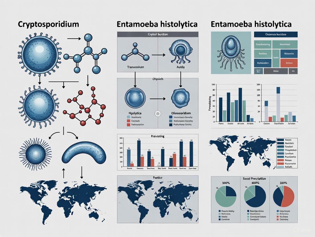
The following diagram illustrates a diagnostic and research workflow for these parasitic infections:
Drug Discovery Experimental Protocols
High-Throughput Screening for Anti-Parasitic Compounds
The development of automated high-throughput screening (HTS) platforms has accelerated drug discovery for parasitic infections. For E. histolytica, researchers have established an HTS method using exponentially growing trophozoites in 96-well or 384-well microtiter plates maintained under anaerobic conditions using GasPak systems [10]. Viability is assessed using ATP-based luminescent assays (CellTiter-Glo), which show linear correlation with parasite numbers [10]. This approach enabled screening of an FDA-approved compound library, identifying auranofin as a potent anti-amebic agent with EC50 of 0.5 µM, significantly better than metronidazole [10].
For Cryptosporidium, HTS faces additional challenges due to the parasite's intracellular nature and complex life cycle. Several scalable methods have been developed:
- Phenotypic assays using automated high-content imaging to quantify parasite numbers or infectious foci [3]
- Cytopathic effect analysis measuring host cell monolayer destruction [3]
- Molecular approaches using qRT-PCR on total cell lysates to quantify parasite burden [3]
- Transgenic parasite strains expressing luciferase or fluorescent reporters for direct quantification [3]
These methods have enabled large-scale drug screening efforts. For example, screening of 1,200 compounds identified Vorinostat as active against C. parvum, while screening of 800 natural products identified cedrelone and baicalein as effective [3].
Target-Based Drug Development
Metabolic pathway targeting represents a promising approach for both parasites. For E. histolytica, key targets include:
- EhADH2: A bifunctional alcohol dehydrogenase 2 essential for energy generation through fermentation [8]
- Phosphofructokinase (PFK): A glycolytic enzyme that uses pyrophosphate rather than ATP as a cofactor [8]
- Thioredoxin reductase: Targeted by auranofin, leading to enhanced sensitivity to reactive oxygen species [10]
For Cryptosporidium, potential targets include:
- Glucose-6-phosphate isomerase (CpGPI): Inhibited by ebselen [3]
- Hexokinase inhibitors: Several identified through target-based approaches [3]
- Other parasite-specific enzymes in glycolytic pathways, fatty acid production, kinase activities, and nucleotide synthesis [3]
Table 4: Research Reagent Solutions for Parasitic Drug Development
| Reagent/Assay | Application | Function |
|---|---|---|
| CellTiter-Glo Luminescent Assay | Viability assessment | ATP-based quantification of viable parasites [10] |
| GasPak Anaerobic Systems | Culture maintenance | Creates anaerobic environment for microaerophilic parasites [10] |
| Transgenic parasite strains (luciferase/GFP) | Drug screening | Enables direct quantification of parasite burden [3] |
| qRT-PCR protocols | Parasite quantification | Molecular quantification of parasite load in host cells [3] |
| Recombinant parasite enzymes | Target-based screening | Testing compound activity against specific molecular targets [3] [8] |
The global incidence and prevalence of Cryptosporidium and Entamoeba histolytica demonstrate significant regional disparities and disproportionate impact on specific high-risk populations. Cryptosporidiosis represents a more extensive global burden, particularly affecting young children in developing countries and immunocompromised individuals worldwide, while amebiasis remains highly endemic in specific tropical regions with poor sanitation. Advanced diagnostic methodologies, particularly molecular techniques, have revealed previously underestimated disease burdens in both developed and developing regions. The experimental frameworks presented—including high-throughput screening platforms, targeted metabolic pathway analysis, and drug repurposing approaches—provide powerful tools for researchers and drug development professionals working to address these significant parasitic diseases. Future efforts should focus on developing practical point-of-care diagnostics, effective treatments for immunocompromised patients, and vaccine candidates to reduce the substantial global burden of these neglected tropical diseases.
Cryptosporidium is recognized as a significant protozoan pathogen contributing substantially to the global burden of childhood diarrheal diseases, malnutrition, and mortality. This parasitic infection represents a critical public health challenge, particularly in resource-limited settings where sanitation infrastructure remains underdeveloped [11] [12]. The parasite's remarkable resilience to conventional water treatment disinfectants and its low infectious dose facilitate rapid transmission through contaminated water supplies, food, and direct person-to-person contact [13]. The World Health Organization (WHO) identifies diarrheal diseases as the third leading cause of death in children aged 1-59 months, with Cryptosporidium emerging as a predominant etiological agent responsible for severe health outcomes including growth faltering, cognitive impairment, and increased mortality [14] [15]. This whitepaper examines the epidemiology, pathophysiology, and long-term sequelae of childhood cryptosporidiosis within the broader context of global enteric protozoan research, with particular emphasis on its comparative relationship with Entamoeba histolytica.
Global Burden and Epidemiological Profile
Prevalence and Geographic Distribution
The global distribution of Cryptosporidium infection demonstrates significant regional variation, with the highest burden concentrated in developing regions. A comprehensive systematic review and meta-analysis covering 1999-2024 established a global protozoan prevalence of 7.5% (95% CI: 5.6%-10.0%) in diarrheal cases, with Cryptosporidium identified as one of the most common pathogens alongside Giardia [11]. The Americas and Africa bear the highest incidence rates, with specific studies revealing alarming infection frequencies among pediatric populations.
Table 1: Global Prevalence of Cryptosporidium in Children with Diarrhea
| Region/Country | Prevalence (%) | Study Period | Sample Size | Diagnostic Method |
|---|---|---|---|---|
| Eastern Ethiopia | 15.2 | 2022-2023 | 756 | LED fluorescence microscopy with auramine-phenol staining [13] |
| Cameroon | 13.4 | 2018 | 67 | Modified Ziehl-Neelsen staining [16] |
| Multicountry Cohort (MAL-ED) | 65.0 (over 2 years) | 2009-2012 | 1,486 | Enzyme-linked immunosorbent assay (ELISA) [12] |
| Pakistan (MAL-ED) | 9.2 (diarrheal episodes) | 2009-2012 | - | ELISA [12] |
| Peru (MAL-ED) | 10.9 (diarrheal episodes) | 2009-2012 | - | ELISA [12] |
| Kenya | 1.3 | - | 550 | Modified Ziehl-Neelsen staining & molecular assays [17] |
The MAL-ED longitudinal multicenter study demonstrated that 65% of children experienced Cryptosporidium infection during the first two years of life, highlighting the extensive exposure in endemic areas [12]. This study also revealed significant regional variations in infection rates, with the highest burden of Cryptosporidium-associated diarrhea observed in Peru (10.9%) and Pakistan (9.2%).
Mortality and Acute Morbidity
The Global Burden of Disease Study 2016 identified Cryptosporidium infection as the fifth leading diarrheal aetiology in children younger than 5 years worldwide, with acute infection causing more than 48,000 deaths (95% UI 24,600-81,900) annually [15]. Beyond mortality, acute cryptosporidiosis presents substantial morbidity through severe diarrheal episodes and dehydration. The MAL-ED study confirmed that Cryptosporidium diarrhea was significantly more likely to be associated with dehydration (16.5% vs 8.3%, P < .01) compared to non-Cryptosporidium diarrhea [12]. Clinical manifestations typically include profuse watery diarrhea that can persist for several days to weeks, eventually leading to dehydration, malabsorption, and malnutrition [16]. In immunocompromised children or those with underlying severe acute malnutrition, cryptosporidiosis can follow a particularly fulminant course, sometimes proving fatal despite therapeutic interventions [18].
Long-Term Consequences and Growth Impairment
The impact of Cryptosporidium infection extends far beyond acute diarrheal episodes, with substantial evidence demonstrating long-term effects on child development and health status.
Growth Faltering and Stunting
Multiple cohort studies have established a causal relationship between Cryptosporidium infection and childhood growth impairment. A meta-analysis of data from seven scientific literature sources and six individual-level datasets demonstrated that each episode of diarrhea caused by Cryptosporidium infection was associated with significant decreases in anthropometric measurements [15]:
- Height-for-age Z score (HAZ) decrease: 0.049 (95% CI 0.014-0.080)
- Weight-for-age Z score (WAZ) decrease: 0.095 (95% CI 0.055-0.134)
- Weight-for-height Z score (WHZ) decrease: 0.126 (95% CI 0.057-0.194)
The MAL-ED study confirmed these findings, with multivariable regression analysis revealing a significantly decreased length-for-age Z score at 24 months in Cryptosporidium-positive children at the India (β = -0.26) and Bangladesh (β = -0.20) sites [12]. This growth faltering represents a critical pathway through which Cryptosporidium infection contributes to childhood malnutrition and its associated developmental delays.
Comprehensive Burden Assessment
When accounting for both acute effects and long-term growth consequences, the true burden of Cryptosporidium infection substantially exceeds conventional estimates. The meta-analysis by the GBD 2016 study determined that diarrhoea from Cryptosporidium infection caused an additional 7.85 million disability-adjusted life-years (DALYs) (95% UI 5.42 million-10.11 million) after accounting for its effect on growth faltering—representing a 153% increase compared to estimates based solely on acute effects [15]. This dramatic reassessment of disease burden underscores the importance of considering the long-term sequelae of enteric infections in resource allocation and intervention planning.
Cryptosporidium morbidity pathways showing acute and long-term consequences
Risk Factors and Transmission Dynamics
Understanding the epidemiological risk factors for Cryptosporidium transmission is essential for developing targeted intervention strategies.
Environmental and Behavioral Risk Factors
Multiple studies have identified consistent environmental, socioeconomic, and behavioral factors associated with increased risk of Cryptosporidium infection in children:
- Seasonality: The wet season demonstrates significantly higher infection rates (APR = 1.7, 95% CI: 1.2-2.4) according to a study in Eastern Ethiopia [13]
- Caregiver education: Children with caregivers having no formal education showed higher infection risk (APR = 2.6, 95% CI: 1.1-6.3) [13]
- Household diarrhea: Presence of a diarrheic member in the household increased risk (APR = 1.9, 95% CI: 1.2-3.2) [13]
- Feeding practices: Lack of exclusive breastfeeding was associated with higher infection risk (APR = 1.6, 95% CI: 1.1-2.3) [13]
- Hygiene practices: Inadequate handwashing after toileting significantly increased risk (APR = 2.8, 95% CI: 1.7-4.5) [13]
- Overcrowding: The MAL-ED study identified overcrowding (>3 people per room) as a significant risk factor in Bangladesh (OR = 2.3, 95% CI: 1.2-4.6) [12]
- Water sources: Consumption of untreated water from potentially contaminated sources increases infection risk [16] [14]
Table 2: Significant Risk Factors for Childhood Cryptosporidium Infection
| Risk Factor Category | Specific Factor | Effect Size (95% CI) | Study |
|---|---|---|---|
| Environmental | Wet season | APR = 1.7 (1.2-2.4) | Eastern Ethiopia [13] |
| Socioeconomic | No formal education of caregiver | APR = 2.6 (1.1-6.3) | Eastern Ethiopia [13] |
| Socioeconomic | Overcrowding in home | OR = 2.3 (1.2-4.6) | MAL-ED Bangladesh [12] |
| Behavioral | Lack of exclusive breastfeeding | APR = 1.6 (1.1-2.3) | Eastern Ethiopia [13] |
| Behavioral | Inadequate handwashing after toileting | APR = 2.8 (1.7-4.5) | Eastern Ethiopia [13] |
| Household | Presence of diarrheic household member | APR = 1.9 (1.2-3.2) | Eastern Ethiopia [13] |
Transmission Pathways and Zoonotic Potential
Cryptosporidium transmission occurs predominantly through the fecal-oral route, with multiple potential reservoirs and transmission pathways. Molecular characterization studies have identified both anthroponotic and zoonotic transmission cycles. A study in Kiambu County, Kenya, identified all Cryptosporidium isolates as C. hominis, with subtypes IbA9G3 and IeA11G3T3 predominating, suggesting primarily anthroponotic transmission in that region [17]. The parasite's oocyst stage is remarkably resistant to conventional water disinfectants, facilitating waterborne outbreaks and environmental persistence [13]. The high infectivity of Cryptosporidium (low infectious dose) further enhances transmission potential in settings with compromised water, sanitation, and hygiene (WASH) infrastructure.
Diagnostic Methodologies and Experimental Protocols
Accurate diagnosis of Cryptosporidium infection remains challenging in resource-limited settings, with significant implications for disease surveillance and clinical management.
Conventional Diagnostic Techniques
Cryptosporidium diagnostic workflow from basic to advanced methods
Microscopy-Based Detection
Modified Ziehl-Neelsen (mZN) Staining Protocol (as used in Cameroon study [16] and Kenya study [17]):
- Sample preparation: Create moderate thick fecal smears on standard microscope slides and air-dry
- Heat fixation: Fix smears by passing through flame or heating
- Primary staining: Flood slide with carbol fuchsin and stain for 10-15 minutes
- Decolorization: Rinse with acid-alcohol until pink color disappears
- Counterstaining: Apply methylene blue or malachite green for 30 seconds to 1 minute
- Microscopic examination: View under 400x and 1000x magnification; Cryptosporidium oocysts appear as bright pink to red spherical structures (4-6μm diameter) against a blue or green background
Limitations: mZN staining has variable sensitivity (55-75%) though high specificity (96-100%) [13]
Advanced Fluorescence Microscopy
Auramine-Phenol Staining with LED Fluorescence Microscopy (as implemented in Eastern Ethiopia study [13]):
- Smear preparation: Create thin fecal smears on slides and allow to air-dry
- Fixation: Fix with methanol for 2-5 minutes
- Staining: Apply auramine-phenol stain for 15-20 minutes
- Decolorization: Rinse with acid-alcohol for 2-3 minutes
- Counterstaining: Apply potassium permanganate for 2-3 minutes
- Examination: View using LED fluorescence microscopy with appropriate filters; oocysts appear as bright apple-green spherical structures against a dark background
Performance: This method demonstrates superior diagnostic performance with 88% sensitivity and 99% specificity compared to conventional mZN staining [13]
Molecular Characterization Techniques
Molecular methods enable precise species and genotype identification, providing valuable epidemiological insights:
Nested PCR Protocol for Cryptosporidium Species Identification (as described in Kenya study [17]):
- DNA extraction: Extract genomic DNA from microscopy-positive stool samples using commercial extraction kits
- Primary PCR amplification: Perform first-round PCR targeting the 60-kDa glycoprotein gene using outer primers
- Secondary PCR amplification: Use primary PCR product as template for nested reaction with internal primers
- Electrophoresis: Analyze PCR products by agarose gel electrophoresis
- Sequencing: Purify amplification products and perform bidirectional sequencing
- Sequence analysis: Align sequences with reference strains for subtype identification
This approach enabled identification of C. hominis subtypes IbA9G3 and IeA11G3T3 in the Kenyan pediatric population [17].
The Scientist's Toolkit: Essential Research Reagents
Table 3: Key Research Reagents for Cryptosporidium Investigation
| Reagent/Assay | Application | Specifications | Research Utility |
|---|---|---|---|
| Pan-Cryptosporidium Immunoassay (TechLab) | Antigen detection in stool samples | ELISA-based detection | Used in MAL-ED study for large-scale screening [12] |
| Auramine-Phenol Stain | Fluorescence microscopic detection | Excitation: 450-490nm, Emission: 515nm | Superior sensitivity (88%) for oocyst detection [13] |
| Modified Ziehl-Neelsen Reagents | Acid-fast staining of oocysts | Carbol fuchsin, acid-alcohol, malachite blue | Conventional detection with high specificity (96-100%) [13] [16] |
| Nested PCR Primers (gp60 gene) | Molecular subtyping | Targets 60-kDa glycoprotein gene | Enables identification of C. hominis subtypes and transmission tracking [17] |
| Formol-Ether Concentration Solution | Parasite concentration in stool | Formalin and diethyl ether | Increases detection sensitivity by concentrating oocysts [17] |
Comparative Analysis with Entamoeba histolytica
While both Cryptosporidium and Entamoeba histolytica constitute significant protozoan causes of childhood diarrheal disease, they demonstrate distinct epidemiological and clinical characteristics with important implications for global burden assessments.
Epidemiological and Diagnostic Distinctions
The systematic review of protozoan pathogens in diarrhea cases worldwide identified Entamoeba histolytica as a notable contributor to diarrheal morbidity, though with varying prevalence across geographic regions [11]. Molecular studies in Kiambu County, Kenya, demonstrated the importance of differentiating pathogenic E. histolytica from non-pathogenic Entamoeba species, with PCR analysis revealing E. histolytica at 3.3% prevalence compared to E. dispar at 3.8% and E. moshkovskii at 1.6% [17]. This differentiation has critical clinical implications, as non-pathogenic Entamoeba species do not require treatment, while E. histolytica infection necessitates prompt antimicrobial therapy [19].
Therapeutic Challenges and Research Imperatives
Both Cryptosporidium and E. histolytica present significant therapeutic challenges, though of different natures. For E. histolytica, effective treatment regimens exist, including metronidazole for invasive disease followed by luminal agents such as paromomycin to eradicate carriage [19]. In contrast, Cryptosporidium treatment options remain limited, with nitazoxanide demonstrating only marginal efficacy in malnourished children and immunocompromised hosts [18]. This therapeutic gap underscores the critical need for targeted drug development for cryptosporidiosis.
Discussion and Future Directions
Integrated Intervention Strategies
The substantial burden of Cryptosporidium infection necessitates comprehensive intervention strategies targeting multiple transmission pathways:
- Preventive interventions: Improved sanitation, water treatment infrastructure, and hygiene education represent foundational prevention approaches [13] [14]
- Nutritional support: Exclusive breastfeeding during early infancy provides significant protective benefits [13]
- Vaccine development: Current vaccine candidates remain in preclinical and early clinical development stages [18]
- Therapeutic innovation: Novel therapeutic compounds with improved efficacy in vulnerable populations represent an urgent unmet need [18]
Research Priorities and Advocacy Needs
The Cryptosporidiosis Therapeutics Advocacy Group (CTAG) has outlined critical steps to address the neglect of this pathogen, including advocating for WHO inclusion of cryptosporidiosis on its list of Neglected Tropical Diseases (NTDs) to stimulate research investment and drug development [18]. Additional priorities include:
- Diagnostic advancement: Development and implementation of cost-effective, highly-sensitive rapid diagnostic tests
- Transmission interruption: Targeted interventions informed by molecular epidemiological tracking
- Therapeutic optimization: Clinical trials to optimize existing treatment regimens in vulnerable populations
- Longitudinal studies: Enhanced understanding of the long-term developmental consequences of childhood infection
Cryptosporidium represents a profoundly significant yet underestimated cause of childhood diarrheal morbidity and mortality, with particularly devastating long-term consequences for physical growth and cognitive development. The true burden of this parasitic infection extends far beyond acute diarrheal episodes to include substantial impacts on linear growth, contributing to childhood stunting and its associated developmental disadvantages. Current therapeutic options remain inadequate for vulnerable pediatric populations, highlighting an urgent need for targeted research and drug development initiatives. Framing cryptosporidiosis within the broader context of global enteric protozoan research illuminates the critical public health imperative of addressing this neglected pathogen through comprehensive prevention strategies, diagnostic improvements, and therapeutic innovation. The notable case fatality despite standard management underscores the devastating potential of this infection in malnourished children and emphasizes the vital importance of multidisciplinary approaches to reduce the global burden of childhood cryptosporidiosis.
Global Burden and Clinical Significance
The enteric parasites Cryptosporidium spp. and Entamoeba histolytica represent a significant global health challenge, particularly in low-resource settings. Beyond their acute gastrointestinal symptoms, a growing body of evidence links these infections to long-term sequelae, including growth stunting in children and subsequent cognitive deficits, contributing to a substantial disease burden.
Cryptosporidium is a leading cause of moderate-to-severe diarrheal disease in children under two years of age in sub-Saharan Africa and South Asia, ranked second only to rotavirus [20]. The Global Enteric Multicenter Study (GEMS) identified it as a major contributor to diarrheal diseases, associated with a 2–3 times higher risk of mortality among children aged 12–23 months [20]. Globally, cryptosporidiosis causes more than 50 million episodes of diarrhea and an estimated 48,000 deaths annually among children under 5, primarily in developing countries [21]. Meanwhile, amebiasis due to E. histolytica is responsible for more than 55,000 deaths worldwide each year [22] [23]. In industrialized nations, these pathogens remain a concern through imported cases, outbreaks, and infections in immunocompromised individuals [22] [24] [25].
The long-term manifestations of these infections contribute significantly to their overall disease burden. A study from the Netherlands found that long-term sequelae contributed nearly 10% of the total Disability-Adjusted Life Years (DALYs) and costs in burden of disease models for Cryptosporidium, indicating a higher public health impact than previously estimated from acute illness alone [24] [25]. This underscores the importance of considering chronic sequelae in health policy decisions.
Table 1: Global Burden of Key Parasitic Infections
| Parasite | Annual Deaths (Estimate) | Annual Diarrheal Episodes (Children <5) | Key Affected Populations |
|---|---|---|---|
| Cryptosporidium spp. | ~48,000 [21] | >50 million [21] | Children in endemic areas, immunocompromised individuals |
| Entamoeba histolytica | >55,000 [22] [23] | Not specified | All ages in endemic areas, returning travelers, MSM |
Pathophysiological Mechanisms Linking Infection to Long-Term Sequelae
The pathway from enteric parasitic infection to long-term cognitive impairment involves a complex interplay of intestinal damage, inflammatory responses, nutrient malabsorption, and subsequent growth faltering.
Intestinal Damage and Nutrient Malabsorption
Cryptosporidium infection causes direct damage to intestinal epithelial cells, disrupting tight junctions and impairing intestinal barrier function [21]. This damage hinders villous development and reduces the absorptive capacity of the intestinal surface, leading to malnutrition, dehydration, and diarrhea [21]. The inflammatory response to E. histolytica infection further exacerbates this damage, as trophozoites invade and penetrate the intestinal mucosa, destroying epithelial cells and inflammatory cells through cytolysis and apoptosis [1]. The resulting mucosal inflammation, thickening, ulcers, and necrosis contribute to impaired absorption and enhanced secretion, promoting diarrheal disease and growth deficits [20].
Inflammatory Pathways and Systemic Effects
The NF-κB signaling pathway plays a critical role in the host response to Cryptosporidium infection [21]. Upon infection, epithelial cells activate NF-κB, which translocates to the nucleus and regulates the expression of host genes involved in inflammation and immune response [21]. This pathway is involved in regulating various RNAs during infection, including miRNAs such as miR-942-5p and miR-181d, which can attenuate apoptosis in infected cells and influence infection burden [21]. The TLR2/TLR4-NF-κB signaling pathway is also activated during Cryptosporidium infection, contributing to the inflammatory response [21].
For E. histolytica, pathogenesis involves adherence of colonic epithelial cells through a specific galactose-N-acetylgalactosamine lectin [1]. Subsequent cytolysis and apoptosis of epithelial cells release interleukin-1α and precursor interleukin-1β, activating NF-κB and producing cytokines and inflammatory mediators such as COX-2, interleukin-1, and interleukin-8 [1]. These mediators attract neutrophils and macrophages, releasing additional inflammatory factors like TNFα, which further contribute to tissue damage and systemic inflammation [1].
Figure 1: Pathophysiological Pathway from Infection to Cognitive Deficit
Epidemiological and Clinical Evidence
Growth Stunting Following Infection
Multiple longitudinal studies have demonstrated the association between parasitic enteric infections and growth impairment. Cryptosporidium infection is particularly associated with prolonged diarrhoea (7-14 days) and persistent diarrhoea (≥14 days), which are significant risk factors for growth faltering [20]. Studies in low-resource settings have confirmed that cryptosporidiosis contributes to childhood malnutrition and growth deficits, with even asymptomatic infection associated with poor growth [20]. Research from Peru showed that symptomatic cryptosporidiosis stunted weight gain more than asymptomatic infection, but asymptomatic infection was twice as common and might have a greater overall adverse effect on child growth [20].
The MAL-ED cohort study, which followed children from birth to 5 years of age in six low- and middle-income countries, provided important insights into the timing and persistence of stunting [26]. Children were categorized as:
- Early-onset persistent (first stunted at 1-6 months and persisting at 60 months)
- Early-onset recovered (first stunted at 1-6 months and not stunted at 60 months)
- Late-onset persistent (first stunted at 7-24 months and persisting at 60 months)
- Late-onset recovered (first stunted at 7-24 months and not stunted at 60 months)
- Never stunted [26]
This classification revealed that the timing and persistence of stunting have distinct impacts on long-term outcomes.
Cognitive Deficits Associated with Stunting
The association between stunting and cognitive impairment is well-established. A study in Indonesia comparing stunted children to undernourished children with normal stature found a trend toward lower cognitive, motor, and adaptive behavior abilities in stunted children, though differences did not reach statistical significance [27]. Both groups exhibited scores below the 50th percentile in all developmental domains, suggesting that undernourished children have below-average abilities even before stunting occurs [27].
The MAL-ED study provided more definitive evidence, demonstrating that children with early-onset persistent stunting had significantly lower cognitive scores at 5 years of age compared to those who were never stunted [26]. The study used the Wechsler Preschool Primary Scales of Intelligence (WPPSI) to assess cognitive abilities, specifically fluid reasoning, adapted for each cultural context [26]. After controlling for confounders including socio-economic status, quality of the home environment, and biomarkers of micronutrient status, early-onset persistent stunting remained independently associated with poorer cognitive development [26].
Table 2: Long-Term Sequelae Following Cryptosporidium Infection (Based on Systematic Review) [24]
| Sequelae | Prevalence in Cases | Likelihood vs. Controls (Odds Ratio) |
|---|---|---|
| Diarrhoea | 25% | 6.0x |
| Abdominal pain | 25% | 2.4x |
| Nausea | 24% | Not reported |
| Fatigue | 24% | 2.6x |
| Headache | 21% | 2.2x |
| Weight loss | Not specified | 6.2x |
| Joint pain | Not specified | 2.6x |
| Vomiting | Not specified | 2.8x |
| Loss of appetite | Not specified | 2.9x |
Research Methodologies and Experimental Approaches
Cognitive Assessment Tools
Research on cognitive outcomes associated with infection and stunting employs standardized neurodevelopmental assessments:
- Bayley Scales of Infant Development, Third Edition (Bayley-III): Assesses cognitive, language, motor, social-emotional, and adaptive behavior domains in young children (1 month to 3 years). Scaled scores of 1-6 are classified as below average, 7-13 as average, and 14-19 as above average [27].
- Wechsler Preschool Primary Scales of Intelligence (WPPSI): Used to assess cognitive abilities, particularly fluid reasoning, in children up to 5 years of age. The instrument should be culturally adapted for different populations [26].
Growth Monitoring Protocols
Longitudinal growth monitoring requires standardized methodologies:
- Anthropometric Measurements: Regular assessment of weight, height/length, mid-upper arm circumference, and head circumference.
- z-score Calculations: Height-for-age (HAZ) and weight-for-age (WAZ) scores calculated based on WHO Child Growth Standards. Stunting is defined as HAZ < -2 standard deviations below the median [27] [26].
- Growth Faltering Identification: Monitoring for inadequate weight gain that may precede stunting.
Laboratory Diagnostic Methods
Accurate parasite identification is crucial for research:
Table 3: Diagnostic Methods for Enteric Parasites
| Method | Sensitivity | Advantages | Disadvantages |
|---|---|---|---|
| Microscopy | ~70% with modified acid-fast stain [20] | Low technology, widely available | Low sensitivity, requires skilled technicians |
| Antigen Detection | 70-100% [20] | Commercially available kits, higher throughput | Costly for resource-poor settings |
| PCR | 92-100% [20] [1] | Excellent sensitivity, can speciate and subtype | Expensive instrumentation, technically demanding |
| Serology | High sensitivity and specificity [1] | Useful for surveillance | Cannot distinguish acute from historical infection |
Figure 2: Cohort Study Design for Infection-Stunting Research
The Scientist's Toolkit: Essential Research Reagents and Materials
Table 4: Key Research Reagent Solutions for Investigating Infection-Stunting-Cognition Pathways
| Reagent/Material | Application | Specific Examples/Protocols |
|---|---|---|
| Parasite Detection Kits | Identification of Cryptosporidium/E. histolytica in stool samples | Commercial antigen detection kits (ELISA, immunofluorescence); PCR primers for 18S rRNA gene [20] [1] |
| DNA Extraction Kits | Nucleic acid isolation for molecular diagnostics | Protocols optimized for stool samples; includes steps to remove PCR inhibitors [20] |
| Cell Culture Systems | In vitro study of host-parasite interactions | HCT-8 cells for Cryptosporidium propagation; biliary epithelial cells for invasion studies [21] |
| Cytokine/Chemokine Assays | Quantification of inflammatory mediators | ELISA for CX3CL1, IL-1β, TNFα; PCR for miRNA expression (miR-942-5p, miR-181d) [1] [21] |
| Cognitive Assessment Kits | Standardized developmental testing | Bayley-III complete kit; WPPSI materials with cultural adaptations [27] [26] |
| Anthropometric Equipment | Growth monitoring | WHO-standard height/length boards; digital scales; mid-upper arm circumference tapes [27] [26] |
| Microbiome Analysis Kits | Investigation of gut-brain axis | 16S rRNA sequencing kits; DNA extraction protocols for fecal samples [28] |
Implications for Drug Development and Public Health Interventions
The link between enteric parasitic infections, growth stunting, and cognitive deficits has profound implications for therapeutic development and public health policy. Current treatments for these infections remain suboptimal, highlighting the need for more effective interventions.
Therapeutic Challenges and Opportunities
Cryptosporidium treatment faces significant hurdles, with nitazoxanide—the only approved medication—showing efficacy ranging from just 56% in malnourished children to 80% in healthy adults, with limited effectiveness in immunocompromised patients [23] [21]. For amebiasis, metronidazole treatment requires subsequent paromomycin to eliminate cysts, resulting in a burdensome 20-day regimen that reduces compliance [23]. Treatment failures in giardiasis occur in up to 20% of cases, with one report noting nitroimidazole therapy failure rates as high as 40.2% [23].
Drug discovery efforts are targeting parasite-specific enzymes and metabolic pathways. Promising approaches include:
- Targeting Cryptosporidium-specific enzymes in metabolic pathways unique to the parasite [21]
- Developing inhibitors of E. histolytica cysteine synthase and heat shock protein 90 (Hsp90) [23]
- Exploring drug repurposing, such as auranofin, which targets thioredoxin reductase in both E. histolytica and Giardia [23]
- Investigating natural products like deacetylkinamycin C and nanomycin A with amebicidal activity [23]
Public Health and Vaccination Strategies
Partial immunity after Cryptosporidium exposure suggests the potential for successful vaccines, with several candidates in development [20]. However, correlates of protection are not well defined, and methods for propagation and genetic manipulation of the organism require significant advances [20]. The development of more sensitive diagnostic tools is also crucial, as current tests miss a substantial proportion of cases, particularly in resource-limited settings where microscopy with modified acid-fast staining remains common despite its limitations [20].
Public health interventions must address the multiple pathways linking infection to cognitive deficits. These include:
- Improving sanitation and access to clean water to prevent transmission
- Enhancing nutritional support for infected children to mitigate growth faltering
- Implementing early detection and treatment protocols
- Developing educational interventions to support cognitive development in affected children
The evidence summarized in this review underscores the importance of considering the long-term sequelae of enteric infections in burden-of-disease calculations and intervention planning. Only by addressing the full spectrum of acute and chronic consequences can the true impact of these parasitic infections be mitigated.
The global burden of disease caused by the protozoan parasites Cryptosporidium and Entamoeba histolytica remains substantial, particularly in regions with limited resources and inadequate public health infrastructure. These pathogens employ diverse and complex transmission strategies—including waterborne, zoonotic, and person-to-person spread—that enable their persistence in human populations and complicate control efforts. Cryptosporidium, a leading cause of severe diarrheal disease and mortality in children under five, demonstrates remarkable environmental resilience and low infectious dose [29] [30]. Entamoeba histolytica, the causative agent of amebiasis, ranks as the fourth leading parasitic cause of human mortality, responsible for nearly 100,000 deaths annually from invasive colitis and extraintestinal abscesses [1] [31]. Understanding the intricate transmission dynamics and specific risk factors associated with these pathogens is fundamental to developing targeted interventions, therapeutic agents, and effective public health policies aimed at reducing their global impact.
Global Burden and Public Health Significance
Cryptosporidium
Cryptosporidium imposes a significant health burden, especially in low- and middle-income countries (LMICs). It is the second most common cause of diarrheal disease and mortality in children under five in sub-Saharan Africa and South Asia [30]. In these regions, cryptosporidiosis is highly prevalent in early childhood, with studies showing infection rates of 77% in slum-dwelling Bangladeshi children and 97% in children under three from a Southern Indian birth cohort [30]. The infection is associated with malnutrition, stunted growth, and cognitive impairment, creating a vicious cycle where cryptosporidiosis exacerbates malnutrition and is more severe in malnourished subjects [30]. In immunocompromised individuals, such as those with HIV/AIDS, cancer, or transplant recipients, cryptosporidiosis can cause prolonged, life-threatening illness [29] [32].
Entamoeba histolytica
Entamoeba histolytica causes approximately 50 million symptomatic infections worldwide each year, resulting in nearly 100,000 deaths [1]. Although 90% of E. histolytica infections are asymptomatic, infected individuals can still transmit the parasite [1]. The highest prevalence of amebiasis is observed in developing countries with poor sanitation, including India, Africa, Mexico, and Central and South America [1]. In rural Mexico, seroprevalence has been reported as high as 42% [1]. The National Institute of Allergy and Infectious Diseases (NIAID) has classified E. histolytica as a category B biodefense pathogen due to its low infectious dose, environmental stability, resistance to chlorine, and ease of dissemination through contaminated food and water [33].
Table 1: Global Burden and Key Characteristics of Cryptosporidium and Entamoeba histolytica
| Characteristic | Cryptosporidium | Entamoeba histolytica |
|---|---|---|
| Global Mortality | >50,000 deaths annually [29]; Leading cause of mortality in children 12-23 months in endemic areas [30] | ~100,000 deaths yearly [1] |
| Symptomatic Cases | Major cause of severe diarrheal illness in children <5 in LMICs [30] | Nearly 50 million people yearly [1] |
| Asymptomatic Rate | Can occur, particularly in endemic areas [29] | ~90% of infections [1] |
| High Prevalence Areas | Developing countries with poor water and sanitation [29] [30] | India, Africa, Mexico, Central and South America [1] |
| At-Risk Populations | Children <5, immunocompromised individuals (HIV/AIDS, transplant), malnourished children [29] [30] [32] | Travelers to endemic areas, immigrants, men who have sex with men, immunosuppressed individuals [1] [33] |
| Long-Term Sequelae | Malnutrition, stunted growth, cognitive impairment [30] | Chronic non-dysenteric colitis [33] |
Pathogen Biology and Life Cycle
Cryptosporidium
Cryptosporidium is an apicomplexan protozoan parasite with a monoxenous (single-host) life cycle that is completed within the gastrointestinal tract of the host [29]. Infection begins with the ingestion of environmentally robust oocysts, each containing four sporozoites [29]. These sporozoites hatch in the intestine, invade epithelial cells, and reside in a unique extracytoplasmic location within a parasitophorous vacuole [29]. The parasite undergoes asexual reproduction (schizogony), producing type I meronts that release merozoites, which can propagate the infection by invading neighboring epithelial cells [29]. Some merozoites then undergo sexual reproduction (gametogony), forming microgametes and macrogametes that unite to form zygotes [29]. These zygotes develop into oocysts that can either be thick-walled or thin-walled. The thick-walled oocysts are shed into the environment through feces, while the thin-walled oocysts can autoinfect the host, leading to prolonged infection, particularly in immunocompromised individuals [29].
Entamoeba histolytica
Entamoeba histolytica has a simple two-stage life cycle consisting of the infective cyst and the invasive trophozoite [33]. Transmission occurs through the ingestion of mature cysts from fecally contaminated food, water, or hands [1] [33]. Excystation occurs in the terminal ileum or colon, releasing trophozoites that colonize the large intestine [33]. Trophozoites multiply by binary fission and can invade the colonic mucosa, causing tissue destruction and secretory bloody diarrhea [33]. Some trophozoites can spread hematogenously via the portal circulation to the liver and other organs, causing abscesses [33]. Trophozoites passed in the stool are unable to survive in the environment, but those remaining in the intestine can undergo encystation, developing into cysts that are passed in the feces [33]. These cysts can survive in the environment for weeks to months, completing the cycle [1] [33].
Diagram 1: Comparative life cycles of Cryptosporidium and Entamoeba histolytica showing key developmental stages and transmission pathways.
Transmission Dynamics and Risk Factors
Waterborne Transmission
Waterborne transmission represents a significant pathway for both parasites, though their environmental characteristics and resistance to disinfectants differ.
Cryptosporidium oocysts are immediately infectious when shed in feces and are remarkably resistant to chlorine-based disinfectants, making them a major challenge for water treatment plants [34] [29]. This chlorine resistance has been responsible for numerous outbreaks associated with drinking water and recreational water venues [29] [32]. The infectious dose is low, with ingestion of as few as 10 oocysts capable of initiating infection [29]. A study from Tabriz, Iran, highlighted that drinking water sourced from surface water is more susceptible to contamination compared to groundwater, though the parasite was not detected in their limited sample set, potentially due to low rainfall in the region [34]. Recreational water activities (swimming in pools, lakes, or water parks) represent a major transmission route, as an infected swimmer can release millions of oocysts, contaminating the water and infecting other swimmers [35] [32].
Entamoeba histolytica cysts can survive in the environment for weeks to months in appropriate conditions and are also resistant to standard chlorine levels used in water treatment [33]. Contamination of water supplies typically occurs through sewage discharge into drinking water sources, inadequate water treatment, or defecation directly into water sources [1] [31]. Unlike Cryptosporidium, E. histolytica does not exhibit the same extreme chlorine resistance, but waterborne outbreaks still occur frequently in endemic areas with poor sanitation infrastructure [33].
Zoonotic Transmission
The importance of zoonotic transmission varies significantly between these two parasites.
Cryptosporidium has a substantial zoonotic component, particularly for certain species. C. parvum, one of the two most common species infecting humans, has an infectious cycle involving both humans and ruminants [29] [30]. Individuals with occupational animal contact, particularly with pre-weaned calves, lambs, or goat kids, are at higher risk [35] [30]. A meta-analysis of risk factors in LMICs found animal contact was associated with a pooled odds ratio of 1.98 (95% CI: 1.11-3.54) for cryptosporidiosis [30]. Other species like C. meleagridis, C. felis, and C. canis have also been reported to infect humans, though less frequently [29].
Entamoeba histolytica is primarily a human pathogen with a limited zoonotic reservoir. The main zoonotic component involves non-human primates, which can act as reservoirs in certain environments [31] [36]. Dogs have also been identified as potential reservoirs, though their role is considered secondary to human-to-human transmission [31] [36]. Other Entamoeba species, such as E. dispar and E. moshkovskii, have been identified in various animal hosts, including non-human primates, swine, and dogs, but their pathogenicity in humans remains uncertain [36].
Person-to-Person Transmission
Direct person-to-person transmission through the fecal-oral route is a major pathway for both parasites, particularly in specific settings.
Cryptosporidium spreads easily in settings with close human contact, such as households, childcare centers, and among family members [35] [30]. The presence of diarrhea in the household has been identified as a significant risk factor, with a pooled odds ratio of 1.98 (95% CI: 1.13-3.49) [30]. Infected individuals can shed the parasite for up to two weeks after symptoms resolve, facilitating silent transmission [35]. Sexual practices involving oral-anal contact also enable transmission [35] [32].
Entamoeba histolytica is efficiently transmitted through direct fecal-oral contact and indirectly through contamination of food or water by infected food handlers [1] [33]. Person-to-person transmission is particularly notable in institutional settings with poor hygiene and among men who have sex with men (MSM) through oral-anal sexual practices [1] [33]. Asymptomatic cyst passers play a crucial role in maintaining transmission within communities [1].
Table 2: Comparative Transmission Routes and Associated Risk Factors
| Transmission Route | Cryptosporidium | Entamoeba histolytica |
|---|---|---|
| Waterborne | Primary route; Chlorine-resistant oocysts; Recreational water (pools, lakes); Untreated drinking water; Low infectious dose (~10 oocysts) [34] [29] [32] | Significant route; Contaminated water supplies; Cysts survive weeks to months; Inadequate water treatment [1] [31] [33] |
| Zoonotic | Significant: C. parvum from ruminants (calves, lambs, goats); Occupational contact (farming); Petting zoos [29] [30] | Limited: Primarily non-human primates; Dogs as potential reservoir; Secondary to human transmission [31] [36] |
| Person-to-Person | High: Household contacts; Childcare centers; Diaper-changing; Shedding for weeks post-symptoms; Sexual practices [35] [30] | High: Asymptomatic cyst passers; Food handlers; Institutional settings; Men who have sex with men [1] [33] |
| Foodborne | Unwashed fruits/vegetables; Unpasteurized milk/cider [32] | Contaminated food; Unwashed produce [31] |
| Environmental | Oocysts survive in soil; Contaminated surfaces (toys, bathrooms) [35] | Cysts in fecally contaminated soil/fertilizer [33] |
Molecular Pathogenesis and Host-Parasite Interactions
Cryptosporidium Pathogenesis
Following excystation, Cryptosporidium sporozoites invade intestinal epithelial cells, occupying a unique extracytoplasmic niche [29]. The parasite alters intestinal barrier function, increasing permeability and disrupting fluid and electrolyte absorption, leading to secretory diarrhea [29]. The infection triggers a pro-inflammatory response, with the attachment and invasion of epithelial cells causing cell damage and apoptosis [29]. In immunocompetent hosts, this results in self-limiting watery diarrhea, while in immunocompromised individuals, the infection can become chronic and disseminate to other parts of the gastrointestinal tract, including the biliary system, causing sclerosing cholangitis and pancreatitis [29].
Entamoeba histolytica Pathogenesis
The pathogenesis of E. histolytica involves a multi-step process beginning with adherence to colonic mucus and epithelial cells, mediated by a galactose/N-acetylgalactosamine (Gal/GalNAc)-specific lectin [1] [33]. Following adherence, trophozoites induce cytolysis and apoptosis of host cells through the action of amoebapores (pore-forming peptides) and cysteine proteinases [33]. The cysteine proteinases also play a role in degrading components of the extracellular matrix, facilitating tissue invasion, and can cleave and inactivate host immune molecules such as anaphylatoxins C3a and C5a, as well as IgA and IgG [33]. The parasite triggers a robust inflammatory response, with epithelial cells producing IL-1β, IL-8, and COX-2, attracting neutrophils and macrophages that contribute to tissue damage [33]. In some cases, trophozoites can disseminate via the portal circulation to the liver, causing amoebic liver abscesses characterized by well-circumscribed regions of dead hepatocytes surrounded by few inflammatory cells [33].
Diagram 2: Key pathogenic mechanisms of Entamoeba histolytica and Cryptosporidium showing cellular invasion, tissue damage, and immune evasion strategies.
Diagnostic Approaches and Experimental Protocols
Accurate diagnosis is crucial for individual patient management, epidemiological surveillance, and understanding transmission dynamics. The following section outlines standard and advanced diagnostic methodologies.
Standard Diagnostic Techniques
Microscopy remains widely used for both parasites, though with limitations. For Cryptosporidium, the modified Ziehl-Neelsen (mZN) stain is commonly employed to detect 4-6 μm oocysts in stool samples, appearing as bright pink to red spherical structures against a blue or green background [37] [29]. For E. histolytica, microscopic examination of stool samples can identify trophozoites or cysts, but cannot differentiate the pathogenic E. histolytica from the non-pathogenic E. dispar and other commensal amoebae [1] [33].
Antigen Detection methods using enzyme immunoassays (EIA) or immunofluorescence assays (IFA) offer improved sensitivity and specificity for both parasites. These tests detect parasite-specific proteins in stool samples and are commercially available as rapid diagnostic tests [1] [29]. For E. histolytica, antigen tests can specifically distinguish E. histolytica from E. dispar [1].
Molecular Diagnostics using polymerase chain reaction (PCR)-based methods represent the current gold standard for sensitive and specific detection, differentiation, and genotyping of both parasites [1] [29]. Multi-copy targets, such as the small ribosomal subunit RNA (18S rRNA) gene, are often used to enhance sensitivity, particularly in nested PCR formats [34] [29].
Detailed Experimental Protocol: Nested PCR for Cryptosporidium Detection
This protocol is adapted from studies detecting Cryptosporidium in water and fecal samples [34] [37].
1. Sample Collection and Processing:
- Water Samples: Collect large volume water samples (e.g., 30 L) and filter through membrane filters with 1.2 μm pore size to concentrate suspended particles and oocysts.
- Fecal Samples: Collect fresh stool samples and preserve in appropriate fixatives (e.g., 10% formalin) or store at -20°C for molecular analysis.
2. DNA Extraction:
- Use commercial DNA extraction kits suitable for parasitic organisms from stool or environmental samples.
- Include a mechanical disruption step (e.g., bead beating) to break open the robust oocyst wall.
- Include appropriate positive (C. parvum DNA) and negative (no template) controls in each extraction batch.
3. Nested PCR Amplification of 18S rRNA Gene:
- Primary PCR Reaction: Use external primers targeting a portion of the 18S rRNA gene.
- Reaction Mix: 2-5 μL template DNA, 1X PCR buffer, 2.5 mM MgCl₂, 200 μM dNTPs, 0.5 μM each primer, 1.25 U DNA polymerase, in a total volume of 25-50 μL.
- Cycling Conditions: Initial denaturation at 94°C for 3 min; 35 cycles of 94°C for 30 s, 55°C for 30 s, 72°C for 1 min; final extension at 72°C for 7 min.
- Nested PCR Reaction: Use internal primers that bind within the primary amplicon to enhance sensitivity and specificity.
- Template: 1-2 μL of 1:10-1:50 dilution of the primary PCR product.
- Reaction Mix and Cycling Conditions: Similar to primary PCR, but with 25-30 cycles.
- Expected Amplicon Size: 826-864 bp for Cryptosporidium [34].
4. Analysis of PCR Products:
- Separate PCR products by electrophoresis on a 1.5-2% agarose gel stained with ethidium bromide.
- Visualize under UV transillumination and document.
5. Further Characterization (Optional):
- For species/genotype identification, purify PCR products and perform DNA sequencing.
- Analyze sequences using bioinformatics tools (BLAST, phylogenetic analysis) against reference sequences.
Diagram 3: Diagnostic workflow for detecting Cryptosporidium using nested PCR amplification of the 18S rRNA gene.
Comparative Diagnostic Performance
Table 3: Comparison of Diagnostic Methods for Cryptosporidium and Entamoeba histolytica
| Diagnostic Method | Cryptosporidium | Entamoeba histolytica |
|---|---|---|
| Microscopy (Staining) | Modified Ziehl-Neelsen (mZN), Kinyoun acid-fast; Sensitivity variable, poor for low parasite burden [37] [29] | Trichrome stain; Cannot differentiate E. histolytica from E. dispar; Sensitivity <60% [1] [33] |
| Antigen Detection | Commercial EIA/IFA kits; Higher sensitivity than microscopy; Rapid results [29] | Commercial EIA kits; Specific for E. histolytica (differentiates from E. dispar); Sensitivity ~88% [1] |
| Molecular (PCR) | Gold standard; High sensitivity (92-100%) and specificity (89-100%); Enables genotyping [1] [29] | Gold standard; High sensitivity and specificity; Differentiates species; Requires specialized equipment [1] |
| Serology | Not useful for acute infection | Useful for extraintestinal amebiasis; Cannot distinguish current from past infection [1] |
| Culture | Not routinely available; Requires cell culture systems [29] | Possible but not used for routine diagnosis [1] |
The Scientist's Toolkit: Essential Research Reagents
Table 4: Key Research Reagents for Studying Cryptosporidium and Entamoeba histolytica
| Reagent/Category | Specific Examples | Research Application |
|---|---|---|
| Microscopy Stains | Modified Ziehl-Neelsen (mZN), Kinyoun acid-fast, Kenyon's Acid-Fast (KAF) staining [37] | Visualization and identification of oocysts (Cryptosporidium) in fecal and environmental samples |
| Molecular Biology | Primers targeting 18S rRNA gene, DNA extraction kits with bead beating, PCR master mixes [34] [29] | Sensitive detection, differentiation, and genotyping of parasites from clinical and environmental samples |
| Antigen Detection | Commercial EIA/IFA kits detecting Cryptosporidium-specific antigens; E. histolytica-specific antigen tests [1] [29] | Rapid, specific diagnosis; Differentiation of pathogenic E. histolytica from non-pathogenic species |
| Cell Culture | Cell lines (e.g., HCT-8, Caco-2), culture media, infection systems [29] | In vitro propagation of Cryptosporidium; Study of host-parasite interactions (limited for Cryptosporidium) |
| Animal Models | Immunosuppressed rodent models (e.g., dexamethasone-treated mice), gerbils [29] [36] | Study of disease pathogenesis, drug efficacy, and immune responses |
| Antibodies/Sera | Monoclonal antibodies against specific surface proteins (e.g., Gal/GalNAc lectin), polyclonal sera [33] | Immunoassays, localization studies, functional blocking experiments |
| Environmental Concentration | Membrane filters (1.2 μm pore size), immunomagnetic separation (IMS) kits [34] | Concentration of oocysts/cysts from large volume water samples for detection |
The transmission dynamics of Cryptosporidium and Entamoeba histolytica encompass complex interactions between waterborne, zoonotic, and person-to-person pathways, with each parasite exhibiting distinct ecological niches and biological characteristics. Cryptosporidium presents particular challenges due to its extreme environmental resistance, low infectious dose, and significant zoonotic potential, while E. histolytica remains a major cause of morbidity and mortality in developing regions despite its more limited transmission routes. Understanding these dynamics is essential for developing targeted interventions. Future research should focus on advancing detection methodologies, elucidating molecular mechanisms of pathogenesis and immunity, and developing effective vaccines and therapeutic agents to reduce the global burden of these significant parasitic diseases.
Cryptosporidium and Entamoeba histolytica represent significant global health challenges, whose transmission and burden are profoundly influenced by socioeconomic and environmental factors. These protozoan parasites cause debilitating diarrheal diseases, primarily affecting children under five in low-resource settings and contributing to over 1.4 million annual deaths from water and sanitation-related pathogens [38]. The global burden of these infections is disproportionately concentrated in tropical regions and low socioeconomic demographics, driven by inadequate sanitation, poor nutrition, and climatic conditions that facilitate pathogen spread. Recent evidence indicates that while the overall burden of Entamoeba infection-associated diseases has declined over the past three decades, it remains persistently high among children under five and in low Socio-demographic Index (SDI) regions [39]. Meanwhile, Cryptosporidium continues as the second leading cause of diarrheal mortality after rotavirus, with no fully effective treatment available, driving urgent drug development initiatives [40] [41]. This whitepaper examines the complex interplay of drivers behind these neglected tropical diseases and outlines critical research methodologies for advancing therapeutic interventions.
Global Burden and Epidemiological Profile
Quantitative Disease Burden Comparison
The global impact of Cryptosporidium and Entamoeba histolytica can be quantified through disability-adjusted life years (DALYs), mortality, and prevalence metrics, which reveal distinct epidemiological patterns across geographic and demographic strata.
Table 1: Comparative Global Burden of Cryptosporidium and Entamoeba histolytica
| Metric | Cryptosporidium | Entamoeba histolytica |
|---|---|---|
| Annual Global Deaths | >50,000 children under five [40] | ≈100,000 people [1] |
| Global DALYs | Not specified in sources | 2,539,799 (95% UI: 850,865-6,186,972) in 2019 [39] |
| Age-Standardized DALY Rate | Not specified in sources | 36.77/100,000 (95% UI: 12.03-90.49) [39] |
| High-Risk Populations | Children <5, immunocompromised adults [40] | Children <5 (257.43/100,000), low SDI regions (100.47/100,000) [39] |
| Global Diarrheal Prevalence | Among most common protozoan pathogens [11] | 7.5% (95% CI: 5.6%-10.0%) in diarrheal cases [11] |
| Trend Over Time | Persistent therapeutic gap [40] | Significant decline (AAPC = -3.79%, 1990-2019) [39] |
Socioeconomic Determinants of Disease Distribution
Socioeconomic status operates as a primary determinant of infection risk and outcomes, creating dramatic disparities in disease burden across populations:
Regional Disparities: Entamoeba histolytica demonstrates a heavy disease burden concentrated in low SDI regions, with an age-standardized DALY rate of 100.47/100,000 compared to global averages of 36.77/100,000 [39]. Cryptosporidium similarly shows higher prevalence in developing regions of the Americas and Africa [11].
Economic Development Impact: Rapid urbanization in developing regions creates environments where waterborne pathogens thrive due to overcrowding and strained sanitation infrastructure [42]. This has established ideal conditions for increased transmission rates of both parasites.
Healthcare Access Disparities: Cryptosporidium stands alone among the top four diarrheal pathogens with no effective treatments or vaccines, creating a critical therapeutic gap that disproportionately affects populations with limited healthcare access [40]. Metronidazole resistance emergence in E. histolytica further compounds treatment challenges in resource-limited settings [8].
Emerging Demographic Shifts: While the overall burden of EIADs has declined, high-income regions like North America and Australia have experienced increasing trends (AAPC = 0.38%) among adults and elderly populations, particularly in age groups 14-49, 50-69, and 70+ years [39].
Environmental and Climatic Drivers
Climate-Pathogen Relationships
Environmental factors significantly influence the survival, transmission, and geographical distribution of both Cryptosporidium and Entamoeba histolytica. The relationship between climatic variables and parasite dynamics presents complex patterns that vary by region and pathogen.
Table 2: Environmental Associations for Cryptosporidium and Giardia/Entamoeba
| Environmental Factor | Cryptosporidium Association | Giardia/Entamoeba Association |
|---|---|---|
| Temperature | Mixed relationships: Positive with incidence in some studies (e.g., Tanzania OR=11.46); Negative with survival in others [43] | Generally negative association with survival; Higher temperatures accelerate cyst die-off [43] |
| Rainfall | Positive association, especially after extreme weather events; Often shows time-lag effect [43] | Positive association noted in multiple studies; Linked to contamination events [43] |
| Water Characteristics | Turbidity, dissolved oxygen, pH, and hardness influence oocyst concentration and survival [43] | Similar water quality parameters affect cyst viability and distribution [43] |
| Soil Properties | Porosity and composition affect transport and persistence [43] | Soil characteristics influence cyst mobility and survival [43] |
Transmission Dynamics and Environmental Persistence
The environmental stability of parasitic forms creates persistent transmission risks that are modulated by climatic conditions:
Waterborne Transmission: Cryptosporidium oocysts and Entamoeba cysts are primarily transmitted through contaminated water sources, with studies demonstrating associations between water characteristics (turbidity, dissolved oxygen, pH, hardness) and pathogen concentration [43]. Water temperature shows variable effects, with some studies reporting positive correlations between Cryptosporidium oocysts and maximum temperatures (OR = 11.46, 95% CI = 2.70-48.81) [43].
Seasonal Patterns: Rainfall demonstrates consistent positive associations with both Cryptosporidium and Giardia/Entamoeba, particularly following extreme weather events [43]. These relationships often exhibit time-lag effects, with peak incidence occurring weeks after precipitation events, suggesting complex environmental mobilization and exposure pathways.
Climate Change Implications: Changing global climate patterns may alter the geographical distribution and transmission seasons of both parasites. Increasing temperatures in temperate regions could expand endemic areas, while changing precipitation patterns may create new exposure risks through flooding and water contamination events.
Experimental Models and Research Methodologies
In Vitro and In Vivo Assessment Platforms
Advancing therapeutic interventions for Cryptosporidium and Entamoeba histolytica requires robust experimental models that recapitulate key aspects of human infection while enabling quantitative assessment of intervention efficacy.
Figure 1: Integrated workflow for evaluating anti-Cryptosporidium compounds, combining in vitro screening with in vivo validation in the C. tyzzeri mouse model [41].
Essential Research Reagents and Tools
Target identification and validation studies for both parasites rely on specialized reagents that enable mechanistic investigation of essential pathogen pathways.
Table 3: Research Reagent Solutions for Protozoan Parasite Investigation
| Reagent/Cell Line | Application | Experimental Function |
|---|---|---|
| HCT-8 Cell Line | Cryptosporidium in vitro culture [41] | Human ileocecal adenocarcinoma cell line supporting C. parvum proliferation and compound screening |
| C. tyzzeri Oocysts | Cryptosporidium in vivo modeling [41] | Genetically similar to C. parvum; establishes natural infection in immunocompetent ICR mice |
| CDPK1 Inhibitors | Cryptosporidium target validation [40] | Selective kinase inhibitors demonstrating reduced parasite growth via essential enzyme targeting |
| Nitrogen-Containing Bisphosphonates | Anti-Cryptosporidium compounds [41] | FPPS/NPPPS enzyme inhibitors (e.g., risedronate, ibandronate, zoledronate) blocking isoprenoid biosynthesis |
| EhADH2 Enzymes | E. histolytica metabolic studies [8] | Bifunctional alcohol dehydrogenase 2 critical for energy generation; potential drug target |
| Metronidazole-Resistant Strains | E. histolytica resistance mechanisms [8] | Laboratory-induced resistant strains for investigating alternative treatment strategies |
Target Validation Methodologies
Drug development efforts against both parasites have identified promising molecular targets through rigorous experimental approaches:
Cryptosporidium CDPK1 Inhibition: Target validation involves silencing CDPK1 (Calcium-dependent protein kinase 1) expression and demonstrating significant reduction in parasite growth [40]. Structural features of CDPK1 enable selective inhibitor design that minimizes off-target effects on human kinase enzymes.
Entamoeba Metabolic Pathway Targeting: E. histolytica possesses unique metabolic pathways that differ substantially from human hosts, including pyrophosphate-dependent phosphofructokinase (PFK) rather than ATP-dependent isoforms [8]. Inhibition studies utilize bisphosphonate compounds and pyrophosphate analogs to validate essential enzymes.
Bisphosphonate Mechanism of Action: Nitrogen-containing bisphosphonates (N-BPs) competitively inhibit farnesyl pyrophosphate synthase (FPPS) and nonspecific polyisoprenyl pyrophosphate synthase (NPPPS) in the non-mevalonate pathway of isoprenoid biosynthesis, which is absent in humans [41]. This selectively affects production of isoprenoid compounds essential for parasite survival.
Drug Development Landscape and Therapeutic Advancements
Current Treatment Limitations and Resistance Patterns
The standard therapeutic arsenal for both parasites faces significant limitations that compromise effective disease management:
Cryptosporidium Therapeutic Gaps: Nitazoxanide, the only FDA-approved treatment, shows limited efficacy in children and immunocompromised patients, while paromomycin demonstrates variable results [41]. This therapeutic inadequacy contributes to persistent mortality rates exceeding 50,000 annual deaths in children under five [40].
Entamoeba histolytica Drug Challenges: Metronidazole remains the drug of choice despite multiple adverse effects including nausea, vomiting, ataxia, and skin rashes [8]. Emerging resistance documented in laboratory strains and clinical isolates from India signals concerning trends that mirror resistance patterns observed in Giardia [8].
Luminal Agent Requirements: Effective E. histolytica treatment requires combination therapy with tissue-active agents (metronidazole) followed by luminal agents (paromomycin, diiodohydroxyquin, or diloxanide furoate) to eradicate cyst forms and prevent relapse [1].
Emerging Therapeutic Approaches
Novel intervention strategies targeting unique parasite vulnerabilities show promising efficacy in preclinical development:
Figure 2: Mechanism of nitrogen-containing bisphosphonates against Cryptosporidium, showing targeted inhibition of the isoprenoid biosynthesis pathway essential for parasite survival [41].
Bisphosphonate Repurposing: Nitrogen-containing bisphosphonates including risedronate, ibandronate, and zoledronate demonstrate promising anti-Cryptosporidium efficacy both in vitro and in vivo. Risedronate shows particular promise with a high therapeutic index of 39.10 and median effective concentration of 17.44 μM against C. parvum [41].
GI-Targeted Drug Design: Innovative formulation approaches exploit enterohepatic recycling to maximize drug concentration at intestinal infection sites while minimizing systemic exposure. This strategy enhances therapeutic efficacy against gastrointestinal stages of both parasites while reducing potential side effects [40].
Multi-Target Therapeutic Strategies: Investigation of combination therapies targeting multiple parasite-specific pathways simultaneously, including calcium-dependent protein kinases (CDPK1), phospho-inositol 4 kinase (PI4K), and cleavage and polyadenylation specific factor 3 (CPSF3) for Cryptosporidium [41].
The interconnected socioeconomic, environmental, and biological drivers of Cryptosporidium and Entamoeba histolytica transmission underscore the necessity for integrated intervention strategies that address both proximal and distal determinants of disease. The World Health Organization emphasizes that well-managed sanitation and drinking-water systems will effectively control WASH-related pathogens, including these protozoan parasites [44]. Research priorities should focus on advancing therapeutic interventions while simultaneously addressing the underlying socioeconomic and environmental conditions that enable disease persistence. Targeted investment in drug development, particularly for Cryptosporidium where no fully effective treatment exists, must be complemented by sustained commitment to water, sanitation, and hygiene infrastructure in endemic regions. The evolving patterns of disease distribution, including the concerning trends in adult and elderly populations in high-income countries, highlight the dynamic epidemiology of these pathogens and the continuing need for vigilant surveillance and innovative control strategies.
Diagnostic and Research Methodologies: From Conventional Tools to High-Throughput Innovation
The protozoan parasites Cryptosporidium and Entamoeba histolytica represent significant global health challenges, causing substantial morbidity and mortality, particularly in vulnerable populations across regions with poor sanitation [45] [46]. Accurate and timely diagnosis is fundamental to patient management, outbreak control, and the effective implementation of public health interventions. The diagnostic landscape for these pathogens has evolved substantially, moving beyond traditional microscopic examination to incorporate sophisticated antigen detection and molecular techniques [47] [48]. This technical guide provides an in-depth analysis of the current core diagnostic methodologies—microscopy, antigen detection, and multiplex PCR assays—framed within the context of understanding and reducing the global burden of these parasitic infections. It is designed to equip researchers, scientists, and drug development professionals with a detailed comparison of diagnostic performance, experimental protocols, and emerging trends that are shaping the field.
Cryptosporidium Diagnostics
Diagnostic Techniques and Performance
Cryptosporidium diagnostics have advanced to address the limitations of conventional microscopy, with molecular methods now recognized for their superior sensitivity and specificity.
Table 1: Comparison of Diagnostic Methods for Cryptosporidium
| Method | Principle | Sensitivity | Specificity | Key Advantages | Key Limitations |
|---|---|---|---|---|---|
| Routine Microscopy | Visualization of oocysts using stains | 6-7% [47] | Moderate to High | Widely available, low cost | Low sensitivity, requires high oocyst burden, operator-dependent |
| Modified Kinyoun's Stain (MKS) | Acid-fast staining of oocyst walls | 7% [47] | High | Enhances oocyst visibility, low cost | Still relatively low sensitivity, operator-dependent |
| Immunochromatography (ICT) | Detection of Cryptosporidium-specific antigens | 15% [47] | High | Rapid, easier to interpret than microscopy | Sensitivity dependent on parasite burden [47] |
| Polymerase Chain Reaction (PCR) | Amplification of parasite DNA | 18-26.8% [47] [49] | Very High | High sensitivity and specificity, enables genotyping | Higher cost, requires specialized equipment and expertise |
Detailed Experimental Protocols
Modified Kinyoun's Acid-Fast Stain (MKS) Protocol
This protocol is designed to identify the acid-fast oocysts of Cryptosporidium in stool samples [47].
- Smear Preparation: Create a thin smear of stool sample on a clean glass slide.
- Heat Fixation: Fix the smear on a hot plate at 55°C for 10 minutes.
- Primary Staining: Flood the slide with Kinyoun's carbol fuchsin stain and allow it to sit for one minute.
- Rinsing: Rinse the slide gently with clean tap water.
- Decolorization: Apply 1% hydrochloric acid for two minutes to decolorize non-acid-fast organisms, then rinse with water.
- Counterstaining: Apply methylene blue counterstain for 15-20 minutes (adjust timing based on smear thickness), then rinse with water.
- Drying and Examination: Blot the slide dry with bibulous paper and examine under a light microscope using a 100× oil immersion objective. Cryptosporidium oocysts stain bright pink to red against a blue background.
Molecular Detection via PCR Protocol
This protocol outlines a generic PCR process for detecting Cryptosporidium DNA, targeting the 18S rRNA gene [49].
- DNA Extraction: Use approximately 0.2 g of fecal specimen sediment. Extract genomic DNA using a commercial stool DNA kit (e.g., QIAamp DNA Stool Mini Kit) according to the manufacturer's instructions.
- PCR Setup: Prepare a reaction mixture containing:
- 5.0 μL of extracted genomic DNA
- PCR buffer (1X concentration)
- Primers specific for the Cryptosporidium 18S rRNA gene
- Deoxynucleoside triphosphates (dNTPs)
- Magnesium chloride (MgCl₂)
- Thermostable DNA polymerase (e.g., Taq polymerase)
- Amplification: Run the PCR with cycling conditions tailored to the primer set, typically involving:
- Initial Denaturation: 94°C for 5 minutes.
- Amplification Cycles (35 cycles):
- Denaturation: 94°C for 30 seconds
- Annealing: 54.5°C for 45 seconds
- Extension: 72°C for 1 minute
- Final Extension: 72°C for 7 minutes.
- Analysis: Separate the PCR products by gel electrophoresis (e.g., 2% agarose gel) and visualize under UV light after ethidium bromide staining. For species identification, positive PCR products should be sequenced.
Research Reagent Solutions for Cryptosporidium
Table 2: Essential Research Reagents for Cryptosporidium Diagnostics
| Reagent / Kit | Function / Application | Specific Example / Target |
|---|---|---|
| QIAamp DNA Stool Mini Kit | Extraction of PCR-quality DNA from complex stool samples. | Genomic DNA for downstream molecular assays [50]. |
| Crypto + Giardia Rapid ICT Kit | Rapid, qualitative detection of C. parvum antigens in stool. | Immunochromatographic lateral flow assay [47]. |
| 18S rRNA Gene Primers | Amplification of a conserved, species-discriminatory genetic target for PCR. | Molecular detection and genotyping [49]. |
| Kinyoun's Carbol Fuchsin & Methylene Blue | Staining reagents for acid-fast microscopy. | Differentiates Cryptosporidium oocysts from background material [47]. |
Entamoeba histolytica Diagnostics
Diagnostic Techniques and Performance
Distinguishing the pathogenic E. histolytica from morphologically identical non-pathogenic species like E. dispar is a critical diagnostic challenge, necessitating non-microscopic methods.
Table 3: Comparison of Diagnostic Methods for Entamoeba histolytica
| Method | Principle | Sensitivity | Specificity | Key Advantages | Key Limitations |
|---|---|---|---|---|---|
| Microscopy | Visualization of trophozoites/cysts in stool | <60% (intestinal) [48] | Low (cannot differentiate species) [48] | Low cost, rapid | Cannot distinguish E. histolytica from E. dispar, E. moshkovskii, etc. [48] [50] |
| Antigen Detection (ELISA) | Detection of E. histolytica-specific Gal/GalNAc lectin | ~90% [48] | >80% [48] | Specific for pathogenic species, faster than PCR | Does not detect cyst form; may miss asymptomatic carriers [48] |
| PCR | Amplification of E. histolytica-specific DNA | >90% [48] | >90% [48] | High sensitivity & specificity, gold standard for speciation | Higher cost, technical expertise required, not universally validated [48] |
| Serology / Digital Immunoassay | Detection of serum anti-IglC antibodies | High (new assays) [51] [52] | High (new assays) [51] [52] | Useful for extra-intestinal amoebiasis; new assays are rapid (~15 min) [51] | Not indicated for intestinal infection only [48] |
Detailed Experimental Protocols
TechLab E. HISTOLYTICA II Antigen Detection ELISA Protocol
This ELISA detects the specific Gal/GalNAc lectin antigen of E. histolytica trophozoites in stool samples [48] [50].
- Specimen Preparation: Use unpreserved stool samples. For longer storage, freeze at -20°C.
- Assay Procedure:
- Add prepared stool sample to wells of the pre-coated microtiter strip.
- Incubate to allow antigen in the sample to bind to immobilized antibodies.
- Wash wells to remove unbound material.
- Add enzyme-conjugated detector antibody and incubate.
- Wash again to remove unbound conjugate.
- Add enzyme substrate and incubate for color development.
- Reading and Interpretation: Measure the optical density (OD) in an automatic microtiter plate reader at 450 nm. A positive result is typically defined as an OD reading of ≥0.050 after subtracting the negative control OD.
PCR for Species Differentiation Protocol
This nested PCR protocol differentiates E. histolytica from E. dispar [50].
- DNA Extraction: Use a commercial stool DNA extraction kit (e.g., QIAamp DNA Stool Mini Kit) with 0.2 g of fecal sediment, following the manufacturer's instructions.
- Primary PCR:
- Use 5.0 μL of extracted DNA in a 50 μL reaction with primers P1 (5′-TAA AGC ACC AGC ATA TTG TC-3′) and P4 (5′-TTA ATT CCA TCT GGT GGT GG-3′).
- Cycling conditions: 35 cycles of 94°C for 30s, 54.5°C for 45s, and 72°C for 1min, with a final extension of 72°C for 7min.
- Nested PCR:
- Use 1.0 μL of the primary PCR product as a template with internal primers HF (5′-AAG AAA TTG ATA TTA ATG AAT ATA-3′) and HR (5′-ATC TTC CAA TTC CAT CAT CAT-3′).
- Cycling conditions are similar, but with an increased annealing temperature of 57°C.
- Restriction Fragment Length Polymorphism (RFLP) Analysis:
- Digest 15 μL of the nested PCR product with the restriction enzyme HinfI for 1 hour at 37°C.
- Separate the digested fragments on a 2% agarose gel.
- Interpretation: E. histolytica produces two fragments (155 bp and 219 bp), while E. dispar produces three fragments (67 bp, 152 bp, and 155 bp, with the latter two often appearing as a single band).
Research Reagent Solutions for Entamoeba histolytica
Table 4: Essential Research Reagents for Entamoeba histolytica Diagnostics
| Reagent / Kit | Function / Application | Specific Example / Target |
|---|---|---|
| TechLab E. HISTOLYTICA II Kit | ELISA-based detection of E. histolytica-specific Gal/GalNAc lectin antigen in stool. | Confirms pathogenic E. histolytica infection [48] [50]. |
| SSU rDNA Primers | Amplification of small subunit ribosomal DNA for species-specific identification via PCR. | Differentiation of E. histolytica from E. dispar and E. moshkovskii [48] [50]. |
| Recombinant IglC Fragment | Antigen for capture in novel serological assays (e.g., digital immunoassays). | Detects specific anti-IglC antibodies in serum for serodiagnosis [51] [52]. |
| HinfI Restriction Enzyme | Enzyme for post-PCR RFLP analysis to differentiate Entamoeba species. | Cuts nested PCR amplicons to generate species-specific banding patterns [50]. |
Technological Advances and Future Directions
The field of parasitic diagnosis is being transformed by technological innovations. In Cryptosporidium research, single-oocyst sequencing using multiple displacement amplification (MDA) followed by long-read Oxford Nanopore Technologies (ONT) sequencing is revolutionizing genomic studies from minimal input material [53]. Hybrid capture techniques are also being employed to selectively enrich Cryptosporidium DNA from complex fecal samples for more efficient sequencing [53]. For Entamoeba histolytica, emerging serologic methods are showing great promise. A novel gradient-based digital immunoassay uses a recombinant Igl-C fragment on a microfluidic chip to capture specific serum antibodies, detecting them with anti-human IgG gold nanoparticles, enabling diagnosis in approximately 15 minutes [51] [52]. Furthermore, the broader application of multiplex PCR panels for gastrointestinal pathogens is streamlining diagnostic workflows, allowing for the simultaneous detection of Cryptosporidium, E. histolytica, and other common bacterial, viral, and parasitic enteric pathogens from a single sample.
Integrated Diagnostic Workflows
The following diagrams illustrate the decision pathways for diagnosing both parasitic infections, integrating the methodologies discussed to highlight optimal and confirmatory testing routes.
Diagram 1: A diagnostic workflow for Cryptosporidium infection, demonstrating the role of microscopy and immunochromatography as screening tools, with molecular methods (PCR and sequencing) providing confirmatory diagnosis and genotyping.
Diagram 2: A diagnostic workflow for Entamoeba histolytica infection, highlighting the necessity of antigen testing or PCR to confirm species identity following initial microscopic observation.
Intestinal protozoan infections, particularly those caused by Cryptosporidium spp. and Entamoeba histolytica, represent a significant global health burden, especially in children under five years of age in low-resource settings. The clinical management and research of these pathogens are profoundly complicated by their frequent occurrence not in isolation, but as co-infections with other enteric pathogens. This whitepaper synthesizes current evidence on the prevalence and complex dynamics of co-infections, emphasizing their impact on disease severity, child growth, and treatment outcomes. Molecular diagnostic advances reveal that a substantial proportion of diarrheal cases are attributable to multiple simultaneous infections, which can exacerbate malnutrition, prolong diarrheal duration, and increase mortality risk. Understanding these interactions is crucial for developing effective public health interventions, diagnostic strategies, and therapeutic protocols that address the reality of polyparasitism in endemic areas.
Global Burden and Epidemiological Significance
Individual Pathogen Burden
Cryptosporidium and Entamoeba histolytica are among the most significant diarrheal pathogens globally, particularly in developing regions.
Cryptosporidium: The Global Enteric Multicenter Study (GEMS) identified Cryptosporidium as the second most common cause of moderate-to-severe diarrhoea in children under two years of age across seven sites in sub-Saharan Africa and South Asia, surpassed only by rotavirus [20]. It is associated with prolonged diarrhoea (7-14 days) and persistent diarrhoea (≥14 days) [20]. Globally, cryptosporidiosis is associated with an estimated annual death rate of over 200,000 children below 2 years of age [54].
Entamoeba histolytica: This pathogen causes an estimated 100 million infections annually worldwide [54] and is a leading cause of severe diarrhea, listed among the top 15 causes of diarrhea in the first two years of life in developing countries [22]. Amebiasis is estimated to kill more than 55,000 people each year [22]. A 2023 analysis of the Global Burden of Disease study found that despite significant declines over past decades, Entamoeba infection-associated diseases (EIADs) still caused 2,539,799 disability-adjusted life years (DALYs) globally in 2019, with a disproportionately heavy burden among children under five years (257.43 per 100,000) and in low sociodemographic index (SDI) regions [39].
Prevalence of Co-infections
The phenomenon of co-infections is not the exception but rather the norm in many high-transmission settings. Modern molecular diagnostic techniques have revealed the extensive nature of polyparasitism in diarrheal diseases.
A 2023 study from South Africa utilizing the BioFire FilmArray Gastrointestinal Panel to simultaneously detect 22 diarrhoea pathogens found that multiple enteric pathogen combinations were recorded in 59% (161/275) of stool specimens from children under five with diarrhea [55]. The distribution of these co-infections was as follows:
- 53% (85/161) contained two pathogens
- 22% (35/161) contained three pathogens
- 25% (41/161) contained four or more pathogens [55]
The same study identified Enteroaggregative E. coli (EAEC) (42%) and Enteropathogenic E. coli (EPEC) (32%) as the most common bacterial pathogens, while Adenovirus F40/41 (19%) and Norovirus (15%) were the most frequently detected viruses [55]. This creates substantial opportunity for co-infections with protozoan parasites.
Table 1: Prevalence of Key Protozoan Pathogens in Selected Studies
| Location | Population | Cryptosporidium Prevalence | E. histolytica Prevalence | Giardia Prevalence | Citation |
|---|---|---|---|---|---|
| Dar es Salaam, Tanzania | Children <2 yrs with diarrhea | 16.3% | Not detected | 3.4% | [56] |
| Dar es Salaam, Tanzania | Children <2 yrs without diarrhea | 3.1% | Not detected | 6.1% | [56] |
| South Africa (Vhembe District) | Children <5 yrs with diarrhea | 6% | Not reported | 8% | [55] |
| Global (GEMS Study) | Children <5 yrs with moderate-severe diarrhea | Second leading pathogen | Among top 10 pathogens at 2/7 sites | Not in top 10 | [20] [22] |
Impact of Co-infections on Disease Severity and Outcomes
Exacerbation of Diarrheal Disease and Malnutrition
Co-infections with multiple enteric pathogens can lead to more severe clinical manifestations and worse health outcomes than single infections.
Synergistic Impact on Diarrhea: The presence of multiple pathogens may lead to more severe diarrhoea [55]. The inflammatory damage caused by one pathogen may compromise intestinal barrier function, potentially increasing susceptibility to damage by concurrent pathogens.
Impact on Nutritional Status: Cryptosporidium infection is strongly associated with growth shortfalls in children [20]. Even asymptomatic infection is associated with poor growth [20]. Studies in Peru and Brazil have shown that children infected with C. hominis experienced persistent decreases in height-for-age Z scores 3-6 months after infection [20]. Malnourished children are also more vulnerable to severe outcomes; among children with diarrhea in Tanzania, those who were stunted had a significantly higher risk of being infected with Cryptosporidium [56].
The mechanism by which cryptosporidium affects child growth appears to be associated with inflammatory damage to the small intestine, resulting in impaired absorption and enhanced secretion that promotes diarrhoeal disease and growth deficits [20]. Mouse models show a greater burden of infection and greater damage to the ileum in malnourished animals versus healthy animals [20].
Increased Mortality Risk
Co-infections contribute significantly to mortality in vulnerable populations. The GEMS study found that at a follow-up visit 2-3 months after enrolment, cryptosporidiosis was associated with a 2-3 times higher risk of mortality among children aged 12-23 months with moderate-to-severe diarrhoea than in controls without diarrhoea [20]. Furthermore, E. histolytica diarrhea was associated with a relatively greater risk of death across all GEMS sites and was the enteric pathogen with the highest hazard ratio for death in the second year of life [22].
Table 2: Risk Factors for Severe Outcomes in Protozoan Infections
| Risk Factor | Impact on Cryptosporidium | Impact on E. histolytica | Citation |
|---|---|---|---|
| HIV/Immunodeficiency | Higher risk of severe, prolonged, and disseminated disease | Increased risk of severe manifestations | [54] [57] [56] |
| Malnutrition | Associated with more severe disease and growth shortfalls | Increased disease severity and mortality | [20] [58] |
| Young Age | Highest burden in children <2 years | Higher mortality in younger age groups | [20] [58] |
| Seasonal Factors | Increased during rainy seasons | Not well characterized | [56] |
Diagnostic Approaches and Experimental Protocols
Molecular Detection of Co-infections
Accurate diagnosis is essential for understanding and addressing co-infections. Traditional microscopy has significant limitations for detecting co-infections, particularly in differentiating pathogenic and non-pathogenic species and in detecting multiple pathogens simultaneously.
Multiplex Real-Time PCR Protocol
A multiplex real-time PCR assay was developed for the simultaneous detection of E. histolytica, G. lamblia, and C. parvum in stool samples [59]. This method also includes an internal control to determine PCR efficiency and detect inhibition in the sample.
DNA Extraction Protocol:
- Prepare a fecal suspension (approximately 0.5 g/ml of phosphate-buffered saline containing 2% polyvinylpolypyrolidone)
- Heat 200 μL of feces suspension for 10 minutes at 100°C
- Perform sodium dodecyl sulfate-proteinase K treatment (2 hours at 55°C)
- Isolate DNA with QIAamp tissue kit spin columns
- Add 10³ PFU of phocin herpesvirus 1 (PhHV-1) per mL to the isolation lysis buffer as an internal control [59]
PCR Amplification and Detection:
- The assay uses specific primers and probes for each pathogen:
- E. histolytica: Targets a 172-bp fragment of the small-subunit (SSU) rRNA gene, detected with an MGB TaqMan probe
- G. lamblia: Targets a 62-bp fragment within the SSU RNA gene
- C. parvum: Targets a 138-bp fragment inside the C. parvum-specific 452-bp fragment
- The PhHV-1 specific primer and probe set serves as an internal control
- The multiplex format allows for simultaneous detection in a single reaction, reducing processing time, cost, and risk of contamination compared to individual assays [59]
This assay achieved 100% specificity and sensitivity when tested on species-specific DNA controls and well-defined stool samples [59].
Diagnostic Challenges and Considerations
Diagnostic tests for cryptosporidium infection have traditionally been suboptimal, necessitating specialized tests that are often insensitive [20]. Modified acid-fast staining has about 70% sensitivity compared with immunofluorescent antibody stains and could miss more than half of cases compared with molecular methods [20].
The limitations of various diagnostic approaches are summarized below:
Table 3: Comparison of Diagnostic Methods for Intestinal Protozoa
| Method | Advantages | Disadvantages | Sensitivity |
|---|---|---|---|
| Microscopy | Low technology; widely available | Low sensitivity; requires special stains and skilled technicians; cannot differentiate E. histolytica from E. dispar | ~70-80% with modified acid-fast stain [20] |
| Antigen Detection | Good sensitivity; commercially available kits | Costly for resource-poor settings; variable specificity | 70-100% [20] |
| Nucleic Acid Amplification | Excellent sensitivity; can speciate, subtype, and quantify; amenable to multiplexing | Expensive instrumentation; technically demanding; requires skilled laboratory technicians | Nearly 100% [59] |
The development of multiplex molecular panels like the BioFire FilmArray Gastrointestinal Panel represents a significant advancement, as it can detect 22 diarrheal pathogens (bacteria, viruses, and parasites) in a single test [55]. This comprehensive approach is particularly valuable for understanding the complex nature of co-infections in diarrheal episodes.
The Scientist's Toolkit: Essential Research Reagents
Table 4: Key Research Reagent Solutions for Protozoan Infection Studies
| Reagent/Kit | Application | Function | Example |
|---|---|---|---|
| Multiplex PCR Panels | Simultaneous pathogen detection | Detects multiple pathogens in a single reaction; ideal for co-infection studies | BioFire FilmArray GI Panel [55] |
| DNA Extraction Kits | Nucleic acid purification from stool | Isolate high-quality DNA from complex stool matrices; includes inhibition removal | QIAamp tissue kit [59] |
| Species-Specific Primers/Probes | Pathogen identification and differentiation | Enable specific detection of pathogenic vs. non-pathogenic species | E. histolytica-specific MGB TaqMan probe [59] |
| Internal Controls | Quality assurance | Monitor PCR inhibition and DNA extraction efficiency | Phocin herpesvirus 1 (PhHV-1) [59] |
| Antigen Detection Kits | Pathogen detection | Alternative to molecular methods; often higher throughput | Giardia lamblia antigen ELISA [59] |
| Modified Stains | Microscopic detection | Enhance visualization of parasites in stool specimens | Modified acid-fast stain for Cryptosporidium [20] |
Treatment Challenges in Co-infections
Pathogen-Specific Treatment Considerations
Cryptosporidium Treatment:
- For immunocompetent individuals: Nitazoxanide is approved for treatment of cryptosporidiosis in people at least one year old [57]
- For immunocompromised patients: The effectiveness of nitazoxanide is unclear; anti-retroviral therapy for HIV-infected patients can rebuild the immune system and decrease or eliminate symptoms [57]
- Supportive care: Fluid replacement therapy is essential to prevent dehydration [57]
Entamoeba histolytica Treatment:
- Asymptomatic infections: Treat with iodoquinol or paromomycin to eliminate cysts and prevent transmission [58]
- Symptomatic intestinal infection and extraintestinal disease: Treat with metronidazole or tinidazole, followed by iodoquinol or paromomycin [58]
- Fulminant amoebic colitis: Add broad-spectrum antibiotics due to increased risk of secondary bacterial infection [58]
Complexities in Treating Co-infections
The presence of multiple pathogens complicates treatment decisions in several ways:
- Drug interactions: Potential interactions between medications targeting different pathogens
- Differential efficacy: Treatments effective for one pathogen may be ineffective for co-infecting pathogens
- Microbiome impact: Broad-spectrum treatments may disrupt protective gut microbiota
- Diagnostic uncertainty: Without comprehensive testing, some co-infecting pathogens may be missed, leading to incomplete treatment
The development of new therapeutic agents is crucial, as current treatments are suboptimal, particularly for cryptosporidiosis in immunocompromised hosts [20] [60]. The use of cryptosporidium genomes has helped identify promising therapeutic targets, and several drugs are in development [20].
Visualization: Molecular Detection Workflow
The following diagram illustrates the multiplex real-time PCR workflow for simultaneous detection of multiple enteric pathogens:
Multiplex Pathogen Detection Workflow
The challenge of co-infections with intestinal protozoa like Cryptosporidium and Entamoeba histolytica represents a significant complication in the global effort to reduce diarrheal disease burden. The high prevalence of multiple enteric pathogen combinations, occurring in over half of diarrheal cases in some settings, demands a paradigm shift in how we approach diagnosis, treatment, and prevention.
Key takeaways for researchers and drug development professionals include:
- Diagnostic Advancement: Multiplex molecular panels represent a crucial tool for understanding the true prevalence and impact of co-infections
- Pathogen Interactions: Future research must focus on how co-infecting pathogens interact at the immunological and pathological levels
- Therapeutic Development: Drug development should consider the reality of polyparasitism, potentially targeting common pathways or developing combination therapies
- Vaccine Strategies: Partial immunity after exposure suggests the potential for successful vaccines, and several are in development for cryptosporidium [20]
Addressing the challenge of co-infections will require integrated approaches that account for the complex ecological relationships between multiple pathogens, their human hosts, and environmental factors. Only through such comprehensive strategies can we effectively reduce the significant morbidity and mortality attributable to these infectious agents in vulnerable populations worldwide.
The global health burden imposed by the protozoan parasites Cryptosporidium and Entamoeba histolytica is substantial, driving an urgent need for advanced research models that can accurately recapitulate their complex lifecycles. Cryptosporidiosis represents a significant cause of diarrheal disease worldwide, with recent surveillance data from Denmark revealing a previously underestimated endemicity—detection rates exceeded 2% of tested patients during seasonal peaks, with hospitalization rates surpassing 10% in recent years [5]. This parasite demonstrates a wide heterogeneity of infecting species, with C. parvum (56.9%) and C. hominis (11.3%) predominating, alongside zoonotically relevant species including C. mortiferum (2.5%), C. meleagridis (1.7%), C. felis (1.2%), and C. erinacei (0.8%) [5]. The morbidity and mortality of cryptosporidiosis outpaces many officially recognized neglected tropical diseases, disproportionately affecting low-income nations and low-resource communities, particularly in sub-Saharan Africa [61].
Similarly, infections with Entamoeba histolytica continue to pose a significant global health challenge, causing approximately 100,000 deaths yearly from nearly 50 million symptomatic cases [1]. The global age-standardized DALY rate for Entamoeba infection-associated diseases was 36.77 per 100,000 in 2019, with a particularly heavy burden among children under five years (257.43/100,000) and in low Socio-demographic Index regions (100.47/100,000) [39]. Although the overall burden has declined over the past 30 years, certain high-income regions like North America and Australia have observed increasing trends, particularly among adults and the elderly [39].
The development of physiologically relevant models is crucial for addressing these significant disease burdens. Traditional cell culture systems have proven inadequate for supporting the complete parasitic lifecycles, particularly for Cryptosporidium, which exhibits a monoxenous development (completed within a single host) and requires specialized environments for sexual reproduction stages [61] [62]. This technical guide examines the implementation and application of organoid and air-liquid interface models as advanced cultivation systems that overcome these limitations, enabling more robust investigation of parasitic biology, host-pathogen interactions, and therapeutic development.
The Limitations of Conventional Model Systems
Historical Challenges in Parasite Cultivation
Conventional cancer cell-based cultures, such as HCT8 cells for Cryptosporidium, have provided valuable but limited platforms for parasitic research. These systems primarily support the exploration of the asexual cycle and sexual differentiation of Cryptosporidium but exhibit a fundamental block to fertilization and zygote formation, thereby preventing robust production of new oocysts [61]. This limitation significantly constrains research into transmission biology and the complete understanding of the parasite's developmental program.
For Entamoeba histolytica, traditional models including cultured cell lines like Caco-2 have been employed to study adhesion and cytotoxicity mechanisms [63]. While these systems have yielded important insights into early host-pathogen interactions, they fail to replicate the complex architecture and cellular diversity of the intestinal epithelium, where the parasite establishes infection, invades tissue, and undergoes encystation [64]. The common model for studying amoebic encystation in laboratory conditions utilizes Entamoeba invadens (a reptile pathogen) rather than E. histolytica itself, creating translational challenges [64].
Specific Technical Limitations of Conventional Systems
The table below summarizes key limitations of conventional culture systems for parasitic lifecycle studies:
Table 1: Limitations of Conventional Culture Systems for Parasite Lifecycle Studies
| System | Supported Lifecycle Stages | Major Limitations | Impact on Research |
|---|---|---|---|
| HCT8 Cell Monolayers (C. parvum) | Asexual reproduction, sexual differentiation | Block to fertilization and robust oocyst production [61] | Prevents study of transmission stages, limits genetic studies |
| Caco-2 Cell Line (E. histolytica) | Trophozoite adhesion, cytolytic mechanisms [63] | Lack of epithelial complexity and cellular diversity | Unable to study encystation or host-specific invasion mechanisms |
| Axenic Culture (E. histolytica) | Trophozoite growth and division | Does not support efficient encystation [64] | Limits study of transmission biology and environmental persistence |
| HCT8 in Candle Jar | Production of thin-walled oocysts [61] | Limited adoption, unknown reproducibility | Not widely validated or accessible to research community |
| Bioreactor with Hollow Nanofibers | Infectious oocysts for extended periods [61] | Incompatible with microscopy methods, complex setup | Limited application for mechanistic or imaging studies |
These limitations have driven the development of more physiologically relevant models that better mimic the intestinal environment where these parasites primarily reside and complete their lifecycles.
Organoid-Derived Models for Cryptosporidium Research
System Establishment and Characterization
Organoid-derived monolayers (ODMs) represent a significant advancement in Cryptosporidium cultivation systems. The protocol involves growing confluent mouse intestinal stem cells on Matrigel-coated permeable supports with a carefully orchestrated differentiation process: 50% stem cell maintenance media (SCMM) for the first 24 hours, 5% SCMM for the second 24 hours, followed by complete differentiation in differentiation media (DM) for 2-3 days before infection with C. parvum [61]. This process results in the formation of functional intestinal epithelial tissue exhibiting characteristic features including microvilli, tight junctions, and presence of differentiated cell types identified by staining for Muc2 (goblet cells), Chromogranin A (enteroendocrine cells), and Lysozyme (Paneth cells) [61].
Gene expression analyses confirm successful differentiation, with significant reduction in stem cell marker Lgr5 and increased expression of differentiation markers including Muc2 in ODMs compared to spheroid cultures [61]. The ODMs demonstrate appropriate cellular turnover, with dead cells sloughing off during media changes while monolayers remain intact, mimicking the natural turnover rate of intestinal epithelium in vivo (2-3 days in mice, 3-5 days in humans) [61].
Experimental Protocol for Lifecycle Studies
The complete workflow for utilizing ODMs in Cryptosporidium research involves the following detailed methodology:
Organoid Differentiation: Plate confluent mouse intestinal stem cells on Matrigel-coated permeable supports and follow the media transition protocol (50% SCMM → 5% SCMM → DM) over 4-5 days to achieve fully differentiated monolayers [61].
Infection Protocol: Infect differentiated monolayers with 10^4 to 10^6 mNeonGreen-expressing C. parvum oocysts. Higher infection doses (10^6) result in significantly higher parasitophorous vacuole (PV) loads but do not necessarily correlate with increased oocyst production [61].
Maintenance and Monitoring: Track infections over 3 weeks using live widefield epifluorescence microscopy. The parasite vacuole load decreases slowly over this period, with noticeable drops concurrent with supernatant collections [61].
Oocyst Collection and Quantification: Collect supernatants regularly and quantify oocyst production using flow cytometry. Interestingly, oocyst output does not directly correlate with initial infection dose, suggesting regulatory mechanisms in sexual reproduction [61].
Infectivity Validation: Validate the infectious potential of ODM-derived oocysts using in vivo models to confirm developmental competence [61].
This system supports the entire Cryptosporidium lifecycle, including the previously elusive fertilization stage, maintaining infection for up to 3 weeks and producing oocysts that remain infectious in vivo [61].
Diagram 1: Organoid differentiation and infection workflow
Fertilization Reporter System
A critical innovation enabled by ODMs is the development of a novel fertilization reporter for studying Cryptosporidium sexual reproduction. This reporter utilizes a DiCre system with cre fragments expressed under the control of sexual stage promoters, creating a rapamycin-inducible switch in fluorescent protein expression from mCherry to mNeonGreen after fertilization [61]. This system results in mCherry-positive parasites in the first generation and mNeonGreen-positive offspring following fertilization, providing both spatial and temporal control for monitoring this critical lifecycle event [61].
The fertilization switch reporter represents a significant advancement over previous approaches that relied solely on mating different C. parvum strains, as it can detect all fertilization events, including those within the same parasite strain [61]. Validation experiments demonstrate the precision and efficiency of this reporter system, confirming excision of the mCherry gene sequence only after rapamycin treatment and enabling observation of the start of a second generation of parasites in ODMs [61].
Air-Liquid Interface and Other Advanced Culture Systems
System Configuration and Applications
While organoid-derived monolayers represent one advanced approach, stem cell-derived air-liquid interface (ALI) culture systems have also demonstrated promise for supporting the complete Cryptosporidium lifecycle. These systems utilize a more complex setup requiring the maintenance of two cell lines and an extended initial cell differentiation period of up to 11 days [61]. The ALI configuration creates physiologically relevant apical and basolateral compartments that mimic the intestinal lumen and underlying tissue environment, respectively.
Another innovative approach reported in the literature utilizes targeted laser ablation to create Matrigel channels that mimic intestinal crypt and villi structures, serving as a scaffold for intestinal stem cell attachment and differentiation [61]. This system supported oocyst production for 4 weeks, demonstrating the potential for long-term maintenance of infections [61].
Comparative Analysis of Advanced Systems
The table below provides a comparative analysis of available advanced cultivation systems for parasitic lifecycle studies:
Table 2: Comparison of Advanced Cultivation Systems for Parasite Lifecycle Studies
| System | Differentiation Time | Lifecycle Support | Key Advantages | Technical Challenges |
|---|---|---|---|---|
| Organoid-Derived Monolayers | 4-5 days | Full lifecycle, including fertilization and oocyst production [61] | Defined media, physiologically relevant, compatible with microscopy | Requires optimization of infection parameters |
| Air-Liquid Interface (ALI) | ~11 days | New oocyst production [61] | Polarized epithelium, separate apical/basolateral access | Complex setup, requires multiple cell lines |
| Stem Cell-Derived with Matrigel Channels | Not specified | Oocyst production for 4 weeks [61] | Crypt-villus architecture, long-term infection | Requires specialized fabrication equipment |
| Bioreactor with Nanofibers | Not specified | Infectious oocysts for up to 2 years [61] | Extended oocyst production, high yield | Incompatible with real-time microscopy |
| Enteroid Microinjection | Variable | New oocyst production [61] | 3D architecture preservation | Technically demanding, low throughput |
The Scientist's Toolkit: Essential Research Reagents
Implementing advanced cultivation systems for parasitic lifecycle studies requires specific reagents and materials carefully selected to support both host cell differentiation and parasite development. The following table compiles key research reagent solutions essential for successful establishment of these models:
Table 3: Essential Research Reagents for Advanced Parasite Cultivation Systems
| Reagent/Material | Function/Application | Specific Example/Notes |
|---|---|---|
| Matrigel | Extracellular matrix for organoid support and differentiation | Coating permeable supports for ODMs [61] |
| Stem Cell Maintenance Media | Expansion and maintenance of intestinal stem cells | Specific formulation not detailed in sources [61] |
| Differentiation Media | Induction of epithelial cell differentiation | Composition not specified but critical for ODM formation [61] |
| Permeable Supports | Physical scaffold for polarized epithelial growth | Enables basolateral and apical compartmentalization [61] |
| mNeonGreen Reporter Strains | Fluorescent tagging of parasites for visualization | mNeonGreen-expressing C. parvum for live imaging [61] |
| Fertilization Switch Reporter | Detection of fertilization events | DiCre system with rapamycin-inducible fluorescence switch [61] |
| Rapamycin | Inducer of Cre recombination in fertilization reporter | Activates fluorescent switch in successful fertilization events [61] |
| Flow Cytometry Reagents | Quantification of oocyst production | Used to enumerate oocysts in supernatant collections [61] |
Applications in Drug Development and Resistance Studies
Addressing Treatment Limitations
The limited therapeutic options for both cryptosporidiosis and amebiasis highlight the critical importance of advanced models for drug development. Nitazoxanide, the only drug approved for cryptosporidiosis, demonstrates poor efficacy in the most vulnerable populations, including immunocompromised individuals and malnourished young children [61]. Similarly, metronidazole, the first-line therapy for amebiasis, is associated with severe side effects and potential resistance concerns [65] [1].
Advanced cultivation systems enable more physiologically relevant drug screening by maintaining the complete parasite lifecycle, including sexual stages that may present novel therapeutic targets. The organoid-based in vitro system supporting Cryptosporidium's full lifecycle represents a transformative platform for evaluating drug candidates against all developmental stages, potentially identifying compounds that block transmission by targeting fertilization or oocyst formation [61].
Investigating Resistance Mechanisms
For Entamoeba histolytica, novel approaches including the development of a "mutator" strain with a high rate of genetic mutations assist in elucidating drug resistance mechanisms [65]. This strain was generated by introducing a proofreading-deficient, error-prone DNA polymerase δ mutant gene under the regulation of a tetracycline-regulatable promoter, resulting in a 60-fold higher mutation rate compared to control strains [65].
This mutator system enabled the identification of genes and specific mutations responsible for resistance against miltefosine, including a mutation in P4-ATPase (EHI_096620N417K) previously implicated in miltefosine resistance in Leishmania and Saccharomyces [65]. Such approaches, combined with advanced cultivation systems, accelerate the identification of resistance mechanisms and guide the development of compounds with higher barriers to resistance.
Diagram 2: Drug resistance mechanism identification workflow
Future Directions and Implementation Recommendations
Technical Optimization Considerations
Successful implementation of advanced cultivation systems requires careful attention to several technical considerations. For organoid-derived models, the differentiation protocol must be rigorously standardized, as variations in stem cell maintenance media transition timing or differentiation media composition can significantly impact susceptibility to infection and support for complete parasitic development [61]. Infection parameters, including oocyst dosage and timing relative to monolayer maturity, require empirical optimization for each specific laboratory setup.
For ALI systems, the extended differentiation period necessitates stringent quality control measures to ensure consistent epithelial formation across experiments. The complexity of maintaining multiple cell lines in ALI configurations demands significant technical expertise and resource allocation [61]. Researchers should implement comprehensive validation protocols including regular assessment of epithelial barrier function, specific cell type markers, and infection kinetics compared to established models.
Integration with Omics Technologies
The future of parasitic lifecycle research lies in the integration of advanced cultivation systems with cutting-edge omics technologies. Single-cell RNA sequencing of parasites grown in these physiologically relevant environments can reveal stage-specific gene expression patterns and host-path interaction networks [61]. Similarly, proteomic analyses of host cell responses to infection can identify novel pathways involved in defense mechanisms or parasite exploitation strategies.
For Entamoeba histolytica, emerging research avenues focus on gene regulation to determine human or parasitic factors activated upon infection, their role in virulence activation, and pathogenesis, considering the parasite as a resident of the complex intestinal ecosystem [64]. These approaches, combined with advanced cultivation models, will provide unprecedented insights into the biology of these significant pathogens and reveal new vulnerabilities for therapeutic intervention.
Knowledge Gaps and Research Opportunities
Despite significant advances, important knowledge gaps remain in the application of advanced cultivation systems for parasitic lifecycle studies. The specific host-derived factors that trigger developmental transitions in both Cryptosporidium and Entamoeba histolytica require further elucidation. The influence of the microbiome, present in natural infections but absent from most current model systems, represents an important frontier for model refinement.
For Entamoeba histolytica, the signaling pathways and genetic regulations controlling encystation remain incompletely understood, with current knowledge relying heavily on the model organism E. invadens rather than the human pathogen itself [64]. The development of advanced cultivation systems that efficiently support the complete E. histolytica lifecycle, including encystation, represents a critical unmet need in the field.
The continued refinement and adoption of these advanced models will ultimately support the global effort to reduce the significant health burden imposed by these parasitic diseases, contributing to the development of more effective treatments and preventive strategies aligned with the goal of eventually eradicating these infections from human populations [64].
The global health burden imposed by the intestinal protozoan parasites Cryptosporidium and Entamoeba histolytica is profound, driving the critical need for innovative drug discovery solutions. Cryptosporidiosis, caused primarily by Cryptosporidium parvum and C. hominis, has emerged as a leading cause of non-viral diarrhea in children under five years of age in the developing world, with annual mortality estimates exceeding 200,000 children below age two [54] [66]. According to the Global Enteric Multi-Center Study (GEMS), Cryptosporidium is the second-leading cause of life-threatening diarrheal disease in young children, responsible for upwards of 25% of cases in some regions [66]. Amebiasis, caused by Entamoeba histolytica, contributes significantly to this burden with over 55,000 deaths and 2.2 million disability-adjusted life years (DALYs) annually [23].
The therapeutic landscape for these parasitic infections remains severely limited. Nitazoxanide, the only approved drug for cryptosporidiosis, demonstrates limited and immune-dependent efficacy, showing poor results in immunodeficient patients and young children who are most vulnerable to severe disease outcomes [66] [23]. For amebiasis, metronidazole treatment faces challenges including side effects, potential resistance, and the requirement for combination therapy with paromomycin to eliminate cysts—a regimen that extends to 20 days and reduces patient compliance [23]. These treatment limitations underscore the urgent need for new therapeutic options, which high-throughput screening (HTS) platforms are uniquely positioned to address through rapid identification of novel chemical starting points for drug development.
High-Throughput Screening Technology Landscape
High-throughput screening represents a paradigm shift in early drug discovery, enabling the rapid assessment of thousands to millions of chemical compounds for biological activity against disease targets. The global HTS market, valued at $22.98 billion in 2024 and projected to grow to $35.29 billion by 2029 at a compound annual growth rate (CAGR) of 8.7%, reflects the critical importance of this technology platform in modern drug discovery [67]. This growth is driven by increasing research and development investments, particularly in the pharmaceutical and biotechnology sectors where HTS has reduced development timelines by approximately 30% and improved forecast accuracy by up to 18% in materials science applications [68].
Core HTS Technologies and Applications
HTS methodologies encompass diverse technological approaches that can be categorized by application, technology type, and end-user segments as detailed in Table 1. The market is segmented by application into target identification, primary and secondary screening, toxicology assessment, and stem cell research, with the target identification segment accounting for a significant market share [68]. Technologically, HTS platforms utilize cell-based assays, ultra-high-throughput screening, label-free technology, and lab-on-a-chip systems to interrogate biological systems [67].
Table 1: High-Throughput Screening Market Segmentation and Applications
| Segment Category | Sub-segments | Market Notes |
|---|---|---|
| Application | Target Identification & Validation, Primary & Secondary Screening, Toxicology Assessment | Target identification segment valued at USD 7.64 billion in 2023 [68] |
| Technology | Cell-Based Assays, Ultra-High-Throughput Screening, Label-Free Technology, Lab-on-a-Chip | Cell-based assays crucial for phenotypic screening [67] |
| End-user | Pharmaceutical & Biotechnology, Academic & Research Institutes, Contract Research Organizations (CROs) | Pharmaceutical segment accounted for largest market revenue share in 2023 [68] |
| Products & Services | Consumables & Reagents, Instruments, Software & Services | Includes automated liquid handlers, plate readers, robotics [67] |
The implementation of HTS platforms provides significant advantages over traditional screening methods, including increased efficiency, cost savings, and improved product quality. Throughput capacity has been amplified to enable researchers to screen thousands of compounds in short timeframes, with some studies reporting up to a 5-fold improvement in hit identification rates and operational cost reductions of up to 15% compared to traditional methods [68]. These technological advances are particularly valuable for neglected disease drug discovery, where resource constraints necessitate highly efficient research approaches.
Phenotypic Screening Platforms for Intestinal Protozoa
Phenotypic screening maintains a crucial role in antiprotozoal drug discovery by preserving the biological context of host-pathogen interactions, which is especially valuable for intracellular parasites like Cryptosporidium and Entamoeba histolytica. This approach has identified promising therapeutic candidates, including the anti-leprosy drug clofazimine, which was repurposed for cryptosporidiosis treatment through phenotypic screening [66].
Phenotypic Assay Design and Implementation
The fundamental principle of phenotypic screening for intestinal protozoa involves maintaining the parasite within a biologically relevant host environment—typically human intestinal epithelial cell lines—to identify compounds that inhibit parasite proliferation or survival without excessive host cell toxicity. A robust phenotypic screening protocol for Cryptosporidium parvum was developed by Love et al. using HCT-8 human intestinal epithelial cells infected with C. parvum parasites [66]. This assay was miniaturized and automated to 1536-well format to enable high-throughput screening of 78,942 compounds, identifying 12 anticryptosporidial hits with sub-micromolar activity, including clofazimine which demonstrated potent activity (EC₅₀ = 15 nM) against C. parvum [66].
Table 2: Key Research Reagent Solutions for Protozoan Phenotypic Screening
| Research Reagent | Function/Application | Example in Context |
|---|---|---|
| HCT-8 Cell Line | Human intestinal epithelial cells providing biologically relevant host environment for Cryptosporidium propagation | Used in high-content imaging assay for C. parvum proliferation inhibitors [66] |
| Assay Plates (1536-well) | Miniaturized format for high-throughput screening, enabling testing of large compound libraries | Critical for screening 78,942 compounds in C. parvum phenotypic assay [66] |
| High-Content Imaging Systems | Automated microscopy and image analysis for quantifying parasite proliferation within host cells | Modified Bessoff et al. assay used high-content imaging in 1536-well format [66] |
| C. parvum Oocysts | Infectious form of parasite used to initiate host cell infection in screening assays | Oocysts activated to release sporozoites for host cell invasion [66] |
| Cell Culture Media | Maintains host cell viability and supports parasite development during assay incubation | Typically 48-hour incubation period for C. parvum proliferation assays [66] |
Workflow for Phenotypic Screening
The following workflow diagram illustrates the key steps in a phenotypic screening campaign for anti-cryptosporidial compounds:
Advanced Phenotypic Screening Methodologies
Next-generation phenotypic screening incorporates advanced technologies including high-content imaging, transcript quantification, and public database integration with machine-learning tools to augment screening results [69]. These emerging methods address the traditional limitation of phenotypic screening—the lack of immediate mechanism of action information—by creating bioactivity profiles that can predict targets and mechanisms. For cryptosporidiosis drug discovery, recent advances involve new in vitro culture methods for oocyst generation, continuous culturing capabilities, and more physiologically relevant assays for testing compounds [70]. These improved methodologies better characterize compound activity, identify and validate drug targets, and prioritize new compounds for development, ultimately advancing compounds toward clinical development.
Target-Based Screening Platforms
Target-based screening approaches offer complementary advantages to phenotypic methods by focusing on specific molecular targets with validated essentiality for parasite survival. This strategy enables more rational drug design and optimization once suitable molecular targets have been identified and validated.
Key Molecular Targets in Intestinal Protozoa
Target-based drug discovery for intestinal protozoa has focused on several essential enzyme systems and metabolic pathways. A 2025 project combines target-based high-throughput screening of active pharmaceutical ingredients (APIs) against validated biological targets in protozoans with AI-directed identification of novel antiprotozoal drugs [71]. Recent research has revealed that key enzyme drug targets in protozoans bind and sometimes react with a broad range of amine-containing compounds, using the amine functional group as a recognition motif [71]. This insight has guided screening efforts toward FDA/EMA-approved amine-containing APIs that can be repurposed for antiprotozoal activity.
Promising molecular targets for intestinal protozoa include:
- Foliate and polyamine biosynthesis enzymes: Essential for parasite nucleotide synthesis and proliferation [71]
- Cysteine synthases: Targeted in natural product screening campaigns against Entamoeba histolytica [23]
- Heat shock protein 90 (Hsp90): Involved in regulation of phagocytosis and encystation in Entamoeba histolytica [23]
- Calcium-dependent protein kinase 1 (CDPK1): A validated target in related protozoans including Toxoplasma gondii and Plasmodium species [66]
- Thioredoxin reductase: Identified as an attractive drug target for both E. histolytica and Giardia lamblia [23]
Workflow for Target-Based Screening
Target-based screening follows a more directed approach than phenotypic screening, as illustrated in the following workflow:
Integrative Target Discovery Approaches
Modern target-based discovery increasingly integrates multiple technology platforms to enhance success rates. For antiprotozoal drug discovery, combinatorial optimization models have been developed where the mathematical nature of the algorithm lends itself well to interpretable models, enabling virtual screening of compound libraries [71]. These models have been applied in the context of antimalarial compound phenotypic screening, and recent extensions include AI/ML models for controllable fragment-based drug discovery, which can generate molecules with desired properties from a given fragment pair [71]. This informatics-driven approach extends to prioritizing fragments in phenotypic screens against various human parasitic protozoans, using phenotypic screening data to develop machine-learning tools that identify novel antiprotozoal drugs from large virtual chemical libraries [71].
Comparative Analysis of Screening Approaches
Both phenotypic and target-based screening approaches offer distinct advantages and limitations for drug discovery against intestinal protozoa. The following table provides a comparative analysis of both methodologies:
Table 3: Comparative Analysis of Phenotypic vs. Target-Based Screening Approaches
| Parameter | Phenotypic Screening | Target-Based Screening |
|---|---|---|
| Biological Context | High (recapitulates host-pathogen interactions) [69] | Low (isolated molecular target) |
| Throughput Capacity | High (78,942 compounds screened for C. parvum) [66] | Very High (can exceed 100,000 compounds) |
| Mechanism of Action | Unknown initially ("black-box" complexity) [69] | Known from outset |
| Hit Rate | 0.015% (12 hits from 78,942 compounds) [66] | Variable, typically higher than phenotypic |
| Target Validation | Not required (identifies compounds with desired phenotypic effect) | Required prior to screening |
| Technical Complexity | Higher (requires host cell culture and infection) [66] | Lower (biochemical assays typically simpler) |
| Examples of Success | Clofazimine for cryptosporidiosis [66] | BKI-1294 for cryptosporidiosis [66] |
The conventional wisdom of the drug-development roadmap that prioritizes target-based approaches has been challenged by evidence that phenotypic screening has been more productive in delivering first-in-class medicines, particularly for infectious diseases [69]. However, the ideal approach increasingly involves an iterative strategy that harnesses the inherent complementarity of both methods—using phenotypic screening to identify biologically active compounds and target-based approaches to optimize selectivity and potency while understanding mechanism of action.
High-throughput screening platforms, encompassing both phenotypic and target-based approaches, provide powerful tools for addressing the significant global health burden imposed by Cryptosporidium and Entamoeba histolytica. The integration of these complementary screening strategies with emerging technologies—including advanced bioinformatics, machine learning, and automated high-content imaging—creates an unprecedented opportunity to accelerate antiprotozoal drug discovery.
Future advances in HTS for intestinal protozoa will likely focus on enhancing biological relevance through improved host-pathogen model systems while increasing screening efficiency via further miniaturization and automation. The application of AI-directed compound prioritization, as exemplified by recent research that extends combinatorial optimization models to prioritize fragments in phenotypic screens against various human parasitic protozoans, represents a particularly promising direction [71]. These computational approaches use phenotypic screening data from multiple parasite species to develop machine-learning tools that identify novel antiprotozoal drugs from large virtual chemical libraries, effectively bridging phenotypic and target-based discovery paradigms.
As HTS technologies continue to evolve, their application to neglected tropical diseases like cryptosporidiosis and amebiasis will be crucial for developing the more effective treatments urgently needed by vulnerable populations worldwide. The convergence of technological advances in screening platforms with increased recognition of the global health importance of these parasitic infections provides renewed hope for addressing these significant sources of morbidity and mortality.
The global burden of parasitic diseases caused by Cryptosporidium and Entamoeba histolytica remains substantial, with cryptosporidiosis causing an estimated 202,000 annual deaths in children under 24 months in sub-Saharan Africa and South Asia alone [72], and amoebiasis contributing to approximately 100,000 deaths yearly [1]. While the age-standardized disease burden for Entamoeba infection-associated diseases has shown a significant decline over the past 30 years, it continues to disproportionately affect children under five and low-resource regions [39]. Confronting these significant public health challenges requires sophisticated genomic and molecular tools that can accelerate research into parasite biology, transmission dynamics, and therapeutic targets. This technical guide examines how bioinformatics platforms like CryptoDB and advanced transgenic approaches are revolutionizing our ability to investigate these pathogens, enabling researchers to decode complex host-parasite interactions and develop novel intervention strategies.
Global Burden of Cryptosporidium and Entamoeba histolytica: The Research Imperative
Understanding the significant health impact of these parasitic infections provides crucial context for the development and application of advanced genomic tools.
Table 1: Comparative Global Burden of Parasitic Infections
| Parameter | Cryptosporidium | Entamoeba histolytica |
|---|---|---|
| Annual Deaths (Estimate) | ~202,000 in children <24 months (sub-Saharan Africa & South Asia) [72] | ~100,000 globally [1] |
| Annual Cases (Estimate) | 2.9 million in sub-Saharan Africa and 4.7 million in South Asian regions in children <24 months [72] | Nearly 50 million symptomatic cases [1] |
| High-Risk Populations | Children <24 months, immunocompromised individuals (especially HIV/AIDS) [2] | Children <5 years, immigrants from endemic areas, travelers to endemic regions [1] |
| Recent Burden Trends | Persistent high burden in low-resource settings [73] | Significant decline in age-standardized DALY rate (AAPC = -3.79%) over 30 years, but remains high in low SDI regions [39] |
| Geographic Hotspots | Sub-Saharan Africa, South Asia, Southeast Asia [72] [73] | India, Africa, Mexico, Central and South America [1] |
The substantial disease burden underscores the critical need for advanced research tools. For Cryptosporidium, the high incidence in children is particularly concerning given evidence that even asymptomatic infection can lead to long-term growth faltering and impaired physical fitness [2]. The high prevalence in Asia (8.1% pooled prevalence across 327,783 specimens) further emphasizes its significance as a enteric pathogen [73]. For E. histolytica, while the overall burden has declined, the persistence in vulnerable populations and the potential for severe extraintestinal manifestations including liver abscesses continues to present diagnostic and therapeutic challenges [1].
CryptoDB: Architecture, Functionality, and Research Applications
CryptoDB represents a paradigm shift in how researchers access and analyze Cryptosporidium genomic data, serving as a unified portal for genomic sequences, annotation data, and analysis tools.
Database Architecture and Core Features
CryptoDB employs a sophisticated bioinformatics architecture designed to handle the unique challenges of Cryptosporidium genomics:
Data Integration: The database houses genomic sequences for multiple Cryptosporidium strains, including the human type 1 H strain (C. hominis) and bovine type 2 IOWA strain (C. parvum), with recent additions including gap-free, telomere-to-telomere (T2T) assemblies [74] [53]. Release 1.0 contained approximately 19 million bases of genome sequence for the H and IOWA strains plus an additional 24 million bases of GSS and EST sequence data [74].
Analysis Toolkit: The platform provides three primary tools for gene discovery: (1) BLAST analysis for sequence similarity searching; (2) pre-computed BLASTX results searchable by keywords; and (3) protein motif searches using PROSITE motifs or user-defined patterns [74].
Comparative Genomics: As part of the ApiDB consortium, CryptoDB enables cross-genus comparisons with other apicomplexan parasites, facilitating identification of conserved and unique genomic features [74].
Practical Applications in Cryptosporidium Research
CryptoDB has become an indispensable resource for multiple research applications:
Gene Discovery: The six-frame ORF prediction (for sequences >50 and >100 amino acids) enables identification of protein-coding genes in a genome with few introns [74]. The keyword search functionality allows researchers to find sequences associated with specific functions without initial BLAST queries.
Comparative Genomics: With multiple Cryptosporidium species and strains sequenced, researchers can perform comparative analyses to identify genetic differences that may explain variations in host specificity, virulence, and transmission patterns [53]. The availability of 74 Cryptosporidium genome sequence assemblies in NCBI GenBank (with more than half submitted since 2018) dramatically expands these possibilities [53].
Population Genetics and Evolution: The database supports SNP analyses and strain identification, enabling tracking of transmission patterns and understanding evolutionary relationships [74]. This is particularly valuable given the identification of 23 distinct Cryptosporidium species in Asia alone, with C. parvum and C. hominis dominating human infections [73].
Advanced Genomic Methodologies for Cryptosporidium Research
Beyond database resources, technological innovations have dramatically enhanced our ability to study Cryptosporidium at the genomic level.
Single-Oocyst Sequencing and Hybrid Capture Techniques
The challenges of obtaining pure Cryptosporidium DNA have driven development of specialized methodologies:
Single-Oocyst Sequencing: This approach involves oocyst sorting, lysis, genome amplification with multiple displacement amplification (MDA), and sequencing using either short-read Illumina or long-read Oxford Nanopore Technologies (ONT) [53]. The technique enables genomic studies from minimal material and permits examination of diversity within a single infection [53].
Hybrid Capture Enrichment: This method uses long, single-stranded RNA probes representing the target Cryptosporidium genome to selectively enrich parasite DNA from complex fecal samples containing as little as 0.1% Cryptosporidium DNA [53] [75]. The iNextEra library preparation combined with CryptoCap_100K bait capture has proven particularly effective for genome-level sequencing from complex samples [75].
Table 2: Genomic Sequencing Methodologies for Cryptosporidium Research
| Methodology | Key Features | Applications | Advantages |
|---|---|---|---|
| Single-Oocyst Sequencing | MDA amplification, Illumina or ONT sequencing [53] | Studies of diversity within single infections, recombination events [53] | Requires minimal starting material, examines individual oocyst variation |
| Hybrid Capture | RNA probe hybridization, target enrichment from complex samples [53] [75] | Genome sequencing from clinical fecal samples, population studies [75] | Enables sequencing from low-parasite samples, reduces host DNA contamination |
| Telomere-to-Telomere (T2T) Assembly | Long-read technologies, resolves complex subtelomeric regions [53] | Complete genome mapping, structural variant identification [53] | Reveals complete chromosome structure, complex genomic regions |
| Amplicon Sequencing | 18S rRNA gene targeting (431 bp), DADA2 pipeline analysis [75] | Species identification in mixed infections, detection of minority variants [75] | High sensitivity (0.001 ng C. parvum DNA), identifies novel species |
Transcriptomic and Gene Regulation Tools
Understanding gene expression in Cryptosporidium requires specialized approaches due to its compact genome (~9.1 Mb) and unique biological characteristics:
RNA Expression Profiling: Advanced transcriptomic data now include single-cell and post-infection time points, providing insights into the Cryptosporidium life cycle and host-pathogen interactions [53]. The discovery that C. parvum produces polycistronic transcripts reveals unusual aspects of its gene regulation [75].
Non-coding RNA Identification: The annotation of antisense transcripts and long noncoding RNAs in new genome assemblies suggests complex regulatory mechanisms [75]. These elements may play crucial roles in parasite development and virulence.
Epigenetic Analysis: While still emerging, studies of histone modifications and chromatin accessibility are providing new dimensions to our understanding of Cryptosporidium gene regulation [75].
Transgenic Parasite Strains and Genetic Manipulation Platforms
The development of reliable genetic manipulation tools has transformed Cryptosporidium research, enabling functional studies that were previously impossible.
CRISPR-Cas9 and Functional Genomics Screens
CRISPR-Cas9 technology has opened new avenues for understanding host-parasite interactions:
Arrayed Genome-Wide CRISPR Screens: Recent implementation of arrayed CRISPR-Cas9 knockout screens enables microscopic analysis of multiple infection phenotypes following individual host gene ablation [76]. This approach identified squalene—an intermediate metabolite in the host cholesterol biosynthesis pathway—as essential for parasite survival within host epithelial cells [76].
Host Dependency Factor Identification: CRISPR screens have revealed that Cryptosporidium depends on host glutathione, having lost the ability to synthesize this critical molecule itself [76]. This dependency creates potential therapeutic opportunities targeting host pathways rather than parasite targets.
Metabolic Dependency Mapping: These approaches have illuminated how the parasite manipulates host metabolism, with squalene buildup creating a reducing environment that makes more reduced glutathione available for parasite uptake [76].
Transgenic Parasite Development and Applications
The creation of transgenic parasite strains has enabled numerous research applications:
Gene Function Studies: Genetic manipulation allows researchers to probe the function of specific Cryptosporidium genes through knockout, knockdown, or overexpression approaches.
Drug Target Validation: Transgenic parasites enable validation of potential drug targets by assessing whether genetic modification affects susceptibility to candidate compounds.
Vaccine Development: Engineered attenuated strains provide platforms for evaluating protective immune responses and testing vaccine candidates.
Experimental Protocols for Key Genomic Applications
This section provides detailed methodologies for essential genomic techniques in Cryptosporidium research.
Single-Oocyst Sequencing Protocol
Purpose: To generate genome-scale data from minimal Cryptosporidium material [53].
Materials:
- Fresh or preserved Cryptosporidium-positive fecal samples
- Disposable filtration units (5-μm pore size)
- Fluorescence-activated cell sorter (FACS) or manual micromanipulation system
- Multiple displacement amplification (MDA) kit
- Library preparation kit (Illumina compatible)
- Sequencing platform (Illumina or Oxford Nanopore)
Procedure:
- Oocyst Purification: Isolate oocysts from fecal samples using discontinuous sucrose or cesium chloride density gradient centrifugation.
- Single Oocyst Isolation: Sort individual oocysts using FACS or manual micromanipulation with a micropipette under microscope visualization.
- Oocyst Lysis: Transfer individual oocysts to lysis buffer containing proteinase K and SDS. Incubate at 56°C for 1 hour followed by 95°C for 10 minutes to inactivate enzymes.
- Whole Genome Amplification: Perform MDA using phi29 DNA polymerase with random hexamer primers according to manufacturer protocols. Purify amplified DNA using standard methods.
- Library Preparation and Sequencing: Prepare sequencing libraries using Illumina compatible kits with 1-10 ng of amplified DNA. Sequence using appropriate platform (Illumina for short-read, Oxford Nanopore for long-read data).
- Bioinformatic Analysis: Process raw sequence data with appropriate quality control, assembly, and annotation pipelines.
Hybrid Capture Enrichment Protocol
Purpose: To enrich Cryptosporidium genomic DNA from complex fecal DNA extracts for cost-effective genome sequencing [75].
Materials:
- DNA extracted from Cryptosporidium-positive fecal samples
- iNextEra DNA Library Prep Kit
- CryptoCap_100K biotinylated RNA baits
- Streptavidin-coated magnetic beads
- Magnetic separation rack
- Appropriate buffers (hybridization buffer, wash buffers)
Procedure:
- Library Preparation: Use iNextEra protocol to create sequencing libraries from fecal DNA extracts (input DNA <1 ng to >60 ng).
- Hybridization: Denature library DNA at 95°C for 10 minutes then mix with CryptoCap_100K baits in hybridization buffer. Incubate at 65°C for 16-24 hours with agitation.
- Capture: Add streptavidin magnetic beads to the hybridization reaction and incubate at room temperature for 30 minutes with mixing.
- Washing: Perform series of washes with increasing stringency to remove non-specifically bound DNA while retaining target-bound fragments.
- Elution: Elute captured DNA from beads using NaOH or by heating at 95°C in elution buffer.
- Amplification and Sequencing: Amplify captured libraries by PCR and sequence using Illumina platform.
Essential Research Reagent Solutions
Table 3: Key Research Reagents for Cryptosporidium Genomic Studies
| Reagent/Resource | Type | Primary Function | Research Applications |
|---|---|---|---|
| CryptoDB Database | Bioinformatics Platform | Centralized genomic data repository and analysis tools [74] | Gene discovery, comparative genomics, population genetics |
| CryptoCap_100K Baits | Hybridization Probes | Target enrichment for Cryptosporidium sequencing [75] | Genome sequencing from complex samples, clinical isolate characterization |
| iNextEra Library Prep Kit | Library Preparation | DNA library construction from low-input samples [75] | Sequencing library preparation from limited clinical material |
| Multiple Displacement Amplification Kit | DNA Amplification | Whole genome amplification from single oocysts [53] | Single-oocyst sequencing, low-input genomic studies |
| CRISPR-Cas9 System | Genetic Manipulation | Targeted gene knockout in host cells [76] | Host factor identification, functional genomics screens |
| Species-Specific 18S rRNA Primers | PCR Assay | Amplification of diagnostic gene region [75] | Species identification, mixed infection detection |
Future Directions and Concluding Perspectives
The integration of genomic databases like CryptoDB with advanced transgenic technologies represents a powerful paradigm for parasitic disease research. Future developments will likely focus on several key areas:
First, the completion of telomere-to-telomere genome assemblies for multiple Cryptosporidium species and strains will provide unprecedented resolution of genomic architecture, particularly in complex subtelomeric regions that have historically been difficult to sequence and assemble [53]. These resources will enable more comprehensive studies of gene families, antigenic variation, and chromosomal evolution.
Second, the application of single-cell technologies to both parasite and host cells during infection will illuminate the dynamics of host-pathogen interactions at unprecedented resolution [53]. Understanding how Cryptosporidium manipulates host cell pathways—such as the recently discovered dependency on host squalene and glutathione [76]—opens new therapeutic possibilities targeting host factors rather than parasite targets.
Third, the development of more robust, scalable genetic manipulation tools for Cryptosporidium will enable systematic functional genomics studies, moving beyond correlation to causation in understanding gene function. The ability to perform high-throughput screening in relevant model systems will accelerate drug and vaccine development.
Finally, the integration of CryptoDB with other apicomplexan databases through the ApiDB initiative facilitates comparative genomics across related parasites, potentially revealing conserved vulnerabilities that could be exploited for broad-spectrum antiparasitic interventions [74].
In conclusion, the sophisticated genomic and molecular tools reviewed in this technical guide—from database resources to transgenic technologies—provide an powerful arsenal for confronting the significant global health challenge posed by Cryptosporidium and Entamoeba histolytica. As these tools continue to evolve and become more accessible to the global research community, they hold the promise of accelerating the development of more effective diagnostics, therapeutics, and preventive strategies against these neglected parasitic diseases.
Therapeutic Gaps and Drug Discovery: Overcoming Current Limitations
Nitazoxanide (NTZ), a broad-spectrum antiparasitic agent, represents a cornerstone in the treatment of protozoal infections, particularly cryptosporidiosis and amoebiasis. Despite its widespread use and FDA approval for cryptosporidiosis in immunocompetent patients, a critical analysis reveals significant limitations in its therapeutic efficacy, especially among immunocompromised populations and across different disease models. This whitepaper synthesizes evidence from clinical studies, animal models, and molecular investigations to delineate the constrained effectiveness profile of nitazoxanide. Within the context of the substantial global burden imposed by Cryptosporidium and Entamoeba histolytica, we examine the pharmacological gaps, species-specific variations in response, and the mechanistic insights that explain its limited armamentarium. The analysis underscores the urgent need for novel therapeutic strategies and refined treatment protocols to address these significant public health challenges.
Cryptosporidiosis and amebiasis remain significant causes of morbidity and mortality worldwide, particularly in regions with limited resources and inadequate sanitation infrastructure. Cryptosporidium is recognized as one of the most important diarrheal pathogens globally, with recent studies identifying it as the second leading cause of moderate-to-severe diarrheal disease in children under two years of age in sub-Saharan Africa and south Asia [20]. The pathogen is estimated to cause over half of all waterborne disease outbreaks associated with treated recreational water venues in the United States, demonstrating its chlorine resistance and remarkable transmissibility [77] [2].
Entamoeba histolytica infections, while asymptomatic in approximately 90% of cases, still result in nearly 50 million people becoming symptomatic annually, with approximately 100,000 deaths worldwide each year [1]. This protozoan represents the third leading cause of death from parasitic infections globally, with higher prevalence in countries with lower socioeconomic conditions and poor public health infrastructure [1].
The therapeutic landscape for these infections remains remarkably limited. Nitazoxanide emerged as a significant advancement when it received FDA approval in 2002 for treatment of cryptosporidiosis in immunocompetent children, with extension to all immunocompetent patients aged ≥1 year in 2004 [77]. For amebiasis, while metronidazole and other nitroimidazoles remain first-line treatments, nitazoxanide has demonstrated efficacy as an alternative therapeutic option [78] [1]. However, a critical analysis of its efficacy reveals substantial limitations that compromise its utility in controlling these global health challenges.
Efficacy Analysis: Quantitative Assessment Across Indications
Cryptosporidium Infections
Table 1: Efficacy of Nitazoxanide Against Cryptosporidium
| Population/Model | Efficacy Outcome | Dosage Regimen | Reference |
|---|---|---|---|
| Immunocompetent Humans | Clinical cure: 72-88%Parasitologic cure: 60-75% | 3-day course: 100-500mg BID (age-dependent) | [77] |
| HIV-Infected/Immunocompromised | Not superior to placebo | Same as immunocompetent | [77] |
| Anti-γ-IFN-conditioned SCID Mice | Ineffective at reducing parasite burden | 100-200 mg/kg/day for 10 days | [79] |
| Gnotobiotic Piglet Model | Partially effective at reducing parasite burden | 250 mg/kg/day for 11 days (not effective at 125 mg/kg/day) | [79] |
| Cell Culture | >90% parasite growth inhibition | 10 μg/mL (32 μM) | [79] |
The efficacy profile of nitazoxanide against Cryptosporidium reveals substantial limitations, particularly in immunocompromised hosts. While the drug achieves reasonable clinical and parasitologic cure rates in immunocompetent individuals (72-88% and 60-75%, respectively) [77], it demonstrates no significant benefit over placebo in HIV-infected or otherwise immunocompromised patients [77]. This limitation represents a critical therapeutic gap, as immunocompromised individuals—particularly those with advanced HIV/AIDS—experience more severe and potentially fatal cryptosporidiosis [2].
The inconsistent efficacy across experimental models further underscores the drug's limitations. In cell culture systems, nitazoxanide demonstrates potent anti-cryptosporidial activity with 90% inhibition at a concentration of 10 μg/mL [79]. However, this promising in vitro activity does not consistently translate to in vivo models. In γ-interferon conditioned SCID mice, nitazoxanide failed to reduce parasite burden even at doses of 200 mg/kg/day for 10 days [79]. The gnotobiotic piglet model, which reportedly best mimics the partial clinical response observed in humans, showed a dose-dependent response, with efficacy only at the higher dose of 250 mg/kg/day—a regimen that induced drug-related diarrhea, potentially confounding therapeutic assessment [79].
Entamoeba histolytica Infections
Table 2: Efficacy of Nitazoxanide Against Entamoeba histolytica
| Population | Efficacy Outcome | Dosage Regimen | Comparative Efficacy | |
|---|---|---|---|---|
| Intestinal Amebiasis (Children & Adults) | 94% symptom resolution94% parasite clearance | 3-day course: 100-500mg BID (age-dependent) | Superior to placebo (50% symptom resolution) [78] | |
| Hepatic Amebiasis (Adults) | 100% clinical response | 500mg BID for 10 days | Limited comparative data | [78] |
For intestinal amebiasis, nitazoxanide demonstrates impressive efficacy in immunocompetent hosts, with 94% of patients achieving both symptomatic resolution and eradication of E. histolytica from stool specimens following a 3-day treatment course [78]. This significantly outperformed placebo, which yielded only 50% symptom resolution and 43% parasite clearance [78]. In hepatic amebiasis, small studies have reported 100% clinical response to nitazoxanide administered at 500mg twice daily for 10 days [78].
Despite these encouraging results, nitazoxanide remains an alternative rather than first-line treatment for amebiasis, with metronidazole followed by luminal agents (paromomycin, diiodohydroxyquin, or diloxanide furoate) constituting the standard of care [1]. The limited comparative studies against first-line agents, particularly for extra-intestinal manifestations, constrain its positioning in treatment guidelines.
Experimental Models and Methodologies: Insights into Efficacy Limitations
In Vitro Cell Culture Protocol for Cryptosporidium
Primary Objective: To evaluate the inhibitory activity of nitazoxanide against Cryptosporidium parvum in cell culture systems and assess drug-associated cytotoxicity.
Methodological Details:
- Cell Culture System: Human enterocyte or other permissive cell lines supporting C. parvum growth
- Infection Model: Cells inoculated with C. parvum oocysts, excysted to release sporozoites
- Drug Exposure: Nitazoxanide tested across concentration range (1-100 μg/mL) with parallel cytotoxicity assessment
- Incubation Period: 48-72 hours under appropriate culture conditions
- Outcome Measures: Parasite load quantification via microscopy, PCR, or immunofluorescence; cell viability via MTT assay or similar
Key Findings: Nitazoxanide at 10 μg/mL (32 μM) consistently reduced parasite growth by >90% with minimal cytotoxicity [79]. This contrasted with paromomycin, which required concentrations of 2,000 μg/mL (3.2 mM) to achieve 80% reduction, suggesting a favorable potency profile for nitazoxanide in vitro [79].
Animal Model Evaluations
SCID Mouse Model:
- Host System: Severe combined immunodeficient (SCID) mice conditioned with anti-γ-interferon antibody
- Infection: Cryptosporidium parvum challenge
- Treatment: Nitazoxanide at 100 or 200 mg/kg/day for 10 days
- Assessment: Parasite burden in intestinal tissues
- Result: No significant reduction in parasite burden at either dosage [79]
Gnotobiotic Piglet Model:
- Host System: Gnotobiotic piglets (closely mimics human disease progression)
- Infection: Cryptosporidium parvum challenge
- Treatment: Nitazoxanide at 125 or 250 mg/kg/day for 11 days
- Assessment: Diarrhea severity and parasite burden
- Result: Partial efficacy at 250 mg/kg/day but not at 125 mg/kg/day; drug-related diarrhea observed at higher dose [79]
Clinical Trial for Intestinal Amebiasis
Study Design: Prospective, randomized, double-blind, placebo-controlled trial in the Nile Delta of Egypt [78]
Participants: Outpatients with confirmed intestinal amebiasis
Intervention:
- Nitazoxanide: 500mg (≥12 years), 200mg (4-11 years), or 100mg (1-3 years) twice daily for 3 days
- Placebo: Matching regimen
Primary Endpoints:
- Symptom resolution 4 days post-treatment
- Parasitological clearance in two consecutive stool specimens
Results: Nitazoxanide demonstrated significant superiority over placebo for both clinical (94% vs. 50%) and parasitological (94% vs. 43%) outcomes [78].
Mechanistic Insights: Signaling Pathways and Metabolic Targets
Recent research has illuminated additional mechanisms of nitazoxanide beyond its antiparasitic activity, particularly its effects on host cellular metabolism and inflammatory pathways. These insights potentially explain both its efficacy limitations and its investigated applications in other disease contexts.
Diagram 1: NTZ modulation of IL-17 signaling and metabolic pathways in inflammatory skin conditions. NTZ activates AMPK, countering IL-17-driven metabolic reprogramming and reducing inflammation [80].
The diagram illustrates how nitazoxanide interacts with inflammatory and metabolic pathways, particularly in the context of IL-17-mediated inflammation. Research demonstrates that NTZ inhibits the transition of metabolic programs induced by IL-17 in human keratinocytes, suppressing glucose uptake and reducing enhanced oxidative phosphorylation [80]. Specifically, NTZ inhibits the mTOR signaling pathway by inducing AMP-activated protein kinase (AMPK) and prevents the development of dysfunctional mitochondria characterized by high mitochondrial mass and elevated ROS levels [80].
This mechanistic understanding of nitazoxanide's effect on metabolic reprogramming provides insights beyond its antiparasitic applications, potentially explaining its investigation in inflammatory conditions such as psoriasis. In a mouse model of imiquimod-induced psoriatic-like skin inflammation, NTZ administration inhibited the accumulation of damaged mitochondria and suppressed T helper 17-mediated inflammatory responses [80].
The Scientist's Toolkit: Essential Research Reagents
Table 3: Essential Research Reagents for Nitazoxanide Efficacy Studies
| Reagent/Model | Specifications | Research Application | Key Findings Enabled |
|---|---|---|---|
| Cell Culture Systems | Human enterocyte lines, HaCaT keratinocytes | In vitro efficacy and mechanism studies | 90% parasite inhibition at 10μg/mL; metabolic reprogramming effects [79] [80] |
| Animal Models | Anti-γ-IFN SCID mice, gnotobiotic piglets, IMQ-induced psoriasis mice | In vivo efficacy assessment | Species-specific efficacy; limitations in immunocompromised hosts [79] [80] |
| Molecular Assays | PCR (18S rRNA), gp60 subtyping, antigen detection kits | Speciation, subtyping, load quantification | Differentiation of C. hominis vs. C. parvum; correlation with severity [20] [2] |
| Metabolic Assays | Glucose uptake, OXPHOS, ROS, mitochondrial mass | Mechanistic studies on metabolic effects | Identification of AMPK/mTOR pathway modulation [80] |
| Immunoassays | Cytokine profiling, inflammatory markers | Immune response characterization | Th17 pathway inhibition in psoriasis model [80] |
Discussion: Therapeutic Gaps and Future Directions
The analysis of nitazoxanide's efficacy reveals a constrained therapeutic armamentarium with particularly significant limitations in vulnerable populations. The complete lack of efficacy in immunocompromised individuals with cryptosporidiosis represents a critical unmet medical need, as this population experiences the most severe disease manifestations [77] [2]. The inconsistent translation from in vitro models to animal systems and human trials suggests either inadequate drug delivery to infection sites, host-specific factors that modulate drug activity, or parasite heterogeneity that influences susceptibility.
The differential efficacy across parasite species may reflect fundamental biological differences in drug target accessibility or metabolic activation requirements. For Cryptosporidium, the partial response in immunocompetent hosts and complete lack of efficacy in immunocompromised patients suggests that an intact immune system is essential for nitazoxanide's antiparasitic activity [79] [77]. This immune dependence fundamentally limits its utility in precisely those patients who most require effective therapy.
The recent insights into nitazoxanide's effects on host metabolic pathways and mitochondrial function offer new perspectives on its mechanism of action [80]. Rather than acting solely through direct parasiticidal activity, the drug appears to modulate host cell processes that are essential for maintaining the inflammatory environment that parasites exploit. This dual activity—both antiparasitic and immunomodulatory—may explain its pattern of efficacy across different infections and patient populations.
Future research directions should include:
- Combination therapies leveraging nitazoxanide with other agents to enhance efficacy in immunocompromised hosts
- Formulation optimization to improve bioavailability and tissue penetration
- Biomarker development to identify patient subgroups most likely to respond
- Exploration of immunomodulatory effects across different disease contexts
Nitazoxanide represents an important but limited weapon in the armamentarium against parasitic infections. While it demonstrates respectable efficacy against cryptosporidiosis in immunocompetent hosts and shows promising results for intestinal amebiasis, its complete lack of efficacy in immunocompromised individuals with cryptosporidiosis and its variable performance across experimental models reveal significant limitations. The growing understanding of its effects on host metabolic pathways and mitochondrial function provides new insights into its mechanisms of action and potential applications beyond parasitic diseases. Within the substantial global burden of cryptosporidiosis and amebiasis, the constrained efficacy profile of nitazoxanide underscores the urgent need for continued therapeutic innovation, combination approaches, and perhaps most importantly, the development of novel agents that do not depend on host immune status for their antiparasitic activity.
Immunocompromised individuals—including those with HIV/AIDS, undergoing transplantation, receiving immunosuppressive therapies, or with hematologic malignancies—face disproportionate risks from opportunistic parasitic infections. Among these, Cryptosporidium and Entamoeba histolytica represent significant global threats due to their widespread distribution, environmental resilience, and capacity for severe disease manifestation. These pathogens exemplify the critical therapeutic challenges in managing infections in vulnerable hosts, where standard treatments often prove inadequate and the risk of severe complications escalates dramatically.
The global impact of these parasites is substantial. Entamoeba histolytica causes approximately 50 million symptomatic infections and over 100,000 deaths annually worldwide [1]. Meanwhile, Cryptosporidium stands as the second leading cause of diarrheal mortality in children under five years old, claiming over 50,000 young lives annually [40] [62]. For immunocompromised hosts, these infections present particular challenges: prolonged symptom duration, higher rates of extra-intestinal dissemination, and limited treatment efficacy with existing therapeutic agents.
Pathogen Profiles and Clinical Challenges in Immunocompromised Hosts
Cryptosporidium: Biology and Clinical Manifestations
Cryptosporidium is a monoxenous protozoan parasite (completing its life cycle within a single host) belonging to the phylum Apicomplexa. The parasite inhabits the microvilli of the intestinal mucosa, though it can also infect the lungs and conjunctiva [62]. Its life cycle involves both asexual (merogony) and sexual (gametogony) reproduction, culminating in the production of environmentally resistant oocysts that are immediately infectious upon excretion, facilitating rapid transmission [62].
A critical feature of Cryptosporidium pathogenesis in immunocompromised hosts is the phenomenon of autoinfection mediated by thin-walled oocysts, which enables persistent infection without repeated environmental exposure [62]. This biological characteristic, combined with compromised host immunity, leads to chronic, severe diarrhea that can persist for months, contributing to profound malnutrition and wasting syndromes. In patients with advanced HIV/AIDS, cryptosporidiosis can cause cholera-like diarrhea with fluid losses exceeding 10-15 liters daily, often proving fatal without immune reconstitution.
Entamoeba histolytica: Pathogenesis and Extraintestinal Complications
Entamoeba histolytica follows a two-stage life cycle consisting of the infective, environmentally resistant cyst and the invasive trophozoite form [1] [8]. The parasite's name ("histolytica") derives from its capacity to destroy host tissues through a multifaceted pathogenic process involving direct cellular adherence via a specific galactose-N-acetylgalactosamine lectin, contact-dependent cytolysis, and apoptosis induction [1].
Following invasion, E. histolytica trophozoites can disseminate hematogenously, with the liver being the most common site of extraintestinal infection. The resulting amoebic liver abscesses represent the most frequent extraintestinal complication and occur with increased frequency and severity in immunocompromised individuals [1]. Risk factors for complicated infection and mortality include pregnancy, corticosteroid treatment, malignancy, and alcoholism, with amoebic liver abscesses being at least three times more likely to affect middle-aged men [1].
Table 1: Comparative Pathogen Profiles of Cryptosporidium and Entamoeba histolytica
| Characteristic | Cryptosporidium | Entamoeba histolytica |
|---|---|---|
| Taxonomic Classification | Phylum Apicomplexa | Archamoebae |
| Infective Form | Oocyst (4-6 μm) | Cyst (10-20 μm) |
| Invasive Form | Sporozoite/Merozoite | Trophozoite (10-60 μm) |
| Primary Site of Infection | Intestinal epithelium (microvilli) | Colonic mucosa |
| Key Virulence Factors | Feeder organelle, autoinfection capability | Gal/GalNAc lectin, amoebapores, cysteine proteases |
| Extraintestinal Dissemination | Rare (respiratory tract) | Common (liver, occasionally lung, brain) |
| Environmental Resistance | Highly chlorine-resistant oocysts | Cysts survive days-weeks in environment |
Current Therapeutic Landscape and Limitations
Cryptosporidium Treatment Challenges
The therapeutic armamentarium against cryptosporidiosis remains severely limited. Nitazoxanide is the only drug approved for immunocompetent individuals but demonstrates limited efficacy in immunocompromised patients [5] [62]. This stark therapeutic gap is particularly concerning given the high mortality rates associated with persistent infection in vulnerable populations. Recent surveillance data from Denmark highlights that hospitalization rates for cryptosporidiosis exceeded 10% in recent years, underscoring the clinical severity even in developed settings [5].
The molecular basis for treatment failure in cryptosporidiosis is multifactorial, involving both host and parasite factors. The parasite's intracellular but extracytoplasmic location within host cells creates a pharmacological sanctuary, while its compact, reduced genome (approximately 9.2 Mb across eight chromosomes) presents limited drug targets [62]. Additionally, the parasite lacks many metabolic pathways common to other eukaryotes, further constraining therapeutic options.
Entamoeba histolytica Treatment Limitations
Metronidazole remains the first-line treatment for invasive amebiasis, despite having considerable side effects and emerging resistance concerns [1] [8]. The drug is activated by parasite-specific thioredoxin reductase and ferredoxin, generating toxic nitro-radical anions that cause DNA damage and protein synthesis inhibition [8]. However, this mechanism also contributes to host toxicity, with side effects ranging from gastrointestinal distress to neurological complications.
Laboratory strains of E. histolytica have developed resistance to metronidazole, and clinical isolates from India have demonstrated increased drug tolerance [8]. This resistance pattern mirrors that observed in other protozoan parasites with similar anaerobic metabolism, such as Giardia [8]. The therapeutic regimen for amebiasis typically requires sequential or combination therapy with luminal agents like paromomycin or diloxanide furoate to eradicate intestinal carriage—a particular challenge in immunocompromised hosts where cyst clearance may be incomplete.
Table 2: Current Therapeutic Options and Their Limitations in Immunocompromised Hosts
| Therapeutic Agent | Mechanism of Action | Efficacy in Immunocompetent | Efficacy in Immunocompromised | Key Limitations |
|---|---|---|---|---|
| Nitazoxanide (Cryptosporidium) | Interfers with pyruvate:ferredoxin oxidoreductase (PFOR) | Moderate (reduces duration) | Limited | Poor efficacy in AIDS patients; not parasiticidal |
| Metronidazole (E. histolytica) | Generates toxic nitro-radicals after activation by parasite enzymes | High for invasive disease | Reduced (risk of relapse) | Significant side effects; resistance concerns; doesn't eradicate cysts alone |
| Paromomycin (Both) | Binds to 30S ribosomal subunit | Moderate (luminal agent) | Reduced | Poor systemic absorption; mainly for intestinal clearance |
| Luminal Agents (Paromomycin, Diloxanide) | Acts on intestinal forms | High for cyst eradication | Variable | Does not treat extraintestinal disease; compliance issues with multi-drug regimens |
Emerging Research and Experimental Approaches
Novel Drug Targets for Cryptosporidium
Recent research has identified several promising molecular targets for anti-cryptosporidial drug development:
Calcium-Dependent Protein Kinase 1 (CDPK1) has emerged as a particularly attractive target due to its essential role in parasite survival and structural features that enable selective inhibitor design [40]. CDPK1 is unique to apicomplexans and absent in humans, minimizing potential host toxicity. Research led by Gregory Cuny at the University of Houston focuses on designing drug candidates that undergo enterohepatic recycling to maintain therapeutic concentrations in the intestinal lumen where Cryptosporidium replication occurs [40]. This approach aims to maximize local drug exposure while minimizing systemic effects—a crucial consideration for immunocompromised patients who may be taking multiple medications.
Additional targets under investigation include enzymes in the parasite's unique nucleotide metabolism and fatty acid biosynthesis pathways. The parasite's feeder organelle, which mediates nutrient uptake from the host, also represents a potential target for disrupting parasite metabolism without affecting human cells.
Advanced Screening Methodologies
The development of a novel culture-based drug screening assay enables high-throughput testing of anti-cryptosporidial compounds [81]. This system has confirmed that existing drugs like nitazoxanide and paromomycin inhibit parasite growth but are not truly parasiticidal—a distinction critical for understanding treatment failures in immunocompromised hosts [81].
Complementing wet lab approaches, artificial intelligence network analysis of host-pathogen protein-protein interaction (PPI) networks provides systems-level insights into Cryptosporidium biology and potential intervention points [81]. These context-dependent PPI networks dictate phenotypic outcomes through rewiring events that may be particularly vulnerable to pharmacological disruption.
Entamoeba histolytica Drug Resistance Research
The development of an E. histolytica "mutator" strain with a high genetic mutation rate represents a breakthrough in understanding drug resistance mechanisms [65]. By introducing a proofreading-deficient, error-prone DNA polymerase δ mutant gene under tetracycline regulation, researchers created a strain that accumulates mutations approximately 60-fold faster than wild-type parasites [65]. This system has enabled the identification of mutations in the P4-ATPase gene (EHI096620) and a kinase gene (EHI035500) that confer resistance to miltefosine, revealing potential resistance mechanisms relevant to drug development [65].
Metabolic pathways unique to E. histolytica offer additional targets for therapeutic intervention. The parasite's reliance on pyrophosphate-dependent enzymes rather than ATP-dependent counterparts in glycolysis presents opportunities for selective inhibition [8]. The bifunctional enzyme alcohol dehydrogenase 2 (EhADH2), essential for the final steps of glucose fermentation to ethanol, has been successfully targeted by cyclopropyl carbinols (CPC) and cyclobutyl carbinols (CBC), which inhibit both its alcohol dehydrogenase and aldehyde dehydrogenase activities [8].
Table 3: Essential Research Reagent Solutions for Parasitic Disease Studies
| Research Tool | Application/Function | Experimental Utility |
|---|---|---|
| C. parvum Culture-Based Drug Screening Assay | High-throughput compound screening | Enables evaluation of anti-cryptosporidial drug activity in automated format [81] |
| E. histolytica "Mutator" Strain | Drug resistance mechanism studies | Accelerates identification of resistance mutations via enhanced mutation rate (60x wild-type) [65] |
| Host-Pathogen Interactome Network Models | AI-based target discovery | Identifies vulnerable points in parasite-specific PPI networks using graph-based deep learning [81] |
| Recombinant EhADH2 Enzyme | Metabolic pathway inhibition studies | Facilitates screening of compounds targeting fermentation pathway; complements E. coli AdhE mutants [8] |
| Pyrophosphate Analogues | Glycolytic pathway inhibition | Specifically targets PPi-dependent enzymes (PFK) absent in human metabolism [8] |
Experimental Protocols for Key Methodologies
High-Throughput Cryptosporidium Drug Screening
Objective: To evaluate compound libraries for anti-cryptosporidial activity using a novel culture-based screening assay.
Methodology:
- Parasite culture: Maintain C. parvum oocysts in human ileocecal adenocarcinoma (HCT-8) cell lines using RPMI-1640 medium supplemented with 10% fetal bovine serum at 37°C in 5% CO₂.
- Compound preparation: Prepare test compounds in DMSO at 10 mM stock concentrations, with subsequent serial dilutions in culture medium to achieve final concentrations typically ranging from 0.1 to 100 μM.
- Infection and treatment: Infect HCT-8 monolayers at 70-80% confluence with 1×10⁵ oocysts per well in 96-well plates. Add test compounds 2 hours post-infection to allow for excystation and host cell invasion.
- Incubation and detection: Incubate for 48-72 hours, then detect parasite growth using immunofluorescence staining targeting Cryptosporidium-specific surface proteins or quantitative PCR amplification of parasite DNA.
- Data analysis: Calculate half-maximal inhibitory concentration (IC₅₀) values using non-linear regression analysis of dose-response curves. Confirm parasiticidal versus parasitostatic activity through wash-out experiments followed by monitoring of parasite regrowth.
Validation: This assay has verified the inhibitory activity of nitazoxanide and paromomycin while identifying new antiparasitic compounds from natural product libraries [81].
Generation of E. histolytica Drug-Resistant Mutants
Objective: To elucidate resistance mechanisms against anti-amebic compounds using the "mutator" strain system.
Methodology:
- Strain preparation: Cultivate tetracycline-regulatable mutator strain (expressing error-prone DNA polymerase δ) in Diamond's TYI-S-33 medium with 10% adult bovine serum at 37°C.
- Mutator induction: Add 5-10 μg/mL tetracycline to culture medium for 12-66 weeks to induce mutagenesis, with control strains maintained without tetracycline.
- Drug selection: Plate approximately 1×10⁶ mutator strain trophozoites in medium containing progressively increasing concentrations of target compound (e.g., miltefosine), starting at 0.5× IC₅₀ and escalating to 10× IC₅₀ over 3-6 months.
- Clone isolation: Islect resistant clones by limiting dilution in 96-well plates, expanding stable populations for further characterization.
- Whole-genome sequencing: Extract genomic DNA from resistant clones and parent strains using standard phenol-chloroform protocol. Perform Illumina sequencing at 30-50× coverage, followed by variant calling using GATK pipeline.
- Resistance gene validation: Clone candidate resistance genes into expression vectors, transfer into naive trophozoites via lipofection, and evaluate miltefosine sensitivity compared to empty-vector controls.
Application: This approach successfully identified mutations in P4-ATPase (EHI096620) and a kinase gene (EHI035500) conferring miltefosine resistance [65].
Future Directions and Therapeutic Prospects
The evolving landscape of parasitic disease management in immunocompromised hosts points toward several promising strategic approaches. First, pathogen-specific enzyme targeting represents a cornerstone of next-generation therapeutics. For Cryptosporidium, the CDPK1 inhibitor program exemplifies this approach, with optimized compounds designed for enterohepatic recycling to maximize intraluminal concentrations [40]. For E. histolytica, the bifunctional enzyme EhADH2 and pyrophosphate-dependent glycolytic enzymes offer similar opportunities for selective disruption of parasite metabolism.
Second, combination therapy regimens may help overcome the limitations of monotherapy, particularly in severely immunocompromised hosts. The simultaneous targeting of multiple essential pathways could enhance efficacy while reducing the emergence of resistance. This approach has proven successful in other infectious diseases including HIV, tuberculosis, and malaria, and deserves greater emphasis in parasitic disease management.
Third, host-directed therapies that augment protective immune responses without exacerbating immunopathology represent an innovative approach. In cryptosporidiosis, enhancing interferon-γ mediated pathways or specific components of intestinal immunity could complement direct antiparasitic agents, particularly in patients with compromised cellular immunity.
Finally, advanced diagnostic methodologies including syndromic PCR panels have dramatically improved detection capabilities, as demonstrated in Denmark where implementation revealed Cryptosporidium as an endemic pathogen rather than primarily travel-associated [5]. Similar diagnostic advances could transform management of both cryptosporidiosis and amebiasis in immunocompromised populations globally.
The global burden of diseases caused by parasitic protozoa such as Cryptosporidium and Entamoeba histolytica represents a significant public health challenge, particularly in tropical regions and developing nations. Cryptosporidiosis, caused by Cryptosporidium species, is the second leading infectious diarrheal disease in infants under five years old and a major concern for immunocompromised individuals [20] [82]. The Global Enteric Multicenter Study identified Cryptosporidium as one of the four major contributors to moderate-to-severe diarrheal diseases during the first two years of life across multiple sites in sub-Saharan Africa and south Asia, ranking second only to rotavirus [20]. Meanwhile, amebiasis, caused by the pathogenic Entamoeba histolytica, affects approximately 50 million people worldwide annually, resulting in an estimated 100,000 deaths per year [83]. These diseases disproportionately affect impoverished populations with poor sanitation, and their impact extends beyond immediate morbidity to include long-term consequences such as childhood malnutrition, growth deficits, and impaired cognitive development [20].
The development of new therapeutic agents for these neglected tropical diseases has been hampered by the enormous costs and time required for traditional drug discovery pipelines, which can exceed one billion dollars per approved drug [84]. This economic reality has driven researchers to explore two complementary approaches for identifying new treatment options: drug repurposing (identifying new parasiticidal applications for existing drugs) and novel compound discovery (screening new chemical entities for anti-parasitic activity). This technical guide examines both strategic approaches within the context of parasitic protozoan research, with a specific focus on Cryptosporidium and Entamoeba histolytica.
Current Treatment Landscape and Unmet Needs
Cryptosporidium Therapeutic Challenges
The treatment landscape for cryptosporidiosis is remarkably limited. Nitazoxanide remains the only drug approved by the U.S. Food and Drug Administration (FDA) for treating cryptosporidiosis, but its efficacy is suboptimal, particularly in vulnerable populations [20] [82]. In immunocompetent individuals, nitazoxanide provides some clinical benefit, but no consistently effective medication exists for immunodeficient patients or children under two years of age [82]. Even prolonged nitazoxanide treatment often proves ineffective during HIV coinfection, highlighting the critical need for better therapeutic options [82].
Entamoeba histolytica Therapeutic Limitations
For amebiasis, the nitroimidazole compounds (primarily metronidazole and tinidazole) represent the primary treatment class [84]. While effective against invasive trophozoites, this drug class has significant limitations: poor activity against cysts, unpleasant side effects, alcohol intolerance, and concerns about use during pregnancy and lactation [84]. A particularly problematic clinical challenge is that approximately 40% of patients treated with metronidazole continue to harbor parasites in the colonic lumen, necessitating multi-drug treatment regimens with additional agents such as paromomycin or iodoquinol to completely clear both trophozoites and cysts [84]. This complexity increases the likelihood of patient non-adherence, especially when secondary agents are required after clinical symptoms have improved.
Drug Repurposing Strategies
Drug repurposing (also called drug repositioning) identifies new therapeutic applications for existing drugs, offering significant advantages in reduced development time, cost savings, and lower overall risk compared to novel drug discovery [85]. This approach leverages existing pharmacokinetic, toxicity, and manufacturing data, potentially accelerating the translation from laboratory discovery to clinical implementation.
High-Throughput Screening of Repurposed Compound Libraries
High-throughput screening (HTS) of established drug libraries represents a powerful methodology for identifying compounds with previously unrecognized anti-parasitic activity. A comprehensive screening approach should aim to identify compounds effective against all relevant life cycle stages of the parasite.
Table 1: Key Findings from High-Throughput Screening of Repurposed Compounds Against Entamoeba
| Compound | Original Indication | Anti-Trophozoite Activity | Anti-Cyst Activity | Activity Against MNZ-Resistant Parasites |
|---|---|---|---|---|
| Auranofin | Rheumatoid arthritis | Potent activity | Not reported | Not reported |
| Anisomycin | Antibiotic | Effective (≤10 μM) | Effective | Yes |
| Prodigiosin | Experimental anticancer | Effective (≤10 μM) | Effective | Yes |
| Obatoclax | Experimental anticancer | Not primary hit | Effective (analog) | Not reported |
A landmark study screened approximately 3,400 compounds from five repurposed drug libraries against Entamoeba invadens (a model organism for E. histolytica), specifically targeting both trophozoite and cyst life stages [84]. The libraries included:
- Sigma Library of Pharmacologically Active Compounds (LOPAC 1280)
- NIH Clinical Collection (NIHCC)
- Biomol Known Bioactives
- Biomol FDA-Approved Library
- Microsource Spectrum Collection
The screening identified eleven compounds with activity against both trophozoites and cysts, representing a significant advance toward simplified treatment regimens [84]. Two lead compounds—anisomycin (an antibiotic) and prodigiosin (an experimental anticancer agent)—demonstrated particular promise, maintaining efficacy against metronidazole-resistant parasites [84].
Experimental Protocol: High-Throughput Screening Workflow
Trophozoite Screening Protocol:
- Parasite culture: Entamoeba invadens trophozoites expressing luciferase (CK-luc cell line) maintained in LYI media at 25°C [84]
- Compound exposure: 200 nL compounds pinned into 384-well plates using SciClone instrument, followed by addition of 100 μL media containing 5,000 parasites per well [84]
- Incubation conditions: Plates sealed to create anaerobic environment, incubated for 48 hours at 25°C [84]
- Viability assessment: Media removed, Bright-GLO luciferase reagent (Promega) added, luminescence measured after 30 minutes using Tecan Infinite M1000 pro plate reader [84]
Cyst Screening Protocol:
- Encystation induction: Entamoeba invadens IP-1 trophozoites harvested, washed in encystation media (47% LG), plated at density of 30,000 parasites per well [84]
- Compound exposure: Compounds added simultaneously with encystation induction [84]
- Incubation conditions: Sealed plates incubated for 48 hours at 25°C [84]
- Cyst quantification: Media removed, cysts stained with 40 μL of 50 μM calcofluor white (Sigma) in PBS, imaged using ImageXpress micro system [84]
Figure 1: High-Throughput Screening Workflow for Anti-Parasitic Compound Identification
Promising Repurposing Candidates
Auranofin, a gold-containing compound originally developed for rheumatoid arthritis, has demonstrated potent activity against E. histolytica trophozoites and efficacy in an animal model of amebic colitis [84]. This compound recently completed a Phase I trial to establish safety and pharmacokinetic profiles for this new indication [84]. Notably, auranofin also exhibits activity against Giardia, suggesting potential for broader-spectrum antiparasitic applications [84].
Clofazimine, an antibiotic used for leprosy, has shown efficacy against Cryptosporidium, positioning it as a potential new treatment for cryptosporidiosis and a valuable chemical tool for understanding Cryptosporidium biology [82].
Novel Compound Discovery Strategies
While drug repurposing offers efficiency advantages, novel compound discovery remains essential for addressing drug resistance and identifying chemotypes with potentially superior efficacy, safety, or specificity profiles.
Natural Product Screening
Natural products and traditional medicines represent rich sources of chemical diversity for novel anti-parasitic compound discovery. A systematic screening of a Traditional Chinese Medicines (TCM) library containing 87 compounds identified several with potent anti-Cryptosporidium activity [82].
Table 2: Anti-Cryptosporidium Activity of Selected Natural Compounds
| Compound | Source | EC₅₀ (nM) In Vitro | Host Cell Survival (%) | In Vivo Efficacy (Oocyst Reduction) |
|---|---|---|---|---|
| Bufotalin | TCM | 63.43 ± 18.7 | >95% | 78.1% at 0.1 mg/kg |
| Alisol-B | TCM | 79.58 ± 13.8 | >95% | Not reported |
| Alisol-A | TCM | 122.9 ± 6.7 | >95% | Not reported |
| Atropine sulfate | TCM | 253.5 ± 30.3 | >95% | 67.8% at 200 mg/kg |
Experimental Protocol: Natural Product Screening
Cryptosporidium Growth Inhibition Assay:
- Parasite culture: C. parvum HNJ-1 strain maintained in HCT-8 host cells [82]
- Infection and compound exposure: Host cells infected with C. parvum sporozoites, followed by immediate addition of test compounds at various concentrations [82]
- Incubation conditions: 48-72 hours in culture under appropriate conditions [82]
- Parasite quantification: Quantitative PCR targeting C. parvum 18S rRNA gene or immunofluorescence detection to determine parasite burden [82]
Host Cell Cytotoxicity Assay:
- Cell culture: HCT-8 cells maintained in standard culture conditions [82]
- Compound exposure: Test compounds added at same concentrations used in growth inhibition assay [82]
- Viability assessment: Cell Titer-Glo luminescent cell viability assay (Promega) or similar method to quantify ATP levels as indicator of metabolically active cells [82]
- Calculation: Percentage cell survival calculated relative to untreated control wells [82]
In Vivo Efficacy Assessment:
- Animal model: Immunosuppressed mice (e.g., dexamethasone-treated) infected with C. parvum oocysts [82]
- Compound administration: Test compounds administered via oral gavage or intraperitoneal injection at various doses [82]
- Outcome measurement: Daily collection of fecal samples with quantification of oocyst shedding by microscopy, qPCR, or immunofluorescence [82]
- Statistical analysis: Comparison of oocyst shedding between treatment and control groups [82]
Targeted Molecular Approaches
Advanced molecular techniques enable more targeted approaches to novel compound discovery:
Bicyclic azetidines have been identified as potent inhibitors of C. parvum phenylalanyl-tRNA synthetase, enabling target-based therapeutic development for anticryptosporidial drugs [82]. This structure-based approach represents a rational strategy for designing compounds with specific molecular targets.
Genome-guided discovery leverages available genomic sequences for Cryptosporidium and Entamoeba species to identify essential parasite-specific pathways that can be targeted selectively with minimal host toxicity [20].
Comparative Analysis of Strategic Approaches
Both drug repurposing and novel compound discovery offer distinct advantages and face specific challenges in the context of antiparasitic drug development.
Figure 2: Strategic Decision Pathway for Lead Identification Approaches
Integration of Diagnostic Advancements
Accurate diagnosis and species differentiation are critical for both drug development and clinical management. For Entamoeba histolytica, microscopic examination alone cannot differentiate the pathogenic E. histolytica from non-pathogenic E. dispar and E. moshkovskii, necessitating more specific diagnostic methods [83]. Molecular-based methods including conventional and real-time PCR have become increasingly important for specific detection and differentiation of Entamoeba species in clinical samples [83].
For Cryptosporidium, diagnostic sensitivity varies significantly across methods:
Table 3: Comparison of Diagnostic Methods for Cryptosporidium Detection
| Diagnostic Method | Sensitivity | Advantages | Limitations |
|---|---|---|---|
| Routine microscopy | 6% [47] | Low cost, widely available | Low sensitivity, requires skilled technician |
| Modified Kinyoun's acid-fast stain | 7% [47] | Low technology | Moderate sensitivity (≈70%) [20] |
| Immunochromatography (ICT) | 15% [47] | Good sensitivity (70-100%), rapid [20] | Variable performance, cost concerns |
| Polymerase chain reaction (PCR) | 18% [47] | Excellent sensitivity, can speciate and subtype [20] | Expensive, technically demanding |
The superior sensitivity of molecular methods supports their integration into routine diagnostics to improve disease detection and public health surveillance [47]. Furthermore, the ability to differentiate species and subtypes is crucial for understanding transmission dynamics and targeting interventions effectively.
The Scientist's Toolkit: Essential Research Reagents
Table 4: Essential Research Reagents for Anti-Parasitic Drug Discovery
| Reagent/Cell Line | Application | Function in Research |
|---|---|---|
| HCT-8 cells | Cryptosporidium culture | Human ileocecal adenocarcinoma cell line supporting C. parvum growth [82] |
| LYI media | Entamoeba culture | Standard growth medium for Entamoeba invadens trophozoites [84] |
| TYI-S-33 media | Entamoeba culture | Standard growth medium for E. histolytica trophozoites [84] |
| 47% LG medium | Encystation induction | Low glucose medium for inducing E. invadens cyst formation [84] |
| Bright-GLO luciferase reagent | Viability assessment | Quantifies metabolically active trophozoites in high-throughput screening [84] |
| Calcofluor white | Cyst staining | Fluorescent stain for chitin in cyst walls, enables cyst quantification [84] |
| C. parvum HNJ-1 strain | In vitro/in vivo studies | Reference strain for anti-cryptosporidial compound screening [82] |
| E. invadens IP-1 strain | Encystation studies | Model organism for E. histolytica with inducible encystation [84] |
The complementary strategies of drug repurposing and novel compound discovery offer a multifaceted approach to addressing the significant unmet therapeutic needs for cryptosporidiosis and amebiasis. Drug repurposing provides a faster, more cost-effective path to clinical implementation, as demonstrated by the progression of auranofin to clinical trials for amebiasis [84]. Meanwhile, novel compound discovery, particularly from natural product libraries, continues to yield promising new chemotypes with potent anti-parasitic activity, such as bufotalin and atropine sulfate against Cryptosporidium [82].
The ideal drug development pipeline incorporates both approaches: leveraging repurposing for rapid clinical impact while continuing novel discovery to address emerging resistance and optimize therapeutic profiles. Furthermore, advances in diagnostic methods are essential for accurate patient identification, clinical trial enrollment, and disease surveillance. The integration of improved diagnostics with both established and novel therapeutic approaches will be critical for reducing the global burden of these neglected parasitic diseases.
The global burden of parasitic diseases remains a significant public health challenge, with protozoan parasites such as Cryptosporidium spp. and Entamoeba histolytica contributing substantially to morbidity and mortality worldwide. Entamoeba histolytica alone causes nearly 50 million symptomatic infections and approximately 100,000 deaths annually [1], while cryptosporidiosis continues to be a leading cause of diarrheal disease and childhood mortality [5]. The control of these parasitic diseases is hampered by limited treatment options, drug toxicity concerns, and the potential for emerging resistance, highlighting the urgent need for novel therapeutic strategies [86] [1].
Targeting essential metabolic pathways offers a promising approach for antiparasitic drug development. By exploiting metabolic differences between parasites and their hosts, researchers can identify parasite-specific vulnerabilities that can be targeted with minimal host toxicity. This technical guide explores three core metabolic pathways—glycolysis, protein synthesis, and lipid metabolism—that provide critical targets for intervention against parasitic pathogens, with particular focus on Cryptosporidium and Entamoeba histolytica.
Metabolic Pathway Targets: Comparative Analysis
Lipid Metabolism in Apicomplexan Parasites
Apicomplexan parasites, including Cryptosporidium, exhibit remarkable flexibility in their lipid acquisition strategies, operating under a "make and take" paradigm where lipid acquisition is met by a combination of de novo synthesis and scavenging from the host [87]. This metabolic flexibility allows parasites to adapt to different host environments with varying nutrient availability.
Table 1: Lipid Metabolism Pathways in Protozoan Parasites
| Parasite | De Novo Synthesis Pathways | Key Enzymes/Transporters | Scavenging Mechanisms | Drug Targeting Potential |
|---|---|---|---|---|
| Apicomplexa General | FASII (apicoplast), FASI (cytosol - T. gondii only) | FabB/F, FabZ, FabI, ATS1/ApiG3PAT, ATS2, ELO1/2/3 elongases | Host lipid uptake, patchwork assembly | Triclosan (FASII), DOXP pathway inhibitors |
| Cryptosporidium | Limited de novo capability | FAS-I (similar to humans) | Extensive host scavenging | Limited due to similarity to host |
| Entamoeba histolytica | Lipid-induced signaling pathways | Trans-membrane kinases (EhTMKB1-9) | Phagocytosis of host cells | TMK signaling inhibitors |
The apicoplast, a relict plastid in Apicomplexa, harbors a prokaryotic type II fatty acid synthesis (FASII) pathway that produces relatively short fatty acid chains (mainly C12:0, C14:0, C16:0) [87]. The identification and characterization of two plant-like acyltransferases (ATS1/ApiG3PAT and ATS2) located within the apicoplast revealed the capacity of Apicomplexa parasites to sequentially synthesize then esterify activated fatty acid chains onto a glycerol-3-phosphate backbone, resulting in the formation of lysophosphatidic acid and phosphatidic acid [87]. Disruption of ATS1 is lethal to tachyzoites and causes a drastic reduction of phospholipid content, demonstrating its essential role in parasite survival.
Cryptosporidium represents a notable exception among Apicomplexa, as it employs a Type I fatty acid synthase (FAS-I) similar to humans, rather than the bacterial-type FASII found in other apicomplexans [88]. This fundamental difference has important implications for drug development, as it eliminates a potential parasite-specific target. Entamoeba histolytica utilizes complex lipid-sensing mechanisms, with a recent study revealing that expression of the transmembrane kinase EhTMKB1-9—involved in virulence, endocytosis, and target cell killing—can be induced via a lipid-dependent signaling pathway [89].
Glycolytic Pathways and Adaptations
Parasitic protists exhibit remarkable diversity in their glycolytic pathways, often localizing key steps in specialized organelles that differ from host metabolism.
Table 2: Glycolytic Pathways in Parasitic Protists
| Parasite/Group | Glycolytic Organization | Unique Features | Key Transporters/Enzymes | Drug Targeting Potential |
|---|---|---|---|---|
| Stramenopiles (Blastocystis) | Branched mitochondrial glycolysis | Pay-off phase in mitochondria | Glycolytic Intermediate Carrier (GIC) | GIC-specific inhibitors |
| Kinetoplastids | Glycosomal glycolysis | Compartmentalized in glycosomes | Glycosomal transporters | Glycosome disruptors |
| Most Eukaryotes | Standard cytosolic glycolysis | N/A | N/A | N/A |
The human gut parasite Blastocystis, a stramenopile, exhibits a unique branched glycolysis where the pay-off phase is localized in both the cytosol and mitochondrial matrix [90]. Recent research has identified a mitochondrial carrier in Blastocystis that transports glycolytic intermediates, such as dihydroxyacetone phosphate and glyceraldehyde-3-phosphate, across the mitochondrial inner membrane, linking the cytosolic and mitochondrial branches of glycolysis [90]. This carrier represents a promising drug target due to its unique substrate specificity and central role in carbon and energy metabolism, particularly since Blastocystis lacks key components of oxidative phosphorylation and relies mainly on glycolysis for ATP production [90].
Protein Synthesis and Amino Acid Metabolism
Comparative genomic analyses reveal significant differences in amino acid metabolism between parasites and their hosts, presenting opportunities for therapeutic intervention.
Table 3: Amino Acid Auxotrophy in Parasitic Protists
| Amino Acid | Plasmodium | Toxoplasma | Cryptosporidium | Trypanosoma | Leishmania |
|---|---|---|---|---|---|
| Arginine | ✓ | ✓ | ✗ | ✓ | ✓ |
| Cysteine | ✗ | ✗ | ✗ | ✓ | ✓ |
| Lysine | ✗ | ✓ | ✗ | ✗ | ✗ |
| Methionine | ✗ | ✗ | ✗ | ✓ | ✓ |
| Tryptophan | ✗ | ✗ | ✓ | ✗ | ✗ |
Key: ✓ = Capable of synthesis; ✗ = Require salvage
Most parasitic protists are auxotrophic for multiple amino acids, relying on salvage from their hosts rather than de novo synthesis [88]. Cryptosporidium has lost multiple biosynthetic pathways and presumably compensates by using proteases and permeases evident in its genome [88]. Plasmodium, for example, digest host hemoglobin to obtain amino acids, and the specialized proteases responsible for this activity provide potential drug targets [88].
Parasite genomes also reveal metabolic pathways for amino acid biosynthesis that are lacking in humans, including the ability of Kinetoplastida to synthesize cysteine from serine, and both methionine and threonine from aspartate semialdehyde [88]. In many cases, these parasite-specific pathways bear resemblance to prokaryotic metabolism, with phylogenetic reconstruction suggesting their acquisition by horizontal gene transfer.
Experimental Approaches and Methodologies
Metabolomic Profiling of Parasites
Nuclear Magnetic Resonance (NMR) spectroscopy has emerged as a powerful tool for characterizing the metabolic composition of parasites and identifying drug-induced perturbations. A recent study applied ¹H-NMR spectroscopy to analyze metabolic changes in Schistosoma mansoni adult worms after drug treatment [91]. The experimental workflow included:
- Parasite Culture and Drug Exposure: Adult worms are maintained in culture medium and exposed to compounds of interest (e.g., perhexiline maleate, gambogic acid) or vehicle control for specified time periods.
- Metabolite Extraction: Parasites are homogenized in a methanol:water (1:1) solution, followed by centrifugation to remove insoluble material.
- NMR Analysis: The supernatant is analyzed by ¹H-NMR spectroscopy using a standard one-dimensional pulse sequence with water suppression.
- Spectral Processing: Free induction decays are multiplied by an exponential function corresponding to 0.3 Hz line broadening, followed by Fourier transformation, phase correction, and baseline correction.
- Multivariate Statistical Analysis: Principal component analysis (PCA) and orthogonal projections to latent structures discriminant analysis (OPLS-DA) are applied to identify metabolic changes associated with drug treatment.
- Pathway Analysis: Metabolite sets enriched in treatment groups are identified and visualized using pathway analysis tools.
This approach can reveal treatment-specific "metabolic fingerprints" and identify perturbations to specific biochemical pathways, providing insights into drug mechanisms of action [91].
Genomic and Comparative Metabolic Analysis
Comparative genomics has proven valuable for identifying essential metabolic pathways and potential drug targets in parasites. The workflow for this approach includes:
- Genome Sequencing and Annotation: High-quality genome sequences are annotated to identify metabolic genes and pathways.
- Metabolic Network Reconstruction: Genome-scale metabolic networks are reconstructed, accounting for compartmentalization (e.g., cytosol, mitochondria, specialized organelles).
- Constraint-Based Modeling: Flux balance analysis (FBA) is used to calculate metabolic fluxes and identify essential reactions under different nutrient conditions.
- Integration with Omics Data: Transcriptomic and proteomic data are integrated to refine model predictions and identify stage-specific metabolic dependencies.
- Target Validation: Predicted essential pathways are validated experimentally using drug inhibition studies or genetic approaches.
This approach has been successfully applied to various pathogens, including the development of iDC625, the first compartmentalized metabolic model of the parasitic worm Brugia malayi [86]. The model contains 1,266 total reactions involving 1,252 metabolites, with 575 enzymatic reactions associated with the cytosolic compartment, 166 with mitochondria, and 270 with the Wolbachia endosymbiont [86].
Molecular Epidemiology and Subtyping
For Cryptosporidium, molecular subtyping based on the gp60 gene has become a standard tool for understanding transmission dynamics and genetic diversity. The experimental protocol includes:
- DNA Extraction: Genomic DNA is isolated from fecal samples or purified oocysts.
- Nested PCR Amplification: The gp60 gene fragment is amplified using nested PCR with genus- and species-specific primers.
- Sequencing and Subtyping: PCR products are sequenced and analyzed to determine subtype families based on trinucleotide repeats and sequence polymorphisms.
- Phylogenetic Analysis: Sequences are compared to reference subtypes to identify genetic relationships and transmission patterns.
This approach has revealed the complex epidemiology of Cryptosporidium, with studies identifying numerous subtype families (e.g., IIa, IId, Ib, Id) with varying host specificity and zoonotic potential [92]. A recent study in Cyprus identified eight C. parvum gp60 subtypes, with subtypes IIaA14G1R1 and IIdA16G1 strongly associated with severe diarrhea in calves [93].
Pathway Visualizations
Apicomplexan Lipid Metabolism
Branched Glycolysis in Stramenopiles
The Scientist's Toolkit: Essential Research Reagents
Table 4: Key Research Reagents for Parasite Metabolic Studies
| Reagent/Category | Specific Examples | Research Application | Key Functions |
|---|---|---|---|
| Metabolomic Analysis | ¹H-NMR Spectroscopy, LC-MS | Untargeted metabolite profiling | Drug mechanism of action studies, metabolic fingerprinting |
| Genomic Tools | gp60 subtyping primers, SSU rRNA PCR | Molecular epidemiology | Cryptosporidium species identification, transmission tracking |
| Stable Isotopes | ¹³C-labeled precursors | Metabolic flux studies | FASII pathway tracing, host-parasite nutrient exchange |
| Cell Culture Media | Low/high lipid media, serum-free formulations | Host nutrient adaptation studies | Lipid auxotrophy testing, metabolic flexibility assays |
| Pathway Inhibitors | Triclosan, DOXP pathway inhibitors | Target validation | FASII inhibition, isoprenoid biosynthesis blockade |
Targeting essential metabolic pathways in parasites remains a promising strategy for developing novel antiparasitic agents. The unique metabolic adaptations of parasites—including the branched glycolysis in stramenopiles, the "make and take" lipid metabolism in Apicomplexa, and the diverse amino acid auxotrophies across species—provide numerous potential targets for therapeutic intervention. The integration of modern approaches such as metabolomics, comparative genomics, and molecular epidemiology with traditional experimental validation offers a powerful framework for identifying and validating these targets.
Future research should focus on leveraging the increasing availability of genomic and metabolomic data to build comprehensive metabolic models of important parasites, particularly understudied species. Additionally, the development of stage-specific metabolic profiles could identify vulnerabilities unique to difficult-to-target life stages. As drug resistance continues to threaten existing treatments, the targeting of essential metabolic pathways represents a promising path forward for combating parasitic diseases that continue to cause significant global burden.
The development of effective therapeutics for parasitic diseases like cryptosporidiosis and amebiasis represents a critical global health priority, particularly for vulnerable populations in low- and middle-income countries. Cryptosporidium spp. are significant enteric pathogens causing life-threatening diarrheal infections, with an estimated 7.6 million diarrhea cases annually attributable to Cryptosporidium infection in South Asia and Sub-Saharan Africa alone [94]. Entamoeba histolytica remains a concerning pathogen, with a recent study in Saudi Arabia's Al-Baha region showing an 8.25% prevalence rate among 6,471 examined stool samples [95]. Despite this substantial disease burden, the therapeutic landscape remains limited—nitazoxanide is the only FDA-approved drug for cryptosporidiosis and demonstrates limited efficacy in immunocompromised patients and malnourished children [94] [96].
The transition from promising in vitro results to demonstrated in vivo efficacy represents one of the most persistent challenges in antiparasitic drug development. This whitepaper examines the methodological framework and practical strategies for establishing robust correlations between in vitro activity and in vivo efficacy, with specific application to Cryptosporidium and E. histolytica research. By addressing these preclinical hurdles through integrated correlation models, the scientific community can accelerate the development of urgently needed therapeutics for these neglected tropical diseases.
Establishing the Correlation Framework: Fundamental IVIVC Principles
Theoretical Foundations of In Vitro-In Vivo Correlation
In vitro-in vivo correlation (IVIVC) represents a predictive mathematical model describing the relationship between a drug's in vitro properties (typically dissolution or release rate) and its in vivo performance (such as plasma concentration-time profile) [97]. In the context of antiparasitic drug development, this concept extends to correlating in vitro antiparasitic activity with in vivo therapeutic efficacy. The U.S. Food and Drug Administration recognizes three primary levels of IVIVC, each with distinct characteristics and regulatory applications [98]:
Table: Levels of In Vitro-In Vivo Correlation
| Level | Definition | Predictive Value | Regulatory Acceptance |
|---|---|---|---|
| Level A | Point-to-point correlation between in vitro dissolution and in vivo absorption | High – predicts full plasma concentration-time profile | Most preferred; supports biowaivers and major formulation changes |
| Level B | Statistical correlation using mean in vitro and mean in vivo parameters | Moderate – does not reflect individual PK curves | Less robust; usually requires additional in vivo data |
| Level C | Correlation between single in vitro time point and one PK parameter | Low – does not predict full PK profile | Least rigorous; insufficient for biowaivers |
For Cryptosporidium and E. histolytica drug development, the most valuable correlations establish quantitative relationships between in vitro susceptibility data (e.g., IC50 values from proliferation assays) and in vivo efficacy endpoints (e.g., reduction in parasite burden or oocyst shedding in appropriate animal models).
Critical Factors Influencing IVIVC for Antiparasitic Compounds
Developing meaningful IVIVC for antiparasitic drugs requires careful consideration of multiple factors spanning physicochemical, biological, and experimental domains:
Physicochemical Properties: Drug solubility, pKa, salt forms, and particle size significantly impact dissolution and absorption characteristics. The Noyes-Whitney dissolution equation provides a fundamental mechanistic framework for understanding dissolution dynamics, where dM/dt = D × S × (Cs - Cb)/h, with M representing the amount of drug dissolved, D the diffusion coefficient, S the surface area, h the diffusion layer thickness, and Cs and Cb representing drug solubility and bulk concentration, respectively [97].
Biopharmaceutical Properties: Membrane permeability, particularly through intestinal epithelium for enteric pathogens, critically determines exposure at infection sites. Permeability (Pm) is defined as Pm = Kp × Dm/Lm, where Kp is the membrane-water partition coefficient, Dm is membrane diffusivity, and Lm is membrane thickness [97]. For Cryptosporidium infections primarily localized in the intestinal epithelium, luminal exposure becomes particularly important.
Physiological Variables: Gastrointestinal pH gradients, transit times, and metabolic conditions create complex environments that differ significantly from in vitro systems. The human GI tract pH ranges from 1-2 in the stomach to 7-8 in the colon, creating dramatically different solubility and stability challenges for drug candidates [97].
Diagram: Factors Influencing IVIVC for Antiparasitic Drugs. Multiple physicochemical, biopharmaceutical, physiological, and experimental factors collectively determine the quality and predictive power of in vitro-in vivo correlations.
Disease-Specific Challenges and Model Systems
Cryptosporidium-Specific Hurdles
Cryptosporidium presents unique challenges for drug development that significantly complicate IVIVC establishment. The parasite's intracellular but extracytoplasmic location within the host epithelial cells creates a distinctive pharmacological sanctuary [62] [96]. Additionally, Cryptosporidium lacks traditional drug targets like mitochondria and apicoplasts present in other apicomplexans, possesses an electron-dense band that may limit drug access, and presents substantial technical challenges for genetic manipulation [96].
The Cryptosporidium life cycle progresses through three major developmental phases within a single host: merogony (asexual reproduction), gametogony (sexual reproduction), and sporogony (sporulation) [62]. Thick-walled oocysts are excreted in feces and are immediately infectious, while thin-walled oocysts drive autoinfection within the same host, contributing to chronic infections in immunocompromised individuals [62]. This complex biology necessitates sophisticated in vitro systems that can support the complete parasitic life cycle for meaningful drug evaluation.
Table: Cryptosporidium Prevalence Across Different Matrices in Asia (2015-2025)
| Matrix | Prevalence | Notes | Regional Variation |
|---|---|---|---|
| Human Infections | 8.1% (overall) | Immunocompromised individuals and children most vulnerable | Southeast Asia highest concern |
| Surface Water | 20.3% | High contamination rate | Some samples contained up to 80,000 oocysts/liter |
| Food Matrices | 5.6% | Particularly vegetables in wholesale markets | Varies by sanitation practices |
| Dominant Species | C. parvum, C. hominis | 23 species reported | C. parvum (IIa, IId) and C. hominis (Ia, Ib) subtypes predominant |
Recent meta-analytical data from Asian studies reveals concerning environmental contamination patterns, with surface water sources showing particularly high Cryptosporidium contamination rates of 20.3%, creating continuous transmission cycles that complicate therapeutic outcomes [73].
Entamoeba histolytica-Specific Considerations
E. histolytica pathogenesis involves complex host-parasite interactions that create distinct challenges for IVIVC development. Recent research has highlighted the role of extracellular vesicles (EVs) in mediating host immune responses [99]. These EVs transfer proteins and RNA between parasites and host cells, driving pro-inflammatory monocyte signaling through activation of NF-κB, IL-17, and TNF signaling pathways [99].
Notably, EVs from different E. histolytica clones (low pathogenic EhA1 versus highly pathogenic EhB2) demonstrate similar size and quantity but differ significantly in their proteome and miRNA cargo, suggesting distinct virulence determinants that must be considered in in vitro models [99]. Furthermore, a pronounced sex-based difference in immunopathology exists, with men exhibiting more severe amebic liver abscess pathology due to stronger monocyte immune responses [99]. This sexual dimorphism must be accounted for in translational models.
Recent clinical data from Saudi Arabia indicates higher E. histolytica prevalence in children aged 6-12 years (32.4% of positive cases), with microscopic analysis revealing cysts in 97% of positive samples and trophozoites in 24.3% [95]. The presence of trophozoites was strongly associated with blood in stool (p<0.001) and radiological findings, highlighting the importance of differentiating between life cycle stages in model systems [95].
Methodological Approaches for Robust Correlation
Experimental Systems for Antiparasitic Compound Evaluation
Establishing predictive IVIVC requires complementary in vitro and in vivo models that recapitulate critical aspects of human infection:
In Vitro Systems: For Cryptosporidium, in vitro cultures using human enterocyte cell lines (e.g., HCT-8, Caco-2) support parasitic replication and enable quantification of anti-cryptosporidial activity through PCR-based parasite load assessment, imaging, or luminescence assays. For E. histolytica, axenic cultures in TYI-S-33 medium support trophozoite proliferation, with drug susceptibility typically assessed by viability staining, morphology changes, or mobility assays.
In Vivo Models: Cryptosporidium drug studies employ immunosuppressed rodent models (e.g., dexamethasone-treated mice, SCID mice) that permit sustained infection, with monitoring of oocyst shedding in feces, intestinal parasite burden, and clinical symptoms. The neonatal calf model closely mirrors human pediatric cryptosporidiosis, exhibiting significant diarrheal symptoms [94]. For E. histolytica, hamster and mouse models of amebic colitis or liver abscess enable evaluation of therapeutic efficacy against invasive disease.
Diagram: IVIVC Workflow for Antiparasitic Drug Development. A systematic approach integrating in vitro screening, in vivo validation, and pharmacokinetic-pharmacodynamic modeling enables robust correlation development and clinical translation.
Quantitative Framework for Correlation Development
Semi-mechanistic mathematical models provide a powerful approach for establishing quantitative IVIVC relationships. For antiparasitic compounds, the relationship between in vitro activity and in vivo efficacy can be described using several key parameters:
IC50 Coverage: The ratio of free plasma concentration to in vitro IC50 value represents a fundamental parameter for IVIVC. Analysis of kinase inhibitors revealed that for 76% of compounds, this ratio ranged from 0.4 to 4 for clinical efficacy [100].
Tumor Growth Inhibition Model Adaptation: Adapted for parasitic diseases, the Mayneord-like growth model describes parasite proliferation dynamics, where the rate of change in parasite burden (dP/dt) equals growth rate (g) minus kill rate (Kmax × Cγ / (Cγ + EC50γ)), where C represents drug concentration, EC50 the concentration for half-maximal effect, and γ the Hill coefficient [100].
Exposure-Response Modeling: Critical pharmacokinetic parameters including peak-trough ratio (PTR), area under the curve (AUC), and maximum concentration (Cmax) are integrated with pharmacodynamic response to establish predictive relationships. Research indicates that xenograft-specific parameters (growth rate g and decay rate d) combined with average exposure often determine efficacy more significantly than PTR alone, except for compounds with high Hill coefficients where peak concentrations become dominant [100].
Table: Key Research Reagent Solutions for Parasitic Disease IVIVC
| Reagent/Category | Specific Examples | Function in IVIVC |
|---|---|---|
| Cell Culture Systems | HCT-8, Caco-2 cell lines | Support Cryptosporidium complete life cycle in vitro |
| Culture Media | TYI-S-33 medium for E. histolytica | Maintain trophozoite viability and proliferation |
| Animal Models | Immunosuppressed mice, neonatal calves, hamster liver abscess model | Evaluate in vivo efficacy and pharmacokinetics |
| Detection Assays | qPCR, immunofluorescence, luminescence | Quantify parasite burden in vitro and in vivo |
| PK/PD Modeling Tools | PBPK software, nonlinear mixed-effects modeling | Establish quantitative exposure-response relationships |
| Biomarkers | Oocyst shedding, stool antigen, inflammatory cytokines | Monitor disease progression and treatment response |
Advanced Applications and Future Perspectives
Innovative Models for Proof-of-Concept Evaluation
The Controlled Human Infection Model (CHIM) for Cryptosporidium offers a promising approach for establishing clinical proof-of-concept while addressing ethical challenges in pediatric drug development [94]. In this model, healthy adult volunteers are deliberately infected with Cryptosporidium under controlled conditions, enabling evaluation of therapeutic efficacy without the confounding factors present in natural infection settings.
Key advantages of the Cryptosporidium CHIM include [94]:
- Establishment of prospect of direct benefit in healthy adults with Cryptosporidium-induced diarrhea
- Informed dose selection for pediatric studies
- Evaluation of clinical syndrome and endpoints under monoinfection conditions
- Faster recruitment and smaller sample sizes in phase 1 settings
This model potentially accelerates the development path to pediatric studies by establishing safety and preliminary efficacy in adults before progressing to vulnerable populations.
Integrated Correlation Strategies
The integration of IVIVC with Physiologically-Based Pharmacokinetic (PBPK) modeling creates a powerful framework for predicting human efficacy from preclinical data. PBPK models incorporate organ perfusion rates, tissue distribution kinetics, and metabolic pathways to simulate drug disposition, while IVIVC links these predictions to therapeutic effect [98]. This combined approach is particularly valuable for special populations where clinical trials are challenging, such as malnourished children with cryptosporidiosis.
Emerging technologies including microfluidics, organ-on-a-chip systems, and artificial intelligence-driven modeling platforms further enhance IVIVC predictive power [98]. These systems better recapitulate human intestinal physiology and parasite-host interactions, potentially bridging the gap between conventional in vitro models and human infection.
Establishing robust correlations between in vitro activity and in vivo efficacy remains a critical challenge in developing novel therapeutics for cryptosporidiosis and amebiasis. The persistent global burden of these parasitic diseases, particularly among vulnerable pediatric populations in resource-limited settings, underscores the urgent need for more efficient and predictive preclinical models. Through the systematic application of IVIVC principles, integration of advanced mathematical modeling, and implementation of innovative approaches like controlled human infection models, the drug development community can overcome historical hurdles in antiparasitic therapeutic development. The ongoing identification of novel drug targets—including Cryptosporidium calcium-dependent protein kinase 1 (CpCDPK1), phosphatidylinositol-4-OH kinase (PI(4)K), and lysyl-tRNA synthetase inhibitors—combined with improved correlation methodologies promises to accelerate the delivery of urgently needed treatments for these neglected tropical diseases [94] [96].
Evaluating Emerging Solutions: A Comparative Analysis of Novel Candidates and Strategies
The development of novel therapeutics for neglected tropical diseases, particularly those caused by the protozoan parasites Cryptosporidium and Entamoeba histolytica, represents a critical frontier in global public health. Cryptosporidiosis is increasingly recognized as a leading cause of life-threatening diarrhea in children, with recent data from the Global Enteric Multicenter Study (GEMS) identifying it as a significant contributor to childhood mortality in Africa and the Indian subcontinent [101]. The global prevalence of Cryptosporidium infection in humans has been estimated at 7.6%, with the highest prevalence reaching 69.6% in some countries [3]. Meanwhile, infections caused by Entamoeba histolytica continue to affect approximately 50 million people worldwide, resulting in nearly 100,000 deaths annually [1]. Despite this substantial disease burden, therapeutic options remain limited, with only one drug (nitazoxanide) approved for human cryptosporidiosis, which demonstrates limited efficacy in immunocompromised individuals and young children [3] [101].
The transition from late-lead discovery to clinical proof-of-concept represents the most resource-intensive and scientifically challenging phase of drug development. For parasitic diseases, this process is further complicated by the complex biology of the pathogens, limited research funding relative to other disease areas, and challenges in conducting clinical trials in resource-limited settings where these diseases are most prevalent. This pipeline review examines the current state of therapeutic development for cryptosporidiosis and amebiasis, focusing specifically on the critical stages from the identification of promising lead compounds through the demonstration of initial clinical efficacy. By synthesizing current methodologies, experimental approaches, and emerging trends, this review aims to provide researchers and drug development professionals with a comprehensive technical framework for advancing therapeutic candidates against these significant pathogens.
Disease-Specific Therapeutic Landscape
Cryptosporidiosis: Current Status and Unmet Needs
Cryptosporidiosis presents unique challenges for therapeutic development. Cryptosporidium species are obligate intracellular parasites with a minimalist metabolism that depends heavily on the host for nutrient and energy supply [3]. The parasite's life cycle occurs entirely within a single host and features both asexual (merogony) and sexual (gametogony) reproduction, culminating in the production of environmentally resistant oocysts that facilitate transmission [62]. The only currently approved drug, nitazoxanide, shows modest efficacy in immunocompetent individuals but is ineffective in immunocompromised patients, including those with HIV/AIDS, and demonstrates limited effectiveness in malnourished children under five [3] [101]. This therapeutic gap is particularly concerning given that cryptosporidiosis is strongly associated with malnutrition and mortality in pediatric populations [101].
Recent epidemiological studies have challenged previous assumptions about the geographic distribution of cryptosporidiosis. Enhanced detection methods, particularly the adoption of gastrointestinal syndromic PCR panels, have revealed that Cryptosporidium is endemic in many developed countries, with Denmark reporting detection in over 2% of tested patients during seasonal peaks and hospitalization rates exceeding 10% in recent years [5]. Molecular studies have identified a diverse array of species contributing to human infection, including not only C. parvum (56.9%) and C. hominis (11.3%) but also zoonotically relevant species such as C. mortiferum (2.5%), C. meleagridis (1.7%), C. felis (1.2%), and C. erinacei (0.8%) [5]. This species heterogeneity suggests multiple transmission routes and may complicate therapeutic strategies.
Amebiasis: Current Treatments and Emerging Resistance
For amebiasis caused by Entamoeba histolytica, metronidazole remains the drug of choice as an amebicidal tissue-active agent despite having significant side effects ranging from nausea and vomiting to more serious neurological effects [102] [1]. Of particular concern is the emergence of metronidazole resistance in pathogens with similar anaerobic metabolism and in laboratory strains of E. histolytica [102]. Clinical isolates from India have demonstrated higher tolerance to metronidazole, highlighting the urgent need for new therapeutic options with novel mechanisms of action [102].
The global burden of amebiasis falls disproportionately on tropical and subtropical countries with poor sanitation facilities [102]. While approximately 90% of E. histolytica infections are asymptomatic, the parasite remains the third leading cause of death from parasitic infections worldwide [1]. The spectrum of disease ranges from mild diarrhea and dysentery to invasive colitis, liver abscesses, and rare pulmonary or brain abscesses [102]. Children under two years are particularly vulnerable to infection, with amebiasis ranking among the top 15 causes of diarrhea in this age group, potentially hampering both mental and physical development [102].
Table 1: Global Burden of Cryptosporidium and Entamoeba histolytica Infections
| Parameter | Cryptosporidium | Entamoeba histolytica |
|---|---|---|
| Global Prevalence | 7.6% (up to 69.6% in some countries) [3] | Approximately 50 million symptomatic cases yearly [1] |
| Annual Mortality | 48,000 deaths in children <5 years [3] | ~100,000 deaths annually [1] |
| High-Risk Populations | Children <5 years, immunocompromised individuals, malnourished children [101] | Children <2 years, immigrants from/travelers to endemic areas [1] |
| Current First-Line Treatment | Nitazoxanide (limited efficacy in immunocompromised) [3] | Metronidazole (side effects, emerging resistance) [102] |
| DALYs (Disability-Adjusted Life Years) | 4.2 million [3] | Not specified in sources |
Late-Lead Discovery: Screening and Prioritization Strategies
Phenotypic Screening Approaches
The transition from early discovery to late-lead optimization requires robust screening methodologies capable of identifying compounds with genuine therapeutic potential. For cryptosporidiosis, phenotypic screening has emerged as a primary approach due to the limited understanding of specific molecular targets and the challenges of genetic manipulation in Cryptosporidium [101]. Modern phenotypic screening employs high-content imaging and analysis to quantify multiple features of cellular events throughout the parasite's life cycle. These assays can evaluate critical processes including host cell invasion, intracellular replication, parasite egress and reinvasion, and sexual differentiation [101].
A significant advancement in this area has been the development of a suite of medium-throughput mode of action assays that enable testing of compounds against various stages of the Cryptosporidium life cycle [101]. These assays are conducted at the 90% effective concentration (EC90) using a 48-hour C. parvum growth inhibition assay as a reference, allowing for standardized comparison across different compounds and mechanisms [101]. The integration of automated liquid handling, imaging, and image analysis has enhanced reproducibility while incorporating nuclear staining to monitor host cell numbers and detect potential cytotoxicity artifacts [101].
For host cell invasion assessment, researchers expose C. parvum to compounds immediately after triggering excystation and throughout the invasion process, enumerating parasite vacuoles after 3 hours using Vicia villosa lectin staining and high-content microscopy [101]. This method has demonstrated medium throughput capacity with Z' scores ≥0.2, indicating excellent assay robustness for screening applications [101]. Similarly, intracellular replication is assessed through DNA quantification assays, while egress and reinvasion processes are measured by quantifying new parasitophorous vacuole formation after removing compounds that initially blocked development [101].
Target-Based Screening Strategies
In contrast to phenotypic approaches, target-based screening focuses on specific metabolic pathways and enzymatic activities essential for parasite survival. This strategy requires detailed knowledge of the parasite's biological processes, which remain limited for Cryptosporidium [3]. However, recent advances in genomic characterization have revealed that Cryptosporidium possesses distinct metabolic pathways and survival mechanisms that render it dependent on certain key proteins [3].
Promising metabolic targets in Cryptosporidium include:
- Glycolytic pathways: Cryptosporidium exhibits a minimized metabolism heavily dependent on host resources, but retains essential glycolytic enzymes such as glucose-6-phosphate isomerase (CpGPI) and hexokinase, which have been successfully targeted with inhibitors like ebselen [3].
- Fatty acid production: The parasite's limited capacity for de novo fatty acid synthesis makes imported host fatty acids essential for growth, creating potential targeting opportunities.
- Kinase activities: Protein kinases regulate essential processes in the parasite life cycle and represent validated drug targets in other apicomplexans.
- tRNA elaboration and nucleotide synthesis: These fundamental biological processes offer potential targeting opportunities with minimal host toxicity.
- Gene expression and mRNA maturation: The unique transcriptional regulation in Cryptosporidium may offer parasite-specific targets.
For Entamoeba histolytica, target-based approaches have focused on the parasite's unique metabolic adaptations to its microaerophilic environment. Promising targets include alcohol dehydrogenase 2 (EhADH2), a bifunctional NAD+-linked and Fe2+-dependent enzyme that catalyzes the final two steps of the glycolytic pathway in the parasite [102]. This enzyme is crucial for survival and growth of trophozoites and has been successfully inhibited by cyclopropyl carbinols (CPC) and cyclobutyl carbinols (CBC) [102]. Similarly, phosphofructokinase (PFK) in E. histolytica represents an attractive target as it utilizes pyrophosphate as a cofactor rather than ATP like the human homolog, enabling species-specific inhibitor design [102].
Table 2: Promising Drug Targets for Cryptosporidium and Entamoeba histolytica
| Target Category | Specific Targets | Rationale | Example Inhibitors |
|---|---|---|---|
| Cryptosporidium Metabolic Enzymes | Glucose-6-phosphate isomerase (CpGPI), Hexokinase [3] | Essential for energy generation; structural differences from host enzymes | Ebselen (CpGPI inhibitor) [3] |
| Entamoeba Metabolic Enzymes | Alcohol dehydrogenase 2 (EhADH2), Phosphofructokinase (PFK) [102] | EhADH2 is bifunctional (ADH/ALDH activity); PFK uses PPi not ATP | Cyclopropyl carbinols, Cyclobutyl carbinols (EhADH2); Bisphosphonates (PFK) [102] |
| Protein Kinases | Calcium-dependent protein kinases (CDPKs) [3] | Unique to parasites; absent in human kinome | Not specified in sources |
| Proteases | Cysteine proteases [102] | Essential for virulence; immune evasion | Not specified in sources |
The Scientist's Toolkit: Essential Research Reagents and Platforms
Table 3: Key Research Reagent Solutions for Antiparasitic Drug Discovery
| Reagent/Platform | Function/Application | Key Features |
|---|---|---|
| Transgenic C. parvum strains (expressing luciferase, GFP, RFP, mCherry) [3] | Drug screening via luminescence/fluorescence assays | Enable high-throughput screening; allow real-time monitoring of parasite development in vitro and in vivo |
| HCT-8 cell line [101] | Host cells for C. parvum culture in vitro | Support parasite development through multiple life cycle stages; compatible with high-content imaging |
| Intestinal epithelial stem cell-based platform (air-liquid interface) [3] | Cryptosporidium culture supporting complete life cycle | Enables >100-fold parasite expansion; generates viable oocysts |
| Hollow fiber technology [3] | Long-term Cryptosporidium culture | Mimics gut environment (aerobic/anaerobic interface); supports oocyst production for >6 months |
| Organoid (3D) systems [3] | Cryptosporidium-host interaction studies | Physiologically relevant environment; models intestinal epithelium |
| qRT-PCR protocols adapted for HTS [3] | Quantification of parasite burden | Uses total cell lysate as template; compatible with high-throughput screening |
| Nano-Glo luciferase reagent [3] | Quantification of luminescent C. parvum strains | Enables sensitive detection of parasite growth in vitro and in vivo imaging |
Lead Optimization: ADME, Toxicology, and Preclinical Development
In Vitro and In Vivo Disease Models
The transition from screening hits to optimized leads requires robust disease models that accurately recapitulate key aspects of human infection. For cryptosporidiosis, significant advances have been made in culture systems that support the complete parasite life cycle. Intestinal epithelial stem cell-based platforms using air-liquid interface culture processes have demonstrated remarkable capability, supporting parasite expansion more than 100-fold and generating viable oocysts that are transmissible both in vitro and in vivo [3]. These systems enable the study of host-parasite interactions in a more physiologically relevant environment and facilitate compound testing against all life cycle stages.
Similarly, hollow fiber technology mimicking the gut environment through creation of an aerobic/anaerobic interface has enabled production of infectious Cryptosporidium oocysts for more than six months, providing a sustainable source of material for drug screening and mechanistic studies [3]. Three-dimensional organoid systems have also emerged as valuable tools for studying the interaction between C. parvum and intestinal epithelial cells, offering architectural and functional complexity that surpasses traditional two-dimensional cultures [3].
For in vivo efficacy testing, the dexamethasone immunosuppressed mouse model has been widely adopted for evaluating anti-cryptosporidial compounds [3]. This model supports robust parasite infection and enables monitoring of disease progression and compound efficacy. Transgenic C. parvum strains expressing bioluminescent reporters have further enhanced these models by allowing real-time monitoring of parasite burden and distribution through in vivo imaging systems [3]. These technological advances have significantly improved the throughput and information content of in vivo efficacy studies, enabling more informed candidate selection before advancing to clinical testing.
Experimental Protocols: Key Methodologies for Compound Assessment
Protocol 1: High-Content Host Cell Invasion Assay for Cryptosporidium
- Objective: Quantify compound effects on early invasion events using high-content microscopy.
- Methodology: Trigger oocyst excystation and immediately add test compounds at EC90 concentration. Incubate with HCT-8 cell monolayers for 3 hours. Fix cells and stain parasitophorous vacuoles with Vicia villosa lectin conjugate. Use automated microscopy to image entire wells and quantify vacuole formation.
- Controls: Include wiskostatin (10 µM) as positive control for invasion inhibition, heat-killed parasites for background signal, and fixed HCT-8 cells with viable parasites for non-specific binding assessment.
- Data Analysis: Normalize parasite vacuole counts to vehicle control (0% inhibition) and no parasite background (100% inhibition). Calculate Z' factor to confirm assay robustness [101].
Protocol 2: In Vivo Efficacy Testing in Immunosuppressed Mouse Model
- Objective: Evaluate compound efficacy against established Cryptosporidium infection.
- Animal Model: IFN-γ KO mice or dexamethasone-immunosuppressed mice.
- Infection Protocol: Infect mice with 1×10^4 to 5×10^4 C. parvum oocysts by oral gavage.
- Compound Administration: Begin treatment 3-5 days post-infection (established infection) and continue for 7-14 days via appropriate route (oral, subcutaneous, etc.).
- Assessment: Monitor oocyst shedding in feces by qPCR or immunofluorescence. For transgenic luciferase-expressing strains, perform in vivo imaging to monitor parasite burden and distribution.
- Endpoint Analysis: Compare parasite burden in treated versus control groups, with statistical significance set at p<0.05 [3].
Clinical Proof-of-Concept: Trial Design and Biomarker Strategies
Innovative Clinical Trial Designs
The transition from preclinical success to clinical proof-of-concept represents one of the most challenging phases in therapeutic development for neglected tropical diseases. Traditional "one drug, one population, one indication, one phase" clinical trial approaches face significant challenges in the context of parasitic diseases, including patient recruitment difficulties, high costs, and limited commercial incentives [103]. Innovative trial designs such as Portfolio of Innovative Platform Engines, Longitudinal Investigations and Novel Effectiveness (PIPELINEs) offer promising alternatives by creating adaptive, seamless, multisponsor, multitherapy clinical trial platforms [103].
These platform trials can test multiple therapies simultaneously through appropriately powered, multicenter, multiarm designs, potentially improving patient access to experimental therapies while generating superior evidence more efficiently than traditional single-drug trials [103]. Basket trials, which test therapies across multiple populations that would traditionally represent distinct regulatory indications, are particularly relevant for cryptosporidiosis given the diverse species and genotypes affecting different patient populations [103]. Similarly, umbrella trials that test a portfolio of therapies for a particular indication could accelerate the identification of effective treatments for specific forms of amebiasis.
Adaptive clinical trial designs that use prespecified algorithms to dynamically modify trial parameters in response to interim evidence can improve the probability of success and trial efficiency [103]. Parameters that may be adapted include randomization schedules, treatment arms, dose options, and sample sizes. Response-adaptive randomization can tune the randomization ratio between winning arms and losing arms to reduce overall trial sizes while maintaining statistical power [103]. These approaches are particularly valuable in resource-constrained settings and for diseases with limited patient populations.
Biomarkers and Endpoint Selection
The selection of appropriate biomarkers and endpoints is critical for successful proof-of-concept trials in parasitic diseases. For cryptosporidiosis, molecular detection methods have largely superseded traditional microscopy-based approaches. Quantitative PCR (qPCR) protocols for detecting Cryptosporidium DNA in stool samples offer high sensitivity (92-100%) and specificity (89-100%), providing robust endpoints for assessing parasite clearance [5] [1]. These molecular methods can be adapted to high-throughput formats, enabling efficient processing of clinical trial samples.
For amebiasis, diagnostic approaches have evolved from traditional microscopy to include antigen detection, serology, and molecular methods. Stool PCR now represents the gold standard with sensitivity of 92-100% and specificity of 89-100% for distinguishing E. histolytica from non-pathogenic species [1]. Serological tests remain valuable for extraintestinal amebiasis but are less useful for acute intestinal disease due to delayed antibody responses [1].
Clinical endpoints for proof-of-concept trials must balance scientific rigor with practical feasibility in often resource-limited settings. For intestinal infections, time to resolution of diarrhea, reduction in stool frequency, and improvement in stool consistency represent clinically meaningful endpoints. For more severe or invasive disease, composite endpoints that incorporate both microbiological and clinical measures may provide the most comprehensive assessment of therapeutic efficacy.
The pipeline from late-lead discovery to clinical proof-of-concept for cryptosporidiosis and amebiasis therapeutics is undergoing significant transformation, driven by advances in screening technologies, disease modeling, and clinical trial design. The integration of phenotypic screening platforms with mechanism-of-action profiling enables the maintenance of diverse compound pipelines even in the absence of fully elucidated molecular targets. For cryptosporidiosis in particular, the development of culture systems supporting the complete parasite life cycle represents a breakthrough that will accelerate compound optimization and mechanistic studies.
The growing recognition of cryptosporidiosis as an endemic disease in both developing and developed countries, coupled with the limitations of current treatments, underscores the urgent need for new therapeutic options [5]. Similarly, the emergence of metronidazole tolerance in E. histolytica clinical isolates highlights the vulnerability of relying on a single therapeutic class for amebiasis [102]. Future success in addressing these neglected tropical diseases will require continued innovation in drug discovery platforms, strategic application of adaptive clinical trial designs, and collaborative models that engage multiple stakeholders across the public and private sectors.
The promising compound classes currently advancing through preclinical development, coupled with innovative clinical trial approaches, provide cautious optimism that the therapeutic landscape for these significant parasitic diseases may improve in the coming years. However, realizing this potential will require sustained investment, scientific innovation, and commitment to addressing the unique challenges of drug development for neglected tropical diseases that disproportionately affect the world's most vulnerable populations.
Protozoan pathogens represent a formidable challenge to global public health, contributing significantly to diarrheal morbidity and mortality worldwide, particularly in resource-limited settings. A recent systematic review and meta-analysis covering studies from 1999 to 2024 revealed a global protozoan prevalence of 7.5% in diarrheal cases, with the highest rates observed in the Americas and Africa [11]. Among these pathogens, Cryptosporidium stands out as a leading cause of severe diarrheal disease, especially in children under 2 years old in resource-limited regions of southeast Asia and sub-Saharan Africa [104]. The Global Burden of Diseases, Injuries, and Risk Factors Study determined that Cryptosporidium was responsible for over 48,000 deaths and 4.2 million disability-adjusted life-years lost in children under 5 in 2016 alone [104].
The therapeutic landscape for cryptosporidiosis remains particularly challenging. Nitazoxanide, the only clinically approved treatment, demonstrates limited efficacy in healthy individuals, is not approved for use in infants, shows only approximately 30% efficacy in malnourished children (after subtracting placebo efficacy), and fails to clear infections in immunocompromised individuals, where infection becomes chronic and often fatal [104]. Similarly, for Entamoeba histolytica, the causative agent of amoebiasis, current treatments, while effective, face challenges in preventing reinfection in endemic areas, with hepatic amoebiasis representing a particularly severe clinical manifestation [105]. This significant unmet medical need has driven accelerated research and development of novel therapeutic agents with improved efficacy and safety profiles, including KDU731, BKI-1708, and other promising compounds in the preclinical pipeline.
Compound Profiles and Mechanisms of Action
KDU731: A PI4K Lipid Kinase Inhibitor
KDU731 is a pyrazolopyridine analog that functions as a cryptosporidial lipid kinase inhibitor. Its primary mechanism of action involves competing with ATP molecules by strongly binding to the lipid kinase phosphatidylinositol 4-kinase (PI4K) binding site of C. parvum parasites [106]. This targeted approach disrupts essential signaling pathways in the parasite, leading to impaired viability and replication.
In vitro studies utilizing HCT-8 host cells to culture C. parvum demonstrated that KDU731 displays the most promising profile among tested compounds, with low nanomolar activity (102 nM ± 2.28) and negligible host cell toxicity [106] [107]. More significantly, KDU731 has been shown to eradicate cryptosporidiosis in an immunocompromised mouse model, offering considerable promise for treating the immunosuppressed population that fails to respond to current therapies [106].
BKI-1708: A Calcium-Dependent Protein Kinase 1 (CDPK1) Inhibitor
BKI-1708 belongs to the class of bumped kinase inhibitors (BKIs) that target calcium-dependent protein kinase 1 (CDPK1), an enzyme expressed during the asexual stages of the Cryptosporidium life cycle [108]. CDPK1 plays a critical role in intestinal epithelium invasion and egress of the parasite and is essential to parasite viability [104]. BKI-1708 specifically inhibits this essential molecular target, which is highly expressed in the major proliferative stages of the parasite life cycle [108].
This compound is characterized by its 5-aminopyrazole-4-carboxamide (AC) scaffold, which was developed to circumvent potential cardiotoxicity issues associated with earlier BKIs that showed inhibition of the human ether-à-go-go-related gene (hERG) potassium channel [104] [109]. BKI-1708 has demonstrated a wide safety margin in preclinical studies and retains efficacy against Cryptosporidium parasites.
AN7973 and MMV665917: Developmental Candidates
While the search results do not contain specific information on AN7973 and MMV665917, these compounds represent additional candidates in the anti-cryptosporidial development pipeline. Based on the general trends in the field, these compounds likely target essential parasite-specific pathways while maintaining favorable safety profiles for human administration. Further research is required to elucidate their precise mechanisms of action and efficacy profiles.
Comparative Efficacy Data
Table 1: In Vitro Efficacy and Safety Profiles of Anti-Cryptosporidial Compounds
| Compound | Mechanism/Target | In Vitro Efficacy (EC₅₀/IC₅₀) | Cytotoxicity (CC₅₀) | Therapeutic Index | Key Advantages |
|---|---|---|---|---|---|
| KDU731 | PI4K lipid kinase inhibitor | 102 nM ± 2.28 [106] | Negligible host cell toxicity [106] | High | Low nanomolar activity, effective in immunocompromised models |
| BKI-1708 | CDPK1 inhibitor | Sub-micromolar activity [108] | Wide safety margin in mice, rats, dogs [108] | High | Active metabolite (M2), good systemic exposure |
| Nitazoxanide | Pyruvate:ferredoxin oxidoreductase inhibitor | Micromolar range | Moderate | Moderate | Only approved drug, efficacy in immunocompetent hosts |
| Halofuginone lactate | Unknown antiprotozoal mechanism | Micromolar range | Narrow therapeutic window [106] | Low | Used in livestock, reduces diarrhea and oocyst shedding |
Table 2: In Vivo Efficacy of Leading Candidates in Animal Models
| Compound | Animal Model | Dosing Regimen | Efficacy Outcome | Reference |
|---|---|---|---|---|
| BKI-1708 | C. parvum IFNγ-KO mouse | 15 mg/kg daily for 3 days | Completely suppressed oocyst shedding [108] | [108] |
| BKI-1708 metabolite M2 | C. parvum IFNγ-KO mouse | 8 mg/kg for 3 days | Completely suppressed oocyst shedding [108] | [108] |
| BKI-1708 | Newborn calf diarrhea model | Not specified | Rapid resolution of diarrhea, improved clinical outcomes [108] | [108] |
| BKI-1770 | C. parvum infected mice | 30 mg/kg twice daily for 5 days | Cleared parasite excretion [104] | [104] |
| BKI-1841 | C. parvum infected mice | 30 mg/kg once daily for 5 days | Cleared parasite excretion to background levels [104] | [104] |
Detailed Experimental Methodologies
In Vitro Growth Inhibition Assay for Cryptosporidium parvum
The standard protocol for assessing anti-cryptosporidial activity in vitro involves several critical steps:
Host Cell Culture: The human ileocecal adenocarcinoma cell line (HCT-8, ATCC # CCL-225) is cultured and maintained using RPMI 1640 with GlutaMAX and HEPES supplements plus 5% fetal calf serum (FCS). Cells are incubated at 37°C + 5% CO₂ until they reach 80-90% confluency, then passaged using 0.25% trypsin-EDTA [106].
Parasite Preparation: C. parvum oocysts (IOWA strain) are treated with diluted household bleach (1:4 in water) for 10 minutes on ice to sterilize potential contaminants. After washing with sterile Milli-Q water and centrifugation twice at 16,000×g for 3 minutes, the final pellet is resuspended in 100 μL of 0.75% sodium taurocholate and incubated for 45 minutes at 37°C + 5% CO₂ to allow excystation of sporozoites [106].
Infection and Compound Testing: Host cell medium is replaced with fresh medium containing 3% horse serum (R3), and each well is supplemented with either 1×10⁴ or 1×10⁵ sporozoites/well for 96-well or 24-well plates, respectively. Plates are centrifuged at 150×g for 3 minutes with low deceleration. After 3 hours of incubation, all wells are gently washed twice with warm PBS, and medium is resupplied with the test compounds before incubation resumes [106].
Assessment of Parasitic Growth: At 48 hours post-infection, parasitic growth is detected using fluorescent microscopy and quantitative PCR (qPCR). The efficacy of compounds is assessed by calculating inhibitory concentrations (IC) against the total growth of C. parvum [106].
In Vivo Efficacy Evaluation in Murine Models
Animal Model Preparation: Interferon-γ knockout (IFN-γ KO) mice are infected with C. parvum to establish a robust infection model that permits parasite proliferation [104] [108].
Dosing and Monitoring: Infected mice receive oral doses of test compounds (e.g., 15-30 mg/kg) once or twice daily for 3-5 days. Parasite burden is monitored through oocyst shedding in feces, typically measured using Nanoluciferase (Nluc) expression or microscopy-based counting methods [104] [108].
Efficacy Endpoints: Primary efficacy endpoints include reduction in oocyst shedding, complete clearance of parasites, and prevention of clinical symptoms such as diarrhea. Secondary endpoints may include histopathological assessment of intestinal tissue and body weight changes [108].
Toxicity Assessment Protocols
Cardiotoxicity Screening: Compounds are evaluated for hERG channel inhibition using in vitro assays to assess potential cardiotoxicity liabilities. Additional cardiovascular testing is conducted in rats and dogs for promising candidates [104] [109].
Bone Toxicity Evaluation: Mice are screened for bone toxicity by examining changes to the tibia epiphyseal growth plate, particularly important for BKIs where some analogs showed growth plate enlargement at slightly higher than efficacious doses [109].
Neurological Effects Assessment: Locomotor activity boxes are used to screen for potential neurological effects, as some BKI analogs (BKI-1770 and BKI-1841) demonstrated neurological effects in preclinical testing [109].
Diagram 1: Molecular Targets and Life Cycle Stage Specificity of Anti-Cryptosporidial Compounds. This diagram illustrates how BKI-1708 inhibits CDPK1, affecting parasite invasion and egress, while KDU731 targets PI4K lipid kinase, disrupting signaling and replication across various life cycle stages.
Safety and Toxicity Profiles
The transition from efficacy to safety evaluation represents a critical juncture in anti-cryptosporidial drug development. Different compound classes demonstrate distinct safety considerations:
BKI-Class Specific Toxicities: Among the bumped kinase inhibitors, significant variation exists in toxicity profiles even among structurally similar analogs. Both BKI-1770 and BKI-1841 demonstrated efficacy in the C. parvum newborn calf model, reducing diarrhea and oocyst excretion. However, both compounds caused hyperflexion of the limbs observed as dropped pasterns [109]. Toxicity experiments in rats and calves dosed with BKI-1770 showed enlargement of the epiphyseal growth plate at doses only slightly higher than the efficacious dose [109]. Further investigation revealed neurological effects from both BKI-1770 and BKI-1841, in addition to bone toxicity in mice from BKI-1770 [109].
In contrast, BKI-1708 has emerged as a superior candidate with no signs of bone toxicity or neurological effects in mice, maintaining strong anti-cryptosporidial efficacy while demonstrating a wider safety margin [109] [108]. This compound showed no hERG inhibition at concentrations >30 μM, addressing the cardiotoxicity concerns that plagued earlier BKI analogs like BKI-1294, which showed hERG inhibition in the submicromolar range [104].
KDU731 Safety Profile: KDU731 has demonstrated an encouraging safety profile in preclinical studies, with negligible host cell toxicity observed in vitro and no significant adverse effects reported in animal models [106]. Its specific targeting of parasite PI4K kinase, with minimal affinity for host kinases, contributes to this favorable therapeutic index.
Halofuginone Lactate Toxicity Concerns: The currently approved veterinary treatment, halofuginone lactate, presents significant safety limitations, causing unspecific toxicity in hosts with even double doses producing toxicity symptoms [106]. Furthermore, tissue residue concerns delay slaughter times in food animals, creating additional economic burdens [106].
Diagram 2: Preclinical Development Workflow for Anti-Cryptosporidial Compounds. This diagram outlines the comprehensive screening cascade from initial in vitro profiling through advanced in vivo models, highlighting both efficacy and specialized toxicity assessments required for candidate selection.
The Scientist's Toolkit: Essential Research Reagents and Materials
Table 3: Key Research Reagents and Experimental Materials for Cryptosporidium Research
| Reagent/Material | Specification/Example | Experimental Function | Application Context |
|---|---|---|---|
| Host Cell Line | HCT-8 (ATCC # CCL-225) | Human ileocecal adenocarcinoma cells for in vitro culture | Provides mammalian host environment for parasite proliferation [106] |
| Parasite Strains | C. parvum IOWA strain | Genetically characterized oocyst source | Standardized inoculum for reproducible infection models [106] |
| Excystation Medium | 0.75% sodium taurocholate | Bile salt solution | Induces sporozoite release from oocysts for infection studies [106] |
| Cell Culture Media | RPMI 1640 with GlutaMAX and HEPES | Complete medium with 5% FCS | Maintains host cell viability during infection and compound exposure [106] |
| Detection Methods | Quantitative PCR (qPCR) | DNA amplification and quantification | Precise measurement of parasitic load in vitro and in vivo [106] |
| Detection Methods | Fluorescent microscopy | Epifluorescence or confocal imaging | Visual localization and quantification of parasitic stages [106] |
| Animal Models | IFN-γ knockout (IFN-γ KO) mice | Immunocompromised murine model | Permits robust C. parvum infection for efficacy testing [104] [108] |
| Animal Models | Neonatal calves | Natural host for C. parvum | Clinically relevant model for diarrhea and transmission studies [104] [108] |
| Compound Solvents | Dimethyl sulfoxide (DMSO) | 100% stock solution | Vehicle for reconstituting lipophilic compounds for in vitro assays [106] |
The development of novel anti-cryptosporidial agents represents an urgent global health priority, particularly for vulnerable populations in resource-limited settings where current treatments show limited efficacy. Among the compounds reviewed, KDU731 and BKI-1708 demonstrate particularly promising profiles, with nanomolar potency, favorable safety margins, and efficacy in immunocompromised host models.
The 5-aminopyrazole-4-carboxamide scaffold of BKI-1708 addresses critical toxicity concerns associated with earlier BKIs, particularly regarding hERG inhibition and bone toxicity, while maintaining potent anti-parasitic activity through CDPK1 inhibition [104] [109] [108]. Similarly, the PI4K inhibitor KDU731 represents a mechanistically distinct approach with low nanomolar activity and minimal host cell toxicity [106] [107].
Future directions for the field include advancing these promising candidates through clinical trials, exploring potential combination therapies to prevent resistance, and further elucidating structure-activity relationships to optimize both efficacy and safety profiles. The significant global burden of cryptosporidiosis, particularly among malnourished children and immunocompromised individuals, underscores the critical importance of these ongoing development efforts.
Cryptosporidiosis, caused by the apicomplexan parasites Cryptosporidium hominis and C. parvum, represents a severe global health burden. It is a leading cause of life-threatening diarrhea in children and incurable diarrhea in immunocompromised individuals, with recent studies identifying it as the fifth leading cause of diarrhea in children under 5, responsible for 48,000 deaths annually [3] [110]. The closely related enteric parasite Entamoeba histolytica causes amoebiasis, affecting nearly 10% of the global population and resulting in approximately 100,000 deaths per year [111]. The therapeutic landscape for these infections remains critically underserved. For cryptosporidiosis, only nitazoxanide is approved, demonstrating modest efficacy (≈56%) in immunocompetent children but proving ineffective in immunocompromised patients [3] [112]. The situation is similarly challenging for amoebiasis, where existing treatments, though effective, necessitate new therapeutic options to improve patient outcomes [111]. This pressing unmet medical need has catalyzed drug discovery efforts focused on novel molecular targets, including phosphatidylinositol 4-kinase (PI4K), calcium-dependent protein kinase 1 (CDPK1), cleavage and polyadenylation specificity factor 3 (CPSF3), and various aminoacyl-tRNA synthetases.
The selected drug targets represent diverse biological pathways essential for parasite survival. The table below summarizes their fundamental characteristics and significance within the context of parasitic infections.
Table 1: Overview of Key Druggable Targets in Protozoal Parasites
| Target | Pathway | Parasite Relevance | Development Status |
|---|---|---|---|
| PI4K | Lipid kinase signaling | Essential for Cryptosporidium replication; potential role in host lipid metabolism hijacking [113] | Pre-clinical candidates identified [3] |
| CDPK1 | Calcium-dependent signaling | Critical for Cryptosporidium gliding motility, host cell invasion, and egress; structurally distinct from host kinases [114] | Target validation and early inhibitor screening |
| CPSF3 | mRNA processing | Catalytic subunit for pre-mRNA cleavage; essential for gene expression in Cryptosporidium and E. histolytica [115] | Pre-clinical candidates poised for human studies [112] |
| tRNA Synthetase | Protein translation | Essential for aminoacylation of tRNAs; differential binding sites enable selective inhibition [116] | Drug-resistant mutations observed in calf studies [112] |
The prioritization of these targets is driven by both biological necessity and practical drug discovery principles. The minimalist metabolism of Cryptosporidium, which lacks mitochondria and an apicoplast, renders it highly dependent on specific host-independent pathways, creating vulnerabilities that can be therapeutically exploited [3] [117]. Furthermore, the distinct structural features of these targets compared to their human homologs offer the potential for high selectivity, minimizing off-target effects—a crucial consideration for anti-infective therapies intended for vulnerable populations, including children and immunocompromised individuals [116] [114].
Detailed Analysis of Molecular Targets and Inhibitors
Phosphatidylinositol 4-Kinase (PI4K)
Biological Mechanism and Role: PI4K is a lipid kinase that phosphorylates phosphatidylinositol (PtdIns) to generate phosphatidylinositol 4-phosphate (PI4P). In apicomplexan parasites, PI4K plays a critical role in the establishment of the membranous niche required for intracellular replication. The parasite manipulates host PI metabolism to create an environment conducive to its survival and proliferation. Specifically, the PI4KIIIβ isoform is recruited and activated at the parasitophorous vacuole membrane, leading to localized accumulation of PI4P [113]. This phosphoinositide serves as a landmark for the recruitment of host effector proteins and is integral to the formation and maintenance of the parasite's replication complex. The reliance on this kinase makes it a compelling target for interrupting the parasite's intracellular lifecycle.
Inhibitor Approach and Compound Profile: Inhibitors targeting PI4K are typically ATP-competitive compounds that block the kinase's activity. The imidazopyrazine series, for example, has yielded potent and selective anti-cryptosporidial compounds. These inhibitors demonstrate low nanomolar potency against the parasite enzyme while showing significantly reduced activity against human PI4K isoforms, ensuring a wide therapeutic window. The efficacy of these compounds has been validated in both in vitro cultures and in vivo murine models of cryptosporidiosis, where oral administration significantly reduces parasite burden [3] [112].
Table 2: Quantitative Data for PI4K Inhibitors in Development
| Compound Series | In Vitro EC₅₀ (μM) | Selectivity Index (vs. Human PI4K) | In Vivo Efficacy (Mouse Model) |
|---|---|---|---|
| Imidazopyrazine | 0.001 - 0.01 | >1,000 | >95% parasite load reduction |
| PI3Ki1 | 0.14 [112] | Not specified | Not specified |
Calcium-Dependent Protein Kinase 1 (CDPK1)
Biological Mechanism and Role: CDPK1 is a plant-like kinase that bridges calcium signaling with essential parasitic functions. It possesses a unique structure, combining an N-terminal kinase domain with a C-terminal calmodulin-like domain containing EF-hand calcium-binding motifs. In its apo state, a long helical extension from the activation domain occupies the substrate-binding cleft, autoinhibiting the kinase. Upon calcium binding, a dramatic conformational change occurs: the CAD reorganizes into a compact fold and relocates to the base of the kinase domain, thereby freeing the active site for substrate binding and phosphorylation [114]. This calcium-sensitive switch regulates critical processes in Cryptosporidium, including gliding motility, actin remodeling, host cell invasion, and eventual egress.
Inhibitor Approach and Compound Profile: The structural uniqueness of CDPK1, particularly its active site, allows for the design of selective inhibitors. A prominent strategy involves targeting a enlarged active site pocket and a specific "gatekeeper" residue that is smaller than in most human kinases. This allows for the design of inhibitors with bulky substituents that are readily accommodated by the parasite enzyme but sterically excluded from human kinases. Pyrazolopyrimidine-based compounds have shown promising results, acting as ATP-competitive inhibitors that lock the kinase in an inactive state. These compounds effectively block parasite invasion and egress in cellular assays [3] [114].
Cleavage and Polyadenylation Specificity Factor 3 (CPSF3)
Biological Mechanism and Role: CPSF3, also known as CPSF73, is the catalytic core of the cleavage and polyadenylation complex, which is responsible for the endonucleolytic cleavage of pre-mRNA at the poly(A) site. This step is essential for the maturation of mRNA, enabling its export from the nucleus and translation. Inhibition of CPSF3 disrupts the entire mRNA processing pipeline, leading to the accumulation of unprocessed transcripts, read-through transcription beyond canonical poly(A) sites, and the formation of DNA-RNA hybrid R-loop structures that can cause DNA damage and cell death [115]. This target is exploited by the compound JTE-607, which is a pro-drug activated by host carboxylesterase 1 (CES1) to an active acid form that directly binds and inhibits CPSF3.
Inhibitor Approach and Compound Profile: The active acid derivative of JTE-607 binds to the active site of CPSF3, blocking its endonuclease activity. This inhibition is particularly effective in cancers and parasites reliant on high transcriptional throughput, but it also shows potent anti-cryptosporidial activity. The compound has demonstrated efficacy in xenograft models, causing tumor stasis and biomarker downregulation, and is considered a pre-clinical candidate for cryptosporidiosis [112] [115].
Table 3: Profile of the CPSF3 Inhibitor JTE-607
| Parameter | Specification |
|---|---|
| Compound (Pro-drug) | JTE-607 (ester) |
| Active Form | Carboxylic Acid (Compound 2) |
| Activation Enzyme | Carboxylesterase 1 (CES1) |
| Key Finding | Enantiomer (Compound 3) is inactive, confirming target stereospecificity [115] |
| In Vivo Model Result | Dose-dependent tumor stasis in MOLM-13 xenograft model [115] |
Aminoacyl-tRNA Synthetase Inhibitors
Biological Mechanism and Role: Aminoacyl-tRNA synthetases (aaRSs) are essential enzymes that charge tRNAs with their cognate amino acids, a critical first step in protein synthesis. They are divided into two structurally distinct classes (I and II). The catalytic mechanism involves the activation of the amino acid by ATP to form an aminoacyl-adenylate (aa-AMP) intermediate, followed by the transfer of the amino acid to the tRNA. Due to their essential role and the presence of structural differences between parasitic and human aaRSs, this enzyme family represents a rich source of drug targets. Inhibitors often mimic the transition state of this reaction or key intermediates like aa-AMP [116].
Inhibitor Approach and Compound Profile: Multiple inhibitor chemotypes have been explored:
- Adenylate Mimetics (e.g., Cladosporin): Cladosporin is a natural product that acts as an ATP-mimetic, selectively inhibiting apicomplexan lysyl-tRNA synthetase (LysRS). Its isocoumarin core mimics the adenosine moiety of ATP. Specificity for the parasite enzyme over the human homolog is achieved through interactions with three key residues that stabilize its methyltetrahydro-pyran group. Cladosporin exhibits potent antiplasmodial activity (IC₅₀ of 40-90 nM) with a high selectivity index (>111) over human cells [116].
- Other aaRS Targets: Methionyl-, phenylalanyl-, and lysyl-tRNA synthetases are also under investigation for cryptosporidiosis. However, the development of a methionine tRNA-synthetase inhibitor encountered a setback when drug-resistant C. parvum with target gene mutations emerged in a calf efficacy study within days of treatment, highlighting the potential for rapid resistance development [112].
Experimental Protocols for Key Assays
High-Throughput Screening (HTS) for Anti-Cryptosporidial Compounds
Objective: To identify compounds with anti-cryptosporidial activity from large chemical libraries using a high-content, phenotypic screening approach. Methodology:
- Cell Culture and Infection: Use a human colon cancer cell line (e.g., HCT-8) cultured in 384-well plates. Infect cells with C. parvum sporozoites at a pre-optimized multiplicity of infection (MOI).
- Compound Treatment: Add test compounds from a library (e.g., at 1 μM and 5 μM final concentrations) to the infected monolayers. Include controls: nitazoxanide (positive control) and DMSO (negative control).
- Incubation and Staining: Incubate plates for 48-72 hours. Fix cells and stain using (i) a fluorescent-labeled monoclonal antibody against Cryptosporidium surface proteins (e.g., anti-CSP) to visualize parasites, and (ii) Hoechst dye to stain host and parasite nuclei.
- Automated Imaging and Analysis: Acquire images using a high-content automated microscope. Use image analysis software to quantify two key parameters:
- Parasite Load: The number of fluorescently labeled parasitic foci per well.
- Host Cell Cytotoxicity: The number of host cell nuclei per well.
- Hit Selection: A compound is considered a primary hit if it inhibits parasite growth by >70% at 5 μM without reducing host cell numbers, indicating specific anti-parasitic activity [3] [112].
Target Validation Using Genetically Tractable Parasite Strains
Objective: To confirm that a compound's effect is mediated through a specific putative molecular target (e.g., CpPDE1). Methodology:
- Generation of Transgenic Parasite: Create a transgenic C. parvum line expressing a bioluminescent (e.g., NanoLuc luciferase) or fluorescent (e.g., mCherry) reporter to enable sensitive and quantitative monitoring of parasite growth [3].
- CRISPR-Cas9 Mediated Mutagenesis: To validate target engagement, engineer a point mutation in the target gene (e.g., CpPDE1 V900A) that is predicted to alter compound binding based on homology modeling and docking studies.
- Comparative Dose-Response Assays:
- In Vitro: Treat wild-type and mutant parasites with the lead compound and a control compound (e.g., sildenafil for the CpPDE1 V900A mutant). Measure the resulting luminescence/fluorescence to generate EC₅₀ values.
- Expected Outcome for Validated Target: The mutant parasite should show a significant shift in susceptibility (change in EC₅₀) to the lead compound and, conversely, become susceptible to a control compound that is inactive against the wild-type parasite [112].
- In Vivo Imaging: Use immunocompromised mouse models (e.g., IFN-γ KO mice) infected with the bioluminescent parasite strain. Administer the lead compound orally and monitor parasite burden in real-time using an in vivo imaging system (IVIS) [3].
Pathway and Workflow Visualizations
Cryptosporidium Drug Screening & Validation Workflow
The following diagram illustrates the integrated multi-stage process from primary screening to target validation.
CPSF3 Inhibition Mechanism
This diagram details the molecular and cellular consequences of inhibiting the CPSF3 target.
The Scientist's Toolkit: Essential Research Reagents
The following table lists key reagents and their applications as derived from the experimental protocols cited in the literature.
Table 4: Key Research Reagents for Anti-Cryptosporidial Drug Discovery
| Reagent / Tool | Specification / Example | Experimental Function |
|---|---|---|
| HCT-8 Cell Line | Human ileocecal adenocarcinoma cell line | Standard in vitro host cell system for culturing C. parvum [112] |
| Transgenic C. parvum | Iowa strain expressing NanoLuc luciferase or mCherry [3] | Enables highly sensitive, quantitative measurement of parasite load in HTS and in vivo models. |
| IFN-γ KO Mice | Immunodeficient mouse model (e.g., on C57BL/6 background) | In vivo model for chronic cryptosporidiosis, allowing evaluation of drug efficacy against intestinal infection [3] [112]. |
| Anti-CSP Antibody | Monoclonal antibody against Cryptosporidium surface protein | Used for immunofluorescence staining to visualize and quantify parasites in fixed cell cultures [3]. |
| Nano-Glo Luciferase Substrate | Furimazine-based substrate for NanoLuc | Used with luciferase-expressing transgenic parasites to measure viability via luminescence in cell lysates or in vivo [3]. |
| CRISPR-Cas9 System | Plasmid-based system for C. parvum genome editing [112] | Validates molecular targets by creating specific mutations (e.g., CpPDE1 V900A) that confer altered drug susceptibility. |
The strategic inhibition of PI4K, CDPK1, CPSF3, and tRNA synthetases represents a paradigm shift in the treatment of protozoal infections like cryptosporidiosis. These targets, central to the parasites' unique biology, offer a path beyond the limited existing therapies. The application of modern drug discovery tools—including high-throughput phenotypic screening, sophisticated genetic manipulation of the parasite, and structure-based drug design—is accelerating the development of a robust pipeline of pre-clinical candidates. However, challenges remain, such as the potential for rapid drug resistance emergence, as seen with tRNA synthetase inhibitors [112], and the need for formulations suitable for pediatric populations in resource-limited settings. The continued exploration of natural products and the repurposing of compounds from other therapeutic areas, coupled with a deep understanding of the host-parasite-microbiota axis [118], will be critical in expanding the therapeutic arsenal. The ongoing research into these mechanistic targets not only holds promise for delivering life-saving treatments but also deepens our fundamental understanding of parasitic biology, paving the way for the next generation of anti-parasitic agents.
Cryptosporidiosis and amebiasis, caused by the protozoan parasites Cryptosporidium spp. and Entamoeba histolytica respectively, represent significant global health threats with disproportionate impacts on developing regions. Cryptosporidiosis is a leading cause of life-threatening diarrhea in children under five, with an estimated annual incidence of 10-20% among children in sub-Saharan Africa and South Asia, causing approximately 100,000 deaths yearly [62] [119]. Amebiasis infects approximately 50 million people worldwide each year, resulting in nearly 70,000 fatalities [120]. Both parasites primarily affect populations with limited access to clean water and sanitation, creating persistent challenges for disease control and eradication.
The economic impact of these parasites extends beyond human healthcare. In livestock, cryptosporidiosis causes substantial economic losses through increased veterinary costs, reduced growth, and mortality, particularly in calves which serve as potential transmission reservoirs [62] [121]. The current treatment landscape remains limited—nitazoxanide is the only FDA-approved drug for cryptosporidiosis but shows limited efficacy in immunocompromised patients, while metronidazole for amebiasis presents toxicity concerns and treatment failures [122] [119]. These limitations, combined with the absence of licensed vaccines for either parasite, underscore the critical need for innovative vaccine development strategies.
Table 1: Global Impact of Cryptosporidium and Entamoeba histolytica
| Parameter | Cryptosporidium | Entamoeba histolytica |
|---|---|---|
| Annual Cases | Tens of millions globally [62] | 50 million [120] |
| Annual Mortality | ~100,000 [119] | ~70,000 [120] |
| High-Risk Populations | Children <5, immunocompromised individuals [62] | Children, immigrants, travelers, immunocompromised individuals [119] |
| Endemic Regions | Sub-Saharan Africa, South Asia [62] | Central/South America, Africa, India [119] |
| Current Treatment | Nitazoxanide (limited efficacy in immunocompromised) [122] | Metronidazole (toxicity concerns) [119] |
| Vaccine Status | No licensed vaccine [121] | No licensed vaccine [119] |
Veterinary Insights: Translational Models for Vaccine Development
Animal models serve as indispensable tools in vaccine development, providing critical insights into host-parasite interactions, immune mechanisms, and vaccine efficacy. The selection of appropriate animal models is guided by their physiological similarity to humans, cost, handling requirements, and availability of species-specific immunological reagents [123].
Cryptosporidium research has utilized bovine models extensively due to the significant burden of cryptosporidiosis in livestock and the similarities between human and bovine infections. Calves are natural hosts for C. parvum, developing clinical disease similar to humans, making them highly relevant for vaccine studies [121]. Murine models, particularly C57BL/6 and BALB/c strains, have been instrumental in elucidating immune responses against Cryptosporidium, with 50% of vaccine studies using mice as the primary model system [121]. These models have demonstrated that protection against cryptosporidiosis primarily depends on T-cell mediated immunity, with both CD4+ and CD8+ T cells playing crucial roles in clearing the infection [122].
Amebiasis research has benefited from animal models that replicate various aspects of human disease. Hepatic abscess formation, a serious extraintestinal complication of E. histolytica infection, can be modeled in hamsters through intrahepatic inoculation [124]. Murine models have been pivotal in demonstrating the protective efficacy of vaccine candidates targeting the Gal/GalNAc lectin, with studies showing that vaccination can generate protective intestinal IgA and systemic IgG responses [124] [119]. The successful use of diverse animal models in veterinary vaccine development provides valuable insights for human vaccine design, particularly regarding antigen selection, delivery routes, and adjuvants.
Table 2: Animal Models in Parasitic Vaccine Development
| Model Species | Advantages | Disadvantages | Applications |
|---|---|---|---|
| Mouse | Low cost, well-characterized immune system, genetic tools available [123] | Small size, limited mucosal access, differs from human PRR expression [123] | Initial vaccine screening, immune mechanism studies [121] |
| Calf | Natural host for C. parvum, clinical disease similar to humans [121] | High cost, specialized facilities required [123] | Vaccine efficacy studies, transmission models [121] |
| Hamster | Suitable for hepatic abscess studies [124] | Limited immunological reagents [123] | Amebic liver abscess vaccine studies [124] |
| Non-Human Primate | Physiologically similar to humans, accessible mucosal sites [123] | Very costly, ethical concerns, specialized facilities [123] | Preclinical vaccine validation |
| Pig | Physiologically similar to humans, large size allows mucosal access [123] | Requires special facilities, fast growth [123] | Mucosal vaccine delivery studies |
Immune Correlates of Protection: Bridging Veterinary and Human Vaccinology
Correlates of protection (CoP) are defined as immune markers predictive of protection against a specified clinical disease endpoint [125]. These biomarkers are critical for vaccine development as they enable evidence-based decision-making, potentially reducing the time and cost of clinical development. For intracellular parasites like Cryptosporidium and E. histolytica, protective immunity involves complex interactions between innate and adaptive immune responses, with the relative contribution of different mechanisms varying by pathogen and disease stage.
Correlates for Cryptosporidium
Protection against cryptosporidiosis involves both antibody-mediated and cell-mediated immunity. The humoral response, particularly secretory IgA, plays a crucial role in preventing parasite attachment and invasion at intestinal mucosal surfaces [122]. Studies in bovine models have demonstrated that antibodies targeting immunodominant surface antigens like P23, GP900, and CP15 can reduce oocyst shedding and clinical severity [121]. However, the cellular immune response is considered paramount for clearing established infections. CD4+ T cells are essential for controlling primary infection, while CD8+ T cells and IL-17-producing T cells contribute to protection against reinfection [122]. The emerging concept of "trained immunity" — long-term functional reprogramming of innate immune cells through metabolic and epigenetic modifications — offers promising new directions for cryptosporidiosis vaccine design [126].
Correlates for Entamoeba histolytica
Protection against amebiasis is strongly associated with mucosal IgA responses directed against the Gal/GalNAc lectin, a key virulence factor mediating parasite adherence to colonic mucosa [124] [119]. Epidemiological studies in endemic areas have demonstrated that children with stool IgA antibodies against the Gal/GalNAc lectin have significantly reduced risk of reinfection [124]. Cell-mediated immunity also plays a crucial role, with a shift toward Th1-type responses (characterized by IFN-γ production) associated with protection, while Th2-type responses (characterized by IL-4) are linked to disease progression [124]. Vaccination studies in animal models have shown that protection against intestinal amebiasis and amebic liver abscess requires both antibody and T-cell responses, with IFN-γ-producing CD4+ T cells and IL-17-secreting CD8+ T cells identified as key mediators of vaccine-induced protection [124].
Table 3: Established and Potential Immune Correlates of Protection
| Immune Parameter | Cryptosporidium | Entamoeba histolytica |
|---|---|---|
| Secretory IgA | Potential correlate for prevention of attachment [122] | Established correlate: reduced reinfection risk [124] |
| Serum IgG | Limited protective value [122] | Not protective; may be associated with disease [124] |
| CD4+ T Cells (IFN-γ+) | Essential for infection control [122] | Key mediator of vaccine-induced protection [124] |
| CD8+ T Cells (IL-17+) | Protection against reinfection [122] | Mediator of vaccine-induced protection [124] |
| Polyfunctional T Cells | Potential correlate (inferred from other intracellular pathogens) [127] | Potential correlate (inferred from other intracellular pathogens) [127] |
| Innate Immune Training | Emerging concept [126] | Not established |
Experimental Methodologies: Technical Approaches and Workflows
Vaccine Antigen Discovery and Validation
The development of effective vaccines against parasitic pathogens requires systematic approaches for antigen discovery and validation. For Cryptosporidium, reverse vaccinology approaches leveraging genomic data have identified numerous potential vaccine targets, including surface-localized and secreted proteins involved in host cell invasion [122]. The CP15 glycoprotein and GP900 mucin-like glycoprotein have shown promise in animal studies, inducing antibody responses that reduce oocyst shedding [121]. More recently, genetic modification techniques have been applied to Cryptosporidium, enabling targeted investigation of gene function and validation of potential vaccine targets [122].
For E. histolytica, the Gal/GalNAc lectin remains the most extensively studied vaccine candidate due to its central role in pathogenesis [119]. This heterotrimeric protein composed of heavy (Hgl), intermediate (Igl), and light (Lgl) subunits mediates parasite adherence to host cells and is highly immunogenic [119]. Recent structural studies have identified specific domains within the lectin that are targets of protective antibodies, facilitating the design of recombinant subunit vaccines [119]. Other promising vaccine candidates for amebiasis include the serine-rich protein and the 29-kDa reductase antigen, both of which have shown protective efficacy in animal models [124].
Diagram 1: Vaccine Antigen Discovery and Validation Workflow. This flowchart illustrates the systematic approach from initial antigen identification through genomic/proteomic analysis, literature mining, and experimental validation, progressing to in vitro characterization including cloning, immunogenicity assessment, and epitope analysis, culminating in preclinical validation through vaccination, challenge studies, immune monitoring, and protection evaluation.
Assessment of Protective Immunity
Evaluating vaccine-induced immune responses requires comprehensive assessment of both humoral and cellular immunity using standardized methodologies. For humoral responses, enzyme-linked immunosorbent assays (ELISAs) remain the gold standard for quantifying antigen-specific antibody titers, with particular emphasis on secretory IgA for mucosal pathogens [127]. More advanced techniques including antibody-dependent cellular cytotoxicity (ADCC) and complement-dependent cytotoxicity (CDC) assays provide functional assessment of antibody responses [127].
For cell-mediated immunity, IFN-γ enzyme-linked immunospot (ELISpot) and intracellular cytokine staining (ICS) coupled with flow cytometry are widely used to quantify antigen-specific T-cell responses [127]. The emerging concept of polyfunctional T cells — T cells capable of producing multiple cytokines simultaneously — has gained attention as a potential correlate of protective immunity against intracellular pathogens [127]. MHC tetramer technology enables direct quantification and characterization of antigen-specific T-cell populations, providing detailed insights into T-cell responses following vaccination [127].
Diagram 2: Protective Immune Responses Against Intestinal Parasites. This diagram illustrates the coordinated immune mechanisms providing protection against parasitic pathogens, including innate immune training involving metabolic and epigenetic reprogramming of monocytes/macrophages; adaptive cellular immunity mediated by CD4+ T-helper cells and CD8+ cytotoxic T-cells; and humoral immunity driven by B-cell differentiation and secretory IgA production at mucosal surfaces.
The Scientist's Toolkit: Essential Research Reagents
Table 4: Essential Research Reagents for Parasitic Vaccine Development
| Reagent/Category | Specific Examples | Research Applications |
|---|---|---|
| Animal Models | C57BL/6 mice, BALB/c mice, calves, hamsters [123] [121] | Vaccine efficacy testing, immune response characterization, challenge models |
| Adjuvants | Freund's adjuvant, Cholera Toxin B subunit, CpG-ODN, TLR agonists [124] [119] | Enhance immunogenicity, direct immune response (Th1/Th2), mucosal vaccination |
| Antigen Targets | Gal/GalNAc lectin (E. histolytica), CP15, GP900, P23 (Cryptosporidium) [124] [121] [119] | Subunit vaccine development, immunogenicity studies, protective epitope mapping |
| Immunoassays | ELISA, ELISpot, intracellular cytokine staining, flow cytometry [127] | Quantify antibody responses, measure antigen-specific T-cells, cytokine profiling |
| Cell Culture Systems | In vitro parasite cultivation, host cell invasion assays [122] | Mechanism of action studies, neutralization assays, antigen expression |
| Molecular Tools | RNAi libraries, CRISPR-Cas9, recombinant protein expression [122] [120] | Gene function studies, antigen engineering, parasite genetics |
The development of effective vaccines against cryptosporidiosis and amebiasis requires integrated strategies that leverage insights from veterinary medicine, advances in immunology, and innovative vaccine technologies. The One Health approach — recognizing the interconnectedness of human, animal, and environmental health — is particularly relevant for these zoonotic and waterborne pathogens. Veterinary studies not only provide models for human disease but also address the significant economic burden of these parasites in livestock, creating synergies between human and animal vaccine development.
Future vaccine efforts should prioritize several key areas: First, standardization of immunoassays and validation of correlates of protection across different populations and endemic settings [125]. Second, exploration of novel vaccine platforms, including viral vectors, virus-like particles, and nucleic acid-based vaccines, which have shown promise for other challenging pathogens. Third, focus on mucosal immunization strategies that can induce robust local immunity at the intestinal portal of entry [119]. Finally, application of systems vaccinology approaches to comprehensively analyze immune responses and identify novel predictors of vaccine efficacy.
While challenges remain, the convergence of technological advances, increased understanding of protective immunity, and growing recognition of the global burden of parasitic diseases provides unprecedented opportunities for vaccine development. Through continued collaboration between human and veterinary researchers, and targeted investment in translational studies, the prospect of effective vaccines against cryptosporidiosis and amebiasis appears increasingly achievable.
Cryptosporidium and Entamoeba histolytica represent significant parasitic pathogens contributing substantially to the global burden of diarrheal diseases, particularly affecting children in low-resource settings. Cryptosporidiosis is increasingly recognized as a leading cause of childhood mortality, with approximately 200,000 child deaths annually and responsibility for 7.5 million annual cases among children under five in high-mortality regions [18]. The Global Enteric Multicenter Study (GEMS) identified Cryptosporidium as a predominant pathogen causing moderate-to-severe diarrhea in children, ranking second only to rotavirus as a cause of diarrheal mortality in children [62]. Meanwhile, Entamoeba histolytica causes approximately 50 million symptomatic infections globally each year, resulting in nearly 100,000 deaths annually, making it the third leading cause of death from parasitic infections [1].
The transmission dynamics of both parasites primarily follow the fecal-oral route, with contamination of water sources serving as a major amplification point for outbreaks [62] [1]. Cryptosporidium oocysts are notably resistant to chlorine-based disinfection, enabling persistence in drinking water systems and recreational water facilities [62]. A systematic review of Asian perspectives on cryptosporidiosis found surface water sources had the highest contamination rate at 20.3%, while vegetables sold in wholesale markets showed a 5.6% contamination rate [73]. These environmental persistence characteristics complicate control efforts and underscore the need for integrated prevention approaches.
Current Landscape of Infection Control
Limitations of Existing Medical Interventions
Therapeutic options for both parasitic infections remain limited, particularly for vulnerable populations. For cryptosporidiosis, nitazoxanide stands as the only FDA-licensed therapeutic, but its efficacy is suboptimal, especially for critically vulnerable groups. Clinical evidence demonstrates that nitazoxanide reduces symptoms by merely two days in healthy hosts, while showing marginal efficacy in malnourished children (effective in only one in three cases) and no significant activity in immunocompromised hosts [18]. This therapeutic gap is particularly concerning given that Cryptosporidium infection is found more often in HIV-positive (24.2%) than in HIV-negative children (3.9%) according to a study in Dar es Salaam [6].
For Entamoeba histolytica, metronidazole serves as first-line treatment for intestinal amebiasis and amebic liver abscess, followed by luminal agents such as paromomycin, diiodohydroxyquin, or diloxanide furoate to eliminate cyst carriage [1]. While generally effective, treatment failures occur, and the potential for resistance development underscores the need for preventive approaches. The case fatality rate for uncomplicated E. histolytica infections with early treatment is less than 1%, but this increases substantially in complicated infections associated with pregnancy, corticosteroid treatment, malignancy, or young age [1].
The Protective Role of WASH Interventions
Water, sanitation, and hygiene (WASH) interventions represent foundational components for breaking transmission cycles of enteric parasites. A systematic review and meta-analysis of 54 studies demonstrated that availability or use of sanitation facilities was associated with significantly lower odds of infection with Entamoeba histolytica or Entamoeba dispar (OR 0.56, 95% CI 0.42–0.74) and Giardia intestinalis (0.64, 0.51–0.81), though evidence for Cryptosporidium was less conclusive (0.68, 0.17–2.68) due to limited studies [128]. Water treatment showed protective effects against multiple parasites, including Cryptosporidium spp infections (OR 0.83, 0.70–0.98) [128].
Table 1: Effectiveness of WASH Interventions Against Intestinal Protozoa
| Intervention Type | E. histolytica/dispar OR (95% CI) | Giardia intestinalis OR (95% CI) | Cryptosporidium spp OR (95% CI) | Blastocystis hominis OR (95% CI) |
|---|---|---|---|---|
| Sanitation Facilities | 0.56 (0.42–0.74) | 0.64 (0.51–0.81) | 0.68 (0.17–2.68) | 1.03 (0.87–1.23) |
| Water Treatment | 0.61 (0.38–0.99) | 0.63 (0.50–0.80) | 0.83 (0.70–0.98) | 0.52 (0.34–0.78) |
The WASH Benefits Bangladesh trial provided high-quality evidence supporting the efficacy of targeted interventions, demonstrating that individual handwashing and sanitation interventions significantly reduced childhood Giardia infections (prevalence ratio 0.75-0.80) [129]. Interestingly, combined WSH interventions provided no additional benefit over individual interventions, suggesting that targeted, single-component approaches may be more cost-effective in certain settings [129]. This finding has important implications for resource allocation in public health programming.
Synergistic Potential: WASH and Vaccine Integration
Biological Plausibility for Synergistic Protection
The biological rationale for synergy between WASH and vaccines centers on reducing inoculum dose and enhancing immune response. The dose-response relationship in enteric infections suggests that lower inoculum sizes resulting from improved WASH conditions may enable more effective immune protection from vaccines. Even partially effective vaccines could provide substantial protection when environmental interventions reduce exposure intensity. Furthermore, reduced environmental exposure may prevent the immune system from being overwhelmed, allowing vaccine-induced immunity to develop more effectively.
Evidence from other enteric diseases demonstrates this synergy. A study in Bangladesh found that households with better WASH practices that received typhoid conjugate vaccines (TCVs) had the highest reduction in typhoid risk [130]. Similarly, research in Zimbabwe found that improvements in household WASH led to modest but significant increases in rotavirus immunity following rotavirus vaccination [130]. These findings support the biological plausibility that reduced environmental transmission pressure enhances vaccine performance.
Strategic Integration for Maximum Public Health Impact
Complementary implementation of WASH and vaccination programs can address both short-term and long-term protection needs. Vaccines can provide immediate protection during outbreaks and in the early phases of WASH infrastructure development, which often requires substantial time and investment [130]. This is particularly valuable for Cryptosporidium, where no vaccine currently exists, but candidates are in preclinical development [18]. The Novartis-led candidate is currently in Phase I clinical trials to assess safety, with six additional compounds being evaluated for efficacy in preclinical studies applying mouse and calf models [18].
Table 2: Comparative Epidemiology and Control Priorities for Target Pathogens
| Parameter | Cryptosporidium spp | Entamoeba histolytica |
|---|---|---|
| Global Annual Cases (Children) | 7.5 million (in high-mortality regions) [18] | 50 million symptomatic (all ages) [1] |
| Annual Mortality Estimate | 200,000 child deaths [18] | Nearly 100,000 deaths [1] |
| High-Risk Populations | Children <5, HIV+, malnourished [6] [18] | Young children, pregnant women, immunocompromised [1] |
| Current Vaccine Status | Preclinical candidates [18] | No advanced candidates |
| WASH Priority | Water treatment (filtration), livestock waste management | Sanitation, hand hygiene, food safety |
The integrated framework leverages the respective strengths of both approaches: WASH provides broad-based protection against multiple fecal-oral pathogens simultaneously, while vaccines offer pathogen-specific immunity that can complement environmental measures. This approach is particularly crucial for pathogens like Cryptosporidium and E. histolytica that have environmental reservoirs and multiple transmission routes. For Cryptosporidium, the zoonotic potential (with C. parvum and C. hominis being dominant species) necessitates integrated One Health approaches that address both human and animal contamination [73].
Experimental Models and Methodologies
Diagnostic Approaches for Integrated Trials
Advanced molecular diagnostics are essential for evaluating intervention efficacy in integrated trials. Multiplex real-time PCR has emerged as a gold standard for sensitive and specific detection of these pathogens in stool samples. A developed multiplex PCR assay achieved 100% specificity and sensitivity for simultaneous detection of E. histolytica, G. lamblia, and C. parvum in stool samples [59]. The methodology includes:
- DNA extraction: Fecal suspensions (0.5g/ml in PBS with 2% polyvinylpolypyrrolidone) undergo sodium dodecyl sulfate-proteinase K treatment (2h at 55°C), with DNA isolation using QIAamp tissue kit spin columns [59]
- Internal control: Phocin herpesvirus 1 (PhHV-1) added to isolation lysis buffer to detect PCR inhibition [59]
- Amplification conditions: Species-specific primers and probes with optimized thermal cycling conditions
- Detection: Fluorescent probe detection with cycle threshold (Ct) values for quantification
For Entamoeba histolytica specifically, stool PCR has the highest sensitivity in distinguishing E. histolytica from non-pathogenic E. dispar, with sensitivity of 92-100% and specificity of 89-100% [1]. This differentiation is clinically essential as E. dispar does not require treatment.
WASH Intervention Study Protocols
The WASH Benefits Bangladesh trial provides a robust methodological template for evaluating environmental interventions [129]. Key methodological components include:
- Cluster randomization: Geographical clusters randomized to intervention arms (water; sanitation; handwashing; combined WSH; nutrition; combined WSH plus nutrition; control) with 1km buffer zones to prevent spillover
- Intervention components:
- Chlorinated drinking water with safe storage vessels
- Child potties and sani-scoop hoes for feces disposal, plus double-pit latrines with water seals
- Handwashing stations with soapy water near latrines and kitchens
- Nutrition interventions including lipid-based nutrient supplements
- Outcome measurement: Protozoan prevalence and infection intensity by multiplex real-time PCR of child stool after 2.5 years of intervention
- Adherence monitoring: Community health promoters conducting regular visits with >80% adherence documented
This methodology successfully demonstrated that individual handwashing and hygienic sanitation interventions significantly reduced childhood Giardia infections (PR 0.75-0.80), providing a model for future integrated studies [129].
Visualizing Synergistic Protection: Conceptual Framework
The following diagram illustrates the synergistic relationship between WASH interventions and vaccine-induced immunity in breaking transmission cycles and enhancing protection:
Diagram 1: Synergistic Framework for Integrated Disease Prevention
The Researcher's Toolkit: Essential Reagents and Methods
Table 3: Key Research Reagent Solutions for Integrated Studies
| Reagent/Method | Application | Technical Specification | Implementation Consideration |
|---|---|---|---|
| Multiplex Real-time PCR | Simultaneous detection of Cryptosporidium, E. histolytica, Giardia [59] | Species-specific primers/probes, internal control (PhHV-1), Ct value quantification | Enables high-throughput screening; requires DNA extraction optimization for stool samples |
| QIAamp DNA Stool Kits | Nucleic acid extraction from fecal samples [59] | Polyvinylpolypyrrolidone pretreatment, proteinase K digestion, spin column purification | Critical for PCR efficiency; reduces inhibitors common in fecal samples |
| Species-Specific Antigen ELISA | Differentiation of E. histolytica from E. dispar [1] | Monoclonal antibodies targeting galactose-N-acetylgalactosamine lectin | Provides rapid diagnosis where PCR unavailable; lower sensitivity than PCR |
| Modified Acid-Fast Staining | Cryptosporidium oocyst detection [59] | Carbol-fuchsin staining, acid-alcohol decolorization | Lower sensitivity than molecular methods; useful for resource-limited settings |
| Serological Assays (EIA, IFA) | Detection of anti-amoebic antibodies [1] | E. histolytica antigen preparation, enzyme or fluorescent conjugates | Useful for extraintestinal amoebiasis; cannot distinguish current from past infection |
Knowledge Gaps and Research Priorities
Despite growing recognition of the importance of integrated approaches, significant knowledge gaps remain. For Cryptosporidium, the lack of effective treatments for vulnerable populations remains a critical barrier [18]. The Cryptosporidiosis Therapeutics Advocacy Group (CTAG) has called for WHO to add cryptosporidiosis to its list of neglected tropical diseases to stimulate investment and research [18]. Specific research priorities include:
- Optimal intervention sequencing: Determining whether WASH infrastructure should precede or accompany vaccine deployment
- Dose-response relationships: Quantifying how reduced environmental exposure affects vaccine immunogenicity and efficacy
- Pathogen-specific considerations: Developing transmission dynamics models that account for Cryptosporidium's chlorine resistance and zoonotic potential
- Diagnostic advancement: Creating field-deployable rapid diagnostics to support surveillance and trial endpoints
For Entamoeba histolytica, the absence of vaccine candidates represents a significant translational gap, particularly given the high incidence in endemic areas. Research is needed to identify protective antigens and develop vaccine platforms that induce effective mucosal immunity.
The integrated implementation of WASH interventions and vaccination strategies represents a promising approach to reducing the substantial global burden of Cryptosporidium and Entamoeba histolytica infections. Evidence from Bangladesh demonstrates that targeted WASH interventions can significantly reduce protozoan infections [129] [128], while studies of other enteric pathogens show the synergistic potential when vaccines are combined with environmental measures [130]. The biological plausibility for synergy is strong, with WASH reducing transmission pressure and vaccines enhancing host immunity.
For researchers and public health professionals, priority actions include advancing Cryptosporidium vaccine candidates through the development pipeline, optimizing diagnostic protocols for integrated trials, and designing implementation research that evaluates sequential and combined intervention approaches. The devastating mortality figures for both pathogens - particularly among children in resource-limited settings - underscore the ethical and practical imperative to pursue integrated, synergistic prevention strategies that leverage the complementary strengths of WASH and vaccination.
Conclusion
The global burden of Cryptosporidium and Entamoeba histolytica remains unacceptably high, driven by diagnostic insufficiencies and a critically limited therapeutic arsenal. While foundational research has illuminated the severe long-term consequences of infection, particularly in children, methodological advances in culture systems and HTS are now accelerating drug discovery. The pipeline of novel therapeutic candidates targeting specific parasitic pathways represents a promising frontier. Future efforts must prioritize advancing these leads through clinical trials, developing point-of-care diagnostics, and establishing well-defined surrogates of protection for vaccine development. A multi-pronged strategy combining effective drugs, robust diagnostics, and preventive public health measures is essential to mitigate the profound impact of these parasitic diseases on global health.
References
- 1. Entamoeba histolytica Infection - StatPearls - NCBI Bookshelf [https://www.ncbi.nlm.nih.gov/books/NBK557718/]
- 2. Burden of disease from Cryptosporidiosis - PMC [https://pmc.ncbi.nlm.nih.gov/articles/PMC4465599/]
- 3. Treating cryptosporidiosis: A review on drug discovery strategies [https://pmc.ncbi.nlm.nih.gov/articles/PMC11066572/]
- 4. Cryptosporidium (BI0111) [https://www.undrr.org/understanding-disaster-risk/terminology/hips/bi0111]
- 5. enhanced detection uncovers Cryptosporidium endemicity ... [https://pmc.ncbi.nlm.nih.gov/articles/PMC12291217/]
- 6. Prevalence of Cryptosporidium parvum/hominis, Entamoeba ... [https://journals.plos.org/plosntds/article?id=10.1371/journal.pntd.0004125]
- 7. Prevalence and risk factors of intestinal protozoal infections ... [https://pmc.ncbi.nlm.nih.gov/articles/PMC12425333/]
- 8. Revisiting Drug Development Against the Neglected ... [https://www.frontiersin.org/journals/cellular-and-infection-microbiology/articles/10.3389/fcimb.2020.628257/full]
- 9. Amebiasis [https://www.cdc.gov/amebiasis/about/index.html]
- 10. A high throughput drug screen for Entamoeba histolytica ... [https://pmc.ncbi.nlm.nih.gov/articles/PMC3411919/]
- 11. A Systematic Review and Meta-Analysis, 1999-2024 [https://pubmed.ncbi.nlm.nih.gov/41049918/]
- 12. Epidemiology and Risk Factors for Cryptosporidiosis in ... [https://pmc.ncbi.nlm.nih.gov/articles/PMC6233690/]
- 13. Cryptosporidium infection and associated factors among ... [https://pmc.ncbi.nlm.nih.gov/articles/PMC12349710/]
- 14. Diarrhoeal disease [https://www.who.int/news-room/fact-sheets/detail/diarrhoeal-disease]
- 15. Articles Morbidity, mortality, and long-term consequences ... [https://www.sciencedirect.com/science/article/pii/S2214109X18302833]
- 16. Prevalence and risk factors associated with ... [https://bmcpublichealth.biomedcentral.com/articles/10.1186/s12889-019-7484-8]
- 17. Genotypic characterization of Giardia lamblia, Entamoeba ... [https://www.sciencedirect.com/science/article/pii/S2405676625000150]
- 18. The Most Neglected NTD: Cryptosporidiosis [https://globalhealthnow.org/2025-01/most-neglected-ntd-cryptosporidiosis]
- 19. Amebiasis Treatment & Management [https://emedicine.medscape.com/article/212029-treatment]
- 20. A review of the global burden, novel diagnostics ... [https://pmc.ncbi.nlm.nih.gov/articles/PMC4401121/]
- 21. Molecular pathogenesis of Cryptosporidium and ... [https://pmc.ncbi.nlm.nih.gov/articles/PMC11792522/]
- 22. A Review of the Global Burden, New Diagnostics, and Current ... [https://pmc.ncbi.nlm.nih.gov/articles/PMC6055529/]
- 23. Editorial: Drug Development for Parasite-Induced Diarrheal ... [https://www.frontiersin.org/journals/microbiology/articles/10.3389/fmicb.2017.00577/full]
- 24. Health sequelae of human cryptosporidiosis in ... [https://pmc.ncbi.nlm.nih.gov/articles/PMC7650228/]
- 25. Accounting for long-term manifestations of Cryptosporidium ... [https://journals.plos.org/plosone/article?id=10.1371/journal.pone.0213752]
- 26. Impact of early-onset persistent stunting on cognitive ... [https://journals.plos.org/plosone/article?id=10.1371/journal.pone.0227839]
- 27. Comparison of Cognitive Function in Children with Stunting ... [https://pmc.ncbi.nlm.nih.gov/articles/PMC9296298/]
- 28. Hookworm infection may cause cognitive impairment ... [https://www.umassmed.edu/news/news-archives/2019/09/hookworm-infection-may-cause-cognitive-impairment-earlier-than-thought/]
- 29. Cryptosporidium Infection: Epidemiology, Pathogenesis, ... [https://pmc.ncbi.nlm.nih.gov/articles/PMC6945992/]
- 30. Risk factors for Cryptosporidium infection in low and middle ... [https://pmc.ncbi.nlm.nih.gov/articles/PMC6014672/]
- 31. Review of zoonotic amebiasis: Epidemiology, clinical signs ... [https://pubmed.ncbi.nlm.nih.gov/33676155/]
- 32. Cryptosporidiosis (Crypto): Symptoms, Treatment & ... [https://my.clevelandclinic.org/health/diseases/21023-cryptosporidiosis]
- 33. Amebiasis: Background, Pathophysiology, Etiology [https://emedicine.medscape.com/article/212029-overview]
- 34. An Evaluation of Transmission Dynamics ... [https://pmc.ncbi.nlm.nih.gov/articles/PMC7903431/]
- 35. How Crypto Spreads | Cryptosporidium ("Crypto") [https://www.cdc.gov/cryptosporidium/causes/index.html]
- 36. Amoebic Colonization in Humans: the Role of Zoonotic ... [https://link.springer.com/article/10.1007/s40588-025-00259-8]
- 37. Evaluating prevalence, risk factors, and diagnostic ... [https://www.frontiersin.org/journals/veterinary-science/articles/10.3389/fvets.2024.1498682/full]
- 38. Top 10 drinking-water and sanitation related pathogens [https://www.who.int/publications/m/item/top-10-drinking-water-and-sanitation-related-pathogens]
- 39. Global burden and trends of the Entamoeba infection ... [https://www.sciencedirect.com/science/article/pii/S0001706X23000530]
- 40. University of Houston Professor Awarded $3.9M To Fight ... [https://www.uh.edu/news-events/stories/2025/october/10072025-cuny-parasites-enzymes-nih.php]
- 41. Promising efficacy of nitrogen-containing bisphosphonates ... [https://pmc.ncbi.nlm.nih.gov/articles/PMC12355920/]
- 42. Amebiasis Market Size to Reach USD 37.3 Billion By 2035, ... [https://www.biospace.com/press-releases/amebiasis-market-size-to-reach-usd-37-3-billion-by-2035-impelled-by-urbanization-and-poor-sanitation-drive-amebiasis-market-demand]
- 43. Environmental Factors Associated with Cryptosporidium ... [https://www.mdpi.com/2076-0817/12/3/420]
- 44. WHO publishes 54 pathogen background documents to ... [https://www.who.int/news/item/21-06-2025-who-publishes-54-pathogen-background-documents-to-support-safe-water-and-sanitation-systems]
- 45. Entamoeba Histolytica: Updates in Clinical Manifestation ... [https://pmc.ncbi.nlm.nih.gov/articles/PMC6304615/]
- 46. Cryptosporidium spp. Diagnosis and Research in the 21st ... [https://www.sciencedirect.com/science/article/pii/S2405676621000226]
- 47. Comparison of four diagnostic techniques for Cryptosporidium ... [https://pmc.ncbi.nlm.nih.gov/articles/PMC12371275/]
- 48. Entamoeba histolytica (Amebiasis) – Microscopy, Antigen, ... [https://www.publichealthontario.ca/en/Laboratory-Services/Test-Information-Index/Amebiasis-PCR]
- 49. Molecular characterization and phylogenetic analysis of ... [https://www.sciencedirect.com/science/article/pii/S2052297525000617]
- 50. Comparison of a Stool Antigen Detection Kit and PCR for ... [https://pmc.ncbi.nlm.nih.gov/articles/PMC1489450/]
- 51. Serologic diagnosis of Entamoeba histolytica infection ... [https://www.sciencedirect.com/science/article/pii/S000326702401403X]
- 52. Serologic diagnosis of Entamoeba histolytica infection ... [https://pubmed.ncbi.nlm.nih.gov/39832871/]
- 53. Cryptosporidium Genomics — Current Understanding ... [https://pmc.ncbi.nlm.nih.gov/articles/PMC11130048/]
- 54. Comparative Pathobiology of the Intestinal Protozoan Parasites Giardia lamblia, Entamoeba histolytica, and Cryptosporidium parvum - PMC [https://pmc.ncbi.nlm.nih.gov/articles/PMC6789772/]
- 55. High Burden of Co-Infection with Multiple Enteric Pathogens in ... [https://pmc.ncbi.nlm.nih.gov/articles/PMC9959912/]
- 56. Prevalence of Cryptosporidium parvum/hominis, Entamoeba histolytica and Giardia lamblia among Young Children with and without Diarrhea in Dar es Salaam, Tanzania - PubMed [https://pubmed.ncbi.nlm.nih.gov/26452235/]
- 57. Treating Crypto | Cryptosporidium ("Crypto") [https://www.cdc.gov/cryptosporidium/treatment/index.html]
- 58. E. histolytica (true pathogen): symptoms, prevention & ... [https://geneticsignatures.com/us/our-products/gastrointestinal-infections/e-histolytica-symptoms-prevention-treatment/]
- 59. Simultaneous Detection of Entamoeba histolytica, Giardia lamblia, and Cryptosporidium parvum in Fecal Samples by Using Multiplex Real-Time PCR - PMC [https://pmc.ncbi.nlm.nih.gov/articles/PMC356880/]
- 60. A review of the global burden, novel diagnostics ... [https://www.sciencedirect.com/science/article/abs/pii/S1473309914707728]
- 61. Organoid-based in vitro system and reporter for the study of ... [https://pmc.ncbi.nlm.nih.gov/articles/PMC10557739/]
- 62. Cryptosporidiosis: A global threat to human and animal ... [https://pmc.ncbi.nlm.nih.gov/articles/PMC12613205/]
- 63. A new in vitro model of Entamoeba histolytica adhesion ... [https://pmc.ncbi.nlm.nih.gov/articles/PMC259008/]
- 64. Pathogenicity and virulence of Entamoeba histolytica, the ... [https://pmc.ncbi.nlm.nih.gov/articles/PMC9815260/]
- 65. Entamoeba histolytica "mutator" strain with a high rate of ... [https://pubmed.ncbi.nlm.nih.gov/40503832/]
- 66. A high-throughput phenotypic screen identifies clofazimine as ... [https://journals.plos.org/plosntds/article?id=10.1371/journal.pntd.0005373]
- 67. High Throughput Screening Market Size, Share Report [https://www.thebusinessresearchcompany.com/report/high-throughput-screening-hts-global-market-report]
- 68. High Throughput Screening (HTS) Market Size 2025-2029 [https://www.technavio.com/report/high-throughput-screening-market-industry-analysis]
- 69. Next-Generation Phenotypic Screening in Early Drug ... [https://www.sciencedirect.com/science/article/pii/S1471492219301084]
- 70. Phenotypic screening techniques for Cryptosporidium drug ... [https://pubmed.ncbi.nlm.nih.gov/32892652/]
- 71. 2025 Project: Combining high-throughput screening and ... [https://kcl-mrcdtp.com/project/2025-project-combining-high-throughput-screening-and-data-driven-ai-predictions-to-accelerate-antiprotozoal-drug-discovery/]
- 72. The Burden of Cryptosporidium Diarrheal Disease among Children < 24 Months of Age in Moderate/High Mortality Regions of Sub-Saharan Africa and South Asia, Utilizing Data from the Global Enteric Multicenter Study (GEMS) - PubMed [https://pubmed.ncbi.nlm.nih.gov/27219054/]
- 73. Cryptosporidium and cryptosporidiosis: An update of Asian perspectives in humans, water and food, 2015–2025 - ScienceDirect [https://www.sciencedirect.com/science/article/pii/S2667114X25000718]
- 74. CryptoDB: the Cryptosporidium genome resource - PMC [https://pmc.ncbi.nlm.nih.gov/articles/PMC308784/]
- 75. Cryptosporidium Archives [https://ctegd.uga.edu/tag/cryptosporidium/]
- 76. The essential host genome for Cryptosporidium survival ... [https://www.sciencedirect.com/science/article/pii/S0092867425007512]
- 77. Clinical Care of Crypto | Cryptosporidium ("Crypto") [https://www.cdc.gov/cryptosporidium/hcp/clinical-care/index.html]
- 78. Nitazoxanide in the treatment of amoebiasis [https://pubmed.ncbi.nlm.nih.gov/17658567/]
- 79. Efficacy of nitazoxanide against Cryptosporidium parvum in ... [https://pubmed.ncbi.nlm.nih.gov/9687390/]
- 80. The Repurposing of Nitazoxanide for Psoriasis Treatment ... [https://www.sciencedirect.com/science/article/abs/pii/S0022202X25002982]
- 81. Cryptosporidium Artificial Intelligence Network Analysis of ... [https://www.ucl.ac.uk/engineering/research-projects/2025/jan/cryptosporidium-artificial-intelligence-network-analysis-drug-action-canada]
- 82. Identification of potent anti-Cryptosporidium new drug leads ... [https://journals.plos.org/plosntds/article?id=10.1371/journal.pntd.0010947]
- 83. Laboratory Diagnostic Techniques for Entamoeba Species [https://pmc.ncbi.nlm.nih.gov/articles/PMC1932757/]
- 84. High-Throughput Screening of Entamoeba Identifies ... [https://www.frontiersin.org/journals/cellular-and-infection-microbiology/articles/10.3389/fcimb.2018.00276/full]
- 85. Drug repurposing and human parasitic protozoan diseases [https://www.sciencedirect.com/science/article/pii/S2211320714000050]
- 86. Modeling the metabolic interplay between a parasitic worm ... [https://elifesciences.org/articles/51850]
- 87. The flexibility of Apicomplexa parasites in lipid metabolism [https://pmc.ncbi.nlm.nih.gov/articles/PMC8929637/]
- 88. Protozoan genomics for drug discovery - PMC [https://pmc.ncbi.nlm.nih.gov/articles/PMC7096809/]
- 89. Entamoeba histolytica: a snapshot of current research and ... [https://pmc.ncbi.nlm.nih.gov/articles/PMC3424301/]
- 90. A mitochondrial carrier transports glycolytic intermediates ... [https://elifesciences.org/reviewed-preprints/94187]
- 91. Drug effects on metabolic profiles of Schistosoma mansoni ... [https://journals.plos.org/plosntds/article?id=10.1371/journal.pntd.0008767]
- 92. A review of recent Cryptosporidium hominis and ... [https://pmc.ncbi.nlm.nih.gov/articles/PMC12304688/]
- 93. First nationwide investigation of Cryptosporidium species ... [https://www.sciencedirect.com/science/article/pii/S2667114X2500086X]
- 94. Opportunities and Challenges in Developing a ... [https://pmc.ncbi.nlm.nih.gov/articles/PMC8154424/]
- 95. Prevalence of Intestinal Amebiasis Among Saudi ... [https://pubmed.ncbi.nlm.nih.gov/41257113/]
- 96. Anti-Cryptosporidial Drug-Discovery Challenges and ... [https://pubmed.ncbi.nlm.nih.gov/38255695/]
- 97. In vitro-In vivo Correlation: Perspectives on Model Development [https://pmc.ncbi.nlm.nih.gov/articles/PMC3099258/]
- 98. CMC Perspectives of In Vitro / In Vivo Correlation (IVIVC) ... [https://premier-research.com/perspectives/cmc-perspectives-of-in-vitro-in-vivo-correlation-ivivc-in-drug-development/]
- 99. Entamoeba histolytica extracellular vesicles drive pro ... [https://journals.plos.org/plosntds/article?id=10.1371/journal.pntd.0012997]
- 100. Explaining in-vitro to in-vivo efficacy correlations ... [https://pmc.ncbi.nlm.nih.gov/articles/PMC10982099/]
- 101. A suite of phenotypic assays to ensure pipeline diversity ... [https://www.nature.com/articles/s41467-019-09880-w]
- 102. Revisiting Drug Development Against the Neglected Tropical ... [https://pmc.ncbi.nlm.nih.gov/articles/PMC7943716/]
- 103. PIPELINEs: Creating Comparable Clinical Knowledge ... [https://pmc.ncbi.nlm.nih.gov/articles/PMC5142736/]
- 104. Comparison of Toxicities among Different Bumped Kinase ... [https://pmc.ncbi.nlm.nih.gov/articles/PMC10112232/]
- 105. Updates on the worldwide burden of amoebiasis: A case ... [https://www.sciencedirect.com/science/article/pii/S1876034122002179]
- 106. Comparative efficacy and safety of anti-cryptosporidial agents [https://www.frontiersin.org/journals/microbiology/articles/10.3389/fmicb.2024.1463457/full]
- 107. Comparative efficacy and safety of anti-cryptosporidial agents [https://pubmed.ncbi.nlm.nih.gov/39430109/]
- 108. Anti-Cryptosporidium efficacy of BKI-1708, an inhibitor of ... [https://journals.plos.org/plosntds/article?id=10.1371/journal.pntd.0013263]
- 109. Comparison of Toxicities among Different Bumped Kinase ... [https://pubmed.ncbi.nlm.nih.gov/36920244/]
- 110. Drug Development Against the Major Diarrhea-Causing ... [https://pmc.ncbi.nlm.nih.gov/articles/PMC4652082/]
- 111. Entamoeba histolytica carbonic anhydrase [https://pubmed.ncbi.nlm.nih.gov/41062221/]
- 112. Identification of potent and orally efficacious ... [https://www.nature.com/articles/s41467-024-52658-y]
- 113. The role of phosphatidylinositol 4-kinases and ... [https://pmc.ncbi.nlm.nih.gov/articles/PMC7111036/]
- 114. 3HX4: Crystal structure of CDPK1 of Toxoplasma gondii, ... [https://www.rcsb.org/structure/3hx4]
- 115. CPSF3-dependent pre-mRNA processing as a druggable ... [https://pmc.ncbi.nlm.nih.gov/articles/PMC7116157/]
- 116. Structural Basis for Specific Inhibition of tRNA Synthetase by ... [https://pmc.ncbi.nlm.nih.gov/articles/PMC4503318/]
- 117. Anti-Cryptosporidial Drug-Discovery Challenges and ... [https://www.mdpi.com/2075-1729/14/1/80]
- 118. Interaction between Intestinal Parasites and the Gut ... [https://pmc.ncbi.nlm.nih.gov/articles/PMC11356857/]
- 119. Entamoeba histolytica: Membrane and Non- ... [https://www.mdpi.com/2077-0375/12/11/1079]
- 120. Wily Parasite Kills Human Cells and Wears Their Remains ... [https://www.ucdavis.edu/news/wily-parasite-kills-human-cells-and-wears-their-remains-disguise]
- 121. Vaccine development for cryptosporidiosis: Systematic ... [https://rsdjournal.org/rsd/article/view/15540]
- 122. Advances in therapeutic and vaccine targets for ... [https://www.sciencedirect.com/science/article/abs/pii/S0001706X21004514]
- 123. The Importance of Animal Models in the Development ... [https://pmc.ncbi.nlm.nih.gov/articles/PMC7121941/]
- 124. The future for vaccine development against Entamoeba ... [https://pmc.ncbi.nlm.nih.gov/articles/PMC5396225/]
- 125. Realising the potential of correlates of protection for ... [https://www.nature.com/articles/s41541-024-00872-6]
- 126. Advancing veterinary vaccines design through trained ... [https://www.frontiersin.org/journals/veterinary-science/articles/10.3389/fvets.2024.1524668/full]
- 127. Immune markers and correlates of protection for vaccine ... [https://www.sciencedirect.com/science/article/abs/pii/S0264410X1200775X]
- 128. Effect of sanitation and water treatment on intestinal ... [https://www.sciencedirect.com/science/article/abs/pii/S1473309915003497]
- 129. Effects of Water, Sanitation, Handwashing, and Nutritional ... [https://pmc.ncbi.nlm.nih.gov/articles/PMC6206106/]
- 130. WASH and vaccines work together to fight disease [https://www.defeatdd.org/blog/wash-and-vaccines-work-together-to-fight-disease/]