Symptomatic vs Asymptomatic Intestinal Protozoan Infections: Epidemiology, Diagnostic Challenges, and Therapeutic Implications
This article provides a comprehensive analysis of the clinical spectrum of intestinal protozoan infections, focusing on the critical dichotomy between symptomatic and asymptomatic presentations.
Symptomatic vs Asymptomatic Intestinal Protozoan Infections: Epidemiology, Diagnostic Challenges, and Therapeutic Implications
Abstract
This article provides a comprehensive analysis of the clinical spectrum of intestinal protozoan infections, focusing on the critical dichotomy between symptomatic and asymptomatic presentations. Tailored for researchers, scientists, and drug development professionals, it synthesizes current evidence on the global prevalence and burden of pathogens like Giardia duodenalis, Entamoeba histolytica, and Cryptosporidium spp. It explores the host, parasite, and environmental factors driving disease outcomes, critically evaluates and compares traditional and advanced diagnostic methodologies, and discusses the implications for accurate surveillance and drug development. The review further addresses challenges in treatment optimization for refractory infections and outlines future directions for research and clinical practice, emphasizing the need for novel therapeutic targets and standardized molecular diagnostics to reduce the global burden of these parasitic diseases.
The Dual Burden: Unraveling the Epidemiology and Clinical Spectrum of Intestinal Protozoa
Intestinal protozoan infections caused by Giardia duodenalis, Entamoeba histolytica, and Cryptosporidium species represent a significant global health challenge, affecting billions of people worldwide. These pathogens are estimated to affect approximately 3.5 billion people globally, with around 450 million individuals currently experiencing active intestinal protozoal infections [1]. The clinical presentation of these infections exists on a spectrum from complete asymptomatic carriage to severe, life-threatening disease, creating a complex epidemiological picture that complicates public health interventions and control measures. Understanding the factors that determine why some infections remain asymptomatic while others progress to symptomatic disease is crucial for developing targeted therapeutic and preventive strategies.
The delicate balance between asymptomatic colonization and symptomatic disease is influenced by a complex interplay of parasite, host, and environmental factors. Parasite virulence factors, host immune status, genetic predisposition, gut microbiome composition, and nutritional status all contribute to this dynamic equilibrium. This whitepaper provides a comprehensive technical overview of the global epidemiology, pathogenic mechanisms, diagnostic methodologies, and research tools essential for advancing our understanding of these significant pathogens within the framework of symptomatic versus asymptomatic intestinal protozoan infection research.
Global Epidemiology and Burden of Disease
Comparative Global Prevalence and Distribution
The three major intestinal protozoa demonstrate distinct geographical distributions and prevalence patterns, with a disproportionate burden falling upon tropical regions and populations with low socioeconomic status. Entamoeba histolytica currently infects nearly 50 million people annually, resulting in approximately 100,000 deaths worldwide [2] [1]. A 2023 analysis of the Global Burden of Disease study revealed that the global age-standardized Disability-Adjusted Life Years (DALY) rate for Entamoeba infection-associated diseases was 36.77 per 100,000 population, representing 2,539,799 total DALY cases [3]. While this rate has shown a significant declining trend over the past 30 years (average annual percent change = -3.79%), the burden remains concentrated in children under 5 years (257.43/100,000) and low Socio-demographic Index (SDI) regions (100.47/100,000) [3].
Giardia duodenalis displays extensive global distribution, with approximately 200 million people infected annually in Asia, Africa, and Latin America, with prevalence rates in children from temperate countries like Spain, the UK, and France ranging between 1.1%-2.1% [1]. In the United States specifically, more than 1 million people annually become ill from Giardia, surpassing all other intestinal parasites in incidence [4]. A recent systematic review and meta-analysis focusing on Malaysia revealed an overall pooled prevalence of intestinal protozoal infections of 24%, with Entamoeba species showing the highest prevalence at 18%, followed by G. lamblia at 11%, and Cryptosporidium species at 9% [5].
Cryptosporidium species demonstrate particularly high prevalence in developing countries, with rates reaching 13% in India and 7.3% in Thailand [1]. Cryptosporidiosis represents a substantial threat to immunocompromised individuals, with an estimated infection risk of approximately 10% in HIV-infected individuals in developed countries [6]. In cattle, which serve as a significant zoonotic reservoir, global prevalence ranges between 27.0% and 37.5% in calves and pre-weaned cattle, with C. parvum being the most frequently identified species [7].
Table 1: Global Epidemiological Profile of Major Intestinal Protozoan Pathogens
| Pathogen | Global Annual Incidence | Key High-Risk Populations | Geographical Hotspots | DALY Burden/Prevalence |
|---|---|---|---|---|
| Entamoeba histolytica | 50 million symptomatic cases [2] [1] | Children <5 years, low SDI regions [3] | India, Africa, Mexico, Central & South America [2] | 36.77/100,000 age-standardized DALY rate [3] |
| Giardia duodenalis | 200 million cases [1] | Children, immunocompromised [4] | Tropical regions, areas with poor sanitation [1] | 11% prevalence in Malaysia [5]; Most common intestinal parasite in US [4] |
| Cryptosporidium spp. | Not specified | HIV+ individuals, malnourished children, transplant recipients [6] | Developing countries, tropical regions [1] | 9% prevalence in Malaysia; 13% in India [1] [5] |
Risk Factors and Transmission Dynamics
The transmission of these protozoa occurs predominantly via the fecal-oral route through multiple pathways, including direct human-to-human contact, zoonotic transmission, and contamination of water and food supplies [1]. Meta-analyses have identified consistent risk factors across geographical regions, with the highest prevalence of protozoal intestinal infections (between 38% and 52%) observed in children under 15 years, males, individuals with low income or no formal education, and those exposed to untreated water, poor sanitation, or unhygienic practices [5].
For Giardia, major risk factors include close contact with infected individuals (particularly in childcare settings), consumption of untreated water from springs, lakes, rivers, or shallow wells, swallowing water during recreational activities, having a weakened immune system, and contact with infected animals or contaminated animal environments [4]. The infectious dose is remarkably low, with swallowing just a few Giardia germs being sufficient to cause illness [4].
Cryptosporidium transmission is facilitated by the parasite's extreme environmental persistence and low infectious dose (as few as 10 oocysts) [6]. Important risk factors include low socioeconomic status, crowded living conditions, age less than 2 years, presence of household animals (pigs, cats, and dogs), storage of cooked food, diarrhea in the family, drinking non-potable water, rainy season, low-birth weight, stunting, and lack of breast feeding [6]. The parasite is classified as a category B biodefense pathogen due to its potential for intentional contamination of water supplies [6].
Entamoeba histolytica infection risk is elevated in areas with poor sanitary conditions, among immigrants from tropical countries, people living in institutions with poor sanitation, and individuals who engage in anal sex [8]. A concerning trend has emerged in high-income regions like North America and Australia, where the age-standardized DALY rate has shown an increasing trend (AAPC = 0.38%), particularly among adults and the elderly [3].
Table 2: Comparative Risk Factors and Transmission Characteristics
| Factor | Giardia duodenalis | Entamoeba histolytica | Cryptosporidium spp. |
|---|---|---|---|
| Infectious Dose | Few cysts [4] | Not specified | As low as 10 oocysts [6] |
| Key Transmission Routes | Contaminated water, person-to-person, animal contact [4] | Fecal-oral, contaminated food/water [2] [8] | Waterborne, zoonotic, person-to-person [6] |
| High-Risk Groups | Children, childcare settings, travelers [4] | Young children, travelers, immigrants, MSM [2] [8] | HIV+ individuals, malnourished children, transplant recipients [6] |
| Environmental Resilience | Survives weeks to months in environment [4] | Cyst form survives prolonged periods [2] | Highly resistant to chlorine disinfection [6] |
Pathogenesis and Host-Parasite Interactions
Molecular Mechanisms of Virulence
The pathogenesis of intestinal protozoa involves sophisticated molecular mechanisms that determine the transition from asymptomatic colonization to symptomatic disease. For Entamoeba histolytica, the progression from commensal colonization to tissue invasion involves a critical adherence phase mediated by a specific galactose-N-acetylgalactosamine (Gal/GalNAc) lectin that facilitates binding to colonic epithelial cells [2]. Following adherence, trophozoites induce cytolysis and apoptosis of intestinal epithelial cells through the secretion of amoebapores and cysteine proteinases [2]. The host inflammatory response is triggered by the release of interleukin-1α and precursor interleukin-1β from damaged epithelial cells, with amoebic cysteine proteinases converting precursor IL-1β to its active form, thereby amplifying the inflammatory cascade [2].
Giardia duodenalis employs a different pathogenic strategy, primarily causing malabsorptive diarrhea through multiple mechanisms including disruption of epithelial tight junctions, induction of enterocyte apoptosis, and brush border enzyme deficiency. The parasite releases enterotoxins that trigger inflammation in the stomach, small intestine, and large intestine, leading to the characteristic symptoms of nausea and watery diarrhea [1]. Unlike Entamoeba, Giardia typically remains non-invasive, causing pathology through functional alterations rather than tissue destruction.
Cryptosporidium species occupy a unique intracellular but extracytoplasmic niche within epithelial cells, creating a distinctive compartment known as the feeder organelle. This strategic localization protects the parasite from host immune responses while allowing nutrient acquisition. Infection triggers a proinflammatory response characterized by the release of cytokines and chemokines that contribute to the secretory diarrhea characteristic of cryptosporidiosis. The severity of disease is profoundly influenced by host immune status, with self-limited infection in immunocompetent hosts contrasting with severe, persistent disease in immunocompromised individuals [6].
Immunological Determinants of Symptomatic versus Asymptomatic Infection
The host immune response plays a decisive role in determining the outcome of infection with intestinal protozoa. For Entamoeba histolytica, approximately 90% of infections are asymptomatic, while only 10% progress to invasive disease [2]. The correlates of protective immunity remain incompletely understood but involve both innate and adaptive immune mechanisms. Mucosal IgA responses, neutrophil-mediated immunity, and cell-mediated immune responses all contribute to containment of infection. The parasite employs immune evasion strategies including cleavage and inactivation of anaphylatoxins C3a, C5a, and immunoglobulins (IgA, IgG) by amoebic cysteine proteinases [2].
In cryptosporidiosis, the critical role of cellular immunity is demonstrated by the devastating consequences of infection in individuals with CD4+ T-cell deficiency, such as those with advanced HIV infection [6]. Studies in India have shown that symptomatic cases typically have CD4 counts <200 cells/mm³, while asymptomatic cases maintain counts >300 cells/mm³, reinforcing the importance of CD4 T cells in mediating resistance [6]. The development of partial immunity following exposure is suggested by the age distribution of symptomatic infection, with the highest prevalence of symptomatic cryptosporidiosis occurring in children aged 6-12 months [6].
The immune status of the host represents a critical determinant of disease severity across all three pathogens, with particularly severe manifestations occurring in immunocompromised individuals, including those with AIDS, malnutrition, or undergoing immunosuppressive therapy following transplantation [2] [6].
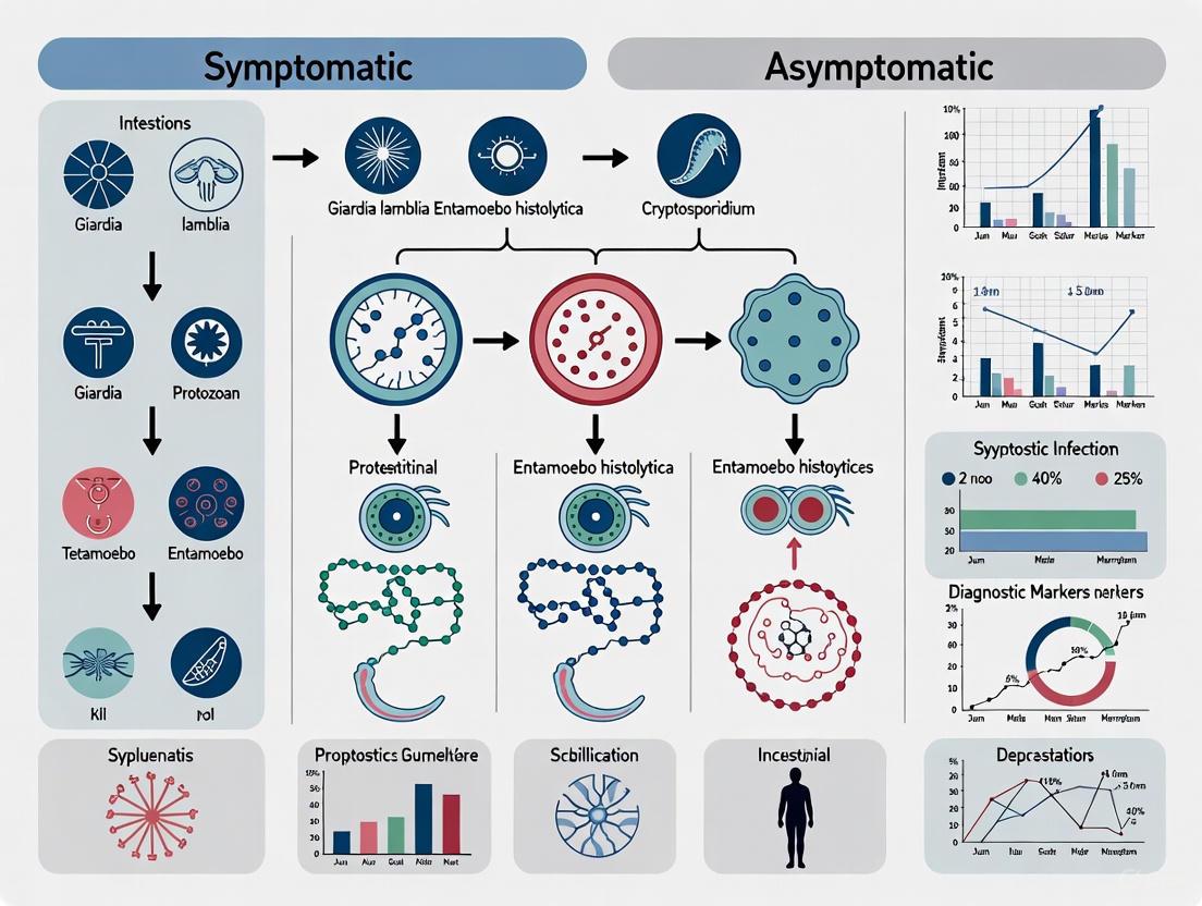
Diagram 1: Pathogenic Mechanisms of Intestinal Protozoa. This diagram illustrates the molecular mechanisms employed by Giardia, Entamoeba, and Cryptosporidium that determine the transition from asymptomatic colonization to symptomatic disease.
Diagnostic Approaches and Methodological Considerations
Comparative Diagnostic Modalities
Accurate diagnosis of intestinal protozoan infections is complicated by the overlap in clinical presentations and the existence of asymptomatic carriage. Diagnostic approaches vary significantly in their sensitivity, specificity, technical requirements, and applicability to different research and clinical settings.
Microscopy remains the most widely available diagnostic method, particularly in resource-limited settings. Conventional stool microscopy examination has poor sensitivity (<60%) for detecting Entamoeba histolytica and cannot differentiate it from non-pathogenic Entamoeba dispar [2]. Similarly, for Giardia and Cryptosporidium, microscopy sensitivity is compromised by intermittent cyst excretion and requires experienced technicians for reliable identification [1]. The primary advantage of microscopy is its ability to screen for multiple parasites simultaneously with minimal equipment requirements.
Immunoassay-based detection methods, particularly stool antigen detection tests, offer improved sensitivity and specificity compared to microscopy. For Entamoeba histolytica, commercially available antigen detection kits using enzyme-linked immunosorbent assay (ELISA), radioimmunoassay, or immunofluorescence achieve sensitivities up to 88% and have the distinct advantage of differentiating E. histolytica from E. dispar [2]. These methods provide a simple platform with quick turnaround time but may vary in performance between different commercial kits.
Molecular detection methods, particularly polymerase chain reaction (PCR)-based assays, represent the current gold standard for detection and differentiation of intestinal protozoa. Stool PCR for Entamoeba histolytica demonstrates sensitivity of 92-100% and specificity of 89-100% [2]. Molecular approaches are particularly valuable for epidemiological studies, as they enable species identification, genotyping, and analysis of genetic diversity. Real-time PCR offers increased sensitivity and efficiency, requiring only a single sample for testing multiple pathogens [1]. The limitations of molecular methods include higher cost, requirement for specialized equipment and technical expertise, and potential inhibition by stool components.
Serological testing has utility for diagnosing extraintestinal amebiasis, with high sensitivity and specificity for detecting anti-amoebic antibodies [2]. However, serology cannot distinguish between acute and past infection, as antibodies remain detectable for years after exposure. For Giardia and Cryptosporidium, serological tests have limited diagnostic value in clinical practice but can be useful tools for seroepidemiological studies.
Table 3: Comparative Analysis of Diagnostic Methods for Intestinal Protozoa
| Method | Sensitivity | Specificity | Advantages | Limitations |
|---|---|---|---|---|
| Stool Microscopy | <60% for E. histolytica [2] | Variable, requires expertise | Low cost, detects multiple parasites, widely available | Poor sensitivity, cannot differentiate species [2] |
| Antigen Detection | Up to 88% for E. histolytica [2] | High, differentiates E. histolytica from E. dispar [2] | Rapid, simple to perform, commercial kits available | Variable performance between kits |
| Molecular PCR | 92-100% for E. histolytica [2] | 89-100% for E. histolytica [2] | High sensitivity/specificity, species differentiation, genotyping | Expensive, requires specialized equipment/expertise [2] |
| Serology | High for invasive amebiasis [2] | High | Useful for extraintestinal amebiasis | Cannot distinguish current from past infection [2] |
Advanced Molecular Epidemiology Tools
Molecular epidemiological tools have revolutionized our understanding of the taxonomy, transmission dynamics, and population genetics of intestinal protozoa. For Cryptosporidium, the application of PCR-based genotyping and subtyping techniques has revealed extensive genetic diversity with implications for clinical manifestations and transmission patterns. More than 90% of human Cryptosporidium infections are caused by two species: C. hominis (anthroponotic) and C. parvum (zoonotic) [6]. Subtyping using the gp60 (cpgp40/15) gene has demonstrated associations between specific subtypes and transmission routes or disease severity [6].
For Giardia duodenalis, molecular tools have identified distinct assemblages with differing host specificities. Assemblages A and B have broad host ranges including humans, while Assemblages C-F appear to be host-adapted [9]. Studies of Giardia in dogs have revealed that 60% of infections were with zoonotic Assemblage A, 12% with dog-specific Assemblages C and D, and 28% with mixed infections [9]. This complex epidemiology highlights the importance of molecular tools for understanding transmission cycles and zoonotic potential.
Entamoeba histolytica diagnosis has been transformed by molecular methods that reliably distinguish it from the morphologically identical but non-pathogenic Entamoeba dispar. This distinction is crucial for appropriate clinical management and epidemiological studies. Molecular techniques have also enabled investigation of genetic diversity and potential associations between specific genotypes and disease outcome.
Diagram 2: Diagnostic Workflow for Intestinal Protozoa. This diagram outlines the integrated approach to diagnosis, combining traditional and advanced methods to guide appropriate therapeutic decisions.
The Scientist's Toolkit: Essential Research Reagents and Methodologies
Advancing research on intestinal protozoa requires specialized reagents, model systems, and methodological approaches. The following toolkit summarizes essential resources for investigating the biology, host-pathogen interactions, and therapeutic vulnerabilities of these significant pathogens.
Table 4: Essential Research Reagents and Resources for Intestinal Protozoan Research
| Reagent/Resource | Specifications | Research Applications | Technical Notes |
|---|---|---|---|
| Axenic Culture Media | TYI-S-33 medium for E. histolytica; Specific media formulations for each parasite | In vitro propagation, drug screening, pathogenicity studies | Requires strict anaerobic conditions for E. histolytica; Cryopreservation essential for strain maintenance |
| Species-Specific PCR Primers | Target genes: SSU rRNA, hsp60, gdh, tpi, gp60, Gal/GalNAc lectin | Species identification, genotyping, molecular epidemiology | Multiplex PCR assays available for simultaneous detection of multiple pathogens |
| Monoclonal Antibodies | Target-specific for surface antigens, virulence factors (e.g., Gal/GalNAc lectin) | Immunofluorescence, Western blot, functional inhibition studies | Commercial availability varies; validation required for each application |
| Animal Models | Neonatal mouse models for Cryptosporidium; Gerbil model for amoebic colitis/liver abscess | Pathogenesis studies, drug efficacy testing, vaccine development | Varying susceptibility across strains; immunosuppression often required |
| Gene Manipulation Tools | CRISPR/Cas9 systems, RNA interference, transfection protocols | Functional genomics, gene essentiality studies, virulence factor characterization | Technical challenges due to unique biology of each parasite; protocol optimization needed |
| Omics Databases | Genomic, transcriptomic, proteomic resources (AmoebaDB, CryptoDB, GiardiaDB) | Comparative genomics, biomarker discovery, drug target identification | Integrated platforms facilitate multi-omics approaches to parasite biology |
Experimental Protocols for Key Research Applications
In Vitro Drug Screening Protocol
Purpose: To evaluate potential therapeutic compounds against intestinal protozoa. Methodology:
- Maintain axenic cultures of target parasites (Giardia, Entamoeba, Cryptosporidium) in appropriate media under optimal conditions.
- Seed parasites in 96-well plates at standardized densities (e.g., 10^4 trophozoites/well for Giardia/Entamoeba).
- Add test compounds in serial dilutions; include appropriate controls (untreated, vehicle, reference drug).
- Incubate for 48-72 hours under optimal growth conditions.
- Assess viability using colorimetric (MTT, Alamar Blue), fluorometric, or luciferase-based assays.
- Calculate IC50 values using non-linear regression analysis.
Technical Notes: For Cryptosporidium, use HCT-8 or Caco-2 cell culture infection models as the parasite cannot be cultured axenically. Include cytotoxicity controls using mammalian cell lines.
Molecular Epidemiology and Genotyping Protocol
Purpose: To characterize genetic diversity and transmission patterns of intestinal protozoa. Methodology:
- Extract genomic DNA from stool samples or cultured isolates using commercial kits with bead-beating step.
- Perform nested PCR for species-specific markers:
- Cryptosporidium: SSU rRNA, gp60, COWP loci
- Giardia: gdh, tpi, bg genes
- Entamoeba: SSU rRNA, chitinase genes
- Purify PCR products and perform Sanger sequencing.
- Analyze sequences using phylogenetic software (MEGA, Geneious) and compare with reference sequences.
- For complex samples, consider next-generation sequencing approaches.
Technical Notes: Include appropriate controls throughout; consider multi-locus sequence typing for higher resolution; adhere to quality control measures for sequence interpretation.
The global burden of Giardia, Entamoeba, and Cryptosporidium infections remains substantial, with particularly severe impacts on children in low-resource settings and immunocompromised individuals worldwide. The dichotomy between asymptomatic carriage and symptomatic disease represents a fundamental challenge and opportunity for understanding host-parasite interactions and developing improved interventions. While significant progress has been made in characterizing the molecular pathogenesis and epidemiology of these parasites, important knowledge gaps persist.
Future research should prioritize several key areas: First, elucidating the host and parasite factors that determine the outcome of infection, with particular focus on immune correlates of protection and parasite virulence determinants. Second, developing point-of-care diagnostic tools that can accurately distinguish between pathogenic and non-pathogenic species and identify asymptomatic carriers. Third, advancing drug discovery efforts to address the limitations of current therapeutic options, particularly for cryptosporidiosis. Finally, implementing integrated control strategies that combine advances in water treatment, sanitation, health education, and potential vaccine development.
The contrasting epidemiology of these pathogens in high-income versus low-income regions highlights the complex interplay between socioeconomic factors and disease transmission. The surprising increase in Entamoeba infection-associated burden among adults and the elderly in high SDI regions [3] underscores the evolving epidemiology of these infections and the need for sustained surveillance and research. By leveraging advances in molecular technologies, immunological tools, and experimental model systems, the research community can address these challenges and work toward reducing the significant global health impact of these important intestinal protozoa.
Intestinal protozoan parasites represent a major global health burden, contributing significantly to morbidity and mortality, particularly in children within developing nations [10]. The clinical presentation of these infections spans a remarkably wide spectrum, from asymptomatic carriage to invasive, life-threatening disease [11]. This variability poses substantial challenges for diagnosis, patient management, and public health control strategies. Understanding the factors that determine a patient's position on this clinical spectrum—including parasite species, host immune status, and environmental influences—is a central focus of contemporary research [12]. Framing this understanding within the core research paradigm of symptomatic versus asymptomatic intestinal protozoan infections is critical for directing drug development efforts towards the most vulnerable populations and identifying novel therapeutic targets that can alter disease progression [13].
The Clinical Spectrum of Major Intestinal Protozoa
The progression and manifestation of intestinal protozoan infections are determined by a complex interplay between parasite virulence factors and the host's immune response. The following section details the clinical characteristics of major pathogenic protozoa.
Entamoeba histolytica
- Noninvasive Intestinal Infection: The majority of E. histolytica infections are asymptomatic, with the parasite existing as a commensal in the intestinal lumen. Some patients may experience vague gastrointestinal symptoms such as alternating periods of mild diarrhea and constipation [11].
- Intestinal Amebiasis (Amebic Colitis): Symptomatic infection typically presents with 1-3 weeks of diarrhea progressing to grossly bloody, dysenteric stools, abdominal pain, and weight loss. Fever is present in only 10-20% of cases [11].
- Fulminant or Necrotizing Colitis: A rare (0.5% of cases), severe complication marked by sudden constipation following acute dysentery, often leading to toxic megacolon and shock. This presentation is associated with inappropriate corticosteroid use [11].
- Ameboma: A granulomatous mass lesion, most often found in the cecum or ascending colon, occurring in fewer than 1% of patients. It is frequently palpable and associated with concurrent dysentery [11].
- Extraintestinal Disease: The most common extraintestinal manifestation is a liver abscess, which occurs in fewer than 10% of patients with invasive disease. Patients typically present with over 1-2 weeks of fever, abdominal pain, and anorexia, with diarrhea being present in only a minority of cases [11].
Giardia lamblia
- Asymptomatic Excretion: Asymptomatic carrier rates are high, estimated at 3-7% in the United States and over 20% in children in southern regions and childcare centers [11].
- Acute Infectious Diarrhea: Often associated with waterborne outbreaks, travel, or childcare centers. Symptoms begin within 10 days of exposure and include short-lasting acute diarrhea, nausea, abdominal distension, greasy stools, and anorexia. Spontaneous resolution is common [11].
- Chronic Diarrhea: Characterized by intermittent, loose, foul-smelling stools resembling those of malabsorption syndromes. Other symptoms include abdominal distension, sulfurous belching, flatulence, epigastric pain, nausea, anorexia, and failure to thrive. Disaccharidase deficiency (especially lactase) can lead to carbohydrate intolerance [11].
Spore-Forming Protozoa (Cryptosporidium, Cyclospora, Isospora, Microsporidia)
- Asymptomatic Infections: A well-documented part of the disease spectrum for all spore-forming protozoa in both immunocompetent and immunodeficient hosts [11].
- Acute Diarrhea in Immunocompetent Hosts: Infections can cause self-limited acute diarrhea, malaise, abdominal pain, nausea, and vomiting. The 1993 Milwaukee cryptosporidiosis outbreak affected an estimated 403,000 residents [11].
- Disease in Immunodeficient Hosts: Immunosuppressed individuals, particularly those with AIDS and low CD4 counts, experience more frequent, prolonged, and severe infections. The disease can present as transient, chronic, or fulminant diarrhea, leading to severe dehydration, malabsorption, lethargy, and malnutrition [11]. The introduction of highly active antiretroviral therapy (HAART) has reduced the frequency and severity of these infections [11].
- Extraintestinal Disease: Infection of the biliary tract can occur, leading to right upper quadrant pain, jaundice, and fever. Dissemination to the liver, respiratory tract, and kidneys has been reported for some microsporidia species [11].
Table 1: Clinical Spectrum of Major Intestinal Protozoan Parasites
| Parasite | Asymptomatic Carriage | Acute Symptomatic Infection | Chronic/Sequela | Severe/Fulminant Disease |
|---|---|---|---|---|
| Entamoeba histolytica | Very common; non-invasive infection [11] | Amebic colitis: bloody dysentery, abdominal pain, weight loss [11] | Chronic nondysenteric diarrhea, abdominal pain, flatulence [11] | Fulminant colitis (<0.5%), ameboma, liver abscess [11] |
| Giardia lamblia | High carrier rate (3-7% general US, >20% in child care) [11] | Acute diarrhea, nausea, abdominal distension, greasy stools [11] | Chronic diarrhea, malabsorption, bloating, weight loss, failure to thrive [11] | Contributes to protein-energy malnutrition, protein-losing enteropathy [11] [10] |
| Cryptosporidium spp. | Common in immunocompetent and immunodeficient hosts [11] | Self-limited watery diarrhea, abdominal cramps, nausea [11] | In immunocompetent: recurrent symptoms; In AIDS: chronic, wasting diarrheal illness [11] | Fulminant, life-threatening diarrhea & dehydration in severe immunodeficiency [11] |
| Dientamoeba fragilis | Not explicitly detailed | Abdominal pain, diarrhea, anorexia, flatulence (no bloody stools) [11] | Diarrhea lasts 1-2 weeks; abdominal pain can persist 1-2 months [11] | Less common: fever, weight loss, fatigue [11] |
| Balantidium coli | Most infections are asymptomatic [11] | Acute diarrhea with mucus and blood, resembling amebic colitis [11] | Intermittent diarrhea & constipation, abdominal pain, weight loss [11] | Fulminant and fatal course, especially in malnourished or immunodeficient [11] |
Impact on Growth and Anthropometry
Quantitative studies have delineated the specific impact of protozoan infections on child growth, revealing distinct patterns even in asymptomatic cases. Data from the Global Enteric Multicenter Study (GEMS), which analyzed over 7,800 children, demonstrates parasite-specific associations with anthropometric faltering [10].
Table 2: Association between Intestinal Protozoan Parasites and Child Anthropometric Outcomes at 60-Day Follow-up (Adapted from GEMS Data) [10]
| Parasite | Symptomatic Status | Length/Height-for-age (HAZ) | Weight-for-age (WAZ) | Weight-for-Length/Height (WHZ) |
|---|---|---|---|---|
| Giardia | Asymptomatic | β: -0.13; (95% CI: -0.17, -0.09); p<0.001 | β: -0.07; (95% CI: -0.11, -0.04); p<0.001 | β: -0.02; (95% CI: -0.06, 0.02); p=0.36 |
| Giardia | Symptomatic | No significant associations found | No significant associations found | No significant associations found |
| Cryptosporidium | Asymptomatic | β: -0.03; (95% CI: -0.09, 0.04); p=0.40 | β: -0.15; (95% CI: -0.22, -0.09); p<0.001 | β: -0.18; (95% CI: -0.25, -0.12); p<0.001 |
| Cryptosporidium | Symptomatic | β: -0.17; (95% CI: -0.23, -0.11); p<0.001 | β: -0.25; (95% CI: -0.31, -0.19); p<0.001 | β: -0.23; (95% CI: -0.30, -0.17); p<0.001 |
| Entamoeba histolytica | Both | No significant associations found with child growth [10] | No significant associations found with child growth [10] | No significant associations found with child growth [10] |
Pathogenesis and Host-Parasite Interactions
The clinical outcome of an intestinal protozoan infection is a direct result of the dynamic interplay between parasite virulence mechanisms and the host's immune defense and inflammatory responses.
Mechanisms of Parasite Pathogenicity and Immune Evasion
Protozoan parasites employ a diverse array of strategies to establish infection, cause damage, and evade host immunity [12]:
- Tissue Invasion and Direct Damage: Parasites like E. histolytica and Cryptosporidium directly invade and damage the intestinal epithelium, leading to cell death, erosion, and the classic symptoms of dysentery or secretory diarrhea [11] [14].
- Toxic Products: Some parasites release metabolic products or surface molecules that can activate complement or have other direct cytotoxic effects, contributing to tissue damage [12].
- Immune Evasion:
- Antigenic Variation: Pathogens like Giardia and Cryptosporidium can alter their surface antigens, allowing them to stay ahead of the host's humoral immune response [12] [15].
- Intracellular Location: Cryptosporidium occupies an intracellular but extracytoplasmic niche, protecting it from immune effectors [12].
- Immunosuppression: Chronic protozoan infections often induce a state of generalized host immunosuppression, which can delay parasite clearance and facilitate the emergence of antigenic variants [12].
Host Immune Response and Immunopathology
The host's immune response is a double-edged sword, essential for controlling infection but also a primary driver of pathology in many protozoan diseases [12].
- Cellular Immunity: A T-helper 1 (Th-1) response, characterized by the production of interferon-gamma (IFN-γ), is crucial for resistance to intracellular protozoa like Cryptosporidium and for controlling E. histolytica infection [12].
- Humoral Immunity: Antibody responses play a significant role in neutralizing parasites and preventing reinfection, particularly for luminal parasites like Giardia [12].
- Pathological Immune Responses:
- Immune Complex Deposition: Antigen-antibody complexes can circulate and deposit in tissues like the kidney, leading to complications such as glomerulonephritis [12].
- Autoimmunity: Cross-reactivity between parasite and host antigens can lead to the production of autoantibodies, causing damage to host tissues [12].
- Cellular Hypersensitivity: Chronic inflammation can lead to granuloma formation (e.g., in ameboma) and tissue destruction [12].
- Cytokine-Mediated Pathology: Pro-inflammatory cytokines like Tumor Necrosis Factor (TNF) are essential for parasite control but are also implicated in the cachexia, wasting, and tissue damage seen in severe and chronic infections [12].
Diagram 1: Host-parasite interactions determine the clinical spectrum of intestinal protozoan infections, ranging from asymptomatic carriage to severe immunopathology.
Research Methodologies for Differentiating Infection States
A critical requirement in protozoan parasite research is the ability to accurately diagnose infection and distinguish between asymptomatic carriage and symptomatic disease. The following experimental protocols are foundational to this field.
Stool-Based Parasite Detection and Differentiation
The definitive diagnosis of intestinal protozoa relies on the detection of the parasite, its antigens, or its genetic material in stool samples. The GEMS study provides a robust model for large-scale surveillance [10].
- Specimen Collection: Fresh stool specimens are collected from enrolled participants (both cases with moderate-to-severe diarrhea and matched healthy controls). Stool samples are aliquoted and stored at -80°C or in specific preservatives for different downstream analyses [10].
- Immunoassay Detection (ELISA):
- Principle: Commercial enzyme-linked immunosorbent assays (ELISAs) are used to detect parasite-specific antigens in stool. This method is often more sensitive and specific than traditional microscopy [10].
- Procedure: Stool suspensions are prepared and applied to ELISA plates pre-coated with capture antibodies specific for Giardia, E. histolytica, or Cryptosporidium antigens. After incubation and washing, a detector antibody conjugated to an enzyme (e.g., horseradish peroxidase) is added. A substrate is then added, and the colorimetric reaction is measured spectrophotometrically. Results are interpreted against standard curves and control samples provided by the manufacturer (e.g., TechLab, Inc.) [10].
- Molecular Detection (PCR):
- Principle: Polymerase Chain Reaction (PCR) amplifies parasite-specific DNA sequences, offering high sensitivity and the ability to speciate and genotype parasites.
- Procedure: DNA is extracted from stool samples using commercial kits designed to overcome PCR inhibitors. Genus- and species-specific primers are used to amplify target genes (e.g., Cryptosporidium oocyst wall protein gene, E. histolytica 18s rRNA gene). Amplified products are visualized by gel electrophoresis or quantified in real-time PCR systems. Multiplex PCR panels allow for the simultaneous detection of multiple pathogens.
Anthropometric Assessment in Longitudinal Studies
To quantify the long-term sequelae of infection, particularly growth faltering in children, standardized anthropometric measurements are essential [10].
- Measurement Protocol: At enrollment and follow-up visits (e.g., 60 days post-enrollment), trained field staff measure:
- Weight: Using a calibrated digital scale.
- Length/Height: For children under 24 months, recumbent length is measured on a length board; for older children, standing height is measured with a stadiometer.
- Z-Score Calculation: The raw measurements are converted into three primary z-scores using the WHO Child Growth Standards as a reference population, typically via a WHO SAS macro:
- Length/Height-for-Age (HAZ): Indicates chronic malnutrition (stunting).
- Weight-for-Age (WAZ): Indicates underweight.
- Weight-for-Length/Height (WHZ): Indicates acute malnutrition (wasting) [10].
Diagram 2: A longitudinal study design, as used in the GEMS study, is essential for linking parasite infection status with long-term outcomes like growth faltering.
The Scientist's Toolkit: Key Research Reagents and Materials
The following table details essential reagents, tools, and materials used in protozoan research, as derived from the methodologies cited.
Table 3: Essential Research Reagents and Materials for Studying Intestinal Protozoa
| Reagent/Material | Function/Application | Example/Specification |
|---|---|---|
| Commercial ELISA Kits | Detection of parasite-specific antigens (e.g., Giardia, E. histolytica, Cryptosporidium) in stool samples with high sensitivity and specificity [10]. | Kits from manufacturers like TechLab, Inc.; used according to manufacturer's protocols with included standards and controls [10]. |
| Parasite-Specific Primers | Amplification of target DNA sequences for sensitive detection, genotyping, and speciation via PCR. | Designed against conserved or species-specific genes (e.g., 18s rRNA, oocyst wall protein); requires validated sequences and optimized PCR master mixes. |
| DNA Extraction Kits | Isolation of high-quality, PCR-ready genomic DNA from complex stool matrices while removing inhibitors. | Commercially available kits (e.g., QIAamp DNA Stool Mini Kit); includes lysis buffers, proteinase K, spin columns, and wash buffers. |
| WHO Growth Standards SAS Macro | Statistical computation of anthropometric z-scores (HAZ, WAZ, WHZ) from raw weight and height data for child growth assessment [10]. | Freely available SAS macro from WHO; requires input of child's age, sex, weight, and length/height measurements [10]. |
| Cell Culture Lines (e.g., HT-29, Caco-2) | In vitro models for studying host-pathogen interactions, including parasite adhesion, invasion, cytotoxicity, and epithelial barrier function. | Human colorectal adenocarcinoma cells; cultured in appropriate media (e.g., DMEM with fetal bovine serum) under standard conditions. |
| Cryopreservation Medium | Long-term storage of parasite isolates or infected cell cultures for future experiments. | Typically contains a base medium (e.g., RPMI-1640) with a cryoprotectant like dimethyl sulfoxide (DMSO) and serum. |
Implications for Drug Development and Therapeutic Strategies
The symptomatic-asymptomatic paradigm directly informs anti-protozoan drug discovery, highlighting the need for treatments that not only clear parasites but also mitigate long-term sequelae and are effective across different patient populations [16] [17] [13].
- Challenges with Current Drugs: The existing arsenal of anti-protozoal drugs is limited, with many discovered over 50 years ago. Key limitations include significant side effects, emerging drug resistance, poor compliance due to long treatment courses, and limited efficacy in immunocompromised hosts [16].
- New Therapeutic Avenues:
- Drug Repurposing: Screening existing drugs developed for other indications for anti-protozoal activity offers a faster, more cost-effective route to new therapies. For example, antimalarial drugs have shown exciting activity against other protozoan diseases [17].
- Combination Therapies: Using drug combinations can improve efficacy, shorten treatment duration, and reduce the risk of resistance emerging [17].
- Novel Targets: Advances in parasite genomics and molecular biology are revealing unique metabolic pathways and essential enzymes in parasites that can be targeted for rational drug design [16] [13].
- Alternative Strategies: Research is exploring innovative approaches, such as the use of viruses that infect protozoan parasites (virotherapy), antimicrobial peptides, and nanoparticles as potential future therapeutics [15].
Intestinal protozoan infections present a significant global health burden, with a striking dichotomy in clinical manifestations ranging from severe symptomatic disease to complete absence of symptoms. This whitepaper synthesizes current scientific understanding of the complex host-parasite interactions that determine these divergent outcomes. The pivotal factors governing symptomatic versus asymptomatic infection include the host's immune response profile, parasite-mediated immunomodulation via extracellular vesicles, alterations in gut microbiota composition, and specific parasite virulence factors. Within the context of broader thesis research on intestinal protozoan infections, this technical guide provides comprehensive experimental methodologies, signaling pathway visualizations, and essential research tools to facilitate advanced investigation into the mechanisms driving these differential clinical outcomes, with direct implications for therapeutic and diagnostic development.
Intestinal protozoan infections, including those caused by Giardia duodenalis, Entamoeba histolytica, Cryptosporidium spp., and Blastocystis sp., affect over one billion people globally, with children and impoverished populations disproportionately impacted [18]. The clinical presentation of these infections varies remarkably, from asymptomatic carriage to severe diarrheal disease, malnutrition, and growth retardation in children [19] [18]. Understanding the mechanisms underlying this variation is crucial for developing targeted interventions.
The host-parasite interaction is a complex, dynamic relationship mediated by factors from both organisms within the gastrointestinal ecosystem. This whitepaper examines the key determinants of symptomatic versus asymptomatic outcomes, focusing on immune response modulation, parasite virulence mechanisms, gut microbiota interactions, and host genetic factors. The intricate host-parasite-microbiota axis represents a critical frontier for understanding the fundamental biology of these infections and developing novel control strategies [20].
Epidemiological Landscape of Intestinal Protozoan Infections
The global prevalence of intestinal protozoan infections highlights their significant public health impact and the variability in their clinical presentation. Table 1 summarizes prevalence data and symptomatic/asymptomatic ratios for major intestinal protozoa across different epidemiological settings.
Table 1: Prevalence and Clinical Presentation of Major Intestinal Protozoa
| Parasite | Overall Prevalence | Population | Symptomatic Cases | Asymptomatic Cases | Reference |
|---|---|---|---|---|---|
| Blastocystis spp. | 63.0% | Schoolchildren, Lebanon | 50.0% | 50.0% | [19] |
| Dientamoeba fragilis | 60.6% | Schoolchildren, Lebanon | 50.0% | 50.0% | [19] |
| Giardia duodenalis | 17.7%-37.0% | Schoolchildren, Lebanon & Palestine | 50.0% | 50.0% | [19] [21] |
| Entamoeba histolytica/dispar | 17.1% | Patients, Yemen | Not specified | Not specified | [22] |
| Cryptosporidium spp. | 1.0%-10.4% | Various | Not specified | Not specified | [19] [22] |
Molecular epidemiological studies reveal significant genetic diversity within protozoan species, contributing to variable clinical outcomes. For instance, Blastocystis sp. has multiple subtypes with potentially different pathogenic potentials [19]. Similarly, G. duodenalis comprises distinct assemblages with varying clinical implications [19]. This genetic diversity represents a key consideration when investigating determinants of symptomatic versus asymptomatic infections.
Host Immune Mechanisms in Symptom Determination
Innate Immune Recognition and Response
The host immune system employs pattern recognition receptors (PRRs), including Toll-like receptors (TLRs) and nucleotide-binding oligomerization domain (NOD)-like receptors (NLRs), to detect protozoan parasites through their microbe-associated molecular patterns (MAMPs) [20] [18]. This recognition initiates downstream immunoregulatory mechanisms that shape the subsequent immune response.
The diagram below illustrates the key innate immune signaling pathways activated upon protozoan recognition:
Immune Signaling Pathways
This initial immune activation produces inflammatory cytokines including interleukin-1 (IL-1) and tumor necrosis factor-alpha (TNF-α), which initiate inflammatory reactions [18]. The specific cytokine profile generated significantly influences whether the infection remains asymptomatic or progresses to symptomatic disease.
Adaptive Immune Responses
Adaptive immunity plays a crucial role in determining infection outcomes. Th2 cells mediate the primary adaptive immune response against intestinal parasites, producing cytokines like IL-4 and IL-13 that stimulate immunoglobulin E (IgE) production and eosinophil activation [18]. The balance between different T-helper responses (Th1, Th2, Th17) and regulatory T cells (Tregs) significantly affects symptom development.
In some cases, regulatory mechanisms activate to prevent excessive inflammation and tissue damage [18]. Parasites have evolved sophisticated evasion strategies, including the release of immunomodulatory molecules that inhibit immune cell activation, effectively dampening the immune response [18]. The successful establishment of asymptomatic infection often reflects the parasite's ability to modulate host immunity or the host's capacity to regulate inflammatory responses appropriately.
Parasite Virulence Factors and Immune Evasion Strategies
Extracellular Vesicles in Host-Parasite Communication
Extracellular vesicles (EVs), including exosomes, have emerged as critical mediators of host-parasite communication [18]. These nano-sized vesicles (30-150 nm) are produced and released by parasitic protozoa and contain diverse biomolecules including proteins, lipids, RNA, and DNA [18]. The diagram below illustrates the biogenesis and function of parasite-derived extracellular vesicles:
EV Biogenesis and Function
Protozoan parasites utilize EVs to deliver virulence factors and immunomodulatory molecules to host cells. For example:
- G. duodenalis EVs disrupt tight junctions in intestinal epithelia and promote Th1 immune responses [18]
- Blastocystis sp. EVs modulate cytokine production, increasing IL-6 and TNF-α while reducing IL-10 and IL-4 [18]
- E. histolytica EVs inhibit immune cell recruitment by downregulating STAT6 signaling and suppressing IL-4 and IL-13 [18]
- Cryptosporidium-infected epithelial cells release EVs enriched in GP60 and CpRom1 proteins that stimulate host immune responses via the TLR4/IKK pathway [18]
Excretory-Secretory Products (ESPs)
Helminths and protozoa release excretory-secretory products (ESPs) that participate in various biological processes, including immune evasion and immunomodulation [20]. These ESPs contain immunomodulatory proteins, glycoproteins, and small RNAs that can directly influence microbial composition and host immune recognition [20]. ESPs from helminths like Teladorsagia circumcincta have demonstrated antimicrobial activity that can impact bacterial growth and survival [20].
Gut Microbiota in Host-Parasite Interactions
Parasite-Induced Alterations to Microbial Communities
The gut microbiota represents a critical component of the host-parasite interaction landscape. Both clinical and experimental models demonstrate that intestinal parasites significantly impact microbial composition and diversity [20]. Table 2 summarizes documented microbiota changes associated with specific parasitic infections.
Table 2: Parasite-Induced Alterations in Gut Microbiota Composition
| Parasite | Model System | Microbiota Changes | Reference |
|---|---|---|---|
| Trichuris muris | Mouse | ↓ Bacteroidetes (Prevotella, Parabacteroides); ↑ Lactobacillus | [20] |
| Heligmosomoides polygyrus | Mouse | ↑ Lactobacillaceae (ileum) | [20] |
| Trichinella spiralis | Mouse | ↑ Proteobacteria; ↓ Bacteroidetes, Clostridiales | [20] |
| Giardia spp. | Mouse | ↑ Proteobacteria; ↓ Firmicutes, Melainabacteria | [20] |
| Helminths (general) | Human (Malaysia) | ↑ Species richness; ↑ Paraprevotellaceae | [20] |
| Nematodes | Human (Sri Lanka) | ↑ Verrucomicrobiaceae, Enterobacteriaceae | [20] |
These parasite-induced alterations to the gut microbiota can either promote or protect against symptomatic disease, depending on the specific shifts and their functional consequences for the host.
Microbiota Influence on Parasite Establishment
In a reciprocal relationship, the commensal gut microbiota can influence parasitic survival, colonization, and expulsion [20]. The gut microbiota provides direct competition and protection from pathogenic organisms through various mechanisms [20]. The microbiota also shapes host immune development and function, indirectly affecting parasite control [23]. The baseline composition of an individual's gut microbiome may therefore predispose to either symptomatic or asymptomatic infection outcomes.
Experimental Methodologies for Investigating Host-Parasite Interactions
Parasite Detection and Characterization Protocols
Advanced diagnostic approaches are essential for accurately differentiating parasitic infections and correlating infection with clinical outcomes. The diagram below outlines a comprehensive workflow for detecting and characterizing intestinal protozoan infections:
Parasite Detection Workflow
Standard Stool Examination Protocol:
- Macroscopic examination: Assess color, consistency, presence of blood or mucus [22]
- Microscopic examination:
- Molecular characterization:
Immune Profiling Techniques
Comprehensive immune profiling is essential for understanding the differential responses in symptomatic versus asymptomatic infections. Key methodologies include:
- Cytokine measurement: ELISA or multiplex assays to quantify IL-4, IL-13, IL-6, TNF-α, IL-10, and other relevant cytokines
- Flow cytometry: Immunophenotyping of immune cell populations in mucosal and systemic compartments
- Transcriptomic analysis: RNA sequencing to identify differentially expressed genes in host cells
- EV characterization: Isolation of extracellular vesicles via ultracentrifugation or commercial kits, followed by proteomic and RNA analysis [18]
Essential Research Reagents and Tools
Table 3: Research Reagent Solutions for Investigating Host-Parasite Interactions
| Reagent/Tool | Application | Function/Utility | Examples |
|---|---|---|---|
| Pattern Recognition Receptor Ligands | Immune activation studies | Activate specific PRRs to study downstream signaling | TLR agonists, NOD ligands |
| Cytokine Detection Antibodies | Immune profiling | Quantify cytokine production in response to infection | ELISA kits, multiplex arrays |
| EV Isolation Kits | Parasite communication studies | Isolate extracellular vesicles for cargo analysis | Ultracentrifugation kits, precipitation kits |
| Parasite Culture Systems | In vitro studies | Maintain parasites for experimental infection models | Axenic culture media, cell co-culture systems |
| Gene Silencing Tools | Functional studies | Investigate specific gene function in parasites or host | RNA interference, CRISPR-Cas9 |
| Germ-Free Animals | Microbiota studies | Define microbiota role in infection outcomes | Gnotobiotic mouse models |
| 16S rRNA Sequencing | Microbiome analysis | Characterize microbiota composition changes | Next-generation sequencing platforms |
The determination of symptomatic versus asymptomatic outcomes in intestinal protozoan infections represents a complex interplay between host immunity, parasite virulence factors, and the gut microbiota. Key factors include the quality and magnitude of the host immune response, particularly the balance between inflammatory and regulatory pathways; parasite-derived immunomodulatory molecules, especially those delivered via extracellular vesicles; and parasite-induced alterations to the gut microbial ecosystem. Future research should focus on integrating multi-omics approaches to develop comprehensive predictive models of infection outcomes, with the goal of identifying novel therapeutic targets and diagnostic biomarkers that can guide personalized intervention strategies for these significant global health challenges.
Within the complex epidemiology of intestinal protozoan infections, the dichotomy between symptomatic and asymptomatic presentations presents a critical research challenge, particularly in understanding host-parasite interactions and long-term health sequelae. Certain demographic groups, including young children, immunocompromised individuals, and indigenous communities, demonstrate heightened vulnerability to both the immediate and chronic manifestations of these infections. This whitepaper examines the distinct yet often overlapping risk profiles of these populations through analysis of recent epidemiological data, exploration of underlying biological mechanisms, and synthesis of standardized methodological approaches for investigating the symptomatic-asymptomatic infection spectrum. The focus extends beyond mere prevalence to encompass the nuanced consequences of subclinical infections, particularly their contribution to growth faltering, nutritional deficiencies, and microbiota alterations, which collectively represent a significant yet often underestimated public health burden.
Epidemiological Landscape and Burden of Disease
Prevalence Across High-Risk Populations
Intestinal protozoan infections disproportionately affect specific demographic groups, with prevalence rates reflecting intersecting vulnerabilities related to age, immune status, socioeconomic factors, and environmental conditions. The quantitative burden of disease, as established by recent global studies, is summarized in Table 1.
Table 1: Prevalence of Intestinal Protozoan Infections in High-Risk Populations
| Population | Location | Protozoan Species | Prevalence | Study Reference |
|---|---|---|---|---|
| Under-5 Children | Sub-Saharan Africa & South Asia | Giardia | 18.7% (symptomatic) | [24] |
| Under-5 Children | Sub-Saharan Africa & South Asia | Cryptosporidium | 14.2% (symptomatic) | [24] |
| Under-5 Children | Sub-Saharan Africa & South Asia | E. histolytica | 14.2% (symptomatic) | [24] |
| Indigenous Communities | Argentina (Mbyá-Guaraní) | Giardia duodenalis | 33.3% | [25] |
| Indigenous Communities | Argentina (Mbyá-Guaraní) | Any intestinal parasite | 87.8% | [25] |
| Indigenous Communities | Malaysia | Any intestinal protozoa | 27.0% | [5] [26] |
| General Population | Malaysia | Entamoeba spp. | 18.0% | [5] [26] |
| General Population | Malaysia | Giardia lamblia | 11.0% | [5] [26] |
| General Population | Malaysia | Cryptosporidium spp. | 9.0% | [5] [26] |
| Symptomatic Patients | Southern Algeria | Blastocystis spp. | 43.8% | [27] [28] |
| Symptomatic Patients | Southern Algeria | E. histolytica/dispar | 25.4% | [27] [28] |
| Asymptomatic Patients | Southern Algeria | Any intestinal protozoa | 14.9% | [27] [28] |
Health Impacts of Symptomatic vs. Asymptomatic Infections
The clinical spectrum of intestinal protozoan infections ranges from acute symptomatic disease to chronic subclinical infections, with both states exerting significant health impacts, particularly in vulnerable populations.
Growth and Nutritional Consequences: Among children, even asymptomatic infections can incur substantial health penalties. A large-scale study analyzing data from 7,800 children in the Global Enteric Multicenter Study (GEMS) found significant negative associations between subclinical protozoan infections and anthropometric outcomes [10]. Asymptomatic Giardia infection was associated with deficits in length/height-for-age z-scores (HAZ) [β: -0.13; 95% CI: -0.17, -0.09] and weight-for-age z-scores (WAZ) [β: -0.07; 95% CI: -0.11, -0.04] [10]. Similarly, asymptomatic Cryptosporidium infection was associated with deficits in WAZ [β: -0.15; 95% CI: -0.22, -0.09] and weight-for-length/height z-scores (WHZ) [β: -0.18; 95% CI: -0.25, -0.12] [10]. These findings underscore that the absence of diarrheal symptoms does not equate to absence of harm, particularly concerning linear growth and weight gain in developing children.
Multiparasitism and Nutritional Double Burden: Indigenous communities often experience high rates of multiparasitism, which compounds health impacts. Research among Mbyá-Guaraní children in Argentina revealed a multiparasitism prevalence of 70%, with some children hosting up to six different parasite species simultaneously [25]. This parasitic burden exists within a context of "double malnutrition," where stunting (prevalence: 38.9%) coexists with overweight/obesity (prevalence: 41.0%) [25]. This complex nutritional landscape creates challenging physiological environments where the metabolic consequences of parasitic infections may be amplified or modified.
Risk Factor Analysis
The transmission dynamics and burden of intestinal protozoan infections are driven by interconnected environmental, socioeconomic, and behavioral determinants. Table 2 synthesizes key risk factors identified across multiple studies.
Table 2: Risk Factors for Intestinal Protozoan Infection in High-Risk Populations
| Risk Factor Category | Specific Factor | Associated Population(s) | Effect Measure/Prevalence |
|---|---|---|---|
| Water, Sanitation & Hygiene (WASH) | Absence of functional toilet | Under-5 children, Indigenous communities | AOR = 1.95 (95% CI: 1.20-3.19) [24] |
| Water, Sanitation & Hygiene (WASH) | Handwashing without soap/ash | Under-5 children | AOR = 3.05 (95% CI: 1.20-7.75) [24] |
| Water, Sanitation & Hygiene (WASH) | Untreated water consumption | Multiple populations | 38-52% higher prevalence [5] [26] |
| Socioeconomic | Maternal illiteracy/no formal education | Under-5 children | AOR = 2.80 (95% CI: 1.67-4.71) [24] |
| Socioeconomic | Low income/poverty | Multiple populations | 38-52% higher prevalence [5] [26] |
| Environmental & Geographic | Animal contact | Multiple populations | Primary risk factor [27] [28] |
| Environmental & Geographic | Rural residence | Asymptomatic populations | Significant association [27] [28] |
| Environmental & Geographic | Hot season | Symptomatic populations | Increased trend [27] [28] |
| Demographic | Age (<15 years) | Children | 38-52% higher prevalence [5] [26] |
| Demographic | Male sex | Multiple populations | 38-52% higher prevalence [5] [26] |
The interrelationship between these risk factors and infection outcomes can be visualized as a complex network of contributing variables:
Biological Mechanisms of Vulnerability
Microbiota-Parasite Interactions
The gut microbiota represents a critical interface in host-parasite interactions, with emerging evidence suggesting that protozoan infections significantly reshape bacterial communities. A large study of 1,204 children in Guinea-Bissau demonstrated that the fecal bacterial microbiota is shaped more profoundly by protozoan infections than by helminths [29]. Specifically, infections with Entamoeba histolytica (R² = 0.0164, P = 0.0001) and Giardia lamblia (R² = 0.00676, P = 0.0001) significantly altered overall community composition [29]. Researchers identified 31 bacterial genera across four major phyla that were differentially abundant in protozoan-infected individuals, including increased abundance of Prevotella, Campylobacter, and two Clostridium clades, and decreased abundance of Collinsella, Lactobacillus, Ruminococcus, Veillonella, and one Clostridium clade [29]. These perturbations to the microbial ecosystem may contribute to both infection susceptibility and the nutritional consequences observed in asymptomatic children.
Immunological Aspects
Immunocompromised individuals, particularly those with HIV/AIDS, demonstrate heightened susceptibility to severe outcomes from certain intestinal protozoa. Cryptosporidiosis causes particularly severe disease in immunocompromised individuals [5] [26]. The increased prevalence and clinical severity observed in immunocompromised hosts underscores the critical role of intact immunity in controlling protozoan infections. This vulnerability extends beyond HIV/AIDS to include other immunocompromised states such as transplantation recipients and individuals undergoing immunosuppressive therapies, though the search results provided limited specific data on these subpopulations.
Methodological Framework for Research
Diagnostic Approaches and Experimental Protocols
Comprehensive detection of intestinal protozoa requires multimodal diagnostic approaches due to limitations in sensitivity and specificity of individual methods. The following workflow illustrates a recommended diagnostic protocol for differentiating symptomatic and asymptomatic infections in research settings:
Detailed Experimental Protocols:
Stool Collection and Initial Processing:
- Collect fresh fecal specimens in clean, wide-mouth plastic containers
- Perform direct microscopic examination with normal saline (0.9%) and Lugol's iodine preparations immediately after collection
- Examine for motile trophozoites, cysts, oocysts, and other parasitic forms
Concentration Techniques:
Staining Methods:
- Modified Ziehl-Neelsen Stain: For detection of cryptosporidial oocysts; fix smear with methanol, stain with carbol fuchsin, decolorize, counterstain with methylene blue [27]
- Trichrome Stain: For permanent mounts and enhanced visualization of protozoan internal structures
Immunoassays:
- Commercial ELISA kits (e.g., TechLab, Inc.) for detection of Giardia, E. histolytica, and Cryptosporidium antigens
- Perform according to manufacturer's protocols with appropriate controls [10]
In Vitro Culture:
- For Blastocystis spp.: Inoculate stool samples into modified Boeck and Drbohlav's Locke-egg serum medium supplemented with 10% horse serum
- Incubate at 37°C and examine for growth on day 3 post-inoculation [27]
Anthropometric Assessment in Longitudinal Studies
For growth faltering studies, standardize anthropometric measurements using WHO protocols:
- Measure weight using calibrated digital scales with minimal clothing
- Measure length/height using standardized boards/stadiometers
- Calculate z-scores using WHO Child Growth Standards macro
- Schedule follow-up measurements at 60 days (±10 days) post-enrollment [10]
The Scientist's Toolkit: Research Reagent Solutions
Table 3: Essential Research Reagents for Intestinal Protozoan Investigation
| Reagent/Kit | Application | Specifications | Research Utility |
|---|---|---|---|
| TechLab ELISA Kits | Antigen detection of Giardia, Cryptosporidium, E. histolytica | Species-specific monoclonal antibodies | High-throughput screening; differentiation of pathogenic vs. non-pathogenic strains [10] |
| Boeck & Drbohlav's Locke-egg Serum Medium | In vitro culture of Blastocystis | Modified with 10% horse serum | Isolation and propagation of viable organisms; sensitivity assessment [27] |
| Formalin-Ether Concentration Reagents | Parasite concentration | 10% formalin, diethyl ether | Enhanced detection sensitivity; preservation of cyst morphology [27] [24] |
| Modified Ziehl-Neelsen Stain | Cryptosporidium oocyst detection | Carbol fuchsin, acid-alcohol, methylene blue | Identification of acid-fast oocysts; differential diagnosis [27] |
| WHO Anthropometry Kit | Growth assessment | Digital scales, stadiometers, MUAC tapes | Standardized measurement of nutritional outcomes [10] |
| DNA/RNA Shield Fecal Collection System | Microbiome studies | Stabilization buffer, collection tubes | Preservation of nucleic acids for microbiota analysis [29] |
| 16S rRNA Gene Sequencing Primers | Microbiota profiling | V3-V4 hypervariable region | Assessment of infection-associated dysbiosis [29] |
The investigation of high-risk populations for intestinal protozoan infections reveals complex interactions between pathogen biology, host vulnerability, and environmental determinants. Children under five years bear the greatest burden of both symptomatic disease and growth-limiting subclinical infections, with Giardia and Cryptosporidium representing particular concerns for linear growth and weight gain, respectively. Indigenous communities experience striking rates of multiparasitism within contexts of socioeconomic marginalization and inadequate WASH infrastructure, creating cycles of infection and nutritional compromise that demand culturally adapted interventions. Immunocompromised individuals face elevated risks of severe disease manifestations, though this population remains understudied in many high-prevalence settings. Critical research gaps include the need for improved diagnostic differentiation of pathogenic and non-pathogenic protozoan species, longitudinal studies examining the long-term developmental consequences of childhood infections, and clinical trials evaluating whether targeted antiprotozoal interventions in asymptomatic children can improve growth trajectories. The methodological framework presented herein provides a standardized approach for advancing research on the symptomatic-asymptomatic infection spectrum, with particular utility for pharmaceutical and public health researchers developing targeted interventions for these vulnerable populations.
The Role of Non-Pathogenic Species and Commensals in Diagnostic Confusion
Within the human intestinal tract, a diverse community of protozoa exists, comprising pathogenic, non-pathogenic, and commensal species. This biological complexity presents a significant challenge in clinical and research settings, where the accurate identification of the true etiological agent of disease is paramount. Non-pathogenic intestinal protozoa are single-celled parasites commonly found in the intestinal tract that are never associated with illness, even in immunocompromised individuals [30]. In contrast, the role of commensal protists, long-neglected members of the gut microbiome, is now being re-evaluated, with emerging evidence highlighting their powerful effects on host immunity and gut ecology [31]. The diagnostic confusion arising from these organisms stems from their morphological similarity to pathogenic species, their high prevalence in both symptomatic and asymptomatic populations, and an incomplete understanding of their potential contributions to health and disease. This whitepaper examines the sources of this diagnostic ambiguity within the context of symptomatic versus asymptomatic intestinal protozoan infections and provides guidance for accurate laboratory differentiation.
Problem Scope: Species Responsible for Diagnostic Challenges
Several groups of intestinal protozoa contribute to diagnostic confusion due to morphological similarities, varying pathogenic potential, and differences in clinical presentation.
Table 1: Protozoan Species Causing Diagnostic Confusion
| Category | Species | Pathogenic Status | Key Diagnostic Challenge |
|---|---|---|---|
| Non-Pathogenic Amoebae | Entamoeba coli, Endolimax nana, Entamoeba hartmanni, Iodamoeba bütschlii | Non-pathogenic [30] | Morphological similarity to Entamoeba histolytica in microscopy |
| Commensal Protists | Tritrichomonas musculis (murine model), Blastocystis spp., Dientamoeba fragilis | Debated/Commensal [31] [32] | High prevalence in asymptomatic individuals; pathogenicity debated |
| Pathogenic Species with Non-Pathogenic Lookalikes | Entamoeba histolytica vs Entamoeba dispar | Pathogenic vs Non-pathogenic [33] | Morphologically identical; require molecular differentiation |
| Opportunistic Pathogens | Cryptosporidium spp., Blastocystis spp. (certain subtypes) | Context-dependent [10] [33] | Can be found in asymptomatic carriers; disease manifestation depends on host immune status |
Non-Pathogenic Intestinal Protozoa
The Centers for Disease Control and Prevention (CDC) specifically identifies several non-pathogenic intestinal protozoa that are never associated with illness, including Chilomastix mesnili, Endolimax nana, Entamoeba coli, and Entamoeba hartmanni [30]. The detection of these organisms in stool indicates past fecal exposure but does not explain current gastrointestinal symptoms, necessitating further investigation for other causes [30].
Commensal Protists with Debated Pathogenicity
The status of several protists remains controversial. Blastocystis spp. and Dientamoeba fragilis are frequently detected in both symptomatic and asymptomatic individuals, complicating clinical interpretation [33] [32]. Their pathogenicity may be subtype-dependent, with certain genetic variants possessing unique virulence traits [33]. Furthermore, commensal protists like Tritrichomonas musculis (in murine models) can actively modulate host immunity, influencing susceptibility to both infectious and inflammatory diseases [31].
Pathogenic Species with Non-Pathogenic Counterparts
The most significant diagnostic challenge involves differentiating the pathogenic Entamoeba histolytica from the morphologically identical but non-pathogenic Entamoeba dispar [33]. Without advanced diagnostic techniques, this distinction is impossible, potentially leading to misdiagnosis and unnecessary treatment.
Quantitative Data: Prevalence in Symptomatic vs. Asymptomatic Populations
Epidemiological studies revealing the carriage of putative pathogens in healthy populations underscore the diagnostic dilemma. The following table synthesizes key findings from recent research.
Table 2: Prevalence of Protozoan Infections in Symptomatic vs. Asymptomatic Populations
| Protozoan Species | Prevalence in Symptomatic/Diarrheal Cases | Prevalence in Asymptomatic/Non-Diarrheal Cases | Statistical Association (OR/p-value) | Study Details |
|---|---|---|---|---|
| Entamoeba histolytica | 11.9% (7/59) [34] | 0.7% (3/448) [34] | OR = 19.9 (95% CI: 5.0-79.5) [34] | Hospital-based study, China (n=507) [34] |
| Giardia duodenalis | 6.8% (4/59) [34] | 1.6% (7/448) [34] | OR = 4.6 (95% CI: 1.3-16.2) [34] | Hospital-based study, China (n=507) [34] |
| Blastocystis sp. | 13.6% (8/59) [34] | 8.9% (40/448) [34] | OR = 1.6 (95% CI: 0.7-3.6), Not Significant [34] | Hospital-based study, China (n=507) [34] |
| Cryptosporidium spp. | N/A | N/A | Negative association with HAZ, WAZ, WHZ in symptomatic children [10] | GEMS study (n=7,800) [10] |
| Giardia spp. | N/A | N/A | Negative association with HAZ, WAZ in asymptomatic children [10] | GEMS study (n=7,800) [10] |
Analysis of this data reveals crucial patterns. Both Entamoeba histolytica and Giardia duodenalis show statistically significant associations with symptomatic disease [34]. In contrast, Blastocystis sp. demonstrates no significant difference in prevalence between symptomatic and asymptomatic groups, supporting its potential role as a commensal in many individuals [34]. Beyond acute symptoms, longitudinal studies reveal that even asymptomatic infections with parasites like Giardia and Cryptosporidium can have long-term consequences, including growth shortfalls in children, highlighting the complexity of defining true pathogenicity [10].
Molecular Diagnostics and Differentiation Protocols
Accurate differentiation between pathogenic and non-pathogenic protozoa requires moving beyond traditional microscopy to advanced molecular and immunological techniques.
Experimental Protocol: Multiplex Real-Time PCR for Protozoan Detection
Molecular diagnostic technologies, particularly real-time PCR (RT-PCR), are gaining traction due to enhanced sensitivity and specificity [35]. The following protocol is adapted from recent multicentre evaluations.
Objective: To simultaneously detect and differentiate major pathogenic and non-pathogenic intestinal protozoa in stool samples. Sample Preparation:
- Collection: Collect fresh stool sample or preserve in appropriate media (e.g., Para-Pak, S.T.A.R Buffer) [35] [32]. Preservation improves DNA yield.
- Homogenization: Suspend approximately 1 µL of stool in 350 µL of Stool Transport and Recovery Buffer (S.T.A.R Buffer) [35].
- DNA Extraction: Use automated nucleic acid extraction systems (e.g., MagNA Pure 96 System, Hamilton MICROLAB STARlet) with commercial kits (e.g., MagNA Pure 96 DNA and Viral NA Small Volume Kit). Include an internal extraction control to monitor extraction efficiency [35] [32].
PCR Amplification:
- Reaction Mix: For each reaction, combine:
- 5 µL of extracted DNA template
- 12.5 µL of 2× TaqMan Fast Universal PCR Master Mix
- 2.5 µL of primer/probe mix (targeting G. duodenalis, Cryptosporidium spp., E. histolytica, D. fragilis, Blastocystis spp.)
- Sterile water to a final volume of 25 µL [35]
- Amplification Protocol: Run on a real-time PCR instrument (e.g., ABI 7900HT, Bio-Rad CFX96) using cycling conditions:
- Initial Denaturation: 95°C for 10 minutes
- 45 Cycles of:
- Denaturation: 95°C for 15 seconds
- Annealing/Extension: 60°C for 1 minute [35]
Analysis:
- Analyze amplification curves. Cycle threshold (Cq) values ≤40 are typically considered positive [32].
- Use software (e.g., Seegene Viewer) for automated interpretation [32].
Complementary and Alternative Diagnostic Methods
- Immunoassays: Enzyme-linked immunosorbent assay (ELISA) and immunochromatographic tests (ICT) are suitable for rapid screening of specific pathogens like Giardia, Cryptosporidium, and E. histolytica [33] [36]. A key advantage is the ability of some E. histolytica-specific tests to detect the pathogenic Gal/GalNAc lectin antigen, differentiating it from E. dispar [33].
- Microscopy: While remaining a common method, especially in resource-limited settings, microscopy has limited sensitivity and specificity and cannot differentiate between morphologically identical species like E. histolytica and E. dispar [33]. Its utility lies in providing a broad overview and detecting parasites not targeted by molecular panels, such as helminths and Cystoisospora belli [32].
The Scientist's Toolkit: Essential Research Reagents
Research into the biological roles of commensal and non-pathogenic protozoa requires specialized tools and assays.
Table 3: Key Research Reagent Solutions for Intestinal Protozoa Studies
| Reagent / Solution | Primary Function | Specific Application Examples |
|---|---|---|
| Seegene AllPlex GIP Assay | Commercial multiplex qPCR for simultaneous detection of 6 protozoa | Detection of G. intestinalis, Cryptosporidium spp., E. histolytica, D. fragilis, Blastocystis spp., Cyclospora spp. in stool DNA [32] |
| TechLab ELISA Kits | Immunoassay for detection of parasite-specific antigens | Detection of Giardia, E. histolytica (lectin antigen), and Cryptosporidium in stool specimens [10] |
| S.T.A.R Buffer (Roche) | Stool transport and DNA stabilization | Preservation of stool samples for optimal DNA extraction and subsequent molecular analysis [35] |
| MagNA Pure 96 System (Roche) | Automated nucleic acid extraction | High-throughput, standardized DNA extraction from complex stool samples [35] |
| Anti-mouse IgA ELISA | Quantification of immunoglobulin A in serum/mucosa | Measurement of T cell-dependent IgA response following commensal protist colonization (e.g., T. musculis) [37] |
| Flow Cytometry Antibody Panels (Anti-CD45, CD4, CD138, B220, IgA) | Immunophenotyping of immune cell populations | Analysis of germinal center B cells, T follicular helper cells, and IgA+ plasma cells in mucosal tissues [37] |
Biological Mechanisms: Commensal Protozoa and Host Immunity
Emerging research on commensal protists reveals complex interactions with the host immune system that may indirectly contribute to diagnostic confusion by altering host susceptibility and symptom presentation.
Immunomodulatory Pathways of Commensal Protozoa
Studies in murine models have elucidated specific mechanisms through which commensals like Tritrichomonas musculis (T.mu) influence host immunity:
- Enhancement of IgA Production: Colonization with T.mu promotes T cell-dependent IgA class-switch recombination and accumulation of IgA-secreting plasma cells in the gut. This process requires ICOS:ICOSL co-stimulation and MHCII expression on B cells, expanding T follicular helper cells and germinal center B cells [37]. This enhanced IgA response can modify the bacterial microbiota and improve responses to oral antigens [37].
- Activation of Type 2 Immunity: T.mu triggers a tuft cell-ILC2 circuit via the release of succinate, leading to intestinal remodeling and systemic effects [31] [38]. This activation provides protection against helminths but can also exacerbate inflammatory conditions.
- Remote Regulation of Pulmonary Immunity: Gut colonization by T.mu instills a tripartite network between gut-derived ILC2s, lung-resident T cells, and B cells, leading to lung eosinophilia. This network exacerbates allergic airway inflammation while conversely hindering systemic dissemination of Mycobacterium tuberculosis [38].
The distinction between pathogenic, non-pathogenic, and commensal intestinal protozoa is critical for accurate diagnosis, appropriate treatment, and meaningful research. Diagnostic confusion arises from morphological similarities, the high prevalence of certain protists in asymptomatic individuals, and a growing understanding that some "commensals" actively modulate host physiology. Resolution of this confusion requires integrating advanced molecular diagnostics capable of specific differentiation with a nuanced clinical interpretation that considers host immune status and the potential for long-term sequelae beyond acute diarrhea. Future research must focus on elucidating the mechanisms of commensal-protist-host interactions, defining genetic virulence factors within species of debated pathogenicity, and developing standardized, accessible diagnostic algorithms that minimize misdiagnosis and unnecessary treatment while correctly identifying true disease-causing agents.
Beyond the Microscope: Advanced Diagnostic Tools for Differentiating Infection and Disease
Within the context of research on symptomatic versus asymptomatic intestinal protozoan infections, accurate diagnosis is the cornerstone of reliable findings. For over a century, conventional microscopy has been the foundational method for parasitic diagnosis in clinical and research settings [39]. However, this traditional approach presents significant limitations that directly impact research quality, particularly when studying the nuanced differences between symptomatic and asymptomatic carriage of pathogens like Entamoeba histolytica, Giardia duodenalis, and Cryptosporidium spp. [35]. These limitations affect not only prevalence data but also the fundamental understanding of disease progression and pathogen-specific effects.
The diagnostic challenges are especially pronounced in asymptomatic cases, where parasite burden is typically low, and in differentiating pathogenic from non-pathogenic species that are morphologically identical [40]. This technical brief examines the critical limitations of conventional microscopy through the lens of research needs, providing quantitative comparisons and methodological insights to guide researchers in selecting appropriate diagnostic approaches for intestinal protozoan infection studies.
Quantitative Limitations: Sensitivity and Specificity
Comparative Sensitivity Across Parasites
Microscopy's sensitivity varies considerably across parasite species and is significantly influenced by infection intensity. The table below summarizes documented sensitivity ranges for common intestinal protozoa using different microscopic techniques.
Table 1: Sensitivity of Microscopy-Based Diagnosis for Intestinal Protozoa
| Parasite | Microscopy Method | Sensitivity Range | Key Limitations | References |
|---|---|---|---|---|
| Giardia duodenalis | Direct wet mount | ~37.9% - 85.7% | Low infection intensity, intermittent shedding | [41] |
| Entamoeba histolytica/dispar | Direct wet mount | Cannot differentiate species | Morphologically identical to non-pathogenic E. dispar | [40] [35] |
| Cryptosporidium spp. | Formol-ether concentration | Limited sensitivity | Small oocyst size, requires special stains | [35] |
| Dientamoeba fragilis | Routine microscopy | Often missed | Trophozoites degrade rapidly, requires permanent stains | [35] |
| Soil-transmitted helminths | Kato-Katz | Lower for low-intensity infections | Less sensitive for STH with low intensity infections | [41] |
The data reveal that microscopy performs acceptably for high-intensity symptomatic infections but becomes progressively unreliable for detecting low-level parasitic carriage common in asymptomatic cases or post-treatment monitoring [41]. This sensitivity limitation directly impacts research on the symptomatic-asymptomatic dichotomy, as low-level infections may be systematically missed in control groups, leading to erroneous conclusions.
Specificity Challenges in Species Differentiation
The specificity limitations of microscopy present perhaps more critical challenges for research requiring precise pathogen identification:
- Entamoeba histolytica vs. dispar: Microscopy cannot differentiate the pathogenic E. histolytica from the non-pathogenic E. dispar, despite their dramatically different clinical implications [40] [35]. This fundamental limitation confounds research aiming to correlate infection status with clinical outcomes.
- Cryptosporidium species: While microscopy can detect Cryptosporidium oocysts, it cannot differentiate between human-pathogenic and non-pathogenic species, limiting insights into species-specific disease mechanisms [35].
- Blastocystis subtypes: Microscopy can detect Blastocystis but provides no information on subtypes, which may have different pathogenic potential and clinical significance in research cohorts [40].
These specificity issues mean that studies relying solely on microscopy likely misclassify participants in symptomatic versus asymptomatic groups, potentially attributing symptoms to non-pathogenic parasites or missing associations with truly pathogenic species.
Methodological Comparisons: Microscopy vs. Molecular Approaches
Standard Microscopy Protocols
The following experimental protocols represent commonly used microscopy methods in parasitology research.
Protocol 1: Direct Wet Mount Microscopy
- Sample Preparation: Emulsify a small portion of stool (approximately 2 mg) in one drop of 0.9% saline on a microscopic slide.
- Slip Preparation: Apply a cover glass (22 × 22 mm).
- Microscopic Examination: Systematically scan the entire cover slip area under 10× and 40× objectives.
- Identification: Identify protozoan trophozoites and cysts based on morphological characteristics, size, and motility.
- Limitations: This method has low sensitivity, detects only moderate to high infection intensities, and cannot differentiate pathogenic species [41].
Protocol 2: Formol-Ether Concentration Technique (FECT)
- Fixation: Add approximately 1 g of stool to 7 mL of 10% formalin in a 15 mL conical tube, then mix thoroughly.
- Filtration: Filter the suspension through a sieve (500-gauge mesh) into a new centrifuge tube.
- Solvent Addition: Add 4 mL of diethyl ether to the filtered solution.
- Centrifugation: Securely cap and shake the tube, then centrifuge at 300 × g for 1-2 minutes.
- Examination: Discard the top layers (ether, plug debris, formalin), prepare a smear from the sediment, and examine under a microscope [41].
- Advantages: This concentration method improves detection sensitivity compared to direct wet mounts but remains unable to differentiate morphologically identical species [41].
Molecular Diagnostic Protocols
Molecular methods, particularly real-time PCR (RT-PCR), have emerged as superior alternatives for research requiring high sensitivity and specificity.
Protocol 3: Multiplex Real-Time PCR for Intestinal Protozoa
- DNA Extraction:
- Suspend 200 mg of stool in PBS with 2% polyvinylpolypyrrolidone (PVPP) and freeze at -20°C overnight.
- Boil samples for 10 minutes at 100°C.
- Extract DNA using automated systems (e.g., MagnaPure LC.2) with "DNA I Blood_Cells High performance II" protocol.
- Elute DNA in 100 μL volume [40].
PCR Reaction Setup:
- Prepare 25 μL reactions containing PCR buffer, BSA (2.5 μg), and primer-probe mixes.
- Use primer concentrations of 100-300 nM depending on target parasites.
- Include internal control (e.g., Phocine Herpes Virus) to monitor inhibition and extraction efficiency [40].
Amplification Parameters:
- Initial denaturation: 95°C for 3 minutes
- 40 cycles of: 95°C for 15 seconds, 60°C for 30 seconds, 72°C for 30 seconds [40]
Target Genes:
- Entamoeba histolytica/dispar: SSU rRNA gene
- Giardia duodenalis: SSU rRNA gene
- Blastocystis sp.: 18S ribosomal RNA gene
- Dientamoeba fragilis: SSU rRNA gene [40]
Advantages: This method enables specific differentiation of pathogenic parasites, detects multiple targets simultaneously, and demonstrates significantly higher sensitivity than microscopy, particularly for low-intensity infections [40] [35].
The following diagram illustrates the diagnostic and research workflow comparing microscopy and molecular approaches for intestinal protozoan infection studies:
Research Implications and Methodological Recommendations
Impact on Symptomatic vs. Asymptomatic Research
The diagnostic limitations of conventional microscopy have profound implications for research on symptomatic versus asymptomatic intestinal protozoan infections:
- Misclassification Bias: In a 2022 study, microscopy failed to differentiate pathogenic E. histolytica from non-pathogenic E. dispar, potentially leading to systematic misclassification in research studies comparing symptomatic and asymptomatic cohorts [41].
- Detection Gap in Asymptomatic Carriers: Research indicates that asymptomatic individuals typically have lower parasite burdens, falling below microscopy's detection threshold of 10,000-100,000 parasites per gram of feces [40]. This creates a systematic detection bias where asymptomatic carriers are underrepresented.
- Epidemiological Distortions: A 2025 meta-analysis found a 24% pooled prevalence of intestinal protozoal infections in Malaysia, but noted that microscopy-based studies likely underestimated true prevalence due to inability to differentiate species and detect low-intensity infections [26].
Essential Research Reagent Solutions
Table 2: Essential Research Reagents for Advanced Protozoan Detection
| Reagent/Kit | Application | Research Function | Considerations |
|---|---|---|---|
| DNA Extraction Kits (e.g., MagNA Pure 96) | Nucleic acid isolation | Provides high-quality DNA from complex stool matrices; critical for PCR efficiency | Automated systems reduce cross-contamination; include internal extraction controls [35] |
| Multiplex PCR Master Mixes | Molecular detection | Simultaneous detection of multiple protozoan targets in single reaction | Enables comprehensive pathogen screening; reduces sample volume requirements [40] |
| Species-specific Primers/Probes (SSU rRNA targets) | Pathogen differentiation | Specifically identifies pathogenic species; discriminates morphologically identical species | Fluorophore-labeled probes (FAM, VIC, CY5) enable multiplex detection [40] |
| Stool Transport Buffer (e.g., S.T.A.R. Buffer) | Sample preservation | Maintains DNA integrity during storage and transport; critical for multi-center studies | Superior to formalin for molecular work; preserves nucleic acids for retrospective studies [35] |
| Internal Control Templates (e.g., PhHV-1) | Process control | Monitors PCR inhibition and extraction efficiency; ensures result reliability | Essential for validating negative results; identifies false negatives due to inhibition [40] |
Conventional microscopy's limitations in sensitivity, specificity, and species differentiation present significant challenges for research on symptomatic versus asymptomatic intestinal protozoan infections. The method's inherent technical constraints systematically bias research outcomes by underestimating true prevalence, misclassifying pathogenic infections, and obscuring correlations between specific pathogens and clinical presentations.
Molecular methodologies, particularly multiplex real-time PCR, offer researchers the necessary tools to overcome these limitations with significantly enhanced sensitivity and the critical ability to differentiate morphologically identical species. While microscopy retains value in resource-limited settings and for detecting certain helminth infections, research investigating the symptomatic-asymptomatic dichotomy in intestinal protozoan infections requires the precision of molecular methods to generate valid, reproducible findings.
The research community's movement toward molecular diagnostics, as evidenced by recent multicenter evaluations [35] and meta-analyses [26], represents not merely a technological upgrade but a fundamental improvement in our ability to accurately understand and characterize intestinal protozoan infections and their clinical manifestations.
Intestinal protozoan infections, caused by pathogens such as Entamoeba histolytica, Giardia duodenalis, and Cryptosporidium spp., represent a significant global health burden, particularly in developing countries in tropical and subtropical regions [33] [42]. These infections manifest across a spectrum from asymptomatic carriage to severe, life-threatening disease, making accurate diagnosis crucial for both patient management and public health interventions, especially within research focusing on symptomatic versus asymptomatic infections [43]. Conventional diagnostic methods like microscopy present important limitations, including labor-intensive processes, dependence on skilled examiners, and limited sensitivity and specificity [33] [44]. Immunodiagnostic platforms that detect parasite antigens directly from clinical samples have emerged as powerful tools to overcome these challenges. These platforms, primarily Enzyme-Linked Immunosorbent Assay (ELISA), Immunochromatographic Test (ICT), and Direct Fluorescent Antibody (DFA) assay, offer enhanced sensitivity, specificity, and operational efficiency [33] [45]. This whitepaper provides an in-depth technical guide to these three core immunodiagnostic platforms, framing their application within the context of intestinal protozoan research, particularly the critical study of symptomatic versus asymptomatic infections.
Enzyme-Linked Immunosorbent Assay (ELISA)
Principle and Procedure: ELISA is a biochemical plate-based assay that detects antigen-antibody interactions using enzyme-labelled conjugates and chromogenic substrates [46]. The core principle involves the immobilization of an antigen or antibody to a solid phase (typically a 96-well microplate), followed by a series of incubations and washes to form an antigen-antibody complex linked to an enzyme. This enzyme then reacts with a substrate to produce a measurable color change [46]. Key components include the solid-phase matrix, a conjugate (enzyme-labelled antibody), a substrate, and wash and stop solutions [46].
Key Protocols: Several ELISA protocols exist, with the indirect and sandwich methods being most relevant for antigen detection in parasitology.
- Direct ELISA: This method is used to detect antibodies. It involves coating the plate with a known antigen, adding the test sample, and then using an enzyme-conjugated antibody that binds directly to the primary antibody from the sample [46].
- Indirect ELISA: This format is preferred for detecting soluble antigens. The plate is coated with a capture antibody. The sample antigen is added and binds to this antibody. A primary antibody specific to the antigen is then added, followed by an enzyme-conjugated secondary antibody that binds to the primary antibody. This indirect labeling provides signal amplification [46].
- Competitive ELISA: This method is based on the competition between a patient's antigen and a labeled antigen for binding to a limited amount of antibody [46].
Experimental Considerations: Optimal performance requires careful optimization of antigen coating concentration, antibody dilutions, and incubation times via checkerboard titration [46] [47]. The color intensity (optical density) is measured spectrophotometrically, and antigen concentration is determined by comparison to a standard curve [46]. Validation of an ELISA protocol for research or regulatory submission must demonstrate accuracy, precision, specificity, robustness, and linearity according to established guidelines [48]. Standardized sample handling is critical, as pre-analytical factors like collection tubes and restraint procedures can influence results [48].
Immunochromatographic Test (ICT)
Principle and Procedure: ICTs, or lateral flow rapid diagnostic tests, are immunochromatographic assays designed for simplicity and speed. They are membrane-based devices where a liquid sample migrates via capillary action, encountering labeled antibodies that form a visible antigen-antibody complex at a test line [45] [49].
Experimental Considerations: ICTs are user-friendly, provide results in 15-30 minutes, and do not require specialized equipment, making them ideal for remote settings and point-of-care use [33] [49]. However, their diagnostic performance can be variable. A comparative study of ICTs for Giardia, Cryptosporidium, and E. histolytica showed sensitivities for Giardia ranging from 58% to 100%, while sensitivities for Cryptosporidium were generally higher (92%-100%) [49]. Specificities are often excellent, reaching up to 100% for some targets and brands [49]. A limitation noted in some settings is the potential for false-positive results [45].
Direct Fluorescent Antibody (DFA) Assay
Principle and Procedure: The DFA assay uses fluorescently labeled antibodies that bind directly to parasite-specific antigens (e.g., cysts or oocysts) in a fecal sample, allowing for their visualization under a fluorescence microscope [45] [49]. This method not only confirms the presence of the parasite but also allows for morphological observation.
Experimental Considerations: DFA is recognized for its high sensitivity and specificity and is often used as a gold standard in comparative diagnostic studies [45]. For instance, one study identified DFA as the most sensitive technique for detecting G. duodenalis in dogs and cats, and its combination with PCR was most effective for Cryptosporidium identification [45]. The technique is considered cost-effective for clinical settings, though it requires a fluorescence microscope and trained personnel [45].
Comparative Performance Analysis
The selection of an appropriate diagnostic platform depends on the research objectives, target parasite, and available resources. The table below summarizes the comparative characteristics of ELISA, ICT, and DFA for antigen detection of major intestinal protozoa.
Table 1: Comparative Analysis of Immunodiagnostic Platforms for Antigen Detection
| Feature | ELISA | ICT (RDT) | DFA |
|---|---|---|---|
| Principle | Microplate-based colorimetric detection | Lateral flow immunochromatography | Fluorescent antibody microscopy |
| Throughput | High (batch processing) | Low (single test) | Moderate |
| Time to Result | Several hours | 15–30 minutes | 1–2 hours |
| Sensitivity | High (80–94% for E. histolytica vs. PCR) [33] | Variable (e.g., 58–100% for Giardia) [49] | High (often used as reference) [45] |
| Specificity | High | Generally high, can be excellent [49] | High |
| Equipment Needed | Spectrophotometer (ELISA reader), washer | None | Fluorescence microscope |
| Operator Skill | Moderate | Low | High |
| Cost per Test | Low to Moderate | Low | Moderate |
| Data Output | Quantitative/Semi-quantitative | Qualitative | Qualitative/Quantitative (with counts) |
| Suitability for Asymptomatic Studies | Excellent for high-throughput screening | Good for field surveys and rapid assessment | Excellent, high sensitivity reduces false negatives |
Table 2: Performance Characteristics of Immunodiagnostics for Specific Protozoa
| Parasite | Platform | Performance Characteristics | Key Considerations for Research |
|---|---|---|---|
| Entamoeba histolytica | Antigen ELISA | Sensitivity: 80–94% vs. PCR [33] | Not all kits differentiate E. histolytica from non-pathogenic E. dispar and E. moshkovskii [33] |
| ICT | Sensitivity: 100%, Specificity: 80–88% [49] | Useful for rapid assessment, but confirmation may be needed. | |
| Giardia duodenalis | Antigen ELISA | Higher sensitivity than microscopy [33] | Essential for detecting low-level infections in asymptomatic carriers. |
| ICT | Sensitivity: 58–100%, Specificity: 94–100% [49] | Brand selection is critical due to variable sensitivity. | |
| DFA | Highly sensitive and specific [45] | Considered a gold standard in many clinical and research settings. | |
| Cryptosporidium spp. | Antigen ELISA | Higher sensitivity than modified acid-fast stain (54.8%) [33] | Crucial for identifying infections in immunocompromised cohorts. |
| ICT | Sensitivity: 92–100%, Specificity: 95–100% [49] | Generally reliable for this parasite. | |
| DFA | Highly sensitive; ideal when combined with PCR [45] | Allows for visualization of oocysts. |
The Scientist's Toolkit: Essential Research Reagents and Materials
The following table details key reagents and materials essential for establishing and executing the discussed immunodiagnostic platforms in a research setting.
Table 3: Key Research Reagent Solutions for Immunodiagnostics
| Item | Function/Description | Application Examples |
|---|---|---|
| Monoclonal Antibodies | Highly specific antibodies recognizing a single epitope; crucial for assay specificity. | Anti-E. histolytica Gal/GalNAc lectin for specific pathogen detection [33]. |
| Polyclonal Antibodies | A mixture of antibodies recognizing multiple epitopes; often used as capture or detector antibodies. | Used in sandwich ELISA or DFA for Giardia and Cryptosporidium [49]. |
| Enzyme Conjugates | Antibodies conjugated to enzymes (e.g., HRP, AP) for signal generation. | HRP-conjugated anti-human IgG for detection in indirect ELISA [46]. |
| Chromogenic Substrates | Substances converted by enzymes into a colored product (e.g., TMB for HRP). | TMB produces a blue color that turns yellow when stopped with acid, read at 450 nm [46]. |
| Solid-Phase Matrices | Surfaces for immobilizing biomolecules (e.g., 96-well microplates, nitrocellulose membranes). | Polystyrene microplates for ELISA; nitrocellulose strips for ICT [46]. |
| Blocking Buffers | Solutions of inert proteins (e.g., BSA, casein) to prevent non-specific binding. | Used in all three platforms to reduce background noise and improve signal-to-noise ratio [47]. |
| Reference Antigens & Controls | Well-characterized positive and negative controls for assay validation and calibration. | Critical for quantifying antigen in samples and validating every test run [48]. |
Workflow and Pathway Diagrams
ELISA Indirect Antigen Detection
Immunochromatographic Test Flow
Diagnostic Selection Pathway
Application in Symptomatic vs. Asymptomatic Research
The accurate differentiation between symptomatic and asymptomatic intestinal protozoan infections is a cornerstone of epidemiological studies and public health interventions. Immunodiagnostic platforms are indispensable in this context due to their ability to detect low-level and subclinical infections that are often missed by conventional microscopy [43]. For example, studies using immunoassays have revealed significant negative associations between asymptomatic Giardia and Cryptosporidium infections and child anthropometric outcomes, such as weight-for-age and height-for-age, highlighting the subtle yet impactful role of subclinical infections in childhood growth faltering [43]. The high throughput of ELISA makes it suitable for screening large cohorts to establish true prevalence and the proportion of asymptomatic carriers. The rapidity and simplicity of ICT are valuable for field-based surveys in remote communities. The superior sensitivity of DFA, sometimes in combination with PCR, ensures the lowest possible false-negative rate, which is critical when characterizing asymptomatic control groups or studying parasite clearance [45]. The choice of platform directly influences the accuracy of the symptomatic/asymptomatic classification and the subsequent understanding of the disease burden and pathogenesis.
The diagnosis of intestinal protozoan infections has long relied on conventional techniques, primarily microscopic examination of stool samples. However, this method is labor-intensive, time-consuming, and requires significant expertise, with limitations in sensitivity and specificity, particularly for differentiating morphologically similar species [50] [51]. In the context of research on symptomatic versus asymptomatic intestinal protozoan infections, these limitations become critically important, as accurately determining true infection status and parasite burden is fundamental to understanding pathogenesis and host-parasite interactions. The advent of molecular assays, particularly quantitative real-time PCR (qPCR) and multiplex PCR, has revolutionized parasitological diagnostics by enabling precise, species-level identification directly from clinical samples [32]. This technical guide explores the transformative role these molecular tools play in intestinal protozoan research, with a specific focus on their application in distinguishing symptomatic from asymptomatic infections.
Performance Comparison: Molecular Assays vs. Conventional Methods
Recent large-scale studies have demonstrated the superior diagnostic performance of multiplex PCR assays compared to traditional microscopy for detecting intestinal protozoa. The following table summarizes key performance metrics from recent clinical evaluations.
Table 1: Diagnostic Performance of Multiplex PCR for Intestinal Protozoa Detection
| Parasite | Sensitivity (%) | Specificity (%) | Positive Predictive Value (%) | Negative Predictive Value (%) | Reference |
|---|---|---|---|---|---|
| Giardia duodenalis | 100 | 99.2 | 68.8 | 100 | [50] [52] [51] |
| Cryptosporidium spp. | 100 | 100 | 100 | 100 | [50] [52] |
| Entamoeba histolytica | 33.3 - 100 | 100 | 100 | 99.6 | [50] [52] [51] |
| Dientamoeba fragilis | 97.2 | 100 | 88.5 | 100 | [50] [52] [51] |
| Blastocystis spp. | 93 | 98.3 | 85.1 | 99.3 | [52] |
| Cyclospora cayetanensis | 100 | 100 | 100 | 100 | [52] |
A prospective three-year study analyzing 3,495 stool samples provided further evidence of the enhanced detection capability of molecular methods. The study reported the following detection rates:
Table 2: Pathogen Detection in a Prospective 3-Year Study (3,495 Stool Samples)
| Parasite | Detection by Multiplex qPCR | Detection by Microscopy |
|---|---|---|
| Giardia intestinalis | 1.28% (45 samples) | 0.7% (25 samples) |
| Cryptosporidium spp. | 0.85% (30 samples) | 0.23% (8 samples) |
| Entamoeba histolytica | 0.25% (9 samples) | 0.68% (24 samples)* |
| Dientamoeba fragilis | 8.86% (310 samples) | 0.63% (22 samples) |
| Blastocystis spp. | 19.25% (673 samples) | 6.55% (229 samples) |
Microscopy cannot differentiate between the pathogenic *E. histolytica and the non-pathogenic E. dispar, whereas the multiplex qPCR can, explaining the apparent higher microscopy rate [53] [32].
This data underscores a critical advantage of molecular assays: their significantly higher sensitivity for detecting protozoa like Dientamoeba fragilis and Blastocystis spp., which are frequently subjects of symptomatic vs. asymptomatic carriage research [53] [32]. Furthermore, qPCR provides the crucial ability to differentiate pathogenic Entamoeba histolytica from non-pathogenic Entamoeba dispar, a distinction impossible with conventional microscopy but essential for clinical decision-making and epidemiological studies [50] [51].
Experimental Protocols for Molecular Detection
Sample Preparation and Nucleic Acid Extraction
The reliability of qPCR and multiplex PCR is contingent on robust sample preparation to overcome PCR inhibitors present in stool samples and to break down the thick walls of parasite (oo)cysts [50] [51].
- Sample Collection and Storage: Collect 50-100 mg of fresh, unpreserved stool specimen or use a fecal swab placed in Cary-Blair or other suitable transport media [52] [51]. For biobanking, samples can be stored frozen at -20°C or -80°C [50] [51].
- Sample Lysis: Suspend the stool sample in a lysis buffer (e.g., ASL Buffer from Qiagen). Vortex the mixture thoroughly and incubate at room temperature to initiate cell wall breakdown [50] [51].
- Nucleic Acid Extraction: Perform automated nucleic acid extraction using systems such as the Hamilton MICROLAB STARlet or Nimbus system with bead-based extraction kits (e.g., STARMag 96 × 4 Universal Cartridge kit). The typical protocol elutes DNA in a final volume of 100 µL [52] [51].
Multiplex Real-Time PCR Amplification
Multiplex assays allow for the simultaneous detection of multiple pathogens in a single reaction, optimizing workflow and sample usage.
- PCR Master Mix Preparation: For each reaction, combine:
- 5 µL of 5X multiplex oligonucleotide mix (MOM)
- 10 µL of RNase-free water
- 5 µL of enzyme mix (EM2) containing DNA polymerase, Uracil-DNA glycosylase (UDG), and buffer with dNTPs
- Reaction Setup: Add 5 µL of the extracted DNA template to 20 µL of the master mix, creating a total reaction volume of 25 µL. Include positive and negative controls in each run [52].
- Thermocycling Conditions: Perform amplification on a real-time PCR instrument (e.g., Bio-Rad CFX96) using the following cycling protocol [52] [51]:
- Initial Denaturation: 95°C for 10-15 minutes.
- 45 Cycles of:
- Denaturation: 95°C for 10 seconds.
- Annealing/Extension: 60°C for 1 minute.
- Result Interpretation: Analyze amplification curves using manufacturer-provided software (e.g., Seegene Viewer). A cycle threshold (Ct) value of ≤40-45 is typically considered positive, depending on the manufacturer's instructions and validation [32] [52] [51].
The Scientist's Toolkit: Key Research Reagent Solutions
Successful implementation of molecular assays for intestinal protozoa research depends on specific reagents and tools. The following table details essential components and their functions.
Table 3: Essential Research Reagents and Materials for Protozoan PCR
| Item | Function/Description | Example Products |
|---|---|---|
| Multiplex PCR Kit | Simultaneously detects multiple protozoan DNA targets in a single reaction. | Allplex GI-Parasite Assay (Seegene) [50] [32] [52] |
| Automated Extraction System | Standardizes and automates nucleic acid purification, reducing hands-on time and cross-contamination. | Hamilton MICROLAB STARlet or Nimbus [52] [51] |
| Nucleic Acid Extraction Kit | Bead-based chemistry optimized for efficient DNA liberation from tough (oo)cyst walls and inhibitor removal. | STARMag Universal Cartridge kit [52] |
| Sample Transport Medium | Preserves nucleic acid integrity from collection to processing. | FecalSwab (Copan) with Cary-Blair media [32] [52] |
| Real-Time PCR Instrument | Performs thermal cycling and detects fluorescence signals for Ct value quantification. | Bio-Rad CFX96 [50] [32] [52] |
| Analysis Software | Interprets amplification curves and assigns positive/negative results for each target. | Seegene Viewer Software [32] [51] |
Application in Symptomatic vs. Asymptomatic Infection Research
Molecular assays are pivotal in investigating the complex epidemiology of symptomatic and asymptomatic intestinal protozoan infections. A multi-country study in Côte d'Ivoire, Mali, and Nepal utilizing multiplex PCR revealed distinct epidemiological patterns and highlighted the challenge of interpreting pathogen detection in the absence of symptoms [54].
The study found Giardia intestinalis was the predominant protozoon across all settings, with prevalences ranging from 2.9% to 20.5%. However, the association between pathogen detection and symptoms was setting-specific. Significantly different prevalences between symptomatic and asymptomatic individuals were observed for bacterial pathogens like Campylobacter and diarrheagenic E. coli in African sites, and for norovirus in Nepal [54]. This underscores that the clinical significance of detecting an enteric protozoon may depend on the co-circulating pathogen milieu and other host and environmental factors.
A key advantage of qPCR in this research area is the ability to quantify infection intensity through Cycle Threshold (Ct) values. Lower Ct values indicate higher pathogen load in the original sample. Research stratifying infections by Ct value (e.g., high: Ct ≤ 24.9; medium: 25.0-29.9; low: 30.0-34.9; very low: Ct ≥ 35.0) can correlate parasite burden with clinical manifestations, helping to distinguish active disease from asymptomatic carriage [54].
The adoption of qPCR and multiplex PCR represents a paradigm shift in the diagnosis and research of intestinal protozoan infections. These molecular assays provide the sensitivity, specificity, and species-level discrimination necessary to advance our understanding of the complex relationships between protozoan carriage, infection intensity, and clinical disease. While microscopy remains valuable for detecting helminths and parasites not included in molecular panels [53] [32], the data generated by molecular tools are indispensable for designing high-quality epidemiological studies, particularly those aimed at unraveling the factors that determine why some individuals remain asymptomatic while others develop significant morbidity from intestinal protozoan infections.
The accurate detection and characterization of intestinal protozoan parasites, such as Giardia duodenalis, Entamoeba histolytica, and Cryptosporidium spp., are fundamental to understanding the spectrum of symptomatic versus asymptomatic infections. These parasites possess robust cyst walls or oocysts that function as formidable barriers to molecular diagnostics, potentially leading to false-negative results and compromising research findings [55] [56]. The resilience of these structures necessitates specialized DNA extraction protocols that can effectively disrupt them while preserving nucleic acid integrity and minimizing co-extraction of PCR inhibitors present in stool matrices [57] [58].
The choice of DNA extraction methodology directly influences downstream analytical outcomes, including PCR sensitivity, metagenomic profiling accuracy, and ultimately, the reliability of correlations between parasite load, genetic diversity, and clinical manifestations [59]. This technical guide provides an in-depth analysis of current protocols, their performance characteristics, and optimized workflows for overcoming the unique challenges posed by robust cyst walls in stool-based research on intestinal protozoans.
Fundamental Challenges in Cyst Wall Disruption
The cyst walls of protozoan parasites such as Giardia and oocysts of Cryptosporidium are complex biological structures designed to protect the organism from harsh environmental conditions. These walls consist of tightly cross-linked proteins and carbohydrate polymers that render them exceptionally resistant to conventional chemical lysis methods used in molecular biology [55]. This structural robustness presents two primary technical challenges for DNA extraction.
First, incomplete lysis leads to significantly reduced DNA yield, as a substantial portion of the target organisms may remain intact throughout the extraction process. This is particularly problematic in asymptomatic infections where parasite load may be low, potentially resulting in false-negative findings and misclassification of study participants [56]. Second, stool matrices contain numerous PCR inhibitors, including bilirubin, bile salts, complex carbohydrates, and hemoglobin derivatives, which can co-purify with nucleic acids and subsequently inhibit enzymatic reactions in downstream molecular applications [57] [58]. The presence of these inhibitors can dramatically reduce detection sensitivity, even when adequate DNA is recovered.
Table 1: Common PCR Inhibitors in Stool Samples and Their Effects
| Inhibitor Type | Source | Effect on PCR |
|---|---|---|
| Bilirubin and Bile Salts | Digestive fluids | Disrupt polymerase activity |
| Complex Carbohydrates | Dietary fiber | Interfere with DNA polymerization |
| Hemoglobin Derivatives | Blood | Degrade DNA polymerases |
| Proteoglycans | Gut microbiota | Bind magnesium ions cofactors |
DNA Extraction Methodologies: Comparative Analysis
Mechanical Lysis Approaches
Mechanical disruption methods physically break the sturdy cyst walls through brute force, making them particularly effective for parasites with robust structures. The bead-beating technique employs small, dense beads (typically glass, ceramic, or zirconia) that are vigorously agitated with the sample, creating shear forces that fracture cyst walls [59] [58]. This approach has demonstrated superior efficacy for gram-positive bacteria and parasites with sturdy walls, though optimization is required to prevent excessive DNA shearing.
Recent research has introduced innovative devices such as the OmniLyse system, which achieves rapid and efficient lysis of Cryptosporidium oocysts and Giardia cysts within 3 minutes through optimized mechanical disruption [55]. This technology has enabled metagenomic detection of as few as 100 oocysts of C. parvum from 25 grams of fresh lettuce, demonstrating exceptional sensitivity when combined with downstream amplification and sequencing [55].
Chemical and Enzymatic Lysis Methods
Chemical and enzymatic approaches provide alternative or complementary strategies for cyst wall disruption. The phenol-chloroform isoamyl alcohol (PCI) method utilizes organic solvents to denature proteins and lipids in the cyst wall, followed by phase separation where DNA partitions into the aqueous phase [57]. While this traditional method yields high DNA concentrations, it has limitations in purity and may not fully disrupt the toughest cyst walls without mechanical assistance.
Commercial kits such as the QIAamp PowerFecal Pro DNA Kit (QB) have been specifically formulated with specialized lysis buffers designed to simultaneously disrupt resistant biological structures and neutralize common PCR inhibitors present in stool matrices [58]. These kits often incorporate enzymatic lysis steps using lysozyme and proteinase K to degrade structural components of the cyst wall, particularly when combined with thermal treatments such as incubation at 65°C for several hours [58].
Hybrid and Integrated Approaches
The most effective protocols often combine multiple disruption strategies to maximize DNA yield and quality. A modified phenol-chloroform method with glass beads (PB) integrates mechanical beating with chemical extraction, addressing both the physical barrier of the cyst wall and the need for high-purity DNA [58]. Similarly, the AllPrep DNA/RNA Mini Kit (APK) incorporates both enzymatic lysis (with lysozyme and proteinase K) and bead-beating, resulting in higher DNA concentration and microbial diversity compared to methods without mechanical disruption [59].
Table 2: Comparison of DNA Extraction Methods for Protozoan Parasites from Stool
| Method | Mechanism | Advantages | Limitations | Best Applications |
|---|---|---|---|---|
| Phenol-Chloroform (P) | Chemical protein denaturation | High DNA yield, cost-effective | Low purity, poor PCR compatibility, toxic chemicals | High biomass samples, non-PCR applications |
| Phenol-Chloroform with Bead-Beating (PB) | Chemical + mechanical disruption | Improved yield over P alone, effective for tough cysts | Moderate purity, time-consuming | Mixed parasite communities, research settings |
| QIAamp Fast DNA Stool Mini Kit (Q) | Chemical lysis, spin-column purification | Good purity, rapid, standardized | Lower yield for encysted parasites, variable sensitivity | Clinical diagnostics, high-throughput screening |
| QIAamp PowerFecal Pro DNA Kit (QB) | Chemical + mechanical + inhibitor removal | Excellent purity & PCR compatibility, broad parasite spectrum | Higher cost, requires specialized equipment | Sensitive detection, microbiome studies, asymptomatic screening |
| AllPrep DNA/RNA Mini Kit (APK) | Enzymatic + mechanical lysis | High yield & diversity, suitable for multi-omics | Complex protocol, higher cost | Metagenomic studies, host-pathogen interactions |
Evaluating Extraction Efficiency: Quantitative and Qualitative Assessments
Metrics for Method Performance
The performance of DNA extraction methods should be evaluated through multiple metrics to ensure both quantity and quality sufficient for downstream applications. DNA concentration measured via spectrophotometry (NanoDrop) or fluorometry (Qubit) provides the most fundamental quantitative assessment, with studies showing significantly higher yields from mechanical disruption methods [59]. The purity ratios (A260/280 and A260/230) indicate the presence of contaminants, with optimal values typically falling between 1.8-2.0 for A260/280 and >2.0 for A260/230 [57].
The diagnostic sensitivity of PCR amplification following extraction serves as the ultimate functional test. Comparative studies have revealed stark differences between methods, with one investigation reporting PCR detection rates of only 8.2% for the basic phenol-chloroform method versus 61.2% for the QIAamp PowerFecal Pro DNA Kit when testing various intestinal parasites [58]. Another study found the phenol-chloroform method achieved 70% sensitivity for Giardia detection compared to 60% for two commercial kits [57].
Impact on Microbial Community Profiles
The choice of extraction method significantly influences the apparent composition of microbial communities in metagenomic studies—a critical consideration when comparing symptomatic versus asymptomatic infections. Research comparing the AllPrep DNA/RNA Mini Kit (APK) with the QIAamp Fast DNA Stool Mini Kit (FSK) demonstrated that the inclusion of a bead-beating step in the APK protocol resulted in higher microbial diversity estimates and more accurate representation of gram-positive bacteria [59]. Without mechanical lysis, gram-positive organisms and parasites with robust walls may be substantially underrepresented, creating systematic biases in community profiles [59].
Table 3: Performance Metrics Across DNA Extraction Methods
| Performance Metric | Phenol-Chloroform (P) | Phenol-Chloroform with Beads (PB) | QIAamp Fast Stool Kit (Q) | QIAamp PowerFecal Pro (QB) |
|---|---|---|---|---|
| Average DNA Yield | ~4× higher than Q/QB | ~4× higher than Q/QB | Lower yield | Lower yield but higher quality |
| PCR Detection Rate | 8.2% | Not reported | ~60% | 61.2% |
| Inhibitor Removal | Ineffective | Moderate | Effective | Highly effective |
| Protocol Duration | Long (>24h with precipitation) | Long | Moderate (~1-3h) | Moderate (~1-3h) |
| Gram-Positive Bias | Not applicable | Not applicable | Underrepresents | Accurate representation |
Optimized Workflow for Symptomatic vs. Asymptomatic Infection Research
For research specifically comparing symptomatic and asymptomatic intestinal protozoan infections, the following optimized protocol is recommended:
Sample Preparation and Cyst Enrichment
Begin with fresh or appropriately preserved stool samples (frozen at -80°C or in specific preservation media), as storage conditions significantly impact DNA integrity [56]. For cyst purification, employ sucrose flotation techniques to separate and concentrate parasites from stool debris, followed by multiple washing steps with distilled water to remove soluble PCR inhibitors [57]. To enhance cyst wall disruption, implement freeze-thaw cycles consisting of freezing in liquid nitrogen for 5 minutes followed by boiling in a water bath for 7 minutes, repeated 7 times [57].
Integrated Lysis and DNA Extraction
Utilize a hybrid approach combining mechanical and chemical lysis for comprehensive disruption of diverse parasite forms. The QIAamp PowerFecal Pro DNA Kit (QB) provides an optimized commercial solution validated for broad-spectrum parasite detection [58]. For maximum efficiency across diverse sample types, implement the OmniLyse device for rapid mechanical disruption (3 minutes) followed by enzymatic treatment with proteinase K and lysozyme at 65°C for 30-60 minutes [55] [59].
DNA Purification and Quality Assessment
After lysis, employ silica-based column purification systems to effectively remove PCR inhibitors while recovering high-quality DNA [58]. For samples with persistent inhibition issues, incorporate inhibitor removal additives such as bovine serum albumin (BSA) in subsequent PCR reactions [57]. Implement rigorous quality control measures including spectrophotometric analysis (A260/280 and A260/230 ratios) and spike tests using plasmid DNA with known target sequences to identify residual inhibition in extracted samples [58].
The Researcher's Toolkit: Essential Reagents and Equipment
Table 4: Essential Research Reagents and Equipment for DNA Extraction
| Item | Function | Examples & Specifications |
|---|---|---|
| Mechanical Disruption Equipment | Physical breakage of robust cyst walls | OmniLyse device, bead-beater with 0.5mm glass or zirconia beads |
| Commercial DNA Extraction Kits | Standardized protocols for consistent results | QIAamp PowerFecal Pro DNA Kit, AllPrep DNA/RNA Mini Kit |
| Enzymatic Lysis Reagents | Biochemical degradation of cyst wall components | Lysozyme, Proteinase K, incubation at 65°C for 30-60 minutes |
| Inhibitor Removal Additives | Neutralization of PCR inhibitors in downstream applications | Bovine Serum Albumin (BSA), added to PCR reactions |
| Quality Control Tools | Assessment of DNA quantity, quality, and PCR compatibility | NanoDrop spectrophotometer, Qubit fluorometer, plasmid spike controls |
| Sample Preservation Media | Maintain nucleic acid integrity during storage | S.T.A.R. buffer, Para-Pak media, 70% ethanol for fixation |
The selection and optimization of DNA extraction protocols represent a critical methodological foundation for research investigating symptomatic versus asymptomatic intestinal protozoan infections. Methods that robustly address the dual challenges of resistant cyst walls and PCR inhibitors—particularly through integrated mechanical and chemical disruption approaches—provide more sensitive detection and accurate characterization of parasite communities. Standardization of these protocols across research studies will enhance reproducibility and enable more meaningful comparisons between symptomatic and asymptomatic infections, ultimately advancing our understanding of the factors influencing clinical outcomes in intestinal protozoan diseases.
Applying Diagnostic Results to Guide Patient Management and Public Health Interventions
Intestinal protozoan infections, caused by pathogens such as Entamoeba histolytica, Giardia duodenalis (syn. Giardia lamblia), and Cryptosporidium spp., represent a significant global health burden, affecting an estimated 3.5 billion people and causing approximately 450 million illnesses annually [60] [61]. The clinical presentation of these infections spans a broad spectrum, from asymptomatic carriage to severe, life-threatening diarrheal disease, with management approaches differing substantially based on symptomatic status and patient immunocompetence [11] [62]. This technical guide examines the pivotal role of diagnostic results in steering both individual patient management and broader public health interventions, framed within the context of research on symptomatic versus asymptomatic intestinal protozoan infections. Accurate differentiation between these states is crucial for implementing targeted treatment, preventing complications, and designing effective community-level control measures [33] [61].
Epidemiological Landscape and Clinical Spectrum
The epidemiology of intestinal protozoan infections reveals distinct patterns between symptomatic and asymptomatic cases, influenced by geographical, socioeconomic, and host factors.
Global and Regional Prevalence
The overall pooled prevalence of intestinal protozoan infections in Malaysia is 24% (95% CI: 0.17–0.29), with significant heterogeneity across regions and populations [61]. The table below summarizes prevalence data for key protozoan pathogens:
Table 1: Prevalence of Intestinal Protozoan Infections in Malaysia
| Pathogen | Overall Prevalence | Population-Specific Findings |
|---|---|---|
| Entamoeba spp. | 18% (95% CI: 0.12–0.24) | 10.2% in Orang Asli indigenous settlements [61] |
| Giardia lamblia | 11% (95% CI: 0.08–0.14) | 2–19.4% across various studies [61] |
| Cryptosporidium spp. | 9% (95% CI: 0.03–0.14) | 4.3% in children with diarrhea; higher in HIV+ intravenous drug users [61] |
Geographical variations within Malaysia are pronounced, with states like Kelantan and Perak reporting the highest prevalence (39% and 29%, respectively), while Selangor and Kuala Lumpur report the lowest (13.6%) [61]. Indigenous communities bear a disproportionate burden, showing a 27% prevalence rate compared to 23% in local communities from rural areas [61].
Symptomatic vs. Asymptomatic Presentations
The clinical manifestations of intestinal protozoan infections differ markedly between symptomatic and asymptomatic states, a key focus for research and clinical management.
Table 2: Clinical Spectrum of Major Intestinal Protozoan Infections
| Pathogen | Symptomatic Presentation | Asymptomatic Presentation |
|---|---|---|
| Entamoeba histolytica | Amebic colitis: 1-3 weeks of diarrhea progressing to grossly bloody, dysenteric stools, abdominal pain, weight loss [11]. Amebic liver abscess: >1-2 weeks of fever, abdominal pain, anorexia [11]. | Non-intestinal infection: Typically produces no symptoms, though some patients may report vague gastrointestinal symptoms [11]. |
| Giardia lamblia | Acute infectious diarrhea: Nausea, abdominal distension, greasy stools, anorexia [11]. Chronic diarrhea: Intermittent, loose, foul-smelling stools, bloating, epigastric pain, malabsorption [11]. | Asymptomatic excretion: Carrier rate of 3-7% in the US, up to 20% or higher in child care centers in southern regions and endemic areas [11]. |
| Cryptosporidium spp. | Immunocompetent hosts: 3-25 days of watery diarrhea, malaise, abdominal pain, nausea, vomiting, fever [11]. Immunocompromised hosts: Severe, life-threatening diarrhea, dehydration, chronic malabsorption, biliary tract infection [11]. | Asymptomatic infection: Documented in both normal and immunodeficient hosts [11]. In immunocompetent individuals, infection can be asymptomatic [56]. |
Diagnostic Methodologies and Experimental Protocols
Accurate diagnosis is fundamental to differentiating symptomatic from asymptomatic infections and guiding appropriate interventions. Diagnostic methods vary significantly in their sensitivity, specificity, and applicability in different settings.
Conventional and Immunological Methods
Microscopy remains the reference standard in many clinical laboratories, particularly in resource-limited settings. The protocol involves direct wet mount examination, formalin-ethyl acetate concentration, and staining techniques (e.g., Giemsa, modified Ziehl-Neelsen for coccidian parasites) [27] [56]. Despite its low cost, microscopy has important limitations: its sensitivity for detecting Cryptosporidium with modified acid-fast stain is only 54.8%, and it cannot differentiate pathogenic E. histolytica from non-pathogenic E. dispar and E. moshkovskii [33] [11].
Immunodiagnostic methods offer improved sensitivity and specificity for specific pathogens. Key techniques include:
- Enzyme-linked Immunosorbent Assay (ELISA): Detects parasite antigens in fecal specimens or anti-parasite antibodies in serum. For E. histolytica, monoclonal antibodies against the Gal/GalNAc lectin antigen achieve 80-94% sensitivity compared to PCR [33].
- Immunochromatographic Tests (ICT): Rapid lateral flow assays suitable for point-of-care testing, providing results within minutes [33].
- Direct Fluorescent Antibody (DFA): Often considered the gold standard for Giardia and Cryptosporidium detection in some settings, offering enhanced sensitivity over conventional microscopy [33].
Molecular Diagnostic Protocols
Molecular methods, particularly real-time PCR (RT-PCR), provide the highest sensitivity and specificity, enabling accurate species differentiation and detection of low-level infections [56].
Table 3: Research Reagent Solutions for Molecular Detection of Intestinal Protozoa
| Reagent/Material | Function | Application Example |
|---|---|---|
| Stool Transport and Recovery (S.T.A.R) Buffer | Stabilizes nucleic acids in stool specimens during transport and storage | DNA extraction from fresh or preserved stool samples [56] |
| MagNA Pure 96 DNA and Viral NA Small Volume Kit | Automated nucleic acid extraction using magnetic bead technology | High-throughput DNA purification in multicentre studies [56] |
| TaqMan Fast Universal PCR Master Mix | Provides enzymes, dNTPs, and optimized buffer for efficient PCR amplification | RT-PCR detection of protozoan DNA in extracted samples [56] |
| Species-Specific Primers and Probes | Target conserved and species-specific genetic regions for amplification and detection | Differentiation of E. histolytica from E. dispar; identification of Giardia assemblages [33] [56] |
| Internal Extraction Control | Monitors efficiency of DNA extraction and identifies PCR inhibition | Quality control in diagnostic RT-PCR assays [56] |
Experimental Protocol: Multicenter Evaluation of RT-PCR Assays
A recent multicenter study across 18 Italian laboratories established this standardized protocol for molecular detection of intestinal protozoa [56]:
Sample Collection and Preservation:
- Collect 1-2 ml of stool specimen in sterile containers.
- Preserve immediately in Para-Pak media or S.T.A.R Buffer for optimal DNA recovery.
- Store at -20°C until processing if not extracted immediately.
DNA Extraction:
- Mix 350 μl of S.T.A.R Buffer with approximately 1 μl of fecal sample using a sterile loop.
- Incubate for 5 minutes at room temperature.
- Centrifuge at 2000 rpm for 2 minutes.
- Transfer 250 μl of supernatant to a fresh tube and add 50 μl of internal extraction control.
- Extract DNA using automated systems (e.g., MagNA Pure 96 System) following manufacturer's instructions.
RT-PCR Amplification:
- Prepare reaction mixture containing:
- 5 μl of extracted DNA
- 12.5 μl of 2× TaqMan Fast Universal PCR Master Mix
- 2.5 μl of primer-probe mix
- Sterile water to 25 μl final volume
- Perform multiplex RT-PCR using platforms such as ABI 7500 Fast.
- Include positive and negative controls in each run.
- Prepare reaction mixture containing:
Result Interpretation:
- Analyze amplification curves and cycle threshold (Ct) values.
- Consider samples with Ct values <40 as positive.
- Verify internal control amplification to rule out inhibition.
This protocol demonstrated complete agreement between commercial and in-house PCR methods for detecting G. duodenalis, with both methods showing high sensitivity and specificity comparable to microscopy [56].
Applying Diagnostic Results to Patient Management
Diagnostic findings must be strategically applied to guide appropriate clinical management, with critical distinctions made between symptomatic and asymptomatic infections, and special consideration given to immunocompromised patients.
Management Based on Diagnostic Findings
Symptomatic Infections require targeted antiprotozoal therapy and supportive care:
- Giardiasis: Tinidazole single dose (50 mg/kg for children, max 2 g; 2 g for adults) OR metronidazole for 3 days (30 mg/kg once daily for children; 2 g once daily for adults) [62]. Some patients may experience temporary disaccharidase deficiency requiring lactose-free diets for up to two weeks [63].
- Amebiasis: Metronidazole as the drug of choice, combined with an opioid to control diarrhea. For infections limited to the intestine, paromomycin or iodoquinol may be used [64]. Therapy should continue until consecutive stools test negative for the protozoa [64].
- Cryptosporidiosis in immunocompetent patients: Typically self-limiting within 1-2 weeks, requiring only supportive care and rehydration [62]. Antimotility agents should be used with caution.
Asymptomatic Infections require a different management approach:
- Asymptomatic cryptosporidiosis in immunocompetent hosts generally does not require specific treatment [62].
- Asymptomatic giardiasis may not require treatment unless there is a public health concern, such as in food handlers [11].
- Asymptomatic E. histolytica cyst passers in non-endemic areas should be treated to prevent potential invasive disease, while in endemic areas, treatment may be deferred due to high reinfection rates [33].
Special Considerations for Immunocompromised Patients
Immunocompromised individuals, particularly those with HIV/AIDS (CD4 count <200), present unique management challenges. They are more likely to develop severe, chronic diarrhea with spore-forming protozoa like Cryptosporidium, Cyclospora, and Isospora [62]. The most effective intervention for these patients is antiretroviral therapy to restore immune function [11]. Specific treatments include co-trimoxazole for cyclosporiasis and isosporiasis [62]. For microsporidiosis, no highly effective therapy exists, though albendazole may provide some benefit [11].
Public Health Interventions and Outbreak Control
Diagnostic data from individual cases, when aggregated, provide powerful insights for guiding public health interventions and implementing targeted control strategies.
From Diagnostic Results to Public Health Action
Public health interventions are informed by diagnostic results that identify transmission patterns, risk factors, and vulnerable populations:
- Identification of Transmission Routes: Molecular typing of protozoan isolates can distinguish between zoonotic and human-specific strains, revealing whether transmission is primarily anthroponotic or zoonotic [33]. This informs whether interventions should focus on human sanitation or animal husbandry practices.
- Risk Factor Analysis: Meta-analyses have identified specific risk factors associated with higher protozoal infection rates (38-52% pooled prevalence), including children under 15 years, males, low income, no formal education, exposure to untreated water, poor sanitation, and unhygienic practices [61].
- Spatial and Temporal Trends: Geographic information system (GIS) mapping of diagnostic results can identify disease hotspots, while temporal analysis can reveal seasonal patterns (e.g., increased transmission during rainy seasons) [60].
Targeted Interventions for Vulnerable Populations
Indigenous and Rural Communities with high prevalence rates (27% in Malaysian indigenous populations) require comprehensive approaches including:
- Improved water quality through protected water sources and point-of-use water treatment
- Sanitation infrastructure development
- Health education focused on handwashing and safe food preparation [61]
Urban Outbreak Response should be triggered when diagnostic results indicate clustering of cases:
- The 1993 Milwaukee cryptosporidiosis outbreak affecting 403,000 residents demonstrated the critical importance of waterborne transmission control [11].
- Enhanced water treatment with multiple barriers (filtration, disinfection) is essential for preventing large-scale outbreaks.
Institutional Settings such as childcare centers with high asymptomatic carrier rates (up to 20% for giardiasis) require:
- Enhanced hygiene protocols
- Environmental cleaning and disinfection
- Exclusion policies for symptomatic children [11]
Research Gaps and Future Directions
Despite advances in diagnostics and management, significant research gaps remain in understanding and addressing intestinal protozoan infections, particularly in the differentiation between symptomatic and asymptomatic states.
Key research priorities include:
- Standardization of Molecular Methods: While PCR shows promise, further standardization of sample collection, storage, and DNA extraction procedures is needed for consistent results across settings [56].
- Point-of-Care Diagnostic Development: Rapid, affordable, accurate tests that can distinguish pathogenic from non-pathogenic species are urgently needed for resource-limited settings [33].
- Therapeutic Efficacy in Asymptomatic Carriage: More research is needed to determine when and how to treat asymptomatic infections, particularly in endemic areas where reinfection rates are high [62].
- Host-Parasite Interactions in Asymptomatic Infection: Understanding the immune mechanisms that permit parasite persistence without disease could inform vaccine development and novel therapeutic approaches [33].
- Long-term Consequences of Asymptomatic Infection: Research is needed to determine whether chronic, asymptomatic protozoan infections contribute to conditions such as environmental enteric dysfunction and childhood stunting [61].
Future research should leverage emerging technologies such as whole-genome sequencing, proteomics, and microbiome analysis to better understand the complex interactions between protozoan pathogens, the gut microbiome, and host immunity that determine clinical outcomes [65].
The strategic application of diagnostic results is fundamental to guiding both clinical management and public health interventions for intestinal protozoan infections. The distinction between symptomatic and asymptomatic states, coupled with accurate pathogen identification, enables healthcare providers to implement targeted treatment strategies while avoiding unnecessary therapy for self-limiting conditions or commensal organisms. For public health professionals, aggregated diagnostic data provides critical insights for designing evidence-based interventions that address the specific transmission dynamics and risk factors in affected communities. As diagnostic technologies continue to evolve, particularly in the molecular domain, our ability to differentiate pathogenic from non-pathogenic infections and tailor interventions accordingly will significantly improve. Future research should focus on validating these technologies across diverse settings, understanding the immunological basis of asymptomatic carriage, and developing integrated management approaches that address both individual patient needs and population-level control.
Navigating Diagnostic Pitfalls and Overcoming Therapeutic Challenges in Protozoal Diseases
In the study of intestinal protozoan infections, the accurate differentiation between symptomatic and asymptomatic disease is fundamental to both clinical management and public health intervention. This process relies heavily on diagnostic precision. However, researchers and clinicians frequently encounter a significant challenge: discordant results between traditional microscopy and modern molecular techniques like polymerase chain reaction (PCR). Such discrepancies are not merely technical artifacts but often reflect critical biological and methodological realities. Resolving these discordances is therefore not a simple exercise in determining which test is "correct," but a diagnostic imperative that can reveal insights into parasite biology, infection dynamics, and the limitations of our tools. This guide provides a systematic, evidence-based framework for investigating and interpreting discordant microscopy and PCR results within the specific context of intestinal protozoan research.
Discordant results arise from the fundamental differences in what each method detects and its associated limitations. A clear understanding of these factors is the first step in resolution.
Limitations of Microscopy
- Sensitivity Thresholds: Microscopy has a finite limit of detection. Low parasite loads, common in chronic or asymptomatic infections, can easily fall below this threshold, leading to false-negative results [61].
- Operator Dependence: The accuracy of microscopy is heavily influenced by the technician's skill and experience. Staining quality, slide examination time, and fatigue can all affect results [66] [67].
- Inability to Differentiate Species: For some protozoa, microscopy cannot distinguish between pathogenic and non-pathogenic species. For example, it cannot differentiate Entamoeba histolytica (pathogenic) from E. dispar (non-pathogenic), potentially leading to false-positive results for a disease-causing organism [61].
Limitations of Molecular Methods
- Inhibition and Extraction Issues: PCR inhibitors in stool samples can lead to false-negative results if the nucleic acid extraction is inefficient or if inhibitors are not adequately removed [68].
- Detection of Non-Viable Organisms: PCR detects genetic material, which can persist after the death of the parasite. This can lead to a positive PCR in a successfully treated patient who is no longer infected, creating a discordance with a negative microscopy result [68].
- Assay Validation: The performance of a PCR assay, especially laboratory-developed tests (LDTs), is contingent on rigorous validation. Inadequate validation of analytical sensitivity (limit of detection) or analytical specificity can produce unreliable results [68].
Biological and Host Factors
- Intermittent Shedding: Parasites may be shed intermittently in stool. A single microscopy sample might be negative while a PCR, being more sensitive, detects the infection [61].
- Spatial Distribution: Parasites may not be uniformly distributed in the stool sample. The aliquot used for DNA extraction might contain the parasite, while the aliquot used for microscopy does not, or vice versa.
Table 1: Common Causes and Implications of Discordant Results
| Discordant Pattern | Primary Causes | Potential Biological/Technical Implications |
|---|---|---|
| Microscopy-Negative, PCR-Positive | • Low parasite density (below microscopy's detection threshold) [61]• Intermittent parasite shedding• Microscopy technical error | • True infection, often asymptomatic or chronic [61]• Resolving infection with residual DNA• PCR contamination (false positive) |
| Microscopy-Positive, PCR-Negative | • PCR inhibition [68]• Species-specific primer mismatch• Errors in DNA extraction | • True infection with technical PCR failure• Microscopy misidentification (e.g., misidentifying leukocytes as cysts) |
A Systematic Protocol for Resolving Discordance
When faced with a discordant result, a structured investigative protocol is essential. The following workflow provides a logical pathway for resolution.
Initial Verification and Re-testing
- Repeat Microscopy: Have the original slide reviewed by a second, experienced microscopist. If sample permits, prepare and examine a new slide from the preserved stool sample.
- Repeat Molecular Testing: Re-extract DNA from the original sample and repeat the PCR, ensuring all appropriate controls are included. If using a commercial assay, verify that the laboratory has correctly established the manufacturer's performance specifications [68].
Analytical Investigation
- Review PCR Controls: Scrutinize the internal control (if present) for signs of inhibition. A suppressed or absent internal control signal indicates the presence of PCR inhibitors in the sample [68].
- Use an Alternative Molecular Target: Perform a second PCR that targets a different gene or genetic region. This can confirm the initial PCR result and rule out primer/probe mismatch due to genetic variations in the parasite strain.
- Utilize Multiplex PCR Panels: Consider using a validated multiplex PCR panel that can detect a broader range of pathogens. This can identify co-infections or alternative pathogens that may explain the clinical presentation but were not targeted by the initial test [69].
Final Resolution and Confirmatory Testing
- Sequencing: For definitive confirmation, Sanger sequence the PCR amplicon. This provides 100% specificity by confirming the identity of the amplified genetic material and is considered a referee method [69].
- Expert Microscopy Review: As a final step, submit the sample to a national or international reference laboratory for expert microscopic review.
The following diagram illustrates this structured investigative pathway:
The Researcher's Toolkit: Essential Reagents and Methods
Successfully navigating diagnostic discordance requires a suite of validated reagents and methodologies. The table below details key solutions for researching intestinal protozoan infections.
Table 2: Research Reagent Solutions for Intestinal Protozoan Investigation
| Reagent/Method | Primary Function | Key Considerations for Resolving Discordance |
|---|---|---|
| Microscopy with Staining (e.g., Trichrome, Iodine) | Visual identification of cysts and trophozoites in stool. | The initial, standard method. Requires high expertise to avoid misidentification of species [61]. |
| Nucleic Acid Extraction Kits | Isolate pathogen DNA/RNA from complex stool matrices. | Critical step. Must be optimized for stool to remove PCR inhibitors; includes an internal control to monitor extraction efficiency and inhibition [68]. |
| Single-Target PCR Assays (LDTs) | Amplify a specific gene from a single protozoan species. | Must be rigorously validated per MIQE guidelines for Limit of Detection (LOD) and specificity. Useful for targeted hypothesis testing [68]. |
| Multiplex PCR Panels (e.g., QIAstat-Dx) | Simultaneously detect multiple pathogens in a single reaction. | Highly efficient for syndromic testing (e.g., diarrhea). Can identify co-infections missed by single-plex tests [69]. |
| Digital PCR (dPCR) | Absolute quantification of parasite DNA without a standard curve. | Used in advanced research for detecting low-level infections and validating deletions (e.g., pfhrp2/3 in malaria), offering high precision [66] [67]. |
| Sanger Sequencing | Determine the nucleotide sequence of a PCR amplicon. | The gold-standard "referee" method for confirming the identity of a PCR product and resolving true positives from false positives [69]. |
Interpreting Results in a Broader Research Context
Resolving a discordant result is not the end of the process. The final, crucial step is contextualizing the findings within the research framework of symptomatic versus asymptomatic infections.
- Submicroscopic Infections: A microscopy-negative, PCR-positive result in an asymptomatic individual may define a true submicroscopic infection. These individuals can be reservoirs for transmission and are essential to include in epidemiological studies and prevalence estimates, which would otherwise be underestimated by microscopy alone [61]. A meta-analysis in Malaysia found a high pooled prevalence of intestinal protozoal infections, underscoring the significant burden that can be missed by less sensitive methods [61].
- Defining the Symptomatic Case: For a symptomatic patient, a confirmed microscopy-positive, PCR-positive result strongly indicates an active, clinical infection. However, a microscopy-negative, PCR-positive result in a symptomatic patient warrants careful interpretation. It could represent a true cause of symptoms at a low parasite load, an irrelevant "bystander" infection, or residual DNA from a recent, resolved infection. Correlation with host immune markers (e.g., fecal leukocytes, serology) and response to treatment is necessary.
- Implications for Drug Development: In clinical trials for new therapeutic agents, using PCR as a primary endpoint requires careful consideration. A positive PCR post-treatment may not indicate drug failure but rather the presence of non-viable parasites. A combination of PCR with viability markers (e.g., mRNA detection) or culture may be necessary to definitively show parasite clearance.
Discordance between microscopy and PCR is a common and informative challenge in protozoan research. Rather than viewing it as a problem, researchers should embrace it as an opportunity to refine diagnostic accuracy and deepen their understanding of infection biology. By adopting a systematic resolution protocol—involving verification, analytical investigation, and confirmatory testing—and by leveraging a modern toolkit of molecular reagents, scientists can transform conflicting data into reliable evidence. This rigorous approach is fundamental to generating high-quality data that accurately defines symptomatic and asymptomatic infection states, informs public health interventions, and drives the development of new anti-parasitic agents.
Optimizing Sample Collection and Storage for Superior DNA Yield in Molecular Testing
The reliability of molecular research into intestinal protozoan infections—caused by pathogens such as Entamoeba histolytica, Giardia lamblia, and Cryptosporidium parvum—is fundamentally dependent on the initial quality of the biological sample. These infections, which affect billions globally and contribute significantly to childhood morbidity and growth faltering in endemic regions, present unique diagnostic challenges, particularly in distinguishing between symptomatic and asymptomatic carriage [70] [1] [43]. Molecular analyses like PCR and next-generation sequencing have become indispensable tools for detecting these pathogens, offering superior sensitivity and specificity compared to traditional microscopy [1]. However, the success of these advanced techniques hinges entirely on the integrity and purity of the extracted DNA.
Optimizing the pre-analytical phase—encompassing sample collection, preservation, and storage—is therefore paramount. This is especially true for large-scale epidemiological studies, such as the Global Enteric Multicenter Study (GEMS), which have demonstrated that even asymptomatic infections with Giardia and Cryptosporidium are significantly associated with growth shortfalls in children under five [70] [43]. For researchers investigating the nuanced differences between symptomatic and asymptomatic infections, consistent and high-quality DNA yield is not merely a technical concern but a prerequisite for generating meaningful, comparable, and valid scientific data. This guide provides a comprehensive technical framework for optimizing these critical pre-analytical steps specifically within the context of intestinal protozoan research.
Sample Collection Fundamentals for Intestinal Protozoan Research
The collection of stool specimens, the primary sample for intestinal protozoan research, requires strict adherence to protocols designed to preserve nucleic acid integrity and prevent cross-contamination.
Stool Sample Collection Guidelines
For parasitological research, stool should be collected in a clean, dry, leak-proof container, ensuring no contamination with urine, water, or soil [71]. The consistency of the stool is a critical factor, as the distribution and form of protozoa vary significantly. Liquid stools are more likely to contain fragile trophozoites, while formed stools typically contain cysts, which directly influences the urgency of processing and the choice of preservation method [71]. If direct inoculation into preservative is not immediately possible, fresh stool should be processed or preserved within a short timeframe: 30 minutes for liquid stools, 1 hour for semi-formed stools, and within 24 hours for formed stools if refrigerated at 4°C [72] [71]. It is also crucial to collect specimens before administering or after the clearance of interfering substances such as barium, bismuth, mineral oil, antimicrobial agents, and antidiarrheal preparations [71].
Selection of Appropriate Preservatives
Preservation is necessary when immediate processing is unfeasible. The choice of preservative is a trade-off between morphological preservation for microscopy and nucleic acid integrity for molecular testing. No single preservative is ideal for all applications, thus the research objective should guide the selection [71].
For studies that require both morphological analysis and molecular testing, the CDC recommends splitting the specimen into two vials: one containing 10% formalin (suitable for concentration procedures and immunoassays) and the other containing low-viscosity Polyvinyl-Alcohol (LV-PVA) (which enables the preparation of permanent stained smears) [71]. Sodium Acetate-Acetic Acid-Formalin (SAF) is a valuable alternative, as it is compatible with both concentration procedures and the preparation of stained smears, does not contain mercuric chloride, and can be used with immunoassay kits [71]. It is important to note that formalin, especially after extended fixation, can interfere with PCR amplification [71]. For molecular-focused research, newer commercial one-vial fixatives (e.g., EcoFix, Proto-fix) that are mercury-free and compatible with various downstream applications offer a practical solution [71].
Table 1: Comparison of Common Preservatives for Stool Specimens in Parasitology Research
| Preservative | Primary Advantages | Primary Disadvantages | Suitability for PCR |
|---|---|---|---|
| 10% Formalin | All-purpose fixative; good for helminth eggs and protozoan cysts; long shelf life; suitable for immunoassays. | Inadequate for trophozoite morphology; can interfere with PCR after extended time. | Variable (Can interfere) |
| LV-PVA | Excellent for protozoan trophozoite and cyst morphology; enables permanent stained smears. | Contains mercuric chloride; not suitable for concentration or immunoassays; expensive disposal. | Good |
| SAF | Suitable for concentration and stained smears; no mercury; compatible with immunoassays. | Requires additive (e.g., albumin) for slide adhesion; permanent stains not as good as PVA. | Good |
| Schaudinn's | Excellent for protozoan morphology. | Contains mercuric chloride; less suitable for concentration. | Good |
| One-Vial Fixatives | Single vial for multiple tests; no mercury; often compatible with immunoassays. | May require specific stains; staining consistency can vary. | Good (Varies by product) |
Optimizing Storage and Shipping Conditions for DNA Stability
The preservation method chosen has profound implications for the stability of DNA during storage and transport, particularly for multi-site studies that require shipping samples from the field to a central laboratory.
Evaluation of Preservation Methods
A systematic study comparing preservation methods for earthworm tissue (a model for soil-dwelling and intestinal organisms) during international shipping found that the DNA amplification success was significantly influenced by both the preservation and the subsequent extraction method [73]. The study compared freezing at -20°C, storage in 75% ethanol, and freeze-drying. The key finding was that freeze-drying (lyophilization) was the most reliable method, particularly when samples were extracted with a silica-based kit (peqGOLD). Freeze-dried samples arrived without special packaging requirements and avoided the risk of thawing that plagues frozen samples [73]. For samples extracted with the Chelex method, storage in 75% ethanol proved most effective, though the overall amplification success with Chelex was lower than with the silica-based method [73].
Practical Workflow for Storage and Shipping
The choice of preservation method must also consider practicality and cost. Frozen samples must be shipped on dry ice, which is expensive and carries a risk of thawing due to shipping delays or dry ice sublimation [73]. Ethanol-preserved samples are simpler to store but are subject to regulations as "dangerous goods" if shipped in large quantities, requiring special packaging [73]. Freeze-dried samples represent the most robust and logistically straightforward option for shipping, as they are stable at room temperature, have no special shipping restrictions, and their integrity is not compromised by extended transit times [73]. Upon arrival, freeze-dried samples and those in ethanol can be stored at room temperature (with freeze-dried samples in a desiccator), while frozen samples must be transferred to a -80°C freezer for long-term storage [73].
Maximizing DNA Yield from Sample Processing and Extraction
Once a properly preserved sample arrives in the laboratory, the DNA extraction process itself must be optimized to maximize yield and quality for downstream molecular applications.
Impact of Sample Support and Extraction Technique
The physical substrate holding the sample can significantly impact DNA yield. A comparative study on cytologic preparations found that scraping tissue from non-frosted (NF), positively charged (PC), or silane-coated (SC) glass slides yielded significantly more DNA than scraping from fully frosted (FF) slides [74]. Furthermore, the scraping method itself outperformed a specialized cell-lifting system (Pinpoint Slide DNA Isolation System), demonstrating that the simple mechanical disruption of scraping is more efficient for DNA recovery [74]. This principle can be extrapolated to stool samples, where thorough mechanical homogenization (e.g., bead beating) is often a critical first step to break down tough cyst walls and release microbial DNA.
Back-to-Back Comparison of DNA Extraction Methods
The choice of DNA extraction methodology is a critical determinant of success. A 2025 study on dried blood spots (DBS) provides a relevant model for comparing efficiency. The study compared five methods: three column-based kits (QIAamp DNA Mini Kit, High Pure PCR Template Preparation Kit, DNeasy Blood & Tissue Kit) and two in-house boiling methods (using TE buffer or Chelex-100 resin) [75]. The results were striking: the Chelex boiling method yielded significantly higher DNA concentrations compared to all other methods, as measured by qPCR for the ACTB gene [75]. Among the column-based methods, the High Pure PCR Template Preparation Kit (Roche) showed superior performance [75].
Optimization of Elution and Sample Input
The same DBS study further optimized the best-performing methods (Chelex and Roche). It demonstrated that reducing the elution volume from 150 µL to 50 µL significantly increased the final DNA concentration without compromising total yield [75]. Conversely, increasing the starting material from one 6 mm DBS punch to two punches did not significantly boost DNA concentration, suggesting that the elution volume is a more critical parameter for concentration than minimal increases in input material [75]. These findings underscore the importance of protocol fine-tuning for specific sample types.
Table 2: Comparison and Optimization of DNA Extraction Methods from Micro-Samples
| Extraction Method | Reported DNA Yield | Cost & Speed | Key Optimization Strategies |
|---|---|---|---|
| Chelex Boiling | Highest (as measured by qPCR) [75] | Very cost-effective and rapid [75] | Use minimal elution volume (e.g., 50 µL); 1x 6mm punch is sufficient input [75]. |
| Column-Based (Roche) | Significantly higher than other column kits [75] | Moderately costly and time-consuming [75] | Reduce elution volume to increase concentration; extend incubation times for lysis [75]. |
| Column-Based (QIAamp, DNeasy) | Lower than Chelex and Roche [75] | Moderately costly and time-consuming [75] | Ensure complete lysis; avoid inefficient binding and elution [76]. |
| Magnetic Beads | Information not compared in studies | Scalable but can be costly; risk of bead carryover [76] | Optimize washing to prevent inhibitor carryover; may require centrifugation to remove beads [76]. |
The Researcher's Toolkit for Superior DNA Workflows
Research Reagent Solutions
Table 3: Essential Materials and Reagents for DNA Workflows
| Item | Function/Application |
|---|---|
| EDTA Blood Tubes | Preserves DNA integrity in whole blood samples better than heparin or citrate [76]. |
| SAF (Sodium Acetate-Acetic Acid-Formalin) | A versatile, mercury-free preservative for stool, suitable for concentration, staining, and immunoassays [71]. |
| Chelex-100 Resin | A chelating resin used in rapid, low-cost boiling DNA extraction methods; superior for qPCR-based studies [75]. |
| Proteinase K | An enzymatic digestor of proteins; crucial for efficient cell lysis and removal of contaminating proteins during extraction [73] [76]. |
| peqGOLD Tissue DNA Mini Kit | A silica-based column extraction kit identified as high-performing for freeze-dried tissue samples [73]. |
| Commercial One-Vial Fixatives (e.g., EcoFix) | Mercury-free preservatives for stool that allow for concentration, staining, and molecular testing from a single vial [71]. |
The pursuit of high-quality DNA for molecular testing is a continuous process that begins at the moment of sample collection. For researchers studying intestinal protozoan infections, the implications of this pre-analytical phase are profound. The ability to accurately detect and subtype pathogens like Giardia and Cryptosporidium—and to correlate their presence with clinical outcomes such as asymptomatic growth faltering—depends on a robust and optimized workflow [70] [43]. By integrating the strategies outlined in this guide—thoughtful selection of preservatives, logistically sound storage and shipping practices, and a meticulously optimized DNA extraction protocol—research teams can ensure that the DNA they extract is of the highest possible yield and quality. This, in turn, provides the solid foundation required for generating reliable, reproducible, and impactful data that can advance our understanding of these significant global health challenges.
Experimental Workflow and Troubleshooting
Integrated Workflow for Sample Processing
The following diagram synthesizes the key stages from sample collection to DNA analysis, integrating the optimal choices and precautions detailed in this guide.
Diagram 1: Integrated workflow for optimal sample collection, storage, and DNA extraction.
Troubleshooting Common Problems in DNA Extraction
Even with optimized protocols, issues can arise. The table below outlines common problems, their causes, and evidence-based solutions.
Table 4: Troubleshooting Guide for DNA Extraction from Complex Samples
| Problem | Possible Cause | Recommended Solution |
|---|---|---|
| Low DNA Yield | Incomplete cell lysis [76]. | Increase incubation time with Proteinase K [76] or use mechanical disruption (bead beating) for tough cysts. |
| Low DNA Yield | Inefficient binding to matrix or elution [76]. | Increase number of binding cycles; elute with pre-warmed (40°C) buffer [76]; reduce final elution volume to 50 µL [75]. |
| Degraded DNA | Harsh handling or old/improperly stored samples [76]. | Use fresh samples whenever possible; minimize vortexing and freeze-thaw cycles; ensure immediate and adequate preservation [76]. |
| PCR Inhibition | Co-purification of inhibitors (e.g., heme, complex polysaccharides) [76]. | Add an extra wash step during purification; use higher-grade reagents; for magnetic bead methods, ensure complete bead removal to prevent carryover [76]. |
| Inconsistent Amplification | Variable preservation or extraction efficiency. | Standardize on freeze-drying for storage and a silica-based or Chelex extraction method for consistent amplification success, especially for longer DNA fragments [73]. |
Addressing the Rise of Drug Refractory Infections and Nitroimidazole Resistance
Intestinal protozoan infections, including giardiasis, amoebiasis, and cryptosporidiosis, represent a significant global health burden, particularly in tropical and low-income regions. These infections are caused by pathogens such as Giardia lamblia, Entamoeba histolytica, and Cryptosporidium parvum, which are transmitted via the fecal-oral route through contaminated food, water, or direct contact [1] [61]. It is estimated that approximately 3.5 billion people are affected by intestinal protozoans globally, with about 450 million currently suffering from active infections [1] [61]. The clinical presentation spectrum ranges from self-limiting asymptomatic carriage to severe, chronic symptomatic disease, creating substantial challenges for clinical management and public health control measures.
The distinction between symptomatic and asymptomatic intestinal protozoan infections forms a critical framework for understanding the epidemiology and clinical impact of these pathogens. Symptomatic infections typically manifest with gastrointestinal disturbances including watery or bloody diarrhea, abdominal pain, nausea, flatulence, and weight loss [1] [77]. In immunocompromised individuals, children, and other vulnerable populations, these symptoms can be particularly severe and potentially life-threatening [61] [78]. In contrast, asymptomatic carriers serve as reservoirs for ongoing community transmission, often unknowingly contributing to parasite dissemination. This dichotomy between symptomatic disease and silent carriage has profound implications for treatment strategies, particularly as drug resistance emerges against first-line therapies.
Epidemiology of Drug-Refractory Protozoan Infections
Global Burden and Regional Variations
The epidemiology of intestinal protozoan infections demonstrates significant geographical variation, with developing nations in tropical and subtropical regions bearing the highest disease burden. A recent meta-analysis of studies from Malaysia reported an overall pooled prevalence of intestinal protozoan infections of 24%, with Entamoeba spp. having the highest prevalence (18%), followed by Giardia lamblia (11%) and Cryptosporidium spp. (9%) [61]. Substantial regional variations within countries have been documented, with the Malaysian states of Kelantan and Perak reporting prevalence rates of 39% and 29% respectively, significantly higher than the prevalence in Selangor and Kuala Lumpur (13.6%) [61]. These disparities highlight the influence of socioeconomic factors, sanitation infrastructure, and educational levels on infection transmission.
Vulnerable populations demonstrate markedly higher infection rates and clinical burden. Studies among indigenous communities in Malaysia revealed a prevalence of 27%, compared to 23% in local communities from rural areas [61]. Similarly, research in Ethiopia found that 35.8% of under-five children with diarrhea were infected with one or more intestinal parasite species, with protozoans accounting for 31.1% of cases [77]. Disabled populations also show elevated risk, with one study in Turkey reporting intestinal protozoa in 41% of disabled patients compared to 9% in non-disabled controls [78]. These findings underscore the disproportionate impact on marginalized groups and the need for targeted interventions.
Emerging Nitroimidazole Resistance in Giardiasis
Nitroimidazole compounds, particularly metronidazole and tinidazole, have served as first-line treatments for giardiasis for decades. However, the emergence of nitroimidazole-refractory giardiasis represents a growing clinical challenge worldwide. A comprehensive study from Stockholm, Sweden, analyzing 4,285 giardiasis cases identified between 2008-2020, found that 102 cases (2.4%) were nitroimidazole-refractory [79]. This refractory disease was defined by persistent positive fecal samples for giardiasis by microscopy or PCR after a complete course of 5-nitroimidazole treatment (metronidazole 400 mg three times daily for 5-7 days or a single 2g dose of tinidazole) without evidence of reinfection [79].
Table 1: Prevalence of Nitroimidazole-Refractory Giardiasis by Geographic Region of Acquisition (2008-2020)
| Region of Acquisition | Total Cases | Refractory Cases | Refractory Percentage |
|---|---|---|---|
| India | 545 | 64 | 12% |
| Nepal | 7 | 2 | 29% |
| Rest of Asia (excluding India) | 792 | 9 | 1.1% |
| Africa | 1,115 | 17 | 1.5% |
| Europe | 1,247 | 11 | 0.9% |
| Americas | 349 | 1 | 0.3% |
| Domestic (Sweden) | 881 | 5 | 0.6% |
The most alarming finding was the significant geographical variation in refractory infection rates, with cases acquired in India demonstrating a remarkably high prevalence of 12% (64/545) compared to 1.0% (38/3,740) from the rest of the world (p<0.0001) [79]. Even more concerning was the temporal trend showing an increase in refractory cases acquired in India from 8.5% (29/341) during January 2008-June 2014 to 17.2% (35/204) during July 2014-December 2020 (p=0.002) [79]. This dramatic rise suggests an accelerating resistance problem in specific endemic regions that warrants urgent attention from the research and public health communities.
Experimental Models for Studying Drug Resistance
Clinical Case Identification and Definition
The standardized definition of nitroimidazole-refractory giardiasis requires both clinical and parasitological criteria. Clinically, patients must have persistent or recurring gastrointestinal symptoms after completing a standard nitroimidazole treatment regimen. Parasitologically, confirmation requires demonstration of persistent Giardia infection through microscopic identification of cysts or trophozoites in stool samples, or detection of Giardia DNA by PCR following treatment [79]. This definition explicitly excludes cases of reinfection, requiring clinicians to obtain detailed travel and exposure histories to distinguish between true treatment failure and new infections.
The Swedish study implemented a rigorous case identification methodology wherein patients not responding to initial treatment were referred to specialized centers at Karolinska University Hospital, which served as the sole providers of second-line treatment alternatives requiring special prescribing licenses [79]. This centralized referral system facilitated comprehensive assessment and follow-up of refractory cases, highlighting the importance of specialized clinical pathways for managing complex parasitic infections. Of the 102 refractory cases identified, 96 had received more than two courses of 5-nitroimidazoles before being designated as refractory, demonstrating the chronicity and treatment challenges these cases present [79].
In Vitro Susceptibility Testing
While antimicrobial susceptibility testing for Giardia is not routinely available in clinical settings, research laboratories employ various in vitro models to study resistance mechanisms and potential alternative treatments. The standard protocol involves cultivating Giardia trophozoites in modified Diamond's TYI-S-33 medium under anaerobic conditions at 37°C [80]. For susceptibility testing, trophozoites are harvested during logarithmic growth phase and inoculated into culture tubes containing serial dilutions of antimicrobial agents, including nitroimidazole compounds.
The minimal inhibitory concentration (MIC) is determined after 24-48 hours of incubation by counting trophozoites in a hemocytometer or using colorimetric assays such as MTT to assess viability. Isolates are considered resistant when they demonstrate significantly higher MIC values compared to reference susceptible strains. This methodology allows researchers to quantify resistance levels and screen potential alternative compounds. However, a significant limitation remains the lack of standardized breakpoints for defining clinical resistance in Giardia, highlighting an important area for methodological development [79] [80].
Molecular Characterization of Resistance Mechanisms
The molecular basis of nitroimidazole resistance in Giardia involves multiple complex mechanisms that remain incompletely characterized. Current research protocols focus on several key pathways that may contribute to the refractory phenotype. The standard workflow begins with extraction of genomic DNA and RNA from susceptible and resistant isolates, followed by various molecular analyses.
Key methodological approaches include:
- PCR amplification and sequencing of candidate genes involved in nitroimidazole activation (e.g., pyruvate:ferredoxin oxidoreductase PFOR) and detoxification pathways
- Gene expression analysis using quantitative reverse transcription PCR (qRT-PCR) to compare transcript levels of potential resistance-associated genes
- Whole-genome sequencing to identify polymorphisms or mutations that differentiate resistant and susceptible isolates
- Proteomic profiling using mass spectrometry to examine differential protein expression in resistant strains
These integrated methodologies facilitate the identification of biomarkers associated with clinical resistance and provide insights into the underlying biochemical mechanisms [79] [80]. However, the genetic tractability of Giardia remains challenging, hindering definitive validation of resistance mechanisms through genetic manipulation.
Diagram 1: Proposed biochemical pathways of nitroimidazole activation and resistance mechanisms in Giardia. The prodrug requires enzymatic activation to become cytotoxic, while resistant strains employ multiple strategies to circumvent this process.
Mechanisms of Drug Resistance in Protozoan Parasites
Biochemical Basis of Nitroimidazole Resistance
Nitroimidazole antibiotics function as prodrugs that require activation within the parasite to exert their cytotoxic effects. In susceptible parasites, the nitro group of these compounds is reduced by pyruvate:ferredoxin oxidoreductase (PFOR) and ferredoxin, generating reactive nitro-radical anions that damage DNA and other critical cellular components [80]. Resistant strains demonstrate various biochemical adaptations that disrupt this activation process or enhance detoxification of the activated compounds.
The primary resistance mechanisms include:
- Downregulation of activation enzymes: Reduced expression or function of PFOR and ferredoxin limits the conversion of nitroimidazoles to their active forms
- Enhanced detoxification systems: Increased activity of antioxidant defense enzymes, including peroxidases and superoxide dismutases, neutralizes the oxidative stress induced by activated nitroimidazoles
- Efflux pump upregulation: Enhanced expression of membrane transport proteins that export nitroimidazoles from the cell before they can be activated
- Metabolic bypass pathways: Alternative metabolic routes that compensate for pathways inhibited by nitroimidazoles
These mechanisms may operate individually or in combination, creating a complex resistance phenotype that varies among parasite strains and geographic isolates [80]. The multifactorial nature of resistance complicates both diagnostic detection and clinical management of refractory cases.
Comparative Resistance Mechanisms Across Protozoan Pathogens
Drug resistance is not unique to Giardia but represents a significant challenge across multiple protozoan pathogens. Plasmodium species, the causative agents of malaria, have developed robust resistance to multiple drug classes, including chloroquine, sulfadoxine-pyrimethamine, and more recently, artemisinin derivatives [80]. Resistance mechanisms in malaria parasites include mutations in drug target genes (e.g., Pfcrt, Pfmdr1, Pfdhps, Pfdhfr, PfK13), enhanced drug efflux, and altered metabolic processes.
Table 2: Drug Resistance Mechanisms in Major Protozoan Pathogens
| Parasite | Disease | Drug Classes | Primary Resistance Mechanisms |
|---|---|---|---|
| Giardia lamblia | Giardiasis | Nitroimidazoles (metronidazole, tinidazole) | Reduced drug activation, enhanced detoxification, efflux pumps |
| Plasmodium falciparum | Malaria | Artemisinin, chloroquine, antifolates | Target site mutations (PfK13, Pfcrt), metabolic adaptations |
| Leishmania spp. | Leishmaniasis | Antimonials, miltefosine | Drug efflux, thiol metabolism, virulence factor expression |
| Trypanosoma cruzi | Chagas disease | Benznidazole, nifurtimox | Reduced drug activation, enhanced antioxidant defenses |
| Entamoeba histolytica | Amoebiasis | Nitroimidazoles, diloxanide | Similar mechanisms to Giardia (limited documentation) |
The Leishmania species demonstrate another sophisticated resistance paradigm, employing genetic amplification to increase expression of drug efflux pumps, particularly in response to antimonial compounds [80]. Trypanosoma cruzi and Trypanosoma brucei utilize similar strategies, including reduced drug uptake, metabolic bypass, and overexpression of detoxification enzymes. This comparative analysis reveals that while specific mechanisms vary, protozoan pathogens share common strategic approaches to resisting chemotherapeutic pressure, including target modification, reduced drug accumulation, and enhanced survival pathways.
Research Reagent Solutions for Resistance Studies
Advancing our understanding of drug-refractory protozoan infections requires specialized research reagents and methodologies. The following table summarizes essential materials and their applications in resistance research.
Table 3: Essential Research Reagents for Studying Protozoan Drug Resistance
| Reagent Category | Specific Examples | Research Application | Technical Considerations |
|---|---|---|---|
| Culture Media | Modified Diamond's TYI-S-33 medium, antibiotic-antimycotic supplements | In vitro cultivation of clinical isolates and reference strains | Requires anaerobic conditions, specific pH optimization |
| Viability Assays | MTT, resazurin, propidium iodide staining | Assessment of parasite viability post-treatment | Colorimetric assays require standardization; fluorescence methods offer higher sensitivity |
| Molecular Biology Reagents | PCR primers for resistance genes, RNA extraction kits, reverse transcriptase | Genetic characterization of resistant isolates | Requires parasite lysis optimization; inhibitor removal critical for clinical samples |
| Antibodies | Anti-PFOR, anti-ferredoxin, anti-thioredoxin reductase | Protein expression analysis in resistant vs. susceptible strains | Limited commercial availability; often requires custom generation |
| Reference Compounds | Metronidazole, tinidazole, quinacrine, nitazoxanide, paromomycin | Drug susceptibility testing and cross-resistance studies | Quality control essential; solubility and stability vary among compounds |
These research tools enable comprehensive investigation of resistance mechanisms across multiple levels, from genetic determinants to functional phenotypic consequences. However, significant reagent limitations persist, particularly regarding standardized protocols for susceptibility testing and validated molecular markers for clinical resistance detection [79] [80]. Development of improved research tools represents a critical priority for advancing the field.
Implications for Drug Development and Clinical Management
Therapeutic Strategies for Refractory Infections
The growing challenge of nitroimidazole-refractory giardiasis necessitates alternative treatment approaches. Quinacrine has demonstrated notable efficacy, with clinical studies reporting cure rates of 98% and parasitological clearance rates of 89% in pooled analyses [79]. This compound represents an important therapeutic alternative, though its use is limited by availability and specific prescribing requirements in many regions.
Other potential treatment options include:
- Combination therapy: Utilizing multiple drug classes with different mechanisms of action to overcome resistance
- High-dose regimens: Modified dosing schedules of nitroimidazoles for extended duration
- Sequential therapy: Alternating between different drug classes to prevent selection of resistant subpopulations
- Novel compounds: Development of new chemical entities targeting alternative pathways essential for parasite survival
The successful implementation of these strategies requires improved diagnostic capabilities to identify refractory cases early and guide appropriate treatment selection. Furthermore, enhanced understanding of resistance mechanisms will facilitate the rational design of next-generation anti-giardial agents less susceptible to existing resistance pathways [79] [80].
Public Health and Prevention Strategies
Addressing the rising threat of drug-refractory intestinal protozoan infections requires integrated public health approaches that extend beyond individual clinical management. The disproportionate burden of refractory infections associated with specific geographic regions, particularly South Asia, highlights the importance of regional targeted interventions and surveillance systems [79].
Key public health priorities include:
- Enhanced surveillance: Establishing systematic monitoring of treatment failures and resistance patterns in high-risk regions
- Diagnostic advancement: Developing point-of-care tests that can distinguish resistant infections and guide appropriate therapy
- Infrastructure improvement: Investing in water sanitation, hygiene infrastructure, and educational programs in endemic areas
- Antibiotic stewardship: Implementing guidelines for appropriate use of anti-parasitic agents to reduce selection pressure
- One Health approaches: Coordinating human, animal, and environmental health interventions given the zoonotic potential of many intestinal protozoa
These integrated strategies recognize that combating drug resistance requires addressing both the biological mechanisms of resistance and the socioeconomic drivers that facilitate transmission and propagation of resistant strains [79] [61] [81].
Diagram 2: Multidisciplinary framework for addressing nitroimidazole resistance, integrating research, clinical, and public health strategies.
The emergence and spread of nitroimidazole-refractory intestinal protozoan infections represents a significant challenge to global health, particularly impacting vulnerable populations in endemic regions. The dramatically high prevalence of refractory giardiasis in infections acquired in India (12%), coupled with the concerning increase from 8.5% to 17.2% over the study period, signals an accelerating public health crisis that demands urgent attention [79]. This problem extends beyond giardiasis, with resistance reported across multiple protozoan pathogens and drug classes, threatening our ability to control these widespread infections.
Addressing this complex challenge requires a multidisciplinary approach integrating basic science research, clinical management innovation, and public health infrastructure development. Critical research priorities include elucidating the precise molecular mechanisms of resistance, developing reliable diagnostic tools for detecting resistant infections, and discovering novel therapeutic compounds with activity against refractory strains. Simultaneously, strengthened surveillance systems, improved sanitation infrastructure, and antimicrobial stewardship programs are essential to limit the spread of resistant parasites. By advancing our understanding of both the biological and epidemiological dimensions of drug-refractory protozoan infections, we can develop more effective strategies to manage these challenging infections and mitigate their impact on global health.
Intestinal protozoan infections, caused by pathogens such as Entamoeba histolytica, Giardia lamblia, and Cryptosporidium spp., represent a significant global health burden with diverse clinical manifestations. Research within this field is complicated by the stark contrast between symptomatic and asymptomatic presentations, creating a challenging landscape for therapeutic development. Current epidemiological data reveal that these infections affect approximately 450 million people globally, with an estimated 3.5 billion exposed, primarily in tropical and low-income regions [1] [26]. The clinical presentation spectrum ranges from self-limiting diarrhea to severe, life-threatening complications such as dysentery and extra-intestinal abscesses, while a substantial proportion of infected individuals remain entirely asymptomatic, acting as reservoirs for continued transmission [77] [26].
This dichotomy between symptomatic and asymptomatic infection underscores the critical need for innovative therapeutic strategies. Drug repurposing—finding new applications for existing compounds—offers a promising pathway to accelerate the development of treatments, particularly for neglected tropical diseases where research funding may be limited. This technical evaluation examines the repurposing potential of two distinct compound classes: novel MTN inhibitors and the anthelmintic agent emodepside. By exploring their mechanisms of action and potential applications against intestinal protozoa, we aim to contribute to the expanded therapeutic arsenal against these pervasive infections, with particular attention to their differential effects on symptomatic and asymptomatic disease states.
The Global Burden and Clinical Dichotomy of Intestinal Protozoan Infections
Epidemiological Profile and Symptomatic Burden
The prevalence of intestinal protozoal infections (IPIs) demonstrates significant geographical and socioeconomic variation. A recent systematic review and meta-analysis in Malaysia found an overall pooled prevalence of 24% [26]. Among the specific pathogens, Entamoeba spp. showed the highest prevalence at 18%, followed by G. lamblia at 11%, and Cryptosporidium spp. at 9% [26]. These infections manifest with a range of gastrointestinal symptoms, including watery diarrhea, abdominal pain, bloody stools (in amoebiasis), flatulence, and weight loss [1] [26]. In immunocompromised individuals, particularly those with HIV/AIDS, these infections can lead to severe, chronic, and life-threatening complications [78].
Table 1: Prevalence of Major Intestinal Protozoan Pathogens
| Pathogen | Overall Pooled Prevalence | Key Clinical Manifestations | High-Risk Populations |
|---|---|---|---|
| Entamoeba histolytica | 18% (95% CI: 0.12-0.24) [26] | Severe diarrhea with bloody stools, amoebic liver abscess [1] | Indigenous communities, rural populations [26] |
| Giardia lamblia | 11% (95% CI: 0.08-0.14) [26] | Watery diarrhea, abdominal pain, flatulence, weight loss [1] | Children under 5, individuals with poor sanitation [77] |
| Cryptosporidium spp. | 9% (95% CI: 0.03-0.14) [26] | Watery diarrhea with stomach cramps, nausea, vomiting; severe in immunocompromised [1] | Immunocompromised individuals, HIV patients [78] |
Risk Factors and the Asymptomatic Carrier State
Identifying risk factors is crucial for understanding transmission dynamics and targeting intervention strategies. Meta-analyses have identified several significant variables that increase susceptibility to IPIs. Individuals with no formal education demonstrate markedly higher infection rates (AOR = 2.801; 95% CI: 1.666-4.711) [77]. The absence of functional toilets in households significantly increases risk (AOR = 1.952; 95% CI: 1.195-3.187) [77], as does the practice of hand washing with water alone rather than with soap/ash (AOR = 3.052; 95% CI: 1.203-7.746) [77]. Additionally, frequent contact with domestic animals represents another important risk factor (AOR = 2.103; 95% CI: 1.238-3.574) [77].
The asymptomatic carrier state presents a particular challenge for disease control. Studies among disabled populations reveal that intestinal protozoa were detected in 41% of disabled individuals compared to only 9% in control groups [78]. Certain subgroups, such as those with spina bifida, showed exceptionally high infection rates of 83.3% [78]. Despite high colonization rates, not all carriers develop symptomatic disease, suggesting complex host-parasite interactions involving immune evasion and metabolic adaptation by the parasites [82].
Table 2: Key Risk Factors for Intestinal Protozoan Infections
| Risk Factor Category | Specific Factor | Adjusted Odds Ratio (AOR) | Public Health Implications |
|---|---|---|---|
| Socioeconomic Factors | No formal education (mothers/guardians) | 2.801 (95% CI: 1.666-4.711) [77] | Highlights need for educational interventions |
| Sanitation Infrastructure | Absence of functional toilet in household | 1.952 (95% CI: 1.195-3.187) [77] | Underscores importance of WASH (Water, Sanitation, and Hygiene) programs |
| Hygiene Practices | Hand washing with water alone (vs. soap/ash) | 3.052 (95% CI: 1.203-7.746) [77] | Supports hygiene promotion campaigns |
| Environmental Exposure | Frequent contact with domestic animals | 2.103 (95% CI: 1.238-3.574) [77] | Suggests zoonotic transmission potential |
Candidate Compounds for Repurposing
Novel MTN Inhibitors as Anti-Protozoal Agents
MTN (5'-methylthioadenosine nucleosidase) inhibitors represent an emerging class of therapeutic agents with potential application against protozoan pathogens. These compounds target a critical enzyme in the purine salvage pathway, which many protozoa depend on for their purine requirements since they lack de novo purine synthesis capabilities [83]. The foundational research on novel MTN inhibitors as a new class of antibiotics demonstrates their broad-spectrum potential, though their specific application against intestinal protozoa remains an area of active investigation [83].
The strategic advantage of MTN inhibition lies in targeting a metabolic pathway that is essential for protozoan survival but absent in human hosts, potentially minimizing off-target effects and reducing host toxicity. This approach aligns with the growing interest in metabolic pathways as drug targets against persistent and latent infections, including asymptomatic carriage states where dormant parasites may exhibit distinct metabolic profiles [82].
Emodepside: Mechanism and Broad-Spectrum Potential
Emodepside (PF 1022-221) is a cyclooctadepsipeptide compound with established broad-spectrum anthelmintic activity [84]. Its primary mechanism of action involves interference with neurotransmission at the neuromuscular junction, specifically targeting body-wall muscles, pharynx, and egg-laying muscles in helminths, thereby inhibiting locomotion, feeding, and reproduction [84].
At the molecular level, emodepside exerts its effects through dual mechanisms:
- Latrophilin receptor binding: Emodepside binds to presynaptic latrophilin receptors in nematodes, disrupting synaptic transmission [84].
- SLO-1 potassium channel activation: The compound produces a slow time-dependent, concentration-dependent hyperpolarization and increase in voltage-activated potassium currents, leading to inhibitory effects on muscle contraction and pharyngeal pumping [84].
The potential for repurposing emodepside against intestinal protozoa rests on the conservation of neuronal signaling mechanisms or the presence of analogous molecular targets in protozoan species. While direct evidence of efficacy against protozoa is not yet established in the available literature, its novel mechanism presents a compelling case for investigation, particularly against motile protozoan stages that may depend on contractile processes for pathogenicity.
Experimental Framework for Evaluating Anti-Protozoal Activity
In Vitro Screening Protocols
Compound Preparation:
- Prepare stock solutions of MTN inhibitors and emodepside in appropriate solvents (DMSO for emodepside [84])
- Serial dilutions should be prepared in culture media appropriate for specific protozoa
- Include vehicle controls and reference anti-protozoal agents as comparators
Protozoan Cultures:
- Maintain axenic cultures of target protozoa (Entamoeba histolytica, Giardia lamblia, Cryptosporidium spp.) in appropriate media
- Standardize inoculum density for assays (e.g., 10⁴ trophozoites/mL for E. histolytica, 10⁵ cysts/mL for Giardia)
Viability and IC₅₀ Determination:
- Incubate parasites with compound dilutions for 24-72 hours at relevant temperatures
- Assess viability using colorimetric assays (MTT, Alamar Blue), propidium iodide exclusion, or direct counting
- Calculate half-maximal inhibitory concentration (IC₅₀) values using non-linear regression analysis
- For emodepside, note its time-dependent effects may require extended exposure periods [84]
Mechanistic Studies:
- For MTN inhibitors: Measure incorporation of radiolabeled purine precursors
- For emodepside: Evaluate effects on protozoan motility and cytoskeletal function
- Assess impact on key virulence factors (adherence, invasion, cytolysis) for both compound classes
In Vivo Evaluation in Animal Models
Infection Models:
- Utilize appropriate rodent models for specific protozoan infections
- Administer compounds at various doses (oral, intraperitoneal) pre- and post-infection
- Include infected untreated controls and standard treatment controls
Assessment Parameters:
- Monitor fecal cyst/trophozoite shedding (quantitative)
- Record clinical symptoms (diarrhea, weight loss, mortality)
- Evaluate parasite burden in intestinal tissues post-sacrifice
- Assess histopathological changes in intestinal mucosa
Immunological Correlates:
- Measure cytokine profiles in intestinal tissues and serum
- Characterize immune cell populations in mesenteric lymph nodes
- Evaluate mucosal antibody responses
Pharmacokinetic Analysis:
- Determine compound levels in blood, intestinal lumen, and tissues
- Establish correlation between exposure and efficacy
Diagram 1: Experimental pipeline for evaluating repurposed compounds against intestinal protozoa, highlighting parallel assessment in symptomatic and asymptomatic infection models.
Metabolic Pathways as Therapeutic Targets
Pathogen Metabolic Reprogramming and Immune Evasion
Intestinal protozoa employ sophisticated metabolic strategies to establish and maintain infections, both in symptomatic and asymptomatic states. These pathogens can manipulate host metabolic pathways to create favorable niches while simultaneously evading immune surveillance:
Protozoan Manipulation of Host Metabolism:
- Modulation of energy metabolism: Several pathogens, including Entamoeba histolytica, induce a shift in host cell energy metabolism from oxidative phosphorylation to glycolysis, providing biosynthetic precursors for parasite replication [82].
- Amino acid metabolism interference: Protozoa may alter host tryptophan metabolism via the kynurenine pathway, leading to immunosuppression through reduced NK cell, dendritic cell, and T-cell activity [82].
- Lipid metabolism exploitation: Some protozoa, such as Cryptosporidium, manipulate host lipid signaling pathways, generating immunomodulatory metabolites that affect disease progression [82].
Metabolic Adaptations in Asymptomatic Carriage: The transition from symptomatic to asymptomatic infection likely involves metabolic reprogramming of both host and pathogen. Asymptomatic carriage may represent a metabolic equilibrium where the parasite persists without triggering destructive immune responses. Understanding these adaptations is crucial for developing drugs that can effectively clear both symptomatic and asymptomatic infections.
Diagram 2: Metabolic reprogramming of host cells by intestinal protozoa and consequent immune evasion mechanisms relevant to both symptomatic and asymptomatic infections.
Targeting Metabolic Dependencies
The distinct metabolic requirements of protozoa present attractive targets for therapeutic intervention:
Purine Salvage Pathway (MTN Inhibitor Target): Most protozoa lack de novo purine synthesis and rely exclusively on salvage pathways. MTN inhibitors disrupt this critical pathway, potentially starving parasites of essential nucleic acid precursors [83].
Energy Metabolism Targets: Protozoan energy metabolism often differs significantly from mammalian cells, featuring unique electron transport chain components and ATP generation mechanisms. Compounds that disrupt these pathways could selectively target parasites while sparing host cells.
Metabolite Transporter Inhibition: Many protozoa depend on specific transporters to acquire essential metabolites from host cells. Blocking these transporters could limit parasite access to nutrients required for growth and replication.
Research Reagent Solutions
Table 3: Essential Research Reagents for Investigating Anti-Protozoal Compounds
| Reagent/Category | Specific Examples | Research Application | Key Considerations |
|---|---|---|---|
| Candidate Compounds | Emodepside (CAS 155030-63-0) [84], Novel MTN inhibitors [83] | Primary investigational agents for anti-protozoal activity screening | Purity >98% [84]; stability in storage conditions (2 years -20°C powder) [84] |
| Protozoan Culture Systems | Axenic E. histolytica, G. lamblia cultures; Cryptosporidium invasion models | Maintain parasite life cycles for in vitro screening | Medium formulation critical for maintaining pathogenicity; cryopreservation protocols |
| Viability Assays | MTT, Alamar Blue, propidium iodide, ATP-based luminescence | Quantify compound effects on parasite survival and replication | Distinguish static vs. cidal effects; assess time-dependency (e.g., emodepside's slow hyperpolarization [84]) |
| Molecular Detection Tools | PCR primers for E. histolytica, G. lamblia, Cryptosporidium; antigen detection ELISA | Specific pathogen identification and burden quantification | Distinguish pathogenic vs. non-pathogenic strains (e.g., E. histolytica vs. E. dispar) |
| Animal Models | Mouse models of intestinal amoebiasis, giardiasis; neonatal mouse cryptosporidiosis | In vivo efficacy and toxicity evaluation | Immunocompetent vs. immunodeficient models; correlate with human disease spectrum |
| Metabolic Analysis | Seahorse extracellular flux analyzers, mass spectrometry-based metabolomics | Investigate compound effects on parasite and host metabolism | Identify metabolic vulnerabilities and mechanisms of drug action |
The repurposing pipeline for intestinal protozoan infections represents a strategic approach to accelerate therapeutic development for neglected tropical diseases. MTN inhibitors and emodepside offer distinct mechanistic approaches—targeting essential metabolic pathways and neuromuscular function, respectively—that may provide new weapons against these pervasive pathogens. The differential activity of these compounds against symptomatic versus asymptomatic infections warrants particular attention, as effective elimination of asymptomatic carriage is essential for interrupting transmission cycles.
Future research directions should include:
- Comprehensive phenotypic screening of these compound classes against a panel of protozoan pathogens
- Mechanistic studies to elucidate precise molecular targets in protozoa
- Metabolic profiling of compound effects on both pathogen and host cells
- Optimization of delivery strategies to target the intestinal lumen while minimizing systemic exposure
Addressing the challenge of intestinal protozoan infections requires innovative approaches that account for the full clinical spectrum of disease. By leveraging compounds with novel mechanisms of action and evaluating their efficacy across symptomatic and asymptomatic states, the repurposing pipeline offers promising pathways to expanded therapeutic options for these globally significant infections.
Bridging the Diagnostic Gap in Low-Resource and High-Prevalence Settings
Intestinal protozoan infections (IPIs), caused by parasites such as Entamoeba histolytica, Giardia lamblia, Cryptosporidium spp., and Blastocystis spp., represent a significant global health burden, particularly in low-resource settings. It is estimated that approximately 450 million people currently suffer from these infections worldwide, contributing substantially to childhood morbidity and mortality, malnutrition, and growth shortfalls [61] [10]. The diagnostic gap between symptomatic and asymptomatic infections presents a critical challenge for effective disease control and accurate epidemiological assessment. Asymptomatic infections, which often remain undetected by conventional diagnostic methods, serve as silent reservoirs for continued transmission while still potentially contributing to negative health outcomes such as growth faltering in children [10]. In Malaysia, for instance, the overall pooled prevalence of intestinal protozoal infections has been reported at 24%, with the highest prevalence observed in indigenous communities (27%) and those from rural areas (23%) [61] [26]. This technical guide examines the current diagnostic landscape for intestinal protozoan infections and presents innovative approaches to bridge the diagnostic gap in resource-limited settings where the burden of these infections is highest.
Epidemiological Landscape: Symptomatic vs. Asymptomatic Infections
The epidemiological profile of intestinal protozoan infections varies significantly across different geographical regions and population groups. Understanding the distribution and burden of both symptomatic and asymptomatic infections is crucial for designing targeted intervention strategies.
Table 1: Prevalence of Common Intestinal Protozoan Parasites in Different Regions
| Parasite | Malaysia Prevalence | Algeria (Symptomatic) | Algeria (Asymptomatic) | Key Population Affected |
|---|---|---|---|---|
| Entamoeba spp. | 18% (95% CI: 0.12-0.24) [61] | 25.4% [27] | 2.5% [27] | Indigenous communities, rural populations [61] |
| Giardia lamblia | 11% (95% CI: 0.08-0.14) [61] | 14.6% [27] | Not specified | Children under 5, low-income groups [61] [10] |
| Cryptosporidium spp. | 9% (95% CI: 0.03-0.14) [61] | Not specified | Not specified | Immunocompromised individuals, children [61] |
| Blastocystis spp. | Not specified | 43.8% (symptomatic) [27] | 8% (asymptomatic) [27] | General population, high fecal-oral transmission areas [27] |
The clinical implications of both symptomatic and asymptomatic infections are significant. A large-scale study analyzing data from 7,800 children under age five found that even asymptomatic infections can have substantial health impacts. Asymptomatic Giardia infection was negatively associated with length/height-for-age z-scores (HAZ) [β: -0.13; 95% CI: -0.17, -0.09; p<0.001] and weight-for-age z-scores (WAZ) [β -0.07; 95% CI: -0.11, -0.04; p<0.001]. Similarly, asymptomatic Cryptosporidium infection showed negative associations with WAZ [β: -0.15; 95% CI: -0.22, -0.09; p<0.001] and weight-for-length/height z-scores (WHZ) [β: -0.18; 95%CI: -0.25, -0.12; p<0.001] [10]. These findings underscore that the absence of overt diarrheal symptoms does not equate to the absence of negative health consequences, particularly in vulnerable pediatric populations.
Table 2: Risk Factors Associated with Increased Prevalence of Intestinal Protozoan Infections
| Risk Factor Category | Specific Factors | Impact on Prevalence |
|---|---|---|
| Socioeconomic Factors | Low income, no formal education, indigenous communities | 27% prevalence in indigenous communities vs. 23% in local rural communities [61] |
| Environmental Factors | Untreated water, poor sanitation, unhygienic practices | 38-52% higher prevalence in exposed groups [61] |
| Geographical Factors | Regional variations, rural vs. urban settings | Kelantan state, Malaysia: 39%; Selangor/Kuala Lumpur: 13.6% [61] |
| Host Factors | Age (children under 15), male gender, immunocompromised status | Significantly higher prevalence (38-52%) [61] |
| Behavioral Factors | Contact with animals, poor personal hygiene | Identified as main risk factor in Algerian study [27] |
Diagnostic Challenges in Resource-Limited Settings
The accurate diagnosis of intestinal protozoan infections in low-resource settings faces multiple challenges that contribute to the significant diagnostic gap. Conventional diagnostic methods primarily rely on microscopic examination of stool samples using direct wet mounts and concentration techniques. While these methods are low-cost and widely available, they suffer from several limitations, including limited sensitivity, inability to distinguish between pathogenic and non-pathogenic species, and requirement for skilled microscopists [61] [27]. For example, microscopic examination cannot differentiate between the pathogenic Entamoeba histolytica and the non-pathogenic Entamoeba dispar, leading to potential misdiagnosis and unnecessary treatment [61].
The sensitivity of conventional diagnostic methods is particularly problematic for detecting asymptomatic infections, which typically have lower parasite loads. In resource-limited settings, the lack of access to more advanced diagnostic technologies such as enzyme immunoassays (ELISA), PCR, and molecular methods further exacerbates the diagnostic gap [10]. The high heterogeneity (I² > 98%) observed in prevalence studies from Malaysia reflects substantial variability in diagnostic methods across studies, making it difficult to obtain accurate prevalence estimates and compare findings across different regions [61].
Additionally, the performance of diagnostic tests may vary between symptomatic and asymptomatic individuals. Research from Algeria demonstrated a highly significant difference in prevalence between symptomatic (82.3%) and asymptomatic subjects (14.9%), with the majority of infections attributable to protozoa [27]. This disparity highlights the limitations of current diagnostic approaches in detecting subclinical infections, which nonetheless contribute to ongoing transmission and may have long-term health consequences.
Advanced Diagnostic Approaches and Methodologies
Established Diagnostic Techniques
To address the limitations of basic microscopy, several enhanced diagnostic techniques can be implemented even in resource-constrained settings:
Formalin-Ether Concentration Technique: This method increases the sensitivity of microscopic detection by concentrating parasites from larger stool samples. The protocol involves emulsifying 1-2 grams of stool in 10% formalin, filtering through gauze, adding ethyl acetate, and centrifuging to obtain a sediment for microscopic examination [27].
Modified Acid-Fast Staining: This specialized staining technique is essential for detecting cryptosporidium and other coccidian parasites that may be missed by routine microscopy. The procedure includes preparing a thin stool smear, fixing with methanol, staining with carbol fuchsin, decolorizing with acid-alcohol, and counterstaining with methylene blue [78].
Xenic In Vitro Culture: For detecting Blastocystis and other protozoa, culture methods can enhance sensitivity. The modified Boeck and Drbohlav's Locke-egg serum medium protocol involves inoculating stool samples into culture tubes containing artificial medium supplemented with 10% horse serum and incubating at 37°C, with examination for parasites on day 3 post-inoculation [27].
Molecular and Immunoassay-Based Detection
While more challenging to implement in low-resource settings, molecular and immunoassay techniques represent the gold standard for accurate speciation and detection:
Enzyme-Linked Immunosorbent Assay (ELISA): Commercial ELISA kits (e.g., from TechLab, Inc.) can detect Giardia, E. histolytica, and Cryptosporidium antigens in stool samples with higher specificity than microscopy. These immunoassays are performed according to manufacturer's protocols and can process multiple samples simultaneously [10].
Polymerase Chain Reaction (PCR): Molecular methods allow for precise identification of species and subtypes. The general workflow includes DNA extraction from stool samples, amplification using species-specific primers targeting genes such as the small subunit rRNA, and analysis of amplified products through gel electrophoresis or sequencing [85] [27].
Diagram 1: Diagnostic Workflow for Intestinal Protozoan Infections
Research Reagent Solutions for Field and Laboratory Settings
Table 3: Essential Research Reagents for Protozoan Infection Studies
| Reagent/Category | Specific Examples | Function/Application | Considerations for Low-Resource Settings |
|---|---|---|---|
| Microscopy Reagents | Lugol's iodine, 10% formalin, normal saline (0.9%), ethyl acetate | Routine parasitological examination, concentration techniques | Low-cost, widely available, requires trained personnel [27] [78] |
| Staining Solutions | Modified acid-fast stain, carbol fuchsin, methylene blue | Detection of Cryptosporidium, Cyclospora, Isospora | Specialized reagents with limited shelf life [78] |
| Culture Media | Modified Boeck and Drbohlav's Locke-egg serum medium, horse serum | Enhancement of Blastocystis and other protozoan detection | Requires quality control, supplementation, incubation facilities [27] |
| Immunoassay Kits | TechLab ELISA kits for Giardia, E. histolytica, Cryptosporidium | Specific antigen detection, higher throughput | Higher cost, requires refrigeration, equipment [10] |
| Molecular Biology Reagents | DNA extraction kits, PCR master mixes, species-specific primers, gel electrophoresis supplies | Species identification, subtyping, strain characterization | Significant infrastructure requirements, higher technical expertise [85] [27] |
| Quality Control Materials | Positive control samples, reference strains | Validation of diagnostic procedures | Limited availability in resource-limited settings [27] |
Innovative Approaches and Future Directions
Computational and Metabolic Modeling Approaches
Novel computational approaches are emerging to enhance our understanding of parasite biology and identify new diagnostic targets. The ParaDIGM (Parasite Database Including Genome-scale metabolic Models) initiative represents one such innovative approach, comprising a collection of genome-scale metabolic models for 192 parasite genomes [86]. This resource enables qualitative and quantitative comparisons of metabolic behavior across different parasites, which can inform the development of novel diagnostic targets and therapeutic strategies. Metabolic network reconstructions serve as biochemical knowledgebases for each parasite, enabling researchers to predict species-specific functions, contextualize experimental results, and optimize selection of experimental systems for fastidious species that are difficult to culture [86].
Drug Discovery and Target Identification
The field of antiparasitic drug discovery is also benefiting from new technological developments. Protease inhibitors targeting crucial protozoan enzymes have shown promise as novel therapeutic agents. For instance, cysteine protease inhibitors such as the epoxysuccinyl peptide E64 and azadipeptide nitrile compounds display structure-dependent antimalarial activity by inhibiting falcipain enzymes in Plasmodium species [65]. Similarly, topoisomerase inhibitors and antimicrobial peptides are being explored as potential therapeutic options. These advances in drug discovery have implications for diagnostics as well, as understanding parasite-specific metabolic pathways can reveal new biomarkers for detection [65].
Diagram 2: Computational Framework for Diagnostic and Drug Target Discovery
Point-of-Care Technologies and Adaptive Strategies
Future efforts to bridge the diagnostic gap should focus on developing and implementing point-of-care technologies that can deliver accurate results in remote settings. These include rapid diagnostic tests (RFTs) based on immunochromatographic principles, which can provide results within minutes without requiring sophisticated equipment or technical expertise. Additionally, leveraging mobile health technologies and telemedicine approaches can enhance diagnostic capabilities in peripheral health centers by connecting local healthcare workers with central reference laboratories for consultation and support.
Adaptive diagnostic strategies that combine multiple approaches based on local resources and needs represent a pragmatic way forward. A tiered diagnostic approach could involve:
- Level 1 (Community/Field Level): Rapid diagnostic tests and basic microscopy
- Level 2 (Primary Health Centers): Enhanced microscopy with concentration techniques and staining
- Level 3 (District Laboratories): Immunoassays and basic molecular tests
- Level 4 (Reference Laboratories): Advanced molecular characterization and subtyping
This tiered approach ensures that appropriate diagnostic methods are available at different levels of the healthcare system while maintaining a referral pathway for complex cases requiring advanced characterization.
Bridging the diagnostic gap for intestinal protozoan infections in low-resource and high-prevalence settings requires a multifaceted approach that combines improved access to conventional diagnostic methods, development of novel point-of-care technologies, and implementation of computational tools for target discovery. The significant prevalence of both symptomatic and asymptomatic infections, particularly in vulnerable populations, underscores the urgent need for enhanced diagnostic capabilities. Accurate detection and characterization of these infections is essential not only for appropriate clinical management but also for understanding the true epidemiology and transmission dynamics of these parasites. By integrating established diagnostic methods with innovative technologies and computational approaches, researchers and healthcare providers can work toward reducing the burden of intestinal protozoan infections in the most affected communities worldwide.
Benchmarking Performance: A Critical Validation of Diagnostic Assays and Their Impact on Prevalence Data
Intestinal protozoan parasites are a major global health concern, contributing to a significant burden of diarrheal diseases and childhood malnutrition [70]. Accurate detection of these pathogens is crucial for both clinical management and research, particularly in distinguishing between symptomatic and asymptomatic infections which may have different impacts on long-term health outcomes [70] [43]. While microscopy has traditionally been the diagnostic mainstay, molecular techniques—especially real-time PCR (RT-PCR)—have emerged as superior alternatives, offering enhanced sensitivity and specificity [56] [87]. This technical guide provides a comprehensive comparison of commercial and in-house PCR assays for protozoan detection, contextualized within the framework of symptomatic versus asymptomatic intestinal protozoan infections research.
The Clinical and Research Context: Symptomatic vs. Asymptomatic Infections
Understanding the differential impact of symptomatic and asymptomatic protozoan infections is fundamental to structuring appropriate detection strategies. Research has revealed that both infection states can yield significant health consequences, particularly in vulnerable populations.
Table 1: Impact of Specific Protozoa in Symptomatic vs. Asymptomatic Infections
| Protozoan | Impact in Symptomatic Infections | Impact in Asymptomatic Infections |
|---|---|---|
| Giardia | Acute diarrheal disease [56] | Associated with growth shortfalls: negative associations with HAZ [β: -0.13] and WAZ [β: -0.07] in children [70] [10]. |
| Cryptosporidium | Watery diarrhea in immunocompetent hosts; severe, life-threatening disease in immunocompromised individuals [56]. | Negative associations with WAZ [β: -0.15] and WHZ [β: -0.18] in children [70] [10]. |
| Entamoeba histolytica | Dysentery, amoebic colitis, and liver abscesses [56]. | No significant associations found with child growth parameters in large-scale studies [70] [10]. |
| Dientamoeba fragilis | Abdominal pain, diarrhea, and other gastrointestinal symptoms [56]. | Prevalence and impact are under-characterized, partly due to historical diagnostic neglect [56]. |
The data underscore that asymptomatic infections are not benign, particularly in pediatric populations where they are associated with growth faltering [70] [10]. This highlights the necessity for highly sensitive diagnostic methods in both clinical and public health research settings to fully understand the burden and long-term consequences of these infections.
Comparative Performance of Commercial and In-House PCR Assays
Key Analytical Performance Metrics
Multiple studies have directly compared the performance of commercial and in-house PCR assays against traditional methods and each other. The following table synthesizes key performance data from recent evaluations.
Table 2: Performance Comparison of Molecular Assays for Key Protozoa
| Assay Type | Target Protozoan | Sensitivity (%) | Specificity (%) | Notes & Study Context |
|---|---|---|---|---|
| Allplex GI-Parasite (Commercial) | Giardia duodenalis | 100 | 98.9 | Compared to traditional techniques (microscopy/antigen) [50]. |
| Entamoeba histolytica | 100 | 100 | Compared to traditional techniques [50]. | |
| Dientamoeba fragilis | 97.2 | 100 | Compared to traditional techniques [50]. | |
| Cryptosporidium spp. | 100 | 99.7 | Compared to traditional techniques [50]. | |
| AusDiagnostics vs. In-House (Multicenter) | Giardia duodenalis | Complete agreement | Complete agreement | Both methods showed high sensitivity and specificity similar to microscopy [56]. |
| Cryptosporidium spp. | Limited (High Specificity) | High | Limited sensitivity likely due to DNA extraction issues [56]. | |
| Dientamoeba fragilis | Limited (High Specificity) | High | Inconsistent detection [56]. | |
| In-House vs. Commercial (Latent Class Analysis) | Dientamoeba fragilis | N/A | N/A | Almost perfect agreement (κ = 0.81–1.00) [88]. |
| Giardia duodenalis | N/A | N/A | Substantial agreement (κ = 0.61–0.80) [88]. | |
| Entamoeba histolytica | N/A | N/A | Moderate agreement (κ = 0.41–0.60) [88]. |
Detailed Experimental Protocols from Key Studies
To facilitate replication and critical appraisal, the methodologies from pivotal comparative studies are outlined below.
Multicenter Italian Study (2025) [56]
- Sample Collection: 355 stool samples (230 fresh, 125 preserved in Para-Pak media) were collected from 18 laboratories.
- DNA Extraction: Automated extraction was performed using the MagNA Pure 96 System (Roche) with the MagNA Pure 96 DNA and Viral NA Small Volume Kit after sample preparation with S.T.A.R Buffer.
- PCR Methods:
- In-house RT-PCR: Used 5 µL of DNA eluate in a 25 µL reaction volume with TaqMan Fast Universal PCR Master Mix and custom primer/probe mixes on an ABI platform.
- Commercial RT-PCR: Utilized the AusDiagnostics (R-Biopharm Group) kit.
- Analysis: Results from both molecular methods were compared against conventional microscopy as the reference standard.
Allplex GI-Parasite Assay Evaluation (2025) [50] [52]
- Sample Preparation: 50-100 mg of stool was suspended in ASL buffer (Qiagen), vortexed, incubated, and centrifuged.
- Automated Nucleic Acid Extraction & PCR Setup: The Microlab Nimbus IVD or Hamilton STARlet systems were used for fully automated extraction and reaction assembly.
- PCR Amplification: The multiplex Allplex GI-Parasite Assay (Seegene) was run on a Bio-Rad CFX96 Real-time PCR system. Results were interpreted using Seegene Viewer software, with a Ct value cutoff of <45.
- Comparison to Reference Standard: PCR results were compared to those from traditional techniques, including microscopy, staining, antigen detection, and culture.
Technical Considerations and Implementation Workflows
The choice between commercial and in-house assays involves balancing multiple factors, including standardization, cost, flexibility, and infrastructure requirements.
A critical technical challenge universal to both approaches is the efficient lysis of the robust (oo)cyst walls of protozoa like Cryptosporidium and Giardia for adequate DNA release [56]. Furthermore, stool is a complex matrix rich in PCR inhibitors, making robust nucleic acid extraction and purification paramount. The Italian multicenter study noted that DNA extraction efficacy was a likely factor in the variable sensitivity for detecting Cryptosporidium and D. fragilis [56]. Another study found that preserved stool samples often yielded better PCR results than fresh samples, likely due to improved DNA preservation [56].
The Scientist's Toolkit: Essential Research Reagents and Materials
Table 3: Key Reagents and Materials for Protozoan PCR Detection
| Item | Function & Application | Example Products/Citations |
|---|---|---|
| Automated Nucleic Acid Extractors | Standardizes and scales up the critical DNA/RNA purification step, reducing hands-on time and contamination risk. | MagNA Pure 96 System (Roche) [56], Microlab Nimbus IVD (Hamilton) [50], Hamilton STARlet [52]. |
| Stool Transport/Lysis Buffers | Preserves nucleic acid integrity in stool specimens and begins the process of breaking down complex fecal matter and parasite walls. | S.T.A.R Buffer (Roche) [56], ASL Buffer (Qiagen) [50], Cary-Blair Media [52]. |
| Master Mixes | Provides the essential enzymes, dNTPs, and optimized buffers for efficient and specific PCR amplification. | TaqMan Fast Universal PCR Master Mix [56], Seegene Allplex GI-Parasite MOM [52]. |
| Primer/Probe Sets | The core reagents that confer assay specificity by binding to unique genomic sequences of the target parasite. | Custom designs for in-house assays [56] [87], Pre-formulated mixes in commercial kits [50] [52]. |
| Commercial Multiplex Kits | Integrated solutions containing primers, probes, and controls for simultaneous detection of multiple pathogens in a single reaction. | Allplex GI-Parasite Assay (Seegene) [50] [52], AusDiagnostics Parasite PCR [56]. |
Discussion and Research Implications
The body of evidence indicates that both commercial and in-house PCR assays demonstrate strong overall performance for detecting common intestinal protozoa like Giardia duodenalis and Cryptosporidium spp., often outperforming traditional microscopy [56] [50]. The decision to implement a commercial or in-house solution is context-dependent, involving a trade-off between standardization and flexibility.
For research focused on the epidemiology of symptomatic versus asymptomatic infections, the enhanced sensitivity of PCR is non-negotiable. It allows for a more accurate quantification of the true prevalence of infection, including subclinical cases that would be missed by less sensitive methods [70] [27]. This is crucial for understanding the full disease burden and the long-term sequelae of infections, such as the growth shortfalls associated with asymptomatic giardiasis [70] [10]. Furthermore, the ability of PCR to differentiate between pathogenic Entamoeba histolytica and non-pathogenic E. dispar—a feat impossible with microscopy—is essential for appropriate clinical interpretation and for research aiming to attribute specific health outcomes to the correct etiological agent [56] [87].
Future developments in the field will likely focus on streamlining workflows, reducing costs, and expanding multiplex panels to include a broader range of parasites, bacteria, and viruses. This will be particularly valuable for comprehensive syndromic testing in both clinical and research settings.
The accurate detection of intestinal protozoan infections, caused by pathogens such as Giardia duodenalis, Entamoeba histolytica, and Cryptosporidium spp., is fundamental to both clinical management and public health research. These infections present a spectrum of clinical manifestations, from severe, symptomatic disease to asymptomatic carriage, with the latter acting as a significant reservoir for ongoing transmission [61]. The differentiation between symptomatic and asymptomatic infections is a central challenge in epidemiological studies, drug efficacy trials, and transmission dynamics modeling. This differentiation hinges on the performance characteristics of the diagnostic methods employed.
For decades, microscopy has been the cornerstone of parasitological diagnosis. However, technological advances have introduced immunoassays and molecular methods as powerful alternatives, each with distinct advantages and limitations pertaining to sensitivity, specificity, throughput, and cost [56] [89]. The choice of diagnostic tool profoundly impacts research outcomes, influencing prevalence estimates, risk factor analyses, and the assessment of new therapeutic agents. Within the context of a broader thesis on symptomatic versus asymptomatic intestinal protozoan infections, this guide provides an in-depth technical analysis of these three core diagnostic platforms. It is designed to equip researchers and drug development professionals with the data and methodologies necessary to select optimal diagnostic strategies for their specific research objectives.
Performance Comparison of Diagnostic Methods
The selection of a diagnostic method is a critical decision in research design, directly affecting data quality and validity. The table below provides a comparative summary of the key characteristics of microscopy, immunoassays, and molecular methods for detecting major intestinal protozoa.
Table 1: Comparative analysis of diagnostic methods for intestinal protozoa
| Method | Principle | Sensitivity & Specificity (by pathogen) | Throughput & Cost | Best Use in Research |
|---|---|---|---|---|
| Microscopy [56] [89] [90] | Visual identification of cysts, trophozoites, or oocysts via light or fluorescence microscopy. | Variable, often low sensitivity. Cannot differentiate E. histolytica from non-pathogenic Entamoeba spp. [56]. IFA for Giardia shows high sensitivity [89]. | Low cost per test. Low to moderate throughput. Time-consuming and requires high expertise. | Large-scale prevalence studies in high-endemic, resource-limited settings. Initial screening where cost is primary. |
| Immunoassays (e.g., ELISA, IFA) [56] [89] [91] | Detection of parasite-specific antigens using antibody-antigen binding. | High specificity, variable sensitivity. IFA for Giardia: High sensitivity vs. microscopy (76,700 CPG by IFA vs. 50-350 CPG by microscopy) [89]. ELISA can have false positives/negatives [56]. | Moderate cost and throughput. Can be automated for screening. | Rapid screening of large sample cohorts. Studies focusing on antigen presence rather than viable parasite. |
| Molecular Methods (e.g., RT-PCR) [56] [89] [92] | Amplification and detection of parasite-specific DNA/RNA sequences. | Very high sensitivity and specificity. Giardia qPCR: 316,000 CPG vs. 50 CPG by microscopy [89]. Essential for speciating E. histolytica [56]. | Higher cost per test. High throughput potential. Requires specialized equipment and technical skill. | Gold standard for pathogen detection. Symptomatic vs. asymptomatic carrier studies. Drug efficacy trials requiring high accuracy. |
Quantitative data from comparative studies underscore the performance gaps between these methods. For instance, a study on Giardia detection found that qPCR quantified 316,000 cysts per gram (CPG), dramatically outperforming microscopy of formol-ethylacetate (FEA) concentrated samples (50 CPG) and even immunofluorescence assay (IFA) (76,700 CPG) [89]. This superior sensitivity is critical for identifying asymptomatic carriers, who typically have lower, intermittent parasite loads. Furthermore, a study on malaria diagnosis demonstrated that nested PCR detected Plasmodium falciparum in 20.57% (72/350) of samples diagnosed as negative by microscopy, highlighting the significant misclassification rate of traditional methods [92]. The high sensitivity of molecular methods like real-time PCR (Rt-PCR) has enabled some laboratories to move from analyzing three stool samples per patient to a single sample without a loss of diagnostic yield, streamlining research workflows [90].
Detailed Experimental Protocols
To ensure the reproducibility of research findings, detailed and standardized protocols are essential. The following sections describe core methodologies for each diagnostic approach.
Protocol: Microscopic Examination (Formol-Ethylacetate Concentration)
Microscopy, while traditional, requires meticulous technique for reliable results.
- Sample Collection and Preparation: Collect three stool samples on consecutive or alternate days in 10% formalin to preserve parasitic elements [90]. This multi-sample collection mitigates the effects of intermittent shedding.
- Concentration Procedure:
- Emulsify 1-2 g of stool in 10 mL of 10% formalin.
- Filter the suspension through gauze into a 15 mL conical tube.
- Add 3-4 mL of ethyl acetate, cap the tube, and shake vigorously for 30 seconds.
- Centrifuge at 500 x g for 2-3 minutes. This creates four layers: ethyl acetate, debris, formalin, and sediment.
- Detach the debris plug from the side of the tube and decant the top three layers.
- Use the remaining sediment for microscopic examination.
- Staining and Examination: Prepare wet mounts from the sediment with a drop of iodine. Examine systematically under 100x and 400x magnification for cysts, oocysts, and trophozoites. Permanent stains (e.g., Trichrome) can be used for enhanced morphological detail.
Protocol: Real-Time PCR (Rt-PCR) for Intestinal Protozoa
Molecular methods offer the highest sensitivity and specificity for pathogen detection and differentiation.
- DNA Extraction:
- Sample Inactivation: Add 200 mg of stool to a solution of 1X PBS with 2% polyvinylpolypyrrolidone (PvPP) and freeze at -20°C overnight. Boil for 10 minutes at 100°C [90].
- Internal Control: Spike the sample with an exogenous internal control (e.g., Phocine Herpes Virus, PhHV-1) to monitor for PCR inhibition and extraction efficiency [90].
- Automated Extraction: Extract DNA using a commercial automated system (e.g., MagNA Pure LC.2) and a corresponding DNA isolation kit, eluting in a final volume of 100 µL [90].
- Multiplex Rt-PCR Amplification:
- Reaction Mix: For a 25 µL reaction, combine: [90]
- SsoFast Master Mix
- 2.5 µg Bovine Serum Albumin (BSA)
- Primers and probes at optimized concentrations (e.g., 300 nM each for Giardia, 100 nM for Dientamoeba fragilis, 200 nM for Cryptosporidium)
- 80 nM of PhHV-1 specific primers and 200 nM of its probe
- 5 µL of extracted DNA template
- Cycling Conditions: [90]
- Initial Denaturation: 95°C for 3 minutes.
- 40 Cycles of:
- Denaturation: 95°C for 15 seconds.
- Annealing/Extension: 60°C for 30-60 seconds.
- Analysis: Analyze fluorescence curves using the CFX 96 detection system. Include positive and negative controls in every run. A sample is positive if the cycle threshold (Ct) value is below a pre-determined cutoff.
- Reaction Mix: For a 25 µL reaction, combine: [90]
Protocol: Single-Molecule Colocalization Assay (SiMCA)
This novel immunoassay enhances specificity by eliminating non-specific background signal.
- Assay Substrate Preparation: Passivate a glass coverslip with a mixture of PEG and PEG-biotin to minimize non-specific binding. Immobilize biotinylated capture antibodies (cAbs) via neutravidin-biotin binding [93].
- Antibody Labeling: Label the cAb with a green fluorophore (e.g., Alexa-546). Label the detection antibody (dAb) with a spectrally distinct red fluorophore (e.g., Alexa-647) [93].
- Assay Procedure: [93]
- Incubate the functionalized coverslip with the sample (e.g., serum, blood) containing the target antigen.
- Add the fluorescently labeled dAb to form a sandwich complex.
- Wash to remove unbound dAb.
- Imaging and Analysis: [93]
- Image the coverslip using a two-color Total Internal Reflection Fluorescence (TIRF) microscope with sequential excitation at 532 nm and 635 nm.
- Use automated image segmentation and registration to identify single-molecule fluorescent spots.
- Specificity Mechanism: Count only the dAb signals (red) that are colocalized with a cAb signal (green) as true positive binding events. Discard non-colocalized red signals as non-specific background.
Figure 1: Molecular detection workflow for intestinal protozoa via real-time PCR.
The Scientist's Toolkit: Key Research Reagents
Successful experimentation relies on high-quality, well-characterized reagents. The following table lists essential materials for implementing the described protocols.
Table 2: Essential research reagents for diagnosing intestinal protozoan infections
| Reagent/Material | Function | Example Use Case |
|---|---|---|
| Biotinylated Capture Antibody [93] | Immobilizes specifically on neutravidin-coated surfaces to capture target antigen. | Used in SiMCA and other sandwich immunoassays for oriented antibody immobilization [93]. |
| Fluorophore-Labeled Detection Antibody [93] | Binds to the captured antigen, generating a detectable signal. | Critical for SiMCA (Alexa-546/Alexa-647) and Immunofluorescence Assays (IFA) [93] [89]. |
| Neutravidin-Coated Surface [93] | Provides a uniform substrate for immobilizing biotinylated antibodies with minimal non-specific binding. | Used in advanced immunoassays like SiMCA to create a consistent assay surface [93]. |
| Species-Specific Primers & Probes [56] [90] | Amplify and detect unique DNA sequences of the target pathogen with high specificity in PCR. | Essential for Rt-PCR assays to differentiate between pathogens (e.g., E. histolytica vs. E. dispar) [56] [90]. |
| Internal Amplification Control (IAC) [90] [91] | Distinguishes true target negatives from PCR inhibition, reducing false negatives. | Phocine Herpes Virus (PhHV-1) added to stool samples before DNA extraction [90]. |
| Polyvinylpolypyrrolidone (PvPP) [90] | Binds polyphenols and other PCR inhibitors present in stool samples, improving DNA amplification. | Added to PBS buffer during stool sample preparation for DNA extraction [90]. |
Figure 2: SiMCA principle: distinguishing specific signal from background via antibody colocalization.
The strategic selection of diagnostic methods is paramount in research on symptomatic and asymptomatic intestinal protozoan infections. Microscopy remains a viable tool for large-scale, cost-sensitive surveys but is hampered by significant limitations in sensitivity and an inability to differentiate pathogenic species. Immunoassays offer a middle ground, providing improved throughput and good specificity, making them suitable for rapid screening. However, molecular methods, particularly real-time PCR, represent the current gold standard for research requiring high diagnostic accuracy. Their superior sensitivity and specificity are indispensable for identifying asymptomatic carriers, accurately measuring infection prevalence, and evaluating the true efficacy of interventional drugs.
The integration of these methods, guided by a clear understanding of their performance characteristics and the specific requirements of the research question, will drive future advancements. As technologies evolve, novel approaches like SiMCA demonstrate pathways to further enhance immunoassay performance. Ultimately, a methodologically rigorous approach to diagnosis forms the bedrock of reliable and actionable research into the complex epidemiology of intestinal protozoal infections.
The accurate measurement of intestinal protozoan infections (IPIs) is fundamentally challenged by the choice of diagnostic methodology. This technical analysis examines how diagnostic selection influences reported prevalence rates, focusing on case studies from Malaysia and Ethiopia. Evidence synthesized from recent systematic reviews and primary studies demonstrates that reliance on less sensitive diagnostic techniques, such as direct wet mount microscopy, results in significant underreporting of protozoan infections compared to advanced concentration methods and immunoassays. In Malaysia, pooled national prevalence estimates reach 24% when comprehensive data is aggregated, while individual studies in Ethiopia report highly variable rates from 31.1% to 57.1%, largely reflecting methodological disparities. The implications for public health surveillance, drug development, and clinical research are substantial, particularly in distinguishing between symptomatic and asymptomatic infections that present different growth faltering patterns in children. This whitepaper provides detailed experimental protocols, analytical frameworks, and research reagent solutions to standardize detection methodologies for more accurate epidemiological assessment and therapeutic development.
Intestinal protozoan infections (IPIs) caused by parasites including Entamoeba histolytica, Giardia lamblia, and Cryptosporidium spp. represent a significant global health burden, particularly in resource-limited settings. The accurate detection and quantification of these pathogens is complicated by several factors: the intermittent shedding of parasites in stool samples, the distinction between pathogenic and non-pathogenic species, and the critical difference between symptomatic and asymptomatic infections that may still contribute to growth shortfalls in children [70] [43]. Diagnostic approaches range from conventional microscopy techniques to advanced molecular methods, each with varying sensitivity, specificity, technical requirements, and cost implications.
The epidemiological understanding of IPIs is heavily dependent on the detection methodologies employed. Studies utilizing direct wet mount microscopy alone typically report lower prevalence rates compared to those incorporating concentration techniques or immunoassays [77] [94]. This methodological limitation directly impacts public health priorities, resource allocation, and drug development initiatives. Furthermore, the distinction between symptomatic and asymptomatic infections has profound implications for understanding the true disease burden, as even subclinical infections have been associated with significant growth faltering in children, particularly for Giardia and Cryptosporidium [70] [43].
Diagnostic Methodologies and Their Technical Parameters
Conventional Microscopy-Based Techniques
Direct Wet Mount Microscopy employs fresh stool samples mixed with physiological saline or iodine solution on a slide for immediate microscopic examination. This method offers rapid results but suffers from limited sensitivity, especially in cases of low parasite load. It is particularly unreliable for detecting Cryptosporidium spp. without special staining techniques. The methodology requires minimal equipment but is highly dependent on technician expertise and immediate sample processing [77].
Formol-Ether Concentration Techniques (such as Ritchie's method) significantly enhance detection sensitivity by concentrating parasites from larger stool samples. The process involves stool suspension in formalin for preservation, filtration through gauze, addition of ether or ethyl acetate, and centrifugation to create a concentrated sediment for examination. This method improves detection of protozoan cysts and oocysts by 20-30% compared to direct wet mounts and allows for better morphological differentiation [77] [94]. The technique requires additional laboratory equipment and reagents but remains cost-effective for resource-limited settings.
Advanced Detection Methods
Immunoassays, including Enzyme-Linked Immunosorbent Assay (ELISA), detect parasite-specific antigens in stool samples with superior sensitivity and specificity for particular pathogens like Giardia, Cryptosporidium, and Entamoeba histolytica. These assays utilize antibody-coated plates or strips to capture target antigens, followed by enzyme-conjugated detection antibodies and substrate addition for colorimetric quantification [70] [43]. ELISA-based methods were employed in the large-scale Global Enteric Multicenter Study (GEMS), enabling precise species differentiation and asymptomatic carrier detection.
Molecular Methods such as Polymerase Chain Reaction (PCR) and related techniques amplify parasite-specific DNA sequences, offering the highest sensitivity and specificity while enabling strain differentiation and genotyping. These methods can distinguish pathogenic E. histolytica from non-pathogenic E. dispar and E. moshkovskii—a critical differentiation not possible with conventional microscopy [61]. While offering unparalleled accuracy, these techniques require sophisticated laboratory infrastructure, trained personnel, and higher cost per sample, potentially limiting their application in routine surveillance.
Table 1: Comparison of Diagnostic Methods for Intestinal Protozoan Infections
| Method | Sensitivity Range | Key Advantages | Key Limitations | Optimal Use Cases |
|---|---|---|---|---|
| Direct Wet Mount | 30-60% | Rapid, low cost, minimal equipment | Low sensitivity, requires immediate processing, operator-dependent | Initial screening in acute settings |
| Formol-Ether Concentration | 50-80% | Improved sensitivity, cost-effective, preserves morphology | Multiple steps, chemical handling, moderate time requirement | Routine surveillance in clinical labs |
| Immunoassay (ELISA) | 85-95% | High sensitivity/specificity, species differentiation, batch processing | Higher cost, equipment needs, limited to target pathogens | Epidemiological studies, drug trials |
| Molecular (PCR) | 95-99% | Highest sensitivity/specificity, strain typing, quantification | Highest cost, technical expertise, infrastructure needs | Research, outbreak investigation |
Case Study Analysis: Malaysia and Ethiopia
Malaysian Meta-Analysis of Diagnostic Impact
A comprehensive systematic review and meta-analysis of IPIs in Malaysia analyzed 49 studies to determine pooled prevalence estimates and methodological impacts [61] [5] [26]. The analysis revealed an overall pooled prevalence of 24% (95% CI: 17.0-29.0) across the Malaysian population, with significant heterogeneity (I² = 98.94%) largely attributable to diagnostic variations. Studies employing concentration techniques and immunoassays reported consistently higher prevalence rates compared to those using only direct smear microscopy.
Subgroup analysis demonstrated notable regional variations, with Kelantan state showing the highest prevalence (39%) followed by Perak (29%), while Selangor and Kuala Lumpur reported the lowest (13.6%) [61]. These disparities reflect both true geographical differences and variable diagnostic approaches across regions. The meta-analysis identified Entamoeba spp. as the most prevalent protozoan (18%, 95% CI: 12-24), followed by G. lamblia (11%, 95% CI: 8-14) and Cryptosporidium spp. (9%, 95% CI: 3-14) [61] [26]. Importantly, studies utilizing molecular differentiation techniques revealed that a substantial proportion of Entamoeba infections previously reported as E. histolytica were actually non-pathogenic species, highlighting the critical importance of diagnostic specificity for accurate burden assessment.
Ethiopian Diagnostic Disparities and Prevalence Reporting
Ethiopian studies demonstrate even more dramatic prevalence variations directly linked to diagnostic methodologies. A cross-sectional study in the Borena district employing both direct wet mount and Ritchie's concentration technique reported an overall protozoan prevalence of 31.1% (95% CI: 26.6-36.1) among under-five children with diarrhea [77]. The concentration method detected approximately 15% more cases than direct smear alone. Species-specific prevalence showed G. lamblia at 18.7% and E. histolytica/dispar at 14.2%, with 1.8% co-infections [77].
In contrast, a health facility-based study in Simada, Northwest Ethiopia, using similar methodology reported a considerably higher overall IPI prevalence of 57.1% [94]. This striking disparity highlights not only regional variation but also the impact of sample processing, technician expertise, and population characteristics. Both Ethiopian studies identified significant risk factors including illiteracy of mothers/guardians (AOR = 2.8; 95% CI: 1.67-4.71), absence of functional toilets (AOR = 1.95; 95% CI: 1.20-3.19), hand washing without soap/ash (AOR = 3.05; 95% CI: 1.20-7.75), and frequent contact with domestic animals (AOR = 2.10; 95% CI: 1.24-3.57) [77] [94].
Table 2: Diagnostic Impact on Reported Prevalence in Malaysia and Ethiopia
| Country/Region | Diagnostic Method | Reported Prevalence | Principal Pathogens | Population Characteristics |
|---|---|---|---|---|
| Malaysia (National) | Mixed methods meta-analysis | 24% (95% CI: 17-29) | Entamoeba spp. (18%), G. lamblia (11%), Cryptosporidium (9%) | General population & indigenous communities |
| Kelantan, Malaysia | Microscopy with concentration | 39% | Entamoeba spp., G. lamblia | Rural communities with limited sanitation |
| Selangor/Kuala Lumpur, Malaysia | Primarily microscopy | 13.6% | Entamoeba spp., G. lamblia | Urban populations with better infrastructure |
| Borena, Ethiopia | Direct smear + concentration | 31.1% (26.6-36.1) | G. lamblia (18.7%), E. histolytica/dispar (14.2%) | Under-five children with diarrhea |
| Simada, Ethiopia | Direct smear + concentration | 57.1% | Mixed protozoan species | Health center visitors, mixed symptoms |
Symptomatic vs. Asymptomatic Infections and Growth Impacts
The GEMS study, utilizing advanced immunoassays, provided critical insights into the differential impacts of symptomatic versus asymptomatic infections on child growth parameters [70] [43]. Analysis of 7,800 children across seven sites in South Asia and sub-Saharan Africa revealed that even asymptomatic infections with Giardia were negatively associated with length/height-for-age (HAZ) z-scores (β: -0.13; 95% CI: -0.17, -0.09) and weight-for-age (WAZ) z-scores (β: -0.07; 95% CI: -0.11, -0.04) at 60-day follow-up [70].
For Cryptosporidium, asymptomatic infections showed negative associations with WAZ (β: -0.15; 95% CI: -0.22, -0.09) and weight-for-length/height (WHZ) z-scores (β: -0.18; 95% CI: -0.25, -0.12), while symptomatic infections demonstrated even stronger negative associations with HAZ (β: -0.17; 95% CI: -0.23, -0.11), WAZ (β: -0.25; 95% CI: -0.31, -0.19), and WHZ (β: -0.23; 95% CI: -0.30, -0.17) [70]. Notably, no significant associations were found between E. histolytica and child growth parameters in this cohort. These findings underscore that routine diagnostics missing asymptomatic cases substantially underestimate the true burden of growth-limiting parasitic infections.
Experimental Protocols for Comprehensive IPI Detection
Standardized Microscopy Workflow
Sample Collection and Processing: Collect fresh stool samples in clean, leak-proof containers. Process within 1 hour for direct smear or preserve in 10% formalin or SAF for concentration techniques. For direct wet mount, emulsify a small portion of stool (approximately 2 mg) in a drop of physiological saline on one end of a slide and iodine solution on the other. Apply coverslips and examine systematically at 10× and 40× magnification [77] [94].
Formol-Ether Concentration Protocol:
- Emulsify 1 g of stool in 10 mL of 10% formalin in a centrifuge tube
- Filter through gauze or sieve into a second centrifuge tube
- Add 3 mL of ether or ethyl acetate, shake vigorously for 30 seconds
- Centrifuge at 500 × g for 2 minutes
- Loosen debris layer with applicator stick, decant supernatant
- Prepare smear from sediment for microscopic examination with saline and iodine [77]
Quality Control Measures: Include known positive and negative samples in each batch. Perform duplicate reading with blinded verification for 10% of samples. Maintain standardized recording forms documenting parasite species and quantification.
Immunoassay Protocol for Species-Specific Detection
Sample Preparation: Process stool samples according to manufacturer specifications, typically involving dilution in provided buffer and homogenization. Remove particulate matter by centrifugation at 2000 × g for 10 minutes [70] [43].
ELISA Procedure:
- Add diluted samples to antibody-coated microwells, incubate at room temperature
- Wash plates to remove unbound material
- Add enzyme-conjugated detection antibody, incubate
- Wash plates to remove unbound conjugate
- Add enzyme substrate, incubate for color development
- Stop reaction and read absorbance at specified wavelength
- Calculate results based on cutoff values per manufacturer instructions [70]
Validation and Interpretation: Include calibrators and controls in each run. Validate with known positive and negative samples. Establish cutoff values according to manufacturer specifications and validate for local population.
The Scientist's Toolkit: Research Reagent Solutions
Table 3: Essential Research Reagents for Intestinal Protozoan Detection
| Reagent/Category | Specific Examples | Research Function | Technical Considerations |
|---|---|---|---|
| Microscopy Stains | Lugol's iodine, Trichrome stain, Modified acid-fast stain | Enhances morphological differentiation of cysts, trophozoites, and oocysts | Iodine concentration affects staining intensity; trichrome requires expertise |
| Concentration Reagents | 10% formalin, Ethyl acetate, Ether, Saline solutions | Concentrates parasites from bulk stool, improves detection sensitivity | Formalin concentration critical for preservation; ether more effective than ethyl acetate |
| Immunoassay Kits | Giardia/Cryptosporidium ELISA, E. histolytica II antigen test | Species-specific detection with high sensitivity, quantitative potential | Batch-to-batch variation requires validation; storage conditions critical |
| Molecular Master Mixes | PCR reagents, DNA extraction kits, Primers/probes for multiplex PCR | Gold standard sensitivity, species differentiation, strain typing | Inhibitor removal essential; primer design critical for differentiation |
| Quality Control Materials | Known positive controls, Negative controls, Proficiency panels | Validates assay performance, ensures reproducibility, inter-lab comparison | Must include local strains; stability monitoring essential |
The evidence from Malaysia and Ethiopia consistently demonstrates that diagnostic methodology significantly impacts reported IPI prevalence, with implications for public health prioritization, drug development pipelines, and clinical trial design. Methodological differences account for substantial variations in reported prevalence, potentially obscuring true disease burden and distribution patterns.
To address these challenges, the following technical recommendations are proposed:
- Implement Tiered Diagnostic Approaches: Combine rapid direct smear microscopy with concentration techniques for improved sensitivity in routine surveillance.
- Standardize Laboratory Protocols: Establish uniform sample processing, examination, and reporting standards across studies to enhance comparability.
- Incorporate Advanced Diagnostics: Utilize immunoassays or molecular methods for species-specific detection in research settings and reference laboratories.
- Differentiate Symptomatic and Asymptomatic Cases: Apply sensitive detection methods to both clinical and asymptomatic populations to understand true infection burden.
- Develop Method-Specific Correction Factors: Create statistical models to adjust prevalence estimates based on diagnostic methods for more accurate cross-study comparisons.
Adoption of these standardized approaches will generate more accurate epidemiological data, inform targeted intervention strategies, and enhance the development of more effective therapeutic agents against intestinal protozoan infections.
The Critical Role of qPCR in Differentiating Pathogenic E. histolytica from E. dispar
In the study of intestinal protozoan infections, a central challenge has been the differentiation of pathogenic Entamoeba histolytica from its morphologically identical but nonpathogenic counterpart, Entamoeba dispar. This distinction is not merely academic; it is fundamental to both clinical management and public health knowledge. While approximately 90% of individuals infected with E. histolytica remain asymptomatic, 10% develop severe invasive diseases including amoebic colitis and amoebic liver abscess (ALA), causing an estimated 100,000 deaths annually [95]. The misidentification of these species can lead to unnecessary treatment or failure to address a potentially fatal infection. Molecular techniques, particularly quantitative real-time PCR (qPCR), have revolutionized this differentiation process by moving beyond morphological examination to genetic analysis. This technical guide explores the pivotal role of qPCR in distinguishing these species within broader research on symptomatic versus asymptomatic intestinal protozoan infections, providing researchers and drug development professionals with comprehensive methodologies and data analysis frameworks.
Molecular Basis for Differentiation
Genetic Divergence Between Species
Although E. histolytica and E. dispar are morphologically indistinguishable under the microscope, they display significant genetic differences at the molecular level that explain their divergent pathogenic potential. Ribosomal RNA genes are distinctly different between "pathogenic" and "nonpathogenic" Entamoeba species, providing a reliable genetic target for differentiation [96]. Research utilizing precision-cut human liver slices has revealed that E. histolytica exhibits significantly greater expression of virulence genes compared to E. dispar, including elevated levels of the EhGal/GalNAc lectin (involved in adherence to intestinal mucosa), amoebapore (Ehap-a, responsible for host cell cytolysis), and specific cysteine proteases (Ehcp-1, Ehcp-2, and Ehcp-5, which enable tissue penetration) [95]. Additionally, E. histolytica demonstrates enhanced expression of genes linked to survival, such as peroxiredoxin, superoxide dismutase, and the 70 kDa heat shock protein, allowing it to withstand host immune responses [95]. These genetic and expressional differences form the molecular foundation that qPCR assays target for accurate species identification.
Virulence Factors and Pathogenic Mechanisms
The pathogenicity of E. histolytica is multifactorial, relying on coordinated action of several virulence factors:
- Gal/GalNAc Lectin: Mediates adherence to colonic mucosa and resistance to host complement by binding C8 and C9 components to prevent membrane attack complex formation [95]
- Amoebapores: Form pore-like structures in target cell membranes, leading to cytolysis of host immune cells, with E. histolytica exhibiting 60% higher amoebapore activity than E. dispar [95]
- Cysteine Proteases: Digest extracellular matrix proteins, facilitating tissue invasion; E. histolytica secretes 10-1000 times more cysteine proteases than E. dispar and possesses unique genes (Ehcp-1 and Ehcp-5) not found in E. dispar [95]
Table 1: Key Virulence Factor Differences Between E. histolytica and E. dispar
| Virulence Factor | Function | E. histolytica Expression | E. dispar Expression |
|---|---|---|---|
| Gal/GalNAc Lectin | Adherence to mucosa, complement resistance | Elevated expression | Reduced expression |
| Amoebapore A (Ehap-a) | Host cell cytolysis | Elevated expression | Decreased or lacking expression |
| Cysteine Protease 1 (Ehcp-1) | Tissue penetration | Present | Absent (gene not present) |
| Cysteine Protease 5 (Ehcp-5) | Tissue penetration | Present | Absent (gene not present) |
| Cysteine Protease 2 (Ehcp-2) | Tissue penetration | Expressed | Decreased or lacking expression |
| Peroxiredoxin | Oxidative stress survival | Elevated expression | Reduced expression |
qPCR Methodologies for Detection and Differentiation
Primer and Probe Design Strategies
Effective qPCR differentiation of E. histolytica from E. dispar relies on careful selection of genetic targets. The small subunit (SSU) rRNA gene has proven particularly effective, with a 135 bp region providing both sensitivity and specificity for discrimination [96]. This relatively short amplicon size enhances detection sensitivity in clinical samples where parasite DNA may be limited. Other potential targets include species-specific sequences within the cysteine protease gene family, particularly those genes absent in E. dispar (Ehcp-1 and Ehcp-5) [95]. When designing assays, researchers should prioritize:
- Specificity: Ensure primers and probes target regions with maximal sequence divergence between species
- Amplicon Length: Design amplicons between 100-150 bp for optimal efficiency in suboptimal sample conditions
- Multiplex Potential: Consider designing assays that can simultaneously detect both species and an internal control in a single reaction
- Efficiency Validation: Confirm amplification efficiencies between 85-110% for both target and reference genes [97]
Sample Processing and DNA Extraction Protocols
Optimal sample processing is crucial for reliable qPCR detection of Entamoeba species from clinical stool samples. Research indicates that pre-PCR processing significantly impacts amplification success, with inhibitors in stool often causing false-negative results [98]. Recommended protocols include:
- Sample Collection: Using flocked swabs (ESwab, Copan) yielding approximately 173 mg stool, which provides superior performance compared to larger stool samples [98]
- Transport Buffer: Guanidinium thiocyanate buffer for transport and storage preserves nucleic acid integrity [98]
- DNA Extraction: Semi-automated DNA extraction systems (e.g., Arrow instrument, Nordiag) preceded by bead beating for mechanical disruption of cysts [98]
- Inhibition Control: Incorporate a sensitive internal inhibitor control in duplex qPCR reactions to detect false negatives [98]
- Inhibition Mitigation: Address inhibition through DNA dilution (1/10-1/20) when the inhibitor control indicates suppression [98]
Table 2: Troubleshooting Common qPCR Issues with Stool Samples
| Problem | Potential Causes | Solutions |
|---|---|---|
| False-negative results | PCR inhibitors in stool sample | Use flocked swab collection; dilute DNA 1:10-1:20; include inhibition control |
| Low DNA yield | Inefficient cyst disruption | Implement bead-beating step; validate extraction efficiency |
| Inconsistent replicates | Uneven sample distribution or inhibitor concentration | Homogenize samples thoroughly; use technical replicates |
| Poor amplification efficiency | Sample degradation or reagent issues | Use guanidinium thiocyanate transport buffer; fresh reagent aliquots |
Experimental Workflow for Differential Detection
The following diagram illustrates the complete qPCR workflow for differentiating E. histolytica from E. dispar in clinical samples:
Diagram 1: qPCR Workflow for Entamoeba Differentiation
Data Analysis and Interpretation
Quantitative Analysis Methods
Accurate interpretation of qPCR data requires robust mathematical approaches that account for amplification efficiency and reference gene stability. Two primary methods are commonly employed:
Livak Method (2^(-ΔΔCT)): This approach assumes nearly 100% amplification efficiency for both target and reference genes and calculates fold change using the formula:
FC = 2^(-ΔCT(Treatment) - ΔCT(Control)) where ΔCT = CT(target) - CT(reference) [97] [99].
Pfaffl Method: More flexible, this method accommodates different amplification efficiencies between target and reference genes using the formula:
FC = (E_target)^(-ΔCT(Treatment)) / (E_reference)^(-ΔCT(Control)) where E represents amplification efficiency [99].
For both methods, amplification efficiency should be calculated using serial dilutions of DNA template with the formula: Efficiency (%) = (10^(-1/slope) - 1) × 100, with acceptable efficiency ranging from 85-110% [97]. Recent advances recommend using Analysis of Covariance (ANCOVA) for statistical analysis of qPCR data, as it provides greater power and robustness compared to the 2^(-ΔΔCT) method, particularly when dealing with efficiency variability [100].
Data Interpretation Framework
The following diagram illustrates the decision process for analyzing qPCR results in Entamoeba differentiation:
Diagram 2: qPCR Data Analysis Decision Pathway
Research Applications and Epidemiological Insights
Epidemiological Findings Using Molecular Methods
The application of qPCR in epidemiological studies has revealed significant insights into the distribution and burden of Entamoeba infections. In Malaysia, a comprehensive meta-analysis found an overall pooled prevalence of intestinal protozoal infections of 24%, with Entamoeba species having the highest prevalence at 18% [61]. Molecular differentiation has been particularly valuable in understanding transmission patterns, with studies confirming that E. dispar infection is significantly more common among travelers to endemic regions [96]. Risk factor analyses have identified that children under 15 years, males, individuals with low income or no formal education, and those exposed to untreated water or poor sanitation have significantly higher infection rates (38-52%) [61]. These findings underscore the importance of targeted screening and molecular differentiation in high-risk populations.
Correlation with Clinical Presentation
Research using qPCR-based differentiation has enabled more precise correlations between Entamoeba species and clinical manifestations. Studies employing precision-cut human liver slices have demonstrated that E. histolytica infection produces significant damage to hepatic parenchyma structure, while E. dispar does not [95]. At the molecular level, E. histolytica-infected tissue exhibits elevated expression of pro-inflammatory cytokines (IL-8, IFN-γ, and TNF-α), whereas E. dispar-infected tissue shows higher levels of the anti-inflammatory cytokine IL-10 [95]. These findings provide a mechanistic explanation for the differential pathogenicity observed between the two species and highlight how qPCR enables researchers to move beyond simple detection to understanding host-parasite interactions at the molecular level.
The Scientist's Toolkit: Essential Research Reagents and Materials
Table 3: Essential Research Reagents for Entamoeba Differentiation by qPCR
| Reagent/Material | Function | Application Notes |
|---|---|---|
| Flocked Swab Collection System (ESwab) | Sample collection | Yields ~173mg stool; improves PCR performance vs. bulk stool [98] |
| Guanidinium Thiocyanate Buffer | Transport and storage | Preserves nucleic acid integrity during sample transport [98] |
| Bead Beating Matrix | Mechanical disruption | Enhances cyst wall breakdown prior to DNA extraction [98] |
| Semi-automated DNA Extraction System | Nucleic acid purification | Systems like Arrow instrument (Nordiag) provide consistent yields [98] |
| Species-specific Primers/Probes | Genetic amplification | Target SSU rRNA (135bp) or species-specific cysteine protease genes [95] [96] |
| Inhibition Control Template | Quality control | Detects PCR inhibitors; essential for clinical sample validation [98] |
| Reference Genes (e.g., Actin) | Normalization control | Stable expression across samples; essential for relative quantification [97] |
| SYBR Green or TaqMan Probes | Detection chemistry | SYBR Green for cost-effectiveness; TaqMan for enhanced specificity [99] |
qPCR technology has fundamentally transformed our ability to differentiate pathogenic E. histolytica from nonpathogenic E. dispar, resolving a long-standing challenge in parasitology and clinical diagnostics. Through specific targeting of genetic markers and quantitative analysis of virulence gene expression, researchers can now accurately identify infections requiring treatment, investigate host-parasite interactions at the molecular level, and conduct precise epidemiological studies. The continued refinement of qPCR methodologies—from sample processing improvements to advanced data analysis techniques like ANCOVA—promises to further enhance the rigor and reproducibility of research on symptomatic versus asymptomatic intestinal protozoan infections. As molecular technologies evolve, qPCR remains an indispensable tool in the researcher's arsenal for understanding and combating amoebic infections worldwide.
Cost-Benefit Analysis and Feasibility of Implementing Advanced Diagnostics in Routine Practice
The accurate and timely diagnosis of intestinal protozoan infections (IPIs) presents a critical challenge in clinical practice, particularly in distinguishing between symptomatic and asymptomatic presentations. Infections caused by pathogens such as Entamoeba histolytica, Giardia lamblia, and Cryptosporidium spp. exhibit a wide spectrum of clinical manifestations, from severe, life-threatening dysentery to completely asymptomatic colonization [26]. This diagnostic complexity is compounded by significant epidemiological burdens, with a pooled prevalence of 24% reported in Malaysia and 31.1% among diarrheal under-five children in central Ethiopia [26] [77]. The economic impact of diagnostic decisions is substantial, as diagnostic testing accounts for more than 10% of all healthcare costs in the United States, with rapid increases driven by advanced diagnostic technologies [101]. Within this context, this technical guide provides a comprehensive framework for evaluating the cost-benefit ratio and implementation feasibility of advanced diagnostic methods for intestinal protozoan infections, with particular emphasis on the differential considerations for symptomatic versus asymptomatic cases.
Epidemiological Landscape and Clinical Significance
Prevalence and Distribution Patterns
The epidemiology of intestinal protozoan infections reveals significant geographic variation and distinct population risk profiles. Meta-analytical data from Malaysia demonstrates an overall pooled prevalence of 24% (95% CI: 0.17-0.29), with heterogeneity (I² = 98.94%) indicating substantial variability across regions and populations [26]. Specific protozoan distributions show Entamoeba spp. at 18% (95% CI: 0.12-0.24), followed by G. lamblia at 11% (95% CI: 0.08-0.14), and Cryptosporidium spp. at 9% (95% CI: 0.03-0.14) [26]. Similar patterns emerge from Ethiopian studies, where 35.8% of under-five children with diarrhea were infected with one or more intestinal parasite species, with protozoans accounting for 31.1% of infections [77].
Table 1: Prevalence of Intestinal Protozoan Infections in Different Populations
| Population/Region | Overall Prevalence | Entamoeba spp. | Giardia lamblia | Cryptosporidium spp. | Citation |
|---|---|---|---|---|---|
| Malaysia (pooled) | 24% | 18% | 11% | 9% | [26] |
| Under-five children, Central Ethiopia | 35.8% (any parasite) | 14.2% | 18.7% | Not specified | [77] |
| Indigenous communities, Malaysia | 27% | Not specified | Not specified | Not specified | [26] |
| Rural communities, Malaysia | 23% | Not specified | Not specified | Not specified | [26] |
Differential Impact: Symptomatic vs. Asymptomatic Infections
The clinical implications of intestinal protozoa vary dramatically between symptomatic and asymptomatic presentations. Symptomatic infections typically manifest with gastroenteritis features: amoebiasis characterized by severe diarrhea with bloody stools, giardiasis presenting with watery diarrhea, abdominal pain and flatulence, and cryptosporidiosis causing particularly severe symptoms in immunocompromised individuals [26]. The health burden falls disproportionately on specific demographic groups, with children under 15 years, individuals with low income or no formal education, and those exposed to untreated water showing significantly higher prevalence rates between 38% and 52% [26].
The diagnosis of asymptomatic disease introduces complex psychological and quality of life considerations. Research on asymptomatic conditions reveals that individuals who know about their asymptomatic diagnosis report more "unhealthy days" and reduced health-related quality of life (HRQoL) compared to those unaware of their condition, independent of actual physiological status [102]. This "labeling effect" has significant implications for screening programs targeting asymptomatic intestinal protozoan infections, where benefits of early detection must be balanced against potential psychological harms and unnecessary treatment costs.
Risk Factors and Transmission Dynamics
Understanding transmission dynamics is essential for targeting diagnostic approaches. Key risk factors identified through multivariate analysis include:
- Maternal education: Children whose mothers/guardians did not attend formal education had significantly higher risk of IPI (AOR = 2.801; 95% CI: 1.666-4.711, p < 0.001) [77]
- Sanitation infrastructure: Absence of functional toilet in the household (AOR = 1.952; 95% CI: 1.195-3.187, p = 0.008) [77]
- Hygiene practices: Hand washing with water alone rather than with soap/ash (AOR = 3.052; 95% CI: 1.203-7.746, p = 0.019) [77]
- Zoonotic exposure: Frequent contact with animals (AOR = 2.103; 95% CI: 1.238-3.574, p = 0.006) [77]
These risk factors highlight the importance of environmental and behavioral interventions alongside diagnostic advancements, particularly in resource-limited settings where intestinal protozoan infections are most prevalent.
Diagnostic Approaches and Methodological Considerations
Conventional Diagnostic Techniques
Traditional methods for detecting intestinal protozoa remain widely used in resource-limited settings:
- Saline and iodine wet mounts: Direct microscopic examination of stool samples
- Concentration techniques: Richie's modified formol-ethyl acetate concentration method improves detection sensitivity [77]
- Staining methods: Permanent stains for improved morphological differentiation
These conventional methods offer advantages of low cost and technical simplicity but suffer from limitations in sensitivity and specificity, particularly for differentiating pathogenic versus non-pathogenic species and detecting low-level infections.
Advanced Diagnostic Platforms
Molecular and immunoassay-based platforms represent significant advancements in intestinal protozoan diagnostics:
- Molecular techniques: PCR-based methods enable species-specific differentiation and detection of low parasite loads
- Immunoassays: Antigen detection tests provide improved sensitivity over microscopy
- Automated platforms: High-throughput systems suitable for population screening
- Multiplex panels: Simultaneous detection of multiple enteric pathogens
The implementation decisions for these advanced platforms must balance technical performance against operational constraints, including equipment costs, technical expertise requirements, and infrastructural dependencies.
Experimental Protocols for Diagnostic Validation
Robust validation of diagnostic assays for intestinal protozoa requires standardized methodological approaches:
Table 2: Key Research Reagent Solutions for Intestinal Protozoan Diagnosis
| Reagent/Assay | Function | Application Context |
|---|---|---|
| Allpex 2019-nCoV Assay | rRT-PCR detection | Adapted for research on diagnostic methodologies [103] |
| Richie's modified formol-ethyl acetate | Parasite concentration | Enhances detection sensitivity in stool specimens [77] |
| Saline and iodine wet mounts | Direct microscopic visualization | Primary detection and morphological identification [77] |
| Antigen detection immunoassays | Specific pathogen identification | Differentiates pathogenic species; point-of-care applications |
| Multiplex PCR panels | Simultaneous multi-pathogen detection | Differential diagnosis of enteric infections |
Sample Collection and Processing Protocol:
- Stool specimen collection: Collect fresh stool samples in clean, leak-proof containers
- Preservation: Portion samples for immediate processing (wet mounts) and fixative preservation (molecular testing)
- Concentration: Process samples using formalin-ethyl acetate concentration technique
- Microscopic examination: Prepare saline and iodine wet mounts for initial screening
- Molecular analysis: Extract DNA/RNA for pathogen-specific PCR assays
- Antigen detection: Perform enzyme immunoassays for specific protozoal antigens
Analytical Validation Parameters:
- Analytical sensitivity: Limit of detection established using quantified parasite standards
- Analytical specificity: Cross-reactivity testing against related pathogens and commensal organisms
- Reproducibility: Intra-assay and inter-assay precision across multiple operators and lots
Economic Framework for Diagnostic Implementation
Cost-Benefit Analysis Methodology
Economic evaluation of diagnostic technologies requires a comprehensive framework that accounts for both direct and indirect costs and benefits. The impact of a diagnostic test on patient outcomes is typically indirect; the mechanism of benefit flows through changes in patient management and the effectiveness of that management in improving health outcomes [104]. Cost-benefit analysis for intestinal protozoan diagnostics should incorporate:
- Direct medical costs: Test acquisition, equipment, personnel, facility costs
- Direct non-medical costs: Patient transportation, caregiving expenses
- Indirect costs: Productivity losses, out-of-pocket expenditures
- Intangible costs: Pain, suffering, anxiety from diagnostic labeling
- Benefits: Averted morbidity, reduced transmission, productivity gains
The Australian Health Technology Assessment Guidelines emphasize separating post-test actions into three components that impact adoption rates: "change in diagnostic thinking, change in recommended management and actual management" [104].
In-House Laboratory Economics
The establishment of in-house testing laboratories presents specific economic considerations for intestinal protozoan diagnostics. Key financial operational parameters include:
- Break-even analysis: Determining the test volume required to recover costs of administration [105]
- Reimbursement structures: Understanding capitated versus fee-for-service payment models
- Workflow efficiencies: Integrating testing into clinical workflows to reduce personnel costs
- Test menu customization: Focusing on high-prevalence conditions in the patient population
As noted in operational research, "if the break-even for [a] practice is three tests a day on a cell counter, and [the clinic is] doing 10 such tests each day, then it makes sense to do that test. If the break-even is three tests a day but it's a test that we only order every third day, then I should definitely not do that" [105].
Value-Based Assessment Framework
Beyond traditional cost-benefit analysis, value-based assessment frameworks provide a more comprehensive approach to diagnostic evaluation:
- Clinical value: Impact on diagnostic accuracy, treatment optimization, and patient outcomes
- Operational value: Workflow efficiency, turnaround time, resource utilization
- Economic value: Total cost of care, reimbursement impact, practice revenue
- Patient value: Convenience, satisfaction, quality of life impact
This multidimensional approach is particularly relevant for intestinal protozoan diagnostics, where the value proposition differs significantly between symptomatic patients (where timely diagnosis directs appropriate therapy) and asymptomatic cases (where screening benefits must outweigh potential harms from labeling).
Decision-Analytic Modeling for Diagnostic Implementation
Modeling Diagnostic Pathways
Decision-analytic modeling provides a structured framework for evaluating the cost-effectiveness of diagnostic tests when "end-to-end" studies following patients from testing through final outcomes are not feasible [104]. For intestinal protozoan infections, modeling must account for the very different pathways for symptomatic versus asymptomatic presentations.
Data Requirements for Modeling
Economic models evaluating diagnostic tests for intestinal protozoan infections require linkage of diagnostic accuracy data to treatment effectiveness data [104]. Key data inputs include:
Table 3: Key Parameters for Diagnostic Cost-Effectiveness Modeling
| Parameter Category | Specific Data Requirements | Data Sources |
|---|---|---|
| Disease Epidemiology | Prevalence by population, Incidence rates, Distribution of symptomatic vs. asymptomatic cases | Population surveys, Surveillance data, Meta-analyses [26] |
| Test Performance | Sensitivity, Specificity, PPV, NPV by population and symptoms | Diagnostic validation studies, Meta-analyses |
| Treatment Effectiveness | Pathogen-specific treatment efficacy, Impact on symptoms, Impact on transmission | Clinical trials, Observational studies |
| Resource Utilization | Test costs, Treatment costs, Follow-up requirements, Productivity losses | Cost studies, Administrative data, Expert opinion |
| Health Outcomes | Quality of life weights, Duration of illness, Mortality risks, Long-term sequelae | Patient-reported outcomes, Cohort studies |
Incorporating Real-World Implementation Factors
Effective modeling must account for real-world implementation challenges that differ from idealized trial conditions:
- Diagnostic stewardship: How test ordering behavior varies by clinical presentation and setting
- Patient adherence: Completion of diagnostic follow-up and treatment regimens
- Resource constraints: Equipment, personnel, and infrastructure limitations
- Regulatory requirements: Accreditation, proficiency testing, quality control
- Reimbursement policies: Impact of payment models on test utilization
The assumption of "uniform test-directed treatment decision making" - that all patients who test positive receive treatment - often does not hold in clinical practice and should be replaced with probability distributions based on real-world adherence data [104].
Implementation Strategies and Operational Considerations
Laboratory Configuration Options
Implementing advanced diagnostics for intestinal protozoa requires strategic decisions regarding laboratory configuration:
- Centralized laboratory testing: High-volume centers with automated platforms
- Distributed point-of-care testing: Rapid tests at clinical point of care
- Hub-and-spoke models: Reference laboratories supporting peripheral facilities
- Mobile laboratory units: Deployment in outbreak situations or remote areas
Each configuration presents distinct trade-offs between test performance, turnaround time, operational complexity, and cost-effectiveness. The decision must be guided by patient population characteristics, healthcare infrastructure, and specific clinical use cases.
Quality Management Systems
Robust quality management is essential for reliable diagnostic implementation:
- Pre-analytical phase: Specimen collection, transport, and processing standards
- Analytical phase: Internal quality control, external proficiency testing, instrument maintenance
- Post-analytical phase: Result reporting, interpretation guidance, clinical consultation
Accreditation requirements, such as those provided by COLA (Commission on Office Laboratory Accreditation), help ensure laboratories meet regulatory standards and maintain testing quality [105].
Economic Sustainability Models
Financial viability of advanced diagnostic implementation requires strategic economic planning:
- Volume-based optimization: Matching test menu to patient population needs
- Strategic pricing: Alignment with reimbursement structures
- Efficiency improvements: Workflow optimization and automation
- Partnership models: Resource sharing between facilities
Critical to sustainability is understanding that "personnel are the biggest expense" for laboratories, making automation and workflow efficiency essential considerations [105].
The implementation of advanced diagnostics for intestinal protozoan infections requires careful consideration of clinical benefits, economic impacts, and operational feasibility across the spectrum of symptomatic and asymptomatic presentations. The high pooled prevalence of 24% in endemic areas [26] underscores the significant disease burden, while the potential negative impact of diagnosing asymptomatic conditions on quality of life [102] highlights the nuanced risk-benefit calculus required for screening programs.
Successful implementation requires a multifaceted approach incorporating targeted test selection based on local epidemiology, strategic laboratory configuration aligned with healthcare infrastructure, robust economic modeling that accounts for real-world constraints, and continuous quality improvement to maintain diagnostic performance. Future developments in diagnostic technologies for intestinal protozoan infections should prioritize cost-effective platforms suitable for resource-limited settings, multiplex assays that address the complex differential diagnosis of enteric illness, and point-of-care tests that enable same-visit treatment decisions.
By applying the structured framework presented in this technical guide, healthcare organizations, researchers, and policymakers can make evidence-based decisions regarding the implementation of advanced diagnostics for intestinal protozoan infections that maximize clinical benefits while ensuring economic sustainability.
Conclusion
The distinction between symptomatic and asymptomatic intestinal protozoan infections is a complex interplay of parasite virulence, host immunity, and diagnostic capability. This synthesis underscores that reliance on traditional microscopy significantly underestimates true prevalence and obscures the understanding of disease burden, as it cannot differentiate pathogenic from non-pathogenic species. The adoption of molecular diagnostics is not merely an advancement but a necessity for accurate surveillance, species-level identification, and understanding the true scope of asymptomatic carriage. For the drug development community, this landscape highlights a pressing need for novel therapeutic targets, such as the MTN enzyme, and new chemical entities to address rising drug refractoriness. Future research must prioritize the development of point-of-care molecular tools, large-scale studies to correlate parasite genetic markers with clinical outcomes, and public health strategies that integrate improved water, sanitation, and hygiene (WASH) with targeted screening and treatment to effectively reduce the global impact of these neglected diseases.
References
- 1. Epidemiological Characteristics of Intestinal Protozoal ... [https://www.researchprotocols.org/2025/1/e66350]
- 2. Entamoeba histolytica Infection - StatPearls - NCBI Bookshelf [https://www.ncbi.nlm.nih.gov/books/NBK557718/]
- 3. Global burden and trends of the Entamoeba infection ... [https://www.sciencedirect.com/science/article/pii/S0001706X23000530]
- 4. About Giardia Infection [https://www.cdc.gov/giardia/about/index.html]
- 5. Prevalence and risk factors of intestinal protozoal infections ... [https://pubmed.ncbi.nlm.nih.gov/40934249/]
- 6. Cryptosporidiosis: An under-recognized public health ... [https://pmc.ncbi.nlm.nih.gov/articles/PMC3680871/]
- 7. Global prevalence of Cryptosporidium infections in cattle [https://pubmed.ncbi.nlm.nih.gov/40487329/]
- 8. Amebiasis [https://www.cdc.gov/amebiasis/about/index.html]
- 9. The public health and clinical significance of Giardia and ... [https://pmc.ncbi.nlm.nih.gov/articles/PMC7128580/]
- 10. Symptomatic and asymptomatic enteric protozoan parasitic ... [https://pmc.ncbi.nlm.nih.gov/articles/PMC10588856/]
- 11. Intestinal Protozoal Diseases Clinical Presentation [https://emedicine.medscape.com/article/999282-clinical]
- 12. Protozoa: Pathogenesis and Defenses - NCBI - NIH [https://www.ncbi.nlm.nih.gov/books/NBK8043/]
- 13. Drug Development against Protozoan Parasites [https://www.frontiersin.org/research-topics/14696/drug-development-against-protozoan-parasites/magazine]
- 14. Parasitic Colitides - PMC - PubMed Central [https://pmc.ncbi.nlm.nih.gov/articles/PMC2780157/]
- 15. Viruses of protozoan parasites and viral therapy: Is the time ... [https://virologyj.biomedcentral.com/articles/10.1186/s12985-020-01410-1]
- 16. Drug Development to Protozoan Diseases - PMC [https://pmc.ncbi.nlm.nih.gov/articles/PMC3103878/]
- 17. Exploration of innovative drug repurposing strategies for ... [https://www.sciencedirect.com/science/article/pii/S2095177924001813]
- 18. Extracellular vesicles in intestinal protozoa: hidden mediators ... [https://gutpathogens.biomedcentral.com/articles/10.1186/s13099-025-00747-8]
- 19. Prevalence and Risk Factors for Intestinal Protozoan ... [https://pmc.ncbi.nlm.nih.gov/articles/PMC4790957/]
- 20. Interaction between Intestinal Parasites and the Gut ... [https://pmc.ncbi.nlm.nih.gov/articles/PMC11356857/]
- 21. Prevalence of selected intestinal protozoan infections in ... [https://bmcpublichealth.biomedcentral.com/articles/10.1186/s12889-019-8024-2]
- 22. Factors Associated with High Prevalence of Intestinal ... [https://journals.plos.org/plosone/article?id=10.1371/journal.pone.0022044]
- 23. When can multiple parasites with complex life cycles ... [https://www.sciencedirect.com/science/article/pii/S0020751925001584]
- 24. Intestinal protozoa infections and associated factors among ... [https://www.sciencedirect.com/science/article/pii/S2405673125000017]
- 25. Intestinal parasitism and nutritional status among ... [https://www.sciencedirect.com/science/article/abs/pii/S0001706X18305436]
- 26. Prevalence and risk factors of intestinal protozoal infections ... [https://journals.plos.org/plosone/article?id=10.1371/journal.pone.0332218]
- 27. Prevalence and risk factors of intestinal protozoan infection among symptomatic and asymptomatic populations in rural and urban areas of southern Algeria | BMC Infectious Diseases | Full Text [https://bmcinfectdis.biomedcentral.com/articles/10.1186/s12879-021-06615-5]
- 28. Prevalence and risk factors of intestinal protozoan infection among symptomatic and asymptomatic populations in rural and urban areas of southern Algeria - PubMed [https://pubmed.ncbi.nlm.nih.gov/34461837/]
- 29. Intestinal protozoan infections shape fecal bacterial microbiota ... [https://pmc.ncbi.nlm.nih.gov/articles/PMC7959362/]
- 30. About Nonpathogenic (Harmless) Intestinal Protozoa [https://www.cdc.gov/protozoa-intestinal/about/index.html]
- 31. The Lost Kingdom: commensal protists in the gut microbiota [https://www.sciencedirect.com/science/article/abs/pii/S0966842X25000095]
- 32. Improvement of the diagnosis of intestinal protozoa using a ... [https://pmc.ncbi.nlm.nih.gov/articles/PMC12077082/]
- 33. Diagnostic Methods of Common Intestinal Protozoa [https://pmc.ncbi.nlm.nih.gov/articles/PMC9606991/]
- 34. Symptomatic and Asymptomatic Protist Infections in ... [https://pmc.ncbi.nlm.nih.gov/articles/PMC8226927/]
- 35. Comparative analysis of commercial and “In-House ... [https://parasitesandvectors.biomedcentral.com/articles/10.1186/s13071-025-06879-9]
- 36. Overview of Intestinal Protozoan and Microsporidia Infections [https://www.msdmanuals.com/professional/infectious-diseases/intestinal-protozoa-and-microsporidia/overview-of-intestinal-protozoan-and-microsporidia-infections]
- 37. The protozoan commensal Tritrichomonas musculis is a ... [https://pmc.ncbi.nlm.nih.gov/articles/PMC11561467/]
- 38. A gut commensal protozoan determines respiratory ... [https://www.sciencedirect.com/science/article/abs/pii/S0092867424013369]
- 39. Digital Pathology vs. Traditional Methods [https://www.grundium.com/blog/digital-pathology-vs-traditional-methods/]
- 40. Molecular Biology Can Change the Classic Laboratory ... [https://pmc.ncbi.nlm.nih.gov/articles/PMC5681914/]
- 41. Advantages and Limitations of Microscopy and Molecular ... [https://pmc.ncbi.nlm.nih.gov/articles/PMC9979072/]
- 42. Diagnostic Methods of Common Intestinal Protozoa [https://www.mdpi.com/2414-6366/7/10/253]
- 43. Symptomatic and asymptomatic enteric protozoan parasitic ... [https://journals.plos.org/plosntds/article?id=10.1371/journal.pntd.0011687]
- 44. Effective Laboratory Diagnosis of Parasitic Infections of the ... [https://pmc.ncbi.nlm.nih.gov/articles/PMC11475984/]
- 45. Enhancing diagnostic accuracy [https://pmc.ncbi.nlm.nih.gov/articles/PMC11445881/]
- 46. An overview of ELISA: a review and update on best laboratory ... [https://pmc.ncbi.nlm.nih.gov/articles/PMC11808753/]
- 47. Development and Validation of ELISA for In Vitro Diagnosis ... [https://www.mdpi.com/2673-8112/5/7/108]
- 48. Validation of Elisa protocol as per FDA Regulation for Drug ... [https://www.mybiosource.com/learn/validation-of-elisa-protocol-as-per-fda-regulation-for-drug-developmen/]
- 49. Comparison of four rapid diagnostic tests, ELISA ... [https://www.sciencedirect.com/science/article/abs/pii/S0167701215000305]
- 50. Evaluation of Allplex™ GI-Parasite Assay—A Multiplex Real ... [https://pmc.ncbi.nlm.nih.gov/articles/PMC12390207/]
- 51. Evaluation of Allplex™ GI-Parasite Assay—A Multiplex ... [https://www.mdpi.com/2414-6366/10/8/234]
- 52. Validation of an Automated High-Throughput Multiplex ... [https://www.mdpi.com/2673-947X/5/1/8]
- 53. a prospective study on 3500 stool samples over 3 years [https://pubmed.ncbi.nlm.nih.gov/40162804/]
- 54. Multiplex PCR for bacterial, viral and protozoal pathogens ... [https://www.nature.com/articles/s41598-024-60491-y]
- 55. Metagenomic detection of protozoan parasites on leafy ... [https://www.frontiersin.org/journals/microbiology/articles/10.3389/fmicb.2025.1566579/full]
- 56. Comparative analysis of commercial and “In-House ... [https://pmc.ncbi.nlm.nih.gov/articles/PMC12317615/]
- 57. Evaluation of Three Protocols of DNA Extraction for ... [https://brieflands.com/journals/jjm/articles/63096]
- 58. Comparative Study of DNA Extraction Methods for the PCR ... [https://www.mdpi.com/2075-4418/12/11/2588]
- 59. Choice of DNA extraction method affects stool microbiome ... [https://pmc.ncbi.nlm.nih.gov/articles/PMC10873414/]
- 60. Epidemiological Characteristics of Intestinal and... Protozoal Infections [https://www.researchprotocols.org/2025/1/e66350/]
- 61. Prevalence and risk factors of intestinal protozoal infections ... [https://pmc.ncbi.nlm.nih.gov/articles/PMC12425333/]
- 62. Intestinal protozoan infections (parasitic diarrhoea) [https://medicalguidelines.msf.org/en/viewport/CG/english/intestinal-protozoan-infections-parasitic-diarrhoea-16689774.html]
- 63. Intestinal Protozoal Diseases Treatment & Management [https://emedicine.medscape.com/article/999282-treatment]
- 64. Treatment of protozoan diseases | Research Starters [https://www.ebsco.com/research-starters/health-and-medicine/treatment-protozoan-diseases]
- 65. Current Developments in the Therapy of Protozoan Infections [https://pmc.ncbi.nlm.nih.gov/articles/PMC3103884/]
- 66. Field evaluation of the Bioline Malaria Ag P.f/Pan Rapid ... [https://pmc.ncbi.nlm.nih.gov/articles/PMC11875311/]
- 67. Field evaluation of the Bioline Malaria Ag P.f/Pan rapid ... [https://malariajournal.biomedcentral.com/articles/10.1186/s12936-025-05379-6]
- 68. Validating Real-Time Polymerase Chain Reaction (PCR) Assays [https://pmc.ncbi.nlm.nih.gov/articles/PMC7834634/]
- 69. Evaluation of the performance of a multiplex PCR assay for ... [https://www.sciencedirect.com/science/article/pii/S120197122400660X]
- 70. Symptomatic and asymptomatic enteric protozoan parasitic ... [https://pubmed.ncbi.nlm.nih.gov/37816031/]
- 71. CDC - DPDx - Diagnostic Procedures - Stool Specimens [https://www.cdc.gov/dpdx/diagnosticprocedures/stool/specimencoll.html]
- 72. Parasitology Specimen Collection Guidelines [https://www.laboratoryalliance.com/healthcare-providers/laboratory-services/specimen-collection-documents/parasitology-specimen-collection-guidelines/]
- 73. Storage and shipping of tissue samples for DNA analyses [https://pmc.ncbi.nlm.nih.gov/articles/PMC4461180/]
- 74. Optimizing the DNA yield for molecular analysis from ... [https://pubmed.ncbi.nlm.nih.gov/26630358/]
- 75. Comparison and Optimization of DNA Extraction Methods ... [https://pmc.ncbi.nlm.nih.gov/articles/PMC11932244/]
- 76. Optimizing Sample Preparation for DNA Extraction [https://dpxtechnologies.com/optimizing-sample-preparation-for-dna-extraction/]
- 77. Intestinal protozoa infections and associated factors among ... [https://pubmed.ncbi.nlm.nih.gov/40008242/]
- 78. Frequency of Intestinal Protozoa in Patients Receiving ... [https://www.turkiyeparazitolderg.org/articles/frequency-of-intestinal-protozoa-in-patients-receiving-treatment-at-van-special-physioactive-special-education-and-rehabilitation-center/doi/tpd.galenos.2025.69875]
- 79. Prevalence of Nitroimidazole-Refractory Giardiasis ... [https://wwwnc.cdc.gov/eid/article/31/6/25-0076_article]
- 80. An Overview of Drug Resistance in Protozoal Diseases - PMC [https://pmc.ncbi.nlm.nih.gov/articles/PMC6888673/]
- 81. WHO warns of widespread resistance to common ... [https://www.who.int/news/item/13-10-2025-who-warns-of-widespread-resistance-to-common-antibiotics-worldwide]
- 82. 中药在病 原 体诱导的代谢重编程及免疫抑制中的作用 - PMC [https://pmc.ncbi.nlm.nih.gov/articles/PMC11914019/]
- 83. "Novel MTN as a New Class of Antibiotics" by Christina Lee Inhibitors [https://scholarworks.boisestate.edu/brc_student_presentations/76/]
- 84. |cas 155030-63-0|DC Chemicals Emodepside [https://www.dcchemicals.com/product_show-Emodepside.html]
- 85. Reduced prevalence of soil-transmitted helminths and high ... [https://www.sciencedirect.com/science/article/pii/S2405673119300789]
- 86. Comparative analyses of parasites with a comprehensive ... [https://pmc.ncbi.nlm.nih.gov/articles/PMC8901074/]
- 87. Implementation of real-time PCR assays for diagnosing ... [https://pmc.ncbi.nlm.nih.gov/articles/PMC11978536/]
- 88. Comparison of commercial and in-house real-time PCR ... [https://www.sciencedirect.com/science/article/abs/pii/S0001706X19317693]
- 89. Comparison of sensitivity and specificity of 4 methods for ... [https://www.sciencedirect.com/science/article/pii/S0732889315004046]
- 90. Molecular Biology Can Change the Classic Laboratory ... [https://www.frontiersin.org/journals/microbiology/articles/10.3389/fmicb.2017.02191/full]
- 91. Pathogen Testing: Immunoassay vs. Molecular Methods [https://www.eurofinsus.com/food-testing/resources/pathogen-testing-immunoassay-vs-molecular-methods/]
- 92. Sensitivity of Microscopy Compared ... [https://pmc.ncbi.nlm.nih.gov/articles/PMC3497842/]
- 93. Improved immunoassay sensitivity and specificity using ... [https://pmc.ncbi.nlm.nih.gov/articles/PMC9468026/]
- 94. Prevalence of intestinal protozoan infections and ... [https://www.sciencedirect.com/science/article/pii/S221339842400246X]
- 95. Differential expression of pathogenic genes of Entamoeba histolytica vs E. dispar in a model of infection using human liver tissue explants - PMC [https://www.ncbi.nlm.nih.gov/pmc/articles/PMC5542602/]
- 96. Detection and differentiation of Entamoeba histolytica and Entamoeba dispar isolates in clinical samples by PCR and enzyme-linked immunosorbent assay - PubMed [https://pubmed.ncbi.nlm.nih.gov/12517854/]
- 97. How To Interpret RT-qPCR Results [https://www.goldbio.com/blogs/articles/how-to-interpret-rt-qpcr-results?srsltid=AfmBOoorgXRHL0saMdcLumyJIvL1Zi7tR78x9aJQu8pmDWChzDSUb9Zh]
- 98. Evaluation of factors affecting real-time PCR performance for diagnosis of Entamoeba histolytica and Entamoeba dispar in clinical stool samples - PubMed [https://pubmed.ncbi.nlm.nih.gov/26296348/]
- 99. rtpcr: a package for statistical analysis and graphical ... [https://pmc.ncbi.nlm.nih.gov/articles/PMC12530193/]
- 100. Analyzing qPCR data: Better practices to facilitate rigor and ... [https://www.sciencedirect.com/science/article/pii/S2405580825004431]
- 101. How much diagnostic safety can we afford, and how should ... [https://pmc.ncbi.nlm.nih.gov/articles/PMC3786645/]
- 102. The Diagnosis and Treatment of Asymptomatic Disease is ... [https://pmc.ncbi.nlm.nih.gov/articles/PMC6329646/]
- 103. Clinical Course of Asymptomatic and Mildly Symptomatic ... [https://pmc.ncbi.nlm.nih.gov/articles/PMC7510714/]
- 104. Evidence Synthesis and Linkage for Modelling the Cost ... [https://pmc.ncbi.nlm.nih.gov/articles/PMC10864520/]
- 105. The costs and benefits of in-house lab testing [https://www.medicaleconomics.com/view/costs-and-benefits-house-lab-testing]