Standardizing Sample Collection for Molecular Parasitology: A Comprehensive Guide from Sample to Analysis
Accurate molecular detection and characterization of parasites are fundamentally dependent on the initial steps of sample collection and preservation.
Standardizing Sample Collection for Molecular Parasitology: A Comprehensive Guide from Sample to Analysis
Abstract
Accurate molecular detection and characterization of parasites are fundamentally dependent on the initial steps of sample collection and preservation. This article provides a comprehensive, evidence-based framework for standardizing pre-analytical procedures in molecular parasitology. Tailored for researchers, scientists, and drug development professionals, it covers foundational principles, detailed methodological protocols, strategies for troubleshooting and optimization, and rigorous validation techniques. By addressing the critical need for standardized workflows, this guide aims to enhance the reliability, reproducibility, and comparability of data in diagnostic, epidemiological, and therapeutic development research.
The Critical Need for Standardization in Parasite Detection
Troubleshooting Guides
Nucleic Acid Degradation in Specimens
- Problem: Poor yield or fragmented DNA/RNA from samples, leading to failed or unreliable molecular assays.
- Causes:
- Prolonged cold ischemia time: The time between tissue removal from the body and the start of fixation should be minimized (e.g., ideally less than 1 hour for some DNA analyses) to prevent nucleic acid decay [1].
- Delayed or inappropriate fixation: Tissues not fixed promptly or fixed for too long (e.g., formalin fixation beyond 72 hours) can lead to nucleic acid fragmentation and cross-links [1].
- Incorrect storage temperature: Whole blood for DNA analysis should not be stored at room temperature for extended periods beyond recommended durations (e.g., up to 24 hours at RT) [1].
- Solutions:
- Standardize and minimize cold ischemia time [1].
- For DNA analysis from tissues, start formalin fixation within 2 hours of tissue removal and limit fixation time to 3-6 hours where possible [1].
- Adhere to recommended storage temperatures and durations (see Table 1). For long-term storage of plasma for DNA analysis, freeze at -20°C or -80°C [1].
Inaccurate or Falsely Negative Molecular Test Results
- Problem: Test results do not reflect the true biological state, such as false negatives in pathogen detection.
- Causes:
- Sample type selection: Using serum instead of plasma for certain viral DNA assays (e.g., CMV, HBV) can impact stability [1].
- Exceeding sample stability: The target nucleic acid may degrade if the sample is stored too long before processing. For example, stool samples for DNA analysis stored at room temperature should be processed within 4 hours [1].
- Use of incorrect fixatives: Unbuffered formalin can significantly decrease DNA quantity compared to Neutral Buffered Formalin (NBF) [1].
- Solutions:
- Select the appropriate sample type as validated for the specific assay (e.g., plasma for many viral load tests) [1].
- Follow established stability guidelines for different sample types (see Table 1).
- Use 10% Neutral Buffered Formalin for tissue fixation and consider the addition of EDTA to optimize nucleic acid preservation [1].
Pre-analytical Errors and Sample Rejection
- Problem: Samples are unsuitable for analysis upon arrival at the lab due to pre-analytical issues.
- Causes:
- Patient misidentification or improper specimen labeling [2].
- Specimen collection errors, such as using wrong collection tubes or drawing from a patient receiving IV fluids [2] [3].
- Inadequate sample transport conditions, including failure to maintain correct temperature during transit or using expired transport media [1].
- Solutions:
Frequently Asked Questions (FAQs)
Q1: Why is the pre-analytical phase considered so critical in molecular testing? A1: Studies show that pre-analytical errors account for up to 60-75% of all laboratory errors [1] [4] [3]. This phase encompasses all steps from test ordering to sample processing, and variables here directly impact nucleic acid integrity, stability, and the presence of interfering substances, ultimately affecting the accuracy and reproducibility of molecular results [1].
Q2: What is the single most important factor for preserving RNA in blood samples? A2: Temperature and timing are critical. For RNA targets like HIV or HCV in plasma, samples can be stored at 4°C for up to 24 hours or longer depending on the specific protocol, but for extended storage, freezing at -20°C or -80°C is necessary to prevent degradation [1].
Q3: How does formalin fixation affect DNA, and how can this be mitigated? A3: Formalin fixation induces DNA-protein cross-links and can cause nucleic acid fragmentation, which may prevent efficient extraction and amplification [1] [4]. Mitigation strategies include using neutral buffered formalin, limiting fixation time to less than 72 hours, and starting fixation promptly after specimen collection [1].
Q4: Our lab receives various sample types for parasitology molecular testing. Are there general stability rules? A4: Yes, stability is highly dependent on sample type and storage temperature. The table below summarizes key stability data for common sample types used in molecular diagnostics, which can serve as a general guide. Always validate for your specific assay.
Q5: What quality assurance measures can reduce pre-analytical errors? A5: Key measures include [2] [3]:
- Implementing standardized procedures and work instructions.
- Using automated systems for sample tracking and identification.
- Establishing and monitoring Quality Indicators (QIs) for the pre-analytical phase, such as rates of mislabeled samples or samples lost in transit.
- Providing continuous training for all staff involved in the sample handling chain.
Data Presentation: Sample Stability for Molecular Analysis
Table 1: Stability of Nucleic Acids in Various Specimen Types Under Different Storage Conditions [1]
| Specimen Type | Target | Temperature | Maximum Recommended Duration |
|---|---|---|---|
| Whole Blood | DNA | Room Temperature (RT) | Up to 24 hours |
| Whole Blood | DNA | 2-8°C | Up to 72 hours (optimal) |
| Plasma | DNA | Room Temperature (RT) | 24 hours |
| Plasma | DNA | 2-8°C | 5 days |
| Plasma | DNA | -20°C or -80°C | Longer than 5 days |
| Plasma | RNA (e.g., HIV, HCV) | 4-8°C | 1 week (varies by pathogen) |
| Stool | DNA | Room Temperature (RT) | 4 hours |
| Stool | DNA | 4°C | 24-48 hours |
| Cervical Swab | DNA (e.g., HPV) | 2-8°C | 10 days |
| Dried Blood Spot | RNA | Room Temperature (RT) | Up to 3 months |
Experimental Protocols
Protocol: Optimal Collection and Fixation of Tissue for FFPE Molecular Analysis
Principle: To preserve tissue morphology and nucleic acid integrity for downstream molecular assays such as PCR and sequencing from FFPE tissue blocks [1].
Materials:
- Neutral Buffered Formalin (NBF), 10%
- Specimen container
- Scalpels and forceps
- Paraffin embedding system
Procedure:
- Tissue Collection: Immediately after surgical removal, place the tissue specimen in a clean container.
- Cold Ischemia Time: Minimize the delay before fixation. The time between tissue removal and the start of fixation should be documented and ideally kept to less than 1 hour [1].
- Fixation:
- Processing and Embedding: After fixation, process the tissue through graded alcohols and xylene using a standard histological processor before embedding in paraffin.
- Storage: Store the resulting FFPE blocks in a cool, dry place.
Troubleshooting Note: Fixation in unbuffered formalin or with prolonged fixation time results in significant DNA degradation and poor assay performance [1].
Protocol: Collection and Storage of Stool Samples for Parasitic DNA Analysis
Principle: To collect a stool specimen in a manner that preserves parasitic DNA for molecular detection methods [1].
Materials:
- Clean, leak-proof, wide-mouthed container
- Refrigerator or freezer for storage
- DNA extraction kit suitable for stool samples
Procedure:
- Collection: Collect a stool sample in a clean, dry container without contamination from urine or water.
- Transport: The sample should be transported to the laboratory as soon as possible.
- Processing and Storage:
- If DNA extraction cannot begin immediately, the sample can be stored at 4°C for 24-48 hours [1].
- If a longer delay is anticipated, the sample should be frozen at -20°C for a few weeks or -80°C for long-term storage (up to 2 years) [1].
- For optimal results, process the sample at room temperature within 4 hours of collection [1].
Troubleshooting Note: Delays in processing at room temperature can lead to overgrowth of commensal bacteria and degradation of target parasite DNA, potentially causing false-negative results [1].
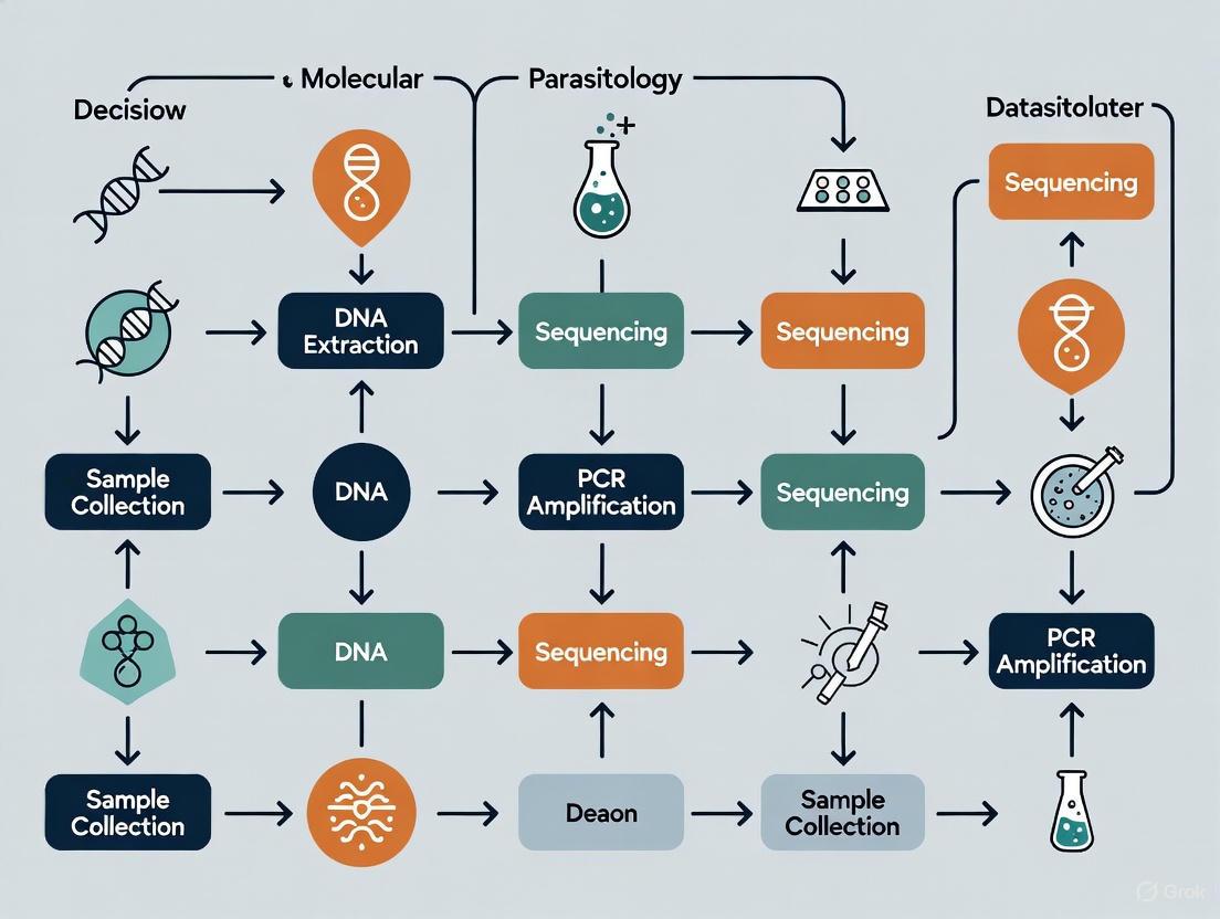
Workflow Visualization
Diagram 1: Pre-analytical workflow with critical error points.
The Scientist's Toolkit: Research Reagent Solutions
Table 2: Essential Materials for Pre-analytical Sample Processing in Molecular Parasitology
| Item | Function | Key Considerations |
|---|---|---|
| Neutral Buffered Formalin (NBF) | Fixative for tissue preservation for histology and molecular analysis. | Prevents acid-induced nucleic acid degradation. Preferred over unbuffered formalin for molecular studies [1]. |
| EDTA Tubes | Anticoagulant for whole blood collection for plasma and DNA analysis. | Prevents clotting; preserves DNA integrity better than other anticoagulants for molecular tests [1]. |
| PAXgene Tubes | Stabilize intracellular RNA in whole blood. | Essential for gene expression studies from blood, as RNA is highly labile [4]. |
| Viral Transport Media (VTM) | Preserves viral pathogens in swab samples (e.g., nasopharyngeal). | Allows for transport and short-term storage (at 4°C) of samples for viral nucleic acid detection [1]. |
| Nucleic Acid Stabilization Cards | Chemically treated cards for room-temperature storage of dried blood/fluid spots. | Enables stable transport of samples without refrigeration for DNA and certain RNA targets [1]. |
| RNase Inhibitors | Added to RNA extraction buffers or reactions. | Protects vulnerable RNA molecules from degradation by ubiquitous RNase enzymes [4]. |
Challenges in Wildlife and Clinical Parasitology Sample Collection
Technical Support Center
Troubleshooting Guides
Table 1: Common Sample Collection Challenges and Solutions
| Challenge | Potential Impact | Recommended Solution |
|---|---|---|
| Sample Degradation | False negatives in molecular tests; loss of parasite viability. [5] | Process fresh fecal samples within 24 hours or freeze at -20°C for molecular analysis. For larval detection (e.g., Baermann), use fresh, unrefrigerated samples. [5] [6] |
| Incorrect Preservative | Inability to perform certain tests; DNA degradation. [5] | Align preservative with study aims: -20°C for molecular work; 10% formalin or 70% alcohol for preserved specimens; no preservative for fresh larval isolation. [5] [7] |
| Host Misidentification | Incorrect host-parasite association data. [5] | Use a multi-evidence approach: combine non-invasive sampling (e.g., camera traps) with genetic host identification from scats. [5] |
| Low Test Sensitivity | Misdiagnosis; underestimation of parasite prevalence. [8] | Use tests in combination. If Cryptosporidium ELISA is negative but flotation is positive, collect a second sample for analysis to rule out false negatives. [7] |
| Anthelmintic Resistance Assessment | Inaccurate efficacy results for Fecal Egg Count Reduction Test (FECRT). [7] | Ensure correct timing: collect post-treatment sample 10-14 days after anthelmintic administration for equine strongyles. [7] |
Table 2: Method-Specific Limitations and Corrections
| Diagnostic Method | Inherent Limitation | Corrective Action |
|---|---|---|
| Baermann Technique | Not recommended as a primary diagnostic; ineffective for eggs, cysts, or some lungworm larvae (e.g., Eucoleus aerophilus). [7] | Use as a complementary test. For primary screening, use flotation techniques. Ensure samples are very fresh (1-2 hours old) and unrefrigerated. [6] |
| Microscopy | Low sensitivity and specificity; inability to differentiate related species (e.g., E. histolytica vs. E. dispar); requires experienced personnel. [8] | Use molecular methods (PCR) for confirmation and species differentiation, especially for pathogenic protozoa. [8] |
| Molecular PCR (for protozoa) | Inconsistent sensitivity for some parasites (e.g., Dientamoeba fragilis); difficult DNA extraction from robust oocyst walls. [8] | Use fixed fecal specimens for better DNA preservation for Giardia and Cryptosporidium. Standardize DNA extraction protocols. [8] |
| Fecal Flotation | Preservatives (formalin, alcohol) may compromise detection quality. [7] | Submit fresh, refrigerated samples where possible. For qualitative floats, use zinc sulfate for delicate protozoa like Giardia. [7] |
Frequently Asked Questions (FAQs)
Q1: What is the single most critical factor for successful molecular parasitology from wildlife samples? The cornerstone of success is the correct and immediate preservation of the sample, tailored to the downstream application. [5] For molecular research, freezing samples at -20°C as soon as possible is paramount to prevent DNA degradation. However, if the aim is to recover live nematode larvae for morphological identification (e.g., via Baermann technique), samples must be processed fresh, without refrigeration or freezing, as low temperatures kill the larvae and lead to false negatives. [5] [6]
Q2: How can I avoid repeated sampling and misidentification biases in non-invasive wildlife studies? To avoid sampling the same individual multiple times (repeated sampling bias) and to correctly identify the host species (identification bias), a multi-evidence approach is essential. [5] This involves combining the collection of scats from the environment with other methods such as camera trapping and analysis of footprints. Furthermore, genetic analysis of the scat itself can be used to confirm the host species, providing crucial epidemiological and ecological data. [5]
Q3: Our lab is transitioning to more molecular testing. Should we completely replace microscopy? No, molecular and microscopic methods should be viewed as complementary rather than exclusive. [8] While molecular techniques like PCR offer superior sensitivity and specificity for targeted pathogens and can differentiate morphologically identical species, microscopic examination of concentrated specimens remains a valuable broad-based screening tool. It can reveal parasitic infections that are not targeted by your specific PCR panel, thus providing a more comprehensive parasitological assessment. [8]
Q4: What is the best way to handle and submit intact adult parasites for identification? The key is to relax the worm's muscle tissue before preservation to allow for critical taxonomic structures to be visible. Place fresh worms collected from feces or an animal cavity in warm phosphate-buffered saline (PBS) or tap water and refrigerate them. This allows the worm to relax before being transferred to a preservative like ethanol or formalin. Note that intestinal parasites submitted in formalin can be difficult to identify; they are best transported in water in a sealed container. [5] [6]
Q5: How reliable are natural history collections for molecular parasitology research? Extremely reliable and invaluable. For many difficult-to-sample host species, museum collections provide a vast resource of tissue samples (stored in alcohol or frozen) that can be used for molecular detection of parasites. [9] These collections can help fill significant host-sampling gaps and are critical for discovering undiscovered parasite diversity and understanding historical biogeography without new field expeditions. [9]
Standardized Experimental Protocols for Sample Collection
Protocol 1: Non-Invasive Fecal Sample Collection for Multi-Evidence Research
Application: Ecological and epidemiological studies of parasite diversity in wild carnivore populations where direct handling is not feasible. [5]
Detailed Methodology:
- Field Collection: Using gloves, collect fresh scat from the environment. Record GPS location, date, and time.
- Host Identification: Take a small sub-sample (e.g., from the outer surface) using a sterile instrument and place it in a sterile tube for potential host genetic analysis. Store this sub-sample at -20°C.
- Parasite Analysis Sub-sampling: Divide the remaining sample for different analyses:
- For Molecular Parasitology: Place a portion (5-10 grams) in a leak-proof container and freeze immediately at -20°C. Do not use preservatives if freezing is possible. [5]
- For Morphological Parasitology: Place another portion in 10% formalin for long-term preservation for future flotation or sedimentation techniques. [7]
- Corroborative Evidence: Set up a camera trap near the collection site to help confirm host species and avoid repeated sampling bias. [5]
Protocol 2: DNA Extraction and RT-PCR for Intestinal Protozoa from Stool
Application: Sensitive and specific detection of pathogenic intestinal protozoa (Giardia duodenalis, Cryptosporidium spp., Entamoeba histolytica, Dientamoeba fragilis) in clinical or wildlife stool samples. [8]
Detailed Methodology:
- Sample Preparation: Mix 350 µl of Stool Transport and Recovery Buffer (S.T.A.R. Buffer) with approximately 1 µl of fecal sample using a sterile loop. Incubate for 5 minutes at room temperature. [8]
- Clarification: Centrifuge the mixture at 2000 rpm for 2 minutes.
- Supernatant Collection: Carefully collect 250 µl of the supernatant and transfer it to a fresh tube. Add 50 µl of an internal extraction control.
- Automated DNA Extraction: Extract DNA using the MagNA Pure 96 DNA and Viral NA Small Volume Kit on the MagNA Pure 96 System (or equivalent automated platform) following the manufacturer's instructions. [8]
- In-House RT-PCR Amplification:
- Reaction Mix: Combine 5 µl of extracted DNA, 12.5 µl of 2× TaqMan Fast Universal PCR Master Mix, 2.5 µl of primers and probe mix, and sterile water to a final volume of 25 µl.
- Cycling Conditions: Run on a Real-Time PCR System with the following program: 1 cycle of 95°C for 10 min; followed by 45 cycles of 95°C for 15 s and 60°C for 1 min. [8]
Workflow Visualization
Sample Collection Pathway for Molecular Parasitology
Diagnostic Method Decision Logic
The Scientist's Toolkit: Research Reagent Solutions
Table 3: Essential Materials for Parasitology Sample Collection & Analysis
| Reagent / Material | Function / Application |
|---|---|
| Zinc Sulfate Solution (spg 1.18) | Flotation medium for delicate protozoa (e.g., Giardia cysts) and some nematode larvae. [10] [7] |
| S.T.A.R. Buffer (Stool Transport and Recovery Buffer) | Stabilizes stool samples for molecular diagnostics, aiding in DNA extraction for PCR. [8] |
| Formalin (10%) | All-purpose fixative and preservative for long-term storage of fecal samples for morphological study. [7] |
| Ethanol (70%) | Preferred preservative for ectoparasites and adult helminths after tissue relaxation. [5] [6] |
| InPouch TF Medium | Selective culture medium for transporting and culturing Tritrichomonas foetus from bovine or feline samples. [10] [6] |
| Para-Pak Preservation Media | Commercial medium for preserving stool samples for later concentration and microscopic examination. [8] |
| MagNA Pure 96 DNA and Viral NA Small Volume Kit | Automated system for high-quality, reproducible DNA extraction from complex samples like stool. [8] |
| TaqMan Fast Universal PCR Master Mix | Ready-to-use mix for sensitive and specific real-time PCR detection of parasite DNA. [8] |
Understanding the Consequences of Non-Standardized Protocols
In molecular parasitology research, the absence of standardized protocols for sample collection and analysis introduces significant variability that can compromise data integrity, hinder reproducibility, and ultimately delay scientific progress and therapeutic development. This technical support center addresses the specific experimental challenges researchers encounter due to this lack of standardization, providing targeted troubleshooting guidance framed within the critical context of methodological harmonization.
Troubleshooting Guides
Issue 1: Inconsistent Pathogen Detection Results Across Laboratories
- Problem: Different laboratories report varying detection rates for the same parasitic protozoa (e.g., Giardia duodenalis, Cryptosporidium spp., Entamoeba histolytica) when analyzing similar sample sets.
- Cause: The use of non-standardized DNA extraction protocols and PCR assays. In a multicentre study, commercial and "in-house" RT-PCR tests showed variable performance, particularly for Dientamoeba fragilis and Cryptosporidium spp., where sensitivity was limited by inadequate DNA extraction efficiency from the parasite's robust wall structure [8].
- Solution:
- Validate DNA Extraction Protocols: Prior to large-scale screening, validate your DNA extraction method using a standardized panel of known positive samples for the target parasites. Ensure the protocol includes rigorous mechanical or chemical lysis steps effective against tough cyst walls.
- Adopt a Reference Method: If possible, adopt a commercially available, validated PCR test as an internal reference standard for cross-comparison with in-house protocols [8].
- Use Preserved Samples: For molecular work, stool samples preserved in specific media (e.g., Para-Pak) can yield better DNA preservation and more consistent PCR results compared to fresh samples [8].
Issue 2: Variable Output in Metabarcoding Studies
- Problem: In amplicon-based next-generation sequencing (NGS) studies, the read counts for different parasite species do not accurately reflect their true proportions in the sample.
- Cause: Bias can be introduced during library preparation. Key factors include the secondary structure of the target gene (e.g., 18S rDNA V9 region) and suboptimal PCR annealing temperatures, which can preferentially amplify certain sequences over others [11].
- Solution:
- Optimize Annealing Temperature: Systematically test a range of annealing temperatures during the amplicon PCR step to find the temperature that minimizes bias and provides the most uniform coverage across your target parasites [11].
- Linearize Plasmid Standards: When using plasmid controls for quantification, linearize them with restriction enzymes to minimize steric hindrance, which can improve amplification efficiency and the accuracy of relative abundance estimates [11].
Issue 3: Low Sensitivity in Detecting Soil-Transmitted Helminth (STH) Eggs
- Problem: Innovative diagnostic devices like lab-on-a-disk (LoD) platforms show promising specificity but suffer from low sensitivity due to significant egg loss during processing [12].
- Cause: Egg loss occurs during sample preparation steps and within the device due to adherence to walls of syringes and disks, as well as obstruction by larger fecal debris [12].
- Solution:
- Modify Sample Preparation: Implement a modified protocol that includes:
- The use of surfactants in the flotation solution to reduce egg adhesion to surfaces.
- Optimized filtration steps to remove large, obstructive debris more effectively before loading the sample into the device [12].
- Device Optimization: Work with engineers to refine the disk's design, such as shortening channel lengths to minimize the adverse effects of inertial forces on egg capture efficiency [12].
- Modify Sample Preparation: Implement a modified protocol that includes:
Frequently Asked Questions (FAQs)
Q1: Why is microscopy still considered a gold standard for many parasitic infections when molecular methods are more sensitive? Microscopy remains essential because it is a broad, non-targeted method that can detect a wide array of expected and unexpected parasites in a single test. It is also low-cost and accessible in resource-limited settings. In contrast, molecular methods like PCR are often highly specific, targeting only a pre-defined set of pathogens, and may miss rare or emerging species not included in the assay panel [13] [14].
Q2: What are the key consequences of using non-standardized molecular protocols in multi-center studies? The primary consequences are a lack of reproducibility and comparability of data. Results from one laboratory may not be directly comparable to another, hindering collaborative research, reliable epidemiological mapping, and the assessment of new therapeutics across different sites. This lack of consensus complicates efforts to integrate findings into mainstream public health surveillance and policy [15] [8].
Q3: How can our laboratory contribute to the standardization of molecular parasitology? Participate in and promote initiatives aimed at harmonizing methods. For example, the COST Action CA21105 is actively working to map Blastocystis epidemiology and diagnostics across Europe with the goal of developing evidence-based guidelines. Contributing your data to such collaborative networks and adhering to their proposed standard operating procedures when available is a significant step forward [15].
Q4: Our in-house PCR for Giardia works well. Why should we consider switching to a commercial kit? While a well-validated in-house PCR is valuable, switching is not always necessary. The advantage of a commercial kit lies in its standardization. It ensures that your results are directly comparable with those from other laboratories using the same kit, which is crucial for multi-center trials or surveillance programs. It also provides a benchmark against which you can further validate your in-house assay [8].
Quantitative Data on Methodological Variability
The tables below summarize empirical data highlighting how methodological choices impact diagnostic outcomes.
Table 1: Variation in Read Count Output for Different Parasites in 18S rRNA Metabarcoding [11]
| Parasite Species | Read Count Ratio (%) |
|---|---|
| Clonorchis sinensis | 17.2 |
| Entamoeba histolytica | 16.7 |
| Dibothriocephalus latus | 14.4 |
| Trichuris trichiura | 10.8 |
| Fasciola hepatica | 8.7 |
| Necator americanus | 8.5 |
| Paragonimus westermani | 8.5 |
| Taenia saginata | 7.1 |
| Giardia intestinalis | 5.0 |
| Ascaris lumbricoides | 1.7 |
| Enterobius vermicularis | 0.9 |
Table 2: Comparison of Diagnostic Methods for Intestinal Protozoa [8]
| Method | Pros | Cons |
|---|---|---|
| Microscopy | Low cost; broad, non-targeted detection; accessible. | Low sensitivity; requires experienced personnel; cannot differentiate some species. |
| In-house PCR | Can be highly sensitive/specific; customizable. | Lack of standardization; variable performance between labs. |
| Commercial PCR | Standardized; good for multi-center studies; high throughput. | Fixed panel of targets; may miss uncommon parasites; cost. |
Standardized Experimental Protocols
Protocol 1: Loop-Mediated Isothermal Amplification (LAMP) forWuchereria bancrofti
This protocol is adapted from a study that evaluated LAMP for diagnosing lymphatic filariasis, demonstrating high sensitivity and specificity [16].
- Sample Collection and DNA Extraction: Collect blood samples into EDTA vacutainers. Extract DNA using an alcohol precipitation method or a commercial kit suitable for blood samples.
- Primer Selection: Use primers targeting the W. bancrofti-specific SspI repeat region (18S rRNA). A standard set includes Forward Inner Primer (FIP), Backward Inner Primer (BIP), Forward Outer Primer (F3), and Backward Outer Primer (B3) [16].
- LAMP Reaction Setup: Prepare a reaction mix containing the extracted DNA, primers, betaine, MgSO₄, dNTPs, and a DNA polymerase with high strand displacement activity (e.g., Bst polymerase).
- Amplification: Incubate the reaction tube at a constant temperature of 63°C for 60 minutes in a water bath or heat block.
- Detection: Visualize amplification results by adding a pH-sensitive dye or fluorescent intercalating dye to the tube post-amplification. A color change or fluorescence under UV light indicates a positive result.
Protocol 2: Metabarcoding for Multi-Parasite Detection using 18S rDNA V9 Region
This protocol is based on an optimization study for simultaneously detecting 11 intestinal parasites [11].
- DNA Extraction and Quality Control: Extract genomic DNA from parasite specimens or clinical samples using a robust soil or stool DNA kit. Quantify DNA concentration and quality using a fluorometer.
- PCR Amplification: Amplify the 18S rDNA V9 region using primers 1391F and EukBR, which have Illumina adapters attached.
- Critical Step: Test a gradient of annealing temperatures (e.g., from 55°C to 65°C) to identify the optimal temperature that minimizes amplification bias.
- Library Preparation and Indexing: Perform a limited-cycle PCR to add dual indices and sequencing adapters to the amplicons.
- Pooling and Sequencing: Pool the indexed libraries in equimolar ratios and sequence on an Illumina platform (e.g., iSeq 100).
- Bioinformatic Analysis: Process the raw sequencing data using a pipeline such as QIIME 2. Steps include demultiplexing, quality filtering (DADA2), chimera removal, and taxonomic assignment against a curated parasite database.
Workflow Visualization
Consequences of Non-Standardized Research Protocols
Method Selection for Parasite Diagnosis
The Scientist's Toolkit: Key Research Reagent Solutions
Table 3: Essential Materials for Molecular Parasitology Experiments
| Item | Function | Example/Note |
|---|---|---|
| S.T.A.R Buffer | Stool Transport and Recovery Buffer for stabilizing stool samples and improving DNA yield for PCR [8]. | Used in automated nucleic acid extraction systems. |
| MagNA Pure 96 System | Automated nucleic acid extraction platform for high-throughput, reproducible DNA purification [8]. | Reduces manual variation in sample preparation. |
| Bst Polymerase | DNA polymerase with strand displacement activity essential for isothermal amplification methods like LAMP [16]. | Allows amplification at a constant temperature. |
| KAPA HiFi HotStart ReadyMix | High-fidelity PCR master mix for generating amplicons with low error rates, critical for NGS library prep [11]. | Ensures accurate sequence data. |
| Saturated NaCl Flotation Solution | Solution used to separate helminth eggs from denser fecal debris via flotation for concentration and detection [12]. | Often supplemented with surfactants to reduce egg loss. |
| Illumina iSeq 100 Reagents | Sequencing kit for running amplicon-based metabarcoding studies on a benchtop sequencer [11]. | Enables parallel screening of multiple parasite species. |
The Role of Standardization in Zoonotic and Veterinary Parasite Surveillance
This technical support center is designed for researchers, scientists, and drug development professionals working within the framework of a thesis on standardization of sample collection for molecular parasitology research. Standardized procedures are critical for generating reliable, reproducible data in zoonotic and veterinary parasite surveillance, particularly as the field increasingly integrates molecular diagnostics with traditional methods. The following troubleshooting guides and FAQs address specific experimental challenges to support your research objectives.
Frequently Asked Questions (FAQs)
FAQ 1: How does the choice of fecal preservative impact downstream morphological and molecular analyses?
The choice between formalin and ethanol as a preservative creates a significant trade-off between morphological preservation and DNA integrity. A 2024 study directly comparing 96% ethanol and 10% formalin for preserving parasites from capuchin monkey feces found that formalin-preserved samples allowed identification of a greater diversity of parasitic morphotypes [17]. However, formalin causes protein cross-linking and DNA fragmentation, which severely impedes PCR-based molecular analyses [17]. Ethanol, while causing some tissue dehydration and shrinkage that can complicate morphological identification, maintains stable DNA levels during long-term storage and is superior for genetic studies [17]. For research aiming to use both methods, the optimal approach is to split the fresh fecal sample and preserve halves in both media.
FAQ 2: Why does my real-time PCR for Cryptosporidium yield negative results when the flotation test is positive?
This discrepancy, noted in the Cryptosporidium ELISA test protocol from Cornell's Animal Health Diagnostic Center, can occur if the animal is infected but producing antigen below the detection limit of the ELISA, or if the molecular test is a false negative [7]. False negatives in PCR can stem from inadequate DNA extraction due to the robust wall structure of protozoan oocysts, which complicates DNA recovery [18]. To resolve this, collect a second sample for analysis and ensure your DNA extraction protocol includes rigorous mechanical disruption steps (e.g., bead beating) designed to break open tough oocysts and cysts.
FAQ 3: Our fecal egg count reduction test (FECRT) suggests anthelmintic resistance. What are the next steps?
A FECRT result indicating resistance is a serious finding. First, confirm the test was performed correctly: the pre-treatment (Day 0) and post-treatment (Day 14) fecal samples were from the same individual, the egg count method was consistent, and the calculation ((1 - Post-Treatment EPG / Pre-Treatment EPG) * 100) was accurate [7]. The following table summarizes the interpretation guidelines for equine strongyles:
Table: Interpretation of Fecal Egg Count Reduction Test (FECRT) for Equine Strongyles
| Anthelmintic Class | Expected Efficacy (No Resistance) | Suspected Resistance | Resistant |
|---|---|---|---|
| Benzimidazole | >99% | 90-95% | <90% |
| Pyrantel | 94-99% | 85-90% | <85% |
| Ivermectin/Moxidectin | >99.9% | 95-98% | <95% |
If resistance is confirmed, you should immediately switch to an anthelmintic from a different drug class with a known effective history on your farm and implement a refugia-based strategy—treating only heavy shedders (FEC > 500 eggs per gram) while leaving low shedders untreated to maintain a population of susceptible parasites [7].
FAQ 4: What are the key internal and external factors driving parasitic infection rates that our surveillance should monitor?
Surveillance programs should be designed to account for a complex interplay of factors:
- Internal Factors: These include the parasite's complex life cycle, which often involves multiple hosts, and the emergence of drug resistance. Resistance is a growing problem, with reports of resistance to all major classes of dewormers in over 70 species of equine parasites [19].
- External Factors: Socioeconomic factors like poverty and poor sanitation are major drivers, particularly for soil-transmitted helminths [20]. Environmental factors, especially climate change, are also critical, as alterations in temperature and rainfall can expand the habitats of vectors and parasites [21]. For example, the northward expansion of the black-legged tick (Ixodes scapularis) is increasing the risk of Lyme disease in dogs and humans in new regions [21].
Troubleshooting Guides
Problem: Inconsistent results between in-house and commercial RT-PCR assays for intestinal protozoa.
- Background: Molecular diagnostics for protozoa like Giardia duodenalis, Cryptosporidium spp., and Entamoeba histolytica are gaining traction but face technical challenges.
- Investigation & Solution: A 2025 multicentre study comparing a commercial RT-PCR test with an in-house assay found complete agreement for G. duodenalis but limited sensitivity for Cryptosporidium spp. and Dientamoeba fragilis [18]. The inconsistency was largely attributed to inadequate DNA extraction from the parasite's robust oocysts.
- Protocol: Use the following standardized DNA extraction protocol from the study:
- Mix 350 µl of Stool Transport and Recovery (S.T.A.R.) Buffer with approximately 1 µl of fecal sample.
- Incubate for 5 minutes at room temperature, then centrifuge at 2000 rpm for 2 minutes.
- Carefully collect 250 µl of the supernatant and transfer it to a fresh tube.
- Add 50 µl of an internal extraction control.
- Perform DNA extraction using a fully automated system, such as the MagNA Pure 96 System with the corresponding DNA and Viral NA Small Volume Kit [18].
- Prevention: Standardize sample collection and storage. The same study found that PCR results from preserved stool samples were superior to those from fresh samples, likely due to better DNA preservation [18].
Problem: Inability to differentiate between pathogenic and non-pathogenic species via microscopy.
- Background: Microscopy remains a cost-effective tool but lacks the sensitivity and specificity to differentiate closely related species, such as the pathogenic Entamoeba histolytica from the non-pathogenic E. dispar [18] [20].
- Investigation & Solution: This is a known limitation of microscopy. Molecular assays are critical for accurate diagnosis. A transition to PCR-based methods is recommended.
- Protocol: Implement a TaqMan qPCR assay.
- Reaction Mixture: Combine 5 µl of extracted DNA, 12.5 µl of 2x TaqMan Fast Universal PCR Master Mix, 2.5 µl of primer and probe mix, and sterile water to a final volume of 25 µl.
- Amplification: Perform multiplex tandem PCR on a platform like the ABI Prism sequence detection system using the recommended cycling conditions [18].
- Targets: Primers and probes should target genes with high discriminatory power, such as ITS1, ITS2, or COI, which have been successfully applied to equine strongylids and other parasites [19].
Standardized Experimental Protocols
Comparative Parasite Preservation and Morphological Grading
- Objective: To evaluate the preservation quality of gastrointestinal parasites in fecal samples stored in different media.
- Sample Collection: Collect fresh fecal samples and immediately partition into two halves.
- Preservation: Preserve one half in 6 ml of 96% ethanol and the other in 10 ml of 10% buffered formalin. Ensure samples are fully submerged [17].
- Coproscopy: Process samples using a modified Wisconsin sedimentation technique, which involves homogenization, straining through cheesecloth, centrifugation, and microscopic screening of the resuspended pellet [17].
- Grading Scale: Use a standardized 3-point scale to rate preservation [17]:
- Score 3 (Well-preserved): Larvae: fully intact cuticle, visible internal structures. Eggs: continuous, unobstructed shell with visible embryo/larvae.
- Score 2 (Moderately preserved): Larvae: degradation of cuticle or internal structures that partially interferes with identification. Eggs: minor shell deformations.
- Score 1 (Poorly preserved): Larvae: heavy degradation, making identification difficult or impossible. Eggs: broken shell or obscured contents.
Table: Comparison of Fecal Sample Preservation Media
| Characteristic | 10% Formalin | 96% Ethanol |
|---|---|---|
| Primary Advantage | Superior for morphological identification; preserves tissue form [17]. | Superior for molecular studies; maintains DNA integrity [17]. |
| Primary Disadvantage | Causes DNA fragmentation, unsuitable for PCR [17]. | Dehydrates tissues, may cause morphological alteration [17]. |
| Toxicity | High; requires careful handling [17]. | Lower; less toxic [17]. |
| Morphotype Diversity | Identifies a greater number of parasitic morphotypes [17]. | Identifies fewer morphotypes compared to formalin [17]. |
| Ideal Use Case | Gold standard for long-term morphological studies. | Essential for any downstream DNA analysis (PCR, NGS). |
Integrated Surveillance for Companion Animal Parasites
- Objective: To establish a surveillance system for companion animal parasites that can integrate data for a One Health approach.
- Data Sources: Surveillance can be passive (relying on data submission by veterinarians) or active (extracting data from electronic health records and veterinary diagnostic laboratories) [22].
- Scope: Define surveillance priorities, which can include specific infectious diseases, toxins, cancers, or cause of death [22].
- Data Integration: A major current limitation is the lack of integration. While only 9.1% of existing systems integrate environmental or public health data at the point of collection, 27.3% utilize such data during the analysis phase [22]. Future systems should be designed to incorporate climate, wildlife density, and human health data from the outset.
- Data Dissemination: Outputs are typically shared through surveillance reports and direct feedback to data contributors [22].
Research Reagent Solutions
Table: Essential Materials for Standardized Parasitology Research
| Reagent/Material | Function | Example Use Case |
|---|---|---|
| S.T.A.R. Buffer | Stabilizes nucleic acids in stool samples for molecular diagnostics. | DNA extraction for PCR detection of intestinal protozoa [18]. |
| MagNA Pure 96 System & Kit | Automated, high-throughput nucleic acid extraction. | Standardized DNA extraction from fecal samples for a multicentre study [18]. |
| TaqMan Fast Universal PCR Master Mix | Ready-to-use mix for fast, sensitive real-time PCR assays. | Detection and differentiation of pathogenic intestinal protozoa [18]. |
| Primers/Probes (ITS1, ITS2, COI) | Target specific genetic regions for parasite identification. | Molecular identification of equine strongylids and other parasites [19]. |
| 10% Buffered Formalin | Preserves tissue morphology by cross-linking proteins. | Long-term storage of fecal samples for microscopic parasite identification [17]. |
| 96% Ethanol | Preserves DNA by dehydrating tissues and inhibiting nucleases. | Long-term storage of fecal samples for genetic analysis [17]. |
| Floitation Media (e.g., Sugar Solution, ZnSO₄) | Concentrates parasite eggs and cysts based on specific gravity. | Routine qualitative and quantitative fecal flotation tests [7]. |
Workflow for Selecting a Fecal Sample Preservation Method
The following diagram outlines the decision-making process for choosing the appropriate fecal sample preservation method based on research objectives.
Ethical and Practical Considerations in Sample Sourcing
Frequently Asked Questions (FAQs)
Q1: What are the most critical factors to ensure ethical sourcing of human samples for parasitology research? Ethical sourcing of human samples requires adherence to several key principles. Specimens should only be collected from individuals through healthcare providers, not directly from the public [23]. Furthermore, all samples must be collected, transported, and stored in a manner that guarantees the best possible results and respects donor consent [24]. This includes ensuring that the methods and reagents used are standardized and established, though alternative methods may also be valid [25]. Finally, transparency and clear communication with all stakeholders, including donors, clinicians, and researchers, are fundamental to maintaining an ethical framework [26].
Q2: Our research involves collecting fecal samples for PCR-based diagnosis. What are the common pitfalls in sample collection and handling? Common pitfalls include incorrect sample quantity, improper preservation, and delays in transport. For molecular diagnosis, it is crucial to follow Standard Operating Procedures (SOPs) specifically designed for this purpose [25]. Key considerations are:
- Quantity: Submitting an adequate amount of sample (e.g., approximately 2 grams for flotation tests) [27].
- Freshness: Using fresh, unpreserved specimens or adding appropriate preservatives if there is a delay in processing to prevent morphological deterioration [24] [27].
- Transport: Sending samples overnight on ice packs (Monday-Thursday is recommended by some diagnostic labs) to maintain sample integrity for analysis [27].
Q3: What types of specimens, other than feces, can be sourced for molecular parasitology studies? A wide variety of non-faecal specimens can be examined for parasites, including urine, sputum, liver aspirates, duodenal/jejunal aspirates, bile, corneal scrapings, and tissue biopsies [24]. The choice of specimen depends on the parasitic infection being investigated. For example, a terminal urine specimen is required for the diagnosis of Schistosoma haematobium [24]. For certain extraintestinal infections, serology or polymerase chain reaction (PCR) performed at specialist laboratories may be necessary for diagnosis [24].
Q4: How can we balance the practical need for high-quality samples with the ethical imperative of fair and equitable sourcing? Balancing quality with ethicality involves a holistic perspective that goes beyond mere compliance. It requires building genuine partnerships with suppliers and healthcare providers, fostering transparency, and engaging in collaborative efforts [26]. This includes:
- Thorough Vetting: Implementing rigorous processes to evaluate the practices of all partners in the supply chain [26].
- Capacity Building: Working alongside suppliers to elevate their practices to meet required ethical and quality standards, rather than simply severing ties [26].
- Clear Communication: Maintaining open dialogue with all stakeholders, including consumers and donors, about sourcing practices and challenges [26].
Troubleshooting Common Experimental Issues
Issue 1: Degraded DNA/RNA from patient samples, leading to failed PCR assays.
- Potential Cause: Improper collection, delayed transport, or incorrect storage conditions.
- Solution: Implement and strictly adhere to validated SOPs for sample collection [25]. Ensure samples are transported promptly on cold packs and stored at appropriate temperatures upon receipt [27]. For fecal samples intended for larval detection, delays can lead to eggs hatching, making differentiation difficult [27].
Issue 2: Inconsistent molecular results between different research sites in a multi-center study.
- Potential Cause: Lack of standardized protocols for sample collection, preservation, and processing.
- Solution: The core of this issue is a lack of standardization. Adopt a common set of SOPs across all sites to ensure consistency [25] [24]. Provide regular training sessions for staff at all sites to update their knowledge and skills [24]. Utilize the same methods that have achieved satisfactory ratings in external quality assurance schemes [24].
Issue 3: Low sample yield or inability to source specific sample types.
- Potential Cause: Complex supply chains, regulatory hurdles, and a lack of visibility into available resources.
- Solution: Build relationships with specialist and reference laboratories/units that can provide expert advice and assistance on the diagnosis and management of parasitic infections, including access to a wider range of sample types [24]. Improve supply chain visibility by leveraging technological advancements and collaborative networks [26].
Experimental Protocols & Data
Standardized Protocol for Urine Sample Collection forS. haematobium
Methodology:
- Collection: Collect a terminal urine specimen (the last 10–20 ml passed) around midday when egg excretion is highest [24]. Alternatively, a 24-hour collection of terminal urine specimens can be used.
- Preservation: If a delay in examination is expected, add 0.5 ml of 10% formalin to 10-20 ml of urine to prevent eggs from hatching [24].
- Transport: Transport the specimen to the laboratory promptly for processing.
- Examination: Process a minimum of 10 ml of terminal urine using standard operating procedures to achieve maximum recovery of parasites [24].
Sample Collection Requirements for Different Analyses
The table below summarizes quantitative data for collecting various sample types for parasitological analysis.
Table: Specimen Collection Guidelines for Parasitology Diagnostics
| Specimen Type | Recommended Quantity | Key Handling & Transport Conditions | Primary Analysis Method |
|---|---|---|---|
| Feces (Flotation) | ~2 grams (2 teaspoons) [27] | Transport overnight on ice; do not freeze [27] | Microscopy, PCR [25] |
| Feces (Baermann) | At least 10 grams (2 tablespoons) [27] | Must be fresh; transport overnight on ice [27] | Larval detection [27] |
| Feces (Direct Smear) | Small amount | Must be examined within 30 minutes of collection [27] | Microscopy [27] |
| Urine (for S. haematobium) | 10-20 ml terminal urine [24] | Add formalin if delayed; examine promptly [24] | Microscopy, PCR |
| Duodenal/Jejunal Aspirates | As obtained by clinician | Transport and process without delay; refrigerate if held [24] | Microscopy, PCR, culture |
| Liver Aspirates (for Entamoeba) | Pus from aspirate | Examine without delay by experienced staff [24] | Microscopy, PCR |
Workflow and Relationship Diagrams
Diagram 1: Ethical Sample Sourcing Decision Pathway
Diagram 2: Standardized Sample Processing Workflow
The Scientist's Toolkit: Research Reagent Solutions
Table: Essential Materials for Parasitology Sample Collection and Processing
| Reagent/Material | Function | Application Example |
|---|---|---|
| Parasitology Single/Multi-Vial Kits [28] | Contains pre-measured preservatives for stool sample fixation and preservation. | Standardized collection and transport of fecal specimens for molecular and morphological analysis. |
| Ethyl Acetate [28] | Used as a solvent in concentration procedures for parasite separation. | Fecal concentration methods for microscopic examination. |
| Zinc PVA (Polyvinyl Alcohol) [28] | Preserves parasite morphology for permanent staining and microscopic identification. | Creation of permanent stained smears from fecal samples for detailed morphological study. |
| Trichrome Stain [28] | A polychrome stain used to differentially color protozoan cysts and helminth eggs. | Enhanced visualization and identification of intestinal protozoa in fixed stool specimens. |
| 10% Formalin [24] | Fixative and preservative that prevents hatching of parasite eggs. | Preservation of urine samples for S. haematobium diagnosis and concentration of fecal samples. |
| Serum/Plasma Samples | Used for serological detection of antibodies against parasitic infections. | Diagnosis of extraintestinal infections like cysticercosis, echinococcosis, strongyloidiasis, and toxocariasis [24]. |
Step-by-Step Protocols for Sample Collection, Preservation, and Processing
Frequently Asked Questions
FAQ 1: What is the core difference between invasive and non-invasive fecal sampling?
- Invasive sampling involves collecting feces directly from the animal, either from the rectum during trapping and handling or from the gut of carcasses. This method requires capturing or handling the animal [29] [30].
- Non-invasive sampling involves collecting scats found in the animal's environment without any contact with or disturbance of the animal [31] [29] [32].
FAQ 2: When should I choose non-invasive sampling over invasive methods? Non-invasive sampling is particularly advantageous in these scenarios:
- Studying elusive, endangered, or difficult-to-trap species [29] [32].
- Conducting large-scale spatial or long-term monitoring where trapping is logistically challenging or cost-prohibitive [29] [30].
- Minimizing research impact on animal welfare and avoiding the stress that handling can inflict on wildlife, which can itself alter physiological parameters and microbiome composition [31] [30].
FAQ 3: How does sample collection method affect downstream molecular analysis? The collection and preservation method is critical for ensuring the integrity of DNA in the sample.
- Temperature: Storage at 4°C is generally acceptable for up to 60 days for hookworm DNA, while higher temperatures (e.g., 32°C) lead to greater DNA degradation without adequate preservatives [33].
- Preservatives: Methods like 95% ethanol, silica gel beads, and RNA later help protect DNA from nucleases present in stool, especially when a cold chain cannot be maintained [33].
- Sample Homogeneity: Sub-sampling from a non-homogenized stool can lead to variable detection of microbial members and metabolites; homogenization prior to aliquoting is recommended [34].
FAQ 4: Can I use fecal samples collected for colorectal cancer screening for microbiome research? Yes, fecal immunochemical test (FIT) tubes and fecal occult blood test (FOBT) cards have been validated for microbiome analysis. Studies show they yield microbial data that is relatively reproducible and stable at room temperature for several days, making them feasible for large-scale population-based studies [35] [36].
FAQ 5: What are the primary limitations of non-invasive sampling?
- Sample Identification: It can be challenging to correctly identify the source species based on scat morphology alone, risking misidentification [29].
- Repeated Sampling: It may be difficult to determine if multiple scats come from the same individual, potentially biasing population-level data [29].
- Environmental Degradation: Samples are exposed to the elements, which can lead to DNA degradation or changes in parasite viability if not collected promptly [29].
Troubleshooting Guides
Problem: Low DNA yield or quality from non-invasively collected scats.
- Potential Cause: DNA degradation due to prolonged exposure to sun, rain, or high temperatures in the environment [29].
- Solution:
- Prioritize collection of fresh samples, identified by moistness and structural integrity [30].
- Store samples at -20°C as soon as possible after collection. If freezing is not immediately available, use a DNA preservative like 95% ethanol or commercial storage tubes [33] [29].
- For preserved samples, ensure they are stored in the dark at a stable, cool temperature until DNA extraction [37].
Problem: Inconsistent microbiome or metabolite profiles from technical replicates.
- Potential Cause: Fecal material is inherently heterogeneous; spot sampling from different parts of a non-homogenized stool can yield different microbial communities and metabolite concentrations [34].
- Solution: Homogenize the entire stool sample thoroughly before aliquoting for DNA extraction or other analyses to ensure a representative subsample [34].
Problem: Trapping stress is suspected of altering the host's fecal microbiome.
- Potential Cause: The stress of trapping and handling can significantly shift the composition of the gut microbiome, skewing results [30].
- Solution: Where ethically and logistically possible, employ non-invasive sampling. If trapping is necessary, compare findings with non-invasively collected samples from the same population to account for potential trapping effects [30].
Comparison of Fecal Sample Preservation Methods
The table below summarizes quantitative data on the effectiveness of various preservatives for maintaining DNA amplification efficiency (as measured by quantitative PCR Cq values) for hookworm DNA in stool samples over 60 days [33].
Table 1: Comparison of Fecal Sample Preservation Methods for DNA Analysis
| Preservation Method | Storage at 4°C (60 days) | Storage at 32°C (60 days) | Key Advantages & Considerations |
|---|---|---|---|
| No Preservative (Control) | No significant Cq increase | Significant Cq increase | Only feasible with reliable cold chain; low cost [33]. |
| 95% Ethanol | No significant Cq increase | Moderate Cq increase | Effective, low-cost, and pragmatic for most field conditions; protects against PCR inhibitors [33]. |
| RNAlater | No significant Cq increase | Moderate Cq increase | Effective preservative; can be more expensive than ethanol [33]. |
| Silica Bead Desiccation | No significant Cq increase | Minimal Cq increase (Most effective) | Effective at high temperatures; requires a two-step process [33]. |
| FTA Cards | No significant Cq increase | Minimal Cq increase (Most effective) | Effective at high temperatures; easy to transport [33] [36]. |
| Potassium Dichromate | No significant Cq increase | Minimal Cq increase (Most effective) | Effective but highly toxic; requires careful handling [33]. |
| PAXgene | No significant Cq increase | Moderate Cq increase | Some protective effect; commercial system [33]. |
Table 2: Performance of Different Collection Methods for Microbiome Studies
This table summarizes the stability (Intraclass Correlation Coefficients) of microbiome metrics from different collection methods after 7 days at various temperatures compared to an immediate freezing gold standard. ICCs ≥ 0.75 are generally considered high [36].
| Collection Method | Storage Condition | Microbiome Community Stability (Beta-diversity) | Relative Abundance of Major Phyla | Notes |
|---|---|---|---|---|
| FIT Tubes (after occult blood screening) | Room Temperature (after processing) | High (ICC ≥ 0.75) | High (ICC ≥ 0.75) | Embedding within screening programs is feasible [36]. |
| FIT Tubes | 7 days at 30°C | Moderate to High (ICC range: 0.41 - 0.90) | Moderate to High (ICC range: 0.41 - 0.90) | Performance varies by specific FIT tube type [36]. |
| FIT Tubes | 7 days at Room Temperature | Low to High (ICC range: 0.06 - 0.94) | Low to High (ICC range: 0.06 - 0.94) | Performance varies by specific FIT tube type [36]. |
| Fecal Occult Blood Test (FOBT) Cards | 3 days at Room Temperature | No significant difference from frozen | No significant difference in major phyla | Low-cost, feasible for large-scale studies [35]. |
Experimental Protocols
Protocol 1: Standardized Non-Invasive Scat Collection for Parasite DNA Analysis
Objective: To collect fresh fecal samples from the environment for downstream molecular detection of parasite DNA, while minimizing degradation and cross-contamination [33] [29].
Materials:
- Disposable gloves
- GPS unit
- Sample collection tubes (e.g., containing 95% ethanol or silica beads)
- Cooler with ice packs or portable freezer (-20°C)
- Data recording forms
Procedure:
- Locate Fresh Scat: Identify fecal samples that appear fresh (moist, intact). Record GPS coordinates, date, and time.
- Sub-sample: Using a clean implement (e.g., wooden stick or disposable spatula), collect a portion of the scat from the interior to minimize environmental contamination.
- Preserve:
- For Ethanol: Place the sub-sample into a tube containing a volume of 95% ethanol that is at least 3 times the volume of the fecal sample to ensure proper preservation [33].
- For Silica Beads: Place the sub-sample into a tube with an excess of silica gel beads, ensuring the sample is surrounded and desiccated [33].
- Store: Keep samples cool on ice packs in the field and transfer to a -20°C or -80°C freezer as soon as possible for long-term storage [29].
Protocol 2: Comparing Invasive vs. Non-Invasive Microbiome Profiles
Objective: To evaluate the impact of trapping and handling stress on gut microbiome composition by comparing samples collected invasively from trapped animals and non-invasively from the same population [30].
Materials:
- Trapping equipment (e.g., box traps, sedative bait)
- Sterile swabs or spatulas
- Sample storage tubes (e.g., with 95% ethanol or DNA/RNA Shield)
- Portable freezer
Procedure:
- Non-Invasive Collection: Prior to or during trapping efforts, collect fresh fecal samples from the ground following observation of animal defecation. Swab the inner portion of the feces and preserve in storage tubes. Record location and time [30].
- Invasive Collection: Trap animals using standard, ethically-approved protocols. After trapping, collect feces defecated onto a clean surface by the sedated or handled animal. Swab the inner portion and preserve identically to the non-invasive samples [30].
- Control for Time: Ensure handling time for invasively collected samples is recorded, as prolonged time between trapping and defecation may be a confounding factor [30].
- Analysis: Extract DNA and perform 16S rRNA gene sequencing on all samples. Compare alpha and beta diversity metrics (e.g., Weighted UniFrac) between the invasively and non-invasively collected groups using PERMANOVA [30].
Workflow Diagram
The Scientist's Toolkit: Essential Research Reagents & Materials
Table 3: Key Reagents and Materials for Fecal Sample Collection and Preservation
| Item | Function & Application | Key Considerations |
|---|---|---|
| 95% Ethanol | A widely used preservative that deactivates nucleases, protecting DNA for PCR-based parasite detection [33]. | Considered a pragmatic, effective, and low-cost choice for most field conditions; resistant to PCR inhibitors [33]. |
| RNAlater | A commercial aqueous, nontoxic storage reagent that stabilizes and protects nucleic acids [33]. | Effective for DNA and RNA; can be more expensive than ethanol for large-scale studies [33] [37]. |
| Silica Gel Beads | A desiccant that preserves DNA by removing moisture from the sample, preventing microbial degradation [33]. | Highly effective for ambient temperature storage, especially in a two-step desiccation process [33]. |
| FTA Cards / FOBT Cards | Cellulose-based cards impregnated with chemicals that lyse cells and protect DNA from nucleases and oxidation [35] [33]. | Ideal for easy, ambient-temperature transport; suitable for microbiome and molecular parasitology studies [35] [36]. |
| Fecal Immunochemical Test (FIT) Tubes | Tubes containing hemoglobin-stabilizing buffer, designed for colorectal cancer screening [36]. | Validated for microbiome analysis; enables leveraging of large-scale screening programs for research [36]. |
| PowerFecal DNA Isolation Kit | A widely used DNA extraction kit optimized for challenging samples like stool, which contain PCR inhibitors [36]. | Provides high-quality microbial DNA; crucial for consistent sequencing results from fecal material [36] [37]. |
In molecular parasitology research, the accuracy of your results is fundamentally determined at the very first step: sample collection and preservation. Selecting an inappropriate preservative can lead to the degradation of nucleic acids, introduction of PCR inhibitors, or render samples useless for intended downstream assays, ultimately compromising research validity. This guide provides targeted technical support to help you navigate the critical pre-analytical phase, ensuring your samples are stabilized in a manner that is fully compatible with sophisticated molecular applications such as PCR, qPCR, and Next-Generation Sequencing (NGS). By standardizing these initial procedures, we can significantly enhance the reliability and reproducibility of research data across the field.
Frequently Asked Questions (FAQs)
What are the key factors when choosing a preservative for molecular parasitology?
Selecting a preservative requires a balanced consideration of several factors:
- Target Analyte: The choice depends on what you aim to preserve—DNA, RNA, viable cells, or parasite morphology [38] [39].
- Sample Matrix: Different rules apply to stool, blood, tissue, or other body fluids [40] [29] [41].
- Downstream Application: The preservative must be compatible with your planned molecular techniques (e.g., PCR, qPCR, NGS) [38] [33].
- Storage and Shipping Conditions: Consider the available infrastructure (cold chain vs. ambient temperature) and the required storage duration [38] [33].
- Health and Safety: The toxicity and handling requirements of the preservative for laboratory personnel are critical [33].
Which preservatives are recommended for stool-based molecular detection of parasites?
For molecular analysis of stool samples, the preservative must protect parasite DNA from degradation by nucleases present in the sample.
- Recommended Preservatives: TotalFix, Unifix, modified Polyvinyl Alcohol (PVA) that is Zn- or Cu-based, and Ecofix are considered acceptable and allow for room temperature storage and shipping [40].
- A Pragmatic and Effective Choice: Multiple studies, including comparative analyses, have found that 95% ethanol provides a highly effective and practical option for preserving parasite DNA in stool, even at elevated ambient temperatures (32°C) for extended periods [33].
- Preservatives to Avoid: Formalin, SAF (Sodium Acetate-Acetic Acid-Formalin), LV-PVA, and Protofix are not recommended for molecular detection as they can interfere with downstream PCR analysis [40].
How should blood samples be preserved for the detection of blood-borne parasites or cell-free DNA?
The preservation method depends on the specific target within the blood.
- For Cell-Free DNA (cf-DNA) and Cell-Free RNA (cf-RNA): Specialized evacuated blood collection tubes containing proprietary, fixative-free preservatives are available. These tubes stabilize cf-DNA and cf-RNA for up to 30 days at room temperature and prevent the release of genomic DNA from blood cells, which is crucial for accurate assays [38] [42].
- For General Parasitological Examination: For the preparation of blood smears for microscopy, EDTA is the preferred anticoagulant. Heparin should be avoided as it can interfere with staining procedures [41].
Can I use the same preservative for both morphological and molecular analysis?
Compatibility varies. While some modern preservatives like Sodium Acetate-Acetic Acid-Formalin (SAF) are designed to be suitable for both concentration procedures (morphology) and staining [41], many traditional fixatives are not. For instance, formalin is excellent for preserving morphology but is strongly discouraged for downstream molecular applications because it causes cross-linking and degradation of nucleic acids, making PCR amplification difficult [40] [33]. If both types of analysis are required, confirm the compatibility of your chosen preservative with all intended downstream assays or plan to split the sample.
What is the maximum storage time for preserved samples?
Stability is highly dependent on the preservative, storage temperature, and sample type.
- Stool in 95% Ethanol: DNA remains amplifiable for at least 60 days when stored at 32°C [33].
- Blood in cf-DNA/cf-RNA Tubes: Cell-free DNA is stable for 30 days at room temperature (15-25°C) and for 8 days at 37°C [38].
- General Rule: For all sample types, consistent and cool storage (4°C) is always advantageous. When a cold chain is not available, the choice of a robust ambient-temperature preservative is critical [33].
Troubleshooting Guides
Problem: Low DNA Yield or Quality from Preserved Stool Samples
Potential Causes and Solutions:
- Cause 1: Use of an inappropriate preservative that degrades DNA.
- Cause 2: Incomplete mixing of the sample with the preservative, leading to uneven stabilization.
- Solution: Ensure the sample is thoroughly and gently mixed with the preservative immediately after collection to guarantee full penetration and stabilization.
- Cause 3: Sample degradation prior to preservation.
- Solution: Minimize the time between sample collection and placement into the preservative. Process fresh samples as quickly as possible [29].
Problem: PCR Inhibition in Samples from Preserved Blood
Potential Causes and Solutions:
- Cause 1: The preservative or sample matrix contains substances that inhibit DNA polymerases.
- Solution: Use preservative tubes that are verified to neutralize common PCR inhibitors [39]. Ensure the DNA purification method used is designed to remove inhibitors effectively. You may also need to dilute the DNA template in the PCR reaction.
- Cause 2: Hemolysis during blood collection or storage.
Problem: Inconsistent Molecular Results from Field-Collected Samples
Potential Causes and Solutions:
- Cause 1: Inconsistent preservation conditions or fluctuating temperatures during transport.
- Solution: Standardize the preservation protocol across all collection sites. Choose preservatives proven to be effective across a range of temperatures relevant to your field conditions [33].
- Cause 2: Variation in the sample-to-preservative ratio.
- Solution: Provide collection kits with pre-measured preservative volumes and clear instructions on the required sample volume to ensure a consistent ratio every time [41].
Preservative Compatibility and Performance Data
The following tables summarize key quantitative data on preservative performance to aid in evidence-based selection.
Table 1: Comparison of Stool Sample Preservatives for Molecular Diagnosis
| Preservative | Compatibility with PCR | Optimal Storage Temperature | Key Advantages / Disadvantages |
|---|---|---|---|
| 95% Ethanol [33] | Excellent | 4°C to 32°C | Advantages: Highly effective, pragmatic, cost-effective. Disadvantages: Flammable; requires safety precautions. |
| Potassium Dichromate(2.5% solution) [40] [33] | Good | Shipped refrigerated | Advantages: Effective for certain parasites. Disadvantages: Toxic and corrosive. |
| TotalFix, Unifix, Ecofix [40] | Good | Room Temperature | Advantages: Commercially available, formulated for molecular work. |
| Formalin (10%) [40] [41] | Not Recommended | Room Temperature | Disadvantages: Causes DNA degradation and cross-linking, leading to PCR failure. |
| SAF [40] | Not Recommended | Room Temperature | Disadvantages: Not suitable for molecular detection. |
Table 2: Performance Data of Blood Sample Preservative Tubes for cf-DNA/cf-RNA
| Parameter | Specification |
|---|---|
| cf-DNA Stability | 30 days at room temperature (15-25°C); 8 days at 37°C [38] |
| cf-RNA Stability | 30 days at room temperature (15-25°C) [38] |
| Circulating Tumour Cell (CTC) Stabilization | 14 days at ambient temperature [42] |
| Blood Draw Volume | 8.7 mL into a 10 mL tube [38] |
| Key Feature | Fixative-free, prevents apoptosis and genomic DNA release [38] [42] |
Standardized Experimental Protocol: Evaluating Preservative Efficacy
This protocol is adapted from a published comparative study to evaluate the performance of different preservatives for maintaining the integrity of parasite DNA in stool samples [33].
Objective: To assess the effectiveness of various preservatives in maintaining the amplifiability of target parasite DNA over time at different storage temperatures.
Materials:
- Naïve (uninfected) stool matrix
- Source of parasite eggs/larvae (e.g., from infected animal model)
- Candidate preservatives (e.g., 95% Ethanol, RNAlater, etc.)
- DNA extraction kit
- PCR/qPCR reagents for target parasite DNA
- Sterile tubes and pipettes
- Incubators set to 4°C and 32°C
Methodology:
- Sample Preparation: Spike known, quantified parasite egg material into consistent aliquots of naïve human stool [33].
- Preservation: Within one hour of spiking, add the different test preservatives to the sample aliquots. Include a "gold standard" control by flash-freezing some aliquots at -20°C without preservative.
- Storage: Store preserved samples at two temperatures: 4°C (mimicking a cold chain) and 32°C (simulating tropical ambient conditions).
- Time-Course Analysis: At predetermined time points (e.g., Day 1, 7, 30, 60), extract DNA from replicate samples for each preservative-temperature combination [33].
- Downstream Analysis: Perform qPCR assays targeting the parasite DNA. The primary metric for effectiveness is the quantification cycle (Cq) value. A smaller increase in Cq value over time compared to controls indicates better preservation of amplifiable DNA.
Diagram: Workflow for Preservative Efficacy Testing
The Scientist's Toolkit: Essential Research Reagents
Table 3: Key Reagents for Sample Collection and Preservation in Molecular Parasitology
| Item | Function | Example Application |
|---|---|---|
| cf-DNA/cf-RNA Preservative Tubes [38] | Stabilizes cell-free nucleic acids and prevents cellular apoptosis in whole blood. | Collection of blood for liquid biopsy, cancer research, and pathogen detection. |
| 95% Ethanol [33] | Deactivates nucleases and preserves DNA integrity in stool samples. | A cost-effective and highly efficient preservative for field collection of fecal samples for PCR. |
| Compatible Stool Fixatives (e.g., TotalFix, Ecofix) [40] | Preserves parasite structures and DNA for combined morphological and molecular work. | Routine diagnostic stool testing where multiple assay types are required. |
| RNAlater [33] | Stabilizes and protects cellular RNA in unfrozen samples. | Preserving tissue or cell samples for downstream RNA expression analysis. |
| EDTA Blood Collection Tubes [41] | Acts as an anticoagulant to prevent blood clotting. | Collection of whole blood for preparation of thin and thick smears for malaria diagnosis. |
| Silica Gel Beads [33] | Desiccates samples by absorbing moisture, inhibiting microbial growth. | Long-term storage of fecal samples or filter papers containing biological samples. |
Optimal Procedures for Blood and Tissue Sample Handling
In molecular parasitology research, the integrity of your data is determined long before any sophisticated analysis begins. It is established during the critical pre-analytical phase of sample collection, handling, and processing. Variations in these initial steps introduce significant confounding factors that can compromise experimental reproducibility, biomarker discovery, and diagnostic accuracy. Standardizing sample handling procedures is therefore not merely a procedural formality but a fundamental scientific requirement for generating reliable, comparable data in parasitology research and drug development.
This technical support center addresses the most frequent challenges researchers encounter when working with blood and tissue samples for parasitic pathogen detection. The following troubleshooting guides, FAQs, and standardized protocols are designed to help you mitigate pre-analytical variables and enhance the inter-laboratory comparability of your research outcomes [43].
Troubleshooting Common Sample Handling Issues
Blood Sample Handling
Table: Troubleshooting Blood Sample Processing for Molecular Parasitology
| Problem | Potential Causes | Recommended Solutions |
|---|---|---|
| Inconsistent molecular results | Use of different blood collection tubes; Delayed processing [44] | Standardize tube type across studies; Adhere to strict processing timelines [43] [44]. |
| Poor RNA yield from plasma/serum | Inefficient RNA purification method; RNA degradation during processing [44] | Select purification methods with high efficiency for your specific sample type and volume; Use manufacturer-designated preservation tubes [44]. |
| Inhibition in downstream PCR | Presence of heme or other PCR inhibitors from incomplete removal [45] | Ensure complete cell removal through centrifugation; Use inhibitor removal steps in DNA/RNA extraction [45]. |
| Low sensitivity in pathogen detection | Suboptimal DNA extraction method; Inefficient lysis of parasitic organisms [8] | Optimize DNA extraction via mechanical disruption or specialized kits; Incorporate proteinase K treatment [46] [8]. |
Tissue and Stool Sample Handling
Table: Troubleshooting Tissue, Stool, and Environmental Samples
| Problem | Potential Causes | Recommended Solutions |
|---|---|---|
| Low DNA yield from tissue | Inefficient lysis of tough, fibrous tissues [45] | Use mechanical homogenization; Consider bead beating; Optimize sample input mass [45]. |
| DNA degradation | Improper preservation; Delayed processing after collection [45] | Flash-freeze in liquid nitrogen; Use RNAlater; Store at -80°C long-term [43]. |
| Inconsistent parasite detection in stool | Inefficient DNA extraction from robust parasite oocysts/cysts [8] | Use specialized DNA extraction kits with rigorous lysis protocols; Consider preserved stool samples for better DNA yield [8]. |
| High inhibitor content in soil/plant samples | Co-extraction of polyphenols, polysaccharides, or humic acids [46] | Use spin-column kits proven for environmental samples; Incorporate PVP in extraction buffers; Use ddPCR which is more inhibitor-tolerant [45] [46]. |
Frequently Asked Questions (FAQs)
Q1: What is the single most important factor for successful RNA-based parasite detection from blood? The choice of RNA purification method and its interaction with the blood collection tube type is critical. Studies systematically evaluating pre-analytical variables found that RNA purification performance varies dramatically, affecting concentration, detected gene numbers, and replicability. Preservation tubes do not always outperform classic tubes for extracellular RNA analysis, and critical interactions exist between tube type, purification method, and processing time intervals [44].
Q2: For detecting low-abundance parasites in complex matrices like soil or food, should I use real-time PCR or digital PCR? Droplet Digital PCR (ddPCR) is often superior for this application. Comparative studies have demonstrated that ddPCR is less prone to PCR inhibition effects common in complex matrices like soil, stool, and fresh produce, providing more reliable detection of pathogens like Cryptosporidium at low oocyst concentrations [46].
Q3: Why do we get variable results for the same parasite when using different commercial DNA extraction kits? The robust wall structure of parasitic oocysts and cysts (e.g., Giardia, Cryptosporidium) makes DNA extraction challenging. Different kits use varying lysis conditions (chemical, mechanical, thermal). Inadequate lysis in some kits leads to inconsistent DNA release, resulting in variable detection sensitivity. Always select kits with proven efficacy for your specific parasitic pathogen and validate them against a known standard [8].
Q4: How does sample preservation impact molecular detection of intestinal protozoa? Preservation significantly improves DNA detection for some protozoa. Studies comparing fresh versus preserved stool samples found that molecular assays, including both commercial and in-house PCR tests, often performed better on preserved samples. This is likely due to better DNA preservation and inhibition of nucleases in fixed specimens, which is particularly important for consistent detection of Dientamoeba fragilis and Cryptosporidium spp. [8].
Featured Experimental Protocols
Protocol: Optimized DNA Extraction and Detection ofCryptosporidiumin Environmental Samples
This protocol, adapted from a study that evaluated 11 DNA extraction methods, provides a robust workflow for detecting Cryptosporidium in water, soil, and fresh produce, which is crucial for One Health surveillance in agricultural systems [46].
1. Sample Inoculation:
- Prepare artificially contaminated samples by inoculating distilled water, environmental water, soil, lettuce, and spinach with serial dilutions of Cryptosporidium oocysts (e.g., ranging from 12,500 to 5 oocysts).
2. DNA Extraction:
- Select a high-performance spin-column kit based on your matrix. The DNeasy PowerLyzer kit has shown high sensitivity.
- Incorporate a proteinase K digestion step to boost oocyst recovery and DNA yield.
- For soil and produce samples, include a inhibitor removal step to combat polysaccharides and polyphenols.
3. Pathogen Detection:
- Perform detection using both real-time PCR and droplet digital PCR (ddPCR) for comparative sensitivity.
- Use the following reaction conditions for real-time PCR:
- Cycling Conditions: 95°C for 10 min, followed by 45 cycles of 95°C for 15 sec and 60°C for 1 min [8].
- For ddPCR, follow manufacturer's protocols for droplet generation and reading.
4. Data Analysis:
- Compare threshold cycles (Ct) for real-time PCR and copies/μL for ddPCR.
- Note that ddPCR is expected to show superior resistance to PCR inhibitors present in complex matrices [46].
Protocol: Development of a Cross-Reactive Monoclonal Antibody for PfEMP1 Detection
This protocol details the generation of a monoclonal antibody (mAb02) that selectively recognizes Plasmodium falciparum erythrocyte membrane protein 1 (PfEMP1) variants associated with cerebral malaria, useful for both diagnostic and therapeutic development [47].
1. Immunogen Preparation:
- Identify the target motif (DBLβmotif) through bioinformatic analysis of sequence databases.
- Select multiple variant domains (e.g., eight different DBLβmotif domains) representing the diversity of the target epitope.
- Express and purify recombinant proteins for each selected domain.
2. Animal Immunization:
- Immunize a single mouse (or other camelid for nanobodies) with a cocktail of multiple immunogens.
- Perform four rounds of immunization, each time with a combination of two different domains.
- Collect peripheral blood mononuclear cells (PBMCs) after the final immunization.
3. Hybridoma Generation and Screening:
- Fuse PBMCs with myeloma cells to generate hybridomas.
- Screen supernatants for cross-reactivity against all immunizing domains using ELISA.
- Confirm negative reactivity against non-target control domains (e.g., Group B or C DBLβ domains).
4. Functional Characterization:
- Test the ability of the antibody (mAb02) to inhibit binding to the host receptor (ICAM-1) in a concentration-dependent manner.
- Determine antibody affinity using surface plasmon resonance (SPR).
- Map the epitope using Western blot, dot blots, and peptide ELISA to confirm it targets a conserved linear epitope [47].
Standard Operating Procedures (SOPs) and Workflows
Sample Collection Workflow for Molecular Diagnosis of Parasites
The following workflow visualizes the standardized pathway for collecting and processing samples for molecular parasitology diagnosis, consolidating best practices from clinical and research settings.
DNA Extraction Decision Guide for Diverse Sample Types
This decision guide helps researchers select the appropriate DNA extraction strategy based on their specific sample type, addressing the unique challenges posed by different matrices.
Research Reagent Solutions
Table: Essential Research Reagents for Sample Processing in Molecular Parasitology
| Reagent / Kit | Specific Function | Application Notes |
|---|---|---|
| EDTA Blood Collection Tubes | Anticoagulation and prevention of clot formation for plasma preparation. | Classic tubes can outperform specialized preservation tubes for extracellular RNA analysis; ensures standardized plasma separation [44]. |
| MagNA Pure 96 System (Roche) | Automated nucleic acid extraction from stool and other complex samples. | Used in multicentre studies for consistent DNA preparation; reduces inter-lab variability [8]. |
| Proteinase K | Digestion of robust parasitic oocyst/cyst walls and tissue proteins. | Critical for efficient DNA release from hardy pathogens like Cryptosporidium; boosts recovery from environmental samples [46]. |
| Polyvinylpyrrolidone (PVP) | Binds and removes polyphenolic compounds that inhibit downstream PCR. | Essential for DNA extraction from plant and environmental samples; included in specialized plant DNA isolation kits [45]. |
| S.T.A.R. Buffer (Roche) | Stool transport and recovery; preserves nucleic acids and inactivates nucleases. | Provides superior DNA preservation for stool samples compared to fresh samples, improving molecular detection of intestinal protozoa [8]. |
| DNase I / RNase A | Removal of contaminating genomic DNA or RNA from preparations. | RNase treatment reduces RNA contamination in tissue DNA extracts; DNase is crucial for pure RNA preparations for transcriptomic studies [45]. |
In molecular parasitology research, the accuracy of your results is fundamentally dependent on the initial steps of DNA extraction. Inconsistent or inefficient cell lysis during this phase can introduce significant bias, misrepresenting the true composition of a sample and compromising the validity of your data [48]. This technical support center is designed to help you standardize your mechanical and chemical lysis protocols, a critical requirement for robust and reproducible research, particularly when dealing with complex and resistant parasites.
Troubleshooting Guides and FAQs
Why is lysis method selection critical for accurate microbiome profiling in parasitology?
Ineffective lysis during nucleic acid extraction leads to microbial profile bias. Chemical or thermal lysis often causes overrepresentation of easy-to-lyse organisms (e.g., many Gram-negative bacteria) due to poor liberation of DNA from tough-to-lyse organisms (e.g., Gram-positive bacteria, yeast, and many parasitic cysts and oocysts) [48]. For complex communities, a single lysis procedure that overcomes resistant structures without degrading DNA from easily lysed cells is essential [49].
What is the "Lysis Bias Crisis" and how does it affect my data?
The "Lysis Bias Crisis" refers to the inaccurate profile characterization that occurs when tough-to-lyse species in a microbial community are not lysed with the same efficiency as easy-to-lyse species. This is a major threat to microbiomics and parasitology, as it can lead to poor inter-lab reproducibility and incorrect conclusions about the sample's composition [48]. For example, the opportunistic human pathogen Cryptococcus neoformans is notoriously difficult to lyse and can even grow a secondary cell wall in response to chemical lysis techniques [48].
My DNA yield from parasite oocysts is low. What could be the problem?
The robust wall structure of intestinal protozoan oocysts (e.g., Cryptosporidium, Giardia) complicates the DNA extraction process [8]. This can lead to limited sensitivity in molecular assays, not due to the PCR itself, but because of inadequate DNA liberation from the parasite [8]. Mechanical disruption methods are often required to break these resistant walls.
How do I decide between a commercial kit and an in-house PCR assay for parasite detection?
Both commercial and in-house molecular tests can perform well. One study on intestinal protozoa found complete agreement between a commercial RT-PCR test and an in-house assay for detecting Giardia duodenalis, with both showing high sensitivity and specificity [8]. The key is that the DNA extraction method must be effective against the parasites you are targeting. Furthermore, the study found that PCR results from preserved stool samples were often better than those from fresh samples, likely due to better DNA preservation [8].
Standardized Experimental Protocols
Bead Beating for Comprehensive Lysis
Mechanical lysis methodologies, particularly bead beating, are considered the gold standard for unbiased microbial lysis due to their stochastic nature, effectively handling both easy-to-lyse and tough-to-lyse organisms [48].
The following table summarizes validated bead beating protocols for use with the ZymoBIOMICS DNA Miniprep Kit, as benchmarked using a microbial community standard [48].
| Equipment | Bead Tube Format | Protocol Parameters | Total Bead Beating Time |
|---|---|---|---|
| MP Fastprep-24 | 2 ml BashingBead | 1 minute on at max speed, 5 minutes rest. Repeat cycle 5 times. | 5 minutes |
| Biospec Mini-BeadBeater-96 | 2 ml BashingBead | 5 minutes on at Max RPM, 5 minutes rest. Repeat cycle 4 times. | 20 minutes |
| Biospec Mini-BeadBeater-96 | 96 well lysis rack | 5 minutes on at Max RPM, 5 minutes rest. Repeat cycle 8 times. | 40 minutes |
| Bertin Precelys Evolution | 2 ml BashingBead | 1 minute on at 9,000 RPM, 2 minutes rest. Repeat cycle 4 times. | 4 minutes |
| Vortex Genie | Horizontal adaptor (max 18 tubes) | 40 minutes of continuous bead beating. | 40 minutes |
Detailed Workflow:
- Sample Preparation: Transfer your sample (e.g., stool, soil, microbial pellet) to a tube containing lysis buffer and beads (e.g., 0.1 mm glass beads) [49].
- Bead Beating: Follow the optimized protocol for your instrument from the table above. The "rest" periods in the cycle are crucial to prevent excessive heat that can degrade DNA [48].
- Separation: After beating, centrifuge the samples (e.g., 16,060 ×g for 15 minutes at 4°C) to pellet debris and beads [49].
- Purification: Transfer the aqueous supernatant containing the liberated DNA to an automated system (e.g., MagNA Pure LC workstation) or a manual purification column for subsequent washing and elution [49].
Automated Chemical Lysis Protocol
Chemical lysis can be highly efficient for certain sample types and is easily automated, ensuring high reproducibility and minimal hands-on time [49].
Detailed Workflow (MagNA Pure LC DNA Isolation Kit III):
- Lysis Mixture: Mix 100 µL of sample with 150 µL of bacterial lysis buffer containing proteinase K (20 mg/mL) [49].
- Incubation: Incubate the mixtures at 55°C for 1 hour to enzymatically digest proteins, followed by 95°C for 10 minutes to inactivate the enzyme and further lyse cells [49].
- Purification: Transfer the lysate to the automated workstation for DNA purification using magnetic bead technology [49].
Lysis Method Decision Framework
The diagram below outlines a logical workflow to help you select and apply the appropriate lysis method for your sample, based on the expected composition and research goals.
The Scientist's Toolkit: Research Reagent Solutions
The table below lists key materials and their functions for standardized DNA extraction in parasitology research.
| Item | Function | Application Notes |
|---|---|---|
| BashingBead Tubes | Provides a standardized matrix for mechanical cell disruption by bead beating. | Critical for breaking tough cell walls of parasites, Gram-positive bacteria, and fungal cells [48]. |
| MagNA Pure LC DNA Isolation Kits | Automated, reproducible nucleic acid purification using magnetic bead technology. | Ensures minimal hands-on time and high reproducibility post-lysis [49] [8]. |
| Proteinase K | Enzymatic digestion of proteins, weakening cellular structures for lysis. | A key component of chemical lysis buffers, especially effective when combined with thermal incubation [49]. |
| S.T.A. R. Buffer (Stool Transport and Recovery Buffer) | Preserves nucleic acids in complex samples like stool, stabilizing them for later DNA extraction. | Improves DNA yield from preserved stool samples, leading to more reliable PCR results for intestinal protozoa [8]. |
| MagNA Pure 96 DNA and Viral NA Small Volume Kit | Automated, high-throughput nucleic acid purification. | Ideal for processing many clinical samples consistently, as used in multicentre parasitology studies [8]. |
Standardizing DNA extraction is not merely a technical detail but a foundational requirement for credible molecular parasitology research. By adopting validated mechanical and chemical lysis protocols, understanding their strengths and limitations, and integrating robust quality assurance measures like internal quality control (IQC) and external quality assessment (EQA) [50], researchers can generate data that is both accurate and reproducible. This commitment to standardization at the earliest stages of sample processing directly supports the reliability of downstream analyses, from diagnostic PCR to complex microbiome profiling.
In molecular parasitology research, the accuracy of downstream analyses, from PCR to sequencing, is fundamentally dependent on the initial quality and quantity of the isolated DNA. Standardized sample collection and preparation are therefore critical, especially when working with complex parasitic organisms whose robust cell walls can complicate DNA extraction [8] [51]. Fluorometry and gel electrophoresis are two cornerstone techniques for nucleic acid assessment. This technical support guide addresses common challenges encountered during these procedures, providing targeted troubleshooting advice to ensure data reliability and reproducibility in your research and drug development workflows.
Fluorometry Troubleshooting Guide
Fluorometers, such as the Qubit system, use DNA-binding fluorescent dyes to provide highly specific quantitation, unlike UV absorbance which can be affected by contaminants [52].
Frequently Asked Questions
Q1: Why does my fluorometer display an error or fail to calibrate?
This is often related to an issue with the calibration standards.
- Check fluorescence values: The raw fluorescence value for Standard 2 should be at least 10- to 50-fold higher than that of Standard 1. If not, the standards may be degraded or prepared incorrectly [52].
- Verify preparation: Ensure you have used exactly 10 µL of each standard with 190 µL of the Qubit working solution, and that the tubes were used in the correct order [52].
- Assay kit age: Kits are typically stable for 6 months to a year. If your kit is older, the standards or dye may have degraded and need replacement [52].
Q2: My sample is "out of range." What should I do?
The sample concentration is too high or too low for the assay's accurate detection range.
- For high concentration: Dilute the sample and re-measure. Alternatively, use a smaller volume of sample in the assay tube (up to 20 µL is possible) [52].
- For low concentration: If you are using a High Sensitivity (HS) assay, try switching to a Broad Range (BR) assay, which is designed for lower sensitivity and a higher concentration range [52].
Q3: Why do my fluorometry results differ significantly from my NanoDrop readings?
This is a common occurrence and usually indicates the presence of contaminants.
- Specificity of assays: The Qubit assay dyes are highly specific for intact DNA or RNA and do not detect free nucleotides, proteins, or salt, which can absorb at 260 nm on a spectrophotometer. The higher NanoDrop reading is likely measuring these contaminants in addition to your nucleic acid [52].
- Action: To determine the composition, you can perform multiple Qubit assays (e.g., dsDNA, RNA, Protein) on the same sample aliquot. Further dilution or purification of the sample is recommended to reduce contaminant concentration [52].
Q4: My fluorescence readings are inconsistent between measurements.
This is frequently caused by temperature sensitivity or photobleaching.
- Temperature: The assay is temperature-sensitive. Ensure your buffer, working solution, and samples are at room temperature. Do not hold tubes in your hand for extended periods, and if taking multiple readings of the same tube, remove it from the instrument and let it equilibrate for at least 30 seconds before reading again [52].
- Light exposure: Protect the assay tubes from light. The dye is light-sensitive, and signal can degrade if left exposed. Readings should be taken within 3 hours of preparing the assay tube [52].
Advanced Troubleshooting Table
For less common issues, consult the following table.
Table 1: Advanced Fluorometry Troubleshooting
| Problem | Possible Cause | Recommended Solution |
|---|---|---|
| Low signal confidence (only 2 significant figures) | Sample is in the low-confidence range of the assay [52] | Dilute the sample or use a larger volume to bring it into the optimal range. |
| Low reading after multiple reads | Tube warmed up inside the instrument [52] | Remove tube and let it cool to room temperature for 30 seconds before rereading. |
| Unexpectedly low DNA concentration | Detection of degraded DNA [52] | Fluorometric dyes require intact DNA strands (>20 bp). Check sample integrity on a gel. |
| High background or noise | Use of non-recommended tubes [52] | Some plastic tubes have high autofluorescence. Always use the tubes recommended by the instrument manufacturer. |
Gel Electrophoresis Troubleshooting Guide
Agarose gel electrophoresis provides a visual assessment of DNA quantity, size, and integrity, which is complementary to fluorometric quantitation [53] [54].
Frequently Asked Questions
Q1: Why are my DNA bands faint or absent?
This indicates low DNA quantity, degradation, or issues with visualization.
- Low quantity: Load a minimum of 0.1–0.2 μg of DNA per millimeter of gel well width. Use a comb with deep, narrow wells to concentrate the sample [53].
- Sample degradation: Follow good laboratory practices: wear gloves, use nuclease-free reagents and labware, and work in a clean area to prevent nuclease contamination [53] [54].
- Low stain sensitivity: For large DNA fragments, increase stain concentration or duration. For thick or high-percentage gels, allow more time for the stain to penetrate [53].
Q2: What causes smeared bands?
Smearing suggests sample degradation, overloading, or suboptimal electrophoresis conditions.
- Sample degradation: This is a primary cause of smearing. Ensure samples are handled properly to avoid nuclease contamination. This is especially critical for RNA [53] [54].
- Sample overloading: Do not exceed 0.1–0.2 μg of DNA per millimeter of well width. Overloaded gels show trailing smears and warped bands [53].
- High voltage: Running the gel at very high voltage (>150V) can cause smearing and overheating. Recommended voltages are typically between 110-130V [54].
- Incorrect gel type: For single-stranded nucleic acids like RNA, always use a denaturing gel to prevent secondary structure formation that leads to smearing [53].
Q3: Why are my bands poorly separated?
Bands that are too close together result from issues with the gel matrix or run parameters.
- Incorrect gel percentage: The agarose concentration must be appropriate for the DNA fragment size. Higher percentages are needed to resolve smaller fragments [53] [54].
- Gel over-run: Running the gel for too long can cause smaller fragments to migrate off the gel, compressing the remaining bands. Monitor the migration of the loading dye [53].
- Insufficient run time: The gel may not have been run long enough to separate fragments of similar sizes [53].
Advanced Troubleshooting Table
Table 2: Advanced Gel Electrophoresis Troubleshooting
| Problem | Possible Cause | Recommended Solution |
|---|---|---|
| "Smiling" bands (curving upwards) | High salt concentration in sample or uneven cooling [54] | Dilute sample in nuclease-free water or desalt before loading. Ensure the gel runs evenly. |
| Sample stuck in well | Protein/debris cross-linking DNA [54] | Purify the sample to remove protein contamination. |
| Uneven staining | Stain not mixed thoroughly into gel [53] | For post-staining, ensure the gel is fully submerged with gentle shaking. |
| Bands only in marker lanes | PCR amplification failed [54] | Optimize PCR conditions, check primer specificity, and ensure template quality. |
Standardized Protocols for Parasitology Research
The unique challenges of molecular parasitology, such as the tough cyst walls of protozoa and the low parasite load in some samples, necessitate rigorous standardization [8] [51]. The following workflows integrate fluorometry and gel electrophoresis into a robust quality control (QC) pipeline.
Workflow Diagram: DNA QC in Molecular Parasitology
The diagram below outlines the logical relationship and decision points in the quality control process for DNA samples in a parasitology research context.
Experimental Protocol: Validating DNA for PCR in Parasitology
This protocol is adapted from methodologies used in studies on intestinal protozoa [8].
- 1. DNA Extraction: Use a validated, automated system (e.g., MagNA Pure 96 System, Roche) with an appropriate kit for your sample type (e.g., stool). Include an internal extraction control to monitor extraction efficiency [8].
- 2. Fluorometric Quantitation:
- Use a dsDNA HS Assay on a Qubit fluorometer or equivalent.
- Prepare working solution by diluting dye 1:200 in the provided buffer.
- For samples, mix 1-20 µL of DNA with working solution to a total of 200 µL.
- Read concentration. If out of range, dilute sample and re-read or switch assays.
- 3. Gel Electrophoresis:
- Cast a 1-2% agarose gel in 1X TAE or TBE buffer, pre-stained with a safe nucleic acid dye (e.g., GelRed/GelGreen) [54].
- Mix 2-5 µL of DNA sample with 6X loading dye.
- Load the mixture alongside an appropriate DNA ladder (e.g., 100 bp ladder).
- Run the gel at 110-130V for 30-60 minutes, or until dyes have migrated sufficiently.
- Visualize under a blue light or UV transilluminator.
- 4. Interpretation and Proceeding:
- Good DNA: A single, sharp band (genomic DNA) or a single band of expected size (PCR product) with good fluorometric concentration. Proceed to PCR.
- Degraded DNA: A smear on the gel with no distinct banding. Re-extraction is recommended [52] [54].
- PCR Setup: Use a high-fidelity PCR master mix. A sample reaction includes: 5 µL of DNA template, 12.5 µL of 2X Master Mix, primers (e.g., 0.5 µM each), and nuclease-free water to 25 µL. Cycling conditions: 95°C for 10 min; 45 cycles of 95°C for 15 sec and 60°C for 1 min [8].
The Scientist's Toolkit: Research Reagent Solutions
Selecting the right reagents is fundamental to success in molecular parasitology. The following table details key materials and their functions.
Table 3: Essential Reagents for Nucleic Acid Analysis in Parasitology
| Item | Function & Application | Examples & Notes |
|---|---|---|
| Fluorometric Assay Kits | Specific quantitation of dsDNA, RNA, or protein. Crucial for avoiding contaminant overestimation [52]. | Qubit dsDNA HS/BR Assay. More specific than spectrophotometry for DNA/RNA. |
| Nucleic Acid Stains | Visualizing nucleic acids in gels. Safety and sensitivity are key considerations [54]. | GelRed, GelGreen, SYBR Safe. Safer alternatives to ethidium bromide (EB). |
| DNA Ladders/Markers | Determining the size of unknown DNA fragments on a gel. | 100 bp DNA Ladder, 1 kb DNA Ladder. Choose a ladder appropriate for your expected fragment size. |
| High-Fidelity PCR Kits | Amplifying DNA with low error rates, essential for sequencing and cloning. | Kits with proofreading activity (e.g., Fidelity >50x that of Taq). |
| Automated DNA Extraction Systems | Standardized, high-throughput nucleic acid purification from complex samples like stool [8]. | MagNA Pure 96 System (Roche). Improves consistency and reduces cross-contamination. |
| Nuclease-Free Water | Diluting samples and preparing reagents without introducing nucleases. | Essential for maintaining sample integrity. |
Frequently Asked Questions
Q1: What is the key advantage of using molecular methods over traditional microscopy for intestinal protozoa? Molecular methods, particularly real-time PCR (RT-PCR), offer enhanced sensitivity and specificity compared to traditional microscopy. They are crucial for accurately differentiating between pathogenic and non-pathogenic species, such as Entamoeba histolytica and E. dispar, which is impossible by microscopic examination alone. However, microscopic examination can reveal additional parasitic infections not targeted by specific PCR assays, so some experts recommend it as a complementary method [8] [55].
Q2: How should stool samples be preserved for molecular parasitology studies? The choice of preservation method depends on the study aims. For molecular analysis, stool samples preserved in specific media like SAF or Total-Fix and stored at room temperature are suitable [56] [57]. Research indicates that PCR results from preserved stool samples are often better than those from fresh samples, likely due to superior DNA preservation in fixatives [8] [55].
Q3: My PCR results for Dientamoeba fragilis are inconsistent. What could be the cause? Inconsistent detection of D. fragilis by PCR is a recognized challenge. Studies point to inadequate DNA extraction from this particular parasite as a likely cause. This highlights the need for further standardization of DNA extraction procedures to improve detection consistency [8] [55].
Q4: What are the best practices for collecting and transporting blood samples for malaria diagnosis? For blood parasite diagnosis, thick and thin blood smears should be made via finger puncture and transported to the laboratory at room temperature for STAT examination. While venipuncture blood collected in EDTA is common for malaria, smears must be prepared within 1 hour of collection to reliably detect characteristic stippling. If transport delays exceed 15 minutes, whole blood with heparin or EDTA should be submitted directly to the lab at room temperature [56].
Q5: Why is sample collection time critical for some parasitic infections? Many parasites exhibit periodic shedding or have life cycles synchronized to specific times. For instance, pinworm specimens are best collected at night or upon waking, while peak egg excretion for Schistosoma haematobium in urine occurs between noon and 3 p.m. Collecting samples at the optimal time significantly increases the probability of detection [56].
Troubleshooting Common Sample Collection Issues
Problem: Low DNA Yield from Stool Samples for PCR
- Potential Cause: Inefficient lysis of robust protozoan cyst/oocyst walls.
- Solution: Ensure samples are preserved in a validated fixative like Total-Fix or Para-Pak C&S and not simply refrigerated. Verify that the DNA extraction protocol includes rigorous mechanical disruption steps (e.g., bead beating) proven effective for breaking tough parasitic structures [8] [57].
Problem: Failed Venipuncture or Difficult Blood Collection
- Potential Cause: Patient-specific factors like small, fragile, or rolling veins, or anxiety.
- Solution:
- Remain calm and reassure the patient.
- Apply warm compresses to the area to help dilate veins.
- Adjust technique by trying a different vein, needle angle, or depth.
- Seek assistance from a colleague if unable to resolve the issue.
- Properly document the incident for future reference [58].
Problem: Inconsistent Parasite Detection in Wildlife Fecal Samples
- Potential Cause: Sample misidentification (collecting scats from non-target species) or repeated sampling from the same individual.
- Solution: Implement a multi-evidence approach for species identification. Use camera traps, footprint analysis, or even scat-detection dogs to confirm the host species before collection. For fresh samples aimed at larval detection, process at room temperature within 24 hours, as freezing can lead to false negatives [29].
Research Reagent Solutions for Sample Collection
The table below details key materials and devices used in parasitology sample collection.
| Item Name | Function/Application |
|---|---|
| SAF (Sodium Acetate-Formalin) | A preservative for stool samples for later O&P examination and molecular testing [56]. |
| Total-Fix Vial | A commercial transport vial for stool specimens, suitable for both antigen testing and traditional microscopy in parasitology [57]. |
| Para-Pak C&S Vial | A commercial transport vial for stool, used for cultures and antigen testing; requires refrigerated storage and transport [57]. |
| V-C-M Transport Medium | A multi-microbe transport medium for the collection of specimens for viral, chlamydial, and mycoplasma isolation [57]. |
| ESwab (Elution Swab) | A flocked nylon-tipped swab that improves specimen collection and elution; used with multipurpose liquid-based transport media [57]. |
| Pinworm Paddle Kit | A device with a sticky adhesive on a paddle, used specifically for collecting Enterobius vermicularis (pinworm) eggs from the perianal region [56]. |
Experimental Protocols for Sample Processing
Protocol 1: DNA Extraction from Stool Samples for PCR
This protocol is adapted from a multicentre study comparing molecular tests for intestinal protozoa [8] [55].
- Sample Preparation: Mix 350 µl of S.T.A.R (Stool Transport and Recovery Buffer) with approximately 1 µl of fecal sample using a sterile loop.
- Incubation and Centrifugation: Incubate the mixture for 5 minutes at room temperature. Then centrifuge at 2000 rpm for 2 minutes.
- Supernatant Collection: Carefully transfer 250 µl of the supernatant to a fresh tube.
- Internal Control Addition: Add 50 µl of the internal extraction control to the supernatant.
- Automated Extraction: Extract DNA using a fully automated system (e.g., MagNA Pure 96 System with the corresponding DNA and Viral NA Small Volume Kit), which uses magnetic bead-based nucleic acid separation.
Protocol 2: In-house RT-PCR Amplification for Intestinal Protozoa
This protocol follows the methodology used for the in-house assay in the comparative study [8].
- Reaction Mixture Setup: For each 25 µl reaction, combine:
- 5 µl of extracted DNA template
- 12.5 µl of 2× TaqMan Fast Universal PCR Master Mix
- 2.5 µl of primers and probe mix
- Sterile water to a final volume of 25 µl
- PCR Amplification: Perform the multiplex tandem PCR on a real-time PCR system (e.g., ABI 7900HT) using the following cycling conditions:
- Initial Denaturation: 1 cycle of 95°C for 10 minutes.
- Amplification: 45 cycles of 95°C for 15 seconds (denaturation) followed by 60°C for 1 minute (annealing/extension).
Sample Collection and Transport Reference
The following table summarizes critical handling requirements for various specimen types in parasitology.
| Specimen Type | Collection Device / Preservative | Transport Time & Temperature | Key Parasites of Interest |
|---|---|---|---|
| Stool (for O&P) | SAF or Total-Fix vial [56] [57] | Indefinite, Room Temp [56] | Helminths, Protozoa (e.g., Giardia, Cryptosporidium) [56] |
| Stool (Unpreserved) | Sterile, leakproof, wide-mouth container [56] | Liquid: ≤30 min, RT; Formed: <24 h, 4°C [56] | Ascaris, Trichuris, Protozoa with cyclical shedding [56] |
| Blood (Smear) | Microscope slide [56] | STAT, ≤2 h, Room Temp [56] | Plasmodium spp. (Malaria), Babesia spp. [56] |
| Blood (Venipuncture) | EDTA or Heparin tube [56] | ≤30 min, Room Temp [56] | Microfilariae, Trypanosoma spp., Leishmania spp. [56] |
| Skin Snip | Sterile tube with saline [56] | ≤30 min, Room Temp [56] | Onchocerca volvulus, Mansonella streptocerca [56] |
| Urine (for Schistosoma) | Sterile, leakproof container [56] [57] | ≤2 h, Room Temp [56] | Schistosoma haematobium [56] |
| Duodenal Aspirate | Sterile centrifuge tube [56] | ≤15 min, Room Temp [56] | Giardia lamblia (trophozoites), Strongyloides spp. (larvae) [56] |
Workflow for Diagnostic Method Selection
The diagram below outlines a decision-making workflow for selecting sample collection and diagnostic methods in parasitology, based on research context and goals.
Solving Common Problems and Enhancing Molecular Assay Performance
FAQs on Nucleic Acid Stability
1. How long can I store my RNA samples at different temperatures before they degrade? RNA stability is highly dependent on storage temperature. The table below summarizes findings from stability studies on tissue RNA stored in a guanidinium thiocyanate-based lysis buffer (like MagMAX) and on human cardiac tissue [59] [60].
Table 1: RNA Stability at Different Storage Temperatures
| Storage Temperature | Recommended Maximum Storage Duration | Observed Effect on RNA |
|---|---|---|
| -80°C | Up to 52 weeks | Minimal change in Ct values; optimal for long-term storage [59]. |
| 4°C | Up to 52 weeks | Minimal change in Ct values; stable for long-term storage [59]. Relatively stable global gene expression profiles for up to 24 hours in tissue; RNA more stable than at 22°C [60]. |
| Room Temperature (21-22°C) | Up to 12 weeks (in lysis buffer) | On average, no significant change in Ct value for up to 12 weeks. Extended storage to 36 weeks shows ~100-1000 fold loss of RNA [59]. For tissue alone, widespread gene expression changes occur after 7 days [60]. |
| Elevated Temperature (32°C) | Up to 4 weeks (in lysis buffer) | On average, no significant change in Ct value for up to 4 weeks. Degradation accelerates thereafter, with only some tissue types yielding quantifiable RNA after 52 weeks [59]. |
2. My PCR failed. What are the most common causes related to my nucleic acid template? PCR failure or poor results can often be traced to the quality, quantity, or integrity of your DNA or RNA template [61].
- Poor Integrity: Degraded DNA or RNA can lead to smearing, high background, or complete amplification failure. Always assess integrity by gel electrophoresis if necessary [61].
- Low Purity: Residual contaminants like phenol, EDTA, salts, or proteins from the extraction process can inhibit polymerase activity. Re-purify your sample or use a polymerase with high inhibitor tolerance [61] [62].
- Insufficient Quantity: Too little template DNA will yield no product. Increase the amount of input DNA or the number of PCR cycles [61].
- Complex Templates: GC-rich sequences or templates with secondary structures can be difficult to denature. Use a polymerase designed for difficult templates or add PCR co-solvents [61].
3. My NGS library yield is low. What went wrong during preparation? Low library yield in Next-Generation Sequencing (NGS) is a common issue, often stemming from the initial steps of sample preparation [63].
- Sample Input/Quality: The primary cause is often degraded nucleic acids or the presence of contaminants that inhibit enzymes during fragmentation and ligation. Re-purify the input sample and use fluorometric quantification (e.g., Qubit) for accuracy [63].
- Fragmentation & Ligation Failures: Over- or under-fragmentation reduces ligation efficiency. An improper adapter-to-insert molar ratio can lead to excessive adapter-dimer formation instead of ligated library fragments. Optimize fragmentation parameters and titrate adapter concentrations [63].
- Overly Aggressive Purification: Incorrect bead-based clean-up ratios or over-drying beads can lead to irreversible sample loss. Ensure you are following size-selection and purification protocols precisely [63].
Troubleshooting Guide: Solving Common Degradation Problems
Table 2: Troubleshooting Guide for DNA/RNA Degradation Issues
| Problem | Possible Cause | Recommended Solution |
|---|---|---|
| No PCR Amplification | PCR inhibitors in the sample (e.g., phenol, heparin, salts) [62]. | Dilute the template DNA 100-fold to reduce inhibitor concentration. Re-purify the template using a clean-up kit. Use a DNA polymerase with higher tolerance to impurities [61] [62]. |
| RNA Degradation in Stored Tissues | RNase activity due to delayed processing or inadequate storage conditions [60]. | For field collection, immerse tissue immediately in a validated RNase-inactivating lysis buffer (e.g., MagMAX) for storage at 21°C for up to 12 weeks [59]. Otherwise, freeze samples at -80°C or below as soon as possible. |
| Nonspecific PCR Bands/Smearing | Suboptimal PCR conditions or poorly designed primers [62]. | Increase the annealing temperature in 2°C increments. Use a hot-start DNA polymerase. Redesign primers to improve specificity. Reduce the number of PCR cycles or the amount of template DNA [61] [62]. |
| High Error Rate in PCR Products | Low fidelity of the DNA polymerase or unbalanced dNTP concentrations [61]. | Use a high-fidelity DNA polymerase. Ensure dNTPs are at equimolar concentrations. Avoid overcycling the PCR reaction [61] [62]. |
Experimental Protocol: Evaluating RNA Stability in Lysis Buffer at Various Temperatures
This protocol is adapted from a published study investigating RNA detection in tissues stored in MagMAX Lysis/Binding Solution Concentrate, a guanidinium thiocyanate-based buffer that inactivates RNases and many viruses [59].
1. Sample Homogenization:
- Materials: Fresh rodent tissues (e.g., liver, kidney, heart), MagMAX Lysis/Binding Solution Concentrate, homogenizer (e.g., Geno/Grinder with stainless steel grinding balls), centrifuge.
- Procedure: Homogenize tissues on ice in MagMAX Lysis/Binding Solution Concentrate. Centrifuge the homogenate at low speed (e.g., 100 × g for 5 minutes) to remove debris. Aliquot the supernatant.
2. Long-Term Storage Experiment:
- Materials: Pre-aliquoted tissue homogenates.
- Procedure: Store aliquots at different temperatures: -80°C, 4°C, 21°C, and 32°C to simulate ideal, refrigerated, room temperature, and elevated field conditions. Remove samples for analysis at defined time points (e.g., 0, 1, 2, 4, 8, 12, 36, and 52 weeks) [59].
3. RNA Extraction and QC:
- Materials: MagMAX Pathogen RNA/DNA Kit, DNase, isopropanol, equipment for RT-qPCR.
- Procedure: At each time point, extract RNA from the homogenates according to the kit instructions, including a DNase treatment step. Elute RNA and store at -80°C. Assess the yield and purity of time-zero (T0) samples using a spectrophotometer (e.g., Nanodrop) [59].
4. Downstream Analysis (RT-qPCR):
- Materials: One-Step RT-qPCR kit, primers/probe for a stable reference gene (e.g., Ppia).
- Procedure: Perform RT-qPCR in duplicate for all samples. Use a stably expressed reference gene to measure changes in Cycle Threshold (Ct) values over time. A Ct value increase of >3.3 indicates a significant (10-fold) loss of detectable RNA [59].
Workflow for Sample Stability Assessment
The following diagram illustrates the key steps for conducting a sample stability study.
Research Reagent Solutions
Table 3: Essential Reagents for Nucleic Acid Preservation and Analysis
| Reagent / Kit | Function | Application Context |
|---|---|---|
| MagMAX Lysis/Binding Solution Concentrate | Guanidinium thiocyanate-based buffer that denatures RNases and inactivates many viruses, stabilizing RNA at room temperature [59]. | Field collection of infectious or non-infectious samples where immediate cold storage is not possible [59]. |
| RNeasy Fibrous Tissue Mini Kit | Silica-membrane based spin column for purification of high-quality RNA from tough tissues [60]. | RNA extraction from tissues rich in fibrous or connective material, followed by DNase treatment to remove genomic DNA. |
| SuperScript III Platinum One-Step qRT-PCR Kit | Integrated system for reverse transcription and quantitative PCR in a single tube, reducing hands-on time and contamination risk [59]. | Sensitive detection and quantification of viral or endogenous RNA targets. |
| SMARTer Stranded Total RNA-Seq Kit | Library preparation for whole transcriptome sequencing, capable of handling partially degraded RNA samples [60]. | Gene expression profiling from samples with variable RNA Integrity Numbers (RIN). |
Overcoming PCR Inhibition from Sample Contaminants
In molecular parasitology research, the reliability of your results hinges on the quality of your sample preparation. Polymerase Chain Reaction (PCR) inhibition remains a significant challenge, particularly when working with complex sample matrices like stool, which contain various organic and inorganic substances that can interfere with amplification. This guide provides targeted troubleshooting strategies and solutions to overcome PCR inhibition, ensuring accurate detection and characterization of parasitic organisms in your research.
Understanding PCR Inhibition and Contamination
What is PCR Inhibition?
PCR inhibition occurs when substances present in a sample interfere with the DNA polymerization process. Inhibitors can affect your results in several ways: they can bind directly to the DNA polymerase enzyme, interact with the template DNA, or chelate essential co-factors like magnesium ions. In some cases, certain compounds can even quench the fluorescence signals used in qPCR and digital PCR [64]. The consequences range from reduced amplification efficiency and false negatives to complete amplification failure.
In molecular workflows, contamination can originate from multiple sources:
- Carryover Contamination: Amplified PCR products from previous reactions are the most common source, posing significant risk due to their high concentration [65].
- Sample Cross-Contamination: This can occur during sample processing when the same tools are used for multiple samples without proper decontamination [66] [67].
- Environmental Contamination: Exogenous DNA present in laboratory reagents, on equipment, or in the workspace [65].
- Reagent Contamination: Impurities in chemicals or water used for preparing reaction mixtures [66].
Troubleshooting Common PCR Problems
FAQ: No Amplification or Low Yield
Q: My PCR shows no amplification or very low yield. What should I check first?
A: Begin troubleshooting with these steps:
- Confirm Template Quality and Quantity: Verify the presence, concentration, and purity of your DNA template using spectrophotometry or fluorometry. Poor DNA quality is a common cause of failure [68].
- Include Appropriate Controls: Always run a positive control to confirm all components are functional, and a no-template control to check for contamination [65].
- Optimize PCR Conditions: Adjust the annealing temperature (lowering in 2°C increments), increase the number of cycles (up to 40 cycles), or extend the extension time [65].
- Check for Inhibitors: If inhibitors are suspected, dilute your template 5-100 fold or purify it using a commercial clean-up kit [69] [65].
- Verify Reaction Components: Ensure all PCR components were included, and consider using inhibitor-tolerant DNA polymerases [65].
FAQ: Non-Specific Products or Primer-Dimer
Q: I'm getting non-specific bands or primer-dimer formations. How can I improve specificity?
A:
- Increase Stringency: Raise the annealing temperature in 2°C increments, use touchdown PCR, or implement a two-step PCR protocol [65].
- Use Hot-Start Polymerases: These enzymes remain inactive until high temperatures are reached, preventing non-specific amplification during reaction setup [68].
- Optimize Primer Design: Check for primer complementarity and secondary structures. Redesign primers if necessary [68] [65].
- Reduce Template Amount: Excessive template can promote non-specific amplification. Try reducing the amount by 2-5 fold [65].
- Shorten Annealing Time: When using enzymes like PrimeSTAR HS or Max, keep annealing times brief (5-15 seconds) [65].
FAQ: Inhibition in Complex Samples
Q: How can I manage PCR inhibition when working with challenging samples like feces?
A: Feces contain numerous PCR inhibitors, including complex polysaccharides, bilirubin, and various metabolic byproducts. Implement these strategies:
- Dilute DNA Extracts: A 5-fold dilution of DNA extract can relieve inhibition, potentially increasing DNA quantification by 3.3-fold and significantly improving test sensitivity [69].
- Add PCR Enhancers: Incorporate bovine serum albumin (BSA) at 0.1-0.5 μg/μL or betaine to reduce the effects of inhibitors [68].
- Use Inhibitor-Tolerant Polymerases: Select enzymes specifically formulated for challenging samples [64] [65].
- Optimize DNA Extraction: Implement mechanical lysis with glass beads and use commercial extraction kits validated for fecal samples [70].
Experimental Protocols for Inhibition Management
Protocol 1: Standardized DNA Extraction from Fecal Samples
This protocol is adapted from methodologies successfully used for molecular detection of intestinal protozoa [70]:
- Sample Preparation: Resuspend 1 g of fecal sample in 10 mL sterile warm distilled water. Macerate thoroughly with wooden sticks.
- Washing Step: Centrifuge at 1750 × g for 10 minutes. Discard supernatant and repeat until supernatant is clear.
- Mechanical Lysis: Resuspend final pellet in 250 μL TE buffer with 200 mg sterile cover glass powder #1.
- Lysis Cycles: Perform three lysis cycles, each consisting of:
- 3 minutes cooling at 4°C in a thermal block
- 3 minutes vigorous vortexing
- Centrifugation at 21,380 × g for 2 minutes
- DNA Extraction: Transfer supernatant to clean tube and complete DNA extraction using Machery-Nagel NucleoSpin Tissue kit or equivalent, following manufacturer's instructions.
- DNA Quantification: Measure DNA concentration using fluorometry (e.g., Qubit) and assess quality by agarose gel electrophoresis.
Protocol 2: Assessing and Overcoming Inhibition with Dilution
When inhibition is suspected [69]:
- Prepare a 5-fold dilution series of your DNA extract in AVE buffer or nuclease-free water.
- Include undiluted extract as a control.
- Perform PCR amplification on all dilutions.
- Compare amplification efficiency across dilutions.
- If inhibition is present, the diluted samples will typically show improved amplification compared to the undiluted extract.
- Select the appropriate dilution that provides optimal amplification for downstream applications.
Protocol 3: Laboratory Setup to Prevent Contamination
Implement unidirectional workflow to prevent amplicon contamination [71]:
Establish Separate Work Areas:
- Pre-PCR Area 1: Reagent aliquoting and mastermix preparation
- Pre-PCR Area 2: Nucleic acid extraction and template addition
- Post-PCR Area 1: Amplification
- Post-PCR Area 2: Product analysis
Equipment Segregation: Maintain separate sets of pipettes, tips, lab coats, and equipment for each area.
Workflow Discipline: Never move equipment or reagents from post-PCR areas back to pre-PCR areas.
Decontamination Procedures:
- Clean surfaces with 10% fresh sodium hypochlorite (10+ minutes contact time) followed by 70% ethanol.
- Alternatively, use commercial DNA-destroying decontaminants.
- Use UV irradiation in closed cabinets for additional decontamination.
Quantitative Data for Assay Sensitivity
Table 1: Sensitivity of Molecular Detection for Common Intestinal Protozoa [70]
| Parasite | Molecular Technique | Sensitivity A (DNA) | Sensitivity B (Life Forms) |
|---|---|---|---|
| Giardia duodenalis | PCR | 10 fg | 100 cysts |
| Entamoeba histolytica or E. dispar | PCR | 12.5 pg | 500 cysts |
| Cryptosporidium spp. | PCR | 50 fg | Not specified |
| Cyclospora spp. | PCR | 225 pg | 1000 oocysts |
| Blastocystis spp. | PCR (1780 bp) | 800 fg | 3600 vegetative forms |
| Blastocystis spp. | nested PCR (310 bp) | 8 fg | 4 vegetative forms |
Table 2: Common PCR Inhibitors and Their Sources [64] [65]
| Inhibitor Category | Specific Compounds | Common Sources | Mechanism of Inhibition |
|---|---|---|---|
| Organic Substances | Humic acids, fulvic acids | Soil, plant material | Interact with template DNA and polymerase |
| Polysaccharides, glycolipids | Feces, plant material | Mimic nucleic acid structure | |
| Melanin, collagen | Tissues, hair | Form reversible complex with DNA polymerase | |
| Hemoglobin, lactoferrin, IgG | Blood, serum, plasma | Bind to polymerase or template | |
| Urea | Urine, feces | Degrades polymerase | |
| Polyphenols, pectin | Plants, food samples | Interfere with polymerization | |
| Inorganic Substances | Calcium, metal ions | Various samples | Compete with magnesium |
| EDTA | Anticoagulants, preservatives | Chelates magnesium | |
| Heparin | Anticoagulants | Interferes with polymerase | |
| Laboratory Reagents | Ethanol, isopropanol | Purification residues | Affect reaction conditions |
| Phenol, SDS, detergents | Extraction reagents | Denature enzymes |
The Scientist's Toolkit: Essential Reagents and Materials
Table 3: Research Reagent Solutions for Overcoming PCR Inhibition
| Reagent/Material | Function | Application Notes |
|---|---|---|
| Inhibitor-Tolerant DNA Polymerases | Engineered enzymes resistant to common inhibitors | More effective than dilution for maintaining sensitivity [64] |
| Bovine Serum Albumin (BSA) | Binds inhibitors, reduces their interaction with polymerase | Use at 0.1-0.5 μg/μL; particularly effective for fecal samples [68] |
| Betaine | Destabilizes secondary structure, reduces GC bias | Helps with difficult templates and inhibitor presence [68] |
| Glass Beads (Mechanical Lysis) | Enhances cell disruption for efficient DNA release | Critical for tough cysts/oocysts in parasitology [70] |
| Commercial DNA Extraction Kits | Standardized purification with inhibitor removal | Select kits validated for your sample type [70] |
| DNA Decontamination Solutions | Destroy contaminating DNA on surfaces | Fresh 10% bleach or commercial DNA-destroying products [71] |
| Aerosol-Resistant Filter Tips | Prevent cross-contamination during pipetting | Essential in pre-PCR areas [71] |
| Hot-Start Polymerases | Minimize non-specific amplification | Activated only at high temperatures [68] |
Workflow and Mechanism Diagrams
PCR Inhibition Workflow and Solutions
This diagram illustrates the molecular parasitology workflow from sample collection to analysis, highlighting points where PCR inhibitors interfere and corresponding solutions. The dashed red lines show where common inhibitors disrupt the process, while green dashed lines indicate appropriate countermeasures.
Additional Technical Solutions
Strategic Approaches to Inhibition
- Digital PCR Advantage: Consider using digital PCR (dPCR) for inhibited samples, as it has demonstrated higher tolerance to inhibitors compared to qPCR due to endpoint measurement and sample partitioning [64].
- Direct PCR Methods: For samples with sufficient DNA, direct PCR approaches that minimize purification steps can reduce DNA loss, though they require highly inhibitor-tolerant polymerases [64].
- Fluorescence Monitoring: Be aware that some inhibitors can quench fluorescence in real-time PCR, leading to inaccurate quantification. Include internal controls to detect this phenomenon [64].
Sample Collection and Preservation
Proper sample collection is crucial for parasitology research:
- Stool Preservatives: For molecular detection, use TotalFix, Unifix, modified PVA (Zn- or Cu-based), or Ecofix. Avoid formalin, SAF, LV-PVA, and Protofix as they are not recommended for molecular applications [40].
- Alternative Preservatives: When commercial fixatives aren't available, potassium dichromate 2.5% or absolute ethanol (1:1 dilution) are acceptable alternatives [40].
- Storage Conditions: Unpreserved specimens must be stored refrigerated or frozen and shipped with cold packs or dry ice [40].
Optimizing Assay Sensitivity and Specificity for Low-Abundance Parasites
In molecular parasitology, the accuracy of any diagnostic test is fundamentally constrained by the quality of the pre-analytical phase. Even the most advanced PCR or isothermal amplification assay cannot compensate for DNA degradation or inhibitors introduced by suboptimal sample collection, storage, or processing. For the detection of low-abundance parasites, this challenge is magnified, where the target nucleic acid may be present at vanishingly low concentrations. The standardization of sample collection is therefore not merely a preliminary step but a critical determinant of experimental success and diagnostic accuracy. This guide addresses the key technical hurdles and provides evidence-based solutions for optimizing your entire workflow—from sample acquisition to amplification—to ensure the sensitive and specific detection of low-abundance parasitic infections.
Core Principles for Maximizing Assay Performance
Pre-Analytical Phase: Sample Collection and Storage
The integrity of nucleic acids begins to be compromised the moment a sample is collected. Establishing a robust and standardized pre-analytical protocol is the first and most crucial defense against false negatives.
Sampling Site and Homogenization: For fecal samples, the distribution of parasite stages (eggs, cysts, oocysts) can be heterogeneous. Studies on Spirometra mansoni have demonstrated that the sampling location within a stool specimen (e.g., outer vs. inner core) did not significantly affect PCR detection results, provided the sample was thoroughly mixed and homogenized before DNA extraction [72]. This underscores the importance of collecting a representative sample and homogenizing it thoroughly to ensure an even distribution of the target parasite.
Storage Conditions and Duration: The stability of parasitic DNA under various storage conditions is paramount for retrospective studies and field applications. Research indicates that the cox1 gene of S. mansoni could be effectively detected in feline fecal samples stored for up to 180 days across a wide range of temperatures, from -80°C to 37°C [72]. While this demonstrates remarkable stability, for optimal preservation of most parasite DNA, especially for low-abundance targets, a consistent storage temperature of -20°C or lower is strongly recommended to minimize long-term degradation.
Sample Preservation for Molecular Work: The choice between fresh and preserved stool can impact DNA yield. One multicentre study on intestinal protozoa found that PCR results from preserved stool samples (e.g., in Para-Pak media) were often superior to those from fresh samples, likely due to better stabilization of DNA and inhibition of nucleases [8]. For blood samples, dried blood spots (DBS) have proven effective for sensitive detection of low-level Plasmodium infections using digital PCR, offering a practical and stable sample format for transport and storage [73].
Analytical Phase: Nucleic Acid Extraction and Assay Selection
Nucleic Acid Extraction
The DNA extraction process must efficiently lyse robust parasite walls while removing PCR inhibitors common in clinical samples like stool.
- Inhibition Removal: Automated systems, such as those using magnetic bead-based technology (e.g., MagNA Pure 96 System), are highly effective for consistent purification and removal of inhibitors [8].
- Internal Controls: Incorporate an internal extraction control to distinguish true target negatives from PCR inhibition [8].
Assay Selection and Optimization
Choosing the right molecular tool is critical for balancing sensitivity, specificity, and practical requirements.
Table 1: Comparison of Molecular Assay Performance for Parasite Detection
| Assay Type | Example Parasite | Reported Sensitivity | Key Advantages | Best Application |
|---|---|---|---|---|
| Conventional PCR | Spirometra mansoni [72] | 0.7 ng/μL (egg DNA) | Cost-effective; good for confirmation | Species identification, genotyping |
| Quantitative PCR (qPCR) | Spirometra mansoni [72] | 100 copies/μL | High sensitivity, quantification, reproducibility | Quantitative studies, high-throughput screening |
| Loop-Mediated Isothermal Amplification (LAMP) | Spirometra mansoni [72] | 355.5 fg/μL (egg DNA) | Rapid, equipment-free, visual detection | Field use, point-of-care testing |
| LAMP | Ancylostoma duodenale [74] | 87.8% (vs. Real-time PCR) | High sensitivity in complex matrices | Resource-limited settings |
| Droplet Digital PCR (ddPCR) | Plasmodium spp. [73] | Higher than microscopy | Absolute quantification, superior for very low abundance | Asymptomatic infection surveillance, absolute quantification |
The following workflow can guide the selection and optimization of a molecular assay:
Assay Optimization Steps:
- Primer and Probe Design: For qPCR, TaqMan probes are highly specific. For LAMP, use specialized software (e.g., Primer Explorer V5) to design 4-6 primers targeting 6-8 regions of the target gene [72] [74]. The β-carbonic anhydrase (β-CA) gene is one of parasite-specific genes for PCR method [75].
- Concentration Optimization: Systematically test primer (e.g., 0.1-0.4 μM), probe (e.g., 0.25-1 μM), and Mg²⁺ concentrations (e.g., 1.5-2.5 mM) [72].
- Thermal Cycling Parameters: Use temperature gradients (e.g., 55°C-65°C for qPCR; 60°C-63°C for LAMP) to determine the optimal annealing/amplification temperature [72] [74].
- Validation: Perform limit of detection (LOD) experiments and test against a panel of related parasites to confirm specificity [72] [74]. For qPCR, ensure amplification efficiency between 90-110% and an intra/inter-batch coefficient of variation (CV) <5% for reliable quantification [72].
Troubleshooting Guide & FAQ
Low Sensitivity or False Negatives
Q: My assay consistently shows low sensitivity, failing to detect known low-abundance infections. What are the primary areas to investigate?
A: This common issue often originates from pre-analytical or analytical failures. Focus on:
- Sample Quality: Ensure optimal and consistent storage conditions. While DNA may be stable at higher temperatures for some parasites, standardize storage at -20°C or lower for maximum sensitivity across targets [72].
- Inhibition Check: Always include an internal control (IC) in your extraction and amplification process. If inhibition is detected, consider diluting the DNA template, using inhibitor removal kits, or switching to more inhibitor-resistant polymerases [8].
- Assay LOD Verification: Re-evaluate your assay's Limit of Detection using a standardized, quantified DNA control. For ultra-low targets, consider switching to a more sensitive platform like ddPCR, which has been shown to outperform standard methods in detecting asymptomatic malaria with parasite levels as low as 0.16 copies/μL [73].
Specificity Issues and Cross-Reactivity
Q: I am observing non-specific amplification or cross-reactivity with genetically similar parasites or host DNA. How can this be resolved?
A: Specificity is paramount for accurate diagnosis.
- Primer/Probe Re-evaluation: Use BLAST analysis to confirm the specificity of your primers and probes against the entire genomic database, not just the target parasite. For qPCR, the design of a specific TaqMan probe can greatly enhance specificity [72] [74].
- Stringency Optimization: Increase the annealing temperature incrementally (e.g., by 1-2°C steps). For LAMP, which is often performed at an isothermal temperature, carefully optimize the reaction temperature and the ratio of inner to outer primers (e.g., test ratios from 1:2 to 1:8) [72] [74].
- Cross-Reactivity Panel: Validate your assay against a panel of DNA from other parasites commonly found in the same host or environment. The established PCR, qPCR, and LAMP assays for S. mansoni showed no cross-reactivity with other common feline and canine parasites, confirming high specificity [72].
Poor Reproducibility
Q: My results lack consistency between replicates or across different batches. What steps can improve reproducibility?
A: Poor reproducibility points to uncontrolled variables in the workflow.
- Standardized DNA Extraction: Implement an automated, high-quality nucleic acid extraction system to minimize operator-dependent variability [8].
- Master Mix Preparation: Prepare a single, large-volume master mix for each batch of reactions to minimize pipetting error and ensure uniform composition across samples.
- QA/QC Implementation: Adhere to strict Quality Assurance (QA) principles. This includes running internal quality controls (IQC) with each batch and participating in external quality assessment (EQA) schemes. A well-organized QA program is essential for ensuring the reproducibility and reliability of molecular results in parasitology [50].
The Scientist's Toolkit: Essential Research Reagents & Materials
Table 2: Key Reagents and Kits for Molecular Detection of Parasites
| Item | Specific Example | Function & Application Notes |
|---|---|---|
| Fecal DNA Extraction Kit | QIAamp Fast DNA Stool MiniKit [74] | Efficiently lyses hardy cysts/oocysts and removes PCR inhibitors from complex stool matrices. |
| General DNA Extraction Kit | EasyPure Genomic DNA Kit [72] | For extracting DNA from purified parasite materials (e.g., adult worms, larvae). |
| Automated Extraction System | MagNA Pure 96 System (Roche) [8] | Provides high consistency for nucleic acid purification, crucial for reproducible results in high-throughput settings. |
| qPCR Master Mix | TaqMan Fast Universal PCR Master Mix [8] | Optimized for probe-based qPCR assays, ensuring high efficiency and sensitivity. |
| LAMP Detection Instrument | Genie II Detection System (OptiGene) [74] | Real-time fluorometer for isothermal amplification, enabling rapid and quantitative LAMP results. |
| Cloning Kit | QIAGEN PCR Cloning Kit [74] | For generating a quantified plasmid containing the target sequence to be used as a positive control and for determining the assay's LOD. |
| Sample Transport Medium | Para-Pak Stool Collection Vials / S.T.A.R. Buffer [8] | Preserves nucleic acid integrity in samples during transport and storage, which is critical for accurate molecular results. |
Achieving optimal sensitivity and specificity for detecting low-abundance parasites is a multifaceted endeavor that depends on a rigorously standardized and fully integrated workflow. There is no single "magic bullet"; success relies on the meticulous execution of every step, from the initial collection and preservation of the sample to the final optimization of the molecular assay. By adopting the standardized protocols, troubleshooting strategies, and quality control measures outlined in this guide, researchers and diagnosticians can significantly enhance the reliability of their results. This, in turn, strengthens epidemiological studies, improves patient care, and bolsters public health efforts to control and eliminate parasitic diseases. The move towards standardized, quality-assured molecular parasitology is not just a technical improvement but a professional and ethical obligation to ensure accurate diagnosis and effective treatment [50].
Addressing Species Identification Bias in Non-Invasive Sampling
Non-invasive sampling (NIS) has revolutionized wildlife monitoring and parasitology research by allowing the collection of genetic and diagnostic material without capturing or disturbing animals. However, a significant challenge in these methods is species identification bias—the misidentification of the source species of collected samples, such as feces, hair, or saliva. This bias can compromise data quality, lead to incorrect population estimates, and invalidate research findings on parasite-host dynamics [29] [76].
In molecular parasitology, the reliability of research depends on accurate specimen identification from the initial collection point. When samples are misattributed to the wrong host species, subsequent molecular analyses, even with advanced protocols, produce flawed results that misrepresent parasite diversity, host range, and disease transmission pathways [29]. This technical support center provides standardized protocols and troubleshooting guides to minimize species identification bias, supporting the broader goal of standardizing sample collection for molecular parasitology research.
Troubleshooting Guides
Problem: Morphologically Similar Sympatric Species Field researchers frequently encounter scats (feces) from different species that appear similar in size, shape, or content. Without proper training, a coyote (Canis latrans) scat may be misidentified as a gray wolf (Canis lupus) scat, or vice-versa. This error is particularly common in areas where multiple carnivore species with overlapping ranges and diets are present [76].
Solution:
- Implement Multi-evidence Identification: Do not rely on scat morphology alone. Combine this with other signs such as tracks, trails, and scrapes found near the sample [29].
- Use Identification Keys: Employ field guides with high-resolution photographs and measurements specific to your study region.
- Collect for Genetic Confirmation: Assume a proportion of field identifications will be incorrect; always design studies to include subsequent genetic confirmation of the host species [76].
Problem: Observer Fatigue and Experience Counterintuitively, observer experience can sometimes be a source of error. A multi-year study on gray wolves found that new observers often outperformed experienced observers in correctly detecting and identifying target species' scats. Experienced observers may suffer from fatigue, boredom, or overconfidence, leading to decreased vigilance and higher error rates [76].
Solution:
- Implement Blind Quality Checks: Regularly have a second observer, unaware of the first observer's identification, re-check a subset of samples from the field.
- Continuous Training: Conduct refresher training sessions for all experience levels throughout the field season to maintain high identification standards [76].
- Rotate Tasks: Mitigate fatigue by rotating team members between field sampling and laboratory processing tasks when possible.
Problem: Suboptimal Sample Preservation Improper preservation of non-invasive samples immediately after collection is a major pre-analytical error. DNA degrades rapidly, especially in warm, humid environments. Degraded DNA is difficult to amplify, leading to genotyping failures and an inability to confirm the host species [29].
Solution:
- Select Preservation Method by Analysis Goal: The intended use of the sample dictates the best preservation method.
- Freeze Immediately: For long-term storage and genetic studies, freezing at -20°C is optimal. If freezing in the field is impossible, use chemical preservatives like ethanol or silica gel [29].
- Document Storage Time: Note the time between collection and preservation/freezing, as this is a critical factor for DNA quality.
Table 1: Sample Preservation Methods for Different Analytical Goals
| Analysis Type | Optimal Preservation | Advantages | Limitations |
|---|---|---|---|
| Genetic Analysis | Freezing at -20°C or lower; Ethanol (96-100%); Silica gel [29] | Preserves high-quality DNA for PCR and sequencing. | Freezing requires reliable power; ethanol is flammable. |
| Microscopy (Parasite Eggs/Larvae) | 10% Formalin; 70% Ethanol [29] | Preserves morphological integrity of parasitic stages. | Formalin is toxic; ethanol can harden specimens. |
| Parasite Larval Viability | Refrigeration (4°C), process within 24 hours [29] | Maintains larvae alive for the Baermann technique. | Short processing window; not for long-term storage. |
Guide to Minimizing Bias in Specific Sample Types
Fecal Samples (Scat) Fecal samples are one of the most common but easily misidentified non-invasive samples.
- Pre-Collection:
- Collection:
- Subsampling: Collect multiple sub-samples from different parts of the scat for different analyses (e.g., one for genetics, one for parasitology) using clean gloves and tools to avoid cross-contamination [29].
- Preservation: For DNA studies, place the sub-sample for genetics in a tube with >90% ethanol or in a specialized stabilizing buffer immediately upon collection.
Hair Samples Hair samples, often collected from hair snares or bedding sites, are less prone to misidentification if collected properly.
- Pre-Collection: Use hair snares designed to capture multiple hairs with follicles, which are the best source of DNA.
- Collection: Use clean forceps to collect hairs. If follicles are visible, ensure they are included. Store in dry paper envelopes or in silica gel to desiccate and preserve DNA [29] [77].
- Troubleshooting: A large number of hairs without follicles may indicate poor snare design or animal behavior, requiring adjustment of the collection method.
Saliva and Other Body Fluids Saliva, collected from discarded food items or swabs, is a promising non-invasive sample for pathogen detection [78] [79].
- Collection: Use specialized kits like the OMNIgene ORAL (OM-501) for standardized saliva collection and stabilization at ambient temperature [78].
- Challenges: Pathogen DNA concentration in saliva can be lower than in blood, requiring highly sensitive molecular methods like LAMP or PCR for detection [78].
Standardized Experimental Protocols
Protocol: Host Species Confirmation from Fecal Samples
This protocol ensures that the host species of a collected fecal sample is genetically confirmed before further parasitological analysis.
1. Sample Collection and Preservation
- Materials: Disposable gloves, GPS unit, camera, scale ruler, sterile collection tubes, 96-100% ethanol, forceps, cool box with ice or portable freezer.
- Procedure: a. Don new disposable gloves for each sample to prevent cross-contamination. b. Record GPS coordinates. Photograph the sample from multiple angles with the scale and GPS in the frame. c. Measure diameter and length. d. Using forceps, place at least 200 mg (about the size of a pea) of the outer layer of the scat into a tube filled with 2 ml of ethanol for genetic analysis. e. Place a separate sub-sample for parasitology (e.g., egg analysis) into a separate container with appropriate preservative (e.g., 10% formalin) or keep cool for fresh processing. f. Store all samples in a cool box with ice packs and transfer to a -20°C freezer within 12 hours of collection [29].
2. DNA Extraction
- Principle: Extract total DNA from the fecal sub-sample. Commercial kits designed for complex or forensic samples are recommended.
- Materials: Machery-Nagel NucleoSpin Tissue Kit or equivalent, glass beads, thermal block, microcentrifuge [70].
- Procedure: a. Follow the manufacturer's instructions for the DNA extraction kit. b. Incorporate a mechanical lysis step using glass beads (3 cycles of 3 minutes cooling at 4°C followed by 3 minutes of vortexing) to ensure efficient breakdown of tough cells and parasite cysts [70]. c. Elute DNA in the provided buffer and quantify using a fluorometer. Store at -20°C.
3. Host Species Molecular Identification
- Principle: Amplify and sequence a species-specific genetic marker, such as a fragment of the mitochondrial cytochrome b (cytb) or cytochrome c oxidase I (COI) gene.
- Materials: PCR reagents, species-specific primers, thermal cycler, agarose gel electrophoresis equipment, DNA sequencer.
- Procedure: a. Perform a PCR reaction targeting a ~400-700 bp fragment of the cytb gene. b. Run the PCR product on an agarose gel to confirm successful amplification of a single band of the expected size. c. Sanger sequence the purified PCR product. d. Analyze the resulting sequence by comparing it to a reference database (e.g., GenBank BLAST) to confirm the host species [29].
Protocol: Comparative Sensitivity Testing for Diagnostic Assays
When developing or adapting a molecular test (e.g., PCR, LAMP) for pathogen detection in non-invasive samples, it is critical to determine its sensitivity compared to a gold standard.
1. Determine Analytical Sensitivity
- Objective: To establish the minimum quantity of target DNA the assay can detect.
- Procedure: a. Extract DNA from a known positive control (e.g., cultured parasites). b. Quantify the DNA precisely using a fluorometer. c. Perform a serial dilution of the DNA (e.g., 10 ng/μL, 1 ng/μL, 100 pg/μL, down to 1 fg/μL). d. Run the diagnostic assay (e.g., PCR) on each dilution. e. The last dilution that yields a positive result (e.g., a band on a gel) indicates the detection limit. For example, a study on intestinal protozoa achieved sensitivities as low as 10 fg for Giardia and 50 fg for Cryptosporidium [70].
2. Determine Sensitivity in Life Forms
- Objective: To establish the minimum number of parasites (cysts, oocysts) the assay can detect from a sample.
- Procedure: a. Obtain a sample with a known concentration of parasite life forms (e.g., via microscopy). b. Perform a serial dilution of the purified cysts/oocysts. c. Extract DNA from each dilution and run the diagnostic assay. d. The detection limit is the lowest number of life forms from which the assay can reliably generate a positive signal. One study reported detection from 100 Giardia cysts and 1000 Cyclospora oocysts [70].
Table 2: Example Sensitivity of Molecular Detection of Parasites in Non-Invasive Samples
| Parasite / Pathogen | Sample Type | Molecular Method | Reported Sensitivity (Analytical) | Reported Sensitivity (Life Forms) |
|---|---|---|---|---|
| Plasmodium falciparum (Malaria) | Saliva | LAMP | N/A | 70.23% (vs. saliva-PCR) [78] |
| Giardia duodenalis | Feces | PCR | 10 fg [70] | 100 cysts [70] |
| Cryptosporidium spp. | Feces | PCR | 50 fg [70] | Not Specified |
| Entamoeba histolytica/dispar | Feces | PCR | 12.5 pg [70] | 500 cysts [70] |
Frequently Asked Questions (FAQs)
Q1: What is the single most effective step I can take to reduce species identification bias in my fieldwork? A1: The most effective step is to mandate genetic confirmation of the host species for a representative subset, if not all, of your non-invasively collected samples. Field identification should be treated as a preliminary hypothesis, not a definitive fact. This practice validates your field team's skills and ensures the integrity of all downstream data [29] [76].
Q2: How can we maintain identification accuracy when working with a large, mixed-experience field team? A2: Implement a structured, ongoing training and quality assurance program:
- Standardized Initial Training: Conduct a multi-day workshop for all team members, using photo guides, plaster molds of tracks, and supervised field practice [76].
- Blinded Re-sampling: Periodically, have a senior team member re-survey a subset of transects without knowing the initial results to calculate a team-wide error rate [76].
- Regular Feedback: Provide the team with results from the genetic confirmation, turning misidentifications into valuable learning opportunities.
Q3: Our molecular tests on non-invasive samples (e.g., saliva for malaria) show lower sensitivity than reported in blood. Is this a failure of our protocol? A3: Not necessarily. Lower pathogen density in non-invasive samples like saliva and urine is a common challenge. For example, saliva-LAMP for malaria showed 70.23% sensitivity compared to saliva-PCR, which itself is less sensitive than blood-PCR [78]. This is a limitation of the sample matrix, not necessarily your protocol. Focus on using highly sensitive methods (e.g., LAMP, nested PCR) and validate your assay's performance specifically for the non-invasive sample type you are using.
Q4: What is the best way to preserve fecal samples for both parasitology and genetics when I have limited cold storage? A4: Prioritize genetics, as DNA is more labile than many parasite eggs. Sub-sample the scat:
- For genetics, use ethanol or commercial stabilizers that do not require freezing.
- For parasitology (microscopy), the sub-sample can often be preserved in 10% formalin, which is stable at room temperature, or processed via flotation within a short time if refrigerated [29] [27].
Essential Research Reagent Solutions
Table 3: Key Reagents and Kits for Non-Invasive Sampling Workflows
| Reagent / Kit | Function | Application Note |
|---|---|---|
| OMNIgene ORAL (OM-501) | Stabilizes DNA in saliva at room temperature [78]. | Critical for field collection of saliva samples in remote areas without immediate cold storage. |
| NucleoSpin Tissue Kit (Machery-Nagel) | DNA extraction from complex samples (feces, hair) [70]. | A standardized, reliable method. The protocol can be enhanced with glass bead beating for more robust lysis. |
| Hydroxynaphthol Blue | Colorimetric indicator in LAMP reactions [78]. | Allows for visual readout of positive (sky blue) vs. negative (violet) amplification, useful in low-resource settings. |
| Bst DNA Polymerase | Isothermal DNA amplification enzyme for LAMP [78]. | The core enzyme for LAMP assays; does not require a thermal cycler, making it field-deployable. |
| Silica Gel | Desiccant for drying and preserving samples like hair and feces [29]. | An inexpensive and effective way to preserve DNA in the absence of freezing or ethanol. |
| Ethanol (96-100%) | Chemical preservative for DNA in tissue and fecal samples [29]. | The most widely used and effective chemical DNA preservative for field collections. |
Workflow Visualization
Strategies for Managing Sample Homogeneity and Representative Sampling
Frequently Asked Questions (FAQs)
FAQ 1: Why is sample homogeneity critical for molecular parasitology research, and what are the common pitfalls? Sample homogeneity is fundamental because variations in the demographic composition of a host sample (e.g., age, size) can significantly skew parasite detection and quantification. A common pitfall is prioritizing large sample sizes over sample comparability. Including hosts from different demographics can increase aggregation, making infracommunity data less reliable. Research on sunfish demonstrated that creating stratified samples based on host size, rather than just species, resulted in more homogeneous and comparable data [80].
FAQ 2: How can I improve the homogeneity of a sample when host individuals vary in size or age? You can improve homogeneity through strategic sub-sampling. The recommended strategy is to exclude larger, older hosts from a single-species sample and/or include hosts of the same size demographic from closely related species. For instance, a mixed-species sample of smaller fishes showed lower aggregation than a single-species sample that included larger individuals. Using cumulative aggregation curves is an effective tool to delineate these homogeneous subsamples [80].
FAQ 3: My DNA yields from parasite cysts/oocysts are low. What step in sample preparation should I optimize? Low DNA yields are often due to inefficient cell wall disruption. Mechanical lysis methods, particularly bead beating, are highly effective for breaking tough parasitic cell walls (e.g., cysts, oocysts) and are superior to chemical methods or ultrasonication. Optimization involves adjusting the bead size, oscillation frequency, and processing time. For example, using a Mixer Mill at 30 Hz for 7 minutes provided a higher protein yield from yeast cells compared to vortexing for 12 minutes [81].
FAQ 4: What preservatives are compatible with molecular detection for stool specimens? Not all preservatives are suitable for molecular assays. Formalin, SAF, LV-PVA, and Protofix are not recommended as they can inhibit PCR. Recommended fixatives/preservatives include:
- TotalFix
- Unifix
- Modified PVA (Zn- or Cu-based)
- Ecofix As an alternative, stool can also be mixed with 2.5% potassium dichromate or absolute ethanol (1:1 dilution) [40].
FAQ 5: How do I choose between conventional PCR and real-time PCR for detecting intestinal parasites? The choice depends on your needs for throughput, sensitivity, and quantification.
- Conventional PCR is well-established and can be followed by RFLP for genotyping. However, it is more prone to contamination and less quantitative [82] [70].
- Real-Time PCR (qPCR) offers a lower risk of contamination, faster turnaround, and the ability to multiplex and quantify parasite load. It is particularly advantageous in a diagnostic setting [82] [40].
Troubleshooting Guides
Issue 1: Inconsistent Molecular Detection Results
| Symptom | Possible Cause | Solution |
|---|---|---|
| False negative PCR results | Inhibitors co-purified during DNA extraction from complex matrices like feces. | Add absorbent substances (e.g., polyvinyl polypyrrolidone) during DNA isolation or include inhibitor-binding substances like BSA in the PCR mix [82]. |
| Low sensitivity in detection | Inefficient disruption of parasitic life stages (cysts, oocysts). | Standardize mechanical lysis with bead beating. Use a Mixer Mill for reproducible disruption [81] [70]. |
| Inability to differentiate species | Using a non-specific detection method (e.g., microscopy for morphologically identical species). | Implement PCR-RFLP or real-time PCR with specific probes to differentiate species like Entamoeba histolytica from E. dispar [82] [70]. |
Issue 2: Low Parasitic Egg/Larval Recovery from Feces
| Symptom | Possible Cause | Solution |
|---|---|---|
| No larvae recovered via Baermann technique | Sample was not freshly voided, leading to contamination by free-living nematodes. | Ensure feces are freshly collected and shipped refrigerated on cold packs. The Baermann technique is not a primary diagnostic tool [7]. |
| Low egg count in quantitative flotation | Inappropriate flotation solution specific gravity. | Use a sugar solution (specific gravity 1.33) for most eggs/cysts. For delicate protozoa like Giardia or nematode larvae, use zinc sulfate (specific gravity 1.18) [7]. |
Experimental Protocols
Protocol 1: Standardized Mechanical Cell Disruption for DNA/RNA Extraction
This protocol is optimized for disrupting tough-walled parasitic cysts and oocysts.
Key Materials (Research Reagent Solutions)
| Item | Function |
|---|---|
| Mixer Mill (e.g., RETSCH MM 400) | Provides reproducible, high-frequency oscillation for effective cell disruption. |
| Glass Beads (0.5-1.0 mm) | Creates shearing forces to break cell walls. Size can be optimized for the target parasite. |
| Conical Centrifuge Tubes (e.g., 50 mL Falcon tubes) | Holds sample and beads during disruption. |
| Lysis/Binding Buffer | Facilitates the release and stabilization of nucleic acids post-disruption. |
Detailed Methodology:
- Sample Preparation: Centrifuge the cell suspension (e.g., washed fecal pellet). Re-suspend the pellet in an appropriate disruption buffer.
- Bead Loading: Transfer the suspension to a 50 mL conical tube. Add glass beads (e.g., 40 mL of beads for a 20 mL suspension). A 1:1 ratio of different bead sizes (e.g., 90–150 μm and 300–400 μm) can enhance efficiency for resilient cells [81].
- Mechanical Disruption: Secure the tubes in the Mixer Mill adapter. Process the samples. Example parameters: 30 Hz for 7 minutes. For particularly tough cells (e.g., diatoms), multiple cycles (e.g., 3 x 60 seconds) with intermediate cooling may be needed [81].
- Post-Processing: After disruption, separate the supernatant containing the nucleic acids from the beads by centrifugation. Proceed with standard DNA/RNA extraction kits.
Protocol 2: PCR-RFLP for Genotyping ofGiardia duodenalis
This protocol allows for the detection and differentiation of Giardia assemblages.
Detailed Methodology:
- DNA Extraction: Extract DNA from fecal samples using a commercial kit, incorporating a mechanical lysis step as described in Protocol 1 [70].
- PCR Amplification: Amplify a target gene (e.g., the beta-giardin gene) using sequence-specific primers. A typical PCR reaction includes genomic DNA, primers, dNTPs, a buffer, and a thermostable DNA polymerase [82] [70].
- Restriction Digestion: Digest the amplified PCR product with a appropriate restriction enzyme (e.g., HaeIII for beta-giardin). The enzyme cuts the DNA at specific sequences, producing fragments of defined lengths.
- Visualization: Separate the digested fragments by electrophoresis on a 2% agarose gel. Visualize the banding pattern under UV light. Different Giardia assemblages will produce distinct RFLP profiles, allowing for genotyping [82] [70].
Workflow and Data Visualization
Sample Processing Workflow for Molecular Parasitology
Decision Tree for Molecular Detection Methods
Analytical Sensitivity of Molecular Detection Methods
The following table summarizes the minimum detection limits for common intestinal parasites using standardized molecular techniques [70].
| Parasite | Target Gene | Method | Sensitivity A (DNA) | Sensitivity B (Life Forms) |
|---|---|---|---|---|
| Giardia duodenalis | Various | PCR | 10 fg | 100 cysts |
| Entamoeba histolytica/dispar | SSU rRNA | PCR | 12.5 pg | 500 cysts |
| Cryptosporidium spp. | SSU rRNA | PCR | 50 fg | Information Missing |
| Cyclospora spp. | SSU rRNA | PCR | 225 pg | 1000 oocysts |
| Blastocystis spp. | SSU rRNA | Nested PCR | 8 fg | 4 vegetative forms |
Interpretation of Fecal Egg Count Reduction Test (FECRT)
The FECRT is the gold standard for monitoring anthelmintic resistance. The following table provides thresholds for interpreting results in equine strongyles [7].
| Anthelmintic Class | Expected Efficacy | Susceptible | Suspected Resistant | Resistant |
|---|---|---|---|---|
| Benzimidazole | 99% | >95% | 90-95% | <90% |
| Pyrantel | 94-99% | >90% | 85-90% | <85% |
| Macrocyclic Lactones (Ivermectin/Moxidectin) | 99.9% | >98% | 95-98% | <95% |
Formula for FECRT: [(Pre-treatment EPG - Post-treatment EPG) / Pre-treatment EPG] x 100 [7].
Assessing Method Efficacy and Establishing Gold Standards
Contents
- Introduction
- Core Concepts: Defining Diagnostic Accuracy
- Troubleshooting Guide: Addressing Common Pitfalls
- Experimental Protocols for Benchmarking
- The Scientist's Toolkit: Research Reagent Solutions
- Frequently Asked Questions (FAQs)
Accurate molecular diagnostics are foundational to modern parasitology research and drug development. Establishing the performance benchmarks of these diagnostic tests—primarily through sensitivity and specificity—is not merely a procedural step but a critical component that defines the reliability and validity of experimental outcomes [83]. Within the specific context of standardizing sample collection for molecular parasitology, the pre-analytical phase presents significant challenges. Variations in how samples are collected, stored, and processed can dramatically affect the integrity of the nucleic acids being tested, thereby influencing the very sensitivity and specificity a researcher seeks to measure [50] [1]. This technical support center provides targeted troubleshooting guides and detailed protocols to help researchers navigate these complexities, ensuring that the benchmarks they define are robust and reproducible.
Core Concepts: Defining Diagnostic Accuracy
To effectively troubleshoot and optimize molecular assays, a clear understanding of key performance metrics is essential. These metrics are typically derived from a 2x2 contingency table that compares test results with a known reference standard [83].
- Sensitivity is the proportion of truly diseased individuals who test positive. It is a measure of a test's ability to correctly identify a condition [83].
- Formula:
Sensitivity = True Positives / (True Positives + False Negatives)
- Formula:
- Specificity is the proportion of truly non-diseased individuals who test negative. It measures a test's ability to correctly rule out a condition [83].
- Formula:
Specificity = True Negatives / (True Negatives + False Positives)
- Formula:
- Positive Predictive Value (PPV) is the probability that a subject with a positive test truly has the disease [83].
- Negative Predictive Value (NPV) is the probability that a subject with a negative test truly does not have the disease [83].
It is crucial to understand that PPV and NPV are highly dependent on disease prevalence in the population, whereas sensitivity and specificity are generally considered intrinsic test characteristics [83].
The diagram below illustrates the logical workflow for establishing and validating these diagnostic benchmarks.
Diagram 1: Diagnostic Benchmark Validation Workflow
Troubleshooting Guide: Addressing Common Pitfalls
Encountering suboptimal sensitivity or specificity is a common challenge. The table below outlines frequent issues, their effects on diagnostic metrics, and recommended solutions.
Table 1: Troubleshooting Common Molecular Diagnostic Issues
| Problem Area | Specific Issue | Impact on Assay | Recommended Solution |
|---|---|---|---|
| Sample Collection & Storage | Prolonged time at room temperature for RNA targets [1]. | Reduced Sensitivity (False Negatives) | Adhere to strict storage guidelines: e.g., store plasma at 4°C for up to 24h for RNA analysis [1]. |
| Use of unbuffered formalin for tissue fixation [1]. | Reduced Sensitivity & Specificity | Use Neutral Buffered Formalin (NBF). Limit cold ischemia time to <1 hour and fixation time to 3-6 hours [1]. | |
| Nucleic Acid Extraction | Inefficient lysis of robust parasite cysts/oocysts (e.g., Cryptosporidium, Giardia) [8]. | Reduced Sensitivity (False Negatives) | Incorporate mechanical disruption (e.g., bead beating). Use specialized buffers like S.T.A.R. Buffer and validate extraction efficiency with an internal control [84] [8]. |
| Assay Design & Validation | Inadequate determination of Limit of Detection (LOD)/Analytical Sensitivity [84]. | Reduced Sensitivity | Perform LOD studies with at least 20 measurements at, above, and below the expected detection limit [84]. |
| Insufficient testing for cross-reactivity [84]. | Reduced Specificity (False Positives) | Conduct interference studies with a panel of related organisms or alleles to assess cross-reactivity for each specimen matrix [84]. | |
| Laboratory Operations | Sample contamination during manual processing [85]. | Reduced Specificity (False Positives) | Implement automated homogenization (e.g., Omni LH 96), use single-use consumables, and establish dedicated clean areas [85]. |
| Cognitive fatigue and human error during complex procedures [85]. | Variable impact on both metrics | Implement structured break periods, comprehensive SOPs, and barcode systems for sample tracking [85]. |
The following diagram maps these common pre-analytical pitfalls to the phases of sample handling.
Diagram 2: Common Pre-analytical Pitfalls
Experimental Protocols for Benchmarking
This section provides a detailed methodology for a comparative study, as exemplified by recent research on intestinal protozoa.
Protocol: Comparative Performance Analysis of Molecular Tests for Intestinal Protozoa
This protocol is based on a 2025 multicentre study comparing commercial and in-house PCR tests against microscopy for detecting Giardia duodenalis, Cryptosporidium spp., Entamoeba histolytica, and Dientamoeba fragilis [8].
1. Sample Preparation and Collection
- Collect fresh stool samples or samples preserved in a preservation medium like Para-Pak [8].
- For fresh samples, perform microscopic examination immediately using concentration techniques and staining (e.g., Giemsa) as per WHO/CDC guidelines [8].
- For fixed samples, process using the formalin-ethyl acetate (FEA) concentration technique [8].
- After microscopic examination, promptly freeze and store all samples at -20°C until nucleic acid extraction [8].
2. Nucleic Acid Extraction
- Sample Pre-treatment: Mix 350 µl of S.T.A.R. Buffer (Stool Transport and Recovery Buffer) with approximately 1 µl of faecal sample. Incubate for 5 minutes at room temperature and centrifuge at 2000 rpm for 2 minutes [8].
- Automated Extraction: Transfer 250 µl of the supernatant to a fresh tube and add 50 µl of an internal extraction control. Perform DNA extraction using an automated system, such as the MagNA Pure 96 System with the corresponding DNA and Viral NA Small Volume Kit [8]. The internal control is critical for detecting errors in the extraction process [84].
3. Real-Time PCR (RT-PCR) Amplification
- Reaction Setup: Prepare a reaction mixture containing:
- 5 µl of extracted DNA.
- 12.5 µl of 2x TaqMan Fast Universal PCR Master Mix.
- 2.5 µl of primer and probe mix.
- Sterile water to a final volume of 25 µl [8].
- Amplification Profile: Run the PCR on a system like the ABI 7900HT with the following cycling conditions:
- 1 cycle: 95°C for 10 minutes (initial denaturation).
- 45 cycles: 95°C for 15 seconds (denaturation) and 60°C for 1 minute (annealing/extension) [8].
4. Data Analysis
- Calculate sensitivity, specificity, PPV, and NPV for the commercial and in-house PCR assays using microscopy as the initial reference standard [83] [8].
- Analyze the concordance between the different molecular methods and note any discrepancies for further investigation.
The Scientist's Toolkit: Research Reagent Solutions
The following table details key reagents and their critical functions in molecular parasitology diagnostics, as applied in the protocol above.
Table 2: Essential Reagents for Molecular Parasitology Diagnostics
| Reagent / Kit | Specific Function | Application in Protocol |
|---|---|---|
| S.T.A.R. Buffer | Stabilizes nucleic acids in stool samples, preventing degradation during transport and storage. | Sample pre-treatment prior to automated nucleic acid extraction [8]. |
| Internal Extraction Control | Non-target nucleic acid sequence added to the sample to monitor the efficiency of the extraction and amplification steps, identifying false negatives. | Added to the sample supernatant before extraction to detect potential PCR inhibitors or extraction failures [84] [8]. |
| MagNA Pure 96 DNA and Viral NA Small Volume Kit | Reagents for the automated purification of viral and bacterial nucleic acids from various sample types. | Used on the MagNA Pure 96 system for consistent, high-throughput DNA extraction [8]. |
| TaqMan Fast Universal PCR Master Mix | Optimized buffer, enzymes, and dNTPs for fast, real-time PCR assays using hydrolysis (TaqMan) probes. | Provides the core components for the multiplex tandem RT-PCR amplification [8]. |
| Para-Pak Preservation Media | A formalin-based medium designed to preserve parasite morphology and nucleic acids in stool samples for extended periods. | Used for the collection and long-term storage of a subset of stool samples [8]. |
| ACCURUN Molecular Controls | Whole-cell or whole-organism controls used to appropriately challenge an assay throughout the entire process, from extraction to detection. | Ideal for verifying the entire workflow's performance during assay validation and quality control [84]. |
Frequently Asked Questions (FAQs)
Q1: In parasitology, when should I use a commercial molecular test versus developing an in-house assay? The choice depends on your lab's resources and needs. A 2025 multicentre study found that commercial tests and well-validated in-house assays can show complete agreement for detecting targets like Giardia duodenalis [8]. Commercial tests offer standardization and convenience, while in-house assays provide flexibility to target a wider range of parasites. However, both require rigorous validation. The study noted that detection of D. fragilis was inconsistent across methods, and DNA extraction efficiency from tough-walled parasites remains a key challenge for both [8].
Q2: How does sample preservation directly impact assay sensitivity and specificity? Sample preservation is a critical pre-analytical factor. Poor preservation leads to nucleic acid degradation, directly causing false negatives (reduced sensitivity) [1]. For instance, stool samples preserved in Para-Pak media demonstrated better PCR results than fresh samples in one study, likely due to superior DNA preservation [8]. Furthermore, using inappropriate fixatives (e.g., unbuffered formalin) can cause DNA fragmentation and cross-linking, while also inducing sequence artifacts that could lead to false positives (reduced specificity) [1].
Q3: What are the best practices for determining the Limit of Detection (LOD) for my assay? The LOD (analytical sensitivity) is determined quantitatively. Best practices recommend testing a panel of samples at different concentrations around the expected LOD. Specifically, you should perform at least 20 measurements at, above, and below the likely detection limit to statistically define the lowest concentration at which the analyte is detected 95% of the time [84]. It is crucial that this validation includes the entire nucleic acid extraction process to accurately challenge the assay.
Q4: Our lab is seeing high variability in results. What are the first things I should check? High variability often originates in the pre-analytical phase. Your first checks should be:
- Sample Integrity: Verify consistent adherence to sample collection, transport temperature, and storage time protocols [1] [85].
- Extraction Efficiency: Ensure your nucleic acid extraction method is robust, especially for difficult-to-lyse parasites, and include an internal extraction control to monitor consistency [84] [8].
- Contamination: Implement strict contamination control measures, including the use of UV hoods, dedicated pre-and post-PCR areas, and single-use consumables to rule out cross-contamination [85] [86].
The diagnostic landscape for parasitic infections is in a period of significant transition. For decades, labor-intensive methods such as microscopy have been the cornerstone of diagnostic laboratories [87]. However, these traditional techniques are increasingly being supplemented or replaced by molecular methods, which offer enhanced sensitivity and specificity [82]. This shift is particularly relevant within the context of standardizing sample collection for molecular parasitology research, where the integrity of the pre-analytical phase directly determines the reliability of downstream results. Inconsistent diagnostic methodologies have hindered our understanding of globally prevalent protists like Blastocystis, leading to significant underreporting and misinterpretation of its presence in clinical, veterinary, and environmental samples [15]. The move towards molecular techniques such as polymerase chain reaction (PCR) is not merely a technological upgrade but a fundamental requirement for generating reproducible, comparable, and high-quality data across different research institutions and surveillance networks [15].
Technical Comparison of Diagnostic Methods
Performance Characteristics and Workflows
The choice of diagnostic technique profoundly impacts the detection capabilities for parasitic infections. The table below summarizes the key characteristics of microscopy, serology, and PCR-based methods.
Table 1: Comparative analysis of diagnostic techniques for parasite detection
| Feature | Microscopy | Serology | PCR (Conventional & Real-Time) |
|---|---|---|---|
| What is Detected | Whole parasites, eggs, cysts, oocysts (whole parasite structures) | Host antibodies (IgG, IgM) or parasite antigens | Parasite-specific DNA or RNA |
| Sensitivity | Low to moderate; highly variable. e.g., 30% for Blastocystis vs. culture, 38% for Giardia vs. PCR [88] | Variable; depends on the parasite and host's immune status | High; can detect a single parasite cell [82] |
| Specificity | Moderate; requires skilled morphologist for accurate identification | Moderate; cross-reactivity with related parasites can occur | High; can differentiate between morphologically identical species [89] |
| Turnaround Time | Minutes to hours after processing | Hours (for single samples) | Several hours to 1-2 days |
| Key Advantage | Broad-range detection, low cost, can quantify parasites | Can detect past or chronic infections when parasites are absent | High sensitivity and specificity, enables species/genotype differentiation, quantitative potential (qPCR) [88] [82] |
| Key Limitation | Low sensitivity, requires expert training, cannot distinguish morphologically similar species | Cannot always distinguish active from past infection | Narrow target range per assay, requires defined genetic sequence, risk of inhibition [88] [82] |
| Suitable Sample Types | Fresh, fixed, or concentrated stool; blood smears | Serum, plasma | Feces, tissue, body fluids; DNA stored from these samples [82] |
Detailed Methodologies and Protocols
Fecal Concentration Microscopy Protocol
The formol-ethyl acetate concentration technique (FECT) is a common method for enhancing the detection of parasites in stool samples [88].
- Sample Emulsification: Emulsify 1-2 grams of feces in 10 mL of 10% formol-saline solution.
- Filtration: Filter the suspension through gauze or a sieve into a conical tube to remove large debris.
- Formol Fixation: Allow the filtered solution to stand for 30 minutes for fixation.
- Ethyl-Acetate Addition: Add 3-4 mL of ethyl-acetate to the tube. Securely cap the tube and shake vigorously for 60 seconds.
- Centrifugation: Centrifuge the tube at 500 x g for 2-3 minutes. This creates four layers: ethyl-acetate (top), debris plug, formol-saline, and sediment (bottom).
- Sediment Collection: Detach the debris plug from the tube sides by ringing it with an applicator stick. Decant the top three layers carefully.
- Examination: Re-suspend the remaining sediment (containing the parasites) in a small volume of formol-saline. Prepare wet mounts with and without iodine and examine under microscope at 100x, 200x, and 400x magnification by a skilled microscopist [88].
PCR-Based Detection Protocol
PCR is primarily used as a confirmatory test for detecting parasitic infections through DNA derived from parasite stages [89]. The following workflow, applicable to both conventional and real-time PCR, outlines the key steps from sample to result.
Workflow for PCR-Based Parasite Detection
- DNA Extraction: Extract genomic DNA from 180-220 mg of fresh or frozen feces using an automated or manual nucleic acid extraction system (e.g., NucliSENS easyMag) [88]. The protocol must be capable of breaking down robust parasite stages (cysts, eggs) and removing PCR-inhibitory substances present in the complex fecal matrix [82].
- PCR Preparation:
- For Species-Specific Real-Time PCR: Prepare a reaction mix containing master mix, primers, and a hydrolysis probe (e.g., TaqMan) specific to the target parasite (e.g., Giardia intestinalis, Cryptosporidium spp.) [88] [82].
- For Conventional/Universal PCR: Prepare a reaction mix containing master mix and primers designed to amplify a variable region (e.g., ITS, CO1, 18S) flanked by conserved regions across a genus or family of parasites [89].
- Amplification: Add the extracted DNA template to the reaction mix and run on a thermocycler.
- Real-Time PCR: The reaction undergoes 35-45 cycles of denaturation, annealing, and extension. Fluorescence is measured in real time at the end of each cycle. A cycle threshold (CT) value is determined for each positive sample, which correlates with the initial amount of target DNA [88] [82].
- Conventional PCR: The reaction undergoes a set number of cycles. The resulting amplicons are visualized via gel electrophoresis.
- Analysis:
- Real-Time PCR: Results are interpreted based on the presence of amplification above a fluorescence threshold. The CT value can be used for semi-quantification [88].
- Universal PCR: The amplified PCR product is sequenced, and the resulting sequence is identified by comparing it to a public database using a tool like BLAST [82] [89].
The Scientist's Toolkit: Essential Research Reagent Solutions
Table 2: Key reagents and materials for molecular parasitology diagnostics
| Reagent/Material | Function | Example/Note |
|---|---|---|
| Nucleic Acid Extraction Kit | Isolates DNA/RNA from complex sample matrices; critical for removing PCR inhibitors. | Automated systems (e.g., BioMeriux easyMag [88]); protocols may include polyvinyl polypyrrolidone or BSA to bind inhibitors [82]. |
| PCR Primers & Probes | Binds specifically to target parasite DNA sequence to initiate amplification. | Species-specific primers/probes for qPCR [88]; universal primers (e.g., targeting 18S, ITS, CO1 genes) for broader detection [82] [89]. |
| DNA Polymerase | Enzyme that synthesizes new DNA strands during PCR. | Thermostable (e.g., Taq polymerase); inhibitor-resistant polymerases are advantageous for fecal samples [82]. |
| PCR Master Mix | Provides optimal buffer conditions, dNTPs, and MgCl2 for efficient amplification. | Commercial mixes are standard; may contain intercalating dye (for SYBR Green qPCR) or be optimized for probe-based chemistry [82]. |
| Positive Control DNA | Contains known target sequence to verify the PCR assay is functioning correctly. | Genomic DNA from a confirmed parasite isolate. Essential for validating results. |
| Negative Control (No-Template Control) | Water or buffer instead of DNA template; monitors for contamination in reagents. | A crucial quality control step; must yield no amplification. |
Troubleshooting Guides and FAQs
Frequently Asked Questions
Q1: My microscopy result is positive, but my PCR result is negative. How is this possible? This discrepancy can occur due to PCR inhibition, where substances in the sample prevent the DNA amplification reaction. It can also happen if the parasite stages in the sample are not viable or have degraded, releasing DNA that is fragmented and unsuitable for amplification, even if the morphological forms are still visible under the microscope [82].
Q2: When should I use a species-specific PCR versus a universal PCR assay? The choice depends on your diagnostic goal. Use a species-specific PCR when you need a rapid, confirmatory answer for a particular parasite (e.g., ruling in or out zoonotic Echinococcus multilocularis in a taeniid egg-positive sample). Use a universal PCR when comprehensive detection is the goal, and you want to identify any and all related parasites in a sample (e.g., identifying all Cryptosporidium species present in a water sample), accepting a longer turnaround time due to the required sequencing step [89].
Q3: Why is my real-time PCR CT value important? The Cycle Threshold (CT) value is the cycle number at which the fluorescence signal exceeds the background level. It is inversely correlated with the amount of target DNA in the original sample. A low CT value (e.g., 20) indicates a high parasitic load, while a high CT value (e.g., 35) indicates a low load. This can explain discrepancies with microscopy, as samples with high CT values (low load) are often missed by microscopy due to its lower sensitivity [88].
Q4: We only have resources for one method. Should we use microscopy or PCR? For general, routine parasitological diagnosis in a clinical setting, microscopy alone has limited diagnostic value due to its low sensitivity for many parasites [88]. If possible, a tiered approach is ideal: use a multiparameter PCR screen for common parasites, followed by targeted microscopy for specific queries (e.g., detecting helminth eggs or confirming active protozoal motility) [88] [87]. If only one method can be chosen, PCR provides superior sensitivity and specificity for protozoal detection.
Troubleshooting Common Experimental Issues
Problem: Inconsistent PCR results or complete PCR failure.
- Potential Cause 1: Inhibitors in the DNA sample. Fecal samples are complex and contain PCR inhibitors (bile salts, complex polysaccharides).
- Solution: Ensure the DNA extraction method is validated for feces. Include a PCR inhibition test by spiking a known amount of target DNA into your sample extracts. Diluting the DNA template (1:5 or 1:10) can sometimes dilute out inhibitors. Use inhibitor-resistant DNA polymerases or add BSA to the reaction [82].
- Potential Cause 2: Suboptimal DNA extraction.
- Solution: The extraction method must be efficient at lysing robust parasite stages (e.g., cysts, spores, eggs) to release DNA [82]. Verify the protocol includes mechanical disruption (e.g., bead beating) if targeting such stages. Always include a known positive control from the same sample matrix.
- Potential Cause 3: Degraded DNA template.
- Solution: Ensure samples are stored appropriately at -20°C or lower immediately after collection. Avoid repeated freeze-thaw cycles of the extracted DNA [88].
Problem: High background noise or non-specific amplification in real-time PCR.
- Potential Cause: Poorly optimized primer/probe concentrations or annealing temperature.
- Solution: Redesign primers/probes if in-house assay. Perform a temperature gradient during assay validation to establish the optimal annealing temperature. Check primer sequences for potential secondary structures or dimer formation.
Problem: Universal PCR was successful, but sequencing failed or yielded uninterpretable data.
- Potential Cause: Non-specific amplification or multiple templates were amplified.
- Solution: Re-run the PCR product on an agarose gel to confirm a single, clean band of the expected size. If multiple bands are present, optimize PCR conditions or re-design primers. For complex samples, consider cloning the PCR product before sequencing to separate mixed templates.
Utilizing External Controls and Spiked Samples for Quality Assurance
Frequently Asked Questions (FAQs)
FAQ 1: What is the fundamental difference between Internal Quality Control (IQC) and External Quality Assessment (EQA) in molecular parasitology?
Both IQC and EQA are essential components of a robust Quality Assurance (QA) program. Internal Quality Control (IQC) is a continuous process used to ensure that routine laboratory operations—such as sample processing, DNA extraction, staining, microscopy, and molecular assays—remain within established, acceptable limits on a day-to-day basis. It enhances the confidence of treating doctors and patients in result reliability. In contrast, External Quality Assessment (EQA), which includes inter-laboratory comparison and proficiency testing, is used to independently verify your laboratory's diagnostic accuracy against other laboratories, helping to identify scope for correction and improvement. Together, they ensure the reproducibility and reliability of parasitological diagnoses [50].
FAQ 2: Why is the use of spiked samples particularly crucial for the molecular detection of intestinal protozoa?
Spiked samples are vital because traditional basic parasitological methods have intrinsic sensitivity limitations. They are often ineffective for detecting low parasite counts or for differentiating between morphologically identical species (e.g., the pathogenic Entamoeba histolytica versus the non-pathogenic Entamoeba dispar). Molecular techniques like PCR overcome these problems. Using spiked samples with a known quantity of parasites allows a laboratory to determine the sensitivity of its molecular tests—that is, the minimum number of parasite life forms (e.g., cysts or oocysts) or the minimum amount of DNA the test can reliably detect. This process validates that your assay is sufficiently sensitive for accurate diagnostics and epidemiological studies [70].
FAQ 3: What are the key steps in creating a reliable spiked sample for quality control?
The process involves several critical steps to ensure consistency and accuracy [70]:
- Obtaining Positive Controls: Parasites are obtained from human or animal feces donated for research. The samples are confirmed positive using direct parasitological analysis (e.g., with saline, lugol, or special stains like modified Ziehl Neelsen for Cryptosporidium spp.).
- Sample Washing: Feces samples containing cysts are washed to remove contaminants inherent in the sample. This involves resuspending the sample in sterile warm distilled water, centrifuging, and discarding the supernatant. This cycle is repeated until a clear supernatant is achieved.
- Culture (for certain parasites): For parasites like Blastocystis spp., samples are cultured in modified Boeck Drbohlav's Medium for up to 96 hours to increase the number of vegetative forms.
- DNA Extraction: The final pellet is used for DNA extraction using commercial kits (e.g., Machery-Nagel NucleoSpin Tissue), often standardizing a mechanical lysis process with glass beads to improve efficiency.
- Standardization: The standardized sample, now with a known parasite content, can be aliquoted and used for routine quality control, proficiency testing, or for determining the sensitivity of molecular assays.
FAQ 4: Our laboratory is establishing a new PCR for Giardia duodenalis. What are some common issues we might encounter during validation, and how can we troubleshoot them?
| Common Issue | Possible Causes | Troubleshooting Steps |
|---|---|---|
| No Amplification | • Inhibitors in DNA extract• Suboptimal PCR conditions• Failed DNA extraction | • Dilute DNA template to reduce inhibitors [70].• Re-assess MgCl2 concentration and annealing temperature.• Check DNA quality/quantity (e.g., via agarose gel electrophoresis, fluorometer) [70]. |
| Weak or Faint Bands | • Low DNA template quality/degradation• Low PCR efficiency• Low parasite count in sample | • Re-extract DNA, ensuring proper storage and handling.• Optimize primer concentrations and PCR cycle number.• Use spiked samples to determine the assay's detection limit (sensitivity B) [70]. |
| Non-Specific Bands/Background | • Primer-dimer formation• Annealing temperature too low• Contamination | • Increase annealing temperature in 2°C increments.• Use a "hot-start" DNA polymerase.• Implement strict physical separation of pre- and post-PCR areas and use UV decontamination. |
| Inconsistent Results Between Runs | • Pipetting inaccuracies• Reagent lot variability• Equipment malfunction | • Use calibrated pipettes and master mixes.• Quality-check new reagent lots with controls.• Regularly maintain and calibrate thermal cyclers and centrifuges [50]. |
FAQ 5: Where can our laboratory source external quality assessment panels for parasitology?
Commercial manufacturers specialize in developing microbiological specimen panels for QA. For example, the Proficiency & Controls Business Unit (PCBU) of Meridian Bioscience offers products like PARA-PANEL, which contain parasitic organisms preserved in human blood or fecal matter intended for educational training, proficiency testing, and reference use. These are available as blood parasite panels (e.g., malaria, filarial) and intestinal parasite panels containing developmental stages of helminths and protozoa. They also provide self-assessment materials like PARAQUAL, which are sets of samples for internal monitoring of laboratory procedures [90].
Troubleshooting Guide: External Controls and Spiked Samples
Problem 1: Inconsistent Results from Spiked Samples Across Multiple Test Runs
- Check Reagent Integrity: Ensure all reagents, especially enzymes in PCR master mixes, are stored at correct temperatures and have not been freeze-thawed excessively. Aliquot reagents to minimize degradation.
- Verify Equipment Calibration: Confirm that equipment critical to reproducibility is properly calibrated. This includes centrifuges (to ensure consistent pellet formation during DNA extraction) [70], pipettes (for accurate volume delivery), and thermal cyclers (for precise temperature cycling).
- Standardize the DNA Extraction Process: Inconsistent DNA yield or purity is a major source of variation. Adhere strictly to a single, validated DNA extraction protocol. The use of commercial extraction kits, as done in standardization studies, helps improve consistency [70].
Problem 2: External Quality Assessment (EQA) Results Indicate a Systematic Error in Identification
- Review Standard Operating Procedures (SOPs): Conduct a thorough audit of the relevant SOP against a reference standard. Pay close attention to critical steps like interpretation criteria. Adoption of and strict adherence to SOPs is a cornerstone of quality assurance [50].
- Investigate Reagents and Stains: Check the expiration dates and storage conditions of all stains and reagents used in identification. Prepare fresh solutions if degradation is suspected.
- Implement Competency Assessment and Refresher Training: EQA failures often reveal skill gaps. Use the EQA results to target continuous education and refresher training for staff. Integrate digital technologies for image-based competency evaluation to sharpen diagnostic skills [50].
Problem 3: Inhibition in Molecular Assays When Using Spiked Clinical Matrices
- Dilution of DNA Eluate: A simple and effective first step is to dilute the extracted DNA (e.g., 1:5 or 1:10) and re-run the assay. This can dilute out PCR inhibitors co-extracted from the fecal matrix [70].
- Incorporate an Internal Control: Include a non-competitive internal control (e.g., a synthetic DNA sequence) in each reaction tube. Amplification failure of the internal control indicates the presence of inhibition in the sample, helping to distinguish it from a true negative result.
- Use an Alternative DNA Extraction Method: Some DNA extraction kits are specifically designed to remove common inhibitors found in complex samples like feces. If inhibition is a persistent issue, validating a different extraction methodology may be necessary.
Problem 4: Discrepancy Between Microscopy and Molecular Results
- Confirm the Sensitivity of Your Molecular Assay: Use spiked samples to determine the detection limit of your PCR. It is possible that the parasite load in the sample is below the detection limit of your molecular test, even if it is visible by microscopy [70].
- Re-examine Microscopy Slides: Have a senior parasitologist re-check the microscopy slides to rule out misidentification of artifacts or non-pathogenic parasites as pathogens.
- Consider Genetic Diversity: In rare cases, the primers used in your molecular assay might not bind effectively due to genetic variation in the parasite strain present in the sample.
The following table summarizes key sensitivity data for standardized molecular detection protocols for common intestinal parasites, as established in the scientific literature. This data serves as a crucial benchmark for laboratories validating their own assays [70].
Table 1: Sensitivity of Molecular Detection Techniques for Intestinal Parasites
| Parasite | Molecular Technique | Sensitivity A (Minimum DNA Detected) | Sensitivity B (Minimum Life Forms Detected) |
|---|---|---|---|
| Giardia duodenalis | PCR | 10 fg | 100 cysts |
| Entamoeba histolytica/Entamoeba dispar | PCR | 12.5 pg | 500 cysts |
| Cryptosporidium spp. | PCR | 50 fg | Not Specified |
| Cyclospora spp. | PCR | 225 pg | 1000 oocysts |
| Blastocystis spp. | PCR (1780 bp) | 800 fg | 3600 vegetative forms |
| Blastocystis spp. | Nested-PCR (310 bp) | 8 fg | 4 vegetative forms |
Abbreviations: fg, femtogram; pg, picogram; bp, base pair.
Experimental Protocol: Standardization of PCR for Intestinal Protozoa
This detailed methodology is adapted from published work on standardizing molecular techniques for the detection of intestinal pathogens [70].
1. Obtaining and Preparing Positive Controls
- Source: Acquire human or animal fecal samples confirmed positive for the target parasite via direct parasitological examination (e.g., with 0.85% saline and Lugol's solution).
- Washing Protocol: Resuspend 1 gram of positive sample in 10 mL of sterile warm distilled water. Centrifuge at 1750 x g for 10 minutes. Discard the supernatant. Repeat this washing step until the supernatant is clear.
- Culture (if applicable): For Blastocystis spp., culture the washed pellet in Modified Boeck Drbohlav's Medium for up to 96 hours to amplify vegetative forms. Afterwards, wash the culture pellet three times with Ringer's buffer via centrifugation.
- Storage: Resuspend the final pellet in a small volume of sterile distilled water, aliquot, and store at -20°C until DNA extraction.
2. DNA Extraction
- Method: Use a commercial DNA extraction kit (e.g., Machery-Nagel NucleoSpin Tissue) according to the manufacturer's instructions for eukaryotic cells.
- Mechanical Lysis Enhancement: To improve lysis efficiency, standardize a process using glass beads. Resuspend the pellet in TE buffer with ~200 mg of sterile cover glass powder #1. Perform three lysis cycles, each consisting of 3 minutes of cooling at 4°C followed by 3 minutes of vigorous vortexing. Centrifuge and use the supernatant for the kit's extraction procedure.
- DNA Quantification and Quality Control: Quantify the extracted DNA using a fluorometer (e.g., Qubit). Assess DNA integrity by running 10 µL on a 1% agarose gel electrophoresis stained with GelRed.
3. Polymerase Chain Reaction (PCR) Amplification
- Technique Selection: Perform PCR, nested-PCR, or PCR-RFLP according to established, parasite-specific protocols from the literature.
- Gel Electrophoresis: Visualize PCR amplicons by running them on a 2% agarose gel. Compare the size of the amplified products to a DNA ladder (e.g., 50 bp or 100 bp GeneRuler) under UV light after staining.
- Sensitivity Determination:
- Sensitivity A: Determine the minimum amount of DNA (in fg or pg) your assay can detect by performing PCR with serial dilutions of a quantified DNA sample.
- Sensitivity B: Determine the minimum number of parasite life forms (cysts, oocysts, etc.) your assay can detect by spiking a negative fecal sample with a known, serial-diluted number of parasites, followed by DNA extraction and PCR.
The Scientist's Toolkit: Research Reagent Solutions
Table 2: Essential Materials for Molecular Parasitology Quality Assurance
| Item | Function/Benefit |
|---|---|
| Commercial Proficiency Testing Panels (e.g., PARA-PANEL) | Contains preserved parasitic organisms in blood or fecal matrices for EQA, staff training, and validation studies, closely simulating clinical specimens [90]. |
| DNA Extraction Kits (e.g., Machery-Nagel NucleoSpin Tissue) | Provide a standardized, reliable method for obtaining high-quality, inhibitor-free DNA from complex fecal samples, critical for reproducible PCR results [70]. |
| Defined Parasite Cultures (e.g., in MBDM) | Allows for the generation of spiked samples with a known quantity and viability of parasites, essential for determining the sensitivity (Sensitivity B) of detection methods [70]. |
| Standardized Operating Procedures (SOPs) | Documents providing step-by-step instructions for all laboratory processes, from sample collection to reporting, ensuring uniformity, minimizing variation, and adherence to biosafety practices [50] [25]. |
| Fluorometer (e.g., Qubit) | Accurately quantifies DNA concentration, which is more specific for nucleic acids than spectrophotometers, aiding in the standardization of DNA template amounts in PCR [70]. |
Workflow Diagram: QA Pathway for Molecular Parasitology
The following diagram illustrates the integrated pathway for implementing a robust quality assurance system in a molecular parasitology laboratory.
Correlating Molecular Results with Phenotypic Drug Resistance Assays
Successfully correlating molecular results with phenotypic drug resistance assays is a critical challenge in modern infectious disease research and diagnostic parasitology. This process is essential for understanding the clinical implications of genetic mutations and for validating molecular methods against the traditional phenotypic "gold standard." Inconsistent results between these methods can stem from a variety of technical and biological factors, requiring systematic troubleshooting to ensure data reliability and translational impact for drug development.
The following guides address common challenges researchers face when integrating these methodologies, providing practical solutions framed within the broader context of standardizing sample collection and analysis for molecular parasitology research.
Troubleshooting Guides
Discordant Results Between Genotypic and Phenotypic Assays
Problem: Molecular tests (e.g., PCR, NGS) detect a resistance-associated mutation, but the phenotypic assay (e.g., culture-based drug susceptibility testing) shows susceptibility, or vice versa.
Solution:
- Investigate Technical Limitations: Begin by verifying the sensitivity and specificity of your molecular assay. Phenotypic methods like the Löwenstein-Jensen solid-medium culture can require 28–35 days for results, during which sample degradation or contamination can occur [91]. For molecular methods, ensure they are validated for detecting minority variants. Next-Generation Sequencing (NGS) is superior for identifying minority variants (<20% frequency) that may not be present at high enough levels to confer a phenotypic resistance in a bulk culture [92]. A study on tuberculosis diagnostics found a high overall concordance (94.74% for RIF, 95.16% for INH) between the GenoType MTBDRplus line probe assay and phenotypic testing, but discordances occurred where genotypically susceptible isolates were phenotypically resistant [91].
- Confirm Phenotypic Assay Conditions: Ensure the phenotypic assay's drug concentrations and incubation conditions are optimized for the specific pathogen and drug class. The interpretation of phenotypic results often relies on arbitrary criteria, such as fold-change in IC50/EC50 values compared to a type-specific median [93].
- Account for Biological Complexity: A single mutation may not always confer full resistance; its effect can be influenced by genetic background or epistatic interactions. Resistance may also be multifactorial, involving efflux pumps or other mechanisms not targeted by your molecular assay [91] [94].
High Background Noise in Western Blot Confirmatory Tests
Problem: When using western blot as a confirmatory test for protein-based resistance mechanisms, high background noise obscures the target bands and complicates quantification.
Solution:
- Optimize Blocking and Washing: Extend the membrane blocking time or switch to a more suitable blocking buffer (e.g., BSA or non-fat milk). Increase the washing time and frequency after antibody incubations [95].
- Adjust Antibody Conditions: High antibody concentrations are a common cause. Perform a gradient dilution to find the optimal primary and secondary antibody concentrations. Incubate the primary antibody at 4°C overnight instead of at room temperature to improve specificity [95].
- Control Detection: Reduce the exposure time during chemiluminescent detection to prevent overexposure, which can saturate signals and amplify background [96].
Detection of Multiple Non-Specific Bands in Western Blot
Problem: Western blot results show multiple, non-specific bands, making it difficult to identify the correct protein of interest.
Solution:
- Investigate Protein Modifications: The target protein may have multiple post-translational modification sites (e.g., phosphorylation, glycosylation), leading to size variants. Review literature or conduct bioinformatics analysis to identify potential modifications [95].
- Prevent Degradation: Protein degradation during sample preparation can produce smaller fragments. Always use fresh protease inhibitors and handle samples on ice [95].
- Optimize Assay Conditions: Reduce the sample loading amount. High protein concentration can overwhelm the detection system. Similarly, ensure primary and secondary antibody concentrations are not excessively high [95].
Frequently Asked Questions (FAQs)
Q1: When should I use a genotypic assay over a phenotypic one, and vice versa?
A: The choice depends on your research goal. Genotypic assays (e.g., PCR, NGS) are faster, highly sensitive for detecting known mutations, and can be applied directly to clinical samples without the need for culture. They are ideal for rapid screening and guiding initial therapy [91] [92]. Phenotypic assays (e.g., culture-based drug susceptibility testing) provide a direct measure of microbial growth in the presence of a drug and can detect resistance regardless of the genetic mechanism, making them crucial for uncovering novel resistance pathways and for definitive confirmation. However, they are slower and require viable, cultivable pathogens [91] [93]. For a comprehensive analysis, an integrated approach using both methods is often recommended [91] [97].
Q2: What are the key controls needed for a reliable western blot in this context?
A: Proper controls are non-negotiable for interpreting western blot results in resistance studies.
- Positive Control: Lysate from a cell line or tissue known to express the target protein (e.g., a strain with confirmed resistance). This verifies that the entire staining protocol works correctly [95].
- Negative Control: Lysate from a knockout cell line or a drug-susceptible strain known not to express the target protein. This checks for non-specific antibody binding and false positives [95].
- Loading Control: An antibody against a constitutively expressed housekeeping protein (e.g., Actin, GAPDH) to ensure equal protein loading across lanes and allow for accurate normalization during quantification [96].
Q3: My molecular weight detected by western blot differs from the theoretical value. Why?
A: Discrepancies between detected and theoretical molecular weights are common and have several causes:
- Increase in Molecular Weight: Can be caused by post-translational modifications like glycosylation or phosphorylation, or by multimerization (e.g., dimer formation). Solution: Use deglycosylation enzymes or phosphatases to confirm PTMs. Always use reducing and denaturing conditions in SDS-PAGE to break non-covalent multimers [95].
- Decrease in Molecular Weight: Often due to protein cleavage (many proteins are synthesized as inactive precursors) or protein degradation. Solution: Use protease inhibitors during sample preparation and employ antibodies specific to different protein domains [95].
Data Presentation and Workflows
Quantitative Comparison of Genotypic and Phenotypic Methods
The following table summarizes a comparative study on Mycobacterium tuberculosis, highlighting the concordance and key limitations of genotypic and phenotypic drug susceptibility testing (DST) [91].
Table 1: Concordance Between GenoType MTBDRplus and Phenotypic DST for M. tuberculosis
| Drug | Phenotypic Resistance Rate (n=66) | Overall Concordance with Genotypic Assay | Key Discordance Findings |
|---|---|---|---|
| Isoniazid (INH) | 84.85% (n=56) | 95.16% | 2 isolates genotypically susceptible but phenotypically resistant |
| Rifampicin (RIF) | 46.97% (n=31) | 94.74% | 3 isolates genotypically susceptible but phenotypically resistant |
| Streptomycin (STR) | 48.48% (n=32) | Not Reported | Not Reported |
| Ethambutol (EMB) | 30.30% (n=20) | Not Reported | Not Reported |
Phenotype-Genotype Correlation in Salmonella Surveillance
Long-term surveillance of Salmonella in waterfowl demonstrated a significant statistical correlation (p < 0.05) between observed resistance phenotypes and the presence of specific antibiotic resistance genes (ARGs) [94].
Table 2: Significant Correlations Between Resistance Genes and Phenotypes in Salmonella
| Antibiotic Class | Resistance Genes | Statistical Significance |
|---|---|---|
| β-lactams | blaCTX-M, blaTEM, blaOXA |
p < 0.05 |
| Aminoglycosides | aacC2, aph(3')-I, aac(3)-IV, aadA1 |
p < 0.05 |
| Fluoroquinolones | qnrS, qnrA |
p < 0.05 |
| Amphenicols | floR, clmA |
p < 0.05 |
| Sulfonamides | sulII |
p < 0.05 |
| Tetracyclines | tetA |
p < 0.05 |
Experimental Protocol: NGS for Detecting Minority Resistance Variants
This protocol is adapted for detecting minority variants in pathogen populations, crucial for predicting emergent resistance [92].
- Nucleic Acid Extraction: Extract viral/bacterial DNA/RNA from clinical samples (e.g., plasma, sputum) using a commercial kit (e.g., Viral NA Large Volume kit on MagNA Pure 24).
- Target Amplification: Perform RT-PCR or PCR using pathogen-specific primers targeting known drug-resistance regions (e.g., using DeepChek Assay kits). Verify amplicon size and integrity via agarose gel electrophoresis.
- NGS Library Preparation:
- Pool and purify PCR products.
- For Illumina platforms: Fragment amplicons enzymatically, followed by end-repair, A-tailing, and adapter ligation. Perform 8 cycles of PCR amplification.
- Purify libraries using bead-based size selection (e.g., AMPure XP beads) to remove fragments <200 bp and >800 bp.
- Assess library quality using a fragment analyzer (e.g., Agilent TapeStation); criteria include a peak of ~400 bp and concentration ≥2 ng/μL.
- Sequencing: Load libraries onto an NGS platform (e.g., Illumina iSeq100 or MiSeq) with a 1% PhiX spike-in for quality control. Use a 2x150 bp paired-end configuration.
- Bioinformatic Analysis: Analyze raw data using specialized software (e.g., DeepChek) to call majority and minority variants, with a particular focus on those with a frequency below 20%.
Workflow for Resolving Discordant Results
The following diagram outlines a logical, step-by-step decision process for troubleshooting when genotypic and phenotypic results do not align.
The Scientist's Toolkit: Essential Research Reagents
Table 3: Key Reagents for Correlating Molecular and Phenotypic Assays
| Reagent / Tool | Function | Example Use Case |
|---|---|---|
| DeepChek Assay Kits [92] | Targeted amplification of drug-resistance genomic regions for NGS. | Generating amplicons for sequencing resistance genes in HIV, HBV, HCV, TB, and SARS-CoV-2. |
| GenoType MTBDRplus [91] | Line probe assay for rapid detection of mutations conferring rifampicin and isoniazid resistance in M. tuberculosis. | Rapid screening of MDR-TB from clinical isolates or directly from specimens. |
| MagNA Pure 24 Instrument [92] | Automated, high-quality extraction of nucleic acids (DNA/RNA) from various sample types. | Standardized sample preparation for downstream molecular assays to minimize pre-analytical variation. |
| Löwenstein-Jensen (L-J) Medium [91] | Solid culture medium for the growth and phenotypic drug susceptibility testing of M. tuberculosis. | The reference standard phenotypic method for determining M. tuberculosis drug resistance. |
| ImageJ Software [96] | Open-source image analysis tool for quantifying band intensity in western blots (densitometry). | Normalizing target protein expression to a loading control to quantify expression changes related to resistance. |
| Protease Inhibitor Cocktail [95] | Added to lysis buffers to prevent protein degradation during sample preparation. | Ensuring intact, full-length proteins for western blot analysis of resistance markers. |
| PhiX Control Library [92] | A well-characterized control library spiked into NGS runs for quality control and error rate calibration. | Monitoring sequencing performance on Illumina platforms to ensure high-quality variant calling. |
This technical support center provides troubleshooting and procedural guidance for researchers validating and running surrogate Virus Neutralization Tests (sVNTs). The content is framed within the critical need for standardized methodologies in life sciences, drawing direct parallels to the rigorous sample collection protocols established in molecular parasitology research [70] [40] [25]. The following guides and FAQs address specific, common issues encountered during sVNT experiments to ensure reliable and reproducible results.
The Scientist's Toolkit: Key Research Reagent Solutions
The following table details essential reagents used in sVNTs and their critical functions in the assay.
Table 1: Key Reagent Solutions for Surrogate Neutralization Assays
| Reagent | Function & Description |
|---|---|
| Recombinant Antigens | Purified viral proteins used to mimic virus-receptor interaction. The Receptor-Binding Domain (RBD) is most common, but trimeric spike proteins can detect a broader range of antibodies [98] [99]. |
| Recombinant hACE2 Protein | The host cell receptor protein; it is coated onto assay plates or labeled to compete with patient antibodies for binding to the viral antigen [99]. |
| Enzyme-Conjugated Detection Probes | Antigens (e.g., RBD) conjugated to enzymes like Horseradish Peroxidase (HRP). They facilitate signal generation when the antigen-ACE2 interaction occurs without antibody blockage [99]. |
| Reference Standards & Controls | Calibrators and controls (positive, negative) are essential for plate-to-plate normalization, run validation, and calculating quantitative results like the half-maximal inhibitory concentration (IC50) [99]. |
Troubleshooting Guides
Common sVNT Performance Issues and Solutions
Table 2: Troubleshooting Common sVNT Assay Problems
| Problem | Potential Cause | Suggested Solution |
|---|---|---|
| High Background Signal | (1) Inadequate washing. (2) Non-specific binding. (3) Over-incubation. | (1) Ensure complete washing per protocol. (2) Use recommended blocking buffers. (3) Strictly adhere to incubation times [99]. |
| Low Signal or Poor Dynamic Range | (1) Suboptimal reagent concentration. (2) Reduced reagent activity. (3) Improper sample handling. | (1) Titrate ACE2 and antigen concentrations. (2) Check reagent storage conditions; avoid freeze-thaw cycles. (3) Ensure sera are heat-inactivated and centrifuged [100]. |
| Poor Correlation with Gold Standard Tests | (1) Assay detects different antibody spectrum. (2) Variant mismatch. (3) Incorrect cutoff value. | (1) Use a trimeric spike-based sVNT for broader detection [98]. (2) Use variant-specific antigens (e.g., Omicron BA.5 RBD) [98]. (3) Re-establish clinical cutoff against current cVNT [100]. |
| High Inter-Assay Variability | (1) Inconsistent sample or reagent preparation. (2) Environmental fluctuations. | (1) Use master mixes for reagents; standardize sample thawing. (2) Control incubation temperature and time precisely [100]. |
Sample Collection & Handling Guide
Proper sample handling is fundamental, a principle strongly emphasized in molecular parasitology standardization [70] [40]. The workflow below outlines the core procedure for processing serum samples for sVNT analysis.
Diagram 1: Serum Sample Processing Workflow
Frequently Asked Questions (FAQs)
1Q: How does the sVNT fundamentally work?
A: The sVNT is a competitive immunoassay that biochemically simulates the virus-host interaction. It detects antibodies in a patient sample that block the interaction between the viral antigen (like the Spike RBD) and its host receptor (ACE2). No live virus or cells are required [99].
2Q: What is the critical difference between an sVNT and a standard binding antibody assay (e.g., ELISA)?
A: A standard ELISA detects antibodies that bind to a viral antigen, but it cannot distinguish which of these are functionally neutralizing. The sVNT specifically detects antibodies that prevent the critical protein-protein interaction for viral entry, providing a functional measure of neutralization [99].
3Q: Our sVNT results for the Omicron variant show poor sensitivity. What could be wrong?
A: This is likely due to a variant mismatch. Many sVNTs were developed against the ancestral (Wuhan) strain. The Omicron variant has extensive mutations in the RBD. To accurately measure Omicron-specific neutralizing antibodies, you must use an sVNT kit that utilizes Omicron-specific RBD or spike antigens [98].
4Q: Why is heat inactivation of serum samples recommended, and what is a potential pitfall?
A: Heat inactivation (typically at 56°C for 30 minutes) is used to denature complement proteins, which could cause non-specific effects. A common pitfall is the precipitation of lipids and proteins during this step, which can clog assay plates or interfere with optics. This is mitigated by a clarification centrifugation step after inactivation [100].
5Q: How do I determine the correct cutoff value for my sVNT to predict neutralization?
A: The cutoff is not universal and must be validated against a reference standard, typically the conventional VNT (cVNT). For example, one study established an optimal threshold of 49.4 IU/mL for their sVNT to identify cVNT titers ≥1:16 with high specificity and sensitivity [100]. You should perform a similar correlation study for your specific assay and population.
Experimental Protocols & Validation Data
Detailed sVNT Protocol
The following diagram illustrates the core procedural steps and underlying biochemical principle of the sVNT.
Diagram 2: sVNT Procedural Steps and Principle
Key Validation Data from Cited Studies
Table 3: Summary of sVNT Validation Performance Metrics
| Assay Target | Correlation with Reference Test (Spearman's r) | Clinical Sensitivity | Clinical Specificity | Key Finding / Optimal Threshold |
|---|---|---|---|---|
| Ancestral (B.1) sVNT | r = 0.8458 vs. VNT [98] | 87.76% [98] | 90.48% [98] | Threshold of 49.4 IU/mL predicted cVNT titers ≥1:16 [100]. |
| Omicron BA.2 sVNT | r = 0.7205 vs. VNT [98] | Comparable to commercial test [98] | Comparable to commercial test [98] | Confirms necessity of variant-specific antigens for accuracy [98]. |
| sVNT (General) | r = 0.87 - 0.88 vs. commercial ELISAs [98] | 95-100% [99] | 99.93% [99] | Detects total NAbs in isotype- and species-independent manner [99]. |
Inter-laboratory Reproducibility and Proficiency Testing
Troubleshooting Guides
Common Challenges in Molecular Parasitology Testing
1. Issue: Inconsistent results between laboratories using the same molecular assay.
- Potential Causes:
- Variations in sample collection, storage, or nucleic acid extraction protocols.
- Differences in reagent lots or equipment calibration.
- Deviations from the standard operating procedure (SOP).
- Unmonitored environmental conditions affecting assay performance.
- Solutions:
- Adopt Standard Operating Procedures (SOPs): Implement and strictly adhere to detailed, written SOPs for all processes, from specimen collection to data interpretation and reporting [50].
- Use Validated Methods: Employ only molecular assays that have undergone a formal validation process to establish their performance characteristics, including diagnostic sensitivity and specificity [101].
- Implement Synthetic Controls: Introduce non-infectious, synthetically created control materials to ensure consistent quality and reproducibility across runs and locations. These controls are stable, safe, and produced under quality-regulated conditions [102].
2. Issue: High rate of false-positive results in PCR-based diagnostics.
- Potential Causes:
- Cross-contamination from amplicon carryover or between samples during processing.
- Solutions:
- Physical Separation: Use separate rooms with dedicated equipment (pipettes, lab coats) for sample preparation, PCR setup, and amplification [101].
- Include Controls: Routinely include a Sample Negative Processing Control and a Reagent Control in every run to detect contamination [101].
- Enzymatic Decontamination: Incorporate uracil DNA glycosylase (UDG) into the PCR protocol. This enzyme catalyses the removal of uracil (used in place of thymidine in the PCR mix) from DNA, effectively degrading carry-over amplicons from previous reactions [101].
3. Issue: High rate of false-negative results in PCR-based diagnostics.
- Potential Causes:
- Presence of PCR inhibitors in the sample.
- Pipetting errors or suboptimal amplification conditions.
- Degradation of nucleic acids.
- Solutions:
- Use Internal Amplification Controls (IACs): Co-amplify a control nucleic acid (e.g., a housekeeping gene or an armoured RNA) with the sample. A failure in the IAC signal indicates the presence of inhibitors or a reaction failure, validating a negative test result [101].
- Sample Quality Assessment: Assess the quality and concentration of extracted nucleic acids before amplification [101].
4. Issue: Failure to meet regulatory quality standards.
- Potential Causes:
- Lack of a comprehensive quality system covering all phases of testing (pre-analytic, analytic, and post-analytic).
- Inadequate documentation or investigation of complaints and problems.
- Solutions:
- Establish a Quality System: For nonwaived testing, laboratories must establish and maintain a quality system with a quality assessment component for continuous improvement [103]. This includes systems for ensuring specimen identification and integrity, investigating complaints, and assessing personnel competency [103].
Frequently Asked Questions (FAQs)
Q1: What are the key components of a robust Quality Assurance (QA) program in a parasitology laboratory? A robust QA program must include two main components [50]:
- Internal Quality Control (IQC): Continuous monitoring of routine operations (processing, staining, microscopy, molecular assays) to ensure they stay within defined performance limits.
- External Quality Assessment (EQA): Also known as proficiency testing, this involves the independent validation of diagnostic accuracy through inter-laboratory comparison. It helps identify scope for correction and improvement.
Q2: How do I validate a new molecular diagnostic test for a parasitic disease? Test validation is a multi-stage process to ensure fitness for purpose [101]. The key stages and parameters are summarized in the table below.
Q3: What is the difference between 'analytical' and 'diagnostic' sensitivity/specificity?
- Analytical Sensitivity: The minimum concentration of an analyte (e.g., the lowest number of parasite genome copies) that an assay can reliably detect. It is a measure of the test's limit of detection [101].
- Analytical Specificity: The ability of an assay to detect only the intended target and not react with related or unrelated organisms [101].
- Diagnostic Sensitivity (DSe): The proportion of true positive samples (from known-infected hosts) that are correctly identified as positive by the test [101].
- Diagnostic Specificity (DSp): The proportion of true negative samples (from known-uninfected hosts) that are correctly identified as negative by the test [101].
Q4: Our laboratory consistently produces accurate results in-house. Why is participation in proficiency testing still necessary? Proficiency testing (EQA) is a critical requirement for laboratory accreditation [103]. It provides an objective, external assessment that ensures your laboratory's results are comparable to those of other laboratories worldwide. This builds confidence in your results for clinicians, patients, and public health authorities, and is essential for demonstrating competence [50] [101].
Data Presentation: Key Validation Parameters
Table 1: Key Parameters for Validating Molecular Diagnostic Tests in Parasitology [101].
| Parameter | Description | Typical Validation Requirement |
|---|---|---|
| Analytical Sensitivity | Lowest number of target copies detectable. | Established during assay development. |
| Analytical Specificity | Reactivity with a range of related/unrelated organisms. | Established during assay development. |
| Diagnostic Sensitivity (DSe) | Ability to correctly identify known positive samples. | Examine ≥300 known-positive reference samples. |
| Diagnostic Specificity (DSp) | Ability to correctly identify known negative samples. | Examine ≥1000 known-negative reference samples. |
| Repeatability | Agreement between replicates within/between runs in the same lab. | Assessed through multiple testing runs. |
| Reproducibility | Agreement of results when the same assay is used in different labs. | Determined through inter-laboratory testing. |
Experimental Protocols
Protocol 1: Estimating Test Agreement Using Kappa Statistic
This protocol is used when a perfect "gold standard" test is not available, to measure the agreement between a new test and an established one [101].
- Testing: A standard set of well-documented samples (
n) is tested using both the new method and the established method. - Contingency Table: Results are organized into a 2x2 contingency table.
- Calculation:
- Calculate the Observed Proportion of Agreement (OP):
(A + D) / n - Calculate the Expected Proportion of Agreement (EP):
[((A+B)/n) * ((A+C)/n)] + [((C+D)/n) * ((B+D)/n)] - Calculate Kappa:
(OP - EP) / (1 - EP)
- Calculate the Observed Proportion of Agreement (OP):
- Interpretation: Kappa values range from -1 (complete disagreement) to 1 (perfect agreement). A value above 0.61 is generally considered "substantial" agreement [101].
Table 2: Contingency Table for Kappa Calculation [101].
| Established Test: Positive | Established Test: Negative | |
|---|---|---|
| New Test: Positive | A | B |
| New Test: Negative | C | D |
Protocol 2: Implementing a Quality Control Routine for Ongoing Monitoring
After initial validation, test performance must be continuously monitored [101].
- Control Charts: Use combined Shewhart-CUSUM (Cumulative Sum) control charts.
- Data Points: Plot the results from control materials run with each batch of patient samples.
- Monitoring: These charts help identify trends, shifts, or deviations from established performance limits over time, allowing for proactive correction before patient results are compromised.
Workflow Visualization
Molecular Assay Validation and QA Workflow
Contamination Control in Molecular Parasitology Lab
The Scientist's Toolkit
Table 3: Essential Research Reagent Solutions for Molecular Parasitology
| Reagent/Material | Function | Example/Benefit |
|---|---|---|
| Synthetic Molecular Standards | Non-infectious, synthetically created control material for quality control and assay calibration. | G-Sphere standards for Plasmodium spp. and Cryptosporidium provide stable, safe, and consistent targets for PCR, eliminating the need to handle infectious material [102]. |
| Internal Amplification Controls (IAC) | A control nucleic acid added to each sample to distinguish true negatives from false negatives caused by reaction failure or inhibitors. | Can be a housekeeping gene (e.g., β-actin) or an "armoured RNA" construct. The failure of the IAC signal invalidates a negative test result [101]. |
| Uracil DNA Glycosylase (UDG) | An enzyme used to prevent carry-over contamination in PCR by degrading uracil-containing DNA from previous amplifications. | When dUTP is used in place of dTTP in PCR mixes, UDG can be used to cleave any contaminating amplicons before the new PCR run begins [101]. |
| Total Nucleic Acid Extraction Kits | Kits optimized for the extraction of high-quality DNA and/or RNA from various sample types relevant to parasitology. | Kits like the E-Sphere Simple NA kit are designed for body fluids, environmental samples, and tissues, ensuring efficient recovery of parasite nucleic acids [102]. |
Conclusion
The standardization of sample collection is not merely a preliminary step but the cornerstone of reliable and impactful molecular parasitology. This synthesis of foundational knowledge, methodological rigor, troubleshooting insights, and validation frameworks underscores that consistent, well-documented pre-analytical protocols are indispensable for data integrity. As the field advances with new technologies like single-cell sequencing and portable diagnostics, the principles of standardization will remain paramount. Future efforts must focus on developing universal reference materials, fostering collaborative networks for sample and data sharing, and integrating these standardized practices into global health initiatives to effectively combat parasitic diseases through improved diagnostics, surveillance, and drug development.
References
- 1. Pre-analytical Practices in the Molecular Diagnostic Tests ... [https://pmc.ncbi.nlm.nih.gov/articles/PMC7691716/]
- 2. Pre-Analytical, Analytical, & Post-Analytical Phases of Lab ... [https://genemod.net/blog/pre-analytical-lab-testing-phase]
- 3. Pre-Analytical Variation | myadlm.org [https://myadlm.org/cln/articles/2016/july/preanalytical-variation-the-leading-cause-of-error-in-laboratory-medicine]
- 4. Basic Molecular Pathology and Pre-Analytical Variables [https://www.leicabiosystems.com/us/knowledge-pathway/basic-molecular-pathology-and-pre-analytical-variables-july/]
- 5. Wildlife parasitology: sample collection and processing ... [https://parasitesandvectors.biomedcentral.com/articles/10.1186/s13071-024-06226-4]
- 6. Selection, Collection, and Submission of Samples [https://www.ksvdl.org/laboratories/parasitology/sample-submission.html]
- 7. Parasitology [https://www.vet.cornell.edu/animal-health-diagnostic-center/testing/testing-protocols-interpretations/parasitology]
- 8. Comparative analysis of commercial and “In-House ... [https://parasitesandvectors.biomedcentral.com/articles/10.1186/s13071-025-06879-9]
- 9. Natural History Collections are Needed to Resolve Host ... [https://bioone.org/journals/journal-of-parasitology/volume-111/issue-6/24-57/Natural-History-Collections-are-Needed-to-Resolve-Host-Sampling-Gaps/10.1645/24-57.full]
- 10. Parasitology [https://www.vetmed.ucdavis.edu/hospital/support-services/lab-services/clinical-laboratory-services/parasitology]
- 11. Optimization of 18 S rRNA metabarcoding for the ... [https://www.nature.com/articles/s41598-024-76304-1]
- 12. A Modified Sample Preparation Protocol for High-Efficiency ... [https://www.mdpi.com/2072-666X/16/8/847]
- 13. Construction of a preliminary digital parasite specimen ... [https://www.nature.com/articles/s41598-025-07673-4]
- 14. Practical Guidance for Clinical Microbiology Laboratories [https://pmc.ncbi.nlm.nih.gov/articles/PMC5740970/]
- 15. A protocol for mapping Blastocystis epidemiology and ... [https://pmc.ncbi.nlm.nih.gov/articles/PMC12171710/]
- 16. Optimization of a Loop-Mediated Isothermal Amplification ... [https://pmc.ncbi.nlm.nih.gov/articles/PMC8337116/]
- 17. Assessing morphological preservation of gastrointestinal ... [https://www.nature.com/articles/s41598-024-53915-2]
- 18. Comparative analysis of commercial and “In-House ... [https://pmc.ncbi.nlm.nih.gov/articles/PMC12317615/]
- 19. Progress in serology and molecular biology of equine ... [https://www.frontiersin.org/journals/veterinary-science/articles/10.3389/fvets.2025.1663577/full]
- 20. Parasitic diagnosis: A journey from basic microscopy to cutting ... [https://pmc.ncbi.nlm.nih.gov/articles/PMC12617590/]
- 21. 2025 Annual Pet Parasite Forecasts [https://capcvet.org/articles/2025-annual-pet-parasite-forecasts/]
- 22. Companion animal health surveillance systems [https://www.sciencedirect.com/science/article/pii/S0167587725003344]
- 23. Medical Parasitology - Frequently Asked Questions [https://www.dshs.texas.gov/laboratory-services/programs-laboratories/microbiology-unit/medical-parasitology/medical-parasitology-frequently-asked]
- 24. Best practice guidelines for the examination of specimens ... [https://pmc.ncbi.nlm.nih.gov/articles/PMC1770126/]
- 25. LUMC SOPs | Collection of sample material for molecular ... [https://www.eliminateschisto.org/resources/lumc-sops-collection-of-sample-material-for-molecular-diagnosis-pcr-of-parasites]
- 26. Tackling Ethical Challenges in Sustainable Sourcing [https://www.jointhecollective.com/article/ethical-dilemmas-in-sustainable-sourcing/]
- 27. Sample Collection – Parasitology [https://vmdl.missouri.edu/sample-collection-parasitology/]
- 28. Parasitology Collection And Transportation [https://www.thermofisher.com/search/browse/featured/us/en/80013069/Parasitology+Collection+And+Transportation]
- 29. Wildlife parasitology: sample collection and processing ... [https://pmc.ncbi.nlm.nih.gov/articles/PMC10938792/]
- 30. Comparing invasive and noninvasive faecal sampling in wildlife microbiome studies: A case study on wild common cranes - PMC [https://www.ncbi.nlm.nih.gov/pmc/articles/PMC10091961/]
- 31. A Review of Non-Invasive Sampling in Wildlife Disease ... [https://www.mdpi.com/2076-2615/12/13/1719]
- 32. Noninvasive Sampling: Monitoring of Wild Carnivores and ... [https://www.intechopen.com/chapters/66198]
- 33. A comparative analysis of preservation techniques for the ... [https://pmc.ncbi.nlm.nih.gov/articles/PMC5773002/]
- 34. Fecal sample collection methods and time of day impact ... [https://www.nature.com/articles/s41598-021-93031-z]
- 35. Comparison of methods for fecal microbiome biospecimen collection - PubMed [https://pubmed.ncbi.nlm.nih.gov/24758293/]
- 36. Comparison of fecal sample collection methods for microbial analysis embedded within colorectal cancer screening programs - PMC [https://pmc.ncbi.nlm.nih.gov/articles/PMC10416615/]
- 37. Method Optimization for Fecal Sample Collection and ... [https://pubmed.ncbi.nlm.nih.gov/25880472/]
- 38. cf-DNA/cf-RNA Preservative Tubes [https://norgenbiotek.com/product/cf-dnacf-rna-preservative-tubes?srsltid=AfmBOorD0FYPNUUdSGbooHnVJHQm0q06ndo1U5n9VuHzkL-xDE8OHktv]
- 39. About Sierra Molecular Corporation [https://sierramolecular.com/about/]
- 40. Molecular diagnosis - Stool Specimens [https://www.cdc.gov/dpdx/diagnosticprocedures/stool/moleculardx.html]
- 41. Collection, Preservation, and Processing of Samples for ... [https://alliedguru.com/collection-preservation-and-processing-of-samples-for-parasitic-diagnosis/]
- 42. cf-DNA & cf-RNA Preservation tubes [https://www.sciencewerke.com/cf-dna-cf-rna-preservation-tubes-vietnam]
- 43. Recommendations for sample selection, collection and ... [https://pubmed.ncbi.nlm.nih.gov/40348843/]
- 44. Blood collection tube and RNA purification method ... [https://www.nature.com/articles/s41467-025-58607-7]
- 45. DNA Extraction Guide: Mastering 7 Essential Sample Types [https://www.thermofisher.com/blog/life-in-the-lab/dna-extraction-guide-mastering-7-essential-sample-types/]
- 46. Optimized Molecular Detection of Cryptosporidium Within ... [https://www.sciencedirect.com/science/article/pii/S0362028X25001206]
- 47. A monoclonal antibody selectively recognizing PfEMP1 ... [https://www.nature.com/articles/s41598-025-18465-1]
- 48. The Lysis Bias Crisis [https://www.zymoresearch.de/blogs/blog/the-lysis-bias-crisis]
- 49. Efficiency of chemical versus mechanical disruption ... [https://pmc.ncbi.nlm.nih.gov/articles/PMC7278108/]
- 50. Ensuring quality assurance in parasitology laboratories [https://pmc.ncbi.nlm.nih.gov/articles/PMC12617588/]
- 51. Review Molecular detection of Strongyloides stercoralis [https://www.sciencedirect.com/science/article/abs/pii/S0009898125000634]
- 52. Nucleic Acid Quantification Support—Troubleshooting [https://www.thermofisher.com/us/en/home/technical-resources/technical-reference-library/nucleic-acid-purification-analysis-support-center/nucleic-acid-quantification-support/nucleic-acid-quantification-support-troubleshooting.html]
- 53. Nucleic Acid Gel Electrophoresis Troubleshooting [https://www.thermofisher.com/us/en/home/life-science/cloning/cloning-learning-center/invitrogen-school-of-molecular-biology/na-electrophoresis-education/na-electrophoresis-troubleshooting.html]
- 54. Troubleshooting Nucleic Acid Gel Electrophoresis—Solved in ... [https://www.yeasenbio.com/blogs/molecular-biology/troubleshooting-nucleic-acid-gel-electrophoresis-solved-in-one-go]
- 55. Comparative analysis of commercial and "In-House ... [https://pubmed.ncbi.nlm.nih.gov/40751255/]
- 56. Parasitology Specimen Collection Guidelines [https://www.laboratoryalliance.com/healthcare-providers/laboratory-services/specimen-collection-documents/parasitology-specimen-collection-guidelines/]
- 57. Microbiology [https://www.questdiagnostics.com/healthcare-professionals/test-directory/specimen-handling/microbiology-general]
- 58. Troubleshooting Blood Collection Challenges: A Guide for ... [https://www.needle.tube/resources-16/Troubleshooting-Blood-Collection-Challenges:-A-Guide-for-Healthcare-Providers]
- 59. Effect of temperature and time on RNA detection by RT ... [https://pmc.ncbi.nlm.nih.gov/articles/PMC12232495/]
- 60. RNA degradation patterns in cardiac tissues kept at different ... [https://journals.plos.org/plosone/article?id=10.1371/journal.pone.0323786]
- 61. PCR Troubleshooting Guide | Thermo Fisher Scientific - US [https://www.thermofisher.com/us/en/home/life-science/cloning/cloning-learning-center/invitrogen-school-of-molecular-biology/pcr-education/pcr-reagents-enzymes/pcr-troubleshooting.html]
- 62. Troubleshooting your PCR [https://www.takarabio.com/learning-centers/pcr/faq/troubleshooting?srsltid=AfmBOop95Wk_y5685BmeKWKhTvuPO2ybAUkWyd2GAgi2-OnkGlyQgvXo]
- 63. How to Troubleshoot Sequencing Preparation Errors (NGS ... [https://www.cd-genomics.com/resource-sequencing-preparation-troubleshooting.html]
- 64. in qPCR, dPCR and MPS—mechanisms and solutions PCR inhibition [https://link.springer.com/article/10.1007/s00216-020-02490-2]
- 65. Frequently asked questions about troubleshooting your PCR ... [https://www.takarabio.com/learning-centers/pcr/faq/troubleshooting]
- 66. How to Reduce Sample Contamination [https://blog.omni-inc.com/blog/how-to-reduce-sample-contamination]
- 67. Risk for molecular contamination of tissue samples ... [https://pmc.ncbi.nlm.nih.gov/articles/PMC5348008/]
- 68. : Common... | MyBioSource Learning Center PCR Troubleshooting [https://www.mybiosource.com/learn/pcr-troubleshooting-common-problems-and-solutions/]
- 69. Frontiers | PCR of a Quantitative Inhibition for Detection of... PCR [https://www.frontiersin.org/journals/microbiology/articles/10.3389/fmicb.2017.00115/full]
- 70. Standardization of molecular techniques for the detection and ... [https://pmc.ncbi.nlm.nih.gov/articles/PMC9084511/]
- 71. Dos and don'ts for molecular testing [https://www.who.int/teams/global-malaria-programme/case-management/diagnosis/nucleic-acid-amplification-based-diagnostics/dos-and-don-ts-for-molecular-testing]
- 72. Development of PCR, qPCR and LAMP methods for the ... [https://pmc.ncbi.nlm.nih.gov/articles/PMC12508883/]
- 73. High-sensitivity detection of asymptomatic malaria in ... [https://www.sciencedirect.com/science/article/pii/S0001706X25003018]
- 74. Development of loop mediated isothermal amplification ... [https://www.nature.com/articles/s41598-025-93995-2]
- 75. Molecular diagnosis of Trichinella spp.: current status and ... [https://link.springer.com/article/10.1007/s00436-025-08591-0]
- 76. The curse of observer experience: Error in noninvasive ... | PLOS One [https://journals.plos.org/plosone/article?id=10.1371/journal.pone.0229762]
- 77. (PDF) Non - invasive genetic sampling and individual identification [https://www.academia.edu/123298067/Non_invasive_genetic_sampling_and_individual_identification]
- 78. Noninvasive Approach of Plasmodium falciparum Molecular ... [https://pmc.ncbi.nlm.nih.gov/articles/PMC9668469/]
- 79. Noninvasive Biological Samples to Detect and Diagnose ... [https://www.mdpi.com/1422-0067/21/5/1684]
- 80. composition: parasite ecology's dirty little secret Sample [https://pubmed.ncbi.nlm.nih.gov/34547102/]
- 81. Preparation of Biological Materials - From Cell Disruption to... Sample [https://www.azom.com/article.aspx?ArticleID=17555]
- 82. Molecular Testing for Clinical Diagnosis and Epidemiological ... [https://pmc.ncbi.nlm.nih.gov/articles/PMC3993103/]
- 83. Diagnostic Testing Accuracy: Sensitivity, Specificity, Predictive ... [https://www.ncbi.nlm.nih.gov/books/NBK557491/]
- 84. Molecular Diagnostics - Part 5: Analytical Sensitivity and ... [https://blog.seracare.com/molecular-serology-blog/verification-part5]
- 85. Common Lab Issues That Impact Biomarker Data [https://blog.omni-inc.com/blog/common-lab-issues-that-impact-biomarker-data]
- 86. Pitfalls in molecular diagnostics [https://www.sciencedirect.com/science/article/abs/pii/S0740257019300607]
- 87. Diagnosis of Parasitic Infections: What's Going On? [https://www.sciencedirect.com/science/article/pii/S2472555222071696]
- 88. Comparison of Microscopy and PCR for Detection ... [https://pmc.ncbi.nlm.nih.gov/articles/PMC3264173/]
- 89. Molecular Testing for Parasite Detection and Disease ... [https://todaysveterinarypractice.com/parasitology/molecular-testing-for-parasite-detection-and-disease-diagnosis/]
- 90. Proficiency and Controls - Meridian ... [https://www.meridianbioscience.com/about/about-diagnostics/proficiency-and-controls/]
- 91. Genotypic and Phenotypic Methods in the Detection of MDR ... [https://pmc.ncbi.nlm.nih.gov/articles/PMC12292002/]
- 92. Advancing Drug Resistance Detection: Comparative ... [https://www.mdpi.com/2813-9038/2/3/14]
- 93. Laboratory Methods for Monitoring Influenza Antiviral ... [https://pubmed.ncbi.nlm.nih.gov/41102606/]
- 94. Molecular characterization of drug-resistance genes and ... [https://bmcmicrobiol.biomedcentral.com/articles/10.1186/s12866-025-04345-1]
- 95. Common Troubleshooting Tips for Western Blot Results [https://www.bosterbio.com/blog/post/common-troubleshooting-tips-for-western-blot-results-part-1?srsltid=AfmBOoqcQGLXN01StMTfipj6-K3fnRwfrga-9kAm9WythhqkTJJXfOZB]
- 96. Guide to western blot quantification [https://www.abcam.com/en-us/knowledge-center/western-blot/western-blot-quantification]
- 97. Phenotypic and targeted drug discovery in immune therapeutics [https://pubs.rsc.org/en/content/articlehtml/2025/ra/d5ra03914b]
- 98. Validation of a SARS-CoV-2 Surrogate Neutralization Test ... [https://www.mdpi.com/1422-0067/24/19/14965]
- 99. A SARS-CoV-2 surrogate virus neutralization test based on ... [https://www.nature.com/articles/s41587-020-0631-z]
- 100. Validation of a SARS-CoV-2 Surrogate Virus Neutralization ... [https://pmc.ncbi.nlm.nih.gov/articles/PMC9958856/]
- 101. Validation of molecular-diagnostic techniques in the ... [https://www.sciencedirect.com/science/article/abs/pii/S0304401705005765]
- 102. New Standards for Molecular Parasite Assays [https://www.rapidmicrobiology.com/news/new-standards-for-molecular-parasite-assays]
- 103. Subpart K—Quality System for Nonwaived Testing [https://www.ecfr.gov/current/title-42/chapter-IV/subchapter-G/part-493/subpart-K]