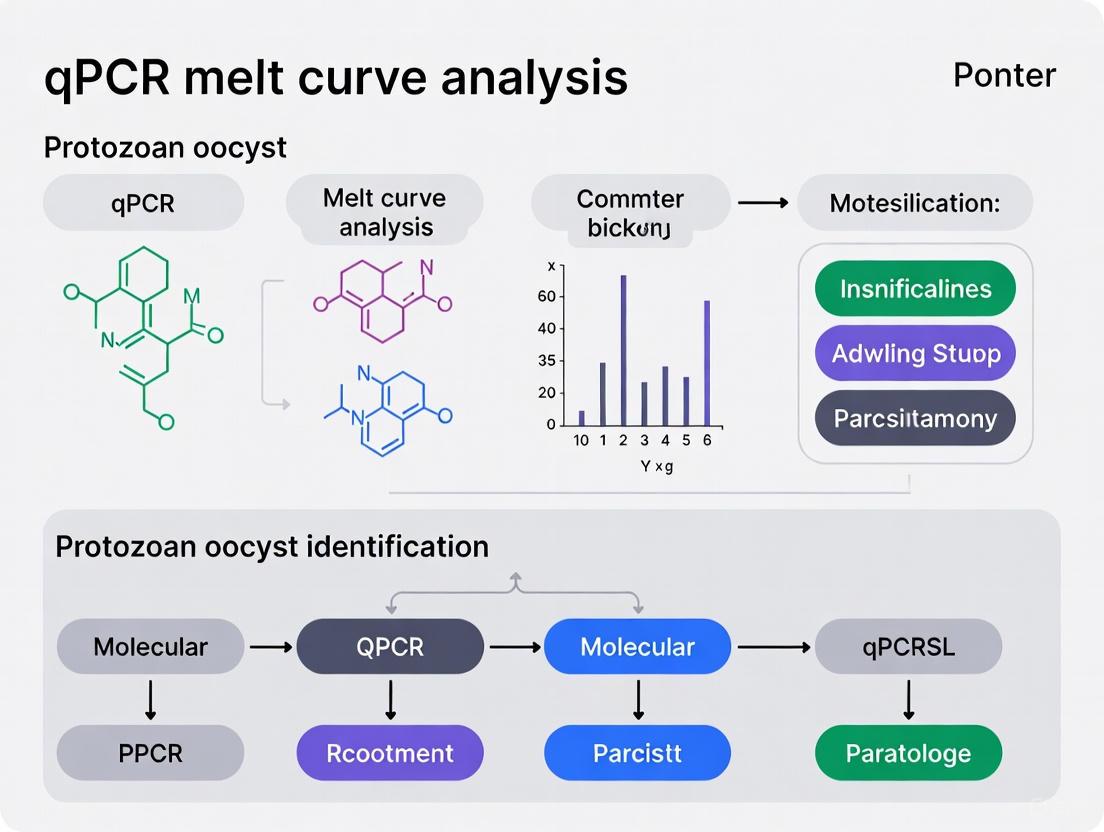
qPCR Melt Curve Analysis for Protozoan Oocyst Identification: A Comprehensive Guide for Molecular Diagnostics
This article provides a comprehensive overview of quantitative PCR (qPCR) coupled with melt curve analysis (MCA) for the sensitive detection and species-level identification of protozoan oocysts.
qPCR Melt Curve Analysis for Protozoan Oocyst Identification: A Comprehensive Guide for Molecular Diagnostics
Abstract
This article provides a comprehensive overview of quantitative PCR (qPCR) coupled with melt curve analysis (MCA) for the sensitive detection and species-level identification of protozoan oocysts. Tailored for researchers and diagnostic professionals, it covers the foundational principles of the technology, detailed methodological protocols for clinical and environmental samples, advanced troubleshooting strategies for assay optimization, and rigorous validation frameworks. By synthesizing recent applications in detecting Cryptosporidium, Cyclospora, and other coccidian parasites, this guide serves as an essential resource for implementing this powerful, cost-effective tool in public health surveillance, food safety, and veterinary diagnostics.
Understanding qPCR Melt Curve Analysis: Principles and Advantages for Oocyst Detection
Core Principles of SYBR Green Chemistry and Melt Curve Analysis
SYBR Green I is a widely used, cost-effective fluorescent dye for quantitative real-time PCR (qPCR) that provides a simple and accurate method for DNA detection and quantification. Its core principle of operation is based on the property of fluorescence enhancement upon binding: the dye is a free-floating molecule that exhibits a significant increase in fluorescence emission only when it intercalates into the double-stranded DNA (dsDNA) minor groove [1] [2]. During the qPCR process, as the DNA polymerase synthesizes new DNA strands, SYBR Green binds to the newly formed double-stranded amplicons. The qPCR instrument measures this increasing fluorescence after each amplification cycle, enabling the quantification of the initial DNA template [1] [3].
A primary advantage of SYBR Green chemistry is its universal applicability; because it binds to any dsDNA, it does not require the design and purchase of expensive target-specific probes, making it ideal for gene expression analysis and other general applications [1] [2]. However, this non-specific binding is also its main drawback. The dye cannot distinguish between the specific target amplicon and non-specific products like primer-dimers or misamplified DNA, which can lead to inaccurate quantification [1] [4]. Furthermore, unlike probe-based methods, SYBR Green assays cannot multiplex, meaning only one target can be analyzed per reaction [1].
The Essential Role of Melt Curve Analysis
Melt curve analysis is a critical post-amplification quality control step that is indispensable for verifying the specificity of SYBR Green qPCR assays [1] [2]. This technique confirms that the fluorescence detected during the run originated from a single, specific amplicon and not from artifacts [1].
The process involves gradually denaturing the PCR products by steadily increasing the temperature from approximately 60°C to 95°C while continuously monitoring fluorescence [1]. As the temperature rises and reaches the melting temperature (Tm) of the amplicon, the dsDNA dissociates into single strands, releasing the SYBR Green dye and causing a rapid decrease in fluorescence [2] [5]. The data is typically presented as a derivative melt curve, which plots the negative derivative of fluorescence relative to temperature (-dF/dT) against temperature. This converts the fluorescence drop-off into a distinct peak, whose position corresponds to the Tm of the product [1].
The presence of a single, sharp peak on the derivative melt curve strongly suggests that only a single PCR product was amplified. Conversely, multiple peaks, shoulders on the main peak, or unusually wide peaks indicate issues such as primer-dimer formation, non-specific amplification, or the presence of multiple amplicons [1]. It is important to note that a single peak is not absolute proof of a pure product, as a single amplicon with complex internal structure can sometimes produce multiple peaks [5]. Therefore, melt curve analysis serves as a powerful indicator, but confirmation by agarose gel electrophoresis is recommended for new assays [1] [5].
Workflow Diagram: SYBR Green qPCR with Melt Curve Analysis
The following diagram illustrates the complete workflow from reagent preparation to data interpretation.
Application in Protozoan Oocyst Identification Research
The combination of SYBR Green qPCR and melt curve analysis (qPCR-MCA) has proven to be a highly effective and reliable method for detecting and differentiating protozoan parasites of significant public health concern. This approach is particularly valuable for identifying coccidian oocysts in complex sample matrices like human feces and food products.
Key Research Findings and Performance
Table 1: Summary of qPCR-MCA Applications in Protozoan Parasite Detection
| Study Focus | Sample Type & Size | qPCR-MCA Performance | Key Detected Pathogens |
|---|---|---|---|
| Clinical Diagnostics [6] [7] | 501 human fecal samples (Dominican Republic) | Reliable screening; more efficient and sensitive than microscopy; detected 10 copies of target. | Cystoisospora belli, Cryptosporidium spp. (parvum, hominis, meleagridis, canis), Cyclospora cayetanensis |
| Food Safety [8] | Leafy greens and berry fruits | Detected as few as 3-5 oocysts per gram of produce; oocyst recovery rates of 4.1-15.5%. | Cryptosporidium, Cyclospora, Toxoplasma (using Eimeria as a surrogate) |
| Malaria Speciation [9] | 300 human blood samples (Iran) | High sensitivity and specificity; complete agreement with sequencing for species identification. | Plasmodium falciparum, Plasmodium vivax |
The power of qPCR-MCA lies in its ability to use universal primer sets that target conserved genomic regions, such as the 18S small subunit ribosomal DNA (SSU rDNA), to detect a broad range of parasites in a single reaction. Subsequent melt curve analysis differentiates the species based on the unique melting temperature (Tm) of each amplicon, which is determined by its GC content, length, and sequence [6] [9]. For instance, one study targeting the 18S SSU rRNA region achieved a significant Tm difference of 2.73°C to distinguish between P. falciparum and P. vivax [9].
Experimental Protocol: qPCR-MCA for Oocyst Detection
The following is a detailed methodology for detecting protozoan oocysts in human fecal samples, adapted from validated protocols [6] [8].
1. Sample Collection and DNA Extraction
- Sample Collection: Collect fecal samples and preserve them in 2.5% potassium dichromate at 4°C.
- Oocyst Concentration: Process samples using a sucrose or ZnSO4 flotation method to concentrate oocysts.
- DNA Extraction: Use a commercial DNA extraction kit (e.g., QIAamp DNA Stool Mini Kit, Qiagen). Incorporate mechanical lysis steps (freeze-thaw cycles) and an overnight proteinase K digestion to ensure efficient oocyst disruption and inhibitor removal. Include positive control (e.g., surrogate oocysts) and negative control (reagents only) in each extraction batch.
2. qPCR Reaction Setup
- Reaction Mix (20 µL final volume):
- 1X SsoFast EvaGreen Supermix (or equivalent SYBR Green master mix)
- 400 nM of each universal coccidia forward and reverse primer (e.g., Crypto-F/R, Cyclo-F/R) [6]
- 5 µL of template DNA
- Cycling Conditions (on a CFX96 Real-Time PCR Detection System or equivalent):
- Initial denaturation: 95°C for 5 minutes
- 40-45 cycles of:
- Denaturation: 95°C for 15 seconds
- Annealing/Extension: 60°C for 30 seconds (with fluorescence acquisition)
3. Melt Curve Analysis
- After amplification, run the melt curve as follows:
- 65°C to 95°C
- Increment by 0.5°C
- Hold for 5-10 seconds at each step, with continuous fluorescence acquisition.
- Analyze the resulting derivative melt curve. Compare the Tm of unknown samples to plasmid controls of known parasite species for identification.
The Scientist's Toolkit: Essential Research Reagents
Table 2: Key Reagents and Materials for qPCR-MCA Experiments
| Item | Function / Description | Example Products / Notes |
|---|---|---|
| SYBR Green Master Mix | Provides DNA polymerase, dNTPs, buffer, and the intercalating dye for fluorescence detection. | SsoFast EvaGreen Supermix (Bio-Rad), Kapa SYBR Fast (Roche), PowerUp SYBR Green (Thermo Fisher) |
| Universal Coccidia Primers | Oligonucleotides designed to amplify a conserved region (e.g., 18S rDNA) across multiple parasite species. | Primers targeting 18S SSU rRNA [6] [9] |
| DNA Extraction Kit | For purifying inhibitor-free genomic DNA from complex samples like feces or food. | QIAamp DNA Stool Mini Kit (Qiagen) [6] |
| Plasmid DNA Controls | Cloned target fragments from known parasite species. Serve as positive controls and Tm standards for melt curve identification. | Linearized plasmid controls for Cryptosporidium, Cyclospora, etc. [6] |
| Real-Time PCR Instrument | Thermocycler capable of precise temperature ramping and fluorescence measurement for qPCR and melt curve generation. | Light Cycler 96 (Roche), CFX96 (Bio-Rad) [9] [6] |
Troubleshooting and Optimization
Successful implementation of SYBR Green qPCR with melt curve analysis requires careful optimization and interpretation.
- Primer Design and Concentration: Primers should be designed with software (e.g., Primer Express) to generate short amplicons (80-150 bp) with an annealing temperature of 58-60°C [3]. Primer concentration must be optimized to maximize specific amplification and minimize primer-dimer formation, which can be a major source of non-specific signal [1] [3].
- Interpreting Complex Melt Curves: A single peak generally indicates a specific product. However, multiple peaks can result from:
- Non-specific amplification: The primers bind to and amplify non-target sequences. This can often be resolved by increasing the annealing temperature or redesigning the primers for greater specificity [1].
- Primer-dimer formation: Primers anneal to themselves or each other. Reducing primer concentration or increasing annealing temperature can help, but primer redesign may be necessary [1].
- Complex amplicon structure: A single, pure amplicon can sometimes produce multiple peaks if it contains regions with differing stability (e.g., high GC-rich domains) that melt at different temperatures [5]. Tools like uMelt software can predict these profiles and aid in assay design and interpretation [5].
- Confirmatory Techniques: For any new assay, it is good practice to confirm the results of the melt curve analysis by running the PCR products on an agarose gel to verify a single band of the expected size [1] [5].
The Critical Role of Melting Temperature (Tm) in Species Differentiation
Quantitative PCR coupled with melting curve analysis (qPCR-MCA) has emerged as a powerful tool for the specific identification and differentiation of closely related species in diagnostic and research settings. This technique leverages the precise melting temperature (Tm) of DNA amplicons, which is a unique function of their sequence composition, length, and GC content. Within the context of protozoan oocyst identification, qPCR-MCA provides a reliable, high-throughput alternative to traditional microscopy, overcoming limitations of labor-intensity, low sensitivity, and the need for specialized expertise [6]. The application of this method is critical for public health, food safety, and veterinary programs, enabling the accurate detection of pathogens like Cryptosporidium spp., Cyclospora cayetanensis, and Cystoisospora belli in clinical, environmental, and food matrices [6] [8].
Principles of Melting Curve Analysis
Following the amplification phase of a qPCR assay using intercalating dyes like SYBR Green, a melting curve analysis is performed. The thermal cycler incrementally increases the temperature while monitoring fluorescence. Intercalating dyes fluoresce brightly when bound to double-stranded DNA (dsDNA) but exhibit minimal fluorescence when unbound or in the presence of single-stranded DNA (ssDNA). As the temperature rises, the dsDNA amplicon denatures, causing the dye to be released and the fluorescence to decrease precipitously. The Tm is defined as the temperature at which half of the dsDNA is denatured, represented by the peak in the negative derivative plot of fluorescence over temperature (-dF/dT vs. Temperature) [5].
A critical assumption is that a single, pure amplicon will produce a single, sharp peak. However, DNA melting is a multi-state process. Complex melting profiles with multiple peaks can arise not only from non-specific amplification but also from a single amplicon with distinct domains of varying stability, such as G/C-rich regions that resist melting until higher temperatures [5]. Therefore, while a single peak often indicates a specific product, multiple peaks should be investigated with complementary techniques like agarose gel electrophoresis or in silico prediction tools such as uMelt software [5] [10].
Application Note: Differentiation of Protozoan Oocysts
Experimental Protocol
Objective: To detect and differentiate protozoan oocyst species in human fecal samples using a universal coccidia qPCR assay followed by melting curve analysis [6].
Workflow Overview: The following diagram illustrates the complete experimental workflow for protozoan oocyst identification:
Materials & Reagents:
- Fecal Samples: Preserved in 2.5% potassium dichromate [6].
- DNA Extraction Kits: QIAamp DNA Stool Mini Kit and QIAamp DNA Micro Kit (Qiagen) [6].
- qPCR Reagents: SsoFast EvaGreen Supermix (Bio-Rad), universal coccidia primer cocktail (e.g., Crypto-F, Crypto-R, Cyclo-F, Cyclo-R) [6].
- Plasmid Controls: Cloned SSU rDNA fragments from target coccidia species for Tm reference [6].
- Equipment: Real-Time PCR Detection System (e.g., CFX96 from Bio-Rad) [6].
Step-by-Step Procedure:
Sample Processing and DNA Extraction:
- Wash approximately 1-2 mL of fecal suspension three times with water to remove potassium dichromate [6].
- Use a commercial DNA extraction kit with modifications: incorporate freeze-thaw cycles (8 cycles of liquid nitrogen and 95°C water bath) and an overnight proteinase K digestion (56°C for 18 hours) to ensure efficient oocyst lysis [6].
- Include negative (reagents only) and positive (e.g., 10^4 Eimeria papillata oocysts) control samples in each extraction batch [6].
qPCR Amplification:
- Prepare a 25 µL reaction mixture containing:
- 1x SsoFast EvaGreen Supermix
- 400 nM of each universal coccidia primer
- Template DNA (e.g., 2 µL of extracted DNA)
- Run the qPCR with the following cycling conditions [6]:
- Initial denaturation: 95°C for 3 min
- 40-45 cycles of:
- Denaturation: 95°C for 10 s
- Annealing/Extension: 56-60°C for 20-30 s
- Prepare a 25 µL reaction mixture containing:
Melting Curve Analysis:
- After amplification, run the melting curve analysis with the following parameters [6]:
- Start at 65°C
- Incrementally increase temperature to 95°C in small steps (e.g., 0.2-0.5°C/step)
- Hold for 5-10 s at each step and measure fluorescence
- After amplification, run the melting curve analysis with the following parameters [6]:
Data Analysis and Species Identification:
- Analyze the melting curves to determine the specific Tm value(s) for each sample.
- Compare the sample Tm values to those of known plasmid controls or reference strains run on the same plate.
- Assign species identity based on the Tm match.
Research Reagent Solutions
The following table details the key reagents and their critical functions in the qPCR-MCA protocol for oocyst identification.
Table 1: Essential Research Reagents for qPCR-MCA-based Oocyst Identification
| Reagent / Kit | Function / Role in the Protocol |
|---|---|
| Universal Coccidia Primers | Target conserved regions (e.g., 18S rDNA) to amplify a broad range of protozoan oocysts in a single reaction [6]. |
| SsoFast EvaGreen Supermix | Provides a ready-to-use mix containing DNA polymerase, dNTPs, buffer, and the intercalating dye for robust qPCR amplification and fluorescence monitoring [6]. |
| QIAamp DNA Stool Mini Kit | Facilitates the isolation of high-quality DNA from complex fecal matrices while removing potent PCR inhibitors [6]. |
| Plasmid DNA Controls | Serve as positive controls and Tm standards for each target species, enabling accurate species calling based on melting temperature [6]. |
| Eimeria papillata Oocysts | A non-pathogenic surrogate used as a process control to monitor DNA extraction efficiency and prepare standard curves [6] [8]. |
Performance Data and Validation
The qPCR-MCA assay has been rigorously validated for the detection of protozoan oocysts. The data below summarize its analytical sensitivity and specificity for differentiating key species.
Table 2: qPCR-MCA Performance in Protozoan Oocyst Detection and Differentiation
| Parameter | Performance Data | Experimental Context |
|---|---|---|
| Analytical Sensitivity | Consistent detection of 10 copies of the cloned target fragment [6]. | Using serial dilutions of plasmid DNA. |
| Detection in Produce | Reliable detection of 3-5 oocysts per gram of food [8]. | Using optimized isolation methods from leafy greens and berries. |
| Specificity (Examples) | Differentiation of C. cayetanensis, C. parvum, C. hominis, C. meleagridis, C. canis, and C. belli [6]. | Analysis of 501 human fecal samples; species confirmed by sequencing. |
| Comparison to Microscopy | More efficient and sensitive than microscopy flotation methods [6]. | Parallel analysis of samples by both qPCR-MCA and microscopy. |
Advanced Applications and Technique Considerations
Multiplexing with 2D Labels
Higher-order multiplexing (beyond 5-plex) can be achieved by combining fluorescence color and Tm as a two-dimensional (2D) label. This approach uses multiple fluorophores, each paired with several probes designed to have distinct Tm values. This creates a library of unique "color-Tm" combinations, allowing for the identification of numerous targets in a single reaction, as demonstrated in genotyping 15 human papillomaviruses using four fluorescence channels and ten Tm values [11].
Mutation Detection and Genotyping
MCA with hybridization probes (e.g., using FRET) enables high-resolution genotyping. A detection probe spanning the mutation site is designed. A single nucleotide mismatch destabilizes the probe-target hybrid, resulting in a measurable decrease in Tm. This allows for the discrimination of wild-type and mutant alleles, such as differentiating extended-spectrum β-lactamase (ESBL) genes from their non-ESBL counterparts in less than an hour [12].
Troubleshooting and Optimization
- Multiple Peaks: Can indicate non-specific amplification, primer dimers, or a single amplicon with complex secondary structure. Validation via agarose gel electrophoresis or uMelt prediction software is recommended [5] [10].
- Primer Design: Primers should be designed for an annealing temperature of 60-65°C, with an amplicon length of 70-200 bp. The annealing temperature must be determined empirically for each primer set and master mix [13] [10].
- Inhibition: Samples like feces or produce can contain PCR inhibitors. The use of an internal control (IC) and DNA extraction kits designed to remove inhibitors is critical [6] [8].
Quantitative PCR (qPCR) coupled with melt curve analysis represents a significant advancement in molecular diagnostics, particularly for the identification of protozoan oocysts. This technique provides a powerful closed-tube system that combines nucleic acid amplification with subsequent product identification based on dissociation characteristics. For researchers and drug development professionals working on enteric pathogens, this method offers substantial benefits over traditional techniques like microscopy and sequencing. This application note details the specific advantages of qPCR melt curve analysis in terms of sensitivity, specificity, and throughput, providing both quantitative comparisons and detailed protocols for implementation in protozoan oocyst identification research.
Comparative Performance Data
The transition from traditional microscopy to molecular methods for protozoan oocyst detection has been driven by demonstrated improvements in key performance metrics. The tables below summarize quantitative data comparing qPCR melt curve analysis to conventional methods across multiple studies.
Table 1: Comparison of detection methods for protozoan oocysts
| Detection Method | Target Organisms | Sensitivity/LOD | Specificity | Sample Throughput | Turnaround Time | Reference |
|---|---|---|---|---|---|---|
| qPCR with Melt Curve Analysis | Multiple diarrheal parasites | 8.78-30.08 copies/μL | 95.8% concordance with reference methods | 5-plex detection in single reaction | ~2 hours | [14] |
| qPCR with Melt Curve Analysis | Coccidian oocysts | 10 copies of cloned target | More efficient than microscopy | Multiple species detection | Not specified | [7] |
| Microscopy | Protozoan oocysts | Variable, operator-dependent | Variable, operator-dependent | Low | 30-60 minutes/sample | [14] |
| Sanger Sequencing | SARS-CoV-2 variants | Lower sensitivity than RT-qPCR assays | 92.6-100% agreement with RT-qPCR | Low, requires specialized staff | ~24 hours | [15] |
Table 2: Performance characteristics of multiplex qPCR-HRM assay for diarrheal parasites
| Parasite | Melting Temperature (°C) | Limit of Detection (copies/μL) | PCR Efficiency (%) | R² Value |
|---|---|---|---|---|
| Cryptosporidium spp. | 78.23 ± 0.25 | 8.78 | 103.11 | 0.9998 |
| Entamoeba histolytica | 75.20 ± 0.25 | 30.08 | 95.77 | 0.9942 |
| Giardia intestinalis A | 83.50 ± 0.00 | 10.00 | 99.76 | 0.9989 |
| Giardia intestinalis B | 81.51 ± 0.08 | 100.00 | 101.22 | 0.9973 |
| Blastocystis spp. | 79.84 ± 0.23 | 10.00 | 100.18 | 0.9981 |
| Dientamoeba fragilis | 71.50 ± 0.00 | 10.00 | 98.52 | 0.9975 |
Advantages Over Traditional Methods
Enhanced Sensitivity
qPCR melt curve analysis demonstrates significantly improved sensitivity compared to traditional microscopy. While microscopy relies on visual identification and is limited by operator skill and oocyst concentration, qPCR can detect as few as 10 target copies/μL [7] [14]. This exceptional sensitivity is particularly valuable for detecting low-level infections and carrier states that often go undetected by conventional methods. The 5-plex qPCR-HRM assay detected additional Cryptosporidium infections (2.8%) and Dientamoeba fragilis infections (4.2%) that were missed by conventional methods in a clinical validation study [14].
The limit of detection (LOD) for qPCR melt curve analysis is both quantifiable and reproducible, unlike microscopy which has variable sensitivity dependent on operator expertise. The mathematical basis for this sensitivity stems from the exponential amplification of target nucleic acids, enabling detection of even single copies of target DNA with 95% confidence when proper quality control measures are implemented [16].
Superior Specificity
The specificity of qPCR melt curve analysis operates at two levels: primer specificity during amplification and melt curve profile during product identification. This dual-layer specificity provides more reliable identification compared to microscopy, which struggles to differentiate morphologically similar oocysts. In clinical validation, qPCR melt curve analysis demonstrated 95.8% concordance with reference methods while additionally detecting missed infections [14].
The melting temperature (Tm) differences between closely related protozoan species are sufficient for clear differentiation. The 5-plex parasite assay maintained ΔTm values of at least 1.5°C between all targets, with the smallest difference being 1.61°C between Cryptosporidium and Blastocystis [14]. This specificity is further enhanced through careful primer design targeting genetically conserved regions unique to each parasite, such as the E1, E4, and L1 regions in HPV genotyping assays [17].
Increased Throughput
The throughput advantages of qPCR melt curve analysis are substantial, enabling multiplex detection of multiple pathogens in a single reaction. The 5-plex parasite assay simultaneously detects and differentiates six targets (including both Giardia assemblages) in a single closed-tube reaction [14]. This multiplex capacity dramatically increases processing efficiency compared to microscopy, which requires individual examination for each parasite.
The streamlined workflow of qPCR melt curve analysis reduces hands-on time and total processing time. A complete analysis can be performed in approximately 2 hours [14], compared to Sanger sequencing which requires approximately 24 hours with specialized staff [15]. The closed-tube nature of the technique eliminates post-amplification processing, reducing contamination risk and enabling automation potential for high-throughput screening applications.
Detailed Experimental Protocol
Multiplex qPCR-HRM for Diarrheal Parasites
Table 3: Research reagent solutions for multiplex qPCR-HRM
| Reagent Category | Specific Examples | Function in Protocol |
|---|---|---|
| Primers | Target-specific primers for conserved regions | Specific amplification of target parasite DNA |
| DNA Polymerase | Hot-start DNA polymerase (e.g., Kapa 2G Fast) | High-efficiency amplification with reduced non-specific products |
| Fluorescent Dye | SYBR Green I, EvaGreen, LCGreen | Intercalation with dsDNA for fluorescence monitoring |
| Sample Material | Stool samples, cultured oocysts | Source of target DNA for detection |
| DNA Purification Kits | Magnetic bead-based systems (e.g., BasePurifier) | Nucleic acid extraction and purification |
| Positive Controls | Plasmids with target sequences, reference strains | Assay validation and quality control |
Sample Preparation and DNA Extraction
- Sample Collection: Collect fecal samples in appropriate storage media. For water samples, concentrate oocysts by filtration and centrifugation.
- DNA Extraction: Use commercial DNA extraction kits with magnetic bead-based systems. Transfer 200-300 μL of sample to the extraction system [15].
- Purification Steps: Follow binding, wash, and elution steps according to manufacturer instructions. Elute DNA in 80 μL buffer [15].
- Quality Assessment: Measure DNA concentration and purity using spectrophotometry. Store extracted DNA at -20°C until use.
Primer Design and Validation
- Target Selection: Identify conserved genomic regions unique to each target parasite. For protozoan oocysts, target regions may include 18S rDNA, COWP, or other genus-specific genes [7] [14].
- Primer Design: Use Primer-BLAST or similar software to design primers with compatible Tm (60-65°C), length (18-22 bp), and GC content (40-60%).
- Specificity Validation: Test primer specificity in silico using BLAST against non-target organisms.
- Empirical Validation: Validate primers using reference strains and clinical samples with known status.
qPCR-HRM Reaction Setup
Reaction Composition:
- 1× PCR buffer
- 3-5 mM MgCl₂ (optimize for each assay)
- 0.2 mM each dNTP
- 0.5 μM each primer
- 1× saturating DNA dye (SYBR Green I, EvaGreen, or LCGreen)
- 0.5-1.0 U DNA polymerase
- 5 μL DNA template
- Nuclease-free water to 25 μL
Cycling Conditions:
- Initial denaturation: 95°C for 5 minutes
- 45 cycles of:
- Denaturation: 95°C for 10 seconds
- Annealing: 56°C for 10 seconds
- Extension: 60°C for 20 seconds [14]
- Melting curve analysis: 65°C to 95°C with 0.2°C increments
Data Analysis
- Amplification Analysis: Determine Cq values using baseline subtraction and threshold setting in exponential phase [18].
- Melting Curve Analysis:
- Convert melting curves to negative derivative plots
- Identify peak melting temperatures (Tm)
- Compare sample Tm to reference Tm for identification
- Result Interpretation:
- Positive identification when Tm matches reference within acceptable range (±0.5°C)
- Reject reactions with non-specific melting peaks or primer-dimer artifacts
Workflow Visualization
Diagram 1: Comparative workflow of qPCR melt curve analysis versus traditional microscopy
Diagram 2: Principles of melt curve analysis for product identification
Discussion and Implementation Considerations
Advantages Synthesis
The collective data demonstrate that qPCR melt curve analysis provides significant advantages over traditional methods for protozoan oocyst identification. The enhanced sensitivity enables detection of low-level infections crucial for public health surveillance and treatment monitoring. The superior specificity reduces false positives and enables differentiation of morphologically similar species that require different treatment approaches. The increased throughput allows laboratories to process more samples with less hands-on time, making large-scale screening programs feasible.
Practical Implementation
For laboratories implementing qPCR melt curve analysis, several factors require consideration. Proper validation against reference methods is essential, with particular attention to limit of detection, reproducibility, and specificity testing against common confounders [16]. Assay design should incorporate appropriate controls, including no-template controls, positive controls, and internal amplification controls when possible.
The melting curve analysis requires optimization of ramp rates and temperature resolution to ensure accurate Tm determination [18] [5]. For multiplex applications, Tm differences of at least 1.5°C between targets are recommended, with 2°C or greater preferred [14]. uMelt software or similar prediction tools can assist in assay design by forecasting melting profiles before empirical testing [5].
Quality Assurance
Adherence to MIQE (Minimum Information for Publication of Quantitative Real-Time PCR Experiments) guidelines ensures robust assay performance and reproducibility [16]. Key parameters including PCR efficiency (90-110%), dynamic range (at least 3 log orders of magnitude), and R² values (>0.98) should be monitored regularly. Melting curve analysis should include verification of product specificity through sequencing during validation and periodic monitoring thereafter.
qPCR melt curve analysis represents a significant advancement in protozoan oocyst identification, offering demonstrated improvements in sensitivity, specificity, and throughput compared to traditional microscopy and other conventional methods. The technique provides a robust platform for clinical diagnostics, epidemiological studies, and drug development applications. The protocols and data presented in this application note provide researchers with the necessary foundation to implement this powerful technology in their laboratories, potentially transforming approaches to parasitic disease diagnosis and monitoring.
The accurate detection and identification of protozoan parasites are critical for public health, clinical diagnostics, and environmental monitoring. Molecular methods, particularly quantitative polymerase chain reaction (qPCR), have become indispensable tools for this purpose. Their success, however, hinges on the selection of appropriate genetic targets that provide a balance of specificity and conservation across relevant species. This application note focuses on three key genetic targets—18S ribosomal DNA (rDNA), Cryptosporidium Oocyst Wall Protein (COWP), and various mitochondrial genes—within the context of protozoan oocyst identification research. We provide detailed protocols and validation data for assays targeting these regions, enabling researchers to implement robust detection strategies for foodborne and waterborne parasites such as Cryptosporidium, Cyclospora, and Sarcocystis.
Key Genetic Targets and Their Applications
The selection of a genetic target dictates the specificity, sensitivity, and application range of a molecular assay. The table below summarizes the core characteristics of the primary genetic targets discussed in this note.
Table 1: Key Genetic Targets for Protozoan Oocyst Identification
| Genetic Target | Key Characteristics | Primary Applications | Example Parasites |
|---|---|---|---|
| 18S rDNA | - Multi-copy gene enhancing sensitivity- Highly conserved across eukaryotes- Requires careful normalization or signal attenuation due to high abundance | Broad-range detection and phylogenetic studies | Various protozoan parasites [19] |
| COWP Gene | - Single-copy gene- Species-specific regions enable differentiation- Ideal for absolute quantification | Specific detection and quantification of Cryptosporidium species | C. parvum, C. hominis, C. ubiquitum [20] [21] |
| Mitochondrial Genes (e.g., cox1) | - High copy number per cell increases sensitivity- Provides high resolution for species differentiation | Discriminating between closely related species | Sarcocystis spp. [22] |
Detailed Experimental Protocols
Protocol 1: Sensitive Detection of Cryptosporidium via COWP Gene qPCR
This protocol is adapted from a validated method for the sensitive detection and absolute quantification of Cryptosporidium spp. by targeting the COWP gene [20] [21].
Sample Preparation and DNA Extraction
- Water Samples: Filter 1L of water through a series of filters: first a metal sieve (1 mm pores), then a qualitative filter paper, and finally a membrane filter with 5 µm pores [22]. Use 2 mL of distilled water to wash the membrane and collect the material.
- Leafy Greens and Berry Fruits: Process 30-50 g of produce. For soft herbs (e.g., cilantro, mint) use a stomacher with glycine buffer. For berries and woody-stemmed herbs (e.g., thyme), use orbital shaking with an elution solution to minimize PCR inhibitors [8].
- DNA Extraction: Extract genomic DNA from 200 µL of the concentrated sample using a commercial DNA purification kit, following the manufacturer's instructions. Elute DNA in a volume of 50-100 µL and store at -20°C.
Primer Design and qPCR Assay
- Primer Design: The following degenerate primers target a conserved region of the COWP gene, generating a 311-317 bp amplicon [21]:
- Forward Primer: 5'- [COWP Conserved Sequence] -3'
- Reverse Primer: 5'- [COWP Conserved Sequence] -3'
- qPCR Reaction Setup:
- Total Volume: 20 µL
- SYBR Green Master Mix: 10 µL
- Forward & Reverse Primers (10 µM each): 0.5 µL each
- Template DNA: 2-5 µL
- Nuclease-free Water: to 20 µL
- Thermal Cycling Conditions:
- Initial Denaturation: 95°C for 5 minutes
- 40 Cycles of:
- Denaturation: 95°C for 30 seconds
- Annealing/Extension: 60°C for 45 seconds
- Melt Curve Analysis: 65°C to 95°C, with increment of 0.5°C every 5 seconds.
Absolute Quantification Using a Standard Curve
- Standard Curve Construction: Clone the COWP target region into a suitable plasmid (e.g., pET-15b). Prepare a serial dilution of the plasmid, typically from 10² to 10⁸ copies/µL, to generate the standard curve [21].
- Validation Parameters: A slope of -3.279 corresponds to 100.8% PCR efficiency. The assay should demonstrate a strong linear correlation (R² = 0.95) and a limit of detection (LOD) as low as 9.55 × 10⁴ copies/µL [21].
Protocol 2: Multi-Species Detection of Sarcocystis via cox1 Gene
This protocol uses nested PCR targeting the mitochondrial cytochrome c oxidase subunit I (cox1) gene to detect multiple Sarcocystis species in environmental water samples [22].
Primer Design and Nested PCR
- Primer Selection: Design species-specific primer pairs for the first and second rounds of PCR. For example, for S. bovifelis detection [22]:
- First Round Primers:
- V2bo1 (Forward): 5'- AACTTCCTAGGTACAGCGGTATTCG -3'
- V2bo2 (Reverse): 5'- TGAACAGCAGTACGAAGGCAAC -3'
- Second Round Primers:
- V2bo3 (Forward): 5'- ATATTTACCGGTGCCGTACTTATGTT -3'
- V2bo4 (Reverse): 5'- GCCACATCATTGGTGCTTAGTCT -3'
- First Round Primers:
- Nested PCR Reaction:
- First Round: Use 2-5 µL of extracted DNA in a 25 µL reaction. Cycling conditions: initial denaturation at 95°C for 5 min; 35 cycles of 95°C for 30s, 60°C for 40s, 72°C for 45s; final extension at 72°C for 7 min.
- Second Round: Use 1 µL of the first-round PCR product as a template. Cycling conditions are similar, but with a reduced number of cycles (e.g., 25-30 cycles).
Analysis and Interpretation
- Gel Electrophoresis: Analyze 5 µL of the final PCR product on a 1.5-2% agarose gel. A single band of the expected size (e.g., 410 bp for S. bovifelis) indicates positive detection.
- Sequencing: For definitive species identification, purify the PCR product and perform Sanger sequencing, followed by comparison to databases like NCBI GenBank.
Using 18S rRNA as an Internal Control with Competimers
The 18S rRNA gene is a common internal control in relative RT-PCR due to its stable expression across many sample types. However, its high abundance can overwhelm the PCR reaction. This protocol outlines its use with competimers for accurate normalization [19].
- Principle: Competimers are modified primers identical in sequence to the functional 18S rRNA primers but blocked at their 3'-end so they cannot be extended. Mixing them with active primers attenuates the amplification of the abundant 18S rRNA to a level comparable to rare target transcripts.
- Procedure:
- Determine Optimal Ratio: Perform a primer:competimer ratio test (e.g., 1:9, 2:8, 3:7) for your specific sample and target to find the ratio that brings the 18S rRNA Ct value within 4-5 cycles of your target gene's Ct value.
- Multiplex RT-PCR: Use the optimized primer:competimer mix in a multiplex qPCR reaction alongside your gene-specific primers.
- Data Analysis: Use the ΔΔCt method to calculate the relative expression of your target gene, normalized to the attenuated 18S rRNA signal.
Critical Quality Control: Melt Curve Analysis
Melt curve analysis is an essential quality control step when using intercalating dyes like SYBR Green I to verify that a single, specific amplicon has been generated [1].
- Procedure: After the final amplification cycle, the thermal cycler gradually increases the temperature from about 60°C to 95°C while continuously monitoring fluorescence. As the temperature passes the melting temperature (Tm) of the amplicon, the double-stranded DNA denatures, causing a sharp drop in fluorescence [5].
- Interpretation:
- A single, sharp peak in the derivative melt curve plot suggests specific amplification of a single product.
- Multiple peaks or shoulders may indicate primer-dimer formation, non-specific amplification, or the presence of multiple amplicons [1].
- Troubleshooting: Multiple peaks can sometimes be caused by a single, complex amplicon with multiple melting domains due to G/C-rich regions [5]. Tools like uMelt software can predict an amplicon's melt profile and help distinguish between a complex single product and non-specific amplification. Always confirm the specificity of a reaction with a suspicious melt curve by running an agarose gel [5].
The Scientist's Toolkit: Essential Research Reagents
Table 2: Key Reagent Solutions for Protozoan Oocyst Identification by qPCR
| Reagent / Kit | Function | Application Notes |
|---|---|---|
| SYBR Green I Master Mix | Fluorescent dye for real-time PCR product detection. | Cost-effective; requires melt curve analysis for specificity confirmation [1]. |
| QuantumRNA 18S rRNA Primers & Competimers | For attenuation of abundant 18S rRNA signal during co-amplification. | Essential for using 18S rRNA as an internal control for rare target transcripts [19]. |
| GeneJET Genomic DNA Purification Kit | Isolation of high-quality genomic DNA from complex samples. | Used for DNA extraction from concentrated water samples [22]. |
| MF-Millipore Membrane Filters (5 µm) | Concentration of oocysts from large volume water samples. | Pore size is critical for efficiently capturing target oocysts [22]. |
| Clustal Omega | Multiple sequence alignment tool for identifying conserved regions. | Used for identifying degenerate primer binding sites in the COWP gene [21]. |
| uMelt Software | Prediction of high-resolution melting curves for amplicon analysis. | Helps interpret complex melt curves and design assays for HRM analysis [5]. |
Workflow and Pathway Diagrams
Molecular Detection of Protozoan Oocysts
qPCR Assay Development and Validation
Quantitative Polymerase Chain Reaction with Melting Curve Analysis (qPCR-MCA) represents a significant advancement in molecular diagnostics for public health, enabling rapid, sensitive, and specific detection and differentiation of protozoan parasites. This technology is particularly valuable for identifying coccidian oocysts, including Cryptosporidium spp., Cyclospora cayetanensis, and Cystoisospora belli, which are significant causes of gastrointestinal illness worldwide [7]. These pathogens pose substantial challenges in both clinical settings, where they cause prolonged diarrheal illness, and in food safety contexts, where they contaminate fresh produce and water supplies [6]. Traditional detection methods relying on microscopy are labor-intensive, require specialized expertise, and lack sensitivity and specificity, often leading to underreporting of these pathogens [6]. The integration of qPCR with melting curve analysis provides a powerful tool that overcome these limitations, offering a reliable screening assay for clinical, environmental, and veterinary samples in public health programs [7]. This application note details standardized protocols and data analysis methods for implementing qPCR-MCA in public health laboratories for both clinical diarrhea investigation and foodborne outbreak response.
Experimental Principles and Workflow
The qPCR-MCA method utilizes universal primer sets targeting conserved regions of the 18S ribosomal DNA (rDNA) gene that are common across various coccidian parasites [7] [6]. Following amplification, the resulting PCR products are subjected to a controlled temperature increase while monitoring fluorescence. As the double-stranded DNA amplicons denature into single strands at specific temperatures (Tm), a rapid decrease in fluorescence occurs [6]. The Tm value is characteristic for each species due to variations in their G-C content and amplicon length, enabling differentiation without the need for probe hybridization or post-PCR processing [7]. This closed-tube system minimizes contamination risk and allows for high-throughput analysis, making it ideal for rapid response during outbreak investigations.
Workflow Visualization
Materials and Reagents
Research Reagent Solutions
Table 1: Essential Research Reagents and Materials
| Item | Function/Application | Example/Specification |
|---|---|---|
| Universal Coccidia Primer Cocktail | Amplification of 18S rDNA gene region conserved across coccidian species [6] | Crypto-F, Crypto-R, Cyclo-F, Cyclo-R (400 nM each) |
| DNA Extraction Kit | Isolation of inhibitor-free DNA from complex matrices (feces, produce) [6] | QIAamp DNA Stool Mini Kit (Qiagen) with modified protocol |
| Fluorescent DNA Binding Dye | Detection of amplified DNA during qPCR and melting phase [6] | SsoFast EvaGreen Supermix (Bio-Rad) |
| Plasmid DNA Controls | Positive controls for species identification via Tm comparison [6] | Cloned SSU rDNA fragments from target coccidia species |
| Oocyst Surrogate | Process control for method validation and recovery efficiency [8] | Eimeria papillata oocysts propagated in mice |
| Sample Wash Buffers | Oocyst elution from various food matrices with minimal inhibitor release [8] | Glycine buffer (0.1 M; pH 5.5) or Elution Solution (0.1% Tween-H2O) |
Application Notes: Protocol for Clinical Specimens
Sample Preparation and DNA Extraction from Stool
- Washing: Approximately 1-2 mL of fecal suspension preserved in potassium dichromate is washed three times with milli-Q H₂O by centrifugation (20,000 × g for 15 minutes) to remove the preservative [6].
- Lysis: The resulting 200 μL fecal pellet is mixed with 1.4 mL ASL buffer (Qiagen) and subjected to eight freeze-thaw cycles (liquid nitrogen for 1 minute followed by a 95°C water bath for 1 minute) to rupture oocyst walls [6].
- Proteinase K Digestion: The sample is incubated with 20 μL proteinase K (20 mg/mL) for 18 hours at 56°C to ensure complete lysis and degradation of contaminating proteins [6].
- Inhibitor Removal: The lysed suspension is centrifuged at 20,000 × g for 3 minutes, and 1.4 mL of supernatant is treated with an InhibitEX tablet according to the manufacturer's protocol to remove PCR inhibitors commonly found in stool [6].
- DNA Purification: The resulting lysate is incubated with 200 μL Buffer AL for 10 minutes at 70°C, followed by purification through a QIAamp microcolumn and elution with 35 μL AE buffer [6].
qPCR-MCA Amplification and Analysis
- Reaction Setup: Prepare a master mix containing 1× SsoFast EvaGreen Supermix, 400 nM of each universal coccidia primer (e.g., Crypto-F, Crypto-R, Cyclo-F, Cyclo-R), and nuclease-free water. Aliquot 18 μL of master mix per well and add 2 μL of template DNA [6].
- Thermal Cycling: Perform amplification on a CFX96 Real-Time PCR Detection System (Bio-Rad) or equivalent using the following cycling conditions [6]:
- Initial denaturation: 98°C for 2 minutes
- 40 cycles of:
- Denaturation: 98°C for 5 seconds
- Annealing/Extension: 60°C for 15 seconds with fluorescence acquisition
- Melting Curve Analysis: Following amplification, immediately generate a melting curve by:
- Denaturing at 95°C for 30 seconds
- Annealing at 60°C for 30 seconds
- Gradually increasing temperature from 60°C to 95°C with a ramp rate of 0.2°C per second and continuous fluorescence acquisition [6].
- Data Interpretation: Identify species by comparing the Tm of unknown samples to plasmid DNA controls from known coccidia species included in each run. Confirm putative positives by conventional PCR and sequencing of the qPCR products [7] [6].
Application Notes: Protocol for Food Matrices
Optimization for Produce Analysis
The physical and biochemical differences between various types of produce necessitate optimized isolation methods for different commodity groups, as summarized in Table 2.
Table 2: Optimized Oocyst Isolation Methods for Different Produce Types
| Produce Type | Examples | Optimal Processing Method | Optimal Wash Buffer | Average Oocyst Recovery Rate |
|---|---|---|---|---|
| Soft Berries | Blackberries, Raspberries, Strawberries | Orbital Shaking | Elution Solution | 4.1 - 12% [8] |
| Blueberries | Blueberries | Orbital Shaking | Glycine Buffer (0.1 M; pH 5.5) | 4.1 - 12% [8] |
| Leafy Herbs (Soft Stems) | Cilantro, Parsley, Mint | Stomaching | Glycine Buffer (0.1 M; pH 5.5) | 5.1 - 15.5% [8] |
| Woody Herbs | Thyme | Orbital Shaking | Elution Solution | 5.1 - 15.5% [8] |
| Allium Vegetables | Green Onions | Orbital Shaking | Elution Solution | 5.1 - 15.5% [8] |
Oocyst Isolation from Leafy Greens and Berry Fruits
- Sample Processing: Weigh 50 g of the produce sample. Process using either an orbital shaker or stomacher according to the optimized methods outlined in Table 2 for 30 minutes to elute oocysts from the produce surface [8].
- Concentration: Centrifuge the wash supernatant at 2,060 × g for 15 minutes to pellet oocysts. Carefully decant the supernatant [6].
- DNA Extraction and qPCR-MCA: Extract DNA from the pellet using the modified stool kit protocol described in Section 4.1. Perform qPCR-MCA as detailed in Section 4.2 [8].
Performance Data and Validation
Analytical Sensitivity and Specificity
The qPCR-MCA assay has been rigorously validated for sensitivity and specificity. The assay consistently detects as few as 10 copies of the cloned target SSU rDNA fragment [7] [6]. When applied to spiked produce samples, the optimized methods can reliably detect 3-5 oocysts per gram of food, demonstrating high sensitivity even in complex matrices [8].
The universal primer cocktail, combined with MCA, differentiates a broad range of coccidia species based on distinct Tm values. This allows for the specific identification of human pathogens like C. cayetanensis while distinguishing them from closely related non-zoonotic Eimeria spp., thereby reducing false-positive results [6].
Application in Public Health Surveillance
A study in the Dominican Republic successfully applied this assay to 501 human fecal samples, demonstrating its utility in public health surveillance. The assay identified multiple protozoan species, with results confirmed by sequencing [7] [6]. The distribution of pathogens detected is summarized in Table 3.
Table 3: Protozoan Oocysts Detected by qPCR-MCA in 501 Human Fecal Samples from the Dominican Republic
| Identified Pathogen | Number of Positive Samples | Confirmation Method |
|---|---|---|
| Cyclospora cayetanensis | 9 | Sequencing [7] |
| Cryptosporidium hominis | 5 | Sequencing [7] |
| Cystoisospora belli | 3 | Sequencing [7] |
| Cryptosporidium parvum | 3 | Sequencing [7] |
| Cryptosporidium meleagridis | 1 | Sequencing [7] |
| Cryptosporidium canis | 1 | Sequencing [7] |
The qPCR-MCA protocol provides public health, veterinary, and food safety laboratories with a comprehensive, efficient, and reliable method for detecting and differentiating protozoan oocysts. Its superior sensitivity and specificity compared to traditional microscopy, combined with the ability to screen for multiple pathogens simultaneously, make it an invaluable tool for diagnosing clinical cases and investigating foodborne outbreaks. The optimized protocols for various sample matrices ensure broad applicability, enhancing surveillance capabilities and supporting timely public health interventions.
Protocol Development: Implementing qPCR-MCA for Clinical and Environmental Samples
Primer Design Strategies for Universal and Species-Specific Amplification
Within the framework of research on qPCR melt curve analysis for protozoan oocyst identification, the design of amplification primers is a critical foundational step. The choice between universal and species-specific primer strategies directly influences the sensitivity, specificity, and ultimate success of molecular assays for detecting protozoan parasites such as Cryptosporidium, Giardia, and Eimeria [13] [23]. This application note details standardized protocols for both approaches, providing researchers with methodologies to develop robust assays capable of identifying and differentiating protozoan oocysts, a crucial need in both clinical diagnostics and environmental surveillance [20] [24] [25].
Universal Primer Design for Broad-Spectrum Detection
Universal primers target conserved genetic regions across a broad taxonomic range, enabling the detection of multiple parasite genera or species in a single reaction. The 18S ribosomal RNA (rRNA) gene is a frequent target due to the presence of conserved regions flanking variable domains that provide taxonomic resolution [23] [26] [27].
Protocol: Designing and Validating Universal 18S rRNA Primers
This protocol is adapted from methods used for the simultaneous detection of Cryptosporidium spp., Giardia spp., and Toxoplasma gondii in complex sample matrices [23].
Target Identification and Sequence Alignment
- Retrieve full-length 18S rRNA gene sequences for your target protozoa (e.g., C. parvum, G. enterica, T. gondii) and the host organism (e.g., Crassostrea virginica) from databases like GenBank.
- Perform a multiple sequence alignment using a tool like Clustal Omega or the alignment function in Geneious to identify conserved regions suitable for primer binding [23].
Primer Design
- Design primers to flank a variable region (e.g., the V4 region). The variable region provides sequence diversity for subsequent melt curve analysis or sequencing-based differentiation [23].
- Digital Validation: Use software (e.g., Geneious) to check for primer self-complementarity, dimer formation, and % GC-content. Verify that primers have minimal complementarity to the host 18S rRNA sequence to reduce background amplification [23].
Wet-Lab Validation
- Perform PCR amplification using a high-fidelity polymerase.
- Test primers with single-species and mixed-species templates.
- Confirm successful amplification by visualizing bands of the expected size on a 2% agarose gel.
- For definitive confirmation, purify the amplicon and submit it for Sanger sequencing [23].
Overcoming Host DNA Background
A significant challenge in universal PCR from clinical or environmental samples is the overwhelming presence of host DNA. The following strategy can enrich for parasite-derived sequences:
- Nested PCR with Restriction Digestion: This method increases sensitivity and specificity by performing two consecutive PCRs and incorporating a restriction digestion step to degrade host amplicons [26].
- Blocking Primers: Design oligonucleotides with a C3-spacer or peptide nucleic acid (PNA) chemistry that specifically bind to the host 18S rRNA sequence at the primer binding site. These modified oligos block polymerase elongation, thereby selectively inhibiting host DNA amplification [27].
Diagram 1: Workflow for nested PCR with host DNA reduction. Optional blocking primers can be added to the first PCR to further suppress host amplification [26] [27].
Species-Specific Primer Design for Targeted Quantification
Species-specific primers target unique genetic sequences, allowing for precise detection and absolute quantification of a single protozoan species, which is vital for understanding infection dynamics [20] [24] [28].
Protocol: Targeting the Cryptosporidium Oocyst Wall Protein (COWP) Gene
This protocol outlines the development of a qPCR assay for the specific detection and quantification of Cryptosporidium spp. [20] [24].
Target Selection and In Silico Analysis
- Gene Identification: Select a suitable target gene, such as the Cryptosporidium oocyst wall protein (COWP) gene, which is conserved across major species like C. parvum, C. hominis, and C. ubiquitum [24].
- Sequence Retrieval and Alignment: Download all available COWP gene sequences for your target species from NCBI in FASTA format. Perform a multiple sequence alignment using Clustal Omega to identify a suitably conserved region for primer design [24].
- Primer Design: Design primers, potentially incorporating degeneracy to account for minor sequence variations across species. Amplicon length should be optimized for qPCR, ideally between 60-150 bp [29] [24].
Assay Validation and Standard Curve Construction
- Standard Curve: For absolute quantification, clone the target amplicon into a plasmid vector (e.g., pET-15b). Use a dilution series of this plasmid of known copy number to generate a standard curve [20] [24].
- Validation Parameters: The standard curve should demonstrate a slope near -3.32, efficiency between 90-110%, and a strong linear correlation (R² > 0.95) [20] [24].
- Specificity Testing: Validate primer specificity using melt curve analysis post-qPCR. A single, sharp peak indicates specific amplification of the target sequence [20] [24].
Bioinformatics Pipelines for High-Throughput Design
For designing multiple species-specific assays, automated bioinformatics pipelines can streamline the process.
- SpeciesPrimer Pipeline: This tool automates genome download, annotation, and pan-genome analysis to identify single-copy core genes unique to the target species. It then designs primers for these species-specific conserved sequences and performs in-silico quality control [30].
Table 1: Performance Metrics of a Species-Specific qPCR Assay for Cryptosporidium spp. [20] [24]
| Assay Parameter | Target Gene | Amplicon Size | Amplification Efficiency | Linearity (R²) | Limit of Detection (LOD) |
|---|---|---|---|---|---|
| Value | COWP | 311-317 bp | 100.8% | 0.95 | 9.55 × 10⁴ copies/µL |
Table 2: Key Research Reagent Solutions for Primer Design and Validation
| Reagent / Resource | Function / Application | Examples / Specifications |
|---|---|---|
| High-Fidelity Polymerase | Reduces PCR errors during initial amplification and validation of primer pairs. | KAPA HiFi Polymerase [23] |
| Cloning Vector | Serves as a template for generating a standard curve for absolute quantification in qPCR. | pET-15b vector [20] [24] |
| Blocking Primers | Suppresses amplification of non-target (e.g., host) DNA in universal assays to improve sensitivity. | C3-spacer modified oligos; Peptide Nucleic Acid (PNA) oligos [27] |
| Bioinformatics Tools | In silico design, specificity checking, and quality control of primer sequences. | SpeciesPrimer [30], Primer-BLAST [30], Geneious [23] |
The strategic selection between universal and species-specific primer design is paramount in qPCR-based protozoan oocyst research. Universal 18S rRNA primers offer a broad screening capability, while species-specific assays, such as those targeting the COWP gene, provide precise quantitative data. The protocols and tools detailed herein provide a clear roadmap for developing, validating, and implementing these critical molecular assays, thereby strengthening the foundation for accurate melt curve analysis and reliable pathogen identification.
Efficient DNA extraction is a critical prerequisite for the reliable detection and identification of protozoan parasites, such as Cryptosporidium and Cyclospora, using qPCR melt curve analysis (MCA) in complex sample matrices. The robustness of oocyst walls and the presence of PCR inhibitors in environmental and biological samples present significant challenges. This application note provides standardized, optimized protocols for extracting high-quality DNA from feces, water, and fresh produce, specifically tailored for downstream qPCR-MCA identification of protozoan oocysts. The procedures outlined herein balance DNA yield, purity, and practical considerations of time and cost to support sensitive molecular detection in public health, food safety, and veterinary diagnostics.
Comparative Performance of DNA Extraction Methods
The following table summarizes the key performance metrics of optimized DNA extraction methods across different sample matrices, as validated for the detection of protozoan parasites.
Table 1: Performance of Optimized DNA Extraction Methods for Protozoan Detection from Complex Matrices
| Sample Matrix | Recommended Method | Key Lysis Mechanism | Average DNA Yield/Recovery | Key Taxonomic/Bias Considerations | Primary Reference |
|---|---|---|---|---|---|
| Feces | Chemagic DNA Stool Kit + Bead Beating | Chemical + Mechanical (Bead Beating) | High, reproducible yield [31] | Essential for Gram-positive bacteria (e.g., Blautia, Bifidobacterium) [31] | Isokääntä et al., 2024 [31] |
| Feces | QIAamp PowerFecal Pro DNA Kit | Mechanical Lysis | High DNA yield [32] | Stable, high yield; particularly effective for Gram-positive bacteria [32] | Slight variation in low-abundance taxa loss [32] |
| Water (Piggery Wastewater) | QIAamp PowerFecal Pro DNA Kit (Optimized) | Chemical + Mechanical (Vortex) | High-quality, inhibitor-free DNA [33] | Most suitable and reliable for complex environmental water [33] | Gunjal et al., 2025 [33] |
| Fresh Produce | Stomaching/Shaking + Glycine/Elution Buffer | Mechanical (Stomaching/Shaking) | 4.1–15.5% oocyst recovery [8] | Dependent on produce type; minimizes inhibitor release [8] | Lalonde & Gajadhar, 2016 [8] |
| Multi-Matrix (Water, Soil, Produce) | DNeasy & PowerLyzer Kits + Proteinase K | Spin-Column + Enzymatic | Detectable DNA from 5 oocysts [34] | Proteinase K boosts oocyst recovery [34] | Sturm et al., 2025 [34] |
Detailed Experimental Protocols
Protocol A: High-Throughput DNA Extraction from Fecal Samples
This protocol is optimized for the lysis of hard-to-lyse, Gram-positive bacteria and is suitable for large-scale microbiome studies [31].
Materials and Reagents
- Sample Preservative: OMNIgeneGUT (DNA Genotek) or DNA/RNA Shield Fluid (Zymo Research) [31]
- Extraction Kit: Chemagic DNA Stool 200 H96 kit (PerkinElmer) [31]
- Equipment: Magnetic Separation Module I (MSM I) robot, TissueLyser II (Qiagen), PowerBead Pro Plates (0.1 mm glass beads) [31]
Step-by-Step Procedure
- Sample Pre-treatment: Transfer 200 µL of preserved fecal sample to a deep-well plate.
- Lysis: Add 800 µL of Chemagic Lysis Buffer 1 to each sample [31].
- Mechanical Lysis:
- Seal the plate and subject it to bead beating using a TissueLyser II at 15 Hz for 2 cycles of 5 minutes each [31].
- Critical Step: Bead beating is necessary for the effective lysis of Gram-positive bacteria with thick peptidoglycan cell walls.
- Proteinase K Incubation: Add 15 µL of proteinase K to the lysate. Incubate in a thermo-shaker at 70°C for 10 min, followed by 95°C for 5 min [31].
- Centrifugation: Centrifuge the plate at high speed for 5 min to pellet debris.
- Automated Extraction: Transfer 800 µL of the supernatant to a new sample plate and proceed with automated DNA extraction on the MSM I instrument according to the manufacturer's protocol [31].
- DNA Elution: Elute DNA in the provided elution buffer.
Quality Control
- Include a positive control (e.g., ZymoBIOMICS Gut Microbiome Standard) and negative controls (preservative fluid, lysis buffer) in each extraction batch to monitor performance and contamination [31].
Protocol B: DNA Extraction from Water and Wastewater
This protocol is optimized for the recovery of pathogen DNA from complex aqueous matrices like piggery wastewater [33].
Materials and Reagents
- Extraction Kit: QIAamp PowerFecal Pro DNA Kit (Qiagen) [33]
- Equipment: Bench-top centrifuge, Vortex-Genie 2, microcentrifuge
Step-by-Step Procedure
- Sample Concentration:
- Centrifuge 10-40 mL of water/wastewater sample at 46 g for 1 min to settle heavy solids [33].
- Transfer the supernatant to a new tube and centrifuge at 4,550 g for 30 min. Discard the supernatant.
- Pellet Homogenization: Weigh the pellet and reconstitute in 500 µL of Milli-Q water. Use 0.3 g of this homogenate for DNA extraction [33].
- Lysis: Add 500 µL of CD1 solution from the kit to the homogenate. Vortex at maximum speed for 10 min [33].
- Inhibitor Removal and Binding: Follow the manufacturer's instructions for the subsequent steps. Modify the wash step with solution C5 by splitting it into two steps of 250 µL each, followed by incubation on ice for 5 min and centrifugation at 13,000 g [33].
- Ethanol Removal: After the final wash, leave the spin column lids open for 10 min to allow complete evaporation of residual ethanol.
- Elution: Elute DNA in 50 µL of Solution C6 [33].
Protocol C: Oocyst Recovery and DNA Extraction from Fresh Produce
This protocol details the isolation of oocysts from leafy greens and berries, with subsequent DNA extraction optimized for qPCR-MCA [8].
Materials and Reagents
- Wash Buffers: Glycine buffer (0.15 M, pH 5.5) or Elution Solution (0.1 M Tris, 0.05 M EDTA, 0.1 M NaCl, 1% SDS, pH 7.2) [8]
- DNA Extraction Kit: QIAamp DNA Stool Mini Kit (Qiagen) with modifications [6]
- Equipment: Stomacher, orbital shaker, centrifuge
Step-by-Step Procedure
- Oocyst Elution:
- For leafy greens and soft herbs (e.g., cilantro, parsley): Place 25 g of produce in a stomacher bag with 40 mL of glycine buffer. Process in a stomacher at 115 rpm for 1 min [8] [35].
- For berries and woody-stemmed herbs (e.g., thyme, blueberries): Use an orbital shaker with the appropriate buffer (Elution Solution for most berries, glycine for blueberries) to minimize co-extraction of PCR inhibitors [8].
- Filtration and Concentration: Pass the eluate through a 35 μm filter to remove large plant debris. Centrifuge the filtrate at 15,000 g for 60 min at 4°C. Discard the supernatant [35].
- DNA Extraction from Pellet:
- Wash the pellet three times with Milli-Q water to remove preservatives or inhibitors [6].
- Re-suspend the pellet in 1.4 mL ASL buffer. Subject to eight freeze-thaw cycles (liquid nitrogen for 1 min, 95°C water bath for 1 min) to disrupt the robust oocyst wall [6].
- Incubate with 20 μL of proteinase K (20 mg/mL) overnight at 56°C [6].
- Centrifuge the lysate and transfer the supernatant to a new tube. Add an InhibitEX tablet to remove PCR inhibitors.
- Complete the extraction following the QIAamp DNA Stool Mini Kit protocol, eluting in 35 μL of AE buffer [6].
Workflow Visualization
The following diagram illustrates the core decision-making pathway for selecting the appropriate DNA extraction protocol based on the sample matrix and research objectives.
Diagram 1: DNA extraction protocol selection workflow for different sample matrices.
The Scientist's Toolkit: Research Reagent Solutions
Table 2: Essential Reagents and Kits for DNA Extraction from Complex Matrices
| Item Name | Function/Application | Specific Example/Note |
|---|---|---|
| OMNIgeneGUT / DNA/RNA Shield | Sample preservation at room temperature for fecal samples. Maintains DNA integrity during transport [31]. | Both showed minor differences in taxonomic signatures and are feasible for large studies [31]. |
| Chemagic DNA Stool 200 H96 Kit | Automated, high-throughput DNA extraction from fecal samples in a 96-well format [31]. | Used with Magnetic Separation Module I robot; compatible with bead beating pre-treatment [31]. |
| QIAamp PowerFecal Pro DNA Kit | DNA extraction from complex matrices (feces, water, soil) with rigorous inhibitor removal [32] [33] [34]. | Demonstrated high sensitivity for pathogen detection in water, soil, and produce [34]. |
| Proteinase K | Enzymatic digestion of proteins, crucial for breaking down oocyst/cyst walls and cellular components [31] [33] [34]. | Boosts recovery of Cryptosporidium oocysts; typically used in incubation steps at 56°C [34]. |
| Glycine Buffer & Elution Solution | Wash buffers for eluting oocysts from the surface of fresh produce without co-extracting high levels of PCR inhibitors [8]. | Buffer choice depends on produce type (e.g., glycine for blueberries, elution solution for raspberries) [8]. |
| ZymoBIOMICS Gut Microbiome Standard | Positive process control for DNA extraction and sequencing to assess sensitivity and potential biases [31]. | Contains a defined microbial community; used to validate the entire workflow from lysis to analysis [31]. |
The optimized DNA extraction protocols detailed in this application note provide a standardized framework for obtaining high-quality DNA from feces, water, and produce. The consistent application of these methods, incorporating mechanical lysis like bead beating for tough cells and rigorous inhibitor removal, is fundamental for the sensitivity and reproducibility of downstream qPCR melt curve analysis for protozoan oocyst identification. By adhering to these protocols, researchers can enhance the reliability of their surveillance data and contribute to more effective public health and food safety interventions.
Within parasitology research, the accurate detection and differentiation of protozoan oocysts are critical for diagnosing infections and understanding transmission dynamics. Quantitative Polymerase Chain Reaction (qPCR), especially when coupled with melt curve analysis, has emerged as a powerful tool for identifying species such as Cryptosporidium spp. and Eimeria spp. based on their distinct melting temperatures ( [6]). The reliability of these assays, however, is fundamentally dependent on two pillars: the precise composition of the qPCR master mix and the optimization of thermal cycling conditions. This application note provides a detailed protocol for establishing a robust qPCR assay, framed within the context of protozoan oocyst identification research.
Master Mix Composition
The master mix is the core biochemical environment of the qPCR reaction. Its components must be carefully selected and balanced to ensure efficient, specific, and reproducible amplification.
Core Components and Their Functions
Table 1: Essential Components of a qPCR Master Mix
| Component | Function | Considerations for Protozoan Detection |
|---|---|---|
| Buffer | Maintains optimal pH and salt conditions for enzyme activity. | May include additives to enhance specificity for complex genomic DNA ( [36]). |
| Hot-Start DNA Polymerase | Enzyme that catalyzes DNA synthesis; "Hot-Start" reduces non-specific amplification. | A robust enzyme is crucial for detecting oocysts from fecal samples, which may contain inhibitors ( [37]). |
| MgCl₂ | Cofactor for DNA polymerase; concentration critically influences primer binding and specificity. | Often provided at a fixed concentration in pre-mixed kits; optimization may be required ( [37]). |
| dNTPs | Building blocks (A, dT, C, G) for new DNA strands. | Quality and balance of all four dNTPs are vital for efficient amplification ( [36]). |
| Fluorescent Detection System | Allows real-time monitoring of amplification. | For melt curve analysis, intercalating dyes like SYBR Green I or EvaGreen are required ( [36] [6]). |
| Primers | Sequence-specific oligonucleotides that define the target amplicon. | Must be designed to target conserved regions (e.g., COWP, 18S rDNA) while discriminating between species ( [20] [38]). |
Recommended Master Mix Setup for a Single Reaction
For absolute quantification of a target like Cryptosporidium COWP gene, a typical 20 µL reaction is set up as follows ( [20] [37]):
- 2X qPCR Master Mix: 10 µL
- This pre-mix contains buffer, Hot-Start polymerase, MgCl₂, and dNTPs.
- Forward Primer (10 µM): 0.8 µL
- Reverse Primer (10 µM): 0.8 µL
- Nuclease-Free Water: 6.4 µL
- DNA Template: 2 µL
- Total Volume: 20 µL
Protocol Notes:
- Prepare a bulk master mix for all reactions plus ~10% excess to account for pipetting error. Mix thoroughly by gentle vortexing and brief centrifugation.
- Dispense the master mix into individual PCR wells before adding the template DNA to minimize variation and contamination.
- Use low-binding tubes and tips to prevent loss of reagents.
Thermal Cycling Conditions
The thermal cycling protocol drives the denaturation, annealing, and extension of the DNA template. Each step must be optimized for the specific primers, template, and instrument.
Step-by-Step Optimization Protocol
Table 2: Optimized Thermal Cycling Protocol for a Two-Step qPCR
| Step | Temperature | Time | Purpose & Optimization Notes |
|---|---|---|---|
| Initial Denaturation | 95°C | 30 sec - 2 min | Fully denatures complex DNA and activates hot-start polymerase. For genomic DNA, 30 sec may suffice; longer activation (10-15 min) is needed for some enzyme systems ( [36]). |
| Denaturation | 95°C | 5-15 sec | Denatures double-stranded DNA for each cycle. Time can be minimized for short targets (<300 bp) to preserve enzyme activity ( [36]). |
| Annealing/Extension | 60°C | 30-60 sec | Critical combined step for two-step PCR. Primers anneal and enzyme extends. The temperature is a key optimization point; use a gradient PCR to test 55-65°C. This is also the data acquisition step for SYBR Green dyes ( [36] [39]). |
| Cycles | 40 cycles (Steps 2-3) | Standard run length. If the plateau phase is reached early, reducing cycles to 30 can save time ( [36]). | |
| Melt Curve Analysis | 65°C to 95°C, increment 0.5°C | 5 sec/step | Essential for SYBR Green assays. Determines the melting temperature (Tm) of the amplicon, confirming specificity and identifying different protozoan species based on unique Tm profiles ( [6]). |
The following workflow diagram illustrates the logical sequence for developing and optimizing a qPCR assay for melt curve analysis.
Key Optimization Strategies
- Annealing Temperature Optimization: The most critical parameter. Use a thermal cycler with a gradient function to test a range of temperatures (e.g., 55°C to 65°C) in a single run. The optimal temperature yields the lowest Cq value and highest fluorescence, indicating maximum efficiency and yield ( [36] [38]).
- Primer and Template Concentration Titration: If non-specific amplification persists, titrate primer concentrations (50-900 nM final concentration) and cDNA/DNA template input to find the optimal signal-to-noise ratio ( [38]).
- Cycle Number Adjustment: If the target is abundant and Cq values are very low (<20), reducing the cycle number can prevent the reaction from reaching the plateau phase unnecessarily, saving time and reagents ( [36]).
The Scientist's Toolkit: Research Reagent Solutions
Table 3: Essential Reagents and Kits for qPCR Assay Development
| Item | Function/Description | Example Product(s) |
|---|---|---|
| qPCR Master Mix | A pre-mixed, optimized solution containing buffer, polymerase, dNTPs, MgCl₂, and fluorescent dye. | GoTaq qPCR Master Mix ( [37]), biotechrabbit Capital qPCR Mix ( [36]) |
| Reverse Transcription Kit | For converting RNA to cDNA in a two-step RT-qPCR workflow, crucial for gene expression studies. | SuperScript IV VILO Master Mix ( [40]) |
| DNA Purification Kit | For extracting high-quality, inhibitor-free DNA from complex samples like feces or tissues. | QIAamp DNA Stool Mini Kit ( [6]) |
| Validated Primers/Probes | Sequence-specific oligonucleotides for detecting target genes (e.g., COWP, CO1). | Custom designs targeting conserved regions ( [20] [25]) |
| White qPCR Plates & Seals | Plates with white wells to reduce signal crosstalk and increase fluorescence reflection for sensitive detection. | Recommended for optimal signal-to-noise ratio ( [36]) |
Application to Protozoan Oocyst Identification
The synergy of optimized master mix and cycling conditions is the foundation for a powerful diagnostic melt curve assay. For instance, a qPCR assay targeting the Cryptosporidium oocyst wall protein (COWP) gene achieved an efficiency of 100.8% and a limit of detection of 9.55 × 10⁴ copies/µL ( [20]). This level of performance is necessary for reliable quantification.
Furthermore, a universal coccidia qPCR assay followed by melt curve analysis (qPCR-MCA) has been successfully used to detect and differentiate Cystoisospora belli, Cryptosporidium parvum, C. hominis, and Cyclospora cayetanensis in human fecal samples based on their distinct melt curve profiles ( [6]). This demonstrates the practicality of this optimized protocol in a complex, field-relevant matrix, providing a more sensitive and efficient alternative to traditional microscopy for public health and veterinary programs.
Quantitative Polymerase Chain Reaction combined with Melt Curve Analysis (qPCR-MCA) is a powerful, cost-effective method for the detection and differentiation of pathogens. Within the specific context of protozoan oocyst identification research, this technique leverages the principle that DNA amplicons with distinct sequences—and thus from different parasite species—will dissociate, or "melt," at characteristically different temperatures. By analyzing these melting temperatures (Tm), researchers can not only confirm the specificity of their reaction but also identify single-species and, crucially, mixed-species infections from a single sample, providing a significant advantage over traditional microscopy [6] [1].
This application note provides a detailed protocol and framework for interpreting melt peaks to accurately discern between single and mixed protozoan infections, a critical capability for public health surveillance, veterinary diagnostics, and drug development research.
Principles of Melt Curve Analysis for Pathogen Differentiation
In a SYBR Green-based qPCR assay, the fluorescent dye binds nonspecifically to all double-stranded DNA (dsDNA) [1]. Following amplification, the post-PCR melt curve analysis is performed by incrementally increasing the temperature while monitoring fluorescence. As the temperature reaches the melting point of a specific amplicon, the dsDNA denatures into single strands, releasing the SYBR Green dye and causing a sharp drop in fluorescence [5].
The negative derivative of this fluorescence change over temperature (-dF/dT) is plotted to produce a melt peak, with the peak's maximum representing the Tm [1]. A single, sharp peak typically indicates the amplification of a single, specific DNA product. The presence of multiple distinct peaks or a broad, complex peak can indicate a mixed infection with multiple pathogen species, provided that primer-dimer formation or non-specific amplification has been ruled out [41] [42].
It is critical to understand that a single peak does not always guarantee a single amplicon, and multiple peaks are not always diagnostic of multiple products. The melting process is a multi-state transition where different domains of a single amplicon, particularly those with varying GC-content or secondary structures, can melt at different temperatures, producing shoulders or multiple peaks [5]. Therefore, confirmatory techniques are essential for validating melt curve findings.
Experimental Protocol: qPCR-MCA for Protozoan Oocysts
Sample Collection and DNA Extraction
- Sample Collection: Collect human fecal samples and preserve them in 2.5% potassium dichromate, storing at 4°C until processing [6].
- DNA Extraction: Use a commercial DNA extraction kit (e.g., QIAamp DNA Stool Mini Kit, Qiagen) with modifications for complex samples [6].
- Wash ~2 mL of fecal suspension three times with milli-Q H₂O via centrifugation (20,000 × g for 15 minutes) to remove potassium dichromate.
- Resuspend the 200 μL fecal pellet in 1.4 mL of ASL buffer and subject to eight freeze-thaw cycles (liquid nitrogen for 1 min, 95°C water bath for 1 min).
- Incubate with 20 μg of Proteinase K overnight at 56°C.
- Pellet stool particles by centrifugation, treat supernatant with an InhibitEX tablet, and complete the extraction per the manufacturer's protocol.
- Elute DNA in 35 μL of AE buffer.
- Controls: Include a negative extraction control (reagents only) and a positive control (e.g., 10⁴ Eimeria papillata oocysts) in each batch [6].
Primer Design and qPCR Reaction Setup
- Primer Design: Design universal coccidia primers targeting a conserved but variable genetic region, such as the 18S rDNA or the msp1 gene, to allow for amplification across species while generating amplicons with species-specific Tm values [6] [41].
- qPCR Reaction: Prepare a 25 μL reaction mix containing [6] [42]:
- 1× SsoFast EvaGreen Supermix (or similar SYBR Green master mix)
- 400 nM of each forward and reverse primer
- 2-5 μL of template DNA
- Thermocycling Conditions: [6]
- Initial Denaturation: 95°C for 2 minutes
- 40 Cycles of:
- Denaturation: 95°C for 10 seconds
- Annealing/Extension: 60°C for 30 seconds (temperature optimized for primers)
- Melt Curve Generation: 65°C to 95°C, increment by 0.5°C for 5 seconds each.
Data Acquisition and Melt Curve Analysis
- Run the qPCR assay using a real-time PCR detection system (e.g., Bio-Rad CFX96).
- Use the instrument's software to generate the melt curve and plot the negative derivative of fluorescence (
-dF/dT) against temperature. - Identify the Tm value(s) for each sample by locating the peak(s) in the derivative melt plot.
Specificity and Sensitivity Validation
- Specificity: Validate the assay using plasmid DNA controls or genomic DNA from confirmed species to establish reference Tm values [6] [41].
- Sensitivity (Limit of Detection): Determine the LoD using 10-fold serial dilutions of a plasmid with a cloned target fragment. The LoD is the lowest concentration detected in 100% of replicates (e.g., 10 copies/μL) [6] [41].
Confirmation of Amplicon Specificity
- Agarose Gel Electrophoresis: Run a portion of the qPCR product on an agarose gel. A single, sharp band corresponding to the expected amplicon size confirms a single product [5] [1].
- Sequencing: Sanger sequence the qPCR product for definitive confirmation of the amplified target [6] [41].
- In Silico Prediction: Use free online tools like uMelt software to predict the theoretical melt profile of your amplicon based on its sequence. This can help determine if multiple peaks are likely from a single, complex amplicon or from multiple products [5].
Interpretation of Melt Peaks for Infection Status
The following table outlines the primary melt curve profiles and their interpretations for diagnosing infection status.
| Melt Peak Profile | Interpretation | Potential Infection Status | Required Confirmatory Steps |
|---|---|---|---|
| Single, sharp peak with a Tm matching a reference control [6] [41]. | Amplification of a single, specific target. | Single-species infection. | Confirm with agarose gel electrophoresis (single band) [1]. |
| Multiple, distinct peaks with Tms matching multiple reference controls [41] [42]. | Amplification of multiple, specific targets from different species. | Mixed-species infection. | Confirm with agarose gel (may show one band per product) and/or sequencing [41]. |
| Single peak with a shoulder or a broad, asymmetrical peak [5] [1]. | May indicate non-specific amplification, primer-dimer formation, or a single amplicon with complex melting behavior. | Inconclusive. | Analyze by agarose gel and/or predict melt curve with uMelt software [5]. |
| Multiple peaks not matching controls, or peaks at low Tm (~60-75°C). | Likely indicates primer-dimer formation or non-specific amplification [1]. | Invalid reaction. | Redesign and/or re-optimize primers and reaction conditions. |
Quantitative Data from Representative Studies
The following tables summarize key performance metrics from validated qPCR-MCA assays, providing benchmarks for sensitivity, specificity, and reproducibility.
Table 1: Melting Temperature (Tm) Values for Pathogen Identification This table compiles species-specific Tm values from published assays, which serve as references for identifying unknown samples [6] [41].
| Pathogen | Target Gene | Mean Tm (°C) | Standard Deviation (±) | Reference / Context |
|---|---|---|---|---|
| Cystoisospora belli | 18S rDNA | Not Explicitly Stated | - | Detection in human fecal samples [6] |
| Cryptosporidium parvum | 18S rDNA | Not Explicitly Stated | - | Detection in human fecal samples [6] |
| Cyclospora cayetanensis | 18S rDNA | Not Explicitly Stated | - | Detection in human fecal samples [6] |
| Plasmodium knowlesi | msp1 | 85.2 | 0.29 | Simian malaria in macaques [41] |
| Plasmodium inui | msp1 | 82.5 | Not Stated | Simian malaria in macaques [41] |
| Plasmodium cynomolgi | msp1 | 78.0 | Not Stated | Simian malaria in macaques [41] |
Table 2: Assay Performance Metrics for qPCR-MCA These metrics demonstrate the high analytical sensitivity and reproducibility of well-designed qPCR-MCA assays [6] [41].
| Performance Parameter | Result | Experimental Details |
|---|---|---|
| Limit of Detection (LoD) | 10 copies/µL | Consistently detected across replicates using cloned plasmid target [6] [41]. |
| Amplification Efficiency | R² > 0.90 | Calculated from standard curves of serial plasmid dilutions [41]. |
| Inter-Assay Reproducibility (CV for Tm) | 0.34% - 0.37% | Triplicate reactions over multiple days for P. knowlesi, P. cynomolgi, P. inui [41]. |
| Specificity | No cross-reactivity | Tested against non-target Plasmodium species, macaque, and human DNA [41]. |
The Scientist's Toolkit: Research Reagent Solutions
| Reagent / Material | Function in qPCR-MCA | Specification Notes |
|---|---|---|
| SYBR Green Master Mix | Fluorescent dye for non-specific detection of dsDNA during amplification and melting. | Use a pre-mixed, optimized formulation (e.g., SsoFast EvaGreen Supermix) for robust performance [6]. |
| Universal Coccidia Primers | Amplify a target genetic region conserved across species of interest. | Target variable regions like 18S rDNA to generate species-specific amplicons [6]. |
| DNA Extraction Kit (Stool) | Isolate high-quality, inhibitor-free genomic DNA from complex fecal samples. | Kits with inhibitor removal steps (e.g., QIAamp DNA Stool Mini Kit) are critical [6]. |
| Plasmid DNA Controls | Provide positive controls and standard curves for quantifying copy number and establishing reference Tm. | Clone the target amplicon for each species into a plasmid vector [6] [41]. |
| uMelt Software | Predicts theoretical melt curves for a given amplicon sequence. | A free online tool used to troubleshoot complex melt curves and guide assay design [5]. |
Within public health and clinical diagnostics, the precise identification of protozoan parasites such as Cryptosporidium, Cystoisospora, and Cyclospora is critical for managing gastrointestinal illnesses, particularly in immunocompromised populations. Traditional microscopic detection methods are often limited by sensitivity, specificity, and the need for expert parasitology training [6]. This application note details the use of a robust molecular tool: quantitative Polymerase Chain Reaction coupled with Melt Curve Analysis (qPCR-MCA). Framed within broader thesis research on qPCR-MCA for protozoan oocyst identification, this document provides validated case studies and detailed protocols to enable researchers and scientists to implement this technique for sensitive detection, quantification, and differentiation of these pathogens in human stool samples.
Case Studies & Performance Data
The application of qPCR-MCA has been successfully demonstrated in multiple field studies, revealing its superior sensitivity compared to conventional methods.
2.1 Detection of Multiple Protozoa in the Dominican Republic A study analyzing 501 human fecal samples from the Dominican Republic utilized qPCR with universal coccidia primers targeting the 18S rDNA, followed by MCA for species identification. The assay consistently detected as few as 10 copies of the cloned target fragment and proved to be more efficient and sensitive than microscopic flotation methods [6] [7]. The parasites detected and their frequencies are summarized in Table 1.
Table 1: Protozoan Oocysts Detected in Human Fecal Samples from the Dominican Republic using qPCR-MCA
| Protozoan Species Detected | Number of Positive Samples |
|---|---|
| Cyclospora cayetanensis | 9 |
| Cryptosporidium hominis | 5 |
| Cryptosporidium parvum | 3 |
| Cystoisospora belli | 3 |
| Cryptosporidium meleagridis | 1 |
| Cryptosporidium canis | 1 |
2.2 Genotyping of Cystoisospora in Egyptian Patients
A study in Egypt on 293 diarrheic stool samples from immunocompromised patients compared microscopy, nested PCR (nPCR), and qPCR-MCA. The qPCR-MCA, targeting the ITS2 region of the rRNA gene, showed a significantly higher detection rate (10.9%) compared to nPCR (5.8%) and microscopy (3.1%) [43]. Furthermore, the melt curve analysis revealed two distinct genotypes of Cystoisospora based on their melting temperatures (Tm), which were confirmed by restriction fragment length polymorphism (RFLP). The prevalence of these genotypes varied among patients with different underlying conditions, suggesting potential differences in pathogenicity or epidemiology [43]. The quantitative performance is detailed in Table 2.
Table 2: Comparative Detection Rates of Cystoisospora in Immunocompromised Patients (n=293)
| Detection Method | Positive Samples | Detection Rate |
|---|---|---|
| Direct Microscopy | 9 | 3.1% |
| ITS2-nested PCR (nPCR) | 17 | 5.8% |
| qPCR-MCA | 32 | 10.9% |
Detailed qPCR-MCA Protocol for Protozoan Detection
This protocol is adapted from published studies and optimized for the detection and differentiation of Cryptosporidium, Cystoisospora, and Cyclospora from human stool samples [6] [43] [7].
3.1 Sample Preparation and DNA Extraction
- Stool Sample Collection: Collect fresh human fecal samples and preserve in 2.5% potassium dichromate or freeze immediately at -20°C or -80°C until processing [6] [44].
- DNA Extraction: Use a commercial stool DNA extraction kit (e.g., QIAamp DNA Stool Mini Kit, Qiagen) to isolate genomic DNA.
- Critical Step: Incorporate additional wash steps or a PCR inhibitor removal kit (e.g., QIAquick PCR Purification Kit) to minimize PCR inhibition, which is common in fecal samples [6] [43].
- Include extraction controls: a negative control (reagents only) and a positive control (e.g., surrogate oocysts like Eimeria papillata) with each batch of extractions [6].
3.2 Primer Design and Selection
The choice of genetic target is crucial for specificity and the ability to differentiate species via Tm.
- Universal Coccidia Detection: For broad screening, use primers targeting the 18S ribosomal DNA (rDNA) gene [6] [7].
- Cryptosporidium-Specific Detection: For specific and quantitative detection of Cryptosporidium spp., primers targeting a conserved region of the COWP gene are highly effective [20].
- Cystoisospora/Cyclospora-Specific Detection: For specific detection, primers for the Internal Transcribed Spacer 2 (ITS2) region of the ribosomal RNA gene are recommended [43] [44].
3.3 qPCR Reaction Setup
- Reaction Mix:
- qPCR Cycling Conditions:
3.4 Melt Curve Analysis
- After amplification, run the melt curve profile:
- Data Analysis: Plot the negative derivative of fluorescence relative to temperature (-dF/dT) against temperature (T) to generate melting peaks. A single, sharp peak typically indicates a specific amplicon. The
Tmis the temperature at the peak maximum [1] [5]. Compare theTmof unknown samples toTmvalues of known controls for species identification.
The following workflow diagram summarizes the entire qPCR-MCA process for protozoan detection:
The Scientist's Toolkit: Essential Research Reagents
Successful implementation of this qPCR-MCA protocol relies on key reagents and instruments. Selected essential materials are listed in Table 3.
Table 3: Key Research Reagent Solutions for qPCR-MCA-Based Protozoan Detection
| Item | Function / Role | Specific Examples / Notes |
|---|---|---|
| DNA Extraction Kit | Isolation of inhibitor-free genomic DNA from complex stool matrices. | QIAamp DNA Stool Mini Kit (Qiagen); must include an inhibitor removal step [6] [43]. |
| SYBR Green Master Mix | Fluorescent detection of double-stranded DNA amplification during qPCR. | SsoFast EvaGreen Supermix (Bio-Rad); contains polymerase, dNTPs, buffer, and intercalating dye [6] [44]. |
| Primer Sets | Target-specific amplification of protozoan DNA. | COWP gene for Cryptosporidium [20]; 18S rDNA for universal coccidia [6]; ITS2 for Cystoisospora/Cyclospora [43] [44]. |
| Positive Control DNA | Validation of assay performance, generation of standard curves for quantification. | Cloned target gene fragment [20] [44] or genomic DNA from known oocysts (e.g., Eimeria papillata) [6]. |
| Real-Time PCR Instrument | Platform for amplification and fluorescence data collection with precise thermal control for melt curve generation. | CFX96 Real-Time PCR Detection System (Bio-Rad) [6] [43] [44]. |
Troubleshooting and Data Validation
- Multiple Peaks in Melt Curve: This can indicate non-specific amplification, primer-dimer formation, or the presence of multiple genuine targets (e.g., mixed infections or different genotypes) [1] [5]. Verify amplification specificity by running the product on an agarose gel, which should show a single band of the expected size [5]. Use prediction software like uMelt to model expected melt curves for your amplicon and check for complex melting behavior due to sequence composition [5].
- Assay Validation: Always run standard curves for quantification, using serial dilutions of a known DNA standard. Acceptable efficiency is 90-110% with an R² value >0.99 [20] [44]. Confirm the identity of amplicons with atypical
Tmvalues by sequencing [6] [43]. - Inhibition Control: To check for PCR inhibitors, spike a known amount of control DNA into a subset of samples and monitor for a shift in the Cq value compared to the control alone.
qPCR coupled with melt curve analysis provides a powerful, sensitive, and specific platform for the detection and differentiation of clinically important protozoan parasites. The case studies and detailed protocol provided here offer a reliable framework for implementing this technology in public health surveillance, clinical diagnostics, and epidemiological research, ultimately contributing to a better understanding of the transmission dynamics of these pathogens.
This application note details the use of quantitative PCR (qPCR) coupled with melting curve analysis (MCA) for the sensitive detection and identification of protozoan parasites and viruses in environmental samples of leafy greens and berry fruits. The protocols within have been optimized for complex food matrices and are presented within the context of a broader research thesis on advanced molecular methods for protozoan oocyst identification. We provide a standardized workflow, from sample processing to data interpretation, complete with performance data and essential reagent solutions, to support researchers in public health, food safety, and veterinary programs.
The consumption of ready-to-eat leafy greens and berries has risen globally, aligning with consumer trends toward nutritious and convenient foods [45]. However, these products have been repeatedly identified as vehicles for enteric viruses and protozoan parasites, which can cause significant human illness [45]. Pathogens such as Cryptosporidium spp., Cyclospora cayetanensis, norovirus (NoV), and rotavirus (RV) are of particular concern. Their detection via traditional microscopy or bacterial indicator organisms is often insufficient, as these methods lack sensitivity, specificity, and the ability to identify non-culturable viruses [6] [45].
Molecular diagnostics, especially qPCR, have become the gold standard for detecting these pathogens. The integration of melting curve analysis (MCA) post-amplification provides an additional layer of specificity, enabling the differentiation of closely related species based on the unique melting temperature ((T_m)) of their amplicons [6]. This application note consolidates and presents optimized protocols and recent findings for applying qPCR-MCA to the environmental monitoring of leafy greens and berries, providing a critical tool for accurate risk assessment.
Recent studies underscore the prevalence of viral and protozoan contaminants in fresh and frozen produce. The following tables summarize key quantitative data from surveillance studies.
Table 1: Viral Pathogen Detection in Leafy Greens and Berries
| Food Matrix | Location | Pathogen | Prevalence | Viral Load (Genome Copies/g) | Detection Method | Citation |
|---|---|---|---|---|---|---|
| Ready-to-Eat Leafy Greens | Córdoba, Argentina | Norovirus, Rotavirus, Adenovirus | 10.3% (10/97 samples) | Not Quantified | RT-qPCR | [45] |
| Berries (primarily strawberries) | Córdoba, Argentina | Norovirus GI, Rotavirus | 4.2% (6/145 samples) | Not Quantified | RT-qPCR | [45] |
| Raspberries & Blackberries | Serbia | Norovirus (GI & GII) | 4.2% (19/450 samples) | GI: 34-105 gc/g (Median: 72); GII: 23-658 gc/g (Median: 153) | RT-dPCR | [46] |
Table 2: Protozoan Pathogen Detection Using qPCR-MCA
| Pathogen Detected | Sample Matrix | qPCR-MCA Performance | Key Finding | Citation |
|---|---|---|---|---|
| Cystoisospora belli, Cryptosporidium spp. (parvum, hominis, meleagridis, canis), Cyclospora cayetanensis | 501 Human Fecal Samples (Dominican Republic) | Consistently detected 10 copies of cloned target fragment; more efficient and sensitive than microscopy. | qPCR-MCA is a reliable screening assay for protozoan oocysts in clinical and environmental samples. | [6] |
| Cryptosporidium spp. | Standardized Assay | Target: COWP gene; Efficiency: 100.8%; R²: 0.95; LOD: 9.55 x 10⁴ copies/µL. | A sensitive and specific qPCR assay for absolute quantification was developed. | [24] |
Detailed Experimental Protocols
Protocol 1: Detection of Enteric Viruses in Berries and Leafy Greens
This protocol is adapted from the ISO 15216-2:2019 standard with modifications reported in recent literature for improved virus recovery and inhibition removal [45] [46].
I. Sample Collection and Processing
- Collect a minimum of 25 g of leafy greens or berries [45] [46].
- For berries, add pectinase (≥3,800 units/mL) to the elution buffer to break down the fruit matrix and improve viral recovery [46].
- Internal Process Control: Add a known quantity of Mengovirus (vMC0) to the sample prior to elution to monitor extraction efficiency [46].
II. Virus Elution and Concentration
- Elution: Add the sample to 120 mL of TGBE buffer (0.1 M Tris-HCl, 0.05 M glycine, 2% polyvinylpyrrolidone, 1% beef extract, pH 9.2-9.5). Shake for 30 minutes at 180 rpm [46].
- Clarification: Centrifuge at 10,000 × g for 30 minutes to pellet debris. Collect the supernatant.
- Concentration: Precipitate viruses overnight at 4°C by adding PEG 8000 to a final concentration of 10% (w/v) and 1.5 M NaCl. Pellet by centrifugation (10,000 × g, 30 min, 4°C) [46].
- Resuspension and Purification: Resuspend the pellet in PBS and treat with a chloroform/butanol mixture (1:1, v/v). After centrifugation, collect the aqueous phase for RNA extraction [46].
III. Viral RNA Extraction and Purification
- Extract RNA using a commercial kit such as the NucliSENS miniMAG or equivalent.
- Perform a two-step inhibitor removal process to ensure RNA quality:
- Use a commercial PCR inhibitor removal kit (e.g., Zymo Research OneStep PCR Inhibitor Removal Kit).
- Further purify with RNA clean-up columns (e.g., Zymo Research RNA Clean & Concentrator-25) [46].
- Elute RNA in 60 µL of RNAse-free water and store at -80°C.
IV. Reverse Transcription Quantitative PCR (RT-qPCR)
- Use a one-step or two-step RT-qPCR protocol with TaqMan or SYBR Green chemistry.
- Reaction Mix (One-Step): TaqMan Fast Virus 1-Step Master Mix, primers, probe, and RNA template [47].
- Cycling Conditions: Reverse transcription at 50°C for 5-15 minutes, initial denaturation at 95°C for 20 seconds, followed by 45 cycles of 95°C for 5 seconds and 60°C for 30 seconds [47].
- Include a standard curve of synthetic RNA for absolute quantification.
V. Data Analysis
- Quantify viral load against the standard curve. For even greater precision, particularly with low-level contamination, consider using digital RT-PCR (RT-dPCR), which provides absolute quantification without a standard curve and is less susceptible to inhibitors [46].
Protocol 2: qPCR with Melting Curve Analysis for Protozoan Oocysts
This protocol is adapted from a study on detecting coccidian oocysts in fecal samples and can be applied to food wash sediments [6].
I. DNA Extraction from Food Matrices
- Wash Step: Elute potential oocysts from 25-50 g of leafy greens or berries using an appropriate elution buffer (e.g., TGBE buffer or PBS).
- Concentration: Concentrate the eluate by centrifugation or immunomagnetic separation.
- DNA Extraction: Use a commercial stool DNA extraction kit (e.g., QIAamp DNA Stool Mini Kit, Qiagen) with the following critical modifications to improve yield [6]:
- Wash the sample pellet multiple times with water to remove PCR inhibitors.
- Subject the pellet to freeze-thaw cycles (liquid nitrogen for 1 min / 95°C water bath for 1 min, repeat 8x) to rupture oocyst walls.
- Perform an overnight proteinase K digestion at 56°C.
- Use inhibitor removal technology (e.g., InhibitEX tablets) during extraction.
II. qPCR Amplification with Universal Coccidia Primers
- Primers: Use a universal coccidia primer cocktail targeting the 18S ribosomal DNA (rDNA) gene [6].
- Reaction Mix: 1x SsoFast EvaGreen Supermix, 400 nM of each primer, and template DNA.
- Cycling Conditions (on a CFX96 System, Bio-Rad):
- Initial denaturation: 98°C for 2 minutes.
- 45 cycles of: 98°C for 5 seconds, 60°C for 10 seconds, with fluorescence acquisition at the end of each cycle.
III. Melting Curve Analysis
- Immediately following amplification, run the melting curve protocol:
- Steps: 95°C for 30 seconds, 65°C for 30 seconds, then increase temperature from 65°C to 95°C in 0.2°C increments with a 5-second hold per step, acquiring fluorescence continuously [6].
- Analysis: Plot the negative derivative of fluorescence (-dF/dT) against temperature. Identify the species based on the specific melting temperature ((Tm)) of the peak(s). For example, distinct (Tm) values can differentiate Cryptosporidium spp. from Cyclospora cayetanensis and Cystoisospora belli [6].
Workflow Diagram
The following diagram illustrates the complete experimental workflow for pathogen detection in fresh produce, integrating both protocols described above.
The Scientist's Toolkit: Research Reagent Solutions
Table 3: Essential Reagents and Kits for Environmental Monitoring of Fresh Produce
| Item | Function/Application | Example Products & Specifications |
|---|---|---|
| Universal Coccidia Primers | Amplification of a conserved region of the 18S rDNA gene for broad detection of protozoan parasites. | Custom-designed primers [6]. |
| Virus & Protozoa Elution Buffer | Efficiently releases viral particles and oocysts from the surface of produce while stabilizing nucleic acids. | TGBE Buffer: 0.1 M Tris-HCl, 0.05 M glycine, 2% PVP, 1% beef extract, pH 9.5 [46]. |
| Pectinase | Degrades pectin in berries to reduce viscosity and improve viral recovery efficiency. | ≥3,800 units/mL [46]. |
| PCR Inhibitor Removal Kit | Critical for removing polyphenols, polysaccharides, and other PCR inhibitors from plant-derived extracts. | OneStep PCR Inhibitor Removal Kit (Zymo Research) [46]. |
| Nucleic Acid Extraction Kits | Standardized and efficient isolation of DNA/RNA from complex food matrices. | QIAamp DNA Stool Mini Kit (for protozoa) [6]. NucliSENS lysis buffer (for viruses) [46]. |
| One-Step RT-qPCR Master Mix | Enables reverse transcription and qPCR in a single tube, reducing hands-on time and contamination risk. | TaqMan Fast Virus 1-Step Master Mix [47]. |
| qPCR Master Mix with Intercalating Dye | For qPCR assays that will be followed by melting curve analysis. | SsoFast EvaGreen Supermix [6]. |
| Plasmid DNA Controls | Serve as positive controls and standards for generating melting curves and ensuring assay specificity. | Cloned target fragments of relevant pathogen genes [6]. |
The integration of qPCR with melting curve analysis provides a powerful, specific, and efficient method for monitoring leafy greens and berry fruits for viral and protozoan contamination. The protocols detailed herein, which include robust sample preparation steps to overcome matrix inhibition, enable laboratories to implement this reliable screening tool. Its application in public health and food safety programs can significantly enhance outbreak prevention and our understanding of pathogen transmission dynamics through the food chain.
Troubleshooting qPCR-MCA Assays: From Primer Dimers to Inhibition
Interpreting Abnormal Amplification Curves and Melt Peaks
Quantitative polymerase chain reaction (qPCR) with melt curve analysis (MCA) has become an indispensable tool in molecular parasitology, particularly for the detection and differentiation of protozoan oocysts. This methodology provides a powerful alternative to traditional microscopic techniques, which are labor-intensive, lack sensitivity and specificity, and require significant parasitology expertise [6]. The application of qPCR-MCA in protozoan oocyst identification represents a significant advancement in diagnostic capabilities for public health, food safety, and veterinary programs.
Within the context of protozoan oocyst research, qPCR-MCA enables simultaneous detection and species differentiation of important human pathogens including Cryptosporidium spp., Cyclospora cayetanensis, and Cystoisospora belli [6]. These gastrointestinal pathogens are of significant concern for immunocompromised individuals, young children, and the elderly, making accurate detection crucial for clinical management and outbreak investigations. The technology's ability to consistently detect as few as 10 copies of the target DNA fragment demonstrates the remarkable sensitivity required for identifying low levels of oocyst shedding in clinical and environmental samples [6] [7].
Fundamentals of Amplification Curves and Melt Curve Analysis
The qPCR Amplification Curve
A standard qPCR amplification curve exhibits three distinct phases that provide critical information about the amplification process. The initial baseline phase represents the early cycles where fluorescence accumulation remains at background levels. This gradually transitions into the exponential phase, where the rate of amplification is maximal and most reproducible. The curve finally reaches the plateau phase, where reaction components become limited and amplification efficiency decreases significantly [48].
The quantification cycle (Cq) value is determined from the cycle number at which the fluorescence emission rises significantly above the background, typically set within the exponential phase where the difference between cycles for different amplification plots remains constant [48]. Accurate Cq determination depends on proper baseline adjustment, which should be set to one cycle after the flat baseline begins and end two cycles before exponential increase is observed [48].
Principles of Melt Curve Analysis
Melt curve analysis is performed after amplification cycles are completed by incrementally increasing the temperature (usually 0.5°C per cycle) while monitoring fluorescence. As double-stranded DNA denatures, the intercalating dye dissociates, resulting in decreased fluorescence. The melting temperature (Tm) represents the point at which half of the DNA duplexes are dissociated, identified by a sharp drop in fluorescence [49].
The fundamental principle underlying melt curve interpretation assumes that a single peak indicates a pure, single amplicon. However, this assumption requires careful validation, as multiple peaks can result from various factors including multiple amplification products, primer dimers, or complex melting behavior of a single amplicon [5].
Interpreting Abnormal Amplification Curves
Abnormal amplification curves present significant challenges for accurate data interpretation in qPCR assays. Understanding the specific patterns and their underlying causes is essential for proper troubleshooting and ensuring reliable results in oocyst detection research.
Common Amplification Curve Abnormalities
| Observation | Potential Causes | Corrective Actions |
|---|---|---|
| Exponential amplification in No Template Control (NTC) | Contamination from laboratory exposure to target; Contaminated reagents | Clean work area with 10% bleach; Prepare reaction mix in clean lab separated from template sources; Order new reagent stocks [48] |
| Looping data points in early cycles; high initial noise | Baseline adjustment starting too early; Excessive template | View raw data prior to baseline correction; Reset baseline; Dilute input samples to linear range [48] |
| Unusually shaped amplification; irreproducible data; delayed Cq | Poor PCR efficiency; Primer Tm differences >5°C; Low annealing temperature; Sequence variants; Inhibitors in template | Optimize primer concentrations and annealing temperature; Redesign primers; Keep GC content between 30-50% [48] |
| Jagged signal throughout amplification | Poor amplification; Weak probe signal; Mechanical error; Buffer-nucleotide instability | Ensure sufficient probe amount; Try fresh probe batch; Mix solutions thoroughly; Contact equipment technician [48] |
| Plateau much lower than expected | Limiting reagents; Degraded dNTPs or master mix; Less bright probe dyes; Incorrect probe concentration | Check master mix calculations; Repeat with fresh stock solutions; Compare end-point fluorescence [48] |
| Technical replicates with Cq differences >0.5 cycles | Pipetting error; Insufficient mixing; Low expression causing stochastic amplification; Poorly optimized reaction | Calibrate pipettes; Use positive-displacement pipettes; Mix all solutions thoroughly; Optimize reaction conditions [48] |
Amplification Efficiency Considerations
Amplification efficiency fundamentally impacts qPCR results, particularly when using the ΔΔCT method for relative quantification. Optimal reactions demonstrate efficiencies between 90-110%, with a standard curve slope of -3.1 to -3.6 indicating acceptable performance. When slope values exceed -3.34 with R² values less than 0.98, potential issues include inaccurate dilutions, standard curves exceeding the linear detection range, or variable data at concentration extremes [48].
The Pfaffl method offers a valuable alternative for calculating fold change expression when amplification efficiencies differ between target and reference genes, providing more accurate quantification through incorporation of efficiency values into the calculation formula [50]. This approach is particularly valuable in protozoan detection where amplification efficiencies may vary significantly between different oocyst species.
Interpreting Abnormal Melt Peaks
Common Melt Curve Abnormalities and Solutions
| Observation | Interpretation | Recommended Actions |
|---|---|---|
| Single peak but not sharp | High-sensitivity instruments may produce broader peaks; Minor non-specific products with similar size | Check temperature span (≤7°C is acceptable); Confirm by high-concentration agarose gel electrophoresis (3%) [51] |
| Single peak with Tm <80°C | Primer dimer formation without true product; Expected for products <100 bp | Redesign primers; Check expected product size [51] |
| Double peaks, minor peak <80°C | Primer dimers; Short nonspecific products | Lower primer concentration; Redesign primers; Increase annealing temperature (not exceeding 63°C); Increase template concentration [51] |
| Double peaks, minor peak >80°C | Nonspecific amplification | Raise annealing temperature; Remove genomic DNA contamination [51] |
| Irregular or noisy peaks | Contaminated template; Uncalibrated instrument; Incompatible consumables | Check template quality; Prepare fresh template; Perform instrument maintenance; Check consumable compatibility [51] |
| Same product, different Tm with different reagents | Ionic strength, pH, and buffer components affect Tm; Different denaturing agent compositions | Understand that Tm varies with reagent composition; Focus on consistent shape rather than absolute Tm values [51] |
| No melt curve detected | Melt curve acquisition disabled in qPCR setup | Ensure fluorescent signal acquisition is enabled during melt step; Select 'camera' icon on most instruments [51] |
Advanced Considerations in Melt Curve Interpretation
The assumption that DNA melting follows a simple two-state process (double-stranded to single-stranded) requires reassessment in complex diagnostic applications. DNA melting often represents a multi-state process where regions with different stability characteristics melt at distinct temperatures [5]. Guanine/cytosine (G/C)-rich regions demonstrate higher stability and melt at higher temperatures compared to adenine/thymine (A/T)-rich regions, potentially creating multiple melt peaks even from a single amplicon [5] [49].
This phenomenon is particularly relevant in protozoan oocyst identification, where target sequences may exhibit regional variations in GC content. Additional sequence factors including amplicon misalignment in A/T-rich regions and secondary structures within the amplicon region can further complicate melt curve profiles [5]. These complexities underscore the importance of validation using complementary techniques such as agarose gel electrophoresis or sequence-based confirmation.
Experimental Protocols for Oocyst Detection Using qPCR-MCA
Sample Processing and DNA Extraction
The following protocol has been optimized for detection of protozoan oocysts in human fecal samples and has been successfully applied in field studies in the Dominican Republic [6]:
Sample Preparation:
- Mix fecal samples with two volumes of 2.5% potassium dichromate and store at 4°C until processing
- Wash approximately 1-2 mL fecal suspension three times with milli-Q H₂O by centrifugation (20,000 × g for 15 minutes) to remove potassium dichromate
- Retain the remaining 200 μL fecal pellet for DNA extraction
DNA Extraction:
- Add 1.4 mL ASL buffer (Qiagen) to the 200 μL fecal pellet
- Subject to eight freeze-thaw cycles (liquid nitrogen for 1 minute followed by 95°C water bath for 1 minute)
- Incubate with 20 μL proteinase K (20 mg/mL) for 18 hours overnight at 56°C
- Centrifuge lysed suspension at 20,000 × g for 3 minutes to pellet stool particles
- Transfer 1.4 mL supernatant to a clean 2-mL tube and treat with InhibitEX tablet (Qiagen) to remove PCR inhibitors
- Incubate resulting lysate with 200 μL Buffer AL for 10 minutes at 70°C
- Purify through QIAamp microcolumns according to manufacturer's protocol
- Elute DNA with 35 μL AE buffer [6]
Quality Control:
- Include negative control (reagents only) and positive control (10⁴ Eimeria papillata oocysts) in each extraction batch
- For standard curve generation, extract DNA from 10-fold serial dilutions of purified E. papillata oocysts (10⁵-10¹) enumerated using a McMaster chamber [6]
qPCR Amplification and Melt Curve Analysis
Reaction Setup:
- Prepare reaction mix containing 1× SsoFast EvaGreen Supermix, 400 nM each of forward and reverse primers from the universal coccidia primer cocktail targeting 18S rDNA
- Use the following cycling parameters on a CFX96 Real-Time PCR Detection System or equivalent:
- Initial denaturation: 95°C for 2 minutes
- 40 cycles of: 95°C for 5 seconds, 60°C for 30 seconds with fluorescence acquisition
- Melt curve analysis: 65°C to 95°C with 0.5°C increments and 5 seconds per step with continuous fluorescence acquisition [6]
Data Analysis:
- Analyze melting curves using instrument software to determine Tm values for each sample
- Compare sample Tm values with plasmid DNA controls from known coccidia species (Eimeria bovis, E. tenella, E. necatrix, C. cayetanensis, T. gondii, C. belli, C. parvum, C. meleagridis, C. hominis) included on each qPCR plate
- Confirm species identification by sequencing of qPCR products from putative positive samples [6]
The Scientist's Toolkit: Research Reagent Solutions
| Reagent/Equipment | Function/Application | Specifications/Alternatives |
|---|---|---|
| Universal Coccidia Primers | Target 18S rDNA for broad detection of coccidia species; Enable differentiation based on Tm | Cocktail of multiple primers; Must be validated for specificity against closely related species [6] |
| SsoFast EvaGreen Supermix | Provides polymerase, buffer, and intercalating dye for qPCR-MCA; Optimized for fast cycling conditions | Alternative: SYBR Green-based master mixes; Must be compatible with melt curve analysis [6] [49] |
| QIAamp DNA Stool Mini Kit | DNA extraction from complex matrices; Includes inhibitor removal technology for challenging samples | Modified protocol with extended proteinase K digestion improves oocyst disruption [6] |
| Plasmid DNA Controls | Reference standards for Tm comparison; Quality control for inter-assay reproducibility | Generated by cloning target fragment from representative species; Linearized for consistent quantification [6] |
| Passive Reference Dye (ROX) | Normalizes for well-to-well variation; Corrects for pipetting errors and evaporation effects | Concentration must match instrument requirements; Disable ROX correction if baseline drift occurs [51] [10] |
| uMelt Prediction Software | Predicts melt curve behavior based on amplicon sequence; Helps distinguish true multiple products from complex melting | Free online tool from University of Utah; Accommodates variations in cation concentrations and experimental conditions [5] [49] |
Validation and Troubleshooting Strategies
Validation Techniques for Melt Curve Analysis
Agarose Gel Electrophoresis: The gold standard for validating qPCR products continues to be agarose gel visualization. The presence of a single band indicates a single amplification product, while multiple bands or smears suggest non-specific amplification or primer dimer formation [5] [49]. For optimal resolution of expected products (typically 70-200 bp), use high-concentration agarose gels (3%) with appropriate DNA size markers [51] [10].
uMelt Software Analysis: uMelt employs algorithms based on nearest-neighbor thermodynamics to predict melting curves and dynamic melting profiles of PCR products. This free online tool recursively calculates the helicity of the amplicon at different temperatures, predicting complicated melting transitions that may occur during dissociation [5]. The software accommodates variations in Na⁺, Mg²⁺, and DMSO concentrations, providing reliable predictions of curve shape and the number of melting events independent of absolute Tm values [5].
Sequencing Confirmation: For definitive species identification in protozoan oocyst research, sequence verification of qPCR products remains essential. This is particularly important when establishing new assays or when unexpected Tm values are observed [6].
Systematic Troubleshooting Workflow
Application in Food Safety and Environmental Monitoring
The qPCR-MCA methodology has been successfully adapted for detection of protozoan oocysts on various food matrices, demonstrating the versatility of this approach. Optimization studies have shown that different produce types require specific processing methods for efficient oocyst recovery [8].
Produce-Specific Processing Methods:
- Blackberries, cranberries, raspberries, and strawberries: Orbital shaking with elution solution
- Blueberries: Glycine buffer for improved recovery
- Soft-stemmed herbs (cilantro, dill, mint, parsley): Stomaching with glycine buffer
- Woody-stemmed herbs (thyme): Orbital shaking to minimize release of PCR inhibitors
- Green onions: Orbital shaking with elution solution [8]
These optimized methods achieve oocyst recovery rates ranging from 4.1-12% for berries and 5.1-15.5% for herbs and green onions, with reliable detection of as few as 3 oocysts per gram of fruit or 5 oocysts per gram of herbs or green onions [8].
Effective interpretation of abnormal amplification curves and melt peaks is essential for reliable qPCR-based detection of protozoan oocysts in clinical, environmental, and food safety applications. The integration of systematic troubleshooting approaches, validation using complementary techniques, and understanding of the underlying principles of DNA melting behavior enables researchers to distinguish true species-specific signals from analytical artifacts. The qPCR-MCA methodology continues to demonstrate significant value as a sensitive, specific, and efficient tool for protozoan oocyst identification, representing a substantial advancement over traditional microscopic methods for public health protection and disease surveillance.
Resolving Non-Specific Amplification and Primer-Dimer Formation
Within the framework of developing a qPCR melt curve analysis (MCA) assay for the identification of protozoan oocysts, achieving absolute reaction specificity is paramount. The accurate differentiation of closely related Cryptosporidium species, Cyclospora cayetanensis, and Cystoisospora belli in complex environmental and clinical samples hinges on the elimination of non-specific amplification and primer-dimer artifacts [6]. These undesirable products compete for reaction components, reduce assay sensitivity and efficiency, and can generate false-positive signals or obscure the interpretation of melt curve data, leading to misidentification of pathogenic species [52] [53]. This application note provides detailed protocols and optimization strategies to resolve these critical issues, with a specific focus on applications in public health, food safety, and veterinary diagnostics.
Understanding the Adversaries: Non-Specific Amplification and Primer-Dimers
Non-specific amplification occurs when primers anneal to non-target sequences or to each other, leading to the synthesis of unintended PCR products. In the context of SYBR Green-based qPCR-MCA, these products possess their own unique melting temperatures (Tm), which can generate extraneous peaks that complicate the analysis and confound species identification based on characteristic Tm values [6].
Primer-dimers are short, double-stranded artifactual products formed when primers anneal to themselves or to each other, particularly via their 3' ends, and are extended by the DNA polymerase [52] [54]. They are a major source of non-specific amplification and are problematic because:
- They consume primers and enzymes, thereby depleting resources for the desired amplification and significantly reducing the yield and sensitivity of the target assay [52] [53].
- In dye-based qPCR, they produce a detectable fluorescence signal, which can lead to inaccurate quantification and false-positive interpretations, especially in no-template controls (NTCs) or samples with low target abundance [53].
Root Causes and Systematic Troubleshooting
A methodical approach is required to identify and eliminate the sources of non-specificity. The table below summarizes the common causes and their corresponding solutions.
Table 1: Troubleshooting Guide for Non-Specific Amplification and Primer-Dimers
| Cause | Effect on Reaction | Solution |
|---|---|---|
| Inadequate Primer Design [55] [54] | Leads to self-/cross-dimers and mis-priming. | Utilize specialized design software; avoid 3' complementarity. |
| Suboptimal Annealing Temperature (Ta) [53] [54] | Low Ta promotes false priming and dimer formation. | Perform gradient PCR to determine the optimal Ta. |
| Excessive Primer Concentration [53] [54] | Increases likelihood of primer interaction. | Titrate primer concentrations (typically 50-500 nM). |
| Low-Quality or Contaminated Reagents [52] | Introduces enzymatic activity or DNA templates at low temperatures. | Use high-quality, HPLC-purified primers and hot-start polymerases. |
| Prolonged Cycling or Improper Reaction Setup [54] | Increases chance for artifacts; allows pre-cycling activity. | Minimize cycle number; prepare reactions on ice. |
The following workflow diagram provides a logical sequence for diagnosing and resolving these issues in the laboratory.
Core Optimization Protocols
Protocol 1: In Silico Primer Design and Validation
Proper primer design is the most critical factor in preventing non-specific amplification [55] [56].
1. Design Parameters:
- Length: 18-24 nucleotides [55].
- Melting Temperature (Tm): 54-65°C for both primers, with the Tm difference between forward and reverse primers not exceeding 2°C [55].
- GC Content: Between 40% and 60%. Avoid long stretches of a single nucleotide [55].
- 3' End Specificity: The 3' end should be stable but not overly GC-rich. A GC clamp (presence of G or C bases) in the last 5 nucleotides can enhance specific binding, but having more than 3 G/C residues at the 3' end can promote non-specific binding [55]. Crucially, avoid any complementarity between the 3' ends of the forward and reverse primers to prevent primer-dimer formation [54].
2. Specificity and Dimer Check:
- Use software tools (e.g., OligoArchitect, Primer-BLAST) to analyze primers for self-complementarity and cross-dimer formation [53].
- The Gibbs free energy (ΔG) for any 3'-end dimer should be weak (ΔG ≥ -2.0 kcal/mol), and the strongest total dimer should be unstable (ΔG ≥ -6.0 kcal/mol) [53].
- Perform a BLAST search against the relevant genome database (e.g., NCBI) to ensure primer sequences are unique to the target protozoan gene (e.g., 18S rDNA, COWP) [24].
3. Degenerate Primers for Conserved Regions:
- When designing primers to target a conserved region across multiple species (e.g., a universal coccidia primer cocktail), use degenerate bases strategically [6] [24].
- Perform multiple sequence alignment (e.g., using Clustal Omega) of the target gene from all species of interest to identify suitable conserved regions [24].
Protocol 2: Empirical Optimization of qPCR Conditions
Even well-designed primers require experimental validation and optimization.
1. Reagent Preparation:
- Use HPLC-purified primers to minimize truncated sequences that can contribute to artifacts [54].
- Employ a hot-start DNA polymerase, which remains inactive until the initial denaturation step, preventing enzymatic activity during reaction setup and reducing primer-dimer formation [52].
2. Primer Concentration Optimization: This is critical for multiplex assays and when using SYBR Green I [53].
- Prepare a matrix of forward and reverse primer concentrations, typically ranging from 50 nM to 500 nM each.
- Run the qPCR with this matrix using a positive control and NTCs.
- Select the concentration combination that yields the lowest Cq value, highest amplification efficiency, lowest variability between replicates, and a negative NTC [53].
Table 2: Example Primer Concentration Optimization Matrix (Cq Values)
| [F] / [R] | 50 nM | 200 nM | 500 nM |
|---|---|---|---|
| 50 nM | 28.5 | 26.1 | 25.9 |
| 200 nM | 25.8 | 24.9 | 25.0 |
| 500 nM | 25.7 | 25.1 | 25.2 |
In this example, 200 nM for both forward (F) and reverse (R) primers is optimal.
3. Annealing Temperature (Ta) Optimization:
- Using a fixed, optimal primer concentration, run a gradient PCR with an annealing temperature range (e.g., 55°C to 65°C).
- Analyze the results by evaluating the Cq values, reaction efficiency, and end-point melt curves.
- The optimal Ta is the highest temperature that provides the lowest Cq and highest efficiency for the specific target, while eliminating non-specific products and primer-dimer signals in the melt curve [53]. A higher Ta increases stringency, favoring specific primer binding.
Protocol 3: qPCR-MCA for Protozoan Oocyst Identification
This protocol integrates the optimized parameters for the specific application of oocyst detection and differentiation.
Application Example: Detection and differentiation of Cryptosporidium spp., Cyclospora cayetanensis, and Cystoisospora belli in human fecal samples using universal coccidia primers targeting the 18S rDNA gene [6].
1. Reaction Setup:
- Master Mix: 1X SsoFast EvaGreen Supermix (or equivalent).
- Primers: Use the optimized concentration (e.g., 400 nM each of Crypto-F, Crypto-R, Cyclo-F, Cyclo-R) [6].
- Template DNA: 1-100 ng of genomic DNA extracted from fecal samples.
- Final Volume: 20-25 µL.
- Include Controls: No-template control (NTC), positive control (plasmid DNA or DNA from known oocysts), and negative control (DNA from non-target organisms) [6] [57].
2. qPCR Cycling Conditions:
- Initial Denaturation: 95°C for 10 min (activates hot-start polymerase).
- Amplification (40 cycles):
- Denaturation: 95°C for 15 sec.
- Annealing/Eextension: 60°C for 30-60 sec. (Use the optimized Ta determined in Protocol 4.2). A two-step protocol is often sufficient with hydrolytic probes or intercalating dyes.
- Melting Curve Analysis:
- 95°C for 15 sec.
- 65°C for 15 sec.
- Ramp from 65°C to 95°C with a continuous fluorescence measurement (e.g., 0.5°C increments).
3. Data Analysis:
- Identify samples as positive based on Cq values below a predetermined cutoff (e.g., 40) and a specific, single peak in the melt curve.
- Differentiate protozoan species based on the characteristic melting temperature (Tm) of the amplicon. For instance, the assay can distinguish C. belli, C. parvum, C. hominis, C. meleagridis, C. canis, and C. cayetanensis by their unique Tm values [6].
The Scientist's Toolkit: Research Reagent Solutions
Table 3: Essential Reagents and Materials for qPCR-MCA Optimization
| Item | Function & Rationale |
|---|---|
| Hot-Start DNA Polymerase | Reduces primer-dimer formation by remaining inactive until the high-temperature denaturation step [52]. |
| HPLC-Purified Primers | Ensures high primer purity and sequence accuracy, minimizing artifacts from truncated oligonucleotides [54]. |
| EVAgreen or SYBR Green I Supermix | Provides the fluorescent dye for real-time detection and subsequent melt curve analysis. Optimized buffer components enhance specificity. |
| Cloned Plasmid DNA Controls | Serves as a quantifiable positive control and standard for generating a standard curve for absolute quantification [24] [58]. |
| qPCR Plates and Seals | Ensure optimal thermal conductivity and prevent well-to-well contamination and evaporation. |
| Gradient qPCR Instrument | Allows for the simultaneous testing of multiple annealing temperatures in a single run, drastically speeding up optimization [53]. |
| Bioinformatics Software (e.g., Primer-BLAST, Clustal Omega) | Critical for in silico primer design, specificity checking, and multiple sequence alignment to identify conserved target regions [24]. |
Resolving non-specific amplification and primer-dimer formation is not merely a technical exercise but a fundamental requirement for generating reliable and reproducible data in qPCR melt curve analysis. The strategies outlined herein—from meticulous in silico primer design to empirical optimization of reaction components—are essential for developing robust diagnostic assays. The application of these protocols within the context of protozoan oocyst identification will significantly enhance the sensitivity and specificity of detection methods, thereby strengthening public health surveillance, food safety protocols, and veterinary diagnostics. By adhering to these best practices, researchers can ensure that their melt curves are unambiguous and their conclusions about pathogen identity and load are scientifically sound.
Addressing PCR Inhibition from Complex Sample Matrices
Polymersse chain reaction (PCR) inhibition remains a significant challenge in molecular diagnostics and environmental testing, particularly when analyzing complex sample matrices. Inhibitory substances present in wastewater, food samples, clinical specimens, and environmental samples can severely compromise PCR efficiency, leading to false-negative results and substantial underestimation of target concentrations [59]. This technical challenge is especially critical in protozoan oocyst identification research, where accurate detection of pathogens like Cryptosporidium, Cyclospora, and Cystoisospora directly impacts public health outcomes [6]. The complex composition of these sample matrices—containing polysaccharides, lipids, proteins, metal ions, RNases, and various chemical compounds—interferes with molecular detection through multiple mechanisms, including inhibition of DNA polymerase activity, fluorescent signaling interference, template degradation or sequestration, and chelation of essential metal ions [59]. This application note provides a comprehensive framework for addressing PCR inhibition through evidence-based strategies, optimized protocols, and practical implementation guidance specifically contextualized within qPCR melt curve analysis for protozoan oocyst identification research.
Quantitative Analysis of PCR Inhibition Mitigation Strategies
Research indicates that inhibitor tolerance varies significantly across different methodological approaches. The following table summarizes the quantitative effectiveness of various PCR enhancement strategies based on experimental data from wastewater analysis, which represents one of the most challenging sample matrices due to its complex composition.
Table 1: Comparison of PCR Enhancement Strategies for Inhibition Mitigation
| Method | Key Parameters | Effectiveness | Limitations | Mechanism of Action |
|---|---|---|---|---|
| T4 gene 32 protein (gp32) | Final concentration: 0.2 μg/μL [59] | Most significant reduction in inhibition; eliminated false negatives [59] | Requires optimization for different sample types; additional cost | Binds to inhibitory substances like humic acids, preventing their interference with DNA polymerases [59] |
| Bovine Serum Albumin (BSA) | Concentration-dependent [59] | Eliminated false negatives [59] | May require concentration optimization | Binds inhibitory compounds, particularly humic acids, that prevent DNA polymerase action [59] |
| Sample Dilution | 10-fold dilution of extracted sample [59] | Eliminated false negatives [59] | Reduces sensitivity; may dilute low-abundance targets | Dilutes inhibitory substances to sub-critical concentrations [59] |
| Inhibitor Removal Kits | Column-based purification [59] | Eliminated false negatives [59] | Additional processing time and cost; potential sample loss | Specifically removes polyphenolic compounds, humic acids, tannins, and other inhibitors [59] |
| Inhibitor-Resistant Polymerases | Engineered Taq variants (e.g., Taq C-66, Klentaq1 H101) [60] | Superior resistance to diverse inhibitors (blood, humic acid, plant extracts) [60] | Higher cost; may have altered enzymatic properties | Structural enhancements (E818V, K738R) improve nucleotide binding or stabilize polymerase-DNA complex [60] |
| Digital PCR | Partitioning into thousands of individual reactions [59] | Higher viral concentrations detected compared to RT-qPCR; 100% detection frequency [59] | Higher cost; longer processing time; specialized equipment | End-point detection with Poisson statistical analysis reduces impact of inhibitors [59] |
Optimized Experimental Protocols
Protocol 1: T4 gp32-Enhanced qPCR for Inhibitor-Prone Matrices
This protocol is optimized for complex samples such as wastewater, soil extracts, and food matrices that typically exhibit significant PCR inhibition.
Reagents and Materials:
- T4 gene 32 protein (commercial source)
- PCR buffer (50 mM Tris-HCl, pH 9.2)
- Magnesium chloride (2.5-3.5 mM)
- Ammonium sulfate (16 mM)
- Brij-58 (0.025%)
- dNTPs (250 μM each)
- Specific primers for target identification
- DNA polymerase (standard or inhibitor-resistant)
- Nuclease-free water
Procedure:
- Sample Processing: Concentrate samples using appropriate methods (centrifugation, filtration) based on sample type.
- Nucleic Acid Extraction: Extract DNA/RNA using commercial kits with modifications for inhibitor removal:
- PCR Master Mix Preparation:
- Prepare master mix on ice with the following components:
- 1× PCR buffer
- 2.5-3.5 mM MgCl₂
- 16 mM (NH₄)₂SO₄
- 0.025% Brij-58
- 250 μM each dNTP
- 400 nM forward and reverse primers
- 0.2 μg/μL T4 gp32 protein [59]
- 0.5-1.25 U DNA polymerase
- Nuclease-free water to volume
- Prepare master mix on ice with the following components:
- Reaction Setup:
- Aliquot master mix into PCR tubes or plates
- Add 2-5 μL template DNA
- Include appropriate controls: positive amplification, negative template, and inhibition control
- Thermal Cycling:
- Initial denaturation: 94°C for 8-10 minutes
- 40-45 cycles of:
- Denaturation: 94°C for 30 seconds
- Annealing: 54°C for 40 seconds (optimize based on primer Tm)
- Extension: 70°C for 2 minutes (adjust based on amplicon size)
- Melt Curve Analysis:
- After amplification, perform melt curve analysis from 65°C to 95°C with 0.5°C increments
- Hold for 5 seconds at each temperature step while monitoring fluorescence
- Data Analysis:
- Identify specific oocyst species based on amplicon melting temperature (Tm)
- Compare with known reference standards for species identification [6]
Protocol 2: Sample-Specific Processing for Oocyst Recovery and Inhibition Reduction
Different sample matrices require optimized processing methods to maximize oocyst recovery while minimizing PCR inhibitors.
Table 2: Sample-Specific Processing Methods for Optimal Oocyst Recovery
| Sample Type | Optimal Processing Method | Recovery Buffer | Average Recovery Rate | Key Considerations |
|---|---|---|---|---|
| Blackberries, Raspberries, Strawberries | Orbital shaking [8] | Elution solution [8] | 4.1-12% [8] | Gentle processing prevents release of inhibitory compounds |
| Blueberries | Orbital shaking [8] | Glycine buffer [8] | 4.1-12% [8] | Glycine buffer improves recovery compared to standard elution |
| Leafy Herbs (Cilantro, Dill, Mint, Parsley) | Stomaching [8] | Glycine buffer [8] | 5.1-15.5% [8] | Effective for soft-stemmed herbs; maximizes oocyst liberation |
| Woody-Stemmed Herbs (Thyme) | Orbital shaking [8] | Glycine buffer [8] | 5.1-15.5% [8] | Minimizes release of PCR inhibitors from aromatic compounds |
| Green Onions | Orbital shaking [8] | Elution solution [8] | 5.1-15.5% [8] | Optimized for allium species with high inhibitor content |
Procedure:
- Sample Homogenization:
- Weigh 10-25g of sample material
- Add appropriate buffer (elution solution or glycine buffer) at 1:5 to 1:10 ratio (sample:buffer)
- Process using optimized method (stomacher or orbital shaker) for 5-15 minutes
- Oocyst Concentration:
- Centrifuge homogenate at 2,000-3,000 × g for 15 minutes
- Resuspend pellet in minimal volume of appropriate buffer
- DNA Extraction:
- Inhibition Assessment:
- Include internal amplification controls in qPCR reactions
- Test sample dilution series to identify inhibition patterns
- Use spike-recovery experiments with known oocyst quantities
Workflow Integration and Visualization
Workflow for Addressing PCR Inhibition in Complex Sample Matrices
Research Reagent Solutions
Table 3: Essential Research Reagents for PCR Inhibition Management
| Reagent/Category | Specific Examples | Function & Application | Considerations |
|---|---|---|---|
| PCR Enhancers | T4 gene 32 protein [59], Bovine Serum Albumin (BSA) [59], Dimethyl Sulfoxide (DMSO) [59] | Counteract inhibitors by binding interfering substances | Concentration optimization required; T4 gp32 at 0.2 μg/μL most effective [59] |
| Inhibitor-Resistant Enzymes | Taq C-66 (E818V) [60], Klentaq1 H101 (K738R) [60], Commercial inhibitor-resistant polymerases | Intrinsic tolerance to diverse inhibitors through structural modifications | Screen multiple options; assess compatibility with sample matrix |
| Sample Processing Buffers | Glycine buffer [8], Elution solutions [8], Commercial inhibitor removal buffers | Optimize oocyst recovery while minimizing co-extraction of inhibitors | Buffer selection depends on sample type; glycine buffer optimal for blueberries [8] |
| Inhibitor Removal Kits | Column-based purification kits [59], Inhibitor removal tablets [6] | Specifically remove polyphenolic compounds, humic acids, tannins | Evaluate recovery rates; potential for target loss during purification |
| Digital PCR Reagents | RT-dPCR mastermixes [61], Partitioning oils, EvaGreen supermix [6] | Confirmatory testing; absolute quantification without standard curves | Higher cost; specialized equipment required; superior inhibitor tolerance [59] |
| qPCR Mastermixes | EvaGreen supermix [6], SYBR Green formulations, Taqman probe chemistries | Fluorescent detection of amplification; compatibility with melt curve analysis | Select based on instrument compatibility and inhibitor tolerance |
Discussion and Implementation Recommendations
The strategic implementation of PCR inhibition countermeasures requires careful consideration of sample type, target abundance, and research objectives. Based on experimental evidence, the addition of T4 gp32 protein at 0.2 μg/μL final concentration represents the most effective single approach for mitigating inhibition in complex matrices [59]. However, for samples with exceptionally high inhibitor content, a combinatorial approach incorporating both sample-specific processing optimization and enhanced PCR chemistry may be necessary.
The integration of melt curve analysis following qPCR amplification provides an additional quality control measure, allowing verification of specific amplification through characteristic melting temperatures [6]. This is particularly valuable in protozoan oocyst identification, where different species can be discriminated based on amplicon Tm values [6]. When establishing laboratory protocols, we recommend systematic inhibition assessment using dilution series and internal controls to determine the optimal strategy for specific sample matrices.
For critical applications where quantitative accuracy is paramount, digital PCR platforms offer superior tolerance to PCR inhibitors through endpoint detection and Poisson statistical analysis [59]. While the higher cost and processing time may limit routine use, dPCR serves as an invaluable confirmatory method for validating qPCR results from particularly challenging samples [61].
Effective management of PCR inhibition in complex sample matrices requires a multifaceted approach incorporating sample-specific processing optimization, strategic application of PCR enhancers, and potential utilization of inhibitor-resistant enzyme formulations. The protocols and strategies outlined herein provide a robust framework for improving detection accuracy in protozoan oocyst identification research using qPCR melt curve analysis. By implementing these evidence-based methods, researchers can significantly reduce false-negative results and obtain more reliable quantitative data, ultimately enhancing the validity and impact of their research outcomes.
Optimizing Primer Concentrations and Annealing Temperatures
In the field of molecular parasitology, the accurate identification and quantification of protozoan oocysts in environmental and clinical samples are critical for public health safety and drug development. Quantitative PCR (qPCR) has emerged as a powerful tool for this purpose, offering high sensitivity and specificity. However, the reliability of qPCR assays is highly dependent on the meticulous optimization of reaction components, particularly primer concentrations and annealing temperatures. This application note provides detailed protocols for optimizing these critical parameters within the context of protozoan oocyst identification research, ensuring that assays achieve maximum efficiency, specificity, and reproducibility for both scientific investigation and diagnostic applications.
Foundational Principles of qPCR Optimization
Primer and Probe Design Considerations
The optimization process begins with proper assay design. Primers and probes must be designed according to established molecular guidelines to increase the likelihood of a successful qPCR assay without requiring extensive troubleshooting.
Primer Design Guidelines:
- Length: Aim for PCR primers between 18–30 bases [62].
- Melting Temperature (Tm): Optimal melting temperature should be 60–64°C, with 62°C being ideal. The melting temperatures of the two primers should not differ by more than 2°C [62] [10].
- GC Content: Design assays with GC content of 35–65%, with an ideal content of 50%. Avoid regions of 4 or more consecutive G residues [62].
- Amplicon Length: Typically, amplicons of 70–150 bp allow for sufficient nucleotide sequence for proper primer and probe design with adequate Tm [62] [10].
Probe Design Guidelines (for hydrolysis probe assays like TaqMan):
- Location: The probe should be in close proximity to the forward or reverse primer but should not overlap with a primer-binding site [62].
- Melting Temperature: Probes should have a Tm 5–10°C higher than the primers [62] [10].
- Sequence Considerations: Avoid a G at the 5' end to prevent quenching of the 5' fluorophore [62].
The Critical Role of Annealing Temperature
The annealing temperature (Ta) is perhaps the most crucial parameter in PCR optimization. It represents the temperature at which primers bind to their complementary sequences during the PCR cycling protocol [63].
Consequences of Suboptimal Annealing Temperatures:
- Too High: If the annealing temperature is too high, primer annealing is reduced significantly, potentially leading to reduced or failed amplification [63] [64].
- Too Low: If the annealing temperature is too low, nonspecific binding can occur, resulting in primer-dimer formation, non-specific amplification, and reduced yield of the desired product [63] [64].
Determining Annealing Temperature: The optimal annealing temperature for a primer pair is typically determined empirically by running a gradient PCR. Computational tools can provide initial guidance, with the standard rule being to set the Ta no more than 2–5°C below the lower Tm of the primers being used [63] [64].
For more precise calculation, the following equation can be used: Ta Opt = 0.3 × (Tm of primer) + 0.7 × (Tm of product) – 14.9 [64] Where the Tm of the primer is the melting temperature of the less-stable primer-template pair, and Tm of the product is the melting temperature of the PCR product.
Experimental Protocols for Systematic Optimization
Protocol 1: Annealing Temperature Optimization
Objective: To determine the optimal annealing temperature for a qPCR assay targeting protozoan oocyst DNA.
Materials:
- qPCR instrument with gradient functionality
- Optimized qPCR master mix
- Forward and reverse primers (stock concentration: 100 μM)
- Probe (if using probe-based chemistry; stock concentration: 10 μM)
- Template DNA (positive control for the target protozoan oocyst)
- Nuclease-free water
- Appropriate reaction tubes or plates
Procedure:
- Prepare a qPCR master mix according to the manufacturer's instructions, ensuring consistent primer concentrations (typically 100–400 nM as a starting point) [10].
- Aliquot the master mix into individual tubes or plate wells.
- Set the qPCR instrument to a gradient annealing temperature range spanning approximately 8–10°C (e.g., 55–65°C).
- Program the thermal cycling conditions as follows:
- Initial denaturation: 95°C for 2–3 minutes
- 40–45 cycles of:
- Denaturation: 95°C for 10–15 seconds
- Annealing: Gradient temperatures for 30–60 seconds
- Extension: 72°C for 20–30 seconds (if a two-step protocol is not used)
- Include appropriate controls (no-template control, positive control if available).
- Run the qPCR program.
- Analyze results by identifying the annealing temperature that yields the lowest Cq (quantification cycle) value with the highest fluorescence intensity (ΔRn) [65].
Interpretation: The optimal annealing temperature is identified as the temperature that produces the lowest Cq value, indicating the most efficient amplification. This temperature should be used for all subsequent experiments.
Figure 1: Annealing temperature optimization workflow
Protocol 2: Primer and Probe Concentration Optimization
Objective: To determine the optimal primer and probe concentrations for a qPCR assay targeting protozoan oocyst pathogens.
Materials:
- qPCR instrument
- qPCR master mix (without primers/probe)
- Forward and reverse primers (stock concentration: 100 μM)
- Probe (stock concentration: 10 μM)
- Template DNA
- Nuclease-free water
- Reaction tubes or plates
Procedure:
- Prepare a matrix of primer and probe concentrations as outlined in Table 1.
- Create separate master mixes for each concentration combination.
- Aliquot the master mixes into individual reaction wells.
- Add template DNA to each reaction.
- Run the qPCR using the previously determined optimal annealing temperature.
- Include controls (no-template controls, positive controls).
- Analyze the results to identify the concentration combination that yields the lowest Cq value with the highest fluorescence intensity.
Table 1: Primer and Probe Concentration Optimization Matrix
| Reaction | Primer Concentration (nM) | Probe Concentration (nM) | Cq Value | ΔRn | Notes |
|---|---|---|---|---|---|
| 1 | 100 | 62.5 | |||
| 2 | 100 | 125 | |||
| 3 | 100 | 250 | |||
| 4 | 200 | 62.5 | |||
| 5 | 200 | 125 | |||
| 6 | 200 | 250 | |||
| 7 | 300 | 62.5 | |||
| 8 | 300 | 125 | |||
| 9 | 300 | 250 |
Note: The concentration ranges shown in this table are based on optimization experiments described in the literature [65].
Interpretation: The optimal primer and probe concentrations are identified as the combination that produces the lowest Cq value, indicating the most efficient amplification. This combination should be used for all subsequent experiments.
Validation and Troubleshooting
Assessing Assay Performance
Once optimal conditions have been established, the assay performance must be rigorously validated using the following parameters:
Standard Curve Analysis:
- Prepare a serial dilution of the target template (e.g., 10-fold dilutions).
- Run qPCR on each dilution in replicate.
- Generate a standard curve by plotting the log of the template concentration against the Cq value.
- Calculate the amplification efficiency using the formula: Efficiency = (10[-1/slope] - 1) × 100% [47].
- Ideal efficiency should be between 90–110%, with an R2 value ≥0.990 [47].
Specificity Testing:
- Test the assay against related non-target protozoan species to ensure specificity.
- Include environmental samples that may contain confounding organisms.
Sensitivity and Limit of Detection:
- Determine the limit of detection (LoD) by testing serial dilutions of the target template.
- The LoD is the lowest concentration at which the target is consistently detected in ≥95% of replicates.
Melt Curve Analysis for SYBR Green Assays
For SYBR Green-based qPCR assays, melt curve analysis is essential for verifying amplification specificity.
Procedure:
- After amplification cycles are complete, program the thermal cycler to incrementally increase temperature (usually 0.5°C per cycle) starting at 60–65°C [49].
- Monitor fluorescence as the temperature increases.
- Analyze the resulting melt curve.
Interpretation:
- A single peak typically indicates specific amplification of a single product [49] [5].
- Multiple peaks may indicate:
Troubleshooting Multiple Peaks:
- If multiple peaks are observed:
Figure 2: Melt curve analysis troubleshooting pathway
Research Reagent Solutions for Protozoan Oocyst qPCR
Table 2: Essential Research Reagents for qPCR Optimization
| Reagent Category | Specific Examples | Function | Optimization Considerations |
|---|---|---|---|
| Polymerase Master Mixes | TaqMan Fast Virus 1-Step Master Mix, SYBR Green Master Mix | Provides enzymes, buffers, dNTPs for amplification | Selection depends on one-step vs. two-step protocol; should be consistent throughout optimization [65] [47] |
| Fluorescent Probes | TaqMan probes (FAM-labeled with BHQ quenchers) | Sequence-specific detection of amplified product | Should have Tm 5-10°C higher than primers; optimal concentration typically 62.5-250 nM [65] [62] |
| Primers | Target-specific forward and reverse primers | Amplification of specific target sequence | Optimal concentration typically 100-400 nM; should be free of secondary structures [65] [10] |
| Standard Reference Materials | Synthetic gBlocks, cloned plasmids, quantitative synthetic RNAs | Generation of standard curves for quantification | Should be aliquoted to prevent degradation; used in serial dilutions for efficiency calculations [65] [47] |
| Nucleic Acid Extraction Kits | Commercial DNA/RNA extraction kits | Isolation of high-quality nucleic acids from oocyst samples | Method should be consistent; extraction efficiency impacts overall assay sensitivity [66] |
Application to Protozoan Oocyst Research
The optimization strategies outlined in this application note are particularly relevant for protozoan oocyst identification research, where detection sensitivity and specificity are paramount. Protozoan oocysts from pathogens such as Cryptosporidium parvum and Cyclospora cayetanensis are often present in low numbers in environmental samples, requiring highly optimized qPCR assays for reliable detection.
Considerations for Protozoan Oocyst Detection:
- Inhibition Management: Environmental samples may contain PCR inhibitors that affect amplification efficiency. The use of internal amplification controls is recommended.
- Multi-copy Gene Targets: Selection of multi-copy gene targets (e.g., 18S rRNA genes) can enhance detection sensitivity for low-abundance oocysts.
- Viability Assessment: Coupling qPCR with viability markers (e.g., mRNA targets) can distinguish between viable and non-viable oocysts, providing more meaningful public health risk assessments.
Systematic optimization of primer concentrations and annealing temperatures is fundamental to developing robust qPCR assays for protozoan oocyst identification. By following the detailed protocols outlined in this application note, researchers can achieve assays with high efficiency, specificity, and sensitivity, enabling reliable detection and quantification of these important waterborne and foodborne pathogens. The implementation of these optimization strategies will enhance the quality of research data and support the development of effective detection methods for public health protection and drug development initiatives.
Correcting Baseline Drift and Improving Signal-to-Noise Ratios
In the realm of quantitative PCR (qPCR), the integrity of melt curve analysis is paramount for accurate interpretation of results, particularly in applications such as protozoan oocyst identification where distinguishing between closely related species depends on precise dissociation characteristics. Baseline drift and poor signal-to-noise ratios represent two fundamental challenges that can compromise data quality, leading to inaccurate melting temperature (Tm) calculations and false positive or negative interpretations. Baseline drift refers to unwanted, systematic changes in the fluorescence background during the thermal ramping process, while signal-to-noise ratio quantifies the proportion of meaningful fluorescence signal relative to background interference. These artifacts can arise from various sources including instrument fluctuations, plasticware impurities, master mix inconsistencies, or suboptimal reaction conditions. For researchers working with protozoan pathogens such as Cryptosporidium or Eimeria species, where melt curve analysis provides a critical tool for species differentiation without probe requirements, optimizing these parameters is essential for diagnostic accuracy and research reproducibility. This application note provides detailed methodologies for identifying, correcting, and preventing these issues to enhance data reliability in molecular parasitology research.
Defining Baseline and Noise Characteristics
The baseline in qPCR melt curve analysis represents the fluorescence signal level during the initial low-temperature phase where DNA is predominantly double-stranded and no significant dissociation occurs. According to established qPCR guidelines, this is typically measured during the first 5-15 cycles of amplification or during the initial temperature stages of melt analysis [67]. An ideal baseline remains stable and horizontal, providing a reference point for the subsequent increase in fluorescence as double-stranded DNA intercalating dyes are released during denaturation. Baseline drift manifests as a gradual upward or downward slope in this region, which can artificially elevate or suppress the calculated fluorescence change during melting transitions.
The signal-to-noise ratio quantifies the relationship between the specific fluorescence generated by DNA dissociation and the non-specific background interference. This ratio is calculated by comparing the amplitude of the fluorescence change during melting events against the standard deviation of baseline fluctuations. A high signal-to-noise ratio (generally >10:1) ensures that melt peaks are clearly distinguishable from background, which is particularly crucial when detecting minor parasite populations or mixed infections in clinical samples. Noise sources can be categorized as:
- Optical noise: Caused by light source instability, detector sensitivity variations, or plasticware autofluorescence
- Chemical noise: Resulting from fluorescent impurities in reaction components or non-specific dye interactions
- Thermal noise: Arising from inadequate temperature uniformity or rapid fluctuation during ramping
Impact on Protozoan Oocyst Identification
For researchers investigating protozoan parasites such as Cryptosporidium species—where the oocyst wall protein (COWP) gene serves as a conservation target for differentiation—precise melt curve analysis enables discrimination of species with high sequence similarity [20] [24]. Baseline abnormalities can shift apparent Tm values by 0.5°C or more, potentially obscuring critical differences between species. In a recent study developing a qPCR assay for Cryptosporidium detection, researchers achieved exceptional performance (100.8% efficiency, R² = 0.95) through rigorous optimization of reaction conditions and baseline correction [24]. Similarly, in Eimeria species identification in kiwi, probe-based qPCR demonstrated superior detection capability compared to histological methods, underscoring the importance of signal quality in diagnostic applications [25].
Table 1: Common Artifacts in qPCR Melt Curve Analysis and Their Implications
| Artifact Type | Visual Characteristics | Potential Causes | Impact on Protozoan Identification |
|---|---|---|---|
| Upward Baseline Drift | Gradual fluorescence increase across temperature range | Dye precipitation, Evaporation, Temperature-dependent plasticware fluorescence | Overestimation of melt curve amplitude, false shoulder peaks |
| Downward Baseline Drift | Progressive fluorescence loss | Photobleaching, Enzyme degradation, Well-to-well temperature variation | Underestimation of true melt peaks, reduced apparent signal |
| High-Frequency Noise | Rapid, jagged fluorescence fluctuations | Electronic interference, Poor probe contact, Bubbles in reaction | Obscured peak resolution, inaccurate Tm determination |
| Low-Frequency Noise | Broad, wavelike patterns | Heater block spatial heterogeneity, Plate sealing defects | Broadened melt peaks, reduced capacity to distinguish similar Tms |
Experimental Protocols for Baseline and Noise Optimization
Protocol 1: Systematic Baseline Correction Methodology
Principle: This protocol establishes a standardized approach for defining and correcting baseline fluorescence to minimize drift artifacts in melt curve analysis, particularly crucial for detecting the subtle Tm variations between protozoan species.
Materials:
- qPCR instrument with high-resolution melt capability
- Optical plates or tubes recommended by instrument manufacturer
- Intercalating dye (SYBR Green, BRYT Green, or equivalent)
- Nuclease-free water for negative controls
- Template DNA from protozoan samples (e.g., Cryptosporidium COWP gene)
Procedure:
- Baseline Region Identification:
- Perform a temperature gradient from 60°C to 95°C with continuous fluorescence acquisition
- Identify the initial stable region where no melting occurs (typically 60-70°C for AT-rich protozoan genes)
- Designate the final region where all DNA is denatured (typically >90°C) as the background reference
Automated Baseline Correction:
- Access the instrument's analysis software and select the melt curve module
- Choose "Manual Baseline" instead of automatic determination
- Set the baseline start temperature 5°C below the expected lowest Tm of your target
- Set the baseline end temperature 1-2°C below the first visible melt transition
- Apply a linear baseline correction algorithm to subtract background trend
Validation:
- Include a no-template control (NTC) to assess reagent background
- Run a synthetic oligonucleotide with known Tm as reference standard
- Verify that corrected baseline shows mean fluorescence near zero in pre-melt region
- Confirm that NTC produces a flat line after correction with no observable peaks
Troubleshooting:
- If baseline remains sloped after correction, extend the baseline region or check for temperature calibration issues
- If excessive noise persists in corrected data, ensure reactions are thoroughly centrifuged to remove bubbles
- For consistent drift across multiple wells, verify plate sealing integrity and consider alternative plasticware
Protocol 2: Signal-to-Noise Enhancement for Low-Abundance Protozoan Targets
Principle: This protocol outlines specific steps to maximize fluorescence signal while minimizing background noise, enabling reliable detection of low-copy number protozoan DNA in complex samples such as clinical specimens or environmental isolates.
Materials:
- Hot-start DNA polymerase with enhanced processivity (e.g., Luna Hot Start Polymerase)
- Passive reference dye (ROX or equivalent) for signal normalization
- Ultrapure dNTPs and optimized reaction buffers
- BSA (molecular biology grade) or other PCR enhancers
- Filter tips to prevent aerosol contamination
Procedure:
- Reaction Optimization:
- Prepare master mix according to Table 2, maintaining final volumes appropriate for detection system
- Include passive reference dye when required by instrument for well-to-well normalization [68]
- Implement hot-start activation to prevent primer-dimer formation and non-specific amplification
Thermal Cycling Conditions:
- Initial denaturation: 95°C for 2 minutes (activates hot-start polymerase)
- Amplification cycles: 40 cycles of [95°C for 15s, 60°C for 30s, 72°C for 30s] with fluorescence acquisition
- Melt curve generation: 60°C to 95°C with 0.2°C increments and 5-second holds at each temperature
Signal Acquisition Optimization:
- Determine the optimal fluorescence acquisition temperature for your target (typically 5°C below expected Tm)
- For high-resolution melt analysis, reduce temperature increment to 0.1°C with extended hold times
- Adjust detector gain settings using positive control samples to maximize dynamic range without saturation
Data Processing:
- Apply normalization using the pre-melt and post-melt regions as reference points
- Calculate derivative plots (-dF/dT) to enhance peak resolution
- Determine signal-to-noise ratio by dividing peak height by standard deviation of baseline
Troubleshooting:
- If signal remains weak despite optimization, consider probe-based detection for increased specificity [69]
- For persistent noise, titrate primer concentrations (100-500nM for dye-based assays) to minimize non-specific amplification [68]
- If distinguishing closely related protozoan species, incorporate locked nucleic acid (LNA) probes to enhance Tm separation
Table 2: Research Reagent Solutions for Optimal Melt Curve Analysis
| Reagent Category | Specific Product Examples | Function in Noise Reduction | Optimal Concentration Range |
|---|---|---|---|
| Hot-Start DNA Polymerase | Luna Hot Start Taq, GoTaq Hot Start | Minimizes primer-dimer formation and non-specific amplification during reaction setup | 0.5-1.25 U/μL depending on target length |
| Intercalating Dyes | BRYT Green, SYBR Green I, EvaGreen | Provides fluorescence signal proportional to dsDNA content; lower background alternatives available | 0.5-1X final concentration per manufacturer |
| Passive Reference Dyes | ROX, fluorescein | Normalizes for well-to-well volume variations and instrument fluctuations | Instrument-dependent (high, low, or no ROX) |
| Reaction Enhancers | BSA, betaine, DMSO, GC enhancer | Stabilizes polymerase activity, reduces secondary structure, improves efficiency especially for GC-rich targets | 0.1-0.5 mg/mL BSA; 0.5-1M betaine; 1-5% DMSO |
| Sample Preparation Kits | Column-based DNA extraction, magnetic bead purification | Removes PCR inhibitors from complex samples (stool, water) that contribute to background noise | As recommended for sample type |
Data Analysis and Interpretation
Quantitative Assessment of Assay Quality
Following optimization experiments, researchers must employ objective metrics to evaluate the success of baseline correction and signal enhancement strategies. For protozoan identification assays, the following parameters should be calculated:
PCR Efficiency: Calculated from standard curves using serial dilutions of known template quantities. Efficiency between 90-110% indicates minimal inhibition and optimal reaction kinetics [67]. The formula for efficiency is:
Efficiency (%) = (10^(-1/slope) - 1) × 100
Linearity (R²): The correlation coefficient of the standard curve, with values ≥0.99 indicating precise quantification across the dynamic range [68].
Signal-to-Noise Ratio: Determined by dividing the fluorescence amplitude of the melt peak by the standard deviation of the baseline signal. Ratios >10:1 are generally acceptable for definitive peak identification.
Melt Peak Sharpness: Calculated as the full width at half maximum (FWHM) of derivative melt peaks. Sharper peaks (lower FWHM) indicate homogeneous amplification products and enable better discrimination of similar sequences.
Table 3: Quantitative Metrics for Assessing Optimization Success
| Quality Metric | Calculation Method | Acceptable Range | Optimal Value for Protozoan ID |
|---|---|---|---|
| Baseline Stability | Standard deviation of fluorescence in pre-melt region | <5% of signal amplitude | <2% of signal amplitude |
| PCR Efficiency | Derived from standard curve slope | 85-110% | 90-105% |
| Linearity (R²) | Correlation coefficient of standard curve | ≥0.98 | ≥0.99 |
| Signal-to-Noise Ratio | Peak height / baseline SD | ≥5:1 | ≥10:1 |
| Inter-Replicate Tm Variance | Standard deviation of Tm across replicates | <0.5°C | <0.2°C |
Application to Protozoan Oocyst Identification
The optimized protocols described herein directly enhance the reliability of melt curve analysis for differentiating protozoan species. In a recent development of a Cryptosporidium COWP gene assay, researchers achieved exceptional performance (100.8% efficiency, R²=0.95) through meticulous optimization [24]. Similarly, for Eimeria species identification in avian hosts, probe-based qPCR demonstrated superior detection compared to histological methods when applied to various tissues [25]. These applications highlight the critical importance of signal quality in molecular parasitology.
When applying these methods to novel protozoan detection assays, researchers should:
- Validate with reference strains: Include well-characterized species as controls during optimization
- Assay specificity: Confirm amplification of target sequences through melt peak consistency and sequencing
- Limit of detection: Establish the minimum oocyst count detectable with reliable melt peak identification
- Mixed infection resolution: Verify the ability to distinguish multiple species in simulated mixed samples
Diagram 1: Baseline correction workflow for protozoan melt curve analysis
Advanced Applications and Future Directions
The principles and protocols described in this application note provide a foundation for robust melt curve analysis in protozoan identification, but several emerging technologies promise further enhancements. High-throughput DNA melt measurement techniques, such as the Array Melt method recently described in Nature Communications, enable simultaneous thermodynamic stability assessment of thousands of sequences, revealing interactions beyond traditional nearest-neighbor models [70]. This approach could revolutionize our understanding of sequence-dependent melting behavior in conserved protozoan genes like COWP or cytochrome c oxidase.
For diagnostic laboratories, incorporating probe-based detection with specific quenching technologies can further improve signal specificity in complex samples. Recent advances in quencher chemistry, particularly non-fluorescence quenchers, provide better signal-to-noise ratios than traditional fluorescent quenchers [68]. When designing such assays for protozoan detection, the probe Tm should be 5-10°C higher than primer Tms to ensure probe binding prior to amplification [68].
The integration of machine learning approaches with melt curve analysis represents another promising frontier. As demonstrated in the development of graph neural network models for DNA folding predictions [70], computational methods can extract subtle patterns from melt curve data that might escape conventional analysis. For protozoan research, this could enable identification of novel species or subtypes based on minimal sequence variations reflected in their melt profiles.
Diagram 2: Protozoan identification workflow using melt curve analysis
As molecular parasitology continues to evolve, the precise melt curve analysis methods described herein will play an increasingly important role in disease surveillance, outbreak investigation, and understanding parasite biodiversity. The rigorous approach to baseline correction and signal optimization ensures that researchers can extract maximum information from each reaction, advancing both diagnostic applications and fundamental research into these significant pathogens.
In molecular research, particularly in quantitative PCR (qPCR) assays for protozoan oocyst identification, the reliability of experimental results is paramount. Reproducibility forms the cornerstone of the scientific method, ensuring that findings are accurate, verifiable, and trustworthy. Within qPCR melt curve analysis for protozoan pathogen detection, two technical elements are critical for achieving this reproducibility: technical replicates and pipetting accuracy [71] [6]. Technical replicates involve repeating the same experimental measurement multiple times to account for random variation, while precise pipetting ensures that reaction components are delivered with exacting consistency. This protocol details methodologies to control these variables, thereby enhancing the robustness of qPCR-based assays for identifying protozoan oocysts from complex sample matrices such as fecal samples and leafy green vegetables [6] [8].
The Role of Technical Replicates in qPCR-MCA
Definition and Purpose
In qPCR followed by melt curve analysis (qPCR-MCA), technical replicates refer to multiple repetitions of the same sample within the same experimental run [71]. Their primary purpose is to account for random, uncontrollable experimental noise inherent in any analytical system. For qPCR-MCA assays targeting protozoan oocysts, this variability can stem from minor fluctuations in thermal cycler temperature, slight inhomogeneities in reaction mixtures, or stochastic molecular interactions during amplification [1]. Technical replicates allow researchers to distinguish true positive signals from background noise, which is particularly crucial when detecting low-abundance targets like protozoan oocysts in environmental or clinical samples [6] [8].
Implementation Guidelines
The implementation of technical replicates requires careful planning of experimental design and resource allocation. Triplicate measurements are recommended as a standard practice for each sample, including standards, quality controls, and unknown test samples [71]. This practice is especially critical for target genes with low expression or when detecting low numbers of oocysts, as errors in early amplification cycles become magnified exponentially in subsequent cycles [71]. When reaction costs or sample quantities are limiting, duplicate measurements represent the absolute minimum acceptable for generating statistically meaningful data. The consistency between technical replicates serves as a vital quality control indicator; significant variation (typically >0.5 Cq difference) between replicates suggests pipetting errors, sample contamination, or reaction inhibition that must be investigated before proceeding with data analysis [58].
Table 1: Technical Replicate Strategy for qPCR-MCA Oocyst Detection
| Sample Type | Recommended Replicates | Primary Purpose | Acceptable CV Threshold |
|---|---|---|---|
| Standard Curve Points | Triplicate | Generate reliable standard curve | <5% |
| Unknown Test Samples | Triplicate | Accurate quantification of oocysts | <10% |
| Positive Controls | Triplicate | Monitor assay performance | <5% |
| Negative Controls | At least duplicate | Detect contamination | N/A |
| Low Abundance Samples | Quadruplicate (if possible) | Enhance detection reliability | <15% |
Pipetting Accuracy and Precision in qPCR Workflows
Impact on Data Integrity
Pipetting represents one of the most pervasive yet often overlooked sources of variation in qPCR-MCA. Volumetric inaccuracies in delivering critical reaction components—particularly cDNA, primers, and master mix—directly impact amplification efficiency and melt curve profile consistency [1]. In protozoan detection assays, where the difference between a true positive and false negative has significant diagnostic implications, consistent pipetting becomes non-negotiable [6] [8]. Imprecise pipetting can create false melt curve profiles due to varying reaction efficiencies or non-specific amplification, potentially leading to misidentification of oocyst species [1] [72].
Practical Pipetting Protocols
Pre-Assay Pipette Calibration and Maintenance
Regular calibration verification of pipettes should be performed using gravimetric analysis or dye-based spectrophotometric methods. For critical low-volume pipetting (<10 μL), use positive displacement pipettes rather than air displacement systems to avoid variability caused by surface tension effects or temperature fluctuations [58]. Establish a routine maintenance schedule that includes cleaning, lubrication, and replacement of worn O-rings and seals.
Master Mix Preparation
Always prepare a single, homogeneous master mix for each target gene that contains all common reaction components (polymerase, buffers, nucleotides, SYBR Green dye, and nuclease-free water) [71]. The master mix should be aliquoted into individual reactions with consistent primer concentrations. This approach minimizes tube-to-tube variation and reduces the number of pipetting steps required for individual reaction setup. For a standard 10 μL qPCR reaction, the master mix typically constitutes 8 μL, with 2 μL reserved for cDNA template [71].
Template Addition Techniques
When adding cDNA templates, use reverse pipetting for volumes less than 2 μL to improve accuracy with viscous solutions. Work systematically from low to high concentration samples to minimize carryover contamination. For consistent results across plates, create aliquot plates containing standardized dilutions of all samples to be run in the experiment.
Table 2: Pipetting Guidelines for qPCR-MCA Reaction Setup
| Component | Volume Range | Pipette Type | Technique | Critical Checkpoints |
|---|---|---|---|---|
| Master Mix | 5-50 μL | Air displacement | Forward pipetting | Homogeneity before aliquoting |
| cDNA Template | 0.5-5 μL | Positive displacement | Reverse pipetting | No bubbles during aspiration |
| Primers | 0.1-1 μL | Positive displacement | Reverse pipetting | Accurate concentration calculation |
| SYBR Green | 1-10 μL | Air displacement | Forward pipetting | Protection from light exposure |
Integrated Workflow for Reproducible qPCR-MCA
The following diagram illustrates the complete integrated workflow for reproducible qPCR melt curve analysis, highlighting stages where technical replicates and pipetting accuracy are most critical:
Application to Protozoan Oocyst Identification Research
Specific Considerations for Oocyst Detection
The qPCR-MCA methodology has been successfully applied for detecting and differentiating protozoan oocysts in human fecal samples and on produce, targeting pathogens including Cryptosporidium spp., Cyclospora cayetanensis, and Cystoisospora belli [6] [8]. When working with these complex biological matrices, inhibitor removal during DNA extraction becomes crucial, as residual PCR inhibitors can cause significant variation between technical replicates [6]. The use of internal amplification controls in parallel reactions helps distinguish true target inhibition from pipetting errors [58]. For environmental samples with potentially low oocyst counts, the combination of technical replicates with a sensitive detection method (capable of detecting as few as 3-5 oocysts per gram of produce) enables reliable detection while maintaining reproducibility [8].
Melt Curve Analysis Specifics
In melt curve analysis for protozoan identification, the requirement for technical replicates extends beyond the amplification phase to the interpretation of melt peaks. Species identification relies on consistent melting temperature (Tm) values, with variations >0.5°C between replicates suggesting issues with reaction consistency or potential mixed infections [6] [72]. The inclusion of reference plasmid controls for each target protozoan species on every plate allows for normalization of melt temperature values across multiple runs, compensating for inter-run variability [6].
Essential Research Reagent Solutions
The following reagents and materials are critical for implementing reproducible qPCR-MCA protocols for protozoan oocyst identification:
Table 3: Essential Research Reagents for Reproducible qPCR-MCA
| Reagent/Material | Function | Application Notes |
|---|---|---|
| Nucleic Acid Extraction Kit (e.g., QIAamp DNA Stool Mini Kit) | Isolation of inhibitor-free DNA from complex matrices | Modified protocols with additional wash steps recommended for fecal and produce samples [6] |
| SYBR Green or EvaGreen Supermix | Fluorescent detection of amplified DNA | EvaGreen may offer better performance for high-resolution melt analysis [72] |
| Nuclease-Free Water | Reaction preparation | Must be certified free of contaminants and nucleases |
| Primers Targeting 18S rDNA or CO1 genes | Specific amplification of protozoan DNA | Universal coccidia primers enable broad detection followed by species differentiation [6] [25] |
| Reference Standard DNA (Plasmid Controls) | Standard curve generation and melt temperature reference | Species-specific plasmid controls essential for accurate melt curve interpretation [6] |
| Low-Binding Microcentrifuge Tubes and Tips | Sample and reagent handling | Minimizes adsorption losses, critical for low-volume pipetting |
Technical replicates and precise pipetting are not merely optional refinements but fundamental requirements for generating reproducible, reliable qPCR-MCA data in protozoan oocyst identification research. The implementation of triplicate reactions, combined with meticulous pipetting techniques and standardized workflows, significantly reduces both type I and type II statistical errors in reporting [71]. As molecular diagnostics continue to advance toward absolute quantification of pathogen load, attention to these foundational technical elements will remain essential for laboratories conducting surveillance, outbreak investigations, and clinical diagnostics for protozoan pathogens.
Assay Validation and Comparative Analysis: Ensuring Diagnostic Reliability
Determining Limits of Detection (LOD) and Quantification (LOQ)
Quantitative polymerase chain reaction coupled with melt curve analysis (qPCR-MCA) represents a significant advancement in the identification and differentiation of protozoan oocysts, which are responsible for foodborne and waterborne illnesses of global health concern. Traditional microscopy-based detection methods are limited by labor-intensive processes, requirement for specialized expertise, and insufficient sensitivity for low-level infections [6]. The determination of Limits of Detection (LOD) and Limits of Quantification (LOQ) for qPCR-MCA assays is therefore critical for validating these methods in clinical diagnostics, food safety surveillance, and veterinary public health programs. This protocol details the experimental procedures for establishing LOD and LOQ within the context of protozoan oocyst identification, providing researchers with a standardized framework to ensure analytical sensitivity and reproducibility. The application of these validated methods enables reliable detection of pathogens such as Cryptosporidium spp., Cyclospora cayetanensis, and Cystoisospora belli in complex sample matrices including human feces and various food matrices [6] [8].
Key Concepts and Definitions
Limit of Detection (LOD): The lowest concentration of target oocyst DNA that can be reliably detected in a sample, though not necessarily quantified with acceptable precision. For qPCR-MCA, this typically corresponds to the target level detectable in ≥95% of replicates [73] [8].
Limit of Quantification (LOQ): The lowest concentration of target oocyst DNA that can be quantitatively determined with acceptable precision and accuracy. Precision at the LOQ is typically defined as a coefficient of variation (CV) of ≤35% for qPCR assays [73].
Threshold Cycle (Ct): The PCR cycle number at which the fluorescence signal crosses the threshold, providing a relative measure of target concentration in the reaction. Ct values are inversely proportional to the starting quantity of target nucleic acid [74].
PCR Efficiency: A measure of amplification performance calculated from the slope of the standard curve using the formula: Efficiency = [10^(-1/slope) - 1] × 100%. Optimal qPCR assays demonstrate efficiencies between 90-110% [74].
Melting Temperature (Tm): The temperature at which 50% of the double-stranded DNA amplicons dissociate into single strands, providing a characteristic signature for differentiating protozoan species in MCA [6].
Experimental Protocols
Sample Preparation and DNA Extraction
Principles: Efficient isolation of high-quality genomic DNA from oocysts is fundamental to achieving sensitive LOD and LOQ. The protocol must effectively remove PCR inhibitors commonly present in fecal and food matrices while maximizing oocyst recovery rates [6] [8].
Procedures:
- Oocyst Isolation from Produce (for food safety applications):
- Process 10-25g of leafy greens or berry fruits using an artificial stomacher or orbital shaker with appropriate elution buffer (e.g., glycine buffer for blueberries, elution solution for other produce) [8].
- Concentrate oocysts by centrifugation at 2,060 × g for 15 minutes.
- Determine oocyst recovery rates using microscopy with a McMaster chamber for enumeration.
Fecal Sample Processing (for clinical applications):
- Wash approximately 2 mL of fecal suspension three times with milli-Q H2O by centrifugation at 20,000 × g for 15 minutes to remove potassium dichromate preservative [6].
- Resuspend the resulting 200 μL fecal pellet in 1.4 mL ASL buffer (Qiagen) and subject to eight freeze-thaw cycles (liquid nitrogen for 1 minute followed by 95°C water bath for 1 minute) to rupture oocyst walls.
- Incubate with 20 μL proteinase K (20 mg/mL) for 18 hours at 56°C for complete cell lysis.
DNA Extraction and Purification:
- Centrifuge lysed suspension at 20,000 × g for 3 minutes to pellet particulate matter.
- Transfer 1.4 mL supernatant to a clean tube and treat with InhibitEX tablet (Qiagen) to remove PCR inhibitors.
- Incubate resulting lysate with 200 μL Buffer AL for 10 minutes at 70°C.
- Purify DNA through QIAamp microcolumns according to manufacturer's protocol.
- Elute DNA in 35 μL AE buffer and store at -20°C until analysis [6].
qPCR-MCA Assay Conditions
Principles: The qPCR-MCA employs universal coccidia primers targeting conserved regions of the 18S rDNA gene, followed by species identification through melting temperature analysis of amplicons [6].
Reaction Setup:
- Reaction Composition:
- 1× SsoFast EvaGreen Supermix
- 400 nM of each primer (Crypto-F, Crypto-R, Cyclo-F, Cyclo-R)
- 5 μL template DNA
- Nuclease-free water to 20 μL final volume [6]
Thermal Cycling Conditions:
- Initial denaturation: 95°C for 3 minutes
- 45 cycles of:
- Denaturation: 95°C for 15 seconds
- Annealing/Extension: 60°C for 30 seconds with fluorescence acquisition
- Melt curve analysis: 65°C to 95°C with 0.5°C increments and 5-second holds [6]
Standard Curve Preparation:
- Prepare 10-fold serial dilutions of purified Eimeria papillata oocysts (10^5 to 10^1 oocysts) enumerated by McMaster chamber counting.
- Extract DNA from each dilution using identical protocols as test samples.
- Include standard curve in each qPCR run for quantification and efficiency determination [6].
LOD and LOQ Determination Protocol
Principles: LOD and LOQ are established through statistical analysis of dilution series targeting low copy numbers, accounting for Poisson distribution limitations at minimal template concentrations [73] [74].
Procedures:
- Preparation of Low-Level Dilutions:
- Start with a stock solution of target DNA with known concentration determined by spectrophotometry (e.g., Nanodrop).
- Prepare serial dilutions in the appropriate matrix (e.g., naïve host DNA, surrogate matrix) to approximate expected concentrations near the detection limit.
- Include at least five concentration levels with 10-24 replicates per concentration to ensure statistical power [73].
qPCR Analysis:
- Analyze all replicates in multiple independent runs (minimum of 3 separate days) to account for inter-assay variability.
- Include negative controls (no template) with each run to establish baseline fluorescence and confirm absence of contamination.
Data Analysis for LOD:
- Calculate the detection rate for each concentration level.
- Determine the concentration at which ≥95% of replicates test positive.
- For absolute LOD, use digital PCR to determine copy number or quantify against standard curves of known concentration [73].
Data Analysis for LOQ:
- Calculate mean, standard deviation, and coefficient of variation (CV) for each concentration level.
- Determine the lowest concentration where CV ≤35% and accuracy (measured as percent relative error) is within ±30% of the theoretical value [73].
- Ensure a minimum of 3 logs of linear dynamic range above the LOQ.
Poisson Distribution Considerations:
- For very low copy numbers (<10 copies/reaction), account for Poisson distribution, which predicts that with an average of one copy per reaction, only 37% of replicates will actually contain one copy, 37% will contain zero copies, and 18% will contain two copies [74].
- Increase replicate numbers (≥24 replicates) at low concentrations to ensure statistical significance and overcome Poisson distribution limitations.
The following workflow illustrates the complete experimental procedure for determining LOD and LOQ in qPCR-MCA assays:
Data Analysis and Interpretation
Performance Criteria for qPCR-MCA Validation
Table 1: Validation Parameters for qPCR-MCA LOD and LOQ Determination
| Parameter | Target Performance | Calculation Method | Acceptance Criteria |
|---|---|---|---|
| PCR Efficiency | 90-110% | Efficiency = [10^(-1/slope) - 1] × 100% | R² ≥ 0.99 over ≥5 logs [74] |
| LOD | <10 oocysts per gram | Lowest concentration with ≥95% detection rate | Consistently detects 10 target copies [6] [8] |
| LOQ | Species-dependent | Lowest concentration with CV ≤35% | Accuracy within ±30% of theoretical value [73] |
| Precision | CV ≤25% for replicates | Standard deviation/mean × 100% | Distinguish 2-fold dilutions in >95% of cases [74] |
| Dynamic Range | 5 orders of magnitude | Linear range of standard curve | Slope of -3.3 ± 10% [74] |
| Specificity | Species differentiation by Tm | Melt curve analysis | Distinct Tm values for different species [6] |
Experimental Data from Oocyst Detection Studies
Table 2: LOD and LOQ Performance in Food and Clinical Matrices
| Matrix Type | Target Organisms | LOD (oocysts/g) | LOQ (oocysts/g) | Recovery Efficiency | Reference |
|---|---|---|---|---|---|
| Berry fruits | Eimeria papillata (surrogate) | 3 | 10 | 4.1-12% | [8] |
| Leafy herbs | Eimeria papillata (surrogate) | 5 | 15 | 5.1-15.5% | [8] |
| Human feces | Cryptosporidium spp. | <10 | 20-30 | Not specified | [6] |
| Human feces | Cyclospora cayetanensis | <10 | 20-30 | Not specified | [6] |
| Human feces | Cystoisospora belli | <10 | 20-30 | Not specified | [6] |
Factors Influencing LOD and LOQ
Master Mix Composition: Fluorescence emission depends on environmental factors including pH and salt concentration, which vary between master mixes and affect baseline fluorescence and resulting Ct values [74].
Inhibition Effects: Complex matrices like feces and produce contain PCR inhibitors that reduce effective template concentration, potentially elevating LOD and LOQ values unless effectively removed during extraction [6] [8].
Template Quality: Degraded DNA or samples with insufficient oocyst rupture may yield false negatives, negatively impacting LOD determinations.
Poisson Distribution at Low Copy Numbers: At concentrations near the detection limit, template distribution follows Poisson statistics, requiring increased replication to ensure reliable detection [74].
The Scientist's Toolkit
Table 3: Essential Research Reagent Solutions for qPCR-MCA Oocyst Detection
| Reagent/Material | Function/Application | Specifications/Alternatives |
|---|---|---|
| Universal Coccidia Primers | Amplification of conserved 18S rDNA region | Crypto-F, Crypto-R, Cyclo-F, Cyclo-R at 400 nM [6] |
| DNA Extraction Kit | Nucleic acid purification from complex matrices | QIAamp DNA Stool Mini Kit with proteinase K digestion [6] |
| qPCR Master Mix | Fluorescent detection of amplification | SsoFast EvaGreen Supermix for intercalating dye chemistry [6] |
| Passive Reference Dye | Normalization of fluorescence signals | ROX dye for signal correction in multi-well plates [74] |
| Eimeria papillata Oocysts | Positive control and standard curve generation | Propagated in mice, sporulated (72%), stored in 2.5% potassium dichromate [6] |
| Elution Buffers | Oocyst recovery from produce samples | Glycine buffer for blueberries; elution solution for other produce [8] |
| Inhibitor Removal Resin | Reduction of PCR inhibitors in complex matrices | InhibitEX tablets or equivalent for fecal and food samples [6] |
Troubleshooting and Optimization
Poor PCR Efficiency: If efficiency falls outside 90-110%, re-design primers and probes using software such as PrimerQuest or Primer3, ensuring Tm of 60°C for primers and 70°C for probes [73]. Verify primer specificity against host genomes using NCBI's Primer Blast.
High Variation in Replicates: At low template concentrations, increase replicate number to account for Poisson distribution. For reliable detection of 2-fold differences, maintain standard deviation ≤0.25 Ct [74].
Inconsistent Melt Curves: Optimize melting temperature range and increment rate. Ensure amplicon length is appropriate for clear Tm differentiation between species.
Matrix Inhibition: Incorporate additional wash steps during DNA extraction and validate with internal controls. Test different elution buffers optimized for specific matrix types (e.g., glycine buffer for blueberries) [8].
Low Oocyst Recovery: Optimize mechanical processing methods - orbital shaking for delicate berries and stomaching for leafy herbs [8]. Validate recovery rates using microscopy with McMaster chamber counting.
Assessing Analytical Specificity and Cross-Reactivity
The accurate identification of protozoan oocysts, such as Cryptosporidium spp., Cyclospora cayetanensis, and Cystoisospora belli, is a critical concern in public health, food safety, and veterinary medicine. These pathogens are significant causes of gastrointestinal illness, particularly affecting immunocompromised individuals, children, and the elderly [6]. Traditional diagnostic methods, primarily microscopy, are hampered by limitations in sensitivity and specificity, are labor-intensive, and require considerable expertise [6]. Molecular methods, particularly quantitative polymerase chain reaction combined with melting curve analysis (qPCR-MCA), have emerged as powerful tools to overcome these limitations. However, the reliability of these assays is fundamentally dependent on their analytical specificity and the management of potential cross-reactivity. Cross-reactivity with non-target organisms, such as closely related non-zoonotic Eimeria species, can lead to false-positive results, complicating diagnosis and epidemiological studies [6]. This application note details protocols and data analysis strategies to rigorously assess the analytical specificity of qPCR-MCA assays for the identification of protozoan oocysts, providing a framework for researchers and drug development professionals to ensure the accuracy of their molecular diagnostics.
Performance Comparison of Molecular Detection Methods
The evaluation of any diagnostic assay requires a clear understanding of its performance metrics relative to existing technologies. The table below summarizes key performance characteristics of different molecular methods for detecting protozoan parasites, as reported in recent studies.
Table 1: Comparative Performance of Molecular Detection Methods for Protozoan Parasites
| Detection Method | Target Parasites | Limit of Detection | Key Advantages | Reference |
|---|---|---|---|---|
| qPCR with Melting Curve Analysis (qPCR-MCA) | C. belli, C. parvum, C. hominis, C. meleagridis, C. canis, C. cayetanensis | 10 copies of cloned target fragment [6] | High sensitivity and specificity; differentiation of species based on unique Tm; reliable for clinical and environmental samples [6] | [6] |
| Multiplex-Touchdown PCR | C. parvum, G. lamblia, C. cayetanensis | >1×10³ oocysts (C. parvum), >1×10⁴ cysts (G. lamblia), >1 copy of 18S rRNA gene (C. cayetanensis) [75] | Simultaneous detection of three major protozoan pathogens in a single reaction [75] | [75] |
| Digital MCA with ddPCR | Multiplex quantification of pathogen genes (e.g., S. aureus, E. coli) | Not specified for protozoa | Absolute quantification without probes; multiplexing in a single fluorescence channel; average accuracy of 85% [76] | [76] |
Experimental Protocol for Specificity Testing
This protocol outlines the steps to validate a qPCR-MCA assay for specific identification of protozoan oocysts, with a focus on evaluating cross-reactivity.
Reagent and Sample Preparation
- Primer Design: Use a universal coccidia primer cocktail targeting the 18S rDNA gene, which is conserved yet contains speciating regions [6]. For multiplex detection, primers can be designed for specific targets such as the Cryptosporidium oocyst wall protein (COWP) for C. parvum, the glutamate dehydrogenase (gdh) gene for G. lamblia, and the 18S ribosomal RNA (18S rRNA) gene for C. cayetanensis [75].
- Control Preparation:
- Positive Controls: Prepare plasmid DNA controls from a selection of target coccidia species (C. cayetanensis, C. parvum, C. hominis, etc.). Generate these by amplifying gDNA with universal coccidia primers, cloning the products, and purifying the plasmid DNA. Linearize the plasmid and quantify it via spectrophotometry [6].
- Cross-Reactivity Panel: Assemble genomic DNA from closely related non-target species (e.g., Eimeria bovis, Eimeria acervulina) and other organisms that may be present in the sample matrix [6].
- Sample Processing:
- Fecal Samples: Preserve samples in 2.5% potassium dichromate. For DNA extraction, wash the fecal suspension to remove the preservative. Mechanically disrupt the oocysts using bead-beating and freeze-thaw cycles. Extract DNA using a commercial stool DNA kit, incorporating an inhibitor removal step [6].
- Oocyst Disruption (Ultra-Simplified Protocol): A highly sensitive method involves suspending oocysts in distilled water and disrupting them with bead-beating, followed by heating at 99°C for 5 minutes. The resulting supernatant can be used directly as a PCR template, achieving detection as low as 0.16 oocysts per PCR [77].
qPCR-MCA Assay and Data Acquisition
- Reaction Setup: Prepare the qPCR reaction mix using a EvaGreen Supermix. Include the universal primer cocktail and template DNA (from samples or controls) [6]. For a 30 μL multiplex-PCR reaction, use 2× PCR premix, a primer mixture (e.g., 10 pmol of each primer per parasite), and 1-3 μL of DNA template [75].
- Thermocycling and Melting Curve Analysis:
- Amplification: Conduct amplification with an initial denaturation at 95°C for 5 minutes, followed by 40 cycles of denaturation (95°C for 30 sec), annealing (temperature specific to primers for 40 sec), and extension (72°C for 1 min) [6] [75].
- Melting Curve: After amplification, generate a melting curve by gradually increasing the temperature from a low (e.g., 65°C) to a high (e.g., 95°C) temperature while continuously monitoring fluorescence. The instrument's software will plot the negative derivative of fluorescence over temperature (-dF/dT) against temperature to produce distinct melting peaks [6] [76].
Specificity and Cross-Reactivity Assessment
- Melting Temperature (Tm) Determination: For each target and non-target organism in the panel, run the qPCR-MCA assay and record the specific Tm value of the resulting amplicon. The Tm is the temperature at the peak of the melting curve [6].
- Analysis of Results: Compile the Tm values for all organisms tested. A specific assay will yield a tight, single peak for each target pathogen, with a Tm that is distinct and distinguishable from all non-target organisms in the panel. Cross-reactivity is indicated if a non-target organism produces an amplification signal with a Tm that overlaps with that of a target pathogen.
The following workflow illustrates the complete experimental process for specificity testing:
Data Analysis and Interpretation
The cornerstone of analytical specificity in qPCR-MCA is the melting temperature (Tm). The following table provides an example of expected Tm values for various protozoan oocysts, enabling species identification and the detection of cross-reactivity.
Table 2: Expected Melting Temperatures (Tm) for Differentiation of Protozoan Oocysts via qPCR-MCA
| Parasite Species | Target Gene | Expected Melting Temperature (Tm) | Distinguishing Features |
|---|---|---|---|
| Cryptosporidium parvum | 18S rDNA [6] | Distinct Tm confirmed by sequencing [6] | Differentiated from other Cryptosporidium spp. and C. cayetanensis by unique Tm [6] |
| Cryptosporidium hominis | 18S rDNA [6] | Distinct Tm confirmed by sequencing [6] | Differentiated from other Cryptosporidium spp. and C. cayetanensis by unique Tm [6] |
| Cyclospora cayetanensis | 18S rDNA [6] | Distinct Tm confirmed by sequencing [6] | Unique Tm prevents false positives from non-zoonotic Eimeria spp. [6] |
| Cystoisospora belli | 18S rDNA [6] | Distinct Tm confirmed by sequencing [6] | Differentiated from Cryptosporidium spp. and C. cayetanensis by unique Tm [6] |
The Scientist's Toolkit: Research Reagent Solutions
The successful implementation of these protocols relies on specific reagents and tools. The following table lists essential materials and their functions.
Table 3: Essential Reagents and Kits for qPCR-MCA Specificity Testing
| Item | Function/Application | Example Product/Specification |
|---|---|---|
| DNA Extraction Kits | Purification of genomic DNA from complex matrices like feces, incorporating inhibitor removal. | QIAamp DNA Stool Mini Kit [6] |
| qPCR Master Mix | Provides optimized buffer, enzymes, and dNTPs for efficient real-time PCR amplification. | SsoFast EvaGreen Supermix [6] |
| Nucleic Acid Stain | Binds double-stranded DNA for fluorescence-based detection in melting curve analysis. | EvaGreen fluorescent dyes [76] |
| Plasmid Cloning Kit | Generation of quantified positive control standards for absolute quantification. | Cloning vectors for plasmid DNA control preparation [6] |
| DNA Polymerase for Control Prep | High-fidelity amplification of target genes from gDNA for control generation. | High-fidelity PCR enzymes (e.g., 83x fidelity of Taq) [78] |
| Agarose | Supporting matrix for analytical gel electrophoresis to validate amplicon size. | Routine nucleic acid electrophoresis [78] |
Within the framework of research employing quantitative PCR (qPCR) melt curve analysis for protozoan oocyst identification, the rigorous evaluation of assay precision is paramount. Precision, which encompasses both intra-assay (repeatability) and inter-assay (reproducibility) variance, defines the degree of agreement between replicate measurements [79]. For a diagnostic method targeting pathogens such as Cryptosporidium spp., Cyclospora cayetanensis, and Cystoisospora belli, high precision ensures that results are reliable, comparable across different laboratories and time points, and suitable for informing public health decisions [6] [7]. This application note details the protocols and analytical methods for evaluating these critical precision parameters.
Key Concepts and Definitions
- Intra-Assay Precision (Repeatability): Reflects the precision of measurements under the same operating conditions over a short interval. It is assessed by running multiple replicates of the same sample within the same qPCR plate or run [79]. Low intra-assay variance indicates a robust and repeatable assay protocol.
- Inter-Assay Precision (Reproducibility): Measures the precision of measurements across separately executed assays, potentially involving different days, different operators, or different instruments [79]. For qPCR, it is more reliable to compare calculated template concentrations rather than raw Cq values when assessing inter-assay variance, as Cq values are prone to significant variation from one run to the next [79].
- Coefficient of Variation (CV): The ratio of the standard deviation to the mean, expressed as a percentage. A low CV for Cq values or calculated concentrations across replicates indicates high precision. In a validated RT-qPCR assay, the CV for both intra- and inter-assays should be less than 2.00% [80].
Experimental Protocol for Precision Evaluation
Sample Preparation
- Positive Control Material: Use a standardized positive control, such as a recombinant plasmid containing the target sequence (e.g., a cloned fragment of the 18S rDNA gene) [6]. The plasmid should be purified and quantified spectrophotometrically [80].
- Sample Matrix: To best simulate real-world conditions, the positive control should be spiked into a matrix that mimics the clinical sample. For protozoan oocyst detection, this involves spiking a known quantity of plasmid or purified oocysts into human fecal DNA extracts previously confirmed to be negative for the target pathogens [6].
- Replication Scheme:
- Intra-Assay: Prepare a minimum of five replicates of at least three different concentrations (e.g., high, medium, and low) of the positive control within the same run.
- Inter-Assay: Analyze the same set of control concentrations in triplicate across at least three separate, independently performed assays conducted on different days [80] [79].
qPCR with Melting Curve Analysis
The following protocol is adapted for the detection and differentiation of protozoan oocysts using a universal coccidia primer set [6] [7].
- Primers and Probe: Use a published universal coccidia primer cocktail targeting the 18S rDNA region. No probe is required for melt curve analysis; detection is achieved with a DNA-binding dye like EvaGreen [6].
- Reaction Setup:
- Reaction Mix: 1x SsoFast EvaGreen Supermix, 400 nM of each primer, and 2-5 µL of template DNA in a final reaction volume of 20 µL [6].
- qPCR Cycling Conditions:
- Initial Denaturation: 95°C for 2-5 minutes.
- 40-45 cycles of:
- Denaturation: 95°C for 20-30 seconds.
- Annealing/Extension: 54-60°C for 30-60 seconds (optimize based on primer Tm).
- Melting Curve Analysis: After amplification, perform melt curve analysis by heating the product from 65°C to 95°C in small increments (e.g., 0.5°C) while continuously monitoring fluorescence.
- Data Collection: Record the Cq value for each reaction. Following the melt curve, record the melting temperature (Tm) for any positive amplification. The Tm is specific to the oocyst species based on amplicon characteristics [6] [7].
Data Analysis for Precision
- Calculate Mean and Standard Deviation: For each concentration level, calculate the mean Cq and the standard deviation (SD) for both intra-assay and inter-assay replicates.
- Determine Coefficient of Variation (CV): Calculate the CV using the formula: CV (%) = (SD / Mean) × 100.
- Interpretation: The assay demonstrates acceptable precision if the CV values for intra- and inter-assay comparisons are below a predefined acceptance criterion (e.g., < 2-5%, with lower values indicating higher precision) [80].
Expected Results and Data Presentation
A well-optimized qPCR assay for protozoan oocyst detection should demonstrate low variability. The following table summarizes expected precision data from a validated assay, consistent with findings in the literature [80].
Table 1: Example Precision Data for a qPCR Assay Targeting Protozoan Oocysts
| Sample Type | Concentration (copies/µL) | Intra-Assay (n=5) | Inter-Assay (n=3 assays, each in triplicate) | ||||
|---|---|---|---|---|---|---|---|
| Mean Cq | SD | CV (%) | Mean Cq | SD | CV (%) | ||
| High | 1.0 x 10^7 | 18.5 | 0.12 | 0.65 | 18.6 | 0.18 | 0.97 |
| Medium | 1.0 x 10^5 | 25.2 | 0.15 | 0.60 | 25.4 | 0.22 | 0.87 |
| Low | 1.0 x 10^3 | 31.8 | 0.20 | 0.63 | 32.1 | 0.25 | 0.78 |
Note: SD = Standard Deviation; CV = Coefficient of Variation. The low CV values across concentrations confirm high assay precision.
The Scientist's Toolkit: Research Reagent Solutions
Table 2: Essential Reagents and Materials for qPCR-MCA of Protozoan Oocysts
| Item | Function/Application |
|---|---|
| Universal Coccidia Primers (e.g., targeting 18S rDNA) | To amplify a conserved genomic region across multiple protozoan species of public health concern [6]. |
| DNA Binding Dye (e.g., SsoFast EvaGreen Supermix) | Fluorescent dye that binds double-stranded DNA, enabling real-time quantification and subsequent melting curve analysis [6]. |
| Recombinant Plasmid Controls | Cloned target fragments for each protozoan species serve as positive controls and for generating standard curves for quantification [6]. |
| DNA Extraction Kit (e.g., QIAamp DNA Stool Mini Kit) | For purification of inhibitor-free genomic DNA from complex fecal samples, which is critical for robust qPCR performance [6]. |
| Potassium Dichromate | A preservative for storing fecal samples to maintain oocyst integrity prior to DNA extraction [6]. |
Workflow for Precision Evaluation
The following diagram illustrates the logical workflow for designing and executing a precision evaluation study for a qPCR melt curve analysis assay.
Figure 1: Logical workflow for precision evaluation in qPCR melt curve analysis.
A systematic approach to evaluating intra-assay and inter-assay reproducibility is a cornerstone of developing a reliable qPCR melt curve analysis assay for protozoan oocyst identification. By adhering to the protocols outlined herein—utilizing appropriate controls, implementing a robust replication scheme, and rigorously analyzing Cq and Tm data—researchers can generate precision data that validates their method. This ensures that the assay will perform consistently, providing trustworthy results that are essential for epidemiological surveillance, food safety monitoring, and clinical diagnostics.
Comparative Performance Against Microscopy, Nested PCR, and Antigen Tests
The diagnosis of protozoan parasitic infections, crucial for public health and clinical microbiology, relies on a spectrum of techniques ranging from traditional microscopy to advanced molecular assays. This application note delineates the comparative analytical performance of quantitative Polymerase Chain Reaction coupled with Melt Curve Analysis (qPCR-MCA) against conventional microscopy, antigen-based rapid diagnostic tests (RDTs), and nested PCR (nPCR). Framed within a broader thesis on qPCR-MCA for protozoan oocyst identification, we present synthesized quantitative data from recent studies, detailed experimental protocols for assay validation, and essential reagent solutions. The consolidated findings demonstrate that qPCR-MCA offers a superior combination of sensitivity, specificity, and multiplexing capability for the detection and differentiation of clinically significant protozoa, including Cryptosporidium, Plasmodium, and other coccidian species, thereby establishing its utility as a robust tool for high-fidelity diagnostic and research applications.
Accurate identification of protozoan pathogens is a cornerstone of effective disease management and control. For decades, microscopy has been the ubiquitous, frontline diagnostic method, valued for its direct visualization of parasites but limited by its dependence on operator skill and its insensitivity to low-level or cryptic infections [81]. The development of antigen-based RDTs provided a rapid, field-deployable alternative, though often with compromised sensitivity and an inability to differentiate mixed species infections [82]. While nested PCR (nPCR) emerged as a highly sensitive molecular benchmark, its requirement for post-amplification processing and multiple reagent additions increases both hands-on time and contamination risk [83].
The integration of melt curve analysis with real-time qPCR presents a sophisticated solution, merging the quantitative capacity and closed-tube format of qPCR with the discriminatory power to identify species based on the unique melting temperature (Tm) of their amplicons. This document provides a comprehensive technical resource, featuring compiled performance metrics, a standardized protocol for a universal coccidian qPCR-MCA assay, and a curated toolkit to facilitate the adoption of this advanced diagnostic methodology in research and development settings focused on protozoan oocysts.
Comparative Performance Data
A synthesis of recent studies directly comparing the diagnostic sensitivity of qPCR-MCA, nPCR, microscopy, and RDTs for detecting various protozoan parasites is presented below. The data unequivocally illustrates the enhanced detection rates of molecular methods, particularly qPCR-MCA.
Table 1: Comparative Sensitivity of Diagnostic Assays for Protozoan Detection
| Parasite | Sample Type | qPCR-MCA Sensitivity/Detection Rate | nPCR Sensitivity/Detection Rate | Microscopy Sensitivity/Detection Rate | RDT Sensitivity/Detection Rate | Citation |
|---|---|---|---|---|---|---|
| Multiple Coccidia (Cryptosporidium spp., Cyclospora, Cystoisospora) | Human Feces | 100% (Confirmed by sequencing) | Not Reported | Significantly lower; 3/22 positives by microscopy were missed by qPCR-MCA | Not Applicable | [6] |
| P. falciparum, P. vivax, P. malariae | Human Blood | 100% (Species differentiated by Tm) | Benchmarking assay produced false positives | Benchmarking assay produced false positives | Not Reported | [84] |
| Plasmodium spp. (Mixed Infections) | Human Blood | 100% (Reference Method) | Not Reported | 21.43% | 15.25% | [82] |
| Plasmodium spp. | Human Blood (Asymptomatic, n=128) | Not Reported | 20.3% | 6.3% | 4.1% | [81] |
| Plasmodium spp. | Human Blood (Symptomatic, n=202) | Not Reported | 27.2% | 13.9% | 12.9% | [81] |
| Plasmodium spp. | Human Blood (n=107 RDT+) | Not Reported | 72.9% | 11.2% | 100% (Pre-selected) | [85] |
Table 2: Analytical Performance of a Representative qPCR-MCA Assay for Cryptosporidium
| Assay Characteristic | Performance Metric | Description |
|---|---|---|
| Target Gene | COWP | Cryptosporidium Oocyst Wall Protein, a conserved gene region. |
| Amplicon Size | 311-317 bp | Product length varies slightly between species. |
| Amplification Efficiency | 100.8% | Indicates a highly efficient and optimized reaction. |
| Linearity (R²) | 0.95 | Demonstrates a strong linear relationship in quantification. |
| Limit of Detection (LOD) | 9.55 × 10⁴ copies/µL | For the cloned standard; analytical sensitivity for clinical samples is typically higher. |
| Specificity | Confirmed by Melt Curve Analysis | Distinct melting temperatures (Tm) allow for species differentiation. |
| Quantification | Absolute | Enabled via a standard curve from a cloned COWP gene construct. |
Citation for Table 2: [20] [24]
Experimental Protocol: qPCR-MCA for Universal Detection of Coccidian Oocysts
The following protocol, adapted from a study screening human fecal samples for multiple protozoan oocysts, details the procedure for a qPCR-MCA assay capable of detecting and differentiating Cryptosporidium spp., Cyclospora cayetanensis, and Cystoisospora belli [6].
Sample Preparation and DNA Extraction
- Fecal Sample Processing: Suspend approximately 1-2 mL of fecal material in potassium dichromate and store at 4°C. Prior to DNA extraction, wash the suspension three times with milli-Q H₂O by centrifugation (20,000 × g for 15 minutes) to remove the potassium dichromate.
- DNA Extraction: Use a commercial stool DNA extraction kit. Incorporate the following modifications to enhance yield:
- Resuspend the final washed fecal pellet in 1.4 mL of ASL buffer.
- Subject the sample to eight freeze-thaw cycles (1 minute in liquid nitrogen followed by 1 minute in a 95°C water bath).
- Incubate the lysed suspension with 20 μL of proteinase K (20 mg/mL) overnight at 56°C.
- Centrifuge the lysate to pellet debris and transfer the supernatant for inhibitor removal and subsequent DNA binding as per the manufacturer's instructions.
- Elute DNA in 35 μL of AE buffer.
- Quality Control: Include a negative control (nuclease-free water) and a positive control (e.g., DNA from Eimeria papillata oocysts) in each batch of extractions.
qPCR-MCA Assay Setup
- Primers: Use a universal coccidia primer cocktail (e.g., Crypto-F, Crypto-R, Cyclo-F, Cyclo-R) targeting a region of the 18S rDNA gene.
- Reaction Mix: Prepare a 20 μL reaction volume containing:
- 1× SsoFast EvaGreen Supermix
- 400 nM of each primer
- 2-5 μL of template DNA
- qPCR Cycling Conditions:
- Initial Denaturation: 98°C for 2 minutes
- 40 Cycles of:
- Denaturation: 98°C for 5 seconds
- Annealing/Extension: 60°C for 20 seconds (with fluorescence acquisition)
- Melt Curve Generation: 65°C to 95°C, increment by 0.5°C for 5 seconds each.
Data Analysis and Species Identification
- Amplification Analysis: Determine the quantification cycle (Cq) for each sample. A sample with a Cq above the negative control threshold is considered positive.
- Melt Curve Analysis: Plot the negative derivative of fluorescence over temperature (-dF/dT) versus temperature (T). Compare the melting temperature (Tm) of unknown samples to the Tm of plasmid DNA controls from known coccidia species.
- Species Identification: Identify the species based on its characteristic Tm:
- Cryptosporidium parvum: ~81.5°C
- Cryptosporidium hominis: ~82.0°C
- Cyclospora cayetanensis: ~84.5°C
- Cystoisospora belli: ~86.0°C
- Confirmation: For research purposes, amplicons from putative positive samples can be purified and sequenced for definitive confirmation.
The workflow for this protocol, from sample to result, is summarized in the following diagram:
The Scientist's Toolkit: Essential Research Reagents
Successful implementation of the qPCR-MCA assay for protozoan detection relies on a set of key reagents and controls.
Table 3: Essential Reagents for qPCR-MCA Protozoan Detection
| Research Reagent | Function & Importance | Example |
|---|---|---|
| Universal Coccidia Primers | Targets a conserved genomic region (e.g., 18S rDNA) to amplify a broad range of protozoan parasites while containing variable sequences for species discrimination via Tm. | Crypto-F/R, Cyclo-F/R primer cocktail [6]. |
| DNA Intercalating Dye | Fluorescent dye that binds double-stranded DNA, allowing for real-time amplification monitoring and subsequent melt curve analysis. | EvaGreen Supermix [6], SsoFast EvaGreen [84]. |
| Cloned Plasmid Controls | Plasmid DNA containing the target amplicon sequence for each species. Serves as a positive control and standard for absolute quantification and Tm verification. | pET-15b vector with cloned COWP gene [20]; species-specific 18S rDNA plasmids [6]. |
| Inhibitor-Resistant DNA Polymerase | Enzyme master mixes formulated to withstand PCR inhibitors commonly found in complex sample matrices like stool. Critical for robust amplification. | Kits designed for stool DNA extraction and amplification. |
| Characterized Reference DNA | Genomic DNA from confirmed parasite cultures or clinical isolates. Used for initial assay validation, optimization, and as a run control. | DNA from Plasmodium 3D7 strain [84]; Eimeria papillata oocysts [6]. |
Discussion and Workflow Integration
The data and protocol herein establish qPCR-MCA as a definitive technique for protozoan identification, addressing critical gaps in traditional methods. Its high sensitivity is paramount for detecting subclinical or submicroscopic infections, which act as reservoirs for transmission [81] [83]. Furthermore, the ability to accurately identify species and detect mixed infections in a single, closed-tube reaction prevents misdiagnosis and guides appropriate species-specific treatment, which is crucial in regions like Colombia where mixed Plasmodium infections are prevalent but underdiagnosed by RDTs and microscopy [82].
The following diagram conceptualizes the position of qPCR-MCA within a comprehensive diagnostic and research workflow, highlighting its role in confirming and refining results from initial screening tests.
For the broader thesis on protozoan oocyst identification, this qPCR-MCA framework provides a versatile platform. The target gene can be adapted—from the 18S rDNA used for universal coccidian screening [6] to the COWP gene for specific Cryptosporidium quantification [20]—depending on the research question. This flexibility, combined with the method's robustness and precision, makes it an indispensable tool for advancing research in parasite epidemiology, host-pathogen interactions, and the development of new therapeutic and control strategies.
Within the field of molecular parasitology, the accurate and efficient identification of protozoan oocysts is critical for public health, food safety, and veterinary medicine. Traditional microscopy, while considered a gold standard for many clinical use cases, is labor-intensive, requires significant expertise, and lacks the sensitivity and specificity required for effective large-scale screening programs [86] [6]. Molecular diagnostics have emerged as powerful alternatives, with quantitative PCR coupled with Melt Curve Analysis (qPCR-MCA) representing a well-established and robust methodology. However, newer technologies, including various probe-based assays and isothermal amplification techniques, offer promising alternatives with potential benefits in speed, cost, and simplicity.
This application note provides a detailed cost-benefit analysis of qPCR-MCA versus probe-based and isothermal assays, specifically within the context of protozoan oocyst identification research. We summarize quantitative performance data, provide detailed experimental protocols, and outline key reagent solutions to equip researchers with the information necessary to select the optimal methodological pathway for their specific applications.
qPCR with Melt Curve Analysis (qPCR-MCA)
Principle: qPCR-MCA utilizes intercalating fluorescent dyes that bind nonspecifically to double-stranded DNA (dsDNA) amplification products. Following amplification, the temperature is gradually increased, and fluorescence is continuously monitored as the dsDNA denatures. The point of inflection in the fluorescence decay curve is known as the melting temperature (Tm), which is a function of the amplicon's length, GC content, and nucleotide sequence [6] [5]. Distinct PCR products from different pathogens can be differentiated based on their unique Tm signatures, allowing for multiplex detection in a single reaction.
Advantages:
- High-Throughput Capability: The method is amenable to 96- or 384-well formats, making it suitable for screening large sample batches [6].
- Closed-Tube System: Reducing post-amplification handling minimizes the risk of cross-contamination.
- Probe-Free Flexibility: The use of intercalating dyes eliminates the need for expensive target-specific fluorescent probes, reducing assay development costs.
Limitations:
- Specificity Concerns: Intercalating dyes bind to any dsDNA, including non-specific amplification products and primer-dimers, which can lead to false-positive signals or complex melt curves that are difficult to interpret [5] [87].
- Limited Multiplexing in Complex Samples: While theoretically possible, differentiating more than a few targets based solely on Tm can be challenging, especially when Tms are very similar.
Probe-Based and Isothermal Assays
Principle: This category encompasses a diverse set of technologies, including probe-based qPCR (e.g., TaqMan, EasyBeacon) and various isothermal amplification methods, with Loop-Mediated Isothermal Amplification (LAMP) being a prominent example. Probe-based assays incorporate a target-specific oligonucleotide probe labeled with a fluorophore and a quencher, providing an additional layer of specificity beyond the primer binding event [15] [87]. Isothermal methods like LAMP amplify nucleic acids at a constant temperature (60–65°C) using a DNA polymerase with high strand displacement activity, often employing four to six primers targeting distinct regions of the genome [87] [88].
Advantages:
- High Specificity: Probe-based methods significantly reduce false positives by requiring hybridization of a third oligonucleotide for signal generation [15].
- Rapid Amplification: Isothermal methods like LAMP do not require thermal cycling, leading to faster amplification times (often under 30 minutes) [88] [89].
- Instrument Simplicity: LAMP can be performed in simple water baths or dry blocks, lowering equipment costs and facilitating use in field or point-of-care settings [89].
- Robustness: LAMP is reported to be more tolerant of inhibitors present in complex sample matrices like feces and food samples compared to conventional PCR [88].
Limitations:
- Assay Design Complexity: Designing robust LAMP assays with multiple primers is more complex than designing standard PCR assays [87].
- Risk of Carryover Contamination: The high yield of amplicons increases the risk of false positives from aerosol contamination if proper precautions are not taken.
- Probe Cost: Probe-based assays (qPCR or LAMP) have a higher per-reagent cost due to the synthesis of labeled oligonucleotides.
Table 1: Quantitative Performance Comparison of Representative Assays for Pathogen Detection
| Assay Type | Specific Technology | Target Pathogen | Limit of Detection (LoD) | Time-to-Result | Key Performance Metrics |
|---|---|---|---|---|---|
| qPCR-MCA | Universal Probe-MCA [86] | Plasmodium spp. | 10 copies/reaction | ~2-3 hours (post-extraction) | Sensitivity: 100%, Specificity: 100% (n=226) |
| qPCR-MCA | qPCR-MCA [6] | Coccidian oocysts | 10 copies/reaction | ~2-3 hours (post-extraction) | Consistently detected multiple species in feces |
| Probe-Based qPCR | RT-qPCR EasyBeacon [15] | SARS-CoV-2 Variants | Not specified | ~1-1.5 hours (post-extraction) | 99.4-100% agreement with Sanger sequencing |
| Isothermal (LAMP) | Colorimetric LAMP [88] | Toxoplasma gondii | 101 copies/µL (plasmid); 1 oocyst/200 mg feces | ~60 minutes (including visual readout) | 83.3% detection rate for single oocyst vs. 50% for PCR |
| Isothermal (LAMP) | Direct Lysis LAMP [89] | Cryptosporidium spp. | 5 oocysts/10 mL water | ~90 minutes (including lysis) | Eliminated commercial DNA isolation kits |
Table 2: Cost and Infrastructure Requirement Analysis
| Parameter | qPCR-MCA | Probe-Based qPCR | Isothermal LAMP |
|---|---|---|---|
| Instrument Cost | High (Thermal cycler with real-time detection) | High (Thermal cycler with real-time detection) | Low (Heating block/water bath) |
| Per-Reaction Cost | Low-Moderate (Primers, dye) | Moderate-High (Primers, probe) | Low-Moderate (Primers, dye) |
| Assay Development | Straightforward | Straightforward | Complex (multiple primers) |
| Technical Expertise | High | High | Moderate |
| Throughput | High | High | Moderate |
| Suitability for Field Use | Low | Low | High |
Experimental Protocols
Application: This protocol is optimized for the simultaneous detection and differentiation of protozoan oocysts (e.g., Cryptosporidium spp., Cyclospora cayetanensis, Cystoisospora belli) in human fecal samples.
Workflow:
Materials & Reagents:
- Primers: Universal coccidia primer cocktail (e.g., Crypto-F/R, Cyclo-F/R) [6].
- qPCR Master Mix: SsoFast EvaGreen Supermix (Bio-Rad) or equivalent.
- Controls: Plasmid DNA controls for each target species.
- Equipment: Real-Time PCR Detection System with melting curve capability (e.g., CFX96, Bio-Rad).
Step-by-Step Procedure:
- Sample Preparation and DNA Extraction:
- Preserve fecal samples in 2.5% potassium dichromate.
- Wash 1-2 mL fecal suspension three times with milli-Q H₂O to remove potassium dichromate.
- Extract genomic DNA using a commercial stool DNA kit (e.g., QIAamp DNA Stool Mini Kit, Qiagen) with modifications: include a freeze-thaw (liquid N₂/95°C) step and an overnight proteinase K digestion at 56°C to improve lysis of robust oocysts.
- Elute DNA in 35-50 µL of AE buffer. Quantify and store at -20°C.
qPCR-MCA Reaction Setup:
- Prepare a 20 µL reaction mix containing:
- 1x SsoFast EvaGreen Supermix
- 400 nM of each primer
- 5 µL of template DNA
- Include no-template controls (NTC) and positive plasmid controls for each target species on every plate.
- Prepare a 20 µL reaction mix containing:
qPCR Amplification and Melt Curve Analysis:
- Run the qPCR using the following cycling conditions:
- Initial Denaturation: 95°C for 5 min
- 45 Cycles:
- Denaturation: 95°C for 5 sec
- Annealing/Extension: 66°C for 30 sec (acquire fluorescence)
- Immediately after amplification, perform the melt curve analysis:
- Denature at 95°C for 30 sec.
- Anneal at 60°C for 30 sec.
- Melt from 60°C to 95°C with 0.5°C increments and a 5 sec hold per step, continuously acquiring fluorescence.
- Run the qPCR using the following cycling conditions:
Data Interpretation:
- Analyze the melting curves using the instrument's software (e.g., Bio-Rad CFX Maestro). Plot the negative derivative of fluorescence over temperature (-dF/dT) vs. Temperature.
- Identify the species based on the characteristic Tm peak(s) of the samples and compare them to the Tm peaks of the plasmid controls.
Application: This protocol describes a rapid, visual method for detecting a single T. gondii oocyst in 200 mg of cat feces, suitable for resource-limited laboratories.
Workflow:
Materials & Reagents:
- Primers: A set of six primers (F3, B3, FIP, BIP, LF, LB) designed against the T. gondii B1 gene using PrimerExplorer V4 software.
- LAMP Master Mix: Commercially available LAMP kit (e.g., from Eiken Chemical or NEB).
- Visual Detection Reagent: Fluorescent Detection Reagent (FDR) or a pH-sensitive dye like phenol red.
- Enzyme: Bst DNA polymerase large fragment.
- Equipment: A heating block or water bath maintained at 60-69°C.
Step-by-Step Procedure:
- Oocyst and DNA Preparation:
- Purify T. gondii oocysts from cat feces using sucrose flotation.
- Extract genomic DNA from purified oocysts or from 200 mg of feces spiked with oocysts using a DNeasy Blood & Tissue Kit or a specialized fecal DNA kit.
LAMP Reaction Setup:
- Prepare a 25 µL reaction mixture containing:
- 12.5 µL of 2x LAMP reaction mix
- 40 pmol each of FIP and BIP
- 20 pmol each of LF and LB
- 5 pmol each of F3 and B3
- 1 µL of Fluorescent Visual Reagent (FDR)
- 1 µL of Bst DNA polymerase
- 2 µL of template DNA
- Adjust volume to 25 µL with nuclease-free water.
- Prepare a 25 µL reaction mixture containing:
Amplification and Detection:
- Incubate the reaction tubes at a constant temperature between 60-69°C for 60 minutes.
- Terminate the reaction by heating at 80°C for 5 minutes to inactivate the enzyme.
- Visual Readout: Observe the color change under natural light. A positive reaction turns green, while a negative reaction remains colorless. For higher precision, real-time turbidity monitoring can be performed using a dedicated instrument (e.g., LA-500, Eiken).
The Scientist's Toolkit: Key Research Reagent Solutions
Table 3: Essential Reagents and Kits for Protozoan Oocyst Detection Assays
| Reagent/Kits | Function | Example Use Case | Supplier Examples |
|---|---|---|---|
| DNA Extraction Kits (Stool/Soil) | Isolate inhibitor-free DNA from complex matrices like feces, soil, and produce. | Essential for reliable qPCR-MCA of coccidia in fecal samples [6] [8]. | Qiagen (QIAamp DNA Stool Mini Kit), MP Biomedicals (FastDNA SPIN Kit for Soil) |
| Intercalating Dye qPCR Master Mix | Provides enzymes, buffers, and dyes for real-time amplification and melt curve analysis. | Core component of qPCR-MCA assays for Plasmodium or coccidia [86] [6]. | Bio-Rad (SsoFast EvaGreen), Thermo Fisher (SYBR Green) |
| Probe-Based qPCR Master Mix | Optimized for hydrolysis or beacon probe-based assays, providing high specificity. | Used in SNP-detection assays for viral variants [15]. | PentaBase (CoviDetect), Thermo Fisher (TaqMan) |
| Colorimetric LAMP Master Mix | All-in-one mix for isothermal amplification with visual, color-based readout. | Enables visual detection of T. gondii or Cryptosporidium without instrumentation [88] [89]. | New England Biolabs (WarmStart Colorimetric LAMP), Eiken Chemical |
| Bst DNA Polymerase | Strand-displacing DNA polymerase essential for LAMP and other isothermal methods. | Key enzyme for all LAMP-based detection protocols [87] [88]. | New England Biolabs, Thermo Fisher |
| Magnetic Beads for IMS | Immunomagnetic separation for specific concentration of oocysts from large volume samples. | Used in water testing protocols (e.g., USEPA 1623.1) prior to DNA extraction or direct LAMP [89]. | Thermo Fisher (Dynabeads) |
The choice between qPCR-MCA, probe-based qPCR, and isothermal LAMP assays for protozoan oocyst identification is not a matter of selecting a universally superior technology, but rather of aligning the method with the specific research context and constraints.
- qPCR-MCA remains a powerful, high-throughput, and cost-effective solution for well-equipped central laboratories that process large sample volumes and require a flexible platform for detecting multiple known targets.
- Probe-based qPCR assays offer an excellent choice when unambiguous specificity is paramount, such as in differentiating between highly similar species or identifying specific single-nucleotide polymorphisms.
- Isothermal assays like LAMP represent the frontier for rapid, field-deployable diagnostics. Their simplicity, speed, and resilience to inhibitors make them ideal for surveillance in resource-limited settings, outbreak investigations, and point-of-care applications where time-to-result is critical.
Researchers must weigh the initial and per-sample costs, infrastructure availability, required throughput, and the need for portability against the required sensitivity and specificity. As demonstrated, protocols can be optimized for extreme sensitivity, down to a single oocyst, using either qPCR-MCA or LAMP, proving that with careful validation, multiple technological paths can lead to robust and reliable results.
Guidelines for Regulatory Compliance and Quality Assurance
Quantitative polymerase chain reaction coupled with melting curve analysis (qPCR-MCA) has emerged as a powerful technique for the detection and identification of protozoan oocysts in clinical, environmental, and food safety testing. This method provides significant advantages over traditional microscopy, including enhanced sensitivity, specificity, and throughput [6]. Within regulated environments, however, implementing this technology requires careful attention to validation protocols, quality control measures, and data interpretation standards to ensure reliable and compliant results. This document provides detailed application notes and protocols for employing qPCR-MCA for protozoan oocyst identification within a framework of regulatory compliance and quality assurance, supporting a broader thesis on molecular parasitology diagnostics.
Principles of qPCR Melt Curve Analysis
Melting curve analysis (MCA) is a technique used to determine the specificity of PCR amplicons by monitoring the dissociation of double-stranded DNA (dsDNA) during a controlled temperature increase. The process relies on the relationship between fluorescence intensity and the thermodynamic state of the DNA. As the temperature rises, dsDNA denatures into single strands, causing intercalating dyes or specialized probes to dissociate and resulting in a decrease in fluorescence [90].
The melting temperature (Tm), at which 50% of the DNA is denatured, is a unique characteristic of an amplicon determined by its GC content, length, and primary sequence [90] [91]. In diagnostic applications, this Tm serves as a molecular fingerprint, allowing differentiation between species such as Cryptosporidium parvum, Cyclospora cayetanensis, and Cystoisospora belli based on a single Tm value [6]. High-resolution melt (HRM) analysis extends this principle further, using precise temperature increments (0.1°C or less) and saturating dsDNA dyes to detect even single nucleotide polymorphisms (SNPs) [90] [91].
Experimental Protocol: qPCR-MCA for Protozoan Oocyst Identification
Sample Preparation and DNA Extraction
Materials:
- Fecal samples or produce washes: Preserved in 2.5% potassium dichromate or similar transport medium [6] [92].
- DNA Extraction Kit: QIAamp DNA Stool Mini Kit (Qiagen) or FastDNA Spin Kit for soil (MP Biomedicals), validated for the sample matrix [6] [92].
- Inhibitor Removal Reagents: Such as ASL buffer and InhibitEX tablets (Qiagen) or G2 blocking agent for soils [6] [91].
- Proteinase K: For enzymatic digestion of sample proteins [6].
Detailed Protocol:
- Wash Step: For fecal samples preserved in potassium dichromate, wash 1-2 mL of sample suspension three times with milli-Q H₂O by centrifugation at 20,000 × g for 15 minutes to remove preservative [6].
- Lysis: Resuspend the remaining 200 μL fecal pellet in 1.4 mL ASL buffer. Subject the mixture to eight freeze-thaw cycles (1 minute in liquid nitrogen followed by 1 minute in a 95°C water bath). Incubate the lysed suspension with 20 μL proteinase K (20 mg/mL) overnight at 56°C [6].
- Purification: Centrifuge the lysate at 20,000 × g for 3 minutes to pellet debris. Transfer the supernatant to a new tube and add an InhibitEX tablet to remove PCR inhibitors. Follow the manufacturer's protocol for subsequent binding, washing, and elution steps. Elute DNA in 35 μL of AE buffer [6].
- Quality Control: Include a negative extraction control (reagents only) and a positive extraction control (e.g., 10⁴ spiked oocysts or a known positive sample) in each batch [6] [92].
qPCR Amplification and Melt Curve Analysis
Materials:
- Universal Coccidia Primers: Cocktail targeting the 18S rDNA gene, e.g., Crypto-F, Crypto-R, Cyclo-F, Cyclo-R, each at 400 nM [6].
- qPCR Master Mix: SsoFast EvaGreen Supermix (Bio-Rad) or similar intercalating dye-based mix [6] [91].
- Plasmid Controls: Linearized plasmid DNA containing cloned target fragments of relevant coccidia species (e.g., C. cayetanensis, C. parvum, C. belli) at 0.05 ng/μL for run controls [6].
- qPCR Instrument: CFX96 Real-Time PCR Detection System (Bio-Rad) or equivalent capable of high-resolution melt analysis [6] [92].
Detailed Protocol:
- Reaction Setup: Prepare a 20 μL reaction mix containing 1× SsoFast EvaGreen Supermix, 400 nM of each primer, and 5 μL of template DNA [6].
- Amplification Program: Run the following cycling conditions on the CFX96 system:
- Melting Curve Analysis: Immediately after amplification, run the melt curve protocol:
- In-Run Controls: Include a no-template control (NTC) and positive amplification controls (plasmid DNA for each target species) on every plate [6].
Data Analysis and Interpretation
- Tm Determination: Using the instrument's software, plot the negative derivative of fluorescence over temperature (-dF/dT vs. Temperature). Identify the Tm as the peak of the melting curve for each sample [90].
- Species Identification: Compare the Tm of unknown samples to the Tm of the plasmid controls. A match within a pre-defined range (e.g., ±0.5°C) indicates species identification [6].
- Specificity Assessment: A single, sharp peak typically indicates a single, specific amplicon. Multiple peaks can suggest non-specific amplification, primer-dimer formation, or the presence of multiple species. However, a single amplicon with complex secondary structure can also produce multiple peaks, which should be verified using tools like uMelt prediction software or gel electrophoresis [5].
The following diagram illustrates the complete experimental workflow, from sample receipt to result interpretation, highlighting key quality control checkpoints.
Performance Validation and Quality Assurance
Method verification and validation are critical for regulatory compliance. The following table summarizes key performance characteristics that must be established for a qPCR-MCA assay, with example data from relevant studies.
Table 1: Key Analytical Performance Characteristics for qPCR-MCA Assay Validation
| Performance Characteristic | Target Acceptance Criterion | Experimental Result from Literature | Method of Verification |
|---|---|---|---|
| Analytical Sensitivity (LOD) | Consistent detection at ≤10 oocyst equivalents | 10 copies of cloned target fragment [6]; 5-10 oocysts in spiked produce [92] | Probit analysis using serial dilutions of oocysts or DNA |
| Analytical Specificity | 100% discrimination from non-target pathogens | 100% specificity for related parasites tested [92] | Testing against a panel of related organisms (e.g., Eimeria spp.) |
| Diagnostic Sensitivity | >90% vs. reference method | 93-100% in spiked leafy greens/berries [92] | Comparison to microscopy or sequencing on known positive samples |
| Diagnostic Specificity | >95% vs. reference method | 100% in spiked leafy greens/berries [92] | Comparison to microscopy or sequencing on known negative samples |
| Repeatability (Precision) | CV of Cq or Tm < 2% | Cq values of 35.36 ± 0.29 for fresh raspberries [92] | Replicate testing (n≥3) of the same sample in the same run |
| Reproducibility | CV of Cq or Tm < 5% | Agreement of 92.6-100% with Sanger sequencing [15] | Replicate testing across different days, operators, or instruments |
Critical Considerations for Data Integrity
- Cq Value Interpretation: The quantification cycle (Cq) is not an absolute value and is highly dependent on PCR efficiency (E) and the setting of the quantification threshold [93]. Reporting only ΔCq or ΔΔCq values without confirming PCR efficiency can lead to highly inaccurate concentration ratios. For compliant data analysis, always calculate efficiency-corrected starting concentrations [93].
- Melt Curve Specificity: A single peak is not a definitive diagnosis of a single, specific product. Complex amplicons with regions of differing stability (e.g., high GC content) can melt in multiple phases, producing multiple peaks from a single, pure product [5]. Confirmation with sequence-based methods or predictive software (e.g., uMelt) is recommended during assay validation [5].
- Robustness Testing: Assess the impact of minor, deliberate variations in the protocol (e.g., annealing temperature ±1°C, sample age). Studies have shown that sample aging (7 days) can significantly increase Cq values, affecting sensitivity [92].
The Scientist's Toolkit: Essential Research Reagents and Materials
The following table catalogs key reagents and materials essential for conducting a compliant qPCR-MCA analysis for protozoan oocysts.
Table 2: Research Reagent Solutions for qPCR-MCA
| Reagent/Material | Function/Application | Example Product/Citation |
|---|---|---|
| Universal Coccidia Primers | Amplifies a conserved region of the 18S rDNA gene across multiple protozoan species, enabling broad detection [6]. | Custom cocktail (e.g., Crypto-F/R, Cyclo-F/R) [6] |
| Intercalating DNA Dye | Fluoresces when bound to dsDNA, allowing real-time monitoring of amplification and subsequent melt curve analysis. | EvaGreen Supermix [6] [91], SYBR Green I [90] |
| Inhibitor Removal Technology | Critical for complex matrices like feces and produce; binds PCR inhibitors to prevent false negatives. | InhibitEX tablets [6], G2 blocking agent [91] |
| Plasmid DNA Controls | Serve as positive controls and Tm standards for species identification; must be linearized for accurate quantification [6]. | Cloned 18S rDNA fragments from target species [6] |
| Saturation Dyes for HRM | Designed for high-resolution melt analysis, these dyes do not inhibit PCR at high concentrations and allow precise Tm determination. | LCGreen [90] |
| Mutation-Specific Probes | For SNP detection, these probes (e.g., EasyBeacon) bind with different affinity to wild-type vs. mutant sequences, yielding distinct Tm values [15]. | EasyBeacon probes [15] |
Troubleshooting and Assay Optimization
Effective troubleshooting is integral to quality assurance. The following decision tree aids in diagnosing common issues encountered in qPCR-MCA.
The application of qPCR-MCA for protozoan oocyst detection represents a significant advancement over traditional methods, offering superior speed, sensitivity, and multiplexing capability. Its successful implementation in a regulated environment, however, hinges on a rigorous commitment to quality assurance. This includes thorough initial validation against established performance criteria, consistent application of internal controls, robust data analysis that accounts for PCR efficiency, and ongoing monitoring of assay performance. By adhering to these detailed protocols and guidelines, researchers and laboratory managers can ensure that their qPCR-MCA results are not only scientifically sound but also fully compliant with the demanding standards of public health, veterinary, and food safety programs.
Conclusion
qPCR melt curve analysis represents a powerful, versatile, and cost-effective molecular tool that has revolutionized the detection and differentiation of protozoan oocysts. By enabling rapid, specific identification of pathogens like Cryptosporidium and Cyclospora in a single, closed-tube reaction, this methodology significantly advances public health diagnostics and environmental monitoring. The future of this technology lies in the continued expansion of multiplexing capabilities, development of standardized protocols for complex matrices, and integration into point-of-care platforms. As the fields of molecular parasitology and One Health surveillance evolve, qPCR-MCA is poised to play an increasingly critical role in understanding transmission dynamics, managing outbreaks, and safeguarding global health.
References
- 1. Trust your SYBR Green qPCR Data [https://www.thermofisher.com/blog/behindthebench/trust-your-sybr-green-qpcr-data/]
- 2. How qPCR works: SYBR Green vs TaqMan | INTEGRA [https://www.integra-biosciences.com/united-states/en/blog/article/how-qpcr-works-sybrr-green-vs-taqmanr]
- 3. Real-time PCR based on SYBR-Green I fluorescence [https://pmc.ncbi.nlm.nih.gov/articles/PMC270040/]
- 4. Dye-based qPCR & RT-qPCR [https://www.neb.com/en-us/applications/dna-amplification-pcr-and-qpcr/qpcr-and-rt-qpcr/dye-based-qpcr?srsltid=AfmBOoqm6yhcqJh-oaJZPaF3gPbgghsxJ26RXDPUTJKZN5-B7zSkTT_a]
- 5. Explaining multiple peaks in qPCR melt curve analysis | IDT [https://www.idtdna.com/page/support-and-education/decoded-plus/interpreting-melt-curves-an-indicator-not-a-diagnosis/]
- 6. Application of a qPCR Assay with Melting Curve Analysis for ... [https://pmc.ncbi.nlm.nih.gov/articles/PMC3820332/]
- 7. Application of a qPCR assay with melting curve analysis for ... [https://pubmed.ncbi.nlm.nih.gov/24019437/]
- 8. Optimization and validation of methods for isolation ... [https://www.sciencedirect.com/science/article/pii/S2405676615300299]
- 9. Optimization of the real-time PCR platform method using ... [https://www.nature.com/articles/s41598-025-16972-9]
- 10. Deep Dive: qPCR [https://blog.addgene.org/deep-dive-qpcr]
- 11. Combination of fluorescence color and melting ... [https://pmc.ncbi.nlm.nih.gov/articles/PMC3627564/]
- 12. Real-Time PCR and Melting Curve Analysis for Reliable ... [https://pmc.ncbi.nlm.nih.gov/articles/PMC90538/]
- 13. qPCR primer design revisited - PMC - PubMed Central - NIH [https://pmc.ncbi.nlm.nih.gov/articles/PMC5702850/]
- 14. A novel 5-Plex qPCR -HRM assay detecting human diarrheal parasites [https://gutpathogens.biomedcentral.com/articles/10.1186/s13099-020-00365-6]
- 15. Validation and advantages of using novel RT-qPCR ... [https://www.nature.com/articles/s41598-022-17339-0]
- 16. High-Throughput Data Analysis for qPCR [https://www.neb.com/en-us/tools-and-resources/feature-articles/development-of-a-high-throughput-data-analysis-method-for-quantitative-real-time-pcr?srsltid=AfmBOop8K5vCXUHC84dj6i5WIhAS2Wts7-1VpwEE50JSUQ8Go-2cNlhs]
- 17. Application of 2D polymerase chain reaction for single-tube ... [https://www.frontiersin.org/journals/microbiology/articles/10.3389/fmicb.2025.1528094/full]
- 18. Web-based LinRegPCR: application for the visualization and ... [https://bmcbioinformatics.biomedcentral.com/articles/10.1186/s12859-021-04306-1]
- 19. Using 18S rRNA as an Internal Control for Relative RT-PCR [https://www.thermofisher.com/us/en/home/references/ambion-tech-support/rtpcr-analysis/general-articles/using-18s-rrna-as-an-internal-control-for-relative-rt-pcr.html]
- 20. Development and validation of qPCR assay targeting ... [https://pubmed.ncbi.nlm.nih.gov/41259837/]
- 21. Development and validation of qPCR assay targeting ... [https://www.sciencedirect.com/science/article/pii/S030440172500264X?dgcid=rss_sd_all]
- 22. Molecular Identification of Protozoan Sarcocystis in Different ... [https://pmc.ncbi.nlm.nih.gov/articles/PMC9864107/]
- 23. Application of next generation sequencing for detection ... [https://pmc.ncbi.nlm.nih.gov/articles/PMC7708651/]
- 24. Development and validation of qPCR assay targeting ... [https://www.sciencedirect.com/science/article/abs/pii/S030440172500264X]
- 25. Development of a qPCR molecular diagnostic assay for the ... [https://link.springer.com/article/10.1007/s00436-025-08521-0]
- 26. Sensitive universal detection of blood parasites by selective ... [https://microbiomejournal.biomedcentral.com/articles/10.1186/s40168-020-00939-1]
- 27. Enhanced blood parasite species identification using V4– ... [https://www.nature.com/articles/s41598-025-25108-y]
- 28. Novel Species-Specific Primers Enable Accurate Detection ... [https://www.sciencedirect.com/science/article/pii/S0362028X25000195]
- 29. qPCR Primer Design Guide [https://tataa.com/tataa-blog/qpcr-primer-design/]
- 30. SpeciesPrimer: a bioinformatics pipeline dedicated to the ... [https://pmc.ncbi.nlm.nih.gov/articles/PMC7034379/]
- 31. High-throughput DNA extraction strategy for fecal ... [https://pmc.ncbi.nlm.nih.gov/articles/PMC11237708/]
- 32. Comparative Evaluation of DNA Extraction Methods from ... [https://link.springer.com/10.1134/S0026893325700517]
- 33. Optimization of DNA extraction methods from pig farm ... [https://pmc.ncbi.nlm.nih.gov/articles/PMC12551764/]
- 34. Optimized Molecular Detection of Cryptosporidium Within ... [https://www.sciencedirect.com/science/article/pii/S0362028X25001206]
- 35. Metagenomic detection of protozoan parasites on leafy ... [https://www.frontiersin.org/journals/microbiology/articles/10.3389/fmicb.2025.1566579/full]
- 36. How to optimize qPCR in 9 steps? [https://westburg.eu/knowledge/pcr_qpcr/hate_optimizing_qpcr]
- 37. GoTaq® qPCR Master Mix Protocol [https://www.promega.com/resources/protocols/technical-manuals/101/gotaq-qpcr-master-mix-protocol/]
- 38. An optimized protocol for stepwise optimization of real-time ... [https://www.nature.com/articles/s41438-021-00616-w]
- 39. Thermal Cycling Optimization [https://www.aatbio.com/resources/application-notes/thermal-cycling-optimization]
- 40. cDNA synthesis for qPCR [https://www.thermofisher.com/us/en/home/life-science/pcr/reverse-transcription/superscript-iv-vilo-master-mix.html]
- 41. Development and validation of a multiplex PCR assay with ... [https://www.nature.com/articles/s41598-025-07337-3]
- 42. Evaluation of the Capacity of PCR and High-Resolution Melt ... [https://journals.plos.org/plosone/article?id=10.1371/journal.pone.0126824]
- 43. Genotypic Identification of Cystoisospora ... [https://www.parahostdis.org/m/journal/view.php?number=2181]
- 44. Detection of Cyclospora in captive chimpanzees and ... [https://parasitesandvectors.biomedcentral.com/articles/10.1186/s13071-015-0872-8]
- 45. Detection and genotyping of enteric foodborne viruses in ... [https://www.sciencedirect.com/science/article/abs/pii/S0963996925013018]
- 46. Detection of Norovirus from Berries in Serbia by Digital ... [https://pmc.ncbi.nlm.nih.gov/articles/PMC12469564/]
- 47. The Impact of the Variability of RT-qPCR Standard Curves ... [https://pmc.ncbi.nlm.nih.gov/articles/PMC12029521/]
- 48. Troubleshooting qPCR: Interpreting Amplification Curves [https://blog.biosearchtech.com/thebiosearchtechblog/bid/63622/qpcr-troubleshooting]
- 49. How to Optimize qPCR Analysis Using a Melt Curve [https://www.mrdnalab.com/how-to-optimize-qpcr-analysis-using-a-melt-curve.html]
- 50. rtpcr: a package for statistical analysis and graphical ... [https://pmc.ncbi.nlm.nih.gov/articles/PMC12530193/]
- 51. Why Does Your Melt Curve Look So Strange? - Yeasen [https://www.yeasenbio.com/blogs/molecular-biology/why-does-your-melt-curve-look-so-strange]
- 52. PCR Optimization: Strategies To Minimize Primer Dimer [https://genemod.net/blog/pcr-optimization]
- 53. PCR Assay Optimization and Validation [https://www.sigmaaldrich.com/US/en/technical-documents/technical-article/genomics/pcr/assay-optimization-and-validation?srsltid=AfmBOorypYgKY2GwNcLXIzsS0ggZXjLnFfV82nvCHjQ7H6H4MCwlDEVS]
- 54. PCR Troubleshooting 103: How to Address Primer-Dimers [https://geneticeducation.co.in/pcr-troubleshooting-103-how-to-address-primer-dimers/]
- 55. Primer design guide - 5 tips for best PCR results [https://the-dna-universe.com/2022/09/05/primer-design-guide-the-top-5-factors-to-consider-for-optimum-performance/]
- 56. qPCR primer design revisited [https://www.sciencedirect.com/science/article/pii/S221475351730181X]
- 57. Development, Optimisation and Validation of a Novel ... [https://pmc.ncbi.nlm.nih.gov/articles/PMC9693193/]
- 58. qPCR and qRT-PCR analysis: Regulatory points to ... [https://pmc.ncbi.nlm.nih.gov/articles/PMC7786041/]
- 59. Evaluation of PCR-enhancing approaches to reduce ... [https://www.sciencedirect.com/science/article/abs/pii/S0048969724069250]
- 60. Live culture-based qPCR screening of Taq DNA ... [https://www.frontiersin.org/journals/bioengineering-and-biotechnology/articles/10.3389/fbioe.2025.1624735/full]
- 61. Digital PCR can augment the interpretation of RT-qPCR Cq ... [https://pmc.ncbi.nlm.nih.gov/articles/PMC8387146/]
- 62. Rules and Tips for PCR & qPCR Primer Design | IDT [https://www.idtdna.com/page/support-and-education/decoded-plus/how-to-design-primers-and-probes-for-pcr-and-qpcr/]
- 63. FAQ: How should I determine the appropriate annealing ... [https://www.neb.com/en-us/faqs/2023/07/27/how-should-i-determine-the-appropriate-annealing-temperature-for-my-reaction?srsltid=AfmBOoqAFcMcEpHZRfO2qJG7JKyF28HCf_-M6j1msn_TuHUVxeD9a8hK]
- 64. Annealing temperature FAQs | IDT [https://www.idtdna.com/page/support-and-education/decoded-plus/annealing-temperature-faqs/]
- 65. One-step and two-step qPCR assays for CAPRV2023 [https://www.frontiersin.org/journals/veterinary-science/articles/10.3389/fvets.2025.1620997/full]
- 66. A TaqMan qPCR for precise detection and quantification of ... [https://www.nature.com/articles/s41598-025-96960-1]
- 67. How To Interpret RT-qPCR Results [https://www.goldbio.com/blogs/articles/how-to-interpret-rt-qpcr-results?srsltid=AfmBOoo9cxTetpkJe6QH75TPBYcPOi0yZvdzPL1RDdX4mb1jIuIOYxig]
- 68. Optimization Tips for Luna® qPCR [https://www.neb.com/en-us/tools-and-resources/usage-guidelines/optimization-tips-for-luna-qpcr?srsltid=AfmBOormwQjfjp6WTV2ydPcV_sROGw84wrH5jLv1HC9vel8fnJUigHgG]
- 69. qPCR and RT-qPCR [https://www.promega.com/resources/guides/nucleic-acid-analysis/qpcr-and-rt-qpcr-amplification/]
- 70. High-throughput DNA melt measurements enable ... [https://www.nature.com/articles/s41467-025-60455-4]
- 71. Protocol for qPCR analysis that corrects for cDNA ... [https://pmc.ncbi.nlm.nih.gov/articles/PMC9283931/]
- 72. Guidelines for Developing Robust and Reproducible High- ... [https://www.bioradiations.com/guidelines-for-developing-high-resolution-melt-analysis/]
- 73. Recommendations for Method Development and ... [https://link.springer.com/article/10.1208/s12248-023-00880-9]
- 74. Real-Time PCR: Understanding Ct [https://www.thermofisher.com/us/en/home/life-science/pcr/real-time-pcr/real-time-pcr-learning-center/real-time-pcr-basics/real-time-pcr-understanding-ct.html]
- 75. Multiplex-Touchdown PCR to Simultaneously Detect ... [https://pmc.ncbi.nlm.nih.gov/articles/PMC5127538/]
- 76. Digital Melting Curve Analysis for Multiplex Quantification ... [https://pmc.ncbi.nlm.nih.gov/articles/PMC11764372/]
- 77. An ultra-simplified protocol for PCR template preparation ... [https://www.sciencedirect.com/science/article/pii/S0032579125000471]
- 78. Troubleshooting Nucleic Acid Gel Electrophoresis—Solved in ... [https://www.yeasenbio.com/blogs/molecular-biology/troubleshooting-nucleic-acid-gel-electrophoresis-solved-in-one-go]
- 79. Quantitative PCR validation for research scientists: Part 2 of 2 [https://virologyresearchservices.com/2023/01/13/quantitative-pcr-validation-for-research-scientists-part-2-of-2/]
- 80. Development of an RT-qPCR Assay for the Detection of an ... [https://pmc.ncbi.nlm.nih.gov/articles/PMC11946190/]
- 81. Comparative assessment of microscopy, malaria rapid ... [https://pmc.ncbi.nlm.nih.gov/articles/PMC11603952/]
- 82. Performance of microscopy and rapid test for the diagnosis ... [https://www.sciencedirect.com/science/article/pii/S0732889325001221]
- 83. Comparison of Microscopy, Nested-PCR, and Real ... - PMC [https://pmc.ncbi.nlm.nih.gov/articles/PMC4042795/]
- 84. A new high-resolution melting analysis for the detection ... [https://www.nature.com/articles/s41598-018-36515-9]
- 85. Diagnostic Performance of Rapid ... [http://article.sapub.org/10.5923.j.phr.20251502.03.html]
- 86. Universal two‐dimensional labelled probe‐mediated ... [https://pmc.ncbi.nlm.nih.gov/articles/PMC10034624/]
- 87. Evolution of the Probe-Based Loop-Mediated Isothermal ... [https://www.mdpi.com/2075-4418/13/9/1530]
- 88. Rapid and visual detection of Toxoplasma gondii oocyst in ... [https://www.nature.com/articles/s41598-023-44658-7]
- 89. a rapid method for Cryptosporidium oocyst detection [https://pubs.rsc.org/en/content/articlehtml/2025/sd/d4sd00344f]
- 90. Melting Curve Analysis - an overview [https://www.sciencedirect.com/topics/biochemistry-genetics-and-molecular-biology/melting-curve-analysis]
- 91. High-Resolution Melt Analysis for Rapid Comparison of ... [https://pmc.ncbi.nlm.nih.gov/articles/PMC4054126/]
- 92. Verification and Use of the US-FDA BAM 19b Method ... [https://www.mdpi.com/2076-2607/10/3/559]
- 93. Use and Misuse of Cq in qPCR Data Analysis and Reporting [https://pmc.ncbi.nlm.nih.gov/articles/PMC8229287/]