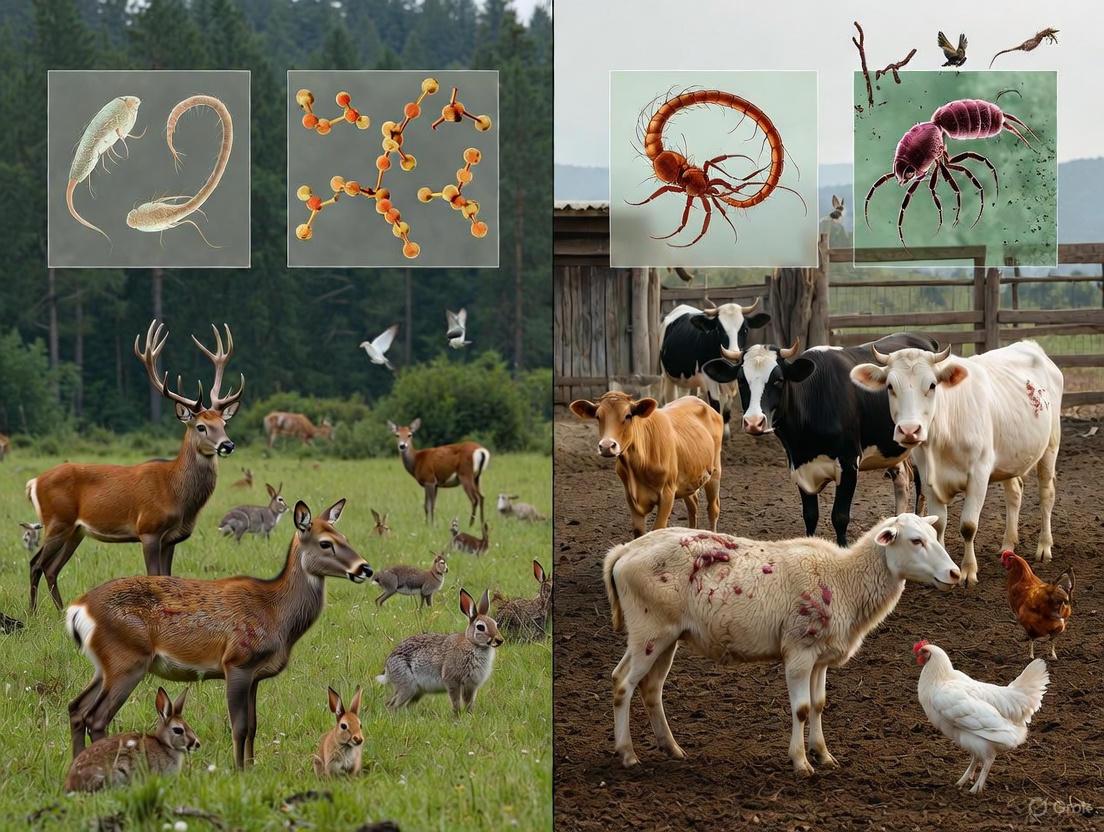
Parasitic Infections Across Species: A Comparative Analysis of Wildlife and Domestic Animals for Research and Therapeutic Development
This article provides a comprehensive comparative analysis of parasitic infections in wild and domestic animals, addressing a critical knowledge gap for researchers, scientists, and drug development professionals.
Parasitic Infections Across Species: A Comparative Analysis of Wildlife and Domestic Animals for Research and Therapeutic Development
Abstract
This article provides a comprehensive comparative analysis of parasitic infections in wild and domestic animals, addressing a critical knowledge gap for researchers, scientists, and drug development professionals. It explores the foundational ecological and pathological differences between these host environments, examines advanced methodologies for parasite detection and surveillance, addresses key challenges in diagnosis and data interpretation, and validates findings through direct comparative pathology. By synthesizing insights from recent studies within a One Health framework, this review aims to inform more effective drug discovery initiatives and public health strategies against zoonotic parasitic diseases.
Ecological Dynamics and Pathological Profiles of Parasites in Contrasting Host Environments
The One Health framework is an integrated, unifying approach that aims to sustainably balance and optimize the health of people, animals, and ecosystems [1]. This approach recognizes that the health of humans, domestic and wild animals, plants, and the wider environment are closely linked and interdependent [2]. The approach mobilizes multiple sectors, disciplines, and communities at varying levels of society to work together to foster well-being and tackle threats to health and ecosystems [3]. In recent years, One Health has gained significant importance because many factors have changed interactions between people, animals, plants, and our environment, including growing human populations expanding into new geographic areas, changes in climate and land use, and increased movement of people, animals, and animal products through international travel and trade [2]. These changes have led to the spread of existing and new zoonotic diseases, with approximately 75% of emerging infectious human diseases having an animal origin [3]. This article will explore the application of the One Health framework to parasitic infections, comparing patterns in wild versus domestic animals and examining the implications for global health security.
Theoretical Foundations of One Health
The conceptual foundation of One Health rests on the understanding that health outcomes are intrinsically interconnected across human, animal, and environmental domains. The One Health High-Level Expert Panel (OHHLEP) has established a comprehensive definition that describes how sectors, disciplines, and societies connect through four main pillars: communication, collaboration, coordination, and capacity building [3]. This definition has been adopted by the Quadripartite organizations - the Food and Agriculture Organization of the United Nations (FAO), the United Nations Environment Programme (UNEP), the World Health Organization (WHO), and the World Organisation for Animal Health (WOAH) - which collaborate to advance the One Health agenda globally [1] [3].
The visual representation below illustrates the interconnected relationships and continuous feedback loops between human, animal, and environmental health within the One Health framework:
Figure 1: The Interconnected Domains of the One Health Framework. This diagram illustrates the continuous feedback loops and transmission pathways connecting human, animal, and ecosystem health, emphasizing the integrated approach required to address health challenges.
The Quadripartite collaboration has developed a One Health Joint Plan of Action that operates through six interdependent Action Tracks: enhancing countries' capacity to strengthen health systems; reducing risks from emerging zoonotic epidemics; controlling and eliminating zoonotic diseases; strengthening food safety; curbing antimicrobial resistance; and better integrating the environment into One Health [3]. This comprehensive approach addresses the collective need for clean water, energy and air, safe and nutritious food, while taking action on climate change and contributing to sustainable development [3].
Comparative Analysis of Parasitic Infections: Wild vs. Domestic Animals
Prevalence and Diversity of Parasites
Parasitic infections represent a significant health challenge across animal species, though prevalence rates, parasite diversity, and zoonotic potential vary considerably between wild and domestic animals. The table below summarizes key comparative findings from recent studies:
Table 1: Comparative Analysis of Parasitic Infections in Wild and Domestic Animals
| Parameter | Wild Animals | Domestic Animals (Pets) | Data Sources |
|---|---|---|---|
| Overall Prevalence | 65.3-69.5% in captive wildlife [4] [5] | ~50% seropositive for T. gondii in dogs [6] | [6] [4] [5] |
| Zoonotic Potential | 64.9% of species carry zoonotic parasites [5] | 18/31 helminth species in dogs are zoonotic [6] | [6] [5] |
| Parasite Diversity | 17+ genera/species identified in safari parks [4] | 31 helminth species in dogs [6] | [6] [4] |
| Influencing Factors | Environmental changes, habitat fragmentation [5] [7] | Veterinary care access, owner compliance [6] | [6] [5] [7] |
| Transmission Dynamics | Complex wildlife-domestic-human interfaces [7] | Primarily domestic cycles with human exposure [6] | [6] [7] |
Key Zoonotic Parasites and Their Significance
Numerous parasites move across the human-animal interface, posing significant public health risks. In wild animals, studies in Greece identified parasites with established or potential zoonotic risks including Leishmania infantum, Cryptosporidium spp., Toxoplasma gondii, Echinococcus granulosus, and Trichinella spp. among others [7]. A study in Southern Brazil found that 64.9% of wild animals were parasitized by at least one morphogroup with zoonotic agents, including Taeniidae, Capillaria, Strongyloides, Spirometra, Lagochilascaris, Sarcocystis, and Giardia [5].
In domestic animals, dogs in Uzbekistan were found to host 31 helminth species, with 18 being zoonotic, including Echinicoccus granulosus, Dipylidium caninum, Toxocara canis, and Dirofilaria repens [6]. The high prevalence (94.7%) of helminth infections in these dogs, particularly in rural areas, highlights the significant transmission potential at the human-domestic animal interface [6].
Experimental Approaches in One Health Parasitology
Diagnostic Methodologies and Protocols
The study of parasitic infections within a One Health context requires standardized diagnostic approaches that allow for comparison across species and environments. The following workflow illustrates a comprehensive diagnostic protocol for assessing parasitic infections in animal populations:
Figure 2: Comprehensive Diagnostic Workflow for Parasitic Infection Assessment. This diagram outlines the standardized laboratory protocols for detecting and identifying parasitic infections in animal populations, incorporating both qualitative and quantitative approaches.
The diagnostic process typically begins with non-invasive faecal sample collection, often preserved in 10% formalin for transport and storage [4]. Laboratory techniques include qualitative methods such as direct smear, sedimentation, and flotation techniques (often with Zinc Sulfate solution), and quantitative methods such as the McMaster technique for determining eggs/oocysts per gram (EPG/OPG) of faeces [4] [5]. Advanced identification combines morphological analysis using micrometric eyepieces and comparison with published literature, supplemented increasingly by molecular methods such as PCR-coupled sequencing for species confirmation [6] [8].
Experimental Manipulation Studies
Controlled experimental studies provide valuable insights into the complex relationships between parasites, host behavior, and environmental factors. One such experimental study with wild black capuchin monkeys (Sapajus nigritus) in Iguazú National Park, Argentina, manipulated both food availability (through banana provisioning) and helminth infections (through antiparasitic drugs) to evaluate their effects on host behavior [9].
The study employed a split-plot experimental design with four treatment groups: (1) high food provisioning with antiparasitic treatment (F+ A+), (2) high provisioning with no antiparasitic treatment (F+ A−), (3) low provisioning with antiparasitic treatment (F− A+), and (4) low provisioning with no antiparasitic treatment (F− A−) [9]. Antiparasitic treatment involved a cocktail of ivermectin (effective against nematodes and ectoparasites) and praziquantel (effective against cestodes) [9]. Researchers collected faecal samples to determine infection intensity and recorded behavioral data including activity budgets and social proximity.
Findings demonstrated that individuals with unmanipulated helminth burdens foraged less than dewormed individuals, but only when food provisioning was low [9]. This interaction between nutritional status and parasitic infection highlights the complex relationship between these factors in influencing host behavior. The results were more consistent with a debilitating effect of parasites rather than an adaptive energy-conserving response to fight infection [9].
The Scientist's Toolkit: Essential Research Reagents and Materials
Table 2: Essential Research Reagents and Materials for One Health Parasitology Studies
| Reagent/Material | Application/Function | Experimental Context |
|---|---|---|
| Ivermectin | Broad-spectrum antiparasitic targeting nematodes and ectoparasites | Experimental manipulation of helminth infections [9] |
| Praziquantel | Antiparasitic effective against cestodes | Used in combination with ivermectin for comprehensive deworming [9] |
| Zinc Sulfate Solution | Flotation medium for parasite egg/oocyst concentration | Routine faecal flotation techniques [4] [5] |
| Formalin (10%) | Preservation of faecal samples for transport and storage | Maintains parasite morphology for identification [4] |
| Potassium Dichromate (2%) | Oocyst sporulation for protozoan identification | Enhances detection and identification of coccidian parasites [5] |
| McMaster Slides | Quantitative assessment of parasite eggs/oocysts per gram (EPG/OPG) | Determining infection intensity [4] |
| PCR Reagents | Molecular identification and characterization of parasites | Species confirmation and genotyping [6] [8] |
Implications for Drug Development and Therapeutic Interventions
The comparative analysis of parasitic infections across wild and domestic animal hosts reveals several critical considerations for drug development. The high prevalence of polyparasitism (concurrent infection with multiple parasite species) in both wild and domestic animals [6] [5] underscores the need for broad-spectrum antiparasitic formulations or combination therapies, as exemplified by the successful use of ivermectin-praziquantel cocktails in experimental studies [9].
The varying physiological responses to parasitic infections between wild and domestic animals suggests potential differences in drug metabolism and efficacy. Wildlife may require different dosing regimens or formulations compared to domestic animals, particularly considering the impact of nutritional status on drug effectiveness [9]. Furthermore, the zoonotic potential of many parasites highlights the importance of developing interventions that break transmission cycles at multiple points in the human-animal-environment interface [6] [7].
Drug development must also consider the potential impact on ecosystem health, as antiparasitic drugs can have non-target effects on environmental organisms. The One Health approach emphasizes the need for environmental risk assessment in the development and deployment of antiparasitic therapies [1] [3].
The One Health framework provides an essential paradigm for understanding and addressing the complex challenges posed by parasitic infections at the human-animal-environment interface. The comparative analysis of parasitic infections in wild versus domestic animals reveals distinct patterns of prevalence, diversity, and transmission dynamics that necessitate integrated approaches to disease surveillance, prevention, and control.
Experimental evidence demonstrates the intricate relationships between parasitic infections, host behavior, and environmental factors such as food availability [9]. These findings highlight the limitations of single-factor interventions and underscore the value of comprehensive approaches that address the multifaceted nature of parasitic diseases.
For researchers, drug development professionals, and public health officials, the One Health framework offers a strategic pathway for developing more effective and sustainable solutions to parasitic diseases. By integrating knowledge across human medicine, veterinary science, and environmental ecology, we can advance our capacity to detect, prevent, and control parasitic infections that threaten human health, animal welfare, and ecosystem integrity.
The growing challenges of climate change, habitat fragmentation, and globalized trade necessitate renewed commitment to One Health principles and practices. Through enhanced collaboration, communication, and coordination across sectors and disciplines, we can build a healthier future for people, animals, and our shared environment.
Parasite Diversity and Host Specificity in Wild Versus Domestic Populations
Parasites represent a significant component of global ecosystems, with their diversity and host specificity patterns providing crucial insights into host-parasite evolutionary relationships and disease transmission risks. Understanding the differences in parasitic infections between wild and domestic animal populations is fundamental to ecological parasitology, conservation biology, and public health policy [10]. This comparative guide examines the current scientific knowledge on parasite diversity and host specificity across these populations, synthesizing empirical data and methodological approaches to inform research and drug development initiatives.
The One Health framework recognizes the interconnectedness of human, animal, and environmental health, highlighting the importance of parasitic diseases that transcend population boundaries [10] [11]. Global change factors—including climate change, urbanization, habitat modification, and increased human-animal contact—have exacerbated parasite spread to new geographical regions, making comparative parasitology increasingly relevant for disease forecasting and management [10] [11].
Theoretical Framework: Ecological and Evolutionary Drivers
Host-Parasite Interactions in Changing Environments
Host-parasite interactions are dynamic relationships influenced by environmental pressures, host characteristics, and parasite life history strategies. Global changes modify these interactions across all organizational levels, from cellular physiology to ecosystem dynamics [11]. Domestic animals often experience fundamentally different selective pressures than their wild counterparts, potentially altering parasite susceptibility and transmission dynamics.
The Parasite-Mediated Domestication Hypothesis (PMD) proposes that parasite susceptibility may have played a role in the domestication process itself [12]. This hypothesis suggests that parasite-susceptible, genetically less resistant wild animals were originally domesticated, and this susceptibility has been passed to contemporary domestic populations. According to PMD predictions, domestic populations under comparable conditions would exhibit higher parasite loads than wild populations, both in terms of parasite diversity and infection intensity [12].
Host Specificity and Range Expansion Mechanisms
Host specificity represents a fundamental property differentiating specialist from generalist parasites, with significant implications for disease emergence risks [13]. Multi-host parasites pose greater health threats to wildlife, livestock, and humans than single-host parasites due to their broader transmission pathways and adaptive capabilities.
Research on parasitic mites has identified key predictors for host range expansion, including:
- Level of immune system contact: Parasites interacting directly with host immune components (e.g., hair follicular mites) show narrower host ranges than those feeding on non-immunogenic tissues [13]
- Host phylogenetic similarity: Closely related host species provide more opportunities for parasite invasion due to similar immune evasion mechanisms [13]
- Host spatial co-distribution: Sympatric host species present direct opportunities for host shifting [13]
- Environmental factors: Temperature and humidity affect parasite survival outside hosts during transmission [13]
Comparative Analysis of Parasite Prevalence and Diversity
Gastrointestinal Parasites in Mammalian Species
Table 1: Comparative Prevalence of Gastrointestinal Parasites in Wild and Domestic Populations
| Host Species | Location | Wild Population Prevalence | Domestic Population Prevalence | Key Parasite Taxa Identified | Citation |
|---|---|---|---|---|---|
| Wild boar/Domestic pig | Slovenia & Croatia | 5 parasite taxa; Strongyles in 0-12.5% | 7 parasite taxa; Strongyles significantly higher | Strongyles, Eimeria sp., Cystoisospora suis, Trichuris sp., Balantidium coli | [12] |
| Himalayan musk deer | Nepal | 94.2% overall prevalence | Not applicable | Pneumocaulus sp., Strongyle, Eimeria sp. | [14] |
| Humans/Domestic dogs | Southern Chile | 39% (human population) | 40% (domestic dogs) | Giardia duodenalis, Blastocystis sp., Toxocara canis | [15] |
| Rodents | Tanzania | 53.59% overall prevalence | Not applicable | Trichuris spp., Strongyloides spp., Capillaria spp., Hymenolepididae | [16] |
| Mixed wild species | Southern Brazil | 69.5% overall; 93.1% mammals | Not applicable | Strongylid-type, Capillaria spp., Taeniidae, Strongyloides | [5] |
Empirical studies consistently demonstrate substantial parasite burdens across host types, with some evidence supporting higher parasite diversity in domestic populations. The wild boar/domestic pig comparison revealed 5 parasite taxa in wild boars versus 7 in free-ranging domestic pigs, with strongyle infections significantly more abundant in domestic populations [12]. Similarly, a study of owned dogs in Chile demonstrated 40% parasite prevalence, comparable to the 39% prevalence found in humans from the same region, highlighting potential zoonotic transmission pathways [15].
Environmental and Host-Related Factors Influencing Parasitism
Table 2: Factors Associated with Parasite Prevalence and Diversity Across Studies
| Factor Category | Specific Factor | Impact on Parasitism | Example from Literature |
|---|---|---|---|
| Host characteristics | Body condition | Positive correlation with scaled mass index | Rodents with higher SMI had higher helminth infection probability [16] |
| Age | Higher prevalence in adults | Adult rodents had higher helminth prevalence than juveniles [16] | |
| Species | Varies by host species | Rattus rattus showed higher mean helminth richness [16] | |
| Environmental factors | Elevation | Negative correlation for some species | Strongyles confined to lower elevations (<3500m) in musk deer [14] |
| Habitat disturbance | Increased transmission risk | Anthropogenically disturbed areas promote parasite transfers [13] | |
| Co-infection dynamics | Ectoparasite load | Positive correlation with helminths | Flea and mite infestation linked to gastrointestinal helminths [16] |
| Multiple parasite taxa | Co-infection potential | 25% of musk deer samples had co-infections [14] |
Environmental factors significantly influence parasite distribution patterns. Research on Himalayan musk deer demonstrated that elevation strongly affected strongyle distribution, with higher elevations associated with lower probability of strongyle presence [14]. Pneumocaulus sp. was widespread across elevation gradients (most typically at 3600-3700m), while strongyles were confined to lower elevations below 3500m [14].
Methodological Approaches in Comparative Parasitology
Standardized Field Sampling and Diagnostic Techniques
The following workflow illustrates a generalized methodology for comparative parasitological studies:
Wildlife Parasitology Research Workflow
Non-invasive Sampling Approaches
Non-invasive sampling methods, particularly fecal collection, have proven valuable for studying parasites in both wild and domestic populations without requiring direct animal handling. A study of endangered Himalayan musk deer successfully utilized 52 fresh fecal pellets to assess gastrointestinal parasite prevalence, demonstrating the efficacy of this approach for sensitive or logistically challenging species [14]. This method minimizes stress on study animals and enables sampling in difficult terrain.
Coproparasitological Diagnostic Techniques
Standardized laboratory techniques enable comparable results across studies:
- Sedimentation techniques: The spontaneous sedimentation method concentrates helminth eggs and protozoan cysts through gravity sedimentation [5]
- Flotation techniques: Centrifugal flotation with zinc sulfate or similar solutions exploits density differences to separate parasitic elements [5]
- Molecular methods: Next-generation sequencing (NGS) and PCR-based approaches enable precise parasite identification and subtype characterization [15]
The modified McMaster technique provides quantitative assessment of parasite egg counts, particularly valuable for comparing infection intensity between populations [16].
Data Standardization and Reporting Frameworks
Recent initiatives have addressed the need for standardized data reporting in wildlife disease research. A proposed minimum data standard identifies 40 core data fields (9 required) and 24 metadata fields (7 required) to facilitate data sharing, reuse, and aggregation [17]. Key categories include:
- Sample data: Collection date, location, storage method
- Host animal data: Species, sex, age, health status
- Parasite data: Diagnostic methods, test results, genetic characterization
Standardization is particularly important for comparative studies, enabling valid cross-population and cross-species analyses.
Essential Research Reagents and Methodological Solutions
Table 3: Key Research Reagents and Methodological Solutions for Comparative Parasitology
| Category | Specific Solution | Application/Function | Example Implementation |
|---|---|---|---|
| Field collection | Fecal sample containers | Preservation of parasitic forms during transport | Storage at 4°C for ≤48 hours before processing [5] |
| Disposable gloves | Prevention of cross-contamination | Used during all sample handling procedures [5] | |
| Diagnostic reagents | Zinc sulfate solution | Flotation medium for parasite concentration | Centrifugal flotation technique [5] |
| Potassium dichromate | Oocyst sporulation | 2% solution for protozoan oocyst sporulation [5] | |
| Molecular tools | Primers for specific gene targets | PCR amplification of parasite DNA | Giardia duodenalis and Blastocystis sp. subtyping [15] |
| Next-generation sequencing | Comprehensive parasite identification | Detection of zoonotic subtypes [15] | |
| Analytical approaches | Firth's logistic regression | Statistical analysis for small sample sizes | Used in Himalayan musk deer study (n=52) [14] |
| Generalized Linear Mixed Models (GLMM) | Assessing multivariate relationships | Analysis of host, ectoparasite, and environmental factors [16] |
Specialized Methodological Considerations
Addressing Small Sample Sizes
Wildlife studies often face limitations in sample availability. Firth's logistic regression has been successfully applied to address small-sample biases, enabling robust statistical analysis even with limited data (e.g., n=52 in the Himalayan musk deer study) [14]. This approach reduces small-sample bias in parameter estimates, particularly valuable for endangered species research.
Controlling for Confounding Variables
Comparative studies must account for potential confounding factors including:
- Host age structure: Adult animals typically show higher parasite prevalence than juveniles [16]
- Body condition: Scaled Mass Index (SMI) may correlate with parasitism [16]
- Seasonal variations: Parasite prevalence often fluctuates temporally
- Spatial heterogeneity: Environmental factors vary across sampling locations
Implications for Disease Management and Drug Development
Zoonotic Transmission Risks
Comparative studies reveal significant zoonotic transmission potential at wildlife-human interfaces. Research in southern Brazil identified that 64.9% of positive wild animal samples contained at least one zoonotic parasite morphogroup, including Taeniidae, Capillaria, Strongyloides, and Giardia [5]. Similarly, a Chile study found zoonotic subtypes of Giardia duodenalis and Blastocystis sp. in both humans and domestic dogs, with 28.2% of humans seropositive for Toxocara canis antibodies [15].
Conservation Implications
High parasite prevalence in endangered species warrants conservation attention. The 94.2% gastrointestinal parasite prevalence in endangered Himalayan musk deer represents a potential threat to population viability [14]. The study recommended holistic conservation methods incorporating habitat management, disease detection, and continued monitoring to address this parasitic burden.
Drug Development Considerations
The parasite diversity documented across wild and domestic populations highlights the need for:
- Broad-spectrum anti-parasitics effective against multiple taxa
- Population-specific treatment protocols accounting for different parasite communities
- Environmental transmission blocking strategies targeting soil and water stages
The following conceptual framework illustrates the testing approach for the Parasite-Mediated Domestication Hypothesis, which has implications for understanding fundamental host-parasite relationships:
Testing the Parasite-Mediated Domestication Hypothesis
This comparative analysis demonstrates distinct patterns of parasite diversity and host specificity between wild and domestic populations, with domestic animals generally exhibiting equal or higher parasite diversity under comparable conditions. These findings align with predictions derived from the Parasite-Mediated Domestication Hypothesis, suggesting potential evolutionary trade-offs between tameness and parasite resistance [12].
Methodological standardization remains crucial for valid comparisons, with emerging frameworks promoting data interoperability and synthesis [17]. Future research directions should include:
- Longitudinal studies tracking parasite dynamics across seasons and years
- Genomic approaches identifying genetic determinants of host susceptibility
- Experimental manipulations directly testing host-parasite evolutionary relationships
- Integrated surveillance implementing One Health approaches across human, domestic animal, and wildlife interfaces
The significant zoonotic potential of many parasites found in both wild and domestic populations underscores the public health relevance of comparative parasitology and the need for continued research investment in this field.
Parasitic infections represent a dynamic challenge at the intersection of veterinary medicine, wildlife conservation, and public health. The clinical presentation and severity of these infections vary dramatically between domestic and wild animals, influenced by factors including evolutionary host-parasite relationships, environmental stress, and human management practices. Comparative pathology, which systematically analyzes these differences, provides invaluable insights for developing targeted control strategies, informing drug development, and understanding ecological disease dynamics. This guide objectively compares infection patterns, severity, and underlying mechanisms across host systems using recent experimental data, providing a structured resource for researchers and pharmaceutical professionals working at the forefront of parasitology.
Comparative Infection Prevalence and Parasite Diversity
The epidemiology of parasitic infections reveals distinct patterns in wild versus domestic animal populations. Quantitative data derived from recent meta-analyses and field studies provide a foundation for this comparison.
Table 1: Comparative Prevalence of Gastrointestinal Parasites
| Host Category | Overall Prevalence (%) | Dominant Parasite Groups | Key Risk Factors | Notable Zoonotic Agents |
|---|---|---|---|---|
| Captive Wild Mammals (Mainland China) [18] | 53.9% | Nematodes (45.1%) | High population density, season (Summer: 61.8%, Winter: 61.6%), host order (Primates: 66.5%) | Strongyloides, Capillaria, Giardia |
| Free-Ranging Wild Animals (Southern Brazil) [5] | 69.5% (Mammals: 93.1%) | Strongylid-type eggs (44.11%), Capillaria spp. (26.47%) | Rehabilitation stress, proximity to human/domestic animals | Taeniidae, Ancylostomid, Toxocara, Giardia |
| Domestic Dogs (Global Estimate) [19] | 21.0% | Giardia, Ancylostoma (Hookworms), Trichuris (Whipworms) | Climate, access to veterinary care, lifestyle | Ancylostoma caninum, Toxocara canis, Giardia |
| Domestic Cats (Europe) [19] | 35.1% (Endoparasites) | Toxocara cati (19.7%) | Outdoor access, hunting behavior | Toxocara cati, Toxoplasma gondii |
Table 2: Host Order-Specific Prevalence in Captive Wild Mammals [18]
| Host Order | Example Species | Prevalence (%) |
|---|---|---|
| Primates | Monkeys, Lemurs | 66.5 |
| Artiodactyla | Deer, Antelope | 59.0 |
| Rodentia | Rats, Squirrels | 57.1 |
| Carnivora | Foxes, Big Cats | 53.3 |
| Proboscidea | Elephants | 19.9 |
Wild animals, particularly in captive or rehabilitation settings, often exhibit a higher prevalence and diversity of parasitic infections. A meta-analysis in mainland China found an overall prevalence of 53.9% in captive wild mammals [18]. In contrast, a study of free-ranging wild animals in Southern Brazil revealed an even higher infection rate of 69.5%, with mammals showing a striking 93.1% prevalence [5]. These high rates are linked to environmental stressors and, in captive situations, higher stocking densities that facilitate parasite transmission [18].
Conversely, domestic animals like dogs and cats generally show lower prevalence rates, a trend attributed to access to routine veterinary care, including antiparasitic treatments [19]. However, specific parasites remain common, with nematodes being the most dominant group across all host types [18]. The host's taxonomic order is a significant factor, with primates, artiodactyls, and rodents showing higher susceptibility than proboscideans [18]. Furthermore, seasonality influences infection dynamics, with peaks often occurring in summer and winter months in captive environments [18].
Clinical Manifestations and Disease Severity
The clinical outcome of a parasitic infection is not merely a function of parasite presence but is determined by a complex interplay of parasite pathogenicity, host immunity, and co-infections.
Clinical Presentation in Domestic Animals
In domestic dogs, parasitic infections are a primary cause of clinical illness. Ancylostoma spp. (hookworms) are frequently associated with severe anemia due to their blood-feeding activity [20]. This can be particularly severe in co-infections, such as with Leishmania infantum, where hookworms are significantly associated with more severe clinical stages of visceral leishmaniasis and decreased red blood cell counts [20]. Such coinfections can alter the host's immune response, potentially worsening disease pathogenesis [20].
Lameness is another significant clinical manifestation in domestic animals linked to parasites. It can be classified as direct or indirect [21]:
- Direct lameness results from parasites damaging musculoskeletal or nervous tissues. For example, the nematode Gurltia paralysans migrates to the central nervous system in cats, causing paralysis [21].
- Indirect lameness is often vector-borne. Ticks can transmit pathogens like Borrelia burgdorferi (causing Lyme disease) and Anaplasma phagocytophilum, which lead to arthritis and lameness. Tick-borne neurotoxins can also cause paralysis [21].
Disease Expression in Wildlife
In wildlife, the paradigm often shifts. Many wild animals are adapted to tolerate certain parasite loads with minimal clinical disease, acting as reservoir hosts [10] [5]. However, introduced or invasive parasite species can cause severe pathology. The invasive nematode Ashworthius sidemi, which spread to Europe with sika deer, now infects native ruminants like red deer at high rates (30.7%) [22]. Similarly, the giant liver fluke Fascioloides magna, native to North America, causes severe liver damage in European ruminants [22]. Stressors like captivity, rehabilitation, and environmental change can disrupt the host-parasite equilibrium, leading to overt disease outbreaks in wild populations [5].
Experimental Methodologies in Modern Parasitology
Accurate diagnosis and research rely on robust experimental protocols. The field utilizes both traditional and advanced molecular techniques, each with specific applications.
Coproparasitological Techniques
These traditional methods are foundational for detecting parasitic forms in feces.
- Spontaneous Sedimentation (Hoffmann et al.): This method relies on the gravity-based sedimentation of helminth eggs and protozoan cysts. It is effective for concentrating and detecting operculated eggs and heavy cysts but may miss lighter elements [5].
- Centrifugal Flotation with Zinc Sulfate (Modified Monteiro Method): This technique uses a solution with a high specific density. Fecal samples are suspended and centrifuged, causing parasite eggs and cysts to float to the surface for collection and identification. It is excellent for recovering most nematode eggs and protozoan cysts [5].
- Oocyst Sporulation with Potassium Dichromate: Specifically for coccidian parasites, this technique allows oocysts to sporulate in a 2% potassium dichromate solution, enabling specific identification based on sporocyst morphology [5].
Molecular Diagnostics
Molecular techniques overcome the limitations of morphological identification, offering high sensitivity and specificity.
- Multiplex Real-Time PCR: This advanced protocol can simultaneously detect and quantify multiple parasite species from a single sample. For a nationwide wildlife survey in the Czech Republic, assays were designed to target specific regions of ribosomal DNA (rDNA) for six helminth species [22]. The process involves:
- DNA Extraction: Genomic DNA is isolated from fecal samples.
- PCR Setup: A reaction mix is prepared containing specific forward and reverse primers and TaqMan probes for each target parasite.
- Amplification and Detection: The PCR run involves cycles of denaturation, annealing, and extension. The TaqMan probe is cleaved during amplification, releasing a fluorescent signal measured in real-time.
- Analysis: The cycle threshold (Ct) value determines the presence and quantity of the target DNA [22].
- Next-Generation Sequencing (NGS): For public health studies, NGS is used to identify subtypes of protozoa like Giardia duodenalis and Blastocystis sp. by sequencing genes like β-giardin and 18S rRNA, respectively, which is crucial for understanding zoonotic transmission chains [23].
Serological and Post-Mortem Techniques
- Enzyme-Linked Immunosorbent Assay (ELISA): Used to detect antibodies against parasites like Toxocara canis in human and animal sera, indicating exposure history [23].
- Necropsy: Remains the gold standard for confirming the presence of adult helminths and associated pathology, such as identifying Ancylostoma sp. and Dipylidium caninum in the intestines of dogs [20].
The Scientist's Toolkit: Essential Research Reagents
Cut-edge parasitology research depends on a suite of specific reagents and tools.
Table 3: Key Research Reagent Solutions
| Reagent / Tool | Primary Function | Application Example |
|---|---|---|
| PAF Fixative (Phenol, Alcohol, Formaldehyde) | Preserves parasite morphology in fecal samples for microscopic analysis. | Long-term storage and transport of human and dog fecal samples in community studies [23]. |
| Zinc Sulfate Solution (Specific gravity ~1.18-1.20) | Flotation medium for concentrating helminth eggs and protozoan cysts. | Recovery of nematode eggs and Giardia cysts from fecal samples via centrifugal flotation [5] [23]. |
| TaqMan Probes | Fluorescently-labeled probes for specific detection of target DNA in real-time PCR. | Multiplex PCR for simultaneous detection of six helminth species (e.g., A. sidemi, F. magna) in wild ruminant feces [22]. |
| Species-Specific Primers | Short DNA sequences designed to amplify unique genomic regions of a parasite. | Molecular identification and differentiation of morphologically similar species (e.g., Calicophoron daubneyi) [22]. |
| Potassium Dichromate (2%) | Promotes sporulation of coccidian oocysts for morphological identification. | Differentiation of Eimeria spp. oocysts in wildlife feces [5]. |
| Commercial ELISA Kits (e.g., NovaLisa) | Detect host-derived IgG antibodies against specific parasitic antigens. | Sero-epidemiological studies to assess human exposure to Toxocara canis [23]. |
Signaling Pathways and Host-Parasite Interactions
At a molecular level, the clinical severity of parasitic diseases is governed by the host's immune response, which can be manipulated by parasites.
The Type 1 (Th1) immune response, characterized by interferon-gamma (IFN-γ) production, is crucial for controlling intracellular parasites like Leishmania infantum [20]. An exaggerated Th1 response, however, can cause severe immunopathology [20]. In contrast, infections with intestinal helminths (e.g., Ancylostoma sp.) predominantly induce a Type 2 (Th2) response, involving interleukins like IL-4, IL-5, and IL-13 [20]. This Th2 polarization can downregulate the protective Th1 response, leading to worsened disease severity and higher parasite burdens in coinfected hosts, as observed in dogs with visceral leishmaniasis [20]. This immunological interference is a key factor in the clinical outcome of polyparasitism.
The comparative pathology of parasitic infections between wild and domestic systems reveals a core principle: clinical manifestation is a function of host-parasite co-evolution, environmental context, and immune status. Wild animals often demonstrate a tolerance to endemic parasites, a balance easily disrupted by captivity, invasive species, and environmental change, leading to severe pathology. Domestic animals, while generally better managed, suffer significant morbidity from a narrower range of parasites, with coinfections presenting complex clinical challenges. Future research leveraging advanced molecular tools and a One Health perspective is critical for developing effective, targeted interventions that safeguard animal welfare, conserve biodiversity, and protect public health.
Zoonotic parasites represent a significant threat to global health, with wildlife serving as crucial reservoirs for infections that spill over to domestic animals and humans. The intricate relationships between wild animals, domestic animals, and humans create a complex web of transmission pathways that facilitate the spread of parasitic diseases. Recent research indicates that approximately 75% of emerging infectious diseases in humans have animal origins, with 71.8% originating specifically from wild fauna [5]. This transmission phenomenon, known as zoonotic spillover, is exacerbated by environmental changes such as deforestation, climate change, and urbanization, which disrupt ecosystem balances and increase contact between wildlife, domestic animals, and human populations [6] [5].
The One Health approach has emerged as a critical framework for understanding these dynamics, emphasizing the interconnectedness of human, animal, and environmental health. However, current research often fails to adequately integrate all three domains simultaneously. A systematic review of recent One Health research found that only 4.8% of studies integrated human, animal, and environmental domains in data collection, and just 29.5% did so in knowledge generation [24]. This highlights significant gaps in our understanding of the complete transmission cycles of zoonotic parasites. The concept of the "zoonotic web" – a network representation of relationships between zoonotic agents, their hosts, vectors, food, and environmental sources – provides a valuable model for visualizing and analyzing these complex interactions [25]. Understanding this web is essential for developing effective surveillance and control strategies for parasitic diseases that threaten public health, veterinary health, and conservation efforts.
Comparative Analysis of Parasitic Infections in Wild and Domestic Animals
Prevalence and Diversity Patterns
Research consistently demonstrates that wild animals harbor a greater diversity and prevalence of parasites compared to their domestic counterparts. A comprehensive study in Southern Brazil examining 82 fecal samples from wild animals found that 69.5% were infected with helminth eggs and/or protozoan cysts/oocysts [5]. When broken down by class, the prevalence was striking: 93.1% of mammals, 47% of birds, and 50% of reptiles showed evidence of parasitic infections [5]. The most frequently encountered parasites were strongylid-type eggs (44.11%), followed by Capillaria spp. eggs (26.47%) [5]. Importantly, 64.9% of the positive samples contained at least one morphogroup with zoonotic potential, including Taeniidae, Strongyloides, Spirometra, Lagochilascaris, Sarcocystis, Trichuris, Giardia, Ancylostomatidae, Physaloptera, Toxocara, and Fasciola [5].
Similar patterns emerge when comparing specific host species. A study of haemosporidian infections in red junglefowl and domestic chickens revealed that 100% of wild red junglefowls tested positive for infection, compared to 85% of domestic chickens [26]. Additionally, the diversity of parasitic lineages was significantly higher in the wild birds, indicating that domestication may reduce both the prevalence and diversity of parasitic infections [26].
Table 1: Comparative Prevalence of Parasitic Infections in Wild and Domestic Animals
| Host Category | Study Location | Sample Size | Overall Prevalence | Most Common Parasites | Zoonotic Potential |
|---|---|---|---|---|---|
| Wild Animals (Multiple Species) | Southern Brazil | 82 | 69.5% | Strongylids (44.11%), Capillaria spp. (26.47%) | 64.9% of positive samples had zoonotic agents |
| Wild Mammals | Southern Brazil | 44 | 93.1% | Strongylids, Capillaria spp. | High diversity of zoonotic parasites |
| Wild Birds | Southern Brazil | 34 | 47% | Strongylids, Coccidia | Multiple zoonotic morphogroups |
| Wild Reptiles | Southern Brazil | 4 | 50% | Strongylids, Protozoa | Several zoonotic parasites detected |
| Red Junglefowl | Thailand | 39 | 100% | Haemosporidian parasites | Limited information available |
| Domestic Chickens | Thailand | 122 | 85% | Haemosporidian parasites | Limited information available |
| Captive Wildlife | Brazil (Mato Grosso do Sul) | 96 | 51.04% | Strongyloidea, Ancylostomatidae | Various zoonotic parasites identified |
Factors Influencing Infection Patterns
The disparity in parasitic prevalence and diversity between wild and domestic animals can be attributed to several ecological and anthropogenic factors. Wild animals are exposed to a broader spectrum of parasites in their natural habitats, where complex food webs and diverse ecosystems facilitate the maintenance of complex parasite life cycles. In contrast, domestic animals often benefit from controlled environments, preventive healthcare, and limited exposure to intermediate hosts and vectors [26].
The stress of captivity can also influence parasitic infections in wildlife. A study of captive and free-ranging wild animals in Brazil found that 51.04% of captive animals were parasitized, compared to only 23.07% of free-living animals [27]. This counterintuitive finding suggests that factors such as high population density, stress, adaptation to new environments, and prolonged confinement in captive situations can exacerbate parasitic infections, even with veterinary care [5]. This highlights the importance of complementary examinations like coproparasitological diagnosis in rehabilitation centers, as many parasites can cause significant health issues in stressed or immunocompromised animals [5].
Critical Experimental Protocols in Zoonotic Parasite Research
Standardized Coproparasitological Diagnostic Techniques
The accurate diagnosis of parasitic infections in animal hosts relies on standardized coproparasitological techniques that allow for the identification of helminth eggs, protozoan cysts, and oocysts in fecal samples. The most commonly employed methodologies in recent studies include:
Zinc Sulfate Centrifugal Flotation Technique (modified Monteiro method): This concentration technique exploits the density differences between parasitic elements and fecal debris to isolate and identify parasites [5]. The specific gravity of zinc sulfate solution (1.18-1.20) allows helminth eggs and protozoan cysts to float to the surface while denser debris sediments.
Spontaneous Sedimentation (Hoffmann et al. method): This technique relies on gravitational sedimentation to concentrate parasitic elements in water or saline solution [5] [27]. It is particularly effective for detecting operculated eggs and heavier parasitic elements that may not float efficiently in flotation techniques.
Oocyst Sporulation with 2% potassium dichromate: This specialized technique is used specifically for coccidian oocysts, promoting sporulation to aid in identification to genus or species level [5].
The identification of parasitic elements is typically performed using an optical microscope with variable magnification (40x to 100x) coupled with a digital camera for morphometric analysis. Morphological identification is based on characteristics such as shell features, ornaments, embryonic and larval formations, and the presence of opercula and spines [5]. For some parasite groups, such as strongylid-type eggs, ancylostomatid eggs, and some coccidian oocysts, identification may remain at the morphogroup level due to the absence of diagnostic characters for species differentiation in fecal samples [5].
Network Analysis for Transmission Pathways
Advanced analytical approaches are being employed to understand the complex transmission dynamics of zoonotic parasites. Network analysis has emerged as a powerful tool for investigating disease transmission potential between different host species [25] [28]. The methodology involves:
Compiling comprehensive datasets of naturally occurring zoonotic interactions through systematic literature searches, spanning several decades to capture temporal trends [25].
Constructing transmission-potential networks (TPNs) where hosts represent network nodes that are connected via edges defined by similarity in pathogen susceptibility [28]. These networks depict the potential for transmission between host species based on known etiology and host range rather than direct contact patterns.
Calculating edge weights using the Jaccard index, which assumes a positive relationship between pathogen infections shared by species and the likelihood that a pathogen would infect both [28].
Applying network metrics such as eigenvalue centrality (EC) to quantify the importance of host species in promoting pathogen transmission potential among all host species [28].
This approach was effectively used in a study of wild pigs, which identified 34 OIE-listed swine pathogens (87%) that cause clinical disease in livestock, poultry, wildlife, and humans [28]. The analysis revealed that on average, 73% of bacterial, 39% of viral, and 63% of parasitic pathogens of swine caused clinical disease in other species, with non-porcine livestock in the family Bovidae sharing the most pathogens with swine (82%) [28].
Table 2: Research Reagent Solutions for Zoonotic Parasite Studies
| Research Reagent | Primary Function | Application Examples | Key Considerations |
|---|---|---|---|
| Zinc Sulfate Solution (Specific gravity 1.18-1.20) | Flotation medium for parasite concentration | Zinc Sulfate Centrifugal Flotation technique for helminth eggs and protozoan cysts | Maintain specific gravity for optimal recovery; appropriate disposal required |
| 2% Potassium Dichromate | Promotes sporulation of coccidian oocysts | Identification of Eimeria, Cystoisospora species | Handle with care due to toxicity; proper safety protocols essential |
| Microscope with Digital Imaging | Morphological identification and morphometric analysis | parasite egg, cyst, and oocyst identification at 100x and 400x magnification | Calibrated micrometric eyepieces essential for accurate measurements |
| PCR Reagents and Sequencing Primers | Molecular identification and genotyping of parasites | Lineage identification of haemosporidian parasites [26]; confirmation of Toxoplasma gondii [6] | Specific primers required for different parasite taxa; quality DNA extraction critical |
| Software for Network Analysis | Modeling transmission pathways and host-parasite interactions | Constructing zoonotic webs [25]; transmission potential networks [28] | Specialized statistical packages (e.g., R, Python) with network analysis capabilities |
Visualization of Research Workflows and Transmission Networks
Experimental Workflow for Parasite Surveillance
The following diagram illustrates the standardized experimental workflow for coproparasitological surveillance studies in wild and domestic animals:
Diagram Title: Parasite Surveillance Workflow
Zoonotic Web of Parasite Transmission
The complex interactions between wildlife, domestic animals, humans, and the environment can be visualized as a zoonotic web, illustrating the potential pathways for parasite transmission:
Diagram Title: Zoonotic Parasite Transmission Web
Implications for Disease Control and Future Research
The comparative analysis of parasitic infections in wild and domestic animals reveals critical insights for disease control strategies. The high prevalence and diversity of zoonotic parasites in wildlife reservoirs, coupled with increasing human-animal interfaces due to environmental changes, create conditions favorable for spillover events [5]. The finding that over 64% of infected wild animals in Southern Brazil carried zoonotic parasites underscores the significant public health threat [5]. Furthermore, network analyses demonstrate that certain species, particularly wild pigs, play disproportionate roles in pathogen sharing, with 82% of pathogens shared with Bovidae species [28].
Future research should address several critical gaps identified in current literature. First, there is a need for more comprehensive integration of all One Health domains – human, animal, and environmental health – in study designs, as current research often neglects environmental components [24]. Second, molecular techniques should be more widely adopted to improve parasite identification and understand transmission dynamics. Third, longitudinal studies tracking parasite transmission at the wildlife-domestic animal-human interface are essential for quantifying spillover risk and identifying effective intervention points. Finally, there is a pressing need to develop evidence-based management strategies that mitigate disease risks while supporting ecosystem health and conservation goals.
The successful implementation of One Health approaches requires breaking down interdisciplinary barriers that separate human and veterinary medicine from ecological, evolutionary, and environmental sciences [10]. Enhanced collaboration between physicians, veterinarians, ecologists, and other specialists is essential for developing integrated solutions that address both the underlying causes and consequences of zoonotic parasitoses. Strengthening epidemiological monitoring initiatives, improving diagnostic tools, and promoting educational programs for both professionals and the public will be crucial for reducing the burden of zoonotic parasitic diseases in an increasingly interconnected world.
Impact of Environmental Change on Parasite Distribution and Emergence
Environmental change, driven by factors such as climate warming, urbanization, and habitat fragmentation, is fundamentally reshaping the landscape of parasitic diseases. These changes alter the distribution, prevalence, and transmission dynamics of parasites, with significant implications for animal health, conservation, and public health. A critical lens through which to view these impacts is the comparison between wild and domestic animals. Domestic animals often live in managed environments with access to veterinary care, whereas wild animals are directly exposed to environmental pressures and serve as bioindicators of ecosystem health [6] [29]. Understanding the differential impact on these groups is essential for developing effective surveillance and control strategies under the One Health framework, which recognizes the interconnectedness of animal, human, and environmental health [30] [31]. This guide provides a comparative overview of the effects of environmental change on parasites in wild versus domestic animals, supported by experimental data and methodologies.
Comparative Prevalence of Parasitic Infections
The table below synthesizes quantitative data from various studies, illustrating the prevalence and diversity of parasitic infections in different animal groups under the influence of environmental factors.
Table 1: Comparative Prevalence of Parasites in Wild and Domestic Animals
| Host Category | Specific Host / Context | Parasite(s) | Key Finding | Reference & Context |
|---|---|---|---|---|
| Wild Birds | Blue tits (Sweden, 1996-2021) | Haemoproteus majoris (avian malaria) | Prevalence increased from 47% (1996) to 92% (2021), correlated with warmer temperatures. | [32] |
| Wild Mammals | Various species (Southern Brazil) | Helminths and/or Protozoa | 69.5% of animals infected; mammals showed 93.1% infection rate. | [5] |
| Wild Mammals | European brown hares (Italy) | Eimeria spp. | 91.2% prevalence in animals bred for restocking. | [6] |
| Domestic Animals | Dogs (Uzbekistan, rural areas) | Helminths | 94.7% of dogs infected with up to 31 helminth species. | [6] |
| Domestic vs. Wild | Pigeons (Iraq) | Various (Protozoa, Helminths, Lice) | Domestic pigeons: 32% prevalence. Wild pigeons: 20% prevalence. | [33] |
| Domestic vs. Wild (Physiological) | Meta-comparison | Glucocorticoids (stress hormones) | Fecal and hair glucocorticoid concentrations are generally lower in domestic animals than in their wild counterparts. | [29] |
Key Experimental Protocols in Parasitology Research
Research in this field relies on specific diagnostic and analytical protocols. The following methodologies are central to the studies cited in this guide.
Table 2: Summary of Key Experimental Protocols
| Protocol Name | Primary Application | Brief Description | Example of Use |
|---|---|---|---|
| Coproparasitological Diagnosis | Detecting endoparasites (helminths/protozoa) in feces. | Fecal samples are processed using techniques like Zinc Sulfate Centrifugal Flotation and Spontaneous Sedimentation to isolate and identify eggs, cysts, or oocysts. | Survey of parasitic fauna in wild mammals, birds, and reptiles [5]. |
| Real-Time PCR (qPCR) | Sensitive and specific detection of parasite DNA. | Uses targeted primers and probes to amplify and quantify parasite DNA from tissue, blood, or environmental samples (eDNA). | Detecting Haplosporidium costale in oysters, plankton, and sediment [34]. |
| Long-Term Population Monitoring & Blood Smear Analysis | Tracking parasite prevalence and transmission dynamics over time. | Wild host populations are monitored annually. Blood samples are collected and analyzed via microscopy and PCR to identify and genotype blood parasites. | 26-year study of avian malaria in blue tits [32]. |
| Glucocorticoid Analysis | Quantifying physiological stress levels. | Glucocorticoid hormones (e.g., cortisol) are measured in matrices like feces, hair, or blood as a biomarker of the stress response. | Comparing stress levels between wild, captive, and domestic animals [29]. |
| Necropsy and Morphological Identification | Comprehensive survey of helminth diversity. | Post-mortem examination of animals followed by collection and morphological identification of helminths from the gastrointestinal tract and organs. | Determining helminth species diversity in dogs [6]. |
Detailed Workflow: Coproparasitological Diagnosis
This protocol is a cornerstone for surveilling parasites in both wild and domestic animal populations. The workflow below outlines the key steps for fecal sample processing.
The Stress Pathway: A Mechanism Linking Environment and Susceptibility
Environmental changes act as chronic stressors that can disrupt an animal's physiological homeostasis. The neuroendocrine stress response is a key mechanism that explains why animals in altered environments may exhibit higher susceptibility to parasitic infections. This pathway is particularly relevant for wildlife exposed to habitat loss, climate change, and human disturbance [29] [30].
The Scientist's Toolkit: Essential Research Reagents and Materials
Table 3: Key Research Reagent Solutions for Parasitology Studies
| Reagent/Material | Primary Function | Application Example |
|---|---|---|
| Zinc Sulfate Solution | Flotation medium for isolating helminth eggs and protozoan cysts from feces based on buoyant density. | Used in coproparasitological surveys to concentrate parasitic elements for microscopic identification [5]. |
| Primers and Probes for qPCR | Target-specific oligonucleotides for amplifying and detecting parasite DNA in a real-time PCR assay. | Essential for sensitive detection and quantification of specific parasites like Haplosporidium costale in host and environmental samples [34]. |
| Potassium Dichromate (2%) | Solution used to promote sporulation of oocysts, allowing for morphological identification of coccidian parasites. | Employed in the Oocyst Sporulation technique to identify Eimeria species in hares and other wildlife [5]. |
| Antibodies for ELISA/IFAT | Immunoglobulins that bind to specific parasite antigens, enabling serological detection of past or present infections. | Used in seroprevalence studies for parasites like Toxoplasma gondii to screen populations for exposure [6] [31]. |
| Glucocorticoid Assay Kits | (e.g., for cortisol/corticosterone) | Used to quantify physiological stress levels in wildlife studies from sample matrices like feces or hair [29]. |
The evidence clearly demonstrates that environmental changes are powerful drivers of parasitic disease emergence and distribution. The comparative approach reveals that wild animals often bear a heavier burden, acting as reservoirs for a diverse community of parasites, including many with zoonotic potential [6] [5]. The chronic stress induced by environmental disruptions likely contributes to this high prevalence by suppressing immune function [29]. In contrast, domestic animals, while still highly susceptible, may have their infection dynamics more directly influenced by human management practices, such as the use of antiparasitic drugs [6].
Climate change, in particular, has a measurable impact on vector-borne parasites, as shown by the dramatic increase in avian malaria in blue tits over 26 years—a trend directly linked to warmer temperatures [32]. This underscores the need for long-term monitoring programs to track these shifts.
A holistic One Health approach is crucial. As shown by the detection of zoonotic parasites in wild animal feces in Brazil [5] and the spread of parasites from domestic dogs to endangered wildlife [6], the health of wild and domestic animals, humans, and ecosystems is inextricably linked. Future research must continue to integrate field surveillance, advanced molecular diagnostics, and physiological studies to better predict and mitigate the impacts of environmental change on parasite dynamics.
Advanced Diagnostic Techniques and Surveillance Strategies for Parasite Detection
Non-invasive fecal sampling has emerged as a cornerstone technique in wildlife parasitology and microbiome research, enabling scientists to study animal health, disease dynamics, and ecological interactions without direct human-animal contact. This approach is particularly valuable for comparative studies of parasitic infections in wild versus domestic animals, as it minimizes stress to the subjects and allows for sampling of elusive, endangered, or dangerous species. The growing emphasis on One Health principles—recognizing the interconnectedness of human, animal, and environmental health—has further elevated the importance of non-invasive methods for monitoring zoonotic diseases and understanding transmission dynamics across species boundaries. This guide provides a comprehensive comparison of non-invasive fecal sampling methodologies, preservation techniques, and analytical approaches, with specific application to parasitology research in wild and domestic animal populations.
Methodological Comparison: Non-Invasive versus Invasive Sampling
The choice between non-invasive and invasive sampling approaches involves important trade-offs that significantly impact research outcomes, particularly in comparative parasitology studies. The table below summarizes key comparative aspects based on current research.
Table 1: Comparison of invasive and non-invasive fecal sampling methods
| Parameter | Invasive Sampling | Non-Invasive Sampling |
|---|---|---|
| Animal Welfare | High stress; potential for injury during trapping/restraint [35] | Minimal disturbance; no direct contact required [35] [36] |
| Microbiome Integrity | Altered by sedatives and stress [35] | More representative of natural state [35] |
| Parasite Detection Reliability | High for fresh samples | Potential false negatives due to environmental degradation [37] [5] |
| Sample Quality | Consistent and fresh | Variable depending on environmental exposure [36] |
| Ideal Applications | Requires precise individual data; clinical interventions | Population-level studies; long-term monitoring; endangered species [35] [38] |
Non-invasive sampling eliminates the confounding effects of anesthesia and stress on microbial communities, potentially providing a more accurate representation of the gut microbiome [35]. However, this approach introduces different challenges related to sample identification, environmental degradation, and potential contamination from soil microorganisms [35]. Research on common cranes revealed significant differences in fecal microbial composition between samples collected invasively from trapped birds and those collected non-invasively, highlighting how sampling method choice can directly impact research findings [35].
Sample Collection and Preservation Standards
Proper collection and preservation are critical for maintaining sample integrity, especially for non-invasively collected specimens. The following table compares common preservation methods and their applications in parasitology and microbiome research.
Table 2: Fecal sample preservation methods and their applications
| Preservative | Advantages | Disadvantages | Best Applications |
|---|---|---|---|
| 10% Formalin | All-purpose fixative; good morphology preservation; suitable for concentration procedures [39] | Not ideal for protozoan trophozoites; can interfere with PCR [39] | Helminth eggs, larvae, protozoan cysts; immunoassays [39] |
| Polyvinyl-Alcohol (PVA) | Excellent protozoan morphology; suitable for permanent stained smears [39] | Contains mercuric chloride; not for concentration procedures [39] | Protozoan trophozoites and cysts; permanent smears [39] |
| 95% Ethanol | Suitable for DNA analysis; easy storage at -20°C [35] | Not ideal for morphological studies | Molecular studies (PCR, sequencing) [35] |
| Freezing (-80°C) | Gold standard for DNA preservation [35] [40] | Not feasible in field conditions | Microbiome studies; biobanking [40] [41] |
| Two-Vial System (Formalin + PVA) | Complementary advantages; comprehensive diagnostic capability [39] | Requires dividing specimen; more complex processing | General parasitological surveys [39] |
For microbiome studies, immediate preservation at -80°C is ideal, though not always feasible in field conditions [40]. The China Association of Chinese Medicine has established standards for fecal sample processing in clinical trials that emphasize careful documentation of storage conditions, transportation methods, and sample management to ensure data reliability [40]. For parasitological diagnosis, the CDC recommends dividing specimens between 10% formalin and PVA preservatives to enable both concentration procedures and permanent stained smears [39].
Analytical Techniques and Detection Efficacy
The choice of analytical method significantly impacts detection sensitivity and the types of information that can be derived from fecal samples. The table below compares key analytical approaches used in non-invasive sampling.
Table 3: Comparison of analytical methods for fecal samples
| Method | Detection Target | Sensitivity | Applications in Wild vs. Domestic Animals |
|---|---|---|---|
| Microscopy (Single Sample) | Helminth eggs, protozoan cysts [37] [5] | 45-75% (varies by parasite) [37] | Initial screening; resource-limited settings [5] |
| Microscopy (Multiple Samples) | Helminth eggs, protozoan cysts [37] | Up to 100% with 3 samples [37] | Essential for reliable wildlife surveys [37] |
| 16S rRNA Sequencing | Bacterial microbiota composition [35] [36] | High for community profiling | Microbiome comparisons between wild and domestic [35] |
| FecalSeq + 3RAD | Host SNPs [38] | 32% success with non-invasive samples [38] | Population genetics; imperiled species [38] |
| Zinc Sulfate Flotation | Protozoan cysts, helminth eggs [5] | Moderate to high | General parasitological surveys [5] |
Multiple sample collection dramatically improves detection rates for intestinal parasites. Research demonstrates that collecting three stool specimens increases cumulative detection rates to nearly 100%, compared to significantly lower rates with single samples [37]. The improvement varies by parasite species; while hookworms are typically detected in the first sample, Trichuris trichiura and Isospora belli often require additional samples for detection [37]. Immunocompetent hosts are significantly more likely to have pathogenic intestinal parasites detected in later stool specimens [37].
For genomic applications, methods like FecalSeq enrichment with 3RAD sequencing can successfully generate SNP data from non-invasively collected fecal samples, achieving a 32% success rate even with samples collected 2-5 days after defecation [38]. This approach increases the proportion of host DNA by an average of 15-fold, addressing a major challenge in non-invasive sampling where exogenous DNA from dietary sources and gut microbiome can dominate extracts [38].
Sample Stability and Environmental Factors
Understanding temporal changes in fecal samples is crucial for interpreting results from non-invasively collected specimens, particularly when exact defecation times are unknown.
Diagram 1: Sample stability timeline for non-invasive fecal sampling. Studies show microbial community stability for up to 7 days with minimal changes, making samples collected within this window suitable for comparative analyses [36].
Research on Rocky Mountain elk fecal samples demonstrated remarkable community stability across a 14-day period, with no statistically significant changes in microbial composition detected within the first week [36]. Modest changes were observed at day 14, with only two genera (Bacteroides and Sporobacter) showing significant abundance shifts [36]. The individual animal explained approximately 21% of the variance in microbial composition, while time since defecation accounted for less than 10% of variance and was not statistically significant [36]. This stability supports the use of non-invasive sampling for comparative studies when samples are collected within one week of defecation.
Environmental conditions significantly impact sample integrity. Factors including UV exposure, temperature fluctuations, oxygen availability, and precipitation can alter microbial communities and parasite morphology in fecal samples [36]. These effects are buffered in environments with low temperatures that inhibit microbial metabolism, making seasonal timing an important consideration for field studies [36].
Comparative Parasitology: Wild vs. Domestic Animals
Non-invasive sampling enables critical comparisons of parasitic infections between wild and domestic animals, revealing important patterns of zoonotic transmission and conservation concern.
Table 4: Comparative parasitic infections in wild and domestic animals from selected studies
| Host Category | Species | Infection Rate | Key Parasites Identified | Zoonotic Potential |
|---|---|---|---|---|
| Wild Pigeons | Columbia livia | 20% [33] | Eimeria spp. (8.8%), Cryptosporidium spp. (5.6%), Ascaridia columbae (1.6%) [33] | Low to moderate [33] |
| Domestic Pigeons | Columbia livia domestica | 32% [33] | Eimeria spp. (16.8%), Trichomonas gallinae (15.2%), Raillietina tetragona (4%) [33] | Low to moderate [33] |
| Wild Mammals (Brazil) | Multiple species | 69.5% overall; 93.1% of mammals [5] | Strongylid-type eggs (44.11%), Capillaria spp. (26.47%) [5] | High (64.9% with zoonotic agents) [5] |
| Dogs (Uzbekistan) | Canis familiaris | 94.7% [6] | 31 helminth species; 18 zoonotic [6] | High (Echinococcus granulosus, Toxocara canis, etc.) [6] |
A study of wild animals in Southern Brazil found that 69.5% of fecal samples contained helminth eggs and/or protozoan cysts, with mammals showing the highest infection rate (93.1%) [5]. Importantly, 64.9% of positive samples contained at least one morphogroup with zoonotic potential, including Taeniidae, Strongyloides, and Giardia [5]. The high prevalence of zoonotic parasites in wildlife underscores the importance of non-invasive monitoring for public health protection.
Comparative studies in pigeons revealed significantly higher infection rates in domestic (32%) versus wild pigeons (20%), likely reflecting differences in population density, nutrition, and feeding habits [33]. The most prevalent parasites also differed between groups, with domestic pigeons showing higher rates of Eimeria spp. and Trichomonas gallinae, while wild pigeons had different infection patterns [33].
The Scientist's Toolkit: Essential Research Reagents and Materials
Successful non-invasive fecal sampling requires specialized reagents and materials tailored to specific research goals. The following table outlines essential solutions and their applications.
Table 5: Essential research reagents for non-invasive fecal sampling
| Reagent/Material | Function | Application Examples |
|---|---|---|
| Zinc Sulfate Solution | Flotation medium for parasite eggs and cysts [5] | Parasitological diagnosis in wild animals [5] |
| 10% Formalin | Fixative for morphological preservation [39] | Long-term storage of samples for helminth egg identification [39] |
| Polyvinyl-Alcohol (PVA) | Preservative for protozoan stages [39] | Permanent stained smears for protozoan identification [39] |
| 95% Ethanol | DNA preservation [35] | Microbiome and molecular studies [35] |
| Quick-DNA Fecal/Soil Microbe Kits | DNA extraction from challenging samples [38] | Host enrichment from fecal samples [38] |
| 2% Potassium Dichromate | Oocyst sporulation [5] | Protozoan parasite identification [5] |
| FecalSeq Enrichment Reagents | Host DNA enrichment [38] | Genomic studies from non-invasive samples [38] |
Non-invasive fecal sampling represents a powerful approach for comparative studies of parasitic infections in wild and domestic animals, offering significant advantages for animal welfare and access to difficult-to-study species. The methodological considerations outlined in this guide—from collection techniques through analytical approaches—provide a framework for designing robust studies that account for the unique challenges of non-invasively collected samples. As molecular technologies continue to advance, non-invasive methods will play an increasingly important role in understanding disease dynamics at the wildlife-domestic animal interface, with critical implications for conservation medicine, public health, and fundamental disease ecology.
The diagnosis of parasitic infections remains a cornerstone of veterinary and public health, particularly within the One Health framework that recognizes the interconnectedness of domestic animals, wildlife, and human populations [6] [5]. Coproparasitological techniques, which detect parasitic elements in feces, provide critical data for surveillance, treatment efficacy assessment, and ecological studies. The choice of diagnostic method significantly impacts detection capabilities, influencing our understanding of parasite distribution and transmission dynamics between wild and domestic animals [42] [31].
This guide objectively compares the performance of three principal coproparasitological approaches: flotation, sedimentation, and molecular diagnostics. Each technique operates on distinct physicochemical or biological principles, yielding varying sensitivities, specificities, and operational requirements [42] [43]. We synthesize recent experimental data to illustrate how method selection affects diagnostic outcomes in both research and clinical settings, with particular emphasis on applications in comparative parasitology of wild and domestic species.
Technical Principles and Methodologies
Flotation Techniques
Flotation techniques separate parasitic elements from fecal debris based on density differences. Parasitic structures (cysts, oocysts, eggs) float to the surface in a solution with specific gravity higher than the parasites (typically 1.20-1.35), while heavier debris sinks to the bottom [42] [44]. Common flotation solutions include zinc sulfate, sucrose, or saturated sodium chloride.
Standard Flotation Protocol [44]:
- Emulsify 1-5 grams of feces in 10-15 ml of flotation solution.
- Filter suspension through a sieve (200-400 μm) to remove large debris.
- Transfer filtrate to a centrifuge tube and fill to form a slightly positive meniscus.
- Centrifuge at 1200 × g for 5-10 minutes.
- Place coverslip on tube mouth and let stand for 10-15 minutes.
- Transfer coverslip to microscope slide for examination.
Innovative Variations: The Mini-FLOTAC technique uses a specially designed chamber to improve quantification, while the dissolved air flotation (DAF) method introduces microbubbles to enhance parasite recovery [45] [46]. The DAF protocol standardizes stool processing with a saturated chamber filled with water containing surfactant hexadecyltrimethylammonium bromide, pressurized at 5 bar for 15 minutes, followed by depressurization to generate microbubbles that carry parasites to the surface [46].
Sedimentation Techniques
Sedimentation techniques exploit gravity or centrifugal force to concentrate heavier parasitic elements in a sediment layer. These methods are particularly effective for detecting operculated eggs or those that collapse in high-specific-gravity flotation solutions [42].
Formalin-Ethyl Acetate Sedimentation Protocol [42]:
- Emulsify 1-2 grams of feces in 10 ml of 10% formalin.
- Filter through gauze or sieve into a conical tube.
- Add 3-4 ml of ethyl acetate, stopper, and shake vigorously.
- Centrifuge at 500 × g for 2-3 minutes.
- Loosen stopper to release pressure, then recentrifuge if needed.
- Decant supernatant and examine sediment microscopically.
Spontaneous Sedimentation follows similar principles without centrifugation, requiring longer processing time (15 minutes to several hours) but less equipment [42]. The TF-Test modified protocol processes approximately 900mg of fecal sample collected over three alternate days, using filtration through 400μm and 200μm meshes to eliminate debris before examination [46].
Molecular Diagnostics
Molecular techniques detect parasite-specific DNA or RNA sequences in fecal samples, offering species-specific identification and high sensitivity. These methods typically involve DNA extraction followed by amplification via polymerase chain reaction (PCR) or quantitative PCR (qPCR) [43] [47].
qPCR Protocol for Toxocara spp. Detection [43]:
- Extract DNA from 180-220 mg feces using mechanical or enzymatic lysis.
- For mechanical lysis: Use garnet matrix and bead-beating equipment.
- For enzymatic lysis: Use proteinase K digestion.
- Perform multiplex qPCR with species-specific primers and TaqMan probes.
- Target T. canis and T. cati-specific genomic sequences.
- Include positive and negative controls in each run.
Recent advancements incorporate automated DNA extraction using 96-well plates for higher throughput, with mechanical lysis generally outperforming enzymatic lysis for parasite egg disruption [43].
Comparative Performance Data
Detection Sensitivity and Specificity
Table 1: Comparative Sensitivity of Diagnostic Techniques for Various Parasites
| Parasite | Technique | Sensitivity | Specificity | Reference |
|---|---|---|---|---|
| Toxocara spp. | Sedimentation-Flotation (SF) | 87% | >90% | [43] |
| Toxocara spp. | Sequential Sieving (SF-SSV) | Significantly higher than DNA methods | >90% (lowest specificity) | [43] [47] |
| Toxocara spp. | DNA detection (qPCR) | Lower than parasitological methods | >90% | [47] |
| T. cati | DNA detection | Higher than T. canis | Similar across species | [47] |
| Soil-transmitted helminths | Sedimentation/Concentration | 87-96% (varies by species) | Not specified | [48] |
| Strongyloides stercoralis | Baermann technique | 70% | Not specified | [48] |
| Fasciola hepatica | Mini-FLOTAC | >90% (>20 EPG) | ~100% | [45] |
| Calicophoron daubneyi | Mini-FLOTAC | >90% (>20 EPG) | ~100% | [45] |
Table 2: Egg Recovery Rates for Fluke Infections Using Different Techniques
| Technique | F. hepatica Recovery | C. daubneyi Recovery | Sample Size | Infection Level |
|---|---|---|---|---|
| Mini-FLOTAC | Highest recovery at 50-100 EPG | Highest recovery at 50-100 EPG | 6 replicates per level | 10, 50, 100 EPG |
| Flukefinder | Intermediate recovery | Intermediate recovery | 6 replicates per level | 10, 50, 100 EPG |
| Sedimentation | Lowest recovery | Lowest recovery | 6 replicates per level | 10, 50, 100 EPG |
For intestinal parasites overall, the dissolved air flotation (DAF) technique demonstrated a maximum slide positivity rate of 73% when processed with 7% CTAB surfactant, compared to 57% positivity with the modified TF-Test technique [46]. When integrated with an automated diagnosis system (DAPI), the DAF technique achieved 94% sensitivity with substantial agreement (kappa=0.80), outperforming the TF-Test modified technique which showed 86% sensitivity (kappa=0.62) [46].
Operational Considerations
Table 3: Operational and Economic Comparison of Diagnostic Techniques
| Parameter | Flotation | Sedimentation | Molecular |
|---|---|---|---|
| Hands-on time (per sample) | Low-moderate | Low | Moderate-high |
| Equipment requirements | Basic centrifuge, microscope | Basic centrifuge or none, microscope | Thermal cycler, real-time PCR, specialized lab |
| Technician expertise required | Moderate | Moderate | High |
| Cost per sample (reagents) | Low | Low | High |
| Throughput capacity | Moderate | Moderate-high | High (when automated) |
| Time to result | 20-60 minutes | 30 minutes to several hours | 3-6 hours |
For larger sample sets (n=100), molecular detection using 96-well plates revealed costs similar to the sequential sieving flotation method (SF-SSV) with faster processing times, though for single samples, microscopy-based techniques required the lowest costs and least hands-on time [43].
Applications in Wild vs. Domestic Animal Research
The diagnostic approach must be tailored to the research context, as wild and domestic animals present distinct challenges and epidemiological patterns.
Wildlife Studies
Wildlife parasitological surveys frequently employ sedimentation and flotation techniques to maximize detection of diverse parasite taxa. A study of 82 wild animals (mammals, birds, reptiles) in Southern Brazil used both zinc sulfate centrifugal flotation and spontaneous sedimentation, revealing a 69.5% overall parasitism rate, with 93.1% of mammals, 47% of birds, and 50% of reptiles infected [5]. Importantly, 64.9% of positive animals carried at least one zoonotic parasite, highlighting the public health significance of wildlife parasitology.
Flotation techniques successfully identified strongylid-type eggs as the most frequent parasites (44.11%), followed by Capillaria spp. (26.47%) [5]. The study documented the first finding of Monocystis spp. in a Southern tamandua, demonstrating how method selection influences biodiversity discovery.
Domestic Animal Studies
Domestic animal diagnostics often prioritize quantification and species-specific identification, particularly in production settings. For livestock fluke infections, Mini-FLOTAC provided the most accurate estimation of infection intensity for both Fasciola hepatica and Calicophoron daubneyi, though all three evaluated techniques (Mini-FLOTAC, Flukefinder, sedimentation) could estimate farm-level infection rates accurately [45].
In companion animals, the sequential sieving flotation method (SF-SSV) showed superior analytical and diagnostic sensitivity for Toxocara spp. eggs compared to DNA detection methods [43] [47]. This has implications for zoonotic risk assessment, as Toxocara species pose significant human health risks, particularly for children.
Diagnostic Challenges in Comparative Studies
When comparing parasite communities between wild and domestic animals, methodological consistency is crucial. However, optimal detection may require different approaches for different host groups. Wildlife studies must account for diverse parasite taxa and the potential for pseudoparasitism (parasites from prey species detected in predator feces) [5]. Molecular methods enable better species differentiation but at higher cost and complexity [47].
Environmental factors also influence method selection; freezing samples for biosafety (common in wildlife studies) causes morphological changes that complicate microscopic identification, potentially increasing the value of molecular methods in these contexts [43].
Methodological Workflows
The following workflow diagrams illustrate standardized protocols for two advanced coproparasitological techniques:
Sequential Sieving Flotation (SF-SSV) Workflow
Dissolved Air Flotation (DAF) with Automated Diagnosis Workflow
Essential Research Reagents and Materials
Table 4: Key Research Reagent Solutions for Coproparasitological Techniques
| Reagent/Material | Application | Function | Technical Notes |
|---|---|---|---|
| Zinc Sulfate Solution (s.g. 1.20-1.35) | Flotation techniques | Creates density gradient for parasite flotation | Higher specific gravity improves protozoan recovery [5] |
| Sucrose Solution (s.g. 1.20-1.30) | Flotation techniques | Creates density gradient for parasite flotation | Less crystallisation than saline solutions [44] |
| Formalin (5-10%) | Sedimentation techniques | Preserves parasitic structures | Used in formalin-ethyl acetate sedimentation [42] |
| Hexadecyltrimethylammonium bromide (CTAB) | DAF technique | Surfactant for enhanced parasite recovery | 7% concentration showed maximum slide positivity (73%) [46] |
| Ethyl Acetate | Sedimentation techniques | Lipid solvent and debris extractor | Used in formalin-ethyl acetate protocol [42] |
| Polymerase Chain Reaction (PCR) Reagents | Molecular diagnostics | Amplifies parasite-specific DNA sequences | Includes primers, probes, enzymes, buffers [43] |
| Garnet Matrix and Bead-Beating Equipment | Molecular diagnostics | Mechanical lysis for DNA extraction | Outperformed enzymatic lysis for Toxocara detection [43] |
| Sequential Sieves (105μm, 40μm, 20μm) | SF-SSV technique | Size-based parasite enrichment | Significantly increases diagnostic sensitivity [43] |
| Saturated Saline Solution (s.g. 1.20) | Flotation techniques | Low-cost flotation solution | Rapid crystallisation can hinder reading [44] |
Flotation, sedimentation, and molecular diagnostic techniques each offer distinct advantages for coproparasitological analysis in comparative studies of wild and domestic animals. Flotation methods provide cost-effective detection with moderate sensitivity, while sedimentation techniques excel for operculated eggs and dense parasitic elements. Molecular methods offer superior specificity and speciation capability but at higher cost and complexity.
Method selection should be guided by research objectives, target parasites, available resources, and intended application. For comprehensive biodiversity studies in wildlife, combined flotation-sedimentation approaches maximize detection of diverse taxa. For quantitative studies in domestic animals or zoonotic risk assessment, enhanced flotation techniques like Mini-FLOTAC or SF-SSV provide optimal sensitivity. Molecular methods are invaluable for species-specific identification, differentiation of morphologically similar species, and high-throughput surveillance.
The ongoing integration of artificial intelligence with automated image analysis systems promises to further transform coproparasitological diagnosis, potentially bridging the sensitivity gap between conventional and molecular methods while maintaining operational practicality for field studies [46].
Post-Mortem Examination and Parasite Recovery from Carcasses
Post-mortem examination serves as a cornerstone technique in veterinary parasitology, providing definitive data on parasite presence, burden, and associated pathology. Within the context of comparative medicine, understanding the differential patterns of parasitic infections between wild and domestic animals is critical for public health, wildlife conservation, and drug development [10]. This guide objectively compares experimental approaches and findings from parasitic infection studies in these distinct host groups, framing the analysis within the broader thesis that ecological factors, host adaptation, and anthropogenic influences create fundamentally different parasitological profiles.
The One Health approach recognizes the interconnectedness of human, animal, and environmental health, emphasizing that parasites circulating in wild and domestic animals can pose significant zoonotic threats [10]. Furthermore, environmental changes, urbanization, and increased human-animal contact have exacerbated the spread of parasites to new geographical regions, making comparative studies increasingly relevant [49].
Comparative Data on Parasitic Infections
Quantitative Comparison of Prevalence and Diversity
Table 1: Comparative Prevalence of Parasitic Infections in Wild and Domestic Animals
| Host Category | Location | Prevalence | Key Parasites Identified | Data Source |
|---|---|---|---|---|
| Captive Wild Mammals [50] | Mainland China | 53.9% (overall) | Nematodes (45.1% most prevalent) | Meta-analysis of 29 studies (n=8,421 animals) |
| Primates (Captive) [50] | Mainland China | 66.5% | Gastrointestinal nematodes | Subgroup analysis |
| Artiodactyla (Captive) [50] | Mainland China | 59.0% | Gastrointestinal nematodes | Subgroup analysis |
| Free-living Wild Animals [5] | Southern Brazil | 69.5% (overall) | Strongylid-type eggs (44.1%), Capillaria spp. (26.47%) | Coproparasitological exam (n=82 animals) |
| Wild Mammals [5] | Southern Brazil | 93.1% | Various helminths and protozoa | Subgroup analysis |
| Domestic Dogs [51] | Coastal Ecuador | 78.0% | Ancylostoma caninum (53.6%), Taenia spp. (15.2%), Toxocara canis (12.4%) | Coproparasitic techniques (n=500 dogs) |
| Cattle Herds [52] | Ireland (2025) | 34% (herd-level) | Fasciola hepatica (liver fluke) | Abattoir inspections (Beef HealthCheck) |
| Sheep [53] | Northern Ireland | 12 cases (2025) | Haemonchus contortus (barber's pole worm) | Post-mortem examination |
Analysis of Comparative Data
The quantitative data reveals a complex landscape of infection patterns. Captive wild mammals exhibit high prevalence rates (53.9%), with primates being particularly susceptible (66.5%) [50]. Free-living wildlife in Brazil showed an even higher overall prevalence (69.5%), with mammals reaching 93.1% [5]. This suggests that captivity does not necessarily reduce parasitic load and may even exacerbate it due to factors like higher population density and stress [5].
In domestic animals, the prevalence can be extremely high in specific contexts, such as the 78% found in dogs in marginalized communities of Ecuador [51]. The data from livestock in Ireland illustrates the value of systematic surveillance programs, with 34% of cattle herds showing evidence of liver fluke damage at slaughter, reaching 60-80% in high-risk northwestern counties [52]. This highlights how environmental factors and geographic location significantly influence parasite burden even within domesticated species.
The diversity of parasites also differs markedly. Wildlife studies consistently report a wide spectrum of parasitic genera, including strongylids, Capillaria, Taeniidae, Strongyloides, and Sarcocystis [5]. Domestic animal studies, while also revealing diverse infections, often show a predominance of a few highly-adapted species, such as Ancylostoma caninum in dogs [51] or Fasciola hepatica in cattle [52].
Experimental Protocols and Methodologies
Standardized Post-Mortem Procedures
Regulatory frameworks, such as the USDA post-mortem inspection rules for livestock, provide a foundation for systematic examination [54]. However, research-oriented necropsy for comprehensive parasite recovery requires more detailed protocols.
Essential Post-Mortem Examination Workflow for Parasitology:
The post-mortem inspection process for parasitology involves external examination, systematic dissection, and targeted sampling of key organs. The gastrointestinal tract, respiratory system, liver, and cardiovascular structures are carefully examined. Parasites are recovered through content collection, mucosal scraping, and tissue lavage, followed by morphological and molecular identification to determine species and burden.
Coproparasitological Diagnostic Techniques
For surveillance studies in live animals, coproparasitological techniques are fundamental. The workflow below outlines a standard diagnostic approach:
The methodology employed in a Southern Brazilian study on wild fauna is illustrative [5]. Researchers processed 82 fecal samples from birds, mammals, and reptiles using a multi-technique approach:
- Centrifugal Flotation with Zinc Sulfate: Effective for recovering light eggs (e.g., nematodes, cestodes) and protozoan cysts [5].
- Spontaneous Sedimentation (Hoffmann et al.): Ideal for heavier operculated eggs (e.g., trematodes like Fasciola) [5].
- Oocyst Sporulation with potassium dichromate: Essential for differentiating coccidian protozoa like Eimeria [5].
Identification was performed via comparative morphometry using an optical microscope with micrometric eyepieces, comparing observed structures to previously described species [5].
Advanced and Supporting Techniques
Molecular Diagnostics: PCR-coupled sequencing is increasingly used for species confirmation, especially when morphology is inconclusive or for detecting co-infections, as demonstrated in a case of concomitant Toxoplasma gondii and Alternaria spp. infection in a dog [6].
Serological Surveillance: Bulk milk testing for liver fluke antibodies in dairy herds provides a herd-level monitoring tool without requiring individual animal handling [52].
Abattoir Monitoring: Systematic inspection of livers and other organs at slaughterhouses provides valuable data on parasite prevalence and the efficacy of control programs, as seen in the Irish Beef HealthCheck program [52].
The Scientist's Toolkit: Key Research Reagents and Materials
Table 2: Essential Research Reagents and Materials for Parasitology Studies
| Reagent/Material | Function/Application | Example in Context |
|---|---|---|
| Zinc Sulfate Solution | Flotation medium for separating parasite elements via density | Used for recovering nematode eggs and protozoan cysts in wildlife surveys [5]. |
| Potassium Dichromate (2%) | Enables sporulation of coccidian oocysts for identification | Critical for differentiating Eimeria species in faecal samples from hares and other wildlife [5]. |
| Microscopy Stains | Enhance contrast for morphological identification of parasites | Used in cytology and histology for confirming parasitic infections like toxoplasmosis [6]. |
| PCR Reagents | Molecular identification and species confirmation | Essential for differentiating Toxoplasma gondii from similar parasites like Neospora in cutaneous lesions [6]. |
| Fixatives (Formalin, etc.) | Tissue preservation for histopathology | Used for preserving gross lesions for detailed pathological examination [6]. |
| Anthelmintics/Antiprotozoals | Treatment controls and resistance studies | Used in controlled trials to assess drug efficacy and monitor resistance, e.g., flukicides [52]. |
Discussion and Research Implications
Bridging the Wild-Domestic Animal Interface
The interface between wild and domestic animals is a dynamic pathway for bi-directional parasite transmission [55]. Highly adaptable wild species like foxes, raccoons, and dingoes are increasingly attracted to peri-urban and urban habitats, reaching higher population densities and facilitating the exchange of pathogens with domestic animals and humans [49]. This complex interaction is exemplified by the concern that microfilaraemic dogs on the Galapagos Islands could introduce Dirofilaria immitis to the endangered Galapagos sea lion [6].
Impact of Environmental and Anthropogenic Factors
Climate change directly influences parasite epidemiology. The emergence of Haemonchosis in Northern Ireland, linked to warmer temperatures favoring larval development on pasture, demonstrates this effect [53]. Similarly, the Ollerenshaw Summer Index uses meteorological data to predict liver fluke risk in livestock, with dry conditions reducing disease prevalence in some regions [52].
Human activities significantly alter transmission dynamics. A meta-analysis in China found that captive wild mammals in economically developed regions had lower parasitic infection rates, likely due to better sanitation and management practices [50]. In contrast, marginalized communities with poor sanitation infrastructure show high burdens of zoonotic parasites in both humans and their domestic dogs [51].
Future Research Directions and Drug Development
The high prevalence of parasites in both wild and domestic animals underscores the continuous need for novel anthelmintics and antiprotozoals. Drug development professionals must consider the different pharmacokinetics and safety profiles required for diverse species. The detection of β-tubulin polymorphisms in parasites like Trichuris trichiura offers promising tools for targeting treatment and understanding resistance mechanisms [10].
Future research should prioritize:
- Longitudinal studies tracking parasite transmission at the wildlife-domestic animal interface.
- Standardized protocols allowing for direct comparison between studies in different host species and geographic regions.
- Integrated One Health approaches that combine veterinary, ecological, and public health perspectives to develop effective control strategies [10].
Molecular Scatology and Genetic Tools for Parasite Identification and Tracking
The study of parasitic infections plays a crucial role in understanding ecosystem health, disease dynamics, and conservation biology. Molecular scatology—the genetic analysis of fecal matter—has revolutionized how researchers identify parasites and track infections across animal populations. This approach is particularly valuable for comparing parasitic infections between wild and domestic animals, where interactions can lead to disease spillover events with significant ecological and veterinary consequences [56]. Wild terrestrial carnivores often serve as reservoir, maintenance, and spillover hosts for a wide variety of parasites, potentially transmitting zoonotic parasites and parasites of veterinary importance to domestic hosts [56]. Environmental changes have facilitated the territorial expansion and urbanization of some wild species, increasing contact between wildlife, humans, and domestic animals [56]. This guide provides a comprehensive comparison of molecular tools available for parasite identification and tracking, with emphasis on their application in comparative studies between wild and domestic animal populations.
Comparative Analysis of Molecular Tools for Parasite Identification
Molecular techniques offer varying advantages for parasite studies in wildlife and domestic animal contexts. The table below compares the primary molecular approaches used in parasite identification and their applicability to different research scenarios.
Table 1: Comparison of Molecular Tools for Parasite Identification and Tracking
| Tool/Technique | Key Principle | Applications in Parasitology | Advantages | Limitations | Suitability (Wild/Domestic) |
|---|---|---|---|---|---|
| DNA Metabarcoding | High-throughput sequencing of short, informative DNA regions using universal primers [57] | Diet analysis, parasite identification, multi-species detection in scats [57] | Detects multiple species simultaneously; high taxonomic resolution; works with degraded DNA [57] | Requires reference databases; bioinformatics expertise needed; higher cost | Excellent for both, especially valuable for elusive wildlife [57] [56] |
| Nanopore Adaptive Sampling (NAS) | PCR-free method that selectively sequences DNA molecules in real-time based on user-specified references [58] | Targeted enrichment of parasite or host mitochondrial DNA; field-based identification [58] | Field-deployable; no PCR amplification needed; generates long reads; portable | Lower throughput than other NGS platforms; requires specialized equipment | Ideal for field studies of wildlife; less practical for high-throughput domestic animal screening |
| PCR-Based Methods (Conventional, multiplex, qPCR) | Amplification of specific DNA targets using designed primers [59] | Species-specific detection; prevalence studies; drug resistance monitoring [59] | Highly sensitive and specific; well-established protocols; cost-effective | Limited to known targets; primer bias; may miss novel species | Excellent for both, especially when target parasites are known |
| Genome-Scale Metabolic Modeling | Computational reconstruction of metabolic networks from genomic data [60] | Drug target identification; understanding parasite biochemistry; comparative genomics [60] | Enables prediction of gene essentiality; identifies metabolic vulnerabilities | Requires complete genome data; computationally intensive | Primarily for well-studied parasites affecting both wild and domestic animals |
| Companion Annotation Tool | Automated genome annotation using reference-based approaches [61] | Standardized genome annotation for comparative genomics; functional prediction [61] | Rapid annotation (4-6 hours); standardized outputs; facilitates comparative analyses | Dependent on quality of reference genomes; limited to supported parasite groups | Essential for genomic studies of parasites from both wild and domestic hosts |
Experimental Protocols for Key Molecular Approaches
DNA Metabarcoding for Dietary and Parasite Analysis
DNA metabarcoding has become a standard method for comprehensive dietary and parasite analysis from scat samples, with proven efficacy in studies of carnivores like grey wolves and Eurasian lynx [57].
Sample Collection and Preservation:
- Collect fresh scat samples using non-invasive methods or during animal captures [56].
- For molecular analysis, store samples at -20°C immediately after collection to prevent DNA degradation [56].
- Alternatively, preserve samples in 70% ethanol solution for subsequent DNA analysis [57].
- Document sample location, date, and suspected host species for ecological context.
DNA Extraction and Quality Control:
- Use commercial kits specifically designed for fecal DNA extraction (e.g., QIAamp Fast DNA Stool Mini Kit) [57].
- Perform DNA isolation in triplicate using 220±5 mg of sample to account for heterogeneity [57].
- Include negative isolation controls to monitor for contamination.
- Measure DNA concentration and purity using fluorometric methods (e.g., Qubit Fluorimeter) [57].
Library Preparation and Sequencing:
- Amplify target regions using universal primers targeting mitochondrial DNA regions.
- Utilize next-generation sequencing platforms (Illumina, Ion Torrent) for high-throughput analysis.
- Sequence amplified products with sufficient depth to detect rare species (typically 10,000-100,000 reads per sample).
Bioinformatic Analysis:
- Process raw sequences through quality filtering and demultiplexing.
- Cluster sequences into operational taxonomic units (OTUs) or amplicon sequence variants (ASVs).
- Compare sequences to reference databases (GenBank, BOLD) for taxonomic assignment.
- Apply statistical analyses to determine relative abundance and frequency of occurrence.
Table 2: Key Research Reagent Solutions for Molecular Scatology
| Reagent/Kit | Specific Function | Application Context | Considerations for Wild vs Domestic Animals |
|---|---|---|---|
| QIAamp Fast DNA Stool Mini Kit | Isolation of total DNA from fresh or frozen fecal samples [57] | DNA extraction for PCR or sequencing-based methods | Effective for both wild and domestic samples; crucial for degraded wildlife samples |
| Universal Primers (e.g., mitochondrial 12S, 16S, COI) | Amplification of barcode regions across multiple taxa [57] | DNA metabarcoding for diet and parasite analysis | Design affects detection range; validation needed for different host species |
| BSA (Bovine Serum Albumin) | PCR inhibitor neutralization in complex samples [58] | Enhancing amplification efficiency in fecal DNA | Particularly important for wildlife scats with high inhibitor content |
| Oxford Nanopore Flow Cells | Real-time sequencing of DNA molecules [58] | Field-based identification using NAS | Enables in-situ analysis for remote wildlife studies |
| EuPathDB Database | Genomic resource for eukaryotic pathogens [60] [59] | Reference for parasite identification and annotation | Essential for comparative studies across parasite species |
Nanopore Adaptive Sampling for Field-Based Parasite Identification
Nanopore Adaptive Sampling (NAS) represents a cutting-edge approach for targeted enrichment of parasite DNA directly in the field, offering distinct advantages for wildlife studies [58].
Sample Preparation:
- Extract DNA from fecal samples using standard protocols, with optional shearing to optimize read lengths.
- Prepare libraries using native DNA sequencing kits (e.g., Ligation Sequencing Kit from Oxford Nanopore).
- Avoid PCR amplification to maintain native DNA modifications and reduce bias.
Sequencing and Real-Time Analysis:
- Load samples onto MinION or GridION flow cells.
- Specify reference databases (e.g., all mammalian mitochondrial genomes or parasite genomes) for selective enrichment.
- Utilize ReadUntil API for real-time sequence comparison and decision-making.
- Retain sequences with ≥70% similarity to reference databases; reject non-target DNA [58].
Data Analysis and Interpretation:
- Assemble mitochondrial genomes from long reads for host and parasite identification.
- Perform phylogenetic analysis using assembled sequences.
- Compare results to existing databases for species assignment.
- Utilize visualization tools for haplotype analysis and geographical tracking.
Visualization of Methodological Workflows
DNA Metabarcoding Workflow for Parasite Detection
The following diagram illustrates the comprehensive workflow for DNA metabarcoding applied to parasite detection in scat samples:
DNA Metabarcoding Workflow for Parasite Detection
Nanopore Adaptive Sampling for Field Identification
This diagram outlines the NAS process for targeted enrichment of parasite DNA in field conditions:
Nanopore Adaptive Sampling for Field Identification
Applications in Comparative Wildlife-Domestic Animal Studies
Molecular scatology enables critical comparisons between parasitic infections in wild and domestic animals. These approaches have been successfully applied to understand disease dynamics at the wildlife-domestic interface [56].
Epidemiological Tracking: Molecular tools allow researchers to track the movement of parasites between wild and domestic populations. For example, studies using metabarcoding have documented the sharing of gastrointestinal parasites between wild carnivores and domestic dogs and cats in shared habitats [56]. This is particularly important for understanding the epidemiology of zoonotic parasites like Echinococcus spp., Toxocara spp., and Ancylostoma spp.
Drug Resistance Monitoring: Molecular techniques facilitate the detection of drug-resistant parasite strains in both wild and domestic animals. Allele-specific PCR, PCR-RFLP, and pyrosequencing can identify mutations associated with drug resistance [59]. For instance, point mutations in the Theileria annulata cytochrome b gene have been linked to buparvaquone treatment failure, with implications for both cattle and wildlife [59].
Conservation Implications: Molecular scatology provides crucial data for conservation efforts by identifying parasites that may threaten endangered species. Non-invasive sampling enables researchers to monitor parasite loads without disturbing vulnerable populations. This approach has been used to study parasites in species ranging from African wild dogs to big cats, informing management strategies to reduce disease threats [56].
The advancement of molecular scatology and genetic identification tools has transformed our ability to study parasitic infections in both wild and domestic animals. DNA metabarcoding offers a comprehensive approach for multi-species detection, while nanopore adaptive sampling enables real-time field identification. PCR-based methods remain valuable for targeted detection of specific parasites, and emerging computational tools like Companion annotation platform facilitate comparative genomics [61]. For researchers comparing parasitic infections across wild and domestic animal populations, integrating these complementary approaches provides the most robust framework for understanding disease dynamics, transmission pathways, and potential interventions. As these technologies continue to evolve, they will further enhance our capacity to monitor and manage parasitic diseases at the critical interface between wildlife and domestic animal populations.
In an era of increasing ecological change, the interconnectedness of wildlife health, domestic animal health, and human public health has never been more apparent. Integrated surveillance systems represent a paradigm shift from sector-specific monitoring to a unified approach that tracks health threats across the human-animal-environment interface. These systems are designed to detect emerging infectious diseases early, monitor endemic parasite transmission, and provide critical data for controlling zoonotic threats that disproportionately impact tropical and subtropical regions [6] [62].
The foundation of effective surveillance lies in its comprehensive, coordinated nature. As the United States Department of Agriculture notes, "Comprehensive, coordinated, integrated surveillance is the foundation for animal health, public health, food safety, and environmental health" [63]. This approach is particularly crucial for parasitic diseases, which demonstrate remarkable adaptability across host species and environments. Parasites represent among the most prevalent health-impairing agents affecting both pet animals and wildlife, with significant implications for conservation, livestock productivity, and public health through their zoonotic potential [6].
Comparative Parasitology: Wildlife and Domestic Animal Infections
Parasitic Infections in Domestic Animals
Domestic animals, particularly pets living in close proximity to humans, serve as important sentinels for parasitic circulation in shared environments. Research demonstrates that domestic cats in Mexico show varying prevalence of parasitic infections based on lifestyle factors, with feral cats, those without recent antiparasitic treatment, females, and those in specific climatic conditions at significantly higher risk [62]. The most frequently identified parasites in these populations include Ancylostoma and Ctenocephalides, with coinfections involving both intestinal parasites and ectoparasites being common [62].
Similarly, studies of domestic dogs in Uzbekistan revealed a startling 94.7% helminth infection rate across 399 examined animals, with 31 different helminth species identified [6]. Particularly concerning was the finding that 18 of these parasites were zoonotic, including Echinicoccus granulosus, Dipylidium caninum, Toxocara canis, and Dirofilaria repens, all capable of causing severe human disease [6]. The study noted clear differences between rural and urban environments, with rural dogs exhibiting higher infection rates and greater parasite diversity, highlighting how human-modified environments influence transmission dynamics.
Parasitic Infections in Wildlife Species
Wildlife populations harbor an even more diverse parasitic fauna, functioning as reservoirs for numerous pathogens with veterinary and medical significance. A recent survey in Southern Brazil found that 69.5% of wild animals sampled were infected with helminths and/or protozoa, with mammals showing the highest infection rate (93.1%), followed by reptiles (50%) and birds (47%) [5]. Notably, strongylid-type eggs were the most frequently detected parasites (44.11%), followed by Capillaria spp. (26.47%) [5].
Of significant concern is that 64.9% of positive samples contained at least one parasite with zoonotic potential, including representatives from Taeniidae, Strongyloides, Spirometra, Lagochilascaris, Sarcocystis, Trichuris, Giardia, and various nematodes [5]. This finding underscores the role of wildlife as reservoirs for human parasitic diseases, particularly in environments where human-wildlife interfaces are increasing due to habitat fragmentation and encroachment.
Table 1: Comparative Prevalence of Parasitic Infections in Wild and Domestic Animals
| Host Category | Sample Size | Infection Rate | Most Common Parasites | Zoonotic Potential | Key Risk Factors |
|---|---|---|---|---|---|
| Domestic Dogs (Uzbekistan) | 399 | 94.7% | 31 helminth species identified | 18 zoonotic species identified | Rural environment, lack of regular veterinary care |
| Domestic Cats (Mexico) | 2758 | Variable by population | Ancylostoma, Ctenocephalides | Yes (multiple species) | Feral lifestyle, no antiparasitic treatment, outdoor access |
| Wild Animals (Brazil) | 82 | 69.5% | Strongylid-type eggs (44.11%), Capillaria spp. (26.47%) | 64.9% of positives had zoonotic parasites | Host species, rehabilitation status, environmental contamination |
| European Brown Hares (Italy) | 215 | 91.2% (Eimeria spp.) | Eimeria spp., Trichostrongylus retortaeformis | Varies by parasite species | Breeding for restocking, population density |
Integrated Surveillance Methodologies: Core Protocols and Technologies
Integrated Wildlife Monitoring (IWM) Framework
The Integrated Wildlife Monitoring (IWM) approach represents a technological and methodological advancement that merges wildlife health surveillance with host community monitoring. This methodology was piloted at eleven sites across Spain, representing diverse habitats, and employed standardized protocols including camera-trap networks and systematic sampling of indicator species for antibody and biomarker analysis [64]. This design enabled researchers to identify differences in biodiversity and host community characteristics across sites, which ranged from 8 to 19 relevant host species per monitoring point [64].
A key finding from this research was the identification of the Eurasian wild boar (Sus scrofa) as the most connected and central species in the host communities, designating it as a key target indicator species for IWM [64]. The methodology also revealed a significant negative relationship between biodiversity and disease risk, with sites containing more species and larger network size showing lower numbers and prevalence of circulating pathogens [64]. This relationship, however, was modified by specific host-community and environmental factors, highlighting the complexity of transmission dynamics in multi-host systems.
Diagnostic Protocols for Parasitological Surveillance
Accurate parasitological surveillance depends on standardized diagnostic methodologies capable of detecting diverse parasitic taxa across host species. The essential diagnostic toolkit includes:
Table 2: Essential Research Reagents and Methodologies for Parasitological Surveillance
| Reagent/Technique | Primary Function | Technical Specifications | Application in Surveillance |
|---|---|---|---|
| Zinc Sulfate Centrifugal Flotation | Separation of parasitic elements | Modified Monteiro technique; specific gravity 1.18-1.20 | Detection of helminth eggs, protozoan cysts, and oocysts |
| Spontaneous Sedimentation | Concentration of parasitic elements | Hoffmann et al. method; gravity sedimentation | Identification of heavier helminth eggs rarely recovered by flotation |
| Oocyst Sporulation | Enhancement of coccidian identification | 2% potassium dichromate solution | Differentiation of Eimeria and other coccidian species |
| Strongyloides ELISA IgG Test | Serological detection | GSD Strongyloides ELISA IgG test kit | Detection of exposure to Strongyloides stercoralis |
| T. cruzi Antibody Test | Serological detection | ORTHO T. cruzi ELISA test | Surveillance for Chagas disease exposure |
The Brazilian parasitological survey employed a rigorous protocol processing 82 fecal samples from birds, mammals, and reptiles using Zinc Sulfate Centrifugal Flotation, Spontaneous Sedimentation, and Oocyst Sporulation with 2% potassium dichromate [5]. Identification was performed through morphological comparison using an optical microscope with variable magnification (40x-100x) coupled with a digital camera and micrometric eyepieces for morphometric analyses [5]. This comprehensive approach allowed for identification to the lowest possible taxonomic level based on shell characteristics, embryonic and larval formations, and other diagnostic features.
SMART for Health: Technology-Enabled Surveillance
The Spatial Monitoring and Reporting Tool (SMART) for Health platform represents an innovative technological approach to wildlife health surveillance. This open-source adaptation uses three components: SMART Desktop, SMART Mobile, and SMART Connect to create an integrated data collection and communication system [65]. The mobile application on smartphones supports rapid, standardized collection of wildlife disease events detected by park rangers or field scientists, including event descriptions, animal characteristics, photographs, and specimen collection data [65].
A critical advantage of this system is its capacity for real-time communication of wildlife disease events from remote areas to senior officers and health experts, facilitating timely notification to trigger risk reduction actions [65]. With approximately 50,000 rangers trained in SMART globally, including 7,000 directly trained in WCS landscapes, the system represents an unprecedented network of "eyes-in-the-field" that can act as sentinels for unusual events in remote areas where key interfaces between humans and wildlife occur [65].
Performance Evaluation: Assessing Surveillance System Effectiveness
Evaluating the performance of integrated surveillance systems requires assessment across multiple domains, including core functions, support functions, and system attributes. A systematic review of global evidence revealed significant variations in performance across countries, with some systems excelling overall while others demonstrated deficiencies in specific areas [66] [67]. Many surveillance systems showed inadequate performance in key measures of both core and supportive functions, as well as essential attributes of effective surveillance [67].
Critical evaluation criteria include sensitivity (ability to detect health threats), timeliness (speed of detection and reporting), data quality (completeness and accuracy), and flexibility (adaptability to new threats) [67]. Successful systems like those documented in India, Uganda, and Tanzania prioritized rapid detection through advanced laboratories and syndromic monitoring, guided by scientific evidence and international standards [67]. The review highlighted that effective surveillance balances managing health issues with protecting privacy, and depends on robust legal frameworks that facilitate data sharing while safeguarding individual rights [67].
Case Studies in Integrated Surveillance Implementation
One Health Surveillance in Migrant Populations
The application of integrated surveillance principles extends beyond traditional wildlife and domestic animal contexts to encompass human populations with specific environmental exposures. A recent study of 632 asylum seekers in Boston and New York City, many of whom had traveled through the ecologically diverse Darién Jungle, revealed an 11.2% prevalence of eosinophilia and a 4.8% positivity rate for Strongyloides infection among those tested [68]. Children were significantly more likely to have eosinophilia compared to adults (odds ratio 1.76), highlighting age-specific susceptibility patterns [68].
This study demonstrates the importance of considering human migration patterns and specific ecological exposures in integrated surveillance frameworks. Although no significant association was found between crossing the Darién Jungle and eosinophilia, the substantial burden of parasitic infections in this mobile population underscores their potential role in disease transmission across regions, particularly as climate change and political instability continue to drive displacement [68].
Wildlife Domestication and Spillback Risks
Integrated surveillance must also account for the risks associated with wildlife domestication and proximity. Research has shown that infected domestic dogs can introduce parasites to previously disease-free areas, creating conservation threats for endangered wildlife populations [6]. On San Cristobal Island in the Galapagos, dogs bearing Dirofilaria immitis microfilariae represent a potential source of infection for the endangered Galapagos sea lion (Zalophus wollebaeki), which can suffer fatal consequences from this parasite [6].
Similarly, studies of thelaziosis (oriental eye worm) in Romania have documented the parasite's expansion from domestic dogs to wild canids (jackals, wolves, foxes), wildcats, mustelids, and lagomorphs [6]. Recent research confirmed the presence of Thelazia callipaeda in European brown hares in Romania, emphasizing the potential role of this animal species as a reservoir host and demonstrating the complex transmission dynamics between domestic and wild animals [6].
Integrated surveillance systems that bridge wildlife monitoring and domestic animal health represent a critical advancement in our ability to detect and respond to parasitic diseases at the human-animal-environment interface. The evidence presented demonstrates that effectively integrated systems leverage standardised diagnostic protocols, technological innovations like SMART for Health, and cross-sectoral coordination to provide comprehensive understanding of parasite circulation.
Future efforts must focus on addressing the significant variations in surveillance system performance documented globally, particularly in resource-limited settings where the burden of parasitic diseases is often highest. Closing the gaps between current practices and international guidelines through strengthened surveillance, professional training, and evidence-based interventions will be essential for reducing the burden of parasitism in both animal and human populations within a One Health framework [62].
As environmental changes, human mobility, and ecological disruptions continue to alter transmission dynamics for parasitic diseases, robust integrated surveillance systems will become increasingly vital for global health security. The continued development, implementation, and refinement of these bridges between wildlife monitoring and domestic animal health will yield dividends in conservation, veterinary medicine, and public health for decades to come.
Overcoming Diagnostic Challenges and Limitations in Parasitology Research
Sample Degradation and Preservation Artifacts in Field Conditions
The integrity of parasitological research hinges on the quality of samples collected from the field. For researchers comparing parasitic infections in wild and domestic animals, sample degradation and preservation artifacts introduce significant variability that can compromise the validity of experimental findings. The challenging conditions of wildlife fieldwork—from remote locations to diverse host species—create numerous opportunities for sample quality deterioration between collection and laboratory analysis. This guide objectively compares common preservation methodologies and their associated artifacts, providing scientists with evidence-based protocols to minimize analytical errors in their comparative parasitology studies.
Quantitative Comparison of Preservation Methods
The selection of a preservation method represents a critical decision point that directly influences parasite detection capabilities. The table below summarizes the performance characteristics of common approaches based on field parasitology studies.
Table 1: Performance comparison of preservation methods for parasitological samples
| Preservation Method | Optimal Storage Conditions | Parasite Groups Best Suited | Key Advantages | Documented Limitations & Artifacts |
|---|---|---|---|---|
| Refrigeration (4°C) | Short-term (24-48 hours) | All groups (eggs, larvae, cysts) | Maintains parasite morphology; no chemical additives | Rapid degradation after 48 hours; bacterial overgrowth distorts morphology [5] |
| Potassium Dichromate (2%) | Long-term for protozoan oocysts | Coccidian oocysts (Eimeria, Cystoisospora) | Promotes sporulation for identification; prevents bacterial growth | Toxic chemical requiring special handling; can alter non-target parasite morphology [5] |
| Formalin (10%) | Long-term stability | Helminth eggs, larvae, and protozoan cysts | Excellent morphological preservation; hardens samples for safe transport | Interferes with molecular analyses; requires ventilation during processing [27] [69] |
| Freezing (-20°C) | Medium to long-term | Helminth eggs and larvae | Halts all biological activity; simple implementation | Crystal formation disrupts fragile protozoan cysts and oocysts [27] |
| FTA Cards | Long-term at room temperature | Molecular studies (DNA/RNA) | Stabilizes nucleic acids; easy transport from remote locations | Not suitable for morphological studies; requires specific elution protocols [5] |
Experimental Protocols for Method Validation
Coproparasitological Diagnostic Techniques
The diagnostic sensitivity for detecting parasitic elements varies significantly by methodology, directly impacting prevalence data in comparative studies.
Table 2: Diagnostic sensitivity of common coproparasitological techniques
| Technique | Principle | Protocol Steps | Detection Capabilities | Reported Efficacy in Wildlife Studies |
|---|---|---|---|---|
| Zinc Sulfate Centrifugal Flotation | Density separation using ZnSO₄ (s.g. 1.18-1.20) | 1. Emulsify 1-2g feces in water2. Strain through sieve3. Centrifuge at 1500xg for 5min4. Resuspend in ZnSO₄ solution5. Centrifuge again6. Transfer coverslip to slide for microscopy | Concentrates helminth eggs and protozoan cysts; excellent for nematode eggs | Identified strongylid-type eggs as most frequent (44.11%) in Brazilian wildlife [5] |
| Spontaneous Sedimentation (Hoffman et al.) | Gravity-based sedimentation in water | 1. Emulsify 2-5g feces in water2. Strain through sieve into conical glass3. Allow to settle for 2-4 hours4. Decant supernatant5. Examine sediment microscopically | Recovers heavier trematode and cestode eggs often missed by flotation | Crucial for detecting diverse helminths in Nepalese mammals [69] |
| Direct Wet Mount | Fresh examination in saline or iodine | 1. Place small fecal smear on slide2. Mix with saline or iodine3. Apply coverslip4. Immediate microscopic examination | Detects motile trophozoites and larvae; rapid assessment | Identified Balantioides coli and other protozoans in Chitwan National Park study [69] |
Sample Degradation Timeline Experiments
Controlled studies examining temporal degradation of parasitic elements under different preservation conditions provide critical data for establishing field protocols.
Table 3: Documented degradation timelines of parasitic elements under different field conditions
| Parasite Element | Refrigeration (4°C) | Room Temperature (20-25°C) | Freezing (-20°C) | Key Morphological Changes |
|---|---|---|---|---|
| Nematode Eggs (Strongylid-type) | 7-10 days viability | 2-3 days viability | >6 months stable | Embryonation continues; larval development affects identification [5] |
| Coccidian Oocysts | 2-3 weeks sporulation | Rapid sporulation (5-7 days) | Structural damage upon thawing | Sporulation rate affects identification timing [5] |
| Protozoan Cysts (Giardia/Entamoeba) | 7-14 days detectable | 3-5 days detectable | Complete cyst rupture | Gradual internal structure disappearance [27] |
| Trematode Eggs | >30 days stable | 10-14 days stable | >12 months stable | Operculum detachment in some species [69] |
Visualization of Method Selection and Artifact Identification
Sample Preservation Decision Workflow
The Scientist's Toolkit: Research Reagent Solutions
Field parasitology requires specialized materials to maintain sample integrity from collection through analysis. The table below details essential reagents and their applications in comparative wildlife studies.
Table 4: Essential research reagents for field parasitology studies
| Reagent/Material | Primary Function | Application Notes | Documented Use in Wildlife Studies |
|---|---|---|---|
| Zinc Sulfate Solution (specific gravity 1.18-1.20) | Flotation medium for parasite concentration | Optimized density separates eggs/cysts from debris; prepare fresh monthly | Most effective for nematode eggs in Brazilian mammal study [5] |
| Potassium Dichromate (2%) | Oocyst sporulation and antimicrobial | Toxic; requires personal protective equipment during handling | Essential for Eimeria and Cystoisospora identification in avian studies [5] |
| Formalin (10% buffered) | Fixation and preservation | Maintains structural integrity but cross-links DNA | Standard for helminth egg preservation in Nepalese wildlife [69] |
| FTA Cards with dessicant | Nucleic acid stabilization at room temperature | Inactivates pathogens while preserving DNA/RNA | Critical for molecular analysis in remote fieldwork [5] |
| Lugol's Iodine Solution | Staining protozoan cysts | Enhances internal structure visibility; apply post-saline examination | Improved cyst identification in Brazilian study [5] |
| Sedimentation Cones | Gravity-based concentration | Collects heavier elements like trematode eggs | Key for detecting Paragonimus spp. in carnivores [27] |
The methodological decisions regarding sample preservation directly impact the comparative validity of parasitological studies between wild and domestic animals. Preservation artifacts introduce systematic biases that can obscure true biological differences between host populations. Through strategic implementation of validated protocols, appropriate preservation methods based on research objectives, and systematic documentation of potential artifacts, researchers can significantly enhance the reliability of their comparative data. The experimental evidence presented herein supports the adoption of split-sample approaches where feasible, enabling both morphological and molecular analyses from single collection events while providing the methodological transparency necessary for reproducible wildlife parasitology research.
Differentiating True Infections from Spurious Passages in Predators
Within the complex dynamics of host-parasite relationships, predators occupy a unique ecological position. Their dietary habits can lead to the detection of parasitic stages in their feces that do not represent an actual infection, a phenomenon known as spurious passage or pseudoparasitism. Differentiating these spurious passages from true infections is a fundamental challenge in wildlife parasitology, disease ecology, and the development of accurate diagnostic protocols [70] [5]. This distinction is particularly critical when studying the parasitic load of wild animals compared to their domestic counterparts, as it directly impacts the validity of surveillance data and our understanding of disease transmission across the human-domestic-wildlife interface [10] [71].
Spurious passages occur when a predator consumes prey infected with parasites, leading to the transient presence of the prey's parasites in the predator's feces without the predator itself serving as a definitive host [5] [72]. Misinterpreting these passages as true infections can lead to severe consequences, including the incorrect identification of reservoir hosts, flawed assessments of parasite biodiversity, and misguided public health or conservation interventions [73] [70]. This guide provides a structured framework, supported by comparative data and standardized protocols, to accurately differentiate between these two parasitic states.
Definitions and Key Concepts
True Infection
A true infection occurs when a parasite successfully invades, establishes, and reproduces within a host organism. The host is a necessary component of the parasite's life cycle, and the parasitic stages detected (e.g., eggs, larvae, oocysts) originate from the host's own tissues or reproductive tracts of adult parasites residing within it [73].
Spurious Passage
A spurious passage describes the detection of parasitic forms in an animal's feces that originate from parasites infecting its prey. The predator acts merely as a transport vehicle; the parasites pass through its gastrointestinal tract without infecting the predator's tissues. The presence of these parasitic elements is temporary and does not indicate a established infection in the predator itself [70] [72].
Table 1: Comparative Features of True Infections vs. Spurious Passages
| Feature | True Infection | Spurious Passage |
|---|---|---|
| Origin of Parasites | Host's own tissues/organs | Ingested prey animal |
| Parasite Development | Maturation and/or reproduction within the host | No development or replication |
| Persistence in Feces | Chronic or periodic shedding, often over time | Transient, typically a single or few detections post-ingestion |
| Host Specificity | Parasite is specific to the host species | Parasite is specific to the prey species |
| Clinical Significance | Can cause pathology and disease in the host | Typically non-pathogenic to the predator |
| Example | Echinococcus spp. in canids [74] | Eimeria spp. oocysts in dogs [72] |
Diagnostic Differentiation: Criteria and Data
Accurate differentiation relies on a multi-faceted approach, combining morphological, molecular, and observational data. The following criteria are essential for making a definitive determination.
Morphological and Biological Criteria
The identity and condition of the parasitic forms found in feces can provide immediate clues.
- Parasite Identification: Many parasites exhibit high host specificity. The detection of a parasite known to exclusively infect a specific prey animal in a predator's feces is a strong indicator of a spurious passage. For instance, finding Eimeria oocysts (typically infecting rodents or birds) in canine feces is a classic example of spurious passage [72].
- Developmental Stage: The developmental stage and viability of the parasite can be informative. In a true infection, eggs are often embryonated or possess specific morphological features consistent with development within the host. In spurious passages, eggs may be non-embryonated or show signs of degradation from digestive processes [73] [70]. For example, Calodium hepaticum eggs found in human stool after consuming infected animal liver are unembryonated and indicate a spurious, non-infectious passage [73].
Epidemiological and Observational Criteria
Contextual information about the host and its environment is invaluable.
- Host Diet and Behavior: Knowledge of a predator's diet is crucial. Detection of parasites in a facultative or opportunistic predator that has recently consumed prey is highly suggestive of a spurious passage [5]. Studies show that dogs with scavenging or coprophagic behaviors have a significantly higher prevalence of spurious parasites in their feces [72].
- Pattern of Shedding: The pattern of parasite detection over time is a key differentiator. True infections typically lead to recurrent or persistent shedding of parasitic stages. In contrast, spurious passages are usually transient, with the parasite disappearing from feces shortly after the infected prey has been digested [70] [72].
Table 2: Case Studies of Spurious Passages in Different Hosts
| Host Species | Spurious Parasite Identified | Likely Source (Prey) | Prevalence in Host | Reference |
|---|---|---|---|---|
| Human | Calodium hepaticum eggs | Undercooked liver of lowland paca, wild mammals | 3 cases out of 276 surveyed in a Brazilian study | [70] |
| Domestic Dog | Eimeria oocysts | Rodents, birds, ruminants | 4.14% (2,007/48,509 samples) | [72] |
| Domestic Dog | Monocystis oocysts | Earthworms (via annelid hosts) | 0.27% (129/48,509 samples) | [72] |
| Southern Tamandua | Monocystis spp. | Earthworms (via annelid hosts) | First reported case in this species | [5] |
| Crab-eating Fox | Giardia spp. | Contaminated environment (zoonotic transmission suspected) | Not specified | [5] |
Experimental Protocols for Differentiation
A definitive diagnosis requires more than a single fecal examination. The following integrated protocol outlines a step-by-step process for differentiating true infections from spurious passages.
Integrated Diagnostic Workflow
This workflow synthesizes multiple diagnostic approaches to achieve a conclusive result.
Detailed Protocol Description
Sample Collection and Initial Processing
- Sample Collection: Collect fresh fecal samples directly from the animal's rectum during handling or immediately after defecation to avoid environmental contamination [74]. For non-invasive sampling in the wild, note the location and, if possible, use camera traps or scat-detection dogs to confirm the host species [74].
- Preservation: For molecular analysis, immediately freeze samples at -20°C to preserve DNA. For morphological studies, analyze fresh samples within 24 hours. For long-term preservation, store in 10% formalin or ethanol, noting that formalin may inhibit PCR and ethanol may distort morphology [74] [5].
- Initial Examination: Process samples using a combination of techniques to maximize detection sensitivity. The Centrifugal Flotation with Zinc Sulfate technique is effective for concentrating eggs, cysts, and oocysts. The Spontaneous Sedimentation technique is better for detecting heavy operculated eggs [5]. The Kato-Katz thick-smear technique can be used for quantitative assessment of egg counts [70].
Morphological and Molecular Analysis
- Morphological Identification: Examine all structures under a microscope with variable magnification (40x to 100x). Use micrometric eyepieces for morphometric analyses. Compare the size, shape, shell characteristics, and internal structures (e.g., embryonic formation, larvae) to reference data for known parasites of both the host and its potential prey [5]. The inability to identify structures at the species level using morphology alone is a major limitation, necessitating molecular confirmation [5].
- Molecular Confirmation: Perform DNA extraction from fecal samples or from individual parasitic stages isolated during microscopy. Use PCR to amplify genus- or species-specific genetic markers (e.g., ITS regions, mitochondrial genes). Sequencing the amplified DNA and comparing it to databases (e.g., GenBank) allows for precise species identification, which is critical for determining host-parasite compatibility and ruling out spurious passages from prey species [74] [5].
Longitudinal Monitoring and Data Integration
- Longitudinal Sampling: Collect at least three fecal samples from the same individual over a period of time (e.g., one sample every 3-7 days). The persistence of the parasite indicates a true infection, while its rapid disappearance strongly supports a spurious passage [72].
- Integrate Findings: Correlate laboratory findings with field data on the host's diet, behavior, and ecology. The conclusive differentiation is a synthesis of all available evidence: molecular identification of the parasite, its known host range, and the pattern of shedding in the host in question.
The Scientist's Toolkit: Essential Research Reagents
The following table details key reagents and materials required for the experiments described in this guide.
Table 3: Research Reagent Solutions for Parasitological Differentiation
| Reagent / Material | Function / Application | Key Considerations |
|---|---|---|
| Zinc Sulfate Solution (specific gravity ~1.18-1.20) | Flotation medium for concentrating helminth eggs and protozoan cysts/oocysts in fecal samples. | Allows separation of parasitic elements based on density for microscopic examination [5]. |
| 10% Neutral Buffered Formalin | Preservation of fecal samples for morphological analysis; fixes parasitic stages. | Not ideal for molecular work as it can cross-link and fragment DNA [74] [5]. |
| Ethanol (70-96%) | Preservation of fecal samples for downstream DNA extraction and molecular analysis. | May cause shrinkage or distortion of some parasitic structures, affecting morphology [74]. |
| DNA Extraction Kits (e.g., stool DNA kits) | Isolation of high-quality genomic DNA from complex fecal samples for PCR. | Must include steps to break robust parasite egg shells and inhibit PCR inhibitors common in feces [74]. |
| PCR Master Mix & Species-Specific Primers | Amplification of target DNA sequences for parasite identification and genotyping. | Primer design is critical for specificity; targets often include ITS1/2, 18S rRNA, or cox1 genes [74]. |
| 2% Potassium Dichromate | Sporulation medium for coccidian oocysts to aid in species identification. | Allows oocysts to sporulate, revealing internal structures necessary for morphological diagnosis [5]. |
Distinguishing true infections from spurious passages is not a mere academic exercise but a foundational element of accurate parasitological research and wildlife disease management. The systematic application of integrated protocols—combining careful morphological examination, molecular genotyping, and longitudinal monitoring—is essential for generating reliable data. This is especially critical within the "One Health" framework, where understanding the complex flow of parasites among wild, domestic, and human populations is key to mitigating disease risks and conserving ecosystem health [10] [71]. As research continues, the development of even more refined diagnostic tools will further enhance our ability to discern these complex ecological interactions.
Addressing Taxonomic Limitations in Morphological Identification
Taxonomic identification, the science of classifying and naming organisms, has long relied on morphological characteristics—the observable physical features of an organism. For centuries, this approach has formed the bedrock of parasitology and the broader biological sciences. However, in the context of parasitic infection research comparing wild and domestic animals, reliance on morphology alone presents significant and growing limitations. The close interconnectedness of human, domestic animal, and wildlife health, often referred to as the One Health approach, means that accurate parasite identification is not merely academic but crucial for public health, animal welfare, and conservation [10].
The rise of molecular technologies has revealed a hidden diversity that morphological features often fail to distinguish. This guide objectively compares traditional morphological identification with modern molecular methods, providing researchers and drug development professionals with the experimental data and protocols needed to navigate this evolving landscape.
Comparative Analysis of Identification Methods
The following table summarizes the core characteristics, performance metrics, and applications of morphological versus molecular identification methods, particularly for parasitic diseases in wild and domestic animals.
Table 1: Comparative Performance of Morphological and Molecular Identification Methods
| Feature | Morphological Identification | Molecular Identification |
|---|---|---|
| Fundamental Principle | Classification based on observable physical structures (size, shape, organs) [75] | Classification based on genetic sequence data (DNA barcoding, multi-locus sequencing) [75] |
| Resolution Power | Low; often fails to distinguish cryptic species [75] | High; readily reveals cryptic species and genetic diversity [75] [76] |
| Quantitative Data Output | Qualitative descriptions and morphometrics | Quantitative sequence data, phylogenetic trees, and population genetics statistics |
| Susceptibility to Misdiagnosis | High due to phenotypic plasticity, preserved specimen damage, and spurious infections [5] [77] | Low, provided adequate quality control; can detect latent/prepatent infections [77] |
| Throughput & Speed | Low to moderate; labor-intensive and requires specialist expertise | High; amenable to automation and high-throughput sequencing |
| Cost Implications | Lower initial costs (microscope-based) | Higher initial investment in instrumentation and reagents |
| Ideal Application | Preliminary surveys, resource-limited settings, and historical specimen comparison | Definitive species diagnosis, elucidating life cycles, and population-level studies |
Revealing Hidden Diversity: A Case Study in Botryosphaeriales Fungi
Experimental Context and Workflow
A seminal study on Botryosphaeriales fungi, which infect woody plants including Acacia species, powerfully illustrates the taxonomic limitation of morphology. Researchers aimed to characterize fungi associated with die-back disease in native Acacia trees in southern Africa [75].
The experimental workflow involved an integrated approach, as illustrated below:
Key Experimental Findings and Data
The study recovered 105 isolates. Traditional morphological characterization of asexual structures initially grouped these isolates. However, molecular analyses using Internal Transcribed Spacer (ITS) rDNA PCR-RFLP and sequence data from the translation elongation factor 1-α (TEF1-α) gene revealed a far more complex picture [75].
The results demonstrated a stark discrepancy between morphological and genetic data:
Table 2: Taxonomic Outcomes from the Botryosphaeriales Case Study
| Identification Category | Number of Phylogenetic Groups | Key Finding |
|---|---|---|
| Groups linked to previously described species | 4 | Botryosphaeria dothidea, Dothiorella dulcispinae, Lasiodiplodia pseudotheobromae, Spencermartinsia viticola |
| Novel groups requiring description as new species | 9 | Aplosporella africana, Botryosphaeria auasmontanum, Lasiodiplodia pyriformis, etc. |
| Total distinct species revealed by molecular data | 13 | Morphological characters were not useful for reliably distinguishing these species. |
This study concluded that morphological characters are often not useful for linking current species to older herbarium material or descriptions, relegating even dominant parasitic groups to a state of taxonomic uncertainty [75].
Implications for Parasitic Infection Research in Wild vs. Domestic Animals
The limitations of morphology have direct consequences for understanding parasitic diseases across the domestic-wild animal interface.
Impact on Prevalence and Diversity Studies
Research on parasitic fauna in wild animals in Southern Brazil highlights this challenge. A coproparasitological survey of 82 wild animals (mammals, birds, reptiles) found a high overall infection rate of 69.5%, with 93.1% of mammals parasitized [5]. However, identification was frequently limited to the morphogroup level (e.g., "strongylid-type eggs," "ancylostomatid eggs") due to the absence of diagnostic morphological characters for species differentiation. This lack of species-level resolution impedes understanding of true parasite diversity and host specificity.
Conversely, a UK study on reptiles utilized both traditional microscopy and molecular methods (rtPCR) and found that molecular techniques revealed hidden co-infections that were missed by microscopy alone. Furthermore, necropsy revealed latent prepatent infections, meaning that 55.6% of all parasitic infections would have been missed by fecal examination alone [77]. This underscores the critical need for molecular tools to accurately assess the parasite burden in both wild and captive populations.
The Zoonotic Risk and One Health Gap
The Brazilian study found that 64.9% of the positive animal species were parasitized by at least one morphogroup with zoonotic potential, such as Taeniidae, Strongyloides, and Giardia [5]. Without precise species-level identification, which is often impossible based on eggs or cysts in feces, assessing the true public health risk is challenging. Molecular tools are essential to determine if parasites circulating in wildlife are genetically identical to those that infect humans and domestic animals, a key tenet of the One Health approach [10].
Essential Research Reagent Solutions
Moving beyond morphological limitations requires a new toolkit. The following reagents and materials are fundamental to modern, molecular-based taxonomic identification in parasitology.
Table 3: Key Research Reagents for Molecular Parasite Identification
| Reagent/Material | Function in Experimental Protocol |
|---|---|
| Primers for rRNA Genes (e.g., ITS) | To amplify highly variable genetic regions via PCR for initial barcoding and phylogenetic placement [75]. |
| Primers for Protein-Coding Genes (e.g., TEF1-α) | To amplify more conserved genes for finer-scale species delimitation and resolving species complexes [75]. |
| Proteinase K | For enzymatic digestion of tissues and cells to release genomic DNA during extraction. |
| Agarose Gel Matrix | For electrophoretic separation and visualization of PCR amplicons to confirm successful amplification. |
| PCR Reagent Mix | A master mix containing thermostable DNA polymerase, dNTPs, and buffer for the DNA amplification process. |
| Sanger Sequencing Reagents | For generating the definitive nucleotide sequence of purified PCR products for comparison with databases. |
| Zinc Sulfate Solution | For centrifugal flotation techniques in coproparasitology to concentrate and isolate parasitic elements for both morphological and molecular analysis [5]. |
| Potassium Dichromate (2%) | For oocyst sporulation techniques to enable identification of coccidian parasites based on sporulation morphology [5]. |
The evidence clearly demonstrates that morphological identification, while a foundational tool, is insufficient alone for modern parasitology research. The comparative data shows that molecular methods provide superior resolution, accuracy, and throughput, which is critical for uncovering true parasite diversity, understanding transmission dynamics between wild and domestic animals, and accurately assessing zoonotic risks.
The case study on Botryosphaeriales fungi is a powerful microcosm of a widespread issue: what appears uniform under the microscope often contains a multitude of genetically distinct species. For researchers and drug development professionals, embracing an integrated approach that uses morphology for initial screening and molecular tools for definitive identification is no longer optional but essential. This path forward is vital for advancing the One Health goal of protecting human, animal, and ecosystem health in an interconnected world.
Ethical and Logistical Constraints in Wildlife Sample Collection
Research on parasitic infections at the wildlife-domestic animal interface is crucial for understanding zoonotic disease dynamics, ecosystem health, and conservation outcomes [10]. However, the collection of biological samples from wild animals presents a distinct set of ethical and logistical challenges not encountered in domestic animal studies. This guide objectively compares the constraints, methodologies, and practical approaches in these two research domains, providing a framework for researchers and drug development professionals designing studies within this field.
Methodological Comparison: Wildlife vs. Domestic Animal Sampling
The fundamental differences in animal accessibility, behavior, and legal status necessitate divergent sampling strategies. The table below summarizes core methodological differences and their impacts on research.
Table 1: Comparison of Sample Collection Protocols in Wildlife and Domestic Animal Studies
| Aspect | Wildlife Research | Domestic Animal Research |
|---|---|---|
| Sample Collection Method | Often non-invasive (e.g., fecal sampling from enclosures) or post-mortem (necropsies) [78] [5] [77]. Requires capture/restraint for direct samples [79]. | Direct handling by owners or veterinarians; easier blood and tissue sampling [51] [79]. |
| Sampling Design | Convenience sampling is common due to unknown population sizes and accessibility [78] [51]. Stratified random sampling attempted in managed areas [79]. | Stratified random sampling is more feasible due to known populations and owner consent [51] [79]. |
| Restraint & Handling | High-stress; requires specialized equipment (traps, nets, chemical immobilization) and veterinary support [79]. | Manual restraint, nose tongs, or simple chutes; animals are more habituated to human contact [79]. |
| Common Sample Types | Feces; samples from necropsy [78] [5] [77]. Blood and tissue less common, require capture. | Blood (serum, EDTA), direct fecal swabs, tissue biopsies [51] [79]. |
| Typical Sample Sizes | Generally smaller (e.g., 82 samples in a Brazilian study [78] [5]). | Can be very large (e.g., 7,760 samples from 1,367 animals in an AlUla study [79]). |
Experimental Protocols and Diagnostic Workflows
The following workflows illustrate standard diagnostic approaches for parasitic detection, adapted for the specific constraints of wildlife and domestic animal samples.
Coproparasitological Diagnostic Workflow
This non-invasive protocol is a cornerstone of wildlife parasitology, as it can be used with fecal samples collected without capturing the animal [78] [5].
Integrated Diagnostic Protocol for Captive Wildlife
For animals under rehabilitation or in managed reserves, a more comprehensive diagnostic approach is possible, combining non-invasive and direct methods [77] [79].
The Scientist's Toolkit: Essential Research Reagents and Materials
Successful sample collection and analysis require specific reagents and materials, the choice of which is heavily influenced by the target species and environment.
Table 2: Key Research Reagent Solutions for Parasitology Studies
| Reagent/Material | Function | Application Notes |
|---|---|---|
| Zinc Sulfate Solution | Flotation medium for concentrating parasite eggs and cysts in feces [78] [5]. | Standard for coproparasitology; effective for most helminth eggs and protozoan cysts. |
| Potassium Dichromate (2%) | Allows sporulation of coccidian oocysts for species identification [78] [5]. | Essential for differentiating Eimeria and Isospora species in wildlife. |
| EDTA Tubes | Anticoagulant for whole blood preservation for molecular and cellular analysis [79]. | Critical for PCR-based pathogen detection; requires immediate gentle agitation after collection. |
| Gel Activator Tubes | Serum separation for antibody detection [79]. | Used for ELISA tests to determine prior pathogen exposure; requires clotting and centrifugation. |
| Primers & Probes for rtPCR | Target-specific molecular detection of parasitic DNA/RNA [77] [79]. | Reveals subclinical or latent infections missed by microscopy; requires cold chain storage. |
| Baermann Apparatus | Isolation of live nematode larvae from fecal material [51]. | Used for diagnosing Strongyloides and other lungworms. |
Ethical Frameworks and Decision-Making
Animal research ethics are a critical constraint, with standards continuously evolving toward greater rigor [80] [81]. The foundational "3Rs" principle (Replacement, Reduction, Refinement) guides ethical experimentation [80] [81]. The following diagram outlines the ethical decision-making pathway for designing a study involving wildlife sampling.
The comparison reveals that wildlife parasitology operates under a paradigm of compromise, balancing maximal information yield against significant ethical and logistical barriers. While domestic animal research can leverage direct sampling and large, structured cohorts, wildlife studies must often rely on non-invasive methods, opportunistic sampling, and sophisticated molecular tools to overcome the challenges of animal access and welfare. A deep understanding of these constraints is not merely a regulatory hurdle but a fundamental component of generating robust, reproducible, and ethically sound scientific data in the field of comparative parasitology.
Standardizing Methodologies for Cross-Species and Cross-Study Comparisons
Research into parasitic infections across wild and domestic animal populations is fundamental to understanding disease ecology, especially for zoonotic and transboundary diseases. However, the comparison of findings across different studies and species is often hampered by inconsistent methodologies, from variable sample collection techniques to disparate data analysis approaches. This guide outlines standardized protocols and comparison frameworks to enhance the reliability, reproducibility, and translational value of parasitological research, enabling more robust meta-analyses and data-driven public health interventions.
Standardized Experimental Protocols for Parasite Surveillance
Adopting consistent experimental protocols is the first critical step toward generating comparable data. The following section details standardized methodologies for key activities in cross-species parasitic infection studies.
Pathogen Surveillance and Host Susceptibility Categorization
A foundational protocol for cross-species comparison involves the systematic assessment of pathogen presence and their clinical impact on different host species. This methodology is adapted from large-scale wildlife-livestock interface studies [82].
- Objective: To identify and categorize pathogens shared between wild and domestic animals, assessing their clinical impact to prioritize risks.
- Method Workflow:
- Define Pathogen List: Begin with a list of economically and clinically significant pathogens, such as those reported by the World Organisation for Animal Health (OIE) [82].
- Literature Review: Systematically review scientific literature to determine the outcome of infection for each pathogen in target host species (e.g., wild pigs, cattle, sheep, goats, cervids, humans).
- Categorize Susceptibility: Assign each host-pathogen pair to one of the following predefined categories based on the literature evidence [82]:
- Clinical (C): Host is capable of developing clinical disease.
- Subclinical (SC): Host can be infected but does not develop clinical disease.
- Affected (A): Host is susceptible (seropositive), but clinical outcome is unclear.
- Occasional (O): Infection is rarely or atypically reported in the species.
- Experimental (EX): Infection is only achieved under experimental conditions.
- Definitive/Intermediate/Dead-end Host (DH/IH/DEH): Specific roles for parasitic lifecycles.
- Independent Review: Have the classified data independently reviewed by multiple veterinary epidemiologists to achieve consensus and reduce bias [82].
The visual workflow below outlines this systematic process for categorizing host-pathogen interactions.
Method Comparison for Diagnostic Assays
When introducing a new diagnostic test (test method) to compare with an existing one (comparative method), a rigorous comparison protocol is essential to ensure results are reliable and comparable across laboratories [83] [84].
- Objective: To estimate the systematic error (bias) between a new test method and a comparative method and determine if they can be used interchangeably.
- Sample Selection:
- Use a minimum of 40 patient or animal specimens, preferably more (e.g., 100) to detect matrix effects or interferences [83] [84].
- Specimens should cover the entire clinically meaningful measurement range of the assay, not just a narrow range [84].
- Select specimens that represent the spectrum of diseases or conditions expected in routine practice.
- Experimental Procedure:
- Analyze each specimen using both the test and comparative methods.
- Perform analyses over at least 5 different days and multiple analytical runs to account for day-to-day variability [83].
- Randomize the sample sequence to avoid carry-over effects and analyze specimens within their stability period, ideally within 2 hours of each other for both methods [83] [84].
- Data Analysis:
- Graphical Inspection: Use scatter plots (test method vs. comparative method) and difference plots (e.g., Bland-Altman plots) to visually inspect the data for agreement, outliers, and trends [83] [84].
- Statistical Analysis: Avoid inappropriate statistics like correlation coefficients (r) and t-tests, as they are poor indicators of agreement [84]. Use:
- Linear Regression: For data covering a wide analytical range, calculate slope, y-intercept, and standard error of the estimate (Sy/x) to quantify constant and proportional systematic error [83].
- Bias Calculation: For a narrow analytical range, calculate the average difference (bias) and standard deviation of the differences between paired measurements [83].
Quantitative Data Synthesis and Comparison Frameworks
Standardizing data analysis and presentation is crucial for cross-study comparisons. The tables below synthesize key quantitative data and methodological criteria from the search results.
Pathogen Sharing and Clinical Risk at the Wildlife-Livestock Interface
Table 1: Pathogen sharing and clinical risks between wild pigs and other species, based on an analysis of 84 OIE-listed pathogens. Data sourced from [82].
| Host Group | Percentage of Pathogens Shared with Wild Pigs | Pathogens Causing Clinical Disease | Remarks |
|---|---|---|---|
| Bovidae (Cattle, Sheep) | 82% | 73% (Bacterial), 63% (Parasitic), 39% (Viral) | Highest pathogen sharing of all groups studied. |
| Humans | 34 OIE-listed pathogens | Specific examples: Hepatitis E, Trichinellosis, Swine Influenza | Wild pigs implicated in foodborne illness outbreaks (e.g., E. coli, Salmonella). |
| OIE Listed Swine Diseases | 49% | Lack of published wild pig surveillance data | Highlights a significant knowledge gap in wild pig epidemiology. |
Method Comparison Statistics and Interpretation
Table 2: Key statistical measures used in method comparison studies and their interpretation for assessing agreement. Adapted from [83] [84].
| Statistical Measure | Description | Interpretation for Method Agreement |
|---|---|---|
| Slope | The slope of the regression line (Y = a + bX). | Ideal: 1.0>1.0: Proportional bias, test method overestimates at high values.<1.0: Proportional bias, test method underestimates at high values. |
| Y-Intercept | The value of Y when X is zero. | Ideal: 0Non-zero value indicates constant systematic bias. |
| Standard Error of the Estimate (Sy/x) | Measures the scatter of data points around the regression line. | Represents random error. A lower value indicates better agreement. |
| Average Bias (Mean Difference) | The average of the differences between paired measurements. | Ideal: 0A value significantly different from zero indicates a constant systematic error. |
| Correlation Coefficient (r) | Measures the strength of a linear relationship. | Poor indicator of agreement. High correlation can exist even with large, systematic biases [84]. |
Data Visualization Standards for Comparative Analysis
Effective visual communication is a cornerstone of scientific comparison. Adhering to principles of effective data visualization ensures that figures are interpreted correctly and consistently.
Principles for Effective Visuals
The following principles, derived from data visualization literature, should guide the creation of figures for comparative studies [85]:
- Diagram First: Prioritize the information and message before engaging with software. Use pen and paper to sketch the visual, focusing on the core story (e.g., comparison, ranking, relationship) [85].
- Use an Effective Geometry: Match the visual representation (geometry) to the type of data and the message [85].
- Amounts/Comparisons: Use bar plots for counts, but avoid them for group means with distributional information; Cleveland dot plots are often superior.
- Distributions: Use box plots, violin plots, or histograms to show data spread and key statistics.
- Relationships: Use scatter plots to show the relationship between two continuous variables.
- Maximize the Data-Ink Ratio: Remove non-data ink (e.g., heavy gridlines, redundant borders) to highlight the actual data [85].
- Ensure Accessibility with Color: Provide sufficient color contrast. For text, a minimum contrast ratio of 4.5:1 is recommended (WCAG AA standard). For graphical elements like icons and graphs, a 3:1 ratio is advised [86] [87].
Conceptual Framework for Cross-Species Transmission Risk
The following diagram visualizes a conceptual model for assessing cross-species transmission risk, integrating factors from disease ecology studies [82] [88].
The Scientist's Toolkit: Essential Reagents and Materials
Successful implementation of standardized comparative studies relies on consistent use of key reagents and materials.
Table 3: Key research reagent solutions for comparative parasitology studies.
| Reagent / Material | Function / Application | Examples / Specifications |
|---|---|---|
| Reference Method Assays | Serves as the benchmark for evaluating a new test method in comparison studies. Should be a high-quality, well-characterized method [83]. | Commercial ELISA kits, PCR assays, or internationally recognized reference laboratory methods. |
| Characterized Biobank Samples | Provides well-defined samples for method validation and comparison. Enables calibration and assessment of accuracy across studies. | Serum, plasma, or tissue samples with known pathogen status and concentration. |
| Standardized Primers/Probes | Ensures consistency and comparability in molecular detection (e.g., PCR, qPCR) of parasites across different laboratories. | Oligonucleotides targeting conserved, specific genetic regions of the parasite. |
| Internal Controls & Calibrators | Monitors assay performance, identifies inhibition (in PCR), and enables quantitative measurement. | Synthetic genes, armored RNAs, or cultured parasites for quantification and process control. |
| Mathematical Modeling Software | Used to develop transmission potential networks and predict disease spread based on host susceptibility and contact patterns [82] [88]. | R statistical software, Bayesian inference tools, and custom scripts for network analysis and dynamic modeling. |
Direct Comparative Analyses and Validation of Interspecies Infection Patterns
Parasitic nematodes represent a significant health challenge in snake populations, with infection dynamics often differing considerably between free-ranging and captive environments. Understanding these differences is critical for the development of effective conservation strategies, captive management protocols, and pharmaceutical interventions. This case study examines the parasitic nematode infections in pit vipers (Bothrops jararaca), comparing the prevalence, intensity, and pathological consequences between free-ranging individuals and those maintained in captivity. The findings are contextualized within the broader thesis of parasitism in wild versus domestic animals, highlighting the complex interplay between host physiology, parasite life history, and environmental factors [89]. Such comparative studies provide invaluable data for researchers, scientists, and drug development professionals working on antiparasitic therapeutics and wildlife health management.
Comparative Analysis of Nematode Infections
A seminal 2004 comparative study investigated the pathology of parasitic infections in two distinct groups of pit vipers (Bothrops jararaca). Group A consisted of 47 free-ranging snakes euthanased upon arrival at the Instituto Butantan, while Group B comprised 91 snakes that had died in an outdoor serpentarium. The research identified nematodes as the predominant parasites, with the lungworm Rhabdias vellardi and the intestinal hookworm Kalicephalus inermis being the most common species. A central finding was the disparity in pathological severity: free-ranging snakes (Group A) occasionally exhibited heavy infestations but only mild lesions, whereas captive snakes (Group B) generally developed severe lesions associated with their parasites. The study also noted that infections were more common during hotter and more humid seasons, and that parasites with direct life cycles predominated over those requiring intermediate hosts [89].
Quantitative Data Comparison
The table below summarizes the core quantitative findings from the comparative study, highlighting the critical differences between the two snake populations.
Table 1: Comparative Summary of Nematode Infections in Free-Ranging vs. Captive Pit Vipers (Bothrops jararaca)
| Aspect | Free-Ranging Snakes (Group A) | Captive Snakes (Group B) |
|---|---|---|
| Sample Size | 47 snakes [89] | 91 snakes [89] |
| Most Common Nematodes | Rhabdias vellardi (lungworm), Kalicephalus inermis (intestinal hookworm) [89] | Rhabdias vellardi (lungworm), Kalicephalus inermis (intestinal hookworm) [89] |
| Infection Intensity | Some heavy infestations observed [89] | Information not specified in abstract |
| Pathological Lesions | Lesions were generally mild, even in heavily infested snakes [89] | Generally severe lesions associated with parasites [89] |
| Influence of Life Cycle | Parasites with direct life cycles were more common [89] | Parasites with direct life cycles were more common [89] |
| Seasonal Influence | More common during hotter and more humid seasons [89] | More common during hotter and more humid seasons [89] |
Broader Context from Global Research
Research on other snake species reinforces the patterns observed in pit vipers. A 2025 study of wild-caught Javanese keelback water snakes found a 95.65% prevalence of helminth infections, identifying a diverse community of nematodes including Ophidascaris spp., Rhabdias spp., Physaloptera spp., and Capillaria spp. [90]. Similarly, a survey of snakes in the global legal trade to Slovakia reported a 21.46% helminth prevalence, with nematodes like Strongyloides spp., Rhabdias spp., and Kalicephalus spp. being frequently detected [91]. These studies underscore the high risk of parasitic infection in wild-caught snakes and the potential for these pathogens to enter captive populations through the animal trade.
Experimental Protocols and Methodologies
Diagnostic Workflow
The methodologies for diagnosing nematode infections in snakes typically involve a combination of gross examination and specialized laboratory techniques. The following diagram illustrates a standardized diagnostic workflow based on procedures described across multiple studies.
Diagram 1: Diagnostic Workflow for Nematode Detection in Snakes
Detailed Methodological Description
The experimental protocols cited in the key studies involve several critical steps:
- Sample Collection and Gross Examination: For deceased snakes, a full parasitological autopsy is performed. This involves a systematic examination of the digestive tract, respiratory system, coelom cavity, and subcutaneous tissues. Adult helminths are carefully extracted, rinsed in physiological saline, and preserved for further analysis [90] [91].
- Fecal Examination: Fecal samples from live snakes are examined using a suite of coprological techniques.
- Flotation Method: This is a standard technique where a fecal suspension is mixed with a high-density solution (e.g., zinc sulfate or saturated sugar solution) and centrifuged. Helminth eggs float to the surface and are collected for microscopic identification [90] [91].
- Sedimentation Method: This method relies on gravity or centrifugation to sediment heavy helminth eggs in a fecal suspension. The sediment is then examined microscopically [90].
- Direct Smear (Native): A small amount of feces is spread on a glass slide, mixed with a drop of water or saline, and examined directly under a microscope for a quick assessment [90].
- Helminth Staining and Identification: For detailed morphological study, recovered adult helminths are stained using techniques like semichen-acetic carmine. This process involves fixation, staining in a carmine solution, dehydration through a series of alcohol baths, and final mounting on slides for preservation and examination under a light microscope, which allows for identification to the genus or species level [90].
The Scientist's Toolkit: Essential Research Reagents and Materials
Successful investigation of nematode infections requires a specific set of reagents and materials. The following table catalogs key items used in the featured experiments and their functions within the research context.
Table 2: Research Reagent Solutions for Nematode Diagnosis in Snakes
| Reagent / Material | Function in Research |
|---|---|
| Ketamine & Xylazine | Chemical agents for euthanizing snakes prior to parasitological autopsy, ensuring ethical standards and specimen stability [90]. |
| Physiological Saline | Used to rinse and temporarily maintain extracted helminths, allowing for observation of movement and response before preservation [90]. |
| Formalin (10%) | A common fixative solution for preserving collected adult helminths and tissue samples for long-term morphological study [90]. |
| Semichen-Acetic Carmine Stain | A histological stain used to differentiate the internal morphological structures of helminths (e.g., reproductive organs, digestive tract), crucial for species identification [90]. |
| Zinc Sulfate / Saturated Sugar Solution | High-specific-gravity solutions used in the flotation concentration method to separate and concentrate buoyant helminth eggs and protozoan oocysts from fecal debris [90] [91]. |
| Potassium Dichromate (2%) | Used in the oocyst sporulation technique to promote the sporulation of coccidian oocysts, enabling identification based on sporocyst structure [5]. |
| Glycerin Alcohol | Used in the staining process as a dehydrating agent and to prepare helminths for mounting on slides [90]. |
| Hung's I & II Solutions | Mounting media used in the final stages of slide preparation to permanently secure and clear stained helminths for microscopic examination [90]. |
Discussion and Implications for Research
The stark contrast in disease severity between free-ranging and captive pit vipers, despite similar parasitic fauna, underscores the critical role of host-environment interactions. The chronic stress associated with captivity—stemming from confinement, altered diets, and proximity to conspecifics—can lead to immunosuppression, increasing a host's susceptibility to severe pathology [89] [91]. Furthermore, the enclosed nature of captive environments facilitates higher transmission rates of parasites with direct life cycles, such as Rhabdias spp., creating a cycle of persistent exposure and reinfection.
From a drug development and therapeutic perspective, these findings are highly relevant. The efficacy of anthelmintic compounds may vary between free-ranging and captive animals due to differences in stress physiology, immune competence, and parasite load. For wildlife conservation, these results argue against the immediate release of heavily parasitized captive-born individuals into wild populations, as they could act as superspreaders. Instead, integrated management strategies that combine antiparasitic treatment with stress reduction and improved husbandry are essential for successful conservation and reintroduction programs. This case study, therefore, provides a powerful model for understanding the broader dynamics of parasitic infections across the wild-to-captive spectrum, informing both clinical practice and ecological management.
Gastrointestinal parasites represent a significant component of canid ecology and a potential health concern for both wild and domestic populations. This review systematically compares the parasite communities of wolves (Canis lupus), foxes (Vulpes vulpes), and domestic dogs (Canis lupus familiaris) across diverse geographical regions and ecological contexts. Our synthesis of recent research reveals distinct patterns in parasite prevalence, community structure, and zoonotic potential, heavily influenced by host density, diet diversity, and the degree of environmental anthropization. The findings underscore the complex interplay at the domestic-wildlife interface, where free-roaming domestic dogs can act as reservoirs for parasites transmissible to wild canids and humans. This comparative analysis provides a foundation for understanding parasite ecology in canids and informs management strategies for wildlife conservation and public health.
Canids, encompassing wild species like wolves and foxes alongside domestic dogs, host a diverse spectrum of gastrointestinal parasites. These parasite communities are not merely indicators of host health but are also integral to ecosystem dynamics, influencing host population regulation and trophic interactions [92]. The study of parasite communities in these sympatric species is crucial within a broader thesis on parasitic infection in wild versus domestic animals, as it illuminates the consequences of anthropogenic pressure, wildlife domestication, and habitat overlap.
The grey wolf, one of the most widely distributed terrestrial mammals, possesses a highly diverse diet that shapes its role as a host for parasites transmitted both directly and via prey species [92] [93]. In contrast, the ecological niche of the red fox has increasingly overlapped with human settlements, while the domestic dog, as a human commensal, presents a unique case of a species whose parasite dynamics are directly mediated by human activity [94] [95]. Comparing these species offers insights into how host ecology, from hunting behavior to dependency on humans, structures parasitic infections. Furthermore, the transmission of zoonotic parasites from canids to humans constitutes a significant public health issue, particularly in developing regions with limited veterinary care and sanitation infrastructure [96] [51]. This review objectively compares the gastrointestinal parasite communities of wolves, foxes, and domestic dogs by synthesizing experimental data from recent global studies, detailing methodological approaches, and highlighting the associated zoonotic risks.
Comparative Prevalence of Gastrointestinal Parasites
Quantitative data from copromicroscopical studies across the world reveal distinct patterns of parasite prevalence among canids. The following tables summarize key findings from recent research, highlighting the most commonly encountered parasites.
Table 1: Prevalence of gastrointestinal parasites in grey wolves from selected studies.
| Region | Sample Size | Overall Prevalence | Most Prevalent Parasites | Citation |
|---|---|---|---|---|
| Central Italy (Apennines) | 66 | 92.4% | Eucoleus boehmi (66.7%), Eucoleus aerophilus (31.8%), Sarcocystis spp. (36.4%) | [97] |
| Tuscany, Italy (Natural Areas) | 39 | 76.9% | Hookworms (Ancylostomatidae), Coccidia | [95] |
| Tuscany, Italy (Anthropized Areas) | 40 | Not Specified | Coccidia, Hookworms | [95] |
| Six Populations, Europe & North America | Compiled Data | Variable | Prevalence associated with wolf density and prey diversity | [92] [93] |
Table 2: Prevalence of gastrointestinal parasites in red foxes and domestic dogs from selected studies.
| Host Species | Region | Sample Size | Overall Prevalence | Most Prevalent Parasites | Citation |
|---|---|---|---|---|---|
| Red Fox | Tuscany, Italy (Natural) | 41 | 78.0% | Cryptosporidium spp., Giardia duodenalis, Coccidia | [95] |
| Red Fox | Tuscany, Italy (Anthropized) | 60 | Not Specified | Hookworms (Ancylostomatidae) | [95] |
| Domestic Dog | Rural Argentina | 51 | 63.0% | Ancylostoma spp., Isospora spp. | [94] |
| Domestic Dog | Tunisia | 1270 | 55.0% | Toxocara spp. (27.2%), E. granulosus (25.8%) | [96] |
| Domestic Dog | Iran | 130 | 97.7% | Ancylostoma spp. (65.8%), Toxocara spp. (37.9%) | [98] |
| Domestic Dog | Ecuador | 500 | 78.0% | Ancylostoma caninum (53.6%), Taenia spp. (15.2%) | [51] |
Table 3: Comparison of selected zoonotic parasites reported in canids.
| Parasite | Primary Host(s) | Zoonotic Risk/Disease | Reported Regions |
|---|---|---|---|
| Echinococcus granulosus | Dogs, Wolves | Hydatidosis (Cystic Echinococcosis) | Tunisia, Iran, Ecuador, Italy [96] [98] [51] |
| Toxocara canis | Dogs, Wolves, Foxes | Visceral/Ocular Larva Migrans | Tunisia, Iran, Ecuador, Italy [96] [98] [51] |
| Ancylostoma caninum | Dogs, Foxes | Cutaneous Larva Migrans | Ecuador, Argentina, Italy, Iran [51] [94] [95] |
| Dipylidium caninum | Dogs, Foxes, Wild Canids* | Dipylidiasis | Iran, Colombia, Tunisia [98] [99] [96] |
| Giardia duodenalis | Dogs, Foxes | Giardiasis | Italy, Ecuador [95] [51] |
| Lagochilascaris cf. minor | Wild Canids (Crab-eating fox) | Lagochilascariosis | Colombia [99] |
*Note: The canine-specific lineage of *D. caninum was found in bush dogs, a wild Neotropical canid species [99].*
Key Experimental Protocols and Methodologies
The comparative data presented rely on standardized, yet varied, parasitological techniques. Understanding these methodologies is critical for interpreting findings across different studies.
Non-Invasive Fecal Sample Collection and Analysis
The most common approach for studying canid gastrointestinal parasites is the non-invasive collection of fresh fecal samples from the field [92] [97] [95]. Samples are typically collected along transects or from specific defecation sites, with host species identified based on morphology, size, and associated tracks [95]. Following collection, samples are often frozen at -20°C to -80°C to inactivate infective stages before analysis [96] [95].
The primary diagnostic method is copromicroscopy using flotation techniques to concentrate parasite eggs, oocysts, and larvae. Common protocols include:
- Mini-FLOTAC: A quantitative technique used with various flotation solutions (e.g., saturated sodium chloride with specific gravity of 1.20) to allow for the counting of parasitic elements in a standardized volume of feces [97] [95].
- Sugar Flotation (Sheather's solution): A centrifugal flotation method using a high-density sugar solution to isolate parasitic elements [92] [98] [96].
- Sodium Acetate–Acetic Acid–Formalin (SAF): A method that combines preservation and flotation, often used in broader ecological studies [92].
Identification and quantification of eggs and oocysts are performed using light microscopy based on morphological and morphometric characteristics [97] [96]. For specific identification, particularly for taeniid eggs which are morphologically similar, molecular techniques like PCR are employed. For example, PCR protocols targeting the Eg1121/1122 genes can distinguish Echinococcus granulosus from other taeniids [96].
Longitudinal and Necropsy Studies
Some studies employ longitudinal monitoring through recaptures of wild canids or repeated sampling of domestic dogs to understand infection dynamics and seroconversion over time [100]. Necropsy of dead animals (e.g., roadkill or found carcasses) provides a more comprehensive survey of endoparasites, allowing for the collection of adult helminths for morphological and molecular characterization [99]. For instance, necropsy of a crab-eating fox in Colombia revealed infections with Lagochilascaris cf. minor and Spirometra mansoni [99].
The workflow below illustrates the general pathway of a copromicroscopical study, from sample collection to data interpretation.
The Scientist's Toolkit: Essential Research Reagents and Materials
Successful investigation of canid gastrointestinal parasites requires a suite of specific reagents and materials. The following table details key solutions and their functions as derived from the cited experimental protocols.
Table 4: Key research reagent solutions used in the featured studies.
| Reagent/Material | Primary Function | Specific Example & Notes | Citation |
|---|---|---|---|
| Flotation Solutions | Concentrate parasite eggs/oocysts by buoyancy for microscopic detection. | Saturated Sodium Chloride (NaCl): Specific gravity ~1.20, used in Mini-FLOTAC. Sheather's Sugar Solution: High-density solution for centrifugal flotation. SAF: Sodium acetate-acetic acid-formalin for combined preservation and flotation. | [97] [98] [92] |
| Fixatives & Preservatives | Preserve fecal sample integrity and inactivate infective parasite stages. | Formalin, Ethanol (70-96%), Freezing at -80°C: Used for long-term storage of samples and recovered parasites. | [96] [99] [95] |
| Molecular Biology Kits | Extract and purify DNA from fecal samples or isolated eggs. | Phenol-chloroform protocols, Commercial kits: Essential for downstream PCR-based identification of morphologically similar species (e.g., E. granulosus). | [96] [99] |
| Stains & Clarifiers | Enhance microscopic visualization of parasitic structures. | Semichon’s acetocarmine, Lactophenol: Used for staining and clearing adult helminths and proglottids for morphological study. | [99] |
| Immunoassays | Detect specific parasite antigens in feces. | Rida Quick Cryptosporidium/Giardia Combi: A rapid commercial test for protozoan detection used in some studies. | [95] |
Discussion and Ecological Drivers
The compiled data reveal critical ecological and anthropogenic factors shaping canid parasite communities.
Host Density, Diet, and Landscape
Cross-continental comparisons of wolf populations show a clear positive association between wolf density and the prevalence of directly transmitted parasites (e.g., those spread via contact with other canids or their excreta) [92] [93]. This density-dependent relationship suggests a potential regulatory role of parasites on host populations. Conversely, a negative association was found between prey diversity and the prevalence of trophically transmitted parasites (those spread via prey), suggesting that a more diverse diet may dilute the transmission of parasites reliant on specific intermediate hosts [92] [93]. Landscape characteristics also play a key role; for example, wolves in Italy's Apennines showed a very high prevalence of respiratory capillariids like Eucoleus boehmi, highlighting the role of wolves as reservoirs for these parasites [97].
The Domestic-Wildlife Interface and Zoonotic Risk
A prominent theme across studies is the risk posed by free-roaming domestic dogs at the wildlife-domestic interface. Dogs with limited veterinary care and unrestricted movement can harbor a high richness and prevalence of parasites, many of which are transmissible to sympatric wild carnivores like the maned wolf, crab-eating fox, and pampas fox [100] [94]. This creates a pathway for spillover, potentially impacting the health of vulnerable wild populations. Furthermore, the high prevalence of zoonotic parasites in dogs, such as E. granulosus, Toxocara spp., and Ancylostoma spp., in regions like Tunisia, Iran, and Ecuador, underscores a significant public health concern [96] [98] [51]. Environmental contamination with eggs from these parasites in dog feces constitutes a major transmission route to humans.
The Impact of Anthropization
The comparison of natural versus anthropized areas reveals shifting parasite dynamics. In central Italy, foxes in anthropized areas had a significantly higher frequency of hookworms, potentially reflecting higher host density and contaminated soils in human-modified landscapes [95]. Conversely, wolves in natural areas had a higher frequency of hookworms than those in anthropized areas, which may be linked to different prey availability or population structure [95]. These findings indicate that human-altered environments can selectively favor certain parasite taxa, modifying the parasite communities of both wild and commensal canids.
The interconnectedness of human, animal, and environmental health, encapsulated by the One Health approach, is fundamental to understanding and mitigating the risks of zoonotic diseases [10] [101]. Zoonoses, diseases transmissible between animals and humans, constitute a significant public health threat, with approximately 75% of emerging infectious diseases originating from animal sources [102]. The interface where wildlife, domestic animals, and humans interact presents critical hotspots for the transmission and evolution of parasitic pathogens. Recent analyses reveal that while the One Health concept is increasingly referenced, its full implementation—especially the integrated study of human, animal, and environmental domains—remains limited, creating gaps in our understanding of complex disease dynamics [24]. This assessment quantitatively compares parasitic infections across these interfaces, synthesizing current data to illuminate transmission pathways and inform integrated surveillance and control strategies.
Comparative Prevalence of Zoonotic Parasites
The risk of zoonotic transmission varies significantly across different animal hosts and geographical contexts. The following tables summarize key quantitative findings from recent studies, enabling a direct comparison of parasite prevalence at critical interfaces.
Table 1: Zoonotic Parasite Prevalence in Urban and Peri-Urban Wildlife
| Host Species | Location | Key Zoonotic Parasites Identified | Prevalence/Findings |
|---|---|---|---|
| Urban Birds (e.g., House Sparrows, Pigeons) | Madrid, Spain | Campylobacter spp., Listeria spp. | Present in nearly all species studied; considered high zoonotic risk [103]. |
| Bats | Madrid, Spain | Chlamydia spp., Listeria spp., Vibrio cholerae | Identified as key reservoirs for these relevant pathogens [103]. |
| European Rabbit (Urban vs. Rural) | Madrid, Spain | Campylobacter spp. | >50% in urban samples vs. 11% in rural samples [103]. |
| Wild Mammals (e.g., Capybaras, Opossums, Crab-eating foxes) | Southern Brazil | Taeniidae, Capillaria, Strongyloides, Giardia | 93.1% of mammals parasitized; high diversity of zoonotic agents [5]. |
| Wild Birds & Reptiles | Southern Brazil | Various helminths and protozoa | 47% of birds and 50% of reptiles parasitized [5]. |
Table 2: Parasite Sharing in Rural and Agricultural Settings
| Host Group | Location | Key Findings | Prevalence |
|---|---|---|---|
| Humans | Wa West District, Ghana | 14 parasite species isolated; high multiple infections. | 67.69% [102]. |
| Domestic Non-Ruminants (Dogs, Pigs, Poultry, Cats) | Wa West District, Ghana | 21 parasite species recovered; cross-transmission with humans. | 60.60% [102]. |
| Specific Cross-Transmission | Wa West District, Ghana | Cryptosporidium sp., Ascaris sp., Strongyloides sp. (human-specific) found in animals, and Ancylostoma caninum (dog-specific) found in humans [102]. | N/A |
| Cattle, Chickens, Humans | Austria (Network Analysis) | Most influential nodes for zoonotic agent sharing in the network [25]. | N/A |
Experimental Methodologies for Zoonotic Parasite Surveillance
Robust field and laboratory protocols are essential for reliable data on zoonotic parasites. The following section details standard and emerging techniques used in the cited studies.
Field Sampling and Coproparasitological Diagnosis
The foundational step involves the collection and processing of fecal samples for microscopic analysis.
- Sample Collection: Feces are collected directly from the animal's rectum or immediately after defecation. Samples are stored in insulated containers with ice and transported to the laboratory, where they are refrigerated at 4°C and processed within 48 hours [5].
- Direct Examination Techniques:
- Zinc Sulfate Centrifugal Flotation: A concentration technique that uses a solution with a specific gravity to float parasite eggs, cysts, and oocysts for easier microscopy detection [5] [102].
- Spontaneous Sedimentation: Relies on gravity to sediment parasitic elements in a water or formalin solution, effective for detecting heavy operculated eggs [5].
- Oocyst Sporulation: Uses a potassium dichromate solution to promote the sporulation of coccidian oocysts, allowing for identification based on sporocyst morphology [5].
Molecular and Advanced Techniques
- Metabarcoding: This molecular technique involves high-throughput sequencing of DNA barcodes from a sample. It is valuable for screening a wide range of potential zoonotic organisms without prior targeting, providing a powerful tool for initial surveillance and identifying novel or unexpected pathogens [103].
- Geometric Morphometrics: Used to differentiate populations of vectors like fleas based on shape and size variations, providing insights into vector community structures and potential transmission dynamics [10].
- Network Analysis: An analytical approach used to map and quantify the complex relationships (the "zoonotic web") between zoonotic agents, their hosts, vectors, and environmental sources. This helps identify the most influential reservoirs and key transmission interfaces [25].
The One Health Conceptual Framework in Practice
The "One Health" approach is a holistic concept recognizing that the health of humans, domestic and wild animals, and the wider environment are interdependent. Its effective implementation requires breaking down interdisciplinary barriers between human medicine, veterinary science, and ecological sciences [10].
Key Interfaces for Spillover
Research has identified specific interfaces where zoonotic spillover is most likely:
- The Human-Domestic Animal Interface: This is a dominant pathway. A network analysis from Austria identified cattle, chickens, and their meat products as among the most influential sources for sharing zoonotic agents with humans [25].
- The Urban Wildlife-Human Interface: The expansion of wildland-urban interfaces (WUI) brings human populations into closer contact with wildlife. Studies in Madrid highlighted urban birds (pigeons, sparrows) and bats as posing a particularly high potential zoonotic risk due to pathogens like Campylobacter and Chlamydia in their feces [103] [104].
- The Environment-Food-Human Interface: Contaminated food, water, and soil act as indirect environmental bridges for transmission. This is a critical route for parasites like Cryptosporidium and Strongyloides [101] [102].
Gaps in Implementation
A systematic review of One Health research on zoonoses and wildlife found that while the concept is widely endorsed, its practical application is often fragmentary [24]. Key gaps include:
- Only 4.8% of studies simultaneously integrated human, animal, and environmental domains in their data collection.
- The environmental health domain was significantly underrepresented, featured in only 34.3% of studies.
- There was limited integration of social science methodologies (19% of studies) and participation of non-academic stakeholders (36.2% of studies).
The Scientist's Toolkit: Essential Research Reagents and Materials
Successful investigation of zoonotic parasites relies on a suite of specific reagents, tools, and methodologies.
Table 3: Key Research Reagent Solutions for Zoonotic Parasitology
| Reagent/Material | Primary Function | Application Example |
|---|---|---|
| Potassium Dichromate (2%) | Oocyst sporulation solution for coccidian parasites. | Enables identification of Eimeria spp. and other coccidia by inducing sporulation in oocysts [5]. |
| Zinc Sulfate Solution | Flotation medium for concentrating parasitic elements. | Used in centrifugal flotation techniques to recover helminth eggs and protozoan cysts from fecal samples [5] [102]. |
| DNA Extraction Kits | Isolation of high-quality genomic DNA from diverse samples. | Essential pre-step for molecular techniques like metabarcoding and PCR used in pathogen screening [103] [32]. |
| Universal Primers for 18S rRNA, etc. | Target conserved genomic regions for amplification. | Allows for the detection and identification of a broad range of unknown parasites in metabarcoding studies [103]. |
| Microscopy Stains (e.g., Giemsa) | Staining blood smears for visual identification. | Critical for detecting and morphologically identifying blood parasites like avian Plasmodium and Haemoproteus [32]. |
| Enzyme Immunoassay (EIA) Kits | Detect parasite-specific antigens or antibodies. | Serological surveillance for pathogens like Echinococcus or Toxoplasma in host populations [25]. |
This comparative assessment underscores the pervasive nature of parasite sharing across the wildlife-domestic-human interface. Key findings reveal that urban wildlife, particularly birds and bats, harbor significant loads of zoonotic pathogens, sometimes at higher prevalences than their rural counterparts [103]. Meanwhile, in rural settings, the close coexistence of humans and domestic non-ruminants facilitates a striking cross-transmission of parasites, as evidenced by the recovery of host-specific parasites in non-standard hosts [102]. The One Health approach is critically needed to unravel these complex transmission webs, yet its implementation remains challenging. Future research must prioritize truly integrated studies that simultaneously consider human, animal, and environmental health, employ advanced tools like network analysis and metabarcoding, and foster greater interdisciplinary collaboration to effectively mitigate the global burden of zoonotic parasitic diseases.
Drug Efficacy and Resistance Patterns Across Host Species
The comparative analysis of drug efficacy and antimicrobial resistance (AMR) patterns across wild and domestic animal species provides critical insights for parasitic disease management and drug development. This guide examines the current research within the framework of a broader thesis investigating parasitic infections across domestication status, highlighting significant differences in resistance prevalence, zoonotic transmission risks, and methodological approaches. The One Health approach recognizes the interconnectedness of human, animal, and environmental health, which is particularly crucial in tackling zoonotic diseases and AMR spread [105] [106]. The escalating threat of AMR, driven by overuse and misuse of antibiotics in both human and veterinary medicine, underscores the urgency of this research [107] [105]. With approximately 50% of globally produced antibiotics used in livestock [105], and resistance mechanisms spreading across species boundaries, understanding these patterns is essential for developing effective therapeutic strategies and mitigating public health risks.
Comparative Resistance Patterns in Wild versus Domestic Animals
Quantitative Analysis of Resistance Prevalence
Table 1: Antimicrobial Resistance Prevalence Across Host Species and Environments
| Category | Host Type / Environment | Resistance Prevalence | Key Pathogens / Genes | Regional Variations |
|---|---|---|---|---|
| Livestock & Poultry | Swine, Cattle, Poultry | Drug resistance gene detection: 59.0% [108] | β-lactamase NDM genes (31.3%), E. coli (37.3%) [108] | Tetracycline, macrolide use common [105] |
| Companion Animals | Dogs and Cats | Lower than livestock [108] | Pasteurella multocida (25.0%) [108] | Owner knowledge gaps affect practices [109] |
| Wild Animals | Free-ranging mammals | Low to moderate [107] | E. coli, Salmonella spp., Campylobacter spp. [107] | Higher in Africa, Asia; carnivores show higher AMR [107] |
| Environmental Samples | Livestock surroundings | Resistance genes: 30.5% [108] | β-lactamase NDM genes (27.1%) [108] | Potential transmission chains between livestock and environment [108] |
Parasite Susceptibility and Domestication Status
The "parasite-mediated domestication hypothesis" (PMD) proposes that parasite-susceptible, genetically less resistant wild animals were originally domesticated, and this susceptibility has been passed to contemporary domestic animals [12]. A comparative study of wild boar and free-ranging domestic pigs revealed distinct parasitological profiles, with domestic pigs exhibiting significantly higher strongyle/Oesophagostomum sp. abundance and hosting exclusive parasite taxa (Cystoisospora suis, Trichuris sp., Balantidium coli) [12]. These findings suggest fundamental differences in parasite susceptibility correlated with domestication status, potentially linked to alterations in stress response systems and neural crest cell development during domestication [12].
Wild animals serve as valuable bioindicators of ecosystem health and AMR circulation in the environment [107] [5]. A systematic review of free-ranging wild mammals found low to moderate AMR prevalences across all continents, with higher values in Africa, Asia, and among carnivorous species [107]. The order Artiodactyla, particularly families Suidae and Cervidae, were most frequently studied, with Escherichia coli, Salmonella spp., and Campylobacter spp. being the primary targeted bacteria [107]. These findings highlight wildlife's role as environmental indicators of AMR pressure rather than primary resistance reservoirs.
Emerging Solutions and Technological Advances
Machine Learning in Anthelmintic Discovery
Table 2: Innovative Approaches for Overcoming Antimicrobial Resistance
| Approach | Technology/Method | Application Example | Efficacy/Performance |
|---|---|---|---|
| Machine Learning | Multi-layer perceptron classifier | Screening 14.2 million ZINC15 compounds for anthelmintics [110] | 83% precision, 81% recall on 'active' compounds; 2 highly potent candidates [110] |
| Natural Products | Traditional medicine derivatives | Artemisinin, quinine, ivermectin [111] | ~60% of current antiparasitic drugs derived from natural products [111] |
| Novel Therapeutics | Antimicrobial peptides, bacteriophage therapy | Alternatives to conventional antibiotics [105] | DNA and mRNA vaccines under development [105] |
| Improved Stewardship | Educational initiatives for pet owners | Addressing knowledge gaps in antibiotic use [109] | Reduced inappropriate antibiotic use in pets [109] |
The critical need for novel anthelmintics has prompted the development of in silico prediction platforms to accelerate drug discovery. Using a supervised machine learning workflow trained on extensive bioactivity data for Haemonchus contortus, researchers achieved 83% precision and 81% recall in identifying active compounds [110]. This model screened 14.2 million compounds from the ZINC15 database, with experimental validation confirming significant inhibitory effects on larval and adult H. contortus for ten candidates, two exhibiting high potency [110]. This approach demonstrates how computational methods can streamline the early discovery pipeline for antiparasitic compounds.
Natural Products as Antiparasitic Agents
Natural products (NPs) and their derivatives continue to provide major contributions to antiparasitic drug discovery, with approximately 60% of current antiparasitic drugs originating from natural sources [111]. Promising NPs like jacoumaric acid, corosolic acid (anti-leishmanial), ascosalipyrrolidinone A (anti-trypanosomal), and fortunilide A (anti-malarial) offer diverse chemical scaffolds with activity against various parasitic diseases [111]. The exceptional structural diversity and marked bioactivities of NPs underscore their continued relevance, particularly when combined with revolutionized technologies like genomics and metabolomics [111].
Experimental Methodologies for Comparative Studies
Field Sampling and Parasitological Diagnostics
The foundation of reliable comparative studies begins with standardized field sampling and diagnostic procedures. A study of parasitic fauna in wild animals from Southern Brazil employed coproparasitological diagnostics on 82 fecal samples from birds, mammals, and reptiles using Zinc Sulfate Centrifugal Flotation, Spontaneous Sedimentation, and Oocyst Sporulation techniques [5]. These methods facilitated the identification of helminth eggs and protozoan cysts/oocysts in 69.5% of samples, with strongylid-type eggs being the most frequent finding (44.11%) [5]. The study documented 12 morphogroups with zoonotic potential, highlighting the public health implications of these findings and the importance of comprehensive fecal examination protocols.
Targeted Next-Generation Sequencing for Pathogen Surveillance
Advanced molecular techniques like targeted next-generation sequencing (tNGS) enable comprehensive pathogen and resistance gene surveillance. A 2023 study in Shenzhen collected 2,000 animal and environmental samples, pooling them into 415 samples for tNGS screening [108]. This approach demonstrated significantly higher detection rates of respiratory pathogens (76.9%), drug resistance genes (59.0%), and co-detection of intestinal pathogens (87.0%) in livestock and poultry compared to dogs, cats, and wild animals [108]. The methodology successfully identified predominant pathogens (E. coli, 37.3%) and resistance genes (β-lactamase NDM genes, 31.3%) while revealing potential transmission chains between livestock and their environment [108].
Research Toolkit: Essential Reagents and Materials
Table 3: Essential Research Reagents and Materials for Comparative Drug Efficacy Studies
| Reagent/Material | Application | Specific Example | Function in Research |
|---|---|---|---|
| Molecular Diagnostic Kits | Pathogen identification | Targeted next-generation sequencing panels [108] | Simultaneous detection of multiple pathogens and resistance genes |
| Culture Media | Bacterial isolation | Selective media for E. coli, Salmonella, Campylobacter [107] | Isolation and antimicrobial susceptibility testing of target bacteria |
| Antibiotic Discs | Susceptibility testing | CLSI/EUCAST-compliant discs [107] | Phenotypic resistance profiling using standardized breakpoints |
| Parasitological Reagents | Fecal examination | Zinc sulfate flotation, potassium dichromate sporulation [5] | Concentration and identification of parasitic elements in feces |
| Bioinformatics Tools | ML-based drug discovery | Multi-layer perceptron classifiers [110] | In silico prediction and prioritization of candidate compounds |
Conceptual Workflows and Pathways
Comparative Research Framework
Machine Learning for Drug Discovery
The comprehensive comparison of drug efficacy and resistance patterns across host species reveals a complex landscape influenced by domestication status, ecological niche, and anthropogenic factors. Domestic animals, particularly livestock, show higher prevalences of resistance genes compared to wild counterparts, supporting the parasite-mediated domestication hypothesis [108] [12]. The propagation of AMR is significantly driven by anthropogenic activities, with livestock production serving as a major contributor to environmental contamination and resistance spread [107] [105]. Emerging technologies, including machine learning for drug discovery and natural product exploration, offer promising avenues for addressing therapeutic challenges posed by resistant parasites [110] [111]. Future research should prioritize standardized methodologies across studies, expanded surveillance in wildlife populations, and integrated One Health approaches to effectively monitor and mitigate the cross-species transmission of resistant pathogens.
Immune Response Variations and Infection Outcomes in Wild vs. Domestic Hosts
The immunological strategies employed by wild and domestic animals represent adaptations to profoundly different selective pressures. Wild animals have evolved under natural selection, optimizing survival and reproduction in environments teeming with diverse pathogens. In contrast, domesticated species have undergone artificial selection primarily for production traits, often at the expense of natural defense mechanisms [112]. This fundamental distinction creates a natural laboratory for investigating how evolutionary trajectories shape immune function and disease outcomes. Understanding these differences is critical for wildlife conservation, domestic animal health, and emerging infectious disease management, as most human pathogens originate from animal reservoirs [113] [114].
Wild animals exist in a state of constant antigenic exposure, maintaining a highly activated immune system as a necessary defense against ubiquitous environmental challenges [115]. Domestic animals, particularly laboratory-reared specimens, inhabit controlled environments with limited pathogen exposure, resulting in qualitatively different immune configurations. This comparison explores the physiological trade-offs, genetic foundations, and environmental interactions that underpin these distinct immunological strategies, providing a framework for understanding susceptibility and resistance patterns across the domestication spectrum.
Baseline Immune Parameters: A Quantitative Comparison
Substantial differences exist in the baseline immune settings of wild versus domestic animals. Comprehensive immunological profiling of wild mice (Mus musculus domesticus) compared to laboratory-bred C57BL/6 mice reveals dramatically elevated levels of key immune markers in wild populations, indicative of a chronically stimulated immune state.
Table 1: Comparison of Baseline Immune Parameters in Wild vs. Laboratory Mice
| Immune Parameter | Wild Mice | Laboratory Mice | Fold Difference |
|---|---|---|---|
| Serum IgG | Highly elevated | Low baseline | ~20x higher |
| Serum IgE | Highly elevated | Low baseline | ~200x higher |
| Faecal IgA | Similar levels | Similar levels | Not significant |
| Acute Phase Proteins (SAP, Haptoglobin) | Highly elevated | Low baseline | Significantly higher |
| T cell activation (CD4+) | High proportion | Lower proportion | Increased |
| B cell activation | High proportion | Lower proportion | Increased |
| Myeloid cell population | Unique activated subset | Absent | Exclusive to wild |
| In vitro cytokine response | Generally lower | Generally higher | Context-dependent |
Wild mice exhibit serum IgG concentrations approximately 20-fold higher and IgE concentrations about 200-fold higher than their laboratory counterparts, reflecting continuous immune stimulation in natural environments [115]. Similarly, acute phase proteins like serum amyloid P component and haptoglobin are significantly elevated in wild populations. Interestingly, despite these systemic differences, mucosal immunity markers like faecal IgA show comparable levels, suggesting distinct regulatory mechanisms for systemic versus mucosal immune compartments [115].
Cellular immunity also demonstrates profound activation in wild animals. Flow cytometric analyses reveal higher proportions of activated CD4+ T cells, B cells, macrophages, and dendritic cells in spleens of wild mice [115]. Furthermore, wild mice possess a unique population of highly activated myeloid cells not present in laboratory mice, representing a potentially novel immune cell subset specialized for managing high pathogen loads [115]. Paradoxically, despite this heightened baseline activation, splenocytes from wild mice often show reduced in vitro cytokine production when stimulated with pathogen-associated molecular patterns, likely reflecting adaptive mechanisms to prevent pathological inflammation under continuous antigenic exposure [115].
Infection Dynamics and Pathogen Control Strategies
Differential Responses to Pathogen Challenge
Wild and domestic hosts exhibit fundamentally different infection dynamics when challenged with identical pathogens, shaped by their distinct life history strategies and evolutionary histories.
Table 2: Infection Dynamics in Wild vs. Domestic Host Models
| Aspect of Infection | Wild Host Patterns | Domestic/Lab Host Patterns | Underlying Mechanisms |
|---|---|---|---|
| Baseline pathogen load | High prevalence of multiple chronic infections | Typically pathogen-free | Continuous environmental exposure |
| Immune priming | Constitutively high ("primed" state) | Naïve, requires activation | Lifetime of antigenic experience |
| Response to reinfection | Strong, rapid containment | Variable, depends on prior exposure | Enhanced immune memory |
| Disease tolerance | Often high (asymptomatic carriage) | Frequently low (clinical disease) | Co-evolution with pathogens |
| Resource allocation trade-offs | Optimized for survival | Often skewed toward production | Different fitness priorities |
| Within-host pathogen competition | Complex infracommunities | Limited pathogen diversity | Diverse microbial exposures |
Experimental infections in closely related species highlight how infection outcomes emerge from specific host-parasite interactions rather than host status alone. In Gerbillus species challenged with Bartonella krasnovii or Mycoplasma haemomuris-like bacteria, susceptibility patterns varied significantly between pathogens, supporting the "specific host-parasite interaction" hypothesis over simple "host trait variation" explanations [116]. While G. gerbillus showed reduced susceptibility to both pathogens, other aspects of infection dynamics—including duration, load, and clearance rates—differed markedly between bacterial species across all host types, underscoring the pathogen-specific nature of immune responses [116].
The Co-infection Landscape
Wild animals invariably host complex communities of coexisting pathogens (infracommunities) that interact in ways shaping overall infection outcomes. These multiple infections create ecological networks within the host involving both facilitative and antagonistic interactions between parasite species [117]. For example, protozoan infections in flour beetles can inhibit transgenerational immune priming against bacterial pathogens, demonstrating how one infection can compromise the host's ability to respond effectively to subsequent challenges [117].
The timing and sequence of infections (priority effects) significantly influence microbial dynamics and host immunity, with early exposures potentially dictating the success of later colonizers [117]. This has profound implications for disease ecology, as within-host parasite interactions can scale up to influence transmission dynamics at population levels [117]. Domestic animals in controlled environments rarely experience this complexity, resulting in immunological development under vastly different conditions.
Methodological Approaches for Comparative Immunology
Experimental Protocols for Wild Immunology
Studying immune function in wild animals requires specialized methodological approaches that account for their unique physiological status and environmental context.
Immune Phenotyping Protocol:
- Sample Collection: Blood and tissue samples collected immediately after capture to minimize stress artifacts. Spleen, lymph nodes, and serum are primary sources for immune analysis [115].
- Flow Cytometry: Comprehensive immunophenotyping using cross-reactive antibodies for T cells (CD3+, CD4+, CD8+), B cells (CD19+), natural killer cells, dendritic cells, and myeloid populations [115]. Wild mice require larger antibody panels to identify activated subsets.
- Serological Analysis: Quantification of immunoglobulins (IgG, IgE, IgA) via ELISA. Acute phase proteins (SAP, haptoglobin) measured using turbidimetric or ELISA methods [115].
- Functional Assays: In vitro stimulation of splenocytes with pathogen-associated molecular patterns (LPS, CpG, peptidoglycan) and mitogens (anti-CD3/CD28). Cytokine production quantified via multiplex bead arrays [115].
- Pathogen Screening: Comprehensive pathogen surveillance via serology (viruses, bacteria), molecular detection (PCR), and parasitological examination (ectoparasites, helminths) [115].
Infection Challenge Protocol:
- Pre-screening: Molecular confirmation of pathogen-free status for experimental subjects [116].
- Inoculation: Controlled infection with specific pathogen strains (e.g., B. krasnovii A2 isolated from wild rodents) via appropriate routes (intravenous, subcutaneous) [116].
- Monitoring: Longitudinal sampling to quantify pathogen loads (via qPCR), immune cell dynamics, and antibody responses [116].
- Rechallenge: Secondary exposure to same pathogen to assess memory responses and immune priming [116].
Computational Modeling Approaches
Mathematical models help unravel the complex interactions between host genetics, nutrition, and infection outcomes. Resource allocation models simulate how hosts partition limited nutrients between growth and immune defense under pathogen challenge [118]. These models incorporate:
- Genetic potentials for growth and immune response
- Nutrient requirements for biological processes
- Infection-induced anorexia reducing voluntary intake
- Partitioning rules for scarce resources
The underlying relationship between nutrition and immunity can be visualized as a trade-off mediated by host genetics:
Simulations predict that under nutrient scarcity, achieving both optimal growth and effective immunity becomes impossible, forcing trade-offs that reflect the host's evolutionary priorities [118]. Wild animals typically prioritize survival functions, while domestics often maintain growth at immune expense.
The Scientist's Toolkit: Essential Research Reagents
Table 3: Key Research Reagents for Comparative Immunology Studies
| Reagent/Category | Specific Examples | Research Applications | Considerations for Wild Species |
|---|---|---|---|
| Flow Cytometry Antibodies | Anti-CD3, CD4, CD8, CD19, CD11b, CD11c | Immune cell phenotyping and activation status | Require validation for cross-reactivity; wild species may have unique subsets |
| Cytokine Detection Assays | Multiplex bead arrays, ELISA kits | Quantifying inflammatory and regulatory responses | Often limited by species-specific reagents; may require custom development |
| Pathogen Detection Reagents | PCR primers, ELISA antigens, culture media | Surveillance and quantification of infection loads | Must target pathogens relevant to study system and host species |
| In vitro Stimulation Agents | LPS, CpG, peptidoglycan, anti-CD3/CD28 | Assessing functional immune capacity | Response magnitudes may differ significantly from domestic models |
| Genomic Tools | Species-specific primers, RNAseq libraries, genome assemblies | Genetic analysis of immune genes | Reference genomes often unavailable; requires de novo approaches |
| Physiological Assays | Corticosterone ELISA, metabolic rate measurements | Assessing stress and energy allocation | Must account for capture-induced stress in wild specimens |
Environmental and Host Factors Modulating Immunity
Landscape and Ecological Drivers
Infection dynamics in wild hosts are profoundly influenced by environmental factors that are largely absent in domestic settings. Landscape attributes like land cover composition, edge density, and habitat fragmentation significantly impact host abundance, movement patterns, and contact rates, thereby modulating pathogen transmission [119]. For example, rodents in protected natural areas exhibit higher parasite prevalence and richness compared to those in anthropogenically altered rural areas, reflecting how human disturbance disrupts parasite transmission cycles [120].
Seasonal variations create dynamic infection landscapes, with winter and spring typically showing elevated parasitism in wild rodents due to favorable conditions for parasite development and potential host population bottlenecks [120]. These temporal patterns are minimal in domestic animals with stable year-round environments.
Host Biological Traits
Specific host characteristics consistently correlate with infection outcomes across species:
- Body mass: Larger individuals often show higher parasite loads, possibly due to greater resource availability or longer exposure history [120].
- Sex: Males frequently exhibit higher infection rates, potentially due to immunosuppressive effects of testosterone and wider-ranging behaviors [119].
- Age: Adults typically carry higher parasite burdens than juveniles, reflecting cumulative exposure [119].
- Reproductive status: Pregnancy involves significant immunomodulation, with gravid females showing altered immune responses and increased infection susceptibility [112].
The interaction of these factors creates complex epidemiological patterns that differ fundamentally from the simplified dynamics in domestic settings.
Implications for Disease Management and Research
Understanding the immunological differences between wild and domestic hosts has practical significance for multiple fields. For conservation biology, it reveals how environmental disturbance affects population health through altered parasite dynamics [120]. For veterinary medicine, it informs strategies to enhance disease resistance in domestic animals without compromising productivity [118]. For public health, it elucidates the mechanisms allowing reservoir hosts to tolerate zoonotic pathogens that cause severe disease in humans [113] [114].
The One Health approach emphasizes integrating knowledge across human, domestic animal, and wildlife health sectors to develop effective disease management strategies [113]. This perspective recognizes that the same pathogen may evoke dramatically different immune responses across host species, and that understanding these differences is key to predicting and preventing disease emergence.
Future research should prioritize developing species-specific reagents for non-model organisms, establishing controlled infection models in wild species, and integrating genomic tools to identify the genetic basis of resistance differences. Such advances will illuminate fundamental immunological principles while providing practical tools for addressing global health challenges at the human-animal-environment interface.
Conclusion
The comparative analysis of parasitic infections in wild and domestic animals reveals critical insights into parasite ecology, host response variations, and zoonotic transmission risks. Key takeaways include the profound influence of host environment—where free-ranging animals often exhibit balanced host-parasite relationships while captive and domestic animals suffer more severe pathological consequences—and the central role of wildlife as reservoirs for zoonotic diseases. The establishment of standardized diagnostic methodologies is paramount for valid cross-species comparisons. Future research must prioritize interdisciplinary collaboration under the One Health umbrella, focus on the discovery of novel therapeutic targets inspired by natural host-parasite equilibria, and develop integrated surveillance systems that monitor parasite flow across ecosystem boundaries. These efforts will directly inform the development of next-generation anthelmintics and comprehensive control strategies, ultimately enhancing health outcomes for animals and humans alike.
References
- 1. One health [https://www.who.int/health-topics/one-health]
- 2. About One Health [https://www.cdc.gov/one-health/about/index.html]
- 3. One Health - WOAH - World Organisation for Animal ... [https://www.woah.org/en/what-we-do/global-initiatives/one-health/]
- 4. Prevalence of gastrointestinal parasitic infections in wild ... [https://pmc.ncbi.nlm.nih.gov/articles/PMC10188080/]
- 5. Survey of parasitic fauna data from wild animals through ... [https://bmcvetres.biomedcentral.com/articles/10.1186/s12917-024-04457-2]
- 6. Pets, Wildlife and Parasites - PMC - PubMed Central [https://pmc.ncbi.nlm.nih.gov/articles/PMC10674324/]
- 7. Wildlife and parasitic infections: A One Health perspective ... [https://www.sciencedirect.com/science/article/pii/S2667114X24000153]
- 8. Parasitic Disease Laboratory Information [https://www.cdc.gov/parasites/hcp/laboratories/index.html]
- 9. Experimental evidence of parasite-induced behavioural ... [https://www.nature.com/articles/s41598-023-30262-2]
- 10. Parasites and Parasitic Diseases in Small Animals - PMC [https://pmc.ncbi.nlm.nih.gov/articles/PMC12024014/]
- 11. Managing host-parasite interactions in humans and wildlife ... [https://link.springer.com/article/10.1007/s00436-022-07649-7]
- 12. Testing the 'parasite-mediated domestication' hypothesis [https://pmc.ncbi.nlm.nih.gov/articles/PMC11604045/]
- 13. Predicting host range expansion in parasitic mites using a ... [https://www.nature.com/articles/s41467-024-49515-3]
- 14. Revealing the Parasites of Himalayan Musk Deer ... [https://www.sciencedirect.com/science/article/pii/S2213224425000847]
- 15. Prevalence and Genetic Diversity of Parasites in Humans and ... [https://pmc.ncbi.nlm.nih.gov/articles/PMC11858041/]
- 16. Gastrointestinal helminth infections and ectoparasitism in ... [https://www.sciencedirect.com/science/article/pii/S2213224425000057]
- 17. A minimum data standard for wildlife disease research and ... [https://www.nature.com/articles/s41597-025-05332-x]
- 18. The Occurrence and Meta-Analysis of Investigations on ... [https://pmc.ncbi.nlm.nih.gov/articles/PMC11860343/]
- 19. Global impact of parasitic infections and the importance ... [https://www.frontiersin.org/journals/parasitology/articles/10.3389/fpara.2025.1546195/full]
- 20. Intestinal parasites in dogs and their association with ... [https://www.sciencedirect.com/science/article/abs/pii/S0304401725002705]
- 21. Parasites and lameness in domestic animals - PMC [https://pmc.ncbi.nlm.nih.gov/articles/PMC12043529/]
- 22. Molecular uncovering of important helminth species in wild ... [https://www.frontiersin.org/journals/veterinary-science/articles/10.3389/fvets.2025.1544270/full]
- 23. High parasite prevalence driven by the human-animal ... [https://www.frontiersin.org/journals/veterinary-science/articles/10.3389/fvets.2025.1536861/full]
- 24. How studies on zoonotic risks in wildlife implement the one ... [https://www.sciencedirect.com/science/article/pii/S2352771424002556]
- 25. A One Health framework for exploring zoonotic interactions ... [https://www.nature.com/articles/s41467-024-49967-7]
- 26. Comparison of haemosporidian infection between wild red ... [https://www.sciencedirect.com/science/article/pii/S1056617124000801]
- 27. Gastrointestinal parasites of captive and free-ranging wild ... [https://pmc.ncbi.nlm.nih.gov/articles/PMC12164656/]
- 28. Cross-species transmission potential between wild pigs ... [https://www.nature.com/articles/s41598-017-07336-z]
- 29. Stress in wildlife: comparison of the stress response among ... [https://pmc.ncbi.nlm.nih.gov/articles/PMC10150102/]
- 30. Our Pets Are Starting To Get Our Chronic Diseases [https://www.popularmechanics.com/science/a69464891/pets-chronic-diseases/]
- 31. Editorial for the Special Issue “Parasitic Diseases from Wild ... [https://www.mdpi.com/2076-2607/9/11/2267]
- 32. Climate‐Driven Increase in Transmission of a Wildlife ... [https://pmc.ncbi.nlm.nih.gov/articles/PMC12536333/]
- 33. A Comparative Study of Parasitic Infections in Domestic ... [https://pubmed.ncbi.nlm.nih.gov/36284957/]
- 34. Exploring the environmental distribution of the oyster ... [https://www.sciencedirect.com/science/article/abs/pii/S002220112500196X]
- 35. Comparing invasive and noninvasive faecal sampling in ... [https://pmc.ncbi.nlm.nih.gov/articles/PMC10091961/]
- 36. Wildlife fecal microbiota exhibit community stability across ... [https://www.frontiersin.org/journals/microbiomes/articles/10.3389/frmbi.2024.1274277/full]
- 37. Factors associated with detecting pathogenic intestinal ... [https://pubmed.ncbi.nlm.nih.gov/41128822/]
- 38. FecalSeq enrichment with RAD Sequencing from non- ... [https://www.nature.com/articles/s41598-024-67764-6]
- 39. CDC - DPDx - Diagnostic Procedures - Stool Specimens [https://www.cdc.gov/dpdx/diagnosticprocedures/stool/specimencoll.html]
- 40. Standards for Collection, Preservation, and Transportation ... [https://pmc.ncbi.nlm.nih.gov/articles/PMC9065286/]
- 41. Waste not: how researchers harness pee and poo for science [https://www.nature.com/articles/d41586-025-03350-8]
- 42. A historical review of the techniques of recovery ... [https://pmc.ncbi.nlm.nih.gov/articles/PMC7269538/]
- 43. Comparison of different diagnostic protocols for the ... [https://pmc.ncbi.nlm.nih.gov/articles/PMC11515329/]
- 44. Evaluation of the detection method by a flotation ... [https://pmc.ncbi.nlm.nih.gov/articles/PMC11360122/]
- 45. Comparison of Mini-FLOTAC, Flukefinder® and sedimentation ... [https://parasitesandvectors.biomedcentral.com/articles/10.1186/s13071-023-05890-2]
- 46. Laboratory validation of the automated diagnosis of intestinal ... [https://parasitesandvectors.biomedcentral.com/articles/10.1186/s13071-024-06434-y]
- 47. Evaluation of established and new methods for the ... [https://www.sciencedirect.com/science/article/pii/S0304401725001554]
- 48. Comparison of parasitological methods for the ... [https://www.sciencedirect.com/science/article/pii/S2405673124000345]
- 49. The role of wildlife in the transmission of parasitic ... [https://www.sciencedirect.com/science/article/pii/S2213224415000085]
- 50. The Occurrence and Meta-Analysis of Investigations on ... [https://www.mdpi.com/2306-7381/12/2/182]
- 51. Ecoepidemiology and zoonotic risk of intestinal parasites in ... [https://www.nature.com/articles/s41598-025-13886-4]
- 52. Liver Fluke Forecast November 2025 [https://www.gov.ie/en/department-of-agriculture-food-and-the-marine/press-releases/liver-fluke-forecast-november-2025/]
- 53. Haemonchosis -The Blood Sucking Worm [https://www.afbini.gov.uk/news/haemonchosis-blood-sucking-worm]
- 54. 9 CFR Part 310 -- Post-Mortem Inspection [https://www.ecfr.gov/current/title-9/chapter-III/subchapter-A/part-310]
- 55. Parasites at the Wildlife-Domestic Animal Interface [https://www.frontiersin.org/research-topics/6149/parasites-at-the-wildlife-domestic-animal-interface/magazine]
- 56. Wildlife parasitology: sample collection and processing ... [https://parasitesandvectors.biomedcentral.com/articles/10.1186/s13071-024-06226-4]
- 57. Molecular analysis of scats revealed diet and prey choice of ... [https://pmc.ncbi.nlm.nih.gov/articles/PMC10949697/]
- 58. Rapid molecular species identification of mammalian scat ... [https://pmc.ncbi.nlm.nih.gov/articles/PMC12054401/]
- 59. Molecular tools-advances, opportunities and prospects for ... [https://pmc.ncbi.nlm.nih.gov/articles/PMC7387080/]
- 60. Comparative analyses of parasites with a comprehensive ... [https://pmc.ncbi.nlm.nih.gov/articles/PMC8901074/]
- 61. Companion: a new tool to generate and visualize annotation ... [https://sangerinstitute.blog/2016/06/29/companion-a-new-tool-to-generate-and-visualize-annotation-of-parasite-genomes/]
- 62. Parasitologia, Volume 5, Issue 3 (September 2025) [https://www.mdpi.com/2673-6772/5/3]
- 63. National Animal Health Surveillance [https://www.aphis.usda.gov/livestock-poultry-disease/surveillance]
- 64. Using integrated wildlife monitoring to prevent future ... [https://www.sciencedirect.com/science/article/pii/S2352771422001112]
- 65. SMART for Health - WCS One Health [https://oneworldonehealth.wcs.org/Initiatives/SMARTforhealth.aspx]
- 66. a systematic review of global evidence [https://pubmed.ncbi.nlm.nih.gov/38636279/]
- 67. Review Article Assessing the performance of the integrated ... [https://www.sciencedirect.com/science/article/pii/S0033350624001239]
- 68. Eosinophilia and Select Parasitic Infections Among People ... [https://pmc.ncbi.nlm.nih.gov/articles/PMC12548787/]
- 69. Gastrointestinal parasites in wild mammals of Chitwan ... [https://www.sciencedirect.com/science/article/pii/S2213224425000069]
- 70. Spurious infection by Calodium hepaticum (Bancroft, 1983 ... [https://pmc.ncbi.nlm.nih.gov/articles/PMC8815845/]
- 71. Parasitic Diseases of Domestic, Wild, and Exotic Animals ... [https://www.mdpi.com/journal/pathogens/special_issues/NV1WWE9TMZ]
- 72. Prevalence of spurious parasites identified in feces of dogs ... [https://www.sciencedirect.com/science/article/pii/S0304401725000779]
- 73. DPDx - Hepatic Capillariasis [https://www.cdc.gov/dpdx/hepaticcapillariasis/index.html]
- 74. Wildlife parasitology: sample collection and processing ... [https://pmc.ncbi.nlm.nih.gov/articles/PMC10938792/]
- 75. Confronting the constraints of morphological taxonomy in the ... [https://pmc.ncbi.nlm.nih.gov/articles/PMC4312931/]
- 76. Morphology is not always useful for diagnosis, and that's ok [http://www.scielo.org.za/scielo.php?script=sci_arttext&pid=S0038-23532022000600012]
- 77. Assessing a potential parasitic disease public health risk? [https://www.sciencedirect.com/science/article/pii/S2213224425000045]
- 78. Survey of parasitic fauna data from wild animals through ... [https://pubmed.ncbi.nlm.nih.gov/39773711/]
- 79. Epidemiological Investigation of Infectious Diseases at the ... [https://pmc.ncbi.nlm.nih.gov/articles/PMC12474512/]
- 80. Ethical considerations regarding animal experimentation [https://pmc.ncbi.nlm.nih.gov/articles/PMC9710398/]
- 81. Animal Research Ethics: Evolving Standards for 2024-2025 [https://editverse.com/animal-research-ethics-evolving-standards-for-2024-2025/]
- 82. Cross-species transmission potential between wild pigs ... [https://pmc.ncbi.nlm.nih.gov/articles/PMC5552697/]
- 83. The Comparison of Methods Experiment [https://www.westgard.com/lessons/basic-method-validation/lesson23.html]
- 84. Statistical analysis in method comparison studies part one [https://acutecaretesting.org/en/articles/statistical-analysis-in-method-comparison-studies-part-one]
- 85. Principles of Effective Data Visualization - PMC [https://pmc.ncbi.nlm.nih.gov/articles/PMC7733875/]
- 86. Color contrast - Accessibility - MDN Web Docs - Mozilla [https://developer.mozilla.org/en-US/docs/Web/Accessibility/Guides/Understanding_WCAG/Perceivable/Color_contrast]
- 87. Elements must meet minimum color contrast ratio thresholds [https://dequeuniversity.com/rules/axe/4.8/color-contrast]
- 88. Assessing risks of disease transmission between wildlife ... [https://www.sciencedirect.com/science/article/abs/pii/S0006320706001613]
- 89. Comparative pathology of parasitic infections in free ... [https://pubmed.ncbi.nlm.nih.gov/15144001/]
- 90. High prevalence of Spirometra spp. and nematode ... [https://pmc.ncbi.nlm.nih.gov/articles/PMC12483421/]
- 91. Parasitic Helminths in Snakes from The Global Legal Trade [https://pmc.ncbi.nlm.nih.gov/articles/PMC8776304/]
- 92. Cross‐continental comparison of parasite communities in a ... [https://pmc.ncbi.nlm.nih.gov/articles/PMC8328421/]
- 93. Cross-continental comparison of parasite communities in a ... [https://pubmed.ncbi.nlm.nih.gov/34367579/]
- 94. Parasitologic examination and associated risk factors of ... [https://www.sciencedirect.com/science/article/abs/pii/S2405939019302333]
- 95. Gray Wolf (Canis lupus italicus) and Red Fox (Vulpes ... [https://www.mdpi.com/2306-7381/10/2/108]
- 96. Gastrointestinal parasites of canids, a latent risk to human ... [https://parasitesandvectors.biomedcentral.com/articles/10.1186/s13071-017-2208-3]
- 97. Parasite diversity in grey wolves (Canis lupus) from ... [https://www.sciencedirect.com/science/article/pii/S2213224425000574]
- 98. Parasitic helminth infections of dogs, wolves, foxes, and ... [https://pmc.ncbi.nlm.nih.gov/articles/PMC7811556/]
- 99. Neglected zoonotic helminthiases in wild canids [https://www.frontiersin.org/journals/veterinary-science/articles/10.3389/fvets.2023.1235182/full]
- 100. Wild and Domestic Canids and Their Interactions in the ... [https://pmc.ncbi.nlm.nih.gov/articles/PMC7600672/]
- 101. One Health and Neglected Zoonotic Diseases - PMC [https://pmc.ncbi.nlm.nih.gov/articles/PMC12115347/]
- 102. Prevalence of Zoonotic Parasites in Non-Ruminants and ... [https://www.medrxiv.org/content/10.1101/2025.05.27.25328388v1.full-text]
- 103. Zoonotic potential of urban wildlife faeces, assessed ... [https://www.sciencedirect.com/science/article/pii/S0048969724060224]
- 104. Humans living in wildland-urban interface at higher risk ... [https://yaledailynews.com/blog/2025/02/28/humans-living-in-wildland-urban-interface-at-higher-risk-for-zoonotic-disease-yale-study-finds/]
- 105. Livestock Antibiotics Use and Antimicrobial Resistance [https://pmc.ncbi.nlm.nih.gov/articles/PMC12189104/]
- 106. Advances in Infectious and Parasitic Diseases of Animals [https://www.mdpi.com/topics/N3YG274VW9]
- 107. Systematic review and meta-analysis of antimicrobial resistant ... [https://bmcvetres.biomedcentral.com/articles/10.1186/s12917-025-04548-8]
- 108. Pathogens and drug resistance or virulence genes from ... [https://www.sciencedirect.com/science/article/pii/S1567134825000449]
- 109. Pet Owners' Knowledge of Antibiotic Use and Antimicrobial ... [https://www.mdpi.com/2079-6382/14/2/158]
- 110. Prediction and Prioritisation of Novel Anthelmintic ... [https://pmc.ncbi.nlm.nih.gov/articles/PMC11988817/]
- 111. Natural products in antiparasitic drug discovery [https://pubs.rsc.org/en/content/articlehtml/2025/np/d5np00007f]
- 112. The Connection between Immunocompetence and ... [https://pmc.ncbi.nlm.nih.gov/articles/PMC10051471/]
- 113. Studying immunity to zoonotic diseases in the natural host [https://pmc.ncbi.nlm.nih.gov/articles/PMC7098194/]
- 114. Reservoir Host Immune Responses to Emerging Zoonotic ... [https://www.sciencedirect.com/science/article/pii/S0092867414015736]
- 115. The comparative immunology of wild and laboratory mice, ... [https://www.nature.com/articles/ncomms14811]
- 116. Variability in infection dynamics emerges from the interplay ... [https://www.nature.com/articles/s41598-025-01351-1]
- 117. Role of Multiple Infections on Immunological Variation in Wild ... [https://pmc.ncbi.nlm.nih.gov/articles/PMC6533374/]
- 118. Unravelling the Relationship between Animal Growth and ... [https://pmc.ncbi.nlm.nih.gov/articles/PMC2760148/]
- 119. Influence of Host and Landscape‐Associated Factors in the ... [https://pmc.ncbi.nlm.nih.gov/articles/PMC11653093/]
- 120. Seasonal and biological drivers of gastrointestinal parasites in ... [https://pmc.ncbi.nlm.nih.gov/articles/PMC12648903/]