Optimizing Sampling Strategies for Archaeoparasitology in Latrine Sediments: A Multimethod Framework for Researchers
This article provides a comprehensive guide for researchers and scientists on advanced sampling strategies for archaeoparasitological analysis of latrine sediments.
Optimizing Sampling Strategies for Archaeoparasitology in Latrine Sediments: A Multimethod Framework for Researchers
Abstract
This article provides a comprehensive guide for researchers and scientists on advanced sampling strategies for archaeoparasitological analysis of latrine sediments. It covers the foundational principles of paleoparasitology, detailing how sediment sampling unlocks data on past human health, diet, and sanitation. The core of the article presents a state-of-the-art multimethodological framework, comparing techniques like microscopy, ELISA, and sedimentary ancient DNA (sedaDNA) for a holistic recovery of parasite assemblages. It further addresses critical troubleshooting for contamination and optimization of extraction protocols. Finally, the article discusses validation through quantitative analysis and genomic databases, synthesizing key takeaways and future directions for integrating archaeoparasitological data into broader biomedical and epidemiological research.
Unearthing the Past: The Role of Latrine Sediments in Archaeoparasitology
Background and Scientific Significance
Paleoparasitology is a specialized interdisciplinary field dedicated to the detection and tracing of parasitic infections in ancient contexts by analyzing archaeological remains [1]. Its primary objective is the identification of parasites within preserved materials, such as sediments from the sacral region of buried individuals, ancient latrines, and coprolites (fossilized or desiccated feces) [1]. This discipline provides a remarkable number of methods for investigating interactions between ancient human societies and their environments, many of which resulted in disease [2]. The parasites under study encompass a range of invasive organisms, including arthropods, helminths (worms), and protozoa [2].
The scientific value of paleoparasitology is profound. It contributes essential knowledge about the past distributions of parasites and the diseases they caused, thereby offering explanations for modern patterns of disease through the archaeological and historic record [2]. Furthermore, it is essential for understanding past human health, diet, and palaeoenvironmental conditions, and can reveal evidence of human and animal migrations, trade, and exchange [2]. Latrine sediments, in particular, serve as exceptional archives for such research. They often contain concentrated evidence of human parasites and provide a direct link to the health and habits of past populations. Analysis of these sediments has been pivotal in tracking the dispersion of parasite infections from prehistoric times to the present [2].
Table 1: Key Parasites in Paleoparasitology and Their Significance
| Parasite | Type | Health Impact | Paleoparasitological Significance |
|---|---|---|---|
| Trichuris trichiura (Whipworm) | Helminth (Nematode) | Trichuriasis (diarrhea, abdominal pain) | One of the most commonly identified parasites in ancient samples, indicates fecal-oral contamination [1]. |
| Ascaris sp. (Giant Intestinal Roundworm) | Helminth (Nematode) | Ascariasis (intestinal blockage, malnutrition) | Common finding; evidence of sanitation conditions and dietary habits [1] [3]. |
| Ancylostomidae (Hookworm) | Helminth (Nematode) | Ancylostomiasis (anemia, protein deficiency) | Provides evidence on trans-Pacific contact and pre-Columbian health [1] [3]. |
| Clonorchis sinensis (Chinese Liver Fluke) | Helminth (Trematode) | Clonorchiasis (liver disease, cholangiocarcinoma) | Evidence of human migration; its presence outside Asia signals infection occurred in the endemic region prior to migration [3]. |
| Echinostoma sp. | Helminth (Trematode) | Echinostomiasis (intestinal inflammation) | Suggests consumption of intermediate hosts like tadpoles, planarians, or fish [1]. |
Application Notes: The Value of Latrine Sediments in Archaeoparasitology
Latrines are a cornerstone of archaeoparasitological research because they act as long-term repositories of human waste and, consequently, of parasites with fecal-oral or fecal-environment transmission cycles. The analysis of latrine sediments allows researchers to reconstruct the parasite burden of a community rather than just an individual. Joint archaeological and paleoparasitological studies of these contexts have been instrumental in evidencing the dispersion of parasite infections from prehistoric times to the modern era [2].
A critical insight from this research is that the mere presence of a latrine does not guarantee its use, a distinction as relevant in the past as it is today. Modern studies show that latrine use is complexly motivated. For instance, a study in rural Ecuador found that social norms and the cleanliness of the latrine were more important predictors of use than knowledge of health benefits or household income [4]. Similarly, research in Ethiopia found that male-headed households and those with school-aged children were more likely to use latrines, and qualitative data revealed that some women found latrines "strange or even scary" [5]. These behavioral factors are crucial for interpreting paleoparasitological results; the absence of parasite eggs in a latrine sediment could indicate good community health, non-use of the facility, or the use of alternative defecation sites.
The discovery of parasites in latrine sediments can also reveal deep insights into past human migration. A seminal study of a 19th-century Chinese-American latrine in San Bernardino, California, uncovered eggs of the Chinese liver fluke (Clonorchis sinensis) [3]. This parasite cannot complete its life cycle in the Americas due to the absence of suitable snail intermediate hosts. Its presence, therefore, provides definitive evidence that the individuals who used the latrine were immigrants who acquired the infection in Asia and sustained it for some time in the New World [3]. This finding powerfully illustrates how paleoparasitology can inform on population movements and cultural history.
Experimental Protocols
This section provides detailed methodologies for the recovery and analysis of parasite remains from archaeological latrine sediments.
Sampling Strategy for Latrine Sediments
A robust sampling strategy is the foundation of successful paleoparasitological research. Sampling should be designed to account for the heterogeneous distribution of parasite eggs within latrine deposits.
- Site Selection & Context: Integrate fully with the archaeological excavation. Sample from clearly defined latrine features and record the precise stratigraphic context of each sample. Samples collected from the sacral region of human burials can provide direct evidence of individual infection [1].
- Sample Collection: Collect sediment samples using clean tools to avoid cross-contamination. A sample size of 50-100 grams is typically sufficient. Collect multiple samples from different locations and depths within the latrine feature to assess vertical and horizontal variation.
- Sample Storage: Place samples in sterile, sealable plastic bags or containers. Label them indelibly with the site, context, and date. Refrigerate or freeze samples if they cannot be processed immediately to prevent fungal and bacterial growth that can degrade ancient DNA [6].
Standard Protocol for Sediment Processing and Microscopy
This protocol, adapted from standard parasitological and paleoparasitological techniques, is designed to concentrate and identify helminth eggs [7] [3].
Principle: The formalin-ethyl acetate sedimentation concentration technique uses solutions of lower specific gravity than parasitic organisms, thus concentrating the latter in the sediment. This method is preferred for its reliability and because it avoids the distortion of eggs and cysts that can occur with flotation techniques [7].
Reagents & Materials:
- 10% Formalin
- Ethyl Acetate
- 0.85% Saline or distilled water
- Cheesecloth or gauze
- Disposable paper funnels
- 15 ml conical centrifuge tubes
- Centrifuge
- Wooden applicator sticks
- Microscope slides and coverslips
Procedure:
- Homogenization: Mix the sediment sample well.
- Filtration: Strain approximately 5 ml of the sediment suspension through wetted cheesecloth placed over a funnel into a 15 ml conical centrifuge tube. Add 0.85% saline or 10% formalin through the debris on the gauze to bring the volume to 15 ml.
- First Centrifugation: Centrifuge at 500 × g for 10 minutes.
- Supernatant Decanting: Decant the supernatant. Add 10 ml of 10% formalin to the sediment and mix thoroughly.
- Solvent Addition: Add 4 ml of ethyl acetate to the tube. Stopper the tube and shake vigorously in an inverted position for 30 seconds. Carefully remove the stopper.
- Second Centrifugation: Centrifuge at 500 × g for 10 minutes. This will result in four layers: a sediment layer containing the parasites, a formalin layer, a fecal debris plug, and an ethyl acetate layer.
- Debris Removal: Free the plug of debris from the top of the tube by ringing the sides with an applicator stick. Decant the top three layers (ethyl acetate, debris plug, and formalin).
- Final Resuspension: Use a cotton-tipped applicator to remove any residual debris from the tube walls. Add several drops of 10% formalin to resuspend the concentrated sediment.
- Microscopy: Place a drop of the resuspended sediment on a microscope slide, add a coverslip, and examine systematically under light microscopy at 100x, 200x, and 400x magnification for parasite eggs, larvae, and cysts.
Workflow for Sediment Processing and Microscopy
Protocol for Paleogenetic Analysis of Parasites
The integration of paleogenetics has revolutionized paleoparasitology by enabling the direct genetic identification of parasites from archaeological remains, even without previous microscopic visualization [2] [1].
Principle: Ancient DNA (aDNA) is extracted from concentrated sediment or coprolites and analyzed using polymerase chain reaction (PCR) with primers specific to target parasites. This allows for high-resolution species identification and the study of genetic lineages.
Reagents & Materials:
- DNA-free workspace (dedicated lab for aDNA is ideal)
- DNeasy PowerSoil Pro Kit (Qiagen) or similar
- Proteinase K
- PCR reagents (polymerase, dNTPs, buffers)
- Species-specific primers (e.g., for Ascaris sp., Trichuris trichiura)
- Agarose gel electrophoresis equipment
- Sequencing facility access
Procedure:
- Sample Preparation: Using sterile tools, transfer a sub-sample (0.25-0.5 g) of the concentrated sediment to a sterile tube.
- DNA Extraction: Follow the manufacturer's instructions for a commercial DNA extraction kit designed for complex samples, such as the DNeasy PowerSoil Pro Kit. This typically includes a bead-beating step to lyse tough eggshells or cysts.
- aDNA Precautions: Implement strict aDNA protocols to prevent contamination, including the use of negative extraction controls and PCR negative controls.
- PCR Amplification: Set up PCR reactions using primers designed to amplify short, informative fragments of parasite DNA (e.g., ribosomal or mitochondrial genes). Use a polymerase optimized for amplifying damaged DNA.
- Visualization and Sequencing: Run PCR products on an agarose gel to check for amplification. Purify successful amplicons and submit them for Sanger sequencing.
- Data Analysis: Compare the obtained DNA sequences to international databases (e.g., GenBank) using BLAST analysis for species identification.
Workflow for Paleogenetic Analysis
The Scientist's Toolkit: Research Reagent Solutions
Table 2: Essential Materials and Reagents for Paleoparasitology
| Item | Function/Application | Notes |
|---|---|---|
| 10% Formalin | Primary fixative and preservative for sediment samples. Prevents disintegration of parasitic structures. | Suitable for long-term storage and various downstream analyses, including microscopy [7]. |
| Ethyl Acetate | Organic solvent used in the sedimentation concentration technique to separate and remove fecal debris and fats. | Less flammable and safer alternative to diethyl ether [7]. |
| Polyvinyl Alcohol (PVA) | Resin used as a preservative for samples intended for permanent staining. | Preserves protozoan trophozoites for later trichrome staining [7]. |
| Proteinase K | Enzyme used in DNA extraction protocols to digest proteins and break down organic material, releasing DNA. | Critical for lysing tough parasite eggshells and cysts [1]. |
| Parasite-Specific Primers | Short, single-stranded DNA molecules that bind to specific target sequences to initiate PCR amplification. | Essential for the genetic identification of parasite species (e.g., Ascaris, Trichuris) from ancient DNA [1]. |
| Trichrome Stain | A combination of dyes used for permanent staining of smears to identify protozoan trophozoites and cysts. | Provides morphological detail for microscopic identification [7]. |
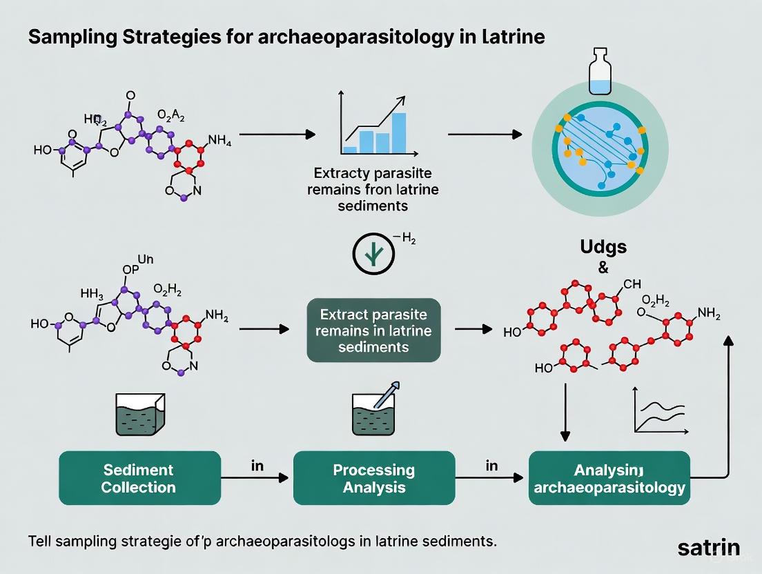
Latrines constitute a unique and invaluable archaeological archive for reconstructing past human health, diet, and migration patterns. As reservoirs of preserved fecal matter, they contain robust assemblages of parasite eggs and cysts, providing a direct window into the enteric infections that afflicted past populations [8]. The anaerobic conditions often found within latrine sediments promote exceptional preservation of organic materials, including the durable eggs of helminths (parasitic worms) and the more fragile cysts of protozoa [9]. Systematic analysis of these parasite assemblages allows researchers to investigate temporal changes in sanitation, dietary preferences, human-animal interactions, and the spread of infectious diseases across centuries and millennia [8] [10]. This document outlines standardized protocols for the paleoparasitological investigation of latrine sediments, framed within a broader thesis on developing effective sampling strategies for archaeoparasitology.
Multimethod Analytical Framework
A multimethod approach is paramount for a comprehensive reconstruction of past parasite diversity. Relying on a single technique can lead to an incomplete taxonomic profile, as different parasites are detected with varying efficacy across methods [11]. The integrated workflow below summarizes the sequential application of these techniques.
Workflow for the Multimethod Analysis of Latrine Sediments
Detailed Experimental Protocols
Sediment Sampling Protocol
Principle: To obtain a representative sample that captures the chronological and spatial variation within a latrine deposit.
Procedure:
- Context Documentation: Record the precise archaeological context of the latrine (e.g., date, location, associated structures). Sample from clearly defined layers where possible [9].
- Grid Sampling: If the latrine fill is homogeneous, establish a sampling grid. Collect multiple sub-samples from different points (e.g., top, middle, bottom, center, edges) to account for potential heterogeneity in parasite egg distribution.
- Control Sampling: Collect control samples from outside the latrine area (e.g., nearby soil not associated with fecal deposition) to distinguish between parasites derived from human infection and environmental contamination.
- Material: Use clean, single-use tools (spatulas, trowels) for each sample to prevent cross-contamination. Store samples in sterile, airtight containers (e.g., Whirl-Pak bags).
- Storage: Label all samples clearly and store in a cool, dark environment at 4°C until processing to minimize modern biological growth and preserve biomolecules [11].
Microscopy for Helminth Eggs
Principle: To isolate, identify, and quantify helminth eggs based on their characteristic size and morphological features [9] [12]. This is the most effective method for detecting robust helminth eggs.
Reagents & Materials:
- 0.5% Trisodium Phosphate (TSP) solution
- Light microscope (e.g., Olympus BX40F)
- Microsieves (e.g., mesh sizes 300μm, 150μm, 20μm)
- Centrifuge and centrifuge tubes
- Glycerol
- Microscope slides and coverslips
Procedure:
- Rehydration: Disaggregate a 0.2 g sediment subsample in 10-15 mL of 0.5% TSP solution. Soak for 24-96 hours, with intermittent vortexing, until fully rehydrated [9] [12].
- Microsieving: Pass the rehydrated sample through a stack of sieves (e.g., 300 μm, 150 μm, 20 μm). Helminth eggs are typically retained on the 20 μm sieve.
- Concentration: Collect the material from the 20 μm sieve, transfer to a centrifuge tube, and centrifuge at 3100 g for 5 minutes. Discard the supernatant.
- Microscopy: Re-suspend the pellet in a small volume of glycerol. Transfer to a microscope slide and examine under a light microscope at 200x and 400x magnification.
- Identification & Quantification: Identify eggs based on standard morphological criteria (size, shape, shell thickness, opercula). Measure and count the first 100 eggs of each taxon. Calculate the concentration in eggs per gram (EPG) of sediment [9].
Enzyme-Linked Immunosorbent Assay (ELISA) for Protozoan Antigens
Principle: To detect species-specific antigens from protozoan parasites (e.g., Giardia, Cryptosporidium) using antibody-based kits, offering high sensitivity for fragile pathogens often missed by microscopy [11].
Reagents & Materials:
- Commercial ELISA kits (e.g., TECHLAB GIARDIA II, E. HISTOLYTICA II, CRYPTOSPORIDIUM II)
- 0.5% Trisodium Phosphate (TSP) solution
- Microsieves (e.g., 20 μm)
- Microplate reader
Procedure:
- Sample Preparation: Disaggregate a 1.0 g sediment subsample in 0.5% TSP.
- Microsieving for Cysts: Since protozoan cysts are small (<20 μm), collect the material that passes through the 20 μm sieve. Concentrate this fraction by centrifugation.
- ELISA Protocol: Follow the manufacturer's instructions for the specific ELISA kit. This typically involves:
- Incubating the sample in antibody-coated wells.
- Washing to remove unbound material.
- Adding a detector antibody and enzyme conjugate.
- Adding a substrate solution that produces a color change in the presence of the antigen.
- Detection: Measure the colorimetric signal with a microplate reader. Compare results to positive and negative controls provided in the kit to confirm the presence of protozoan antigens [11].
Sedimentary Ancient DNA (sedaDNA) Analysis
Principle: To recover and identify parasite DNA from latrine sediments, allowing for species-level confirmation and detection of parasites that leave no morphological trace [11].
Reagents & Materials:
- Garnet PowerBead Tubes (Qiagen)
- Lysis buffer (e.g., containing guanidinium isothiocyanate, NaPO₄)
- Proteinase K
- Dabney binding buffer
- Silica columns for DNA purification
- Illumina DNA library preparation kit
- Targeted enrichment baits for parasites
Procedure: All steps must be performed in dedicated ancient DNA facilities to prevent contamination with modern DNA [11].
- Bead-Beating Lysis: Add a 0.25 g sediment subsample to a Garnet PowerBead tube containing lysis buffer. Vortex vigorously for 15 minutes to mechanically disrupt sediment and parasite eggs.
- Enzymatic Digestion: Add Proteinase K and incubate with continuous rotation at 35°C overnight.
- DNA Binding & Purification: Combine the supernatant with a high-volume Dabney binding buffer. Centrifuge at 4500 rpm at 4°C for 6-24 hours to precipitate inhibitors. Pass the clear supernatant through a silica column to bind DNA. Wash and elute the DNA in a final volume of 50 µL [11].
- Library Preparation & Sequencing: Prepare double-stranded DNA libraries for Illumina sequencing. For optimal results, use a targeted enrichment (capture) approach with bait sets designed for a comprehensive range of human parasites, followed by high-throughput sequencing [11].
The Scientist's Toolkit: Essential Research Reagents & Materials
Table 1: Key reagents, materials, and their functions in paleoparasitology protocols.
| Item Name | Function/Application | Protocol |
|---|---|---|
| 0.5% Trisodium Phosphate (TSP) | Rehydration and disaggregation of sediment samples to release parasite eggs. | Microscopy, ELISA [9] [12] |
| Microsieves (20 µm mesh) | Isolation of helminth eggs by size; collection of fine fraction for protozoan analysis. | Microscopy, ELISA [11] [9] |
| Glycerol | Mounting medium for microscopy; clears debris and enhances egg visibility. | Microscopy [9] |
| Commercial ELISA Kits | Immunological detection of specific protozoan antigens (e.g., Giardia, Cryptosporidium). | ELISA [11] |
| Garnet PowerBead Tubes | Mechanical disruption (bead beating) of sediment and tough egg shells for DNA release. | sedaDNA Analysis [11] |
| Silica Columns | Purification and concentration of ancient DNA from complex sediment extracts. | sedaDNA Analysis [11] |
| Parasite-Specific DNA Baits | Targeted enrichment of parasite DNA from total sedaDNA libraries to increase sensitivity. | sedaDNA Analysis [11] |
Data Interpretation and Contribution to Sampling Strategies
The quantitative and qualitative data derived from these protocols feed directly into robust archaeological interpretation and inform future sampling strategies.
Quantitative Data from Parasite Assemblages
Table 2: Example quantitative data from parasitological analysis of a 15th–16th c. CE latrine in Bruges, demonstrating how egg concentration (EPG) is calculated and reported [9].
| Sample Layer | Parasite Taxa | Total Egg Count | Concentration (EPG) | Avg. Length (µm) |
|---|---|---|---|---|
| Layer A (Upper) | Ascaris sp. (Roundworm) | 142 | 710 | 65.2 |
| Trichuris sp. (Whipworm) | 85 | 425 | 54.1 | |
| Layer B (Lower) | Ascaris sp. (Roundworm) | 210 | 1050 | 64.8 |
| Trichuris sp. (Whipworm) | 110 | 550 | 53.9 | |
| Taenia sp. (Tapeworm) | 15 | 75 | 35.2 | |
| Schistosoma mansoni | 1 | 5 | 143.0 |
Table 3: Effectiveness of different paleoparasitological methods based on a multimethod study [11].
| Analytical Method | Optimal For Detecting | Key Advantage |
|---|---|---|
| Microscopy | Helminths (e.g., Ascaris, Trichuris, Taenia) | Most effective for detecting and identifying helminth eggs based on morphology. |
| ELISA | Protozoa (e.g., Giardia duodenalis) | Highest sensitivity for detecting diarrhea-causing protozoa. |
| sedaDNA with Targeted Capture | Species-level confirmation, detecting non-morphological taxa. | Can differentiate between species (e.g., T. trichiura vs T. muris) and reveal full diversity. |
Logical Framework for Interpreting Results
The data generated should be interpreted within a logical framework that connects evidence to archaeological inference, guiding the development of a thesis on sampling strategies.
Applying this multimethod approach has revealed significant historical trends. For instance, research shows a marked shift in parasite diversity in Europe from the pre-Roman to the Roman period, with a decrease in zoonotic parasites and a concurrent increase in fecal-oral transmitted species like roundworm and whipworm, consistent with changes in urbanization and sanitation practices [11]. Furthermore, the detection of Schistosoma mansoni (a parasite endemic to Africa and the Middle East) in a 15th–16th c. CE latrine in Bruges, Belgium, provides direct evidence of long-distance travel or migration, possibly linked to medieval trade networks or the early Atlantic slave trade [9]. These insights underscore the critical role of latrines as archives for understanding the complex interplay between human health, behavior, and the environment through time.
The analysis of gastrointestinal parasites from archaeological latrine sediments provides a powerful lens through which to understand human health, migration, dietary practices, and sanitation throughout history. Paleoparasitology, the study of ancient parasites, identifies two primary categories of parasitic markers: heirloom parasites inherited from our primate ancestors in Africa, and souvenir parasites acquired from animals during human migration and settlement across the globe [13]. These parasites leave behind morphological and biomolecular evidence that persists for millennia in favorable preservation environments, particularly in latrine sediments where organic matter accumulates.
The robust nature of helminth eggs, protected by chitinous shells containing chitin, keratin, and sclerotin, enables their exceptional preservation in the archaeological record [14]. Protozoan cysts, while more fragile, can be detected through immunological and molecular methods even when morphological preservation is poor [11]. This application note details the key parasitic markers, quantitative detection methods, and experimental protocols essential for comprehensive archaeoparasitological research focused on latrine sediments, providing researchers with standardized approaches for analyzing past human-parasite relationships.
Key Parasitic Markers: Morphology and Historical Context
Characteristic Features of Major Helminths
Table 1: Diagnostic Characteristics of Primary Helminth Eggs in Archaeological Contexts
| Parasite | Egg Size (Micrometers) | Egg Morphology | Historical Significance & Geographic Distribution |
|---|---|---|---|
| Ascaris lumbricoides (Roundworm) | 45-75 × 35-50 | Oval with thick, mammillated coat | One of the oldest human parasites; heirloom species; global distribution; indicates fecal-oral contamination [15] [13] |
| Trichuris trichiura (Whipworm) | 50-54 × 22-23 | Barrel-shaped with polar plugs | Heirloom species; indicates fecal-oral contamination and poor sanitation [11] |
| Hookworm (Ancylostoma & Necator) | 55-60 × 35-40 | Oval, thin-shelled with embryonic cells | Heirloom species; indicates soil contamination and barefoot exposure [16] [13] |
| Diphyllobothrium sp. (Fish Tapeworm) | 66-82 × 62-71 | Oval with operculum and abopercular knob | Souvenir species; indicates consumption of raw/undercooked fish; found in Arctic and subarctic regions [17] |
| Opisthorchis felineus | 30 × 11 | Small, operculated | Souvenir species; indicates fish consumption; found in Western Siberia [17] |
Characteristic Features of Intestinal Protozoa
Table 2: Diagnostic Characteristics of Primary Protozoan Parasites in Archaeological Contexts
| Parasite | Cyst Size (Micrometers) | Morphology | Historical Significance & Detection Methods |
|---|---|---|---|
| Giardia duodenalis | 8-12 × 7-10 | Oval, refractile with axostyles | Causes diarrheal illness; detected by ELISA and PCR; indicates waterborne transmission [11] |
| Entamoeba histolytica | 12-15 | Spherical with 1-4 nuclei | Causes amebic dysentery; differentiated from non-pathogenic E. dispar by molecular methods [18] |
| Cryptosporidium spp. | 4-6 | Small, spherical | Causes diarrheal illness; detected by antigen tests and PCR; indicates zoonotic transmission [18] |
| Entamoeba coli | 10-35 | Spherical with 8 nuclei in mature cysts | Non-pathogenic commensal; indicates fecal contamination of environment [19] |
Heirloom vs. Souvenir Parasites: Evolutionary Origins
The classification of parasites as heirlooms or souvenirs provides critical insights into human migration patterns and cultural practices:
Heirloom Parasites: These parasites were inherited from primate ancestors and accompanied humans out of Africa. Examples include Ascaris lumbricoides, Trichuris trichiura, and pinworm (Enterobius vermicularis) [13]. Their presence in archaeological sites worldwide demonstrates their establishment in early human populations.
Souvenir Parasites: These parasites were acquired when humans came into contact with new animals and environments during migrations. Examples include the fish tapeworm (Diphyllobothrium sp.) in Arctic regions and the liver flukes (Opisthorchis and Clonorchis) in Asia [13] [17]. Their presence reveals dietary practices and local environmental exposures.
Quantitative Prevalence Data from Archaeological Studies
Table 3: Prevalence of Parasitic Infections Across Archaeological and Modern Studies
| Study Population/Period | Ascaris Prevalence | Trichuris Prevalence | Giardia Prevalence | Hookworm Prevalence | Detection Method |
|---|---|---|---|---|---|
| Preschool children, Amhara, Ethiopia (2017) | 10.8% | 1.4% | 10.4% | 0% | Ether-concentration microscopy [16] |
| Children, Boboye Department, Niger (2020) | 0% | 0% | 65.1% | 0% | Real-time PCR [20] |
| Disabled individuals, global (2025) | Significant (specific % not reported) | Significant (specific % not reported) | Significant (specific % not reported) | Significant (specific % not reported) | Microscopy, serology, molecular techniques [21] |
| Roman & Medieval periods, Europe | Dominant | Dominant | Increased prevalence | Variable | Multi-method approach [11] |
Experimental Protocols for Latrine Sediment Analysis
Multi-Method Workflow for Comprehensive Parasite Detection
Detailed Methodological Protocols
Microscopy for Helminth Eggs
Principle: Helminth eggs are identified based on morphological characteristics (shape, operculum presence, shell ornamentation) and size measurements [19] [14].
Procedure:
- Sample Preparation: Disaggregate 0.2 g of sediment in 10 mL of 0.5% trisodium phosphate solution and allow to rehydrate for 72 hours [17] [11].
- Microsieving: Pass the suspension through a 160 μm sieve stacked above a 20 μm sieve to concentrate the fraction containing most helminth eggs [11].
- Microscopy: Mix the 20-160 μm fraction with glycerol and examine under light microscope at 200× and 400× magnification.
- Identification: Identify eggs based on established morphological criteria and measure dimensions using calibrated microscopy software [17].
Quality Control: Include negative control samples from outside the archaeological context to monitor environmental contamination [17]. Have an independent expert confirm positive slides and every 10th negative specimen [16].
ELISA for Protozoan Antigens
Principle: Enzyme-linked immunosorbent assay detects protozoan-specific antigens even when cysts are not morphologically preserved [11].
Procedure:
- Sample Processing: Disaggregate 1 g of sediment in 0.5% trisodium phosphate and microsieve to collect material below 20 μm where protozoan cysts concentrate [11].
- Commercial Kits: Use manufacturer protocols for commercial ELISA kits (e.g., GIARDIA II, E. HISTOLYTICA II, CRYPTOSPORIDIUM II from TECHLAB, Inc.) [11].
- Protocol Adaptation: Follow established archaeological adaptations of clinical protocols with appropriate controls [11].
Sedimentary Ancient DNA (sedaDNA) Analysis
Principle: Targeted enrichment and high-throughput sequencing recover parasite DNA from complex sediment matrices [11].
Procedure:
- DNA Extraction:
- Subsample 0.25 g of sediment in dedicated ancient DNA facilities [11].
- Use garnet PowerBead tubes with lysis buffer for mechanical disruption of parasite eggs [11].
- Add proteinase K and rotate continuously at 35°C overnight [11].
- Extract DNA using high-volume Dabney binding buffer and silica columns [11].
- Centrifuge at 4°C for 6-24 hours to precipitate inhibitors [11].
- Library Preparation & Sequencing:
The Scientist's Toolkit: Essential Research Reagents and Materials
Table 4: Key Research Reagents and Materials for Paleoparasitology
| Reagent/Material | Application | Function | Example Specifications |
|---|---|---|---|
| Trisodium Phosphate (0.5%) | Sample rehydration and disaggregation | Dissolves sediment matrix while preserving parasite eggs | 0.5% w/v solution in distilled water [17] [11] |
| Glycerol | Microscopy slide preparation | Clears debris and enhances egg visibility for microscopy | Mixed with processed sample sediment [16] [17] |
| Commercial ELISA Kits | Protozoan antigen detection | Immunological detection of Giardia, Entamoeba, Cryptosporidium antigens | GIARDIA II, E. HISTOLYTICA II, CRYPTOSPORIDIUM II (TECHLAB, Inc.) [11] |
| Garnet PowerBead Tubes | DNA extraction | Mechanical disruption of robust parasite egg shells through bead beating | Contains garnet beads for improved lysis efficiency [11] |
| Dabney Binding Buffer | DNA extraction and purification | Binds DNA to silica columns while removing inhibitors | High-volume formulation for sedaDNA [11] |
| Parasite-Specific Baits | Targeted DNA enrichment | Hybridization capture of parasite DNA from complex extracts | Comprehensive set covering diverse human parasites [11] |
| Diethyl Ether | Concentration methods | Parasite egg concentration in stool specimens | Used in ether-concentration methods [16] |
| Sodium Acetate-Acetic Acid-Formalin (SAF) | Stool preservation | Preserves parasite morphology for later analysis | Preserves 1g stool in 10mL SAF [16] |
Data Interpretation and Historical Reconstruction
The integration of results from microscopy, ELISA, and sedaDNA provides the most comprehensive understanding of past parasitic infections [11]. Each method has distinct strengths:
- Microscopy excels at detecting helminth eggs and providing quantification of infection intensity [11] [14].
- ELISA is particularly sensitive for detecting protozoa that cause diarrheal diseases, notably Giardia duodenalis [11].
- sedaDNA allows species-specific identification, detection of parasites that produce few eggs, and phylogenetic analysis of ancient parasite strains [11].
Temporal analysis of parasite assemblages in latrine sediments can reveal significant shifts in sanitation, dietary practices, and human-animal relationships. For example, during the Roman period, there was a marked transition toward dominance of fecal-oral transmitted parasites (roundworm, whipworm, and diarrheal protozoa) alongside a decrease in zoonotic parasites, reflecting changes in sanitation infrastructure and dietary practices [11].
The classification of parasites as heirloom or souvenir species provides evidence for human migration patterns and cultural exchanges throughout history [13]. The presence of souvenir parasites in archaeological contexts reveals contact with new animal species and environments, while heirloom parasites demonstrate the continuity of human-parasite relationships dating back to our primate ancestors.
Archaeoparasitology, the study of ancient parasites, stands at the intersection of archaeology, parasitology, and history, providing a unique lens through which to investigate human health, sanitation practices, dietary habits, and zoonotic disease trajectories [14]. Latrine sediments serve as a critical archaeological substrate for this research, preserving a rich record of gastrointestinal parasites that infected past populations. The analysis of these sediments reveals not only the pathogens that afflicted our ancestors but also offers indirect evidence of sanitation efficacy, culinary practices, and human-animal interactions [22] [14]. The recovery of parasite eggs, antigens, and ancient DNA (aDNA) from such contexts has revolutionized our understanding of the long-term relationship between humans and their parasites. This application note details the sampling strategies and analytical protocols essential for robust archaeoparasitological research, framing them within a broader thesis on unlocking historical lifeways and disease burdens through the systematic study of latrine sediments.
Core Principles and Quantitative Foundations
The foundation of archaeoparasitology lies in the robust recovery and identification of parasite remains. The table below summarizes the primary diagnostic targets and their significance for interpreting past human ecology.
Table 1: Diagnostic Targets in Archaeoparasitology and Their Interpretative Value
| Diagnostic Target | Description | Key Parasites Identified | Interpretative Value |
|---|---|---|---|
| Helminth Eggs [14] | Microscopic, chitinous-shelled eggs (30-160 µm) produced by parasitic worms. Resistant to decay. | Ascaris lumbricoides (roundworm), Trichuris trichiura (whipworm), hookworms [23]. | Direct evidence of fecal-oral transmission; indicator of sanitation levels and personal hygiene [22]. |
| Protozoan Antigens [11] [14] | Protein markers detected via immunological methods like Enzyme-Linked Immunosorbent Assay (ELISA). | Giardia duodenalis, Entamoeba histolytica, Cryptosporidium spp. [11]. | Evidence of diarrheal diseases; indicates water quality and contamination [11]. |
| Sedimentary Ancient DNA (sedaDNA) [11] | Trace DNA preserved in sediment, recovered via specialized extraction and sequencing. | All parasite species, allows for species/strain differentiation (e.g., Trichuris trichiura vs. T. muris) [11]. | High-specificity detection; reveals parasite diversity and evolutionary history, even in low-abundance cases [11]. |
Data from archaeological sites provides quantitative insights into historical infection patterns. The following table synthesizes findings from key studies, illustrating the prevalence of specific parasites and their implications.
Table 2: Archaeological Case Studies of Parasite Infection from Latrine Sediments
| Archaeological Site / Context | Period | Key Parasite Findings | Inferred Socio-Environmental Context |
|---|---|---|---|
| Ephesus, Turkey [22] | Roman Period (1st c. BCE - 6th c. CE) | Whipworm and roundworm eggs found in private latrine, public latrine, and harbour canal. Whipworm was dominant. | Widespread sanitation challenges despite Roman infrastructure. Fecal contamination of soil, food, and water was common [22]. |
| Northwestern Argentina [24] | Modern (2010-2019) | Prevalence of A. lumbricoides (11.14%), hookworm (8.16%), T. trichiura (1.38%), and S. stercoralis (6.36%) in human populations. | High burden of soil-transmitted helminths (STHs) linked to inadequate sanitation and socioeconomic conditions [24]. |
| Roman Empire & Medieval Sites [11] | Neolithic - Medieval (c. 6400 BCE - 1500 CE) | Multimethod analysis revealed a shift: decrease in zoonotic parasites and increase in fecal-oral parasites (roundworm, whipworm, diarrheal protozoa) in Roman/Medieval periods. | Changes in parasite diversity reflect shifts in sanitation, animal husbandry, and settlement patterns during the Roman period [11]. |
| Jerusalem & Riga [25] | Medieval (14th-15th c. CE) | Recovery of bacterial and eukaryotic DNA from latrines revealed a unique gut microbiome, distinct from both modern industrial and hunter-gatherer populations. | Provides pre-industrial baseline for human gut contents and illustrates the impact of lifestyle on microbiome composition [25]. |
Experimental Protocols for Latrine Sediment Analysis
A multimethod approach is critical for a comprehensive paleoparasitological reconstruction, as each technique has unique strengths and limitations [11]. The following protocols are standardized for the analysis of latrine sediments.
Protocol 1: Microscopy-Based Identification of Helminth Eggs
Principle: This method relies on the liberation, concentration, and morphological identification of durable helminth eggs from sediment matrices using microscopy [26].
Workflow:
Detailed Steps:
- Disaggregation: Weigh 0.2-0.5 g of sediment into a specimen container. Add 10-15 mL of 0.5% aqueous trisodium phosphate (Na₃PO₄) solution. Allow the sample to soak for 72 hours, vortexing periodically to break down the sediment [11] [26].
- Microsieving: Pour the disaggregated sample through a stack of geological sieves, typically collecting the fraction between 20 µm and 160 µm. This step removes very fine silt and large debris, enriching the sample for parasite eggs [11].
- Concentration: Transfer the sieved material to a centrifuge tube and use a flotation technique with a high-specific-gravity solution like Sheather's sugar solution (specific gravity 1.27). Centrifuge to concentrate eggs in the supernatant, which is then collected for microscopy [26].
- Microscopy and Identification: Place a drop of the concentrate on a microscope slide, add a coverslip, and systematically examine under light microscopy at 200x and 400x magnification. Identify eggs based on standard morphological criteria (size, shape, wall structure, opercula) and count them. Results can be expressed as eggs per gram (epg) of sediment using the formula:
epg = (egg count / sediment weight) * (total volume of concentrate / volume examined)[26].
Protocol 2: Immunological Detection of Protozoan Antigens (ELISA)
Principle: Commercial Enzyme-Linked Immunosorbent Assay (ELISA) kits are used to detect species-specific antigens from protozoan parasites, which are not reliably visible via light microscopy [11].
Workflow:
Detailed Steps:
- Sample Preparation: Weigh 1 g of sediment and disaggregate in 0.5% trisodium phosphate. Given the small size of protozoan cysts (<20 µm), microsieving is performed to collect the material in the catchment container below the 20 µm sieve. This fraction is concentrated via centrifugation [11].
- ELISA Procedure: Follow the manufacturer's protocol for the commercial ELISA kit (e.g., TECHLAB GIARDIA II, E. HISTOLYTICA II, CRYPTOSPORIDIUM II). This typically involves: a. Adding the prepared sample to antibody-coated wells. b. Incubating to allow antigen-antibody binding. c. Washing wells to remove unbound material. d. Adding an enzyme-linked secondary antibody. e. Adding a substrate that produces a colorimetric change in the presence of the enzyme. f. Measuring the signal and comparing it to positive and negative controls to determine a positive or negative result [11].
Protocol 3: Targeted Sedimentary Ancient DNA (sedaDNA) Analysis
Principle: This protocol uses specialized DNA extraction, library preparation, and targeted enrichment (hybridization capture) to retrieve and sequence trace amounts of parasite DNA from complex latrine sediments [11].
Workflow:
Detailed Steps:
- DNA Extraction (Dedicated aDNA Facility): Subsample 0.25 g of sediment in a garnet PowerBead tube. Add a lysis buffer containing guanidinium isothiocyanate and vortex vigorously for 15 minutes to mechanically disrupt sediment and parasite eggs. Add Proteinase K and rotate at 35°C overnight. Bind DNA from the supernatant using a high-volume binding buffer and purify via silica columns, with extended cold centrifugation to remove inhibitors [11].
- Library Preparation and Enrichment: Prepare double-stranded DNA libraries for Illumina sequencing, incorporating dual-indexing adapters. A subset of libraries can be shotgun sequenced. For targeted enrichment, hybridize the libraries with biotinylated RNA "baits" designed to cover a comprehensive set of parasite genomes. Capture the bound DNA with streptavidin-coated beads, wash, and amplify the enriched library [11].
- Sequencing and Analysis: Sequence the enriched libraries on an Illumina platform. Process the raw sequencing data through a bioinformatic pipeline, including: adapter trimming; mapping reads to reference genomes; and taxonomic assignment to identify the preserved parasite species [11].
The Scientist's Toolkit: Essential Research Reagents and Materials
Table 3: Key Research Reagent Solutions for Archaeoparasitology
| Reagent / Material | Function / Application | Key Considerations |
|---|---|---|
| Trisodium Phosphate (0.5%) [11] [26] | Disaggregation of sediment samples to release parasite eggs and other inclusions. | Effective at breaking down clay and organic aggregates without destroying most helminth eggs. |
| Hydrofluoric Acid (HF) [26] | Digestion of silicate minerals in sediment samples during palynological processing. | Highly hazardous. Requires specialized fume hoods and training. Not essential but can improve recovery in clay-rich sediments. |
| Sheather's Sugar Solution [26] | Flotation medium (specific gravity ~1.27) for concentrating parasite eggs via centrifugation. | High specific gravity allows buoyancy of most helminth eggs. Less hazardous than HF. |
| Guanidinium Thiocyanate-based Lysis Buffer [11] | Chemical disruption of sediment and organic matter, and inactivation of nucleases during DNA extraction. | Critical for releasing and preserving highly degraded ancient DNA from complex sediments. |
| Biotinylated RNA Baits [11] | Target enrichment for sedaDNA; hybridize to and allow capture of parasite DNA from sequencing libraries. | Enables cost-effective sequencing of low-abundance parasite targets by reducing background DNA. |
| Commercial ELISA Kits [11] [22] | Immunological detection of protozoan antigens (e.g., Giardia, Cryptosporidium). | Provide high specificity and sensitivity for fragile protozoa that are rarely preserved as cysts. |
The protocols outlined herein form the basis for generating data that deeply informs our understanding of historical sanitation, diet, and disease. The detection of fecal-oral parasites like Ascaris and Trichuris directly reflects the level of sanitation and hygiene in a community, as their transmission thrives in environments contaminated with human feces [22]. The finding of these parasites in Roman Ephesus, despite the presence of complex sanitation infrastructure, indicates that the mere existence of latrines and sewers did not necessarily break the cycle of infection [22]. Furthermore, the recovery of zoonotic parasites (e.g., Echinococcus granulosus from dogs) provides evidence of dietary practices, such as the consumption of parasitized meat, and the nature of close human-animal co-habitation [11] [27]. The shift in parasite diversity observed from the pre-Roman to the Roman period, with a decrease in zoonotic species and an increase in human-specific fecal-oral species, signals profound changes in animal husbandry, food preparation, and settlement density [11].
In conclusion, a multimethod approach—integrating microscopy, immunology, and sedaDNA—is no longer optional but essential for a complete and accurate reconstruction of past parasite communities [11]. This interdisciplinary framework, grounded in rigorous sampling and analytical protocols, allows archaeoparasitology to move beyond simple catalogs of past pathogens. It empowers researchers to critically interrogate the complex interactions between human behavior, environmental manipulation, sanitation technology, and the enduring burden of infectious disease throughout history.
A Multimethod Toolkit: From Field Sampling to Laboratory Analysis
This application note provides a standardized framework for the collection and preservation of latrine sediments in archaeoparasitology. The protocols detailed herein are designed to maximize the recovery of parasite evidence—including helminth eggs, protozoan cysts, and faecal biomarkers—and to facilitate subsequent multi-method analyses. Adherence to these procedures ensures the generation of robust, comparable data sets essential for investigating ancient human health, diet, and sanitation practices.
Latrine sediments represent a critical archaeological resource for reconstructing past human lifeways and disease burdens through the analysis of preserved parasite remains [14]. The field of paleoparasitology has evolved from relying on a single analytical method to employing a multimethod approach that significantly enhances taxonomic recovery and interpretation [11]. The success of these advanced laboratory analyses is, however, entirely contingent upon the implementation of rigorous field sampling and preservation strategies. This document outlines evidence-based protocols to guide researchers from initial site assessment to sample stabilization, ensuring the integrity of delicate paleoparasitological evidence.
Field Sampling Strategies
Proper sampling strategy is fundamental to the scientific value of any archaeoparasitological study. The following section outlines proven methods for sediment collection.
Sampling Location and Context
The choice of sampling location within a latrine feature directly influences the probability of recovering parasite evidence.
- Primary Contexts: Prioritize sediment from the pelvic region of skeletons, as the sacrum acts as a "natural bowl" preserving intestinal contents after decomposition [28]. Samples from latrine fills, sewer drains, and coprolites are also highly valuable [11].
- Stratigraphic Sampling: Collect samples from every distinct stratigraphic layer within a latrine feature. This enables the investigation of temporal changes in parasite prevalence and diversity [29].
- Control Samples: Always collect control samples from outside the anticipated area of faecal contamination (e.g., from surrounding natural sediments) to establish background levels and assess potential contamination.
Table 1: Sampling Protocols by Archaeological Context
| Context Type | Recommended Sample Mass | Primary Target | Key Consideration |
|---|---|---|---|
| Pelvic Sediment | 5–10 g [28] | Helminth eggs, protozoa | Sample directly from the sacral foramina. |
| Latrine Fill | 10–20 g [26] | Helminth eggs, biomarkers | Sample multiple strata for a time series. |
| Coprolites | Entire coprolite, or 1–2 g subsamples [30] | Eggs per gram (EPG), aDNA | Provides individual-level infection data. |
| Cave Sediments | 10–20 g [29] | Faecal biomarkers | Target deeper, protected zones like Palaeolithic layers. |
Sample Collection Workflow
The following standardized workflow minimizes contamination and ensures sample integrity:
- Documentation: Photograph and record the precise three-dimensional location of the sample before collection.
- Surface Cleaning: Gently remove the exposed surface layer (1–2 cm) of the sediment using a clean trowel or spatula to eliminate modern contaminants.
- Collection: Using a clean, disposable instrument, collect the required mass of sediment and immediately place it into a pre-labeled, sterile container.
- Chain of Custody: Label containers with permanent, waterproof ink. Include site, context number, sample ID, and date.
Preservation Methodologies for Downstream Analysis
The choice of preservation method at the time of collection dictates which future analytical techniques can be successfully applied. A multi-faceted preservation strategy is recommended.
Chemical Preservation
Different preservatives stabilize different types of molecular and morphological evidence.
- For DNA Analysis: Dimethyl sulfoxide, ethylenediaminetetraacetic acid, sodium chloride (DESS) solution is highly effective for stabilizing DNA at room temperature, preserving microbial and parasite community structure for months [31]. Commercial DNA/RNA stabilizers (e.g., Zymo DNA/RNA Shield) are also a viable option.
- For Immunological Assays: If enzyme-linked immunosorbent assay (ELISA) for protozoan antigens is planned, storing a subsample at -20°C without any preservative is the standard practice, though 95% ethanol can also be used for ambient temperature storage [11] [31].
- For Microscopy: Samples for microscopic identification of helminth eggs can be air-dried or stored in a 0.5% trisodium phosphate solution to aid in disaggregation during later processing [11].
Table 2: Preservation Methods for Specific Analytical Targets
| Analytical Method | Recommended Preservation | Storage Temperature | Key Application |
|---|---|---|---|
| Microscopy | Air-dried or 0.5% Trisodium Phosphate [11] | Room Temperature | Identification of helminth eggs based on morphology. |
| ELISA | Frozen (-20°C) or 95% Ethanol [11] [31] | -20°C or Room Temperature | Detection of protozoan antigens (e.g., Giardia). |
| sedaDNA / Metagenomics | DESS Solution or Commercial Stabilizer [31] | Room Temperature | Targeted capture, sequencing of parasite DNA. |
| Faecal Biomarkers | Frozen (-20°C) [32] | -20°C | Analysis of sterols, stanols, and bile acids. |
Workflow for a Multi-Method Approach
Given the value of a multi-method framework [11], field researchers should plan to collect and preserve multiple subsamples from a single context.
The Scientist's Toolkit: Essential Reagents & Materials
The following reagents are critical for the field collection and initial processing of latrine sediments.
Table 3: Key Research Reagent Solutions for Fieldwork
| Reagent / Material | Function | Application Notes |
|---|---|---|
| DESS Solution | A chemical cocktail (Dimethyl sulfoxide, EDTA, NaCl) that stabilizes DNA at room temperature for metagenomic studies [31]. | Ideal for remote locations; samples can be stored for months without freezing. |
| Trisodium Phosphate (0.5%) | A rehydrating and disaggregating solution that softens hardened sediments and coprolites for microscopic analysis [11]. | Standard for rehydration before micro-sieving for egg recovery. |
| Hydrofluoric Acid (HF) | Used in specialized palynology-derived lab methods to dissolve silica and silicate minerals, liberating parasite eggs [26]. | High hazard; requires advanced lab facilities and safety protocols. |
| Sheather's Sugar Solution | A high-specific-gravity flotation medium used with centrifugation to concentrate parasite eggs from sediment for microscopy [26]. | Effective for recovering a wide range of egg types; safe for standard labs. |
| Guanidinium Isothiocyanate Lysis Buffer | A powerful denaturant used in DNA extraction buffers to inactivate nucleases and release DNA from complex sediments and parasite eggs [11]. | Used with physical disruption (bead beating) for optimal DNA yield. |
Implementing these structured protocols for the collection and preservation of latrine sediments establishes a strong foundation for high-quality archaeoparasitological research. By planning for a multi-method analytical approach from the outset, researchers can maximize the informational yield from precious and non-renewable archaeological samples. The consistent application of these strategies across different sites and studies will generate robust, comparable datasets, ultimately advancing our understanding of the historical relationships between humans, their environments, and their parasites.
Paleoparasitology, the study of ancient parasites, provides invaluable insights into the health, sanitation, dietary habits, and migration patterns of past populations [14]. The analysis of archaeological sediments, particularly from latrines, offers a direct source of evidence for understanding parasitic infections throughout history [9]. The RHM protocol (Rehydration–Homogenization–Micro-sieving) represents a fundamental methodological approach in this field, specifically designed for the optimal recovery of parasite eggs from complex archaeological matrices [33]. This protocol is noted for its effectiveness in maximizing parasite biodiversity recovery while preserving egg morphology, making it particularly suitable for archaeoparasitological studies of latrine sediments [33].
The RHM Protocol: Principle and Rationale
The RHM protocol is a three-step sedimentation technique designed to extract helminth eggs from archaeological sediments while minimizing damage and loss. Its primary advantage over flotation or chemical-intensive methods lies in its non-aggressive nature, which aims to recover all types of eggs without selection, thereby providing a more comprehensive view of parasite biodiversity [33]. Comparative studies have demonstrated that the RHM protocol yields maximum biodiversity of parasite taxa when directly compared to methods incorporating acids (HCl, HF) or bases (NaOH) [33]. Methods using sodium hydroxide, in particular, have been shown to significantly reduce recoverable biodiversity, likely due to chemical damage to the chitinous shell of the eggs [33]. The protocol's robustness makes it especially valuable for the analysis of latrine sediments, which often contain a diverse array of parasite species indicative of past sanitation, diet, and trade connections [9].
Detailed Experimental Protocol
Materials and Reagents
Table 1: Essential Research Reagents and Materials for the RHM Protocol
| Item Name | Specification/Concentration | Primary Function in Protocol |
|---|---|---|
| Trisodium Phosphate | 0.5% aqueous solution | Rehydrates and disperses the sediment sample. |
| Glycerol | Laboratory grade | Mixed with rehydration solution and final residue for microscopy. |
| Micro-sieve Column | Mesh sizes typically include 300 µm, 150 µm, and 20 µm | Filters and concentrates parasite eggs by size. |
| Ultrasonic Bath | Laboratory-grade | Homogenizes the sample to liberate eggs from the sediment. |
| Centrifuge & Tubes | Standard laboratory equipment | Concentrates the sample after micro-sieving. |
| Light Microscope | With 100x, 200x, and 400x magnification | For final identification and quantification of eggs. |
Step-by-Step Workflow
The following diagram illustrates the streamlined, three-stage workflow of the RHM protocol:
Step 1: Rehydration A 0.2-1.0 g subsample of the archaeological sediment is placed in a chemical beaker or centrifuge tube. A 0.5% aqueous trisodium phosphate (Na₃PO₄) solution is added, sometimes supplemented with glycerol [33] [9]. The sample is left to soak for a period of 48 to 96 hours to fully disaggregate; longer times are required for heavily mineralized sediments, with intermittent vortexing to aid the process [9].
Step 2: Homogenization The rehydrated sample is mechanically homogenized to liberate the parasite eggs from the sediment matrix. This is achieved using a mortar and pestle, combined with agitation in an ultrasonic bath [33]. This step is critical for breaking down the sediment without destroying the delicate morphological features of the eggs.
Step 3: Micro-sieving The homogenized suspension is passed through a stacked column of micro-sieves with decreasing mesh sizes (e.g., 300 μm, 150 μm, and finally 20 μm) [33] [9]. The choice of the 20 μm sieve is deliberate, as it is designed to retain the vast majority of helminth eggs, which typically range from 30-160 μm in length [14]. The material retained on the 20 μm sieve is collected for examination. This fraction is then centrifuged (e.g., at 3100 g for 5 minutes) to form a pellet [9].
Step 4: Microscopic Analysis The supernatant is discarded, and the resulting pellet is mixed with a small amount of glycerol, which clears the debris and facilitates microscopic observation. The suspension is mounted on a glass slide and examined under a light microscope at magnifications of 200x and 400x [34] [9]. Helminth eggs are identified based on standard morphological criteria (size, shape, shell ornamentation, presence of opercula, etc.) [14].
Comparative Performance Data
The efficacy of the RHM protocol is best demonstrated through direct comparison with alternative methods. As established, the RHM protocol serves as a benchmark for biodiversity recovery.
Table 2: Quantitative Comparison of RHM vs. Acid/Base-Based Extraction Methods
| Extraction Method | Number of Parasite Taxa Identified | Relative Egg Concentration | Key Observations on Egg Morphology |
|---|---|---|---|
| RHM Protocol (Standard) | 7 (Ascaris, Trichuris, 2 Capillaria types, Dicrocoelium, Fasciola, Paramphistomum) | Baseline | Preserves diagnostic features intact; optimal for identification. |
| HCl only | 6 | Higher for Ascaris and Trichuris | Effective but reduces overall biodiversity. |
| HCl then HF | 4 | Lower than baseline | Further reduction in recoverable taxa. |
| Methods using NaOH | < 4 | Significantly lower | Causes severe damage to parasite eggs; not recommended. |
Data adapted from [33], which tested multiple acid/base combinations against the RHM standard.
The RHM protocol's superiority is further contextualized by its role within a broader, multi-method approach in paleoparasitology. For instance, while microscopy following the RHM protocol is highly effective for helminth eggs, enzyme-linked immunosorbent assay (ELISA) has proven to be the most sensitive method for detecting protozoa like Giardia duodenalis, and sedimentary ancient DNA (sedaDNA) analysis can confirm species identification and reveal diversity invisible to microscopy [11].
Application in Archaeoparasitological Research
The RHM protocol has been successfully applied in numerous studies to reconstruct the parasitological landscape of historical sites. For example, analysis of a 15th–16th century CE merchant latrine in Bruges, Belgium, using this methodology, revealed eggs of Ascaris sp. (roundworm), Trichuris sp. (whipworm), Taenia sp. (tapeworm), and Dicrocoelium dendriticum (lancet liver fluke) [9]. Crucially, it also identified an egg of Schistosoma mansoni, providing direct evidence for long-distance trade or migration with Africa prior to the colonization of the Americas [9]. This finding underscores how the application of reliable extraction techniques like RHM to latrine sediments can illuminate complex historical questions about population movement and disease spread.
The RHM protocol is a cornerstone technique in the paleoparasitological analysis of latrine sediments. Its standardized, non-destructive workflow ensures the high-quality recovery of helminth eggs, enabling accurate taxonomic identification and quantification. Its demonstrated superiority over more aggressive chemical methods in preserving parasite biodiversity makes it an indispensable first step for researchers aiming to obtain a comprehensive understanding of parasitic infection in past populations. When integrated with other techniques like ELISA and sedaDNA analysis, the RHM protocol forms part of a powerful multidisciplinary toolkit for exploring the intricate relationships between humans, their environment, and pathogens throughout history.
Sedimentary ancient DNA (sedaDNA) analysis, particularly when coupled with targeted enrichment strategies, represents a transformative tool for archaeoparasitology. This approach enables the precise detection of parasite DNA from complex latrine sediments, overcoming limitations of traditional microscopy. By focusing on the genetic signatures of pathogens, researchers can reconstruct past infection burdens, differentiate between closely related species, and uncover temporal trends in human health. These Application Notes detail the protocols and analytical frameworks for implementing sedaDNA and hybrid-capture target enrichment to study parasite diversity and evolution in archaeological contexts.
The study of ancient parasites (paleoparasitology) has traditionally relied on the microscopic identification of resilient helminth eggs preserved in archaeological sediments [35]. While effective for many worms, this method struggles to detect protozoan parasites and cannot differentiate between species with morphologically similar eggs. The analysis of sedaDNA has emerged as a powerful complementary technique [36]. It involves extracting total DNA from archaeological sediments, including latrine fills, coprolites, and pelvic soil from burials, to recover genetic traces of all organisms that contributed to the deposit [35] [37].
The recovery of pathogen DNA from such environments is challenging due to its low abundance and high degradation. Hybrid-capture target enrichment addresses this by using biotinylated RNA or DNA baits designed to bind and enrich for specific genomic regions of interest from a complex metagenomic library [38]. This review provides a detailed protocol for applying a sedaDNA and targeted enrichment workflow to latrine sediments, framing it within the broader sampling strategy for a robust archaeoparasitological investigation.
The following diagram illustrates the comprehensive, multi-stage workflow for sedaDNA analysis of latrine sediments, from initial sampling to final bioinformatic identification.
Quantitative Data: Methodological Comparison and Temporal Trends
Integrating sedaDNA with established methods creates a powerful multimethod approach. The tables below summarize the comparative effectiveness of different techniques and the temporal parasite trends revealed by their combined application.
Table 1: Comparative sensitivity of paleoparasitological methods applied to 26 archaeological samples (c. 6400 BCE – 1500 CE). Adapted from Ledger et al. (2025) [35] [11].
| Methodology | Key Strength | Typical Sample Mass | Parasite Groups Detected | Key Findings in Comparative Study |
|---|---|---|---|---|
| Microscopy | Most effective for helminth eggs | 0.2 g | Helminths (e.g., whipworm, roundworm) | Identified 8 distinct helminth taxa. |
| ELISA | Sensitive detection of protozoan antigens | 1.0 g | Protozoa (e.g., Giardia duodenalis, Entamoeba histolytica) | Most sensitive for identifying diarrhea-causing protozoa. |
| sedaDNA with Targeted Enrichment | Species-specific ID; detects low-abundance DNA | 0.25 g | Helminths, Protozoa, Bacteria, Viruses | Recovered parasite DNA from 9/26 samples; identified cryptic species (e.g., T. trichiura vs. T. muris). |
Table 2: Temporal shifts in human parasite burden in Europe and the Eastern Mediterranean from a multimethod study [35] [11].
| Chronological Period | Representative Parasite Taxa | Inferred Transmission Route & Context |
|---|---|---|
| Pre-Roman (c. 6400 BCE –) | Whipworm, Zoonotic parasites (e.g., fish tapeworm) | Mixed spectrum: fecal-oral and food-borne zoonoses from hunting/foraging. |
| Roman & Medieval (c. 1 – 1500 CE) | Dominance of: Roundworm, Whipworm, Giardia duodenalis | Primarily fecal-oral transmission; indicates intensive settlement and sanitation challenges. |
Experimental Protocols
This section provides detailed methodologies for key experiments and procedures cited in the application notes.
Detailed Protocol: sedaDNA Extraction and Library Construction from Latrine Sediments
This protocol is optimized for the recovery of short, degraded DNA fragments typical of ancient latrine sediments [35] [11] [39].
- Sample Pre-treatment: All steps must be performed in a dedicated ancient DNA clean laboratory with strict contamination controls (full-body suits, gloves, masks, UV irradiation, and bleach decontamination) [36].
- DNA Extraction:
- Subsampling: Weigh 0.25 g of sediment into a garnet PowerBead tube.
- Lysis and Binding: Add 750 µL of a lysis buffer (e.g., 181 mM NaPO₄, 121 mM guanidinium isothiocyanate). Vortex for 15 minutes for mechanical disruption.
- Digestion: Add Proteinase K and rotate tubes at 35°C overnight.
- Inhibitor Removal: Bind DNA using a high-volume Dabney binding buffer. Centrifuge at 4°C for 6-24 hours to precipitate and remove enzymatic inhibitors like humic acids.
- Purification: Pass supernatant through silica columns and elute in 50 µL of elution buffer.
- Library Preparation: Use a double-stranded DNA library preparation method [39]. Omit the shearing step to preserve already fragmented aDNA. Clean up reactions using a MinElute PCR Purification Kit and elute in EBT buffer.
Detailed Protocol: Hybrid-Capture Target Enrichment for Parasites
This protocol enriches sequencing libraries for parasite DNA using biotinylated probes [35] [38].
- Bait Design: Design a comprehensive panel of biotinylated RNA or DNA baits (typically 75-140 nt long) based on conserved and variable genomic regions of target parasites (e.g., mitogenomes, ribosomal DNA, specific antigen genes).
- Capture Reaction:
- Hybridization: Pool indexed sequencing libraries and mix with the bait panel. Hybridize at 65°C for 16 hours.
- Washing: Use magnetic streptavidin beads to capture the bait-DNA complexes. Perform four rounds of washing to remove non-specifically bound, off-target DNA.
- Elution: Elute the enriched target DNA from the beads at 95°C for 5 minutes.
- Post-Capture Amplification: Re-amplify the enriched library using a high-fidelity DNA polymerase (e.g., KAPA HiFi HotStart) for a limited number of cycles (e.g., 20 cycles) determined by qPCR. Clean up with magnetic beads.
Optimizing Efficiency: A Pooled Testing Approach
To maximize throughput and reduce costs when screening numerous sediment samples, a post-extraction pooling strategy can be employed [39].
- Procedure: Extract DNA from individual sediment samples as in Protocol 4.1. Before library preparation, pool equal volumes of DNA extract from multiple samples (e.g., 5 samples). Construct a single sequencing library from the pool.
- Validation: Research has demonstrated that an aDNA signal remains detectable even when a positive sample is pooled with four negative samples, while reducing costs and hands-on laboratory time by up to 70% [39]. Samples within a pool that show an aDNA signal can be processed individually for deeper analysis.
The Scientist's Toolkit: Research Reagent Solutions
Table 3: Essential reagents and materials for sedaDNA and targeted enrichment workflows.
| Research Reagent / Kit | Function in the Workflow | Specific Application Note |
|---|---|---|
| Garnet PowerBead Tubes | Physical and chemical disintegration of sediment matrix and robust parasite eggs to release DNA. | Essential for lysis; garnet beads are more effective than glass beads for tough environmental samples [35]. |
| High-Volume Dabney Binding Buffer | Binds DNA to silica in the presence of inhibitors common in feces-rich sediments. | Critical for high-recovery extraction from complex sediments; increases yield 7-20 fold vs. commercial kits [35] [11]. |
| Double-Stranded DNA Library Prep Kit | Prepares fragmented aDNA for Illumina sequencing by adding platform-specific adapters. | Must be optimized for aDNA (e.g., omitting sonication) [39]. |
| Custom Biotinylated Probe Panel | Enriches sequencing libraries for DNA from target parasites via hybridization. | The breadth of the bait set determines the range of detectable parasites [35] [38]. |
| Magnetic Streptavidin Beads | Captures the biotinylated probe-DNA complexes during the enrichment process. | Used to separate target-bound sequences from off-target DNA after hybridization [38]. |
| Commercial ELISA Kits | Immunological detection of specific protozoan antigens (e.g., Giardia, Cryptosporidium). | Used on material sieved to <20 µm to detect cysts; highly sensitive for protozoa missed by microscopy [35] [11]. |
The integration of sedaDNA analysis with hybrid-capture target enrichment provides a powerful, sensitive, and species-specific method for detecting ancient parasites in latrine sediments. When combined with traditional microscopy and immunological assays in a multimethod framework, it enables a more comprehensive and nuanced reconstruction of past human health and disease ecology. The protocols and data summarized herein provide a roadmap for researchers in archaeoparasitology to design and implement robust molecular sampling strategies. This approach is poised to revolutionize our understanding of the temporal and spatial dynamics of human-pathogen interactions throughout history. ```
Within the multidisciplinary field of archaeoparasitology, the analysis of latrine sediments provides direct evidence of parasitic infections in historical populations. The accurate identification of protozoan parasites in these contexts has traditionally been challenging due to the morphological degradation of cysts and oocysts over centuries. This application note details the implementation of enzyme-linked immunosorbent assay (ELISA) for the sensitive and specific detection of protozoan antigens in archaeological sediments, focusing on the simultaneous identification of Giardia lamblia, Cryptosporidium parvum, and Entamoeba histolytica [40]. Compared to traditional microscopy, which shows low sensitivity for these pathogens (50-70% for G. lamblia, 5-60% for E. histolytica), antigen capture ELISA provides a robust methodological approach capable of detecting protozoan infections even in non-diarrheal samples and preserved archaeological materials [40]. The techniques described herein are validated for use in remote field settings and specialized laboratories, making them particularly suitable for archaeoparasitological investigations where resources may be limited.
Comparative Methods in Parasitological Detection
Performance Characteristics of Diagnostic Assays
The selection of appropriate detection methods is crucial for accurate archaeoparasitological diagnosis. Table 1 summarizes the performance characteristics of various techniques used for protozoan detection in archaeological and contemporary samples.
Table 1: Comparison of Diagnostic Methods for Protozoan Parasite Detection
| Method | Target Parasites | Sensitivity Range | Specificity Range | Remarks/Archaeological Application |
|---|---|---|---|---|
| Antigen ELISA | G. lamblia, C. parvum, E. histolytica [40] | 90-100% [40] | >90-100% [40] | Does not cross-react with non-pathogenic E. dispar; suitable for degraded specimens |
| Microscopy | General parasite structures and eggs | 5-84% (varies by species) [40] | 10-99% (varies by species) [40] | Low sensitivity for protozoa; highly dependent on preservation and operator skill |
| DNA Microarray | 18 blood protozoan species (e.g., Plasmodium, Leishmania, Trypanosoma) [41] | 82.4-100% [41] | 95.1-100% [41] | Detection limit: 200-500 copies/reaction; high-throughput but requires specialized equipment |
| Metabarcoding (18S rRNA) | Cryptosporidium spp., Giardia spp., T. gondii [42] | Comparable to conventional PCR | High specificity with correct primer design | Can detect unknown protozoa; background amplification of host DNA can be challenging |
| Dot-ELISA | Multiple protozoan and metazoan parasites [43] | High (visually read) | High (visually read) | Field-portable, reagent-conservative; useful for rapid screening in resource-limited settings |
Method Selection for Archaeological Contexts
The unique preservation conditions in archaeological sediments, particularly in latrines and permafrost regions, significantly impact diagnostic outcomes [26] [17]. Sediments from shaft features like latrines present variable taphonomic conditions where parasite egg integrity can be compromised by microbial activity, fungal infiltration, and laboratory processing methods [26]. Palynology-derived processing methods, which utilize hydrochloric and hydrofluoric acid, have demonstrated efficacy in recovering eggs while preserving morphological integrity, though simplified techniques without hydrofluoric acid also provide viable alternatives for non-specialized laboratories [26]. The robustness of ELISA makes it particularly suitable for detecting protozoan antigens in these challenging matrices where morphological preservation is suboptimal.
ELISA Protocol for Protozoan Antigen Detection
Workflow for Sediment Sample Analysis
The following workflow outlines the complete process for analyzing archaeological sediment samples, from processing to final ELISA interpretation.
Detailed Experimental Methodology
Sample Preparation from Archaeological Sediments
Soil samples (10-30g) from latrine sediments are placed in Bunsen beakers and rehydrated with a 0.5% solution of trisodium phosphate (Na₃PO₄) [17]. The supernatant is elutriated three times over a week, followed by sifting the residue through a 200μm sieve. Sample separation is performed in centrifugal tubes at 1,500 rpm for 7 minutes [17]. For optimal antigen recovery, the resulting sediment can be further processed through glycerin flotation [17].
TRI-COMBO ELISA Procedure
The TRI-COMBO PARASITE SCREEN (TechLab, Inc., Blacksburg, VA) is a prototype screening stool ELISA simultaneously diagnostic for G. lamblia, E. histolytica, and C. parvum [40]. The procedure is performed as follows:
- Coating: Sensitize 96-well microplates (Nunc Maxisorp) with capture antibodies. For individual assays, use specific antibodies against each target (1μg/well for crude antigens or 200ng/well for recombinant proteins) [44].
- Blocking: Incubate with an appropriate blocking solution (e.g., 1% bovine serum albumin in PBS-Tween) to prevent non-specific binding [45].
- Sample Incubation: Add processed sediment samples (0.1ml) diluted in suitable buffer. Incubate for 2 hours at room temperature [40] [44].
- Detection Antibody: Add specific detection antibodies (0.1ml) diluted to optimal concentration (typically 0.5-5μg/mL for affinity-purified antibodies) [45].
- Enzyme Conjugate: Incubate with enzyme-conjugated secondary antibody (e.g., horseradish peroxidase-conjugated Protein G diluted 10,000-fold) for 30 minutes at room temperature [44].
- Signal Development: Add chromogenic substrate (e.g., 3,3',5,5'-tetramethylbenzidine) and incubate for 30 minutes in the dark [44].
- Detection: Read results visually or measure optical density at 450nm using a microplate reader [40] [44].
Critical Assay Optimization Parameters
ELISA development requires systematic optimization of multiple components to ensure robust performance. Table 2 outlines key parameters and their recommended ranges for assay optimization.
Table 2: ELISA Optimization Parameters for Protozoan Antigen Detection
| Parameter | Recommended Range | Purpose | Validation Approach |
|---|---|---|---|
| Coating Antibody Concentration | 1-15μg/mL (depending on purity) [45] | Maximize antigen capture | Check for strong signal vs. low background |
| Blocking Solution | 1-5% BSA or other proteins [46] | Minimize non-specific binding | Test different solutions/concentrations |
| Sample Dilution | Dilution in PBS-Tween with 1% BSA [44] | Reduce matrix interference | Spike-and-recovery, dilutional linearity |
| Detection Antibody Concentration | 0.5-10μg/mL (depending on type) [45] | Optimal antigen detection | Checkerboard titration with coating antibody |
| Enzyme Conjugate Concentration | HRP: 20-200ng/mL (colorimetric) [45] | Signal generation within linear range | Titration against fixed antibody concentrations |
| Incubation Time/Temperature | Room temperature to 37°C [46] | Balance between efficiency and convenience | Time course experiments at different temperatures |
Essential Research Reagent Solutions
The successful implementation of ELISA for protozoan antigen detection requires specific reagents and materials. Table 3 catalogues the essential research reagent solutions for establishing this diagnostic protocol in archaeoparasitology research.
Table 3: Essential Research Reagent Solutions for Protozoan Antigen ELISA
| Reagent/Material | Function | Examples/Specifications |
|---|---|---|
| Capture Antibodies | Bind target antigens from sample | Specific to G. lamblia, C. parvum, E. histolytica; affinity-purified recommended [45] |
| Detection Antibodies | Recognize captured antigens | Must form matched pair with capture antibody; often biotinylated [45] |
| Microplates | Solid phase for assay | High protein-binding capacity (e.g., Nunc Maxisorp) [44] |
| Blocking Buffer | Prevent non-specific binding | 1-5% BSA in PBS-Tween [46] |
| Enzyme Conjugate | Signal generation | Horseradish peroxidase (HRP)-conjugated secondary antibody or streptavidin [44] |
| Chromogenic Substrate | Visualize positive reactions | TMB (3,3',5,5'-tetramethylbenzidine) for HRP [44] |
| Wash Buffer | Remove unbound reagents | PBS or Tris buffer with 0.05% Tween-20 [46] |
| Reference Antigens | Assay validation and controls | Recombinant proteins (e.g., rSj1TR, rSjTPx-1) or crude antigens [44] |
Implementation in Archaeoparasitology
Data Interpretation and Validation
In archaeological contexts, establishing appropriate controls is essential for valid interpretation. Negative controls should include samples from non-parasitological contexts and assay blanks, while positive controls may utilize reference antigens when available [46]. The TRI-COMBO ELISA demonstrates high agreement with individual ELISAs (kappa coefficient of 0.90), though it cannot distinguish between the three protozoa without confirmation testing [40]. For archaeological samples, a positive result indicates the historical presence of the pathogen in the population using the latrine, providing insights into sanitation, health status, and dietary practices [17].
Validation experiments including spike-and-recovery and dilutional linearity should be performed to assess matrix effects from sediment components [46]. Samples known to contain a high concentration of the analyte should be serially diluted to demonstrate parallelism with the standard curve [46]. These validation steps are particularly important for archaeological samples where preservation conditions and interfering substances may affect assay performance.
Complementary Molecular Techniques
While ELISA provides excellent sensitivity for antigen detection, complementary molecular techniques offer additional capabilities for archaeoparasitological research. Next-generation sequencing approaches targeting the 18S rRNA gene can simultaneously detect multiple protozoan species through metabarcoding, providing a broader picture of parasitic infections in historical populations [42]. Similarly, DNA microarray technology enables parallel detection of 18 blood protozoan species with detection limits of 200-500 copies/reaction and 100% concordance with DNA sequencing results [41]. These molecular methods can confirm ELISA findings and provide species-specific identification when necessary.
The application of ELISA for protozoan antigen detection in archaeoparasitology represents a significant advancement over traditional microscopy, particularly for the identification of Giardia, Cryptosporidium, and Entamoeba in latrine sediments. The TRI-COMBO ELISA provides a field-deployable solution with sensitivity ranging from 90-100% and specificity exceeding 90% for these pathogens [40]. When properly optimized and validated following the protocols outlined in this application note, ELISA serves as a powerful tool for reconstructing the history of parasitic infections in past populations, contributing to our understanding of human-parasite relationships through time. The integration of this immunological approach with complementary molecular techniques and careful archaeological interpretation provides a comprehensive framework for advancing research in archaeoparasitology.
Application Notes
The Necessity of a Multimethod Approach
The comprehensive reconstruction of parasite populations from archaeological latrine sediments presents significant challenges due to the diverse nature of parasites and varying taphonomic conditions. Helminths (worms) produce robust eggs often identifiable through morphology, while protozoa are fragile and rarely survive intact [11]. Similarly, parasite genetic material degrades and is often present in low concentrations [47]. Consequently, reliance on a single analytical method results in an incomplete and biased dataset. A multimethod approach that integrates microscopy, immunological assays, and sedimentary ancient DNA (sedaDNA) analysis is essential to overcome the limitations inherent to each technique and to provide a holistic view of past parasitic infections [11]. This workflow is designed to maximize taxonomic recovery and reliability of diagnoses.
Synergistic Value of an Integrated Workflow
Recent research demonstrates that a tripartite workflow is not merely additive but synergistic. In a 2025 study, microscopy proved most effective for identifying helminth eggs, confirming the presence of 8 different taxa [47] [11]. Concurrently, enzyme-linked immunosorbent assay (ELISA) was the most sensitive method for detecting protozoa that cause diarrheal diseases, such as Giardia duodenalis, which lack durable morphological stages [11]. Finally, sedaDNA analysis, particularly with targeted enrichment, confirmed species identification, revealed hidden diversity (e.g., differentiating between human Trichuris trichiura and rodent Trichuris muris), and detected parasites in samples where microscopy was inconclusive or only identified a different parasite [11]. This integration provides unprecedented resolution, revealing temporal trends, such as a shift in parasite burden, that would be invisible to a single-method study [47] [11].
Experimental Protocols
The following integrated workflow is designed for the sequential analysis of a single sediment sample to extract the maximum amount of parasitological information. The process is summarized in Figure 1.
Diagram 1: Multimethod Parasite Reconstruction Workflow
Detailed Methodologies
Microscopy for Helminth Eggs
Principle: To liberate, concentrate, and morphologically identify robust helminth eggs based on size, shape, and surface features [26] [11].
- Disaggregation: Suspend a 0.2 g sediment subsample in 10 mL of 0.5% trisodium phosphate (Na₃PO₄) solution. Allow to soak for 15-30 minutes to break down the sediment matrix [11].
- Microsieving: Vortex the suspension and pass it through a stack of microsieves. Collect the fraction retained on the 20 µm sieve, which contains most helminth eggs, as they typically range from 20-160 µm [11].
- Microscopy: Transfer the recovered material to a microscope slide, mix with glycerol, and apply a coverslip. Examine systematically under a light microscope at 200x and 400x magnification [11].
- Identification: Identify eggs based on established morphological keys. For example:
- Ascaris lumbricoides: 45-75 µm, oval with a thick, mamillated outer layer.
- Trichuris trichiura: 50-55 µm, barrel-shaped with polar plugs.
Note: Palynology-derived processing (using HCl and HF) offers superior recovery and preservation of egg morphology but requires specialized facilities [26]. Simplified methods using HCl alone or Sheather's flotation solution are effective alternatives for most laboratories [26].
ELISA for Protozoan Antigens
Principle: To detect species-specific protein antigens from protozoa (e.g., Giardia, Entamoeba, Cryptosporidium) using antibody-based assays, which is necessary as their cysts are rarely preserved [11].
- Disaggregation and Sieving: Suspend a 1.0 g sediment subsample in 0.5% trisodium phosphate. Process as in the microscopy method, but collect the material in the catchment container below the 20 µm sieve, as protozoan cysts are smaller than helminth eggs [11].
- Concentration: Centrifuge the collected suspension to concentrate the particulate matter.
- Immunoassay: Re-suspend the pellet and analyze using commercial, species-specific ELISA kits (e.g., GIARDIA II, E. HISTOLYTICA II, CRYPTOSPORIDIUM II from TECHLAB, Inc.). Follow the manufacturer's protocol precisely, which typically involves incubating the sample in antibody-coated wells, washing, and adding a substrate to produce a colorimetric signal [11].
Sedimentary Ancient DNA (sedaDNA) with Targeted Enrichment
Principle: To extract, enrich, and sequence trace amounts of parasite DNA, allowing for species confirmation and detection of taxa invisible to other methods [47] [11]. All steps must be performed in dedicated ancient DNA facilities.
- Subsampling & Lysis: Weigh 0.25 g of sediment into a garnet PowerBead tube. Add a lysis buffer containing NaPO₄ and guanidinium isothiocyanate [11].
- Mechanical & Chemical Disruption:
- DNA Binding & Purification:
- Library Preparation & Enrichment:
- Prepare double-stranded Illumina sequencing libraries, including blunt-end repair and adapter ligation [11].
- Use a parasite-specific targeted capture probe set (e.g., baits designed against a comprehensive database of parasite genomes) to hybridize and enrich the libraries for parasite DNA. This is critical for cost-effectively sequencing the low-abundance pathogen DNA against a background of environmental DNA [11].
- Sequencing & Analysis: Sequence the enriched libraries on an Illumina platform. Process the sequences by mapping them to reference genomes to identify the parasite species present.
Data Presentation
Comparative Sensitivity of Paleoparasitological Methods
Table 1: The relative performance and optimal use case for microscopy, ELISA, and sedaDNA in analyzing archaeological latrine sediments, based on published results [11] [24].
| Method | Target Parasites | Key Advantage | Primary Limitation | Ideal Application in Workflow |
|---|---|---|---|---|
| Microscopy | Helminths (e.g., A. lumbricoides, T. trichiura) | Direct morphological identification and quantification of eggs [11] | Cannot detect protozoa; misdiagnosis of degraded eggs possible [26] | Primary screening for helminth infections |
| ELISA | Protozoa (e.g., G. duodenalis, E. histolytica) | High sensitivity for specific protozoan antigens [11] | Limited to a few, pre-selected protozoan species | Essential follow-up for diarrheal pathogens |
| sedaDNA | Broad-spectrum (Helminths, Protozoa, Viruses) | Species-level confirmation; reveals "hidden" diversity [11] | Complex, costly, requires specialized aDNA facilities [47] | Confirmatory testing and comprehensive diversity assessment |
Relative Sensitivity of Diagnostic Methods
Table 2: Comparative sensitivity of different diagnostic methods for soil-transmitted helminths (STH) as demonstrated in modern clinical studies, informing method selection in paleoparasitology [24]. Sensitivity is calculated against a composite reference standard. Data is from a retrospective study of 944 samples [24].
| Parasite | Sedimentation/ Concentration | McMaster Method | Baermann Method | Harada-Mori Method |
|---|---|---|---|---|
| A. lumbricoides | 96% | 62% | - | - |
| Hookworm | 87% | 70% | 13% | 43% |
| T. trichiura | * | * | - | - |
| S. stercoralis | 62% | - | 70% | 22% |
Note: Data for _T. trichiura_ sensitivity was not explicitly detailed in the provided results excerpt [24]. The sedimentation/concentration method is considered a gold standard in this comparison.
The Scientist's Toolkit: Research Reagent Solutions
Table 3: Essential reagents and kits for implementing the multimethod paleoparasitology workflow.
| Item | Function / Principle | Specific Example / Formula |
|---|---|---|
| Trisodium Phosphate (0.5%) | Disaggregation solution that gently breaks down sediment and coprolitic matrices without destroying parasite eggs. | 0.5% (w/v) Na₃PO₄ in distilled water [11] |
| Microsieves (20 µm & 160 µm) | Size-based separation of particulate matter; the 20-160 µm fraction is rich in helminth eggs. | Nylon or stainless steel sieve series [11] |
| Commercial ELISA Kits | Immunoassay for detecting specific protozoan antigens (e.g., Giardia, Cryptosporidium). | GIARDIA II, CRYPTOSPORIDIUM II (TECHLAB, Inc.) [11] |
| Guanidinium Isothiocyanate Buffer | A powerful chaotropic agent used in sedaDNA lysis buffer to denature proteins, inhibit nucleases, and aid in DNA release from sediment and spores. | 121 mM GuSCN in 181 mM NaPO₄ buffer [11] |
| Garnet PowerBead Tubes | Contain garnet beads for mechanical disruption (bead beating) of tough sediment and resilient parasite egg shells during DNA extraction. | PowerBead Tubes (Qiagen) or equivalent [11] |
| Dabney Binding Buffer | A high-volume binding buffer optimized for the recovery of short-fragment ancient DNA onto silica columns. | As per Dabney et al. 2013 protocol [11] |
| Parasite-Specific Capture Baits | Biotinylated oligonucleotide probes used to selectively enrich DNA libraries for sequences from a wide array of parasite taxa prior to sequencing. | Custom-designed panel based on parasite genome databases [11] |
Overcoming Challenges: Contamination, Taphonomy, and Protocol Optimization
The analysis of ancient DNA (aDNA) from archaeological sediments, particularly in archaeoparasitology studies of latrine contexts, offers unparalleled insights into past human health, diet, and lifestyle. However, the low endogenous DNA content and high susceptibility to environmental contamination pose significant challenges for reliable data interpretation. Effective contamination control must be implemented throughout the entire research process, from archaeological field sampling to laboratory DNA analysis. This application note synthesizes current methodologies and presents integrated protocols for mitigating contamination risks in aDNA research, with specific application to parasitological studies of latrine sediments.
Field Sampling Strategies for Latrine Sediments
Archaeological Sample Collection
Proper collection of archaeological sediment samples is the first critical step in minimizing contamination. For latrine sediments, which often contain preserved parasite eggs and other biological indicators, specific protocols must be followed:
- Stratigraphic Control: Samples should be collected from clearly defined stratigraphic layers with meticulous documentation of archaeological context and dating evidence [17].
- Specialized Area Sampling: Target specific areas within latrine complexes, including drainage systems, accumulation zones, and well-preserved organic layers [11].
- Contamination-Aware Collection: Use sterile instruments for each sample and collect samples in clean, sealed containers to prevent cross-contamination between layers and contexts [17].
- Contextual Documentation: Record precise archaeological context, including association with human activities, dating evidence, and spatial relationships within the site [14].
Permafrost Advantage
When working in permafrost regions, the preservation of ancient parasite eggs is significantly enhanced. The constant freezing temperatures inhibit degradation processes, providing superior sample integrity for archaeoparasitological analysis [17].
Table 1: Field Sampling Documentation Requirements
| Documentation Element | Specification | Importance for Contamination Control |
|---|---|---|
| Stratigraphic position | Layer description and depth | Ensures temporal context and prevents mixing of chronologically distinct materials |
| Spatial coordinates | Precise location within site | Enables tracking of potential contamination sources |
| Sampling tools | Sterile, single-use instruments | Prevents cross-contamination between samples |
| Container type | Sealed, sterile containers | Protects samples from modern environmental contamination |
| Environmental conditions | Temperature, humidity at time of collection | Helps assess preservation conditions and potential degradation |
Laboratory Decontamination Protocols
Comparative Efficacy of Decontamination Methods
Multiple decontamination protocols have been systematically evaluated for ancient dental calculus samples, with implications for latrine sediment processing. A 2021 study compared four methods against untreated controls using 16S rRNA gene amplicon and shotgun sequencing [48].
Table 2: Comparison of Decontamination Protocol Efficacy
| Decontamination Protocol | Treatment Specifications | Impact on Microbial Composition | Recommended Applications |
|---|---|---|---|
| UV Irradiation Only | 30 minutes per side under UV light | Moderate reduction in environmental taxa | Preliminary screening when sample preservation is high |
| 5% Sodium Hypochlorite (NaClO) Immersion | 3-minute submersion in 3mL solution | Significant reduction in contaminants but may affect some endogenous DNA | Samples with high visible contamination from soil |
| EDTA Pre-digestion | 1-hour submersion in 1mL 0.5M EDTA | Effective reduction of environmental taxa with increased oral taxa | Delicate samples where DNA preservation is paramount |
| Combined UV + NaClO | UV (30min/side) + NaClO (3min) | Highly effective at reducing environmental taxa and increasing authentic signal | High-priority samples requiring maximal decontamination |
| Untreated Controls | No decontamination treatment | Highest proportion of environmental contaminants | Essential baseline for evaluating decontamination efficacy |
Implementation of Surface Decontamination
For latrine sediments, which may contain a mixture of parasite eggs, dietary remains, and environmental contaminants, the combined UV and sodium hypochlorite approach or EDTA pre-digestion have demonstrated the most favorable results in comparable archaeological materials [48]. The selection of method should be guided by sample size, preservation quality, and research objectives.
Multi-Method Analytical Approach
Integrated Paleoparasitology Framework
A 2025 study demonstrated that a multimethod approach provides the most comprehensive reconstruction of parasite diversity in past populations [11]. The integration of microscopy, immunological assays, and sedimentary ancient DNA (sedaDNA) analysis with targeted enrichment maximizes taxonomic recovery while providing validation through methodological triangulation.
Method-Specific Advantages
Each analytical method in the multimethod framework offers unique advantages for parasite detection:
- Microscopy: Most effective for identifying helminth eggs based on morphological characteristics, with 8 taxa typically identifiable in well-preserved samples [11].
- ELISA (Enzyme-Linked Immunosorbent Assay): Superior sensitivity for detecting protozoa that cause diarrhea (notably Giardia duodenalis), which are often undetectable via microscopy [11].
- sedaDNA with Targeted Enrichment: Confirms species identification and can detect additional taxa not visible through microscopy, such as distinguishing between Trichuris trichiura (human whipworm) and Trichuris muris (mouse whipworm) [11].
aDNA Laboratory Standards
Dedicated Facility Requirements
Analysis of ancient DNA from archaeological sediments must be conducted in facilities specifically designed to prevent contamination from modern DNA sources [11]. Key requirements include:
- Physical Separation: Dedicated cleanrooms for DNA extraction with unidirectional workflow from clean to potentially contaminated areas.
- Environmental Control: Regular UV irradiation of surfaces and equipment, with cleaning using 6% sodium hypochlorite [11].
- Personal Protective Equipment: Full suits, gloves, and masks worn by all personnel to minimize modern DNA introduction.
DNA Extraction and Library Preparation
The sedaDNA extraction protocol must be optimized for complex sediment matrices:
- Physical Disruption: Use of garnet PowerBead tubes with vortexing for 15 minutes to mechanically break down organo-mineralized content and parasite eggs [11].
- Chemical Lysis: Proteinase K digestion with continuous rotation at 35°C overnight to maximize DNA release [11].
- Inhibitor Removal: Extended centrifugation (6-24 hours) at refrigerated temperatures to precipitate enzymatic inhibitory compounds common in sediment and fecal samples [11].
- Targeted Enrichment: Parasite-specific bait sets to preferentially sequence parasite DNA of interest, avoiding high sequencing costs associated with deep shotgun sequencing for low-abundance targets [11].
Reference Database Considerations
The Contamination Challenge in Public Databases
Shotgun metagenomics for parasite identification depends on reference genome databases, which are known to contain widespread contamination. A 2025 analysis of 831 published endoparasite genomes found that 64 genomes contained more than 1% contamination, with one extreme case consisting entirely of bacterial sequences mistakenly identified as parasitic nematode DNA [49].
ParaRef: A Decontaminated Reference Solution
The ParaRef database addresses this challenge through systematic decontamination of parasite reference genomes using FCS-GX and Conterminator algorithms [49]. Implementation of this curated database significantly reduces false detection rates in metagenomic analyses of both ancient and modern samples.
Research Reagent Solutions
Table 3: Essential Research Reagents for aDNA Parasitology Studies
| Reagent/Kit | Application | Function | Considerations |
|---|---|---|---|
| Trisodium Phosphate (0.5%) | Sample rehydration | Disaggregation of sediment samples for microscopy and ELISA | Standard concentration for paleoparasitology [17] [11] |
| GIARDIA II, E. HISTOLYTICA II, CRYPTOSPORIDIUM II ELISA Kits (TECHLAB, Inc) | Protozoan detection | Immunological detection of Giardia, Entamoeba, and Cryptosporidium antigens | Validated for use with ancient fecal samples [11] |
| Guanidinium DNA Binding Buffer | DNA extraction | Binding of nucleic acids to silica columns in presence of inhibitors | In-house formulation reduces costs for large-scale studies [48] |
| Garnet PowerBead Tubes (Qiagen) | Physical disruption | Mechanical breakdown of parasite eggs and sediment matrices | Superior to chemical lysis alone for sedaDNA recovery [11] |
| NaPO4 and Guanidinium Isothiocyanate Lysis Buffer | DNA extraction | Release of DNA from complex sediment organic matter | Optimized for sedaDNA recovery, increasing yield 7-20 fold vs. commercial kits [11] |
| Parasite-Specific Baits (Targeted Enrichment) | DNA library preparation | Selective capture of parasite DNA sequences | Reduces sequencing costs while increasing target sequence depth [11] |
Effective contamination control in archaeoparasitology requires an integrated approach spanning from field sampling to computational analysis. The combination of appropriate surface decontamination protocols, a multimethod analytical framework, dedicated aDNA laboratory facilities, and curated reference databases provides a robust foundation for reliable parasite detection in ancient latrine sediments. Implementation of these strategies enables researchers to maximize authentic signal recovery while minimizing false positives from environmental contamination, thereby producing more accurate reconstructions of past human health and lifestyle.
The recovery of parasitic helminth eggs from archaeological sediments is a fundamental step in paleoparasitology, directly influencing the accuracy of interpretations about past health and sanitation. This application note evaluates the efficacy of the Rehydration-Homogenization-Microsieving (RHM) protocol against various acid and base extraction methods. Based on comparative experimental data, we provide detailed protocols and evidence-based recommendations for researchers working with latrine sediments and similar matrices to optimize taxonomic recovery and egg integrity for downstream analysis.
In archaeoparasitology, the accurate identification and quantification of intestinal parasite eggs from latrine sediments are crucial for reconstructing the health, sanitation, and dietary practices of past populations. The physical and chemical properties of parasite eggshells can be altered by taphonomic processes, making the choice of extraction protocol a critical determinant of analytical success [33]. The standard RHM (Rehydration–Homogenization–Microsieving) protocol, developed to recover the full spectrum of parasitic taxa without selection, is widely used. However, the frequent co-extraction of abundant non-parasitic elements like mineral particles and plant fragments can complicate microscopic analysis [33]. This has prompted the investigation of alternative methods, derived from fields like palynology, that use acid and base treatments to clarify samples by dissolving these interfering materials. This evaluation directly compares the performance of the RHM protocol with several acid-base combinations to determine the optimal balance between sample clarity and the preservation of parasitic biodiversity and concentration.
Comparative Efficacy: Quantitative and Qualitative Analysis
A controlled study tested several acid and base combinations against the standard RHM protocol, with results quantified using a parasite egg counting method [33]. The following tables summarize the key findings.
Table 1: Comparison of Parasite Biodiversity and Egg Concentration by Extraction Method
| Extraction Method | Key Steps | Parasite Taxa Identified (Biodiversity) | Relative Egg Concentration | Notes on Sample Clarity |
|---|---|---|---|---|
| Standard RHM Protocol | Rehydration, Homogenization, Micro-sieving | 7 taxa (Maximum biodiversity) | Baseline | Concentrates all microscopic elements, which can interfere with observation [33] |
| Combination 2 (HCl only) | Hydrochloric Acid | 6 taxa | Increased concentration for Ascaris sp. and Trichuris sp. | Appreciable decrease in vegetal and mineral remains [33] |
| Combination 6 (HCl then HF) | Hydrochloric then Hydrofluoric Acid | 4 taxa | Not Specified | - |
| Methods with NaOH | Sodium Hydroxide (various combinations) | < 4 taxa | Systematically lower | Significant damage to parasite eggs observed [33] |
Table 2: Summary of Advantages and Disadvantages by Method Type
| Method Type | Advantages | Disadvantages |
|---|---|---|
| Non-Aggressive (e.g., RHM) | Maximizes recovery of parasitic biodiversity [33] | Sample slides can be "dirty," containing many non-parasitic elements [33] |
| Acid-Based | Effective at clearing mineral and vegetal debris; can concentrate certain taxa (e.g., Ascaris, Trichuris) [33] | Systematically reduces the number of parasite species identified compared to RHM [33] |
| Base-Based (NaOH) | Effective for removing organic matter in other fields (e.g., radiocarbon dating) [50] | Causes significant damage to parasite eggs; not recommended for paleoparasitology [33] |
Detailed Experimental Protocols
Standard RHM Protocol
The RHM protocol is designed as a gentle, non-aggressive method to recover the entire spectrum of parasite eggs without chemical alteration [33].
Step 1: Rehydration
Step 2: Homogenization
Step 3: Micro-Sieving
- Pour the homogenized suspension through a column of stacked micro-sieves with mesh sizes of 315 μm, 160 μm, 50 μm, and 25 μm.
- Discard the residues from the 315 μm and 160 μm meshes, as they contain larger debris.
- Retain the residues from the 50 μm and 25 μm meshes, which contain the target parasitic eggs (typically ranging from ~30 μm to ~160 μm).
- Allow the retained residues to sediment for 24 hours.
- Prepare microscope slides from the sedimented material (typically 6 slides from the 50 μm sieve and 6 from the 25 μm sieve) for analysis [51] [33].
Acid-Base Combination Protocols
The tested acid-base protocols were adapted from palynology to eliminate non-parasitic elements. The following is an example of a tested combination.
- Combination 2: Hydrochloric Acid (HCl) Only
- Process: Treat the sample with hydrochloric acid.
- Outcome: This method was effective in reducing vegetal and mineral remains and concentrated eggs of Ascaris sp. and Trichuris sp. However, it yielded lower overall biodiversity (6 taxa) compared to the RHM protocol (7 taxa) [33].
- General Workflow for Combinations: Most tested methods involved a sequence of acid and/or base treatments (e.g., HCl, HF, NaOH) in varying orders, followed by a micro-sieving step similar to the RHM protocol to collect the residue for microscopy [33].
The Scientist's Toolkit: Essential Research Reagents
Table 3: Key Reagents and Materials for Paleoparasitology Extraction
| Reagent / Material | Function in Protocol | Key Considerations |
|---|---|---|
| Trisodium Phosphate (TSP) | Rehydrating and softening desiccated sediment samples to release parasite eggs [33] | Part of the standard rehydration solution in the RHM protocol |
| Glycerol | Prevents complete drying and potential damage to eggs during processing [33] | Used in the rehydration solution (e.g., 5% glycerinated solution) |
| Formalin (Formaldehyde solution) | Acts as a biocide to prevent microbial growth during the extended rehydration period [33] | Typically used at low concentration (e.g., a few drops of 10% solution) |
| Hydrochloric Acid (HCl) | Dissolves calcareous and mineral contaminants; a component of some acid-based extraction methods [33] | Systematically reduces biodiversity; use only if targeting specific robust taxa |
| Hydrofluoric Acid (HF) | Dissolves siliceous materials, such as plant phytoliths and mineral particles [33] | Requires extreme caution; damages some parasite eggs; reduces biodiversity |
| Sodium Hydroxide (NaOH) | Removes organic matter and humic acids; used in radiocarbon dating pretreatment [50] | Not recommended for paleoparasitology as it damages parasite egg chitin [33] |
| Micro-Sieve Column | Physically separates parasite eggs from sediment debris by size | A critical component for the RHM and related methods; standard sizes include 315 μm, 160 μm, 50 μm, and 25 μm [51] |
Method Selection Workflow
The following diagram illustrates the decision-making process for selecting an extraction protocol based on research objectives.
The choice between the RHM protocol and acid-base treatments involves a direct trade-off between biodiversity and sample clarity. The experimental evidence leads to the following conclusions:
- The standard RHM protocol is the most effective method for comprehensive paleoepidemiological studies, as it preserves the full spectrum of parasitic taxa and is considered the best compromise for general research [33].
- Acid-based methods (e.g., using HCl) can be considered in specific scenarios where the research question focuses on robust taxa like Ascaris and Trichuris, and where mineral contamination severely impedes analysis. Researchers must accept that this will result in lower overall biodiversity.
- Base treatments using Sodium Hydroxide (NaOH) are not recommended for the extraction of parasite eggs due to the significant damage they cause to the chitinous eggshell, leading to substantial data loss [33].
For research framed within a thesis on sampling strategies for latrine sediments, the RHM protocol should be established as the primary, default extraction method. Acid-based treatments may be used selectively to address specific taphonomic challenges or research questions, with the understanding of their inherent limitations.
Taphonomy, derived from the Greek words táphos (burial) and nomos (law), is formally defined as the study of how organisms decay and become fossilized or preserved in the archaeological record [52]. In archaeoparasitology, taphonomic bias refers to the systematic distortion in parasite recovery data caused by differential preservation of parasite remains, particularly in latrine sediments. These biases significantly impact the accuracy of reconstructing past parasite infections and interpreting historical disease patterns [26]. Understanding taphonomic processes is essential for developing effective sampling strategies, as preservation factors can dramatically affect which parasites are detected and quantified in archaeological contexts [11] [26].
The taphonomic processes affecting parasite remains occur in two distinct phases: biostratinomy (events between organism death and burial) and diagenesis (post-burial alterations) [52]. For parasite eggs in latrine sediments, these processes include mechanical damage, chemical degradation, biological activity, and the complex interactions between environmental conditions and the structural composition of the eggs themselves [26]. Recent research demonstrates that a multimethod approach combining microscopy, ELISA, and sedimentary ancient DNA (sedaDNA) analysis provides the most comprehensive reconstruction of parasite diversity in past populations [11].
Quantitative Taphonomic Data and Preservation Factors
Structural Composition and Preservation Potential of Common Parasites
Table 1: Structural Characteristics and Preservation Potential of Key Parasites
| Parasite Species | Egg Size (μm) | Structural Layers | Key Diagnostic Features | Preservation Potential | Primary Taphonomic Vulnerabilities |
|---|---|---|---|---|---|
| Ascaris lumbricoides | 45-75 × 35-50 | Outer uterine layer (acid mucopolysaccharide/protein), chitinous layer, inner lipoprotein layer | Knobby albuminous outer coat [26] | High (thick chitinous layer) | Loss of outer uterine layer ("decortication") [26] |
| Trichuris trichiura | 50-54 × 22-23 | Chitinous layer (helical fibers), inner lipoprotein layer | Bipolar plugs, lack of outer uterine layer [26] | High (lipid-rich composition) | Structural collapse, plug displacement |
| Giardia duodenalis | 8-12 × 7-10 | Thin cyst wall | Oval shape, internal structures | Low (small size, fragile wall) | Requires ELISA or DNA for detection [11] |
Method Efficacy in Parasite Recovery from Archaeological Sediments
Table 2: Comparative Efficacy of Parasite Recovery Methods Across Multiple Studies
| Analytical Method | Detection Principle | Optimal For | Sensitivity Limitations | Sample Requirement | Taphonomic Insights Provided |
|---|---|---|---|---|---|
| Light Microscopy | Morphological identification of eggs | Helminth eggs (especially A. lumbricoides and T. trichiura) | Cannot identify decorticated eggs, requires intact morphology [26] | 0.2g sediment [11] | Reveals physical degradation, decortication, fragmentation |
| ELISA | Antigen-antibody reaction | Protozoa (Giardia, Entamoeba, Cryptosporidium) [11] | Limited to specific pathogens, potential false negatives | 1.0g sediment [11] | Detects biochemical persistence despite morphological loss |
| sedaDNA with Targeted Enrichment | DNA hybridization and sequencing | Species confirmation, degraded specimens, multiple species [11] | Requires specialized facilities, higher cost | 0.25g sediment [11] | Reveals genetic preservation, species differentiation |
Experimental Protocols for Taphonomic Assessment
Multi-Method Parasite Recovery Workflow
Detailed Sedimentary Ancient DNA (sedaDNA) Protocol
Principle: This protocol maximizes recovery of ancient parasite DNA from complex sediment matrices through physical disruption, enzymatic digestion, and specialized purification [11].
Reagents and Equipment:
- Garnet PowerBead tubes (Qiagen)
- Lysis buffer: 181 mM NaPO₄ and 121 mM guanidinium isothiocyanate
- Proteinase K
- Dabney binding buffer
- Silica columns
- High-speed refrigerated centrifuge
- Dedicated ancient DNA laboratory facilities
Procedure:
- Subsampling: Weigh 0.25g of sediment into garnet PowerBead tube.
- Physical Disruption: Add 750μL lysis buffer, vortex for 15 minutes for mechanical breakdown of organo-mineralized content and parasite eggs.
- Enzymatic Digestion: Add Proteinase K, rotate tubes continuously in oven at 35°C overnight.
- Binding: Transfer supernatant, mix with high-volume Dabney binding buffer.
- Inhibitor Removal: Centrifuge at 4500 rpm at 4°C for 6-24 hours until supernatant is clear.
- Purification: Pass binding buffer through silica columns, elute in 50μL elution buffer.
- Library Preparation: Use double-stranded method for Illumina sequencing with modifications for blunt end repair.
- Targeted Enrichment: Apply parasite-specific bait set for hybridization capture.
- Sequencing: Perform high-throughput sequencing with minimum 2 million reads per sample.
Quality Control:
- Process negative controls alongside samples
- Monitor UV decontamination of workspaces
- Use unidirectional workflow from clean to amplification rooms
Simplified Palynological Processing for Egg Recovery
Principle: This method liberates parasite eggs from sediment matrices while preserving morphological integrity, adapted from palynological techniques without hydrofluoric acid [26].
Reagents:
- Hydrochloric acid (HCl)
- Trisodium phosphate (0.5%)
- Sheather's solution (sucrose gradient, specific gravity 1.27)
- Glycerol
Procedure:
- Chemical Digestion: Treat 1g sediment with HCl to dissolve carbonates.
- Disaggregation: Soak in 0.5% trisodium phosphate for 48 hours with occasional agitation.
- Sieving: Pass through 160μm and 20μm mesh series.
- Flotation: Centrifuge with Sheather's solution at 2000g for 10 minutes.
- Microscopy: Transfer concentrate to slides, mount in glycerol.
- Quantification: Count eggs using Stockmarr pollen concentration formula.
Taphonomic Assessment:
- Document preservation state for each egg
- Categorize decortication levels for A. lumbricoides
- Note fragmentation and structural integrity
The Scientist's Toolkit: Essential Research Reagents
Table 3: Key Research Reagents for Archaeoparasitology and Taphonomic Assessment
| Reagent/Kit | Application | Function in Analysis | Taphonomic Insight Provided |
|---|---|---|---|
| Sheather's Solution | Flotation centrifugation | Sugar-based solution (specific gravity 1.27) concentrates parasite eggs from sediment [26] | Recovery efficiency of intact vs. degraded eggs |
| Garnet PowerBead Tubes | sedaDNA extraction | Physical disruption of parasite eggs and sediment matrix through bead beating [11] | DNA recovery potential from preserved vs. degraded specimens |
| Trisodium Phosphate (0.5%) | Microscopy sample preparation | Disaggregation of sediment bonds while preserving egg morphology [11] | Liberation of eggs without structural damage |
| Parasite-Specific ELISA Kits (Giardia II, E. HISTOLYTICA II) | Protozoan detection | Immunological detection of pathogen-specific antigens [11] | Persistence of protein antigens despite morphological degradation |
| Hydrofluoric Acid (HF) | Advanced palynological processing | Dissolution of silicate minerals to concentrate organic remains [26] | Recovery of eggs from mineral-rich sediments |
| Dabney Binding Buffer | sedaDNA purification | Enhanced binding of ancient DNA to silica columns in presence of inhibitors [11] | DNA yield from complex sediment matrices |
| Targeted Enrichment Baits | sedaDNA analysis | Hybridization capture of parasite DNA from total extract [11] | Species-specific detection despite low abundance |
Taphonomic Degradation Pathways and Analytical Solutions
Implications for Sampling Strategy in Latrine Sediments
The integration of taphonomic understanding directly informs effective sampling strategies for latrine sediments in archaeoparasitology research. The multimethod approach demonstrates that no single technique can comprehensively capture parasite diversity due to differential preservation of morphological, antigenic, and genetic evidence [11]. Sampling must therefore be designed to accommodate multiple analytical methods simultaneously, requiring sufficient material for parallel processing.
Strategic sampling should prioritize:
- Horizontal and vertical sampling across feature profiles to assess spatial taphonomic variation
- Subsampling protocols that allocate material for microscopy (0.2g), ELISA (1.0g), and sedaDNA (0.25g) [11]
- Control samples from adjacent non-feature sediments to establish background contamination levels
- Documentation of sediment properties including pH, moisture, and organic content that influence preservation
The recognition that taphonomic processes are not merely destructive but also informative reframes sampling strategy from simple data collection to a systematic documentation of preservation contexts [53]. This approach enables researchers to not only account for taphonomic bias but to extract additional information about depositional environments and post-depositional histories that shaped the final archaeological assemblage.
Understanding the structural vulnerabilities of different parasite taxa allows for targeted analysis; for instance, the outer uterine layer of A. lumbricoides is particularly vulnerable to chemical degradation, making it essential to employ sedaDNA methods in contexts where decorticated eggs are observed [26]. Similarly, the small size and fragile walls of protozoan cysts necessitate ELISA for reliable detection [11]. By incorporating taphonomic awareness into sampling design, researchers can develop more accurate reconstructions of past parasite communities and better understand the complex interplay between human behavior, environmental conditions, and disease in archaeological contexts.
Within the scope of a broader thesis on sampling strategies for archaeoparasitology, this application note addresses the critical technical challenges encountered during the analysis of latrine sediments. These unique archaeological matrices are invaluable for reconstructing past human health and disease but are often fraught with analytical complications. The two most significant hurdles are the co-extraction of substances that inhibit molecular analysis and interference from non-parasitic elements like pollen and minerals during microscopic examination. This document provides detailed protocols and evidence-based troubleshooting strategies to mitigate these issues, thereby ensuring the rigor and reproducibility of data derived from latrine sediment research.
Overcoming Inhibitors in Molecular Analysis
The recovery of sedimentary ancient DNA (sedaDNA) from latrine sediments is paramount for detecting a wide range of enteric pathogens, including protozoa, which are often missed by microscopy alone [11]. However, these sediments contain complex mixtures of enzymatic inhibitors, including humic acids, fulvic acids, and other organic and inorganic compounds, which can co-purify with DNA and prevent downstream enzymatic reactions like PCR.
Key Strategies and Protocols
To overcome this, a multi-pronged extraction and purification approach is recommended. The following protocol, adapted from methods proven effective for paleoparasitology, is designed to maximize DNA recovery while minimizing inhibitors [11].
Detailed Protocol for Inhibitor-Rich sedaDNA Extraction
- Step 1: Physical and Chemical Lysis. Begin with a 0.25 g sediment subsample. Use garnet PowerBead tubes containing a lysis buffer with guanidinium isothiocyanate for effective physical and chemical disintegration of the sample. Vortex for 15 minutes to mechanically break down organo-mineralized content and hardy parasite eggs [11].
- Step 2: Enzymatic Digestion. After bead beating, add Proteinase K to the tubes and incubate at 35°C with continuous rotation overnight. This step further digests proteins and releases DNA from complex organic materials [11].
- Step 3: Inhibitor Precipitation and Binding. A critical step involves mixing the supernatant with a high-volume Dabney binding buffer. The solution is then centrifuged at 4500 rpm at 4°C for a minimum of 6 hours (up to 24 hours if the supernatant remains cloudy). This prolonged refrigerated centrifugation precipitates enzymatic inhibitory compounds commonly found in sediment and fecal samples, dramatically increasing the recovery of amplifiable sedaDNA [11].
- Step 4: Silica Column Purification. Pass the binding buffer mixture through silica columns to bind DNA, followed by a wash step to remove residual contaminants. Elute the purified DNA in a small volume (e.g., 50 µL) of elution buffer [11].
For an additional layer of sensitivity, particularly for low-abundance pathogen DNA, a targeted enrichment approach is highly recommended after library preparation. This method uses biotinylated RNA baits designed to capture parasite DNA of interest, which is then pulled down with streptavidin-coated magnetic beads before high-throughput sequencing. This avoids the high costs of deep shotgun sequencing and increases the chance of detecting specific pathogens [11].
The Scientist's Toolkit: Reagents for sedaDNA Analysis
Table 1: Essential Reagents for sedaDNA Extraction and Analysis
| Reagent/Item | Function | Key Consideration |
|---|---|---|
| Garnet PowerBead Tubes | Physical disruption of sediment and hardy parasite eggs during lysis. | Superior to other beads for breaking tough biological structures [11]. |
| Guanidinium Isothiocyanate | Chaotropic agent in lysis buffer; denatures proteins and helps release DNA. | A key component in effective sedaDNA lysis buffers [11]. |
| Proteinase K | Enzymatic digestion of proteins to further liberate DNA. | Incubation with continuous rotation improves efficiency [11]. |
| Dabney Binding Buffer | A high-volume buffer that facilitates binding of DNA to silica in the presence of inhibitors. | Crucial for the recovery of short, degraded aDNA fragments [11]. |
| Biotinylated RNA Baits | For targeted enrichment; hybridize to and capture specific parasite DNA from sequencing libraries. | Allows for preferential sequencing of pathogen DNA over background [11]. |
The logical workflow for the molecular analysis of latrine sediments, from sample preparation to final data interpretation, is summarized in the diagram below.
Mitigating Interference from Pollen and Minerals
During microscopic analysis, the abundance of non-parasitic elements like pollen, mineral particles, and plant fragments can obscure the visualization and identification of helminth eggs [33]. While methods from palynology (which use acids and bases to clear these elements) may seem like a logical solution, they have been shown to be damaging to parasite eggs and reduce overall taxonomic recovery [33].
Quantitative Comparison of Extraction Methods
A systematic study tested various combinations of hydrochloric acid (HCl), hydrofluoric acid (HF), and sodium hydroxide (NaOH) against the standard RHM protocol. The results clearly demonstrate the superiority of the gentle RHM method for preserving parasite biodiversity [33].
Table 2: Impact of Different Extraction Methods on Parasite Egg Recovery
| Extraction Method | Chemicals Used | Parasite Taxa Identified | Effect on Non-Parasitic Elements | Recommendation |
|---|---|---|---|---|
| Standard RHM Protocol | Trisodium phosphate, glycerol, water | 7 taxa (Maximum biodiversity) | Concentrates all elements (pollen, minerals, etc.) | Best compromise. Optimal for biodiversity studies [33]. |
| Combination #2 | HCl only | 6 taxa | Reduces vegetal and mineral remains; concentrates some taxa (e.g., Ascaris). | Can be considered if targeting specific, robust taxa. |
| Combination #6 | HCl then HF | 4 taxa | Further reduction of mineral content. | Significant biodiversity loss. |
| Methods with NaOH | NaOH (with or without acids) | < 4 taxa | Clears organic material. | Not recommended. Systematically damages eggs and reduces counts [33]. |
Optimized Protocol for Microscopy: The RHM Method
The Rehydration-Homogenization-Microsieving (RHM) protocol is the current gold standard for microscopic paleoparasitology as it avoids damaging chemicals [33].
- Step 1: Rehydration. Disaggregate a 0.2–0.5 g subsample in a 0.5% aqueous trisodium phosphate solution. For the standard RHM protocol, this solution includes glycerol. Allow the sample to rehydrate for at least 30 minutes, though some laboratories extend this to 48 hours [11] [33].
- Step 2: Homogenization. Thoroughly homogenize the sample using a mortar and an ultrasonic bath. This step ensures an even distribution of parasite eggs for subsequent subsampling [33].
- Step 3: Microsieving. Filter the homogenized solution through a column of stacked micro-sieves, typically with mesh sizes ranging from 160 µm down to 20 µm. This process captures parasite eggs, which generally fall within this size range, while allowing smaller particles to pass through. The retained fraction is then washed into a catchment container [11] [33].
- Step 4: Microscopy and Quantification. The resulting material is examined under a light microscope at 200x and 400x magnification for morphological identification. For quantification, the Lycopodium spore marker method can be used, where a known number of Lycopodium spores are added to the sample at the beginning of processing, allowing for the calculation of eggs per gram (EPG) of sediment [33] [54].
The decision-making process for selecting the appropriate sample processing method based on research goals is outlined below.
Integrated Workflow for Comprehensive Analysis
Given that microscopy, ELISA, and sedaDNA each have unique strengths, a multimethod approach is recommended for the most comprehensive reconstruction of past parasite diversity [11].
- Microscopy is most effective for identifying the eggs of helminths like Ascaris (roundworm) and Trichuris (whipworm) [11].
- ELISA is highly sensitive for detecting protozoan antigens, such as Giardia duodenalis and Cryptosporidium spp., which cause diarrheal illness and are difficult to see under microscopy [11] [55].
- sedaDNA with targeted capture can confirm species identification, reveal hidden diversity (e.g., differentiating between human and mouse whipworm), and detect pathogens that do not leave behind morphologically distinct eggs [11] [55].
For rigorous results, always include appropriate control samples. This includes collecting sediment samples from outside the primary context (e.g., near the latrine rather than inside) to account for environmental background and using extraction blanks in molecular analyses to monitor for modern contamination [56]. Furthermore, archiving samples in permanent collections, such as a Paleoparasitology Collection within a museum, is essential for ensuring the reproducibility and long-term validation of scientific findings [56].
Ensuring Accuracy: Quantitative Analysis and Comparative Metagenomics
Quantitative paleoparasitology provides crucial data on parasite infection intensities in past populations, enabling researchers to reconstruct disease burden, assess sanitation effectiveness, and understand historical epidemiological transitions. This discipline applies quantitative egg counting methods and statistical analyses to archaeological materials, primarily latrine sediments, coprolites, and soil samples from burial contexts. Recent advances have demonstrated that a multimethod approach combining microscopy, immunology, and ancient DNA techniques provides the most comprehensive reconstruction of past parasite diversity and infection dynamics [11]. This protocol outlines standardized methods for generating and analyzing quantitative paleoparasitological data within the context of latrine sediment research.
Experimental Protocols
Sample Collection and Processing
Archaeological Sampling Strategies
- Latrine Sediment Sampling: Collect multiple subsamples from different strata within latrine deposits to assess temporal changes in parasite prevalence. Document precise contextual information for each sample, including depth, associated artifacts, and estimated chronology [11].
- Sacral Soil Sampling: For burial contexts, target soil from the pelvic girdle and sacral region where intestinal contents concentrate during decomposition. Sample from the sacral foramina for highest yield [28].
- Control Samples: Collect control samples from areas away from fecal contamination, such as near the skull or from outside the burial context, to distinguish parasitic remains from environmental contamination [57].
- Museum Collection Sampling: When fieldwork sampling isn't possible, sample from sacrums and pelvic bones stored in museum collections using gentle scraping techniques to recover preserved intestinal contents [28].
Sediment Processing for Microscopy
The following protocol adapts established methods for quantitative analysis [11] [57]:
- Disaggregation: Weigh 0.2-0.5 g of sediment and disaggregate in 10 mL of 0.5% trisodium phosphate solution. Allow to soak for 72 hours with periodic agitation.
- Microsieving: Pass the disaggregated sample through a series of sieves (160 µm, 20 µm). Retain the 20-160 µm fraction which contains most helminth eggs.
- Concentration: Transfer the retained fraction to a conical tube and centrifuge at 2000 rpm for 5 minutes. Carefully remove supernatant.
- Microscopy Preparation: Resuspend the pellet in 5 mL of glycerol solution. Transfer three 100 µL aliquots to microscope slides and cover with coverslips.
Quantitative Egg Counting and Calculation
- Microscopic Analysis: Systematically examine entire slides at 200x magnification using a light microscope. Confirm suspicious structures at 400x magnification.
- Egg Enumeration: Count all helminth eggs in each aliquot. Identify taxa based on morphological characteristics including size, shape, color, and special structures.
- Calculation of Eggs per Gram: Calculate the mean egg count across the three aliquots and apply the following formula: EPG = (Mean egg count × Total suspension volume) / (Aliquot volume × Sample weight)
- Data Recording: Record counts by parasite taxon, noting preservation quality and developmental stage of eggs.
Statistical Analysis and Interpretation
Basic Statistical Framework
- Prevalence Calculation: Calculate prevalence as the percentage of positive samples for each parasite taxon within a defined context or chronological period.
- Intensity Ranges: Classify infection intensity as low, moderate, or high based on established EPG thresholds for each parasite taxon.
- Temporal Trends: Analyze changes in prevalence and intensity across different archaeological periods to understand epidemiological transitions [11].
Advanced Statistical Approaches
Recent methodological advances incorporate more robust statistical frameworks for paleoparasitological data [58] [59]:
- Sample Size Determination: Adapt prospective sample size calculation methods from veterinary parasitology to ensure sufficient statistical power.
- Confidence Interval Estimation: Calculate 90% confidence intervals for prevalence estimates to classify results with appropriate statistical rigor.
- Comparative Analyses: Use chi-square tests or Fisher's exact tests to compare prevalence between different archaeological contexts or time periods.
Table 1: Statistical Classification Framework for Paleoparasitological Data
| Analysis Type | Statistical Approach | Application in Paleoparasitology |
|---|---|---|
| Prevalence Estimation | Proportion with confidence intervals | Comparing infection rates between time periods |
| Intensity Classification | EPG ranges by taxon | Assessing disease burden in past populations |
| Temporal Trend Analysis | Regression analysis | Understanding epidemiological transitions |
| Sample Size Determination | Power analysis | Ensuring adequate sampling in latrine studies |
Research Reagent Solutions
Table 2: Essential Reagents and Materials for Quantitative Paleoparasitology
| Reagent/Material | Function | Protocol Specifics |
|---|---|---|
| Trisodium phosphate (0.5%) | Disaggregation of sediment samples | 72-hour soaking with agitation [11] |
| Glycerol solution | Mounting medium for microscopy | Prevents desiccation and clarifies structures [11] |
| Sodium hypochlorite (6%) | Surface decontamination | Laboratory cleaning to prevent contamination [11] |
| Guanidinium isothiocyanate | DNA extraction buffer | Breaks down sediment and preserves aDNA [11] |
| Proteinase K | Digestive enzyme for aDNA | Overnight incubation at 35°C [11] |
| Silica columns | DNA purification | Concentrates aDNA from complex sediments [11] |
| Commercial ELISA kits | Protozoan antigen detection | Specific for Giardia, Cryptosporidium, Entamoeba [11] |
| Microsieves (20-160 µm) | Particle size separation | Concentrates helminth eggs from sediment [11] |
Workflow Visualization
Quantitative Paleoparasitology Workflow
Data Analysis and Application
Parasite Taxa Identification and Quantification
Table 3: Common Helminth Eggs in Archaeological Contexts and Their Characteristics
| Parasite Taxa | Egg Morphology | Size Range | Archaeological Significance |
|---|---|---|---|
| Ascaris lumbricoides | Round to oval, thick mammillated coat | 45-75 µm × 35-50 µm | Indicator of fecal-oral transmission [60] |
| Trichuris trichiura | Barrel-shaped with polar plugs | 50-55 µm × 20-25 µm | Sanitation and hygiene indicator [11] |
| Diphyllobothrium sp. | Oval with operculum, knob opposite end | ~65 µm × 45 µm | Dietary practices (raw fish consumption) [28] |
| Clonorchis sinensis | Small operculated flask-shaped | 27-35 µm × 12-20 µm | Food preparation practices [60] |
| Trichostrongylus sp. | Thin-shelled, elongated | 75-95 µm × 40-50 µm | Zoonotic transmission [60] |
Multimethod Approaches for Enhanced Detection
Contemporary paleoparasitology employs integrated methodologies to overcome limitations of individual techniques [11]:
- Microscopy Advantages: Most effective for helminth egg identification and quantification, providing direct evidence of parasite presence and enabling EPG calculations.
- ELISA Applications: Essential for detecting protozoan parasites (Giardia, Cryptosporidium, Entamoeba) that lack preservable morphological structures or are too small for reliable microscopic identification.
- Ancient DNA Analysis: Provides species-level identification, detects parasites that don't preserve as whole eggs, and reveals genetic diversity of ancient parasites. Targeted enrichment approaches increase sensitivity for low-abundance pathogens.
Temporal and Geographical Analysis
Applying these quantitative methods to latrine sediments across different time periods reveals significant epidemiological patterns:
- Temporal Transitions: Studies demonstrate a marked change in parasite assemblages during the Roman period, with decreasing zoonotic parasites and increasing dominance of fecal-oral transmitted species like Ascaris and Trichuris [11].
- Regional Variations: Comparative analysis of latrine sediments from different geographical regions reveals distinct parasite profiles reflecting varying dietary practices, sanitation technologies, and cultural behaviors.
- Sampling Strategies: Effective latrine research requires strategic sampling of multiple contexts within sites and comparison between sites to distinguish local patterns from broader epidemiological trends.
This protocol outlines comprehensive methods for implementing quantitative paleoparasitology in latrine sediment research. The integrated approach combining microscopic quantification with immunological and molecular techniques provides the most complete understanding of past parasite infections. Proper sample processing, systematic egg counting, and robust statistical analysis generate reliable data on prevalence and infection intensity, enabling researchers to reconstruct historical disease burden and assess public health in past populations. The standardized methodologies presented here facilitate comparative analyses across archaeological sites and chronological periods, advancing our understanding of long-term relationships between humans and their parasites.
This application note provides a comparative analysis of three core techniques in modern archaeoparasitology: microscopy, enzyme-linked immunosorbent assay (ELISA), and sedimentary ancient DNA (sedaDNA) analysis. Framed within the context of developing robust sampling strategies for latrine sediment research, we summarize quantitative data on methodological efficacy, detail standardized protocols, and visualize integrated workflows. The data underscore the necessity of a multimethod approach, as no single technique successfully reconstructed full parasite diversity in a study of samples dating from 6400 BCE to 1500 CE. Microscopy proved most effective for helminth eggs, ELISA was superior for detecting protozoa, and sedaDNA provided unparalleled species-level resolution, together revealing temporal shifts in human parasitic burden.
Archaeoparasitology of latrine sediments provides direct evidence of past human health, diet, and sanitation. The field has evolved from relying on a single tool to employing a multifaceted methodological arsenal. Each technique possesses distinct strengths and sensitivities, making the choice of method—or, more aptly, combination of methods—a critical determinant of research outcomes. This document synthesizes current protocols and quantitative data on the sensitivity of microscopy, ELISA, and sedaDNA, providing a foundation for designing effective sampling strategies in archaeological research. A comparative analysis confirms that a multimethod approach is fundamental to achieving the most comprehensive and accurate reconstruction of past parasite infections [11].
Comparative Sensitivity Data
The following tables summarize the quantitative efficacy of each method based on a study of 26 archeological samples previously analyzed using all three techniques [11].
Table 1: Overall Method Performance in Parasite Detection
| Method | Key Strength | Typical Sample Mass | Parasite Groups Identified | Key Limitations |
|---|---|---|---|---|
| Microscopy | Most effective for helminth eggs [11] | 0.2 g [11] | 8 helminth taxa [11] | Cannot detect protozoa; relies on morphological preservation |
| ELISA | Most sensitive for diarrhea-causing protozoa (e.g., Giardia duodenalis) [11] | 1.0 g [11] | Giardia duodenalis, Entamoeba histolytica, Cryptosporidium spp. [11] | Targeted; requires specific kits for each protozoan |
| sedaDNA | Species-level identification; detects mixed infections [11] | 0.25 g [11] | Helminths (e.g., Trichuris trichiura, T. muris) [11] | No parasite DNA recovered from some sites (e.g., pre-Roman) [11] |
Table 2: Quantitative Results from a Multimethod Study (26 samples)
| Method | Samples Positive for Parasites | Notable Diagnostic Achievements |
|---|---|---|
| Microscopy | Identified 8 helminth taxa across samples [11] | The primary method for helminth egg identification and quantification [11] |
| ELISA | Detected Giardia duodenalis antigens [11] | Identified protozoa in samples where microscopy and sedaDNA failed [11] |
| sedaDNA | 9 samples [11] | Revealed whipworm at a site where only roundworm was visible via microscopy; identified a mixed infection of T. trichiura and T. muris at another site [11] |
Detailed Experimental Protocols
Microscopy for Helminth Eggs
This protocol is adapted from established methods in paleoparasitology [11] [26].
1. Disaggregation: A 0.2 g subsample of sediment is placed in a solution of 0.5% trisodium phosphate (Na₃PO₄) to rehydrate and disaggregate the matrix [11]. 2. Micro-Sieving: The disaggregated sample is passed through a stack of sieves, typically collecting the fraction between 20 μm and 160 μm to isolate parasite eggs [11]. 3. Microscopic Analysis: The concentrated residue is mixed with glycerol and examined under a light microscope at 200x and 400x magnification. Helminth eggs are identified based on standard morphological characteristics (size, shape, shell ornamentation, etc.) [11].
ELISA for Protozoan Antigens
This protocol uses commercial ELISA kits designed for clinical diagnostics, validated for ancient material [11].
1. Disaggregation and Sieving: A 1.0 g subsample is disaggregated in 0.5% trisodium phosphate. Given the small size of protozoan cysts (<20 μm), the material that passes through a 20 μm sieve is collected for analysis [11]. 2. Concentration: The filtrate is concentrated via centrifugation to create a sample suitable for the ELISA procedure. 3. Immunoassay: The concentrated sample is analyzed following the manufacturer's protocol for commercial ELISA kits (e.g., GIARDIA II, E. HISTOLYTICA II, CRYPTOSPORIDIUM II from TECHLAB, Inc.). The assay detects genus-specific antigens preserved in the sediment [11].
Sedimentary Ancient DNA (sedaDNA) Analysis
This protocol outlines the specific sedaDNA method used, which includes enhancements for DNA recovery from complex sediments [11].
1. DNA Extraction (Dedicated aDNA Facility):
- Subsampling: 0.25 g of sediment is subsampled in a cleanroom.
- Bead Beating: The sample is added to a garnet PowerBead tube containing a lysis buffer (e.g., 181 mM NaPO₄, 121 mM guanidinium isothiocyanate) and vortexed for 15 minutes to mechanically disrupt the sediment and parasite eggs [11].
- Digestion: Proteinase K is added, and the tube is incubated overnight at 35°C with continuous rotation.
- Purification: The supernatant is mixed with a high-volume binding buffer and centrifuged for a minimum of 6 hours (up to 24 hours) at 4°C to precipitate inhibitors. DNA is then bound to silica columns, washed, and eluted in a small volume (e.g., 50 µL) [11].
2. Library Preparation and Sequencing:
- Library Construction: DNA libraries are prepared for Illumina sequencing, often using a double-stranded method with modifications for ancient DNA [11].
- Targeted Enrichment: To overcome the low abundance of parasite DNA, libraries are subjected to a targeted capture approach using biotinylated RNA baits designed to hybridize to parasite DNA of interest, followed by high-throughput sequencing [11].
Workflow Visualization
Multimethod Parasitology Workflow
The Scientist's Toolkit: Research Reagent Solutions
Table 3: Essential Reagents and Kits for Archaeoparasitology
| Reagent / Kit | Function | Application / Note |
|---|---|---|
| Trisodium Phosphate (0.5%) | Disaggregation and rehydration of archaeological sediments to release parasite eggs [11]. | Standard for microscopy and initial processing for ELISA. |
| Glycerol | Mounting medium for microscopy slides; clarifies eggs and prevents drying [11]. | Allows for detailed morphological examination. |
| Commercial ELISA Kits (e.g., GIARDIA II, E. HISTOLYTICA II) | Immunological detection of specific protozoan antigens in sediment solutions [11]. | Critical for identifying protozoa that lack distinct hard parts. |
| Garnet PowerBead Tubes | Physical disruption of sediment and tough parasite eggs during DNA extraction [11]. | Significantly improves DNA yield in sedaDNA protocols. |
| Guanidinium Isothiocyanate Buffer | A chaotropic salt in the lysis buffer that denatures proteins, inhibits nucleases, and aids in DNA binding to silica [11]. | Protects degraded ancient DNA during extraction. |
| Biotinylated RNA Baits | For targeted enrichment of parasite DNA from total sedaDNA libraries prior to sequencing [11]. | Makes sedaDNA analysis cost-effective by focusing sequencing on targets. |
The data and protocols presented herein demonstrate that microscopy, ELISA, and sedaDNA are complementary, not substitutive, tools. Microscopy remains the bedrock for helminth study, ELISA is indispensable for protozoa, and sedaDNA provides a powerful lens for genetic resolution. An effective sampling strategy for latrine sediments must therefore integrate all three methods to mitigate the limitations of any single technique and to maximize the recovery of past parasite diversity, enabling more robust interpretations of historical health and sanitation.
The analysis of ancient latrine sediments through shotgun metagenomics has revolutionized archaeoparasitology, offering unprecedented insights into historical human health, diet, and migration patterns. However, this approach faces a critical challenge: widespread contamination in publicly available reference genomes that severely compromises detection accuracy. Contamination occurs when DNA from other organisms is inadvertently incorporated during genome assembly, leading to false-positive identifications and faulty conclusions [49]. This issue is particularly acute for parasite genomes, as samples frequently contain host DNA or microbial contaminants from associated biological environments. The recently developed ParaRef database addresses this fundamental problem through systematic decontamination of 831 published endoparasite genomes, establishing a new standard for reliable parasite detection in metagenomic studies [49] [61] [62].
The implications for archaeoparasitology are substantial. Traditional morphological methods for identifying parasites in latrine sediments face limitations in species-level resolution, particularly for closely related taxa or degraded specimens. While molecular approaches like PCR offer higher specificity, they require prior knowledge of target organisms. Shotgun metagenomics circumvents these limitations by enabling untargeted detection of all parasite DNA present in a sample—but only if the reference databases used for identification are free from contamination. The ParaRef resource represents a critical advancement for this field, potentially enabling more accurate reconstructions of past parasitic infections and their relationship to human lifestyle patterns [49].
The Contamination Problem: Quantification and Impact
Comprehensive analysis of published parasite genomes reveals an alarming prevalence of contamination issues. When screening 831 endoparasite genomes, researchers found that 818 contained significant contaminant sequences, totaling over 528 million contaminant bases [49]. The distribution of this contamination follows distinct patterns relative to genome assembly quality. Only 17% of complete genomes or those assembled to chromosome level showed contamination, with a maximum of 0.5% of bases identified as contaminants. In stark contrast, over 50% of genomes at scaffold and contig level were contaminated, with 18 genomes containing 10% or more contamination [49]. This demonstrates a clear correlation between assembly quality and contamination prevalence, highlighting the particular risk of using lower-quality references in metagenomic analyses.
Table 1: Major Contaminant Categories in Parasite Genomes
| Contaminant Category | Percentage of Total | Common Sources | Representative Examples |
|---|---|---|---|
| Bacterial Origins | 86% | Microbiome associations, laboratory reagents | Stenotrophomonas indicatrix, Escherichia coli |
| Metazoan Sources | 8.4% | Host DNA from specimen isolation | Human, mouse, pig DNA |
| Laboratory Introduced | Not quantified | DNA extraction kits, ultra-pure water | Bradyrhizobium spp., Afipia spp. |
| Misidentified Host | Not quantified | Incorrect taxonomic labeling | Damara mole-rat DNA misidentified |
The sources of contamination are diverse and reflect multiple points of potential introduction throughout the research process. Bacterial contaminants dominate, accounting for 86% of all contamination, with many originating from biologically associated species such as those forming part of the nematode microbiome [49]. Notably, nematode-associated species like Stenotrophomonas indicatrix and Sphingomonas spp.—components of the commercially available CeMbio kit for inoculating Caenorhabditis elegans—appear frequently as contaminants, pointing to standardized laboratory procedures as a contamination source [49]. Metazoan contaminants, primarily host DNA, constitute the second largest category at 8.4% [49]. In many cases, the identified contaminant directly matched the host information provided in genome metadata, confirming the host organism as the contamination source.
Impact on Metagenomic Detection
The practical consequences of database contamination for archaeoparasitology are severe. During metagenomic classification, sequences from environmental samples (including latrine sediments) are compared against reference databases. If these references contain contaminated sequences, DNA reads from non-parasite organisms can falsely align to contaminant regions, generating false-positive detections of parasite species [49]. This problem is particularly pronounced in ancient DNA studies, where fragmentary DNA and low pathogen loads increase susceptibility to misidentification.
Contamination also complicates the detection of genuine ancient parasite DNA through characteristic damage patterns. When reference genomes contain modern contaminants, it becomes challenging to distinguish between true ancient parasite sequences and modern microbial DNA that has matched to contaminated regions. This fundamentally undermines the core advantage of shotgun sequencing in archaeological contexts—the ability to authenticate ancient DNA through damage pattern analysis [49]. The ParaRef study demonstrated that decontamination significantly reduces these false detection rates while maintaining sensitivity for true positives, thereby enhancing the overall reliability of parasite detection in metagenomic screening [49] [61].
The ParaRef Solution: Database Development and Validation
Decontamination Methodology
The creation of the ParaRef database employed a rigorous, multi-stage workflow to identify and remove contaminant sequences from parasite genomes. The process began with the collection of 831 published endoparasite genomes from public repositories, representing a comprehensive cross-section of available parasite genomic data [49]. Each genome underwent parallel screening using two complementary contamination detection tools: FCS-GX and Conterminator [49].
FCS-GX, part of NCBI's Foreign Contamination Screen suite, is optimized for speed and efficiency, capable of screening genomes in minutes while maintaining high sensitivity and specificity [49]. Concurrently, Conterminator employs an all-against-all sequence comparison approach to identify contaminants across taxonomic kingdoms, with particular effectiveness for detecting foreign sequences embedded within scaffolds [49]. This dual-method approach leveraged the complementary strengths of both tools—FCS-GX's rapid processing and Conterminator's sensitivity to cross-kingdom contamination—to maximize contaminant detection.
Following the identification phase, all flagged contaminant sequences were systematically removed from the genomes. The combined approach proved essential, as Conterminator identified contamination in nearly twice as many genomes as FCS-GX, though the total number of contaminant bases detected was comparable between methods [49]. The resulting decontaminated genomes were then compiled into the integrated ParaRef database, providing a curated resource specifically optimized for metagenomic parasite detection.
Performance Validation
The efficacy of the ParaRef database was rigorously validated through controlled experiments comparing detection accuracy against non-curated reference databases. Researchers employed both simulated metagenomes and real-world archaeological samples to evaluate performance across different detection scenarios [49]. The results demonstrated that decontamination significantly improved detection accuracy by reducing false positives without compromising true positive sensitivity.
In quantitative terms, the validation experiments revealed that standard databases produced substantially more false detections across multiple parasite taxa. This was particularly evident for closely related species where contaminated reference sequences created cross-mapping opportunities. After decontamination, the ParaRef database maintained high sensitivity for target parasites while virtually eliminating false assignments to non-target species [49]. This balanced performance profile makes it particularly valuable for archaeological applications where sample material is often limited and contains complex mixtures of organisms.
Table 2: ParaRef Database Validation Metrics
| Validation Metric | Standard Databases | ParaRef Database | Improvement |
|---|---|---|---|
| False Positive Rate | Elevated across multiple taxa | Significantly reduced | Substantial |
| True Positive Sensitivity | Maintained | Preserved | No loss detected |
| Cross-species Misassignment | Common in closely related taxa | Nearly eliminated | Dramatic improvement |
| Detection Specificity | Compromised by contamination | Enhanced through decontamination | Marked gain |
The practical implications of these improvements are profound for archaeoparasitology. With the ParaRef database, researchers can assign parasite detections in latrine sediments to specific species with higher confidence, enabling more precise reconstructions of historical disease burden and transmission dynamics. The reduction in false positives is particularly valuable when working with low-biomass archaeological samples where contaminant DNA might otherwise overwhelm the authentic signal [49].
Practical Protocols for Archaeoparasitology Applications
Latrine Sediment Sampling and Processing
The foundation of accurate parasite detection begins with proper sample collection and processing. For latrine sediments, this requires specialized approaches to maintain stratigraphic integrity while minimizing modern contamination. While the search results do not provide specific protocols for latrine sediment sampling, general principles from sediment plastic sampling offer transferable insights [63].
The Kinoshita-type grab (K-grab) sediment sampler, though designed for marine environments, exemplifies the careful approach needed for uncontaminated sampling. Its stainless steel construction reduces contamination risk, while the head-slide weight mechanism ensures successful collection across varying sediment consistencies [63]. For latrine contexts, modified approaches using stainless steel containers and specialized tools can maintain similar principles of contamination control. After collection, sediments should be transferred to pre-cleaned containers using stainless steel implements, with field blanks exposed during sampling to monitor contamination levels [63].
For DNA extraction from these complex matrices, protocols must be optimized for the dual challenges of inhibition removal and recovery of degraded ancient DNA. Although specific ancient DNA extraction protocols were not detailed in the search results, the general principle of incorporating appropriate negative controls at every stage remains paramount. These controls enable detection of contamination introduced during laboratory processing, which is essential for distinguishing authentic ancient parasite DNA from modern introductions.
Metagenomic Analysis with ParaRef
The integration of ParaRef into standard metagenomic analysis workflows significantly enhances detection reliability while maintaining compatibility with established bioinformatic tools. The following protocol outlines the key steps for utilizing ParaRef in archaeoparasitology research:
Sample Processing and Sequencing:
- Extract DNA from latrine sediment samples using methods optimized for ancient DNA recovery, including uracil-DNA-glycosylase (UDG) treatment to characteristic ancient DNA damage patterns.
- Prepare Illumina-compatible sequencing libraries with dual-indexing to enable sample multiplexing while preventing index hopping.
- Sequence libraries on an appropriate Illumina platform to generate 50-100 million paired-end reads per sample, depending on expected parasite DNA concentration.
Bioinformatic Analysis:
- Perform quality control and adapter removal using Fastp or similar tools.
- Remove human and common contaminant sequences by alignment to reference genomes (e.g., hg19) to reduce non-target DNA.
- For parasite detection, align cleaned reads to both the standard reference databases and the ParaRef database using optimized aligners such as Bowtie2 or MALT.
- Process alignment files to generate taxonomic abundance profiles using tools like MEGAN or custom scripts.
Result Validation:
- Compare detection results between standard databases and ParaRef to identify potential false positives in the standard approach.
- Authenticate ancient DNA through damage pattern analysis using tools like mapDamage for sequences aligning to ParaRef references.
- Cross-validate detections with morphological evidence where possible, particularly for well-preserved parasite eggs in latrine sediments.
This workflow leverages the key advantage of ParaRef—the elimination of database-derived false positives—while maintaining standard analytical approaches. The critical improvement comes at the interpretation stage, where detections against ParaRef references carry higher confidence of representing genuine parasite DNA rather than database contamination artifacts.
Research Reagent Solutions
Successful implementation of decontaminated genomic resources requires complementary laboratory and computational tools. The table below outlines essential research reagents and their applications in the archaeoparasitology workflow.
Table 3: Essential Research Reagents and Tools for Decontaminated Parasite Detection
| Reagent/Tool | Category | Function | Application Notes |
|---|---|---|---|
| FCS-GX [49] | Computational Tool | Rapid contamination screening of genome assemblies | Part of NCBI's Foreign Contamination Screen; processes genomes in minutes |
| Conterminator [49] | Computational Tool | All-against-all sequence comparison for cross-kingdom contamination | Effective for detecting embedded contaminants in scaffolds |
| Stainless Steel Containers [63] | Sampling Equipment | Contamination-minimized sediment collection | Preferred over plastic for reduced contamination risk |
| J-shaped Aluminum Tubes [63] | Sampling Equipment | Maintains sediment integrity during collection | Enables ventilation to minimize physical disturbance |
| UDG Treatment | Laboratory Reagent | Characteristic ancient DNA damage pattern analysis | Essential for authenticating ancient parasite DNA |
| Bowtie2/MALT | Computational Tool | Metagenomic read alignment | Standard aligners compatible with ParaRef database |
| mapDamage | Computational Tool | Ancient DNA damage pattern analysis | Validates antiquity of parasite DNA sequences |
The integration of decontaminated genomic resources like ParaRef represents a paradigm shift for archaeoparasitology research, particularly in the study of latrine sediments. By addressing the pervasive issue of database contamination, this curated resource enables more accurate detection and interpretation of parasite DNA from complex archaeological matrices. The documented reduction in false positive rates without loss of sensitivity addresses a critical methodological challenge in molecular archaeoparasitology.
For researchers investigating historical parasitism through latrine sediment analysis, the adoption of ParaRef offers substantial advantages. The database's rigorous curation facilitates species-level identification with higher confidence, enabling more precise reconstructions of past human-parasite relationships. When combined with careful sampling protocols optimized for contamination control and ancient DNA authentication methods, ParaRef provides a foundation for more reliable insights into how parasitic infections have shaped human history, culture, and health.
Application Notes: A Multimethod Approach to Temporal Analysis
This research provides a framework for analyzing long-term trends in human parasitism through the study of latrine sediments. The application of a multimethod approach is critical, as it leverages the respective strengths of different techniques to provide a more complete and reliable reconstruction of past parasite diversity than any single method could achieve [11]. This is particularly valuable for testing hypotheses about how major societal changes, such as the growth of urban centers during the Roman Empire, impacted human health and sanitation.
Analysis of samples from contexts spanning the Neolithic (c. 6400 BCE) to the medieval period (c. 1500 CE) has revealed significant temporal shifts. Pre-Roman parasite assemblages often show a mixed spectrum of zoonotic parasites (acquired from animals) alongside the human-adapted whipworm (Trichuris trichiura). A marked change occurs in the Roman and medieval periods, with a noted decrease in zoonotic parasites and a concurrent increase in dominance of fecal-oral transmitted parasites, particularly the roundworm (Ascaris lumbricoides) and whipworm, as well as protozoa that cause diarrheal illness [11]. This pattern suggests a change in human-environment interaction and sanitation practices.
The table below summarizes the core strengths and applications of the primary methods used in such temporal studies.
Table 1: Core Methodologies in Paleoparasitology and Their Applications
| Method | Primary Application | Key Parasites Identified | Sample Type |
|---|---|---|---|
| Microscopy [26] [11] | Identification and quantification of helminth eggs based on morphology. | Ascaris lumbricoides, Trichuris trichiura, Trichuris muris | Sediment, coprolites |
| ELISA (Enzyme-Linked Immunosorbent Assay) [11] | Detection of protozoan antigens that cause diarrheal diseases. | Giardia duodenalis, Entamoeba histolytica, Cryptosporidium spp. | Sediment, coprolites |
| Sedimentary Ancient DNA (sedaDNA) [11] | Species confirmation, detection of parasites with fragile eggs, and reconstruction of parasite genomes. | Trichuris trichiura vs. T. muris, tapeworms | Sediment, coprolites |
The recovery of parasite remains is highly dependent on laboratory processing techniques. Methods derived from palynology (pollen analysis) have proven highly efficacious in liberating and preserving the morphology of nematode eggs from complex sediments [26]. These techniques often involve the use of hydrochloric acid (HCl) and hydrofluoric acid (HF) to dissolve mineral components, though simplified, safer protocols using only HCl have also shown effectiveness [26]. For concentration, Sheather's sugar solution is an effective floatation medium, especially when coupled with centrifugation, to isolate parasite eggs from the processed soil matrix [26].
Experimental Protocols
Protocol 1: Microscopic Analysis for Helminth Eggs
This protocol is designed for the recovery and morphological identification of helminth eggs such as Ascaris and Trichuris [26] [11].
- Sample Preparation: Disaggregate a 0.2-0.5 g subsample of sediment in 10-15 mL of a 0.5% aqueous solution of trisodium phosphate (Na₃PO₄) for 72 hours.
- Micro-Sieving: Pour the rehydrated sample through a stack of geological sieves. Collect the fraction containing parasite eggs, typically the material captured between the 20 µm and 160 µm sieves.
- Microscopy: Transfer the concentrated sample to a microscope slide, mix with a mounting medium like glycerol, and apply a coverslip. Scan the slide systematically under a light microscope at 100x, 200x, and 400x magnification. Identify eggs based on standard morphological characteristics (size, shape, shell thickness, opercula, etc.).
- Quantification: The concentration of parasite eggs can be calculated as eggs per gram (epg) of original sediment using the pollen concentration method of Stockmarr (1971), which involves adding a known quantity of exotic marker grains (e.g., Lycopodium spores) to the sample before processing [26].
Protocol 2: Immunological Detection of Protozoan Antigens (ELISA)
This protocol is used for detecting protozoa that are difficult to identify via microscopy due to their small size and fragile cysts [11].
- Sample Disaggregation: Disaggregate a 1.0 g subsample of sediment in 0.5% trisodium phosphate solution.
- Collection of Fine Fraction: Micro-sieve the sample. Because protozoan cysts are small (<20 µm), collect the material that passes through the 20 µm sieve from the catchment container.
- Concentration: Centrifuge the liquid fraction to concentrate the fine particulate matter, including protozoan cysts.
- Antigen Detection: Follow the manufacturer's protocol for commercial ELISA kits (e.g., GIARDIA II, E. HISTOLYTICA II, CRYPTOSPORIDIUM II from TECHLAB, Inc.). These kits are designed to detect species-specific antigens in the sample extract.
Protocol 3: Sedimentary Ancient DNA (sedaDNA) Analysis with Targeted Enrichment
This protocol is for extracting and analyzing DNA from parasite eggs in ancient sediments. All steps must be performed in dedicated ancient DNA facilities to prevent contamination [11].
- Subsampling and Lysis: Subsample 0.25 g of sediment. Use mechanical disruption (e.g., vortexing with garnet beads in a lysis buffer) to break down the sediment matrix and the chitinous shells of parasite eggs.
- DNA Extraction and Purification: Follow a silica-column-based extraction method optimized for sedaDNA, such as the protocol by Murchie et al. (2020) [11]. This includes steps to remove enzymatic inhibitors common in sediments and fecal samples.
- Library Preparation and Sequencing: Prepare double-stranded DNA libraries for Illumina sequencing.
- Targeted Enrichment: To overcome the low abundance of parasite DNA, use a targeted enrichment (or "capture") approach. This involves using biotinylated RNA "baits" designed to bind to and enrich for DNA from a comprehensive set of human parasites before high-throughput sequencing.
The Scientist's Toolkit: Research Reagent Solutions
Table 2: Essential Reagents and Materials for Paleoparasitology Research
| Reagent/Material | Function/Application | Key Consideration |
|---|---|---|
| Trisodium Phosphate (0.5% solution) [11] | Rehydrates and disaggregates dried sediment samples without damaging parasite eggs. | Gentle disaggregation is crucial for liberating eggs while preserving morphological integrity. |
| Hydrochloric Acid (HCl) [26] | Dissolves calcium carbonate and other carbonate components in the sediment matrix. | A key step in palynology-derived methods; helps liberate eggs from the sediment. |
| Hydrofluoric Acid (HF) [26] | Dissolves silica-based minerals (e.g., quartz, clay) in the sediment. | Highly hazardous; requires specialized laboratory equipment and safety protocols. |
| Sheather's Sugar Solution [26] | A high-density flotation medium (specific gravity ~1.27) used to concentrate parasite eggs via centrifugation. | Effective for recovering most types of helminth eggs; safer than some chemical alternatives. |
| Glycerol [11] | A mounting medium for microscope slides; clears debris and enhances the visibility of parasite eggs. | Provides a clear, stable medium for detailed morphological examination under the microscope. |
| Commercial ELISA Kits [11] | Immunoassay kits containing antibodies specific to antigens of protozoa like Giardia and Cryptosporidium. | Designed for clinical diagnostics but validated for use with ancient samples; highly sensitive for protozoa. |
| Silica-column DNA Extraction Kits [11] | Purify DNA from complex sediment samples, removing humic acids and other PCR inhibitors. | Critical for successful downstream genetic analysis; specialized protocols exist for ancient DNA. |
| Parasite-specific DNA Baits [11] | Biotinylated RNA or DNA sequences used to capture and enrich parasite DNA from total extracted DNA libraries. | Allows for targeted sequencing of parasite DNA, making the study of low-abundance pathogens feasible. |
The application of a multimethod approach yields distinct but complementary data types. The following table synthesizes the quantitative and qualitative outcomes that form the basis for temporal analysis.
Table 3: Synthesis of Data Outputs from a Multimethod Analysis
| Data Type | Source Method | Contribution to Temporal Trend Analysis | Example Finding |
|---|---|---|---|
| Eggs per gram (ep/g) | Microscopy [26] [11] | Quantifies infection intensity and relative abundance of different helminths over time. | Increase in Ascaris lumbricoides ep/g from Neolithic to Roman periods. |
| Preservation Status | Microscopy [26] | Informs on taphonomic conditions; "decorticated" Ascaris eggs (lacking outer layer) can lead to misdiagnosis. | Decorticated eggs are rare in good preservation contexts [26]. |
| Protozoan Antigen Presence/Absence | ELISA [11] | Tracks the emergence and prevalence of diarrheal protozoa, linked to sanitation and crowding. | Detection of Giardia duodenalis in Roman period latrines. |
| Parasite Species Identification | sedaDNA [11] | Confirms species (e.g., human T. trichiura vs. rodent T. muris), revealing zoonotic transfers. | Identification of two whipworm species (T. trichiura and T. muris) at a single site [11]. |
| Parasite Population Diversity | sedaDNA [11] [64] | Assesses genetic diversity and connectivity between ancient populations. | Higher parasite diversity in major ports (e.g., Lübeck) suggests trade links [64]. |
Conclusion
Effective archaeoparasitology hinges on a robust, multimethod sampling strategy that integrates traditional microscopy with advanced molecular and immunological techniques. This approach mitigates the limitations of any single method, providing a more comprehensive and accurate reconstruction of past parasite communities. The insights gained from meticulously sampled latrine sediments extend far beyond archaeology, offering critical long-term data on the evolution of human-parasite relationships, sanitation efficacy, and zoonotic disease trajectories. Future research should focus on standardizing extraction protocols, expanding decontaminated genomic databases, and further refining sedaDNA applications. For biomedical and clinical professionals, this historical perspective provides an invaluable framework for understanding disease patterns, informing models of parasite spread, and contributing to the development of modern diagnostic and public health strategies.
References
- 1. Frontiers | Integrating paleoparasitological, paleogenetic, and archaeological data to understand the paleoecological scenario of pre-Columbian archaeological site Gruta do Gentio II, Brazil [https://www.frontiersin.org/journals/microbiology/articles/10.3389/fmicb.2024.1505059/full]
- 2. Introduction to the special issue of IJPP: Study cases in palaeoparasitology from the palaeolithic to the modern times - ScienceDirect [https://www.sciencedirect.com/science/article/abs/pii/S1879981713000880]
- 3. (PDF) Chinese Liver Flukes in Latrine Sediments From Wong Nim’s Property, San Bernardino, California: Archaeoparasitology of the Caltrans District Headquarters | Karl Reinhard and Karen Swope - Academia.edu [https://www.academia.edu/211401/Chinese_Liver_Flukes_in_Latrine_Sediments_From_Wong_Nim_s_Property_San_Bernardino_California_Archaeoparasitology_of_the_Caltrans_District_Headquarters]
- 4. Determinants of Latrine Use Behavior: The Psychosocial ... [https://pmc.ncbi.nlm.nih.gov/articles/PMC6402891/]
- 5. Is Using a Latrine “A Strange Thing To Do”? A Mixed- ... [https://pmc.ncbi.nlm.nih.gov/articles/PMC5239711/]
- 6. Wildlife parasitology: sample collection and processing ... [https://pmc.ncbi.nlm.nih.gov/articles/PMC10938792/]
- 7. CDC - DPDx - Diagnostic Procedures - Stool Specimens [https://www.cdc.gov/dpdx/diagnosticprocedures/stool/specimenproc.html]
- 8. Editorial The importance of research into ancient parasites [https://www.sciencedirect.com/science/article/abs/pii/S1879981713000818]
- 9. Paleoparasitological analysis of a 15th–16th c. CE latrine from ... [https://pmc.ncbi.nlm.nih.gov/articles/PMC11894012/]
- 10. Tracing Ancient Parasites to Understand the Past [https://www.dainst.blog/crossing-borders/2025/11/10/tracing-ancient-parasites-to-understand-the-past-parasitology-research-at-the-rgk/]
- 11. Sedimentary ancient DNA as part of a multimethod ... [https://pmc.ncbi.nlm.nih.gov/articles/PMC12151383/]
- 12. A parasitological study on the possible toilet ruins of ... [https://pmc.ncbi.nlm.nih.gov/articles/PMC10234820/]
- 13. The origins of human parasites: Exploring the evidence for ... [https://www.sciencedirect.com/science/article/abs/pii/S187998171300082X]
- 14. Accessing Ancient Population Lifeways through the Study ... [https://www.mdpi.com/2076-3417/11/11/4868]
- 15. History of Human Parasitology - PMC - PubMed Central - NIH [https://pmc.ncbi.nlm.nih.gov/articles/PMC126866/]
- 16. Epidemiology of Soil-Transmitted Helminth and Intestinal ... [https://pmc.ncbi.nlm.nih.gov/articles/PMC5392634/]
- 17. Archaeoparasitological Analysis of Samples from the ... [https://pmc.ncbi.nlm.nih.gov/articles/PMC6960246/]
- 18. Application of a Multiplex Quantitative PCR to Assess ... [https://journals.plos.org/plosntds/article?id=10.1371/journal.pntd.0004380]
- 19. Morphologic comparison of intestinal parasites [https://www.cdc.gov/dpdx/diagnosticprocedures/stool/morphcomp.html]
- 20. Molecular detection of intestinal helminths and protozoa ... [https://pmc.ncbi.nlm.nih.gov/articles/PMC7468184/]
- 21. Worldwide prevalence of protozoans and helminths among ... [https://bmcpublichealth.biomedcentral.com/articles/10.1186/s12889-025-24074-4]
- 22. Intestinal parasites from public and private latrines and the ... [https://www.sciencedirect.com/science/article/abs/pii/S2352409X18300671]
- 23. Diagnostic Techniques of Soil-Transmitted Helminths [https://pmc.ncbi.nlm.nih.gov/articles/PMC7344795/]
- 24. Comparison of parasitological methods for the ... [https://www.sciencedirect.com/science/article/pii/S2405673124000345]
- 25. Microbiomes of Medieval Latrines [https://www.shh.mpg.de/1870643/microbiomes-of-mediveval-latrines]
- 26. Attempting to simplify methods in parasitology of ... [https://www.sciencedirect.com/science/article/abs/pii/S2352409X20303138]
- 27. Ecoepidemiology and zoonotic risk of intestinal parasites in ... [https://www.nature.com/articles/s41598-025-13886-4]
- 28. Using sacrum stored in museums and anthropological ... [https://www.sciencedirect.com/science/article/abs/pii/S2352409X21003850]
- 29. Faecal biomarkers as evidence of human presence in the ... [https://www.sciencedirect.com/science/article/pii/S0305440325001608]
- 30. Recovering parasites from mummies and coprolites [https://parasitesandvectors.biomedcentral.com/articles/10.1186/s13071-018-2729-4]
- 31. A Wipe-Based Stool Collection and Preservation Kit for ... [https://pmc.ncbi.nlm.nih.gov/articles/PMC9196042/]
- 32. Human and livestock faecal biomarkers at the prehistorical ... [https://bg.copernicus.org/articles/19/1135/2022/]
- 33. Testing new parasite egg extraction methods in ... [https://www.sciencedirect.com/science/article/abs/pii/S1879981713000302]
- 34. A 12th-13th century sewer systems from an Islamic funduq ... [https://www.sciencedirect.com/science/article/pii/S0305440325001153]
- 35. Sedimentary ancient DNA as part of a multimethod ... [https://journals.plos.org/plosntds/article?id=10.1371/journal.pntd.0013135]
- 36. implementing sedaDNA in the study of the human past [https://link.springer.com/article/10.1007/s12520-024-01999-2]
- 37. implementing sedaDNA in the study of the human past - PMC [https://pmc.ncbi.nlm.nih.gov/articles/PMC11213777/]
- 38. Hybrid-Capture Target Enrichment in Human Pathogens [https://pmc.ncbi.nlm.nih.gov/articles/PMC11054155/]
- 39. Maximizing efficiency in sedimentary ancient DNA analysis [https://www.nature.com/articles/s41598-024-69741-5]
- 40. Diagnosis of Multiple Enteric Protozoan Infections by ... [https://pmc.ncbi.nlm.nih.gov/articles/PMC3541730/]
- 41. DNA Microarray Detection of 18 Important Human Blood ... [https://journals.plos.org/plosntds/article?id=10.1371/journal.pntd.0005160]
- 42. Application of next generation sequencing for detection ... [https://pmc.ncbi.nlm.nih.gov/articles/PMC7708651/]
- 43. Recent applications of the Dot-ELISA in immunoparasitology [https://www.sciencedirect.com/science/article/abs/pii/0304401788901203]
- 44. Field Evaluation of Recombinant Antigen ELISA in ... [https://www.frontiersin.org/journals/veterinary-science/articles/10.3389/fvets.2020.592783/full]
- 45. ELISA Development and Optimization [https://www.thermofisher.com/us/en/home/life-science/protein-biology/protein-biology-learning-center/protein-biology-resource-library/pierce-protein-methods/overview-elisa/elisa-development-optimization.html]
- 46. ELISA Guide; Part 3: ELISA Optimization [https://www.jacksonimmuno.com/secondary-antibody-resource/immuno-techniques/elisa-guide-part-3-elisa-optimization/]
- 47. (PDF) ( 2025 ) Sedimentary ancient DNA as part of a multimethod ... [https://www.academia.edu/129898142/_2025_Sedimentary_ancient_DNA_as_part_of_a_multimethod_paleoparasitology_approach_reveals_temporal_trends_in_human_parasitic_burden_in_the_Roman_period]
- 48. Effectiveness of decontamination protocols when analyzing ... [https://pmc.ncbi.nlm.nih.gov/articles/PMC8018977/]
- 49. ParaRef: a decontaminated reference database for parasite ... [https://genomebiology.biomedcentral.com/articles/10.1186/s13059-025-03818-w]
- 50. AMS Carbon-14 Dating Lab Pretreatment Protocols [https://www.radiocarbon.com/carbon-dating-pretreatment.htm]
- 51. Assessing the Parasitic Burden in a Late Antique ... [https://pmc.ncbi.nlm.nih.gov/articles/PMC6960238/]
- 52. Taphonomy - Wikipedia [https://en.wikipedia.org/wiki/Taphonomy]
- 53. Theoretical Origins and Biocultural Approaches to Taphonomy in Bioarchaeology | Journal of Archaeological Method and Theory [https://link.springer.com/article/10.1007/s10816-025-09722-8]
- 54. Recovering parasites from mummies and coprolites [https://pmc.ncbi.nlm.nih.gov/articles/PMC5902992/]
- 55. Targeted pathogen profiling of ancient feces reveals common ... [https://journals.plos.org/plosone/article?id=10.1371/journal.pone.0318140]
- 56. Review Reestablishing rigor in archaeological parasitology [https://www.sciencedirect.com/science/article/abs/pii/S1879981717300815]
- 57. Quantitative paleoparasitology applied to archaeological ... [https://pubmed.ncbi.nlm.nih.gov/17308806/]
- 58. A statistical framework for calculating prospective sample ... [https://pubmed.ncbi.nlm.nih.gov/36621042/]
- 59. A statistical framework for calculating prospective sample ... [https://www.sciencedirect.com/science/article/pii/S0304401722002217]
- 60. Paleoparasitology research on ancient helminth eggs and ... [https://pmc.ncbi.nlm.nih.gov/articles/PMC10693964/]
- 61. a decontaminated reference database for parasite ... [https://pubmed.ncbi.nlm.nih.gov/41131579/?utm_source=FeedFetcher&utm_medium=rss&utm_campaign=None&utm_content=0C9sSr4rCnG5qW8eu_6UKZXSjgER-LOzC4SwV1MLqZl&fc=None&ff=20251027105935&v=2.18.0.post22+67771e2]
- 62. ParaRef: a decontaminated reference database for ... [https://researchprofiles.ku.dk/en/publications/pararef-a-decontaminated-reference-database-for-parasite-detectio/]
- 63. Accurate sampling of undisturbed top sediment from grab ... [https://pmc.ncbi.nlm.nih.gov/articles/PMC11875680/]
- 64. Parasites from medieval latrines unlock secrets of human history [https://www.archaeology.wiki/blog/2018/10/16/parasites-from-medieval-latrines-unlock-secrets-of-human-history/]