Optimizing DNA Extraction Kits for Cryptosporidium spp. from Stool: A Comprehensive Guide for Diagnostic Research
Efficient DNA extraction is a critical determinant for the sensitive molecular detection of Cryptosporidium spp.
Optimizing DNA Extraction Kits for Cryptosporidium spp. from Stool: A Comprehensive Guide for Diagnostic Research
Abstract
Efficient DNA extraction is a critical determinant for the sensitive molecular detection of Cryptosporidium spp. in human stool samples, impacting diagnostic accuracy, public health surveillance, and drug development research. This article synthesizes recent evidence on the performance of commercial DNA extraction kits, highlighting the profound influence of pre-treatment protocols, mechanical grinding, and kit selection on PCR sensitivity. We explore foundational principles of the resilient oocyst structure, provide a methodological review of manual and automated systems, offer troubleshooting strategies for common inhibitors, and present comparative validation data from multicenter studies. Aimed at researchers and scientists, this resource underscores that an integrated approach—combining optimized mechanical pre-treatment with a validated extraction method—is essential for reliable detection, particularly at low oocyst concentrations prevalent in asymptomatic or chronic cases.
Cryptosporidium Oocyst Challenges: Why DNA Extraction is Critical for Molecular Detection
Cryptosporidium oocysts possess an exceptionally resilient wall that is a major impediment to efficient DNA extraction for molecular diagnostics and research. This complex, multi-layered structure protects the internal sporozoites from harsh environmental conditions, including chlorine-based disinfection, but also resists standard laboratory lysis methods [1] [2]. The oocyst wall is composed of an outer acid-fast lipid layer, a translucent middle layer, and an inner layer of cross-linked glycoproteins, which together create a formidable barrier that must be disrupted to release genetic material [2]. Understanding the composition and resistance mechanisms of this wall is fundamental to developing effective DNA extraction protocols, which are critical for advancing research and drug development against this significant pathogen.
The Structural and Molecular Basis of Resilience
Layered Architecture of the Oocyst Wall
The robustness of the Cryptosporidium oocyst wall stems from its sophisticated multi-layered architecture. Ultrastructural analyses reveal a wall composed of several distinct layers: an outer electron-dense layer, a translucent middle layer, and two inner electron-dense layers [2]. A suture structure embedded in the inner layers acts as a predefined opening point for excystation [2]. Externally, some oocysts display an ephemeral glycocalyx layer that provides immunogenicity and affects attachment properties [2].
- Outer Layer: This layer is resistant to SDS and proteases, composed of acid-fast lipids similar to those found in mycobacteria, and is hypothesized to act as a waxy coating that protects against environmental disinfectants like chlorine [1].
- Inner Layer: The inner fibrillar glycoprotein layer is highly cross-linked, providing structural strength and rigidity to the oocyst [1] [2]. Once the outer layer is compromised, this inner layer becomes susceptible to protease degradation [1].
Biochemical analyses of purified oocyst walls have confirmed the presence of carbohydrate components, medium- and long-chain fatty acids, aliphatic hydrocarbons, and hydrophobic proteins [2]. This complex chemistry explains both the acid-fast staining properties of oocysts and their remarkable survival characteristics in various environments.
The COWP Protein Family: Key Structural Components
The Cryptosporidium oocyst wall protein (COWP) family comprises at least nine cysteine-rich proteins that serve as fundamental structural components of the oocyst wall [1]. All COWP family members contain signal peptides and lack transmembrane domains, but are characterized by an exceptionally high cysteine content, with these residues appearing every 10th-12th amino acid [1]. This abundance of cysteine residues enables the formation of extensive inter- and intramolecular disulfide bonds, contributing significantly to the structural stability and resilience of the oocyst wall [1].
Recent research using CRISPR/Cas9-generated fluorescent protein fusions has confirmed that COWPs 2-9 all localize to the oocyst wall, with COWPs 2-4 specifically targeting the oocyst suture—the site from which parasites emerge during infection [1]. Interestingly, parasites lacking the COWP8 gene produce oocysts with normal morphology that remain infectious and maintain typical oocyst wall strength, suggesting functional redundancy within this protein family [1].
Table 1: Characteristics of Cryptosporidium Oocyst Wall Proteins (COWPs)
| Protein | Localization | Expression Level | Functional Notes |
|---|---|---|---|
| COWP1 | Oocyst wall | High | First COWP studied; expressed in female wall-forming bodies [1] |
| COWP2-4 | Oocyst wall and suture | Not specified | First identified markers for the suture structure [1] |
| COWP5 | Oocyst wall | Low | Dim fluorescence signal suggests lower abundance [1] |
| COWP6 | Oocyst wall | High | Localizes to wall-forming bodies in female parasites [1] |
| COWP7 | Oocyst wall | Low | Lower intensity fluorescence signal [1] |
| COWP8 | Oocyst wall | High | Knockout parasites produce viable, infectious oocysts [1] |
| COWP9 | Oocyst wall | Low | Lower intensity fluorescence signal [1] |
Diagram 1: Multi-layered structure of the Cryptosporidium oocyst wall showing key components and their properties.
Comparative Analysis of DNA Extraction Methods
Mechanical Pretreatment Strategies
Mechanical pretreatment has consistently demonstrated superior performance for disrupting the resilient oocyst wall compared to chemical or thermal methods alone. A comprehensive study evaluating eleven commercial mechanical lysis matrixes composed of beads with different sizes, shapes, and compositions found that all matrixes showed varying performances depending on these physicochemical parameters [3]. The optimal results were obtained using a lysing matrix containing ceramic beads with a median diameter of 1.4 mm, processed at a speed of 6.0 m/s for 60 seconds using a FastPrep-24 grinder/homogenizer [3].
Another study confirmed that bead-beating pretreatment significantly enhanced DNA recoveries from both the DNeasy Powersoil Pro kit (314 gc/μL DNA) and the QIAamp DNA Mini kit (238 gc/μL DNA) [4]. In contrast, freeze-thaw pretreatment reduced DNA recoveries to under 92 gc/μL DNA, likely through DNA degradation [4]. This underscores the critical importance of mechanical disruption for efficient oocyst wall breakage.
Table 2: Efficiency of Different DNA Extraction and Pretreatment Methods for Cryptosporidium
| Method Category | Specific Protocol | Performance Results | Reference |
|---|---|---|---|
| Mechanical Pretreatment | Ceramic beads (1.4 mm), 6.0 m/s for 60s | Best performance among 11 matrixes tested | [3] |
| Bead-beating + DNA Extraction | DNeasy Powersoil Pro Kit with bead-beating | 314 gc/μL DNA recovery | [4] |
| Bead-beating + DNA Extraction | QIAamp DNA Mini Kit with bead-beating | 238 gc/μL DNA recovery | [4] |
| Freeze-thaw + DNA Extraction | Various kits with freeze-thaw | <92 gc/μL DNA recovery (inefficient) | [4] |
| Commercial Kits (without pretreatment) | MAGNEX DNA Kit (paramagnetic resin) | Detection up to 100 oocysts/mL | [5] |
| Commercial Kits (without pretreatment) | GFX Kit (silica membrane) | Detection up to 10^4 oocysts/mL | [5] |
| Direct Heat Lysis | Heat in TE buffer + LAMP detection | 5-10 oocysts/10 mL water | [6] |
Commercial DNA Extraction Kits: Comparative Performance
Evaluation of five commercial DNA extraction methods for detection of enteric protozoan parasites revealed that all tested kits could yield amplifiable DNA, but performance varied significantly depending on the parasite and infection burden [7]. Methods that combined chemical, enzymatic, and/or mechanical lysis procedures at temperatures of at least 56°C proved more efficient for releasing DNA from resilient Cryptosporidium oocysts [7].
A separate comparison of four DNA extraction methods for environmental samples found that kits using paramagnetic resins (MAGNEX DNA Kit) showed the highest sensitivity, detecting as few as 100 oocysts/mL, compared to 10^4 oocysts/mL for silica membrane-based methods (GFX Kit) [5]. The superior performance of paramagnetic resin-based methods highlights the importance of the DNA binding matrix in optimizing recovery efficiency.
Innovative Approaches: Simplifying Workflows
Recent methodological advances have focused on simplifying the extraction workflow while maintaining or improving detection sensitivity. A 2025 study demonstrated a rapid approach that eliminates commercial kit-based DNA isolation and purification steps altogether [6]. This method utilizes direct heat lysis of magnetically isolated oocysts in TE buffer at 98°C for 10 minutes, followed by loop-mediated isothermal amplification (LAMP)-based detection [6]. The assay detected as few as 5 oocysts per 10 mL of tap water without simulated matrices and 10 oocysts per 10 mL with simulated matrices, offering a promising simplified alternative for rapid field detection [6].
Another study optimizing high-throughput multiplex qPCR found that a pretreatment protocol combining heat shock (98°C for 10 minutes) followed by overnight proteinase K treatment provided superior DNA release compared to other methods [8]. The QIAamp Viral RNA Mini Kit surprisingly outperformed dedicated stool DNA kits for parasite DNA extraction in this study [8].
Recommended Protocols for Efficient DNA Extraction
Optimized Mechanical Bead-Beating Protocol
Based on comparative studies, the following protocol represents current best practices for efficient Cryptosporidium DNA extraction from stool samples:
Sample Preparation: Suspend 0.5 mL of stool sample in 1 mL of appropriate lysing buffer (e.g., NucliSENS lysing buffer) [3].
Mechanical Disruption:
Incubation and Centrifugation:
DNA Extraction:
DNA Elution: Elute DNA in 50-100 μL of elution buffer or nuclease-free water [3].
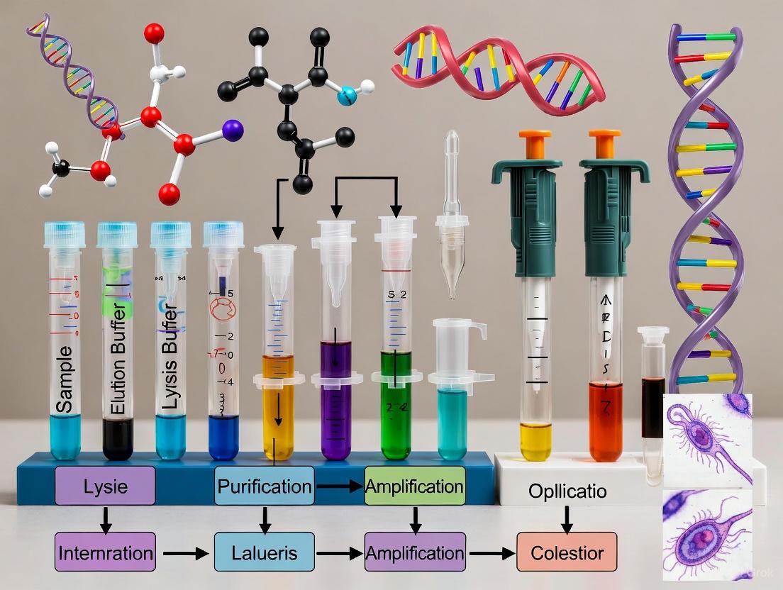
Diagram 2: Optimized workflow for Cryptosporidium DNA extraction using mechanical bead-beating pretreatment.
Rapid Direct Lysis Protocol for Water Samples
For water surveillance applications requiring rapid processing, the direct heat lysis method provides a simplified alternative:
Oocyst Concentration: Concentrate oocysts from water samples by immunomagnetic separation (IMS) or centrifugation [6] [4].
Direct Lysis:
Cooling and Clarification:
- Briefly centrifuge the lysate to pellet large debris.
- Transfer the supernatant to a clean tube.
Molecular Detection:
- Use 2-5 μL of the crude lysate directly in LAMP reactions [6].
- Alternatively, utilize the lysate in PCR-based assays with inhibitor-resistant polymerases.
The Scientist's Toolkit: Essential Research Reagents
Table 3: Key Research Reagent Solutions for Cryptosporidium DNA Extraction
| Reagent/Kit | Function | Application Notes |
|---|---|---|
| Ceramic Beads (1.4 mm) | Mechanical disruption of oocyst wall | Optimal size for balance between disruption efficiency and DNA shearing [3] |
| Paramagnetic Resin-based Kits (e.g., MAGNEX) | DNA binding and purification | Higher sensitivity for low oocyst concentrations [5] |
| NucliSENS easyMAG | Automated nucleic acid extraction | Compatible with bead-beating pretreatment; high recovery [3] [4] |
| Proteinase K | Enzymatic degradation of wall proteins | Effective when combined with heat shock (98°C, 10 min) [8] |
| TE Buffer (10 mM Tris, 0.1 mM EDTA) | Direct lysis medium | Suitable for heat-based lysis without commercial kits [6] |
| WarmStart LAMP Master Mix | Isothermal amplification | Enables detection from crude lysates without purified DNA [6] |
| Immunomagnetic Separation (IMS) Beads | Oocyst concentration from water | Critical for isolating oocysts from large volume samples [6] |
The resilient structure of the Cryptosporidium oocyst wall, particularly its layered architecture and COWP protein components, presents significant but surmountable challenges for DNA extraction. Mechanical disruption methods, especially bead-beating with optimally sized ceramic beads, have proven most effective for breaching this barrier. Paramagnetic resin-based DNA purification systems consistently outperform other methods for downstream molecular detection. The ongoing development of simplified protocols, such as direct heat lysis coupled with LAMP detection, offers promising avenues for rapid field-based applications. As research continues to elucidate the precise composition and structural organization of the oocyst wall, further refinements in DNA extraction methodologies will undoubtedly emerge, enhancing our capacity to study and combat this significant pathogen.
Impact of Extraction Efficiency on Downstream PCR Sensitivity and Specificity
The molecular diagnosis of eukaryotic enteric pathogens, particularly Cryptosporidium spp., presents a significant challenge for clinical and research laboratories. The accuracy of PCR-based detection is critically dependent on the initial DNA extraction step, which must efficiently recover trace amounts of parasitic DNA while removing potent PCR inhibitors present in fecal samples [9] [10]. Efficient and easy-to-use DNA extraction and purification methods are critical in implementing PCR-based diagnosis of these pathogens [9]. This application note systematically evaluates the impact of DNA extraction efficiency on downstream PCR sensitivity and specificity within the context of Cryptosporidium spp. detection in stool research, providing evidence-based protocols for optimal pathogen detection.
The challenges associated with DNA extraction from fecal samples for Cryptosporidium detection are multifaceted. Target DNA is often present in low abundance compared to host and bacterial DNA, and Cryptosporidium oocysts have robust walls that are difficult to disrupt [11]. Additionally, fecal samples contain numerous PCR inhibitors, including bile salts, complex polysaccharides, and hemoglobin derivatives, which can significantly reduce amplification efficiency [10]. The selection of an appropriate DNA extraction method must therefore balance DNA yield, purity, and practical considerations such as throughput and cost-effectiveness.
Comparative Performance of DNA Extraction Methods
Quantitative Comparison of Extraction Method Efficacy
Table 1: Performance comparison of DNA extraction methods for eukaryotic enteric pathogen detection
| Extraction Method | Mechanism of Lysis | Hands-on Time (min) | Sensitivity for Cryptosporidium | Key Advantages | Key Limitations |
|---|---|---|---|---|---|
| Glass Beads + Mechanical Disruption [11] | Chemical lysis with mechanical disruption | ~15-30 (after optimization) | 100% sensitivity; Limit of detection: 1 oocyst/g feces | Highest sensitivity for Cryptosporidium; Effective oocyst disruption | Requires optimization; Additional equipment needed |
| Semi-automated EZ1 (Qiagen) [9] [10] | Chemical lysis with mechanical disruption | 15 | Similar performance to QIAamp; Significantly lower Ct values for most pathogens | High throughput; Minimal hands-on time; Lower contamination risk | Higher cost per sample; Specialized equipment required |
| QIAamp DNA Stool Mini Kit (Manual) [9] [10] | Enzymatic lysis | 36 | Similar performance to EZ1 for Cryptosporidium | Well-established protocol; No specialized equipment | Lengthy hands-on time; More prone to cross-contamination |
| NucliSENS miniMAG [12] | Chemical lysis with mechanical disruption | ~30 | 5-fold higher sensitivity than enzymatic methods | Efficient inhibitor removal; Consistent results | Method optimization may be required |
| HotShot Vitis (HSV) Method [13] | Alkaline lysis with chemical treatment | ~30 | Not specifically tested on Cryptosporidium, but effective on difficult samples | Rapid; Low chemical risk; Cost-effective | May require optimization for fecal samples |
Table 2: Impact of extraction method on PCR sensitivity for pathogen detection
| Pathogen | EZ1 vs. QIAamp Performance (Ct values) | Statistical Significance |
|---|---|---|
| Cryptosporidium spp. | Similar performance | Not significant |
| Blastocystis spp. | EZ1 significantly lower Ct | p < 0.002 |
| Cyclospora cayetanensis | EZ1 significantly lower Ct | p < 0.002 |
| Giardia intestinalis | EZ1 significantly lower Ct | p < 0.002 |
| Dientamoeba fragilis | Similar performance | Not significant |
| Cystoisospora belli | EZ1 significantly lower Ct | p < 0.002 |
| Enterocytozoon bieneusi | EZ1 significantly lower Ct | p < 0.002 |
Impact of Lysis Efficiency on Detection Sensitivity
The efficiency of the initial lysis step profoundly impacts downstream PCR sensitivity, particularly for robust structures like Cryptosporidium oocysts. Comparative studies have demonstrated that mechanical disruption methods consistently outperform purely enzymatic or chemical lysis alone. Lindergard et al. reported that a glass beads extraction method achieved 100% sensitivity for Cryptosporidium detection compared to 83% for a freeze-thaw method using liquid nitrogen, with limits of detection of 1 oocyst/g and 10 oocysts/g of fecal sample, respectively [11].
This performance advantage stems from the ability of mechanical disruption to effectively break down the resilient oocyst walls, releasing DNA that would otherwise remain inaccessible. The importance of rigorous lysis is further supported by research showing that "thorough mechanical disruption with simultaneous chemical lysis allows efficient isolation of DNA of the investigated intestinal parasites present in stool and the subsequent PCR detection" [12]. In one study, this approach enabled Cryptosporidium detection in human fecal samples from children with diarrhea that might otherwise have been missed with less efficient extraction methods [12].
Factors Influencing Extraction Efficiency and Downstream PCR
Technical Considerations for Optimal DNA Extraction
Several technical factors critically influence the efficiency of DNA extraction from fecal samples for Cryptosporidium detection:
Inhibitor Removal: Efficient removal of PCR inhibitors is paramount. The semi-automated EZ1 procedure demonstrated superior removal of contaminating compounds, achieving an A260/A280 ratio of 2.34 ± 0.41 compared to 1.98 ± 0.17 for the manual QIAamp method [10]. The incorporation of inhibitor removal tablets or specific washing steps significantly reduces false-negative results.
Sample Input and Elution Volume: Consistent sample input (typically 180-220 mg of stool) and minimal elution volumes (50-100 µL) help maximize DNA concentration. The higher nucleic acid concentration obtained with the EZ1 method (29.61 ± 18.46 ng/µL vs. 15.31 ± 18.78 ng/µL for QIAamp) directly contributes to improved detectability in downstream PCR [10].
Protocol Modifications: Strategic modifications to manufacturer's protocols significantly enhance recovery. The addition of mechanical lysis using glass powder, combined with extended proteinase K incubation (12-16 hours), has been shown to substantially improve DNA yield from eukaryotic enteric pathogens [10].
Impact on Downstream Molecular Applications
The quality of extracted DNA directly influences the performance of subsequent molecular applications:
qPCR Efficiency: DNA extraction methods that efficiently remove inhibitors enable more accurate quantification and lower Ct values in qPCR applications. Proper efficiency assessment requires robust standard curves with at least 3-4 qPCR replicates at each concentration to reduce uncertainty in efficiency estimation [14].
Assay Sensitivity and Specificity: High-quality DNA extraction reduces false-negative results and improves assay specificity by preventing non-specific amplification. In one study, 46% of field samples previously classified as negative for Cryptosporidium parvum by flotation-concentration and ELISA methods showed positive detection with an optimized PCR protocol following efficient DNA extraction [11].
Recommended Protocols for Cryptosporidium Detection
Optimized Mechanical Disruption Protocol for Maximum Sensitivity
Based on the highest reported sensitivity for Cryptosporidium detection [11], the following protocol is recommended:
Workflow Diagram: Mechanical Disruption Method
Reagents Required:
- Acid-washed glass beads (425-600 µm)
- G2 lysis buffer or equivalent
- Proteinase K (20 mg/mL)
- Appropriate DNA purification system (silica membrane or magnetic beads)
Critical Steps:
- Mechanical Disruption: Use of glass beads in a homogenizer at maximum power for 40 seconds is essential for oocyst wall disruption.
- Thermal Lysis: Subsequent incubation at 100°C ensures complete lysis.
- Extended Enzymatic Digestion: Overnight proteinase K digestion further improves DNA recovery from difficult samples.
Rapid High-Throughput Protocol for Clinical Screening
For laboratories processing large sample volumes, the semi-automated EZ1 protocol offers an optimal balance of sensitivity and efficiency [9] [10]:
Workflow Diagram: Semi-Automated Method
Reagents Required:
- EZ1 DNA Tissue Kit (Qiagen)
- ASL lysis buffer
- InhibitEX tablets
- Proteinase K
Key Advantages:
- Minimal hands-on time (15 minutes vs. 36 minutes for manual method)
- Lower contamination risk due to automated processing
- Consistent results across multiple operators and batches
- Higher throughput capability for clinical laboratories
The Scientist's Toolkit: Essential Reagents and Equipment
Table 3: Essential research reagents and equipment for optimal DNA extraction
| Item | Specification | Function in Protocol | Example Brands/Products |
|---|---|---|---|
| Mechanical Disruption Beads | Acid-washed, 425-600µm | Oocyst wall breakdown for DNA release | Sigma-Aldrich glass beads |
| Homogenization Equipment | High-speed benchtop homogenizer | Effective tissue and oocyst disruption | FastPrep BIO 101 apparatus |
| Inhibitor Removal Technology | Proprietary resin or tablet formulation | Binds PCR inhibitors common in stool | InhibitEX tablets (Qiagen) |
| Silica-Based Purification | Membrane or magnetic beads | Selective DNA binding and washing | QIAamp silica membranes, NucleoSpin columns |
| Proteinase K | Molecular biology grade, 20mg/mL | Enzymatic degradation of proteins | Qiagen Proteinase K |
| Automated Extraction System | Modular cartridge-based platform | High-throughput, consistent purification | EZ1 Advanced XL (Qiagen) |
| Nucleic Acid Quality Assessment | UV-Vis spectrophotometer | DNA quantification and purity check | NanoDrop 2000 |
The selection and optimization of DNA extraction methods significantly impact the sensitivity and specificity of downstream PCR detection of Cryptosporidium spp. in fecal samples. Based on current evidence:
For maximum sensitivity in research settings where detection limit is paramount, methods incorporating mechanical disruption with glass beads are recommended, achieving detection limits as low as 1 oocyst/g of fecal sample [11].
For clinical laboratories requiring balance between sensitivity and throughput, semi-automated systems like the EZ1 provide optimal performance with significantly reduced hands-on time and lower contamination risk [9] [10].
Protocol modifications, including the addition of mechanical lysis steps and extended proteinase K digestion, should be considered even when using commercial kits to enhance recovery of Cryptosporidium DNA [10].
Quality control measures should include not only DNA concentration and purity assessment, but also evaluation of extraction efficiency through spike-in controls or amplification of reference genes to identify potential PCR inhibition.
The implementation of these optimized DNA extraction protocols will enhance the accuracy and reliability of Cryptosporidium detection in both research and clinical settings, ultimately improving diagnostic outcomes and epidemiological understanding of this important enteric pathogen.
Cryptosporidium spp. are intracellular parasitic protozoa and a significant cause of diarrheal disease worldwide, posing a particular threat to immunocompromised individuals and children in developing countries [15] [16]. The clinical and public health imperative for robust detection and identification methods stems from the parasite's high infectivity, resistance to conventional water disinfectants, and potential to cause large-scale outbreaks [16]. Effective management of cryptosporidiosis requires sensitive detection for prompt outbreak response and precise species identification to understand transmission dynamics, as Cryptosporidium hominis primarily infects humans while Cryptosporidium parvum causes zoonotic infections [16].
Molecular techniques have progressively overcome limitations of traditional microscopic methods, which offer limited sensitivity and cannot differentiate species [15] [16]. This application note details standardized protocols for DNA extraction and subsequent molecular analysis of Cryptosporidium spp., framed within a broader research context on optimizing DNA extraction methodologies from stool samples.
Research Reagent Solutions for Cryptosporidium Detection
The following table catalogues essential reagents and kits utilized in molecular detection of Cryptosporidium, providing researchers with a curated selection of validated tools.
Table 1: Key Research Reagents and Kits for Cryptosporidium Molecular Detection
| Reagent/Kit Name | Primary Function | Specific Application |
|---|---|---|
| QIAamp DNA Stool Mini Kit [15] | DNA extraction from stool | Efficiently extracts DNA while inhibiting PCR inhibitors in complex matrices |
| Nuclisens Easymag [17] | Nucleic acid extraction | Demonstrated efficacy in optimal protocol combinations for C. parvum detection |
| FTD Stool Parasite [17] | DNA amplification | Multiplex PCR assay identifying multiple intestinal parasites |
| DNeasy Blood & Tissue Kit [6] | DNA extraction from oocysts | Used in standardized protocols for DNA purification |
| FastDNA SPIN Kit for Soil [6] | DNA extraction from environmental samples | Effective for oocysts in water or environmental concentrates |
| WarmStart Colorimetric LAMP 2× Master Mix [6] | Isothermal amplification | Enables rapid, equipment-free detection of Cryptosporidium |
| Dynabeads MyOne Streptavidin C1 [6] | Immunomagnetic separation | Captures and concentrates oocysts from water samples prior to lysis |
Comparative Sensitivity of Molecular Detection Methods
Evaluating the performance of different molecular targets and methods is crucial for selecting appropriate diagnostics for clinical or environmental testing.
Table 2: Sensitivity Comparison of PCR Targets and Methods for Cryptosporidium parvum Detection [15]
| Target Gene | Method | Amplicon Size (bp) | Detection Limit |
|---|---|---|---|
| COWP | Nested PCR | 311 | 1 oocyst |
| COWP-1 | Nested PCR | 550 | 100 oocysts |
| SSU rRNA | Nested PCR | 826 | 10 oocysts |
| SSU rRNA | Single-round PCR | 1325 | 103-104 oocysts |
| COWP | Single-round PCR | 550 | 103-104 oocysts |
| RAPD | Single-round PCR | 433 | 104 oocysts |
The data demonstrates that nested PCR significantly enhances sensitivity by 2-4 orders of magnitude compared to single-round PCR [15]. The COWP gene target with a smaller amplicon size (311 bp) in a nested format provides the highest sensitivity, capable of detecting a single oocyst, making it particularly valuable for clinical scenarios with low oocyst shedding [15].
Experimental Protocols
Protocol 1: DNA Extraction from Stool Samples Using Commercial Kits
This protocol outlines an optimized combination for DNA extraction and amplification from stool samples, achieving 100% detection for C. parvum [17].
4.1.1 Sample Pretreatment
- Mechanical Pretreatment: Transfer 0.1-0.2 g of stool sample to a tube containing lysis buffer and garnet matrix. Disrupt oocysts using bead beating (e.g., FastPrep-24 system) at 6 m/s for 40 seconds. Repeat for two cycles [6].
- Alternative Alkali Wash Method: For a rapid, cost-effective approach suitable for outbreak investigations, wash oocysts with an alkaline solution (e.g., dilute KOH) followed by repeated freeze-thaw cycles (15 cycles alternating between -70°C and 65°C) to liberate genomic DNA [18].
4.1.2 DNA Extraction
- Employ the Nuclisens Easymag system or QIAamp DNA Stool Mini Kit for automated or manual extraction, respectively [17].
- For the QIAamp kit, incubate the pretreated sample at 70°C for 30 minutes in ASL lysis buffer. Follow the manufacturer's instructions for subsequent steps, including inhibitor removal and DNA binding to the silica membrane [15].
- Elute DNA in 50-100 µL of elution buffer or TE (10 mM Tris, 0.1 mM EDTA, pH 7.5).
4.1.3 DNA Amplification
- Utilize the FTD Stool Parasite multiplex PCR assay according to manufacturer specifications [17].
- Alternatively, for single-plex detection, prepare a 50 µL PCR reaction containing: 5 µL template DNA, 10 mM Tris-HCl (pH 8.3), 2.5 mM MgCl₂, 200 µM dNTPs, 25 pmol of each primer, and 2.5 U Taq DNA polymerase [15].
- Cycling conditions: Initial denaturation at 94°C for 5 min; 30 cycles of 94°C for 50 sec, annealing (temperature dependent on primer set, 50-60°C) for 30 sec, 72°C for 50 sec; final extension at 72°C for 10 min [15].
Protocol 2: Rapid Oocyst Detection via LAMP Without DNA Purification
This protocol enables rapid, sensitive detection of Cryptosporidium in water samples, eliminating time-consuming DNA purification steps and suitable for field application [6].
4.2.1 Oocyst Concentration and Lysis
- Concentrate oocysts from 10 mL water samples by filtration and/or immunomagnetic separation (IMS) using anti-Cryptosporidium antibody-conjugated magnetic beads [6].
- Resuspend the isolated oocysts in 50-100 µL of TE buffer (10 mM Tris, 0.1 mM EDTA, pH 7.5).
- Perform heat lysis by incubating the suspension at 95°C for 10 minutes. Centrifuge briefly at 10,000 × g for 1 min to pellet debris.
4.2.2 Loop-Mediated Isothermal Amplification (LAMP)
- Use the WarmStart Colorimetric LAMP 2× Master Mix.
- Prepare a 25 µL reaction containing: 12.5 µL master mix, 1-2 µL of the prepared lysate (supernatant), and F3/B3/FIP/BIP/LF/LB primer sets (final concentration 0.2-1.6 µM each) targeting the Cryptosporidium SAM gene or other intron-less genes [6].
- Incubate the reaction at 65°C for 30-60 minutes in a heating block or water bath.
- Result Interpretation: A color change from pink to yellow indicates positive amplification. Include negative (nuclease-free water) and positive (purified gDNA) controls.
This method achieves a detection limit of 5-10 oocysts per 10 mL of tap water [6].
Molecular Species Identification
Accurate species identification is critical for elucidating transmission cycles and implementing targeted public health interventions.
5.1 Target Gene Selection
- For species-level identification, sequence variable regions within the 18S small subunit (SSU) rRNA gene or the Cryptosporidium oocyst wall protein (COWP) gene [15] [16].
- These genetic loci provide sufficient inter-species polymorphism while maintaining conserved regions for primer binding.
5.2 Nested PCR and Sequencing
- Perform primary PCR amplification with external primers targeting a larger fragment (e.g., ~1325 bp for SSU rRNA) [15].
- Use 1-2 µL of the primary PCR product as a template for nested PCR with internal primers generating a smaller, species-discriminatory fragment (e.g., ~826 bp for SSU rRNA) [15].
- Purify the nested PCR product and subject it to Sanger sequencing in both directions.
- Analyze the resulting sequence by alignment and comparison with reference sequences in genomic databases (e.g., GenBank) using BLAST or similar tools.
This approach differentiates between C. hominis, C. parvum, and other less common species infecting humans [16].
Evaluating Commercial Kits and Protocols: From Manual to Automated Systems
Comparative Analysis of Major Commercial DNA Extraction Kits
The protozoan parasite Cryptosporidium spp. is a significant cause of diarrheal disease worldwide, particularly affecting children, immunocompromised individuals, and those in resource-limited settings. Research into this pathogen relies heavily on molecular detection methods, for which the efficiency of DNA extraction from stool samples is a critical determinant of success. The robust oocyst wall of Cryptosporidium presents a substantial challenge for DNA recovery, making the selection of an appropriate extraction methodology paramount. This application note provides a comparative analysis of major commercial DNA extraction kits within the context of Cryptosporidium research, presenting standardized protocols and performance data to guide researchers in their selection process.
The accurate detection and quantification of Cryptosporidium in stool samples is essential for various research applications, including epidemiological studies, drug development, and transmission dynamics. However, studies consistently demonstrate that DNA extraction efficiency varies significantly between different commercial kits and methodological approaches, directly impacting downstream molecular detection sensitivity. This variability necessitates a systematic evaluation of available technologies to optimize research outcomes.
Performance Comparison of Commercial Kits
Quantitative Comparison of DNA Extraction Kits
Table 1: Comparative performance of DNA extraction kits for parasite DNA recovery from stool samples
| Kit Name | Mechanical Pretreatment | Relative DNA Yield | PCR Detection Rate | Key Applications |
|---|---|---|---|---|
| QIAamp PowerFecal Pro DNA Kit (QB) | Bead beating included | High | 61.2% (highest) | Broad-spectrum parasite detection; Cryptosporidium research |
| QIAamp Fast DNA Stool Mini Kit (Q) | Not specified | Moderate | Lower than QB | General stool DNA extraction |
| Phenol-Chloroform (P) | None | High (~4x QB) | 8.2% (lowest) | Traditional method; requires optimization |
| FastDNA SPIN Kit for Soil | Bead beating included | Comparable to IMS-based methods | High for Cryptosporidium | Environmental samples; water concentrates |
| FTD Stool Parasites Kit | Not specified | Not specified | Excellent for Cryptosporidium | Multiplex parasite detection |
Comparative Sensitivity of PCR Methods
Table 2: Limit of detection of various PCR methods for Cryptosporidium spp.
| PCR Method | Type | Target Gene | Limit of Detection (C. parvum) | Limit of Detection (C. hominis) |
|---|---|---|---|---|
| FTD Stool Parasites | Commercial | DNA J-like protein | 1 oocyst/gram | 10 oocysts/gram |
| Allplex GI Parasite Assay | Commercial | Not specified | 10 oocysts/gram | 100 oocysts/gram |
| Valeix et al. 2020 | In-house | 18S rRNA | 10-100 oocysts/gram | 10-100 oocysts/gram |
| Mary et al. 2013 | In-house | 18S rRNA | 100 oocysts/gram | 1000 oocysts/gram |
| Fontaine et al. 2002 | In-house | Single copy gene | 1000 oocysts/gram | 1000 oocysts/gram |
Mechanical Pretreatment Optimization
The efficiency of DNA extraction from Cryptosporidium oocysts is significantly enhanced through mechanical pretreatment, which disrupts the robust oocyst wall. A multicenter comparative study demonstrated that optimal performance was achieved using the Fastprep-24 system with Lysing Matrix E at a speed of 4 m/s for 60 seconds [19]. This protocol resulted in improved detection sensitivity, particularly at low oocyst concentrations (10-50 oocysts/mL).
The Quick DNA Fecal/Soil Microbe-Miniprep manual kit demonstrated the best overall performance when combined with proper mechanical pretreatment, outperforming five other extraction systems in a comparative evaluation [19]. The inclusion of bead beating steps was consistently identified as a critical factor for efficient DNA recovery across multiple studies [20] [21].
Workflow for Optimal DNA Extraction
Figure 1: Optimal workflow for Cryptosporidium DNA extraction from stool samples
Detailed Experimental Protocols
Standardized DNA Extraction Protocol Using QIAamp PowerFecal Pro Kit
Principle: This protocol utilizes mechanical lysis with bead beating followed by chemical lysis and spin-column purification to efficiently recover Cryptosporidium DNA from stool samples while removing PCR inhibitors.
Reagents and Equipment:
- QIAamp PowerFecal Pro DNA Kit (QIAGEN)
- Fresh or preserved stool sample (200 mg)
- Ethanol (70-96%)
- Microcentrifuge tubes (2 mL)
- Bead beater (e.g., FastPrep-24) with Lysing Matrix E
- Microcentrifuge
- Vortex mixer
- Water bath or heating block
Procedure:
- Sample Preparation: Transfer approximately 200 mg of stool specimen to a 2 mL microcentrifuge tube. For preserved samples, wash three times with sterile distilled water to remove preservatives.
- Mechanical Pretreatment: Add the sample to a tube containing Lysing Matrix E. Process in a bead beater at 4 m/s for 60 seconds [19].
- Initial Lysis: Add 800 μL of CD1 solution from the kit to the sample. Vortex thoroughly for 5 minutes until homogeneous.
- Incubation: Heat the sample at 65°C for 10 minutes to enhance lysis efficiency.
- Inhibitor Removal: Centrifuge at 13,000 × g for 1 minute. Transfer 400 μL of supernatant to a new 2 mL tube.
- Chemical Lysis: Add 200 μL of CD2 solution and mix by vortexing for 5 seconds.
- Binding: Transfer 600 μL of the mixture to an MBQ column and centrifuge at 13,000 × g for 1 minute. Discard the flow-through.
- Washing: Add 500 μL of EA solution to the column and centrifuge at 13,000 × g for 1 minute. Discard the flow-through.
- Drying: Centrifuge the empty column at 13,000 × g for 2 minutes to remove residual ethanol.
- Elution: Transfer the column to a clean 1.5 mL tube. Add 50-100 μL of C6 solution (10 mM Tris, pH 8.0) to the center of the membrane and incubate at room temperature for 3 minutes. Centrifuge at 13,000 × g for 1 minute to elute DNA.
- Storage: Store extracted DNA at -20°C until PCR analysis.
Technical Notes:
- For liquid stools, use 200 μL of sample and adjust reagent volumes proportionally.
- For inhibitor-rich samples, additional purification steps or increased BSA concentration in PCR reactions may be necessary.
- DNA quality can be assessed by spectrophotometry (A260/A280 ratio of 1.8-2.0 indicates pure DNA).
Alternative Protocol for Water Samples Using FastDNA SPIN Kit for Soil
Application: This protocol is optimized for Cryptosporidium detection in water concentrates, which present different challenges compared to stool samples.
Procedure:
- Sample Concentration: Concentrate water samples (at least 10 L) using Envirochek HV filtration according to EPA Method 1623.
- Pellet Preparation: Transfer 0.5 mL of water concentrate pellet to a lysing matrix E tube.
- Direct DNA Extraction: Add appropriate lysing solutions from the FastDNA SPIN Kit and process in a bead beater at maximum speed for 45 seconds [21].
- DNA Purification: Follow manufacturer's instructions for binding, washing, and elution steps.
- Inhibitor Management: Add 400 ng/μL of non-acetylated BSA to PCR reactions to counteract residual inhibitors [21].
PCR Detection and Inhibitor Management
Comparison of Molecular Targets
The selection of target genes for PCR detection significantly impacts assay sensitivity. Studies comparing three common molecular targets found that the small subunit ribosomal RNA (SSU rRNA) gene provided the highest sensitivity (100%), while the Cryptosporidium oocyst wall protein (COWP) gene offered superior specificity (99.6%) [22]. The DnaJ-like protein gene showed intermediate performance for both parameters.
A two-step approach using SSU rRNA gene PCR for initial screening followed by COWP gene PCR for confirmatory testing is recommended for optimal diagnostic accuracy [22]. This combination leverages the strengths of both targets while mitigating their individual limitations.
Managing PCR Inhibition
Stool samples contain various substances that can inhibit PCR amplification, including bile salts, complex carbohydrates, and hemoglobin degradation products. Effective strategies to overcome inhibition include:
- Chemical Additives: Addition of 400 ng/μL non-acetylated bovine serum albumin (BSA) or 25 ng/μL T4 gene 32 protein to PCR reactions significantly improves amplification efficiency [21].
- Sample Dilution: Diluting DNA extracts 1:10 can reduce inhibitor concentration while maintaining detectable DNA levels.
- Alternative Polymerases: Using inhibitor-resistant DNA polymerases can improve performance with challenging samples.
- Internal Controls: Incorporating internal amplification controls validates results and detects inhibition.
Table 3: Research reagent solutions for Cryptosporidium DNA extraction and detection
| Reagent/Kit | Function | Application Note |
|---|---|---|
| QIAamp PowerFecal Pro DNA Kit | DNA purification | Highest PCR detection rate for diverse intestinal parasites [20] |
| Lysing Matrix E with Fastprep-24 | Mechanical disruption | Optimal at 4 m/s for 60s for oocyst wall breakage [19] |
| Non-acetylated BSA | PCR facilitator | Use at 400 ng/μL to counteract inhibitors [21] |
| T4 gene 32 protein | PCR facilitator | Alternative to BSA at 25 ng/μL [21] |
| 18S rRNA PCR assay | Molecular detection | Broader specificity and 5x lower LOD than COWP assay [4] [22] |
| FTD Stool Parasites Kit | Multiplex PCR detection | Best overall performance for Cryptosporidium detection [23] |
The comparative analysis of commercial DNA extraction kits for Cryptosporidium research demonstrates that the QIAamp PowerFecal Pro DNA Kit, when combined with optimized mechanical pretreatment, provides the most reliable performance for DNA recovery from stool samples. The integration of bead beating at 4 m/s for 60 seconds significantly enhances oocyst disruption and DNA yield, particularly important for low-intensity infections.
For molecular detection, targeting the 18S rRNA gene offers superior sensitivity, while the COWP gene provides higher specificity, suggesting that a combined approach may be optimal for research requiring high accuracy. Additionally, the inclusion of PCR facilitators such as BSA is essential for counteracting inhibitors prevalent in stool samples.
These standardized protocols and comparative performance data provide researchers with evidence-based guidance for selecting and implementing DNA extraction methodologies in Cryptosporidium research, ultimately enhancing the reliability and reproducibility of molecular detection in both clinical and environmental contexts.
The robust, multi-layered wall of the Cryptosporidium oocyst is a major barrier to efficient DNA extraction, making molecular detection challenging [24] [3]. Mechanical pre-treatment is a critical step to disrupt this resilient structure and release DNA for subsequent analysis. It has been proven to significantly enhance the performance of Cryptosporidium DNA extraction compared to thermal or chemical methods alone [24] [3]. This protocol details the optimization of mechanical pre-treatment, focusing on the key parameters of bead type, homogenizer settings, and workflow integration to achieve maximal DNA recovery from stool samples for reliable PCR detection.
Equipment and Materials
Research Reagent Solutions
Table 1: Essential Materials for Mechanical Pre-treatment
| Item | Specification/Example | Function |
|---|---|---|
| Lysing Matrix | Lysing Matrix E (MP Biomedical) | Contains a mix of ceramic (1.4 mm), silica (0.1 mm), and glass (4 mm) beads for comprehensive mechanical disruption [25]. |
| Homogenizer | FastPrep-24 (MP Biomedicals) | High-speed oscillating homogenizer for efficient and consistent bead beating [24] [25]. |
| DNA Extraction Kit | NucliSENS easyMAG (BioMérieux) | Automated extraction system based on Boom technology, compatible with lysates from mechanical pre-treatment [25] [3]. |
| Lysis Buffer | NucliSENS lysis buffer | Buffer used in conjunction with bead beating to facilitate cell lysis and stabilize nucleic acids [24] [3]. |
| Sample Matrix | Stool suspension in physiological saline | Prepared from human feces negative for common parasites, filtered for homogeneity [24] [25]. |
Experimental Protocol & Data Analysis
Sample Preparation
- Prepare a 20% (w/v) stool suspension in physiological saline (0.09% NaCl) using human feces negative for common digestive parasites [24] [25].
- Filter the suspension through a large mesh strainer to remove large particulate matter.
- Seed the filtered suspension with Cryptosporidium parvum oocysts to achieve the desired concentration for validation (e.g., 10-100 oocysts/mL) [24].
Mechanical Pre-treatment Procedure
- Pipette 0.5 mL of the stool sample into a Lysing Matrix E tube [24] [3].
- Add 1.0 mL of NucliSENS lysis buffer to the tube [24] [3].
- Securely cap the tube and place it in the FastPrep-24 homogenizer.
- Process the sample at a speed of 6.0 m/s for 60 seconds [24].
- After homogenization, incubate the stool suspension at room temperature for 10 minutes.
- Centrifuge the tube at 10,000 × g for 10 minutes to pellet debris [3].
- Transfer 250 µL of the supernatant to the NucliSENS easyMAG system for automated DNA extraction, following the manufacturer's protocol [3].
Optimal Settings and Performance Data
Table 2: Impact of Homogenizer Parameters on DNA Extraction Efficiency
| Grinding Speed (m/s) | Grinding Duration (s) | DNA Extraction Efficiency | Key Findings |
|---|---|---|---|
| 4.0 | 60 | High | Optimal balance of sensitivity and specificity for low oocyst concentrations [25] [26]. |
| 5.0 | 60 | Moderate | -- |
| 6.0 | 60 | High | Recommended setting for general use with Lysing Matrix E [24]. |
| 6.0 | 30 | Lower | Insufficient disruption of oocyst walls. |
| 6.0 | 120 | Not Superior | No significant improvement over 60s, potential for increased DNA fragmentation [25]. |
Table 3: Comparison of Bead Types for Mechanical Pre-treatment
| Bead Material | Size (Diameter) | Relative Performance | Rationale |
|---|---|---|---|
| Ceramic | 1.4 mm | Highest | Optimal balance of hardness, density, and impact force for breaking robust oocyst walls [24] [3]. |
| Silica | 0.1 mm & 1.0 mm | Moderate | Smaller beads may be less effective at fracturing the thick oocyst wall [24]. |
| Garnet | 0.56-0.7 mm (flakes) | Variable | Performance varies based on shape and size distribution [24]. |
| Glass | 4 mm | Component in Mix | Part of a composite matrix (Lysing Matrix E), not typically used alone for this application [25]. |
Workflow Integration & Downstream Analysis
The following diagram illustrates the complete experimental workflow from sample preparation to final detection, highlighting the critical role of mechanical pre-treatment.
The optimized lysate is compatible with downstream qPCR assays. For Cryptosporidium detection, the 18S rRNA qPCR assay is recommended due to its superior sensitivity (5-fold lower detection limit) and broader specificity for different Cryptosporidium species compared to the COWP gene assay [4].
Troubleshooting
- Low DNA Yield: Confirm homogenizer speed and time settings. Ensure beads are not degraded and are appropriate for the sample type.
- PCR Inhibition: If inhibition is suspected in downstream applications, ensure the post-beating centrifugation step is performed correctly to pellet debris before supernatant transfer.
- Inconsistent Results Between Samples: Standardize stool suspension consistency. Ensure homogenizer tubes are securely fastened to guarantee consistent bead beating across all samples.
Within Cryptosporidium research, obtaining high-quality DNA from stool specimens is a critical first step for downstream molecular applications such as PCR, genotyping, and next-generation sequencing. The complex structure of the Cryptosporidium oocyst wall and the presence of PCR-inhibitory substances in stool complicate DNA extraction. This protocol details a robust method that integrates a mandatory pre-treatment step for mechanical and thermal oocyst disruption with subsequent purification using a commercial silica-column kit, ensuring optimal DNA yield and purity for sensitive detection and analysis [27] [6].
Experimental Protocols and Methodologies
Key Research Reagent Solutions
The following table catalogues essential materials and their specific functions in the DNA extraction workflow for Cryptosporidium spp. from stool samples.
Table 1: Essential Research Reagents and Materials
| Item | Function/Application |
|---|---|
| Anti-Cryptosporidium Antibody | Immunomagnetic separation (IMS) for specific capture and concentration of oocysts from stool samples [6]. |
| Dynabeads with Streptavidin | Magnetic beads coupled with streptavidin to bind biotin-labeled antibodies, enabling magnetic separation [6]. |
| FastDNA SPIN Kit for Soil | A commercial kit optimized for isolating PCR-quality genomic DNA from difficult, complex matrices like stool [6]. |
| Proteinase K | Enzyme that digests proteins and degrades nucleases, crucial for breaking down stool components and oocyst walls [28]. |
| CTAB Buffer | Cetyltrimethylammonium bromide-based buffer; effective in precipitating polysaccharides and other contaminants common in stool [28]. |
| Chelex-100 Resin | An alternative, rapid, and cost-effective chelating agent used in boiling methods to purify DNA by binding metal ions [29]. |
| LAMP Master Mix | For direct detection of Cryptosporidium from lysates, bypassing the need for extensive DNA purification, ideal for field use [6]. |
Detailed Step-by-Step Protocol
Sample Pre-treatment and Oocyst Lysis
This pre-treatment phase is critical for breaking down the robust oocyst wall to release nucleic acids.
- Immunomagnetic Separation (IMS): Resuspend stool sample in phosphate-buffered saline (PBS) and filter through a 0.1-mm mesh to remove large debris. Incubate the filtrate with biotinylated anti-Cryptosporidium monoclonal antibody. Add streptavidin-coated magnetic beads to the mixture and incubate to form antibody-bead-oocyst complexes. Separate the complexes using a magnetic rack and wash thoroughly with PBS to remove contaminants [6].
- Mechanical Disruption: Transfer the isolated oocysts to a tube containing 1.0 mm glass beads. Perform bead beating using an instrument like the FastPrep-24 at 6 m/s for 40 seconds. Repeat this cycle once. This step physically fractures the resilient oocyst wall [6].
- Enzymatic and Thermal Lysis: To the homogenate, add a lysis buffer containing Proteinase K (e.g., 200 μg/mL). Incubate at 55°C for 60 minutes to enzymatically degrade internal proteins. For an additional lysis boost, a subsequent heat step at 95°C for 10 minutes can be incorporated [29] [28].
DNA Purification
After effective pre-treatment, the lysate is purified using a commercial kit.
- Follow the manufacturer's instructions for the FastDNA SPIN Kit for Soil (or a similar kit validated for stool) starting from the lysed sample. This typically involves binding DNA to a silica matrix in the presence of a chaotropic salt [6].
- Wash the column multiple times with the provided wash buffers to remove salts, proteins, and other inhibitors.
- Elute the purified genomic DNA in a small volume of TE buffer (10 mM Tris-HCl, 0.1 mM EDTA, pH 7.5) or nuclease-free water. A reduced elution volume (e.g., 50 μL) can significantly increase the final DNA concentration [29].
Performance Evaluation: Quantitative Comparison of Methods
The integration of a rigorous pre-treatment step prior to kit-based purification significantly enhances performance. The following table summarizes the detection rates of Cryptosporidium using different diagnostic methods, which is directly influenced by DNA extraction efficiency [27].
Table 2: Comparative Performance of Cryptosporidium Detection Methods
| Detection Method | Detection Rate (%) | Key Advantages | Noted Limitations |
|---|---|---|---|
| Polymerase Chain Reaction (PCR) | 18% | High sensitivity, gold standard for molecular detection [27] | Requires purified DNA, susceptible to inhibitors [27] |
| Immunochromatography (ICT) | 15% | Rapid, easy to use [27] | Lower sensitivity than PCR, depends on parasite burden [27] |
| Modified Kinyoun's Stain (MKS) | 7% | Low cost, applicable in basic labs [27] | Low sensitivity, requires high oocyst concentration and expertise [27] |
| Routine Microscopy | 6% | Widely available, low cost [27] | Low sensitivity, prone to false negatives [27] |
Workflow Visualization
The following diagram illustrates the complete integrated pathway from sample collection to analysis, detailing the core pre-treatment and purification steps.
This application note provides a validated, detailed protocol for extracting DNA from Cryptosporidium spp. in stool samples. The critical differentiator of this method is the dedicated pre-treatment phase involving IMS, mechanical disruption, and enzymatic lysis, which is seamlessly integrated with a commercial kit's workflow. This combined approach directly addresses the challenges posed by the tough oocyst wall and complex stool matrix, leading to superior DNA yield and purity. By following this protocol, researchers can achieve the high-quality genetic material necessary for robust and reliable molecular detection and characterization of Cryptosporidium., ultimately enhancing diagnostic accuracy and public health surveillance [27] [6].
The accurate detection of Cryptosporidium spp., a significant waterborne and foodborne protozoan pathogen, is crucial for public health, clinical diagnostics, and drug development research. Conventional molecular detection methods rely heavily on commercial DNA extraction kits, which, while effective, involve laborious, time-consuming, and costly multi-step procedures for nucleic acid isolation and purification. These processes introduce significant delays and are unsuitable for rapid or field-applicable diagnostics. Emerging techniques that combine kit-free direct lysis with isothermal amplification present a paradigm shift, offering streamlined, rapid, and sensitive detection of Cryptosporidium directly from complex sample matrices such as stool and water. This application note details these innovative protocols, providing researchers with actionable methodologies to enhance the speed and efficiency of their cryptosporidiosis research, framed within the broader context of evaluating and moving beyond traditional DNA extraction kits.
Key Experimental Findings and Performance Data
The integration of direct lysis with isothermal amplification methods has demonstrated performance comparable to, and in some aspects superior to, traditional kit-based methods coupled with PCR. The table below summarizes key quantitative findings from recent studies evaluating these emerging techniques.
Table 1: Performance Comparison of Cryptosporidium Detection Methods
| Method Category | Specific Technique | Limit of Detection (LOD) | Time to Result | Key Advantages | Reference |
|---|---|---|---|---|---|
| Kit-Free Direct Lysis + LAMP | Direct heat lysis of magnetically isolated oocysts + LAMP | 5 oocysts/10 mL (tap water), 10 oocysts/10 mL (with matrix) | Rapid; significantly faster than EPA 1623.1 | Eliminates DNA purification; suitable for complex water matrices | [30] [6] |
| Stool DNA Kit + PCR | QIAamp DNA Stool Mini Kit + 18S rRNA PCR | 26.8% prevalence (vs. 23.2% by microscopy) | Several hours (includes kit extraction) | High sensitivity and specificity; gold standard for species identification | [31] |
| Commercial Multiplex PCR | FTD Stool Parasites PCR | 1 oocyst/gram (C. parvum), 10 oocysts/gram (C. hominis) | ~2-3 hours | Detects a broad range of Cryptosporidium species; high sensitivity | [23] |
| Enhanced LAMP | Stem primer LAMP with Lateral Flow Dipstick (LFD) | 10 oocysts/mL | ~80 minutes | Improved sensitivity; visual result readout; suitable for field use | [32] |
| Microscopy (Reference) | Modified Ziehl-Neelsen Staining | >50,000 oocysts/gram (low sensitivity) | ~1-2 hours | Low cost; widely available; low sensitivity and specificity | [27] |
Detailed Experimental Protocols
Protocol 1: Kit-Free Magnetic Separation and Direct Lysis-LAMP for Water Samples
This protocol, adapted from Mahmudunnabi et al. (2025), is designed for the rapid detection of Cryptosporidium oocysts in water samples, bypassing commercial DNA extraction kits [30] [6].
3.1.1 Workflow Overview
The following diagram illustrates the streamlined workflow for this kit-free method.
3.1.2 Materials and Reagents
- Anti-Cryptosporidium Monoclonal Antibody (e.g., Abcam ab54066): For specific capture of oocysts.
- Dynabeads MyOne Streptavidin C1: Magnetic beads conjugated with a biotinylated antibody for immunomagnetic separation (IMS).
- TE Buffer (10 mM Tris, 0.1 mM EDTA, pH 7.5): Lysis buffer.
- WarmStart Colorimetric LAMP 2× Master Mix: Contains Bst polymerase and phenol red for visual detection.
- LAMP Primers: Sets targeting an intron-less gene (e.g., SAM-1) for ultra-sensitive amplification [32].
- Thermal Cycler or Heat Block: Capable of maintaining 65°C for LAMP and 95°C for lysis.
3.1.3 Step-by-Step Procedure
- Sample Concentration: Centrifuge 10 mL of water sample at 3,000 × g for 15 minutes. Carefully decant the supernatant.
- Immunomagnetic Separation (IMS): Resuspend the pellet in 1 mL of PBS. Add biotinylated anti-Cryptosporidium antibody and incubate for 30 minutes. Then, add streptavidin-coated magnetic beads and incubate for another 30 minutes with gentle mixing. Place the tube on a magnetic stand for 2 minutes, discard the supernatant, and wash the bead-bound oocysts twice with PBS.
- Direct Heat Lysis: Resuspend the washed bead-oocyst complex in 50 μL of TE buffer. Incubate the tube at 95°C for 10 minutes in a heat block to lyse the oocysts and release genomic DNA. Briefly vortex and then centrifuge the tube at 10,000 × g for 1 minute.
- LAMP Reaction Setup: Prepare a 25 μL LAMP reaction mix containing:
- 12.5 μL WarmStart Colorimetric LAMP 2× Master Mix
- 5 μL of the prepared LAMP primer mix (FIP, BIP, F3, B3, LF, LB)
- 2.5 μL of the direct heat lysate (supernatant from step 3) as template
- 5 μL Nuclease-free water
- Isothermal Amplification: Run the reaction at 65°C for 30-60 minutes.
- Result Interpretation: Observe the color change. A color change from pink to yellow indicates a positive amplification. Include positive (known Cryptosporidium DNA) and negative (nuclease-free water) controls in each run.
Protocol 2: Direct Lysis from Stool Samples for LAMP Amplification
This protocol is optimized for human stool samples, incorporating a pre-lysis washing step to reduce PCR inhibitors commonly found in stool [32].
3.2.1 Workflow Overview
The workflow for stool samples involves additional steps to manage sample complexity.
3.2.2 Materials and Reagents
- QiAmp DNA Stool Mini Kit (Optional): For benchmark comparison with kit-based extraction.
- Lysis Buffer ASL (from QiAmp kit or equivalent): Can be used for initial suspension.
- Glass Beads (0.5 mm): For mechanical disruption of oocyst walls.
- Stem LAMP Primer Mix: Includes FIP, BIP, F3, B3, LF, LB, and stem primers (SF, SB) for enhanced sensitivity [32].
- Lateral Flow Dipsticks (LFD): For visual detection of biotin- and FAM-labeled LAMP amplicons.
3.2.3 Step-by-Step Procedure
- Sample Washing: Suspend approximately 200 mg of stool in 1 mL of distilled water. Centrifuge at 14,000 rpm for 5 minutes. Discard the supernatant and repeat the wash cycle four more times to remove inhibitors.
- Oocyst Lysis: To the washed pellet, add 1.4 mL of ASL buffer and subject the suspension to mechanical disruption. This can be done via:
- Bead Beating: Add 0.5 mm glass beads and homogenize in a bead beater at high speed for 1-2 minutes.
- Freeze-Thaw Cycling: Subject the suspension to five cycles of freezing at -80°C and thawing at 80°C.
- Heat Treatment and Clarification: Incubate the lysate at 80°C for 5 minutes. Centrifuge at 14,000 rpm for 2 minutes to pellet debris.
- LAMP Reaction with Stem Primers: Use 2-5 μL of the supernatant as a template in a 25 μL LAMP reaction. The reaction should include the standard LAMP primer set plus the accelerating stem primers.
- Lateral Flow Detection: After amplification, dilute the LAMP product in an appropriate running buffer and apply it to the sample pad of the LFD. The result is read within 5-10 minutes. The appearance of both control and test lines indicates a positive result.
The Scientist's Toolkit: Essential Research Reagent Solutions
The successful implementation of kit-free protocols requires specific reagents and materials. The following table catalogs the essential components.
Table 2: Key Research Reagent Solutions for Kit-Free Cryptosporidium Detection
| Item Name | Function/Application | Specific Example / Catalog Number |
|---|---|---|
| Anti-Cryptosporidium Antibody | Specific capture and enrichment of oocysts from samples via IMS. | Monoclonal Antibody (e.g., Abcam ab54066) [6] |
| Streptavidin Magnetic Beads | Solid-phase support for biotinylated antibodies, enabling rapid IMS. | Dynabeads MyOne Streptavidin C1 [6] |
| Bst Polymerase 2.0 / LAMP Master Mix | Engineered DNA polymerase for strand displacement, core enzyme in isothermal amplification. | WarmStart Colorimetric LAMP 2× Master Mix [6] |
| Stem Primers for LAMP | Accelerating primers that increase speed and sensitivity of LAMP assays. | Custom-designed primers targeting SAM-1 or 18S rRNA genes [32] |
| Lateral Flow Dipsticks (LFD) | Simple, visual detection of labeled LAMP amplicons for endpoint analysis. | Milenia HybriDetect strips or equivalent [32] |
| Poly-Lysine Magnetic Beads | Novel method for rapid, kit-free isolation of ribosomes and nucleic acids. | Potential alternative for RNA isolation from complex samples [33] |
The move towards kit-free direct lysis coupled with isothermal amplification represents a significant advancement in the molecular detection of Cryptosporidium. These protocols directly address the limitations of traditional DNA extraction kits by drastically reducing processing time, cost, and procedural complexity while maintaining high sensitivity. The detailed application notes provided here empower researchers to implement these robust methods, facilitating faster turnaround in clinical studies, more efficient environmental surveillance, and accelerated drug development efforts against cryptosporidiosis. The integration of these techniques into lab-on-a-chip platforms and point-of-care devices presents a clear trajectory for the future of pathogen diagnostics [34] [35].
Maximizing Yield and Overcoming Inhibitors: A Troubleshooting Guide
Addressing PCR Inhibition from Stool Components and Co-Purified Contaminants
In the molecular diagnosis of Cryptosporidium spp. from human stool, the polymerase chain reaction (PCR) offers a highly sensitive and specific detection method. However, its diagnostic accuracy is critically compromised by PCR inhibition, a prevalent issue where various constituents within fecal samples interfere with the enzymatic amplification process, leading to false-negative results [36]. These inhibitors, which include complex polysaccharides, bile salts, metabolic by-products, and co-purified humic substances, can originate from the stool matrix itself or be introduced during sample processing and nucleic acid extraction [36] [37]. For researchers and drug development professionals, the failure to identify such inhibition can invalidate experimental results and lead to incorrect conclusions. Therefore, robust monitoring and mitigation strategies are essential components of any reliable diagnostic protocol. This application note details the sources and effects of PCR inhibition and provides validated methodologies, including the use of an Internal Amplification Control (IAC), to ensure the accuracy and reliability of Cryptosporidium detection in stool research.
Understanding PCR Inhibition in Stool Samples
PCR inhibition in stool samples arises from a diverse array of substances. Fecal samples are particularly challenging as they can contain bile salts, complex polysaccharides, lipids, and bacterial metabolites [36]. Furthermore, during DNA extraction from frozen specimens or environmentally complex samples, additional substances such as humic acids, collagen, and calcium phosphate can be co-purified with the nucleic acids [38] [39]. These inhibitors can act through several mechanisms, including direct degradation of the DNA template, interference with the DNA polymerase enzyme, or chelation of magnesium ions that are essential co-factors for PCR [37].
The effects of inhibition can be partial or complete. Partial inhibition reduces the analytical sensitivity of the assay, potentially leading to a failure to detect low-level infections. Complete inhibition results in a false-negative outcome, which is particularly problematic in a clinical or research setting [36]. Studies comparing inhibition in quantitative PCR (qPCR) and multiplex STR assays have demonstrated that inhibition can cause a general loss of larger amplification products and more sequence-specific allele dropouts, altering the Ct values and efficiency of the reaction [38].
Table 1: Common PCR Inhibitors in Stool-Based Cryptosporidium Research
| Inhibitor Category | Specific Examples | Primary Source | Proposed Mechanism of Action |
|---|---|---|---|
| Organic Compounds | Humic and Fulvic Acids | Stool, Environmental contaminants | Bind to DNA polymerase and/or single-stranded DNA [39] |
| Bile Salts | Stool | Disrupt the activity of DNA polymerase [36] | |
| Inorganic Ions | Calcium Phosphate | Stool, Bone fragments | Alters DNA melt curve and cycle threshold [38] |
| Heavy Metals (e.g., Hg, Pb) | Contaminated samples | Interfere with enzyme function [39] | |
| Complex Molecules | Polysaccharides | Stool, Bacterial cell walls | Increase viscosity and physically impede polymerization |
| Heme/Hemoglobin | Blood in stool | Degrades DNA template [37] | |
| Proteins | Collagen | Tissue contaminants | Binds DNA in a sequence-specific manner [38] |
Key Methodologies for Monitoring and Controlling Inhibition
The Internal Amplification Control (IAC)
The most robust method for monitoring PCR inhibition is the incorporation of a non-target nucleic acid sequence, known as an Internal Amplification Control (IAC), which is co-amplified within the same reaction tube as the primary target [36]. A competitive IAC shares the same primer binding sequences as the target DNA but yields an amplicon of a different molecular weight, allowing for clear differentiation via gel electrophoresis.
Protocol: Construction and Use of a Competitive IAC for Cryptosporidium COWP Gene PCR
This protocol is adapted from a study that developed an IAC for a conventional PCR assay targeting the Cryptosporidium oocyst wall protein (COWP) gene [36].
IAC Plasmid Construction:
- Initial Cloning: Amplify the native ≈550 bp COWP target region from C. parvum genomic DNA using specific primers (e.g., Cry-9 and Cry-15). Clone the gel-purified PCR product into a suitable plasmid vector, such as the pGEM-T-Easy vector [36].
- Inverse PCR: Design inverse primers that include a restriction enzyme recognition site (e.g., HindIII). Use these primers to perform an inverse PCR on the constructed plasmid, effectively deleting a ≈210 bp internal fragment to create a shorter, modified target sequence of ≈375 bp [36].
- Ligation and Transformation: Digest the inverse PCR product with DpnI to remove the methylated template DNA, followed by digestion with HindIII. Re-ligate the truncated plasmid and transform into competent E. coli cells (e.g., TOP10) [36].
- Stock Preparation: Purify the plasmid DNA and prepare a 1 ng/μL stock solution. Calculate the copy number concentration for accurate quantification.
Determination of Optimal IAC Concentration:
- Perform a two-step titration. First, serially dilute the IAC plasmid to determine the lowest concentration that yields a consistent amplicon on an agarose gel.
- Second, include this IAC concentration in a duplex PCR reaction with a defined, low concentration of the native target (or an external control plasmid). The optimal IAC concentration is the one that is consistently detectable without competing with or suppressing the amplification of the primary target. The cited study determined an optimum of 1 fg per reaction [36].
Integration into Diagnostic Duplex PCR:
- Include the optimized IAC concentration in every diagnostic PCR reaction.
- Interpretation of Results:
- Positive Sample: Both the native target (≈550 bp) and IAC (≈375 bp) amplicons are visible.
- True Negative Sample: The native target amplicon is absent, but the IAC amplicon is present, confirming a successful amplification.
- Inhibited Sample (False Negative): Both the native target and IAC amplicons are absent, indicating a failure of the PCR and invalidating the negative result [36].
Optimized DNA Extraction Protocols
The choice of DNA extraction method is critical to minimize the co-purification of inhibitors. Commercial kits based on silica-binding chemistry under high-salt chaotropic conditions have proven effective for stool samples [36] [40]. The following protocol is based on the QIAamp DNA Stool Mini Kit with modifications for optimal Cryptosporidium DNA recovery.
Protocol: DNA Extraction from Stool for Cryptosporidium Detection
This protocol utilizes a silica-membrane technology to purify DNA while removing PCR inhibitors [36] [40].
- Sample Preparation: Homogenize approximately 200 mg of stool specimen in a suspension buffer. For frozen samples, a pre-treatment to facilitate oocyst excystation or disruption is recommended. One effective method involves resuspending the pellet in a 1.5% taurocholic acid solution for 2 hours to induce excystation [37].
- Lysis: Transfer the sample to a tube containing garnet beads and lysis buffer. Incubate at elevated temperatures (e.g., 70°C) to efficiently disrupt cells and oocysts. This step inactivates proteins and nucleases.
- Inhibition Removal: A short, high-speed centrifugation step is often included to pellet stool debris and inhibitors. The supernatant, containing the DNA, is then transferred to a new tube.
- DNA Binding: The lysate is applied to a silica-based membrane column in the presence of a chaotropic salt, which facilitates the binding of DNA to the membrane while contaminants are washed away.
- Washing: Wash the membrane twice with ethanol-based wash buffers to remove residual salts and other impurities.
- Elution: Elute the pure genomic DNA in a low-ionic-strength buffer such as Tris-EDTA (TE) or nuclease-free water. The purified DNA is now ready for PCR amplification [36] [40].
The Scientist's Toolkit: Essential Research Reagents
Table 2: Key Reagent Solutions for Cryptosporidium DNA Research from Stool
| Research Reagent | Function/Application | Example Product/Chemistry |
|---|---|---|
| Silica-Membrane Kits | Selective binding and purification of DNA from complex lysates; effective inhibitor removal. | QIAamp DNA Stool Mini Kit (Qiagen) [36] |
| Chaotropic Salts | Disrupt cellular structures, inactivate nucleases, and enable DNA binding to silica matrices. | Guanidine hydrochloride [40] |
| Internal Amplification Control (IAC) | Non-target nucleic acid sequence co-amplified to monitor for PCR inhibition and validate results. | In-house constructed competitive IAC plasmid [36] |
| PCR Additives | Enhance polymerase stability and processivity, helping to overcome partial inhibition. | Bovine Serum Albumin (BSA), T4 Gene 32 Protein [37] |
| Hot-Start Polymerase | Reduces non-specific amplification and improves assay specificity and sensitivity in complex samples. | GoTaq Hot Start Polymerase (Promega) [36] |
| Proteinase K | Enzymatic digestion of proteins during lysis, improving DNA yield and purity. | Included in many commercial lysis buffers [40] |
Workflow for Reliable Cryptosporidium Detection
The following diagram summarizes the integrated experimental workflow, from sample preparation to result interpretation, incorporating the IAC to control for PCR inhibition.
The reliable detection of Cryptosporidium spp. in stool samples by PCR is contingent upon effectively addressing the challenge of PCR inhibition. By implementing a rigorous DNA extraction protocol optimized for fecal samples and, most critically, incorporating a well-designed Internal Amplification Control (IAC) into the assay, researchers can confidently distinguish true negative results from false negatives caused by inhibition. The methodologies and controls detailed in this application note provide a robust framework for generating high-quality, reliable data in both basic research and drug development contexts, ensuring that downstream analyses and conclusions are built upon a foundation of technically sound molecular diagnostics.
Optimization Strategies for Low Oocyst Counts and Sub-Patent Infections
The accurate detection of Cryptosporidium spp., particularly in cases characterized by low oocyst counts and sub-patent infections, remains a significant challenge in clinical and research settings. The robust, multi-layered oocyst wall impedes efficient DNA extraction, while the inherently low parasite burden in many infections often falls below the detection limit of conventional molecular assays [25] [41]. This technical hurdle is of paramount importance, as cryptosporidiosis can be life-threatening in immunocompromised individuals and is associated with long-term health consequences in malnourished children [41] [42]. The limitations of current therapeutics, with nitazoxanide being the only approved drug and showing reduced efficacy in vulnerable populations, further underscore the need for highly sensitive diagnostic tools to support drug development and clinical management [43] [44]. This document outlines optimized protocols and strategic approaches, framed within a broader thesis on DNA extraction, to enhance the sensitivity and reliability of Cryptosporidium detection in stool research.
Performance Comparison of Detection Methods
Sensitivity in detecting Cryptosporidium is highly dependent on the choice of methods for stool pretreatment, DNA extraction, and DNA amplification. Different combinations of these steps can lead to vastly different outcomes, especially at low oocyst concentrations.
Table 1: Comparison of Method Combinations for Detecting Low C. parvum Oocyst Concentrations
| Pretreatment Method | DNA Extraction Technique | DNA Amplification Assay | Key Performance Findings | Reference |
|---|---|---|---|---|
| Mechanical (60s at 4 m/s) | Quick DNA Fecal/Soil Microbe-Miniprep (Manual) | Cryptosporidium-specific real-time PCR | Showed the best overall performance in a multicenter comparison | [25] |
| Mechanical Grinding | Nuclisens Easymag | FTD Stool Parasite | Achieved 100% detection in a 30-protocol comparison | [17] |
| Mechanical Grinding | QIAamp DNA Stool Mini Kit | Nested PCR (COWP gene target) | Most sensitive; detection limit of 1 oocyst | [15] |
| Not Specified | Not Specified | Standard PCR (SSU rRNA target) | Detection limit > 150 oocysts | [45] |
The data clearly demonstrates that nested PCR, particularly targeting the COWP gene, offers superior sensitivity compared to standard PCR, improving the detection limit from over 150 oocysts to as low as a single oocyst [15] [45]. Furthermore, comprehensive evaluations show that the diagnostic workflow must be considered as a whole, as a powerful PCR assay may fail if paired with an inefficient extraction technique [17] [25].
Optimized Experimental Protocols
Optimal Protocol for Sensitive Detection of Low Oocyst Counts
The following integrated protocol, synthesized from recent multicenter studies, is designed to maximize detection sensitivity for low-intensity Cryptosporidium infections.
1. Sample Pretreatment and Mechanical Lysis
- Sample Preparation: Emulsify 0.1-0.2 g of stool specimen in a suitable buffer provided by the extraction kit.
- Mechanical Grinding: Transfer the sample to a tube containing a lysing matrix (e.g., Lysing Matrix E containing ceramic and silica beads).
- Homogenization: Process the sample using a high-speed homogenizer (e.g., Fastprep-24) at a speed of 4 m/s for 60 seconds [25]. This critical step physically disrupts the resilient oocyst wall, which is composed of filamentous glycoproteins and acid-fast lipids, thereby facilitating the release of genomic DNA [25] [41].
2. DNA Extraction
- Recommended Kit: Use the Quick DNA Fecal/Soil Microbe-Miniprep manual kit, which demonstrated the best overall performance in a comparative study [25].
- Procedure: Follow the manufacturer's instructions for the chosen kit. Manual methods, while time-consuming, have been shown to provide excellent outcomes [17].
3. DNA Amplification via Nested PCR
- Target Gene: Amplify the Cryptosporidium oocyst wall protein (COWP) gene, which has been shown to be the most sensitive target [15].
- First Round PCR:
- Primers: Use outer primers (e.g., BcowpF and BcowpR).
- Reaction Mix: 5 µl of template DNA in a 50 µl reaction volume containing 10 mM Tris-HCl (pH 8.3), 2.5 mM MgCl₂, 200 µM of each dNTP, 25 pmol of each primer, and 2.5 U of Taq DNA polymerase.
- Cycling Conditions: Initial denaturation at 94°C for 5 min; followed by 30 cycles of denaturation at 94°C for 50 sec, annealing at a primer-specific temperature (e.g., 55°C) for 30 sec, and extension at 72°C for 50 sec; with a final extension at 72°C for 10 min [15].
- Second Round (Nested) PCR:
- Primers: Use inner primers (e.g., cowpnest-F1 and cowpnest-R2).
- Template: 2 µl of the purified initial PCR product.
- Cycling Conditions: Use the same conditions as the first round PCR.
- Analysis: Analyze the amplified DNA fragments (e.g., 311 bp for the COWP gene) by electrophoresis on a 1.5% agarose gel stained with ethidium bromide [15].
Workflow for Sub-Patent Infection Research
The following diagram illustrates the logical workflow for a research project aiming to optimize detection and study sub-patent infections.
Molecular Pathogenesis and Signaling Pathways in Sub-Patent Infections
Understanding the molecular interplay between Cryptosporidium and the host is crucial for contextualizing detection challenges. The parasite's ability to establish sub-patent infections is linked to its sophisticated invasion and immune evasion strategies.
Key Molecular Interactions:
- Invasion Mechanism: C. parvum injects a rhoptry protein (ROP1) into the host cell during invasion. ROP1 binds to the host actin regulator LMO7, hijacking the host's actin dynamics to facilitate the parasite's entry into the cell [42].
- Immune Evasion: Once intracellular, the parasite resides in a unique parasitophorous vacuole (PV), which limits its exposure to the host immune system [42]. Furthermore, infection activates host signaling pathways like NF-κB and TLR/NF-κB. The parasite can manipulate these pathways, including through the induction of specific non-coding RNAs like ciRS-7 and miR-942-5p, to suppress host cell apoptosis and sustain its own survival, thereby promoting persistent infection [41].
The diagram below summarizes these key host-pathogen interactions.
The Scientist's Toolkit: Essential Research Reagents
Selecting the appropriate reagents and tools is fundamental for success in detecting low-level Cryptosporidium infections. The following table details key solutions for building a robust research protocol.
Table 2: Key Research Reagent Solutions for Cryptosporidium Detection
| Item | Function / Application | Key Features for Sensitivity |
|---|---|---|
| Lysing Matrix E (MP Biomedicals) | Mechanical pretreatment to disrupt the robust oocyst wall. | Mix of ceramic and silica beads of varying sizes for efficient oocyst breakage. |
| Fastprep-24 Homogenizer | Instrument for high-speed mechanical grinding of samples. | Standardizes the pretreatment step with optimized speed and time settings (e.g., 4 m/s for 60s). |
| Quick DNA Fecal/Soil Microbe-Miniprep Kit (ZymoResearch) | Manual DNA extraction from complex stool samples. | Demonstrated superior performance in multicenter comparative studies for low oocyst counts [25]. |
| COWP Gene Primers (cowpnest-F1/R2) | Target for nested PCR amplification. | Provides the highest sensitivity, with a demonstrated detection limit of 1 oocyst [15]. |
| FTD Stool Parasite Kit | Commercial DNA amplification assay for stool parasites. | In one evaluation, achieved 100% detection when paired with an effective extraction method [17]. |
| Nano-Glo Luciferase Assay | Detection in assays using transgenic C. parvum strains (e.g., expressing luciferase). | Enables highly sensitive, high-throughput drug screening by quantifying luminescence [43]. |
| Nitazoxanide | Positive control for drug efficacy studies in cell culture. | The only FDA-approved drug for cryptosporidiosis; useful for benchmarking new compounds [43] [44]. |
Optimizing the detection of low oocyst counts and sub-patent Cryptosporidium infections requires an integrated approach that addresses the entire workflow from sample pretreatment to final amplification. The data and protocols presented herein confirm that mechanical grinding is a critical pretreatment step, and that the combination of manual DNA extraction with a COWP gene-targeted nested PCR represents one of the most sensitive methodological combinations currently available. These optimized strategies are essential for advancing our understanding of the molecular pathogenesis of cryptosporidiosis, accurately assessing disease prevalence, and supporting the discovery and development of novel, much-needed therapeutic agents.
Robust quality control (QC) is the cornerstone of reliable molecular detection of Cryptosporidium spp. in stool samples. Within the broader context of DNA extraction kit performance, implementing systematic internal controls and yield assessments is critical for generating accurate, reproducible data for drug development and public health surveillance. This protocol provides detailed methodologies for monitoring extraction efficiency, quantifying DNA recovery, and verifying assay accuracy to ensure diagnostic reliability in clinical and research settings.
Internal Control Strategies
Process Controls for Extraction Efficiency
Exogenous Whole-Process Control:
- Principle: Introduce a known quantity of non-human parasite oocysts (e.g., Ascaris suum) at the sample pre-treatment stage. This control co-purifies with the target pathogen, monitoring DNA recovery through the entire workflow, from lysis to elution [17].
- Application: Accurately identifies step-specific failures and quantifies overall extraction efficiency, which is crucial for normalizing pathogen load in quantitative studies.
Exogenous Molecular Control:
- Principle: Spike a synthetic DNA sequence (non-competitive) or a cloned plasmid (competitive) into the lysis buffer [46]. This control specifically assesses the efficiency of the DNA extraction chemistry, excluding the lysis step.
- Application: Detects the presence of PCR inhibitors in the final eluate and verifies the performance of the amplification reaction.
Table 1: Characteristics of Internal Process Controls
| Control Type | Target Process Stage | Material | Detection Method | Key Advantage |
|---|---|---|---|---|
| Exogenous Whole-Process | Entire extraction workflow | Ascaris suum oocysts | Species-specific qPCR | Monitors mechanical & chemical lysis efficiency |
| Exogenous Molecular (Non-competitive) | DNA extraction & amplification | Synthetic DNA fragment | Target-specific qPCR | Simple quantification of DNA loss/inhibition |
| Exogenous Molecular (Competitive) | DNA extraction & amplification | Cloned plasmid with internal sequence | Multiplex qPCR | Normalizes for amplification variability |
Yield Assessment and Quantification
Absolute Quantification using Standard Curves:
- Principle: Generate a standard curve using a serial dilution of a recombinant plasmid containing a cloned target gene (e.g., COWP or 18S rRNA) [46] [47]. This allows for the conversion of qPCR cycle threshold (Ct) values into an absolute copy number of the target DNA per unit volume of stool or eluate.
- Protocol: The standard curve should have a slope of -3.0 to -3.6, efficiency of 90-110%, and an R² value >0.98 [46].
Sample-Specific Yield Calculations: Extraction Yield (copies/µL) = (Calculated DNA copy number from qPCR × Elution Volume (µL)) / Stool Sample Input (mg) This metric is critical for cross-comparison of different DNA extraction kits and protocols [17].
Experimental Protocols
Protocol: Assessment of DNA Extraction Kit Efficiency Using an Exogenous Whole-Process Control
1. Scope: This protocol describes a method to evaluate the efficiency and consistency of a DNA extraction kit for recovering Cryptosporidium DNA from stool samples by using Ascaris suum oocysts as an exogenous whole-process control.
2. Materials:
- Stool samples (patient or spiked)
- DNA extraction kit(s) under evaluation
- Ascaris suum oocysts (commercially sourced)
- Phosphate-Buffered Saline (PBS)
- Lysis tubes with beads (e.g., zirconia/silica beads)
- Microcentrifuge, vortex mixer, and thermomixer
- Real-time PCR instrument
- Primers and probes for Cryptosporidium spp. (e.g., targeting COWP gene) and Ascaris suum [46]
3. Procedure: 1. Sample Pre-treatment: Homogenize 200 mg of stool sample in 1 mL PBS. Filter through a 100 µm mesh to remove large debris. 2. Spiking: Add a known, quantified number of Ascaris suum oocysts (e.g., 10,000 oocysts) to the filtered stool suspension. Mix thoroughly by vortexing. 3. Lysis: Transfer 200 µL of the spiked suspension to a lysis tube containing beads. Perform mechanical disruption (bead-beating) at high speed for 2-3 minutes. 4. DNA Extraction: Extract DNA from the lysate according to the manufacturer's instructions for the kit being evaluated. Include a negative control (PBS only) and a positive control (known positive stool sample) with each batch. 5. Elution: Elute DNA in a final volume of 100 µL. 6. qPCR Analysis: - Perform multiplex qPCR reactions in duplicate for each sample. - Use primers/probes for both the Cryptosporidium target and the Ascaris suum control. - Include the standard curve for absolute quantification of both targets.
4. Data Analysis: - Calculate the percentage recovery of the Ascaris suum control: (Recovered copy number / Initial spiked copy number) × 100. - A recovery of >5% is often considered acceptable for complex matrices like stool, though lab-specific baselines should be established. - Normalize the recovered Cryptosporidium copy number based on the extraction efficiency of the Ascaris control.
Protocol: Limit of Detection (LOD) Determination with a Commercial Kit
1. Scope: To determine the analytical sensitivity (LOD) of a complete diagnostic workflow (extraction + amplification) for Cryptosporidium in stool.
2. Materials:
- Negative stool matrix (confirmed by PCR)
- Live C. parvum oocysts for spiking
- DNA extraction kit
- PCR/qPCR master mix and reagents
3. Procedure: 1. Create a dilution series of C. parvum oocysts in negative stool matrix (e.g., 1000, 500, 100, 50, 10 oocysts/200 mg stool). 2. For each dilution level, prepare and extract 5-10 replicates using the kit protocol. 3. Amplify all extracts using the target PCR/qPCR assay. 4. Record the number of positive replicates at each dilution level.
4. Data Analysis: The LOD is defined as the lowest concentration at which ≥95% of replicates test positive. Probit analysis is the recommended statistical method for a robust LOD estimate.
Table 2: Example LOD Determination for a Commercial Kit (N=10 replicates)
| Spiked Oocyst Count | Positive Replicates | Detection Rate (%) |
|---|---|---|
| 500 | 10/10 | 100 |
| 100 | 10/10 | 100 |
| 50 | 9/10 | 90 |
| 10 | 5/10 | 50 |
| 5 | 1/10 | 10 |
Conclusion: The LOD for this workflow is 50 oocysts, as the 95% confidence interval around 50 oocysts will contain the 95% positivity threshold.
The Scientist's Toolkit: Research Reagent Solutions
Table 3: Essential Reagents for Quality Control in Cryptosporidium DNA Extraction
| Reagent / Material | Function | Application Note |
|---|---|---|
| Recombinant Plasmid (COWP gene) [46] | Standard for absolute quantification in qPCR | Enables precise copy number determination; ensure linear dynamic range covers 10¹-10⁷ copies/µL. |
| Exogenous Whole-Process Control (Ascaris suum) | Monitors entire DNA extraction workflow | Spiked before lysis; controls for mechanical disruption and chemical extraction efficiency. |
| Magnetic Beads (Silica-coated) [6] | Solid-phase DNA binding and purification | Key component of many commercial kits; performance can vary by bead size/surface chemistry. |
| Bead-beating Lysis Matrix [47] | Mechanical disruption of tough oocyst walls | Essential for efficient DNA release; zirconia/silica beads (0.1-2.0 mm) are most effective. |
| Inhibitor Removal Buffers | Binds PCR inhibitors (bile salts, complex polysaccharides) | Critical for stool samples; often included in commercial kits but may require optimization. |
| Sucrose Flotation Solution [47] | Parasite oocyst concentration from stool | Pre-extraction enrichment step to improve oocyst recovery and subsequent detection sensitivity. |
| Multiplex qPCR Master Mix | Simultaneous amplification of target and control | Allows for co-amplification of pathogen and internal control in a single reaction, saving sample. |
Implementing the internal controls and yield assessments described herein transforms DNA extraction from a presumed black-box procedure into a quantitatively measured and quality-assured step. For researchers focused on Cryptosporidium spp., this rigorous QC framework is indispensable. It ensures that downstream results, whether for clinical diagnosis, epidemiological surveillance, or drug efficacy trials, are built upon a foundation of reliable and reproducible data, thereby strengthening the validity of all subsequent scientific conclusions.
Benchmarking Performance: Multicenter Studies and Sensitivity Comparisons
A comprehensive multicenter investigation evaluated 30 distinct molecular protocol combinations for detecting Cryptosporidium parvum in stool samples, analyzing three pretreatment methods, four DNA extraction techniques, and six DNA amplification assays. The findings demonstrate that diagnostic sensitivity is highly dependent on the complete workflow, with no single step determining overall success. The optimal combination identified—mechanical pretreatment, Nuclisens Easymag extraction, and FTD Stool Parasite amplification—achieved 100% detection efficiency [17] [48]. This application note details these critical findings and provides standardized protocols to assist clinical and research laboratories in optimizing their molecular diagnostics for intestinal protozoan parasites, thereby improving detection capabilities for this significant gastrointestinal pathogen.
Key Findings on Diagnostic Sensitivity
The multicenter analysis revealed substantial variation in detection capabilities across different methodological combinations. Manual extraction methods demonstrated excellent sensitivity but were characterized by increased time requirements and labor intensity [17] [48]. Critically, the study demonstrated that an otherwise effective PCR amplification method may yield suboptimal results when paired with an incompatible extraction technique, highlighting the necessity of workflow synergy rather than focusing on individual components alone [17].
Mechanical pretreatment emerged as a consistently critical factor for optimal detection, with one study noting that the sensitivity of real-time PCR was "unequally impacted by the pretreatment/extraction protocol," showing significant differences at low oocyst concentrations (0–94.4% and 33.3–100% for 10 and 50 oocysts/mL, respectively) [25]. Another study focusing on mechanical parameters further confirmed that the physicochemical characteristics of grinding beads (size, composition, shape) significantly influence extraction efficiency [3].
Sensitivity Data Across Protocol Combinations
Table 1: Performance of Selected Protocol Combinations for Cryptosporidium parvum Detection
| Pretreatment Method | DNA Extraction Technique | Amplification Assay | Relative Sensitivity | Key Observations |
|---|---|---|---|---|
| Mechanical | Nuclisens Easymag | FTD Stool Parasite | 100% | Optimal combination [17] [48] |
| Mechanical | Quick DNA Fecal/Soil Microbe-Miniprep | Real-time PCR (18S rRNA) | Best Performance | Identified in multicenter comparison [25] |
| Mechanical | Nuclisens easyMAG | Real-time PCR (18S rRNA) | High | Boom technique with mechanical grinding [49] |
| Mechanical | QIAamp PowerFecal DNA Kit | Real-time PCR (18S rRNA) | Variable | Performance depends on bead composition [3] |
| Not Specified | Manual Methods | Various PCR | Excellent | Time-consuming despite good outcomes [17] [48] |
Table 2: Impact of Oocyst Concentration on Detection Sensitivity
| Oocyst Concentration (per mL) | Sensitivity Range Across Protocols | Factors Influencing Detection |
|---|---|---|
| 10 oocysts | 0% – 94.4% | Pretreatment efficiency, extraction binding capacity [25] |
| 50 oocysts | 33.3% – 100% | Bead composition in mechanical lysis [25] [3] |
| 100 oocysts | Improved detection | DNA extraction yield [25] |
| 500-1000 oocysts | High detection across most protocols | Amplification assay efficiency [25] |
Experimental Protocols
Sample Preparation Protocol
Principle: Simulated clinical stool samples provide standardized material for evaluating diagnostic protocols across multiple laboratories [25].
Procedure:
- Negative Stool Matrix: Use human feces confirmed negative for common digestive parasites by microscopy and PCR (Cryptosporidium sp., Giardia duodenalis) [25].
- Sample Preparation: Create Type 7 stools according to the Bristol Stool Form Scale (BSFS) using 20 g of stool in 50 mL of physiological saline (0.09% NaCl) [25].
- Filtration: Filter suspension through a large mesh strainer to remove large particulate matter [25].
- Spiking: Seed filtered matrix with C. parvum oocysts at predetermined concentrations (e.g., 0, 10, 50, 100, 500, 1000 oocysts/mL) [25].
- Storage: Maintain samples at 4°C until processing, typically within 24 hours [25].
Mechanical Pretreatment Protocol
Principle: Mechanical disruption of the robust, multi-layered oocyst wall is essential for efficient DNA release [25] [3].
Procedure:
- Aliquot: Transfer 0.5 mL of stool sample into a lysing matrix tube containing grinding beads [3].
- Buffer Addition: Add 1 mL of appropriate lysing buffer (e.g., NucliSENS lysis buffer) [3].
- Homogenization: Process using a high-speed grinder/homogenizer (e.g., FastPrep-24) at 6.0 m/s for 60 seconds [3].
- Incubation: Incubate at room temperature for 10 minutes to facilitate complete lysis [49].
- Clarification: Centrifuge at 10,000 × g for 10 minutes [49].
- Supernatant Collection: Transfer 250 μL of supernatant to fresh tube for DNA extraction [49].
Optimization Notes:
- Bead Composition: Ceramic beads (1.4 mm diameter) show optimal performance for oocyst disruption [3].
- Parameters: Adjust grinding speed and duration according to sample viscosity and oocyst load [25].
DNA Extraction Protocol
Principle: Efficient nucleic acid purification is critical for downstream molecular detection, with magnetic silica-based methods showing superior consistency [49].
Procedure (NucliSENS easyMAG System):
- Input: Transfer 250 μL of pretreated sample supernatant to easyMAG cartridge [49].
- Lysis: Add 50 μL of easyMAG magnetic silica solution to facilitate nucleic acid binding [3].
- Processing: Execute automated extraction protocol with five washing steps using extraction buffer to remove inhibitors [49].
- Elution: Elute nucleic acids in 100 μL of elution buffer at 70°C to maximize yield [49].
- Storage: Store extracted DNA at -20°C if not used immediately for amplification [49].
DNA Amplification Protocol
Principle: Real-time PCR targeting multi-copy genes (e.g., 18S rRNA) provides sensitive detection and quantification of Cryptosporidium DNA [49] [50].
Procedure (FTD Stool Parasite Protocol):
- Reaction Setup: Prepare master mix according to manufacturer's specifications [17].
- Template Addition: Add 5-10 μL of extracted DNA template per reaction [51].
- Amplification Parameters:
- Detection: Monitor fluorescence accumulation during cycling for real-time detection [49].
- Analysis: Determine positivity based on cycle threshold (Ct) values compared to standard curves [49].
Workflow Diagram
Figure 1: Optimal Workflow for Cryptosporidium Detection. This diagram illustrates the standardized protocol identified through multicenter evaluation as achieving 100% detection sensitivity for C. parvum in stool samples [17] [48].
The Scientist's Toolkit
Table 3: Essential Research Reagents & Equipment
| Category | Specific Product/Type | Application Function |
|---|---|---|
| Mechanical Pretreatment | Lysing Matrix E (ceramic/silica/glass beads) | Oocyst wall disruption for DNA release [25] [3] |
| Homogenizer | FastPrep-24 grinder/homogenizer | High-speed sample disruption at controlled speeds [25] |
| DNA Extraction | Nuclisens Easymag automated system | Magnetic silica-based nucleic acid purification [17] [49] |
| DNA Extraction | Quick DNA Fecal/Soil Microbe-Miniprep | Manual column-based extraction for difficult samples [25] |
| Amplification | FTD Stool Parasite DNA kit | Multiplex PCR detection of intestinal parasites [17] [48] |
| Amplification | 18S rRNA gene primers/probes | Sensitive Cryptosporidium detection via multi-copy target [49] [50] |
| Sample Storage | 2.5% potassium dichromate | Preservative for stool samples prior to processing [52] |
| Lysis Buffer | NucliSENS lysis buffer | Chemical disruption complementing mechanical pretreatment [49] |
This multicenter evaluation of 30 protocol combinations establishes that optimal molecular detection of C. parvum requires integrated protocol optimization across all stages—pretreatment, extraction, and amplification. The synergistic combination of mechanical pretreatment using ceramic beads, Nuclisens Easymag extraction, and FTD Stool Parasite amplification represents the current gold standard, achieving 100% detection sensitivity [17] [48]. These findings provide evidence-based guidance for clinical and research laboratories seeking to improve diagnostic accuracy for cryptosporidiosis, with particular relevance for surveillance studies, outbreak investigations, and patient management. Standardization of these optimized protocols across laboratories will enhance comparability of results and advance our understanding of this significant enteric pathogen's epidemiology.
Within the framework of broader thesis research on DNA extraction kits for Cryptosporidium spp. from stool, the accurate determination of the Limit of Detection (LOD) is a critical performance metric. The LOD is defined as the lowest concentration of an analyte that can be reliably detected by an assay, typically with a ≥95% probability [53]. This application note synthesizes current research to provide a comparative analysis of the LODs of various molecular detection methods for Cryptosporidium oocysts, details standardized experimental protocols for their evaluation, and outlines essential research reagents. The data and methodologies presented herein are designed to support researchers, scientists, and drug development professionals in selecting and optimizing diagnostic assays for cryptosporidiosis.
Comparative LOD Analysis of Detection Methods
The performance of molecular methods for detecting Cryptosporidium is significantly influenced by the entire workflow, from sample pretreatment and DNA extraction to the amplification technique itself [17]. The following table summarizes the documented Limits of Detection (LOD) for various protocols, providing a critical reference for assay selection and evaluation.
Table 1: Comparative Limits of Detection (LOD) for Cryptosporidium Detection Methods
| Detection Method | Target Gene / Assay | Sample Matrix | Reported LOD | Key Notes | Reference |
|---|---|---|---|---|---|
| Direct LAMP (after magnetic isolation & heat lysis) | Not specified (intron-less gene) | Tap water | 5 oocysts/10 mL | Eliminates commercial DNA isolation; accelerated by target choice. | [6] |
| FTD Stool Parasites (Commercial PCR) | Multiplex panel | Stool | C. parvum: 1 oocyst/gC. hominis: 10 oocysts/g | Identified as the most effective method in a comparative study. | [54] |
| Allplex GI Parasite Assay (Commercial PCR) | Multiplex panel | Stool | C. parvum: 10 oocysts/gC. hominis: 100 oocysts/g | Required testing in triplicate to achieve stated LOD. | [54] |
| "In-House" PCR (Valeix et al. 2020) | Not specified | Stool | C. parvum: 10 oocysts/gC. hominis: 10^3 oocysts/g | Most sensitive of the four "in-house" methods evaluated. | [54] |
| qPCR (18S rRNA assay) | 18S rRNA | Wastewater | 5-fold lower than COWP assay | More sensitive and broadly specific than the COWP qPCR assay. | [4] |
| Microscopy (with concentration) | Visual (e.g., auramine-phenol) | Stool | 1 × 10^3 to 5 × 10^3 oocysts/g | Sensitivity increases 10-fold with sample concentration. | [55] |
| Antigen Detection (ELISA/EIA) | Oocyst antigen | Stool | ~3 × 10^5 to 1 × 10^6 oocysts/g | High specificity (98%–100%) but variable sensitivity. | [55] |
Detailed Experimental Protocols for LOD Determination
Protocol for Determining LOD using qPCR
This protocol provides a robust, probabilistic method for determining the Limit of Detection for a qPCR assay [53].
- Primary Dilution Series: Prepare a 1:10 serial dilution of the target analyte (e.g., a cloned amplicon or quantified genomic DNA) starting from a high concentration (e.g., 1000 copies/reaction) down to a very low concentration (e.g., 1 copy/reaction).
- Initial qPCR Run: Set up the qPCR reactions, testing each dilution from the primary series in triplicate. Include a no-template control (NTC) with water.
- Data Tabulation and Analysis: Tabulate the results as the number of positive detections per number of replicates for each dilution. This identifies a preliminary range for the LOD (e.g., between 10 and 100 copies/reaction).
- Secondary Dilution Series: Create a finer, secondary serial dilution (e.g., 1:2 dilutions) centered around the preliminary LOD range (e.g., from 100 copies/reaction down to ~1.5 copies/reaction).
- High-Replicate qPCR Run: Test each dilution from the secondary series in a high number of replicates (e.g., 10 to 20 reactions). Include an NTC.
- LOD Calculation: Tabulate the results. The LoD is defined as the lowest analyte concentration that is detected in ≥95% of the replicate reactions [53].
Protocol for Direct LAMP Detection from Water Samples
This method bypasses commercial DNA extraction kits, using direct heat lysis for a rapid detection workflow [6].
- Sample Concentration: Filter the water sample (e.g., 10 mL) to concentrate oocysts.
- Immunomagnetic Separation (IMS): Use Dynabeads conjugated with an anti-Cryptosporidium monoclonal antibody to selectively capture and isolate oocysts from the concentrate.
- Direct Heat Lysis: Resuspend the magnetically isolated oocysts in TE buffer (10 mM Tris, 0.1 mM EDTA, pH 7.5). Lyse the oocysts by heating.
- LAMP Reaction Setup: Use a portion of the crude lysate directly in a LAMP reaction without nucleic acid purification. The LAMP reaction uses WarmStart Colorimetric or Fluorescent LAMP Master Mix and is performed at a constant temperature of 65°C for 30-60 minutes.
- Result Detection: Detect amplification products via colorimetric change (visible to the eye), fluorescence, or turbidity.
Protocol for Evaluating DNA Extraction Kits and PCR Methods from Stool
This protocol emphasizes the impact of pre-treatment and DNA extraction on the final LOD of the PCR assay [17] [4] [54].
- Sample Pretreatment: Subject the stool sample to mechanical pretreatment, such as bead-beating with 1.0 mm glass beads (e.g., using a FastPrep-24 instrument), to break open the robust oocyst wall. Studies show bead-beating enhances DNA recoveries more than freeze-thaw pretreatment [6] [4].
- DNA Extraction: Extract DNA from the pretreated sample using a commercial kit. Manual extraction methods have demonstrated excellent outcomes, though they can be time-consuming [17]. The DNeasy Powersoil Pro Kit and QIAamp DNA Mini Kit have shown comparable performance for wastewater samples [4].
- DNA Amplification and Detection: Amplify the extracted DNA using a selected PCR method. For optimal sensitivity when detecting low oocyst counts, it is recommended to test each DNA extract in at least triplicate [54]. Both "in-house" (e.g., targeting the 18S rRNA gene) and commercial multiplex PCR assays (e.g., FTD Stool Parasites) are widely used.
Diagram 1: Stool Sample Molecular Workflow.
The Scientist's Toolkit: Essential Research Reagents
The following table lists key reagents and their functions critical for the molecular detection of Cryptosporidium.
Table 2: Essential Research Reagents for Cryptosporidium Detection
| Reagent / Kit | Function / Application | Research Context |
|---|---|---|
| Dynabeads MyOne Streptavidin C1 | Magnetic bead platform for Immunomagnetic Separation (IMS) of oocysts. | Coated with biotinylated anti-Cryptosporidium antibodies to purify oocysts from complex samples like water [6]. |
| Anti-Cryptosporidium Monoclonal Antibody | Specific recognition and binding to oocyst wall antigens. | Used for IMS and fluorescent antibody staining for microscopy [6] [55]. |
| FastDNA SPIN Kit for Soil / DNeasy Powersoil Pro Kit | DNA extraction from tough, complex matrices. | Effective for breaking down the robust oocyst wall and purifying DNA from stool and environmental samples [6] [4]. |
| WarmStart Colorimetric LAMP Master Mix | Isothermal amplification of DNA/RNA. | Enables rapid, equipment-free detection of target sequences; resistant to inhibitors [6]. |
| FTD Stool Parasites Kit | Multiplex PCR for gastrointestinal pathogens. | A commercial PCR-based method identified as highly sensitive and specific for detecting Cryptosporidium in stool [54]. |
| SensiFAST SYBR No-ROX Kit | qPCR master mix for DNA detection. | Used for quantitative real-time PCR assays for sensitive detection and quantification [6]. |
Diagram 2: Cryptosporidium Detection Methods.
Correlating Extraction Efficiency with Clinical Detection Rates
This application note systematically analyzes how DNA extraction efficiency directly impacts clinical detection rates of Cryptosporidium spp. in stool samples. We present comparative data demonstrating that optimized mechanical pretreatment protocols combined with specific extraction methods can improve detection sensitivity by up to 94.4% for low oocyst concentrations (10 oocysts/mL). The findings underscore that extraction methodology selection is critical for reliable molecular detection in both clinical diagnostics and research settings.
Molecular detection of Cryptosporidium spp. in stool samples presents unique challenges due to the robust, multi-layered oocyst wall that protects the internal sporozoites [25]. This structure significantly impedes DNA extraction, potentially reducing clinical detection rates despite using highly sensitive amplification methods. Recent multicenter studies have demonstrated that extraction efficiency varies dramatically between methods, directly impacting diagnostic sensitivity [25] [23]. This correlation between extraction efficiency and clinical detection rates necessitates careful optimization of the entire process from sample pretreatment to DNA amplification.
Within the broader thesis context of evaluating DNA extraction kits for Cryptosporidium research, this analysis focuses specifically on quantifying how extraction efficiency influences clinical detection sensitivity. We present systematically collected data comparing multiple extraction systems and their corresponding detection rates across varying oocyst concentrations, providing evidence-based recommendations for clinical and research applications.
Quantitative Comparison of Extraction and Detection Methods
DNA Extraction Method Performance
Table 1: Comparison of DNA extraction methods for Cryptosporidium detection from stool samples
| Extraction Method | Mechanical Pretreatment | Sensitivity at 10 oocysts/mL | Sensitivity at 50 oocysts/mL | Reference |
|---|---|---|---|---|
| Quick DNA Fecal/Soil Microbe-Miniprep | FastPrep-24 (4 m/s, 60 s) with Lysing Matrix E | 94.4% | 100% | [25] |
| NucliSENS easyMAG | Bead beating (6.0 m/s, 60 s) | 33.3-100%* | 33.3-100%* | [25] [17] |
| QIAamp DNA Stool Mini Kit | Glass bead beating (40 s) + proteinase K | 100% (after optimization) | 100% | [56] |
| DNeasy PowerSoil Pro Kit | Bead beating | 40% (from berries) | Not reported | [57] |
| UNEX-based method | Not specified | 5% (from berries) | Not reported | [57] |
*Varied based on specific protocol parameters
PCR Method Performance with Optimized Extraction
Table 2: Performance comparison of PCR methods using optimized extraction protocols
| PCR Method | Target Gene | Limit of Detection (C. parvum) | Limit of Detection (C. hominis) | Ability to Detect Rare Species | Reference |
|---|---|---|---|---|---|
| FTD Stool Parasites | DNA J-like protein | 1 oocyst/gram | 10 oocysts/gram | All five tested species detected | [23] |
| "In-house" (Valeix et al.) | 18S rRNA | 10 oocysts/gram | 100 oocysts/gram | All five tested species detected | [23] |
| Allplex GI Parasite Assay | Not specified | 10 oocysts/gram | 100 oocysts/gram | All five tested species detected | [23] |
| "In-house" (Fontaine et al.) | Single copy gene | 1,000 oocysts/gram | 10,000 oocysts/gram | Limited detection | [23] |
| Amplidiag | COWP | 100 oocysts/gram | 1,000 oocysts/gram | All five tested species detected | [23] |
Impact of Mechanical Pretreatment on Extraction Efficiency
Mechanical Lysis Matrix Comparison
The composition and properties of grinding beads significantly influence oocyst disruption efficiency. A systematic evaluation of eleven mechanical lysis matrixes revealed that beads with a diameter of 1.4 mm ceramic spheres demonstrated optimal performance for C. parvum DNA extraction [3]. The hardness and density of the beads directly impacted the efficacy of oocyst wall disruption, with technical ceramic beads generally outperforming glass or garnet alternatives.
The optimal mechanical pretreatment protocol established through multicenter comparison consists of:
- Homogenizer: FastPrep-24 system
- Grinding speed: 4-6 m/s
- Grinding duration: 60 seconds
- Lysing matrix: Ceramic beads (1.4 mm diameter) in Lysing Matrix E [25] [3]
Correlation Between Pretreatment and Detection Rates
Implementing optimized mechanical pretreatment dramatically improves downstream detection sensitivity, particularly at clinically relevant low oocyst concentrations. Without mechanical pretreatment, detection rates for samples containing 10 oocysts/mL ranged from 0-33.3% across extraction systems. With optimized bead beating pretreatment, detection rates increased to 94.4-100% for the same oocyst concentration [25]. This direct correlation underscores the critical importance of the pretreatment step in the overall diagnostic workflow.
Experimental Protocols
Optimized DNA Extraction Protocol for Cryptosporidium spp. from Stool
Principle: This protocol combines mechanical, chemical, and enzymatic lysis to efficiently disrupt the robust oocyst wall and release DNA for subsequent molecular detection [25] [10] [56].
Materials:
- Stool sample (200-500 mg)
- Mechanical homogenizer (FastPrep-24 or equivalent)
- Lysing Matrix E tubes (MP Biomedicals)
- Lysis buffer (NucliSENS lysing buffer or ASL buffer)
- Proteinase K (20 mg/mL)
- DNA extraction kit (NucliSENS easyMAG or QIAamp DNA Stool Mini Kit)
- Microcentrifuge
- Water bath or heating block
Procedure:
- Sample Preparation:
- Transfer 200-500 mg of stool sample to a Lysing Matrix E tube containing appropriate lysis buffer.
- For liquid stools, use 200 μL of sample with 350 μL of lysis buffer.
Mechanical Pretreatment:
- Secure tubes in the homogenizer and process at 6.0 m/s for 60 seconds.
- Centrifuge tubes at 10,000 × g for 1 minute to pellet debris.
Chemical and Enzymatic Lysis:
- Transfer 200 μL of supernatant to a new microcentrifuge tube.
- Add 20 μL of proteinase K (20 mg/mL).
- Incubate at 56°C for 60 minutes (or overnight for increased yield).
- Heat at 95°C for 10 minutes to inactivate inhibitors.
DNA Extraction:
- For automated extraction: Transfer 250 μL of lysate to the NucliSENS easyMAG system following manufacturer's protocol.
- For manual extraction: Use the QIAamp DNA Stool Mini Kit according to optimized protocol [56].
- Elute DNA in 50-100 μL of elution buffer.
DNA Storage:
- Store extracted DNA at 4°C if used within one week.
- For long-term storage, keep at -20°C.
Quality Control:
- Include positive and negative extraction controls with each batch.
- Verify absence of PCR inhibitors using universal bacterial 16S rRNA PCR [10].
- Assess DNA purity by measuring A260/A280 ratio (optimal range: 1.8-2.0).
Protocol for Evaluating Extraction Efficiency
Principle: This protocol describes how to correlate extraction efficiency with detection rates using spiked stool samples [25] [23].
Materials:
- Cryptosporidium-negative stool matrix
- C. parvum oocysts of known concentration
- DNA extraction methods to compare
- Real-time PCR reagents and equipment
- Appropriate primers and probes
Procedure:
- Sample Preparation:
- Prepare stool matrix according to Bristol Stool Form Scale (Type 7).
- Spike with C. parvum oocysts to create concentrations of 0, 10, 50, 100, 500, and 1000 oocysts/mL.
- For each concentration, prepare multiple aliquots (n=3-5) to account for Poisson distribution.
DNA Extraction:
- Extract DNA from all samples using the methods being compared.
- Process all extractions in the same batch to minimize inter-assay variation.
Molecular Detection:
- Amplify all extracts using a standardized real-time PCR method.
- Use consistent PCR conditions and reagent batches.
- Include appropriate positive and negative controls.
Data Analysis:
- Calculate detection rates for each oocyst concentration and extraction method.
- Determine limit of detection for each method.
- Compare Ct values for identical oocyst concentrations across methods.
- Perform statistical analysis to determine significant differences.
Workflow Visualization
Figure 1: Optimized workflow for Cryptosporidium DNA extraction and detection, highlighting critical parameters that correlate with enhanced clinical detection rates.
The Scientist's Toolkit: Essential Research Reagents
Table 3: Essential reagents and equipment for optimized Cryptosporidium DNA extraction
| Item | Function | Example Products | Optimal Specifications |
|---|---|---|---|
| Mechanical Homogenizer | Oocyst wall disruption | FastPrep-24, MagNA Lyser | Speed: 4-6 m/s, Timer: 60 s |
| Lysis Matrix | Mechanical grinding aid | Lysing Matrix E, Garnet beads | 1.4 mm ceramic beads |
| DNA Extraction Kit | Nucleic acid purification | NucliSENS easyMAG, QIAamp DNA Stool Mini Kit | Boom technology silica |
| Lysis Buffer | Chemical disruption of membranes | NucliSENS lysing buffer, ASL buffer | Contains guanidine salts |
| Protease Enzyme | Protein degradation | Proteinase K | 20 mg/mL, incubation at 56°C |
| PCR Master Mix | DNA amplification | FTD Stool Parasites, SensiFAST | Includes inhibitor resistance |
| Positive Control | Quality assurance | C. parvum oocysts | Known concentration |
| Inhibition Control | PCR quality check | Universal 16S rRNA primers | Detects sample inhibition |
This application note establishes a clear correlation between DNA extraction efficiency and clinical detection rates for Cryptosporidium spp. The data demonstrate that optimized mechanical pretreatment combined with appropriate extraction methods can improve detection sensitivity by up to 94.4% for low oocyst concentrations (10 oocysts/mL). The NucliSENS easyMAG system with bead beating pretreatment and FTD Stool Parasites PCR amplification currently represents the most effective combination for sensitive detection, achieving 100% detection at 50 oocysts/mL. These findings emphasize that extraction methodology should be carefully validated as part of any Cryptosporidium molecular detection protocol, as it directly impacts clinical sensitivity and diagnostic accuracy.
Within the specific context of Cryptosporidium spp. research from stool samples, the choice between manual and automated DNA extraction systems is a critical methodological decision that impacts data quality, laboratory efficiency, and operational costs. The robust, multi-layered oocyst wall of Cryptosporidium presents a significant challenge for DNA extraction, making the optimization of this step paramount for reliable downstream molecular detection [3]. This application note provides a structured comparison of manual and automated workflows, integrating quantitative performance data and detailed protocols to guide researchers and scientists in making an evidence-based selection that aligns with their project goals and constraints.
Quantitative System Comparison
The following table summarizes key performance and operational metrics for DNA extraction systems as evidenced by comparative studies focused on Cryptosporidium detection in stool samples.
Table 1: Comparative Analysis of DNA Extraction Systems for Cryptosporidium Research
| Feature | Manual Systems | Automated Systems | Supporting Evidence |
|---|---|---|---|
| Typical Sensitivity (at low oocyst concentration) | Variable (0-94.4% at 10 oocysts/mL) [25] | Variable (33.3-100% at 50 oocysts/mL) [25] | Multicenter comparative study [25] |
| Hands-on Time (per sample) | ~36 minutes [10] | ~15 minutes [10] | Evaluation of two DNA extraction methods [10] |
| Throughput | Low to medium | High | Suited for batch processing [10] |
| Initial Cost | Lower | Higher (instrument purchase) | - |
| Consumable Cost per Sample | Lower | Higher | - |
| Contamination Risk | Higher (due to numerous manual steps) | Lower (closed systems) | - |
| Reproducibility | Subject to user variability | High (standardized protocols) | - |
| Best-Performing Example | Quick DNA Fecal/Soil Microbe-Miniprep [25] | NucliSENS easyMAG [17] [3] | Performance of 30 protocol combinations [17] |
Detailed Experimental Protocols
Protocol A: Manual DNA Extraction using the QIAamp DNA Stool Mini Kit
This protocol is a commonly used reference method in parasitology research, optimized for the lysis of hardy Cryptosporidium oocysts [10].
- Step 1: Mechanical and Thermal Lysis
- Weigh 200 mg of stool sample and add it to a tube containing 1.3 mL of ASL lysis buffer and acid-washed glass beads (425–600 µm).
- Mechanically disrupt the sample using a homogenizer (e.g., FastPrep BIO 101) at maximum power for 40 seconds.
- Heat the mixture for 10 minutes at 95°C to further facilitate lysis.
- Step 2: Removal of Inhibitors
- Centrifuge the tube briefly.
- Add an InhibitEX tablet to the supernatant to adsorb PCR inhibitors present in the stool matrix.
- Vortex continuously for 1 minute or until the tablet is completely dissolved.
- Incubate at room temperature for 1 minute.
- Centrifuge at full speed for 3 minutes to pellet the inhibitor-bound matrix.
- Step 3: Enzymatic Lysis and Binding
- Transfer 200 µL of the supernatant to a new microcentrifuge tube.
- Add 200 µL of Buffer AL and 20 µL of Proteinase K.
- Mix by pulse-vortexing and incubate overnight (~12 hours) at 56°C to ensure complete digestion of the oocyst wall and cellular components.
- Step 4: DNA Purification and Elution
- Briefly centrifuge the tube to remove drops from the lid.
- Add 200 µL of ethanol (96-100%) to the sample, mix by pulse-vortexing, and centrifuge.
- Apply the mixture to a QIAamp spin column and centrifuge at full speed for 1 minute.
- Wash the column with 500 µL of Buffer AW1, centrifuge, and then with 500 µL of Buffer AW2, centrifuging for 3 minutes.
- Place the column in a clean 1.5 mL microcentrifuge tube and elute the DNA by adding 100-200 µL of Buffer AE, incubating for 5 minutes at room temperature, and centrifuging for 1 minute.
Protocol B: Automated DNA Extraction using the EZ1 Advanced XL System
This semi-automated protocol offers a significantly faster hands-on time while maintaining high DNA quality, suitable for higher-throughput laboratories [10].
- Step 1: Enhanced Sample Pretreatment
- Weigh 200 mg of stool sample and add it to a tube containing 350 µL of G2 lysis buffer and glass powder.
- Disrupt the sample in a homogenizer at maximum power for 40 seconds.
- Incubate the tube for 10 minutes at 100°C to achieve complete lysis.
- Centrifuge the tube at 10,000 × g for 1 minute.
- Step 2: Enzymatic Digestion
- Transfer 200 µL of the supernatant to a new tube.
- Add 20 µL of Proteinase K (20 mg/mL).
- Incubate overnight at 56°C to maximize DNA release.
- Step 3: Automated Nucleic Acid Extraction
- Load the lysate, along with the EZ1 DNA Bacteria Card and reagents from the EZ1 DNA Tissue Kit, into the EZ1 Advanced XL instrument.
- Select the appropriate program (e.g., "Gram-positive" or "Generic" protocol with an elution volume of 100 µL).
- Initiate the run. The process is fully automated, including lysis, binding, washing, and elution, and completes in approximately 15-30 minutes without operator intervention.
Workflow Visualization
The following diagram illustrates the key decision points and considerations when selecting between manual and automated systems for Cryptosporidium DNA extraction.
The Scientist's Toolkit: Key Research Reagent Solutions
Table 2: Essential Materials for DNA Extraction from Cryptosporidium in Stools
| Item | Function/Application | Specific Example(s) |
|---|---|---|
| Mechanical Lysis Matrix | Disrupts the robust oocyst wall to release DNA. Critical for sensitivity. | Lysing Matrix E (ceramic beads, 1.4 mm) [3], Glass powder (425-600 µm) [10], ZR BashingBeads [25] |
| DNA Extraction Kit (Manual) | Provides reagents and columns for manual nucleic acid purification. | QuickDNA Fecal/Soil Microbe-Miniprep Kit [25], QIAamp DNA Stool Mini Kit [10] |
| DNA Extraction System (Automated) | Instrument and companion reagents for automated, high-throughput nucleic acid extraction. | NucliSENS easyMAG [17] [3], EZ1 Advanced XL with DNA Tissue Kit [10] |
| Proteinase K | Enzymatic digestion of proteins, crucial for breaking down the oocyst wall. | Included in many kits; extended incubation (overnight) recommended [10] |
| PCR Inhibitor Removal Solution | Adsorbs and removes compounds from stool that inhibit downstream PCR. | InhibitEX Tablets (in QIAamp kit) [10] |
| Nucleic Acid Elution Buffer | Low-salt buffer (e.g., TE or AE Buffer) for eluting purified DNA from silica membranes. | Buffer AE [10] |
| Homogenizer Instrument | Provides consistent mechanical disruption of samples during the pretreatment step. | FastPrep-24 [3], Vortex with adapter [21] |
The decision between manual and automated DNA extraction systems for Cryptosporidium research is not a matter of absolute superiority but of strategic alignment with project parameters. Manual systems, such as the QuickDNA Fecal/Soil Kit, offer a low-cost entry point and can achieve excellent sensitivities, making them ideal for budget-conscious laboratories or projects with lower sample volumes [25]. Conversely, automated systems like the NucliSENS easyMAG or EZ1 Advanced XL provide significant advantages in throughput, reproducibility, and significant reduction in hands-on time, which is a critical cost factor in larger studies or diagnostic settings [17] [10]. Ultimately, regardless of the platform chosen, the findings consistently highlight that the efficiency of any DNA extraction method for Cryptosporidium is profoundly dependent on a rigorously optimized mechanical pretreatment step using bead-beating, which is essential for breaking the resilient oocyst wall [25] [3]. Researchers are therefore advised to base their selection on a holistic cost-benefit analysis that prioritizes either maximal sensitivity and low initial cost (favoring manual) or operational efficiency and reproducibility (favoring automated).
Conclusion
The molecular diagnosis of cryptosporidiosis is highly dependent on a meticulously optimized DNA extraction process, not just the choice of amplification assay. Evidence conclusively shows that integrating a rigorous mechanical pre-treatment step with a high-performance extraction kit, such as those validated in multicenter studies, dramatically enhances detection sensitivity, especially for low-intensity infections. For researchers and drug development professionals, this underscores the necessity of validating the entire workflow—pre-treatment, extraction, and amplification—as an integrated system. Future directions should focus on standardizing these protocols for broader adoption, developing more robust kit-free methods for field deployment, and creating extraction kits specifically designed to overcome the unique challenges posed by the Cryptosporidium oocyst wall, thereby accelerating diagnostic innovation and therapeutic discovery.
References
- 1. Cryptosporidium oocyst wall proteins are true components ... [https://pmc.ncbi.nlm.nih.gov/articles/PMC12548878/]
- 2. Significance of Wall Structure, Macromolecular Composition ... [https://pmc.ncbi.nlm.nih.gov/articles/PMC2838015/]
- 3. Comparative Study of Eleven Mechanical Pretreatment ... [https://www.mdpi.com/2076-2607/9/2/297]
- 4. Evaluation of molecular-based methods for the detection ... [https://www.sciencedirect.com/science/article/pii/S0048969724043675]
- 5. Evaluation of Different Oocyst DNA Extraction Methods for ... [https://pubmed.ncbi.nlm.nih.gov/32533417/]
- 6. a rapid method for Cryptosporidium oocyst detection [https://pubs.rsc.org/en/content/articlehtml/2025/sd/d4sd00344f]
- 7. Evaluation of five commercial methods for the extraction ... [https://www.sciencedirect.com/science/article/abs/pii/S0167701216301075]
- 8. High-Throughput Multiplex Quantitative Polymerase Chain ... [https://pmc.ncbi.nlm.nih.gov/articles/PMC4458829/]
- 9. Evaluation of two DNA extraction methods for the PCR-based detection of eukaryotic enteric pathogens in fecal samples - PubMed [https://pubmed.ncbi.nlm.nih.gov/29587846/]
- 10. Evaluation of two DNA extraction methods for the PCR-based detection of eukaryotic enteric pathogens in fecal samples | BMC Research Notes | Full Text [https://bmcresnotes.biomedcentral.com/articles/10.1186/s13104-018-3300-2]
- 11. The sensitivity of PCR detection of Cryptosporidium oocysts in fecal samples using two DNA extraction methods - PubMed [https://pubmed.ncbi.nlm.nih.gov/15068384/]
- 12. [Development of efficient DNA isolation procedures for Cryptosporidium and Trichinella PCR detection in fecal samples] - PubMed [https://pubmed.ncbi.nlm.nih.gov/20120929/]
- 13. An optimized DNA extraction protocol for reliable PCR-based ... [https://pmc.ncbi.nlm.nih.gov/articles/PMC12532460/]
- 14. How good is a PCR efficiency estimate: Recommendations ... [https://www.sciencedirect.com/science/article/pii/S2214753515000169]
- 15. Comparative Sensitivity of PCR Primer Sets for Detection ... [https://pmc.ncbi.nlm.nih.gov/articles/PMC2735697/]
- 16. Cryptosporidium Infection: Epidemiology, Pathogenesis, ... [https://pmc.ncbi.nlm.nih.gov/articles/PMC6945992/]
- 17. Performance of 30 protocol combinations for the detection ... [https://www.sciencedirect.com/science/article/pii/S1684118225000064]
- 18. a rapid and simple method suited for outbreak analysis [https://www.sciencedirect.com/science/article/abs/pii/S1438463904700930]
- 19. Multicenter Comparative Study of Six Cryptosporidium ... [https://hal.science/hal-02964505/]
- 20. Comparative Study of DNA Extraction Methods for the PCR ... [https://www.mdpi.com/2075-4418/12/11/2588]
- 21. Development of Procedures for Direct Extraction ... [https://pmc.ncbi.nlm.nih.gov/articles/PMC1065175/]
- 22. Comparison of Three Real-Time PCR Assays Targeting ... [https://pmc.ncbi.nlm.nih.gov/articles/PMC8472038/]
- 23. Comparative Performance of Eight PCR Methods to Detect ... [https://www.mdpi.com/2076-0817/10/6/647]
- 24. Comparative Study of Eleven Mechanical Pretreatment ... [https://pmc.ncbi.nlm.nih.gov/articles/PMC7912823/]
- 25. Multicenter Comparative Study of Six Cryptosporidium ... [https://pmc.ncbi.nlm.nih.gov/articles/PMC7564494/]
- 26. Multicenter Comparative Study of Six Cryptosporidium ... [https://www.mdpi.com/2076-2607/8/9/1450]
- 27. Comparison of four diagnostic techniques for Cryptosporidium ... [https://pmc.ncbi.nlm.nih.gov/articles/PMC12371275/]
- 28. Advances in DNA Extraction: Methods, Improvement and ... [https://www.cd-genomics.com/blog/dna-extraction-methods-optimization-troubleshooting/]
- 29. Comparison and Optimization of DNA Extraction Methods ... [https://pmc.ncbi.nlm.nih.gov/articles/PMC11932244/]
- 30. a rapid method for Cryptosporidium oocyst detection [https://www.sciencedirect.com/org/science/article/pii/S2635099825000081]
- 31. Molecular characterization and phylogenetic analysis of ... [https://pmc.ncbi.nlm.nih.gov/articles/PMC12410485/]
- 32. Lateral Flow Loop-Mediated Isothermal Amplification Test ... [https://pmc.ncbi.nlm.nih.gov/articles/PMC5846371/]
- 33. A rapid, simple, and economical method for the isolation of ... [https://www.nature.com/articles/s41467-025-62314-8]
- 34. Advances in point-of-care and molecular techniques to ... [https://www.nature.com/articles/s41545-024-00368-9]
- 35. Rapid on-site nucleic acid testing: On-chip sample ... [https://www.frontiersin.org/journals/bioengineering-and-biotechnology/articles/10.3389/fbioe.2023.1020430/full]
- 36. Internal Amplification Control for a Cryptosporidium ... [https://pmc.ncbi.nlm.nih.gov/articles/PMC4416379/]
- 37. Rapid methods to isolate Cryptosporidium DNA from frozen ... [https://www.sciencedirect.com/science/article/abs/pii/S0732889301002760]
- 38. A comparison of the effects of PCR inhibition in quantitative ... [https://pubmed.ncbi.nlm.nih.gov/21462225/]
- 39. Methods for microbial DNA extraction from soil for PCR ... [https://pmc.ncbi.nlm.nih.gov/articles/PMC140122/]
- 40. DNA Purification | DNA Extraction Methods [https://www.promega.com/resources/guides/nucleic-acid-analysis/dna-purification/]
- 41. Molecular pathogenesis of Cryptosporidium and ... [https://pmc.ncbi.nlm.nih.gov/articles/PMC11792522/]
- 42. Cryptosporidium-host interactions: What's new? - PMC [https://pmc.ncbi.nlm.nih.gov/articles/PMC12271909/]
- 43. Treating cryptosporidiosis: A review on drug discovery strategies [https://pmc.ncbi.nlm.nih.gov/articles/PMC11066572/]
- 44. Clinical Care of Crypto | Cryptosporidium ("Crypto") [https://www.cdc.gov/cryptosporidium/hcp/clinical-care/index.html]
- 45. The Detection Limit of PCR Amplification for ... [https://link.springer.com/article/10.1007/s40009-018-0770-7]
- 46. Development and validation of qPCR assay targeting ... [https://www.sciencedirect.com/science/article/abs/pii/S030440172500264X]
- 47. 18S/28S rDNA metabarcoding identifies Cryptosporidium ... [https://parasitesandvectors.biomedcentral.com/articles/10.1186/s13071-025-07043-z]
- 48. Performance of 30 protocol combinations for the detection ... [https://pubmed.ncbi.nlm.nih.gov/39984420/]
- 49. Multicentric Evaluation of a New Real-Time PCR Assay for ... [https://pmc.ncbi.nlm.nih.gov/articles/PMC3719639/]
- 50. Development and evaluation of a real-time PCR for ... [https://www.sciencedirect.com/science/article/abs/pii/S0014489422001606]
- 51. Sensitivity, specificity and comparison of three ... [https://pmc.ncbi.nlm.nih.gov/articles/PMC5790642/]
- 52. Multicenter Evaluation of an ELISA for the Detection ... [https://www.mdpi.com/2076-2607/9/2/209]
- 53. The How and Why of Limit of Detection [https://bitesizebio.com/44092/how-to-calculate-the-limit-of-detection-for-an-assay-and-why-youd-want-to/]
- 54. Comparative Performance of Eight PCR Methods to Detect ... [https://pmc.ncbi.nlm.nih.gov/articles/PMC8224656/]
- 55. Laboratory diagnosis of cryptosporidiosis - PMC [https://pmc.ncbi.nlm.nih.gov/articles/PMC5991046/]
- 56. DNA Extraction from Protozoan Oocysts/Cysts in Feces for ... [https://pmc.ncbi.nlm.nih.gov/articles/PMC4096637/]
- 57. Comparative evaluation of UNEX-based DNA extraction for ... [https://www.sciencedirect.com/science/article/pii/S0740002020300368]