Optimizing Ancient DNA Recovery: A Comprehensive Comparison of Extraction Methods for Biomedical Research
This article provides a systematic evaluation of ancient DNA (aDNA) extraction methodologies, addressing the critical challenge of recovering analyzable genetic material from degraded and challenging substrates.
Optimizing Ancient DNA Recovery: A Comprehensive Comparison of Extraction Methods for Biomedical Research
Abstract
This article provides a systematic evaluation of ancient DNA (aDNA) extraction methodologies, addressing the critical challenge of recovering analyzable genetic material from degraded and challenging substrates. Tailored for researchers and drug development professionals, it synthesizes foundational principles, detailed protocols, and optimization strategies for samples including skeletal remains, archaeological plants, and museum specimens. We present a comparative analysis of silica-based, organic, and commercial kit-based methods, assessing their efficacy based on DNA yield, endogenous content, fragment size, and suitability for next-generation sequencing. The content aims to serve as a definitive guide for selecting and optimizing aDNA extraction protocols to maximize data quality and reliability in genomics-driven research.
The Foundations of Ancient DNA: Understanding Degradation and Sample Source Impact
Ancient DNA (aDNA) research has revolutionized our understanding of evolutionary history, domestication processes, and past ecosystems. However, the recovery of genetic material from archaeological and paleontological remains presents significant challenges due to the degraded nature of aDNA. The field consistently grapples with three fundamental obstacles: post-mortem DNA fragmentation, contamination from exogenous sources, and co-extraction of inhibitory substances that hamper downstream analyses. The efficacy of aDNA studies hinges on the selection of laboratory methods that optimally address these challenges. This guide provides a comparative analysis of current aDNA extraction and library preparation methodologies, evaluating their performance in overcoming these persistent hurdles to maximize the recovery of authentic, processable genetic information from ancient remains.
Fundamental Challenges in Ancient DNA Research
The successful recovery of aDNA is fundamentally constrained by the biochemical degradation processes that begin after an organism's death. Understanding these challenges is crucial for selecting and optimizing laboratory protocols.
DNA Fragmentation and Damage
Upon death, cellular repair mechanisms cease, and the genome becomes exposed to destructive factors. Endogenous nucleases become activated and begin cleaving the DNA backbone, a process soon followed by exogenous enzymatic attack from microbes that colonize the remains [1]. This results in extensive fragmentation, producing short DNA strands that complicate analysis.
Simultaneously, slower chemical processes cause molecular damage. Hydrolytic reactions lead to depurination (loss of nitrogenous bases) and deamination of cytosine to uracil, which manifests as cytosine-to-thymine misincorporations in sequencing data—a characteristic signature of aDNA used for authentication [2] [1]. Oxidative damage from free radicals causes base modifications, sugar alterations, and additional strand breaks [1] [3]. The cumulative effect is a highly fragmented and damaged DNA population with very low concentrations of endogenous material, particularly in ancient plant remains where secondary metabolites can further complicate preservation [2].
Contamination
Contamination imposes a major concern for paleomicrobiological samples due to their low endogenous DNA content and exposure to environmental sources [4] [5]. Exogenous DNA can originate from the burial environment, handling during excavation, laboratory surfaces, or reagents. This is particularly problematic when the contaminant DNA is more abundant and better preserved than the endogenous target, potentially leading to erroneous interpretations [5]. The field has established strict controls, including dedicated aDNA laboratories, UV irradiation, chemical decontamination, and the use of blank controls to monitor for contamination throughout the analytical process [2] [6] [5].
Co-Extraction of Inhibitors
Archaeological samples often contain substances that inhibit enzymatic reactions essential for downstream analyses like PCR and library preparation. In plant remains, polyphenols, sugars, and other secondary tissue-specific metabolites can interfere with analysis [2]. For samples recovered from sediments, humic acids are frequently co-extracted with DNA and act as potent inhibitors [2]. Similarly, the calcium phosphate matrix of dental calculus and the mineralized composition of bone can harbor inhibitors that copurify with nucleic acids [4] [1]. The presence of these inhibitors can drastically reduce sequencing efficiency and must be addressed through specialized extraction and purification methods.
Comparative Analysis of aDNA Extraction Methods
The efficiency of aDNA recovery is profoundly influenced by the choice of extraction protocol. Methods have been developed and optimized for different types of ancient substrates, each with distinct advantages for addressing the core challenges of aDNA work.
Table 1: Comparison of Ancient DNA Extraction Methods
| Extraction Method | Principle/Technique | Optimal Sample Type | Key Advantages | Reported Limitations |
|---|---|---|---|---|
| Silica-Power Beads (S-PDE) [2] | Reagent against soil inhibitors + silica binding for short fragments | Plant remains, sediments | Effective inhibitor removal; high yield of short fragments | Protocol less established for non-sediment samples |
| Phenol-Chloroform (Phe-chl) [2] | Organic separation using phenol-chloroform | Plant seeds, various tissues | Higher DNA yield; fewer inhibitors compared to CTAB | Requires hazardous organic solvents |
| CTAB-Based Protocol [2] | Cetyltrimethylammonium bromide precipitates polysaccharides | Fresh plant tissues, some ancient plants | Effective for polysaccharide-rich tissues | Lower efficiency for ancient samples; more inhibitors |
| DNeasy Plant Mini Kit (Qiagen) [2] | Silica-column based purification | Fresh plant tissues | Commercial convenience; standardized | Lower efficiency for aDNA recovery |
| QG Method (Rohland & Hofreiter) [4] | Silica-based binding with high guanidinium thiocyanate | Bones, dental calculus | Efficient DNA release; minimizes PCR inhibitors | Less effective for fragments <50 bp |
| PB Method (Dabney et al.) [4] [7] | Sodium acetate/isopropanol/guanidinium HCl buffer for short fragments | Highly degraded DNA, bones | Enhanced recovery of ultrashort fragments (<50 bp) | Requires precise buffer preparation |
| High-Throughput 96-Column Plate [7] | Silica-based binding in 96-well plate format | Large-scale bone screening | Cost-effective (~39% reduction); high-throughput (96 samples in ~4 hours) | Requires protocol adjustments for library complexity |
Methodologies for Extraction Protocol Comparisons
Experimental comparisons of extraction methods typically follow a standardized approach. Samples are first decontaminated using techniques such as UV irradiation (30 minutes per side), sodium hypochlorite immersion (0.5-5% for 3-5 minutes), or EDTA pre-digestion [6] [5] [7]. After surface cleaning, samples are pulverized using drills or mixer mills at low speeds to minimize heat damage [2] [6].
For extraction, samples are digested in a lysis buffer containing EDTA, proteinase K, and detergents (e.g., SDS or Triton X-100) for 12-72 hours at 37-56°C with agitation [2] [6] [7]. DNA is then purified from the lysate using different binding strategies specific to each method (e.g., silica columns, silica suspension, or organic extraction). The resulting DNA extracts are quantified using fluorometric methods (e.g., Qubit) and quality-checked with fragment analyzers (e.g., Agilent Tapestation) [2] [8] [7].
Performance is evaluated based on multiple metrics: DNA concentration (yield), endogenous DNA content (proportion of target DNA), fragment length distribution, and suitability for downstream applications like library preparation and sequencing [2] [6] [8].
Comparative Analysis of Library Preparation Methods
Following DNA extraction, the construction of sequencing libraries is equally critical for accessing the fragmented and damaged aDNA molecules. Different library building methods vary significantly in their ability to convert damaged aDNA fragments into sequenceable libraries.
Table 2: Comparison of Library Preparation Methods for Ancient DNA
| Library Method | Principle | Strandedness | Optimal DNA Input | Key Advantages | Reported Limitations |
|---|---|---|---|---|---|
| Double-Stranded Library (DSL) [4] | End repair & double-stranded adapter ligation | Double-stranded | Standard input | Widely used; established protocol | Higher loss of short/damaged fragments |
| Single-Stranded Library (SSL) [4] | Denaturation & single-stranded adapter ligation | Single-stranded | Low input | Recovers short, damaged fragments; high conversion efficiency | Longer protocol; historically more expensive |
| Santa Cruz Reaction (SCR) [4] [8] | Single-stranded method with streamlined workflow | Single-stranded | Low input, degraded DNA | Cost-effective; high-throughput; efficient for degraded DNA | Less established than traditional methods |
| NEB Next Ultra II [8] | Commercial kit for double-stranded library prep | Double-stranded | Standard input | Commercial convenience; optimized reagents | Lower efficiency for highly fragmented DNA |
| xGen ssDNA & Low-Input [8] | Commercial kit for single-stranded library prep | Single-stranded | Low input | Commercial SSL alternative; uracil-tolerant | Higher cost than DIY methods like SCR |
Methodologies for Library Protocol Comparisons
Library preparation comparisons typically involve preparing libraries from identical DNA extracts using different methods. For DSL protocols, DNA fragment ends are repaired and phosphorylated before ligation to double-stranded adapters [4] [8]. SSL methods involve denaturing DNA into single strands before adapter ligation, which theoretically allows higher conversion of short, damaged fragments [4]. The SCR method represents a streamlined SSL approach that substantially reduces both cost and processing time compared to earlier SSL methods [4] [8].
After adapter ligation, libraries are amplified with index primers for multiplexing, with cycle numbers optimized based on DNA input [8]. Libraries are then sequenced on platforms such as Illumina NextSeq, and data are analyzed using standardized bioinformatics pipelines.
Performance metrics include: library complexity (unique molecules), duplication rates, endogenous DNA content, damage patterns (to authenticate antiquity), fragment length distribution, and GC content [4] [6] [8].
Experimental Data and Performance Comparison
Direct comparisons of laboratory protocols provide the most actionable insights for method selection. Recent systematic studies have quantified the performance of different extraction and library building approaches across various sample types.
Extraction Method Performance
In a study comparing extraction methods for archaeological plant seeds, the S-PDE method (Silica-Power Beads DNA Extraction), adapted from sediment protocols, demonstrated higher yields and more consistent performance across sites compared to Phe-chl, CTAB, and commercial kit methods [2]. This method was particularly effective at removing inhibitors from challenging sites, significantly improving the library production step [2].
For skeletal remains, a systematic comparison found that petrous bone samples yielded the highest endogenous DNA with longer fragment sizes compared to tooth or other skeletal samples [6]. In DNA extraction from bones, the MinElute column method preserved slightly longer fragments than handmade silica suspension, though both methods performed adequately [6].
High-throughput approaches have also been validated. A 96-column plate extraction method showed no significant difference in endogenous DNA content compared to single MinElute columns, reducing costs by approximately 39% and processing 96 samples within about 4 hours [7]. After optimizing the library purification protocol (including adding Tween-20 during elution), differences in fragment lengths and library complexities became non-significant [7].
Library Method Performance
In studies on museum specimens (which share degradation characteristics with ancient samples), the Santa Cruz Reaction (SCR) library build method proved most effective at retrieving degraded DNA [8]. When comparing SCR, NEB Next Ultra II, and xGen ssDNA methods, SCR provided an optimal balance of performance, cost, and throughput, making it suitable for large-scale projects [8].
Research on dental calculus revealed that no single protocol consistently outperformed others across all assessments [4]. The effectiveness of specific protocol combinations depended on sample preservation, highlighting the context-dependent nature of method optimization. For instance, the PB extraction method paired with SSL preparation was particularly effective for recovering short fragments (<100 bp), while the QG method with DSL preparation showed increased clonality [4].
Table 3: Quantitative Performance Comparison Across Method Combinations
| Extraction Method | Library Method | Endogenous DNA Content | Average Fragment Size | Library Complexity | Best For |
|---|---|---|---|---|---|
| S-PDE [2] | DSL or SSL | High | Short fragments | High | Challenging plant remains |
| PB Method [4] | SSL [4] | High | Very short (<100 bp) | Moderate-High | Highly degraded samples |
| QG Method [4] | DSL [4] | Moderate-High | Longer fragments | Moderate (higher clonality) | Better-preserved bones |
| 96-Column Plate [7] | SCR [7] | High (similar to MinElute) | Moderate | High (with optimization) | Large-scale screening |
| MinElute Column [6] | DSL/SCR | High | Slightly longer | High | Premium samples (petrous bone) |
The Scientist's Toolkit: Essential Research Reagents and Materials
Successful aDNA work requires specialized reagents and materials optimized for recovering degraded nucleic acids while minimizing contamination and inhibitor effects.
Table 4: Essential Research Reagents and Materials for aDNA Work
| Reagent/Material | Function in aDNA Research | Key Considerations |
|---|---|---|
| Guanidinium Thiocyanate/HCl [2] [4] | Chaotropic salt in binding buffer for silica-based DNA purification | Promotes DNA binding to silica; concentration affects fragment retention |
| Silica Membranes/Beads [2] [6] [8] | Solid-phase for DNA binding and purification | Format (column, plate, suspension) affects throughput and cost |
| Proteinase K [2] [6] [7] | Enzyme for digesting proteins and releasing DNA from complexes | Incubation time (up to 72 hours) critical for complete digestion |
| EDTA (Ethylenediaminetetraacetic acid) [2] [6] [5] | Chelating agent for demineralization and inhibitor removal | Known PCR inhibitor; requires balance in concentration [3] |
| Tween-20 [7] | Detergent for improving DNA elution and recovery | Addition during elution increases library complexity [7] |
| Isopropanol [4] [6] | Alcohol for promoting DNA binding to silica in binding buffers | Concentration affects binding efficiency of short fragments |
| Sodium Hypochlorite [5] [7] | Chemical decontaminant for sample surfaces | Concentration (0.5-5%) and exposure time critical to avoid DNA damage |
| Uracil-Tolerant Polymerases [8] | Enzymes for amplifying damaged DNA with cytosine deamination | Essential for authentic amplification of aDNA damage signatures |
The comparative data presented in this guide demonstrates that method selection in aDNA research must be tailored to specific sample types, preservation conditions, and research objectives. For highly degraded plant remains, inhibitor-removal methods like S-PDE show distinct advantages. For large-scale skeletal screening, high-throughput 96-column approaches provide cost-effective solutions without compromising data quality. For the most challenging, low-concentration samples, single-stranded library methods like SCR offer superior recovery of short, damaged fragments.
Critically, the interaction between extraction and library preparation methods significantly influences final outcomes, emphasizing the need for integrated protocol optimization rather than isolated method selection. As the field advances toward increasingly sensitive applications, these comparative frameworks provide foundational guidance for maximizing the yield of authentic ancient DNA while contending with its inherent limitations.
Ancient DNA Analysis Workflow and Method Selection
The genetic analysis of ancient and degraded human remains is a cornerstone of archaeological, evolutionary, and forensic investigations. The success of such analyses critically depends on the initial selection of the skeletal element from which DNA is extracted. Among the various options, the petrous bone, teeth, and long bones are most frequently utilized. Each of these substrates offers a unique combination of DNA yield, quality, and practical handling, making the choice of sample a pivotal first step in any research workflow. This guide provides an objective, data-driven comparison of these three skeletal elements to inform researchers and laboratory professionals in selecting the optimal sample for their specific experimental context, particularly within the framework of comparing ancient DNA extraction methodologies.
Performance Comparison: Quantitative Data Analysis
The comparative efficacy of petrous bone, teeth, and long bones can be evaluated through several key metrics, including endogenous DNA yield, the success rate of Short Tandem Repeat (STR) typing, and the degree of DNA degradation. The following tables synthesize quantitative data from recent studies to facilitate a direct comparison.
Table 1: Comparison of DNA Yield and STR Typing Success from Different Skeletal Elements
| Skeletal Element | Specific Part | Average DNA Yield | STR Typing Success | Key Findings |
|---|---|---|---|---|
| Petrous Bone | Pars petrosa (otic capsule) | Highest endogenous DNA content [9] [6] | High amplification success [9] | Considered the highest DNA-yielding bone; despite higher DNA degradation, it shows superior STR success [9]. |
| Teeth | Cementum (tooth root surface) | Lower yield than petrous bone but high in well-preserved specimens [9] | 74% of canines produced highly informative STR profiles [10]; comparable to petrous bone when well-preserved [9] | Dental cementum is a rich source of DNA; non-destructive extraction methods are highly effective [10] [9]. |
| Long Bones | Femur (compact tissue) | Variable; generally lower than petrous bone [6] | STR profiles can be improved via total demineralization [11] [12] | DNA preservation is better in compact tissue; highly susceptible to environmental degradation [11] [12] [6]. |
Table 2: Comparison of Practical and Preservation Characteristics
| Characteristic | Petrous Bone | Teeth | Long Bones |
|---|---|---|---|
| DNA Preservation Quality | High endogenous DNA despite higher degradation index [9] | DNA is well-protected by enamel; less contaminated [13] | More susceptible to environmental degradation [12] |
| Practical Sampling | Highly destructive process; requires specialized drilling [9] [6] | Amenable to non-destructive methods; practical for forensic contexts [10] [9] | Standard destructive powdering; often requires demineralization [11] [12] |
| Resistance to Contamination | Good, but predigestion steps are recommended [6] | High, due to low porosity and hard mineral composition [9] | Lower, more porous, requiring rigorous decontamination [11] |
Detailed Experimental Protocols and Methodologies
To ensure the reproducibility of results, this section outlines the standard and optimized protocols for DNA extraction from each skeletal element, as cited in the comparative studies.
Non-Destructive DNA Extraction from Tooth Cementum
This protocol, which achieved a 74% success rate in generating STR profiles from archaeological canines, is designed to preserve the physical integrity of the specimen [10].
- Cleaning and Decontamination: The tooth is subjected to chemical cleaning with sodium hypochlorite (bleach), water, and ethanol, followed by UV irradiation to remove surface contaminants [10] [9].
- Demineralization: The entire tooth is submerged in 10-15 mL of 0.5 M Ethylenediaminetetraacetic acid (EDTA), pH 8.0. It is then incubated overnight at 37°C with constant mixing (e.g., at 750 rpm on a thermomixer) [10] [9].
- Lysis: After demineralization, the EDTA is discarded. The demineralized tissue is lysed using a buffer (e.g., G2 buffer from Qiagen) supplemented with Proteinase K and Dithiothreitol (DTT), and incubated for 2 hours at 56°C with mixing [10].
- DNA Purification: The supernatant is transferred after centrifugation, and DNA is purified using a commercial forensic extraction kit (e.g., EZ1 & EZ2 DNA Investigator kit from Qiagen) on an automated nucleic acid extractor, following a "trace" protocol and eluting in 50 µL of TE buffer [10] [9].
Total Demineralization DNA Extraction from Long Bones
This method is optimized for degraded long bones, such as femoral diaphyses, and has been shown to significantly improve STR typing results [11] [12] [14].
- Sample Preparation: The bone is mechanically cleaned, and a section is cut or powdered using a freezer mill with liquid nitrogen [11] [12].
- Total Demineralization: 100-500 mg of bone powder is incubated in a lysis/demineralization buffer. A key optimized factor is the use of a high concentration of EDTA (e.g., 0.45M - 0.5M) for effective calcium chelation [12]. The buffer typically contains:
- DNA Purification: Following digestion, the lysate is concentrated and purified. This can be achieved using centrifugal filter units (e.g., Centricon from Millipore) or silica-based columns/magnetic beads (e.g., QIAamp DNA Investigator Kit or MinElute columns) [11] [12] [7].
DNA Extraction from Petrous Bone
This is a destructive method targeting the dense otic capsule of the pars petrosa, which yields the highest endogenous DNA content [9] [6].
- Sampling: The petrous bone is accessed, and the dense inner part around the otic capsule is drilled or cut out according to established methods [9] [6].
- Powdering and Predigestion: The bone piece is powdered in a cryogenic mill. To remove external contamination, the bone powder can be predigested in 0.5 M EDTA with Proteinase K for 30 minutes at 48°C; the supernatant is then discarded [6].
- Lysis and Demineralization: The predigested pellet is subjected to a full lysis in an extraction buffer (e.g., 0.45-0.5 M EDTA, 1% Triton X-100, 250 µg/mL Proteinase K) for up to 72 hours at 48°C with constant agitation to solubilize DNA [6] [7].
- DNA Purification: The lysate is purified using silica-based methods, either with homemade silica suspension in a binding buffer or with commercial MinElute columns, both of which are effective for recovering short, fragmented aDNA [6] [7].
Workflow and Decision Pathway
The following diagram illustrates the logical decision process for selecting the most appropriate skeletal element based on research objectives and sample conditions.
The Scientist's Toolkit: Essential Research Reagents and Materials
Successful DNA extraction from challenging skeletal remains relies on a specific set of reagents and equipment. The following table details key solutions and their functions in the experimental workflow.
Table 3: Key Research Reagent Solutions for Ancient DNA Extraction from Skeletal Elements
| Reagent/Equipment | Function | Application Notes |
|---|---|---|
| EDTA (Ethylenediaminetetraacetic acid) | A chelating agent that binds calcium ions, demineralizing the hydroxyapatite matrix of bone and tooth to release trapped DNA [12] [15]. | Using a high concentration (0.5 M) is critical for efficient demineralization. It also inhibits DNases [12]. |
| Proteinase K | A broad-spectrum serine protease that digests proteins and nucleases, facilitating the release of DNA from the organic component of the sample [6] [15]. | Incubation is typically performed overnight at 56°C for complete digestion. |
| Silica-Based Purification | Binds DNA in the presence of a high-concentration chaotropic salt (e.g., GuHCl), allowing impurities to be washed away. DNA is then eluted in a low-salt buffer [6] [15]. | Can be performed using spin columns (e.g., MinElute), magnetic beads, or homemade silica suspension. |
| Guanidine Hydrochloride (GuHCl) | A chaotropic agent that disrupts molecular structures, denatures proteins, and enables DNA to bind to silica [6] [15]. | A key component of the binding buffer in silica-based purification methods. |
| Sodium Hypochlorite (Bleach) | A strong oxidizing agent used for the decontamination of the bone/tooth surface prior to powdering or extraction, destroying exogenous DNA [10] [6]. | Typically used at concentrations <0.5% to avoid damaging endogenous DNA. |
| DTT (Dithiothreitol) | A reducing agent that breaks disulfide bonds in proteins, aiding in the lysis of tightly packed organic matrices [11] [6]. | Often added to the lysis buffer, especially for older or more recalcitrant samples. |
| Automated Nucleic Acid Extractor | Instruments (e.g., Qiagen EZ1 Advanced XL) that automate the purification steps, increasing throughput and reducing the risk of human contamination [10] [7]. | Particularly useful for processing multiple forensic or archaeological samples consistently. |
The selection of a skeletal element for ancient DNA analysis is a strategic decision with significant implications for the success of downstream genetic analyses. Petrous bone is unequivocally the optimal choice when the research goal is to maximize the recovery of endogenous DNA, despite its highly destructive sampling nature. Teeth represent a superior alternative when balancing DNA yield with the need for physical preservation, as non-destructive methods targeting the cementum layer are highly effective and ethically favorable. Long bones, while more variable and susceptible to degradation, remain a viable source, particularly when optimized protocols like total demineralization are employed. The choice between these substrates should be guided by a careful consideration of the research question, the preservation state of the remains, and the ethical constraints governing the destruction of unique or forensically relevant specimens.
The recovery of ancient DNA (aDNA) has revolutionized our understanding of evolution, migration, and the history of extinct species [16]. However, the inherent susceptibility of DNA to post-mortem degradation means that obtaining sufficient amounts of high-quality genetic material from historical specimens is a significant challenge [17]. The success of any paleogenomic study is not merely a function of the laboratory extraction and sequencing techniques employed, but is fundamentally constrained by the preservation state of the DNA within the source material. This preservation is governed by a triad of critical, interconnected factors: the age of the specimen, its depositional environment over centuries or millennia, and the conditions of its handling and storage after excavation. Understanding these factors is essential for researchers, scientists, and drug development professionals who rely on authentic genetic data. This guide objectively compares the impact of these preservation factors, drawing on supporting experimental data to outline their relative efficacy in safeguarding DNA integrity.
The Foundation of DNA Survival: A Triad of Factors
The journey of aDNA from a biological specimen to a sequenced dataset is a race against time and the elements. DNA begins to decay immediately after death, and its long-term survival is a precarious balance [17]. The following three factors create a framework that determines the initial quantity and quality of DNA available for extraction, upon which all subsequent laboratory protocols depend.
Age and Temporal Depth: While older specimens generally have more highly degraded DNA, age alone is not the sole determinant of preservation. The extensive degradation over time results in DNA that is extremely fragmented and of low endogenous content [18]. The primary characteristic of aDNA is that it is present in a chemically degraded state, which poses immense challenges for analysis and authentication [19].
Pre-Excavation Environmental Conditions: The environment in which a specimen rests for the majority of its existence is perhaps the most critical factor for DNA survival. Stable conditions that minimize chemical and microbial activity are essential. Favorable environments include cold, dry, and stable temperatures, which significantly slow the decay processes. In contrast, hot, humid climates with acidic soils, such as tropical rainforests, are notoriously poor for DNA preservation, leading to rapid degradation [16]. Skeletal remains act as an open system, and their decay is strongly influenced by environmental factors like temperature, humidity, and pH [17].
Post-Excavation Handling and Storage: The moment of excavation abruptly changes a specimen's microenvironment, potentially restarting or accelerating degradation processes. Proper handling and storage are therefore not merely logistical concerns but critical preservation steps. Recent research emphasizes that unregulated storage conditions after excavation can cause significant DNA deterioration, sometimes more than what occurred over millennia in the ground [17]. Adherence to guidelines for stable, climate-controlled storage is imperative to maintain molecular integrity.
Comparative Analysis of Preservation Factors
To objectively compare the impact of different preservation scenarios, the table below summarizes key findings from experimental studies on DNA yield and quality.
Table 1: Impact of Preservation Factors on DNA Yield and Quality
| Preservation Factor | Experimental Comparison | Impact on DNA Yield & Quality | Key Evidence |
|---|---|---|---|
| Sample Type & Tissue | Skin vs. Hair from decades-old museum specimens [18] | Skin yielded more endogenous DNA than hair across tested protocols [18] | Comparison of DNA extraction from matched skin and hair samples [18] |
| Extraction Protocol | Custom laboratory silica-based method vs. Commercial kit (Qiagen) [18] | Laboratory method performed better overall in DNA yield and quality [18] | Recovery of short DNA fragments was superior with custom binding buffer [18] |
| Post-Excavation Storage | Freshly excavated petrous bones vs. Bones stored for 12 years in an unregulated museum depot [17] | A significant reduction in DNA yield and increased degradation after long-term unregulated storage [17] | Real-time PCR quantification of DNA from geographically/historically equivalent samples [17] |
| Fundamental Extraction Efficiency | Application of various common extraction methods to synthesized aDNA standards [19] | All methods performed poorly in retaining short DNA segments, resulting in low copy number output even with high input [19] | Use of quantitative PCR to measure "copies in" versus "copies out" [19] |
Experimental Protocols and Methodologies
The comparative data presented above are derived from rigorous experimental designs. The following methodologies detail how key studies investigated these critical factors.
Protocol for Comparing Sample Type and Extraction Methods
A 2021 study directly compared the efficacy of DNA extraction from soft tissues using a custom laboratory protocol and a commercial kit [18].
- Sample Preparation: Hair and skin samples from decades-old museum specimens and Iron Age archaeological dogs were cut into pieces smaller than 1 mm³. All preparation steps were conducted in a clean room with strict contamination controls, including UV irradiation of tools and work areas [18].
- Surface Decontamination: Samples were washed three times with 1.0 mL of 70% ethanol, vortexed, and centrifuged, with the supernatant removed each time. Tubes were left open to allow complete ethanol evaporation [18].
- Experimental Design: Researchers tested four combinations of lysis and binding buffers from both the commercial kit (K) and the laboratory (L) protocol: KK, KL, LK, and LL [18].
- DNA Extraction & Purification: The laboratory method (LL) used a silica-based protocol optimized for recovering short DNA fragments. The commercial method (KK) followed the manufacturer's "Purification of Total DNA from Animal Tissues" spin-column protocol [18].
- Quantification and Sequencing: Libraries were prepared from 40% of each extract, treated with uracil-DNA-glycosylase to remove deamination damage, and quantified via quantitative real-time PCR (qPCR) [18].
Protocol for Assessing Post-Excavation Storage Impact
A 2025 study investigated the effect of long-term storage by comparing freshly excavated samples with those stored for over a decade [17].
- Sample Selection: Petrous bones were selected from two geographically and historically equivalent archaeological sites in Ljubljana, Slovenia. The control group consisted of freshly excavated samples from Vrazov trg (2023), while the test group consisted of samples from Njegoševa ulica that had been excavated in 2011 and stored for 12 years [17].
- Storage Conditions: The test group samples were stored in a non-climate-controlled museum depot, where temperature was estimated to fluctuate seasonally between 5°C and 35°C. The control group was processed immediately after excavation [17].
- DNA Extraction and Quantification: All petrous bones were subjected to identical, stringent decontamination procedures involving chemical cleaning and UV irradiation. DNA was extracted from the dense part of the petrous bone. DNA yield and the degree of DNA degradation were determined and compared between the two groups using real-time PCR [17].
Visualizing the Ancient DNA Workflow and Preservation Bottlenecks
The following diagram illustrates the complete pathway from specimen to sequence, highlighting the critical points where preservation factors and methodological choices impact the final outcome.
Figure 1: The aDNA research workflow, from specimen collection to data analysis, is influenced by critical preservation factors at every stage. These factors—specimen age, pre-excavation environment, and post-excavation handling—fundamentally determine the quantity and quality of DNA available for laboratory processing and subsequent sequencing.
The Scientist's Toolkit: Essential Research Reagents and Materials
The following table details key reagents and materials used in specialized aDNA extraction protocols, along with their critical functions in ensuring the recovery of authentic, high-quality genetic material.
Table 2: Key Research Reagents for Ancient DNA Extraction
| Reagent/Material | Function in Experimental Protocol |
|---|---|
| Proteinase K | Digests and denatures protein complexes to release DNA from the cellular and mineral matrix of ancient tissues and bone [18]. |
| Silica-based Binding Buffers | Selective binding of DNA molecules to a silica matrix in the presence of specific salts, facilitating separation from contaminants and inhibitors; custom formulations can significantly outperform commercial buffers for aDNA [18]. |
| Uracil-DNA Glycosylase (UDG) | Enzyme treatment that removes characteristic aDNA damage (cytosine deamination to uracil) to prevent sequencing errors and improve data authenticity [18]. |
| Ethanol (70-80%) | Primary agent for surface decontamination of skeletal elements and tools, removing surface contaminants and potential inhibitors before powdering or digestion [18] [17]. |
| UV Crosslinker | Used to sterilize tools and work surfaces by irradiating with ultraviolet light, destroying contaminating modern DNA prior to sample processing [18] [17]. |
| DNA LoBind Tubes | Specialized low-binding microcentrifuge tubes that minimize the loss of the already scarce and fragmented aDNA molecules during liquid handling and storage [18]. |
The efficacy of ancient DNA research is dictated by a chain of interconnected factors, the weakest of which can determine the success or failure of a study. While advanced laboratory techniques like silica-based extraction and UDG treatment are crucial for retrieving authentic sequences [18], their performance is fundamentally constrained by the preservation state of the source material. Evidence consistently shows that post-excavation handling and storage are active and critical factors in the preservation equation. The significant reduction in DNA yield observed in samples stored under unregulated conditions [17] serves as a stark warning to the field. Therefore, a comprehensive preservation strategy must acknowledge that the responsibility of a scientist begins not at the laboratory bench, but at the excavation site. Prioritizing stable, climate-controlled storage is not merely a curatorial best practice but a fundamental scientific requirement for maximizing the recovery of our genetic heritage.
Ancient DNA (aDNA) research provides invaluable insights into evolutionary history, migration patterns, and population dynamics. However, working with aDNA presents unique challenges due to the degraded nature of genetic material from historical and archaeological specimens. The fundamental characteristics of aDNA—including post-mortem damage patterns and fragmentation—require specialized authentication methods to distinguish endogenous DNA from modern contamination. This guide objectively compares the efficacy of different aDNA extraction and authentication methodologies, providing researchers with experimental data and protocols to inform their experimental design. As the field continues to evolve with new high-throughput techniques, proper authentication remains paramount for ensuring scientific validity in paleogenomic studies [20].
Fundamental Characteristics of Ancient DNA
Post-Mortem Damage Patterns
Ancient DNA exhibits specific chemical damage patterns that distinguish it from modern DNA. The most characteristic and well-studied alteration is cytosine deamination, which converts cytosine (C) to uracil (U), leading to apparent C-to-T transitions in sequencing data. This damage occurs preferentially at the ends of DNA fragments due to the exposure of single-stranded overhangs, which degrade approximately two orders of magnitude faster than double-stranded regions [21].
The patterns of deamination differ based on library preparation methods. For single-stranded libraries—which fully preserve strand orientation and are widely used in aDNA studies—C-to-T substitutions occur predominantly at sequence ends but are also found in internal regions where clustering of adjacent deamination events has been observed, contradicting assumptions of independence between substitutions [21].
Research indicates that these internal C-to-T substitutions are not independent events but show significant clustering in many samples, with a "significant deviation from the geometric distribution expected from independent events (p < 10^(-15))" [21]. This clustering suggests the presence of internal single-stranded regions within aDNA fragments that are particularly susceptible to deamination.
Fragmentation and Preservation
Beyond base modifications, aDNA is characterized by extensive fragmentation. While fresh DNA can contain strands thousands of base pairs long, aDNA typically fragments into pieces shorter than 100 base pairs due to enzymatic cleavage and hydrolytic processes that occur post-mortem [21] [1].
DNA preservation is highly dependent on environmental conditions. Factors such as temperature, humidity, ultraviolet radiation, pH, chemical agents, and microbial activity significantly influence DNA survival [1]. Optimal preservation occurs in environments that inhibit microbial activity and chemical degradation, such as rapid dehydration, instant freezing, or stable cool conditions [1].
Comparative Damage Patterns Across Sample Types
Table 1: DNA Damage Characteristics Across Sample Types
| Sample Type | Average Fragment Length | Key Damage Features | Preservation Challenges |
|---|---|---|---|
| Petrous Bone | Longest fragments [6] | Lower deamination rates in optimal specimens | Limited sample availability |
| Teeth | Moderate to short [6] | Variable deamination patterns | Surface contamination |
| Museum Specimens | Highly fragmented (<100bp) [8] | Elevated deamination, potential cross-linking | Historical preservative chemicals |
| Forensic Remains | Highly variable [1] | Mixed damage patterns | Environmental exposure, inhibitors |
Authentication Methods for Ancient DNA
Damage-Based Authentication Tools
Authentication of aDNA relies on detecting characteristic post-mortem damage patterns to distinguish endogenous sequences from modern contaminants. Multiple computational tools have been developed for this purpose:
AuthentiCT is a command-line tool specifically designed for estimating present-day DNA contamination in datasets generated from single-stranded DNA libraries. Its prediction is based "solely on the patterns of post-mortem damage observed on ancient DNA sequences" and can quantify contamination from as few as 10,000 mapped sequences, making it particularly valuable for poorly preserved specimens or those with limited data [21].
Unlike methods that assume independence between C-to-T substitutions, AuthentiCT employs a hidden Markov model (HMM) that jointly models all C-to-T substitutions, accounting for the observed clustering of these substitutions within a sequence. The model uses four hidden states corresponding to double-stranded or single-stranded stretches, further separated into internal single-stranded regions and terminal overhangs [21].
MapDamage is another widely used tool that assesses patterns of cytosine deamination characteristic of authentic aDNA. It calculates deamination rates by comparing a reference genome to mapped target sequences, generating plots that show the expected C-to-T and G-to-A substitutions at the 5' and 3' ends of DNA fragments, respectively [20]. This tool has been successfully applied in numerous paleomicrobiological studies, with simulations and empirical data showing that "only a few thousand sequences from the genome of interest are required to assess the presence of cytosine deamination" [20].
Other approaches include contDeam and aRchaic, which use empirical distributions of C-to-T substitutions along sequences, and PMDtools, which employs parametric distributions of these substitutions [21].
Contamination Control Methods
Beyond damage pattern analysis, controlling for contamination requires rigorous laboratory practices:
- Blank controls: Extraction and sampling blanks processed without biological material are essential for monitoring contaminant DNA [20]
- Restricted laboratory access: Dedicated clean rooms separated from post-PCR laboratories [6]
- Surface decontamination: Protocols using sodium hypochlorite and UV treatment of samples [6]
- Predigestion steps: Brief incubation with EDTA and Proteinase K to remove surface contamination [6]
Genetic methods for contamination assessment include:
- Mitochondrial DNA analysis: Identifying diagnostic positions that differ between contaminating and endogenous sequences [21]
- Sex chromosome analysis: Exploiting ploidy differences (e.g., apparent heterozygosity on X chromosome of male individuals) [21]
- Population genetic approaches: Leveraging differences in allele frequencies between populations [21]
Comparative Analysis of aDNA Extraction Method Efficacy
Extraction Method Performance
Recent studies have directly compared DNA extraction methods for ancient and historical specimens:
Table 2: Comparison of DNA Extraction Methods for Ancient and Historical Specimens
| Extraction Method | Endogenous DNA Yield | Fragment Size Preservation | Cost Efficiency | Throughput Capacity |
|---|---|---|---|---|
| MinElute Columns | High [6] | Longest fragments [6] | Moderate | Low (manual processing) |
| Silica Suspension | High [6] | Slightly shorter fragments [6] | Higher | Medium (manual processing) |
| 96-Column Plate | Highly similar to MinElute [7] | Similar with protocol optimization [7] | ~39% reduction vs. single columns [7] | High (96 extracts in ~4 hours) |
| Magnetic Beads | Variable [8] | Moderate | Moderate | High (potential for automation) |
A systematic comparison of extraction methods found that while selected DNA extraction methods "do not significantly differ in DNA yield," the choice of library construction method significantly impacts data quality and quantity [8]. The high-throughput 96-column plate method demonstrates particular promise for large-scale screening applications, generating "highly similar endogenous DNA contents" compared to routine single MinElute columns while substantially reducing costs and processing time [7].
Library Preparation Method Performance
Library construction methods significantly impact the recovery of degraded DNA:
Table 3: Comparison of Library Preparation Methods for Degraded DNA
| Library Method | Degraded DNA Recovery | Cost Efficiency | Throughput Potential | Special Features |
|---|---|---|---|---|
| Santa Cruz Reaction (SCR) | Most effective [8] | High [8] | High [8] [7] | Optimized for low-input degraded DNA |
| NEB Next Ultra II | Moderate [8] | Moderate | Medium | Commercial kit reliability |
| xGen ssDNA & Low-Input | Moderate [8] | Lower | Medium | Designed for challenging samples |
| Double-Stranded Libraries | Lower for highly degraded DNA [21] | Variable | Variable | Standard for fresh DNA |
The Santa Cruz Reaction (SCR) method has emerged as particularly effective for museum specimens and ancient material, with studies finding it "not only the most effective at retrieving degraded DNA from museum specimens but also easily implemented at high throughput for low cost" [8]. This method has been successfully applied in high-throughput screening of diverse sample types including Holocene reindeer, Pleistocene bovids, and Late Pleistocene mammoth bones [7].
Sample Source Efficacy
The anatomical source of skeletal material significantly impacts aDNA recovery:
- Pars petrosa: Consistently yields the "highest yield of endogenous DNA and longer fragment sizes compared to tooth or skeletal samples" [6]
- Tooth cementum: Provides good DNA recovery, particularly from the root portion [6]
- Long bones: Variable yields depending on preservation conditions and skeletal element [6]
Protocols recommend drilling the petrous bone or wrapping tooth crowns in parafilm to focus extraction on the root cementum, maximizing endogenous DNA while minimizing external contamination [6].
Experimental Protocols for aDNA Authentication
High-Throughput DNA Extraction Protocol
Based on [7], an optimized high-throughput extraction protocol includes:
Sample Pretreatment: Bone fragments are treated with <0.5% sodium hypochlorite solution for approximately 4 minutes at room temperature, followed by three rinses with UltraPure DNase/RNase-Free Distilled Water.
Sample Lysis: Incubate bone fragments with lysis buffer (0.45 M EDTA, 0.05% Tween-20, 0.25 μg/μL Proteinase K) under motion at 37°C for overnight to 72 hours.
DNA Binding: Combine lysate with binding buffer (5 M guanidine hydrochloride, 40% v/v isopropanol, 0.05% Tween-20) and transfer to 96-column plate.
Washing and Elution: Wash columns twice with 80% ethanol, then elute DNA in TE buffer. Addition of Tween-20 during elution "results in higher complexity libraries, thereby enabling higher genome coverage for the same sequencing effort" [7].
This protocol enables processing of 96 extracts within approximately 4 hours of laboratory work while reducing costs by approximately 39% compared to single columns [7].
Authentication Analysis Workflow
The following diagram illustrates the complete aDNA authentication workflow, from extraction to verification:
Method Selection Algorithm
Choosing appropriate methods depends on sample characteristics and research goals:
Research Reagent Solutions for aDNA Studies
Table 4: Essential Research Reagents for Ancient DNA Studies
| Reagent/Kit | Primary Function | Application Context | Key Considerations |
|---|---|---|---|
| Guanidine Hydrochloride | DNA binding to silica | Extraction buffer component | Concentration affects yield (typically 5M) |
| Silica Suspension | DNA purification | Custom extraction protocols | Particle size distribution affects efficiency |
| MinElute Columns | DNA purification and concentration | Standard aDNA extraction | Preserves longer fragments |
| Santa Cruz Reaction (SCR) | Library preparation | Highly degraded DNA | Superior for museum specimens |
| Proteinase K | Tissue digestion | Lysis step | Concentration and incubation time critical |
| Tween-20 | Surfactant | Elution improvement | Enhances library complexity in elution |
| AccuPrime Pfx | Polymerase for indexing | NGS library preparation | Produces consistent insert sizes |
| GoTaq G2 | Polymerase for indexing | NGS library preparation | More economical alternative |
The authentication of ancient DNA relies on recognizing characteristic damage patterns, particularly cytosine deamination, while controlling for modern contamination through rigorous laboratory practices and computational tools. Extraction efficiency varies significantly by method, with high-throughput 96-column plates offering cost-effective screening for large sample sets, while MinElute columns preserve slightly longer fragments. For library preparation, the Santa Cruz Reaction method demonstrates superior performance with degraded DNA from museum and archaeological specimens. The anatomical source of skeletal material remains a critical factor, with pars petrosa consistently yielding the highest endogenous DNA content. As methodological advancements continue to enhance our capacity to recover genetic information from ancient specimens, maintaining rigorous authentication standards remains essential for ensuring the validity of paleogenomic research.
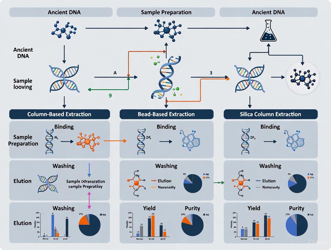
aDNA Extraction in Practice: Protocols from Silica Columns to High-Throughput Automation
The recovery of DNA from ancient and highly degraded samples is a cornerstone of paleogenomics and forensic genetics. The efficacy of this recovery is fundamentally dependent on the extraction method, with silica-based purification being the near-universal choice due to its ability to isolate DNA from complex inhibitors. Within this category, a key methodological divergence exists between the use of silica-in-suspension (often magnetic or non-magnetic beads) and silica-column-based approaches. The central thesis of this guide is that while both methods are capable of recovering short, damaged DNA fragments, their performance differs in critical aspects of fragment retention, yield, and procedural workflow. These differences are not merely academic; they directly impact downstream analytical success, influencing the quantity and quality of data obtained from sequencing or PCR. This guide provides a objective, data-driven comparison of these two methods, framing the discussion within the broader context of optimizing ancient DNA (aDNA) research.
Performance Comparison: Suspension vs. Column
The choice between silica suspension and column-based methods involves trade-offs. The following table summarizes the core performance characteristics of each method, synthesized from comparative studies.
Table 1: Performance Comparison of Silica Suspension and Column-Based Methods for Short DNA Fragments
| Performance Characteristic | Silica-in-Suspension | Silica Column (e.g., MinElute) |
|---|---|---|
| Optimal Fragment Retention | Designed for very short fragments (as low as 25-50 bp) [22] [4] | Retains fragments as short as 70 bp [23] [6] |
| Endogenous DNA Yield | Can be highly efficient, but may be lower in direct comparisons [6] | Often results in higher yields of endogenous DNA; MinElute showed superior results in one study [6] |
| Average Fragment Length | Can recover shorter fragments, potentially reducing average length | Preserves slightly longer fragment lengths on average [6] |
| Hands-On Time & Automation | High potential for automation on liquid handling platforms [24] | Manual protocol involves multiple tube changes, less amenable to high-throughput automation [22] |
| Cost & Throughput | Higher cost per sample for magnetic beads; efficient for high-throughput | Generally cost-effective; ideal for laboratories with lower sample throughput [22] |
Detailed Experimental Protocols
To ensure reproducibility and provide a clear understanding of the methodologies being compared, this section details two representative protocols used in comparative studies.
Silica Suspension DNA Extraction Protocol
This protocol, adapted from a study comparing methods for aDNA, involves creating a homemade silica suspension and using it to bind and purify DNA [6].
- Silica Suspension Preparation: 2.4 g of silicon dioxide powder is suspended in 20 mL of DNA-free water and allowed to settle for one hour at room temperature in the dark. After settling, 19.5 mL of the supernatant is pipetted out. The mixture settles for another four hours, after which 17 mL of supernatant is discarded. The pH of the remaining 2.5 mL of sediment is adjusted with hydrochloric acid [6].
- Digestion & Binding: Between 50-200 mg of bone powder is digested in an extraction buffer (e.g., containing EDTA, Urea, and Proteinase K) for up to 72 hours at 48°C. The digested lysate is then added to a binding buffer (e.g., 5.83 M GuHCl, 105 mM NaOAc, 46.8% isopropanol, 0.06% Tween-20) along with 150 µL of the prepared silica suspension. The pH is adjusted to between 4 and 6. Binding proceeds for 3 hours at room temperature [6].
- Washing & Elution: The silica with bound DNA is pelleted by centrifugation and the supernatant is discarded. The pellet is then washed twice with 80% ethanol to remove salts and other impurities. After the washes, the DNA is finally eluted in 100 µL of TE buffer [6].
Silica Column-Based DNA Extraction Protocol
This column-based protocol, specifically optimized for aDNA, highlights the use of MinElute columns, which are designed to retain shorter fragments than standard QIAquick columns [23] [6].
- Digestion: Approximately 50 mg of bone powder is digested in a buffer containing 0.45 M EDTA, 250 µg/mL Proteinase K, 1% Triton X-100, and 50 mM DTT. Digestion occurs under motion at 55°C overnight [6].
- Concentration & Clean-up: The digested lysate is concentrated using centrifugal concentrator filters (e.g., Vivaspin with a 30 kDa molecular weight cut-off) to a volume of less than 120 µL [23].
- Column Purification: The concentrated lysate is applied to a MinElute spin column following the manufacturer's instructions, with critical modifications to reduce contamination: the collection tube is changed after each centrifugation step, and the column is air-dried after the final wash to remove residual ethanol. DNA is eluted in two steps with EB buffer for a final volume of ~96 µL [23] [6].
Workflow Visualization
The procedural differences between the two DNA extraction methods are illustrated in the following workflow diagram. The visualization highlights the key distinction: silica suspension uses a single-tube binding approach, while the column method requires multiple liquid transfer steps.
The Scientist's Toolkit: Essential Research Reagents
Successful DNA extraction from degraded samples relies on a specific set of reagents and materials. The following table details key components and their functions in the process.
Table 2: Essential Reagents for Silica-Based DNA Extraction of Short Fragments
| Reagent / Material | Function / Rationale |
|---|---|
| Silica Matrix | The core binding substrate; in suspension as beads or in columns as a membrane. Selectivity for DNA is enabled in the presence of chaotropic salts [24] [6]. |
| Chaotropic Salts | Disrupt hydrogen bonding and solubilize proteins, allowing DNA to bind efficiently to the silica matrix [4]. |
| Binding Buffer | Typically contains chaotropic salts (e.g., GuHCl) and a buffering agent (e.g., sodium acetate). A lower pH (e.g., 4-6) is critical for maximizing binding efficiency by reducing electrostatic repulsion [24] [6]. |
| Proteinase K | A broad-spectrum serine protease essential for digesting proteins and breaking down cellular and bone matrices to release DNA [22] [6]. |
| EDTA (Ethylenediaminetetraacetic acid) | A chelating agent that binds metal ions, inactivating DNases that would otherwise degrade DNA. It is also crucial for demineralizing hard tissues like bone and tooth [22] [6]. |
| Isopropanol | Added to the binding buffer to reduce solvation and promote the precipitation of DNA onto the silica surface [4] [6]. |
| MinElute Columns | A specific type of silica spin column optimized for purifying and concentrating small DNA fragments (as short as 70 bp) from dilute solutions [23] [6]. |
| USER Enzyme | A mixture of Uracil-DNA Glycosylase (UDG) and Endonuclease VIII. It is used to remove uracil bases resulting from cytosine deamination, a common post-mortem damage, thereby reducing errors in downstream analyses [23]. |
The decision between silica suspension and silica column methods is not a matter of declaring one universally superior. Instead, it requires a strategic choice based on project-specific goals and constraints. Silica suspension methods offer a significant advantage for projects focused on recovering the very shortest of DNA fragments (below 70 bp) and are the obvious choice for high-throughput, automated laboratories. In contrast, silica columns (particularly MinElute) provide a robust, cost-effective solution for laboratories that process a moderate number of samples and aim to maximize the yield of endogenous DNA while still effectively recovering fragments in the 70 bp and above range. The experimental data and protocols outlined in this guide provide a foundation for researchers to make an informed selection, ultimately enhancing the recovery of genetic information from the fragile and precious resource that is ancient and degraded DNA.
Within the field of ancient DNA (aDNA) research, the extraction of high-quality DNA from degraded samples is a foundational step. Among the various techniques available, organic extraction using phenol-chloroform is often considered a "gold-standard" method, particularly for challenging samples [25]. This guide provides an objective comparison of the phenol-chloroform protocol against other common extraction methods, focusing on its performance in terms of DNA yield and purity. The data presented herein, drawn from recent scientific evaluations, aims to assist researchers in selecting the most appropriate extraction methodology for their specific sample types and research goals.
Performance Comparison: Organic Extraction vs. Alternative Methods
The following table summarizes key findings from recent studies that directly compared phenol-chloroform extraction with other techniques on degraded and ancient samples.
Table 1: Comparative Performance of DNA Extraction Methods from Degraded Biological Material
| Extraction Method | Sample Type | Reported DNA Yield | Reported DNA Purity (A260/280) | Key Advantages | Key Limitations |
|---|---|---|---|---|---|
| Organic (Phenol-Chloroform) | Historical & roadkill mammalian specimen [25] | High (Median: 202 ng/µL for modern, 98.9 ng/µL for museum) [25] | Satisfactory (1.8-2.0) [25] | High yield, effective for degraded DNA, high purity [25] [26] | Use of toxic reagents, labor-intensive [25] [27] |
| Degraded human skeletal remains [28] | Highest DNA quantification values [28] | - | Produced the most informative STR profiles [28] | - | |
| Archival FFPE tissue blocks [27] | Better DNA integrity [27] | - | Suitable for PCR amplification [27] | - | |
| Silica Spin-Column | Historical & roadkill mammalian specimen [25] | High (Satisfactory for downstream processes) [25] | Satisfactory (1.8-2.0) [25] | Safety, higher quality DNA, effective [25] | Potential loss of short DNA molecules [25] |
| Degraded human skeletal remains [28] | - | - | Efficient method [28] | - | |
| Museomics (QIAamp kits) [26] | Quantifiable concentrations [26] | - | Successful isolation across diverse museum samples [26] | - | |
| Magnetic Bead-Based | Historical & roadkill mammalian specimen [25] | Lower yield compared to PCI and silica column [25] | Less satisfactory purity [25] | Amenable to automation [22] | Lower yield and purity for historical samples [25] |
| Museomics (Zymo kits) [26] | Quantifiable concentrations [26] | - | - | Underperformed compared to Qiagen and PCI [26] | |
| Shotgun metagenomics (HMW kits) [29] | High yield of HMW DNA [29] | - | Suitable for long-read sequencing, pure HMW DNA [29] | Performance is kit and application-dependent [29] | |
| Salting-Out | Clinical specimens (sputum, BAL, tissue) [30] | Comparable quality to phenol-chloroform [30] | - | Non-toxic, time-efficient, cost-effective [30] | - |
Detailed Experimental Protocols
To ensure reproducibility, this section outlines the specific experimental protocols from key studies cited in the comparison table.
Protocol for Mammalian Museum and Roadkill Specimens
A 2023 study evaluated five DNA extraction methods on skin samples from modern roadkill and museum European hedgehogs [25].
- Sample Preparation: Skin samples were processed from both traffic-killed (n=10) and museum (n=10) specimens.
- Organic Extraction Protocol: The phenol-chloroform-isoamyl alcohol (PCI) protocol was followed, which involves digesting the sample with a lysis buffer and proteinase K, followed by separation using phenol and chloroform, and finally DNA precipitation [25].
- Comparison Methods: The study compared the PCI method against two silica spin-column kits (NucleoSpin Tissue and QIAamp), and two magnetic bead-based kits (NucleoMag DNA and Mag-Bind Blood & Tissue) [25].
- Quantification and Quality Control: DNA concentration was measured using both a NanoDrop 2000 spectrophotometer and a fluorometer (Quant-iT PicoGreen). DNA purity was assessed via NanoDrop A260/280 and A260/230 ratios. DNA fragmentation patterns and average sizing were determined using the 2100 Bioanalyzer Instrument (Agilent) [25].
Protocol for Degraded Human Skeletal Remains
A 2023 study compared five DNA extraction protocols on 25 degraded skeletal remains, including bones such as the humerus, tibia, and petrous bone [28].
- Sample Preparation: A standard quantity of bone powder was used for each extraction method.
- Organic Extraction Protocol: Organic extraction by phenol/chloroform/isoamyl alcohol was performed [28].
- Comparison Methods: The protocol was tested against silica in suspension, High Pure Nucleic Acid Large Volume silica columns (Roche), InnoXtract Bone (InnoGenomics), and an automated protocol (PrepFiler BTA with AutoMate Express robot) [28].
- Quantification and Quality Control: The study analyzed five DNA quantification parameters: small human target quantity, large human target quantity, human male target quantity, degradation index, and internal PCR control threshold. DNA profile performance was assessed based on the number of alleles, average RFU, heterozygous balance, and number of reportable loci from STR typing [28].
Protocol for Archival Clinical Tissues
A 2022 study compared DNA extraction from 75 archival oral squamous cell carcinoma (OSCC) samples, including formalin-fixed paraffin-embedded (FFPET) tissues and long-term formalin-fixed tissues (FFT) [27].
- Sample Preparation: For FFPET samples, deparaffinization was carried out using xylene or a heating method. For FFT, tissues were pulverized after freezing with liquid nitrogen [27].
- Organic Extraction Protocol: The conventional phenol-chloroform method involved digesting tissues with an extraction buffer and proteinase K overnight. This was followed by separation with saturated phenol and a mixture of phenol:chloroform:isoamyl alcohol. DNA was then precipitated using sodium acetate and ethanol, washed, and resuspended in TE buffer [27].
- Comparison Methods: The organic method was compared to a commercial kit method (HiPurATM Paraffin-Embedded Tissue DNA Purification Spin Kit) [27].
- Quantification and Quality Control: The quality and quantity of extracted DNA were assessed, and viability was evaluated by successful PCR amplification of the P53 gene [27].
Workflow and Practical Considerations
Experimental Workflow Diagram
The following diagram illustrates the general workflow for organic extraction of ancient or degraded DNA, integrating steps from the cited protocols.
The Scientist's Toolkit: Key Research Reagents
Table 2: Essential Reagents for Organic DNA Extraction and Their Functions
| Reagent / Solution | Primary Function in the Protocol |
|---|---|
| EDTA (Ethylenediaminetetraacetic acid) | Chelating agent that binds metal ions, inactivating DNases that degrade DNA [7] [26]. |
| SDS (Sodium Dodecyl Sulfate) | Ionic detergent that disrupts cell membranes and denatures proteins [27]. |
| Proteinase K | A broad-spectrum serine protease that digests and inactivates nucleases and other proteins [25] [27]. |
| DTT (Dithiothreitol) | A reducing agent that breaks disulfide bonds in proteins, aiding in the lysis of tough tissues [26]. |
| Phenol:Chloroform:Isoamyl Alcohol | Organic solvent mixture that denatures and dissolves proteins, separating them from the nucleic acids in the aqueous phase [25] [27]. |
| Sodium Acetate (3M) | Provides salt ions necessary for the efficient precipitation of DNA by ethanol [27]. |
| Ethanol (100% and 70%) | 100% ethanol precipitates nucleic acids from the aqueous solution; 70% ethanol removes residual salt from the DNA pellet [27]. |
| TE Buffer (Tris-EDTA) | A stable buffer (Tris-HCl + EDTA) used to resuspend and store purified DNA, maintaining its pH and stability [27]. |
The organic extraction method remains a powerful and effective technique for isolating DNA from highly degraded and ancient samples, consistently delivering high yields and pure DNA suitable for downstream analyses like PCR and sequencing [25] [28]. Its primary drawbacks are the requirement for handling hazardous chemicals and a more labor-intensive process. The choice between phenol-chloroform and alternative methods, such as modern silica-based kits, should be guided by a balanced consideration of sample type, desired fragment length, required yield and purity, and available laboratory resources. For the most challenging samples where yield is paramount and safety resources are adequate, organic extraction continues to be a top-performing choice.
The recovery of ancient DNA (aDNA) presents unique challenges for researchers, requiring specialized extraction methods to overcome issues of extreme degradation, low endogenous DNA content, and co-extraction of PCR inhibitors. The efficacy of aDNA studies in fields such as evolutionary biology, archaeology, and forensic science is fundamentally dependent on the extraction methodology employed. This guide provides an objective comparison of three commercial DNA extraction kits—PrepFiler BTA, InnoXtract Bone, and DNeasy Plant Mini Kit—evaluating their performance across various experimental parameters relevant to ancient and degraded sample analysis. These kits represent different approaches to aDNA recovery: PrepFiler BTA and InnoXtract Bone utilize silica-coated magnetic bead technology optimized for challenging forensic and ancient samples, while the DNeasy Plant Mini Kit employs a silica-membrane column system originally developed for modern plant tissues.
The fundamental challenge in aDNA research lies in the molecular nature of the starting material. Ancient DNA is typically highly fragmented, with most fragments measuring less than 100 base pairs due to post-mortem degradation processes. Additionally, aDNA molecules often contain chemical modifications such as cytosine deamination and exhibit low copy numbers amidst environmental contaminants. These characteristics necessitate extraction methods specifically optimized to recover short DNA fragments while effectively removing PCR inhibitors such as humic acids, polyphenols, and other secondary metabolites that accumulate in ancient remains over time. The selection of an appropriate extraction kit can significantly impact downstream applications including real-time quantitative PCR (qPCR), next-generation sequencing (NGS) library preparation, and short tandem repeat (STR) analysis, ultimately determining the success or failure of aDNA studies.
Performance Comparison and Experimental Data
Quantitative Performance Metrics Across Sample Types
Table 1: DNA Extraction Efficiency from Degraded Human Skeletal Remains [31]
| Extraction Method | Small Autosomal Target (pg/μL) | Degradation Index | STR Alleles Detected | Average RFU |
|---|---|---|---|---|
| PrepFiler BTA | Data not statistically different | Comparable across methods | No significant difference | Consistent across methods |
| InnoXtract Bone | Data not statistically different | Comparable across methods | No significant difference | Consistent across methods |
| Organic Extraction | Highest yield | Most favorable | Maximum alleles | Highest intensity |
Table 2: Specialized Application Performance Metrics [32] [33]
| Extraction Method | Ancient Plant DNA Yield | Inhibitor Removal Efficiency | Fragment Size Recovery | Automation Compatibility |
|---|---|---|---|---|
| DNeasy Plant Mini Kit | Lower efficiency on ancient material | Moderate for humic acids | Standard range | Manual processing |
| InnoXtract Bone | Not specialized for plants | High (optimized for inhibitors) | Down to 100 bp | Full automation (96-well) |
| PrepFiler BTA | Not specialized for plants | High (BTA lysis buffer) | Short fragments | AutoMate Express system |
A comprehensive 2023 study compared five DNA extraction methods on 25 degraded skeletal elements, including humerus, ulna, tibia, femur, and petrous bone [31]. Researchers analyzed five DNA quantification parameters (small human target quantity, large human target quantity, human male target quantity, degradation index, and internal PCR control threshold) and five DNA profile parameters (number of alleles with peak height higher than analytic and stochastic threshold, average relative fluorescence units (RFU), heterozygous balance, and number of reportable loci). While organic extraction by phenol/chloroform/isoamyl alcohol demonstrated the best overall performance in both quantification and DNA profile results, both PrepFiler BTA and InnoXtract Bone provided sufficient DNA recovery for successful STR typing of degraded remains [31].
For ancient plant remains, a 2025 study evaluated extraction methods on archaeological grape seeds, demonstrating that the DNeasy Plant Mini Kit showed lower efficiency compared to specialized ancient DNA protocols [32]. The study found that customized approaches combining sediment-optimized extraction buffers with silica purification strategies outperformed commercial kits for recovering ultrashort aDNA fragments from challenging archaeobotanical samples. This highlights a significant limitation of standard plant kits when applied to ancient material, as they are primarily optimized for fresh tissues containing longer, less degraded DNA molecules.
Methodological Approaches and Technical Specifications
Figure 1: Unified Workflow for Magnetic Bead-Based DNA Extraction Kits. PrepFiler BTA and InnoXtract Bone share a similar silica-coated magnetic bead purification approach but differ in their specialized lysis buffer formulations and incubation conditions.
Table 3: Technical Specifications and Experimental Protocols [34] [35] [33]
| Parameter | PrepFiler BTA | InnoXtract Bone | DNeasy Plant Mini Kit |
|---|---|---|---|
| Technology | Silica-coated magnetic beads | Silica-coated magnetic beads | Silica-membrane column |
| Lysis Buffer | PrepFiler BTA Lysis Buffer with Proteinase K, DTT | Specialized digestion buffer | AP1 buffer |
| Optimal Lysis | 56°C for 40min-2h (sample-dependent) | Protocol-specific incubation | 65°C for 10-30min |
| Binding Mechanism | Magnetic particle with multi-component surface chemistry | Optimized for fragments ≥100 bp | Silica membrane binding |
| Inhibitor Removal | PrepFiler LySep Column | Silica bead washing steps | AW1/AW2 wash buffers |
| Elution Volume | Small volume (kit-optimized) | Small volume (kit-optimized) | 100-400μL AE buffer |
| Automation | AutoMate Express, liquid handlers | MagMAX Express-96 | Manual or QIAcube |
The experimental protocol for PrepFiler BTA involves an initial lysis step using specialized BTA Lysis Buffer supplemented with Proteinase K and DTT, incubated at 56°C for 40 minutes to 2 hours depending on sample type [35] [36]. The unique PrepFiler LySep Column enables streamlined separation of substrate from lysate through centrifugation, eliminating manual transfer steps and reducing contamination risk. For bone samples, the protocol typically involves powdering the specimen followed by extended lysis to ensure complete digestion of the mineralized matrix.
InnoXtract Bone extraction employs a similar magnetic bead approach but is specifically optimized to recover short DNA fragments as small as 100 base pairs, which is particularly advantageous for highly degraded ancient remains [33]. The validation studies demonstrate consistent performance across various sample types including insulted and un-insulted bone and teeth. The method has been successfully automated on a MagMAX Express-96 system, providing high-throughput capacity with 6 hours total extraction time for up to 96 samples, significantly improving laboratory efficiency for large-scale studies [33].
The DNeasy Plant Mini Kit protocol involves lysis with AP1 buffer, often supplemented with β-mercaptoethanol for plant tissues, followed by incubation at 65°C [32]. The lysate is mixed with ethanol and applied to a DNeasy mini column where DNA binds to the silica membrane. Subsequent wash steps with AW1 and AW2 buffers remove contaminants, followed by elution in AE buffer. While effective for modern plant samples, this method shows limitations with ancient plant remains due to less efficient recovery of ultrashort DNA fragments and incomplete removal of humic acid inhibitors commonly found in archaeological contexts [32].
Research Reagent Solutions and Materials
Table 4: Essential Research Reagents for Ancient DNA Extraction [34] [32] [35]
| Reagent/Material | Function | Kit Applications |
|---|---|---|
| Silica-coated Magnetic Beads | DNA binding and purification through silica technology | PrepFiler BTA, InnoXtract Bone |
| PrepFiler BTA Lysis Buffer | Digestion of complex matrices (bone, tooth, adhesive) | PrepFiler BTA specific |
| Proteinase K | Protein digestion and tissue lysis | All three kits |
| DTT (Dithiothreitol) | Reduction of disulfide bonds in keratinous tissues | PrepFiler BTA, InnoXtract Bone |
| PrepFiler LySep Column | Separation of substrate from lysate | PrepFiler BTA specific |
| CTAB Buffer | Polysaccharide precipitation and inhibitor removal | Alternative ancient plant protocol |
| Power Beads Solution | Removal of humic acid inhibitors from sediments | Alternative ancient protocol |
| AP1 Buffer | Lysis buffer for plant tissues | DNeasy Plant Mini Kit |
| AW1/AW2 Buffers | Wash buffers for removing contaminants | DNeasy Plant Mini Kit |
| AE Buffer | Elution buffer for DNA recovery | DNeasy Plant Mini Kit |
The selection of appropriate reagents is critical for successful aDNA recovery. Silica-coated magnetic beads form the core technology for both PrepFiler BTA and InnoXtract Bone kits, providing efficient DNA binding and purification [34] [33]. These magnetic particles feature optimized multi-component surface chemistry that enhances DNA recovery from challenging samples. The PrepFiler BTA system includes specialized LySep Columns that dramatically streamline the off-line lysis portion of the extraction method by permitting lysate flow through a burstable membrane while retaining substrate in the column [34].
For plant-specific applications, the DNeasy Plant Mini Kit utilizes AP1 lysis buffer designed to disrupt plant cell walls and membranes while stabilizing released DNA [32]. However, studies on archaeological plant remains indicate that conventional CTAB (cetyltrimethylammonium bromide) protocols or sediment-optimized extraction buffers like Power Beads Solution may provide superior performance for ancient samples by more effectively removing PCR inhibitors such as humic acids and polyphenols [32]. These specialized reagents precipitate polysaccharides and other contaminants that often co-extract with DNA from ancient plant tissues.
Discussion and Comparative Analysis
Performance in Ancient DNA Contexts
The comparative analysis reveals distinct strengths and limitations for each kit within ancient DNA research contexts. For degraded human skeletal remains, a 2023 comparative study found that while organic extraction outperformed commercial kits, both PrepFiler BTA and InnoXtract Bone provided statistically similar results for DNA quantification and STR profiling [31]. This demonstrates their viability as efficient alternatives to labor-intensive organic extraction methods, particularly valuable in high-throughput laboratory settings.
When examining thermally altered samples specifically, a 2025 study comparing InnoXtract and PrepFiler BTA for extracting DNA from burned porcine femurs found no statistically significant difference in DNA quantity recovery based on the extraction kit used [37]. This research employed real-time PCR quantification on samples subjected to different burning durations (unburned, burned at 200°C for 15 minutes, and burned at 200°C for 30 minutes), demonstrating that both kits performed comparably on heat-degraded skeletal material. The length of burning exposure also proved statistically insignificant in relation to DNA recovery, suggesting that both kits effectively handle the protein cross-linking and fragmentation challenges posed by thermal degradation.
For ancient plant remains, the DNeasy Plant Mini Kit shows notable limitations. A comprehensive 2025 evaluation of DNA extraction from archaeological grape seeds found that specialized ancient DNA protocols significantly outperformed commercial plant kits [32]. The DNeasy method demonstrated lower efficiency in recovering endogenous aDNA and less consistent performance across archaeological sites compared to customized approaches combining sediment-optimized extraction with silica purification strategies. This performance gap highlights the fundamental mismatch between kits designed for modern plant tissues and the unique requirements of ancient plant material, which contains shorter DNA fragments and higher inhibitor concentrations.
Throughput and Operational Considerations
Operational efficiency represents a significant differentiator among these extraction kits. InnoXtract Bone offers substantial advantages for high-throughput applications, with complete automation on the MagMAX Express-96 system enabling processing of up to 96 samples in approximately 6 hours [33]. This throughput capability is particularly valuable for large-scale studies involving multiple specimens, such as missing persons identifications, mass disaster responses, or population-scale ancient DNA research.
PrepFiler BTA provides flexibility across multiple workflow formats, including manual, automated liquid handler, and AutoMate Express instrument platforms [36]. A 2022 study demonstrated that a modified "top-up" method—adding PrepFiler lysis buffer over BTA-lysed sample remnants—could enhance DNA recovery from certain sample types including blood, semen, and buccal cells on tape [35]. This optimization illustrates how protocol modifications can maximize DNA yield while maintaining compatibility with automated systems.
The DNeasy Plant Mini Kit is primarily designed for manual processing, though it can be automated using the QIAcube system [32]. Its lower throughput capacity and reduced efficiency with ancient samples make it less suitable for large-scale aDNA studies compared to the magnetic bead-based alternatives. However, for smaller projects focusing on relatively well-preserved archaeological plant specimens, it may offer a straightforward, user-friendly option despite its limitations in recovery efficiency.
The comparative analysis of PrepFiler BTA, InnoXtract Bone, and DNeasy Plant Mini Kit reveals a clear differentiation in their applicability to ancient DNA research. For human skeletal remains, both PrepFiler BTA and InnoXtract Bone demonstrate comparable efficacy in recovering quantifiable DNA capable of generating STR profiles, making them viable alternatives to traditional organic extraction methods. Their automated platforms offer significant throughput advantages for processing multiple specimens efficiently.
For archaeobotanical studies, researchers should exercise caution when considering the DNeasy Plant Mini Kit, as evidence indicates specialized ancient DNA protocols consistently outperform this commercial plant kit for recovering endogenous DNA from ancient plant remains. The magnetic bead technology employed by both PrepFiler BTA and InnoXtract Bone provides superior recovery of short DNA fragments characteristic of ancient DNA, along with more effective removal of common PCR inhibitors encountered in archaeological contexts.
Future methodological developments in ancient DNA extraction will likely continue refining magnetic bead technologies for enhanced recovery of ultrashort fragments while improving inhibitor removal efficiency. The optimal kit selection ultimately depends on specific research objectives, sample characteristics, and laboratory infrastructure, with each system offering distinct advantages for particular applications within the broader field of ancient DNA research.
CTAB Protocol Adaptation for Archaeological Plant and Seed Material
The recovery of ancient DNA (aDNA) from archaeological plant remains is a cornerstone of paleogenomics, enabling researchers to trace crop evolution and domestication [2]. However, this work is fraught with challenges, as endogenous aDNA from archaeobotanical remains is typically highly fragmented, present in low copy numbers, and often co-extracted with potent inhibitors like humic acids from sediments [2] [32]. Among the various methods employed, the cetyltrimethylammonium bromide (CTAB)-based protocol remains a widely used and foundational technique. This guide provides a objective comparison of the adapted CTAB method against other prominent aDNA extraction protocols, evaluating their efficacy based on recent experimental data from the field. The performance of these methods is critically assessed for their suitability in downstream next-generation sequencing (NGS) applications, which are essential for advanced paleogenomic studies [2] [32].
Comparative Analysis of Ancient DNA Extraction Methods
The selection of an appropriate DNA extraction method is a critical first step that can determine the success or failure of subsequent genetic analysis. The table below provides a quantitative comparison of four primary methods used for archaeological plant and seed material.
Table 1: Performance Comparison of Ancient DNA Extraction Methods for Plant Materials
| Extraction Method | Key Principle | Reported DNA Yield | Inhibitor Removal Efficiency | Suitability for NGS | Key Advantages | Key Limitations |
|---|---|---|---|---|---|---|
| Silica-Power Beads (S-PDE) [2] [32] | Reagent optimized against soil inhibitors coupled with aDNA-specific silica binding | Higher yields and more consistent performance across sites | Effective; designed for humic acid removal | High; improves library complexity and sequencing metrics | Consistent performance, effective inhibitor removal | Relatively newer method for macrofossils |
| Phenol-Chloroform (Phe-chl) [2] | Organic separation using phenol-chloroform | High DNA yield | Moderate; can co-precipitate inhibitors | Moderate | High DNA yield, familiar protocol | Use of hazardous phenol, may retain inhibitors |
| CTAB-Based Protocol [2] [38] | CTAB surfactant precipitates polysaccharides & polyphenols | Variable; can be high with optimization [38] | Good for polysaccharides; less effective for humic acids [2] | Good; DNA suitable for PCR and sequencing [38] | Cost-effective, good for complex plant tissues [39] | Can be time-consuming, may require optimization for humics |
| Commercial Kits (e.g., DNeasy) [2] | Silica-column based purification | Generally lower efficiency for aDNA [2] | Variable; can struggle with potent inhibitors | Lower; shown to have less consistent results [2] | Standardized, easy to use, fast | Lower efficiency for challenging ancient samples, cost |
Detailed Experimental Protocols
Adapted CTAB Protocol for Ancient Plant Remains
The standard CTAB protocol requires specific adaptations to handle the unique challenges posed by archaeological plant seeds. The following workflow incorporates optimizations noted across several studies [2] [38] [40].
Figure 1: A generalized workflow for extracting DNA from archaeological plant seeds using an adapted CTAB protocol. Key optimization steps, such as the addition of SDS and Proteinase K, are included [2] [40].
Key Steps and Reagents:
- Surface Decontamination: Subject seeds to UV light for 20 minutes and wash with sterile water to remove surface contaminants [2].
- Mechanical Grinding: Pulverize the seeds to a fine powder using a low-speed drill (approx. 100 RPM) to minimize heat damage. This has been shown to be more effective than a mortar and pestle for these sample types [2].
- Cellular Lysis:
- CTAB Buffer: The standard lysis buffer contains 2% CTAB, 1.4 M NaCl, 100 mM Tris-HCl (pH 7.5-8.0), and 20 mM EDTA. The high salt concentration helps to precipitate polysaccharides [38] [40].
- Enhancements: For improved lysis of ancient and complex tissues, add Sodium Dodecyl Sulfate (SDS) and Proteinase K to the CTAB buffer. This combination is more effective at lysing cell walls and removing proteins [40].
- Incubation: Incubate the mixture at 60-65°C for 30 minutes to several hours, or even overnight at room temperature, to maximize DNA yield from degraded samples [40].
- Purification and Precipitation:
- DNA Resuspension: Resuspend the final pellet in a low-EDTA TE buffer or nuclease-free water [38].
Competing Protocol: Silica-Power Beads DNA Extraction (S-PDE)
A method optimized for sedimentary ancient DNA (sedaDNA) has shown promising results when applied directly to plant macrofossils like grape seeds [2] [32]. This protocol involves using a Power Beads Solution (Qiagen), a reagent designed to dislodge cells from sediment particles and counteract soil-derived inhibitors like humic acids. This is followed by a silica-based purification step specifically designed to bind and recover short, fragmented aDNA molecules [2] [32]. This S-PDE method has been demonstrated to achieve higher yields and more consistent performance across different archaeological sites compared to other extraction methods, significantly improving the success of the critical NGS library production step [2].
Decision Workflow and Application Guidance
Choosing the most appropriate extraction method depends on the sample characteristics and research goals. The following decision tree can help guide this selection.
Figure 2: A decision workflow to guide the selection of an appropriate DNA extraction method based on sample characteristics and research objectives.
The Scientist's Toolkit: Essential Research Reagents
Successful DNA extraction from challenging archaeological plant materials relies on a set of key reagents, each performing a critical function in the multi-step process of lysing cells, neutralizing contaminants, and purifying fragile DNA strands.
Table 2: Key Reagent Solutions for Ancient Plant DNA Extraction
| Research Reagent | Core Function in Extraction Protocol |
|---|---|
| CTAB (Cetyltrimethylammonium bromide) | A cationic detergent that effectively lyses plant cells and precipitates polysaccharides and polyphenols, which are common PCR inhibitors in plant tissues [38] [40]. |
| Proteinase K | A broad-spectrum serine protease that degrades cellular proteins and nucleases, helping to liberate and protect DNA during the lysis step [40]. |
| SDS (Sodium Dodecyl Sulfate) | An anionic detergent that synergizes with CTAB to disrupt cell walls and lipid membranes, facilitating more complete lysis, particularly in tough or ancient tissues [40]. |
| Silica Magnetic Beads | Used in modern purification; DNA binds to the silica surface in the presence of high-salt buffers, allowing for efficient washing and elution with minimal loss of short fragments [2] [41]. |
| Polyvinylpyrrolidone (PVP) | Binds to and helps remove phenolic compounds, which are common inhibitors in both plants and soils, often used as a pre-treatment [41]. |
| Power Beads Solution | A commercial solution containing silica beads designed to dislodge cells from sediment particles and counteract co-extracted humic acids, a major inhibitor from soils [2]. |
| Chloroform:Isoamyl Alcohol | Used in organic extraction to separate proteins and lipids from the nucleic acid-containing aqueous phase, effectively deproteinizing the sample [38]. |
| Sodium Chloride (NaCl) | Used at high concentration (e.g., 1.4 M) in CTAB buffer to prevent the co-precipitation of polysaccharides with DNA [38] [40]. |
The adaptation of the CTAB protocol remains a viable and cost-effective strategy for recovering DNA from archaeological plant and seed material, particularly for samples rich in polysaccharides and polyphenols. However, empirical evidence from recent studies demonstrates that no single method is universally superior. The Silica-Power Beads (S-PDE) method shows exceptional promise for samples contaminated with soil-derived inhibitors, often yielding DNA more suitable for demanding downstream NGS applications [2]. Researchers must therefore base their choice on a careful assessment of their specific sample type, preservation state, and the primary inhibitors present. As the field of paleogenomics continues to evolve, so too will these fundamental protocols, driving forward our ability to unlock genetic secrets from the past.
The field of ancient DNA (aDNA) research has undergone a significant transformation, driven by the need to process large archaeological and palaeontological collections efficiently. Traditional DNA extraction methods, often relying on single-column processing, present substantial limitations in throughput, cost, and time when dealing with large-scale studies. In response, high-throughput workflows utilizing 96-column plates and robotic automation have emerged as powerful alternatives. These methods are framed within a broader research thesis focused on comparing the efficacy of different aDNA extraction methodologies, aiming to identify protocols that maximize recovery of authentic ancient DNA while optimizing resource utilization. This guide objectively compares the performance of 96-column plates against traditional single-column methods and other alternatives, providing supporting experimental data to inform researchers, scientists, and drug development professionals in their methodological selections.
Comparative Analysis of Extraction Platforms
96-Column Plates vs. Single-Column Methods
The transition from single-column processing to 96-column plate-based systems represents a fundamental shift in aDNA extraction methodology. A 2025 study directly compared a high-throughput 96-column plate method against routine single MinElute columns, revealing highly similar endogenous DNA content recovery—a critical metric in aDNA screening [7]. The research demonstrated that mitogenomes with coverage depth greater than 0.1× could be successfully generated from 96-column plate extracts, allowing for accurate taxonomic assignment [7].
While the 96-column plate method showed significant advantages in throughput and cost-efficiency, researchers noted initial differences in average fragment lengths, DNA damage patterns, and library complexities compared to single-column methods. However, these differences became nonsignificant after modifications to library purification protocols [7], indicating that optimization of downstream processes can mitigate potential quality variations.
Table 1: Performance Comparison of 96-Column Plates vs. Single Columns
| Performance Metric | 96-Column Plate Method | Single Column Method |
|---|---|---|
| Endogenous DNA Content | Highly similar to single columns [7] | Benchmark for comparison [7] |
| Cost Per Sample | ~39% reduction [7] | Higher cost baseline [7] |
| Processing Time | ~4 hours for 96 samples [7] | Significantly longer per sample [7] |
| Fragment Length | Initially differed, equalized after protocol optimization [7] | Longer fragments preserved [6] |
| Mitogenome Generation | Possible (>0.1× coverage) [7] | Possible (>0.1× coverage) [7] |
| Automation Compatibility | High [7] | Low to moderate [7] |
Extraction Method Efficacy Across Sample Types
The performance of DNA extraction methods varies significantly depending on sample type and preservation conditions. A comprehensive comparison of DNA extraction protocols for ancient soft tissues found that silica-based laboratory methods consistently outperformed commercial kits for challenging sample types [18]. When comparing the efficiency of different methods for reliable results in ancient DNA NGS workflows, researchers found that samples from Pars petrosa (the densest part of the temporal bone) yielded the highest endogenous DNA content and longer fragment sizes compared to tooth or skeletal samples [6].
Table 2: DNA Extraction Performance Across Sample Types
| Sample Type | Optimal Method | Endogenous DNA Yield | Key Findings |
|---|---|---|---|
| Pars Petrosa | MinElute columns [6] | Highest yield [6] | Best preservation; longest fragments [6] |
| Tooth Cementum | Silica suspension [6] | High yield [6] | Good alternative to petrous bone [6] |
| Ancient Skin | Laboratory silica protocol [18] | Higher than hair samples [18] | Outperforms commercial kits [18] |
| Ancient Hair | Laboratory silica protocol [18] | Lower than skin samples [18] | Challenging substrate [18] |
| Sedimentary DNA | Dabney/Korlević protocol [42] | Variable (site-dependent) [42] | Pooling strategy effective [42] |
The selection of extraction methods also impacts the efficiency of downstream processes. Research comparing DNA extraction and library building methods for museum specimens found that while extraction methods didn't significantly differ in DNA yield, the choice of library build method profoundly affected outcomes [8]. The Santa Cruz Reaction (SCR) library build method proved most effective at retrieving degraded DNA while being easily implemented at high throughput for low cost [8].
High-Throughput Experimental Protocols
96-Column Plate DNA Extraction Protocol
The high-throughput 96-column plate DNA extraction method enables processing of 96 samples within approximately 4 hours of laboratory work [7]. The protocol is adapted from established single-column methods but optimized for parallel processing:
Sample Preparation: Bone fragments are drilled or crushed to obtain approximately 50 mg of bone powder. Samples are typically bleach-pretreated in <0.5% sodium hypochlorite solution for approximately 4 minutes at room temperature to remove surface contamination, followed by rinsing with UltraPure DNase/RNase-Free Distilled Water [7].
Lysis Step: Bone powder is mixed with ~1 mL of lysis buffer composed of 900 μL EDTA (0.5 M, pH 8), 75 μL UltraPure water, 0.5 μL Tween-20 (final concentration: 0.05%), and 25 μL Proteinase K (10 μg/μL). Incubation occurs under motion at 37°C from overnight to 72 hours depending on sample digestion [7].
DNA Binding: Binding buffer is prepared with guanidine hydrochloride (GuHCl) and isopropanol (final concentration: 5 M GuHCl, 40% v/v isopropanol) with added Tween-20. Lysates are combined with binding buffer in the 96-column plate format [7].
Washing and Elution: Multiple wash steps remove contaminants, followed by elution in TE buffer or similar. The addition of Tween-20 during elution has been formally demonstrated to result in higher complexity libraries [7].
This protocol maintains the fundamental principles of aDNA extraction while dramatically increasing throughput and reducing costs compared to single-column methods [7].
Pooled Screening Approach for Sedimentary Ancient DNA
A novel high-throughput method for sedimentary ancient DNA (sedaDNA) analysis employs a pooled testing approach that combines multiple extracts for efficient screening:
DNA Extraction: Amounts of 50-60 mg of sediment are used for DNA extraction following established protocols with adaptations, eluted in 50 μL TET buffer [42].
Library Preparation: Double-stranded libraries are prepared from half of the extract without shearing DNA into smaller fragments. MinElute PCR Purification kits or SPRI beads are used for clean-up [42].
Pooling Strategy: Extracts from multiple sediment samples are pooled before capture and sequencing. Research demonstrates that aDNA signals remain discernible even when pooled with four negative samples, with DNA yield increasing significantly when reducing extract input [42].
Hybridization Capture: Enrichment for mammalian mitogenomes uses custom-designed capture probes following standardized protocols with 16-hour hybridization at 65°C [42].
This approach enables researchers to analyze large numbers of sediment samples for aDNA preservation, achieving significant cost reductions of up to 70% and reducing hands-on laboratory time to one-fifth [42].
Diagram 1: High-throughput ancient DNA analysis workflow showing key steps from sample collection to data analysis.
Robotic Automation Platforms
Robotic Systems for Ancient DNA Extraction
Robotic automation platforms have been successfully implemented in aDNA research to minimize hands-on time and reduce experimental errors. The Opentrons OT-2 liquid-handling robot has been utilized for automated target capture processing in hyRAD protocols [43]. These systems provide:
- Consistent Extraction Processes: Automated pipetting procedures achieve uniform sample processing across bind, wash, and elute steps [44].
- Minimal Cross-Contamination: Reduced sample handling lowers contamination risk and sample carryover [44].
- Time Efficiency: Automated systems significantly reduce processing time and human effort, allowing researchers to process samples unattended [45] [44].
- Scalability: Larger sample numbers can be processed with greater speed, broadening research scope [44].
Advanced Robotic Systems for Laboratory Automation
Next-generation robotic platforms like RoboCulture represent advances in flexible laboratory automation. These systems utilize general-purpose robotic manipulators for complete lab automation, offering capabilities beyond conventional gantry-based systems [46]. Key features include:
- Computer Vision-Based Liquid Handling: Uses vision feedback to establish closed-loop control and robust pipette insertion into wells of arbitrarily positioned 96-well plates [46].
- Force Feedback Pipette Tip Exchange: Leverages robot force feedback to reliably attach new pipette tips [46].
- Real-Time Monitoring: Computer vision systems monitor optical density to inform robotic decisions about experimental progress [46].
- Modular Behavior Framework: Behavior trees govern coordination between subsystems, enabling complex workflows and decision-making for experimental management [46].
These systems prioritize flexibility and autonomy over throughput, enabling long-duration, hands-free operation by responding to changing experimental conditions [46].
Research Reagent Solutions
Table 3: Essential Research Reagents for High-Throughput Ancient DNA Workflows
| Reagent/Kit | Function | Application Notes |
|---|---|---|
| Silica-coated Magnetic Beads | DNA binding and purification | Core component of automated extraction; compatible with high-throughput platforms [45] [44] |
| Guanidine Hydrochloride (GuHCl) | Chaotropic salt for DNA binding | Facilitates DNA binding to silica in presence of isopropanol [7] |
| Proteinase K | Enzymatic digestion of proteins | Critical for lysis step; digests contaminating proteins [7] [6] |
| Tween-20 | Surfactant for improved elution | Addition during elution results in higher complexity libraries [7] |
| Santa Cruz Reaction (SCR) Kit | Library preparation | Most effective for degraded DNA; easily implemented at high throughput for low cost [8] |
| MinElute PCR Purification Kit | DNA clean-up | Used in library preparation and clean-up steps [42] |
| Custom Capture Probes | Target enrichment | Enables hybridization capture for specific genomic regions [43] [42] |
Diagram 2: Robotic automation system architecture showing key components and their relationships in high-throughput workflows.
The comprehensive comparison of high-throughput workflows for ancient DNA extraction reveals a clear trajectory toward 96-column plate methods and robotic automation. The experimental data demonstrates that 96-column plate methods achieve highly similar endogenous DNA content compared to single-column methods while reducing costs by approximately 39% and significantly decreasing processing time [7]. When combined with optimized library preparation methods like the Santa Cruz Reaction [8] and strategic pooling approaches for sedimentary DNA [42], these workflows enable unprecedented scale in aDNA research.
Robotic automation platforms further enhance these benefits by providing consistent, reproducible results with minimal contamination risk [45] [44]. While initial investments in equipment may be substantial, the long-term benefits in throughput, data quality, and operational efficiency present a compelling case for laboratories engaged in large-scale aDNA studies. As the field continues to evolve, the integration of increasingly sophisticated robotic systems with computer vision and real-time monitoring capabilities promises to further advance the efficiency and scope of ancient DNA research.
Maximizing aDNA Yield: Strategic Protocol Optimization and Contamination Control
The recovery of ancient DNA (aDNA) from historical, archaeological, and forensic specimens is a cornerstone of paleogenomics, evolutionary biology, and forensic science. The highly degraded and low-yield nature of aDNA necessitates extraction protocols that maximize the recovery of short, damaged DNA fragments while minimizing the co-extraction of inhibitors [47] [32]. The lysis phase—comprising demineralization, enzymatic digestion, and the careful management of time and temperature—is the critical first step that dictates the success of all downstream analyses. This step is designed to break down the rigid mineralized matrix of materials like bone and tooth, and to liberize DNA from ancient cells while preserving its integrity [6] [3].
Optimizing this phase involves balancing efficient release of DNA fragments from the substrate against the risk of further degrading the already-fragile target molecules. This guide provides a comparative analysis of lysis optimization strategies, synthesizing recent experimental data to offer evidence-based protocols for researchers working with degraded DNA from human remains, museum specimens, and archaeobotanical materials.
Comparative Analysis of Lysis Parameters
The effectiveness of DNA extraction is highly dependent on the initial lysis conditions. The table below summarizes optimized lysis parameters from recent studies for different sample types.
Table 1: Comparative Lysis Parameters for Different Ancient Sample Types
| Sample Type | Optimal Demineralization | Optimal Digestion Time | Optimal Temperature | Key Additives | Reported Performance |
|---|---|---|---|---|---|
| Degraded Skeletal Remains [28] | 0.5 M EDTA | ~72 hours | 37°C - 56°C | Proteinase K, Tween-20 | Organic extraction yielded highest DNA quantification and most informative STR profiles. |
| Ancient Bone (General) [47] [7] [6] | 0.45 - 0.5 M EDTA | 48 - 72 hours | 37°C - 56°C | Proteinase K, Tween-20, Triton X-100, DTT | Minimalist EDTA/Proteinase K buffer coupled with silica binding yielded high DNA yields. |
| Museum Mammalian Specimens [26] | Varies by kit | Overnight (≥12 hours) | 56°C | Proteinase K, DTT, SDS | Qiagen kits and phenol/chloroform outperformed magnetic bead kits for these samples. |
| Archaeological Plant Remains [32] | -- | Overnight | 56°C | Proteinase K, SDS, DTT | Sediment-optimized & phenol-chloroform protocols outperformed CTAB & commercial kits. |
Detailed Experimental Protocols and Methodologies
High-Throughput Protocol for Ancient Bone
A 2025 study developed a high-throughput aDNA extraction method for screening large numbers of bone fragments, such as those from Denisova Cave [7]. The lysis protocol was designed to maximize DNA release while being scalable to a 96-well plate format.
- Demineralization and Digestion Buffer: The protocol uses a downscaled version of a established method [7]. The buffer consists of 0.45 M EDTA, 0.05% Tween-20, and 0.25 µg/µL Proteinase K.
- Sample Mass: Between 24 and 299 mg of crushed, bleach-pretreated bone is used [7].
- Incubation Conditions: The samples are incubated under motion at 37°C for a period ranging from overnight to 72 hours. The duration is adjusted based on the degree of bone fragment digestion, with an additional 20 µL of Proteinase K added if a visible bone pellet remains after 48 hours [7].
This protocol demonstrates that efficient lysis can be achieved in a high-throughput format without resorting to higher temperatures that could be more damaging to aDNA.
Optimized "Minimalist" Protocol for Ancient DNA
A foundational study compared and optimized ancient DNA extraction methods, finding that a "minimalist" approach often outperformed methods using complex buffers [47].
- Buffer Composition: The optimized method uses a buffer consisting solely of EDTA and Proteinase K for bone digestion. The researchers found that most chemicals routinely added to aDNA extraction buffers did not increase, and sometimes even decreased, DNA yields [47].
- Purification: DNA is subsequently purified by binding to silica via guanidinium thiocyanate. This combination of a simple lysis buffer with efficient silica purification resulted in higher DNA yields as measured by quantitative PCR compared to other published methods at the time [47].
Protocol for Degraded Skeletal Remains in Forensics
A 2023 study directly compared five DNA extraction methods for degraded skeletal remains, including the humerus, ulna, tibia, femur, and petrous bone [28].
- Lysis Buffer: The protocols used various lysis buffers, but a common effective one contained 0.45 M EDTA, 250 µg/mL Proteinase K, 1% Triton X-100, and 50 mM DTT for a QIAGEN MinElute column protocol [6].
- Incubation: A 72-hour DNA solubilization was performed in an Eppendorf Thermomixer operating at 48°C and 1500 rpm [6].
- Performance: The study concluded that organic extraction by phenol/chloroform/isoamyl alcohol achieved the highest DNA quantification values and the most informative Short Tandem Repeat (STR) profiles, highlighting the impact of the entire workflow, not just lysis [28].
Workflow Visualization
The following diagram illustrates the decision-making pathway for optimizing lysis parameters based on sample type and condition, integrating recommendations from the cited studies.
The Scientist's Toolkit: Key Research Reagent Solutions
The following table details essential reagents used in the optimization of lysis for ancient DNA extraction, along with their specific functions.
Table 2: Essential Reagents for Ancient DNA Lysis Optimization
| Reagent | Function in Lysis | Typical Concentration | Application Notes |
|---|---|---|---|
| EDTA (Ethylenediaminetetraacetic acid) | Demineralization; chelates calcium ions to break down hydroxyapatite bone matrix; inhibits nucleases [7] [6]. | 0.45 M - 0.5 M | A critical first step for bone/tooth. It is a known PCR inhibitor, so balance is key [3]. |
| Proteinase K | Enzymatic digestion; breaks down proteins and liberates DNA from nucleoprotein complexes [7] [26]. | 0.25 - 0.50 µg/µL | Essential for digesting collagen. Often requires replenishment during long incubations [7] [6]. |
| Tween-20 / Triton X-100 | Detergent; disrupts lipid membranes and aids in the release of DNA; helps prevent surface adhesion of fragments [7] [6]. | 0.05% - 1% | Tween-20 addition during elution can increase library complexity [7]. |
| DTT (Dithiothreitol) | Reducing agent; breaks disulfide bonds in keratin and other resistant proteins, particularly useful for hair and nail [26]. | 50 mM | Commonly added to digestion buffers for museum specimens [26]. |
| SDS (Sodium Dodecyl Sulfate) | Denaturing detergent; disrupts membranes and denatures proteins; effective for difficult tissues like skin and plant remains [32]. | 1% - 2% | Used in phenol-chloroform and some plant-specific protocols [32]. |
| Guanidinium Thiocyanate | Chaotropic salt; denatures proteins and facilitates binding of DNA to silica in downstream purification [47]. | 5 M | The key component in binding buffers for silica-based purification post-lysis [47]. |
The optimization of the lysis step is a fundamental determinant of success in ancient DNA studies. The experimental data compared in this guide consistently show that a tailored approach, which considers the sample type, age, and preservation environment, is necessary. Key trends include the preference for longer, gentler digestion at lower temperatures (e.g., 37-48°C for 48-72 hours) for highly degraded skeletal remains, and the use of SDS-based buffers for museum skin and archaeobotanical specimens. Furthermore, the move towards simplified, high-throughput protocols that maintain high recovery rates of endogenous DNA is clear in recent methodological advances. By applying these optimized lysis parameters and understanding the function of key reagents, researchers can significantly improve the yield and quality of DNA recovered from the most challenging and precious ancient samples.
The success of ancient DNA (aDNA) research is fundamentally constrained by the presence of co-extracted inhibitory substances that interfere with downstream molecular analyses. Humic acids, polyphenols, and EDTA represent significant challenges during the extraction and amplification of aDNA from palaeontological and archaeological specimens. Humic acids, complex organic polymers derived from decomposed plant and animal matter, share structural similarities with DNA and can inhibit enzymatic reactions by binding to polymerases [41] [48]. Polyphenols, abundant in plant remains, oxidize to form quinones that covalently modify nucleic acids, while EDTA, often used in extraction buffers, can chelate magnesium ions essential for PCR amplification at higher concentrations [32] [41]. This guide objectively compares the efficacy of various removal strategies and provides experimental data to inform protocol selection for aDNA researchers, forensic scientists, and drug development professionals working with inhibited samples.
Comparative Performance of Removal Strategies
Research across ancient DNA, forensic science, and environmental DNA fields has systematically evaluated numerous techniques for mitigating these inhibitors. The following table summarizes the key removal strategies and their documented effectiveness.
Table 1: Comparison of Inhibitor Removal Strategies and Their Performance
| Inhibitor | Removal Strategy | Mechanism of Action | Reported Efficacy & Experimental Data |
|---|---|---|---|
| Humic Acids | Polyvinylpyrrolidone (PVP) Pre-treatment | Forms hydrogen bonds with phenolic compounds in humic acids, precipitating them out [41]. | In blood-stained soil, a PVP pre-treatment before proteinase K digestion and silica bead purification enabled successful human DNA profiling where other methods failed [41]. |
| Humic Acids | Silica-based Purification | DNA binds to silica in high-salt buffer, separating it from humic acids which remain in solution [32] [6]. | A sediment-optimized silica method for ancient grape pips recovered higher aDNA yields with fewer inhibitors compared to CTAB and commercial kits, improving library preparation [32]. |
| Humic Acids | Size Exclusion Chromatography | Separates molecules by size; high molecular weight humic acids are retained while smaller DNA fragments elute [41]. | Effective for removing PCR inhibitors like humic acids, but is a lengthy process less suitable for trace DNA or automation [41]. |
| Polyphenols | CTAB-based Extraction | Cetyltrimethylammonium bromide precipitates polysaccharides and polyphenols during extraction [32] [49]. | While used for modern plants, its performance for aDNA is variable. It was outperformed by a phenol-chloroform protocol in one study on ancient grape seeds [32]. |
| Polyphenols | Silica Bead Purification | Similar mechanism to silica columns; mechanical beating with silica beads helps disrupt polyphenol-rich tissues [6] [49]. | Successfully used for genomic DNA extraction from polyphenol-rich Persian oak tissues, yielding DNA suitable for PCR [49]. |
| EDTA | Dilution of Extract | Reduces the concentration of all solutes, including EDTA, below inhibitory levels. | A common practical approach, though it also dilutes the target DNA and may not be suitable for low-concentration samples. |
| EDTA | Ethanol Precipitation | Precipitates DNA while leaving most EDTA and salts in the supernatant [41]. | A standard post-extraction clean-up step; effectiveness can be variable and the process is time-consuming [41]. |
| EDTA | Silica Column Purification | As with humic acids, DNA is bound and washed, removing soluble EDTA [6] [7]. | High-throughput 96-column plate methods effectively purify aDNA from digestion buffers containing EDTA, producing high-complexity libraries [7]. |
Detailed Experimental Protocols
This section outlines specific methodologies from published studies that have directly compared the performance of different inhibitor removal techniques.
Protocol A: Sediment-Optimized Silica Method for Plant aDNA
This protocol, adapted from ancient sediment DNA extraction, was tested against CTAB and commercial kits for recovering aDNA from ancient waterlogged grape seeds [32].
- Step 1: Pre-treatment. The sample is incubated with an inhibitor-removal buffer, specifically the Power Beads Solution (Qiagen), to dislodge and neutralize inhibitors like humic acids from the sediment matrix.
- Step 2: Digestion. The sample is digested in a buffer containing Proteinase K, EDTA, and detergent to lyse cells and release DNA.
- Step 3: Silica Binding. The lysate is combined with a binding buffer containing guanidine hydrochloride and a silica suspension. The DNA binds to the silica under specific pH conditions.
- Step 4: Washing and Elution. The silica pellet with bound DNA is washed with ethanol to remove residual salts and inhibitors, and the pure DNA is finally eluted in a low-salt buffer like TE.
Performance Data: This method achieved higher aDNA yields and more consistent performance across different archaeological sites compared to the CTAB and kit-based methods. It significantly improved the efficiency of NGS library production, particularly for challenging samples, by better removing co-extracted inhibitors [32].
Protocol B: PVP Pre-treatment for Forensic Soil Samples
Developed for human DNA extraction from blood-stained soil, this protocol systematically evaluated individual steps to build an optimal workflow [41].
- Step 1: PVP Pre-treatment. The blood-stained soil sample is mixed with a polyvinylpyrrolidone (PVP) solution and incubated. This critical step complexes humic acids before the main extraction begins.
- Step 2: Centrifugation. The sample is centrifuged, and the supernatant containing the inhibitors complexed with PVP is removed.
- Step 3: Proteinase K Digestion. The remaining pellet is incubated with an extraction buffer containing proteinase K and SDS to break down cellular material and release DNA.
- Step 4: Silica Magnetic Bead Purification. The DNA is purified from the lysate using magnetic silica beads, which are separated using a magnet through a series of wash steps before elution.
Performance Data: The study concluded that a PVP pre-treatment with a proteinase K extraction buffer followed by magnetic silica bead purification was the most effective procedure. It reliably produced a reportable human DNA profile from blood-stained soil, whereas methods without this pre-treatment struggled due to inhibitor presence [41].
Protocol C: High-Throughput Silica Plate Extraction
A recent high-throughput method was developed to screen large bone collections efficiently, comparing a 96-column plate approach to single MinElute columns [7].
- Step 1: Bleach Pre-treatment. Bone powder is treated with a <0.5% sodium hypochlorite solution for ~4 minutes to remove surface contamination.
- Step 2: Lysis. The bone powder is digested in a lysis buffer containing EDTA, Proteinase K, and Tween-20 for up to 72 hours.
- Step 3: DNA Binding. The lysate is combined with a guanidine hydrochloride-based binding buffer and applied to a 96-column silica plate.
- Step 4: Washing and Elution. The plate is processed on a vacuum manifold for washing. DNA is eluted in a buffer containing Tween-20, which was shown to increase library complexity.
Performance Data: This high-throughput method retrieved endogenous DNA content highly similar to single-column extractions but reduced costs by ~39% and processing time significantly. The addition of Tween-20 in the elution buffer was experimentally confirmed to yield higher-complexity libraries, enabling better genome coverage [7].
Strategic Workflows for Inhibitor Removal
The following diagrams summarize the logical decision-making pathways for selecting and applying the most appropriate inhibitor removal strategies.
Strategic Selection of Removal Methods
High-Throughput Silica Workflow
The Scientist's Toolkit: Essential Research Reagents
The following table catalogs key reagents used in the featured protocols, explaining their specific roles in overcoming inhibition challenges.
Table 2: Key Reagent Solutions for Inhibitor Removal in DNA Extraction
| Research Reagent | Primary Function in Inhibitor Removal |
|---|---|
| Polyvinylpyrrolidone (PVP) | Pre-treatment reagent that binds to and precipitates polyphenols and humic acids via hydrogen bonding [41]. |
| Silica (Magnetic Beads/Columns) | Solid-phase matrix that selectively binds DNA in high-salt buffers, separating it from inhibitors in solution [32] [41] [6]. |
| Guanidine Hydrochloride (GuHCl) | Chaotropic salt used in binding buffers to denature proteins and facilitate DNA binding to silica [6] [7]. |
| Cetyltrimethylammonium Bromide (CTAB) | Detergent that helps in separating nucleic acids from polysaccharides and polyphenols, commonly used for plant tissues [32] [49]. |
| Proteinase K | Broad-spectrum serine protease that digests proteins and helps disrupt cellular structures to release DNA [41] [6] [7]. |
| Ethylenediaminetetraacetic Acid (EDTA) | Chelating agent that binds metal ions, inactivating DNases and chelating co-factors for enzymes that degrade samples [32] [41]. |
| Tween-20 | Non-ionic surfactant that reduces surface tension and improves elution efficiency and library complexity in aDNA workflows [7]. |
| Power Beads Solution (Qiagen) | Commercial reagent solution designed to dislodge inhibitors from sediment and soil matrices during initial sample processing [32]. |
| Sodium Hypochlorite (Bleach) | Oxidizing agent used in dilute solutions for surface decontamination of bone and tooth samples to remove exogenous contaminants [6] [7]. |
The efficient removal of humic acids, polyphenols, and excess EDTA is a critical determinant of success in aDNA research. Experimental data demonstrates that no single method is universally superior; rather, the optimal strategy depends on the sample type and primary inhibitor.
- For humic acid-rich samples (e.g., sediments, soils), a PVP pre-treatment coupled with silica-based purification offers a robust solution, as validated in forensic and archaeological contexts [32] [41].
- For polyphenol-rich plant remains, silica-based methods have shown more consistent performance than traditional CTAB for aDNA recovery, though CTAB remains useful for certain modern plant applications [32] [49].
- For high-throughput workflows dealing with residual EDTA and other inhibitors, 96-column silica plates provide a cost-effective and efficient purification strategy without compromising endogenous DNA content [7].
The continued refinement of these protocols, informed by direct comparative studies, is essential for unlocking the full potential of precious and irreplaceable ancient materials.
Non-Destructive Extraction Techniques for Irreplaceable Specimens
The field of ancient DNA (aDNA) research faces a fundamental challenge: the need to extract genetic information from unique biological sources without compromising their physical and structural integrity. For decades, destructive sampling methods, which involve grinding bones or teeth into powder, were the standard for DNA recovery from skeletal remains [50]. While effective for genetic analysis, these methods permanently damage specimens that are often irreplaceable and unique, posing significant ethical and practical concerns for archaeologists, forensic scientists, and museum curators [50] [10].
In recent years, significant advancements have been made in developing non-destructive extraction techniques that preserve original specimens while still yielding sufficient DNA for comprehensive analysis. This comparison guide objectively evaluates the performance of these emerging methodologies against traditional destructive approaches and among themselves, providing researchers with experimental data to inform their protocol selection for working with precious anthropological, archaeological, and forensic materials.
The following table summarizes the core non-destructive DNA extraction methods developed for skeletal elements, highlighting their fundamental approaches, applications, and key performance metrics as reported in recent studies.
Table 1: Comparison of Non-Destructive Ancient DNA Extraction Methods
| Method Name | Core Principle | Primary Specimen Type | Key Performance Metrics | Preservation Level |
|---|---|---|---|---|
| EDTA Demineralization [10] | Chemical decalcification of tooth root surface using EDTA solution | Human teeth (canines) | STR profiles obtained from 74% of archaeological canines | Complete physical preservation |
| Temperature-Controlled Phosphate Buffer [51] | Stepwise DNA release using sodium phosphate buffer at increasing temperatures (21°C to 90°C) | Bone/tooth artefacts (e.g., deer tooth pendant) | Recovery of sufficient human nuclear DNA for ancestry determination | No visible surface alteration |
| Passive Soaking in Lysis Buffer [50] [13] | Immersion of entire tooth in aggressive lysis solution with subsequent purification | Contemporary and archaeological teeth | Appropriate DNA yield for sequencing and STR analysis | Possible surface changes to nerve canals |
| Forensic aDNA-Based Extraction (FADE) [22] | Optimized lysis and silica-based purification for degraded forensic samples | Femoral diaphyses, heat-treated teeth | 30-45% increase in STR peak heights vs. standard methods | Destructive (powderization required) |
Detailed Experimental Protocols
EDTA Demineralization of Tooth Root Surfaces
This protocol, designed specifically for tooth cementum, enables DNA extraction without grinding, drilling, or scraping [10].
- Sample Preparation: Canine teeth from adult skeletons are subjected to chemical cleaning and UV irradiation to reduce surface contamination.
- Demineralization: Each tooth is completely submerged in 0.5 M EDTA solution for decalcification, which releases DNA from the mineralized matrix without physical destruction.
- Lysis and Purification: The demineralized tissue undergoes proteinase K digestion for lysis. DNA purification is then performed using automated systems with commercial forensic extraction kits, reducing manual handling and contamination risk.
- Analysis: Extracted DNA is quantified via real-time PCR, with success evaluated based on DNA yield, degradation index, and short tandem repeat (STR) typing efficiency [10].
Temperature-Mediated DNA Release from Osseous Artefacts
This innovative method enables DNA recovery from precious artefacts while preserving their structural integrity and surface microtopography [51].
- Reagent Selection: Initial screening identifies sodium phosphate buffer as causing only sporadic, minor surface alterations compared to substantial damage from guanidinium thiocyanate or EDTA solutions.
- Stepwise Extraction:
- Serial incubations in sodium phosphate buffer are performed at progressively higher temperatures: 21°C, 37°C, 60°C, and 90°C.
- Three separate incubations are conducted at each temperature step.
- DNA is collected from each fraction separately for subsequent analysis.
- Library Preparation and Enrichment: Single-stranded DNA libraries are prepared from each fraction. Mammalian mitochondrial DNA is enriched via hybridization capture using custom-designed probes targeting specific mitogenomes [51].
Forensic aDNA-Based Extraction (FADE) for Degraded Samples
The FADE method adapts ancient DNA techniques for forensic applications involving highly degraded samples [22].
- Sample Processing: Bone samples are powdered using destructive grinding, making this method unsuitable for irreplaceable specimens but highly effective for compromised forensic materials.
- Optimized Lysis: The protocol systematically optimizes lysis conditions, including temperature (testing 37°C vs. 56°C), duration, and buffer composition to maximize DNA recovery from degraded tissues.
- Silica-Based Purification: Both column-based and magnetic bead-based silica purification methods are evaluated for their efficiency in binding short DNA fragments characteristic of ancient and highly degraded DNA.
- STR Typing Enhancement: The method is specifically refined to preserve DNA fragments long enough for standard STR analysis (typically >100 bp), bridging the gap between aDNA techniques and forensic requirements [22].
Performance Comparison: Quantitative Analysis
The evaluation of extraction efficiency is critical for method selection. The following table synthesizes experimental data from comparative studies assessing DNA yield, quality, and downstream application success.
Table 2: Experimental Performance Data for DNA Extraction Methods
| Method | DNA Yield (Average) | STR Typing Success | Endogenous DNA Proportion | Key Advantages |
|---|---|---|---|---|
| EDTA Demineralization [10] | Sufficient for qPCR quantification | 74% of samples produced highly informative profiles | High (targets cementum, DNA-rich layer) | Simple, fast, automated, low contamination risk |
| Phosphate Buffer [51] | Up to 77,910 mtDNA fragments from a single pendant | Not applicable (mtDNA analysis performed) | High human DNA from one individual identified | Preserves artefact integrity; associates users with objects |
| Destructive Pulverization [50] | Higher overall yield | Standard method for well-preserved samples | Variable (higher in petrous bone vs. teeth) | Maximum surface area for extraction; established protocols |
| FADE Method [22] | Significantly improved for degraded samples | 30-45% increased STR peak heights in heat-treated teeth | Enhanced recovery of short fragments | Optimal for compromised forensic samples |
The Scientist's Toolkit: Essential Research Reagents
Successful implementation of non-destructive aDNA extraction requires specific laboratory reagents and materials. The following table details key solutions and their functions in the experimental workflows.
Table 3: Essential Reagents for Non-Destructive aDNA Extraction
| Reagent/Material | Function in Protocol | Specific Application Example |
|---|---|---|
| Ethylenediaminetetraacetate (EDTA) | Chelating agent that demineralizes hard tissue by binding calcium ions | 0.5 M solution for tooth decalcification [10] |
| Sodium Phosphate Buffer | Gradual release of DNA adsorbed to hydroxyapatite without structural damage | Stepwise incubations at 21-90°C for artefact analysis [51] |
| Proteinase K | Enzymatic digestion of proteins and cellular membranes to release DNA | Standard component of lysis buffer in multiple protocols [10] [22] |
| Guanidinium Thiocyanate | Chaotropic salt that denatures proteins and facilitates DNA binding to silica | Component of aggressive lysis solutions for tooth immersion [50] [13] |
| Silica-Based Purification Matrix | Selective binding of DNA molecules in presence of high salt concentrations | Magnetic beads or columns for automated extraction [10] [22] |
| TET Buffer | Elution and storage of extracted DNA while maintaining stability | 10 mM Tris-HCl, 1 mM EDTA, 0.05% Tween 20, pH 8.0 [42] |
Method Selection Workflow
The following diagram illustrates the decision-making process for selecting the most appropriate extraction method based on specimen characteristics and research objectives.
Non-destructive DNA extraction methods represent a significant advancement in the ethical analysis of irreplaceable specimens, enabling genetic insights while preserving physical integrity for future research and cultural heritage. The EDTA demineralization approach offers a balanced solution for dental remains, providing reliable STR typing success without physical damage. For culturally significant artefacts, the temperature-mediated phosphate buffer method enables unprecedented association between objects and their users while maintaining structural integrity.
While destructive methods like FADE remain valuable for severely compromised forensic samples where preservation isn't paramount, the development of non-destructive protocols has expanded research possibilities for unique museum collections, archaeological finds, and forensic cases requiring specimen return. As these methodologies continue to evolve, they will further bridge the fields of archaeology, anthropology, and forensic science, creating new opportunities for understanding our past without compromising the preservation of precious materials for future generations.
In ancient DNA (aDNA) research, the extraction of endogenous DNA from precious and often irreplaceable skeletal remains is a foundational step. The process is fraught with challenges, primarily due to the low quantity and highly degraded nature of the target DNA, which is characterized by short fragment lengths. The initial mechanical disruption of the bone or tooth sample is a critical juncture that can determine the success or failure of downstream genetic analysis. This guide provides an objective comparison of mechanical disruption methods, weighing the necessity of thorough powdering against the imperative to control DNA shearing. The objective is to equip researchers with the data and protocols needed to optimize this delicate balance for superior aDNA recovery.
Performance Comparison of Mechanical Disruption and DNA Extraction Techniques
The efficacy of aDNA extraction is influenced by the combined effect of the mechanical disruption method and the subsequent chemical extraction protocol. The table below summarizes the performance of various approaches documented in recent research, providing a comparative overview of their strengths and weaknesses.
Table 1: Performance Comparison of DNA Extraction and Disruption Methods
| Extraction/Disruption Method | Key Performance Characteristics | Sample Types Applied | Key Findings |
|---|---|---|---|
| FADE Method (Forensic aDNA-based Extraction) [22] | Optimized lysis & silica purification; improves STR peak heights by 30–45%; enhances allele recovery. | Femoral diaphyses, heat-treated teeth. | Significantly increases success rates for highly degraded forensic samples; outperforms standard forensic kits. |
| Bead Ruptor Elite Homogenizer [3] | Automated mechanical homogenization; allows for precise control of speed, duration, and temperature to minimize shearing. | Tough, fibrous tissues; bone; bacterial samples. | Enables efficient lysis while minimizing mechanical and thermal DNA damage; reduces need for harsh chemicals. |
| Non-Destructive DNA Extraction [13] | No powdering; utilizes root canal access or immersion in lysis buffer to preserve specimen integrity. | Human teeth (modern to archaeological). | Yields sufficient DNA for sequencing while leaving the specimen physically intact; ideal for unique/irreplaceable samples. |
| High-Throughput 96-Column Plate [7] | Based on Dabney & Meyer silica protocol; adapted for 96-well format to process many samples in parallel. | Ancient bone fragments (e.g., bovids, mammoths). | Reduces costs by ~39% and lab work time compared to single columns; maintains high endogenous DNA content. |
| Silica-Based aDNA Extraction (Dabney) [52] [18] | Silica purification optimized for short DNA fragments; can be column or magnetic bead-based. | Ancient skeletal remains, historical soft tissues. | Superior for retrieving short, highly degraded DNA fragments; outperforms some commercial kit buffers. |
| Total Demineralization (Loreille) [52] | Uses high EDTA concentration and large powder amounts (500 mg to several grams) for complete decalcification. | Skeletal remains of varying ages. | Provides higher total DNA yield when sample quantity is not limiting; more effective in better-preserved samples. |
Detailed Experimental Protocols and Data
Protocol 1: The FADE Method for Degraded Hard Tissues
The FADE method represents a forensic-optimized adaptation of ancient DNA extraction principles, with a focus on balancing disruption and recovery [22].
- Sample Preparation: Femoral diaphyses or teeth are processed. For heat-treated teeth, simulation is performed with controlled heating at specific temperatures for 30-40 minutes.
- Mechanical Disruption: Bone samples are powdered using a freezer mill or similar cryogenic milling apparatus. This step is conducted at low temperatures to mitigate heat-induced DNA damage during the grinding process.
- Optimized Lysis: The bone powder is incubated in a specialized lysis buffer. The protocol was systematically optimized, finding that a lysis temperature of 56°C, as opposed to 37°C, was associated with increased DNA yields in degraded samples [22].
- Silica-Based Purification: The lysate is purified using a silica-based method (either column or magnetic beads) with a binding buffer containing a high concentration of guanidine hydrochloride to efficiently capture short DNA fragments.
Protocol 2: Non-Destructive DNA Extraction from Teeth
This protocol prioritizes specimen integrity, making it ideal for unique or morphologically valuable specimens [13].
- Surface Decontamination: The tooth surface is cleaned with a dilute sodium hypochlorite solution (<0.5%) or a series of washes (sodium hypochlorite, water, ethanol) to remove external contaminants and modern DNA [52] [7].
- Non-Destructive Access: The pulp chamber is accessed via the root canal using a sterile dental drill, minimizing external visible damage. As an alternative, the entire tooth can be immersed in lysis buffer, allowing the solution to penetrate internally [13].
- Chemical Lysis: The sample is incubated in a lysis buffer (e.g., 0.45 M EDTA, 0.05% Tween-20, and Proteinase K) for 12-72 hours at 37°C with constant agitation. This lengthy incubation demineralizes the hard tissue and digests proteins without mechanical powdering [7] [13].
- DNA Purification: The resulting lysate is purified using a silica spin column or magnetic beads, following protocols like Dabney's, to isolate the released DNA [52] [13].
Protocol 3: High-Throughput Screening of Bone Fragments
This protocol scales up the Dabney & Meyer method for processing dozens of samples simultaneously, crucial for paleogenomic studies [7].
- Powdering: Bone fragments are either drilled to obtain ~50 mg of powder or crushed and pulverized. A bleach pre-treatment is often applied to the crushed pieces to reduce surface contamination [7].
- Batch Lysis: The powder is digested in a lysis buffer (0.45 M EDTA, 0.05% Tween-20, Proteinase K) in a 96-well plate format, incubating at 37°C for up to 72 hours.
- High-Throughput Purification: Instead of single columns, a 96-column plate is used. The lysate is mixed with a binding buffer (5 M guanidine hydrochloride, 40% isopropanol, 0.05% Tween-20) and passed through the plate via centrifugation, enabling the parallel processing of 96 samples in approximately 4 hours [7].
Table 2: Impact of Mechanical Disruption on DNA Yield and Quality
| Disruption Method | Relative DNA Yield | Average Fragment Size | Risk of Contamination / Cross-Contamination | Specimen Preservation Post-Extraction |
|---|---|---|---|---|
| Cryogenic Milling | High | Controlled (can be optimized for shorter fragments) | Moderate (due to multiple handling steps) | Destructive |
| Drilling | Moderate to High | Varies (potential for larger fragments if careful) | Moderate (aerosols from drilling) | Partially Destructive |
| Bead Homogenization | High | Controlled (parameters adjustable to minimize shearing) | Low (closed-tube system) | Destructive |
| Non-Destructive (Immersion) | Lower | Preserves longest native fragments | Low (minimal handling of interior) | Non-Destructive |
| Non-Destructive (Root Canal) | Low to Moderate | Preserves longest native fragments | Low (targeted access) | Largely Non-Destructive |
Workflow and Decision Pathways
The following diagram illustrates the key decision-making process for selecting an appropriate mechanical disruption method based on research goals and sample value.
The Scientist's Toolkit: Essential Research Reagents and Materials
Successful DNA extraction from challenging ancient samples relies on a suite of specialized reagents and materials.
Table 3: Essential Research Reagent Solutions for aDNA Extraction
| Item | Function / Role in Protocol | Key Considerations |
|---|---|---|
| EDTA (Ethylenediaminetetraacetic acid) | Demineralizes bone by chelating calcium, disrupting the hydroxyapatite matrix to release bound DNA. PCR inhibitor if carried over; requires balance between efficiency and downstream success [52] [3]. | |
| Guanidine Hydrochloride (GuHCl) | Chaotropic salt in binding buffer; disrupts hydrogen bonding, enabling DNA to bind to silica matrices. Crucial for recovering short, fragmented aDNA [22] [7]. | |
| Proteinase K | Serine protease that digests histones and other proteins, liberating DNA from nucleoprotein complexes. Incubation temperature and duration must be optimized for different sample types [22] [7]. | |
| Silica-Magnetic Beads | Provide a high-throughput, automatable solid phase for DNA binding and purification. Reduce co-extraction of PCR inhibitors compared to organic methods [22] [28]. | |
| Tween-20 | Non-ionic detergent added to lysis and binding buffers. Improves DNA recovery by reducing surface adsorption of fragments to tube walls [7]. | |
| Sodium Acetate (3 M) | Used with ethanol to precipitate and concentrate DNA after organic extraction or to improve binding efficiency in some silica protocols [52] [18]. | |
| Sodium Hypochlorite (Bleach) | Used for surface decontamination of bones and teeth prior to powdering/drilling. Degrades contaminating modern DNA on the sample exterior [52] [7]. | |
| Uracil-DNA Glycosylase (UDG) | Enzyme used in library preparation to remove uracils resulting from cytosine deamination, a common post-mortem damage feature in aDNA. Reduces sequencing errors [53]. |
The recovery of ancient DNA (aDNA) presents a significant challenge for researchers due to the profoundly degraded, fragmented, and contaminated nature of genetic material from archaeological and palaeontological remains. The success of these endeavors hinges on the efficiency of DNA extraction protocols, where specific additives play a critical role in maximizing yield and quality. This guide objectively compares the performance and underlying mechanisms of three essential reagents—Tween-20, Dithiothreitol (DTT), and Proteinase K—in the context of aDNA research. By synthesizing current experimental data and methodologies, this analysis provides a scientific foundation for selecting optimal extraction conditions, enabling researchers to make informed decisions tailored to their specific sample types and preservation states.
The Role of Key Additives in aDNA Extraction
The extraction of aDNA requires a specialized set of chemical tools to overcome the unique challenges posed by ancient samples, including mineral binding, cross-linking, and extreme fragmentation. The targeted application of Tween-20, DTT, and Proteinase K is central to modern aDNA protocols.
Proteinase K: The Core Lytic Agent
Function: Proteinase K is a broad-spectrum serine protease that catalyzes the hydrolysis of peptide bonds. In aDNA extraction, its primary role is to digest proteins that encapsulate and protect DNA within the cellular structure, thereby facilitating the release of DNA from the sample matrix. It is particularly effective in degrading nucleases that would otherwise compromise the integrity of the already-fragile aDNA [7].
Typical Usage in Protocols:
- Concentration: Protocols commonly use a final concentration of 0.25 µg/µL to 250 µg/mL [54] [7].
- Incubation: Digestion is performed over an extended period (often several hours to overnight) at temperatures between 37°C and 48°C to ensure complete lysis while preserving DNA fragments [54] [7]. Some protocols incorporate a pre-digestion step to remove potential surface contaminants before the main solubilization phase [6].
Dithiothreitol (DTT): The Disulfide Bond Reducer
Function: DTT is a reducing agent that cleaves disulfide bonds within protein structures. In the context of skeletal remains, its key target is the breakdown of keratin, a structural protein rich in disulfide linkages that is a major component of hair and can be present as a contaminant or within the sample itself. By disrupting these bonds, DTT enhances the efficiency of Proteinase K by exposing more peptide bonds to enzymatic cleavage [54].
Typical Usage in Protocols:
- Concentration: A concentration of 50 mM is standard in multiple established aDNA extraction buffers [54] [6].
- Application: It is incorporated directly into the lysis buffer alongside EDTA, Triton X-100, and Proteinase K for a combined demineralizing, reducing, and digesting action [54].
Tween-20: The Surfactant and Elution Aid
Function: Tween-20, a non-ionic detergent, serves multiple purposes:
- Surfactant: It reduces surface tension and disrupts lipid membranes, aiding in the penetration of the lysis buffer into the sample and the release of DNA.
- Inhibition Mitigation: It helps to solubilize and neutralize potential PCR inhibitors that may co-extract with aDNA.
- Elution Efficiency: Recent studies demonstrate that the addition of Tween-20 to the elution buffer significantly improves DNA recovery by reducing the non-specific binding of DNA to tube walls and silica matrices, thereby increasing library complexity and potential genome coverage [7].
Typical Usage in Protocols:
- Lysis Buffer: Used at a low concentration of 0.05% to 0.06% to aid in sample disruption without interfering with downstream enzymatic steps [6] [7].
- Elution Buffer: Added to the final elution step to boost DNA yield and complexity [7].
Table 1: Summary of Critical Additives in aDNA Extraction
| Additive | Primary Function | Typical Working Concentration | Key Mechanism |
|---|---|---|---|
| Proteinase K | Protein digestion & DNA release | 0.25 µg/µL - 250 µg/mL | Hydrolyzes peptide bonds |
| DTT (Dithiothreitol) | Disulfide bond reduction | 50 mM | Cleaves S-S bonds in keratinous proteins |
| Tween-20 | Surfactant & elution aid | 0.05% - 0.06% (lysis); added to elution | Disrupts membranes, reduces surface adhesion |
Comparative Experimental Data and Protocols
The efficacy of aDNA extraction protocols is not based on a single additive but on their synergistic combination. Comparative studies have evaluated these methods across diverse sample types, from skeletal remains to dental calculus.
Standardized aDNA Extraction Protocol with Critical Additives
A widely cited and foundational protocol, based on the work of Rohland & Hofreiter (2007) and later modifications, effectively incorporates all three critical additives [54] [6].
Detailed Methodology:
- Sample Preparation: 100 mg of bone powder (ideally from the petrous bone or tooth root) is obtained.
- Predigestion (Decontamination): The powder is incubated in 1 mL of 0.5 M EDTA and 100 µg/mL Proteinase K at 48°C for 30 minutes. The supernatant, containing surface contaminants, is discarded [54] [6].
- Main Digestion (Solubilization): The pellet is subjected to overnight incubation at 48°C in 1 mL of an extraction buffer containing:
- 0.45 M EDTA (demineralizes the bone matrix)
- 250 µg/mL Proteinase K (digests proteins)
- 1% Triton X-100 (surfactant)
- 50 mM DTT (reduces disulfide bonds) [54]
- DNA Binding and Purification: The lysate is clarified by centrifugation. DNA is bound to silica in a high-volume binding buffer (e.g., containing GuHCl, NaOAc, isopropanol, and Tween-20) for 3 hours at room temperature [6]. The silica is washed with ethanol, and the DNA is eluted in TE buffer.
Performance Comparison of Extraction Methodologies
Different research groups have optimized extraction protocols for specific goals, such as maximizing endogenous DNA yield or improving throughput. The following table synthesizes performance data from recent comparative studies.
Table 2: Quantitative Comparison of aDNA Extraction Method Performance
| Extraction Method / Feature | Key Additives Utilized | Reported Performance Advantages | Sample Type (Study) |
|---|---|---|---|
| Silica Column (MinElute) | Proteinase K, DTT, Tween-20 (in buffer) | Preserved slightly longer DNA fragments compared to silica suspension [6] | Petrous bone, teeth [6] |
| Silica Suspension | Proteinase K, DTT, Tween-20 (in buffer) | Cost-effective; high efficiency in recovering endogenous DNA [28] [6] | Degraded skeletal remains [28] |
| High-Throughput 96-Well Plate | Proteinase K, Tween-20 (in lysis & elution) | ~39% cost reduction; high throughput; Tween-20 in elution boosted library complexity [7] | Fragmented faunal bones [7] |
| Organic (Phenol-Chloroform) | Proteinase K | Achieved highest DNA quantification values and most informative STR profiles in one study [28] | Critical skeletal remains [28] |
| QG Method (Rohland & Hofreiter) | Proteinase K, DTT, Tween-20 | Effective recovery of fragmented DNA; widely adopted and benchmarked [54] [4] | Various skeletal elements [54] [4] |
| PB Method (Dabney et al.) | Proteinase K, Tween-20 | Enhanced recovery of ultra-short DNA fragments (<50 bp) [4] [7] | Highly degraded samples [4] |
Impact on Downstream Analysis
The choice of extraction method and its additive composition has a direct and measurable impact on the quality of subsequent sequencing data.
- Fragment Length & Endogenous DNA: The petrous bone, processed with a DTT and Proteinase K-containing protocol, consistently yields the highest endogenous DNA content with longer average fragment sizes compared to other skeletal elements [6].
- Library Complexity: The strategic addition of Tween-20 to the elution buffer, as demonstrated in the high-throughput 96-column plate method, results in higher complexity libraries. This translates directly into higher genome coverage for the same sequencing effort, a critical efficiency metric [7].
- Metagenomic Recovery: For complex samples like dental calculus, no single extraction method consistently outperforms others across all metrics (e.g., microbial composition, GC content, endogenous content). The effectiveness of a protocol (e.g., QG vs. PB method) can depend on the sample's specific preservation state [4].
Essential Research Reagent Solutions
The following table catalogues the key reagents and materials required to implement the aDNA extraction protocols discussed in this guide.
Table 3: Research Reagent Solutions for aDNA Extraction
| Reagent / Material | Function in Protocol | Example Application |
|---|---|---|
| Proteinase K | Enzymatic digestion of proteins for DNA release | Core component of lysis buffer in all major methods [54] [7] |
| DTT (Dithiothreitol) | Reduction of disulfide bonds in keratinous proteins | Included at 50 mM in lysis buffer for skeletal samples [54] |
| Tween-20 | Surfactant and elution enhancer | Added to lysis (0.05-0.06%) and elution buffers to improve yield [7] |
| EDTA (Ethylenediaminetetraacetic acid) | Demineralization of bone/teeth; chelation of metal ions | 0.45-0.5 M solution to dissolve hydroxyapatite matrix [54] [7] |
| Guanidinium Thiocyanate/HCl (GuHCl) | Chaotropic salt for DNA binding to silica | Primary component of DNA binding buffer [54] [6] |
| Silica Suspension / Columns | Solid-phase matrix for DNA purification and concentration | Used to bind, wash, and elute purified DNA from lysate [54] [6] |
Experimental Workflow and Decision Pathways
The process of extracting and analyzing aDNA is a multi-stage workflow, from sample selection to sequencing. The diagram below illustrates the critical steps and the points at which the key additives exert their primary influence.
The comparative data and protocols presented in this guide underscore that Tween-20, DTT, and Proteinase K are not merely supplementary but are critical additives that directly determine the success of aDNA recovery. Their roles are distinct yet synergistic: Proteinase K serves as the primary digestive workhorse, DTT acts as a crucial adjunct for disrupting resilient structures, and Tween-20 functions as a multi-purpose enhancer for lysis and elution efficiency.
No single extraction protocol is universally superior; the optimal choice depends on the research objective. For screening large assemblages of fragmented bones, a high-throughput 96-column plate method with Tween-20-enhanced elution offers an excellent balance of cost, efficiency, and data quality [7]. For seeking the highest possible yield from critical human remains, a silica-column or organic extraction protocol incorporating DTT and Proteinase K may be most effective [28] [6]. Ultimately, a deep understanding of the function and impact of these key additives empowers researchers to optimize their methodologies, pushing the boundaries of what can be recovered from the genetic remnants of the past.
Benchmarking aDNA Extraction Efficacy: Quantitative and Sequencing-Based Metrics
The efficacy of ancient DNA (aDNA) research is fundamentally constrained by the quality and quantity of DNA that can be recovered from degraded remains. The field relies on comparative metrics to evaluate and select optimal laboratory methods for unlocking genetic information from palaeontological and archaeological material. Key performance indicators—endogenous DNA content, average fragment length, and library complexity—serve as critical benchmarks for assessing the success of extraction and library preparation protocols. This guide provides a structured comparison of contemporary aDNA techniques, drawing on recent experimental data to objectively define these success metrics and guide researchers in method selection for genome-scale characterization.
Success Metrics in Ancient Genomics
The quality of data derived from aDNA workflows is quantified through three primary metrics, each measuring a distinct aspect of the extracted genetic material.
- Endogenous DNA Content: This metric represents the percentage of DNA sequences in an extract that originate from the target organism. In most ancient remains, this is a minor fraction overwhelmed by environmental and microbial contamination. Protocols that enrich for endogenous content, such as pre-digestion or targeting specific skeletal elements, dramatically improve the cost-efficiency of shotgun sequencing and the success of genomic studies [55].
- Average DNA Fragment Length: aDNA is highly fragmented due to postmortem degradation. The average length of recovered DNA fragments is a key indicator of preservation quality. Longer average fragment lengths facilitate more straightforward sequence alignment and genome assembly. Physical preservation differs significantly between skeletal elements; for instance, the dense pars petrosa bone consistently yields longer fragments than teeth [6].
- Library Complexity: Library complexity measures the uniqueness of DNA molecules in a sequencing library, reflecting the diversity of genomic regions represented. High-complexity libraries contain a lower proportion of duplicate sequences, which maximizes the informational yield per sequencing run and provides more comprehensive genome coverage [8] [7].
Comparative Performance of aDNA Extraction Methods
Choosing an appropriate DNA extraction method significantly impacts the yield and quality of endogenous aDNA. The following table compares the performance of several established methods based on recent, direct comparative studies.
Table 1: Performance Comparison of Ancient DNA Extraction Methods
| Extraction Method | Key Features | Endogenous DNA Yield | Average Fragment Length | Relative Cost & Throughput |
|---|---|---|---|---|
| Silica Column (MinElute) | Uses silica-based spin columns for DNA purification [6]. | High | Longest preserved [6] | Higher cost, lower throughput [7] |
| Silica Suspension (Handmade) | Uses a laboratory-made silica suspension in a 96-well plate format [7] [6]. | Comparable to MinElute [6] | Shorter than columns [6] | ~39% lower cost, high-throughput (96 samples in ~4 hours) [7] |
| Pre-Digestion Protocol | Involves a brief (15-60 min) initial digestion to remove contaminating surface DNA [55]. | 2.7-fold average increase [55] | Not significantly affected | Low additional cost, easily integrated into other methods [55] |
| Targeted Sampling (Pars Petrosa) | Sampling the dense petrous bone or tooth cementum, not an extraction protocol per se [6] [55]. | Highest (vs. tooth dentine or other bones) [6] [55] | Longest (vs. other skeletal elements) [6] | Requires specific, often rare, skeletal elements |
Experimental Data on Extraction Methods
A 2025 study directly compared MinElute columns and a homemade silica suspension method. The research found that while both methods recovered highly similar endogenous DNA contents, the MinElute columns were superior at preserving longer DNA fragments. However, the high-throughput silica suspension method reduced costs by approximately 39% and enabled the processing of 96 samples within about four hours, presenting a compelling alternative for large-scale screening projects [7] [6].
Furthermore, a foundational study demonstrated that a simple pre-digestion step can dramatically increase endogenous DNA content. By subjecting bone powder to a digestion buffer for a short period (15 minutes to 1 hour) before a full incubation, contaminants released from the bone surface are discarded. This protocol led to an asymptotic increase in endogenous DNA, with a 2.7-fold average improvement after one hour, making it a highly recommended standard procedure [55].
Comparative Performance of aDNA Library Preparation Methods
The construction of sequencing libraries from aDNA extracts must be optimized for highly fragmented and damaged templates. The choice of library preparation method heavily influences the complexity and quality of data available for downstream analysis.
Table 2: Performance Comparison of Ancient DNA Library Preparation Methods
| Library Method | Methodology | Optimized For | Relative Cost | Key Findings |
|---|---|---|---|---|
| Santa Cruz Reaction (SCR) | Single-stranded library preparation method [8]. | Highly degraded DNA [8] | Low cost, "DIY" protocol [8] | Most effective for retrieving DNA from museum specimens; easily implemented at high throughput [8] |
| NEB Next Ultra II | Commercial double-stranded library prep kit [8]. | Standard modern DNA | High (commercial kit) | Requires uracil-tolerant polymerase for aDNA [8] |
| xGen (IDT) | Commercial single-stranded library prep kit [8]. | Low-input DNA | High (commercial kit) | Requires uracil-tolerant polymerase for aDNA [8] |
| Enzyme Choice (e.g., AccuPrime Pfx vs. GoTaq G2) | Different polymerases used in the indexing PCR [6]. | PCR amplification of aDNA libraries | Varies | AccuPrime Pfx: More consistent insert sizes.GoTaq G2: More unique molecules, economical [6] |
Experimental Data on Library Preparation Methods
A large-scale 2025 comparison of library build methods for collections-based genomics found the Santa Cruz Reaction (SCR) to be the most effective for retrieving endogenous DNA from degraded museum specimens. The study highlighted that SCR is not only highly effective but also easily implemented for high-throughput workflows at a low cost, making it a superior choice for challenging samples [8].
Research also indicates that the choice of polymerase during the library indexing PCR can influence outcomes. A comparative study showed that AccuPrime Pfx polymerase produced libraries with slightly more consistent insert sizes, while GoTaq G2 generated a slightly higher number of unique molecules. Given that duplication rates were not significantly impacted, GoTaq G2 represents a viable and more economical alternative for working with degraded archaic samples [6].
Detailed Experimental Protocols
High-Throughput DNA Extraction Using Silica Suspension
This protocol, adapted from a 2025 high-throughput method, enables efficient processing of many samples simultaneously [7].
- Lysis: Digest ~50 mg of bone powder in a buffer containing 0.45 M EDTA, 0.05% Tween-20, and 0.25 μg/μL Proteinase K. Incubate at 37°C with motion for 12-72 hours.
- Binding: Centrifuge the lysate to pellet undigested material. Mix 1 mL of supernatant with 6 mL of binding buffer (5 M GuHCl, 40% isopropanol, 0.06% Tween-20) and 150 μL of handmade silica suspension. Adjust pH to 4-6. Bind for 3 hours at room temperature.
- Washing and Elution: Pellet the silica, discard the supernatant, and wash the pellet twice with 80% ethanol. Dry the pellet and elute the DNA in 100 μL of TE buffer. The inclusion of Tween-20 in the elution buffer has been shown to increase library complexity [7].
Pre-Digestion for Enhanced Endogenous DNA
This simple step can be added to the start of most extraction protocols to significantly increase endogenous DNA yield [55].
- Sample Preparation: Transfer 400 mg of homogenized bone powder to a tube.
- Pre-Digestion: Add 5 mL of digestion buffer (0.5 M EDTA, 1% N-Laurylsarcosyl, and Proteinase K). Incubate at 50°C for 1 hour.
- Contaminant Removal: Centrifuge the sample and carefully remove and discard the supernatant (the "pre-digest"), which contains soluble surface contaminants.
- Full Digestion: Add a fresh aliquot of digestion buffer to the remaining bone pellet and proceed with a standard overnight digestion and DNA extraction.
Santa Cruz Reaction (SCR) Library Preparation
The SCR is a single-stranded library preparation method ideal for degraded DNA [8].
- End Repair and A-Tailing: The purified DNA is treated with enzymes to create blunt ends, followed by 3' A-tailing.
- Adapter Ligation: Pre-annealed, partially double-stranded adapters are ligated to the A-tailed DNA fragments.
- Indexing PCR: The adapter-ligated product is amplified by PCR. Cycle number is determined by DNA input (e.g., 10 cycles for 2-4.9 ng input, 8 cycles for 5-19.9 ng input). Using a uracil-tolerant polymerase like AmpliTaq Gold is recommended to handle common cytosine deamination damage in aDNA [8].
- Purification: The final library is purified using a 1.2x ratio of SPRI beads to retain small fragments.
The Scientist's Toolkit: Essential Research Reagents
Successful aDNA work depends on specialized reagents to manage the challenges of degraded and contaminated material.
Table 3: Essential Reagents for Ancient DNA Research
| Reagent / Solution | Critical Function | Application Notes |
|---|---|---|
| Guanidine Hydrochloride (GuHCl) | Chaotropic salt in binding buffer; enables DNA to bind to silica [7] [55]. | Used in both column and suspension-based silica extraction methods. |
| Silica Matrix | The solid support that binds DNA, separating it from contaminants. | Can be used in spin columns (MinElute) or as a suspension [7] [6]. |
| EDTA | Chelating agent that demineralizes bone powder by binding calcium, releasing trapped DNA [7] [55]. | Critical for efficient lysis of hard tissues. |
| Proteinase K | Enzyme that digests proteins, breaking down the bone's collagen matrix to release DNA [7] [55]. | Long incubations (up to 72 hours) maximize yield. |
| Tween-20 | Non-ionic surfactant that reduces surface tension and DNA loss. | Adding Tween-20 to the elution buffer increases library complexity [7]. |
| Uracil-Tolerant Polymerase | PCR enzyme that can read through uracils resulting from cytosine deamination, a common aDNA damage [8]. | Essential for accurate amplification of aDNA libraries (e.g., AmpliTaq Gold). |
| Sodium Hypochlorite | Oxidizing agent used for surface decontamination of bones and teeth [7]. | A <0.5% solution is used for brief pretreatment to destroy surface contaminants. |
The systematic comparison of aDNA methods reveals a clear trade-off between performance, cost, and throughput. For the highest quality data from precious samples, protocols combining targeted sampling of the pars petrosa, a pre-digestion step, MinElute column-based extraction, and Santa Cruz Reaction library construction are optimal. However, for large-scale screening projects where cost and throughput are paramount, high-throughput silica suspension extraction paired with an economical polymerase like GoTaq G2 provides a highly viable and efficient alternative. The continued refinement of these protocols, guided by the quantitative metrics of endogenous DNA content, fragment length, and library complexity, ensures that the field of paleogenomics will continue to expand its access to the genetic history preserved within ancient remains.
The recovery of ancient DNA (aDNA) is a cornerstone of archaeological, evolutionary, and forensic genetics. The highly degraded, fragmented, and contaminated nature of aDNA presents unique challenges, making the initial DNA extraction step arguably the most critical in the entire workflow. The efficacy of this step directly determines the quantity and quality of endogenous DNA available for downstream analyses, such as next-generation sequencing (NGS) and short tandem repeat (STR) typing. Among the methodologies available, three principal categories dominate: organic extraction, silica-based methods (in both column and magnetic bead formats), and commercial kits tailored for difficult samples.
This guide provides an objective, data-driven comparison of these three approaches, framing the analysis within the broader thesis that the choice of DNA extraction method significantly impacts the success of aDNA studies. We summarize quantitative performance metrics from recent studies, detail experimental protocols, and provide visual workflows to aid researchers, scientists, and drug development professionals in selecting the most appropriate technique for their specific research context and sample type.
Performance Data Comparison
The following tables consolidate key quantitative findings from recent comparative studies, evaluating the three method classes across several performance metrics.
Table 1: Comparison of DNA Yield and Purity Across Extraction Methods
| Extraction Method | Sample Type | DNA Yield | Purity (A260/A280) | Key Findings | Source |
|---|---|---|---|---|---|
| Phenol-Chloroform (Organic) | Historical & roadkill mammalian skin [25] | High (Median: 98.9-202 ng/µL) | Good (1.8-2.0) | Yields the highest DNA concentrations and satisfactory purity. | [25] |
| Silica Spin-Column | Historical & roadkill mammalian skin [25]; Ancient bone [56] | High (Median: 23.5-167 ng/µL) [25] | Good (1.8-2.0) [25] | Superior to silica-in-solution for endogenous DNA and fragment length [56]; MinElute columns preserve longer fragments than handmade silica suspension [6]. | [25] [56] [6] |
| Silica Magnetic Beads | Forensic bone & heat-treated teeth [22] | Variable | Not Specified | The FADE method (bead-based) improved STR peak heights by 30-45% and allele recovery in degraded samples. | [22] |
| Commercial Kit (Qiagen DNeasy) | Historical mammalian skin [25]; Ancient soft tissue [18] | Moderate [25] | Satisfactory (1.8-2.0) for some kits [25] | Can perform poorer than lab-optimized silica methods in endogenous DNA recovery from ancient soft tissues [18]. | [25] [18] |
Table 2: Comparison of DNA Quality and Suitability for Downstream Analysis
| Extraction Method | Endogenous DNA Content | Fragment Size Preservation | STR / NGS Performance | Key Advantages & Disadvantages | Source |
|---|---|---|---|---|---|
| Phenol-Chloroform (Organic) | High in some contexts | Longer fragments [25] | Good PCR amplification [57] | Adv: High yield, high purity, long fragments. Dis: Toxic reagents, more labor-intensive. | [25] [57] |
| Silica Spin-Column | High (especially from petrous bone) [6] [56] | Longer than silica suspension [6] | Ideal for manual/semi-automated labs [22] | Adv: Good yield & purity, simple operation. Dis: Risk of contamination from tube changes. | [6] [56] [22] |
| Silica Magnetic Beads | High with optimized protocols [22] | Excellent for short fragments (<50 bp) [4] | Improved STR profiles from degraded samples [22]; Excellent for NGS [4] | Adv: Amenable to automation, reduces inhibitors, recovers ultrashort fragments. Dis: Can lose short molecules without optimization. | [22] [4] |
| Commercial Kit | Lower than lab methods in some studies [18] | Variable | Good PCR amplification possible [57] | Adv: Convenience, safety, uniformity. Dis: Binding buffer may be less efficient for aDNA [18]. | [18] [57] |
Experimental Protocols
To ensure reproducibility and provide a clear technical basis for the comparisons, this section outlines the core protocols for the methods discussed.
Silica-Based Extraction (Dabney / Rohland Methods)
This laboratory protocol is widely adopted in aDNA studies for its high efficiency in recovering short, fragmented DNA [18] [4].
- Sample Preparation: Bone or tooth powder is obtained through drilling or crushing. A predigestion step is often performed by incubating the powder in a weak EDTA solution to remove surface contaminants [6].
- Lysis: The sample is digested in a lysis buffer containing EDTA, Proteinase K, and detergents (e.g., Triton X-100 or SDS) for 12-72 hours at 37-56°C with constant agitation [22] [6].
- DNA Binding: The lysate is centrifuged, and the supernatant is transferred to a binding buffer. The key difference between sub-protocols lies here:
- Purification: Silica-coated magnetic beads or a homemade silica suspension are added to bind the DNA. The bead-DNA complex is captured magnetically or by centrifugation, followed by multiple washes with an ethanol-based buffer [6].
- Elution: The purified DNA is eluted in a low-salt elution buffer (e.g., TE buffer or nuclease-free water) [6].
Forensic aDNA-Based Extraction (FADE) Method
This method is an optimization of aDNA protocols for forensic STR typing from degraded bones and teeth [22].
- Sample & Lysis: Femoral diaphyses or teeth are powdered. Lysis is performed under optimized conditions (e.g., temperature, buffer-to-powder ratio) not fully detailed in the abstract [22].
- Purification: Uses a silica magnetic bead-based purification process. The specific optimizations to the binding and washing conditions are designed to maximize the recovery of endogenous human DNA while minimizing co-purification of PCR inhibitors [22].
- Evaluation: The success of the protocol is evaluated based on STR analysis, where it demonstrated a 30-45% increase in peak heights and a higher number of reportable alleles compared to standard methods for heat-treated samples [22].
Commercial Kit Protocol for FFPE Tissues
Commercial kits, such as the QIAamp DNA FFPE Tissue Kit, provide a standardized workflow for challenging samples like formalin-fixed tissues [57].
- Deparaffinization: Tissue sections are deparaffinized using xylene, followed by washes with descending concentrations of ethanol. Studies show deparaffinization on slides before scraping into a tube yields higher DNA quality [57].
- Lysis: The tissue pellet is digested in a buffer containing Proteinase K. Extended digestion times (e.g., 72 hours) significantly improve DNA yield [57].
- Purification & Elution: The subsequent steps follow the manufacturer's instructions, which typically involve binding DNA to a silica membrane in a spin column, washing, and eluting in a small volume of buffer [57].
Workflow Visualization
The following diagram illustrates the logical progression and key decision points in a comparative methodological study for aDNA extraction.
The Scientist's Toolkit: Key Research Reagent Solutions
Table 3: Essential Materials and Reagents for aDNA Extraction
| Item | Function / Role | Example Use Case |
|---|---|---|
| Proteinase K | Digests proteins and denatures enzymes that degrade DNA. | Critical for the lysis step in all protocols to release DNA from mineralized tissues [22] [6]. |
| EDTA (Ethylenediaminetetraacetic acid) | Chelates metal ions, inhibiting DNases. A component of lysis and digestion buffers. | Used in lysis buffers to demineralize bone and tooth powder and protect DNA [6]. |
| Guanidinium Salts (e.g., GuHCl, GuSCN) | Chaotropic agent that denatures proteins and facilitates DNA binding to silica. | Core component of binding buffers in silica-based methods (e.g., Dabney, Rohland protocols) [4]. |
| Silica Matrices | Solid phase that binds DNA in the presence of chaotropic salts. | Silica membranes in spin columns (e.g., MinElute) or silica-coated magnetic beads for purification [22] [6] [56]. |
| Binding & Wash Buffers | Optimized solutions to promote DNA binding to silica and remove contaminants. | The composition (e.g., salt concentration, pH, isopropanol content) is key to efficiency, especially for short fragments [22] [18]. |
| Nuclease-Free Water / TE Buffer | A low-ionic-strength solution for eluting purified DNA from the silica matrix. | Final resuspension of DNA for storage and downstream applications [6]. |
Next-generation sequencing (NGS) has revolutionized ancient DNA (aDNA) research, enabling the recovery of genetic information from degraded and fragmented samples that were previously inaccessible. The success of these endeavors depends critically on the laboratory methods employed for DNA extraction and library preparation. These initial steps determine the quantity and quality of genetic material available for sequencing, thereby directly influencing the robustness and reliability of all downstream analyses. This guide objectively compares the performance of various established protocols within aDNA research, providing experimental data to help researchers select the most effective methods for their specific samples and research goals.
Experimental Protocols in Ancient DNA Research
To ensure meaningful comparisons, aDNA researchers typically implement controlled experimental designs that evaluate different methods using the same ancient specimen. The following summarizes key methodological approaches from recent studies.
Comparative DNA Extraction Protocols
A 2021 study compared DNA extraction methods for historical and ancient soft tissues, including museum-preserved monkey skin and hair, and archaeological dog skin samples [18]. The experimental workflow is summarized below:
The protocol involved four combinations of lysis and purification buffers [18]:
- KK: Commercial kit lysis buffer (Qiagen ATL) + commercial kit binding buffer
- KL: Commercial kit lysis buffer + laboratory-made binding buffer
- LK: Laboratory-made lysis buffer + commercial kit binding buffer
- LL: Laboratory-made lysis buffer + laboratory-made binding buffer
All extracts were converted into double-stranded sequencing libraries following established aDNA protocols, treated with uracil-DNA-glycosylase (UDG) to remove characteristic deamination damage, and quantified via qPCR before sequencing [18].
Comparative Library Preparation Methods
A 2025 large-scale study compared two DNA extraction methods and three library building protocols using insect museum specimens ranging from 14 to 72 years old [8]. The experimental design is visualized below:
This comprehensive design created six methodological combinations for direct comparison [8]. Libraries were quantified using both Qubit fluorometry and Agilent Tapestation analysis, then sequenced on an Illumina NextSeq500 platform.
Performance Comparison of Methods
DNA Extraction Method Efficiency
Table 1: Comparison of DNA Extraction Methods for Ancient and Historical Samples
| Extraction Method | Sample Type Tested | Endogenous DNA Yield | Fragment Size Preservation | Key Advantages | Study |
|---|---|---|---|---|---|
| Laboratory Silica-based (Dabney/Rohland) | Skin, hair, bone | High (skin superior to hair) | Better retention of short fragments | Optimized for aDNA; cost-effective | [18] [6] |
| Commercial Column-based (Qiagen/MinElute) | Bone, tissue | Moderate | Preserves slightly longer fragments | Convenience; standardized protocol | [18] [6] |
| Magnetic Bead-based | Various aDNA samples | Variable (depends on baiting) | Good for target enrichment | Ideal for target enrichment; automation compatible | [58] |
The laboratory silica-based protocol consistently outperformed commercial kits for ancient soft tissues, with the binding buffer identified as the critical differentiator [18]. For bone samples, a 2025 study found that MinElute columns preserved slightly longer DNA fragments compared to handmade silica suspensions [6].
Library Preparation Method Performance
Table 2: Comparison of NGS Library Preparation Methods for Ancient DNA
| Library Method | Cost per Sample | Unique Reads Generated | Library Conversion Efficiency | Hands-on Time | Best For | |
|---|---|---|---|---|---|---|
| Santa Cruz Reaction (SCR) | Low (~10x less than commercial) | Highest | Most effective for degraded DNA | Moderate | High-throughput projects; degraded museum specimens | [8] |
| NEB Next Ultra II | High | Low to moderate | Moderate | High | Standard fresh DNA; well-preserved historical samples | [8] |
| IDT xGen | High | Moderate | Moderate | High | Standard fresh DNA; low-input modern samples | [8] |
| Single-stranded Library Prep | Variable | High for degraded samples | High for extremely fragmented DNA | High | Highly degraded samples; oldest specimens | [59] [60] |
The SCR method demonstrated superior performance for museum specimens, generating more unique reads and more effective conversion of degraded DNA, while also being significantly more cost-effective than commercial alternatives [8]. Single-stranded library methods remain valuable for extremely fragmented samples where double-stranded approaches struggle [59].
Sample Type and Preservation Impact
Table 3: Impact of Sample Source and Preservation on DNA Yield
| Sample Source | Endogenous DNA Content | Average Fragment Length | Recommended Extraction Method | |
|---|---|---|---|---|
| Pars Petrosa (dense bone) | Highest | Longest among bone elements | Silica-based or MinElute column | [6] |
| Tooth Cementum | High | Moderate to long | Silica-based or MinElute column | [6] |
| Skin | Moderate to high | Short to moderate | Laboratory silica-based protocol | [18] |
| Hair | Lower than skin | Short | Laboratory silica-based protocol | [18] |
| Long Bones | Variable | Short to moderate | Silica-based protocol | [6] |
The pars petrosa (dense inner ear bone) consistently yields the highest endogenous DNA content with longer fragment sizes compared to other skeletal elements [6]. For soft tissues, skin outperforms hair in terms of endogenous DNA yield [18].
The Scientist's Toolkit: Essential Research Reagents
Table 4: Key Research Reagents for Ancient DNA Workflows
| Reagent/Kit | Function | Application Note |
|---|---|---|
| Silica Beads/Suspension | DNA binding and purification | More effective for aDNA than commercial binding buffers [18] |
| Dynabeads Magnetic Beads | Target enrichment | Essential for isolating human DNA from contaminating background [58] |
| Proteinase K | Tissue digestion and lysis | Standard component of lysis buffers across methods [18] [6] |
| UDG Enzyme | Damage removal | Reduces characteristic ancient DNA damage signals [18] |
| AccuPrime Pfx & GoTaq G2 | Library amplification | AccuPrime provides more consistent insert sizes; GoTaq offers economical alternative [6] |
| Santa Cruz Reaction (SCR) Reagents | Library preparation | Cost-effective alternative to commercial kits [8] |
The choice of DNA extraction and library preparation methods significantly impacts downstream sequencing outcomes for ancient DNA. Laboratory-developed silica-based extraction methods consistently outperform commercial kits for ancient and historical samples, particularly for soft tissues. For library preparation, the Santa Cruz Reaction method offers superior performance for degraded DNA while dramatically reducing costs compared to commercial alternatives. Sample source remains a critical factor, with pars petrosa bones providing the highest endogenous DNA yields. These findings enable researchers to make evidence-based decisions when designing aDNA studies, optimizing limited resources for maximum genetic data recovery from precious ancient specimens.
The efficient extraction of ancient DNA (aDNA) is a foundational step in paleogenomics, forensic science, and archaeological research. The highly degraded and fragmented nature of DNA from historical sources demands specialized extraction protocols that maximize the recovery of endogenous DNA while minimizing the introduction of contaminants and modern DNA [4] [22]. With a diverse array of methods available, researchers face critical decisions that balance throughput, time, and resource requirements against the quality and quantity of the resulting genetic data. This guide provides an objective comparison of established aDNA extraction methodologies, synthesizing recent experimental data to inform protocol selection for specific research objectives and logistical constraints. The performance of these methods is evaluated based on key metrics such as DNA yield, endogenous content, fragment size, and cost-effectiveness, providing a framework for optimizing aDNA research workflows.
Performance Comparison of Ancient DNA Extraction Methods
A comparative analysis of recent studies reveals significant differences in the performance and resource requirements of various aDNA extraction methods. The following table synthesizes quantitative data on their efficacy and cost.
Table 1: Performance and Cost-Benefit Analysis of aDNA Extraction Methods
| Extraction Method | Key Sample Types | Reported Performance Advantages | Throughput & Time Requirements | Reported Cost & Resource Considerations |
|---|---|---|---|---|
| High-Throughput 96-Column Plate [7] | Ancient bone (mammoth, bovid, reindeer) | Highly similar endogenous DNA content compared to single-column methods; allows for taxonomic assignment [7]. | High: 96 extracts in ~4 hours of hands-on work [7]. | Cost reduction of ~39% compared to single MinElute columns [7]. |
| Dabney (Lab) Protocol (Silica-Based) [4] [22] [18] | Bone, tooth, ancient skin, hair | Superior for recovering short DNA fragments (<50-100 bp); higher endogenous DNA yield from ancient skin vs. hair; improved STR peak heights (30-45%) in degraded forensic samples [22] [18]. | Low-throughput and time-consuming for large numbers of samples when using single columns [7]. | Utilizes custom laboratory-prepared buffers, which can be more cost-effective than commercial kits [18]. |
| QG (Rohland) Protocol (Silica-Based) [4] | Dental calculus, bone | Effective recovery of aDNA; paired with DSL can increase clonality [4]. | Similar low-throughput limitations as other manual column-based methods. | Similar to the Dabney method, relies on in-house buffer preparation. |
| Qiagen DNeasy Tissue Kit (Commercial) [18] | Decades-old museum skin and hair | Convenient and standardized protocol. | Protocol is relatively quick and straightforward. | Commercial kit cost; binding buffer performance was poorer for aDNA recovery compared to laboratory methods [18]. |
| Automated Maxwell Kits [61] [62] | FFPE tissues, modern faecal samples | Delivered DNA of the highest quality from FFPE tissues; high total DNA yield [61]. | Medium-High: Enables convenient, parallel processing of multiple samples with reduced hands-on time [61]. | Commercial kit cost; requires investment in proprietary instrumentation. |
Detailed Experimental Protocols and Methodologies
To ensure reproducibility and provide a clear understanding of the technical groundwork for the data presented, this section details the key experimental protocols cited in the comparison.
High-Throughput 96-Column Plate Extraction
This protocol was developed as a cost-effective alternative to robotic extraction systems for screening large sets of palaeontological and archaeological samples [7].
- Sample Preparation: Bone fragments were drilled or crushed. For most samples, a bleach pretreatment (<0.5% sodium hypochlorite) was applied for ~4 minutes to remove surface contaminants, followed by rinses with UltraPure water [7].
- Lysis: Bone powder/fragments were incubated in a lysis buffer (0.45 M EDTA, 0.05% Tween-20, 0.25 µg/µL Proteinase K) at 37°C under motion for several hours to days [7].
- Purification: The lysate was centrifuged to pellet undigested material. A binding buffer (5 M GuHCl, 40% isopropanol, Tween-20) was added to the supernatant. The DNA was then bound to a 96-column plate (e.g., a MinElute-style plate). The columns were washed, and DNA was eluted in a low-salt buffer [7].
- Critical Modification: The addition of Tween-20 to the elution buffer was formally demonstrated to result in higher complexity libraries, enabling better genome coverage [7].
Dabney (Lab) Protocol for Ancient Soft Tissues
This silica-based method, optimized for recovering short DNA fragments from skeletal material, was adapted for use with historical and ancient skin and hair [18].
- Sample Preparation: Skin samples were cut into <1 mm³ pieces. Samples were cleaned with 1.0 mL 70% ethanol, vortexed, and spun down; this step was repeated three times to remove surface contaminants and inhibitors [18].
- Lysis: Samples were digested in a lysis buffer containing EDTA and Proteinase K to release DNA from the cellular matrix and reverse formalin-induced cross-links where applicable [18].
- Purification: A laboratory-made binding buffer (high concentration of guanidinium thiocyanate or guanidinium hydrochloride, sodium acetate, isopropanol, Tween-20) was added to the lysate. DNA binds to silica (in suspension or in a column) in this buffer. After binding, the silica was washed multiple times with an ethanol-based wash buffer. Pure DNA was eluted in a low-salt buffer like TE [22] [18].
- Key Advantage: The custom binding buffer outperforms commercial kit buffers in recovering the short, fragmented DNA molecules characteristic of aDNA [18].
Forensic aDNA-Based Extraction (FADE) Method
This method was developed by refining aDNA extraction techniques for highly degraded forensic samples, such as femoral diaphyses and heat-treated teeth [22].
- Optimized Lysis: The protocol involved systematic optimization of lysis conditions, including testing different temperatures (37°C vs. 56°C) and durations to maximize DNA release from compact bone tissue [22].
- Optimized Purification: The purification step was adapted to balance the recovery of both very short fragments (for NGS) and longer fragments (above 100 bp) necessary for forensic STR analysis [22].
- Performance: The FADE method significantly increased STR peak heights by 30–45% and increased allele recovery in heat-treated samples compared to standard forensic methods [22].
Workflow Visualization of High-Throughput aDNA Extraction
The following diagram illustrates the streamlined workflow of the high-throughput 96-column plate extraction method, which significantly reduces hands-on time and cost.
The Scientist's Toolkit: Essential Research Reagents
Successful aDNA extraction relies on a specific set of reagents and materials designed to handle degraded biomolecules. The following table lists key solutions and their critical functions in the process.
Table 2: Key Reagents for Ancient DNA Extraction and Their Functions
| Reagent / Material | Function in the Protocol |
|---|---|
| EDTA (Ethylenediaminetetraacetic acid) | A chelating agent that binds metal ions, inactivating nucleases (DNases) that would otherwise destroy DNA. It also helps to demineralize hard tissues like bone and tooth [7] [6]. |
| Proteinase K | A broad-spectrum protease that digests proteins and degrades nucleases, facilitating the release of DNA from the cellular and mineral matrix by breaking down cross-linked proteins [7] [6]. |
| Guanidinium Salts (GuHCl, GuSCN) | Chaotropic salts that disrupt hydrogen bonding and denature proteins. They are a key component of binding buffers, promoting the binding of DNA to silica matrices [7] [4] [18]. |
| Silica Matrix | The solid phase (in columns, magnetic beads, or suspension) to which DNA binds in the presence of a high-salt, chaotropic binding buffer. It allows for the separation and purification of DNA from other cellular contaminants [22] [6] [18]. |
| Tween-20 | A non-ionic detergent that reduces surface tension and improves the efficiency of DNA recovery by preventing the loss of molecules to tube surfaces. Its addition during the elution step has been shown to increase library complexity [7]. |
| Isopropanol | Used in binding buffers to promote the precipitation and binding of DNA, especially short fragments, to the silica matrix [7] [18]. |
| Sodium Acetate | Used to adjust the pH of the binding buffer to an optimal range (typically between 4-6) for efficient DNA binding to silica [6] [18]. |
The choice of an aDNA extraction method is a critical determinant of research success, requiring a careful balance between scientific goals and practical constraints. The high-throughput 96-column plate method presents a compelling option for large-scale screening studies, offering a ~39% cost reduction and the ability to process 96 samples within approximately four hours of hands-on time without sacrificing the recovery of endogenous DNA [7]. For studies focusing on the most challenging samples where the recovery of ultra-short fragments is paramount, such as ancient soft tissues or highly degraded forensic remains, the Dabney (Lab) protocol and its derivatives remain the gold standard, consistently outperforming standard commercial kits in endogenous DNA yield [22] [18].
The data indicate that no single protocol excels universally across all metrics. While automated and commercial kits offer standardization and reduced hands-on time, custom laboratory methods provide superior performance and flexibility for a lower per-sample cost, albeit with a greater initial investment in protocol establishment. Therefore, the optimal extraction strategy depends heavily on the specific research context: the preservation state of the sample, the required yield of endogenous DNA, the scale of the project, and the available budget and personnel time. Researchers are encouraged to consider these factors in light of the comparative data provided to select the most efficacious and efficient method for their specific needs.
This guide objectively compares the performance of different ancient DNA (aDNA) extraction and analysis methods applied to skeletal remains, archaeobotanical seeds, and museum collection specimens. The comparative data presented is synthesized from recent, peer-reviewed studies to inform researchers and scientists in the selection of optimal protocols for their specific sample types and research goals.
The table below summarizes the key performance metrics of different methods across various sample types, based on contemporary research findings.
TABLE: Comparison of aDNA Method Performance Across Sample Types
| Sample Type | Optimal Source / Method | Key Performance Metrics | Comparative Method / Source | Reference |
|---|---|---|---|---|
| Skeletal Remains | Pars petrosa (petrous bone) | Highest endogenous DNA yield; longer fragment sizes | Tooth samples; other skeletal elements | [6] |
| Skeletal Remains | FADE method (forensic-optimized) | 30-45% STR peak height improvement; increased allele recovery | Standard forensic extraction kits | [22] |
| Museum Soft Tissues | Lab protocol (Dabney et al.) | Superior DNA yield and quality; better aDNA recovery from skin | Commercial kit (Qiagen DNeasy) | [18] |
| Museum Insects | Rohland (R) DNA Extraction + SCR Library Build | Most effective for degraded DNA; high-throughput & low-cost | Patzold (P) Extraction; NEB & IDT Library Kits | [8] |
| Dental Calculus | PB (Dabney) + DSL (Meyer & Kircher) | Lower clonality | QG (Rohland) + DSL | [4] |
| Dental Calculus | QG (Rohland) + SSL (SCR) | Higher endogenous content; better for poor preservation | PB (Dabney) + DSL | [4] |
Experimental Protocols and Detailed Workflows
The following section details the experimental methodologies that generated the performance data summarized above.
Case Study 1: Skeletal Remains from Archaeological Contexts
1.1 Experimental Protocol for Bone and Tooth Comparisons [6]
- Sample Preparation: Bone powder was obtained from the pars petrosa (petrous bone) via drilling. For tooth samples, incisors were used, with the crown wrapped in parafilm to isolate DNA from the cementum of the root.
- Surface Decontamination: All samples were cleaned with sodium hypochlorite and ultra-pure water, followed by UV irradiation on each side.
- DNA Extraction (Compared Methods):
- Silica Suspension: A homemade silica suspension was used for DNA binding and purification.
- MinElute Columns: Commercial silica spin columns (QIAGEN MinElute) were used for purification.
- Library Preparation (Compared Enzymes): NGS libraries were indexed using AccuPrime Pfx versus GoTaq G2 enzymes.
- Analysis: Endogenous DNA yield, average fragment size, and library complexity were assessed.
1.2 Experimental Protocol for Forensic-Optimized Extraction (FADE) [22]
- Sample Preparation: Forensic case samples (femoral diaphyses) and simulated heat-treated teeth were used.
- Optimized Lysis: The lysis step was optimized for temperature and duration.
- Optimized Purification (FADE method): Based on aDNA silica-based principles, the purification was tailored to recover a balance of both short and longer DNA fragments critical for forensic STR analysis.
- Analysis: Success was measured by STR profile quality, including peak heights and the number of alleles recovered.
Case Study 2: Museum Collections
2.1 Experimental Protocol for Historical Soft Tissues [18]
- Samples: Decades-old museum specimens of snub-nosed monkey skin and hair, and ancient dog skin (2,400-3,100 years old).
- DNA Extraction (Compared Methods):
- Lab Protocol (LL): Lysis and silica-based binding buffers prepared in-house following Dabney et al.
- Commercial Kit (KK): Qiagen DNeasy Tissue Extraction kit, following manufacturer guidelines.
- Hybrid Protocols (KL, LK): Combinations of lab and kit lysis and binding buffers were also tested.
- Analysis: Performance was evaluated based on DNA yield, quality, and endogenous DNA content.
2.2 Experimental Protocol for Entomological Collections [8]
- Samples: Museum beetle and bumblebee specimens collected between 1953 and 2011.
- DNA Extraction (Compared Methods):
- Rohland (R) method: A silica magnetic bead-based protocol.
- Patzold (P) method: A modified protocol using a Monarch PCR & DNA Clean-up Kit.
- Library Preparation (Compared Methods):
- Santa Cruz Reaction (SCR): A cost-effective, single-stranded library method.
- NEB Next Ultra II: A commercial double-stranded library kit.
- IDT xGen ssDNA & low-input DNA: A commercial single-stranded library kit.
- Analysis: Metrics included DNA yield, quality, and the efficiency of data generation for degraded DNA.
Case Study 3: Archaeobotanical and Dental Calculus
3.1 Experimental Protocol for Dental Calculus [4]
- Samples: Archaeological dental calculus from Hungary and Niger.
- DNA Extraction (Compared Methods):
- QG Method: Based on Rohland and Hofreiter, using a guanidinium thiocyanate-based binding buffer.
- PB Method: Based on Dabney et al., using a sodium acetate/isopropanol/guanidinium hydrochloride binding buffer.
- Library Preparation (Compared Methods):
- Double-Stranded Library (DSL): The widely used method by Meyer and Kircher.
- Single-Stranded Library (SSL): The Santa Cruz Reaction (SCR) method.
- Analysis: Multiple metrics were assessed, including DNA fragment length, endogenous content, clonality, and microbial community composition.
Visual Workflows for Method Selection and Comparison
The following diagrams illustrate the logical pathways for method selection and comparative experimental workflows based on the cited studies.
Diagram 1: Ancient Sample Analysis Decision Pathway - This flowchart outlines the primary method selection based on sample type, guiding researchers to the most relevant protocols.
Diagram 2: Generic aDNA Workflow with Method Comparisons - This graph visualizes the key stages in aDNA analysis and the specific protocol alternatives compared in recent studies.
The Scientist's Toolkit: Essential Research Reagents and Materials
TABLE: Key Reagent Solutions for Ancient DNA Extraction
| Reagent / Material | Function in Protocol | Key Characteristics / Alternatives |
|---|---|---|
| Silica-based Matrix | Binds and purifies DNA fragments from lysate. | Forms: Spin columns, magnetic beads, homemade suspension. Beads allow higher throughput [8] [22]. |
| Guanidinium Thiocyanate (QG Buffer) | Chaotropic salt in binding buffer; denatures proteins and enables DNA binding to silica. | Used in Rohland et al. protocol [4]. |
| Guanidine Hydrochloride / Sodium Acetate (PB Buffer) | Chaotropic salt system in binding buffer; optimized for recovery of very short DNA fragments (<50 bp). | Used in Dabney et al. protocol [4]. |
| Proteinase K | Enzymatically digests and denatures proteins, releasing bound DNA from the mineral matrix. | Used in lysis buffer during long-term incubation (e.g., 72 hours) [6]. |
| EDTA (Ethylenediaminetetraacetic acid) | Chelating agent that demineralizes bone/tooth powder by binding calcium, releasing DNA from hydroxyapatite. | A key component of the lysis buffer [6] [4]. |
| AccuPrime Pfx / GoTaq G2 | DNA polymerases used in the indexing PCR during NGS library preparation. | AccuPrime Pfx: Produces more consistent insert sizes. GoTaq G2: More economical, yields slightly more unique molecules [6]. |
| Uracil-DNA-glycosylase (UDG) | Enzyme treatment that removes uracil bases resulting from cytosine deamination, a common damage type in aDNA. | Reduces characteristic aDNA damage to improve data authenticity [18]. |
The efficacy of ancient DNA extraction and analysis is highly dependent on the interplay between sample type, preservation state, and the chosen wet-lab protocols. No single method universally outperforms all others. For skeletal remains, pars petrosa combined with forensic-optimized silica methods like FADE provides superior results. For museum specimens, particularly soft tissues and insects, customized laboratory protocols (Dabney) and cost-effective single-stranded library builds (SCR) offer significant advantages over commercial kits. In complex substrates like dental calculus, the choice between QG and PB extraction buffers depends on the sample's preservation and the research question, whether targeting the host genome or the metagenome. Researchers must therefore carefully match their methodological toolkit to their specific sample and analytical goals to maximize the recovery of authentic ancient DNA.
Conclusion
The comparative analysis unequivocally demonstrates that no single aDNA extraction method is universally superior; optimal protocol selection is contingent on sample type, degradation state, and research objectives. Silica-based methods, particularly in suspension, excel in recovering ultrashort fragments from highly degraded skeletal remains, while organic extraction can yield high DNA quantities. For specialized substrates like archaeological seeds, inhibitor-removing buffers coupled with silica purification show exceptional promise. The future of aDNA research hinges on continued methodological refinements—especially in high-throughput, cost-effective automation and non-destructive techniques—to fully unlock the vast genomic archives within historical and archaeological collections. These advancements are pivotal for generating high-quality data that can inform evolutionary studies, trace genetic diseases, and revolutionize our understanding of human and pathogen history.
References
- 1. Analysis of Human Degraded DNA in Forensic Genetics [https://pmc.ncbi.nlm.nih.gov/articles/PMC12652486/]
- 2. Optimizing ancient DNA recovery from archaeological plant ... [https://www.nature.com/articles/s41598-025-21743-7]
- 3. How to Work with Challenging or Limited Genomic ... [https://blog.omni-inc.com/blog/how-to-work-with-challenging-or-limited-genomic-samples-updated-for-2025]
- 4. Wet Lab Protocols Matter: Choice of DNA Extraction and ... [https://pmc.ncbi.nlm.nih.gov/articles/PMC12550470/]
- 5. Effectiveness of decontamination protocols when analyzing ... [https://www.nature.com/articles/s41598-021-86100-w]
- 6. Comparing the Efficiency of Different Methods for Reliable ... [https://www.mdpi.com/2673-8856/5/2/26]
- 7. A High‐Throughput Ancient DNA Extraction Method for Large ... [https://pmc.ncbi.nlm.nih.gov/articles/PMC11969639/]
- 8. a comparison of DNA extraction and library building methods [https://www.nature.com/articles/s41598-025-88443-0]
- 9. Petrous bones versus tooth cementum for genetic analysis ... [https://pmc.ncbi.nlm.nih.gov/articles/PMC11732775/]
- 10. Nondestructive automated DNA extraction method from the ... [https://www.sciencedirect.com/science/article/pii/S0379073825002580]
- 11. of three methods of Comparison from human DNA ... extraction bones [https://pmc.ncbi.nlm.nih.gov/articles/PMC3254873/]
- 12. Factors influencing DNA from human skeletal remains... extraction [https://ejfs.springeropen.com/articles/10.1186/s41935-021-00216-8]
- 13. Destructive and Non-Destructive Methods for aDNA ... [https://pmc.ncbi.nlm.nih.gov/articles/PMC12469623/]
- 14. Development and evaluation of an optimized Ancient DNA ... [https://pubmed.ncbi.nlm.nih.gov/41056792/]
- 15. Ancient DNA from extraction and teeth | Nature Protocols bones [https://www.nature.com/articles/nprot.2007.247?error=cookies_not_supported&code=316c2697-fefa-4b37-a2e2-aef0e921f090]
- 16. Advancements and Challenges in Ancient DNA Research [https://pmc.ncbi.nlm.nih.gov/articles/PMC9956214/]
- 17. The Impact of Storage Conditions on DNA Preservation in ... [https://www.mdpi.com/2073-4425/16/1/78]
- 18. Comparative analysis of DNA extraction protocols for ... [https://pmc.ncbi.nlm.nih.gov/articles/PMC8175948/]
- 19. One of the key characteristics of ancient DNA, low copy ... [https://www.sciencedirect.com/science/article/abs/pii/S0305440314001198]
- 20. Proper Authentication of Ancient DNA Is Still Essential - PMC [https://www.ncbi.nlm.nih.gov/pmc/articles/PMC5867843/]
- 21. AuthentiCT: a model of ancient DNA damage to estimate the proportion of present-day DNA contamination | Genome Biology | Full Text [https://genomebiology.biomedcentral.com/articles/10.1186/s13059-020-02123-y]
- 22. Development and evaluation of an optimized Ancient DNA ... [https://www.sciencedirect.com/science/article/abs/pii/S1872497325001516]
- 23. Development and Optimization of a Silica Column-Based ... [https://pmc.ncbi.nlm.nih.gov/articles/PMC9032354/]
- 24. A rapid and high-yield method for nucleic acid extraction [https://www.nature.com/articles/s41598-025-95226-0]
- 25. An evaluation of DNA extraction methods on historical and ... [https://www.nature.com/articles/s41598-023-39465-z]
- 26. A comparative analysis of extraction protocol performance ... [https://www.frontiersin.org/journals/ecology-and-evolution/articles/10.3389/fevo.2022.984056/full]
- 27. Evaluation and Comparison of Genomic DNA Extraction ... [https://pmc.ncbi.nlm.nih.gov/articles/PMC9139996/]
- 28. A comparison of five DNA extraction methods from ... [https://www.sciencedirect.com/science/article/pii/S0379073823001809]
- 29. Comparison of 6 DNA extraction methods for isolation of high ... [https://bmcgenomics.biomedcentral.com/articles/10.1186/s12864-023-09537-5]
- 30. Qualification Study of Two Genomic DNA Extraction Methods ... [https://pmc.ncbi.nlm.nih.gov/articles/PMC4386014/]
- 31. A comparison of five DNA extraction methods from ... [https://pubmed.ncbi.nlm.nih.gov/37224759/]
- 32. Optimizing ancient DNA recovery from archaeological plant ... [https://pmc.ncbi.nlm.nih.gov/articles/PMC12540996/]
- 33. Validation of the InnoXtract ™ DNA extraction method for bone , teeth... [https://pubmed.ncbi.nlm.nih.gov/36959718/]
- 34. Express PrepFiler ™ Forensic DNA Extraction Kit | Contact Us BTA [https://www.thermofisher.com/order/catalog/product/4441351]
- 35. Enhanced Efficiency of PrepFiler™ BTA DNA Extraction Kit ... [https://www.mdpi.com/2673-6756/2/4/50]
- 36. PrepFiler Forensic DNA Extraction Kits [https://www.thermofisher.com/us/en/home/industrial/forensics/human-identification/forensic-dna-analysis/sample-preparation-extraction/prepfiler-forensic-dna-kits.html]
- 37. "InnoXtract vs PrepFiler BTA: Extracting DNA from Burned ... [https://digitalcommons.newhaven.edu/honorstheses/89/]
- 38. Comparison of five DNA extraction methods in three medicinal ... [https://pmc.ncbi.nlm.nih.gov/articles/PMC10186858/]
- 39. Assessing the effect of sample storage time on viral detection ... [https://plantmethods.biomedcentral.com/articles/10.1186/s13007-024-01175-6]
- 40. Optimization of the Cetyltrimethylammonium bromide ... [https://pmc.ncbi.nlm.nih.gov/articles/PMC9558105/]
- 41. An assessment of DNA extraction methods from blood- ... [https://www.sciencedirect.com/science/article/abs/pii/S0379073822003322]
- 42. Maximizing efficiency in sedimentary ancient DNA analysis [https://www.nature.com/articles/s41598-024-69741-5]
- 43. Performance and automation of ancient DNA capture with ... [https://pmc.ncbi.nlm.nih.gov/articles/PMC9291508/]
- 44. The Right Platform for High-Throughput DNA Extraction [https://norgenbiotek.com/blog/automation-right-platform-high-throughput-dna-extraction?srsltid=AfmBOooYV0iCBVHxb3l-lPfU0251vepvaXMjsKSTboXhWLPI7F0_AcWC]
- 45. Automated vs. Manual DNA Extraction: Advancing Molecular ... [https://blog.invitek.com/articles/lifescience/automated-vs.-manual-dna-extraction]
- 46. RoboCulture: A Robotics Platform for Automated Biological ... [https://arxiv.org/html/2505.14941v2]
- 47. Comparison and optimization of ancient DNA extraction [https://pubmed.ncbi.nlm.nih.gov/17390541/]
- 48. Humic Substances: Bridging Ecology and Agriculture for a ... [https://www.mdpi.com/2073-4395/15/2/410]
- 49. Rapid and economical protocols for genomic and ... [https://annforsci.biomedcentral.com/articles/10.1007/s13595-018-0705-y]
- 50. Destructive and Non-Destructive Methods for aDNA Isolation from Teeth and Their Analysis: A Comparison [https://www.mdpi.com/2073-4425/16/9/1059]
- 51. Ancient human DNA recovered from a Palaeolithic pendant [https://www.nature.com/articles/s41586-023-06035-2]
- 52. Evaluation of DNA Extraction Methods Developed for ... [https://pmc.ncbi.nlm.nih.gov/articles/PMC7911526/]
- 53. Improved detection of methylation in ancient DNA [https://genomebiology.biomedcentral.com/articles/10.1186/s13059-024-03405-5]
- 54. Ancient DNA protocols [https://www.protocols.io/view/ancient-dna-protocols-rmvd466.html]
- 55. Improving access to endogenous DNA in ancient bones ... [https://www.nature.com/articles/srep11184]
- 56. Comparing the performance of three ancient DNA ... [https://researchprofiles.ku.dk/en/publications/comparing-the-performance-of-three-ancient-dna-extraction-methods/]
- 57. Comparison of Methods for the Extraction of DNA from ... [https://www.medsci.org/v11p0494.htm]
- 58. Breakthroughs in Ancient DNA Analysis - Connect to Science [https://www.thermofisher.com/blog/life-in-the-lab/ancient-dna-sample-prep/]
- 59. Next Generation Sequencing of Ancient DNA [https://pmc.ncbi.nlm.nih.gov/articles/PMC3954087/]
- 60. NGS Ancient DNA Library Prep Kit [https://biodynami.com/product/ngs-ancient-dna-library-prep-kit/?srsltid=AfmBOooplarqQT5vKdYwag3bIV5u1sawqx9FKNvt22jY8hOiaUtMG5CK]
- 61. DNA extraction from FFPE tissue samples – a comparison ... [https://pmc.ncbi.nlm.nih.gov/articles/PMC6500389/]
- 62. Comparison of four DNA extraction methods for 16s rRNA ... [https://pmc.ncbi.nlm.nih.gov/articles/PMC10422837/]