Multiplex PCR for Protozoa Detection: A Comprehensive Guide from Protocol Development to Clinical Validation
This article provides a comprehensive resource for researchers and scientists developing multiplex PCR assays for the simultaneous detection of intestinal protozoa.
Multiplex PCR for Protozoa Detection: A Comprehensive Guide from Protocol Development to Clinical Validation
Abstract
This article provides a comprehensive resource for researchers and scientists developing multiplex PCR assays for the simultaneous detection of intestinal protozoa. It covers the foundational principles and clinical necessity of these assays, delves into detailed methodological design including primer selection and cycling conditions, and offers proven troubleshooting strategies for optimization. Furthermore, it synthesizes current data on the clinical validation and comparative performance of multiplex PCR against traditional methods like microscopy, highlighting its superior sensitivity for detecting major pathogens like Giardia lamblia, Cryptosporidium spp., and Cyclospora cayetanensis. The content is tailored to support professionals in microbiology and drug development in implementing robust, high-throughput diagnostic solutions.
The Why and What: Foundations of Multiplex PCR for Intestinal Protozoa
The Clinical and Public Health Need for Multiplex Protozoa Detection
Intestinal protozoan parasites represent a significant global health burden, causing a spectrum of diseases from mild gastrointestinal discomfort to life-threatening hemorrhagic diarrhea and extra-intestinal complications. Accurate and timely diagnosis of these pathogens is fundamental to effective clinical management, public health surveillance, and control efforts. Historically, microscopic examination of stool specimens has been the reference method for diagnosis. However, this technique is labor-intensive, time-consuming, and highly dependent on operator expertise, leading to variable sensitivity and specificity.
The limitations of traditional methods have accelerated the development and adoption of molecular diagnostics. Among these, multiplex real-time PCR (qPCR) has emerged as a powerful tool for the simultaneous detection of multiple enteric protozoa from a single stool sample. This application note details the compelling clinical and public health need for these multiplex assays, supported by recent comparative performance data, and provides a detailed protocol for their implementation in a research setting.
Performance Comparison: Multiplex qPCR vs. Conventional Methods
Recent large-scale prospective studies have quantitatively demonstrated the superior diagnostic performance of multiplex qPCR assays compared to traditional microscopy.
Table 1: Comparative Detection Rates of Intestinal Protozoa: Multiplex qPCR vs. Microscopy
| Parasite | Detection by Multiplex qPCR | Detection by Microscopy | Relative Performance |
|---|---|---|---|
| Giardia intestinalis | 45/3,495 (1.28%) [1] | 25/3,495 (0.7%) [1] | qPCR detected 80% more cases [1] |
| Dientamoeba fragilis | 310/3,495 (8.86%) [1] | 22/3,495 (0.63%) [1] | qPCR detected >14 times more cases [1] |
| Cryptosporidium spp. | 30/3,495 (0.85%) [1] | 8/3,495 (0.23%) [1] | qPCR detected 3.75 times more cases [1] |
| Blastocystis spp. | 673/3,495 (19.25%) [1] | 229/3,495 (6.55%) [1] | qPCR detected ~3 times more cases [1] |
| Entamoeba histolytica | 9/3,495 (0.25%) [1] | 24/3,495 (0.68%)* [1] | Microscopy cannot differentiate E. histolytica from non-pathogenic E. dispar [1] |
Note: Microscopy result is for *Entamoeba histolytica/dispar.
The dramatic increase in detection rates for parasites like Dientamoeba fragilis and Blastocystis spp. highlights the profound impact of improved diagnostic sensitivity. Furthermore, multiplex qPCR provides species-specific differentiation, accurately identifying the pathogenic Entamoeba histolytica while the same study showed microscopy could only report the E. histolytica/dispar complex [1]. In a controlled clinical trial, multiplex PCR also demonstrated a superior ability to detect polyparasitism, identifying a significantly higher number of co-infections involving two, three, or even four parasites compared to microscopy [2].
Table 2: Diagnostic Sensitivity and Specificity of a Commercial Multiplex qPCR Assay
| Parasite | Sensitivity (%) | Specificity (%) | Study Details |
|---|---|---|---|
| Giardia duodenalis | 100 [3] | 99.2 [3] | Multicentric Italian study (n=368) [3] |
| Cryptosporidium spp. | 100 [3] | 99.7 [3] | Multicentric Italian study (n=368) [3] |
| Dientamoeba fragilis | 97.2 [3] | 100 [3] | Multicentric Italian study (n=368) [3] |
| Entamoeba histolytica | 100 [3] | 100 [3] | Multicentric Italian study (n=368) [3] |
| Blastocystis hominis | 93 [4] | 98.3 [4] | Validation study (n=461) [4] |
| Cyclospora cayetanensis | 100 [4] | 100 [4] | Validation study (n=461) [4] |
Detailed Experimental Protocol: Seegene AllPlex GI-Parasite Assay
The following protocol is adapted from validation studies for the automated high-throughput detection of six major enteric protozoa [4].
Research Reagent Solutions and Essential Materials
Table 3: Essential Materials and Reagents for Multiplex qPCR Workflow
| Item | Function / Description | Example Product |
|---|---|---|
| Automated Nucleic Acid Extractor | High-throughput, consistent DNA extraction; critical for overcoming PCR inhibitors in stool. | Hamilton STARlet liquid handler [4] |
| Bead-Based DNA Extraction Kit | Efficient lysis of thick-walled (oo)cysts and purification of nucleic acids. | StarMag Universal Cartridge kit [4] |
| Multiplex PCR Master Mix | Contains primers, probes, DNA polymerase, dNTPs, and buffer for targeted amplification. | Allplex GI-Parasite Assay [1] [4] [3] |
| Real-Time PCR Thermocycler | Platform for amplification and fluorescent signal detection in multiple channels. | Bio-Rad CFX96 [4] |
| Fecal Transport Medium | Preserves nucleic acid integrity in unpreserved stool specimens during processing. | Cary-Blair media in FecalSwab tubes [4] |
| Positive Control Material | Contains target DNA for all pathogens in the panel; verifies assay performance. | Provided in commercial kits or laboratory-prepared samples [5] |
Step-by-Step Workflow Protocol
Specimen Collection and Pre-processing
- Collection: Collect a fresh, unpreserved stool sample.
- Aliquoting: Using a calibrated 10μL loop, collect a small stool aliquot and inoculate it into a FecalSwab tube containing 2 mL of Cary-Blair transport media [4].
- Homogenization: Vortex the FecalSwab tube for at least 10 seconds to ensure a homogeneous suspension.
Automated Nucleic Acid Extraction
This protocol uses the Hamilton STARlet automated system with the StarMag kit.
- Loading: Transfer the FecalSwab suspension tubes to the automated platform.
- Extraction: The system automatically executes the following steps:
- Lysis: Uses a bead-based system to mechanically disrupt sturdy (oo)cyst walls.
- Binding: Nucleic acids bind to magnetic particles.
- Washing: Impurities and PCR inhibitors are removed through wash buffers.
- Elution: Purified DNA is eluted in a final volume of 100 μL [4].
- Storage: Extracted DNA should be stored at -20°C to -80°C if not used immediately.
Multiplex Real-Time PCR Setup and Amplification
- Master Mix Preparation: For each reaction, combine:
- 5 μL of 5X GI-P MOM (Multiplex Oligo Mix) primer
- 10 μL of RNase-free water
- 5 μL of EM2 (enzyme mix containing DNA polymerase and UDG)
- Total Master Mix volume: 20 μL [4]
- Reaction Assembly: Aliquot 20 μL of Master Mix into each PCR tube or well. Add 5 μL of extracted template DNA, for a total reaction volume of 25 μL.
- qPCR Cycling Conditions: Run the plate on a Bio-Rad CFX96 instrument using the following parameters:
- Initial Denaturation: 95°C for 10 seconds (one cycle)
- Amplification: 45 cycles of:
- Denaturation: 95°C for 10 seconds
- Annealing/Extension: 60°C for 1 minute
- Signal Acquisition: Read fluorescence at 60°C for 30 seconds [4]
- Internal Control: The assay includes an internal control to monitor for extraction failures or PCR inhibition.
Results Interpretation
- Cycle Threshold (Ct): A sample is considered positive for a specific target if the Ct value is ≤43, as per the manufacturer's instructions [4].
- Analysis Software: Use the instrument's integrated software to analyze amplification curves and assign results automatically based on pre-defined Ct thresholds and fluorescence channels.
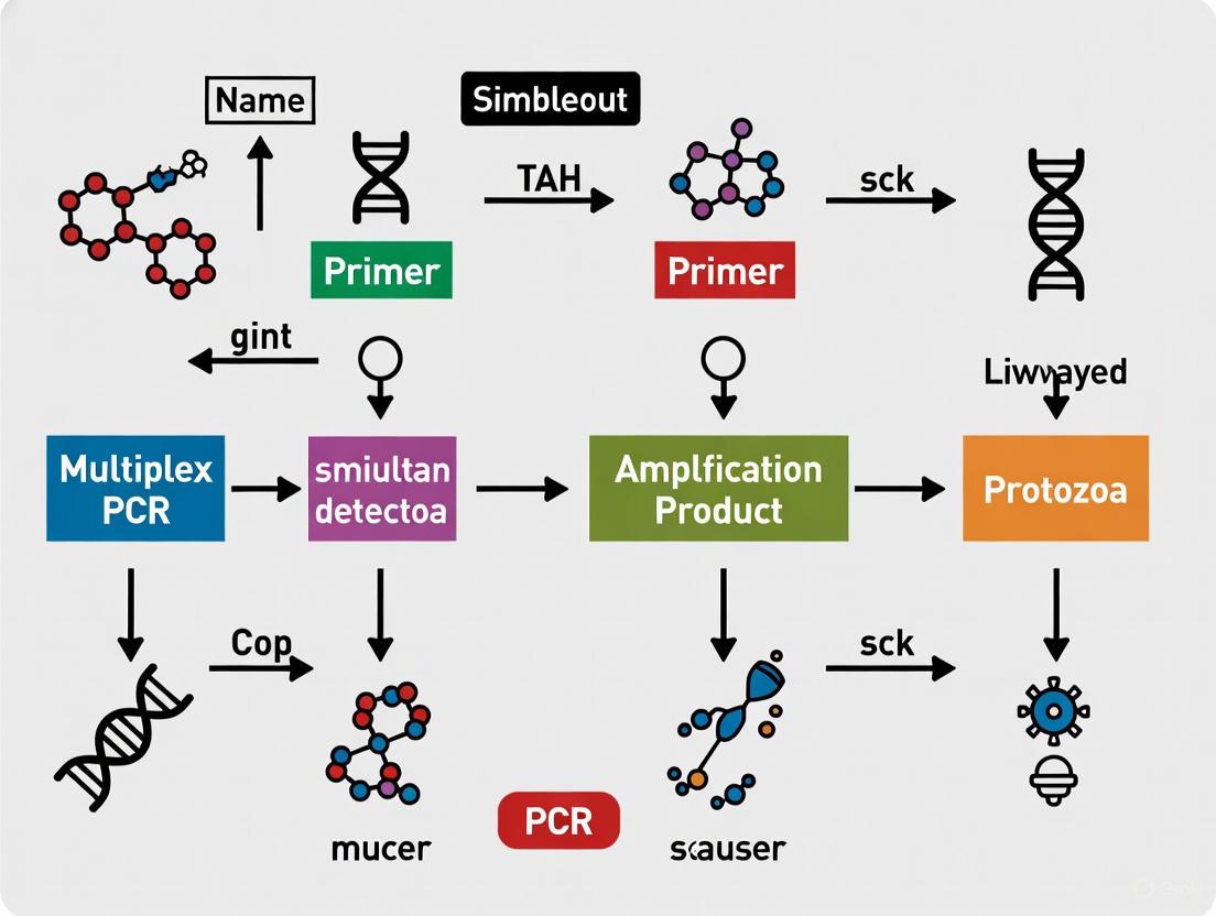
The following workflow diagram summarizes the key steps of this protocol:
Discussion and Implementation Considerations
The evidence demonstrates that multiplex qPCR is a superior diagnostic tool for the most clinically relevant enteric protozoa, including Giardia duodenalis, Cryptosporidium spp., Cyclospora cayetanensis, and Dientamoeba fragilis [4] [3]. The integration of this technology into clinical and public health laboratories can streamline workflows, reduce turnaround time, and ultimately improve patient care and disease surveillance.
However, successful implementation requires careful consideration of several factors:
- Assay Limitations: Not all parasites are covered by commercial panels. Microscopy remains necessary when infection with helminths or protozoa not included in the PCR panel (e.g., Cystoisospora belli) is suspected, particularly in high-risk groups like immunocompromised patients or migrants [1].
- DNA Extraction: The DNA extraction step is critical. The efficiency of lysing the thick walls of (oo)cysts significantly impacts sensitivity. Bead-beating or repeated freeze-thaw cycles during extraction are often necessary for optimal results [5] [6].
- Cost-Benefit Analysis: While the per-test cost of multiplex PCR may be higher than microscopy, the increased throughput, reduced labor, and improved diagnostic yield offer a compelling value proposition for medium- to high-volume laboratories.
In conclusion, multiplex qPCR for intestinal protozoa detection represents a significant advancement in diagnostic parasitology. Its high sensitivity, specificity, and ability to detect co-infections meet an urgent clinical and public health need, enabling more accurate diagnosis, timely treatment, and effective surveillance of these important pathogens.
Molecular diagnostics have revolutionized the detection of gastrointestinal protozoa, with multiplex PCR emerging as a powerful tool for simultaneous identification of multiple pathogens. This Application Note details the development and implementation of a multiplex real-time PCR protocol for the detection of five major enteric protozoan parasites: Giardia lamblia, Cryptosporidium spp., Cyclospora cayetanensis, Entamoeba histolytica, and Dientamoeba fragilis. These pathogens collectively represent significant causes of waterborne and foodborne diarrheal disease worldwide, affecting both immunocompetent and immunocompromised populations [7] [8].
Traditional diagnostic methods based on microscopic examination present numerous challenges, including requirement for high technical expertise, multiple staining procedures, prolonged turnaround times, and limited sensitivity and specificity [9] [4]. Molecular methods provide higher throughput and potentially superior performance characteristics, with multiplex PCR offering the distinct advantage of detecting co-infections that commonly occur in clinical settings [7] [10]. This protocol is framed within broader thesis research on optimizing molecular diagnostics for simultaneous protozoa detection, with particular emphasis on workflow efficiency and diagnostic accuracy.
Assay Principle and Design
The multiplex PCR assay employs a one-step real-time PCR approach capable of detecting and differentiating all five target parasites in a single reaction tube. This is achieved through sophisticated primer design and detection chemistry that allows for individual identification of multiple analytes within single fluorescent channels [10].
The assay incorporates an Internal Control (IC) to monitor for potential PCR inhibition and ensure extraction efficiency, utilizing Uracil-DNA Glycosylase (UDG) treatment to prevent carry-over contamination from previous amplification products [8] [4] [10]. The fundamental innovation enabling this multiplex approach is the use of modified oligonucleotide technology that permits multiple cycle threshold (Ct) values to be reported in individual channels, thereby expanding the multiplexing capacity without requiring additional fluorescent detectors [10].
Target Selection and Clinical Relevance
The five protozoan targets were selected based on their clinical significance and prevalence in gastrointestinal infections:
- Giardia lamblia: A common flagellate parasite with worldwide distribution, considered a main non-viral cause of diarrhea in industrialized countries [7]
- Cryptosporidium spp.: Notorious for waterborne outbreaks; causes self-limiting diarrhea in immunocompetent hosts but can be life-threatening in immunocompromised patients [7]
- Cyclospora cayetanensis: Associated with foodborne outbreaks, particularly from fresh produce; causes prolonged watery diarrhea [4]
- Entamoeba histolytica: A potentially invasive pathogen and causative agent of amebiasis, requiring differentiation from non-pathogenic Entamoeba species [7]
- Dientamoeba fragilis: An increasingly recognized cause of persistent gastrointestinal symptoms, including diarrhea and abdominal pain [7]
Performance Characteristics
Diagnostic Accuracy
Recent validation studies demonstrate excellent performance characteristics for multiplex PCR assays targeting gastrointestinal parasites. The following table summarizes the sensitivity and specificity data from clinical evaluations:
Table 1: Diagnostic Performance of Multiplex PCR for Enteric Protozoa
| Analyte | Sensitivity (%) | Specificity (%) | PPV (%) | NPV (%) | Reference |
|---|---|---|---|---|---|
| Blastocystis hominis | 93.0 | 98.3 | 85.1 | 99.3 | [4] |
| Cryptosporidium spp. | 100 | 100 | 100 | 100 | [4] |
| Cyclospora cayetanensis | 100 | 100 | 100 | 100 | [4] |
| Dientamoeba fragilis | 100 | 99.3 | 88.5 | 100 | [4] |
| Entamoeba histolytica | 33.3-75* | 100 | 100 | 99.6 | [4] |
| Giardia lamblia | 100 | 98.9 | 68.8 | 100 | [4] |
Sensitivity for *E. histolytica increased to 75% with inclusion of frozen specimens
When compared to conventional diagnostic methods, multiplex PCR demonstrates significant advantages. One evaluation of 472 fecal specimens found that microscopy exhibited markedly lower sensitivities: 56% for Cryptosporidium spp., 38% for D. fragilis, 47% for E. histolytica, and 50% for G. intestinalis [7]. Another study on Lophomonas spp. detection reported 100% sensitivity for multiplex PCR versus 86.2% for Giemsa staining [9].
Analytical Sensitivity
The limit of detection (LOD) varies by target organism but typically ranges from 10-100 organisms per reaction for well-optimized multiplex PCR assays. Studies have demonstrated that the combination of pretreatment, extraction, and amplification methods significantly impacts the LOD [11]. For Cryptosporidium parvum, optimal detection was achieved with mechanical pretreatment, automated extraction systems, and multiplex amplification chemistry [11].
Table 2: Process Comparison Between Traditional and Multiplex PCR Methods
| Parameter | Conventional Microscopy | Multiplex PCR |
|---|---|---|
| Technical Expertise | High requirement for skilled microscopists | Operator-independent, standardized output |
| Throughput | Low (manual process) | High (automation compatible) |
| Turnaround Time | Prolonged (multiple staining procedures) | Reduced by approximately 7 hours per batch |
| Multiplexing Capacity | Limited, requires separate examinations | Simultaneous detection of 6+ targets |
| Objectivity | Subjective interpretation | Objective Ct value measurement |
| Co-infection Detection | Challenging, may be missed | Straightforward identification |
Materials and Reagents
Research Reagent Solutions
The following table details essential materials and their functions for implementing the multiplex PCR protocol:
Table 3: Essential Research Reagents for Multiplex PCR Detection of Enteric Protozoa
| Reagent/Material | Function | Specifications |
|---|---|---|
| Allplex GI-Parasite Assay | Multiplex PCR mastermix | Contains primer sets for 6 parasites + IC [4] [10] |
| STARMag 96 × 4 Universal Cartridge | Automated nucleic acid extraction | Magnetic bead-based DNA purification [4] |
| Cary–Blair transport media | Specimen preservation | Maintains nucleic acid integrity during transport [4] |
| UDG (Uracil-DNA Glycosylase) | Contamination prevention | Digests PCR products from previous reactions [8] [10] |
| FecalSwab tubes | Sample collection and storage | Contains 2mL Cary–Blair media [4] |
| Real-time PCR plates/tubes | Reaction vessel | Compatible with thermal cycler |
| Positive control templates | Assay validation | Verified target sequences for all analytes |
| Nuclease-free water | Reaction preparation | Free of RNases and DNases |
Equipment
- Automated nucleic acid extraction system (e.g., Hamilton STARlet)
- Real-time PCR thermal cycler with multiple detection channels (e.g., Bio-Rad CFX96)
- Centrifuge capable of microcentrifuge tubes and plates
- Vortex mixer
- Precision pipettes and sterile tips
- Biological safety cabinet
- Freezer (-20°C ± 5°C) for reagent storage
Detailed Experimental Protocol
Sample Collection and Pre-analytical Processing
Specimen Requirements:
- Collect fresh stool specimens in sterile, leak-proof containers
- Alternatively, use fecal swabs placed in Cary–Blair transport media
- Process specimens within 24-72 hours of collection if refrigerated (4°C)
- For longer storage, freeze at -70°C to -80°C [8] [4]
Sample Preparation:
- For liquid stools: transfer 200-500μL to a cryovial
- For formed stools: take a portion approximately the size of a pea (0.5-1g)
- For swab specimens: ensure adequate saturation of the swab in transport media
- Homogenize samples by vortexing for 10-15 seconds [4]
Nucleic Acid Extraction
Automated Extraction Protocol (Hamilton STARlet):
- Transfer 50μL of stool suspension to the extraction plate
- Use STARMag 96 × 4 Universal Cartridge kit according to manufacturer's instructions
- Elute nucleic acids in 100μL of elution buffer
- Store extracted DNA at -20°C if not used immediately [4]
Quality Assessment:
- Monitor Internal Control (IC) amplification in each sample
- Acceptable IC Ct values should be consistent across samples
- Investigate samples with significantly delayed IC Ct (potential inhibition) [4]
Multiplex PCR Setup
Reaction Preparation:
- Thaw all reagents completely and mix by gentle vortexing
- Prepare master mix according to the following proportions:
Table 4: PCR Reaction Setup Components
| Component | Volume per Reaction | Final Concentration |
|---|---|---|
| 5X GI-P MOM Primer Mix | 5.0μL | 1X |
| EM2 (Enzyme Mix) | 5.0μL | Contains DNA polymerase, UDG, dNTPs |
| RNase-free Water | 10.0μL | - |
| Template DNA | 5.0μL | - |
| Total Volume | 25.0μL | - |
- Aliquot 20μL of master mix into each PCR tube/well
- Add 5μL of extracted template DNA to respective reactions
- Seal plates properly and centrifuge briefly to collect contents [4]
Thermal Cycling Conditions
Real-time PCR Parameters:
- UDG treatment: 50°C for 2 minutes (optional, if not included in mastermix)
- Initial denaturation: 95°C for 10 minutes
- Amplification (45 cycles):
- Denaturation: 95°C for 10 seconds
- Annealing/Extension: 60°C for 1 minute
- Acquisition of fluorescence signals at 60°C step [4]
Data Collection:
- Configure filters according to the assay specifications (FAM, HEX, Cal Red 610, Quasar 670)
- Set threshold automatically or manually based on exponential phase of amplification
- Record Ct values for each target [10]
Workflow Visualization
Figure 1: Comprehensive workflow for multiplex PCR detection of enteric protozoa, illustrating the integrated process from specimen collection to result reporting with quality control checkpoints.
Quality Control and Validation
Controls Requirements
Each PCR run should include:
- Positive controls: For each target organism (if available)
- Negative control: Nuclease-free water instead of template
- Internal control: Included in each patient sample to monitor inhibition
- Extraction controls: Process alongside patient samples [4]
Interpretation Criteria
Positive Result:
- Ct value ≤43 for any target analyte
- Exponential amplification curve with characteristic sigmoidal shape
- Appropriate IC amplification (Ct within acceptable range)
Negative Result:
- No amplification curve for target analytes (Ct >43 or undetermined)
- Valid IC amplification (Ct within acceptable range)
Invalid Result:
- Inhibition suspected if IC Ct is significantly delayed or absent
- Requires specimen re-extraction or dilution to overcome inhibition [4]
Technical Notes and Troubleshooting
Optimization Considerations
Successful multiplex PCR requires careful optimization of several parameters:
Primer Design and Validation:
- Ensure primers have similar annealing temperatures (Tm differences ≤2°C)
- Avoid primer-dimer formation and secondary structures
- Verify specificity against sequence databases [12] [13]
Reaction Component Optimization:
- Titrate primer concentrations (typically 50-500nM each)
- Optimize MgCl₂ concentration (1.5-3.0mM)
- Consider PCR additives (DMSO, glycerol, BSA) for difficult templates [12] [13]
Thermal Cycling Optimization:
- Optimize annealing temperature using gradient PCR
- Adjust ramp rates for improved specificity
- Determine optimal cycle number to balance sensitivity and background [13]
Common Issues and Solutions
Table 5: Troubleshooting Guide for Multiplex PCR Assay
| Problem | Potential Causes | Solutions |
|---|---|---|
| Poor Sensitivity | Suboptimal primer concentration | Titrate primer pairs individually and in combination |
| Inhibitors in sample | Dilute template 1:10 or use inhibitor removal steps | |
| False Positives | Contamination | Implement UDG system, separate pre- and post-PCR areas |
| Primer dimers | Redesign primers with stronger 3' end constraints | |
| Inconsistent Replicates | Pipetting errors | Calibrate pipettes, use master mix aliquoting |
| Template quality | Standardize extraction protocol, check degradation | |
| High Background | Excessive primer concentration | Reduce primer concentration, increase annealing temperature |
| Non-specific amplification | Optimize Mg²⁺ concentration, use hot-start polymerase |
Applications in Research and Drug Development
The multiplex PCR protocol described has significant applications beyond clinical diagnostics, particularly in pharmaceutical research and therapeutic development. As outlined in [14], two primary strategies guide antiprotozoal drug development: whole-organism screening and target-based drug design.
Whole-Organism Screening:
- Multiplex PCR enables rapid assessment of compound efficacy against multiple parasites simultaneously
- Facilitates high-throughput screening of chemical libraries
- Allows evaluation of drug candidates against clinically relevant co-infections [14]
Target-Based Drug Design:
- Provides tool for validating novel drug targets identified through genomic approaches
- Enables monitoring of parasite load during in vivo efficacy studies
- Supports pharmacodynamic assessment in preclinical models [14]
Drug Repurposing:
- Multiplex PCR offers efficient platform for screening existing drugs against non-target parasites
- Particularly valuable for neglected parasitic diseases where market returns are limited [14]
The protocol's ability to detect and quantify multiple parasites simultaneously makes it particularly valuable for assessing drug efficacy across different parasite species and stages, ultimately accelerating the development of novel therapeutic interventions for parasitic gastrointestinal infections.
This Application Note provides a comprehensive framework for implementing multiplex PCR detection of five major gastrointestinal protozoa. The protocol demonstrates significant advantages over traditional microscopic methods, including improved sensitivity and specificity, higher throughput, reduced turnaround time, and objective result interpretation. The incorporation of automated extraction and analysis systems further enhances reproducibility and efficiency, making this approach particularly suitable for both clinical diagnostics and research applications.
When properly optimized and validated, this multiplex PCR protocol serves as a powerful tool for gastrointestinal pathogen detection, outbreak investigation, and antiparasitic drug development. The methodology continues to evolve with technological advancements, promising even greater multiplexing capacity and integration with total laboratory automation systems in the future.
Multiplex PCR represents a significant evolution in molecular diagnostics, enabling the simultaneous amplification and detection of multiple nucleic acid targets in a single reaction [15]. This technical advancement offers substantial improvements over traditional, single-analyte methods, particularly for the detection of enteric protozoa where conventional microscopy has long been the standard [1] [4]. This application note details the specific advantages of multiplex PCR in throughput, sensitivity, and objectivity, providing validated experimental protocols and data from recent clinical studies. The content is framed within ongoing research for developing a multiplex PCR protocol for simultaneous protozoa detection, offering actionable insights for researchers, scientists, and drug development professionals seeking to implement this technology.
Performance Comparison: Multiplex PCR vs. Traditional Methods
Quantitative Advantages in Diagnostic Parameters
The transition from conventional microscopy to molecular methods like multiplex PCR brings transformative gains in key performance metrics as demonstrated in recent, large-scale prospective studies.
Table 1: Performance Comparison of Microscopy vs. Multiplex PCR for Protozoa Detection
| Parameter | Traditional Microscopy | Multiplex PCR | Study Details |
|---|---|---|---|
| Detection Rate | 6.55% (Blastocystis spp.) [1] | 19.25% (Blastocystis spp.) [1] | 3,495 stool samples over 3 years [1] |
| Giardia Detection | 0.7% (25/3,495 samples) [1] | 1.28% (45/3,495 samples) [1] | Prospective study [1] |
| Dientamoeba fragilis Detection | 0.63% (22/3,495 samples) [1] | 8.86% (310/3,495 samples) [1] | Routine clinical samples [1] |
| Time to Results | 48-72 hours [16] | 1-4 hours [16] [4] | Various clinical validations |
| Analytical Specificity | Variable, subjective [4] | 97.72%-100% [17] | Compared to sequencing [17] |
| Sample Throughput | Low, labor-intensive [4] | High, automated [4] | Automated DNA extraction and PCR setup [4] |
Throughput and Workflow Efficiency
Multiplex PCR dramatically increases laboratory throughput and operational efficiency. A key validation study demonstrated that implementing an automated multiplex PCR platform reduced pre-analytical and analytical testing turnaround time by 7 hours per batch compared to conventional methods [4]. This acceleration stems from several factors:
- Simultaneous Multi-Pathogen Detection: A single reaction detects 6-20+ pathogens, replacing multiple individual tests [18] [19].
- Workflow Consolidation: Testing for broad pathogen panels from single samples eliminates separate procedures like staining, concentration, and antigen testing [19].
- Automation Compatibility: Platforms like the Hamilton STARlet liquid handler enable automated nucleic acid extraction and PCR setup, reducing hands-on technologist time and manipulation errors [4].
For clinical laboratories, this throughput enhancement allows consolidation of testing for a broad range of pathogens from the same sample, potentially eliminating resource-intensive procedures like complete ova and parasite (O&P) exams as first-line tests [19].
Enhanced Sensitivity and Diagnostic Yield
The superior sensitivity of multiplex PCR directly translates to improved detection of clinically significant pathogens, as evidenced by a 3-year prospective study on 3,495 stool samples. The study found multiplex PCR detected approximately three times more positive samples for key protozoa compared to microscopy (909 vs. 286 samples) [1].
Table 2: Analytical Sensitivity of Multiplex PCR for Enteric Protozoa
| Organism | Sensitivity (%) | Specificity (%) | Positive Predictive Value (%) | Negative Predictive Value (%) | Reference |
|---|---|---|---|---|---|
| Blastocystis hominis | 93.0 | 98.3 | 85.1 | 99.3 | [4] |
| Cryptosporidium spp. | 100 | 100 | 100 | 100 | [4] |
| Cyclospora cayetanensis | 100 | 100 | 100 | 100 | [4] |
| Dientamoeba fragilis | 100 | 99.3 | 88.5 | 100 | [4] |
| Entamoeba histolytica | 33.3-75.0* | 100 | 100 | 99.6 | [4] |
| Giardia lamblia | 100 | 98.9 | 68.8 | 100 | [4] |
Note: Sensitivity for E. histolytica increased to 75% with inclusion of frozen specimens in the validation [4].
This enhanced sensitivity is particularly crucial for detecting low-abundance pathogens and in patients who have previously received antibiotics, where traditional culture methods often fail [16]. The increased diagnostic yield enables more accurate epidemiological data and better understanding of true infection prevalence.
Improved Objectivity and Standardization
Unlike microscopy which requires significant technical expertise and is prone to interpretive subjectivity, multiplex PCR provides operator-independent, objective results with standardized outputs [4]. Key aspects include:
- Reduced Technical Variability: Automated platforms generate consistent results regardless of operator skill level, eliminating the "high technical expertise burden" of microscopy [4].
- Standardized Interpretation: Results are based on cycle threshold (Ct) values and fluorescence signals, providing binary positive/negative outcomes against established cutoffs (e.g., Ct ≤43) [4].
- Minimized Cross-Reactivity: Carefully designed primers and validation processes ensure high specificity, with one study showing 100% specificity for multiple targets compared to reference standards [4].
- Integrated Controls: Multiplex assays enable inclusion of internal positive controls and sample processing controls, ensuring reaction validity and correct interpretation [18].
This objectivity is further enhanced by standardized commercial assays that reduce inter-laboratory variability compared to laboratory-developed tests [19].
Experimental Protocols for Validation
Protocol: Multicenter Validation of Multiplex PCR for Respiratory Pathogens
This protocol is adapted from a study comparing novel multiplex fluorescence PCR to conventional culture methods for six common lower respiratory tract pathogens [17].
Materials:
- Sputum samples (n=2047 collected across multiple centers)
- Nucleic acid extraction kit (magnetic bead method)
- Multiple respiratory pathogen nucleic acid diagnostic kit
- PCR amplification instrument
- VITEK MS automated identification system for culture reference
Methods:
- Sample Preparation: Add sterile normal saline to sputum samples, mix thoroughly, and incubate until completely liquefied. For difficult-to-liquefy samples, add 4% sodium hydroxide solution followed by centrifugation and resuspension.
- Nucleic Acid Extraction: Extract nucleic acids using magnetic bead method according to manufacturer's instructions, eluting in 50 μL eluate.
- PCR Reaction Setup: Prepare 50 μL total reaction volume containing:
- 5 μL DNA template
- 44 μL multiplex PCR mix (containing combined primers and probes)
- 1 μL enzyme mix
- Amplification Parameters:
- UDGase reaction: 50°C for 2 min (1 cycle)
- Pre-denaturation: 94°C for 3 min (1 cycle)
- Amplification: 45 cycles of:
- Denaturation: 94°C for 10s
- Annealing: 60°C for 20s
- Extension: 72°C for 20s
- Detection: Monitor fluorescence in FAM, HEX, ROX, and CY5 channels during amplification.
- Analysis: Interpret results using instrument software with Ct <37 and characteristic melting curves indicating positive results.
Validation: Compare results to conventional bacterial culture and sequencing methods. The referenced study demonstrated 100% sensitivity and 72.22% specificity compared to culture methods, with 98.84% overall agreement with sequencing [17].
Protocol: Automated High-Throughput Detection of Enteric Protozoa
This protocol details the validation of an automated system for detecting six gastrointestinal protozoal pathogens from unpreserved fecal specimens [4].
Research Reagent Solutions:
| Reagent/Equipment | Function | Specifications |
|---|---|---|
| Hamilton STARlet | Automated liquid handling | Nucleic acid extraction and PCR setup |
| STARMag 96 × 4 Cartridge | Nucleic acid extraction | Bead-based DNA extraction |
| Allplex GI-Parasite Assay | Multiplex PCR detection | Detects 6 protozoal pathogens |
| FecalSwab Tubes | Sample transport/preservation | Contains Cary-Blair media |
| Bio-Rad CFX96 | Real-time PCR detection | Four-color fluorescence detection |
Methods:
- Sample Preparation:
- Inoculate one swab of unpreserved stool into FecalSwab tube containing 2 mL Cary-Blair media.
- Vortex for 10 seconds to homogenize.
Automated DNA Extraction:
- Load samples into Hamilton STARlet platform.
- Extract DNA using STARMag Universal Cartridge kit.
- Use 50 μL stool suspension for extraction, eluting to 100 μL final volume.
PCR Setup:
- Combine in PCR tubes:
- 5 μL 5X GI-P MOM primer
- 10 μL RNase-free water
- 5 μL EM2 (contains DNA polymerase, UDG, buffer, dNTPs)
- Add 5 μL extracted DNA to 20 μL master mix (25 μL total reaction volume).
- Combine in PCR tubes:
Real-Time PCR:
- Run on Bio-Rad CFX96 with following parameters:
- Denaturing step
- 45 cycles of:
- 95°C for 10s
- 60°C for 1min
- 72°C for 30s
- Monitor four fluorophores: FAM, HEX, Cal Red 610, Quasar 670.
- Run on Bio-Rad CFX96 with following parameters:
Result Interpretation:
- Positive: Ct value ≤43 in appropriate channel.
- Include internal controls for process validation.
This protocol demonstrated significantly reduced hands-on time and a 7-hour reduction in total processing time per batch compared to conventional microscopy [4].
Technological Workflows
The transition from traditional methods to multiplex PCR involves significant workflow restructuring. The following diagram illustrates the key procedural differences and efficiency gains in high-throughput laboratory settings.
Diagram 1: Comparative workflow analysis showing efficiency gains with multiplex PCR implementation. Traditional methods require multiple manual procedures and subjective interpretation, while multiplex PCR utilizes automation and objective analysis, significantly reducing turnaround time.
The detection mechanism of multiplex fluorescence PCR utilizes target-specific probes with distinct fluorophores, enabling simultaneous detection of multiple pathogens in a single reaction. The following diagram illustrates this molecular process.
Diagram 2: Molecular detection mechanism of multiplex fluorescence PCR. The system utilizes multiple target-specific primers and differentially labeled fluorescent probes to simultaneously detect and distinguish multiple pathogens in a single closed-tube reaction, providing objective results through Ct value analysis.
Multiplex PCR technology demonstrates unequivocal advantages over traditional methods in throughput, sensitivity, and objectivity for protozoa detection. The quantitative data from recent studies confirms significantly higher detection rates – up to threefold increases for some protozoa compared to conventional microscopy [1]. The dramatic reduction in processing time, from days to hours, enables clinically actionable results that improve patient management and antimicrobial stewardship [16] [19]. Furthermore, the automation and standardization of multiplex platforms reduce technical variability and expertise dependency, providing consistently objective results essential for both clinical diagnostics and research applications [4].
For researchers developing multiplex PCR protocols for simultaneous protozoa detection, these findings validate the technical superiority of this approach. The enhanced sensitivity ensures more accurate prevalence data, while the high throughput enables larger-scale studies with more efficient resource utilization. Future developments will likely focus on expanding pathogen panels, reducing costs, and further simplifying workflows to make this technology accessible in diverse laboratory settings.
Key Technical Hurdles and Design Considerations in Multiplexing
Multiplex polymerase chain reaction (PCR) enables the simultaneous amplification of multiple nucleic acid targets in a single reaction, representing a powerful tool for clinical diagnostics and public health surveillance. Within gastrointestinal diagnostics, syndromic multiplex PCR panels have revolutionized the detection of enteric pathogens, allowing laboratories to rapidly identify bacteria, viruses, and parasites from a single stool sample [20]. These panels provide significant advantages over conventional diagnostic methods, including superior analytical sensitivity, reduced turnaround time, and comprehensive pathogen coverage [20] [21]. However, the development and implementation of robust multiplex PCR assays, particularly for the detection of intestinal protozoa, present substantial technical challenges that require careful consideration of primer design, reaction optimization, and result interpretation.
This application note details the key technical hurdles and design considerations for developing multiplex PCR protocols for simultaneous protozoa detection, providing validated experimental frameworks and analytical tools to support researchers and assay developers in creating reliable, high-performance diagnostic systems.
Technical Hurdles in Multiplex PCR Development
Primer Dimer Formation and Primer-Primer Interactions
The primary challenge in highly multiplexed PCR is the exponential increase in potential primer dimer interactions. For an N-plex PCR primer set comprising 2N primers, the number of potential primer dimer interactions grows quadratically, with (\left(\begin{array}{l}2N\ 2\end{array}\right)) possible simple primer dimer interactions [22]. For example, a 50-plex assay with 100 primers must contend with 4,950 potential primer dimer combinations, compared to just 1 in a single-plex reaction [22].
These nonspecific interactions compete for reaction resources and can significantly reduce amplification efficiency of target sequences. In a naively designed 96-plex primer set (192 primers), primer dimers constituted up to 90.7% of reaction products, dramatically impacting assay sensitivity and specificity [22]. This challenge is exacerbated when targeting protozoal DNA from stool samples, which often contains potent PCR inhibitors that further reduce amplification efficiency [21].
PCR Amplification Bias and Artifacts
Non-uniform amplification efficiency across different targets presents another significant hurdle in multiplex PCR development. This amplification bias stems from variations in primer binding efficiency, amplicon length, and sequence characteristics, resulting in differential representation of targets in the final amplification products [23]. This bias significantly affects quantification accuracy, as final sequence read counts may not accurately represent the relative abundance of original DNA templates [23].
Additionally, polymerase errors during PCR cycles generate sequence artifacts not present in the original sample. These artifacts manifest as false sequence variants present at low fractions in final sequence reads, complicating the detection of genuine low-abundance targets [23]. This challenge is particularly relevant for detecting mixed infections or low parasite burdens in clinical samples.
Analytical Sensitivity and Specificity Requirements
Multiplex assays for protozoan detection must maintain high sensitivity and specificity while simultaneously detecting multiple targets. Closely related species, such as the pathogenic Entamoeba histolytica and non-pathogenic E. dispar, present particular challenges as they require differentiation at the genetic level, which is impossible with conventional microscopy [21].
Table 1: Performance Characteristics of Multiplex PCR Assays for Enteric Protozoa
| Organism | Sensitivity (%) | Specificity (%) | PPV (%) | NPV (%) | Reference |
|---|---|---|---|---|---|
| Blastocystis hominis | 93.0 | 98.3 | 85.1 | 99.3 | [4] |
| Cryptosporidium spp. | 100 | 100 | 100 | 100 | [4] |
| Cyclospora cayetanensis | 100 | 100 | 100 | 100 | [4] |
| Dientamoeba fragilis | 100 | 99.3 | 88.5 | 100 | [4] |
| Entamoeba histolytica | 33.3-75.0* | 100 | 100 | 99.6 | [4] |
| Giardia duodenalis | 100 | 98.9 | 68.8 | 100 | [21] [4] |
Sensitivity for *E. histolytica increased to 75% with inclusion of frozen specimens [4].
Design Considerations and Computational Approaches
Advanced Primer Design Algorithms
Conventional primer design approaches become computationally intractable for highly multiplexed assays due to the exponentially many choices for multiplex primer sequence selection. Innovative computational approaches have been developed to address this challenge:
Simulated Annealing Design using Dimer Likelihood Estimation (SADDLE) is a stochastic algorithm that minimizes primer dimer formation in highly multiplexed primer sets [22]. The algorithm employs a six-step process: (1) generation of primer candidates for each target; (2) selection of an initial primer set; (3) evaluation of a loss function estimating primer dimer severity; (4) generation of a modified primer set; (5) probabilistic acceptance of the modified set based on improved loss function values; and (6) iteration until an optimal primer set is identified [22]. This approach reduced primer dimer formation from 90.7% to 4.9% in a 96-plex PCR primer set [22].
primerJinn provides another specialized tool for designing multiplex PCR primers for targeted sequencing, incorporating unique considerations for high-fidelity polymerases used in diagnostic applications [24]. The tool uses primer3 to design primers and a clustering method to select the best primer set based on amplicon size, melting temperature, and primer interactions [24].
Molecular Barcoding Strategies
Incorporating molecular barcodes (unique sequence identifiers) into PCR primers provides a powerful solution to mitigate PCR amplification bias and artifacts in high multiplex amplicon sequencing [23]. This approach enables:
- Accurate variant calling by distinguishing true low-frequency mutations from polymerase errors
- Improved quantification by counting unique molecular barcodes rather than total reads
- Reduction of false positives in low-frequency variant detection
The barcoding protocol involves: (1) annealing barcoded primers to target DNA; (2) removing unused primers; (3) limited PCR amplification with non-barcoded primers; (4) purification of amplicons; and (5) universal PCR to add sequencing adapters [23]. This approach has been successfully implemented in high multiplex PCR with hundreds of amplicons, enabling detection of mutations at frequencies as low as 1% with minimal false positives [23].
Figure 1: Molecular Barcoding Workflow for High Multiplex PCR. This protocol incorporates unique molecular identifiers during the initial amplification step to distinguish true biological variants from PCR artifacts and enable accurate quantification.
Multiplex PCR Optimization Parameters
Successful multiplex PCR requires careful optimization of several reaction parameters:
Primer Design Constraints should include:
- Tm optimization: Optimal ΔG° of approximately -11.5 kcal/mol for primer-template hybridization [22]
- Length distribution: Primers typically 10-40 nucleotides, with amplicons of 400-800 nucleotides for sequencing applications [24]
- GC content: Restricted to 25-75% to ensure uniform amplification efficiency [22]
- Specificity filters: Elimination of primers with >10 bases complementary at 3' ends to prevent cross-hybridization [23]
Reaction Condition Optimization must address:
- Primer concentration balancing: Varying concentrations from 0.25-1.5 μM for different targets to equalize amplification efficiency [25]
- Thermal cycling parameters: Adjusted annealing temperatures and extension times based on polymerase characteristics [24]
- Magnesium concentration: Critical for efficient amplification across multiple targets
- Polymerase selection: High-fidelity enzymes with proofreading capability for sequencing applications
Experimental Protocol: Validation of Multiplex PCR for Enteric Protozoa
Sample Preparation and DNA Extraction
Materials:
- Fresh or frozen unpreserved stool specimens
- FecalSwab tubes with Cary-Blair media (COPAN Diagnostics)
- STARMag 96 × 4 Universal Cartridge kit (Seegene Inc.) for nucleic acid extraction
- Hamilton STARlet automated liquid handling system
Procedure:
- Sample Collection: Collect one swab of stool and inoculate into FecalSwab tube containing 2 mL Cary-Blair media
- Homogenization: Vortex sample for 10 seconds to ensure uniform suspension
- Automated Extraction: Load samples onto Hamilton STARlet system
- Use 50 μL stool suspension for DNA extraction
- Elute in 100 μL elution buffer
- Transfer 5 μL extracted DNA to PCR reaction
Multiplex PCR Amplification
Reaction Setup:
- Master Mix Preparation:
- 5 μL 5X GI-P MOM primer mix
- 10 μL RNase-free water
- 5 μL EM2 (contains DNA polymerase, UDG, buffer, dNTPs)
- Total master mix volume: 20 μL per reaction
- PCR Assembly:
- Aliquot 20 μL master mix into PCR tubes
- Add 5 μL extracted DNA (total reaction volume: 25 μL)
- Include positive and negative controls in each run
Thermal Cycling Conditions:
- Denaturation: 95°C for 10 s
- Amplification: 45 cycles of:
- 95°C for 10 s (denaturation)
- 60°C for 1 min (annealing)
- 72°C for 30 s (extension)
- Detection: Collect fluorescence at 60°C annealing step
Result Interpretation:
- Positive result: Cycle threshold (Ct) value ≤43 [4]
- Analyze using manufacturer's software (e.g., Seegene Viewer)
Analytical Validation
Limit of Detection (LoD) Determination:
- Prepare serial dilutions of quantified parasite DNA
- Extract and amplify each dilution in six replicates
- LoD defined as the lowest concentration detected in 100% of replicates [25]
Specificity Testing:
- Cross-reactivity panel: Test against genetically similar species and commensal organisms
- Analytical specificity: Evaluate against human DNA and common stool flora
Table 2: Multiplex PCR Validation Parameters for Enteric Protozoa Detection
| Validation Parameter | Experimental Approach | Acceptance Criteria |
|---|---|---|
| Analytical Sensitivity | Probit analysis of serial dilutions | Detection of ≤10-100 targets/reaction |
| Analytical Specificity | Cross-reactivity panel | No amplification of non-target organisms |
| Precision | Intra-assay and inter-assay variability | CV <10% for Ct values |
| Reproducibility | Testing across multiple lots, operators, instruments | >95% concordance |
| Clinical Performance | Comparison to reference method (microscopy) | Sensitivity >90%, Specificity >95% |
Research Reagent Solutions
Table 3: Essential Research Reagents for Multiplex PCR Development
| Reagent Category | Specific Examples | Function | Considerations |
|---|---|---|---|
| Polymerases | Q5 Hot Start High-Fidelity (NEB), qScriptXLT 1-Step RT-qPCR ToughMix (Quantabio) | DNA amplification with high processivity and fidelity | Buffer composition significantly affects primer Tm [24] |
| Extraction Systems | STARMag 96 × 4 Universal Cartridge (Seegene), Hamilton STARlet platform | Automated nucleic acid purification from complex matrices | Critical for removing PCR inhibitors from stool samples [4] |
| Detection Chemistries | TaqMan probes (FAM, HEX, Cal Red 610, Quasar 670), Molecular beacons | Specific signal generation for multiple targets | Fluorophore selection must match instrument detection channels [25] |
| Commercial Panels | Allplex GI-Parasite Assay (Seegene), BioFire FilmArray GI Panel | Validated multi-target detection systems | Provide standardized protocols and performance characteristics [20] [21] |
| Automation Platforms | Hamilton STARlet, Bio-Rad CFX96, Microlab Nimbus IVD | High-throughput processing and reduced manual error | Essential for consistent results in clinical laboratory settings [4] |
Developing robust multiplex PCR assays for simultaneous protozoa detection requires addressing significant technical challenges through advanced computational design, strategic molecular barcoding, and rigorous validation. The SADDLE algorithm and molecular barcoding approaches provide powerful solutions to minimize primer dimers and amplification artifacts, enabling highly multiplexed detection with improved accuracy. Validation data demonstrates that optimized multiplex PCR assays can achieve sensitivity and specificity exceeding 90-95% for most clinically relevant enteric protozoa, with the exception of more challenging targets like Entamoeba histolytica which may require additional confirmation.
These protocols and considerations provide researchers with a framework for developing, optimizing, and validating multiplex PCR assays that meet the rigorous demands of clinical diagnostics and public health surveillance. As multiplexing technologies continue to evolve, incorporating innovations such as digital PCR and microfluidics will further enhance the sensitivity, throughput, and accessibility of these essential diagnostic tools.
Figure 2: Computational Primer Design Workflow Using SADDLE Algorithm. This iterative optimization process minimizes primer dimer formation through stochastic evaluation and modification of primer sets, significantly improving amplification efficiency in highly multiplexed reactions.
From Theory to Bench: Designing and Executing a Robust Multiplex PCR Protocol
Within the framework of developing a multiplex PCR protocol for the simultaneous detection of enteric protozoa, the design of primers and probes constitutes the most critical determinant of assay success. This protocol details a refined methodology for creating specific and compatible primer-probe sets, enabling accurate, high-throughput identification of protozoal pathogens such as Cryptosporidium spp., Cyclospora cayetanensis, and Giardia lamblia, among others. The principles outlined are derived from validated molecular assays described in contemporary literature [26] [4].
The principal challenge in multiplex assay development lies in ensuring that each primer-probe set exhibits unwavering specificity for its target while functioning harmoniously in a single reaction tube without cross-reactivity or amplification interference. This document provides a systematic approach to overcome these challenges, ensuring robust and reliable detection.
Experimental Design and Workflow
The following workflow outlines the comprehensive process, from initial bioinformatic analysis to the final validation of the multiplex assay.
Research Reagent Solutions
The following reagents are essential for executing the primer and probe design and validation workflow effectively.
Table 1: Essential Research Reagents for Multiplex PCR Development
| Reagent/Material | Function/Purpose | Exemplary Product/Note |
|---|---|---|
| DNA Polymerase | Catalyzes DNA synthesis; specific types are needed for long-range or probe-based assays. | LA Taq (for long-range AS-PCR [27]), Taq DNA Polymerase (standard qPCR [26]) |
| dNTPs | Building blocks for new DNA strands. | 200-400 µM each dNTP in final reaction [26] [27] |
| Primers & Probes | Species-specific oligonucleotides for target binding and detection. | Desalted primers; Hydrolysis Probes (e.g., FAM, HEX-labeled) for qPCR [28] [4] |
| PCR Buffer | Provides optimal chemical environment (pH, Mg²⁺) for amplification. | Often supplied with enzyme; MgCl₂ concentration (e.g., 2-2.5 mM) is critical [26] [27] |
| Template DNA | The target genetic material to be amplified. | Extracted from stool samples using kits like QIAquick Stool Mini Kit or automated systems [26] [4] |
| Thermal Cycler | Instrument that precisely controls PCR temperature cycles. | Bio-Rad CFX96 (for qPCR [4]), GeneAmp PCR System 9700 [27] |
Methodology for Primer and Probe Design
Target Sequence Selection and In Silico Design
- Sequence Alignment and Conserved Region Identification: Begin by compiling sequences of the target genes (e.g., 18S rRNA, COI) for all protozoa of interest from public databases like GenBank. Perform multiple sequence alignments to identify regions that are highly conserved within the target species but exhibit significant variation (≥5-10%) in non-target species, especially those co-circulating in the same sample type [28] [26].
- Primer and Probe Design Parameters: Design oligonucleotides with the following characteristics using specialized software (e.g., Primer-BLAST):
- Length: Primers: 18-25 bases; Probes: 15-20 bases.
- Melting Temperature (Tm): Aim for a Tm of 50-65°C. Probes should have a Tm 5-10°C higher than the primers to ensure they bind before the primers extend.
- GC Content: Maintain 40-60% to ensure stable binding.
- 3'-End Stability: Avoid GC-rich 3' ends to prevent mis-priming and enhance specificity.
- Compatibility Check: Ensure primers and probes for different targets do not form dimers (homo- or hetero-dimers) and have similar Tms to function under unified cycling conditions [28].
Experimental Validation and Optimization
Single-plex Validation:
- Test each primer-probe set individually against a panel of DNA samples. This panel must include the target protozoa, closely related non-target species, and other organisms commonly found in the sample matrix (e.g., other stool pathogens, human DNA) to rigorously test specificity [28] [4].
- Determine the optimal annealing temperature for each set using a thermal gradient PCR.
- Assess analytical sensitivity by performing a limit of detection (LOD) assay with serial dilutions of a known quantity of target DNA. The LOD for a robust assay can be as low as 10¹ to 10² targets per reaction [26].
Multiplex Assembly and Optimization:
- Combine all validated primer-probe sets into a single master mix. Use probes labeled with distinct fluorophores (e.g., FAM, HEX, Cal Red 610, Quasar 670) that are compatible with your real-time PCR instrument's detection channels [4].
- Optimize the concentration of each primer and probe in the multiplex mix to balance sensitivity and minimize competition. This often involves titrating concentrations and may require reducing them compared to single-plex reactions (e.g., from 1 µM to 0.7 µM) [28] [26].
- Validate the final multiplex assay with the same specificity panel and LOD dilutions to confirm performance has not been compromised.
Table 2: Key Performance Metrics from Validated Multiplex PCR Assays
| Assay Target | Reported Sensitivity | Reported Specificity | Key Experimental Notes |
|---|---|---|---|
| Fresh Stool (Giardia lamblia) [4] | 100% | 98.9% | Used automated DNA extraction; Ct cut-off of ≤43. |
| Fresh Stool (Cryptosporidium) [4] | 100% | 100% | Multiplex PCR demonstrated superior throughput vs. microscopy. |
| Waterborne Protozoa (Microsporidia, Cyclospora, Cryptosporidium) [26] | 10² - 10¹ spores/oocysts | No cross-reactivity reported | Used a nested multiplex PCR approach to achieve high sensitivity. |
| Freshwater Fish Species (COI gene target) [28] | 1 ng DNA | 100% accuracy in blinded tests | Emphasized primer-probe design criteria and a decoder algorithm for automation. |
Supplementary Techniques
For enhanced specificity, especially to distinguish between highly similar species, supplementary techniques can be employed post-amplification:
- Restriction Fragment Length Polymorphism (RFLP): Digest PCR products with species-specific restriction enzymes (e.g., BsaBI for E. intestinalis vs E. bieneusi, BsiEI for C. parvum vs C. hominis) and visualize the fragment patterns on an agarose gel [26].
- Melting Curve Analysis: If using hybridization probes (e.g., FRET probes), perform a melting curve analysis post-amplification. Different alleles or species will produce distinct melting temperatures (Tm), allowing for discrimination [27].
This protocol establishes that meticulous primer and probe design is the cornerstone of a specific and compatible multiplex PCR assay. The process, encompassing comprehensive in silico analysis followed by systematic empirical validation and optimization, ensures the development of a robust diagnostic tool. By adhering to these detailed methodologies, researchers can create highly accurate multiplex assays that significantly improve the detection and management of enteric protozoal infections.
The molecular diagnosis of intestinal protozoan parasites via polymerase chain reaction (PCR) is paramount for clinical diagnostics, epidemiological studies, and drug development. However, stool presents a uniquely complex and inhibitory matrix that severely challenges DNA extraction and subsequent amplification. PCR inhibitors ubiquitous in fecal samples, alongside the resilient structural walls of protozoan oocysts and cysts, frequently lead to false-negative results, compromising assay sensitivity and reliability [29] [30]. The efficacy of the DNA extraction method is therefore a critical determinant of success for downstream applications, including multiplex PCR protocols designed for the simultaneous detection of multiple enteric pathogens. This application note details optimized methodologies to overcome these challenges, ensuring the recovery of high-quality, amplifiable DNA.
The Challenge of Stool-Derived PCR Inhibitors
Stool samples contain a heterogeneous mixture of PCR inhibitors that can derail molecular detection. These include bilirubin, bile salts, complex carbohydrates, heme, and various metabolic by-products [30] [31]. Concurrently, protozoan parasites such as Cryptosporidium spp., Giardia duodenalis, and Entamoeba histolytica possess robust oocyst or cyst walls that are difficult to lyse, protecting the genetic material within but complicating DNA extraction [30].
The mechanisms of PCR inhibition are diverse. Inhibitors can:
- Degrade or denature DNA polymerases (e.g., via proteases or ionic detergents) [31].
- Chelate co-factors required for enzymatic activity, such as magnesium ions [32] [31].
- Bind directly to nucleic acids, preventing primer annealing and elongation [31]. The co-extraction of these substances with target DNA can lead to partial or complete amplification failure, underscoring the necessity for extraction protocols that efficiently remove these compounds while effectively disrupting the hardy walls of parasitic stages [29] [33].
Comparative Evaluation of DNA Extraction Methods
Selecting an appropriate DNA extraction method is crucial for balancing DNA yield, purity, and the effective removal of inhibitors. The table below summarizes the performance of various methods as evaluated in comparative studies.
Table 1: Comparison of DNA Extraction Methods for Stool Samples
| Extraction Method | Reported DNA Yield | PCR Detection Rate | Key Advantages | Key Limitations |
|---|---|---|---|---|
| Phenol-Chloroform (P) | High (~4x higher than some kits) [29] | Very Low (8.2%) [29] | High DNA yield; cost-effective for some applications [29] | Ineffective inhibitor removal; low sensitivity; uses hazardous organic solvents [29] |
| Phenol-Chloroform with Bead-Beating (PB) | High [29] | Not specified, but higher than P [29] | Improved lysis of hardy parasites due to mechanical disruption [29] | Less effective at inhibitor removal compared to modern silica-column kits [29] |
| QIAamp Fast DNA Stool Mini Kit (Q) | Moderate [29] | Lower than QB [29] | Designed for stool; includes inhibitor removal technology [30] | May be less effective for certain hardy oocysts (e.g., Cryptosporidium) without optimization [30] |
| QIAamp PowerFecal Pro DNA Kit (QB) | Moderate [29] | High (61.2%) [29] | Highest reported detection rate; effective lysis and inhibitor removal; suitable for a broad range of parasites [29] | Higher cost per sample compared to in-house methods |
| Repeat Silica Extraction | Good (post-purification) [32] | Restored amplification in inhibited samples [32] | Simple, effective technique for removing persistent inhibitors from DNA extracts [32] | Additional step post-initial extraction; potential for DNA loss |
Recommended Protocols
Based on comparative studies, methods that integrate mechanical, chemical, and thermal lysis with robust inhibitor removal offer the best performance for multiplex PCR applications.
Optimized Protocol using the QIAamp PowerFecal Pro DNA Kit
The QIAamp PowerFecal Pro DNA Kit (QB) has been demonstrated to provide the highest PCR detection rates for a diverse range of intestinal parasites, including fragile protozoa like Blastocystis sp. and hardy helminths like Ascaris lumbricoides [29].
Workflow Overview:
Detailed Procedure:
- Sample Preparation: Aliquot 180-220 mg of stool specimen into a PowerBead Pro tube. For preserved samples, wash with sterile distilled water to remove preservative agents before extraction [29].
- Lysis: Add the provided lysis solution (CD1). Incubate the mixture at 65°C for 10 minutes to initiate thermal lysis [29].
- Mechanical Disruption: Securely vortex the tube at maximum speed for 10 minutes. This bead-beating step is critical for the physical disruption of tough oocysts and cysts [29].
- Inhibitor Removal: Centrifuge the lysate at high speed (e.g., 13,000-15,000 × g) for 1 minute. This pellets stool particles and a significant proportion of PCR inhibitors.
- DNA Binding: Transfer the clarified supernatant to a new microcentrifuge tube. Add binding solution and ethanol, then load the mixture onto a silica-membrane column.
- Wash: Perform two wash steps using the provided buffers to remove residual salts and inhibitors.
- Elution: Elute the purified genomic DNA in 50-100 µL of elution buffer. Using a smaller elution volume can increase the final DNA concentration [30].
Protocol Optimization for Specific Protozoa
For optimal recovery of DNA from particularly resilient protozoa like Cryptosporidium, further optimization of the lysis conditions is recommended.
Table 2: Optimization Steps for Enhanced DNA Recovery
| Parameter | Standard Protocol | Optimized Recommendation | Effect |
|---|---|---|---|
| Lysis Temperature | 65-70°C [30] | 95-100°C for 10 min [29] [30] | Significantly improves lysis efficiency of hardy oocysts [30]. |
| Lysis Duration | 5-10 min | Up to 3 hours at 65°C (for in-house methods) [29] | Allows for more complete digestion and release of DNA. |
| Inhibitor Removal | Incubation with InhibitEX tablet | Extend incubation time to 5 minutes [30] | Ensures more thorough binding and precipitation of inhibitors. |
| Elution Volume | 100-200 µL | 50-100 µL [30] | Increases final DNA concentration for improved PCR sensitivity. |
Post-Extraction Purification: Repeat Silica Extraction
For samples that remain inhibited after standard extraction, a "repeat silica extraction" is a simple and effective post-purification technique [32].
Procedure:
- Combine the extracted DNA with 5 volumes of a guanidine hydrochloride-based binding buffer (e.g., from a silica-based kit).
- Add 2 volumes of absolute ethanol and mix thoroughly.
- Pass the mixture through a fresh silica column.
- Wash the column according to the manufacturer's instructions.
- Elute the DNA in a small volume of Tris-EDTA (TE) buffer or nuclease-free water. This process effectively re-binds the DNA to the silica matrix, leaving many persistent inhibitors in the flow-through [32].
The Scientist's Toolkit: Essential Reagents and Additives
The following table lists key reagents and additives that are critical for successful DNA extraction and PCR amplification from stool samples.
Table 3: Research Reagent Solutions for Overcoming PCR Inhibition
| Reagent/Kit | Function | Application Note |
|---|---|---|
| QIAamp PowerFecal Pro DNA Kit | Integrated DNA extraction and purification using bead-beating and silica-membrane technology. | Recommended for its high PCR detection rate across a broad spectrum of intestinal parasites [29]. |
| Bovine Serum Albumin (BSA) | PCR facilitator that binds to and neutralizes a wide range of inhibitors. | Add at 0.1-0.5 µg/µL to PCR reactions to counteract inhibition from humic acids, heme, and tannins [32] [31]. |
| Proteinase K | Broad-spectrum serine protease that digests proteins and degrades nucleases. | Critical for enzymatic lysis; use at high concentrations (e.g., 150 µg/mL) with extended incubation (e.g., 3 hours at 65°C) [29]. |
| Glass Beads (0.5mm) | Used for mechanical cell disruption (bead-beating). | Essential for breaking open the sturdy walls of helminth eggs and protozoan cysts [29]. |
| InhibitEX Tablets | Composed of silica and other compounds that adsorb PCR inhibitors. | Used in several Qiagen kits to remove impurities from stool lysates [30]. |
| Betaine | Amplification facilitator that reduces the formation of secondary structures in DNA. | Can be added to the PCR mix to improve the amplification of GC-rich targets and enhance specificity [31]. |
Successful implementation of a multiplex PCR protocol for the simultaneous detection of intestinal protozoa is fundamentally dependent on the upstream DNA extraction process. The challenges posed by stool-derived PCR inhibitors and resilient parasite structures necessitate a method that incorporates rigorous mechanical, thermal, and chemical lysis coupled with robust purification. The QIAamp PowerFecal Pro DNA Kit, potentially with optimized lysis conditions, has been demonstrated to be among the most effective solutions, providing high-quality DNA that enables sensitive and reliable downstream detection. For persistently inhibited samples, supplementary techniques such as the addition of BSA to PCR mixes or repeat silica extraction of the DNA eluate are recommended to ensure amplification success.
In the field of molecular parasitology, the development of robust multiplex real-time PCR (qPCR) assays has revolutionized the diagnosis of intestinal protozoan infections, providing superior sensitivity and specificity compared to traditional microscopic examination [34]. The simultaneous detection of pathogens such as Giardia intestinalis, Cryptosporidium spp., Entamoeba histolytica, Dientamoeba fragilis, and Blastocystis spp. in a single reaction tube conserves valuable sample volume, reduces laboratory turnaround time, and decreases overall costs [35]. However, the success of these multiplex assays is critically dependent on the careful optimization of reaction components, including polymerase selection, Mg2+ concentration, dNTP balance, and specialized additives. This protocol details the systematic optimization of these elements specifically for the detection of gastrointestinal protozoa, providing researchers and drug development scientists with a validated framework to enhance assay performance in diagnostic and research applications.
Core Reaction Components: Functions and Optimization Strategies
The simultaneous amplification of multiple target sequences in a single tube creates a competitive environment where reaction components must be balanced to ensure equivalent efficiency across all amplicons. The following sections provide detailed analysis and optimization guidelines for each critical component.
Polymerase Selection and Buffer Systems
Function: DNA polymerase catalyzes the template-dependent addition of nucleotides to the growing DNA chain. In multiplex PCR, the enzyme must maintain high processivity and fidelity while amplifying multiple targets of varying lengths and GC content, often in the presence of potential inhibitors found in stool samples [36].
Optimization Strategies:
- GC-Rich Amplification: For protozoan targets with GC-rich sequences (≥60% GC content), polymerases specifically formulated with GC enhancers are recommended. These enhancers contain additives that help disrupt secondary structures and increase primer stringency [36].
- Master Mix Considerations: While master mixes offer convenience, they may provide limited flexibility for challenging amplifications. Specialized formulations such as the OneTaq Hot Start 2X Master Mix with GC Buffer (New England Biolabs) are tailored for GC-rich templates and can amplify targets with up to 80% GC content when combined with their proprietary GC enhancer [36].
- Fidelity Requirements: For applications requiring high accuracy, such as pathogen genotyping or drug resistance monitoring, high-fidelity polymerases like Q5 High-Fidelity DNA Polymerase (280x the fidelity of Taq) are recommended despite their higher cost [36].
Table 1: Polymerase Selection Guide for Protozoan Detection
| Polymerase Type | Best Application | GC-Rich Performance | Fidelity (Relative to Taq) | Example Products |
|---|---|---|---|---|
| Standard Taq | Routine detection | Moderate (up to 60% GC) | 1x | Conventional Taq polymerases |
| Enhanced Taq | Challenging templates, multiplexing | Good (up to 80% GC with enhancer) | 2x | OneTaq DNA Polymerase with GC Buffer |
| High-Fidelity | Genotyping, resistance mutation detection | Excellent (up to 80% GC with enhancer) | 280x | Q5 High-Fidelity DNA Polymerase |
Magnesium Chloride (Mg2+) Concentration
Function: Magnesium ions serve as essential cofactors for DNA polymerase activity and facilitate primer binding by neutralizing the negative charge on DNA backbone phosphate groups [36]. In the catalytic site, Mg2+ ligands like Asp882 and Asp705 in DNA polymerase I play critical roles in fingers-closing conformational changes and nucleotidyl transfer reactions [37].
Optimization Strategies:
- Concentration Range: Standard PCR typically uses 1.5-2.0 mM MgCl2, but multiplex reactions often require higher concentrations (2.0-4.0 mM) to accommodate multiple primer-template interactions [38].
- Empirical Testing: Perform a concentration gradient from 1.0-4.0 mM in 0.5 mM increments to identify the optimal concentration that maximizes yield while minimizing non-specific amplification [36].
- dNTP Relationship: Maintain a balanced ratio with dNTPs, as Mg2+ binds to dNTPs, reducing the free Mg2+ available for enzymatic catalysis. The effective Mg2+ concentration should exceed the total dNTP concentration [38].
Deoxynucleotide Triphosphates (dNTPs)
Function: dNTPs (dATP, dTTP, dCTP, dGTP) serve as the building blocks for DNA synthesis. In multiplex PCR, balanced dNTP concentrations are critical to prevent premature termination and ensure uniform amplification of all targets [38].
Optimization Strategies:
- Concentration Balance: Use equimolar concentrations of all four dNTPs (typically 200-400 μM each) to prevent misincorporation and ensure balanced amplification across targets with different nucleotide compositions [38].
- Mg2+ Coordination: Remember that dNTPs chelate Mg2+ ions; therefore, the Mg2+ concentration should exceed the total dNTP concentration (e.g., for 800 μM total dNTPs, use at least 2.4 mM Mg2+) [38].
- Quality Considerations: Use high-quality, pH-neutral dNTP solutions to prevent degradation and ensure consistent performance across multiple freeze-thaw cycles.
Specialized Additives
Function: Additives modify nucleic acid melting behavior, disrupt secondary structures, and enhance primer specificity, which is particularly valuable for multiplex assays targeting protozoan genomes with variable GC content [36] [12].
Optimization Strategies:
- Secondary Structure Reducers: DMSO, glycerol, and betaine help denature GC-rich regions that can form stable secondary structures, impeding polymerase progression [36].
- Specificity Enhancers: Formamide and tetramethyl ammonium chloride increase primer annealing stringency, reducing off-target amplification in complex multiplex reactions [36].
- Stabilizing Agents: Bovine serum albumin (BSA) can neutralize inhibitors commonly found in stool samples, improving reaction robustness for fecal DNA extracts [12].
Table 2: Additives for Multiplex PCR Optimization
| Additive | Recommended Concentration | Primary Mechanism | Application in Protozoan Detection |
|---|---|---|---|
| DMSO | 2-10% | Reduces DNA secondary structure | GC-rich target amplification |
| Betaine | 0.5-1.5 M | Equalizes Tm of AT and GC pairs | Varying GC content across targets |
| Formamide | 1-5% | Increases annealing stringency | Reduces primer-dimer formation |
| BSA | 0.1-0.5 μg/μL | Binds inhibitors | Fecal sample analysis |
| Glycerol | 5-15% | Destabilizes DNA secondary structures | Difficult amplicons |
Integrated Experimental Protocol for Multiplex PCR Optimization
This section provides a step-by-step protocol for optimizing multiplex PCR conditions specifically for simultaneous detection of intestinal protozoa, based on validated clinical laboratory methods [34] [4].
Primer Design and Validation
Step 1: In Silico Design
- Design primers targeting conserved regions of protozoan genomes (Giardia intestinalis, Cryptosporidium spp., Entamoeba histolytica, Dientamoeba fragilis, Blastocystis spp.) with similar length (18-25 bp) and Tm (±2°C) [12].
- Avoid complementary sequences at 3' ends to prevent primer-dimer formation.
- Verify specificity using BLAST against human and microbial genomes.
Step 2: Empirical Validation
- Test each primer pair individually in singleplex reactions before multiplexing.
- Confirm amplification efficiency (90-110%) and specificity using control DNA.
- Use a temperature gradient (55-65°C) to determine optimal annealing conditions.
Reaction Setup and Thermal Cycling
Master Mix Preparation (25 μL reaction):
- PCR Buffer (1X final concentration)
- MgCl2 (optimized concentration, typically 2.5-3.5 mM)
- dNTPs (200 μM each)
- Primer Mix (0.1-0.5 μM each primer)
- DNA Polymerase (0.5-1.25 U/reaction)
- Additives (as determined by optimization)
- Template DNA (2-5 μL of extracted nucleic acid)
- Nuclease-free water to 25 μL
Thermal Cycling Parameters:
- Initial Denaturation: 95°C for 2-5 minutes
- Denaturation: 95°C for 10-30 seconds
- Annealing: Optimized temperature (60-65°C) for 30-60 seconds
- Extension: 72°C for 30-60 seconds (30 seconds per kb)
- Cycles: 40-45 cycles
- Final Extension: 72°C for 5-7 minutes
Quality Control:
- Include negative controls (no template) and positive controls for each target.
- Implement an internal control to detect PCR inhibition, especially for stool samples [34] [4].
Analytical Validation
Sensitivity and Specificity Assessment:
- Determine limit of detection (LOD) for each target using serial dilutions of control DNA.
- Evaluate cross-reactivity with related non-target protozoa and human DNA.
- Compare performance with reference methods (e.g., microscopy, singleplex PCR) using clinical samples [34].
Workflow Visualization
The following diagram illustrates the key decision points and optimization pathway for developing a multiplex PCR assay for protozoan detection:
Research Reagent Solutions for Protozoan Detection
The following table outlines essential reagents and their specific functions in multiplex PCR for protozoan detection, compiled from validated protocols [34] [36] [4]:
Table 3: Essential Research Reagents for Multiplex PCR
| Reagent Category | Specific Product Examples | Function in Protozoan Detection | Application Notes |
|---|---|---|---|
| DNA Polymerase | OneTaq DNA Polymerase with GC Buffer (NEB #M0480) | Robust amplification of GC-rich protozoan targets | Includes GC enhancer for difficult amplicons up to 80% GC |
| Q5 High-Fidelity DNA Polymerase (NEB #M0491) | High-accuracy amplification for genotyping | Essential for mutation detection and resistance monitoring | |
| Master Mix | AllPlex GI-Parasite Assay (Seegene) | Commercial multiplex for enteric protozoa | Validated for Giardia, Cryptosporidium, E. histolytica, etc. |
| Additives | OneTaq High GC Enhancer (NEB) | Disrupts secondary structures in GC-rich targets | Optimized concentration varies by target (10-20%) |
| DMSO, Betaine | Alternative secondary structure reducers | Requires empirical concentration optimization | |
| DNA Extraction | STARMag 96 × 4 Universal Cartridge (Seegene) | Automated nucleic acid purification from stool | Compatible with Hamilton STARlet liquid handler |
| Sample Transport | FecalSwab with Cary-Blair media (COPAN) | Preserves nucleic acids in stool specimens | Maintains target integrity during transport and storage |
The optimization of reaction components—particularly polymerase selection, Mg2+ concentration, dNTP balance, and strategic use of additives—represents a critical foundation for developing robust multiplex PCR assays for simultaneous protozoa detection. The protocols outlined here, validated in clinical laboratory settings [34] [4], provide researchers with a systematic approach to overcome the inherent challenges of amplifying multiple targets from complex sample matrices like stool. As molecular diagnostics continue to evolve, these optimization principles will remain essential for advancing research in parasitic disease epidemiology, drug development, and clinical diagnostics, ultimately contributing to improved detection and control of protozoan infections worldwide.
Within the framework of developing a multiplex PCR protocol for the simultaneous detection of enteric protozoa, the selection and optimization of the thermal cycling profile are paramount. Protocols must balance specificity, sensitivity, and efficiency, especially when amplifying multiple targets from complex sample types such as stool specimens. This application note details three core PCR approaches—Standard, Nested, and Touchdown—providing detailed protocols and quantitative data to guide their implementation in a diagnostic research setting for the detection of pathogens like Giardia lamblia, Cryptosporidium spp., and Cyclospora cayetanensis [26] [4].
Standard PCR Cycling Profiles
Standard PCR is the foundational method for DNA amplification, employing a repetitive three-step cycle of denaturation, annealing, and extension. Its reliability and straightforward optimization make it suitable for a wide range of applications, including initial assay development and single-plex detection of protozoan DNA.
Detailed Protocol
The following procedure outlines a standard PCR protocol suitable for amplifying a 500 bp amplicon, consistent with methodologies used in pathogen detection [39].
Reaction Setup: Assemble all reaction components on ice. A typical 50 µL reaction contains:
- 1X PCR Buffer
- 1.5–2.0 mM MgCl₂ (optimize in 0.5 mM increments) [39]
- 200 µM of each dNTP [39]
- 0.1–0.5 µM of each forward and reverse primer [39] [40]
- 0.5–2.0 units of Taq DNA Polymerase (1.25 units is often ideal) [39]
- 1 pg–10 ng of plasmid DNA or 1 ng–1 µg of genomic DNA [39]
- Nuclease-free water to volume. Add the DNA polymerase last and immediately transfer the reaction to a thermocycler preheated to the denaturation temperature.
Thermal Cycling Conditions: Run the following profile in a thermal cycler [39]:
- Initial Denaturation: 95°C for 2 minutes (ensures complete denaturation of complex genomic DNA).
- Amplification Cycles (25–35 cycles):
- Denaturation: 95°C for 15–30 seconds.
- Annealing: 50–60°C for 15–30 seconds. Set the temperature 5°C below the lowest primer Tm [39].
- Extension: 68°C for 45–60 seconds per 1 kb of amplicon length.
- Final Extension: 68°C for 5–10 minutes (ensures all amplicons are fully extended).
- Hold: 4–10°C indefinitely.
Application in Protozoa Detection
Standard qPCR is the backbone of many validated, high-throughput diagnostic assays. For instance, the Seegene Allplex GI-Parasite Assay, an automated multiplex real-time PCR test for six enteric protozoa, uses a standard cycling profile on the Bio-Rad CFX96 system: an initial denaturation followed by 45 cycles of 95°C for 10 seconds, 60°C for 1 minute, and 72°C for 30 seconds [4]. This demonstrates the utility of robust standard cycling conditions in a clinical diagnostic context.
Touchdown PCR Cycling Profiles
Touchdown PCR enhances reaction specificity by systematically lowering the annealing temperature during the initial cycles, thereby selectively favoring the amplification of the intended target over non-specific products [41].
Detailed Protocol
This protocol is adapted from methods used to amplify challenging targets with high specificity [41] [42].
Reaction Setup: Prepare the reaction mix as described for Standard PCR.
Thermal Cycling Conditions:
- Initial Denaturation: 95°C for 2–5 minutes.
- Initial High-Stringency Cycles (e.g., 10 cycles):
- Denaturation: 95°C for 30 seconds.
- Annealing: Start at 65–70°C (a few degrees above the calculated Tm of the primers) for 30 seconds. Decrease the annealing temperature by 1°C per cycle.
- Extension: 68°C for 1 minute per 1 kb.
- Low-Stringency Cycles (e.g., 25 cycles):
- Denaturation: 95°C for 30 seconds.
- Annealing: Use the optimal, lower annealing temperature (e.g., 55–60°C, or 3–5°C below the lowest primer Tm) for 30 seconds [41].
- Extension: 68°C for 1 minute per 1 kb.
- Final Extension: 68°C for 5–10 minutes.
- Hold: 4–10°C.
Application in Protozoa Detection
Touchdown PCR has been successfully applied in the detection of fastidious pathogens. For example, a study comparing PCR assays for Chlamydia pneumoniae utilized a Touchdown Enzyme Time-Release (TETR) PCR targeting the 16S rRNA gene, which demonstrated superior analytical sensitivity in detecting the pathogen in spiked sputum samples compared to several other conventional PCR formats [42]. This highlights its value for detecting pathogens in complex clinical matrices.
Nested and Semi-Nested PCR Cycling Profiles
Nested PCR dramatically improves both the sensitivity and specificity of amplification by using two sequential rounds of PCR with two sets of primers. The second set of "nested" primers bind within the amplicon generated by the first, outer set of primers [41]. Semi-nested PCR follows a similar principle but uses one of the original primers in the second round.
Detailed Protocol
This protocol is based on established methods for detecting low-abundance targets, such as viral DNA and protozoan RNA [26] [43].
First Round PCR:
- Reaction Setup: Prepare a 50–70 µL reaction mixture similar to the standard PCR, using the outer primer pair.
- Thermal Cycling: Run 15–35 cycles with an annealing temperature optimized for the outer primers. A typical profile includes [26]:
- Initial Denaturation: 94°C for 5 minutes.
- Cycles (15–35): 94°C for 30 sec, 50–55°C for 30 sec, 72°C for 60–90 sec.
- Final Extension: 72°C for 10 minutes.
Second Round PCR:
- Reaction Setup: Prepare a new 30–50 µL reaction mixture containing the nested or semi-nested primer pair.
- Template Transfer: Dilute the first-round PCR product (e.g., 1:100 to 1:1000) and add 1–2 µL to the second-round reaction [26] [44].
- Thermal Cycling: Run 20–35 cycles with an annealing temperature optimized for the (semi-)nested primers, which is often higher than that of the outer primers [26]. A typical profile is:
- Initial Denaturation: 94°C for 5 minutes.
- Cycles (20–35): 94°C for 20–30 sec, 55–60°C for 20–30 sec, 72°C for 45–60 sec.
- Final Extension: 72°C for 10 minutes.
Application in Protozoa Detection
Nested PCR is a gold standard for sensitive detection of waterborne protozoa. A multiplex nested PCR assay was developed for the simultaneous detection of Microsporidia, Cyclospora cayetanensis, and Cryptosporidium in a single tube [26]. The first-round PCR used an annealing temperature of 53°C, and the second-round used 55°C, achieving detection limits as low as 10¹ to 10² oocysts/spores. This demonstrates the power of nested PCR for sensitive multiplex detection. Furthermore, semi-nested qPCR has shown high agreement with digital PCR for quantifying low-abundance HIV DNA, underscoring its utility for precise measurement of scarce targets [43].
Comparative Analysis of Thermal Cycling Approaches
The choice of PCR approach entails a trade-off between speed, specificity, sensitivity, and procedural complexity. The following table provides a direct comparison to guide method selection.
Table 1: Comparative Analysis of Standard, Touchdown, and Nested PCR Methods
| Feature | Standard PCR | Touchdown PCR | Nested/Semi-Nested PCR |
|---|---|---|---|
| Primary Utility | Routine amplification of specific, abundant targets. | High-specificity amplification, reducing mispriming and primer-dimer artifacts [41]. | High-sensitivity detection of low-copy-number targets [41] [43]. |
| Key Advantage | Simplicity, speed, and reliability. | Reduces need for extensive optimization; improves specificity. | Extremely high sensitivity and specificity from dual amplification. |
| Key Disadvantage | Lower specificity for complex templates; may yield non-specific products. | Longer cycle times; protocol is more complex than standard PCR. | High contamination risk due to tube opening; more labor-intensive. |
| Typical Annealing Temperature | Constant, 5°C below the lowest primer Tm [39]. | Decreases incrementally from a high start temperature to the optimal temperature [41]. | Two distinct temperatures, one for outer and one for nested primers. |
| Sensitivity | Good for moderate to high template concentrations. | Good, as it preferentially enriches the correct product. | Excellent, capable of detecting single-digit copy numbers [43]. |
| Suitability for Multiplexing | Good, with careful primer design and optimization. | Very good, as it suppresses non-specific amplification from multiple primer pairs. | Challenging due to complexity, but possible as demonstrated [26]. |
The Scientist's Toolkit: Essential Reagents and Materials
Successful implementation of PCR protocols relies on high-quality reagents. The following table lists key solutions used in the experiments cited herein.
Table 2: Key Research Reagent Solutions for PCR-Based Detection
| Reagent / Kit Name | Function / Application | Specific Example or Citation |
|---|---|---|
| Platinum II Taq Hot-Start DNA Polymerase | Hot-start enzyme for high-specificity PCR; ideal for multiplexing and GC-rich targets [41]. | Used for direct PCR from blood and colony PCR, offering room-temperature stability [41]. |
| QIAamp DNA Mini Kit / QIAamp UCP Pathogen Mini Kit | Nucleic acid extraction from various sample types, including stool and cultured cells [42] [4]. | Used for DNA extraction from sputum for C. pneumoniae detection and from clinical samples for enteric protozoa testing [42] [4]. |
| Allplex GI-Parasite Assay | Commercial multiplex real-time PCR for simultaneous detection of six enteric protozoa [4]. | Validated for detection of Giardia, Cryptosporidium, C. cayetanensis, etc., in clinical stool specimens [4]. |
| Hieff Ultra-Rapid II HotStart PCR Master Mix | Optimized master mix for fast and efficient amplification of complex templates, including high-GC content [40]. | Recommended for rapid colony PCR and amplification of long fragments with high yield [40]. |
| TaqPath ProAmp Master Mix | Master mix designed for robust real-time PCR amplification, compatible with probe-based detection. | Used in the development of the Color Cycle Multiplex Amplification (CCMA) qPCR assay [45]. |
Workflow and Decision Pathway
The following diagram illustrates the logical workflow for selecting and executing the appropriate PCR strategy based on experimental goals.
In the development of multiplex PCR protocols for the simultaneous detection of enteric protozoa, researchers face a critical decision in selecting the appropriate method for amplicon analysis. Gel electrophoresis and melting curve analysis represent two fundamental techniques with distinct advantages and limitations. This application note provides a detailed comparison of these methodologies within the context of a multiplex PCR assay targeting major waterborne protozoan parasites, including Cryptosporidium parvum, Giardia lamblia, Cyclospora cayetanensis, and microsporidia [26] [46]. As these pathogens share similar clinical manifestations but require different treatment approaches, their accurate identification is crucial for both clinical diagnosis and public health surveillance [47] [48]. We present experimental protocols, performance metrics, and practical considerations to guide researchers in selecting the optimal detection strategy for their specific application.
Technical Comparison of Detection Methodologies
Fundamental Principles and Workflows
Gel Electrophoresis separates DNA fragments based on size through migration in an agarose matrix under an electric field. In multiplex PCR detection of protozoa, amplified products of different lengths are visualized using intercalating dyes like ethidium bromide, with band sizes indicating specific pathogens [26] [48]. For example, a developed multiplex assay for goat parasites produces distinct bands: 1400 bp for G. duodenalis, 755 bp for C. parvum, 573 bp for Blastocystis spp., and 314 bp for E. bieneusi [48].
Melting Curve Analysis discriminates amplicons based on their thermal denaturation properties. Following PCR amplification in the presence of double-stranded DNA binding dyes, the temperature is gradually increased while monitoring fluorescence. The point of rapid fluorescence decrease (melting temperature, Tm) is determined by the amplicon's GC content, length, and sequence [47] [49]. In a qPCR-MCA assay for coccidian parasites, distinct Tm values allow differentiation of C. cayetanensis (82.5°C), C. parvum (85.0°C), and C. hominis (87.0°C) without further processing [47].
The following diagram illustrates the key decision points in selecting and implementing each analysis method:
Performance Metrics and Analytical Characteristics
Table 1: Comparative Analysis of Gel Electrophoresis and Melting Curve Analysis for Multiplex Protozoa Detection
| Parameter | Gel Electrophoresis | Melting Curve Analysis |
|---|---|---|
| Detection Principle | Fragment size separation | Thermal denaturation profile |
| Multiplexing Capacity | 4-5 targets [48] | 2-4 targets [47] [50] |
| Throughput | Medium (batch processing) | High (closed-tube, automated) |
| Hands-on Time | Significant (gel casting, loading, staining) | Minimal (post-amplification analysis) |
| Sensitivity | 10¹-10² copies [26] [48] | 10¹-10² copies [47] [46] |
| Cross-contamination Risk | High (post-PCR manipulation) | Low (closed-tube system) |
| Quantification Capability | Semi-quantitative (band intensity) | Quantitative (Ct values) |
| Cost per Sample | Low (equipment and reagents) | High (specialized instrumentation) |
| Species Differentiation | Limited to size variants | Possible with similar sizes [49] |
| Protocol Complexity | Simple, widely accessible | Requires optimization [51] |
Detailed Experimental Protocols
Multiplex PCR with Gel Electrophoresis Detection
Protocol for Simultaneous Detection of Waterborne Protozoa [26] [48]
- Primer Design: Design species-specific primers targeting conserved genes with distinguishable amplicon sizes (e.g., COWP for C. parvum, GDH for G. lamblia, ITS1 for C. cayetanensis) [46] [48]. Ensure compatible annealing temperatures (∼55-60°C) and avoid primer-dimer formation.
- PCR Reaction Setup:
- Reaction Volume: 20-25 μL
- Template DNA: 2-5 μL (∼50-100 ng)
- Primer Mix: 0.1-1 μM each primer [26]
- PCR Buffer: 1× with 2-4 mM MgCl₂
- dNTPs: 200 μM each
- DNA Polymerase: 1-2 U (e.g., Taq DNA polymerase)
- Thermal Cycling Conditions:
- Initial Denaturation: 95°C for 5 min
- 35-40 cycles of:
- Denaturation: 94°C for 30 sec
- Annealing: 53-60°C for 30 sec
- Extension: 72°C for 60-90 sec
- Final Extension: 72°C for 10 min
- Gel Electrophoresis Analysis:
- Prepare 2-3% agarose gel in 1× TAE buffer
- Add intercalating dye (e.g., ethidium bromide, GelRed) to gel or loading buffer
- Mix 5-10 μL PCR product with loading buffer
- Load samples alongside DNA molecular weight marker
- Run electrophoresis at 80-100 V for 30-45 minutes
- Visualize under UV transilluminator and document
Real-time PCR with Melting Curve Analysis
Protocol for qPCR-MCA of Coccidian Parasites [47] [50]
- Primer and Probe Design: Design primers amplifying 100-300 bp fragments with Tm differences >2°C for distinct peaks. For probe-based systems, use fluorophores with non-overlapping emission spectra (FAM, HEX, Cy5) [46].
- qPCR Reaction Setup:
- Thermal Cycling and Melting Conditions:
- Initial Denaturation: 95°C for 10-15 min
- 40-45 cycles of:
- Denaturation: 95°C for 10-15 sec
- Annealing/Extension: 60°C for 30-60 sec (with fluorescence acquisition)
- Melting Curve Analysis:
- Denaturation: 95°C for 15 sec
- Annealing: 60°C for 60 sec
- Continuous temperature ramp to 95°C at 0.1-0.5°C/sec with continuous fluorescence monitoring
- Data Interpretation: Analyze derivative plots (-dF/dT vs Temperature) to identify specific Tm values for each pathogen. Use positive controls to establish expected Tm ranges.
Research Reagent Solutions
Table 2: Essential Materials for Multiplex Protozoa Detection Assays
| Reagent/Category | Specific Examples | Function in Protocol |
|---|---|---|
| DNA Polymerases | Taq DNA Polymerase, AmpliTaq Gold [51] | PCR amplification with robust yield and specificity |
| DNA Binding Dyes | SYBR Green I, EvaGreen [49] [51] | Fluorescent detection of double-stranded DNA in qPCR-MCA |
| Nucleic Acid Extraction Kits | QIAamp DNA Stool Mini Kit [47] [46], E.Z.N.A. Stool DNA Kit [48] | Efficient DNA isolation from complex fecal samples while removing PCR inhibitors |
| Positive Control Materials | Cloned plasmid DNA [47], gBlocks Gene Fragments [52] | Quantification standards and assay validation controls |
| Electrophoresis Reagents | Agarose, Ethidium Bromide, GelStar Nucleic Acid Stain [51] | Matrix and detection for size-based amplicon separation |
| Fluorophore-Labeled Probes | FAM, HEX, Cy5, CAL Fluor Red 610 [46] | Sequence-specific detection in multiplex real-time PCR assays |
Technical Considerations and Troubleshooting
Method Selection Criteria
The choice between gel electrophoresis and melting curve analysis depends on several application-specific factors. Gel electrophoresis remains preferable for: (1) initial assay development and optimization, (2) laboratories with budget constraints, (3) applications requiring detection of more than 4 targets, and (4) when amplicon size differences are substantial (>50 bp) [48]. Conversely, melting curve analysis is superior for: (1) high-throughput screening, (2) quantitative applications, (3) minimizing cross-contamination risks, and (4) when rapid results are critical [47] [50].
Optimization Strategies for Melting Curve Analysis
Successful implementation of melting curve analysis requires careful optimization to address common challenges:
- Primer Design: Design amplicons with minimal secondary structure and length below 300 bp to promote two-state melting behavior [49].
- Dye Concentration: Optimize SYBR Green I concentration (typically 0.2-0.4×) as limiting dye can cause preferential detection of higher Tm products in multiplex reactions [51].
- Melting Rate: Use slow ramping rates (0.1-0.5°C/sec) for improved resolution of Tm differences [49].
- Multi-peak Interpretation: Utilize prediction tools like uMelt to determine if multiple peaks represent distinct products or complex melting domains of a single amplicon [52].
Limitations and Alternative Approaches
Both techniques present specific limitations. Gel electrophoresis cannot distinguish amplicons of similar size and offers limited quantitative capability. Melting curve analysis may fail to resolve targets with nearly identical Tm values and is susceptible to misinterpretation when multiple melting domains create complex curve profiles [52]. When neither method provides sufficient discrimination, researchers may consider alternative approaches such as nested PCR with species-specific inner primers [26], TaqMan probe-based detection [46], or next-generation sequencing methods [53].
Gel electrophoresis and melting curve analysis offer complementary approaches for amplicon detection in multiplex protozoa PCR assays. Gel electrophoresis provides an accessible, cost-effective method with visual confirmation of amplification, while melting curve analysis enables rapid, quantitative, closed-tube discrimination of pathogens. The optimal technique depends on specific research requirements including throughput, quantification needs, equipment availability, and expertise. As molecular diagnostics advance, understanding the capabilities and limitations of each method ensures appropriate implementation for accurate detection of waterborne protozoa in both clinical and environmental samples.
Solving the Puzzle: Expert Troubleshooting and Optimization Strategies
Multiplex PCR has revolutionized diagnostic parasitology by enabling the simultaneous detection of multiple protozoan pathogens in a single reaction, significantly enhancing throughput and efficiency in clinical laboratories [34] [12]. However, this powerful technique introduces significant technical challenges, primarily through non-specific amplification artifacts, with primer-dimers representing a particularly problematic issue. Primer-dimers are short, spurious amplification products that form when primers anneal to each other instead of the target DNA, creating free 3' ends that DNA polymerase can extend [54] [55]. These artifacts compete with legitimate amplicons for PCR reagents, potentially reducing sensitivity and leading to false-negative results or inaccurate quantification [56] [57]. In multiplexed protozoan detection assays, where numerous primer pairs coexist, the probability of such non-specific interactions increases dramatically, potentially compromising the detection of pathogens such as Giardia intestinalis, Cryptosporidium spp., and Entamoeba histolytica [34].
The following application note details systematic approaches to overcome these challenges, providing validated protocols and innovative solutions to ensure robust and reliable multiplex PCR performance for enteric protozoan detection.
Understanding the Problem: Artifacts and Their Consequences
Types of Non-Specific Amplification
Non-specific amplification in multiplex PCR manifests in several distinct forms, each with characteristic features and underlying causes:
Primer-dimers: Short DNA fragments (typically 20-60 bp) formed when two primers hybridize to each other and are extended by DNA polymerase [54]. These appear as bright, smeary bands at the bottom of agarose gels and consume essential PCR reagents, reducing amplification efficiency of target protozoan DNA [55].
Primer multimers: Higher molecular weight complexes (100 bp or more) that produce a ladder-like pattern on electrophoresis gels, formed when primer-dimers join with other primers or dimer complexes [54]. These can interfere with interpretation of results and are difficult to remove during clean-up procedures.
PCR smears: A continuous range of randomly amplified DNA fragments visible as a diffuse background smear on gels, often caused by highly fragmented template DNA, excessive template concentration, or degraded primers [54]. Smears can obscure specific amplification bands and make sequencing impossible.
Amplicons of unexpected sizes: Discrete bands larger or smaller than expected target amplicons, resulting from mis-priming events where primers bind to non-target sequences with sufficient complementarity to permit extension [54] [12].
Impact on Protozoan Detection Assays
The presence of amplification artifacts directly impacts the reliability of multiplex PCR for protozoan detection in several ways. Primer-dimers and other non-specific products consume reaction components (dNTPs, primers, polymerase), reducing the resources available for target amplification and potentially decreasing sensitivity [56] [58]. In severe cases, this can lead to false-negative results for low-abundance pathogens. Additionally, non-specific amplification products can obscure electrophoretic separation or generate false-positive signals in probe-based detection systems, complicating result interpretation [34]. The problem is particularly acute in samples with low microbial biomass, such as biopsy specimens, where human DNA can vastly outnumber pathogen DNA, creating opportunities for off-target amplification [59].
Experimental Protocols for Troubleshooting and Optimization
Standardized Multiplex PCR Protocol for Intestinal Protozoa
The following protocol provides a foundation for detecting common intestinal protozoa, with optimization steps to minimize artifacts.
Materials and Reagents:
- Template DNA: Purified from stool samples using a commercial extraction kit (e.g., Ultra Deep Microbiome Prep)
- Primers: Validated primer sets for target protozoa (Giardia intestinalis, Cryptosporidium spp., Entamoeba histolytica, Dientamoeba fragilis, Blastocystis spp.)
- PCR Master Mix: Contains DNA polymerase, dNTPs, MgCl₂, and reaction buffer
- Thermal cycler with gradient capability
Procedure:
- Reaction Setup: Prepare 50 µL reactions containing:
- 1X PCR buffer (typically 20 mM Tris-HCl pH 8.4, 50 mM KCl)
- 2.5-4.0 mM MgCl₂ (optimize concentration)
- 0.2 mM each dNTP
- 0.2-0.5 µM each primer (optimize concentration)
- 1.25 U DNA polymerase
- 5-15 ng template DNA
- Nuclease-free water to volume
Thermal Cycling:
- Initial denaturation: 95°C for 3-10 minutes
- 35-40 cycles of:
- Denaturation: 95°C for 30-60 seconds
- Annealing: 55-65°C for 30-60 seconds (optimize temperature)
- Extension: 72°C for 30-60 seconds per kb
- Final extension: 72°C for 5-7 minutes
Analysis:
- Resolve 5 µL PCR products on 2% agarose gel
- Visualize with ethidium bromide or SYBR Safe
- Include appropriate controls (no-template, positive template)
Troubleshooting Notes:
- If primer-dimers persist, implement a hot-start protocol
- If sensitivity is low, increase template amount or cycle number
- If smearing occurs, reduce template amount or increase annealing temperature [54] [34]
Systematic Optimization of Reaction Conditions
When establishing a new multiplex protozoan detection assay, systematic optimization is essential. The following step-by-step protocol addresses key variables:
Step 1: Primer Design Evaluation
- Utilize bioinformatics tools to assess primer self-complementarity and cross-dimer formation
- Ensure primers have similar melting temperatures (within 2-5°C)
- Verify specificity against target protozoan sequences and human genome
Step 2: Magnesium Titration
- Prepare reactions with MgCl₂ concentrations from 1.5-5.0 mM in 0.5 mM increments
- Identify concentration providing strongest target amplification with minimal artifacts
Step 3: Annealing Temperature Optimization
- Perform gradient PCR with annealing temperatures spanning 5-10°C below calculated primer Tm
- Select highest temperature yielding specific amplification
Step 4: Primer Concentration Titration
- Test primer concentrations from 50-500 nM in 50 nM increments
- Identify lowest concentration providing robust signal without artifacts
Step 5: Cycle Number Optimization
- Test cycle numbers from 25-45 in 5-cycle increments
- Select lowest number providing adequate sensitivity [54] [12]
Protocol for Evaluating Primer-Dimer Formation Using No-Template Controls
Regular inclusion of no-template controls (NTCs) is essential for monitoring primer-dimer formation:
Procedure:
- Prepare PCR reactions identical to test samples but replacing template DNA with nuclease-free water
- Process NTCs alongside test samples throughout entire procedure
- Analyze NTCs on agarose gel alongside test samples
- Compare banding patterns - any products in NTC represent primer-derived artifacts
- If artifacts are detected in NTC, implement corrective measures (e.g., redesign primers, adjust conditions) [55]
Advanced Technological Solutions
Innovative Primer Technologies
Recent advances in primer chemistry offer powerful solutions to primer-dimer formation:
Table 1: Advanced Primer Technologies for Multiplex PCR
| Technology | Mechanism of Action | Key Advantages | Application in Protozoan Detection |
|---|---|---|---|
| Co-Primers | Two target recognition sequences linked by polyethylene glycol linker; short primer requires anchoring by longer capture sequence for amplification [56] | Dramatically reduces primer-dimer formation; increases signal through greater probe efficiency (2.5x fluorescent signal reported) | Suitable for multiplexed tests like combined dengue/chikungunya or Influenza A/B/COVID-19; applicable to protozoan panels |
| SAMRS (Self-Avoiding Molecular Recognition Systems) | Alternative nucleobases that pair with natural complements but not with other SAMRS components [57] | Reduces primer-primer interactions while maintaining primer-target binding; improves SNP discrimination | Valuable for distinguishing between closely related protozoan species or strains |
| OXP (4-oxo-1-pentyl) Modified Primers | Thermolabile phosphotriester modifications at 3' terminal linkages that block extension at low temperatures [58] | Primer-based "Hot Start" functionality; decreases off-target amplification and increases amplicon yield | Improves detection limits in multiplex protozoan assays; enhances amplification uniformity |
| Crosslinked Primers | Covalent crosslinking of primers via 5'-ends creates steric hindrance preventing dimer elongation [60] | Prevents elongation of primer dimers; favors perfectly matching sequences; increases specificity | Enables highly multiplexed detection of antibiotic resistance genes; applicable to complex protozoan detection panels |
Hot-Start Methods and Formulation Additives
Table 2: Reaction Composition and Hot-Start Strategies
| Component/Strategy | Optimization Guidelines | Effect on Non-Specific Amplification |
|---|---|---|
| DNA Polymerase | Use hot-start versions (antibody-mediated, chemical modification, or aptamer-based) | Suppresses activity during reaction setup, preventing primer-dimer formation at low temperatures [54] [58] |
| Magnesium Concentration | Optimize between 1.5-5.0 mM (typically higher than uniplex) | Excess Mg²⁺ promotes non-specific priming; insufficient Mg²⁺ reduces efficiency [12] |
| Primer Concentration | Reduce concentration (50-200 nM) while maintaining sensitivity | Lower concentrations reduce probability of primer-primer interactions [55] |
| Additives | DMSO (1-3%), glycerol (1-5%), betaine (0.5-1.5 M), or BSA (0.1-0.5 μg/μL) | Destabilize secondary structures, reduce base composition biases, and enhance specificity [12] |
| Template Quality | Use high-quality DNA with minimal fragmentation; dilute if necessary | Reduces smearing caused by self-priming of fragmented DNA [54] |
Research Reagent Solutions for Protozoan Detection
Table 3: Essential Reagents for Robust Multiplex PCR
| Reagent Category | Specific Examples | Function in Multiplex PCR |
|---|---|---|
| Specialized Polymerases | Platinum Taq DNA Polymerase, AmpliTaq Gold DNA Polymerase, HotStart-IT DNA Polymerase | Hot-start functionality reduces pre-amplification artifacts; maintains stability in multiplex reactions [58] |
| Primer Design Tools | OLIGO, Primer-BLAST, commercial design software | Identify sequences with minimal self-complementarity and cross-homology; ensure compatible melting temperatures [57] |
| PCR Additives | DMSO, betaine, glycerol, BSA, formamide | Reduce secondary structure formation, improve amplification efficiency of GC-rich targets, enhance polymerase stability [12] |
| Nucleic Acid Extraction Kits | Ultra Deep Microbiome Prep (Molzym), QIAamp DNA Blood Mini Kit (Qiagen) | Efficiently isolate pathogen DNA while removing PCR inhibitors; critical for complex matrices like stool [34] |
| Modified Nucleotides | SAMRS phosphoramidites (Glen Research, ChemGenes), CleanAmp primers (Metabion) | Incorporate specialized bases that reduce primer-dimer formation while maintaining binding to natural targets [57] [58] |
Workflow Visualization and Decision Pathways
Comprehensive Troubleshooting Workflow
The following diagram illustrates a systematic approach to identifying and addressing amplification artifacts in multiplex protozoan detection assays:
Strategic Selection of Primer Chemistry
The decision pathway below guides the selection of appropriate primer technologies based on specific assay requirements:
Effective management of primer-dimers and non-specific amplification is essential for developing robust multiplex PCR assays for simultaneous protozoan detection. The integration of careful experimental design, systematic optimization, and innovative technologies such as Co-Primers, SAMRS, and modified primer chemistries provides powerful strategies to overcome these challenges. By implementing the protocols and decision pathways outlined in this application note, researchers can significantly enhance the reliability, sensitivity, and specificity of their molecular detection assays, ultimately advancing diagnostic capabilities for parasitic infections.
Optimizing for GC-Rich Templates and Secondary Structures
The development of multiplex PCR protocols for the simultaneous detection of waterborne intestinal protozoa presents significant technical challenges, particularly when targeting genomic regions with high GC content. GC-rich templates, defined as DNA sequences where 60% or greater of the bases are guanine or cytosine, exhibit unique characteristics that complicate amplification efficiency and specificity [61]. The presence of three hydrogen bonds in G-C base pairs compared to two in A-T pairs creates greater thermostability, requiring more energy for denaturation [61]. This molecular characteristic is particularly relevant in protozoan detection, where target genes of diagnostic importance often reside in GC-rich regions.
These thermodynamic properties predispose GC-rich regions to form stable secondary structures, including hairpins and stem-loops, which can block polymerase progression during amplification [61] [62]. When amplifying multiple protozoan targets simultaneously, such as Cryptosporidium parvum, Giardia lamblia, and Cyclospora cayetanensis, researchers frequently encounter suboptimal results including blank gels, DNA smears, or complete amplification failure [61] [63]. The development of robust multiplex PCR protocols therefore requires systematic optimization strategies specifically designed to overcome the structural challenges posed by GC-rich templates while maintaining detection sensitivity for multiple targets.
Key Optimization Parameters for GC-Rich Multiplex PCR
Polymerase Selection and Buffer Systems
The choice of DNA polymerase fundamentally impacts the success of GC-rich template amplification in multiplex protozoan detection assays. Standard Taq polymerase often struggles with the complex secondary structures that form when GC-rich stretches fold onto themselves, resulting in enzyme blocking and synthesis of incomplete molecules [61]. Polymerases specifically engineered for difficult templates, such as OneTaq Hot Start DNA Polymerase and Q5 High-Fidelity DNA Polymerase, demonstrate superior performance for GC-rich amplification [61].
These specialized polymerases are frequently supplied with GC enhancers containing proprietary additive formulations that help inhibit secondary structure formation and increase primer stringency [61]. The Q5 High-Fidelity DNA Polymerase provides more than 280 times the fidelity of Taq polymerase and is particularly suitable for long or difficult amplicons, including GC-rich DNA [61]. When using master mix formats for multiplex applications, selection of products specifically tailored for GC-rich sequences (e.g., OneTaq Hot Start 2X Master Mix with GC Buffer) ensures consistent performance across multiple targets with varying GC content [61].
Table 1: Polymerase Selection Guide for GC-Rich Multiplex PCR
| Polymerase Type | Fidelity Relative to Taq | Recommended GC Content | Special Features | Suitable Protozoan Targets |
|---|---|---|---|---|
| OneTaq DNA Polymerase | 2x | Up to 80% with enhancer | Standard and GC buffers provided | C. cayetanensis 18S rRNA [61] |
| Q5 High-Fidelity DNA Polymerase | 280x | Up to 80% with enhancer | Ideal for long/difficult amplicons | C. parvum COWP gene [61] |
| Taq DNA Polymerase | 1x (reference) | <60% | Low cost, general use | Less demanding targets [61] |
Magnesium Concentration and Additives
Magnesium chloride (MgCl₂) concentration serves as a critical cofactor in PCR, required for both polymerase enzymatic activity and primer binding [61]. Standard PCR reactions typically utilize 1.5 to 2 mM MgCl₂, but DNA templates with high GC content often require concentration adjustments to optimize yield and specificity [61]. Excessive MgCl₂ can lead to non-specific primer binding evident as multiple DNA bands on agarose gels, while insufficient MgCl₂ reduces polymerase activity, resulting in weak or no amplification [61].
For GC-rich multiplex PCR optimization, implementing a concentration gradient of MgCl₂ in 0.5 mM increments between 1.0 and 4 mM helps identify the optimal concentration that eliminates non-specific binding while maximizing yield [61]. Several additives significantly impact GC-rich amplification through different mechanisms. Dimethyl sulfoxide (DMSO), glycerol, and betaine work by reducing secondary structures that inhibit polymerase progression [61]. Formamide and tetramethyl ammonium chloride increase primer annealing stringency, thereby enhancing amplification specificity [61]. For multiplex applications targeting protozoan parasites, incorporating these additives at optimized concentrations dramatically improves simultaneous detection of multiple targets.
Table 2: Additives for GC-Rich Multiplex PCR Optimization
| Additive | Mechanism of Action | Typical Concentration | Effect on Protozoan Detection |
|---|---|---|---|
| DMSO | Reduces secondary structures | 1-10% | Improves C. cayetanensis 18S rRNA amplification [61] |
| Betaine | Equalizes base stacking | 1-2.5 M | Enhances C. parvum COWP gene detection [61] |
| Formamide | Increases primer stringency | 0.5-3% | Reduces non-specific binding in multiplex reactions [61] |
| 7-deaza-2′-deoxyguanosine | dGTP analog | Variable | Improves yield but may challenge staining [61] |
Thermal Cycling Parameters
Annealing temperature optimization represents another crucial parameter for successful GC-rich multiplex PCR. Non-specific amplification evidenced by multiple bands on gels often indicates the need for increased annealing temperature (Ta), while complete absence of product may suggest excessively high temperature preventing primer hybridization [61]. For GC-rich templates, higher annealing temperatures generally promote more specific primer binding but may reduce overall product formation, potentially necessitating additional PCR cycles [61].
Touchdown PCR protocols, which systematically decrease annealing temperature during initial cycles, have proven particularly effective for multiplex detection of protozoan parasites. One established protocol for simultaneous detection of C. parvum, G. lamblia, and C. cayetanensis begins with 20 cycles of denaturation at 95°C for 30 seconds, annealing at 65°C (decreasing by 0.2°C per cycle to 61.2°C) for 40 seconds, and extension at 72°C for 1 minute, followed by 25 additional cycles at a stable annealing temperature of 61.2°C [63]. This approach enhances specificity during early cycles while maintaining amplification efficiency in later stages, making it ideal for multiplex applications with varying GC content across targets.
Incorporating a controlled heat denaturation step (5 minutes at 98°C) in low-salt buffer prior to thermal cycling significantly improves sequencing through difficult templates, including GC-rich regions and long homopolymer stretches [62]. While developed for sequencing applications, this approach shows promise for challenging PCR applications and may be adapted for problematic protozoan targets.
Research Reagent Solutions for Protozoan Detection
Table 3: Essential Research Reagents for GC-Rich Multiplex PCR
| Reagent Category | Specific Product Examples | Function in Protozoan Detection | Protocol Application |
|---|---|---|---|
| Specialized Polymerases | OneTaq DNA Polymerase (NEB #M0480), Q5 High-Fidelity DNA Polymerase (NEB #M0491) | Amplification of GC-rich targets; high fidelity for accurate detection | All amplification steps for Cryptosporidium, Giardia, Cyclospora [61] |
| PCR Enhancers | OneTaq High GC Enhancer, Q5 High GC Enhancer | Disruption of secondary structures in GC-rich regions | Added to reaction mix for difficult targets [61] |
| DNA Extraction Kits | QIAquick stool mini kit (QIAGEN), AccuPrep Stool DNA extraction kit (Bioneer) | Efficient DNA isolation from complex stool samples | Sample preparation for clinical specimens [26] [63] |
| PCR Premixes | 2×Taq PCR Pre-Mix (Solgent Co.) | Standardized reaction components reducing pipetting errors | Multiplex touchdown PCR protocols [63] |
| Restriction Enzymes | BsaBI, BsiEI (New England BioLabs) | Species differentiation through PCR-RFLP | Post-amplification analysis of Cryptosporidium species [26] |
Experimental Protocol: Multiplex TD-PCR for Protozoan Detection
Sample Preparation and DNA Extraction
Begin with collection of stool samples preserved in appropriate fixatives or fresh-frozen specimens. For spiked samples, use known quantities of C. parvum oocysts (1×10⁷–1×10³) or G. lamblia cysts decimally diluted in PBS and seed into 200 mg of uninfected human stool samples [63]. Extract genomic DNA using commercial stool DNA extraction kits (e.g., QIAquick stool mini kit or AccuPrep Stool DNA extraction kit) according to manufacturer protocols with modifications for protozoan cysts/oocysts [26] [63]. Incorporate a repeated freezing and thawing process (6 cycles of 95°C for 1 minute and liquid nitrogen for 30 seconds) to improve cyst wall disruption [63]. Elute purified DNA in 20 μl of elution buffer and quantify using spectrophotometry (e.g., Nanodrop 2000) [63]. For C. cayetanensis, use synthetic DNA containing target genes if natural cysts are unavailable [63].
Primer Design and Validation
Design primers targeting species-specific genes with varying GC content: Cryptosporidium oocyst wall protein (COWP) for C. parvum, glutamate dehydrogenase (gdh) for G. lamblia, and 18S ribosomal RNA (18S rRNA) for C. cayetanensis [63]. Validate primer specificity in silico using tools such as Primer-BLAST before synthesis [26]. Confirm experimental specificity through singleplex PCR reactions against DNA from related protozoan parasites (e.g., E. histolytica, T. gondii, B. microti) and parasite-free stool samples [63]. Ensure amplified fragments of distinct sizes (e.g., 555 bp for C. parvum, 188 bp for G. lamblia, and 400 bp for C. cayetanensis) for clear differentiation in multiplex applications [63].
Optimized Multiplex Touchdown PCR Workflow
Multiplex TD-PCR Workflow
Prepare PCR reactions in total volumes of 30 μl containing 1-3 μl template DNA, 15 μl of 2× PCR premix, 5 μl of primer mixture (10 pmol of each primer), and HPLC-grade distilled water to volume [63]. Include negative controls without template DNA to monitor contamination. Perform amplification using a thermal cycler with the following parameters [63]:
- Initial Denaturation: 95°C for 5 minutes
- Touchdown Phase: 20 cycles of:
- Denaturation: 95°C for 30 seconds
- Annealing: 65°C decreasing by 0.2°C per cycle to 61.2°C for 40 seconds
- Extension: 72°C for 1 minute
- Standard Amplification Phase: 25 cycles of:
- Denaturation: 95°C for 30 seconds
- Annealing: 61.2°C for 40 seconds
- Extension: 72°C for 1 minute
- Final Extension: 72°C for 5 minutes
Product Analysis and Detection Limits
Analyze PCR products by electrophoresis on 2% agarose gels stained with nucleic acid gel stain (e.g., StaySafe Nucleic Acid Gel Stain) and visualize under UV transillumination [63]. Expect distinct bands at 555 bp for C. parvum, 188 bp for G. lamblia, and 400 bp for C. cayetanensis [63]. Determine detection limits using serial dilutions of target DNA: typical limits range from >1×10³ oocysts for C. parvum, >1×10⁴ cysts for G. lamblia, and >1 copy of the 18S rRNA gene for C. cayetanensis [63]. For species differentiation, perform restriction enzyme digestion of PCR products with BsaBI or BsiEI to distinguish between related species (e.g., C. parvum and C. hominis) [26].
Optimizing multiplex PCR protocols for GC-rich templates requires integrated approaches addressing polymerase selection, reaction chemistry, and thermal cycling parameters. The specialized protocol presented here enables reliable simultaneous detection of major waterborne protozoan parasites, overcoming the fundamental challenges posed by secondary structures and high GC content. As molecular diagnostics continue evolving toward higher multiplexing capabilities, these optimization principles provide a foundation for developing increasingly sophisticated detection assays for parasitic pathogens impacting global health.
Balancing Primer Concentrations and Annealing Temperatures
Within the framework of developing a robust multiplex PCR protocol for the simultaneous detection of enteric protozoa, the balancing of primer concentrations and annealing temperatures emerges as a critical determinant of success. Multiplex PCR, the amplification of multiple distinct DNA targets in a single reaction, conserves valuable sample, saves time, and reduces reagent costs [41]. However, the simultaneous presence of multiple primer pairs creates a complex environment where reagents compete, and primers may interact adversely, leading to preferential amplification of specific targets, formation of primer-dimers, and ultimately, assay failure [64] [41].
This application note provides detailed methodologies for optimizing these two key parameters—primer concentrations and annealing temperatures—to achieve a balanced, efficient, and specific multiplex PCR assay for protozoal detection, enabling reliable results for researchers and diagnostic developers.
Core Principles and Challenges
The fundamental challenge in multiplex PCR optimization stems from the need for all primer pairs to function with similar efficiency under a single set of reaction conditions. In a typical single-plex PCR, conditions can be finely tuned for one pair of primers. In multiplex, however, the reaction must accommodate all primers simultaneously. Primer efficiency is inherently variable; primers with better performance characteristics will be amplified preferentially, which can lead to significant bias where some targets are amplified strongly while others are weak or undetectable [64]. This is particularly problematic when DNA is limited or partially degraded, as is common with clinical stool samples [64] [65].
Furthermore, the probability of primer-primer interactions increases exponentially with the number of primers in the reaction. These interactions can form primer-dimers that consume reagents and reduce overall amplification efficiency [66] [41]. The design of a multiplex assay is therefore subject to computational constraints, where achieving high levels of multiplexing becomes dramatically more difficult beyond a critical threshold of potential primer interactions [66].
Computational Design and In Silico Evaluation
Before any wet-lab experimentation, comprehensive in silico analysis is essential to select compatible primer pairs and minimize predictable failures.
Primer Design Criteria
Initial primer design should adhere to stringent criteria to maximize compatibility:
- Sequence Uniqueness: Primer sequences must be as unique as possible to their intended target to avoid mispriming and non-specific amplification [41].
- Melting Temperature (Tm) Uniformity: The Tms of all primers in the multiplex set should be within a narrow range of 5°C of each other to allow for a common, efficient annealing temperature [41] [67].
- Amplicon Length: Amplicons should be of distinct sizes that can be easily resolved by gel electrophoresis or other detection methods for clear identification [41].
- Dimer Analysis: Tools like the IDT OligoAnalyzer Tool or Primer3 should be used to evaluate the potential for homo- and hetero-dimer formation between all primers and probes in the set. Interaction scores should be minimized [68].
Automated Design Tools
Automated tools can significantly streamline this process. PMPrimer, a Python-based tool, is designed specifically for automating the design of multiplex PCR primer pairs. It uses Shannon’s entropy to identify conserved regions across diverse template sequences (e.g., different protozoal genomes) and evaluates candidate primers for template coverage and taxon specificity, which is crucial for designing a broad yet specific detection assay [69].
The following workflow outlines the comprehensive process from in silico design to experimental validation:
Experimental Optimization Protocols
Once candidate primers are selected in silico, systematic experimental optimization is required.
Single-Plex Validation
A critical first step is to validate each primer pair individually in a single-plex reaction before combining them.
- Procedure: Perform a standard PCR or real-time PCR with each primer pair using a control template (e.g., purified DNA from the target protozoa or synthetic oligos).
- Objective: Confirm that each primer pair produces a single, specific amplicon of the expected size without primer-dimers or non-specific products [68]. This step isolates problems to individual primers before dealing with the complexity of multiplexing.
Optimization of Annealing Temperature
The annealing temperature (Ta) is perhaps the most crucial cycling parameter for specificity.
- Gradient PCR: Employ a thermal cycler with a gradient function to test a range of annealing temperatures (e.g., 50–65°C) in a single run [67].
- Analysis: Identify the temperature that produces the highest yield of the desired specific product with the lowest background for each single-plex reaction. The goal for multiplexing is to find a common annealing temperature that is within the optimal range for all primer pairs, often 5°C below the lowest primer's Tm [67]. For higher specificity, consider Touchdown PCR, which starts with an annealing temperature higher than the expected Tm and gradually decreases it in subsequent cycles, thereby favoring the accumulation of specific products from the outset [41].
Balancing Primer Concentrations
With a common annealing temperature established, the relative concentrations of the primers must be balanced to achieve uniform amplification efficiency across all targets.
- Initial Concentration: Begin with low concentrations of all primers (e.g., 0.1–0.25 µM) to minimize potential interactions and reduce reagent costs [68].
- Titration Experiment: Systematically vary the concentration of each primer pair while keeping others constant. This can be done in a multiplex format. The signal intensity (e.g., band intensity on a gel or Ct value in qPCR) for each amplicon is measured.
- Goal: Find the set of concentrations that results in relatively equal signal strength for all targets. This often requires iterative adjustments. Using a hot-start DNA polymerase is highly recommended during this process to prevent non-specific amplification and primer-dimer formation that can occur during reaction setup [41].
Table 1: Summary of Key Optimization Parameters and Their Effects
| Parameter | Objective | Recommended Starting Point | Adjustment Direction if Problem |
|---|---|---|---|
| Annealing Temperature | Maximize specificity for all targets | 5°C below lowest primer Tm [67] | Increase if spurious bands are present [67] |
| Primer Concentration | Balance amplification efficiency | 0.1 - 0.25 µM per primer [68] | Titrate individually (typically 0.05 - 1 µM range) [67] [68] |
| MgCl₂ Concentration | Optimize polymerase processivity | 1.5 - 2.0 mM [67] | Titrate in 0.1 - 0.5 mM increments |
| Cycle Number | Ensure sufficient product yield | 35 - 45 cycles [68] | Increase for low-abundance targets |
The Scientist's Toolkit: Research Reagent Solutions
The following reagents and tools are essential for developing and executing a balanced multiplex PCR assay for protozoal detection.
Table 2: Essential Reagents and Tools for Multiplex PCR Optimization
| Item | Function/Description | Example Use Case |
|---|---|---|
| Hot-Start DNA Polymerase | Enzyme inactive at room temp; prevents mispriming and dimer formation during setup. Crucial for multiplex specificity [41]. | Used in all multiplex PCR steps to ensure a specific reaction start. |
| Multiplex PCR Master Mix | Specially formulated buffer with optimized salt/pH and enhancers to support co-amplification of multiple targets [41]. | Provides ideal buffer conditions for complex primer mixtures. |
| Automated Nucleic Acid Extraction System | Standardized, high-throughput purification of nucleic acids from complex samples like stool (e.g., Promega Maxwell) [65]. | Preparing consistent, inhibitor-free DNA from clinical stool specimens. |
| Gradient Thermal Cycler | Allows testing of multiple annealing temperatures in a single experiment, drastically speeding up optimization [67]. | Rapidly identifying the common optimal annealing temperature for all primer pairs. |
| Fluorophore-Labeled Probes | For real-time multiplex PCR, enables detection of multiple targets via different fluorescent colors (e.g., FAM, HEX) [70] [68]. | Enabling quantitative detection of 6+ protozoal targets in a single tube with a 6-color system [68]. |
| In Silico Design Tools | Software to analyze primer characteristics, dimer potential, and specificity (e.g., PMPrimer, Primer3, OligoAnalyzer) [69] [68]. | Initial screening of primer pairs for compatibility and specificity before ordering. |
The development of a reliable multiplex PCR assay for the simultaneous detection of protozoal pathogens hinges on the meticulous balancing of primer concentrations and annealing temperatures. This process, while iterative, is greatly facilitated by a structured approach that begins with rigorous in silico design and proceeds through systematic experimental validation. By adhering to the protocols outlined—validating individual primer pairs, using gradient PCR to find a universal annealing temperature, and carefully titrating primer concentrations—researchers can overcome the inherent challenges of multiplexing. This results in a robust, high-performance assay that delivers accurate and reproducible results, ultimately advancing diagnostic capabilities and public health research in the field of enteric protozoal infections.
Within the framework of developing a multiplex PCR protocol for the simultaneous detection of intestinal protozoa, reaction efficiency and yield are paramount. The amplification of complex DNA templates, particularly those with high guanine-cytosine (GC) content, presents a significant challenge in molecular diagnostics. Such challenges are frequently encountered when targeting protozoan pathogens like Giardia intestinalis, Cryptosporidium spp., and Entamoeba histolytica [71] [26]. To overcome these hurdles, the strategic use of PCR additives is a critical component of protocol optimization. This application note details the use of dimethyl sulfoxide (DMSO), formamide, and bovine serum albumin (BSA) as synergistic enhancers to significantly increase the yield and reliability of multiplex PCR assays, providing detailed protocols for their implementation in a research setting.
The Scientific Basis for PCR Additives
Mechanisms of Action
PCR additives enhance amplification through distinct but sometimes complementary mechanisms. Understanding these modes of action is essential for their rational application in a multiplex protozoa detection panel.
DMSO: This organic solvent interacts with water molecules surrounding the DNA strand, reducing hydrogen bonding and thereby lowering the melting temperature (Tm) of DNA. This facilitates the denaturation of templates with strong secondary structures, a common feature of GC-rich regions [72] [73]. However, it is crucial to note that DMSO also reduces Taq polymerase activity, necessitating careful concentration optimization [72].
Formamide: As a denaturing agent, formamide weakens base pairing by binding to the grooves of the DNA double helix, destabilizing it and improving initial melting at lower temperatures. This action not only aids in denaturing difficult templates but also increases primer annealing specificity, reducing non-specific amplification [71] [72].
BSA: Bovine Serum Albumin plays a multifaceted role. It acts as a stabilizer by binding and neutralizing PCR inhibitors commonly found in complex biological samples, such as fecal extracts [71] [73]. Furthermore, when used in combination with organic solvents, BSA acts as a powerful co-enhancer, significantly boosting yields in the early PCR cycles, potentially by protecting the polymerase or mitigating the inhibitory effects of the solvents themselves [71] [74].
Additive Synergy in GC-Rich and Complex Templates
The combination of these additives can be particularly effective. Research has demonstrated that while organic solvents like DMSO and formamide individually improve the amplification of GC-rich DNA targets, their effect is substantially enhanced when BSA is added as a co-additive [71]. This synergistic effect results in a significant increase in PCR amplification yields for DNA targets ranging from 0.4 kb to 7.1 kb with GC content exceeding 66%. The promoting effect of BSA is observed within the first 10-15 cycles of the PCR, and it broadens the effective concentration range of the organic solvents, allowing for lower, less inhibitory concentrations to be used [71] [74]. This synergy is vital for multiplex assays where the simultaneous amplification of multiple targets of varying lengths and GC content is required.
Quantitative Data and Optimization Guidelines
The effective use of PCR additives requires careful titration, as their effects are concentration-dependent. The following table summarizes optimal concentration ranges and key considerations for each additive.
Table 1: Optimization Guidelines for Key PCR Additives
| Additive | Common Stock Solution | Final Working Concentration | Primary Mechanism | Key Considerations |
|---|---|---|---|---|
| DMSO | 100% | 1 - 10% [72] [73] | Reduces DNA secondary structure, lowers Tm [72] | Can inhibit Taq polymerase at higher concentrations (>10%) [72] |
| Formamide | 100% | 1.25 - 10% [71] [73] | Destabilizes DNA double helix, increases specificity [71] [72] | Most effective within a narrow concentration range; effectiveness drops for fragments >~2.5 kb [71] |
| BSA | 10-20 mg/mL | 0.01 - 0.10 mg/mL (10-100 µg/µL) [71] [75] | Binds inhibitors, stabilizes polymerase, co-enhancer [71] [72] | Enhancing effect is most pronounced in initial PCR cycles; higher concentrations (up to 0.10 mg/mL) beneficial for longer amplicons [71] |
| Betaine | 5M | 0.5 - 2.5 M [75] [72] | Reduces formation of DNA secondary structures [72] | Use betaine or betaine monohydrate instead of hydrochloride salt to avoid pH changes [72] |
| Mg2+ | 25 mM | 1.5 - 4.0 mM [75] [73] | Essential cofactor for DNA polymerase [72] | Concentration must be optimized; too high leads to non-specific bands, too low reduces yield [72] |
Detailed Experimental Protocols
Standard PCR Protocol with Additives for GC-Rich Targets
This protocol is designed for the amplification of difficult templates, such as those from protozoan parasites with high GC content.
Table 2: Reagent Setup for a 50 µL PCR with Additives
| Reagent | Stock Concentration | Volume per 50 µL Reaction | Final Concentration |
|---|---|---|---|
| Sterile Nuclease-free Water | - | Q.S. to 50 µL | - |
| PCR Buffer | 10X | 5 µL | 1X |
| dNTP Mix | 10 mM (each) | 1 µL | 200 µM (each) |
| MgCl2 | 25 mM | 3-8 µL * | 1.5 - 4.0 mM |
| Forward Primer | 20 µM | 1 µL | 20 pmol |
| Reverse Primer | 20 µM | 1 µL | 20 pmol |
| DMSO | 100% | 0.5 - 5 µL * | 1 - 10% |
| Formamide | 100% | 0.6 - 5 µL * | 1.25 - 10% |
| BSA | 10 mg/mL | 0.5 - 5 µL * | 0.01 - 0.10 mg/mL |
| DNA Template | Variable | X µL | 104-107 molecules |
| DNA Polymerase | 5 U/µL | 0.5 µL | 2.5 U |
Note: Volumes and final concentrations for MgCl2 and additives are variable and must be optimized. A starting point is 2.5% DMSO, 2.5% Formamide, and 0.04 mg/mL BSA.
Procedure:
- Reaction Assembly: Thaw all reagents on ice and prepare a master mix in a sterile 1.8 mL microcentrifuge tube. Add reagents in the following order: water, 10X buffer, dNTPs, MgCl2, primers, DMSO, formamide, BSA, and DNA polymerase. Gently mix by pipetting up and down 20 times [75].
- Aliquot and Add Template: Dispense the master mix into individual 0.2 mL PCR tubes. Then, add the required amount of DNA template to each tube. Include a negative control (water) and a positive control if available.
- Thermal Cycling: Place tubes in a thermal cycler and run the following program:
- Initial Denaturation: 94-98°C for 2-5 minutes [73]
- Amplification (35 cycles):
- Denaturation: 94-98°C for 10-60 seconds
- Annealing: 52-68°C for 30 seconds (Optimize based on primer Tm)
- Extension: 70-80°C for 1-2 minutes (1 min/kb is a general guide) [73]
- Final Extension: 70-80°C for 5-10 minutes [73]
- Hold: 4°C ∞
Protocol for Multiplex PCR Application in Protozoa Detection
This protocol adapts the use of additives for a multiplex setting, based on methodologies used in validated enteric pathogen panels [34] [4].
Workflow:
Figure 1: Automated Multiplex PCR Workflow for Protozoa Detection
Detailed Steps:
- Sample Preparation and DNA Extraction:
- Suspend fresh, unpreserved stool samples (one swab full) in 2 mL of Cary-Blair media in a FecalSwab tube [4].
- Vortex for 10 seconds to homogenize.
- Extract DNA using an automated, bead-based system (e.g., Hamilton STARlet with StarMag Universal Cartridge kit). Elute DNA in 100 µL of elution buffer [34] [4].
Multiplex PCR Setup:
- Prepare a master mix for the Seegene AllPlex GI-Parasite Assay or an equivalent in-house formulation. Per reaction, combine:
- 5 µL of 5X primer mix (containing target-specific primers)
- 10 µL of RNase-free water
- 5 µL of EM2 (containing DNA polymerase, UDG, buffer, dNTPs)
- Additives: Include DMSO at a final concentration of 2-5% and BSA at 0.04 mg/mL based on pre-optimization.
- Aliquot 20 µL of the master mix into each PCR tube.
- Add 5 µL of extracted sample DNA (or control) for a total reaction volume of 25 µL [4].
- Prepare a master mix for the Seegene AllPlex GI-Parasite Assay or an equivalent in-house formulation. Per reaction, combine:
Real-time PCR Amplification:
- Run the reaction on a real-time PCR detection system (e.g., Bio-Rad CFX96) with the following cycling conditions [4]:
- Initial Denaturation: 95°C for 10-15 minutes (activates polymerase and incorporates hot-start)
- 45 Cycles of:
- Denaturation: 95°C for 10 seconds
- Annealing/Extension: 60°C for 1 minute (with fluorescence acquisition)
- A cycle threshold (Ct) value of ≤40-43 is typically considered positive [34] [4].
- Run the reaction on a real-time PCR detection system (e.g., Bio-Rad CFX96) with the following cycling conditions [4]:
The Scientist's Toolkit: Research Reagent Solutions
Table 3: Essential Reagents and Kits for Protozoan Multiplex PCR
| Item | Function/Description | Example Product / Citation |
|---|---|---|
| DNA Polymerase | Thermostable enzyme for PCR amplification; hot-start versions reduce nonspecific amplification. | GoTaq G2 Hot Start Polymerase [76] |
| Reverse Transcriptase | For RT-PCR; converts RNA to cDNA for detection of RNA targets. | GoScript Reverse Transcriptase [76] |
| Multiplex PCR Kit | Commercial master mixes optimized for simultaneous amplification of multiple targets. | Seegene AllPlex GI-Parasite Assay [4] |
| DNA Extraction Kit | For purification of inhibitor-free DNA from complex samples like stool. | QIAquick Stool Mini Kit [26] |
| Automated Extraction | High-throughput, consistent nucleic acid extraction. | Hamilton STARlet with StarMag Cartridge [4] |
| PCR Additives | DMSO, Formamide, BSA to enhance yield and specificity in challenging amplifications. | Molecular Biology Grade DMSO, BSA [71] [75] |
| Real-time PCR System | Instrument for quantitative, multiplex PCR with fluorescence detection. | Bio-Rad CFX96 [4] |
The integration of DMSO, formamide, and BSA into PCR protocols provides a robust, cost-effective strategy for overcoming the significant challenge of amplifying complex DNA templates, such as those from intestinal protozoa. Their synergistic action enhances yield, improves specificity, and broadens the effective range of organic solvents. The detailed protocols and quantitative guidelines provided here offer researchers a clear pathway to optimize these additives for their specific multiplex PCR applications, ultimately contributing to the development of more sensitive and reliable diagnostic assays for protozoan detection.
Implementing Hot-Start PCR and Automated Pipetting to Reduce Contamination
The diagnosis of intestinal protozoa through multiplex PCR represents a significant advancement in clinical diagnostics, offering higher throughput and potentially superior sensitivity and specificity compared to conventional microscopy [1] [4]. However, the exquisite sensitivity of PCR-based methods also makes them particularly vulnerable to contamination, which can lead to false-positive results and a consequent loss of data integrity [77] [78]. The implementation of robust laboratory practices and technologies is therefore paramount for ensuring diagnostic accuracy. Among the most effective strategies for contamination control are the adoption of Hot-Start PCR technology and the integration of automated pipetting systems. Used in conjunction with rigorous laboratory workflows, these approaches significantly minimize the risks of pre-amplification contamination and nonspecific amplification, thereby enhancing the reliability of results for drug development and clinical research [79] [80].
Hot-Start PCR technology functions by modifying DNA polymerases to remain inactive at room temperature, preventing enzymatic activity during reaction setup. This inhibition crucially prevents the extension of nonspecifically bound primers and the formation of primer-dimers, which are common sources of spurious amplification that can compromise assay sensitivity and specificity [80]. Concurrently, automated pipetting systems enhance precision and reproducibility while minimizing the human factor—a major potential source of sample-to-sample cross-contamination [81] [79]. These systems provide a closed and controlled environment for liquid handling, often incorporating HEPA filtration to further reduce contamination risks [79]. For laboratories engaged in high-throughput multiplex protozoan detection, the combination of these technologies establishes a powerful framework for maintaining data quality and operational efficiency.
Application Note: Enhanced Protocol for Multiplex Protozoa Detection
This application note details an integrated methodology for detecting gastrointestinal protozoa, validated in a clinical laboratory setting. The protocol leverages automated liquid handling for nucleic acid extraction and PCR setup, combined with a multiplex Hot-Start real-time PCR assay to maximize specificity and minimize contamination.
Research Reagent Solutions and Essential Materials
Table 1: Essential Reagents and Materials for Automated Multiplex PCR Detection of Enteric Protozoa
| Item | Function/Brief Explanation |
|---|---|
| Automated Liquid Handler (e.g., Hamilton STARlet) | Precision aspiration and dispensing of liquids; reduces human error and repetitive strain injury. The system's closed environment minimizes aerosol contamination [79] [4]. |
| Hot-Start DNA Polymerase (Antibody-, Affibody-, or Aptamer-based) | Enzyme remains inactive until initial PCR denaturation step, preventing nonspecific amplification and primer-dimer formation during reaction setup at room temperature [80]. |
| Multiplex PCR Master Mix (e.g., Seegene AllPlex GI-Parasite Assay) | Contains optimized buffer, dNTPs, and primers/probes for simultaneous detection of multiple protozoan targets (e.g., Giardia, Cryptosporidium, E. histolytica) in a single tube [1] [4]. |
| Automated DNA/RNA Extraction Kit (e.g., STARMag Universal Cartridge kit) | Magnetic bead-based nucleic acid purification; compatible with automated liquid handlers for high-throughput, consistent sample processing [4]. |
| Vendor-Approved Disposable Pipette Tips | Ensure accurate and precise volume transfer; cheaper bulk tips may have manufacturing flaws that lead to delivery errors and cross-contamination [82]. |
| DNase I Enzyme | Degrades contaminating genomic DNA in RNA samples prior to reverse transcription, preventing false positives in RNA-based assays [78]. |
Detailed Experimental Protocol
Pre-Analytical Sample Processing
- Sample Collection and Storage: Collect fresh, unpreserved stool samples. For the protocol, one swab full of stool is inoculated into a FecalSwab tube containing Cary-Blair transport media. Vortex the tube for 10 seconds to create a homogeneous suspension [4]. Store samples at 4°C prior to analysis.
- Laboratory Workflow and Preparation: Adhere to a strict unidirectional workflow. All pre-PCR steps (master mix preparation, sample aliquoting, and nucleic acid extraction) must be performed in a physically separated pre-PCR room, dedicated to template-free reagents. The post-PCR area for amplification and analysis must be entirely separate. Personnel must change gloves frequently and decontaminate all work surfaces, pipettors, and equipment with a 10% bleach solution or commercial DNA decontamination reagents before and after PCR work [77] [78].
Automated Nucleic Acid Extraction and PCR Setup
This protocol uses the Hamilton STARlet liquid handler for a fully automated process.
Automated Extraction:
- Load prepared FecalSwab tubes and the STARMag 96 × 4 Universal Cartridge kit onto the liquid handler deck.
- The instrument automatically aspirates 50 µL of the stool suspension for DNA extraction.
- The extracted DNA is eluted in a final volume of 100 µL [4].
Automated PCR Setup:
- Program the liquid handler to prepare the PCR master mix in a clean, designated area. The master mix per reaction contains:
- 5 µL of 5X GI-P MOM (MuDT Oligo Mix) primer
- 10 µL of RNase-free water
- 5 µL of EM2 (containing DNA polymerase, Uracil-DNA glycosylase, and buffer with dNTPs)
- The instrument aliquots 20 µL of the master mix into each PCR tube.
- Subsequently, the liquid handler adds 5 µL of the extracted sample nucleic acid to the PCR tubes, ensuring no cross-contamination between samples [4].
- Use filter tips or positive displacement tips on the automated system to prevent aerosol contamination [78].
- Program the liquid handler to prepare the PCR master mix in a clean, designated area. The master mix per reaction contains:
Hot-Start Multiplex Real-Time PCR Amplification
- Thermal Cycling Protocol: Transfer the prepared PCR plate to a real-time PCR detection system (e.g., Bio-Rad CFX96). Run the assay with the following cycling conditions, which are standard for many commercial Hot-Start polymerases and multiplex panels [80] [4]:
- Initial Denaturation/Activation: 95°C for 10-15 minutes (activates the Hot-Start polymerase).
- Amplification (45 cycles):
- Denature: 95°C for 10 seconds.
- Anneal/Extend: 60°C for 1 minute.
- Final Extension: 72°C for 30 seconds (optional, depending on the polymerase).
- Data Interpretation: A sample is considered positive for a specific protozoan target if the cycle threshold (Ct) value is ≤43, as per the manufacturer's instructions for the AllPlex assay [4].
Quality Control and Validation
Incorporate the following controls in each run to monitor for contamination and ensure process integrity:
- No-Template Control (NTC): A well containing all PCR reagents but with molecular-grade water instead of sample nucleic acid. This verifies the absence of contamination in reagents and the environment [77].
- Positive Control: A well containing a known, low-copy number of the target DNA to confirm that the extraction and amplification processes are working correctly.
Results and Data Analysis
The integration of automated systems and Hot-Start PCR has demonstrated significant improvements in diagnostic workflows. A prospective study comparing multiplex qPCR to microscopy for 3,495 stool samples over three years found that PCR was substantially more efficient at detecting protozoa. For instance, Dientamoeba fragilis was detected in 8.86% of samples via PCR compared to only 0.63% by microscopy, highlighting the enhanced sensitivity of the molecular approach [1]. Furthermore, the automation of DNA extraction and PCR setup can reduce the pre-analytical and analytical turnaround time by approximately 7 hours per batch, dramatically increasing laboratory throughput [4].
Table 2: Diagnostic Performance of a Validated Automated Multiplex qPCR Assay for Enteric Protozoa (n=461 fresh specimens) [4]
| Organism | Sensitivity (%) | Specificity (%) | Positive Predictive Value (%) | Negative Predictive Value (%) |
|---|---|---|---|---|
| Blastocystis hominis | 93.0 | 98.3 | 85.1 | 99.3 |
| Cryptosporidium spp. | 100 | 100 | 100 | 100 |
| Cyclospora cayetanensis | 100 | 100 | 100 | 100 |
| Dientamoeba fragilis | 100 | 99.3 | 88.5 | 100 |
| Entamoeba histolytica | 33.3* | 100 | 100 | 99.6 |
| Giardia lamblia | 100 | 98.9 | 68.8 | 100 |
Note: Sensitivity for E. histolytica increased to 75% with the inclusion of frozen specimens, suggesting sample preservation is critical for this target [4].
The primary benefit of Hot-Start technology is the drastic reduction of nonspecific amplification. This leads to increased sensitivity and yield of the desired target amplicons, which is critical for detecting low-abundance pathogens in complex samples like stool [80]. The selection of the Hot-Start mechanism (e.g., antibody-based, chemical modification) can influence performance parameters such as activation time and suitability for long amplicons.
Discussion and Implementation Guidelines
Strategic Advantages and Limitations
The combined implementation of Hot-Start PCR and automated pipetting delivers a compelling value proposition for modern laboratories. The five key benefits are: (1) Increased Accuracy and Precision, minimizing intra- and inter-operator variability for more dependable results; (2) Time Savings, freeing researchers from tedious pipetting tasks; (3) Improved Ergonomics and Safety, reducing repetitive strain injuries and exposure to hazardous substances; (4) Enhanced Data Quality and Integrity, via closed systems that minimize contamination and integrated software for better traceability; and (5) Scalability and Flexibility, allowing labs to easily adjust protocols and scale up operations [81] [79].
A primary limitation to consider is the capital investment required for automated liquid handlers. However, the economic impact of liquid handling error must be factored into this decision. In high-throughput screening, inaccurate dispensing of expensive reagents can lead to millions of dollars in annual losses due to both reagent waste and the generation of false data, which necessitates costly retesting [82]. Furthermore, while multiplex PCR is highly effective for detecting specific protozoa, it is crucial to note that microscopy remains necessary for detecting pathogens not included in the panel, such as Cystoisospora belli and most helminths, particularly when evaluating samples from migrants or travelers [1].
Troubleshooting and Risk Mitigation
Even with automation, laboratories must be vigilant about potential sources of error. Regular calibration and verification of volume transfer accuracy are essential [82]. Common issues and their solutions include:
- Sequential Dispensing Inaccuracies: When an automated handler aspirates a large volume and dispenses it sequentially, the first and last dispenses can have slightly different volumes. Validate that the same volume is dispensed in each transfer [82].
- Inefficient Mixing in Serial Dilutions: If reagents in wells are not homogenized before transfer, concentration levels will be inaccurate. Ensure the liquid handler's mixing step (e.g., via aspirate/dispense cycles or on-deck shaking) is sufficient [82].
- Amplicon Contamination: If contamination is confirmed, discard all existing reagents and repeat the experiment with fresh stocks. Store oligonucleotides in single-experiment aliquots to minimize the risk of contaminating stock solutions [78].
The implementation of Hot-Start PCR technology and automated pipetting systems provides a robust, integrated solution for minimizing contamination in multiplex PCR assays for intestinal protozoa detection. This approach significantly enhances the specificity, sensitivity, and reproducibility of diagnostic results, which is fundamental for both clinical research and drug development. By adhering to the detailed protocols, quality control measures, and risk mitigation strategies outlined in this application note, laboratories can achieve higher throughput, superior data integrity, and more efficient utilization of valuable reagents, ultimately accelerating the pace of scientific discovery and diagnostic innovation.
Proving Performance: Analytical Validation and Comparison with Gold Standards
Within the framework of developing a multiplex PCR protocol for the simultaneous detection of protozoa, establishing analytical sensitivity is a critical step in validation. The Limit of Detection (LOD) is defined as the lowest concentration of an analyte that can be consistently detected by an assay [83]. For multiplex protozoan detection, this involves determining the minimal parasite load for each target that the protocol can identify with high confidence. This document outlines the experimental procedures and data analysis methods for determining the LOD, drawing on best practices from established molecular diagnostics [84] [85] [86].
Key Concepts and Experimental Design
The LOD represents the smallest quantity of a target nucleic acid that an assay can detect with a defined probability, typically ≥95% [85]. In the context of a multiplex protozoan PCR, this must be established for each target parasite individually and within the multiplex reaction to account for potential competitive inhibition. A well-designed LOD experiment confirms that the assay is sufficiently sensitive to detect clinically or environmentally relevant pathogen levels.
A robust LOD determination involves the preparation of standardized reference materials, a dilution series to challenge the assay, and extensive replication to generate statistically sound results. The following workflow outlines the core activities in establishing the LOD for a multiplex protozoan detection assay.
Materials and Reagents
Research Reagent Solutions
The following reagents are essential for the execution of the LOD determination protocol.
Table 1: Essential Research Reagents for LOD Determination
| Item | Function & Specification in LOD Experiments |
|---|---|
| Purified Genomic DNA | Source of target sequence; from cultured protozoa or cloned genes for accuracy [84] [87]. |
| Quantified Plasmid DNA | Artificial standard containing cloned target genes; enables precise copy number calculation [84] [85]. |
| PCR Master Mix | Contains DNA polymerase, dNTPs, and buffer; use a robust mix suitable for multiplexing [12]. |
| Species-Specific Primers | Designed for conserved, species-specific gene regions (e.g., msp1 for Plasmodium) [84]. |
| Nuclease-Free Water | Solvent for creating dilution series; must be free of contaminants and nucleases. |
| Internal Amplification Control (IAC) | Non-target DNA sequence; detects PCR inhibition, ruling out false negatives [83]. |
Detailed Experimental Protocol
Preparation of Standardized Stock Material
- Source Material: Use purified genomic DNA from cultured protozoan strains or synthetic gBlocks/gene fragments. For absolute quantification, use plasmid DNA with a cloned target gene insert [84] [85].
- Quantification: Precisely quantify the DNA stock solution using a fluorometric method (e.g., Qubit). For plasmid DNA, determine the concentration in nanograms per microliter (ng/µL).
- Copy Number Calculation: Calculate the copy number concentration of the stock solution using the formula:
Copy number (copies/µL) = [DNA concentration (g/µL) / (Plasmid length (bp) × 660 g/mol/bp)] × 6.022 × 10^23 mol⁻¹. - Aliquoting: Prepare single-use aliquots of the high-concentration stock to avoid freeze-thaw cycles and maintain stability.
Serial Dilution and LOD Determination
- Dilution Series: Perform a 10-fold serial dilution of the standardized stock in nuclease-free water or a background of negative matrix (e.g., host DNA) to create a concentration series. A typical range should span from 10⁶ to 10⁰ copies/µL [84] [85].
- Replication: For each dilution level, a minimum of 20 replicate reactions is recommended to achieve statistical rigor for probit analysis [84] [85] [87].
- Multiplex PCR Setup:
- Prepare a master mix containing all PCR components: reaction buffer, dNTPs, hot-start DNA polymerase, MgCl₂, and the full panel of primer sets for the target protozoa.
- Include an Internal Amplification Control (IAC) to identify reaction inhibition [83].
- Dispense the master mix into individual PCR tubes or wells.
- Add the respective dilution of the template (and the IAC template, if separate) to each replicate.
- Include no-template controls (NTCs) to confirm the absence of contamination.
- Amplification and Analysis:
- Run the PCR using the optimized thermal cycling protocol.
- Analyze the amplification results (e.g., Ct values for qPCR, band intensity for gel-based PCR).
- Record the number of positive replicates for each dilution level. A positive result is defined as an amplification signal above the background or threshold, with the expected characteristics (e.g., specific melt peak or amplicon size) [84].
Data Analysis and LOD Calculation
- Probit Analysis: The preferred statistical method for determining the LOD with a 95% detection rate.
- Input the dilution concentrations (or log₁₀ concentrations) and the corresponding number of positive/total replicates into statistical software (e.g., R, SPSS).
- Perform probit regression to model the relationship between the log of the concentration and the probability of detection.
- The LOD is the concentration at which the model predicts a 95% probability of detection [85].
- Alternative Method (If ≥95% Detection at a Low Concentration): If a specific low concentration shows 100% detection (e.g., 20/20 replicates) in preliminary tests, this concentration can be validated as the LOD with the required 20 replicates, provided the 95% confidence interval is acceptable [84] [87].
Data Presentation and Interpretation
Experimental Results from Published Studies
The following table summarizes LOD data from validated multiplex PCR assays, illustrating the typical sensitivity achieved for various pathogens.
Table 2: Exemplary LOD Data from Published Multiplex PCR Assays
| Assay Target | Pathogens Detected | LOD (Copies/µL) | Key Technical Features | Reference |
|---|---|---|---|---|
| Simian Malaria | P. knowlesi, P. cynomolgi, P. inui | 10 | SYBR Green-based real-time PCR with melt curve analysis; 20 replicates for LOD. | [84] |
| Respiratory Pathogens | SARS-CoV-2, Influenza A/B, RSV, hADV, M. pneumoniae | 4.94 - 14.03 | Fluorescence melting curve analysis (FMCA); LOD determined by probit analysis. | [85] |
| Foodborne Pathogens | E. coli, Salmonella, Shigella, etc. | 100 fg (~20 bacterial cells) | Conventional multiplex PCR; specificity confirmed against 12 non-target strains. | [87] |
| Respiratory Viruses (1) | SARS-CoV-2 | 29.3 IU/mL | Automated high-throughput lab-developed test; LoD determined with digital PCR standards. | [86] |
| Respiratory Viruses (2) | Human Coronavirus NL63 | 9.4 cp/mL | Part of a 16-plex panel; demonstrates varying sensitivity across targets. | [86] |
Troubleshooting and Optimization
The diagram below outlines a systematic approach to resolving common issues encountered during LOD determination.
- High LOD for One Target: This often indicates PCR selection, where one primer pair outcompetes others. Re-design primers to have similar lengths and annealing temperatures, and adjust their concentrations in the multiplex mix [12].
- Inconsistent Replicates (PCR Drift): This stochastic effect at low concentrations is mitigated by using a hot-start polymerase to reduce non-specific amplification and ensuring a homogenous master mix [12].
- Inhibition: If the IAC fails to amplify in negative samples, it suggests PCR inhibitors are present. Solutions include further dilution of the template or implementing a more rigorous nucleic acid purification protocol [83] [12].
A rigorously determined LOD is a cornerstone of a reliable multiplex PCR protocol for protozoan detection. By employing a standardized material, a serial dilution series, high replication, and appropriate statistical analysis (e.g., probit analysis), researchers can establish a defensible LOD for each target. This process ensures the assay is analytically sensitive enough for its intended application, whether for clinical diagnosis, environmental surveillance, or food safety monitoring. The protocol outlined here, supported by examples from contemporary research, provides a framework for the robust validation of analytical sensitivity.
Assessing Specificity and Cross-Reactivity with Extensive Panels
The diagnosis of gastrointestinal protozoan infections is crucial for public health, particularly in managing diseases in immunocompromised patients, returning travelers, and in outbreak settings. Traditional diagnostic methods, primarily microscopy, present significant challenges including high technical expertise requirements, multiple staining procedures, prolonged turnaround times, and subjective interpretation [34] [4]. In recent years, multiplex real-time PCR (qPCR) assays have transformed diagnostic parasitology by offering higher throughput, increased sensitivity and specificity, and reduced operational burdens [34]. However, the implementation of these molecular panels requires rigorous assessment of their specificity and cross-reactivity profiles to ensure diagnostic accuracy and prevent false-positive or false-negative results that could impact patient management. This application note details comprehensive protocols for evaluating these critical analytical performance parameters within the context of a broader thesis on multiplex PCR protocols for simultaneous protozoa detection, providing researchers with standardized methodologies for assay validation.
Key Performance Data from Clinical Evaluations
Table 1: Diagnostic Performance of Multiplex PCR Assays for Enteric Protozoa Detection
| Target Pathogen | Sensitivity (%) | Specificity (%) | Positive Predictive Value (%) | Negative Predictive Value (%) | Reference Standard |
|---|---|---|---|---|---|
| Blastocystis hominis | 93.0 | 98.3 | 85.1 | 99.3 | Microscopy [4] |
| Cryptosporidium spp. | 100 | 100 | 100 | 100 | Microscopy [4] |
| Cyclospora cayetanensis | 100 | 100 | 100 | 100 | Microscopy [4] |
| Dientamoeba fragilis | 100 | 99.3 | 88.5 | 100 | Microscopy [4] |
| Entamoeba histolytica (fresh specimens) | 33.3 | 100 | 100 | 99.6 | Microscopy + ELISA [4] |
| Entamoeba histolytica (with frozen specimens) | 75.0 | - | - | - | Microscopy + ELISA [4] |
| Giardia lamblia | 100 | 98.9 | 68.8 | 100 | Microscopy [4] |
Table 2: Detection Frequency Comparison: Multiplex PCR vs. Microscopy (3,495 Stool Samples)
| Target Pathogen | Multiplex PCR Positive (%) | Microscopy Positive (%) | PCR-/Microscopy+ Cases |
|---|---|---|---|
| Giardia intestinalis | 45 (1.28%) | 25 (0.7%) | 0 [34] |
| Cryptosporidium spp. | 30 (0.85%) | 8 (0.23%) | 0 [34] |
| Entamoeba histolytica | 9 (0.25%) | 24 (0.68%)* | 0 [34] |
| Dientamoeba fragilis | 310 (8.86%) | 22 (0.63%) | 6 [34] |
| Blastocystis spp. | 673 (19.25%) | 229 (6.55%) | 20 [34] |
Note: Microscopy cannot differentiate *E. histolytica from E. dispar [34]*
Experimental Protocols
Specimen Panel Composition for Specificity and Cross-Reactivity Testing
Purpose: To validate assay specificity and identify potential cross-reactions with non-target organisms.
Materials:
- Fresh, unpreserved stool specimens confirmed positive for target protozoa [4]
- Biobanked frozen stool specimens for enhancing rare pathogen representation [4]
- Cross-reactivity panel comprising stool specimens positive for non-target helminths and protozoa [4]
Procedure:
- Panel Assembly: Collect specimens positive for target pathogens (Blastocystis hominis, Cryptosporidium spp., Cyclospora cayetanensis, Dientamoeba fragilis, Entamoeba histolytica, Giardia lamblia) through prospective screening [4].
- Cross-Reactivity Panel: Include specimens positive for organisms not targeted by the multiplex panel, such as:
- Ascaris lumbricoides
- Entamoeba dispar
- Strongyloides stercoralis
- Trichuris trichiura
- Human hookworm
- Taenia spp. [4]
- Include human DNA samples to assess non-specific amplification [4].
- DNA Extraction: Process all specimens through identical extraction protocols (e.g., STARMag 96 × 4 Universal Cartridge kit on Hamilton STARlet system) [4].
- Multiplex PCR Testing: Analyze all panel members using the multiplex PCR assay (e.g., AllPlex GI-Parasite Assay) following manufacturer's protocols [4].
- Data Analysis: Record amplification signals for all targets. Interpret any signal in non-target specimens as potential cross-reactivity.
Limit of Detection (LOD) Determination
Purpose: To establish the lowest concentration of each target reliably detected by the assay.
Materials:
- Unpreserved stool specimens with known target density [4]
- Sodium acetate-acetic acid-formalin (SAF)-preserved stool specimens for microscopy quantification [4]
- Cary-Blair media for specimen suspension [4]
Procedure:
- Specimen Preparation: Use unpreserved stool specimens for qPCR and SAF-preserved specimens for parallel microscopy quantification [4].
- Quantification Correlation: Use optical density reading at 450 nm as a proxy to correlate microscopy counts with PCR target levels [4].
- Serial Dilution: Prepare serial dilutions of positive stool specimens in negative stool matrix [4].
- Testing Replicates: Test each dilution level in multiple replicates (typically 20 replicates per concentration) to establish detection frequency at each concentration [4].
- LOD Definition: Identify the concentration at which 95% of replicates test positive [4].
- Data Recording: Document cycle threshold (Ct) values for all positive reactions to establish expected ranges for low-level detection [4].
Confirmatory Testing for Discrepant Results
Purpose: To resolve discrepancies between multiplex PCR and reference method results.
Materials:
- SYBR Green master mix [34]
- Target-specific primers for simplex qPCR [34]
- Thermal cycler with melting curve analysis capability [34]
Procedure:
- Discrepant Sample Identification: Flag samples with discordant results between multiplex PCR and reference methods [34].
- DNA Re-extraction: Re-extract DNA from original stool specimens using the same method [34].
- Simplex qPCR Confirmation: Perform simplex qPCR using previously validated primer sets specific for each target [34].
- Melting Curve Analysis: Analyze amplification products using dissociation programs to confirm target specificity through melting temperature (Tm) [34].
- Result Interpretation: Consider samples truly positive only when confirmed by both multiplex PCR and simplex qPCR [34].
Workflow and Relationship Visualizations
Multiplex PCR Validation Workflow
Specificity and Cross-Reactivity Assessment Logic
Research Reagent Solutions
Table 3: Essential Materials for Multiplex PCR Validation Studies
| Reagent/Equipment | Manufacturer | Function in Protocol |
|---|---|---|
| AllPlex GI-Parasite Assay | Seegene Inc. | Simultaneous detection of 6 protozoan targets in single reaction [4] |
| STARMag 96 × 4 Universal Cartridge | Seegene Inc. | Automated nucleic acid extraction with bead-based technology [4] |
| Hamilton STARlet System | Hamilton Company | Automated liquid handling for extraction and PCR setup [4] |
| FecalSwab with Cary-Blair Media | COPAN Diagnostics | Sample collection, transport, and preservation [4] |
| Bio-Rad CFX96 Thermal Cycler | Bio-Rad | Real-time PCR amplification and fluorescence detection [4] |
| PowerUp SYBR Green Master Mix | Applied Biosystems | Confirmatory simplex qPCR with melting curve analysis [34] |
| QuantStudio 5 System | Applied Biosystems | Alternative platform for confirmatory testing [34] |
Discussion
Comprehensive specificity and cross-reactivity assessment is paramount for implementing multiplex PCR assays in clinical diagnostics. The data demonstrate that well-validated multiplex panels exhibit exceptional performance for most protozoan targets, with 100% sensitivity and specificity for Cryptosporidium spp. and Cyclospora cayetanensis [4]. However, detection of Entamoeba histolytica may require optimization, particularly with fresh specimens where sensitivity as low as 33.3% has been reported, improvable to 75% with inclusion of frozen specimens [4].
The significantly higher detection rates of multiplex PCR compared to microscopy for Dientamoeba fragilis (8.86% vs 0.63%) and Blastocystis spp. (19.25% vs 6.55%) highlight the superior sensitivity of molecular methods for these parasites [34]. The complete absence of PCR-/Microscopy+ results for Giardia intestinalis, Cryptosporidium spp., and E. histolytica further confirms the minimal false-negative rate of multiplex PCR for these pathogens [34].
Cross-reactivity assessment remains critical, particularly for panels that do not include all potential parasitic targets. Microscopy maintains value for detecting parasites not covered by molecular panels, such as Cystoisospora belli and helminths, necessitating its parallel use in specific clinical contexts including HIV-infected patients, migrants, and travelers from endemic areas [34]. The described protocols provide a standardized framework for clinical laboratories to rigorously evaluate multiplex PCR assays before implementation, ensuring diagnostic accuracy while leveraging the throughput and efficiency advantages of molecular methods.
The diagnosis of gastrointestinal protozoan infections has long relied on microscopic examination of stool specimens. However, this conventional method presents significant challenges, including high technical expertise burden, multiple staining procedures, prolonged turnaround time, and limited sensitivity [4]. In recent years, multiplex real-time PCR (qPCR) assays have emerged as powerful alternatives, offering higher throughput and potentially superior performance characteristics [1]. This application note synthesizes evidence from recent prospective studies validating the clinical performance of multiplex PCR panels for detecting enteric protozoa compared to traditional microscopy.
Performance Comparison: Multiplex PCR versus Microscopy
Comprehensive Detection Metrics
Recent large-scale prospective studies demonstrate the enhanced sensitivity of multiplex PCR platforms for detecting most clinically relevant intestinal protozoa compared to conventional microscopy.
Table 1: Detection Rates of Enteric Protozoa by Multiplex PCR versus Microscopy in Prospective Studies
| Parasite | Multiplex PCR Detection Rate | Microscopy Detection Rate | Study Population | Sample Size | Study Duration |
|---|---|---|---|---|---|
| Blastocystis spp. | 19.25% (673/3,495) [1] | 6.55% (229/3,495) [1] | Routine clinical samples | 3,495 samples from 2,127 patients | 3 years |
| Dientamoeba fragilis | 8.86% (310/3,495) [1] | 0.63% (22/3,495) [1] | Routine clinical samples | 3,495 samples from 2,127 patients | 3 years |
| Giardia lamblia | 1.28% (45/3,495) [1] | 0.7% (25/3,495) [1] | Routine clinical samples | 3,495 samples from 2,127 patients | 3 years |
| Cryptosporidium spp. | 0.85% (30/3,495) [1] | 0.23% (8/3,495) [1] | Routine clinical samples | 3,495 samples from 2,127 patients | 3 years |
| Entamoeba histolytica | 0.25% (9/3,495) [1] | 0.68% (24/3,495)* [1] | Routine clinical samples | 3,495 samples from 2,127 patients | 3 years |
| Giardia intestinalis | 2.7% (24/889) [88] | 1.0% (9/889) [88] | Danish patients with various diarrheal presentations | 889 fecal samples | 6 months |
Microscopy cannot distinguish between *Entamoeba histolytica and Entamoeba dispar.
Analytical Performance Characteristics
Table 2: Diagnostic Accuracy Metrics of Multiplex PCR Assay for Enteric Protozoa
| Parasite | Sensitivity | Specificity | Positive Predictive Value | Negative Predictive Value | Reference Standard |
|---|---|---|---|---|---|
| Blastocystis hominis | 93% [4] | 98.3% [4] | 85.1% [4] | 99.3% [4] | Microscopy |
| Cryptosporidium spp. | 100% [4] | 100% [4] | 100% [4] | 100% [4] | Microscopy |
| Cyclospora cayetanensis | 100% [4] | 100% [4] | 100% [4] | 100% [4] | Microscopy |
| Dientamoeba fragilis | 100% [4] | 99.3% [4] | 88.5% [4] | 100% [4] | Microscopy |
| Entamoeba histolytica | 33.3% (fresh), 75% (with frozen) [4] | 100% [4] | 100% [4] | 99.6% [4] | Microscopy + stool ELISA |
| Giardia lamblia | 100% [4] | 98.9% [4] | 68.8% [4] | 100% [4] | Microscopy |
Experimental Protocols
Sample Collection and Preparation
Sample Collection: Fresh, unpreserved stool specimens are collected and stored at 4°C prior to analysis. For the molecular platform, one swab full of stool is inoculated into FecalSwab tubes (COPAN Diagnostics) containing 2 mL of Cary-Blair media [4]. The tubes are vortexed for 10 seconds before loading into the automated extraction platform.
Microscopic Examination Reference Method: The conventional method includes light microscopy of iron-hematoxylin-stained smears, light microscopy of formalin-ethyl acetate wet prep concentrates for detection of larger protozoa and helminth eggs, and confocal microscopy of auramine-rhodamine-stained smears for detection of coccidia using standard laboratory microscopes [4].
DNA Extraction and Purification
The platform utilizes a two-step approach using the automated Hamilton STARlet liquid handler (Hamilton Company) [4]:
- Nucleic Acid Extraction: Using the STARMag 96 × 4 Universal Cartridge kit (Seegene Inc.)
- PCR Reaction Setup: Using the Allplex GI-Parasite Assay (Seegene Inc.)
The bead-based extraction system processes 50 μL of stool suspension for DNA extraction and elutes to 100 μL of DNA, of which 5 μL is used for the PCR reaction in a total volume of 25 μL [4].
Multiplex PCR Amplification and Detection
Reaction Setup: 20 μL of PCR Mastermix (containing 5 μL 5X GI-P MOM primer, 10 μL RNase-free water, and 5 μL EM2 [DNA polymerase, Uracil-DNA glycosylase, buffer containing deoxynucleotide triphosphates]) is aliquoted into PCR tubes, to which 5 μL of extracted sample nucleic acid is added [4].
Amplification Parameters: Real-time PCR assays are run on the Bio-Rad CFX96 real-time PCR detection system using four fluorophores (FAM, HEX, Cal Red 610, and Quasar 670) with a denaturing step followed by 45 cycles at 95°C for 10 s, 60°C for 1 min, and 72°C for 30 s [4].
Interpretation: Specimens are considered positive at a cycle threshold (Ct) value of ≤43 according to manufacturer's instructions [4]. For the AllPlex GIP assay, all Cq values ≤40 are considered positive, though only qualitative results (positive/negative) are typically transmitted to clinicians [34].
Workflow Integration and Diagnostic Algorithms
Figure 1: Diagnostic Algorithm for Intestinal Parasite Detection
The Scientist's Toolkit: Essential Research Reagents and Materials
Table 3: Key Research Reagent Solutions for Multiplex PCR-Based Protozoan Detection
| Item | Manufacturer | Function | Application Notes |
|---|---|---|---|
| Allplex GI-Parasite Assay | Seegene Inc. | Multiplex PCR detection of 6 protozoa | Detects B. hominis, Cryptosporidium spp., C. cayetanensis, D. fragilis, E. histolytica, G. lamblia [4] |
| STARMag 96 × 4 Universal Cartridge | Seegene Inc. | Automated nucleic acid extraction | Bead-based extraction system; integrates with Hamilton platform [4] |
| Hamilton STARlet Liquid Handler | Hamilton Company | Automated sample processing | Handles both extraction and PCR setup; reduces manual error [4] |
| FecalSwab Tubes with Cary-Blair Media | COPAN Diagnostics | Sample transport and preservation | Maintains sample integrity for molecular testing [4] |
| Bio-Rad CFX96 Thermal Cycler | Bio-Rad | Real-time PCR amplification | Four-color detection system; compatible with multiplex assays [4] |
Operational Advantages and Workflow Efficiency
Figure 2: Workflow Efficiency: Traditional vs. Molecular Methods
Limitations and Complementary Approaches
While multiplex PCR demonstrates superior sensitivity for most protozoa, microscopy remains essential for detecting parasites not included in molecular panels. Robert-Gangneux et al. noted that "microscopy allowed the detection of parasites not targeted by the multiplex panel (5 Cystoisospora belli, 331 samples with non-pathogenic protozoa, and 68 samples with helminths)" [1]. This highlights the continued importance of microscopic techniques in specific clinical contexts:
- Immunocompromised patients: Microscopy is crucial when infection with C. belli is suspected, particularly in HIV-infected patients [1] [34]
- Returning travelers and migrants: Microscopy remains necessary to detect helminths not covered by PCR panels [1] [34]
- Non-pathogenic protozoa: Microscopy can identify commensal organisms that may be clinically relevant in certain contexts [1]
For Entamoeba histolytica, the variable sensitivity of multiplex PCR (33.3-75%) suggests that confirmatory testing with stool antigen ELISA or serology may be warranted, particularly in high-prevalence settings [4].
This application note provides a detailed cost-benefit analysis for implementing multiplex PCR protocols for the simultaneous detection of intestinal protozoa. We present structured quantitative data comparing multiplex PCR with conventional diagnostic methods, detailed experimental protocols from recent studies, and visual workflow analyses. Data synthesized from current literature demonstrates that multiplex PCR significantly reduces turnaround time while improving workflow efficiency and diagnostic accuracy, offering substantial benefits for clinical laboratories and research institutions engaged in protozoan detection.
Quantitative Data Analysis: Multiplex PCR vs. Conventional Methods
The integration of multiplex PCR into parasitology diagnostics has demonstrated measurable improvements in detection rates and operational efficiency. The following tables summarize key performance metrics and cost-benefit considerations from recent studies.
Table 1: Comparative Detection Rates of Intestinal Protozoa by Multiplex PCR vs. Microscopy
| Parasite | Detection by Multiplex PCR | Detection by Microscopy | Performance Notes |
|---|---|---|---|
| Giardia intestinalis | 1.28% (45/3,495 samples) [34] | 0.7% (25/3,495 samples) [34] | No PCR-/Microscopy+ cases reported [34] |
| Dientamoeba fragilis | 8.86% (310/3,495 samples) [34] | 0.63% (22/3,495 samples) [34] | Higher detection rate with PCR [34] |
| Cryptosporidium spp. | 0.85% (30/3,495 samples) [34] | 0.23% (8/3,495 samples) [34] | No PCR-/Microscopy+ cases reported [34] |
| Blastocystis spp. | 19.25% (673/3,495 samples) [34] | 6.55% (229/3,495 samples) [34] | Higher detection rate with PCR [34] |
| Entamoeba histolytica | 0.25% (9/3,495 samples) [34] | 0.68% (24/3,495 samples) [34] | Microscopy detects E. histolytica/dispar; PCR specific for pathogenic E. histolytica [34] |
| Overall Protozoa Detection | 26.0% (909/3,495 samples) [34] | 8.18% (286/3,495 samples) [34] | PCR detects protozoa on first sample in vast majority of cases [34] |
Table 2: Analytical Performance and Workflow Efficiency Metrics
| Parameter | Multiplex PCR Performance | Traditional Methods | Implications |
|---|---|---|---|
| Analytical Sensitivity/Specificity | 97.2-100% sensitivity; 99.2-100% specificity for major protozoa [89] | Variable; lower sensitivity for low-intensity infections [34] | Reduced false negatives/positives |
| Sample Requirement | Single stool sample sufficient [90] | Often requires 3+ samples for optimal sensitivity [90] | Faster diagnosis, improved patient compliance |
| Result Concordance | 100% concordance between multiplex and single-plex PCR [90] | N/A | Validation of multiplexing reliability |
| Co-infection Detection | Identifies multiple pathogens in single reaction [90] | Challenging; requires multiple separate tests [90] | Comprehensive diagnostic picture |
| Technician Time | Significant reduction through automation [34] | Labor-intensive; requires skilled microscopists [89] | Reduced labor costs, addressing staff shortages |
Experimental Protocols
Protocol: Multiplex Real-Time PCR for Intestinal Protozoa Detection Using Commercial Assays
This protocol is adapted from recent multicentric studies evaluating the AllPlex GI-Parasite Assay (Seegene Inc.) and similar platforms [34] [89].
Sample Collection and Storage
- Sample Type: Fresh stool samples collected in standard containers.
- Preservation: If not processed immediately, suspend approximately 50-100 mg of stool specimen in 1 mL of stool lysis buffer (e.g., ASL buffer from Qiagen) [89].
- Storage: Store samples at -20°C or -80°C for retrospective analysis [89].
Nucleic Acid Extraction
- Automated Extraction: Use automated systems such as the Microlab Nimbus IVD (Hamilton) or MICROLAB STARlet (Hamilton) with manufacturer-recommended protocols [34] [89].
- Manual Option: Pulse vortex samples for 1 minute, incubate at room temperature for 10 minutes, then centrifuge at 14,000 rpm for 2 minutes. Use supernatant for extraction [89].
- Quality Control: Include negative and positive extraction controls in each batch [34].
Multiplex PCR Setup
- Reaction Composition:
- DNA extract: 5 µL
- Master mix: As per manufacturer's specification
- Primers/Probes: Pre-mixed in commercial assays
- Internal control: Included in the reaction mix [34]
- Platforms: CFX96 Real-time PCR System (Bio-Rad) or equivalent [34] [89].
- Amplification Conditions:
- Initial denaturation: 95°C for 10-15 minutes
- 45 cycles of:
- Denaturation: 95°C for 15 seconds
- Annealing/Extension: 60°C for 1 minute [34]
- Analysis: Use manufacturer's software (e.g., Seegene Viewer) for automatic interpretation. A positive result is typically defined as a fluorescence curve intersecting the threshold line at a Ct value <45 [89].
Protocol: In-House Conventional Multiplex PCR for Resource-Limited Settings
This protocol provides a cost-effective alternative for laboratories without real-time PCR capabilities, based on studies demonstrating 100% concordance with single-plex PCR [90].
Primer Design and Validation
- Target Genes: Select species-specific genes with similar melting temperatures.
- Design Considerations: All primers should have similar melting temperatures. Amplicon sizes should be distinct yet comparable to ensure uniform amplification. Avoid primer-dimer formation [90].
- Validation: Validate each primer set individually before multiplexing.
PCR Reaction Setup
- Reaction Volume: 25-50 µL final volume.
- Reaction Composition:
- Template DNA: 2-5 µL
- PCR buffer (with MgCl₂): 1X final concentration
- dNTPs: 200 µM each
- Primers: 0.5-1.0 µM each (optimized to prevent competition)
- Taq DNA polymerase: 1.25-2.5 U/reaction [90]
- Thermal Cycling Conditions:
- Initial denaturation: 94°C for 5 minutes
- 35-40 cycles of:
- Denaturation: 94°C for 30 seconds
- Annealing: 53-55°C for 30-60 seconds (optimize for primer set)
- Extension: 72°C for 60-90 seconds
- Final extension: 72°C for 7-10 minutes [90]
Product Analysis
- Electrophoresis: Analyze 5-10 µL of PCR product on 2% agarose gel stained with ethidium bromide.
- Visualization: Use UV transilluminator or gel documentation system.
- Confirmation: Sequence PCR products for validation, especially during assay development [90].
Workflow Visualization and Analysis
Comparative Diagnostic Workflows
The following diagram illustrates the key steps in traditional microscopy versus multiplex PCR workflows for intestinal protozoa detection, highlighting critical differences that impact turnaround time and efficiency.
Cost-Benefit Decision Pathway
This decision pathway outlines the key factors to consider when evaluating the implementation of multiplex PCR for protozoan detection, highlighting both direct and indirect benefits.
The Scientist's Toolkit: Essential Research Reagent Solutions
Table 3: Key Reagents and Materials for Multiplex PCR-Based Protozoa Detection
| Item | Function | Example Products/Suppliers |
|---|---|---|
| Multiplex PCR Kits | Simultaneous amplification of multiple parasite DNA targets | AllPlex Gastrointestinal Panel (Seegene), Allplex GI-Parasite Assay [34] [89] |
| Automated Nucleic Acid Extraction Systems | Standardized DNA extraction, reduced contamination risk | Microlab Nimbus IVD (Hamilton), MICROLAB STARlet [34] [89] |
| Real-Time PCR Instruments | Amplification and fluorescence detection | CFX96 (Bio-Rad), QuantStudio 5 (Applied Biosystems) [34] [91] |
| Stool Transport/Lysis Buffers | Sample preservation and nucleic acid stabilization | ASL buffer (Qiagen), FecalSwab medium (Copan) [34] [89] |
| Positive Control Materials | Assay validation and quality control | Certified reference materials, cloned target sequences [92] [89] |
| Internal Control Templates | Monitoring PCR inhibition and extraction efficiency | Included in commercial assays, exogenous DNA sequences [34] |
Multiplex PCR technology represents a significant advancement in protozoan detection, offering substantially improved workflow efficiency and reduced turnaround time compared to traditional microscopy. While implementation requires initial investment in equipment and training, the benefits of increased detection sensitivity, automated workflows, and comprehensive pathogen identification provide compelling value for clinical and research laboratories. The protocols and analyses presented herein provide a framework for laboratories considering adoption of this transformative technology.
Multiplex real-time PCR (qPCR) panels have revolutionized the diagnostic workflow for intestinal protozoan infections, offering high sensitivity and automation for clinical laboratories [34]. These assays are particularly valuable for the simultaneous detection of common protozoan pathogens like Giardia intestinalis, Cryptosporidium spp., and Entamoeba histolytica [34]. However, the targeted nature of these panels means they do not cover the full spectrum of enteric pathogens, potentially leading to missed diagnoses if used in isolation. This application note delineates the key limitations of a representative commercial multiplex PCR panel regarding pathogen coverage and outlines essential supplementary diagnostic protocols to ensure comprehensive patient evaluation, particularly for immunocompromised individuals, migrants, and travelers [34].
Pathogen Coverage Gaps in a Commercial Multiplex PCR Panel
The AllPlex Gastrointestinal Panel (GIP) assay (Seegene) targets six primary protozoa: Giardia intestinalis, Cryptosporidium spp., Entamoeba histolytica, Dientamoeba fragilis, Blastocystis spp., and Cyclospora spp. [34]. While effective for these targets, prospective routine use over 39 months revealed significant diagnostic gaps.
Table 1: Pathogens Not Detected by the AllPlex GIP Multiplex PCR Panel
| Pathogen Type | Specific Pathogens Not Covered | Clinical Significance |
|---|---|---|
| Intestinal Protozoa | Cystoisospora belli | Opportunistic pathogen causing severe chronic diarrhea in immunocompromised hosts (e.g., HIV-infected patients) [34]. |
| Helminths | Diverse species including Strongyloides stercoralis, hookworms, Ascaris lumbricoides, and Schistosoma mansoni | Cause substantial morbidity; particularly relevant for patients from endemic areas (migrants and travelers) [34]. |
| Non-pathogenic Protozoa | Species like Entamoeba coli and Endolimax nana | While not disease-causing, their detection can indicate fecal-oral exposure [34]. |
The study based on 3,495 stool samples from 2,127 patients found that microscopy identified pathogens outside the panel's scope, including 5 cases of Cystoisospora belli, 68 samples with helminths, and 331 samples with non-pathogenic protozoa [34]. This underscores a critical limitation: reliance solely on this multiplex PCR can miss clinically relevant infections.
Essential Supplementary Diagnostic Protocols
To compensate for the inherent limitations of multiplex PCR panels, laboratories must implement supplementary testing protocols. The choice of method should be guided by the patient's clinical presentation, immune status, and travel history.
Microscopic Examination Techniques
Microscopy remains a cornerstone for broad-based parasite detection and is indispensable when infection with non-panel targets is suspected [34].
Protocol: Microscopic Examination of Stool Samples
- 1. Sample Preparation:
- Perform a direct wet mount examination of fresh stools. Examine the entire surface of a 22x22 mm coverslip [34].
- Conduct two concentration methods to increase detection sensitivity. The specific methods can be tailored based on available resources and clinical suspicion:
- 2. Microscopy and Analysis:
- 3. Special Stains:
- Perform acid-fast staining when Cryptosporidium detection is specifically requested, as it aids in visualizing the oocysts [34].
Pathogen-Specific Molecular Assays
For targeted detection of pathogens known to be missed by the multiplex panel, specific molecular tests are recommended.
Protocol: Simplex qPCR for Specific Protozoa
This protocol, adapted from the referenced study, can be used for confirmatory testing or for targeting specific organisms [34].
- 1. DNA Re-extraction:
- Re-extract DNA from stool samples using the same method as for the multiplex PCR (e.g., using the Hamilton MICROLAB STARlet system with Universal Cartridges) [34].
- 2. PCR Reaction Setup:
- Prepare a reaction mix for a 25 µL final volume:
- Include positive and negative controls in each run. The positive control can be a stool sample with a high number of the target parasite (e.g., Blastocystis spp.) confirmed by microscopy [34].
- 3. Amplification and Detection:
- Perform amplification on a real-time PCR instrument (e.g., QuantStudio 5) using the following cycling conditions [34]:
- Hold Stage: 2 min at 50°C; 10 min at 95°C.
- Amplification Stage (45 cycles): 15 sec at 95°C; 1 min at 60°C.
- Perform a dissociation step (melting curve analysis) post-amplification.
- Analyze melting curves carefully. For Blastocystis spp., a melting temperature (Tm) between 74.7°C and 75.7°C was used for confirmation [34].
- Perform amplification on a real-time PCR instrument (e.g., QuantStudio 5) using the following cycling conditions [34]:
The Scientist's Toolkit: Research Reagent Solutions
Table 2: Essential Reagents and Materials for Multiplex PCR and Supplementary Diagnostics
| Item Name | Function/Application | Example Product/Note |
|---|---|---|
| Multiplex PCR Kit | Simultaneous detection of a panel of protozoan pathogens in a single reaction. | AllPlex Gastrointestinal Panel (GIP) assay (Seegene) [34]. |
| Nucleic Acid Extraction System | Automated purification of DNA/RNA from stool samples, reducing hands-on time and cross-contamination. | MICROLAB STARlet (Hamilton) with Universal Cartridges [34]. |
| Real-time PCR Instrument | Amplification and fluorescence-based detection of target DNA. | CFX96 device (Bio-Rad) [34]. |
| SYBR Green Master Mix | For simplex qPCR assays; intercalates with double-stranded DNA for detection, allows melting curve analysis. | PowerUp SYBR Green master mix (Applied Biosystems) [34]. |
| Fecal Transport Medium | Preserves stool sample integrity for both molecular and morphological studies during transport and storage. | FecalSwab medium (Copan Diagnostics) [34]. |
| Concentration Reagents | In-house reagents for diphasic or flotation concentration methods to enhance microscopic detection of parasites. | Custom formulations for Bailanger, Thebault, or Faust methods [34]. |
Multiplex PCR panels represent a significant advancement in the diagnosis of intestinal protozoa, offering high sensitivity and workflow efficiency. However, their targeted design necessitates a thorough understanding of their limitations. A definitive diagnosis, especially in high-risk populations, requires a complementary approach that integrates traditional microscopy and, when necessary, specific molecular assays to ensure no pathogen is overlooked. The protocols and tools outlined herein provide a framework for laboratories to implement a robust and comprehensive diagnostic strategy for intestinal parasitic infections.
Conclusion
Multiplex PCR has unequivocally established itself as a superior diagnostic tool for the detection of intestinal protozoa, offering significant advantages in sensitivity, throughput, and operational efficiency over traditional microscopy. The integration of automated platforms has further streamlined workflows, making high-volume screening feasible for clinical and public health laboratories. Future directions should focus on expanding pathogen panels to include emerging parasites and helminths, developing point-of-care multiplex systems for resource-limited settings, and standardizing protocols to ensure reproducibility across labs. The continued evolution of this technology is poised to profoundly enhance outbreak investigations, patient management, and global surveillance of parasitic diseases.
References
- 1. a prospective study on 3500 stool samples over 3 years [https://pubmed.ncbi.nlm.nih.gov/40162804/]
- 2. Application of a Multiplex Quantitative PCR to Assess ... [https://journals.plos.org/plosntds/article?id=10.1371/journal.pntd.0004380]
- 3. Evaluation of Allplex™ GI-Parasite Assay-A Multiplex Real ... [https://pubmed.ncbi.nlm.nih.gov/40864137/]
- 4. Validation of an Automated High-Throughput Multiplex ... [https://www.mdpi.com/2673-947X/5/1/8]
- 5. Comparison of three commercial multiplex PCR assays for ... [https://pmc.ncbi.nlm.nih.gov/articles/PMC6144649/]
- 6. High Throughput Multiplex PCR and Probe-based ... [https://pmc.ncbi.nlm.nih.gov/articles/PMC3029193/]
- 7. Evaluation of Multiplex Tandem Real-Time PCR for ... [https://pmc.ncbi.nlm.nih.gov/articles/PMC3020426/]
- 8. 33443 - Gastrointestinal Parasite Panel | Clinical [https://www.eurofins-viracor.com/test-menu/33443-gastrointestinal-parasite-panel/]
- 9. Development and Validation of In-House Conventional ... [https://pubmed.ncbi.nlm.nih.gov/40348581/]
- 10. Allplex™ GI-Parasite Assay [https://www.seegene.com/assays/allplex_gi_parasite_assay]
- 11. Performance of 30 protocol combinations for the detection ... [https://www.sciencedirect.com/science/article/pii/S1684118225000064]
- 12. Multiplex PCR: Optimization and Application in Diagnostic ... [https://pmc.ncbi.nlm.nih.gov/articles/PMC88949/]
- 13. PCR Assay Optimization and Validation [https://www.sigmaaldrich.com/US/en/technical-documents/technical-article/genomics/pcr/assay-optimization-and-validation?srsltid=AfmBOooVJ9kXQgsYbwoJw04b0jGyOlOmWRZlRpyOtNhQGOapp5iO2iPn]
- 14. Drug target identification in protozoan parasites - PMC [https://pmc.ncbi.nlm.nih.gov/articles/PMC4983455/]
- 15. What is multiplex PCR? [https://frontlinegenomics.com/what-is-multiplex-pcr/]
- 16. Impact of Multiplex PCR on Diagnosis of Bacterial and Fungal ... [https://pmc.ncbi.nlm.nih.gov/articles/PMC12026191/]
- 17. Application of Multiplex Fluorescence Polymerase Chain ... [https://pmc.ncbi.nlm.nih.gov/articles/PMC10625741/]
- 18. Evolution of Multiplex PCR: Benefits, Challenges & Key ... [https://www.eurogentec.com/en/news/111_evolution-of-multiplex-pcr-benefits-challenges-key-applications]
- 19. Clinical Utility of Multiplex Tests for Respiratory and GI ... [https://asm.org/guideline/clinical-utility-of-multiplex-tests-for-respirator]
- 20. Panels for Gastrointestinal... Multiplex Polymerase Chain Reaction [https://pmc.ncbi.nlm.nih.gov/articles/PMC12541719/]
- 21. Evaluation of Allplex™ GI-Parasite Assay—A Multiplex Real ... [https://pmc.ncbi.nlm.nih.gov/articles/PMC12390207/]
- 22. Designing highly multiplex PCR primer sets with Simulated ... [https://www.nature.com/articles/s41467-022-29500-4]
- 23. Reducing amplification artifacts in high multiplex amplicon sequencing... [https://bmcgenomics.biomedcentral.com/articles/10.1186/s12864-015-1806-8]
- 24. primerJinn: a tool for rationally designing multiplex PCR ... [https://bmcbioinformatics.biomedcentral.com/articles/10.1186/s12859-023-05609-1]
- 25. Development and Validation of a Multiplex Real-Time PCR ... [https://pmc.ncbi.nlm.nih.gov/articles/PMC12024442/]
- 26. Multiplex PCR Detection of Waterborne Intestinal Protozoa [https://pmc.ncbi.nlm.nih.gov/articles/PMC3018578/]
- 27. Long-Range (17.7 kb) Allele-Specific Polymerase Chain ... [https://pmc.ncbi.nlm.nih.gov/articles/PMC1867631/]
- 28. Development and validation of probe-based multiplex real ... [https://journals.plos.org/plosone/article?id=10.1371/journal.pone.0210165]
- 29. Comparative Study of DNA Extraction Methods for the PCR ... [https://pmc.ncbi.nlm.nih.gov/articles/PMC9689707/]
- 30. DNA Extraction from Protozoan Oocysts/Cysts in Feces for ... [https://pmc.ncbi.nlm.nih.gov/articles/PMC4096637/]
- 31. Inhibit inhibition: PCR inhibition and how to prevent it [https://www.bioecho.com/blog/inhibit-inhibition-pcr-inhibition-and-how-to-prevent-it]
- 32. a simple technique for the removal of PCR inhibitors from ... [https://www.sciencedirect.com/science/article/abs/pii/S0305440306000550]
- 33. Evaluation of Five Commercial Methods for the Extraction ... [https://pubmed.ncbi.nlm.nih.gov/27241828/]
- 34. Improvement of the diagnosis of intestinal protozoa using a ... [https://pmc.ncbi.nlm.nih.gov/articles/PMC12077082/]
- 35. Guidelines for 3-color multiplex assay design for optimized ... [https://www.stillatechnologies.com/digital-pcr/naica-system-analysis/guidelines-for-3-color-multiplex-assay-design-for-optimized-performance-with-crystal-digital-pcr/]
- 36. Four tips for PCR amplification of GC-rich sequences [https://www.neb.com/en-us/nebinspired-blog/four-tips-for-pcr-amplification-of-gc-rich-sequences?srsltid=AfmBOoqiJNhhz6nvfyK8MvZSwFQNlm1768A-eH6dcgrbTelwOKT3D5mZ]
- 37. Distinct Roles of the Active-site Mg2+ Ligands, Asp882 and ... [https://pmc.ncbi.nlm.nih.gov/articles/PMC3030377/]
- 38. Multiplex PCR: critical parameters and step-by-step protocol [https://pubmed.ncbi.nlm.nih.gov/9298224/]
- 39. Guidelines for PCR Optimization with Taq DNA Polymerase [https://www.neb.com/en/tools-and-resources/usage-guidelines/guidelines-for-pcr-optimization-with-taq-dna-polymerase?srsltid=AfmBOoq0xn5Mdb0Hknwr3ZXhI6qTzh24xf4-2FP8fIDPYceLl8s666nx]
- 40. Free Guide: Top 5 PCR Optimization Tips - Yeasen [https://www.yeasenbio.com/blogs/molecular-biology/free-guide-top-5-pcr-optimization-tips]
- 41. PCR Methods - Top Ten Strategies [https://www.thermofisher.com/us/en/home/life-science/cloning/cloning-learning-center/invitrogen-school-of-molecular-biology/pcr-education/pcr-reagents-enzymes/pcr-methods.html]
- 42. Comparison of a New Quantitative ompA-Based Real-Time ... [https://pmc.ncbi.nlm.nih.gov/articles/PMC149699/]
- 43. Comparison of digital PCR platforms and semi-nested ... [https://www.nature.com/articles/srep13811]
- 44. Optimization of the real-time PCR platform method using ... [https://www.nature.com/articles/s41598-025-16972-9]
- 45. Advancing quantitative PCR with color cycle multiplex ... [https://pmc.ncbi.nlm.nih.gov/articles/PMC11417387/]
- 46. Development of Molecular Diagnosis Using Multiplex Real ... [https://www.parahostdis.org/journal/view.php?number=2249]
- 47. Application of a qPCR Assay with Melting Curve Analysis for ... [https://pmc.ncbi.nlm.nih.gov/articles/PMC3820332/]
- 48. A Multiplex PCR Assay for Simultaneous Detection of ... [https://www.mdpi.com/2306-7381/11/9/448]
- 49. DNA melting analysis [https://www.sciencedirect.com/science/article/abs/pii/S009829972400027X]
- 50. Development of a Melting-Curve-Based Multiplex Real ... [https://www.mdpi.com/2076-2607/11/11/2692]
- 51. Demonstration of preferential binding of SYBR Green I to ... [https://pmc.ncbi.nlm.nih.gov/articles/PMC275573/]
- 52. Explaining multiple peaks in qPCR melt curve analysis | IDT [https://www.idtdna.com/page/support-and-education/decoded-plus/interpreting-melt-curves-an-indicator-not-a-diagnosis/]
- 53. Application of next generation sequencing for detection ... [https://www.sciencedirect.com/science/article/pii/S2405676620300251]
- 54. Troubleshooting Non-Specific Amplification [https://bento.bio/resources/bento-lab-advice/troubleshooting-non-specific-amplification-with-bento-lab/]
- 55. Troubleshooting primer dimer in PCR [https://www.minipcr.com/primer-dimer-pcr/]
- 56. Overcoming the challenge of primer-dimers in multiplexed ... [https://blog.biosearchtech.com/overcoming-the-challenge-of-primer-dimers-in-multiplexed-pcr-assays]
- 57. Eliminating primer dimers and improving SNP detection ... [https://pmc.ncbi.nlm.nih.gov/articles/PMC7200914/]
- 58. Chemically modified primers for improved multiplex PCR [https://pmc.ncbi.nlm.nih.gov/articles/PMC2716723/]
- 59. Non-specific amplification of human DNA is a major ... [https://www.nature.com/articles/s41598-020-73403-7]
- 60. Crosslinking of PCR primers reduces unspecific ... [https://www.sciencedirect.com/science/article/abs/pii/S0167701220307673]
- 61. Having trouble when amplifying GC-rich sequences? [https://bionordika.no/science-hub/blog-articles/having-trouble-when-amplifying-gc-rich-sequences-here-are-four-tips-on-how-to-troubleshoot]
- 62. Fundamentals of Sequencing of Difficult Templates—An Overview - PMC [https://pmc.ncbi.nlm.nih.gov/articles/PMC2291785/]
- 63. Multiplex-Touchdown PCR to Simultaneously Detect ... [https://www.parahostdis.org/m/journal/view.php?doi=10.3347/kjp.2016.54.5.631]
- 64. Advances in multiplex PCR: balancing primer efficiencies ... [https://pmc.ncbi.nlm.nih.gov/articles/PMC3573865/]
- 65. Multiplex PCR for bacterial, viral and protozoal pathogens ... [https://www.nature.com/articles/s41598-024-60491-y]
- 66. Computational tradeoffs in multiplex PCR assay design for ... [https://pmc.ncbi.nlm.nih.gov/articles/PMC1190169/]
- 67. Optimization of Annealing Temperature and other PCR ... [https://www.genaxxon.com/shop/en/blog/optimization-of-annealing-temperature-and-other-pcr-parameters]
- 68. Guidelines for 6-color multiplex assay design for optimized ... [https://www.stillatechnologies.com/digital-pcr/naica-system-analysis/guidelines-for-6-color-multiplex-assay-design-for-optimized-performance-with-crystal-digital-pcr/]
- 69. A tool to automatically design multiplex PCR primer pairs ... [https://www.nature.com/articles/s41598-023-43825-0]
- 70. Color-coded molecular beacons for multiplex PCR screening ... [https://pubmed.ncbi.nlm.nih.gov/30883590/]
- 71. Bovine serum albumin further enhances the effects of organic ... [https://pmc.ncbi.nlm.nih.gov/articles/PMC3466135/]
- 72. Enhancement of DNA amplification efficiency by using ... [https://www.bocsci.com/blog/enhancement-of-dna-amplification-efficiency-by-using-pcr-additives/?srsltid=AfmBOopfT8-bs6cuKSzjwSXmL1geb5vQ9llwmgkHjO2-b6l4nlP3EQN8]
- 73. Optimizing PCR: Proven Tips and Troubleshooting Tricks [https://www.the-scientist.com/optimizing-pcr-proven-tips-and-troubleshooting-tricks-71660]
- 74. Bovine serum albumin further enhances the effects of ... [https://pubmed.ncbi.nlm.nih.gov/22624992/]
- 75. Polymerase Chain Reaction: Basic Protocol Plus ... [https://pmc.ncbi.nlm.nih.gov/articles/PMC4846334/]
- 76. PCR Amplification | An Introduction to PCR Methods [https://www.promega.com/resources/guides/nucleic-acid-analysis/pcr-amplification/]
- 77. 6 Ways to Minimize Contamination during PCR [https://www.labclinics.com/2019/07/22/six-ways-to-minimize-contamination-during-pcr/?lang=en]
- 78. Could your PCR be affected by contamination? | IDT [https://www.idtdna.com/page/support-and-education/decoded-plus/could-your-pcr-be-affected-by-contamination/]
- 79. Why Automating Pipetting is Essential for Modern Labs [https://www.rdworldonline.com/why-automating-pipetting-is-essential-for-modern-labs-5-key-benefits/]
- 80. How is Hot-Start Technology Beneficial For Your PCR [https://www.thermofisher.com/us/en/home/brands/thermo-scientific/molecular-biology/molecular-biology-learning-center/molecular-biology-resource-library/spotlight-articles/hot-start-technology-benefits-PCR.html]
- 81. The Complete Guide to Automated Pipetting [https://dispendix.com/blog/complete-guide-to-automated-pipetting]
- 82. Automated Liquid Handlers As Sources of Error [https://www.bioprocessintl.com/assays/automated-liquid-handlers-as-sources-of-error]
- 83. Analytical performance of a multiplex Real-Time PCR ... [https://pubmed.ncbi.nlm.nih.gov/23350002/]
- 84. Development and validation of a multiplex PCR assay with ... [https://www.nature.com/articles/s41598-025-07337-3]
- 85. Development and clinical validation of a novel multiplex ... [https://www.nature.com/articles/s41598-025-16434-2]
- 86. Analytical and clinical validation of a novel, laboratory ... [https://www.sciencedirect.com/science/article/pii/S1386653224000556]
- 87. Development and Validation of an Efficient Multiplex PCR ... [https://pubmed.ncbi.nlm.nih.gov/37222746/]
- 88. Comparison of Microscopy and PCR for Detection ... [https://pmc.ncbi.nlm.nih.gov/articles/PMC3264173/]
- 89. Evaluation of Allplex™ GI-Parasite Assay—A Multiplex ... [https://www.mdpi.com/2414-6366/10/8/234]
- 90. Multiplex PCR for gastrointestinal parasites in stool [https://www.sciencedirect.com/science/article/abs/pii/S0732889324003018]
- 91. Development of an affordable multiplex quantitative RT-PCR ... [https://journals.plos.org/plosntds/article?id=10.1371/journal.pntd.0013250]
- 92. Comparison of two digital PCR platforms for quantification ... [https://pmc.ncbi.nlm.nih.gov/articles/PMC12622311/]