Multiplex PCR for Mosquito Species Identification: A Comprehensive Guide from Principles to Advanced Applications
This article provides a comprehensive resource for researchers and public health professionals on developing, optimizing, and applying multiplex PCR protocols for precise mosquito species identification.
Multiplex PCR for Mosquito Species Identification: A Comprehensive Guide from Principles to Advanced Applications
Abstract
This article provides a comprehensive resource for researchers and public health professionals on developing, optimizing, and applying multiplex PCR protocols for precise mosquito species identification. It covers foundational molecular principles for designing species-specific assays and explores practical implementation in surveillance programs, including integration with automated monitoring systems. The content details systematic troubleshooting for common amplification issues and presents rigorous validation frameworks comparing multiplex PCR performance against DNA barcoding and morphological identification. With a focus on enhancing vector-borne disease control, this guide synthesizes current methodologies to support accurate vector surveillance, efficient resource allocation, and targeted intervention strategies.
The Foundation of Mosquito Surveillance: Why Multiplex PCR is Replacing Traditional Identification Methods
The Critical Need for Accurate Mosquito Surveillance in Public Health
The expanding global distribution of invasive mosquito species, coupled with the increasing burden of mosquito-borne diseases, underscores a critical public health challenge. Accurate mosquito surveillance forms the cornerstone of effective prevention and control programs, enabling early detection of invasive species, monitoring of vector populations, and timely intervention strategies. Traditional morphological identification methods often face limitations, including misidentification of cryptic species and the inability to process large sample volumes efficiently. This application note details the integration of advanced molecular techniques, specifically multiplex PCR protocols, into mosquito surveillance frameworks. Designed for researchers and public health professionals, this document provides a comparative analysis of surveillance methodologies, standardized protocols for species identification, and a curated toolkit of research reagents to enhance the accuracy and efficiency of vector surveillance programs.
Comparative Analysis of Surveillance and Identification Methods
The choice of surveillance and identification methodology significantly impacts the speed, accuracy, and scope of mosquito monitoring programs. The following table summarizes the key characteristics of contemporary approaches.
Table 1: Comparison of Mosquito Surveillance and Identification Methods
| Method Category | Specific Method | Key Advantages | Key Limitations | Typical Application |
|---|---|---|---|---|
| Molecular Identification | Multiplex PCR | Detects multiple species in a single reaction; high throughput; cost-effective for targeted species [1] [2]. | Limited to pre-specified target species; requires DNA extraction and PCR setup [1]. | High-volume screening for known invasive species (e.g., in ovitraps) [1]. |
| DNA Barcoding (mtCOI) | Identifies a wide range of species; high accuracy; useful for discovering cryptic species [1]. | Cannot reliably detect multiple species in one sample; more expensive and time-consuming than multiplex PCR [1]. | Biodiversity studies and confirmation of morphologically ambiguous specimens [1]. | |
| Field Surveillance | Ovitraps | Cost-effective; sensitive for detecting container-breeding Aedes species egg-laying activity [1]. | Requires subsequent egg hatching or molecular analysis for species ID; weekly servicing [1]. | Nationwide monitoring programs for tracking the spread of container-breeding mosquitoes [1]. |
| Automated Traps (e.g., MS-300) | Real-time, continuous data upload; reduces manual labor; provides activity patterns [2]. | Can damage specimens, complicating morphological ID; initial hardware cost [2]. | Large-scale, continuous monitoring of adult mosquito population density and dynamics [2]. | |
| Human Landing Catches (HLC) | Considered gold standard for collecting host-seeking mosquitoes [2]. | Poses health risks to collectors; labor-intensive; results vary between collectors [2]. | Measuring human-vector contact in specific research settings. |
Performance Data: Multiplex PCR vs. DNA Barcoding
A direct comparison of a multiplex PCR protocol with DNA barcoding for analyzing ovitrap samples from a nationwide monitoring program demonstrates the operational advantages of multiplex PCR for targeted surveillance.
Table 2: Performance Comparison of Multiplex PCR and DNA Barcoding in Ovitrap Analysis
| Performance Metric | Multiplex PCR | DNA Barcoding (mtCOI) |
|---|---|---|
| Total Samples Analyzed | 2,271 | 2,271 |
| Successful Identifications | 1,990 (87.6%) | 1,722 (75.8%) |
| Samples with Mixed-Species Detection | 47 | 0 |
| Key Advantage | Superior identification rate and efficient detection of mixed-species in a single sample. | Broad-range identification but fails when multiple species are present in one sample [1]. |
This data, derived from a 2024 study, validates multiplex PCR as a more effective tool for routine surveillance of known target species in scenarios like ovitrap monitoring, where mixed infestations are common [1].
Detailed Experimental Protocol: Multiplex PCR for Mosquito Identification
The following protocol is adapted from established methods for the identification of container-breeding Aedes species, including Ae. albopictus, Ae. japonicus, Ae. koreicus, and Ae. geniculatus [1].
Sample Collection and DNA Extraction
- Sample Collection: Collect mosquito eggs, larvae, or adults. For ovitraps, remove the wooden spatula with eggs and submerge in water to encourage hatching, or directly place the spatula in a sealed bag for transport to the lab. Morphologically identify and pool samples as needed [1].
- Homogenization: Transfer samples to a 1.5 mL microcentrifuge tube with one ceramic bead (2.8 mm). Homogenize using a TissueLyser II or similar bead-beating instrument.
- DNA Extraction: Isolate genomic DNA using a commercial kit, such as the innuPREP DNA Mini Kit or the BioExtract SuperBall Kit, following the manufacturer's instructions for animal tissues [1]. Elute DNA in a final volume of 50-200 µL.
- DNA Quantification: Measure DNA concentration using a spectrophotometer (e.g., Nanodrop) and normalize to a working concentration of 10-50 ng/µL. Store at -20°C until PCR setup.
Multiplex PCR Setup and Execution
This protocol uses a universal forward primer and species-specific reverse primers to generate amplicons of distinct sizes for each target species.
- Primer Sequences:
- Reaction Mix Preparation:
Table 3: Multiplex PCR Master Mix Components
Component Final Concentration Volume per 25 µL Reaction 2x Multiplex PCR Master Mix 1X 12.5 µL Aedes-F Primer 0.2 µM 1.0 µL AL Reverse Primer 0.2 µM 1.0 µL JA Reverse Primer 0.2 µM 1.0 µL KO Reverse Primer 0.2 µM 1.0 µL GE Reverse Primer 0.2 µM 1.0 µL Template DNA - 2-5 µL (50-250 ng total DNA) Nuclease-free Water - To 25 µL - Thermal Cycler Conditions:
- Initial Denaturation: 95°C for 5 minutes.
- Amplification (35 cycles):
- Denaturation: 95°C for 30 seconds.
- Annealing: 58°C for 30 seconds (optimize temperature based on primer Tm).
- Extension: 72°C for 1 minute.
- Final Extension: 72°C for 7 minutes.
- Hold: 4°C ∞.
Analysis and Interpretation
- Gel Electrophoresis: Separate PCR products on a 2% agarose gel stained with ethidium bromide or a safer alternative. Run alongside a DNA molecular weight ladder.
- Amplicon Sizing: Identify species based on the presence of bands with expected sizes:
- Ae. albopictus: ~
- Ae. japonicus: ~
- Ae. koreicus: ~
- Ae. geniculatus: ~ (Note: Specific band sizes are detailed in the original source protocol [1])
- Data Recording: Document the presence/absence of bands for each sample. The presence of multiple bands indicates a mixed-species sample.
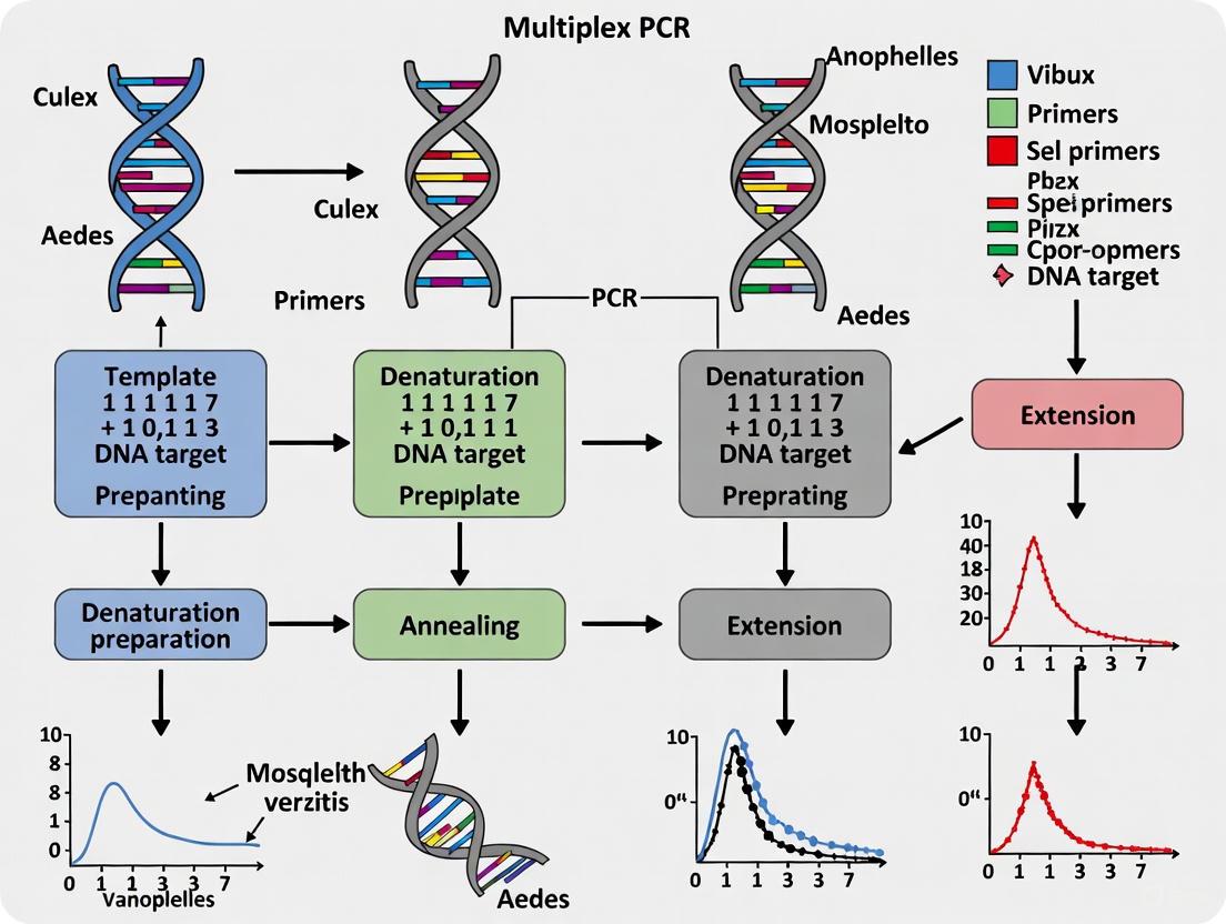
Workflow Visualization: Integrated Mosquito Surveillance
The following diagram illustrates the streamlined workflow for mosquito surveillance incorporating molecular identification.
The Scientist's Toolkit: Research Reagent Solutions
Table 4: Essential Reagents and Kits for Mosquito Surveillance and Identification
| Reagent / Kit | Function / Application | Example Product / Note |
|---|---|---|
| DNA Extraction Kits | Isolation of high-quality genomic DNA from whole mosquitoes or tissue parts. | DNeasy Blood & Tissue Kit (QIAGEN) for individual/small pools; DNAzol Reagent for large pools [3]. |
| Multiplex PCR Master Mix | Provides optimized buffer, enzymes, and dNTPs for simultaneous amplification of multiple targets. | Commercial hot-start master mixes (e.g., from QIAGEN or Thermo Fisher) to enhance specificity and reduce primer-dimer formation [1] [4]. |
| Species-Specific Primers | Oligonucleotides designed to bind to unique genetic regions of target mosquito species. | Custom-designed primers for a defined panel of invasive and native species (e.g., Ae. albopictus, Ae. japonicus) [1] [3]. |
| Gel Electrophoresis Reagents | Visualization and sizing of PCR amplicons to confirm species identity. | Agarose, DNA ladder, and fluorescent nucleic acid gel stain (e.g., GelRed). |
| Automated Surveillance Device | Continuous, real-time monitoring and counting of adult mosquito populations in the field. | MS-300 monitor uses attractants and infrared detection for automated data upload [2]. |
| Digital Data Platform | Mobile and cloud-based systems for field data entry, management, and analysis. | Epi Info Vector Surveillance app for mobile data collection and dashboard analysis [5]. |
The critical need for accurate mosquito surveillance in public health is met by integrating robust field methods with advanced molecular diagnostics. As demonstrated, multiplex PCR protocols offer a powerful, specific, and efficient means for the high-throughput screening of target mosquito vectors, directly feeding into responsive and data-driven public health action. By adopting the standardized protocols and tools outlined in this application note, surveillance programs can significantly enhance their capacity to monitor vector populations, detect invasions early, and mitigate the risk of mosquito-borne disease transmission.
The accurate identification of mosquito species is a cornerstone of effective public health monitoring and vector control programs. Traditional methods have heavily relied on morphological examination of specimens. However, two significant and interconnected challenges often compromise the reliability of these morphological approaches: phenotypic plasticity and the requirement for high taxonomic expertise. Phenotypic plasticity refers to the ability of a single genotype to produce different phenotypes in response to varying environmental conditions [6]. This biological phenomenon can lead to substantial variation in physical characteristics, confounding species identification based on morphology alone. Furthermore, morphological identification demands specialized, extensive training to achieve proficiency, creating a dependency on expert taxonomists whose numbers are declining. Within the context of developing molecular diagnostics, such as multiplex PCR protocols, understanding these limitations is crucial for designing robust and accessible species identification systems that can complement or surpass traditional methods, ultimately enhancing the capabilities of researchers and public health professionals in monitoring invasive and native mosquito populations.
Theoretical Foundation: Phenotypic Plasticity as a Biological Constraint
Phenotypic plasticity is a ubiquitous biological strategy that allows organisms to survive in variable environments, but it fundamentally constrains the reliability of morphological identification [6]. Theoretically, the optimal response to environmental heterogeneity would be perfect plasticity, where an organism possesses perfect information about its environment and mechanisms to produce an ideal phenotypic response. However, such perfect plasticity is rare in nature, indicating that evolutionary constraints prevent its realization [6]. These constraints manifest as limits and costs that directly impact phenotypic expression and stability.
A critical distinction must be made between the costs of plasticity and the costs of the phenotype itself. A cost of plasticity is a fitness decrement that a highly plastic genotype pays relative to a less plastic genotype, regardless of the environment. In contrast, a cost of phenotype refers to the fitness trade-offs inherent in producing a specific trait in a particular environment [6]. For mosquito identification, this means that the same species might develop different morphological traits (e.g., size, coloration, scale patterns) in different environments, while different species might converge on similar phenotypes under similar conditions. This variation introduces significant ambiguity into morphological keys and diagnostic characteristics.
Several specific mechanisms underlie these constraints:
- Maintenance Costs: Plastic organisms may incur costs for maintaining the sensory and regulatory machinery needed for facultative development, which non-plastic organisms avoid [7].
- Information Reliability Limits: The reliability of environmental cues is paramount. Unreliable cues can lead to a poor match between the expressed phenotype and the actual selective environment, making certain traits inconsistent indicators of species identity [6].
- Lag-Time Limits: A delay between sensing an environmental cue and producing a phenotypic response can result in temporary mismatches, meaning the phenotype observed at a single time point may not represent the stable or typical state for that species [7].
These evolutionary constraints on phenotypic plasticity create inherent variability that challenges the static character states used in morphological identification, necessitating the use of molecular methods to reveal the underlying genetic identity.
The Expertise Barrier in Morphological Analysis
The accurate morphological identification of mosquitoes, especially at the species level, requires a level of expertise that constitutes a significant practical barrier for many monitoring programs. This expertise encompasses not only the ability to recognize subtle diagnostic characters but also the experience to correctly interpret phenotypic variation that may arise from plasticity.
Taxonomic specialists capable of distinguishing between closely related species, such as those within the Aedes genus, are essential yet often a limited resource. The challenge is compounded when dealing with immature life stages (eggs and larvae), where diagnostic characters may be fewer and less pronounced than in adults. For instance, the eggs of container-breeding Aedes species are often laid on the same ovitrap spatula, and their morphological differentiation can be exceptionally difficult, even for experienced personnel [1]. This reliance on specialized human capital creates a bottleneck in large-scale surveillance efforts, where rapid processing of thousands of samples is required to inform timely public health decisions. The decline in formal taxonomic training further exacerbates this problem, increasing the potential for misidentification. Misidentifications can have severe consequences, including the failure to detect an invasive species early or the misallocation of control resources. Therefore, reducing the program's dependency on this limited expertise is a key driver for adopting molecular diagnostic protocols.
Comparative Analysis: Molecular vs. Morphological Identification
Empirical data from a nationwide Austrian mosquito monitoring program quantitatively demonstrates the superiority of molecular methods over traditional morphological identification, particularly when dealing with the challenges of phenotypic plasticity and mixed samples. The study analyzed 2,271 ovitrap samples collected in 2021 and 2022 [1] [8].
Table 1: Comparison of Identification Success Between Molecular Methods
| Method | Total Samples Identified | Identification Success Rate | Detection of Mixed-Species Samples |
|---|---|---|---|
| Multiplex PCR | 1,990 out of 2,271 | 87.6% | 47 samples |
| DNA Barcoding (mtCOI) | 1,722 out of 2,271 | 75.8% | Not possible with standard protocol |
The data reveals that the multiplex PCR protocol was markedly more successful in assigning a species identity to the collected samples compared to DNA barcoding of the mitochondrial cytochrome c oxidase subunit I (mtCOI) gene [1] [8]. A key advantage of the multiplex PCR was its ability to detect mixtures of different Aedes species within a single sample, a common occurrence when multiple females oviposit on the same spatula in an ovitrap. Standard Sanger sequencing used for DNA barcoding typically cannot resolve multiple species in one sample, as it produces overlapping chromatograms that are unreadable [1]. This capability of multiplex PCR to identify species mixtures is a direct solution to a limitation inherent in both morphology and standard barcoding.
Table 2: Target Species of the Adapted Multiplex PCR Protocol
| Species | Status | Relevance in Austria | Vector Competence |
|---|---|---|---|
| Aedes albopictus | Invasive | Found in all provinces; stable populations in Vienna and Graz [1] | Dengue, Zika, Chikungunya, Dirofilaria [1] |
| Aedes japonicus | Invasive | Established in all provinces in high numbers [1] | Usutu virus, West Nile virus (field detection) [1] |
| Aedes koreicus | Invasive | Reports in Carinthia, Styria, and Tyrol [1] | Potential for Dirofilaria immitis (lab conditions) [1] |
| Aedes geniculatus | Native | Present in monitoring samples [1] | Varies |
Detailed Multiplex PCR Protocol for Species Identification
The following protocol, adapted from Bang et al. (2024), provides a detailed methodology for the simultaneous identification of four container-breeding Aedes species (Ae. albopictus, Ae. japonicus, Ae. koreicus, and Ae. geniculatus) relevant to European monitoring programs [1]. This protocol is designed to be integrated into a high-throughput pipeline for processing ovitrap samples.
Sample Collection and DNA Preparation
- Ovitrap Sampling: Deploy black plastic containers (1 L volume) filled with approximately 0.75 L of tap water. Insert a wooden spatula as an oviposition substrate. Spatulas should be collected and replaced weekly.
- Morphological Examination: Examine collected spatulas under a stereo microscope. Identify and remove all mosquito eggs morphologically to the finest taxonomic level possible. Place all eggs from a single spatula into a 1.5 mL Eppendorf tube.
- Homogenization and DNA Extraction:
- Add a single ceramic bead (2.8 mm) to the tube containing the eggs.
- Homogenize the samples using a tissue lyser (e.g., TissueLyser II, Qiagen).
- Extract genomic DNA from the homogenate using a commercial kit (e.g., innuPREP DNA Mini Kit, Analytik Jena, or BioExtract SuperBall Kit on a KingFisher Flex96 robot). Follow the manufacturer's instructions.
Multiplex PCR Setup
The PCR leverages one universal forward primer and multiple species-specific reverse primers that generate amplicons of distinct sizes for easy differentiation via gel electrophoresis.
- Primers: The protocol uses the universal forward primer
Aedes-Fand specific reverse primers for Ae. albopictus (AL...), Ae. japonicus, Ae. koreicus, and Ae. geniculatus (the exact sequences are detailed in the original publication by Bang et al., as adapted by Reichl et al.) [1]. - Reaction Mixture:
- PCR Master Mix (e.g., 2x Concentrate): 12.5 µL
- Template DNA: 2-5 µL (depending on yield)
- Primer Mix (containing all forward and reverse primers at working concentrations): 2.5 µL
- Nuclease-free water: to a final volume of 25 µL
- Thermal Cycling Conditions:
- Initial Denaturation: 95°C for 5 minutes
- 35 Cycles of:
- Denaturation: 95°C for 30 seconds
- Annealing: [Specify temperature from protocol] for 30 seconds
- Extension: 72°C for 1 minute
- Final Extension: 72°C for 10 minutes
- Hold: 4°C
Analysis and Interpretation
- Gel Electrophoresis: Separate the PCR products on a 2% agarose gel stained with a DNA intercalating dye.
- Species Identification: Identify the species based on the presence of specific band sizes when visualized under UV light.
- Ae. albopictus: [Band Size] bp
- Ae. japonicus: [Band Size] bp
- Ae. koreicus: [Band Size] bp
- Ae. geniculatus: [Band Size] bp
- Mixed Samples: The presence of multiple bands indicates that eggs from more than one species were present on the ovitrap spatula.
Experimental Workflow and Logical Decision Pathway
The following diagram illustrates the integrated workflow for mosquito surveillance, from sample collection to final identification, highlighting the points where molecular methods overcome the limitations of morphology.
Research Reagent Solutions
The following table details the essential reagents and materials required for the implementation of the multiplex PCR protocol for mosquito species identification.
Table 3: Essential Research Reagents for Multiplex PCR-based Mosquito Identification
| Item | Function / Application | Example / Note |
|---|---|---|
| Ovitrap & Spatula | Field collection of mosquito eggs. | Black plastic container with wooden spatula [1]. |
| Stereo Microscope | Initial morphological examination and sorting of eggs. | Critical for pre-sorting before molecular analysis. |
| Tissue Lyser & Beads | Mechanical homogenization of egg samples for DNA release. | e.g., TissueLyser II with ceramic beads [1]. |
| DNA Extraction Kit | Purification of high-quality genomic DNA from homogenates. | e.g., innuPREP DNA Mini Kit or BioExtract SuperBall Kit [1]. |
| PCR Master Mix | Contains Taq polymerase, dNTPs, Mg²⁺, and buffer for amplification. | Requires robust performance for multiplex reactions. |
| Species-Specific Primers | Oligonucleotides designed to bind to unique genomic regions of each target species. | The core of the protocol; specificity is paramount [1]. |
| Agarose & Electrophoresis System | Size separation and visualization of PCR amplicons. | Standard equipment for post-PCR analysis. |
The limitations of morphological identification, driven by phenotypic plasticity and the scarcity of taxonomic expertise, present tangible obstacles to effective mosquito surveillance. The multiplex PCR protocol detailed herein provides a robust, high-throughput solution that directly addresses these constraints. It offers higher identification success rates than DNA barcoding and possesses the unique ability to detect mixed-species infestations from a single egg sample, a routine challenge in ovitrap-based monitoring. By integrating this molecular tool into surveillance programs, researchers and public health professionals can achieve more accurate, reliable, and efficient tracking of both invasive and native mosquito populations. This enhanced capability is critical for timely vector control interventions and for understanding the changing distribution of species in the face of globalization and climate change.
DNA barcoding has emerged as a powerful molecular tool for species identification in large-scale surveillance programs. This application note examines the technical advantages and operational constraints of implementing DNA barcoding, with a specific focus on mosquito surveillance as a model system. We present comparative performance data between traditional DNA barcoding and multiplex PCR approaches, detailed experimental protocols for both methods, and a structured framework for selecting appropriate molecular identification strategies based on program objectives. The integration of these methods enhances vector surveillance capabilities, supporting more effective public health interventions against mosquito-borne diseases.
DNA barcoding utilizes short, standardized genetic markers to identify organisms to species level. The mitochondrial cytochrome c oxidase subunit I (COI) gene serves as the primary barcode for animal species identification due to its sufficient sequence variation to distinguish between species and conserved flanking regions that facilitate primer design [9]. For mosquito surveillance and other large-scale biodiversity assessment programs, DNA barcoding has transformed traditional morphological identification approaches by enabling rapid processing of large specimen volumes, distinguishing cryptic species, and identifying specimens across all life stages [10] [11].
The application of DNA barcoding within operational surveillance programs presents both significant advantages and notable constraints. This document examines these factors within the context of mosquito surveillance, though the principles apply broadly to biodiversity monitoring and vector surveillance programs. We provide a comparative analysis of DNA barcoding and alternative molecular methods, with specific emphasis on multiplex PCR protocols for targeted species identification.
Advantages of DNA Barcoding
High Taxonomic Resolution and Cryptic Species Detection
DNA barcoding provides exceptional resolution for distinguishing morphologically similar species and revealing cryptic diversity. The high mutation rate of the COI gene enables discrimination of closely related species that may be indistinguishable using morphological characters alone [9]. Studies on Chironomidae families have demonstrated that DNA barcoding supports species delimitation based on color patterns, with distance thresholds of 4.5-7.7% providing appropriate species boundaries in Stictochironomus species [11]. This resolution is particularly valuable for surveillance programs targeting specific vector species among complex insect communities.
Capacity for High-Throughput Processing
The standardization of DNA barcoding protocols enables processing of hundreds to thousands of specimens simultaneously, dramatically increasing surveillance efficiency compared to morphological identification. Next-generation sequencing platforms further enhance this capability through "megabarcoding" approaches that combine individual barcodes into high-throughput sequencing runs [10]. This scalability makes DNA barcoding particularly suitable for large-scale monitoring programs where processing speed and sample throughput are critical operational considerations.
Ability to Identify All Life Stages
Unlike morphological identification, which often requires adult specimens for reliable species determination, DNA barcoding successfully identifies immature life stages (eggs, larvae, pupae) that constitute substantial portions of biodiversity [10]. This capability is especially valuable for mosquito surveillance, where egg identification from ovitraps is essential for early detection of invasive species but challenging using morphological methods [1]. The ability to identify all life stages provides a more comprehensive understanding of vector population dynamics and biodiversity patterns.
Standardization Across Taxonomic Groups
DNA barcoding provides a standardized identification framework across diverse taxonomic groups, reducing reliance on specialized taxonomic expertise that is increasingly limited [9] [12]. This standardization enables consistent species identification across different surveillance sites, operators, and time periods, improving data comparability and quality control in long-term monitoring programs.
Operational Constraints
Reference Database Incompleteness
The effectiveness of DNA barcoding depends on comprehensive reference libraries containing validated barcode sequences for target species. Significant gaps persist in these databases, particularly for tropical regions and underrepresented taxa [9] [12]. Peru, one of the world's most megadiverse countries, represents only 0.52% of records in the Barcode of Life Database (BOLD), highlighting the substantial disparities in genetic representation [12]. This constraint necessitates additional resources for sequence validation and database expansion when working with under-barcoded taxa or regions.
Inability to Detect Mixed Species in Single Samples
Standard Sanger sequencing-based DNA barcoding cannot reliably detect multiple species in a single sample because the sequencing process generates a single consensus sequence [1] [8]. This limitation is particularly problematic for mosquito egg mass analysis from ovitraps, where multiple species may oviposit on the same substrate. Surveillance programs must therefore implement individual specimen processing rather than bulk sample analysis, increasing processing time and costs [1].
Infrastructure and Cost Requirements
Traditional DNA barcoding requires laboratory infrastructure for DNA extraction, PCR amplification, and Sanger sequencing, which may be inaccessible in remote field stations or resource-limited settings [12]. While sequencing costs have decreased significantly, the per-sample expenses remain substantial for large-scale applications. Recent advances in portable sequencing technologies (e.g., Oxford Nanopore MinION) offer promising alternatives for in situ barcoding, yet still require initial investment in equipment and technical training [12].
Technical Expertise and Data Management
Effective implementation requires molecular biology expertise for laboratory work and bioinformatics capabilities for sequence analysis, database management, and quality control. The computational demands of sequence alignment, phylogenetic analysis, and data storage present additional challenges for decentralized surveillance networks [12] [11].
Comparative Performance Data
Table 1: Comparative Performance of DNA Barcoding and Multiplex PCR in Mosquito Surveillance
| Parameter | DNA Barcoding | Multiplex PCR | Study Context |
|---|---|---|---|
| Identification Rate | 75.8% (1722/2271 samples) | 87.6% (1990/2271 samples) | Aedes surveillance in Austria [1] [8] |
| Mixed Species Detection | Not possible with standard Sanger sequencing | 47 samples with multiple species detected | Aedes egg mass analysis [1] |
| Target Range | Broad spectrum (requires reference sequences) | Narrow (pre-defined target species) | Species-specific primer design [13] |
| Infrastructure Requirements | High (sequencing facility) | Moderate (standard molecular lab) | Resource-limited settings [12] |
| Cost Per Sample | Moderate to High | Low to Moderate | Large-scale processing [10] |
| Technical Expertise | High (bioinformatics required) | Moderate (standard PCR skills) | Implementation capacity [12] |
Table 2: Performance Metrics for Quantitative PCR Barcoding Assay
| Parameter | Aedes aegypti | Aedes sierrensis |
|---|---|---|
| Egg Sensitivity | >95% | >95% |
| Larval Sensitivity | >95% | >95% |
| Adult Sensitivity | >95% | 75% (field-collected) |
| eDNA Sensitivity | 91% | 100% |
| eDNA Specificity | 86% | 94% |
| Detection Limit | Not specified | 1fg/μL for An. anthropophagus [13] |
Experimental Protocols
Standard DNA Barcoding Protocol for Mosquito Species
Principle: Amplification and sequencing of the COI gene region for comparison with reference databases.
Reagents and Equipment:
- DNA extraction kit (e.g., innuPREP DNA Mini Kit)
- PCR reagents: MasterMix, primers, molecular grade water
- COI primers: LCO1490 (5'-GGTCAACAAATCATAAAGATATTGG-3') and HCO2198 (5'-TAAACTTCAGGGTGACCAAAAAATCA-3')
- Agarose gel electrophoresis equipment
- Sanger sequencing facility access
Procedure:
- DNA Extraction: Extract genomic DNA from individual mosquito specimens (legs, thorax, or entire specimens for small insects) using standard protocols. Preserve voucher specimens when possible.
- PCR Amplification: Prepare 25μL reaction mixtures containing 2× MasterMix (12.5μL), forward and reverse primers (0.625μL each, 10μM), template DNA (2μL), and deionized water (9.25μL).
- Thermocycling Conditions:
- Initial denaturation: 95°C for 5 minutes
- 34 cycles of: 94°C for 30 seconds, 51°C for 30 seconds, 72°C for 1 minute
- Final extension: 72°C for 3 minutes
- Amplicon Verification: Confirm successful amplification via 1.0% agarose gel electrophoresis (expected product size: ~658bp).
- Sequencing: Purify PCR products and submit for bidirectional Sanger sequencing.
- Data Analysis:
- Assemble and edit sequence traces using bioinformatics software (e.g., BioEdit)
- Perform sequence alignment (e.g., ClustalW algorithm in MEGA)
- Compare to reference databases (BOLD, NCBI) using BLAST
- Calculate genetic distances (Kimura 2-Parameter model)
- Construct phylogenetic trees (Neighbor-Joining, Maximum Likelihood) [11]
Multiplex PCR Protocol for Mosquito Identification
Principle: Simultaneous amplification of species-specific DNA fragments in a single reaction tube.
Reagents and Equipment:
- DNA extraction reagents
- Multiplex PCR Master Mix
- Species-specific primer mixtures
- Agarose gel electrophoresis equipment
- DNA size standard
Procedure:
- DNA Extraction: As described in Protocol 5.1.
- Primer Design: Design species-specific primers targeting conserved genetic regions with variable intervening sequences. Tools such as PMPrimer automate this process using Shannon's entropy to identify conserved regions and evaluate primer specificity [14].
- Reaction Setup: Prepare multiplex PCR mixtures containing:
- Multiplex PCR Master Mix
- Species-specific primer pairs (optimized concentrations)
- Template DNA
- Thermocycling Conditions: Optimize based on primer characteristics (typical parameters: 95°C initial denaturation, 35 cycles of 95°C denaturation, primer-specific annealing temperature, 72°C extension, final extension at 72°C).
- Product Analysis: Separate amplification products by agarose gel electrophoresis. Identify species by comparing band sizes to expected patterns [1] [13].
In Situ DNA Barcoding with Portable Sequencing
Principle: Field-based barcoding using portable equipment for rapid species identification.
Reagents and Equipment:
- Portable PCR equipment
- Oxford Nanopore MinION sequencer
- Field DNA extraction kits
- Lyophilized reagents
Procedure:
- Field DNA Extraction: Use simplified extraction protocols adapted to field conditions.
- PCR Amplification: Perform amplification with portable thermal cyclers.
- Library Preparation: Utilize rapid library prep kits optimized for nanopore sequencing.
- Sequencing: Conduct real-time sequencing with MinION device.
- Data Analysis: Perform basecalling and sequence analysis using laptop-based bioinformatics pipelines [12].
Workflow Integration
Workflow Diagram 1: Integrated Molecular Surveillance System
Multiplex PCR Development Pipeline
Workflow Diagram 2: Multiplex PCR Assay Development Pipeline
Research Reagent Solutions
Table 3: Essential Research Reagents for DNA Barcoding and Multiplex PCR
| Reagent Category | Specific Examples | Application Function |
|---|---|---|
| DNA Extraction Kits | innuPREP DNA Mini Kit, BioExtract SuperBall Kit | High-quality DNA extraction from various specimen types (whole insects, tissues, eDNA filters) |
| PCR Master Mixes | 2× Es Taq MasterMix, Multiplex PCR Master Mix | Amplification of target regions with optimized buffer conditions |
| Universal Primers | LCO1490/HCO2198, mlCOIintF/jgHC02198 | Broad-range amplification of COI barcode region across diverse taxa |
| Species-Specific Primers | Aedes albopictus (AL-), Ae. japonicus (JA-) | Targeted detection of predetermined species in multiplex reactions |
| Sequencing Reagents | BigDye Terminator v3.1, Nanopore Ligation Kits | Sanger sequencing or portable nanopore sequencing library preparation |
| Electrophoresis Materials | Agarose, DNA size standards, nucleic acid stains | Amplification product verification and multiplex PCR result interpretation |
| Positive Controls | Reference DNA from voucher specimens | Assay validation and quality assurance |
Implementation Framework
Method Selection Guidelines
The choice between DNA barcoding and multiplex PCR depends on surveillance objectives, resource availability, and target species characteristics:
Select DNA Barcoding when:
- Target species diversity is broad or poorly characterized
- Cryptic species complexes are suspected
- Building comprehensive reference databases is a priority
- Taxonomic expertise is limited for morphological identification
- Sequencing infrastructure and bioinformatics capacity are available
Select Multiplex PCR when:
- Surveillance targets a defined set of species (typically <10)
- Rapid, high-throughput identification is required
- Mixed samples requiring species composition analysis are common
- Field-based implementation or minimal infrastructure is needed
- Cost constraints preclude sequencing-based approaches
Integrated Surveillance Approach
For comprehensive surveillance programs, a hierarchical approach combining both methods provides optimal efficiency:
- Initial screening with multiplex PCR for high-throughput processing of known target species
- Follow-up barcoding for unidentified specimens, cryptic species detection, and database expansion
- Periodic validation of multiplex PCR results through DNA barcoding to maintain assay accuracy
This integrated framework leverages the strengths of both approaches while mitigating their individual limitations, creating a robust surveillance system adaptable to diverse operational contexts.
DNA barcoding represents a powerful tool for large-scale surveillance programs, offering high taxonomic resolution, species discovery capability, and standardization across diverse taxa. However, operational constraints including reference database gaps, infrastructure requirements, and limitations in detecting mixed samples necessitate careful consideration of implementation strategies. For targeted surveillance of known vector species, multiplex PCR provides a complementary approach that addresses several limitations of standard barcoding while offering enhanced throughput and cost efficiency. The integration of both methods within a hierarchical identification framework creates a robust surveillance system capable of addressing diverse operational requirements in mosquito and biodiversity monitoring programs.
Multiplex Polymerase Chain Reaction (PCR) is an advanced molecular technique that enables the simultaneous amplification of multiple distinct DNA sequences in a single reaction tube. This is achieved by incorporating numerous primer sets, each specifically designed to target a unique DNA region, resulting in amplicons of varying sizes specific to different sequences [15]. First described in 1988 for detecting deletion mutations in the dystrophin gene, multiplex PCR has evolved into a powerful tool for various applications, including species identification in ecological surveillance and diagnostic microbiology [15]. In the context of mosquito species identification, this technique has proven particularly valuable for monitoring invasive species and disease vectors, providing significant advantages over traditional morphological identification methods that can be unreliable for certain species and life stages [1].
The fundamental principle of multiplex PCR relies on careful primer design and reaction optimization to ensure that all primer sets function efficiently under uniform thermal cycling conditions. This requires primers to have similar melting temperatures (typically 55-60°C for standard designs) and minimal tendency to form primer dimers or cross-hybridize [15]. When properly optimized, multiplex PCR delivers substantial benefits for mosquito surveillance programs, including increased throughput, reduced reagent consumption, and conservation of valuable DNA samples, making it an indispensable tool for large-scale monitoring programs and ecological studies [1] [16].
Principles and Advantages for Multi-Species Detection
Core Principles
Multiplex PCR operates on the same fundamental principles as conventional PCR but extends this foundation to enable parallel amplification. The core mechanism involves multiple primer sets combined in a single reaction mixture, each designed to anneal specifically to unique target sequences from different species or genetic loci [15]. Successful implementation requires careful optimization of several parameters to ensure balanced amplification of all targets. Primer design is particularly critical—all primers must have compatible annealing temperatures to function under uniform thermal cycling conditions [15]. Additionally, amplicons must be designed with sufficient size variation to allow clear differentiation through gel electrophoresis or other detection methods [15].
The reaction components must be adjusted to accommodate the increased primer concentrations and potential competition between amplification targets. This often involves increasing polymerase concentration and optimizing buffer composition to maintain amplification efficiency across all targets [15]. For real-time multiplex PCR applications, the system becomes more complex through the incorporation of species-specific fluorescent probes labeled with different dyes, enabling simultaneous detection and differentiation of multiple targets based on their spectral signatures [16]. This approach allows researchers to distinguish between morphologically similar species that would be difficult to identify through traditional methods.
Comparative Advantages
Multiplex PCR offers several significant advantages over alternative detection methods, particularly for mosquito surveillance applications:
Comprehensive Information from Limited Samples: By targeting multiple species simultaneously, researchers can obtain more comprehensive biodiversity data from limited starting material, which is particularly valuable when working with precious or minimal samples such as mosquito eggs or degraded environmental DNA [15].
Internal Control Mechanism: The simultaneous amplification of multiple targets provides built-in quality control, as each amplified product serves as an internal control for others, helping reveal false negatives that might remain undetected in singleplex PCR assays [15].
Detection of Mixed Infestations: Unlike Sanger sequencing-based approaches, multiplex PCR can detect multiple species present in the same sample simultaneously [1]. This capability is particularly valuable for analyzing mosquito eggs collected from ovitraps, where different Aedes species may lay eggs on the same substrate.
Resource Efficiency: The technique significantly reduces time, labor, and reagent requirements compared to running multiple singleplex reactions [1] [16]. This efficiency enables more extensive sampling and higher throughput screening within constrained research budgets.
Table 1: Performance Comparison Between Multiplex PCR and DNA Barcoding for Mosquito Identification
| Parameter | Multiplex PCR | DNA Barcoding |
|---|---|---|
| Samples Identified | 1,990 out of 2,271 samples | 1,722 out of 2,271 samples |
| Mixed Species Detection | Possible (47 samples identified) | Not possible with Sanger sequencing |
| Throughput | High | Moderate |
| Cost per Sample | Lower | Higher |
| Equipment Requirements | Standard PCR equipment | Sequencing facility |
| Processing Time | Shorter | Longer |
Application in Mosquito Species Identification
Container-Breeding Aedes Surveillance
Multiplex PCR has proven particularly valuable for monitoring container-breeding Aedes species, which include important disease vectors such as the Asian tiger mosquito (Aedes albopictus), the Asian bush mosquito (Ae. japonicus), and the Korean bush mosquito (Ae. koreicus) [1]. These species typically lay their eggs in artificial containers and natural water-holding cavities, with surveillance often conducted using ovitraps—black containers filled with water with wooden spatulas serving as oviposition substrates [1]. A significant challenge in this surveillance approach is that multiple species may deposit eggs on the same spatula, creating mixed samples that are difficult to analyze with methods that can only detect a single species per reaction.
Research demonstrates that multiplex PCR outperforms DNA barcoding for identifying mosquito species from ovitrap samples. In a comprehensive study analyzing 2,271 ovitrap samples collected during an Austrian nationwide monitoring program, multiplex PCR successfully identified 1,990 samples, while DNA barcoding of the mitochondrial cytochrome c oxidase subunit I (mtCOI) gene could only identify 1,722 samples [1]. Crucially, the multiplex PCR approach detected mixtures of different species in 47 samples, a capability that was not possible with standard Sanger sequencing used for DNA barcoding [1]. This advantage makes multiplex PCR particularly suitable for large-scale monitoring programs where efficiency and detection of co-occurring species are priorities.
Environmental DNA and Field Surveillance
The application of multiplex PCR extends beyond direct insect identification to include environmental DNA (eDNA) analysis, which detects genetic material shed by organisms into their environment. This approach has been successfully used to determine species distributions in aquatic systems without requiring physical capture or observation of the target organisms [16]. For example, a study on two Japanese medaka fish species (Oryzias latipes and O. sakaizumii) developed a real-time multiplex PCR system that simultaneously detected both species from water samples, with results consistent with traditional capture surveys across all field sites [16].
This eDNA approach has significant implications for mosquito surveillance, particularly for detecting rare or elusive species in hard-to-survey habitats. The method is sensitive enough to detect species even when their abundances are highly biased, as demonstrated in aquarium experiments where less abundant species were consistently detected in the presence of dominant species [16]. Furthermore, real-time multiplex PCR provides practical advantages for field studies by reducing reagent use, labor requirements, and processing time while conserving valuable eDNA samples for additional analyses [16].
Advanced Surveillance Systems
Innovative mosquito monitoring systems that combine automated collection devices with multiplex PCR identification represent the cutting edge of vector surveillance technology. The MS-300, an internet-based vector mosquito monitor, continuously captures mosquitoes and uploads real-time data to cloud services [2]. However, the fan-based collection method often damages mosquitoes, compromising morphological characteristics and necessitating molecular identification [2].
In a recent study, researchers developed a multiplex PCR system specifically for identifying mosquitoes collected by such automated devices, targeting six key vector species: Aedes albopictus, Aedes aegypti, Culex pipiens pallens, Armigeres subalbatus, Anopheles sinensis, and Anopheles anthropophagus [2]. This system demonstrated high specificity and remarkable sensitivity, with detection limits for An. anthropophagus reaching 1 femtogram per microliter [2]. The results showed complete consistency with DNA barcoding technology while offering greater efficiency for processing mixed samples collected through continuous monitoring devices [2].
Experimental Protocols
Multiplex PCR Protocol for Aedes Species Identification
This protocol adapts and expands upon methodologies from recent research on container-breeding mosquito surveillance [1].
Sample Collection and DNA Extraction
Ovitrap Setup and Collection:
- Deploy black plastic containers (1L capacity) filled with approximately 0.75L of tap water
- Insert wooden spatulas as oviposition substrates, secured with stainless-steel clamps
- Exchange spatulas weekly and transport to the laboratory for analysis
Morphological Examination:
- Examine spatulas under a stereo microscope for presence of mosquito eggs
- Identify eggs to species level when possible based on morphological characteristics
- Transfer all eggs from each spatula to a 1.5mL Eppendorf tube
- Store samples at -80°C until molecular analysis
DNA Extraction:
- Homogenize eggs using one ceramic bead (2.8mm) and a TissueLyser II
- Extract DNA using commercial kits such as:
- innuPREP DNA Mini Kit (Analytik Jena)
- BioExtract SuperBall Kit on a KingFisher Flex96 robot
- Elute DNA in appropriate buffer and store at -20°C
Multiplex PCR Reaction
- Reaction Setup:
- Adapt the primer sequences from Bang et al. [1] for simultaneous detection of:
- Aedes albopictus
- Aedes japonicus
- Aedes koreicus
- Aedes geniculatus (native Austrian species)
- Prepare PCR master mix according to the following formulation:
- Adapt the primer sequences from Bang et al. [1] for simultaneous detection of:
Table 2: Multiplex PCR Reaction Components
| Component | Final Concentration | Volume per Reaction (μL) |
|---|---|---|
| PCR Buffer (10X) | 1X | 2.5 |
| MgCl₂ (25mM) | 2.5mM | 2.5 |
| dNTP Mix (10mM) | 0.2mM each | 0.5 |
| Primer Mix (10μM each) | 0.4μM each | 1.0 |
| DNA Polymerase (5U/μL) | 1.25U | 0.25 |
| Template DNA | Variable | 2.0 |
| Nuclease-Free Water | - | 16.25 |
| Total Volume | - | 25.0 |
Thermal Cycling Conditions:
- Initial Denaturation: 95°C for 5 minutes
- 35-40 cycles of:
- Denaturation: 95°C for 30 seconds
- Annealing: 58-60°C for 45 seconds (optimize for primer set)
- Extension: 72°C for 60 seconds
- Final Extension: 72°C for 7 minutes
- Hold: 4°C indefinitely
Product Analysis:
- Separate PCR products by gel electrophoresis (2% agarose)
- Visualize with ethidium bromide or SYBR Safe
- Identify species by amplicon size using appropriate DNA ladder
Real-Time Multiplex PCR for Environmental DNA
This protocol is adapted from methodologies for detecting aquatic species from environmental DNA [16].
Water Sample Collection and Filtration
Field Collection:
- Collect water samples in sterile containers
- Process samples immediately or store at 4°C for short-term storage
- Filter 1-2L of water through fine-pore filters (0.45μm to 1.2μm pore size)
DNA Extraction from Filters:
- Extract DNA from filters using commercial DNA extraction kits
- Include negative controls (filtered purified water) during extraction
- Elute DNA in 50-100μL of elution buffer
- Store at -20°C until analysis
Real-Time Multiplex PCR
Primer and Probe Design:
- Design species-specific primer-probe sets targeting unique DNA regions
- Label each probe with a different fluorescent dye (FAM, HEX, CY5, etc.)
- Ensure probes have similar melting temperatures and minimal spectral overlap
Reaction Setup:
- Prepare reaction mix according to the following formulation:
Table 3: Real-Time Multiplex PCR Reaction Components
| Component | Final Concentration | Volume per Reaction (μL) |
|---|---|---|
| Multiplex PCR Master Mix (2X) | 1X | 10.0 |
| Primer Mix (10μM each) | 0.4μM each | 1.0 |
| Probe Mix (5μM each) | 0.2μM each | 1.0 |
| Template DNA | Variable | 2.0 |
| Nuclease-Free Water | - | 6.0 |
| Total Volume | - | 20.0 |
Thermal Cycling Conditions:
- Initial Denaturation: 95°C for 5 minutes
- 45 cycles of:
- Denaturation: 95°C for 10-15 seconds
- Annealing/Extension: 60°C for 30-60 seconds (with fluorescence acquisition)
Data Analysis:
- Analyze amplification curves using real-time PCR software
- Determine cycle threshold (Ct) values for each target
- Identify species based on fluorescent channel showing amplification
Research Reagent Solutions
Successful implementation of multiplex PCR depends on careful selection of reagents and optimization of their concentrations. The following table outlines essential reagents and their functions in multiplex PCR assays for mosquito species identification.
Table 4: Essential Research Reagents for Multiplex PCR
| Reagent Category | Specific Examples | Function in Multiplex PCR |
|---|---|---|
| DNA Polymerases | Hot-start Taq polymerases | Reduces non-specific amplification and primer-dimer formation |
| Primers | Species-specific primers (18-22bp) | Targets unique DNA sequences for each mosquito species |
| Fluorescent Probes | FAM, SUN, HEX, CY5, ROX | Enables real-time detection of multiple targets; each dye corresponds to a specific species |
| Quenchers | ZEN, Iowa Black FQ, TAO | Suppresses reporter fluorescence until probe cleavage |
| dNTPs | dATP, dCTP, dGTP, dTTP | Building blocks for DNA synthesis |
| Buffer Components | MgCl₂, KCl, Tris-HCl | Optimizes reaction conditions for multiple primer sets |
| Commercial Kits | Qiagen Multiplex PCR Kit, Agilent Hybrid Capture | Provides pre-optimized reagent mixtures for multiplex applications |
For real-time multiplex PCR applications, dye selection is particularly critical. Researchers should choose dyes with minimal spectral overlap and ensure compatibility with their detection instruments [17]. Recommended dye combinations include FAM for low-copy transcripts due to its high fluorescence intensity, with alternative dyes such as SUN, JOE, or HEX for additional targets [17]. Using double-quenched probes with internal ZEN or TAO quenchers can significantly reduce background fluorescence, which is particularly important in multiplex reactions containing several fluorophores in the same tube [17].
Visualization of Workflows
Data Management and FAIR Principles
Effective data management is crucial for ensuring the reproducibility and utility of multiplex PCR research. Adopting the FAIR Data Principles (Findable, Accessible, Interoperable, Reusable) enhances data value and facilitates collaboration [18]. For multiplex PCR studies in mosquito surveillance, researchers should:
Implement Structured Data Tables: Organize experimental data using standardized tabular formats with clear column headings, appropriate units, and consistent decimal alignment [19]. Include all relevant metadata such as sampling locations, dates, primer sequences, and thermal cycling conditions.
Maintain Data Provenance: Document all steps from sample collection through analysis, including DNA extraction methods, PCR conditions, and analysis parameters. This information is crucial for experimental reproducibility and data validation [18].
Use Community Standards: Where possible, adopt community-approved ontologies and standardized formats for data annotation. This practice enhances interoperability and enables integration with larger datasets [18].
Publish Complete Datasets: Rather than providing only minimal data to support published claims, disseminate complete datasets through appropriate repositories. This approach maximizes research impact and enables secondary analyses [18].
Tools such as the ODAM (Open Data for Access and Mining) framework can help researchers structure their data from the initial acquisition phase, making subsequent FAIRification more straightforward [18]. This proactive approach to data management ultimately saves time and resources while producing more valuable and reusable research outputs.
Accurate mosquito species identification is a cornerstone of effective vector control and disease management programs. While morphological keys have traditionally been used for species differentiation, molecular methods have become indispensable for distinguishing cryptic species, identifying damaged specimens, and detecting invasive vectors [20]. This application note explores the primary genetic markers—Internal Transcribed Spacer 2 (ITS2) and Cytochrome c Oxidase I (COI)—used in mosquito surveillance and provides detailed protocols for their application in a research setting. The content is framed within the development of a multiplex PCR protocol, emphasizing how these markers can be integrated into efficient, high-throughput screening tools for researchers and public health professionals.
Key Genetic Markers for Mosquito Identification
The selection of an appropriate genetic marker is critical for the balance between species discrimination power and methodological practicality. The table below summarizes the core characteristics of the two most prevalent markers.
Table 1: Comparison of Primary Genetic Markers for Mosquito Identification
| Genetic Marker | Genomic Location | Key Advantages | Common Applications | Considerations |
|---|---|---|---|---|
| Internal Transcribed Spacer 2 (ITS2) | Nuclear rRNA cluster | High variability for discriminating closely related and cryptic species [21] [22]. | Identification of species within complexes (e.g., Anopheles species) [21] [23]. | Potential for intra-individual variation due to multiple gene copies [20]. |
| Cytochrome c Oxidase I (COI) | Mitochondrial genome | Standardized "DNA barcode" for animals; extensive reference databases [24] [25]. | General species identification, biodiversity studies, and building barcode libraries [20] [25]. | Can lack resolution for some closely related species; maternal inheritance only [22]. |
Experimental Protocols
The following protocols are adapted from recent research and can be utilized for standard PCR-based identification or incorporated into the development of a multiplex PCR system.
ITS2 PCR Assay for Identification ofAnopheles squamosus
This protocol describes a species-specific PCR assay to reliably distinguish An. squamosus from other morphologically similar species [21].
- Primer Design and Validation: The assay uses the forward primer ITS2-ASQ-F10 (5'-CCC TCG AAG GGT GCT GTG-3') and the reverse primer ITS2-ASQ-R10 (5'-AAT CCA CGG TGT GAT GGC-3') [21]. These primers were designed from aligned ITS2 contig sequences of An. squamosus, An. sp. 11, and An. sp. 15 to ensure specificity.
- PCR Reaction Setup:
- Total Volume: 25 µL
- Reaction Components:
- 12.5 µL of 2X New England Biolabs Master Mix
- 1 µL of DNA template
- Forward and Reverse Primers (concentration as optimized in original protocol [21])
- Nuclease-free water to 25 µL
- Thermal Cycling Conditions:
- Initial Denaturation: 95°C for 5 minutes
- Amplification (35 cycles):
- Denaturation: 95°C for 30 seconds
- Annealing: 60°C for 30 seconds (optimal temperature to be determined empirically)
- Extension: 72°C for 30 seconds
- Final Extension: 72°C for 5 minutes
- Result Analysis: A positive identification of An. squamosus is indicated by a clear PCR product of 301 bp on an agarose gel. This assay can be multiplexed with existing ITS2 assays for other anophelines [21].
Multiplex PCR for Container-BreedingAedesSpecies
This protocol is adapted for the simultaneous identification of multiple container-breeding Aedes species, which is highly valuable for processing ovitrap samples [1].
- Primer Design and Validation: The multiplex PCR uses a universal forward primer paired with species-specific reverse primers that generate amplicons of distinct sizes for:
- Aedes albopictus
- Aedes japonicus
- Aedes koreicus
- Aedes geniculatus [1]
- PCR Reaction Setup: The reaction is set up as per the original publication, with primers optimized for compatibility and non-interference in a single tube.
- Thermal Cycling Conditions: Use standardized cycling conditions with an annealing temperature that allows all primers to bind efficiently.
- Result Analysis: Species are identified based on the unique amplicon size visualized via gel electrophoresis. This method successfully identified species in 1990 out of 2271 ovitrap samples and detected mixed-species compositions in 47 samples, a feat difficult to achieve with standard DNA barcoding [1].
DNA Barcoding with COI Gene
This is a generalized protocol for species identification using the COI barcode region [24] [25].
- DNA Extraction: Extract genomic DNA from mosquito legs or thoracic tissue using commercial kits (e.g., DNeasy Blood & Tissue Kit, QIAGEN) or a standard Chelex-based protocol.
- PCR Amplification:
- Primers: Use universal COI primers, such as LCO1490 and HCO2198, or other well-established pairs.
- Reaction: Set up a standard PCR reaction with ~50-100 ng of DNA template.
- Thermal Cycling Conditions:
- Initial Denaturation: 95°C for 5 minutes
- Amplification (35-40 cycles):
- Denaturation: 94°C for 30-40 seconds
- Annealing: 45-55°C for 30-60 seconds
- Extension: 72°C for 45-60 seconds
- Final Extension: 72°C for 5-10 minutes
- Sequencing and Analysis: Purify PCR products and perform Sanger sequencing. Analyze the resulting sequences by comparing them to reference sequences in databases like GenBank or BOLD (Barcode of Life Data Systems) [24].
Workflow Visualization
The following diagram illustrates the decision pathway for selecting the appropriate molecular identification method based on research objectives and sample type.
The Scientist's Toolkit: Research Reagent Solutions
Successful implementation of molecular identification protocols relies on key reagents and tools. The following table details essential solutions for the featured experiments.
Table 2: Essential Research Reagents for Molecular Identification of Mosquitoes
| Reagent / Kit | Specific Example | Function in Protocol |
|---|---|---|
| DNA Extraction Kit | DNeasy Blood & Tissue Kit (QIAGEN) [3] [24] | High-quality genomic DNA extraction from individual or pooled mosquitoes. |
| DNA Extraction Reagent | DNAzol Reagent (ThermoFisher) [3] | Cost-effective DNA isolation from large pools of mosquitoes (e.g., 25-500 specimens). |
| PCR Master Mix | New England Biolabs Master Mix [21] | Provides buffer, dNTPs, and polymerase for robust PCR amplification. |
| Multiplex PCR Kit | QuantiFast Multiplex PCR Kit (QIAGEN) [3] | Optimized for simultaneous amplification of multiple targets in a single reaction. |
| Species-Specific Primers | ITS2-ASQ-F10/R10 for An. squamosus [21] | Enable precise targeting and identification of a single mosquito species. |
| Universal Barcoding Primers | COI primers (e.g., LCO1490/HCO2198) [24] | Allow amplification of the standard barcode region from a wide range of species. |
The strategic selection of genetic markers is fundamental to modern mosquito surveillance. ITS2 provides robust resolution for cryptic species complexes, while COI offers a universal system for general biodiversity assessment and library building. The development of species-specific and multiplex PCR protocols represents a significant advancement for high-throughput surveillance, enabling the accurate screening of large sample volumes—such as those from ovitraps—and the detection of multiple species in a single reaction. Integrating these molecular tools into public health and research pipelines ensures precise vector identification, which is the foundation for targeted and effective mosquito control strategies.
Molecular identification of mosquito species is a cornerstone of effective vector surveillance and control programs. The expansion of invasive species and the persistent threat of mosquito-borne diseases necessitate accurate, efficient, and scalable diagnostic methods [1] [3]. This application note provides a comparative workflow analysis of three primary molecular techniques used in mosquito surveillance: morphological identification, DNA barcoding, and multiplex PCR. The analysis is framed within a broader research context focused on developing and implementing multiplex PCR protocols for mosquito species identification, particularly for container-breeding Aedes species and other critical vectors. We present detailed experimental protocols, quantitative comparisons of labor, cost, and time requirements, and practical recommendations for researchers seeking to implement these methods in both laboratory and field settings. The data presented herein demonstrate that multiplex PCR offers significant advantages in throughput and cost-efficiency for large-scale surveillance operations while maintaining accuracy comparable to more resource-intensive methods.
Experimental Protocols and Methodologies
Morphological Identification Protocol
Principle: Morphological identification relies on expert examination of physical characteristics under stereoscopic magnification to differentiate species based on established taxonomic keys.
Procedure:
- Sample Collection: Deploy ovitraps (black plastic containers filled with water with wooden spatulas for oviposition) at monitoring sites [1]. Exchange spatulas weekly and transport to laboratory for analysis.
- Microscopic Examination: Examine wooden spatulas under stereo microscope for presence of mosquito eggs.
- Species Identification: Identify eggs to species level based on morphological characteristics when possible, using taxonomic references and identification keys.
- Sample Storage: Remove identified eggs from spatula and transfer to 1.5 mL Eppendorf tubes. Store at -80°C for potential molecular validation.
Critical Considerations: Morphological identification requires substantial taxonomic expertise. Phenotypic plasticity and genetic variability can lead to misidentification [2]. Specimens collected by certain automated traps (e.g., MS-300 monitor) may lose morphological characteristics due to fan operation, compromising identification accuracy [2].
DNA Barcoding Protocol
Principle: DNA barcoding utilizes Sanger sequencing of a standardized genetic marker (mitochondrial cytochrome c oxidase subunit I - mtCOI gene) to identify species based on sequence divergence [1].
Procedure:
- DNA Extraction:
- Homogenize samples using ceramic beads and TissueLyser II [1].
- Extract DNA using commercial kits (e.g., innuPREP DNA Mini Kit or BioExtract SuperBall Kit) according to manufacturer's protocols [1].
- Alternative rapid extraction: Use HOTShot protocol or Dipstick-based method for field applications [26].
- PCR Amplification:
- Prepare PCR mix: 1X PCR buffer, 2.5 mM MgCl₂, 0.2 mM dNTPs, 0.2 µM each primer (mtCOI-specific), 1 U DNA polymerase, 2-5 µL template DNA.
- Thermal cycling: Initial denaturation at 94°C for 2 min; 35 cycles of 94°C for 30 s, 52°C for 30 s, 72°C for 1 min; final extension at 72°C for 5 min.
- Sequencing and Analysis:
- Purify PCR products and perform Sanger sequencing.
- Compare obtained sequences to reference databases (e.g., NCBI GenBank) using BLAST or specialized barcoding platforms.
Critical Considerations: DNA barcoding provides high accuracy but does not allow identification of multiple species in a single sample [1]. The method involves additional processing steps, higher costs, and longer turnaround times compared to multiplex PCR [2].
Multiplex PCR Protocol for Mosquito Identification
Principle: Multiplex PCR enables simultaneous amplification of multiple species-specific targets in a single reaction through careful primer design to generate amplicons of distinct sizes resolvable by gel electrophoresis [1] [27].
Procedure:
- Primer Design:
- Design species-specific primers targeting informative genetic regions (e.g., ITS2 or species-specific nuclear sequences) [2] [28].
- Ensure primers have similar melting temperatures (within 5°C) and generate amplicons of distinct sizes for clear differentiation by electrophoresis [27].
- Validate each primer set in singleplex reactions before multiplexing.
- DNA Extraction: Follow same protocol as DNA barcoding (section 2.2).
- Multiplex PCR Reaction:
- Prepare master mix: 1X PCR buffer, 3-4 mM MgCl₂ (optimized), 0.2-0.4 mM dNTPs, 0.1-0.4 µM each primer (multiple species-specific primers), 1-2 U hot-start DNA polymerase, 2-5 µL template DNA.
- Use hot-start DNA polymerase to enhance specificity in multiplex reactions [27].
- Thermal cycling: Initial activation/denaturation at 95°C for 5 min; 35 cycles of 95°C for 30 s, optimized annealing temperature (55-60°C) for 30 s, 72°C for 45 s; final extension at 72°C for 7 min.
- Product Analysis:
- Separate PCR products by gel electrophoresis (2% agarose).
- Visualize with DNA staining and image under UV light.
- Identify species based on amplicon sizes compared to reference ladder.
Critical Considerations: Primer design is crucial to minimize mispriming and ensure balanced amplification [27]. Optimization may be required for different instrument platforms. The method is particularly advantageous for identifying mixed species in a single sample [1].
Workflow Comparison and Data Analysis
Quantitative Comparison of Method Performance
Table 1: Comparative Analysis of Mosquito Identification Methods
| Parameter | Morphological Identification | DNA Barcoding | Multiplex PCR |
|---|---|---|---|
| Sample Throughput | Low to moderate | Low | High (parallel processing) |
| Hands-on Time per Sample | 15-30 minutes | 45-60 minutes | 20-30 minutes |
| Total Processing Time | Immediate to 24 hours | 2-3 days | 4-6 hours |
| Cost per Sample | $2-5 | $15-25 | $8-12 |
| Specialized Equipment Required | Stereo microscope | Thermal cycler, sequencer | Thermal cycler, electrophoresis |
| Training Requirements | Extensive taxonomic expertise | Molecular biology skills | Standard molecular techniques |
| Multi-species Detection | Limited | Not possible in single sample | Yes (up to 6+ species) |
| Accuracy | Variable (species-dependent) | High | High (comparable to barcoding) |
| Field Applicability | Limited (lab required) | Limited (lab required) | Moderate (with portable PCR) |
Table 2: Performance Metrics from Validation Studies
| Study | Method | Samples Analyzed | Success Rate | Mixed Species Detection |
|---|---|---|---|---|
| Austrian Monitoring [1] | Multiplex PCR | 2271 | 87.5% (1990/2271) | 47 samples |
| Austrian Monitoring [1] | DNA Barcoding | 2271 | 75.8% (1722/2271) | Not possible |
| Zhejiang Province [2] | Multiplex PCR | 9749 | High consistency with barcoding | Not specified |
| An. stephensi Detection [3] | Species-specific PCR | Pooled samples | Detection at 1:500 ratio | Not applicable |
Workflow Diagrams
Workflow Comparison of Mosquito Identification Methods
Integrated Surveillance Decision Workflow
The Scientist's Toolkit: Research Reagent Solutions
Table 3: Essential Reagents and Materials for Mosquito Molecular Identification
| Reagent/Material | Application | Function | Examples/Alternatives |
|---|---|---|---|
| Hot-Start DNA Polymerase | Multiplex PCR, conventional PCR | Reduces nonspecific amplification by inhibiting enzyme activity until high temperatures | Platinum II Taq, antibody-modified enzymes [27] |
| DNA Extraction Kits | All molecular methods | Nucleic acid purification from mosquito tissue | DNeasy Blood & Tissue Kit, innuPREP DNA Mini Kit [1] [3] |
| Chelex Resin | Rapid DNA extraction | Cheaper alternative for DNA extraction, suitable for field applications | Chelex-100 Resin [29] |
| Species-Specific Primers | Multiplex PCR, species-specific PCR | Target unique genetic regions for species identification | ITS2 primers, mtCOI primers [2] [3] |
| Agarose | Gel electrophoresis | Matrix for separation of DNA fragments by size | Standard agarose, high-resolution agarose |
| DNA Size Standards | Gel electrophoresis | Reference for amplicon size determination | DNA ladders (100 bp, 50 bp) |
| PCR Additives | GC-rich PCR, challenging templates | Enhance amplification efficiency | DMSO, betaine, GC enhancers [27] |
| Multiplex PCR Master Mix | Multiplex PCR | Optimized buffer system for simultaneous amplification of multiple targets | Commercial multiplex mixes [27] |
Discussion and Implementation Guidance
The comparative analysis demonstrates that method selection should be guided by surveillance objectives, resource availability, and required throughput. Morphological identification remains valuable for preliminary screening but requires substantial expertise and may be unreliable for cryptic species or damaged specimens [2]. DNA barcoding provides definitive identification but is cost-prohibitive for large-scale surveillance and cannot detect mixed species in a single sample [1].
Multiplex PCR emerges as the optimal balance of accuracy, throughput, and cost-effectiveness for routine surveillance, particularly for container-breeding Aedes species [1] [28]. The method successfully identified 87.5% of samples in a large-scale study (n=2271) compared to 75.8% for DNA barcoding, while additionally detecting 47 mixed-species samples that barcoding missed [1]. The recent development of the Smart-Plexer computational workflow further enhances multiplex PCR by using singleplex reaction data to predict optimal multiplex combinations, reducing development time and optimization resources [30].
For field applications and point-of-entry screening, portable PCR systems like Bento Lab enable implementation of CDC-validated protocols in decentralized settings without full laboratory infrastructure [26]. This approach supports rapid response in field stations and regional surveillance hubs. Additionally, rapid DNA extraction methods (10-minute protocols) combined with multiplex PCR facilitate early detection of invasive species at strategic locations [28].
When implementing these methods, researchers should consider that multiplex PCR development requires careful primer design and validation but offers long-term efficiency gains. For ongoing surveillance programs targeting specific vector species, the initial investment in multiplex PCR development yields substantial returns in reduced processing time and cost per sample.
This comparative workflow analysis demonstrates that multiplex PCR provides significant advantages for mosquito species identification in both research and surveillance contexts. The method offers an optimal balance of accuracy, throughput, and cost-effectiveness, particularly for large-scale monitoring programs and studies involving mixed species samples. While DNA barcoding remains valuable for definitive species confirmation and discovery of cryptic diversity, and morphological identification serves for rapid preliminary assessment, multiplex PCR represents the most practical solution for routine vector surveillance. The protocols and comparative data presented herein provide researchers with a foundation for selecting and implementing appropriate identification methods based on specific project requirements, resources, and surveillance objectives.
Developing and Implementing Robust Multiplex PCR Assays for Field and Laboratory
The accurate identification of mosquito species is a cornerstone of effective vector surveillance and control programs. Traditional morphological identification can be challenging when dealing with damaged specimens, morphologically similar life stages, or cryptic species complexes [31] [1]. Molecular techniques have therefore become indispensable tools. Among these, multiplex Polymerase Chain Reaction (PCR) offers a powerful, specific, and cost-effective solution for the simultaneous detection of multiple target species in a single reaction [1] [4].
This application note details a comprehensive assay design pipeline, from in-silico primer selection to wet-lab reaction optimization, framed within the context of developing a multiplex PCR protocol for mosquito species identification. The protocols are tailored to meet the needs of researchers and surveillance programs requiring robust, sensitive, and specific diagnostic tools.
Primer Selection and In-Silico Design
The success of a multiplex PCR assay is critically dependent on careful primer design. The primary goal is to ensure all primer pairs function efficiently and specifically under a single set of reaction conditions.
Identification of Unique Genomic Regions
The first step involves identifying unique genomic regions that can unambiguously differentiate the target species.
- Comparative Genomics: For bacterial or complex organism identification, whole genome sequences (WGS) of target and non-target strains are compared to find unique genetic loci. An in-silico pipeline can involve aligning raw sequencing reads against genomes of the same species to identify non-aligning reads (unique regions), which are then assembled into contigs [32] [33].
- DNA Barcoding Genes: For mosquito identification, mitochondrial genes, particularly the Cytochrome c Oxidase subunit I (COI), are frequently used. These regions contain conserved sequences within species and single-nucleotide polymorphisms (SNPs) that differentiate among species [31] [1]. These SNPs serve as ideal targets for species-specific primer design.
Primer Design Criteria
When designing primers for multiplex assays, adhere to the following criteria to ensure uniformity and minimize nonspecific amplification [4] [27]:
- Length and Tm: Primer lengths should be 18–30 bp, with all primers in the multiplex having closely matched melting temperatures (Tm), ideally within a 5°C range.
- GC Content: Maintain a GC content of 35–60%.
- Specificity: Use tools like Primer-BLAST to verify specificity against nucleotide databases, ensuring primers bind only to the intended targets [32].
- Minimize Dimer Formation: Avoid complementarity within and between primers, especially at the 3' ends, to prevent primer-dimer artifacts.
Advanced Computational Design for High-Level Multiplexing
For highly multiplexed assays (e.g., >50 primer pairs), the number of potential primer-dimer interactions grows quadratically, making manual design infeasible. Algorithms like SADDLE (Simulated Annealing Design using Dimer Likelihood Estimation) can be employed [34].
- SADDLE Workflow: This stochastic algorithm generates primer candidates, selects an initial set, and iteratively refines the set by minimizing a "Loss" function that estimates the severity of primer-dimer formation between all possible primer pairs [34]. This approach can design primer sets with drastically reduced dimer formation potential.
Table 1: Key Criteria for Multiplex PCR Primer Design
| Parameter | Optimal Range/Guideline | Purpose |
|---|---|---|
| Primer Length | 18–30 base pairs | Balances specificity and binding efficiency. |
| Melting Temp (Tm) | Within 5°C for all primers | Ensures uniform annealing conditions. |
| GC Content | 35% - 60% | Provides stable priming; outside this range can cause issues. |
| 3'-End Sequence | Avoid complementary sequences | Minimizes primer-dimer formation. |
| Specificity Check | BLAST against relevant databases | Confirms binding is unique to the target species. |
Figure 1: A workflow for designing primers for a multiplex PCR assay, highlighting the iterative process of generation, filtering, and optimization.
Reaction Optimization and Experimental Protocols
After in-silico design, the primer sets must be empirically validated and the reaction conditions optimized.
Reaction Components and Cycling Conditions
A standardized protocol provides a starting point for optimization.
Protocol: Initial Multiplex PCR Setup
- Reaction Mix:
- Template DNA: 1–10 ng from mosquito specimens (eggs, larvae, or adults) or environmental DNA (eDNA) extracts [31].
- Primers: 0.1–0.5 µM of each primer. Concentration may require optimization to balance amplification efficiency [4].
- Master Mix: Use a commercial hot-start PCR master mix formulated for multiplexing. These mixes often contain optimized buffer salts, dNTPs, and a hot-start DNA polymerase [27].
- PCR Grade Water: To volume.
- Thermal Cycling:
- Initial Denaturation: 95°C for 2–5 minutes (activates hot-start polymerase).
- Amplification (35–40 cycles):
- Denaturation: 95°C for 15–30 seconds.
- Annealing: Tm + 5°C (start 3–5°C below the lowest primer Tm) for 30–60 seconds.
- Extension: 72°C for 30–60 seconds per kb.
- Final Extension: 72°C for 5–10 minutes.
Key Optimization Strategies
Several strategies can be employed to overcome common challenges in multiplex PCR development.
- Hot-Start PCR: This technique is crucial for multiplexing. It inactivates the DNA polymerase at room temperature, preventing nonspecific amplification and primer-dimer formation during reaction setup [4] [27].
- Touchdown PCR: This method involves starting with an annealing temperature higher than the calculated Tm and gradually decreasing it in subsequent cycles. This promotes specific amplification in the early cycles by favoring the most specific primer-template interactions [27].
- Use of Additives: Reagents like DMSO, glycerol, or betaine can help amplify difficult templates, such as those with high GC content, by destabilizing secondary structures [4] [27].
- Primer and Mg²⁺ Concentration Titration: Fine-tuning primer concentrations can help balance the amplification efficiency of multiple targets. Similarly, Mg²⁺ concentration, a cofactor for DNA polymerase, can be optimized (typically in the 1.5–4.0 mM range) to enhance yield and specificity [4].
Table 2: Troubleshooting Common Issues in Multiplex PCR Assay Development
| Problem | Potential Cause | Solution |
|---|---|---|
| Nonspecific Bands | Low annealing temperature, nonspecific primer binding. | Increase annealing temperature; use touchdown or hot-start PCR. |
| Primer-Dimer Formation | 3'-end complementarity between primers. | Redesign primers; use hot-start polymerase; optimize primer concentrations. |
| Missing Amplicons | Primer concentration too low, inefficient priming. | Increase primer concentration; check primer Tm and redesign if necessary. |
| Uneven Amplification | Different amplification efficiencies among targets. | Titrate primer concentrations for weaker amplicons; adjust Mg²⁺ concentration. |
Figure 2: A flowchart for the experimental optimization of a multiplex PCR assay, showing the iterative process of testing and refinement.
Validation and Application in Mosquito Identification
Once optimized, the assay must be rigorously validated before deployment.
Analytical Validation
- Specificity: Test the assay against a panel of genomic DNA from all target and non-target mosquito species likely to be encountered in the survey area. The assay should only produce amplicons for the intended targets. For example, a multiplex PCR for Aedes species showed no cross-reactivity between Ae. albopictus, Ae. japonicus, Ae. koreicus, and Ae. geniculatus [1].
- Sensitivity/Limit of Detection (LoD): Determine the minimum amount of target DNA that can be reliably detected. This is done by performing serial dilutions of known DNA quantities. For instance, a multiplex PCR for detecting Achromobacter species could quantify down to ~110 genome equivalents [33]. Sensitivity can exceed 95% for well-optimized assays on specimen DNA [31].
- Analysis of Environmental DNA (eDNA): The assay can be adapted for use with eDNA from water samples collected from mosquito breeding sites. This involves filtering water to capture eDNA, followed by extraction and PCR. One study demonstrated 100% sensitivity and 94% specificity for detecting Ae. sierrensis from eDNA samples [31].
Comparison with Other Methods
Multiplex PCR offers distinct advantages for specific applications in mosquito surveillance.
- vs. DNA Barcoding: While DNA barcoding via Sanger sequencing is highly accurate, it does not allow for the detection of multiple species in a single sample (e.g., a single ovitrap egg sample). One study showed that a multiplex PCR could identify species in 1990 out of 2271 ovitrap samples, while DNA barcoding was only successful for 1722 samples. The multiplex PCR also detected 47 mixed-species samples that were missed by barcoding [1].
- vs. Metabarcoding: Metabarcoding using next-generation sequencing is powerful for broad community analysis but is inefficient and costly when only a few target species are of interest, as it sequences DNA from all organisms present [16].
The Scientist's Toolkit
Table 3: Essential Research Reagent Solutions for Multiplex PCR Assay Development
| Reagent / Material | Function | Example Use in Protocol |
|---|---|---|
| Hot-Start DNA Polymerase | Inhibits polymerase activity at low temperatures to prevent nonspecific amplification and primer-dimers. | Essential for setting up multiplex reactions at room temperature. Used in the initial reaction mix [27]. |
| Multiplex PCR Master Mix | A pre-mixed solution optimized for multiplexing, containing buffer, salts, dNTPs, and enhancers. | Provides a consistent starting point for assay development, reducing optimization time [27]. |
| dNTP Mix | The building blocks (dATP, dCTP, dGTP, dTTP) for DNA synthesis. | A component of the master mix for primer extension. |
| PCR Additives (e.g., DMSO, Betaine) | Destabilize DNA secondary structures, aiding in the amplification of GC-rich templates. | Added to the reaction mix to improve the yield of difficult amplicons [4] [27]. |
| DNA Ladder | A molecular weight marker for sizing DNA fragments by gel electrophoresis. | Used to confirm the correct size of amplicons after PCR. |
| Gel Electrophoresis System | Separates DNA fragments by size for visualization and analysis of PCR products. | Standard method for analyzing endpoint multiplex PCR results [1]. |
| Real-Time PCR System | Enables detection and quantification of PCR products during amplification using fluorescent probes. | Allows for multiplex detection via different fluorescent dyes and provides quantitative data [31] [16]. |
Within mosquito-borne disease research, the precise identification of mosquito species through molecular techniques is a foundational step for effective vector control and pathogen surveillance [2] [1] [35]. The reliability of these molecular methods, including the multiplex PCR assays detailed in the broader thesis, is fundamentally dependent on the quality and integrity of the extracted genomic DNA [36]. This application note provides detailed, standardized protocols for extracting DNA from three critical sample types—whole mosquitoes, mosquito legs, and eggs. The procedures outlined herein are designed to be robust, reproducible, and readily integrable into a high-throughput workflow, thereby ensuring the generation of high-quality template DNA essential for accurate multiplex PCR identification of mosquito vectors.
Pre-Extraction Considerations
Sample Type Selection and Impact on Downstream Applications
The choice of sample material significantly influences DNA yield, potential inhibitor co-extraction, and the preservation of specimen vouchers.
- Whole Mosquitoes: This approach is recommended for maximum DNA yield, which is crucial for pathogen detection studies where the entire mosquito, including the gut and salivary glands, may harbor pathogens [37]. However, the exoskeleton and pigments can harbor PCR inhibitors, necessitating rigorous purification [37].
- Mosquito Legs/Wings: This is the ideal sample type for species identification via multiplex PCR. Legs provide sufficient template DNA while preserving the main body of the specimen as an intact voucher for morphological re-examination or archival purposes [35]. This non-destructive approach is highly recommended for field surveys and biodiversity studies.
- Eggs and Exuviae: Genomic DNA can be successfully extracted from eggshells and larval/pupal exuviae [38]. This is particularly valuable for container-breeding Aedes surveillance using ovitraps, allowing for species identification without rearing eggs to adulthood [1]. It should be noted that DNA from older, field-collected exuviae may be more degraded [38].
Research Reagent Solutions
The following table catalogues essential reagents and their functions in the DNA extraction process.
Table 1: Key Research Reagents for Mosquito DNA Extraction
| Reagent/Kits | Primary Function | Application Notes |
|---|---|---|
| Chelex 100 Resin | Chealting resin that binds metal ions; simple and rapid purification of DNA. | Ideal for high-throughput processing of legs or whole mosquitoes for PCR-based ID. Yields single-stranded DNA, not suitable for long-term storage or some downstream applications [36]. |
| CTAB (Cetyltrimethylammonium bromide) | Surfactant that dissociates and selectively precipitates DNA from proteins and polysaccharides. | Effective for overcoming PCR inhibitors from complex samples like whole mosquitoes [36] [37]. More labor-intensive than Chelex. |
| DNeasy Blood & Tissue Kit (Qiagen) | Silica-membrane technology for purification of high-quality, double-stranded DNA. | Robust and reliable for various sample types (whole, legs, eggs). Provides high-purity DNA suitable for long-term storage and sensitive downstream applications [3]. |
| InstaGene Matrix (Bio-Rad) | Ready-to-use chelating resin for rapid DNA preparation from small samples. | Comparable to Chelex, frequently used for extracting DNA from mosquito legs for PCR [35]. |
| Proteinase K | Serine protease that digests contaminating proteins and nucleases. | Critical for the lysis step in most protocols, especially for whole mosquitoes and eggs, to release DNA and inactivate nucleases [37]. |
Detailed Extraction Protocols
Protocol 1: Chelex Extraction from Mosquito Legs or Whole Mosquitoes
This protocol is adapted from Musapa et al. (2013) and demonstrated to be effective for DNA extraction from mosquito legs [36] [35].
Workflow Overview:
Materials:
- Chelex 100 Resin (Bio-Rad)
- Microcentrifuge tubes (1.5 mL or 2 mL)
- Pestles for homogenization or TissueLyser (Qiagen)
- Water bath or heating block (56°C and 100°C)
- Microcentrifuge
- Vortex mixer
Step-by-Step Procedure:
- Preparation: Transfer a single mosquito leg, a small portion of a whole mosquito, or a pool of eggs into a 1.5 mL microcentrifuge tube.
- Homogenization: Macerate the sample thoroughly using a sterile pestle in 300 µL of a 5% Chelex 100 suspension.
- Brief Centrifugation: Centrifuge the tube at 10,000 RPM for 10 seconds to pellet coarse debris and the Chelex resin.
- Incubation: Incubate the sample in a heating block at 56°C for 1 hour.
- Vortexing: Vortex the sample for 5-10 seconds to ensure thorough mixing.
- Heat Inactivation: Incubate the sample at 100°C for 10 minutes.
- Final Centrifugation: Centrifuge at 10,000 RPM for 10 minutes. The Chelex resin and insoluble debris will form a tight pellet.
- Recovery: Carefully transfer 20-50 µL of the supernatant (containing the DNA) to a new tube. Avoid disturbing the pellet. The DNA extract is now ready for use as a PCR template and should be stored at -20°C.
Protocol 2: CTAB Extraction for Whole Mosquitoes or Inhibitor-Rich Samples
This protocol, based on the methods of Tel-Zur et al. (1999), is superior for difficult samples where PCR inhibitors are a concern [36] [37].
Workflow Overview:
Materials:
- CTAB Extraction Buffer
- Chloroform
- Isopropanol (cold, -20°C)
- 70% Ethanol (cold, -20°C)
- Microcentrifuge tubes
- Water bath (65°C)
Step-by-Step Procedure:
- Lysis: Homogenize the sample in 100 µL of CTAB buffer. Incubate at 65°C for 20 minutes.
- Organic Extraction: Add 200 µL of chloroform to the tube. Vortex for 15 seconds and then centrifuge at 13,000 RPM for 5 minutes.
- Phase Separation: Following centrifugation, the mixture will separate into a lower organic phase, an interface, and an upper aqueous phase containing the DNA. Transfer the upper aqueous phase to a new 1.5 mL tube.
- DNA Precipitation: Add 100 µL of cold isopropanol to the aqueous phase. Mix gently and incubate at -30°C for at least 15 minutes to precipitate the DNA.
- Pellet DNA: Centrifuge at 13,000 RPM for 5 minutes. A small white pellet of DNA should be visible at the bottom of the tube. Carefully decant the supernatant.
- Wash: Add 100 µL of cold 70% ethanol to the pellet and centrifuge at 13,000 RPM for 3 minutes. Decant the ethanol carefully.
- Resuspension: Air-dry the pellet for 10-15 minutes to evaporate residual ethanol. Do not over-dry. Resuspend the DNA in 20-50 µL of nuclease-free water or TE buffer.
Quantitative Comparison of DNA Extraction Methods
The following table summarizes experimental data comparing the performance of the Chelex and CTAB methods across different mosquito life stages.
Table 2: Performance Comparison of Chelex and CTAB DNA Extraction Methods [36]
| Life Stage | Extraction Method | Average DNA Concentration (ng/µL) | Average Absorbance Ratio (260/280) |
|---|---|---|---|
| Larvae | Chelex | 137.46 ± 23.68 | 1.96 ± 0.05 |
| CTAB | 13.93 ± 4.69 | 1.75 ± 0.09 | |
| Pupae | Chelex | 150.81 ± 32.79 | 1.81 ± 0.07 |
| CTAB | 23.33 ± 9.39 | 1.84 ± 0.15 | |
| Adult Females | Chelex | 377.15 ± 49.68 | 1.82 ± 0.08 |
| CTAB | 56.92 ± 19.48 | 1.85 ± 0.11 |
Table 2 data adapted from a study comparing Chelex and CTAB extraction from Aedes aegypti life stages [36]. Values represent mean ± standard deviation.
The DNA extraction protocols detailed in this document are purpose-built to supply high-quality template for the simultaneous identification of multiple mosquito species using multiplex PCR. The integrity of the DNA is paramount for the success of these assays, which often rely on amplifying size-specific fragments from targets like the Internal Transcribed Spacer 2 (ITS2) region [2] [35] or the mitochondrial cytochrome c oxidase I (COI) gene [1].
The Chelex method, with its speed and high DNA purity, is perfectly suited for high-throughput screening of legs or eggs where the sample size is small and PCR inhibitors are less problematic [36] [1]. In contrast, the CTAB method, while more time-consuming, is the preferred choice for processing whole mosquitoes or other samples prone to inhibition, as it effectively removes contaminants that can compromise the multiplex PCR reaction [36] [37].
In conclusion, the selection of an appropriate DNA extraction protocol is a critical first step in any mosquito surveillance program reliant on molecular identification. By providing reliable, inhibitor-free DNA, these protocols ensure the accuracy and sensitivity of downstream multiplex PCR assays, thereby strengthening vector monitoring and control efforts.
Accurate identification of mosquito vectors is a cornerstone of effective public health responses to mosquito-borne diseases such as malaria, dengue, Zika, and West Nile virus [39] [3]. Traditional morphological identification is often insufficient, as many critical vector species are morphologically cryptic or can be damaged during collection [8] [3]. This protocol details a multiplex PCR system for the precise identification of six medically important mosquito species across the genera Aedes, Culex, and Anopheles, enabling high-throughput surveillance and supporting targeted vector control measures within a research framework.
Methods
Primer Design and Selection of Target Species
This six-species identification system was designed to target key vector mosquitoes. The target species and the genetic marker used are summarized in Table 1.
- Table 1: Target Species and Genetic Marker of the Six-Species Identification System
Genus Species Primary Vector Role Genetic Target Aedes Ae. albopictus Dengue, Zika, Chikungunya [1] ITS2 [13] Aedes Ae. aegypti Dengue, Zika, Yellow Fever [13] ITS2 [13] Culex Cx. pipiens pallens West Nile Virus [13] ITS2 [13] Anopheles An. sinensis Malaria [13] ITS2 [13] Anopheles An. anthropophagus Malaria [13] ITS2 [13] Other Ar. subalbatus Potential Zika vector [13] ITS2 [13]
The internal transcribed spacer 2 (ITS2) of ribosomal DNA (rDNA) was selected as the genetic marker. This region is ideal for species discrimination because it is highly conserved within a species but variable between species, and it exists in multiple copies per cell, enhancing detection sensitivity [13] [3]. The primers and probes were designed to bind to unique, species-specific segments within the ITS2 region, ensuring high specificity. In silico validation via BLAST analysis is crucial to confirm that primers and probes lack significant homology to non-target organisms [3].
DNA Extraction Protocol
DNA can be extracted from individual or pooled samples of adult mosquitoes or larvae. The extraction method should be chosen based on sample type and available laboratory resources.
- Sample Preparation: Homogenize individual mosquitoes or pools of mosquitoes using a TissueLyser II with ceramic beads or by grinding in liquid nitrogen for larger pools [1] [3].
- DNA Isolation (Kit-Based): For individual mosquitoes or small pools (≤25 mg tissue), use the DNeasy Blood and Tissue Kit (Qiagen) according to the manufacturer's protocol, eluting in 200 µL of elution buffer [40] [3]. For larger pools (25-50 mg tissue/mL), DNAzol Reagent (ThermoFisher Scientific) is recommended [3].
- DNA Isolation (Rapid Field Method): For resource-limited settings, a rapid boiling method or the HOTShot protocol can be used for DNA extraction, providing sufficient template quality for PCR [26].
- DNA Quantification: Measure DNA concentration using a fluorometer (e.g., Qubit with dsDNA Broad Range Assay Kit) [40].
Multiplex PCR Protocol
The following protocol is adapted from a system validated on 9,749 field-collected mosquitoes [13].
Research Reagent Solutions:
- Table 2: Essential Research Reagents
Reagent / Equipment Function / Note Example / Source Species-Specific Primers Amplifies unique ITS2 region for each of the 6 target species. Custom DNA Oligos [13] DNA Polymerase Master Mix Enzymatic amplification of DNA; must be compatible with multiplexing. QuantiFast Multiplex PCR Kit (QIAGEN) [3] Template DNA The genetic material extracted from the mosquito sample. Extracted via DNeasy Kit or rapid method [3] [26] Nuclease-Free Water Solvent for preparing reaction mix. --- Thermal Cycler Instrument to perform precise temperature cycling for PCR. Bio-Rad CFX96 Touch [3] or portable unit (e.g., Bento Lab) [26] Electrophoresis System To visualize and size-separate PCR products for analysis. Standard agarose gel system or portable unit [26]
Procedure:
- Reaction Setup: Prepare a 10-25 µL PCR reaction mixture containing:
- 1X QuantiFast Multiplex PCR Master Mix
- Species-specific primer mix (optimized concentrations, typically 0.2-0.5 µM each)
- 1 µL of template DNA
- Nuclease-free water to volume.
- Thermal Cycling: Run the PCR in a thermal cycler using the following protocol:
- Product Analysis: Analyze the amplified PCR products using gel electrophoresis. A successful reaction will yield DNA fragments of distinct, predetermined sizes for each species, allowing for clear identification based on the banding pattern.
Diagram 1: Experimental workflow for mosquito species identification.
Results and Performance
Assay Specificity and Sensitivity
The six-species multiplex PCR system demonstrates high specificity, with no cross-reactivity observed among the target species or with other common mosquito species [13]. The assay is also highly sensitive, capable of detecting a single target mosquito in a pooled sample.
- Table 3: Performance Metrics of Molecular Identification Techniques
Assay Characteristic Six-Species Multiplex PCR [13] DNA Barcoding (mtCOI) [8] [1] Real-time PCR (An. stephensi) [3] Specificity High (distinguishes 6 target species) High (requires reference database) Very High (probe-based) Sensitivity (LOD) Not explicitly stated Varies with sample quality 1 fg/µL (for An. anthropophagus) [13] Mixed Sample ID Yes (detects multiple species in one sample) [8] No (Sanger sequencing fails with mixtures) [8] [1] Possible with multiplexing design Throughput High (multiple samples per run) Low (requires individual sequencing) Medium to High Best Use Case High-throughput screening of specific targets Discovery, biodiversity studies, cryptic species Highly sensitive detection/quantification
Validation with Field Samples
The protocol's effectiveness is proven in large-scale field applications. In a direct comparison involving 2,271 ovitrap samples, multiplex PCR successfully identified the species in 1,990 samples (87.6%), outperforming DNA barcoding, which was successful in only 1,722 samples (75.8%) [8] [1]. Critically, the multiplex PCR detected mixtures of different species in 47 samples, a scenario where Sanger sequencing-based barcoding consistently fails [8] [1].
Discussion and Application
This standardized protocol provides a robust and reliable tool for the molecular identification of six key vector mosquito species. Its primary advantage over traditional morphology and DNA barcoding is its ability to efficiently process large numbers of samples and identify species mixtures, which is common in ovitrap surveillance [8] [1]. The system's high specificity prevents misidentification, a critical factor for mapping the distribution of invasive and native vectors [39] [3].
For researchers, this protocol is a foundational tool. It can be integrated into studies on population dynamics, the spread of invasive species, and the ecological factors influencing vector distribution. Furthermore, the principles of this system can be adapted to include additional species of local interest or to incorporate simultaneous detection of pathogens (e.g., Plasmodium, dengue virus) within the mosquitoes, creating a comprehensive surveillance toolkit [13] [26]. The move towards portable PCR equipment, as demonstrated with the Bento Lab system for related assays, also opens the possibility for rapid, field-deployable molecular identification, drastically reducing the time between sample collection and result [26].
The spread of invasive Aedine mosquito species, potential vectors of numerous pathogens, represents a significant public health concern in many parts of the world [41] [42]. Surveillance of these container-breeding mosquitoes is primarily conducted using ovitraps, which attract gravid females to lay eggs on an oviposition substrate [41]. However, the simultaneous presence of multiple invasive species (particularly Ae. albopictus, Ae. japonicus, and Ae. koreicus) alongside indigenous species (such as Ae. geniculatus) creates a significant challenge for surveillance programs, as their eggs are morphologically very similar and difficult to distinguish visually [41]. This protocol outlines specialized methods for the surveillance and identification of four Aedine mosquito species, enabling accurate monitoring essential for public health risk assessment and vector control programs.
Materials and Equipment
Research Reagent Solutions and Essential Materials
Table 1: Key research reagents and materials for ovitrap surveillance and species identification.
| Item | Function/Application | Specification Notes |
|---|---|---|
| Ovitrap | Field collection of mosquito eggs | 1.5 L black plastic container (e.g., Luwasa Ramona) [41]. |
| Oviposition Substrate | Provides surface for egg-laying | Wooden paddle (e.g., steamed beechwood, 200 x 25 x 5 mm) [41]. |
| Formic Acid (10%) | Protein extraction for MALDI-TOF MS | Used to homogenize single eggs or pools of eggs [42]. |
| Sinapic Acid Matrix Solution | Matrix for MALDI-TOF MS analysis | Saturated solution in 60% acetonitrile, 40% H2O, 0.3% trifluoroacetic acid [42]. |
| Reference Biomass | Internal calibrator for MALDI-TOF MS | Conserved aedine egg proteins (m/z 5660.1, m/z 11,321.8) [42]. |
| High-Resolution Stereomicroscope | Optical examination of egg exochorion | Enables observation of species-specific patterns on egg surfaces [41]. |
| MALDI-TOF MS Instrument | Protein profiling for species identification | e.g., Axima Confidence machine; generates protein mass fingerprints [42]. |
| PCR Reagents & Probes | Molecular identification via qPCR/dPCR | Includes primers, probes (e.g., FAM, VIC dyes), dNTPs, and master mix [43] [44]. |
Experimental Workflow and Methodologies
The following diagram illustrates the comprehensive workflow for the four-species container breeding assay, from field sampling to final species identification.
Sample Collection Protocol
Ovitrap Deployment and Egg Collection:
- Ovitrap Setup: Use 1.5 L black plastic containers as ovitraps. Place a wooden paddle (200 x 25 x 5 mm) vertically inside each trap as an oviposition substrate [41].
- Field Deployment: Deploy ovitraps throughout the surveillance area according to a standardized grid or risk-based placement strategy. Maintain traps for a standard duration (typically 1-2 weeks) [41].
- Egg Collection: Carefully collect wooden paddles with adhered eggs from the ovitraps using fine forceps. Place each paddle in a labeled, breathable container for transport to the laboratory.
- Storage Conditions: For eggs not processed immediately, store at 12°C ± 1°C under a short day regime (8 hours light/16 hours dark) with high humidity to preserve viability [42].
Detailed Methodologies for Species Identification
Optical Identification of Eggs
This method relies on visual distinction of exochorion patterns under magnification [41].
- Sample Preparation: Place individual eggs on a microscope slide. For high-resolution stereomicroscopy, no specific preparation is required.
- Microscopic Examination: Examine each egg under a high-resolution stereomicroscope. Use reflecting (episcopic) lighting to illuminate the exochorion surface details.
- Pattern Recognition: Identify species based on characteristic exochorion ornamentation. Ae. albopictus and Ae. geniculatus are generally more easily distinguished, while Ae. japonicus and Ae. koreicus present greater challenges and require expert training [41].
- Validation: Confirm identification of a subset of eggs through alternative methods (MALDI-TOF MS or molecular biology) to ensure accuracy, particularly when establishing the method.
MALDI-TOF MS Protein Profiling
This method utilizes protein mass fingerprints for highly accurate species identification [42].
- Single Egg Preparation: Place individual eggs directly on a MALDI-TOF MS target plate. Add 1 µL of 10% formic acid and crush the egg with forceps. Overlay with 1 µL of saturated sinapic acid matrix solution and air-dry at room temperature [42].
- Pooled Egg Preparation: For pools of 10 eggs, homogenize them with 10 µL of 10% formic acid in a microtube. Spot 1 µL of the homogenate in quadruplicate on the target plate, overlay with matrix solution, and air-dry [42].
- Mass Spectrometry Analysis: Acquire protein mass fingerprints using a MALDI-TOF MS instrument (e.g., Axima Confidence) in linear, positive mode within a mass range of 3,000-20,000 Da. Use a minimum of 10 laser shots per sample, averaging 100 spectra per insect sample [42].
- Data Processing and Identification: Internally calibrate spectra using two conserved aedine egg masses (m/z 5660.1 and m/z 11,321.8). Compare resulting protein profiles against a reference database of known biomarker mass sets for the nine container-inhabiting aedine species [42].
Multiplex PCR for Molecular Identification
Multiplex PCR allows simultaneous detection of multiple species in a single reaction, saving time and reagents [44] [45].
- DNA Extraction: Extract genomic DNA from individual or pooled eggs using a commercial DNA extraction kit suitable for insect material.
- Assay Design: Design species-specific primers and probes labeled with distinct fluorophores (e.g., FAM, VIC) that can be distinguished by your real-time PCR instrument. For higher-order multiplexing (beyond 2-plex), consider systems with multiple detection channels or amplitude multiplexing capabilities [45].
- Reaction Optimization: Pre-optimize each primer-probe set in singleplex reactions before multiplexing. For multiplex reactions, carefully balance primer concentrations to prevent competition; consider primer-limiting the most abundant target if one amplicon dominates [44].
- Amplification and Detection: Perform qPCR or dPCR runs according to optimized cycling conditions. For digital PCR, partition the reaction mixture into thousands of nanodroplets or nanowells for absolute quantification without standard curves [43] [45].
- Data Analysis: For qPCR, analyze using standard curve quantification; for dPCR, use Poisson statistics to determine absolute copy numbers of each target directly from positive and negative partitions [43].
Performance Data and Comparison
Table 2: Performance comparison of identification methods for Aedine mosquito eggs.
| Method | Identification Target | Specificity | Sensitivity / Accuracy | Sample Throughput | Key Advantages |
|---|---|---|---|---|---|
| Optical Examination [41] | Egg Exochorion Pattern | 100% for Ae. albopictus & Ae. geniculatus | Lower for Ae. japonicus vs Ae. koreicus | Low to Medium | Low cost, rapid, no specialized equipment |
| MALDI-TOF MS [42] | Protein Mass Fingerprint | 100% | 98.75% (single egg) | High | High accuracy, identifies species in pooled samples |
| Multiplex qPCR [43] [44] | Species-specific DNA | High | High with optimization | High | High specificity, potential for quantification |
| Droplet Digital PCR (ddPCR) [43] [45] | Species-specific DNA | High | High precision & resistance to inhibition | Medium | Absolute quantification, handles PCR inhibition well |
Technical Notes and Troubleshooting
- Optical Method Limitations: The quality of the egg exochorion plays a critical role in accurate identification. Expertise is particularly important for distinguishing between Ae. japonicus and Ae. koreicus [41]. Initial training with known specimens is essential.
- MALDI-TOF MS Sensitivity: For pooled egg analysis (10-egg pools), all species in the mixture are reliably identified when the "lesser abundant" species accounts for at least three eggs in the pool, as confirmed in at least one of four replicate loadings [42].
- Multiplex PCR Optimization: When developing multiplex PCR assays, test 5-6 samples in both duplex and singleplex configurations. If results are comparable, it is safe to proceed with multiplexing; if not, re-optimization is required [44]. Digital PCR offers advantages in multiplexing by better handling PCR inhibition and competitive effects between targets [43] [45].
- Quality Control: For MALDI-TOF MS, include internal calibrators and externally calibrate each target plate using a reference strain (e.g., Escherichia coli DH5α) [42]. For molecular methods, include positive and negative controls in each run.
The MS-300 is an internet-based vector mosquito monitor designed to transform traditional mosquito surveillance by enabling continuous, automated capture and real-time data upload to cloud services [13] [46]. This system addresses critical limitations of conventional methods like human landing catches (HLCs) and BG-Sentinel traps, which are labor-intensive, provide fragmented data, and pose potential health risks to collectors [13]. The core function of the MS-300 is to sensitively detect mosquito entry through a specialized infrared window and automatically transmit population density data to remote terminals, facilitating timely analysis of mosquito dynamics and supporting public health interventions for mosquito-borne disease control [47].
MS-300 System Specifications and Performance Data
Technical Specifications and Field Performance
The MS-300 system was specifically engineered with an infrared window designed based on precise morphological measurements of target vector mosquitoes, allowing for sensitive detection of entries [47]. Its integrated environmental sensor captures critical abiotic factors correlated with mosquito activity. A key feature is its internet connectivity, which enables the automatic transmission of captured data to cloud-based servers, providing researchers with real-time access to field information [13] [47].
Rigorous testing across multiple environments has demonstrated the system's reliability. The table below summarizes the capture and identification efficiencies of the MS-300 system from controlled laboratory to complex field conditions:
Table 1: Performance Metrics of the MS-300 Monitoring System Across Testing Environments
| Testing Environment | Mosquito Species | Capture Efficiency | Identification Efficiency |
|---|---|---|---|
| Laboratory mosquito-net cages | Ae. albopictus | 98.5% | 99.3% |
| Laboratory mosquito-net cages | Cx. quinquefasciatus | 95.8% | 98.6% |
| Semi-field wire-gauze screened house (with lure) | Ae. albopictus | 54.2% | N/A |
| Semi-field wire-gauze screened house (with lure) | Cx. quinquefasciatus | 51.3% | N/A |
| Semi-field wire-gauze screened house (without lure) | Ae. albopictus | 4.0% | N/A |
| Semi-field wire-gauze screened house (without lure) | Cx. quinquefasciatus | 4.2% | N/A |
| 98-day field trial | Ae. albopictus | 1,118 total captured | N/A |
| 98-day field trial | Cx. quinquefasciatus | 2,302 total captured | N/A |
Field deployments have confirmed strong correlation between MS-300 capture data and traditional methods. Research indicates "a positive correlation in the species composition of the captured samples among the mosquitoes using MS-300, BioGents Sentinel traps and human landing catches" [47]. This validation confirms the MS-300 as a dependable replacement for more labor-intensive or ethically complicated methods.
Temporal Activity Patterns Revealed by Continuous Monitoring
The continuous monitoring capability of the MS-300 system has yielded detailed insights into mosquito activity patterns that would be difficult to capture with intermittent methods. Real-time monitoring data has revealed distinct diurnal activity peaks for different species: "Ae. albopictus exhibits two daily activity peaks (8:00–10:00 and 17:00–19:00), while Cx. quinquefasciatus shows one peak (20:00–24:00)" [47].
Long-term seasonal tracking across ten monitoring sites in Zhejiang Province, China, from May to December 2023 demonstrated that "mosquito density gradually increased from May 2023, peaked around June 22nd, and then declined in a wave-like pattern" [13] [46]. This comprehensive monitoring encompassed 9,749 mosquitoes, providing robust data on population dynamics [13] [46]. The system also detected variations in peak activity times depending on location and season, highlighting the importance of localized monitoring for effective control strategies [13].
Multiplex PCR Protocol for Mosquito Species Identification
Workflow for Species Identification
The following workflow diagram illustrates the integrated process from field collection to species identification:
Detailed Experimental Protocol
Sample Collection and DNA Extraction
Field Collection: Deploy MS-300 monitors across target surveillance sites. The devices should be checked regularly for maintenance, but mosquito collection is automated. In the Zhejiang Province study, monitors were deployed across "ten monitoring sites located in seven cities" for eight months [13] [46].
Sample Processing: Collect mosquitoes from the MS-300 retention chamber. For optimal DNA preservation, "store samples at -20°C or in 95% ethanol until processing" [13]. The multiplex PCR system can utilize DNA from whole mosquitoes or specific dissected tissues, depending on research requirements.
DNA Extraction: Use commercial DNA extraction kits following manufacturer protocols. For the multiplex PCR system described, DNA should be eluted in TE buffer or nuclease-free water and quantified using spectrophotometry. The established protocol has demonstrated high sensitivity, with detection capability for An. anthropophagus reaching "an impressive 1fg/µL" [13] [46].
Multiplex PCR Amplification
Primer Design: The protocol targets the "internal transcribed spacer 2 (ITS2) region" for discrimination between species [13]. Design species-specific primers that generate amplicons of distinct sizes for clear separation by electrophoresis.
Reaction Setup: Prepare 25µL reactions containing:
- 1X PCR buffer
- 1.5-2.5mM MgCl₂ (optimize concentration)
- 200µM of each dNTP
- 0.2-0.5µM of each primer
- 1-2 units of DNA polymerase
- 2-5µL DNA template (10-50ng)
Thermal Cycling Conditions:
- Initial denaturation: 94-95°C for 3-5 minutes
- 35-40 cycles of:
- Denaturation: 94-95°C for 30-45 seconds
- Annealing: 50-60°C for 30-60 seconds (optimize temperature based on primer design)
- Extension: 72°C for 45-90 seconds
- Final extension: 72°C for 5-10 minutes
- Hold at 4°C
Product Analysis and Species Identification
Electrophoresis: Analyze PCR products by running on 1.5-2% agarose gels stained with ethidium bromide or safer alternatives. Run alongside a DNA molecular weight marker appropriate for the expected fragment sizes.
Species Identification: Identify species based on "band sizing patterns compared to known standards" [13]. The established system can distinguish six mosquito species: Ae. albopictus, Ae. aegypti, Cx. p. pallens, Ar. subalbatus, An. sinensis, and An. anthropophagus [13] [46].
Validation: For quality control, include positive controls (known species DNA) and negative controls (no template) in each run. The protocol validation showed "high specificity" in distinguishing target species and "highly consistent" results with DNA barcoding technology [13] [46].
Research Reagent Solutions and Essential Materials
Table 2: Essential Research Reagents and Materials for Integrated MS-300 Surveillance and Multiplex PCR
| Item | Specification/Function | Application Notes |
|---|---|---|
| MS-300 Monitor | Internet-based vector mosquito monitor with infrared detection | Automated collection and real-time data upload; requires periodic maintenance [13] [47] |
| Species-Specific Primers | ITS2 region targets for mosquito species identification | Designed to generate distinct fragment sizes for 6 key vector species [13] [46] |
| DNA Polymerase | High-fidelity PCR enzyme with buffer system | Optimized for multiplex reactions with multiple primer sets |
| dNTP Mix | Deoxynucleotide solution (25mM each) | Quality critical for efficient multiplex amplification |
| DNA Extraction Kit | Commercial kit for insect tissue | Silica-membrane based methods provide high-quality DNA [13] |
| Agarose | Molecular biology grade for electrophoresis | 1.5-2% gels optimal for resolving species-specific bands |
| DNA Size Marker | 50-500bp range appropriate for ITS2 fragments | Essential for accurate band size determination |
| Gel Staining Agent | Ethidium bromide or safe alternatives | Enables visualization of PCR products |
Discussion and Implementation Framework
The integration of MS-300 automated monitors with multiplex PCR identification creates a powerful surveillance system that addresses fundamental limitations of traditional mosquito monitoring approaches. This combined methodology provides "effective guidance for mosquito control based on the environment and reducing labor costs" while enabling "precise identification of crucial vector mosquitoes" [13] [46].
The continuous, real-time data provided by the MS-300 system allows public health officials to track mosquito population dynamics with unprecedented temporal resolution, identifying both diurnal activity patterns and seasonal trends [47]. When coupled with the species-specific identification capability of the multiplex PCR system, researchers can "facilitate a comprehensive understanding of population structures across diverse regions for selecting effective control measures" [13] [46]. This is particularly valuable for targeting control efforts against specific disease vectors, such as Ae. albopictus for dengue and Zika viruses, or An. sinensis as a malaria vector [13].
Implementation of this integrated system requires strategic placement of MS-300 monitors across ecologically diverse sites to capture spatial heterogeneity in mosquito populations [13]. The molecular biology component necessitates standard PCR laboratory capabilities but offers scalability for high-throughput screening. The validation of this approach against established methods like human landing catches and DNA barcoding ensures scientific rigor while providing operational advantages in safety, efficiency, and continuous data collection [13] [47]. This integrated surveillance and identification framework represents a significant advancement in evidence-based vector management and mosquito-borne disease prevention.
Within the framework of developing a multiplex PCR protocol for the identification of mosquito species, the accurate interpretation of experimental data is paramount. This document provides detailed application notes and protocols for two fundamental techniques in molecular biology: determining DNA fragment sizes using agarose gel electrophoresis and analyzing amplification products via melt curve analysis. These methods are essential for validating the specificity of a multiplex PCR assay designed to distinguish key vector mosquito species, such as Aedes albopictus, Culex pipiens pallens, and Anopheles sinensis [13] [2]. Proficiency in these data interpretation skills ensures that researchers can confidently confirm the identity of target species, a critical step in mosquito-borne disease surveillance and control programs.
Experimental Protocols
Protocol: Agarose Gel Electrophoresis for Band Sizing
This protocol describes how to separate and visualize DNA fragments from a multiplex PCR reaction to confirm the presence and size of species-specific amplicons.
Materials and Reagents
- Agarose
- Electrophoresis buffer (e.g., TAE or TBE)
- DNA ladder (molecular weight marker) with known fragment sizes
- DNA-binding fluorescent dye (e.g., ethidium bromide, SYBR Safe)
- Gel loading dye
- Gel electrophoresis chamber and power supply
- Ultraviolet (UV) transilluminator or gel imaging system
Detailed Procedure
- Prepare Agarose Gel: Mix agarose with an appropriate volume of electrophoresis buffer and heat until the agarose is completely dissolved. Add the DNA-binding dye to the cooled solution, pour into a gel tray with a comb, and allow it to solidify.
- Load the Gel: Place the solidified gel into the electrophoresis chamber filled with buffer. Mix PCR samples with loading dye and load into the wells. Include a DNA ladder in at least one well.
- Run Electrophoresis: Apply an electric field (typically 5-10 V/cm of gel length). Run until the dye front has migrated an adequate distance through the gel.
- Visualize and Image: Examine the gel under UV light to visualize the DNA bands and capture an image for analysis.
Data Interpretation and Analysis
The primary goal is to determine the size of the DNA fragments in the experimental samples by comparing their migration distances to that of the DNA ladder [48].
- Digital Analysis: Use software tools like ImageJ/Fiji or GelAnalyzer for precise measurements [48].
- Standard Curve: For accurate sizing, create a standard curve by plotting the logarithm of the known sizes (in base pairs) of the DNA ladder fragments against their migration distance. The sizes of unknown fragments can be interpolated from this curve [48].
Table 1: Expected Band Sizes for Mosquito Species in a Representative Multiplex PCR Assay
| Mosquito Species | Target Gene/Region | Expected Amplicon Size (Base Pairs) |
|---|---|---|
| Aedes albopictus | Internal Transcribed Spacer 2 (ITS2) | To be determined from assay design |
| Aedes aegypti | Internal Transcribed Spacer 2 (ITS2) | To be determined from assay design |
| Culex pipiens pallens | Internal Transcribed Spacer 2 (ITS2) | To be determined from assay design |
| Anopheles sinensis | Internal Transcribed Spacer 2 (ITS2) | To be determined from assay design |
Protocol: Melting Curve Analysis
Melt curve analysis is a technique used in real-time PCR (qPCR) to distinguish between specific and non-specific PCR products, such as primer-dimers, based on their melting temperature (Tm).
Materials and Reagents
- Real-time PCR instrument with melt curve acquisition capability
- qPCR reaction mix (including DNA polymerase, dNTPs, buffer)
- Sequence-specific DNA probes or DNA-binding fluorescent dyes (e.g., SYBR Green)
- Primer sets for target mosquito species
Detailed Procedure
- Perform qPCR Amplification: Set up the qPCR reaction with all necessary components and run the amplification protocol with the recommended cycling parameters.
- Execute Melt Curve Protocol: Following the final amplification cycle, the instrument gradually increases the temperature (e.g., from 60°C to 95°C) while continuously monitoring the fluorescence of the reaction.
- Generate Melt Curve: The instrument software plots the negative derivative of fluorescence versus temperature (-dF/dT vs. Temperature) to produce the melt curve plot.
Data Interpretation and Analysis
- Specific Product Identification: A specific, single PCR product is indicated by a single, sharp peak on the melt curve plot. Each species-specific amplicon, with its unique nucleotide sequence and length, will have a characteristic Tm [49].
- Detection of Non-specific Products: Multiple peaks or broad peaks suggest the presence of non-specific amplification or primer-dimers, indicating a need for reaction optimization.
- Genotype Differentiation: This method can be used to differentiate genotypes, such as wild-type from mutant, based on the Tm shift caused by sequence variations [49].
Table 2: Key Phases of a Standard PCR and Melt Curve Protocol
| Protocol Phase | Description | Key Parameters & Notes |
|---|---|---|
| Reaction Setup | Preparation of the PCR master mix. | Includes template DNA, primers, nucleotides, polymerase, and buffer. Glycerol may be added to prevent evaporation [49]. |
| Thermal Cycling | Amplification of the target DNA. | Involves denaturation, annealing, and extension steps. A "touchdown" protocol (starting at a higher annealing temperature that decreases each cycle) is often used for increased specificity [49]. |
| Melt Curve Analysis | Post-amplification dissociation of DNA. | Fluorescence is measured as temperature increases. The resulting curve verifies amplification specificity and can differentiate products [49]. |
The Scientist's Toolkit: Research Reagent Solutions
Table 3: Essential Reagents for Multiplex PCR and Analysis
| Item | Function | Application Notes |
|---|---|---|
| DNA Polymerase | Enzyme that synthesizes new DNA strands. | Kapa 2G Fast Hot Start is an example used in high-throughput facilities for standard PCR and melt curve protocols [49]. |
| Primers | Short, single-stranded DNA sequences that define the start and end of the amplified region. | A multiplex assay requires specific primer sets for each target. Primer sequences must be provided to an oligonucleotide vendor for synthesis [49]. |
| dNTPs | Nucleotides (dATP, dCTP, dGTP, dTTP) that are the building blocks for new DNA. | Added to the reaction mix for DNA polymerase to incorporate into the amplicon [50]. |
| DNA Ladder | A mixture of DNA fragments of known sizes. | Essential reference for determining the size of PCR amplicons during gel electrophoresis [48]. |
| Intercalating Dye | Fluorescent dye that binds to double-stranded DNA. | Examples are SYBR Green or ethidium bromide. Used for visualization in gels and for melt curve analysis in qPCR [50]. |
Workflow and Data Analysis Diagrams
Diagram 1: Electrophoresis band analysis workflow.
Diagram 2: Melting curve analysis procedure.
Troubleshooting Multiplex PCR: Solving Specificity, Sensitivity, and Contamination Issues
In the development and execution of a multiplex PCR protocol for mosquito species identification, amplification failure represents a significant bottleneck that can compromise data integrity and research progress. Effective mosquito surveillance, particularly for invasive Aedes species, relies on robust molecular diagnostics to distinguish between morphologically similar specimens or eggs collected from ovitraps [1]. The complexity of multiplex assays, which target multiple species simultaneously, introduces specific challenges related to template quality, the presence of PCR inhibitors, and suboptimal cycling conditions. This application note provides a detailed, experimentalist-focused framework for diagnosing and resolving amplification failure, ensuring the reliability of your mosquito identification research.
Systematic Diagnosis of Amplification Failure
A structured approach to troubleshooting is essential. The flow diagram below outlines a logical pathway for diagnosing the root cause of PCR failure.
Following this diagnostic path ensures that the most common issues are addressed systematically before moving to more complex optimizations.
Core Experimental Protocols for Troubleshooting
Protocol 1: Assessment of Template DNA Quality and Quantity
The quality of input DNA is a critical success factor, especially when dealing with mosquito eggs or degraded field samples [1].
Materials:
- Qubit Fluorometer (Thermo Fisher Scientific) and Qubit dsDNA HS Assay Kit: For accurate double-stranded DNA quantification.
- NanoDrop Spectrophotometer (Thermo Fisher Scientific): For rapid assessment of DNA purity and concentration.
- Agarose Gel Electrophoresis System: To visually confirm DNA integrity.
Method:
- Quantification:
- Perform the Qubit assay according to the manufacturer's instructions. This method is highly specific for dsDNA and is unaffected by common contaminants.
- Use the NanoDrop to measure the absorbance ratio at A260/A280. A ratio between 1.8 and 2.0 indicates pure DNA. Ratios outside this range suggest protein or chemical contamination [51].
Quality Assessment:
- Run 100-200 ng of DNA on a 1% agarose gel stained with ethidium bromide.
- Intact genomic DNA should appear as a tight, high-molecular-weight band. Smearing indicates degradation.
Template Dilution:
- If the initial concentration is too high (>100 ng/μL for a 50 μL reaction) or impurities are suspected, perform a series of dilutions (e.g., 1:10, 1:100) with nuclease-free water and re-test amplification.
Protocol 2: Removal of PCR Inhibitors
Inhibitors are a common issue with samples from ovitraps or environmental DNA (eDNA) sources [31]. This protocol outlines strategies to overcome inhibition.
Materials:
- Bovine Serum Albumin (BSA): Acts as a competitor for binding inhibitors.
- Betaine: A chemical additive that can destabilize secondary structures in GC-rich regions.
- PCR Purification Kits (e.g., QIAamp UCP Pathogen Mini Kit, used in mosquito eDNA studies [31]).
Method:
- Additive Incorporation:
- Prepare a master mix containing 0.1 to 0.5 μg/μL of BSA [51].
- Alternatively, or additionally, add betaine to a final concentration of 1.0 M.
- Test the amended master mix with the problematic sample.
- DNA Purification:
- If additives fail, re-purify the DNA using a dedicated kit.
- For eDNA from water samples, vacuum filtration through a 0.22 μm polyethersulfone (PES) membrane followed by a kit-based extraction has proven effective for mosquito surveillance [31].
Protocol 3: Optimization of PCR Cycling Parameters
Cycling conditions must be tailored to the specific primer sets and template in a multiplex reaction.
Materials:
- Thermocycler with Gradient Functionality: Essential for empirical annealing temperature optimization.
- Hot-Start DNA Polymerase: Reduces non-specific amplification and primer-dimer formation at low temperatures [51].
Method:
- Annealing Temperature Optimization:
- Set up a gradient PCR with an annealing temperature range spanning 5-10°C below and above the calculated Tm of your primers.
- Analyze the results by gel electrophoresis. The optimal temperature yields the strongest specific band(s) with the absence of non-specific products.
- Cycle Number Adjustment:
- If product yield is low but specific, increase the number of amplification cycles. Studies on mosquito identification have successfully used up to 45 cycles to achieve robust detection [52].
- A typical optimized protocol for a mosquito identification multiplex PCR might be: Initial denaturation at 94°C for 2 min; 30-45 cycles of [94°C for 30 s, 55°C for 45 s, 72°C for 45 s]; and a final extension at 72°C for 5 min [1] [53].
Quantitative Data for Optimization
Table 1: Troubleshooting Common Amplification Failures and Solutions
| Problem | Potential Cause | Recommended Solution | Typical Optimal Range |
|---|---|---|---|
| No/Low Yield | Low DNA concentration/quality | Re-quantify with fluorometer; concentrate if too dilute | 1-100 ng/μL (genomic DNA) [51] |
| Non-Specific Bands | Low reaction stringency | Use hot-start polymerase; optimize MgCl₂ concentration; increase annealing temperature | Annealing temp: Tm +5°C; MgCl₂: 1.5-4.0 mM [51] |
| Primer-Dimer | High primer concentration; primer complementarity | Lower primer concentration; redesign primers | 0.1-0.5 μM per primer [51] |
| Inhibition | Co-purified contaminants from samples | Add BSA; re-purify DNA; dilute template | BSA: 0.1-0.5 μg/μL [51] |
| Smeared Bands | Excessive cycle number; degraded template | Reduce cycle number; assess DNA integrity on gel | Cycle number: 30-45 [52] |
The Scientist's Toolkit: Research Reagent Solutions
Table 2: Essential Reagents for Multiplex PCR in Mosquito Research
| Reagent / Kit | Function / Application | Example Use Case |
|---|---|---|
| innuPREP DNA Mini Kit (Analytik Jena) | DNA extraction from mosquito eggs/larvae | Used for DNA isolation from ovitrap samples in Austrian monitoring [1] |
| TaqPath ProAmp Master Mix (Thermo Fisher) | Robust, ready-to-use PCR master mix | Employed in advanced multiplexing techniques like Color Cycle Multiplex Amplification [54] |
| Hot-Start Polymerase | Prevents non-specific amplification prior to thermocycling | Critical for improving multiplex assay specificity and yield [51] |
| Bovine Serum Albumin (BSA) | Binds to and neutralizes common PCR inhibitors | Added to reactions to overcome inhibition in complex samples [51] |
| Qubit dsDNA HS Assay Kit (Thermo Fisher) | Highly specific quantification of double-stranded DNA | Provides accurate DNA concentration measurements for sensitive assays [31] |
Implementation in Mosquito Surveillance
Integrating these troubleshooting protocols directly into a mosquito surveillance pipeline enhances the reliability of species identification. For instance, a multiplex PCR designed to distinguish Aedes albopictus, Ae. japonicus, Ae. koreicus, and Ae. geniculatus must perform consistently across thousands of ovitrap samples [1]. Adherence to the protocols for template quality control and inhibitor removal is paramount for the detection of mixed-species egg lays, a scenario where traditional DNA barcoding fails [1]. Furthermore, the move towards detecting mosquito eDNA from water samples [31] places an even greater emphasis on the inhibitor removal and purification techniques outlined in Protocol 2. By systematically applying these principles, researchers can ensure their molecular assays deliver precise and actionable data for public health entomology.
In the development of a multiplex PCR protocol for mosquito species identification, the occurrence of non-specific amplification and primer-dimer formation presents a significant technical challenge that can compromise assay accuracy and reliability. Primer-dimer artifacts arise when primers anneal to each other instead of the target DNA, creating short, unintended fragments that consume reaction reagents and reduce amplification efficiency [55]. In multiplex PCR systems, where multiple primer sets operate simultaneously in a single tube, the risk of these interference phenomena increases exponentially due to the greater complexity of primer interactions [56]. The molecular diagnosis of mosquito species, particularly for surveillance of vector-borne diseases, demands high specificity to distinguish between closely related species [1] [2]. This application note provides a systematic approach to troubleshooting and resolving these issues through optimized reaction stringency and strategic primer redesign, framed within our broader research on developing a robust multiplex PCR assay for identifying mosquito vectors.
Understanding Amplification Artifacts
Non-Specific Bands
Non-specific amplification occurs when primers bind to non-target sequences under suboptimal annealing conditions, generating unwanted PCR products. These artifacts frequently appear as multiple bands or smearing on agarose gels, complicating the interpretation of results. In multiplex PCR for mosquito identification, this is particularly problematic when discriminating between species with high genetic similarity, such as members of the Anopheles Leucosphyrus Group [35].
Primer-Dimer Formation
Primer-dimer artifacts form through two primary mechanisms:
- Self-dimerization: A single primer contains regions complementary to itself, enabling hairpin structures that provide a free 3' end for polymerase extension [55].
- Cross-dimerization: Two different primers anneal to each other through complementary regions, creating extendable templates [55].
These artifacts typically manifest as fuzzy bands or smears below 100 bp in agarose gel electrophoresis and can significantly deplete reaction reagents, thereby reducing the sensitivity and efficiency of target amplification [55]. In multiplex PCR applications for mosquito surveillance, where template concentration may be limited (e.g., from ovitrap samples or degraded field specimens), primer-dimer formation can substantially compromise detection capability [1].
Experimental Protocols for Troubleshooting
Protocol 1: Establishing Optimal Stringency Conditions
Principle: Systematically adjust reaction parameters to favor specific primer-template binding over non-specific interactions.
Reagents and Equipment:
- Hot-start DNA polymerase (e.g., Innuprep DNA Mini Kit) [1]
- Standard PCR reagents (dNTPs, MgCl₂, reaction buffer)
- Thermal cycler
- Agarose gel electrophoresis system
Procedure:
- Initial Setup: Prepare a master mix containing all reaction components except template DNA.
- Hot-Start Implementation: Use a hot-start DNA polymerase to prevent activity during reaction setup and initial denaturation [55].
- Temperature Gradient PCR:
- Set up identical reactions with a temperature gradient across the annealing step (e.g., 45-65°C).
- Use the following cycling parameters:
- Initial denaturation: 95°C for 2-5 minutes
- 35 cycles of:
- Denaturation: 95°C for 30 seconds
- Annealing: Temperature gradient for 30 seconds
- Extension: 72°C for 1 minute per kb
- Final extension: 72°C for 10 minutes
- Analysis: Resolve PCR products on a 2-3% agarose gel stained with ethidium bromide.
- Optimization: Identify the highest annealing temperature that yields strong target amplification without non-specific products.
Protocol 2: Primer Design and Evaluation Workflow
Principle: Employ bioinformatic tools to design primers with minimal complementarity and maximal target specificity.
Reagents and Equipment:
- Primer design software (e.g., MPprimer, Primer3) [56]
- Specificity evaluation tool (e.g., MFEprimer) [56]
- Dimer checking utility (e.g., PriDimerCheck) [56]
Procedure:
- Target Selection: Identify conserved, species-specific regions for primer binding. For mosquito identification, the internal transcribed spacer 2 (ITS2) region or mitochondrial cytochrome c oxidase subunit I (COI) gene are effective targets [2] [35].
- Initial Design Parameters:
- Primer length: 18-30 bp
- Melting temperature (Tm): 58-65°C with less than 3°C difference between forward and reverse primers
- GC content: 40-60%
- Amplicon size: 100-500 bp with sufficient differences for clear separation [56]
- Specificity Validation: Check candidate primers against genomic databases using BLAST or MFEprimer to ensure species-specific binding [56].
- Dimer Evaluation: Analyze all possible primer pairs for complementarity using PriDimerCheck with a stringent cutoff of -7 kcal/mol for dimer stability [56].
- Experimental Validation: Test selected primers using the optimized stringency conditions from Protocol 1.
Data Presentation and Analysis
Optimization Parameters for Stringency Adjustment
Table 1: Key Parameters for Reducing Non-Specific Amplification and Primer-Dimers
| Parameter | Optimal Range | Effect | Implementation Example |
|---|---|---|---|
| Annealing Temperature | 5-10°C below primer Tm | Increases specificity of primer binding | Gradient PCR from 51-66°C to determine optimal temperature [57] |
| Primer Concentration | 0.15-0.50 μM each | Reduces primer-primer interactions | Titrate primers from 0.1-1.0 μM to find minimal effective concentration [57] |
| MgCl₂ Concentration | 1.5-3.0 mM | Affects enzyme fidelity and primer annealing | Test 0.5 mM increments around manufacturer's recommendation |
| Hot-Start Polymerase | Manufacturer's specification | Prevents nonspecific extension during setup | Use commercial hot-start enzymes [55] |
| Cycle Number | 30-40 cycles | Minimizes late-cycle artifacts | Use minimal cycles for reliable detection [35] |
Multiplex PCR Primer Design Criteria
Table 2: Essential Criteria for Effective Multiplex PCR Primer Design
| Design Consideration | Specification | Rationale | Validation Method |
|---|---|---|---|
| Tm Uniformity | ±3°C for all primers | Ensures similar annealing behavior | Tm calculation using nearest-neighbor method [56] |
| 3' End Stability | ΔG ≥ -9 kcal/mol | Preforms spontaneous dimerization | Thermodynamic analysis with PriDimerCheck [56] |
| Amplicon Size Separation | Minimum 20-50 bp difference | Enables clear resolution on gels | Virtual electrophoretogram analysis [56] |
| Specificity | No off-target binding | Prevents cross-species amplification | BLAST against genomic databases [56] |
| Primer Length | 18-27 bp | Optimal for specificity and annealing | Primer3 design parameters [56] |
The Scientist's Toolkit
Table 3: Essential Research Reagents and Tools for Multiplex PCR Optimization
| Reagent/Tool | Function | Application Example |
|---|---|---|
| Hot-Start DNA Polymerase | Prevents enzymatic activity before initial denaturation | Reduces primer-dimer formation during reaction setup [55] |
| MPprimer Software | Designs multiplex-compatible primer sets | Identifies optimal primer combinations with minimal dimerization potential [56] |
| MFEprimer | Evaluates primer specificity against databases | Checks for cross-hybridization to non-target sequences [56] |
| No-Template Controls (NTC) | Detects contamination and primer-dimer formation | Identifies artifacts not dependent on template DNA [55] |
| DNA Extraction Kits | Provides high-quality template DNA | Ensures efficient amplification from complex samples (e.g., mosquito homogenates) [1] |
| Gel Electrophoresis System | Separates and visualizes amplification products | Distinguishes specific amplicons from non-specific artifacts [55] |
Discussion and Implementation
Successful implementation of a multiplex PCR protocol for mosquito species identification requires careful attention to both experimental conditions and computational design principles. The optimization process should begin with computational screening to eliminate primers with inherent dimerization potential before proceeding to wet-lab optimization of stringency parameters [56]. For mosquito surveillance applications, where samples may contain multiple species or degraded genetic material, these optimization steps are particularly critical [1] [2].
In practice, researchers developing multiplex PCR assays for Aedes species identification have achieved superior identification rates compared to traditional DNA barcoding by implementing these optimization strategies [1]. Similarly, assays for Anopheles species identification have successfully targeted the ITS2 region with carefully designed primer systems that produce distinct, non-overlapping amplicons for clear species discrimination [35].
The systematic approach outlined in this application note—combining bioinformatic primer design with empirical optimization of reaction conditions—provides a robust framework for developing reliable multiplex PCR assays that minimize artifacts while maximizing specificity and sensitivity for mosquito species identification.
In mosquito species identification research, the limit of detection (LoD) defines the lowest number of target DNA molecules that can be reliably detected by a multiplex PCR protocol, which is fundamental for accurately identifying species from minimal biological material, such as single mosquito eggs [1]. Establishing a precise LoD is particularly crucial for surveillance programs that rely on ovitraps, where the quantity of collected genetic material can be extremely low [1] [8]. Similarly, the limit of quantification (LoQ) represents the lowest target quantity that can be measured with acceptable precision and accuracy, which is essential for applications requiring not just detection but also estimation of target abundance [58] [59]. Proper characterization of these parameters ensures that monitoring data is both reliable and interpretable, forming the foundation for effective public health decisions regarding invasive mosquito species spread.
Theoretical Foundations: Defining Detection and Quantification Limits
Regulatory Definitions and Distinctions
According to the Clinical Laboratory Standards Institute (CLSI), the Limit of Detection (LoD) is formally defined as "the lowest amount of analyte in a sample that can be detected with stated probability, although perhaps not quantified as an exact value" [58] [59]. In practical terms, this represents the minimal target concentration where a positive signal can be distinguished from background noise with 95% confidence [60]. The Limit of Quantification (LoQ), meanwhile, is defined as "the lowest amount of measurand in a sample that can be quantitatively determined with stated acceptable precision and stated acceptable accuracy, under stated experimental conditions" [58] [59]. For quantitative PCR (qPCR) applications, the LoQ generally serves as the more practical lower limit, as it represents the point where measurements remain within the assay's linear dynamic range [59].
Implications for Multiplex PCR in Mosquito Surveillance
Molecular identification techniques have demonstrated distinct advantages in mosquito surveillance. A 2024 study analyzing 2,271 ovitrap samples found that a multiplex PCR protocol successfully identified 1990 samples compared to 1722 samples identified through DNA barcoding [1] [8]. The multiplex approach offered the additional advantage of detecting multiple species in a single sample, which occurred in 47 samples—a capability not possible with standard Sanger sequencing [1] [8]. This enhanced detection capability directly depends on optimizing assay sensitivity through proper LoD determination, particularly when analyzing mixed species egg clutches from ovitraps where target material may be limited [1].
Experimental Protocols for LoD Determination
Primary Dilution Series for Initial LoD Estimation
Objective: To establish an approximate range for the LoD using a broad dilution series. Materials: Purified target DNA (e.g., cloned amplicon), nuclease-free water, qPCR master mix, species-specific primers and probes, thermal cycler. Procedure:
- Prepare a 10-fold serial dilution series of the DNA template, spanning from a concentration known to produce consistent detection (e.g., 1000 copies/µL) down to a concentration likely to be undetectable (e.g., 1 copy/µL) [60].
- Include a no-template control (NTC) containing nuclease-free water to monitor for contamination.
- Analyze each dilution level in a minimum of 3-5 replicate reactions [60].
- Perform qPCR amplification using validated cycling conditions.
- Record the detection rate (number of positive replicates/total replicates) at each concentration level.
Table 1: Example Results from a Primary Dilution Series for LoD Estimation
| Analyte Input (copies/reaction) | Detection Rate (positive/total replicates) |
|---|---|
| 1000 | 3/3 |
| 100 | 3/3 |
| 10 | 1/3 |
| 1 | 0/3 |
Interpretation: Based on the hypothetical data in Table 1, the LoD falls between 10 and 100 copies per reaction, necessitating a secondary, more precise dilution series to pinpoint the exact concentration where detection probability reaches 95% [60].
Secondary Dilution Series for Precise LoD Determination
Objective: To precisely determine the LoD with 95% confidence using a narrow dilution series with high replication. Materials: As in Protocol 3.1, with emphasis on precise dilution techniques. Procedure:
- Prepare a 2-fold serial dilution series centered around the approximate LoD identified in the primary series (e.g., from 100 copies/reaction down to approximately 1.5 copies/reaction) [60].
- Include a no-template control (NTC).
- Analyze each dilution level in 10-20 replicate reactions to establish statistical significance [60].
- Perform qPCR amplification using the same conditions as the primary series.
- Record the detection rate at each concentration level.
Table 2: Example Results from a Secondary Dilution Series for Precise LoD Determination
| Analyte Input (copies/reaction) | Detection Rate (positive/total replicates) |
|---|---|
| 100 | 20/20 |
| 50 | 20/20 |
| 25 | 20/20 |
| 12.5 | 19/20 |
| 6.25 | 7/20 |
| 3.125 | 1/20 |
| 1.5625 | 0/20 |
Interpretation: The LoD is defined as the lowest concentration where detection occurs in ≥95% of replicates [60]. In Table 2, this corresponds to 12.5 copies per reaction. For greater precision, this determination should be repeated across multiple independent experiments, and if possible, using different reagent lots and instruments [60].
Statistical Approach for LoD Calculation
For researchers requiring more sophisticated statistical analysis, the logistic regression model provides a robust mathematical framework for LoD determination. This model assumes the detection indicator at each concentration follows a binomial distribution [58]. The probability of detection (fᵢ) at concentration cᵢ is modeled as:
fᵢ = 1 / (1 + e^(−β₀ − β₁·xᵢ))
where xᵢ = log₂(cᵢ), and β₀ and β₁ are parameters estimated via maximum likelihood estimation [58]. The LoD is derived from this fitted curve, typically defined as the concentration corresponding to a 95% detection probability. Specialized software such as GenEx can perform these calculations [58].
Workflow Visualization: LoD Determination for Multiplex PCR
Figure 1: This workflow illustrates the stepwise process for determining the Limit of Detection (LoD), beginning with a coarse dilution series to estimate the detection range, followed by a refined series with sufficient replication to statistically determine the 95% detection limit.
The Scientist's Toolkit: Essential Reagents and Materials
Table 3: Key Research Reagent Solutions for LoD Studies
| Reagent/Material | Function in LoD Determination | Application Notes |
|---|---|---|
| Purified Target DNA | Serves as quantitative standard for dilution series | Use cloned amplicons or genomic DNA calibrated against reference standards (e.g., NIST SRM 2372) [58] |
| Species-Specific Primers/Probes | Enables specific amplification of target sequences | Optimize concentrations (e.g., 200 nM probe, 400 nM each primer) [58]; validate for cross-reactivity [1] |
| qPCR Master Mix | Provides enzymes, dNTPs, and buffer for amplification | Use optimized mixes; maintain consistency across LoD experiments [58] |
| Nuclease-Free Water | Serves as diluent and negative control | Critical for preventing contamination in low-copy-number experiments [60] |
| Optical Reaction Plates/Tubes | Contain reactions for thermal cycling | Ensure compatibility with detection system; proper sealing prevents evaporation |
Advanced Considerations for Sensitivity Enhancement
Addressing qPCR-Specific Challenges in LoD Determination
Quantitative PCR presents unique challenges for LoD determination because it generates a logarithmic response (Cq values are proportional to the log of the starting concentration) rather than a linear one [58]. This fundamentally differentiates it from other analytical techniques and complicates the application of conventional statistical methods for LoD calculation. Furthermore, negative samples that never cross the detection threshold cannot be assigned a Cq value, creating difficulties in estimating standard deviation for these samples [58]. These technical specificities necessitate specialized approaches, such as the use of indicator functions and logistic regression models that accommodate the binary nature of detection/non-detection at low concentrations [58].
Precision and Accuracy Assessment
Assay precision, representing the variation between repeated measurements of the same sample, is divided into repeatability (intra-assay variance) and reproducibility (inter-assay variance) [59]. When assessing reproducibility for qPCR assays, it's preferable to compare calculated template concentrations rather than raw Cq values, as Cq values can show significant variation between different experimental runs [59]. For comprehensive validation, reproducibility testing should ideally incorporate different instruments, operators, and days to establish robust performance characteristics [59].
Rigorous determination of the Limit of Detection through systematic template dilution series represents a critical component in the validation of multiplex PCR protocols for mosquito species identification. The experimental and statistical approaches outlined provide a framework for establishing assay sensitivity that meets scientific and regulatory standards. By implementing these protocols, researchers can generate reliable surveillance data capable of detecting low-level infestations of invasive species, thereby enhancing early warning systems and supporting effective public health responses to mosquito-borne disease threats.
In the context of mosquito species identification research utilizing multiplex PCR protocols, preventing contamination is not merely a recommendation but an absolute necessity. The exceptional sensitivity of PCR, which enables the detection of just a few DNA copies, also makes it profoundly vulnerable to minute quantities of contaminating nucleic acids, leading to potentially catastrophic false-positive results [61] [62]. This application note details the critical procedures for establishing rigorous physical and procedural separation between pre- and post-PCR areas, with a specific focus on applications such as the identification of Aedes albopictus, Ae. japonicus, and other container-breeding mosquitoes via multiplex assays [1]. The principles outlined herein form the foundational integrity for any broader thesis involving molecular entomology and pathogen surveillance.
The Critical need for separation in multiplex PCR research
Multiplex PCR for mosquito identification, which targets multiple species in a single reaction, is particularly susceptible to the devastating effects of contamination. The primary threat stems from PCR amplicons (the amplified products themselves). These products exist in extremely high concentrations—often exceeding 10¹³ copies per milliliter—relative to the detection limit of the technique [61]. Consequently, even a single aerosol droplet, invisible to the naked eye, can introduce a vast number of false targets, compromising the entire experiment.
The table below summarizes the major sources and descriptions of contamination in a PCR laboratory focused on mosquito surveillance.
Table 1: Major Contamination Sources in Mosquito Identification PCR Laboratories
| Contamination Type | Description | Particular Relevance to Mosquito Research |
|---|---|---|
| PCR Amplicon Contamination | Most significant source due to extremely high copy number of amplification products [61]. | Contamination from a previous run identifying Ae. albopictus can lead to false positives in subsequent screens for other species [1]. |
| Cross-Specimen Contamination | Occurs between collected samples during handling or nucleic acid extraction [61]. | Critical when processing multiple ovitrap samples simultaneously; cross-contamination misrepresents species distribution [1]. |
| Cloned Plasmid Contamination | Positive controls consisting of high-copy-number recombinant plasmids [61]. | Plasmids containing target sequences (e.g., for An. introlatus or Ae. koreicus) are potent contamination sources if mishandled [35]. |
| Reagent Contamination | Contamination of PCR reagents, water, or equipment with target nucleic acids [62]. | Can cause systematic failure across all samples in a batch, rendering an entire experiment invalid. |
Aerosols, generated by common laboratory actions such as vigorously pipetting, vortexing, or opening reaction tubes, are a primary vector for amplicon dispersal [61]. Without physical separation, these aerosols can travel throughout the lab, contaminating reagents, equipment, and new sample sets in the pre-PCR area.
Establishing a separated laboratory workflow
The most effective strategy to control contamination is the strict spatial separation of the PCR process into distinct, dedicated areas. A unidirectional workflow must be enforced, meaning personnel and materials move from the pre-PCR (clean) areas to the post-PCR (potentially contaminated) areas, but never in reverse.
Laboratory Zoning and Workflow
A minimum of three separate rooms or physically isolated areas are required for a robust workflow.
Figure 1: The recommended one-way workflow for a contamination-controlled PCR laboratory.
- Reagent Preparation Area (Pre-PCR): This is a pristine, dedicated clean room. Activities here include preparing and aliquoting master mixes, primers, dNTPs, and enzyme buffers [61]. All reagents should be prepared using high-quality, high-pressure distilled water and divided into single-use aliquots to minimize freeze-thaw cycles and the risk of contamination [61]. Important: No template DNA, RNA, or amplified products should ever be introduced into this area.
- Specimen Preparation Area (Pre-PCR): This isolated area is designated for processing field-collected samples, such as homogenizing mosquito eggs or legs, and for nucleic acid extraction [61] [1]. The DNA/RNA templates are introduced and added to the pre-aliquoted master mixes here. This area must be equipped with dedicated lab coats, equipment, and consumables. After the reaction tubes are sealed, they can be transferred to the next area.
- PCR Amplification and Product Analysis Area (Post-PCR): This designated "contaminated" zone houses the thermal cyclers and the equipment for analyzing the amplified products, such as gel electrophoresis systems or capillary sequencers [61]. Crucially, all materials, pipettes, tips, and lab coats used in this area must remain there permanently. Amplified tubes should not be opened outside of this area unless specific decontamination procedures are followed.
Practical Adherence to the Workflow
- Dedicated Equipment and Consumables: Each zone must have its own set of micropipettes, tips, centrifuges, and lab coats. Pipettors are a common contamination vector and should be regularly decontaminated. Using filter tips in the pre-PCR areas is highly recommended to prevent aerosol contamination of the pipettor shafts [61].
- Personal Protective Equipment (PPE): Operators must wear dedicated lab coats and gloves in each area, changing them when moving between zones. Gloves should be changed frequently, especially after contacting any potentially contaminated surface [61].
- Operational Discipline: The workflow must be strictly unidirectional. After entering the post-PCR area, personnel should not re-enter the pre-PCR areas on the same day without a complete change of clothing and a shower. Transport of materials should be planned to avoid backtracking.
Experimental protocol: contamination-controlled multiplex PCR for mosquito identification
This protocol is adapted for the identification of container-breeding Aedes species, incorporating contamination control measures at every step [1].
Sample Collection and DNA Extraction
- Mosquito Sampling: Collect eggs or adult mosquitoes using ovitraps or other surveillance methods [1].
- Homogenization: Place individual or pooled samples (e.g., all eggs from one ovitrap) in a 1.5 mL tube with a ceramic bead and homogenize using a tissue lyser [1].
- Nucleic Acid Extraction: Perform DNA extraction using a commercial kit. If processing multiple samples, prepare a master mix of the lysis and binding buffers to minimize pipetting steps and variability. Always include a negative extraction control (a sample with no tissue) to monitor contamination during the extraction process.
Multiplex PCR Setup in a Clean Area
Prepare Master Mix in the Reagent Preparation Area:
- Thaw all PCR reagents (except template) and briefly centrifuge.
- Prepare a master mix for all reactions, including extra to account for pipetting error. A sample formulation for a 25 µL reaction is shown below. The specific primer sequences and ratios must be optimized for the target species (e.g., Ae. albopictus, Ae. japonicus, Ae. koreicus) [1].
Table 2: Multiplex PCR Master Mix Formulation
Component Final Concentration Volume per 25 µL Reaction PCR Buffer (10X) 1X 2.5 µL MgCl₂ (25 mM) 2.5 - 4.0 mM (optimize) 2.5 µL dNTP Mix (10 mM each) 0.2 mM each 0.5 µL Species-Specific Forward/Reverse Primers 0.2 - 0.4 µM each (optimize) Variable Hot Start Taq DNA Polymerase 1.0 - 1.25 U 0.5 µL Nuclease-Free Water - To 22 µL Total Master Mix Volume 22 µL Aliquot and Add Template in the Specimen Preparation Area:
- Aliquot 22 µL of the master mix into each PCR tube/strip.
- Add 3 µL of the extracted DNA template (from Step 4.1) to each respective tube. Cap the tubes securely.
- Include necessary controls:
- Negative Control: 3 µL of nuclease-free water.
- Positive Control: 3 µL of a known, weak positive control (e.g., plasmid with target sequence at ~100 copies) [61].
PCR Amplification and Analysis in the Post-PCR Area
- Amplification: Transfer the sealed PCR strips to a thermal cycler located in the Post-PCR Area. Run the optimized cycling protocol for your multiplex assay.
- Product Analysis: In the Post-PCR Area, analyze the PCR products using gel electrophoresis. Do not open tubes or handle amplified products outside this area.
Emergency response to suspected contamination
Despite best efforts, contamination can occur. A clear decontamination protocol is essential.
- Identify and Halt: Use the negative controls to identify contamination. Stop all experimental work in the affected area.
- Chemical Decontamination:
- Prepare fresh sodium hypochlorite (bleach) solutions: 2000 mg/L for surfaces and equipment, 500 mg/L for soaking plasticware [62].
- Wipe down all surfaces, equipment (including pipettes and centrifuge lids), and tube racks with 2000 mg/L bleach.
- Soak tube racks, centrifuge buckets, and other non-porous items in 500 mg/L bleach for 2 hours, then rinse thoroughly with water and air-dry [62].
- Enzymatic Degradation: Use a commercial DNA degradation solution (e.g., DNA AWAY) to treat sensitive equipment that cannot be bleached, such as analytical instruments [62].
- UV Irradiation: After chemical cleaning, irradiate the entire workspace and equipment with UV light for at least one hour using a mobile UV lamp, and leave the room under UV light overnight if possible [62].
- Verification: Before resuming work, verify decontamination success by running several negative controls (water instead of template) through the entire process, from reagent preparation to analysis. Only resume full operations after these controls consistently yield clean results for at least three consecutive days [62].
The scientist's toolkit: key reagent solutions
Table 3: Essential Reagents and Materials for Contamination-Controlled PCR
| Item | Function and Importance in Contamination Control |
|---|---|
| Hot-Start Taq DNA Polymerase | Reduces non-specific amplification and primer-dimer formation by remaining inactive until high temperatures are reached, improving specificity and yield [63]. |
| dNTPs with dUTP | Allows incorporation of dUTP in place of dTTP into amplicons. The enzyme Uracil-DNA Glycosylase (UNG) can then be used in pre-PCR setups to degrade contaminating carry-over PCR products from previous reactions [63]. |
| Dedicated Pre-PCR Labware | Includes pipettors, tip boxes, centrifuges, and lab coats used exclusively in clean areas. This is the first line of defense against physical carry-over of amplicons [61]. |
| Aerosol-Barrier Pipette Tips | Prevent aerosolized samples and reagents from contaminating the pipette shaft, a common source of cross-contamination [61]. |
| Sodium Hypochlorite (Bleach) | Effective chemical agent for degrading DNA on surfaces and non-critical equipment. Must be freshly prepared for reliable efficacy [62]. |
| Commercial DNA Decontamination Solutions | Ready-to-use solutions specifically formulated to hydrolyze and remove DNA from surfaces of sensitive equipment [62]. |
| UNG (Uracil-N-Glycosylase) | An enzyme used in the PCR setup to incubate reactions prior to amplification, selectively destroying any uracil-containing contaminating DNA from previous amplifications [63]. |
Within the framework of developing a robust multiplex PCR protocol for mosquito species identification, researchers frequently encounter complex samples that present significant technical challenges. These include genomic templates with high GC content, degraded DNA from environmental samples, and the need to simultaneously distinguish multiple species in a single reaction. Such challenges can severely compromise assay sensitivity, specificity, and overall reliability. This application note provides detailed, experimentally validated protocols to overcome these hurdles, ensuring accurate and efficient results for mosquito surveillance and other ecological or diagnostic applications. The strategies outlined here are particularly critical for monitoring invasive Aedes species, where precise identification directly impacts public health responses [1].
Tackling High GC Content and Secondary Structures
Amplifying targets with high Guanine-Cytosine (GC) content is problematic due to the formation of stable secondary structures that impede polymerase progression. This results in poor yields or complete amplification failure. The use of PCR additives, which act as destabilizing agents or osmoprotectants, is a primary strategy to mitigate this issue.
Research Reagent Solutions
- Betaine (0.5 M to 2.5 M final concentration): Acts as a destabilizing agent, reducing the melting temperature of GC-rich sequences and promoting a more uniform DNA denaturation. It also serves as an osmoprotectant, increasing polymerase resistance to denaturation [4].
- Dimethyl Sulfoxide - DMSO (1-10% final concentration): Disrupts base pairing, helping to prevent the stalling of DNA polymerization through regions of template DNA that form secondary structures during the extension process [4] [64].
- Formamide (1.25-10% final concentration): Similar to DMSO, formamide helps denature stable DNA secondary structures, facilitating primer annealing and extension [64].
- Bovine Serum Albumin - BSA (10-100 μg/ml final concentration): Binds to inhibitors that may be co-extracted with DNA, thereby stabilizing the polymerase enzyme and improving amplification efficiency in complex samples [4] [64].
Protocol: Optimization with PCR Additives
- Prepare Master Mixes: Create separate master mixes for your multiplex PCR, each containing a different additive (e.g., one with 1 M Betaine, another with 5% DMSO).
- Thermal Cycling Conditions: Implement a "slow-down" thermal cycling profile. This involves using extended ramping rates between denaturation and annealing steps to facilitate better denaturation of difficult templates.
- Evaluation: Analyze PCR products using agarose gel electrophoresis. Compare the yield and specificity against a control reaction without additives. A successful optimization will show a clear, specific band of the expected size with minimal background.
Assessing and Recovering Information from Degraded DNA
Environmental DNA (eDNA) and archived specimens are often degraded, leading to preferential amplification of shorter fragments and failure to detect longer targets. A multiplex quantitative PCR (qPCR) assay can be employed to assess the degree of fragmentation and guide downstream analysis.
Quantitative Assessment of DNA Degradation
Table 1: qPCR Assay for DNA Degradation Assessment
| Target Type | Amplicon Size | Information Provided | Considerations |
|---|---|---|---|
| Small Nuclear Target | 71 bp | Quantifies the total amplifiable nuclear DNA, less affected by degradation. | Useful for highly degraded samples; may interact with other small amplicons in multiplex [65]. |
| Large Nuclear Target | 181 bp | Quantifies longer, more intact DNA fragments. | Signal loss relative to small target indicates degradation [65]. |
| Small Mitochondrial Target | 69 bp | Quantifies total mitochondrial DNA (mtDNA); high copy number increases sensitivity. | Crucial for samples with low nuclear DNA content (e.g., hair shafts) [65]. |
| Large Mitochondrial Target | 143 bp | Assesses integrity of the mitochondrial genome. | Degradation index for mtDNA [65]. |
| Internal Positive Control (IPC) | Varies | Detects the presence of PCR inhibitors in the reaction. | Essential for validating negative results from complex samples like soils [65]. |
Protocol: Multiplex qPCR for DNA Quality Assessment
- Assay Design: Select targets from a multi-copy nuclear gene (e.g., RNU2) and the mitochondrial genome with amplicon sizes as listed in Table 1 [65].
- qPCR Setup: Perform a multiplex TaqMan probe-based qPCR reaction using the designed primer-probe sets.
- Data Analysis: Calculate the ratio of the quantification cycle (Cq) values for the large versus small targets (both nuclear and mitochondrial). A significantly higher Cq for the larger target indicates DNA fragmentation. This information helps decide whether to proceed with standard STR profiling or switch to more degradation-tolerant methods like whole genome amplification (e.g., RCA-RCA) [66].
Multiplex PCR for Simultaneous Identification of Multiple Mosquito Species
Multiplex PCR is a powerful tool for detecting several target species in a single reaction, saving time, reagents, and precious sample material. This is especially advantageous when analyzing ovitraps, which can contain eggs from multiple container-breeding Aedes species that are morphologically similar [1].
Performance Comparison with DNA Barcoding
Table 2: Comparison of Identification Methods for Mosquito Eggs from Ovitraps
| Method | Principle | Success Rate | Key Advantage | Key Disadvantage |
|---|---|---|---|---|
| Multiplex PCR | Species-specific primers yield size-specific amplicons. | 87.6% (1990/2271 samples) [1]. | Detects multiple species in a single sample (e.g., 47 mixed samples detected) [1]. | Limited to pre-defined target species. |
| DNA Barcoding (mtCOI) | Sanger sequencing of a universal gene. | 75.8% (1722/2271 samples) [1]. | Can identify unexpected or cryptic species. | Cannot reliably detect species mixtures in a single sample [1]. |
Protocol: Multiplex PCR for Container-BreedingAedesSpecies
- Primer Design: Design species-specific primers that anneal to unique genomic regions of the target mosquitoes (Ae. albopictus, Ae. japonicus, Ae. koreicus, Ae. geniculatus). Ensure primers have similar melting temperatures (Tm ± 2°C), are 20-30 nucleotides long, and have a GC content of 40-60% to promote balanced amplification [4] [64].
- Reaction Optimization:
- Use a hot-start DNA polymerase to minimize primer-dimer formation and improve specificity [4].
- Systematically optimize the concentration of each primer pair (typically 10-300 nM) and MgCl2 (usually 1.5-2.0 mM) to achieve uniform amplification of all targets [64].
- Perform a thermal gradient PCR to determine the optimal annealing temperature.
- Analysis: Separate the PCR products by capillary electrophoresis or agarose gel. Species are identified based on the distinct sizes of the amplified fragments.
Integrated Workflow for Complex Sample Analysis
The following diagram illustrates the logical workflow for processing a complex environmental sample, such as an ovitrap containing mosquito eggs, from collection to final identification, integrating the protocols described above to overcome key challenges.
The optimization strategies detailed in this application note provide a comprehensive framework for overcoming the most common obstacles in multiplex PCR-based mosquito surveillance. By systematically addressing high GC content, DNA degradation, and multi-species detection, researchers can significantly enhance the accuracy and reliability of their species identification assays. The adoption of a multiplex PCR protocol, as opposed to relying solely on DNA barcoding, offers a more efficient and effective method for routine monitoring, which is vital for the timely implementation of public health measures against invasive and vector-borne mosquito species.
In the context of mosquito species identification research, the development of a robust multiplex PCR (mPCR) protocol is paramount for distinguishing between invasive and native species, such as Aedes albopictus, Ae. japonicus, Ae. koreicus, and Ae. geniculatus [1]. However, researchers often encounter technical challenges, including smearing and poor amplification yield, which can compromise the accuracy and reliability of species identification [67]. These issues are frequently interrelated and stem from suboptimal reaction component concentrations or inappropriate thermal cycling parameters. This application note provides a systematic troubleshooting guide, framed within mosquito surveillance studies, to diagnose and resolve these common mPCR complications through precise titration of reaction components and adjustment of thermal cycling conditions. The protocols outlined are designed to enhance the specificity and yield of mPCR assays, which are critical for effective mosquito-borne disease surveillance and control programs [1] [2].
Common Problems and Initial Assessment
Smearing in mPCR results typically appears as a continuous background of nonspecific DNA products on an agarose gel, while poor yield is characterized by faint or absent target bands. These issues are especially prevalent when amplifying complex targets, such as genomic DNA from mosquito specimens, or when the protocol is designed to simultaneously target multiple species [67]. Before embarking on optimization, confirm that the fundamental setup is correct: ensure the integrity and purity of the mosquito DNA template, verify primer specificity for the target species, and use a high-fidelity, hot-start DNA polymerase to prevent nonspecific amplification during reaction setup [67] [68].
Titration of Reaction Components
Optimizing the concentrations of reaction components is a critical first step in resolving smearing and poor yield. The following table summarizes the key components to titrate and their optimal ranges for a multiplex PCR assay.
Table 1: Titration of Key Reaction Components for Multiplex PCR Optimization
| Reaction Component | Common Issue | Optimization Strategy | Optimal Concentration Range for mPCR |
|---|---|---|---|
| Mg2+ Concentration | Excess Mg2+ causes smearing; insufficient Mg2+ causes low yield [67] | Titrate Mg2+ concentration in 0.5 mM increments [68] | 1.5 - 5.0 mM [68] |
| Primer Concentration | High primer concentrations promote primer-dimer and smearing; low concentrations cause poor yield [67] | Optimize each primer pair individually, then in combination [68] | 0.1 - 1.0 µM each primer [67] |
| DNA Polymerase Amount | Too much enzyme can increase nonspecific products; too little reduces yield [67] | Adjust units per reaction based on manufacturer's recommendations and complexity [67] | 0.5 - 2.5 units per 50 µL reaction [68] |
| dNTP Concentration | Unbalanced dNTPs increase error rate and can reduce yield [67] | Use balanced, equimolar dNTP mixtures [67] | 200 µM of each dNTP [68] |
| Template DNA Quality/Quantity | Degraded or impure DNA causes smearing; too much DNA inhibits reaction, too little gives no product [67] | Re-purify DNA to remove inhibitors; quantify accurately [67] | 1 - 1000 ng per 50 µL reaction [68] |
Detailed Protocol: Component Titration Experiment
This protocol provides a method for systematically titrating Mg2+ and primer concentrations to establish optimal conditions for your mosquito identification mPCR.
Materials:
- DNA template (e.g., genomic DNA from a known Aedes species)
- 10X PCR buffer (without MgCl2)
- 25 mM or 50 mM MgCl2 stock solution
- Primer mix (containing all species-specific primers)
- dNTP mix (10 mM total)
- Hot-start DNA polymerase
- Nuclease-free water
- Standard agarose gel electrophoresis equipment
Procedure:
- Prepare Mg2+ Titration Master Mixes: Create a series of master mixes where everything is constant except the Mg2+ concentration. For example, set up reactions with final Mg2+ concentrations of 1.0, 1.5, 2.0, 2.5, 3.0, 3.5, and 4.0 mM.
- Prepare Primer Titration Master Mixes: Using the optimal Mg2+ concentration determined in a preliminary test, set up another series with varying primer concentrations. Test each primer pair at 0.1, 0.3, 0.5, and 0.7 µM final concentration.
- Run PCR Amplification: Perform thermal cycling using the current cycling conditions.
- Analyze Results: Separate PCR products by agarose gel electrophoresis. Identify the condition that produces the brightest, sharpest target bands with the least background smearing.
Adjustment of Thermal Cycling Parameters
Suboptimal thermal cycling is a major contributor to poor mPCR performance. The denaturation, annealing, and extension steps must be meticulously adjusted for the specific primer-template system, particularly when dealing with multiple targets in a single reaction.
Table 2: Optimization of Thermal Cycling Parameters for Multiplex PCR
| Cycling Parameter | Common Issue | Optimization Strategy | Recommendation |
|---|---|---|---|
| Annealing Temperature (Ta) | Low Ta causes nonspecific binding and smearing; high Ta reduces efficiency and yield [67] | Use a gradient thermal cycler. Start 3-5°C below the lowest primer Tm and increase in 1-2°C increments [69] | Increase Ta to improve specificity [67] |
| Denaturation Time/Temperature | Incomplete denaturation of GC-rich templates reduces yield [67] | Increase denaturation temperature or time for complex genomic DNA [69] | 94-98°C for 0.5-2 minutes per cycle [69] |
| Extension Time | Insufficient time causes truncated products and poor yield of longer amplicons [67] | Adjust extension time according to amplicon length and polymerase speed (e.g., 1 min/kb for Taq) [69] | Increase extension time for long targets [67] |
| Cycle Number | Too many cycles increases nonspecific products and smearing [67] | Reduce cycle number without drastically lowering yield [67] | 25-35 cycles (up to 40 for low-copy targets) [69] |
| Final Extension | Incomplete products can appear as a smear [69] | Implement a final, single extension step to ensure all products are full-length [69] | 5-15 minutes at extension temperature [69] |
Detailed Protocol: Annealing Temperature Optimization
A gradient PCR is the most efficient method for determining the optimal annealing temperature.
Materials:
- Optimized master mix (from Section 3.1)
- Gradient thermal cycler
Procedure:
- Calculate Melting Temperatures (Tm): Determine the Tm for all primers in the multiplex set using the nearest-neighbor method. Note the lowest Tm in the set.
- Set Gradient: Program the thermal cycler with an annealing temperature gradient that spans a range, for example, from 50°C to 65°C.
- Run Amplification: Load the reactions and start the cycling program.
- Analyze Results: Resolve the products on an agarose gel. The optimal annealing temperature is the highest temperature that still produces a strong, specific yield for all target amplicons.
The Scientist's Toolkit: Research Reagent Solutions
The following table lists essential reagents and their critical functions in establishing a robust mPCR protocol for mosquito identification.
Table 3: Key Research Reagent Solutions for Multiplex PCR
| Reagent Solution | Function in Multiplex PCR | Application Note |
|---|---|---|
| Hot-Start DNA Polymerase | Inhibits polymerase activity at room temperature, preventing nonspecific priming and primer-dimer formation prior to the initial denaturation step, thereby enhancing specificity [67] | Essential for complex multiplex assays; choose enzymes with high processivity for difficult templates [67] |
| PCR Enhancers/Additives | Agents like DMSO, betaine, or formamide help denature GC-rich secondary structures and promote primer binding to difficult templates, improving yield and specificity [67] [68] | Titrate carefully (e.g., DMSO at 1-10%); they can lower the effective annealing temperature [68] |
| MgCl2 Solution | Serves as a essential cofactor for DNA polymerase activity; its concentration directly influences enzyme fidelity, specificity, and yield [67] [68] | The most critical component to titrate, as its optimal concentration is template and primer-specific [68] |
| Ultra-Pure dNTPs | Provides the essential nucleotides for DNA synthesis; balanced equimolar concentrations are critical to maintain polymerase fidelity and prevent misincorporation [67] | Use a pre-mixed, pH-balanced solution to ensure consistency and prevent PCR errors that can lead to heterogeneous products [67] |
Integrated Workflow for Troubleshooting
The following diagram illustrates a logical, step-by-step workflow for diagnosing and resolving smearing and poor yield in multiplex PCR experiments.
Successful implementation of a multiplex PCR protocol for mosquito species identification requires careful attention to both reaction composition and thermal cycling dynamics. By systematically titrating critical components like Mg2+ and primers, and by optimizing annealing temperatures and other cycling parameters, researchers can effectively eliminate smearing and poor yield. The integrated troubleshooting workflow provided here offers a structured approach to achieve a highly specific and efficient assay. A robust mPCR protocol is a powerful tool in public health entomology, enabling the rapid and accurate identification of container-breeding Aedes species, which is fundamental for monitoring the spread of invasive mosquitoes and the pathogens they transmit [1] [2].
Validation Frameworks and Comparative Performance: Establishing Assay Reliability
Within the framework of research focused on developing a multiplex PCR protocol for mosquito species identification, rigorous analytical validation is paramount. This application note details the experimental procedures and benchmarks for establishing the key analytical performance characteristics of a multiplex PCR assay. The protocols herein are designed to ensure that the assay is sensitive, specific, reproducible, and fit-for-purpose in identifying and differentiating mosquito vectors, a critical task in public health entomology [1] [2]. Adhering to these validation principles provides the foundation for reliable data in both research and surveillance contexts.
Experimental Design and Validation Parameters
A robust validation strategy for a mosquito identification multiplex PCR assay must evaluate three core parameters: the Limit of Detection (LOD), which defines the lowest quantity of target DNA reliably detected; Specificity, which confirms the assay only amplifies the intended targets; and Precision, which measures reproducibility across replicates and users [52] [70].
Table 1: Core Analytical Validation Parameters and Targets
| Validation Parameter | Description | Target Performance Criterion |
|---|---|---|
| Limit of Detection (LOD) | The lowest concentration of target DNA detectable in ≥95% of replicates [52]. | Probit analysis confirming 95% detection probability [52]. |
| Analytical Sensitivity | The lowest DNA concentration (e.g., in copies/µL or fg/µL) that is consistently amplified [2]. | e.g., 1 fg/µL for a target species [2]. |
| Specificity (Inclusivity/Exclusivity) | Ability to identify target species (inclusivity) and not cross-react with non-targets (exclusivity) [52] [70]. | 100% inclusivity and exclusivity against a panel of relevant non-target mosquitoes and organisms [70]. |
| Precision (Intra-assay) | Measure of repeatability, i.e., variation between replicates in the same run [52]. | Coefficient of Variation (CV) for Tm or Ct values ≤ 0.70% [52]. |
| Precision (Inter-assay) | Measure of reproducibility, i.e., variation between runs conducted on different days or by different users [52]. | Coefficient of Variation (CV) for Tm or Ct values ≤ 0.50% [52]. |
Detailed Experimental Protocols
Protocol for Determining Limit of Detection (LOD) and Analytical Sensitivity
This protocol determines the lowest concentration of target DNA that the multiplex PCR assay can reliably detect.
- Preparation of Standard Curve: Serially dilute quantified genomic DNA from each target mosquito species (e.g., Aedes albopictus, Ae. aegypti). Use DNA extracted from reference specimens verified by DNA barcoding. Dilutions should span a range expected to be near the LOD [52].
- Testing Dilutions: Run each dilution in a minimum of 20 replicates using the optimized multiplex PCR protocol [52].
- Data Analysis: Calculate the detection rate for each dilution. The LOD is defined as the lowest concentration at which ≥95% of the replicates test positive. This is typically determined using probit analysis [52].
- Sensitivity Reporting: Report the final LOD as a concentration in copies/µL or mass per volume (e.g., fg/µL) [2].
Protocol for Determining Analytical Specificity
This protocol verifies that the assay correctly identifies target species (inclusivity) and does not amplify non-target species (exclusivity).
- Panel Assembly:
- Inclusivity Panel: Assemble DNA from multiple geographically distinct strains or individuals of each target species to ensure the assay detects all known genetic variations [52].
- Exclusivity Panel: Assemble DNA from a wide range of non-target organisms. This must include sympatric mosquito species (e.g., Culex pipiens, Anopheles sinensis), other insects, and common environmental microbiota that could be present in sample collections [70].
- Testing: Run the multiplex PCR with each sample in the inclusivity and exclusivity panels.
- Analysis: For inclusivity, calculate the percentage of target species correctly identified. For exclusivity, confirm the absence of any amplification signal (or a correct negative result) for all non-target species. The performance goal is 100% for both parameters [70].
Protocol for Determining Precision (Repeatability and Reproducibility)
This protocol assesses the assay's variability under different testing conditions.
- Sample Preparation: Prepare at least two DNA samples representing different concentrations (e.g., a low concentration near the LOD and a high concentration) for each target species [52].
- Intra-Assay Precision (Repeatability):
- Test each concentration sample in five replicates within a single PCR run [52].
- Calculate the mean, standard deviation, and Coefficient of Variation (CV) for the cycle threshold (Ct) or melting temperature (Tm) values.
- Inter-Assay Precision (Reproducibility):
- Test each concentration sample in five replicates across three separate PCR runs. These runs should be performed on different days and preferably by different analysts [52].
- Calculate the mean, standard deviation, and CV for the Ct or Tm values across all runs.
- Acceptance Criteria: The assay is considered precise if the intra-assay and inter-assay CVs are within pre-defined limits, for example, ≤ 0.70% and ≤ 0.50%, respectively, for Tm values [52].
Workflow Visualization
The following diagram illustrates the logical sequence and key decision points in the analytical validation process.
The Scientist's Toolkit: Research Reagent Solutions
Successful development and validation of a multiplex PCR assay rely on carefully selected reagents and materials.
Table 2: Essential Reagents and Materials for Multiplex PCR Validation
| Item | Function / Role in Validation | Examples / Considerations |
|---|---|---|
| Target DNA | Serves as positive control and for standard curve generation. | Genomic DNA from morphologically or barcode-verified mosquito specimens [1] [2]. |
| Multiplex Master Mix | Provides optimized buffer, enzymes, and dNTPs for simultaneous amplification of multiple targets. | Use master mixes specifically formulated for multiplex PCR to offset reagent competition [71]. |
| Species-Specific Primers/Probes | Binds to and enables detection of unique genetic markers for each mosquito species. | Primers for mtCOI or ITS2 regions; probes labeled with distinct dyes (FAM, VIC, Cy5) [1] [52] [28]. |
| Non-Target DNA Panel | Used to establish assay specificity (exclusivity). | DNA from sympatric non-target mosquito species, other insects, and common environmental contaminants [70]. |
| Digital PCR System | An advanced tool for absolute quantification and high-sensitivity validation, especially for rare targets or complex samples. | Systems like QIAcuity enable high-order multiplexing (up to 12-plex) and are highly resistant to PCR inhibitors [72] [45]. |
| Real-Time PCR Instrument | The core platform for running the assay, enabling fluorescence-based detection and melting curve analysis. | Instruments must have multiple optical channels to distinguish between different fluorescent dyes used in the assay [52] [71]. |
Accurate mosquito species identification is a cornerstone of effective vector control programs. Traditional morphological methods, while useful, are often labor-intensive and can be prone to inaccuracies due to phenotypic plasticity and genetic variability [13] [2]. In recent years, molecular techniques have provided more reliable tools for species discrimination. This application note details the clinical and field validation of a multiplex PCR protocol for mosquito species identification, summarizing its performance in large-scale studies involving thousands of samples. We present comprehensive data from these validation studies, provide detailed experimental protocols, and outline essential reagents to facilitate implementation of this robust identification system in research and public health settings.
Large-Scale Validation Study Summaries
Recent large-scale studies have demonstrated the efficacy and reliability of multiplex PCR protocols for mosquito surveillance. The table below summarizes key findings from two significant validation studies.
Table 1: Summary of Large-Scale Validation Studies for Multiplex PCR Mosquito Identification
| Study Focus | Sample Size | Identification Rate | Key Advantage Demonstrated | Reference |
|---|---|---|---|---|
| Aedes species identification in ovitraps | 2,271 samples | 87.6% (1,990/2,271 samples) | Detection of multiple species in single samples (47 mixed samples identified) | [1] |
| Vector mosquito surveillance in Zhejiang, China | 9,749 mosquitoes | High consistency with DNA barcoding | High specificity for six vector species; sensitivity to 1 fg/μL for An. anthropophagus | [13] [2] |
The validation study in Austria demonstrated the particular advantage of multiplex PCR for analyzing ovitrap samples, where multiple species often lay eggs on the same collection spatula [1]. Meanwhile, the research in China established a robust system for continuous monitoring and identification of key vector species, confirming results with DNA barcoding technology [13].
Detailed Experimental Protocols
Mosquito Sample Collection and DNA Extraction
Field Collection Methods:
- Ovitraps: Deploy black plastic containers (1L) filled with approximately 0.75L tap water, with a wooden spatula inserted for oviposition support [1]. Exchange spatulas weekly for laboratory analysis.
- MS-300 Automated Monitors: Utilize internet-based vector mosquito monitors that continuously capture mosquitoes using attractants and count them automatically via infrared detection [13] [2]. These devices upload real-time density data to cloud services.
DNA Extraction Protocols:
- Homogenization: Homogenize eggs or mosquitoes using ceramic beads (e.g., 2.8mm Precellys Ceramic Beads) and a TissueLyser II [1].
- Extraction Kits: Use commercial kits such as:
- Alternative Method: Boiling method can be used for rapid DNA isolation from individual specimens [3].
Multiplex PCR Amplification
Primer Design:
- Target species-specific regions such as the Internal Transcribed Spacer 2 (ITS2) of ribosomal DNA [13] [3]. This region is conserved within species but variable between species.
- For Aedes species identification, adapt previously published primers targeting Ae. albopictus, Ae. japonicus, Ae. koreicus, and include additional primers for local species such as Ae. geniculatus [1].
Reaction Setup:
- Prepare PCR master mix according to the manufacturer's instructions for the selected DNA polymerase.
- Include appropriate positive controls (confirmed single-species DNA) and negative controls (no template) in each run.
- The China study developed a system distinguishing six vector species: Aedes albopictus, Aedes aegypti, Culex pipiens pallens, Armigeres subalbatus, Anopheles sinensis, and Anopheles anthropophagus [13].
Thermal Cycling Conditions:
- Initial denaturation: 95°C for 5 minutes [3]
- Amplification: 35 cycles of:
- Denaturation: 95°C for 10 seconds
- Annealing/Extension: 60°C for 30 seconds [3]
- Final extension: 72°C for 5-10 minutes (if required by polymerase)
Product Analysis:
- Separate PCR products by agarose gel electrophoresis (e.g., 2% agarose)
- Visualize bands under UV light and document with a gel imaging system
- Identify species by their unique banding patterns based on amplicon size
Workflow Diagram
The following diagram illustrates the complete workflow for large-scale mosquito surveillance and species identification using multiplex PCR, from field collection to final analysis.
The Researcher's Toolkit
Implementation of this multiplex PCR identification system requires several key reagents and equipment. The table below details essential components for establishing this protocol in the laboratory.
Table 2: Essential Research Reagents and Equipment for Multiplex PCR Mosquito Identification
| Item | Function/Application | Examples/Specifications |
|---|---|---|
| DNA Extraction Kits | Isolation of high-quality DNA from mosquito samples | DNeasy Blood & Tissue Kit (QIAGEN), innuPREP DNA Mini Kit, BioExtract SuperBall Kit [1] [3] |
| DNA Polymerase | PCR amplification of target sequences | QuantiFast Multiplex PCR Kit (for real-time PCR) [3] |
| Species-Specific Primers | Targeted amplification of species-discriminating regions | Primers targeting ITS2 or other species-specific genetic markers [13] [1] |
| Agarose Gel Electrophoresis System | Separation and visualization of PCR amplicons | Standard gel electrophoresis equipment with UV transilluminator [1] |
| Automated Mosquito Monitor | Field surveillance and collection | MS-300 monitor with cloud data upload capability [13] [2] |
| Homogenization Equipment | Tissue disruption for DNA extraction | TissueLyser II with ceramic beads (2.8mm) [1] |
The large-scale validation studies summarized in this application note demonstrate that multiplex PCR provides a robust, reliable, and efficient method for mosquito species identification across thousands of field samples. The protocol offers significant advantages over traditional morphological identification and even DNA barcoding in certain applications, particularly when analyzing mixed samples from ovitraps or automated monitoring systems. With detailed methodologies and essential reagents outlined, researchers can implement this system to enhance vector surveillance programs, ultimately contributing to more effective mosquito-borne disease prevention and control strategies.
Accurate mosquito species identification is a cornerstone of effective vector control and disease management programs. For decades, the foundation of mosquito taxonomy has been morphological identification, which relies on expert examination of physical characteristics under a microscope [1] [73]. However, the rise of invasive species and the existence of morphologically indistinguishable sibling species have necessitated the incorporation of molecular techniques into the diagnostic pipeline [1] [74].
Two such powerful molecular methods are DNA barcoding and multiplex PCR. DNA barcoding, often using the mitochondrial cytochrome c oxidase subunit I (mtCOI) gene, provides a versatile tool for species identification by comparing sequence data to reference databases [73]. In contrast, multiplex PCR is a targeted approach that enables the simultaneous detection of multiple pre-defined species in a single reaction [1] [2]. This application note, framed within broader thesis research on multiplex PCR protocol development, provides a detailed performance comparison of these techniques to guide researchers and scientists in selecting the appropriate method for their mosquito surveillance objectives.
Performance Data Comparison
A large-scale study from an Austrian nationwide monitoring program directly compared the performance of multiplex PCR and DNA barcoding for identifying container-breeding Aedes species from ovitraps. The results, derived from 2,271 samples, are summarized in the table below [1] [75] [8].
Table 1: Quantitative Comparison of Identification Methods from a 2024 Study
| Identification Method | Number of Samples Identified | Percentage of Total Samples | Detection of Mixed-Species Samples |
|---|---|---|---|
| Multiplex PCR | 1,990 | 87.6% | Yes (47 samples) |
| DNA Barcoding (mtCOI) | 1,722 | 75.8% | No |
The data demonstrates that the multiplex PCR protocol provided a higher rate of successful identification.- Furthermore, a key advantage of multiplex PCR was its ability to detect mixtures of different species in a single sample, a scenario that is common when analyzing eggs from ovitraps and one that Sanger sequencing-based DNA barcoding cannot reliably resolve [1] [75].
Detailed Experimental Protocols
To ensure reproducibility and facilitate the adoption of these methods, the core protocols for DNA barcoding and multiplex PCR are detailed below.
Protocol for DNA Barcoding
This protocol is adapted from studies that successfully utilized the mtCOI gene for mosquito identification [73] [74].
- DNA Extraction: Extract genomic DNA from mosquito tissue (e.g., legs or the entire specimen for eggs). Commercial kits such as the DNeasy Blood and Tissue Kit (Qiagen) or similar are commonly used. For ovitrap eggs, samples are typically homogenized using a TissueLyser with ceramic beads before extraction [1] [73].
- PCR Amplification:
- Primers: Use universal primers such as LCO1490 (5'-GGTCAACAAATCATAAAGATATTGG-3') and HCO2198 (5'-TAAACTTCAGGGTGACCAAAAAATCA-3') [35].
- Reaction Mix: A typical 25 µL reaction contains:
- 5 µL of DNA template
- 0.5 µM of each primer
- 0.2 mM dNTPs
- 3 mM MgCl₂
- 1x reaction buffer
- 1.0 U of Taq DNA polymerase [35].
- Thermocycling Conditions:
- Initial Denaturation: 95°C for 3 minutes.
- 35 Cycles of:
- Denaturation: 95°C for 1 minute.
- Annealing: 50°C for 1 minute.
- Extension: 72°C for 1 minute.
- Final Extension: 72°C for 10 minutes [35].
- Sequencing and Analysis: Purify the PCR amplicons and perform Sanger sequencing. The resulting sequence is compared to reference databases such as NCBI GenBank or BOLD (Barcode of Life Data System) for species identification [73].
Protocol for Multiplex PCR forAedesSpecies
This protocol is adapted from a study that identified four relevant Aedes species in Austria: Ae. albopictus, Ae. japonicus, Ae. koreicus, and Ae. geniculatus [1].
- DNA Extraction: Follow the same DNA extraction method as described in section 3.1, step 1.
- Multiplex PCR Amplification:
- Primers: The assay uses a universal forward primer and species-specific reverse primers that produce amplicons of distinct sizes for each species. The protocol is adapted from Bang et al. (2024) [1].
- Reaction Mix: The exact primer concentrations and reaction mix should be optimized. The study utilized a standard PCR ready-mix.
- Thermocycling Conditions: The specific cycling conditions were not fully detailed in the provided results, but standard multiplex PCR protocols typically involve an initial denaturation, 30-35 cycles of denaturation, annealing (at a temperature optimized for the primer set, often 48-60°C), and extension, followed by a final extension [1] [76].
- Analysis: Analyze the PCR products using agarose gel electrophoresis. Species are identified based on the presence and size of the amplified bands [1].
Workflow Diagram
The following diagram illustrates the procedural workflow and logical relationship between the two identification methods, highlighting their key differences in process and output.
The Scientist's Toolkit: Essential Research Reagents and Materials
Successful implementation of these molecular identification methods requires specific reagents and equipment. The following table lists key materials as referenced in the studies.
Table 2: Essential Research Reagents and Materials for Molecular Identification
| Item | Function / Application | Specific Examples from Literature |
|---|---|---|
| DNA Extraction Kit | Isolation of high-quality genomic DNA from mosquito specimens. | innuPREP DNA Mini Kit [1], DNeasy Blood & Tissue Kit [73], BioExtract SuperBall Kit [1]. |
| PCR Enzymes & Master Mix | Amplification of target DNA regions. | Platinum Taq DNA Polymerase [74], GoTaq DNA Polymerase [35], iScript One-Step RT-PCR Ready-Mix [76]. |
| Species-Specific Primers | Targeted detection of predefined mosquito species in multiplex PCR. | Primers for Ae. albopictus, Ae. japonicus, etc. [1], primers for Anopheles Leucosphyrus Group [35]. |
| Universal Barcoding Primers | Amplification of standard gene regions (e.g., COI, ITS2) for sequencing. | LCO1490 / HCO2198 (COI) [35], ITS2A / ITS2B (ITS2) [35]. |
| Agarose Gel Electrophoresis System | Size separation and visualization of PCR amplicons. | Standard system for analyzing multiplex PCR products [1] [35]. |
| Sanger Sequencing Service | Determination of DNA sequence for barcoding identification. | Required for DNA barcoding after PCR amplification [73]. |
Both multiplex PCR and DNA barcoding are invaluable tools that move beyond the limitations of morphological identification. The choice between them depends on the specific research or surveillance goals.
- For targeted surveillance of known, predefined species where processing speed, cost-effectiveness, and detection of mixed infections are priorities, multiplex PCR is the superior choice. Its higher success rate in identifying samples and ability to reveal species mixtures in ovitraps makes it highly suitable for routine monitoring programs [1].
- For biodiversity studies, discovery of new species, or when a broad taxonomic range is targeted, DNA barcoding remains the preferred method. Its open-ended nature allows for the identification of unexpected species, provided reference sequences are available in databases [73].
In conclusion, multiplex PCR offers a robust, high-throughput, and practical solution for large-scale monitoring of specific container-breeding mosquito vectors, while DNA barcoding provides a powerful, broad-range tool for exploratory biodiversity and taxonomic research.
The accurate identification of mosquito species and the detection of mixed-species samples are critical for effective vector control and pathogen surveillance. This application note demonstrates that multiplex PCR provides a decisive advantage over traditional Sanger sequencing by enabling simultaneous detection of multiple species in a single reaction. We present quantitative evidence showing superior detection rates, detailed protocols for implementation, and practical tools for researchers adopting this methodology in mosquito surveillance programs.
The rapid and accurate identification of mosquito vectors is a cornerstone of public health efforts to control mosquito-borne diseases. Traditional methods, including morphological identification and DNA barcoding via Sanger sequencing, face significant limitations in throughput, cost, and their inability to reliably detect multiple species in a single sample [2] [1]. This is particularly problematic for surveillance of container-breeding Aedes species, where eggs from different species may be deposited on the same ovitrap spatula [1].
Multiplex PCR addresses these limitations by allowing researchers to amplify unique genetic markers for several target species in a single, optimized reaction. This approach transforms the efficiency of mosquito surveillance programs, providing the data necessary to understand complex population structures and implement timely, targeted control measures [2].
Comparative Performance Data
Quantitative Advantage in Sample Identification
A large-scale comparative study analyzing 2,271 ovitrap samples from a national monitoring program provides compelling evidence for the superiority of multiplex PCR.
Table 1: Comparative Identification Rates: Multiplex PCR vs. DNA Barcoding
| Method | Total Samples Identified | Identification Rate | Mixed-Species Samples Detected |
|---|---|---|---|
| Multiplex PCR | 1,990 | 87.6% | 47 |
| DNA Barcoding (Sanger) | 1,722 | 75.8% | 0 |
The multiplex PCR protocol demonstrated a significantly higher overall identification rate and successfully detected 47 mixed-species samples that were missed by Sanger sequencing [1]. This capability is impossible with standard Sanger sequencing, which produces unreadable chromatograms when multiple templates are present in a single reaction [1].
Diagnostic Sensitivity and Specificity
Multiplex PCR systems can be engineered for high sensitivity and specificity, making them suitable for both surveillance and research applications.
Table 2: Analytical Performance of a Representative Multiplex PCR System
| Target Mosquito Species | Primary Disease Association | Detection Sensitivity |
|---|---|---|
| Aedes albopictus | Dengue Virus, Zika Virus [2] | High Specificity [2] |
| Aedes aegypti | Dengue Virus, Zika Virus [2] | High Specificity [2] |
| Culex pipiens pallens | West Nile Virus [2] | High Specificity [2] |
| Anopheles sinensis | Malaria [2] | High Specificity [2] |
| Anopheles anthropophagus | Malaria [2] | 1 fg/μL [2] |
The system developed by He et al. showed high specificity in distinguishing six key vector mosquito species, with exceptional sensitivity for An. anthropophagus [2]. The results were highly consistent with those from DNA barcoding technology, validating its reliability [2].
Experimental Protocols
Adapted Multiplex PCR for Container-BreedingAedesSpecies
This protocol, adapted from Bang et al., is designed for the simultaneous identification of four Aedes species relevant to European monitoring programs: Ae. albopictus, Ae. japonicus, Ae. koreicus, and Ae. geniculatus [1].
Sample Collection and DNA Extraction
- Sample Collection: Collect mosquito eggs using standard ovitraps (black containers filled with water and a wooden spatula for oviposition). On a weekly basis, remove spatulas and examine them for eggs under a stereo microscope [1].
- DNA Extraction:
- Place all eggs from a single spatula into a 1.5 mL microcentrifuge tube.
- Homogenize the eggs using a single ceramic bead (e.g., 2.8 mm Precellys Ceramic Beads) and a tissue lyser (e.g., TissueLyser II, Qiagen) [1].
- Extract genomic DNA using a commercial kit (e.g., innuPREP DNA Mini Kit, Analytik Jena, or BioExtract SuperBall Kit, Biosellal) according to the manufacturer's instructions [1].
- Quantify the DNA and store at -20°C until PCR amplification.
Multiplex PCR Amplification
- Primer Set: The reaction uses one universal forward primer and multiple species-specific reverse primers that generate amplicons of distinct sizes for easy differentiation [1].
- Reaction Setup:
- Prepare a 25 μL reaction mixture containing:
- 1X PCR Buffer
- 2.5 mM MgCl₂
- 200 μM of each dNTP
- 0.4 μM of each primer
- 1.25 U of DNA Polymerase
- 2 μL of template DNA
- Adjust the volume to 25 μL with nuclease-free water.
- Prepare a 25 μL reaction mixture containing:
- Thermal Cycling Conditions:
- Initial Denaturation: 94°C for 5 minutes
- Amplification (35 cycles):
- Denaturation: 94°C for 30 seconds
- Annealing: 60°C for 30 seconds
- Extension: 72°C for 30 seconds
- Final Extension: 72°C for 7 minutes
- Hold: 4°C
Analysis of PCR Products
- Analyze 5 μL of the PCR product by gel electrophoresis (2% agarose gel, stained with ethidium bromide).
- Visualize the amplicons under UV light and identify species based on the distinct band sizes.
Workflow Comparison: Multiplex PCR vs. Sanger Sequencing
The following diagram illustrates the streamlined workflow of multiplex PCR compared to the more complex and sequential process required for Sanger sequencing, highlighting the key step where mixed-species detection fails with the latter method.
The Scientist's Toolkit: Research Reagent Solutions
Table 3: Essential Reagents and Kits for Multiplex PCR-Based Mosquito Surveillance
| Item | Function/Description | Example Product(s) |
|---|---|---|
| DNA Extraction Kit | Purifies genomic DNA from single or pooled mosquitoes/eggs for downstream PCR. | innuPREP DNA Mini Kit (Analytik Jena) [1], BioExtract SuperBall Kit (Biosellal) [1] |
| Multiplex PCR Master Mix | A pre-mixed solution containing buffer, dNTPs, and a thermostable DNA polymerase optimized for co-amplification of multiple targets. | Commercial master mixes suitable for multiplex PCR. |
| Species-Specific Primers | Designed to target genetic regions (e.g., ITS2, mtCOI) that yield amplicons of distinct sizes for different species [2] [1]. | Custom oligonucleotides. |
| Electrophoresis System | Separates PCR amplicons by size for visual confirmation of species based on banding patterns. | Standard agarose gel electrophoresis equipment. |
| Automated Nucleic Acid Analyzer | Provides digital, automated sizing and quantification of PCR fragments, offering higher resolution than gel electrophoresis. | Qsep100 Bio-Fragment Analyzer (Bioptic) [77] |
Multiplex PCR represents a significant methodological advancement in mosquito surveillance, directly addressing the critical blind spot of Sanger sequencing in mixed-species detection. The quantitative data and robust protocols outlined in this application note provide researchers with the evidence and tools needed to implement this powerful technique. By enabling accurate, efficient, and comprehensive monitoring of mosquito populations and their co-occurrence, multiplex PCR empowers public health officials to make more informed decisions and deploy vector control resources with greater precision.
This application note provides a structured framework for conducting a Cost-Benefit Analysis (CBA) specifically tailored for evaluating a multiplex PCR protocol for mosquito species identification in resource-limited settings. We present a complete economic evaluation protocol that enables researchers and program managers to quantitatively assess whether the implementation of this molecular identification method represents an efficient allocation of scarce scientific resources compared to traditional morphological and DNA barcoding approaches. The analysis incorporates both direct financial costs and broader economic benefits related to improved vector surveillance efficiency, demonstrating how a systematic CBA can inform strategic decisions in public health entomology and drug development research.
Cost-Benefit Analysis (CBA) is a systematic economic evaluation approach that compares the projected costs and benefits of a project or intervention to determine its financial viability and economic efficiency [78] [79]. In the context of scientific research and public health programs, CBA provides a quantitative framework for decision-makers to allocate limited resources optimally by evaluating whether the benefits of a proposed intervention justify its costs [80] [81].
For research on mosquito species identification methodologies, CBA offers particular value in resource-constrained environments where choices between traditional morphological identification, DNA barcoding, and multiplex PCR approaches have significant implications for both budgetary requirements and program effectiveness. The fundamental principle of CBA involves tallying all costs associated with a project and subtracting this amount from the total projected benefits, with a positive net benefit indicating a economically sound investment [79].
Economic Evaluation Framework for Multiplex PCR Implementation
Defining the Analytical Perspective
The perspective adopted in an economic evaluation determines which costs and benefits are included in the analysis [82]. For evaluating mosquito identification methods in research settings, three perspectives are particularly relevant:
- Healthcare Provider/Research Institution Perspective: Focuses on costs and benefits directly affecting the implementing organization, including equipment, reagents, and personnel time.
- Healthcare Sector Perspective: Broadens to include all direct medical and scientific costs regardless of payer, including cross-sectoral resources utilized.
- Societal Perspective: The most comprehensive approach, incorporating all relevant costs and benefits to society, including productivity losses and broader public health impacts [82].
For most research applications, the healthcare provider/research institution perspective provides the most practical framework, though the societal perspective may be warranted when evaluating public health surveillance programs.
Core Principles of Full Economic Evaluation
A comprehensive CBA represents a "full economic evaluation" that compares both the costs and consequences of two or more alternative courses of action [80]. This distinguishes it from partial evaluations that examine only costs or only consequences without formal comparison. The critical components of a full economic evaluation include:
- Comparative Analysis: Formal comparison of the new multiplex PCR protocol against relevant alternatives (typically morphological identification and/or DNA barcoding)
- Cost Measurement: Comprehensive identification and valuation of all relevant resources consumed
- Benefit Measurement: Quantification of the consequences of each identification method, including time savings, accuracy improvements, and downstream public health impacts
- Incremental Analysis: Assessment of the additional costs versus additional benefits of the multiplex PCR approach compared to alternatives
Table 1: Types of Economic Evaluations for Scientific Method Selection
| Analysis Type | Cost Measurement | Benefit/Outcome Measurement | Application to Method Selection |
|---|---|---|---|
| Cost-Benefit Analysis | Monetary units | Monetary units | Direct comparison of financial costs and benefits |
| Cost-Effectiveness Analysis | Monetary units | Natural units (e.g., species identified per hour) | Useful when primary outcome is non-monetary |
| Cost-Utility Analysis | Monetary units | Quality-adjusted units (e.g., QALYs) | Appropriate for health impact assessment |
| Cost-Minimization Analysis | Monetary units | Assumed equivalent | When methods yield identical outcomes |
Time Horizon and Discounting
The evaluation time horizon should reflect the period over which significant costs and benefits occur. For multiplex PCR implementation, a 3-5 year horizon typically captures the initial investment and operational benefits. Future costs and benefits should be discounted to present values using an appropriate discount rate (typically 3-5%) to reflect time preference [81]. The present value (PV) is calculated as:
PV = FV/(1+r)^n
Where FV is future value, r is the discount rate, and n is the number of periods [78].
Experimental Protocol: Cost-Benefit Analysis of Multiplex PCR for Mosquito Surveillance
This protocol provides a step-by-step methodology for conducting a CBA of implementing multiplex PCR for mosquito species identification compared to traditional methods (morphological identification and DNA barcoding) in resource-limited research settings.
Equipment and Reagents
Table 2: Essential Research Reagent Solutions for Multiplex PCR Economic Evaluation
| Item | Function | Economic Consideration |
|---|---|---|
| Thermal cycler | DNA amplification | Major capital investment; consider throughput capacity and reliability |
| PCR reagents | Master mix, primers, probes | Consumable cost; significant with high sample volumes |
| DNA extraction kits | Nucleic acid purification | Cost per sample; potential for manual methods to reduce costs |
| Electrophoresis equipment | Amplicon visualization | Lower cost alternative to real-time detection systems |
| Species-specific primers | Target amplification | Design costs versus broad applicability across surveillance needs |
Step-by-Step Methodology
Step 1: Establish Analysis Framework
- Define the specific objectives for mosquito surveillance (e.g., routine monitoring, outbreak investigation, research study)
- Identify the appropriate comparator(s): typically morphological identification alone or with DNA barcoding confirmation
- Determine the analytical perspective (Section 2.1) and time horizon (Section 2.3)
- Establish the decision rule: typically a benefit-cost ratio >1 or positive net present value [79]
Step 2: Identify Cost Categories
- Direct costs: Equipment (thermal cycler, electrophoresis system), reagents (primers, master mix, extraction kits), consumables (pipette tips, tubes, gloves)
- Personnel costs: Time required for sample processing, data analysis, and interpretation for each method
- Training costs: Initial training and proficiency development for each technique
- Infrastructure costs: Space requirements, electricity, maintenance contracts
- Quality control costs: Proficiency testing, repeat testing, validation studies
Step 3: Quantify Benefits
- Efficiency benefits: Reduction in technician time per identification (multiplex PCR identified 1990/2271 samples vs. 1722/2271 for DNA barcoding) [1] [8]
- Accuracy benefits: Value of correct species identification (particularly for cryptic species)
- Throughput benefits: Ability to process multiple samples simultaneously and detect mixed species infections (multiplex PCR detected species mixtures in 47 samples missed by barcoding) [1]
- Timeliness benefits: Faster turnaround time for public health decision-making
- Downstream benefits: Improved vector control targeting, outbreak prevention, research productivity
Step 4: Assign Monetary Values
- Assign dollar values to all cost and benefit items identified in Steps 2-3
- For equipment, calculate annual equivalent costs based on purchase price, useful life, and discount rate
- For personnel costs, use fully loaded hourly rates (salary + benefits + overhead)
- For benefits without market prices (e.g., accuracy), use proxy values or sensitivity analysis
Step 5: Calculate Cost-Benefit Metrics
- Net Present Value (NPV): Sum of discounted benefits minus sum of discounted costs
- Benefit-Cost Ratio (BCR): Sum of discounted benefits divided by sum of discounted costs
- Economic Rate of Return (ERR): Discount rate that makes NPV equal to zero [81]
Step 6: Conduct Sensitivity Analysis
- Test how results change with variations in key parameters (equipment costs, reagent costs, personnel time savings, accuracy improvements)
- Identify critical thresholds where conclusions would change
- Address uncertainty in benefit valuation, particularly for intangible benefits
Workflow Visualization
Application Notes: Implementing CBA for Mosquito Identification Methods
Comparative Performance Data
Table 3: Comparative Performance of Mosquito Identification Methods
| Performance Metric | Morphological ID | DNA Barcoding | Multiplex PCR |
|---|---|---|---|
| Species identification rate | Variable (species-dependent) | 75.8% (1722/2271 samples) [1] | 87.6% (1990/2271 samples) [1] |
| Mixed species detection | Limited capability | Not possible with Sanger sequencing [1] | 47 samples with mixtures detected [1] |
| Technician time per sample | Medium | High | Medium (after setup) |
| Equipment requirements | Microscope | Sequencer, thermal cycler | Thermal cycler |
| Reagent cost per sample | Low | Medium-High | Medium |
| Technical expertise required | High (taxonomic) | Medium | Medium |
Resource-Limited Setting Adaptations
- Equipment Sharing: Consider shared equipment models across multiple research groups or institutions to reduce capital costs
- Reagent Optimization: Validate lower-cost reagent alternatives and master mix formulations without compromising sensitivity
- Sample Batching: Maximize throughput through efficient sample batching to reduce per-sample costs
- Phased Implementation: Begin with essential primer sets for most prevalent species, expanding as resources allow
- Local Training: Develop local technical expertise to reduce ongoing training costs and improve sustainability
Integration with Surveillance Systems
The multiplex PCR protocol for container-breeding Aedes species (Ae. albopictus, Ae. japonicus, Ae. koreicus, and Ae. geniculatus) demonstrated particular value in ovitrap-based surveillance systems where multiple species eggs may be deposited on the same substrate [1]. The economic benefits are magnified in settings where:
- Multiple invasive Aedes species coexist and require differentiation for targeted control
- Laboratory capacity exists but taxonomic expertise is limited
- Rapid identification is needed for outbreak response
- High sample volumes create efficiency pressures
Economic Analysis and Interpretation
Calculating Cost-Benefit Ratio
The cost-benefit ratio (CBR) provides a direct indicator of economic efficiency:
CBR = Sum of Present Value Benefits / Sum of Present Value Costs
A CBR greater than 1.0 indicates that benefits outweigh costs, supporting implementation [78]. For example, if a multiplex PCR system generates $288,000 in present value benefits with $65,000 in present value costs, the CBR would be 4.43, indicating a highly favorable investment [78].
Addressing Limitations in Resource-Limited Settings
CBA in resource-constrained environments faces particular challenges:
- Data Limitations: Historical cost data may be unavailable, requiring careful estimation and sensitivity analysis
- Capacity Constraints: Technical capacity may limit the ability to achieve theoretical efficiency gains
- Uncertain Benefits: Some benefits (e.g., outbreak prevention) are inherently uncertain and should be addressed through scenario analysis
- External Factors: Exchange rate fluctuations, supply chain disruptions, and infrastructure limitations can affect both costs and benefits
Decision Framework
The final implementation decision should consider both quantitative and qualitative factors:
- Quantitative Thresholds: Projects with CBR >1.5 or ERR >10% typically represent strong candidates for implementation [81]
- Budget Constraints: Even economically favorable projects may be unaffordable within current budget limitations
- Strategic Alignment: Projects that advance core institutional missions or address critical public health threats may warrant implementation even with marginal economic returns
- Stakeholder Preferences: End-user preferences and operational practicalities should inform final decisions
By applying this structured CBA framework, researchers and public health officials in resource-limited settings can make evidence-based decisions about implementing multiplex PCR for mosquito species identification, optimizing the allocation of scarce resources while advancing vector surveillance and control objectives.
The global expansion of mosquito-borne diseases, driven by climate change, urbanization, and increased international travel, has necessitated the development of sophisticated surveillance programs to track invasive and native vector species [83] [84]. Traditional morphological identification methods are often labor-intensive, prone to inaccuracies due to phenotypic plasticity, and impractical for large-scale monitoring [13] [2]. This application note details the implementation of advanced surveillance protocols and molecular identification techniques in national programs in Austria and China. Within the context of a broader thesis on multiplex PCR protocol development, these case studies demonstrate how integrated systems—combining automated field monitoring with precise molecular diagnostics—are being deployed to map mosquito populations, identify invasive species, and inform public health interventions. The protocols and data structures outlined herein provide a replicable framework for researchers and public health professionals establishing or enhancing their own vector surveillance capabilities.
Case Study I: National Alien Mosquito Surveillance in Austria
Austria's first nationwide monitoring program for alien mosquitoes was established to obtain a comprehensive overview of the distribution and abundance of potentially invasive mosquito species, particularly focusing on Aedes albopictus (Asian tiger mosquito), Aedes japonicus (Asian bush mosquito), and Aedes koreicus [85]. The program was designed to detect populations of these species as early as possible, enabling effective countermeasures to eliminate or reduce established populations and assess the associated public health risks [85].
Surveillance Protocol and Methodology
Field Surveillance Protocol: The Austrian program utilized a standardized protocol across 45 trap sites throughout the country, sampled weekly from May to October 2020 [85].
- Ovitrap Deployment: Black 500-ml ovitraps were used, filled with approximately 400 ml of water and equipped with wooden paddles as oviposition substrates [86] [85].
- Site Selection: Monitoring sites were primarily located in urban or suburban areas, and at high-risk introduction points such as motorway service areas, airports, and train stations [85].
- Sample Collection: Wooden paddles were collected weekly and analyzed for mosquito eggs under a dissection microscope. Aedes eggs were pooled and transferred to 1.5-ml Eppendorf tubes for molecular analysis [86].
Molecular Identification Protocol: Species identification was performed using genetic analysis to ensure accuracy.
- DNA Extraction: Egg samples were homogenized using a TissueLyser II with ceramic beads. DNA was isolated using the Qiagen DNeasy Blood & Tissue kit according to the manufacturer's instructions [86].
- Species Barcoding: The mitochondrial cytochrome oxidase subunit I (mt COI) gene was amplified using the primers LepF1 and LepR1. PCR products were sequenced, and resulting sequences were compared to reference sequences in BOLD Systems and GenBank databases [86].
Table: Austria Nationwide Mosquito Surveillance Program (2020) Design
| Program Aspect | Specification |
|---|---|
| Sampling Period | May to October 2020 (calendar week 18-40) [85] |
| Number of Sites | 45 locations across Austria [85] |
| Trap Type | Ovitraps (500ml black cups with wooden paddles) [85] |
| Sampling Frequency | Weekly intervals [85] |
| Primary Target Species | Aedes albopictus, Aedes japonicus, Aedes koreicus [85] |
| Identification Method | Genetic analysis (mt COI gene barcoding) [85] |
Key Findings and Outcomes
The surveillance program yielded critical data on the distribution and population dynamics of invasive mosquitoes in Austria.
- Aedes albopictus Detection: The Asian tiger mosquito was found at two sites: once in Tyrol (where it had been reported previously) and for the first time in Lower Austria at a motorway rest stop, indicating potential new introduction points [85].
- Aedes japonicus Establishment: The Asian bush mosquito was widespread and abundant, found in all provinces and constituting the most abundant species in the ovitraps. It was more prevalent in southern Austria and in habitats with artificial surfaces [85].
- Environmental Correlations: Ae. japonicus egg counts increased with higher ambient temperature and decreased with higher wind speed, providing insights into environmental factors influencing population density [85].
These findings confirmed the establishment of Ae. japonicus in Austria and highlighted transportation networks as key introduction pathways for invasive species, providing a baseline for future monitoring and public health planning [86] [85].
Case Study II: Real-Time Dynamic Monitoring in Zhejiang, China
In Zhejiang Province, China, researchers implemented a real-time dynamic monitoring system to track mosquito vector populations and activity patterns. The program aimed to overcome limitations of traditional surveillance methods that require substantial manpower and provide only fragmented data [46] [13] [2]. A key component of this program was the development and validation of a novel multiplex PCR system for precise identification of key vector mosquito species.
Surveillance Protocol and Methodology
Field Surveillance Protocol: The Chinese program utilized an advanced, internet-based monitoring system deployed across multiple locations.
- MS-300 Monitor Deployment: Internet-based vector mosquito monitors (MS-300) were deployed at ten monitoring sites across seven cities in Zhejiang Province from May to December 2023 [46] [2].
- Automated Operation: The devices used a proprietary mosquito attractant (Mix-5) and an infrared detection window to automatically capture, count, and identify mosquitoes. Data were transmitted to cloud servers in real-time via 4G network [2].
- Environmental Monitoring: The devices incorporated multiple sensors to continuously monitor micro-environmental fluctuations throughout the day, correlating mosquito density with environmental conditions [2].
Molecular Identification Protocol: A novel multiplex PCR system was developed to identify key vector species from field-collected samples.
- Target Species: The system was designed to distinguish six mosquito species: Aedes albopictus, Aedes aegypti, Culex pipiens pallens, Armigeres subalbatus, Anopheles sinensis, and Anopheles anthropophagus [46] [2].
- Genetic Target: The multiplex PCR targeted the internal transcribed spacer 2 (ITS2) region [46] [2].
- Sensitivity and Validation: The assay demonstrated high specificity and remarkable sensitivity, detecting An. anthropophagus at concentrations as low as 1fg/μL. Results were highly consistent with DNA barcoding technology [46] [2].
Table: Key Outcomes from Zhejiang Province Mosquito Surveillance (2023)
| Metric | Finding |
|---|---|
| Total Mosquitoes Monitored | 9,749 specimens [46] [2] |
| Seasonal Density Peak | Around June 22, 2023 [46] [2] |
| Predominant Species | Culex pipiens pallens [46] [2] |
| Species Identified in Province | Ae. albopictus, Ar. subalbatus, An. sinensis, Cx. p. pallens [46] [2] |
| Detection Sensitivity | An. anthropophagus detected at 1fg/μL [46] [2] |
Key Findings and Outcomes
The comprehensive monitoring program generated valuable insights into mosquito population dynamics and demonstrated the efficacy of the integrated surveillance approach.
- Temporal Activity Patterns: Mosquito density gradually increased from May 2023, peaked around June 22nd, and then declined in a wave-like pattern. The data revealed two peak activity times daily, which varied by location and season [46] [2].
- Species Distribution: Four key vector species were identified in Zhejiang Province, with Culex pipiens pallens as the predominant population. Aedes aegypti and Anopheles anthropophagus were included in the PCR panel but were not found in the province, highlighting the method's utility for detecting potential future invasions [46] [2].
- Operational Efficiency: The MS-300 system continuously and automatically monitored mosquito population density and activity, providing effective guidance for mosquito control while significantly reducing labor costs compared to traditional methods [46] [13].
Comparative Analysis of Surveillance Approaches
Methodological Comparison
The Austrian and Chinese programs represent two advanced but distinct approaches to mosquito surveillance, each tailored to their specific operational contexts and objectives.
Table: Comparison of Austrian and Chinese Surveillance Program Designs
| Program Characteristic | Austrian Program | Zhejiang, China Program |
|---|---|---|
| Primary Surveillance Method | Ovitrap sampling with citizen science involvement [86] [85] | Automated MS-300 electronic monitors [2] |
| Molecular Identification | DNA barcoding (mt COI gene) [86] | Novel multiplex PCR (ITS2 region) [46] [2] |
| Target Species Focus | Alien/invasive Aedes species [85] | Comprehensive vector species including native and invasive [46] |
| Data Collection Frequency | Weekly intervals [85] | Real-time, continuous monitoring [2] |
| Geographic Scope | Nationwide (45 sites) [85] | Regional (10 sites across 7 cities) [46] [2] |
| Key Advantage | Early detection of invasive species at points of entry [85] | Real-time density monitoring with automated species identification [46] [2] |
Integration with Public Health Response
Both programs demonstrated how surveillance data directly informs public health interventions:
- Austria: Findings guided targeted control measures at points of entry and urban areas where invasive species were detected [86] [85].
- China: Real-time mosquito density data provided immediate guidance for control operations based on actual field conditions rather than predetermined schedules [46] [2].
Experimental Protocols for Multiplex PCR Implementation
Sample Collection and DNA Extraction
Rapid DNA Extraction Protocol (Adapted from [28]):
- Homogenization: Place mosquito eggs or tissue in a 1.5-ml microcentrifuge tube with two ceramic beads (2.8 mm). Homogenize using a TissueLyser II or similar bead-beating instrument for 2 minutes at 30 Hz [2].
- DNA Extraction: Use the Qiagen DNeasy Blood & Tissue kit following manufacturer's instructions. For rapid processing, a ten-minute DNA extraction method can be implemented as described in recent protocols [28].
- Quantification and Quality Control: Measure DNA concentration using a spectrophotometer. Dilute samples to working concentration (typically 10-50 ng/μL) for PCR amplification.
Multiplex PCR Amplification
PCR Reaction Setup:
- Primer Design: Design species-specific reverse primers targeting the ITS2 region for identification of Aedes aegypti and other target species [28]. Primer sequences should be validated against local mosquito populations.
- Reaction Composition: Prepare 25 μL reactions containing:
- 1X PCR buffer
- 2.5 mM MgCl₂
- 0.2 mM each dNTP
- 0.4 μM each primer
- 1.25 U DNA polymerase
- 2 μL template DNA
- Thermal Cycling Conditions:
- Initial denaturation: 95°C for 5 minutes
- 35 cycles of: 95°C for 30 seconds, 55-60°C (primer-specific) for 30 seconds, 72°C for 45 seconds
- Final extension: 72°C for 7 minutes
- Hold at 4°C until analysis [28]
Product Analysis and Validation
Agarose Gel Electrophoresis:
- Prepare a 2% agarose gel in 1X TBE buffer containing a DNA-intercalating dye.
- Load 5-10 μL of PCR products alongside a appropriate DNA molecular weight marker.
- Run gel at 100V for 45-60 minutes.
- Visualize under UV light and document banding patterns.
- Species are identified by their unique band sizes based on the multiplex PCR design [28].
Validation: Confirm results by comparing with DNA barcoding for a subset of samples. Sequence the COI gene using primers LepF1 and LepR1 and compare with reference sequences in BOLD Systems or GenBank databases [86] [2].
The Scientist's Toolkit: Essential Research Reagents and Materials
Table: Key Research Reagents for Mosquito Surveillance and Molecular Identification
| Reagent/Material | Function/Application | Example/Specification |
|---|---|---|
| Ovitrap | Field surveillance; egg collection | 500ml black cups with wooden paddles [86] [85] |
| MS-300 Monitor | Automated field surveillance | Internet-based monitor with attractant Mix-5, infrared detection, and 4G data transmission [2] |
| Qiagen DNeasy Blood & Tissue Kit | DNA extraction from mosquito specimens | Standardized silica-membrane based nucleic acid purification [86] [2] |
| LepF1/LepR1 Primers | DNA barcoding (COI gene amplification) | Universal primers for mosquito species identification [86] |
| ITS2-based Primers | Multiplex PCR species identification | Species-specific primers for key vector mosquitoes [46] [28] |
| TissueLyser II | Sample homogenization | Bead-based disruption of mosquito tissue for DNA extraction [2] |
Workflow and Signaling Pathways
The following diagrams illustrate the integrated workflows for the surveillance programs and the molecular identification process.
Integrated Surveillance Program Workflow
Multiplex PCR Identification Workflow
The national surveillance programs in Austria and China demonstrate successful implementations of integrated mosquito monitoring systems that combine field surveillance with advanced molecular identification techniques. The Austrian program highlights the effectiveness of a standardized ovitrap network for early detection of invasive species, while the Chinese program showcases the potential of automated, real-time monitoring systems for continuous vector surveillance. Both programs underscore the critical role of multiplex PCR and DNA barcoding technologies in achieving accurate species identification, which is fundamental for understanding vector population structures and implementing targeted control measures. These case studies provide valuable protocols and frameworks that can be adapted and implemented in other regions facing threats from native and invasive mosquito vectors, contributing significantly to global efforts in mosquito-borne disease prevention and control.
Conclusion
Multiplex PCR has emerged as a transformative methodology for mosquito species identification, successfully addressing critical limitations of traditional morphological and molecular approaches. By enabling rapid, cost-effective, and simultaneous detection of multiple vector species—even from damaged specimens or mixed samples—this technology significantly enhances surveillance capabilities for public health programs. The integration of multiplex PCR with automated monitoring systems represents the future of vector surveillance, providing real-time, data-driven insights for targeted control interventions. Future developments should focus on expanding assay panels to include emerging vector species, adapting protocols for point-of-care field deployment, and incorporating digital reporting systems. As mosquito-borne diseases continue to present global health challenges, robust multiplex PCR protocols will play an increasingly vital role in prevention strategies, outbreak response, and ultimately, reducing disease transmission worldwide.
References
- 1. Comparison of a multiplex PCR with DNA barcoding for ... [https://parasitesandvectors.biomedcentral.com/articles/10.1186/s13071-024-06255-z]
- 2. Real-time dynamic monitoring and multiplex PCR ... [https://pmc.ncbi.nlm.nih.gov/articles/PMC12140415/]
- 3. Molecular Tools for Early Detection of Invasive Malaria ... [https://wwwnc.cdc.gov/eid/article/29/1/22-0786_article]
- 4. Multiplex PCR: Optimization and Application in Diagnostic ... [https://pmc.ncbi.nlm.nih.gov/articles/PMC88949/]
- 5. Epi Info Vector Surveillance Application | Parasites [https://www.cdc.gov/parasites/hcp/epi-info/index.html]
- 6. Constraints on the evolution of phenotypic plasticity: limits and ... [https://pmc.ncbi.nlm.nih.gov/articles/PMC4815460/]
- 7. Costs and limits of phenotypic plasticity [https://www.sciencedirect.com/science/article/abs/pii/S0169534797012743]
- 8. Comparison of a multiplex PCR with DNA barcoding for ... [https://pubmed.ncbi.nlm.nih.gov/38566239/]
- 9. High-throughput sequencing for community analysis [https://pmc.ncbi.nlm.nih.gov/articles/PMC7127999/]
- 10. Use of massive DNA barcoding to monitor biodiversity [https://www.sciencedirect.com/science/article/pii/S0378112725005122]
- 11. DNA Barcoding Supports “Color-Pattern''-Based Species of ... [https://www.mdpi.com/2075-4450/15/3/179]
- 12. Decoding the Peruvian Amazon with in situ DNA barcoding ... [https://www.nature.com/articles/s41597-025-05697-z]
- 13. Real-time dynamic monitoring and multiplex PCR ... [https://journals.plos.org/plosntds/article?id=10.1371/journal.pntd.0013129]
- 14. A tool to automatically design multiplex PCR primer pairs ... [https://www.nature.com/articles/s41598-023-43825-0]
- 15. What is multiplex PCR? [https://frontlinegenomics.com/what-is-multiplex-pcr/]
- 16. Real-time multiplex PCR for simultaneous detection of ... [https://www.nature.com/articles/s41598-018-27434-w]
- 17. Selecting dyes for multiplex qPCR | IDT [https://www.idtdna.com/page/support-and-education/decoded-plus/recommended-dye-combinations-for-multiplex-qpcr/]
- 18. Making experimental data tables in the life sciences more FAIR [https://pmc.ncbi.nlm.nih.gov/articles/PMC7736789/]
- 19. Designing Tables - Graphing - LabWrite [https://labwrite.ncsu.edu/res/gh/gh-tables.html]
- 20. Barcoding of Italian mosquitoes (BITMO): generation and ... [https://parasitesandvectors.biomedcentral.com/articles/10.1186/s13071-024-06478-0]
- 21. A new assay for molecular identification of Anopheles ... [https://pmc.ncbi.nlm.nih.gov/articles/PMC12532879/]
- 22. DNA barcoding mosquitoes: advice for potential prospectors [https://pubmed.ncbi.nlm.nih.gov/29564995/]
- 23. Prediction of species composition ratios in pooled specimens ... [https://pmc.ncbi.nlm.nih.gov/articles/PMC8349024/]
- 24. DNA barcoding: complementing morphological identification ... [https://parasitesandvectors.biomedcentral.com/articles/10.1186/s13071-014-0569-4]
- 25. Mitochondrial DNA barcoding of mosquito species (Diptera [https://journals.plos.org/plosone/article?id=10.1371/journal.pone.0275090]
- 26. CDC Malaria PCR Protocols Demonstration [https://bento.bio/story/pcr-based-identification-of-anopheles-gambiae-and-kdr-mutations/]
- 27. PCR Methods - Top Ten Strategies [https://www.thermofisher.com/us/en/home/life-science/cloning/cloning-learning-center/invitrogen-school-of-molecular-biology/pcr-education/pcr-reagents-enzymes/pcr-methods.html]
- 28. Rapid molecular method for early detection of the invasive ... [https://www.sciencedirect.com/science/article/pii/S0001706X25000828]
- 29. Scalable and cost-effective methods for xenomonitoring of ... [https://www.nature.com/articles/s41598-025-20554-0]
- 30. Smart-Plexer: a breakthrough workflow for hybrid ... [https://www.nature.com/articles/s42003-023-05235-w]
- 31. Barcoding Quantitative PCR Assay to Distinguish Between ... [https://pmc.ncbi.nlm.nih.gov/articles/PMC12390609/]
- 32. Development of a Multiplex PCR Assay for Efficient ... [https://pmc.ncbi.nlm.nih.gov/articles/PMC10609308/]
- 33. Duplex real-time PCR assay for the simultaneous detection ... [https://pmc.ncbi.nlm.nih.gov/articles/PMC7478622/]
- 34. Designing highly multiplex PCR primer sets with Simulated ... [https://www.nature.com/articles/s41467-022-29500-4]
- 35. Multiplex PCR Assay for the Identification of Four Species ... [https://www.mdpi.com/2075-4450/13/2/195]
- 36. Comparison of two DNA extraction methods from larvae ... [https://pmc.ncbi.nlm.nih.gov/articles/PMC6806409/]
- 37. Optimization of a semi-nested multiplex PCR to identify ... [https://www.sciencedirect.com/science/article/abs/pii/S003592030800059X]
- 38. Molecular identification of mosquito vectors using genomic ... [https://pubmed.ncbi.nlm.nih.gov/20562813/]
- 39. A beginner's guide to mosquito identification [https://sangerinstitute.blog/2023/08/17/a-beginners-guide-to-mosquito-identification/]
- 40. Rapid and accurate species identification for ecological ... [https://pmc.ncbi.nlm.nih.gov/articles/PMC7497203/]
- 41. Optical recognition of the eggs of four Aedine mosquito ... [https://pmc.ncbi.nlm.nih.gov/articles/PMC10619821/]
- 42. Rapid protein profiling facilitates surveillance of invasive ... [https://pmc.ncbi.nlm.nih.gov/articles/PMC4022357/]
- 43. Comparison of Quantitative PCR and Droplet Digital PCR ... [https://pmc.ncbi.nlm.nih.gov/articles/PMC4495218/]
- 44. Multiplex vs. Singleplex qPCR: What Do I Need To Know? [https://www.thermofisher.com/blog/behindthebench/multiplex-vs-singleplex-qpcr-what-do-i-need-to-know/]
- 45. Guide to multiplexing dPCR assays [https://www.qiagen.com/us/applications/digital-pcr/beginners/dpcr-multiplexing]
- 46. Real-time dynamic monitoring and multiplex PCR ... [https://pubmed.ncbi.nlm.nih.gov/40424257/]
- 47. Development and evaluation of an efficient and real-time ... [https://journals.plos.org/plosntds/article?id=10.1371/journal.pntd.0010701]
- 48. Measuring band size on an electrophoresis gel [https://www.minipcr.com/measuring-band-size/]
- 49. Standard PCR and Melt Curve Analysis [https://www.jax.org/jax-mice-and-services/customer-support/technical-support/genotyping/jax-protocols-explained/standard-pcr-and-melt-curve-analysis]
- 50. Polymerase Chain Reaction (PCR) - PMC [https://pmc.ncbi.nlm.nih.gov/articles/PMC4102308/]
- 51. PCR Troubleshooting: Common Problems and Solutions [https://www.mybiosource.com/learn/pcr-troubleshooting-common-problems-and-solutions/]
- 52. Development and clinical validation of a novel multiplex ... [https://www.nature.com/articles/s41598-025-16434-2]
- 53. Development and optimization of multiplex PCR for rapid ... [https://www.sciencedirect.com/science/article/abs/pii/S1045105625000156]
- 54. Advancing quantitative PCR with color cycle multiplex ... [https://pmc.ncbi.nlm.nih.gov/articles/PMC11417387/]
- 55. Troubleshooting primer dimer in PCR [https://www.minipcr.com/primer-dimer-pcr/]
- 56. MPprimer: a program for reliable multiplex PCR primer design [https://pmc.ncbi.nlm.nih.gov/articles/PMC2858037/]
- 57. Development and Application of a Multiplex PCR Assay for ... [https://pmc.ncbi.nlm.nih.gov/articles/PMC11945608/]
- 58. Methods to determine limit of detection and ... [https://pmc.ncbi.nlm.nih.gov/articles/PMC5496743/]
- 59. Quantitative PCR validation for research scientists: Part 2 of 2 [https://virologyresearchservices.com/2023/01/13/quantitative-pcr-validation-for-research-scientists-part-2-of-2/]
- 60. The How and Why of Limit of Detection [https://bitesizebio.com/44092/how-to-calculate-the-limit-of-detection-for-an-assay-and-why-youd-want-to/]
- 61. PCR实验室污染原因及解决方法 [https://ivdc.chinacdc.cn/sysgl/swaq/jszl/201701/t20170104_137167.htm]
- 62. PCR实验室污染的应急处理方法 - 基层检验网 [https://jclab.net/article/362.html]
- 63. 通用SYBR Green qPCR 协议 [https://www.sigmaaldrich.com/HK/zh/technical-documents/protocol/genomics/qpcr/sybr-green-qpcr?srsltid=AfmBOoofOl6oD1REWi2QG_lw4YuEIQwgoCrPidsKNVQdx93CCUeVMBfV]
- 64. General Guidelines for PCR Optimization [https://primerdigital.com/pcr.html]
- 65. Developmental validation of a multiplex qPCR assay for ... [https://www.sciencedirect.com/science/article/abs/pii/S1872497324001601]
- 66. DNA Degradation Test Predicts Success in Whole-Genome ... [https://pmc.ncbi.nlm.nih.gov/articles/PMC1975106/]
- 67. PCR Troubleshooting Guide | Thermo Fisher Scientific - US [https://www.thermofisher.com/us/en/home/life-science/cloning/cloning-learning-center/invitrogen-school-of-molecular-biology/pcr-education/pcr-reagents-enzymes/pcr-troubleshooting.html]
- 68. Polymerase Chain Reaction: Basic Protocol Plus ... [https://pmc.ncbi.nlm.nih.gov/articles/PMC4846334/]
- 69. PCR Cycling Parameters—Six Key Considerations for ... [https://www.thermofisher.com/us/en/home/life-science/cloning/cloning-learning-center/invitrogen-school-of-molecular-biology/pcr-education/pcr-reagents-enzymes/pcr-cycling-considerations.html]
- 70. Development and validation of multiplex real-time PCR for ... [https://bmcinfectdis.biomedcentral.com/articles/10.1186/s12879-024-09028-2]
- 71. What is multiplex qPCR? [https://www.thermofisher.com/us/en/home/life-science/pcr/real-time-pcr/real-time-pcr-learning-center/real-time-pcr-basics/what-is-multiplex-qpcr.html]
- 72. Five tips on how to design your first or 15th dPCR multiplex ... [https://www.qiagen.com/us/knowledge-and-support/knowledge-hub/science-matters/pcr-solutions/five-tips-on-how-to-design-your-first-or-15th-dpcr-multiplex-assay]
- 73. DNA barcoding: complementing morphological identification ... [https://pmc.ncbi.nlm.nih.gov/articles/PMC4282734/]
- 74. Geometric morphometrics versus DNA barcoding for the ... [https://www.nature.com/articles/s41598-022-17646-6]
- 75. Comparison of a multiplex PCR with DNA barcoding for ... [https://pmc.ncbi.nlm.nih.gov/articles/PMC10985852/]
- 76. Development of Multiplex Real-Time Reverse Transcriptase ... [https://pmc.ncbi.nlm.nih.gov/articles/PMC1829073/]
- 77. Comparative and clinical impact of targeted next ... [https://www.frontiersin.org/journals/microbiology/articles/10.3389/fmicb.2025.1590792/full]
- 78. Cost-Benefit Analysis: A Quick Guide with Examples and ... [https://www.projectmanager.com/blog/cost-benefit-analysis-for-projects-a-step-by-step-guide]
- 79. What Is Cost-Benefit Analysis? 4 Step Process - HBS Online [https://online.hbs.edu/blog/post/cost-benefit-analysis]
- 80. An Introduction to the Main Types of Economic Evaluations ... [https://pmc.ncbi.nlm.nih.gov/articles/PMC8424074/]
- 81. Cost Benefit Analysis Guidelines [https://www.mcc.gov/resources/doc/cost-benefit-analysis-guidelines/]
- 82. An overview of the perspectives used in health economic ... [https://resource-allocation.biomedcentral.com/articles/10.1186/s12962-024-00552-1]
- 83. Aedes albopictus - Factsheet for experts - ECDC [https://www.ecdc.europa.eu/en/disease-vectors/facts/mosquito-factsheets/aedes-albopictus]
- 84. APMENxChange Webinar Marks World Mosquito Day 2025 [https://apmen.org/blog/one-health-action-apmenxchange-webinar-marks-world-mosquito-day-2025]
- 85. First Nationwide Monitoring Program for the Detection of ... [https://www.mdpi.com/2075-4450/13/3/276]
- 86. Monitoring of alien mosquitoes in Western Austria (Tyrol ... [https://pmc.ncbi.nlm.nih.gov/articles/PMC7337398/]