Morphological Profiling of Intestinal Protozoa Cysts: A Comprehensive Guide for Diagnostic and Research Applications
This article provides a comprehensive overview of morphological profiling techniques for intestinal protozoa cysts, addressing the critical needs of researchers and drug development professionals.
Morphological Profiling of Intestinal Protozoa Cysts: A Comprehensive Guide for Diagnostic and Research Applications
Abstract
This article provides a comprehensive overview of morphological profiling techniques for intestinal protozoa cysts, addressing the critical needs of researchers and drug development professionals. It covers foundational morphological characteristics of major pathogenic species, details both conventional and advanced diagnostic methodologies, and offers troubleshooting strategies for common identification challenges. Furthermore, it presents a comparative analysis of diagnostic performance between microscopic and molecular techniques, synthesizing recent evidence to guide method selection in clinical and research settings. The content aims to enhance diagnostic accuracy, support epidemiological studies, and inform the development of novel therapeutic interventions.
Essential Morphology of Intestinal Protozoa Cysts: Structure, Classification, and Identification Fundamentals
Intestinal protozoan parasites represent a significant global health burden, causing substantial morbidity and mortality, particularly in developing nations [1]. Among the most prevalent and pathogenic are Entamoeba histolytica, Giardia duodenalis (also known as Giardia lamblia), and Cryptosporidium spp. [2]. These organisms share simple biological cycles with no intermediate hosts; infection occurs via the fecal-oral route through the ingestion of environmentally resistant cysts or oocysts excreted in feces [2]. The diagnosis of these parasitic infections has historically relied on microscopic examination of stool specimens, making morphological differentiation a cornerstone of parasitological practice [3] [4]. However, these parasites often present with similar clinical manifestations such as diarrhea and abdominal pain, complicating differential diagnosis based on symptoms alone [2] [4]. Within the broader context of morphological profiling research for intestinal protozoa cysts, this technical guide provides a comprehensive comparative analysis of the key morphological characteristics of these pathogens, supported by current epidemiological data and advanced diagnostic methodologies essential for accurate identification in both clinical and research settings.
Etiology and Epidemiological Significance
Global Health Impact
The three protozoan parasites discussed herein are major causes of diarrheal diseases worldwide. Giardiasis and cryptosporidiosis primarily colonize the small intestine and are leading causes of persistent diarrhea, while amebiasis (caused by E. histolytica) affects the colon and may disseminate to extra-intestinal organs, most commonly the liver [2]. The global distribution of these parasites is uneven, with higher prevalence in regions characterized by poor sanitation and inadequate access to clean water [1]. A 2024 systematic review and meta-analysis focusing on Asian schoolchildren revealed an overall pooled prevalence of intestinal protozoan parasites of 20.8%, with Giardia duodenalis being the most prevalent species at 8.2% [1]. The epidemiology of these infections in specific populations underscores their public health importance, with studies reporting a 51.4% prevalence of any intestinal protozoa among people living with HIV in the Peruvian Amazon, with Cryptosporidium spp. being detected in 25.7% of participants [5].
Table 1: Epidemiological Profile of Major Pathogenic Intestinal Protozoa
| Species | Global Incidence (Annual) | High-Risk Populations | Primary Transmission | Major Health Burden |
|---|---|---|---|---|
| Giardia lamblia | 250 million [2] | Children in poor sanitary conditions [2]; immunocompromised [5] | Fecal-oral (waterborne, foodborne, person-to-person) [2] | Persistent diarrhea, malabsorption, stunting in children [2] |
| Entamoeba histolytica | 100 million [2] | Travelers to endemic areas [2]; immunocompromised [5] | Fecal-oral (waterborne, foodborne, person-to-person) [2] | Amebic colitis, liver abscess, disseminated fatal infection [2] |
| Cryptosporidium parvum | Not known (Common) [2] | Immunocompromised (e.g., HIV), children <2 years [2] [5] | Fecal-oral (zoonotic, waterborne) [2] [6] | Severe, life-threatening diarrhea in immunodeficient persons; associated with ~200,000 annual deaths in children <2 [2] |
Morphological Profiling in Public Health Context
The morphological profiling of these protozoan cysts extends beyond academic interest, serving critical functions in public health and clinical management. Accurate identification is fundamental for implementing appropriate treatment regimens, as drugs effective against one parasite may be ineffective against another [2] [7]. Furthermore, surveillance data derived from morphological and molecular characterization informs outbreak investigations and the development of targeted control measures. The environmental stability of these cysts and oocysts poses significant challenges for water safety, with Giardia and Cryptosporidium representing particular concerns for water treatment facilities due to their resistance to conventional chlorine-based disinfection [8]. Consequently, health agencies like Health Canada recommend a minimum 3-log reduction of these protozoa in drinking water treatment [8].
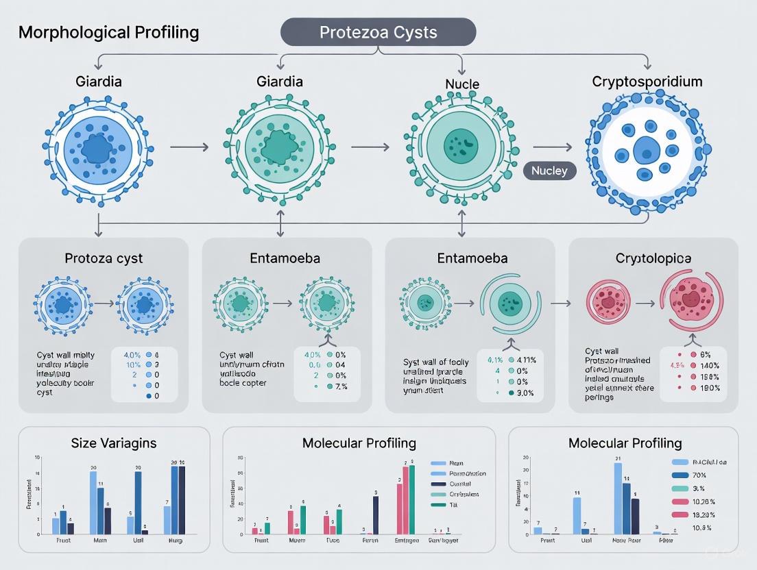
Comparative Morphology of Cysts
The definitive identification of intestinal protozoa relies on the meticulous examination of cyst morphology in stained or unstained preparations. Key diagnostic features include size, shape, number of nuclei, and the presence of specific intracellular structures such as chromatoid bodies and glycogen vacuoles [3]. The following section and table provide a detailed comparative analysis of these characteristics.
Table 2: Comparative Morphology of Major Pathogenic Protozoa Cysts
| Parasite | Size (Diameter or Length) | Shape | Number of Nuclei (Mature Cyst) | Nuclear Characteristics | Key Internal Structures | Staining Characteristics |
|---|---|---|---|---|---|---|
| Entamoeba histolytica | 10-20 µm (usual range: 12-15 µm) [3] | Usually spherical [3] | 4 [3] | Fine, uniformly distributed peripheral chromatin; small, discrete, usually central karyosome [3] | Chromatoid bodies with elongated, bluntly rounded ends [3] | Chromatoid bodies visible in unstained wet mounts; cysts stain reddish-brown with iodine [3] |
| Giardia duodenalis | 8-10 µm in diameter [2] | Oval to ellipsoidal | Not specified in morphological tables | Not visible in unstained mounts [3] | Internal axostyles; median bodies [3] | Cysts demonstrate a "falling leaf" motility in unstained wet mounts of fresh specimens [3] |
| Cryptosporidium parvum | 4-6 µm [4] | Spherical | Not applicable (oocysts contain sporozoites) | Not applicable | Contains four sporozoites [4] | Oocysts are acid-fast positive [3] and can be demonstrated with modified Ziehl-Neelsen stain [5] |
Key Morphological Differentiators
- Size Discrimination: Cryptosporidium oocysts (4-6 µm) are significantly smaller than Entamoeba (10-20 µm) and Giardia (8-10 µm) cysts, providing a primary distinguishing characteristic [2] [3] [4].
- Nuclear Characteristics: The mature quadrinucleated form of E. histolytica is diagnostic and contrasts with Giardia cysts, whose nuclei are not visible in unstained preparations [3].
- Internal Structures: The presence of chromatoid bodies with characteristic blunt ends in E. histolytica differs from the internal axostyles and median bodies of Giardia and the sporozoite-filled oocysts of Cryptosporidium [3] [4].
- Staining Properties: Cryptosporidium oocysts are distinguished by their acid-fast properties, which can be visualized with modified Ziehl-Neelsen staining, while Entamoeba and Giardia do not share this characteristic [3] [5].
Diagnostic Methodologies and Protocols
Accurate diagnosis of intestinal protozoan infections requires a multifaceted approach, as no single technique offers perfect sensitivity and specificity. The following section outlines standard and advanced diagnostic protocols used in both clinical and research settings.
Stool Specimen Processing and Staining
The initial examination of stool specimens for intestinal protozoa typically involves a combination of direct wet mounts and permanent stains, each providing complementary information [3].
- Unstained Wet Mounts (Saline and Formalin): Direct examination of fresh stool in saline allows for the assessment of motility in trophozoites (e.g., the "falling leaf" motility of Giardia trophozoites) [3]. Formalin-fixed specimens are used for concentration procedures and initial cyst identification, allowing visualization of general cyst shape and size [3] [5].
- Temporary Stains (Iodine): Lugol's iodine solution is widely used to enhance the visualization of protozoan cysts by staining glycogen vacuoles and nuclear structures, causing cysts to appear reddish-brown [3] [5]. This method is particularly useful for confirming the presence of Giardia cysts and differentiating the nuclear structure of Entamoeba species [5].
- Permanent Stains (Trichrome, Iron-Hematoxylin): Permanent staining is essential for the detailed morphological examination required for species differentiation, particularly for Entamoeba histolytica and other amoebae [3]. These stains provide high-resolution detail of nuclear characteristics and intracellular structures, allowing for definitive identification.
- Specialized Stains for Cryptosporidium: Due to their small size and weak uptake of common stains, Cryptosporidium oocysts require specialized staining techniques for visualization by light microscopy. The modified Ziehl-Neelsen (MZN) stain is commonly employed, which renders the oocysts bright red against a blue or green background [5]. Alternative methods include auramine-phenol fluorescence staining [4].
Immunological and Molecular Techniques
While microscopy remains fundamental, limitations in sensitivity and specificity have driven the development of alternative diagnostic methods.
- Immunoassays: Immunochromatographic tests (ICT) and enzyme immunoassays (ELISA) that detect parasite-specific antigens in stool samples have become valuable tools for the rapid diagnosis of Giardia, Cryptosporidium, and Entamoeba histolytica [5] [4]. These assays offer improved specificity for distinguishing E. histolytica from the morphologically identical non-pathogenic E. dispar [4].
- Molecular Detection (PCR): Molecular methods, particularly polymerase chain reaction (PCR), have demonstrated superior sensitivity and specificity compared to microscopy and antigen detection tests [4]. Multiplex real-time PCR assays have been developed for the simultaneous detection of E. histolytica, G. lamblia, and C. parvum in a single reaction, providing a powerful tool for differential diagnosis and epidemiological studies [4]. These assays can achieve 100% specificity and sensitivity when validated against well-defined samples and can include an internal control to detect PCR inhibition [4].
The diagnostic workflow for these parasites, integrating both traditional and modern techniques, can be visualized as follows:
Diagram Title: Diagnostic Workflow for Intestinal Protozoa
The Scientist's Toolkit: Essential Research Reagents and Materials
Successful morphological profiling and research on intestinal protozoa require a suite of specialized reagents and materials. The following table catalogs key solutions used in the field, drawing from established experimental protocols cited in this review [3] [5] [4].
Table 3: Key Research Reagent Solutions for Protozoan Cyst Analysis
| Reagent/Material | Function/Application | Specific Use Case |
|---|---|---|
| Lugol's Iodine Solution | Temporary staining of cysts; enhances visualization of glycogen and nuclei [3] [5] | Differentiation of protozoan cysts in wet mounts based on nuclear number and structure [5] |
| Modified Ziehl-Neelsen (MZN) Stain | Acid-fast staining of Cryptosporidium oocysts [5] | Specific identification of Cryptosporidium oocysts in stool smears; oocysts stain bright red [5] |
| Trichrome Stain | Permanent staining for detailed morphological examination of protozoa [3] | Differentiation of Entamoeba species based on nuclear detail and cytoplasmic appearance [3] |
| Immunomagnetic Separation (IMS) Kits | Purification and concentration of (oo)cysts from complex samples [9] | Separation of Giardia cysts and Cryptosporidium oocysts from fecal debris prior to IFA or molecular analysis [9] |
| Direct Immunofluorescent Antibody (IFA) Test | Specific detection and visualization of (oo)cysts using fluorescently labeled antibodies [9] | Gold-standard for detection and enumeration of Giardia and Cryptosporidium in water and environmental samples [9] |
| Multiplex Real-time PCR Assays | Simultaneous detection and differentiation of multiple parasite DNA targets in a single reaction [4] | High-throughput, specific, and sensitive diagnosis of E. histolytica, G. lamblia, and C. parvum from stool DNA extracts [4] |
| QIAamp DNA Stool Mini Kit | Isolation of high-quality PCR-grade DNA from stool specimens [4] | Standardized DNA extraction for downstream molecular detection and characterization [4] |
The comparative morphological profiling of Entamoeba histolytica, Giardia duodenalis, and Cryptosporidium parvum cysts remains a foundational skill in clinical parasitology and a critical component of public health responses to parasitic diseases. Despite the advent of highly sensitive molecular techniques, microscopy continues to provide the first line of identification in many settings, enabling rapid and cost-effective diagnosis. The distinct morphological features summarized in this guide—particularly size, nuclear morphology, and specialized structures—allow for their differentiation when examined by trained personnel using appropriate staining techniques. However, the limitations of microscopy, including its inability to distinguish pathogenic E. histolytica from non-pathogenic E. dispar and its variable sensitivity, underscore the necessity of integrating traditional methods with modern immunological and molecular assays. The continued refinement of multiplex PCR and other advanced diagnostics promises to enhance our understanding of the epidemiology and biology of these pathogens, ultimately contributing to more effective individual patient management and broader disease control strategies.
Within the framework of modern morphological profiling research for drug discovery, the detailed analysis of intestinal protozoa represents a critical frontier for understanding parasitic diseases and identifying therapeutic targets [10] [11]. This technical guide provides an in-depth examination of the light microscopy characteristics of intestinal protozoa cysts, focusing on the key morphometric parameters of size, shape, nuclear structure, and cytoplasmic inclusions. As morphological profiling evolves with high-content imaging and machine learning, standardized quantitative descriptions of parasitic structures become increasingly vital for automated classification and mechanism-of-action studies [12]. This whitepaper synthesizes traditional microscopic techniques with contemporary analytical approaches to establish a comprehensive reference for researchers and drug development professionals working in parasitology and infectious disease.
The diagnosis of intestinal protozoan parasites, which affect approximately 3.5 billion people globally, continues to pose formidable challenges despite advancements in molecular diagnostics [13] [14]. Microscopy remains the reference diagnostic method in clinical laboratories worldwide, but its effectiveness is limited by significant variations in sensitivity, specificity, and the ability to differentiate closely related species [13] [15]. For researchers investigating novel compounds against parasitic diseases, accurate morphological profiling provides a critical phenotypic readout for assessing drug efficacy, while for diagnostic scientists, these characteristics form the basis of differentiation between pathogenic and non-pathogenic species [10]. The resurgence of interest in morphological approaches, enhanced by digital imaging and artificial intelligence, underscores the continued relevance of precise morphological characterization in both basic research and applied drug discovery [12] [11].
Core Morphological Characteristics of Intestinal Protozoa Cysts
The cyst stage of intestinal protozoa represents the infectious, environmental-resistant form of these organisms, characterized by distinct morphological features that serve as primary diagnostic and research indicators. The identification and differentiation of cysts rely on four principal characteristics observable through light microscopy: size ranges, shape parameters, nuclear structure, and specialized cytoplasmic inclusions [3]. These features remain stable across specimen preparation methods and provide reliable metrics for both manual classification and automated image analysis pipelines in high-content screening environments [12].
Critical Morphological Parameters for Differentiation
For researchers engaged in morphological profiling of intestinal protozoa, the comparative analysis of cyst characteristics provides the foundation for accurate species identification. The following tables synthesize quantitative and qualitative data from standardized morphological references, enabling direct comparison between species during experimental analysis.
Table 1: Comparative Morphology of Amoebae Cysts
| Species | Size (Diameter) | Shape | Nuclear Count | Peripheral Chromatin | Karyosomal Chromatin | Cytoplasmic Inclusions |
|---|---|---|---|---|---|---|
| Entamoeba histolytica | 10-20 µm (usual range: 12-15 µm) | Spherical | 4 in mature cyst | Fine, uniform granules, evenly distributed | Small, discrete, usually central | Chromatoid bars with bluntly rounded ends; diffuse glycogen mass |
| Entamoeba coli | 10-35 µm (usual range: 15-25 µm) | Spherical (occasionally oval, triangular) | 8 in mature cyst | Coarse granules, irregular in size and distribution | Large, discrete, usually eccentric | Splinter-like chromatoid bodies with pointed ends; diffuse glycogen |
| Entamoeba hartmanni | 5-10 µm (usual range: 6-8 µm) | Spherical | 4 in mature cyst | Similar to E. histolytica | Similar to E. histolytica | Elongated chromatoid bars with bluntly rounded ends |
| Endolimax nana | 5-10 µm (usual range: 6-8 µm) | Spherical to oval | 4 in mature cyst | None | Large, blot-like, usually central | Occasionally granules; no typical chromatoid bodies |
| Iodamoeba bütschlii | 5-20 µm (usual range: 10-12 µm) | Ovoidal, ellipsoidal, triangular | 1 in mature cyst | None | Large, usually eccentric with refractile achromatic granules | Compact, well-defined glycogen mass |
Table 2: Comparative Morphology of Flagellate Cysts and Other Intestinal Protozoa
| Species | Size (Diameter or Length) | Shape | Nuclear Count | Other Diagnostic Features | Cytoplasmic Inclusions |
|---|---|---|---|---|---|
| Giardia duodenalis | 8-12 µm (cysts) | Oval | 4 | Sucking disk (in trophozoites); median bodies | Flagella remnants rarely visible |
| Chilomastix mesnili | 6-10 µm (cysts) | Lemon-shaped | 1 | Prominent cytostome extending 1/3-1/2 length of body (trophozoites) | Fibrils; single nucleus |
| Cryptosporidium spp. | 4-6 µm (oocysts) | Spherical | - | Small, poorly stained; modified acid-fast positive | 4 sporozoites (in mature oocysts) |
Diagnostic Significance of Morphological Features
The morphological parameters detailed in Tables 1 and 2 provide critical diagnostic information that enables researchers to differentiate between pathogenic and non-pathogenic species. For example, the differentiation between Entamoeba histolytica (pathogenic) and Entamoeba dispar (non-pathogenic) represents a significant diagnostic challenge, as these species are morphologically identical under light microscopy and can only be distinguished through molecular techniques [15] [14]. This limitation underscores the importance of correlating morphological observations with ancillary testing in research settings focused on drug discovery against amoebiasis.
The size ranges provided serve as primary differentiators between species with overlapping characteristics. For instance, Entamoeba hartmanni cysts (5-10 µm) can be distinguished from those of Entamoeba histolytica (10-20 µm) primarily through size determination [3]. Similarly, the nuclear structure provides crucial diagnostic information: the number of nuclei in mature cysts (ranging from 1 in Iodamoeba bütschlii to 8 in Entamoeba coli), the distribution of peripheral chromatin (fine and uniform in E. histolytica versus coarse and irregular in E. coli), and the appearance of the karyosomal chromatin (small and discrete versus large and blot-like) collectively create a diagnostic profile for each species [3].
Cytoplasmic inclusions offer additional differentiation criteria, with chromatoid bodies appearing as elongated bars with rounded ends in E. histolytica versus splinter-like forms with pointed ends in E. coli [3]. Glycogen masses, which stain reddish-brown with iodine, vary from diffuse in most species to compact and well-defined in Iodamoeba bütschlii [3]. These features remain observable across different staining methodologies, though their prominence may vary depending on the preparation technique employed.
Advanced Light Microscopy Techniques for Morphological Profiling
The accurate identification of intestinal protozoa cysts requires not only understanding morphological characteristics but also selecting appropriate contrast-enhancement techniques to visualize these virtually transparent specimens. Conventional brightfield microscopy often provides insufficient contrast for detailed observation of unstained cysts, necessitating the implementation of specialized illumination techniques that convert subtle phase variations induced by the specimen into measurable intensity differences [16] [17].
Contrast-Enhancement Methodologies
Phase Contrast Microscopy: Originally described by Frits Zernike in the 1930s, phase contrast microscopy employs an optical mechanism to translate minute variations in phase into corresponding changes in amplitude, which can be visualized as differences in image contrast [16]. The technique utilizes a specialized condenser containing an annulus matched to objectives with corresponding phase rings in the rear focal plane. As light passes through specimens with different refractive indices, the resulting phase shifts are converted into intensity variations, rendering transparent cysts visible without staining [16]. While ideal for observing unstained specimens, phase contrast often produces halo artifacts around cyst boundaries, which can obscure fine details and complicate automated image analysis [16].
Differential Interference Contrast (DIC): Also known as Nomarski interference contrast, DIC microscopy employs a Wollaston prism-based system to separate polarized light into two beams that pass through adjacent areas of the specimen before being recombined [17]. The resulting interference pattern creates a pseudo-three-dimensional image with shadow-cast relief, emphasizing edges and internal structures. DIC provides superior resolution compared to phase contrast without halo artifacts, making it particularly valuable for observing fine nuclear details and cytoplasmic inclusions [17].
Darkfield Microscopy: This technique employs oblique illumination beyond the maximum angle that optical imaging systems can capture, thereby minimizing unscattered background light while collecting only light scattered by the specimen [18]. The resulting images display bright specimen features against a dark background, providing high contrast for detecting cysts but limited internal detail [18]. Darkfield is particularly sensitive to edges and can be useful for initial detection of cysts in low-concentration samples.
Color-Coded LED Microscopy (cLEDscope): Recent advances in computational microscopy have enabled multi-contrast imaging through color-coded illumination. This innovative approach uses a programmable three-color LED array to illuminate specimens, with each color corresponding to a different illumination angle [18]. A single color image sensor records transmitted light, and computational separation of color channels enables simultaneous brightfield, darkfield, and differential phase contrast imaging from a single exposure [18]. This method shows particular promise for high-throughput morphological profiling applications in drug discovery research.
Technical Workflow for Microscopic Analysis
The following diagram illustrates the integrated workflow for morphological analysis of intestinal protozoa cysts, incorporating both traditional and advanced computational approaches:
Figure 1: Integrated Workflow for Morphological Analysis of Intestinal Protozoa Cysts
Experimental Protocols for Morphological Analysis
Standard Stool Processing and Staining Protocols
Sample Collection and Fixation:
- Collect fresh stool samples in clean, waterproof containers without preservatives for immediate processing, or use preservation media (SAF or Para-Pak) for stored specimens [13] [14].
- For fixation, emulsify 1-2 g of stool in 10 mL of sodium-acetate-acetic acid-formalin (SAF) solution for optimal preservation of morphological features [12] [14].
- Fixed samples can be stored at 4°C for several weeks without significant degradation of morphological features.
Concentration Techniques:
- Formalin-Ethyl Acetate Concentration: Homogenize approximately 1 g of stool in 10 mL of 10% formalin. Filter through gauze into a 15 mL conical tube. Add 4 mL of ethyl acetate, shake vigorously for 30 seconds, and centrifuge at 500 × g for 2 minutes. Decant the top layers and examine the sediment [13] [3].
- Ritchie-Frick Concentration: Adapt the formalin-ethyl acetate method by incorporating a specific centrifugation protocol at 505 × g for 10 minutes after the addition of TritonX-100 and ethyl acetate [12].
Staining Methods for Enhanced Contrast:
- Iodine Staining: Mix a small amount of stool sediment with a drop of Lugol's iodine on a glass slide. Apply a coverslip and examine immediately. Iodine stains glycogen masses reddish-brown and enhances nuclear visibility [3] [14].
- Permanent Stains (Trichrome): Prepare a fixed smear of stool sediment on a slide and allow to air dry. Place in trichrome stain for 10 minutes, rinse in acid-alcohol, dehydrate through alcohol series, clear in xylene, and mount with synthetic resin [15] [12]. Permanent stains enhance nuclear detail and cytoplasmic inclusions.
- Modified Acid-Fast Staining: Particularly valuable for Cryptosporidium spp. identification. Prepare smears from stool sediment, air dry, and fix with methanol. Flood with carbol-fuchsin for 10-15 minutes, decolorize with acid-alcohol, and counterstain with methylene blue for 1 minute [15]. Acid-fast oocysts stain bright red against a blue background.
Digital Microscopy and Computational Analysis Protocol
Recent advances in computational microscopy have enabled automated detection and classification of intestinal parasites through convolutional neural networks (CNNs). The following protocol outlines the validated approach for digital morphological profiling:
Slide Preparation and Scanning:
- Mix 15 µL of stool sediment with 15 µL of mounting medium (Lugol's iodine and glycerol in PBS) on a standard glass slide [12].
- Cover with a 22 × 22 mm coverslip, ensuring even distribution without bubbles.
- Scan slides using a digital slide scanner (e.g., Grundium Ocus 40) equipped with a 20× 0.75 NA objective, capturing images at an effective 40× magnification (0.25 microns per pixel) across multiple focal planes [12].
CNN-Based Analysis:
- Upload digital slide images to a specialized analysis platform (e.g., Techcyte Human Fecal Wet Mount algorithm) [12].
- The algorithm performs pre-classification to determine presence or absence of target parasites, followed by organism/class-level identification by labeling image regions accordingly.
- Validate algorithm performance against manual light microscopy, with expert review of discrepant results [12].
Validation Parameters:
- Assess analytical sensitivity through dilution series of reference samples
- Determine positive and negative percent agreement with gold standard microscopy
- Evaluate intra- and inter-run precision for reproducibility assessment [12]
The Scientist's Toolkit: Research Reagent Solutions
Table 3: Essential Research Reagents for Morphological Analysis of Intestinal Protozoa
| Reagent Solution | Function | Application Notes |
|---|---|---|
| SAF Fixative (Sodium-Acetate-Acetic Acid-Formalin) | Preserves morphological integrity of cysts during transport and storage | Maintains nuclear structure and cytoplasmic inclusions without distortion; compatible with molecular assays [12] [14] |
| Lugol's Iodine Solution | Enhances contrast of glycogen masses and nuclear structures in wet mounts | Stains glycogen reddish-brown; provides temporary staining for immediate examination [3] [14] |
| Trichrome Stain | Permanent staining for detailed observation of nuclear characteristics and cytoplasmic inclusions | Differentiates nuclear chromatin and reveals internal structures; permanent preparation for archival purposes [15] [12] |
| Modified Acid-Fast Stain | Specific identification of Cryptosporidium oocysts | Stains oocysts bright red against blue background; essential for Cryptosporidium detection [15] |
| Formalin-Ethyl Acetate Solution | Concentration of parasitic elements through sedimentation | Standard concentration method that preserves morphological features; increases detection sensitivity [13] [3] |
| S.T.A.R. Buffer (Stool Transport and Recovery Buffer) | Stabilizes nucleic acids while preserving morphological integrity | Enables parallel morphological and molecular analysis from same specimen [13] |
Integration with Contemporary Research Methodologies
The morphological characteristics detailed in this guide form the foundation for emerging technologies in parasitic disease research and drug discovery. High-content morphological profiling, which captures quantitative features of cellular and parasitic structures, enables the rapid prediction of compound bioactivity and mechanism of action in anti-parasitic drug screening [10]. The standardized parameters outlined herein provide the essential framework for developing automated classification systems that combine traditional morphological expertise with computational efficiency.
Advanced imaging platforms now leverage these morphological descriptors to train convolutional neural networks capable of identifying intestinal protozoa with accuracy comparable to experienced microscopists [12]. These systems typically achieve >90% slide-level agreement with light microscopy while significantly reducing analysis time, demonstrating the enduring value of well-defined morphological criteria in the era of digital pathology [12]. Furthermore, the integration of morphological profiling with molecular techniques creates powerful multidimensional datasets for understanding parasite biology and host-parasite interactions [13] [14].
As morphological profiling continues to evolve in the drug discovery pipeline, the precise characterization of intestinal protozoa cysts will remain essential for phenotypic screening campaigns targeting parasitic diseases. The quantitative parameters established in this technical guide provide the necessary foundation for correlating morphological changes with therapeutic interventions, ultimately contributing to the development of novel anti-parasitic therapeutics.
The Entamoeba histolytica and Entamoeba dispar complex represents a significant diagnostic challenge in clinical parasitology and protozoan research. These two species, while morphologically identical in their cyst and trophozoite forms through light microscopy, differ profoundly in their pathogenic potential [19] [20]. E. histolytica is a well-recognized pathogen capable of causing invasive amebic colitis and extraintestinal abscesses, while E. dispar is generally considered non-pathogenic [19] [21]. This biological dichotomy, concealed by morphological similarity, complicates both accurate diagnosis and appropriate treatment decisions, necessitating reliable differentiation methods beyond conventional microscopy.
The significance of this differentiation extends to both clinical management and public health. Without species-level identification, patients with E. dispar infection may receive unnecessary antiamoebic chemotherapy, while those with E. histolytica risk undertreatment and disease progression [20]. Within the context of morphological profiling of intestinal protozoa cysts, this complex serves as a paradigm for the limitations of purely morphological approaches and the necessity for molecular characterization in research settings, particularly in drug development where pathogen-specific targets are essential.
Morphological Characteristics and Limitations
Comparative Morphology of Entamoeba Cysts and Trophozoites
Traditional diagnosis relies on microscopic examination of stool specimens to identify characteristic cysts and trophozoites. However, as members of the E. histolytica/dispar complex are morphologically indistinguishable, they are identified collectively during routine microscopy [19] [22]. The defining features visible under microscopy are summarized in Table 1.
Table 1: Morphological Characteristics of Entamoeba histolytica/dispar Complex
| Stage | Size | Nuclear Characteristics | Cytoplasmic Inclusions | Additional Features |
|---|---|---|---|---|
| Cyst | 10-20 µm (usual range 12-15 µm) [3] | Mature cysts: 4 nuclei with fine, uniformly distributed peripheral chromatin and small, centrally located karyosomes [19] [3] | Chromatoid bodies with blunt, rounded ends [19]; glycogen mass in immature cysts [3] | Spherical shape; nuclei may not be visible in unstained preparations [3] |
| Trophozoite | 15-20 µm (range 10-60 µm) [19] [3] | Single nucleus with fine peripheral chromatin and small, central karyosome [19] [3] | Finely granular, "ground-glass" appearance [19]; may contain ingested bacteria [3] | Progressive, directional motility with hyaline, finger-like pseudopods [3]; elongated in diarrheal stool [19] |
A morphological feature historically associated with E. histolytica pathogenicity is erythrophagocytosis (ingestion of red blood cells) by trophozoites [19]. While this finding is highly suggestive of E. histolytica infection, it is not an entirely reliable diagnostic criterion, as it has been rarely reported in E. dispar infections, and is infrequently observed in stained smears [19] [22]. Furthermore, one study noted that hematophagy, when present in direct smears, was always associated with E. histolytica infection, but this finding is rare [22].
Diagnostic Constraints of Microscopy
The primary limitation of microscopy is its inability to differentiate E. histolytica from E. dispar [23] [20] [24]. This fundamental shortcoming has led to the widespread over-reporting of E. histolytica in regions where E. dispar is more common [22]. Additional constraints include:
- Variable Sensitivity: The sensitivity of microscopic O&P examinations is reported to be as low as 20-90% compared to molecular assays, requiring the examination of multiple stool specimens (typically three) to improve detection yield [25].
- Technical Expertise: Accurate morphological identification demands highly skilled and experienced technologists, a resource becoming increasingly scarce in many clinical laboratories [25].
- Inability to Detect Co-infections: Microscopy cannot discern mixed infections with different Entamoeba species, which do occur, further complicating the clinical picture [20].
Molecular Differentiation Techniques
Molecular techniques have emerged as the reference standard for the precise differentiation of Entamoeba species, overcoming the limitations of morphological and antigen-based assays.
PCR-Based Assays
Polymerase chain reaction (PCR) assays target species-specific genetic sequences, offering high sensitivity and specificity. Various gene targets and PCR methodologies have been successfully implemented, as detailed in Table 2.
Table 2: Molecular Assays for Differentiation of Entamoeba Species
| Assay Type | Genetic Target | Key Primers (Sequence 5'→3') | Differentiation Power | Reported Performance |
|---|---|---|---|---|
| In-house PCR [23] | Small-subunit (SSU) rRNA (135 bp amplicon) | EH1: GTACAAAATGGCCAATTCATTCAATGED1: TACAAAGTGGCCAATTTATGTAAGTAEHD2 (common reverse): ACTACCAACTGATTGATAGATCAG | E. histolytica vs E. dispar | Much more sensitive than microscopy; well-suited as a reference test [23] |
| Nested PCR-RFLP [20] | 16S-like ribosomal RNA gene | Not specified in detail | E. histolytica, E. dispar, and E. moshkovskii | Successfully identified species in 52/75 microscopy-positive samples [20] |
| PCR-DGGE [26] | adh112 gene (228 bp region) | First PCR:Fw: GCAGAAAAAAATAATAATAACRv: TTCATTTGTTTTACTTTCANested PCR (with GC-clamp):Fw: CGCCCGCCGCGCGGC...CAGAAAAAAATAATAATAACRv: TTCATTTGTTTTACTTTCA | E. histolytica vs E. dispar based on sequence denaturation profiles | A promising, highly specific tool for differentiation; confirmed 10/62 samples as E. histolytica [26] |
| Multiplex Real-time PCR(Allplex Assay) [24] | Not specified | Proprietary (commercial kit) | E. histolytica directly detected | 100% sensitivity and specificity for E. histolytica vs traditional methods [24] |
Experimental Protocol: PCR for E. histolytica/dispar Differentiation
The following protocol, adapted from published research, outlines a standard procedure for differentiating Entamoeba species using PCR amplification of the SSU rRNA gene [23].
1. DNA Extraction:
- Sample Input: Use approximately 10 mg of unfixed, frozen stool specimen. Formalin fixation can inhibit PCR amplification and is not recommended for molecular workups [23] [25].
- Method: Employ a commercial DNA extraction kit (e.g., QIAamp DNA Mini Kit) according to the manufacturer's instructions, incorporating a mechanical lysis step to break open the robust cyst walls [23] [24].
2. PCR Amplification:
- Reaction Mix: 50 µL volume containing:
- Cycling Conditions:
- Initial denaturation: 95°C for 15 minutes.
- 40 cycles of:
- Denaturation: 94°C for 30 seconds.
- Annealing: 51°C for 60 seconds.
- Extension: 72°C for 40 seconds.
- Final extension: 72°C for 5 minutes [23].
3. Internal Amplification Control:
- To control for PCR inhibition, a competitive internal control (IC) should be co-amplified. The IC is a constructed DNA fragment that uses the same primer binding sites but yields a slightly larger amplicon (240-266 bp) [23].
4. Analysis:
- Analyze PCR products by gel electrophoresis (e.g., 3% agarose). A positive result is indicated by the presence of a 135-bp band corresponding to either E. histolytica or E. dispar, depending on the primer set used. The absence of both the target and IC bands indicates PCR inhibition [23].
Diagram 1: Workflow for PCR-based differentiation of E. histolytica and E. dispar, highlighting critical steps like unfixed sample use and internal amplification control.
The Researcher's Toolkit: Essential Reagents and Materials
Successful differentiation of Entamoeba species relies on specific laboratory reagents and tools. Table 3 lists key research solutions for experiments in this field.
Table 3: Research Reagent Solutions for Entamoeba Differentiation
| Reagent/Material | Function/Application | Example Product/Note |
|---|---|---|
| DNA Extraction Kit | Isolation of inhibitor-free genomic DNA from tough-walled cysts in stool. | QIAamp DNA Mini Kit [23]; PowerSoil DNA Isolation Kit [20]; Wizard Genomic DNA Purification Kit [26] |
| Species-specific Primers | Amplification of unique genetic sequences for identification. | SSU rRNA primers (EH1/ED1/EHD2) [23]; adh112 gene primers [26]; 16S-like rRNA primers for nested PCR [20] |
| PCR Enzyme & Master Mix | Robust amplification, often from low-quantity or degraded samples. | HotStar Taq DNA Polymerase [23]; Accu Prime Taq DNA Polymerase High Fidelity [26] |
| Internal Control (IC) DNA | Detection of PCR inhibition in stool samples, a common issue. | Competitively amplified pBR322-derived fragment [23] |
| Commercial Multiplex PCR Assay | Standardized, high-throughput detection of E. histolytica and other GI parasites. | Allplex GI-Parasite Assay (Seegene Inc.) [24] |
| Reference Strain Genomic DNA | Essential positive control for assay validation and troubleshooting. | E. histolytica HM-1:IMSS; E. dispar SAW760 [26] [21] |
Implications for Research and Drug Development
The clear differentiation between pathogenic and non-pathogenic Entamoeba species is not merely a diagnostic concern but is fundamental to basic research and therapeutic development.
- Virulence Factor Discovery: Comparative proteomic studies between E. histolytica and E. dispar have revealed proteins differentially expressed between the species, such as alcohol dehydrogenase 3 (EhADH3), which is present at significantly higher levels in the pathogenic species [21]. These proteins represent candidate virulence factors and potential targets for novel therapeutics.
- Accurate Preclinical Models: The use of genetically confirmed E. histolytica strains in animal models of amebic colitis and liver abscess is critical for validating drug efficacy against the true pathogen, rather than a non-pathogenic look-alike [21].
- Epidemiological Accuracy: Molecular tools provide a true picture of the distribution and disease burden of E. histolytica, allowing public health resources and drug development efforts to be directed appropriately [20] [22].
Diagram 2: The critical role of molecular differentiation in bridging fundamental morphological research to applied clinical and drug development outcomes.
The differentiation of pathogenic E. histolytica from non-pathogenic E. dispar is a critical capability in modern parasitology. While microscopic examination remains a foundational tool for the morphological profiling of intestinal protozoa, its limitations necessitate the integration of molecular methods for definitive species identification. PCR-based techniques, targeting genetic markers such as the SSU rRNA or adh112 genes, provide the sensitivity, specificity, and reliability required for accurate diagnosis, meaningful epidemiological research, and targeted drug development. As the field advances, the adoption of multiplexed molecular panels in research and reference settings will further enhance our understanding of this complex and contribute to the development of more effective, pathogen-specific interventions.
Within the scope of morphological profiling of intestinal protozoa cysts, differential identification of oocysts of Cryptosporidium spp. and Cyclospora cayetanensis presents a significant diagnostic challenge. These coccidian parasites are important causes of waterborne and foodborne diarrheal illnesses worldwide [27] [28]. A critical first step in the diagnostic pipeline and subsequent drug development research is their accurate morphological characterization, particularly using acid-fast staining methods. This technical guide provides an in-depth comparison of their key morphological features, detailed experimental protocols for their detection, and essential research reagents, serving as a foundational resource for scientists and laboratory professionals engaged in enteric protozoa research.
Morphological Profiling: A Comparative Analysis
The definitive differentiation between Cryptosporidium and Cyclospora oocysts relies on a combination of size, internal structure, and staining characteristics. The table below provides a consolidated summary of their distinguishing features.
Table 1: Comparative Morphological and Staining Characteristics of Cryptosporidium and Cyclospora Oocysts
| Feature | Cryptosporidium spp. | Cyclospora cayetanensis |
|---|---|---|
| Size | 4.2 - 5.4 µm in diameter [29] | 8 - 10 µm in diameter [30] [31] |
| Shape | Rounded [29] | Spherical [30] |
| Staining Property | Acid-fast positive [29] | Variable acid-fast staining [30] |
| Modified Acid-Fast Appearance | Bright pink to red [29] [32] | Pink to red, or unstained "ghost" cells [30] [31] |
| Autofluorescence | Yes, with auramine-rhodamine stain [29] | Yes, natural autofluorescence under UV light [30] [31] |
| UV Microscopy | Not typically used for primary detection | Blue (330-365 nm) or green (450-490 nm) fluorescence [31] |
| Sporozoites Visible by Light Microscopy | Sometimes [29] | Not in unstained, unsporulated oocysts [30] |
| Infectivity upon Excretion | Yes (immediate fecal-oral transmission) [29] | No (requires days/weeks to sporulate in environment) [30] [33] |
Cryptosporidium Oocysts
Cryptosporidium oocysts are notably small, with a diameter of 4.2 to 5.4 µm [29]. In a wet mount, they appear as rounded, refractive bodies [29]. When stained using a modified acid-fast technique, the oocysts stain a bright pink-to-red color and may contain visible internal sporozoites [29] [32]. It is important to note that staining can be variable, and infections that are resolving may show increasing numbers of non-acid-fast "ghost" cells [29]. Oocysts are also fluorescent when stained with auramine-rhodamine [29]. A key diagnostic feature is that the oocysts are infectious immediately upon excretion, facilitating direct fecal-oral transmission [29].
Cyclospora Oocysts
Cyclospora cayetanensis oocysts are larger, measuring 8 to 10 µm in diameter [30] [31]. In unstained wet mounts, they are spherical and unsporulated [30]. Under UV fluorescence microscopy, which is a highly reliable detection method, the oocysts autofluoresce with a blue or green glow, a property that is impaired by iodine [30] [31]. With modified acid-fast staining, the oocysts can exhibit considerable variability, staining from pink to brilliant red, or failing to take up the stain and appearing as clear "ghost" cells [30] [31]. Critically, unlike Cryptosporidium, the oocysts are not infectious when shed and require days to weeks in the environment to sporulate and become infective [30] [33].
Experimental Protocols for Detection and Identification
Standard Diagnostic Workflow
The following diagram illustrates the core decision-making pathway for the microscopic identification and differentiation of these oocysts in a clinical or research setting.
Modified Acid-Fast Staining Protocol
The modified acid-fast stain is a cornerstone technique for the morphological profiling of these coccidian parasites. The following is a detailed protocol, adaptable for both parasites, though results must be interpreted with an understanding of their specific staining variabilities [32].
Table 2: Research Reagent Solutions for Acid-Fast Staining
| Reagent | Function | Technical Notes |
|---|---|---|
| Absolute Methanol | Fixative. Preserves morphology and adheres specimen to slide. | Ensure slides are completely dry before proceeding to staining. |
| Kinyoun's Carbol Fuchsin | Primary stain. Phenol and basic fuchsin penetrate the complex oocyst wall. | Can be used cold (without heating). Filter before use for consistent results [32]. |
| 10% Sulfuric Acid (H₂SO₄) | Decolorizer. Removes stain from non-acid-fast organisms and background debris. | Acts as a milder alternative to the acid-alcohol used in traditional AFB stains [32]. |
| 3% Malachite Green | Counterstain. Provides contrast by staining background material. | A 2-5 minute application is typical. Methylene blue is also an option [29] [32]. |
Procedure:
- Smear Preparation: Create a thin smear of fecal specimen on a clean glass slide and allow it to air dry completely [32].
- Fixation: Flood the slide with absolute methanol for 10 minutes. Allow the slide to dry after fixation [32].
- Primary Staining: Flood the fixed smear with filtered Kinyoun's carbol fuchsin stain. Allow it to stand for 5 minutes [32].
- Washing: Rinse the slide thoroughly with tap water. It is critical to wash until no more color runs from the slide [32].
- Decolorization: Decolorize by applying 10% sulfuric acid solution. For very thin smears, a quick dip may be sufficient. Rinse immediately with tap water to stop the decolorization process [32].
- Counterstaining: Apply 3% malachite green counterstain for 2 to 5 minutes [32].
- Final Wash and Drying: Rinse the slide gently with tap water, blot dry, and allow it to air dry completely [32].
- Microscopic Examination: Examine the smear under oil immersion (100x objective). Cryptosporidium oocysts appear as bright pink-to-red spherical bodies, 4-6 µm in diameter, often surrounded by a colorless halo. Cyclospora oocysts, measuring 8-10 µm, will show variable staining from pink to red, or may be unstained "ghosts" [29] [30] [32].
Advanced Techniques and Research Considerations
Molecular Profiling
For definitive species identification and epidemiological tracking, molecular methods are paramount. Polymerase chain reaction (PCR) is increasingly used in reference laboratories, as it can differentiate Cryptosporidium at the species level (e.g., distinguishing C. hominis from C. parvum) [29] [34]. For Cyclospora, several conventional and real-time PCR protocols have been developed, including the FDA-approved FilmArray Gastrointestinal Panel, which offers high sensitivity [30] [31]. It is crucial to note that specimen preservation is key for molecular success; formalin-based fixatives are not recommended as they adversely affect nucleic acids [29].
Limitations and Methodological Considerations
- Staining Variability: A significant limitation of the modified acid-fast stain is its inconsistency, particularly for Cyclospora, which can lead to false negatives if "ghost" cells are predominant. UV fluorescence microscopy is significantly more reliable for detecting Cyclospora [31].
- Oocyst Shedding: Both parasites can be excreted intermittently and in low numbers. Therefore, analysis of multiple stool specimens collected over several days is recommended to maximize detection sensitivity [29] [30].
- Concentration Techniques: To maximize oocyst recovery, stool specimens must be concentrated. The formalin-ethyl acetate sedimentation method is recommended, potentially with increased centrifugation speed or time (500 x g for 10 minutes) to prevent the small, low-mass oocysts from being trapped [29].
This technical guide examines the sophisticated structural and biochemical composition of intestinal protozoan cysts and oocysts, which underpin their remarkable environmental resilience. Focusing on key human pathogens including Giardia duodenalis, Cryptosporidium spp., and Entamoeba histolytica, we delineate the molecular architecture of their protective walls and the consequent resistance to chemical disinfectants and environmental stressors. The integration of advanced molecular diagnostics and proteomic analyses has revealed a complex framework of cysteine-rich proteins, carbohydrate polymers, and tyrosine-based cross-links that determine interaction forces, predation resistance, and ultimately, transmission dynamics. This morphological profiling is crucial for informing drug development targets and improving public health strategies against these pervasive pathogens.
Intestinal protozoan parasites represent a significant global health burden, with their transmission and persistence heavily reliant on the environmental stability of their cystic stages [35] [15]. The cyst (for Giardia and Entamoeba) and oocyst (for Cryptosporidium and Toxoplasma) forms constitute a critical interface between the parasite and its external environment, governing survival, transport, and infectivity [35]. These structures exhibit formidable resistance to a range of physicochemical stressors, including standard water disinfection protocols, enabling their survival for months in water, soil, and food sources [35] [36]. This resilience is directly attributable to the unique biochemical composition and nano-scale architecture of the cyst/oocyst wall—a subject of intense study within morphological profiling research. Understanding these structures is paramount for developing novel chemotherapeutic agents, disinfectants, and surveillance methodologies aimed at interrupting the transmission cycle of these pathogens.
Structural and Biochemical Composition of Cyst Walls
The cyst/oocyst wall is a complex, specialized structure that is largely impermeable to environmental insults. Its composition varies significantly among protozoan species, reflecting diverse evolutionary adaptations.
Table 1: Comparative Structural Characteristics of Key Protozoan Cysts/Oocysts
| Characteristic | Giardia duodenalis Cyst | Cryptosporidium Oocyst | Toxoplasma gondii Oocyst | Entamoeba histolytica Cyst |
|---|---|---|---|---|
| Size (μm) | 7–10 × 5 [35] | 3.8–6.3 × 4.6–8.4 [35] | 10 × 12 [35] | 10–20 [3] |
| Wall Thickness (nm) | 300–500 [35] | 50–80 [35] | ~100 [35] | Not Specified |
| Number of Layers | 2 [35] | 3 [35] | 2 [35] | Not Specified |
| Key Biochemical Components | Filamentous N-acetylgalactosamine (GalNAc) homopolymer; Cysteine-rich wall proteins (CWP1-3) [35] | Glucose-rich glycocalyx; High-molecular-weight cysteine-rich proteins (COWPs) [35] | Cysteine-rich proteins (OWP1-3); Tyrosine-rich proteins forming dityrosine cross-links [35] | Not fully elucidated; Presence of chitin suggested in related species |
| Major Structural Features | Dense network of curled 10 nm fibrils [35] | Inner glycoprotein layer, central lipid-protein layer, outer glycocalyx [35] | Protein-rich (>90%); Inner layer provides robustness [35] | Mature cyst contains 4 nuclei [3] |
Giardia duodenalis Cysts
The Giardia cyst wall is remarkably thick (300-500 nm) and is composed primarily of a filamentous layer of curled fibrils made of an N-acetylgalactosamine (GalNAc) homopolymer [35]. This polysaccharide matrix is closely associated with three major leucine-rich, cysteine-rich cyst wall proteins (CWP1-3) that contribute to structural integrity [35]. This assembly forms a dense, impermeable barrier that shields the internal parasite from hydraulic stress and chemical disinfectants [35].
Cryptosporidium Oocysts
The Cryptosporidium oocyst wall is thinner (50-80 nm) but more complex, featuring three distinct layers [35]. The inner layer is composed of a matrix of glycoproteins cross-linked by disulfide bridges formed by cysteine-rich proteins (COWPs), conferring mechanical strength [35]. This is overlain by a central lipid-protein layer and an outer, delicate glucose-rich glycocalyx that interfaces directly with the environment and may facilitate attachment [35] [36].
Toxoplasma gondii Oocysts
The Toxoplasma oocyst wall is approximately 100 nm thick and is predominantly proteinaceous (>90%) [35]. Its robustness is derived from two key components: cysteine-rich proteins (TgOWP1-3), which are structurally homologous to those in Cryptosporidium, and tyrosine-rich proteins that form protein-protein dityrosine cross-links [35]. These dityrosine bonds are responsible for the hardening of the wall and its characteristic blue autofluorescence under UV light [35].
Mechanisms of Environmental Resistance and Disinfection Survival
The structural complexity of the cyst/oocyst wall translates directly into exceptional environmental persistence and resistance to control measures.
Table 2: Resistance Profiles and Key Physicochemical Properties
| Property / Mechanism | Giardia duodenalis | Cryptosporidium spp. | Toxoplasma gondii |
|---|---|---|---|
| Zeta Potential | -33.5 mV (distilled H₂O, pH 6.4) [35] | -25.0 mV (deionized H₂O, pH 6.5) [35] | -43.7 mV (ultrapure H₂O, pH 6.7) [35] |
| Specific Gravity | 1.013–1.117 [35] | 1.009–1.08 [35] | 1.050–1.100 [35] |
| Primary Resistance Mechanism | Impermeable filamentous polysaccharide-protein wall [35] | Triple-layered wall with cross-linked protein matrix and lipid layer [35] | Dityrosine cross-linked protein wall [35] |
| Chlorine Resistance | High resistance at standard doses [36] | Extremely high; more resistant than bacterial spores [36] | Not Specified |
The highly negative zeta potential of cysts and oocysts creates an electrostatic repulsion that prevents aggregation with other particles and surfaces, influencing their transport through aquatic and terrestrial environments [35]. Furthermore, the compact, polymeric nature of the wall acts as a formidable permeability barrier. For instance, the outer membrane of mycobacteria, which is also highly resistant, has an extremely low permeability coefficient due to its hydrophobic, lipid-rich structure, a property shared by many cyst walls [36]. This barrier function limits the uptake of hydrophilic disinfectants like chlorine, rendering standard water treatment processes ineffective against pathogens like Cryptosporidium, which is notably more resistant to chlorination than bacterial spores [36].
Advanced Experimental Methodologies for Morphological Profiling
A multi-faceted approach is required to fully deconstruct the complexity of protozoan cysts. The integration of microscopy, molecular biology, and proteomics provides a comprehensive toolkit for researchers.
Proteomic Analysis of Encystment (Based onEuplotes encysticusModel)
The process of encystment involves drastic physiological and morphological changes driven by alterations in protein expression. Isobaric Tags for Relative and Absolute Quantitation (iTRAQ)-based proteomics is a powerful method to identify these changes [37].
Protocol: iTRAQ-Based Identification of Encystment-Related Proteins
- Sample Preparation: Collect vegetative cells and resting cysts. Lyse cells and extract total protein. Assess protein concentration and integrity via SDS-PAGE [37].
- Protein Digestion and Labeling: Digest the protein extract with trypsin. Label the resulting peptides from vegetative cells and resting cysts with different iTRAQ reagents (e.g., 114 and 115 tags) [37].
- Liquid Chromatography and Tandem Mass Spectrometry (LC-MS/MS): Combine the labeled peptide samples and fractionate using liquid chromatography. Analyze the fractions by tandem mass spectrometry to sequence the peptides [37].
- Data Analysis: Search the acquired spectra against a protein database for identification. Quantify protein abundance based on iTRAQ reporter ion intensities. Proteins with a log2 ratio (Cyst/Vegetative) of >0.1 and a p-value < 0.05 are considered significantly upregulated during encystment [37].
Application: This protocol identified 130 differentially expressed proteins in Euplotes encysticus, with 19 significantly altered during encystment. Key upregulated proteins included β-tubulin (cytoskeleton), histones (H2A, H2B, H3, H4 for chromosome condensation), and energy metabolism proteins, revealing the molecular underpinnings of cyst formation [37].
Figure 1: Experimental workflow for iTRAQ-based proteomic analysis of encystment.
Molecular Diagnostics for Specific Detection
Conventional microscopy is limited in sensitivity and specificity, often failing to differentiate pathogenic from non-pathogenic species [15] [14]. Molecular methods have become the gold standard.
Protocol: Multiplex Real-Time PCR (qPCR) for Intestinal Protozoa
- DNA Extraction: Suspend fresh or preserved stool samples in a transport medium (e.g., FecalSwab). Extract DNA using an automated system (e.g., MICROLAB STARlet with universal cartridges) [38].
- PCR Setup and Amplification: Use a commercial multiplex PCR panel (e.g., AllPlex Gastrointestinal Panel). The reaction mix contains primers and probes for targets like Giardia intestinalis, Cryptosporidium spp., Entamoeba histolytica, Dientamoeba fragilis, and Blastocystis spp., plus an internal control. Amplify on a real-time PCR device (e.g., CFX96) [38].
- Analysis: Analyze amplification curves using proprietary software. A cycle threshold (Cq) value ≤ 40 is typically considered positive. Results are reported qualitatively (positive/negative) to clinicians [38].
Performance: This method has proven significantly more efficient than microscopy for detecting protozoan parasites, particularly for Giardia, Cryptosporidium, and Entamoeba histolytica [38]. It allows for precise species identification, crucial for understanding transmission dynamics and pathogenicity.
Functional Analysis via Gene Interference
Understanding the function of specific proteins in cyst wall integrity requires functional genetics approaches.
Protocol: shRNA Interference of Target Genes (e.g., β-tubulin)
- Vector Construction: Clone short hairpin RNA (shRNA) sequences targeting the gene of interest (e.g., β-tubulin) into an expression vector [37].
- Delivery System: Use recombinant E. coli to produce and deliver the shRNA to the protozoan cells [37].
- Morphological Assessment: After interference (e.g., over 3 weeks), analyze morphological changes using fluorescent labeling (e.g., FLUTAX for microtubules) and light microscopy [37].
- Phenotypic Validation: Observe for aberrant morphology, inhibition of cyst formation, or cell rupture, indicating the target protein's critical role in structural integrity or encystment [37].
The Scientist's Toolkit: Essential Research Reagents and Materials
Table 3: Key Reagent Solutions for Protozoan Cyst Research
| Reagent / Material | Function / Application | Example Use Case |
|---|---|---|
| iTRAQ Reagents | Isobaric tagging for multiplexed relative protein quantification in complex samples. | Identifying upregulated proteins (e.g., β-tubulin, histones) during resting cyst formation [37]. |
| Multiplex PCR Panels | Simultaneous detection of multiple parasite DNA targets in a single reaction. | High-throughput, specific diagnosis of Giardia, Cryptosporidium, and E. histolytica in stool samples [38]. |
| shRNA Vectors | Knockdown of specific gene expression to determine protein function. | Functional validation of β-tubulin's role in cytoskeletal organization and cyst viability [37]. |
| FLUTAX & Fluorescent Dyes | Direct labeling and visualization of cellular structures (e.g., microtubules). | Monitoring cytoskeletal changes and morphological integrity during gene interference [37]. |
| Seegene Viewer / Bio-Rad CFX96 | Software and hardware for analysis and execution of real-time PCR. | Running and interpreting multiplex qPCR assays for parasite detection [38]. |
| Specific Fixatives (SAF, Schaudinn's) | Preservation of stool samples for morphological and molecular analysis. | Maintaining parasite integrity for concurrent microscopy and DNA extraction [3] [39]. |
The structural complexity of protozoan cysts, characterized by multi-layered walls composed of specialized proteins and carbohydrates, is the cornerstone of their environmental tenacity. The integration of advanced proteomic techniques like iTRAQ, sensitive molecular diagnostics like multiplex qPCR, and functional genetic tools has dramatically advanced our capacity for morphological profiling. This detailed understanding of cyst wall composition and the molecular mechanisms of encystment provides a critical foundation for future research. It directly informs the development of novel drug targets aimed at disrupting cyst wall synthesis, the design of more effective disinfectants that can penetrate these resilient structures, and the implementation of accurate surveillance systems to monitor and control the spread of these significant human pathogens.
Diagnostic Techniques for Cyst Identification: From Conventional Staining to Advanced Molecular Methods
The morphological profiling of intestinal protozoa cysts represents a cornerstone in parasitology research and drug development. Staining techniques are indispensable tools for the accurate identification and differentiation of parasitic organisms, providing the foundation for epidemiological studies, diagnosis, and the assessment of therapeutic efficacy. Despite the emergence of molecular diagnostics, conventional staining methods remain vital for visualizing morphological features, understanding life cycles, and characterizing pathological changes in host tissues [38] [40]. Within the context of drug discovery research, these techniques enable the evaluation of parasite viability, structural integrity, and morphological alterations in response to experimental compounds, providing crucial visual evidence for anti-parasitic activity and mechanisms of action [41] [42].
Intestinal protozoan infections continue to pose significant global health challenges, with pathogens such as Giardia duodenalis, Entamoeba histolytica, and Cryptosporidium spp. infecting billions annually and contributing substantially to the global burden of diarrheal diseases [42] [40]. The development of new therapeutic agents against these parasites has been hampered by multiple factors, including emerging drug resistance, limited drug targets, and the complex biological characteristics of the parasites themselves [41] [43] [42]. Within this research landscape, standardized staining protocols provide reproducible methodologies for characterizing protozoan cysts and trophozoites, enabling researchers to document morphological changes associated with drug treatments, differentiate between pathogenic and non-pathogenic species, and validate findings from molecular assays through direct visualization.
Staining Technique Methodologies
Iodine Staining Protocol
Iodine staining serves as a fundamental technique for preliminary examination of stool specimens, providing rapid visualization of protozoan cysts while preserving their structural integrity for further analysis with permanent stains.
Specimen Preparation:
- Prepare a saline wet mount of fresh or preserved stool sediment on a glass slide
- Add a drop of iodine solution (typically Lugol's or D'Antoni's iodine) adjacent to the saline suspension
- Carefully apply a coverslip, allowing the iodine to mix with the specimen through capillary action
- Examine immediately under light microscopy at 100x to 400x magnification [3]
Diagnostic Utility and Morphological Features: Iodine staining enhances the visualization of key internal structures in protozoan cysts, particularly nuclear features and glycogen vacuoles. The technique produces a characteristic yellow-brown staining pattern where cytoplasm appears yellow, glycogen masses stain reddish-brown, and nuclear structures appear as refractile bodies with less distinct chromatin detail compared to permanent stains [3]. Iodine is particularly valuable for preliminary cyst identification and cannot reliably differentiate species with similar morphological characteristics, such as Entamoeba histolytica and Entamoeba dispar [3] [40].
Trichrome Staining Protocol
The Wheatley Trichrome technique is a permanent staining procedure that facilitates detailed morphological examination of intestinal protozoa, providing superior differentiation of internal structures compared to temporary stains.
Specimen Preparation and Staining Procedure:
- Smear Preparation: Prepare thin smears from fresh stool or polyvinyl alcohol (PVA)-preserved specimens on microscope slides. Air dry or place on a slide warmer at 60°C until completely dry [44] [45].
- Fixation: Fix smears in Schaudinn's solution (with added acetic acid) for a minimum of 30 minutes. For PVA-preserved specimens, additional heating of the fixative may be necessary [44].
- Staining Sequence:
- Place fixed smears in 70% iodine alcohol for 1-2 minutes
- Transfer to 70% alcohol for 3 minutes
- Stain in Trichrome working solution for 7-10 minutes
- Rinse in 90% acid alcohol for 1-3 seconds
- Dehydrate through 95% alcohol and two changes of 100% alcohol (3 minutes each)
- Clear in xylene or xylene substitute and mount with synthetic resin [44] [45]
Morphological Differentiation: Trichrome staining produces distinctive coloration where protozoan cytoplasm appears blue-green to purple, nuclear chromatin stains red to purple, and ingested bacteria and debris appear red. The technique allows for excellent differentiation of key diagnostic features including nuclear number and structure, chromatoidal bodies, and cytoplasmic inclusions [44] [3]. Recent advances in digital pathology have demonstrated the compatibility of trichrome-stained specimens with automated analysis using deep convolutional neural networks, enhancing detection sensitivity and standardization in research settings [46].
Modified Acid-Fast Staining Protocol
Modified acid-fast staining techniques are essential for identifying coccidian parasites, particularly Cryptosporidium spp., Cystoisospora belli, and Cyclospora cayetanensis, which exhibit variable staining with routine methods.
Kinyoun's Cold Acid-Fast Method:
- Smear Preparation: Prepare smears from concentrated sediment of fresh or formalin-preserved stool. Dry on a slide warmer at 60°C [45].
- Fixation: Fix with absolute methanol for 30 seconds [44] [45].
- Staining Sequence:
Alternative Modified Safranin Technique (Hot Method): This method provides more uniform staining of coccidian oocysts, particularly for Cyclospora species:
- Fix smears in acid alcohol (3% HCl in methanol) for 5 minutes
- Rinse with distilled water
- Place in boiling safranin for 1 minute
- Rinse with distilled water
- Counterstain with malachite green for 1 minute
- Rinse, dry, and mount [44] [45]
Diagnostic Characteristics: With modified acid-fast stains, coccidian oocysts stain a characteristic pinkish-red against a green background. The number of sporozoites within oocysts and the staining uniformity can help differentiate between coccidian species [44] [45]. This technique is particularly valuable for evaluating drug efficacy against coccidian parasites by enabling quantification of oocyst shedding and structural integrity.
Comparative Analysis of Staining Techniques
Table 1: Diagnostic Capabilities of Different Staining Techniques for Intestinal Protozoa
| Parasite/Stage | Iodine Stain | Trichrome Stain | Modified Acid-Fast |
|---|---|---|---|
| Entamoeba histolytica cysts | Nuclear structure visible, glycogen vacuoles stain reddish-brown | Excellent nuclear detail, chromatoidal bodies clearly visible | Not applicable |
| Giardia duodenalis cysts | Nuclei and median bodies visible but faint | Sucking disk, axonemes, and median bodies clearly defined | Not applicable |
| Cryptosporidium oocysts | Poorly visualized | Not reliably stained | Oocysts stain pinkish-red against green background |
| Cystoisospora oocysts | Poorly visualized | Not reliably stained | Oocysts stain pinkish-red, may show internal sporocyst structure |
| Dientamoeba fragilis trophozoites | Nuclear structure not visible | Nuclei with characteristic central karyosome visible in 80-90% of organisms | Not applicable |
| Blastocystis spp. | Central vacuole and peripheral nuclei may be visible | Cytoplasmic and nuclear detail enhanced; multiple forms distinguishable | Not applicable |
Table 2: Technical Requirements and Limitations of Staining Methods
| Parameter | Iodine Staining | Trichrome Staining | Modified Acid-Fast |
|---|---|---|---|
| Procedure Time | 2-5 minutes | 45-60 minutes | 15-20 minutes |
| Shelf Life | Several months if protected from light | 6-12 months | 3-6 months for carbol fuchsin |
| Specimen Compatibility | Fresh or preserved specimens | Preferably PVA-preserved specimens | Fresh or formalin-preserved specimens |
| Morphological Detail | Moderate for cysts, poor for trophozoites | Excellent for cysts and trophozoites | Specific for coccidian oocysts |
| Primary Applications | Preliminary screening, cyst identification | Definitive identification, permanent record | Detection of coccidian parasites |
| Quality Control | Regular testing with known positive samples | Control slides with each batch | Control slides with each batch |
Research Applications in Drug Development
Integration with Modern Diagnostic Approaches
While morphological staining remains fundamental in parasitology research, its integration with molecular techniques creates a powerful synergistic approach for anti-protozoal drug development. Multiplex real-time PCR (qPCR) assays have demonstrated superior sensitivity for detecting low-intensity infections in clinical studies, identifying 8.86% of samples positive for Dientamoeba fragilis and 19.25% for Blastocystis spp. compared to 0.63% and 6.55% respectively by microscopy [38]. However, staining techniques provide crucial complementary information about parasite viability, structural integrity, and morphological alterations following drug exposure—parameters that molecular methods cannot assess [38] [40].
This integrated approach is particularly valuable for evaluating drug efficacy, where staining methods can visualize dose-dependent morphological changes such as cyst wall degradation, nuclear pyknosis, and cytoplasmic vacuolization. Furthermore, microscopy maintains advantage for detecting parasites not targeted by molecular panels, including certain helminths and Cystoisospora belli, which remains clinically important in immunocompromised populations [38]. The development of automated digital imaging systems combined with deep learning algorithms has enhanced the objectivity and throughput of morphological analysis, making stained specimen evaluation compatible with high-throughput drug screening platforms [46].
Research Reagent Solutions
Table 3: Essential Research Reagents for Protozoan Staining Techniques
| Reagent/Chemical | Application | Function in Protocol | Technical Considerations |
|---|---|---|---|
| Polyvinyl Alcohol (PVA) | Trichrome staining | Preservative that maintains parasite morphology while providing medium for adherence to slides | Compatible with various fixatives; essential for creating permanent stained specimens |
| Schaudinn's Fluid | Trichrome staining | Fixative that preserves structural details of trophozoites and cysts | Typically contains mercuric chloride; mercury-free alternatives available |
| Chromotrope 2R | Trichrome & chromotrope staining | Anionic dye that stains cytoplasmic components blue-green to purple | Concentration affects intensity; part of polychrome mixture |
| Fast Green | Trichrome & chromotrope staining | Counterstain that provides background differentiation | Optimizes contrast against chromotrope-stained structures |
| Carbol Fuchsin | Modified acid-fast staining | Primary stain that penetrates acid-fast cell walls | Kinyoun's formulation does not require heating; contains phenol |
| Phosphotungstic Acid | Chromotrope staining | Mordant that enhances dye binding and selectivity | Critical for microsporidia staining protocols |
| Malachite Green | Modified acid-fast staining | Counterstain that provides background coloration | Concentration and timing critical for optimal contrast |
Experimental Workflows and Technical Diagrams
Integrated Diagnostic Workflow for Protozoan Research
Integrated Workflow for Protozoan Research and Drug Screening
Trichrome Staining Procedure Workflow
Trichrome Staining Procedural Sequence
Standard staining protocols including iodine, trichrome, and modified acid-fast techniques remain essential methodologies in the morphological profiling of intestinal protozoa cysts for research and drug development applications. These techniques provide critical insights into parasite morphology, viability, and structural integrity that complement molecular approaches in the evaluation of anti-protozoal compounds. As drug discovery efforts intensify to address the significant global burden of intestinal protozoan infections and emerging drug resistance, standardized staining methods will continue to play a vital role in validating therapeutic efficacy, understanding mechanisms of action, and characterizing parasite responses to experimental treatments. The integration of these classical techniques with modern automated imaging systems and computational analysis represents a promising direction for enhancing throughput and objectivity in parasitology research.
Within the field of intestinal protozoan research, the morphological profiling of cysts represents a critical line of inquiry for understanding parasite biology, pathogenesis, and transmission. The foundation of any robust morphological study is the consistent recovery of high-quality, intact cysts from complex sample matrices, most commonly stool or water. Concentration techniques are therefore not merely preliminary steps but are fundamental determinants of the reliability and reproducibility of all subsequent analyses. This technical guide provides an in-depth examination of three pivotal approaches for cyst recovery: the traditional Ritchie and Faust methods, which are cornerstone parasitological techniques, and contemporary commercial systems exemplified by the ParaFlo technology. The selection of a concentration method directly influences key parameters such as cyst yield, morphological preservation, and compatibility with downstream molecular assays, making it a critical consideration for any research program focused on the morphological profiling of intestinal protozoa cysts.
Comparative Analysis of Cyst Recovery Methods
A comprehensive understanding of the performance characteristics of different concentration methods is essential for methodological selection. The following section provides a detailed, data-driven comparison of the Ritchie, Faust, and commercial ParaFlo systems.
Quantitative Performance Metrics
The efficacy of a concentration method is quantified through its recovery rate, sensitivity, and ability to handle varying parasitic loads. Table 1 summarizes comparative data for the Ritchie and Faust methods, while Table 2 contrasts the core characteristics of all three systems.
Table 1: Comparative Performance of Ritchie and Faust Methods for Giardia Cyst Recovery
| Method | Parasitic Load | Cyst Recovery Efficacy | Molecular Detection & Genotyping | Key Findings |
|---|---|---|---|---|
| Ritchie (Modified) | High, Medium, and Low | High recovery across all load levels [47] | Successful detection and genotyping (e.g., G. duodenalis AII) from all load levels [47] | Considered most suitable; reduces false-negative risk [47] |
| Faust | High, Medium, and Low | Lower recovery compared to modified Ritchie [47] | Inconsistent genotyping, particularly from low-load samples [47] | Higher chance of false negatives in low-load samples [47] |
| Faust (Centrifugal-Flotation) | Not Specified | Detected Giardia cysts in 89.6% (86/96) of fecal samples from humans, dogs, and cats [48] | Not Assessed | Cited as a highly sensitive and specific diagnostic assay [48] |
Table 2: Core Characteristics of Cyst Concentration Methods
| Characteristic | Ritchie Method (Formalin-Ether) | Faust Method (ZnSO₄ Flotation) | Commercial Kits (e.g., ParaFlo) |
|---|---|---|---|
| Primary Principle | Sedimentation [47] | Flotation [48] | Proprietary (often based on sedimentation or density gradient) |
| Key Modifications | Replacement of formaldehyde with distilled water [47] | Various zinc sulfate concentration and specific gravity adjustments | Standardized, pre-packaged reagents |
| Throughput & Ease | Labor-intensive, requires skilled technician [49] | Labor-intensive, requires skilled technician [50] | High, designed for workflow efficiency and minimal training |
| Morphological Preservation | Good | Good | Designed for optimal preservation of cyst integrity |
| Downstream Compatibility | Compatible with PCR and genotyping after DNA extraction [47] | Compatible with PCR after DNA extraction [48] | Typically designed for direct compatibility with DNA extraction and PCR |
The data in Table 1 highlights a critical finding from a comparative study: the modified Ritchie method demonstrated superior performance for recovering Giardia duodenalis cysts, enabling successful molecular detection and genotyping even from samples with low parasitic loads. This is a significant advantage for epidemiological studies, as individuals with low parasite loads are often asymptomatic but act as key disseminators of infection [47]. In contrast, the Faust method showed lower recovery and inconsistent genotyping performance from low-load samples [47]. However, as shown in the same table, another study found a centrifugal-flotation Faust technique to be highly effective for diagnostic detection [48], underscoring that protocol variations and study objectives can influence perceived performance.
The broader characteristics in Table 2 illustrate the fundamental trade-offs. Traditional methods like Ritchie and Faust are labor-intensive and require technical expertise, leading to significant variability in recovery rates between laboratories [49]. Commercial systems like ParaFlo address this by offering standardization and ease of use, which enhances inter-laboratory reproducibility.
Impact on Diagnostic and Research Workflows
The choice of concentration method has a profound impact on the overall workflow and the reliability of results. Microscopic examination, while widely used, has important limitations, including low sensitivity and specificity, and a reliance on experienced examiners [50]. Concentration methods are therefore critical for improving the sensitivity of microscopy.
Furthermore, the shift towards molecular detection and genotyping in research necessitates methods that yield high-quality DNA. The modified Ritchie method has been validated for this purpose, allowing for effective PCR-based genotyping [47]. Commercial multiplex PCR panels are now widely used for diagnosing intestinal protozoan infections due to their high sensitivity and specificity [38]. The selection of a concentration method must be made in the context of this downstream diagnostic or research application. It is also important to note that microscopy remains essential for detecting parasites not targeted by multiplex PCR panels, such as Cystoisospora belli and most helminths [38].
Detailed Experimental Protocols
Standardized protocols are vital for achieving consistent and reproducible results in cyst recovery. Below are detailed methodologies for the two primary traditional techniques.
Ritchie Method (Formalin-Ether Sedimentation Technique)
The Ritchie method is a sedimentation technique that uses formalin and ether to concentrate cysts and other parasitic elements from fecal samples.
- Sample Preparation: Emulsify approximately 1 g of fresh stool or 1.5 mL of preserved stool in 10 mL of 10% formalin in a conical centrifuge tube. Mix thoroughly and filter the suspension through a single layer of gauze or a commercial sieve into a second centrifuge tube to remove large particulate matter.
- Initial Centrifugation: Centrifuge the filtered suspension at 500 × g for 2 minutes. Decant the supernatant completely.
- Resuspension and Washing: Resuspend the pellet in 10 mL of 10% formalin, mix thoroughly, and centrifuge again at 500 × g for 2 minutes. Decant the supernatant.
- Ethyl Acetate (or Ether) Treatment: Resuspend the pellet in 10 mL of 10% formalin. Add 3-4 mL of ethyl acetate (a safer substitute for diethyl ether) to the tube. Stopper the tube tightly and shake vigorously for 30 seconds. Ensure to release pressure by venting the tube carefully immediately after shaking.
- Final Centrifugation and Examination: Centrifuge the tube at 500 × g for 2 minutes. This will result in four distinct layers: an ethyl acetate plug at the top, a plug of debris, a formalin layer, and the sediment at the bottom. Free the debris plug from the sides of the tube by ringing it with an applicator stick, and then carefully pour off the top three layers. A drop of the sediment is used for microscopic examination, either directly as a wet mount or stained with iodine [47].
Key Modification: A critical modification to the traditional Ritchie protocol involves replacing formaldehyde with distilled water in the final resuspension step before examination. This modification has been shown to improve the recovery of Giardia cysts and enhance the success of subsequent molecular detection and genotyping by PCR [47].
Faust Method (Zinc Sulfate Flotation Technique)
The Faust method is a flotation technique that relies on a solution with a specific gravity higher than that of parasitic cysts but lower than that of debris, causing cysts to float to the surface.
- Sample Preparation: Emulsify a pea-sized portion of stool (approximately 1 g) in 10-15 mL of tap water in a flat-bottomed tube. Filter the suspension through a single layer of gauze into a second tube to remove large particles.
- Centrifugation and Decanting: Centrifuge the filtered suspension at 500 × g for 2 minutes. Decant the supernatant completely.
- Zinc Sulfate Flotation: Resuspend the pellet in 10-15 mL of zinc sulfate solution (specific gravity 1.18-1.20). Fill the tube to just below the rim, creating a positive meniscus. Add more zinc sulfate solution dropwise until a meniscus forms above the rim of the tube.
- Coverslip Placement and Standing: Carefully place a clean coverslip on top of the tube, ensuring it makes contact with the meniscus. Allow the tube to stand undisturbed for 15-20 minutes.
- Microscopic Examination: Carefully lift the coverslip straight up from the tube and place it on a glass slide. The sample is now ready for immediate microscopic examination. Cysts will be found in the fluid adhering to the coverslip [48].
The Scientist's Toolkit: Essential Research Reagents and Materials
Successful cyst recovery and profiling depend on a suite of specialized reagents and tools. The following table details key items essential for the featured concentration methods and downstream analyses.
Table 3: Key Research Reagent Solutions for Cyst Recovery and Analysis
| Reagent/Material | Function/Application | Example Use in Protocols |
|---|---|---|
| Formalin (10%) | Fixative and preservative; hardens stools and kills viable organisms. | Used in Ritchie method to emulsify stool and preserve cyst morphology during centrifugation [47]. |
| Ethyl Acetate | Solvent; extracts fat and debris from the fecal suspension. | Added in Ritchie method to create a clean plug of debris, leaving cysts in the sediment [47]. |
| Zinc Sulfate Solution (Sp. Gr. 1.18-1.20) | Flotation medium; specific gravity allows cysts to float to the surface. | Core reagent in Faust method; cysts concentrate at the surface for coverslip retrieval [48]. |
| Commercial DNA Extraction Kits | Isolation of high-quality genomic DNA from concentrated cysts. | Used post-concentration (e.g., from Ritchie sediment) for PCR and genotyping [47]. |
| Multiplex PCR Master Mix | Simultaneous amplification of multiple parasite-specific DNA targets. | Used on extracted DNA for sensitive detection and differentiation of protozoa [38]. |
| Iodine Stain (e.g., Lugol's) | Microscopy stain; highlights internal structures of cysts (glycogen, nuclei). | Applied to wet mounts of concentrated samples for morphological identification [50]. |
| Parasitic Concentration Kit (e.g., ParaFlo) | Integrated system for standardizing cyst recovery from fecal samples. | Replaces in-house reagent preparation; typically involves proprietary buffers and columns [49]. |
Methodological Workflow and Pathway Visualization
The process from sample collection to morphological and molecular analysis involves a series of critical decision points that directly impact research outcomes. The following diagram illustrates the integrated workflow for cyst recovery and profiling.
This workflow highlights that the choice of concentration method is a pivotal branch point, directing the quality and type of data that can be acquired. The modified Ritchie method, demonstrating high cyst yield and compatibility with DNA-based methods, is particularly well-suited for research aiming to correlate morphological observations with genotypic data [47]. In contrast, commercial systems provide a standardized path that prioritizes reproducibility and ease of use, which is valuable in high-throughput settings [49].
The meticulous concentration of cysts is an indispensable prerequisite for advanced morphological profiling of intestinal protozoa. This guide has detailed the operational parameters, performance metrics, and experimental protocols for the Ritchie, Faust, and commercial ParaFlo-class systems. The evidence indicates that while the Faust method remains a competent diagnostic tool, the modified Ritchie method offers distinct advantages for research contexts, particularly when recovery from low-parasite-load samples and subsequent genetic characterization are required. Commercial systems, conversely, address the critical need for standardization and reproducibility. The optimal methodological selection is not universal but must be strategically aligned with the specific objectives of the research program, whether they prioritize maximum cyst recovery for fundamental morphological studies, compatibility with sophisticated molecular analyses, or operational efficiency and consistency across multiple laboratory sites. Future developments in this field will continue to refine these techniques, pushing the boundaries of sensitivity and integration to empower more profound insights into the biology of intestinal protozoa.
Accurate identification of intestinal protozoa cysts is a cornerstone of effective diagnosis and research in parasitology. Traditional morphological examination by microscopy, while foundational, faces challenges in specificity and sensitivity, particularly for morphologically similar species and in cases of low parasite burden. Within the broader context of morphological profiling research, immunofluorescence and antigen detection assays have emerged as powerful techniques that augment visual analysis with molecular specificity. These methods significantly enhance diagnostic accuracy by targeting unique antigenic markers on the cyst surface, providing a critical tool for researchers and drug development professionals dedicated to understanding and combating parasitic infections.
The Diagnostic Performance Landscape
The transition from traditional microscopy to modern antigen-based detection represents a significant advancement in diagnostic capability. The following table summarizes the performance of various diagnostic techniques for key intestinal protozoa, illustrating this evolution.
Table 1: Performance Comparison of Diagnostic Methods for Intestinal Protozoa
| Parasite | Detection Method | Key Performance Metric | Value | Context/Study |
|---|---|---|---|---|
| Giardia duodenalis & Cryptosporidium spp. | Direct Immunofluorescence Assay (DFA) | Overall Prevalence (vs. PCR/Microscopy) | 24.4% (G. duodenalis), 4.0% (Cryptosporidium) [51] | Considered gold standard in canine/feline study [51] |
| Giardia duodenalis | Multiplex Real-Time PCR (qPCR) | Detection Rate | 1.28% (45/3,495 samples) [38] | Human stool samples; more sensitive than microscopy [38] |
| Cryptosporidium spp. | Multiplex Real-Time PCR (qPCR) | Detection Rate | 0.85% (30/3,495 samples) [38] | Human stool samples; more sensitive than microscopy [38] |
| Blastocystis spp. | Multiplex Real-Time PCR (qPCR) | Detection Rate | 19.25% (673/3,495 samples) [38] | Human stool samples; highlights high background rate [38] |
| Echinococcus granulosus | ELISA (Crude Protoscolex Antigen) | Sensitivity / Specificity | 100% / 97.4% [52] | Human hydatidosis serodiagnosis [52] |
The data underscore the high sensitivity and specificity of antigen and molecular methods. Notably, a comparative veterinary study designated DFA as the "gold standard," finding it to be the most sensitive technique for detecting Giardia duodenalis and highly effective for Cryptosporidium when combined with PCR [51]. Furthermore, the high sensitivity and specificity of ELISA using crude protoscolex antigens for serodiagnosis demonstrate the utility of antigen detection beyond direct cyst visualization [52].
Core Experimental Protocols
Direct Immunofluorescence Assay (DFA) for Cyst Detection
The DFA protocol is a benchmark for the specific identification of cysts and oocysts in fecal samples [51].
- Sample Preparation: Fresh or preserved fecal samples are processed. A small aliquot (approximately 0.5g) is emulsified in phosphate-buffered saline (PBS) and filtered to remove large particulate debris.
- Staining Procedure: The filtered fecal suspension is applied to a well of a microscope slide. A fluorescein isothiocyanate (FITC)-labeled monoclonal antibody specific to the cyst wall antigens of Giardia and Cryptosporidium is added. The slide is incubated in a humidified chamber at room temperature for 30-45 minutes, protected from light.
- Washing and Mounting: Unbound antibody is removed by rinsing the slide with PBS. The slide is then air-dried, and a mounting medium is applied before adding a coverslip.
- Microscopy and Interpretation: Stained slides are examined using an epifluorescence microscope with the appropriate FITC filter set (typically excitation at 495 nm and emission at 525 nm). Positive cysts (8-12 µm for Giardia, 4-6 µm for Cryptosporidium) appear as bright, apple-green, spherical or oval structures with distinct morphology. The internal morphological details can often be discerned, aiding in confirmation [51].
Enzyme-Linked Immunosorbent Assay (ELISA) for Antigen Detection
This protocol details an indirect ELISA for detecting anti-Echinococcus antibodies, demonstrating the principles of antigen-based serodiagnosis [52].
- Antigen Coating: A microtiter plate is sensitized (coated) with a crude protoscolex antigen (PSCsTP) at a concentration of 0.1 µg per well, diluted in a coating buffer (e.g., PBS, pH 6.0). The plate is incubated overnight at 4°C.
- Blocking: After washing the plate three times with a PBS-Tween 20 (PBS-T20) buffer, a blocking solution (e.g., 5% skimmed milk in PBS) is added to each well. The plate is incubated at room temperature for 1-2 hours to block non-specific binding sites.
- Sample Incubation: Following another wash cycle, diluted patient serum samples (e.g., 1:10 in PBS with 1% skimmed milk) are added to the wells and incubated at room temperature for 1 hour.
- Conjugate Incubation: After washing, a secondary antibody (e.g., Alkaline Phosphatase-conjugated rabbit anti-human immunoglobulin) is added at a predetermined optimal dilution (e.g., 1:2000) and incubated for 1 hour at room temperature.
- Substrate Reaction and Reading: The plate is washed a final time, and an enzyme substrate (e.g., p-Nitrophenyl Phosphate, pNPP) is added. The plate is incubated in the dark at room temperature for 45 minutes. The reaction is stopped with a stop solution (e.g., 0.2 M H2SO4), and the optical density (OD) is read at 405 nm. A cutoff value is calculated based on negative control OD values (e.g., average + 3 standard deviations) to determine positive results [52].
Workflow and Pathway Visualizations
DFA Staining and Detection Workflow
Diagram 1: DFA Staining and Detection Workflow.
Antigen-Antibody Binding Logic
Diagram 2: Antigen-Antibody Binding Logic.
The Scientist's Toolkit: Essential Research Reagents
Table 2: Key Reagent Solutions for Immunofluorescence and Antigen Detection Assays
| Research Reagent | Function / Application | Specific Example / Note |
|---|---|---|
| FITC-Conjugated Monoclonal Antibodies | Primary detection reagent for DFA; binds specifically to cyst-wall antigens. | Target-specific for Giardia, Cryptosporidium, etc. Key to assay specificity [51]. |
| Crude Protoscolex Antigen (PSCsTP) | Coating antigen for ELISA; captures specific antibodies from patient serum. | Prepared from E. granulosus protoscolices; used for serodiagnosis of hydatid disease [52]. |
| Fluorescence Mounting Medium | Preserves fluorescence and optimizes optical clarity for microscopy. | Prevents photobleaching and allows for detailed morphological analysis [51]. |
| Antigen Retrieval Buffers | Unmasks hidden antigenic epitopes in tissue or cyst specimens. | Critical for IHC/mIHC; enhances antibody binding efficiency [53]. |
| Tyramide Signal Amplification (TSA) Reagents | Amplifies weak signals in multiplex immunofluorescence (mIF). | Enables detection of >5 markers on a single slide [53]. |
| DNA Barcoded Antibodies | Allows for highly multiplexed protein detection via cyclical staining. | Used in technologies like CODEX for predicting dozens of protein biomarkers [54]. |
Immunofluorescence and antigen detection assays provide an indispensable layer of specificity to the morphological profiling of intestinal protozoa cysts. By leveraging antibody-antigen interactions, these techniques transform subjective visual identification into an objective, biomarker-driven process. The detailed protocols, performance data, and essential reagents outlined in this guide provide a foundation for researchers to implement these powerful methods, driving forward both fundamental research and the development of novel therapeutic interventions in the fight against parasitic diseases.
Multiplex Polymerase Chain Reaction (PCR) represents a significant advancement in molecular diagnostics, enabling the simultaneous detection of multiple nucleic acid targets in a single reaction. For researchers focused on the morphological profiling of intestinal protozoa cysts, this technology addresses critical diagnostic challenges. Conventional microscopic identification of pathogens like Entamoeba histolytica, Giardia duodenalis, and Cryptosporidium spp. is often hampered by low sensitivity, specificity, and the inability to differentiate morphologically identical species [50]. Multiplex PCR assays provide a powerful tool to overcome these limitations, offering high-throughput, precise differentiation of co-infections, and generating comprehensive pathogen profiles essential for both clinical management and research into cyst development and virulence mechanisms.
This technical guide examines the core principles of multiplex PCR technology, its analytical performance, and workflow integration, with specific application to intestinal protozoa research. We present structured data on assay performance, detailed experimental protocols, and visualization of workflows to assist researchers in implementing these methodologies effectively within their morphological profiling studies.
Target Pathogens and Analytical Performance
Multiplex PCR assays are extensively validated for diverse pathogen panels, demonstrating high analytical sensitivity and specificity. The following tables summarize performance metrics for various multiplex assays targeting respiratory and gastrointestinal pathogens, providing benchmarks for assay development in intestinal protozoa research.
Table 1: Analytical Performance of Representative Multiplex PCR Assays for Respiratory Pathogens
| Assay Name | Target Pathogens | Limit of Detection (LOD) | Positive Percent Agreement (PPA) | Negative Percent Agreement (NPA) | Reference |
|---|---|---|---|---|---|
| LabTurbo Multiplex RT-PCR | SARS-CoV-2, Influenza A/B, RSV | 3,333-8,333 copies/mL | 100% | 100% | [55] |
| FMCA-based Multiplex PCR | SARS-CoV-2, IAV, IBV, RSV, hADV, M. pneumoniae | 4.94-14.03 copies/µL | 98.81% (vs. RT-qPCR) | 98.81% (vs. RT-qPCR) | [56] |
| Multiplex PCR & Capillary Electrophoresis | 28 Respiratory Pathogens | 2.77 × 101 - 2.77 × 10² copies/mL | 75.5% (vs. culture/colloidal gold/qPCR/NGS) | 75.5% (vs. culture/colloidal gold/qPCR/NGS) | [57] |
Table 2: Key Diagnostic Challenges for Major Intestinal Protozoa and Molecular Solutions
| Parasite | Conventional Microscopy Limitations | Multiplex PCR Advantages |
|---|---|---|
| Entamoeba histolytica | Cannot differentiate from non-pathogenic E. dispar and E. moshkovskii without erythrophagocytosis [50]. | Specific detection of E. histolytica virulence genes (e.g., Gal/GalNAc lectin), distinguishing it from non-pathogenic species [50]. |
| Giardia duodenalis | Sensitivity of permanent stained smear is only 66.4% [50]. | High sensitivity and specificity; capable of genotyping assemblages to determine zoonotic potential [50]. |
| Cryptosporidium spp. | Oocysts are small and stain poorly; sensitivity of modified acid-fast stain is ~55% [50]. | High sensitivity detection; differentiation between species (e.g., C. hominis and C. parvum) [50]. |
| Blastocystis spp. | Limited morphological diversity for differentiating pathogenic subtypes [50]. | Identification of genetically distinct subtypes to elucidate potential pathogenicity [50]. |
Experimental Protocols for Multiplex PCR
This section outlines a general framework for developing and validating a multiplex PCR assay, which can be adapted for the detection of intestinal protozoa.
Assay Design and Primer/Probe Selection
The foundation of a robust multiplex assay lies in careful primer and probe design.
- Conserved Region Selection: Design primers and probes to bind highly conserved genomic regions specific to each target parasite. For E. histolytica, the Gal/GalNAc lectin gene is a common target; for Cryptosporidium, the 18S rRNA gene is often used [50].
- Specificity Verification: Use bioinformatics tools (e.g., NCBI BLAST) to check primer/probe sequences for specificity against all relevant pathogen genomes and human DNA to minimize cross-reactivity [56].
- Multiplexing Optimization: Ensure that all primers in the reaction have similar melting temperatures (Tm) to guarantee efficient co-amplification. The use of asymmetric PCR with unequal primer ratios can be employed to favor the production of single-stranded DNA, improving probe hybridization in downstream detection methods like melting curve analysis [56]. Probes for each target should be labeled with distinct fluorescent dyes (e.g., FAM, HEX, ROX) to enable simultaneous detection and differentiation.
Nucleic Acid Extraction
The quality of the nucleic acid template is critical for assay success.
- Sample Type: The protocol can be applied to fresh, frozen, or preserved fecal samples. For preserved samples, remove debris by centrifugation (13,000 × g for 10 min) and wash the pellet in sterile saline before extraction [56].
- Extraction Method: Use automated or manual commercial nucleic acid extraction kits designed for fecal samples, which effectively inhibit PCR inhibitors commonly found in stool. For a comprehensive profile, extract total nucleic acid (DNA and RNA) to allow for the detection of various parasites and potential viral co-infections. Eluted RNA/DNA should be stored at -80 °C if not used immediately [56].
PCR Amplification and Detection
The following protocol is adapted from a validated FMCA-based multiplex PCR assay [56].
- Reaction Setup: Prepare a 20 µL reaction mixture containing:
- 5 µL of 5x One-Step PCR Mix (includes buffer, dNTPs, and polymerase)
- 1 µL of One-Step Enzyme Mix (reverse transcriptase and DNA polymerase for RNA/DNA targets)
- Limiting and excess primers at optimized concentrations (e.g., 0.1–0.4 µM each)
- Fluorescently labeled probes at optimized concentrations (e.g., 0.1–0.2 µM each)
- 10 µL of extracted nucleic acid template
- Thermal Cycling: Perform amplification on a real-time PCR system with the following cycling conditions:
- Reverse Transcription: 50°C for 5 min (if RNA targets are included)
- Initial Denaturation: 95°C for 30 s
- 45 Cycles of:
- Denaturation: 95°C for 5 s
- Annealing/Extension: 60°C for 13 s
- Post-Amplification Analysis (Melting Curve): After amplification, a melting curve analysis is performed to differentiate the targets based on their probe-specific melting temperatures (Tm).
- Denaturation: 95°C for 60 s
- Hybridization: 40°C for 3 min
- Gradual Melting: Increase temperature from 40°C to 80°C at a slow rate (e.g., 0.06°C/s). The instrument continuously monitors fluorescence, generating distinct peaks for each pathogen as the probes dissociate from their targets at characteristic Tm values [56].
Workflow Integration and Visualization
Integrating multiplex PCR into the research workflow for intestinal protozoa significantly enhances diagnostic and profiling capabilities. The following diagram illustrates the comparative pathways between conventional and multiplex PCR methodologies.
The principle of probe-based detection and differentiation in a typical multiplex PCR assay, such as one using Fluorescence Melting Curve Analysis (FMCA), relies on the unique melting temperatures (Tm) of specific hybridization probes.
The Scientist's Toolkit: Essential Research Reagents
Successful implementation of multiplex PCR for intestinal protozoa detection requires a suite of specific reagents and instruments. The following table details the essential components.
Table 3: Essential Research Reagents and Materials for Multiplex PCR
| Item Name | Function/Benefit | Application Example |
|---|---|---|
| Specific Primer/Probe Sets | Designed to bind conserved genomic regions of target protozoa for specific amplification and detection. | Primers for E. histolytica Gal/GalNAc lectin gene enable differentiation from E. dispar [50]. |
| One-Step RT-PCR Master Mix | Contains reverse transcriptase and DNA polymerase for combined reverse transcription and amplification in a single tube, streamlining workflow. | Essential for detecting RNA viruses in co-infection studies or protozoan messenger RNA [56]. |
| Fluorescent Dyes (e.g., FAM, HEX) | Label probes for different targets; allow simultaneous detection and differentiation of multiple pathogens in a single reaction via distinct fluorescence channels. | Enables a single assay to report on Giardia, Cryptosporidium, and E. histolytica simultaneously [56]. |
| Automated Nucleic Acid Extractor | Standardizes and accelerates the extraction of high-quality, PCR-inhibitor-free DNA/RNA from complex fecal samples. | Critical for reproducible results and high-throughput sample processing in large-scale profiling studies [56]. |
| Real-Time PCR System with Melting Curve Capability | Instrument that performs thermal cycling, monitors fluorescence in real-time, and generates high-resolution melting curves for product identification. | Platform for running FMCA-based multiplex assays and analyzing results [56]. |
Multiplex PCR assays represent a paradigm shift in the morphological profiling of intestinal protozoa cysts, moving research beyond the limitations of conventional microscopy. By enabling the simultaneous, sensitive, and specific detection of multiple pathogens, including the critical differentiation of morphologically identical species, this technology provides a more accurate and comprehensive picture of parasitic infections. The structured data, protocols, and workflows outlined in this guide offer a foundation for researchers to integrate these powerful assays into their studies, ultimately driving advances in understanding protozoan biology, host-pathogen interactions, and the development of improved diagnostic and therapeutic strategies.
The morphological profiling of intestinal protozoa cysts, a cornerstone of traditional parasitology, is being revolutionized by metagenomic next-generation sequencing (mNGS). This in-depth technical guide explores how these high-throughput, hypothesis-free molecular approaches are enabling the comprehensive detection, differentiation, and functional characterization of complex protozoan communities. By moving beyond the limitations of microscopy and targeted assays, mNGS provides unprecedented insights into the genetic diversity, ecological dynamics, and public health significance of intestinal protozoans within their native microenvironments. This whitepaper details the core experimental protocols, bioinformatic workflows, and reagent solutions that empower researchers and drug development professionals to harness these powerful technologies for advanced parasitological research.
The study of intestinal protozoan parasites has long relied on morphological identification of cysts and oocysts from fecal samples using microscopy. While this method is foundational, it suffers from limitations in sensitivity, throughput, and inability to resolve genetically distinct but morphologically similar species—a phenomenon known as "cryptic genetic diversity" [58]. The common luminal intestinal parasitic protists (CLIPPs), including genera such as Blastocystis, Dientamoeba, Entamoeba, Endolimax, and Iodamoeba, are frequently detected in clinical microbiology laboratories, yet their impact on global health remains incompletely understood [58]. For instance, while Entamoeba histolytica is invasive and pathogenic, the morphologically identical Entamoeba dispar is generally considered non-pathogenic, a distinction impossible to make with traditional microscopy [58].
Metagenomic next-generation sequencing (mNGS) has emerged as a transformative tool in infectious disease diagnostics by enabling simultaneous, hypothesis-free detection of a broad array of pathogens—including bacteria, viruses, fungi, and parasites—directly from clinical specimens [59]. Unlike traditional methods, mNGS requires no prior knowledge of the causative pathogens, making it particularly valuable for identifying novel, fastidious, and polymicrobial infections [59]. In the context of intestinal protozoan research, mNGS allows researchers to move beyond single-pathogen detection to profile entire protozoan communities, unravel their interactions with the host and other gut microbes, and investigate their functional potential through shotgun metagenomics and pathway analysis [60] [61].
The application of mNGS to protozoan research is particularly relevant for understanding the true diversity and public health significance of these organisms. DNA-based methods have revealed that genera like Blastocystis and Dientamoeba are far more common than previously thought based on light microscopy alone [58]. Furthermore, emerging data suggest that some CLIPPs may be more common in gut-healthy individuals than in patients with gastrointestinal symptoms, potentially indicating a role as biomarkers for a healthy gut flora (eubiosis) [58]. This paradigm shift underscores the need for advanced molecular tools like mNGS to elucidate the complex relationships between intestinal protozoan parasites, gut ecology, and host health.
Core Methodologies and Workflows
The successful application of mNGS to protozoan community profiling relies on robust and standardized experimental protocols. The following section details the key methodological steps, from sample preparation to bioinformatic analysis.
Sample Processing and Nucleic Acid Extraction
The initial phase of any mNGS workflow is critical for obtaining high-quality, representative genetic material from complex samples.
Sample Collection: Fresh stool samples should be collected using appropriate containers, ideally via rectal sampling to minimize contamination. For intestinal protozoan profiling, samples from different health states (e.g., normal, watery diarrhea, bloody diarrhea) enable comparative analysis [61]. Immediate freezing at -80°C is essential to preserve nucleic acid integrity during storage and transport.
DNA Extraction: Total genomic DNA is extracted from fecal microbiota using specialized kits such as the QIAamp Fast DNA Stool Mini Kit, following manufacturer protocols [61]. The quality and quantity of extracted DNA must be rigorously assessed using multiple methods:
- Purity: Measured using a spectrophotometer (e.g., NanoPhotometer); optimal A260/A280 ratios are ~1.8.
- Concentration: Quantified using fluorescent assays (e.g., Qubit dsDNA Assay Kit) for accuracy.
- Degradation: Evaluated by agarose gel electrophoresis (e.g., 1% agarose) to confirm high molecular weight DNA [61].
Library Preparation and Sequencing Strategies
Following DNA extraction, the next steps involve preparing sequencing libraries and selecting appropriate sequencing platforms.
Library Construction: One microgram of qualified DNA is typically used for library preparation. DNA is fragmented to a target size of ~350 base pairs via sonication, followed by end-repair, adenylation, and ligation of platform-specific adapters. The resulting libraries are amplified via PCR, quantified, and assessed for size distribution using an Agilent 2100 Bioanalyzer [61].
Sequencing Platforms: The Illumina platform (e.g., PE150) is widely used for high-throughput short-read sequencing, providing the depth required for complex community analysis [61] [62]. For more comprehensive profiling, a combination of 18S rRNA amplicon sequencing and shotgun metagenomics is recommended [60].
- 18S rRNA Amplicon Sequencing: This targeted approach uses PCR to amplify a specific hypervariable region of the 18S rRNA gene, providing a cost-effective method for taxonomic profiling of eukaryotic communities [60].
- Shotgun Metagenomics: This untargeted approach sequences all DNA in a sample, enabling not only taxonomic assignment but also functional analysis of microbial communities through gene annotation and pathway reconstruction [60] [61].
Table 1: Comparison of Sequencing Approaches for Protozoan Profiling
| Feature | 18S rRNA Amplicon Sequencing | Shotgun Metagenomics |
|---|---|---|
| Target | Specific hypervariable region of 18S rRNA gene | All genomic DNA in sample |
| Primary Output | Taxonomic profile | Taxonomic profile + functional potential |
| Cost | Lower | Higher |
| Bioinformatic Complexity | Moderate | High |
| Ability to Detect Novel Organisms | Limited by primer specificity | High, hypothesis-free |
| Functional Insights | No | Yes, via gene annotation (e.g., KEGG) |
| Detection of Cryptic Diversity | High for targeted taxa | Comprehensive across all domains |
Bioinformatic Analysis and Taxonomic Classification
The raw data generated by sequencers must be processed through sophisticated bioinformatic pipelines to yield biologically meaningful information.
Data Pre-processing: Raw sequences are quality-filtered and trimmed to remove adapters and low-quality bases using tools like BBDuk [62]. Host-derived sequences (e.g., bovine or human) should be removed using alignment tools such as Bowtie2 to increase microbial signal [61] [59].
Assembly and Gene Prediction: For shotgun metagenomics, quality-filtered reads can be assembled into longer contigs using assemblers like MEGAHIT [61] [62]. Open reading frames (ORFs) are then predicted on contigs ≥500 bp using tools such as MetaGeneMark [61].
Taxonomic Classification: This is a critical step where sequences are assigned to taxonomic groups. Several classifiers with different algorithms are available, each with performance considerations, especially for complex eukaryotic communities [62]:
- Kaiju: Translates nucleotides to amino acids and performs protein-level alignment; found to be highly accurate for genus-level classification [62].
- Kraken2: A k-mer-based classifier; performance is highly dependent on confidence thresholds and can suffer from misclassification errors if not carefully tuned [62].
- RiboFrame: Specifically extracts and classifies 16S/18S rRNA reads from WGS data using a dedicated database (e.g., SILVA); can offer low misclassification rates [62].
- kMetaShot: A k-mer-based classifier designed for Metagenome-Assembled Genomes (MAGs); can achieve high accuracy with MAGs when confidence thresholds are applied [62].
The resulting gene catalogs are annotated against reference databases (e.g., NCBI NR, KEGG) using tools like DIAMOND, and taxonomic abundance is calculated based on read counts normalized by gene length [61].
Diagram 1: End-to-end mNGS workflow for protozoan community profiling, showing key stages from sample collection to downstream analysis.
Key Experimental Protocols in Practice
To illustrate the application of these methodologies, this section outlines specific experimental protocols from seminal studies profiling protozoan communities in different environments.
Protocol 1: Metagenomic Profiling of Calf Intestinal Protozoa
This protocol, adapted from a 2023 study, details the process for investigating the relationship between intestinal protozoan parasites and the gut microecological balance in calves [61].
Experimental Design and Sampling: A case-control approach is employed. Fresh stool samples are collected via rectal sampling from calves grouped by health status: (1) calves with bloody diarrhea, (2) calves with watery diarrhea, and (3) normal calves. Samples are immediately stored in cryotubes, transported on dry ice, and preserved at -80°C until DNA extraction [61].
DNA Extraction and Quality Control: Total DNA is extracted using the QIAamp Fast DNA Stool Mini Kit according to the manufacturer's instructions. DNA quality is assessed via 1% agarose gel electrophoresis for degradation, a spectrophotometer for purity, and a Qubit Fluorometer with the dsDNA Assay Kit for precise concentration measurement [61].
Library Preparation and Sequencing: One microgram of qualified DNA is used for library construction. DNA is fragmented by sonication to 350 bp, followed by end-polishing, A-tailing, and adapter ligation. After PCR amplification, the final libraries are quantified and sequenced on an Illumina PE150 platform [61].
Bioinformatic Analysis:
- Host DNA Removal: Bovine host sequences are removed from raw data using Bowtie2.
- Assembly and Gene Prediction: Clean reads are assembled with MEGAHIT, and genes are predicted from contigs (≥500 bp) using MetaGeneMark.
- Taxonomic and Functional Assignment: Non-redundant genes are aligned to the NCBI NR database using DIAMOND, and taxonomic abundance is calculated. Functional potential is assessed via annotation against the KEGG database [61].
Parasite-Specific Validation: To confirm metagenomic findings and achieve species-level identification, PCR amplification of specific marker genes is performed:
- Cryptosporidium, Eimeria, Blastocystis, Entamoeba: Small subunit (SSU) ribosomal RNA (rRNA) gene.
- Giardia: β-giardin (bg) gene. Positive PCR amplicons are sequenced bidirectionally, and phylogenetic analysis is conducted using tools like MEGA 7.0 [61].
Protocol 2: Wastewater-Based Epidemiology of Pathogenic Protozoa
This protocol, derived from a 2023 study, uses 18S rRNA amplicon and shotgun metagenomics to profile pathogenic protozoa and their functional pathways in wastewater treatment plants (WWTPs), relevant for public health surveillance [60].
Sample Collection: Wastewater samples are collected from both untreated (influent) and treated (effluent) stages of WWTPs across different geographic locations.
Multi-Omics Sequencing:
- 18S rRNA Amplicon Sequencing: Provides a cost-effective survey of protozoan diversity and community composition.
- Shotgun Metagenomic Sequencing: Applied to a subset of samples (typically untreated wastewater with higher biomass) to enable functional pathway analysis and deeper investigation of virulence factors [60].
Bioinformatic and Statistical Analysis:
- Community Analysis: Protozoan diversity (e.g., Shannon index) and taxonomic composition are compared between sample types (treated vs. untreated) and locations using statistical tests like PERMANOVA.
- Functional Profiling: Shotgun sequences are analyzed to identify virulence gene families (e.g., serine/threonine protein phosphatase, mucin-desulfating sulfatase) and to reconstruct enriched metabolic pathways (e.g., thiamine diphosphate biosynthesis, heme biosynthesis, pentose phosphate pathway) [60].
Table 2: Prevalence of Key Pathogenic Protozoa in Untreated Wastewater as Detected by Shotgun Metagenomics [60]
| Protozoan Pathogen | Prevalence in Untreated Wastewater (%) |
|---|---|
| Entamoeba hystolytica | 6.58% |
| Cryptosporidium species | 3.48% |
| Blastocystis hominis | 2.91% |
| Naegleria gruberi | 2.37% |
| Toxoplasma gondii | 1.98% |
| Cyclospora cayetanensis | 1.30% |
| Giardia intestinalis | 0.31% |
The Scientist's Toolkit: Essential Research Reagents and Materials
Successful execution of mNGS-based protozoan profiling requires a suite of specialized reagents, kits, and software tools. The following table catalogs key solutions used in the featured experiments and the broader field.
Table 3: Research Reagent Solutions for Metagenomic Profiling of Protozoan Communities
| Item Name | Function / Application | Example Use Case / Specification |
|---|---|---|
| QIAamp Fast DNA Stool Mini Kit | DNA extraction from complex fecal samples | Efficient lysis of protozoan cysts and oocysts; removal of PCR inhibitors [61]. |
| Illumina PE150 Platform | High-throughput short-read sequencing | Provides the depth and accuracy needed for complex community metagenomics [61]. |
| Agilent 2100 Bioanalyzer | Library quality control | Assessment of DNA fragment size distribution and library integrity prior to sequencing [61]. |
| Qubit dsDNA Assay Kit | Accurate DNA quantification | Fluorometric measurement of DNA concentration, superior to spectrophotometry for low-concentration samples [61]. |
| Bowtie2 | Removal of host DNA | Alignment of sequencing reads to a host genome (e.g., bovine, human) for depletion, increasing microbial signal [61] [62]. |
| MEGAHIT | De novo metagenomic assembly | Efficient assembly of large and complex metagenomic datasets from short reads [61] [62]. |
| Kaiju | Taxonomic classification | Protein-level classification for highly accurate genus- and species-level assignment of eukaryotic microbes [62]. |
| DIAMOND | Sequence alignment for functional annotation | Fast alignment of metagenomic sequences against reference protein databases (e.g., NR, KEGG) [61]. |
| SILVA Database | Reference database for rRNA genes | High-quality, curated database for taxonomic classification of 16S/18S rRNA gene sequences [62]. |
| MetaGeneMark | Gene prediction from metagenomic assemblies | Identifies open reading frames (ORFs) in assembled contigs for downstream functional analysis [61]. |
Diagram 2: A decision guide for selecting a taxonomic classifier based on data type and research priorities, highlighting tools evaluated in [62].
Metagenomic next-generation sequencing represents a paradigm shift in the study of complex protozoan communities, moving research beyond the constraints of morphological cyst profiling. The detailed technical guidelines, experimental protocols, and reagent solutions outlined in this whitepaper provide a framework for researchers to implement these powerful approaches. By enabling comprehensive taxonomic profiling, functional pathway analysis, and the discovery of cryptic genetic diversity, mNGS is poised to dramatically advance our understanding of intestinal protozoan ecology, host-parasite interactions, and the true public health impact of these ubiquitous organisms. As sequencing technologies continue to evolve and bioinformatic tools become more refined and accessible, the integration of mNGS into standard parasitological research will undoubtedly uncover novel insights and drive the development of new diagnostic and therapeutic strategies.
Overcoming Diagnostic Challenges: Artifact Interference, Low Sensitivity, and Methodological Limitations
Accurate morphological profiling of intestinal protozoa cysts is a cornerstone of parasitological research and diagnostic drug development. However, this process is fraught with challenges, as numerous artifacts and mimics can compromise analytical integrity. Degenerative cellular changes, staining contaminants, and non-pathogenic yeast forms can closely resemble pathogenic protozoa, leading to misidentification and flawed data interpretation. This technical guide details the common pitfalls in morphological identification, providing researchers with structured data, definitive protocols, and strategic workflows to enhance the reliability of their findings in intestinal protozoa cyst research.
Morphological Pitfalls and Differentiating Features
Cellular Degeneration Artifacts Mimicking Intracellular Yeast
Prolonged specimen storage is a significant source of pre-analytical error. In body fluids and peripheral blood, white blood cells, particularly segmented neutrophils, can undergo nuclear pyknosis. This process involves chromatin condensation and effacement of the strands separating nuclear lobes, creating forms that are morphologically suspicious for intracellular yeast or bacteria [63].
Key Differentiating Features [63]:
- Nuclear Pyknosis vs. True Yeast: Degenerated nuclear lobes display size variations between lobes and are connected by fine strands of chromatin, unlike the discrete budding and separation seen in true yeast.
- Associated Cytoplasmic Clues: Accompanying cytoplasmic changes, such as basophilia, vacuolation, and cytoplasmic blebbing, are indicative of prolonged storage and signal that nuclear changes are likely artifactual.
- Supporting Evidence: True fungal infections are often supported by other tests. In cases of artifact, Gram stains, fungal cultures (e.g., bacterial and fungal cultures showing no growth), and specialized fungal stains (e.g., Grocott's methenamine Silver (GMS) stain) will yield negative results for microorganisms [63].
Staining and Contamination Artifacts
Histopathological processing introduces its own set of artifacts that can be misinterpreted as parasitic structures.
- Starch Granules: From surgical glove powder, these are refractile, glassy, polyzonal bodies that may have a dark central area, potentially mistaken for a cyst or a nucleus [64].
- Suture Material: Fragments can appear in sections and may be birefringent under polarized light [64].
- Specimen-Specimen Contamination: Tissue fragments from a previous specimen can be transferred via instruments or processing cassettes, appearing as incongruent tissue types [64].
- Formalin Pigment: Brown-black granular deposits from formalin fixation can be distributed randomly throughout tissues, potentially obscuring or mimicking pathological features [64].
Quantitative Data on Common Misidentifications
The following table summarizes the key morphological characteristics that differentiate true protozoa from common mimics, based on standardized morphological comparisons [3].
Table 1: Morphological Differentiation of Intestinal Protozoa Cysts from Common Mimics
| Structure | Size Range | Key Morphological Features | Common Pitfalls/Mimics |
|---|---|---|---|
| Yeast (e.g., Candida) | 3-12 µm | Round to oval, budding forms, may show pseudohyphae. | Nuclear lobes from degenerated neutrophils [63]; starch granules [64]. |
| Starch Granule | 5-20 µm | Refractile, polyzonal, glassy with spore-like dark center. | Protozoan cysts, yeast forms [64]. |
| Entamoeba histolytica cyst | 10-20 µm | Spherical, 1-4 nuclei, fine peripheral chromatin, elongated chromatoid bodies. | Entamoeba dispar (non-pathogenic, morphologically identical) [50]; Entamoeba coli cysts [3]. |
| Giardia duodenalis cyst | 8-12 µm | Oval, refractile wall, 2-4 nuclei, axonemes, median bodies. | Yeast forms, debris with fibrillar appearance. |
| Cryptosporidium oocyst | 4-6 µm | Small, spherical, may appear as "ghost" cells; acid-fast positive. | Easily missed in routine microscopy; poorly stained oocysts resemble debris [50]. |
| Blastocystis cyst | 5-15 µm | Highly variable; central body (vacuolar form), multiple nuclei at periphery. | Other protozoan cysts, yeasts; genetic diversity requires molecular subtyping [65] [50]. |
Essential Methodologies for Accurate Identification
Standard Staining and Microscopy Protocols
1. Permanent Stained Smear (e.g., Trichrome or Chlorazol Black Dye)
- Application: The cornerstone for identifying most intestinal protozoa in stool specimens [3] [50].
- Procedure: a. Prepare a thin smear of fresh or polyvinyl alcohol (PVA)-preserved stool on a slide. b. Fix in Schaudinn's fixative. c. Stain with trichrome stain (chromotrope 2R, light green SF, fast green) or chlorazol black dye. d. Dehydrate, clear, and mount.
- Utility: Allows detailed visualization of internal structures (nuclei, chromatoid bodies, etc.). The sensitivity for Giardia with a chlorazol black stain is approximately 66.4% [50].
2. Modified Acid-Fast Stain (for Cryptosporidium, Cyclospora, Cystoisospora)
- Application: Differentiates acid-fast oocysts from non-acid-fast yeasts and debris.
- Procedure: a. Prepare a fecal smear and heat-fix. b. Flood with carbol fuchsin and stain for 5-30 minutes (often with heating). c. Decolorize with acid-alcohol. d. Counterstain with methylene blue or malachite green.
- Utility: Cryptosporidium oocysts stain bright red; "ghost" cells (non-staining) may also be present. Sensitivity is limited (~54.8%) [50].
3. Direct Fluorescent Antibody (DFA) Assay
- Application: Considered a gold standard for detecting Giardia and Cryptosporidium [50].
- Procedure: a. Fix sedimented stool sample on a slide. b. Add fluorescein-labeled monoclonal antibodies specific to Giardia cyst wall antigen and/or Cryptosporidium oocyst wall antigen. c. Incubate, wash, and mount. d. Examine with an epifluorescence microscope.
- Utility: High sensitivity and specificity; directly confirms identity via antigenic recognition.
Advanced Molecular Protocols
Multiplex PCR for Differentiation of Entamoeba Species and Other Protozoa Conventional microscopy cannot differentiate pathogenic Entamoeba histolytica from the non-pathogenic E. dispar and E. moshkovskii [50]. Multiplex PCR provides a definitive identification.
- DNA Extraction: Use a commercial kit for genomic DNA extraction from fresh, frozen, or preserved stool samples.
- Primer Design: Design species-specific primers targeting genetic markers such as the 18S rRNA gene or chromosomal patterns. For example:
- E. histolytica-specific primer
- E. dispar-specific primer
- A general Entamoeba genus primer (as internal control)
- PCR Master Mix:
- 1X PCR Buffer
- 2.5 mM MgCl₂
- 200 µM of each dNTP
- 0.5 µM of each primer
- 1.25 U of DNA Polymerase
- ~50 ng of template DNA
- Nuclease-free water to 25 µL
- Thermocycling Conditions:
- Initial Denaturation: 94°C for 5 min
- 35 Cycles: 94°C for 30 sec, 55-60°C for 30 sec, 72°C for 1 min
- Final Extension: 72°C for 7 min
- Analysis: Analyze PCR products by gel electrophoresis. The presence and size of the amplified bands confirm the species.
Diagnostic Workflow and Logical Pathways
A structured diagnostic pathway is critical for avoiding misdiagnosis. The following diagram outlines a logical workflow for differentiating true protozoa from artifacts.
The Scientist's Toolkit: Research Reagent Solutions
Table 2: Essential Reagents for Morphological Identification of Intestinal Protozoa
| Research Reagent / Kit | Primary Function | Key Considerations |
|---|---|---|
| CHROMagar Candida | Selective & differential culture medium for yeast. | Identifies common Candida spp. by colony color; useful for ruling out yeast mimics in parasite cultures [66]. |
| Trichrome & Chlorazol Black Dye | Permanent staining of stool smears for microscopy. | Allows visualization of internal cyst structures. Sensitivity for Giardia is moderate (~66%) [50]. |
| Modified Acid-Fast Stain Kits | Stains oocysts of Cryptosporidium and Cystoisospora. | Low sensitivity (~55%); requires specific request if not routine [50]. |
| Direct Fluorescent Antibody (DFA) Kits | Immunofluorescent detection of Giardia/Cryptosporidium. | High sensitivity/specificity; considered a reference method [50]. |
| Antigen Detection ELISA/EIA | Detects parasite-specific antigens (e.g., E. histolytica Gal/GalNAc lectin) in stool. | Does not require intact organisms; cannot differentiate E. histolytica from E. dispar with all kits [50]. |
| PCR Master Mixes & Specific Primers | Molecular identification and differentiation of species. | Gold standard for species-level ID (e.g., E. histolytica vs. E. dispar); requires specialized equipment [66] [50]. |
| 10% Neutral Buffered Formalin & PVA | Fixation and preservation of stool samples. | Maintains parasite morphology for staining; critical pre-analytical step [3]. |
Morphological identification of intestinal protozoa cysts requires a critical eye and systematic approach to distinguish true pathogens from a myriad of potential artifacts. Key pitfalls include misinterpreting nuclear degeneration as yeast and confusing contaminants with parasitic structures. Robust morphological profiling must therefore integrate multiple methodologies: careful observation of stained specimens using defined protocols, application of confirmatory fluorescent antibody or histochemical stains, and ultimate verification through molecular assays. By adhering to the detailed protocols, differential criteria, and logical workflow outlined in this guide, researchers can significantly enhance the accuracy of their morphological profiling, thereby strengthening subsequent drug development and epidemiological studies.
The morphological profiling of intestinal protozoa cysts represents a critical frontier in parasitology research, with direct implications for public health, drug development, and epidemiological surveillance. Efficient recovery and concentration of cysts from complex sample matrices constitutes a foundational step in this research pipeline, directly influencing the accuracy of subsequent morphological and molecular analyses. This technical guide provides an in-depth examination of established and emerging concentration techniques, evaluating their performance characteristics within the specific context of high-fidelity cyst purification for research applications. The optimization of these pre-analytical procedures is paramount for ensuring the reliability of data generated in studies investigating cyst biology, host-pathogen interactions, and therapeutic efficacy.
Intestinal protozoa such as Giardia spp., Cryptosporidium spp., and Eimeria spp. pose significant challenges to both animal and human health globally [67]. Research into these pathogens, particularly concerning their cystic stages, is essential for developing effective control strategies. As noted in studies of dairy calves in Kazakhstan, the prevalence of such parasites can be remarkably high, with Cryptosporidium spp. infections reaching 49.2% in calves aged 1-30 days [67]. These findings underscore the necessity of robust laboratory methods for accurate detection and characterization, which fundamentally depend on effective cyst recovery techniques.
Comparative Analysis of Concentration Techniques
A variety of concentration methods are employed in parasitology laboratories to enhance the detection and recovery of protozoan cysts from fecal and environmental samples. These techniques leverage differences in specific gravity, size, and other physical properties to separate cysts from debris. The following section provides a detailed comparative analysis of the most widely used methods.
Table 1: Performance Comparison of Primary Concentration Techniques for Intestinal Protozoa Cysts
| Technique | Principle | Target Cysts | Recovery Efficiency | Advantages | Limitations |
|---|---|---|---|---|---|
| Formol-Ethyl Acetate Sedimentation (FECT) | Differential sedimentation using formalin and ethyl acetate [58] | Broad-spectrum for most intestinal protozoa | High for most cysts; considered a standard | Excellent for routine diagnosis, preserves cyst morphology | Less effective for Cryptosporidium; involves chemical handling |
| Flotation Techniques (ZnSO₄, Sucrose) | Flotation based on density using high-specific-gravity solutions [67] | Giardia, Eimeria, some Cryptosporidium | Variable; highly dependent on specific gravity adjustment | Cleaner preparations, easier microscopy | Can distort delicate cysts; osmotic stress may affect viability |
| Centrifugal Concentration | Enhanced sedimentation via centrifugation | All cyst types | Consistently high across cyst types [67] | High recovery, suitable for low cyst numbers | Requires specialized equipment |
| Immunomagnetic Separation (IMS) | Antigen-antibody interaction using magnetic beads [58] | Species-specific (e.g., Cryptosporidium, Giardia) | Very high for target organisms | High specificity, superior purification from background | Expensive, limited to available antibodies, not for broad profiling |
The choice of concentration method significantly impacts downstream analyses. For comprehensive morphological profiling studies aiming to characterize diverse cyst populations, a combination of methods often yields optimal results. Flotation techniques, such as ZnSO₄ flotation, have been effectively employed in large-scale epidemiological studies. For instance, one study of 1,586 calves utilized Fuelleborn, Heine, and ZnSO₄ flotation microscopic techniques to identify parasites including Giardia spp., Cryptosporidium spp., and Eimeria spp. [67].
Detailed Experimental Protocols
Formol-Ethyl Acetate Sedimentation (FECT)
The FECT method is a standard concentration technique widely used in clinical parasitology due to its broad efficacy.
Materials:
- Formalin (10%): Serves as a fixative and preservative, maintaining cyst integrity.
- Ethyl Acetate: Acts as a lipid solvent and extraction medium, removing debris and fats.
- Centrifuge Tubes (Conical, 15 mL): Essential for the sedimentation process.
- Sieves or Gauze (500-600 µm pore size): For initial filtration of large particulate matter.
- Clinical Centrifuge: For generating the necessary gravitational force for separation.
Procedure:
- Emulsify approximately 1-2 g of fecal sample in 10 mL of 10% formalin. Filter through a sieve to remove large debris.
- Transfer the filtrate to a 15 mL conical centrifuge tube. Centrifuge at 500 × g for 5 minutes. Decant the supernatant.
- Resuspend the sediment in 10 mL of 10% formalin. Add 3-4 mL of ethyl acetate. Securely cap the tube and shake vigorously for 30 seconds.
- Centrifuge again at 500 × g for 5 minutes. This results in four distinct layers:
- Layer 1 (Top): Ethyl acetate plug containing extracted fats and debris.
- Layer 2: A debris ring.
- Layer 3: Formalin (supernatant).
- Layer 4 (Pellet): Sediment containing the concentrated cysts.
- Carefully decant the top three layers. Use a swab to wipe the inner walls of the tube to remove residual lipid debris.
- The final sediment is ready for microscopic examination, staining, or molecular analysis. A drop of sediment is placed on a microscope slide, often with an iodine stain, and covered with a coverslip for examination [58].
Zinc Sulfate Flotation Technique
This method is particularly useful for obtaining cleaner samples for microscopic analysis, as it causes cysts to float to the surface.
Materials:
- Zinc Sulfate Solution (33% w/v, specific gravity ~1.18): The flotation medium.
- Centrifuge Tubes (Conical, 15 mL): For sample preparation and centrifugation.
- Inoculation Loop or Coverslips: For harvesting the floated cyst layer.
Procedure:
- Prepare and filter the fecal sample as described in the FECT method. Centrifuge at 500 × g for 5 minutes and decant the supernatant.
- Resuspend the pellet in 10-12 mL of ZnSO₄ solution. Mix thoroughly to ensure a homogeneous suspension.
- Centrifuge the suspension at 500 × g for 5-10 minutes. Do not brake the centrifuge. This allows cysts to float to the surface undisturbed.
- Carefully remove the tube from the centrifuge. The cysts will be concentrated in a thin film on the surface of the solution.
- "Harvest" the surface film by gently touching it with a wire inoculation loop or by carefully placing a coverslip on the meniscus.
- Transfer the loop or coverslip to a microscope slide for immediate examination. This preparation is often superior for visualizing cyst morphology and internal structures [67].
The Scientist's Toolkit: Essential Research Reagents
Successful cyst recovery and analysis depend on a suite of specialized reagents and materials. The following table outlines key solutions and their specific functions in the research workflow.
Table 2: Essential Research Reagent Solutions for Cyst Recovery and Analysis
| Reagent/Material | Function/Application | Technical Notes |
|---|---|---|
| Formalin (10%) | Fixative and preservative for fecal samples; stabilizes cyst morphology for microscopy [58]. | Standard solution for long-term storage of specimens; handled with appropriate PPE. |
| ZnSO₄ Solution (33% w/v) | Flotation medium for density-based separation of cysts from fecal debris [67]. | Specific gravity is critical and should be calibrated; effective for Giardia and Eimeria. |
| Ethyl Acetate | Organic solvent used in sedimentation protocols to extract fats and debris, purifying the sample [58]. | Improves sample clarity by removing non-parasitic material. |
| Iodine Stains (e.g., Lugol's) | Contrast-enhancing stain for microscopic visualization of cyst walls and internal structures. | Highlights nuclei and glycogen bodies; does not permanently stain cysts. |
| Immunomagnetic Beads | Antibody-coated magnetic particles for high-specificity isolation of target cysts (e.g., Cryptosporidium) [58]. | Enables highly pure preparations for genetic or proteomic studies. |
| Lysis Buffers | Chemical solutions for disrupting cysts to liberate nucleic acids or proteins for downstream analysis. | Composition varies based on downstream application (DNA, RNA, or protein). |
| PCR Master Mixes | Essential reagents for the genetic characterization of cysts and investigation of genetic diversity [58]. | Used with subtype-specific primers for differentiating pathogenic and non-pathogenic strains. |
Workflow and Impact of Concentration Techniques
The process of cyst recovery and analysis is a multi-stage workflow, where the initial choice of concentration technique directly influences all subsequent results. The following diagram illustrates this integrated process and the critical role of concentration methods.
Diagram 1: Integrated workflow for cyst recovery and analysis, showing how the concentration method choice influences downstream applications and research outcomes.
The optimization of cyst recovery through comparative concentration techniques is not merely a procedural necessity but a strategic research decision that fundamentally shapes the quality and scope of morphological profiling studies. As this guide demonstrates, no single method universally outperforms all others; rather, the selection must be tailored to the specific research objectives, whether they prioritize broad cyst recovery, high sample purity, or target-specific isolation. The integration of traditional methods like FECT and flotation with advanced molecular tools such as PCR and next-generation sequencing creates a powerful synergistic effect, enabling researchers to navigate the complex landscape of intestinal protozoa diversity with unprecedented precision. This methodological foundation is essential for advancing our understanding of parasite biology and transmission dynamics, ultimately contributing to improved public health outcomes and therapeutic interventions.
In the context of morphological profiling of intestinal protozoa cysts, the accurate detection of low parasite loads presents a significant diagnostic hurdle. Conventional microscopy, while the historical gold standard, is profoundly limited by intermittent parasite shedding, the expertise required for identification, and the simple fact that low abundance organisms are easily missed [68] [69]. These limitations directly contribute to high false-negative rates, undermining both clinical diagnostics and research integrity. For researchers and drug development professionals, this unreliability can skew study outcomes and impede the development of new therapeutic interventions. This guide details advanced strategies, from refined traditional techniques to cutting-edge technologies, designed to enhance diagnostic sensitivity and ensure the accurate detection of intestinal protozoa, even at minimal concentrations.
Limitations of Conventional Microscopy
Traditional stool microscopy via the ova and parasite (O&P) examination is a labor-intensive, manual process that has remained largely unchanged for decades [70]. Its sensitivity is highly dependent on parasite density, the number of samples analyzed, and the skill of the microscopist [69] [71]. A critical flaw is the intermittent shedding of parasites, which means a single stool sample may yield a negative result even in an active infection. Consequently, diagnostic guidelines typically recommend the analysis of three stool samples collected on alternate days to improve sensitivity [71]. However, even with multiple samples, the detection limit of microscopy is constrained, and it cannot differentiate between morphologically identical species with differing pathogenicity, such as Entamoeba histolytica and the non-pathogenic Entamoeba dispar [68] [40].
Advanced Strategies to Enhance Detection Sensitivity
To overcome the limitations of microscopy, a multi-faceted approach incorporating methodological rigor, technological innovation, and molecular techniques is essential.
Optimization of Sample Collection and Processing
The foundation of accurate diagnosis begins pre-analytics with proper sample collection and handling. Inadequate procedures can lead to false negatives before testing even begins.
- Sample Collection: Patients should provide stool samples in wide-necked containers, avoiding contact with toilet water or soil [71].
- Sample Type and Transport: Formed stool can be transported unpreserved if it arrives at the laboratory within 24 hours. However, liquid stools must be examined within 30 minutes of excretion or immediately preserved with a suitable fixative to preserve fragile trophozoites [71].
- Fixatives: Common fixatives include 10% formalin and SAF (sodium acetate-acetic acid-formaldehyde) solution. The choice of fixative is critical as it affects subsequent analyses, including morphology, staining properties, and compatibility with molecular tests [71].
Molecular Detection Methods
Nucleic acid amplification tests, particularly real-time PCR (Rt-PCR), have revolutionized the detection of intestinal protozoa by offering superior sensitivity and specificity compared to microscopy.
Table 1: Comparison of Diagnostic Methods for Key Intestinal Protozoa
| Parasite | Microscopy Limitations | Molecular Method Advantages | Key Considerations |
|---|---|---|---|
| Giardia duodenalis | Moderate sensitivity [40] | High sensitivity and specificity; enables species confirmation [69] [40] | Well-suited for Rt-PCR; one of the most reliably detected protozoa via molecular methods [40] |
| Entamoeba histolytica | Cannot distinguish from non-pathogenic E. dispar [68] | Specific identification of the pathogenic species [69] [40] | Critical for accurate diagnosis and appropriate treatment [40] |
| Cryptosporidium spp. | Requires special stains; sensitivity variable [68] | High sensitivity, especially in immunocompromised patients [69] | Robust wall structure can complicate DNA extraction [40] |
| Dientamoeba fragilis | Trophozoites degrade rapidly; easily missed [68] | High sensitivity compared to microscopy [69] | Detection can be inconsistent; may require optimized DNA extraction [40] |
Molecular methods significantly reduce the reliance on multiple samples. A 2017 study demonstrated that a diagnostic approach using one stool sample for both Rt-PCR and a single coproparasitological exam was as sensitive as the traditional routine of three coproparasitological exams plus Rt-PCR [69]. This strategy saves time and resources for both patients and laboratories. However, challenges remain, primarily related to the robust wall of protozoan cysts and oocysts, which can impede DNA extraction and lead to false-negative PCR results if not properly optimized [40]. Furthermore, while multiplex PCRs are highly efficient, they are typically targeted; microscopy may still detect parasites not included in the PCR panel [40].
Artificial Intelligence and Digital Microscopy
Artificial intelligence (AI) is an emerging powerful tool to augment traditional morphology-based diagnosis. Deep convolutional neural networks (CNNs) can be trained to detect and classify parasites in digital images of concentrated wet mounts and permanent stains with high accuracy.
- Performance: One validation study reported an AI model that correctly identified 94.3% of positive specimens before discrepant resolution. After review, positive agreement reached 98.6% (472/477) [70].
- Sensitivity: In a limit-of-detection study, the AI system consistently detected more organisms and identified parasites at lower dilutions than human technologists, regardless of their experience level [70].
- Impact: AI not only improves diagnostic yield but also detects additional organisms missed during initial manual review, thereby reducing the labor burden and subjectivity associated with traditional microscopy [70].
The following diagram illustrates the integrated diagnostic workflow that combines AI and molecular methods with traditional techniques to maximize sensitivity.
Figure 1: Integrated diagnostic workflow for sensitive parasite detection. This pathway combines AI-powered morphological analysis with targeted molecular detection to maximize sensitivity and overcome the limitations of either method used alone.
Experimental Protocols for Sensitive Detection
For researchers aiming to implement these strategies, the following detailed protocols provide a starting point.
Protocol: AI-Assisted Wet Mount Analysis
This protocol is adapted from the clinical validation of a deep learning model for wet-mount examination [70].
- Specimen Preparation: Prepare concentrated wet mounts from stool specimens using standard formalin-ethyl acetate concentration techniques.
- Digital Scanning: Scan the entire wet mount slide using a high-throughput digital microscopy scanner to create a whole-slide digital image.
- AI Analysis: Process the digital image through a pre-trained Convolutional Neural Network (CNN) model. The model used in the cited study was trained on 4,049 unique parasite-positive specimens from diverse geographical sources, encompassing 30 different parasite classes.
- Output and Adjudication: The AI software provides initial detection and presumptive classification. All positive findings and a subset of negatives should be reviewed by a trained technologist for final verification. The model can flag regions of interest for human expert review.
Protocol: Multiplex Real-Time PCR for Intestinal Protozoa
This protocol summarizes the procedure used in a multicentre comparative study [40].
DNA Extraction:
- Homogenize ~1 µl of fecal sample in 350 µL of Stool Transport and Recovery Buffer (S.T.A.R. Buffer).
- Centrifuge at 2000 rpm for 2 minutes.
- Transfer 250 µL of the supernatant to a fresh tube and add an internal extraction control.
- Perform automated nucleic acid extraction using a system such as the MagNA Pure 96 with the corresponding DNA and Viral NA Small Volume Kit.
PCR Setup:
- Reaction Mix: For a 25 µL reaction, combine:
- 5 µL of extracted DNA
- 12.5 µL of 2x TaqMan Fast Universal PCR Master Mix
- 2.5 µL of a custom primer and probe mix targeting G. duodenalis, Cryptosporidium spp., E. histolytica, and D. fragilis.
- Nuclease-free water to volume.
- Cycling Conditions: Run on a real-time PCR instrument with the following protocol:
- 1 cycle: 95°C for 10 min (polymerase activation)
- 45 cycles: 95°C for 15 sec (denaturation) and 60°C for 1 min (annealing/extension).
- Reaction Mix: For a 25 µL reaction, combine:
The Scientist's Toolkit: Key Research Reagent Solutions
Table 2: Essential Reagents and Kits for Sensitive Parasite Detection
| Reagent / Kit | Function | Application Note |
|---|---|---|
| Formalin & SAF Solution | Fixative for stool specimens. Preserves parasite morphology for microscopy. | SAF is often preferred for compatibility with both concentration procedures and permanent staining [71]. |
| S.T.A.R. Buffer (Roche) | Stool transport and recovery buffer. Stabilizes nucleic acids and facilitates homogenization prior to DNA extraction. | Used to prepare stool samples for automated nucleic acid extraction platforms [40]. |
| MagNA Pure 96 DNA Kit (Roche) | Automated, high-throughput nucleic acid purification. Ensures consistent DNA yield and purity, critical for PCR sensitivity. | Reduces manual labor and variability in DNA extraction, a key step in molecular detection [40]. |
| TaqMan Fast Universal PCR Mix | Ready-to-use master mix for real-time PCR. Contains enzymes, dNTPs, and optimized buffers for efficient amplification. | Enables fast cycling conditions, reducing assay time and potential for non-specific amplification [40]. |
| AusDiagnostics Parasite PCR Kit | Commercial multiplex PCR test. Provides standardized, quality-controlled assays for simultaneous detection of multiple protozoa. | Helps standardize testing across different laboratories, though in-house validated tests can perform comparably [40]. |
The problem of false-negative results in intestinal protozoa detection, particularly at low parasite loads, is being effectively addressed by a new generation of diagnostic strategies. While meticulous attention to sample collection and the analysis of multiple specimens remain important, the integration of molecular biology and artificial intelligence represents a paradigm shift. Rt-PCR provides unparalleled specificity and sensitivity for targeted pathogens, while AI-powered microscopy enhances the detection and classification of a broad range of parasites based on their morphology. For researchers focused on the morphological profiling of cysts and the development of new anti-parasitic drugs, leveraging these complementary technologies is no longer optional but essential for generating robust, reliable, and reproducible data.
In the specialized field of morphological profiling of intestinal protozoa cysts, maintaining high-quality microscopy is paramount for research integrity and diagnostic accuracy. This domain faces significant challenges, including a reliance on labor-intensive, technically demanding tests and a shortage of clinical specimens positive for intestinal protozoa, which limits training opportunities and the ability of technologists to maintain proficiency [25]. Compounding this issue is the retirement of experienced technologists, leaving a void filled by staff who may be inadequately trained in parasitology [25]. This technical guide outlines a systematic framework for quality control, focusing on the development and maintenance of staff expertise within the context of intestinal protozoa research.
Core Challenges in Maintaining Microscopy Proficiency
The morphological analysis of intestinal protozoa, such as Giardia duodenalis, Cryptosporidium spp., Entamoeba histolytica, and Dientamoeba fragilis, is particularly vulnerable to inconsistencies without rigorous quality control. Key challenges include:
- Irregular Parasite Shedding: The diagnostic yield of microscopic examination is significantly impacted by the number of specimens analyzed. A single stool specimen detects only 58-72% of protozoa present, with studies showing that evaluating three specimens increases the yield for E. histolytica by 22.7% and for Giardia by 11.3% [25].
- Inadequate Training Resources: Few training programs and resources are dedicated to clinical parasitology, a problem confounded by the retirement of experienced technologists who would otherwise perform training [25].
- Methodological Insensitivity: Traditional microscopy, such as the ova and parasite examination (O&P), is associated with a sensitivity of only 20-90% compared to molecular assays, making it highly dependent on operator skill [25].
Strategies for Building and Maintaining Staff Expertise
Structured Training and Competency Programs
A proactive approach to training is essential to counteract the shortage of expertise.
- Affiliation with Expert Organizations: Laboratories should develop affiliations with organizations that conduct parasitology surveillance and have unique access to clinical specimens for teaching purposes. Examples include the Walter Reed Army Institute of Research, the Naval Medical Research Unit, the Joint Pathology Center, and the CDC DPDx laboratories [25].
- Internal Competency Assurance: Implement a protocol where all positive specimens are reviewed by all trained technologists to maximize staff competency and ensure consistent interpretation [25].
- Local Resource Pooling: Laboratories should consider pooling resources on a local level for both training purposes and to share specimens for competency assessment, creating a collaborative network for quality assurance [25].
Quantitative Morphology and Profiling Techniques
The application of quantitative morphological phenotyping (QMP) provides an objective framework for cell analysis, enhancing reproducibility.
- Systematic Workflows: Adopt a systematic data analysis pipeline for quantitative morphological cell phenotyping. This interdisciplinary method captures morphological features at cellular and population levels and can leverage subtle cellular morphological changes with high analytical specificity [72].
- Design-Based Stereology: Utilize design-based stereological methods for generating quantitative morphological data. These methods allow for the estimation of basic parameters (e.g., volume, surface, length, number) in representative samples and are designed to be free from probing-related artifacts, thus providing more reliable data [73]. While specialized software can be used, these methods can, in principle, be applied without special resources, making them accessible.
- Multi-Contrast Imaging: Employ advanced microscopy techniques, such as color-coded LED microscopy (cLEDscope), which can obtain bright-field, dark-field, and differential phase contrast (DPC) images simultaneously from a single shot. This provides complementary information of specimens and can improve observational capabilities [18].
Algorithmic Testing and Method Integration
Modernize laboratory workflows to lessen reliance on the O&P alone, which is a primary source of variability.
- Front-Line Molecular Testing: Implement algorithmic testing that involves front-line multiplex PCR or antigen testing. A large prospective study over three years demonstrated that a multiplex qPCR strategy was significantly more efficient for detecting common protozoa like Giardia intestinalis, Cryptosporidium spp., and Dientamoeba fragilis compared to classical microscopy [74].
- Hybrid Approach: For comprehensive coverage, combine molecular methods with traditional microscopy. This hybrid approach is particularly important when infection with parasites not targeted by multiplex panels (e.g., Cystoisospora belli) or helminths is suspected [74]. One study found that combining qPCR with traditional methods on a single stool sample improved detection rates for most parasites and had a sensitivity of 100% for Strongyloides spp. compared to a reference standard of three samples tested by traditional methods alone [75].
Table 1: Comparison of Detection Rates for Intestinal Protozoa: Multiplex qPCR vs. Microscopy
| Parasite | Detection by Multiplex qPCR (n=3,495 samples) | Detection by Microscopy (n=3,495 samples) |
|---|---|---|
| Giardia intestinalis | 1.28% | 0.7% |
| Cryptosporidium spp. | 0.85% | 0.23% |
| Entamoeba histolytica | 0.25% | 0.68%* |
| Dientamoeba fragilis | 8.86% | 0.63% |
| Blastocystis spp. | 19.25% | 6.55% |
Note: Microscopy often cannot distinguish *E. histolytica from non-pathogenic E. dispar [74].*
Experimental Protocols for Proficiency Assessment
Protocol: Dissolved Air Flotation (DAF) for Parasite Recovery
The DAF technique effectively recovers parasites from processed feces and can be integrated with automated diagnosis systems to standardize the pre-analytical stage [76].
- Saturation: Fill the saturation chamber with 500 ml of treated water containing 2.5 ml of a surfactant (e.g., 7% CTAB). Pressurize the chamber to 5 bar for 15 minutes [76].
- Sample Preparation: Collect approximately 300 mg of fecal material into each of three collection tubes. Couple the tubes to a set of filters (400 μm and 200 μm mesh) and agitate for 10 seconds in a vortex to mechanically filter the contents [76].
- Flotation: Transfer the 9 ml filtered sample to a test tube (10 ml or 50 ml). Insert a depressurization cannula and inject a saturated fraction (10% of tube volume) into the tube. After 3 minutes of microbubble action, the parasites are carried to the supernatant [76].
- Sample Retrieval and Slide Preparation: Recover 0.5 ml of the floated supernatant and transfer it to a microcentrifuge tube containing 0.5 ml of ethyl alcohol. Homogenize the sample, transfer a 20 μL aliquot to a microscope slide, and add 40 μL of 15% Lugol’s dye solution and 40 μL of saline solution for observation [76].
This protocol has been shown to achieve a slide positivity rate of 73%, with a diagnostic sensitivity of 94% and substantial agreement (kappa=0.80) with standard methods when used with an automated image analysis system [76].
Protocol: Automated Diagnosis of Intestinal Parasites (DAPI)
Automated systems reduce human error and the burden of microscopic analysis.
- System Components: The DAPI system consists of a computer, a motorized optical microscope with a digital camera, and software that interfaces to automatically control the microscope, capture images, and analyze them using artificial intelligence (AI) and convolutional neural networks (CNN) [76].
- Integration with DAF: The fecal smear prepared via the DAF protocol is analyzed by the DAPI system. The AI model has shown sensitivities between 74% and 99% for the simultaneous detection of multiple parasite species and a favorable agreement of 98% with manual detection of intestinal protozoa [76].
The Scientist's Toolkit: Key Research Reagent Solutions
Table 2: Essential Reagents for Intestinal Protozoa Morphological Profiling
| Reagent/Material | Function in Experimental Protocol |
|---|---|
| Cationic Surfactants (e.g., CTAB, CPC) | Modifies surface charge in DAF protocol to enhance parasite recovery from fecal supernatant [76]. |
| Polymer-based Flotation Reagents | Acts as a charge-modifying chemical to improve separation efficiency in stool processing techniques [76]. |
| Ethyl Alcohol | Used as a fixative and preservative for sample aliquots recovered during the DAF process prior to slide preparation [76]. |
| Lugol's Dye Solution | Provides contrast for microscopic visualization of protozoan cysts and nuclei; applied during smear preparation [76]. |
| Formalin and Ethyl Acetate | Used in the Formalin-Ethyl Acetate (FEA) concentration method, a traditional standard for purifying specimens for microscopy [75]. |
| Charcoal Culture Medium | Supports the cultivation and subsequent detection of larvae from stool samples, particularly for Strongyloides stercoralis [75]. |
| Multiplex PCR Master Mix | Essential component for simultaneous molecular detection of multiple protozoan DNA targets in a single reaction [74] [75]. |
Workflow Visualization and Logical Pathways
The following diagram illustrates the integrated quality control workflow for maintaining staff proficiency, combining traditional and modern methods.
Diagram 1: Integrated QC Workflow for Microscopy Proficiency
Validating Proficiency Through Quantitative Data Analysis
The ultimate measure of successful quality control is the validity and reliability of the generated data. Quantitative morphological phenotyping (QMP) provides a framework for this validation [72]. The systematic pipeline involves robust data analysis, often using R or Python, with publicly available source code for analyzing and defining morphological parameters [72]. Furthermore, the integration of molecular methods serves as a high-sensitivity benchmark against which microscopic proficiency can be gauged. For instance, a large-scale study confirmed that multiplex qPCR identified significantly more infections of Giardia, Cryptosporidium, and Dientamoeba fragilis than microscopy, highlighting the detection gap that rigorous training aims to close [74].
Within the broader context of research on the morphological profiling of intestinal protozoa cysts, effective DNA extraction represents a foundational technical challenge. The robust cyst walls of protozoa such as Giardia duodenalis, Cryptosporidium spp., and Entamoeba histolytica function as formidable barriers designed to protect the genetic material within from environmental extremes [77] [78]. These physical barriers, combined with the complex chemical environment of fecal samples, severely restrain PCR detection by either yielding poor DNA recovery or failing to remove potent PCR inhibitors present in feces [77]. Consequently, the development of reliable DNA extraction protocols is not merely a procedural preliminary but a critical research axis that directly enables accurate genotyping, taxonomic classification, and epidemiological tracking essential for comprehensive morphological and molecular profiling.
Core Challenges in DNA Extraction
Physical and Biochemical Barriers
The efficacy of DNA extraction from intestinal protozoa is compromised by two primary categories of challenges:
- Robust Cyst/Oocyst Walls: The cyst walls of protozoa like Giardia and oocysts of Cryptosporidium are structurally complex, making lysis difficult. These walls are highly resistant to standard chemical and physical disruption methods, necessitating specialized lysis conditions [77] [79].
- PCR Inhibitors in Fecal Samples: Feces is a complex mixture containing numerous substances known to inhibit downstream molecular techniques like PCR. Common inhibitors include bile salts, complex carbohydrates, lipids, heme, and bilirubins [77] [79]. If co-extracted with the target DNA, these substances can degrade nucleic acids or inhibit polymerase activity, leading to false-negative results.
Impact on Diagnostic Sensitivity
The consequences of these challenges are evident in comparative studies. A 2024 prospective study on 3,500 stool samples demonstrated that multiplex qPCR significantly outperformed classical microscopy in detecting pathogenic protozoa [74]. For instance, while microscopy identified Giardia intestinalis in 0.7% of samples, qPCR detected it in nearly twice as many (1.28%). Similarly, Cryptosporidium was found in 0.23% by microscopy versus 0.85% by qPCR [74]. This underscores that overcoming extraction barriers is crucial for unlocking the full potential of molecular diagnostics.
Optimization of DNA Extraction Protocols
Evaluation and Refinement of Commercial Kits
Commercial DNA extraction kits, such as the QIAamp DNA Stool Mini Kit (Qiagen), provide a standardized starting point but often require protocol optimization for maximal recovery of protozoan DNA.
Initial evaluation of the manufacturer's protocol showed excellent sensitivity for Giardia and Entamoeba (100%), but suboptimal performance for Cryptosporidium (60% sensitivity) [77]. A series of optimization experiments identified key modifications that dramatically improved DNA recovery, as detailed in the diagram below:
These protocol adjustments collectively address the core challenges: enhanced lysis disrupts tough cyst walls, while extended inhibitor incubation and optimized precipitation improve DNA purity and concentration.
Comparative Performance of Extraction Methods
Different DNA extraction methods exhibit varying efficiencies in recovering protozoan DNA from fecal samples. A comparative study evaluated three protocols for detecting Giardia duodenalis:
Table 1: Comparison of DNA Extraction Methods for Giardia duodenalis
| Extraction Method | DNA Concentration | Purity (A260/230) | Diagnostic Sensitivity |
|---|---|---|---|
| Phenol-Chloroform-Isoamyl Alcohol | Highest | Lower | 70% |
| QIAamp DNA Stool Mini Kit | Moderate | Best | 60% |
| YTA Stool DNA Isolation Mini Kit | Lower | Lower | 60% |
The Phenol-Chloroform-Isoamyl Alcohol (PCI) method, a conventional in-house technique, yielded the highest DNA concentration and the best diagnostic sensitivity (70%) for a 350-bp fragment of the SSU rRNA gene [79]. This suggests its superior efficiency in breaking down the sturdy cyst wall of Giardia. However, the QIAamp kit produced DNA with the best purity, indicating more effective removal of PCR inhibitors [79]. This trade-off between yield and purity must be considered based on downstream applications.
Supplementary Sample Preparation Techniques
Several preparatory techniques applied to samples before nucleic acid extraction can significantly enhance DNA recovery:
- Mechanical Disruption: Multiple cycles of freeze-thawing, using liquid nitrogen followed by a boiling water bath, facilitate cyst wall rupture [77] [79]. Incorporating glass beads during lysis provides additional mechanical disruption.
- Cyst Purification: Techniques such as sucrose flotation or formol-ether concentration can purify and concentrate cysts/oocysts from the bulk fecal material, thereby reducing the carry-over of PCR inhibitors [77] [79]. While effective, these steps can add time and cost and may lead to some loss of target organisms.
The Scientist's Toolkit: Essential Research Reagents
Successful DNA extraction from robust cyst walls requires a combination of specialized reagents and laboratory materials. The following table details key solutions used in the optimized protocols discussed in this guide.
Table 2: Key Research Reagent Solutions for DNA Extraction from Protozoan Cysts
| Reagent/Material | Function | Application Example |
|---|---|---|
| QIAamp DNA Stool Mini Kit | Silica-membrane-based DNA binding and purification; includes buffers and InhibitEX tablets for inhibitor removal. | Standardized DNA extraction from feces; optimized protocol for Cryptosporidium [77]. |
| Phenol-Chloroform-Isoamyl Alcohol | Organic solvent for protein denaturation and removal, and nucleic acid separation. | In-house PCI method for Giardia DNA extraction [79]. |
| InhibitEX Tablets | Adsorbent matrix to bind and remove PCR inhibitors (e.g., bile salts, complex carbohydrates) from fecal lysates. | Used in QIAamp kit; extended incubation (5 min) improves inhibitor removal [77]. |
| Proteinase K | Broad-spectrum serine protease for digesting proteins and degrading nucleases. | Standard component in many lysis buffers to aid in cell disruption [78]. |
| Diamond's TYI-S-33 Medium | Axenic culture medium for cultivating Giardia intestinalis trophozoites. | Used for in-vitro studies on parasite biology and method validation [78]. |
| BSA (Bovine Serum Albumin) | Protein additive that can bind to PCR inhibitors, neutralizing their effects in amplification reactions. | Added to PCR mixes to ameliorate effects of co-extracted inhibitors [79]. |
Integrated Workflow for Optimal DNA Extraction
The following diagram synthesizes the optimized steps and strategic choices into a comprehensive workflow for obtaining high-quality DNA from intestinal protozoan cysts, from sample collection to PCR amplification.
The morphological profiling of intestinal protozoa cysts is intrinsically linked to the success of downstream molecular analyses, which is entirely dependent on the efficacy of DNA extraction. This guide has detailed the major challenges—the resilient cyst wall and pervasive PCR inhibitors—and has outlined a path toward robust solutions through protocol optimization, method selection, and strategic sample preparation. The optimized protocol for the QIAamp kit, which increased sensitivity for Cryptosporidium from 60% to 100%, demonstrates the profound impact of fine-tuning established methods [77]. The consistent finding that molecular methods outperform microscopy in prospective studies [74] confirms that overcoming these technical hurdles is essential for advancing research. As the field moves forward, the continued refinement of DNA extraction will remain a critical enabler for accurate genotyping, population genetics, and the comprehensive morphological and molecular characterization of intestinal protozoa.
Assessing Diagnostic Performance: Microscopy Versus Molecular Techniques in Clinical Practice
The morphological profiling of intestinal protozoa cysts represents a critical area of research in the development of novel chemotherapeutic agents and diagnostic tools. Within this context, the accurate detection and identification of pathogenic protozoa through diagnostic methods with high sensitivity and specificity is paramount for both clinical management and research validation. For decades, microscopic examination of stool specimens has served as the traditional reference method for intestinal protozoa diagnosis, providing the morphological foundation for parasite identification [25]. However, this method faces significant challenges in contemporary research environments, including technical subjectivity and limited sensitivity [15].
The emergence of molecular diagnostic technologies, particularly multiplex polymerase chain reaction (PCR), offers a paradigm shift in detection capabilities with potential implications for morphological profiling research. This technical analysis provides a comprehensive comparison of the sensitivity and specificity of microscopy versus multiplex PCR based on prospective clinical studies, framing the findings within the context of methodological considerations for cyst profiling research. The evaluation focuses specifically on intestinal protozoa including Giardia duodenalis, Entamoeba histolytica, Cryptosporidium spp., and Dientamoeba fragilis, which represent primary targets for drug development initiatives.
Comparative Performance Data from Prospective Studies
Prospective studies directly comparing microscopy and multiplex PCR reveal significant differences in detection capabilities. The table below summarizes key performance metrics from recent investigations:
Table 1: Overall Sensitivity and Specificity of Microscopy vs. Multiplex PCR for Intestinal Protozoa Detection
| Diagnostic Method | Overall Sensitivity (Range) | Overall Specificity (Range) | Reference Standard | Study Context |
|---|---|---|---|---|
| Microscopy (O&P) | 20-90% [25] | Variable, depends on technologist skill | - | Routine clinical practice |
| Microscopy for Giardia | 38-66.4% [80] [15] | High but variable [81] | PCR | Danish patients & general |
| Multiplex PCR (commercial) | High, comparable to in-house PCR [13] | High [13] | Microscopy & reference PCR | Italian multicentre study |
| Real-time PCR for Giardia | 100% [81] | 92% [81] | Microscopy & clinical diagnosis | Comparative study |
Pathogen-Specific Performance
The performance of each method varies significantly across different protozoan pathogens, with implications for research focusing on specific therapeutic targets:
Table 2: Pathogen-Specific Performance Metrics of Microscopy Versus Molecular Methods
| Pathogen | Microscopy Limitations | Multiplex PCR Advantages | Notable Performance Data |
|---|---|---|---|
| Entamoeba histolytica | Cannot differentiate from non-pathogenic E. dispar and E. moshkovskii [15] [82] | Species-specific differentiation [82] | Microscopy sensitivity as low as 38% compared to PCR; PCR enables specific detection of pathogenic species [82] |
| Giardia duodenalis | Sensitivity of 38-66.4% in controlled studies [80] [15] | Sensitivity approaching 100% [13] [81] | Rapid immunoassay shows 98% sensitivity, closer to PCR performance [81] |
| Cryptosporidium spp. | Low sensitivity (54.8% with modified acid-fast stain) [15] | Detects low parasite loads missed by microscopy [80] | PCR detected 16 positives vs. 0 by microscopy in Danish study [80] |
| Dientamoeba fragilis | Cannot be detected by conventional concentration techniques [80] | Specific detection available [13] | Major diagnostic advantage for PCR; microscopy requires specific staining not routinely available |
| Blastocystis spp. | Low sensitivity (30% compared to culture) [80] | Not always included in commercial panels [13] | Microscopy identifies various forms but cannot differentiate potentially pathogenic subtypes |
Experimental Protocols for Method Comparison
Standardized Microscopy Protocol
For prospective studies comparing diagnostic methods, microscopy should follow standardized protocols:
Specimen Collection and Processing: Collect fresh stool specimens or preserve in appropriate fixatives. Process using formalin-ethyl acetate concentration technique (FECT) to enhance parasite recovery [80].
Slide Preparation: Prepare direct wet mounts with saline and Lugol's iodine for initial examination. Create permanent stained smears (e.g., trichrome or Giemsa) for detailed morphological analysis [25] [15].
Microscopic Examination: Systematically scan slides under 10× and 40× objectives, with confirmation under 100× oil immersion. Examine at least 200 high-power fields before declaring a specimen negative [83].
Quality Control: Implement blinded duplicate reading by experienced technologists. Use third-party resolution for discrepant results. Incorporate ongoing proficiency testing using known positive samples [25].
Multiplex PCR Methodology
Standardized molecular protocols for intestinal protozoa detection typically include:
Nucleic Acid Extraction: Use commercial DNA extraction kits optimized for stool samples (e.g., QIAamp Stool DNA Mini Kit, MagNA Pure 96 System). Incorporate internal extraction controls to monitor inhibition [13] [82].
PCR Amplification: Employ multiplex real-time PCR assays targeting species-specific genetic markers. Reaction mixtures typically include:
- 5-12.5 µL of extracted DNA
- 2× TaqMan Fast Universal PCR Master Mix
- Species-specific primers and probes
- Sterile water to final volume of 25 µL [13]
Thermal Cycling Conditions: Implement optimized cycling parameters with an initial denaturation at 95°C for 3-5 minutes, followed by 40-45 cycles of denaturation (95°C for 15 seconds) and annealing/extension (60°C for 30-60 seconds) [13].
Result Interpretation: Analyze amplification curves using appropriate threshold settings. Determine species based on specific fluorescence channels and cycle threshold (CT) values.
Methodological Workflow and Decision Pathways
The following diagram illustrates the comparative workflows and decision pathways for protozoa detection using microscopy versus multiplex PCR:
Diagram 1: Comparative Workflows for Protozoa Detection Methods
Research Reagent Solutions for Diagnostic Comparisons
The following table details essential research reagents and their applications in comparative studies of microscopy versus multiplex PCR:
Table 3: Essential Research Reagents for Diagnostic Method Comparisons
| Reagent Category | Specific Products | Research Application | Technical Notes |
|---|---|---|---|
| DNA Extraction Kits | QIAamp Stool DNA Mini Kit [82], MagNA Pure 96 System [13] | Nucleic acid purification for PCR | Include internal controls; optimize for cyst wall disruption |
| PCR Master Mixes | TaqMan Fast Universal PCR Master Mix [13] | Amplification of target sequences | Select compatible with multiplexing; check inhibition resistance |
| Microscopy Stains | Giemsa, Trichrome, Modified Acid-Fast [15] [83] | Enhanced morphological visualization | Different stains optimize different parasite features |
| Fecal Concentrators | Formalin-Ethyl Acetate (FEA) kits [80] | Parasite concentration for microscopy | Increases sensitivity but adds processing time |
| Commercial PCR Panels | AusDiagnostics GI PCR Panel [13], BioFire FilmArray [84] | Standardized multi-target detection | Useful for validation; may lack coverage for all research targets |
| Positive Controls | Reference strains, cloned targets | Assay validation and quality control | Essential for both microscopy and PCR proficiency |
Implications for Morphological Profiling Research
Impact on Cyst Characterization Studies
The methodological differences between microscopy and multiplex PCR have profound implications for morphological profiling research:
Validation of Experimental Models: Multiplex PCR provides a sensitive reference method for verifying infection status in animal models used for chemotherapeutic testing, particularly for low-level infections that may be missed by microscopy [83].
Species-Specific Investigations: The ability to differentiate E. histolytica from non-pathogenic species enables research focused specifically on the cyst structures and life cycle stages of pathogenic species [82].
Quantification Challenges: While microscopy provides direct cyst counts, PCR offers quantitative potential through CT values, though this requires careful calibration and may not directly correlate with viable cyst numbers [80].
Integrated Approach for Research Applications
For comprehensive morphological profiling research, an integrated diagnostic approach maximizes advantages of both techniques:
Primary Screening: Utilize multiplex PCR for sensitive detection and species differentiation in research samples.
Morphological Validation: Employ microscopy for confirmation of cyst morphology, structural integrity, and developmental staging.
Methodological Cross-Validation: Establish correlation between cyst counts (microscopy) and genetic markers (PCR) for quantitative studies.
The following decision framework guides researchers in selecting appropriate detection methods based on study objectives:
Diagram 2: Research Method Selection Based on Study Objectives
The comparative analysis of microscopy and multiplex PCR for intestinal protozoa detection reveals a complex landscape of complementary strengths and limitations with significant implications for morphological profiling research. While microscopy provides direct visualization essential for cyst structural studies, its limitations in sensitivity and species differentiation present substantial constraints for research requiring precise pathogen identification. Multiplex PCR addresses these limitations with enhanced sensitivity and species-specific detection capabilities but does not directly support morphological assessment.
For research focused on morphological profiling of intestinal protozoa cysts, an integrated approach that leverages the sensitivity of multiplex PCR for screening and confirmation, combined with detailed microscopic analysis for structural characterization, represents the most rigorous methodological framework. This dual approach ensures both accurate pathogen identification and comprehensive morphological assessment, providing a robust foundation for drug development studies targeting specific protozoan pathogens.
Detection Rates for Giardia, Cryptosporidium, and Dientamoeba fragilis Across Methods
Intestinal protozoan parasites represent a significant global health burden, causing widespread diarrheal diseases and substantial morbidity. Accurate detection and identification of these pathogens are fundamental to epidemiological surveillance, clinical management, and public health interventions. This technical guide provides a comprehensive analysis of detection methodologies for three clinically relevant intestinal protozoa: Giardia duodenalis, Cryptosporidium spp., and Dientamoeba fragilis. Within the broader context of morphological profiling of intestinal protozoa cysts research, understanding the relative performance characteristics of available diagnostic tools is paramount for selecting appropriate methodologies based on specific research objectives and clinical scenarios. The continuous evolution of diagnostic technologies, from traditional microscopy to advanced molecular techniques, offers researchers and clinicians a spectrum of options with varying sensitivity, specificity, and practical applicability.
The morphological profiling of intestinal protozoa cysts relies heavily on precise detection methods, each with distinct advantages and limitations. This guide synthesizes current evidence on detection rates across different methodological approaches, providing structured comparisons of quantitative data, detailed experimental protocols, and analytical workflows to support research in parasitology and drug development. By framing this information within the context of cyst morphology and analytical technique performance, we aim to equip scientists with the necessary knowledge to optimize their diagnostic and research strategies for these important pathogens.
Comparative Detection Rates of Diagnostic Methods
The sensitivity and specificity of diagnostic methods vary significantly across different protozoan parasites and specimen types. The tables below summarize detection rates for various methodologies based on recent comparative studies.
Table 1: Comparative Detection Rates for Cryptosporidium spp. Across Diagnostic Methods
| Diagnostic Method | Target Specimen | Detection Rate | Reference Study |
|---|---|---|---|
| Multiplex PCR | Human Stool | 18% (36/205) | [85] [86] |
| Immunochromatography (ICT) | Human Stool | 15% (31/205) | [85] [86] |
| Modified Kinyoun's Stain (MKS) | Human Stool | 7% (14/205) | [85] [86] |
| Routine Microscopy | Human Stool | 6% (12/205) | [85] [86] |
| EPA Method 1623 (Microscopy) | Surface Water | 60% (9/15 sites) | [87] |
Table 2: Comparative Detection Rates for Giardia duodenalis Across Diagnostic Methods
| Diagnostic Method | Target Specimen | Detection Rate / Performance | Reference Study |
|---|---|---|---|
| PCR (Dog Samples) | Dog Stool | 22.6% | [88] |
| Enzyme Immunoassay (EIA) | Dog Stool | 12.5% | [88] |
| Immunofluorescence (IFAT) | Dog Stool | 14% | [88] |
| Direct Fluorescence Antibody (DFA) | Dog Stool | High Performance (Reference Standard) | [89] |
| Zinc Sulfate Flotation | Dog Stool | High Performance | [89] |
| Rapid Diagnostic Test (RDT) | Dog Stool | Useful Alternative/Complementary | [89] |
| EPA Method 1623 (Microscopy) | Surface Water | 40% (6/15 sites) | [87] |
| Ritchie's Method (Microscopy) | Human Stool | Most Common Primary Choice | [90] |
Table 3: Detection of Dientamoeba fragilis and Other Protozoa
| Parasite | Diagnostic Method | Target Population | Detection Rate | Reference Study |
|---|---|---|---|---|
| Dientamoeba fragilis | PCR (SSU rRNA gene) | Preschool Children, China | 4.4% (27/609) | [91] |
| Microsporidia spp. | Microscopy | Cancer Patients, Malaysia | 20.1% (27/134) | [92] |
| Entamoeba spp. | Microscopy | Cancer Patients, Malaysia | 3.7% (5/134) | [92] |
| Cryptosporidium spp. | Microscopy | Cancer Patients, Malaysia | 3.0% (4/134) | [92] |
Detailed Experimental Protocols
EPA Method 1623 for Water Samples
Principle: This standardized protocol involves filtration, immunomagnetic separation (IMS), and immunofluorescence assay (IFA) microscopy for detecting Giardia cysts and Cryptosporidium oocysts in water [87].
Procedure:
- Sample Collection: 10 L of surface water is filtered on-site through an Envirochek HV capsule filter using a peristaltic pump with a flow rate maintained below 2 L per minute [87].
- Elution: Filter capsules are transported cool (on ice) and eluted within 96 hours of collection. The eluant is concentrated via centrifugation, retaining the pellet and a small volume of supernatant (≈5 mL) [87].
- Immunomagnetic Separation (IMS): The concentrated pellet is subjected to IMS using antibody-coated magnetic beads to specifically isolate Giardia cysts and Cryptosporidium oocysts from debris [87].
- Staining and Microscopy: The IMS isolate is transferred to a well slide and stained with:
- Fluorescein Isothiocyanate (FITC)-labeled monoclonal antibodies for fluorescent detection.
- 4',6-Diamidino-2-Phenylindole (DAPI) as a nuclear counterstain.
- Differential Interference Contrast (DIC) microscopy for morphological confirmation [87].
- Enumeration: Cysts and oocysts are identified and counted via epifluorescence and DIC microscopy. Results are reported as estimated counts per 10 L, adjusted for recovery efficiency determined from matrix spike controls [87].
Molecular Detection of Dientamoeba fragilis
Principle: This protocol uses polymerase chain reaction (PCR) targeting the small subunit (SSU) rRNA gene to detect D. fragilis DNA in stool samples with high specificity [91].
Procedure:
- DNA Extraction: Genomic DNA is extracted from fecal samples. The specific kit or method used should be detailed (e.g., QIAamp DNA Stool Mini Kit with modifications for inhibitor removal).
- PCR Amplification: Amplification is performed using D. fragilis-specific primers targeting the SSU rRNA gene. A typical reaction mix includes:
- Template DNA (e.g., 2-5 µL)
- PCR buffer (1X)
- MgCl₂ (e.g., 1.5-2.5 mM)
- dNTPs (e.g., 200 µM each)
- Forward and reverse primers (e.g., 0.2-0.5 µM each)
- Thermostable DNA polymerase (e.g., 0.5-1.25 U)
- Nuclease-free water to the final volume (e.g., 25-50 µL)
- Thermocycling Conditions: An example profile includes:
- Initial Denaturation: 94-95°C for 5-7 minutes
- 35-40 cycles of:
- Denaturation: 94-95°C for 30-60 seconds
- Annealing: 55-60°C for 30-90 seconds
- Extension: 72°C for 60-90 seconds
- Final Extension: 72°C for 5-10 minutes
- Analysis: PCR products are analyzed by gel electrophoresis for expected amplicon size and may be sequenced for genotype confirmation (all 27 isolates in one study were Genotype 1 [91]).
Comparative Stool Sample Analysis Using Multiple Techniques
Principle: This approach evaluates a panel of diagnostic methods on the same set of stool samples to compare their relative performance [85] [13].
Procedure:
- Sample Collection and Preparation: Stool samples are collected in sterile containers. Soft and loose samples are concentrated using the Formalin-Ether Acetate (FEA) technique prior to microscopic examination [85].
- Microscopic Examination:
- Direct Saline/Iodine Smear: A small amount of feces is mixed with saline and iodine on a slide and examined under 20x and 40x objectives for cysts and oocysts [85].
- Modified Kinyoun's Acid-Fast Stain (MKS): Fixed smears are stained with carbol fuchsin, decolorized with acid-alcohol, and counterstained with methylene blue to visualize acid-fast Cryptosporidium oocysts under 100x oil immersion [85].
- Immunochromatography (ICT): A commercial rapid test kit (e.g., Crypto/Giardia ICT) is used according to the manufacturer's instructions. Briefly, a stool sample is diluted in assay buffer and applied to the test device. The presence of a test line indicates antigen detection [85].
- Molecular Detection (PCR):
- DNA Extraction: DNA is extracted from stool specimens, often with an additional step for breaking robust cyst/oocyst walls (e.g., bead beating, freeze-thaw cycles) [13].
- Amplification: Multiplex real-time PCR is performed using parasite-specific primers and probes for targets like the 18S rDNA gene for Cryptosporidium [85] [13]. Positive and negative controls are included in each run.
Workflow Visualization
Diagnostic Method Selection Workflow
The following diagram outlines a decision-making pathway for selecting an appropriate detection method based on research goals, resources, and required information.
Molecular vs. Microscopy Analysis Workflow
This diagram contrasts the key steps involved in traditional microscopic analysis versus molecular detection for intestinal protozoa.
Research Reagent Solutions
The following table details key reagents and materials essential for conducting experiments in the detection and morphological profiling of intestinal protozoa.
Table 4: Essential Research Reagents for Protozoan Detection
| Reagent/Material | Primary Function | Application Examples |
|---|---|---|
| Envirochek HV Capsule Filter | On-site filtration of large volume water samples (10 L) for concentrating cysts/oocysts. | EPA Method 1623 for monitoring Giardia and Cryptosporidium in surface water [87]. |
| Immunomagnetic Separation (IMS) Beads | Antibody-coated magnetic beads for specific isolation of target parasites from complex sample debris. | Purification of Giardia cysts and Cryptosporidium oocysts from water and stool concentrates prior to microscopy or DNA extraction [87] [13]. |
| FITC-Labeled Monoclonal Antibodies | Fluorescently-tagged antibodies for specific detection of parasite surface antigens under fluorescence microscopy. | Identification of cysts/oocysts in IFA (e.g., EPA 1623); provides high specificity [87]. |
| DAPI Stain | Fluorescent nuclear counterstain that binds to AT-rich regions of DNA. | Differentiates parasitic structures from background debris and assesses sporozoite viability within oocysts in IFA [87]. |
| Primers & Probes (SSU rRNA target) | Oligonucleotides designed to amplify and detect parasite-specific genetic sequences. | PCR and qPCR for sensitive, species-specific detection and genotyping of Giardia, Cryptosporidium, and D. fragilis [85] [91] [13]. |
| DNA Extraction Kits (Stool/Water) | optimized kits for efficient lysis of robust cyst walls and purification of nucleic acids, removing PCR inhibitors. | Critical pre-analytical step for molecular detection; often includes mechanical lysis (bead beating) [13]. |
| Flotation Solutions (e.g., ZnSO₄) | Solutions with high specific gravity to float parasitic cysts/oocysts for microscopic enrichment. | Concentration of Giardia cysts from stool samples for microscopic examination; less damaging to cysts than high-salt flotation [88] [89]. |
| Modified Kinyoun's Carbol Fuchsin | Primary stain in acid-fast procedure, retained by Cryptosporidium oocyst walls. | Differentiation and identification of Cryptosporidium oocysts in stool smears via light microscopy [85]. |
The data synthesized in this guide clearly demonstrate that detection method selection profoundly influences the recorded prevalence and understanding of intestinal protozoan infections. Molecular techniques, particularly PCR, consistently show superior sensitivity for detecting Cryptosporidium spp. in stool samples compared to conventional microscopy and staining techniques [85] [86] [13]. This heightened sensitivity is crucial for accurate diagnosis in symptomatic patients, public health surveillance, and drug efficacy trials where false negatives can skew results.
However, the gold standard of microscopy remains deeply relevant, particularly within the context of morphological profiling research. While its sensitivity is lower, it provides irreplaceable, direct visual information about cyst and oocyst integrity, size, and morphological features [90]. Furthermore, standardized microscopic methods like EPA 1623 are indispensable for environmental monitoring of water safety [87]. The identification of Giardia and Cryptosporidium in 60% of urban Alaskan waterbodies underscores the critical public health value of such methods [87].
The choice of diagnostic pathway should be guided by the specific research or clinical question. For species identification, genotyping, and achieving maximum sensitivity, molecular methods are unequivocally the gold standard [91] [89]. For high-throughput screening in resource-limited settings or when cyst morphology is of primary interest, antigen detection tests or microscopy offer practical value [88] [90]. A multi-method approach, leveraging the strengths of each technique, often provides the most comprehensive picture.
In conclusion, advancing the field of morphological profiling for intestinal protozoa cysts necessitates a nuanced understanding of available detection technologies. Researchers and drug development professionals must strategically select methods based on a balanced consideration of sensitivity, specificity, cost, throughput, and the specific morphological or genetic data required. Future developments will likely focus on standardizing molecular methods, improving DNA extraction efficiencies from complex samples [13], and developing integrated platforms that can simultaneously provide morphological and genotypic data from a single sample.
Within the modern landscape of molecular diagnostics, microscopic morphological profiling remains an indispensable tool for the detection and identification of specific intestinal parasites. Despite significant advancements in antigen detection and multiplexed PCR, certain parasitic organisms continue to be most reliably identified through direct microscopic examination of stool specimens [25]. This is particularly true for many helminths (worm-like parasites) and coccidian parasites such as Cystoisospora belli [93]. The reliance on microscopy stems from several factors: a lack of commercially available, FDA-cleared antigen tests for numerous parasites, the superior ability of trained technologists to differentiate pathogenic from non-pathogenic species based on subtle morphological cues, and the cost-prohibitive nature of molecular methods in resource-limited settings where many parasitic infections are endemic [25]. Furthermore, for researchers engaged in the morphological profiling of intestinal protozoa cysts, microscopic analysis provides irreplaceable, high-resolution data on the structural integrity and staining characteristics of parasites, information that is crucial for understanding parasite biology and host-parasite interactions [94]. This guide details the critical complementary role of microscopy in detecting these organisms, providing a detailed technical framework for their identification within a broader research context.
Parasites with Limited Non-Microscopic Diagnostic Options
Key Parasites and Diagnostic Challenges
The following table summarizes parasites that are predominantly detected via microscopy, highlighting the specific limitations of non-microscopic methods for each.
Table 1: Parasites with Limited Non-Microscopic Diagnostic Options
| Parasite | Type | Why Microscopy is Primary | Limitations of Non-Microscopic Methods |
|---|---|---|---|
| Cystoisospora belli | Coccidian Protozoan | Modified acid-fast staining is a standard, reliable method for visualizing oocysts [93]. | No FDA-cleared antigen tests are widely available; molecular methods like PCR are not universally implemented and require specialized lab setup [93]. |
| Many Helminths (e.g., Trichuris trichiura, Diphyllobothrium latum, Fasciola hepatica) | Helminths (Nematodes, Cestodes, Trematodes) | Distinct egg and larval morphology allows for species identification; provides direct visualization of the parasite form [95] [96]. | Antigen tests are species-specific and not available for the vast diversity of helminths; molecular methods may not differentiate all species and can miss low-burden infections [97]. |
| Dientamoeba fragilis | Protozoan | Identification relies on recognizing characteristic nuclear structure in permanently stained smears [25]. | No FDA-cleared antigen tests exist, limiting diagnostic options outside of specialized labs performing microscopy or lab-developed molecular tests [25]. |
| Cyclospora cayetanensis | Coccidian Protozoan | UV fluorescence microscopy is highly sensitive and specific for detecting oocysts, often more so than modified acid-fast stains [31]. | Stains poorly in routine O&P examinations; not all commercial multiplex PCR panels include Cyclospora as a target [25] [31]. |
Experimental Protocols for Microscopic Detection
Protocol 1: Merthiolate-Iodine-Formalin (MIF) Staining and Concentration for Coccidian Parasites
The MIF technique is a robust method for the fixation, staining, and concentration of stool specimens, ideal for field surveys and the detection of coccidian oocysts [98].
- Sample Preparation: Emulsify approximately 1-2 grams of fresh stool specimen in a vial containing MIF solution.
- Filtration and Concentration: Filter the mixture through gauze to remove large debris into a conical tube. Add ethyl acetate, cap the tube, and shake vigorously for 30 seconds.
- Centrifugation: Centrifuge the tube at 500 x g for 5 minutes. This will create four layers: ethyl acetate, plug of debris, formalin, and sediment.
- Sediment Examination: Loosen the debris plug and decant the top three layers. Use the sediment to prepare wet mounts for microscopic examination.
- Microscopy: Examine the wet mounts under 100x and 400x magnification. Iodine in the MIF solution stains internal structures of cysts and oocysts, aiding in identification. Note that iodine can impair the autofluorescence of Cyclospora oocysts, so a separate unstained wet mount is needed for UV microscopy [31].
Protocol 2: Modified Acid-Fast Staining for Coccidian Oocysts
This staining method is critical for differentiating coccidian parasites like Cystoisospora belli and Cyclospora cayetanensis [93].
- Smear Preparation: Prepare a thin smear of concentrated stool sediment on a glass slide and allow it to air-dry.
- Fixation: Fix the smear with absolute methanol for 1 minute.
- Primary Staining: Flood the slide with carbol fuchsin stain and allow it to sit for 10-30 minutes. Gently heat the slide until vapor rises, but do not boil.
- Decolorization: Rinse the slide with water and decolorize with acid-alcohol for 10-30 seconds. This is a critical step; over-decolorization will remove too much stain.
- Counterstaining: Rinse with water and apply methylene blue counterstain for 1-2 minutes.
- Rinsing and Examination: Rinse the slide, air-dry, and examine under oil immersion (1000x magnification). Cystoisospora and Cyclospora oocysts will stain pink to red, while background material and some other organisms will stain blue.
Protocol 3: UV Fluorescence Microscopy for Cyclospora cayetanensis
This is a highly sensitive method for detecting Cyclospora oocysts [31].
- Wet Mount Preparation: Prepare a wet mount from concentrated stool sediment using 0.85% saline only. Do not use iodine, as it quenches autofluorescence.
- Excitation: Examine the slide using a fluorescence microscope with a 330-365 nm excitation filter (UV light).
- Identification: Cyclospora cayetanensis oocysts, which are 8-10 µm in diameter, will appear as round, crisp-walled structures that autofluoresce a bright blue color. Their size and fluorescence intensity are consistent and distinctive.
The Researcher's Toolkit: Essential Reagents for Morphological Analysis
Table 2: Key Research Reagent Solutions for Microscopic Parasitology
| Research Reagent | Function in Parasite Identification |
|---|---|
| Lugol's Iodine Solution | Temporary stain that enhances visualization of internal structures of protozoan cysts (e.g., nuclei, glycogen vacuoles) by staining them brown [3]. |
| Merthiolate-Iodine-Formalin (MIF) | A combined fixative and stain used in concentration procedures; preserves parasites and simultaneously stains cysts for easier detection [98]. |
| Carbol Fuchsin (in Acid-Fast Stains) | Primary red dye in modified acid-fast stains; is retained by the complex oocyst walls of coccidian parasites like Cystoisospora and Cyclospora [93]. |
| Alcohol-Formalin-Acetic Acid (AFA) | A fixative solution used for preserving the morphological integrity of helminths and protozoa, particularly trematodes, for long-term storage and staining [96]. |
| Harris' Hematoxylin | A permanent nuclear stain used in histological procedures; critical for highlighting the nuclear morphology of amoebic trophozoites and cysts in permanently stained slides, allowing for species differentiation [96]. |
| Lactophenol | A clearing agent for nematodes and small acanthocephalans; renders the worms transparent, allowing for visualization of internal morphological features critical for taxonomic identification [96]. |
Workflow for Microscopy-Centric Parasite Identification
The following diagram illustrates the decision-making pathway and technical workflow for identifying parasites that are primarily detected via microscopy.
Diagram 1: Microscopy-Centric Parasite Identification Workflow
Discussion: Integrating Microscopy with Emerging Technologies
While microscopy is foundational, its limitations—including labor-intensity, operator dependency, and subjective interpretation—are well-documented [25] [95]. Consequently, the field is moving toward an integrated approach that leverages new technologies to complement traditional morphological profiling.
- Advanced Microscopy and Image Analysis: Pattern recognition algorithms and deep-learning models are being developed to automate the detection and classification of parasite eggs in digital images of stool samples [95] [98]. These systems extract morphologic features (length, thickness, circularity, brightness distribution) and use multivariate logistic regression or convolutional neural networks (CNNs) to achieve high sensitivity and specificity, reducing the burden on human technologists [95] [98].
- DNA Metabarcoding: This molecular technique allows for the simultaneous identification of multiple parasite species from a single sample by sequencing a standardized genetic marker [97]. It is particularly powerful for assessing complex helminth communities and detecting cryptic species. However, it requires bioinformatic expertise and may not reliably quantify parasite abundance from sequence read data [97].
- Confocal Scanning Laser Microscopy (CSLM): When interfaced with immunocytochemistry, CSLM provides high-resolution, three-dimensional imaging of parasite structures and the localization of specific proteins or neuropeptides [94]. This is invaluable for basic research into parasite functional morphology and host-parasite interactions at a subcellular level, informing drug discovery efforts.
In conclusion, the detection of helminths and Cystoisospora exemplifies the complementary role of microscopy in modern parasitology. Despite the rise of molecular diagnostics, morphological analysis remains the cornerstone for identifying these pathogens. A synergistic approach, combining meticulous microscopic profiling with targeted molecular assays and emerging technologies like AI and metabarcoding, provides the most powerful framework for research, diagnostics, and drug development aimed at combating these persistent infections.
Within the broader research on the morphological profiling of intestinal protozoa cysts, selecting appropriate diagnostic techniques is paramount. The choice of method is fundamentally guided by a laboratory's resource setting and its required diagnostic throughput. These decisions directly impact the cost-effectiveness, accuracy, and scalability of research and clinical services. "Resource settings" encompass the financial, infrastructural, and human capital available, ranging from well-funded, high-tech core facilities to field laboratories with limited equipment. "Diagnostic throughput" refers to the number of samples that can be processed and analyzed within a given timeframe, a critical factor for large-scale epidemiological studies or clinical trials. This guide provides a technical framework for evaluating diagnostic methods based on these core considerations, providing researchers and drug development professionals with the data needed to optimize their operational workflows for intestinal protozoa analysis.
The diagnosis of intestinal protozoa has evolved from traditional microscopic techniques to advanced molecular and immunoassay-based methods. Each technology offers a distinct balance of sensitivity, specificity, cost, and technical demand, making it suitable for different operational environments.
Table 1: Comparative Overview of Major Diagnostic Methods for Intestinal Protozoa
| Method | Key Principle | Typical Protozoa Detected | Relative Cost per Sample | Throughput Potential | Technical Skill Required |
|---|---|---|---|---|---|
| Microscopy | Morphological identification of cysts/trophozoites using stains and concentration techniques [38] [50] | Full spectrum of protozoa and helminths; cannot differentiate E. histolytica from non-pathogenic Entamoeba spp. [50] [13] | Low | Low to Moderate | High (requires expert parasitologist) |
| Immunoassay (ELISA, ICT) | Detection of parasite-specific antigens in stool samples [50] | Giardia, Cryptosporidium, Entamoeba histolytica [50] | Moderate | High | Moderate |
| Multiplex Real-Time PCR | Simultaneous amplification and detection of DNA from multiple protozoan targets [38] [13] | Panel-specific (e.g., G. intestinalis, Cryptosporidium spp., E. histolytica, D. fragilis, Blastocystis spp.) [38] | High | High | High (for setup and analysis) |
| Microfluidic Impedance Cytometry (MIC) | Label-free analysis of single (oo)cysts based on intrinsic electrical properties [99] | Cryptosporidium spp., Giardia lamblia [99] | Very High (R&D stage) | High | Very High |
Detailed Methodologies and Experimental Protocols
Conventional Microscopy with Concentration Techniques
Principle: This method relies on the visual identification of parasitic forms (cysts, oocysts, trophozoites) based on their size, shape, and internal structures, often enhanced by flotation or sedimentation concentration methods and specific stains [38] [50].
Protocol:
- Sample Preparation: Emulsify 1-2 g of fresh or preserved stool sample in 10 mL of 10% formalin.
- Concentration (Formalin-Ethyl Acetate Sedimentation): a. Filter the suspension through gauze into a 15 mL conical tube. b. Add 4 mL of ethyl acetate, cap the tube, and shake vigorously for 30 seconds. c. Centrifuge at 500 × g for 10 minutes. Four layers will form: ethyl acetate, plug of debris, formalin, and sediment. d. Detach the debris plug by ringing the tube with an applicator stick and decant the top three layers. e. Resuspend the sediment in a small amount of formalin for examination.
- Staining and Examination: a. Prepare a wet mount from the sediment for direct observation. b. For permanent staining, prepare a smear from the sediment, fix with Schaudinn's fluid, and stain with Wheatley's trichrome or Chromotrope 2R (for microsporidia). c. Examine the entire coverslip (22x22 mm) systematically under microscope (10x and 40x objectives). Identify protozoa based on morphological criteria [38] [92].
Multiplex Real-Time PCR for Protozoan Detection
Principle: This protocol involves the automated extraction of DNA from stool samples followed by a multiplex real-time PCR reaction that uses target-specific primers and probes to simultaneously detect and differentiate several intestinal protozoa [38] [13].
Protocol:
- DNA Extraction: a. Suspend fresh stool sample in FecalSwab or similar transport medium [38]. For preserved samples, use S.T.A.R. Buffer [13]. b. Extract DNA using an automated system (e.g., MagNA Pure 96 System, Roche) with a dedicated kit (e.g., MagNA Pure 96 DNA and Viral NA Small Volume Kit) according to the manufacturer's instructions. Include an internal extraction control [38] [13].
- PCR Amplification: a. Prepare the reaction mix for a commercial multiplex PCR assay (e.g., AllPlex GIP, Seegene). A typical 25 µL reaction contains: 5 µL of extracted DNA, 12.5 µL of 2x TaqMan Fast Universal PCR Master Mix, and 2.5 µL of primer/probe mix [13]. b. Perform amplification on a real-time PCR instrument (e.g., CFX96, Bio-Rad) using the manufacturer's specified cycling conditions. A typical program is: 10 min at 95°C, followed by 45 cycles of 15 s at 95°C and 1 min at 60°C [38].
- Analysis: Analyze amplification curves using the assay's dedicated software. A cycle threshold (Cq) ≤ 40 is generally considered positive. Results are qualitative (positive/negative) for each target [38].
Diagram 1: Molecular diagnostic workflow for intestinal protozoa.
Cost-Benefit Analysis by Resource Setting
The optimal diagnostic strategy is contingent upon the laboratory's operating context. The following section breaks down the considerations for low, medium, and high-resource settings, supported by performance and prevalence data.
Table 2: Cost-Benefit Analysis and Method Selection by Resource Setting
| Setting & Throughput | Recommended Primary Method(s) | Key Cost-Benefit Considerations | Reported Performance & Prevalence Data |
|---|---|---|---|
| Low-Resource / Low-Moderate Throughput | Microscopy with concentration [50] | Benefits: Very low reagent cost; detects all parasitic forms. Costs: High labor time; requires continuous training to maintain expertise; lower sensitivity [50] [13]. | In a 3-year study (n=3,495), microscopy sensitivity was lower than PCR: G. intestinalis (0.7% vs 1.28%), D. fragilis (0.63% vs 8.86%) [38]. |
| Medium-Resource / High Throughput | Immunoassays (ELISA, ICT) for screening; Supplementary PCR [50] [13] | Benefits: High-speed, user-friendly; minimal training needed. Costs: Per-test kit cost; limited to targeted pathogens; cannot distinguish active infection [50]. | Antigen tests for E. histolytica show sensitivities of 80-94% vs. PCR [50]. In Malaysia, prevalence was highest for Entamoeba spp. (18%), then G. lamblia (11%) [100] [101]. |
| High-Resource / High Throughput (Reference Labs, Clinical Trials) | Multiplex Real-Time PCR [38] [13] | Benefits: Highest sensitivity/specificity; automated; detects multiple pathogens in one run. Costs: High initial instrument investment; expensive reagents; requires skilled molecular biology staff [38] [13]. | Multiplex PCR detected protozoa in 26% (909/3495) of samples, vastly outperforming microscopy (8.2%, 286/3495). Most infections (like D. fragilis [8.86%] and Blastocystis [19.25%]) were detected by PCR first [38]. |
| High-Resource / Research & Niche Applications | Microfluidic Impedance Cytometry [99] | Benefits: Rapid, label-free analysis; provides viability and species discrimination at single-(oo)cyst level. Costs: Extremely high R&D cost; specialized equipment and expertise; not yet routine [99]. | MIC could discriminate C. parvum from G. lamblia with >92% certainty and identify live/inactive C. parvum with >90% certainty in minutes [99]. |
The Scientist's Toolkit: Key Research Reagent Solutions
Table 3: Essential Reagents and Materials for Intestinal Protozoa Diagnostics
| Reagent/Material | Function | Example Use Case |
|---|---|---|
| Formalin-Ethyl Acetate | Fixative and flotation medium for stool concentration | Standard sedimentation technique for microscopic examination [13]. |
| Trichrome & Chromotrope 2R Stains | Differential staining of parasitic structures in fixed smears | Differentiating protozoan cysts and microsporidial spores from background debris [50] [92]. |
| Commercial Antigen Detection Kits (ELISA/ICT) | Immunological detection of specific parasite antigens | Rapid, high-throughput screening for Giardia, Cryptosporidium, and E. histolytica [50]. |
| Automated Nucleic Acid Extraction Kits | Isolation of PCR-quality DNA from complex stool matrices | Standardized and high-throughput sample preparation for molecular assays [38] [13]. |
| Multiplex PCR Master Mix & Assay Panels | Simultaneous amplification of multiple DNA targets | Detection of a custom panel of protozoan pathogens in a single, streamlined reaction [38] [13]. |
| Internal Extraction Control (IEC) | Monitors efficiency of DNA extraction and PCR inhibition | Quality control step in molecular diagnostics to prevent false negatives [13]. |
The strategic selection of diagnostic methods for intestinal protozoa cysts is a critical function of resource constraints and throughput requirements. Microscopy remains a vital, low-cost tool in settings with limited resources and for detecting parasites outside standard PCR panels, but its value is constrained by sensitivity and expertise requirements [38] [50]. For high-throughput laboratories in medium and high-resource environments, immunoassays offer a practical screening solution, while multiplex PCR represents the gold standard for sensitivity, specificity, and comprehensive profiling in clinical and research contexts [38] [13]. Emerging technologies like microfluidic impedance cytometry promise new capabilities for rapid, single-cell analysis but remain in the research domain [99]. Ultimately, an integrated approach, often combining molecular and morphological techniques, provides the most robust framework for accurate morphological profiling and effective public health intervention against intestinal protozoal infections.
Within the context of advanced research on the morphological profiling of intestinal protozoa cysts, the selection of an appropriate diagnostic method is not a trivial task. It constitutes a critical, multi-fariable decision that directly impacts the accuracy of parasite identification, the efficacy of subsequent drug development efforts, and the understanding of parasite biology. The morphological profiling of cysts, which aims to delineate subtle phenotypic differences and their correlation with pathogenicity, drug resistance, or strain typing, relies fundamentally on the initial diagnostic step [50] [102]. An ill-suited method can lead to misidentification, failed recognition of novel subtypes, and ultimately, compromised research outcomes.
This whitepaper provides a technical guide for developing a method selection algorithm that systematically integrates three core inputs: patient history, clinical symptoms, and specific diagnostic objectives. The goal is to furnish researchers and drug development professionals with a structured framework to navigate the complex landscape of diagnostic technologies, from conventional microscopy to advanced artificial intelligence (AI)-assisted image analysis, thereby ensuring that the chosen method aligns precisely with the demands of their morphological profiling research.
Core Inputs for the Method Selection Algorithm
The proposed algorithm is predicated on the careful acquisition and analysis of three domains of information.
Patient History and Epidemiological Context
Patient history provides indispensable context for interpreting laboratory findings and selecting a diagnostically sensitive method [103]. Key historical elements include:
- Travel and Geographic Exposure: Knowledge of endemic regions is crucial. For instance, exposure in areas with known Cryptosporidium outbreaks should steer selection towards methods that reliably detect this parasite [50] [104].
- Immunological Status: Immunocompromised individuals, such as those with HIV/AIDS, are susceptible to severe, chronic infections with parasites like Cryptosporidium and Entamoeba histolytica, necessitating highly sensitive detection methods to monitor disease burden and treatment response [50] [105].
- Socio-Behavioral Factors: Sexual practices involving oral-anal contact are a recognized risk factor for the transmission of amebas [105].
Clinical Presentation and Symptom Profile
The nature and severity of the patient's symptoms help prioritize which parasites to target. The primary clinical manifestation of intestinal protozoan infection is diarrhea, but its characteristics are informative [105] [104].
- Acute vs. Chronic Diarrhea: Giardia and Cryptosporidium are frequent causes of acute, often watery diarrhea [104]. Chronic symptoms may suggest a need for methods capable of detecting low-level or intermittent shedding.
- Dysentery: The presence of blood and pus in stool is a hallmark of invasive amebic colitis caused by E. histolytica [105].
- Extraintestinal Symptoms: Right upper quadrant pain or jaundice may indicate an amebic liver abscess, a complication of E. histolytica infection [105].
Diagnostic and Research Objectives
The specific goal of the examination is the most critical determinant in method selection.
- Species Differentiation: Pathogenic E. histolytica must be differentiated from non-pathogenic E. dispar and E. moshkovskii, which is impossible by microscopy alone [50]. This requires immunological or molecular methods.
- High-Throughput Screening: For epidemiological studies or drug screening assays involving thousands of samples, rapid, automated methods are essential.
- Morphological Profiling Research: The core objective of detailed cyst characterization—including size, nuclear structure, and chromatoid body morphology—demands high-resolution imaging and advanced image analysis [102] [98].
Diagnostic Methodologies: Performance and Protocols
A spectrum of diagnostic methods is available, each with distinct advantages, limitations, and suitability for different research scenarios.
Table 1: Comparative Analysis of Diagnostic Methods for Intestinal Protozoa
| Method Category | Examples | Sensitivity & Specificity | Key Advantages | Major Limitations | Suitability for Morphological Profiling |
|---|---|---|---|---|---|
| Conventional Microscopy | Wet mounts, Permanent stained smears (e.g., Trichrome) | Low to moderate sensitivity (e.g., 54.8% for modified acid-fast stain for Cryptosporidium) [50] | Low cost, widely available, provides morphological data | Labor-intensive, requires high expertise, subjective, poor sensitivity | Foundation for basic morphology; limited for subtle profiling |
| Immunological Methods | ELISA, Rapid Immunochromatographic Tests (ICT), Direct Fluorescent Antibody (DFA) | Sensitivity: 80-94% for E. histolytica antigen tests [50] | Faster and more objective than microscopy, can differentiate pathogenic species | Cannot differentiate all species (e.g., E. histolytica from E. moshkovskii) [50] | Low; primarily for antigen detection, not detailed morphology |
| Molecular Methods | PCR, Multiplex PCR, NAATs | High sensitivity and specificity [50] | High accuracy, species identification, genotyping, can assess drug resistance targets | Higher cost, requires specialized lab, does not provide visual cyst data | Low for morphology; high for genotyping and strain linking to phenotype |
| AI-Assisted Image Analysis | Deep learning models (YOLOv8, DINOv2, CNN) | DINOv2-large: Sensitivity 78.00%, Specificity 99.57% [98] | High-throughput, consistent, can quantify subtle morphological features | Requires large, curated datasets for training, "black box" nature | High; enables automated, quantitative morphological profiling |
Detailed Experimental Protocols
Protocol: Formalin-Ethyl Acetate Centrifugation Technique (FECT)
The FECT is a concentration method used as a gold standard in many clinical and research settings to increase the detection sensitivity of parasitic elements [98].
- Emulsification: Emuls 1-2 g of stool in 10 mL of 10% formalin.
- Filtration: Strain the suspension through gauze into a 15 mL conical tube.
- Addition of Solvent: Add 3 mL of ethyl acetate, cap the tube, and shake vigorously for 30 seconds.
- Centrifugation: Centrifuge at 500 x g for 2 minutes. Four layers will form: ethyl acetate (top), plug of debris, formalin, and sediment (bottom).
- Sediment Examination: Detach the debris plug by ringing it with an applicator stick. Decant the top three layers. Use the sediment for wet mount preparation or for making permanent stained smears for microscopic examination.
Protocol: AI-Assisted Morphological Analysis of Protozoan Cysts
This protocol outlines the workflow for training a deep learning model for cyst detection and profiling [98].
- Image Acquisition: Capture high-resolution digital micrographs of stool specimens prepared via direct smear or concentration techniques. Ensure a consistent magnification and lighting setup.
- Dataset Curation and Labeling: Compile a large dataset of images. Manually annotate (label) each parasitic cyst and egg in the images, specifying its bounding box and class (e.g., Giardia cyst, E. histolytica cyst). This labeled set is the "ground truth."
- Model Training: Split the dataset into training (e.g., 80%) and testing (20%) sets. Train a state-of-the-art object detection model (e.g., YOLOv8-m) or a self-supervised model (e.g., DINOv2-large) on the training set. The model learns to associate image features with the labeled classes.
- Performance Validation: Evaluate the trained model on the held-out test set. Use metrics such as sensitivity, specificity, and F1 score to compare its performance against human expert technologists [98].
- Morphological Feature Extraction: Deploy the validated model to new images. The model can not only identify and count cysts but can also be designed to extract quantitative morphological descriptors (e.g., diameter, circularity, texture, nuclear count).
The Method Selection Algorithm: A Structured Workflow
Integrating the above inputs and methodologies, the following algorithm provides a decision pathway for selecting the optimal diagnostic approach. The accompanying diagram visualizes this logical workflow.
Diagram 1: Method Selection Workflow. This flowchart outlines the decision-making process for selecting a diagnostic method based on primary research objectives. AI and molecular methods are prioritized for specific, advanced needs.
Algorithm Logic and Pathway
- Initiate with Input Integration: The algorithm begins with the collection and synthesis of patient history, symptom profile, and the defined diagnostic objective.
- Priority Check for High-Throughput Need: If the primary objective is the analysis of a large number of samples for surveillance or initial screening, the algorithm directly recommends AI-assisted image analysis. This pathway leverages the speed and consistency of deep learning models, which have been validated to perform on par with or exceed human experts in detection tasks [98] [106].
- Check for Species Differentiation Requirement: If the objective requires definitive discrimination between morphologically identical species, the algorithm selects Molecular Methods (e.g., PCR). This is non-negotiable for distinguishing pathogenic E. histolytica from non-pathogenic look-alikes [50].
- Check for Advanced Morphological Profiling: If the core research aim is the detailed quantitative analysis of cyst morphology or if clinical history suggests a very low parasite load (e.g., in chronic or asymptomatic infections), the algorithm recommends a combined approach: Concentration + Stained Smear + AI-Assisted Analysis. The concentration step enriches the sample, the staining provides morphological detail, and the AI enables objective, quantitative profiling of features that may be imperceptible to the human eye [102] [98].
- Default Pathway: For routine confirmation or in resource-limited settings where the primary goal is basic parasite identification, conventional microscopy remains the default. While it has lower sensitivity, it provides a foundational morphological assessment.
The Scientist's Toolkit: Essential Research Reagents and Materials
Successful implementation of the diagnostic method selection algorithm, particularly for morphological studies, relies on a suite of essential laboratory reagents and tools.
Table 2: Key Research Reagent Solutions for Morphological Profiling
| Reagent/Material | Function in Diagnostic Protocol | Key Considerations for Research |
|---|---|---|
| 10% Formalin Solution | Fixation and preservation of stool specimens for concentration procedures (e.g., FECT) and safe transport. | Maintains cyst morphology but destroys trophozoites. Essential for creating biobanks of samples. |
| Ethyl Acetate | Solvent used in the FECT procedure to extract fats and debris, clearing the sample for easier microscopic examination. | Critical for producing a clean sediment for downstream analysis, including AI imaging. |
| Trichrome & Chlorazol Black Dyes | Permanent staining solutions used to enhance contrast and visualize internal structures of protozoan cysts (nuclei, chromatoid bodies). | Chlorazol Black may offer superior sensitivity for Giardia than Trichrome [50]. Choice of stain affects feature set for AI models. |
| Merthiolate-Iodine-Formalin (MIF) | A combined fixative and staining solution suitable for field surveys; allows direct examination. | Iodine can distort trophozoite morphology. Useful for rapid sample preservation prior to detailed analysis [98]. |
| Specific Antibodies (e.g., anti-Gal/GalNAc lectin) | Used in immunological methods (DFA, ELISA) to detect parasite-specific antigens, confirming the presence of pathogenic species. | Monoclonal antibodies against E. histolytica adhesin are key for differentiating it from E. dispar in research contexts [50]. |
| PCR Master Mixes & Specific Primers | Essential reagents for molecular detection and differentiation of protozoa. Target genes include SSU rRNA for Entamoeba and Giardia. | Primer design is critical for specificity. Enables genotyping and investigation of genotype-phenotype correlations in morphology. |
| Curated Image Datasets | A collection of digitally labeled images of parasites used for training and validating deep learning models. | The quality, size, and diversity of this dataset are the most critical factors determining the performance of an AI tool [98] [106]. |
The integration of patient history, clinical symptoms, and precise diagnostic objectives into a structured method selection algorithm provides a robust framework for advancing research on intestinal protozoa. This systematic approach moves beyond one-size-fits-all diagnostics, enabling researchers to strategically select from a suite of technologies—from the conventional microscope to sophisticated AI models. As the field of morphological profiling progresses, the synergy between detailed clinical context and cutting-edge diagnostic technology will be paramount in unraveling the complex relationship between protozoan cyst structure, pathogenicity, and drug response, thereby accelerating the development of new therapeutic interventions.
Conclusion
Morphological profiling remains an essential component in the diagnosis and research of intestinal protozoa, providing valuable information that complements molecular methods. While advanced techniques like multiplex PCR demonstrate superior sensitivity for specific pathogens, microscopy maintains irreplaceable value for detecting a broader spectrum of parasites and in resource-limited settings. Future directions should focus on developing standardized protocols, enhancing training for morphological identification, and creating integrated diagnostic algorithms that leverage the strengths of both conventional and molecular approaches. The continued refinement of these profiling techniques will significantly impact public health surveillance, personalized treatment strategies, and drug development efforts against these widespread pathogens.
References
- 1. Prevalence of intestinal protozoan parasites among Asian ... [https://link.springer.com/article/10.1007/s15010-024-02339-1]
- 2. Comparative Pathobiology of the Intestinal Protozoan ... [https://pmc.ncbi.nlm.nih.gov/articles/PMC6789772/]
- 3. Morphologic comparison of intestinal parasites [https://www.cdc.gov/dpdx/diagnosticprocedures/stool/morphcomp.html]
- 4. Simultaneous Detection of Entamoeba histolytica, Giardia ... [https://pmc.ncbi.nlm.nih.gov/articles/PMC356880/]
- 5. Prevalence of Intestinal Protozoa Among Patients Living ... [https://www.mdpi.com/2414-6366/10/11/324]
- 6. Prevalence and infestation dynamics of intestinal protozoa ... [https://pmc.ncbi.nlm.nih.gov/articles/PMC12533579/]
- 7. Exploration of innovative drug repurposing strategies for ... [https://pmc.ncbi.nlm.nih.gov/articles/PMC11786068/]
- 8. Enteric Protozoa: Giardia and Cryptosporidium [https://www.canada.ca/en/health-canada/services/environmental-workplace-health/reports-publications/water-quality/enteric-protozoa-giardia-cryptosporidium.html]
- 9. Occurrence of Giardia, Cryptosporidium, and Entamoeba in ... [https://www.sciencedirect.com/science/article/pii/S2213224416300542]
- 10. Morphological profiling data resource enables prediction of ... [https://www.sciencedirect.com/science/article/pii/S2589004225007060]
- 11. Morphological Profiling for Drug Discovery in the Era of ... [https://pmc.ncbi.nlm.nih.gov/articles/PMC10760198/]
- 12. Validation of Digital Slide Scanning and a Convolutional ... [https://www.mdpi.com/2075-4418/15/23/2974]
- 13. Comparative analysis of commercial and “In-House ... [https://pmc.ncbi.nlm.nih.gov/articles/PMC12317615/]
- 14. Molecular and descriptive epidemiology of intestinal ... [https://bmcinfectdis.biomedcentral.com/articles/10.1186/s12879-019-3810-0]
- 15. Diagnostic Methods of Common Intestinal Protozoa [https://pmc.ncbi.nlm.nih.gov/articles/PMC9606991/]
- 16. Microscopy Basics | Enhancing Contrast in Transmitted Light [https://zeiss-campus.magnet.fsu.edu/articles/basics/contrast.html]
- 17. Specimen Contrast in Optical Microscopy [https://www.microscopyu.com/techniques/phase-contrast/specimen-contrast-in-optical-microscopy]
- 18. Color-coded LED microscopy for multi-contrast and ... [https://pmc.ncbi.nlm.nih.gov/articles/PMC4679265/]
- 19. DPDx - Amebiasis [https://www.cdc.gov/dpdx/amebiasis/index.html]
- 20. Differentiating Entamoeba histolytica, Entamoeba dispar and ... [https://parasitesandvectors.biomedcentral.com/articles/10.1186/1756-3305-5-187]
- 21. Proteomic Comparison of Entamoeba histolytica and ... [https://journals.plos.org/plosntds/article?id=10.1371/journal.pntd.0000415]
- 22. Clinical and microscopic predictors of Entamoeba ... [https://pmc.ncbi.nlm.nih.gov/articles/PMC6233926/]
- 23. Detection and Differentiation of Entamoeba histolytica and ... [https://pmc.ncbi.nlm.nih.gov/articles/PMC149615/]
- 24. Evaluation of Allplex™ GI-Parasite Assay—A Multiplex Real ... [https://pmc.ncbi.nlm.nih.gov/articles/PMC12390207/]
- 25. Detection of Intestinal Protozoa in the Clinical Laboratory - PMC [https://pmc.ncbi.nlm.nih.gov/articles/PMC3957779/]
- 26. Detection and differentiation of Entamoeba histolytica and ... [https://pmc.ncbi.nlm.nih.gov/articles/PMC5423754/]
- 27. Cryptosporidium Oocysts - an overview [https://www.sciencedirect.com/topics/engineering/cryptosporidium-oocysts]
- 28. Life Cycle and Transmission of Cyclospora cayetanensis [https://pmc.ncbi.nlm.nih.gov/articles/PMC8779055/]
- 29. DPDx - Cryptosporidiosis [https://www.cdc.gov/dpdx/cryptosporidiosis/index.html]
- 30. DPDx - Cyclosporiasis [https://www.cdc.gov/dpdx/cyclosporiasis/index.html]
- 31. Cyclospora Detection and Reporting from Clinical Samples [https://asm.org/guideline/cyclospora-detection-and-reporting-from-clinical-s]
- 32. SOP 6: Acid Fast Stain for Cryptosporidium oocysts. [https://www.troccap.com/canine-guidelines/standard-operating-procedures/sop6-acid-fast-stain-for-cryptosporidium-oocysts/]
- 33. Epidemiologic Differences Between Cyclosporiasis and ... [https://pmc.ncbi.nlm.nih.gov/articles/PMC2738493/]
- 34. First microscopic and molecular identification of ... [https://pmc.ncbi.nlm.nih.gov/articles/PMC11800300/]
- 35. Interaction Forces Drive the Environmental Transmission of ... [https://pmc.ncbi.nlm.nih.gov/articles/PMC3273037/]
- 36. Protozoan Cyst - an overview [https://www.sciencedirect.com/topics/engineering/protozoan-cyst]
- 37. New contribution to the morphology and molecular ... [https://www.nature.com/articles/s41598-018-31160-8]
- 38. Improvement of the diagnosis of intestinal protozoa using a ... [https://pmc.ncbi.nlm.nih.gov/articles/PMC12077082/]
- 39. Molecular diagnosis of intestinal protozoa in young adults and ... [https://journals.plos.org/plosone/article?id=10.1371/journal.pone.0283824]
- 40. Comparative analysis of commercial and “In-House ... [https://parasitesandvectors.biomedcentral.com/articles/10.1186/s13071-025-06879-9]
- 41. Drug Development to Protozoan Diseases - PMC [https://pmc.ncbi.nlm.nih.gov/articles/PMC3103878/]
- 42. Perspectives on the Drug Discovery of Intestinal Protozoan ... [https://www.intechopen.com/chapters/1189194]
- 43. Drug Development against Protozoan Parasites [https://www.frontiersin.org/research-topics/14696/drug-development-against-protozoan-parasites/magazine]
- 44. procedures | DOCX Trichrome stain [https://www.slideshare.net/slideshow/trichrome-stain-procedures/132948948]
- 45. CDC - DPDx - Diagnostic Procedures - Stool Specimens [https://www.cdc.gov/dpdx/diagnosticprocedures/stool/staining.html]
- 46. Detection of Intestinal Protozoa in Trichrome-Stained Stool ... [https://www.semanticscholar.org/paper/Detection-of-Intestinal-Protozoa-in-Stool-Specimens-Mathison-Kohan/9e695bd46f24943fba46a2490e6d473f2e198ef9]
- 47. Comparative analysis of routine parasitological methods ... [https://pubmed.ncbi.nlm.nih.gov/34059933/]
- 48. Assessment of the diagnostic performance of four methods ... [https://www.sciencedirect.com/science/article/pii/S0167701218300010]
- 49. Exploring Methodologies from Isolation to Excystation for ... [https://www.mdpi.com/2076-2607/13/8/1719]
- 50. Diagnostic Methods of Common Intestinal Protozoa [https://www.mdpi.com/2414-6366/7/10/253]
- 51. Direct immunofluorescence assay as the gold standard for ... [https://bmcvetres.biomedcentral.com/articles/10.1186/s12917-024-04297-0]
- 52. Crude protoscolex antigens of echinococcus granulosus as ... [https://pmc.ncbi.nlm.nih.gov/articles/PMC12398513/]
- 53. Society for Immunotherapy of Cancer - PubMed Central - NIH [https://pmc.ncbi.nlm.nih.gov/articles/PMC11749220/]
- 54. ROSIE: AI generation of multiplex immunofluorescence ... [https://www.nature.com/articles/s41467-025-62346-0]
- 55. Analytical performance evaluation of a multiplex real-time RT ... [https://pmc.ncbi.nlm.nih.gov/articles/PMC12317686/]
- 56. Development and clinical validation of a novel multiplex ... [https://www.nature.com/articles/s41598-025-16434-2]
- 57. A rapid, sensitive, and high-throughput pathogen detection ... [https://www.sciencedirect.com/science/article/pii/S0732889324003882]
- 58. Aspects of Genetic Diversity, Host Specificity and Public ... [https://pmc.ncbi.nlm.nih.gov/articles/PMC12418315/]
- 59. Metagenomic Next-Generation Sequencing in Infectious ... [https://pmc.ncbi.nlm.nih.gov/articles/PMC12384723/]
- 60. Profiling pathogenic protozoan and their functional ... [https://www.sciencedirect.com/science/article/pii/S0048969723082323]
- 61. Metagenomic analysis reveals the relationship between ... [https://parasitesandvectors.biomedcentral.com/articles/10.1186/s13071-023-05877-z]
- 62. Testing the limits of short-reads metagenomic ... [https://www.nature.com/articles/s41598-025-07734-8]
- 63. Storage Artifact Masquerading as Yeast: Presenting a ... [https://pmc.ncbi.nlm.nih.gov/articles/PMC9569209/]
- 64. Artifacts in Histopathology: A Potential Cause of ... [https://www.rroij.com/open-access/artifacts-in-histopathology-a-potential-cause-of-misinterpretation-.php?aid=34581]
- 65. unravelling the multifaceted role of Blastocystis in human ... [https://pubmed.ncbi.nlm.nih.gov/40541220/]
- 66. A Comprehensive Review of Identification Methods for ... [https://pmc.ncbi.nlm.nih.gov/articles/PMC10492613/]
- 67. Prevalence and infestation dynamics of intestinal protozoa ... [https://www.frontiersin.org/journals/veterinary-science/articles/10.3389/fvets.2025.1635456/full]
- 68. Effective Laboratory Diagnosis of Parasitic Infections of the ... [https://pmc.ncbi.nlm.nih.gov/articles/PMC11475984/]
- 69. Molecular Biology Can Change the Classic Laboratory ... [https://pmc.ncbi.nlm.nih.gov/articles/PMC5681914/]
- 70. Detection of protozoan and helminth parasites in concentrated ... [https://pmc.ncbi.nlm.nih.gov/articles/PMC12607898/]
- 71. Chapter 44: Parasitic infections [https://clinical-laboratory-diagnostics.com/k44.html]
- 72. Systematic data analysis pipeline for quantitative ... [https://www.sciencedirect.com/science/article/pii/S2001037024002460]
- 73. Basic quantitative morphological methods applied to the ... [https://pmc.ncbi.nlm.nih.gov/articles/PMC7818269/]
- 74. a prospective study on 3500 stool samples over 3 years [https://pubmed.ncbi.nlm.nih.gov/40162804/]
- 75. A comparative study of traditional and molecular diagnostic ... [https://www.sciencedirect.com/science/article/pii/S0163445324002585]
- 76. Laboratory validation of the automated diagnosis of intestinal ... [https://parasitesandvectors.biomedcentral.com/articles/10.1186/s13071-024-06434-y]
- 77. DNA Extraction from Protozoan Oocysts/Cysts in Feces for ... [https://pmc.ncbi.nlm.nih.gov/articles/PMC4096637/]
- 78. Influence of Selected Factors on the Survival Assessment ... [https://www.mdpi.com/2076-0817/12/2/316]
- 79. Evaluation of Three Protocols of DNA Extraction for ... [https://brieflands.com/journals/jjm/articles/63096]
- 80. Comparison of Microscopy and PCR for Detection ... [https://pmc.ncbi.nlm.nih.gov/articles/PMC3264173/]
- 81. Comparison of microscopy, real-time PCR and a rapid ... [https://www.sciencedirect.com/science/article/pii/S1198743X14625866]
- 82. COMPARISON OF MULTIPLEX SINGLE ROUND PCR AND ... [https://pmc.ncbi.nlm.nih.gov/articles/PMC5876774/]
- 83. Comparative performance of microscopy, rapid diagnostic ... [https://malariajournal.biomedcentral.com/articles/10.1186/s12936-025-05256-2]
- 84. Comparative Evaluation of a multiplex PCR panel for high ... [https://www.sciencedirect.com/science/article/pii/S1201971225003698]
- 85. Comparison of four diagnostic techniques for Cryptosporidium ... [https://pmc.ncbi.nlm.nih.gov/articles/PMC12371275/]
- 86. Comparison of four diagnostic techniques for ... [https://pubmed.ncbi.nlm.nih.gov/40861829/]
- 87. Detection of Giardia and Cryptosporidium in surface water ... [https://pmc.ncbi.nlm.nih.gov/articles/PMC12032176/]
- 88. Giardia – Advantages and Disadvantages of Various ... [https://battlab.com/giardia-advantages-and-disadvantages-of-various-detection-methods/]
- 89. Comparative performance evaluation of four different ... [https://www.sciencedirect.com/science/article/pii/S0304401724000803]
- 90. Systematic Review of Diagnostic Approaches for Human ... [https://www.mdpi.com/2075-4418/14/4/364]
- 91. Molecular detection of Dientamoeba fragilis in children ... [https://www.sciencedirect.com/science/article/pii/S2405676625000228]
- 92. Study on intestinal parasitic infections and gut microbiota in ... [https://www.nature.com/articles/s41598-024-59969-6]
- 93. Microscopic and Molecular Identification of Cyclospora ... [https://pmc.ncbi.nlm.nih.gov/articles/PMC10385261/]
- 94. Microscopy and the helminth parasite [https://www.sciencedirect.com/science/article/abs/pii/S0968432803002117]
- 95. Mathematical algorithm for the automatic recognition of ... [https://pmc.ncbi.nlm.nih.gov/articles/PMC5391948/]
- 96. Helminth Collection and Identification from Wildlife - PMC [https://pmc.ncbi.nlm.nih.gov/articles/PMC4048360/]
- 97. Transforming gastrointestinal helminth parasite ... [https://parasitesandvectors.biomedcentral.com/articles/10.1186/s13071-024-06388-1]
- 98. Performance validation of deep-learning-based approach in ... [https://parasitesandvectors.biomedcentral.com/articles/10.1186/s13071-025-06878-w]
- 99. Analysis of Parasitic Protozoa at the Single-cell Level using ... [https://www.nature.com/articles/s41598-017-02715-y]
- 100. Prevalence and risk factors of intestinal protozoal infections ... [https://pmc.ncbi.nlm.nih.gov/articles/PMC12425333/]
- 101. Prevalence and risk factors of intestinal protozoal infections ... [https://journals.plos.org/plosone/article?id=10.1371/journal.pone.0332218]
- 102. Towards an automated medical diagnosis system for ... [https://www.sciencedirect.com/science/article/pii/S2352914819302424]
- 103. Medical History - StatPearls - NCBI Bookshelf [https://www.ncbi.nlm.nih.gov/books/NBK534249/]
- 104. Drug Development Against the Major Diarrhea-Causing ... [https://www.frontiersin.org/journals/microbiology/articles/10.3389/fmicb.2015.01208/full]
- 105. Intestinal Protozoa: Amebas - Medical Microbiology - NCBI - NIH [https://www.ncbi.nlm.nih.gov/books/NBK7742/]
- 106. AI tool detects intestinal parasites in stool samples more ... [https://www.news-medical.net/news/20251023/AI-tool-detects-intestinal-parasites-in-stool-samples-more-accurately-than-traditional-methods.aspx]