Molecular Diagnosis of Intestinal Protozoa: A Comprehensive Review of qPCR, RPA, and Automated Technologies for Clinical and Research Applications
This review synthesizes current advancements and methodologies in the molecular diagnosis of major intestinal protozoa, including Giardia duodenalis, Cryptosporidium spp., Entamoeba histolytica, and Blastocystis spp.
Molecular Diagnosis of Intestinal Protozoa: A Comprehensive Review of qPCR, RPA, and Automated Technologies for Clinical and Research Applications
Abstract
This review synthesizes current advancements and methodologies in the molecular diagnosis of major intestinal protozoa, including Giardia duodenalis, Cryptosporidium spp., Entamoeba histolytica, and Blastocystis spp. Aimed at researchers, scientists, and drug development professionals, it critically evaluates the transition from traditional microscopy to molecular techniques such as real-time PCR (qPCR), multiplex assays, and isothermal amplification (e.g., RPA). The article covers foundational principles, methodological applications, troubleshooting for common challenges like inhibitor resistance and DNA extraction, and rigorous validation against conventional diagnostics. It concludes by discussing the implications of these technological shifts for accurate disease burden assessment, antimicrobial stewardship, and the development of novel therapeutics.
The Diagnostic Shift: From Microscopy to Molecular Assays for Intestinal Protozoa
Global Burden and Clinical Significance of Key Pathogenic Intestinal Protozoa
Intestinal protozoan pathogens represent a significant and persistent global health challenge, contributing substantially to diarrheal morbidity and mortality worldwide [1]. These unicellular eukaryotic organisms disproportionately affect populations in resource-limited settings where poor sanitation and inadequate water infrastructure facilitate transmission [1] [2]. The clinical significance of these pathogens extends beyond acute diarrheal episodes to include long-term sequelae such as childhood malnutrition, growth faltering, and cognitive impairment [1]. Despite their considerable disease burden, intestinal protozoa remain understudied compared to bacterial and viral agents, with critical gaps in understanding their spatiotemporal distribution and transmission dynamics [1].
Within the context of molecular diagnosis research, accurate characterization of these pathogens is fundamental for developing targeted interventions. Molecular techniques have revolutionized protozoan detection, revealing higher prevalence rates and more frequent polyparasitism than previously recognized through conventional microscopy [1] [3]. This technical guide provides a comprehensive overview of the global burden and clinical significance of key pathogenic intestinal protozoa, with specific emphasis on implications for diagnostic research and development.
Global Epidemiology and Burden of Disease
Prevalence and Geographic Distribution
The global distribution of intestinal protozoan infections reveals striking geographical disparities, with the highest burden concentrated in tropical and subtropical regions [1] [4]. A comprehensive systematic review and meta-analysis of studies published from 1999 to 2024 revealed a global protozoan prevalence of 7.5% (95% CI: 5.6%-10.0%) in diarrheal cases [1]. The highest prevalence rates were documented in the Americas and Africa, with studies from specific regions such as Simada, Northwest Ethiopia reporting infection rates as high as 57.1% [1] [5].
Table 1: Global Prevalence of Major Pathogenic Intestinal Protozoa
| Organism | Global Prevalence | Endemic Regions | Health Risk Level |
|---|---|---|---|
| Giardia duodenalis | 2-7% (developed countries), 30-40% (developing countries) [1] | Worldwide, higher in areas with poor sanitation [1] | Pathogenic [1] |
| Cryptosporidium spp. | 1-4% worldwide; up to 10% in children in low-income regions [1] | Sub-Saharan Africa, South Asia [1] | Pathogenic [1] |
| Entamoeba histolytica | Approximately 1-2% true infections (10% carry Entamoeba species) [1] | Central and South America, parts of Asia [1] | Pathogenic [1] |
| Blastocystis spp. | 10-60% worldwide [1] | Global distribution [1] | Possibly pathogenic [1] |
| Cyclospora cayetanensis | Rare (<1%); outbreaks in Latin America, Asia, USA [1] | Latin America, Asia, USA [1] | Pathogenic [1] |
| Cystoisospora belli | Very rare (<0.5%); mostly in tropics [1] | Tropical regions [1] | Opportunistic pathogen [1] |
Population-Specific Risk Factors
The burden of intestinal protozoan infections disproportionately affects specific demographic groups. Children under five in low- and middle-income countries (LMICs) experience the highest morbidity, with protozoan pathogens responsible for 10-15% of diarrheal deaths in this age group [1]. Occupational and behavioral factors also significantly influence infection risk, with farmers, students, and merchants demonstrating higher odds of infection according to recent studies [5]. Low income (AOR = 3.3) and failure to wash hands before meals (AOR = 12.4) were identified as significant risk factors [5].
Immunocompromised individuals, particularly those with AIDS, experience more severe and prolonged infections with intestinal protozoa [2] [6]. Spore-forming protozoa including Cryptosporidium, Cyclospora, and Isospora can cause life-threatening diarrhea and wasting in AIDS patients, with the clinical spectrum ranging from asymptomatic infection to fulminant disease [6]. The advent of highly active antiretroviral therapy (HAART) has decreased the frequency and severity of these infections in HIV-infected individuals [6].
Key Pathogenic Intestinal Protozoa: Clinical Manifestations and Significance
Spectrum of Clinical Disease
Intestinal protozoal infections manifest across a broad clinical spectrum, from asymptomatic carriage to invasive, life-threatening disease [6]. The pathogenesis of these infections involves complex host-parasite interactions, with protozoa employing various mechanisms to evade host immune responses, including antigenic masking, intracellular location, antigenic variation, and immunosuppression [7].
Table 2: Clinical Presentation and Significance of Major Intestinal Protozoa
| Organism | Incubation Period | Primary Clinical Features | Invasive Potential/Complications |
|---|---|---|---|
| Entamoeba histolytica | 1-3 weeks for amebic colitis [6] | Bloody dysentery, abdominal pain, weight loss [6] | Fulminant colitis (0.5% of cases), ameboma, liver abscess (10% of invasive cases) [6] |
| Giardia duodenalis | 10 days (90% within 3 weeks) [6] | Acute or chronic diarrhea, nausea, abdominal distension, greasy stools, malabsorption [6] | Chronic diarrhea with failure to thrive, protein-losing enteropathy, disaccharidase deficiency [6] |
| Cryptosporidium spp. | 3-25 days [6] | Watery diarrhea, malaise, abdominal pain, nausea, vomiting, fever [6] | Biliary tract infection, prolonged illness in immunocompromised hosts [6] |
| Balantidium coli | Variable [6] | Asymptomatic to acute dysentery with mucus and blood in stools [6] | Fulminant colitis in malnourished or immunodeficient hosts, appendicitis-like illness [6] |
| Blastocystis spp. | Variable [6] | Abdominal discomfort, diarrhea, flatulence, bloating [6] | Association with traveler's diarrhea and disease in immunosuppressed patients [6] |
| Dientamoeba fragilis | Variable [6] | Abdominal pain, diarrhea, anorexia, nausea, flatulence [6] | Symptoms typically last 1-2 weeks, abdominal pain may persist 1-2 months [6] |
Molecular Pathogenesis and Immune Evasion
Protozoan parasites have evolved sophisticated mechanisms to establish infection and evade host immune responses [7]. These include:
- Antigenic masking: Covering with host antigens to avoid immune detection [7]
- Intracellular location: Concealing parasite antigens within host cells [7]
- Antigenic variation: Changing surface antigens during infection [7]
- Immunosuppression: Reducing host immune response to delay detection [7]
The pathology associated with protozoan infections often results from the host immune response rather than direct parasite-induced damage [7]. Chronic infections can lead to immunopathology, including immune complex deposition and autoimmune phenomena [7]. Cytokines produced during the immune response play crucial roles in both controlling infection and contributing to disease pathology [7].
Modern Diagnostic Approaches and Methodologies
Evolution of Diagnostic Techniques
Traditional diagnostic methods for intestinal protozoa have relied primarily on microscopy with various staining techniques, which remains the gold standard in many settings due to its simplicity and accessibility [4]. However, these methods have limitations including low detection rates, long detection times, limited automation, and inability to distinguish morphologically similar species [3]. The advent of molecular diagnostics has revolutionized protozoan detection, revealing higher prevalence rates and more frequent polyparasitism than previously recognized [1] [3].
Molecular Diagnostic Platforms
Contemporary molecular diagnostic techniques offer significant advantages in sensitivity, specificity, and automation for detecting intestinal protozoa [3]. These methods not only enable pathogen detection but also facilitate analysis of drug resistance genes and phylogenetic relationships [3].
Table 3: Performance Characteristics of Molecular Diagnostic Platforms for Protozoan Detection
| Method | Limit of Detection | Analysis Time | Advantages | Limitations |
|---|---|---|---|---|
| Quantitative PCR (qPCR) | 100-500 copies/ml [3] | ~2 hours [3] | High sensitivity and specificity, quantitative capability, automation [3] | Prone to nucleic acid contamination, requires complex instrumentation, primer dimer formation [3] |
| Multiplex qPCR (MqPCR) | 250-500 copies/ml [3] | ~2 hours [3] | Simultaneous detection of multiple pathogens, reduced detection time and cost [3] | Potential cross-reactivity, optimization challenges [3] |
| Digital PCR (dPCR) | 100-500 copies/ml [3] | ~2 hours [3] | Absolute quantification, high precision, reduced inhibition effects [3] | High cost, specialized equipment required [3] |
| Loop-Mediated Isothermal Amplification (LAMP) | 101-102 copies/μl [3] | 15-60 minutes [3] | Isothermal conditions, minimal equipment, direct detection from crude samples [3] | Primer design complexity, limited multiplexing capability [3] |
| High-Throughput Sequencing | 10-20 ng/μl [3] | 24-48 hours [3] | Unbiased detection, discovery potential, comprehensive pathogen profiling [3] | High cost, complex data analysis, specialized expertise required [3] |
Experimental Workflow for Molecular Detection
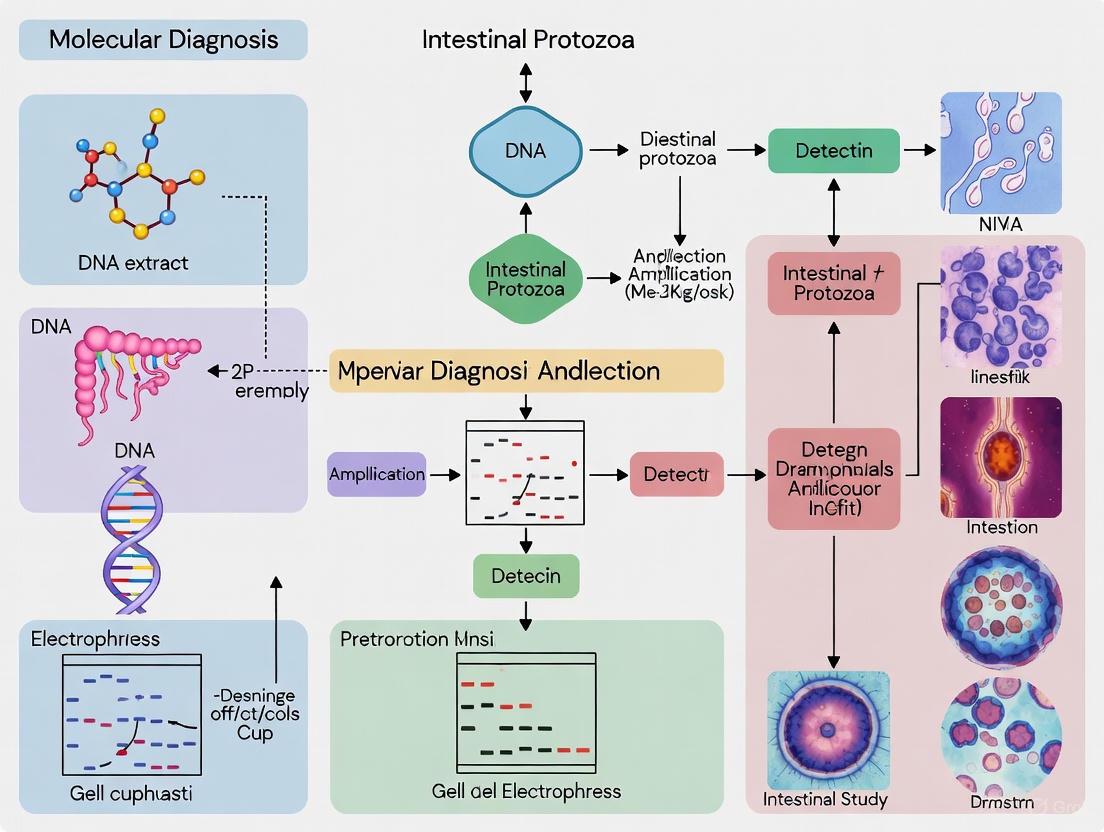
The standard workflow for molecular detection of intestinal protozoa involves multiple critical steps to ensure accurate and reproducible results. The following diagram illustrates the comprehensive experimental workflow from sample collection to result interpretation:
Taxonomic Considerations in Molecular Diagnosis
Accurate taxonomic classification is fundamental for molecular diagnosis of intestinal protozoa. Recent taxonomic revisions have significant implications for diagnostic laboratories [8]. Key updates include:
- Giardia duodenalis has replaced Giardia lamblia and G. intestinalis [8]
- Balantioides coli has replaced Balantidium coli and Neobalantidium coli [8]
- Rodentolepis nana has replaced Hymenolepis nana [8]
Laboratories are advised to adopt current nomenclatural changes in a timely fashion while noting previous names on reports for 2-3 years after revision to avoid confusion [8]. Molecular techniques have been instrumental in driving these taxonomic revisions, enabling more accurate species identification and recognition of cryptic species complexes [8].
Research Reagent Solutions for Experimental Studies
Essential Research Tools
Cutting-edge research on intestinal protozoa requires specialized reagents and materials to support experimental investigations. The following table details key research reagent solutions essential for studying these pathogens:
Table 4: Essential Research Reagents for Intestinal Protozoan Studies
| Reagent Category | Specific Examples | Research Application | Technical Considerations |
|---|---|---|---|
| Nucleic Acid Extraction Kits | Stool DNA/RNA extraction kits with inhibitor removal | Efficient nucleic acid isolation from complex stool matrices | Must address PCR inhibitors common in fecal samples [3] |
| Amplification Master Mixes | qPCR/LAMP master mixes with optimized buffer formulations | Sensitive amplification of protozoan nucleic acid targets | Should include uracil-N-glycosylase for carryover prevention [3] |
| Target-Specific Primers/Probes | TaqMan probes, molecular beacons, LAMP primers | Specific detection of protozoan genetic targets | Multiplex panels enable simultaneous detection of multiple pathogens [3] |
| Sequence-Specific Capture Probes | Microarray probes, bead-based hybridization probes | Pathogen identification and genotyping | Enable high-throughput screening of multiple targets [4] |
| Enzymatic Reagents | Recombinase polymerase amplification enzymes, reverse transcriptases | Isothermal amplification and reverse transcription | Critical for field-deployable diagnostic platforms [3] |
| Positive Control Materials | Synthetic gene constructs, quantified genomic DNA | Assay validation and quality control | Should encompass genetic diversity of target pathogens [3] |
Methodological Framework for Diagnostic Development
The development and validation of molecular diagnostic assays for intestinal protozoa follows a structured methodological framework. The relationship between key methodological components and their applications in diagnostic development can be visualized as follows:
Intestinal protozoan pathogens remain significant contributors to the global burden of diarrheal diseases, with particular impact in resource-limited settings and among vulnerable populations [1] [5]. The clinical significance of these pathogens extends beyond acute gastrointestinal illness to include long-term sequelae such as growth faltering and cognitive impairment in children [1]. Advances in molecular diagnostics have revolutionized our understanding of protozoan epidemiology, revealing higher prevalence and greater genetic diversity than previously recognized [3] [8].
Future directions in intestinal protozoan research will likely focus on several key areas: development of point-of-care molecular diagnostics suitable for resource-limited settings [3], implementation of multiplex platforms for comprehensive pathogen surveillance [3], application of next-generation sequencing for outbreak investigation and transmission tracking [3], and integration of molecular data with clinical outcomes to better understand the health significance of less characterized protozoa [4]. Additionally, the growing challenge of drug resistance in protozoan pathogens necessitates continued research into resistance mechanisms and development of novel therapeutic approaches [9] [10].
As molecular technologies continue to evolve and become more accessible, they offer unprecedented opportunities to deepen our understanding of intestinal protozoan pathogens and develop more effective strategies for their control and elimination. The integration of these advanced diagnostic tools into public health programs will be essential for reducing the global burden of intestinal protozoan infections.
Conventional microscopy has long been the cornerstone of diagnostic parasitology, providing a foundational method for identifying intestinal protozoan infections in clinical and research settings. Despite its widespread use and low-cost appeal, this technique faces significant challenges that impact diagnostic accuracy and reliability. The inherent limitations of microscopy become particularly evident when compared with emerging molecular technologies, revealing critical gaps in sensitivity, specificity, and operational efficiency. Within the context of a broader thesis on molecular diagnosis of intestinal protozoa, understanding these limitations provides the essential rationale for the adoption of more advanced diagnostic methodologies. This technical guide examines the core constraints of conventional microscopy through experimental data, comparative analyses, and technical evaluations, offering researchers and drug development professionals a comprehensive resource for advancing diagnostic capabilities beyond traditional microscopic approaches.
Core Limitations of Conventional Microscopy
Suboptimal Diagnostic Sensitivity
The diagnostic sensitivity of conventional microscopy remains substantially limited by several factors, primarily due to intermittent parasite excretion and the technical constraints of visual identification. A fundamental study investigating the yield of multiple stool samples demonstrated that collecting three specimens was necessary to achieve a cumulative detection rate of 100% for pathogenic intestinal parasites, with significant variation in detection capability across species [11]. For instance, while hookworms were easily detected in the first sample, more than half of patients infected with Trichuris trichiura and all patients infected with Isospora belli were missed with single-sample examination [11]. This variability in detection sensitivity directly impacts diagnostic accuracy and patient management.
Comparative studies between microscopy and molecular methods have consistently demonstrated this sensitivity gap. Research examining 355 stool samples across 18 Italian laboratories revealed that conventional microscopy frequently failed to detect infections identified by molecular methods, particularly for organisms like Dientamoeba fragilis and Entamoeba histolytica [12] [13]. Another study conducted in Spain found that molecular diagnosis identified positivity in 27% (n=74) of samples compared to only 9.5% (n=26) by microscopic examination [14]. The most striking differences were observed for Dientamoeba fragilis, which was not detected in any case by microscopy but was found in 20% (n=15) of positive samples by PCR [14].
Table 1: Comparative Sensitivity of Microscopy vs. Molecular Methods for Protozoan Detection
| Parasite | Microscopy Detection Rate | Molecular Detection Rate | Reference |
|---|---|---|---|
| Blastocystis hominis | 84% (23/64 samples) | 85% (64/64 samples) | [14] |
| Dientamoeba fragilis | 0% (0/15 samples) | 20% (15/74 samples) | [14] |
| Giardia lamblia | 37.5% (3/8 samples) | 11% (8/74 samples) | [14] |
| Entamoeba histolytica | Limited differentiation from non-pathogenic species | Accurate identification | [13] [15] |
Limited Specificity and Differentiation Capability
The specificity of conventional microscopy is compromised by its inability to differentiate between morphologically similar species, a critical limitation for determining appropriate treatment protocols. This is particularly problematic for the Entamoeba histolytica/dispar/moshkovskii complex, where microscopic examination cannot distinguish the pathogenic E. histolytica from non-pathogenic species [13] [15]. This differentiation has significant clinical implications, as treatment decisions depend on accurate identification of the pathogenic species.
The limitation extends beyond amoebic species. One study noted that "microscopy remains the reference diagnostic method for intestinal protozoa, but is limited in terms of sensitivity, specificity and the ability to differentiate closely related species" [13]. This lack of discriminatory power can lead to both false-positive and false-negative results, ultimately affecting patient care and treatment outcomes. Molecular assays have proven critical for the accurate diagnosis of E. histolytica and other protozoa with similar morphological characteristics [13].
Operator Dependency and Technical Variability
Conventional microscopy is inherently dependent on the skill and experience of the laboratory personnel performing the examination. This operator dependency introduces significant variability in diagnostic outcomes, as the identification of parasites relies heavily on human interpretation of visual characteristics. The technical expertise required for proficient microscopic diagnosis presents a substantial barrier to consistent, reliable results across different settings [13].
The challenges of operator dependency are further compounded by the physical demands of microscopy. Extended periods of microscopic examination have been associated with asthenopic symptoms and measurable impairment in near binocular vision, underscoring the physical strain imposed by conventional diagnostic modalities [16]. This fatigue factor can contribute to decreased accuracy over time, particularly in high-volume laboratory settings.
Automated systems have demonstrated the potential to mitigate these operator-related challenges. For instance, an evaluation of the AiDx Assist automated microscope for schistosomiasis diagnosis found that it reduced dependency on trained experts while maintaining diagnostic accuracy [17]. Similarly, an AI-powered Fluorescence Microscopic Image Analyzer (FMIA) demonstrated superior consistency compared to manual microscopy in fungal infection diagnosis, achieving a sensitivity of 96.27% compared to 75.52% for conventional KOH microscopy [16].
Experimental Protocols and Comparative Methodologies
Standard Microscopy Protocol for Intestinal Protozoa
The conventional microscopy approach for intestinal protozoa detection typically follows a standardized protocol based on WHO and CDC guidelines [13]:
Sample Collection: Patients provide three stool samples collected on consecutive or alternate days to account for intermittent parasite excretion [11] [15].
Sample Preparation:
Microscopic Examination:
- Initial examination under low-power magnification (10×10) to detect potential parasitic elements
- Detailed observation under high-power magnification (40×10) to assess morphology, characteristics, location, size, and arrangement of parasitic structures
- Documentation of findings for diagnostic evaluation
Quality Control: Implementation of verification procedures by experienced microscopists to confirm uncertain identifications
This protocol, while established, demonstrates the labor-intensive nature of conventional microscopy and its vulnerability to technical and human factors.
Molecular Biology Protocol for Comparative Analysis
In contrast to conventional microscopy, molecular methods offer a more standardized approach with reduced operator dependency. A representative protocol from a comparative study includes [15]:
DNA Extraction:
- 350 μl of S.T.A.R. (Stool Transport and Recovery Buffer) mixed with approximately 1 μl of each fecal sample
- Incubation for 5 minutes at room temperature
- Centrifugation at 2000 rpm for 2 minutes
- Collection of 250 μl supernatant transferred to a fresh tube with 50 μl internal extraction control
- Automated DNA extraction using MagNA Pure 96 System with "DNA isolation kit I" [15]
- DNA elution in a final volume of 100 μl
Real-Time PCR Amplification:
- Reaction mixture: 5 μl DNA extract, 12.5 μl 2× TaqMan Fast Universal PCR Master Mix, primers and probe mix (2.5 μl), sterile water to 25 μl final volume
- Multiplex tandem PCR performed using ABI detection systems
- Cycle protocol: 3 minutes at 95°C followed by 40 cycles of 15 seconds at 95°C, 30 seconds at 60°C, and 30 seconds at 72°C
- Inclusion of positive and negative controls in all experiments
Result Interpretation:
- Analysis of amplification curves and threshold cycles (Ct values)
- Multiplex detection of various parasites in a single reaction tube
- Verification using internal controls to identify potential inhibition
Diagram 1: Workflow comparison highlighting operator-dependent steps in microscopy versus standardized molecular protocols.
Quantitative Comparison: Microscopy vs. Molecular Methods
The performance gap between conventional microscopy and molecular techniques has been quantitatively demonstrated across multiple studies. The data reveal consistent patterns of superior sensitivity and specificity for molecular methods, particularly for specific protozoan species that challenge microscopic identification.
Table 2: Comprehensive Performance Metrics of Diagnostic Methods for Intestinal Protozoa
| Diagnostic Method | Overall Sensitivity | Overall Specificity | Advantages | Limitations |
|---|---|---|---|---|
| Conventional Microscopy | 9.5%-75.5% [16] [14] | 84%-93.2% [16] | Low cost; Wide parasite detection; Established technology | Low sensitivity; Operator dependency; Limited species differentiation |
| Fluorescence Staining | 92.95% [16] | 96.61% [16] | Enhanced visualization; Improved sensitivity over KOH | Requires technical expertise; Manual process |
| Molecular Methods (PCR) | 86.8%-96.3% [16] [17] | 81.4%-94.9% [16] [17] | High sensitivity & specificity; Species differentiation; Automation potential | Higher cost; Technical infrastructure requirements |
| AI-Based Automated Systems | 56.9%-94.6% [17] | 81.4%-91.3% [17] | Reduced operator dependency; Consistency; Digital archiving | Developing technology; Validation ongoing |
The data clearly demonstrate that molecular methods consistently outperform conventional microscopy across multiple performance metrics. The difference is particularly pronounced for specific protozoans like Dientamoeba fragilis, which was not detected in any case by microscopy but was identified in 20% of positive samples by PCR in one study [14]. Similarly, molecular assays have proven critical for the accurate diagnosis of E. histolytica, which cannot be reliably differentiated from non-pathogenic species by microscopy alone [13].
Diagram 2: Visual representation of sensitivity gaps and variable detection rates in conventional microscopy.
The Scientist's Toolkit: Essential Research Reagents and Materials
Table 3: Research Reagent Solutions for Parasitology Diagnostics
| Reagent/Material | Application & Function | Technical Specifications | Research Context |
|---|---|---|---|
| S.T.A.R. Buffer (Stool Transport and Recovery) | DNA stabilization in stool samples; Preserves nucleic acid integrity for molecular testing | Commercial buffer solution; Used in automated extraction systems | Essential for reliable DNA extraction from difficult stool matrices [13] [15] |
| Formalin-Ethyl Acetate | Concentration of parasitic elements for microscopy; Enhances detection sensitivity | Standard concentration technique following CDC guidelines | Reference method for microscopic examination; Used in multicentre comparative studies [13] |
| MagNA Pure 96 System | Automated nucleic acid extraction; Standardizes DNA preparation process | Utilizes magnetic separation technology; High-throughput capability | Critical for reducing variability in molecular assay performance [15] |
| TaqMan Fast Universal PCR Master Mix | Real-time PCR amplification; Enables sensitive detection of parasite DNA | Contains optimized enzyme mix, dNTPs, and buffer components | Foundation of multiplex real-time PCR protocols for parasite detection [15] |
| Para-Pak Preservation Media | Stool sample preservation; Maintains parasite morphology for microscopy | Commercial fixation medium; Prevents degradation of parasitic structures | Enables batch processing and transport of samples in multicentre studies [13] |
| Seegene AllplexTM Parasite Assay | Multiplex PCR detection of common intestinal parasites | Commercial panel targeting multiple protozoan pathogens | Used in comparative studies demonstrating superior sensitivity over microscopy [14] |
The collective evidence presented in this technical assessment demonstrates that conventional microscopy suffers from significant limitations in sensitivity, specificity, and operator dependency that constrain its effectiveness in modern parasitology diagnostics. The inherent constraints of visual identification, combined with technical variability and the inability to differentiate morphologically similar species, position conventional microscopy as a suboptimal standalone method for intestinal protozoan detection. The experimental data and performance metrics clearly establish that molecular methods offer substantial advantages in diagnostic accuracy, standardization, and species-specific identification. For researchers and drug development professionals working within the field of intestinal protozoan diagnostics, these findings strongly support the integration of molecular technologies as either complementary or primary diagnostic approaches. The ongoing development of automated systems and AI-enhanced platforms further promises to address the operational challenges inherent in conventional microscopy, potentially ushering in a new era of standardized, efficient, and reliable parasitological diagnosis.
Intestinal protozoan infections present a significant global health challenge, with Entamoeba species being among the most prevalent. For decades, the pathogenic potential of Entamoeba infections was misunderstood due to the morphological identicality of distinct species, particularly Entamoeba histolytica, the causative agent of amebiasis, and Entamoeba dispar, a non-pathogenic commensal [18]. This diagnostic challenge has profound implications for clinical management, public health surveillance, and drug development efforts.
The differentiation between these species represents a critical case study in molecular diagnosis, illustrating how technological advances have transformed our understanding of disease epidemiology and pathogenesis. Before the widespread adoption of molecular techniques, microscopy-based diagnosis significantly overestimated the prevalence of true E. histolytica infection, leading to unnecessary treatments and inaccurate disease burden calculations [19]. This review examines the imperative for species-level differentiation through the lens of E. histolytica versus E. dispar, framing this requirement within the broader context of molecular diagnosis of intestinal protozoa.
Epidemiological and Clinical Imperatives for Differentiation
Global Burden and Distribution
Entamoeba infections occur worldwide with higher frequency in countries of low socioeconomic status and poor public health infrastructure [20]. Amebiasis, caused specifically by E. histolytica, ranks as the second leading cause of death from protozoan parasitic disease, approximately 40,000-100,000 deaths annually [21] [18]. Accurate prevalence data have been difficult to establish due to historical diagnostic limitations, but molecular studies have revealed that E. dispar infections are significantly more common than E. histolytica in most populations [22] [20].
Studies conducted in various geographical regions consistently demonstrate this pattern. In Iran, research showed that Entamoeba dispar and, in one case, E. moshkovskii were the exclusive Entamoeba species found in asymptomatic cyst passers, with no E. histolytica detected [22]. Similarly, in Argentina, E. dispar was more prevalent than E. histolytica among the studied populations [20]. Even in non-endemic settings like Italy, studies found more patients with E. dispar infection (8.3%) than patients with E. histolytica infection (5.6%) [18].
Clinical Management Implications
The clinical implications of species differentiation are substantial. While E. histolytica can cause invasive intestinal disease (dysentery) and extraintestinal abscesses (most commonly in the liver), E. dispar does not require treatment [18] [19]. The World Health Organization recommends that all cases of E. histolytica infection, including asymptomatic carriers, should be treated to prevent invasive disease and interrupt transmission [23]. Approximately 10% of individuals asymptomatically infected with E. histolytica will develop invasive amebiasis over time [22].
Table 1: Clinical Implications of Entamoeba Species Differentiation
| Feature | Entamoeba histolytica | Entamoeba dispar |
|---|---|---|
| Pathogenic Potential | Causes intestinal and extraintestinal disease | Non-pathogenic commensal |
| Treatment Requirement | Always requires treatment | Does not require treatment |
| Asymptomatic Carrier State | 90% of infections; 10% progress to invasive disease | 100% of infections remain asymptomatic |
| Public Health Significance | Significant burden; targeted control needed | Primarily an indicator of fecal-oral exposure |
Diagnostic Modalities: From Microscopy to Molecular Assays
Limitations of Conventional Methods
Traditional microscopic examination, while cost-effective and widely available, cannot differentiate between the cysts and trophozoites of E. histolytica, E. dispar, and E. moshkovskii [24] [19]. This technique is further limited by requirements for expert microscopists, subjective interpretation, and inadequate sensitivity [21]. Although E. histolytica may occasionally be observed with ingested red blood cells (erythrophagocytosis), this finding is not a reliable differentiator as it may rarely occur with E. dispar as well [19].
Antigen-detection assays represented an advancement in species differentiation. The TechLab E. histolytica II test, for example, is designed to specifically detect E. histolytica and not the closely related non-pathogenic E. dispar [22]. Studies have shown 100% correlation between this antigen detection kit and nested PCR results [22]. However, antigen tests may lack the sensitivity of molecular methods and typically do not detect emerging species like E. moshkovskii.
Serological testing detects antibodies to amoebae in patient sera, which typically indicate E. histolytica infection. However, this method cannot distinguish between past and present infections in individuals from endemic areas [24] [18].
Molecular Diagnostic Advancements
Molecular technologies, particularly PCR-based assays, have revolutionized Entamoeba diagnosis by enabling specific detection and differentiation at the species level. Various PCR platforms have been developed with different target genes, sensitivity thresholds, and methodological approaches.
Table 2: Comparison of Molecular Assays for Entamoeba histolytica and dispar Detection
| Assay Type | Target Genes/Components | Sensitivity | Species Differentiated | Key Features |
|---|---|---|---|---|
| Nested PCR with RFLP [22] | SSU rRNA gene followed by HinfI digestion | High (exact sensitivity not specified) | E. histolytica, E. dispar, E. moshkovskii | 100% correlation with TechLab E. histolytica II; requires post-amplification processing |
| Single-round Multiplex PCR [24] | SSU rRNA gene with species-specific reverse primers | 10 pg of E. histolytica/moshkovskii DNA; 20 pg of E. dispar DNA | E. histolytica (166bp), E. dispar (752bp), E. moshkovskik (580bp) | Single-round amplification; different product sizes for visual differentiation |
| Real-time PCR [21] | SSU rRNA gene | High (detection in 31.4% of samples in field study) | E. histolytica vs. E. dispar | Quantitative capability; reduced contamination risk; higher throughput |
| Commercial RT-PCR [25] | Multiple proprietary targets | Variable between platforms | Typically E. histolytica specifically | Standardized reagents; quality control; often includes internal controls |
Real-time PCR (qPCR) has emerged as particularly valuable for diagnostic laboratories, offering advantages in speed, contamination control, and quantification potential [21]. A 2025 study implemented duplex qPCR assays to detect E. dispar + E. histolytica, finding that one-third of these infections were caused by E. histolytica in their study population from Tanzania [21].
Comparative studies of multiple PCR assays have revealed important considerations for test selection. A 2025 evaluation of three published E. histolytica-specific real-time PCR assays found that diagnostic accuracy estimates for E. histolytica ranged from 75% to 100% for sensitivity and 94% to 100% for specificity [26]. The study also noted that high cycle threshold values (Ct > 35) showed particularly reduced likelihood of reproducibility when applying competitor real-time PCR assays [26].
Experimental Protocols for Species Differentiation
Sample Collection and DNA Extraction
Proper sample collection and processing are critical for reliable molecular detection. Fresh stool samples should be collected and ideally tested within 24 hours, or stored at -20°C for later analysis [22]. For DNA extraction, commercial kits such as the QIAamp DNA stool mini kit (QIAGEN) have been successfully used, with approximately 0.2g of fecal sediment processed according to manufacturer instructions [22]. Automated extraction systems like the MagNA Pure 96 System (Roche) have also been implemented in multicentre studies [25].
Nested PCR with RFLP Analysis
This protocol adapted from a 2006 study provides robust differentiation of Entamoeba species [22]:
Primary PCR Amplification:
- Reaction Volume: 50μL
- DNA Template: 5.0μL of genomic DNA
- Primers: P1 (5'-TAA AGC ACC AGC ATA TTG TC-3') and P4 (5'-TTA ATT CCA TCT GGT GGT GG-3')
- Cycling Conditions: 35 cycles of 94°C for 30s, 54.5°C for 45s, 72°C for 1min; final extension of 72°C for 7min
- Expected Product: 540bp fragment
Nested PCR Amplification:
- Reaction Volume: 50μL
- DNA Template: 1μL of primary PCR product
- Primers: HF (5'-AAG AAA TTG ATA TTA ATG AAT ATA-3') and HR (5'-ATC TTC CAA TTC CAT CAT CAT-3')
- Cycling Conditions: 35 cycles of 94°C for 30s, 57°C for 45s, 72°C for 1min; final extension of 72°C for 7min
- Expected Product: 374bp fragment
Restriction Fragment Length Polymorphism (RFLP) Analysis:
- Enzyme: HinfI (1μL)
- Digestion Conditions: 37°C for 1 hour
- Expected Fragments:
- E. histolytica: 155bp and 219bp
- E. dispar: 67bp, 152bp, and 155bp (152bp and 155bp fragments overlap in electrophoresis)
- Visualization: 2% agarose gel electrophoresis with ethidium bromide staining
Single-Round Multiplex PCR Assay
This streamlined protocol enables differentiation in a single reaction [24]:
PCR Reaction Setup:
- Reaction Volume: 50μL
- DNA Template: 10μL of extracted DNA
- Primers:
- Forward (EntaF): 5'-ATG CAC GAG AGC GAA AGC AT-3' (conserved across species)
- Reverse (EhR): 5'-GAT CTA GAA ACA ATG CTT CTC T-3' (E. histolytica-specific)
- Reverse (EdR): 5'-CAC CAC TTA CTA TCC CTA CC-3' (E. dispar-specific)
- Reverse (EmR): 5'-TGA CCG GAG CCA GAG ACA T-3' (E. moshkovskii-specific)
- Final Magnesium Chloride Concentration: 6mM
- Primer Concentration: 0.1μM each
Amplification Conditions:
- Initial Denaturation: 94°C for 3 minutes
- Cycling: 30 cycles of:
- Denaturation: 94°C for 1 minute
- Annealing: 58°C for 1 minute
- Extension: 72°C for 1 minute
- Final Extension: 72°C for 7 minutes
Product Analysis:
- Separation: 1.5% agarose gel electrophoresis
- Expected Product Sizes:
- E. histolytica: 166bp
- E. moshkovskii: 580bp
- E. dispar: 752bp
Real-Time PCR Implementation
Modern qPCR assays provide rapid, sensitive detection with reduced contamination risk:
Reaction Setup:
- Reaction Volume: 10μL (reduced volume format) [21]
- Primers/Probes: Species-specific designs targeting SSU rRNA gene or episomal repeat sequences [21] [26]
- Platform Examples: CFX Maestro (Bio-Rad Laboratories Inc.), ABI 7900HT Fast Real-Time PCR System [21] [25]
Cycling Parameters:
- Holding Stage: 95°C for 10 minutes (enzyme activation)
- Amplification: 45 cycles of:
- Denaturation: 95°C for 15 seconds
- Annealing/Extension: 60°C for 1 minute [25]
Research Reagent Solutions for Entamoeba Differentiation
Successful implementation of molecular differentiation assays requires specific research reagents and materials. The following table compiles key solutions used in the featured experimental protocols.
Table 3: Essential Research Reagents for Entamoeba Species Differentiation
| Reagent/Material | Specific Examples | Function/Application | References |
|---|---|---|---|
| DNA Extraction Kits | QIAamp DNA Stool Mini Kit (QIAGEN), MagNA Pure 96 DNA and Viral NA Small Volume Kit (Roche) | Efficient DNA isolation from complex stool matrices; includes inhibitors removal | [22] [24] [25] |
| PCR Enzymes & Master Mixes | Taq Polymerase, TaqMan Fast Universal PCR Master Mix (Thermo Fisher) | DNA amplification with consistent performance; optimized reaction conditions | [24] [25] |
| Species-specific Primers/Probes | SSU rRNA gene targets; episomal repeat sequence (SREPH) primers | Selective amplification of target species; differential detection | [24] [21] [26] |
| Restriction Enzymes | HinfI (Roche), XhoI (Roche) | RFLP analysis for species identification; verification of PCR products | [22] |
| Electrophoresis Materials | Agarose gels (NuSieve, Merck), ethidium bromide, DNA size markers | Product separation and visualization; size determination | [22] [24] |
| Commercial PCR Assays | AusDiagnostics GI Parasite Detection, TechLab E. histolytica II | Standardized detection; quality-controlled reagents | [22] [27] [25] |
| Sample Preservation Media | S.T.A.R. Buffer (Roche), Para-Pak, Sodium Acetate-Acetic Acid-Formalin (SAF) | Nucleic acid stabilization; maintenance of DNA integrity during storage/transport | [25] |
Implications for Drug Development and Therapeutic Strategies
The accurate differentiation of Entamoeba species has profound implications for drug development pipelines and therapeutic strategies. Historically, the conflation of E. histolytica and E. dispar led to overestimation of treatment efficacy and misunderstanding of drug resistance patterns.
Current Therapeutic Landscape
The current treatment paradigm for amebiasis involves different drug classes targeting various parasite stages:
- Luminal agents (paromomycin, diloxanide furoate) kill intraluminal cysts
- Tissue amebicides (metronidazole, tinidazole) target trophozoites in invasive disease
- Combination therapy is often recommended to eliminate both cysts and trophozoites [23]
Metronidazole has remained the cornerstone of amebiasis treatment for decades, but its efficacy against luminal stages is limited, necessitating combination therapy with luminal agents [23]. While clear resistance to metronidazole has not emerged clinically, the need for novel therapeutic targets is widely recognized.
Drug Repurposing and Novel Targets
Drug development efforts have explored repurposing existing compounds and identifying novel targets:
- Auranofin, an anti-rheumatic gold-containing compound, shows promising anti-amoebic activity in clinical trials (NCT02736968) [23]
- Azidothymidine (AZT), an antiretroviral drug, exhibits inhibitory activity against E. histolytica [23]
- Cysteine proteases, galactose-binding lectin, and amoebapores represent potential molecular targets for novel therapeutics [23]
The critical need for species-level differentiation between Entamoeba histolytica and Entamoeba dispar represents a paradigm case in molecular diagnosis of intestinal protozoa. The implementation of PCR-based assays has fundamentally transformed our understanding of amebiasis epidemiology, revealing that most Entamoeba infections in asymptomatic carriers are attributable to non-pathogenic species. This differentiation has direct implications for clinical management, preventing unnecessary treatments for E. dispar infections while ensuring appropriate therapy for E. histolytica carriers at risk of developing invasive disease.
Molecular diagnostics continue to evolve, with real-time PCR assays offering increasingly rapid, sensitive, and specific detection. The ongoing development of multiplexed platforms that can simultaneously detect multiple intestinal protozoa represents the future of parasitological diagnosis, providing comprehensive pathogen detection while maintaining cost-effectiveness. For researchers and drug development professionals, accurate species differentiation remains fundamental to understanding disease pathogenesis, assessing therapeutic efficacy, and developing targeted interventions against this significant global health burden.
Molecular diagnostics have revolutionized the detection and identification of intestinal protozoa, moving beyond the limitations of traditional microscopy. Techniques based on nucleic acid amplification offer superior sensitivity and specificity, enabling the differentiation of morphologically similar species, such as the pathogenic Entamoeba histolytica from non-pathogenic Entamoeba dispar [13]. This technical guide explores the core principles of the primary molecular methods used in research and clinical settings: Polymerase Chain Reaction (PCR), quantitative real-time PCR (qPCR), and isothermal amplification techniques. The adoption of these methods is crucial for accurate diagnosis, epidemiological studies, and drug development, particularly as the field moves towards the surveillance and control of neglected tropical diseases [28] [29].
Core Principles of Nucleic Acid Amplification Techniques
Polymerase Chain Reaction (PCR) and Quantitative Real-Time PCR (qPCR)
The Polymerase Chain Reaction (PCR) is a fundamental technique for the in vitro amplification of specific DNA sequences. The process relies on thermal cycling, involving repeated cycles of heating and cooling to facilitate three core steps: DNA denaturation, primer annealing, and enzymatic extension of the primers by a DNA polymerase.
Quantitative real-time PCR (qPCR) builds upon this principle by allowing for the monitoring of the amplification process in real-time as it occurs, rather than just at the end-point. This is achieved through the use of fluorescent reporters. As the target DNA amplifies, the fluorescent signal increases proportionally. The cycle threshold (Cq), the point at which the fluorescence crosses a predefined threshold, is used for quantification. A lower Cq value indicates a higher starting concentration of the target nucleic acid [30]. qPCR is the gold standard for sensitive and quantitative detection of pathogens, including intestinal protozoa [13].
The following diagram illustrates the core workflow and mechanism of a qPCR assay:
Isothermal Amplification Methods
Isothermal amplification methods (IAMs) represent a group of techniques that amplify nucleic acids at a constant temperature, eliminating the need for sophisticated thermal cyclers. This makes them particularly suitable for field applications and point-of-care (POC) diagnostics in resource-limited settings [28] [31]. Key techniques include Loop-Mediated Isothermal Amplification (LAMP) and Recombinase Polymerase Amplification (RPA).
- Loop-Mediated Isothermal Amplification (LAMP): LAMP utilizes a strand-displacing DNA polymerase and four to six primers that recognize distinct regions on the target DNA. This complex primer set ensures high specificity. The reaction, typically performed at 60–65°C, generates stem-loop DNA structures that facilitate auto-cycling amplification, leading to the rapid synthesis of a large amount of DNA [29] [31] [32].
- Recombinase Polymerase Amplification (RPA): RPA employs recombinase enzymes that form nucleoprotein complexes with primers. These complexes scan double-stranded DNA for homologous sequences and facilitate strand invasion. Once bound, a strand-displacing polymerase extends the primers, enabling exponential amplification at a low, constant temperature (typically 37–42°C) [28] [32].
The mechanism of LAMP, a prominent isothermal method, is detailed in the diagram below:
Comparative Performance Analysis of Amplification Techniques
The selection of an appropriate molecular diagnostic technique depends on the application requirements, including sensitivity, specificity, speed, cost, and need for quantification. The table below summarizes a comparative analysis of key performance metrics for PCR, qPCR, and isothermal methods like LAMP and RPA, with data drawn from applications in parasitology.
Table 1: Comparative performance of nucleic acid amplification techniques for parasite detection
| Technique | Reported Sensitivity | Reported Specificity | Limit of Detection (LoD) | Time to Result | Quantification | Key Applications in Parasitology |
|---|---|---|---|---|---|---|
| PCR | Varies by assay | Varies by assay | 0.7 ng/µL (Spirometra mansoni egg DNA) [33] | 2-4 hours | No (End-point) | Species identification [13] |
| qPCR | High | High | 100 copies/µL (Spirometra mansoni) [33] | 1-2 hours | Yes (Cq value) | Gold standard for detection and quantification of Giardia, Cryptosporidium, E. histolytica [30] [13] |
| LAMP | 87.8% (vs. qPCR for A. duodenale) [29] | 100% (for A. duodenale) [29] | 355.5 fg/µL (Spirometra mansoni egg DNA) [33] | <40 minutes [29] | Semi-quantitative | Rapid field detection of hookworms, Spirometra [29] [33] |
| RPA | Similar to PCR [28] | High [28] | Similar to PCR [28] | 30 minutes [28] | No (Lateral Flow) | Point-of-care detection of Cryptosporidium, Giardia [28] |
Determining Assay Performance: Limit of Detection and Quantification
For a diagnostic assay to be clinically or research-ready, its performance must be rigorously characterized. Two critical parameters are the Limit of Detection (LoD) and the Limit of Quantification (LoQ).
- Limit of Detection (LoD): The LoD is the lowest concentration of an analyte that can be reliably detected, though not necessarily precisely quantified. In the context of qPCR, standard statistical methods for determining LoD are challenging because the data (Cq values) are proportional to the logarithm of the concentration and are not linearly distributed [30]. An alternative approach involves testing a high number of replicate samples at various low concentrations and applying a statistical model, such as logistic regression, to determine the concentration at which the target is detected with a defined probability (e.g., ≥95%) [30].
- Limit of Quantification (LoQ): The LoQ is the lowest amount of analyte that can be quantitatively determined with acceptable precision and accuracy. This requires that the measurements at this concentration demonstrate a low coefficient of variation (CV), ensuring the results are reproducible and reliable for quantification [30].
Table 2: Key statistical definitions for assay validation
| Term | Definition | Application in qPCR |
|---|---|---|
| Limit of Blank (LoB) | The highest apparent analyte concentration expected from a blank (negative) sample. | Calculated as Mean_blank + 1.645 * SD_blank (assuming 95% confidence) [30]. |
| Limit of Detection (LoD) | The lowest concentration reliably distinguished from the LoB. | LoD = LoB + 1.645 * SD_low concentration sample (CLSI EP17 guideline) [30]. |
| Limit of Quantification (LoQ) | The lowest concentration measured with stated precision and accuracy. | The concentration at which the coefficient of variation (CV) is below an acceptable threshold (e.g., <5% for qPCR) [33]. |
| Coefficient of Variation (CV) | The ratio of the standard deviation to the mean, expressed as a percentage. | Used to assess the precision and repeatability of an assay. A low CV (<5%) indicates good reproducibility [33]. |
Essential Research Reagent Solutions
The successful implementation of nucleic acid-based detection assays relies on a suite of specialized reagents and tools. The following table details key components of the researcher's toolkit.
Table 3: Essential research reagents and materials for nucleic acid-based detection
| Reagent / Material | Function | Examples & Notes |
|---|---|---|
| Strand-Displacing DNA Polymerase | Enzymatic DNA synthesis; critical for LAMP. | Bst DNA polymerase is commonly used in LAMP assays [29] [31]. |
| Reverse Transcriptase | Converts RNA to cDNA for detection of RNA targets. | Used in RT-qPCR and RT-RPA for RNA virus or transcript detection [28] [32]. |
| Recombinase Enzymes | Facilitate primer invasion into double-stranded DNA. | Core component of RPA/RAA kits (e.g., from TwistDx) [28] [32]. |
| Fluorescent Probes & Intercalating Dyes | Enable real-time detection of amplification. | TaqMan probes (qPCR), SYBR Green, or specialized fluorescent probes for LAMP/RPA [30] [33]. |
| Primer Sets | Provide specificity by binding to unique target sequences. | Designed using software (e.g., PrimerExplorer for LAMP); require careful optimization [29] [33]. |
| Commercial Kits | Integrated solutions for specific assays. | AusDiagnostics RT-PCR, QIAamp DNA Stool Mini Kit, MagNA Pure 96 system [13]. |
Detailed Experimental Protocols
Protocol for Determining LoD via Logistic Regression in qPCR
This protocol outlines a statistical method for determining the Limit of Detection in qPCR, adapted for its logarithmic data characteristics [30].
- Experimental Replication: Prepare a dilution series of the target nucleic acid covering a range that includes very low concentrations. Analyze each concentration level in a high number of replicates (e.g., n ≥ 60).
- Data Collection and Dichotomization: Run the qPCR assay for all replicates. For each reaction, record a binary outcome: "1" for a detected result (Cq value below a defined cut-off, C0) and "0" for an undetected result (Cq > C0 or no amplification).
- Model Fitting: For each concentration level (expressed in log scale, e.g., log2(molecules)), calculate the proportion of detected replicates. Fit a logistic regression model to the data, where the probability of detection (p) is modeled as:
p = 1 / (1 + e^-(β₀ + β₁ * log2(concentration)))The parameters β₀ and β₁ are estimated using maximum likelihood estimation (MLE). - LoD Calculation: The LoD is defined as the concentration corresponding to a specified detection probability (e.g., 95%). This is calculated from the fitted model as:
LoD = 2^((logit(0.95) - β₀) / β₁)wherelogit(0.95) = ln(0.95 / 0.05).
Protocol for Optimizing a LAMP Assay
This protocol describes the key steps in developing and optimizing a LAMP assay, as demonstrated for the detection of Ancylostoma duodenale [29].
- Primer Design: Select a conserved target gene (e.g., ITS-1, ITS-2, cytb). Use specialized software (e.g., PrimerExplorer V5) to design a set of inner (FIP, BIP), outer (F3, B3), and loop (LF, LB) primers.
- Reaction Condition Optimization:
- Temperature Gradient: Test the primer set across a temperature range (e.g., 60°C to 65°C) to identify the optimal amplification temperature.
- Primer Ratio: Systematically test different ratios of inner to outer primers (e.g., 1:2, 1:4, 1:8) to maximize efficiency and speed.
- Time Course: Perform the reaction for different durations (e.g., 15 to 60 minutes) to determine the minimum time required for robust detection.
- Specificity Testing: Validate the assay against DNA from closely related non-target organisms (e.g., Necator americanus for a hookworm assay) to ensure no cross-reactivity.
- Sensitivity and LoD Determination: Perform the LAMP assay on a serial dilution of the target DNA to establish the minimum detectable concentration. Compare the results with those from PCR or qPCR to determine relative sensitivity.
The arsenal of nucleic acid-based detection methods provides powerful tools for the molecular diagnosis of intestinal protozoa. While qPCR remains the gold standard for sensitive and quantitative detection in centralized laboratories, isothermal amplification methods like LAMP and RPA are emerging as robust, rapid, and field-deployable alternatives. The choice of technique involves a careful balance between performance requirements and operational constraints. As these technologies continue to evolve and become more integrated with point-of-care platforms, they hold immense promise for improving disease surveillance, guiding treatment, and supporting the global elimination of neglected intestinal protozoan infections.
Advanced Molecular Techniques: qPCR, Multiplexing, and Emerging Point-of-Care Platforms
Real-Time Quantitative PCR (qPCR) has established itself as a cornerstone technology in molecular diagnostics, particularly for the detection and identification of intestinal protozoa. This technique combines polymerase chain reaction (PCR) amplification with simultaneous fluorescent detection, enabling precise quantification of target nucleic acids without the need for post-processing gel electrophoresis [34]. The application of qPCR in parasitology represents a significant advancement over traditional diagnostic methods like bright-field microscopy, which, despite its cost-effectiveness, is hampered by challenges in sample preservation, subjective readouts, and an inability to distinguish morphologically identical species [21]. For researchers and drug development professionals focused on enteric pathogens, qPCR offers a powerful tool for species-level differentiation of parasites such as the pathogenic Entamoeba histolytica and the non-pathogenic Entamoeba dispar, which are visually indistinguishable under a microscope [21] [25]. The technology's superior sensitivity and specificity, coupled with its capacity for multiplexing, make it indispensable for monitoring disease burden, assessing treatment efficacy, and furthering our understanding of protozoan biology within the host [21].
Fundamental qPCR Principles and Protocols
Core Principles and Reaction Setup
At its core, a qPCR reaction monitors the accumulation of fluorescent signal at every cycle of the amplification process. The fluorescence, typically expressed as normalized reporter (Rn), is plotted against the PCR cycle number, generating an amplification curve [35]. The key quantitative parameter derived from this curve is the Cycle threshold (Ct), also known as quantification cycle (Cq). The Ct value is defined as the intersection between the amplification curve and a threshold line set above the baseline fluorescence; it is an inverse, relative measure of the target concentration in the initial reaction [34] [35]. A lower Ct value indicates a higher starting quantity of the target nucleic acid.
A robust qPCR protocol requires careful attention to reaction assembly. While commercial master mixes simplify the process, a typical 25 µL reaction may contain:
- 1X TaqMan Fast Universal PCR Master Mix (or equivalent): Provides the DNA polymerase, dNTPs, MgCl₂, and optimized buffer [25].
- Forward and Reverse Primers: Typically at concentrations ranging from 0.3 to 0.5 µM each, depending on the assay [21].
- Fluorogenic Probe(s): e.g., a TaqMan probe at a specified concentration.
- DNA Template: Usually 1-5 µL of extracted nucleic acid.
- Nuclease-Free Water: To volume.
The cycling conditions on an instrument like the ABI 7900HT often follow a two-step protocol: an initial hold at 95°C for 10 minutes for enzyme activation, followed by 45 cycles of 95°C for 15 seconds (denaturation) and 60°C for 1 minute (combined annealing/extension) [25]. This protocol can be adapted for one-step RT-qPCR by including a reverse transcription step at the beginning.
Comprehensive Workflow for Intestinal Pathogen Detection
The following diagram outlines the complete workflow for detecting intestinal protozoa, from sample collection to data analysis.
Critical Experimental Considerations
Several pre-analytical and analytical factors are critical for a successful qPCR experiment, especially when working with complex samples like stool:
Sample Collection and DNA Extraction: The robust wall structure of protozoan cysts and oocysts complicates DNA extraction [25]. Automated systems like the MagNA Pure 96 System (Roche) can be used with specific stool transport buffers (e.g., S.T.A.R. Buffer) to ensure efficient and reproducible nucleic acid isolation [25]. The purity of the extracted DNA, with an A260/A280 ratio of 1.8-2.0, is paramount for minimizing PCR inhibitors [36].
Instrument Selection and Validation: The choice of qPCR instrument should align with research needs. Key considerations include throughput (e.g., 384-well vs. 48-well plates), the uniformity of the thermal block, and the sensitivity and range of the optical detection system [36]. Instruments must be properly calibrated for the specific fluorophores used in the assay [37].
Preventing Contamination: The use of uracil-DNA-glycosylase (UNG) in the reaction mix is a recommended good laboratory practice. UNG degrades any PCR products from previous reactions that contain dUTP (substituted for dTTP), thereby preventing carryover contamination [36].
Primer and Probe Design for Specific Detection
Design Strategies and Validation
The design of highly specific primers and probes is the most critical pre-experimental step in developing a reliable qPCR assay [36]. For the detection of intestinal protozoa, the target genes are often within the ribosomal RNA cluster, such as the small subunit ribosomal RNA (SSU rRNA) or 18S rRNA genes, due to their high copy number and the availability of conserved regions for genus/species differentiation [21] [38].
The general principles for design include:
- Specificity: Sequences must be unique to the target organism. This is confirmed in silico using tools like Nucleotide BLAST (BLASTN) to ensure no significant similarity to non-target organisms, including human DNA or other commensal gut flora [21] [38].
- Amplicon Length: Shorter amplicons (typically 70-150 bp) are preferred as they amplify with higher efficiency.
- Primer Parameters: Primers should have a length of 20-24 bases, a GC content of approximately 50%, and an estimated melting temperature (Tm) of around 58°C [21]. Software such as Primer Express is commonly used for this purpose [38].
- Probe Design: For TaqMan assays, the probe should have a Tm that is 5-10°C higher than the primers, should not contain a G at the 5' end, and should be located close to the forward or reverse primer binding site.
Practical Examples for Protozoan Detection
The table below provides real-world examples of primer and probe sequences used in research for detecting major intestinal protozoa.
Table 1: Primer and Probe Sequences for Detecting Intestinal Protozoa
| Organism | Target Gene | Sequence (5' → 3') | Concentration (µM) | Source |
|---|---|---|---|---|
| Entamoeba histolytica | Small subunit ribosomal RNA | F: AGG ATT GGA TGA AAT TCA GAT GTA CAR: TAA GTT TCA GCC TTG TGA CCA TACProbe: TGA... | 0.5 | [21] |
| Giardia duodenalis | Small subunit ribosomal RNA | F: GCT GCG TCA CGC TGC TCR: GAC GGC TCA GGA CAA CGG T | 0.5 | [21] |
| Cryptosporidium spp. | Small subunit ribosomal RNA | F: ACA TGG ATA ACC GTG GTA ATT CTR: CAA TAC CCT ACC GTC TAA AGC TG | 0.5 | [21] |
| Blastocystis spp. | Small subunit ribosomal RNA | F: GGT CCG GTG AAC ACT TTG GAT TTR: CCT ACG GAA ACC TTG TTA CGA CTT CA | 0.3 | [21] |
Fluorophore Selection and Multiplex qPCR Design
Principles of Multiplexing and Dye Selection
Multiplex qPCR, which allows for the simultaneous detection of multiple targets in a single reaction, is particularly valuable in parasitology for comprehensive screening and saving precious sample material [21]. The design of a multiplex assay requires careful selection of reporter dyes attached to the target-specific probes. The fundamental rule is that each probe must have a unique reporter dye with a distinct emission spectrum that can be discriminated by the qPCR instrument's optical system [37].
Key considerations for dye selection include:
- Instrument Compatibility: The instrument must be capable of exciting and detecting the emission spectrum of each chosen dye. Manufacturers provide lists of compatible dyes, and tools like the IDT PrimeTime Multipux Dye Selection Tool can guide appropriate choices [37].
- Minimizing Spectral Overlap: Dyes should be chosen to have minimal overlap in their emission spectra to reduce signal cross-talk (bleed-through). For low-copy targets, a bright dye like FAM is a good choice, while for high-copy targets (e.g., a housekeeping gene), a fluorophore with lower signal intensity can be used [37].
- Quencher Selection: Using efficient dark quenchers (e.g., Iowa Black FQ, BHQ) is crucial to minimize background fluorescence, which becomes more critical in a multiplex reaction with multiple fluorophores. Double-quenched probes (e.g., those with an internal ZEN or TAO quencher) provide even lower background and clearer signals [37].
Dye and Quencher Reference
The following table summarizes the properties of common fluorophores and recommended quenchers for multiplex assay design.
Table 2: Common Fluorophores and Quenchers for Multiplex qPCR
| Fluorescent Dye | Excitation (nm) | Emission (nm) | Recommended Dark Quencher |
|---|---|---|---|
| 6-FAM | 495 | 520 | ZEN / Iowa Black FQ |
| HEX/JOE | 538 | 555 | ZEN / Iowa Black FQ |
| Cy3 | 550 | 564 | Iowa Black RQ |
| ROX | 575 | 608 | Iowa Black RQ |
| Texas Red-X | 598 | 617 | Iowa Black RQ |
| Cy5 | 648 | 668 | TAO / Iowa Black RQ |
| Cy5.5 | 683 | 706 | Black Hole Quencher-3 |
Multiplex qPCR Assay Design Workflow
Designing a successful multiplex assay involves a systematic process to ensure all components work harmoniously.
Applications in Intestinal Protozoa Diagnosis and Research
The application of qPCR in the molecular diagnosis of intestinal protozoa has led to more accurate prevalence data and a better understanding of the pathogenicity of different species. Studies consistently demonstrate its superior performance over microscopy.
Table 3: Performance of qPCR in Detecting Intestinal Protozoa in Research Studies
| Study Context | Key Findings | Implication |
|---|---|---|
| HIV/AIDS Patients (n=100) [39] | Detected Blastocystis (22%), Microsporidia (17%), Cryptosporidium spp. (12%), G. intestinalis (11%); 12% had multiple infections. | qPCR is crucial for diagnosing opportunistic infections in immunocompromised populations. |
| Pemba Island, Tanzania (n=70) [21] | qPCR detected protozoa in 74.4% of samples; differentiated E. histolytica (pathogenic) from E. dispar (non-pathogenic) in 31.4% of Entamoeba-positive cases. | Highlights high prevalence and the unique ability of qPCR to provide species-level differentiation critical for treatment. |
| Multicentre Italy Study [25] | Commercial and in-house PCR showed high sensitivity/specificity for G. duodenalis; performed better on preserved stool samples than fresh ones. | Molecular methods are reliable for specific pathogens, but sample preservation is key for DNA quality. |
| Triplex qPCR Development [38] | The assay simultaneously detected E. histolytica, G. lamblia, and C. parvum with a limit of detection of 500 copies/μL and no cross-reactivity. | Demonstrates the feasibility and efficiency of multiplexing for high-throughput, cost-effective screening. |
The Scientist's Toolkit: Essential Reagents and Materials
Successful implementation of a qPCR protocol, particularly for complex samples, relies on a suite of reliable reagents and instruments.
Table 4: Essential Research Reagent Solutions for qPCR
| Item | Function/Description | Example Products/Brands |
|---|---|---|
| Nucleic Acid Extraction Kit | Isolates high-quality, inhibitor-free DNA from complex stool samples. | QIAamp DNA Stool Mini Kit (Qiagen), MagNA Pure 96 System (Roche) [25] [38] |
| qPCR Master Mix | A pre-mixed solution containing DNA polymerase, dNTPs, MgCl₂, and optimized buffer. | TaqMan Fast Universal PCR Master Mix (Thermo Fisher) [25] |
| Hot-Start Taq Polymerase | Reduces non-specific amplification and primer-dimer formation by requiring thermal activation. | Antibody-mediated or chemically modified enzymes [36] |
| Primers & Probes | Oligonucleotides designed for specific detection of the target protozoan DNA. | Custom synthesized by companies (e.g., Microsynth, Shanghai BioGerm) [21] [38] |
| Reference Dye | Passive dye used in some qPCR instruments to normalize for non-PCR-related fluorescence fluctuations. | ROX [37] [36] |
| UNG Enzyme | Prevents carryover contamination by degrading PCR products from previous reactions. | Included in many commercial master mixes [36] |
Data Analysis and Quality Control
Quantification Methods and PCR Efficiency
Accurate data interpretation is the final, critical step in qPCR. The two primary quantification methods are:
- Absolute Quantification: Used to determine the exact copy number of a target sequence in a sample. This method requires a standard curve generated from serial dilutions of a known quantity of the target DNA (e.g., a plasmid standard) [34] [35]. The target quantity in unknown samples is extrapolated from this curve.
- Relative Quantification: Used to analyze changes in gene expression (or parasite load) in a given sample relative to a reference sample (e.g., an untreated control). This method does not require a standard curve of known concentration but relies on one or more reference genes (e.g., host housekeeping genes) for normalization [34] [35]. The Livak method (2^(-ΔΔCt)) is commonly used when the amplification efficiencies of the target and reference genes are approximately equal and close to 100% [35].
A key parameter for validating any qPCR assay is the calculation of PCR efficiency. Efficiency is calculated from the slope of the standard curve using the formula: Efficiency (%) = (10^(-1/slope) - 1) x 100 [35]. An efficiency of 90-110% (corresponding to a slope of -3.58 to -3.10) is generally considered acceptable, indicating a near-ideal doubling of product every cycle. Efficiency outside this range can lead to inaccurate quantification [35].
Adherence to MIQE Guidelines
To ensure the reproducibility, transparency, and reliability of qPCR data, researchers are strongly encouraged to follow the MIQE (Minimum Information for Publication of Quantitative Real-Time PCR Experiments) guidelines [40]. These guidelines provide a checklist of essential information that should be provided in any publication, including detailed descriptions of sample handling, nucleic acid extraction, assay design, validation data (e.g., efficiency, LOD), and data analysis methods. Adherence to MIQE is crucial for maintaining high standards in qPCR-based research on intestinal protozoa and beyond.
Implementing Duplex and Multiplex qPCR Assays for High-Throughput Screening
Quantitative PCR (qPCR) has evolved from a tool for analyzing gene expression in basic research to a powerful method for high-throughput screening (HTS) applications. Duplex and multiplex qPCR refer to the simultaneous amplification and detection of two or more target sequences in a single reaction well, using the same reagent mix [41]. This approach stands in contrast to singleplex reactions, where only one target is amplified per well. The implementation of multiplexing is particularly valuable in HTS contexts, where it enables researchers to probe biological systems for changes of interest across multiple targets while significantly conserving resources [42] [43].
In the specific context of molecular diagnosis of intestinal protozoa, multiplex qPCR has demonstrated considerable utility. Studies have successfully implemented duplex qPCR assays to detect relevant parasite combinations such as Entamoeba dispar + Entamoeba histolytica and Cryptosporidium spp. + Chilomastix mesnili, alongside singleplex assays for other parasites [44]. This capacity for simultaneous, multi-target detection provides a robust framework for comprehensive parasitological screening, which is especially important in both clinical diagnostics and drug development settings.
Strategic Advantages in High-Throughput Screening
The adoption of duplex and multiplex qPCR formats for HTS applications offers several compelling advantages that address key constraints in large-scale screening efforts. These benefits extend beyond simple cost savings to encompass technical improvements that enhance data quality and experimental efficiency.
Table 1: Key Advantages of Multiplex qPCR in High-Throughput Screening
| Advantage | Impact on High-Throughput Screening |
|---|---|
| Sample Conservation | Reduces amount of valuable sample required; crucial for limited clinical specimens [41] [43] |
| Reagent Cost Reduction | Saves on reagents, plasticware, and personnel time when screening large compound libraries [42] [43] |
| Increased Throughput | Allows more data points per instrument run; accelerates screening timeline [43] |
| Reduced Pipetting Errors | Minimizes well-to-well variation by amplifying targets in same reaction [41] |
| Improved Data Normalization | Enables more reliable normalization with internal controls in same well [42] [43] |
| Experimental Consistency | All targets experience identical reaction conditions, improving comparability [41] |
For HTS campaigns targeting intestinal protozoa, these advantages translate to more practical and efficient screening protocols. For instance, in a study screening stool samples from 70 patients in Tanzania, researchers utilized multiplex qPCR assays to efficiently evaluate prevalence of multiple intestinal parasites simultaneously, demonstrating the practicality of this approach in real-world screening scenarios [44]. Similarly, the development of a high-throughput qPCR platform for soil-transmitted helminth infections enabled the semi-automated, high-throughput detection of four species in human stool samples, showcasing the scalability of multiplex approaches for large-scale operational research [45].
Core Technical Principles and Design Considerations
Detection Chemistry and Probe Design
The foundation of successful multiplex qPCR rests on appropriate detection chemistry and careful probe design. Two primary fluorescence detection methods are employed in qPCR: Sybr green and hydrolysable probes [42]. Sybr green is a general intercalator that measures all double-stranded DNA present, making it necessary to run separate qPCR reactions for each target. While simpler and less expensive initially, this approach is less suitable for multiplexing. In contrast, hydrolysable probes (such as TaqMan probes) provide target-specific detection through fluorophore-quencher pairs, with different fluorophores enabling multiple targets to be detected in the same reaction [42].
For effective multiplexing, each probe must be labeled with a distinct fluorescent dye whose emission spectra exhibit minimal overlap [41] [43]. Typical dye combinations include FAM (emission peak at 517 nm) and VIC (551 nm), which are easily distinguishable by most real-time PCR instruments [41]. For higher-level multiplexing (3-4 targets), additional dyes such as ABY (580 nm) and JUN (617 nm) can be incorporated [41]. The selection of quenchers is equally important, with non-fluorescent quenchers like MGB-NFQ and QSY being preferred for multiplex applications to minimize background fluorescence [41].
Reaction Optimization and Validation
Multiplex qPCR reactions introduce complexity through potential interactions between the various primer pairs, probes, targets, and amplicons [41]. Several critical factors must be addressed during assay design and optimization:
- Primer and Probe Specificity: All primers should be specific and should not bind to non-target sequences, to the probe, or to each other. Primer-dimer formation and other unfavorable interactions can be minimized using bioinformatics tools [41].
- Amplicon Characteristics: Amplicons should not overlap and should be approximately the same size. Mapping assays to the genome or transcriptome ensures proper target coverage [41].
- Probe Characteristics: For TaqMan probes, the melting temperature (Tm) should be approximately 10°C higher than the primers (around 68-70°C) [41].
- Primer Limitation: When targets with significantly different abundance levels are multiplexed, the highly expressed gene may consume reagents before less abundant targets amplify properly. Implementing primer limitation—reducing primer concentrations for abundant targets—ensures sufficient reagents remain for other targets [41]. Typically, primers are reduced from 900nM (singleplex) to 150nM each in primer-limited assays [41].
Validation of multiplex assays requires direct comparison with singleplex reactions to ensure equivalent performance [41] [43]. The general validation procedure involves: (1) establishing and confirming singleplex amplification; (2) setting up multiplex conditions; (3) determining whether singleplex and multiplex reactions yield the same Ct values; and (4) optimizing primer/probe concentrations if discrepancies are observed [41]. Each reaction should be carried out in triplicate to assess reproducibility [41].
Experimental Protocols for Implementation
Workflow for High-Throughput qPCR Screening
The implementation of qPCR for HTS involves a coordinated series of steps from experimental setup through data analysis. The workflow can be visualized as follows:
Diagram 1: High-throughput qPCR screening workflow.
Two-Step cDNA Generation and qPCR Analysis Protocol
For mRNA expression analysis in small-molecule screening, the following protocol outlines a robust approach for HTS applications [42]:
Materials:
- Cells and cell culture components (optimized for cell line)
- Multiwell sterile cell-culture treated plates
- RNAse-free PCR plates and sealing film
- Small molecule compounds for screening
- RNA isolation & cDNA preparation kit (e.g., Applied Biosystems Cells-to-Ct, Qiagen Fastlane, Roche RealTime ready)
- Real-time qPCR mastermix containing dNTPs, buffer, polymerase
- Oligonucleotide primers for each gene of interest or primer-probe sets
Instrumentation:
- Bulk reagent dispenser (e.g., Thermo Combi Multidrop)
- Multichannel pipette or robotic pipetting station (e.g., CyBio Vario)
- Real-time qPCR instrument (96, 384, or 1536-well format)
- Centrifuge for multiwell plates
- Optional: Acoustic dispenser (Labcyte Echo) for ultra-miniaturization
Procedure:
- Cell Culture and Compound Treatment:
- Culture and expand cells using established methods.
- Plate cells in multiwell tissue culture-treated plates at optimal density (e.g., 5-10k cells in 200 µL for 96-well plates).
- Incubate overnight in tissue culture incubator (37°C, 5% CO₂, 95% humidity).
- Treat cells with small molecule compounds according to screening protocol.
Cell Lysis and cDNA Synthesis:
- For adherent cells: Wash cells with phosphate-buffered saline (pH 7.4) prior to lysis to remove media components.
- Lyse cells using appropriate lysis buffer from RNA isolation/cDNA preparation kit.
- Synthesize cDNA according to kit manufacturer's instructions.
qPCR Reaction Setup:
- Prepare master mix containing qPCR reagents, primers, and probes for both target and control genes.
- For duplex reactions: Include primer-probe sets for both genes with distinct fluorophores (e.g., FAM for target gene, VIC for control gene).
- Distribute master mix to qPCR plates using bulk reagent dispenser.
- Transfer cDNA samples to qPCR plates using multichannel pipette or robotic pipetting station.
- Seal plates with optical sealing film and centrifuge briefly.
qPCR Amplification and Detection:
- Run qPCR protocol according to instrument manufacturer's recommendations.
- Typical thermal cycling conditions: Initial denaturation (95°C for 10 min), followed by 40 cycles of denaturation (95°C for 15 sec) and annealing/extension (60°C for 60 sec).
- Collect fluorescence data at the end of each annealing/extension step.
Data Analysis:
- Determine Ct values for both target and control genes.
- Calculate ΔCt values (Cttarget - Ctcontrol) for each sample.
- Normalize data to vehicle control or untreated samples (ΔΔCt method).
- For HTS applications, implement quality control criteria (e.g., Z'-factor calculations) to assess assay performance.
Protocol for Intestinal Protozoa Detection
For molecular diagnosis of intestinal protozoa in stool samples, the following protocol has been successfully implemented in recent studies [44] [13]:
Materials:
- Stool samples (fresh or preserved in Para-Pak media)
- S.T.A.R. (Stool Transport and Recovery Buffer; Roche)
- DNA extraction kit (e.g., MagNA Pure 96 DNA and Viral NA Small Volume Kit)
- PCR plates and sealing film
- qPCR master mix (e.g., TaqMan Fast Universal PCR Master Mix)
- Primer-probe sets for target protozoa
Procedure:
- Sample Preparation:
- Mix 350 µL of S.T.A.R. buffer with approximately 1 µL of each fecal sample using a sterile loop.
- Incubate for 5 minutes at room temperature.
- Centrifuge at 2000 rpm for 2 minutes.
- Collect 250 µL of supernatant and transfer to a fresh tube.
DNA Extraction:
- Add 50 µL of internal extraction control to supernatant.
- Extract DNA using automated system (e.g., MagNA Pure 96 System) according to manufacturer's instructions.
qPCR Setup:
- Prepare reaction mixture containing:
- 5 µL of extracted DNA
- 12.5 µL of 2× TaqMan Fast Universal PCR Master Mix
- Primer-probe mix (2.5 µL)
- Sterile water to final volume of 25 µL
- For duplex reactions: Include primer-probe sets for two target protozoa with distinct fluorophores.
- Seal plates and centrifuge briefly.
- Prepare reaction mixture containing:
qPCR Amplification:
- Run qPCR protocol with appropriate thermal cycling conditions.
- Monitor fluorescence in respective channels for each target.
Data Interpretation:
- Analyze amplification curves and determine Ct values.
- Implement appropriate positive and negative controls for each run.
- Establish Ct cutoffs for positive detection based on validation experiments.
Essential Research Reagent Solutions
Successful implementation of duplex and multiplex qPCR assays requires careful selection of reagents and tools. The following table outlines key solutions and their functions in high-throughput screening applications:
Table 2: Essential Research Reagent Solutions for Multiplex qPCR
| Reagent/Tool | Function | Application Notes |
|---|---|---|
| TaqMan Multiplex Master Mix | Provides optimized buffer, enzymes, and dNTPs for multiplex reactions | Formulated with Mustang Purple passive reference dye; reduces reagent competition [41] |
| Hydrolysable Probes | Target-specific detection with fluorophore-quencher pairs | Enable multiplexing with distinct dyes (FAM, VIC, ABY, JUN); MGB or QSY quenchers [41] |
| RNA-to-cDNA Kits | Integrated sample preparation for gene expression | Kits like Applied Biosystems Cells-to-Ct enable direct lysis to cDNA [42] |
| Automated Nucleic Acid Extraction Systems | High-throughput DNA/RNA purification | Systems like MagNA Pure 96 enable consistent sample processing [13] |
| Primer Design Software | Bioinformatics tool for assay design | Tools like Multiple Primer Analyzer check for dimer formation [41] |
| Internal Positive Controls | Monitor extraction efficiency and inhibition | Detect potential false negatives; use dimmer dyes for abundant controls [41] |
Applications in Intestinal Protozoa Research
Multiplex qPCR assays have demonstrated significant utility in the detection and differentiation of intestinal protozoa, addressing critical limitations of traditional microscopy-based methods. The technical advantages of multiplexing align particularly well with the needs of comprehensive parasitological screening.
In a recent multicenter study comparing commercial and "in-house" RT-PCR assays for intestinal protozoa detection, researchers evaluated 355 stool samples for Giardia duodenalis, Cryptosporidium spp., Entamoeba histolytica, and Dientamoeba fragilis [13]. The study found complete agreement between commercial and in-house PCR methods for detection of G. duodenalis, with both demonstrating high sensitivity and specificity comparable to conventional microscopy. For Cryptosporidium spp. and D. fragilis, both molecular methods showed high specificity but limited sensitivity, potentially due to challenges in DNA extraction from these parasites [13].
Notably, molecular assays proved critical for accurate diagnosis of E. histolytica, which cannot be differentiated from non-pathogenic Entamoeba species by microscopy alone [13]. This species-level differentiation represents a significant advantage of multiplex qPCR approaches in both clinical diagnostics and drug development settings, where targeting the correct pathogenic species is essential.
In studies focused on specific patient populations, multiplex qPCR has revealed important epidemiological patterns. Research involving HIV/AIDS patients demonstrated significantly higher prevalence of intestinal parasites compared to healthy controls, with Blastocystis found in 22% of patients, microsporidia in 17%, Cryptosporidium spp. in 12%, and G. intestinalis in 11% [46]. Importantly, multiple parasitic infections were identified in 12% of HIV/AIDS patients, highlighting the value of multiplex approaches capable of detecting co-infections that might be missed with single-target assays [46].
The high-throughput capacity of multiplex qPCR also enables large-scale surveillance studies. On Pemba Island, Tanzania, researchers implemented duplex qPCR assays to detect Entamoeba dispar + Entamoeba histolytica and Cryptosporidium spp. + Chilomastix mesnili, along with singleplex assays for Giardia duodenalis and Blastocystis spp. [44]. This approach allowed efficient screening of 70 patients, with qPCR reliably detecting protozoa in 74.4% of samples [44]. The study marked the first molecular detection of Chilomastix mesnili by qPCR, demonstrating how multiplex assays can expand diagnostic capabilities [44].
For soil-transmitted helminths, a high-throughput qPCR platform has been developed and validated for the detection of four species in human stool samples [45]. This semi-automated system demonstrated accuracy measuring at or above 99.5% and 98.1% for each target species at the level of technical replicate and individual extraction respectively, showcasing the robustness achievable with optimized multiplex platforms [45]. Such platforms provide the necessary throughput and performance required for large-scale clinical trials and operational research efforts.
The development of nucleic acid amplification techniques has revolutionized molecular diagnostics, with polymerase chain reaction (PCR) serving as the benchmark technology for decades. However, the requirement for sophisticated thermal cycling equipment has largely restricted PCR to centralized laboratories, limiting its application in resource-limited settings. Isothermal nucleic acid amplification protocols emerged to address this limitation, with Recombinase Polymerase Amplification (RPA) positioning itself as a particularly promising technology for point-of-care (POC) applications. RPA features rapid kinetics, high efficiency (achieving up to 10¹²-fold amplification), low operating temperature, and lyophilized format compatibility, making it an ideal platform for field-deployable diagnostics [47].
First developed by Armes et al. in 2006 and subsequently commercialized by TwistDx, RPA utilizes proteins involved in cellular DNA synthesis, recombination, and repair to achieve exponential amplification at a constant low temperature [48]. This technology has gained significant traction for diagnosing infectious diseases, particularly in the context of neglected tropical diseases (NTDs) and intestinal protozoan infections where laboratory infrastructure is often unavailable [49] [50]. The ability of RPA to operate with minimal sample preparation, while maintaining high sensitivity and specificity, makes it particularly valuable for the molecular diagnosis of intestinal protozoa in field settings where conventional microscopy exhibits limited sensitivity and specificity [49].
Principles and Mechanism of RPA
Core Mechanism
The RPA process mimics DNA amplification through a multi-enzyme system that operates at a constant temperature of 37-42°C. The amplification mechanism fundamentally differs from PCR by replacing thermal denaturation with enzymatic strand invasion [48] [47].
The process begins when a recombinase enzyme (typically uvsX from T4-like bacteriophages) binds to primers in the presence of ATP and a crowding agent such as polyethylene glycol, forming recombinase-primer complexes. These complexes then interrogate double-stranded DNA to locate homologous sequences. Upon finding a cognate site, the complex promotes strand invasion by the primer, forming a D-loop structure. To prevent ejection of the inserted primer through branch migration, the displaced DNA strand is immediately stabilized by single-stranded binding proteins (SSBs), such as gp32 in the T4 system. Finally, the recombinase disassembles, allowing a strand-displacing DNA polymerase (e.g., the large fragment of Bacillus subtilis Pol I, Bsu) to bind to the 3' end of the primer and initiate synthesis using dNTPs. Cyclic repetition of this process results in exponential amplification [48].
Molecular Dynamics and Key Components
The RPA reaction relies on a carefully balanced system of proteins and cofactors. Two primary recombinase systems have been utilized: the uvsX system from bacteriophage T4 and the RecA system from Escherichia coli. The uvsX system employs gp32 as the SSB and uvsY as an accessory protein that facilitates recombinase loading. In contrast, the RecA system utilizes single strand DNA-binding protein (SSB), RecF, and RecO as accessory proteins [47].
The formation of the active nucleoprotein filament proceeds through three critical steps: (1) primer binding with SSBs to melt secondary structures; (2) loading of accessory proteins to modify complex conformation; and (3) recombinase displacement of SSBs to form the active nucleoprotein filament [47]. The ATP refresh system is crucial for maintaining the recombination process, typically achieved through creatine kinase-catalyzed conversion of phosphocreatine and ADP to ATP and creatine. For the uvsX system, which can hydrolyze ATP to AMP, chicken myokinase is added to convert AMP back to ADP [47].
Table 1: Key Functional Components in RPA Systems
| Category | Strategy I (T4 System) | Strategy II (RecA System) | Function Description |
|---|---|---|---|
| Recombinase | uvsX | RecA | DNA-dependent ATPase; key protein for DNA strand exchange |
| ssDNA Binding Proteins | gp32 | SSB | Melts secondary structure of primers; decreases undesirable interactions |
| Accessory Proteins | uvsY | RecF, RecO | Assists loading of recombinase on primer |
| Crowding Agents | PEGs, dextran, ficoll | PEGs, dextran, ficoll | Accelerates recombinase process and primer extending |
| Polymerase | Bsu DNA polymerase I | Bsu DNA polymerase I | Extends primer and displaces complementary strand of templates |
| ATP Regeneration | Phosphocreatine/creatine system | Phosphocreatine/creatine system | Transforms ADP into ATP |
| Reverse Transcriptase | Mu-MLV reverse transcriptase | Mu-MLV reverse transcriptase | Converts RNA templates to ssDNA |
RPA Methodology and Workflow
Primer and Probe Design
Effective RPA assay design begins with appropriate primer and probe selection. While early RPA protocols suggested the need for specifically designed primers of 30-35 bases, subsequent research has demonstrated that standard PCR primers can often be used successfully [48]. However, optimal RPA primers typically range from 30-35 nucleotides to facilitate stable nucleoprotein filament formation. Key design considerations include:
- GC Content: Should be between 30-70%; extremes outside this range are not recommended
- Sequence Composition: Avoid long tracks of guanines at the 5' end; cytidines may be beneficial
- 3' End Stability: Guanines and cytidines at the 3' end tend to improve performance
- Secondary Structures: Avoid sequences that promote primer-primer interactions, secondary structures, or hairpins
- Amplicon Length: RPA can amplify targets up to 1.5 kb but performs optimally with 100-200 bp amplicons [48]
For detection, RPA employs specific probes that operate through various mechanisms. Fluorescent detection often utilizes exonuclease (exo) probes containing a fluorophore and quencher separated by a nuclease target site that is cleaved only during amplification of the specific target, resulting in fluorescence increase [51]. Lateral flow detection typically employs combination probes with labels such as FAM and biotin at opposite ends for capture on immunochromatographic strips [52].
Comprehensive Experimental Protocol
The following protocol outlines a standard RPA assay for detection of intestinal protozoa, adaptable for both laboratory and field settings:
Sample Preparation
- DNA extraction from stool samples can be performed using commercial kits (e.g., QIAamp DNA Mini Kit) with modifications for field use
- For crude samples, simple alkaline lysis (e.g., incubation in 0.3 M NaOH for 5 minutes at room temperature) may be sufficient [51]
- Include host DNA detection (e.g., plant ITS regions) as an internal control to verify DNA extraction quality [53]
Reaction Setup
- Utilize TwistAmp nfo kits (TwistDx) for probe-based detection
- Prepare master mix containing:
- 29.5 μL rehydration buffer
- 2.1 μL forward primer (10 μM)
- 2.1 μL reverse primer (10 μM)
- 0.6 μL probe (10 μM)
- 7.5 μL 5M betaine (final concentration ~720 mM)
- 2.5 μL nuclease-free water
- Transfer 37.5 μL master mix to tubes containing lyophilized enzyme pellets
- Add 10 μL DNA template to reaction tubes
- Initiate reaction by adding 2.5 μL magnesium acetate (280 mM) to tube lids, then briefly centrifuging to mix [52]
Amplification and Detection
- Incubate reactions at 37-42°C for 15-40 minutes
- Include mixing steps at 4 and 15 minutes during incubation to counteract viscosity effects of crowding agents [48] [52]
- For real-time fluorescent detection, monitor signal continuously using portable devices
- For lateral flow detection:
- Add 2-5 μL RPA product to 98-70 μL detection buffer
- Insert lateral flow strip (e.g., Milenia Genline HybriDetect strips)
- Interpret results after 5-10 minutes by visual inspection of test and control lines [52]
Research Reagent Solutions
Table 2: Essential Reagents for RPA-Based Diagnostics
| Reagent/Kit | Function | Application Example | Considerations |
|---|---|---|---|
| TwistAmp nfo Kit (TwistDx) | Basic RPA reaction components with exonuclease probe capability | Fluorescent detection of target pathogens | Lyophilized format available for field use |
| Reverse Transcriptase (MuLV, Transcriptor) | Converts RNA to cDNA for RNA target detection | Detection of RNA viruses or transcript biomarkers | Can be added directly to RPA reaction (RT-RPA) |
| Magnesium Acetate (280 mM) | Reaction initiator | Essential for starting RPA reaction | Added after other components are mixed |
| Betaine | Reduces secondary structure; enhances specificity | Improvement of amplification efficiency | Particularly useful for GC-rich targets |
| Lateral Flow Strips (e.g., Milenia HybriDetect) | Visual detection of amplicons | Field-deployable readout without instrumentation | Requires labeled primers/probes (FAM/biotin) |
| Polyethylene Glycol (PEG) | Crowding agent that accelerates biochemical reactions | Standard component of RPA master mix | High viscosity may impede diffusion at low target copies |
Performance Characteristics and Optimization
Operational Parameters
RPA performance depends on several critical parameters that require optimization for specific applications:
Temperature Requirements
- Operational range: 22-45°C, with optimal performance between 37-42°C
- No requirement for precise temperature control; can use incubators, heating blocks, chemical heaters, or body heat [48]
- Demonstrated functionality using body heat (approximately 31-35°C) for HIV-1 detection [48]
Time to Detection
- Typically 15-20 minutes for high copy numbers (≥10 copies)
- Can be as rapid as 3-4 minutes for abundant targets
- Extended incubation beyond 25-30 minutes generally not beneficial due to ATP depletion in the system [48]
Sensitivity and Specificity
- Capable of detecting 1-10 target copies, comparable to PCR
- High specificity achieved through proper primer design and probe systems
- Resistance to inhibitors commonly found in clinical and environmental samples (e.g., plant compounds, stool components) [53]
Comparison with Other Amplification Methods
Table 3: Performance Comparison of Isothermal Amplification Techniques
| Isothermal Technique | Target | Primers Needed | Incubation Temperature (°C) | Amplification Time (min) | Limit of Detection (copies) | Multiplexing | Lyophilized Reagents |
|---|---|---|---|---|---|---|---|
| NASBA | RNA | 2 | 41 | 60-180 | 1 | Yes | Yes |
| SDA | DNA | 4 | 30-55 | 60-120 | 10 | Yes | No |
| RCA | DNA/RNA | 1 | 30-65 | 60-240 | 10 | No | No |
| LAMP | DNA | 4-6 | 60-65 | 60 | ~5 | Yes | No |
| HDA | DNA | 2 | 65 | 30-120 | 1 | Yes | No |
| RPA | DNA/RNA | 2 | 37-42 | 20-40 | 1 | Yes | Yes |
Applications in Molecular Diagnosis of Intestinal Protozoa
Detection of Diarrhea-Causing Protozoa
RPA has been successfully applied to detect major intestinal protozoa responsible for significant global morbidity and mortality:
Cryptosporidium spp.
- RPA assay targets Cryptosporidium oocysts in stool samples
- Amplification and lateral flow detection completed within 30 minutes
- Clinical validation showed 100% correlation with PCR across 21 human stool samples [49]
- Limit of detection: 1-10 parasite equivalents per reaction [48]
Giardia duodenalis
- Targets β-giardin gene for specific detection
- Field testing in Uganda demonstrated feasibility in resource-limited settings
- Sensitivity of 61.9% and specificity of 84.6% compared to SSU rDNA qPCR [52]
- Capable of distinguishing assemblages A and B through target selection
Entamoeba histolytica
- Multiplex RPA assays developed for simultaneous detection of Giardia, Cryptosporidium, and Entamoeba
- Lateral flow readout enables differentiation using test lines specific to each pathogen
- Limit of detection: 368 gene copies per reaction for Entamoeba in multiplex format [54]
Multiplex Detection Capabilities
Multiplex RPA assays represent a significant advancement for comprehensive diarrheal disease diagnosis:
- Primer and Probe Design: Requires careful selection to avoid cross-reactivity and ensure balanced amplification
- Sensitivity Trade-offs: Multiplex assays show approximately 1.5 orders of magnitude reduced sensitivity compared to singleplex formats [54]
- Field-Deployable Multiplexing: Lateral flow strips with multiple test lines enable simultaneous pathogen differentiation without instrumentation [54]
Table 4: Performance Characteristics of RPA Assays for Intestinal Protozoa
| Pathogen | Target Gene | Sample Type | Limit of Detection | Amplification Time | Comparison to PCR |
|---|---|---|---|---|---|
| Cryptosporidium | Not specified | Human stool | 1-10 copies | <30 min | 100% correlation (21/21 samples) |
| Giardia duodenalis | β-giardin | Human stool | 403 gene copies (multiplex) | 30 min | 61.9% sensitivity, 84.6% specificity |
| Entamoeba histolytica | Not specified | Synthetic DNA | 368 gene copies (multiplex) | 20-40 min | Not specified |
| Multiplex Protozoa Panel | Species-specific | Stool spiked with parasites | 9-444 parasites/reaction | 20-40 min | Species differentiation achieved |
Field Deployment and Practical Implementation
Case Study: Field Testing in Uganda
A comprehensive field validation of Giardia RPA was conducted at Lake Albert, Uganda, highlighting both promises and practical challenges:
Study Design
- 129 stool samples collected from schoolchildren in endemic area
- Parallel testing using RPA, QuikChek coproantigen test, and SSU rDNA qPCR
- RPA conducted at field station with limited resources [52]
Results and Implications
- QuikChek coproantigen test identified 37.9% positive samples
- Field RPA detected only a single positive, while laboratory RPA on subset identified 28.8% positive
- Discrepancy attributed to challenges in stool sample preparation under field conditions
- Demonstration that sample preparation remains a critical bottleneck for field deployment [52]
Implementation Considerations
Successful field deployment of RPA diagnostics requires addressing several practical considerations:
Reagent Stability
- Lyophilized RPA reagents maintain stability ≥12 months at ambient temperature
- Elimination of cold chain enhances accessibility to remote settings [55]
Equipment Needs
- Minimal equipment required: simple heating block or water bath
- Portable fluorescence readers or lateral flow strips for detection
- Power options including battery-powered devices for completely field-based operation
Personnel Training
- Technical training focused on sample preparation rather than complex instrumentation
- Protocol simplification to minimize pipetting steps and handling errors
RPA technology represents a significant advancement in molecular diagnostics for intestinal protozoa and other infectious diseases, particularly in resource-limited settings. The technology's ability to provide rapid, sensitive, and specific detection without sophisticated instrumentation positions it as an ideal platform for field-deployable diagnostics. The compatibility with multiple detection formats (real-time fluorescence, lateral flow, electrophoresis) provides flexibility for different application settings and resource levels.
Future development efforts should focus on addressing current limitations, including optimization of sample preparation methods for field use, enhancement of multiplexing capabilities, and rigorous validation across diverse geographical settings. Integration with microfluidic platforms and smartphone-based readout systems may further expand the accessibility and functionality of RPA-based diagnostics.
For the molecular diagnosis of intestinal protozoa, RPA offers the potential to transition from microscopy-based methods with limited sensitivity and specificity to molecular methods with laboratory-grade performance in field settings. This transition could significantly improve disease surveillance, outbreak response, and individual patient management in endemic areas, ultimately contributing to reduced morbidity and mortality from these neglected tropical diseases.
As technology development continues, RPA-based tests may eventually become the standard for point-of-care diagnosis of intestinal protozoa, fulfilling the WHO ASSURED (Affordable, Sensitive, Specific, User-friendly, Rapid and robust, Equipment-free, and Deliverable to end-users) criteria for ideal diagnostics in resource-limited settings.
Automated Diagnosis of Intestinal Parasites (DAPI) Systems
The diagnosis of intestinal parasitic infections, a significant global health challenge, is undergoing a technological transformation driven by automation and artificial intelligence. Conventional diagnosis relying on manual microscopic examination is limited by moderate sensitivity, requirement for expert personnel, and labor-intensive processes [56]. Automated Diagnosis of Intestinal Parasites (DAPI) systems represent an innovative approach that integrates advanced fecal processing techniques with computerized image analysis to overcome these limitations. These systems are particularly relevant within the broader context of molecular diagnosis research, offering a complementary approach that bridges traditional microscopy with high-throughput automated analysis.
DAPI systems address critical needs in both clinical and research settings where intestinal parasites affect over a billion people globally, with soil-transmitted helminths alone infecting approximately 1.5 billion individuals [57]. The integration of artificial intelligence (AI) in parasitic disease control has emerged as a transformative tool with immense promise for enhanced diagnostics, predictive modeling, and personalized treatment approaches [58]. This technical guide examines the core components, methodologies, and performance metrics of DAPI systems, providing researchers and drug development professionals with comprehensive insights into this advancing field.
System Architecture and Core Technologies
Integrated DAPI System Components
The DAPI system comprises a harmonized integration of hardware and software components designed to automate the entire diagnostic workflow from sample processing to final classification. The physical system incorporates a motorized optical microscope coupled with a high-resolution digital camera, an appropriate optical tube, and a platinum motorized dome for automated slide scanning [56]. This imaging apparatus connects to a computer system equipped with specialized software that controls microscope functions, captures images from microscopy slides, and analyzes the acquired images through advanced computational methods.
The processing workflow begins with fecal sample collection using specialized kits like the TF-Test that enables triple sampling on alternate days to evaluate approximately 900mg of fecal material, enhancing detection sensitivity [59]. Samples undergo processing through either the TF-Test Modified protocol or the Dissolved Air Flotation (DAF) technique before slide preparation. The automated microscope then scans each slide, generating approximately 2,000 high-resolution images (4M pixels each with 12 bits per color channel) per microscopy slide, which are processed in less than 4 minutes on a modern PC with Core i7 CPU and 16 threads [57].
Artificial Intelligence and Computational Framework
The computational core of DAPI systems employs sophisticated artificial intelligence approaches for image segmentation and parasite classification. The system successfully segments objects (parasites and similar impurities) and separates them into three morphological groups: helminth eggs, protozoa cysts and vacuolar forms of protozoa, and helminth larvae [57]. Each group contains similar fecal impurities that cannot be eliminated during the initial object segmentation phase, presenting a classification challenge that the AI must overcome.
Advanced machine learning approaches form the classification backbone, including a hybrid decision-making system that combines two complementary approaches [57]. The first system utilizes handcrafted image feature extraction with probabilistic Support Vector Machine classification, providing rapid analysis, while the second system employs a Vgg-16 deep neural network for more accurate but computationally intensive classification. This hybrid approach enables the system to achieve high accuracy while maintaining efficiency suitable for clinical routines, demonstrating the practical application of AI in parasitic disease diagnostics [58] [57].
Figure 1: DAPI System Architecture and Workflow illustrating the integrated stages from sample processing to AI analysis
Comparative Performance Analysis
Diagnostic Accuracy Metrics
The performance of DAPI systems has been rigorously validated through laboratory studies comparing automated diagnosis with conventional microscopy. Key performance metrics include sensitivity, specificity, and agreement rates with reference standards. In comprehensive evaluations involving 400 samples, the DAPI system achieved remarkable performance levels when combined with optimized sample processing techniques [59] [60].
Table 1: Diagnostic Performance Comparison of DAPI with Different Sample Processing Methods
| Processing Method | Sensitivity (%) | Kappa Agreement | Slide Positivity Rate (%) | Parasite Recovery Range (%) |
|---|---|---|---|---|
| DAF + DAPI (7% CTAB) | 94 | 0.80 (Substantial) | 73 | 41.9 - 91.2 |
| TF-Test Modified + DAPI | 86 | 0.62 (Substantial) | 57 | Not Reported |
| Traditional Microscopy | 67-75 | Reference | 30-50 | 25-60 |
The DAPI system demonstrates particular strength in detecting multiple parasite species simultaneously. Research has validated its capability to identify the 15 most common species of human intestinal parasites in Brazil, achieving average Cohen's Kappa values of 94.9% for helminth eggs, 87.8% for helminth larvae, and 92.5% for protozoa cysts [57]. These performance metrics indicate substantial improvement over conventional microscopy, especially in field settings where expert microscopists may be unavailable.
Comparison with Molecular Diagnostic Methods
While DAPI systems excel in morphological identification, molecular methods provide complementary advantages for species differentiation and detection in preserved specimens. Recent comparative studies have evaluated commercial and in-house molecular tests against traditional microscopy for identifying infections with major intestinal protozoa including Giardia duodenalis, Cryptosporidium spp., Entamoeba histolytica, and Dientamoeba fragilis [12] [25].
Table 2: Method Comparison: Automated DAPI vs. Molecular Diagnostics
| Parameter | DAPI System | Molecular Methods (PCR) | Traditional Microscopy |
|---|---|---|---|
| Detection Basis | Morphological features | DNA sequences | Visual morphology |
| Sensitivity | 86-94% | High but variable (DNA extraction dependent) | 67-75% |
| Species Differentiation | Limited to morphological distinctions | Excellent for closely related species | Limited, requires expertise |
| Throughput | High (~2000 images in <4 mins) | Moderate (batch processing) | Low (manual examination) |
| Equipment Cost | Moderate | High | Low |
| Personnel Expertise | Technical operation | Molecular biology expertise | Parasitology expertise |
| Multiplex Capability | Simultaneous detection of multiple species | Targeted detection (panel-dependent) | Simultaneous but limited by recognition skill |
Molecular assays have demonstrated complete agreement between commercial and in-house PCR methods for detecting G. duodenalis, with both methods showing high sensitivity and specificity comparable to conventional microscopy [25]. However, for Cryptosporidium spp. and D. fragilis detection, molecular methods showed high specificity but limited sensitivity, likely due to inadequate DNA extraction from these parasites [25]. This performance variability highlights the potential complementary role of DAPI systems in diagnostic workflows, particularly for high-throughput screening followed by molecular confirmation of uncertain cases.
Experimental Protocols and Methodologies
Dissolved Air Flotation (DAF) Sample Processing Protocol
The Dissolved Air Flotation technique represents a significant advancement in sample preparation for automated parasite diagnosis. The protocol optimizes parasite recovery while eliminating fecal debris that can interfere with automated image analysis [59] [60].
Materials and Reagents:
- DAF device (air saturation chamber, air compressor, rack for flotation tubes)
- Saturation chamber filled with 500ml treated water
- Surfactant: 2.5ml hexadecyltrimethylammonium bromide (CTAB)
- Filtration set with filter mesh (400μm and 200μm diameter orifices)
- Flotation tubes (10ml or 50ml)
- Ethyl alcohol (0.5ml in microcentrifuge tubes)
- 15% Lugol's dye solution
- Saline solution or distilled water
Procedure:
- Pressurize the saturation chamber containing water and surfactant under 5 bar pressure with 15 minutes saturation time
- Collect 300mg biological material in each of three TF-Test collection tubes on alternate days
- Couple collection tubes to filters and agitate for 10 seconds in vortex equipment for mechanical filtration
- Transfer 9ml filtered sample volume to test tubes (10ml or 50ml)
- Insert depressurization system using cannula device and inject saturated fractions (1ml or 5ml, representing 10% of volume)
- Allow 3 minutes for microbubble action to separate parasites
- Recover 0.5ml floated sample from supernatant region using Pasteur pipette
- Transfer to microcentrifuge tube containing 0.5ml ethyl alcohol
- Homogenize recovered sample and transfer 20μL aliquot to microscope slide
- Add 40μL of 15% Lugol's dye solution and 40μL saline solution for observation
Optimization Parameters:
- Surfactant concentration: 7% CTAB demonstrated maximum slide positivity (73%)
- Tube volume: No significant difference between 10ml and 50ml tubes (P > 0.05)
- Surfactant type: CTAB and cetylpyridinium chloride (CPC) showed parasite recoveries between 41.9% and 91.2%
Automated Image Analysis and Classification Protocol
The computational analysis of acquired images follows a structured workflow that transforms raw images into diagnostic classifications [57].
Image Acquisition Parameters:
- Resolution: 4M pixels per image, 12 bits per color channel
- Images per slide: Approximately 2,000
- Focus: Single compromise focus plane
- Processing time: <4 minutes per slide on modern PC
Segmentation and Pre-processing:
- Apply Image Foresting Transform (IFT) for initial object segmentation
- Separate objects into three morphological groups:
- Helminth eggs
- Protozoa cysts and vacuolar forms of protozoa
- Helminth larvae
- Align objects for consistent feature extraction
Hybrid Classification Approach:
- DS1 System (Rapid Analysis):
- Extract handcrafted image features
- Classify using probabilistic Support Vector Machine (p-SVM)
- Generate confidence values for each classification
Image Selection for Reclassification:
- Identify images with higher misclassification probabilities based on DS1 confidence values
- Use validation set-derived probabilities for error prediction
DS2 System (Accurate Analysis):
- Process selected images using Vgg-16 deep neural network
- Extract features through 15 neuronal layers
- Perform final classification with higher accuracy
Result Integration:
- Combine classifications from DS1 and DS2
- Generate final diagnostic report with species identification
Figure 2: DAF Experimental Protocol Workflow detailing the sample processing methodology
Research Reagent Solutions and Essential Materials
Successful implementation of DAPI systems requires specific reagents and materials optimized for automated diagnosis. The following table details key research-grade solutions and their functions within the diagnostic workflow.
Table 3: Essential Research Reagents and Materials for DAPI System Implementation
| Reagent/Material | Specification | Function in Protocol | Optimization Notes |
|---|---|---|---|
| CTAB Surfactant | 7% hexadecyltrimethylammonium bromide | Enhances parasite recovery in DAF | Maximum 73% slide positivity; recovery range 41.9-91.2% |
| TF-Test Collection Kit | Three tubes with preservatives | Standardized fecal sample collection | Enables 900mg total sample over alternate days |
| Filtration System | 400μm and 200μm mesh filters | Removes large fecal debris | Mechanical filtration via vortex agitation (10 seconds) |
| DAF Saturation Chamber | Jartest 218-3LDB with pressure control | Generates microbubbles for flotation | 5 bar pressure, 15 min saturation time |
| Flotation Tubes | 10ml or 50ml tubes | Container for DAF separation | No significant recovery difference between sizes (P > 0.05) |
| Lugol's Solution | 15% concentration | Staining for enhanced contrast | 40μL added to 20μL sample aliquot |
| Ethyl Alcohol | 70-100% concentration | Fixation and preservation | 0.5ml in microcentrifuge tubes for sample storage |
| Image Analysis Software | Hybrid SVM/Vgg-16 algorithm | Automated parasite classification | Processes ~2000 images in <4 minutes |
Discussion and Research Implications
Integration with Molecular Diagnostic Frameworks
DAPI systems represent a pivotal advancement in the diagnostic pipeline for intestinal parasites, offering a complementary approach to molecular methods within comprehensive research frameworks. While molecular techniques like PCR provide exceptional specificity for species differentiation, particularly for closely related protozoa, they face challenges including DNA extraction efficiency from robust parasite walls and higher operational costs [12] [25]. DAPI systems address these limitations by providing rapid, cost-effective morphological analysis that can serve as an initial screening mechanism before targeted molecular confirmation.
The integration of DAPI within molecular research protocols is particularly valuable for epidemiological studies investigating multiparasitism, where concurrent infections with multiple parasite species are common and may impact disease severity and treatment outcomes [61]. The high-throughput capability of DAPI systems enables efficient processing of large sample volumes in community-based studies, while their quantitative output facilitates intensity measurements that correlate with clinical manifestations. This integrated approach strengthens research on the health impacts of intestinal parasites, including growth deficits, cognitive impairment, and anemia in vulnerable populations such as schoolchildren [62] [61].
Future Directions and Development Opportunities
Several promising research directions emerge from current DAPI capabilities. Further optimization of sample processing protocols, including surfactant combinations and flotation parameters, could enhance parasite recovery rates beyond the current maximum of 91.2% [59] [60]. Expansion of image databases to include rare parasite species and developmental stages would improve classifier accuracy and broaden the system's diagnostic scope. Additionally, integration of quantitative intensity metrics into automated reports would support research on infection burden and treatment efficacy.
The transfer of DAPI technology to veterinary parasitology represents another significant opportunity, given the zoonotic potential of many intestinal parasites and the similar diagnostic challenges in animal health [56]. Adaptation of the system for veterinary use would require additional training sets specific to animal parasites but would leverage the same core technological framework. Such cross-disciplinary application highlights the versatility of automated diagnostic platforms and their potential to advance One Health initiatives addressing parasitic diseases at the human-animal-environment interface.
Automated Diagnosis of Intestinal Parasites (DAPI) systems represent a transformative approach that effectively integrates advanced sample processing methodologies with artificial intelligence-based image analysis. The Dissolved Air Flotation technique, particularly with 7% CTAB surfactant optimization, significantly enhances parasite recovery and slide positivity rates compared to conventional concentration methods. When coupled with the hybrid AI classification system combining rapid SVM analysis and accurate deep neural network verification, DAPI achieves diagnostic sensitivity of 94% with substantial agreement (kappa = 0.80) with reference standards.
Within the broader context of molecular diagnosis research, DAPI systems offer a complementary high-throughput screening approach that can efficiently process large sample volumes before targeted molecular confirmation. This integrated diagnostic strategy addresses the limitations of individual methods while leveraging their respective strengths. As research continues to refine these systems through expanded image databases, optimized reagents, and multi-species validation, DAPI technology holds significant promise for advancing both clinical diagnostics and epidemiological research on intestinal parasitic infections that continue to affect vulnerable populations globally.
Dissolved Air Flotation (DAF) represents a significant advancement in sample preparation for the molecular diagnosis of intestinal protozoa. This technology, adapted from water treatment and mining industries, utilizes microscopic air bubbles to separate and concentrate parasitic structures from fecal samples based on their buoyancy [63] [64]. The core principle involves dissolving air into water under pressure and subsequently releasing it at atmospheric pressure within a flotation tank or chamber, generating microbubbles (typically 20-100 μm in diameter) that attach to parasites, eggs, and cysts, carrying them to the supernatant for efficient recovery [63] [65]. This physical separation method overcomes limitations of conventional sedimentation techniques, particularly for detecting low parasite loads, by enabling superior recovery of pathogens from larger sample volumes [64].
Within the context of molecular diagnosis, optimal sample preparation is paramount. The presence of PCR inhibitors in stool, along with low pathogen densities, can severely compromise test sensitivity. DAF addresses these challenges by simultaneously concentrating parasitic elements and reducing inhibitory substances, thereby providing purified samples ideal for downstream molecular analyses like PCR [64]. Studies validate that DAF processing of fecal specimens enhances the sensitivity of automated diagnostic systems, establishing it as a powerful pre-analytical step in modern parasitology workflows [64] [66].
Fundamental Principles of DAF Technology
The Core Mechanism of Dissolved Air Flotation
The DAF process hinges on a straightforward physical principle: the reduction of effective density of particles through bubble attachment, facilitating their flotation. The process sequence involves three critical stages, as shown in Table 1.
Table 1: Core Process Stages of DAF for Parasite Recovery
| Stage | Key Action | Objective in Parasitology |
|---|---|---|
| 1. Air Dissolution | Air is dissolved under high pressure (3-6 bar) into a water stream, often containing a surfactant [63] [67]. | Create a pressurized, air-saturated solution that will form the microbubbles essential for flotation. |
| 2. Flotation | The pressurized stream is injected into the sample, releasing microbubbles that attach to parasitic structures [63] [64]. | Facilitate the collision and adhesion between bubbles and parasites (e.g., eggs, cysts, larvae) based on surface charge and hydrophobicity. |
| 3. Separation | The buoyant bubble-parasite aggregates rise to form a concentrated layer (scum) on the surface [63] [65]. | Enable the collection of a purified and enriched parasite fraction from the supernatant while leaving fecal debris in the sediment. |
The Role of Surface Chemistry and Reagents
The efficiency of bubble-particle attachment is governed by surface chemistry. Many suspended particles in feces, including parasites, carry a negative surface charge that causes electrostatic repulsion, preventing aggregation and bubble attachment. Chemical conditioning is therefore critical [63] [67].
- Coagulants/Flocculants: Neutralize the surface charges on particles, destabilizing suspensions.
- Surfactants: Reduce surface tension, making particles more hydrophobic and receptive to bubble adhesion. The choice and concentration of surfactant are crucial, with studies showing cationic surfactants like CTAB (Hexadecyltrimethylammonium bromide) significantly enhance parasite recovery yields [64].
Standardized DAF Protocol for Stool Samples
The following section details a laboratory-validated protocol for processing stool samples using the DAF technique, optimized for integration with automated diagnostic platforms like the Automated Diagnosis of Intestinal Parasites (DAPI) system [64].
Equipment and Reagent Setup
Table 2: Essential Research Reagent Solutions and Materials
| Item | Function/Description |
|---|---|
| DAF Device | Includes saturation chamber, air compressor, and rack for flotation tubes [64]. |
| Flotation Tubes | 10 mL or 50 mL tubes; no significant difference in recovery efficiency was noted between these sizes [64]. |
| Surfactant Solution | 7-10% CTAB in treated water. Critical for modifying surface charges and enhancing parasite-bubble attachment [64]. |
| Ethyl Alcohol | Used at a 1:1 ratio with the recovered sample to fix and preserve parasitic structures for smear preparation [64]. |
| Filtration Set | Filter meshes (400 μm and 200 μm diameter orifices) for initial removal of large fecal debris [64]. |
| Chemical Pre-Treatment | Coagulants (e.g., PAC) and flocculants (long-chain polymers) may be used to form separable flocs [65] [67]. |
Step-by-Step Experimental Workflow
- Saturation Chamber Preparation: Fill the chamber with 500 mL of treated water and add 2.5 mL of 10% CTAB surfactant. Pressurize the system to 5 bar and allow 15 minutes for air saturation [64].
- Sample Collection and Homogenization: Collect approximately 300 mg of fecal sample into each of three collection tubes (e.g., TF-Test kit), totaling ~900 mg. Homogenize the samples appropriately [64].
- Mechanical Filtration: Couple the collection tubes to the filter set (400 μm and 200 μm) and vortex for 10 seconds to remove large, undigested particles [64].
- Flotation Tube Preparation: Transfer a 9 mL aliquot of the filtered sample into a 10 mL or 50 mL flotation tube [64].
- Air Injection and Flotation: Insert a depressurization cannula into the bottom of the flotation tube. Inject a saturated volume (e.g., 1 mL for a 10 mL tube, representing 10% of the volume) from the saturation chamber. Allow the system to stand for 3 minutes for microbubble formation and flotation [64].
- Supernatant Collection: After flotation, carefully retrieve 0.5 mL of the supernatant using a Pasteur pipette [64].
- Smear Preparation for Analysis: Transfer the recovered 0.5 mL supernatant to a microcentrifuge tube containing 0.5 mL of ethyl alcohol. Homogenize, pipette a 20 μL aliquot onto a microscope slide, and add 40 μL of 15% Lugol's solution and 40 μL of saline or distilled water for microscopic or automated analysis [64].
The following workflow diagram visualizes this standardized DAF protocol:
Performance Evaluation and Comparative Data
Key Performance Metrics
Validation studies demonstrate that the DAF technique significantly enhances diagnostic sensitivity. When slides prepared using the DAF protocol with 10% CTAB were analyzed by an automated system (DAPI), a maximum positivity of 73% was achieved, compared to 57% positivity with the modified TF-Test technique [64] [66]. This represents a substantial increase in detection capability. In terms of diagnostic performance against a composite reference standard, the DAF technique coupled with automated analysis achieved a sensitivity of 94% with a kappa agreement of 0.80 (substantial), outperforming the modified TF-Test technique which showed 86% sensitivity and a kappa of 0.62 [64].
The efficiency of parasite recovery is highly dependent on the chemical reagents used. Research has shown that different surfactants yield varying recovery rates, with a range observed between 41.9% and 91.2% in the float supernatant [64].
Table 3: Impact of Surfactant Type on Parasite Recovery
| Surfactant/Polymer | Concentration | Reported Recovery Range | Notes |
|---|---|---|---|
| CTAB | 7-10% | Up to 91.2% | Shows superior performance in parasite recovery and slide positivity [64] [66]. |
| CPC | 10% | Data specific range not provided | Cationic surfactant, effective but potentially less than CTAB in studied protocols [64]. |
| PolyDADMAC | 0.25% | Data specific range not provided | Cationic polymer; recovery generally lower compared to optimized surfactant protocols [64]. |
Integration with Molecular Diagnostic Workflows
The DAF technique is particularly valuable as a pre-analytical step for molecular diagnostics. By concentrating parasitic targets and reducing PCR inhibitors from fecal samples, it directly improves the sensitivity of subsequent DNA-based tests [64] [12]. Recovered pellets from the DAF supernatant can be used for DNA extraction, making the protocol compatible with commercial and in-house PCR assays for protozoa like Giardia duodenalis, Cryptosporidium spp., and Entamoeba histolytica [12]. Furthermore, fixing the recovered sample in ethyl alcohol preserves morphological integrity for microscopic validation while also stabilizing nucleic acids for molecular analysis [64].
Dissolved Air Flotation presents a robust, efficient, and highly effective method for preparing stool samples prior to parasitological analysis. Its ability to significantly enhance parasite recovery and slide positivity rates over conventional methods makes it a powerful tool for both clinical diagnostics and research. The standardized protocol outlined here, utilizing surfactants like CTAB and integrated with modern automated or molecular detection systems, provides researchers and drug development professionals with a reliable technique to improve the accuracy and sensitivity of intestinal protozoa diagnosis, thereby strengthening the foundational data critical for public health initiatives and therapeutic development.
Overcoming Technical Hurdles: DNA Extraction, Inhibition, and Assay Standardization
Optimizing DNA Extraction from Robust Cysts and Oocysts
The molecular diagnosis of infections caused by protozoan parasites such as Cryptosporidium spp., Giardia duodenalis, and Entamoeba histolytica represents a significant challenge for clinical and research laboratories. The robust cell walls of protozoan oocysts and cysts impede efficient DNA recovery, while inhibitors present in complex sample matrices like stool, soil, and fresh produce can compromise downstream molecular analyses [68] [69]. Consequently, the DNA extraction step becomes a critical determinant for the sensitivity and overall success of polymerase chain reaction (PCR)-based detection methods. This technical guide examines the principal obstacles associated with nucleic acid isolation from these resilient structures and outlines optimized, evidence-based protocols to enhance DNA yield and purity for reliable molecular diagnosis.
Challenges in DNA Extraction from Cysts and Oocysts
The effective molecular detection of intestinal protozoa is constrained by several interconnected technical challenges. The robust wall structure of cysts and oocysts is a major physical barrier to efficient cell lysis and DNA liberation [25] [69]. Furthermore, complex sample matrices like feces contain PCR inhibitors such as heme, bilirubins, bile salts, and carbohydrates, which can co-purify with nucleic acids and impair polymerase activity [68]. The typically low abundance of parasites in environmental and clinical samples necessitates highly sensitive methods, as the infectious dose can be as low as 1 to 100 oocysts [69]. These factors collectively underscore the need for optimized, matrix-specific extraction protocols that can overcome these hurdles to achieve consistent and accurate diagnostic results.
Optimization of DNA Extraction Methodologies
Evaluation of DNA Extraction Kits and Reagents
Selecting an appropriate DNA extraction method is paramount for successful downstream detection. Commercial kits demonstrate variable performance depending on the sample matrix and target parasite.
Table 1: Comparison of DNA Extraction Methods for Protozoan Parasites
| Method | Sample Matrix | Key Advantages | Limitations | Sensitivity/Performance |
|---|---|---|---|---|
| DNeasy & PowerLyzer Kits [70] | Water, Soil, Produce | High sensitivity; effective for various matrices | Performance varies by matrix | Proteinase K boost recovery; detected 5 oocysts |
| QIAamp DNA Stool Mini Kit (Standard Protocol) [68] | Feces | Convenient, commercially available | Low sensitivity for Cryptosporidium (60%) | 100% sensitivity for Giardia & E. histolytica |
| QIAamp DNA Stool Mini Kit (Optimized Protocol) [68] | Feces | High sensitivity; breaks robust oocyst walls | Requires protocol modifications | 100% sensitivity for Cryptosporidium; ≈2 oocysts/cysts detectable |
| Phenol-Chloroform Method [69] | Wastewater | High DNA yield; effective for environmental samples | Labor-intensive; uses hazardous chemicals | Yielded 223 ng/μl; detected 1 cyst/L (5.93 copies/ddPCR) |
| MagNA Pure 96 System [25] | Stool | Automated, high-throughput | Limited sensitivity for some protozoa | Inconsistent for D. fragilis; better for Giardia |
Critical Steps in Protocol Optimization
Optimizing existing protocols can dramatically improve DNA recovery from resilient cysts and oocysts. Key modifications focus on enhancing lysis efficiency and maximizing nucleic acid yield.
Lysis Enhancement: Boiling the sample at 100°C for 10 minutes is a highly effective physical disruption method that significantly improves DNA recovery from tough Cryptosporidium oocysts [68]. Incorporating mechanical disruption steps, such as bead-beating or glass-bead grinding, also facilitates the breakdown of the robust cyst and oocyst walls [71] [69].
Incubation and Precipitation Adjustments: Increasing the incubation time with the InhibitEX tablet (or similar inhibitory removal agents) to 5 minutes enhances the removal of PCR inhibitors from complex matrices like stool [68]. Using pre-cooled ethanol for nucleic acid precipitation improves the efficiency of DNA recovery, and eluting the final DNA in a small volume (e.g., 50-100 µl) increases the effective concentration for downstream assays [68].
Sample-Specific Workflow Optimizations
For Fresh Produce: The general workflow involves washing the produce (e.g., with 1M Glycine or 0.1% Tween 80 buffer) to elute oocysts, followed by concentrating the eluate via centrifugation or flocculation. An optimized DNA extraction method, such as the phenol-chloroform protocol or a commercial kit with bead-beating, is then applied [72].
For Wastewater: To improve the recovery of (oo)cysts from wastewater, centrifugation should be followed by filtration of the supernatant. Analyzing both the pellet and the filtered supernatant can increase the total recovery rate by over 10% [69].
Advanced Detection Methods
The choice of detection technology significantly impacts the final result, especially when dealing with low-quantity or inhibitor-containing DNA extracts.
Droplet Digital PCR (ddPCR) has emerged as a superior detection platform for protozoan parasites. It demonstrates greater resistance to PCR inhibitors commonly found in complex matrices like stool, soil, and wastewater compared to traditional real-time PCR (qPCR) [70]. Furthermore, ddPCR provides absolute quantification without the need for a standard curve and has a demonstrated lower limit of detection (LOD), capable of detecting as few as 1.32 copies of C. parvum per reaction [69]. A comparative study on agricultural samples demonstrated the stark difference in sensitivity: while real-time PCR failed to detect any positives, ddPCR identified Cryptosporidium in 13.6% of water, 23.3% of soil, and 34.7% of fresh produce samples [70].
The Scientist's Toolkit: Essential Reagents and Materials
Table 2: Key Research Reagent Solutions for DNA Extraction from Cysts/Oocysts
| Reagent/Kit | Primary Function | Application Note |
|---|---|---|
| Proteinase K [70] [71] | Enzymatic digestion of proteins | Boosts parasite recovery from samples; used during lysis incubation. |
| InhibitEX Tablets/Buffer [68] | Adsorption and removal of PCR inhibitors | Critical for complex matrices like stool; extended incubation (5 min) recommended. |
| Lysis Buffer (SDS/EDTA) [71] | Cell membrane disruption & nuclease inhibition | SDS lyses cells; EDTA chelates metals for nucleic acid protection ("triple protection"). |
| QIAamp DNA Stool Mini Kit [68] | Silica-membrane based DNA purification | Requires protocol optimization (e.g., boiling) for Cryptosporidium sensitivity. |
| Phenol-Chloroform [69] | Organic separation of DNA from proteins | Yields high DNA concentration from environmental samples; involves hazardous chemicals. |
| Glycine Buffer (1M, pH 5.5) [72] | Washing buffer for eluting oocysts from produce | Effective for recovering oocysts from leafy greens and berry fruits. |
Optimizing DNA extraction from the robust cysts and oocysts of intestinal protozoa is a multi-faceted process essential for accurate molecular diagnosis. Key strategies include selecting and customizing DNA extraction protocols to the specific sample matrix, incorporating enhanced lysis techniques like boiling and bead-beating, and utilizing inhibitor-resistant detection technologies like ddPCR. The ongoing development and standardization of these methods are critical for advancing surveillance, accurate disease burden estimation, and informing effective public health interventions against these significant pathogens.
The polymerase chain reaction (PCR) has become a cornerstone of molecular diagnostics for intestinal protozoa, offering enhanced sensitivity and specificity over traditional microscopy [13] [73]. However, the complex composition of stool samples presents a formidable challenge to reliable PCR amplification. Stool contains a heterogeneous group of PCR inhibitors, including bile salts, complex polysaccharides, hemoglobin degradation products, lipids, and various metabolic byproducts [74] [75]. These substances can interfere with PCR through multiple mechanisms: they may directly inhibit DNA polymerase activity, interact with or degrade nucleic acid templates, or chelate essential metal ions like magnesium [76] [75]. The consequences are false-negative results, underestimated target concentrations, and ultimately, compromised diagnostic accuracy [76] [77]. This is particularly critical in the molecular diagnosis of intestinal protozoa such as Giardia duodenalis, Cryptosporidium spp., and Entamoeba histolytica, where detection sensitivity directly impacts patient management and public health surveillance [13] [73]. Addressing PCR inhibition is therefore not merely a technical concern but a fundamental prerequisite for robust parasite detection.
Understanding PCR Inhibitors in Stool Matrices
The efficacy of PCR-based detection from stool samples is compromised by a wide spectrum of inhibitory substances. These inhibitors originate from the host, the diet, gut microbiota, and sample processing reagents [77] [75]. Key inhibitors in stool include:
- Bile salts and bilirubin: These components can disrupt the function of DNA polymerase by interfering with enzyme activity [74] [75].
- Complex polysaccharides: Often co-extracted with DNA, these can physically impede the PCR reaction [74] [77].
- Hemoglobin and its breakdown products: These are known to inhibit polymerase activity [75].
- Calcium ions, urea, and bile acids [75].
The mechanisms of inhibition are equally diverse. Some compounds, such as humic acids and polyphenolics (also common in environmental samples), bind directly to the DNA polymerase, blocking its active site [76] [78]. Others, like complex polysaccharides, may sequester the cofactor Mg²⁺, which is essential for polymerase function and fidelity, or interact with the nucleic acids themselves, preventing denaturation or primer annealing [76] [75]. The variable composition of stool means that the type and concentration of inhibitors can differ significantly between individuals and even between samples from the same individual, collected at different times or preserved using different methods [77]. This variability necessitates the use of robust, universal strategies for inhibitor management.
Impact on Protozoan Detection
The presence of inhibitors in stool DNA extracts has a direct and detrimental effect on the sensitivity of molecular assays for intestinal protozoa. A 2025 multicentre study highlighted that even with advanced molecular methods, inhibitor-related issues can lead to inconsistent detection, particularly for parasites like Dientamoeba fragilis and Cryptosporidium spp. [13]. The study noted that inadequate DNA extraction from the parasite itself, compounded by co-purified inhibitors, resulted in limited sensitivity for these targets [13]. Furthermore, the physical structure of protozoan cysts and oocysts, such as the robust wall of Cryptosporidium oocysts, makes them particularly difficult to lyse, thereby reducing DNA yield and increasing the likelihood that inhibitors will be present in the final extract in proportionally higher concentrations [13] [77]. This problem is acute in vulnerable patient populations; for instance, a study on HIV/AIDS patients found that comprehensive molecular screening was necessary to accurately diagnose parasitic infections, as standard methods were often insufficient, partly due to undetected inhibition causing false negatives [46]. The risk of underestimating parasite prevalence or missing polyparasitism is therefore significantly heightened when inhibition is not adequately addressed [46].
Strategic Approaches to Mitigate PCR Inhibition
A multi-pronged strategy, encompassing sample preparation, nucleic acid extraction, and amplification enhancement, is essential for overcoming PCR inhibition in stool samples. The following workflow outlines a comprehensive approach to obtaining inhibitor-free PCR results.
Sample Pre-processing and DNA Extraction
The first line of defense against PCR inhibitors is effective sample pre-processing and nucleic acid extraction. Evidence suggests that stool samples preserved in specific media, such as Para-Pak or 70% ethanol, can yield better PCR results than fresh samples, likely due to improved DNA preservation and potentially the inactivation of some inhibitory substances [13] [77]. A simple but effective pre-extraction wash step with sterile distilled water can remove some soluble inhibitors [77].
The choice of DNA extraction method is arguably the most critical factor in determining PCR success. Comparative studies have demonstrated that extraction kits specifically designed for complex matrices like stool and soil outperform conventional methods. For instance, the QIAamp PowerFecal Pro DNA Kit (QB) demonstrated a superior PCR detection rate (61.2%) for a range of intestinal parasites compared to the phenol-chloroform method (8.2%) and a standard stool kit (QIAamp Fast DNA Stool Mini Kit) [77]. The key to the QB kit's success is its incorporation of a mechanically intensive bead-beating step (using 0.5 mm glass beads), which is highly effective at disrupting the resilient walls of protozoan cysts and oocysts, thereby releasing more DNA [77]. This step also helps to homogenize the sample, allowing for more consistent inhibitor removal. In contrast, the conventional phenol-chloroform method, while providing a high DNA yield, was ineffective at removing inhibitors, resulting in a high proportion of false-negative PCR results [77]. Therefore, methods that combine mechanical lysis with optimized silica-based column purification, which binds inhibitors, are recommended for optimal DNA extraction from stool for protozoan detection [13] [77].
Amplification-Enhancing Strategies
Even with careful extraction, residual inhibitors may persist, necessitating strategies within the PCR itself. The most straightforward approach is diluting the DNA template, which reduces the concentration of co-amplified inhibitors. A 10-fold dilution is commonly used, though it carries the risk of also diluting the target DNA and reducing sensitivity [76]. A more sophisticated strategy is the use of PCR additives that counteract inhibitors. Research has identified several effective compounds:
- T4 Gene 32 Protein (gp32): This single-stranded DNA binding protein has proven highly effective in mitigating inhibition in complex matrices. One study found that adding gp32 at a final concentration of 0.2 μg/μL was the most significant among several approaches for removing inhibition in wastewater, another challenging matrix [76]. It is thought to protect DNA and the polymerase from inhibitory substances [76].
- Bovine Serum Albumin (BSA): BSA acts as a "sacrificial protein," binding to inhibitors such as polyphenolics and humic acids, thereby preventing them from inactivating the DNA polymerase [76]. It is a widely used and effective additive.
- Spermidine: This polyamine has been shown to act as a powerful PCR facilitator specifically for stool DNA. One study reported that the addition of 1 mM spermidine increased the amplification efficiency (AE) of the albumin gene by 1680% in inhibited stool samples and boosted the detection signal of methylation biomarkers by a factor of 1.5 to 23 [74]. It is believed that spermidine directly neutralizes PCR inhibitors present in stool [74].
Finally, the selection of the DNA polymerase itself is crucial. Inhibitor-resistant mutant DNA polymerases, developed through directed evolution, offer a direct solution. Novel variants like Taq C-66 (E818V) and Klentaq1 H101 (K738R) have been engineered to exhibit superior resistance to a wide range of inhibitors found in blood, plant tissues, and humic acid compared to wild-type enzymes [79]. The use of such specialized enzymes can be a simple yet powerful way to enhance assay robustness.
Table 1: Key PCR Additives and Their Applications
| Additive | Recommended Concentration | Proposed Mechanism of Action | Effectiveness Notes |
|---|---|---|---|
| T4 gp32 | 0.2 μg/μL [76] | Binds to single-stranded DNA, protecting it and the polymerase from inhibitors [76]. | Most significant inhibitor removal in a comparative study [76]. |
| Spermidine | 1 mM [74] | Neutralizes common stool inhibitors (e.g., bile salts, polysaccharides) [74]. | Increased PCR amplification efficiency by 1680% on stool samples [74]. |
| Bovine Serum Albumin (BSA) | 0.1-0.5 μg/μL [76] | Acts as a competitive binder for inhibitors (e.g., polyphenolics, humics) [76]. | A common and effective additive for reducing inhibition. |
| Inhibitor-Resistant Polymerase | As per manufacturer | Mutated enzyme with reduced affinity for inhibitory compounds [79]. | Provides intrinsic tolerance; suitable for high-throughput settings. |
Quality Control: Detecting Inhibition
Implementing rigorous quality control is non-negotiable for validating negative PCR results and ensuring data integrity. The most reliable method is the use of an exogenous internal control (also known as an amplification control). This involves spiking the sample with a known quantity of a non-interfering nucleic acid sequence (e.g., from a bacteriophage like MS2) either before nucleic acid extraction or directly into the PCR reaction [80]. Successful amplification of the control indicates that the sample is free of significant inhibitors, whereas a failure or significantly delayed amplification cycle (Ct) suggests the presence of inhibitors that must be addressed [80]. Studies have shown that the performance of different extraction systems can be directly compared based on the rate of inhibition of such an external control, with some systems consistently co-purifying fewer inhibitors than others [80]. For laboratories performing diagnostic screening for intestinal protozoa, incorporating this control in every sample provides a critical check against false-negative reports.
The Scientist's Toolkit: Research Reagent Solutions
Table 2: Essential Reagents for Managing PCR Inhibition in Stool Samples
| Reagent / Kit | Primary Function | Key Application Note |
|---|---|---|
| QIAamp PowerFecal Pro DNA Kit (QIAGEN) | DNA extraction with mechanical lysis and inhibitor removal. | Most effective for diverse intestinal parasites; includes bead-beating [77]. |
| T4 Gene 32 Protein (gp32) | PCR additive that enhances polymerase processivity and inhibitor resistance. | Use at 0.2 μg/μL final concentration in the PCR mix [76]. |
| Spermidine | PCR facilitator that neutralizes specific inhibitors in stool. | Use at 1 mM final concentration; higher concentrations (e.g., 10 mM) can be inhibitory [74]. |
| Bovine Serum Albumin (BSA) | PCR additive that binds and neutralizes inhibitors. | A versatile additive effective against a range of inhibitory substances [76]. |
| Inhibitor-Resistant DNA Polymerases | Engineered enzymes for robust amplification from inhibitory samples. | Select mutants like Taq C-66 for intrinsic tolerance to complex matrices [79]. |
| MS2 Coliphage | Exogenous internal control for monitoring PCR inhibition. | Spike into lysis buffer or sample prior to DNA extraction [80]. |
PCR inhibition stemming from the complex stool matrix is a significant, yet surmountable, obstacle in the molecular diagnosis of intestinal protozoa. A single solution is insufficient; a systematic strategy is required. This begins with selecting a DNA extraction method that integrates mechanical lysis and proven inhibitor removal technology, such as the QIAamp PowerFecal Pro DNA Kit. The strategic use of PCR enhancers like spermidine, T4 gp32, and BSA can effectively neutralize residual inhibitors in the amplification reaction. Furthermore, the adoption of inhibitor-resistant DNA polymerases provides a direct biochemical solution to the problem. Critically, the integrity of all results, particularly negative ones, must be safeguarded by implementing rigorous quality control using exogenous internal controls. By adopting this multi-layered approach, researchers and clinical scientists can unlock the full potential of PCR, ensuring highly sensitive and specific detection of intestinal protozoa and thereby advancing both individual patient care and public health epidemiology.
The molecular diagnosis of pathogenic intestinal protozoa, which affect approximately 3.5 billion people globally and cause nearly 1.7 billion annual episodes of diarrheal disease, represents a critical frontier in clinical parasitology [13]. The accurate detection of protozoa like Giardia duodenalis, Cryptosporidium spp., Entamoeba histolytica, and Dientamoeba fragilis is essential for proper patient management and public health surveillance. Historically, microscopic examination of stool specimens has served as the reference diagnostic method, but it suffers from significant limitations including variable sensitivity (20-90%), limited specificity, and an inability to differentiate closely related species [81]. Perhaps most concerning is the declining number of skilled technologists capable of reliably interpreting microscopic examinations, creating a pressing need for standardized diagnostic alternatives [81].
Molecular diagnostic technologies, particularly real-time PCR (RT-PCR), have emerged as promising solutions offering enhanced sensitivity and specificity, especially in non-endemic areas characterized by low parasitic prevalence [13]. However, the implementation of molecular methods introduces a fundamental dilemma for clinical laboratories: whether to adopt commercially manufactured diagnostic kits or develop and validate in-house laboratory-developed tests. This technical guide examines the core standardization challenges associated with both approaches within the context of intestinal protozoan detection, providing researchers and drug development professionals with evidence-based insights for navigating this complex landscape.
Performance Comparison: Commercial vs. In-House Molecular Assays
Analytical Performance Across Protozoan Targets
A 2025 multicenter study involving 18 Italian laboratories provided direct comparative data on the performance of a commercial RT-PCR test (AusDiagnostics) versus an in-house RT-PCR assay for detecting major intestinal protozoa [13] [12]. The study analyzed 355 stool samples (230 fresh, 125 preserved) and compared both molecular methods against conventional microscopy as the reference standard.
Table 1: Performance Comparison of Commercial and In-House PCR Assays for Intestinal Protozoa Detection
| Target Organism | Assay Type | Sensitivity | Specificity | Key Observations |
|---|---|---|---|---|
| Giardia duodenalis | Commercial PCR | High | High | Complete agreement between commercial and in-house methods |
| In-house PCR | High | High | Performance similar to conventional microscopy | |
| Cryptosporidium spp. | Commercial PCR | Limited | High | Limited sensitivity likely due to DNA extraction issues |
| In-house PCR | Limited | High | Similar limitations as commercial kit | |
| Entamoeba histolytica | Commercial PCR | Critical for diagnosis | High | Essential for differentiation from non-pathogenic species |
| In-house PCR | Critical for diagnosis | High | Molecular methods overcome microscopy limitations | |
| Dientamoeba fragilis | Commercial PCR | Inconsistent | High | Detection inconsistent across sample types |
| In-house PCR | Inconsistent | High | Affected by DNA extraction efficiency |
The data revealed complete agreement between commercial and in-house PCR methods for detecting G. duodenalis, with both demonstrating high sensitivity and specificity comparable to conventional microscopy [13]. For Cryptosporidium spp. and D. fragilis, both methods showed high specificity but limited sensitivity, attributed primarily to inadequate DNA extraction from these parasites with robust wall structures [13]. Molecular assays proved particularly critical for the accurate diagnosis of E. histolytica, where microscopic differentiation from non-pathogenic species is impossible [13].
Impact of Sample Preparation on Assay Performance
The same multicenter study demonstrated that PCR results from preserved stool samples were superior to those from fresh samples, likely due to better DNA preservation in fixed specimens [13]. This finding highlights how pre-analytical factors can significantly impact both commercial and in-house assay performance, often overshadowing differences in the assays themselves.
Experimental Protocols and Methodological Considerations
DNA Extraction Protocol from Stool Samples
The Italian multicenter study employed a standardized DNA extraction protocol across participating laboratories [13]:
Sample Preparation: 350 µl of Stool Transport and Recovery Buffer (S.T.A.R; Roche Applied Sciences) was mixed with approximately 1 µl of each fecal sample using a sterile loop and incubated for 5 minutes at room temperature.
Centrifugation: Samples were centrifuged at 2000 rpm for 2 minutes.
Supernatant Collection: The supernatant (250 µl) was carefully collected, transferred to a fresh tube, and combined with 50 µl of the internal extraction control.
Automated Extraction: DNA was extracted using the MagNA Pure 96 DNA and Viral NA Small Volume Kit on the MagNA Pure 96 System (Roche Applied Sciences), which utilizes magnetic separation of nucleic acid-bead complexes.
This protocol highlights the critical importance of standardized pre-analytical processing for both commercial and in-house applications, particularly the challenges posed by the robust wall structures of protozoan cysts and oocysts [13].
In-House RT-PCR Amplification Methodology
The in-house RT-PCR assay validation included the following reaction conditions [13]:
Reaction Mixture: 5 µl of MagNA extraction suspension, 2× TaqMan Fast Universal PCR Master Mix (12.5 µl) (Thermo Fisher Scientific), primers and probe mix (2.5 µl), and sterile water to a final volume of 25 µl.
Amplification Protocol: A multiplex tandem PCR assay was performed using the ABI platform with optimized cycling conditions for each protozoan target.
Quality Control: Incorporation of internal controls to monitor amplification efficiency and inhibition.
Alternative Workflow: Dissolved Air Flotation (DAF) Protocol
Recent technological innovations have introduced alternative processing methods that may benefit both commercial and in-house detection systems. The Dissolved Air Flotation (DAF) technique standardizes parasite recovery from fecal samples [59]:
Saturation Chamber Preparation: The chamber was filled with 500 ml of treated water containing 2.5 ml of the surfactant hexadecyltrimethylammonium bromide and pressurized at 5 bar with a saturation time of 15 minutes.
Biological Material Collection: 300 mg portions were collected in each of three collection tubes on alternate days (total ~900 mg fecal sample).
Mechanical Filtration: Collection tubes were coupled to filters with mesh orifices of 400 μm and 200 μm diameter and agitated for 10 seconds in vortex equipment.
Microbubble Flotation: The 9 ml filtered sample was transferred to test tubes; saturated fractions (10%) were injected via a depressurization system.
Sample Recovery: After 3 minutes of microbubble action, 0.5 ml of floated sample was retrieved from the supernatant and transferred to a microcentrifuge tube containing 0.5 ml of ethyl alcohol.
Microscopy Preparation: A 20 μL aliquot was transferred to a microscope slide with 40 μL of 15% Lugol's dye solution and 40 μL of saline for examination.
This protocol achieved a maximum positivity of 73% when combined with 7% CTAB surfactant, compared to 57% positivity with the modified TF-Test technique [59]. When coupled with automated image analysis, the DAF protocol demonstrated 94% sensitivity with substantial agreement (kappa = 0.80) [59].
Standardization Workflows: Navigating the Validation Process
The validation pathway for both commercial and in-house assays requires careful attention to regulatory and performance standards. The following workflow delineates the critical decision points and validation requirements:
Commercial Kit Verification Requirements
For commercial assays, laboratories must conduct verification studies to confirm manufacturer performance claims under local conditions [82]. This process includes:
- Analytical Sensitivity: Determining the limit of detection (LOD) using reference materials.
- Analytical Specificity: Evaluating cross-reactivity with related organisms.
- Precision: Assessing repeatability and reproducibility across multiple runs.
- Clinical Performance: Establishing diagnostic sensitivity and specificity against reference methods.
A 2024 study on GMO detection kits demonstrated that commercial kits fully respected all validation parameter criteria according to minimum performance requirements, providing laboratories with greater choice based on their specific needs [82].
In-House Assay Validation Parameters
For in-house assays, a more extensive validation is required [82] [83]:
- Accuracy and Traceability: Using Certified Reference Materials (CRMs) when available.
- Precision Studies: Including within-run, between-run, and between-technologist variability.
- Robustness: Testing deliberate variations in reaction conditions.
- Stability Studies: Establishing specimen and reagent stability under various storage conditions.
The in-house ELISA developed by the Armauer Hansen Research Institute for SARS-CoV-2 antibody detection exemplifies this comprehensive approach, demonstrating substantial agreement (κ = 0.61) with commercial assays while maintaining cost-effectiveness [83].
Reagent and Quality Control Considerations
Research Reagent Solutions for Intestinal Protozoan Detection
Table 2: Essential Research Reagents for Protozoan Molecular Detection
| Reagent Category | Specific Examples | Function & Importance |
|---|---|---|
| Nucleic Acid Extraction | Stool Transport and Recovery Buffer (S.T.A.R; Roche) | Stabilizes nucleic acids during transport and storage |
| MagNA Pure 96 DNA and Viral NA Small Volume Kit | Automated extraction with consistent yield and purity | |
| Amplification Chemistry | TaqMan Fast Universal PCR Master Mix | Provides enzymes and buffers for efficient amplification |
| Target-specific primers and probes | Determines assay specificity and detection capability | |
| Sample Processing | Surfactants (CTAB, CPC) | Enhances parasite recovery from fecal samples [59] |
| Polymers (PolyDADMAC, Chitosan) | Modifies surface charges to improve flotation efficiency [59] | |
| Quality Control | Internal Extraction Controls | Monitors extraction efficiency and inhibition |
| Certified Reference Materials | Provides traceability and accuracy assessment |
Reagent Stability and Expiry Management
Proper management of reagent expiry dates is fundamental for maintaining assay performance [84]:
Commercial Reagents: Use by manufacturer's expiration date for unopened containers; establish laboratory-specific stability after opening based on validation data.
In-House Prepared Reagents:
- Water for chromatography: Maximum 24 hours after production due to contamination risk
- Organic solvent-water mixtures (≥50% organic): Up to 6 months expiry
- Organic solvent with buffer: Limited to 2 weeks due to precipitation risks
- Acid solutions: General expiration of 12 months (2-7 months for strong acids)
- Alkaline solutions: 6-month shelf life (1-5 months for sodium hydroxide)
Establishing appropriate expiry dates requires regular stability testing and documentation of opening dates to ensure reagent integrity [84].
The molecular diagnosis of intestinal protozoa presents distinct standardization challenges for both commercial kits and in-house assays. Current evidence suggests that both approaches can achieve high performance when properly validated and implemented. Commercial kits offer advantages in standardization and convenience, while in-house assays provide flexibility and cost-effectiveness in resource-limited settings [13] [83].
The 2025 multicenter study demonstrated that molecular methods show significant promise for diagnosing intestinal protozoan infections, with commercial and in-house PCR performing comparably for most targets [13]. However, pre-analytical factors including sample collection, storage conditions, and DNA extraction procedures require further standardization to achieve consistent results across laboratories [13] [59].
For researchers and drug development professionals, the choice between commercial and in-house approaches should be guided by resource availability, testing volume, regulatory requirements, and technical expertise. Future developments in automated sample processing [59] and artificial intelligence-based detection systems may help overcome current limitations, ultimately improving the diagnosis and management of intestinal protozoal infections that affect billions worldwide.
Impact of Sample Preservation and Storage on Molecular Assay Performance
The accuracy and reliability of molecular diagnostic tests for intestinal protozoa are fundamentally dependent on pre-analytical conditions. Sample preservation and storage introduce critical variables that can profoundly impact the integrity of nucleic acids, thereby influencing the sensitivity, specificity, and overall performance of subsequent molecular assays [85]. This guide details the core principles and practical protocols for managing these pre-analytical factors, specifically within the context of a broader research thesis on the molecular diagnosis of intestinal protozoa such as Giardia duodenalis, Cryptosporidium spp., and Entamoeba histolytica.
The shift from traditional microscopy to molecular techniques like real-time PCR (qPCR) has underscored the importance of sample integrity. While qPCR offers superior sensitivity and specificity, as well as the ability to differentiate morphologically identical species, its success is contingent upon the quality of the input genetic material [21] [73]. Pre-analytical errors, which occur from the moment of sample collection through to nucleic acid extraction, are estimated to account for a majority of laboratory errors, highlighting the need for standardized procedures to ensure reproducible and accurate research outcomes [85].
Mechanisms of Sample Degradation
Understanding the biological and chemical processes that lead to sample degradation is essential for implementing effective preservation strategies. The primary threats to nucleic acid integrity post-collection are:
- Nuclease Activity: Endogenous enzymes like DNases and RNases are released upon cell lysis and rapidly degrade DNA and RNA unless inactivated.
- Chemical Damage: Fixatives like formalin, while preserving cellular structure, can cause nucleic acid-protein cross-linking and fragmentation, complicating extraction and PCR amplification [85]. The pH of preservatives can also affect nucleic acid stability.
- Microbial Contamination: Bacteria and fungi present in the sample can consume host cells and release nucleases, accelerating degradation.
- Oxidative Damage: Exposure to oxygen can lead to DNA base modifications, causing mutations and PCR failures.
Preservation Methods and Storage Conditions for Different Sample Types
The optimal preservation protocol varies significantly by sample type. The following sections and tables summarize evidence-based recommendations.
Stool Samples for Intestinal Protozoa Detection
Stool samples are complex matrices where degradation begins rapidly. The choice between fresh, frozen, or preserved stool depends on logistical constraints and diagnostic targets.
Table 1: Storage Conditions for Stool Samples for Protozoan DNA Detection
| Condition | Temperature | Maximum Recommended Duration | Key Findings and Considerations |
|---|---|---|---|
| Fresh Stool | Room Temperature | ~4 hours [85] | Rapid degradation occurs; not recommended for molecular assays without preservation. |
| Fresh Stool | 4°C | 24-48 hours [85] | Short-term storage option for quick processing. |
| Frozen Stool | -20°C | A few weeks [85] | Suitable for shorter-term storage. |
| Frozen Stool | -80°C | At least 2 years [86] | Gold standard for long-term preservation of nucleic acids. |
| Preservation Media | Room Temperature | Varies by media | Commercial kits (e.g., DNA/RNA Shield) lyse cells and inactivate nucleases. |
| Filter Cards | Room Temperature | At least 6 months [86] | Chemically treated cards (e.g., FTA) lyse cells and protect DNA; safe for transport without cold chain. |
Multiple studies have demonstrated that molecular assays can perform better with preserved stool samples compared to fresh ones, likely due to superior DNA stabilization [12]. For instance, one comparative analysis reported that PCR results from preserved stool samples were more reliable than those from fresh samples [12].
Filter cards present a particularly innovative solution for biobanking and field studies in remote areas. Research shows that cards like the Whatman FTA Classic Card, FTA Elute Micro Card, and 903 Protein Saver Card can effectively preserve Giardia duodenalis and Cryptosporidium hominis DNA in faecal samples for up to six months at room temperature, maintaining compatibility with downstream PCR and genotyping methods [86].
Other Relevant Biological Samples
While stool is primary for intestinal protozoa, other samples may be used in broader research contexts.
Table 2: Storage Conditions for Other Biological Samples
| Sample Type | Target | Temperature | Duration | Reference |
|---|---|---|---|---|
| Whole Blood | DNA | Room Temperature | Up to 24 hours | [85] |
| Whole Blood | DNA | 2-8°C | Up to 72 hours (optimal) | [85] |
| Plasma | DNA | -20°C | Longer than 5 days | [85] |
| Plasma | RNA | 4°C | Up to 24 hours | [85] |
| Dried Blood Spot | RNA | Room Temperature | Up to 3 months | [85] |
| Swabs (in VTM) | DNA | 2-8°C | 3-4 days | [85] |
| FFPE Tissue | DNA/RNA | Room Temperature | Years | [85] [87] |
Impact on Assay Performance: Experimental Evidence
Variations in pre-analytical handling directly affect the quantitative and qualitative results of molecular assays.
- Sensitivity and Detection Limits: Suboptimal storage can reduce the effective sensitivity of a qPCR assay. A study on foodborne protozoa demonstrated that optimized isolation and storage methods allowed detection of as few as 3-5 oocysts per gram of produce [88]. Degraded DNA from poorly preserved samples raises the cycle threshold (Ct) in qPCR, potentially causing false negatives in low-parasite-burden samples.
- Specificity and Genotyping Accuracy: Degraded or cross-linked DNA is prone to amplification errors, which can lead to false positive or negative results in assays designed to distinguish between species, such as Entamoeba histolytica and E. dispar [73]. Successful genotyping depends on obtaining long, intact DNA fragments, which is only possible with proper fixation or freezing [86].
- Comparative Performance Studies: A 2025 multicentre study comparing commercial and in-house PCR tests for intestinal protozoa found complete agreement between methods for detecting G. duodenalis when sample integrity was maintained. However, for Cryptosporidium spp. and Dientamoeba fragilis, both methods showed limited sensitivity, a limitation the authors attributed partly to inadequate DNA extraction from the parasite, a pre-analytical factor [12].
Best Practices and Standardized Protocols
Workflow for Optimal Sample Handling
The following diagram illustrates the critical decision points for handling samples destined for molecular assays.
Experimental Protocol: Validating Preservation on Filter Cards
The protocol below, adapted from a study on faecal sample preservation, can be used to validate filter cards for specific research applications [86].
Objective: To evaluate the performance of filter cards for long-term storage of faecal samples containing Giardia duodenalis and Cryptosporidium spp. for PCR detection and genotyping.
Materials (Research Reagent Solutions):
- Whatman FTA Classic Cards: Contains chemical denaturants that lyse cells and protect DNA from degradation [86].
- Whatman FTA Elute Micro Cards: Contains a chaotropic salt for cell lysis; DNA is isolated via a simple water elution [86].
- Whatman 903 Protein Saver Cards: Untreated cellulose matrix; does not stabilize DNA but is useful for comparison [86].
- Phosphate-Buffered Saline (PBS): For softening faecal material to aid uniform impregnation of cards.
- Lysis Buffer: For DNA extraction from filter card punches.
- PCR Reagents: Primers and probes specific for target protozoa (e.g., G. duodenalis, Cryptosporidium spp.) [21].
Methodology:
- Sample Impregnation: Homogenize faecal samples. Using a PBS-moistened cotton swab, impregnate a defined area of each filter card type with 200 mg of faecal material. Include positive and negative control samples.
- Drying and Storage: Air-dry the cards for several hours at room temperature. Store punched discs of the impregnated cards under different conditions:
- Temperatures: -20°C, 4°C, and Room Temperature (~15-25°C)
- Durations: 1 month, 3 months, 6 months
- DNA Extraction:
- For FTA Elute Micro Cards, use a simple water or TE buffer elution method.
- For FTA Classic and 903 Cards, punch a disc from the sample area and place it directly into a PCR mix, or perform a more comprehensive lysis and silica-column-based DNA extraction.
- Downstream Analysis:
- Perform singleplex or duplex qPCR assays for the target protozoa [21].
- Compare Cycle Threshold (Ct) values across storage conditions and card types to assess DNA preservation efficacy.
- Perform genotyping (e.g., Sanger sequencing) on positive samples to confirm that storage did not compromise the accuracy of genetic characterization [86].
The performance of molecular assays in diagnosing intestinal protozoa is inextricably linked to the rigor applied during sample preservation and storage. As molecular diagnostics continue to evolve, with multiplex qPCRs and genotyping becoming standard tools in research and clinical practice, the implementation of robust, standardized pre-analytical protocols becomes paramount. Adherence to the best practices outlined in this guide—selecting the appropriate preservation method for the study context, maintaining consistent storage temperatures, and minimizing storage durations when possible—will ensure the integrity of biological samples. This, in turn, maximizes the sensitivity, specificity, and reliability of the data generated, ultimately strengthening the conclusions drawn in research on the molecular diagnosis of intestinal protozoa.
Primer and Probe Design for Specificity and Resistance to Degradation
The molecular diagnosis of intestinal protozoa represents a significant advancement over traditional diagnostic methods such as microscopy, which often lacks the sensitivity and specificity to distinguish between morphologically identical species [25]. Real-time polymerase chain reaction (qPCR) has emerged as a powerful tool for detecting pathogens like Giardia duodenalis, Cryptosporidium spp., Entamoeba histolytica, and Dientamoeba fragilis, offering superior sensitivity, specificity, and the ability to provide species-level differentiation [21] [25]. However, the accuracy and reliability of these molecular assays are fundamentally dependent on the quality of the primers and probes employed [89] [90]. Flaws in oligonucleotide design can compromise reaction specificity, leading to false-positive results and ultimately affecting diagnostic reliability [89]. This technical guide examines the core principles of designing primers and probes with enhanced specificity and resistance to nuclease degradation, framed within the context of developing robust diagnostic assays for intestinal protozoa.
The Imperative for Specificity in Primer and Probe Design
Consequences of Specificity Failures
The design of specific oligonucleotides is paramount for accurate diagnosis. A study evaluating the LEISH-1/LEISH-2 primer pair with a TaqMan MGB probe for visceral leishmaniasis demonstrated critical specificity failures when the assay unexpectedly amplified all serologically negative control samples [89]. Subsequent in silico analyses revealed that these false positives were primarily associated with probe-related issues, including structural incompatibilities and low sequence selectivity [89]. This case underscores how a single design flaw can compromise an entire diagnostic assay.
Specificity Challenges in Intestinal Protozoa Detection
The molecular diagnosis of intestinal protozoa presents unique challenges for specificity. For instance, differentiating the pathogenic Entamoeba histolytica from the non-pathogenic Entamoeba dispar is impossible by microscopy alone due to their morphological similarity, necessitating molecular methods with high specificity [25]. Similarly, multiparasitism is common in intestinal protozoa, particularly in HIV/AIDS patients, further complicating the diagnostic picture [39]. These scenarios demand carefully designed primers and probes that can distinguish between closely related species and strains to ensure accurate diagnosis and appropriate treatment.
Strategic Framework for Designing Specific Primers and Probes
Bioinformatics-Driven Design Approaches
Consensus Primer Design: The ConsensusPrime bioinformatics pipeline exemplifies a sophisticated approach to designing robust primers and probes [90]. This method involves creating a multiple sequence alignment (MSA) of target gene sequences from diverse isolates, followed by automated curation to remove low-quality sequences. The consensus sequence derived from this curated alignment serves as the basis for primer design, ensuring that the resulting oligonucleotides target conserved regions across variants [90]. This strategy offers multiple advantages, including broad applicability across different strains, high diagnostic reliability, reduced variability in performance, and long-term validity of the assay [90].
Leveraging Automated Design Tools: Several specialized software tools can facilitate the primer and probe design process. These tools, including PrimerQuest [91], RealTimeDesign [92], and GenScript's TaqMan design tool [93], incorporate algorithms that perform multiple checks to reduce primer-dimer formation and optimize parameters such as melting temperature (Tm), GC content, and secondary structures. These tools allow researchers to customize numerous design parameters, including the option to design primers that span exon-exon junctions to avoid amplification of genomic DNA [93].
Key Design Parameters for Optimal Performance
The table below summarizes critical parameters for designing specific primers and probes for qPCR assays.
Table 1: Key Design Parameters for qPCR Primers and Probes
| Component | Parameter | Optimal Range | Rationale |
|---|---|---|---|
| Primers | Length | 18-24 bases | Balances specificity and binding efficiency |
| Melting Temperature (Tm) | 58-60°C | Ensures efficient annealing during thermal cycling | |
| GC Content | 40-60% | Provides appropriate duplex stability | |
| 3' End | Avoid repetitive sequences & secondary structures | Prevents mispriming and primer-dimer formation | |
| Probes | Tm | 5-10°C higher than primers | Ensures probe binds before primers |
| Placement | Close to but not overlapping forward primer | Maximizes fluorescence signal during amplification | |
| GC Content | ~50% | Prevents overly stable secondary structures [21] | |
| Amplicon | Length | 70-150 bp | Shorter fragments amplify more efficiently |
Ensuring Oligonucleotide Resistance to Degradation
Molecular Strategies for Nuclease Resistance
In enzymatic environments where nucleases are present, such as complex sample matrices, standard oligonucleotides are susceptible to degradation, which can impair assay sensitivity. Incorporating chemical modifications is a key strategy to enhance nuclease resistance:
Phosphorothiolate Modifications: Replacing the standard phosphodiester linkages in the DNA backbone with phosphorothiolate bonds creates resistance to exonuclease activity [94]. This approach was successfully employed in the SENTINEL (Specific Enzymatic Nucleic Acid Targeting with Nucleases and Exonuclease Lambda) assay, where 5' phosphorothiolate modifications on LAMP primers protected the amplification products from degradation by lambda exonuclease (λ-exo) [94]. This modification is particularly valuable in complex diagnostic workflows that involve multiple enzymatic steps.
Modified Bases and Backbones: Incorporating modified nucleotides such as 2'-O-methyl RNA bases or using locked nucleic acid (LNA) chemistry in probe design can enhance binding affinity and resistance to nucleases. While not explicitly detailed in the search results for intestinal protozoa assays, these modifications are widely used in molecular diagnostics to improve probe stability and performance.
Integrated Protection in Complex Assays
Advanced diagnostic platforms like SENTINEL combine isothermal amplification with sequence-specific degradation, demonstrating how engineered nuclease resistance can be integral to assay functionality [94]. In this system, the deliberate design of primers resistant to λ-exo degradation enables selective digestion of off-target amplification products, thereby enhancing overall assay specificity [94].
Experimental Protocols for Validation
Protocol for Specificity Validation
Objective: To experimentally validate the specificity of newly designed primers and probes for intestinal protozoa detection.
Materials:
- qPCR master mix (e.g., TaqMan Fast Universal PCR Master Mix) [25]
- Designed primers and probes
- DNA templates from target protozoa and closely related non-target organisms
- Real-time PCR instrument
Method:
- Prepare qPCR reactions according to standardized protocols, using 5 μL of template DNA, 12.5 μL of 2× master mix, and primers/probe mix in a total volume of 25 μL [25].
- Include positive controls (target DNA) and negative controls (non-target DNA and no-template controls).
- Run qPCR with the following cycling parameters: 1 cycle of 95°C for 10 min; followed by 45 cycles of 95°C for 15 s and 60°C for 1 min [25].
- Analyze amplification curves and Ct values. Specific primers and probes should show early amplification only with the target DNA, not with non-target organisms or negative controls.
Interpretation: A lack of amplification in non-target samples indicates high specificity. Amplification in negative controls suggests design flaws requiring redesign.
Protocol for Assessing Nuclease Resistance
Objective: To evaluate the stability of modified oligonucleotides in nuclease-rich environments.
Materials:
- Modified and unmodified primers/probes
- Nuclease-containing solutions (e.g., serum, stool extracts)
- Agarose gel electrophoresis equipment
- qPCR reagents
Method:
- Incubate modified and unmodified oligonucleotides in nuclease-containing solutions at 37°C for varying durations (0, 15, 30, 60 minutes).
- Extract nucleic acids if using complex matrices like stool samples. Automated systems like the MagNA Pure 96 System can standardize this process [25].
- Use the treated oligonucleotides in qPCR reactions with known target DNA.
- Compare amplification efficiency (Ct values) between modified and unmodified oligonucleotides across different incubation times.
Interpretation: Significantly better performance (lower Ct values) of modified oligonucleotides after nuclease exposure demonstrates successful protection against degradation.
Advanced Applications and Integrated Workflows
SENTINEL: An Integrated Approach to Specificity
The SENTINEL platform represents a significant advancement in nucleic acid detection, combining the sensitivity of isothermal amplification with the precision of DNA endonucleases to achieve attomolar detection limits while differentiating between sequences that differ by only one or two base pairs [94]. This system employs a unique degradation-based specificity mechanism: after initial amplification with nuclease-resistant primers, sequence-specific endonucleases (such as Cas9) cleave only the correct amplicons, making them susceptible to exonuclease degradation. The specific signal is then calculated as the difference in fluorescence between reactions with target-specific and non-targeting nucleases [94].
Diagram 1: SENTINEL assay workflow for specific detection
Multiplex Assays for Intestinal Protozoa
Multiplex qPCR assays that detect several protozoa simultaneously have been developed to enhance diagnostic efficiency. One study implemented two duplex qPCR assays to detect Entamoeba dispar + Entamoeba histolytica and Cryptosporidium spp. + Chilomastix mesnili, along with singleplex assays for Giardia duodenalis and Blastocystis spp. [21]. This approach reduces reagent costs and processing time while maintaining detection sensitivity. Successful multiplexing requires careful design of all primers and probes to have compatible Tm values and to avoid cross-hybridization.
Research Reagent Solutions for Intestinal Protozoa Assay Development
Table 2: Essential Research Reagents for Protozoa Molecular Assay Development
| Reagent/Tool | Function | Example Application |
|---|---|---|
| PrimerQuest Tool [91] | Designs primers/probes with customizable parameters | Custom qPCR assay design for protozoa targets |
| RealTimeDesign Software [92] | Free web-based qPCR assay design | Designing BHQ probe-based assays for multiplex detection |
| MagNA Pure 96 System [25] | Automated nucleic acid extraction | Standardized DNA preparation from stool samples |
| TaqMan Fast Universal PCR Master Mix [25] | Optimized qPCR reaction mix | Protozoa detection with standardized cycling conditions |
| S.T.A.R Buffer [25] | Stool transport and recovery | Preserves nucleic acids in fecal specimens before DNA extraction |
| Phosphorothiolate-modified Oligos [94] | Nuclease-resistant primers | Protection in complex enzymatic assays (e.g., SENTINEL) |
| BHQ/BHQplus Probes [92] | Fluorescent quenchers for qPCR | Signal generation in real-time detection assays |
The molecular diagnosis of intestinal protozoa requires carefully designed primers and probes that combine high specificity with resistance to degradation in complex sample matrices. Bioinformatics approaches that leverage consensus sequences from curated multiple alignments provide a robust foundation for designing oligonucleotides with broad reactivity and long-term validity [90]. Incorporating chemical modifications such as phosphorothiolate linkages protects against nuclease degradation in complex enzymatic environments [94]. Furthermore, integrated platforms like SENTINEL demonstrate how combining amplification with sequence-specific degradation can achieve exceptional specificity, even distinguishing between single-nucleotide variations [94]. As molecular diagnostics continue to evolve, these design principles will remain fundamental to developing reliable assays for intestinal protozoa that meet the demands of both clinical diagnostics and public health surveillance.
Assessing Diagnostic Performance: Sensitivity, Specificity, and Clinical Utility
Comparative Analysis of Molecular vs. Microscopy and Antigen Detection Methods
Intestinal protozoan infections are a major global health concern, affecting approximately 3.5 billion people annually and causing significant diarrheal disease burden worldwide [12] [25]. Accurate diagnosis of these infections remains challenging despite their widespread prevalence. For decades, microscopic examination of stool specimens has served as the reference standard for detecting intestinal protozoa in clinical laboratories [12] [25]. However, this method presents considerable limitations in sensitivity, specificity, and the ability to differentiate between morphologically similar species [95] [73].
In recent years, molecular diagnostic technologies, particularly real-time PCR (RT-PCR), have gained traction in non-endemic areas with low parasitic prevalence due to their enhanced sensitivity and specificity [12] [25]. Simultaneously, immunodiagnostic methods such as enzyme-linked immunosorbent assay (ELISA) and immunochromatographic tests have emerged as suitable techniques for rapid screening [12] [73]. This technical guide provides a comprehensive comparative analysis of these diagnostic approaches, focusing on their applications, performance characteristics, and implementation protocols to support researchers, scientists, and drug development professionals in selecting appropriate methodologies for intestinal protozoa detection.
Performance Comparison of Diagnostic Methods
Analytical Sensitivity and Specificity Across Platforms
Table 1: Performance Characteristics of Diagnostic Methods for Intestinal Protozoa
| Parasite | Method | Sensitivity (%) | Specificity (%) | Notes/Limitations |
|---|---|---|---|---|
| Giardia duodenalis | Microscopy | Variable [95] | Variable [95] | Sensitivity improves with experienced microscopists [95] |
| Antigen Detection | 66.4 [73] | 97.7 [96] | Varies by platform and manufacturer [73] | |
| PCR (Allplex) | 100 [95] [97] | 99.2 [95] [97] | Excellent performance in multicentric studies [95] [97] | |
| In-house/AusDiagnostics PCR | High [12] [25] | High [12] [25] | Complete agreement between methods for G. duodenalis [12] [25] | |
| Entamoeba histolytica | Microscopy | Limited [95] | Limited [95] | Cannot differentiate from non-pathogenic E. dispar [95] [73] |
| Antigen Detection | 80-94 [73] | Variable [73] | Cannot differentiate E. histolytica from E. dispar and E. moshkovskii [73] | |
| PCR (Allplex) | 100 [95] [97] | 100 [95] [97] | Enables species differentiation [95] | |
| Molecular Methods | Critical for accurate diagnosis [12] [25] | Critical for accurate diagnosis [12] [25] | Essential for distinguishing pathogenic species [12] [25] | |
| Cryptosporidium spp. | Microscopy (modified acid-fast) | 54.8 [73] | Variable [73] | Small, poorly stained oocysts easily missed [73] |
| Antigen Detection | 100 [96] | 90.6-98.9 [96] | High performance in immunoassays [96] | |
| PCR (Allplex) | 100 [95] [97] | 99.7 [95] [97] | Superior detection capability [95] [97] | |
| In-house/AusDiagnostics PCR | High specificity, limited sensitivity [12] [25] | High specificity, limited sensitivity [12] [25] | Sensitivity issues potentially from DNA extraction [12] [25] | |
| Dientamoeba fragilis | Microscopy | Limited [95] | Limited [95] | Difficult to distinguish from non-pathogenic protozoa [95] |
| PCR (Allplex) | 97.2 [95] [97] | 100 [95] [97] | High performance in evaluated assay [95] [97] | |
| In-house/AusDiagnostics PCR | Limited sensitivity [12] [25] | High specificity [12] [25] | Inconsistent detection across platforms [12] [25] |
Operational Characteristics and Practical Considerations
Table 2: Operational Characteristics of Diagnostic Platforms
| Parameter | Microscopy | Antigen Detection | Molecular Methods |
|---|---|---|---|
| Hands-on Time | High [95] | Low [73] | Moderate (automation possible) [95] [98] |
| Time to Results | 30 mins - several hours [95] | 15-30 minutes [73] | 2-4 hours (including extraction) [95] |
| Expertise Required | High (experienced microbiologist) [12] [95] [25] | Low [73] | Moderate (technical training) [95] |
| Cost Considerations | Low equipment, high personnel [73] | Moderate [73] | High equipment, reduced personnel time [98] |
| Throughput Capacity | Low to moderate [95] | High [73] | High (especially with automation) [95] [98] |
| Species Differentiation | Limited (e.g., cannot differentiate E. histolytica from E. dispar) [95] [73] | Variable (some tests differentiate species) [73] | Excellent (pathogenic vs. non-pathogenic) [95] |
| Suitability for Screening | Poor for large volumes [95] | Excellent [73] | Excellent for targeted pathogens [95] [98] |
| Multi-Pathogen Detection | Broad spectrum [12] | Limited to targeted antigens [73] | Excellent with multiplex platforms [95] [97] |
Detailed Experimental Protocols
Conventional Microscopy Methods
The microscopic examination of stool samples remains the reference method for diagnosing intestinal protozoan infections despite its limitations [12] [25]. The standard protocol involves:
Macroscopic Examination: Stool specimens are first evaluated for consistency, color, and the presence of blood or mucus, which provides initial clues about potential infections [95] [97].
Direct Wet Mount Preparation: A small portion of stool (approximately 2 mg) is emulsified in a drop of saline (0.85% NaCl) on a microscope slide and covered with a coverslip. The preparation is examined under 10× and 40× objectives for motile trophozoites, cysts, oocysts, and larvae [96].
Concentration Techniques: Formalin-ethyl acetate (FEA) concentration method is employed to enhance detection sensitivity:
- Approximately 1 g of stool is mixed with 10 mL of 10% formalin.
- The mixture is filtered through gauze into a 15-mL conical tube.
- 3 mL of ethyl acetate is added, and the tube is shaken vigorously.
- The tube is centrifuged at 500 × g for 2 minutes.
- The sediment is used for microscopic examination and permanent staining [25].
Staining Methods:
- Giemsa Stain: Used for fresh stool samples to highlight parasitic structures [25].
- Trichrome Stain: Employed for fixed samples to improve differentiation of internal structures [95] [97].
- Modified Acid-Fast Stain: Specifically used for Cryptosporidium, Cyclospora, and Isospora detection, where oocysts appear bright red against a blue or green background [96] [73].
Immunofluorescence Microscopy: Direct fluorescent antibody (DFA) detection shows better analytical performance than conventional microscopy for G. duodenalis and Cryptosporidium spp. but requires skilled microscopists and appropriate equipment [95].
Antigen Detection Methods
Immunodiagnostic tests provide rapid results and are generally inexpensive and user-friendly [73]. Common platforms include:
Enzyme-Linked Immunosorbent Assay (ELISA):
- Monoclonal antibodies against specific protozoan antigens (e.g., G. duodenalis adhesin, Cryptosporidium wall antigen) are coated onto microplate wells.
- Stool samples are added, and specific antigens bind to the antibodies.
- Enzyme-conjugated secondary antibodies are added, followed by substrate solution.
- Color development indicates positive results, measured spectrophotometrically [73].
Immunochromatographic Tests (Rapid Tests):
- Lateral flow devices contain immobilized antibodies in a test line.
- Stool suspensions migrate along the strip via capillary action.
- Antigen-antibody complexes form visible lines within 15-30 minutes.
- These tests are particularly useful for rapid screening of G. duodenalis and Cryptosporidium spp. [73].
Direct Fluorescent Antibody (DFA) Test:
- Stool samples are fixed on slides and incubated with fluorescein-labeled antibodies.
- After washing, samples are examined under fluorescence microscopy.
- DFA shows high sensitivity and specificity for G. duodenalis and Cryptosporidium spp. but requires specialized equipment [95].
Molecular Detection Protocols
DNA Extraction Procedures
Effective DNA extraction is critical for molecular detection due to the robust wall structure of protozoan cysts and oocysts and the presence of PCR inhibitors in stool samples [12] [95]. Two representative protocols include:
MagNA Pure 96 System Protocol [25]:
- Mix 350 µL of Stool Transport and Recovery Buffer (S.T.A.R. Buffer; Roche) with approximately 1 µL of fecal sample using a sterile loop.
- Incubate for 5 minutes at room temperature.
- Centrifuge at 2000 rpm for 2 minutes.
- Collect 250 µL of supernatant and transfer to a fresh tube.
- Add 50 µL of internal extraction control.
- Extract DNA using MagNA Pure 96 DNA and Viral NA Small Volume Kit on MagNA Pure 96 System.
Microlab Nimbus IVD System Protocol [95] [97]:
- Collect 50-100 mg of stool specimens and suspend in 1 mL of stool lysis buffer (ASL buffer; Qiagen).
- Pulse vortex for 1 minute and incubate at room temperature for 10 minutes.
- Centrifuge at full speed (14,000 rpm) for 2 minutes.
- Use supernatant for nucleic acid extraction with Microlab Nimbus IVD system.
PCR Amplification Methods
Allplex GI-Parasite Assay Protocol [95] [97]:
- Reaction Setup: DNA extracts are amplified with one-step real-time PCR multiplex using the Allplex GI-Parasite Assay (Seegene Inc.) on CFX96 Real-time PCR system (Bio-Rad).
- Cycling Conditions: Fluorescence detection at two temperatures (60°C and 72°C).
- Interpretation: A positive test result is defined as a sharp exponential fluorescence curve intersecting the crossing threshold (Ct) at <45 for individual targets.
- Targets: Giardia duodenalis, Dientamoeba fragilis, Entamoeba histolytica, Blastocystis hominis, Cyclospora cayetanensis, and Cryptosporidium spp.
In-house RT-PCR Protocol [25]:
- Reaction Mixture: 5 µL DNA extract, 12.5 µL 2× TaqMan Fast Universal PCR Master Mix, 2.5 µL primers and probe mix, sterile water to 25 µL final volume.
- Amplification: ABI 7900HT Fast Real-Time PCR System with cycling regimen: 1 cycle of 95°C for 10 min; followed by 45 cycles of 95°C for 15 s and 60°C for 1 min.
Workflow Visualization
Performance Comparison Visualization
The Scientist's Toolkit: Research Reagent Solutions
Table 3: Essential Research Reagents for Intestinal Protozoa Detection
| Reagent/Kit | Manufacturer | Primary Function | Application Notes |
|---|---|---|---|
| Allplex GI-Parasite Assay | Seegene Inc. | Multiplex real-time PCR detection | Simultaneously detects 6 major parasites; excellent sensitivity and specificity [95] [97] |
| AusDiagnostics RT-PCR Test | AusDiagnostics | Commercial PCR detection | Shows complete agreement with in-house methods for G. duodenalis [12] [25] |
| MagNA Pure 96 DNA and Viral NA Small Volume Kit | Roche Applied Sciences | Automated nucleic acid extraction | Used with MagNA Pure 96 System; includes internal extraction control [25] |
| Stool Transport and Recovery Buffer (S.T.A.R) | Roche Applied Sciences | Stool sample preservation and transport | Maintains DNA integrity; improves PCR results from preserved samples [25] |
| ASL Stool Lysis Buffer | Qiagen | Stool sample processing for DNA extraction | Effective disruption of protozoan cysts and oocysts [95] [97] |
| TaqMan Fast Universal PCR Master Mix | Thermo Fisher Scientific | PCR amplification | Used in in-house RT-PCR protocols; provides reliable amplification [25] |
| Formalin-ethyl acetate (FEA) | Various | Stool concentration | Standard method for microscopic examination; enriches parasitic elements [25] |
| Para-Pak Preservation Media | Various | Stool sample preservation | Maintains parasite morphology for microscopy; better DNA preservation for PCR [12] [25] |
| Microlab Nimbus IVD System | Hamilton | Automated nucleic acid processing | Fully automated system for DNA extraction and PCR setup [95] [97] |
The comparative analysis of diagnostic methods for intestinal protozoa reveals a clear evolution from traditional microscopy toward molecular technologies, particularly in settings where accuracy, species differentiation, and processing efficiency are prioritized. Molecular methods, especially multiplex real-time PCR assays, demonstrate superior sensitivity and specificity for most intestinal protozoa, with performance metrics reaching 100% for key pathogens like Giardia duodenalis, Entamoeba histolytica, and Cryptosporidium spp. [95] [97]. The transition to molecular screening has been shown to improve diagnostic yield, reduce hands-on time, and decrease time-to-results, leading to enhanced diagnostic efficiency [98].
Nevertheless, microscopy maintains relevance for broad-spectrum parasitic detection and in resource-limited settings, while antigen detection methods offer rapid screening solutions for specific pathogens [96] [73]. The optimal diagnostic approach depends on specific requirements including target pathogens, available resources, expertise, and testing volume. Future developments should focus on standardizing sample collection, storage, and DNA extraction procedures to ensure consistent molecular detection across different platforms and laboratory settings [12] [25]. For researchers and drug development professionals, understanding these methodological nuances is crucial for selecting appropriate detection strategies that align with specific research objectives and clinical needs.
Molecular diagnostics have revolutionized the detection of intestinal protozoa, yet laboratories face critical decisions in selecting between commercial or in-house PCR assays. This comprehensive review synthesizes findings from recent multicenter studies to evaluate the comparative performance, operational requirements, and clinical utility of these diagnostic approaches. Evidence indicates that both commercial and in-house methods demonstrate high sensitivity and specificity for major intestinal protozoa including Giardia duodenalis, Cryptosporidium spp., and Entamoeba histolytica, with commercial kits offering superior standardization while in-house assays provide greater flexibility. Key determinants of performance include DNA extraction efficiency, sample preservation methods, and operator expertise. This analysis provides evidence-based guidance for clinical laboratories navigating assay selection amid evolving regulatory landscapes and diagnostic demands.
Intestinal protozoan infections represent a significant global health burden, affecting approximately 3.5 billion people annually and causing substantial diarrheal morbidity worldwide [13]. Accurate diagnosis remains challenging due to the limitations of conventional microscopy, which suffers from variable sensitivity, requirement for expert microscopists, and inability to differentiate morphologically similar species [13] [97]. Molecular diagnostic technologies, particularly real-time PCR (RT-PCR), have emerged as powerful tools with enhanced sensitivity and specificity, especially in non-endemic areas with low parasitic prevalence [13].
The fundamental choice between commercially developed PCR assays and laboratory-developed in-house tests represents a critical decision point for clinical laboratories. This decision has been further complicated by the European In Vitro Diagnostic (IVD) Regulation (IVDR), which requires laboratories to justify the use of in-house assays when CE-IVD-marked commercial kits are available [99]. Commercial assays offer standardized protocols, regulatory compliance, and quality-controlled reagents, while in-house methods provide customization, flexibility for specific research needs, and potential cost efficiencies [99] [100].
This technical review synthesizes findings from recent multicenter studies to provide an evidence-based comparison of commercial versus in-house PCR assays for detecting intestinal protozoa, focusing on analytical performance, implementation considerations, and clinical applicability.
Performance Comparison Across Intestinal Protozoa
Comprehensive Performance Metrics
Table 1: Comparative Performance of Commercial and In-House PCR Assays for Intestinal Protozoa Detection
| Parasite | Assay Type | Sensitivity (%) | Specificity (%) | Agreement Between Methods | Notes/Limitations |
|---|---|---|---|---|---|
| Giardia duodenalis | Commercial (AusDiagnostics) | High (complete agreement with in-house) | High (complete agreement with in-house) | Complete agreement [13] | Both methods performed similarly to conventional microscopy |
| In-house (Padua Hospital) | High (complete agreement with commercial) | High (complete agreement with commercial) | Complete agreement [13] | Performance consistent across methods | |
| Commercial (Allplex GI-Parasite) | 100.0 | 99.2 | Excellent [97] | Superior to conventional techniques | |
| Cryptosporidium spp. | Commercial (AusDiagnostics) | High specificity, limited sensitivity | High | High specificity [13] | Limited sensitivity likely due to DNA extraction challenges |
| In-house (Padua Hospital) | High specificity, limited sensitivity | High | High specificity [13] | Similar limitations to commercial assay | |
| Commercial (Allplex GI-Parasite) | 100.0 | 99.7 | Excellent [97] | Outstanding performance | |
| Entamoeba histolytica | Commercial (AusDiagnostics) | Critical for accurate diagnosis | Critical for accurate diagnosis | N/A [13] | Molecular methods essential for differentiation from non-pathogenic species |
| Commercial (Allplex GI-Parasite) | 100.0 | 100.0 | Perfect [97] | Flawless performance in multicentric study | |
| Dientamoeba fragilis | Commercial (AusDiagnostics) | High specificity, limited sensitivity, inconsistent | High | Inconsistent detection [13] | Performance varied across sample types |
| In-house (Padua Hospital) | High specificity, limited sensitivity, inconsistent | High | Inconsistent detection [13] | Similar limitations to commercial assay | |
| Commercial (Allplex GI-Parasite) | 97.2 | 100.0 | Excellent [97] | High performance in multicentric study | |
| Strongyloides stercoralis | Commercial (Biosynex Helminths) | No significant difference | No significant difference | Good (AC1 = 0.78) [99] | Performance comparable to in-house |
| In-house (DITM) | No significant difference | No significant difference | Good (AC1 = 0.78) [99] | Performance comparable to commercial | |
| Schistosoma mansoni | Commercial (Biosynex Helminths) | No significant difference | No significant difference | Poor (AC1 = 0.38) [99] | Significant discrepancies in cases despite similar overall performance |
Sample Preparation Impact
The performance of both commercial and in-house PCR assays is significantly influenced by sample preparation methods. A multicenter evaluation of 355 stool samples demonstrated that PCR results from preserved stool samples (n=125) were consistently superior to those from fresh samples (n=230), likely due to better DNA preservation in fixed specimens [13]. This finding underscores the critical importance of standardized sample collection and preservation protocols in molecular parasitology diagnostics.
The thick wall structure of protozoan (oo)cysts presents particular challenges for DNA extraction, affecting assay sensitivity differently across target organisms [13]. For Cryptosporidium spp. and D. fragilis detection, both commercial and in-house methods showed high specificity but limited sensitivity, with researchers attributing this limitation to inadequate DNA extraction from the parasite rather than inherent assay deficiencies [13].
Experimental Protocols and Methodologies
DNA Extraction Protocols
Table 2: Essential Research Reagent Solutions for Molecular Detection of Intestinal Protozoa
| Reagent/Equipment | Function/Application | Examples/Specifications |
|---|---|---|
| Stool Transport Buffer | DNA stabilization and preservation | S.T.A.R. Buffer (Roche), Para-Pak preservation media [13] |
| Nucleic Acid Extraction System | Automated nucleic acid purification | MagNA Pure 96 System (Roche), Hamilton Microlab Nimbus IVD [13] [97] |
| Extraction Kits | Nucleic acid isolation and purification | MagNA Pure 96 DNA and Viral NA Small Volume Kit (Roche) [13] |
| PCR Master Mix | Amplification reaction foundation | 2× TaqMan Fast Universal PCR Master Mix (Thermo Fisher) [13] |
| Commercial PCR Kits | Targeted parasite detection | AusDiagnostics, Allplex GI-Parasite Assay (Seegene), Biosynex Helminths AMPLIQUICK [13] [99] [97] |
| Internal Controls | Extraction and amplification monitoring | Phocid alphaherpesvirus 1 (PhHV-1) [99] |
| Real-time PCR Platforms | Amplification and detection | ABI systems, CFX96 (Bio-Rad) [13] [97] |
Standardized DNA Extraction Protocol
The majority of multicenter studies employed rigorous, standardized DNA extraction protocols. The typical workflow involved:
Sample Preparation: 350 µl of S.T.A.R. buffer was mixed with approximately 1 µl of each fecal sample using a sterile loop and incubated for 5 minutes at room temperature, followed by centrifugation at 2000 rpm for 2 minutes [13].
Supernatant Collection: The supernatant (250 µl) was carefully collected, transferred to a fresh tube, and combined with 50 µl of the internal extraction control [13].
Automated Extraction: DNA extraction was performed using the MagNA Pure 96 DNA and Viral NA Small Volume Kit on the MagNA Pure 96 System, a fully automated nucleic acid preparation system based on magnetic separation of nucleic acid-bead complexes [13].
Alternative protocols from other studies incorporated mechanical lysis steps using bead-based homogenization (MagnaLyzer Green Beads) supplemented with S.T.A.R. buffer and internal control, followed by heat incubation at 95°C for 10 minutes [99].
PCR Amplification Conditions
In-House RT-PCR Protocol
The in-house RT-PCR protocol validated at Padua Hospital employed the following reaction setup:
- Reaction mixture: 5 µl of MagNA extraction suspension, 12.5 µl of 2× TaqMan Fast Universal PCR Master Mix, 2.5 µl of primers and probe mix, and sterile water to a final volume of 25 µl [13].
- Amplification: A multiplex tandem PCR assay was performed using ABI systems with specific thermal cycling conditions optimized for each target parasite [13].
Commercial Assay Protocols
Commercial assays typically provide optimized, standardized protocols:
- The Allplex GI-Parasite Assay utilizes a one-step real-time PCR multiplex approach with fluorescence detection at two temperatures (60°C and 72°C) [97].
- A positive test result is defined as a sharp exponential fluorescence curve intersecting the crossing threshold at a value of less than 45 for individual targets [97].
- The Biosynex Helminths AMPLIQUICK RT-PCR follows manufacturer-specified conditions with integrated internal controls [99].
Figure 1: Comparative Workflow for Commercial versus In-House PCR Assays
Discussion
Strategic Implications for Laboratory Practice
The accumulated evidence from multicenter studies indicates that both commercial and in-house PCR assays can deliver excellent performance for detecting intestinal protozoa, with minimal significant differences in overall sensitivity and specificity [13] [99] [97]. However, the choice between these approaches involves nuanced considerations beyond basic performance metrics.
Commercial assays demonstrate particular strength in standardization and regulatory compliance, showing perfect agreement between different laboratories in multicenter evaluations [13] [97]. The Allplex GI-Parasite Assay achieved 100% sensitivity and specificity for Entamoeba histolytica and near-perfect performance for other protozoa across 12 Italian laboratories [97]. This consistency is invaluable in settings where reproducibility across multiple sites is essential, such as in public health surveillance or multicenter clinical trials.
In-house assays offer distinct advantages in flexibility and customization. Laboratories can optimize protocols for specific research needs, incorporate novel targets rapidly, and adjust to local parasite variants or emerging pathogens [13] [99]. The Padua Hospital in-house assay demonstrated complete agreement with commercial methods for G. duodenalis detection while maintaining the adaptability to address specific diagnostic challenges [13].
Technical Considerations and Limitations
A critical finding across studies is that molecular diagnostics face particular technical challenges for certain parasites. Both commercial and in-house methods showed limited sensitivity for Cryptosporidium spp. and inconsistent detection of D. fragilis, primarily due to difficulties in DNA extraction from these organisms' robust wall structures [13]. This suggests that assay performance depends more on sample preparation efficiency than on the PCR chemistry itself.
Sample preservation methods significantly impact results, with fixed fecal specimens yielding better PCR results than fresh samples due to superior DNA preservation [13]. This finding has important practical implications for laboratory workflow design and sample transportation protocols.
The comparative study of Schistosoma mansoni detection revealed poor concordance (AC1 = 0.38) between commercial and in-house methods despite similar overall sensitivity and specificity [99]. This highlights that even when aggregate performance metrics appear equivalent, clinically significant discrepancies may occur in individual cases, necessitating careful validation and potentially parallel testing during assay transition periods.
Regulatory and Economic Considerations
The European IVD Regulation (IVDR) now requires laboratories to justify the use of in-house assays when CE-IVD-marked commercial kits are available [99]. This regulatory framework shifts the decision calculus toward commercial assays in many clinical settings, particularly for routine diagnostics where standardization and compliance are paramount.
Economic considerations extend beyond reagent costs to include personnel training, quality control, validation, and troubleshooting requirements. Commercial assays typically have higher per-test reagent costs but lower development and validation expenses, while in-house methods reverse this cost structure [99]. The optimal economic choice depends on testing volume, technical expertise availability, and the need for assay customization.
Multicenter studies demonstrate that both commercial and in-house PCR assays can achieve excellent performance for detecting intestinal protozoa, with minimal significant differences in overall sensitivity and specificity. Commercial assays offer advantages in standardization, regulatory compliance, and operational simplicity, making them ideal for routine diagnostic laboratories and multicenter studies. In-house methods provide greater flexibility, customization potential, and cost control for specialized applications and research settings.
The critical technical challenges in molecular parasitology diagnosis revolve around sample preparation, DNA extraction efficiency, and inhibition management rather than fundamental PCR chemistry. Future developments should focus on standardizing pre-analytical processes, improving DNA extraction from resistant parasite forms, and developing integrated quality control systems.
Laboratory directors should base their choice between commercial and in-house assays on specific diagnostic needs, available expertise, testing volume, and regulatory requirements rather than presumed performance superiority of either approach. Both methodologies represent valuable tools in the diagnostic arsenal for combating intestinal protozoan infections, with the optimal choice varying according to specific clinical and laboratory contexts.
The diagnosis of parasitic infections has long relied on traditional methods such as microscopy, which remains the reference standard in many settings despite significant limitations in sensitivity, specificity, and the ability to differentiate closely related species [25]. Molecular diagnostic technologies, particularly real-time PCR (RT-PCR), are transforming parasite detection by offering enhanced sensitivity and specificity, especially in non-endemic areas characterized by low parasitic prevalence [25] [12]. These advances are crucial for pathogens like Entamoeba histolytica, where microscopic differentiation from non-pathogenic species is impossible, and molecular assays become critical for accurate diagnosis [25]. This technical review examines the application, validation, and performance of molecular assays across diverse geographical and clinical contexts, providing researchers and drug development professionals with actionable insights into their implementation and limitations.
The transition to molecular methods is particularly significant for intestinal protozoa, which exhibit a global distribution and are significant causes of diarrhea, affecting approximately 3.5 billion individuals annually [25]. While molecular techniques offer time-efficient solutions for laboratory personnel and can reduce the financial burden associated with diagnosing intestinal protozoa, they still face technical challenges related to the robust wall structure of these organisms, which complicates DNA extraction [25]. This review analyzes how different regions are addressing these challenges through case studies that illustrate both the promise and limitations of molecular diagnostics in parasitology.
Comparative Performance of Molecular Assays in Non-Endemic Settings
Italian Multicenter Study: Commercial vs. In-House PCR
A comprehensive multicentre study involving 18 Italian laboratories compared the performance of a commercial RT-PCR test (AusDiagnostics) and an in-house RT-PCR assay against traditional microscopy for identifying infections with Giardia duodenalis, Cryptosporidium spp., Entamoeba histolytica, and Dientamoeba fragilis [25]. The study analyzed 355 stool samples (230 freshly collected and 125 stored in preservation media), providing robust data on assay performance in a low-prevalence setting.
Table 1: Performance Comparison of Molecular Assays for Intestinal Protozoa Detection
| Parasite | Commercial PCR Performance | In-House PCR Performance | Key Findings |
|---|---|---|---|
| Giardia duodenalis | Complete agreement with in-house PCR | Complete agreement with commercial PCR | Both methods demonstrated high sensitivity and specificity similar to microscopy |
| Cryptosporidium spp. | High specificity but limited sensitivity | High specificity but limited sensitivity | Reduced sensitivity likely due to inadequate DNA extraction from oocysts |
| Entamoeba histolytica | Critical for accurate diagnosis | Critical for accurate diagnosis | Molecular methods essential for differentiation from non-pathogenic species |
| Dientamoeba fragilis | High specificity but limited sensitivity | High specificity but limited sensitivity | Inconsistent detection, possibly due to DNA preservation issues |
The data revealed complete agreement between the AusDiagnostics and in-house PCR methods for detecting G. duodenalis, with both demonstrating high sensitivity and specificity comparable to conventional microscopy [25]. For Cryptosporidium spp. and D. fragilis detection, both methods showed high specificity but limited sensitivity, likely attributable to inadequate DNA extraction from the parasite structures [25]. Notably, PCR results from preserved stool samples were superior to those from fresh samples, likely due to better DNA preservation in fixed specimens [25]. These findings suggest that although PCR techniques are promising for reliable and cost-effective parasite identification, further standardization of sample collection, storage, and DNA extraction procedures is necessary for consistent results [25].
Dientamoeba fragilis Focus in Northern Italy
A 10-year retrospective study at a tertiary-care hospital in Parma, Northern Italy, investigated the occurrence of intestinal parasites, with particular focus on D. fragilis [101]. The laboratory diagnosis was performed on 16,275 cases of suspected intestinal parasitoses, with intestinal parasites detected in 3,254 cases. D. fragilis represented 18.6% (606/3,254) of all intestinal parasitoses, with a 3.7% (606/16,275) overall prevalence and an increasing trend during the last five years of the study (2011–2015: 2.8% vs. 2016–2020: 4.8%) [101].
Table 2: Dientamoeba fragilis Detection in Northern Italy (2011-2020)
| Parameter | Finding | Statistical Significance |
|---|---|---|
| Overall prevalence | 3.7% (606/16,275 cases) | Increasing trend: 2.8% (2011-2015) to 4.8% (2016-2020) |
| Distribution by origin | Higher detection in foreigners, especially from developing countries | p < 0.05 |
| Age distribution | Commonly detected in children | p < 0.05 |
| Clinical correlation | Anal pruritus statistically related to dientamoebiasis | p < 0.05 |
| Detection method | Real-time PCR performed on 1,457 fecal samples | Higher sensitivity than microscopy alone |
The study employed a TaqMan real-time PCR assay for D. fragilis detection, highlighting the importance of molecular methods for identifying this often-neglected protozoan [101]. The statistical correlation between dientamoebiasis and anal pruritus provides valuable clinical insight for diagnosis [101]. The increasing prevalence of D. fragilis detection, particularly through molecular methods, underscores the need to include this parasite in differential diagnoses of gastrointestinal disease, even in non-endemic settings [101].
Molecular Diagnosis in Endemic and Special Clinical Contexts
Chagas Disease Megaesophagus with Negative Serology
Chagasic megaesophagus represents an uncommon clinical manifestation in individuals with chronic Chagas disease that has not been extensively documented in literature [102]. A prospective cohort study in Brazil evaluated the performance of molecular diagnostics for Chagas disease in participants with megaesophagus and negative or inconclusive conventional serology [102]. The study included 26 participants with negative or inconclusive serology (Group I), 33 with positive serology and megaesophagus (Group II), and 10 with negative serology and no Chagas disease epidemiological history (Group III) [102].
Diagram 1: Molecular Diagnostic Pathway for Chagas Disease Megaesophagus with Negative Serology
The study applied nested PCR (nPCR) targeting Sat-DNA and kDNA, as well as quantitative PCR (qPCR) for T. cruzi detection [102]. Statistical analyses using the Composite Reference Standard showed that diagnosis by Sat-DNA nPCR had a sensitivity of 95% (95% CI: 82%–99%), a specificity of 81% (95% CI: 64%–93%), and an accuracy of 88% [102]. When considering a positive result from at least one molecular test, 20 out of 26 participants with megaesophagus and negative or inconclusive conventional serology were identified (76.9%) [102]. This demonstrates the superior detection capacity of Sat-DNA nPCR compared to standard diagnostic methods and emphasizes the importance of employing molecular diagnosis to clarify etiology in megaesophagus cases with discordant serology [102].
Migrant Population Screening in Italy
The increasing migration from low- to high-income countries has introduced new diagnostic challenges for parasitic diseases in non-endemic regions [103]. A 10-year retrospective study conducted in Florence, Italy, analyzed screening ratios and seroprevalences of strongyloidiasis, schistosomiasis, and Chagas disease among migrants living with HIV (MLHIVs) [103]. Between 2014 and 2023, 251 MLHIVs were enrolled, of which 85 (33.9%), 137 (54.6%), and 219 (87.3%) were at risk for schistosomiasis, Chagas disease, and strongyloidiasis, respectively [103].
Among individuals at risk for each parasitic disease, screening rates were suboptimal: 43.4% were screened for strongyloidiasis, 25.9% for schistosomiasis, and 65.0% for Chagas disease, with seroprevalences of 5.3%, 13.6%, and 0%, respectively [103]. These findings confirm the significant burden of neglected parasitic diseases in the MLHIV population while highlighting the need to improve awareness and screening protocols for these diseases, even in specialist contexts, to reduce underdiagnosis and the risk of severe complications in this vulnerable population [103].
Validation of Molecular Assays in Low-Prevalence Settings
BD MAX Enteric Parasite Panel Evaluation in Korea
In low endemic countries, infections caused by intestinal parasites have been underestimated because they are difficult to diagnose, and traditional methods like microscopy lack sensitivity and specificity [104] [105]. A study from Korea addressed the challenge of evaluating molecular assays in low-prevalence settings by using simulated samples to validate the BD MAX Enteric Parasite Panel (BD MAX EPP) for detecting Cryptosporidium parvum, Giardia lamblia, and Entamoeba histolytica [104] [105].
Table 3: Performance Characteristics of BD MAX Enteric Parasite Panel
| Parameter | Giardia lamblia | Cryptosporidium parvum | Entamoeba histolytica |
|---|---|---|---|
| Limit of detection | 781 cysts/mL | 6,250 oocysts/mL | 125 DNA copies/mL |
| Concordance at low concentration | 100% (at 6,250 cysts/mL) | 50-75% (at 6,250 oocysts/mL) | Not specified |
| Concordance at high concentration | 100% (at 62,500 cysts/mL) | 89-100% (at 62,500 oocysts/mL) | Not specified |
| Overall sensitivity | 100% | 70.6% (44.0%–89.7%) | Not specified |
| Overall specificity | 100% | 100% (84.6%–100%) | Not specified |
The study found that simulated G. lamblia-positive stool samples with concentrations exceeding 6,250 cysts/mL consistently yielded positive results (100% concordance) [104]. In contrast, simulated C. parvum-positive stool samples at 6,250 oocysts/mL showed relatively low concordance rates (50% initially and 75% after retesting) [104]. At higher concentrations (62,500 oocysts/mL), concordance rates improved to 89% initially and 100% after retesting [104]. The overall diagnostic performance showed a sensitivity of 87.8% and specificity of 100%, though sensitivity for C. parvum was lower at 70.6% [104]. This evaluation demonstrates that spiked samples provide a useful alternative for evaluating protozoan PCR assays in low-incidence regions where clinical samples are scarce [104].
Advanced Molecular Detection for Parasitic Diseases
The CDC's Advanced Molecular Detection (AMD) program has developed innovative approaches to parasitic disease diagnosis, particularly for schistosomiasis, which affects more than 250 million people worldwide [106]. The program created new analytics that automatically sort results based on key features of protein sequences, transforming a laborious manual process into a streamlined, efficient system [106].
This automated process enables research teams to examine approximately 500 potential targets in just a few hours, a task that previously required approximately 10 days of manual work [106]. The enhanced timeline represents a dramatic improvement in efficiency, fundamentally changing the development pipeline for diagnostic testing [106]. These advanced computational approaches have broader applications, potentially aiding in the development of tests for cysticercosis, Chagas disease, and strongyloidiasis [106].
The Scientist's Toolkit: Essential Research Reagents and Protocols
Key Research Reagent Solutions
Table 4: Essential Research Reagents for Molecular Detection of Intestinal Protozoa
| Reagent/Kit | Application | Specific Function | Research Context |
|---|---|---|---|
| AusDiagnostics RT-PCR | Detection of G. duodenalis, Cryptosporidium spp., E. histolytica, D. fragilis | Commercial multiplex PCR for simultaneous pathogen detection | Italian multicenter study [25] |
| BD MAX Enteric Parasite Panel | Detection of E. histolytica, G. intestinalis, C. parvum/hominis | Fully automated nucleic acid extraction and real-time amplification | Validation in low endemic settings [104] [105] |
| MagNA Pure 96 System | DNA extraction from stool samples | Automated nucleic acid preparation using magnetic separation | Italian multicenter study [25] |
| S.T.A.R Buffer | Stool sample processing | Stool Transport and Recovery Buffer for sample preservation | DNA extraction protocol [25] |
| TaqMan Real-Time PCR | Dientamoeba fragilis detection | Species-specific molecular detection | 10-year epidemiological study [101] |
| MultiCruzi Assay | Chagas disease serological profiling | Detects 15 different antibodies specific to T. cruzi | Biomarker research [107] |
Experimental Protocol for Molecular Detection of Intestinal Protozoa
Based on the methodologies described in the case studies, the following protocol provides a standardized approach for molecular detection of intestinal protozoa:
Sample Preparation and DNA Extraction:
- Mix 350 µL of S.T.A.R (Stool Transport and Recovery Buffer) with approximately 1 µL of each fecal sample using a sterile loop
- Incubate for 5 minutes at room temperature, then centrifuge at 2000 rpm for 2 minutes
- Carefully collect 250 µL of supernatant and transfer to a fresh tube
- Add 50 µL of internal extraction control
- Extract DNA using the MagNA Pure 96 DNA and Viral NA Small Volume Kit on the MagNA Pure 96 System (or equivalent automated nucleic acid extraction system) [25]
RT-PCR Amplification:
- Reaction mixture: 5 µL of DNA extraction suspension, 12.5 µL of 2× TaqMan Fast Universal PCR Master Mix, 2.5 µL of primers and probe mix, and sterile water to a final volume of 25 µL
- Cycling conditions: 1 cycle of 95°C for 10 minutes; followed by 45 cycles each of 95°C for 15 seconds and 60°C for 1 minute
- Perform multiplex tandem PCR assay using a real-time PCR detection system [25]
Quality Control:
- Include positive controls for each target pathogen
- Use negative controls (extraction and amplification) to detect contamination
- Verify extraction efficiency using internal extraction controls
- For low endemic settings, consider using spiked samples with known concentrations of target organisms for validation [104]
Molecular assays have unequivocally demonstrated their value in the diagnosis of parasitic infections across both endemic and non-endemic regions. The case studies presented herein reveal that while molecular methods show exceptional promise for the diagnosis of intestinal protozoan infections and other parasitic diseases, their implementation requires careful consideration of regional prevalence, sample processing methodologies, and the specific technical limitations of each platform.
The complete agreement between commercial and in-house PCR methods for detecting G. duodenalis underscores the reliability of molecular approaches for this pathogen [25]. However, the variable sensitivity for Cryptosporidium spp. and D. fragilis highlights the critical importance of DNA extraction efficiency and sample preservation methods [25]. The superior performance of molecular assays in preserved stool samples compared to fresh specimens further emphasizes the need for standardized sample handling procedures [25].
In specialized clinical contexts such as Chagas disease megaesophagus with negative serology, molecular diagnostics provide essential diagnostic clarity, with nPCR demonstrating 95% sensitivity for detection [102]. For low-prevalence regions, simulated samples offer a practical validation approach when clinical samples are scarce [104] [105]. As molecular technologies continue to evolve and become more accessible, their integration into both endemic and non-endemic settings will undoubtedly enhance our ability to accurately diagnose parasitic infections, ultimately improving patient care and disease control efforts worldwide.
Cost-Benefit Analysis and Workflow Efficiency in Clinical and Research Laboratories
In the specialized field of molecular diagnosis of intestinal protozoa, laboratories face unique challenges in balancing diagnostic accuracy with operational sustainability. This technical guide explores the core principles of cost-benefit analysis and workflow efficiency, providing a structured framework for evaluating molecular techniques—such as conventional PCR, nested PCR, and multiplex panels—against traditional methods like microscopy and immunoassays. As the detection of pathogens like Giardia duodenalis, Cryptosporidium spp., and Entamoeba histolytica becomes increasingly critical in clinical and research settings, particularly for immunocompromised patient populations [39] [73], the adoption of streamlined workflows and automated systems presents a significant opportunity to enhance diagnostic precision while managing costs effectively. The following sections synthesize current methodologies, quantitative cost-benefit data, and strategic recommendations to equip laboratory professionals with the tools necessary for informed decision-making.
Cost-benefit analysis (CBA) in laboratory management is a systematic process for quantifying and comparing the costs and benefits of different diagnostic technologies or workflow improvements. The primary objective is to determine the solution that offers the greatest net benefit or the highest return on investment. In the context of a molecular diagnosis of intestinal protozoa, this involves a detailed assessment that goes beyond the initial price of a test kit to include factors such as personnel time, equipment utilization, impact on patient isolation days, and the clinical consequences of diagnostic accuracy.
For intestinal protozoa testing, the limitations of conventional methods like microscopy—including low sensitivity (e.g., 54.8% for modified acid-fast staining of Cryptosporidium), an inability to differentiate non-pathogenic species, and a reliance on skilled technicians [73]—create a compelling case for evaluating more advanced molecular methods. A robust CBA framework helps laboratories justify the transition to potentially more expensive but also more accurate and efficient technologies.
Quantitative Cost-Benefit Analysis of Diagnostic Techniques
A critical component of laboratory management is the empirical comparison of diagnostic methods. The tables below summarize key cost and performance metrics for various techniques used in detecting intestinal protozoa, with a focus on Giardia spp. as a representative model.
Table 1: Per-Sample Cost and Time Analysis for Giardia spp. Diagnostic Techniques [108]
| Technique | Target | Average Cost per Sample (US$) | Time per Sample (Minutes) |
|---|---|---|---|
| FLOTAC | Parasite (cysts) | 1.00 | 12–15 |
| Immunofluorescence Assay (IFA) | Antigen | 9.80 | 40–50 |
| Enzyme-Linked Immunosorbent Assay (ELISA) | Antigen | 11.40 | 11–12 |
Table 2: Performance Metrics of Giardia spp. Diagnostic Techniques (n=80 samples) [108]
| Parameter | ELISA | FLOTAC | IFA (Gold Standard) |
|---|---|---|---|
| Sensitivity | 98.75% | 100% | 100% |
| Specificity | 100% | 100% | 100% |
| Accuracy | 98.75% | 100% | 100% |
Table 3: Extended Cost-Benefit Analysis of a Multiplex Molecular Panel for Gastroenteritis [109]
| Factor | Conventional Laboratory Testing | Luminex xTAG GPP (Molecular Panel) | Net Impact |
|---|---|---|---|
| Total Laboratory Testing Cost | Baseline | + £22,283 | Increased Cost |
| Total Patient Isolation Days | 2202 days | 1447 days | Saved 755 days |
| Value of Saved Isolation Days | - | - | £66,765 (Savings) |
Interpretation of Quantitative Data
The data reveals several key insights for laboratory decision-making:
- Direct Cost vs. Overall Value: While the FLOTAC technique demonstrates the lowest direct cost per sample and perfect accuracy in this study [108], its applicability is specific to certain parasites. In a broader hospital setting, multiplex molecular panels, despite higher upfront laboratory costs, can generate substantial net savings by drastically reducing patient isolation days, as shown in Table 3 [109]. This highlights the necessity of considering downstream economic impacts on the wider healthcare system.
- The Speed-Accuracy Trade-off: ELISA provides a good balance of speed and high sensitivity, making it suitable for high-throughput settings. However, IFA, while highly accurate, is significantly more time-consuming, which impacts laboratory throughput and personnel costs [108].
- The Role of Molecular Methods: Molecular methods like PCR are crucial for differentiating morphologically identical species, such as E. histolytica from E. dispar, a distinction that is critical for appropriate patient treatment but impossible with standard microscopy [73]. Their high sensitivity and specificity make them particularly valuable for diagnosing infections in immunocompromised patients, where accurate detection is paramount [39].
Workflow Efficiency and Automation Strategies
Improving workflow efficiency is synonymous with eliminating bottlenecks, reducing manual errors, and optimizing the use of personnel, equipment, and time. Inefficient workflows, characterized by manual scheduling, disconnected systems, and repetitive low-value tasks, can severely limit a laboratory's scaling capacity [110].
Core Principles of Laboratory Workflow Automation
Lab automation software addresses these challenges by centralizing and streamlining operations. Key capabilities of modern automation platforms include [111] [110]:
- Automated Workflow Execution: Multi-step protocols are executed automatically, reducing variability and freeing up valuable staff time for higher-value analysis and research tasks.
- Real-Time Scheduling and Resource Management: Software can automatically adjust task assignments and optimize the use of instruments and consumables, minimizing downtime and improving overall operational efficiency.
- Centralized Data Integration: Consolidating data from disparate systems (e.g., instruments, Electronic Lab Notebooks (ELN), LIMS) into a single platform enhances traceability, reproducibility, and compliance with regulatory standards like 21 CFR Part 11 and GxP [111].
- Parallel Processing: The ability to run multiple workflows simultaneously across different instruments significantly increases assay throughput, accelerating research and diagnostic timelines.
The Impact of AI and Advanced Automation
The integration of Artificial Intelligence (AI) with automation tools is transforming clinical research and laboratory workflows. AI-driven algorithms can streamline patient recruitment by rapidly analyzing patient data to identify eligible candidates for clinical trials, cutting recruitment times from weeks to days [112]. Furthermore, AI enhances data analysis, with machine learning models being used to identify patterns in large datasets, such as analyzing medical images in oncology trials with greater speed and consistency than manual methods [112]. The operational efficiency gained from combining automation and AI can lead to substantial cost savings; one analysis suggests that using generative AI for creating study documents can reduce associated process costs by up to 50% [112].
Molecular Diagnostic Workflow
Experimental Protocols for Molecular Detection of Intestinal Protozoa
This section outlines standard methodologies for the detection of common intestinal protozoa, reflecting protocols used in cited research.
Conventional and Nested PCR Protocols
Protocol 1: Conventional PCR for Blastocystis spp. and Microsporidia [39]
- Nucleic Acid Extraction: Extract DNA from 200 mg of fecal sample using a commercial stool DNA extraction kit, following the manufacturer's instructions. Include negative (extraction) controls.
- PCR Master Mix Preparation: For a 25 µL reaction, combine:
- 12.5 µL of PCR ready-mix (containing buffer, dNTPs, and Taq polymerase)
- 1.0 µL each of forward and reverse primer (10 µM stock) targeting genus-specific genes.
- 5.5 µL of nuclease-free water.
- Amplification: Add 5 µL of template DNA to the master mix. Perform amplification in a thermal cycler with the following conditions:
- Initial Denaturation: 95°C for 5 minutes.
- 35–40 Cycles of:
- Denaturation: 95°C for 30 seconds.
- Annealing: 55–60°C (primer-specific) for 30 seconds.
- Extension: 72°C for 1 minute.
- Final Extension: 72°C for 7 minutes.
- Analysis: Analyze PCR amplicons by gel electrophoresis (e.g., 1.5% agarose) and visualize under UV light.
Protocol 2: Nested PCR for Cryptosporidium spp. and Giardia intestinalis [39]
- Primary PCR: Perform the first round of PCR as in Protocol 1, using outer primers that amplify a larger fragment of the target gene.
- Secondary (Nested) PCR: Use 1–2 µL of the primary PCR product as the template in a second PCR reaction. This reaction uses inner primers that bind within the primary amplicon to enhance sensitivity and specificity.
- Amplification Conditions: The thermal cycling profile for the nested reaction is similar to Protocol 1 but typically run for 25–30 cycles.
- Analysis: Analyze the final nested PCR product by gel electrophoresis.
Immunoassay Protocol
Protocol 3: Enzyme-Linked Immunosorbent Assay (ELISA) for Giardia spp. Antigen Detection [108] [73]
- Sample Preparation: Homogenize fecal sample in the provided dilution buffer. Centrifuge briefly to pellet particulate matter.
- Incubation: Add the prepared sample supernatant to the wells of a pre-coated monoclonal antibody plate. Incubate at room temperature for 60 minutes to allow antigen-antibody binding.
- Washing: Wash the plate multiple times with wash buffer to remove unbound material.
- Detection Antibody Incubation: Add an enzyme-conjugated detection antibody to the wells. Incubate for 30–60 minutes, then wash again.
- Substrate Addition and Stopping: Add a enzyme substrate solution. Incubate in the dark for 10–15 minutes for color development. Add stop solution.
- Reading: Measure the absorbance of each well with a plate reader at the appropriate wavelength (e.g., 450 nm). Calculate results based on positive and negative controls.
The Scientist's Toolkit: Research Reagent Solutions
Table 4: Essential Reagents for Molecular Detection of Intestinal Protozoa
| Reagent / Material | Function in the Experimental Workflow |
|---|---|
| Commercial Stool DNA Extraction Kit | Purifies and isolates PCR-grade DNA from complex fecal samples, inhibiting PCR inhibitors. |
| PCR Ready-Mix | A pre-mixed solution containing buffer, dNTPs, and a thermostable DNA polymerase for robust and consistent amplification. |
| Genus/Species-Specific Primers | Short, single-stranded DNA sequences designed to bind to unique genetic targets of the protozoan, enabling specific amplification. |
| Agarose | A polysaccharide used to create gels for the electrophoresis and size-based separation of DNA amplicons. |
| Monoclonal Antibody-based ELISA Kit | Contains pre-coated antibodies and conjugates for the specific capture and detection of parasite antigens in a fecal sample. |
| FLOTAC Apparatus & Flotation Solutions | A set of centrifugation tubes and solutions of specific density (e.g., Zinc sulfate) to concentrate parasite cysts/oocysts for microscopic counting. |
Strategic Implementation and Decision Framework
Integrating the findings from cost-benefit analysis and workflow efficiency principles allows for the development of a robust decision-making framework.
CBA Decision Framework
The integration of rigorous cost-benefit analysis with strategic workflow optimization is fundamental to the advancement of laboratories focused on the molecular diagnosis of intestinal protozoa. The evidence demonstrates that while advanced molecular and immunoassay techniques may carry a higher direct cost than traditional methods, their superior accuracy, speed, and ability to inform critical clinical decisions often deliver a greater net benefit, both economically and clinically. The future of efficient and cost-effective laboratory management lies in the thoughtful adoption of integrated software platforms, AI-driven tools, and automated systems. By leveraging these technologies, laboratories can transform their operations, transitioning from siloed, manual workflows to connected, data-driven ecosystems capable of scaling to meet the growing demands of modern clinical research and diagnostic medicine.
The diagnosis of intestinal protozoan infections remains a critical public health challenge, particularly in low- and middle-income countries where these pathogens contribute significantly to the burden of diarrheal diseases and associated morbidities [113]. For decades, the cornerstone of parasitological diagnosis has been traditional microscopy, which, despite its widespread use, suffers from important limitations including variable sensitivity, reliance on skilled microscopists, and inability to differentiate between morphologically identical species [113] [15]. The emergence of molecular biological methods has revolutionized diagnostic paradigms across microbiology, offering unprecedented sensitivity and specificity. This technical guide explores the hybrid diagnostic approach, which synergistically combines molecular and traditional methods to maximize diagnostic yield for intestinal protozoa, framed within the context of a broader thesis on molecular diagnosis review research.
The Diagnostic Challenge: Limitations of Conventional Methods
Sensitivity and Specificity Constraints of Microscopy
Traditional microscopic examination, often referred to as the ova-and-parasite (O&P) examination, has been considered the gold standard for nearly a century yet possesses significant drawbacks. The method suffers from variable sensitivity that is highly dependent on both the target protozoa and the operator's skill level [114]. For Cryptosporidium spp., modified acid-fast staining demonstrates a sensitivity of only 54.8%, while permanent stained smears for Giardia duodenalis achieve merely 66.4% sensitivity [113]. Furthermore, microscopy cannot differentiate between pathogenic and non-pathogenic species within the Entamoeba complex, requiring reporting as E. histolytica/dispar/moshkovskii without clinical clarification [113].
Operational and Workforce Challenges
Maintaining diagnostic parasitology services faces substantial operational hurdles. Traditional microscopy is time-consuming, resource-intensive, and represents a "dying art" in laboratory medicine [114]. Competent morphologists are increasingly scarce as newly educated technologists gravitate toward automated, technology-driven disciplines. Additionally, in settings where most specimens are negative (∼95-98%), staff experience repetitive stress injuries, low job satisfaction, and diagnostic errors due to fatigue [114].
The Molecular Revolution: Enhanced Detection Capabilities
Molecular methods, particularly polymerase chain reaction (PCR) and real-time PCR (Rt-PCR), have dramatically improved the detection of intestinal protozoa. These methods offer several transformative advantages:
- Superior Sensitivity and Specificity: Molecular methods detect parasite DNA with significantly higher sensitivity than microscopy, enabling identification of low-level infections [115] [15].
- Species Differentiation: PCR assays can distinguish between morphologically identical species, such as pathogenic E. histolytica from non-pathogenic E. dispar and E. moshkovskii [116] [15].
- Multiplexing Capabilities: Multiplex Rt-PCR allows simultaneous detection of multiple pathogens in a single reaction tube, streamlining workflow [15].
- Objectivity and Standardization: Molecular methods reduce reliance on subjective morphological interpretation, providing more standardized results [114].
The Hybrid Approach: Conceptual Framework and Advantages
The hybrid approach strategically combines molecular and traditional methods on a single fecal sample to leverage the strengths of both techniques while mitigating their individual limitations. This integrated methodology addresses the practical challenge of obtaining multiple samples while maximizing diagnostic yield.
Theoretical Foundation
The hybrid approach is predicated on the complementary nature of methodological strengths. Traditional methods provide broad detection capability for diverse parasites (both protozoa and helminths) without requiring prior knowledge of suspected pathogens, while molecular methods offer exquisite sensitivity and specificity for targeted protozoa. When applied to a single sample, this combination achieves sensitivity comparable to the traditional reference standard of examining three separate samples with microscopy alone [115] [117] [15].
Comparative Performance Data
Recent studies demonstrate the superior performance of hybrid approaches compared to traditional methods alone. A 2024 comparative study examining Nepalese migrants to the UK found that the reference standard of examining three faecal samples using traditional methods (formalin-ethyl acetate concentration and charcoal culture) identified only 139 gastrointestinal parasite infections in 133 participants [115] [117]. In contrast, analysis of a single sample using the hybrid approach detected 187 infections in 156 participants, representing a significant increase in detection rate [115] [117].
Table 1: Comparison of Diagnostic Approaches for Gastrointestinal Parasite Detection
| Diagnostic Approach | Number of Infections Detected | Number of Positive Participants | Detection Rate |
|---|---|---|---|
| Reference Standard (3 samples, traditional methods) | 139 | 133 | 22.3% |
| qPCR alone (1 sample) | 176 | 147 | 24.8% |
| Hybrid Approach (1 sample) | 187 | 156 | 26.3% |
Table 2: Sensitivity of Hybrid Approach by Parasite Species
| Parasite Species | Sensitivity of Hybrid Approach | Prevalence Increase vs. Reference Standard |
|---|---|---|
| Strongyloides spp. | 100% | 1.0% (6 cases) |
| Trichuris trichiura | 90.9% | 2.9% (17 cases) |
| Hookworm species | 86.8% | 0.5% (3 cases) |
| Giardia duodenalis | 75% | 4.5% (27 cases) |
Experimental Protocols and Methodologies
Standardized Hybrid Diagnostic Protocol
The following protocol details the comprehensive processing of a single fecal sample using the hybrid approach, based on validated methodologies from recent studies [115] [117] [116]:
Sample Collection and Preparation
- Collect fresh fecal sample in appropriate preservative (10% formalin, SAF, or single-vial alcohol-based preservatives).
- For molecular component, aliquot 200 mg of stool for DNA extraction.
- For traditional methods, process sample for concentration and staining.
Traditional Methods Component
- Macroscopic examination: note consistency, color, presence of blood or mucus.
- Perform formalin-ethyl acetate (FEA) concentration procedure:
- Emulsify 1-2 g feces in 10% formalin.
- Filter through gauze into conical tube.
- Add ethyl acetate, shake vigorously, and centrifuge at 500 × g for 10 minutes.
- Examine sediment under light microscopy using saline and iodine preparations.
- Prepare permanent stained smears (trichrome or modified acid-fast) for detailed morphological assessment:
- Prepare fecal smear on glass slide.
- Fix in Schaudinn's fixative.
- Stain with trichrome stain (chromotrope 2R, light green SF, fast green) or modified acid-fast stain for coccidian parasites.
- Examine under oil immersion (1000× magnification).
Molecular Methods Component
- DNA Extraction:
- Pretreat sample with lysis buffer (0.15 M NaCl, 0.1 M EDTA, 0.5% SDS).
- Freeze at -80°C overnight, then thaw and heat at 95°C for 10 minutes.
- Add proteinase K, incubate at 56°C for 10 minutes.
- Extract total DNA using commercial fecal DNA extraction kit.
- Elute DNA in 100 μL elution buffer.
- Multiplex Real-Time PCR:
- Prepare reaction mix with appropriate primers and probes for target protozoa.
- Common targets: E. histolytica, E. dispar, G. duodenalis, Cryptosporidium spp., D. fragilis, Blastocystis sp.
- Include internal control (e.g., Phocine Herpes Virus) to monitor inhibition.
- Amplify with cycling conditions: 3 min at 95°C, then 40 cycles of 15 sec at 95°C, 30 sec at 60°C, and 30 sec at 72°C.
- Analyze amplification curves and determine presence of target DNA.
Emerging Technological Enhancements
Digital Microscopy with Artificial Intelligence Convolutional neural network (CNN) models are being developed to augment microscopic detection. One validated system achieved 98.88% positive agreement and 98.11% negative agreement with manual microscopy when trained on 1,394-23,566 exemplars per protozoan class [114]. These systems can screen out negative specimens, allowing morphologists to focus on potential positives.
YOLOv4 Algorithm for Automated Detection Deep learning object detection algorithms like YOLOv4 are demonstrating remarkable accuracy (97%) in real-time protozoa detection from microscopic images, with potential applications in high-throughput settings [118].
Visualizing the Hybrid Workflow
Essential Research Reagents and Materials
Table 3: Essential Research Reagents for Hybrid Diagnostic Approaches
| Reagent/Material | Application | Function and Importance |
|---|---|---|
| Formalin-Ethyl Acetate | Fecal concentration | Preserves morphology and separates parasites from debris via density gradient |
| Trichrome Stain | Permanent staining | Differentiates internal structures of protozoa for morphological identification |
| Charcoal Culture Medium | Larval culture | Enhances detection of Strongyloides and hookworm species through larval development |
| DNA Extraction Kit (Fecal) | Nucleic acid purification | Isolves high-quality DNA while removing PCR inhibitors from complex stool matrix |
| TaqMan Probes | Multiplex qPCR | Enable simultaneous detection of multiple pathogens with high specificity through fluorescent labeling |
| Primer Sets | PCR amplification | Target species-specific genes for differentiation of morphologically identical parasites |
| Proteinase K | DNA extraction | Digests proteins and degrades nucleases to protect nucleic acids during extraction |
| Internal Control DNA | Quality control | Monitors for PCR inhibition and validates nucleic acid extraction efficiency |
Implications for Research and Drug Development
The hybrid approach has profound implications for pharmaceutical development and clinical research. Accurate diagnosis is fundamental to patient recruitment for clinical trials, and the enhanced sensitivity of hybrid methods ensures proper patient stratification and endpoint assessment [115] [116]. For drug development against specific pathogens like E. histolytica or Cryptosporidium, molecular confirmation is essential for evaluating drug efficacy, particularly for target-specific therapies [113]. Additionally, the ability to detect low-level persistent infections after treatment provides more sensitive measures of drug efficacy and potential resistance development [115] [15].
Epidemiological studies benefit tremendously from hybrid approaches, as accurate prevalence data informs vaccine development priorities and clinical trial site selection. The documented increase in prevalence detection (e.g., 4.5% for Giardia) represents a more accurate disease burden assessment [115] [116].
The hybrid approach, combining molecular and traditional diagnostic methods on a single fecal sample, represents a significant advancement in parasitological diagnosis. This integrated methodology offers maximal diagnostic yield while addressing practical constraints of sample collection and processing. For researchers and drug development professionals, this approach provides the accuracy, specificity, and reliability required for robust clinical trials, epidemiological studies, and treatment efficacy monitoring. As molecular technologies continue to evolve and become more accessible, the hybrid approach is poised to become the new standard for intestinal protozoa diagnosis in both research and clinical settings.
Conclusion
Molecular diagnostics, particularly qPCR and emerging isothermal methods like RPA, have unequivocally demonstrated superior sensitivity and specificity for detecting intestinal protozoa compared to traditional microscopy. This paradigm shift is crucial for accurate species differentiation, understanding true disease prevalence, and monitoring interventional drug efficacy. Future directions must focus on standardizing protocols, reducing costs for resource-limited settings, and fully integrating automated platforms with advanced sample processing techniques. For researchers and drug development professionals, these advanced diagnostic tools are indispensable for conducting robust clinical trials, tracking drug resistance, and ultimately developing more effective anti-protozoal therapies to reduce the global burden of these infections.
References
- 1. Burden and Distribution of Protozoan Pathogens in Diarrhea ... [https://pmc.ncbi.nlm.nih.gov/articles/PMC12494489/]
- 2. Protozoa: Structure, Classification, Growth, and Development [https://www.ncbi.nlm.nih.gov/books/NBK8325/]
- 3. Advances in the application of molecular diagnostic ... [https://pmc.ncbi.nlm.nih.gov/articles/PMC10086565/]
- 4. Parasitic protozoa: Biosystematics, identification, ... [https://www.sciencedirect.com/science/article/abs/pii/S0882401025005923]
- 5. Prevalence of intestinal protozoan infections and ... [https://www.sciencedirect.com/science/article/pii/S221339842400246X]
- 6. Intestinal Protozoal Diseases Clinical Presentation [https://emedicine.medscape.com/article/999282-clinical]
- 7. Protozoa: Pathogenesis and Defenses - NCBI - NIH [https://www.ncbi.nlm.nih.gov/books/NBK8043/]
- 8. Medical Parasitology Taxonomy Update, June 2020 ... [https://pmc.ncbi.nlm.nih.gov/articles/PMC10204622/]
- 9. An Overview of Drug Resistance in Protozoal Diseases - PMC [https://pmc.ncbi.nlm.nih.gov/articles/PMC6888673/]
- 10. Advancing Treatments for Protozoan Diseases [https://www.frontiersin.org/research-topics/65129/advancing-treatments-for-protozoan-diseases-from-resistance-mechanisms-to-novel-therapiesundefined]
- 11. Factors associated with detecting pathogenic intestinal ... [https://pmc.ncbi.nlm.nih.gov/articles/PMC12549424/]
- 12. Comparative analysis of commercial and "In-House ... [https://pubmed.ncbi.nlm.nih.gov/40751255/]
- 13. Comparative analysis of commercial and “In-House ... [https://pmc.ncbi.nlm.nih.gov/articles/PMC12317615/]
- 14. Microscopy vs. molecular biology in the diagnosis of ... [https://pubmed.ncbi.nlm.nih.gov/36453171/]
- 15. Molecular Biology Can Change the Classic Laboratory ... [https://www.frontiersin.org/journals/microbiology/articles/10.3389/fmicb.2017.02191/full]
- 16. a novel AI-based tool for early diagnosing superficial fungal ... [https://bmcinfectdis.biomedcentral.com/articles/10.1186/s12879-025-11296-5]
- 17. Evaluation of the AiDx Assist device for automated ... [https://www.frontiersin.org/journals/parasitology/articles/10.3389/fpara.2025.1440299/full]
- 18. Entamoeba histolytica and Entamoeba dispar [https://www.sciencedirect.com/science/article/abs/pii/S0035920305002725]
- 19. DPDx - Amebiasis [https://www.cdc.gov/dpdx/amebiasis/index.html]
- 20. Differential diagnosis of human Entamoeba infections ... [https://www.sciencedirect.com/science/article/pii/S0325754123000494]
- 21. Implementation of real-time PCR assays for diagnosing ... [https://pmc.ncbi.nlm.nih.gov/articles/PMC11978536/]
- 22. Comparison of a Stool Antigen Detection Kit and PCR for ... [https://pmc.ncbi.nlm.nih.gov/articles/PMC1489450/]
- 23. Perspectives on the Drug Discovery of Intestinal Protozoan ... [https://www.intechopen.com/chapters/1189194]
- 24. Differential Detection of Entamoeba histolytica, Entamoeba ... [https://pmc.ncbi.nlm.nih.gov/articles/PMC1594701/]
- 25. Comparative analysis of commercial and “In-House ... [https://parasitesandvectors.biomedcentral.com/articles/10.1186/s13071-025-06879-9]
- 26. Evaluation of Each Three Entamoeba histolytica [https://www.mdpi.com/2076-2607/13/9/1976]
- 27. Molecular Testing for Intestinal Protozoa: How a Laboratory ... [https://geneticsignatures.com/us/molecular-testing-for-intestinal-protozoa-how-a-laboratory-developed-test-compares-to-one-that-is-fda-cleared/]
- 28. Molecular diagnosis of protozoan parasites by Recombinase ... [https://pmc.ncbi.nlm.nih.gov/articles/PMC6779323/]
- 29. Development of loop mediated isothermal amplification ... [https://www.nature.com/articles/s41598-025-93995-2]
- 30. Methods to determine limit of detection and limit of quantification in quantitative real-time PCR (qPCR) - PMC [https://pmc.ncbi.nlm.nih.gov/articles/PMC5496743/]
- 31. Critical review of methods for isothermal amplification of nucleic acids for environmental analysis - ScienceDirect [https://www.sciencedirect.com/science/article/abs/pii/S0167701220308150]
- 32. Progress in the application of isothermal amplification ... [https://www.frontiersin.org/journals/microbiology/articles/10.3389/fmicb.2025.1601644/full]
- 33. Development of PCR, qPCR and LAMP methods for the ... [https://pmc.ncbi.nlm.nih.gov/articles/PMC12508883/]
- 34. Real-Time PCR (qPCR) Basics [https://www.thermofisher.com/us/en/home/life-science/pcr/real-time-pcr/real-time-pcr-learning-center/real-time-pcr-basics.html]
- 35. How To Interpret RT-qPCR Results [https://www.goldbio.com/blogs/articles/how-to-interpret-rt-qpcr-results?srsltid=AfmBOorWxcF3LQMJDhrMIqWWhid7W2S8IBodcA6nh7aLDO-gF_bDky0F]
- 36. Universal SYBR Green qPCR Protocol [https://www.sigmaaldrich.com/US/en/technical-documents/protocol/genomics/qpcr/sybr-green-qpcr?srsltid=AfmBOorwttB0YXZX61M0jO2hfTut03skmsKYL4bIyugJG41UZ6Q-6ivX]
- 37. Recommended dye combinations for multiplex qPCR [https://eu.idtdna.com/page/support-and-education/decoded-plus/recommended-dye-combinations-for-multiplex-qpcr/]
- 38. Development and Preliminary Application of a Triplex Real ... [https://www.frontiersin.org/journals/microbiology/articles/10.3389/fmicb.2022.888529/full]
- 39. Molecular detection of intestinal protozoa and ... [https://pubmed.ncbi.nlm.nih.gov/40848374/]
- 40. MIQE 2.0: Revision of the Minimum Information for ... [https://pubmed.ncbi.nlm.nih.gov/40272429/]
- 41. What is multiplex qPCR? [https://www.thermofisher.com/us/en/home/life-science/pcr/real-time-pcr/real-time-pcr-learning-center/real-time-pcr-basics/what-is-multiplex-qpcr.html]
- 42. High-Throughput RT-PCR for small-molecule screening ... [https://pmc.ncbi.nlm.nih.gov/articles/PMC3593604/]
- 43. When is multiplexing the right choice for your qPCR assay? [https://blog.biosearchtech.com/when-multiplexing-for-qpcr-assay]
- 44. Implementation of real-time PCR assays for diagnosing ... [https://pubmed.ncbi.nlm.nih.gov/40198454/]
- 45. Development and validation of a high-throughput qPCR ... [https://pubmed.ncbi.nlm.nih.gov/39792930/]
- 46. Molecular detection of intestinal protozoa and ... [https://www.sciencedirect.com/science/article/abs/pii/S0732889325003918]
- 47. Recombinase Polymerase Amplification: from Principle to ... [http://www.cjcu.jlu.edu.cn/article/2020/0251-0790/0251-0790-2020-41-12-2587.shtml]
- 48. Recombinase polymerase amplification: Basics, applications ... [https://pmc.ncbi.nlm.nih.gov/articles/PMC7112910/]
- 49. Molecular diagnosis of protozoan parasites by ... [https://www.sciencedirect.com/science/article/abs/pii/S0001706X17308215]
- 50. Recombinase polymerase amplification technology for ... [https://www.sciencedirect.com/science/article/pii/S1201971225000554]
- 51. RPA technology for nucleic acid amplification [https://www.bmglabtech.com/en/application-notes/rapid-ultra-sensitive-isothermal-dna-detection-using-rpa-technology-and-a-bmg-labtech-microplate-reader/]
- 52. Application of a recombinase polymerase amplification (RPA ... [https://parasitesandvectors.biomedcentral.com/articles/10.1186/s13071-020-04168-1]
- 53. Field-Deployable Recombinase Polymerase Amplification ... [https://pmc.ncbi.nlm.nih.gov/articles/PMC8301136/]
- 54. Multiplexed Recombinase Polymerase Amplification Assay ... [https://pubmed.ncbi.nlm.nih.gov/26669715/]
- 55. Recombinase Polymerase Amplification (RPA) Real-Time Kit [https://www.synthego.com/products/isothermal-amplification/recombinase-polymerase-amplification-rpa-real-time-kit/]
- 56. Advances in the Diagnosis of Intestinal Parasitic Infections ... [https://www.frontiersin.org/journals/veterinary-science/articles/10.3389/fvets.2021.715406/full]
- 57. Automated diagnosis of intestinal parasites: A new hybrid ... [https://www.sciencedirect.com/science/article/abs/pii/S0010482520302596]
- 58. Artificial intelligence in parasitic disease control: A paradigm ... [https://pmc.ncbi.nlm.nih.gov/articles/PMC10911181/]
- 59. Laboratory validation of the automated diagnosis ... [https://parasitesandvectors.biomedcentral.com/articles/10.1186/s13071-024-06434-y]
- 60. Laboratory validation of the automated diagnosis ... [https://pubmed.ncbi.nlm.nih.gov/39215369/]
- 61. A longitudinal study using molecular detection methods [https://journals.plos.org/plosntds/article?id=10.1371/journal.pntd.0013004]
- 62. A systematic review and meta-analysis on the global ... [https://bmcpublichealth.biomedcentral.com/articles/10.1186/s12889-025-23958-9]
- 63. Dissolved Air Flotation (DAF): A Comprehensive Guide [https://www.nihaowater.com/news/dissolved-air-flotation-daf-a-comprehensive-guide.html]
- 64. Laboratory validation of the automated diagnosis of intestinal ... [https://pmc.ncbi.nlm.nih.gov/articles/PMC11363502/]
- 65. Dissolved Air Flotation: Wastewater Treatment Explained [https://www.racoman.com/blog/dissolved-air-flotation-wastewater-treatment-explained?srsltid=AfmBOoq9wmFe5x0Xb4jAksFsjtAQPvYV9ooHDXoUU6ePckkPxDl_FHf3]
- 66. Table 3 Positivity of slides prepared by the TF-Test modified ... [https://parasitesandvectors.biomedcentral.com/articles/10.1186/s13071-024-06434-y/tables/3]
- 67. Dissolved air flotation systems: Frequently Asked Questions [https://sigmadafclarifiers.com/en/preguntas-frecuentes-daf/]
- 68. DNA Extraction from Protozoan Oocysts/Cysts in Feces for ... [https://pmc.ncbi.nlm.nih.gov/articles/PMC4096637/]
- 69. Development and evaluation of a molecular based protocol ... [https://www.sciencedirect.com/science/article/abs/pii/S0014489422000108]
- 70. Optimized Molecular Detection of Cryptosporidium Within ... [https://www.sciencedirect.com/science/article/pii/S0362028X25001206]
- 71. Nucleic acid : protocols and Extraction - PMC optimization [https://pmc.ncbi.nlm.nih.gov/articles/PMC5361071/]
- 72. Molecular Methods for the Detection of Toxoplasma gondii ... [https://www.mdpi.com/2076-2607/9/1/167]
- 73. Diagnostic Methods of Common Intestinal Protozoa [https://www.mdpi.com/2414-6366/7/10/253]
- 74. Improved amplification efficiency on stool by samples of... addition [https://bmcbiotechnol.biomedcentral.com/articles/10.1186/s12896-015-0148-6]
- 75. - occurrence, properties and removal-Bohrium PCR inhibitors [https://www.bohrium.com/paper-details/pcr-inhibitors-occurrence-properties-and-removal/813299183963340802-5810]
- 76. Evaluation of PCR-enhancing approaches to reduce ... [https://www.sciencedirect.com/science/article/abs/pii/S0048969724069250]
- 77. Comparative Study of DNA Extraction Methods for the PCR Detection... [https://pmc.ncbi.nlm.nih.gov/articles/PMC9689707/]
- 78. Magnetic Bead Capture Eliminates PCR Inhibitors ... [https://pmc.ncbi.nlm.nih.gov/articles/PMC92598/]
- 79. Live culture-based qPCR screening of Taq DNA ... [https://www.frontiersin.org/journals/bioengineering-and-biotechnology/articles/10.3389/fbioe.2025.1624735/full]
- 80. Evaluation of Four Different Systems for Extraction of RNA from Stool ... [https://journals.plos.org/plosone/article?id=10.1371/journal.pone.0039455]
- 81. Detection of Intestinal Protozoa in the Clinical Laboratory - PMC [https://pmc.ncbi.nlm.nih.gov/articles/PMC3957779/]
- 82. Comparative assessment of three commercial kits and in ... [https://pmc.ncbi.nlm.nih.gov/articles/PMC11345693/]
- 83. Comparative evaluation of in-house ELISA and two ... [https://www.nature.com/articles/s41598-025-97050-y]
- 84. Expiry Dates For Commercial And In-House Prepared ... [https://gmpinsiders.com/expiry-dates-reagents/]
- 85. Pre-analytical Practices in the Molecular Diagnostic Tests ... [https://pmc.ncbi.nlm.nih.gov/articles/PMC7691716/]
- 86. Long-Term Preservation and Storage of Faecal Samples in ... [https://www.mdpi.com/2076-2615/11/5/1369]
- 87. how long can biological samples be stored before testing? [https://www.neoteryx.com/microsampling-blog/how-long-can-samples-be-stored-before-testing]
- 88. Optimization and validation of methods for isolation ... [https://www.sciencedirect.com/science/article/pii/S2405676615300299]
- 89. Specificity of Primers and Probes for Molecular Diagnosis ... [https://pmc.ncbi.nlm.nih.gov/articles/PMC12566895/]
- 90. ConsensusPrime—A Bioinformatic Pipeline for Efficient ... [https://www.mdpi.com/2673-7426/4/2/68]
- 91. PrimerQuest PCR & qPCR Primer Design Tool | IDT [https://www.idtdna.com/pages/tools/primerquest]
- 92. RealTimeDesign qPCR Assay Design Software [https://oligos.biosearchtech.com/tools/realtimedesign-software]
- 93. Real-time PCR (TaqMan) Primer and Probes Design Tool [https://www.genscript.com/tools/real-time-pcr-taqman-primer-design-tool]
- 94. Improving the specificity of nucleic acid detection with ... [https://www.nature.com/articles/s42003-022-03242-x]
- 95. Evaluation of Allplex™ GI-Parasite Assay—A Multiplex Real ... [https://pmc.ncbi.nlm.nih.gov/articles/PMC12390207/]
- 96. Comparison of Different Methods of Detection of Enteric ... [https://www.sciencedirect.com/science/article/pii/S0255085721007313]
- 97. Evaluation of Allplex™ GI-Parasite Assay—A Multiplex ... [https://www.mdpi.com/2414-6366/10/8/234]
- 98. Impact of transition from microscopy to molecular screening ... [https://www.sciencedirect.com/science/article/pii/S1198743X14653556]
- 99. Performance Comparison of In-House and Commercial ... [https://www.mdpi.com/2075-4418/15/22/2928]
- 100. Molecular Testing for Intestinal Protozoa: How a Laboratory ... [https://geneticsignatures.com/eu/molecular-testing-for-intestinal-protozoa-how-a-laboratory-developed-test-compares-to-one-that-is-fda-cleared/]
- 101. Prevalence of Intestinal Parasitoses in a Non-Endemic ... [https://www.mdpi.com/2076-2607/10/2/426]
- 102. Molecular diagnosis of Chagas disease in patients with ... [https://pmc.ncbi.nlm.nih.gov/articles/PMC12380845/]
- 103. Serology-Based Screening and Prevalence of ... [https://www.mdpi.com/2414-6366/10/10/294]
- 104. 0 [https://www.parahostdis.org/m/journal/view.php?viewtype=cited&number=2703]
- 105. Performance validation of the BD MAX Enteric Parasite ... [https://pmc.ncbi.nlm.nih.gov/articles/PMC11895086/]
- 106. Improving Parasite Tests | AMD [https://www.cdc.gov/advanced-molecular-detection/php/success-stories/improving-parasite-tests.html]
- 107. DNDi 2025 Projects of the Year recognize contributions ... [https://dndi.org/news/2025/2025-projects-of-the-year-recognize-contributions-teams-partners-working-innovations-chagas-disease-river-blindness/]
- 108. Comparative cost-effectiveness of immunoassays and ... [https://pmc.ncbi.nlm.nih.gov/articles/PMC6454601/]
- 109. A cost benefit analysis of the Luminex xTAG ... [https://www.sciencedirect.com/science/article/pii/S0163445314003533]
- 110. How Lab Automation Unlocks New Levels of Productivity [https://www.wakoautomation.com/scaling-your-lab-how-automation-unlocks-new-levels-of-productivity]
- 111. What are the best software solutions for managing lab ... [https://www.scispot.com/blog/what-are-the-best-software-solutions-for-managing-lab-workflows]
- 112. Automation and AI in Clinical Research: Increasing Cost ... [https://insights.digitalgrowthcollective.com/automation-and-ai-in-clinical-research-increasing-efficiency-and-saving-costs/]
- 113. Diagnostic Methods of Common Intestinal Protozoa [https://pmc.ncbi.nlm.nih.gov/articles/PMC9606991/]
- 114. Detection of Intestinal Protozoa in Trichrome-Stained Stool ... [https://pmc.ncbi.nlm.nih.gov/articles/PMC7269375/]
- 115. A comparative study of traditional and molecular diagnostic ... [https://pubmed.ncbi.nlm.nih.gov/39433178/]
- 116. Molecular diagnosis of intestinal protozoa in young adults ... [https://pmc.ncbi.nlm.nih.gov/articles/PMC10204978/]
- 117. A comparative study of traditional and molecular diagnostic ... [https://www.sciencedirect.com/science/article/pii/S0163445324002585]
- 118. Real-Time Protozoa Detection from Microscopic Imaging ... [https://www.mdpi.com/2076-3417/14/2/607]