Modified McMaster Technique for Equine Strongyle Egg Counts: A Comprehensive Guide for Research and Diagnostic Applications
This article provides a comprehensive analysis of the modified McMaster technique for quantifying equine strongyle egg counts, a cornerstone of evidence-based parasite control and anthelmintic efficacy testing.
Modified McMaster Technique for Equine Strongyle Egg Counts: A Comprehensive Guide for Research and Diagnostic Applications
Abstract
This article provides a comprehensive analysis of the modified McMaster technique for quantifying equine strongyle egg counts, a cornerstone of evidence-based parasite control and anthelmintic efficacy testing. Tailored for researchers and drug development professionals, the content explores the foundational principles of the method, details standardized and advanced application protocols, and addresses key troubleshooting and optimization strategies to enhance precision and accuracy. Furthermore, it presents a critical comparative evaluation against emerging diagnostic technologies, including FLOTAC, Mini-FLOTAC, and AI-driven automated systems, synthesizing current validation data to inform best practices in biomedical research and clinical trial design.
The Critical Role of Fecal Egg Counts in Modern Equine Strongyle Management and Research
The Imperative for Evidence-Based Parasite Control and Anthelmintic Resistance Mitigation
Equine parasite control has entered a critical era characterized by widespread anthelmintic resistance (AR), threatening the efficacy of all major drug classes. Historically, control strategies relied heavily on calendar-based, prophylactic treatment of entire herds. This approach, while initially successful, has exerted immense selection pressure on parasite populations, leading to the emergence of multi-drug resistant nematodes that are now ubiquitous in managed equine establishments worldwide [1] [2]. With no new anthelmintic classes introduced for equine use in over four decades, the preservation of existing anthelmintics through evidence-based practices is not merely advisable but essential for sustainable equine health [2]. This paradigm shift mandates a surveillance-based strategy, moving from indiscriminate treatment to targeted interventions informed by precise diagnostic data. The modified McMaster technique for quantifying strongyle fecal egg counts (FEC) is a cornerstone of this modern approach, providing the critical data required to identify high-shedding individuals, monitor treatment efficacy, and ultimately mitigate the further development of AR [3].
The Current State of Anthelmintic Resistance
Anthelmintic resistance is defined as a heritable loss of sensitivity in a parasite population that was once susceptible to a given anthelmintic dose [4]. The situation in equine parasites is grave, with resistance reported to all three major anthelmintic classes—benzimidazoles (BZ), tetrahydropyrimidines (e.g., pyrantel), and macrocyclic lactones (ML) [2] [5]. The problem is global, affecting cyathostomins (small strongyles), Parascaris spp. (ascarids), and Oxyuris equi (pinworms) [2].
Table 1: Documented Anthelmintic Resistance in Equine Cyathostomins (since year 2000)
| Anthelmintic Class | Number of Studies Evaluating Efficacy | Studies Reporting Resistance | Prevalence of Resistance |
|---|---|---|---|
| Benzimidazoles | 58 | 58 | 100% |
| Pyrimidines | 37 | 34 | 92% |
| Macrocyclic Lactones | 57 | 13 | 23% |
Data compiled from a 2022 review of 71 studies conducted in 31 countries [2].
Furthermore, a shortened egg reappearance period (ERP)—the time between anthelmintic treatment and the resumption of egg shedding—has been widely observed for macrocyclic lactones. The ERP for ivermectin and moxidectin has decreased from initial ranges of 8–10 and 12–16 weeks, respectively, to as little as 5 weeks for both compounds in contemporary studies [2]. This shortening ERP is a strong indicator of developing resistance and reduces the useful lifespan of these vital drugs.
Table 2: Anthelmintic Resistance in Parascaris spp. and Oxyuris equi
| Parasite | Drug Classes with Documented Resistance | Geographic Spread |
|---|---|---|
| Parascaris spp. | Macrocyclic Lactones (common), Benzimidazoles & Pyrimidines (emerging) | Worldwide [2] [5] |
| Oxyuris equi | Macrocyclic Lactones (Ivermectin, Moxidectin) | Countries across four continents [2] |
The exhaustive use of anthelmintics is the primary driver of AR. Key factors contributing to its development include frequent treatment, underdosing (often from visual weight estimation), and the use of mass prophylactic treatment strategies that maintain a constant selection pressure [4]. Once established, AR appears to be permanent; benzimidazole resistance in cyathostomins has been shown to persist even after 22 years without exposure to the drug [2].
The Central Role of Faecal Egg Counts in Evidence-Based Control
The American Association of Equine Practitioners (AAEP) and other international bodies now strongly advocate for evidence-based, targeted control programs [1] [3]. These strategies are founded on the principle of refugia—maintaining a population of parasites in the environment that have not been exposed to anthelmintics and remain susceptible to treatment. The core of this approach is the use of Faecal Egg Counts (FEC) to identify individual horses based on their egg-shedding intensity, which is over-dispersed in a herd.
Table 3: Horse Categorization by Fecal Egg Count (FEC) for Selective Treatment
| Shedding Category | Eggs per Gram (EPG) | Approximate % of Adult Herd | Treatment Recommendation |
|---|---|---|---|
| Low Shedder | 0 – 200 EPG | 50 – 75% | Do not treat; key component of refugia |
| Moderate Shedder | 201 – 500 EPG | 10 – 20% | Treatment decision based on context |
| High Shedder | > 500 EPG | 15 – 30% | Target for anthelmintic treatment [3] |
By treating only the 15-30% of horses that are responsible for the majority of pasture contamination, this selective therapy approach slows the development of AR by preserving susceptible genes in the refugia population [3]. Furthermore, FECs are indispensable for conducting the Faecal Egg Count Reduction Test (FECRT), the gold standard for detecting AR in the field.
The Modified McMaster Technique: A Critical Diagnostic Tool
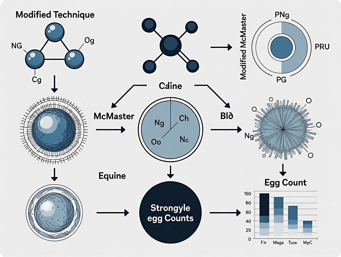
Among the various FEC techniques, the modified McMaster method is one of the most widely used quantitative approaches. It is a dilution technique that estimates the number of eggs per gram (EPG) of faeces. The principle involves creating a homogenized faecal suspension in a flotation solution of specific gravity (typically ≥1.20), which causes helminth eggs to float. An aliquot of this solution is then transferred to a McMaster counting chamber, and the eggs within the grid lines are counted. The count is multiplied by a predetermined factor to calculate the EPG.
Diagram 1: McMaster FEC Workflow
The multiplication factor is determined by the volume of the chamber and the dilution. A common modification uses a factor of 50, meaning each egg counted represents 50 EPG. The diagnostic performance of the McMaster technique, however, is influenced by several variables. A 2023 study found that McMaster variants had a higher coefficient of variation (CV%) in bead recovery experiments compared to Mini-FLOTAC methods, and bead replicates showed greater dispersion from the regression curve, indicating lower repeatability and linearity [3]. This underscores the importance of strict protocol standardization for reliable results.
Advanced Diagnostic and Resistance Monitoring Methods
Comparative Performance of FEC Techniques
While the McMaster technique is a workhorse of parasitology, several other methods are available, each with distinct advantages and limitations. A 2022 comparative study of 1067 equine faecal samples evaluated sedimentation/flotation, Mini-FLOTAC, and FECPAKG2 [6].
Table 4: Comparison of Common Faecal Egg Count Techniques
| Technique | Type | Multiplication Factor | Key Advantages | Key Limitations |
|---|---|---|---|---|
| Modified McMaster | Dilution / Estimation | 25 - 100 | Inexpensive, rapid, widely established | Lower sensitivity, higher variability [3] |
| Mini-FLOTAC | Dilution / Estimation | 5 - 10 | Lower factor improves statistical power, less debris [6] | Requires specific device |
| Sedimentation/ Flotation | Concentration / Enumeration | N/A | High sensitivity for detection, good for tapeworms [6] | Semi-quantitative, more time-consuming |
| FECPAKG2 | Digital / Image-based | 45 | Remote analysis, standardized digital imaging | Lower sensitivity for some parasites, cost [6] |
The choice of technique depends on the objective. For simple detection of parasite eggs, sedimentation/flotation offers high sensitivity. For FECRT, where statistical power is crucial, methods with lower multiplication factors like Mini-FLOTAC are preferred as they count more eggs under the microscope, enhancing test precision [2] [6].
The Faecal Egg Count Reduction Test (FECRT)
The FECRT is the primary in vivo method for detecting anthelmintic resistance in a population. It involves performing FECs on the day of treatment (Day 0) and again 10-14 days post-treatment. The percent reduction is calculated as: % FECR = (1 - (Mean Post-Treatment FEC / Mean Pre-Treatment FEC)) × 100
New World Association for the Advancement of Veterinary Parasitology (WAAVP) guidelines are forthcoming and will provide updated efficacy thresholds and robust statistical methods for interpreting FECRT results. A key concept in these guidelines is the focus on the absolute number of eggs counted under the microscope, not just the final EPG, to improve the statistical power of the test [2]. Resistance is confirmed when the %FECR falls below a specific threshold for a given drug class (e.g., 95% for benzimidazoles, 90% for macrocyclic lactones in some guidelines) and/or when the lower credible interval is below the threshold [5].
In Vitro and Advanced Motility Assays
To complement FECRT, researchers are developing in vitro assays for more precise resistance monitoring. The WMicrotracker Motility Assay (WMA) is an emerging technology that measures the motility of nematodes in response to anthelmintic exposure. A 2025 study demonstrated its utility by successfully discriminating between ivermectin-susceptible and ivermectin-resistant strains of Caenorhabditis elegans and Haemonchus contortus [7]. The assay generates dose-response curves, allowing for the calculation of half-maximal inhibitory concentration (IC50) values. Resistant isolates exhibit significantly higher IC50 values and resistance factors (RF), providing a phenotypic measure of drug tolerance that is independent of host immunity and faecal egg count variability [7].
Diagram 2: WMicrotracker Assay Flow
Research Reagent Solutions and Essential Materials
Table 5: Essential Research Materials for Equine Strongyle FEC and AR Studies
| Item | Function/Application | Specification Notes |
|---|---|---|
| Flotation Solution | Creates specific gravity for egg flotation | Saturated Sugar (SpG ~1.33) or NaNO3 (SpG 1.33) are optimal [8] [3]. |
| McMaster Counting Chamber | Standardized grid for egg enumeration | Two chambers per slide; volume determines multiplication factor. |
| Microscope | Visualization and identification of helminth eggs | Compound microscope with 10x and 40x objectives. |
| Digital Scale | Precise weighing of faecal samples | Capacity to 3-5g with 0.1g accuracy. |
| Polystyrene Microspheres | Proxy for strongyle eggs in method validation | 45µm diameter, Specific Gravity 1.06 [3]. |
| Anthelmintic Standards | For in vitro efficacy assays (e.g., WMA) | Pure chemical standards (e.g., Ivermectin, Fenbendazole). |
The crisis of anthelmintic resistance necessitates an urgent and permanent shift away from calendar-based deworming. The imperative for evidence-based control is clear. The modified McMaster technique and other advanced diagnostic tools provide the scientific foundation for this transition, enabling veterinarians and researchers to implement selective therapy, monitor anthelmintic efficacy, and document the emergence of resistance. The integration of traditional FEC/FECRT with innovative in vitro methods like the WMicrotracker assay will strengthen global resistance surveillance efforts. Preserving the efficacy of existing anthelmintics is paramount, and this can only be achieved through a commitment to surveillance-based principles, prudent anthelmintic use, and a shared understanding that parasite control, not eradication, is the sustainable goal.
Equine strongylid nematodes, parasitic worms residing in the large intestine, represent one of the most prevalent and clinically significant helminth groups affecting horses worldwide [9] [10]. They are broadly categorized into two subfamilies with distinct biological and pathological characteristics: the Cyathostominae (small strongyles or cyathostomins) and the Strongylinae (large strongyles) [11] [10]. The management of these parasites is a critical component of equine health, and the Modified McMaster technique serves as a cornerstone quantitative diagnostic tool for surveillance and treatment decisions [12].
Biological Characteristics and Comparison
While both cyathostomins and large strongyles produce morphologically similar strongyle-type eggs in feces, making them indistinguishable by standard coproscopic examination, their life cycles, tissue migration, and pathogenicity differ substantially [11] [10].
Table 1: Comparative Biology of Cyathostomins and Large Strongyles
| Characteristic | Cyathostomins (Small Strongyles) | Large Strongyles (Strongylus spp.) |
|---|---|---|
| Subfamily | Cyathostominae [10] | Strongylinae [10] |
| Prevalence | Highly prevalent; often >95% of strongyle eggs in feces [10] | Now rare in managed horse populations [9] [10] |
| Adult Worm Size | Small [10] | Large, stout (∼1.5-4.5 cm) [10] |
| Buccal Capsule | Small [10] | Substantial, globular [10] |
| Prepatent Period | ~2-3 months [11] | Long, 6-12 months [9] [10] |
| Larval Migration | Intramucosal (within gut wall) [9] [11] | Extensive extra-intestinal migration [9] [10] |
| Inhibited Larval Stages | Yes (significant epidemiological reservoir) [11] | No |
The following diagram illustrates the core life cycle and key biological differences between these two nematode groups.
Pathogenicity and Clinical Disease
The pathogenic mechanisms and associated clinical syndromes of cyathostomins and large strongyles are direct consequences of their distinct biological strategies.
Cyathostomin Pathogenicity
Pathology arises from both the larval encystment stages and the feeding activities of adults in the large intestine.
- Larval Cyathostominosis: This is a severe, potentially fatal clinical syndrome resulting from the synchronous emergence of encysted larvae from the intestinal mucosa [11]. The massive larval egress causes widespread destruction of the mucosal lining, leading to protein-losing diarrhea, edema, weight loss, and colic [11]. This syndrome shares similarities with Type II ostertagiasis in cattle [11].
- Adult Worm Burden: Non-emergent infections with adult cyathostomins can cause nonspecific signs such as weight loss, poor condition, and occasional colic due to their plug-feeding activity on the intestinal mucosa [10].
Large Strongyle Pathogenicity
The primary pathogenicity of Strongylus spp. is linked to the extensive tissue migration of larval stages.
- Strongylus vulgaris: This is the most pathogenic species [9]. Larvae migrate within the arterial system, specifically targeting the cranial mesenteric artery and its branches, causing verminous arteritis [9] [10]. This lesion is characterized by endothelial damage, thrombus formation, and thickening of the arterial wall, which can lead to non-strangulating intestinal infarction and life-threatening colic [9] [10].
- Strongylus edentatus and Strongylus equinus: Larvae of these species migrate through the liver and, in the case of S. equinus, the pancreas, causing hemorrhagic and inflammatory tracts [10]. While less frequently associated with acute clinical signs, these migrations contribute to the general inflammatory state [10].
Table 2: Pathogenicity and Clinical Syndromes of Equine Strongyles
| Parasite Group | Key Pathogenic Mechanism | Primary Clinical Syndromes | Diagnostic Challenges |
|---|---|---|---|
| Cyathostomins | Synchronous larval emergence from mucosa [11] | Larval cyathostominosis: severe diarrhea, weight loss, edema, colic [11] | Difficult antemortem diagnosis of encysted burden; fecal egg count not correlative [11] |
| Strongylus vulgaris | Larval migration in cranial mesenteric artery [9] [10] | Verminous arteritis, thromboembolic colic, non-strangulating infarction [9] [10] | Low egg output; long prepatent period; direct detection rare [9] |
| Other Strongylus spp. | Larval migration in liver/pancreas [10] | Nonspecific signs: weight loss, poor growth; rarely acute clinical disease [10] | Low egg output; long prepatent period [10] |
Diagnostic Protocols: Application of the Modified McMaster Technique
Accurate diagnosis is essential for effective strongyle control. The following section details the application of the Modified McMaster technique and other advanced diagnostic methods.
The Modified McMaster Technique: A Detailed Protocol
The Modified McMaster technique is a quantitative fecal flotation method used to determine the number of strongyle eggs per gram (EPG) of feces [13] [12]. This protocol is validated for Strongyle-type eggs and coccidia [12].
Principle: The technique uses a counting chamber that enables a known volume of fecal suspension to be examined microscopically [13]. By using a known weight of feces and a known volume of flotation fluid, the number of eggs per gram of feces (EPG) can be calculated [13].
Table 3: Research Reagent Solutions for the Modified McMaster Technique
| Reagent/Material | Function / Specification | Notes |
|---|---|---|
| McMaster Counting Chamber | Holds 2 x 0.15 mL of fecal suspension under grids [13] [12] | Each chamber's grid is calibrated for EPG calculation. |
| Saturated Salt or Sugar Solution | High-specific-gravity flotation fluid [10] | Causes parasite eggs to float to the surface for counting. |
| Analytical Balance | Weighs a precise mass of feces (e.g., 2 g) [12] | Critical for accurate EPG calculation. |
| Graduated Cylinder | Measures a precise volume of flotation fluid (e.g., 60 mL) [12] | Critical for accurate EPG calculation. |
Step-by-Step Workflow:
Calculation: The EPG is calculated using the formula [12]: EPG = (Total egg count in both chambers) × (Total volume of flotation fluid (mL) / Fecal mass (g)) / Number of chambers
Example: For a 2 g fecal sample in 60 mL of fluid, with a total count of 7 strongyle eggs in both chambers: EPG = 7 × (60 / 2) / 2 = 7 × 30 / 2 = 700 EPG [12].
Advanced Diagnostic Methodologies
For researchers and drug development professionals, basic coproscopy must be supplemented with more sophisticated techniques to differentiate species and detect pre-patent infections.
- Fecal Larval Culture and Identification: Feces containing strongyle eggs are incubated for 10-14 days to allow development to infective third-stage larvae (L3), which can then be identified morphologically to the genus or species level [10]. This allows for proportional quantification of cyathostomin versus Strongylus contributions to the total egg output [10].
- Molecular Diagnostics (PCR): PCR-based methods, including real-time PCR and reverse line blot assays, have been developed to identify and semi-quantify a wide range of cyathostomin species and Strongylus vulgaris directly from fecal samples [9] [10]. These methods are more sensitive and specific than larval culture [9].
- Serological Assays (ELISA): An ELISA that detects antibodies against a recombinant S. vulgaris larval antigen (SvSXP) has been developed [9]. This is a powerful tool for detecting migrating larval stages long before patency, revealing exposure that would be missed by fecal examination alone [9]. Studies show a much higher farm-level seroprevalence (83.3%) compared to PCR-based prevalence (12.5%), suggesting many horses are exposed to larvae that never mature to egg-laying adults due to anthelmintic treatments [9].
Epidemiological Insights and Anthelmintic Resistance
Understanding transmission dynamics and the threat of anthelmintic resistance is crucial for designing sustainable control programs.
Epidemiology: Transmission requires pasture access, with environmental conditions dictating seasonal patterns [10]. In northern temperate climates, transmission is perennial, while in southern regions, risk is highest from autumn through spring [10]. A key epidemiological finding is that horses under selective anthelmintic treatment had 4.4 times higher odds of being seropositive for S. vulgaris than horses treated four times per year, highlighting the risk of reduced treatment intensity allowing this pathogen to re-emerge [9].
Anthelmintic Resistance:
- Cyathostomins: Resistance to benzimidazoles (e.g., fenbendazole) is widespread [11]. Resistance to pyrantel is increasingly reported, and reduced efficacy to ivermectin and moxidectin (macrocyclic lactones) has been documented in some regions [11].
- Large Strongyles: No polymorphisms associated with benzimidazole resistance were detected in the S. vulgaris samples in a recent German study, but continued monitoring is essential [9].
Table 4: Summary of Key Quantitative Findings from Recent Research (2022)
| Parameter | Cyathostomins | Strongylus vulgaris |
|---|---|---|
| Prevalence (Horse Level) | 66.7% (strongyle-type eggs) [9] | 1.3% by PCR from feces [9] |
| Prevalence (Farm Level) | >90% in German studies [9] | 12.5% by PCR; 83.3% by Serology (ELISA) [9] |
| Key Risk Factors | Low age; increasing pasture access [9] | Low age; increasing pasture access [9] |
| Anthelmintic Resistance Status | Widespread BZ resistance; emerging ML resistance [11] | No BZ resistance polymorphisms detected in recent study [9] |
| Recommended Diagnostic | FEC (McMaster); Larval Culture; PCR [10] [12] | Serology (ELISA); PCR; Larval Culture [9] [10] |
The biology and pathogenicity of cyathostomins and large strongyles present distinct challenges in equine parasitology. While the highly pathogenic Strongylus vulgaris has been controlled through modern anthelmintics, serological data indicates exposure remains common, warranting vigilance. Cyathostomins, due to their high prevalence, potential for larval arrest, and widespread anthelmintic resistance, remain the primary therapeutic and control challenge. The Modified McMaster technique is an essential, validated tool for quantifying strongyle egg shedding at the herd level to guide treatment decisions. However, a comprehensive diagnostic approach for researchers must integrate this with larval culture, PCR, and serology (ELISA) to accurately define parasite burdens, monitor for emerging resistance, and evaluate the efficacy of novel chemotherapeutic agents in development.
The McMaster technique is a quantitative fecal egg count (FEC) method and remains one of the most widely used diagnostic tools in veterinary parasitology for estimating parasite burden in equine populations [14] [15]. This technique's fundamental principle relies on the combination of flotation and enumeration to identify and quantify helminth eggs, particularly strongyle eggs, in fecal samples. Within the context of equine strongyle egg count research, the modified McMaster method serves as a cornerstone for surveillance-based parasite control programs, enabling researchers to identify high shedders and monitor anthelmintic efficacy [14] [16].
The technique's continued relevance stems from its practical balance of simplicity, cost-effectiveness, and quantitative output, making it suitable for both laboratory and field settings. As anthelmintic resistance in cyathostomins continues to emerge as a significant threat to equine health globally, the accuracy and precision of the McMaster technique have become subjects of ongoing research and refinement [15] [17].
Core Principles: Flotation and Enumeration
The Flotation Principle
The first fundamental component of the McMaster technique is flotation, which exploits differences in specific gravity between parasite eggs and the surrounding fecal debris. Helminth eggs typically have a specific gravity ranging from 1.05 to 1.23 [16]. When a fecal suspension is prepared using a flotation solution with a specific gravity higher than that of the eggs (generally between 1.20 and 1.35), the eggs become buoyant and float to the surface [14] [18].
This physical separation enables the researcher to isolate parasite eggs from the denser fecal material that sinks to the bottom. The efficiency of flotation depends critically on several factors:
- Specific gravity of the flotation solution: Must be carefully calibrated to exceed that of the target eggs
- Viscosity of the solution: Affects the rate at which eggs rise
- Egg characteristics: Size, weight, and morphology influence flotation capability
- Sedimentation time: Adequate time must be allowed for eggs to rise to the surface
Commonly used flotation solutions for equine strongyle egg counts include saturated sodium chloride (specific gravity 1.20), magnesium sulfate (specific gravity 1.32), and Sheather's sugar solution (specific gravity 1.20-1.25) [18]. The optimal specific gravity for floating most strongyle eggs is approximately 1.20-1.25 [16] [18].
The Enumeration Principle
The second fundamental component is enumeration, which provides the quantitative aspect of the technique. The McMaster method employs a specialized counting chamber with a gridded area that allows for the systematic counting of eggs present in a known volume of the fecal suspension [14].
The quantitative nature of the test stems from the precise relationship between the sample preparation and the chamber dimensions:
- A known weight of feces is suspended in a known volume of flotation solution
- Only eggs within the gridded areas of the chamber are counted
- The chamber volume under the grid is precisely calibrated
- A multiplication factor is applied to calculate eggs per gram (EPG) of feces
The standard calculation for the modified McMaster technique is: Total eggs counted × (Total volume of suspension / Volume under grids) × (1 / Weight of feces) = Eggs per gram (EPG) [14]. For a typical protocol using 4g feces in 56mL flotation solution, the multiplication factor is 50 [18].
Table 1: Standard Multiplication Factors in McMaster Technique
| Feces Weight (g) | Flotation Solution Volume (mL) | Total Volume (mL) | Multiplication Factor |
|---|---|---|---|
| 4 | 56 | 60 | 50 |
| 4 | 26 | 30 | 25 |
| 2 | 28 | 30 | 50 |
| 5 | 45 | 50 | 10 |
Comparative Performance of Fecal Egg Count Techniques
Recent research has evaluated the McMaster technique alongside other fecal egg counting methods to assess their relative diagnostic performance for equine strongyle detection.
Table 2: Comparative Performance of Fecal Egg Count Techniques for Equine Strongyles
| Technique | Principle | Reported Sensitivity | Reported Precision | Key Advantages | Key Limitations |
|---|---|---|---|---|---|
| McMaster | Dilution & flotation | 85% [15] | Lower precision compared to FLOTAC [15] | Rapid, inexpensive, no centrifugation required [14] | Higher variability, egg count overestimation [16] [15] |
| Mini-FLOTAC | Dilution & flotation | 93% [15] | Better repeatability and linearity [16] | Higher sensitivity, improved linearity [16] [15] | Requires specialized device, longer settling time |
| FLOTAC | Flotation & centrifugation | 89% [15] | 72% [15] | Highest precision, sensitive for low egg counts [15] | Requires centrifugation, more complex protocol [15] |
| Wisconsin | Concentration & flotation | >98% [17] | Lower biological variability [17] | High sensitivity for low egg counts [19] | Requires centrifugation, more time-consuming [17] [19] |
| Automated Counting | Image analysis & machine learning | >98% [17] | Highest technical precision [17] | Reduces operator variability, high throughput [17] | High equipment cost, requires technical expertise |
A 2025 study comparing McMaster, FLOTAC, and Mini-FLOTAC techniques for diagnosing strongylid infections in horses found that McMaster detected significantly higher egg shedding (584 ± 179 EPG) compared to the other methods, suggesting possible overestimation [15]. The same study reported that FLOTAC achieved the highest precision (72%), which was significantly better than McMaster [15].
Detailed Experimental Protocol for Equine Strongyle Egg Counts
Materials and Equipment
- McMaster counting slides (specialized chambers with grids)
- Microscope with 100x magnification (10x objective and 10x eyepiece)
- Digital scale capable of weighing to 0.1g precision
- Flotation solution (specific gravity 1.20-1.25): saturated sodium chloride, Sheather's sugar solution, or sodium nitrate
- Graduated cylinder or syringe for measuring flotation solution
- Mixing containers: disposable cups or beakers
- Strainer (tea strainer or gauze) with ~150μm mesh
- Fecal sample collection equipment: gloves, rectal sleeves, sample containers
- Timer
- Pipette or dropper for transferring suspension
Step-by-Step Procedure
Sample Collection and Preparation
- Collect fresh fecal samples directly from the rectum or immediately after defecation
- Label all samples clearly with animal identification and date
- Process samples within 1-2 hours of collection, or refrigerate at 4°C for up to 5 days [14] [18]
- Do not freeze samples, as freezing distorts parasite eggs [18]
Flotation Solution Preparation
- Prepare flotation solution with appropriate specific gravity (1.20-1.25 for strongyles)
- For saturated sodium chloride: dissolve 159g NaCl in 1L warm water, verify specific gravity with hydrometer [18]
- For Sheather's sugar solution: dissolve 454g granulated sugar in 355mL water with gentle heat, cool, add 6mL formalin to prevent microbial growth [18]
Fecal Suspension Preparation
- Weigh 4g of well-mixed feces to the nearest 0.1g
- Add 56mL of flotation solution to create a 1:15 dilution [18]
- Mix thoroughly until a homogeneous suspension is achieved
- Strain the suspension through a tea strainer or gauze to remove large debris
Loading McMaster Chamber
- Using a pipette or dropper, carefully transfer the strained suspension to both chambers of the McMaster slide
- Avoid introducing air bubbles, which can disrupt the grid pattern
- Fill chambers completely but not overflowing
Egg Flotation
- Allow the loaded slide to stand undisturbed for 5-10 minutes
- This settling period enables eggs to float up to the grid level
- Do not exceed 60 minutes before examination to prevent crystallization or egg distortion [18]
Microscopic Examination and Enumeration
- Place the slide on the microscope stage and examine at 100x magnification
- Systematically scan all grid areas in both chambers
- Count only eggs that lie entirely within the grid lines; eggs touching boundary lines may be counted according to established laboratory protocols
- Identify strongyle eggs based on morphological characteristics: oval shape, thin-shelled, containing morula, ~90x50μm [15]
Calculation of Eggs Per Gram (EPG)
- Sum the egg counts from both chambers
- Apply the multiplication factor: EPG = Total egg count × 50 (for 4g feces in 56mL solution) [18]
- For example: (25 eggs in chamber 1 + 20 eggs in chamber 2) × 50 = 2,250 EPG
Diagram 1: McMaster Technique Workflow
The Scientist's Toolkit: Essential Research Reagents and Materials
Table 3: Essential Research Reagents and Materials for McMaster Technique
| Item | Specification/Function | Research Application |
|---|---|---|
| McMaster Slides | Specialized counting chambers with calibrated grids and volumes | Standardized enumeration of eggs across studies [14] |
| Flotation Solutions | Sucrose, NaCl, NaNO₃, ZnSO₄ with specific gravity 1.20-1.25 | Optimal recovery of strongyle eggs [16] [18] |
| Digital Scale | Precision to 0.1g | Accurate fecal sample weighing for reproducible EPG calculations [18] |
| Compound Microscope | 10x eyepiece, 10x and 40x objectives, internal light source | Identification and enumeration of strongyle eggs [18] |
| Fluorescent Stains | Chitin-binding dyes (e.g., Calcofluor White, CFB) | Enhanced egg detection in automated systems [17] |
| Refrigeration Equipment | 4°C storage | Sample preservation before processing [14] [18] |
| Polystyrene Microspheres | 45μm diameter, specific gravity 1.06 | Quality control and method validation [16] |
Critical Factors Influencing Results and Methodological Considerations
The McMaster technique exhibits several inherent limitations that researchers must consider when interpreting results:
- Detection threshold: The method has a sensitivity of 25-50 EPG, potentially missing low-level infections [18] [19]
- Egg count variability: Fecal egg counts do not directly correlate with actual worm burdens due to factors including host immunity, nutritional status, and parasite reproductive biology [14] [20]
- Identification limitations: The technique cannot differentiate between strongyle species based on egg morphology alone [14]
- Operator dependency: Technical variability arises from differences in sample preparation, counting technique, and individual interpretation [17]
Methodological Refinements for Research Applications
For high-precision research applications, several methodological refinements can enhance the reliability of McMaster results:
- Multiple replicates: Perform triplicate counts per sample to assess variability [15]
- Standardized timing: Maintain consistent flotation periods across all samples
- Quality control: Implement periodic validation with bead standards to monitor recovery rates [16]
- Blinded counting: Eliminate observer bias by masking sample identities during microscopy
- Staff training: Ensure consistent identification and counting through regular training and inter-observer reliability assessments
Research Applications in Equine Strongyle Studies
Within equine parasitology research, the modified McMaster technique serves several critical functions:
Fecal Egg Count Reduction Test (FECRT)
Shedder Categorization for Targeted Selective Treatment
Pasture Contamination Monitoring
- Estimates parasite transmission potential in equine facilities
- Informs pasture management decisions to break parasite life cycles
Parasite Control Program Assessment
- Evaluates the effectiveness of integrated parasite management strategies
- Monitors long-term trends in parasite prevalence and intensity
The fundamental principles of flotation and enumeration embodied in the McMaster technique continue to make it an indispensable tool in equine parasitology research, despite the development of more sensitive alternatives. Its simplicity, cost-effectiveness, and standardization across laboratories ensure its ongoing relevance for studies of equine strongyle ecology, anthelmintic efficacy, and resistance management.
Faecal Egg Count (FEC) techniques are fundamental tools for diagnosing gastrointestinal strongyle infections in horses and guiding evidence-based anthelmintic treatment programs. The modified McMaster technique, despite its widespread use, is often compared to newer methods like FLOTAC and Mini-FLOTAC regarding key analytical performance metrics. This application note delineates the critical parameters—sensitivity, precision, and accuracy—used to evaluate FEC diagnostics. We provide a structured comparison of quantitative data, detailed experimental protocols for method assessment, and visual workflows to aid researchers in selecting and validating the most appropriate FEC technique for equine strongyle egg count research, emphasizing the context of the modified McMaster technique.
Gastrointestinal strongyle infections, particularly those caused by cyathostomins (small strongyles), represent a ubiquitous challenge to equine health and welfare worldwide [21]. The cornerstone of modern, sustainable parasite control is evidence-based targeted treatment, which relies on Faecal Egg Counts (FEC) to identify individual horses based on their egg-shedding potential [3]. The American Association of Equine Practitioners (AAEP) guidelines categorize horses as low (0-200 EPG), moderate (201-500 EPG), or high (>500 EPG) shedders, with treatment frequently targeted only at high shedders [3]. The success of this strategy is entirely dependent on the diagnostic performance of the FEC method employed. The modified McMaster technique is a historical standard, but its performance must be critically assessed and compared to emerging methodologies like FLOTAC and Mini-FLOTAC using well-defined metrics: sensitivity, precision, and accuracy [22] [23].
Confusion in terminology often complicates the validation of FEC techniques. It is crucial to understand that the "detection limit" is a theoretical value, while "analytical sensitivity" is determined experimentally; the two terms are not synonymous [22]. For quantitative FEC tests, precision (reproducibility) is arguably the most important performance parameter, more so than accuracy (closeness to the true value) [22]. This note clarifies these concepts and provides a framework for their practical evaluation in a research setting focused on equine strongyles.
Comparative Performance of FEC Techniques
The following tables summarize key performance data from recent studies comparing FEC techniques, with a specific focus on equine strongyle diagnostics.
Table 1: Comparative Analytical Performance of McMaster, FLOTAC, and Mini-FLOTAC for Equine Strongyles [21]
| Performance Metric | McMaster | FLOTAC | Mini-FLOTAC |
|---|---|---|---|
| Mean Strongyle EPG | 584 ± 179 | Not Specified | Not Specified |
| Precision | 72%* | 72% | Not Specified |
| Diagnostic Sensitivity | 85% | 89% | 93% |
| Correlation (Spearman's rs) | 0.92 - 0.96 (vs. other methods) | 0.92 - 0.96 (vs. other methods) | 0.92 - 0.96 (vs. other methods) |
| Cohen's Kappa (Agreement) | 0.67 - 0.76 (vs. other methods) | 0.67 - 0.76 (vs. other methods) | 0.67 - 0.76 (vs. other methods) |
| *The precision value for McMaster was significantly lower than that of FLOTAC (p=0.03). |
Table 2: Performance Summary from Studies in Other Host Species [24] [3]
| Study / Host | Metric | McMaster | Mini-FLOTAC | Other Methods |
|---|---|---|---|---|
| Camels [24] | Strongyle Positivity | 48.8% | 68.6% | Semi-Quantitative Flotation: 52.7% |
| Mean Strongyle EPG | 330.1 | 537.4 | - | |
| Moniezia spp. Positivity | 2.2% | 7.7% | Semi-Quantitative Flotation: 4.5% | |
| Horses (Bead Study) [3] | Linearity (R²) with Beads | Lower R² (Dispersed) | >0.95 | Modified Wisconsin (NaNO₃): >0.95 |
| Coefficient of Variation (CV%) | Highest | Lowest | - |
Experimental Protocols for Key FEC Experiments
Protocol: Sample Collection and Preparation for Method Comparison
- Objective: To collect and prepare equine fecal samples for a standardized comparison of FEC technique performance.
- Materials: Disposable gloves, labelled plastic bags, cooling bag, refrigerator (4°C), pestle and mortar.
- Procedure:
- Collect fecal samples immediately after excretion from the superficial portion of the stool [21].
- Place samples in labelled plastic bags and transport them to the laboratory in a cooling bag.
- Store samples at 4–5°C for a maximum of two weeks before processing [21].
- Prior to analysis, homogenize the entire fecal sample thoroughly using a pestle and mortar [24].
Protocol: Modified McMaster Technique
- Objective: To quantify strongyle eggs per gram (EPG) of feces using the modified McMaster technique.
- Materials: Saturated sucrose solution (specific gravity 1.20), 0.3-mm mesh strainer, McMaster slide, light microscope, balance.
- Procedure:
- Weigh 2 g of homogenized feces and mix with 28 mL of saturated sucrose solution (dilution 1:15) [21].
- Filter the mixture through a 0.3-mm mesh strainer.
- Transfer the filtered suspension to the two chambers of a McMaster slide.
- Allow the slide to stand for 5-10 minutes to enable eggs to float to the surface.
- Examine both chambers under a light microscope at 100x magnification.
- Count all strongyle eggs within the engraved grids of both chambers.
- Calculation: Multiply the total egg count by 50 (multiplication factor) to obtain the EPG value [21].
Protocol: Assessment of Precision Using Technical Replicates
- Objective: To determine the precision (repeatability) of a FEC technique.
- Procedure:
- Select a fecal sample with a moderate to high strongyle egg count.
- Process the same sample using the chosen FEC technique (e.g., McMaster) multiple times (e.g., 3-6 technical replicates), ensuring each replicate is prepared from the initial homogenate independently [21] [24].
- Record the EPG for each replicate.
- Calculation: Calculate the Coefficient of Variation (CV%) for the replicate counts: (Standard Deviation / Mean) x 100. Precision can then be computed as 100% - CV% [21].
Protocol: Assessment of Diagnostic Sensitivity
- Objective: To determine the ability of a FEC technique to correctly identify positive infections.
- Procedure:
- Process a set of fecal samples (e.g., n=32) using the test method (e.g., McMaster) and one or more comparator methods (e.g., FLOTAC, Mini-FLOTAC) [21].
- Define the "true positive" population. In the absence of a perfect gold standard, this is often defined as samples that test positive by any of the techniques used in the comparison [21].
- Calculation: Sensitivity = (Number of true positives detected by the test method / Total number of true positives) x 100 [21].
Visualizing Performance Metrics and Method Selection
Relationship of Key FEC Performance Metrics
The following diagram illustrates the interconnectedness of core FEC performance concepts and their practical implications for diagnostic outcomes.
Experimental Workflow for FEC Method Validation
This workflow outlines the key steps for a robust comparison and validation of fecal egg count techniques in a research setting.
The Scientist's Toolkit: Research Reagent Solutions
Table 3: Essential Materials for FEC Research in Equine Strongyles
| Reagent / Material | Function / Specification | Research Application Note |
|---|---|---|
| Saturated Sucrose Solution | Flotation medium (Specific Gravity ~1.20) | Common, cost-effective medium for strongyle eggs. High viscosity can slow flotation [21] [3]. |
| Sodium Nitrate (NaNO₃) Solution | Flotation medium (Specific Gravity up to 1.33) | Often provides superior egg recovery and clarity compared to sucrose due to higher specific gravity [3]. |
| Polystyrene Microspheres | Proxy for strongyle eggs (SPG ~1.06, 45 µm diameter) | Enables standardized method comparison and accuracy assessment without relying on highly variable natural samples [3]. |
| Fill-FLOTAC Device | Standardized sample collection and homogenization | Ensures consistent sample preparation and dilution for FLOTAC and Mini-FLOTAC techniques [25]. |
| McMaster Slide | Counting chamber with calibrated grids | Allows for quantitative estimation of EPG. The design limits the volume examined, affecting sensitivity and precision [21]. |
| Mini-FLOTAC Base | Precision counting chamber | Enables examination of a larger fecal volume (up to 1g vs. McMaster's ~0.3g), improving sensitivity [24]. |
Standardized Protocols and Advanced Applications of the Modified McMaster Technique
The modified McMaster technique is a quantitative fecal egg count (FEC) method cornerstone of surveillance-based equine anthelmintic programs [17]. Its primary function is identifying horses that are high strongyle egg shedders, a critical step in mitigating widespread anthelmintic resistance [16] [17]. The accuracy and precision of this protocol are confounded by numerous variables, including the choice of flotation solution and the calculation of the multiplication factor [26]. This application note provides a detailed, standardized protocol for the modified McMaster technique, contextualized within current research findings to ensure reliable data for research and drug development.
The Scientist's Toolkit: Research Reagent Solutions
The following table details essential materials and reagents required for the modified McMaster technique, with specifications informed by comparative methodological studies.
Table 1: Essential Research Reagents and Materials
| Item | Function/Description | Research Context & Considerations |
|---|---|---|
| McMaster Slide | Specialized counting chamber with two grids; each chamber holds 0.15 mL of fecal suspension [12]. | The design (grid area/volume) directly influences the detection limit and reliability of the count [26]. |
| Flotation Solution | High-specific-gravity fluid that floats parasite eggs out of fecal debris for visualization [18]. | Specific Gravity (SPG) is critical. Common solutions include saturated Sodium Chloride (NaCl, SPG 1.20) and Sodium Nitrate (NaNO₃, SPG 1.33) [16] [18]. The choice of solution impacts egg recovery and clarity [16]. |
| Digital Scale | Precisely weighs fecal sample to maintain accurate dilution ratios [18]. | Critical for standardizing the initial sample mass (e.g., 4 grams). Inconsistencies here introduce significant error. |
| Microscope | Magnifies the McMaster slide grid for egg identification and enumeration. | A microscope capable of 100x magnification with a 10x wide-field lens is recommended [18]. |
| Tea Strainer/Filter | Removes large fecal debris from the suspension to prevent obstruction of the counting chamber [18]. | Standardizing the filtration step is necessary to improve sample homogeneity and counting ease. |
Comparative Performance Data
Evaluating the diagnostic performance of the McMaster method against emerging and established techniques is vital for research quality control. The following table synthesizes key quantitative findings from recent comparative studies.
Table 2: Quantitative Comparison of Fecal Egg Count Techniques
| Technique | Principle | Key Performance Metrics (vs. McMaster) | Research Implications |
|---|---|---|---|
| Modified McMaster | Dilution and flotation [16]. | Precision (CV%): Lower precision in bead recovery studies [16]. Accuracy: 23.5% in spiked sample study [27]. | Considered a standard but has inherent variability; requires strict protocol adherence. |
| Mini-FLOTAC | Dilution and flotation in a different chamber design [16]. | Precision (CV%): Higher precision in bead recovery [16]. Accuracy: 42.6% [27]. Sensitivity: Higher specificity and sensitivity reported [27]. | A more reliable alternative for precise quantification, though may be more time-consuming [27]. |
| Modified Wisconsin | Concentration by centrifugation and flotation [16]. | Precision (CV%): Lower biological variability for samples >200 EPG than some automated methods [17]. | Considered a highly sensitive concentration technique, but requires a centrifuge, limiting field use [17]. |
| Automated Egg Counting | Fluorescent staining and image analysis with machine learning [17]. | Precision: Significantly lower technical variability than McMaster for samples >200 EPG [17]. Specificity: Highest for a custom camera with a particle shape analysis algorithm [17]. | Promising for removing operator-based variability; algorithm refinement is an active research area [17]. |
Core Experimental Protocol
Sample Preparation and Processing
Step 1: Collection. Collect fresh fecal samples directly from the rectum or immediately after defecation [18]. Place samples in labeled bags or containers and refrigerate if not processed within 1–2 hours. Note: Do not freeze samples, as this distorts parasite eggs [18].
Step 2: Homogenization. Weigh 4 grams of feces and combine with 56 mL of the chosen flotation solution (e.g., NaCl SPG 1.20 or NaNO₃ SPG 1.33) in a disposable cup [18] [12]. Mix thoroughly with a tongue depressor until a homogeneous suspension is achieved.
Step 3: Filtration. Pour the homogenized mixture through a tea strainer or fecal sieve into a second container to remove large particulate debris [18].
Step 4: Chamber Filling. Using a 3 cc syringe or dropper, draw the filtered suspension and carefully fill both chambers of the McMaster slide. Avoid creating bubbles, as they can disrupt the counting grid [18].
Step 5: Flotation. Allow the filled slide to sit undisturbed for 5 minutes [18]. This enables helminth eggs to float to the surface of the fluid within the chambers.
Enumeration and Calculation
Step 6: Microscopic Examination. After the flotation period, place the slide under the microscope. Systematically count all strongyle-type eggs within the engraved grid lines of both chambers. Eggs touching the grid lines or outside the grid are excluded [12].
Step 7: Calculation of Eggs per Gram (EPG). The total number of eggs counted from both chambers is multiplied by the multiplication factor of 50 to obtain the EPG value [18] [12].
Total EPG = (Number of eggs in Chamber 1 + Number of eggs in Chamber 2) × 50
This factor is derived from the dilution (4 g feces in 60 mL total volume, with 0.3 mL examined)[ccitation:5] [12]. For a lower detection limit, a protocol using 4 g of feces in 26 mL of solution with a multiplication factor of 25 can be employed [18].
The following workflow diagram summarizes the core procedural steps.
Critical Research Parameters & Troubleshooting
Flotation Solution Selection
The specific gravity and chemical composition of the flotation solution are significant variables. Saturated sodium chloride (SPG 1.20) is common and cost-effective, but slides must be read promptly to avoid crystallization [18]. Sodium nitrate (SPG 1.33) is effective for floating most strongyle eggs and is used in standardized variants of other methods like Wisconsin and Mini-FLOTAC [16]. Research indicates that the Mini-FLOTAC method is less influenced by the choice of flotation solution, whereas McMaster results can show greater variability [16].
Addressing Limitations and Variability
The modified McMaster technique has several documented limitations that researchers must account for:
- Detection Sensitivity: The common 50 EPG limit may fail to detect low-level infections [18].
- Inherent Variability: Technical variability (CV%) is significantly higher for McMaster compared to automated counting methods, especially in samples >200 EPG [17].
- Accuracy: The method can overestimate true counts, as it is a dilution technique rather than a full concentration method [16]. One study found its accuracy to be 23.5% compared to known spike-in counts [27].
To mitigate these issues, researchers should:
- Run Replicates: Perform counts in triplicate to assess variability [27].
- Standardize Rigorously: Use calibrated equipment and strict timing across all samples.
- Validate with Controls: Use spiked samples with known egg or proxy bead counts to determine a correction factor for accuracy if necessary [16].
Within parasitology research, particularly in the quantification of equine strongyle eggs via the Modified McMaster technique, centrifugation is a critical step for enhancing the accuracy and reliability of results. The sedimentation principle, where centrifugal force causes denser particles (like parasite eggs) to migrate outward and form a pellet, is fundamental to this process [28] [29]. This document details established protocols and design variations for centrifugation, framed within the context of optimizing the Modified McMaster technique for research and drug efficacy studies. The primary goal of these modifications is to improve egg recovery rates and the precision of eggs-per-gram (EPG) counts, which are crucial for evaluating anthelmintic resistance and genetic resistance in equine strongyles [30] [31].
Centrifugation Principles and Application to Fecal Egg Counting
Centrifugation separates components of a mixture based on their size, density, and shape by applying a centrifugal force. In a fecal sample suspension, this force causes the denser strongyle eggs to settle, facilitating their separation from less dense fecal debris [28] [29].
The process relies on centrifugal force, an outward force experienced during rotation, which causes the sedimentation of particles. The rate at which particles sediment is determined by their size and density, with heavier and more dense particles moving faster [28]. During rapid centrifugation, these particles form a pellet at the base of the tube, while the lighter, clarified liquid, or supernatant, remains above [28]. This pellet, enriched with strongyle eggs, can then be used for further analysis in the McMaster counting chamber.
Table 1: Core Components of a Centrifuge and Their Function in Fecal Egg Counting
| Component | Function | Consideration for Fecal Egg Counting |
|---|---|---|
| Rotor | Holds sample tubes in place during rotation. | Must be balanced to ensure operational integrity and consistent results. Fixed-angle vs. swinging-bucket rotors can influence pellet formation [28]. |
| Motor | Provides the power for the rotation of the rotor. | Determines the maximum speed (RPM) and relative centrifugal force (RCF or g-force) achievable, which directly impacts sedimentation efficiency [28] [29]. |
| Driveshaft | Connects the motor to the rotor. | A critical link; improper alignment can lead to instrument failure and inconsistent results [28]. |
Centrifugation Techniques and Modified Protocols
Several centrifugation techniques can be adapted to the sample preparation phase of the Modified McMaster technique to purify and concentrate strongyle eggs before quantification.
Differential Centrifugation
This technique uses multiple rounds of centrifugation at progressively higher speeds to separate components based on size and density [28] [29].
Detailed Protocol for Strongyle Egg Isolation:
- Sample Homogenization: Thoroughly mix the fecal sample in a saturated saline or sugar flotation solution to create a uniform suspension.
- Low-Speed Spin: Centrifuge the homogenized sample at a low speed (e.g., 500 x g for 5 minutes). This step pellets large, dense debris, undigested feed particles, and other contaminants, while most strongyle eggs remain in the supernatant [29].
- Supernatant Transfer: Carefully decant or pipette the supernatant into a new, clean centrifuge tube. The initial pellet is discarded.
- Medium-Speed Spin: Centrifuge the collected supernatant at a medium speed (e.g., 1,500 x g for 10 minutes). This forces the strongyle eggs to form a pellet at the bottom of the tube.
- Supernatant Removal and Resuspension: Discard the final supernatant. The resulting pellet, now enriched with strongyle eggs, is resuspended in a precise, small volume of flotation medium for loading onto the McMaster chamber [29]. This concentration step significantly improves the detection limit of the assay.
Density Gradient Centrifugation
This method separates particles based on their buoyant density by using a medium with a pre-formed density gradient [28] [29]. Particles will migrate until they reach a point where their density matches that of the surrounding medium.
Detailed Protocol for Species/Component Separation:
- Gradient Preparation: Gently layer solutions of decreasing density (e.g., sucrose or commercial media like Percoll) into a centrifuge tube to create a continuous or discontinuous gradient. The highest density is at the bottom.
- Sample Layering: Carefully layer the prepared fecal sample suspension on top of the density gradient.
- High-Speed Centrifugation: Centrifuge the tube at a high speed (e.g., 2,000 x g for 15-20 minutes). Different parasite eggs (e.g., strongyles, ascarids) and components will band at distinct positions in the gradient according to their specific densities.
- Fraction Collection: After centrifugation, carefully collect the distinct bands using a pipette. These fractions can be washed to remove the gradient medium and then analyzed separately. This is particularly useful for research aiming to isolate specific parasitic elements or to remove specific contaminants [29].
Isopycnic Centrifugation
A specific type of density gradient centrifugation where separation occurs solely on the basis of particle density, not size. Particles sediment until they reach their isopycnic point (where their density equals the medium's) and then cease moving [29]. This is considered a true equilibrium method.
Research Reagent Solutions and Essential Materials
The following reagents and materials are critical for implementing the centrifugation protocols described.
Table 2: Key Research Reagent Solutions for Centrifugation-Based Fecal Egg Counting
| Reagent/Material | Function/Explanation |
|---|---|
| Saturated Salt or Sugar Solution | Flotation medium with high specific gravity to cause parasite eggs to float. Centrifugation enhances contact with this medium, improving egg recovery [30]. |
| Density Gradient Media (e.g., Sucrose, Percoll) | Used to create a density gradient for advanced separation techniques, allowing for the purification of eggs based on their specific buoyant densities [29]. |
| Fixatives (e.g., Formalin) | May be added to preserve parasite egg morphology during processing and storage, especially if there is a delay between sample collection and analysis. |
| Wash Buffers (e.g., PBS) | Used to resuspend and wash pellets between centrifugation steps, removing soluble contaminants and residual gradient media that could interfere with counting. |
| Microcentrifuge Tubes & Rotors | Tubes must be compatible with the applied g-forces. Rotor design (fixed-angle, swinging-bucket) influences the pellet geometry and the ease of supernatant removal [28]. |
Workflow and Chamber Design Visualization
The following diagram illustrates the logical workflow for integrating these centrifugation techniques into a research pipeline for equine strongyle egg counts.
Diagram 1: Centrifugation-enhanced McMaster workflow.
The Modified McMaster technique is a cornerstone quantitative method in veterinary parasitology, enabling researchers to estimate parasite egg burden in faecal samples by calculating Eggs per Gram (EPG). Within equine strongyle research, precise EPG data forms the critical foundation for a range of applications, from classifying individual shedding intensity to conducting Faecal Egg Count Reduction Tests (FECRT) for anthelmintic efficacy testing [32] [16]. The shift from calendar-based deworming to surveillance-based control paradigms underscores the technique's importance in mitigating widespread anthelmintic resistance [8] [32]. This application note details the standardized protocol for the Modified McMaster technique, provides context for interpreting results, and compares its performance against emerging methodologies to support rigorous scientific research.
Principle of the McMaster Technique
The McMaster technique is a dilution egg count method that enables the quantification of parasite eggs within a known mass of faeces [16]. The core principle involves creating a homogeneous faecal suspension using a specific volume of flotation solution, which has a high specific gravity sufficient to cause parasite eggs to float. A defined volume of this suspension is then transferred to a specialized counting chamber. The chamber features grids etched onto its surface, allowing the microscopical examination of a known volume of the suspension where the eggs have floated to the surface [13]. By counting the eggs within the grid areas and applying a standard calculation that accounts for the original faecal mass and the total dilution volume, the number of eggs per gram of faeces can be reliably estimated [12].
Figure 1: Experimental workflow for the Modified McMaster EPG technique.
Research Reagent Solutions and Essential Materials
The following table catalogues the key reagents and materials required to perform the Modified McMaster technique, with explanations of their specific functions within the protocol.
Table 1: Essential Research Reagents and Materials for the Modified McMaster Technique
| Item | Function/Explanation |
|---|---|
| McMaster Counting Chamber | A specialized slide with two chambers, each holding 0.15 mL of sample and featuring etched grids to define the counting area [12] [13]. |
| Flotation Solution | A solution with a specific gravity (SG) sufficient to float parasite eggs (typically SG ≥1.2). Common solutions include saturated sodium chloride (NaCl), sodium nitrate (NaNO₃), or zinc sulfate (ZnSO₄) [8] [16]. |
| Analytical Balance | For precise measurement of the faecal sample mass (e.g., 2 grams or 4 grams), which is a critical variable in the EPG calculation [12]. |
| Graduated Cylinder | For accurate measurement of the total volume of flotation solution used to create the faecal suspension [12]. |
| Microscope | A standard light microscope with 10x objective, used for identifying and counting strongyle eggs within the chamber grids. |
Detailed Experimental Protocol
Sample Preparation and Homogenization
- Weigh a specific mass of faeces (e.g., 2 grams or 4 grams) using an analytical balance [12].
- Transfer the sample to a mixing vessel (e.g., a plastic cup or a Fill-FLOTAC homogenizer) [27].
- Add a predetermined volume of flotation solution (e.g., 28 mL or 58 mL, depending on the desired multiplication factor) to the faecal matter [12].
- Homogenize the mixture thoroughly until a consistent suspension is achieved. This can be done with a wooden tongue depressor, a spatula, or by using a specialized homogenizer device [27].
Chamber Loading and Egg Floatation
- Strain the homogenized suspension through a sieve or tea strainer to remove large, coarse debris [16].
- Pipette the strained suspension into the two chambers of the McMaster slide. Care should be taken to avoid introducing air bubbles.
- Allow the loaded chamber to stand for a period of approximately 3-5 minutes. This enables parasite eggs to float to the surface of the liquid and come into focus within the grid area under the microscope [12].
Microscopic Examination and Counting
- Place the chamber on the microscope stage and examine the areas beneath the grids of both chambers using a 10x objective.
- Systematically count all strongyle-type eggs within the grid lines of each chamber. Eggs that are outside the grid lines or touching the borderlines should be excluded from the count [12].
- Tally the total number of eggs counted in both chambers.
EPG Calculation and Formula
The Eggs per Gram (EPG) is calculated using the following formula, which accounts for the dilution factor and the volume examined:
EPG = (Total egg count from both chambers) × (Total volume of flotation solution) / (Volume of one chamber × Mass of faeces) [12]
Table 2: Example EPG calculation using standard parameters (2g faeces, 60mL total volume, 0.15mL chambers)
| Parameter | Chamber 1 Count | Chamber 2 Count | Total Count | EPG Calculation | Final EPG |
|---|---|---|---|---|---|
| Strongyle-type Eggs | 5 | 2 | 7 | 7 × (60 / (0.15 × 2)) = 7 × 200 | 1,400 |
| Coccidia Oocysts | 30 | 40 | 70 | 70 × 200 | 14,000 |
When using common standard parameters (e.g., 2g faeces, 60mL solution, and chambers of 0.15mL each), the formula simplifies to EPG = Total egg count × 50 [12]. The multiplication factor (e.g., 25, 50, or 100) is predetermined by the specific mass of faeces and total volume of flotation solution used in the protocol.
Performance Comparison of Quantitative FECT
The diagnostic performance of the Modified McMaster technique must be understood in the context of other available methods. The following table summarizes a comparative analysis of key faecal egg counting techniques (FECT) based on recent scientific literature.
Table 3: Comparison of faecal egg count techniques used in equine strongyle research
| Technique | Principle | Reported Precision (CV%) | Reported Accuracy | Key Research Findings |
|---|---|---|---|---|
| Modified McMaster | Dilution & visual counting | Lower precision (higher CV%) [16] | 23.5% [27] | Widely used; overestimates counts; performance varies with flotation solution [16]. |
| Mini-FLOTAC | Dilution & visual counting | Higher precision (lower CV%) [16] | 42.6% [27] | Higher accuracy/precision than McMaster; better linearity with true counts [27] [16]. |
| Wisconsin Flotation | Concentration & enumeration | Information Missing | Information Missing | Considered a concentration method; can have a lower detection limit [16]. |
| FECPAK | Dilution & digital imaging | Information Missing | Information Missing | Enables image capture for remote analysis; performance compared to McMaster [8]. |
Data Interpretation and Application in Research
Classifying Egg Shedding Intensity
For adult horses, the EPG result obtained via the McMaster technique is used to classify animals into shedding categories, which informs targeted selective treatment strategies [32]:
- Low Shedder: < 200 EPG
- Moderate Shedder: 200 - 500 EPG
- High Shedder: > 500 EPG
Approximately 50-75% of adult horses in a herd are typically low shedders, and avoiding unnecessary anthelmintic treatment in these individuals is a key tenet of resistance management [16].
Faecal Egg Count Reduction Test (FECRT)
The FECRT is the gold standard for assessing anthelmintic efficacy in the field and relies on comparative EPG data. The percentage reduction is calculated as: FECR (%) = (1 - (Mean EPG post-treatment / Mean EPG pre-treatment)) × 100 [32] [27]. A reduction of less than 90-95% at 14 days post-treatment is strongly indicative of anthelmintic resistance [32] [27]. The precision and accuracy of the underlying FECT are critical, as measurement error can confound the FECRT outcome [27].
The Modified McMaster technique remains a vital, accessible tool for quantifying equine strongyle EPG in both research and clinical settings. Its proper execution, as detailed in this protocol, generates the essential data needed for evidence-based parasite control. Researchers must, however, be cognizant of its limitations, particularly in relation to its precision and accuracy compared to more modern techniques like the Mini-FLOTAC [27] [16]. The choice of FECT should be dictated by the specific research objective, whether it is classifying shedder status, conducting a rigorous FECRT, or monitoring for the early emergence of anthelmintic resistance. As the field advances, the push for standardization and the adoption of methods with superior diagnostic performance will be crucial for generating reliable and comparable data across equine parasitology studies.
The Fecal Egg Count Reduction Test (FECRT) serves as the primary in vivo method for detecting anthelmintic resistance in parasitic nematode populations [33] [34]. In the context of equine strongyle control, the FECRT is a cornerstone of evidence-based parasite management, allowing researchers and veterinarians to quantify the efficacy of anthelmintic compounds [31] [16]. With widespread resistance to benzimidazoles and emerging resistance to macrocyclic lactones in cyathostomins (small strongyles), the accurate assessment of anthelmintic efficacy has never been more critical for sustainable parasite control [16]. The FECRT operates on the principle of comparing quantitative fecal egg counts (FEC) from the same animals before and after treatment, with the percent reduction indicating drug efficacy [33]. The test is particularly valuable for detecting early stages of resistance development, informing treatment protocols, and preserving the efficacy of existing anthelmintic classes through judicious use [35]. This document outlines detailed protocols and applications of FECRT within equine strongyle research, with emphasis on methodology, interpretation, and integration with advanced diagnostic techniques.
Fecal Egg Count Methodologies: A Quantitative Comparison
The diagnostic performance of the FECRT is fundamentally dependent on the accuracy and precision of the underlying fecal egg counting technique [27] [16]. Several methods exist for quantifying strongyle egg shedding in equines, each with varying degrees of diagnostic performance.
Table 1: Comparison of Common Fecal Egg Count Techniques for Equine Strongyles
| Technique | Principle | Detection Limit (EPG) | Relative Accuracy | Relative Precision | Key Advantages | Key Limitations |
|---|---|---|---|---|---|---|
| Modified McMaster [18] [27] [16] | Dilution & flotation | 25 or 50 | 23.5% [27] | 53.7% [27] | Inexpensive, widely established, rapid | Lower accuracy and precision, overestimates true count [16] |
| Mini-FLOTAC [27] [16] | Dilution & flotation | 5 | 42.6% [27] | 83.2% [27] | Higher accuracy/precision, better linearity [16] | More time-consuming per sample [27] |
| Wisconsin Flotation [16] [8] | Concentration & flotation | Varies | High (vs. bead standard) [16] | High (vs. bead standard) [16] | Considered a reference for egg enumeration | Requires centrifugation, more complex |
The choice of FEC technique can significantly confound the outcome of the FECRT [27]. Studies have demonstrated that the Mini-FLOTAC technique exhibits superior precision (83.2%) and accuracy (42.6%) compared to the traditional McMaster technique (53.7% and 23.5%, respectively) [27]. This enhanced diagnostic performance is crucial for assuring that changes in egg counts before and after treatment reflect a genuine reduction and are not due to chance variability [27]. Furthermore, Mini-FLOTAC and the NaNO₃ variant of the modified Wisconsin technique demonstrate better linearity (R² > 0.95) when recovering known quantities of polystyrene beads used as egg proxies, whereas McMaster variants show greater dispersion from the regression curve [16]. For research purposes where detecting low-level egg shedding post-treatment is critical, a method with a lower detection limit and higher precision is recommended.
The Researcher's Toolkit: Essential Reagents and Materials
Table 2: Key Research Reagent Solutions for FECRT
| Item | Function/Application | Specific Examples & Notes |
|---|---|---|
| Flotation Solutions | Creates specific gravity for egg flotation; critical for recovery of different egg types [18]. | Sodium Nitrate (Fecasol, SPG 1.20): Common for strongyles. Sheather's Sugar (SPG 1.20-1.25): Effective for tapeworms and dense nematode eggs. Zinc Sulfate (SPG 1.18): For Giardia cysts. SPG should be checked with a hydrometer [18]. |
| Counting Chambers | Holds standardized volume of fecal suspension for microscopy. | McMaster Slide: Two chambers, each holding 0.15 mL [18] [13]. Mini-FLOTAC Chamber: Uses a different design, often with a 1 mL flotation volume [27]. |
| FEC Enhancement Tools | Improves accuracy and simplifies the counting process. | Digital Scale (0.1-g increments): For precise fecal sample weighing [18]. Tea Strainer: Removes large debris after mixing [18]. Hydrometer: Verifies specific gravity of flotation solution [18]. |
| Molecular Biology Reagents | For species-specific identification of larvae to enhance FECRT accuracy. | Primers for ITS-2 rDNA: Used in nemabiome metabarcoding to determine species composition in larval cultures [36]. β-tubulin deep amplicon sequencing: Detects BZ-resistance associated SNPs [34]. |
FECRT Experimental Protocol for Equine Strongyles
Pre-Test Considerations and Sample Collection
- Animal Selection: Select a cohort of at least 6-10 horses with sufficiently high pre-treatment FEC (e.g., > 150 EPG). A control group of untreated animals is recommended to account for natural changes in egg output, though individual-based FECRT without controls is also common in practice [33].
- Fecal Sample Collection: Collect fresh fecal samples directly from the rectum of each animal. If this is not possible, collect samples immediately after defecation [18].
- Sample Storage: Place samples in clearly labeled bags or containers. If analysis cannot be performed within 1-2 hours, refrigerate samples (do not freeze, as freezing distorts parasite eggs) [18].
Faecal Egg Count Procedure: Modified McMaster Technique
The following protocol is adapted for a sensitivity of 50 EPG [18].
- Weigh and Mix: Precisely weigh 4 grams of feces and mix it with 56 mL of flotation solution (e.g., saturated sodium chloride or sugar solution with SPG ~1.20-1.25) in a disposable cup. Crush and stir the mixture thoroughly to achieve a homogenous suspension [18].
- Strain: Pour the mixture through a tea strainer or gauze into another container to remove large particulate debris [18].
- Fill the Chamber: Using a syringe or dropper, immediately transfer the strained suspension to the two chambers of a McMaster slide. Avoid producing bubbles. Each chamber holds a specific volume (typically 0.15 mL) [18] [13].
- Microscopic Evaluation: Allow the slide to sit for 5 minutes to let the eggs float to the surface. Then, examine both chambers under a microscope at 100x magnification. Systematically count all eggs within the etched grid areas of each chamber. The slide should be evaluated within 60 minutes of filling to prevent crystallization [18].
- Calculate Eggs per Gram (EPG): The number of eggs counted under both grids is multiplied by the dilution factor. In this protocol (4g feces + 56mL fluid), the factor is 50. Therefore, EPG = Total egg count × 50 [18]. For a sensitivity of 25 EPG, use 4g of feces in 26mL of fluid and multiply the total count by 25 [18].
Calculation and Statistical Analysis of Efficacy
- Calculate Fecal Egg Count Reduction (FECR): The efficacy of the treatment is calculated as the percentage reduction in group mean FEC. FECR (%) = [1 - (Arithmetic Mean Post-Treatment FEC / Arithmetic Mean Pre-Treatment FEC)] × 100 [35]. The arithmetic mean, not the geometric mean, is recommended for estimating drug efficacy [35].
- Determine Confidence Intervals (CI): Calculating 95% confidence intervals for the FECR estimate is essential for interpreting the results. A novel binomial method has been proposed for situations where efficacy is very high (near 100%) or when nematode aggregation is high. This method reframes the FECRT based on the total number of eggs counted pre- and post-treatment. The lower confidence limit (LCL) can be approximated by: 95% LCL = 100 × (1 - (BETAINV(0.975, x + 1, n - x + 1))), where
nis the total number of eggs counted pre-treatment andxis the total number counted post-treatment [35]. This method highlights that for 100% efficacy, at least 37 eggs must be counted pre-treatment for the LCL to exceed 90% [35]. - Interpretation: According to the American Association of Equine Practitioners (AAEP), a reduction of less than 90% with a lower 95% confidence limit below 90% is indicative of resistance for benzimidazoles. For macrocyclic lactones, a reduction below 98% is suggestive of resistance [31]. A reduction of less than 60% indicates severe resistance [18].
Advanced Applications and Integration with Molecular Techniques
The standard FECRT provides an overall efficacy estimate but does not differentiate resistance between species within a complex community like equine strongyles. Advanced molecular techniques are now being integrated to resolve this limitation.
- Larval Culture and Nemabiome Sequencing: Post-treatment, larvae cultured from fecal samples can be identified to species. Traditional morphological identification is limited, but nemabiome metabarcoding (deep amplicon sequencing of the ITS-2 rDNA region) allows for precise quantification of the species mix [36]. One study found that genus-level identification led to a 25% false negative diagnosis of resistance, as resistance in a poorly represented species could be masked [36].
- Detection of Resistance-Associated Mutations: For benzimidazole resistance, deep amplicon sequencing of the β-tubulin gene can detect single nucleotide polymorphisms (SNPs) at codons 167, 198, and 200 that are linked to resistance [34]. This molecular approach can detect early stages of resistance when the resistant allele frequency is still low, potentially before it becomes apparent in a FECRT.
The Fecal Egg Count Reduction Test remains an indispensable tool for researching anthelmintic efficacy and monitoring the development of resistance in equine strongyles. The reliability of the test is profoundly influenced by the choice of fecal egg counting method, with newer techniques like Mini-FLOTAC offering advantages in precision and accuracy over the traditional McMaster method. Adherence to standardized protocols for sample collection, processing, and statistical analysis—including the use of confidence intervals and novel binomial approaches for high-efficacy drugs—is crucial for generating reliable data. Furthermore, the integration of molecular techniques such as nemabiome metabarcoding and deep amplicon sequencing for resistance markers significantly enhances the resolution and utility of the FECRT, enabling researchers to detect resistance at the species level and identify emerging resistance earlier. The continued refinement and sophisticated application of the FECRT are paramount for guiding effective anthelmintic use and combating the global challenge of anthelmintic resistance.
Enhancing Diagnostic Accuracy: Troubleshooting Common Pitfalls and Optimizing Protocol Parameters
Within equine parasitology research, the modified McMaster technique is a cornerstone quantitative method for estimating strongyle egg shedding and monitoring anthelmintic efficacy. A core thesis of ongoing research is that the accuracy and diagnostic performance of this technique are heavily influenced by the control of key pre-analytical and analytical variables. Two of the most critical sources of variability are the homogeneity of the fecal sample and the specific gravity (SG) of the flotation fluid. This document provides detailed application notes and standardized protocols to address these variables, thereby enhancing the reliability of data generated for evidence-based parasite control programs and drug development studies.
The Critical Role of Flotation Fluid Specific Gravity
The principle of fecal egg flotation relies on density differentials. Parasite eggs float to the surface when the specific gravity of the flotation fluid exceeds that of the eggs. The consensus has been to use a flotation medium with an SG of ≥1.20. However, recent empirical data on the actual density of equine parasite eggs necessitates a re-evaluation of this practice.
Experimentally Determined Specific Gravity of Equine Parasite Eggs
A systematic study determined the mean specific gravity of key equine parasite eggs using a series of aqueous glucose-salt solutions with SGs ranging from 1.06 to 1.16. The results are summarized in Table 1 [37].
Table 1: Experimentally Determined Specific Gravity of Equine Parasite Eggs
| Parasite Egg Type | Mean Specific Gravity | 95% Confidence Interval | Sample Size (n) |
|---|---|---|---|
| Strongylid | 1.0453 | 1.0448 - 1.0458 | 9,291 |
| Anoplocephala perfoliata (Tapeworm) | 1.0636 | 1.0629 - 1.0642 | 3,811 |
| Parascaris spp. (Ascarid) | 1.0903 | 1.0897 - 1.0909 | 3,478 |
The findings reveal a statistically significant difference (p < 0.0001) in the density of the three egg types [37]. Strongylid eggs have the lowest density, while Parascaris spp. eggs are the most dense. The SG of tapeworm eggs falls between these two. This indicates that a flotation fluid with an SG of 1.20 is far in excess of what is required for strongyle egg flotation and may not be the primary reason for the poor recovery of tapeworm eggs, suggesting other factors are at play [37].
Properties of Common Flotation Solutions
The choice of flotation solute impacts not only the SG but also viscosity, cost, and crystal formation. Table 2 outlines common solutions used in FEC tests.
Table 2: Common Flotation Fluids Used in Fecal Egg Counts
| Flotation Solution | Typical Specific Gravity | Key Properties & Considerations |
|---|---|---|
| Sodium Chloride (NaCl) | 1.18 - 1.20 | Low cost, low viscosity; corrosive to equipment and can crystallize. |
| Sodium Nitrate (NaNO₃) | 1.18 - 1.33 [16] | Commonly used in McMaster and Mini-FLOTAC; less corrosive than NaCl. |
| Zinc Sulfate (ZnSO₄) | 1.18 - 1.27 [16] | Often used in diagnostic settings for various protozoan cysts. |
| Sucrose (Sugar) | Up to 1.33 [16] | High SG achievable, less corrosive; very viscous and prone to fungal growth. |
Recent research highlights that the performance of different FEC techniques can be significantly influenced by the choice of flotation solution. For instance, the Mini-FLOTAC method demonstrated lower coefficients of variation and better linearity in bead recovery studies across different solutions, whereas modified McMaster variants showed higher variability and were more affected by the fluid choice [16] [3].
Experimental Protocols
Protocol: Determination of Optimal Flotation Fluid Specific Gravity
Objective: To empirically determine the specific gravity of a flotation fluid that maximizes the recovery of equine strongyle eggs in the modified McMaster technique.
Materials:
- Saturated sodium chloride (NaCl) or sodium nitrate (NaNO₃) solution
- Distilled water
- Hydrometer or precision density meter
- Magnetic stirrer and stir bar
- Graduated cylinder
- Fecal samples with known high strongyle EPG
- Standard modified McMaster equipment (glass beakers, stirrer, McMaster slides, microscope)
Methodology:
- Prepare Flotation Solutions: Create a series of NaNO₃ solutions with targeted SGs of 1.10, 1.15, 1.20, and 1.25 by diluting a saturated solution with distilled water.
- Measure and Verify SG: Use a hydrometer or density meter to measure the exact SG of each prepared solution. Record the precise value for each.
- Standardize Fecal Suspensions: For each SG variant, prepare a standardized fecal suspension from a homogenized, well-characterized fecal sample. Use a fixed fecal-to-flotation fluid ratio (e.g., 2g feces to 28mL fluid) and standardized mixing time.
- Perform McMaster Counts: Perform the modified McMaster technique in triplicate for each SG variant.
- Data Collection and Analysis:
- Record the EPG for each replicate.
- Calculate the mean EPG and coefficient of variation (CV%) for each SG group.
- Compare the mean EPG counts across the different SG groups to identify the point of maximal recovery. The optimal SG is one that yields a high egg count with low CV%, indicating high recovery and good repeatability.
Protocol: Validation of Sample Homogeneity Using Polystyrene Beads
Objective: To validate the sample preparation and homogenization procedure by using polystyrene beads as a proxy for strongyle eggs.
Rationale: Polystyrene beads with an SG of 1.06 and a diameter of 45 µm have been validated as a suitable proxy for strongyle eggs (average SG ~1.055) in method comparison studies [16] [3]. Their uniformity allows for the evaluation of procedural efficiency without the biological variability of real eggs.
Materials:
- Polystyrene microspheres (1.06 SG, 45 µm diameter)
- Fecal samples from horses with a known EPG of zero
- Positive displacement pipettes
- Standard FEC equipment (flotation fluids, centrifuge, McMaster or Mini-FLOTAC chambers)
Methodology:
- Bead Stock Preparation: Prepare a working stock of polystyrene beads in 1x PBS with 0.1% Tween 20. Titrate the concentration so that a known volume contains a precise number of beads (e.g., 2080 ± 134 beads per 50 µL) [16] [3].
- Spiking Fecal Matrix: Spike a precise volume of the bead working stock (e.g., 12.5 µL containing ~520 beads) into the sediment of a homogenized, parasite-free fecal sample.
- Flotation and Recovery: Process the spiked sample using the standardized modified McMaster protocol with the flotation fluid of choice.
- Calculation of Recovery Rate:
- Count the number of beads recovered in the McMaster chambers.
- Calculate the recovery rate:
(Number of beads counted / Number of beads added) * 100. - A high recovery rate with low CV% across replicates indicates effective homogenization and flotation procedures. This protocol is also useful for determining a correction factor (CF) for a specific FEC test if recovery is consistent but less than 100% [16] [3].
The Scientist's Toolkit: Research Reagent Solutions
Table 3: Essential Reagents and Materials for Standardized FEC Research
| Item | Function / Rationale |
|---|---|
| Sodium Nitrate (NaNO₃) | A preferred solute for flotation fluid; allows for high SG (up to 1.33) with relatively low viscosity and corrosiveness [16]. |
| Polystyrene Microspheres | Proxy for strongyle eggs (1.06 SG, 45µm) for method validation, quality control, and determining correction factors without biological variability [16] [3]. |
| Hydrometer / Density Meter | Essential for accurately measuring and standardizing the Specific Gravity of flotation fluids, a key variable. |
| Tween 20 | A surfactant added to bead stock solutions or flotation fluids to reduce surface tension and prevent clumping, improving homogeneity [16]. |
| Positive Displacement Pipettes | Provides high precision and accuracy when dispensing viscous flotation fluids and bead stock solutions, reducing volumetric errors. |
Workflow Diagrams
The fecal weight to flotation volume ratio is a fundamental parameter in quantitative coprological techniques, directly influencing diagnostic sensitivity, accuracy, and precision. Within equine strongyle surveillance and anthelmintic efficacy trials, optimizing this ratio is critical for reliable fecal egg count (FEC) data. This application note synthesizes current evidence to present standardized protocols for modifying the McMaster technique, providing researchers and drug development professionals with validated methodologies to enhance diagnostic outcomes. We detail experimental procedures, provide comparative performance data, and outline a structured framework for integrating ratio optimization into parasitology research.
The modified McMaster technique remains a cornerstone of quantitative parasitology in equine practice and research, enabling the estimation of parasite burden through the calculation of eggs per gram (EPG) of feces [8]. The core principle of this technique involves preparing a standardized fecal suspension with a flotation solution, allowing parasite eggs to float to the surface of a counting chamber for quantification. The ratio of fecal weight to flotation volume is a critical analytical variable; it determines the dilution factor and the minimum detection limit (MDL), thereby profoundly impacting the test's sensitivity, particularly for low-level strongyle infections [26] [38].
Optimizing this ratio is not merely a procedural detail but a prerequisite for generating robust data in key research areas, including the monitoring of anthelmintic resistance—a growing concern in equine parasitology—and the accurate determination of egg reappearance periods [8]. This document provides a detailed experimental framework for evaluating and validating different fecal weight-to-volume ratios, aiming to standardize methodologies and improve the reliability of FEC data in equine strongyle research.
Comparative Analysis of Quantitative Fecal Egg Count Methods
Multiple fecal egg counting techniques (FECT) exist, with the McMaster and Mini-FLOTAC being among the most compared. The table below summarizes key performance characteristics of these techniques, highlighting how methodological differences influence outcomes.
Table 1: Comparative Performance of McMaster and Mini-FLOTAC Techniques
| Feature | McMaster Technique | Mini-FLOTAC Technique |
|---|---|---|
| Typical Dilution Ratio | 1:15 (e.g., 3g feces + 42mL solution) [39] | 1:10 (e.g., 2g feces + 20mL solution) [39] |
| Minimum Detection Limit (MDL) | Higher MDL (e.g., 50 EPG) due to higher dilution factor and smaller chamber volume [40] [38] | Lower MDL (e.g., 5 EPG) due to lower dilution and examination of a larger total volume [39] |
| Sensitivity & Precision | Lower sensitivity, especially for low-intensity infections; generally lower precision (CV: ~22-87%) [40] [26] | Higher sensitivity and consistently greater precision (CV: ~12-19%) [39] [40] |
| Accuracy (Recovery Rate) | Relatively more accurate (e.g., ~75% recovery in poultry study) [40] | Can underestimate true counts (e.g., ~60% recovery in poultry study) [40] |
| Operational Considerations | Faster to perform, simple equipment [40] | Requires more time but no centrifugation, suited for field use [39] |
| Strongyle Egg Detection | Good agreement with Mini-FLOTAC for strongyles (κ ≥ 0.76) but may under-diagnose low-shedders [39] | Detects a broader spectrum of parasites and higher FECs [39] |
Experimental Protocol: Evaluating Fecal Weight to Flotation Volume Ratios
This protocol is designed to systematically evaluate the impact of different fecal weight-to-flotation volume ratios on the sensitivity and accuracy of strongyle egg counts in equine feces.
Research Reagent Solutions and Essential Materials
Table 2: Key Materials and Reagents for McMaster Protocol Optimization
| Item | Function/Specification |
|---|---|
| Saturated Sodium Chloride (NaCl) | Flotation fluid (Specific Gravity ~1.20) [38] |
| Saturated Sucrose Solution | Flotation fluid (Specific Gravity ~1.32); can increase egg recovery by ~10% but is more viscous [40] |
| McMaster Counting Slides | Chambers with calibrated volume (e.g., 0.3 mL total volume examined) [38] |
| Analytical Balance | Precise measurement of fecal sample mass (e.g., 2g, 3g) [39] |
| Graduated Cylinder/Pipette | Accurate measurement of flotation volume [39] |
| Laboratory Sieve or Gauze | Filtration of fecal suspension (250 μm aperture) [39] [38] |
| Mixing Beakers & Stirring Rod | Homogenization of fecal-flotation fluid mixture [38] |
Step-by-Step Methodology
Sample Preparation and Experimental Design:
- Collect fresh fecal samples from a cohort of research animals, ensuring individual host identification and sample integrity [41].
- For a single fecal sample, create a homogeneous mixture and aliquot into portions for testing different ratios. For example, test the following three ratios in parallel:
- Ratio A: 3 g feces + 42 mL flotation fluid (1:15 dilution) [39]
- Ratio B: 2 g feces + 28 mL flotation fluid (1:14 dilution)
- Ratio C: 1 g feces + 14 mL flotation fluid (1:14 dilution, but lower total mass)
- The choice of flotation fluid (NaCl or sucrose solution) should be consistent across all ratio tests to isolate the variable.
Suspension and Filtration:
- For each aliquot, gradually add the corresponding measured volume of flotation fluid while stirring vigorously to create a homogeneous suspension.
- Filter the entire suspension through a 250 μm sieve or multiple layers of gauze to remove large debris [39] [38]. Collect the filtrate in a clean beaker.
Sample Loading and Flotation:
- Continuously mix the filtrate to prevent sedimentation. Using a Pasteur pipette, rapidly draw the suspension and transfer it to the two chambers of the McMaster slide.
- Allow the slide to stand for a flotation period (e.g., 5-10 minutes) to ensure eggs rise to the focal plane [38].
Microscopic Examination and Data Recording:
- Place the slide on a microscope stage and examine both chambers systematically under 100x magnification. Focus on the etched grid lines, then slightly adjust the focus downward to locate floating eggs.
- Count all strongyle-type eggs within the grid lines of both chambers.
- Calculate the EPG for each ratio using the formula:
EPG = (Total egg count from both chambers) × (Dilution Factor)The dilution factor isTotal flotation volume (mL) / (Fecal weight (g) × Chamber volume (mL)). For a standard slide with 0.3 mL total chamber volume, this simplifies toEPG = Total egg count × (Total flotation volume / (Fecal weight * 0.3))[38].
Data Analysis and Interpretation
- Sensitivity Comparison: Calculate the percentage of samples identified as positive for each ratio. A ratio that yields a higher positivity rate, particularly in samples with low expected burdens, indicates higher sensitivity.
- Precision Assessment: Process multiple aliquots of the same homogeneous sample using the same ratio. Calculate the coefficient of variation (CV) for the resulting EPGs. A lower CV signifies higher precision and reproducibility.
- Statistical Analysis: Use statistical tests (e.g., ANOVA or paired t-tests) to determine if the mean EPG values and their variances differ significantly between the tested ratios. The optimal ratio should provide a balance of high sensitivity, high precision, and a low minimum detection limit.
The following diagram illustrates the logical workflow for this optimization experiment.
Discussion & Application in Equine Research
The choice of fecal weight-to-volume ratio involves a direct trade-off between sensitivity and practicality. A lower dilution ratio (e.g., 1:10), while increasing sensitivity, may also increase debris in the chamber, potentially obscuring eggs. Conversely, a higher ratio (e.g., 1:25) speeds up the process but raises the minimum detection limit, risking the misclassification of low-level infections as negative [39] [26] [38]. This is critical in equine strongyle research, where accurate baseline FECs are essential for calculating fecal egg count reduction (FECR) tests to detect anthelmintic resistance [8].
Furthermore, the specific gravity of the flotation solution interacts with the chosen ratio. Saturated sucrose solution (SG ~1.32) can improve the recovery of heavier strongyle eggs compared to saturated sodium chloride (SG ~1.20), but its higher viscosity can prolong processing time [40]. Researchers should therefore validate their entire system—fecal weight, flotation volume, and fluid type—against a gold standard, such as Mini-FLOTAC or a centrifugal flotation technique, to establish a laboratory-specific optimized protocol [39] [8].
Optimizing the fecal weight to flotation volume ratio is a foundational step in ensuring the reliability of the modified McMaster technique for equine strongyle egg counts. By systematically testing and validating this parameter, as outlined in this protocol, researchers can significantly enhance the sensitivity and precision of their data. This, in turn, strengthens the validity of anthelmintic efficacy studies and supports the development of sustainable parasite control strategies in the face of rising anthelmintic resistance.
Fecal egg count (FEC) diagnostics are fundamental to evidence-based parasite control in equids. The modified McMaster technique remains widely used for quantifying equine strongyle eggs, yet its precision is influenced by methodological variables. This application note examines how strategic implementation of technical replicates and counting chamber selection can enhance the precision and diagnostic performance of the McMaster technique. We present quantitative comparisons of egg counting methods, detailed experimental protocols for precision optimization, and reagent specifications to support standardized implementation in research and diagnostic settings.
The modified McMaster technique serves as a cornerstone diagnostic in equine parasitology, enabling quantification of strongyle egg shedding to guide targeted anthelmintic treatment strategies [17] [3]. However, this method exhibits inherent technical variability that can impact measurement precision, particularly at low egg concentrations [17] [38]. Precision optimization is essential for reliable categorization of horses into shedding groups (low: 0-200 EPG, moderate: 201-500 EPG, high: >500 EPG) and for accurate fecal egg count reduction tests (FECRT) to detect anthelmintic resistance [3] [42].
This application note examines two critical factors affecting precision: technical replication and counting chamber characteristics. We provide evidence-based protocols to optimize these parameters within the context of equine strongyle egg counting, supporting the generation of more reliable data for both research and clinical decision-making.
Comparative Performance of Fecal Egg Counting Methods
Quantitative Comparison of Method Precision
Research demonstrates significant differences in precision between fecal egg counting methods. A comprehensive comparison of five techniques revealed that technical variability for samples >200 EPG was significantly higher for McMaster than for automated counting systems employing custom cameras with particle shape analysis (PSA) or machine learning (ML) algorithms (p < 0.0001) [17].
Table 1: Precision Comparison of Fecal Egg Counting Methods for Equine Strongyles
| Method | Technical Variability (CV% for samples >200 EPG) | Biological Variability (CV% for samples >0 EPG) | Sensitivity | Specificity | Reference |
|---|---|---|---|---|---|
| McMaster (MM) | Significantly higher | Significantly lower than MW (p=0.001) | >98% | Moderate | [17] |
| Wisconsin (MW) | Intermediate | Higher than MM (p=0.001) | >98% | Lowest | [17] |
| Custom Camera/PSA | Lowest (p<0.0001 vs MM) | Highest | >98% | Highest | [17] |
| Custom Camera/ML | Lowest (p<0.0001 vs MM) | Significantly lower than MW and SP/PSA (p<0.0001) | >98% | Moderate | [17] |
| Mini-FLOTAC | Not reported in study | Not reported in study | >98% | High | [3] |
Impact of Technical Replication on Method Correlation
The correlation between different FEC methods improves with increased technical replication. Research comparing Mini-FLOTAC and McMaster techniques in bison demonstrated that correlation between the two techniques increased with the number of averaged technical replicates of the modified McMaster technique [43]. This principle applies equally to equine strongyle counting, where triplicate McMaster counts showed higher correlation with reference methods than single counts.
Table 2: Bead Recovery Performance of Different FEC Methods
| Method | Floatation Solution | Coefficient of Variation (CV%) | Linearity (R²) | Reference |
|---|---|---|---|---|
| Mini-FLOTAC | Various | Lowest | >0.95 | [3] |
| McMaster variants | Various | Highest | Lower (dispersed from regression curve) | [3] |
| Modified Wisconsin | NaNO₃ 1.33 SG | Low | >0.95 | [3] |
Experimental Protocols for Precision Optimization
Protocol: Technical Replication for McMaster Egg Counts
Principle: Averaging multiple technical replicates reduces the impact of random error and improves precision, particularly important at low egg concentrations (<200 EPG) where counting error represents a larger proportion of the total count.
Materials:
- Standard McMaster equipment [13] [38]
- Homogenized fecal sample
- Floatation solution (specific gravity 1.20-1.35) [3]
Procedure:
- Prepare fecal suspension according to standard McMaster protocol (2g feces + 60ml floatation solution) [38]
- Homogenize the suspension thoroughly before each subsampling
- Fill first chamber of McMaster slide and count eggs under grid
- Empty slide, remix suspension, and refill same chamber for second count
- Repeat process for third replicate count
- Calculate mean of three replicate counts
- Multiply mean count by appropriate conversion factor (typically 50 or 100) to obtain EPG [38]
Validation: Studies demonstrate that correlation with more sensitive methods (e.g., Mini-FLOTAC) improves significantly when using triplicate versus single McMaster counts [43].
Protocol: Counting Chamber Selection and Standardization
Principle: Chamber characteristics including volume, grid design, and manufacturing consistency impact egg distribution and counting accuracy.
Materials:
- McMaster counting chambers (various commercial sources available) [38]
- Standardized fecal suspension for comparison
Evaluation Procedure:
- Acquire multiple chamber types (e.g., standard McMaster, Paracount-EPG, Eggzamin) [38]
- Prepare homogeneous fecal suspension spiked with known concentration of polystyrene beads (1.06 specific gravity, 45µm diameter) as strongyle egg proxy [3]
- Fill each chamber type with identical suspension
- Count beads in 10 separate fields for each chamber type
- Calculate coefficient of variation for each chamber type
- Compare mean recovery rates against known bead concentration
Performance Metrics: Optimal chambers demonstrate CV < 15% across replicates and bead recovery >90% of expected value [3].
Research Reagent Solutions
Table 3: Essential Materials for McMaster Technique Optimization
| Reagent/Material | Specification | Function | Performance Considerations |
|---|---|---|---|
| McMaster Chambers | Two compartments, 0.15 ml volume each [13] | Enables examination of known fecal suspension volume | Grid etching quality affects counting accuracy; chamber depth consistent |
| Floatation Solution | Saturated NaCl (SG 1.20) or NaNO₃ (SG 1.33) [3] [38] | Floats parasite eggs to surface for visualization | Higher SG solutions (1.33) improve recovery of heavier strongyle eggs [3] |
| Polystyrene Microspheres | 45µm diameter, SG 1.06 [3] | Proxy for strongyle eggs in validation studies | Similar physical properties to strongyle eggs (SG 1.03-1.10) [3] |
| Digital Scale | 0.01g sensitivity | Accurate fecal sample weighing | Critical for precise fecal:solution ratio |
| Homogenization Device | Mortar and pestle or mechanical mixer | Creates uniform fecal suspension | Reduces subsampling variability |
Workflow Optimization for Precision Enhancement
The following workflow diagram illustrates the strategic integration of technical replicates and chamber selection to maximize McMaster precision:
Workflow for Precision-Enhanced FEC
Discussion and Implementation Guidelines
The evidence presented demonstrates that methodological optimization significantly impacts McMaster precision. Technical replication addresses the inherent random error associated with subsampling and egg distribution heterogeneity [17] [43]. Chamber selection influences systematic error through volume accuracy and optical properties [3] [38].
For research applications requiring high precision, we recommend:
- Implementing triplicate counting for all samples
- Validating chambers with bead recovery studies
- Using higher specific gravity flotation solutions (1.33 vs 1.20) to improve egg recovery [3]
- Establishing laboratory-specific correction factors based on recovery rates of standardized beads [3]
For clinical applications balancing precision and throughput:
- Implement duplicate counting for samples <500 EPG
- Use single counts for high shedders (>500 EPG) where relative error is smaller
- Standardize on a single chamber type across the practice
- Validate precision periodically with bead standards
These precision-enhancement strategies ensure more reliable strongyle egg count data, supporting accurate anthelmintic efficacy monitoring and evidence-based parasite control programs essential for addressing the growing challenge of anthelmintic resistance in equine strongyles [44] [42].
The modified McMaster technique is a cornerstone of evidence-based parasite control in equids, enabling the classification of horses as low (0-200 EPG), moderate (201-500 EPG), or high (>500 EPG) strongyle egg shedders [16] [3]. This categorization forms the basis of targeted anthelmintic treatment programs advocated by the American Association of Equine Practitioners (AAEP), which are critical for mitigating widespread anthelmintic resistance in cyathostomins [31] [45]. However, numerous modifications and variations of the McMaster technique exist in practice, leading to significant inter-laboratory variability and challenging the validity of comparative studies and treatment decisions.
All fecal egg count (FEC) techniques inherently underestimate the true parasite egg concentration in feces due to systematic losses during sample processing [16] [3] [45]. This limitation poses substantial challenges for accurate monitoring of anthelmintic efficacy through fecal egg count reduction tests (FECRT) and for reliable implementation of targeted treatment programs. This application note details standardized methodologies employing polystyrene bead standards and the derivation of correction factors (CF) to compensate for these inherent inaccuracies, thereby promoting uniformity in FEC results across different laboratories and techniques.
Theoretical Foundation and Principle
The Need for Standardization in FEC Diagnostics
The diagnostic performance of FEC techniques varies significantly due to factors including flotation solution specific gravity, centrifugation protocols, and counting chamber designs [16]. The 2023 Cornell University study demonstrated that different FEC methods yield substantially different egg recovery rates, with Mini-FLOTAC-based variants exhibiting superior linearity (R² > 0.95) compared to McMaster variants [16] [3]. Without standardization, this methodological variability compromises the reliability of parasite surveillance data and anthelmintic efficacy evaluations.
The principle of bead standardization operates on the premise that polystyrene microspheres with specific gravity (1.06) and diameter (45μm) comparable to strongyle eggs (average SG: 1.055; range: 1.03-1.10) can serve as consistent, reproducible proxies for natural parasite eggs [16] [3] [45]. Unlike purified strongyle eggs, which are challenging to procure in sufficient quantities for large-scale method validation studies, bead standards offer practical advantages for routine quality control and inter-laboratory calibration [45].
Mathematical Framework for Correction Factors
The relationship between observed counts and true concentration follows the linear model:
True Count = (Observed Count) × Correction Factor
Where the correction factor (CF) is derived from the inverse of the slope in Deming regression analysis of observed versus expected bead counts [16] [3]. For FEC tests with high coefficient of determination (R² > 0.95) that systematically underestimate true counts, application of a validated CF enables estimation of true egg concentrations, thereby improving the accuracy of quantitative parasitological data.
Research Reagent Solutions
Table 1: Essential Research Reagents for Bead Standardization Protocols
| Reagent/Material | Specifications | Function/Application |
|---|---|---|
| Polystyrene Microspheres | 45μm diameter, 1.06 specific gravity, red-colored | Proxy for strongyle eggs in method validation studies [16] [3] |
| Flotation Solutions | Varied specific gravities: NaCl (1.20), NaNO₃ (1.33), sugar (1.33), ZnSO₄ (1.18) | Evaluation of solution effects on egg recovery efficiency [16] [45] |
| Surfactant Solution | 0.1% Tween 20 in PBS | Reduces bead/egg adherence to surfaces, improves recovery [16] [46] |
| Biological Matrix | Strongyle-free equine fecal sediment | Validation of bead recovery from complex fecal matrix [16] [3] |
Experimental Protocols
Bead Stock Preparation and Standardization
Protocol 1: Preparation of Polystyrene Bead Working Stock
- Transfer 0.1g of polystyrene microspheres (1.06 SG, 45μm diameter) to 1mL distilled water using a laboratory spatula (0.1×0.2 inches) [16] [3].
- Dilute the suspension in 1.5mL of 10× phosphate-buffered saline (PBS) containing five drops of 0.1% Tween 20 and sodium azide as preservative [16] [45].
- Titrate and enumerate beads under a compound microscope to standardize the working stock concentration. The established protocol specifies a target concentration of 2080 ± 134 beads per 50μL of working solution [16] [3].
- Verify bead floatation characteristics in common flotation solutions: NaCl (1.20 SG), NaNO₃ (1.33 SG), sugar (1.33 SG), and ZnSO₄ (1.18 SG) [16].
Protocol 2: Generation of Precisely Defined Bead Standards Using Flow Cytometry
- Disperse 0.1g of beads in 30mL deionized distilled water containing Tween 20, blending thoroughly to prevent aggregation [16] [45].
- Using a large-object flow cytometer (BioSorter), sort precisely defined bead quantities into collection vials: 63 beads in 0.1mL, 125 beads in 0.5mL, 250 beads in 1.0mL, and 500 beads in 2.0mL [16] [3].
- Verify enumerated counts by dispensing three aliquots for each concentration onto glass slides and performing manual counts under light microscopy [45].
- Store sorted bead standards at 4°C until use, typically within 24 hours to maintain stability [16].
Validation of Bead Recovery from Fecal Matrix
Protocol 3: Fecal Matrix Compatibility Testing
- Obtain fecal sediment from six different horses with known zero EPG status by straining 1g of feces through a tea strainer [16] [3].
- Spike 12.5μL (520 ± 33 beads) of working stock solution into each sediment sample [16].
- Divide spiked sediments into centrifuge tubes and mix separately with ZnSO₄ (1.18 SG) and sugar (1.33 SG) flotation solutions [16] [45].
- Recover beads using the modified Wisconsin double centrifugation flotation technique [16].
- Enumerate recovered beads under coverslips and calculate percentage recovery for each flotation solution [3].
Method Comparison and Correction Factor Derivation
Protocol 4: Linear Range Assessment of FEC Methods
- Process standardized bead dilutions (63, 125, 250, and 500 beads) in triplicate using twelve FEC methodology variants [16] [3].
- Include four variants each of Mini-FLOTAC, modified McMaster, and modified Wisconsin techniques with different flotation solutions (NaCl 1.20, NaNO₃ 1.33, sugar 1.33, ZnSO₄ 1.18) [16].
- Perform Deming regression analysis with bead counts as independent variable and observed counts as dependent variable [16] [45].
- Calculate coefficient of determination (R²) for each method to assess linearity across the clinically relevant range (63-1000 beads) [16] [3].
Protocol 5: Derivation and Validation of Correction Factors
- For FEC tests demonstrating R² > 0.95, calculate correction factors as the inverse of the regression slope: CF = 1/slope [16] [3].
- Apply correction factors to strongyle egg count data from 40 different horses with known shedding status [16].
- Compare corrected EPG values across methods to assess uniformity in horse categorization (low, moderate, high shedders) [16] [45].
- Validate correction factors through inter-method comparison and assessment of reduction in coefficient of variation between techniques [16].
Data Analysis and Interpretation
Quantitative Performance Comparison
Table 2: Performance Characteristics of FEC Methods with Bead Standards
| FEC Method | Flotation Solution | Linearity (R²) | Coefficient of Variation (%) | Correction Factor |
|---|---|---|---|---|
| Mini-FLOTAC | Sugar (1.33 SG) | >0.95 | Lowest | ~1.2 [16] |
| Mini-FLOTAC | NaNO₃ (1.33 SG) | >0.95 | Low | ~1.3 [16] [3] |
| Mini-FLOTAC | ZnSO₄ (1.18 SG) | >0.95 | Low | ~1.4 [16] |
| Modified Wisconsin | NaNO₃ (1.33 SG) | >0.95 | Moderate | ~1.5 [16] [3] |
| Modified McMaster | NaNO₃ (1.33 SG) | <0.95 | Highest | Not recommended [16] |
| Modified McMaster | Sugar (1.33 SG) | <0.95 | High | Not recommended [16] |
The Cornell University study demonstrated that Mini-FLOTAC-based methods exhibited the highest linearity (R² > 0.95) and lowest coefficient of variation in bead recovery, whereas McMaster variants showed lower linearity and higher variability [16] [3]. All quantitative FEC methods systematically underestimated the true bead count, necessitating application of correction factors ranging from approximately 1.2 to 1.5 depending on the specific method and flotation solution [16].
Impact on Equine Strongyle Monitoring
Application of bead standards and correction factors significantly improves the accuracy of fecal egg counting, with particular importance for:
- Fecal Egg Count Reduction Tests (FECRT): Accurate assessment of anthelmintic efficacy depends on precise pre- and post-treatment EPG values [16] [45].
- Shedder Categorization: Correct classification of horses as low (0-200 EPG), moderate (201-500 EPG), or high (>500 EPG) shedders guides targeted treatment decisions [16] [3].
- Longitudinal Monitoring: Standardized FEC values enable reliable tracking of individual and herd egg shedding patterns over time [42].
Workflow Integration and Visualization
Diagram 1: Workflow for derivation and validation of FEC correction factors using bead standards. Methods demonstrating poor linearity (R² < 0.95) in regression analysis should be rejected for clinical application.
The implementation of bead standards and correction factors represents a methodological advancement in equine strongyle monitoring, addressing the critical need for standardized fecal egg counting across different laboratories and techniques [16] [3]. The Cornell University study established that Mini-FLOTAC methods with sugar or NaNO₃ flotation solutions (1.33 SG) demonstrate superior performance characteristics for standardization efforts [16].
For researchers and veterinary diagnosticians implementing the modified McMaster technique, these protocols enable:
- Quality Control: Routine verification of FEC method performance using bead standards [16] [45].
- Inter-laboratory Calibration: Harmonization of EPG values across different laboratories using correction factors [16] [3].
- Method Selection: Evidence-based choice of FEC techniques based on linearity, precision, and accuracy parameters [16].
Integration of these standardization protocols into routine equine strongyle surveillance supports more reliable implementation of AAEP parasite control guidelines and enhances the detection of emerging anthelmintic resistance through improved FECRT accuracy [16] [31] [45].
Benchmarking Performance: A Comparative Analysis of the Modified McMaster Against Novel Diagnostic Platforms
Within equine parasitology, the accurate quantification of strongyle egg shedding is a critical tool for informing targeted anthelmintic treatment strategies and mitigating the spread of anthelmintic resistance [15] [16]. The choice of fecal egg count (FEC) technique directly influences diagnostic outcomes, treatment decisions, and the subsequent success of parasite control programs [16]. For decades, the modified McMaster technique has been the field's cornerstone quantitative method due to its simplicity and speed [40]. However, the development of the FLOTAC and Mini-FLOTAC techniques has introduced alternatives promising enhanced sensitivity and precision [15].
This application note provides a structured comparison of the analytical performance of the McMaster, FLOTAC, and Mini-FLOTAC methods. It is framed within a broader thesis on the modified McMaster technique, aiming to equip researchers, scientists, and drug development professionals with the data and protocols necessary to select the most appropriate diagnostic tool for their specific research objectives in equine strongyle control.
A synthesis of recent comparative studies reveals distinct performance characteristics for each technique. The following table summarizes key quantitative findings from research conducted in equids and other host species, providing a direct, data-driven comparison.
Table 1: Comparative Analytical Performance of Fecal Egg Count Techniques
| Parameter | McMaster | FLOTAC | Mini-FLOTAC | Notes & Context |
|---|---|---|---|---|
| Reported Sensitivity | 85% [15] | 89% [15] | 93% [15] | In equine strongyle diagnosis. |
| Typical Precision | 63.4% [40] | 72% [15] | 79.5% [40] | Precision calculated as (100% - Coefficient of Variation). |
| Egg Recovery Rate (Accuracy) | ~75% [40] | Information Missing | ~60% [40] | Data from chicken study; recovery is host- and parasite-dependent [40]. |
| Mean EPG Reported | 584 ± 179 [15] | Information Missing | Information Missing | Significantly higher than FLOTAC/Mini-FLOTAC in an equine study [15]. |
| Time per Sample | ~6 minutes [40] | Requires centrifugation | ~12 minutes [40] | MF is more time-consuming but does not require centrifugation [40]. |
| Correlation with Other Methods | ( r_s = 0.92-0.96 ) [15] | ( r_s = 0.92-0.96 ) [15] | ( r_s = 0.92-0.96 ) [15] | High, significant correlation between all techniques in horses [15]. |
| Analytical Sensitivity (MDL) | 33.33 EPG [43] [47] | Information Missing | 5 EPG [43] [47] | The Minimum Detection Limit (MDL) is a function of dilution factor and chamber volume. |
Experimental Protocols
To ensure reproducibility and facilitate the adoption of these techniques in research settings, detailed protocols are provided below. Standardization is critical for comparing results across studies [16].
Modified McMaster Technique
The McMaster technique is a dilution method that estimates the number of eggs per gram (EPG) of feces by examining a fixed volume of a fecal suspension under a counting chamber [13] [12].
- Sample Preparation: Weigh 2 grams of thoroughly homogenized feces [15].
- Suspension: Add the sample to 28 mL of saturated sucrose solution (specific gravity 1.20). This creates a 1:15 dilution [15].
- Homogenization and Filtration: Mix vigorously until a uniform suspension is achieved. Filter the suspension through a sieve or gauze (e.g., 250 µm) to remove large debris [24].
- Loading: Using a Pasteur pipette, draw the filtered suspension and carefully fill both chambers of the McMaster slide [13].
- Flotation: Allow the slide to stand for 5-10 minutes. This enables eggs to float to the surface of the chambers [12].
- Counting and Calculation: Place the slide on the microscope stage and examine at 100x magnification. Count all strongyle eggs that lie within the gridlines of both chambers (eggs on the outer lines are not counted). The total egg count is multiplied by a predetermined factor to calculate EPG. For a 1:15 dilution using a standard chamber of 0.15 mL per side, the multiplication factor is 50 [15] [12]. ( EPG = \text{Total egg count from both chambers} \times 50 )
FLOTAC Technique
The FLOTAC technique is a centrifugation-based method that offers higher sensitivity and precision by examining a larger sample volume [15].
- Sample Preparation: Weigh 5 grams of feces into a Fill-FLOTAC device [15].
- Dilution and Homogenization: Add 45 mL of tap water (dilution 1:10) and homogenize thoroughly [15].
- Centrifugation: Transfer the suspension to test tubes and centrifuge at 1500 rpm for 3 minutes. Discard the supernatant [15].
- Flotation and Second Centrifugation: Re-suspend the pellet in 6 mL of saturated sucrose solution (specific gravity 1.20). Transfer this suspension to the FLOTAC apparatus and centrifuge at 1000 rpm for 5 minutes [15].
- Reading and Calculation: Rotate the reading disk of the FLOTAC apparatus to align the chambers with the microscope objective. Read the entire content of both chambers at 100x magnification. The multiplication factor for this protocol is 1 [15].
Mini-FLOTAC Technique
The Mini-FLOTAC is a simplified version of FLOTAC that does not require centrifugation, making it suitable for field use while maintaining high sensitivity [48] [15].
- Sample Preparation: Weigh 5 grams of feces into a Fill-FLOTAC device [15].
- Suspension: Add 45 mL of saturated sucrose solution (specific gravity 1.20), creating a 1:10 dilution. Homogenize thoroughly [15].
- Filling: Directly transfer the suspension from the Fill-FLOTAC into the two chambers of the Mini-FLOTAC disc [15].
- Passive Flotation: Allow the apparatus to stand on a lab bench for 10 minutes. During this time, parasite eggs float to the surface [15].
- Reading and Calculation: Rotate the reading disk and examine the entire volume of both chambers under a microscope at 100x and 400x magnification. The multiplication factor for this protocol is 5 [15].
Workflow Visualization
The logical sequence of steps for the three techniques, highlighting their procedural differences, is illustrated below.
The Scientist's Toolkit: Essential Research Reagents & Materials
Successful implementation of these FEC techniques requires specific reagents and equipment. The following table details key solutions and their functions in the diagnostic process.
Table 2: Key Research Reagents and Materials for Fecal Egg Counts
| Item | Function / Description | Example Use in Protocols |
|---|---|---|
| Saturated Sucrose Solution | Flotation fluid with high specific gravity (≈1.27-1.33). Allows parasite eggs to float to the surface for easy counting [15] [16]. | Primary flotation fluid for McMaster and Mini-FLOTAC; secondary for FLOTAC [15]. |
| Saturated Sodium Chloride (NaCl) | A common, low-cost flotation fluid with a lower specific gravity (≈1.20) [24]. | Used in McMaster and semi-quantitative flotation [24]. |
| Sodium Nitrate (NaNO₃) Solution | Flotation fluid with high specific gravity (≈1.33) [16]. | An alternative high-performance flotation fluid used in modified Wisconsin and other techniques [16]. |
| Fill-FLOTAC Device | A disposable plastic container with a sealed cap and an internal plunger used for homogenizing and precisely dispensing the fecal suspension [48] [15]. | Used for sample preparation in both FLOTAC and Mini-FLOTAC protocols [15]. |
| McMaster Slide | A specialized microscope slide with two gridded chambers, each calibrated to hold 0.15 mL of sample [13]. | The counting chamber for the McMaster technique [15] [12]. |
| FLOTAC / Mini-FLOTAC Apparatus | The core counting device. FLOTAC requires centrifugation, while Mini-FLOTAC relies on passive flotation [48] [15]. | The counting chamber for their respective techniques, allowing examination of 1-2 mL of suspension [15]. |
| Polystyrene Microspheres | Synthetic beads (e.g., 45 µm, SPG 1.06) used as a standardized proxy for strongyle eggs in method validation and comparison studies [16]. | Spiked into fecal matrices to evaluate and compare the recovery rate, precision, and linearity of different FEC techniques [16]. |
The comparative data indicates a clear trade-off between diagnostic performance and practical efficiency. The McMaster technique offers speed and operational simplicity but achieves this through a higher dilution factor, resulting in lower sensitivity and precision. This can lead to the misclassification of low-level shedders in targeted treatment programs [16]. In contrast, the FLOTAC and Mini-FLOTAC techniques provide superior sensitivity and precision by examining a larger volume of fecal suspension, enhancing the reliability of egg quantification [15].
For research focused on the modified McMaster technique, findings from studies like the 2025 equine report are highly relevant [15]. They demonstrate that while McMaster remains a valuable and widely used tool, its limitations must be acknowledged. Researchers can improve its reliability by incorporating technical replicates, as correlation with more sensitive methods increases with the number of replicates averaged [43] [47]. The choice of technique should be a deliberate decision based on the specific aims of the research, whether prioritizing high-throughput screening or achieving maximum diagnostic accuracy for detecting low-level infections and monitoring anthelmintic efficacy.
Sensitivity and Precision Metrics in Peer-Reviewed Studies
Equine parasite control has undergone a significant paradigm shift, moving from routine anthelmintic administration to surveillance-based strategies [1]. The accurate quantification of strongyle egg shedding through fecal egg counts (FEC) is fundamental to this modern approach, enabling targeted treatment of heavy shedders and mitigating anthelmintic resistance [16] [6]. The modified McMaster technique remains widely used for this purpose, though newer methods have emerged. This application note provides a detailed comparison of the sensitivity and precision metrics of key FEC techniques within the context of equine strongyle research, supporting evidence-based method selection for researchers and veterinary professionals.
Comparative Performance of FEC Techniques
Quantitative Performance Metrics
Recent studies have directly compared the analytical performance of different fecal egg counting techniques for diagnosing equine strongyle infections. The table below summarizes key findings from method comparison studies.
Table 1: Comparative performance metrics of quantitative fecal egg count techniques for equine strongyles
| Technique | Reported Sensitivity | Reported Precision | Mean Strongyle EPG Reported | Key Advantages | Key Limitations |
|---|---|---|---|---|---|
| Modified McMaster | 85% [15] | 72% [15] | 584 ± 179 [15] | Widely available; simple protocol; fast [38] | Lower sensitivity and precision; higher CV% [16] [15] |
| Mini-FLOTAC | 93% [15] | Information missing | Significantly lower than McMaster [15] | Highest diagnostic sensitivity; good linearity (R² > 0.95) [16] [15] | Requires specific device [6] |
| FLOTAC | 89% [15] | Information missing | Significantly lower than McMaster [15] | Highest precision (72%); excellent sensitivity [15] | Requires centrifugation [6] |
| FECPAKG2 | Lower than Mini-FLOTAC & Sedimentation/Flotation [6] | Information missing | Variable correlation with other methods [6] | Digital imaging; remote analysis [6] | Moderate agreement with combined methods (κ = 0.62 for strongyles) [6] |
| Sedimentation/Flotation | Highest for detection [6] | Lower precision (high variance) [6] | Semi-quantitative (categories, not raw EPG) [6] | Excellent for simple detection; no specialized equipment [6] | Semi-quantitative; not ideal for FECRT [6] |
Statistical Agreement and Correlation
Statistical analyses reveal substantial correlation and agreement between quantitative techniques despite differences in absolute egg counts. A 2025 study found all tested methods (McMaster, FLOTAC, Mini-FLOTAC) were positively (rs = 0.92–0.96) and significantly (p < 0.001) correlated, sharing substantial (κ = 0.67–0.76) and significant (p < 0.001) agreement [15]. However, a 2022 study reported only moderate agreement (κ = 0.62) for FECPAKG2 for strongyle detection when compared to a combined result of three methods, and weak agreement (κ = 0.51) for Parascaris spp. [6].
Experimental Protocols for FEC Evaluation
Standardized Bead Recovery Assay
Principle: This protocol uses polystyrene microspheres as a proxy for strongyle eggs to compare the diagnostic performance of different FEC methods without biological variability [16].
Materials:
- Polystyrene beads (1.06 specific gravity, 45 µm diameter)
- Floatation solutions (e.g., ZnSO₄ SPG 1.18, sugar SPG 1.33)
- Standard FEC equipment (microscopes, scales, pipettes)
Procedure:
- Prepare a working stock of polystyrene beads so that every 50 µL contains a known concentration (e.g., 2080 ± 134 beads) [16].
- Use a large-object flow cytometer (e.g., BioSorter) to sort beads into standardized dilutions (e.g., 63, 125, 250, and 500 beads) for controlled experiments [16].
- Spike 12.5 μL (approximately 520 beads) of the working stock into fecal sediment from known strongyle-negative horses [16].
- Process the spiked samples using different FEC techniques (e.g., Mini-FLOTAC, McMaster, Wisconsin) and their variants with different floatation solutions [16].
- Count the recovered beads and calculate the coefficient of variation (CV%) for each method-variant combination [16].
- Perform regression analysis between expected and observed bead counts. Methods with R² > 0.95 but that underestimate the true count can be applied a correction factor [16].
Comparative Field Study Protocol
Principle: This protocol evaluates FEC technique performance using natural equine strongyle infections in a field setting, assessing sensitivity, precision, and correlation.
Materials:
- Fresh equine fecal samples (≥32 recommended for statistical power)
- Equipment for McMaster, FLOTAC, and Mini-FLOTAC techniques
- Light microscope with 100x magnification
Procedure:
- Collect fresh fecal samples immediately after excretion, taking only from the superficial portion [15].
- Transport samples in a cooling bag and store at 4–5°C for a maximum of two weeks before processing [15].
- Homogenize each fecal sample thoroughly before sub-sampling [24].
- Process each sample using the techniques being compared (e.g., McMaster, FLOTAC, Mini-FLOTAC). Perform a minimum of three technical replicates per sample per method [15].
- For each method, calculate:
- Perform statistical analysis including Spearman's correlation between methods and Cohen's kappa for agreement on positive/negative classification [15].
Figure 1: Experimental workflow for comparative evaluation of fecal egg count techniques, showing parallel paths for controlled bead assays and field validation studies.
The Scientist's Toolkit: Essential Research Reagents and Materials
Table 2: Key research reagents and materials for FEC method evaluation studies
| Item | Specification / Function | Research Application |
|---|---|---|
| McMaster Slide | Counting chamber with etched grid; examines 0.15-0.30 mL suspension [13] [38]. | Standard quantitative FEC; reference method for comparisons [15] [24]. |
| Mini-FLOTAC Device | Two chambers rotated 90° to separate eggs from debris; multiplication factor of 5 [6]. | High-sensitivity quantitative FEC; avoids centrifugation [15] [6]. |
| FLOTAC Apparatus | Centrifugation-based flotation; multiplication factor of 1-2 [6]. | Highest precision quantitative FEC; requires centrifugation [15]. |
| FECPAKG2 System | Digital imaging platform; remote analysis by certified technicians [6]. | Digital FEC evaluation; enables standardized remote reading [6]. |
| Polystyrene Beads | 45 µm diameter, 1.06 specific gravity proxy for strongyle eggs [16]. | Standardized recovery assays without biological variation [16]. |
| Floatation Solutions | Sucrose (SPG 1.20-1.25), NaCl (SPG 1.20), ZnSO₄ (SPG 1.18), NaNO₃ (SPG 1.33) [16] [18]. | Egg flotation; different solutions optimize recovery for various parasites [16] [18]. |
| Hydrometer | Measures specific gravity of floatation solutions [18]. | Quality control for floatation solution preparation [18]. |
Method Selection Framework
Figure 2: Decision framework for selecting appropriate FEC techniques based on research objectives, technical requirements, and available resources.
The evidence demonstrates that while the modified McMaster technique provides a practically adequate and widely accessible method for equine strongyle egg counts, researchers requiring high sensitivity and precision for FECRT or low-level infection studies should consider implementing Mini-FLOTAC or FLOTAC techniques. The consistent finding of higher sensitivity in Mini-FLOTAC and superior precision in FLOTAC across multiple species suggests these methods offer tangible advantages for critical research applications. Method selection should be guided by the specific research question, required performance metrics, and available laboratory resources, using the standardized protocols and frameworks provided herein.
The modified McMaster technique has long been the cornerstone quantitative method for estimating helminth egg shedding in equine strongyle surveillance and anthelmintic efficacy trials [26]. This method, while established, is characterized by several procedural modifications across laboratories, leading to variations in sensitivity, precision, and reported eggs per gram (EPG) of feces [26]. The core principle involves using a flotation solution with a specific gravity to separate and concentrate parasite eggs from fecal debris, which are then enumerated in a calibrated chamber under a microscope [26]. However, the diagnostic landscape is rapidly evolving. Novel imaging technologies and artificial intelligence (AI) are emerging as transformative tools, offering the potential to automate the egg counting process, significantly improve diagnostic accuracy, and provide a level of standardization previously difficult to achieve [49] [50]. This evolution is critical for advancing equine parasitology research, particularly in the precise monitoring of anthelmintic resistance, which is a global concern [1].
Comparative Analysis of Egg Counting Techniques
Performance Metrics of Established and Emerging Methods
The evaluation of any diagnostic method hinges on key performance metrics. Sensitivity refers to the method's ability to detect true positive infections, particularly those with low egg shedding. Precision indicates the reproducibility and repeatability of results, crucial for reliable longitudinal studies. The multiplication factor, determined by the sample dilution and chamber volume, defines the lowest level of detection (e.g., 50 EPG for a factor of 50) [26] [21].
Recent comparative studies highlight the performance characteristics of various techniques. The Mini-FLOTAC method, for instance, has demonstrated superior sensitivity compared to the standard McMaster for detecting strongyle infections in camels and sheep [39] [24]. A 2024 study on equine strongyles found that Mini-FLOTAC achieved a diagnostic sensitivity of 93%, compared to 85% for McMaster, although this difference was not statistically significant [21]. Furthermore, methods that incorporate a centrifugation step and use flotation solutions with higher specific gravities have been shown to offer greater sensitivity and more efficient egg recovery [26].
Table 1: Comparative Performance of Fecal Egg Count Techniques
| Technique | Key Features | Reported Sensitivity | Reported Precision | Key Advantages | Key Limitations |
|---|---|---|---|---|---|
| Modified McMaster | Flotation without centrifugation; multiple chamber designs [26]. | 85% for equine strongyles [21]. | Lower than FLOTAC; CV can be high [21]. | Widespread use, simple protocol, minimal equipment [39]. | Moderate sensitivity & precision, variable modifications [26]. |
| FLOTAC | Centrifugation-enhanced flotation; larger chamber volume [21]. | 89% for equine strongyles [21]. | High (72% in equine study) [21]. | High sensitivity and precision, standardized [24]. | Requires centrifuge, more complex steps [39]. |
| Mini-FLOTAC | Passive flotation (no centrifugation); portable design [39]. | 93% for equine strongyles [21]. | >80% in field conditions [39]. | High sensitivity, no electricity needed, good for field use [39]. | Slightly less precise than FLOTAC [21]. |
| AI-Based Imaging | Automated image capture & deep learning analysis [50]. | 92.1-95.9% for human STH* [50]. | High; model-dependent [50]. | High-throughput, objective, minimal operator bias, data storage [49] [50]. | High initial cost, requires computational resources & expertise [49]. |
*STH: Soil-Transmitted Helminths; performance in equine strongyles is an emerging research area.
Quantitative Data from Recent Studies
Empirical data underscores the quantitative differences between these methods. In a 2025 study on camel helminths, Mini-FLOTAC detected a significantly higher proportion of strongyle-positive samples (68.6%) compared to McMaster (48.8%) and a semi-quantitative flotation method (52.7%) [24]. Crucially, Mini-FLOTAC also yielded a higher mean strongyle EPG (537.4) compared to McMaster (330.1), directly impacting treatment threshold decisions [24]. A similar trend was observed in equids, where the McMaster method reported a higher mean strongyle EPG (584 ± 179) compared to FLOTAC and Mini-FLOTAC, a difference attributed to variations in chamber volume and counting rules rather than true biological variation [21]. These findings highlight that the choice of diagnostic method can directly influence treatment decisions and efficacy assessments.
Table 2: Key Reagent Solutions for Fecal Egg Count Methodologies
| Research Reagent / Material | Function in Protocol | Typical Specification / Notes |
|---|---|---|
| Saturated Sucrose Solution | Flotation medium; specific gravity (~1.20-1.27) floats helminth eggs [26] [21]. | Preferential for delicate eggs; hygroscopic, requires careful cleaning of slides [26]. |
| Saturated Sodium Chloride (NaCl) Solution | Flotation medium; specific gravity (~1.20) [39] [24]. | Low cost and readily available; lower specific gravity than sucrose [26]. |
| McMaster Slide | Calibrated counting chamber; defines volume examined and multiplication factor [26]. | Various designs exist (2-chamber, 3-chamber); chamber depth and grid style can vary [26]. |
| Fill-FLOTAC / Mini-FLOTAC Device | Integrated fecal suspension homogenizer and chamber assembly [21] [39]. | Standardizes sample preparation and loading for FLOTAC techniques. |
| Digital Microscope / Slide Scanner | Automated image acquisition of entire slides or multiple fields of view [50]. | Generates high-resolution image datasets for AI model training and inference. |
| Annotated Image Dataset | Labeled ground-truth data for training and validating deep learning models [50]. | Requires expert microscopists; size and diversity are critical for model performance [50]. |
Advanced Molecular and AI-Based Methodologies
The Nemabiome and Larval Speciation
Beyond egg counting, the Faecal Egg Count Reduction Test (FECRT) is the gold standard for assessing anthelmintic efficacy. A critical advancement involves identifying larvae cultured from feces to species using deep amplicon sequencing, known as the "nemabiome" approach [51]. This method addresses a major limitation of traditional FECRTs, where visual identification of larvae is often only reliable to the genus level. Research has demonstrated that genus-level identification can result in a 25% false-negative diagnosis of anthelmintic resistance, as it can mask resistant populations within a genus [51]. Furthermore, increasing the number of larvae identified via nemabiome to over 500 significantly reduces uncertainty around efficacy estimates, providing greater confidence in resistance diagnosis [51].
Deep Learning for Automated Egg Detection
Deep learning (DL), a subset of AI, is making significant inroads in veterinary diagnostics, particularly through Convolutional Neural Networks (CNNs) [49]. These models are trained on large datasets of annotated microscopic images to automatically detect, classify, and count parasite eggs. A 2025 study on human soil-transmitted helminths achieved a weighted average sensitivity of 92.1% and precision of 95.9% using an EfficientDet model, demonstrating the high performance achievable with DL [50]. These systems typically employ a transfer learning approach, fine-tuning pre-trained models on domain-specific image data, which is efficient and effective even with datasets of a few thousand images [49] [50]. The integration of such AI models with cost-effective, automated digital microscopes like the Schistoscope presents a compelling path toward high-throughput, objective, and standardized parasite egg counting for both clinical and research applications [50].
Experimental Protocols
Protocol 1: Standardized Mini-FLOTAC for Equine Strongyles
This protocol is adapted from recent comparative studies for use with equine fecal samples [21] [39].
- Sample Preparation: Weigh 5 grams of fresh, homogenized equine feces.
- Initial Suspension: Transfer the sample into the Fill-FLOTAC apparatus. Add 45 mL of saturated sucrose solution (specific gravity 1.25-1.27). Close the apparatus and shake vigorously to achieve a homogenous suspension (1:10 dilution).
- Chamber Filling: After 10-15 seconds of settling, remove the filler cap. Draw the suspension into two Mini-FLOTAC chambers by capillary action, avoiding air bubbles.
- Flotation: Allow the filled chambers to stand on a flat, vibration-free surface for 10 minutes to enable egg flotation.
- Microscopy and Counting: Rotate the reading disk of the Mini-FLOTAC to align the chambers with the microscope objective. Examine the entire area of both chambers systematically under a light microscope at 100x total magnification. Count all strongyle-type eggs observed.
- Calculation: Calculate the EPG using the formula: Total egg count from both chambers × 5 (multiplication factor for a 1:10 dilution and chamber volume).
Protocol 2: AI-Based Egg Detection and Classification Workflow
This protocol outlines the steps for developing and implementing a deep learning model for automated egg counting, based on validated systems for human helminths [50].
Image Acquisition:
- Prepare fecal smears using a standardized method (e.g., Kato-Katz or single-slide flotation).
- Use an automated digital microscope or slide scanner (e.g., Schistoscope) to capture high-resolution images (e.g., 2028x1520 pixels) of the entire smear or multiple random fields of view (FOVs). A 4x or 10x objective lens is typically sufficient.
Dataset Curation and Annotation:
- Compile a diverse dataset of thousands of FOV images from multiple samples.
- Have expert microscopists annotate all parasite eggs in each image, labeling them by species (e.g., cyathostomin, S. vulgaris, Parascaris spp.) using bounding boxes. This creates the "ground truth" dataset.
- Split the annotated dataset into training (e.g., 70%), validation (e.g., 20%), and test (e.g., 10%) sets.
Model Training and Validation:
- Select a pre-trained object detection model architecture (e.g., EfficientDet, YOLO).
- Perform transfer learning by fine-tuning the model on the training set of annotated fecal images.
- Use the validation set to monitor training progress and tune hyperparameters to prevent overfitting.
- The final model's performance is evaluated on the held-out test set by calculating standard metrics (Precision, Sensitivity, F-Score) against the expert-generated ground truth.
Deployment and Inference:
- Integrate the trained model into a software application that can interface with the digital microscope.
- For a new sample, the system automatically acquires images, runs inference with the AI model to detect and classify eggs, and outputs a quantitative report including EPG and species composition.
Integrating FEC with Molecular and Serological Diagnostics for a Comprehensive Profile
The diagnosis of gastrointestinal strongyle infections in equids has long relied on traditional coprological techniques, such as the modified McMaster (McM) faecal egg count (FEC) method. While these methods provide valuable quantitative data on egg shedding intensity, they cannot deliver species-specific information or assess the immunological status of the host. The limitations of conventional FEC methods have become increasingly apparent in the context of widespread anthelmintic resistance and the need for more sophisticated parasite management strategies. This application note details how the integration of conventional FEC with molecular and serological diagnostic approaches can generate a comprehensive parasitological profile, enabling more informed and sustainable strongyle control decisions in equine populations.
Performance Comparison of Diagnostic Techniques
Table 1: Comparative performance of different FEC techniques for equine strongyle diagnosis
| Technique | Principle | Sensitivity | Precision | Key Advantages | Key Limitations |
|---|---|---|---|---|---|
| Modified McMaster | Dilution & floatation | 85% [15] | Lower precision [16] | Wide availability, simple protocol | Lower sensitivity, egg underestimation [16] |
| Mini-FLOTAC | Dilution & floatation | 93% [15] | Higher precision [15] | Improved sensitivity, better precision | Requires specialized equipment |
| Modified Wisconsin | Concentration & floatation | >98% [17] | High biological precision [17] | High sensitivity, considered reference | Time-consuming, requires centrifuge |
| Automated Egg Counting | Fluorescent staining & image analysis | >98% [17] | Lowest technical variability [17] | Reduced operator bias, high throughput | Requires specialized equipment and algorithms |
Table 2: Diagnostic performance of serological and molecular assays
| Assay Type | Target | Sensitivity | Specificity | Application Context |
|---|---|---|---|---|
| ELISA (A. suum in pigs) | Antibodies to A. suum | 92% [52] | Not reported | Detects exposure/infection despite low egg output [52] |
| ITS2 Nemabiome Metabarcoding | Ribosomal DNA | Equivalent to larval differentiation [53] | Equivalent to larval differentiation [53] | Species composition in mixed infections [53] |
| PCR-coupled FECRT | Species-specific DNA | 98% [54] | 100% [54] | Anthelmintic resistance monitoring at species level [54] |
| Serological Anti-O157 LPS Assay (STEC-HUS) | LPS antibodies | Added 23% diagnosis over fecal tests alone [55] | Not reported | Extended diagnostic window beyond fecal detection [55] |
Integrated Diagnostic Workflow
The integration of fecal egg counts with molecular and serological diagnostics creates a powerful comprehensive profiling system. The workflow diagram below illustrates how these components interact to provide a complete parasitological assessment.
Detailed Experimental Protocols
Protocol 1: FECPAKG2 Egg Harvesting for Nemabiome Metabarcoding
This protocol enables the collection of concentrated strongyle eggs from FECPAKG2 cassettes for downstream molecular analysis [53].
Materials:
- FECPAKG2 system with imaged cassette
- Repurposed 200 μL pipette tip
- DNA isolation lysis buffer or 80% ethanol (v/v)
- 1.5 mL microcentrifuge tubes
- Centrifuge
- Gloves and appropriate PPE
Procedure:
- After completing digital FEC analysis with the FECPAKG2 system, carefully open the imaging cassette.
- Using a repurposed 200 μL pipette tip, harvest the concentrated strongyle eggs from the sedimentation chamber of the cassette.
- Transfer the harvested eggs to a 1.5 mL microcentrifuge tube containing either DNA isolation lysis buffer or 80% ethanol (v/v).
- Store samples at room temperature (for lysis buffer) or 4°C (for ethanol) for transport to the molecular diagnostics facility.
- Process samples within 60 days, as storage in either solution has no impact on gastrointestinal nematode identification outcomes for at least this duration [53].
- Proceed with DNA isolation and Illumina next-generation amplicon sequencing targeting the ITS2 rDNA region.
Protocol 2: PCR-Coupled Faecal Egg Count Reduction Test (FECRT)
This protocol replaces traditional larval culture with molecular analysis for species-specific anthelmintic resistance monitoring [54].
Materials:
- Faecal samples collected pre- and 10-14 days post-anthelmintic treatment
- DNA extraction kit suitable for nematode eggs
- PCR reagents and species-specific primers
- Gel electrophoresis equipment or real-time PCR system
- Software for data analysis
Procedure:
- Perform standard FEC on faecal samples collected pre-treatment (day 0) and post-treatment (day 10-14).
- Calculate FEC reduction percentage using standard formulas.
- Isolate genomic DNA from strongyle eggs present in faecal samples using a commercial DNA extraction kit.
- Perform multiplex PCR assays using genus- and species-specific primers for common equine strongyles.
- Analyze PCR products by gel electrophoresis or quantitative PCR.
- Determine the species composition pre- and post-treatment to identify which species are resistant.
- Correlate species-specific presence/absence with FEC reduction percentages to ascertain resistance patterns.
Protocol 3: Serological Assessment for Enhanced Diagnostic Sensitivity
This protocol incorporates serological testing to complement fecal diagnostics, particularly useful when fecal samples are collected after the narrow window of parasite detectability [55].
Materials:
- Serum samples from subject animals
- ELISA plates coated with relevant parasite antigens
- Blocking buffer (e.g., PBS with BSA)
- Anti-host IgM and IgG secondary antibodies with enzyme conjugates
- Enzyme substrate solution
- Plate reader for absorbance measurement
Procedure:
- Collect serum samples alongside fecal samples during acute infection phase and convalescence (7-10 days later).
- Coat ELISA plates with specific parasite antigens and block nonspecific binding sites.
- Add diluted serum samples to antigen-coated plates and incubate to allow antibody binding.
- Wash plates and add isotype-specific secondary antibodies (anti-IgM for recent infections, anti-IgG for longer-term exposure).
- Add enzyme substrate and measure color development using a plate reader.
- Compare acute and convalescent titers for significant rises in antibody levels, or single high titers for recent exposure assessment.
The Scientist's Toolkit: Research Reagent Solutions
Table 3: Essential research reagents for integrated parasitological diagnostics
| Reagent/Category | Specific Examples | Function/Application |
|---|---|---|
| Floatation Solutions | Saturated sucrose (SPG 1.20), NaCl (SPG 1.20), ZnSO₄ (SPG 1.18) [16] | Egg floatation and concentration for FEC methods |
| Molecular Biology Kits | DNA isolation kits, Illumina amplicon sequencing kits [53] | Genetic material extraction and library preparation |
| PCR Reagents | ITS2 rDNA primers, species-specific primers, polymerase enzymes [54] | Species identification and differentiation |
| Serological Assay Components | ELISA plates, parasite-specific antigens, enzyme conjugates [52] | Antibody detection for exposure assessment |
| Storage/Preservation Media | DNA isolation lysis buffer, 80% ethanol (v/v) [53] | Sample preservation for transport and storage |
| Automated Imaging Components | Custom camera units, particle shape analysis algorithms, machine learning algorithms [17] | Automated egg detection and enumeration |
The integration of conventional FEC methods with molecular and serological diagnostics represents a paradigm shift in equine strongyle management. This multifaceted approach moves beyond simple egg enumeration to provide a comprehensive parasitological profile that includes species composition, anthelmintic resistance status, and host immune responses. The protocols detailed herein enable researchers to implement this integrated diagnostic strategy, which is critical for developing sustainable strongyle control programs in the face of increasing anthelmintic resistance. As molecular technologies become more accessible and automated FEC systems continue to evolve, this comprehensive profiling approach promises to become the new standard for evidence-based equine parasite control.
Conclusion
The modified McMaster technique remains a vital, accessible tool for equine strongyle surveillance and anthelmintic research, though its limitations in precision and sensitivity compared to FLOTAC and Mini-FLOTAC are well-documented. For researchers and drug development professionals, protocol optimization is essential for generating reliable data, particularly for FECRTs. The future of equine parasitology diagnostics lies in the strategic integration of quantitative methods like the optimized McMaster with high-sensitivity techniques and automated AI-driven platforms. This multi-faceted approach will be crucial for advancing sustainable control strategies, monitoring the global spread of anthelmintic resistance, and evaluating the efficacy of next-generation therapeutics.
References
- 1. Global equine parasite control guidelines: Consensus or ... [https://www.sciencedirect.com/science/article/pii/S2211320725000235]
- 2. Anthelmintic resistance in equine nematodes - PubMed Central [https://pmc.ncbi.nlm.nih.gov/articles/PMC9630620/]
- 3. Evaluation of Fecal Egg Count Tests for Effective Control ... [https://www.mdpi.com/2076-0817/12/11/1283]
- 4. Anthelmintic Resistance and Its Mechanism: A Review - PMC [https://pmc.ncbi.nlm.nih.gov/articles/PMC8687516/]
- 5. A national survey of anthelmintic resistance in ascarid and ... [https://www.sciencedirect.com/science/article/pii/S2211320723000398]
- 6. Comparison of FECPAK G2 , a modified Mini-FLOTAC ... [https://parasitesandvectors.biomedcentral.com/articles/10.1186/s13071-022-05266-y]
- 7. Evaluation of nematode susceptibility and resistance to ... [https://www.nature.com/articles/s41598-025-02866-3]
- 8. Comparative studies on faecal egg counting techniques used ... [https://pmc.ncbi.nlm.nih.gov/articles/PMC8906068/]
- 9. Occurrence of Strongylid Nematode Parasites on Horse ... [https://pmc.ncbi.nlm.nih.gov/articles/PMC9226773/]
- 10. Strongyle Infection - an overview [https://www.sciencedirect.com/topics/pharmacology-toxicology-and-pharmaceutical-science/strongyle-infection]
- 11. Cyathostomins - Learn About Parasites [https://wcvm.usask.ca/learnaboutparasites/parasites/cyathostomins.php]
- 12. Module 3.3: Modified McMaster's Technique [https://pressbooks.umn.edu/cvdl/chapter/module-3-3-modified-mcmasters-technique/]
- 13. McMaster egg counting technique: Principle [https://www.rvc.ac.uk/static/review/parasitology/EggCount/Principle.htm]
- 14. McMaster worm egg count in Cows (Bovis) [https://www.vetlexicon.com/bovis/laboratory-medicine/articles/mcmaster-worm-egg-count/]
- 15. Comparing the Performance of McMaster, FLOTAC and ... [https://www.mdpi.com/2076-0817/14/11/1075]
- 16. Evaluation of Fecal Egg Count Tests for Effective Control of ... [https://pmc.ncbi.nlm.nih.gov/articles/PMC10674696/]
- 17. Diagnostic performance of McMaster, Wisconsin, and ... [https://www.sciencedirect.com/science/article/abs/pii/S0304401720301795]
- 18. Guide to Performing McMaster's Fecal Egg Count for Small ... [https://edis.ifas.ufl.edu/publication/VM266]
- 19. Module 3.2: The McMaster's and Wisconsin Techniques [https://pressbooks.umn.edu/cvdl/chapter/the-mcmasters-and-wisconsin-techniques/]
- 20. McMaster egg counting technique: Interpretation of results [https://www.rvc.ac.uk/static/review/parasitology/EggCount/Interpretation.htm]
- 21. Comparing the Performance of McMaster, FLOTAC and ... [https://pmc.ncbi.nlm.nih.gov/articles/PMC12655540/]
- 22. What makes a good fecal egg count technique? [https://www.sciencedirect.com/science/article/abs/pii/S0304401721001680]
- 23. Comparative studies on faecal egg counting techniques ... [https://www.sciencedirect.com/science/article/pii/S2667114X21000406]
- 24. Comparative assessment of Mini-FLOTAC, McMaster and ... [https://parasitesandvectors.biomedcentral.com/articles/10.1186/s13071-024-06637-3]
- 25. Rapid assessment of faecal egg count and faecal ... - PMC - NIH [https://pmc.ncbi.nlm.nih.gov/articles/PMC6636157/]
- 26. A comparison of modifications of the McMaster method for ... [https://www.sciencedirect.com/science/article/abs/pii/S0304401707002348]
- 27. Accuracy and Precision of Mini-FLOTAC and McMaster Techniques for Determining Equine Strongyle Egg Counts - ScienceDirect [https://www.sciencedirect.com/science/article/abs/pii/S0737080616302258]
- 28. Centrifugation Principles | Supernatant vs Pellet [https://www.akadeum.com/centrifugation/?srsltid=AfmBOopsy0W923FpkOgdbUECHjNeoscGiKhlaMhkr7QPyCm9lfUTnuGZ]
- 29. 8 Types of Centrifugation with Principles and Uses [https://microbenotes.com/centrifugation-principle-types-and-applications/]
- 30. ACSRPC | Fecal egg counting [https://www.wormx.info/fecal-egg-counting]
- 31. Internal Parasite Control Guidelines [https://aaep.org/resource/internal-parasite-control-guidelines/]
- 32. Equine Recommended Deworming Schedule [https://vetmedbiosci.colostate.edu/vth/services/equine-field-service/equine-recommended-deworming-schedule/]
- 33. evaluation of faecal egg count reduction test procedures ... [https://pubmed.ncbi.nlm.nih.gov/23537948/]
- 34. Faecal egg count reduction test, deep amplicon ... [https://pmc.ncbi.nlm.nih.gov/articles/PMC12444184/]
- 35. A simple method for estimating faecal egg count reduction ... [https://www.sciencedirect.com/science/article/abs/pii/S0304401711007813]
- 36. The faecal egg count reduction test: Will identification of ... [https://www.sciencedirect.com/science/article/pii/S2211320725000120]
- 37. Determination of the specific gravity of eggs of equine ... [https://pubmed.ncbi.nlm.nih.gov/30197012/]
- 38. McMaster Egg Counting Technique - Learn About Parasites [https://wcvm.usask.ca/learnaboutparasites/diagnostics/quantitative-faecal-flotation-mcmaster.php]
- 39. a comparative study of McMaster and Mini-FLOTAC methods [https://bmcvetres.biomedcentral.com/articles/10.1186/s12917-025-05099-8]
- 40. Accuracy and precision of McMaster and Mini-FLOTAC egg ... [https://www.sciencedirect.com/science/article/abs/pii/S0304401720301382]
- 41. A standardised faecal collection protocol for intestinal ... [https://pmc.ncbi.nlm.nih.gov/articles/PMC4501537/]
- 42. US-wide equine strongylid egg count data demonstrate ... [https://pmc.ncbi.nlm.nih.gov/articles/PMC11428020/]
- 43. A comparison of Mini-FLOTAC and McMaster techniques ... [https://pmc.ncbi.nlm.nih.gov/articles/PMC9684701/]
- 44. Unravelling the Effectiveness of Anthelmintic Treatments on ... [https://pmc.ncbi.nlm.nih.gov/articles/PMC11240484/]
- 45. Evaluation of Equine Fecal Egg Count Tests for Accurate Monitoring of Anthelmintic Resistance[v1] | Preprints.org [https://www.preprints.org/manuscript/202308.0053/v1]
- 46. A Modified Sample Preparation Protocol for High Efficiency ... [https://www.preprints.org/manuscript/202505.1334]
- 47. A comparison of Mini-FLOTAC and McMaster techniques ... [https://www.sciencedirect.com/science/article/pii/S2667114X22000292]
- 48. Mini-FLOTAC, an Innovative Direct Diagnostic Technique for ... [https://pmc.ncbi.nlm.nih.gov/articles/PMC3731229/]
- 49. Review of applications of deep learning in veterinary ... [https://www.frontiersin.org/journals/veterinary-science/articles/10.3389/fvets.2025.1511522/full]
- 50. Deep learning-based automated detection and multiclass ... [https://www.nature.com/articles/s41598-025-02755-9]
- 51. The faecal egg count reduction test: Will identification of larvae ... [https://pmc.ncbi.nlm.nih.gov/articles/PMC11994324/]
- 52. Bayesian latent class analyses show that serological ... [https://www.sciencedirect.com/science/article/pii/S1090023325001005]
- 53. FECPAK G2 egg nemabiome metabarcoding [https://www.sciencedirect.com/science/article/pii/S0020751922000182]
- 54. A Molecular Diagnostic Tool to Replace Larval Culture in ... [https://journals.plos.org/plosone/article?id=10.1371/journal.pone.0037327]
- 55. Fecal diagnostics in combination with serology: best test to ... [https://pmc.ncbi.nlm.nih.gov/articles/PMC5039220/]