Microscopy to Molecular Diagnostics: A Comparative Analysis of Parasitology Methods for Research and Drug Development
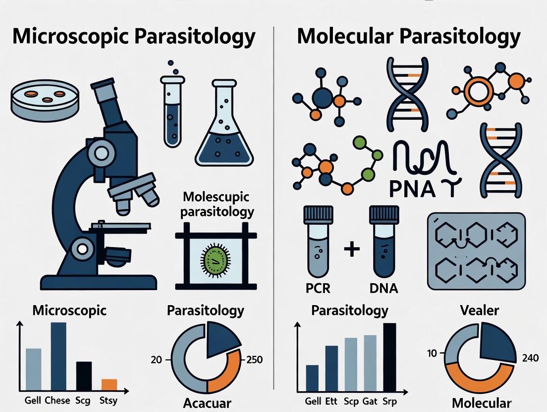
This article provides a comprehensive comparative analysis of traditional microscopic and advanced molecular diagnostic methods in parasitology, tailored for researchers, scientists, and drug development professionals.
Microscopy to Molecular Diagnostics: A Comparative Analysis of Parasitology Methods for Research and Drug Development
Abstract
This article provides a comprehensive comparative analysis of traditional microscopic and advanced molecular diagnostic methods in parasitology, tailored for researchers, scientists, and drug development professionals. It explores the foundational principles of established and emerging techniques, detailing their specific applications in research and clinical settings. The content addresses critical troubleshooting and optimization challenges, from DNA extraction in protozoa to AI integration in microscopy. Finally, it presents a rigorous validation and comparative assessment of method performance, synthesizing key takeaways on the integrated role of these technologies in enhancing diagnostic accuracy, guiding therapeutic development, and shaping the future of parasitology research.
From Leeuwenhoek to Next-Generation Sequencing: The Evolving Foundation of Parasite Diagnosis
The invention of the microscope in the 17th century marked a revolutionary turning point in the history of parasitology, enabling researchers to visualize the microscopic world for the first time [1] [2]. Before this technological breakthrough, parasitic infections were often poorly understood and misdiagnosed, with symptoms frequently attributed to supernatural forces or imbalances in bodily humors [1]. The pioneering work of Antonie van Leeuwenhoek, who built simple microscopes and discovered microscopic "animalcules," laid the groundwork for the entire field of parasitology by revealing previously invisible life forms [1] [2]. For centuries thereafter, microscopy established itself as the fundamental tool for parasite detection in biological samples, providing the critical baseline against which all future diagnostic methodologies would be measured [3] [2].
This established microscopy as the reference diagnostic method for parasitic infections worldwide, particularly for gastrointestinal parasites and soil-transmitted helminths (STHs) that affect approximately one-quarter of the global population [1] [4]. The Kato-Katz technique, developed specifically for stool sample analysis, became the World Health Organization's recommended diagnostic approach for large-scale monitoring of STH infections within mass drug administration programs and epidemiological surveys [5]. Despite the recent emergence of molecular technologies, microscopy maintains its vital role in parasitology, especially in resource-limited settings, due to its direct visualization capabilities, cost-effectiveness, and minimal equipment requirements [3].
Experimental Protocols: Core Methodologies in Microscopic Parasitology
Kato-Katz Thick Smear Technique for Soil-Transmitted Helminths
The Kato-Katz technique remains the standard method for diagnosing soil-transmitted helminths (Ascaris lumbricoides, Trichuris trichiura, and hookworms) in stool samples [5]. The protocol begins with preparing a thick blood film by making a smear with a drop of blood on a clean grease-free slide [6]. The films are air-dried, then covered with a 1:10 dilution of stock Giemsa stain with buffered distilled water at pH 7.0 [6]. After 30 minutes of staining, the stain is washed off using buffered distilled water, and the slide is placed in a slide rack to dry vertically [6].
For examination, trained microscopists count leucocytes in batches of 100, 200, 400, and 800 using oil immersion (100x) objective, counting malaria parasites alongside each batch of leucocyte [6]. A critical consideration is the limited time window for analysis—samples must be examined within 30-60 minutes of preparation because glycerol causes disintegration of hookworm eggs, potentially compromising accuracy [5]. Parasite density is calculated using the formula: (Number of parasites counted ÷ Number of white blood cells counted) × assumed white blood cell count per mm³ (typically 6,000-8,000/mm³) [6]. This assumption of a standard white blood cell count represents a significant limitation, as different age groups have varying normal WBC ranges, potentially affecting accuracy [6].
Stool Concentration Methods for Intestinal Protozoa
For intestinal protozoa like Giardia duodenalis, Cryptosporidium spp., and Entamoeba histolytica, microscopy of concentrated fecal specimens serves as the reference method in clinical laboratories [7]. The formalin-ethyl acetate (FEA) concentration technique is widely employed to enhance detection sensitivity [7]. This method involves suspending stool samples in formalin for preservation, followed by ethyl acetate extraction to concentrate parasitic elements through centrifugation [7]. Fresh stool samples may be stained with Giemsa, while fixed samples are processed using concentration techniques before microscopic examination [7]. This approach enables identification of cysts, oocysts, and trophozoites based on morphological characteristics, though differentiation between morphologically identical species (e.g., pathogenic E. histolytica versus nonpathogenic E. dispar) remains impossible by microscopy alone [7].
Performance Comparison: Microscopy Versus Modern Alternatives
Diagnostic Accuracy for Soil-Transmitted Helminths
Recent comparative studies have quantified the performance of traditional microscopy against emerging technologies. In a 2025 study evaluating 704 Kato-Katz smears from school children in Kenya, manual microscopy demonstrated significant limitations, particularly for light-intensity infections which comprised 96.7% of cases [5].
Table 1: Sensitivity Comparison for Soil-Transmitted Helminth Detection (n=704)
| Diagnostic Method | A. lumbricoides Sensitivity | T. trichiura Sensitivity | Hookworm Sensitivity | Specificity (All STHs) |
|---|---|---|---|---|
| Manual Microscopy | 50.0% | 31.2% | 77.8% | >97% |
| Autonomous AI | 50.0% | 84.4% | 87.4% | >97% |
| Expert-Verified AI | 100% | 93.8% | 92.2% | >97% |
The expert-verified AI approach significantly outperformed manual microscopy for detecting T. trichiura (p < 0.001) and hookworm (p = 0.019), while maintaining comparably high specificity [5]. This demonstrates both the limitations of conventional microscopy and the potential of augmented diagnostic approaches.
Detection of Intestinal Protozoa: Microscopy vs. Molecular Methods
A multicenter study comparing microscopy with molecular techniques for intestinal protozoa detection analyzed 355 stool samples (230 fresh, 125 preserved) across 18 Italian laboratories [7].
Table 2: Microscopy vs. PCR for Intestinal Protozoa Detection
| Parasite | Microscopy Limitations | Molecular Method Advantages |
|---|---|---|
| Giardia duodenalis | Moderate sensitivity, requires experienced personnel | Complete agreement between commercial and in-house PCR, high sensitivity and specificity |
| Cryptosporidium spp. | Challenging to identify, requires special stains | High specificity but limited sensitivity due to DNA extraction challenges |
| Entamoeba histolytica | Cannot differentiate from non-pathogenic Entamoeba species | Critical for accurate diagnosis, enables species differentiation |
| Dientamoeba fragilis | Often missed in routine examination | High specificity but inconsistent detection |
Molecular assays proved particularly critical for accurate diagnosis of E. histolytica, which is morphologically identical to nonpathogenic Entamoeba species [7]. Overall, PCR results from preserved stool samples yielded better results than fresh samples, likely due to better DNA preservation [7].
The Scientist's Toolkit: Essential Research Reagent Solutions
Table 3: Essential Research Reagents for Microscopic Parasitology
| Research Reagent | Function/Application | Key Considerations |
|---|---|---|
| Giemsa Stain | Staining blood parasites and fresh stool samples | Requires 1:10 dilution with buffered distilled water (pH 7.0), 30-minute processing [6] |
| Formalin-Ethyl Acetate (FEA) | Concentration of fecal specimens | Enhances detection of cysts and oocysts through centrifugation [7] |
| Kato-Katz Glycerol Solution | Preparation of thick smears for STH detection | Causes disintegration of hookworm eggs, requiring rapid analysis within 30-60 minutes [5] |
| Acid-Fast Stains (Ziehl-Neelsen, Kinyoun) | Identification of cryptosporidium and other acid-fast parasites | Differentiates based on cell wall properties, requires specific staining protocols [2] |
| Acridine Orange/Calcofluor White | Non-specific fluorescent staining | Enhances visualization of parasitic elements, requires fluorescence microscopy [2] |
Technological Workflows: From Traditional to Enhanced Microscopy
The diagnostic workflow for parasitic infections has evolved significantly from basic microscopic examination to technology-enhanced protocols. The following diagram illustrates this progression:
Contemporary Applications and Integration with Modern Approaches
Despite its limitations, microscopy maintains critical importance in contemporary parasitology. The integration of digital imaging with artificial intelligence represents a natural evolution of microscopic techniques, enhancing accuracy while preserving the foundational principles of morphological identification [3] [5]. Portable whole-slide scanners and deep learning algorithms have demonstrated significant improvements in detection sensitivity, particularly for light-intensity infections that would often be missed by manual microscopy alone [5]. This augmented approach preserves the direct visualization advantages of traditional microscopy while addressing its limitations through technological enhancement.
Modern microscopy also serves as an essential validation tool for novel diagnostic platforms. In comparative studies evaluating molecular techniques, conventional microscopy frequently provides the reference standard against which new methods are measured [7] [8]. This ongoing role underscores microscopy's enduring value in parasitology, even as the field increasingly adopts molecular approaches. Furthermore, in resource-limited settings where sophisticated laboratory infrastructure remains unavailable, microscopy continues to provide the most accessible and cost-effective diagnostic option for parasitic infections [3] [9]. The continued refinement of rapid staining methods, including acridine orange, fluorophores, and molecular dyes, further improves the sensitivity of parasite detection through microscopic examination [3].
The dawn of the microscopic era established an indispensable foundation for parasitology that continues to support both clinical diagnostics and research advancements. While molecular methods offer enhanced sensitivity and specificity for particular applications, microscopy provides the morphological confirmation and broad-spectrum detection capability that maintains its relevance in comprehensive parasitic diagnosis [7] [4]. The integration of artificial intelligence with digital microscopy represents a promising hybrid approach that preserves the direct visualization advantages of traditional techniques while overcoming limitations related to human expertise and variability [5]. As parasitology continues to evolve, the microscopic baseline established centuries ago remains essential for accurate diagnosis, species identification, and validation of emerging technologies, ensuring that this foundational methodology will continue to inform both clinical practice and scientific discovery for the foreseeable future.
Parasitic infections represent a formidable global public health challenge, affecting nearly a quarter of the world's population and contributing significantly to mortality, chronic illness, and socioeconomic burdens, particularly in tropical and subtropical regions [1] [10]. Accurate diagnosis is the cornerstone of effective disease management, control, and eventual elimination. For decades, microscopy has been the foundational diagnostic tool in parasitology. However, the evolving landscape of disease surveillance and the need to detect low-level infections are driving a shift toward highly sensitive molecular techniques [1] [9]. This guide provides a comparative analysis of these diagnostic methodologies, underpinned by experimental data, to inform researchers and drug development professionals.
The Scale of the Problem and the Imperative for Accurate Diagnosis
The burden of parasitic diseases is both vast and multifaceted. The World Health Organization (WHO) reports that 13 of the 20 listed Neglected Tropical Diseases (NTDs) are caused by parasites [1]. Malaria alone was responsible for an estimated 249 million cases and over 600,000 deaths annually in recent reports, primarily affecting children under five [10] [11]. The impact extends beyond mortality; parasitic infections are a leading cause of malnutrition, anemia, and impaired cognitive and physical development in children, thereby perpetuating cycles of poverty and hindering socioeconomic development [1].
The economic toll is staggering. India spends an estimated 0.34% of its total consumption expenditure on infectious diseases, including parasitic infections, with malaria alone costing the country US$1,940 million in 2014 [1]. The dairy industry in India suffers annual losses of US$787.63 million due to ticks and tick-borne diseases, while porcine cysticercosis results in economic losses exceeding US$164 million in Latin America [1]. These figures underscore that investing in accurate diagnostic tools is not merely a public health necessity but also an economic imperative.
Comparative Analysis of Diagnostic Methods: Data from the Frontlines
The choice of diagnostic method directly influences clinical decision-making, treatment outcomes, and the accuracy of disease surveillance. The following tables summarize experimental data comparing the performance of traditional and molecular diagnostic techniques across different parasitic infections.
Table 1: Performance Comparison of Diagnostic Methods for Malaria
| Diagnostic Method | Target | Sensitivity | Specificity | Limit of Detection | Key Findings/Notes |
|---|---|---|---|---|---|
| Microscopy [11] | Plasmodium spp. | 74.6% | 95.2% | 10-50 parasites/µL [12] | Sensitivity drops significantly at very low parasitaemia (<100 parasites/µL) [11]. |
| Rapid Diagnostic Tests (RDTs) [11] | Plasmodium antigens (e.g., HRP2) | 94.0% | 87.5% | ~100 parasites/µL [11] | Performance can be affected by pfhrp2/3 gene deletions and prozone effect [11]. |
| qPCR [11] | 18S rRNA gene | ~100% (Reference) | ~100% (Reference) | 0.02-5 parasites/µL [11] [9] | Detects low-density, chronic, and asymptomatic infections missed by other methods [11]. |
| High-Resolution Melting (HRM) [12] | 18S SSU rRNA | High (Concordance with sequencing) | High (Concordance with sequencing) | - | A rapid, closed-tube method for species differentiation based on melting temperature [12]. |
Table 2: Performance Comparison of Diagnostic Methods for Intestinal Protozoa
| Diagnostic Method | Study Details | Positivity Rate | Key Advantages | Key Limitations |
|---|---|---|---|---|
| Microscopy [13] | 274 stool samples | 9.5% | Low-cost; can detect a broad range of parasites [7]. | Missed 64% of infections detected by PCR; cannot differentiate E. histolytica from non-pathogenic species [13] [7]. |
| Multiplex PCR [13] | Same 274 stool samples | 27% | High sensitivity; specific species identification; detects coinfections (e.g., B. hominis + D. fragilis) [13]. | Requires specialized equipment and technical expertise [9]. |
| Commercial vs. In-House PCR [7] | 355 stool samples (multicenter) | - | Both showed high sensitivity and specificity for G. duodenalis; crucial for accurate diagnosis of E. histolytica [7]. | Sensitivity for D. fragilis and Cryptosporidium was limited, potentially due to DNA extraction challenges [7]. |
Table 3: Molecular Detection of Other Parasites
| Parasite | Sample Type | Molecular Method | Key Finding | Reference |
|---|---|---|---|---|
| Leishmania spp. | Blood, tissue | qPCR (kDNA & 18S rDNA) | qPCR on blood samples showed 54.2% sensitivity, offering a less invasive method for monitoring tegumentary leishmaniasis. | [14] |
| Spirometra mansoni | Dog and cat feces | qPCR, LAMP | qPCR sensitivity reached 100 copies/µL. LAMP allowed visual interpretation, suitable for rapid field screening. | [15] |
Detailed Experimental Protocols from Key Studies
To ensure reproducibility and provide a clear technical foundation, here are the detailed methodologies from two pivotal studies cited in the comparison tables.
Protocol: Multicenter Evaluation of Molecular Tests for Intestinal Protozoa
This multicentre study across 18 Italian laboratories compared commercial and in-house RT-PCR with microscopy for diagnosing common intestinal protozoa [7].
- Sample Collection and Preparation: A total of 355 stool samples were collected, comprising 230 fresh samples and 125 samples preserved in Para-Pak media. All samples were examined by conventional microscopy following WHO and CDC guidelines. Fresh samples were stained with Giemsa, while fixed samples were processed using the formalin-ethyl acetate (FEA) concentration technique [7].
- DNA Extraction: A 350 µL aliquot of Stool Transport and Recovery Buffer (S.T.A.R) was mixed with approximately 1 µL of each faecal sample. After centrifugation, 250 µL of the supernatant was used for automated DNA extraction on the MagNA Pure 96 System using the MagNA Pure 96 DNA and Viral NA Small Volume Kit [7].
- In-house RT-PCR Amplification: Each 25 µL reaction mixture contained 5 µL of extracted DNA, 12.5 µL of 2× TaqMan Fast Universal PCR Master Mix, 2.5 µL of a primers and probe mix, and sterile water. Amplification was performed on an ABI 7900HT Fast Real-Time PCR System with the following cycling conditions: 1 cycle of 95°C for 10 minutes, followed by 45 cycles of 95°C for 15 seconds and 60°C for 1 minute [7].
Protocol: Molecular Detection and Speciation of Malaria Parasites using HRM
This study optimized a real-time PCR platform with High-Resolution Melting (HRM) analysis for detecting and differentiating Plasmodium species in southeastern Iran [12].
- Sample and Smear Preparation: Blood samples were collected from 300 individuals with suspected malaria. Thick and thin blood smears were prepared, stained with Giemsa, and examined by experienced specialists at 1000x magnification for parasite identification and parasitemia quantification [12].
- DNA Extraction and Primary PCR: Genomic DNA was extracted from all samples using the Qiagen DNA Mini Kit. The primary PCR amplification targeted the conserved 18S SSU rRNA gene using primers MEH and UNR. The 20 µL reaction underwent initial denaturation at 95°C for 5 minutes, followed by 40 cycles of 94°C for 45s, 60°C for 45s, and 72°C for 70s, with a final elongation at 72°C for 10 minutes [12].
- Nested PCR and HRM Analysis: A 1:1000 dilution of the first-round PCR product was used as a template for the nested PCR. The nested reaction used species-specific primers (FAR, VIR, MAR, OVR) under conditions of 95°C for 4 minutes, then 35 cycles of 94°C for 20s, 60°C for 20s, and 72°C for 45s, with a final extension at 72°C for 10 minutes. For HRM, the amplified products were analyzed on the Light Cycler 96 Instrument (Roche), which enabled species differentiation based on their distinct melting temperature (Tm) profiles [12].
Visualizing the Diagnostic Workflow
The following diagram illustrates the logical workflow and decision-making process for selecting and applying the discussed diagnostic methods in a parasitology research context.
Parasitology Diagnostic Decision Workflow. This chart outlines the pathway for selecting diagnostic methods based on clinical or research objectives, highlighting how different techniques can be used in conjunction to achieve accurate results.
The Scientist's Toolkit: Essential Research Reagents and Materials
The successful implementation of the diagnostic protocols described above relies on a suite of specific reagents and instruments. The following table details key solutions used in the featured experiments.
Table 4: Key Research Reagent Solutions for Parasitology Diagnostics
| Reagent/Material | Function/Application | Example from Research |
|---|---|---|
| Stool Transport and Recovery (S.T.A.R) Buffer | Preserves nucleic acids in stool samples during transport and storage, crucial for downstream molecular analysis. | Used in the multicenter intestinal protozoa study for DNA extraction prior to PCR [7]. |
| Automated Nucleic Acid Extraction System | Provides high-throughput, consistent purification of DNA/RNA from complex clinical samples, reducing contamination and variability. | The MagNA Pure 96 System was used for DNA extraction from stool samples [7]. |
| TaqMan Fast Universal PCR Master Mix | A pre-mixed, optimized solution for fast, sensitive, and specific real-time PCR (qPCR) detection. | Used in the in-house RT-PCR assay for detecting intestinal protozoa [7]. |
| Species-Specific Primers & Probes | Short, synthetic oligonucleotides designed to bind to and amplify unique DNA sequences of a target parasite, enabling detection and differentiation. | Used in nested PCR and HRM analysis for speciating Plasmodium [12]. |
| High-Resolution Melting (HRM) Dyes | Fluorescent dyes that bind double-stranded DNA and are released during melting curve analysis, allowing discrimination of PCR products based on sequence. | Enabled differentiation of P. falciparum and P. vivax based on Tm differences [12]. |
The evidence clearly demonstrates that while traditional microscopy remains a vital tool, particularly in resource-limited settings, modern molecular methods offer unparalleled sensitivity and specificity. The choice between them is not always binary; an integrated approach often yields the best outcomes. For instance, microscopy can provide an initial broad screen, while multiplex PCR or HRM can be deployed for confirmatory testing, species identification, and detecting drug-resistant strains [13] [12] [9].
The future of parasitology diagnostics is moving toward point-of-care molecular tools, CRISPR-based systems, and the integration of artificial intelligence to automate and enhance the accuracy of microscopy [1] [9]. For researchers and drug development professionals, leveraging the appropriate diagnostic toolkit is paramount. Accurate diagnosis not only guides immediate patient care but also fuels effective drug development, robust clinical trials, and intelligent public health policies, ultimately reducing the immense global burden of parasitic diseases.
For over a century, conventional light microscopy has served as the foundational diagnostic tool in parasitology, enabling the visualization and identification of pathogenic organisms directly from clinical samples [1] [16]. This technique, pioneered by Antonie van Leeuwenhoek in the 17th century, revolutionized the understanding of parasitic infections and remains the reference method in many clinical laboratories, particularly in resource-limited settings where parasitic diseases are often endemic [1] [7]. Its persistence is attributed to its relatively low direct cost, simplicity, and the ability to detect a broad range of parasites without the need for sophisticated equipment [7]. However, the diagnostic landscape is rapidly evolving, with molecular technologies offering unprecedented levels of sensitivity and specificity. Within the context of comparative analysis between microscopic and molecular parasitology methods, it becomes critically important to objectively delineate the inherent limitations of conventional microscopy, which span analytical performance, operational efficiency, and accessibility.
This guide provides a systematic comparison of conventional microscopy against modern molecular diagnostics, focusing on the critical parameters of sensitivity, specificity, and expertise dependency. By synthesizing recent experimental data and outlining core methodologies, we aim to furnish researchers, scientists, and drug development professionals with a clear, evidence-based framework for evaluating these complementary diagnostic approaches.
Performance Comparison: Quantitative Data Analysis
The limitations of conventional microscopy become starkly evident when its diagnostic performance is quantitatively compared against molecular methods like polymerase chain reaction (PCR). The following tables consolidate key findings from recent comparative studies across different parasitic infections.
Table 1: Comparative Sensitivity of Microscopy vs. PCR for Soil-Transmitted Helminths (STHs)
| Parasite | Microscopy Sensitivity (%) | PCR Sensitivity (%) | Study Population (n) | Reference |
|---|---|---|---|---|
| Any STH infection | 22.4 | 100 (Reference) | 650 pregnant women | [17] |
| Ascaris lumbricoides | 5.4 (Prevalence) | 2.6 (Prevalence) | 650 pregnant women | [17] |
| Necator americanus | 1.8 (Prevalence) | 6.3 (Prevalence) | 650 pregnant women | [17] |
| Trichuris trichiura | 31.2 | 84.4 (Autonomous AI) / 93.8 (Expert-verified AI) | 704 school children | [5] |
| Hookworms | 77.8 | 87.4 (Autonomous AI) / 92.2 (Expert-verified AI) | 704 school children | [5] |
Table 2: Agreement Between Microscopy and Molecular Methods
| Infection Type | Kappa Value (κ) | Interpretation | Study Details |
|---|---|---|---|
| Any STH infection | 0.12 | Poor agreement | Pregnant women in India [17] |
| Ascaris lumbricoides | 0.16 | Poor agreement | Pregnant women in India [17] |
| Necator americanus | 0.20 | Poor agreement | Pregnant women in India [17] |
| Strongylid nematodes in sheep | 0.93 - 0.97 | High agreement | Lamb faecal samples (n=858) [18] |
Core Limitations of Conventional Microscopy
The data presented above underscores three fundamental constraints of conventional microscopy that impact its utility in both clinical and research settings.
Limited Diagnostic Sensitivity
Microscopy suffers from intrinsically low sensitivity, particularly in low-intensity infections or during periods of low egg excretion. A study on antenatal women in a low-prevalence setting found microscopy's sensitivity for detecting any STH infection was only 22.4% compared to PCR [17]. This is because the detection limit is constrained by the volume of sample examined and the parasite's egg output. Light-intensity infections, which now constitute the vast majority (over 96%) of STH cases in many regions, are frequently missed by manual microscopy [5]. Furthermore, the identification of certain protozoa is notoriously difficult; for example, it is impossible to differentiate the pathogenic Entamoeba histolytica from non-pathogenic species like E. dispar based on morphology alone [7].
Specificity and Diagnostic Specificity Challenges
The specificity of microscopy is heavily reliant on the technician's ability to accurately distinguish parasite eggs, cysts, or trophozoites from other fecal elements, artifacts, or similar-looking organisms. Misidentification can lead to false positives or the misclassification of species. While specificity can be high in expert hands (exceeding 97% in some studies [5]), it is highly variable. Molecular methods overcome this by targeting unique genetic sequences, enabling precise species-level discrimination. For instance, PCR can definitively identify E. histolytica, Cryptosporidium spp., and Dientamoeba fragilis, which are challenging for microscopy [7].
Expertise Dependency and Workflow Inefficiency
Manual microscopy is a labor-intensive process that demands significant expertise and training. The diagnostic accuracy is subject to human interpretation, leading to inter-observer variability [1] [7]. This creates a critical bottleneck, as experienced microscopists are not always available, especially in remote or high-throughput settings. The process is also time-consuming; reviewing a single sample can take 10-15 minutes, and it is recommended to examine multiple stool samples over several days to improve sensitivity, further increasing the workload [17]. This dependency on skilled personnel and the tedious nature of the work can hinder large-scale surveillance and control programs.
Experimental Protocols for Method Comparison
To ensure the validity of comparative studies, standardized protocols for both conventional and molecular methods are essential.
Protocol: Manual Microscopy for STHs via Kato-Katz Technique
The Kato-Katz technique is a WHO-recommended, quantitative method for diagnosing STHs in field surveys [5].
- Sample Preparation: A small portion of stool (approximately 50-100mg) is pressed through a mesh screen to remove large debris.
- Slide Preparation: The sieved sample is transferred to the hole of a template placed on a microscope slide. After filling the hole, the template is removed, leaving a precise volume of feces on the slide.
- Cellophane Coverage: A piece of glycerol-soaked or cellosolve-soaked cellophane cover is placed over the sample. The glycerol clears the debris, making the eggs more visible.
- Microscopic Examination: After a clearing time (typically 30-60 minutes, but shorter for hookworms to prevent over-clearing), the slide is examined systematically under a microscope at low magnification (10x objective). The number of eggs of each helminth species is counted.
- Quantification: The egg count is converted to eggs per gram (EPG) of feces using the known volume of the template. The infection intensity is then classified as light, moderate, or heavy based on WHO thresholds [5].
Protocol: Molecular Detection via Conventional PCR
PCR offers a highly sensitive and specific alternative for detecting parasite DNA directly from stool samples [17].
- DNA Extraction: Genomic DNA is extracted directly from approximately 1 µl of fecal sample. Commercial DNA isolation kits (e.g., those from Roche) are commonly used, often with an added internal extraction control. This step may involve mechanical or chemical lysis to break down the robust walls of protozoan cysts or helminth eggs [7] [18].
- PCR Reaction Setup: Each reaction mixture contains:
- 5 µl of extracted DNA.
- 12.5 µl of 2x TaqMan Universal PCR Master Mix.
- A custom mix of primers and probes (2.5 µl) targeting specific genetic markers (e.g., the ITS-1 or ITS-2 regions of ribosomal DNA for STHs [17]).
- Sterile water to a final volume of 25 µl.
- Amplification: The PCR tube is placed in a thermal cycler (e.g., ABI 7900HT). A typical cycling regimen includes:
- Initial denaturation: 1 cycle of 95°C for 10 minutes.
- Amplification: 45 cycles of:
- Denaturation: 95°C for 15 seconds.
- Annealing/Extension: 60°C for 1 minute.
- Analysis: The results are analyzed based on the amplification of target DNA, often visualized through gel electrophoresis or in real-time using fluorescent probes.
Visualizing the Diagnostic Workflows
The contrasting workflows of traditional and modern, AI-supported diagnostics highlight the significant efficiency gains of the latter. The diagram below illustrates the key steps in each process.
The Scientist's Toolkit: Key Research Reagent Solutions
The transition to molecular and digital methodologies relies on a distinct set of reagents and tools.
Table 3: Essential Research Reagents and Materials
| Item | Function/Application | Example Use Case |
|---|---|---|
| Kato-Katz Template | Provides a standardized volume of stool for quantitative microscopy. | Soil-transmitted helminth egg counts in epidemiological surveys [5]. |
| Glycerol-Soaked Cellophane | Clears fecal debris on slides for better visualization of helminth eggs. | Used in the Kato-Katz technique for microscopic diagnosis [5]. |
| DNA Extraction Kits | Isolate high-quality genomic DNA from complex stool samples. | Preparation of template DNA for PCR-based detection of parasites [7] [18]. |
| Species-Specific Primers/Probes | Bind to unique genetic sequences for amplification and detection. | Enables specific identification of pathogens like E. histolytica or N. americanus via PCR [17]. |
| TaqMan Universal PCR Master Mix | Contains enzymes, dNTPs, and buffer for efficient DNA amplification in real-time PCR. | Core component of the PCR reaction mixture for molecular diagnosis [7]. |
| Fluorescent Stains (e.g., WGA) | Label specific cellular components (e.g., membranes) for high-resolution imaging. | Used in advanced microscopy techniques to visualize subcellular structures [19]. |
| Convolutional Neural Network (CNN) Algorithm | AI model trained to automatically identify and count parasitic structures in digital images. | Used in AI-supported digital microscopy for automated parasite detection [1] [5]. |
The comparative analysis unequivocally demonstrates that conventional microscopy, while accessible, carries significant limitations in sensitivity, specificity, and operational efficiency, particularly in the context of modern parasitology research and low-prevalence settings. The dependency on expert personnel introduces subjectivity and bottlenecks that are alleviated by molecular techniques and emerging AI-supported digital platforms.
For researchers and drug development professionals, the choice of diagnostic method must be guided by the specific application. Microscopy retains value in high-burden, resource-limited settings and for detecting a broad range of unsuspected parasites. However, for precise species identification, drug efficacy studies, surveillance in low-transmission areas, and high-throughput research, molecular methods offer a level of performance that conventional microscopy cannot match. The future of parasitological diagnosis lies in integrated approaches, leveraging the strengths of each technology to achieve the ultimate goal of effective disease control and eradication.
The diagnosis of parasitic diseases has long relied on traditional methods such as microscopy, serology, and histopathology. While these techniques have been foundational, they often require significant time, expertise, and are frequently impractical in resource-limited endemic regions due to their dependency on specialized equipment and laboratory infrastructure [9]. The field is undergoing a transformative shift with the advent of molecular techniques that provide enhanced sensitivity, specificity, and reliability. Among these, Polymerase Chain Reaction (PCR) and its advanced derivatives have emerged as powerful tools, enabling researchers to move from mere observation to precise genetic characterization. This revolution is particularly critical in parasitology, where accurate identification of pathogens like Echinococcus granulosus or Plasmodium species directly impacts treatment outcomes and public health surveillance [20] [9]. The core of this transition lies in understanding the principles of PCR, the efficiency of multiplexing, and the precision of genotyping—technologies that form the backbone of modern molecular diagnostics.
Fundamental Principles and Key Technologies
The Evolution of PCR: From Simple Amplification to Multiplexed Detection
At its core, PCR is a technique used to amplify specific DNA sequences, creating millions of copies from a single template. This process enables the detection and analysis of genetic material even from minimal samples. The basic PCR reaction involves repeated cycles of denaturation, annealing, and extension, facilitated by primers, nucleotides, and a heat-stable DNA polymerase.
Multiplex PCR represents a significant advancement, allowing for the simultaneous amplification of multiple targets in a single reaction. This is achieved by using multiple primer sets, each designed to bind to a unique DNA sequence. The efficiency of multiplexing is demonstrated in various applications, such as a novel fluorescence melting curve analysis (FMCA)-based multiplex PCR assay that can detect six respiratory pathogens from a single sample, significantly reducing turnaround time and cost compared to running multiple single-plex reactions [21]. The primer and probe design is critical in these assays; for instance, probes can be modified with base-free tetrahydrofuran (THF) residues to minimize the impact of base mismatches and enhance hybridization stability across genetic variants [21].
Genotyping refers to the process of determining differences in the genetic makeup of an individual by examining their DNA sequence. Single Nucleotide Polymorphisms (SNPs) are the most common type of genetic variation. Identifying these SNPs is crucial for understanding traits such as disease resistance in crops or pathogen virulence in parasites [22]. PCR-based genotyping methods leverage amplified DNA to identify these variations with high precision.
Advanced Genotyping and Multiplexing Platforms
Table 1: Comparison of Key PCR-Based Genotyping and Multiplexing Technologies
| Technology | Principle | Key Features | Applications in Parasitology & Biomedicine |
|---|---|---|---|
| USE-PCR (Universal Signal Encoding PCR) [23] | Combines universal hydrolysis probes, amplitude modulation, and multispectral encoding. | Standardized analysis; high multiplexing (32 targets); portability across dPCR platforms. | Detection of single nucleotide variants (SNVs) in cancer cell lines; potential for pathogen identification. |
| Color Cycle Multiplex Amplification (CCMA) [24] | Identifies targets by pre-programmed permutation of fluorescence increases across multiple channels. | High-plex detection (theoretically 136 targets with 4 colors); uses standard qPCR instruments. | Syndromic testing for sepsis-related bacteria; identification of multiple pathogens in clinical samples. |
| High-Resolution Melting (HRM) Analysis [22] | Analyzes melt curves of amplicons using intercalating dyes; differences in Tm indicate variants. | Closed-tube, non-destructive; no need for electrophoresis; cost-effective. | Genotyping of known SNPs/InDels; discovery of novel SNPs and haplotypes in parasites. |
| TaqMan Assay [22] | Uses allele-specific probes with reporter/quencher dyes; 5' exonuclease activity cleaves probe. | High specificity and sensitivity; automated, high-throughput; qualitative and quantitative. | Differentiating pathogenic genotypes from wild-type; marker-assisted selection in crops. |
| Multiplex Pyrosequencing [25] | Detects nucleotide incorporation in real-time through light emission; optimized dispensation orders. | Quantitative; rapid turnaround; cost-effective alternative to NGS for short sequences. | Microhaplotype genotyping for forensic applications; useful with low-input/degraded DNA. |
Experimental Protocols: A Glimpse into Methodologies
Protocol: Multiplex PCR-NGS for High-Resolution Genotyping
This protocol, adapted from a study on HLA genotyping, illustrates a robust method for achieving high-resolution sequencing of highly polymorphic loci [26].
- Step 1: Primer Design. Multiplex PCR primers targeting specific loci (e.g., HLA-A, -B, -C, -DPB1, -DQB1, -DRB1) are designed based on high-frequency alleles from public databases. The primers are optimized to avoid dimer formation and ensure balanced amplification across all targets, with product lengths ranging from 2,000–6,000 bp.
- Step 2: Multiplex PCR Amplification. Genomic DNA (50 ng) is amplified in a 25-μL reaction system containing a multiplex primer pool. The primer concentrations are carefully optimized for each locus (e.g., HLA-A at 0.04 μM, HLA-B at 0.1 μM). The thermocycling conditions are: 95°C for 10.5 min; 30 cycles of 98°C for 10 s, 63°C for 1 min, 72°C for 5 min; and a final elongation at 72°C for 5 min.
- Step 3: Library Preparation and Sequencing. The PCR products are fragmented enzymatically to sizes of 250–350 bp. Specific adapters are ligated to these fragments for sample identification during sequencing. The library is purified using magnetic beads, quantified, and sequenced on a platform such as an MGI sequencer. Subsequent bioinformatic analysis aligns the sequences to a reference genome for genotyping [26].
Protocol: Digital PCR for Parasite Detection and Monitoring
This protocol highlights the application of dPCR for sensitive detection of parasitic DNA, as demonstrated in a study on Echinococcus granulosus [20].
- Step 1: Sample Collection and DNA Extraction. Stool samples are collected longitudinally (e.g., daily over 50 days post-infection). Genomic DNA is extracted from the samples using commercial kits.
- Step 2: Digital PCR Setup. The extracted DNA is combined with a reaction mix containing primers, probes, and dPCR master mix. This mixture is partitioned into thousands of individual reactions in a digital PCR chip or droplet generator.
- Step 3: Amplification and Analysis. The partitioned reactions undergo PCR amplification. The fluorescence in each partition is read to determine the presence (positive) or absence (negative) of the target parasite DNA. The copy number of the target DNA in the original sample is then quantified using Poisson statistics. This method has been shown to detect parasite DNA earlier than microscopic egg detection and can identify DNA even after treatment has halted viable egg shedding [20].
Comparative Performance Data
Quantitative Comparison of Diagnostic Methods
Table 2: Analytical and Clinical Performance of Featured Molecular Assays
| Assay / Method | Targets / Loci | Reported Sensitivity / Accuracy | Limit of Detection (LOD) | Key Advantage |
|---|---|---|---|---|
| FMCA-based Multiplex PCR [21] | 6 respiratory pathogens | 98.81% agreement with RT-qPCR | 4.94 - 14.03 copies/μL | Cost-effective ($5/sample); rapid (1.5 hr) turnaround. |
| Multiplex PCR-NGS (HLA) [26] | 6 HLA loci | ≥ 95% at 4-digit resolution | N/R | Resolves allelic ambiguities; provides high-resolution, long-range sequences. |
| Digital PCR (E. granulosus) [20] | E. granulosus DNA | Higher sensitivity than microscopy & qPCR | N/R | Detects DNA pre-patent and post-treatment; quantifies low copy numbers. |
| USE-PCR [23] | 32 synthetic templates | 97.6% ± 4.4% (low copy) | Dynamic range of 4 orders of magnitude | Standardized universal probes; works on multiple dPCR platforms. |
| Multiplex Pyrosequencing (CLPSQ-Net) [25] | 4 Microhaplotype loci | 89.7% classification accuracy | 100 pg input DNA | Low-cost alternative to NGS; effective with degraded DNA. |
N/R: Not explicitly Reported in the search results
Comparison with Traditional Microscopy
The superiority of molecular methods is starkly evident when compared to traditional microscopy. A study on Echinococcus granulosus in experimentally infected dogs found that while flotation methods began detecting eggs in stool samples 44-46 days post-infection, PCR-based methods detected parasite DNA as early as day 20 [20]. Furthermore, dPCR provided consistent detection from day 1 to 50 post-infection and identified DNA copies even after treatment, when qPCR cycle threshold (Ct) values were undetectable. This highlights the role of molecular tools not just in diagnosis, but also in sensitive post-treatment monitoring, where microscopy fails once viable egg shedding ceases [20].
The Scientist's Toolkit: Essential Research Reagents
Table 3: Key Research Reagent Solutions for Molecular Assays
| Reagent / Material | Function in the Workflow | Specific Example (from search results) |
|---|---|---|
| Multiplex PCR Primers | Simultaneously amplifies multiple specific DNA targets from a single sample. | Optimized primer pools for HLA loci (A, B, C, DPB1, DQB1, DRB1) [26]. |
| Universal Hydrolysis Probes | A single, standardized probe mixture that detects multiple targets, simplifying assay design. | USE-PCR employs an 8-probe universal mix to encode signals for 32 targets [23]. |
| Fluorophore-Labeled Probes | Binds to amplified DNA, emitting a fluorescent signal for detection and quantification. | TaqMan probes labeled with FAM, VIC, or HEX dyes for SNP genotyping [22]. |
| DNA Polymerase Master Mix | Enzyme and buffer system optimized for efficient and specific DNA amplification. | TaqPath ProAmp Master Mix used in CCMA and other qPCR/dPCR assays [23] [24]. |
| Magnetic Beads (SPRI) | Purifies and size-selects DNA fragments post-amplification for library preparation. | AMPure XP SPRI magnetic beads used for cleaning up amplicons before NGS [26] [24]. |
| Nucleotide Dispensation Mix | The sequenced addition of nucleotides for real-time sequencing-by-synthesis in pyrosequencing. | Algorithmically optimized dispensation orders for multiplex pyrosequencing of microhaplotypes [25]. |
Technological Workflows and Logical Pathways
The following diagram illustrates the logical progression and decision-making pathway for selecting an appropriate molecular genotyping method based on research goals and constraints.
Diagram 1: A decision pathway for selecting a molecular genotyping technology, based on key research requirements such as multiplexing capacity, resource availability, and sample quality [26] [20] [23].
The molecular revolution, powered by advanced PCR, multiplexing, and genotyping technologies, has fundamentally redefined parasitology research and diagnostics. The move from microscopic observation to nucleotide-level analysis provides unprecedented sensitivity, specificity, and the ability to conduct high-throughput screenings. As evidenced by the experimental data, methods like multiplex NGS, dPCR, and FMCA-based assays consistently outperform traditional techniques, offering faster, more accurate, and increasingly cost-effective solutions. The ongoing integration of these tools with artificial intelligence and machine learning, as seen in platforms like CLPSQ-Net for pyrosequencing analysis, promises to further enhance their power and accessibility [25]. For researchers and drug development professionals, understanding and leveraging this expanding toolkit is essential for driving innovation, improving diagnostic accuracy, and ultimately developing more effective treatments for parasitic diseases that continue to challenge global health.
Parasitic infections represent a significant global health challenge, affecting nearly a quarter of the world's population and contributing substantially to the burden of neglected tropical diseases [1]. Traditional diagnostic methods, particularly microscopy, have long served as the cornerstone of parasite identification. However, these methods face significant limitations in sensitivity, specificity, and the ability to differentiate closely related species [7]. The field is now undergoing a transformative shift with the integration of artificial intelligence (AI), proteomics, and advanced imaging technologies, which are collectively enhancing diagnostic accuracy and enabling new capabilities in parasite characterization [1] [27]. This comparative analysis examines the performance of established microscopic techniques against emerging molecular and AI-driven approaches, providing researchers and drug development professionals with evidence-based insights to guide methodological selection.
Performance Comparison: Microscopy vs. Molecular vs. AI-Assisted Diagnostics
Quantitative Performance Metrics Across Diagnostic Platforms
Table 1: Comparative diagnostic performance for soil-transmitted helminths (n=704 samples)
| Diagnostic Method | A. lumbricoides Sensitivity | T. trichiura Sensitivity | Hookworm Sensitivity | Overall Specificity |
|---|---|---|---|---|
| Manual Microscopy | 50.0% | 31.2% | 77.8% | >97% |
| Autonomous AI | 50.0% | 84.4% | 87.4% | >97% |
| Expert-Verified AI | 100% | 93.8% | 92.2% | >97% |
Data adapted from a study comparing diagnostic methods for soil-transmitted helminths using Kato-Katz thick smears [5].
Table 2: Molecular vs. microscopic detection of intestinal protozoa in stool samples (n=355)
| Parasite | Microscopy Performance | Commercial RT-PCR | In-House RT-PCR |
|---|---|---|---|
| G. duodenalis | Reference standard | High sensitivity/specificity | High sensitivity/specificity |
| Cryptosporidium spp. | Limited sensitivity | High specificity, variable sensitivity | High specificity, variable sensitivity |
| E. histolytica | Cannot differentiate from non-pathogenic species | Accurate differentiation | Accurate differentiation |
| D. fragilis | Often missed | High specificity, limited sensitivity | High specificity, limited sensitivity |
Data summarized from a multicentre study comparing molecular and microscopic methods for intestinal protozoa detection [7].
Diagnostic Challenges in Complex Cases
The limitations of conventional methods become particularly evident in diagnostically challenging scenarios. For instance, Plasmodium cynomolgi, a simian malaria parasite, is frequently misdiagnosed as P. vivax using both routine microscopy and standard molecular tools due to their morphological similarities and genetic homology [28]. A 2023 case report from Malaysia detailed how initial microscopy and qPCR identified a infection as P. vivax, while more specialized molecular analysis including real-time PCR and sequencing ultimately confirmed a mixed-species infection with P. cynomolgi [28]. Similarly, in reptile endoparasite screening, molecular methods revealed hidden infections missed by microscopy alone, with necropsy identifying latent infections in 55.6% of cases [29].
Experimental Protocols and Methodologies
AI-Assisted Digital Microscopy Workflow
Table 3: Key research reagents for AI-assisted parasitology diagnostics
| Reagent/Equipment | Function | Application Example |
|---|---|---|
| Portable whole-slide scanner | Digitizes microscope slides for analysis | Field-based digital diagnostics of Kato-Katz smears |
| Deep learning algorithm with convolutional neural networks | Detects and classifies parasite eggs | Soil-transmitted helminth identification |
| Additional DL algorithm for disintegrated eggs | Improves detection of degraded specimens | Hookworm egg detection in Kato-Katz smears |
| AI-verificator tool | Allows expert verification of AI findings | Quality assurance in diagnostic workflows |
Methodology: In a recent study evaluating AI for soil-transmitted helminth diagnosis, Kato-Katz thick smears were prepared from stool samples (n=965) collected from school children in Kenya [5]. The smears were digitized using portable whole-slide scanners, then analyzed using deep learning-based AI. The AI system incorporated two algorithms: one for intact parasite eggs and an additional algorithm specifically designed to detect partially disintegrated hookworm eggs, which had been a limitation in previous versions. Three diagnostic approaches were compared: (1) manual microscopy by on-site experts, (2) autonomous AI analysis, and (3) expert-verified AI where human experts reviewed AI-detected findings. Performance was benchmarked against a composite reference standard that combined expert-verified eggs in both physical and digital smears [5].
Molecular Detection of Intestinal Protozoa
Table 4: Essential reagents for molecular detection of intestinal protozoa
| Reagent/Equipment | Function | Specification |
|---|---|---|
| S.T.A.R Buffer | Stool transport and recovery | Preserves nucleic acids during storage/transport |
| MagNA Pure 96 System | Automated nucleic acid extraction | Uses magnetic separation technology |
| MagNA Pure 96 DNA and Viral NA Small Volume Kit | DNA extraction | Optimized for difficult-to-lyse organisms |
| TaqMan Fast Universal PCR Master Mix | PCR amplification | Provides reagents for real-time PCR |
| ABI 7900HT Fast Real-Time PCR System | PCR amplification/detection | Enables quantitative analysis |
Methodology: In a multicentre study comparing molecular methods for intestinal protozoa detection, 355 stool samples (230 fresh, 125 preserved) were processed [7]. DNA extraction was performed using the MagNA Pure 96 System with the Stool Transport and Recovery Buffer for sample preparation. For the in-house real-time PCR assay, each reaction mixture contained 5 μL of extracted DNA, 12.5 μL of 2× TaqMan Fast Universal PCR Master Mix, 2.5 μL of primers and probe mix, and sterile water to a final volume of 25 μL. Amplification was performed using the ABI 7900HT Fast Real-Time PCR System with the following cycling conditions: 1 cycle of 95°C for 10 minutes; followed by 45 cycles each of 95°C for 15 seconds and 60°C for 1 minute. This protocol was validated for detecting Giardia duodenalis, Cryptosporidium spp., Entamoeba histolytica, and Dientamoeba fragilis [7].
Integration of Proteomics and Spatial Biology in Parasitology
While proteomics applications in parasitology are still emerging, the technology has demonstrated transformative potential in adjacent fields. Spatial proteomics enables the exploration of protein expression in cells and tissues while maintaining sample integrity, mapping protein distributions and interactions within microenvironmental contexts [27] [30]. Mass spectrometry-based proteomics can comprehensively characterize proteins in a sample without needing predefined targets, simultaneously quantifying protein abundance, subcellular localization, and post-translational modifications [30]. Benchtop protein sequencers, such as Quantum-Si's Platinum Pro, are making protein sequencing more accessible by providing single-molecule, single-amino acid resolution without requiring specialized expertise [30]. These technologies could be readily adapted to parasitology research for identifying novel drug targets, understanding host-parasite interactions, and developing new biomarkers for parasitic diseases.
The comparative data presented in this analysis demonstrates a clear diagnostic continuum from traditional microscopy to AI-enhanced and molecular methods. Expert-verified AI emerges as the most sensitive approach for detecting soil-transmitted helminths, particularly for light-intensity infections that represent over 96% of cases in contemporary surveys [5]. Molecular methods provide superior differentiation of closely related species that are morphologically identical, such as pathogenic versus non-pathogenic Entamoeba species [7]. However, microscopy retains value as an accessible, low-cost technique, particularly in resource-limited settings, and for detecting parasites not targeted by specific molecular assays [7]. For researchers and drug development professionals, the optimal diagnostic strategy will depend on specific research questions, resources, and the parasitic organisms of interest. Integrating these complementary technologies through a multimodal approach will likely yield the most comprehensive understanding of parasitic infections and facilitate the development of novel therapeutic interventions.
Benchside to Bedside: A Practical Guide to Parasitology Methodologies
Within the framework of comparative parasitology research, the diagnosis of parasitic infections remains foundational to disease management, surveillance, and drug development. Despite the rapid advancement of molecular diagnostics, traditional microscopic techniques continue to be the cornerstone of parasite detection, particularly in resource-limited settings and for routine surveillance [1] [9]. These methods range from simple direct smears to more sophisticated quantitative concentration techniques. Direct wet mount examinations using saline and iodine are primarily used for initial, rapid morphological assessment, while concentration methods like flotation or sedimentation are employed to increase the detection sensitivity by increasing the number of parasites in the sample [31] [1].
This guide provides an objective comparison of two prominent quantitative diagnostic techniques: the long-established McMaster method and the newer Mini-FLOTAC technique. The comparison is situated within a broader research context that acknowledges the growing importance of molecular assays but recognizes that microscopy's affordability, directness, and immediate applicability secure its ongoing relevance [9] [7]. Accurate diagnosis is the first critical step in any parasitology research, drug efficacy trial, or control program, and the choice of technique can significantly impact findings and subsequent recommendations [32] [33].
Comparative Analysis of Key Microscopic Techniques
The following table summarizes the core characteristics and performance data of the most common microscopic techniques, based on recent comparative studies.
Table 1: Comparative Overview of Standard Parasitological Diagnostic Techniques
| Technique | Primary Function | Key Performance Findings (from Recent Studies) | Advantages | Limitations |
|---|---|---|---|---|
| Direct Wet Mount (Saline/Iodine) | Rapid morphological identification of motile trophozoites, cysts, and eggs. | Detected parasites in 41% of samples in a pediatric study, significantly lower than concentration methods [31]. | Low cost, rapid, requires minimal equipment [31]. | Low sensitivity, requires immediate examination, highly dependent on technician expertise [31] [9]. |
| Formol-Ether Concentration (FEC) | Sedimentation-based concentration for a wide range of helminths and protozoa. | Detected parasites in 62% of samples; effective for operculated and heavy eggs [31]. | Broad spectrum of parasite recovery, considered a standard in medical parasitology [31]. | Involves hazardous chemicals, more procedural steps [31]. |
| Formol-Ether Acetate (FAC) | Sedimentation-based concentration, considered a refinement of FEC. | Demonstrated the highest recovery, detecting parasites in 75% of samples in a comparative study [31]. | High sensitivity and recovery rate for diverse parasites [31]. | Uses hazardous chemicals, requires a centrifuge [31]. |
| McMaster | Quantitative flotation for estimating eggs per gram (EPG) of feces. | Lower sensitivity (85-93%) and precision; frequently missed low-intensity and specific parasite infections (e.g., Nematodirus spp.) [34] [32] [33]. | Simple, cost-effective, widely used and understood [34] [32]. | Lower sensitivity and precision, can underdiagnose infections, leading to misclassification [34] [33]. |
| Mini-FLOTAC | Quantitative flotation with improved design for higher sensitivity and precision. | Superior sensitivity (93-100%), detected higher EPG values and a broader parasite spectrum; showed greater precision (CVs 12-19%) [34] [32] [33]. | High sensitivity and precision, no need for centrifugation, ideal for field settings [34] [32]. | Requires a specialized device, slightly more complex protocol [34]. |
Experimental Protocols for Key Comparative Studies
To ensure reproducibility and provide a clear understanding of the methodological basis for the data in this guide, detailed protocols from recent, pivotal studies are outlined below.
Protocol: McMaster vs. Mini-FLOTAC in Sheep
A 2024 study in southern Benin directly compared these two methods for diagnosing GI parasites in West African Long-legged sheep [34].
- Sample Collection: 200 fresh fecal samples were collected directly from the rectum of lambs. Samples were stored at 4°C and processed within 24 hours [34].
- Modified McMaster Technique: 3 g of feces were mixed with 42 mL of saturated sodium chloride solution (specific gravity = 1.2), creating a 1:15 dilution. The mixture was homogenized, filtered, and transferred to a McMaster slide. Eggs were counted under a microscope, and the count was multiplied by a factor of 50 to calculate Eggs/Oocysts per Gram (EPG/OPG) [34].
- Mini-FLOTAC Technique: 2 g of feces were diluted in a 1:10 ratio with saturated sodium chloride solution (specific gravity = 1.2). The mixture was homogenized and transferred into two Mini-FLOTAC chambers. After a 10-minute passive flotation period, the chambers were read under a microscope. The EPG/OPG was calculated using a multiplication factor of 5 [34].
- Statistical Analysis: Diagnostic performance was assessed based on prevalence, infection intensity (EPG/OPG), and precision (coefficient of variation). Agreement between techniques was evaluated using Cohen’s kappa coefficient [34].
Protocol: A Three-Way Comparison in Horses
A 2025 study in Portugal provided a three-way comparison of McMaster, FLOTAC, and Mini-FLOTAC for diagnosing strongyle infections in horses [32].
- Sample Collection: 32 fecal samples were collected from two horse facilities, stored at 4–5°C, and processed within two weeks [32].
- McMaster Method: 2 g of feces were mixed with 28 mL of saturated sucrose solution (specific gravity = 1.2). The suspension was filtered and transferred to an McMaster slide. A multiplication factor of 50 was used for EPG calculation [32].
- FLOTAC Method: 5 g of feces were mixed with 45 mL of water (1:10 dilution), centrifuged, and the supernatant was discarded. The pellet was resuspended in 6 mL of saturated sucrose solution, added to FLOTAC chambers, and centrifuged. A multiplication factor of 1 was used [32].
- Mini-FLOTAC Method: 5 g of feces were mixed with 45 mL of saturated sucrose solution (1:10 dilution). The suspension was transferred to Mini-FLOTAC chambers and left to rest for 10 minutes before reading. A multiplication factor of 5 was used [32].
Workflow Diagram: McMaster vs. Mini-FLOTAC
The diagram below illustrates the key procedural differences between the McMaster and Mini-FLOTAC techniques, highlighting steps that contribute to their differing performance.
The Scientist's Toolkit: Essential Research Reagents and Materials
Successful implementation of the discussed techniques requires specific laboratory materials. The following table lists key reagents and their functions in the diagnostic process.
Table 2: Essential Research Reagents and Materials for Fecal Parasitology
| Item | Specific Function | Application Notes |
|---|---|---|
| Saturated Sodium Chloride (NaCl) Solution | Flotation medium (specific gravity ~1.20). Causes helminth eggs and protozoan cysts to float. | A common, cost-effective flotation solution, though it may distort delicate structures [34] [33]. |
| Saturated Sucrose Solution | Flotation medium (specific gravity ~1.20-1.27). Superior for floating most nematode and cestode eggs. | Preferable for recovering more delicate eggs; however, it is viscous and can be messy [32]. |
| Formalin (10%) | Fixative and preservative. Kills pathogens and preserves parasite morphology. | Used in Formol-Ether/Acetate sedimentation methods to fix stool specimens [31]. |
| Ethyl Acetate / Diethyl Ether | Solvent for extraction. Dissolves fats and removes debris in concentration methods. | Helps to clear the sample of organic debris, concentrating parasites in the sediment [31]. |
| Iodine Solution (e.g., Lugol's) | Staining agent. Stains glycogen and nuclei of protozoan cysts for easier identification. | Used in wet mounts to enhance the visualization of internal structures of cysts [31]. |
| McMaster Slide | Specialized counting chamber. Allows for standardized counting and EPG calculation. | Features two ruled chambers that enable quantification of parasite eggs [34] [32]. |
| Mini-FLOTAC Device | Integrated system. Combines sample preparation and quantitative examination in a single device. | Contains two 1 mL flotation chambers which increase the volume of sample examined, enhancing sensitivity [34] [32]. |
Discussion: Implications for Research and Drug Development
The comparative data consistently demonstrates that Mini-FLOTAC offers superior diagnostic sensitivity and precision compared to the McMaster method [34] [32] [33]. This performance advantage has direct implications for research and drug development. In veterinary parasitology, the higher sensitivity of Mini-FLOTAC means it detects a greater number of true positive infections, particularly those with low egg shedding, which can be missed by McMaster [34] [33]. This is critical for epidemiological studies aiming to determine true prevalence and for anthelmintic efficacy trials (Fecal Egg Count Reduction Tests), where accurate pre- and post-treatment counts are essential for reliably detecting the emergence of drug resistance [32] [33].
The choice of diagnostic technique fits into the broader thesis of microscopic versus molecular methods. While molecular techniques like PCR offer unparalleled sensitivity and specificity for species-level identification—and are increasingly seen as the future gold standard—they require sophisticated infrastructure, higher costs, and technical expertise [9] [7]. In this context, Mini-FLOTAC represents an optimal "middle-ground" for many field-based and resource-constrained settings. It provides a significant improvement over traditional microscopy without the resource demands of molecular assays, making it a robust tool for large-scale surveillance and monitoring programs that inform broader parasite control strategies and drug development efforts [34].
The diagnostic landscape for intestinal parasites is defined by a clear trade-off between the accessibility of traditional microscopy and the superior sensitivity of modern molecular techniques. The following data summarizes the comparative performance of these methods as established in contemporary research.
| Method | Sensitivity | Specificity | Key Advantages | Key Limitations |
|---|---|---|---|---|
| Microscopy (Conventional) | 66.9%–87.9% [13] [35] | 100% [35] | Low cost; Provides direct morphological information; Wide availability [13] [35] | Lower sensitivity; Requires high expertise; Time-consuming [13] [35] |
| Molecular (PCR) | 97%–99% [13] [35] | 100% [35] | High sensitivity/specificity; Detects low-load & co-infections; Species-level differentiation [13] | Higher cost; Requires specialized lab; Does not provide morphological data [13] |
| Culture | 72.7% [35] | 100% [35] | Provides live organisms for further study | Long turnaround time; Risk of contamination; Not for all protozoa [35] |
Differential Morphology of Intestinal Protozoa
Advanced microscopy relies on detailed morphological criteria to differentiate between various intestinal protozoa. The tables below consolidate key diagnostic features for common amoebae, as visible in permanently stained stool specimens [36].
Amoebae: Trophozoite Morphology
| Species | Size (Length) | Motility | No. of Nuclei | Peripheral Chromatin | Karyosomal Chromatin | Cytoplasmic Inclusions |
|---|---|---|---|---|---|---|
| Entamoeba histolytica | 10-60 µm | Progressive, hyaline pseudopods | 1 | Fine, uniform granules | Small, discrete, usually central | Red blood cells (invasive form) or bacteria [36] |
| Entamoeba coli | 15-50 µm | Sluggish, blunt pseudopods | 1 | Coarse, irregular granules | Large, discrete, usually eccentric | Bacteria, yeasts [36] |
| Entamoeba hartmanni | 5-12 µm | Usually non-progressive | 1 | Similar to E. histolytica | Small, discrete, often eccentric | Bacteria [36] |
| Endolimax nana | 6-12 µm | Sluggish, blunt pseudopods | 1 | None | Large, irregular, blot-like | Bacteria [36] |
| Dientamoeba fragilis | 5-15 µm | Angular, hyaline pseudopods | 2 (in 80% of organisms) | None | Large cluster of 4-8 granules | Bacteria; occasionally RBCs [36] |
Amoebae: Cyst Morphology
| Species | Size (Diameter) | Shape | No. of Nuclei (Mature) | Peripheral Chromatin | Chromatoid Bodies | Glycogen Mass |
|---|---|---|---|---|---|---|
| Entamoeba histolytica | 10-20 µm | Spherical | 4 | Fine, uniform granules | Elongated bars with rounded ends | Usually diffuse [36] |
| Entamoeba coli | 10-35 µm | Spherical, often irregular | 8 | Coarse, irregular granules | Splinter-like with pointed ends | Usually diffuse [36] |
| Entamoeba hartmanni | 5-10 µm | Spherical | 4 | Similar to E. histolytica | Elongated bars with rounded ends | Similar to E. histolytica [36] |
| Endolimax nana | 5-10 µm | Spherical to Oval | 4 | None | Not typically present | Occasionally present [36] |
| Iodamoeba bütschlii | 5-20 µm | Irregular | 1 | None | Not typically present | Compact, well-defined [36] |
Flagellates: Trophozoite Morphology
| Species | Size (Length) | Shape | Motility | Key Identifying Features |
|---|---|---|---|---|
| Giardia duodenalis | 10-20 µm | Pear-shaped | "Falling leaf" | Sucking disk; 2 nuclei; 4 pairs of flagella [36] |
| Chilomastix mesnili | 6-24 µm | Pear-shaped | Stiff, rotary | Prominent cytostome; spiral groove [36] |
| Pentatrichomonas hominis | 6-20 µm | Pear-shaped | Nervous, jerky | Undulating membrane; 3-5 anterior flagella [36] |
Experimental Protocols for Microscopy
Detailed staining protocols are required to achieve the morphological detail necessary for parasite identification.
Wheatley's Trichrome Staining
The trichrome stain is a critical differential staining technique for the identification of intestinal protozoa in permanently stained slides [37] [38].
- Principle: A combination of dyes (chromotrope 2R, light green SF, phosphotungstic acid) differentially stains cellular components. Protozoan cytoplasm typically appears blue-green or purple, nuclear structures red or purple-red, and the background material green [38].
- Procedure [37] [38]:
- Smear Preparation: Create a thin smear of polyvinyl alcohol (PVA)-preserved stool on a slide and allow it to dry completely.
- Fixation: Fix the smear in Schaudinn's fixative (which contains mercuric chloride) for 30-60 minutes.
- Iodine/Ethanol Wash: Rinse in 70% ethanol with iodine to remove mercury, followed by 70% ethanol alone.
- Staining: Immerse in trichrome stain for 10-15 minutes.
- Destaining: Rinse briefly in 90% ethanol with 1% acetic acid to sharpen color contrast.
- Dehydration & Clearing: Pass through 95% ethanol, 100% ethanol, and finally xylene.
- Mounting: Place a coverslip using a permanent mounting medium.
- Modern Alternatives: Due to mercury disposal issues, non-mercury-based fixatives like EcoFix (zinc-based) paired with modified trichrome stains (e.g., EcoStain) are available. Studies show organism identification is comparable, though color range and morphology can differ slightly from traditional methods [39].
Modified Acid-Fast Staining for Coccidia
This technique is essential for detecting oocysts of Cryptosporidium, Cystoisospora, and Cyclospora species, which stain poorly with trichrome [37].
- Procedure (Kinyoun's Cold Method) [37]:
- Prepare a smear from concentrated stool sediment and heat-fix.
- Flood the slide with Kinyoun's carbol fuchsin for 1 minute. Rinse.
- Decolorize with acid-alcohol (3% HCl in methanol) for 2 minutes. Rinse.
- Counterstain with malachite green (3%) for 2 minutes. Rinse and dry.
- Interpretation: Oocysts of Cryptosporidium and other coccidia stain a bright pinkish-red against a green background [37].
The Shift to Molecular Diagnostics: A Data-Driven Comparison
Recent studies directly comparing microscopy and molecular biology highlight a significant performance gap.
Head-to-Head Diagnostic Performance
A 2023 study provided a direct comparison of microscopy versus multiplex PCR for intestinal protozoal infections [13].
| Parasite | Microscopy Positivity | Molecular (PCR) Positivity |
|---|---|---|
| Blastocystis hominis | 84% (n=23) | 85% (n=64) |
| Dientamoeba fragilis | 0% (n=0) | 20% (n=15) |
| Giardia lamblia | 3 cases | 11% (n=8) |
| Overall Sample Positivity | 9.5% (n=26) | 27% (n=74) |
This study demonstrates that microscopy failed to detect any Dientamoeba fragilis infections, which were confirmed by PCR in 15 cases. Furthermore, the overall positivity rate more than doubled with molecular testing, underscoring its significantly higher sensitivity [13].
Integrative Taxonomy: A Modern Framework
The concept of "integrative taxonomy" has emerged as the modern paradigm for pathogen identification, combining morphological, molecular, ecological, and pathological data for a comprehensive analysis [40]. This approach is increasingly applied in helminthology and parasitology to delineate species boundaries, identify cryptic diversity, and describe new species accurately [40]. While this guide focuses on microscopy and molecular diagnostics, a complete analysis would also incorporate ecological data and pathological findings from host tissues.
Essential Research Reagent Solutions
Successful parasitological diagnosis, whether by microscopy or molecular methods, depends on specific reagent systems.
| Reagent / Solution | Primary Function | Application Notes |
|---|---|---|
| Schaudinn's Fixative | Preserves protozoan morphology in stool specimens [38]. | Contains toxic mercuric chloride, requiring careful handling and disposal [38]. |
| EcoFix / ZnSO₄ Fixatives | Mercury-free alternative for stool preservation [39]. | Paired with specific trichrome stains (e.g., EcoStain); morphology may differ from HgCl₂-fixed specimens [39]. |
| Wheatley's Trichrome Stain | Differential staining of protozoan cytoplasm, nuclei, and inclusions [37] [38]. | The standard for permanent stained smears; allows for critical diagnosis based on nuclear detail [37]. |
| Kinyoun's Carbol Fuchsin | Primary stain in the modified acid-fast procedure [37]. | Critical for visualizing Cryptosporidium, Cystoisospora, and Cyclospora oocysts [37]. |
| Seegene Allplex Parasite Assay | Multiplex PCR for simultaneous detection of multiple protozoan parasites [13]. | Represents a modern molecular diagnostic platform with high throughput and sensitivity [13]. |
| Optimal Cutting Temperature (O.C.T.) Compound | Medium for embedding tissue samples for cryosectioning [41]. | Used in research for preparing tissue sections to study host-parasite interactions via immunofluorescence [41]. |
The comparative analysis between advanced microscopy and molecular methods reveals a clear, complementary relationship in diagnostic parasitology. Microscopy remains a foundational tool, providing direct morphological evidence and being cost-effective for initial screening. However, molecular techniques like PCR demonstrate unequivocal superiority in sensitivity and specificity, particularly for detecting low parasite loads and co-infections. The future of parasitological diagnosis lies not in choosing one method over the other, but in the strategic integration of both, alongside ecological and pathological data, within the modern framework of integrative taxonomy.
The shift from traditional microscopy to molecular techniques represents a paradigm change in parasitology and microbiological diagnostics. While microscopy remains a fundamental tool, its limitations in sensitivity, specificity, and throughput have accelerated the adoption of nucleic acid-based methods [1] [42] [3]. These molecular protocols, including various forms of the Polymerase Chain Reaction (PCR) and isothermal amplification techniques like Loop-Mediated Isothermal Amplification (LAMP), provide unprecedented accuracy in detecting pathogenic organisms [7] [43]. This guide provides an objective comparison of these technologies, supported by experimental data, to assist researchers in selecting appropriate methodologies for their diagnostic and research applications.
Molecular detection methods leverage the unique nucleic acid sequences of pathogens to achieve specific identification. Conventional PCR utilizes thermal cycling to amplify target DNA sequences exponentially, with detection typically performed via gel electrophoresis. Real-time PCR (qPCR) builds upon this principle by incorporating fluorescent reporting systems that monitor amplification in real-time, enabling both detection and quantification. Nested PCR (nPCR) enhances sensitivity and specificity through two successive amplification rounds with two primer sets. In contrast, isothermal amplification methods, particularly LAMP, employ strand-displacing DNA polymerases to amplify nucleic acids at a constant temperature, eliminating the need for thermal cyclers [44] [45] [46].
The following diagram illustrates the fundamental workflow and decision pathway for selecting among these key molecular detection methods:
Performance Comparison: Experimental Data
Recent studies across diverse pathogens provide quantitative performance data for these molecular methods. The following tables summarize key comparative metrics including sensitivity, detection limits, and operational characteristics.
Table 1: Comparative Sensitivity and Specificity of Molecular Detection Methods for Various Pathogens
| Pathogen | Method | Sensitivity | Specificity | Limit of Detection (LoD) | Reference |
|---|---|---|---|---|---|
| Entamoeba histolytica | LAMP | 100%* | 100% | 1 trophozoite | [43] |
| nPCR | 100%* | 100% | 100 trophozoites | [43] | |
| qPCR | 100%* | 100% | 100 trophozoites | [43] | |
| Conventional PCR | 100%* | 100% | 1000 trophozoites | [43] | |
| Ancylostoma duodenale | LAMP | 87.8% | 100% | Not specified | [47] |
| Conventional PCR | 83.7% | 100% | Not specified | [47] | |
| Microscopy (Kato-Katz) | 59% | 100% | Not specified | [47] | |
| Fusarium tricinctum | qPCR | Highest sensitivity | High | 3.1 fg/µL | [44] |
| LAMP | Moderate sensitivity | High | ~31 fg/µL† | [44] | |
| nPCR | Moderate sensitivity | High | ~31 fg/µL† | [44] |
*Analytical sensitivity based on spiked stool samples; †Estimated from 10-fold less sensitivity than qPCR reported in the study.
Table 2: Operational Characteristics and Resource Requirements
| Characteristic | Conventional PCR | Real-Time PCR (qPCR) | Nested PCR | LAMP |
|---|---|---|---|---|
| Amplification Time | 1.5-2 hours | 1.5-2 hours | 3-4 hours | 30-60 minutes |
| Equipment Needs | Thermal cycler, gel electrophoresis | Real-time thermal cycler | Two thermal cyclers, gel electrophoresis | Heating block/water bath |
| Quantification Capability | No (end-point) | Yes (absolute/relative) | No (end-point) | Semi-quantitative (TTP) |
| Throughput Potential | Moderate | High | Low | High |
| Ease of Use | Moderate | Moderate | High (risk of contamination) | Simple |
| Cost per Reaction | Low | Moderate-High | Low-Moderate | Low |
| Resistance to Inhibitors | Low | Moderate | Low | High |
Detailed Experimental Protocols
Protocol 1: Loop-Mediated Isothermal Amplification (LAMP)
The following protocol for detecting Ancylostoma duodenale is adapted from a 2025 study and can be modified for other targets with appropriate primer redesign [47].
Primer Design
- Target Gene: Internal Transcribed Spacer 1 (ITS-1) region.
- Software: Primer Explorer V5.
- Primer Set: Consists of F3, B3, FIP, BIP, and optional LF/LB loop primers.
- Specificity Check: Validate using BLAST against genomic databases.
Reaction Setup
- Reaction Volume: 25 µL.
- Reaction Components:
- 2× LAMP Master Mix (isothermal buffer, betaine, MgSO₄, dNTPs).
- Primer mix (FIP/BIP: 1.6 µM each; F3/B3: 0.2 µM each; LF/LB: 0.8 µM each).
- 8 U of Bst 2.0 or 3.0 DNA polymerase (strand-displacing).
- 2 µL of template DNA.
- Incubation: 63°C for 40-60 minutes using a heating block or water bath.
- Detection Methods:
Protocol 2: Real-Time PCR (qPCR)
This protocol for detecting Fusarium tricinctum based on the CYP51C gene can be adapted for other targets [44].
Reaction Setup
- Reaction Volume: 20 µL.
- Reaction Components:
- 1× TaqMan Universal PCR Master Mix.
- Forward and reverse primers (900 nM each).
- TaqMan hydrolysis probe (250 nM).
- 2-5 µL template DNA.
- Thermal Cycling Conditions:
- Initial denaturation: 95°C for 3 minutes.
- 40-45 cycles of:
- Denaturation: 95°C for 15 seconds.
- Annealing/Extension: 60°C for 1 minute.
- Data Analysis: Use the quantification cycle (Cq) value for quantification relative to a standard curve.
Protocol 3: Nested PCR
This protocol for Entamoeba histolytica detection targets the SREHP gene and involves two amplification rounds [43].
First Round PCR
- Primers: Outer primers (Eh-F3-SER and Eh-B3-SER).
- Reaction Volume: 25 µL.
- Cycling Conditions:
- Initial denaturation: 94°C for 5 minutes.
- 30 cycles of: 94°C (30s), 55°C (30s), 72°C (30s).
- Final extension: 72°C for 5 minutes.
Second Round PCR
- Template: 1 µL of first-round product.
- Primers: Inner primers (from F2 region of Eh-FIP-SER and B2 region of Eh-BIP-SER).
- Reaction Conditions: Identical to first round.
- Detection: Analyze 5-10 µL of PCR product on 2% agarose gel stained with ethidium bromide.
Research Reagent Solutions
The following table outlines essential reagents and materials required for implementing these molecular detection protocols.
Table 3: Essential Research Reagents and Materials for Molecular Detection
| Reagent/Material | Function/Purpose | Example Specifications | Application in Protocols |
|---|---|---|---|
| Strand-Displacing DNA Polymerase | Isothermal DNA amplification | Bst 2.0 or 3.0 DNA polymerase, 8 U/reaction | LAMP [43] [47] |
| Thermostable DNA Polymerase | Thermocyclic DNA amplification | Taq DNA polymerase, 1.25 U/reaction | Conventional PCR, nPCR [43] |
| Hot-Start DNA Polymerase | Increased specificity in qPCR | Hot-Start Taq DNA polymerase | qPCR [44] [7] |
| Primers | Target sequence recognition | HPLC-purified, species-specific sequences | All protocols |
| Hydrolysis Probes | Target-specific fluorescence in qPCR | FAM/TAMRA or FAM/NFQ-MGB labeled | qPCR [44] [45] |
| Intercalating Dyes | Non-specific DNA detection | SYBR Green I, EvaGreen | qPCR, qLAMP [46] |
| Colorimetric Dyes | Visual detection of amplification | Phenol red, calcein-manganese | Endpoint LAMP [48] [43] |
| Nucleic Acid Extraction Kits | DNA purification from samples | QIAamp Fast DNA Stool Mini Kit, MagNA Pure 96 | Sample preparation [7] [47] |
| Master Mixes | Optimized reaction buffers | 2× LAMP master mix, TaqMan Universal PCR Master Mix | Standardized reactions [48] [7] |
The experimental data demonstrates a clear trade-off between sensitivity, operational complexity, and resource requirements across molecular detection platforms. Real-time PCR (qPCR) consistently provides the highest sensitivity and quantitative capability, making it ideal for research requiring precise quantification [44]. However, it requires sophisticated instrumentation and higher per-reaction costs. LAMP technology offers an excellent balance of speed, sensitivity, and operational simplicity, particularly valuable for field applications and point-of-care diagnostics [44] [43] [47]. Nested PCR achieves sensitivity comparable to qPCR for some applications but carries increased contamination risk and requires longer processing times [43].
The choice between these methods ultimately depends on the specific research context. For high-throughput laboratory settings with adequate resources, qPCR remains the gold standard. For field surveillance, resource-limited settings, or rapid screening applications, LAMP provides a compelling alternative with minimal compromise in sensitivity and specificity. As molecular technologies continue to evolve, hybrid approaches that combine the sensitivity of PCR with the speed of isothermal methods are emerging, promising even more powerful diagnostic capabilities in the future [46].
The field of parasitology is undergoing a transformative shift from traditional microscopic identification to sophisticated molecular characterization. While microscopic examination of concentrated fecal specimens has long been the reference method in clinical laboratories, it faces significant limitations in sensitivity, specificity, and the ability to differentiate closely related species [7] [1]. Proteomics, the large-scale study of the complete set of proteins expressed in a cell, tissue, or organism, has emerged as a powerful complementary approach that captures dynamic biological events including protein degradation and post-translational modifications [30]. Unlike genomics, which provides a static view of genetic information, proteomics offers a dynamic view of protein expression and regulation in response to various internal and external factors, making it particularly valuable for understanding parasite biology, host-parasite interactions, and drug mechanisms of action [49].
The application of proteomic technologies to parasitology research has enabled unprecedented insights into the molecular mechanisms of parasitism, including immune evasion strategies, metabolic adaptation, and developmental regulation. This comparative guide examines how modern proteomic approaches are revolutionizing parasite characterization and drug efficacy assessment, providing researchers with powerful tools to advance diagnostic capabilities and therapeutic development against parasitic diseases that continue to pose significant global health challenges [1].
Technological Foundations of Modern Proteomics
Core Proteomic Technologies and Their Applications
Proteomic research relies on several complementary technological platforms, each with distinct strengths and applications in parasitology research. Mass spectrometry (MS)-based proteomics has become the cornerstone of proteomic analysis, enabling high-throughput identification and quantification of proteins from complex biological samples [49]. Key advancements include data-independent acquisition (DIA) methods like SWATH-MS, which provide comprehensive proteome coverage with improved reproducibility [50] [51], and Orbitrap Astral mass spectrometers that offer superior sensitivity, broader proteome coverage, and faster acquisition times [52] [30].
Affinity-based platforms such as SomaScan and Olink provide alternative approaches for targeted protein quantification, often demonstrating enhanced sensitivity for detecting low-abundance proteins in complex biological fluids [30]. These platforms are particularly valuable for large-scale biomarker discovery studies and have been employed in major proteomic initiatives like the U.K. Biobank Pharma Proteomics Project [30].
Emerging technologies are further expanding proteomic capabilities. Spatial proteomics platforms including the Phenocycler Fusion and Lunaphore COMET enable the exploration of protein expression in cells and tissues while maintaining sample integrity, mapping protein expression directly in intact tissue sections down to the level of individual cells [30]. Single-molecule protein sequencers such as Quantum-Si's Platinum Pro represent another technological advancement, providing benchtop accessibility for protein sequencing without requiring specialized expertise [30].
Essential Research Reagent Solutions for Proteomic Workflows
Table 1: Key Research Reagents and Instruments for Parasite Proteomics
| Item Name | Function/Application | Key Features |
|---|---|---|
| Orbitrap Astral Mass Spectrometer | High-sensitivity proteome profiling | Fast acquisition, high mass accuracy, broad proteome coverage [52] |
| SomaScan Platform | Affinity-based proteomic screening | Extensive published literature, suitable for comparative studies [30] |
| Pressure Cycle Technology (PCT) | Sample preparation | Enhanced throughput and automation of microscale sampling [51] |
| Single-pot Solid-Phase-Enhanced Sample Preparation (SP3) | Sample processing | Efficient, automated sample preparation for proteomic analysis [51] |
| MagNA Pure 96 System | Automated nucleic acid/protein preparation | Fully automated system based on magnetic separation [7] |
| Olink Explore HT Platform | Large-scale protein quantification | High-throughput protein analysis for population-scale studies [30] |
Proteomic Applications in Parasite Characterization
Case Study: Developmental Proteome Atlas of Haemonchus contortus
A recent study established a comprehensive developmental somatic proteome atlas of the barber's pole worm (Haemonchus contortus), a highly pathogenic blood-feeding nematode that causes significant economic losses in livestock worldwide [53]. By integrating advanced tandem mass spectrometry with a chromosome-scale genome assembly, researchers identified and quantified 7,002 proteins across five key developmental stages and sexes—eggs, third-stage larvae (L3s), fourth-stage larvae (L4s), adult females, and adult males—tripling the number of proteins identified in previous studies [53].
The experimental protocol employed a hybrid data-independent acquisition (DIA) and data-dependent acquisition (DDA) approach to maximize proteome coverage. Parasite materials were carefully collected from infected hosts, with proteins extracted using urea-based lysis buffer followed by sonication. Proteins were reduced with Tris(2-carboxyethyl)phosphine, alkylated with iodoacetamide, and subjected to double enzymatic digestion with Lys-C/trypsin mix before LC-MS/MS analysis [53]. This rigorous methodology enabled the construction of the most detailed somatic proteome of H. contortus to date, revealing pronounced stage-specific expression patterns and functional specialization.
Key findings included the identification of 150 proteases implicated in hemoglobin degradation, with functional assays confirming markedly elevated hemoglobinolytic activity in blood-feeding stages. Additionally, 15 proteins associated with the hypoxia-inducible factor 1 (HIF-1) signaling pathway were upregulated in parasitic stages, suggesting a crucial role in adaptation to hypoxic conditions within the host [53]. This high-resolution proteomic framework provides fundamental insights into H. contortus development and parasitism while enabling the discovery of molecular targets for novel interventions.
Case Study: Proteomic Profile of Fasciola hepatica During Early Infection
Investigations into the liver fluke Fasciola hepatica have leveraged proteomics to understand the critical early stages of vertebrate host infection. Using an in vivo mouse model coupled with SWATH-MS quantification, researchers examined proteomic changes in the parasite as it crosses the intestinal barrier and migrates to the peritoneal cavity (24 hours post-infection) and liver parenchyma (8 days post-infection) [50].
The experimental workflow involved collecting parasites at three developmental time points: newly excysted juveniles (FhNEJ) at 3 hours post-excystment in vitro, and parasites collected in vivo at 24 hours and 8 days post-infection, separated into two different parasite compartments (somatic and tegumental). The study identified a total of 1,180 F. hepatica proteins, with 274 and 463 differentially expressed proteins in parasites obtained at 24 hours and 8 days post-infection, respectively [50]. This detailed temporal analysis revealed the dynamic proteomic adaptations F. hepatica undergoes during the initial week of infection.
The findings highlighted several crucial adaptive mechanisms, including a shift toward anaerobic metabolic pathways, induction of signal transduction pathways involved in growth, and enrichment of cysteine peptidases associated with feeding and immunomodulation [50]. This study represents the first in-depth proteome analysis of parasites recovered 8 days into infection, adding to the molecular data available for Fasciola species and enhancing our understanding of early host-parasite interactions crucial for vaccine development.
Case Study: Sex-Specific Proteomic Profiling of Toxocara canis
Research on the globally distributed zoonotic nematode Toxocara canis has provided insights into sex-specific molecular adaptations through comprehensive proteomic profiling of adult excretory-secretory (ES) proteins. Using Orbitrap Astral-based proteomics, researchers identified 2,513 ES proteins from female and male adult T. canis, including 1,516 proteins shared between both sexes, with 180 proteins upregulated and 310 downregulated between sexes [52].
The experimental protocol involved collecting adult worms from the small intestines of infected dogs, followed by incubation in antibiotic-supplemented RPMI-1640 medium to collect ES proteins. After dialysis and lyophilization, ES proteins were subjected to tryptic digestion using a 1:50 (w/w) enzyme-to-protein ratio, with resulting peptides analyzed via liquid chromatography-tandem mass spectrometry in data-independent acquisition mode [52]. This sophisticated approach enabled the first comprehensive proteomic profile of ES proteins from adult T. canis.
Functional analysis revealed that many ES proteins were enriched in key signaling pathways, including PI3K-Akt, Rap1, MAPK, and JAK-STAT. Additionally, co-immunoprecipitation experiments identified 150 immune-interacting ES proteins, such as 14-3-3-like protein 2, 26S protease regulatory subunit, tubulin, heat shock protein 70, and enolase [52]. Importantly, 74 proteins were common to both co-immunoprecipitation and direct proteomic datasets, highlighting their potential significance in host-parasite interactions and positioning them as promising targets for novel therapeutic interventions against zoonotic toxocariasis.
Diagram Title: Fasciola hepatica Early Infection Proteomic Adaptations
Proteomics in Drug Efficacy Assessment
Proteomic Profiling of GLP-1 Receptor Agonists
Proteomic technologies have demonstrated significant utility in elucidating the mechanisms of drug efficacy, as exemplified by studies of glucagon-like peptide-1 (GLP-1) receptor agonists. A 2025 investigation published in Nature Medicine employed the SomaScan affinity-based platform to analyze the effects of semaglutide on the circulating proteome in two Phase III trials (STEP 1 and STEP 2) involving overweight participants with and without type 2 diabetes [30].
The proteomic analysis revealed that semaglutide treatment induced beneficial effects on several organs, including the liver, pancreas, brain, and intestines. Interestingly, researchers observed that semaglutide tended to lower the abundance of proteins associated with substance use disorder, fibromyalgia, neuropathic pain, and depression, suggesting potential expanded therapeutic applications beyond metabolic disorders [30]. These findings demonstrate how proteomic profiling can identify novel drug effects and potential new indications that might not be apparent through traditional clinical endpoints.
A key advantage of proteomic approaches in drug development is their ability to provide insights into biological causality when combined with genomic data. As highlighted by Lotte Bjerre Knudsen, Chief Scientific Advisor at Novo Nordisk, "With proteomics, you cannot get to causality. There can be many reasons why proteins are moving in the same or opposite direction. But if you have genetics, you can also get to causality" [30]. This integration of multi-omics data represents a powerful approach for understanding complex drug mechanisms and identifying robust biomarkers of treatment response.
Proteomic Biomarkers in Precision Medicine
Proteomics is increasingly establishing itself as a cornerstone of precision medicine by enabling deeper insights into dynamic biological processes. Advances in clinical proteomics have led to the creation of comprehensive reference maps of proteomes, identification of over 90% of human protein-coding genes, and detailed cellular and molecular landscapes within organs [51]. These resources provide critical foundations for understanding disease mechanisms and drug actions.
Proteomics has significantly advanced health monitoring and disease surveillance by developing aging models, predicting disease risks with superior protein risk scores, identifying biomarkers for early conditions like dementia, and securing FDA approval for multiple cancer biomarkers [51]. The strategic integration of proteomic data with clinical outcomes enables more precise patient stratification and treatment selection, moving beyond the limitations of genomic-only approaches.
The emerging paradigm of "proteomics-driven precision medicine" emphasizes the potential of proteomic technologies to transform healthcare by offering more nuanced clinical decision-making [51]. This approach interlinks key pillars including protein expression profiling, post-translational modifications, protein-protein interactions, and spatial proteomics to provide a comprehensive understanding of disease processes and therapeutic responses.
Comparative Analysis of Proteomic Approaches in Parasitology
Quantitative Comparison of Parasite Proteomic Studies
Table 2: Comparative Proteomic Analysis Across Parasite Species
| Parasite Species | Proteins Identified | Key Technological Platform | Major Biological Insights | Reference |
|---|---|---|---|---|
| Haemonchus contortus | 7,002 proteins across 5 stages | LC-MS/MS with chromosome-scale genome | Stage-specific expression; 150 hemoglobin-degrading proteases; HIF-1 pathway adaptation | [53] |
| Fasciola hepatica | 1,180 proteins | SWATH-MS | 274 DEPs at 24h; 463 DEPs at 8 days; anaerobic shift; cysteine peptidase enrichment | [50] |
| Toxocara canis | 2,513 ES proteins | Orbitrap Astral mass spectrometer | 180 upregulated, 310 downregulated between sexes; immune-interacting proteins | [52] |
| Giardia duodenalis | N/A | RT-PCR (comparative method) | Highlighted limitations of non-proteomic methods for species differentiation | [7] |
Experimental Protocols for Parasite Proteomics
Standardized experimental protocols have been developed for parasite proteomic studies, with common methodological steps across different research applications:
Sample Preparation Protocol:
- Parasite collection and washing: Parasites are recovered from infected hosts and washed extensively in physiological saline to remove host contaminants [52] [53].
- Protein extraction: Samples are transferred to lysis buffer (typically 8M urea in triethylammonium bicarbonate) and subjected to sonication on ice [53].
- Protein reduction and alkylation: Disulfide bonds are reduced with agents like Tris(2-carboxyethyl)phosphine and alkylated with iodoacetamide [52] [53].
- Enzymatic digestion: Proteins are digested with trypsin or Lys-C/trypsin mix at 37°C for 16 hours [52] [53].
- Peptide purification: Digested peptides are desalted using C18 solid-phase extraction cartridges or Oasis HLB cartridges [52] [53].
Mass Spectrometry Analysis:
- Liquid chromatography separation: Peptides are separated using nanoflow reversed-phase HPLC with analytical columns [53].
- Mass spectrometry detection: Samples are analyzed using Orbitrap mass spectrometers operating in data-dependent acquisition or data-independent acquisition modes [52] [53].
- Data processing: Raw spectra are processed against appropriate genomic or protein databases for identification and quantification [50] [53].
Diagram Title: Standard Parasite Proteomics Experimental Workflow
Proteomic technologies have fundamentally transformed our approach to parasite characterization and drug efficacy assessment, enabling insights that were previously inaccessible through traditional microscopic or molecular methods alone. The case studies presented demonstrate the powerful applications of modern proteomic platforms across diverse parasite species, from the developmental atlas of Haemonchus contortus to the sex-specific profiling of Toxocara canis and the temporal analysis of Fasciola hepatica infection.
The comparative analysis reveals several consistent advantages of proteomic approaches, including their ability to identify thousands of proteins across developmental stages, reveal dynamic adaptive responses to host environments, and discover novel targets for therapeutic intervention. When integrated with genomic data and traditional parasitological methods, proteomics provides a comprehensive understanding of parasite biology that can accelerate the development of new diagnostics, vaccines, and anti-parasitic therapies.
As proteomic technologies continue to advance, becoming more sensitive, accessible, and cost-effective, their implementation in parasitology research will likely expand, potentially enabling real-time monitoring of treatment responses and more personalized approaches to parasitic disease management. The ongoing development of spatial proteomics, single-cell proteomics, and integrative multi-omics approaches promises to further enhance our understanding of the complex molecular interactions between parasites and their hosts, ultimately contributing to improved global control of parasitic diseases.
The diagnosis of parasitic infections represents a critical public health challenge, particularly in tropical and subtropical regions where these diseases affect nearly a quarter of the global population [1]. Traditional diagnostic methods, primarily manual microscopy, have long been the standard in clinical parasitology. However, this approach is limited by its time-consuming nature, requirement for specialized expertise, and susceptibility to human error, particularly in settings with high sample volumes [54] [1]. The emergence of artificial intelligence (AI), specifically convolutional neural networks (CNNs), is now revolutionizing parasitic diagnostics by enhancing detection accuracy, improving efficiency, and enabling rapid screening in both clinical and resource-limited settings [54] [55] [56]. This guide provides a comparative analysis of CNN-based automated detection systems for parasitic eggs and protozoa against traditional microscopic and molecular methods, offering researchers and drug development professionals a comprehensive overview of performance metrics, experimental protocols, and implementation considerations.
Performance Comparison: CNN Models vs. Traditional Methods
Quantitative Performance Metrics of Detection Systems
Automated diagnostic systems for parasitology have demonstrated remarkable performance characteristics across various implementation paradigms. The table below summarizes key quantitative metrics for AI-powered detection systems compared to traditional diagnostic approaches.
Table 1: Performance comparison of parasitic egg detection methods
| Method Type | Specific Model/Technique | Target Parasite | Key Performance Metrics | Reference |
|---|---|---|---|---|
| AI-Powered Detection | YCBAM (YOLO + CBAM) | Pinworm eggs | Precision: 0.997, Recall: 0.993, mAP@0.5: 0.995 | [54] |
| AI-Powered Detection | CoAtNet (Convolution + Attention) | Multiple helminth eggs | Average accuracy: 93%, F1-score: 93% | [56] |
| AI-Powered Detection | Convolutional Selective Autoencoder | Nematode eggs | Detection accuracy: 92-96% (varies by image clustering) | [56] |
| AI-Powered Detection | YOLO-based detection | Multiple parasitic eggs | Average accuracy: 99.4% | [56] |
| AI-Human Hybrid | Expert-verified AI microscopy | Soil-transmitted helminths | Sensitivity: 92-100%, Specificity: >97% | [55] |
| Traditional Method | Manual microscopy | Soil-transmitted helminths | Sensitivity: 31-78% (varies by species) | [55] |
| Molecular Method | RT-PCR (Commercial & In-house) | Intestinal protozoa | High specificity, variable sensitivity based on DNA extraction | [7] |
Table 2: Comparison of diagnostic approaches for intestinal protozoa
| Diagnostic Method | Giardia duodenalis | Cryptosporidium spp. | Entamoeba histolytica | Dientamoeba fragilis |
|---|---|---|---|---|
| Microscopy | Reference standard | Reference standard | Cannot differentiate from non-pathogenic species | Requires experienced microscopist |
| Commercial RT-PCR | High sensitivity/specificity | High specificity, limited sensitivity | Accurate differentiation | Inconsistent detection |
| In-house RT-PCR | High sensitivity/specificity | High specificity, limited sensitivity | Accurate differentiation | Inconsistent detection |
| AI-Powered Microscopy | Research phase | Research phase | Research phase | Research phase |
Clinical Validation and Field Performance
The transition from research settings to clinical implementation represents a critical phase for automated diagnostic systems. A recent study conducted in Kenya demonstrated that AI-supported microscopy significantly enhanced detection capabilities for intestinal worm infections in resource-limited settings [55]. The expert-verified AI system achieved notably higher sensitivities compared to manual microscopy: 92% versus 78% for hookworm, 94% versus 31% for Trichuris trichiura, and 100% versus 50% for Ascaris lumbricoides, while maintaining specificities above 97% for all species [55]. This hybrid approach, where AI pre-screens samples and experts verify findings, reduced the expert's workload to less than one minute per sample while maximizing diagnostic accuracy [55].
For intestinal protozoa, molecular methods like RT-PCR have demonstrated superior capability in differentiating pathogenic species, particularly for Entamoeba histolytica, which is morphologically identical to non-pathogenic Entamoeba species under microscopy [7]. However, challenges remain in DNA extraction efficiency from protozoan cysts and oocysts, which can affect test sensitivity [7].
Experimental Protocols and Methodologies
CNN Model Development Workflow
The development of CNN models for parasitic egg detection follows a structured pipeline from data collection to model deployment. The YCBAM framework for pinworm egg detection exemplifies this approach, integrating YOLO with self-attention mechanisms and a Convolutional Block Attention Module (CBAM) to enhance feature extraction from complex microscopic backgrounds [54].
Table 3: Key research reagents and computational tools for AI-powered parasitology
| Reagent/Software Tool | Function/Purpose | Implementation Example |
|---|---|---|
| Digital Microscopy Systems | Sample digitization for analysis | Portable whole-slide scanners for field use [55] |
| S.T.A.R. Buffer | Stool transport and DNA preservation | Maintains nucleic acid integrity for molecular assays [7] |
| MagNA Pure 96 System | Automated nucleic acid extraction | Standardized DNA preparation for PCR [7] |
| Python Deep Learning Frameworks | Model development and training | Implementation of YOLO, CoAtNet architectures [54] [56] |
| Data Augmentation Tools | Dataset expansion for training | Improves model generalization across visual conditions [54] |
| Grad-CAM/Saliency Maps | Model interpretability visualization | Identifies features influencing AI decisions [57] |
CNN Development Workflow for Parasite Detection
Molecular Methods Protocol
Molecular diagnostics for intestinal protozoa follow standardized laboratory procedures, as demonstrated in a multicenter comparison of commercial and in-house RT-PCR tests [7]. The protocol encompasses sample preparation, DNA extraction, and amplification/detection phases:
Sample Preparation: Fresh stool samples (approximately 1μL) are mixed with 350μL of S.T.A.R. (Stool Transport and Recovery Buffer) and incubated for 5 minutes at room temperature, followed by centrifugation at 2000 rpm for 2 minutes [7].
DNA Extraction: The supernatant (250μL) is collected and combined with 50μL of internal extraction control. DNA extraction is performed using the MagNA Pure 96 DNA and Viral NA Small Volume Kit on the MagNA Pure 96 System, an automated platform based on magnetic separation of nucleic acid-bead complexes [7].
RT-PCR Amplification: Each reaction mixture includes 5μL of extracted DNA, 12.5μL of 2× TaqMan Fast Universal PCR Master Mix, 2.5μL of primers and probe mix, and sterile water to a final volume of 25μL. The cycling regimen consists of 1 cycle of 95°C for 10 minutes, followed by 45 cycles of 95°C for 15 seconds and 60°C for 1 minute [7].
Technological Implementation and Integration
Architecture Design and Attention Mechanisms
Advanced CNN architectures incorporate attention mechanisms to enhance feature extraction capabilities, particularly for challenging detection tasks involving small objects with morphological similarities. The YCBAM framework integrates YOLO with self-attention mechanisms and a Convolutional Block Attention Module (CBAM), enabling precise identification and localization of parasitic elements in complex microscopic backgrounds [54]. This integration allows the model to focus on spatially and channel-wise relevant information while suppressing irrelevant background features [54].
The CoAtNet architecture combines convolution and attention networks, achieving 93% average accuracy and F1-score on the Chula-ParasiteEgg dataset containing 11,000 microscopic images [56]. This approach addresses limitations of traditional methods, including computational complexity, training time, and uninformative feature extraction [56].
AI Detection Architecture with Attention Mechanisms
Integration Strategies for Diagnostic Laboratories
The implementation of AI-powered diagnostics follows different integration paradigms based on operational requirements and available resources:
Fully Autonomous AI Systems: Complete automated analysis without human intervention, suitable for high-throughput screening with well-defined parameters and consistent sample quality [55].
Expert-Verified AI Systems: AI performs initial screening and presents potential findings to human experts for verification, achieving optimal sensitivity while maintaining specificity through human oversight [55].
Hybrid Diagnostic Workflows: Integration of AI microscopy with molecular confirmation, particularly for protozoa identification where morphological differentiation is challenging [7].
The comparative analysis of automated diagnostics for parasitic egg and protozoa detection reveals a rapidly evolving landscape where CNN-based approaches demonstrate superior performance characteristics compared to traditional microscopy, particularly in detection sensitivity and operational efficiency. The integration of attention mechanisms with architectures like YOLO and CoAtNet has addressed fundamental challenges in small object detection within complex backgrounds, achieving precision rates exceeding 99% in controlled conditions [54] [56].
Molecular methods, particularly RT-PCR, provide definitive species differentiation for intestinal protozoa but face technical challenges related to DNA extraction efficiency and procedural standardization [7]. The emerging paradigm of expert-verified AI systems represents a promising direction, combining the scalability of automated analysis with the analytical prowess of human expertise [55].
Future developments in parasitic diagnostics will likely focus on multi-modal integration, combining AI-powered microscopy with molecular confirmation, enhanced generalization across diverse imaging conditions, and streamlined deployment in resource-constrained settings. These advances will collectively contribute to improved diagnostic accuracy, expanded accessibility, and enhanced control of parasitic infections that continue to affect vulnerable populations worldwide.
Overcoming Diagnostic Hurdles: Optimization Strategies for Parasite Detection
The accurate diagnosis of parasitic infections remains a formidable challenge in clinical and research settings, fundamentally complicated by two critical variables: parasite burden and sample preservation. These pre-analytical factors introduce significant variability in diagnostic sensitivity, potentially compromising patient care, public health surveillance, and drug development outcomes [1]. The global burden of parasitic diseases is substantial, affecting nearly one-quarter of the world's population, with neglected tropical diseases (NTDs) disproportionately impacting tropical and subtropical regions [1]. Within this context, the comparative analysis of diagnostic methods—specifically traditional microscopy versus evolving molecular techniques—reveals a complex interplay between technological advancement and biological reality.
This guide provides a systematic comparison of microscopic and molecular parasitology methods, framing their performance within the critical constraints of parasite load and preservation integrity. The diagnostic journey in parasitology has evolved from the dawn of the microscopic era in the 17th century to contemporary molecular techniques [1]. Despite technological progress, diagnostic sensitivity continues to be influenced by fundamental biological and procedural factors that affect all detection platforms, albeit to varying degrees. Understanding these relationships is essential for researchers, scientists, and drug development professionals seeking to optimize diagnostic strategies for specific applications, from clinical trials to field surveillance.
Theoretical Background: Sensitivity Thresholds and Technical Limitations
The sensitivity of any parasitological diagnostic method is ultimately constrained by fundamental physical and biological principles. Microscopy-based methods are limited by optical resolution, human visual acuity, and the statistical reality of parasite distribution in samples. The theoretical detection limit for light microscopy is approximately 10⁻¹² grams (1 picogram) of parasitic material, but practical sensitivity is often much lower due to factors such as staining variability, observer experience, and preparation artifacts [1].
Molecular methods, particularly polymerase chain reaction (PCR) and its derivatives, face different constraints related to nucleic acid integrity, amplification efficiency, and inhibitor presence. The theoretical detection limit for qPCR can approach a single DNA copy, but this exceptional sensitivity is rarely achieved with clinical samples due to degradation during storage, insufficient sample volume, or suboptimal preservation [1]. The relationship between parasite burden and detection probability follows a sigmoidal curve for both methodological approaches, with molecular methods typically exhibiting a lower minimum detection threshold but greater susceptibility to nucleic acid degradation from improper preservation.
The concept of variable sensitivity emerges directly from these technical limitations. Low parasite burdens challenge microscopic detection due to statistical sampling limitations, where a small volume examined may simply not contain detectable parasites despite infection. For molecular methods, low burdens challenge amplification efficiency, requiring more cycles that increase false-positive risks from contamination [1]. Sample preservation affects both approaches differently; microscopy is more vulnerable to morphological degradation, while molecular methods fail when nucleic acids deteriorate.
Comparative Performance Across Parasite Burden and Preservation Conditions
Quantitative Comparison of Diagnostic Sensitivity
Table 1: Impact of Parasite Burden on Diagnostic Sensitivity Across Methods
| Diagnostic Method | High Parasite Burden (>1000 parasites/mL) | Moderate Parasite Burden (100-1000 parasites/mL) | Low Parasite Burden (<100 parasites/mL) | Critical Parasite Burden Threshold |
|---|---|---|---|---|
| Light Microscopy | High Sensitivity (90-95%) | Moderate Sensitivity (60-80%) | Low Sensitivity (10-30%) | ~50 parasites/mL |
| Concentration Methods | Very High Sensitivity (95-98%) | High Sensitivity (80-90%) | Moderate Sensitivity (40-60%) | ~20 parasites/mL |
| Conventional PCR | High Sensitivity (95-98%) | High Sensitivity (85-95%) | Moderate Sensitivity (70-85%) | ~5-10 parasites/mL |
| qPCR | Very High Sensitivity (98-99%) | Very High Sensitivity (95-98%) | High Sensitivity (85-90%) | ~1-2 parasites/mL |
| Multiplex PCR | High Sensitivity (95-98%) | High Sensitivity (90-95%) | Moderate-High Sensitivity (75-85%) | ~5 parasites/mL |
Table 2: Impact of Sample Preservation on Diagnostic Performance
| Preservation Method | Microscopy Readout | Molecular Readout | Optimal Storage Conditions | Maximum Storage Duration |
|---|---|---|---|---|
| Unpreserved | Poor after 2-4 hours | Poor after 4-6 hours | 4°C | <24 hours |
| Formalin (10%) | Excellent | Poor (cross-linking) | Room temperature | Years |
| PVA (Polyvinyl Alcohol) | Good | Moderate | Room temperature | 1-2 years |
| Ethanol (70-95%) | Poor (dehydration) | Excellent | 4°C to -20°C | Years |
| RNAlater | Poor | Excellent | -20°C to -80°C | Years |
| Freezing (-20°C) | Moderate (crystal artifacts) | Good | -20°C | Months to years |
| Freezing (-80°C) | Moderate | Very Good | -80°C | Years |
Experimental Evidence and Validation Data
Recent comparative studies illuminate the practical implications of these sensitivity thresholds. In a systematic assessment of protozoan detection, conventional microscopy demonstrated 95% sensitivity at high parasite burdens (>1000 parasites/mL) but declined to ≤30% at burdens below 100 parasites/mL [1]. Concentration techniques like formalin-ethyl acetate sedimentation improved sensitivity to approximately 60% at low burdens but required adequate sample preservation to prevent morphological distortion.
Molecular methods consistently outperform microscopy at low parasite burdens but exhibit greater vulnerability to preservation artifacts. Multiplex PCR platforms detecting common intestinal parasites maintained 85% sensitivity at burdens of 10-50 parasites/mL, compared to 20% sensitivity for microscopy in matched samples [1]. However, when ethanol-preserved samples were improperly stored at room temperature for extended periods, PCR sensitivity declined by 30-40% due to DNA fragmentation, while microscopy results remained largely unaffected in formalin-preserved counterparts.
The temporal dimension of sample preservation introduces another critical variable. Experiments tracking diagnostic performance over time revealed that unpreserved stool samples for microscopic examination experienced a 50% reduction in sensitivity within 4 hours of collection when stored at room temperature, compared to stable sensitivity for at least 72 hours in formalin-preserved samples [1]. For molecular applications, unpreserved samples showed significant RNA degradation within 2 hours and DNA degradation within 12 hours, fundamentally compromising quantitative accuracy even when detection remains possible.
Experimental Protocols for Method Comparison
Standardized Microscopy Protocol for Parasite Detection
Principle: Concentrate and visualize parasitic elements through morphological characteristics using staining techniques to enhance contrast.
Materials:
- Fresh or appropriately preserved stool samples
- Formalin (10%)
- Ethyl acetate
- Centrifuge and tubes
- Microscope slides and coverslips
- Lugol's iodine or other staining solutions
- Light microscope with 100x-1000x magnification
Procedure:
- Sample Preparation: Emulsify 1g stool in 10mL of 10% formalin. Filter through gauze or sieve to remove large debris.
- Formalin-Ethyl Acetate Concentration: Transfer filtered suspension to 15mL centrifuge tube. Add 4mL ethyl acetate, stopper, and shake vigorously for 30 seconds. Centrifuge at 500 × g for 2 minutes.
- Slide Preparation: Loosen debris layer at interface with applicator stick. Decant supernatant. Transfer sediment to microscope slide using applicator stick. Add stain if indicated (e.g., iodine for protozoan cysts).
- Microscopic Examination: Systematically examine entire coverslip area under 100x and 400x magnification. Confirm suspicious structures under oil immersion (1000x). Examine minimum of 300 microscopic fields before declaring negative.
Quality Control: Include known positive and negative samples in each batch. Maintain consistent examination time (minimum 10 minutes per sample for adequate sensitivity).
Molecular Detection Protocol for Intestinal Parasites
Principle: Extract parasite DNA and amplify target sequences using polymerase chain reaction with parasite-specific primers.
Materials:
- Sample preserved in ethanol (95%) or RNAlater
- Commercial DNA extraction kit (e.g., QIAamp DNA Stool Mini Kit)
- PCR reagents: primers, nucleotides, polymerase, buffer
- Thermal cycler
- Agarose gel electrophoresis equipment or real-time PCR detection system
Procedure:
- DNA Extraction: Aliquot 200mg of preserved stool sample. Follow manufacturer's protocol for DNA extraction with modifications for difficult samples: include bead-beating step for robust cyst wall disruption, and extended proteinase K digestion (overnight at 56°C for certain parasites).
- Primer Selection: Use validated primer sets for target parasites (e.g., small subunit rRNA gene for protozoa, ITS regions for helminths). Include internal control primers to detect inhibition.
- PCR Amplification: Prepare 25μL reaction mixtures containing 5μL template DNA. Use thermal cycling conditions appropriate for primer set: typically 95°C for 5min initial denaturation, followed by 40 cycles of 95°C for 30s, appropriate annealing temperature (55-65°C) for 30s, and 72°C for 60s, with final extension at 72°C for 7min.
- Amplicon Detection: Analyze products by gel electrophoresis (conventional PCR) or monitor fluorescence in real-time (qPCR). For qPCR, establish cycle threshold (Ct) values with samples exceeding Ct=40 considered negative.
Quality Control: Include extraction controls, no-template PCR controls, and positive amplification controls in each run. Standardize quantification against known standards for qPCR applications.
Visualizing Diagnostic Pathways and Method Selection
Research Reagent Solutions for Parasitology Diagnostics
Table 3: Essential Research Reagents for Parasitology Method Comparisons
| Reagent/Category | Specific Examples | Function/Application | Method Compatibility |
|---|---|---|---|
| Fixatives/Preservatives | 10% Neutral Buffered Formalin, Sodium Acetate-Acetic Acid-Formalin (SAF), Schaudinn's Fluid | Preserve morphological integrity, prevent degradation | Primarily Microscopy |
| Molecular Preservation | 95% Ethanol, RNAlater, DNA/RNA Shield | Stabilize nucleic acids, inhibit nucleases | Primarily Molecular |
| Staining Reagents | Trichrome, Giemsa, Modified Ziehl-Neelsen, Calcofluor White | Enhance contrast for morphological identification | Microscopy |
| DNA Extraction Kits | QIAamp DNA Stool Mini Kit, PowerSoil DNA Isolation Kit | Isolate PCR-quality DNA from complex samples | Molecular |
| PCR Master Mixes | Taq DNA Polymerase, SYBR Green, TaqMan Probes | Amplify target sequences, enable detection | Molecular |
| Primary Antibodies | Species-specific monoclonal/polyclonal antibodies | Detect parasite antigens in serological assays | Immunoassays |
| Reference Materials | Quantitative DNA Standards, Reference Slides | Quality control, quantification standardization | Both Methods |
The comparative analysis of microscopic and molecular parasitology methods reveals a nuanced landscape where diagnostic sensitivity is profoundly influenced by parasite burden and sample preservation. Microscopy maintains clinical utility in high-burden infections and resource-limited settings but demonstrates significant limitations at low parasite concentrations. Molecular methods offer superior sensitivity for low-level infections and species-specific identification but require stringent preservation conditions and greater technical infrastructure [1].
The optimal diagnostic approach incorporates methodological pluralism, leveraging the complementary strengths of both techniques while acknowledging their respective vulnerabilities to pre-analytical variables. Future directions in parasitic diagnostics point toward integrated platforms combining concentrated morphological examination with multiplex molecular detection, potentially enhanced by emerging technologies such as artificial intelligence for automated microscopic screening and portable sequencing devices for field-based molecular identification [1].
For researchers and drug development professionals, the strategic selection of diagnostic methods must be guided by realistic assessment of expected parasite burdens, available preservation infrastructure, and the specific diagnostic questions being addressed. Only through mindful consideration of these variables can diagnostic sensitivity be optimized to advance both clinical management and pharmaceutical development for parasitic diseases that continue to affect vulnerable populations globally.
The shift from traditional microscopic analysis to molecular techniques represents a significant evolution in parasitology diagnostics. However, this transition is hampered by a fundamental technical challenge: the efficient disruption of the robust cyst and oocyst walls of protozoan parasites to release quality DNA for molecular analysis [58] [7] [1]. These protective structures, while enabling environmental persistence, act as formidable barriers to nucleic acid extraction, leading to potential false negatives in subsequent PCR and sequencing applications [59] [60]. This comparative analysis examines the performance of various DNA extraction methodologies, evaluating their efficacy in overcoming these physical barriers to enable sensitive and accurate molecular detection.
Performance Comparison of DNA Extraction Methodologies
The effectiveness of parasite DNA extraction is influenced by the sample matrix (e.g., stool, water, produce) and the specific disruption methods employed. The table below summarizes key performance metrics from recent studies.
Table 1: Comparison of DNA Extraction Methods and Their Performance for Protozoan Parasites
| Method Category | Specific Method/Kit | Sample Matrix | Key Performance Findings | Reference |
|---|---|---|---|---|
| Mechanical Lysis | OmniLyse device | Lettuce | Rapid lysis achieved within 3 minutes; enabled detection of 100 C. parvum oocysts from 25g lettuce. [58] | [58] |
| Commercial Kits | QIAamp PowerFecal Pro DNA Kit (QB) | Human Stool | Highest PCR detection rate (61.2%); effective for a wide range of parasites from fragile protozoa to hardy helminths. [61] | [61] |
| Commercial Kits | QIAamp DNA Stool Mini Kit (with modified protocol) | Human Stool | Boosting lysis to 100°C and using small elution volumes increased sensitivity for Cryptosporidium from 60% to 100%. [59] | [59] |
| Commercial Kits | DNeasy Powersoil Pro vs. QIAamp DNA Mini | Wastewater | Bead-beating pretreatment enhanced DNA recoveries (314 gc/μL vs 238 gc/μL); freeze-thaw reduced yields. [62] | [62] |
| Phenol-Chloroform | Conventional Phenol-Chloroform (P) | Human Stool | Provided high DNA yields but lowest PCR detection rate (8.2%), indicating poor lysis or inhibitor removal. [61] | [61] |
| Phenol-Chloroform | Phenol-Chloroform (custom) | Wastewater | Yielded highest DNA concentration (223 ng/μl) and detected C. parvum from as few as 1 cyst/L. [60] | [60] |
Key Insights from Comparative Data
- Mechanical Disruption is Superior: Methods incorporating rigorous mechanical lysis, such as the OmniLyse device or bead-beating, consistently outperform those relying solely on chemical or thermal lysis by physically breaking the tough cyst walls [58] [62].
- Inhibitor Removal is Critical: The success of commercial kits like the QIAamp PowerFecal Pro is largely due to their integrated systems for removing PCR inhibitors common in complex matrices like stool, which is a limitation of traditional organic extraction methods [7] [61].
- Protocol Optimization is Essential: The performance of any method can be dramatically improved with optimization. Simple modifications, such as increased lysis temperature or adjusted incubation times, can significantly boost sensitivity [59].
Detailed Experimental Protocols
To ensure reproducibility, this section outlines the core methodologies from the cited studies that demonstrated high efficacy.
Optimized Protocol for Stool Samples Using the QIAamp PowerFecal Pro DNA Kit
This protocol, adapted from a 2022 comparative study, proved most effective for diverse intestinal parasites [61].
- Sample Preparation: Preserve approximately 200 mg of stool in 70% ethanol. Before extraction, wash the sample three times with sterile distilled water to remove preservatives.
- Lysis and Homogenization: Add the stool sample to the PowerBead Tubes provided in the kit. The combination of a proprietary lysis solution and mechanical beating via a vortex adapter or similar device disrupts the robust cyst and egg walls.
- Inhibition Removal: Bind the lysate to a silica membrane in the presence of a solution designed to adsorb PCR inhibitors.
- Wash and Elution: Perform two wash steps to remove salts and other impurities. Elute the purified genomic DNA in a small volume (50-100 µL) of Tris-EDTA (TE) buffer to maximize DNA concentration.
Modified Protocol for Food Matrices Using Mechanical Lysis
This protocol, from a 2025 study, enabled sensitive metagenomic detection of parasites from leafy greens [58].
- Sample Washing: Place a 25g lettuce sample in a stomacher bag with 40 ml of buffered peptone water supplemented with 0.1% Tween. Process in a stomacher at 115 rpm for 1 minute to dislodge microbes and oocysts.
- Concentration and Purification: Filter the wash fluid through a custom 35 μm filter under vacuum to remove large plant debris. Centrifuge the filtrate at 15,000 x g for 60 minutes at 4°C to pellet the oocysts.
- Efficient Lysis: Resuspend the pellet and lyse the oocysts/cysts using the OmniLyse device, which achieves complete lysis within 3 minutes.
- DNA Extraction and Amplification: Recover DNA from the lysate using acetate precipitation. Subject the extracted DNA to whole-genome amplification to generate sufficient quantities (0.16–8.25 μg) for metagenomic next-generation sequencing (mNGS).
Workflow Visualization
The following diagram illustrates the logical decision-making process for selecting an appropriate DNA extraction strategy based on sample type and research goals.
Diagram 1: DNA extraction strategy selection workflow.
The Scientist's Toolkit: Essential Research Reagents and Materials
Successful detection relies on a suite of specific reagents and materials for sample processing, DNA extraction, and analysis.
Table 2: Key Research Reagent Solutions for Protozoan DNA Analysis
| Item Name | Function/Application | Key Feature / Rationale for Use |
|---|---|---|
| OmniLyse Device | Rapid mechanical lysis of oocysts/cysts [58]. | Achieves complete lysis in 3 minutes, critical for tough walls and metagenomic sequencing. |
| QIAamp PowerFecal Pro DNA Kit | DNA extraction from complex stool samples [61]. | Integrated bead-beating and inhibitor removal technology for broad parasite detection. |
| DNeasy Powersoil Pro Kit | DNA extraction from environmental water/soil [62]. | Effective for inhibitor-laden samples; performance enhanced with bead-beating. |
| MagNA Pure 96 System | Automated nucleic acid extraction [7]. | High-throughput, consistent purification, reducing cross-contamination and time. |
| Whole Genome Amplification Kits | DNA amplification from low-biomass samples [58]. | Generates sufficient DNA for metagenomic sequencing from limited oocysts. |
| Glycine Buffer / Elution Solution | Wash buffer for processing produce samples [63]. | Optimized for eluting oocysts from various berry and leafy green surfaces. |
| InhibitEX Tablets/Technology | Adsorption of PCR inhibitors in stool [59]. | Crucial for overcoming inhibition from bilirubins, bile salts, and carbohydrates. |
The journey from basic microscopy to advanced molecular parasitology hinges on overcoming the technical hurdle of DNA extraction from robust cysts and oocysts. Evidence demonstrates that no single method is universally superior; however, protocols incorporating robust mechanical lysis and rigorous inhibitor removal consistently achieve the highest sensitivity. Commercial kits like the QIAamp PowerFecal Pro Kit are highly effective for complex stool samples, while dedicated lysis devices like the OmniLyse show exceptional promise for food and environmental testing. The continued optimization of these molecular techniques is paramount for accurate outbreak investigation, drug development, and advancing surveillance studies in public health.
The differentiation between pathogenic Entamoeba histolytica and non-pathogenic Entamoeba dispar represents a critical challenge in clinical parasitology. These morphologically identical species exhibit fundamentally different clinical implications: E. histolytica causes invasive amebiasis, potentially leading to life-threatening conditions such as amebic colitis and liver abscesses, while E. dispar is generally considered a harmless commensal [64] [65]. This diagnostic dilemma impacts approximately 500 million people infected annually worldwide with these organisms, with more than 90% of infections attributed to E. dispar [65]. Accurate differentiation directly influences treatment decisions, preventing unnecessary chemotherapy for E. dispar carriers while ensuring appropriate management for E. histolytica infections [66].
The evolution of diagnostic methodologies from traditional microscopy to advanced molecular techniques reflects the ongoing effort to resolve this diagnostic challenge. Conventional microscopy, while widely available and cost-effective, cannot reliably distinguish between these species, leading to potential misdiagnosis and inflated disease prevalence estimates [66] [65]. This review provides a comprehensive comparative analysis of current diagnostic approaches, focusing on their performance characteristics, technical requirements, and applicability in diverse clinical and research settings.
The Organisms: Pathogenic versus Non-pathogenic Entamoeba
Biological and Clinical Distinctions
Despite their morphological similarity, E. histolytica and E. dispar differ significantly in their pathogenic potential and genetic makeup. E. histolytica demonstrates tissue-invasive capabilities, enabled by molecular mechanisms that facilitate host cell adhesion, cytolysis, and phagocytosis [67] [68]. A key histological feature sometimes associated with E. histolytica is erythrophagocytosis (ingestion of red blood cells), though this is rarely observed on stained smears and may occasionally occur with E. dispar, making it an unreliable distinguishing criterion [64].
The clinical spectrum of E. histolytica infection ranges from asymptomatic luminal colonization to invasive disease. Approximately 90% of infected individuals remain asymptomatic carriers, while others develop amebic colitis characterized by severe dysentery, or extraintestinal complications, most commonly liver abscesses [64]. In contrast, E. dispar colonization does not typically lead to tissue invasion or clinical disease, though some studies suggest the possibility of invasiveness under certain conditions, indicating that the "distinctiveness of the species" may not be fully resolved [69].
Epidemiology and Public Health Significance
E. histolytica infections occur worldwide but are predominantly endemic in developing countries with poor sanitation, causing an estimated 100 million infections and approximately 26,000 deaths annually [67] [68]. In industrialized nations, risk groups include travelers returning from endemic regions, men who have sex with men, immunocompromised individuals, and institutionalized populations [64]. The World Health Organization specifically recommends that "E. histolytica should be specifically identified and treated, if present," highlighting the importance of accurate differentiation from non-pathogenic species [65].
Comparative Analysis of Diagnostic Methods
Performance Characteristics of Major Diagnostic Techniques
Table 1: Performance Characteristics of Diagnostic Methods for E. histolytica Detection
| Method | Principle | Sensitivity | Specificity | Advantages | Limitations |
|---|---|---|---|---|---|
| Microscopy | Morphological identification of cysts/trophozoites | Cannot differentiate species | Cannot differentiate species | Low cost, widely available | Requires expertise, poor sensitivity [66] |
| Stool Antigen ELISA (E. histolytica II) | Detection of E. histolytica-specific galactose adhesin | 71% (vs PCR) [65] | 100% (vs PCR) [65] | Species-specific, technical simplicity | Limited sensitivity for carrier detection [65] |
| Serology | Detection of anti-amoebic antibodies | 83.3-90% (for invasive disease) [65] | 95.2-98.8% [65] | Excellent for invasive disease | Cannot distinguish past vs. current infection [66] [65] |
| Conventional PCR | Amplification of species-specific DNA sequences | High [66] | High [66] | Gold standard accuracy | Requires specialized equipment, cost |
| Real-time PCR (qPCR) | Quantitative amplification with fluorescent probes | High but requires optimized cut-offs [70] | High [70] | Quantification, high throughput | False positives with high Ct values [70] |
| Droplet Digital PCR (ddPCR) | Absolute quantification by sample partitioning | High for low target concentrations [70] [71] | High [70] [71] | Absolute quantification, less affected by inhibitors | Higher cost, complex operation [71] |
Advanced Molecular Detection Parameters
Table 2: Performance Metrics of Molecular Detection Methods Based on Recent Studies
| Parameter | Real-time PCR (qPCR) | Droplet Digital PCR (ddPCR) |
|---|---|---|
| Quantification Approach | Relative (Cycle threshold) | Absolute (Positive droplet count) |
| Optimal Cut-off Value | 36 cycles (for Eh-specific assays) [70] | Not applicable (absolute count) |
| Template Measurement | Sum of total template DNA per sample [71] | Individual template molecules in partitioned droplets [71] |
| Low Target Performance | Unclear Ct values for low-titer positives [70] | Accurate even at low concentrations [71] |
| Inhibitor Resistance | Moderate | High [71] |
| False Positive Sources | Microbial-independent reactions in stool [70] | Rare, but can occur [70] |
Experimental Protocols and Workflows
Sample Processing and DNA Extraction
For molecular detection of E. histolytica, proper sample processing is critical. Fresh stool specimens should be collected and immediately processed for DNA extraction to minimize degradation [71]. The recommended protocol utilizes the QIAamp Fast DNA Stool Mini Kit (Qiagen), which includes an inhibitor removal step optimized for PCR analysis [71]. The final elution volume is 50 μL of DNase/RNase-free water. Prior to pathogen-specific PCR, amplification efficacy should be verified using an internal positive control to confirm the absence of PCR inhibitory factors [71].
For reference strain preparation, the laboratory strain E. histolytica (HM1:IMSS clone 6) can be maintained in axenic YIMDHA-S medium. Trophozoites are collected during log-phase growth, with DNA extraction performed using standard kits, eluted in 200 μL of DNase/RNase-free water to achieve a concentration equivalent to 100,000 trophozoites/μL as stock template solution [71].
Molecular Detection and Differentiation Workflow
The following diagram illustrates the complete diagnostic workflow for differentiating Entamoeba species, integrating both standard and advanced approaches:
qPCR Optimization Using ddPCR Methodology
Recent advances in molecular diagnostics have utilized droplet digital PCR (ddPCR) to optimize traditional qPCR assays. The experimental protocol involves several key steps:
Primer-Probe Set Evaluation: Twenty primer-probe sets targeting small subunit rRNA gene regions (X64142) are designed and screened. Amplification efficacy is evaluated using ddPCR by measuring absolute positive droplet counts (APD) and mean fluorescence intensity at different PCR cycles and annealing temperatures [70].
Efficiency Assessment: Amplification efficacy remains consistent at high PCR cycles (50 cycles) but differs at lower cycles (30 cycles), identifying candidate sets with higher amplification efficiency. Of these, only sets maintaining efficiency at higher annealing temperatures (62°C) are selected [70].
Cut-off Determination: Cycle threshold (Ct) values are inversely proportional to the square of APD, enabling logical determination of specific cut-off Ct values (defined as 36 cycles in recent studies) [70].
Clinical Validation: Selected primer-probe sets with established cut-offs are validated against clinical specimens, though discordant results between Ct value and APD may occur in some cases with high Ct values, potentially due to microbial-independent false positive reactions [70].
The optimization relationship between these techniques is visualized as follows:
Essential Research Reagents and Materials
Table 3: Research Reagent Solutions for Entamoeba Differentiation Studies
| Reagent/Material | Specification | Research Application | Key Features |
|---|---|---|---|
| E. histolytica Reference Strain | HM1:IMSS clone 6 [71] | Protocol optimization, controls | Maintained in axenic YIMDHA-S medium |
| DNA Extraction Kit | QIAamp Fast DNA Stool Mini Kit (Qiagen) [71] | Nucleic acid purification | Includes inhibitor removal step |
| qPCR Primers/Probes | SSU rRNA gene targets (X64142) [70] | Species-specific detection | Designed from published sequences |
| ddPCR Reagents | Droplet generation oil, supermix | Absolute quantification | Enables partitioning into >10,000 droplets [71] |
| Antigen Detection Kits | E. histolytica II ELISA (TechLab) [65] | Stool antigen detection | Targets galactose-specific adhesin |
| Culture Medium | YIMDHA-S axenic medium [71] | Parasite cultivation | Supports trophozoite growth |
| Internal Control | Commercial IPC assays [71] | Inhibition monitoring | Verifies amplification efficacy |
Discussion and Future Perspectives
The optimization of qPCR using ddPCR represents a significant advancement in molecular diagnostics for amebiasis, addressing the critical challenge of false positives in low-titer infections [70]. This approach demonstrates the value of third-generation PCR methods for establishing accurate cut-off values and validating primer-probe efficiency, particularly for stool specimens where microbial interference can complicate results [70] [71].
Future directions in Entamoeba diagnostics will likely focus on multiplexed detection systems capable of simultaneously identifying multiple intestinal pathogens, enhancing screening efficiency in endemic areas [66]. Additionally, the integration of CRISPR-based technologies with established PCR methods may provide rapid, point-of-care solutions that maintain the sensitivity of molecular approaches while improving accessibility in resource-limited settings [67]. The continued development of microbiome-based diagnostic adjuncts may further refine our understanding of the complex interactions between Entamoeba species and the intestinal ecosystem, potentially identifying commensal patterns associated with pathogenicity [67].
For researchers and clinical laboratories, the strategic implementation of diagnostic algorithms that combine serology for invasive disease with PCR confirmation for carrier states offers the most pragmatic approach to the Entamoeba differentiation dilemma. As molecular technologies continue to evolve and become more accessible, the goal of universal, accurate speciation moves increasingly within reach, promising improved patient outcomes and more targeted public health interventions for this persistent global health challenge.
Parasitic infections remain a profound public health challenge, particularly in tropical and subtropical regions, where they affect nearly a quarter of the global population and contribute significantly to malnutrition, anemia, and impaired cognitive development [1]. The accurate diagnosis of these infections is fundamental to treatment, control, and eventual elimination efforts. However, researchers and healthcare professionals in resource-limited settings face a perennial dilemma: how to balance the competing demands of diagnostic cost, accuracy, and throughput when selecting parasitological methods [72].
This guide provides a comparative analysis of two dominant diagnostic approaches—traditional microscopy and evolving molecular techniques—framed specifically for the constraints and considerations of low-resource environments. We objectively evaluate performance through published experimental data, detail essential methodologies, and provide a practical toolkit to inform selection for research and surveillance activities.
Comparative Performance Analysis
Diagnostic Accuracy
Sensitivity and specificity are paramount for accurate prevalence studies and individual patient care, especially as mass drug administration programs reduce infection intensities globally [5].
Table 1: Comparative Sensitivity of Diagnostic Methods for Soil-Transmitted Helminths (STHs)
| Parasite | Microscopy Sensitivity (%) | PCR Sensitivity (%) | Notes / Context |
|---|---|---|---|
| Any STH Infection | 22.4 [17] | Benchmark | Low-prevalence setting in pregnant women |
| Ascaris lumbricoides | ~50.0 [5] | Benchmark | Light-intensity infections (AI-Verified) |
| Trichuris trichiura | 31.2 [5] | Benchmark | Light-intensity infections (AI-Verified) |
| Hookworms | 77.8 [5] | Benchmark | Light-intensity infections (AI-Verified) |
| Necator americanus | Lower via Microscopy [17] | Higher via PCR [17] | Prevalence was 1.8% by microscopy vs. 6.3% by PCR |
For protozoan infections, molecular methods demonstrate a superior ability to differentiate between pathogenic and non-pathogenic species, a significant limitation of microscopy. For example, one multicentre study on intestinal protozoa found that molecular assays are critical for the accurate diagnosis of Entamoeba histolytica, as microscopy cannot differentiate its cysts from those of non-pathogenic Entamoeba species [7].
Cost, Infrastructure, and Operational Throughput
While accuracy is critical, practical considerations often dictate the feasibility of a diagnostic method in the field.
Table 2: Balanced Comparison of Microscopy and Molecular Methods
| Characteristic | Microscopy | Molecular (PCR) |
|---|---|---|
| Initial Equipment Cost | Low [3] | High |
| Per-Test Reagent Cost | Low [3] | Moderate to High |
| Personnel Skill Requirement | High (requires expert microscopist) [7] | High (requires molecular biology training) |
| Infrastructure Needs | Basic lab; reliable electricity for microscope [3] | Advanced lab; PCR machines, reliable cold chain [73] |
| Throughput (Manual) | Low to Moderate (time-consuming) [1] | Moderate to High (amenable to automation) [74] |
| Time to Result | ~Hours (can be rapid for single samples) | ~Several hours to a full day |
| Key Strength | Low cost, direct observation, broad applicability [3] | High sensitivity and specificity, species differentiation [7] |
| Key Limitation | Low sensitivity in light infections, operator-dependent [17] | High cost, complex sample preparation, infrastructure demands [73] |
Experimental Protocols and Validation Data
Protocol: Kato-Katz Microscopy and AI-Verification
The Kato-Katz thick smear technique remains the WHO-recommended method for STH diagnosis in monitoring programs [5]. A recent study validated an AI-supported digital microscopy workflow to augment its performance [5].
- Sample Preparation: A standardized template (e.g., 41.7 mg) of stool is placed on a microscope slide and pressed through a mesh screen to remove large debris. The sample is covered with a cellophane strip soaked in glycerol-malachite green and pressed to form a uniform smear.
- Digitization: The prepared slides are digitized using a portable, whole-slide imaging scanner on-site in a primary healthcare setting.
- AI Analysis: The digital smear is analyzed by a deep learning (DL) algorithm. In the validated protocol, an additional DL-algorithm was added specifically to detect partially disintegrated hookworm eggs, which increased sensitivity significantly (p < 0.001) [5].
- Verification: An expert microscopist verifies the AI-detected eggs in the digital smear. This "expert-verified AI" method achieved near-perfect sensitivity (100% for A. lumbricoides, 93.8% for T. trichiura, 92.2% for hookworms) while maintaining specificity over 97% [5].
Protocol: High-Throughput qPCR for STHs
For large-scale surveillance studies where high sensitivity is required, high-throughput qPCR platforms have been developed and validated. The following protocol was used for the DeWorm3 trial [74].
- DNA Extraction: Stool samples are mixed with a stool transport buffer and an internal extraction control. DNA is then extracted using a fully automated system (e.g., MagNA Pure 96 System) based on magnetic separation of nucleic acids. This automation ensures consistency and high throughput.
- qPCR Amplification: Each reaction mixture contains the extracted DNA, a TaqMan Universal PCR Master Mix, and a specific primer and probe mix for the target STH species (Ascaris lumbricoides, Trichuris trichiura, and hookworms).
- Platform Validation: The platform's performance was compared to singleplex reference assays. The validated results showed an assay accuracy of ≥99.5% at the level of the technical replicate, making it suitable for large-scale operational research [74].
Emerging Methods: Xenomonitoring for Malaria Surveillance
Innovative methods are being developed to bypass the logistical and ethical challenges of human blood sampling. Xenomonitoring uses blood-fed mosquitoes as a source of parasite DNA [75].
- Sample Collection: Wild-caught or laboratory-reared blood-fed mosquitoes are collected.
- Cost-Effective DNA Extraction: Whole mosquitoes are homogenized using a plastic stick, and DNA is extracted using a Chelex-based method. This protocol is highly scalable and cost-efficient, estimated at ~1€ per sample [75].
- PCR and Sequencing: PCR assays target Plasmodium genes (e.g., 18s rRNA) and specific antimalarial drug resistance markers (e.g., PfK13 and PfMDR1). The method has proven sensitive enough to detect a single infected mosquito in a pool of 20 and to sequence resistance genes from field-caught samples [75].
The Scientist's Toolkit: Essential Research Reagents and Materials
Table 3: Key Reagent Solutions for Parasitology Diagnostics
| Item | Function | Application Notes |
|---|---|---|
| Cellophane Strips | Pre-treated with glycerol to clear debris and malachite green for staining in Kato-Katz thick smears. | Essential for quantitative microscopy; allows light to pass through for egg visualization [5]. |
| Formalin-Ethyl Acetate | A preservative and fixative solution used in concentration techniques (FECT). | Preserves parasite morphology in stool samples for later microscopy, improving detection [76]. |
| Chelex Resin | A chelating agent used in low-cost DNA extraction protocols. | Binds metal ions that inhibit PCR; ideal for rapid, cost-effective DNA extraction from stool or mosquitoes [75]. |
| TaqMan Master Mix | A ready-to-use solution containing DNA polymerase, dNTPs, and optimized buffers for qPCR. | Essential for probe-based qPCR assays; provides high sensitivity and specificity for parasite detection [74]. |
| Specific Primers & Probes | Short, target-specific DNA sequences for amplifying parasite DNA in PCR. | Must be designed for specific parasite targets (e.g., ITS regions for STHs); defines assay specificity [74] [17]. |
| Monoclonal Antibodies | Antibodies specific to parasite antigens, used in immunological assays and nanobiosensors. | Can be conjugated to nanoparticles or enzymes for detection in ELISA or emerging nanobiosensors [73]. |
| Gold Nanoparticles (AuNPs) | Metallic nanoparticles used as a platform in optical or electrochemical nanobiosensors. | Functionalized with antibodies or DNA probes to detect specific parasite biomarkers with high sensitivity [73]. |
The choice between microscopic and molecular parasitology methods in resource-limited settings is not a simple binary. Traditional microscopy offers an unbeatable cost-profile and immediate applicability but is hampered by significant sensitivity limitations, particularly in the era of light-intensity infections. Molecular techniques, particularly PCR, provide the gold standard in sensitivity and specificity but demand greater financial and infrastructural investments.
The path forward lies in context-specific optimization. For large-scale, low-cost mapping and routine surveillance where moderate sensitivity is acceptable, standardized microscopy remains a viable tool. However, for impact evaluation studies, validation of elimination, or clinical diagnosis where accuracy is non-negotiable, the higher throughput and superior sensitivity of molecular methods like qPCR justify their cost. Emerging technologies—especially AI-supported digital microscopy and low-cost nucleic acid tests—promise to reshape this landscape, offering a future where the trade-offs between cost, accuracy, and throughput are far less severe.
The diagnostic landscape for parasitic infections is dominated by two principal methodologies: traditional microscopy and advanced molecular techniques. The enduring role of microscopy, characterized by its direct visualization of parasites, is now complemented by molecular methods that offer superior sensitivity and specificity for specific pathogens [1] [3]. The critical bridge between these technological approaches is a robust and standardized framework for quality control, encompassing every stage from sample collection to final analysis. Variations in pre-analytical and analytical protocols can significantly impact diagnostic accuracy, leading to false negatives, misidentification, and ultimately, inadequate patient care or public health interventions [77] [7]. This guide provides a comparative analysis of the requisite quality control measures for standard parasitological methods, underpinned by experimental data and detailed protocols.
Comparative Performance of Diagnostic Methods
The choice of diagnostic method profoundly influences the detection of parasitic infections. The following tables summarize experimental data from recent studies, highlighting the performance variations between different techniques and underscoring the necessity of method selection based on the target parasite.
Table 1: Comparative Sensitivity of Microscopic and Molecular Methods for Detecting Soil-Transmitted Helminths (STH) and Strongyloides stercoralis (Based on a study of 944 samples in Argentina [77])
| Parasite | Sedimentation/Concentration | McMaster | Baermann | Harada-Mori | Agar Plate Culture |
|---|---|---|---|---|---|
| A. lumbricoides | 96% | 62% | - | - | - |
| Hookworm | 87% | 51% | 13% | 43% | - |
| S. stercoralis | 62% | - | 70% | 20% | 55% |
Table 2: Diagnostic Performance for Taeniasis Using Microscopy and rrnS PCR (Based on a study of 1,156 individuals in Laos [78])
| Method | Sensitivity (Informed BLCM) | Specificity (Informed BLCM) |
|---|---|---|
| rrnS PCR | 91.45% (CrI: 73.41–99.52%) | >99.02% |
| FECT (Microscopy) | 71.20% (CrI: 50.53–85.48%) | >99.02% |
| McMaster2 (Microscopy) | 51.31% (CrI: 32.00–71.29%) | >99.02% |
| Malachite Smear (Microscopy) | 32.23% (CrI: 15.40–54.47%) | >99.02% |
Table 3: Key Research Reagent Solutions for Parasitology Diagnostics
| Item | Function | Application Context |
|---|---|---|
| Formalin-ethyl acetate | Sedimentation and preservation of parasite elements in stool for concentration techniques (FECT) [78]. | Microscopy (FECT) |
| Para-Pak preservation media | Preserves parasite morphology and nucleic acids in stool during transport and storage [7]. | Sample Collection & Storage |
| S.T.A.R. Buffer | Stabilizes nucleic acids in stool samples for molecular testing, facilitating better DNA/RNA recovery [7]. | Molecular Diagnostics (DNA Extraction) |
| CTAB (Cetyltrimethylammonium bromide) | A detergent-based method for effective DNA extraction from complex and inhibitor-rich samples like feces [79]. | Molecular Diagnostics (DNA Extraction) |
| MagNA Pure 96 System / Kits | Automated, high-throughput nucleic acid extraction system, ensuring consistency and reducing cross-contamination risk [7]. | Molecular Diagnostics (Automated Extraction) |
| AusDiagnostics RT-PCR Kit | A commercial multiplex PCR platform for the simultaneous detection of multiple intestinal protozoa [7]. | Molecular Diagnostics (Commercial PCR) |
| Primers (e.g., 1391f, EukBr) | Target and amplify the V9 region of the 18S rRNA gene for high-throughput sequencing and identification of diverse eukaryotes [79]. | Metagenomics / NGS |
Standardized Experimental Protocols for Method Comparison
To ensure the reliability and reproducibility of data used in comparative analyses, adherence to standardized experimental protocols is paramount. The following sections detail methodologies commonly cited in recent parasitology research.
Protocol 1: Multi-method Microscopic Examination for STH
This protocol, derived from a large observational study, outlines a comprehensive microscopic workflow for detecting soil-transmitted helminths and Strongyloides stercoralis [77].
- Sample Collection: Collect fresh stool samples in clean, leak-proof containers.
- Sample Processing:
- Sedimentation/Concentration (FECT): Emulsify 1-2 g of stool in formalin. Filter the suspension through a sieve, and mix the filtrate with ethyl acetate. Centrifuge, discard the top layers, and examine the sediment microscopically [77] [78].
- Baermann Technique: Place a stool sample on a mesh lined with gauze, submerged in warm water in a funnel. Larvae migrate out of the feces and settle at the bottom of the funnel, from where they are collected for identification [77].
- Harada-Mori Culture: Smear a small amount of feces onto a filter paper strip, which is then placed in a test tube with a small amount of water. The tube is sealed and incubated for 7-10 days. Larvae migrate into the water and are examined microscopically [77].
- McMaster Egg Counting: A quantified amount of feces is mixed with a flotation solution. The mixture is loaded into a McMaster chamber, and eggs float to the surface. The number of eggs is counted to estimate the parasite burden (eggs per gram) [77].
- Quality Control: All slides should be read by trained technologists, with a portion re-read for quality assurance.
Protocol 2: Molecular Detection of Intestinal Protozoa
This protocol is based on a multicentre study comparing commercial and in-house molecular tests [7].
- Sample Collection & Storage:
- Collect fresh stool samples.
- For molecular testing, immediately aliquot and preserve a portion in a dedicated preservation medium like Para-Pak or S.T.A.R. Buffer.
- Store preserved samples at -20°C until DNA extraction [7].
- DNA Extraction:
- Use an automated system (e.g., MagNA Pure 96) with a dedicated kit (e.g., MagNA Pure 96 DNA and Viral NA Small Volume Kit) for consistent, high-throughput extraction.
- Include an internal extraction control to monitor for inhibition and extraction efficiency [7].
- Real-Time PCR (RT-PCR) Amplification:
- Commercial Assay: Follow the manufacturer's instructions for the specific multiplex PCR kit (e.g., AusDiagnostics). This typically involves preparing a master mix and adding extracted DNA.
- In-House Assay: A sample reaction mix contains 5 µL of DNA, 12.5 µL of 2× TaqMan Fast Universal PCR Master Mix, primers and probe mix, and sterile water to a 25 µL final volume.
- Cycling Conditions: Amplify on a real-time PCR instrument with a cycle of 95°C for 10 min; followed by 45 cycles of 95°C for 15 s and 60°C for 1 min [7].
- Quality Control: Include negative controls (nuclease-free water) and positive controls (plasmid or DNA containing the target sequence) in each run.
Protocol 3: 18S rRNA Amplicon Sequencing for Parasite Community Analysis
This protocol uses high-throughput sequencing for the untargeted detection of eukaryotic parasites, as applied in wildlife parasitology [79].
- Sample Collection & DNA Extraction:
- Collect fecal samples using sterile techniques to avoid cross-contamination. Immediately freeze on dry ice or at -20°C, with long-term storage in liquid nitrogen or at -80°C.
- Extract DNA using the CTAB method, which is effective for breaking down complex cell walls of spores and cysts [79].
- PCR Amplification & Library Preparation:
- Amplify the V9 hypervariable region of the 18S rRNA gene using universal eukaryotic primers (e.g., 1391f and EukBr).
- Perform PCR in 25 µL reactions with ~25 ng template DNA, PCR premix, and 3 µM of each primer.
- Cycling Conditions: Initial denaturation at 98°C for 30 s; 32 cycles of 98°C for 10 s, 54°C for 30 s, 72°C for 45 s; final extension at 72°C for 10 min [79].
- Purify the PCR products, quantify, and prepare libraries for sequencing on a platform such as Illumina NovaSeq PE250.
- Bioinformatic & Quality Control Analysis:
- Process raw sequences (quality control, merging, chimera removal) using pipelines like DADA2 to generate high-resolution amplicon sequence variants (ASVs).
- Taxonomically annotate ASVs by aligning to reference databases (e.g., NCBI) using BLAST.
- Analyze alpha and beta diversity to assess parasite community structure and differences between sample groups [79].
Workflow Visualization for Diagnostic Pathways
The following diagrams illustrate the logical workflows for the key methodological approaches discussed, highlighting critical quality control checkpoints.
Multi-Method Microscopy Workflow
Molecular Detection Workflow
Metagenomic Sequencing Workflow
The comparative data and protocols presented herein clearly demonstrate that the diagnostic efficacy of both microscopic and molecular parasitology methods is inextricably linked to stringent quality control throughout the entire analytical pipeline. Molecular methods, particularly PCR and sequencing, generally offer higher sensitivity and are less prone to operator-dependent error, yet their performance is critically dependent on optimal sample preservation and DNA extraction [78] [7]. Conversely, while microscopy remains a vital, cost-effective tool, its sensitivity varies dramatically between techniques and is highly dependent on technician expertise [77] [3]. Therefore, standardizing protocols for sample collection, storage, and processing is not merely a recommendation but a fundamental requirement for generating reliable, comparable, and actionable data in both clinical management and public health surveillance of parasitic diseases. The choice of method must be guided by the clinical or research question, target parasites, and available resources, but in all cases, it must be underpinned by a robust and standardized quality control framework.
Head-to-Head: Validating Diagnostic Performance Across Parasitology Platforms
The accurate detection of parasitic infections is a cornerstone of effective disease management, impacting everything from individual patient care to public health initiatives. As parasitic diagnostics evolve from traditional microscopic techniques to advanced molecular methods, understanding the metrics used to evaluate these tests is crucial for researchers and clinicians. Sensitivity, specificity, and predictive values provide a standardized framework for assessing diagnostic performance, enabling informed comparisons between different testing methodologies. Within parasitology, these metrics reveal significant trade-offs between established techniques like microscopy and emerging molecular approaches, guiding appropriate test selection based on specific diagnostic needs and resource constraints. This comparative analysis examines these essential metrics within the context of parasite detection, providing a foundation for methodological evaluation in both research and clinical settings.
Foundational Diagnostic Metrics
Defining the Core Concepts
Diagnostic test accuracy is quantitatively assessed through several interconnected metrics that describe a test's ability to correctly identify both positive and negative cases [80] [81].
Sensitivity (True Positive Rate): Measures a test's ability to correctly identify individuals who have the disease [80]. It is calculated as the proportion of true positives out of all individuals who actually have the condition:
Sensitivity = True Positives / (True Positives + False Negatives)[80] [81]. A highly sensitive test minimizes false negatives, making it particularly valuable for screening and ruling out diseases when the test result is negative [81].Specificity (True Negative Rate): Measures a test's ability to correctly identify individuals who do not have the disease [80]. It is calculated as the proportion of true negatives out of all disease-free individuals:
Specificity = True Negatives / (True Negatives + False Positives)[80] [81]. A highly specific test minimizes false positives, making it crucial for confirmatory testing and "ruling in" a disease when the test result is positive [81].Positive Predictive Value (PPV) : Also referred to as Precision in some contexts, this metric indicates the probability that a person with a positive test result actually has the disease [80]. It is calculated as:
PPV = True Positives / (True Positives + False Positives)[80]. Unlike sensitivity and specificity, PPV is influenced by the prevalence of the disease in the population [80].Negative Predictive Value (NPV): Indicates the probability that a person with a negative test result truly does not have the disease [80]. It is calculated as:
NPV = True Negatives / (True Negatives + False Negatives)[80]. NPV also depends on disease prevalence [80].
The Inverse Relationship and Clinical Application
Sensitivity and specificity typically share an inverse relationship; as one increases, the other tends to decrease [80] [82] [81]. The optimal balance between these metrics depends on the clinical context. For severe diseases with effective treatments, high sensitivity is prioritized to ensure no cases are missed. Conversely, when confirmatory tests are invasive or expensive, high specificity becomes more important to avoid false positives [81]. Likelihood ratios, which combine sensitivity and specificity, offer another useful measure for determining how much a test result will shift the probability of a disease [80].
Quantitative Comparison of Diagnostic Methods for Parasites
The application of these metrics reveals clear performance differences between diagnostic methods in parasitology. The table below summarizes findings from comparative studies.
Table 1: Comparative Performance of Microscopy vs. Molecular Methods for Detecting Select Intestinal Protozoa
| Parasite | Diagnostic Method | Sensitivity | Specificity | PPV | NPV | Study Details |
|---|---|---|---|---|---|---|
| Giardia duodenalis | Microscopy (Reference) | Not Specified | Not Specified | Not Specified | Not Specified | Multicenter study (n=355 samples) [7] |
| Commercial & In-House PCR | High | High | Not Specified | Not Specified | Complete agreement with microscopy [7] | |
| Cryptosporidium spp. | Microscopy (Reference) | Not Specified | Not Specified | Not Specified | Not Specified | Multicenter study (n=355 samples) [7] |
| Commercial & In-House PCR | Limited | High | Not Specified | Not Specified | [7] | |
| Strongylid Nematodes (Sheep) | McMaster Microscopy (WEC) | Reference | Reference | Not Specified | Not Specified | n=858 samples [18] |
| PCR Assay | 99.7%-100% | 91.4%-96.6% | Not Specified | Not Specified | [18] | |
| Pathogenic Protozoa (G. lamblia, E. histolytica, C. parvum) | Microscopy, RDTs, Real-time PCR | No statistical difference | No statistical difference | Not Specified | Not Specified | Study on 13 pathogenic cases [83] |
Table 2: Overall Detection Rates in Comparative Studies
| Study Context | Method 1 | Detection Rate (Method 1) | Method 2 | Detection Rate (Method 2) | Notes |
|---|---|---|---|---|---|
| Strongylid Nematodes (Sheep) [18] | McMaster Microscopy (WEC) | 502/858 (58.5%) | PCR Assay | 519/858 (60.5%) | No significant difference in strongylid-positive samples; PCR detected 2.0% more positives. |
| Intestinal Protozoa (Food Handlers) [83] | Microscopy | 59/112 (52.7%) | Real-time PCR & RDTs | Not Specified | No statistical difference for pathogenic protozoa; high rate of Blastocystis hominis (86.4%). |
Experimental Protocols in Parasite Diagnostics Comparison
Protocol: Multicenter Comparison of Molecular and Microscopic Methods
A 2025 Italian multicenter study involving 18 laboratories directly compared commercial RT-PCR, in-house RT-PCR, and conventional microscopy for detecting intestinal protozoa [7].
- Sample Collection and Preparation: A total of 355 stool samples were collected, comprising 230 fresh samples and 125 samples preserved in Para-Pak media [7]. All samples were examined by conventional microscopy according to WHO and CDC guidelines. Fresh stools were stained with Giemsa, while fixed samples were processed using the formalin-ethyl acetate (FEA) concentration technique [7].
- DNA Extraction: For molecular testing, 350 µl of Stool Transport and Recovery Buffer (S.T.A.R Buffer) was mixed with a small quantity of fecal sample. After centrifugation, the supernatant was combined with an internal extraction control. DNA was extracted using the MagNA Pure 96 System and the corresponding DNA and Viral NA Small Volume Kit [7].
- PCR Amplification (In-House Assay): Each 25 µl reaction mixture included 5 µl of extracted DNA, 12.5 µl of 2× TaqMan Fast Universal PCR Master Mix, a primer and probe mix (2.5 µl), and sterile water. Amplification was performed on an ABI 7900HT Fast Real-Time PCR System with the following cycling conditions: 1 cycle of 95°C for 10 minutes, followed by 45 cycles of 95°C for 15 seconds and 60°C for 1 minute [7].
Protocol: Molecular vs. Microscopy for Strongylid Nematodes in Sheep
A study compared McMaster worm egg count (WEC) microscopy and PCR for diagnosing naturally acquired strongylid infections in sheep [18].
- Sample Collection: A total of 858 fecal samples were collected rectally from lambs on four different farms on two separate occasions [18].
- Control Samples: The study included 96 negative controls (WEC < 50 eggs per gram) and 96 positive controls (faecal samples spiked with a suspension of third-stage larvae (L3) containing known proportions of five strongylid species) [18].
- DNA Extraction and PCR: Genomic DNA was extracted directly from lamb faecal samples using a commercial DNA isolation kit. The PCR assays targeted the ITS-2 region of nuclear ribosomal DNA to detect and differentiate five strongylid genera [18].
Visualization of Diagnostic Testing Workflows and Outcomes
Diagnostic Test Evaluation Workflow
Interpreting a 2x2 Contingency Table
The Scientist's Toolkit: Essential Reagents and Materials
Table 3: Key Research Reagents and Materials for Parasite Diagnostic Studies
| Reagent / Material | Function / Application | Example Use Case |
|---|---|---|
| Stool Transport and Recovery (S.T.A.R) Buffer | Preserves nucleic acids in stool samples during transport and storage. | DNA extraction for PCR-based detection of intestinal protozoa [7]. |
| MagNA Pure 96 System & Kits | Automated, high-throughput nucleic acid extraction from clinical samples. | Isolation of DNA from stool samples for multiplex PCR testing [7]. |
| TaqMan Fast Universal PCR Master Mix | Optimized buffer/enzyme mix for fast, sensitive real-time PCR assays. | Amplification of parasite-specific DNA targets in in-house RT-PCR protocols [7]. |
| Formalin-Ethyl Acetate (FEA) | Reagents for fecal concentration procedures to isolate parasite elements. | Conventional microscopic diagnosis by concentrating cysts and oocysts [7]. |
| CerTest Biotec / Operon RDTs | Rapid immunochromatographic cards for antigen detection at point-of-care. | Rapid screening for Giardia, Cryptosporidium, and E. histolytica [83]. |
| Primers & Probes (ITS-2 rDNA) | Species-specific oligonucleotides for molecular identification and differentiation. | Detection and discrimination of strongylid nematode species in sheep feces [18]. |
The comparative analysis of sensitivity, specificity, and predictive values provides an evidence-based framework for selecting parasite diagnostic methods. While molecular techniques like PCR generally offer superior sensitivity and specificity for specific pathogens, microscopy retains utility for broad detection and in resource-limited settings. The optimal choice depends on the clinical question, target parasites, and available resources. As the field advances, integrating artificial intelligence with these diagnostic modalities promises to further enhance detection accuracy and efficiency, ultimately improving patient care and public health outcomes in the global fight against parasitic diseases [1].
In the field of parasitology, accurate diagnosis of key pathogens such as Giardia duodenalis, Cryptosporidium spp., and soil-transmitted helminths (STHs) is fundamental to both clinical management and public health control efforts. For more than a century, conventional microscopy has served as the cornerstone of parasitological diagnosis, providing a cost-effective and widely accessible method for pathogen detection [84]. However, the advent of molecular technologies has introduced powerful alternatives with potential for enhanced sensitivity and specificity. This guide provides an objective, data-driven comparison of these two diagnostic approaches, synthesizing evidence from multiple experimental studies to inform researchers, scientists, and drug development professionals. The analysis is framed within the broader context of advancing diagnostic precision in parasitology research, with a specific focus on performance metrics, methodological requirements, and practical applications for the selected pathogens.
Performance Data Comparison
Numerous controlled studies have directly compared the diagnostic performance of microscopy and molecular methods across various parasite species. The table below summarizes key quantitative findings from published research, providing a clear comparison of sensitivity and specificity metrics.
Table 1: Comparative Performance Metrics of Microscopy versus Molecular Methods
| Pathogen | Study Reference | Microscopy Sensitivity (%) | Microscopy Specificity (%) | Molecular Method | Molecular Sensitivity (%) | Molecular Specificity (%) |
|---|---|---|---|---|---|---|
| Giardia lamblia | Elsafi et al., 2013 [85] | Lower than molecular | Higher than PCR | Real-time PCR (18S rRNA) | 85.7 | Lower than microscopy |
| Cryptosporidium parvum | Elsafi et al., 2013 [85] | Lower than molecular | Higher than PCR | Conventional PCR (18S rRNA) | 85.7 | Lower than microscopy |
| Giardia lamblia | Onchiri et al., 2020 [86] | 64.4 | 86.6 | Multiplex PCR | 100 | 100 |
| Entamoeba histolytica | Onchiri et al., 2020 [86] | 64.2 | 83.6 | Multiplex PCR | 100 | 100 |
| Cryptosporidium spp. | Morgan et al., 1998 [87] | 83.7 | 98.9 | PCR (Genotyping) | 100 | 100 |
| Taenia spp. (Taeniasis) | Mwape et al., 2024 [78] | 71.2 (FECT) | >99.02 | rrnS PCR & Sequencing | 91.5 | >99.02 |
The data reveals a consistent trend: molecular methods, particularly PCR-based assays, demonstrate superior sensitivity for detecting target parasites compared to conventional microscopy [85] [86] [87]. This enhanced detection capability is crucial for identifying low-burden infections and in epidemiological studies aiming for accurate prevalence estimates. For example, one study on Cryptosporidium found that several samples negative by initial microscopy were positive by PCR and were eventually confirmed to be positive by more extensive microscopic examination, highlighting the potential for false negatives with microscopy [87]. Furthermore, a study in Kenya demonstrated that a significant number of samples (20.57%) diagnosed as negative by microscopy for Plasmodium falciparum were positive by nested PCR, indicating a high level of misdiagnosis [84].
However, microscopy maintains a valuable role due to its high specificity, as noted in several studies [85] [78], and its ability to detect a broad range of non-target parasites. For instance, one evaluation noted that Blastocystis hominis was detected by microscopy in a sample and would have been missed if only a targeted molecular panel was used [88]. Molecular methods also provide significant advantages beyond sensitivity, including the ability to differentiate between morphologically identical species (e.g., Entamoeba histolytica and E. dispar) and to genotype strains, which is vital for understanding transmission dynamics and outbreak sources [87] [89].
Experimental Protocols and Methodologies
A critical understanding of the compared data requires insight into the standard experimental protocols for both microscopy and molecular techniques.
Conventional Microscopy Workflows
Microscopy for intestinal parasites typically relies on the examination of stool samples subjected to various preparation techniques to enhance parasite visibility.
- Formalin-Ethyl Acetate Concentration Technique (FECT): This is a standardized concentration method used for the detection of intestinal parasites. A pea-sized stool sample is fixed and preserved, then concentrated by centrifugation to separate parasites from fecal debris, resulting in a sediment that is examined microscopically [86] [78].
- Staining Methods: Different stains are used to identify specific pathogens.
- Kinyoun's Acid-Fast Stain: This is used to detect Cryptosporidium oocysts and other acid-fast parasites, which retain the red carbol-fuchsin dye against a blue or green counterstain [85] [87].
- Iodine-Stained Wet Mount: Used for the initial observation of cysts and trophozoites, providing a view of internal structures [85] [86].
- Smear Techniques: Both thick and thin smears are used, particularly for blood parasites like Plasmodium. Thick smears are sensitive for screening, while thin smears allow for species identification [84].
The entire diagnostic process, from sample preparation to final reading, requires significant technical expertise. As reported in one study, reviewing a single slide by acid-fast staining required about 10 minutes of preparation and an additional 5 minutes for reading [87]. The accuracy of the diagnosis is heavily dependent on the quality of the smear and the experience of the personnel [84].
Molecular Detection Workflows
Molecular protocols involve the extraction and amplification of parasite nucleic acids, offering a different set of requirements and advantages.
- DNA Extraction: This is a critical first step. Protocols often require specialized kits (e.g., QIAamp Stool Mini Kit) and methods to disrupt hardy parasite cysts, such as freeze-thaw cycles in liquid nitrogen and a heated water bath [86]. To reduce PCR inhibition from fecal samples, some protocols use additives like polyvinylpolypyrrolidone (PVPP) [87].
- Polymerase Chain Reaction (PCR): Different PCR approaches are employed.
- Conventional PCR: Amplifies target DNA, and products are visualized using agarose gel electrophoresis [85] [90].
- Real-time PCR (qPCR): Amplification is monitored in real-time using fluorescent dyes (e.g., SYBR Green) or sequence-specific probes (e.g., TaqMan). This method is faster, can be quantitative, and has a lower risk of contamination [85] [90].
- Multiplex PCR: Allows for the simultaneous detection of multiple pathogens in a single reaction, streamlining the process for panels of parasites [88].
- Genotyping and Sequencing: For genetic characterization of parasites, PCR products are sequenced. For example, a multilocus genotyping (MLG) approach targeting genes like gdh, bg, and tpi is used for Giardia duodenalis to identify assemblages and sub-assemblages [89]. Similarly, sequencing the rrnS gene is used for Taenia species identification [78].
The following diagram illustrates the core workflows for both methodologies, highlighting the key steps involved.
The Scientist's Toolkit: Key Research Reagents
Successful implementation of either diagnostic approach requires specific reagents and materials. The following table details essential solutions and their functions for the core experiments cited in this guide.
Table 2: Essential Research Reagents for Parasitological Diagnostics
| Research Reagent / Solution | Primary Function in Experimentation | Application Context |
|---|---|---|
| Carbol Fuchsin (ZN/Kinyoun Stain) | Primary stain that binds to mycolic acids in oocyst walls, imparting a red color. | Acid-fast staining for Cryptosporidium spp. detection [85] [87]. |
| Formalin-Ethyl Acetate | Solution for fecal sample fixation and concentration via sedimentation. | FECT protocol for concentrating parasites prior to microscopy [86] [78]. |
| QIAamp DNA Stool Mini Kit | Spin-column-based system for purifying high-quality genomic DNA from complex stool samples. | DNA extraction for PCR-based detection of intestinal parasites [86]. |
| PCR Master Mix (dNTPs, MgCl₂, Taq Polymerase) | Provides essential components for the enzymatic amplification of specific DNA targets. | Conventional and real-time PCR assays for parasite detection [84] [86]. |
| Species-Specific Primers (e.g., for gdh, bg genes) | Short, single-stranded DNA sequences designed to anneal to and amplify unique parasite genomic regions. | Nucleic acid amplification for detection and genotyping of Giardia and other parasites [86] [89]. |
| Chelex 100 Resin | Chelating resin used to bind metal ions, aiding in DNA purification by inhibiting nucleases. | Low-cost DNA extraction method, often used with filter paper blood spots [84]. |
The direct comparison between microscopy and molecular methods reveals a nuanced landscape for the diagnosis of key parasites. Microscopy remains a vital tool, particularly in resource-limited settings, due to its broad-spectrum capability, high specificity, and low reagent cost. However, its limitations in sensitivity, operator dependency, and inability to differentiate between certain species are significant drawbacks.
Molecular techniques, primarily PCR, offer a paradigm shift in diagnostic precision with superior sensitivity, the ability to automate and batch process samples, and the powerful advantage of genotyping for epidemiological investigations [87] [91] [88]. The primary constraints for molecular methods are higher costs, the need for sophisticated equipment and technical skills, and a targeted approach that may miss unexpected pathogens.
The choice between these methods is not necessarily binary. An integrated approach, such as using microscopy for initial broad screening followed by molecular confirmation and genotyping of specific pathogens, is often the most effective strategy [78]. This is especially true in research and outbreak investigations where understanding transmission dynamics is crucial. As molecular technologies continue to evolve and become more accessible, they are poised to play an increasingly central role in parasitology research, drug development, and high-precision diagnostic applications.
The diagnostic landscape for parasitic infections is undergoing a significant transformation, moving from traditional microscopic techniques toward molecular methods. This shift presents clinical laboratories with a critical decision: whether to implement commercially available molecular tests or develop in-house laboratory-developed tests. Multicentre studies provide essential evidence for this decision-making process by generating comparative performance data across diverse settings and populations [7] [8].
Molecular diagnostics offer several advantages over conventional microscopy, including increased sensitivity and specificity, differentiation of morphologically similar species, and reduced dependency on subjective morphological interpretation [92] [8]. However, the transition to molecular methods requires careful consideration of multiple factors, including diagnostic performance, standardization, regulatory compliance, and cost-effectiveness [93] [8].
This review synthesizes findings from recent multicentre studies comparing commercial and in-house molecular tests for parasitic diagnostics, providing evidence-based guidance for researchers, clinical microbiologists, and laboratory managers navigating this complex landscape.
Methodological Approaches in Multicentre Comparisons
Study Designs and Sample Processing
Multicentre comparative studies typically employ standardized protocols across participating laboratories to ensure consistent and comparable results. The Italian multicentre study on intestinal protozoa implemented a rigorous approach where 18 laboratories examined 355 stool samples using conventional microscopy, followed by molecular testing with both a commercial RT-PCR test (AusDiagnostics) and an in-house RT-PCR assay at a central reference laboratory [7].
A critical methodological consideration is sample preparation and DNA extraction. In the intestinal protozoa study, samples were processed using MagNA Pure 96 DNA and Viral NA Small Volume Kit on the MagNA Pure 96 System, with an internal extraction control added to monitor extraction efficiency [7]. Similarly, a comparative study of Schistosoma mansoni and Strongyloides stercoralis detection emphasized that DNA was re-extracted from all fecal samples before performing both in-house and commercial Biosynex RT-PCR assays to ensure fair comparison [93].
Reference Standards and Statistical Analysis
Determining appropriate reference standards poses challenges in parasitology due to the limitations of conventional methods. Many studies use a composite reference standard combining multiple techniques. For example, the Schistosoma and Strongyloides study classified samples as positive cases based on positivity in the in-house RT-PCR and/or copromicroscopy at diagnosis on the same sample [93].
Statistical analyses in these comparisons typically include calculations of sensitivity, specificity, positive and negative predictive values, and agreement metrics such as Cohen's kappa or Gwet's AC1 coefficient. The 2025 study on schistosomiasis and strongyloidiasis diagnostics used McNemar's Chi-squared test to compare sensitivity and specificity between methods, along with Gwet's AC1 and Cohen's K coefficients to assess agreement [93].
Table 1: Key Multicentre Studies Comparing Molecular Tests for Parasite Detection
| Pathogens Targeted | Commercial Test(s) | In-House Test(s) | Sample Size | Key Findings | Reference |
|---|---|---|---|---|---|
| Giardia, Cryptosporidium, E. histolytica, D. fragilis | AusDiagnostics RT-PCR | Multiplex RT-PCR validated at Padua Hospital | 355 stool samples | Complete agreement for G. duodenalis; high specificity but limited sensitivity for Cryptosporidium and D. fragilis | [7] |
| S. mansoni, S. stercoralis | Biosynex Helminths AMPLIQUICK RT-PCR | Multiplex in-house RT-PCR | 45 S. mansoni cases/52 controls; 29 S. stercoralis cases/54 controls | No significant differences in sensitivity/specificity; perfect concordance for controls | [93] |
| Cryptosporidium, Giardia, E. histolytica | Four commercial multiplex qPCR assays | Not applicable | 126 DNA samples | Variable performance across platforms; highlighted need for standardization | [94] |
Performance Comparison of Testing Approaches
Diagnostic Accuracy Metrics
Multicentre studies consistently demonstrate that both commercial and in-house molecular tests generally show superior sensitivity compared to conventional microscopy, particularly for low parasite burdens. However, the specific performance varies by target pathogen and test characteristics.
The Italian multicentre study found complete agreement between commercial AusDiagnostics and in-house PCR methods for detecting Giardia duodenalis, with both demonstrating high sensitivity and specificity comparable to microscopy [7]. For Cryptosporidium spp. and Dientamoeba fragilis, both methods showed high specificity but limited sensitivity, which authors attributed to inadequate DNA extraction from these parasites due to their robust wall structures [7].
A 2025 study comparing in-house and Biosynex commercial PCR for Schistosoma mansoni and Strongyloides stercoralis found no statistically significant differences in sensitivity and specificity between the two methods. For S. mansoni, both assays showed identical sensitivity (97.8%) and specificity (100%), while for S. stercoralis, both demonstrated identical sensitivity (96.6%) and specificity (100%) [93].
Reproducibility and Concordance
A key advantage of commercial tests is their potentially higher reproducibility across different laboratory settings due to standardized reagents and protocols. The Italian multicentre study noted that PCR results from preserved stool samples were more reliable than those from fresh samples, likely due to better DNA preservation in fixed specimens [7].
Interestingly, the Schistosoma and Strongyloides study found perfect concordance for negative controls (AC1 = 1) for both parasites, but only poor to good concordance for positive samples (AC1 = 0.38 for S. mansoni cases and AC1 = 0.78 for S. stercoralis cases) despite identical sensitivity and specificity metrics [93]. This highlights how different statistical measures provide complementary information about test performance.
Table 2: Performance Metrics from Comparative Studies
| Test Comparison | Sensitivity | Specificity | Agreement/Concordance | Limitations/Notes |
|---|---|---|---|---|
| Intestinal Protozoa Panel [7] | Variable by pathogen: High for G. duodenalis, limited for Cryptosporidium and D. fragilis | High for all pathogens | Complete agreement for G. duodenalis | DNA extraction challenges for some parasites; better performance with preserved samples |
| S. mansoni Detection [93] | 97.8% (both methods) | 100% (both methods) | Poor for positive cases (AC1 = 0.38); perfect for negatives (AC1 = 1) | Discrepancies in positive samples despite identical performance metrics |
| S. stercoralis Detection [93] | 96.6% (both methods) | 100% (both methods) | Good for positive cases (AC1 = 0.78); perfect for negatives (AC1 = 1) | More consistent performance across platforms compared to S. mansoni |
Technical and Operational Considerations
DNA Extraction and Amplification Efficiency
A critical technical factor influencing test performance is the efficiency of DNA extraction, particularly for parasites with robust cyst walls or oocysts such as Cryptosporidium. The Italian multicentre study specifically noted that the limited sensitivity for Cryptosporidium detection was likely due to "inadequate DNA extraction from the parasite" rather than amplification failures [7].
The Schistosoma and Strongyloides comparison study used different pre-extraction procedures for the in-house and commercial tests, reflecting real-world laboratory practices. For the in-house method, samples were processed with beads supplemented with S.T.A.R. buffer and internal control (Phocid alphaherpesvirus 1 - PhHV-1), followed by mechanical disruption and heat incubation [93]. This highlights how sample preparation protocols can vary significantly between methods and impact overall performance.
Multiplexing Capabilities and Target Selection
Molecular panels vary in their multiplexing capacities and specific targets. Commercial tests often include carefully selected targets with demonstrated clinical utility, while in-house methods can be customized to specific epidemiological needs. A 2019 study comparing four commercial multiplex PCR assays for diarrhoea-causing protozoa found variable performance across platforms, emphasizing that "the choice of nucleic acid target(s)" significantly impacts detection capabilities [94] [92].
The differentiation of pathogenic and non-pathogenic species represents a particular advantage of molecular methods. As noted in the Italian study, "Molecular assays seem to be critical for the accurate diagnosis of E. histolytica," since microscopy cannot differentiate cysts of non-pathogenic Entamoeba species from the pathogenic E. histolytica [7] [8].
Regulatory and Standardization Framework
The European In Vitro Diagnostic Regulation (IVDR) has significantly impacted laboratory testing practices by requiring justification for using in-house assays when CE-IVD-marked commercial kits are available [93]. This regulatory framework aims to ensure test quality, reproducibility, and clinical validity but presents challenges for laboratories with well-validated in-house methods.
Commercial tests typically offer comprehensive documentation including performance verification data, standardized protocols, and quality control procedures, which facilitates regulatory compliance. In contrast, in-house assays require laboratories to maintain "detailed documentation of the entire test lifecycle (design, performance, risk management, clinical use, follow-up)" [93]. This regulatory burden may influence the decision between commercial and in-house platforms, particularly for smaller laboratories with limited resources.
Research Reagent Solutions
The following table summarizes key reagents and materials used in the molecular detection of parasites as reported in multicentre studies:
Table 3: Essential Research Reagents for Molecular Parasitology
| Reagent/Material | Function/Application | Examples from Literature |
|---|---|---|
| Nucleic Acid Extraction Systems | DNA isolation and purification; critical for sensitivity | MagNA Pure 96 System (Roche), S.T.A.R. buffer [7] [93] |
| Amplification Master Mixes | Provides enzymes, nucleotides, buffers for PCR | TaqMan Fast Universal PCR Master Mix [7] |
| Commercial Test Kits | Complete validated reagent sets for specific pathogens | AusDiagnostics PCR, Biosynex Helminths AMPLIQUICK RT-PCR [7] [93] |
| Internal Extraction Controls | Monitors extraction efficiency and inhibition | Phocid alphaherpesvirus 1 (PhHV-1) [93] |
| Preservation Media | Maintains specimen integrity for DNA analysis | Para-Pak media, SAF fixative [7] [95] |
Workflow Diagram of Multicentre Evaluation
The following diagram illustrates the typical workflow for conducting multicentre comparisons of molecular tests:
Multicentre Test Comparison Workflow
Multicentre studies provide valuable evidence for evaluating commercial and in-house molecular tests for parasitic diagnostics. The current evidence suggests that both approaches can deliver excellent performance, with the optimal choice depending on specific laboratory needs, resources, and patient populations.
Commercial tests offer advantages in standardization, regulatory compliance, and ease of implementation, while in-house methods provide flexibility for customization and adaptation to local requirements. Future developments in molecular parasitology will likely focus on improving multiplexing capabilities, streamlining workflows, and enhancing accessibility in resource-limited settings.
As molecular diagnostics continue to evolve, ongoing multicentre evaluations will remain essential for validating new technologies and ensuring that laboratories can provide accurate, timely, and clinically relevant parasitological testing.
The diagnosis of parasitic infections has long relied on a constellation of techniques, each with varying performance characteristics dependent on the target pathogen, infection intensity, and host factors. Traditional "gold standards" in parasitology, particularly optical microscopy for malaria or stool examination for helminths, are increasingly recognized as imperfect benchmarks against which newer molecular methods are measured [96]. This recognition has catalyzed a fundamental shift in diagnostic validation strategies—the adoption of composite reference standards (CRS) that combine multiple test results to approximate the true infection status more accurately [97] [78].
This paradigm shift occurs alongside growing appreciation for how parasite load influences diagnostic performance across different methodologies [98]. Low parasite density infections, common in chronic conditions or after treatment, present particular challenges for conventional diagnostics while highlighting the superior sensitivity of molecular techniques [99]. This comparative analysis examines the evolving role of composite reference standards and parasite load quantification in the methodological debate between microscopic and molecular parasitology diagnostics, providing researchers with evidence-based guidance for test selection and validation.
Composite Reference Standards: Moving Beyond Single Test Paradigms
Theoretical Foundation and Implementation
A composite reference standard combines results from multiple diagnostic tests to establish a more reliable benchmark for true infection status than any single test can provide. This approach acknowledges the limitations of imperfect reference tests and reduces misclassification bias in diagnostic accuracy studies [78]. The CRS approach is particularly valuable in parasitology due to the absence of perfect reference standards for many parasitic diseases [96] [99].
Table 1: Components of Composite Reference Standards in Parasitology Studies
| Parasite Category | Common CRS Components | Research Application |
|---|---|---|
| Soil-Transmitted Helminths (Ascaris, Trichuris, hookworms) | Sedimentation/concentration, McMaster, Baermann, Harada-Mori, Agar Plate Culture [97] | Determining method sensitivity in epidemiological surveys [97] |
| Strongyloides stercoralis | Baermann, Agar Plate Culture, Sedimentation, Molecular Methods [97] | Enhanced detection in control programs [97] |
| Plasmodium species | Microscopy, PCR, Rapid Diagnostic Tests, Clinical Criteria [96] | Assessing new diagnostic tests in absence of perfect standard [96] |
| Cutaneous Leishmaniasis | Direct microscopy, culture, clinical presentation, PCR [99] | Evaluating PCR accuracy against conventional methods [99] |
| Taeniasis | FECT, McMaster, Malachite smear, PCR [78] | Bayesian latent class modeling of test performance [78] |
Practical Applications in Method Validation
The implementation of CRS has revealed significant limitations in traditional diagnostic methods while validating the enhanced performance of molecular techniques. In STH diagnostics, CRS-based studies demonstrated that sedimentation/concentration methods showed superior sensitivity for A. lumbricoides (96%) and hookworms (87%) compared to McMaster (62% and 70%, respectively) [97]. For S. stercoralis, the Baermann technique and sedimentation/concentration were most sensitive (70% and 62%, respectively) [97].
In malaria diagnosis, systematic reviews using methods that avoid defining a gold standard have demonstrated that HRP2-based rapid diagnostic tests achieve 92.7% sensitivity and 99.2% specificity for Plasmodium falciparum in endemic areas, while acridine orange staining reached 97.1% sensitivity in epidemiological studies [96]. These findings would likely be underestimated if conventional microscopy alone served as the reference standard.
For cutaneous leishmaniasis, where direct microscopic examination has low sensitivity (particularly for chronic infections with low parasite loads), a CRS approach combining microscopy, culture, and clinical presentation has been essential for validating PCR as the most accurate method with pooled sensitivity of 95% and specificity of 91% in smear samples [99].
Parasite Load: The Critical Determinant of Diagnostic Performance
Quantification Methodologies and Technical Considerations
Parasite load quantification employs diverse methodologies across different parasitic infections, each with distinct technical considerations and applications:
Microscopy-Based Quantification:
- McMaster technique: Quantifies eggs per gram (EPG) of feces for STHs by counting eggs in chambers and multiplying by dilution factor [97]
- Kato-Katz method: Recommended by WHO for STH intensity quantification despite lower sensitivity in low-intensity infections [42]
- Microscopic parasite counting: For blood parasites like malaria, can involve counting against white blood cells (200-500 WBCs) or using ocular grids for direct parasites/volume measurement [100]
Molecular Quantification:
- qPCR methods: Provide absolute quantification through standard curves and relative quantification comparing cycle threshold values [98]
- Real-time PCR melting curve analysis: Enables species differentiation through Tm values (e.g., 74-74.5°C for P. falciparum vs. 77-78°C for P. vivax) [101]
Impact on Diagnostic Accuracy
Parasite load profoundly influences diagnostic performance across methodologies. In cutaneous leishmaniasis caused by L. (V.) braziliensis, characterized by low parasite loads in chronic phases, direct microscopic examination demonstrates significantly reduced sensitivity [98] [99]. qPCR studies of cutaneous lesions reveal varying parasite distribution, with some studies showing higher loads at the inner edge or center of ulcers, explaining variable sensitivity of sampling methods [98] [99].
For STHs, most infections in endemic areas are of light intensity, which traditional methods may miss [97] [42]. As control programs progress and infection intensities decline, the need for more sensitive molecular diagnostics becomes increasingly important for accurate monitoring [97] [42].
In malaria diagnosis, the sensitivity of all methods decreases at low parasite densities, but molecular methods maintain superior performance. One study found that microscopy missed 9.6% of infections detected by molecular consensus, with false negatives more likely in low parasitemia cases [101].
Comparative Analysis of Diagnostic Techniques
Microscopic Methods: Traditional Workhorses with Persistent Limitations
Table 2: Performance Characteristics of Microscopic Diagnostic Methods
| Method | Target Parasites | Sensitivity Range | Advantages | Limitations |
|---|---|---|---|---|
| Sedimentation/ Concentration | STHs, Strongyloides [97] | 62-96% (species-dependent) [97] | Cost-effective, simple procedure [42] | Sensitivity affected by infection intensity [42] |
| McMaster | STHs (quantification) [97] | 62-70% (vs. CRS) [97] | Provides egg counts for burden assessment [97] | Lower sensitivity for low-intensity infections [97] |
| Baermann | Strongyloides stercoralis [97] | 70% (vs. CRS) [97] | Superior for larval detection [97] | Requires specialized equipment, technical expertise [97] |
| Kato-Katz | STHs [42] | Variable, decreases with low intensity [42] | WHO-recommended, quantitative [42] | Not recommended for Strongyloides [42] |
| Direct Wet Mount | Various parasites [42] | 12.5-85.7% (species-dependent) [42] | Rapid, low cost, detects motile organisms [42] | Low sensitivity, affected by intermittent excretion [42] |
| Formol-Ether Concentration | STHs [42] | 32.5-81.4% (species-dependent) [42] | Improves detection through concentration [42] | Requires multiple processing steps [42] |
Molecular Methods: Enhanced Sensitivity with Implementation Considerations
Molecular techniques provide superior sensitivity and specificity but present implementation challenges in resource-limited settings:
PCR-Based Methods:
- Conventional PCR: Provides species-specific detection but lacks quantification capability [101]
- Real-time PCR: Enables quantification through cycle threshold values, demonstrates 100% sensitivity and specificity in malaria speciation [101]
- Multiplex PCR: Allows simultaneous detection of multiple pathogens, though may show reduced sensitivity for mixed infections [101]
- Colorimetric LAMP/RPA: Emerging isothermal methods suitable for field applications with minimal equipment [99]
Performance Characteristics:
- For cutaneous leishmaniasis, PCR shows pooled sensitivity of 95% and specificity of 91% in smear samples compared to conventional methods [99]
- In malaria diagnosis, real-time PCR melting curve analysis correctly speciates P. falciparum and P. vivax with 100% accuracy [101]
- For taeniasis, rrnS PCR demonstrated significantly higher sensitivity (91.5%) compared to FECT (71.2%) and McMaster (51.3%) methods [78]
Standardization and Quality Assurance in Parasitology Diagnostics
The Standardization Imperative
The absence of standardized methodologies plagues both conventional and molecular parasitology diagnostics, compromising result comparability across studies and laboratories. Research on fibrous reinforcement permeability characterization illustrates how standardized methodologies reduce inter-laboratory variability, with values previously differing by orders of magnitude for identical materials [102]. Similar challenges affect molecular parasitology, where varying DNA extraction methods, primer sequences, and amplification conditions hinder comparison between studies [99].
The 2023 publication of ISO 4410:2023 for permeability measurement exemplifies the rigorous process of standardization development: initial benchmarking reveals methodological disparities, followed by iterative refinement of procedures to establish reproducible protocols [102]. Parasitology diagnostics would benefit from similar structured standardization efforts, particularly for nucleic acid amplification tests where despite superior accuracy, "the complexity and cost are limiting factors for its routine application in clinics" [99].
Quality Control and Assurance Frameworks
Effective quality assurance in parasitology diagnostics requires:
- Microscopy proficiency programs: Regular cross-checking to maintain identification skills, with one study showing species identification accuracy varied from 64% to 95% across laboratories [96]
- Molecular method controls: Inclusion of internal amplification controls, standard curves for quantification, and proficiency testing for molecular protocols [101] [99]
- Reference materials: Development of well-characterized positive controls for both conventional and molecular methods [102]
Integrated Workflows and Future Directions
Optimized Diagnostic Pathways
Based on comparative performance data and practical considerations, integrated diagnostic pathways can be proposed for different parasitology applications:
Diagram 1: Integrated Parasitology Diagnostic Workflow - This pathway illustrates how microscopic, immunologic, and molecular methods can be strategically combined for optimal diagnostic accuracy.
Essential Research Reagents and Materials
Table 3: Key Research Reagent Solutions for Parasitology Diagnostics
| Reagent/Material | Application | Function and Importance |
|---|---|---|
| DNA Extraction Kits (QIAamp DNA MiniKits) [101] [98] | Molecular diagnostics | High-quality DNA extraction crucial for PCR sensitivity and reliability [101] |
| PCR Master Mixes (Multiplex PCR Master Mix, SYBR Green) [101] | Amplification reactions | Provides enzymes, buffers, nucleotides for specific and efficient DNA amplification [101] |
| Primer Sets (Species-specific, genus-level) [101] [99] | Species identification | Determines specificity of molecular detection; designed from conserved genomic regions [101] |
| Agarose Gels | Electrophoresis | Separates PCR products by size for detection and verification [101] |
| Staining Reagents (Giemsa, Acridine Orange) [100] [96] | Microscopy | Enhances contrast for parasite visualization; acridine orange allows fluorescent detection [96] |
| Culture Media (Agar plates) [97] | Parasite cultivation | Supports parasite growth from patient samples for isolation and morphological study [97] |
| Fecal Concentration Reagents (Formalin-ethyl acetate) [42] [78] | STH diagnostics | Concentrates parasite elements for improved microscopic detection [42] |
Methodological Protocols
Standardized Sedimentation/Concentration Technique for STHs [97]:
- Mix equal quantities of fresh feces (3-50g) and tap water in collection cup
- Homogenize until semi-liquid consistency achieved
- Filter approximately 5mL through four gauze layers into 15mL conical tube
- Centrifuge at 2500 rpm for 5 minutes and discard supernatant
- Resuspend sediment in tap water with vigorous agitation
- Prepare microscope slide with sediment drop and Amido Schwartz dye
- Examine microscopically at 100× and 400× magnification
Composite Reference Standard Development [97] [78] [99]:
- Apply multiple diagnostic tests to same patient samples
- Define positive cases based on predetermined combination of test results
- Utilize statistical approaches (Bayesian latent class models) when true status unknown
- Calculate test performance characteristics against composite standard
- Validate composite standard against clinical outcomes when possible
qPCR Protocol for Parasite Load Quantification [98]:
- Extract DNA from clinical samples using standardized kits
- Prepare reaction mix with primers, probe, and master mix
- Run quantitative PCR with appropriate standard curve
- Calculate parasite load based on cycle threshold values
- Analyze melting curves for species verification when applicable
The methodological debate in parasitology diagnostics is evolving beyond simplistic microscopic versus molecular dichotomies toward strategic integration of complementary techniques. Composite reference standards have proven invaluable for objectively quantifying the performance characteristics of both established and novel diagnostics, consistently demonstrating the superior sensitivity of molecular methods while contextualizing their practical limitations in resource-constrained settings.
Parasite load emerges as a fundamental determinant of diagnostic accuracy, with molecular methods maintaining sensitivity at low infection intensities where conventional microscopy declines. This advantage positions molecular diagnostics as essential tools for elimination programs, drug efficacy trials, and chronic infection detection. However, microscopic methods retain important roles in initial screening, species identification in high-burden infections, and settings where infrastructure constraints preclude molecular testing.
Future advancements will likely focus on standardizing methodologies across laboratories, developing point-of-care molecular platforms, and refining composite reference standards that incorporate clinical outcomes. Through strategic integration of diagnostic approaches tailored to specific clinical and epidemiological contexts, parasitology can advance toward more accurate detection, characterization, and monitoring of parasitic infections worldwide.
Parasitic infections remain a significant global health challenge, particularly in tropical and subtropical regions, affecting nearly one-quarter of the world's population and contributing substantially to illness and economic burdens in endemic areas [1] [9]. Accurate diagnosis is fundamental to effective treatment, disease control, and prevention of drug resistance. For decades, traditional diagnostic methods such as microscopy have served as the cornerstone of parasitic infection detection. However, the evolving landscape of parasitology now demands a more sophisticated approach that strategically integrates microscopy with advanced molecular and serological techniques. This integrated diagnostic framework enables researchers and clinicians to leverage the complementary strengths of each method, providing a comprehensive toolset for accurate detection, species differentiation, and therapeutic monitoring. The selection and combination of these techniques must be guided by specific clinical scenarios, resource availability, and diagnostic objectives to optimize patient care and research outcomes.
Comparative Analysis of Diagnostic Techniques
Microscopy: The Foundation
Microscopy remains the most accessible and widely used diagnostic tool in parasitology, particularly in resource-limited settings. Its advantages include low operational costs, rapid results, and the ability to detect a broad range of parasites without specialized equipment.
Key Advantages and Limitations:
- Direct Visualization: Provides confirmatory evidence of parasitic infection through direct observation of eggs, larvae, or adult parasites [42] [103].
- Cost-Effectiveness: Requires minimal infrastructure compared to molecular techniques, making it ideal for high-burden, low-resource settings [7].
- Quantification Capability: Techniques like the Kato-Katz thick smear allow egg quantification to assess infection intensity, which is crucial for morbidity control and treatment monitoring [42].
- Limitations: Sensitivity is highly dependent on parasite burden, technician expertise, and proper sample collection and processing. Microscopy often fails to differentiate between morphologically similar species, such as Entamoeba histolytica and Entamoeba dispar, which have different pathogenic potential [42] [7].
Table 1: Performance Characteristics of Common Microscopy Techniques
| Technique | Target Parasites | Sensitivity Range | Key Advantages | Major Limitations |
|---|---|---|---|---|
| Direct Wet Mount | STH, Protozoa | 37.9-85.7% (varies by parasite) | Rapid, low cost, detects motile trophozoites | Low sensitivity, requires immediate examination |
| Kato-Katz Thick Smear | STH (Ascaris, Trichuris, Hookworm) | Varies with infection intensity | Quantifies egg count, low cost | Low sensitivity in light infections, not recommended for Strongyloides |
| Formol-Ether Concentration (FEC) | STH, Intestinal Protozoa | 32.5-81.4% (varies by parasite) | Concentrates parasites, improves detection | Requires multiple steps, chemical handling |
Molecular Techniques: Precision and Sensitivity
Molecular diagnostics have revolutionized parasitic disease detection by offering exceptional sensitivity, specificity, and the ability to differentiate closely related species and strains. These techniques are particularly valuable in low-prevalence settings, for detecting low parasite burdens, and for epidemiological studies requiring precise genotyping.
Key Molecular Platforms:
- Polymerase Chain Reaction (PCR) and Real-Time PCR (qPCR): Amplify and detect parasite-specific DNA sequences with high sensitivity. qPCR additionally provides quantification of parasite load [9] [7].
- Next-Generation Sequencing (NGS): Enables comprehensive analysis of parasite populations, detection of mixed infections, and identification of genetic markers associated with drug resistance [1] [9].
- Loop-Mediated Isothermal Amplification (LAMP): Operates at constant temperatures, making it suitable for field applications without requiring sophisticated thermal cyclers [9].
- CRISPR-Cas Systems: Emerging technology that provides highly specific and sensitive detection of parasitic DNA/RNA, with potential for point-of-care applications [9].
Table 2: Performance Comparison of Molecular Detection Methods
| Parasite | Microscopy Sensitivity | PCR/qPCR Sensitivity | Key Molecular Targets | Differentiation Capability |
|---|---|---|---|---|
| Giardia duodenalis | Moderate to High | Significantly Higher | tpi, gdh, bg genes | Differentiates assemblages with zoonotic potential |
| Entamoeba histolytica | Cannot differentiate from non-pathogenic species | High (near 100%) | 18S rRNA, specific antigen genes | Distinguishes from E. dispar and E. moshkovskii |
| Cryptosporidium spp. | Moderate (requires special stains) | High | COWP, GP60 genes | Differentiates species and subtypes |
| Soil-Transmitted Helminths | Variable (depends on burden) | High, especially in low infections | ITS1, ITS2, COI genes | Differentiates hookworm species (N. americanus vs A. duodenale) |
Serology: Detecting Host Immune Response
Serological methods detect antibodies or antigens associated with parasitic infections, providing valuable information about exposure and current infection status. These techniques are particularly useful for tissue-dwelling parasites that are difficult to detect through routine microscopy or molecular methods.
Key Serological Platforms:
- Enzyme-Linked Immunosorbent Assay (ELISA): Widely used for high-throughput screening of serum samples to detect parasite-specific antibodies or antigens [9] [104].
- Rapid Diagnostic Tests (RDTs): Lateral flow immunoassays that provide quick results (typically 15-30 minutes) without requiring sophisticated equipment, making them ideal for field use [9].
- Immunofluorescence Assays (IFA): Often used as reference standards for several parasitic diseases, though they require specialized equipment and expertise [104].
- Western Blot (Immunoblot): Used as a confirmatory test due to its high specificity in differentiating between cross-reactive antibodies [1].
Strategic Integration: When to Combine Diagnostic Methods
Scenarios Requiring Integrated Approaches
Initial Outbreak Investigations: During suspected parasitic disease outbreaks, a combination of rapid screening tests (RDTs or microscopy) with confirmatory molecular testing provides both timely intervention and accurate species identification. This approach was effectively demonstrated during protozoan diarrhea outbreaks where initial microscopy screening was followed by multiplex PCR to identify specific etiological agents [7].
Differentiating Pathogenic from Non-Pathogenic Species: For parasites with morphologically similar but clinically distinct species, such as the Entamoeba complex, integrated diagnosis is essential. Microscopy can identify the presence of Entamoeba cysts or trophozoites, while PCR specifically differentiates the pathogenic E. histolytica from non-pathogenic species, guiding appropriate treatment decisions [7].
Monitoring Treatment Efficacy and Drug Resistance: While microscopy remains valuable for quantifying reduction in parasite burden post-treatment, molecular methods provide additional insights into potential drug resistance emergence. For example, in equine parasites, microscopic fecal egg count reduction tests are complemented by molecular assays detecting genetic markers associated with benzimidazole resistance [104].
Comprehensive Epidemiological Studies: Integrated approaches combining serology (to determine exposure history), microscopy (for active infection), and molecular typing (for strain characterization) provide the most comprehensive data for understanding transmission dynamics, zoonotic potential, and population genetics of parasitic diseases [9] [104].
Decision Framework for Method Selection
The following diagnostic workflow provides a systematic approach to method selection and integration based on clinical presentation, available resources, and diagnostic objectives:
Experimental Protocols for Integrated Diagnostics
Multicentre Study Protocol for Intestinal Protozoa Detection
A recent multicentre study across 18 Italian laboratories established a standardized protocol for comparing microscopy, commercial PCR, and in-house PCR for intestinal protozoa detection [7]. This protocol provides a robust framework for validating integrated diagnostic approaches.
Sample Collection and Processing:
- Collect 200-500mg of fresh stool sample or preserve in Para-Pak media immediately after collection
- For microscopy: process fresh samples within 2 hours or fixed samples using formalin-ethyl acetate (FEA) concentration technique
- For molecular testing: freeze samples at -20°C until DNA extraction
DNA Extraction Protocol:
- Mix 350μl of S.T.A.R. Buffer with approximately 1μl of fecal sample using a sterile loop
- Incubate for 5 minutes at room temperature
- Centrifuge at 2000 rpm for 2 minutes
- Transfer 250μl supernatant to a fresh tube and add 50μl internal extraction control
- Extract DNA using MagNA Pure 96 System with MagNA Pure 96 DNA and Viral NA Small Volume Kit
In-House Real-Time PCR Amplification:
- Reaction mixture: 5μl DNA template, 12.5μl 2× TaqMan Fast Universal PCR Master Mix, 2.5μl primer-probe mix, sterile water to 25μl total volume
- Cycling conditions: 1 cycle of 95°C for 10 minutes; 45 cycles of 95°C for 15 seconds and 60°C for 1 minute
- Platform: ABI 7900HT Fast Real-Time PCR System
Integrated Analysis Workflow:
Soil-Transmitted Helminth (STH) Detection Protocol
The WHO recommends the Kato-Katz technique for STH detection in endemic areas, but molecular methods provide enhanced sensitivity, particularly for hookworm species differentiation [42].
Kato-Katz Technique:
- Place 50mg of sieved stool sample through a template hole on a microscope slide
- Cover with a glycerol-soaked cellophane strip
- Clear for 30-60 minutes before microscopic examination
- Count eggs by species and calculate eggs per gram (EPG)
Molecular Differentiation of Hookworm Species:
- Target genes: ITS1, ITS2, COI
- Multiplex PCR protocols can simultaneously detect Necator americanus, Ancylostoma duodenale, and Ancylostoma ceylanicum
- DNA can be extracted directly from stool samples or from microscopy slides
Essential Research Reagent Solutions
Successful implementation of integrated diagnostic approaches requires access to specialized reagents and materials. The following table outlines essential research reagent solutions for parasitic diagnosis:
Table 3: Essential Research Reagents for Parasitic Diagnosis
| Reagent Category | Specific Examples | Application | Technical Considerations |
|---|---|---|---|
| Sample Preservation | S.T.A.R. Buffer, Para-Pak, 10% Formalin, RNAlater | Nucleic acid preservation, morphological integrity | Choice affects DNA/RNA quality and downstream applications |
| DNA Extraction Kits | MagNA Pure 96 DNA and Viral NA Kit, QIAamp DNA Stool Mini Kit | Nucleic acid isolation from diverse sample types | Must overcome inhibitory substances in stool and parasitic cell walls |
| PCR Master Mixes | TaqMan Fast Universal PCR Master Mix, Multiplex PCR kits | Amplification of parasite-specific targets | Selection depends on singleplex vs multiplex requirements |
| Primary Antibodies | Parasite-specific monoclonal and polyclonal antibodies | ELISA, Immunofluorescence, Western Blot | Species specificity and cross-reactivity must be validated |
| Microscopy Stains | Giemsa, Iodine, Modified Acid-Fast, Trichrome | Enhanced visualization of parasitic structures | Specific stains required for different parasites (e.g., Cryptosporidium) |
| Positive Controls | Genomic DNA from reference strains, recombinant proteins | Assay validation and quality control | Essential for maintaining test reliability across batches |
| Lateral Flow Components | Nitrocellulose membranes, gold nanoparticles, conjugate pads | RDT development and production | Optimization required for flow characteristics and binding kinetics |
The strategic integration of microscopy, molecular, and serological methods represents the most effective approach to parasitic disease diagnosis in contemporary research and clinical practice. Each technique offers distinct advantages that, when combined, provide a comprehensive diagnostic picture that surpasses the capabilities of any single method. Microscopy remains indispensable for initial screening and quantification, molecular techniques offer unparalleled specificity for species differentiation and drug resistance detection, while serology provides valuable insights into exposure history and tissue-invasive parasites. The optimal integration of these approaches depends on specific diagnostic scenarios, available resources, and the particular parasitic pathogens under investigation. As diagnostic technologies continue to evolve, particularly with advancements in nanotechnology, CRISPR-based detection, and artificial intelligence-assisted microscopy, the potential for more refined, accessible, and cost-effective integrated diagnostic platforms will undoubtedly expand, ultimately enhancing global capacity for parasitic disease control and elimination.
Conclusion
The comparative analysis unequivocally demonstrates that neither microscopic nor molecular methods are universally superior; instead, they offer complementary strengths. Microscopy remains a vital, low-cost tool for morphological assessment and broad detection, while molecular techniques provide unparalleled sensitivity, specificity, and species differentiation, especially for low-intensity infections and drug efficacy studies. The future of parasitological diagnosis lies in integrated, context-specific approaches. For researchers and drug developers, this means leveraging molecular tools for high-precision research, clinical trials, and resistance monitoring, while adopting optimized, AI-enhanced microscopy for large-scale surveillance. Future directions must focus on standardizing molecular protocols, developing field-deployable molecular platforms, and advancing multi-omics integrations to uncover new therapeutic targets and biomarkers, ultimately accelerating the development of more effective anti-parasitic interventions.
References
- 1. Parasitic diagnosis: A journey from basic microscopy to cutting ... [https://pmc.ncbi.nlm.nih.gov/articles/PMC12617590/]
- 2. Parasitology of the twenty-first century: are we moving in the ... [https://pmc.ncbi.nlm.nih.gov/articles/PMC12451759/]
- 3. Revisiting microscopy in parasitology - PMC [https://pmc.ncbi.nlm.nih.gov/articles/PMC12105783/]
- 4. Effective Laboratory Diagnosis of Parasitic Infections of the ... [https://www.mdpi.com/2075-4418/14/19/2148]
- 5. AI-supported versus manual microscopy of Kato-Katz ... [https://www.nature.com/articles/s41598-025-07309-7]
- 6. Comparative Evaluation of Methods of Estimation ... [https://www.auctoresonline.org/article/comparative-evaluation-of-methods-of-estimation-of-malaria-parasite-density]
- 7. Comparative analysis of commercial and “In-House ... [https://parasitesandvectors.biomedcentral.com/articles/10.1186/s13071-025-06879-9]
- 8. Molecular Diagnostics in the Diagnosis of Parasitic Infection [https://www.sciencedirect.com/science/article/abs/pii/S0580951715000070]
- 9. Recent advancements in the diagnosis of parasitic diseases [https://www.sciencedirect.com/science/article/abs/pii/S0166685125000428]
- 10. Global impact of parasitic infections and the importance ... [https://www.frontiersin.org/journals/parasitology/articles/10.3389/fpara.2025.1546195/full]
- 11. Performance of rapid diagnostic tests, microscopy, and qPCR ... [https://pmc.ncbi.nlm.nih.gov/articles/PMC11984112/]
- 12. Optimization of the real-time PCR platform method using ... [https://www.nature.com/articles/s41598-025-16972-9]
- 13. Microscopy vs. molecular biology in the diagnosis of ... [https://pubmed.ncbi.nlm.nih.gov/36453171/]
- 14. High detection rate of parasitic load by qPCR targeting 18S ... [https://pubmed.ncbi.nlm.nih.gov/39808375/]
- 15. Development of PCR, qPCR and LAMP methods for the ... [https://www.sciencedirect.com/science/article/pii/S2405676625000381]
- 16. Digital Pathology vs. Traditional Methods [https://www.grundium.com/blog/digital-pathology-vs-traditional-methods/]
- 17. Performance of microscopy compared to conventional PCR ... [https://www.sciencedirect.com/science/article/abs/pii/S0255085723001433]
- 18. Comparison of molecular and McMaster microscopy ... [https://www.sciencedirect.com/science/article/abs/pii/S0166685111001708]
- 19. Real-time, high-throughput super-resolution microscopy ... [https://www.nature.com/articles/s41467-025-64368-0]
- 20. Initial findings from digital PCR [https://pubmed.ncbi.nlm.nih.gov/40413838/]
- 21. Development and clinical validation of a novel multiplex ... [https://www.nature.com/articles/s41598-025-16434-2]
- 22. PCR-based single nucleotide polymorphism (SNP) ... [https://link.springer.com/article/10.1007/s44372-025-00262-9]
- 23. Multiplexed, universal probe-based rare variant detection ... [https://www.nature.com/articles/s41598-025-08814-5]
- 24. Advancing quantitative PCR with color cycle multiplex ... [https://pmc.ncbi.nlm.nih.gov/articles/PMC11417387/]
- 25. Proof-of-concept study on an integrated multiplex ... [https://www.sciencedirect.com/science/article/abs/pii/S0379073825003950]
- 26. Optimized multiplex PCR-NGS for comprehensive HLA ... [https://pmc.ncbi.nlm.nih.gov/articles/PMC12240775/]
- 27. A perspective on integrating digital pathology, proteomics ... [https://www.sciencedirect.com/science/article/pii/S1874391925001204]
- 28. Molecular and Microscopic Challenges in Detecting ... [https://www.mdpi.com/2076-0817/14/7/651]
- 29. Assessing a potential parasitic disease public health risk? [https://www.sciencedirect.com/science/article/pii/S2213224425000045]
- 30. 2025 Proteomics Breakthroughs [https://www.genengnews.com/topics/drug-discovery/2025-proteomics-breakthroughs/]
- 31. Evaluation of different concentration techniques for ... [https://pmc.ncbi.nlm.nih.gov/articles/PMC11922389/]
- 32. Comparing the Performance of McMaster, FLOTAC and ... [https://www.mdpi.com/2076-0817/14/11/1075]
- 33. Comparative assessment of Mini-FLOTAC, McMaster and ... [https://parasitesandvectors.biomedcentral.com/articles/10.1186/s13071-024-06637-3]
- 34. a comparative study of McMaster and Mini-FLOTAC methods [https://pmc.ncbi.nlm.nih.gov/articles/PMC12590713/]
- 35. Comparison of Molecular, Microscopic, and Culture Methods ... [https://pmc.ncbi.nlm.nih.gov/articles/PMC6807126/]
- 36. Morphologic comparison of intestinal parasites [https://www.cdc.gov/dpdx/diagnosticprocedures/stool/morphcomp.html]
- 37. Stool Specimens – Staining Procedures [https://www.cdc.gov/dpdx/diagnosticprocedures/stool/staining.html]
- 38. Trichrome | MI - Microbiology [https://microbiology.mlsascp.com/trichrome.html]
- 39. Evaluation of Intestinal Protozoan Morphology in Human ... [https://pmc.ncbi.nlm.nih.gov/articles/PMC104963/]
- 40. Integrative taxonomy in helminth analysis: protocols and ... [https://parasitesandvectors.biomedcentral.com/articles/10.1186/s13071-025-06682-6]
- 41. Protocol for immunofluorescence staining of murine ... [https://pmc.ncbi.nlm.nih.gov/articles/PMC10322902/]
- 42. Advantages and Limitations of Microscopy and Molecular ... [https://pmc.ncbi.nlm.nih.gov/articles/PMC9979072/]
- 43. a comparative analysis study of LAMP, conventional PCR ... [https://bmcbiotechnol.biomedcentral.com/articles/10.1186/s12896-020-00629-8]
- 44. Development and comparative evaluation of LAMP, nested ... [https://pubmed.ncbi.nlm.nih.gov/40885921/]
- 45. A novel approach for evaluating the performance of real time ... [https://pmc.ncbi.nlm.nih.gov/articles/PMC5121211/]
- 46. Combinations of PCR and Isothermal Amplification ... [https://pmc.ncbi.nlm.nih.gov/articles/PMC7672014/]
- 47. Development of loop mediated isothermal amplification ... [https://www.nature.com/articles/s41598-025-93995-2]
- 48. Data treatment methods for real-time colorimetric loop ... [https://www.nature.com/articles/s41598-023-40737-x]
- 49. The role of genomics and proteomics in drug discovery and its... [https://link.springer.com/article/10.1007/s42452-025-07155-2]
- 50. Deciphering the Proteomic Profile of the Parasite Fasciola ... [https://www.sciencedirect.com/science/article/pii/S1535947625001045]
- 51. The coming era of proteomics-driven precision medicine - PMC [https://pmc.ncbi.nlm.nih.gov/articles/PMC12365760/]
- 52. insights into parasite–host interactions [https://pmc.ncbi.nlm.nih.gov/articles/PMC12455767/]
- 53. Comprehensive developmental somatic proteome atlas of ... [https://parasitesandvectors.biomedcentral.com/articles/10.1186/s13071-025-07050-0]
- 54. Automated detection of pinworm parasite eggs using YOLO ... [https://pmc.ncbi.nlm.nih.gov/articles/PMC12568454/]
- 55. AI-supported Microscopy Enhances the Detection of ... [https://communities.springernature.com/posts/ai-supported-microscopy-enhances-the-detection-of-intestinal-parasitic-infections-in-primary-healthcare]
- 56. Parasitic egg recognition using convolution and attention ... [https://www.nature.com/articles/s41598-023-41711-3]
- 57. A review of deep learning architectures for plant disease ... [https://pmc.ncbi.nlm.nih.gov/articles/PMC12614366/]
- 58. Metagenomic detection of protozoan parasites on leafy ... [https://www.frontiersin.org/journals/microbiology/articles/10.3389/fmicb.2025.1566579/full]
- 59. DNA Extraction from Protozoan Oocysts/Cysts in Feces for ... [https://pmc.ncbi.nlm.nih.gov/articles/PMC4096637/]
- 60. Development and evaluation of a molecular based protocol ... [https://www.sciencedirect.com/science/article/abs/pii/S0014489422000108]
- 61. Comparative Study of DNA Extraction Methods for the PCR ... [https://www.mdpi.com/2075-4418/12/11/2588]
- 62. Evaluation of molecular-based methods for the detection ... [https://www.sciencedirect.com/science/article/pii/S0048969724043675]
- 63. Optimization and validation of methods for isolation ... [https://www.sciencedirect.com/science/article/pii/S2405676615300299]
- 64. DPDx - Amebiasis [https://www.cdc.gov/dpdx/amebiasis/index.html]
- 65. Diagnostic methods for differentiation of Entamoeba ... [https://www.sciencedirect.com/science/article/abs/pii/S1438422106001214]
- 66. Conventional Microscopy Versus Molecular and ... [https://pmc.ncbi.nlm.nih.gov/articles/PMC3573523/]
- 67. Advances in Molecular Biology of Entamoeba histolytica [https://www.mdpi.com/journal/microorganisms/special_issues/30684370S7]
- 68. Comparative Pathobiology of the Intestinal Protozoan ... [https://pmc.ncbi.nlm.nih.gov/articles/PMC6789772/]
- 69. Differentiation of pathogenic and non-pathogenic Entamoeba [https://pubmed.ncbi.nlm.nih.gov/9563222/]
- 70. Optimization of TaqMan-based quantitative PCR diagnosis ... [https://pubmed.ncbi.nlm.nih.gov/40472050/]
- 71. Optimization of TaqMan-based quantitative PCR diagnosis for ... [https://journals.plos.org/plosntds/article?id=10.1371/journal.pntd.0012935]
- 72. Parasitology Testing Decade Long Trends, Analysis and ... [https://www.archivemarketresearch.com/reports/parasitology-testing-143924]
- 73. Nanobiosensors for revolutionizing parasitic infections diagnosis [https://eurjmedres.biomedcentral.com/articles/10.1186/s40001-025-02685-2]
- 74. Development and validation of a high-throughput qPCR ... [https://pubmed.ncbi.nlm.nih.gov/39792930/]
- 75. Scalable and cost-effective methods for xenomonitoring of ... [https://www.nature.com/articles/s41598-025-20554-0]
- 76. Performance validation of deep-learning-based approach in ... [https://parasitesandvectors.biomedcentral.com/articles/10.1186/s13071-025-06878-w]
- 77. Comparison of parasitological methods for the ... [https://www.sciencedirect.com/science/article/pii/S2405673124000345]
- 78. Sensitivity and specificity of microscopic and molecular ... [https://www.sciencedirect.com/science/article/pii/S0001706X2400295X]
- 79. Analysis of parasite communities and potentially ... [https://www.frontiersin.org/journals/veterinary-science/articles/10.3389/fvets.2025.1555400/full]
- 80. Diagnostic Testing Accuracy: Sensitivity, Specificity, Predictive ... [https://www.ncbi.nlm.nih.gov/books/NBK557491/]
- 81. Sensitivity and specificity [https://en.wikipedia.org/wiki/Sensitivity_and_specificity]
- 82. Sensitivity and Specificity Fundamentals [https://www.beckmancoulter.com/en/blog/diagnostics/sensitivity-and-specificity-fundamentals]
- 83. Prevalence, type of infections and comparative analysis of ... [https://pmc.ncbi.nlm.nih.gov/articles/PMC9375965/]
- 84. Sensitivity of Microscopy Compared ... [https://pmc.ncbi.nlm.nih.gov/articles/PMC3497842/]
- 85. Comparison of microscopy, rapid immunoassay, and molecular techniques for the detection of Giardia lamblia and Cryptosporidium parvum - PubMed [https://pubmed.ncbi.nlm.nih.gov/23411740/]
- 86. Comparison of Microscopy and PCR for Detection ... [https://pmc.ncbi.nlm.nih.gov/articles/PMC8047253/]
- 87. Comparison of PCR and Microscopy for Detection of ... - PMC [https://pmc.ncbi.nlm.nih.gov/articles/PMC104676/]
- 88. Molecular detection of common intestinal parasites: a performance evaluation of the BD Max™ Enteric Parasite Panel - PubMed [https://pubmed.ncbi.nlm.nih.gov/27394158/]
- 89. Microscopy detection and molecular characterisation of ... [https://www.frontiersin.org/journals/public-health/articles/10.3389/fpubh.2024.1377123/full]
- 90. Molecular diagnosis - Stool Specimens [https://www.cdc.gov/dpdx/diagnosticprocedures/stool/moleculardx.html]
- 91. Molecular detection of intestinal parasites in a rural ... [https://pubmed.ncbi.nlm.nih.gov/38688683/]
- 92. Molecular Testing for Clinical Diagnosis and Epidemiological ... [https://pmc.ncbi.nlm.nih.gov/articles/PMC3993103/]
- 93. Performance Comparison of In-House and Commercial ... [https://www.mdpi.com/2075-4418/15/22/2928]
- 94. Comparative performance evaluation of four commercial ... [https://journals.plos.org/plosone/article?id=10.1371/journal.pone.0215068]
- 95. Automated parasitological diagnosis in clinical ... [https://www.nature.com/articles/s41598-021-92441-3]
- 96. The reliability of diagnostic techniques in the diagnosis and ... [https://www.sciencedirect.com/science/article/abs/pii/S1473309906705795]
- 97. Comparison of parasitological methods for the ... [https://pmc.ncbi.nlm.nih.gov/articles/PMC11321430/]
- 98. Comparison of parasite load by qPCR and histopathological ... [https://journals.plos.org/plosone/article?id=10.1371/journal.pone.0243978]
- 99. Test accuracy of polymerase chain reaction methods against ... [https://journals.plos.org/plosntds/article?id=10.1371/journal.pntd.0007981]
- 100. Systematic comparison of two methods to measure ... [https://pmc.ncbi.nlm.nih.gov/articles/PMC2509584/]
- 101. Comparison of three molecular methods for the detection and ... [https://pmc.ncbi.nlm.nih.gov/articles/PMC2020467/]
- 102. A new ISO standard for the experimental characterization ... [https://www.sciencedirect.com/science/article/abs/pii/S1359835X24005906]
- 103. Diagnosis of Parasitic Infections: What's Going On? [https://www.sciencedirect.com/science/article/pii/S2472555222071696]
- 104. Progress in serology and molecular biology of equine ... [https://www.frontiersin.org/journals/veterinary-science/articles/10.3389/fvets.2025.1663577/full]