Merthiolate-Iodine-Formalin (MIF) Staining: A Comprehensive Guide to Parasite Fixation and Diagnosis
This article provides a detailed examination of the Merthiolate-Iodine-Formalin (MIF) technique, a established method for the fixation and concentration of stool specimens in parasitology.
Merthiolate-Iodine-Formalin (MIF) Staining: A Comprehensive Guide to Parasite Fixation and Diagnosis
Abstract
This article provides a detailed examination of the Merthiolate-Iodine-Formalin (MIF) technique, a established method for the fixation and concentration of stool specimens in parasitology. Tailored for researchers, scientists, and drug development professionals, the content spans from foundational principles and chemical mechanisms to standardized laboratory protocols. It further addresses common troubleshooting scenarios and offers a critical, evidence-based comparison with other common fixatives and diagnostic methods, including formalin-ethyl acetate centrifugation technique (FECT), polyvinyl alcohol (PVA), and molecular assays. The goal is to serve as a definitive resource for the effective application and evaluation of MIF in both research and clinical diagnostic contexts.
MIF Staining: Principles, Composition, and Historical Context in Parasitology
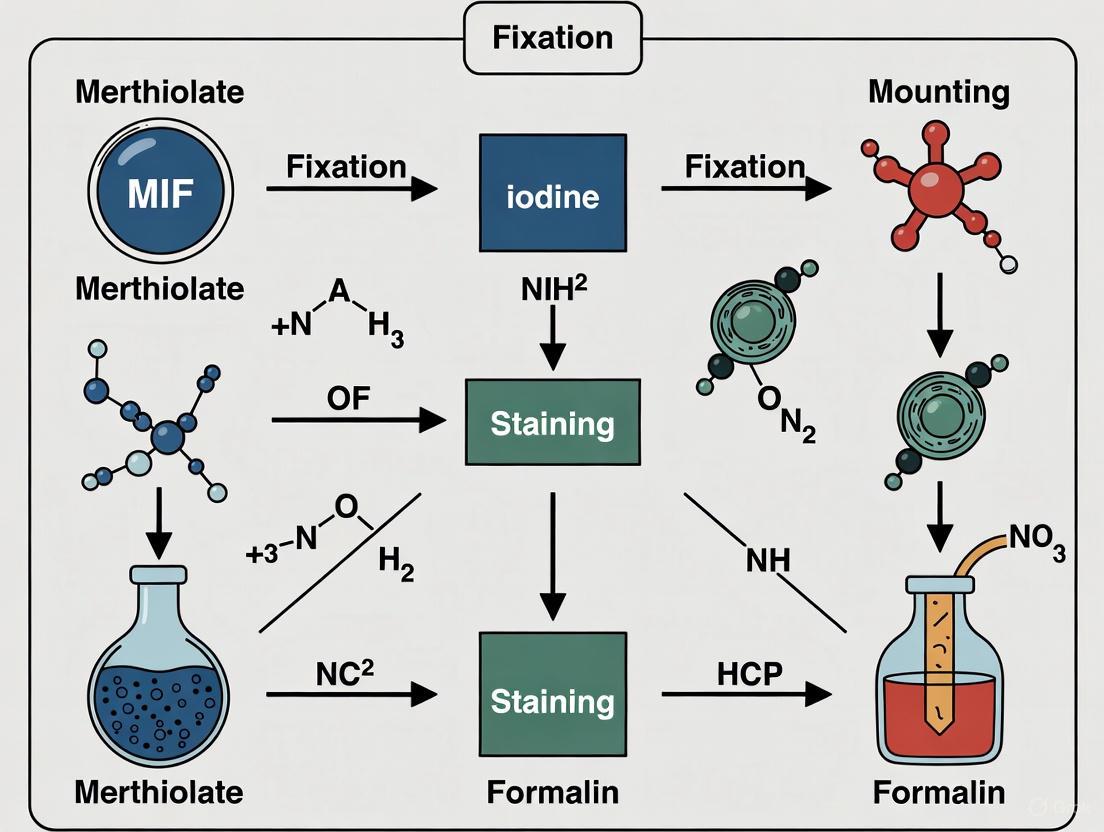
The Merthiolate-Iodine-Formaldehyde (MIF) technique is a comprehensive parasitological procedure that combines fixation, concentration, and staining into a single protocol for the examination of stool specimens. This method serves as a valuable diagnostic tool in clinical and field settings for the identification of intestinal parasites. The core principle of MIF lies in its dual functionality: the merthiolate-formaldehyde component acts as a fixative and preservative, while Lugol's iodine solution provides immediate staining, allowing for the visualization of protozoan cysts, helminth eggs, and larvae in a single wet mount preparation [1].
This technique is particularly valued in field surveys due to its long shelf life and ease of preparation [2]. The MIF method provides a stable medium for preserving parasite morphology, enabling accurate identification even when immediate microscopy is not possible. The combination of fixing and staining components in one solution offers a practical advantage for comprehensive parasitological examination, making it a versatile technique for diagnostic laboratories and research studies focused on parasite fixation.
Core Principles and Chemical Basis
The effectiveness of the MIF technique derives from the complementary actions of its chemical constituents, each serving a specific function in the preservation and visualization of parasitic elements.
Chemical Components and Their Functions
Table 1: MIF Solution Components and Functions
| Component | Chemical Function | Role in Parasitology |
|---|---|---|
| Merthiolate (Thimerosal) | Antimicrobial preservative | Prevents microbial overgrowth; maintains specimen integrity |
| Formaldehyde | Fixative agent | Cross-links proteins; preserves morphological structure of parasites |
| Iodine | Staining agent | Contrast enhancement; stains glycogen inclusions in cysts |
| Glycerin | Stabilizing agent | Reduces crystallization; improves preparation clarity |
The MIF working reagent is prepared by combining two stock solutions immediately prior to use [1]. Solution A (Merthiolate-Formaldehyde or MF) contains distilled water (250.0 ml), saturated formaldehyde (25.0 ml), tincture of merthiolate (1:1,000 concentration, 200.0 ml), and glycerin (5.0 ml), stored in a stoppered brown glass bottle for stability. Solution B consists of Lugol's iodine solution, prepared by dissolving powdered iodine crystals (5.0 gm) and potassium iodide (10.0 gm) in distilled water (100.0 ml), then filtered and stored in a brown bottle with a three-week stability limit [1]. The working reagent is prepared by adding 18.0 milliliters of Solution A to 1.2 milliliters of Solution B, with proportional adjustments for smaller or larger volumes. Mixing the two solutions too far in advance causes precipitate formation, which reduces staining effectiveness [1].
Comparative Advantages of MIF Preservation
Table 2: MIF Advantages and Limitations Compared to Other Common Preservatives
| Preservative Type | Key Advantages | Key Limitations |
|---|---|---|
| MIF | Combines fixation & staining; long shelf life; good for field surveys | Iodine interferes with other stains; may distort protozoa; not ideal for permanent smears |
| 10% Formalin | All-purpose fixative; good for helminth eggs/larvae; suitable for concentration procedures | Not optimal for protozoan trophozoites; can interfere with PCR after extended fixation |
| PVA (Polyvinyl-Alcohol) | Excellent for protozoan trophozoites/cysts; enables permanent stained smears | Contains mercuric chloride (disposal issues); inadequate for helminth eggs/larvae |
| SAF (Sodium Acetate-Formalin) | Suitable for concentration & permanent stains; no mercury content | Requires additives for slide adhesion; permanent stains not as good as with PVA |
The MIF technique is particularly distinguished by its dual fixation-staining capability, which allows for both immediate examination and long-term storage of specimens. The formaldehyde component creates covalent cross-links between proteins, effectively preserving the structural integrity of parasites, while the iodine provides chromatic differentiation of internal structures, particularly the glycogen vacuoles in protozoan cysts [2] [1]. This combination makes MIF especially useful for comprehensive parasitological surveys where both immediate and delayed examinations are necessary.
MIF Protocol: Stool Specimen Processing
Specimen Collection and Preparation
Proper specimen collection is critical for accurate parasitological diagnosis. Stool should be collected in a dry, clean, leakproof container, taking care to avoid contamination with urine, water, soil, or other materials [2]. The distribution of protozoa varies with stool consistency, which should be considered when processing specimens. Fresh stool should be examined, processed, or preserved immediately. When preservation is required, the specimen should be added to the MIF working reagent in a ratio of one volume of stool to three volumes of preservative [2]. Formed stools need to be thoroughly broken up to ensure proper mixing with the preservative. Certain substances interfere with stool examination and should be avoided before specimen collection, including antacids, kaolin, mineral oil, non-absorbable antidiarrheal preparations, barium or bismuth (requires 7-10 days clearance), antimicrobial agents (requires 2-3 weeks clearance), and gallbladder dyes (requires 3 weeks clearance) [2].
Merthiolate-Iodine-Formaldehyde Concentration (MIFC) Procedure
The MIFC technique enhances parasite detection by concentrating parasitic elements while combining fixation and staining in a single procedure [1]. The step-by-step protocol is as follows:
Specimen Preparation: Emulsify approximately 1 g of stool in the MIF working reagent.
Filtration and Settling: Strain the mixture through gauze into a conical centrifuge tube and allow it to stand for 5-10 minutes.
Ethyl Acetate Addition: Add 3 ml of ethyl acetate to the tube, stopper, invert, and shake vigorously for approximately 30 seconds until thoroughly mixed. Release the stopper carefully to avoid spraying.
Centrifugation: Centrifuge for 2 minutes at 2,500 rpm. After centrifugation, four distinct layers form in the tube:
- Top layer: Ethyl acetate
- Second layer: Debris plug
- Third layer: Formalin solution
- Bottom layer: Sediment containing parasites (if present)
Sediment Recovery: Free the debris plug with an applicator stick and carefully decant the top three layers, leaving the sediment undisturbed. Use a cotton swab to clean remnants of ethyl acetate from inside the tube.
Microscopic Preparation: Prepare both saline and stained wet preparations from the sediment and examine for parasites under microscopy [1].
The Scientist's Toolkit: Essential Research Reagents
Table 3: Research Reagent Solutions for MIF Technique
| Reagent/Material | Specification/Function | Application Notes |
|---|---|---|
| Merthiolate-Formaldehyde (MF) | Stock Solution A: Preservative and fixative | Store in brown glass bottle; stable for months |
| Lugol's Iodine | Stock Solution B: Staining component | Store in brown bottle; stable for 3 weeks only |
| Ethyl Acetate | Lipid solvent for concentration | Creates top layer in centrifugation; extracts debris |
| Gauze | Filtration medium | Removes large particulate matter from stool sample |
| Conical Centrifuge Tubes | 15ml graduated tubes | For concentration procedure and layer separation |
| Applicator Sticks | Wooden or plastic | For freeing debris plug after centrifugation |
| Microscope Slides & Coverslips | Glass slides for microscopy | For saline and stained wet mount preparations |
Technical Considerations and Limitations
While the MIF technique offers significant advantages for parasitological studies, researchers should be aware of its specific limitations. The iodine component, while providing excellent immediate staining for wet mount examinations, may interfere with other stains and fluorescence techniques [2]. Additionally, the merthiolate component contains mercury, which raises environmental concerns and requires special disposal procedures. Some studies note that MIF provides inadequate preservation of protozoan trophozoite morphology compared to polyvinyl-alcohol (PVA) based methods, and is not ideal for preparing permanent stained smears with trichrome stain [2] [1].
The MIF technique shows particular strength in the preservation of helminth eggs, larvae, and protozoan cysts, making it well-suited for comprehensive parasitological surveys [2]. However, for studies focused specifically on protozoan trophozoites, PVA-based preservation may yield superior morphological preservation. The concentration aspect of the MIFC procedure significantly enhances detection sensitivity for low-burden infections, providing a practical advantage in both clinical and research settings where parasite density may be variable.
The Merthiolate-Iodine-Formalin (MIF) staining and fixation technique is a fundamental tool in parasitology, providing a means for the simultaneous preservation and identification of intestinal parasites in stool specimens. Its utility spans both clinical diagnostics and large-scale epidemiological field surveys, largely due to its simple preparation and long shelf life [3]. This method leverages the synergistic action of its three key components to stabilize parasitic structures and enhance their visual contrast under microscopic examination. These properties make it particularly competitive in resource-limited settings [4]. These application notes and protocols detail the chemical composition, formulation, and standardized procedures for employing MIF in parasitological research, providing a framework for reliable and reproducible results.
Chemical Composition and Reagent Functions
The efficacy of the MIF technique stems from the distinct and complementary roles of its constituent chemicals. The formulation is typically divided into two separate stock solutions that are combined immediately before use.
Research Reagent Solutions
The table below summarizes the key reagents, their common formulations, and primary functions within the MIF protocol.
Table 1: Essential Reagents for the MIF Staining and Fixation Technique
| Reagent Name | Chemical Composition | Primary Function in the Protocol |
|---|---|---|
| Solution I: MF Stock Solution [5] | Contains Formaldehyde, Glycerol, and Tincture Merthiolate (Thimerosal) [5]. | Fixation and Preservation. Formaldehyde denatures proteins and hardens structural components, preserving trophozoites, cysts, eggs, and larvae. Glycerol may help reduce distortion. Merthiolate acts as a bactericide and fungicide, preventing microbial overgrowth. |
| Solution II: Lugol's Iodine [5] | A solution of Iodine and Potassium Iodide [5]. | Staining. Iodine interacts with glycogen and other cellular components of parasites, staining them in shades of brown, which provides contrast for microscopic identification of protozoa and helminth eggs. |
| MIF Working Solution | A mixture of Solution I and Solution II, typically at a ratio of 2.35 mL Sol. I to 0.15 mL Sol. II, combined with a fecal sample [5]. | Simultaneous Fixation and Staining. This ready-to-use mixture allows for immediate direct smear examination and long-term preservation of the sample without significant deterioration of organisms [5]. |
| 10% Formalin [6] | A 10% dilution of formaldehyde gas in water. | Sample Washing & Concentration. Used in modified MIF concentration techniques to wash agar plates [7] or as an initial fecal suspension medium prior to filtration and concentration steps [6]. |
| Ethyl Acetate [7] | An organic solvent. | Concentration (in QFEC). Used in concentration techniques following MIF preservation to extract debris and fat from the fecal suspension, thereby concentrating parasitic elements in the sediment [7]. |
Experimental Protocols
Standard MIF Staining and Fixation Protocol
This protocol describes the fundamental procedure for preparing and examining a stool sample using the MIF technique [5].
Workflow: Standard MIF Staining
Materials:
- Solution I: MF Stock Solution
- Solution II: Lugol's Iodine
- Fresh stool specimen
- Test tube with stopper
- Calibrated pipettes or droppers
- Microscope slides and coverslips
- Microscope
Procedure:
- Reagent Preparation: In a well-stoppered tube (e.g., a 5-7 mL screw-capped tube), combine 2.35 mL of Solution I (MF Stock Solution) with 0.15 mL of Solution II (Lugol's Iodine) [5].
- Sample Addition: Add approximately 0.25 grams of fresh feces (about twice the size of a pea) to the tube containing the MIF working solution [5].
- Mixing: Close the tube tightly and mix the contents thoroughly to ensure the stool specimen is completely emulsified with the fixative-stain solution [5].
- Incubation: Allow the mixture to stand for an appropriate fixation and staining period. The sample can be examined immediately or stored for later analysis, as the MIF solution preserves the organisms [5].
- Microscopic Examination:
- Just before examination, gently shake or invert the tube to resuspend the sediment.
- Using a medicine dropper, draw off a drop of the mixture from the layer of sediment and place it on a clean microscope slide.
- Apply a coverslip and examine systematically under the microscope, starting with a lower magnification (e.g., 10x objective) to locate potential parasitic structures, and then switch to a higher magnification (40x objective) for detailed identification [5].
Expected Results: Trophozoites and cysts of protozoa, as well as helminth eggs and larvae, should be stained an eosin (pinkish) color with contrasting internal structures, allowing for morphological identification [5].
Modified Concentrated MIF Technique
To increase the sensitivity of the standard MIF technique, particularly for detecting low-burden infections, a concentration step can be added. This modification is suitable for large-scale screening programs [6].
Workflow: Modified Concentrated MIF Technique
Materials:
- 10% Neutral Buffered Formalin
- Double-layered cotton filter (or gauze)
- Centrifuge tubes
- Centrifuge
- Standard MIF Solutions
Procedure:
- Initial Fixation: Mix the stool specimen in a tube containing 10% formalin [6].
- Filtration: Filter the formalin-stool mixture through a double-layered cotton filter into a centrifuge tube. This step removes large, coarse debris [6].
- Concentration: Remove most of the liquid content from the filtered suspension, either by decanting or through centrifugation, to concentrate the parasitic elements. The original publication mentions an "overnight drying procedure" as part of this concentration [6].
- MIF Preservation: The resulting concentrated fecal material is then preserved with the standard MIF solution (a mixture of Solution I and II) for staining and long-term storage [6].
- Examination: Proceed with microscopic examination as described in the standard protocol, using the concentrated sediment.
Applications and Performance Data
The MIF technique is valued for its versatility in diagnosing a wide range of intestinal parasites. The following table summarizes its performance in comparative studies against other common coprological methods.
Table 2: Comparative Performance of MIF Against Other Diagnostic Techniques
| Comparison | Key Findings | Implications for Research and Diagnostics |
|---|---|---|
| MIF vs. Kato-Katz (KK) [4] | - KK showed higher sensitivity for Trichuris trichiura.- Both methods had almost perfect agreement (Kappa index).- MIF provided higher median parasitic loads for low and total egg counts for Ascaris lumbricoides, T. trichiura, and hookworms.- KK was not able to detect high loads of helminths as effectively as MIF.- A key advantage of MIF is its ability to detect protozoa and other helminths (e.g., Strongyloides stercoralis), for which KK is not suited. | MIF is a competitive, simple, and inexpensive technique for intestinal helminth diagnosis, especially in resource-limited settings. Its ability to detect a broader spectrum of parasites (helminths and protozoa) makes it a more comprehensive single-test solution than KK. |
| MIF vs. Agar Plate Culture (APC) [7] | - APC demonstrated superior sensitivity for detecting Strongyloides stercoralis larvae.- The Quantitative Formalin-Ethyl Acetate Concentration technique (QFEC), which can use MIF-preserved samples, could only substitute for APC when the parasite load was high (>50 larvae per gram of stool). | For epidemiological studies targeting Strongyloides, APC is preferable. However, for clinical diagnosis where parasite burdens are often higher, a concentration technique like QFEC on MIF-preserved samples may be sufficient. |
| MIF within Diagnostic Systems [8] | A study comparing collection-preservation methods found that when methods were grouped into systems, the MIF system was more effective for parasite recovery and more time-efficient than a Formalin/Polyvinyl Alcohol (PVA) fixation system. | The MIF technique offers a strong balance of diagnostic yield and workflow efficiency in a laboratory setting. |
Integration with Advanced Methodologies
The role of MIF extends beyond conventional microscopy into modern diagnostic approaches. Recent research has validated MIF as a key sample preparation method for deep-learning-based automated parasite identification [3]. In these studies, MIF is used to prepare samples for creating image datasets. The fixation and staining provided by MIF create consistent and well-contrasted images of parasitic forms, which are essential for training robust AI models like YOLOv8 and DINOv2. These models have shown high accuracy, precision, and sensitivity in identifying helminth eggs and larvae from digital images of MIF-stained samples, highlighting the continued relevance of MIF in the evolving landscape of diagnostic parasitology [3].
Historical Development and Evolution of the MIF Protocol
The Merthiolate-Iodine-Formalin (MIF) protocol represents a significant milestone in the field of parasitology, providing a reliable method for the fixation, staining, and concentration of parasitic elements in fecal specimens. Developed in the mid-20th century, this technique has endured as a valuable diagnostic and research tool, particularly for intestinal parasitic infections (IPI). The MIF method effectively combines the preservative qualities of formalin with the staining capabilities of merthiolate and iodine, creating a stable medium for the morphological identification of helminth eggs, larvae, and protozoan cysts. Its simplicity, cost-effectiveness, and long shelf life have rendered it particularly suitable for field surveys and resource-limited settings where more advanced molecular methods may be unavailable or impractical [9]. This application note delineates the historical development, technical evolution, and contemporary applications of the MIF protocol within the context of parasite fixation research.
Historical Background and Initial Development
The MIF technique was formally described in 1957 as a concentration method for the detection of parasitic material in fecal samples. The initial development aimed to address the need for a robust diagnostic tool that could surpass the efficiency of existing methods, such as Willis's salt flotation and direct saline smear techniques [10].
Early comparative studies demonstrated that the MIF method showed a much greater efficiency for the detection of helminth ova compared to the other two contemporary methods. However, it did not prove as satisfactory for the detection of protozoan parasites, though it was effective for the concentration of trophozoites [10]. The original MIF protocol involved the use of two stock solutions:
- MIF A: Containing distilled water, thimerosal (merthiolate) 1:1000, formaldehyde, and glycerin.
- MIF B: A potassium iodide-iodine solution [11].
This two-solution system ensured the simultaneous fixation and staining of parasitic structures, preserving their morphology for microscopic examination. The appearance of helminth ova and protozoan parasites in MIF preparations was systematically described, establishing a foundation for parasitological diagnosis [10].
Technical Evolution and Protocol Refinements
Standardization of the Staining Procedure
Over time, the MIF protocol has been refined to enhance its diagnostic performance. The procedure involves thoroughly resuspending 3–5 g of fecal material in phosphate-buffered saline (PBS), followed by filtration through a sieve to remove large debris. The filtered suspension is then centrifuged, and the supernatant is carefully removed. The resulting sediment is mixed with the MIF solutions before microscopic examination [11].
A key application of MIF in contemporary diagnostics is its use in differentiating between active and degenerate cysts of Giardia duodenalis in fecal concentrates [11]. Its effectiveness in evaluating intestinal parasitic infections has been documented in comparative studies, where it addresses practical drawbacks of direct stool examination and provides highly competitive performance [9].
Comparative Diagnostic Performance
Recent studies have validated the performance of MIF against modern diagnostic standards. The following table summarizes its performance for detecting Giardia duodenalis in a 2024 veterinary study, where Direct Immunofluorescence Assay (DFA) was used as the gold standard.
Table 1: Diagnostic Performance of MIF for Giardia duodenalis Detection (2024)
| Host Species | Prevalence by DFA | Prevalence by MIF | Statistical Significance (p-value) |
|---|---|---|---|
| Dogs (n=225) | 30.2% (95% CI: 24.3–36.7) | 22.7% | < 0.001 |
| Cats (n=103) | 11.6% (95% CI: 6.2–19.5) | 7.8% | < 0.001 |
Source: Adapted from [11]
The data indicates that while DFA is the most sensitive technique, MIF remains a viable diagnostic method, particularly in settings where advanced immunofluorescence assays are not feasible [11].
Contemporary Applications in Parasitology Research
Role in Ground-Truthing for Advanced Diagnostics
The MIF technique continues to serve as a reference method in studies developing novel diagnostic approaches. For instance, a 2025 study evaluating deep-learning-based models for intestinal parasite identification used human experts performing FECT and MIF techniques to establish the ground truth and reference for parasite species [9]. This underscores the enduring value of MIF in providing a reliable benchmark against which new technologies are measured.
Use in Epidemiological Surveillance
MIF staining has been effectively employed in recent surveillance of emerging parasitic diseases. A 2025 study on human intestinal sarcocystosis in France utilized microscopic examination of fresh homogenized stool samples to identify Sarcocystis spp. oocysts and sporocysts, with molecular analysis confirming the presence of multiple species, including S. sigmoideus, S. hominis, and S. heydorni [12]. This demonstrates MIF's role in the initial detection of parasites in complex multi-species infections.
Experimental Protocol: MIF Staining for Fecal Parasitology
Reagent Preparation
Table 2: Research Reagent Solutions for the MIF Protocol
| Reagent/Solution | Composition / Function | Application Note |
|---|---|---|
| MIF Solution A | 50 ml distilled water, 40 ml thimerosal 1:1000, 10 ml formaldehyde, 5 ml glycerin. | Acts as a fixative and preservative. Formaldehyde fixes structures, thimerosal is a preservative, glycerin adds viscosity. |
| MIF Solution B | Potassium iodide-iodine solution. | Serves as a staining solution, highlighting nuclei and internal structures of cysts and ova. |
| Phosphate-Buffered Saline (PBS) | Diluent for fecal suspension. | Provides an isotonic medium for homogenizing the sample without distorting parasitic elements. |
| Lugol's Iodine | Iodine-based stain. | Sometimes used in combination with MIF to enhance the observation of Giardia cysts [11]. |
Step-by-Step Procedure
- Sample Preparation: Thoroughly resuspend 3–5 g of fresh fecal material in 20 ml of PBS.
- Filtration: Filter the homogenate through a sieve mesh (e.g., 250 µm diameter) to remove large particulate debris.
- Centrifugation: Transfer the filtered suspension into a 10 ml centrifuge tube. Centrifuge at 1,500 rpm for 10 minutes.
- Supernatant Removal: Carefully decant or pipette off the supernatant after centrifugation.
- Staining: Add the appropriate volumes of MIF Solution A and MIF Solution B to the sediment and mix thoroughly.
- Microscopy: Examine the stained sediment under a microscope at appropriate magnifications (e.g., 100x, 400x) for the identification of helminth eggs, larvae, and protozoan cysts.
The workflow below illustrates the key steps in the MIF staining procedure.
Comparison with Modern Diagnostic Techniques
While the MIF technique remains a valuable tool, its performance must be contextualized alongside modern diagnostic methods. The table below provides a comparative overview of MIF against other common techniques used in parasitology.
Table 3: Comparative Analysis of MIF with Other Diagnostic Methods
| Diagnostic Method | Principle | Key Advantages | Key Limitations |
|---|---|---|---|
| MIF Staining | Fixation and concentration with chemical staining. | Cost-effective, long shelf life, suitable for field use, good for helminths [9] [10]. | Lower sensitivity for protozoa compared to DFA/PCR, potential for morphological distortion [9] [11]. |
| Direct Immunofluorescence (DFA) | Fluorescently-labeled antibodies target specific (oo)cyst surface antigens. | High sensitivity and specificity, considered gold standard for Giardia/Cryptosporidium [11]. | Requires fluorescence microscope, higher cost, dependent on reagent quality. |
| Molecular Methods (PCR, qPCR, HTS) | Detection of parasite-specific DNA/RNA sequences. | High sensitivity and specificity, enables species/genotype identification [9] [12]. | Time-consuming, expensive, requires skilled personnel, risk of contamination [9]. |
| Deep-Learning-Based AI | Automated image analysis and pattern recognition. | High-throughput, objective, can achieve high accuracy (e.g., DINOv2-large: 98.93% accuracy) [9]. | Requires large, labeled datasets for training, limited by quality of input images [9]. |
The MIF protocol has demonstrated remarkable resilience in the diagnostic parasitology landscape. From its inception in 1957 to its current applications, it has provided a cost-effective, practical, and reliable method for the detection of intestinal parasites, particularly helminths. While modern techniques like DFA and PCR offer superior sensitivity and specificity for specific pathogens, and AI-driven image analysis promises a new era of automated diagnostics, the MIF technique retains its relevance. Its role in basic diagnostic services, field epidemiology, and as a ground-truthing benchmark in research ensures that the MIF protocol remains an integral component of the scientist's toolkit for parasite fixation and staining. Future developments may see it further integrated with digital pathology platforms, enhancing its utility in the era of computational biology.
The Merthiolate-Iodine-Formaldehyde (MIF) technique represents a significant advancement in diagnostic parasitology, combining fixation, concentration, and staining into a single comprehensive procedure. This method addresses critical challenges in field surveys and resource-limited settings where rapid preservation of stool specimens is essential for accurate morphological analysis. MIF's unique formulation provides simultaneous fixation and staining capabilities, making it particularly valuable for epidemiological studies and drug efficacy trials where sample integrity must be maintained during transport and storage [2] [1]. The technique's design principle centers on integrating morphological preservation with immediate staining visualization, creating a robust tool for comprehensive parasitological assessment.
For research scientists and drug development professionals, MIF offers practical advantages that extend beyond basic diagnostics. The method preserves a wide spectrum of parasitic elements including helminth eggs, protozoan cysts, and occasionally trophozoites, though with varying efficacy across different parasite species [13] [2]. This preservation stability enables standardized analysis across multiple study sites and timepoints, crucial for multicenter clinical trials and longitudinal studies of parasitic disease burden. Understanding MIF's capabilities and limitations allows researchers to strategically deploy this technique within a broader diagnostic algorithm that may include molecular methods and advanced imaging technologies.
Key Advantages and Technical Specifications
The MIF technique offers three distinct advantages that make it particularly suitable for field-based research and studies requiring high-throughput sample processing.
Extended Shelf Life and Storage Stability
MIF solutions demonstrate exceptional stability when properly stored, maintaining diagnostic efficacy for extended periods. The merthiolate-formaldehyde (MF) stock solution (Solution A) remains stable for several months when stored in stoppered brown glass bottles, protected from direct light [1]. This extended shelf life reduces reagent wastage and ensures availability in remote research settings where frequent reagent preparation is impractical. The glycerin in the formulation further enhances stability by preventing evaporation and crystallization, particularly important in tropical climates with high temperatures [1].
Field Deployment Suitability
MIF's design characteristics make it uniquely suited for field surveys and sample collection in non-laboratory settings. The technique requires minimal equipment - essentially centrifuge tubes and basic personal protective equipment - unlike many staining methods that require electricity-dependent heating or precise humidity control [2]. Research comparisons have confirmed MIF's effectiveness in field surveys where infrastructure limitations preclude immediate microscopic analysis [2] [3]. The method's tolerance to variable environmental conditions (temperature, humidity) allows researchers to collect and preserve specimens directly at point-of-care sites before centralized laboratory analysis.
Simultaneous Fixation and Staining Mechanism
MIF's integrated approach provides both immediate fixation through formaldehyde and simultaneous staining via iodine, creating a time-efficient diagnostic workflow. The formaldehyde component rapidly penetrates parasitic structures, cross-linking proteins and preserving morphological details against autolysis and degradation [13] [2]. Concurrently, the iodine component stains glycogen inclusions and structural features, providing immediate contrast for preliminary microscopic assessment without additional processing steps [1]. This dual functionality enables researchers to perform initial parasitological assessment even in field conditions, with option for more detailed analysis after transport to central laboratories.
Table 1: Technical Advantages of MIF Compared to Alternative Fixation Methods
| Advantage | MIF Performance | Formalin | PVA |
|---|---|---|---|
| Shelf Life | Several months [1] | Long [2] | Several months [2] |
| Field Suitability | Excellent [2] | Good | Poor (requires careful disposal) [2] |
| Simultaneous Fixation & Staining | Yes [1] | No (staining requires separate steps) [2] | No (requires trichrome stain) [2] |
| Helminth Egg Recovery | Excellent [14] | Good [2] | Inadequate [2] |
Table 2: Efficacy of MIF for Different Parasite Forms
| Parasite Form | MIF Preservation Quality | Research Applications |
|---|---|---|
| Helminth Eggs | Excellent morphology [14] | Quantitative egg counts, morphological studies |
| Protozoan Cysts | Good to variable [13] | Prevalence surveys, species identification |
| Trophozoites | Inadequate to poor [2] | Limited utility for trophozoite studies |
| Coccidia | Not suitable [2] | Requires alternative methods |
Research Reagent Solutions
The following table details essential materials and reagents required for implementing the MIF technique in research settings.
Table 3: Essential Research Reagents for MIF Protocol
| Reagent/Material | Composition/Specifications | Research Function |
|---|---|---|
| Merthiolate-Formaldehyde (MF - Solution A) | Distilled water (250.0 ml), formaldehyde saturated (25.0 ml), tincture of merthiolate (Lilly 1:1,000, 200.0 ml), glycerin (5.0 ml) [1] | Primary fixative and preservative component; merthiolate acts as antibacterial/antifungal agent |
| Lugol's Iodine (Solution B) | Iodine crystals (5.0 gm), potassium iodide (10.0 gm), distilled water (100.0 ml) [1] | Staining component that highlights internal structures of parasites |
| Ethyl Acetate | Laboratory grade, 3 ml per sample [1] | Organic solvent for extraction of fecal debris and fats during concentration |
| Centrifuge Tubes | Conical, leak-proof, 15 ml capacity [1] | Sample processing during concentration step |
| Parafilm or Sealing Material | Laboratory grade | Secures containers during transport and storage |
Comprehensive Experimental Protocols
Reagent Preparation Protocol
Stock Solution A (Merthiolate-Formaldehyde) Preparation:
- Add 250.0 ml of distilled water to a clean glass container
- Incorporate 25.0 ml of saturated formaldehyde solution while using fume hood protection
- Add 200.0 ml of tincture of merthiolate (Lilly 1:1,000 formulation)
- Mix in 5.0 ml of glycerin as a stabilizing agent
- Transfer to a stoppered brown glass bottle for storage
- Label with preparation date and expiration date (6 months from preparation)
- Store at room temperature, protected from direct light [1]
Stock Solution B (Lugol's Iodine) Preparation:
- Dissolve 10.0 gm of potassium iodide in 100.0 ml of distilled water
- Gradually add 5.0 gm of powdered iodine crystals while continuously shaking
- Continue shaking until complete dissolution of iodine crystals occurs
- Filter the solution through laboratory filter paper to remove particulates
- Transfer to a light-resistant brown glass bottle with tight seal
- Label with preparation date and expiration date (3 weeks from preparation due to iodine volatility) [1]
Working MIF Solution Preparation:
- Combine 18.0 ml of Solution A with 1.2 ml of Solution B immediately before use
- Mix thoroughly by gentle inversion (avoid vigorous shaking to prevent precipitate formation)
- Prepare only the volume required for immediate processing
- Do not store working solution as iodine will precipitate upon standing [1]
Stool Processing and Staining Protocol
Sample Collection and Fixation:
- Collect approximately 1 gram of formed stool in a clean, leak-proof container (increase to 2-4 grams for soft or liquid specimens) [15]
- Immediately add one volume of stool to three volumes of freshly prepared working MIF solution [2]
- Mix thoroughly until homogeneous consistency is achieved, ensuring all particulate matter is broken up
- Seal container with parafilm and place in plastic bag for transport
- Label with sample ID, date, and time of collection [2]
MIF Concentration Technique:
- Transfer 3-4 ml of MIF-preserved stool to a 15 ml conical centrifuge tube
- Add 3 ml of ethyl acetate to the tube, stopper securely
- Invert tube and shake vigorously for 30 seconds to ensure complete mixing
- Carefully release pressure by loosening stopper slowly to avoid aerosolization
- Centrifuge at 2,500 rpm for 2 minutes using a clinical centrifuge
- Identify the four distinct layers formed after centrifugation:
- Top layer: Ethyl acetate
- Second layer: Debris plug
- Third layer: Formalin solution
- Bottom layer: Sediment containing parasites [1]
Sediment Processing and Slide Preparation:
- Free the debris plug from tube walls using an applicator stick
- Carefully decant the top three layers, leaving sediment undisturbed
- Use a cotton swab to wipe residual ethyl acetate from tube interior
- Resuspend sediment in small volume of remaining fluid by gentle tapping
- Prepare both saline and stained wet mounts for examination:
- Saline preparation: Mix one drop of sediment with one drop of saline
- Stained preparation: Mix sediment with appropriate stain if additional contrast needed
- Apply coverslips and examine systematically under microscope [1]
Quality Control and Method Validation
Positive Control Implementation:
- Maintain reference samples with known parasite morphology (Giardia lamblia cysts, Ascaris eggs)
- Process control samples alongside test specimens using identical protocols
- Document control results to monitor technique consistency
- Establish criteria for acceptable control performance (morphology preservation, staining intensity)
Staining Quality Assessment:
- Evaluate iodine staining intensity microscopically (optimal: golden-brown cytoplasm with contrast)
- Verify morphological preservation criteria:
- Helminth eggs: Intact egg walls, visible internal structures
- Protozoan cysts: Distinct cell membranes, visible nuclei
- Structures should not appear distorted, shrunken, or overly swollen
- Document staining artifacts for future reference (precipitates, crystalline formations)
Method Comparison and Validation:
- Parallel test subset of samples using MIF and reference methods (FECT, trichrome staining)
- Calculate diagnostic concordance metrics (sensitivity, specificity)
- Establish inter-observer agreement for morphological interpretation
- Validate concentration efficiency through quantitative egg counts in helminth studies [3]
Research Applications and Integration with Advanced Methodologies
Field Study Applications
MIF's stability and all-in-one functionality make it particularly valuable for large-scale epidemiological studies in resource-limited settings. Research teams can establish standardized sample collection protocols across multiple field sites, with centralized processing and analysis [2]. The method's compatibility with various microscopic examination techniques enables comprehensive parasitological surveys that capture both helminth and protozoan infections. For longitudinal studies monitoring intervention efficacy, MIF-preserved samples provide consistent morphological reference points across multiple timepoints.
Integration with Modern Diagnostic Technologies
While MIF remains primarily a morphological technique, researchers are increasingly integrating it with advanced diagnostic approaches:
Molecular Applications: Though formalin-based fixatives like MIF can interfere with PCR amplification, protocol modifications can sometimes enable subsequent molecular analysis [2]. Researchers should validate DNA extraction protocols specifically for MIF-preserved specimens when molecular work is anticipated.
Digital Pathology and AI: The standardized preparation of MIF specimens makes them suitable for digital imaging and computational analysis. Recent studies have demonstrated the potential of deep-learning models for automated parasite detection in stained specimens [3]. MIF's consistent staining characteristics facilitate the development of robust machine learning algorithms for high-throughput sample screening.
Limitations and Complementary Techniques
Researchers should acknowledge MIF's limitations and implement complementary methods when necessary:
Trophozoite Preservation: MIF provides inadequate preservation of delicate trophozoite forms, requiring alternative fixation methods (Schaudinn's, PVA) for studies focusing on trophic stages [2].
Molecular Compatibility: For studies requiring subsequent genetic analysis, Sodium Acetate-Acetic Acid-Formalin (SAF) or specific one-vial fixatives may offer better compatibility with molecular techniques while maintaining reasonable morphological preservation [2].
Advanced Staining Requirements: When superior morphological detail is required for protozoan identification, permanent stains (trichrome, iron-hematoxylin) on PVA-preserved specimens provide enhanced cytological detail, though with increased complexity and cost [13] [2].
The Merthiolate-Iodine-Formaldehyde technique remains a valuable tool in parasitology research, particularly for field studies, epidemiological surveys, and drug development programs requiring standardized sample processing across multiple sites. Its integrated fixation-staining approach, combined with exceptional shelf life and field suitability, provides researchers with a robust method for comprehensive parasitological assessment. While limitations exist regarding trophozoite preservation and molecular compatibility, MIF's advantages for helminth egg recovery and protozoan cyst preservation ensure its continued relevance in both basic and applied parasitology research.
As diagnostic technologies evolve, MIF-prepared specimens show promising compatibility with digital imaging and artificial intelligence approaches, potentially enhancing throughput and standardization in parasite detection and morphological analysis. Researchers should consider MIF as a core methodology within a comprehensive diagnostic strategy, complementing it with specialized techniques when specific research questions require enhanced trophozoite preservation, molecular analysis, or ultrastructural detail.
Inherent Limitations and Safety Considerations, including Mercuric Chloride Content
Merthiolate-Iodine-Formalin (MIF) staining is a established method in parasitology for the fixation, preservation, and detection of intestinal protozoa and helminth eggs in fecal specimens [8]. While effective for parasite recovery, this technique incorporates mercuric chloride, a chemical agent of significant toxicological concern. The utility of MIF in diagnostic and research settings must be balanced against a thorough understanding of its inherent limitations and the critical safety protocols mandated by the presence of this hazardous compound. This document details the chemical risks, operational limitations, and stringent safety measures required for the safe use of MIF in scientific research, providing a framework for compliance and risk mitigation for researchers, scientists, and drug development professionals.
Chemical Hazards and Toxicity Profile of Mercuric Chloride
Mercuric chloride (HgCl₂) is a highly toxic inorganic compound historically used in various applications, including as a pesticide and fungicide [16]. Its high toxicity profile has led to its obsolescence and banning in many countries [16].
Toxicity Data
The following table summarizes key toxicity data for mercuric chloride, illustrating its extreme hazard to mammalian and environmental systems.
Table 1: Toxicity Profile of Mercuric Chloride
| Organism/System | Endpoint | Value | Toxicity Rating |
|---|---|---|---|
| Mammals (Rat) | Acute Oral LD₅₀ | < 1.0 mg/kg | High [16] |
| General Hazard | WHO Hazard Classification | Classes Ia or Ib | Highly Hazardous [16] |
| Regulatory | Rotterdam Convention | Listed in Annex III | Subject to Prior Informed Consent (PIC) regulations [16] |
Mode of Toxic Action
As a fungicide, mercuric chloride acts as a non-specific, multi-site toxicant [16]. Its primary mechanism involves disrupting essential enzymatic activities in cells, interfering with mitochondrial respiration, and inhibiting carbohydrate synthesis [16]. It can also alter membrane permeability, leading to widespread cellular dysfunction and death [16]. This non-specific mode of action contributes to its broad-spectrum toxicity across biological systems.
Limitations of MIF Staining in Parasitological Research
While valued for its diagnostic utility, the MIF technique presents several inherent limitations that researchers must consider when designing studies and interpreting results.
- Incomplete Parasite Recovery: A foundational study comparing parasite preservation methods found that no single method was effective in recovering all parasites present in a specimen [8]. This indicates that reliance solely on MIF staining may lead to false negatives or an incomplete profile of parasitic infection within a sample.
- Diagnostic Performance Variability: The MIF concentration technique has demonstrated a much greater efficiency for the detection of helminth ova compared to both Willis's salt flotation method and direct saline smear examination [14]. While this confirms its strength for helminths, it also implies that its performance for other parasite types (e.g., some protozoa) may be less effective or variable, potentially introducing bias into research findings.
- Incompatibility with Modern High-Throughput Drug Discovery: Contemporary anthelmintic discovery pipelines increasingly rely on high-throughput, phenotypic screening of small-molecule compounds against parasites like Haemonchus contortus [17]. The manual, labor-intensive nature of MIF staining and the associated safety burdens of handling mercuric chloride make it poorly suited for these large-scale, automated experimental systems.
Safety Protocols for Handling Mercuric Chloride
The use of mercuric chloride in the laboratory demands uncompromising adherence to safety protocols to protect personnel and the environment.
Personal Protective Equipment (PPE) and Engineering Controls
- PPE: Handling mercuric chloride requires appropriate personal protective equipment. This includes safety goggles, nitrile gloves, and a lab coat [18]. The use of respiratory protection may also be necessary depending on the potential for aerosol generation.
- Engineering Controls: Due to its high toxicity and the risk of inhaling harmful fumes or dust, this substance must be handled exclusively in a fume hood [18].
Storage, Spill Response, and Disposal
- Storage: Store mercuric chloride in a secure, well-ventilated, and controlled facility specifically designated for hazardous compounds [18]. The storage area must be clearly labeled with hazard warnings and access should be restricted to authorized, trained personnel [18].
- Spill Response: In the event of a spill, immediately isolate the area and use mercury-specific absorbents for containment and cleanup [18]. For larger spills, a certified hazardous materials response team must be contacted for proper handling. All contaminated materials must be disposed of as hazardous waste [18].
- Disposal: Disposal practices must strictly follow EPA hazardous waste regulations and all applicable local guidelines [18]. Facilities must have comprehensive emergency response plans that include decontamination procedures and specific medical protocols for mercury exposure [18].
Regulatory Compliance and Documentation
Handling mercuric chloride is subject to stringent regulatory oversight. Compliance is non-negotiable and requires meticulous documentation.
Table 2: Key U.S. Regulatory Standards and Documentation for Mercuric Chloride
| Agency/Area | Standard/Requirement | Key Documentation |
|---|---|---|
| OSHA | Permissible Exposure Limit (PEL): 0.1 mg/m³ [18] | Workplace exposure records; Medical surveillance records. |
| EPA | Regulated as a hazardous substance under TSCA [18]. | Hazardous waste manifests; Toxics Release Inventory (TRI) reporting. |
| General Compliance | Hazard Communication Standard [18]. | Safety Data Sheets (SDS), accessible to all personnel [18]. |
| Product Quality and Traceability. | Certificates of Analysis (CoA), verifying ACS-grade purity [18]. |
The Scientist's Toolkit: Essential Research Reagent Solutions
The following table outlines key materials and their functions relevant to MIF-based parasitology research and safety.
Table 3: Essential Research Reagents and Materials for MIF and Parasitology Research
| Item | Function/Application |
|---|---|
| Mercuric Chloride ACS Grade | High-purity (≥99.5%) compound for precise formulation of MIF stain-preservative solutions [18]. |
| MIF Stain-Preservative Solution | Used for simultaneous fixation, preservation, and staining of parasitic elements in fecal specimens [8] [14]. |
| Certificate of Analysis (CoA) | Document verifying a chemical batch meets specific purity and quality standards (e.g., ACS grade), ensuring experimental consistency [18]. |
| Safety Data Sheet (SDS) | Provides critical information on hazards, safe handling, storage, and emergency measures for a chemical substance [18]. |
| In Silico Screening Models | Machine learning models (e.g., Multi-layer Perceptron) used to predict and prioritize novel anthelmintic candidates, reducing reliance on manual, low-throughput methods [17]. |
Experimental Workflow and Safety Pathway
The diagram below outlines the key experimental and safety decision pathways for working with MIF and mercuric chloride.
MIF Safety Workflow
Risk Mitigation and Compliance Pathway
This diagram maps the critical compliance and risk mitigation steps required when sourcing and handling mercuric chloride.
Compliance Pathway
Mercuric chloride, as a component of MIF staining, presents a clear paradox of utility and hazard. Its role in facilitating parasite fixation is tempered by significant limitations in diagnostic completeness and a demanding safety profile. Contemporary research must acknowledge these constraints, giving paramount importance to the stringent safety and regulatory protocols outlined herein. The successful integration of MIF into modern research relies on a foundational commitment to risk assessment, continuous training, and regulatory diligence, ensuring that scientific inquiry proceeds without compromising personnel safety or environmental integrity.
Standardized MIF Protocol: A Step-by-Step Guide from Specimen Collection to Microscopy
Optimal Stool Specimen Collection and Pre-processing Guidelines
The reliability of intestinal parasitic infection (IPI) research, particularly studies utilizing Merthiolate-iodine-formalin (MIF) staining for parasite fixation and identification, is fundamentally dependent on optimal stool specimen collection and pre-processing methodologies [3] [19]. MIF technique serves as an effective fixation and staining solution with relatively easy preparation and long shelf life, making it suitable for field surveys and routine laboratory diagnostics [3]. However, the accuracy of this method can be compromised by inadequate specimen collection, improper storage, or suboptimal transport conditions. This protocol outlines standardized procedures to ensure specimen integrity from collection through processing, thereby enhancing the reliability of downstream MIF-based microscopic and deep-learning analyses in parasitology research and drug development.
Specimen Collection Protocols
Basic Collection Materials and Preparation
Research Reagent Solutions and Essential Materials:
Table 1: Essential Materials for Stool Specimen Collection
| Item | Function | Specifications |
|---|---|---|
| Wide-mouth Container | Primary specimen collection | 30-100 mL capacity, screw-top lid with leak-proof seal, sterile [20] [21] |
| Attached Spoon/Ladle | Sample transfer | Integrated into container lid for standardized sampling [20] |
| Disposable Gloves | Infection control | Latex or nitrile to prevent cross-contamination [20] [21] |
| Toilet Hat/Collection Pot | Specimen capture | Fits standard toilet or standalone use; metal pot suitable for low-resource settings [20] [21] |
| Transport Bag | Biohazard containment | Leak-proof, sealable plastic bag with patient identification area [20] |
| Cold Chain Supplies | Specimen preservation | Ice packs, insulated transport container [20] |
| Preservative Buffers | Microbial stabilization | RNAlater, PSP buffer, or 95% ethanol for specific downstream analyses [22] |
Pre-collection Considerations: Prior to specimen collection, researchers should confirm that participants have not recently used barium, bismuth, oil-based laxatives, or antidiarrheal medications that might interfere with analysis [21]. For pediatric collections, ensure no creams or ointments are present on the child's perianal area, though cornstarch or petroleum jelly are acceptable as they don't interfere with testing [21].
Step-by-Step Collection Procedure
The following workflow outlines the optimal stool collection process:
Detailed Collection Steps:
Kit Distribution and Instruction: Provide participants with a complete collection kit and detailed verbal and written instructions in appropriate language [20]. For low-resource settings without modern toilet facilities, include a metal pot for specimen capture [20].
Specimen Capture:
Sample Transfer: Using the attached spoon or ladle, collect a portion of stool approximately the size of a cashew nut (1-2 grams) and place it into the collection container [20]. For liquid stools, transfer 5-15 mL using a pipette if available.
Labeling Protocol: Label container before collection with:
- Participant's full name
- Date of birth or unique identifier
- After collection: add date and time of collection (including a.m./p.m.) [21]
Transport Preparation: Secure lid tightly, place in transport bag, and maintain at recommended temperature based on intended analysis.
Multiple Specimen Collection
For comprehensive parasitic investigation, collecting multiple specimens is essential. Research demonstrates significant improvements in detection rates with additional samples:
Table 2: Diagnostic Yield Based on Number of Stool Specimens [23]
| Number of Specimens | Cumulative Detection Rate | Notable Parasite-Specific Findings |
|---|---|---|
| Single specimen | Baseline (63/103 cases) | Hookworms easily detected; >50% of Trichuris trichiura and all Isospora belli missed |
| Two specimens | Significantly increased detection (25 additional cases) | Improves detection of intermittently excreted parasites |
| Three specimens | 100% cumulative detection (15 additional cases) | Essential for comprehensive parasite detection, particularly in immunocompetent hosts |
Studies indicate that immunocompetent hosts are significantly more likely to have pathogenic intestinal parasites detected in later stool specimens (adjusted ordinal odds ratio = 3.94), justifying multiple sample collections in research settings [23].
Specimen Preservation and Storage
Preservation Methods for Different Analytical Goals
Preservation strategy must align with downstream analytical applications:
Table 3: Preservation Methods Based on Research Applications
| Application | Optimal Method | Temperature | Transport Window | Key Research Findings |
|---|---|---|---|---|
| Microscopy (MIF staining) | Immediate processing or refrigeration | 4°C | Within 2-4 hours | MIF suitable for field surveys; provides fixation and staining [3] [19] |
| Molecular Studies | PSP buffer or RNAlater with PBS wash | Room temperature or 4°C | Up to 3 days | PSP and RNAlater most closely recapitulate original microbial diversity [22] |
| Microbiome Analysis | PSP buffer or immediate freezing | -80°C (gold standard) or 4°C | Varies | Refrigeration at 4°C effective for maintaining microbial diversity; OMNIgene·GUT varies in effectiveness [22] [24] |
| Short-chain Fatty Acid Analysis | Immediate freezing | -80°C | N/A | Preservation buffers yield poor results for metabolomic profiles [22] |
Temperature and Time Considerations
The integrity of stool specimens is highly dependent on storage conditions:
Key Preservation Guidelines:
For MIF Staining: Process specimens within 4 hours of collection or refrigerate at 4°C for short-term storage [19]. MIF solution itself serves as both preservative and stain, making it suitable for field conditions with limited immediate access to refrigeration [3].
For Microbiome Studies: Immediate freezing at -80°C remains the gold standard. When not feasible, refrigeration at 4°C effectively maintains microbial diversity, while preservation buffers like PSP show superior performance compared to ethanol or OMNIgene·GUT [22] [24].
Buffer-Specific Considerations: RNAlater requires a PBS washing step before DNA extraction to improve yield, while PSP buffer demonstrates similar DNA quantities to unbuffered samples [22].
Pre-processing for MIF Staining and Analysis
MIF Staining Protocol
The Merthiolate-iodine-formalin technique combines fixation and staining in a single solution:
Reagent Preparation:
- Merthiolate (thimerosal) as preservative
- Formalin for parasite fixation
- Iodine for staining parasitic elements
- Glycerin to prevent drying [19]
Staining Procedure:
- Emulsify 1-2 mg of stool specimen in MIF solution on microscope slide
- Allow to stand for 5-10 minutes for adequate fixation and staining
- Apply coverslip and examine microscopically
- Identify eggs, cysts, or trophozoites based on morphological characteristics [19]
Quality Control Considerations:
- Prepare fresh iodine solution regularly as it deteriorates with time
- Ensure proper fixation time to maintain parasite morphology
- Note that iodine may cause distortion in some trophozoites, requiring experienced interpretation [3]
Integration with Advanced Detection Methods
Recent advancements combine traditional MIF staining with deep-learning approaches for enhanced detection:
Deep-Learning Enhanced Workflow:
- Prepare modified direct smear using MIF-stained specimens
- Capture high-quality digital images of microscopic fields
- Apply state-of-the-art models (YOLOv8-m, DINOv2-large) for automated parasite identification [3]
- Validate model performance against expert microscopists
Performance Metrics:
- DINOv2-large demonstrates exceptional accuracy (98.93%), precision (84.52%), and sensitivity (78.00%) in intestinal parasite identification [3]
- YOLOv8-m shows strong performance with 97.59% accuracy and 62.02% precision [3]
- Helminthic eggs and larvae show higher detection precision due to distinct morphological features [3]
Quality Assurance and Troubleshooting
Common Collection Issues and Solutions
Low Participant Compliance: Implement simplified instructions with visual infographics and verbal explanations in local language [20]
Insufficient Sample Volume: Emphasize need for adequate specimen (minimum 1-2 grams) for reliable detection, particularly for low-intensity infections [23]
Transport Delays: Establish local collection points with immediate refrigeration capabilities
Diagnostic Discrepancies: Collect multiple specimens over consecutive days to address intermittent parasite excretion [23]
Documentation and Metadata Collection
Consistent documentation is essential for research reproducibility:
- Record time between collection and processing
- Note storage conditions and preservation methods
- Document specimen consistency (Bristol Stool Scale)
- Record any deviations from standard protocol
Optimal stool specimen collection and pre-processing methodologies form the foundation of reliable parasitology research using MIF staining techniques. Through standardized collection protocols, appropriate preservation strategies, and integration with modern detection technologies, researchers can significantly enhance diagnostic accuracy and research reproducibility. The guidelines presented herein provide a comprehensive framework for maintaining specimen integrity from collection through analysis, supporting advanced research in drug development and parasitic disease management.
Merthiolate-Iodine-Formalin (MIF) staining serves as a comprehensive technique within parasitology research, combining fixation, concentration, and staining into a single procedure. This method is particularly valued for field surveys and epidemiological studies due to its long shelf life and effectiveness in preserving a wide range of intestinal parasites for morphological analysis [2] [9]. For researchers and drug development professionals, mastering the MIF protocol is essential for obtaining reliable diagnostic results and conducting accurate parasitological assessments.
The Scientist's Toolkit: Research Reagent Solutions
The following table details the essential materials and reagents required for the MIF staining procedure.
Table 1: Essential Reagents for the MIF Staining Procedure
| Reagent/Material | Function/Explanation |
|---|---|
| Solution A (MF Stock) | Fixative component; preserves parasite morphology by cross-linking proteins and preventing decay [1] [2]. |
| Solution B (Lugol's Iodine) | Staining component; provides contrast for microscopic visualization of parasites [1]. |
| Ethyl Acetate | Organic solvent used in concentration steps to separate debris from parasitic elements [1]. |
| Formalin | Active fixative agent in Solution A; stabilizes cellular structures. |
| Glycerol | Additive in Solution A; may aid in preserving specimen integrity. |
| Merthiolate Tincture | Antimicrobial agent in Solution A; prevents microbial growth in the stock solution [1] [25]. |
| Centrifuge Tubes | Vessels for the concentration phase of the protocol. |
| Applicator Sticks | Tools for freeing the debris plug post-centrifugation without disturbing the sediment [1]. |
Experimental Protocols: Detailed MIF Staining Methodology
Part 1: Reagent Preparation and Specimen Preparation
A. Stock Reagent Formulation
Precise preparation of stock solutions is critical for protocol reproducibility and long-term stability.
Table 2: Formulation of MIF Stock Solutions
| Solution | Component | Quantity | Preparation Instructions |
|---|---|---|---|
| Solution A (MF Stock) | Distilled Water | 250.0 ml | Combine all components in the listed order. Store in a stoppered brown glass bottle. This solution is stable for months [1]. |
| Formaldehyde (Saturated) | 25.0 ml | ||
| Tincture of Merthiolate (1:1,000) | 200.0 ml | ||
| Glycerin | 5.0 ml | ||
| Solution B (Lugol's Iodine) | Potassium Iodide | 10.0 gm | First, dissolve the potassium iodide in water. Then, slowly add the iodine crystals, shaking until dissolved. Filter the final solution and store in a brown bottle. This solution is stable for approximately three weeks [1]. |
| Iodine Crystals (Powdered) | 5.0 gm | ||
| Distilled Water | 100.0 ml |
B. Working Reagent and Specimen Mixing
- Working MIF Solution: Combine 18.0 mL of Solution A (MF Stock) with 1.2 mL of Solution B (Lugol's Iodine) immediately prior to use [1]. Mixing the solutions too far in advance can cause precipitate formation, reducing staining efficacy.
- Specimen Mixing: In a well-stoppered tube, mix the freshly prepared working MIF solution with approximately 0.25 grams of fecal specimen (roughly twice the size of a pea) [25]. Ensure the specimen is thoroughly broken up and mixed with the preservative for optimal fixation and staining [2].
Part 2: Staining and Concentration Procedural Workflow
The MIF technique integrates staining with a concentration step to enhance diagnostic yield. The following diagram outlines the core workflow.
Figure 1: MIF Staining and Concentration Workflow
Critical Procedural Notes:
- Centrifugation: After the addition of ethyl acetate and vigorous shaking, centrifugation at 2,500 RPM for 2 minutes results in four distinct layers [1].
- Sediment Recovery: Carefully free the debris plug with an applicator stick before decanting the top three layers (ethyl acetate, debris plug, and formalin solution). The target for microscopic examination is the sediment at the bottom of the tube [1].
- Microscopy: Use a medicine dropper to draw a drop from the sediment layer for wet mount preparation. Trophozoites and cysts are typically stained an eosin color [25].
Research Implications and Limitations
The MIF technique is a robust tool for parasitology research, enabling efficient specimen preservation and staining. However, researchers must be aware of its constraints. A significant limitation is the inadequate preservation of protozoan trophozoite morphology and potential distortion caused by iodine, which can complicate precise species identification [2]. Furthermore, the technique is not suitable for preparing permanent smears with certain stains, like trichrome, and the iodine component interferes with fluorescent stains and immunoassays [2]. These factors should guide its application within a broader research context, particularly when detailed morphological study or advanced staining is required.
Within parasitology research, particularly in studies focusing on stain formulation such as Merthiolate-Iodine-Formalin (MIF), the preparation of high-quality microscopic smears is a foundational step. Saline and iodine wet mounts represent two primary temporary staining techniques essential for the initial examination and identification of intestinal parasites [26] [27]. These methods provide a rapid means for visualizing the morphology, motility, and key internal structures of protozoan trophozoites, cysts, oocysts, and helminth eggs and larvae [26]. The reliability of these preparations is critical for subsequent staining and fixation research, as the initial preservation of morphological integrity directly influences the performance of more complex staining protocols, including the permanent stained smears used in MIF-based studies [28] [29]. This protocol details the standardized methodologies for preparing saline and iodine wet mounts, contextualized within a research framework aimed at optimizing MIF and related staining techniques for parasite fixation.
Principle of the Test
The saline wet mount utilizes an isotonic solution, typically physiological saline (8.5 g/L sodium chloride), to maintain the structural integrity and, for a limited time, the motility of parasitic organisms without causing osmotic damage [27]. This allows for the observation of characteristic movements and general morphology. The iodine wet mount, often prepared using Lugol's iodine, acts as a temporary stain that highlights internal structures such as glycogen masses and nuclei within cysts and trophozoites [26]. This enhances the contrast and facilitates differentiation between species [30]. In the context of MIF staining research, the iodine wet mount is of particular interest, as the iodine component is also a key constituent of the MIF solution, which is used for both preservation and staining in modified concentration techniques [28] [6]. Together, these wet mounts serve as indispensable first-line diagnostic and research tools, enabling preliminary assessment before proceeding to permanent staining or concentration procedures.
Research Reagent Solutions and Essential Materials
The following table catalogues the essential reagents and materials required for the preparation of saline and iodine wet mounts in a research setting.
Table 1: Key Research Reagents and Materials for Wet Mount Preparation
| Item | Function/Explanation |
|---|---|
| Physiological Saline (0.85% NaCl) | Isotonic medium that preserves parasite morphology and motility for initial examination [27]. |
| Lugol's Iodine Solution | Temporary stain that highlights internal structures (e.g., glycogen, nuclei) of parasites, aiding identification [26]. A component of MIF stain [28]. |
| Microscope Slides (1mm thick) | Standard substrate for preparing smears; 1mm thick is common for scientific applications [31]. |
| Coverslips (22x22 mm) | Covers the specimen, creating a uniform layer for microscopy and preventing objective contamination [26]. |
| Specimen (Stool Sample) | Test material, potentially fixed in research fixatives like MIF or Proto-fix for subsequent staining [29]. |
| Applicator Sticks | For transferring and homogenizing small amounts of stool specimen on the slide [27]. |
| Personal Protective Equipment (PPE) | Gloves, lab coat; essential for safely handling potentially infectious specimens [27]. |
| Compound Microscope | Equipped with 10x, 40x, and 100x oil immersion objectives. Must be properly calibrated using an ocular micrometer for accurate measurement of parasites [26]. |
Detailed Experimental Protocols
Calibration of the Microscope
A correctly calibrated microscope is crucial because the size of microorganisms is a primary characteristic for identification [26].
- Place the stage micrometer: Put the stage micrometer on the microscope stage and focus until the scale divisions are clear.
- Superimpose zeros: Adjust the stage so the "0" line on the ocular micrometer aligns precisely with the "0" line on the stage micrometer.
- Find distant superimposition: Without moving the stage, find another set of lines that are exactly superimposed as far as possible from the zeros.
- Calculate calibration factor: Note the number of ocular divisions and the corresponding length on the stage micrometer. Calculate the calibration factor as follows:
- Calculation: Suppose 48 ocular divisions equal 0.6 mm. Then, 0.6 mm / 48 divisions = 0.0125 mm/division.
- Convert to micrometers: 0.0125 mm/division × 1000 µm/mm = 12.5 µm per ocular division [26].
- Repeat for all objectives: Perform this calibration for each objective (10x, 40x, 100x). Record and post these calibration factors on the microscope [26].
Saline Wet Mount Preparation
This is the simplest method for initial analysis of a stool specimen [27].
- Apply saline: Place one or two drops of physiological saline onto the center of a clean microscope slide.
- Add specimen: Using an applicator stick, transfer a small amount of stool specimen (approximately 2 mg, or the size of a match head) to the saline drop. If the specimen is solid, mix it thoroughly with the saline to create a smooth, semi-transparent emulsion. If it is liquid, the saline may be omitted [26] [27].
- Apply coverslip: Gently place a coverslip over the suspension, avoiding the formation of air bubbles. The preparation should be thin enough to read newsprint through it [26].
- Optional sealing: To prevent drying and organism movement during oil immersion observation, the coverslip can be sealed. A 1:1 mixture of petroleum jelly and paraffin, heated to 70°C, can be applied with a cotton swab around the edges of the coverslip [26].
- Microscopic examination: Systematically scan the entire coverslip area using the 10x objective. Switch to higher magnifications (40x, 100x oil) to observe suspicious objects in greater detail [26] [27].
Iodine Wet Mount Preparation
The iodine wet mount is often prepared alongside the saline mount on the same slide for comparative analysis [26].
- Apply iodine: Place one or two drops of Lugol's iodine solution next to the saline drop on the same slide.
- Add specimen: Using a fresh applicator stick, transfer a separate small amount of the same stool specimen to the iodine drop and mix evenly.
- Apply coverslip: Carefully place a second coverslip over the iodine mixture.
- Examine immediately: Iodine staining is temporary and fades. Examine the preparation promptly (within 10-15 minutes) to observe the characteristic yellow-brown staining of cytoplasmic inclusions and nuclei [26].
Workflow and Data Presentation
The following diagram illustrates the logical workflow for preparing and analyzing microscopic smears, integrating both basic and advanced techniques relevant to MIF research.
Diagram 1: Experimental Workflow for Microscopic Smear Analysis
The table below summarizes the quantitative data and key characteristics observable from each type of wet mount preparation, providing a structured basis for comparison and analysis.
Table 2: Comparative Analysis of Saline and Iodine Wet Mounts
| Parameter | Saline Wet Mount | Iodine Wet Mount |
|---|---|---|
| Primary Function | Observe motility and general morphology [27] | Highlight internal structures [26] |
| Staining Result | Unstained, natural appearance | Yellow-brown stained structures |
| Key Identifiable Features | Trophozoite motility, cyst wall structure, helminth eggs/larvae [26] | Glycogen vacuoles, nuclei, karyosomes [26] |
| Optimal Viewing Time | Immediately after preparation; motility diminishes | Within 10-15 minutes; stain fades |
| Compatibility with MIF Research | Baseline morphological assessment | Directly related to iodine component of MIF [28] |
Advanced Applications in MIF Staining Research
The principles of wet mount examination are directly applicable to the evaluation of specimens processed through the Merthiolate-Iodine-Formalin (MIF) technique. Research into MIF staining has shown that adding a concentration step prior to preservation can significantly improve the sensitivity of parasite detection [28] [6]. This modified MIF technique involves mixing the stool specimen in 10% formalin, filtering it through a double-layered cotton filter, and removing excess liquid before adding the MIF solution [6]. In this context, the iodine wet mount serves as a critical quality control step, allowing researchers to quickly assess the staining efficacy and morphological preservation of parasites before they are processed into permanent stained smears.
Furthermore, advancements in fixative formulations, such as the single-vial, non-mercury Proto-fix used in conjunction with CONSED sedimentation reagent, have been evaluated against traditional methods. One study found that this system yielded considerably more well-stained pathogenic species and individual parasites in wet preparations compared to the formalin-ethyl acetate (FEA) method [29]. This highlights the ongoing innovation in the field, where the goal is to enhance diagnostic clarity and parasite recovery rates for both clinical diagnostics and large-scale epidemiological studies [28] [29]. The reliable preparation of saline and iodine wet mounts remains the foundational skill upon which these advanced staining and concentration protocols are validated and optimized.
In the field of parasitic disease research, particularly within drug development and diagnostic innovation, the Merthiolate-iodine-formalin (MIF) technique serves as a critical tool for parasite fixation, staining, and concentration. This method effectively preserves morphological details while facilitating the differentiation of parasitic structures in fecal specimens [11]. The MIF technique addresses practical drawbacks of direct stool examination and provides highly competitive performance for evaluating intestinal parasitic infections (IPI), making it suitable for field surveys due to its relatively easy preparation and long shelf life [9]. For researchers investigating novel therapeutic compounds or diagnostic markers, maintaining consistent morphological evaluation standards is paramount. These guidelines establish a standardized framework for microscopic examination of cysts, eggs, and larvae, with specific emphasis on the application and interpretation of MIF-stained specimens within rigorous research settings.
Performance Comparison of Diagnostic Techniques
Quantitative Comparison of Parasitological Methods
The selection of appropriate diagnostic techniques significantly impacts research outcomes and diagnostic accuracy. The table below summarizes the performance characteristics of various methods relevant to parasitology research:
Table 1: Performance Metrics of Parasitological Techniques
| Method | Key Advantage | Limitation | Reported Accuracy/Performance |
|---|---|---|---|
| MIF Technique [9] [11] | Effective fixation and staining; suitable for field surveys | Potential distortion of trophozoite morphology due to iodine; incompatible with some trichrome stains | Highly competitive performance for IPI evaluation; suitable for G. duodenalis cyst observation [11] |
| Deep Learning Models (DINOv2-large) [9] | Automated detection; high-throughput capability | Requires extensive training datasets | Accuracy: 98.93%; Sensitivity: 78.00%; Specificity: 99.57%; AUROC: 0.97 [9] |
| Formalin-Ethyl Acetate Concentration (FECT) [9] | Routine gold standard; simplicity | Variable results based on analyst; lower detection rate in comparative studies | Detected 46% of unknown parasite species in proficiency testing [29] |
| Proto-fix-CONSED System [29] | Superior parasite yield; environmentally safe | Requires specific commercial reagents | Detected 85% of unknown parasite species in proficiency testing [29] |
| Direct Immunofluorescence (DFA) [11] | High sensitivity for (oo)cysts | Requires fluorescence microscopy; higher cost | Identified as most accurate for G. duodenalis and Cryptosporidium detection [11] |
Diagnostic Yield Based on Specimen Collection
Research design must account for optimal specimen collection protocols to maximize detection sensitivity. A comprehensive study demonstrated that collecting multiple stool specimens significantly increases diagnostic yield:
Table 2: Cumulative Detection Rates with Sequential Sampling
| Number of Specimens | Cumulative Detection Rate | Research Implications |
|---|---|---|
| First specimen | Baseline (61.2% of total infections detected) | Single samples miss approximately 39% of infections |
| Second specimen | Increased to 85.4% of total infections | Two samples detect most infections; suitable for many study designs |
| Third specimen | 100% of infections detected in study cohort | Essential for maximum sensitivity in longitudinal studies or low-prevalence populations |
This research further revealed that immunocompetent hosts were significantly more likely (adjusted ordinal odds ratio = 3.94) to have pathogenic intestinal parasites detected in later stool specimens compared to immunocompromised hosts [23]. Certain parasites, particularly Trichuris trichiura and Isospora belli, demonstrated higher likelihood of detection in subsequent samples, suggesting that study protocols for these pathogens should mandate multiple specimen collection [23].
Experimental Protocol: Merthiolate-Iodine-Formalin Staining and Microscopy
Research Workflow for MIF Staining and Analysis
The following diagram illustrates the complete experimental workflow for the MIF technique, from sample preparation to microscopic analysis:
Detailed MIF Staining Methodology
Principle: The MIF technique combines the parasiticidal and fixative properties of merthiolate and formalin with the staining capability of iodine to preserve and highlight morphological features of intestinal parasites for microscopic identification [11] [10].
Materials Required:
- MIF Solution A: 50 ml distilled water, 40 ml thimerosal 1:1000, 10 ml formaldehyde, 5 ml glycerin [11]
- MIF Solution B: Lugol's solution (iodine-based) [11]
- Phosphate-buffered saline (PBS)
- Centrifuge tubes (10 ml capacity)
- Sieve mesh (250 μm diameter)
- Clinical centrifuge
- Microscope slides and coverslips
- Light microscope with 10×, 40×, and 100× objectives
Procedure:
- Sample Preparation: Thoroughly resuspend 3-5 g of fecal material in 20 ml of PBS [11].
- Filtration: Filter the homogenate through a 250 μm sieve mesh to remove large debris and particulate matter [11].
- Centrifugation: Transfer the filtered suspension to a 10 ml centrifuge tube and centrifuge at 1,500 rpm for 10 minutes [11].
- Supernatant Removal: Carefully decant or aspirate the supernatant after centrifugation [11].
- MIF Staining: Add MIF solutions to the sediment according to established protocols [11].
- Microscopic Examination: Place a drop of the stained sediment on a microscope slide, apply coverslip, and examine systematically under appropriate magnification.
Technical Notes:
- The MIF procedure is particularly suited for detecting Giardia duodenalis cysts in fecal concentrates and allows observation of morphological differences between active and degenerate cysts [11].
- For optimal results, examine samples immediately after staining preparation to prevent potential distortion of delicate structures.
- This method demonstrates particular strength in helminth egg detection, with studies showing "much greater efficiency for the detection of helminth ova" compared to direct smear and flotation methods [10].
Morphological Identification Guidelines
Differential Morphology of Intestinal Protozoa
Table 3: Morphological Characteristics of Common Amebae Cysts
| Species | Size (Diameter) | Nuclei Number | Peripheral Chromatin | Karyosomal Chromatin | Cytoplasmic Inclusions |
|---|---|---|---|---|---|
| Entamoeba histolytica | 10-20 μm (usual: 12-15 μm) | 4 in mature cyst | Fine, uniform granules | Small, discrete, usually central | Elongated chromatoid bars with rounded ends |
| Entamoeba coli | 10-35 μm (usual: 15-25 μm) | 8 in mature cyst | Coarse, irregular granules | Large, discrete, usually eccentric | Splinter-like chromatoid bars with pointed ends |
| Entamoeba hartmanni | 5-10 μm (usual: 6-8 μm) | 4 in mature cyst | Similar to E. histolytica | Similar to E. histolytica | Elongated chromatoid bars with rounded ends |
| Endolimax nana | 5-10 μm (usual: 6-8 μm) | 4 in mature cyst | None | Large, blot-like, usually central | Occasionally granules, no typical chromatoid bodies |
| Iodamoeba bütschlii | 5-20 μm (usual: 10-12 μm) | 1 in mature cyst | None | Large, eccentric with achromatic granules | Compact, well-defined glycogen mass |
Recognition of Flagellates and Other Parasitic Forms
Table 4: Characteristic Features of Flagellates and Coccidia
| Species | Stage | Size | Distinguishing Morphological Features | Staining Characteristics |
|---|---|---|---|---|
| Giardia duodenalis | Cyst | 11-14 μm × 7-10 μm | Four nuclei, four axonemes, median bodies [32] | Distinctive features visible with MIF staining |
| Giardia duodenalis | Trophozoite | 10-20 μm (usual: 12-15 μm) | Pear-shaped, "falling leaf" motility, sucking disk [33] | Motility observable in fresh preparations only |
| Chilomastix mesnili | Trophozoite | 6-24 μm (usual: 10-15 μm) | Pear-shaped, prominent cytostome, spiral groove [33] | Stiff, rotary motility |
| Cryptosporidium spp. | Oocyst | 4-6 μm | Bright red spherical structures with acid-fast staining [32] | Modified acid-fast staining required; not routinely detected by O&P [32] |
| Balantidium coli | Trophozoite | 50-100 μm (length) | Ciliated, rotary motility, large macronucleus [33] | Largest protozoan parasite of humans |
The Scientist's Toolkit: Essential Research Reagents and Materials
Table 5: Key Research Reagents for Parasitological Studies
| Research Reagent | Function/Application | Research Context |
|---|---|---|
| Merthiolate-Iodine-Formalin (MIF) | Fixation, preservation, and staining of parasitic elements | Primary fixative/stain for field surveys and routine lab diagnosis; effective for cyst preservation [11] |
| Formalin-Ethyl Acetate | Concentration of parasites via sedimentation | Conventional gold standard method; enables detection of low-level infections [9] |
| Proto-fix-CONSED System | Mercury-free alternative fixation and concentration | Environmentally safe option with superior parasite recovery (85% vs 46% in proficiency testing) [29] |
| Lugol's Iodine Solution | Staining of glycogen and nuclear structures | Enhances contrast for cyst identification; component of MIF technique [11] |
| Wheatley's Trichrome Stain | Permanent staining for protozoan trophozoites and cysts | Provides polychromatic contrast distinguishing organisms from artifacts [32] |
| Modified Acid-Fast Stain | Detection of coccidian parasites (Cryptosporidium, Cyclospora) | Essential for identifying Cryptosporidium oocysts (4-6 μm) that resist routine methods [32] |
Quality Assurance and Methodological Considerations
Pre-Analytical Factors Affecting Results
Research outcomes depend heavily on proper specimen handling and processing. Critical considerations include:
- Specimen Collection: Samples should be collected into clean, dry containers with tight-fitting lids, avoiding contamination with urine or water, which can lyse trophozoites [32].
- Timing of Examination: Liquid specimens should be examined within 30 minutes of passage, semiformed stools within 1 hour, and formed stools within 24 hours for optimal detection of motile forms [32].
- Interfering Substances: Specimens should not be collected within 7 days of administration of barium, mineral oil, bismuth, antibiotics, antimalarial agents, or nonabsorbable antidiarrheal agents, as these substances interfere with parasite detection [32].
- Preservation Methods: When immediate examination is not feasible, specimens should be mixed with appropriate preservatives (e.g., 5-10% formalin for concentration procedures; PVA-based preservatives for permanent stains) in a 3:1 preservative-to-stool ratio [32].
Advancements in Parasitological Diagnosis
Emerging technologies are transforming parasitological research:
- Deep Learning Algorithms: Recent studies demonstrate the potential of self-supervised learning models like DINOv2-large, achieving 98.93% accuracy in intestinal parasite identification, which could augment traditional microscopic methods [9].
- Molecular Techniques: PCR-based methods provide greater sensitivity and specificity, particularly for differentiating pathogenic vs. non-pathogenic strains (e.g., Entamoeba histolytica vs. E. dispar) [32].
- Immunofluorescence Assays: DFA demonstrates superior sensitivity for detecting Giardia and Cryptosporidium (oo)cysts and is considered a reference standard in many clinical settings [11].
These guidelines provide a framework for standardized microscopic examination of intestinal parasites, with particular emphasis on the application of MIF techniques within research contexts. Proper implementation of these protocols ensures reliable morphological identification, which remains fundamental to parasitological research despite technological advancements in diagnostic methods.
Application in Field Surveys and Large-Scale Epidemiological Studies
Field Applicability and Advantages
Merthiolate-Iodine-Formalin (MIF) staining presents significant advantages for large-scale parasitological studies in resource-limited settings. The technique is characterized by its simple preparation, long shelf life, and effectiveness for field surveys, making it particularly suitable for epidemiological research in remote locations [34] [2]. Unlike some specialized methods that target specific helminths, MIF provides broad-spectrum detection capabilities, allowing for the identification of diverse parasitic forms including helminth eggs, larvae, and protozoan cysts within a single sample [4] [35].
This versatility is particularly valuable in field settings where logistical constraints prevent the use of multiple specialized diagnostic techniques. The method's design enables separation of different strata of stool matter and sedimentation of parasite-containing particles, thereby concentrating parasitic elements for easier microscopic identification [34]. Furthermore, MIF solutions can be prepared in advance and transported to field sites without requiring complex storage conditions, enhancing their practicality for large-scale survey operations.
Performance in Epidemiological Studies
Comparative Diagnostic Performance
Table 1: Comparative performance of MIF against common diagnostic methods in field settings
| Diagnostic Method | Target Parasites | Relative Strengths | Limitations | Suitable Settings |
|---|---|---|---|---|
| Merthiolate-Iodine-Formalin (MIF) | Broad spectrum: helminth eggs (Ascaris, Trichuris, hookworm), larvae (Strongyloides), protozoan cysts [4] [35] | Simple, inexpensive, long shelf life, suitable for concentration, good for field surveys [4] [2] | Iodine may cause distortion of protozoa; not ideal for permanent stained smears [2] | Large-scale screening, resource-limited field studies, broad surveillance [4] |
| Kato-Katz (KK) | Soil-transmitted helminths (Ascaris, Trichuris, hookworm) [4] | Higher sensitivity for low parasite loads of T. trichiura and A. lumbricoides; quantitative [4] [35] | Unable to detect high parasitic loads reliably; not specific for protozoa or larvae [4] | Focal studies targeting specific helminths, drug efficacy monitoring |
| Formalin-ethyl acetate centrifugation technique (FECT) | Broad spectrum: helminths and protozoa [9] | Considered a gold standard; suitable for preserved samples [9] | Results can vary based on the analyst [9] | Well-equipped laboratories requiring high diagnostic accuracy |
| Direct Immunofluorescence Assay (DFA) | Giardia duodenalis, Cryptosporidium spp. [11] | High sensitivity and specificity; cost-effective for target pathogens [11] | Requires fluorescence microscope; less effective for helminths [11] | Clinical and veterinary settings focusing on protozoal infections |
Quantitative Performance Data
Table 2: Quantitative performance of MIF in epidemiological research
| Study Focus | Key Findings | Research Context |
|---|---|---|
| Comparison with Kato-Katz [4] [35] | KK showed higher sensitivity for low loads of T. trichiura and A. lumbricoides; MIF provided higher median parasitic loads for low and total egg counts; MIF detected Strongyloides stercoralis and protozoa, which KK cannot. | 227 fecal samples from a rural setting in Venezuela with high-moderate helminth prevalences. |
| Detection of Protozoa [11] | MIF identified G. duodenalis in 22.7% of dogs and 7.8% of cats, but was less sensitive than DFA and PCR. | 328 fecal samples from dogs and cats; DFA used as a gold standard. |
| Integration with Advanced Diagnostics [9] | Served as a ground truth reference for deep-learning models (DINOv2, YOLOv8), demonstrating its utility in generating training data for automated parasite identification systems. | Evaluation of AI-based parasite identification, comparing models against human expert performance using FECT and MIF. |
Detailed Experimental Protocols
Standard MIF Staining Protocol for Field Surveys
Principle: The MIF technique combines fixation (formaldehyde), preservation (merthiolate/thimerosal), and staining (iodine) to facilitate the microscopic identification of intestinal parasites in concentrated stool samples [34] [2].
Reagents and Solutions:
- Stock Solution A (MIF Fixative-Preservative):
- Formaldehyde (10%), 10 mL
- Merthiolate (Thiomersal) 1:1000 tincture, 40 mL
- Glycerin, 5 mL
- Distilled water, 50 mL [11]
- Stock Solution B (Staining Solution):
- Lugol's Iodine Solution (freshly prepared or properly stored) [11]
- Alternative Commercial Formulation (per liter of prepared MIF solution) [34]:
- Ethanolamine, 1,000 mg
- Ethylenediamine, 280 mg
- Sodium chloride, 8,000 mg
- Disodium-Tetraborate-Decahydrate, 2,650 mg
- Distilled water, 1,000 mL
- Optional: Thiomersal, 1,000 mg (if extended preservation is needed)
Procedure:
- Sample Collection: Collect stool in a clean, leak-proof container. Avoid contamination with urine, water, or soil [2].
- Preservation: In the field, immediately preserve the specimen by adding one volume of stool to approximately three volumes of the MIF fixative-preservative (Stock Solution A) in a suitable container. Mix thoroughly to ensure full fixation [2].
- Transport: Seal the container well. Transport to the processing site at ambient temperature.
- Concentration (at laboratory site): a. Resuspend 3-5 g of preserved fecal material in 20 mL of phosphate-buffered saline (PBS) [11]. b. Filter the homogenate through a sieve (e.g., 250 μm mesh) to remove large debris [11]. c. Transfer the filtered suspension to a centrifuge tube and spin at 1,500 rpm for 10 minutes. Carefully decant the supernatant [11].
- Staining: To the sediment, add 1-2 drops of Lugol's iodine (Stock Solution B) and mix gently.
- Microscopy: Examine the prepared sediment under a microscope (typically 100x and 400x magnification) for parasitic stages. The iodine stains protozoan cysts (yellowish) and highlights internal structures, while helminth eggs are fixed and cleared by the formaldehyde [2].
Modified MIF Concentration Technique
Wang (1998) described a concentration step to increase the sensitivity of the MIF technique [6]. This modification is particularly useful for large-scale screening programs.
Procedure:
- Initial Fixation: Mix the fresh stool specimen in 10% formalin.
- Filtration: Filter the mixture through a double-layered cotton filter.
- Concentration: Remove most of the liquid content from the filtered sample.
- Preservation and Staining: Preserve the concentrated material with the standard MIF solution [6].
- Drying: Include an overnight drying procedure.
This modified MIF technique was found to have a positive identification rate comparable to the formalin-ethyl acetate (FEA) sedimentation method for 10 helminths and 2 protozoa, with hookworm eggs being readily recognizable [6].
Research Reagent Solutions
Table 3: Essential reagents for MIF-based field studies
| Reagent/Material | Function/Application | Notes for Field Use |
|---|---|---|
| Merthiolate (Thiomersal) | Primary preservative and antimicrobial agent in the fixative solution [34]. | Due to hazardous qualities, it may be omitted in routine preparations unless longer preservation (>1-2 days) is required [34]. |
| Lugol's Iodine | Stains protozoan cysts (cytoplasm yellowish, glycogen brown) and highlights morphological structures [2] [11]. | Must be fresh or properly stored to maintain staining efficacy; iodine can distort protozoa over time [2]. |
| Formaldehyde (10%) | Fixes parasitic stages, preserving morphology and preventing further development or degradation [2]. | Ensures broad-spectrum fixation of helminth eggs, larvae, and protozoan cysts. |
| Glycerin | Adds density to the solution and helps reduce distortion of parasitic forms [11]. | Included in the Stock Solution A formulation. |
| Ethanolamine & Ethylenediamine | Components in alternative MIF formulations, likely acting as solvents and stabilizers [34]. | Found in commercial MIF recipes. |
| Sodium Chloride & Disodium-Tetraborate | Provide appropriate ionic strength and pH buffering in commercial MIF solutions [34]. | Maintains stability of the solution and preserves parasite morphology. |
Workflow Integration and Technological Synergy
The MIF technique serves as a foundational method that can be integrated with modern diagnostic approaches. Its ability to preserve samples makes it valuable for creating biobanks in longitudinal studies, where samples can be re-examined later using advanced techniques [2].
Furthermore, MIF-prepared samples have proven to be a reliable ground truth for training and validating deep-learning-based automated parasite identification systems [9]. Models such as DINOv2-large and YOLOv8-m have demonstrated high performance (accuracy up to 98.93%) when referenced against MIF, highlighting its ongoing relevance in the era of digital pathology and artificial intelligence [9].
The following workflow diagram illustrates the application of MIF in a comprehensive field study, from collection to analysis and data processing:
Troubleshooting MIF Staining: Overcoming Common Pitfalls and Optimizing Results
The Merthiolate-Iodine-Formalin (MIF) technique is an established method in parasitology for the fixation, preservation, and staining of parasitic elements in fecal specimens. This method is particularly valued for field surveys and epidemiological studies due to its relatively easy preparation and long shelf life [9]. The MIF solution simultaneously fixes specimens with formalin, preserves parasite morphology with merthiolate (thimerosal), and stains structural details with iodine, creating a versatile single-solution approach for stool examination [9] [14].
When functioning optimally, the MIF technique provides excellent visualization of helminth eggs, larvae, and protozoan cysts, facilitating accurate microscopic identification. However, suboptimal staining remains a significant challenge that can compromise diagnostic accuracy in both clinical and research settings. This application note systematically addresses the common causes of MIF staining deficiencies and provides evidence-based corrective protocols to enhance staining quality and diagnostic reliability within the broader context of parasite fixation research.
Common Causes of Suboptimal Staining and Their Solutions
Suboptimal MIF staining manifests as poor contrast, inadequate morphological detail, or incorrect staining intensity, ultimately leading to diagnostic inaccuracies. The table below summarizes the primary technical factors contributing to these issues and their respective corrective actions.
Table 1: Troubleshooting Guide for Suboptimal MIF Staining
| Staining Issue | Primary Causes | Corrective Actions |
|---|---|---|
| Poor Protozoan Cyst Visualization | Inadequate iodine concentration; improper fixation-staining balance [9] | Standardize iodine concentration; ensure proper MIF solution preparation; consider modified concentration steps [6] |
| Helminth Egg Recognition Difficulties | Low parasite density in sample; suboptimal microscopy [36] | Implement concentration techniques (sedimentation/flotation) prior to MIF staining [6] [36] |
| Inconsistent Staining Between Batches | Solution degradation; improper storage conditions [9] | Prepare fresh iodine component; protect from light and heat; establish standardized quality control |
| Inadequate Morphological Preservation | Incorrect formalin-to-stool ratio; insufficient contact time [9] [6] | Follow established fixation protocols; ensure adequate specimen mixing; extend fixation time if needed |
Solution Preparation and Storage Instability
The integrity of MIF staining begins with proper solution preparation. A critical failure point involves the iodine component, which is photosensitive and degrades over time, losing its staining capacity. This degradation directly impacts the ability to visualize protozoan cysts and other parasitic elements [9]. The merthiolate component can also deteriorate with improper storage, reducing its preservative efficacy. Researchers must implement strict solution preparation protocols with documented expiration dates and storage conditions—specifically, protection from light in amber containers and maintenance at stable room temperatures. Quality control measures should include testing each new batch with known positive control specimens before diagnostic use.
Sample Preparation Inconsistencies
Inadequate sample processing represents another common source of staining variability. Even with optimal staining solution, low parasite density in fecal samples can lead to false-negative results and poor representative staining [36]. Direct smear techniques without concentration often lack the sensitivity needed for accurate diagnosis, particularly in low-intensity infections. A modified MIF technique incorporating a concentration step significantly improves detection capabilities for both helminths and protozoa [6]. This concentration process involves mixing the stool specimen in 10% formalin, filtering through a double-layered cotton filter, and removing excess liquid before preservation with MIF solution, thereby enhancing the density of parasitic elements available for examination [6].
Advanced MIF Protocol for Enhanced Staining
Concentrated MIF Staining Technique
Building upon the standard MIF procedure, the concentrated MIF technique represents a significant methodological advancement that addresses multiple staining limitations simultaneously. The following workflow diagram illustrates the optimized procedural pathway:
Diagram 1: Concentrated MIF Technique Workflow
This modified protocol requires an overnight drying procedure but involves relatively simple concentrating steps, making it suitable for large-scale screening programs or epidemiologic studies [6]. The concentration step prior to MIF preservation enhances diagnostic sensitivity by increasing the density of parasitic elements in the examined sample.
Comparative Performance Data
The diagnostic performance of MIF techniques varies significantly based on methodology and target parasites. The table below quantifies the effectiveness of different MIF applications across multiple studies:
Table 2: Performance Metrics of MIF Staining Techniques Across Studies
| Technique Variation | Target Parasites | Sensitivity/Success Indicators | Application Context |
|---|---|---|---|
| Standard MIF [9] | Intestinal protozoa and helminths | Effective fixation and staining; competitive performance for IPI evaluation | Field surveys; routine diagnostics |
| Concentrated MIF [6] | 10 helminths and 2 protozoa | Comparable to formalin-ethyl acetate sedimentation; improved hookworm egg recognition | Large-scale screening; epidemiologic studies |
| MIF with Kato-Katz [37] | Cyclospora cayetanensis | Dominant pathogenic intestinal parasite (11.5% of cases) | Expatriate populations; gastrointestinal illness studies |
The Scientist's Toolkit: Essential Research Reagents
Successful implementation of MIF staining protocols requires specific laboratory reagents, each serving distinct functions in the fixation-staining process. The following table details these essential components:
Table 3: Essential Research Reagents for MIF Staining Protocols
| Reagent Solution | Primary Function | Technical Specification | Research Application |
|---|---|---|---|
| Merthiolate (Thimerosal) | Preservation of parasite morphology; antimicrobial protection | Organic mercury compound; typically 0.1-0.25% final concentration | Prevents microbial overgrowth; maintains structural integrity |
| Lugol's Iodine | Staining of cytoplasmic inclusions and nuclear structures | 1-2% iodine/potassium iodide; gold standard for protozoan cysts | Enhances contrast for microscopic identification |
| Formalin (10%) | Fixation of parasitic elements; tissue preservation | Formaldehyde 3.7% in buffer; cross-links proteins | Stabilizes morphology for long-term storage and analysis |
| Formalin-Ethyl Acetate | Sedimentation concentration | Formalin-ethyl acetate solution; centrifugation-based | Enhances parasite recovery before MIF staining [37] |
| Double-Layered Cotton Filter | Debris removal and preliminary concentration | Physical filtration medium | Clarifies specimens for better visualization [6] |
Integration with Contemporary Diagnostic Frameworks
While MIF staining remains a valuable technique, contemporary diagnostic pipelines increasingly combine conventional methods with advanced approaches. Recent research demonstrates that MIF-prepared specimens can be effectively analyzed using deep-learning-based automated identification systems, achieving high accuracy rates (up to 98.93% with DINOv2-large model) [9]. This integration addresses the limitation of result variability between analysts [9] [37].
For comprehensive parasite surveillance, MIF staining fits within a broader diagnostic ecosystem as illustrated below:
Diagram 2: Integrated Diagnostic Pipeline Incorporating MIF
This integrated approach leverages the preservation advantages of MIF staining while mitigating interpretation subjectivity through computational analysis. For drug development professionals, this combination offers a standardized method for assessing pre-clinical and clinical efficacy of anti-parasitic compounds through consistent morphological evaluation.
Suboptimal MIF staining typically originates from solution instability, inadequate sample concentration, or procedural inconsistencies rather than fundamental methodological flaws. The concentrated MIF technique combined with rigorous solution management represents a significant advancement that addresses these limitations while maintaining the practical benefits that make MIF valuable for parasitology research. When properly implemented with appropriate quality controls, MIF staining continues to serve as a reliable technique for parasite fixation and identification, particularly in resource-limited settings and large-scale epidemiological studies where its combination of preservation, staining, and stability offers distinct practical advantages.
Managing Iodine-Related Distortion of Protozoan Morphology
Within parasitology research, particularly in drug development and diagnostic studies, the accurate morphological analysis of intestinal protozoa is paramount. The Merthiolate-Iodine-Formalin (MIF) staining and preservation technique serves as a critical tool for field surveys and laboratory diagnosis due to its unique combination of fixation and staining in a single procedure [2] [1]. However, researchers consistently encounter a significant challenge: iodine-induced morphological distortion in protozoan parasites, which can compromise diagnostic accuracy and morphological studies [2] [8]. This application note delineates the specific conditions under which iodine-related distortion occurs, provides validated methodologies to mitigate these effects, and presents quantitative data to guide researchers in optimizing their staining protocols for reliable morphological preservation. The recommendations are framed within the context of advancing MIF-based research for parasite fixation, ensuring that scientists can maximize the technique's benefits while minimizing its analytical pitfalls.
Iodine-Induced Morphological Distortions: Mechanisms and Impacts
Iodine, a key component in temporary stains and permanent preservation methods like MIF, interacts with protozoan cellular components in ways that can alter morphological integrity. The primary mechanism of distortion involves the interaction of iodine with glycogen and other cytoplasmic components, leading to shrinkage, cytoplasmic granulation, and obscuration of key diagnostic features [2] [33]. These effects are particularly pronounced in trophozoite stages, where delicate cytoplasmic structures and membrane integrity are essential for accurate speciation.
Comparative morphology tables reveal that while iodine staining enhances visualization of certain cyst components like glycogen vacuoles, it simultaneously obscures other critical features. Specifically, chromatoid bodies become more difficult to visualize in iodine preparations compared to unstained wet mounts, and the refractile achromatic granules characteristic of Iodamoeba bütschlii become indistinct, even in properly prepared specimens [33]. Furthermore, some research indicates that MIF methods may be of limited value for the identification of amoebic trophozoites, primarily due to these distortion effects [1].
Table 1: Effects of Iodine on Visualization of Key Morphological Features
| Parasite Stage | Enhanced Features | Obscured/Distorted Features |
|---|---|---|
| Cysts | Glycogen vacuoles (stain reddish-brown) [33] | Chromatoid bodies [33] |
| Nuclei (in some flagellates) [33] | Achromatic granules (e.g., in Iodamoeba bütschlii) [33] | |
| Trophozoites | - | Motility patterns [33] |
| Membrane and cytoplasmic detail [2] |
Quantitative Comparison of Preservation Method Efficiencies
The selection of an appropriate preservation and staining system significantly impacts parasite recovery rates and morphological integrity. A comparative study of three collection-preservation methods revealed that no single method effectively recovered all parasites found in specimens [8]. This finding underscores the necessity of a systematic approach to method selection based on research objectives.
Table 2: Comparative Performance of Preservation Systems for Parasite Recovery
| Preservation System | Parasite Recovery Effectiveness | Time Efficiency | Notable Advantages | Notable Limitations |
|---|---|---|---|---|
| MIF System | Higher recovery rate for diverse parasites [8] | More time-efficient [8] | Combines fixation and staining; useful for field surveys [2] | Iodine causes distortion; not ideal for permanent smears with trichrome [2] |
| Formalin-PVA System | Complementary recovery when used together [2] | Less time-efficient [8] | 10% Formalin: Good for helminth eggs/larvae concentration. PVA: Superior for protozoan trophozoites and permanent smears [2] | Formalin inadequate for trophozoites; PVA contains mercury and is unsuitable for concentration [2] |
When the methods were grouped into diagnostic systems, the MIF system demonstrated superior effectiveness for parasite recovery as well as greater time efficiency compared to the Formalin/PVA fixation system [8]. However, this advantage in recovery must be balanced against the potential for morphological distortion when selecting methods for precise morphological studies.
Research Reagent Solutions for Morphology Preservation
The following toolkit comprises essential reagents for researchers investigating protozoan morphology and developing improved staining methodologies.
Table 3: Essential Research Reagent Solutions for Morphology Studies
| Research Reagent | Function in Protocol | Key Research Applications |
|---|---|---|
| Lugol's Iodine Solution | Temporary staining of cysts [1] | Visualization of glycogen vacuoles and nuclei in wet mounts [33] |
| Merthiolate-Iodine-Formalin (MIF) | Combined fixation and staining [1] | Field surveys; simultaneous fixation and staining [2] [8] |
| Polyvinyl Alcohol (PVA) | Preservative for protozoan trophozoites and cysts [2] | Preparation of permanent stained smears for definitive identification [2] [38] |
| 10% Neutral Buffered Formalin | All-purpose fixative [2] [8] | Concentration procedures; preservation of helminth eggs and larvae [2] |
| Zinc-Based Schaudinn's Fixative (e.g., EcoFix) | Non-mercury fixative for permanent smears [38] | Protozoan morphology preservation while avoiding mercury disposal issues [38] |
| Trichrome Stain | Permanent staining for protozoa [2] [38] | Differentiation of protozoan nuclear and cytoplasmic features [39] [38] |
| Modified Acid-Fast Stain | Differentiation of coccidian oocysts [39] | Identification of Cryptosporidium, Cystoisospora, and Cyclospora [39] |
Experimental Protocols for Morphology Management
Protocol 1: Standardized MIF Staining Procedure
The Merthiolate-Iodine-Formalin Concentration (MIFC) method combines fixing, concentration, and staining in one comprehensive procedure [1].
Reagents:
- Solution A (Merthiolate-Formaldehyde):
- Distilled water: 250.0 ml
- Formaldehyde (saturated): 25.0 ml
- Tincture of merthiolate (Lilly 1:1,000): 200.0 ml
- Glycerin: 5.0 ml
- Store in a stoppered brown glass bottle (stable for months) [1]
- Solution B (Lugol's Iodine Solution):
- Iodine crystals (powdered): 5.0 gm
- Potassium iodide: 10.0 gm
- Distilled water: 100.0 ml
- Dissolve potassium iodide in water first, then add iodine crystals slowly while shaking until dissolved
- Filter and store in a brown bottle (stable for three weeks) [1]
Procedure:
- Prepare working reagent immediately before use by adding 18.0 ml of Solution A to 1.2 ml of Solution B [1].
- Add one volume of stool specimen to three volumes of the MIF working reagent in a suitable container [2].
- Mix thoroughly to ensure complete fixation and staining.
- Add three milliliters of ethyl acetate, stopper the tube, and invert and shake vigorously for approximately 30 seconds until thoroughly mixed [1].
- Centrifuge for two minutes at 2,500 rpm. Four distinct layers will form after centrifugation [1].
- Free the debris plug with an applicator stick and carefully decant the top three layers (ethyl acetate, debris plug, and formalin solution), leaving the sediment undisturbed [1].
- Prepare saline and stained wet preparations from the sediment and examine for parasites [1].
Protocol 2: Dual-Preservation Approach for Optimal Morphology
To compensate for the limitations of any single method, the CDC recommends a dual-preservation approach using both 10% formalin and PVA [2].
Procedure:
- Collect stool in a dry, clean, leakproof container, ensuring no urine, water, soil, or other material contaminates the specimen [2].
- Divide the specimen into two equal portions.
- Preserve one portion in 10% formalin at a ratio of one volume stool to three volumes preservative [2].
- Preserve the second portion in PVA (or a non-mercury alternative such as zinc-based PVA) at the same ratio [2] [38].
- Ensure the specimen is mixed well with each preservative, with formed stool broken up thoroughly [2].
- Process the formalin-preserved portion for concentration procedures and the PVA-preserved portion for permanent stained smears.
- For molecular detection (PCR), note that extended formalin fixation can interfere with results; refer to specific molecular diagnosis guidelines [2].
Protocol 3: Alternative Staining with Methylene Blue-Glycerol
For temporary wet mounts that provide longer-lasting preparation with minimal distortion, methylene blue-glycerol offers a viable alternative to iodine-based methods [40].
Reagents:
- Methylene blue-glycerol solution: Prepare by mixing methylene blue dye with glycerol at concentrations ranging from 10% to 50% glycerol [40].
Procedure:
- Prepare a thick smear of faecal sample by adding a greater volume of faeces to a drop of methylene blue-glycerol solution on a microscope glass slide [40].
- Place a coverslip over the smear and examine using low-power (10x) and high-power (40x) objectives [40].
- For optimal results, use 25% glycerol concentration which provides extended preparation life without excessive extraction of water that can cause distortion [40].
Workflow for Managing Iodine-Related Distortion
The following workflow provides a systematic approach for researchers to manage and mitigate iodine-related distortion in morphological studies of intestinal protozoa.
The management of iodine-related distortion in protozoan morphology remains a critical consideration in parasitology research, particularly within MIF-based staining research. By understanding the specific conditions under which iodine causes distortion, implementing complementary preservation techniques, and following standardized protocols, researchers can significantly improve the reliability of their morphological assessments. The quantitative data and methodological frameworks presented in this application note provide a foundation for optimizing staining protocols to balance recovery efficiency with morphological precision. As research in parasite fixation advances, continued refinement of these techniques will further enhance diagnostic accuracy and drug development efforts.
Strategies for Detecting Low-Intensity Infections and Intermittent Shedding
Low-intensity infections and intermittent shedding of pathogens present a significant challenge in diagnostic parasitology and microbiology. These phenomena, characterized by irregular release of pathogens or parasitic stages into samples such as stool or milk, can lead to false-negative results and substantially compromise disease control efforts [41] [42]. The Merthiolate-iodine-formalin (MIF) staining and fixation technique serves as a valuable tool in this diagnostic landscape, particularly for population-level studies and field surveys [3]. This application note details standardized protocols for detecting these elusive infections, leveraging MIF methods while integrating contemporary approaches to maximize detection sensitivity and diagnostic accuracy across veterinary and human medical contexts.
Background and Significance
Intermittent shedding is a pervasive challenge across multiple pathogen types. In bovine mastitis caused by Staphylococcus aureus, the pathogen is shed cyclically, often below the detection limit of standard diagnostics, creating obstacles for herd sanitation [41] [43]. Similarly, in human intestinal parasites like Giardia duodenalis, intermittent cyst shedding means that not all stool samples from infected hosts contain the parasite [42].
The probability of detecting an infection when one is present (clinical sensitivity) is the product of two probabilities: (1) the probability that the pathogen is available in the sample (θ, shedding probability), and (2) the probability that the test detects the pathogen when present (p, test sensitivity) [42]. This relationship is expressed as Pr(d|i) = θ × p. For example, in paediatric Giardia infections, the shedding probability was estimated at approximately θ ≈ 0.44, and the sensitivity of a single microscopy test was p ≈ 0.46-0.64, resulting in an overall clinical sensitivity of only about 0.20-0.28 for a single test on a single sample [42]. This fundamental limitation underscores the need for the sophisticated strategies outlined in this document.
Quantitative Framework for Detection
Table 1: Detection Limits of Various Diagnostic Methods for Staphylococcus aureus in Milk
| Method Category | Specific Method | Reported Detection Limit (cfu/mL) | References |
|---|---|---|---|
| Microbiological Culture | Standard BC (10μL) | 100 | [41] |
| Microbiological Culture | BC with pre-incubation | Improved detection (50% increase) | [41] |
| Serology | Biosensing with IgG beads | 100 | [41] [43] |
| Serology | Immunochromatographic strip test | 10,000 | [41] [43] |
| Molecular Diagnostics | qPCR | 40-100 | [41] [43] |
| Isothermal Amplification | Various isothermal methods | 100-2,000 | [41] [43] |
Table 2: Shedding and Detection Probabilities for Giardia in Paediatric Populations
| Parameter | Symbol | Estimated Value | Interpretation |
|---|---|---|---|
| Shedding probability (given infection) | θ | 0.440 ± 0.116 SE | Probability a sample from an infected child contains Giardia |
| Test sensitivity (Senior observer) | pSenior | 0.639 ± 0.080 SE | Probability senior detects Giardia when present |
| Test sensitivity (Junior observer) | pJunior | 0.460 ± 0.071 SE | Probability junior detects Giardia when present |
| Infection frequency (Girls) | ΨGirls | 0.540 ± 0.137 SE | Proportion of girls infected |
| Infection frequency (Boys) | ΨBoys | 0.337 ± 0.098 SE | Proportion of boys infected |
The Scientist's Toolkit: Key Research Reagents
Table 3: Essential Reagents for Parasite Fixation and Staining
| Reagent Solution | Primary Function | Advantages | Limitations |
|---|---|---|---|
| Merthiolate-Iodine-Formalin (MIF) | Fixation and staining of stool specimens | Easy preparation, long shelf life, suitable for field surveys | Iodine may distort protozoa; not suitable for some permanent stains [2] [3] |
| 10% Formalin | All-purpose fixative | Good preservation of helminth eggs/larvae and protozoan cysts; suitable for concentration procedures | Not ideal for protozoan trophozoites; can interfere with PCR after extended fixation [2] |
| Low-Viscosity Polyvinyl-Alcohol (LV-PVA) | Preservation of protozoan trophozoites and cysts for permanent smears | Excellent morphology preservation; suitable for trichrome staining | Contains mercuric chloride (hazardous); not for concentration procedures [2] |
| Sodium Acetate-Acetic Acid-Formalin (SAF) | Fixative for concentration and permanent stains | Suitable for concentration and stained smears; no mercury | Requires additive for slide adhesion; permanent stains not as good as with PVA [2] |
| Proto-fix | Single-vial fixative | Environmentally safe; suitable for EIA and concentration | May require specific stains for optimal performance [29] |
Experimental Protocols
Protocol: MIF Staining and Microscopy for Intestinal Parasites
Principle: The MIF technique combines fixation (formalin), staining (iodine), and preservation (merthiolate) to facilitate microscopic identification of intestinal parasites in stool specimens [3].
Reagents:
- MIF solution
- Lugol's iodine solution
- Ethyl acetate
- 10% formalin
Procedure:
- Sample Collection: Collect fresh stool specimen in a clean, leak-proof container without urine or water contamination [2].
- Preservation: Immediately preserve the specimen by adding one volume of stool to three volumes of MIF solution. Mix thoroughly to ensure complete fixation [2] [3].
- Processing: Allow the fixed sample to stand for 15 minutes for complete staining and fixation.
- Concentration: Transfer approximately 1 mL of fixed sample to a centrifuge tube. Add 3 mL of 10% formalin and mix thoroughly. Add 1 mL of ethyl acetate, stopper the tube, and shake vigorously for 30 seconds.
- Centrifugation: Centrifuge at 500 × g for 3 minutes. Four layers will form: ethyl acetate plug (top), debris plug, formalin layer, and sediment (bottom).
- Examination: Transfer the sediment to a microscope slide, add a coverslip, and systematically examine under 100× and 400× magnification [3].
- Quality Control: Have samples examined by both expert and trainee personnel to account for observer variability in detection sensitivity [42].
Protocol: Enhanced Detection of Intermittently Shedding Mastitis Pathogens
Principle: Multiple sampling strategies and sensitive detection methods overcome the limitations of intermittent S. aureus shedding in bovine milk [41] [43].
Reagents:
- Selective media (Baird Parker Agar, CHROMagar)
- Buffered peptone water
- DNA extraction kits for Gram-positive bacteria
- PCR/qPCR reagents
Procedure:
- Sample Collection:
- Pre-milking sampling: Collect quarter milk samples before milking for higher detection rates [41] [43].
- Aseptic technique: Clean, disinfect teats, and discard first milk streaks to reduce false positives from skin flora [41].
- Multiple sampling: Collect 3 consecutive samples at 24-hour intervals to increase detection probability [41].
Sample Processing:
Cultural Detection:
Molecular Detection:
Protocol: Hierarchical Testing for Intermittent Shedding Studies
Principle: This statistical approach disentangles true infection prevalence, shedding probability, and test performance using replicate samples and tests [42].
Procedure:
- Study Design:
- Collect 1-3 stool samples per host over consecutive weeks [42].
- Test each sample by multiple examiners or multiple methods independently.
Laboratory Analysis:
- Process all samples using standardized concentration techniques (e.g., formalin-ethyl acetate) [3].
- Perform duplicate or triplicate microscopy examinations by different observers.
Data Analysis:
- Use multilevel hierarchical models to estimate:
- Host-level infection probability (Ψ)
- Sample-level shedding probability, given infection (θ)
- Test-level detection probability, given shedding (p)
- Compare observer-specific sensitivities to identify training needs [42].
- Use multilevel hierarchical models to estimate:
Workflow Visualization
Diagram 1: Comprehensive Workflow for Detecting Intermittent Shedding
Discussion and Future Directions
The MIF technique remains a valuable tool in diagnostic parasitology, particularly for field studies and resource-limited settings where its ease of preparation and long shelf life are advantageous [3]. However, this application note demonstrates that optimal detection of low-intensity infections and intermittently shedding pathogens requires an integrated approach combining appropriate fixation methods, strategic sampling, sensitive detection techniques, and sophisticated statistical analysis.
Emerging technologies show promise for enhancing detection capabilities. Deep-learning approaches for automated parasite identification in stool samples have demonstrated remarkable performance, with models like DINOv2-large achieving accuracy of 98.93%, precision of 84.52%, and sensitivity of 78.00% [3]. Similarly, phage-based detection methods coupled with qPCR have enabled specific monitoring of viable Mycobacterium avium subsp. paratuberculosis in calf feces and environmental samples [44].
Future methodological developments should focus on standardizing quantification approaches across parasite groups, improving the sensitivity of field-applicable tests, and developing integrated statistical frameworks that properly account for both biological (shedding) and technical (detection) sources of variation in diagnostic outcomes [45] [42]. The hierarchical modeling approach illustrated here for Giardia infections provides a powerful framework that can be adapted to various pathogens and settings, ultimately strengthening both clinical diagnosis and epidemiological surveillance [42].
Incompatibility Issues with Specific Stains like Trichrome
Within parasitology and histology research, the integrity of microscopic analysis is fundamentally dependent on the synergistic relationship between fixation and staining. The fixation process aims to preserve cellular structure in a life-like state, while staining techniques are designed to differentiate these structures visually. However, the chemical interactions between certain fixatives and stains can lead to significant incompatibility issues, resulting in poor morphological preservation, inadequate staining intensity, and compromised diagnostic or research outcomes. This application note explores the documented incompatibilities between Merthiolate-iodine-formalin (MIF) and other aldehyde-based fixatives with trichrome staining methods, providing evidence-based data, detailed protocols for mitigation, and practical guidance for researchers engaged in parasite fixation studies.
Quantitative Evidence: Staining Adequacy Across Fixation Methods
A critical study provides quantitative evidence demonstrating how prolonged formalin fixation directly impacts staining quality for trichrome methods. Researchers compared staining adequacy across three tissue preservation conditions using hematoxylin and eosin (H&E), Mallory's trichrome, and Van Gieson's stains [46] [47].
Table 1: Staining Adequacy Comparison Across Tissue Preservation Methods [47]
| Preservation Method | Staining Quality (H&E) | Staining Quality (Mallory's Trichrome) | Staining Quality (Van Gieson's) |
|---|---|---|---|
| Freshly Fixed Tissues (FFT) | 62.7% very good, 37.3% good | 62.7% very good, 35.3% good | 41.2% very good, 54.9% good |
| Old Tissue Blocks (OTB) | 25.5% very good, 68.6% good | 9.8% very good, 70.6% good | 3.9% very good, 60.8% good |
| Long-Term Formalin Tissues (LFT) | 2.0% very good, 23.5% good, 56.9% satisfactory | 0% very good, 29.4% good, 66.7% satisfactory | 0% very good, 13.7% good, 58.8% satisfactory |
The data reveals a clear degradation in staining quality with increased formalin exposure time. Trichrome stains exhibited particular vulnerability, with 0% of long-term formalin-fixed tissues achieving "very good" staining with Mallory's or Van Gieson's methods [47]. This decline is attributed to formalin-induced cross-linking of proteins, which alters native three-dimensional structures and reduces the availability of binding sites for trichrome dyes [47].
Experimental Protocols for Trichrome Staining
Protocol 1: Masson's Trichrome Staining for Connective Tissue
This protocol is adapted for formalin-fixed tissues and includes steps to mitigate formalin-induced staining challenges [48]:
- Step 1: Deparaffinization and Hydration - Process paraffin-embedded sections through xylene and graded ethanol series to distilled water.
- Step 2: Bouin's Solution Post-Fixation - Incubate slides in pre-heated Bouin's fluid at 54-64°C for 60 minutes. This critical step helps reverse some formalin-induced cross-links and enhances subsequent staining [49] [48].
- Step 3: Nuclear Staining - Rinse in water and incubate in Weigert's Iron Hematoxylin for 5 minutes [48].
- Step 4: Cytoplasmic Staining - Rinse in water and incubate in Biebrich Scarlet/Acid Fuchsin solution for 15 minutes [48].
- Step 5: Differentiation - Treat with phosphomolybdic/phosphotungstic acid solution for 10-15 minutes. This step selectively removes red stain from collagen fibers [49] [48].
- Step 6: Collagen Staining - Transfer directly to Aniline Blue solution for 5-10 minutes without rinsing [48].
- Step 7: Final Rinsing - Rinse in water, then treat with 1% acetic acid for 3-5 minutes [48].
- Step 8: Dehydration and Mounting - Dehydrate through graded alcohols, clear in xylene, and mount with resinous medium [48].
Expected Results: Collagen stains blue, muscle fibers appear red, and nuclei display dark red to blue/black coloration [48].
Protocol 2: Trichrome Staining for Intestinal Protozoa
This method is specifically optimized for parasite diagnosis using MIF-fixed specimens [50]:
- Step 1: Preparation of PVA Smears - Mix 3-4 drops PVA with 1-2 drops of fecal material on a slide. Allow to dry several hours at 35°C or overnight at room temperature [51].
- Step 2: Iodine-Alcohol Treatment - Place smears in 70% ethanol-iodine solution for 10-20 minutes to remove mercuric chloride from Schaudinn's fixative [50].
- Step 3: Alcohol Rinsing - Transfer through two changes of 70% ethanol for 3-5 minutes each to remove iodine [50].
- Step 4: Trichrome Staining - Stain in trichrome solution for 8 minutes [51].
- Step 5: Destaining - Differentiate in 90% acid alcohol for 10-20 seconds only. Prolonged destaining results in poor differentiation [50].
- Step 6: Dehydration - Rinse with 95% ethanol to remove acid, then place in 100% ethanol for 5 minutes [51].
- Step 7: Clearing and Mounting - Clear in xylene substitute for 10 minutes and mount with coverslip [51].
Troubleshooting Tip: If smears appear predominantly green, this may indicate inadequate iodine removal. Lengthen the 70% ethanol rinsing time or change solutions more frequently [50].
The Scientist's Toolkit: Essential Research Reagent Solutions
Table 2: Key Reagents for Trichrome Staining and Their Functions
| Reagent | Function | Application Notes |
|---|---|---|
| Bouin's Fluid | Post-fixative; enhances trichrome staining | Counteracts formalin effects; significantly less hazardous than mercury-based alternatives [52] [48] |
| Phosphomolybdic/Phosphotungstic Acid | Differentiating agent | Selectively removes red stain from collagen; acts as "colorless acid dye" [49] |
| Aniline Blue | Collagen stain | Large molecule dye; stains collagen fibers blue [48] |
| Biebrich Scarlet/Acid Fuchsin | Cytoplasmic stain | Small molecule dye; stains muscle, cytoplasm, erythrocytes red [48] |
| Weigert's Iron Hematoxylin | Nuclear stain | Acid-resistant; necessary due to acidic trichrome solutions [49] |
| Acetic Acid (1%) | Final rinse | Stops destaining action; enhances color contrast [48] |
Mechanisms of Incompatibility and Pathways to Improvement
The fundamental incompatibility between aldehyde fixatives and trichrome stains operates through multiple chemical and physical mechanisms. Understanding these pathways is essential for developing effective countermeasures.
Figure 1: Mechanism of Formalin-Induced Staining Issues and Mitigation Pathways
The molecular size of dyes plays a crucial role in trichrome staining effectiveness. In optimally fixed tissues, smaller dye molecules (like Biebrich scarlet) penetrate and stain multiple tissue components, while larger molecules (like aniline blue) selectively stain more permeable structures like collagen [49]. However, with prolonged formalin fixation, increased cross-linking reduces tissue permeability, preventing proper penetration of all dye molecules and resulting in poor differential staining [49] [47].
Trichrome staining incompatibility with aldehyde-based fixatives represents a significant methodological challenge in parasitology research. The evidence demonstrates that paraffin embedding provides superior long-term specimen preservation compared to prolonged formalin storage for maintaining trichrome staining capability [46] [47]. For researchers working with MIF-fixed parasites, implementing the optimized protocols outlined in this document—particularly the critical steps of controlled differentiation, adequate iodine removal, and potential post-fixation with Bouin's fluid—can substantially improve staining outcomes and ensure research reproducibility in drug development and diagnostic applications.
Best Practices for Specimen Preservation and Storage Duration
Within parasitology research, particularly in studies focused on Merthiolate-iodine-formalin (MIF) staining for parasite fixation, the integrity of diagnostic and experimental outcomes is fundamentally dependent on the initial steps of specimen preservation and storage [8] [53]. The MIF technique serves a dual purpose: it simultaneously fixes and stains the fecal sample, preserving parasitic structures while providing optimal contrast for viewing the nuclear details essential for protozoan identification [54]. This application note details standardized protocols and best practices for specimen handling, grounded in historical efficacy and contemporary validation, to ensure the reliability of downstream analyses in research and drug development.
Comparative Analysis of Preservation Methods
The selection of a preservation method involves trade-offs between diagnostic efficacy, morphological preservation, and operational practicality. The table below summarizes the performance characteristics of key methods discussed in the literature.
Table 1: Comparison of Fecal Specimen Preservation Methods
| Preservation Method | Key Advantages | Key Limitations | Primary Application Context |
|---|---|---|---|
| Merthiolate-Iodine-Formalin (MIF) | Simultaneous fixation and staining; effective for a broad spectrum of parasites; time-efficient [8] [54]. | Iodine can cause distortion; not ideal for all permanent stains [3]. | Field surveys; routine screening where concurrent fixation and staining are beneficial [8] [3]. |
| 10% Neutral Buffered Formalin | Excellent preservation of most helminth eggs and protozoan cysts; compatible with many concentration techniques [8] [53]. | Does not stain specimens; requires separate staining steps [53]. | Gold standard for concentration procedures like FECT; general-purpose preservation [53]. |
| Polyvinyl Alcohol (PVA) Fixative | Superior fixation for protozoan trophozoites and cysts; ideal for permanent staining and preparation of smears [8]. | Requires parallel formalin preservation for concentration; higher resource cost [8]. | Reference laboratory diagnosis; detailed morphological studies of protozoa [8]. |
| Semi-Permanent Staining (Betaiod-Fast Green-Glycerol) | Clear, polychrome staining; lower resource, labor, and time consumption compared to traditional permanent stains [55]. | Relatively new method; requires further independent validation [55]. | Rapid microscopic detection of a spectrum of intestinal protozoa in clinical settings [55]. |
Detailed Experimental Protocol for MIF Staining and Analysis
The following protocol provides a step-by-step methodology for using the MIF technique for the fixation, concentration, and examination of fecal specimens for parasitic elements.
Research Reagent Solutions and Essential Materials
Table 2: Key Research Reagent Solutions for MIF Staining
| Item | Function/Description | Application Note |
|---|---|---|
| Merthiolate-Iodine-Formalin Kit [54] | A commercial kit providing the fixative-stain solution. Typically contains thimerosal (merthiolate) as a preservative, formaldehyde as a fixative, and iodine as a stain. | Ensures solution consistency and stability. Vegetative and cystic forms stain pink-brown with varying intensity, while eggs and larvae are easily noticed but not stained [54]. |
| 10% Formalin Solution | A neutral buffered formalin used for specimen preservation in parallel procedures or for creating specific MIF formulations [8] [53]. | Serves as a foundational component in many preservation systems, including MIF and Formalin-ethyl acetate sedimentation [53]. |
| Ethyl Acetate | A solvent used in the Formalin-ethyl acetate concentration technique (FECT) to extract fats and debris from the fecal suspension [53] [3]. | Replaces the more hazardous ether in modern FECT protocols. Used after initial preservation in formalin or MIF for concentration [53]. |
| Betaiod-Fast Green-Glycerol Dye [55] | A semi-permanent staining solution for wet fecal smears. Provides polychrome staining of protozoan cells. | An emerging alternative that offers contrasting staining of protozoa like Blastocystis sp., Dientamoeba fragilis, and Giardia lamblia with reduced processing time [55]. |
Step-by-Step Procedure
Step 1: Sample Collection and Preservation
- Collect a fecal specimen into a clean, dry, leak-proof container.
- For the MIF technique, immediately add a portion of the fresh stool to the MIF solution in a recommended preservative-to-fecal material ratio of 3:1 [53] [54]. The solution simultaneously fixes and stains the specimen.
- If the specimen is to be used for multiple assays, split the sample and preserve a portion in 10% neutral buffered formalin for concentration procedures and another portion in PVA for permanent staining [8].
Step 2: Specimen Concentration (Formalin-Ethyl Acetate Technique) This step is often performed on formalin-preserved samples but can be adapted for MIF-preserved specimens that have been fixed. The procedure concentrates parasitic elements by removing soluble and fatty debris [53].
- Emulsify approximately 1-2 g of preserved stool in 5-10 mL of 10% formalin and strain through a sieve to remove large particles.
- Transfer the filtrate to a 15 mL centrifuge tube and add a volume of ethyl acetate equal to the formalin. Cap the tube and shake vigorously for 30 seconds.
- Centrifuge at 500 x g for 2-3 minutes. Four layers will form: a sediment containing parasites (bottom), a formalin layer, a plug of debris, and an ethyl acetate layer (top).
- Free the debris plug by ringing it with an applicator stick and carefully decant the top three layers, leaving the sediment.
- Re-suspend the sediment in a small amount of formalin or saline for examination.
Step 3: Smear Preparation and Microscopy
- Prepare a wet smear from the concentrated sediment.
- If using MIF, the stain is already incorporated, and the smear can be examined directly under the microscope [54].
- For other preservatives, add a drop of iodine solution (e.g., 1% Lugol's) as a temporary stain before applying a coverslip.
- Systematically examine the smear under light microscopy, starting with 10x objective for locating objects and switching to 40x for identifying morphological details. The MIF technique is particularly effective for visualizing the nuclear structures of protozoa [54].
Workflow Visualization
The diagram below outlines the logical workflow for processing a fecal specimen using the MIF and related methods.
Storage Duration and Emerging Technologies
Storage and Longevity
MIF and 10% formalin solutions effectively preserve samples for prolonged periods, which is crucial for batch processing and epidemiological surveys [53] [54]. The key is to store preserved samples in sealed containers at room temperature or refrigerated at 4°C for very long-term storage to prevent evaporation and microbial growth [56] [53]. A study on long-term preservation of anatomical specimens demonstrated that a protocol using a low concentration of formaldehyde with glycerol and ethanol could maintain tissue quality, including softness and color vividness, over nine years of intermittent use [56]. This principle of combining fixatives with humectants like glycerol supports the long-term shelf life achievable with well-formulated MIF and similar solutions.
Validation and Future Directions
Modern research is validating traditional methods like MIF against advanced technologies. A 2025 study demonstrated that deep-learning models (DINOv2 and YOLOv8) achieved high performance metrics (e.g., >98% accuracy) in identifying intestinal parasites from images, showing strong agreement with human experts using FECT and MIF techniques [3]. This highlights the potential of artificial intelligence to augment and potentially standardize the interpretation of stained specimens. Furthermore, a 2024 scoping review on staining techniques concluded that while diagnostic efficacy studies are common, there is scarce knowledge on the fundamental physicochemical interactions between dyes and parasites, pointing to a critical area for future research [30].
MIF Versus Other Methods: A Critical Analysis of Diagnostic Performance
Accurate diagnosis of soil-transmitted helminth (STH) infections is fundamental to patient management, drug efficacy evaluations, and large-scale control programs. This application note provides a comparative analysis of the diagnostic performance of the Merthiolate-Iodine-Formaldehyde (MIF) and Kato-Katz techniques for STH detection. We summarize quantitative data on sensitivity, detail standardized experimental protocols for both methods, and visualize their workflows. Within the broader context of parasite fixation research, we highlight that while Kato-Katz remains the WHO gold standard for prevalence mapping and intensity quantification, the MIF concentration technique offers complementary advantages for qualitative detection of a broader spectrum of intestinal parasites, including protozoa. The choice between methods should be guided by specific research objectives, required sensitivity, and available laboratory infrastructure.
Soil-transmitted helminth infections, including Ascaris lumbricoides, Trichuris trichiura, hookworms (Ancylostoma duodenale and Necator americanus), and Strongyloides stercoralis, contribute significantly to the global burden of neglected tropical diseases [36]. The diagnostic accuracy of copromicroscopic techniques is crucial for reliable epidemiological data and effective control interventions [57]. The Kato-Katz technique, recommended by the World Health Organization (WHO), is the most widely used method in field surveys due to its simplicity, low cost, and ability to quantify infection intensity [57] [36]. In contrast, the Merthiolate-Iodine-Formaldehyde (MIF) concentration technique serves as a comprehensive staining and fixation procedure that facilitates the preservation and identification of diverse parasitic stages [6] [2]. A modified concentration version (MIFC) further enhances sensitivity by incorporating a centrifugation step to concentrate parasitic elements [6] [1]. This review systematically compares the diagnostic accuracy of these two techniques, providing researchers with structured protocols and performance data to inform methodological selection for STH research and surveillance.
Comparative Diagnostic Performance
The diagnostic sensitivity of a technique is paramount, particularly in settings of low infection intensity. Table 1 summarizes the relative diagnostic sensitivity of the Kato-Katz and MIF techniques compared to other common methods and composite reference standards.
Table 1: Comparative Diagnostic Sensitivity of Copromicroscopic Techniques for Soil-Transmitted Helminths
| Parasite | Kato-Katz | MIF/MIFC | FLOTAC | Koga Agar Plate |
|---|---|---|---|---|
| Hookworm | Lower sensitivity [57] | Comparable to FEA [6] | Highest sensitivity [57] | Detects larvae [36] |
| Ascaris lumbricoides | Lower sensitivity [57] | Information Missing | Highest sensitivity [57] | Not Applicable |
| Trichuris trichiura | Lower sensitivity [57] | Information Missing | Highest sensitivity [57] | Not Applicable |
| Strongyloides stercoralis | Fails detection [57] | Information Missing | Fails detection [57] | Method of choice [57] |
| Intestinal Protozoa | Not suitable | Effective [2] | Information Missing | Not Applicable |
Note: FEA = Formalin-Ether Acetate Sedimentation. The table summarizes relative performance based on available comparative studies. A direct, quantitative comparison of sensitivity between MIF and Kato-Katz from the search results is limited.
The Kato-Katz technique's primary limitation is its lower sensitivity, especially for light-intensity infections and hookworm, due to the small amount of stool examined (typically 41.7 mg) [57] [36]. Its sensitivity can be improved by examining multiple thick smears from the same sample or from multiple stool samples collected on different days [57]. The MIF technique, particularly in its concentrated form (MIFC), is recognized as a "comprehensive technique" that is effective for the recovery of a wide range of parasites, including helminths and protozoa [6] [2] [1]. One study noted that the MIF system was more effective for parasite recovery than a formalin-polyvinyl alcohol system [8].
For specific parasites, the Koga agar plate method remains the most accurate diagnostic assay for detecting Strongyloides stercoralis larvae, as both Kato-Katz and FLOTAC can fail to detect this parasite [57]. The FLOTAC technique has demonstrated higher sensitivity than Kato-Katz for common STHs but may yield lower egg counts [57].
Experimental Protocols
Kato-Katz Technique
The Kato-Katz technique is a quantitative method for detecting helminth eggs based on a standardized thick smear [57] [36].
Reagents and Materials
- Template: A metal or plastic template with a 6-mm diameter hole (delivering approximately 41.7 mg of stool) [36].
- Microscopic slides
- Cellophane strips: Pre-soaked in a glycerin-based solution (e.g., aqueous malachite green or eosin) for 24 hours or more to ensure clarity and transparency.
- Applicator stick: A wooden or plastic stick for transferring and sieving the stool sample.
Procedural Workflow
Diagram 1: Kato-Katz thick smear procedure.
- Sample Preparation: Place a clean microscopic slide on the bench and position the template on top of it.
- Stool Application: Using an applicator stick, place a small portion of sieved stool onto the slide to fill the hole of the template completely. Level the excess stool with the stick and remove the template carefully.
- Covering with Cellophane: Take a glycerol-soaked cellophane strip and place it over the stool sample on the slide.
- Pressing: Press the cellophane gently downward with another slide or a rubber stopper to spread the stool into a uniform, thick smear.
- Clearing: Invert the slide and allow it to clear for a minimum of 20-40 minutes at room temperature. This step clarifies the stool matrix, making helminth eggs more visible.
- Microscopic Examination: Examine the entire smear systematically under a microscope (usually at 100x magnification) for the presence of helminth eggs. Count and record the number of eggs for each species to calculate eggs per gram (EPG) of stool.
Merthiolate-Iodine-Formaldehyde (MIF) Staining and Concentration
The MIF technique combines fixation, staining, and concentration into a single procedure, suitable for the identification of a wide variety of intestinal parasites [6] [1].
Reagent Preparation
Table 2: MIF Stock Reagent Preparation
| Reagent | Components | Volume/Mass | Storage & Stability |
|---|---|---|---|
| Solution A (MF) | Distilled Water | 250.0 ml | Stable for months in a stoppered brown glass bottle. |
| Formaldehyde (saturated) | 25.0 ml | ||
| Tincture of Merthiolate (1:1,000) | 200.0 ml | ||
| Glycerin | 5.0 ml | ||
| Solution B (I) | Iodine Crystals (powdered) | 5.0 gm | Stable for ~3 weeks in a brown bottle. |
| Potassium Iodide | 10.0 gm | ||
| Distilled Water | 100.0 ml | ||
| Working MIF Solution | Solution A (MF) | 9.4 ml | Prepare immediately before use to avoid precipitate formation. |
| Solution B (I) | 0.6 ml |
MIF Concentration (MIFC) Procedural Workflow
Diagram 2: MIF concentration (MIFC) procedure.
- Fixation and Staining: Emulsify a portion of stool (approximately 1-2 g) in the working MIF solution (a freshly prepared mixture of Solution A and Solution B) in a suitable container [6] [1].
- Concentration (MIFC): a. Transfer the mixture to a 15-ml centrifuge tube. b. Add 3-4 ml of ethyl acetate to the tube, stopper, and shake vigorously for 30 seconds [1]. c. Centrifuge at 2500 rpm for 2 minutes. After centrifugation, four distinct layers will form: ethyl acetate (top), a debris plug, formalin solution, and the sediment containing parasites (if present) [1]. d. Free the debris plug by ringing it with an applicator stick and carefully decant the top three layers. e. Use a cotton swab to clean the inner walls of the tube.
- Microscopic Examination: Prepare saline and iodine wet mounts from the remaining sediment and examine under the microscope for helminth eggs, larvae, and protozoan cysts/trophozoites [1].
The Scientist's Toolkit: Key Research Reagent Solutions
Table 3: Essential Reagents and Materials for MIF and Kato-Katz Techniques
| Item | Function/Application | Technique |
|---|---|---|
| Tincture of Merthiolate (Thiomersal) | Acts as a preservative and stain in the MIF solution, inhibiting microbial growth. | MIF |
| Formaldehyde (Formalin) | Fixes parasitic elements, preserving morphological structure for identification. | MIF, Kato-Katz (cellophane) |
| Lugol's Iodine Solution | Stains protozoan cysts (glycogen vacuoles and nuclear structures) and helminth eggs, enhancing contrast. | MIF |
| Ethyl Acetate | An organic solvent used in the concentration step to dissolve debris and fat, forming a plug for easy removal. | MIFC |
| Glycerol (Glycerin) | Clears stool debris in the Kato-Katz smear; added to MIF to prevent drying. | Kato-Katz, MIF |
| Cellophane Strips | Serves as a cover and clearing medium for the standardized stool smear. | Kato-Katz |
Application in Research and Control Programs
The choice between MIF and Kato-Katz is dictated by the objectives of the investigation. For large-scale epidemiological surveys aimed at mapping STH prevalence and intensity to guide mass drug administration (MDA) programs, the Kato-Katz technique is the preferred tool due to its quantitative output, cost-effectiveness, and standardization according to WHO guidelines [36]. However, for comprehensive parasitological surveys, clinical diagnostics, or drug efficacy trials where high sensitivity and detection of a broad spectrum of parasites (including intestinal protozoa) are required, the MIF concentration method offers a distinct advantage [6] [2] [8]. Its ability to preserve specimens for later analysis also makes it suitable for field studies where immediate processing is not feasible. In the context of a broader thesis on parasite fixation, MIF represents a versatile and robust fixation-staining-concentration approach that continues to be relevant for qualitative multi-parasite detection, whereas Kato-Katz remains the cornerstone for quantitative STH monitoring in public health.
Within parasitology research, the selection of an appropriate fecal concentration technique is critical for the accurate detection and identification of intestinal parasites. The Merthiolate-Iodine-Formalin (MIF) staining and concentration technique and the Formalin-Ether Concentration Technique (FECT) represent two established methodological approaches for parasite fixation and identification. This application note provides a detailed performance evaluation of these methods, offering structured experimental protocols and analytical data to guide researchers in their methodological selections for parasite fixation research.
The MIF technique simultaneously fixes and stains fecal samples, preserving morphological detail while enabling microscopic visualization. In comparison, FECT represents a standardized concentration approach that enhances parasite recovery through density-based separation. This systematic comparison provides researchers with the quantitative data and standardized protocols necessary to optimize diagnostic accuracy in experimental settings.
Performance Comparison and Quantitative Results
Comparative Effectiveness in Parasite Recovery
Table 1: Comparative Performance of Diagnostic Methods and Systems for Parasite Recovery
| Method/System | Population Prevalence | Family Prevalence | Key Advantages | Limitations |
|---|---|---|---|---|
| MIF System | 24.4% (39/160 individuals) | 29.7% of families | • Simultaneous fixation and staining• Effective for protozoa, cysts, helminthic eggs, larvae• More time-efficient [8] | • Lower initial recovery for some helminths [58] |
| Formalin/PVA Fixation System | Data not specifically provided | Data not specifically provided | • Standardized approach• Widely adopted | • Less effective and time-efficient than MIF system [8] |
| MIF with Brine Flotation Modification | Not applicable | Not applicable | • 77% increase in hookworm egg recovery• 87% increase in Trichuris trichiura egg recovery• 71% increase in Ascaris lumbricoides recovery [58] | • Requires additional processing step |
A comprehensive study comparing three collection-preservation methods found that parasites were detected in specimens from 24.4% of individuals (39 out of 160) representing 29.7% of families surveyed. When these methods were grouped into diagnostic systems, the MIF system proved more effective for parasite recovery and more time-efficient compared to the formalin/polyvinyl alcohol fixation system [8]. Importantly, the study emphasized that no single method was effective in recovering all parasites found in the study population, highlighting the potential value of complementary approaches.
Technical Comparison of Methodologies
Table 2: Technical Comparison of MIF and FECT Methodologies
| Parameter | MIF Technique | Formalin-Ether Technique |
|---|---|---|
| Primary Principle | Chemical fixation + staining | Physical concentration + fixation |
| Key Components | Merthiolate, iodine, formalin | Formalin, ethyl acetate/diethyl ether |
| Staining Capability | Yes (differential staining) | No (requires separate staining) |
| Protozoa Identification | Excellent nuclear visualization | Requires additional staining steps |
| Helminth Egg Recovery | Good (excellent with modification) | Good |
| Processing Time | More time-efficient [8] | Standard |
| Sample Preservation | Long-term with quality fixation [54] | Formalin-based preservation |
Experimental Protocols
Standard MIF Staining Protocol
Reagent Preparation
- MIF Solution: Prepare fresh before use by combining:
- Merthiolate tincture: Acts as preservative and antiseptic
- Formalin (10%): Serves as fixative
- Lugol's iodine: Provides staining capability for morphological differentiation
- Storage: Prepared reagents should be stored according to manufacturer specifications, typically at room temperature, protected from light
Staining Procedure
- Sample Preparation: Emulsify approximately 1-2 mg of fresh stool specimen in MIF solution on a glass slide
- Mixing: Thoroughly mix specimen with reagent using an applicator stick to ensure even distribution
- Coverslipping: Apply coverslip, ensuring no air bubbles are trapped
- Microscopy: Examine under light microscope at 100× and 400× magnification
- Interpretation: Vegetative and cystic forms of protozoa appear pink-brown with varying intensity; eggs and larvae are easily noticed but not stained [54]
Improved MIF Technique with Brine Flotation
A significant methodological improvement to the standard MIF technique was developed to address its initial inefficiency in detecting hookworm and Trichuris trichiura eggs [58]. The modified protocol includes these additional steps:
- Following standard MIF processing, prepare a saturated brine solution (specific gravity approximately 1.20)
- Transfer the MIF-processed sediment to a 15-mL centrifuge tube containing the brine solution
- Mix thoroughly to ensure even distribution of the specimen in the solution
- Top with additional brine solution to form a positive meniscus
- Place a coverslip on top of the tube and allow to stand for 15-20 minutes
- Carefully remove the coverslip and place it on a glass slide for microscopic examination
- Evaluate under light microscope at 100× magnification for helminth eggs
This modification significantly enhances the recovery of helminth eggs, with studies demonstrating a 77% increase in hookworm egg recovery, 87% increase in Trichuris trichiura egg recovery, and 71% increase in Ascaris lumbricoides recovery [58].
Formalin-Ether Concentration Technique (FECT) Protocol
Sample Preparation
- Fixation: Preserve approximately 1 g of stool in 10% formalin solution
- Filtration: Strain the fixed sample through a sieve or gauze to remove large debris
- Centrifugation: Transfer to a centrifuge tube and spin at 500 × g for 2 minutes
Ether Concentration
- Decant the supernatant and resuspend the sediment in 10% formalin
- Add ethyl acetate or diethyl ether (4 mL) to the suspension
- Shake vigorously for 30 seconds to ensure thorough mixing
- Centrifuge again at 500 × g for 2 minutes
- Separate the resulting layers: fecal debris at the interface, ether above, and sediment at the bottom
Microscopic Examination
- Prepare slides from the sediment obtained after decanting all upper layers
- Examine under light microscope at 100× and 400× magnification
- Stain permanently stains if necessary for protozoan identification
The Scientist's Toolkit: Essential Research Reagents
Table 3: Key Research Reagent Solutions for Parasitology Research
| Reagent/Kits | Primary Function | Application Notes |
|---|---|---|
| M.I.F. Kit [59] [54] | Simultaneous fixation and staining of fecal parasites | • Optimal for protozoa, cysts, helminthic eggs, larvae• Provides differential staining of parasites• Concentrates parasitic elements on sediment surface |
| Formalin (10% Neutral Buffered) | Fixation and preservation of parasite morphology | • Maintains structural integrity• Standardized concentration for optimal results |
| Ethyl Acetate/Diethyl Ether | Solvent for density-based separation in FECT | • Facilitates separation of parasitic elements from fecal debris |
| Saturated Brine Solution | Flotation medium for enhanced helminth egg recovery | • Specific gravity of ~1.20• Critical for MIF modification protocol |
| Polyvinyl Alcohol (PVA) Fixative | Adhesive for stool samples and preservation of protozoa | • Alternative fixation approach• Often compared in system evaluations |
Workflow and Method Selection Diagram
Discussion and Research Implications
The comparative data indicates that while the standard MIF technique provides excellent results for protozoa identification and general parasite screening, researchers focusing specifically on helminth egg recovery should implement the brine flotation modification to achieve optimal sensitivity. The 77-87% increase in helminth egg recovery with this modification represents a significant methodological advancement that substantially improves the performance characteristics of the MIF system [58].
For comprehensive parasitology studies requiring detection of both protozoan and helminth species, the enhanced MIF technique with brine flotation modification provides the most robust analytical approach, combining the staining advantages of MIF for protozoa with enhanced helminth recovery comparable to FECT.
The finding that no single method recovers all parasites [8] suggests that research protocols requiring maximum diagnostic sensitivity should consider implementing complementary techniques rather than relying on a single methodological approach. This is particularly relevant for epidemiological studies and drug efficacy trials where false negatives could significantly impact research conclusions.
These methodological comparisons and performance evaluations provide researchers with evidence-based guidance for selecting appropriate fecal concentration techniques based on specific research objectives, target parasites, and laboratory resources.
Advantages and Disadvantages Relative to PVA and SAF Fixatives
Within the context of parasitic disease research and drug development, the selection of an appropriate fecal fixative is a critical foundational step that directly impacts diagnostic accuracy, morphological preservation, and subsequent analytical outcomes. Merthiolate-iodine-formalin (MIF) represents a historically significant fixative with unique properties, yet its position in the modern laboratory must be evaluated against other established methods [2]. This application note provides a detailed comparative analysis of MIF relative to two other cornerstone fixatives—Polyvinyl Alcohol (PVA) and Sodium Acetate-Acetic Acid-Formalin (SAF). We synthesize quantitative data from controlled studies, provide reproducible protocols for evaluation, and contextualize these findings within a broader research framework aimed at optimizing parasitic fixation protocols for scientists and drug development professionals. The ensuing data and protocols are designed to inform material selection for epidemiological surveys, diagnostic development, and therapeutic efficacy studies.
Comparative Analysis of Fixative Performance
The evaluation of fixatives encompasses multiple criteria, including preservation quality, diagnostic utility, safety, and operational practicality. The following sections and summarized tables provide a direct comparison of MIF, PVA, and SAF across these domains.
Preservation Efficacy and Diagnostic Utility
A study specifically evaluating the fixation of Giardia lamblia cysts across five fixatives and four staining methods provided quantitative scores based on key morphologic indexes (e.g., nucleus clarity, median bodies, flagellar remnants). The results, summarized in Table 1, demonstrate that optimal fixative choice is heavily dependent on the subsequent staining method employed [13] [60].
Table 1: Morphological Preservation Scores for Giardia lamblia Cysts Across Fixative-Stain Combinations
| Staining Method | Best Performing Fixative(s) | Morphology Score (Max 20) |
|---|---|---|
| Hematoxylin I | Formalin | 17 [13] [60] |
| Hematoxylin II | Formalin / SAF | 15 / 14 [13] [60] |
| Trichrome | MIF | 13 [13] [60] |
| Carbol-fuchsin | SAF / PVA / MIF | 11.5 / 11.5 / 11 [13] [60] |
The data indicates that while no single fixative is universally superior, MIF shows particular compatibility with Trichrome and Carbol-fuchsin staining. Meanwhile, SAF presents a versatile profile, performing well in multiple staining environments [13] [60].
A broader comparison of fixative characteristics, based on data from the CDC and other comparative studies, is provided in Table 2. This overview highlights the fundamental trade-offs between morphological preservation, diagnostic scope, and safety [2] [61].
Table 2: Comprehensive Comparison of Fixative Properties
| Characteristic | Merthiolate-Iodine-Formalin (MIF) | Polyvinyl Alcohol (PVA) | Sodium Acetate-Acetic Acid-Formalin (SAF) |
|---|---|---|---|
| Primary Advantage | Components both fix and stain organisms; useful for field surveys [2] | Excellent preservation of protozoan trophozoites and cysts; ideal for permanent stained smears [2] | Suitable for both concentration and permanent stained smears; "all-purpose" [2] |
| Morphology Preservation | Good for helminth eggs and protozoan cysts; may distort some protozoa [2] | Superior for protozoan trophozoites and cysts [2] [62] | Good for general morphology [2] |
| Staining Compatibility | Not suitable for some permanent stains; iodine may interfere [2] | Excellent for permanent stains (e.g., Trichrome) [2] | Suitable for permanent stains (iron hematoxylin), though not as good as PVA [2] [61] |
| Safety & Environmental | Contains mercury (thimerosal) and formaldehyde [2] | Traditional LV-PVA contains mercuric chloride; disposal is difficult and expensive [2] [61] | No heavy metals; safer profile [2] [61] |
| Key Disadvantage | Iodine may cause distortion of protozoa and interfere with fluorescence and other stains [2] | Inadequate for some helminth eggs/larvae; expensive mercury waste disposal [2] | Requires an additive (e.g., albumin) for smear adhesion; permanent stains not as high quality as with PVA [2] |
Experimental Protocol: Fixative Comparison for Stained Smear Preparation
Objective: To evaluate and compare the efficacy of MIF, SAF, and PVA fixatives in preserving the morphological details of intestinal protozoa in permanently stained smears.
Materials:
- Research Reagent Solutions:
- Fixatives: MIF, SAF, and LV-PVA (e.g., from Meridian Diagnostics) [61] [62].
- Stains: Trichrome stain (e.g., Wheatley's modification), Iron Hematoxylin, Ecostain (for compatible fixatives) [61] [62].
- Other Reagents: 10% Formalin, Ethyl Acetate, Saline, various concentrations of Ethanol, Xylene, Microscope slides and coverslips [2] [61].
Methodology:
- Specimen Preparation: Collect fresh, positive stool samples. For each sample, prepare multiple aliquots.
- Fixation: Within 1-2 hours of passage, add one volume of stool to three volumes of each fixative (MIF, SAF, PVA) in separate, labeled vials. Mix thoroughly to ensure full fixation [2].
- Smear Preparation:
- For PVA-fixed samples: Use sediment after centrifugation to prepare smears directly on slides. Allow to air-dry [62].
- For SAF-fixed samples: After concentration, add an adhesive (e.g., albumin-glycerin) to the slide before applying the sediment to ensure adhesion [2].
- For MIF-fixed samples: Prepare smears from the fixed sediment.
- Staining: Stain the smears according to standard procedures for each stain [13] [61]:
- Trichrome Staining: Follow manufacturer's protocol, typically involving fixation, staining, decolorization, and dehydration steps.
- Iron Hematoxylin Staining: A more complex procedure involving mordanting, staining, and differentiation to achieve nuclear detail.
- Microscopic Evaluation: Examine all stained smears under oil immersion (1000x magnification). A blinded review by experienced microscopists is recommended. Grade each slide based on:
The experimental workflow for this protocol is visualized in the following diagram:
Figure 1: Experimental workflow for fixative comparison on stained smears.
Application Protocols for Research and Diagnostics
Protocol for Concentration Procedures from Single-Vial Fixatives
Objective: To process stool specimens fixed in MIF, SAF, or other single-vial fixatives for the microscopic detection of parasites via concentration wet mounts.
Materials:
- Fixatives: MIF, SAF, or commercial one-vial alternatives (e.g., Ecofix, Parasafe) [61].
- Reagents: Formalin (10%), Ethyl Acetate, Saline, Surfactant (e.g., Tween 40) [61].
- Equipment: Centrifuge, Conical tubes, Pipettes, Microscope slides.
Methodology:
- Specimen Fixation: Preserve stool in MIF, SAF, or other fixative at a 1:3 ratio (stool:fixative) [2].
- Concentration:
- For SAF and MIF: Use the formalin-ethyl acetate sedimentation technique. Strain fixed specimen if necessary. Centrifuge, decant supernatant, resuspend in 10% formalin or saline (for SAF), add ethyl acetate, shake, recentrifuge [61].
- For Commercial One-Vial Fixatives: Follow manufacturer-specific concentration protocols (e.g., Spincon for Ecofix, Consed for Proto-fix) [61].
- Examination: The final sediment is used for wet mount examination under bright-field microscopy. Iodine can be added to enhance detail, though this is often redundant for MIF-fixed samples [2] [61].
The Scientist's Toolkit: Essential Research Reagents
The following table details key materials required for the experiments described in this note.
Table 3: Essential Reagents for Parasite Fixation Research
| Reagent/Fixative | Function/Application | Key Considerations |
|---|---|---|
| MIF Fixative | Field surveys; combines fixation and partial staining for wet mounts [2]. | Iodine can distort morphology; not ideal for permanent stains [2]. |
| SAF Fixative | All-purpose fixative; suitable for concentration and permanent stains [2]. | Requires adhesive for smears; no heavy metals [2]. |
| LV-PVA Fixative | Gold standard for permanent stained smears of protozoa [2] [62]. | Contains mercuric chloride; costly disposal [61]. |
| Trichrome Stain | Standard for staining PVA-fixed smears; highlights internal structures [13] [62]. | Performance varies with fixative (e.g., poor with MIF) [2]. |
| Iron Hematoxylin Stain | Provides excellent nuclear detail; used with SAF and other fixatives [61]. | More complex staining procedure [13]. |
| Formalin (10%) | All-purpose preservative for helminth eggs and protozoan cysts; used in concentration procedures [2]. | Not optimal for trophozoite morphology; toxic carcinogen [2] [61]. |
The decision-making process for selecting an appropriate fixative based on research goals is summarized below:
Figure 2: Fixative selection logic for research and diagnostics.
The comparative data and protocols presented herein confirm that the choice between MIF, PVA, and SAF is contingent upon specific research objectives and operational constraints. MIF's principal advantage lies in its utility for field surveys and rapid screening, where its combined fixation and staining properties expedite processing [2]. However, the potential for iodine-induced morphological distortion and its incompatibility with certain permanent stains limit its application in definitive taxonomic studies [2].
Conversely, PVA remains the undisputed benchmark for permanent smear morphology, providing unparalleled clarity for identifying protozoan trophozoites and cysts [2] [62]. This comes at the cost of containing mercuric chloride, which poses significant environmental and safety challenges, powerful incentives for laboratories to seek alternatives [61]. SAF emerges as a robust and safer compromise, offering broad compatibility with both concentration procedures and permanent staining, albeit with a slight reduction in the quality of stained smears compared to PVA and a requirement for an adhesive [2] [61].
For the research community, this analysis underscores the absence of a universally perfect fixative. The trend is moving toward non-mercuric, non-formalin alternatives that strive to balance performance with safety [61] [62]. Future work in merthiolate-iodine-formalin research should focus on refining these safer formulations to match the gold-standard performance of traditional fixatives while expanding the toolkit available for combating parasitic diseases worldwide.
Assessment of Sensitivity and Specificity Against Immunofluorescence (DFA) and PCR
Accurate diagnosis of parasitic infections remains a cornerstone of effective public health interventions and clinical management. The merthiolate-iodine-formalin (MIF) staining technique serves as a valuable method for parasite fixation and detection in fecal specimens, particularly within resource-limited settings [58] [63]. This application note provides a systematic assessment of the sensitivity and specificity of the MIF technique compared to established reference standards, including direct immunofluorescence assay (DFA) and polymerase chain reaction (PCR)-based methods. The data and protocols presented herein are framed within broader research on MIF staining optimization, offering researchers and drug development professionals evidence-based guidance for diagnostic selection and implementation.
Comparative Diagnostic Performance
Evaluation of diagnostic tests requires understanding their performance characteristics relative to a reference standard. The following tables summarize the sensitivity and specificity of microscopy-based methods, DFA, and PCR for detecting various pathogens.
Table 1: Comparative Sensitivity of Diagnostic Methods for Parasitic Infections
| Pathogen | MIF/Sedimentation | DFA | PCR | Study Details |
|---|---|---|---|---|
| Giardia duodenalis | 22.7% (Dogs), 7.8% (Cats) [11] | 100% (Gold Standard) [11] | Lower than DFA [11] | Fecal samples from dogs and cats; DFA used as gold standard [11]. |
| Cryptosporidium spp. | Not specified | 100% (Gold Standard) [11] | Most effective in combination with DFA [11] | Fecal samples from dogs and cats; DFA used as gold standard [11]. |
| Hookworm eggs | 77% increase in recovery with MIF-brine flotation [58] | Not applicable | Not applicable | Human fecal samples; modified MIF technique [58]. |
| Trichuris trichiura | 87% increase in recovery with MIF-brine flotation [58] | Not applicable | Not applicable | Human fecal samples; modified MIF technique [58]. |
| Ascaris lumbricoides | 71% increase in recovery with MIF-brine flotation [58] | Not applicable | Not applicable | Human fecal samples; modified MIF technique [58]. |
Table 2: Performance of DFA vs. PCR for Viral and Bacterial Pathogens
| Pathogen | Test Method | Sensitivity | Specificity | Study Context |
|---|---|---|---|---|
| Respiratory Syncytial Virus (RSV) | DFA | 77.8% (Overall), 86% (0-3 days post-symptom) [64] | 99.6% [64] | Nasopharyngeal aspirates from children [64]. |
| rt-RT-PCR | Gold Standard [64] | Gold Standard [64] | ||
| Influenza A/B | DFA/Culture | Gold Standard [65] | Gold Standard [65] | Pre-selected pediatric nasal swabs [65]. |
| Commercial rRT-PCR | 96.2% vs. DFA/Culture; 98.7% vs. expanded standard [65] | 94% vs. DFA/Culture; 100% vs. expanded standard [65] | ||
| Legionella pneumophila | DFA (Sputum) | 52.4% (Year 1), 31.3% (Year 2) [66] | 96.1% (Year 1), 99.5% (Year 2) [66] | Review of clinical specimens over two years; culture as reference [66]. |
| Bordetella pertussis | PCR (in-house) | 93.5% [67] | 97.1% [67] | Nasopharyngeal swabs; resolved with clinical case definition [67]. |
| Culture | 15.2% [67] | 100% [67] |
Experimental Protocols
Merthiolate-Iodine-Formalin (MIF) Staining Protocol
The MIF technique is a versatile method for preserving and staining stool specimens, allowing for the simultaneous fixation and direct microscopic examination of parasites [63].
Workflow Overview:
Materials:
- MIF Solution A: 50 mL distilled water, 40 mL thimerosal (1:1000), 10 mL formaldehyde, 5 mL glycerin [11].
- MIF Solution B: Lugol's iodine solution.
- Phosphate-buffered saline (PBS).
- Centrifuge tubes and a clinical centrifuge.
- Sieve mesh (250 μm diameter).
- Microscope slides and coverslips.
- Light microscope.
Procedure:
- Sample Preparation: Thoroughly resuspend 3-5 g of fecal material in 20 mL of PBS [11].
- Filtration: Filter the homogenate through a sieve mesh (250 μm diameter) to remove large debris [11].
- Concentration: Transfer the filtered suspension to a centrifuge tube and centrifuge at 1,500 rpm for 10 minutes. Carefully decant the supernatant [11].
- Staining: Add approximately 2-3 mL of the sediment to a tube containing 0.15 mL of MIF Solution B (Lugol's iodine) and 1.35 mL of MIF Solution A. Mix thoroughly [63].
- Microscopy: Place a drop of the stained sediment on a microscope slide, add a coverslip, and examine systematically under the microscope using 10x and 40x objectives.
Note: The addition of a saturated-brine flotation step to the MIF procedure has been shown to significantly increase the recovery of helminth eggs, such as those from hookworm, Trichuris trichiura, and Ascaris lumbricoides [58].
Direct Immunofluorescence Assay (DFA) Protocol
DFA is widely regarded as a gold standard for detecting specific pathogens like Giardia and Cryptosporidium due to its high sensitivity and specificity [11].
Materials:
- Commercial DFA Kit: (e.g., Crypto/Giardia Cel IF, CeLLabs, Brookvale, Australia) containing specific fluorescein isothiocyanate (FITC)-conjugated antibodies and negative control [11].
- Specimen: Fresh or fixed fecal sample.
- Microscope slides and coverslips.
- Fluorescence microscope with appropriate filters.
- Humidified chamber.
Procedure:
- Slide Preparation: Prepare a smear of the fecal sample on a microscope slide. Air-dry and heat-fix the smear [66].
- Staining: Apply the FITC-conjugated monoclonal antibody according to the manufacturer's instructions. Incubate the slide in a humidified chamber for the specified time (typically 30 minutes at room temperature) [11].
- Washing: Rinse the slide gently with PBS or the buffer provided in the kit to remove unbound antibody. Air-dry the slide in the dark.
- Mounting: Place a drop of mounting medium on the stained area and apply a coverslip.
- Examination: Examine the slide using a fluorescence microscope with a 40x objective. Giardia cysts (8-12 μm) and Cryptosporidium oocysts (4-6 μm) will appear as bright apple-green, round to oval structures with the correct morphology [11].
Interpretation: A result is typically considered positive when at least one morphologically typical fluorescing organism is observed, though some protocols use stricter criteria (e.g., ≥ five organisms) to enhance specificity, particularly for pathogens like Legionella [66].
The Scientist's Toolkit: Essential Research Reagents
Table 3: Key Reagents for Parasite Diagnostic Research
| Reagent/Material | Function | Application Notes |
|---|---|---|
| Merthiolate (Thimerosal) | Preservative and fixative | Component of MIF Solution A; inhibits microbial growth in stored samples [11] [63]. |
| Lugol's Iodine | Stain | Component of MIF Solution B; stains glycogen and nuclei of protozoan cysts [63]. |
| Formaldehyde | Fixative | Component of MIF Solution A; cross-links proteins, preserving parasite morphology [11] [63]. |
| FITC-Conjugated Antibodies | Detection | Core component of DFA; provides specific antigen recognition and fluorescent labeling [67] [66] [11]. |
| DNA Polymerase (e.g., AmpliTaq Gold) | Enzymatic Amplification | Essential for PCR; catalyzes the synthesis of new DNA strands during thermal cycling [67]. |
| Guanidine Thiocyanate Lysis Reagent | Nucleic Acid Extraction | Facilitates cell lysis and inactivation of nucleases during DNA extraction for PCR [67]. |
This assessment demonstrates that while the MIF technique is a cost-effective and versatile method for parasite fixation and detection, its sensitivity is generally lower than that of DFA and PCR. DFA establishes itself as a highly sensitive and specific gold standard for several pathogens, whereas PCR offers superior sensitivity, particularly for detecting low pathogen loads and for use in epidemiological typing. The choice of diagnostic method should be guided by the specific research or clinical question, available resources, and the required balance between sensitivity, specificity, and operational throughput.
Role of MIF in the Era of Molecular Diagnostics and Automated Image Analysis
Merthiolate-Iodine-Formalin (MIF) staining has maintained a significant position in parasitology diagnostics for decades, serving as a reliable method for parasite fixation, preservation, and morphological visualization. This simultaneous fixation and staining technique continues to offer practical advantages in both resource-limited settings and modern research laboratories, even as molecular diagnostics and automated image analysis transform diagnostic paradigms. MIF enables specimen preservation while providing staining contrast for microscopic identification of intestinal protozoa, helminth eggs, and larvae [2] [5]. The technique's enduring value lies in its capacity to bridge traditional morphological assessment with contemporary technological advances, particularly through integration with deep learning-based image analysis systems that are revolutionizing parasite identification and classification [3].
As molecular methods like polymerase chain reaction (PCR) gain prominence for their high sensitivity and specificity, MIF retains relevance through its cost-effectiveness, simplicity, and suitability for large-scale epidemiological studies [6] [3]. The method's compatibility with digital imaging platforms and artificial intelligence algorithms positions it as a valuable component in the modern diagnostic toolkit, facilitating the development of automated parasite detection systems while maintaining the morphological context essential for species identification. This application note details standardized MIF protocols and explores its integration with advanced image analysis technologies, providing researchers with methodologies to enhance diagnostic accuracy in parasite fixation research.
Technical Specifications: MIF in Comparative Context
MIF Formulation and Components
The MIF technique employs a two-solution system that provides both fixation and staining functions. Solution I (MF Stock Solution) contains formaldehyde as a fixative and glycerol, while Solution II (Lugol's Iodine) serves as the staining component [5]. When combined, these solutions preserve parasite morphology while staining trophozoites and cysts with an eosin-like color for enhanced microscopic visualization. The formaldehyde component fixes and preserves parasitic structures, merthiolate (thiomersal) acts as a preservative for long-term storage, and iodine stains glycogen and nuclei to facilitate identification [34] [5]. This combination allows for both immediate examination and delayed diagnosis without significant organism deterioration.
Performance Comparison with Alternative Diagnostic Methods
Table 1: Comparative analysis of MIF against other parasite diagnostic methods
| Method | Key Advantages | Key Limitations | Optimal Use Cases |
|---|---|---|---|
| MIF Staining | Simultaneous fixation and staining; long shelf life; cost-effective; suitable for field surveys [2] [6] | Iodine may cause distortion of protozoa; inadequate for some permanent smears; not compatible with all stains [2] [3] | Large-scale screening; epidemiological studies; resource-limited settings; training collections |
| Molecular Methods (PCR) | High sensitivity and specificity; species differentiation; detects low-level infections [3] | Time-consuming; costly; requires specialized equipment and trained personnel; contamination risks [3] | Reference confirmation; species-specific identification; research applications |
| Formalin-Ethyl Acetate Sedimentation (FEC) | Considered gold standard; excellent for helminth eggs and larvae; suitable for concentration procedures [3] | Requires fresh or refrigerated specimens; less effective for protozoan trophozoites [2] | Routine clinical diagnosis; quantitative assessments |
| Multiplex Immunofluorescence | Enables multiparametric analysis; spatial context preservation; detailed cell phenotype characterization [68] [69] | Requires specialized instrumentation; fluorophore decay over time; expensive [70] [71] | Tumor microenvironment analysis; translational cancer research |
Table 2: Performance metrics of deep learning models applied to parasite identification
| Deep Learning Model | Accuracy (%) | Precision (%) | Sensitivity (%) | Specificity (%) | F1 Score (%) |
|---|---|---|---|---|---|
| DINOv2-large [3] | 98.93 | 84.52 | 78.00 | 99.57 | 81.13 |
| YOLOv8-m [3] | 97.59 | 62.02 | 46.78 | 99.13 | 53.33 |
| YOLOv4-tiny [3] | N/A | 96.25 | 95.08 | N/A | N/A |
Research Reagent Solutions
Table 3: Essential research reagents for MIF-based parasitology studies
| Reagent/Fixative | Primary Function | Research Applications | Technical Considerations |
|---|---|---|---|
| MIF Kit (Solution I + Lugol's Iodine) [5] | Simultaneous stool preservation and staining | Field surveys; epidemiological studies; training sample collections | Trophozoites and cysts stain eosin color; requires well-stoppered tubes |
| 10% Formalin [2] | All-purpose fixative | Concentration procedures; UV fluorescence; acid-fast, safranin, and chromotrope stains | Good preservation of helminth eggs, larvae, protozoan cysts; long shelf life |
| Low-Viscosity PVA [2] | Preservation for permanent smears | Protozoan trophozoite and cyst morphology; trichrome staining | Contains mercuric chloride; expensive disposal; not for concentration procedures |
| SAF [2] | Fixative for concentration and permanent smears | Acid-fast, safranin, and chromotrope stains; immunoassay kits | Requires additive for specimen adhesion; permanent stains not as good as PVA |
| Schaudinn's Fixative [2] | Preservation for permanent smears | Protozoan trophozoites and cysts; trichrome staining | Contains mercuric chloride; less suitable for concentration procedures |
Experimental Protocols
Standardized MIF Staining Protocol for Fecal Specimens
Principle: The MIF technique provides simultaneous fixation and staining of intestinal parasites through chemical interaction between formalin, iodine, and merthiolate components, preserving morphological features while enhancing contrast for microscopic identification [5].
Reagents and Equipment:
- MIF Kit (containing Solution I - MF Stock Solution and Solution II - Lugol's Iodine) [5]
- Dry, clean, leakproof collection container
- Calibrated pipettes (0.15 mL and 2.35 mL capacity)
- Test tubes with secure stoppers
- Microscope slides and coverslips
- Tea strainer or cheesecloth for sieving (optional)
Specimen Collection and Preparation:
- Collect approximately 0.25 grams of feces (approximately twice the size of a pea) in a dry, clean, leakproof container
- Ensure no urine, water, soil, or other material contaminates the specimen [2]
- Process fresh stool immediately for optimal results, though MIF-preserved specimens can be stored for several months [2]
Staining Procedure:
- Prepare working MIF solution by combining 2.35 mL of Solution I (MF Stock Solution) with 0.15 mL of Solution II (Lugol's Iodine) in a test tube [5]
- Add the 0.25-gram fecal specimen to the MIF solution in the tube
- Securely stopper the tube and mix thoroughly until a homogeneous suspension is achieved
- Allow the mixture to stand for 10-15 minutes to enable adequate fixation and staining
- Using a medicine dropper, draw a drop of the mixture from the sediment layer
- Transfer the drop to a clean microscope slide and apply a coverslip
- Examine immediately under microscope, beginning with 10x objective and progressing to 40x for detail assessment
Results Interpretation:
- Trophozoites and cysts typically stain an eosin color for enhanced visibility [5]
- Helminth eggs and larvae appear with preserved morphological characteristics
- Protozoan cysts demonstrate characteristic staining of internal structures
Quality Control:
- Ensure proper storage of MIF solutions protected from light
- Monitor solution expiration dates and discard expired reagents
- Use positive control slides when available to verify staining quality
Modified MIF Concentration Protocol for Enhanced Sensitivity
Principle: This modified protocol incorporates a concentration step before MIF preservation to increase detection sensitivity for low-burden infections, particularly valuable in epidemiological studies and monitoring intervention efficacy [6].
Additional Reagents and Equipment:
- 10% formalin solution
- Double-layered cotton filter
- Centrifuge with swinging bucket rotor
- Centrifuge tubes
Procedure:
- Mix the stool specimen in 10% formalin at approximately 1:3 ratio (feces:formalin)
- Filter the mixture through a double-layered cotton filter to remove large particulate debris [6]
- Remove most liquid content through filtration or gentle aspiration
- Transfer concentrated specimen to MIF working solution (prepared as in section 4.1)
- Complete the staining procedure as described in steps 3-7 of the standard protocol
Performance Notes:
- This modification demonstrates comparable identification rates for 10 helminths and 2 protozoa compared to formalin-ethyl acetate sedimentation [6]
- Hookworm eggs remain readily recognizable despite the concentration process
- The technique requires overnight drying but involves relatively simple procedures suitable for large-scale screening [6]
Protocol for MIF Sample Preparation for Deep Learning Analysis
Principle: Preparation of MIF-stained samples for digital imaging and automated analysis requires standardized slide preparation to ensure consistency for machine learning algorithms.
Additional Equipment:
- Digital slide scanner or high-resolution microscope with camera
- Standardized lighting conditions
- Image annotation software
Procedure:
- Prepare MIF-stained slides following the standard protocol in section 4.1
- Ensure even distribution of material under coverslip to avoid dense clumping
- Seal coverslip edges with nail polish to prevent drying during scanning [15]
- Digitize slides using consistent magnification (typically 40x recommended)
- Annotate images to identify parasite species for training datasets
- Curate dataset with 80% for training and 20% for testing deep learning models [3]
Integration with Deep Learning Models:
- For classification approach: Utilize ResNet-50 or DINOv2 models [3]
- For object detection: Implement YOLOv4-tiny, YOLOv7-tiny, or YOLOv8-m models [3]
- Train models using annotated MIF-stained image datasets
- Validate model performance against human expert identification [3]
Integration with Automated Image Analysis Systems
The application of deep learning algorithms to MIF-stained samples represents a significant advancement in parasitology diagnostics, combining the cost-effectiveness of conventional techniques with the analytical power of artificial intelligence. Recent studies have demonstrated exceptional performance when applying state-of-the-art models to parasite identification, with DINOv2-large achieving 98.93% accuracy, 84.52% precision, and 78.00% sensitivity in intestinal parasite recognition [3]. Object detection models like YOLOv4-tiny have shown 96.25% precision and 95.08% sensitivity in automated recognition of multiple parasite classes [3].
This integrated approach leverages the morphological preservation strengths of MIF staining while overcoming limitations related to human fatigue and inter-observer variability. The implementation of self-supervised learning models like DINOv2 is particularly valuable for parasitology applications, as these models can learn features from unlabeled datasets, reducing the manual annotation burden [3]. The integration workflow typically involves MIF-stained sample preparation, digital image acquisition, preprocessing and annotation, model selection and training, and automated identification with human expert validation.
MIF and AI Integration Workflow
The synergy between MIF staining and deep learning approaches creates a powerful diagnostic tool that maintains the cost advantages and morphological basis of conventional microscopy while adding the scalability, consistency, and quantitative capabilities of automated image analysis. This integration is particularly valuable for large-scale screening programs and retrospective studies where large archives of MIF-preserved samples exist, enabling the application of advanced analytical techniques to historical specimen collections.
MIF staining maintains significant relevance in modern parasitology by providing a robust, cost-effective method for parasite preservation that integrates effectively with advanced image analysis technologies. The technique's capacity for simultaneous fixation and staining, combined with long-term sample stability, positions it as a valuable component in both resource-limited settings and research laboratories employing cutting-edge diagnostic approaches. The standardized protocols detailed in this application note provide researchers with methodologies to optimize MIF techniques for various research scenarios, from basic morphological studies to advanced computational analyses.
The integration of MIF with deep learning models represents a particularly promising direction for parasitology diagnostics, combining the accessibility of conventional methods with the analytical power of artificial intelligence. As demonstrated by recent studies, this hybrid approach can achieve diagnostic accuracy exceeding 98% while maintaining the morphological context essential for species identification [3]. This positions MIF as a bridge between traditional parasitology and digital pathology, offering researchers and clinical laboratories a practical pathway to implement automated diagnostic systems while leveraging existing expertise and infrastructure.
Conclusion
Merthiolate-Iodine-Formalin remains a vital, cost-effective tool for parasitic diagnosis, particularly in resource-limited settings and field surveys due to its long shelf life and simultaneous fixation-staining capability. While it demonstrates competitive performance for helminth diagnosis and allows detection of protozoa, practitioners must be mindful of its limitations, including potential morphological distortion from iodine and incompatibility with some permanent stains. The future of MIF lies not in replacement but in integration; it can serve as a reliable frontline method, with positive results confirmed by more sensitive techniques like DFA or PCR when necessary. Furthermore, the rise of deep-learning-based image analysis presents a promising avenue to automate and enhance the accuracy of parasite identification from MIF-prepared samples, potentially revitalizing its utility in large-scale research and public health surveillance.
References
- 1. Merthiolate-Iodine-Formaldehyde Concentration (MIFC). [http://armymedical.tpub.com/MD0841/Merthiolate-Iodine-Formaldehyde-Concentration-MIFC-Parasitology-I-83.htm]
- 2. CDC - DPDx - Diagnostic Procedures - Stool Specimens [https://www.cdc.gov/dpdx/diagnosticprocedures/stool/specimencoll.html]
- 3. Performance validation of deep-learning-based approach in ... [https://parasitesandvectors.biomedcentral.com/articles/10.1186/s13071-025-06878-w]
- 4. Comparison between merthiolate–iodine–formalin and ... [https://www.cambridge.org/core/journals/journal-of-helminthology/article/comparison-between-merthiolateiodineformalin-and-katokatz-methods-for-the-diagnosis-of-human-helminth-infections-in-resourcelimited-settings/F62B7F7750527D965CA9D73A66743DF6]
- 5. ENG Scientific MIF Stain and Fixative Kit [https://www.fishersci.com/shop/products/eng-scientific-mif-stain-fixative-kit-2/ES6780]
- 6. Improvement in the identification of intestinal parasites by ... [https://pubmed.ncbi.nlm.nih.gov/9576528/]
- 7. Comparison of the Quantitative Formalin Ethyl Acetate ... [https://pmc.ncbi.nlm.nih.gov/articles/PMC1081356/]
- 8. Comparison of three collection-preservation methods for ... [https://pmc.ncbi.nlm.nih.gov/articles/PMC274015/]
- 9. Performance validation of deep-learning-based approach in ... [https://pmc.ncbi.nlm.nih.gov/articles/PMC12317604/]
- 10. The merthiolate iodine formaldehyde concentration ... [https://scite.ai/reports/the-merthiolate-iodine-formaldehyde-concentration-MWXLLE]
- 11. Direct immunofluorescence assay as the gold standard for ... [https://bmcvetres.biomedcentral.com/articles/10.1186/s12917-024-04297-0]
- 12. Simultaneous Detection of Sarcocystis hominis, S ... [https://wwwnc.cdc.gov/eid/article/31/3/24-1640_article]
- 13. The comparison of the efficacy of various fixatives on diverse staining ... [https://pubmed.ncbi.nlm.nih.gov/19943457/]
- 14. The merthiolate iodine formaldehyde concentration ... [https://www.semanticscholar.org/paper/The-merthiolate-iodine-formaldehyde-concentration-Jayewardene/7ba546949c0503edb8d46a78ee452106fa75a85f]
- 15. Microscopic Fecal Exam Procedures [https://capcvet.org/articles/fecal-exam-procedures/]
- 16. Mercuric chloride [https://sitem.herts.ac.uk/aeru/ppdb/en/Reports/4079.htm]
- 17. Prediction and Prioritisation of Novel Anthelmintic ... [https://pmc.ncbi.nlm.nih.gov/articles/PMC11988817/]
- 18. ACS: Analytical Applications & Stringent Mercuric ... Chloride Safety [https://allanchem.com/mercuric-chloride-acs-analytical-applications-stringent-safety-protocols/]
- 19. Comparative Analysis of Intestinal Parasitic Infections and ... [https://pmc.ncbi.nlm.nih.gov/articles/PMC12360875/]
- 20. Feasibility of an At-Home Adult Stool Specimen Collection ... [https://www.mdpi.com/1660-4601/18/23/12430]
- 21. Stool Collection Guidelines [https://www.nationwidechildrens.org/family-resources-education/health-wellness-and-safety-resources/helping-hands/stool-collection-guidelines]
- 22. Optimised human stool sample collection for multi-omic ... [https://www.nature.com/articles/s41598-024-67499-4]
- 23. Factors associated with detecting pathogenic intestinal ... [https://pmc.ncbi.nlm.nih.gov/articles/PMC12549424/]
- 24. Best Practice Guidelines for Collecting Microbiome ... [https://www.sciencedirect.com/science/article/abs/pii/S2405456924002657]
- 25. Mif Stain And Fixative Kit (250Ml + 100Ml) [https://engscientific.com/mif-stain-and-fixative-kit-250ml-100ml/]
- 26. Stool Specimens - Microscopic Examination [https://www.cdc.gov/dpdx/diagnosticprocedures/stool/microexam.html]
- 27. Stool wet mount preparation | PDF [https://www.slideshare.net/slideshow/stool-wet-mount-preparation/82100551]
- 28. Improvement in the identification of intestinal parasites ... [https://read.qxmd.com/read/9576528/improvement-in-the-identification-of-intestinal-parasites-by-a-concentrated-merthiolate-iodine-formaldehyde-technique]
- 29. Evaluation of a new system for the fixation, concentration ... [https://www.sciencedirect.com/science/article/abs/pii/S0167701299001086]
- 30. Staining blindly: an update on coloring techniques for fecal ... [https://pmc.ncbi.nlm.nih.gov/articles/PMC11229586/]
- 31. Microscope Glass Slide Market Report 2025 (Global Edition) [https://www.cognitivemarketresearch.com/microscope-glass-slide-market-report]
- 32. Stool Ova and Parasite Test [https://emedicine.medscape.com/article/2117799-overview]
- 33. Morphologic comparison of intestinal parasites [https://www.cdc.gov/dpdx/diagnosticprocedures/stool/morphcomp.html]
- 34. Merthiolate-Iodine-Formaldehyde [https://en.wikipedia.org/wiki/Merthiolate-Iodine-Formaldehyde]
- 35. Comparison between merthiolate-iodine-formalin and Kato ... [https://pubmed.ncbi.nlm.nih.gov/27790963/]
- 36. Diagnostic Techniques of Soil-Transmitted Helminths [https://www.mdpi.com/2414-6366/5/2/93]
- 37. Cyclospora cayetanensis Among Expatriate and ... [https://wwwnc.cdc.gov/eid/article/5/4/99-0426_article]
- 38. Evaluation of Intestinal Protozoan Morphology in Human ... [https://pmc.ncbi.nlm.nih.gov/articles/PMC104963/]
- 39. Stool Specimens – Staining Procedures [https://www.cdc.gov/dpdx/diagnosticprocedures/stool/staining.html]
- 40. Identification and Preservation of Intestinal Parasites Using ... [https://pmc.ncbi.nlm.nih.gov/articles/PMC4003778/]
- 41. Occurrence and diagnostic of intermittent shedding ... [https://www.frontiersin.org/journals/veterinary-science/articles/10.3389/fvets.2025.1523698/full]
- 42. Disentangling the effects of intermittent faecal shedding and ... [https://pmc.ncbi.nlm.nih.gov/articles/PMC11717355/]
- 43. Occurrence and diagnostic of intermittent shedding ... [https://pmc.ncbi.nlm.nih.gov/articles/PMC11850531/]
- 44. Early-life shedding and environmental presence of ... [https://www.sciencedirect.com/science/article/pii/S0022030225006897]
- 45. Quantitative Monitoring of Selected Groups of Parasites in ... [https://pmc.ncbi.nlm.nih.gov/articles/PMC8467286/]
- 46. Comparison of staining adequacy between tissues stored ... [https://pubmed.ncbi.nlm.nih.gov/33967516/]
- 47. Comparison of staining adequacy between tissues stored ... [https://pmc.ncbi.nlm.nih.gov/articles/PMC8083417/]
- 48. Trichrome Stain Kit (Connective Tissue Stain) (ab150686) [https://www.abcam.com/en-us/products/assay-kits/trichrome-stain-kit-connective-tissue-stain-ab150686]
- 49. Trichrome - an overview [https://www.sciencedirect.com/topics/medicine-and-dentistry/trichrome]
- 50. Trichrome Staining Tips - by Lynne Garcia - parasitology [https://hardydiagnostics.com/media/assets/product/documents/TrichromeStainingTips.html]
- 51. Trichrome Stain Kit with PVA Fixative - Solution IV [https://engscientific.com/trichrome-stain-kit-with-pva-fixative-solution-iv-trichrome-stain-2-x-950ml/]
- 52. Trichrome Stain Kit (with Iodine) [https://www.scytek.com/products/72.18-TIM-2-Trichrome-Stain-Kit-(with-Iodine).asp]
- 53. A historical review of the techniques of recovery ... [https://pmc.ncbi.nlm.nih.gov/articles/PMC7269538/]
- 54. M.I.F. (tincture of mercurothiolate) - Kit | 01MIF100T [https://www.histoline.com/en/01mif100t]
- 55. Practical efficiency the new method of staining wet stool ... [https://journals.uran.ua/ami/article/view/287089]
- 56. Long-Term Preservation of Human Head and Neck ... [https://pmc.ncbi.nlm.nih.gov/articles/PMC12469029/]
- 57. Comparing Diagnostic Accuracy of Kato-Katz, Koga Agar ... [https://pmc.ncbi.nlm.nih.gov/articles/PMC2907416/]
- 58. Improvement of the merthiolate-iodine-formalin (MIF) fecal ... [https://pubmed.ncbi.nlm.nih.gov/8195951/]
- 59. M.I.F. kit [https://www.biognost.com/product/m-i-f-kit/]
- 60. The Comparison of the Efficacy of Various Fixatives on ... [https://scialert.net/fulltext/?doi=pjbs.2009.1212.1216]
- 61. Evaluation of Commercially Available Preservatives for ... [https://pmc.ncbi.nlm.nih.gov/articles/PMC86633/]
- 62. Comparison of Polyvinyl Alcohol Fixative with Three Less ... [https://www.alphatecsystems.com/comparison-of-polyvinyl-alcohol-fixative-with-three-less-hazardous-fixatives-for-detection-and-identification-of-intestinal-parasites/]
- 63. Intestinal Flukes Workup: Laboratory Studies, Other Tests [https://emedicine.medscape.com/article/219662-workup]
- 64. Comparison of Direct Fluorescence Assay and Real-Time ... [https://pmc.ncbi.nlm.nih.gov/articles/PMC3246791/]
- 65. Comparison of a commercial qualitative real-time RT-PCR ... [https://www.sciencedirect.com/science/article/abs/pii/S1386653208000565]
- 66. Direct immunofluorescence for the diagnosis of legionellosis [https://pmc.ncbi.nlm.nih.gov/articles/PMC3250764/]
- 67. Comparison of PCR, Culture, and Direct Fluorescent ... [https://pmc.ncbi.nlm.nih.gov/articles/PMC85400/]
- 68. Multiplex Immunofluorescence and the Digital Image ... [https://www.frontiersin.org/journals/oncology/articles/10.3389/fonc.2022.889886/full]
- 69. Identification of distinct immune landscapes using an ... [https://www.nature.com/articles/s41598-021-83858-x]
- 70. Multiplex immunofluorescence staining and image analysis ... [https://www.sciencedirect.com/science/article/pii/S002217591930287X]
- 71. Deep learning-based image analysis methods for brightfield ... [https://diagnosticpathology.biomedcentral.com/articles/10.1186/s13000-020-01003-0]