Landmark-Free Morphometrics: A New Frontier for High-Throughput Phenotypic Identification Across Disparate Taxa
Landmark-free morphometrics is emerging as a transformative methodology that overcomes the critical limitations of traditional landmark-based approaches, particularly for large-scale comparative studies across phylogenetically distinct species.
Landmark-Free Morphometrics: A New Frontier for High-Throughput Phenotypic Identification Across Disparate Taxa
Abstract
Landmark-free morphometrics is emerging as a transformative methodology that overcomes the critical limitations of traditional landmark-based approaches, particularly for large-scale comparative studies across phylogenetically distinct species. This article explores the foundational principles, methodological pipelines, and practical applications of these automated, high-resolution techniques. By leveraging approaches like Deterministic Atlas Analysis (DAA) and Large Deformation Diffeomorphic Metric Mapping (LDDMM), researchers can now efficiently analyze vast 3D image datasets without the bottlenecks of manual landmarking. We detail how these methods enhance reproducibility, capture comprehensive shape variation, and enable fine mapping of local morphological differences. The discussion includes essential troubleshooting for data standardization and a comparative validation against traditional morphometrics, underscoring its significant implications for evolutionary biology, pest identification, biomedical phenotyping, and clinical research.
Beyond Landmarks: The Principles and Promise of Landmark-Free Analysis
Geometric morphometrics (GMM) has revolutionized the quantitative analysis of biological form by enabling researchers to capture and statistically analyze the precise geometry of anatomical structures [1]. This approach represents a significant advancement over traditional morphometrics, which relied on linear measurements, ratios, and angles that often failed to capture complex shape information and were highly autocorrelated [1]. By preserving the geometric relationships among defined points throughout statistical analysis, GMM has become an indispensable tool across biological disciplines, from taxonomy and ecology to evolutionary biology [2] [1].
However, as the scale and scope of morphological studies expand to encompass larger and more disparate taxa, significant bottlenecks in traditional landmark-based GMM have emerged. This application note examines three fundamental constraints—manual labor intensiveness, the homology requirement, and operator-induced bias—that limit the application of traditional GMM in large-scale evolutionary studies. Furthermore, we frame these challenges within the context of emerging landmark-free approaches that promise to enhance morphological analyses across taxonomically diverse datasets.
The Bottlenecks of Traditional Geometric Morphometrics
Manual Labor and Time Consumption
The traditional GMM workflow requires extensive manual intervention at multiple stages, creating significant bottlenecks in research productivity. Landmarking, the process of identifying and digitizing homologous points across specimens, is particularly time-intensive, especially with large sample sizes or complex structures [3]. This process becomes increasingly impractical as study scope expands, potentially requiring hours per specimen for high-density landmark schemes [3]. The labor-intensive nature of data collection ultimately constrains research design, forcing trade-offs between sample size, taxonomic coverage, and landmark density.
The Homology Constraint
A fundamental requirement of traditional GMM is the identification of biologically homologous points (landmarks) across all specimens in a study [1]. This presents particular challenges when comparing morphologically disparate taxa where true homology may be ambiguous or non-existent [3]. Landmarks are typically categorized into three types:
- Type I: Anatomically defined points (e.g., vein intersections in plant leaves) [2]
- Type II: Geometrically defined points (e.g., points of maximum curvature) [2]
- Type III: Constructed points (e.g., extreme points or projections) [2]
While Type I landmarks represent clear biological homology, Types II and III present increasing challenges for comparative studies across divergent forms. This homology requirement fundamentally limits the taxonomic scope of traditional GMM studies, particularly for macroevolutionary analyses spanning deep phylogenetic divergences [3].
Operator Bias and Technical Variability
Manual landmark placement introduces multiple sources of variability that can affect data quality and reproducibility. Intra-operator variability (inconsistency by a single individual) and inter-operator variability (differences between multiple individuals) can introduce systematic errors that confound biological signal [3]. This technical variation is particularly problematic for studies of subtle shape differences or when data collection spans extended time periods or multiple research teams. The requirement for extensive training and calibration to minimize these effects further increases the time investment needed for traditional GMM studies.
Table 1: Quantitative Comparison of Traditional and Landmark-Free Morphometric Approaches
| Characteristic | Traditional GMM | Landmark-Free Methods |
|---|---|---|
| Data Collection Time | Hours to days for large datasets | Minutes to hours after initial setup |
| Taxonomic Scope | Limited to homologous structures | Potentially unlimited across disparate forms |
| Operator Bias | Significant potential for variability | Minimal after parameter optimization |
| Homology Requirement | Essential | Not required |
| Morphological Capture | Discrete points | Continuous surfaces and forms |
| Macroevolutionary Application | Challenging for disparate taxa | Promising for broad comparisons |
Experimental Protocols for Methodological Comparison
Protocol 1: Assessing Methodological Concordance in Mammalian Morphology
This protocol outlines a comparative framework for evaluating traditional geometric morphometrics against landmark-free approaches, based on a recent large-scale study of mammalian cranial evolution [3].
Specimen Acquisition and Preparation
- Sample Selection: Assemble a dataset spanning taxonomically diverse groups. The referenced study utilized 322 mammalian specimens representing 180 families to ensure broad morphological and phylogenetic coverage [3].
- Data Modality Standardization: Account for mixed imaging modalities (CT scans, surface scans) by applying uniform surface reconstruction algorithms. The referenced study employed Poisson surface reconstruction to generate watertight, closed surfaces for all specimens, enabling valid comparisons across data sources [3].
- Quality Control: Implement strict criteria for inclusion based on preservation quality, completeness, and imaging resolution to minimize extraneous sources of variation.
Traditional Landmarking Protocol
- Landmark Scheme Design: Develop a comprehensive landmark protocol capturing functionally and developmentally significant cranial regions. Include Type I (e.g., suture intersections), Type II (e.g., maxima of curvature), and Type III (e.g., extremal points) landmarks [2].
- Data Collection: Utilize specialized software (e.g., TPS Dig2) for landmark digitization. Implement blinding procedures to minimize observer bias during data collection.
- Error Assessment: Incorporate replicate landmarking sessions (intra-observer) and multiple observers (inter-observer) to quantify technical variability using Procrustes ANOVA.
Landmark-Free Analysis Using Deterministic Atlas Analysis (DAA)
- Image Registration: Apply Large Deformation Diffeomorphic Metric Mapping (LDDMM) to establish correspondence between specimens without predefined landmarks [3].
- Surface Analysis: Compute deformation fields required to align each specimen to a common atlas or template shape.
- Feature Extraction: Derive quantitative shape descriptors from the deformation fields for subsequent statistical analysis.
Comparative Statistical Analysis
- Shape Variance Comparison: Calculate Procrustes variance for traditional GMM and equivalent variance metrics for DAA to compare overall morphological diversity captured by each method.
- Pattern Concordance: Assess correlation between principal component axes from traditional GMM and DAA to evaluate whether similar patterns of morphological variation are recovered.
- Phylogenetic Signal: Compare estimates of phylogenetic signal (e.g., Kmult) derived from each method to assess sensitivity to evolutionary history.
- Disparity Analysis: Calculate morphological disparity indices for major clades using both approaches to evaluate consistency in patterns of morphological diversity.
Protocol 2: Flower Symmetry Analysis Using Geometric Morphometrics
This protocol details a landmark-based approach for analyzing symmetry and asymmetry in floral structures, illustrating the application of traditional GMM to complex morphological systems [2].
Sample Preparation and Imaging
- Specimen Collection: Select flowers at comparable developmental stages to minimize ontogenetic variation. The referenced examples include species with different symmetry types: Fedia graciliflora (bilateral), Erysimum mediohispanicum (disymmetry), Vinca minor (rotational), and Trillium undulatum (combined symmetry) [2].
- Standardized Imaging: Position flowers with their primary axis oriented vertically and the proximal/distal direction consistent. Use a copy stand with consistent lighting and scale reference.
- Image Processing: Convert images to binary format and apply standardized orientation procedures using image analysis software (e.g., ImageJ).
Landmark Configuration Design
- Landmark Identification: Locate biologically homologous points across all specimens. For flowers, suitable landmarks include points at the intersection between primary and secondary veins or connections between veins and petal boundaries [2].
- Configuration Schema: Develop a landmark scheme that captures the overall corolla shape while accommodating the specific symmetry properties of the study system.
- Symmetry Considerations: For bilaterally symmetric flowers, define the midline (symmetry axis) based on anatomical reference points.
Data Collection and Organization
- Landmark Digitization: Use specialized software (e.g., TPS Dig2) to collect landmark coordinates. Employ consistent magnification and image display settings throughout.
- Data Validation: Implement outlier detection procedures to identify potential landmarking errors through Procrustes distance analysis.
- File Management: Maintain consistent file naming conventions and data organization practices, storing raw landmark coordinates separately from subsequent analyses.
Statistical Shape Analysis
- Generalized Procrustes Analysis (GPA): Superimpose landmark configurations to remove effects of position, orientation, and scale through translation, rotation, and scaling [2] [1].
- Symmetry Analysis: Decompose total shape variation into symmetric and asymmetric components using appropriate symmetry groups [2].
- Principal Component Analysis (PCA): Reduce dimensionality of shape data and visualize major patterns of morphological variation [2].
- Hypothesis Testing: Evaluate specific biological hypotheses regarding group differences, allometry, or integration using multivariate statistical methods (e.g., MANOVA, regression).
Table 2: Essential Tools for Geometric Morphometrics Research
| Tool Category | Specific Examples | Function/Purpose |
|---|---|---|
| Imaging Equipment | CT scanners, surface scanners, digital cameras with copy stands | Capture high-resolution morphological data from specimens |
| Landmark Digitization Software | TPS Dig2, ImageJ with plugins | Collect 2D and 3D landmark coordinates from image data |
| Statistical Analysis Environments | R with geomorph, morpho, shapes packages | Perform Procrustes analysis, PCA, and other multivariate shape statistics |
| Landmark-Free Analysis Platforms | Deterministic Atlas Analysis (DAA) tools, Large Deformation Diffeomorphic Metric Mapping (LDDMM) | Analyze shape without predefined landmarks using deformation-based approaches |
| Data Standardization Tools | Poisson surface reconstruction algorithms | Create comparable surfaces from mixed imaging modalities (CT, surface scans) |
| Visualization Software | MorphoJ, EVAN Toolbox, PAST | Visualize shape variation, deformation grids, and statistical outputs |
The bottlenecks of traditional geometric morphometrics—manual labor intensiveness, homology constraints, and operator bias—present significant challenges for contemporary morphological research, particularly as studies expand to encompass larger and more taxonomically diverse datasets [3]. While landmark-based approaches remain powerful for focused comparisons of homologous structures, landmark-free methods offer promising alternatives for macroevolutionary analyses across disparate taxa [3]. The ongoing development and validation of these approaches, coupled with methodological comparisons as outlined in this application note, will enhance our ability to extract meaningful biological signal from morphological data across broad phylogenetic scales. As these technologies mature, they promise to expand the scope of morphometric studies, enabling researchers to address fundamental questions in evolutionary biology with unprecedented taxonomic and morphological coverage.
Table 1: Core Concepts in Landmark-Free Diffeomorphic Morphometrics
| Concept | Formal Definition | Role in Landmark-Free Morphometrics | Key Quantitative Measure(s) |
|---|---|---|---|
| Diffeomorphic Mapping | A differentiable and invertible function with a differentiable inverse, defining a smooth, continuous transformation between shapes. | Provides the foundational mathematical framework for establishing dense correspondence between anatomical forms without pre-defined landmarks [3] [4]. | Deformational energy; Jacobian determinant (for local volume change). |
| Atlas Generation | The process of creating a representative reference template (atlas) from a population of shapes by computing average shape and appearance. | Serves as the common reference space (y0) onto which all specimens are mapped via diffeomorphisms, enabling comparison across disparate taxa [4]. |
Population variance; template sharpness. |
| Momentum Vectors | Initial vectors (m0) in a high-dimensional space that fully parameterize a geodesic flow of diffeomorphisms via the conservation of momentum principle [4]. |
Encodes the essential information for shape deformation in a compact form; the "summary statistic" for shape change in a Riemannian framework [4]. | Initial momenta m0 at control points c0. |
| LDDMM Framework | (Large Deformation Diffeomorphic Metric Mapping) A computational anatomy framework for mapping shapes through flows of diffeomorphisms that are solutions to geodesic equations on a Riemannian manifold [4]. | The primary algorithmic framework for computing diffeomorphic mappings between a template and target images or surfaces in a metric space [3] [4]. | Geodesic distance; computation time (seconds). |
| Deterministic Atlas Analysis (DAA) | An application of LDDMM that uses a standardized approach to build atlases and map new specimens into the atlas space [3]. | An automated, landmark-free method for large-scale evolutionary studies across morphologically disparate taxa [3]. | Measures of shape variation comparable to traditional Geometric Morphometrics (GM). |
Detailed Experimental Protocols
Protocol: Large-Scale Diffeomorphic Mapping for Disparate Taxa
This protocol is adapted for cross-taxonomic analysis, using a dataset of 322 mammals spanning 180 families as an example [3].
I. Research Question and Design
- Objective: To quantify and compare anatomical shape variation across a wide range of mammalian taxa using a fully automated, landmark-free pipeline.
- Hypothesis: Landmark-free methods like DAA can capture macroevolutionary shape signals comparable to traditional geometric morphometrics but with greater efficiency and scalability [3].
II. Specimen and Data Acquisition
- Input Data: 3D image data (e.g., computed tomography - CT, or surface scans) from 322 specimens representing 180 mammalian families [3].
- Data Standardization (Critical Step):
- Challenge: Mixed imaging modalities (CT, surface scans) can introduce artifacts.
- Solution: Apply Poisson surface reconstruction to all specimens to generate watertight, closed surfaces. This creates a uniform data structure, mitigating modality-induced biases and significantly improving correspondence accuracy [3].
III. Computational Mapping and Atlas Generation
- Software Implementation: Utilize a software toolkit like FireANTs or ANTs, which implement the LDDMM framework [5] [4].
- FireANTs Note: This next-generation toolkit offers a significant runtime improvement (up to 1200x faster on GPU) and lower memory consumption, enabling large-scale studies [5].
- Template Selection: Select a single specimen or generate a population-average template to serve as the initial atlas
y0[4]. - Diffeomorphic Registration: For each specimen in the dataset, compute the diffeomorphism
ϕthat maps the atlasy0to the specimen's shapey1(i.e.,y1 = ϕ1⋆y0) [4]. - Momentum Vectorization: For each transformation, solve for the initial system
S0 = {c0, m0}(control points and initial momenta) that parameterizes the geodesic. These momentum vectors are the compact, quantitative descriptors of each specimen's shape relative to the atlas [4].
IV. Downstream Macroevolutionary Analysis
- Phylogenetic Signal: Calculate metrics (e.g., Kmult) using the momentum vectors or the coordinates of the deformed atlas to test for phylogenetic patterning in shape data.
- Morphological Disparity: Estimate disparity metrics (e.g., sum of variances) from the momentum vectors to quantify the morphological variety within and between clades.
- Evolutionary Rates: Use the momentum-based shape descriptors in conjunction with a phylogeny to model and compare rates of shape evolution across lineages.
Protocol: Geodesic Regression for Longitudinal Shape Change
This protocol models shape change over time, such as in studies of disease progression or ontogeny [4].
I. Research Question and Design
- Objective: To model the continuous trajectory of anatomical shape change over time from longitudinal image data.
- Hypothesis: The spatiotemporal trajectory of shape change follows a geodesic path in the shape manifold.
II. Data Requirements
- Input Data: Longitudinal series of 3D images for each subject (e.g., monthly MRI scans of a developing bone or a degenerating brain structure).
III. Geodesic Regression Analysis
- Initialization: Define a baseline shape
y0(e.g., from the first time point). - Geodesic Fitting: The algorithm finds a single initial momentum vector
m0that defines a geodesic path. This path best fits the observed sequence of shapesy_t1, y_t2, ..., y_tnfor a subject over time pointst1, t2, ..., tn[4]. - Output: The initial momentum
m0encapsulates the inherent direction and rate of shape change for an individual, providing a powerful summary for statistical analysis of growth or degeneration patterns [4].
Workflow and Pathway Visualizations
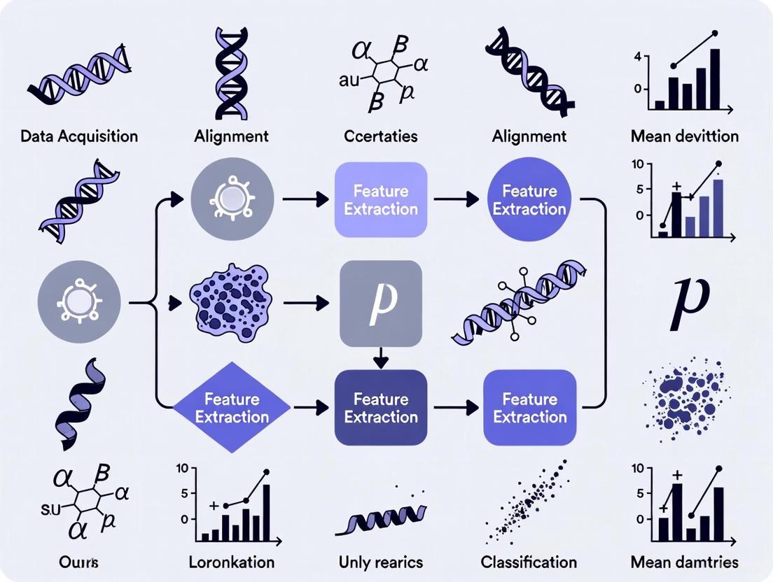
Diagram Title: Landmark-Free Morphometrics Pipeline
Mathematical Pathway of Diffeomorphic Mapping
Diagram Title: Mathematical Basis of Shape Mapping
Research Reagent Solutions
Table 2: Essential Computational Tools for Diffeomorphic Morphometrics
| Tool / Reagent | Function / Purpose | Application Note |
|---|---|---|
| FireANTs | A GPU-accelerated, multi-scale Adaptive Riemannian Optimization algorithm for fast, memory-efficient diffeomorphic image matching [5]. | Ideal for large-scale studies; runs ~1200x faster than ANTs on GPU; requires no training; generalizes across modalities and species [5]. |
| ANTs (Advanced Normalization Tools) | A well-established software ecosystem for biomedical image analysis, including robust implementations of LDDMM and atlas generation [3] [5]. | The benchmark for accuracy; can be slower than FireANTs. Suitable for standard-scale studies and method validation [3]. |
| Deterministic Atlas Analysis (DAA) | An LDDMM-based method for automated, landmark-free atlas construction and analysis [3]. | Applied in macroevolutionary studies across 180 mammalian families. Performance is enhanced by standardizing input data with Poisson surface reconstruction [3]. |
| Poisson Surface Reconstruction | An algorithm that creates unified, watertight 3D surface models from input data [3]. | Critical Preprocessing Step: Mitigates biases from mixed imaging modalities (CT vs. surface scans), ensuring robust downstream analysis [3]. |
Initial Momentum Vectors (m₀) |
The compact mathematical representation of a shape deformation relative to a template, obeying the conservation of momentum [4]. | Serves as the primary data for statistical shape analysis. Encodes the necessary information to reconstruct the entire deformation path (geodesic) [4]. |
Conceptual Framework and Core Workflows
Large Deformation Diffeomorphic Metric Mapping (LDDMM) is a computational framework that quantifies anatomical shape by modeling the smooth, reversible (diffeomorphic) transformations required to map one anatomical structure onto another. Unlike traditional landmark-based methods, it operates on the entire shape's geometry without requiring pre-defined homologous points. This makes it particularly valuable for comparing morphologically disparate taxa where identifiable homologous landmarks may be scarce [3] [6]. A specific application of this framework, Deterministic Atlas Analysis (DAA), leverages the LDDMM approach to iteratively compute a sample-specific mean shape, known as an atlas, and then quantifies the deformation of each specimen onto this atlas to analyze population-level shape variation [3] [7].
The following diagram illustrates the core workflow of the DAA pipeline, from data standardization to the final shape analysis.
Core Methodological Components
The LDDMM and DAA framework integrates several mathematical and procedural components to achieve a landmark-free shape analysis.
The LDDMM Mathematical Basis
LDDMM defines a space of shapes, M, and seeks optimal diffeomorphisms (φ) that transform an atlas template T to match a target shape S by minimizing a distance functional. The transformation is governed by a time-dependent velocity field v(t) that flows the template to the target, ensuring smooth and biologically plausible deformations [6]. The optimization minimizes an energy functional of the form:
E(φ) = ∫_0^1 ‖v(t)‖_V² dt + 1/σ² Sim[I(φ(1)), S]
Where ‖v(t)‖_V is the norm on the smooth velocity field, ensuring diffeomorphism, and Sim is a similarity measure between the deformed template and target shape [6].
Deterministic Atlas Analysis (DAA) Protocol
DAA implements LDDMM in a practical pipeline for analyzing large morphological datasets, as demonstrated in a macroevolutionary study of 322 mammalian crania spanning 180 families [3] [7]. The detailed protocol is as follows:
- Data Acquisition and Standardization: Acquire 3D anatomical data (e.g., CT or surface scans). A critical first step is to address mixed modalities using Poisson surface reconstruction to create watertight, closed surface meshes for all specimens, ensuring topological consistency for subsequent analysis [3] [7].
- Initial Template Selection: Manually or algorithmically select an initial template specimen from the dataset to begin atlas construction. The choice of template (e.g., Arctictis binturong in the mammalian study) can influence the number of control points generated but shows minimal impact on final shape correlations [7].
- Atlas Generation: The software (e.g., Deformetrica) iteratively computes the deterministic atlas—a geodesic mean shape of the entire dataset. This is achieved by minimizing the total deformation energy required to map the atlas onto every specimen in the dataset, making the results sample-dependent [7].
- Diffeomorphic Mapping and Control Point Generation: The framework calculates the diffeomorphic transformation (φ) that maps the atlas to each specimen. A kernel width parameter controls the spatial scale of deformation. Smaller kernel widths yield finer-scale deformations and a higher density of automatically generated control points that guide the shape comparison [7].
- Momenta Extraction: For each control point, a corresponding momentum vector ("momenta") is calculated. This vector represents the optimal deformation trajectory required to align the atlas with each specific specimen. The collection of momenta across all specimens forms the basis for shape comparison, replacing traditional landmark coordinates [7].
- Downstream Shape Analysis: The matrix of momenta is analyzed using techniques like kernel Principal Component Analysis (kPCA) to visualize major patterns of shape variation. These shape variables can then be used for macroevolutionary analyses, such as estimating phylogenetic signal, morphological disparity, and evolutionary rates [3] [7].
Quantitative Outcomes and Macroevolutionary Application
The application of DAA in large-scale evolutionary studies provides quantitative evidence of its performance and utility. The following table summarizes key quantitative findings from a benchmark study comparing DAA to traditional landmark-based geometric morphometrics on a dataset of 322 mammal crania [7].
Table 1: Summary of DAA Workflow Parameters and Macroevolutionary Analysis Outcomes from a Mammalian Crania Study (n=322 specimens)
| Workflow Stage | Parameter / Outcome | Quantitative Result / Observation | Biological / Analytical Implication |
|---|---|---|---|
| Data Standardization | Use of Poisson surface reconstruction | Significant improvement in correspondence between DAA and manual landmarking after standardization [7] | Crucial for harmonizing datasets from mixed imaging modalities (CT, surface scans) |
| Atlas Generation | Impact of initial template selection | Low overall impact on shape predictions (e.g., R²=0.957 between different templates) [7] | Enhances methodological robustness and reduces operator bias |
| Control Point Generation | Control points from A. binturong template (Kernel Width) | 20.0 mm: 270 points10.0 mm: 1,782 points [7] | Kernel width allows control over the resolution of shape capture |
| Method Correlation | Correlation with manual landmarking (Mantel/PROTEST) | Strong but not perfect correlation; differences noted in specific clades (Primates, Cetacea) [7] | DAA captures complementary aspects of shape variation |
| Downstream Analysis | Estimates of phylogenetic signal & evolutionary rates | Comparable but varying estimates between DAA and landmarking [7] | Useful for large-scale evolutionary hypothesis testing |
The Researcher's Toolkit for LDDMM/DAA
Implementing a landmark-free morphometric pipeline requires a suite of computational tools and reagents. The following table details the essential components.
Table 2: Essential Research Reagents and Computational Tools for LDDMM/DAA
| Tool / Resource | Type / Category | Specific Function in Workflow | Example Implementation / Note |
|---|---|---|---|
| Deformetrica | Software Platform | Implements the core DAA framework; performs atlas construction and diffeomorphic registration [7] | Used with a kernel width parameter to control the scale of deformation and density of control points [7] |
| Poisson Surface Reconstruction | Algorithm | Creates watertight, closed surfaces from input point clouds or open meshes [7] | Critical pre-processing step to standardize data from mixed modalities (CT, laser scan) [3] |
| Control Points | Data Representation | Automatically generated points that guide the non-rigid alignment of the atlas to each specimen [7] | Replace the need for manually defined homologous landmarks; number is determined by kernel width |
| Momenta Vectors | Data Output | Quantitative descriptors of the deformation needed to map the atlas to a target shape; the primary raw data for statistical analysis [7] | Represent shape in a high-dimensional space; can be analyzed with multivariate statistics like kPCA [7] |
| Kernel PCA (kPCA) | Statistical Method | Dimensionality reduction technique used to visualize and explore patterns of covariation in the momenta-based shape data [7] | Allows for the identification of major axes of shape variation in the dataset without predefined landmarks |
Resolving Morphological Comparisons Across Highly Disparate Taxa
Landmark-free morphometrics represents a paradigm shift in the quantitative analysis of biological form, enabling researchers to overcome long-standing limitations of traditional methods. While geometric morphometrics has been the cornerstone of shape analysis for decades, its reliance on manually placed anatomical landmarks makes it time-consuming, susceptible to operator bias, and particularly challenging when comparing morphologically divergent taxa [3]. The emergence of automated, landmark-free techniques offers a transformative approach for large-scale evolutionary studies and identification tasks across highly disparate organisms.
This application note details the implementation of one such landmark-free method—Deterministic Atlas Analysis (DAA), an application of Large Deformation Diffeomorphic Metric Mapping (LDDMM). We frame this within a broader research thesis that advocates for these methods to enable more comprehensive macroevolutionary analyses and accurate taxonomic identification across wide phylogenetic scales. The protocols below are designed for researchers investigating phenotypic evolution, comparative anatomy, and taxonomic relationships, with specific utility for professionals requiring robust morphological comparisons in evolutionary biology and palaeontology [3] [8].
Key Methodological Concepts and Comparative Framework
Core Conceptual Advancements
Landmark-free morphometrics addresses a critical bottleneck in large-scale morphological studies. Traditional geometric morphometrics (GMM) requires the identification of homologous landmarks—anatomically corresponding points across all specimens in an analysis. This process becomes infeasible or biologically meaningless when comparing organisms with vastly different body plans, such as mammals and insects, or even different anatomical structures within the same organism [3] [9]. Landmark-free methods circumvent this by modeling the entire shape as a deformable entity, quantifying differences without requiring point-to-point correspondence.
The Deterministic Atlas Analysis (DAA) method operates by computing diffeomorphic transformations—smooth, reversible mappings—that warp a reference shape (the "atlas") to match each specimen in the dataset. The amount of "warping energy" required represents the morphological distance between specimens. This approach captures continuous shape variation across entire surfaces, including details between traditional landmarks, potentially offering a more comprehensive characterization of form [3].
Quantitative Comparison with Traditional Geometric Morphometrics
Table 1: Comparative Analysis of Morphometric Methods for Disparate Taxa
| Analysis Criterion | Traditional Landmark-Based GMM | Landmark-Free DAA |
|---|---|---|
| Data Collection Efficiency | Low to moderate (manual/semi-automated landmarking) | High (automated surface processing) |
| Operator Bias | Susceptible due to landmark identification | Minimal after parameter initialization |
| Comparability Across Disparate Taxa | Limited by need for homologous points | High; does not require point correspondence |
| Data Modality Handling | Challenging with mixed imaging types | Can be standardized (e.g., Poisson surface reconstruction) |
| Captured Shape Information | Discrete landmarks only | Entire surface geometry |
| Downstream Macroevolutionary Metrics | Established protocols for disparity, rates | Produces comparable but varying estimates [3] |
Experimental Protocol: Landmark-Free Analysis Pipeline
Specimen Data Acquisition and Standardization
Objective: To assemble and standardize a 3D morphological dataset from potentially heterogeneous sources for robust landmark-free analysis.
Materials:
- 3D Surface Scans or CT Data: Raw data from specimens, which may include computed tomography (CT) scans or surface laser scans.
- Poisson Surface Reconstruction Software: Algorithm for creating watertight, closed surfaces from point cloud data (e.g., in MeshLab, CloudCompare).
- Computational Hardware: Workstation with sufficient RAM and GPU capabilities for handling large 3D meshes.
Procedure:
- Data Collection: Assemble 3D morphological data for all specimens. The dataset can include mixed modalities (e.g., CT scans, surface scans) but must be documented.
- Surface Reconstruction: Process all raw data through Poisson surface reconstruction. This critical step creates uniform, watertight, and closed 3D surfaces, standardizing the input for subsequent analysis [3].
- Note: This step mitigates artifacts arising from mixed imaging modalities, which was identified as an initial challenge in landmark-free applications.
- Data Verification: Visually inspect all reconstructed surfaces to ensure they are topologically sound (single, closed manifold) and accurately represent the original specimen morphology.
- File Format Conversion: Export all standardized surfaces in a consistent file format (e.g., .PLY, .VTK) compatible with the DAA software.
Shape Analysis via Deterministic Atlas Analysis (DAA)
Objective: To quantify morphological differences among all specimens in a standardized, automated framework without landmark placement.
Materials:
- DAA Software Implementation: Access to computational code implementing Large Deformation Diffeomorphic Metric Mapping (LDDMM), often available in specialized platforms (e.g., Python-based tools).
- High-Performance Computing Cluster: Recommended for datasets exceeding 100 specimens due to computational intensity.
Procedure:
- Atlas Selection: Choose a reference specimen ("atlas") from the dataset. This can be an average specimen or a representative individual. The choice may be iteratively refined.
- DAA Parameter Initialization: Set parameters for the diffeomorphic mapping, including the regularization weight, which controls the smoothness of the deformation.
- Diffeomorphic Registration: For each specimen in the dataset, run the DAA algorithm to compute the diffeomorphic transformation that deforms the atlas shape to match the target specimen's shape.
- Shape Distance Matrix Calculation: Extract the metric distance (warping cost) from each pairwise deformation, generating a symmetric N x N matrix of morphological distances for all N specimens.
The following workflow diagram illustrates the core steps of this landmark-free protocol:
Downstream Macroevolutionary Analysis
Objective: To translate the morphological distance matrix into biologically meaningful evolutionary patterns and metrics.
Materials:
- Statistical Software: R or Python with packages for multivariate statistics and phylogenetics (e.g.,
geomorphin R). - Phylogenetic Tree: A time-calibrated phylogeny of the taxa under study.
Procedure:
- Dimensionality Reduction: Input the morphological distance matrix into a Principal Coordinates Analysis (PCoA) to visualize the major axes of shape variation across taxa.
- Phylogenetic Signal Calculation: Quantify the degree to which morphological similarity is predicted by phylogenetic relatedness using metrics like Blomberg's K or Pagel's λ, applied to the distance matrix.
- Morphological Disparity Analysis: Partition morphological variance among taxonomic groups (e.g., families, orders) to assess differences in evolutionary diversification.
- Evolutionary Rate Estimation: Compare rates of morphological evolution across lineages using the distance matrix and the phylogenetic tree.
Table 2: Essential Computational Tools and Resources for Landmark-Free Morphometrics
| Tool/Resource | Primary Function | Application Context |
|---|---|---|
| Poisson Surface Reconstruction | Creates watertight 3D models from point clouds | Critical data standardization; enables analysis of mixed-modality data (CT, surface scans) [3] |
| DAA/LDDMM Software | Computes diffeomorphic mappings between shapes | Core analytical engine for quantifying shape differences without landmarks [3] |
| Supervised Classifiers (e.g., in MORPHIX) | Classifies specimens based on shape data | Provides more accurate taxonomic identification than PCA alone; useful for detecting novel taxa [8] |
R geomorph Package |
Comprehensive GMM analysis suite | Downstream statistical analysis of shape, including phylogenetic comparative methods [9] |
| Phylogenetic Comparative Methods | Analyzes trait evolution in a phylogenetic context | Quantifying phylogenetic signal, evolutionary rates, and morphological disparity from shape distances [3] [10] |
Critical Interpretation and Analytical Validation
Addressing the Limitations of Traditional Methods
The standard geometric morphometric workflow of Generalized Procrustes Analysis (GPA) followed by Principal Component Analysis (PCA) has recently faced scrutiny. When applied to highly disparate taxa, PCA can produce artifactual patterns highly dependent on the specific taxa included in the analysis [8]. Furthermore, the subjective interpretation of PCA scatterplots can lead to unstable taxonomic conclusions, as evidenced by debates in human evolution where different PC axes supported conflicting phylogenetic placements [8].
Landmark-free methods like DAA are not immune to these challenges but provide a more reproducible and automated framework. It is crucial to recognize that all morphometric analyses are sensitive to data quality and composition. Therefore, supervised machine learning classifiers are recommended as a complementary approach to improve classification accuracy and objectivity when identifying unknown specimens or proposing new taxa [8].
Validation and Best Practices
Data Quality Control: The adage "garbage in, garbage out" is particularly relevant. Meticulous data standardization via Poisson reconstruction is non-negotiable for reliable results [3]. Method Validation: For novel taxonomic identification, always cross-validate findings using supervised classification models alongside exploratory methods like PCoA [8]. Interpretation: Morphological similarity inferred from any morphometric method does not exclusively indicate phylogenetic relatedness; it can also reflect convergent evolution. Conclusions about taxonomy and evolutionary history should be drawn cautiously and integrated with other lines of evidence (e.g., genomic data) [3] [8].
Implementing the Pipeline: From Data Acquisition to Macroevolutionary Insight
Landmark-free morphometrics represents a paradigm shift in the quantitative analysis of biological shape, enabling researchers to capture and compare complex anatomical forms without the constraints of manual landmark placement. Traditional geometric morphometrics relies on the manual identification of homologous anatomical points, which is time-consuming, requires extensive training, and introduces operator bias [11]. This approach becomes particularly limiting when studying disparate taxa where homologous points are scarce or difficult to identify consistently [7]. Landmark-free methods address these limitations by utilizing entire 3D surfaces obtained from computed tomography (CT) or surface scanning technologies, capturing morphological information at a much higher resolution and enabling comparisons across broad phylogenetic scales [7] [12].
These advanced methodologies are transforming evolutionary biology, comparative anatomy, and developmental genetics by providing powerful tools to quantify subtle shape variations that were previously difficult to capture. The application of landmark-free approaches allows researchers to investigate fundamental questions about morphological evolution, phenotypic diversity, and the genetic basis of form across widely divergent species [11] [7]. This technical note establishes standardized protocols for handling 3D mesh data derived from CT and surface scans, ensuring reproducibility and comparability in landmark-free morphometric research.
Data Acquisition Standards
Imaging Modalities for 3D Data Capture
The foundation of landmark-free morphometrics lies in acquiring high-quality 3D representations of biological specimens. Different imaging modalities offer complementary advantages depending on research questions, specimen characteristics, and available resources.
Table 1: Comparison of 3D Data Acquisition Modalities for Morphometrics
| Modality | Resolution | Data Type | Primary Applications | Key Considerations |
|---|---|---|---|---|
| CT Scanning | High (sub-millimeter) | Volumetric data with density information | Both external and internal structures; skeletal morphology | Radiation exposure concerns; limited field-of-view possible [13] |
| Surface Scanning | Variable (mm to sub-mm) | Surface mesh only | External morphology; living subjects | Cannot capture internal structures; sensitive to surface properties |
| Micro-CT | Very high (micrometer) | Volumetric data | Detailed skeletal morphology; small specimens | High cost; limited to smaller specimens [11] |
| Structured Light Systems | High (sub-millimeter) | Surface mesh | External morphology at high resolution | Requires specific patterns; sensitive to lighting conditions [13] |
Clinical CT scanners typically used for larger specimens may encounter field-of-view limitations that fail to capture the full patient habitus, potentially impacting analytical accuracy [13]. In such cases, supplemental surface scanning can extend the captured anatomical information without additional radiation exposure. For studies focusing on external morphology, portable surface scanning systems such as iPad-based solutions with attached sensors (e.g., Structure Sensor) provide an accessible, low-cost alternative that can achieve spatial accuracy with mean distances under 1 mm when compared to CT-derived surfaces [13] [14].
Data Preprocessing and Standardization
Raw scan data requires careful preprocessing to ensure compatibility with landmark-free analytical pipelines. The standardization of mesh topology has been identified as a critical factor, particularly when combining datasets from different modalities (CT and surface scans) [7].
Mesh Processing Workflow:
- Data Conversion: Convert volumetric CT data to surface meshes using appropriate thresholding values to distinguish structures of interest from background [11].
- Noise Reduction: Apply mesh cleaning algorithms to remove artifacts and scanning noise while preserving biological signal.
- Mesh Repair: Identify and repair topological errors including non-manifold edges, self-intersections, and holes that may impede analysis.
- Modality Standardization: For mixed datasets, apply Poisson surface reconstruction to create watertight, closed meshes for all specimens, regardless of original modality [7].
- Decimation: Reduce mesh complexity through controlled decimation to optimize computational efficiency while preserving morphological details relevant to the research question.
The importance of modality standardization was demonstrated in a comprehensive study of 322 mammalian crania, where the use of Poisson surface reconstruction significantly improved correspondence between shape variation measured using manual landmarking and landmark-free methods [7]. This preprocessing step is particularly crucial for macroevolutionary analyses spanning disparate taxa where consistent mesh topology ensures comparable shape representations.
Landmark-Free Analytical Methods
Methodological Approaches
Landmark-free morphometrics encompasses several computational approaches that enable shape comparison without relying on predefined anatomical points.
Table 2: Landmark-Free Morphometric Methods for Disparate Taxa
| Method | Core Principle | Advantages | Limitations | Suitable Taxonomic Scale |
|---|---|---|---|---|
| Deterministic Atlas Analysis (DAA) | Uses diffeomorphic transformations to map specimens to a computed atlas shape [7] | No fixed template required; captures global and local shape variation | Performance varies across highly disparate groups; sensitive to parameters | Broad phylogenetic scales [7] |
| Generalized Procrustes Surface Analysis (GPSA) | Extends Iterative Closest Point algorithm for multiple surface superimposition [12] | Provides Procrustes-like distance metric; intuitive workflow | Requires good initial alignment; computational intensity | Closely related species to moderate disparateness |
| Iterative Closest Point (ICP) | Minimizes distances between surfaces through point correspondences [12] | Conceptually straightforward; widely implemented | Sensitive to initial positioning; may converge to local minima | Intraspecific to closely related species |
| Dense Correspondence Analysis | Establences point-to-point correspondence across surfaces using surface descriptors | High-resolution shape capture; detailed local comparisons | Computationally demanding; requires surface parameterization | Moderate taxonomic scales |
Deterministic Atlas Analysis (DAA) has demonstrated particular utility for broad taxonomic comparisons, as it iteratively estimates an optimal atlas shape by minimizing the total deformation energy needed to map it onto all specimens in a dataset [7]. This approach generates control points that guide shape comparison without requiring homologous landmarks, making it suitable for analyzing morphological variation across diverse taxa where traditional landmarks become scarce.
Implementation Protocols
Protocol: Deterministic Atlas Analysis for Disparate Taxa
Purpose: To quantify shape variation across phylogenetically divergent specimens using a landmark-free approach.
Materials and Software:
- 3D surface meshes of all specimens (standardized format)
- Deformetrica software or equivalent implementation
- Computational resources adequate for dataset size
Procedure:
- Initial Template Selection:
- Select an initial template specimen that represents a morphological intermediate within the dataset
- Avoid extreme morphological forms as initial templates to minimize bias
- For mammalian crania studies, templates such as Arctictis binturong have proven effective [7]
Atlas Generation:
- Set kernel width parameter to control spatial extent of deformations
- Smaller kernel widths (e.g., 10.0 mm) yield finer-scale deformations with more control points
- Larger kernel widths (e.g., 40.0 mm) produce broader-scale deformations with fewer control points
- Iteratively estimate optimal atlas shape through geodesic registration
Specimen Registration:
- Compute deformation fields mapping atlas to each specimen
- Calculate momentum vectors ("momenta") at control points for each specimen
- These vectors represent optimal deformation trajectories for atlas-specimen alignment
Shape Variation Analysis:
- Perform kernel Principal Component Analysis (kPCA) on momentum vectors
- Visualize and explore covariation in shape data
- Generate heatmaps based on thin-plate spline deformations to localize shape differences
Validation:
- Compare results with traditional landmark-based methods using Procrustes distance correlations
- Assess morphological disparity and evolutionary rate estimates against established methods
- Evaluate phylogenetic signal recovery compared to landmark-based approaches [7]
Research Reagent Solutions
The implementation of landmark-free morphometrics requires specific computational tools and resources. The following table outlines essential solutions for establishing an analytical pipeline.
Table 3: Essential Research Reagents for Landmark-Free Morphometrics
| Reagent/Tool | Type | Primary Function | Application Notes |
|---|---|---|---|
| Deformetrica | Software | Deterministic Atlas Analysis implementation | Enables DAA for disparate taxa; open-source availability [7] |
| DICOMator | Software | Converts 3D meshes to synthetic DICOM CT images | Facilitates use of mesh data with medical imaging workflows [15] |
| 3D Slicer | Software | 3D mesh processing and analysis | Open-source platform for medical image visualization and processing [13] |
| Blender | Software | 3D modeling and mesh manipulation | Open-source; extensible via Python API for custom pipelines [15] |
| Structure Sensor | Hardware | Portable 3D surface scanning | Mobile solution for surface capture; ~1 mm accuracy [13] |
| Poisson Reconstruction | Algorithm | Creates watertight meshes from point clouds | Critical for standardizing mixed-modality datasets [7] |
| Iterative Closest Point | Algorithm | Surface registration and alignment | Foundation for GPSA and other surface comparison methods [12] |
Workflow Visualization
Figure 1: Landmark-Free Morphometrics Workflow for Disparate Taxa. This pipeline integrates data from multiple imaging modalities through standardized processing, enabling shape comparison across phylogenetically diverse specimens.
Figure 2: Method Comparison: Traditional vs. Landmark-Free Approaches. Landmark-free methods overcome key limitations of traditional morphometrics, particularly for studies encompassing phylogenetically disparate taxa where homologous landmarks are scarce.
Applications and Validation
Biological Applications
Landmark-free morphometrics has enabled novel insights across evolutionary biology, particularly for research questions spanning broad phylogenetic scales:
Macroevolutionary Analyses: Landmark-free methods successfully capture shape variation across 322 mammalian species spanning 180 families, demonstrating their utility for investigating deep-time evolutionary patterns [7]. These approaches reveal patterns of morphological disparity and evolutionary rates that are comparable to, yet distinct from, those derived from landmark-based methods.
Craniofacial Phenotyping: In mouse models of Down syndrome (Dp1Tyb), landmark-free analysis identified cranial dysmorphologies including smaller size and brachycephaly, homologous to human phenotypes [11]. The method provided finer mapping of local differences in mid-snout structures and occipital bones that were not apparent using traditional landmark-based approaches.
Morphological Integration and Modularity: The dense sampling of shape information enables sophisticated analyses of how different anatomical regions co-vary across evolutionary lineages, particularly valuable for understanding how developmental processes constrain or facilitate evolutionary change.
Validation and Quality Control
Rigorous validation ensures that landmark-free methods produce biologically meaningful results:
Spatial Accuracy Assessment: Compare surface scans against CT-derived surfaces using distance metrics. Studies report mean distances under 1 mm between CT surfaces and 3D scans when using appropriate scanning protocols [14].
Methodological Correlation: Evaluate correspondence between landmark-free and traditional morphometric results using Procrustes distance correlations and matrix comparison tests (e.g., Mantel test, PROTEST) [7].
Parameter Sensitivity Analysis: Assess the impact of analytical parameters (e.g., kernel width in DAA) on resulting shape spaces and biological interpretations.
Phylogenetic Signal Evaluation: Compare estimates of phylogenetic signal (e.g., Kmult) derived from landmark-free methods against those from landmark-based approaches to ensure evolutionary patterns are adequately captured.
When properly validated, landmark-free methods demonstrate strong concordance with traditional approaches while providing enhanced resolution and greater efficiency for analyzing morphological diversity across disparate taxa [11] [7]. This validation framework ensures that researchers can adopt these advanced methodologies with confidence in their biological relevance.
Step-by-Step Guide to Automated Shape Capture and Correspondence
Landmark-free morphometrics represents a paradigm shift in quantitative shape analysis, addressing critical limitations of traditional landmark-based methods. While geometric morphometrics has been the gold standard for evolutionary biology studies, it relies on manual placement of landmarks—a process that is time-consuming, susceptible to operator bias, and limits comparisons across morphologically disparate taxa where homologous points become obscure [7]. Landmark-free techniques overcome these constraints by capturing shape variation without relying solely on homologous landmarks, enabling researchers to analyze larger and more diverse datasets with enhanced efficiency and resolution [7] [11].
Within the context of identification across disparate taxa, landmark-free approaches are particularly valuable as they allow comparison of anatomical structures that may share limited homologous points due to evolutionary divergence. These methods capture comprehensive shape data that can reveal subtle phenotypic relationships across broad phylogenetic scales, making them indispensable for modern macroevolutionary studies and comparative anatomy research [7].
Theoretical Foundation
Key Concepts and Terminology
Deterministic Atlas Analysis (DAA): A landmark-free approach based on Large Deformation Diffeomorphic Metric Mapping (LDDMM) that quantifies shape variation by computing deformations required to map a dynamically generated mean shape (atlas) onto each specimen in a dataset [7].
Atlas Generation: The process of creating a geodesic mean shape that represents the dataset under study. Unlike methods using a fixed template, DAA iteratively estimates the optimal atlas shape by minimizing the total deformation energy needed to map it onto all specimens [7].
Control Points: Reference points generated during DAA that are initially evenly distributed within the ambient space surrounding the atlas but adjust to fit areas with greater variability. These guide shape comparison without requiring standard landmarks [7].
Momenta Vectors: Mathematical representations of the optimal deformation trajectory for aligning the atlas with each specimen. These vectors provide the basis for directly comparing shape variation across specimens [7].
Comparative Framework: Landmark-Based vs. Landmark-Free Methods
Table 1: Comparison of Morphometric Approaches
| Feature | Traditional Landmark-Based | Landmark-Free (DAA) |
|---|---|---|
| Data Collection | Manual/semi-automated landmark placement | Automated shape capture and correspondence |
| Processing Time | Time-consuming (hours to days) | Efficient (minutes to hours) |
| Operator Bias | Susceptible to inter-operator variability | Minimal human intervention |
| Homology Requirement | Dependent on identifiable homologous points | Does not rely solely on homology |
| Taxonomic Scope | Limited for disparate taxa | Suitable for broad phylogenetic comparisons |
| Resolution | Limited by landmark number | High-resolution with comprehensive coverage |
| Data Output | Landmark coordinates | Deformation fields and momenta vectors |
| Macroevolutionary Application | Challenging for highly divergent forms | Suitable for cross-taxa analyses |
Experimental Protocols
Specimen Preparation and Imaging
Protocol 3.1.1: Standardized Image Acquisition
Specimen Selection: Curate a representative dataset spanning the taxonomic range of interest. For mammalian cranial studies, this may include 180+ families to ensure adequate morphological diversity [7].
Imaging Modalities: Utilize high-resolution imaging techniques appropriate for your specimens:
- Micro-computed tomography (μCT): For detailed internal and external structures of hard tissues [11]
- Surface scanning: For external morphology capture
- Clinical CT: For larger specimens where internal structures are required
Resolution Standardization: Set consistent resolution parameters across all specimens to ensure comparable data. For cranial studies of small mammals, 20-50μm voxel size provides sufficient detail.
Quality Control: Verify image quality through contrast-to-noise ratio measurements and ensure complete coverage of anatomical structures of interest.
Protocol 3.1.2: Handling Mixed Modalities
When combining data from different imaging sources (e.g., CT and surface scans), employ Poisson surface reconstruction to create watertight, closed meshes for all specimens. This standardization significantly improves correspondence between shape variation patterns measured using different methods [7].
Surface Mesh Processing Pipeline
Protocol 3.2.1: Mesh Generation and Refinement
Thresholding: Apply appropriate thresholds to extract anatomical structures from raw image data [11].
Cartilage Removal: For skeletal studies, digitally remove cartilaginous structures to isolate bony elements [11].
Segmentation: Use bone density differences to separate anatomical units (e.g., cranium from mandible) [11].
Mesh Generation: Create triangulated meshes from surfaces, including internal structures where relevant [11].
Mesh Decimation: Reduce mesh complexity while preserving morphological details through controlled decimation.
Mesh Cleaning: Remove non-manifold edges, self-intersections, and topological errors.
Alignment: Spatially align all meshes to a common coordinate system using Procrustes superimposition or other registration techniques [11].
Deterministic Atlas Analysis (DAA) Implementation
Protocol 3.3.1: Initial Template Selection
Template Criteria: Select an initial template specimen that represents intermediate morphology within your dataset rather than extreme forms [7].
Evaluation Method: Test multiple initial templates based on preliminary morphological assessments (e.g., from principal component analysis of traditional landmarks) [7].
Validation: Verify template selection by ensuring it generates an appropriate number of control points (e.g., 270 for mammalian crania with 20.0 mm kernel width) [7].
Bias Mitigation: Avoid templates that cluster with morphological extremes, as they may be artificially drawn toward the center of morphospace in subsequent analyses [7].
Protocol 3.3.2: Atlas Generation and Deformation Mapping
Atlas Computation: Implement iterative atlas generation using software such as Deformetrica to compute the optimal mean shape representing your dataset [7].
Kernel Width Parameterization: Test multiple kernel widths (e.g., 10.0 mm, 20.0 mm, 40.0 mm) to determine the optimal spatial extent for deformation mapping. Smaller values yield finer-scale deformations [7].
Control Point Generation: Allow the algorithm to automatically generate control points based on the kernel width and morphological variability [7].
Momenta Calculation: Compute momentum vectors for each specimen representing the deformation trajectory required to align the atlas with each specimen [7].
Data Analysis and Validation
Protocol 3.4.1: Method Comparison and Validation
Correlation Assessment: Compare shape matrices from landmark-free and traditional methods using:
Shape Visualization: Generate heatmaps based on thin-plate spline deformations and Euclidean distance measures to identify how shape is captured differently by each method [7].
Downstream Analysis: Evaluate the impact of method choice on macroevolutionary analyses including:
- Phylogenetic signal estimation
- Morphological disparity
- Evolutionary rates [7]
Research Reagent Solutions
Table 2: Essential Research Tools for Landmark-Free Morphometrics
| Tool Category | Specific Software/Solutions | Function | Application Context |
|---|---|---|---|
| Image Processing | Deformetrica [7] | DAA implementation | Shape correspondence and atlas generation |
| Mesh Processing | Poisson Surface Reconstruction [7] | Mesh standardization | Creating watertight surfaces from mixed modalities |
| Shape Analysis | Kernel Principal Component Analysis (kPCA) [7] | Dimensionality reduction | Visualizing and exploring shape covariation |
| Statistical Validation | PROTEST [7] | Method comparison | Assessing correlation between shape matrices |
| 3D Visualization | Mesh visualization tools | Results interpretation | Exploring shape differences and patterns |
| Data Integration | Custom scripting (Python/R) | Pipeline automation | Connecting different analytical steps |
Workflow Visualization
Figure 1: Comprehensive workflow for automated shape capture and correspondence analysis, showing multiple entry points for different imaging modalities and key processing stages.
Analytical Framework
Shape Data Processing
Protocol 6.1.1: Shape Variable Extraction
Momenta Processing: Extract momenta vectors from DAA output for statistical analysis [7].
Dimensionality Reduction: Apply kernel Principal Component Analysis (kPCA) to visualize and explore covariation in momenta-based shape data [7].
Matrix Preparation: Prepare shape matrices for comparative analysis with traditional landmark data.
Methodological Validation
Protocol 6.2.1: Quantitative Comparison
Correlation Analysis: Assess correspondence between landmark-free and traditional methods using matrix correlation techniques [7].
Localization Assessment: Identify anatomical regions where methods differ in shape capture using Euclidean distance measures and deformation-based heatmaps [7].
Taxonomic Specificity: Evaluate method performance across different taxonomic groups, noting potential variations (e.g., in Primates and Cetacea) [7].
Macroevolutionary Applications
Protocol 6.3.1: Evolutionary Analysis
Phylogenetic Signal: Estimate phylogenetic signal using both landmark-free and traditional shape data to assess methodological impacts [7].
Disparity Analysis: Calculate morphological disparity across taxa using Procrustes variance or equivalent metrics [7].
Evolutionary Rates: Compare rates of evolution across lineages using both approaches to identify potential biases [7].
Implementation Considerations
Technical Specifications
Table 3: Technical Parameters for Landmark-Free Morphometrics
| Parameter | Specification | Impact on Analysis |
|---|---|---|
| Kernel Width | 10.0-40.0 mm (mammalian crania) | Determines spatial scale of deformations [7] |
| Control Points | 45-1,782 points (depending on kernel) | Influences resolution of shape capture [7] |
| Mesh Resolution | 50,000-500,000 faces | Balances detail and computational load |
| Dataset Size | 322+ specimens (for broad taxonomic coverage) | Affects atlas stability and statistical power [7] |
| Computational Requirements | High-performance computing recommended | Impacts processing time for large datasets |
Troubleshooting and Optimization
Challenge: Mixed Modality Integration Solution: Implement Poisson surface reconstruction to create consistent, watertight meshes from different imaging sources, significantly improving correspondence between shape patterns [7].
Challenge: Template Selection Bias Solution: Select intermediate morphologies as initial templates and verify they don't artificially shift toward morphospace centers in analysis [7].
Challenge: Parameter Sensitivity Solution: Systematically test kernel width parameters and evaluate their impact on control point generation and subsequent biological interpretations [7].
Landmark-free morphometrics, particularly Deterministic Atlas Analysis, provides a powerful framework for automated shape capture and correspondence across disparate taxa. By overcoming the limitations of traditional landmark-based methods, these approaches enable researchers to conduct large-scale macroevolutionary analyses with enhanced efficiency and resolution. The protocols outlined in this guide provide a comprehensive foundation for implementing these cutting-edge techniques in evolutionary biology and comparative anatomy research.
Landmark-free morphometrics represents a paradigm shift in the quantitative analysis of biological shape, enabling researchers to overcome longstanding limitations of traditional landmark-based methods. By capturing comprehensive shape data without relying on predefined homologous points, these techniques allow for comparisons across highly disparate taxa and facilitate the analysis of larger, more diverse datasets [7]. This application note details specific protocols and case studies demonstrating the practical utility of landmark-free approaches in two distinct domains: mammalian cranial evolution and insect pest identification. The documented methodologies provide researchers with robust frameworks for implementing these analyses in their own taxonomic investigations.
Case Study 1: Macroevolutionary Analysis of Mammalian Crania
Experimental Aims and Rationale
This case study applied a landmark-free approach to investigate cranial evolution across 322 placental mammals spanning 180 families, with the goal of testing whether automated methods could produce comparable results to traditional geometric morphometrics in large-scale macroevolutionary analyses [7]. The primary research question centered on whether landmark-free methods could reliably capture shape variation across phylogenetically disparate taxa where homologous landmarks become increasingly difficult to identify and quantify.
Protocol: Deterministic Atlas Analysis (DAA)
Software Requirements: Deformetrica software platform implementing Large Deformation Diffeomorphic Metric Mapping (LDDMM)
Specimen Preparation and Imaging:
- Data Acquisition: Obtain 3D cranial meshes using computed tomography (CT) or surface scanning
- Modality Standardization: Apply Poisson surface reconstruction to create watertight, closed surfaces, converting all specimens to consistent mesh topology [7]
- Data Quality Control: Visually inspect all meshes for completeness and proper reconstruction
Atlas Generation and Template Selection:
- Initial Template Testing: Evaluate multiple initial templates (e.g., Arctictis binturong, Cacajao calvus, Schizodelphis morckhoviensis) based on morphological extremes
- Template Selection Criteria: Choose template that minimizes clustering artifacts in preliminary analyses
- Control Point Generation: Allow software to automatically generate control points guided by kernel width parameter (typically 10.0mm, 20.0mm, or 40.0mm) [7]
Shape Correspondence and Analysis:
- Geodesic Registration: Perform iterative estimation of optimal atlas shape by minimizing total deformation energy required to map onto all specimens
- Momentum Calculation: Compute momentum vectors ("momenta") for each control point, representing optimal deformation trajectory for atlas-to-specimen alignment [7]
- Shape Space Exploration: Apply kernel principal component analysis (kPCA) to visualize and explore covariation in momenta-based shape data
Downstream Macroevolutionary Analyses:
- Phylogenetic Signal: Compare estimates of phylogenetic signal with those derived from manual landmarking
- Morphological Disparity: Calculate morphological disparity metrics across taxonomic groups
- Evolutionary Rates: Estimate evolutionary rates and compare with traditional morphometric approaches [7]
Key Experimental Parameters
Table 1: Key Parameters for DAA in Mammalian Cranial Analysis
| Parameter | Settings/Specifications | Impact on Analysis |
|---|---|---|
| Kernel Width | 10.0mm, 20.0mm, 40.0mm | Determines spatial extent of deformations and number of control points |
| Initial Template | Arctictis binturong (selected), Cacajao calvus, Schizodelphis morckhoviensis | Minimal overall impact on shape predictions but affects control point distribution |
| Control Points | 45 (40mm), 270 (20mm), 1,782 (10mm) | Higher density captures finer-scale shape variation |
| Specimen Count | 322 specimens, 180 families | Provides broad taxonomic coverage for method validation |
| Mesh Standardization | Poisson surface reconstruction | Critical for analyzing mixed-modality datasets (CT + surface scans) |
Workflow Visualization
Figure 1: Deterministic Atlas Analysis (DAA) workflow for landmark-free mammalian cranial analysis
Results and Interpretation
The DAA approach successfully captured cranial shape variation across the 322 mammalian specimens, with results significantly correlating with those obtained through manual landmarking after mesh standardization [7]. The method demonstrated particular utility for broad taxonomic comparisons where homologous landmarks are limited. Differences emerged in specific clades (Primates and Cetacea), highlighting the importance of validating automated approaches against traditional methods during initial implementation. Both phylogenetic signal and morphological disparity metrics were generally comparable between methods, supporting the use of landmark-free approaches for large-scale evolutionary questions.
Case Study 2: Insect Pest Identification Using Wing Morphometrics
Experimental Aims and Rationale
This case study applied landmark-based geometric morphometrics to address practical challenges in insect identification, focusing on discriminating between closely related pest species where morphological differences are subtle and traditional taxonomy requires expertise [16] [17]. The research aimed to develop a standardized protocol that could supplement or partially replace molecular methods for species identification in field settings with limited resources.
Protocol: Wing Geometric Morphometrics (WGM)
Software Requirements: TpsUtil, TpsDig2, MorphoJ; R with geomorph package as alternative
Specimen Preparation and Imaging:
- Specimen Collection: Capture insects using appropriate trapping methods (Nzi traps for biting flies, funnel traps for blow flies) [16] [17]
- Wing Removal: Carefully remove right wing using fine forceps
- Slide Mounting: Place wing on microscope slide with Permount Mounting Medium, submerge in xylene to eliminate bubbles, cover with coverslip
- Digital Imaging: Photograph mounted wings using digital camera attached to stereomicroscope at 1.5× magnification [16]
Landmark Digitization Protocol:
- Landmark Selection: Digitize 19 biologically homologous landmarks based on wing venation patterns (consistent with Hall et al. protocol) [16]
- Replication Strategy: Digitize each wing twice to assess and minimize measurement error
- File Management: Build TPS files using TpsUtil software for organized data management
Data Processing and Analysis:
- Procrustes Superimposition: Align raw landmark coordinates using Generalized Procrustes Analysis (GPA) to remove non-shape variation (position, orientation, scale)
- Size Calculation: Compute centroid size as square root of sum of squared distances from landmark configuration center to each landmark [16]
- Statistical Analysis:
- Canonical Variate Analysis (CVA): Identify features that best discriminate between groups
- Cross-Validation Test: Assess classification accuracy using discriminant function analysis
- Allometry Assessment: Test for size-dependent shape variation using multivariate regression of shape on centroid size [16]
Machine Learning Integration (Advanced Protocol):
- Classifier Selection: Implement Support Vector Machine (SVM) with linear, polynomial, and radial kernels; Artificial Neural Networks (ANN) [18]
- Model Training: Optimize hyperparameters (e.g., SVM cost parameter, ANN nodes and decay) using cross-validation
- Performance Validation: Compare model accuracy against "no-information rate" using appropriate statistical tests [18]
Key Experimental Parameters
Table 2: Key Parameters for Wing Morphometrics in Insect Identification
| Parameter | Settings/Specifications | Impact on Analysis |
|---|---|---|
| Landmarks | 19 Type II landmarks on wing venation | Must be biologically homologous across species |
| Sample Size | 372 blow flies (12 species); 140 Haematobosca flies | Sufficient statistical power for species discrimination |
| Imaging Magnification | 1.5× on stereomicroscope | Consistent resolution across specimens |
| Statistical Tests | CVA, DFA with cross-validation, Procrustes ANOVA | Determines discriminatory power and significance |
| Machine Learning | SVM (optimal), ANN | Enhanced classification accuracy for complex datasets |
Workflow Visualization
Figure 2: Wing geometric morphometrics workflow for insect species identification
Results and Interpretation
The wing morphometrics approach demonstrated high effectiveness in discriminating between closely related insect species. For blow flies, wing shape provided reliable discrimination at both genus and species levels, particularly for Chrysomya species, though it was less robust for Lucilia and Hemipyrellia [16]. For Haematobosca flies, the method achieved 99.3% accuracy in distinguishing H. sanguinolenta from H. aberrans based on wing shape alone [17]. Machine learning implementations, particularly SVM models, showed predictive accuracy >95%, significantly outperforming traditional random forest and k-nearest neighbor classifiers [18].
The Scientist's Toolkit: Essential Research Reagents and Materials
Table 3: Essential Research Reagents and Materials for Morphometric Analyses
| Item | Specification/Type | Function/Application |
|---|---|---|
| Imaging Equipment | Micro-CT scanner or surface scanner | 3D data acquisition for mammalian specimens |
| Microscopy System | Stereomicroscope with digital camera | 2D wing imaging for insect morphometrics |
| Specialized Software | Deformetrica, MorphoJ, TpsDig2, R | Data processing and statistical analysis |
| Mounting Medium | Permount Mounting Medium | Wing preservation and slide preparation |
| Chemical Solvents | Xylene | Bubble elimination in wing mounting |
| Specimen Traps | Nzi traps, funnel traps | Standardized insect collection |
| Reference Specimens | Voucher specimens with expert ID | Method validation and calibration |
The quantitative analysis of biological shape is fundamental to evolutionary biology, taxonomy, and paleontology. For decades, geometric morphometrics (GMM), based on the manual placement of homologous landmarks, has been the gold standard for capturing shape variation [19]. However, this approach is time-consuming, susceptible to operator bias, and its reliance on homology limits its application across highly disparate taxonomic groups where homologous points are obscure [19] [20]. These limitations become critical when scaling analyses to leverage large, modern 3D image datasets.
Landmark-free morphometrics represents a paradigm shift, offering automated, high-throughput methods for capturing shape without the constraints of manual landmarking. Techniques such as Large Deformation Diffeomorphic Metric Mapping (LDDMM) and its application, Deterministic Atlas Analysis (DAA), quantify shape by computing the deformation energy required to map a sample-derived atlas shape onto each specimen in a dataset [19]. This landmark-free approach promises enhanced efficiency and the ability to compare more morphologically diverse taxa. This protocol details the application of these novel shape data to two core evolutionary analyses: phylogenetics and morphological disparity.
Application Notes: Linking Shape to Evolutionary Hypotheses
Shape Data in Phylogenetic Inference
Integrating morphological data into phylogenetic analysis is crucial for incorporating fossil taxa into the tree of life. While traditional methods use discrete characters, continuous shape data from landmark-free methods offer a more objective and quantitative alternative [20].
- Performance Considerations: A systematic review found that phylogenies reconstructed from continuous morphometric data, including GMM, did not consistently show increased resolution or accuracy compared to those from discrete characters when benchmarked against molecular phylogenies [20]. This highlights the need for careful methodological choices rather than an assumption of superior performance.
- Recommended Analytical Frameworks:
- Bayesian Methods: These are particularly promising for handling continuous morphometric data. Models can explicitly account for the correlation between landmarks (or points) and integrate shape data with molecular and discrete morphological data in a total-evidence analysis [20]. Methods also exist for placing multiple fossil taxa onto a scaffold phylogeny of extant species using continuous traits [20].
- Parsimony and Distance-Based Methods: Landmark analysis under parsimony (LAUP) and neighbor-joining trees based on Procrustes distances are used [20]. However, these methods have been criticized for not adequately modeling trait evolution and may be less reliable than model-based approaches [20].
Assessing Morphological Disparity and Evolutionary Rates
Morphological disparity quantifies the variety of form within a group, and evolutionary rates measure the pace of morphological change over time. Landmark-free methods are highly suited for these macroevolutionary analyses as they capture comprehensive shape variation [19].
- Utility of Landmark-Free Data: Studies comparing DAA with traditional landmarking found that both methods produced comparable but varying estimates of phylogenetic signal, morphological disparity, and evolutionary rates [19]. This confirms the potential of landmark-free approaches for large-scale disparity studies across disparate taxa.
- Critical Parameter: The kernel width parameter in DAA significantly influences results. A smaller kernel width captures finer-scale shape variations (generating more control points) and can affect downstream macroevolutionary metrics [19]. The choice of template for atlas generation, however, has a minimal overall impact if the template is chosen judiciously [19].
Experimental Protocols
Protocol 1: Deterministic Atlas Analysis (DAA) for Shape Capture
This protocol adapts the DAA pipeline, implemented in software like Deformetrica, for capturing cranial shape across highly disparate mammalian taxa [19].
I. Specimen and Data Preparation
- Input: A dataset of 3D meshes from computed tomography (CT) or surface scans. Mixed modalities (CT and surface scans) can introduce bias and must be standardized [19].
- Standardization Step: Use Poisson surface reconstruction to create watertight, closed surfaces for all specimens, which mitigates issues arising from mixed modalities [19].
- Software Recommendation: Deformetrica [19].
II. Atlas Generation and Shape Capture
- Initial Template Selection: Select an initial template mesh for the atlas generation process. While the choice has minimal impact on overall results, avoid templates that are morphological outliers. An initial template close to the sample mean is recommended [19].
- Set Kernel Width Parameter: Choose a kernel width (e.g., 10.0 mm, 20.0 mm, 40.0 mm). This controls the spatial scale of deformation and the number of control points. Smaller values capture more localized shape changes [19].
- Run Atlas Generation: The software iteratively estimates an optimal atlas shape (a geodesic mean shape) by minimizing the total deformation energy needed to map it onto all specimens [19].
- Compute Deformations: For each specimen, the software calculates a deformation that maps the atlas onto it. This deformation is guided by control points and quantified by momentum vectors ("momenta") [19].
- Output: The momentum vectors for all specimens serve as the basis for shape comparison and downstream analysis [19].
Protocol 2: Phylogenetic Analysis with Continuous Shape Data
This protocol outlines a Bayesian approach for integrating landmark-free shape data into phylogenetic inference.
I. Data Preprocessing
- Input: Momentum vectors from DAA or Procrustes-aligned coordinates from traditional GMM.
- Data Reduction (Optional): If using a high-dimensional shape descriptor (e.g., many momentum vectors), a dimension reduction technique like Principal Component Analysis (PCA) can be applied. The resulting principal component scores can be used as continuous characters [20]. Note: The use of PC scores has been debated [20].
II. Phylogenetic Inference
- Software Selection: Use a Bayesian inference software package capable of handling continuous traits (e.g., RevBayes, BEAST).
- Model Selection: Apply a multivariate Brownian motion model to model the evolution of the continuous shape characters. For shape data, models that account for trait covariation are advantageous [20].
- Analysis Setup:
- For a total-evidence analysis, combine the continuous shape data with molecular sequence data and/or discrete morphological data [20].
- For fossil placement, use methods designed to estimate the positions of fossil taxa on a fixed scaffold phylogeny of extant taxa using continuous character data [20].
- Run Analysis: Execute the Markov Chain Monte Carlo (MCMC) analysis with appropriate chain length and convergence diagnostics.
Table 1: Impact of Kernel Width in Deterministic Atlas Analysis (DAA)
| Kernel Width | Number of Control Points Generated | Scale of Shape Variation Captured | Influence on Downstream Analyses |
|---|---|---|---|
| 10.0 mm | 1,782 | Fine-scale, localized | Higher resolution, may affect estimates of disparity and evolutionary rates |
| 20.0 mm | 270 | Intermediate | Balanced perspective |
| 40.0 mm | 45 | Broad-scale, global | Coarse-grained perspective |
Table 2: Comparison of Morphometric Methods for Phylogenetic Analysis
| Method | Key Advantage | Key Challenge/Limitation | Suitability for Disparate Taxa |
|---|---|---|---|
| Traditional Landmarking | Well-established, biologically homologous data | Time-consuming, operator bias, limited homologous points | Low |
| Landmark-Free (e.g., DAA) | Automated, high-throughput, homology-free | Correlation structure of data must be modeled; emerging method | High |
| Discrete Characters | Standard for morphological phylogenetics | Subjectivity in character coding; information loss when discretizing continuous data | Variable |
The Scientist's Toolkit: Research Reagent Solutions
Table 3: Essential Tools for Landmark-Free Morphometric Analysis
| Research Reagent / Tool | Function / Application | Protocol Section |
|---|---|---|
| Deformetrica Software | Implements the Deterministic Atlas Analysis (DAA) framework for landmark-free shape comparison. | 3.1 |
| Poisson Surface Reconstruction Algorithm | Creates watertight, closed 3D meshes from scan data; critical for standardizing mixed-modality datasets. | 3.1 |
| 3D Slicer / ITK-SNAP | Open-source software for image segmentation and 3D mesh generation from CT/MRI data. | 3.1 |
| R (geomorph package) | Statistical analysis of shape, including Procrustes ANOVA, phylogenetic signal testing, and disparity calculations. | 2.2 |
| RevBayes / BEAST | Software for Bayesian phylogenetic inference, supporting the use of continuous trait models. | 3.2 |
| Large Deformation Diffeomorphic Metric Mapping (LDDMM) | The underlying mathematical framework for quantifying deformations between shapes in DAA. | 3.1 |
Workflow Visualization
Navigating Challenges: Data Standardization and Parameter Optimization
In the evolving field of landmark-free morphometrics, researchers are increasingly leveraging three-dimensional digital data to quantify phenotypic shape across disparate taxa. Such large-scale macroevolutionary studies often combine specimens from various sources, leading to a common challenge: mixed modality data obtained from both computed tomography (CT) scans and surface scanners [3]. This heterogeneity in data acquisition can introduce significant biases and artifacts, potentially compromising downstream biological analyses. Poisson Surface Reconstruction (PSR) offers a robust computational solution to this problem by generating standardized, watertight, closed surfaces from oriented point clouds, thereby enabling meaningful cross-taxa comparisons [3] [21]. This application note details the implementation of PSR as a critical preprocessing step for landmark-free morphometric analyses, providing validated protocols and quantitative assessments to guide researchers in handling mixed-modality datasets effectively.
Background
The Challenge of Mixed Modalities in Morphometrics
Traditional geometric morphometrics relies on manually placed anatomical landmarks, a process that is not only time-consuming but also susceptible to operator bias, particularly when comparing morphologically divergent taxa [3]. Landmark-free approaches promise to overcome these limitations by automating shape quantification, thus enabling analyses of larger and more phylogenetically diverse datasets [3]. However, the practical implementation of these methods faces a fundamental data standardization issue. Research datasets often comprise specimens scanned using different technologies—CT scanners that capture internal structures and surface scanners that record external morphology. These modalities produce fundamentally different data structures: CT-derived point clouds often represent watertight volumes, while surface scans typically yield open surfaces with potential holes and inconsistencies [3] [22]. When applying landmark-free methods like Deterministic Atlas Analysis without standardization, these structural differences can lead to artifacts in shape quantification, especially for taxonomic groups with distinctive morphologies such as Primates and Cetacea [3].
Poisson Surface Reconstruction: Theoretical Foundation
Poisson Surface Reconstruction, initially developed by Kazhdan, Bolitho, and Hoppe, is an implicit function-based approach that solves a 3D Laplacian system to reconstruct surfaces from oriented point clouds [21]. The core mathematical principle involves computing an indicator function χ that best approximates the input point cloud with surface normals, then extracting the isosurface where χ equals a threshold value. This formulation proves particularly advantageous for mixed-modality data integration because it inherently generates watertight surfaces regardless of input data characteristics, effectively converting both CT and surface scans into a consistent representation [3] [21]. Unlike methods that assume specific interior-exterior classifications (e.g., Signed Distance Fields), PSR gracefully handles the non-watertight geometries common in surface scanning through its variational framework, making it ideal for biological specimens with complex topological features [23] [21].
Quantitative Assessment of Reconstruction Performance
Comparative Analysis of Surface Reconstruction Methods
Table 1: Performance Comparison of Surface Reconstruction Methods on Sparse Point Clouds
| Method | Hausdorff Distance | Computational Efficiency | Handling of Sparse Data | Watertight Output |
|---|---|---|---|---|
| Poisson Surface Reconstruction | Low | Moderate | Excellent | Yes |
| Ball Pivoting | Moderate | High | Poor | No |
| Power Crust | Moderate-High | Low | Moderate | Partial |
| Variational Surface Reconstruction | Low-Moderate | Moderate | Good | Yes |
A comprehensive evaluation of surface reconstruction methods demonstrated that Poisson Surface Reconstruction consistently outperforms alternative approaches when processing sparse, non-uniform point clouds typical of freehand 3D ultrasound imaging [22]. In quantitative metrics, PSR produced surfaces that most closely approximated the original anatomy as measured by Hausdorff distance, while reliably generating watertight models essential for subsequent morphometric analysis [22]. This performance advantage proves particularly critical when working with limited input data, where PSR successfully generated accurate surfaces from as few as two contours, whereas other methods failed under such sparse conditions [22].
Impact on Downstream Morphometric Analyses
Table 2: Effect of Surface Reconstruction Standardization on Macroevolutionary Metrics
| Analysis Type | Without PSR Standardization | With PSR Standardization | Improvement |
|---|---|---|---|
| Phylogenetic Signal Estimation | Inconsistent across taxa | Comparable across methods | Significant |
| Morphological Disparity | Variable between groups | Robust estimates | Moderate |
| Evolutionary Rates | Method-dependent | More reliable comparisons | Significant |
| Cross-Taxa Comparison | Problematic for Primates/Cetacea | Improved consistency | Substantial |
Empirical assessment using a dataset of 322 mammals spanning 180 families revealed that standardizing mixed-modality data with Poisson Surface Reconstruction significantly improved correspondence between shape variation patterns measured using manual landmarking and landmark-free approaches [3]. After PSR standardization, both high-density geometric morphometrics and Deterministic Atlas Analysis produced more comparable estimates of key macroevolutionary parameters, including phylogenetic signal, morphological disparity, and evolutionary rates [3]. The implementation of PSR specifically addressed previous challenges in analyzing certain taxonomic groups, with notable improvements observed for Primates and Cetacea, whose distinctive morphologies previously yielded divergent results between methodologies [3].
Experimental Protocols
Protocol 1: Standardized Surface Reconstruction from Mixed Modalities
Purpose: To generate watertight, closed surfaces from mixed CT and surface scan data for landmark-free morphometric analysis.
Materials and Input Data:
- 3D Point Clouds: Raw data from CT scanners (DICOM format) and/or surface scanners (PLY, OBJ formats)
- Computational Resources: Workstation with minimum 16GB RAM, multi-core processor
- Software: Poisson Surface Reconstruction implementation [21]
Procedure:
- Data Preprocessing: Convert all input data to oriented point clouds with consistent scale and coordinate system.
- Normal Estimation: Compute consistent surface normals for all points using PCA-based orientation or scanner-provided normals.
- Parameter Configuration: Set key PSR parameters:
--depth 8(reconstruction depth)--pointWeight 2(interpolation weight)--samplesPerNode 1.5(density adaptation)
- Surface Reconstruction: Execute PSR algorithm to generate watertight mesh.
- Quality Validation: Inspect output mesh for artifacts and verify watertight property.
Troubleshooting:
- For large datasets exceeding available memory, enable the
BIG_DATAflag in PreProcessor.h [21] - If reconstruction lacks detail, incrementally increase
--depthparameter (note: doubles memory requirements with each increment) - For noisy input data, increase
--samplesPerNodeto 15.0-20.0 for smoother reconstruction [21]
Protocol 2: Curvature-Aware Adaptive Reconstruction
Purpose: To enhance reconstruction accuracy for specimens with high geometric complexity.
Rationale: Recent advances in point cloud surface reconstruction have demonstrated that adaptive methods that modulate processing parameters based on local curvature significantly improve reconstruction accuracy, particularly in regions of high geometric complexity [23].
Procedure:
- Curvature Estimation: Compute local curvature metrics using surface variation based on PCA eigenvalues:
σₙ(p) = λ₀ / (λ₀ + λ₁ + λ₂)where λ₀ ≤ λ₁ ≤ λ₂ [23]
- Adaptive Parameter Mapping: Adjust patch radius inversely with curvature:
- High-curvature regions: Reduced radius to prevent surface mixing
- Low-curvature regions: Increased radius to ensure sufficient point density [23]
- Multi-Resolution Query: Implement two-stage query point placement:
- Baseline uniform grid (128³ resolution)
- Local refinement to 256³ in high-curvature regions [23]
- Curvature-Conditioned Resampling:
- High-curvature: Point duplication to preserve features
- Low-curvature: Centroid-based replication for efficiency [23]
Validation: Compare against fixed-radius approaches using distance metrics (Chamfer Distance) and feature preservation (normal consistency) [23].
Research Reagent Solutions
Table 3: Essential Computational Tools for Landmark-Free Morphometrics
| Tool/Resource | Function | Application Context | Access |
|---|---|---|---|
| PoissonRecon | Surface reconstruction from oriented points | Standardizing mixed-modality data | GitHub [21] |
| Deterministic Atlas Analysis | Landmark-free shape quantification | Macroevolutionary analyses across disparate taxa | Research publications [3] |
| Curvature-Aware UDF | Handling non-watertight geometries | Complex specimens with edges/open boundaries | Custom implementation [23] |
| MORPHIX | Supervised machine learning for morphometrics | Addressing PCA biases in shape analysis | Python package [24] |
Workflow Visualization
Figure 1: Integrated workflow for handling mixed-modality data in landmark-free morphometrics, highlighting the central role of Poisson Surface Reconstruction in standardizing diverse data sources for robust cross-taxa comparisons.
Figure 2: Curvature-aware adaptive reconstruction workflow demonstrating parameter adjustment based on local geometric complexity to optimize surface reconstruction fidelity.
Poisson Surface Reconstruction serves as a critical enabling technology for landmark-free morphometrics, effectively addressing the fundamental challenge of mixed modality data integration. By standardizing disparate data sources into consistent, watertight surfaces, PSR establishes a foundation for robust macroevolutionary analyses across broad taxonomic scales. The protocols and assessments presented herein provide researchers with practical frameworks for implementing these methods, while the emerging approaches of curvature-aware adaptation and unsigned distance fields point toward future advancements in handling morphologically complex specimens. As landmark-free methods continue to evolve, the principled standardization of input geometry through PSR and related techniques will remain essential for generating biologically meaningful insights from cross-taxa morphological comparisons.
The adoption of landmark-free morphometrics, such as Deterministic Atlas Analysis (DAA), represents a significant advancement for evolutionary biology and biomedical research, enabling large-scale morphological comparisons across highly disparate taxa. These methods overcome critical limitations of traditional geometric morphometrics, including operator bias and the diminishing number of homologous landmarks across distantly related species [7]. The initial template selection and atlas generation process forms the analytical foundation for these studies, making it a critical control point for minimizing systematic bias. Proper protocol ensures that downstream macroevolutionary analyses—including measurements of phylogenetic signal, morphological disparity, and evolutionary rates—are biologically accurate rather than artifacts of analytical choices [7]. This protocol provides a standardized framework for template selection and bias mitigation, specifically designed for research spanning broad taxonomic samples.
Template Selection Criteria
The initial template specimen serves as the starting point for the iterative atlas generation process in Landmark-Free Morphometric analyses. The choice of template can systematically influence the distribution of specimens in the resulting morphospace [7]. Selection should be guided by the following quantitative and qualitative criteria:
- Phylogenetic Centrality: Select specimens located near the phylogenetic center of your dataset to minimize directional deformation requirements. Avoid taxa from highly derived clades with specialized morphologies.
- Morphological Median: Choose specimens close to the multivariate median of shape space, as determined by preliminary principal components analysis or similar ordination methods.
- Data Completeness: Prioritize specimens with complete, high-resolution scans exhibiting minimal taphonomic damage or deformation.
- Taxonomic Representativeness: For focused studies within specific clades, select a template that represents the typical bauplan of that group rather than an extreme morphological variant.
Table 1: Quantitative Evaluation Metrics for Initial Template Candidates
| Evaluation Metric | Calculation Method | Target Value | Bias Risk if Suboptimal |
|---|---|---|---|
| Phylogenetic Distance Index | Mean patristic distance to all other taxa in the study | Minimized value | Introduces directional bias in morphospace |
| Mean Shape Distance | Procrustes distance from candidate to all other specimens | At or below dataset median | Atlas may poorly represent morphological extremes |
| Deformation Energy | Total deformation required to map atlas to all specimens [7] | Lower relative to other candidates | Inefficient analysis with potential for overfitting |
| Control Point Distribution | Even spatial distribution of control points around anatomy [7] | Uniform coverage | Inadequate capture of shape variation in under-represented regions |
Experimental Protocol for Atlas Selection
Preliminary Morphological Assessment
- Data Acquisition: Assemble 3D mesh files for all specimens (N=322 in the reference study [7]). Ensure consistent mesh quality by applying Poisson surface reconstruction to create watertight, closed surfaces, which standardizes data from mixed imaging modalities (CT, surface scans) [7].
- Initial Placement: Conduct a preliminary geometric morphometric analysis using a minimal set of reliably homologous landmarks to calculate an initial Procrustes-aligned shape space.
- Candidate Identification: Within this preliminary shape space, identify 3-5 template candidates located near the multivariate median and encompassing different regions of phylogenetic space.
Template Testing and Evaluation
- Multiple Atlas Generation: Run the DAA pipeline separately using each template candidate. The software Deformetrica is recommended for this purpose, as it implements DAA without relying on a fixed template, instead iteratively estimating an optimal atlas shape [7].
- Kernel Width Parameterization: For each template, test multiple kernel widths (e.g., 40.0 mm, 20.0 mm, 10.0 mm). This parameter controls the spatial extent of deformations, with smaller values capturing finer-scale shape details and generating more control points [7].
- Control Point Analysis: Document the number and spatial distribution of control points generated by each template-kernel combination. Templates producing too few points may fail to capture morphological detail.
Comparative Analysis and Selection
- Morphospace Correlation: Compare the final morphospaces generated by different template candidates using Procrustes distance correlations and Mantel tests [7].
- Template Positioning: Examine kernel Principal Component Analysis (kPCA) plots to verify that the template specimen clusters with its morphologically similar counterparts rather than being artificially drawn to the center of morphospace—a known artifact of poor template selection [7].
- Bias Assessment: Use thin-plate spline deformation heatmaps to identify regions where shape is captured differently across template choices, focusing on areas of known phylogenetic importance [7].
Figure 1: Experimental workflow for systematic template selection and bias assessment in landmark-free morphometric analysis.
Validation and Downstream Analysis
Methodological Cross-Validation
Validate landmark-free results against traditional morphometric methods where feasible:
- Correlation Analysis: Calculate Procrustes distances between specimens using both landmark-based and landmark-free approaches. Perform PROTEST (Procrustes Randomization Test) to assess concordance between shape spaces [7].
- Macroevolutionary Consistency: Compare estimates of key evolutionary parameters (phylogenetic signal, morphological disparity, evolutionary rates) derived from both methods. While results should be broadly comparable, expect and document method-specific variations, particularly for clades with extreme morphological specialization like Primates and Cetacea [7].
Table 2: Comparative Framework for Method Validation
| Analytical Metric | Landmark-Free Approach | Traditional Landmarks | Expected Correlation |
|---|---|---|---|
| Phylogenetic Signal (K) | Calculated from momentum vectors [7] | Calculated from Procrustes coordinates | High (R² > 0.8) in most clades |
| Morphological Disparity | Sum of variances in kPCA scores [7] | Sum of variances in PC scores from GPA | Moderate to high, but absolute values may differ |
| Evolutionary Rates | Brownian motion or Ornstein-Uhlenbeck models from momentum data [7] | Models from Procrustes residuals | Comparable relative rates across clades |
| Group Discrimination | MANOVA on kPCA scores | MANOVA on PC scores | Consistent statistical significance |
Mitigating Sampling Concentration Effects
Sampling concentration (spatial clustering of specimens) differs from sampling bias and requires specific consideration [25]:
- Information Retention: Avoid aggressive spatial thinning of clustered records, as this may remove biologically meaningful information about suitable morphological space [25].
- Error Incorporation: Modern georeferenced records typically contain minimal locational errors (<5%). Overzealous filtering of potential outliers may remove valuable samples from undersampled morphological regions [25].
Research Reagent Solutions
Table 3: Essential Computational Tools for Landmark-Free Morphometrics
| Tool Category | Specific Software/Package | Primary Function | Application Notes |
|---|---|---|---|
| Atlas Generation | Deformetrica [7] | DAA implementation using LDDMM | Core software for landmark-free analysis; requires mesh preprocessing |
| Mesh Processing | MeshLab, Blender | 3D mesh cleaning and repair | Critical for creating watertight meshes via Poisson surface reconstruction [7] |
| Shape Analysis | R (geomorph, Morpho) | Statistical shape analysis | For preliminary analyses and validation against landmark-based methods |
| Visualization | R (ggplot2, rgl), Paraview | Morphospace and deformation visualization | kPCA plotting and thin-plate spline visualization [7] |
| Data Integration | Custom Python/R scripts | Pipeline automation and data integration | Handles format conversion between software packages |
Robust initial template selection is fundamental to minimizing systematic bias in landmark-free morphometric analyses, particularly when studying broad taxonomic samples. By implementing this standardized protocol—emphasizing phylogenetic and morphological centrality, conducting comparative template testing, and employing rigorous validation—researchers can ensure their findings reflect biological reality rather than analytical artifacts. As landmark-free methods continue to expand the scope of morphological research, establishing and following such standardized protocols becomes increasingly critical for generating comparable, reproducible results across studies and research groups.
In landmark-free morphometrics, kernel width is a pivotal parameter that directly controls the spatial scale and resolution of shape analysis. This parameter is fundamental to methods like Deterministic Atlas Analysis (DAA), which uses Large Deformation Diffeomorphic Metric Mapping (LDDMM) to compare anatomical shapes without manual landmarking [7]. The kernel width parameter determines the spatial extent of deformations by defining the width of a Gaussian kernel that governs how control points influence the surrounding space during the registration of a mean atlas shape to individual specimens [7]. Proper tuning of this parameter is essential for achieving biologically meaningful results, especially in studies encompassing disparate taxa where morphological variation can be extreme. The selection of kernel width represents a critical trade-off: broader widths capture large-scale morphological trends, while narrower widths resolve finer-grained shape differences, making its optimization fundamental for cross-taxon identification research.
Quantitative Impact of Kernel Width on Analysis Output
The kernel width setting has direct, quantifiable effects on the computational framework of the analysis. Primarily, it determines the number of control points that guide the deformation of the atlas onto each specimen. These control points, and their associated momentum vectors ("momenta"), form the basis for all subsequent shape comparisons and statistical analyses [7].
Table 1: Effect of Kernel Width on Control Points and Shape Capture
| Kernel Width (mm) | Number of Control Points | Scale of Shape Variation Captured | Suitable Analysis Context |
|---|---|---|---|
| 40.0 | 45 | Global, large-scale shape trends | Initial data exploration, highly disparate groups |
| 20.0 | 270 | Multi-scale shape features | Standard analysis for mixed-scale morphology |
| 10.0 | 1,782 | Localized, fine-grained shape details | Intraspecific variation or subtle phenotypic shifts |
The choice of kernel width also introduces methodological artifacts that must be recognized. Studies comparing different initial templates (e.g., Arctictis binturong, Cacajao calvus, Schizodelphis morckhoviensis) found that while overall shape patterns were highly correlated (R² = 0.957 between A. binturong and C. calvus templates), specific templates could bias results by drawing morphological outliers toward the center of variation in kernel Principal Component Analysis (kPCA) plots [7]. This underscores that kernel width operates in concert with template selection, and both require careful consideration.
Kernel Width Effects on Downstream Macroevolutionary Metrics
The resolution set by the kernel width propagates through the entire analytical pipeline, influencing key macroevolutionary inferences. Research comparing landmark-free DAA with traditional landmark-based geometric morphometrics demonstrates that while both approaches recover broadly similar patterns, the absolute values of evolutionary metrics can vary significantly depending on the chosen parameters [7].
- Phylogenetic Signal: Estimates of phylogenetic signal (e.g., Kmult) can differ between methods and kernel widths. DAA with a suboptimal kernel might over-smooth shape data, potentially obscuring phylogenetic constraints visible to landmark-based methods or higher-resolution DAA.
- Morphological Disparacy: The measured morphological disparity of a clade is sensitive to the scale of shape variation captured. A large kernel width may underestimate true disparity by missing localized shape differences, while a very small width might overestimate it by amplifying random, non-functional variations.
- Evolutionary Rates: Inferences about the tempo of evolution can be affected. For instance, studies on primates and cetaceans have shown that landmark-free and landmark-based methods can yield different estimates of evolutionary rates, partly due to how kernel width resolves complex shape changes in specific anatomical regions [7].
These downstream effects highlight that no single kernel width is universally optimal. The appropriate setting is inherently question-dependent, influenced by the phylogenetic breadth of the taxa and the specific morphological features under investigation.
Experimental Protocol for Kernel Width Optimization
The following standardized protocol provides a robust procedure for empirically determining the optimal kernel width for a given dataset.
Preliminary Data Standardization
Objective: To eliminate mesh modality as a confounding variable before kernel width testing. Procedure:
- Input: Acquire 3D meshes from mixed modalities (e.g., CT scans, surface scans).
- Processing: Apply Poisson surface reconstruction to all specimens to generate watertight, closed surfaces [7]. This step is critical for mixed-modality datasets, as "aligned-only" open meshes can introduce significant bias and reduce the accuracy of subsequent DAA.
- Output: A standardized set of closed meshes ready for landmark-free analysis.
Initial Template Selection
Objective: To select a suitable initial template for atlas generation, minimizing bias. Procedure:
- Morphological Survey: Perform an initial assessment of the morphological diversity in the dataset (e.g., via Principal Component Analysis on a sparse set of easily identifiable landmarks).
- Template Candidates: Identify several candidate specimens that represent different areas of the morphospace. Avoid choosing a specimen from an extremely derived or highly atypical taxon as the initial template.
- Template Testing: Generate atlases from different candidate templates (e.g., A. binturong, C. calvus) using a fixed, intermediate kernel width (e.g., 20.0 mm).
- Bias Evaluation: Examine kPCA plots for artifacts, such as the template specimen being drawn to the center of the morphospace instead of clustering with its morphologically similar counterparts [7].
- Selection: Choose the template that generates a sufficient number of control points and shows minimal evidence of systematic bias in the kPCA.
Kernel Width Sweep and Validation
Objective: To test a range of kernel widths and validate them against a known morphological pattern or a landmark-based benchmark. Procedure:
- Parameter Sweep: Run the DAA pipeline (e.g., using software like Deformetrica) with the selected initial template across a range of kernel widths. A suggested starting range is 10.0 mm, 20.0 mm, and 40.0 mm [7].
- Output Recording: For each width, record the resulting number of control points and the matrix of momenta representing the shape of each specimen.
- Correlation Analysis: Quantify the correlation between the shape matrices derived from the DAA and those from a traditional high-density geometric morphometric (GM) analysis for the same specimens. Use:
- Heatmap Analysis: Employ thin-plate spline deformation heatmaps and Euclidean distance measures to visually identify how specific anatomical regions are captured differently by each kernel width and the GM method [7].
- Optimal Width Selection: The optimal kernel width is the one that produces a shape matrix with a strong, significant correlation to the benchmark GM analysis while also providing a level of resolution (number of control points) appropriate for the biological question.
Diagram 1: Kernel width optimization workflow.
The Scientist's Toolkit: Essential Research Reagents & Materials
Table 2: Key Research Reagents and Computational Tools for Landmark-Free Morphometrics
| Reagent / Tool | Function / Description | Application Note |
|---|---|---|
| Deformetrica | Open-source software platform implementing the DAA framework and LDDMM. | Core software for performing landmark-free registration and computing deformation momenta [7]. |
| 3D Mesh Data | Input data representing anatomical surfaces; typically from CT or surface scanners. | Requires standardization; Poisson reconstruction is critical for mixed modalities [7]. |
| Initial Template Mesh | A specimen used to initialize the iterative atlas generation process. | Should be chosen to minimize bias; not necessarily an average shape but a representative one [7]. |
| Kernel Width Parameter | The key tuning parameter (σ) controlling the spatial scale of deformation. | Expressed in mm; determines the fineness of shape capture [7]. |
| Control Points & Momenta | The automatically generated reference points and their deformation vectors. | The raw output of DAA; serve as the landmark-free representation of shape for all downstream analyses [7]. |
| R/Python Geomorph | Statistical packages for performing Procrustes ANOVA, phylogenetic comparisons, etc. | Used for downstream macroevolutionary analysis of shape variation derived from DAA momenta. |
The quantitative analysis of biological shape is a cornerstone of evolutionary biology, palaeontology, and developmental genetics [3]. For decades, geometric morphometrics has relied on manual landmarking—the identification of anatomically corresponding points across specimens [11]. While powerful, this approach creates a fundamental scalability bottleneck when studies expand to encompass hundreds of specimens across highly disparate taxa [3]. Manual landmarking is time-consuming, requires extensive anatomical expertise, and is susceptible to operator bias, which can be as significant as the biological variation under investigation [11]. These limitations restrict the scope of morphometric analyses and hinder direct comparisons across morphologically divergent groups.
Landmark-free morphometrics has emerged as a paradigm shift, enabling the quantification of shape from entire biological surfaces without manual point specification [3] [11]. These methods leverage automated algorithms to establish dense point correspondences across specimens, capturing global and local geometry [26]. This application note outlines standardized protocols and strategies for deploying landmark-free methods effectively in large-scale, taxonomically broad studies, providing a framework for researchers to overcome traditional scalability constraints.
Core Principles of Landmark-Free Analysis
Landmark-free methods, including approaches like Deterministic Atlas Analysis (DAA) and Non-rigid Iterative Closest Point (NICP) algorithms, analyze shape by modeling the deformation required to align a reference specimen (or template) to each target specimen in a dataset [3] [26]. The core output is a quantitative description of this deformation, which serves as a comprehensive signature of shape difference. This contrasts with landmark-based approaches, which only capture information at pre-specified anatomical locations.
A key advantage of these high-resolution methods is their ability to localize shape differences with high precision. For instance, in analyzing a mouse model of Down syndrome, a landmark-free pipeline not only confirmed overall cranial dysmorphologies like brachycephaly but also pinpointed specific reductions in interior mid-snout structures and occipital bones that were not apparent using traditional landmark-based methods [11]. This capability to map patterns of planar expansion or shrinkage across surfaces provides unprecedented analytical resolution.
Experimental Protocols for Large-Scale Studies
Data Acquisition and Standardization
The foundation of any successful large-scale morphometric study is a standardized and well-curated imaging dataset.
- Imaging Modalities: High-resolution computed tomography (µCT) and surface scanning are standard. For internal structures, µCT is essential.
- Modality Mixing: Studies combining specimens from different imaging sources (e.g., mixed CT and surface scans) require rigorous standardization. Apply Poisson surface reconstruction to all specimens to create watertight, closed surfaces. This step has been shown to significantly improve the correspondence between shape variation measured by manual landmarking and landmark-free methods like DAA [3].
- Data Cleaning Protocol:
- Thresholding: Segment the structure of interest (e.g., skull) from the background.
- Removal: Delete non-relevant structures (e.g., cartilaginous elements, postcranial bones) to isolate the target morphology [11].
- Mesh Generation: Create triangulated surface meshes for all specimens.
- Decimation and Cleaning: Reduce mesh complexity to a manageable number of polygons and remove topological errors (e.g., non-manifold edges, holes) that can disrupt analysis algorithms [11].
Landmark-Free Processing Workflow
The following workflow, implemented in tools like Morphologica or auto3dgm, processes raw 3D image data into quantitative shape data [11] [26].
Diagram 1: The Landmark-Free Morphometrics Processing Pipeline. This workflow transforms raw 3D data into a quantitative shape data matrix ready for statistical analysis.
Detailed Procedural Notes:
- Step 3: Initial Template Selection: The choice of template is critical. For highly disparate taxa, select a template that represents an overall "average" shape or is geometrically similar to the majority of the sample to minimize registration error [26]. For more focused intraspecific studies, any representative specimen can suffice.
- Step 4: Non-Rigid Registration: Algorithms like NICP or Large Deformation Diffeomorphic Metric Mapping (LDDMM) are used. This step iteratively deforms the template mesh to match the target mesh, minimizing the distance between corresponding surfaces. The parameters controlling the elasticity/rigidity of the transformation should be consistent across all registrations.
- Step 5: Deformation Field Analysis: The output is a set of deformation fields (or Jacobian matrices) that describe the local expansion or contraction required at every point on the template to match each target. These fields are the primary data for downstream statistical analysis.
Downstream Statistical Analysis
The high-dimensional shape data generated requires careful statistical treatment. The standard method of Principal Component Analysis (PCA) has recently been challenged. It is crucial to note that PCA outcomes can be artefacts of the input data and are not always reliable or reproducible for classifying specimens or inferring relatedness [24].
- Supervised Machine Learning as an Alternative: For classification and identifying new taxa, supervised machine learning classifiers have been shown to be more accurate and robust than PCA [24]. Tools like the
MORPHIXPython package provide a framework for applying these methods to morphometric data. - Standard Macroevolutionary Metrics: Even with landmark-free inputs, estimates of phylogenetic signal, morphological disparity, and evolutionary rates are comparable, though not identical, to those derived from landmark-based data [3]. These analyses should be performed within established phylogenetic frameworks.
The Scientist's Toolkit: Essential Research Reagents
Successful implementation of a landmark-free pipeline relies on a suite of computational tools and resources. The table below details key solutions.
Table 1: Essential Research Reagents and Computational Tools for Landmark-Free Morphometrics
| Tool/Resource Name | Function/Brief Explanation | Application Context |
|---|---|---|
| Deterministic Atlas Analysis (DAA) [3] | A landmark-free method based on LDDMM for analyzing shape across disparate taxa. | Macroevolutionary studies across highly divergent groups (e.g., 322 mammal families). |
| Non-rigid ICP (NICP) [26] | An algorithm for non-rigidly registering a template surface to a target surface to establish dense correspondences. | High-resolution phenotyping of complex surfaces (e.g., craniofacial structures). |
| MORPHIX Python Package [24] | A supervised machine learning package for morphometrics that provides more accurate classification than PCA. | Robust taxonomic classification and novelty detection in shape data. |
| FaceDig [27] | An AI-powered, open-source tool for automated landmark placement on 2D facial images. | Standardizing and accelerating 2D facial morphology studies. |
| Auto3dgm [26] | A landmark-free package that uses an ICP framework to automatically place semilandmarks on 3D surfaces. | Establishing point correspondences on 3D models without manual landmarks. |
| Geomorph R Package [27] | A comprehensive R package for geometric morphometric analysis, including tools for processing semilandmarks. | Statistical analysis and visualization of shape data. |
Discussion and Strategic Considerations
Performance and Validation
When optimized for scale, landmark-free methods perform as well as, and often better than, traditional landmark-based approaches in capturing global shape variation [11]. Their superior resolution allows for the fine mapping of local morphological differences that are otherwise missed [11]. However, validation is essential. For macroevolutionary studies, differences in shape patterns between landmark-free and landmark-based methods can emerge, particularly in specific clades like Primates and Cetacea [3]. It is therefore good practice to run initial parallel analyses with both methods to understand the impact on downstream biological interpretations.
Navigating Challenges and Limitations
- Template Selection Bias: The choice of template specimen can influence results, especially in highly disparate datasets [26]. Mitigation strategies include using a sample mean shape as a template or testing for sensitivity to template choice.
- Data Standardization: Mixed imaging modalities (CT vs. surface scans) introduce significant bias [3]. The Poisson surface reconstruction protocol is a critical step to overcome this.
- Algorithmic Homology: The "correspondences" established by algorithms are mathematical and may not reflect true biological homology. This is a fundamental conceptual shift from landmark-based methods, and results, especially of local analyses, must be interpreted with this in mind [26].
Landmark-free morphometrics represents a transformative advancement for scaling biological shape analysis. By adopting the standardized protocols and tools outlined here—from rigorous data standardization and automated processing pipelines to robust statistical evaluation using supervised machine learning—researchers can confidently apply these methods to large, diverse datasets. This enables the investigation of evolutionary questions at unprecedented scale and resolution, paving the way for a new era of data-driven discovery in morphology.
Proof of Performance: Validation Against Traditional Methods and Real-World Efficacy
Application Note
This application note provides a framework for comparing shape data obtained from landmark-based and landmark-free morphometric methods, with a specific focus on evaluating the correlation between the shape matrices they produce. For researchers in evolutionary biology and palaeontology, establishing this correlation is crucial for validating automated, high-throughput methods against traditional standards, thereby enabling the analysis of larger and more phylogenetically disparate taxa [7] [3].
Landmark-based geometric morphometrics, which relies on the manual placement of homologous anatomical points, is the established gold standard for quantifying biological shape [7]. However, it is labor-intensive, susceptible to observer bias, and its application diminishes when comparing morphologically distant taxa with few identifiable homologous points [7] [28]. Landmark-free methods, such as those based on Large Deformation Diffeomorphic Metric Mapping (LDDMM), offer a potential solution by automating shape capture and comparing entire surfaces without relying on predefined landmarks [7] [3].
Recent research on mammalian crania demonstrates that while shape matrices from landmark-free and landmark-based methods are significantly correlated, they are not identical. The strength of this correlation is influenced by taxonomic scope and data preprocessing, highlighting the need for rigorous validation within a research context [7].
Key Comparative Studies and Their Quantitative Findings
The following table summarizes core findings from pivotal studies that have directly compared landmark-free and landmark-based morphometric approaches.
Table 1: Key Findings from Comparative Morphometric Studies
| Study Focus | Method(s) Compared | Key Finding on Method Agreement | Correlation/Similarity Metric | Notes and Implications |
|---|---|---|---|---|
| Macroevolutionary Analysis of 322 Mammals [7] [3] | Landmark-free (DAA) vs. High-density Landmarking | Significant overall correlation, but with variation across clades. | Strong correlation after mesh standardization (Specific R² not provided). PROTEST & Mantel tests used. | Agreement was highest for Carnivora and lower for Primates and Cetacea. Standardizing mesh topology (e.g., Poisson reconstruction) was critical for improving correspondence [7]. |
| Craniofacial Phenotyping in Mouse Models [11] | Landmark-free pipeline vs. 68 Manual Landmarks | The landmark-free method performed as well as, or better than, the landmark-based method. | Not Specified | The landmark-free method identified subtle, local differences in mid-snout and occipital bones not apparent with traditional landmarking [11]. |
| Automated Phenotyping with morphVQ [28] | morphVQ (landmark-free) vs. Manual Landmarking & auto3DGM | morphVQ performed similarly to manual digitization and auto3DGM in classifying specimens to the Genus level. | Comparable accuracy in genus-level classification. | Demonstrates that landmark-free methods can achieve biological classification accuracy on par with established methods, validating their use in taxonomic research [28]. |
Essential Research Reagents and Computational Tools
The following table details key software and methodological components required for implementing the comparative analyses discussed in this note.
Table 2: Research Reagent Solutions for Comparative Morphometrics
| Item Name | Function / Purpose | Specific Application in Protocol |
|---|---|---|
| Deterministic Atlas Analysis (DAA) [7] [3] | A landmark-free method using diffeomorphic mappings to compute an optimal atlas (mean shape) and quantify deformations to each specimen. | Serves as the primary landmark-free approach for generating shape data for correlation analysis with landmark-based data. |
| Poisson Surface Reconstruction [7] | Algorithm to create watertight, closed surface meshes from scan data. | Critical data preprocessing step to standardize input meshes from different modalities (CT vs. surface scans), improving correlation between methods [7]. |
| Generalized Procrustes Analysis (GPA) [11] [12] | Standard geometric morphometrics procedure to align landmark configurations by removing differences in position, scale, and orientation. | Generates the Procrustes-aligned shape coordinates from manual landmark data, which form the benchmark for comparison. |
| Procrustes Surface Metric (PSM) [12] | A landmark-free shape difference metric analogous to Procrustes distance, calculated from surface superimpositions. | Used to generate a pairwise distance matrix between specimens for comparison with matrices from landmark-based methods. |
| Mantel Test & PROTEST [7] | Statistical tests to assess the correlation between two distance or shape matrices (Mantel) and the concordance between two ordinations (PROTEST). | The primary statistical methods for quantitatively evaluating the correlation between landmark-based and landmark-free shape matrices [7]. |
Experimental Protocols
Protocol 1: Correlation Analysis Between Morphometric Methods
This protocol outlines the steps to collect and compare shape data using landmark-based and landmark-free (DAA) methods for a set of biological specimens, culminating in a statistical test of correlation.
Materials and Software:
- 3D surface scans or mesh models of all specimens (e.g., in .ply or .obj format).
- Software for manual landmarking (e.g., Landmark Editor [12]).
- Software for implementing DAA (e.g., Deformetrica [7]).
- Statistical computing environment (e.g., R).
Procedure:
- Data Standardization: Convert all specimen meshes into watertight, closed surfaces using Poisson surface reconstruction. This step is critical for handling datasets from mixed imaging modalities [7].
- Landmark-Based Data Generation: a. Manually digitize a set of homologous 3D landmarks and sliding semi-landmarks on all specimen meshes [7]. b. Perform Generalized Procrustes Analysis (GPA) on the landmark data to obtain a matrix of Procrustes-aligned coordinates [11] [12]. c. Calculate a Procrustes distance matrix between all pairs of specimens.
- Landmark-Free Data Generation (DAA): a. Select an initial template specimen for atlas generation (e.g., a specimen close to the morphological mean) [7]. b. Run the Deterministic Atlas Analysis (DAA) on the standardized meshes. This will compute a set of momentum vectors ("momenta") for each specimen, which describe the deformation from the atlas. c. Perform a Kernel Principal Component Analysis (kPCA) on the momenta to obtain a set of major axes of shape variation [7]. d. Calculate a Euclidean distance matrix between specimens in the space defined by the first n principal components that capture the majority of shape variance.
- Statistical Correlation Testing: a. In your statistical software, perform a Mantel test to assess the correlation between the landmark-based Procrustes distance matrix and the landmark-free Euclidean distance matrix [7]. b. Perform a PROTEST to evaluate the concordance between the ordinations (e.g., PCA from GPA and kPCA from DAA) [7]. c. Interpret the significance (p-value) and strength (correlation coefficient) of the tests to draw conclusions about the agreement between the two methods for your specific dataset.
Protocol 2: Implementing Deterministic Atlas Analysis (DAA) with Deformetrica
This protocol details the specific steps for conducting a landmark-free analysis using the DAA framework, which is central to the correlation study in Protocol 1.
Materials and Software:
- Standardized, watertight mesh files (.vtk or .obj) for all specimens.
- Deformetrica software (or equivalent).
Procedure:
- Initial Template Selection: Choose a specimen to serve as the initial template for the atlas generation process. This choice can influence results; selecting a specimen that is morphologically central (e.g., not an extreme outlier) is recommended, though the iterative process minimizes this bias [7].
- Set Kernel Width Parameter: Define the kernel width, which controls the spatial scale of the deformations. A smaller kernel width captures finer-scale shape variations but requires more control points and computational power. Testing multiple widths (e.g., 10mm, 20mm, 40mm) is advised to ensure results are robust to this parameter [7].
- Run Atlas Generation and Geodesic Registration: Execute the DAA software. It will iteratively compute the optimal atlas shape by minimizing the total deformation energy required to map it onto every specimen in the dataset. This process generates a set of evenly distributed control points across the atlas [7].
- Compute Momenta Vectors: For each specimen, the software calculates a momentum vector at each control point. These vectors represent the direction and magnitude of the deformation needed to map the atlas onto that specific specimen and serve as the primary shape variables [7] [3].
- Dimensionality Reduction and Distance Calculation: Perform kPCA on the matrix of momentum vectors for all specimens. This reduces the high-dimensional shape data into a lower-dimensional space of principal components. The Euclidean distance in this kPCA space is then used as the landmark-free shape distance for downstream correlation analysis [7].
Application Note: Advancing Macroevolutionary Analysis with Landmark-Free Morphometrics
Traditional geometric morphometrics, while established as a gold standard in evolutionary biology, faces significant limitations when comparing highly disparate taxa. The requirement for manual landmark placement on homologous anatomical points becomes increasingly challenging as phylogenetic distance increases, reducing the number of identifiable homologous points and potentially weakening biological inferences [7]. Landmark-free approaches, particularly Large Deformation Diffeomorphic Metric Mapping (LDDMM) and its application in Deterministic Atlas Analysis (DAA), offer a transformative alternative by capturing comprehensive shape variation without reliance on sparse landmarks [7]. This application note details the implementation and advantages of these methods for identifying local morphological differences across diverse taxonomic groups.
Comparative Analysis: Traditional vs. Landmark-Free Morphometrics
The table below summarizes key methodological differences and their implications for cross-taxa research:
Table 1: Methodological Comparison Between Traditional and Landmark-Free Morphometrics
| Parameter | Traditional Geometric Morphometrics | Landmark-Free Morphometrics (DAA) |
|---|---|---|
| Data Capture | Manual/semi-automated landmark placement [7] | Automated deformation mapping via control points [7] |
| Homology Requirement | Requires identifiable homologous points [7] | Does not rely solely on homology [7] |
| Processing Time | Time-consuming and labor-intensive [7] | Enhanced efficiency for large datasets [7] |
| Operator Bias | Susceptible to observer bias [7] | Reduced operator-dependent variability [7] |
| Taxonomic Scope | Limited for disparate taxa [7] | Suitable for broad phylogenetic comparisons [7] |
| Resolution | Limited to placed landmarks [7] | High-density shape capture [7] |
| Data Output | Landmark coordinates [7] | Momenta vectors representing deformation trajectories [7] |
Quantitative Parameters for Landmark-Free Analysis
Implementation of Deterministic Atlas Analysis requires optimization of several key parameters that govern resolution and analytical sensitivity:
Table 2: Key Experimental Parameters for Deterministic Atlas Analysis
| Parameter | Experimental Range | Effect on Analysis | Recommendation for Disparate Taxa |
|---|---|---|---|
| Kernel Width | 10.0 mm, 20.0 mm, 40.0 mm [7] | Determines spatial extent of deformation; smaller values yield finer-scale deformations [7] | 20.0 mm provides balance between detail and computational efficiency [7] |
| Control Points | 32-1,782 points [7] | Higher density improves resolution of local shape differences [7] | 270 points (at 20.0 mm kernel) sufficient for most comparative analyses [7] |
| Mesh Type | Aligned-only, Poisson reconstruction [7] | Closed, watertight meshes improve correspondence across modalities [7] | Poisson surface reconstruction for mixed CT/surface scan datasets [7] |
| Initial Template | Representative specimens from dataset [7] | Minimal impact on overall shape patterns [7] | Select morphologically intermediate specimen to avoid bias toward extremes [7] |
Experimental Protocol: Landmark-Free Morphometrics for Cross-Taxa Comparison
Specimen Preparation and Data Acquisition
Purpose: To standardize 3D morphological data acquisition across disparate taxa for landmark-free analysis.
- Imaging Modalities: Utilize computed tomography (CT) scanning or surface scanning depending on specimen availability and preservation state [7].
- Data Standardization: Apply Poisson surface reconstruction to create watertight, closed surfaces for all specimens, essential when using mixed imaging modalities [7].
- Quality Control: Verify mesh integrity and resolution consistency across dataset. Recommended resolution: minimum of 50,000 vertices per specimen for mammalian crania [7].
- Dataset Composition: For macroevolutionary analyses, include representative specimens spanning taxonomic diversity (e.g., 322 mammals across 180 families) [7].
Deterministic Atlas Analysis (DAA) Workflow
Principle: The DAA framework compares shapes by quantifying deformation energy required to map a dynamically computed geodesic mean shape (atlas) onto each specimen in the dataset [7].
Procedural Steps:
- Initial Template Selection:
- Atlas Generation:
- Control Point Generation:
- Momenta Calculation:
- Shape Variation Analysis:
Downstream Macroevolutionary Analysis
Purpose: To extract evolutionary insights from landmark-free shape data.
- Phylogenetic Signal: Assess phylogenetic constraint on shape using Procrustes-based approaches adapted for momenta vectors [7].
- Morphological Disparity: Quantify morphological diversity across clades using multivariate variance measures [7].
- Evolutionary Rates: Compare rates of shape evolution across lineages using comparative phylogenetic methods [7].
- Validation: Correlate results with traditional landmarking using Mantel tests and PROTEST to quantify matrix correspondence [7].
Research Reagent Solutions
Table 3: Essential Research Materials and Computational Tools for Landmark-Free Morphometrics
| Item | Function/Application | Specifications/Alternatives |
|---|---|---|
| Deformetrica Software | Primary platform for DAA implementation [7] | Open-source software for shape analysis via diffeomorphic registration [7] |
| High-Resolution CT Scanner | Non-destructive 3D data acquisition [7] | Minimum resolution 50μm for small specimens; surface scanners as alternative [7] |
| Poisson Surface Reconstruction Algorithm | Mesh standardization for mixed modalities [7] | Creates watertight, closed surfaces from point cloud data [7] |
| Comparative Phylogeny | Evolutionary context for shape analysis [7] | Time-calibrated species tree with branch lengths [7] |
| Shape Atlas Template | Reference for deformation mapping [7] | Dynamically computed geodesic mean shape representing dataset [7] |
| Kernel Width Parameter | Controls resolution of shape analysis [7] | Optimize for specific research question (10-40mm range) [7] |
Workflow: Comparative Analysis Pipeline
Discussion and Implementation Considerations
Validation and Correlation with Traditional Methods
Studies comparing DAA with high-density geometric morphometrics demonstrate significant improvement in correspondence after mesh standardization, though differences emerge in specific clades like Primates and Cetacea [7]. Both methods produce comparable but varying estimates of phylogenetic signal, morphological disparity, and evolutionary rates, suggesting complementary rather than redundant information [7]. For validation, we recommend:
- Performing Mantel tests to quantify matrix correlation between landmark-based and landmark-free shape data [7].
- Implementing PROcrustean randomisation TEST (PROTEST) to assess concordance between multivariate datasets [7].
- Using heatmaps based on thin-plate spline deformations to visualize how shape is captured differently by each method [7].
Limitations and Future Directions
While landmark-free approaches show exceptional promise for large-scale studies across disparate taxa, several challenges remain:
- Computational Intensity: DAA requires substantial computational resources for large datasets [7].
- Parameter Optimization: Kernel width selection influences results and requires empirical testing [7].
- Template Sensitivity: While minimal, initial template selection can influence results, particularly with uneven taxonomic sampling [7]. Future development should focus on optimizing computational efficiency, establishing standardized parameter selection protocols, and expanding applications to other morphological structures beyond mammalian crania.
Landmark-free morphometrics represents a paradigm shift in the quantitative analysis of biological form, enabling researchers to conduct large-scale macroevolutionary studies across highly disparate taxa. These methods overcome the significant limitations of traditional geometric morphometrics, which relies on manual landmarking—a process that is not only time-consuming but also introduces operator bias and struggles to compare morphologically divergent groups due to a lack of homologous points [3]. Techniques such as Deterministic Atlas Analysis (DAA), an application of Large Deformation Diffeomorphic Metric Mapping (LDDMM), utilize automated processes to quantify shape variation without predefined landmarks [3]. This Application Note details the protocols for applying these advanced methods to quantify two central parameters in evolutionary biology: phylogenetic signal, which measures the tendency for related species to resemble each other more than they resemble species drawn at random from a phylogenetic tree, and evolutionary rates, which quantify the pace of morphological change over time. By providing standardized workflows and analytical frameworks, this document serves as an essential resource for researchers investigating deep-time evolutionary patterns across the tree of life.
Application Notes
Key Concepts and Definitions
- Landmark-Free Morphometrics: A suite of computational methods that quantify shape variation from entire biological structures (e.g., 3D skull models, leaf outlines) without the need to define corresponding anatomical landmarks beforehand. This is particularly advantageous for comparing lineages that lack clear homologies [3] [29].
- Phylogenetic Signal: A statistic that quantifies the degree to which phenotypic similarity (e.g., in shape) is predicted by phylogenetic relatedness. A strong phylogenetic signal indicates that closely related taxa are more morphologically similar than distantly related ones [30].
- Evolutionary Rates: The rate at which a phenotypic trait, such as overall shape, changes per unit of time along the branches of a phylogeny. These rates can vary across a tree and through time, revealing periods of accelerated or decelerated morphological evolution [3].
- Deterministic Atlas Analysis (DAA): A specific landmark-free method that constructs a mean shape atlas ("template") from a population of forms. Individual specimens are then mapped to this template via non-linear transformations, and the parameters of these transformations serve as the data for subsequent evolutionary analysis [3].
- Morphological Disparity: A measure of the variety of morphological forms within a group of organisms, often quantified as the volume of morphospace occupied by a clade. It is used to understand the exploration of morphological design over evolutionary history [3].
Performance and Comparative Analysis
Recent large-scale studies have validated the application of landmark-free methods to macroevolutionary questions. The table below summarizes a comparative analysis of landmark-free (DAA) and high-density geometric morphometrics (GM) based on a dataset of 322 mammalian specimens spanning 180 families [3].
Table 1: Comparative performance of landmark-free (DAA) and traditional geometric morphometrics in macroevolutionary analyses.
| Analytical Metric | Landmark-Free (DAA) Performance | Traditional GM Performance | Notes and Implications |
|---|---|---|---|
| Patterns of Shape Variation | Showed significant correspondence with GM after data standardization [3]. | Remains the benchmark for capturing shape variance [3]. | Discrepancies remained in specific clades (e.g., Primates, Cetacea), highlighting the need for clade-specific validation [3]. |
| Phylogenetic Signal | Produced comparable but not identical estimates to GM [3]. | Standardized method for estimating phylogenetic signal in shape [3]. | Differences may arise from how each method captures different aspects of shape variation. |
| Evolutionary Rates | Yielded broadly similar estimates of evolutionary rates compared to GM [3]. | Provides a reference for rates of morphological evolution [3]. | Confirms the utility of DAA for identifying periods of rapid or slow phenotypic change. |
| Data Acquisition Efficiency | Highly efficient and automatable, suitable for very large datasets [3]. | Time-intensive and susceptible to operator bias due to manual landmarking [3]. | Landmark-free methods dramatically increase the scale and scope of feasible studies. |
| Taxonomic Scope | Excellent for broad-scale studies across highly disparate taxa [3]. | Limited by the need for biologically homologous landmarks across all specimens [3]. | Landmark-free methods unlock comparisons between morphologically divergent groups. |
Experimental Protocols
Protocol 1: Data Acquisition and Standardization
Objective: To acquire and pre-process 3D morphological data from disparate taxa into a standardized format suitable for landmark-free analysis.
Materials:
- Specimens: 3D surface scans or CT scans of the biological structures of interest.
- Software: Poisson surface reconstruction software (e.g., in MeshLab, CloudCompare).
Workflow:
- Data Collection: Gather 3D models from a mix of modalities (e.g., CT scans, surface scans). It is critical to document the source and modality for each specimen.
- Data Standardization:
- Convert all specimens to watertight, closed surface models using Poisson surface reconstruction. This step is essential for normalizing data from mixed imaging modalities and is a key prerequisite for successful landmark-free analysis [3].
- Ensure models are free of artifacts, holes, or non-manifold geometry that could interfere with subsequent mapping algorithms.
- File Output: Save all standardized models in a common, high-fidelity 3D file format (e.g., .PLY, .OBJ).
Protocol 2: Landmark-Free Shape Analysis using DAA
Objective: To quantify shape variation across the standardized dataset using Deterministic Atlas Analysis.
Materials:
- Input Data: Standardized 3D models from Protocol 1.
- Software: Computational environment capable of running LDDMM/DAA (e.g., specialized implementations in Python, R, or standalone software like Deformetrica).
Workflow:
- Template Construction: The DAA algorithm automatically constructs a mean shape template ("atlas") from all input specimens [3].
- Diffeomorphic Mapping: Each individual specimen in the dataset is non-linearly mapped to the consensus template. This mapping captures the complex deformation required to warp the template onto each target specimen.
- Feature Extraction: The parameters of these deformations (e.g., momentum vectors) are extracted for each specimen. This set of parameters constitutes the landmark-free shape data for all downstream macroevolutionary analyses [3].
Protocol 3: Macroevolutionary Analysis
Objective: To estimate phylogenetic signal and evolutionary rates from the landmark-free shape data.
Materials:
- Input Data: Shape data (deformation parameters) from Protocol 2.
- Phylogenetic Tree: A time-calibrated molecular phylogeny encompassing all taxa in the study.
- Software: Phylogenetic comparative analysis software (e.g.,
geomorph[31] orphytoolsin R).
Workflow:
- Phylogenetic Signal Calculation:
- Use the multivariate shape data and the phylogeny to compute a statistic of phylogenetic signal, such as Kmult [30].
- Assess the statistical significance of the signal via permutation tests.
- Evolutionary Rate Estimation:
- Fit models of evolution to the shape data across the phylogeny (e.g., Brownian Motion, Ornstein-Uhlenbeck, Early Burst) [30].
- Compare models to identify the best-fitting mode of evolution.
- Use the parameters of the best-fit model (e.g., the sigma-squared parameter in Brownian Motion) to estimate the rate of morphological evolution. Compare rates between different clades or time periods.
Workflow Visualization
The Scientist's Toolkit: Research Reagent Solutions
Table 2: Essential materials and software for landmark-free macroevolutionary studies.
| Item Name | Function/Description | Application in Protocol |
|---|---|---|
| Poisson Surface Reconstruction | Algorithm that creates watertight, closed 3D surface models from point cloud or scan data [3]. | Protocol 1: Critical for standardizing 3D models from mixed imaging modalities (CT, surface scans) prior to analysis. |
| Deterministic Atlas Analysis (DAA) | A landmark-free method that quantifies shape via non-linear mappings of specimens to a mean template [3]. | Protocol 2: The core method for extracting shape data without landmarks. |
| MorphoLeaf | A software plugin for geometric morphometric analysis of leaf outlines, demonstrating landmark-free principles [29]. | An example application for 2D structures; useful for plant evolutionary studies. |
| MORPHIX | A Python package using supervised machine learning for morphometric analysis and outlier detection [8]. | An alternative/complementary tool for classifying shapes and detecting novel taxa from morphometric data. |
geomorph R Package |
A comprehensive R package for geometric morphometric analysis and phylogenetic comparative methods [31]. | Protocol 3: Used for integrating shape data with phylogenies to calculate phylogenetic signal and evolutionary rates. |
| Time-Calibrated Phylogeny | A phylogenetic tree where branch lengths are proportional to time (e.g., from molecular dating) [30]. | Protocol 3: The essential framework for all macroevolutionary analyses, including rate and signal calculations. |
Landmark-based geometric morphometrics, while the established gold standard for shape analysis, presents significant operational challenges for large-scale or taxonomically broad studies. These methods are time-consuming, susceptible to operator bias, and their reliance on homologous points limits comparisons across evolutionarily disparate taxa [7]. The emergence of landmark-free approaches, such as Large Deformation Diffeomorphic Metric Mapping (LDDMM) and Deterministic Atlas Analysis (DAA), offers a paradigm shift. This document details the application notes and experimental protocols for quantifying the operational advantages of these landmark-free methods, specifically in the context of identification across disparate taxa.
Quantified Operational Advantages
The adoption of landmark-free morphometrics confers significant, measurable benefits across key operational metrics essential for modern high-throughput research. The table below summarizes these gains, supported by empirical findings.
Table 1: Quantitative Operational Advantages of Landmark-Free Morphometrics
| Operational Metric | Traditional Landmarking | Landmark-Free Approach (e.g., DAA) | Quantified Gain & Evidence |
|---|---|---|---|
| Analysis Speed | Manual or semi-automated; hours to days per specimen for landmark placement [7]. | Highly automated; processing of hundreds of specimens in a single batch run [7]. | Dramatic reduction in person-hours; enables analysis of datasets an order of magnitude larger within the same timeframe. |
| Data Throughput | Limited by human labor; typically few specimens and limited landmarks (dozens to hundreds) [7]. | Limited only by computational power; capable of capturing shape from entire 3D surfaces, generating thousands of data points (e.g., 1,782 control points) per specimen [7]. | >10x increase in data density per specimen; facilitates analysis of hundreds of specimens and 180+ families simultaneously [7]. |
| Taxonomic Accessibility | Limited by the number of identifiable homologous points, which decreases with taxonomic disparity [7]. | Does not rely on predefined homology; captures overall shape geometry, enabling comparison of morphologically highly divergent taxa [7]. | Enables macroevolutionary studies across broad phylogenies; successful application demonstrated across 180 mammalian families [7]. |
| Standardization & Repeatability | Prone to observer bias and low repeatability due to manual landmark placement [7]. | Fully automated pipeline eliminates inter-observer bias, ensuring perfect repeatability of data acquisition [7]. | Elimination of a major source of experimental variance; results are consistent and reproducible across users and laboratories. |
Detailed Experimental Protocols
This section provides a step-by-step protocol for a landmark-free morphometric analysis using a DAA framework, from data preparation to downstream evolutionary analysis.
Protocol 1: Data Standardization and Preprocessing
Objective: To convert raw 3D scan data into watertight, topologically consistent meshes suitable for landmark-free analysis, mitigating artifacts from mixed imaging modalities (CT, surface scans) [7].
Materials & Reagents:
- Input Data: 3D meshes from computed tomography (CT) or surface scanning.
- Software: Mesh processing software (e.g., MeshLab, CloudCompare).
- Computing Environment: Standard workstation.
Procedure:
- Data Import: Collect all 3D specimen meshes. Note the original modality (CT or surface scan).
- Modality Check: Visually inspect meshes for inconsistencies. CT-derived meshes are often "open" (e.g., missing surfaces where the specimen was mounted), while surface scans are typically "closed."
- Poisson Surface Reconstruction: For all meshes, especially open CT-derived ones, apply Poisson surface reconstruction. This algorithm creates a watertight, closed surface mesh, standardizing the topology across the entire dataset [7].
- Mesh Decimation (Optional): If the resulting meshes are too computationally heavy, apply mesh decimation to reduce polygon count while preserving overall shape.
- Output: A standardized set of watertight, closed-surface meshes for all specimens.
Protocol 2: Deterministic Atlas Analysis (DAA)
Objective: To capture shape variation across the entire dataset without manual landmarking by computing deformations of a dynamically generated atlas shape.
Materials & Reagents:
- Input Data: Standardized, watertight meshes from Protocol 1.
- Software: Deformetrica or other software capable of LDDMM/DAA.
- Computing Environment: High-performance computing (HPC) cluster recommended for large datasets (>100 specimens).
Procedure:
- Initial Template Selection:
- Select an initial template specimen for atlas generation. This specimen should not be a morphological extreme.
- Recommendation: Choose a specimen near the morphological mean of the dataset, or test multiple templates (e.g., Arctictis binturong, Cacajao calvus) to ensure results are not biased by this choice [7].
- Set Kernel Width Parameter:
- Define the kernel width, which controls the spatial scale of deformation and the number of control points.
- Recommendation: Test a range of values (e.g., 10.0 mm, 20.0 mm, 40.0 mm). A smaller width captures finer-scale shape variations but increases the number of control points and computational cost [7].
- Atlas Generation and Registration:
- Run the DAA software. The algorithm will iteratively compute a sample-specific atlas shape that minimizes the total deformation energy required to map it onto all specimens [7].
- Momenta Calculation:
- For each specimen, the software calculates a set of momentum vectors ("momenta") at each control point. These vectors quantitatively describe the deformation trajectory needed to map the atlas to each specimen's shape [7].
- Output: A data matrix where rows are specimens and columns are the momentum values from all control points, representing their shape.
Protocol 3: Downstream Macroevolutionary Analysis
Objective: To utilize the shape data generated by DAA for standard macroevolutionary analyses and compare results with traditional landmarking.
Materials & Reagents:
- Input Data: Momenta matrix from Protocol 2; phylogenetic tree of the taxa studied.
- Software: R or Python with packages for phylogenetics and morphometrics (e.g.,
geomorph,phytoolsin R).
Procedure:
- Dimensionality Reduction:
- Perform Kernel Principal Component Analysis (kPCA) on the momenta matrix to reduce dimensionality and visualize major axes of shape variation [7].
- Phylogenetic Signal:
- Calculate Blomberg's K or Pagel's λ using the principal component scores to determine the degree to which shape similarity is predicted by phylogenetic relatedness.
- Morphological Disparity:
- Calculate Procrustes variance or sum of variances from PC scores for different taxonomic groups to estimate their morphological diversification.
- Evolutionary Rates:
- Use Brownian motion or Ornstein-Uhlenbeck models to compare rates of shape evolution across lineages or through time.
- Validation: Consistently apply the same analyses (Steps 1-4) to a Procrustes-aligned landmark dataset from the same specimens. Use Mantel tests and PROTEST to compare covariance structures between the landmark and DAA-derived data [7].
Workflow Visualization
The following diagram illustrates the integrated workflow for landmark-free morphometric analysis, from raw data to biological insight.
The Scientist's Toolkit: Essential Research Reagents & Materials
Table 2: Key Research Reagent Solutions for Landmark-Free Morphometrics
| Item | Function & Application Note |
|---|---|
| High-Resolution 3D Scanner (CT, laser) | Generates the primary 3D digital specimen data. CT scanning is preferable for internal structures, while surface scanning is effective for external morphology. |
| Poisson Surface Reconstruction Algorithm | Critical software function for creating watertight, closed meshes from point clouds or open surfaces, standardizing data from mixed modalities [7]. |
| Deformetrica Software | An implementation of the LDDMM framework and Deterministic Atlas Analysis (DAA) used to perform the core landmark-free shape registration and momenta calculation [7]. |
| High-Performance Computing (HPC) Cluster | Essential computational resource for running DAA on large datasets (>100 specimens), as the geodesic registration process is computationally intensive. |
| R/Python with Morphometrics Packages | Statistical computing environment for performing dimensionality reduction (kPCA, PCA) and downstream macroevolutionary analyses (phylogenetic signal, disparity) [7]. |
| Validated Landmark Dataset | A set of specimens with manually placed landmarks and semilandmarks. Used as a benchmark to validate the results and quantify the correlation of the landmark-free approach [7]. |
Conclusion
Landmark-free morphometrics represents a paradigm shift, moving the field beyond the constraints of manual landmarking towards a future of high-resolution, high-throughput phenotypic analysis. The synthesis of evidence confirms that these methods are not merely a convenient alternative but offer enhanced power to detect and localize subtle morphological differences across highly disparate taxa, as demonstrated in applications from mammalian crania to invasive insect pests. While challenges in data standardization and parameter selection remain, solutions like Poisson mesh reconstruction provide effective pathways to robust analyses. The future of this technology is bright, promising seamless integration with expanding 3D image databases to unlock new discoveries in evolutionary biology, enhance precision in model organism phenotyping for drug development, and ultimately contribute to a more automated and comprehensive understanding of morphological diversity.
References
- 1. Unveiling Nature’s Architecture: Geometric Morphometrics as an Analytical Tool in Plant Biology - PMC [https://pmc.ncbi.nlm.nih.gov/articles/PMC11901851/]
- 2. A Step-by-Step Guide for Geometric Morphometrics of ... [https://pmc.ncbi.nlm.nih.gov/articles/PMC6191499/]
- 3. Assessing the application of landmark-free morphometrics ... [https://pubmed.ncbi.nlm.nih.gov/40289084/]
- 4. Shape modeling of longitudinal medical images [https://pmc.ncbi.nlm.nih.gov/articles/PMC12611964/]
- 5. Adaptive Riemannian Optimization for Multi-Scale ... [https://arxiv.org/html/2404.01249v4]
- 6. Solving the where problem and quantifying geometric ... [https://www.nature.com/articles/s41467-025-65317-7]
- 7. Assessing the application of landmark-free morphometrics to ... [https://bmcecolevol.biomedcentral.com/articles/10.1186/s12862-025-02377-9]
- 8. Resolving sample identification bias in morphometrics ... [https://elifesciences.org/reviewed-preprints/94685v1]
- 9. Geometric Morphometrics - The Polly Lab [https://www.pollylab.org/courses/geometric-morphometrics]
- 10. An effect size for comparing the strength of morphological ... [https://pmc.ncbi.nlm.nih.gov/articles/PMC9804739/]
- 11. A landmark-free morphometrics pipeline for high-resolution ... [https://pmc.ncbi.nlm.nih.gov/articles/PMC7969589/]
- 12. A Landmark-Free Method for Three-Dimensional Shape ... [https://journals.plos.org/plosone/article?id=10.1371/journal.pone.0150368]
- 13. Three‐dimensional surface scanning of patients to extend ... [https://pmc.ncbi.nlm.nih.gov/articles/PMC12602972/]
- 14. 3D Surface Scanning for Virtual CT-Based Electron Beam ... [https://www.sciencedirect.com/science/article/pii/S0360301624001913]
- 15. An open-source tool for converting 3D mesh volumes into ... [https://link.springer.com/article/10.1007/s13246-025-01599-x]
- 16. Wing morphometrics as a tool in species identification of ... [https://parasitesandvectors.biomedcentral.com/articles/10.1186/s13071-017-2163-z]
- 17. Wing geometric morphometrics to distinguish and identify ... [https://pmc.ncbi.nlm.nih.gov/articles/PMC10151224/]
- 18. Leveraging machine learning tools and algorithms for ... [https://www.nature.com/articles/s41598-022-11258-w]
- 19. Assessing the application of landmark-free morphometrics to ... [https://pmc.ncbi.nlm.nih.gov/articles/PMC12034209/]
- 20. Do morphometric data improve phylogenetic reconstruction ... [https://bmcecolevol.biomedcentral.com/articles/10.1186/s12862-024-02313-3]
- 21. GitHub - mkazhdan/PoissonRecon: Poisson Surface Reconstruction [https://github.com/mkazhdan/PoissonRecon]
- 22. Quantitative Assessment of Variational Surface ... [https://www.mdpi.com/2076-3417/6/4/114]
- 23. Accurate and Efficient Surface Reconstruction from Point ... [https://arxiv.org/html/2511.08233v1]
- 24. Resolving sample identification bias in morphometrics ... [https://elifesciences.org/reviewed-preprints/94685v1/pdf]
- 25. Clarifying Influences of Sampling Bias (Concentration) and ... [https://www.mdpi.com/2073-445X/14/8/1620]
- 26. A Comparison of Semilandmarking Approaches in the ... [https://www.mdpi.com/2076-2615/13/7/1179]
- 27. FACEDIG automated tool for placing landmarks on facial ... [https://www.nature.com/articles/s41598-025-09714-4]
- 28. A novel approach to geometric morphometrics [https://journals.plos.org/ploscompbiol/article?id=10.1371/journal.pcbi.1009061]
- 29. Digital morphometrics: Application of MorphoLeaf in shape ... [https://pmc.ncbi.nlm.nih.gov/articles/PMC8564096/]
- 30. A phylogenetic analysis of macroevolutionary patterns in ... [https://pmc.ncbi.nlm.nih.gov/articles/PMC4972215/]
- 31. Quantitative Morphology Technical [https://hominid.la.psu.edu/quantitative-morphology/quantitative-morphology-technical/]