Integrating Morphological and Molecular Approaches for Advanced Parasite Identification: A New Paradigm for Research and Drug Development
This article provides a comprehensive overview of the integrated morphological and molecular framework for parasite identification, a critical advancement for researchers, scientists, and drug development professionals.
Integrating Morphological and Molecular Approaches for Advanced Parasite Identification: A New Paradigm for Research and Drug Development
Abstract
This article provides a comprehensive overview of the integrated morphological and molecular framework for parasite identification, a critical advancement for researchers, scientists, and drug development professionals. It explores the foundational principles underscoring the necessity of this dual approach, especially for discovering cryptic species and understanding pathogenicity. The content details state-of-the-art methodological applications, from DNA barcoding and deep learning to multiplex PCR and proteomics. It further addresses practical challenges in implementation and offers optimization strategies for diverse laboratory settings. Finally, the article presents a rigorous validation and comparative analysis of diagnostic techniques, highlighting their specific contexts of use and implications for accelerating the development of qualified Drug Development Tools (DDTs) and novel therapeutics.
The Why: Foundational Principles and the Critical Need for an Integrated Approach
Application Note
This note outlines the critical challenge of cryptic species—organisms that are morphologically indistinguishable but genetically distinct—in parasitology and biomedical research. The over-reliance on traditional morphological identification risks underestimating true biodiversity, misdirects conservation efforts, and hampers the accurate diagnosis of parasitic infections. Here, we detail protocols for integrating molecular and advanced morphological techniques to uncover hidden diversity, ensuring robust species delineation for effective disease management and drug discovery.
The Conceptual and Practical Challenge
Cryptic species are populations that are difficult or impossible to distinguish using traditional morphological systematics but are reproductively isolated and represent distinct evolutionary lineages [1] [2]. The converse challenge is phenotypic noise (or phenotypic plasticity), where a single genotype exhibits different phenotypes under varying environmental conditions, potentially leading to the over-splitting of a single species [2]. This dilemma speaks directly to the resolution of morphological analysis. Many species initially deemed "cryptic" based on genetic data are later reclassified as pseudocryptic after detailed morphological examination, revealing that the inadequacy was not of the morphological method itself, but of the thoroughness of its application [1].
The implications of unreliable species identification are profound. A review of management plans in Brazilian protected areas found that 60% of deer records used methods unsuitable for reliable species-level identification, risking the exclusion of threatened species from conservation policies [3]. In clinical parasitology, the inability to morphologically differentiate the pathogenic Entamoeba histolytica from non-pathogenic relatives complicates diagnosis and treatment [4] [5].
Quantitative Evidence: Morphological vs. Molecular Diagnostics
The following table summarizes findings from a 2025 multicentre study comparing diagnostic methods for intestinal protozoa, highlighting the performance gaps between traditional and molecular techniques [4] [5].
Table 1: Comparative Performance of Diagnostic Methods for Intestinal Protozoa (n=355 samples)
| Parasite | Method | Sensitivity & Specificity | Key Limitations |
|---|---|---|---|
| Giardia duodenalis | Microscopy | High (Reference) | Requires experienced personnel, time-consuming [4] [5] |
| Commercial & In-House PCR | High, complete agreement between methods | Performance depends on sample storage [4] [5] | |
| Cryptosporidium spp. | Microscopy | Limited (Reference) | Difficult differentiation from related species [4] |
| Commercial & In-House PCR | High Specificity, Limited Sensitivity | Inadequate DNA extraction from robust oocysts [4] [5] | |
| Entamoeba histolytica | Microscopy | Not Possible | Cannot differentiate from non-pathogenic E. dispar [4] [5] |
| PCR | Critical for accurate diagnosis | Essential for specific identification [4] [5] | |
| Dientamoeba fragilis | Microscopy & PCR | Inconsistent | Detection remains challenging across methods [4] [5] |
Integrated Workflow for Cryptic Species Delineation
An integrative taxonomic approach is the gold standard for uncovering and validating cryptic diversity. The workflow below synthesizes morphological and molecular protocols into a coherent framework for researchers.
Protocol 1: Advanced Morphological Analysis
This protocol moves beyond simple visual inspection to detect subtle, statistically significant phenotypic differences.
- Sample Preparation: Collect and preserve specimens using standardized methods (e.g., fixation for microscopy, silica gel for DNA). For parasites, use stool preservation media like Para-Pak for concurrent morphological and molecular analysis [4].
- Multivariate Statistics: Conduct detailed measurements of multiple morphological characters. Apply Principal Component Analysis (PCA) to reduce dimensionality and visualize clustering of putative cryptic groups. In the Eurytemora affinis copepod complex, PCA was a powerful technique for morphological discrimination [1].
- Geometric Morphometrics: For structures like leaves or insect wings, use landmark-based geometric morphometrics to analyze shape variations independent of size, as demonstrated in studies of evergreen oaks [6].
- Fluctuating Asymmetry (FA): Calculate FA as a measure of developmental instability. In the E. affinis complex, FA provided independent information for distinguishing forms and was significantly different between them [1].
Protocol 2: Molecular Identification and Phylogenetics
This protocol confirms genetic divergence and establishes evolutionary relationships.
- DNA Extraction: Use dedicated kits for challenging samples. For protozoa from stool, employ buffers like S.T.A. R. (Stool Transport and Recovery) and automated systems (e.g., MagNA Pure 96) to improve DNA yield from robust cysts and oocysts [4].
- Locus Selection & Sequencing:
- Single Locus (Barcoding): Use mitochondrial COI for initial screening, but be aware of potential discordance with genomic data [7].
- Multilocus/Multiplex PCR: For diagnostic purposes, use validated RT-PCR assays (commercial or in-house) targeting specific parasites (e.g., Giardia, Cryptosporidium, E. histolytica) [4] [5].
- Genome-Scale Data: For robust phylogenetics, use reduced-representation methods (e.g., 2b-RAD for SNPs) or whole chloroplast genomes for plants [8] [7].
- Phylogenetic Analysis: Assemble sequences with tools like MAFFT. Construct phylogenetic trees using Maximum Likelihood (IQ-TREE) and Bayesian Inference (MrBayes). Strong support (e.g., ML bootstrap ≥ 70%, Bayesian PP ≥ 0.95) for monophyletic lineages indicates evolutionary independence [8] [7].
The Scientist's Toolkit: Essential Reagents & Solutions
Table 2: Key Research Reagents for Integrative Taxonomy
| Reagent / Kit | Primary Function | Application Note |
|---|---|---|
| CTAB Extraction Buffer | DNA extraction from complex tissues (e.g., silica-dried leaves) | Preferred for plant and fungal material; effective against polysaccharides and secondary metabolites [8] [7] |
| S.T.A. R. Buffer (Roche) | Stool transport, recovery, and homogenization | Critical pre-step for efficient DNA extraction from tough-walled protozoan cysts/oocysts [4] |
| MagNA Pure 96 System (Roche) | Automated nucleic acid extraction | Ensures consistency and throughput for clinical and population-level studies [4] |
| BsaXI Restriction Enzyme | Genotyping-by-Sequencing (GBS) / 2b-RAD | Used in reduced-representation library preparation for SNP discovery and population genomics [7] |
| TaqMan Fast Universal PCR Master Mix | Real-Time PCR (RT-PCR) | Enables sensitive and specific multiplex detection of pathogenic protozoa in diagnostic workflows [4] |
Implications for Drug Discovery and Pathogen Control
The cryptic species concept extends to the molecular level in drug development. Cryptic pockets on proteins—transient binding sites not present in static structures—represent promising targets for "undruggable" proteins [9] [10]. The druggability of these pockets depends on their opening mechanism: sites formed by loop or hinge motion are more viable than those formed solely by side-chain movements [9]. This parallels organismal biology; just as accurate species identification is crucial for targeting the correct pathogen, correctly identifying and characterizing these cryptic pockets is fundamental to rational drug design. Computational methods like mixed-solvent molecular dynamics and AI are increasingly used to discover these hidden targets [10].
Unveiling hidden diversity requires moving beyond singular approaches. The limitations of traditional morphology are clear, but its power is enhanced when combined with molecular phylogenomics. The integrated protocols and data presented here provide a roadmap for researchers to accurately delineate species, which is the foundational step for all subsequent basic, clinical, and conservation efforts. Embracing this integrative philosophy is essential for progressing from merely describing biodiversity to truly understanding and preserving it.
The study of avian haemosporidians has long been hindered by a dual challenge: taxonomic descriptions based primarily on morphological characteristics seen in blood smears, and a significant sampling bias toward volant passerine birds, leaving other avian orders largely unexplored [11] [12]. This gap is particularly evident in the order Gruiformes, a diverse and globally distributed avian group where only 14 haemosporidian species had been described prior to this research [12]. The integration of morphological and molecular data has emerged as an essential approach for robust parasite species description, revealing substantial cryptic diversity that morphological methods alone might mask [13].
This case study details the discovery and description of Plasmodium aramidis n. sp., a novel haemosporidian species identified in Grey-necked Wood Rails (Aramides cajaneus) in Southeastern Brazil. The research exemplifies the power of integrative taxonomy—combining traditional morphological observation with modern molecular phylogenetics and histopathological analysis—to resolve species boundaries and understand pathogenic potential in understudied host-parasite systems [11]. The findings offer critical insights for avian conservation, particularly as environmental changes accelerate disease emergence and spread.
Background and Significance
Taxonomic Challenges in Avian Haemosporidia
Avian haemosporidians (genera Plasmodium, Haemoproteus, and Leucocytozoon) are vector-borne protozoa with global distribution, infecting a wide range of vertebrate hosts [12]. Traditionally, more than 200 species have been described based primarily on morphological characters of their erythrocytic stages, particularly merogony within red blood cells and the presence of hemozoin pigment granules [13] [14]. However, recent molecular studies suggest that morphological identification alone may conceal substantial cryptic diversity [13].
Comparative studies have demonstrated that while morphological species are generally supported by genetic and phylogenetic concepts, exceptions exist. For instance, the morphological species Haemoproteus belopolskyi falls into at least two genetically distant clades, indicating possible cryptic speciation [13] [14]. This underscores the necessity of integrative approaches that link morphological, ecological, and molecular data for reliable species delimitation [11].
The Grey-necked Wood Rail as an Understudied Host
The Grey-necked Wood Rail (Aramides cajaneus) exemplifies the host sampling bias in haemosporidian research. This medium-sized, non-migratory bird of the family Rallidae is widely distributed from Mexico to Argentina, yet its parasite fauna remains poorly characterized [12]. Only two haemosporidian species had been previously described in this host: Plasmodium lutzi (reported in Brazil, Colombia, and Venezuela) and Plasmodium bertii (described in Venezuela) [12]. Prior to the current study, only one additional molecular record existed—a Plasmodium lineage (ARACAJ01) identified in a captive A. cajaneus at the São Paulo Zoo, Brazil, with no associated morphological data [12].
Methodology and Experimental Protocols
Host Sampling and Ethical Considerations
This study was conducted under rigorous ethical standards approved by the Ethics Committee on Animal Experimentation at the Universidade Federal de Minas Gerais, Brazil (Protocol 48/2024), and by the Instituto Estadual de Florestas under Authorization No. 75722467 [12].
Host sampling protocol:
- Sample source: Five Grey-necked Wood Rails rescued from the wild in Southeastern Brazil
- Sample collection: Blood samples collected via venipuncture
- Tissue sampling: Complete necropsies performed on deceased individuals with tissue collection from major organs (liver, spleen, lungs, heart, skeletal muscle)
- Sample preservation: Blood smears prepared and fixed in methanol for morphological analysis; tissue samples preserved in 10% neutral buffered formalin for histopathology [12]
Morphological Identification Protocol
Blood smear analysis:
- Staining: Thin blood smears stained with 10% Giemsa solution for 30-40 minutes
- Microscopy: Examination under oil immersion at 1000× magnification
- Morphometry: Measurements of all parasite stages taken using image analysis software with stage-specific identification based on established taxonomic keys [12]
- Documentation: Photomicrographs of meronts, gametocytes, and hemozoin pigment granules taken for morphological characterization [11]
Molecular Characterization Protocol
DNA extraction and amplification:
- Extraction method: Genomic DNA extracted from blood or tissue samples using commercial kits
- Target gene: Partial cytochrome b (cytb) gene amplified using primers and protocols described by Hellgren et al. (2004) [12]
- PCR conditions: Initial denaturation at 95°C for 3 minutes, followed by 35 cycles of denaturation at 95°C for 30 seconds, annealing at 52°C for 30 seconds, and extension at 72°C for 45 seconds, with a final extension at 72°C for 10 minutes
- Sequencing: Bidirectional sequencing of PCR products using an automated DNA sequencer [12]
Mitochondrial genome sequencing:
- Target: Near-complete mitochondrial genome sequences
- Methodology: Next-generation sequencing approaches to obtain high-coverage mitochondrial data
- Phylogenetic analysis: Sequences aligned with reference taxa from MalAvi database; phylogenetic trees reconstructed using maximum likelihood and Bayesian inference methods [11] [12]
Histopathological Analysis Protocol
Tissue processing:
- Fixation: Tissues fixed in 10% neutral buffered formalin for 24-48 hours
- Processing: Standard dehydration through graded ethanol series, clearing in xylene, and embedding in paraffin wax
- Sectioning: 4-5 μm sections cut using a rotary microtome
- Staining: Sections stained with hematoxylin and eosin (H&E) for general histology; Perl's Prussian blue for iron detection [12]
Histopathological evaluation:
- Screening: Systematic examination of all tissue sections for lesions and exoerythrocytic meronts
- Documentation: Photomicrography of significant findings
- Interpretation: Lesions classified by type, severity, and distribution [12]
Results and Data Analysis
Parasite Detection and Prevalence
Microscopic and molecular analyses revealed a high prevalence of Plasmodium aramidis n. sp. in the sampled wood rails:
Table 1: Parasite Detection and Prevalence in Sampled Wood Rails
| Host ID | Microscopy Result | PCR Result | Parasitemia Level | Co-infections |
|---|---|---|---|---|
| 2 | Positive | Positive | High | None |
| 57 | Positive | Positive | High | None |
| 67 | Positive | Positive | High | None |
| 87 | Positive | Positive | High | None |
| 167 | Positive | Positive | High | None |
| Other 3 | Negative | Negative | N/A | N/A |
Overall infection frequency was 62.5% (5/8 individuals). All positive individuals exhibited parasitemia levels sufficiently high for morphological characterization, and no evidence of co-infections was detected either microscopically or through electropherogram evaluation of partial cytb gene sequences [12].
Morphological Characterization
The morphological description of Plasmodium aramidis n. sp. revealed consistent characteristics across all infected hosts:
Table 2: Morphological Characteristics of Plasmodium aramidis n. sp.
| Feature | Description |
|---|---|
| Trophozoites | Small, rounded to amoeboid forms with single chromatin dot; cytoplasm staining pale blue with Giemsa |
| Meronts | Mature meronts containing 6-12 merozoites; hemozoin pigment granules concentrated in central or scattered distribution |
| Gametocytes | Macrogametocytes with diffuse pigment; microgametocytes with compact chromatin; sexual stages filling host cells |
| Erythrocyte Impact | Moderate distortion of infected erythrocytes; occasional displacement of host cell nucleus |
| Pigment | Prominent hemozoin granules in all blood stages; golden-brown under oil immersion |
The morphology was consistent with the ARACAJ01 cytb gene lineage previously identified by Chagas et al. (2017) but lacking morphological description [12].
Molecular and Phylogenetic Findings
Molecular characterization confirmed the identity of the ARACAJ01 lineage and its distinction from other known Plasmodium species:
- Genetic divergence: The cytb sequence of ARACAJ01 showed approximately 5% genetic divergence from the TFUS06 lineage associated with Plasmodium unalis [12]
- Mitochondrial genomes: Near-complete mitochondrial sequences provided robust phylogenetic resolution
- Phylogenetic position: Analyses placed P. aramidis within a distinct clade of avian Plasmodium species, supporting its status as a novel species [11]
Histopathological Evidence of Pathogenicity
Histopathological examination provided critical evidence of the parasite's pathogenicity:
Table 3: Histopathological Findings in Infected Wood Rails
| Tissue | Lesions/Finding | Prevalence |
|---|---|---|
| Lungs | Edema, hemorrhage, exoerythrocytic meronts in putative histiocytes and endothelial cells | All analyzed individuals |
| Liver | Hepatic hemosiderosis | All analyzed individuals |
| Heart | Exoerythrocytic meronts in endothelial cells | 2 of 3 examined individuals |
| Skeletal Muscle | Exoerythrocytic meronts in endothelial cells | 2 of 3 examined individuals |
The presence of tissue meronts in multiple organs and associated pathological lesions provided clear evidence of the parasite's ability to cause significant disease in its avian host [11] [12].
Research Reagent Solutions
The following table details key research reagents and their applications in integrative taxonomic studies of haemosporidian parasites:
Table 4: Essential Research Reagents for Haemosporidian Studies
| Reagent/Kit | Application | Specific Use in P. aramidis Study |
|---|---|---|
| Giemsa stain | Morphological identification of blood stages | Staining of thin blood smears for microscopic analysis |
| QIAamp DNA Mini Kit | Genomic DNA extraction from blood and tissues | Isolation of high-quality DNA for PCR amplification |
| Hellgren et al. (2004) primers | Amplification of partial cytb gene | Molecular detection and lineage identification |
| Hematoxylin and Eosin (H&E) | General histopathological examination | Tissue structure visualization and lesion characterization |
| Perl's Prussian blue stain | Detection of iron deposits in tissues | Confirmation of hepatic hemosiderosis |
| PCR reagents | Amplification of target DNA sequences | Molecular characterization and phylogenetic analysis |
| Formalin (10% neutral buffered) | Tissue fixation for histopathology | Preservation of tissue architecture and parasite forms |
Experimental Workflows and Visualization
Integrated Taxonomic Workflow
The discovery of Plasmodium aramidis n. sp. followed a comprehensive integrated workflow that combined morphological, molecular, and pathological approaches:
Tissue Distribution and Pathogenesis
The histopathological findings revealed a specific pattern of tissue distribution and associated pathogenesis:
Discussion and Implications
Taxonomic Significance
The description of Plasmodium aramidis n. sp. represents a significant contribution to our understanding of haemosporidian diversity in several important aspects:
- Integrative taxonomy validation: This study demonstrates the critical importance of combining multiple data sources for robust species delimitation. The consistent correlation between morphological characteristics, molecular data, and pathological findings provides a comprehensive framework for haemosporidian taxonomy [11] [12].
- Cryptic diversity revelation: The discovery highlights the substantial hidden diversity within avian haemosporidians, particularly in understudied host groups like Gruiformes. This supports earlier findings that morphological methods alone may mask significant cryptic diversity [13].
- Lineage clarification: The research successfully links the previously molecularly-identified but morphologically uncharacterized ARACAJ01 lineage to a formally described species, addressing a significant gap in the MalAvi database where many lineages lack taxonomic species status [12].
Ecological and Conservation Implications
The findings have important implications for avian conservation and disease ecology:
- Pathogenic potential: The histopathological evidence confirms that P. aramidis is not a benign infection but can cause significant tissue damage in its avian host. The presence of pulmonary lesions, hepatic hemosiderosis, and meronts in multiple organs indicates substantial pathogenic potential [11] [12].
- Transmission dynamics: The detection of tissue meronts in wild-caught birds provides valuable insights into the exoerythrocytic development of avian malaria parasites in natural systems, a aspect that remains poorly documented, particularly in free-ranging birds [12].
- Conservation urgency: Understanding the diversity and pathogenicity of haemosporidian parasites in wild birds is crucial for conservation efforts, particularly as environmental and ecological changes continue to increase the risk of disease emergence and spread [12].
The discovery of Plasmodium aramidis n. sp. through integrated taxonomic approaches underscores the value of combining morphological, molecular, and pathological data in parasite systematics. This case study demonstrates that:
- Integrative taxonomy is essential for robust species delimitation in haemosporidian parasites, particularly given the potential for cryptic diversity [13].
- Pathogenicity assessment should be incorporated into species descriptions where possible, as tissue stages can cause significant pathology even in natural host-parasite systems [11] [12].
- Sampling bias toward passerine birds has limited our understanding of haemosporidian diversity in other avian orders, highlighting the need for targeted studies on underrepresented host groups [12].
Future research should focus on identifying additional hosts for P. aramidis, understanding its transmission dynamics, and further elucidating its pathological impacts on wild bird populations. The integrated methodological framework presented here provides a template for future studies aimed at characterizing the true diversity and ecological significance of avian haemosporidian parasites.
Core Concepts and Their Applications in Parasitology
In modern parasitology, the integration of traditional morphological techniques with advanced molecular methods forms the cornerstone of accurate parasite identification, taxonomy, and phylogenetic research. Integrative taxonomy, which synthesizes data from these disparate sources, provides a more robust framework for understanding parasite diversity and evolution than any single approach.
Morphological Analysis
Morphological identification relies on the observation and measurement of physical characteristics. For parasites, this often involves microscopic examination of eggs, larvae, or adult structures.
- Application in Diagnosis: Despite the rise of molecular methods, microscopic morphologic analysis remains the gold standard for diagnosing many parasitic infections. It is indispensable for detecting unknown or emerging species that may not be targeted by specific molecular assays [15].
- Educational Value: Morphological knowledge is a crucial component of parasitology education. Digital specimen databases are being developed to preserve and provide wide access to morphological data, which is becoming scarce due to declining infection rates in developed nations [15].
Molecular Markers
Molecular markers are specific DNA sequences used to identify and differentiate organisms. The selection of an appropriate genetic marker is critical and depends on the required level of taxonomic resolution.
Table 1: Characteristics of Common Molecular Markers in Parasitology
| Marker Type | Genetic Locus | Resolution | Primary Application | Key Advantages / Disadvantages |
|---|---|---|---|---|
| Mitochondrial | Cytochrome c oxidase I (COI/cox1) | High (species-level) | Species differentiation, phylogenetics [16] | High interspecies resolution; large number of reference sequences in databases [16]. |
| Mitochondrial | 12S rRNA | High (species-level) | Species differentiation [16] | Useful for interspecies differentiation; fewer sequences available than cox1 [16]. |
| Mitochondrial | 16S rRNA | High (species-level) | Species differentiation [16] | Useful for interspecies differentiation; fewer sequences available than cox1 [16]. |
| Nuclear Ribosomal | 18S rRNA | Low (higher taxa) | Phylogenetics at genus/family level [16] | Highly conserved; poor interspecies resolution; separate species may be intermixed in phylogenetic trees [16]. |
| Nuclear Ribosomal | Internal Transcribed Spacer 1 (ITS-1) | High (species-level) | Species differentiation [16] | High degree of sequence variation; effective for distinguishing closely related species [16]. |
| Nuclear Ribosomal | Internal Transcribed Spacer 2 (ITS-2) | High (species-level) | Species differentiation [16] | High degree of sequence variation; effective for distinguishing closely related species [16]. |
Integrative Taxonomy
Integrative taxonomy is a framework that combines multiple lines of evidence—including morphology, molecular data, karyology, ecology, and geography—to delineate species boundaries. This approach is particularly powerful for:
- Resolving Cryptic Species Complexes: Morphologically identical species can be distinguished using molecular data.
- Validating New Species Descriptions: As demonstrated with the planarian Dugesia cantonensis, combining morphology, karyology (2n=16), molecular phylogeny (using 18S, 28S, COI), and mitochondrial genome data provides a comprehensive basis for describing new species [17].
Experimental Protocols for Integrative Identification
Protocol: Morphological Processing and Digital Archiving
This protocol outlines the creation of a digital morphological database for education and research, based on established methods [15].
1. Specimen Collection and Preparation:
- Obtain fixed parasite specimens (eggs, adults, or arthropods) on glass slides. Specimens can be from institutional collections or commercially sourced.
- Ensure specimens are free of personal identifying information for ethical compliance.
2. Digital Slide Scanning:
- Use a high-resolution slide scanner (e.g., SLIDEVIEW VS200).
- For thicker specimens, employ the Z-stack function to capture multiple focal planes and create a composite image with complete depth-of-field.
- Rescan any areas with poor focus to ensure image clarity.
3. Database Construction and Annotation:
- Upload digitized images to a secure, accessible server (e.g., Windows Server 2022).
- Organize the database folder structure according to taxonomic classification.
- Attach explanatory notes for each specimen in multiple languages (e.g., English and Japanese) to facilitate wider use. Include key morphological features for identification.
Protocol: DNA Barcoding for Species Identification
This protocol details the use of single-locus molecular markers for identifying nematodes of clinical and veterinary importance, leveraging the high resolution of the cox1 gene [16].
1. DNA Extraction:
- Tissue Source: Use a small segment of an adult worm or purified eggs.
- Kit Method: Extract genomic DNA using a commercial kit (e.g., E.Z.N.A. Mollusc DNA Kit) following the manufacturer's protocol [17].
2. PCR Amplification:
- Primer Pair: Use primers specific to the cox1 gene.
- Forward:
AGCTGCAGTTTTGGTTTTTTGGA - Reverse:
ATGAGCAACAACATAATAAGTATCATG[17]
- Forward:
- Reaction Setup: Prepare a PCR mix using Premix Taq. Cycling conditions [17]:
- Initial Denaturation: 94°C for 1 min.
- 30 cycles of:
- Denaturation: 98°C for 10 s
- Annealing: 55°C for 15 s
- Extension: 68°C for 5 min
- Final Extension: 68°C for 5 min.
3. Sequencing and Analysis:
- Purify PCR products and perform Sanger sequencing.
- Analyze the resulting sequence:
- Use BLAST against the NCBI database to find closest matches.
- For robust identification, perform phylogenetic analysis (e.g., Maximum Likelihood in MEGA X) with reference sequences, complementing the result with morphological examination of the worm [16].
Protocol: DNA Metabarcoding for Parasite Community Analysis
Metabarcoding allows for the simultaneous identification of multiple parasite species from a single complex sample, such as feces [18].
1. Sample Collection and DNA Extraction:
- Sample Types: Collect fecal matter, intestinal contents, or cloacal swabs. Fecal samples are recommended for live animals [19].
- Extraction: Use a bulk DNA extraction method on the entire sample to capture DNA from all parasites present, not just isolated eggs.
2. Library Preparation and Sequencing:
- Marker Selection: Choose a marker with high resolution, such as cox1 or ITS-2.
- PCR Amplification: Amplify the target region with primers containing unique sample barcodes and Illumina sequencing adapters.
- Pooling and Sequencing: Combine the amplified libraries from all samples and sequence on a high-throughput platform (e.g., Illumina MiSeq or Novaseq 6000).
3. Bioinformatic Processing:
- Quality Control: Filter raw sequencing reads for quality and remove primers.
- Clustering/Optimization: Cluster sequences into Operational Taxonomic Units (OTUs) or Amplicon Sequence Variants (ASVs).
- Taxonomic Assignment: Compare representative sequences to curated reference databases (e.g., Nemabiome) for species-level identification.
DNA Metabarcoding Workflow for Parasite Community Analysis [18]
Table 2: Essential Reagents and Resources for Parasite Identification Research
| Category | Item | Specific Example / Model | Function / Application |
|---|---|---|---|
| Sample Collection | Fecal Sample Container | Sterile, leak-proof container | Non-invasive collection of eggs/larvae for morphology or DNA [18]. |
| Cloacal Swab | Sterile swab with transport medium | Alternative non-invasive DNA source; lower sensitivity than feces [19]. | |
| Morphology | Slide Scanner | SLIDEVIEW VS200 | Creates high-resolution digital whole-slide images for archiving/analysis [15]. |
| DNA Extraction | Commercial Kit | E.Z.N.A. Mollusc DNA Kit | Is high-quality genomic DNA from parasite tissue [17]. |
| PCR & Sequencing | PCR Master Mix | Premix Taq (Takara) | Robust amplification of target DNA markers [17]. |
| Sequencing Platform | Illumina Novaseq 6000 | High-throughput sequencing for metabarcoding and mitogenomics [17]. | |
| Sanger Sequencing | Service from commercial provider | Confirms sequence of individual DNA barcodes [17]. | |
| Bioinformatics | Reference Database | NCBI GenBank, Nemabiome | Provides reference sequences for taxonomic assignment [18] [16]. |
| Analysis Software/Pipeline | PhyloSuite, MEGA X, BEAST2 | For sequence alignment, phylogenetic tree construction, and evolutionary analysis [17] [16]. | |
| Network Analysis Tool | Cytoscape | Visualizes and analyzes complex drug-target-disease interactions in network pharmacology [20]. |
Integrative Taxonomy Workflow Diagram
Integrative Taxonomy Workflow [17]
Application Note: Comparative Analysis of Exoerythrocytic Biology
The exoerythrocytic stage of parasitic infections represents a critical gateway in which parasites establish infection within the vertebrate host before clinical symptoms manifest. This application note provides a comparative framework for investigating two major parasitic pathogens—Plasmodium spp. (causative agents of malaria) and Schistosoma spp. (causative agents of schistosomiasis). While biologically distinct, both parasites undergo essential extracellular and intracellular developmental transitions within host tissues, presenting unique challenges and opportunities for diagnostic, therapeutic, and vaccine development. Understanding the molecular mechanisms governing host-parasite interactions during these stages is fundamental to bridging current knowledge gaps in parasitology [21] [22].
For Plasmodium species, the exoerythrocytic phase occurs exclusively within hepatocytes, where sporozoites develop into exoerythrocytic forms (EEFs) through a process known as exoerythrocytic schizogony. This stage is entirely asymptomatic and culminates in the release of thousands of merozoites into the bloodstream [22] [23]. Conversely, Schistosoma species exhibit a more complex migration pattern through various host tissues, with adult worms residing in the mesenteric veins, where egg production triggers the primary pathological manifestations of the disease [24] [25]. This note outlines standardized protocols for morphological and molecular analysis of these critical life cycle stages, enabling researchers to dissect the sophisticated immune evasion strategies and pathogenic mechanisms employed by these parasites.
Quantitative Parasite Dynamics
Table 1: Comparative Quantitative Dynamics of Exoerythrocytic Stages
| Parameter | Plasmodium spp. | Schistosoma mansoni |
|---|---|---|
| Infective Stage | Sporozoite (10-15 injected) [23] | Cercariae [25] |
| Primary Target Tissue | Liver hepatocytes [22] | Skin, then mesenteric veins [24] |
| Replication Strategy | Intracellular schizogony (asexual) [21] | No replication in definitive host; paired adults produce eggs [24] |
| Amplification Yield | ~30,000 merozoites per hepatocyte [22] [23] | ~300 eggs per worm pair daily [24] |
| Pre-patent Period | ~9 days (P. falciparum) to ~12 days (P. vivax) [23] | 4-6 weeks post-infection [24] [26] |
| Dormant Forms | Hypnozoites (P. vivax, P. ovale) [21] [23] | Not applicable |
| Key Host Receptors | EphA2, CD81, HSPGs [22] | ICAM-1, VCAM-1, E-selectin [24] |
Table 2: Host Immune Response Profiles
| Immune Parameter | Plasmodium Liver Stage | Schistosoma Infection |
|---|---|---|
| Initial Response | Limited visibility to immune system [22] | Mixed Th1/Th2 (pre-patent) [26] |
| Dominant Response Post-Establishment | Not fully characterized | Th2-skewed (post-egg production) [26] [27] |
| Key Regulatory Cytokines | Not specified in search results | IL-4, IL-5, IL-13, IL-10 [26] [27] |
| Critical Immune Cells | Not specified in search results | CD11c+ Dendritic Cells, Tregs, Bregs, Eosinophils [26] |
| Immunomodulation Tactics | Subversion of hepatocyte cell death [22] | Tegument turnover, molecular mimicry, apoptosis induction, granuloma modulation [28] [29] |
Visualizing Key Pathways and Workflows
The following diagrams, generated using Graphviz DOT language, illustrate core experimental workflows and host-parasite interactions central to exoerythrocytic stage research.
Experimental Protocols
Protocol: Analysis of Host-Parasite Interactions During Plasmodium Sporozoite Invasion
Objective: To characterize molecular interactions between Plasmodium sporozoites and host hepatocytes, focusing on receptor-ligand binding and invasion mechanisms.
Materials:
- Reagents: Primary human hepatocytes, P. berghei or P. falciparum sporozoites, anti-EphA2 antibody, anti-CSP antibody, heparinase III, CD81-knockout hepatocyte line, culture medium.
- Equipment: Confocal microscope, flow cytometer, tissue culture incubator, transwell inserts.
Procedure:
- Hepatocyte Preparation: Seed primary human hepatocytes or hepatocyte cell lines (e.g., HepG2) in collagen-coated 24-well plates and culture until 80% confluent.
- Inhibition Assay: Pre-treat hepatocyte monolayers for 1 hour with specific inhibitors:
- Group A: 10 µg/mL anti-EphA2 blocking antibody.
- Group B: 2 U/mL heparinase III to cleave HSPGs.
- Group C: CD81-knockout cell line.
- Group D: Isotype control antibody.
- Sporozoite Infection: Add purified, fluorescently labeled sporozoites (50,000 per well) to each group. Centrifuge plates at 300 x g for 3 minutes to synchronize contact.
- Incubation: Incubate at 37°C, 5% CO₂ for 3 hours.
- Fixation and Staining: Wash wells with PBS to remove non-invaded sporozoites. Fix cells with 4% PFA for 15 minutes. Permeabilize with 0.1% Triton X-100 and stain with phalloidin (for actin) and DAPI (for nuclei).
- Quantification: Using confocal microscopy, count the number of intracellular sporozoites (green fluorescence inside phalloidin-stained cell boundaries) in 10 random fields per well. Calculate the percentage of inhibition for each treatment group compared to the control.
- Flow Cytometry: As an alternative method, trypsinize cells after infection and quantify the percentage of GFP-positive hepatocytes using flow cytometry.
Data Analysis: Calculate the invasion efficiency as (Number of infected hepatocytes / Total number of hepatocytes) x 100. Normalize data to the control group (set to 100%). Statistical significance can be determined using a one-way ANOVA with post-hoc tests. A significant reduction in invasion efficiency in treated groups indicates the functional importance of the targeted host receptor [22].
Protocol: Longitudinal Immune Profiling During Murine Schistosomiasis
Objective: To track the dynamic development of CD4+ T cell and cytokine responses in multiple tissues over the course of S. mansoni infection.
Materials:
- Animals: Female C57BL/6 mice (8-12 weeks old).
- Infection Material: 40-80 S. mansoni cercariae per mouse, obtained from infected Biomphalaria glabrata snails.
- Reagents: Praziquantel (PZQ), Liberase TL, DNase I, Percoll, anti-CD3 antibody, Soluble Egg Antigen (SEA), Adult Worm Antigen (AWA), flow cytometry antibodies for Th1/Th2/Treg markers.
- Equipment: Flow cytometer, tissue homogenizer, cell culture incubator.
Procedure:
- Infection and Monitoring: Percutaneously infect mice with 40-80 cercariae. Maintain control, uninfected mice. At timepoints representing key phases—Pre-patent (4 weeks), Acute (8 weeks), and Chronic (12, 15 weeks)—euthanize groups of mice (n=5 per group) for analysis [26].
- Cell Isolation from Tissues:
- Liver Perfusion and Digestion: Perfuse livers with PBS, mince, and digest with 0.8 U/mL Liberase TL and 80 U/mL DNase I at 37°C for 45 min. Stop with EDTA. Pass through a 100µm strainer and isolate leukocytes using 33% Percoll centrifugation [26].
- Spleen and MLN Processing: Dice and digest spleens and mesenteric lymph nodes (MLNs) similarly for 30 minutes. Generate single-cell suspensions by passing through a 70µm strainer. Perform RBC lysis for splenocytes.
- Ex Vivo Cell Stimulation & Intracellular Cytokine Staining (ICS): Plate 1x10^6 cells per well in 96-well plates. Stimulate with:
- Positive Control: 0.5 µg/well plate-bound anti-CD3.
- Antigen-Specific: 0.25 µg/well SEA or AWA.
- Negative Control: Media alone.
- Incubate for 72 hours for supernatant collection (cytokine assay) or for 6 hours with a protein transport inhibitor (e.g., Brefeldin A) for ICS.
- Flow Cytometry Analysis: Surface stain for CD4, CD8, CD44, CD62L. Then, permeabilize and stain intracellularly for IFN-γ (Th1), IL-4, IL-5, IL-13 (Th2), and IL-10 (Treg). Include viability dye.
- Cytokine Quantification: Use ELISA or multiplex bead-based assays (e.g., Luminex) on culture supernatants to quantify cytokine levels.
Data Analysis: Use flow cytometry software to gate on live CD4+ T cells and determine the frequency of cytokine-producing subsets. Graph the frequencies and cytokine concentrations over time to visualize the immune trajectory from mixed/Th1 to Th2-dominated and finally to a regulated state. Compare responses between tissue sites (liver, spleen, MLN) and to different antigen preparations (SEA, AWA) [26] [27].
Protocol: Functional Assessment of Schistosome Egg Extravasation
Objective: To investigate the molecular mechanisms by which S. mansoni eggs traverse the vascular endothelium, a critical step in pathogenesis and transmission.
Materials:
- Reagents: HUVECs or other endothelial cell lines, purified S. mansoni eggs, anti-ICAM-1 blocking antibody, anti-VCAM-1 blocking antibody, anti-E-selectin blocking antibody, platelet inactivation drugs (e.g., Clopidogrel), recombinant VWF.
- Equipment: Transwell inserts (3.0 µm pore), confocal microscope, electric cell-substrate impedance sensing (ECIS) system.
Procedure:
- Endothelial Monolayer Setup: Culture endothelial cells on collagen-coated transwell inserts until a tight, confluent monolayer is formed. Confirm integrity by measuring transendothelial electrical resistance (TEER).
- Inhibition Assay: Pre-treat endothelial monolayers for 2 hours with:
- Group A: 10 µg/mL anti-ICAM-1 antibody.
- Group B: 10 µg/mL anti-VCAM-1 antibody.
- Group C: 10 µg/mL anti-E-selectin antibody.
- Group D: Combination of above.
- Group E: Isotype control.
- In Vivo Extension: Administer platelet-inactivating drugs to infected mice and monitor egg excretion in feces [24].
- Egg Migration Assay: Add 1,000 purified, viable eggs to the upper chamber of each transwell. Incubate at 37°C for 24-48 hours.
- Quantification:
- Count the number of eggs that have migrated to the lower chamber using a microscope.
- Fix and stain the monolayers (e.g., for F-actin and VE-cadherin). Use confocal microscopy to visualize egg-endothelial interactions and assess endothelial barrier disruption.
- Data Analysis: Calculate the percentage of egg extravasation for each group. Normalize to the isotype control. A significant reduction in egg migration in a specific antibody-treated group indicates the importance of that adhesion molecule in the extravasation process. Correlate in vitro findings with in vivo egg excretion data [24].
The Scientist's Toolkit: Research Reagent Solutions
Table 3: Essential Reagents for Exoerythrocytic Stage Research
| Reagent / Material | Primary Function | Example Application |
|---|---|---|
| Anti-EphA2 Antibody | Blocks sporozoite receptor; invasion inhibition control [22] | Protocol 2.1: Validating host receptor necessity for Plasmodium hepatocyte entry. |
| Soluble Egg Antigen (SEA) | Stimulates egg-specific immune responses in vitro [26] | Protocol 2.2: Probing the Th2-skewed immune response during schistosomiasis. |
| Liberase TL / DNase I | High-fidelity tissue digestion for leukocyte isolation [26] | Protocol 2.2: Obtaining single-cell suspensions from liver, spleen, and MLNs for flow cytometry. |
| CD11c.DOG Mouse Model | Enables inducible depletion of CD11c+ dendritic cells [26] | Studying the role of DCs in coordinating anti-schistosome CD4+ T cell responses. |
| Recombinant Sm16 Protein | Schistosome immunomodulatory protein; inhibits macrophage activation [29] | In vitro studies of innate immune evasion mechanisms during early infection. |
| Praziquantel (PZQ) | Anti-schistosomal drug; clears adult worms [27] [25] | Protocol 2.2: Treatment of infected mice to study immune responses post-drug or for reinfection models. |
| Anti-ICAM-1/VCMA-1 Blocking Antibodies | Inhibit endothelial adhesion molecules [24] | Protocol 2.3: Functional assessment of schistosome egg extravasation mechanisms. |
| Purified Schistosome Eggs | Source of egg antigens; for migration and granuloma studies [24] | Protocol 2.3: Studying egg extravasation and granuloma formation in vitro and in vivo. |
The identification and characterization of parasites represent a critical frontier in ecological and epidemiological research, with direct implications for wildlife conservation, ecosystem health, and public health policy. The integration of morphological and molecular approaches has revolutionized parasite taxonomy and diagnostics, addressing significant challenges posed by cryptic species—morphologically similar but genetically distinct organisms that have been historically misclassified using traditional systematic methods [30]. This integration is essential not only for fully characterizing parasite biodiversity but also for broader aspects of comparative biology, including systematics, evolution, ecology, and biogeography [30] [31].
The presence of cryptic parasite species complicates ecological studies and epidemiological tracking, as seemingly generalist parasites may actually comprise multiple host-specific species with different life histories and pathogenic impacts. Wildlife health assessments serve as a sentinel for ecosystem functioning and provide essential baseline information for managing conservation threats, particularly for endangered species [32]. Deviations from baseline physiological states in wildlife populations can signal the impact of environmental changes, nutritional deficiencies, exposure to toxins, or emerging disease threats [32]. Furthermore, the One Health approach recognizes that the health of humans, domestic animals, and wildlife are deeply interconnected, extending beyond simple pathogen sharing to encompass the complex ecological relationships that sustain ecosystem functionality [33].
Integrated Methodological Framework
Theoretical Foundations for Species Delimitation
Theoretical considerations are critical for the interpretation of data in parasite species delimitation. Cryptic species complexes necessitate careful attention to theory and operational practices involved in finding, delimiting, and describing new species [30]. The integrative approach combines multiple lines of evidence to test species hypotheses, moving beyond purely morphology-based classifications that may fail to detect evolutionary significant units. This framework acknowledges that parasite biodiversity is substantially underestimated when relying solely on morphological characteristics, with molecular data often revealing previously unrecognized diversity with important ecological and epidemiological consequences [30] [31].
Integrated Workflow for Parasite Identification
The following workflow diagram outlines the comprehensive process for integrating morphological and molecular approaches in parasite identification:
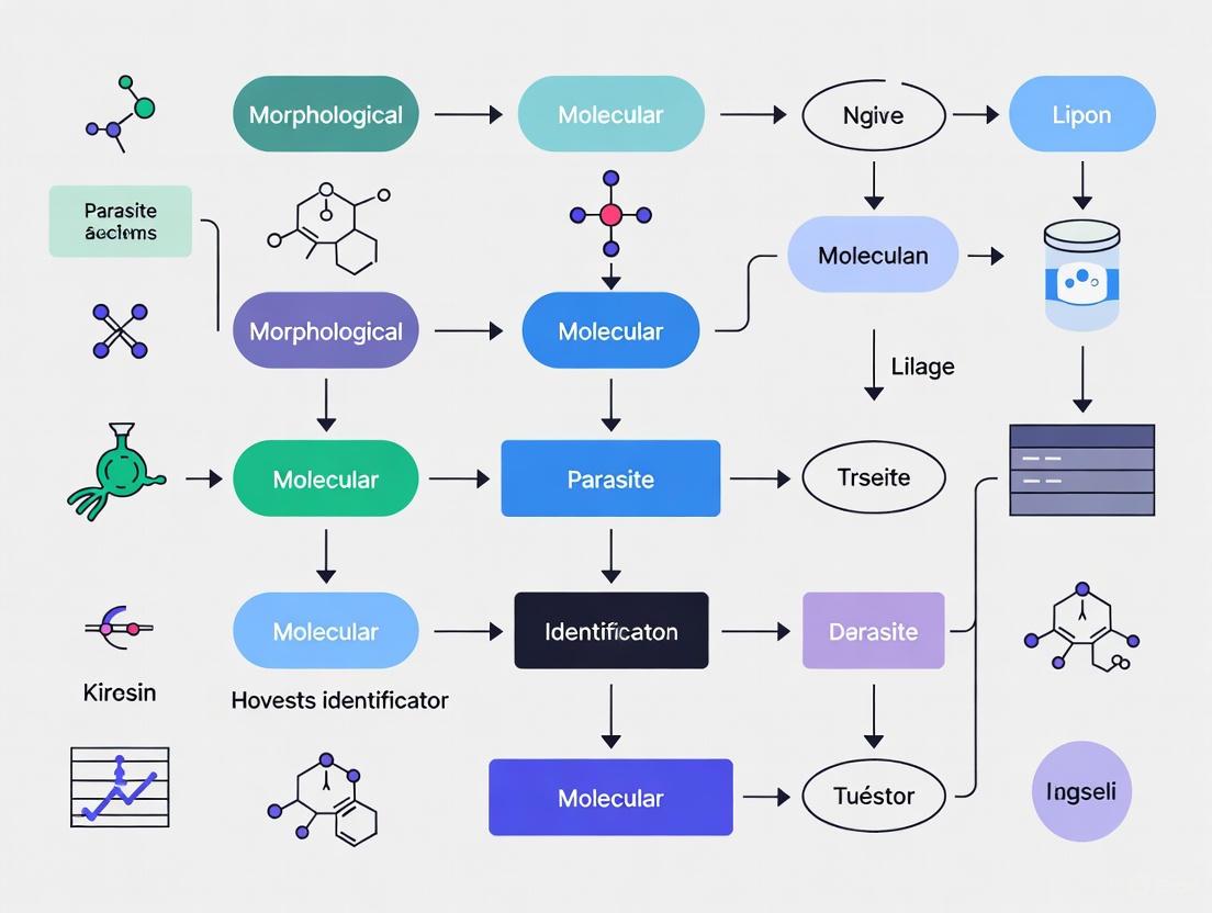
Figure 1: Integrated workflow combining morphological and molecular approaches for comprehensive parasite identification and its application to ecological and epidemiological studies.
Comparative Analysis of Methodological Approaches
Table 1: Comparison of Morphological and Molecular Approaches for Parasite Identification
| Parameter | Morphological Approach | Molecular Approach | Integrated Approach |
|---|---|---|---|
| Primary Focus | Physical characteristics, anatomy, and structural features [30] | Genetic sequences, markers, and phylogenetic signals [30] | Combined morphological and molecular data with ecological context [30] [32] |
| Key Methods | Microscopy, morphometrics, histological staining [30] | DNA barcoding, multi-locus sequencing, phylogenetic analysis [30] [31] | Complementary use of both methodological frameworks |
| Advantages | Provides phenotypic context; historical data rich; cost-effective for preliminary identification [30] | Detects cryptic diversity; establishes evolutionary relationships; high resolution for closely related taxa [30] [31] | Comprehensive species characterization; validates taxonomic conclusions; reveals eco-evolutionary patterns |
| Limitations | May miss cryptic species; requires taxonomic expertise; phenotypic plasticity can cause misidentification [30] | Does not capture phenotypic variation; potential for technical artifacts; database dependencies [30] | Resource intensive; requires interdisciplinary collaboration; complex data integration |
| Applications | Initial screening; descriptive taxonomy; museum collections [30] | Species delimitation; population genetics; phylogenetic studies [30] [31] | Biodiversity assessments; disease surveillance; conservation planning [30] [32] |
Essential Research Reagents and Materials
Table 2: Essential Research Reagents and Materials for Integrated Parasite Studies
| Category | Specific Items | Function/Application |
|---|---|---|
| Sample Collection & Preservation | Sterile containers, ethanol (70-95%), RNAlater, sterile swabs, cryovials, liquid nitrogen [32] | Maintain sample integrity for both morphological and molecular analyses during field collection |
| Morphological Analysis | Microscope slides, coverslips, histological stains (e.g., Giemsa, H&E), fixatives (e.g., formalin, FAA) [30] | Enable detailed examination and documentation of physical characteristics for taxonomic identification |
| Molecular Biology | DNA extraction kits, proteinase K, PCR reagents, primers (e.g., ITS, COI, 18S rRNA), agarose, sequencing reagents [30] [31] | Facilitate genetic characterization, amplification of target markers, and sequence-based identification |
| Data Analysis | Sequence alignment software (e.g., ClustalW, MAFFT), phylogenetic programs (e.g., MrBayes, BEAST), statistical packages (e.g., R) [30] | Support computational analysis, phylogenetic reconstruction, and species delimitation testing |
Detailed Experimental Protocols
Protocol 1: Integrated Field Collection and Preservation
Purpose: To collect and preserve parasite samples in a manner compatible with both morphological and molecular analyses.
Materials:
- Sterile collection equipment (forceps, scissors, scalpels)
- Cryogenic vials and labels
- 95% ethanol, RNAlater, and 10% neutral buffered formalin
- Liquid nitrogen or dry ice for transport
- Data recording forms and camera
Procedure:
- Field Collection:
- Collect parasite specimens aseptically from host organisms using sterile instruments.
- Document collection details (host species, geographic location, date, collector) and take representative photographs.
- For blood parasites, collect blood samples in EDTA tubes for molecular analysis and prepare blood smears for morphological examination.
Sample Division and Preservation:
- Divide each sample into three aliquots whenever possible:
- Aliquot 1: Preserve in 95% ethanol for DNA analysis (store at -20°C)
- Aliquot 2: Preserve in RNAlater for RNA studies (store at -80°C)
- Aliquot 3: Fix in 10% neutral buffered formalin for morphological studies (process within 24-48 hours)
- Divide each sample into three aliquots whenever possible:
Voucher Specimen Preparation:
- Preserve representative specimens as vouchers in 70% ethanol with detailed collection data.
- Assign unique catalog numbers and deposit in recognized museum or institutional collections.
Quality Control:
- Avoid cross-contamination between samples by using separate sterile instruments.
- Ensure proper labeling of all samples with permanent, solvent-resistant markers.
- Maintain cold chain during transport from field to laboratory.
Protocol 2: Morphological Characterization and Analysis
Purpose: To conduct comprehensive morphological examination and description of parasite specimens.
Materials:
- Light microscope with digital imaging capabilities
- Scanning electron microscope (when available)
- Histological processing equipment
- Staining solutions (hematoxylin and eosin, Giemsa, etc.)
- Slide mounting medium and coverslips
Procedure:
- Macroscopic Examination:
- Examine specimens under dissecting microscope and document overall size, shape, color, and external features.
- Take measurements of key morphological characters using calibrated ocular micrometer.
Histological Processing:
- Dehydrate formalin-fixed specimens through graded ethanol series.
- Clear in xylene substitute and embed in paraffin wax.
- Section at 4-7μm thickness using microtome.
- Mount sections on glass slides and stain with appropriate stains (H&E for general morphology, special stains for specific structures).
Microscopic Analysis:
- Examine stained sections under light microscope and document key diagnostic features.
- For selected specimens, process for scanning electron microscopy to examine surface ultrastructure.
- Capture digital images of diagnostic characters for documentation and comparison.
Morphometric Analysis:
- Take standardized measurements from multiple specimens to establish morphological variation.
- Conduct statistical analysis of morphometric data to identify significant differences between populations.
Protocol 3: Molecular Characterization and Phylogenetic Analysis
Purpose: To generate and analyze molecular data for parasite identification and phylogenetic placement.
Materials:
- DNA extraction kits suitable for the sample type
- PCR reagents (polymerase, dNTPs, buffer, MgCl₂)
- Taxon-specific primers for target genes (e.g., ITS, COI, 18S rRNA)
- Agarose gel electrophoresis equipment
- Sanger sequencing or next-generation sequencing platforms
Procedure:
- DNA Extraction and Quantification:
- Extract genomic DNA from ethanol-preserved samples using commercial kits with modifications for specific parasite taxa.
- Quantify DNA concentration using spectrophotometry or fluorometry.
- Assess DNA quality by running aliquot on agarose gel.
PCR Amplification:
- Design or select primers for appropriate genetic markers based on parasite group.
- Set up PCR reactions with positive and negative controls.
- Optimize cycling conditions for specific primer-template combinations.
- Verify amplification success by gel electrophoresis.
Sequencing and Data Generation:
- Purify PCR products using appropriate cleanup methods.
- Prepare sequencing reactions using BigDye or similar chemistry.
- Run sequences on appropriate platform (Sanger or NGS).
- Process raw sequence data using quality assessment tools.
Phylogenetic Analysis:
- Align sequences using ClustalW, MAFFT, or similar algorithms.
- Select appropriate substitution models using ModelTest or similar approaches.
- Construct phylogenetic trees using maximum likelihood, Bayesian inference, or parsimony methods.
- Assess node support with bootstrapping or posterior probabilities.
Data Integration and Species Delimitation
The following diagram illustrates the process for integrating morphological and molecular data for comprehensive species delimitation:
Figure 2: Decision workflow for integrating morphological and molecular data in parasite species delimitation, highlighting the detection of cryptic species.
Applications in Ecological and Epidemiological Studies
Wildlife Health Assessments and Conservation
Integrated parasite identification directly contributes to wildlife health assessments, which are critical for defining the normal physiological status of populations and detecting deviations that may signal environmental impacts [32]. The systematic approach to wildlife health involves multiple complementary methods, including physiological, morphological, nutritional, and behavioral data collection, combined with disease screening and parasite identification [32]. This comprehensive assessment is particularly valuable for:
- Sentinel species monitoring: Identifying and measuring impacts of environmental contaminants and emerging diseases [32]
- Endangered species management: Detecting health threats that may impact population viability [32]
- Ecosystem health indicators: Using parasite communities as biomarkers of environmental change and ecosystem integrity
The conceptual framework for wildlife health assessments emphasizes proper experimental design, adequate sample sizes, standardized methods, and appropriate data analysis to ensure meaningful conservation outcomes [32].
One Health Implications and Epidemiological Tracking
The integrated approach to parasite identification has significant implications for the One Health framework, which recognizes the interconnectedness of human, domestic animal, and wildlife health [33]. The operational framework for wildlife health categorizes wildlife based on management systems, habitat types, interfaces with humans and livestock, and levels of sanitary control [33]. This categorization enables targeted health management strategies that consider:
- Pathogen spillover risk: Identifying parasites with zoonotic potential at wildlife-human-livestock interfaces
- Disease emergence: Detecting novel parasites and tracking their spread across populations and ecosystems
- Transdisciplinary collaboration: Integrating expertise from ecology, veterinary science, medicine, and public health
The holistic health definition adapted for wildlife encompasses not only the absence of disease but also the ability to maintain normal physiological functions and contribute to ecosystem processes [33].
Data Analysis and Visualization Framework
Effective data presentation is essential for communicating research findings. The use of tables, figures, charts, and graphs enhances manuscript readability and facilitates data interpretation [34]. The following principles should guide data presentation in integrated parasitology studies:
- Complementary presentation: Use tables for exact numerical values and figures for overall patterns and relationships [34]
Appropriate graph selection: Select visualization methods based on data type and research questions:
- Line graphs: Depict trends or relationships between variables over time [34]
- Bar graphs: Compare values between discrete groups or categories [34]
- Scatter plots: Display relationships between two continuous variables [34]
- Box and whisker plots: Represent variations in samples and show median, quartiles, and outliers [34]
Standardized formatting: Ensure consistent design elements across all visualizations for easy comparison [34]
- Self-explanatory captions: Include complete titles and legends that enable understanding without reference to the text [34]
Table 3: Quantitative Data Summary from Wildlife Health Assessment Studies [35] [32]
| Study Component | Sample Size | Metric | Younger Group | Older Group | Difference | Significance |
|---|---|---|---|---|---|---|
| Gorilla Chest-Beating [35] | 25 individuals | Rate (beats/10h) | 2.22 (SD=1.270, n=14) | 0.91 (SD=1.131, n=11) | 1.31 | Distinct difference observed |
| Diarrhoea in Households [35] | 85 households | Woman's age (years) | 38.1 (SD=13.44, n=59) | 45.0 (SD=14.04, n=26) | 6.9 | Associated with incidence |
| Wildlife Health Publications [32] | 261 studies | International collaboration | 35% involved cross-border collaboration | - | - | Underrepresented in biodiversity hotspots |
| Diagnostic Methods [32] | 261 studies | Blood analysis usage | 89% of studies included method | - | - | Most common technique |
The integration of morphological and molecular approaches for parasite identification represents a transformative methodology with far-reaching implications for ecological and epidemiological studies. This integrated framework significantly enhances our ability to detect cryptic species diversity, understand parasite evolution, track disease patterns, and inform conservation strategies. The application of this approach within the One Health paradigm acknowledges the complex interconnections between wildlife, domestic animal, and human health, providing a comprehensive foundation for addressing emerging disease threats and ecosystem changes.
Future developments in this field will likely focus on standardizing protocols across research groups, expanding genetic reference databases, developing bioinformatic tools for data integration, and building capacity for wildlife health assessment in biodiversity-rich regions [32]. The continued refinement of integrated morphological and molecular approaches will be essential for advancing our understanding of parasite biodiversity, host-parasite interactions, and the ecological dynamics of infectious diseases in a rapidly changing world.
The How: State-of-the-Art Techniques and Their Practical Application
The accurate identification of parasites represents a cornerstone of parasitological research, disease diagnosis, and drug development. Traditional morphological identification, while foundational, often encounters limitations when dealing with cryptic species, juvenile stages, or damaged specimens [36]. The integration of molecular techniques with classical morphology has revolutionized the field, enabling precise species discrimination, detection of co-infections, and understanding of epidemiological dynamics. This application note details standardized protocols for a suite of molecular tools—DNA barcoding, PCR, multiplex real-time PCR, and LAMP assays—framed within the context of integrative parasitology research. These methodologies provide researchers and drug development professionals with a hierarchical toolkit, ranging from gold-standard species identification to rapid, field-deployable diagnostic solutions.
DNA Barcoding for Species Identification
Principle and Applications
DNA barcoding utilizes short, standardized genetic markers to classify and identify organisms. The cytochrome c oxidase subunit I (COI) gene is the primary barcode region for animals, while the internal transcribed spacer (ITS) region is widely used for fungi and other groups [37] [36]. This technique is particularly valuable for identifying cryptic species, larval stages, and specimens lacking distinguishing morphological features, thereby facilitating precise ecological assessments and biodiversity monitoring [36] [38].
Protocol: DNA Barcoding Workflow
Sample Preparation and DNA Extraction
- Specimen Collection: Collect parasite specimens using standard morphological preservation methods (e.g., in 70-100% ethanol). For integrative studies, photograph key morphological features prior to DNA extraction.
- DNA Extraction: Use a commercial tissue DNA extraction kit (e.g., GeneAll Exgene Tissue SV Plus kit). Follow the manufacturer's protocol, including a proteinase K digestion step.
- DNA Quantification: Assess DNA concentration and purity using a spectrophotometer (e.g., Nanodrop One). Normalize DNA concentrations to 0.5-10 ng/μL for subsequent PCR.
PCR Amplification and Sequencing
- Primer Selection: Use universal primers for the target barcode region. For metazoan COI, primers LCO1490 (5'-GGTCAACAAATCATAAAGATATTGG-3') and HCO2198 (5'-TAAACTTCAGGGTGACCAAAAAATCA-3') are commonly used [36].
- PCR Setup:
- Prepare a 25 μL reaction mixture containing:
- 1X PCR Buffer
- 2.5 mM MgCl₂
- 0.2 mM each dNTP
- 0.4 μM each forward and reverse primer
- 1 U of DNA polymerase (e.g., Taq polymerase)
- 2 μL of template DNA
- Use the following thermocycling conditions:
- Initial Denaturation: 94°C for 3-5 minutes
- 35-40 Cycles:
- Denaturation: 94°C for 30-45 seconds
- Annealing: 50-55°C (primer-specific) for 45-60 seconds
- Extension: 72°C for 60-90 seconds
- Final Extension: 72°C for 5-10 minutes
- Prepare a 25 μL reaction mixture containing:
- Verification and Sequencing: Visualize PCR products on a 1.5% agarose gel. Purify successful amplicons and submit them for Sanger sequencing in both directions.
Data Analysis
- Sequence Assembly and Curation: Assemble forward and reverse sequences and perform base calling. Trim low-quality ends.
- Taxonomic Identification: Compare the curated sequence against validated reference databases (e.g., BOLD or GenBank) using the BLASTn algorithm. A sequence identity of ≥97-99% is typically required for species-level assignment [36].
The following workflow diagram summarizes the DNA barcoding process from specimen collection to final identification.
Research Reagent Solutions
Table 1: Essential Reagents for DNA Barcoding.
| Reagent/Category | Specific Examples | Function |
|---|---|---|
| DNA Extraction Kit | GeneAll Exgene Tissue SV Plus kit | High-quality genomic DNA isolation from tissue samples. |
| Universal PCR Primers | LCO1490 / HCO2198 (COI) [36]; ITS1 / ITS4 (ITS) [37] | Amplification of standardized barcode regions. |
| DNA Polymerase | Taq DNA Polymerase | Enzymatic amplification of target DNA sequences. |
| Reference Database | Barcode of Life Data System (BOLD), GenBank | Repository of validated reference sequences for taxonomic assignment. |
PCR and Quantitative Real-Time PCR (qPCR)
Endpoint PCR and Multiplex PCR
Conventional PCR allows for the targeted amplification of specific DNA fragments, which are then visualized by gel electrophoresis. Multiplex PCR expands this capability by including multiple primer sets in a single reaction to amplify distinct targets simultaneously, which is useful for differentiating related species or detecting several pathogens in one test [39].
Protocol: Multiplex PCR for Trichobilharzia Species Discrimination This protocol is designed to differentiate three European Trichobilharzia species in a single reaction [39].
Primer Design:
- Design a degenerate forward primer and species-specific reverse primers targeting a variable gene such as cox1.
- Ensure amplicons have distinct lengths (e.g., 100-200 bp for T. szidati, 500-600 bp for T. franki, and 700-900 bp for T. regenti) for clear separation on a gel.
Reaction Setup:
- Prepare a 25 μL reaction mixture containing:
- 1X PCR Buffer
- 3.0 mM MgCl₂
- 0.3 mM each dNTP
- 0.2-0.4 μM of each primer (optimize concentration)
- 1.25 U of DNA Polymerase
- 2 μL of template DNA
- Use the following thermocycling conditions:
- Initial Denaturation: 95°C for 5 minutes
- 35 Cycles:
- Denaturation: 95°C for 30 seconds
- Annealing: 58-62°C (requires optimization) for 45 seconds
- Extension: 72°C for 60-90 seconds
- Final Extension: 72°C for 7 minutes
- Prepare a 25 μL reaction mixture containing:
Product Analysis: Separate PCR products on a 2% agarose gel. Species are identified based on the size of the amplified fragment.
Quantitative PCR (qPCR)
qPCR provides a method for quantifying pathogen DNA with high sensitivity, making it suitable for environmental DNA (eDNA) studies and assessing pathogen load.
Protocol: Trichobilharzia Genus-Specific qPCR (Tricho-qPCR) [39] This TaqMan assay targets the 28S rRNA gene for highly sensitive detection.
Primer and Probe Design:
- Design primers and a TaqMan probe from conserved regions of the 28S rRNA gene alignment of target species.
- The probe should be labeled with a FAM fluorophore at the 5' end and internal quenchers (e.g., ZEN/Iowa Black FQ).
Reaction Setup:
- Prepare a 20 μL reaction mixture containing:
- 1X TaqMan Environmental Master Mix
- 0.9 μM each forward and reverse primer
- 0.25 μM TaqMan probe
- 2-5 μL of template DNA
- Run the reaction on a real-time PCR instrument using the following conditions:
- Initial Denaturation: 95°C for 10 minutes
- 45 Cycles:
- Denaturation: 95°C for 15 seconds
- Annealing/Extension: 60°C for 60 seconds (with fluorescence acquisition)
- Prepare a 20 μL reaction mixture containing:
Data Analysis: Determine the cycle threshold (Ct) values. Use a standard curve of known DNA copy numbers for absolute quantification.
Performance Comparison
Table 2: Performance characteristics of different PCR-based assays for pathogen detection.
| Assay | Target Gene | Limit of Detection | Key Application | Advantages |
|---|---|---|---|---|
| Multiplex PCR (Trichobilharzia) [39] | cox1 | 10⁻² - 10⁻³ ng/μL | Species differentiation | Cost-effective; single-tube species ID via gel electrophoresis |
| qPCR (Trichobilharzia) [39] | 28S rRNA | 10 copies/reaction | Quantification & high-sensitivity detection | Excellent for eDNA; enables absolute quantification |
| qPCR (F. tricinctum) [40] | CYP51C | 3.1 fg/μL | Absolute pathogen quantification | Highest sensitivity; suitable for early detection |
Loop-Mediated Isothermal Amplification (LAMP)
Principle and Advantages
LAMP is an isothermal nucleic acid amplification technique that uses a strand-displacing DNA polymerase and 4-6 primers recognizing 6-8 distinct regions of the target DNA. It amplifies DNA with high efficiency at a constant temperature (60-65°C) in 30-60 minutes [39] [41]. Its key advantages include operational simplicity, speed, and the potential for result visualization by colorimetric change, making it ideal for point-of-care (POC) and field applications [40] [42].
This protocol is designed for the specific detection of Trichobilharzia genus DNA.
Primer Design:
- Design LAMP primers (F3, B3, FIP, BIP, and optional loop primers LF/LB) from a conserved region of the 28S rRNA gene using specialized software (e.g., PrimerExplorer V5).
Reaction Setup:
- Prepare a 25 μL reaction mixture containing:
- 1X Isothermal Amplification Buffer
- 6 mM MgSO₄
- 1.4 mM each dNTP
- 1.6 μM each FIP and BIP primer
- 0.2 μM each F3 and B3 primer
- 0.8 μM each LF and LB primer (if used)
- 8 U of Bst DNA Polymerase (large fragment)
- 1 μL of template DNA
- Incubate the reaction at 63°C for 45-60 minutes.
- Prepare a 25 μL reaction mixture containing:
Result Detection:
- Real-time: Monitor amplification in a real-time fluorometer using intercalating dyes like SYBR Green.
- End-point: Add an intercalating dye (e.g., SYBR Green) post-amplification. A color change from orange to green under UV light indicates a positive reaction. Alternatively, analyze products by gel electrophoresis.
Advanced Applications: Multiplex LAMP
Multiplex LAMP (mLAMP) allows for the simultaneous detection of multiple targets in one reaction. Techniques like DARQ (Detection of Amplification by Release of Quenching) use fluorophore- and quencher-labeled primers to generate target-specific signals, enabling the detection of up to four targets (e.g., different Plasmodium species) in a single tube [42].
The diagram below illustrates the workflow for setting up and interpreting a LAMP assay, highlighting its simplicity compared to PCR-based methods.
Research Reagent Solutions
Table 3: Essential Reagents for LAMP Assays.
| Reagent/Category | Specific Examples | Function |
|---|---|---|
| Strand-Displacing Polymerase | Bst 2.0/3.0 DNA Polymerase | Isothermal amplification of target DNA. |
| LAMP Primer Sets | F3, B3, FIP, BIP (LF, LB) | Recognize multiple target sites for highly specific amplification. |
| Visual Detection Dyes | SYBR Green I, Calcein, Hydroxy Naphthol Blue (HNB) | Visual interpretation of results by color change or fluorescence. |
| Isothermal Instrumentation | Portable Dry Bath, Fluorometer | Simple heating for amplification; real-time fluorescence reading. |
The integration of morphological and molecular methods creates a powerful framework for modern parasitology research. DNA barcoding provides a reliable foundation for species identification, especially in taxonomically complex groups. PCR and qPCR offer versatile, sensitive, and quantitative tools for specific detection and quantification in laboratory settings. Finally, LAMP assays represent a transformative technology for rapid, low-cost, and field-deployable diagnostics. The choice of technique depends on the research question, required sensitivity, turnaround time, and available infrastructure. This molecular toolbox empowers researchers and drug development professionals to advance our understanding of parasite biology, ecology, and epidemiology, ultimately contributing to improved disease control strategies.
The integration of advanced morphological and molecular techniques is revolutionizing parasite identification research. This synergy provides a more comprehensive framework for understanding parasite biology, host-pathogen interactions, and for discovering new therapeutic targets. Geometric morphometrics (GMM) offers a powerful quantitative method for analyzing shape and size variations in biological structures, moving beyond descriptive observations to statistically robust morphological data [43]. Concurrently, breakthroughs in high-resolution microscopy are enabling the visualization of parasitic structures and subcellular details with unprecedented clarity and scale [44] [45]. When combined with molecular data, these detailed morphological profiles contribute to a multi-omics understanding of parasitic diseases, enhancing diagnostic accuracy and paving the way for novel interventions [46] [47].
Geometric Morphometrics (GMM) in Parasitology
Core Principles and Workflow
Geometric morphometrics is an advanced morphometric method that uses Cartesian coordinates of biologically defined points, known as landmarks, for quantitative shape analysis. Unlike traditional morphometrics, which relies on linear measurements, GMM preserves the geometric relationships among points throughout the analysis, providing a more powerful and detailed description of form [43]. The core principle involves using homologous points (landmarks), curves, and contours to capture the shape of a structure, followed by statistical analysis of the coordinate data.
The standard GMM workflow involves several key stages, as illustrated in the diagram below:
Protocol: Outline-Based Geometric Morphometrics for Vector Identification
This protocol details the application of outline-based GMM for distinguishing morphologically similar species of Tabanus (horse flies), which are vectors for various pathogens. The method is also applicable to parasite and vector identification more broadly [48].
Sample Preparation
- Collect and preserve insect vectors (e.g., horse flies) using standard entomological techniques.
- Carefully remove one wing from each specimen and mount it on a microscope slide using clear, double-sided tape. Ensure the wing is perfectly flat and not folded or damaged.
Image Acquisition
- Use a standard compound microscope or a stereomicroscope equipped with a high-resolution digital camera.
- Capture images of the wings at a consistent magnification (e.g., 40x). Ensure uniform lighting across all images to avoid shadows.
- Include a scale bar in each image for calibration.
Landmark and Outline Digitization
- Import wing images into GMM software (e.g., the
geomorphpackage in R) [49]. - For outline-based analysis, select a specific wing cell (e.g., the first submarginal cell in Tabanus).
- Use software tools to digitally place a sequence of semi-landmarks along the contour of the chosen wing cell. These semi-landmarks capture the outline shape between two fixed, homologous landmarks placed at the junctions of the wing veins.
- Import wing images into GMM software (e.g., the
Procrustes Superimposition and Statistical Analysis
- Perform a Generalized Procrustes Analysis (GPA). This algorithm scales, translates, and rotates all landmark configurations to a common coordinate system, removing variations due to size, position, and orientation. This isolates "pure shape" for analysis [43].
- Analyze the resulting Procrustes shape coordinates using multivariate statistics:
- Principal Component Analysis (PCA): To visualize the major patterns of shape variation among species.
- Canonical Variate Analysis (CVA): To maximize shape separation between pre-defined groups (e.g., species) and perform classification.
- Mahalanobis Distance: To test for statistically significant shape differences between groups using permutation tests [48].
Validation
- Perform a validated classification test. The software will classify each specimen into a species group based on its shape, and the accuracy of this classification is calculated. In the case of Tabanus, the first submarginal cell contour achieved 86.67% classification accuracy, demonstrating high efficacy for species identification [48].
High-Resolution Microscopy for Parasitic Phenotyping
Advanced Microscopy Modalities
Recent advancements in microscopy overcome the limitations of conventional techniques, enabling high-throughput, high-resolution imaging ideal for analyzing parasites and host-parasite interactions.
Super-Resolution Panoramic Integration (SPI) Microscopy: SPI is an on-the-fly technique that enables instantaneous generation of sub-diffraction-limited images with high throughput. It leverages multifocal optical rescaling and a synchronized line-scan readout to achieve a twofold resolution enhancement (~120 nm) while imaging at speeds up to 1.84 mm²/s, typically containing 5,000–10,000 cells per second [44]. This is invaluable for population-level analysis and screening.
PANORAMA Multi-Camera Microscopy: This innovative microscope uses an array of 48 tiny cameras working together to capture gigapixel-scale images (e.g., 630 megapixels) of large, non-flat samples in a single snapshot, achieving submicron details (as small as 0.84 µm) across an area the size of a U.S. dime. Its ability to adaptively focus on curved samples makes it ideal for uneven plant, tissue, or material samples without the need for slow mechanical scanning [45].
Protocol: High-Throughput Phenotypic Screening of Infected Cells
This protocol utilizes SPI microscopy for high-content screening of host cells infected with parasites, enabling the study of subcellular changes and the identification of potential therapeutic compounds [44] [46].
Sample Preparation and Staining
- Culture host cells (e.g., mammalian cell lines) in multi-well plates suitable for high-throughput screening.
- Infect cells with the parasite of interest at a defined multiplicity of infection (MOI). Include uninfected control wells.
- For phenotypic profiling, perform a Cell Painting assay. Fix the cells and stain with a panel of fluorescent dyes targeting key cellular components:
- Hoechst 33342 or DAPI: for nucleus.
- Phalloidin: for actin cytoskeleton.
- Wheat Germ Agglutinin (WGA): for cell membrane and cytoplasm [44].
- MitoTracker: for mitochondria.
- LysoTracker: for lysosomes and vacuoles.
SPI Microscopy Imaging
- Mount the multi-well plate on the motorized stage of the SPI microscope.
- Define the imaging area for each well. The system will perform continuous raster scanning.
- The SPI system, using its TDI (Time-Delay Integration) sensor, will acquire super-resolution images on the fly as the stage moves. No post-processing reconstruction is required for initial image formation.
- For enhanced resolution, apply non-iterative rapid Wiener-Butterworth (WB) deconvolution, which provides an additional √2× enhancement and processes images in as little as 10 ms [44].
Image and Data Analysis
- Use image analysis software to extract morphological features from the stained cells (e.g., nucleus size and texture, cytoskeletal organization, cell shape).
- Compare the morphological profiles ("phenotypic fingerprints") of infected vs. uninfected cells.
- Integrate this high-content morphological data with molecular data (e.g., transcriptomics from the same samples) to link phenotypic changes to specific biological pathways [46].
Application in Drug Discovery
- Treat infected cells with a library of chemical compounds.
- Use the SPI microscope to rapidly image all wells and identify compounds that revert the infected-cell phenotype back to the uninfected state.
- AI/ML platforms (e.g., PhenAID) can then integrate this phenotypic data with omics layers to predict the mechanism of action (MoA) of the active compounds, accelerating hit validation [46].
Integrated Data and Reagent Solutions
Quantitative Comparison of Microscopy Techniques
Table 1: Performance Metrics of Advanced Microscopy Platforms
| Microscopy Technique | Best Resolution | Throughput / Speed | Key Advantage | Primary Application in Parasitology |
|---|---|---|---|---|
| SPI Microscopy [44] | ~120 nm | 1.84 mm²/s (~5,000-10,000 cells/s) | On-the-fly super-resolution without reconstruction | High-throughput subcellular phenotyping of host-pathogen interactions; drug screening. |
| PANORAMA Multi-Camera [45] | ~0.84 µm | Single snapshot (630 MP image in one shot) | Curvature adaptation for large, uneven samples | Rapid, whole-slide imaging of large tissue sections or curved specimens. |
| Conventional Fluorescence | ~250 nm | Limited by field of view and focus stacking | Widely available and established | Standard cytological examination. |
Essential Research Reagent Solutions
Table 2: Key Reagents and Software for Morphological Research
| Research Tool | Function / Application | Example Use Case |
|---|---|---|
geomorph R Package [49] |
Comprehensive software for performing all stages of geometric morphometric analysis. | Digitizing landmarks, Procrustes superimposition, and statistical shape analysis of parasite ova or vector wings. |
| Cell Painting Assay Dyes [46] | A panel of fluorescent dyes that label multiple organelles to generate a detailed phenotypic profile of cells. | Creating morphological fingerprints of host cells upon parasitic infection for high-content screening. |
| AI/ML Integration Platforms (e.g., PhenAID) [46] | AI-powered platform that integrates cell morphology data with omics layers to identify mechanisms of action. | Predicting how a compound reverses infection-associated phenotypes by fusing imaging and molecular data. |
| Wiener-Butterworth (WB) Deconvolution [44] | A rapid, non-iterative deconvolution algorithm that enhances image resolution with minimal processing time. | Providing an additional √2× resolution enhancement for high-throughput SPI image data. |
Integrated Workflow for Morpho-Molecular Research
The most powerful applications arise from the strategic integration of morphological and molecular tools. The following diagram outlines a synergistic workflow for comprehensive parasite research, from identification to drug discovery.
The integration of advanced computational techniques into parasitology represents a paradigm shift in diagnostic medicine, enabling rapid, objective, and reliable identification of parasitic infections. Traditional diagnostic methods, particularly egg-based microscopy, are fraught with challenges including subjectivity, low throughput, and a high potential for misdiagnosis due to the morphological polymorphism of parasite eggs and the need for specialized laboratory personnel [50]. Helminth infections, such as those caused by Ascaris lumbricoides and Taenia saginata, remain a widespread global health concern, infecting an estimated 1.5 billion people worldwide [50]. The limitations of conventional diagnostics have catalyzed the exploration of artificial intelligence (AI) solutions. This document details the application of state-of-the-art deep learning models—specifically ConvNeXt Tiny, EfficientNet V2 S, and MobileNet V3 S—for the automated classification of parasite eggs from microscopic images, framing these technological advancements within a broader research context that bridges morphological identification with molecular parasitology [50] [51].
Comparative Evaluation of Deep Learning Models for Helminth Diagnosis
Experimental Setup and Dataset
A recent comparative study evaluated three advanced deep learning models for diagnosing helminth infections: ConvNeXt Tiny, EfficientNet V2 S, and MobileNet V3 S [50] [51]. The research utilized a diverse dataset comprising microscopic images of Ascaris lumbricoides eggs, Taenia saginata eggs, and uninfected samples to perform multiclass classification experiments. These models were selected as they represent new-generation architectures designed to achieve high accuracy with computational efficiency.
Performance Metrics and Results
All three models demonstrated high classificatory accuracy in distinguishing between the different parasite species and uninfected samples. The performance was quantitatively assessed using the F1-score, a metric that balances precision and recall. The results are summarized in the table below.
Table 1: Performance comparison of deep learning models in classifying helminth infections
| Deep Learning Model | Reported F1-Score | Key Characteristics |
|---|---|---|
| ConvNeXt Tiny | 98.6% | Modern CNN architecture that modernizes traditional ConvNets with design ideas from Vision Transformers [50] [51] [52]. |
| MobileNet V3 S | 98.2% | Highly efficient architecture optimized for mobile and low-compute environments using depthwise separable convolutions [50] [51]. |
| EfficientNet V2 S | 97.5% | Improves upon EfficientNet with faster training speed and better parameter efficiency through adaptive scaling [50] [51]. |
The superior performance of ConvNeXt Tiny highlights the potential of modernized convolutional neural networks that incorporate design elements from transformers [52]. The high F1-scores achieved by all models prove the feasibility of leveraging deep learning to streamline and improve the diagnostic process for helminthic infections, potentially reducing misdiagnosis rates associated with traditional microscopy [50].
Experimental Protocol: Model Training and Evaluation
Data Preprocessing and Model Training
The following protocol outlines the key steps for replicating the model training and evaluation process for helminth egg classification.
- Image Acquisition and Annotation: Collect a dataset of microscopic images of stool samples. Annotate the images by expert parasitologists into defined classes: Ascaris lumbricoides, Taenia saginata, and uninfected [50].
- Image Preprocessing: Segment the original high-resolution microscopic images based on coordinates to obtain smaller sub-images centered on individual spots or eggs. Use image transformation and enhancement functions (e.g.,
torchvision.transformsin PyTorch) to resize and augment the sub-images into consistent, high-quality patches (e.g., 224x224 pixels) [53]. - Feature Extraction (Optional): Employ a pre-trained EfficientNet-B0 model to extract features from the image patches. Reduce the dimensionality of the extracted features to 50 dimensions using Principal Component Analysis (PCA) to form an image feature matrix [53].
- Model Selection and Training:
- Implement the ConvNeXt Tiny, EfficientNet V2 S, and MobileNet V3 S architectures.
- Initialize the models with pre-trained weights from large-scale image datasets like ImageNet.
- Use an Adam optimizer with a learning rate of 0.0005 and a cross-entropy loss function.
- Employ a batch size of 256 and train for a sufficient number of epochs (e.g., 20) [54].
- Performance Evaluation:
- Split the dataset into training (80%), validation (10%), and testing (10%) sets [54].
- Evaluate model performance using metrics including accuracy, precision, recall, specificity, and F1-score.
- Generate confusion matrices to visualize the classification performance for each class [54].
- Perform K-fold cross-validation (e.g., 5 folds) to robustly assess the model's generalization capacity [54].
Workflow Diagram
The following diagram illustrates the experimental workflow for automated parasite classification using deep learning.
The Scientist's Toolkit: Research Reagent Solutions
Successful implementation of an automated parasite classification system requires both computational and wet-lab resources. The table below lists key materials and their functions.
Table 2: Essential research reagents and materials for automated parasite diagnosis
| Item Name | Function/Application |
|---|---|
| Stool Sample Collection Kit | Standardized collection and preservation of patient stool samples for subsequent microscopy and DNA analysis. |
| Microscope with Digital Camera | Acquisition of high-resolution digital images of microscopic fields from prepared slides for model input. |
| H&E Staining Reagents | Hematoxylin and Eosin staining for enhancing contrast and morphological features in tissue sections or smears, aiding image analysis [53]. |
| DNA Extraction Kit | Extraction of high-quality genomic DNA from parasite samples for downstream molecular confirmation and sequencing. |
| PCR Reagents for SSU rDNA & cox1 | Amplification of small subunit ribosomal DNA (SSU rDNA) and cytochrome c oxidase subunit 1 (cox1) gene regions for molecular phylogeny [55] [56]. |
| GPU-Accelerated Computing Station | Hardware (e.g., with NVIDIA GPUs) essential for training and evaluating complex deep learning models within a practical timeframe [54]. |
Integration with Morphological and Molecular Parasitology
The application of deep learning for morphological classification is not an endpoint but a powerful component within an integrated research framework. Accurate AI-based identification can directly inform and streamline downstream molecular analyses.
- Guiding Molecular Assays: A deep learning model's initial classification of a sample as containing potential Taenia eggs can guide researchers to perform targeted PCR for specific genetic markers, such as the cytochrome c oxidase subunit 1 (cox1) gene, for precise species identification [55].
- Resolving Taxonomic Discrepancies: Phylogenetic analyses based on molecular markers like SSU rDNA often reveal intermixed groupings that conflict with traditional morphology-based taxonomy [55] [56]. High-throughput, AI-driven morphological analysis can provide the large-scale, consistent phenotypic data needed to reconcile these discrepancies and refine phylogenetic trees.
- Novel Species Discovery: The discovery of new parasite species, such as Darwinorhynchus bajacaliforniaensis n. gen., n. sp. [55] or Henneguya cystigena n. sp. [56], relies on a combination of morphological characterization and molecular sequencing. An AI system trained on known morphologies can flag specimens with anomalous features, prioritizing them for detailed molecular characterization.
Integrated Diagnostic and Research Pipeline
The following diagram illustrates the synergistic relationship between AI-driven morphology and molecular biology in modern parasitology.
The revolution in automated parasite classification, led by deep learning models like ConvNeXt and EfficientNet, demonstrates a clear path toward more efficient and accurate diagnostics. The high F1-scores, exceeding 97.5% for the models evaluated, underscore the readiness of this technology for clinical application, particularly in resource-limited settings. More profoundly, the integration of this AI-driven morphological analysis with established molecular techniques creates a powerful, synergistic pipeline. This framework not only enhances diagnostic precision but also accelerates taxonomic validation and the discovery of novel parasites, ultimately advancing our fundamental understanding of parasitology and contributing to global public health outcomes.
The integration of proteomic technologies, particularly liquid chromatography tandem mass spectrometry (LC-MS/MS), is revolutionizing antigen detection and vaccine development. This approach is especially transformative for parasitology, where traditional morphological identification methods often face challenges such as limited morphological differentiation between species and cryptic diversity [57]. Proteomics provides a powerful orthogonal method that complements and validates traditional techniques, enabling the precise identification of protein biomarkers and vaccine antigens directly from complex biological samples [58] [59]. The dynamic nature of the proteome, which reflects the functional state of an organism under specific conditions, makes it an invaluable resource for discovering targets for diagnostic assays and vaccine development [58] [60].
For parasitic diseases, which affect millions globally and present significant economic challenges, accurate diagnosis and effective treatments are urgently needed [61]. The limitations of conventional diagnostic methods—including time consumption, requirement of expert interpretation, and limited application in endemic regions with poor infrastructure—have created an pressing need for more advanced solutions [61]. Proteomics, particularly through LC-MS/MS workflows, offers the sensitivity, specificity, and multiplexing capabilities required to address these challenges by identifying promising diagnostic markers and vaccine targets from the proteome diversity across different life cycle stages of pathogens [59].
Technical Application Notes: LC-MS/MS for Antigen Detection
Core Principles and Advantages
The in-vitro expression LC-MS/MS (IVE-LC/MS/MS) assay represents a state-of-the-art characterization method for mRNA-based vaccines and antigen discovery [62] [63]. This approach serves as an orthogonal method to antibody-based techniques like flow cytometry, offering several significant advantages for antigen detection and characterization. It is fundamentally an antibody-free method that can detect multiple expressed antigens simultaneously, making it particularly valuable for multivalent vaccine characterization [62] [63].
A key application of this technology has been the simultaneous detection of influenza hemagglutinin (HA) antigens from four distinct strains, demonstrating its robust multiplexing capability [62]. The method also successfully identified specific immunoglobulin heavy and light chain variable domains in tuberculosis research, suggesting an oligoclonal humoral response to TB disease [60]. This highlights the technology's utility in uncovering subtle immune responses that might be missed by conventional methods.
Quantitative Performance Data
Table 1: Performance Metrics of LC-MS/MS in Biomarker Detection
| Performance Parameter | Experimental Findings | Experimental Context |
|---|---|---|
| Multiplexing Capacity | Simultaneous detection of HA antigens from 4 influenza strains [62] | IVE-LC/MS/MS assessment of quadrivalent mRNA vaccine candidate |
| Detection Dynamic Range | Proteins spanning >4 orders of magnitude quantified [60] | Plasma proteomics analysis of pediatric tuberculosis |
| Protein Detection Threshold | SERPINF2 detected at 12.1 ng/L concentration [60] | High-throughput plasma proteomics with DIA-PASEF MS |
| Coefficient of Variation | Average CV of 7.9% within countries, ~8% across countries [60] | Multi-country pediatric TB study with COMBAT batch correction |
| Data Completeness | 60.4% average completeness; 411 proteins detected in >75% of samples [60] | Analysis of 504 samples from 4 countries for TB biomarker discovery |
Comparative Methodological Advantages
Table 2: Comparison of Proteomic Approaches with Traditional Methods
| Methodological Aspect | Traditional Morphological/Serological Methods | LC-MS/MS Proteomic Approach |
|---|---|---|
| Species Differentiation | Limited morphological differentiation between species [57] | Molecular identification resolves cryptic diversity [57] |
| Antibody Dependency | Requires specific antibodies [62] | Antibody-free method [62] |
| Multiplexing Capability | Limited, often single analyte detection | Detects multiple antigens simultaneously [62] |
| Throughput | Variable, often time-consuming | High-throughput with automation potential [62] [60] |
| Dynamic Range | Limited by antibody affinity | >4 orders of magnitude [60] |
| Standardization | Subjective interpretation | Highly reproducible with CV ~8% [60] |
Experimental Protocols
Sample Preparation Workflow for IVE-LC/MS/MS
The following dot script defines the sample preparation workflow for LC-MS/MS analysis:
Protocol Details:
Cell Harvesting and Transfection: Begin with 200,000-500,000 HEK293T cells transfected with 3-500 ng mRNA in lipid nanoparticle (LNP) formulation. Multiple identical-cell-count plate wells are dosed with different mRNA levels to probe dose response. After culture period (typically 1 day), harvest transfected cells [62].
Cell Lysis: Incubate cells in sodium dodecyl sulfate (SDS) anionic surfactant for 15 minutes at 90°C, followed by 3-cycle pulsed sonication. This target-agnostic lysis ensures solubilization of all cellular components regardless of mRNA-coded cellular routing [62].
Reduction and Alkylation: Perform disulfide bond reduction and cysteine thiol alkylation before precipitation rather than after. This enables efficient purification and simplifies the workflow [62].
Protein Precipitation: Precipitate proteins, DNA and RNA using room temperature acetonitrile treatment. This approach is safer than dichloromethane/methanol-based extraction and simpler than more complicated protein purification approaches [62].
Pellet Processing: Resolubilize proteinaceous pellet with sonication and add universal nuclease to completely destroy DNA and RNA, improving chromatography robustness and performance [62].
Digestion: Conduct Lys-C/Trypsin protease digestion for 2 hours to yield target peptides [62].
LC-MS/MS Configuration and Analysis
Instrumentation and Parameters:
- Mass Spectrometer: High-resolution mass spectrometer (e.g., Orbitrap Fusion Lumos Tribrid HRMS) with high resolving power (240,000 RP at 200 m/z) to limit interference from background proteome peptides [62].
- Chromatography: Ultra-high performance liquid chromatography (UHPLC) system for reversed phase 70-minute separation of peptides [62].
- Flow Rate: Analytical flow rate (0.2 mL/min) for sufficient sensitivity while providing high reproducibility and robustness [62].
- Acquisition Method: Parallel reaction monitoring (PRM) mode for enhanced sensitivity and selectivity over conventional PRM or selected reaction monitoring (SRM) [62].
- Peptide Selection: Choose "quantotypic" peptides that are 8-25 amino acids, unique for the target, lack miscleavage sites, and lack problematic modification sites. Empirically select the best 2-4 MS/MS fragment ions for each target peptide [62].
Biomarker Discovery and Validation Workflow
The following dot script defines the biomarker discovery and validation pathway:
Protocol Details:
Sample Collection: Collect samples across well-characterized cohorts from multiple geographical locations. For pediatric tuberculosis research, this included 511 children from The Gambia, Peru, South Africa, and Uganda [60].
High-Throughput Proteomics: Start with minimal sample volume (1 μL of undepleted plasma) and perform high-throughput proteomics sample preparation [60].
DIA-PASEF Analysis: Utilize data-independent acquisition (DIA-PASEF) mass spectrometry analysis, achieving quantification of thousands of peptides and proteins with high-throughput (approximately 35 minutes sample-to-sample) [60].
Batch Effect Correction: Apply COMBAT normalization, a parametric approach commonly used in proteomics to mitigate batch effects across different clinical sites, sample preparation batches, and MS acquisition batches [60].
Machine Learning Analysis: Employ machine learning approaches to derive parsimonious biosignatures. In TB research, this yielded signatures encompassing 3-6 proteins achieving AUCs of 0.87-0.88, reaching minimum WHO target product profile accuracy thresholds [60].
The Scientist's Toolkit: Essential Research Reagents
Table 3: Key Research Reagent Solutions for LC-MS/MS-based Antigen Detection
| Reagent/Equipment | Specification/Model | Function in Workflow |
|---|---|---|
| Cell Line | HEK293T cells | Suitable growth characteristics and ability to support robust antigen expression for IVE assays [62] |
| Mass Spectrometer | Orbitrap Fusion Lumos Tribrid HRMS | High-resolution mass analysis with resolving power of 240,000 at 200 m/z for sensitive peptide detection [62] |
| Liquid Handler | Hamilton Vantage | Automation of sample preparation to streamline high-throughput analysis and improve method precision [62] |
| Protease | Lys-C/Trypsin | Enzyme combination for efficient protein digestion to yield target peptides for MS analysis [62] |
| Chromatography System | UHPLC with reversed phase column | High-resolution separation of peptides prior to mass spectrometric analysis [62] |
| Lysis Reagent | Sodium dodecyl sulfate (SDS) | Effective cell lysis and protein solubilization under heating (90°C) conditions [62] |
| Protein Precipitation Solvent | Acetonitrile (ACN) | Room temperature protein precipitation for safer and simpler sample cleanup [62] |
| Data Acquisition Method | Parallel Reaction Monitoring (PRM) | Targeted mass spectrometry method for enhanced sensitivity and selectivity in peptide quantification [62] |
Integration with Parasite Identification Research
The integration of proteomic approaches with traditional morphological identification creates a powerful framework for comprehensive parasite research. Molecular tools, including proteomics, have been demonstrated as necessary to validate trematode species composition, with studies revealing that approximately half of optically identified species require molecular confirmation [57]. This integrated approach is particularly valuable for detecting cryptic diversity, where morphological differences are minimal but genetic and proteomic variations are significant [57].
In parasite research, proteomic analysis of different life cycle stages of Plasmodium falciparum has identified numerous potential vaccine antigens and diagnostic markers, including merozoite surface proteins (MSPs), apical membrane antigen 1 (AMA1), rhoptry-associated membrane antigen (RAMA), and circumsporozoite protein (CSP) [59]. These proteins, identified through proteomic approaches, play major roles in the life cycle, pathogenicity, and key pathways of parasites, making them suitable targets for diagnostic and vaccine development [59].
The application of LC-MS/MS technologies in parasitology extends beyond antigen discovery to include the identification of host response biomarkers. In pediatric tuberculosis research, plasma proteomics identified WARS1 (tryptophanyl t-RNA synthetase) as significantly upregulated in confirmed TB cases, highlighting its potential as a biomarker linked to infection through multiple mechanisms [60]. Similar approaches can be applied to parasitic diseases to identify host proteome alterations indicative of infection.
LC-MS/MS-based proteomics represents a transformative technology for antigen detection and vaccine development, particularly when integrated with traditional morphological identification methods in parasite research. The IVE-LC/MS/MS assay provides a robust, antibody-free platform for simultaneous detection of multiple antigens with high sensitivity and specificity. Its application spans from characterizing multivalent mRNA vaccines to discovering diagnostic biomarkers for infectious diseases, including parasitic infections.
The detailed protocols and application notes presented here provide researchers with comprehensive methodologies for implementing these approaches in their own laboratories. As proteomic technologies continue to advance, with improvements in instrumentation sensitivity, computational analysis, and integration with other omics platforms, their impact on vaccine development and disease diagnosis is expected to grow substantially, particularly for neglected tropical diseases that disproportionately affect global health.
Integrative taxonomy, which combines morphological and molecular data, provides a robust framework for parasite identification and research [64]. This approach mitigates the limitations inherent in using either method in isolation, such as phenotypic plasticity in morphological analyses or introgression and incomplete lineage sorting in DNA barcoding [64]. This protocol details a standardized workflow from biological sample collection to final data integration, designed to generate high-quality, reproducible data for complex analyses. The methodology is structured to support a broader thesis on integrated parasite identification.
Experimental Protocol & Workflow
Step-by-Step Sample Processing and Data Generation
Sample Collection and Storage
- Collection: Aseptically collect parasite samples using sterile instruments. Record all metadata (e.g., host species, geographical location, date).
- Preservation: Divide each sample into two aliquots.
- For Morphological Analysis: Fix specimens in 10% neutral buffered formalin for 24 hours before transferring to 70% ethanol for long-term storage.
- For Molecular Analysis: Preserve specimens in 95-100% ethanol or store at -80°C to prevent DNA degradation.
Morphological Identification
- Preparation: Rehydrate fixed specimens in a descending ethanol series (70% to 30%) if needed. For microscopic parasites, prepare permanent slides using appropriate mounting media.
- Imaging and Characterization: Capture high-resolution digital images using compound or stereo microscopes with calibrated measurement software. Identify and record key diagnostic characters based on established taxonomic keys.
Molecular Identification
- DNA Extraction: Isolate genomic DNA from preserved tissue samples using a commercial kit designed for difficult samples, incorporating a proteinase K digestion step. Quantify DNA yield and purity using a spectrophotometer.
- PCR Amplification: Amplify target genetic markers (e.g., COI for DNA barcoding, 18S rRNA) using standard or custom primers. Perform reactions in a 25 µL volume and verify amplification success via agarose gel electrophoresis.
- Sequencing: Purify successful PCR products and submit them for bidirectional Sanger sequencing.
Data Integration and Analysis
- Sequence Processing: Assemble and edit chromatograms from forward and reverse reads. Perform a BLAST search against public databases (e.g., GenBank) for preliminary identification.
- Phylogenetic Analysis: Align consensus sequences with closely related sequences obtained from databases using alignment software. Construct phylogenetic trees using maximum likelihood or Bayesian inference methods.
- Integrative Analysis: Correlate molecular identification and phylogenetic placement with morphological character data to reach a definitive species diagnosis.
Integrated Research Workflow
The following diagram illustrates the logical workflow and data relationships from sample collection to integrated analysis, as described in the protocol.
Research Reagent Solutions
The following table details the essential materials and reagents required to execute the experimental protocol.
Table 1: Key Research Reagents and Materials for Integrated Parasite Identification
| Item Name | Function/Application |
|---|---|
| Sterile Collection Instruments | Aseptic sample collection to prevent cross-contamination. |
| 10% Neutral Buffered Formalin | Tissue fixation for preservation of morphological structures. |
| 95-100% Ethanol | Preservation of tissue samples for subsequent DNA extraction. |
| DNA Extraction Kit | Isolation of high-quality genomic DNA from preserved samples. |
| Proteinase K | Enzymatic digestion of proteins to improve DNA yield and quality. |
| PCR Primers (e.g., COI, 18S rRNA) | Target-specific amplification of genetic barcodes for sequencing. |
| PCR Master Mix | Enzymes, buffers, and nucleotides for DNA amplification. |
| Agarose | Gel electrophoresis to verify success of PCR amplification. |
| Sequence Alignment Software | Aligning and editing sequence data for phylogenetic analysis. |
| Taxonomic Identification Keys | Reference materials for morphological characterization. |
Data Presentation and Analysis
The integration of morphological and molecular data is crucial because these methods can yield contrasting trends, underscoring the need for validation [65]. The following table summarizes the core characteristics of each approach.
Table 2: Comparison of Morphological and Molecular Identification Methods
| Characteristic | Morphological Identification | Molecular Identification |
|---|---|---|
| Data Type | Qualitative descriptions, morphometric measurements, images. | DNA/RNA sequences, single nucleotide polymorphisms (SNPs). |
| Primary Output | Species description based on physical traits. | Genetic similarity, phylogenetic placement. |
| Key Strength | Direct observation of phenotypic traits; cost-effective. | High discrimination for cryptic species; uses trace/degraded samples. |
| Key Limitation | Phenotypic plasticity can lead to misidentification [64]. | Introgression or incomplete lineage sorting can cause errors [64]. |
| Data Integration Role | Provides phenotypic context and validates molecular findings. | Offers a genetic framework and clarifies evolutionary relationships. |
Quantitative Data Structure
For consistent data presentation, summary statistics from experiments (e.g., specimen counts, sequencing success rates, morphometric data) should be structured in tables with clear titles, column headers, and units of measurement [66]. The example below demonstrates a recommended format for presenting frequency data.
Table 3: Example Format for Presenting Experimental Frequency Data
| Identification Result | Absolute Frequency (n) | Relative Frequency (%) |
|---|---|---|
| Concordant (Morph & Molecular) | 185 | 92.5 |
| Discordant (Morph & Molecular) | 15 | 7.5 |
| Total Samples | 200 | 100.0 |
Advanced convolutional neural networks like MMNet have demonstrated the power of integrated data, achieving high identification accuracies (>96%) across diverse taxa, including groups with closely related species [64]. This highlights the superior outcome of a combined methodology.
Overcoming Obstacles: Troubleshooting and Optimizing the Integrated Workflow
Application Note: A Comparative Framework for Parasite Preservation and Analysis
In parasitology research, the choice between morphological and molecular identification methods is often constrained by technical and resource limitations, including costs, equipment availability, and expertise [67]. This application note details a practical framework for integrating these approaches, leveraging their complementary strengths. Morphology provides accessible, cost-effective identification and crucial life-stage information [68], while molecular methods offer high specificity for distinguishing morphologically similar taxa [69] [68]. The following protocols and data demonstrate how a combined methodology maximizes research outcomes under typical resource constraints.
Experimental Comparison of Preservation Media
A direct comparison of parasite preservation mediums was conducted using fecal samples from wild capuchin monkeys (Cebus imitator) [69]. Samples were halved and stored in either 10% buffered formalin or 96% ethanol at ambient temperature for 8-19 months before analysis [69]. This design directly addresses resource constraints by evaluating affordable, field-deployable preservatives.
Table 1: Morphological Preservation and Diversity by Preservation Medium [69]
| Metric | 10% Formalin | 96% Ethanol | Statistical Significance (p-value) |
|---|---|---|---|
| Number of Parasitic Morphotypes Identified | Higher | Lower | p < 0.05 (Wilcoxon test) |
| Parasites per Fecal Gram (PFG) - Overall | No significant difference | No significant difference | p > 0.05 |
| PFG - Filariopsis Larvae | No significant difference | No significant difference | p > 0.05 |
| PFG - Strongyle-type Eggs | No significant difference | No significant difference | p > 0.05 |
| Preservation Rating - Filariopsis Larvae | Better | Worse | p < 0.05 |
| Preservation Rating - Strongyle-type Eggs | No significant difference | No significant difference | p > 0.05 |
Key Findings: Formalin is superior for preserving larval nematode morphology and identifying a greater diversity of parasites [69]. However, ethanol adequately preserves key forms like strongyle eggs and is compatible with downstream molecular assays, making it a balanced choice for integrated studies [69].
Protocol 1: Integrated Morphological-Molecular Workflow for Preserved Fecal Samples
This protocol, adapted from studies of modern and ancient samples, allows comprehensive analysis from a single sample [69] [68].
Materials and Equipment
- Sample Collection: Sterile 15ml tubes [69].
- Preservation Media: 10% buffered formalin, 96% ethanol [69].
- Rehydration Solution: Tris-EDTA (TE) buffer, pH 8.0 [68].
- Fecal Concentration: Sheather’s Sugar Solution, centrifuge, microscope slides [68].
- DNA Extraction: Mo Bio Ultra-Clean Fecal DNA Kit or equivalent; thermal cycler for heat/freeze lysis [68].
- PCR and Sequencing: Genus-specific primers (e.g., 18S rRNA for Ascaris), standard PCR reagents, sequencer [68].
Procedure
- Sample Partitioning and Preservation: Immediately upon collection, partition the sample into two approximately 2g portions. Place one in 10ml of 10% formalin and the other in 6ml of 96% ethanol. Ensure samples are fully submerged [69].
- Microscopic Processing (Formalin-Preserved Half):
- Separate solids from preservative and record fecal weight [69].
- Rehydrate and homogenize a portion in distilled water. Strain through cheesecloth [69].
- Centrifuge the solution (10 min at 1500 rpm) and discard supernatant [69].
- Resuspend the pellet in 5-10ml distilled water and screen using a sedimentation technique on a microscopy plate [69].
- Identify and morphologically grade all parasites based on established rubrics (e.g., larval cuticle integrity, eggshell continuity) [69].
- Molecular Processing (Ethanol-Preserved Half):
- Use the rehydration protocol for robust analysis: homogenize 0.5-1.0g of sample in 2-5ml TE buffer and orbit for 72 hours [68].
- DNA Extraction: Extract from rehydrated material or directly from microscopy slide coverslips [68]. Incorporate a mechanical heat/freeze step (5min at 63°C, 5min at -20°C, 5min at 63°C) to lyse durable parasite eggs [68].
- PCR and Sequencing: Amplify using taxon-specific primers. Sequence PCR products and conduct phylogenetic analysis against genomic databases [68].
Protocol 2: Leveraging Advanced Imaging and Computational Tools
For projects with access to computational resources, deep learning (DL) can augment morphological analysis, addressing expertise gaps [70].
Table 2: Performance of Selected Deep Learning Models in Parasite Identification [70]
| Deep Learning Model | Accuracy (%) | Precision (%) | Sensitivity (%) | Specificity (%) | F1 Score (%) | AUROC |
|---|---|---|---|---|---|---|
| DINOv2-Large | 98.93 | 84.52 | 78.00 | 99.57 | 81.13 | 0.97 |
| YOLOv8-m | 97.59 | 62.02 | 46.78 | 99.13 | 53.33 | 0.755 |
| ResNet-50 | 95.40 (Validation) | - | - | - | - | - |
Procedure
- Image Acquisition and Dataset Preparation: Capture high-quality digital micrographs of parasite eggs, larvae, and adults. Split images into training (80%) and testing (20%) sets [70].
- Model Selection and Training: Train state-of-the-art models. Use YOLO models for object detection and DINOv2 or ResNet-50 for classification tasks [70].
- Validation and Integration: Validate model performance against human expert identifications. Integrate the highest-performing model into the diagnostic workflow as a screening or decision-support tool [70].
The Scientist's Toolkit: Research Reagent Solutions
Table 3: Essential Materials for Integrated Parasitology Research
| Item | Function/Application | Notes on Use |
|---|---|---|
| 10% Buffered Formalin | Optimal preservative for morphological studies; maintains tissue integrity for microscopy [69]. | Toxic; requires careful handling. Causes DNA fragmentation, limiting molecular utility [69]. |
| 96% Ethanol | Effective preservative for DNA; suitable for molecular analyses. Adequate for some morphological studies [69]. | Causes tissue dehydration, leading to potential morphological distortion [69]. |
| Tris-EDTA (TE) Buffer | Rehydration solution for desiccated or preserved fecal samples prior to DNA extraction [68]. | Preferred over water for ancient DNA and well-preserved samples to chelate divalent cations and protect DNA [68]. |
| Sheather's Sugar Solution | Flotation medium for concentrating parasite eggs and larvae via centrifugation [68]. | Creates a density gradient; parasitic forms float to the surface for easy collection on coverslips [68]. |
| Ultra-Clean Fecal DNA Kit | DNA isolation and purification from complex fecal samples [68]. | A mechanical heat/freeze lysis step is recommended to break down resilient parasite egg walls [68]. |
| Ascaris 18S rRNA Primers | PCR amplification of a small ribosomal RNA gene segment for phylogenetic identification of ascarids and relatives [68]. | An example of a targeted molecular assay; primers must be selected based on the parasite taxa of interest [68]. |
The constraints of cost, equipment, and expertise in parasitology are best navigated through integrated protocols. Formalin remains the gold standard for pure morphological studies, while ethanol offers a versatile medium for projects aiming for both morphological and molecular analyses [69]. The combined workflow mitigates the limitations of each method alone: morphology guides efficient molecular targeting, while molecular data resolves morphological uncertainties [68]. Furthermore, emerging deep-learning tools show great promise in augmenting human expertise, potentially bridging the growing gap in morphological skills [70]. By adopting this synergistic framework, researchers can maximize diagnostic accuracy and phylogenetic resolution within practical resource limitations.
The accurate detection and identification of parasitic infections remain a cornerstone of effective disease control, clinical management, and epidemiological surveillance. The diagnostic landscape for parasitology has evolved significantly from reliance on morphological techniques to incorporating advanced molecular methods, each with distinct advantages and limitations. This evolution is particularly critical within the context of integrating morphological and molecular identification research, where understanding the performance characteristics of available methods directly impacts diagnostic accuracy. Selecting the appropriate diagnostic technique requires careful consideration of the target parasite, sample type, and operational setting to optimize both sensitivity and specificity [67].
The persistent global burden of parasitic diseases, including schistosomiasis, soil-transmitted helminths, and intestinal protists, underscores the necessity for reliable diagnostic tools. Conventional microscopy, while widely used and cost-effective, suffers from limitations in sensitivity and requires significant expertise, particularly in low-endemicity settings where infection intensities are minimal [71] [67]. Molecular techniques, including polymerase chain reaction (PCR), isothermal amplification, and next-generation sequencing (NGS), have emerged as powerful alternatives offering superior sensitivity and specificity, along with the ability to differentiate between morphologically similar species [72] [73]. This application note provides a structured comparison of current diagnostic methodologies, detailed experimental protocols, and practical guidance for selecting optimal diagnostic approaches based on specific parasitological requirements.
Comparative Performance of Diagnostic Methods
The selection of a diagnostic method requires a clear understanding of its performance metrics. The table below summarizes the key characteristics of major diagnostic platforms for parasitic infections, highlighting their relative sensitivities, specificities, and appropriate applications.
Table 1: Comparative Analysis of Parasite Diagnostic Methods
| Method Category | Example Technique | Reported Sensitivity | Reported Specificity | Best Application Context |
|---|---|---|---|---|
| Molecular Isothermal Amplification | Loop-Mediated Isothermal Amplification (LAMP) for Schistosoma [71] | Pooled: 0.90 (95% CI: 0.80–0.90) [71] | Pooled: 0.82 (95% CI: 0.60–0.93) [71] | Resource-limited field settings; low-intensity infections |
| Automated AI-Based Microscopy | AiDx Assist (S. haematobium in urine) [74] | Semi-automated: 94.6%; Fully automated: 91.9% [74] | Semi-automated: 90.6%; Fully automated: 91.3% [74] | High-throughput screening in endemic areas; reducing reliance on expert microscopists |
| Automated AI-Based Microscopy | AiDx Assist (S. mansoni in stool) [74] | Semi-automated: 86.8%; Fully automated: 56.9% [74] | Semi-automated: 81.4%; Fully automated: 86.8% [74] | Intestinal schistosomiasis detection; requires further optimization for full automation |
| Next-Generation Sequencing | Metataxonomics (18S rRNA) for Strongyloides [72] | Outperformed microscopy [72] | Enabled species/subtype classification [72] | Broad-spectrum parasite detection; species-level resolution for protists |
| Nanobiosensors | Various (e.g., AuNP for PfHRP2, GO for Schistosoma SEA) [75] | Highly sensitive; can detect biomarkers at low concentrations [75] | High specificity with functionalized probes [75] | Point-of-care (PoC) detection; rapid, sensitive antigen/biomarker detection |
The data illustrates that no single method is universally superior. LAMP demonstrates high sensitivity suitable for field applications, while AI-microscopy offers a promising bridge between traditional morphology and modern automation. NGS-based methods provide unparalleled breadth of detection and resolution, and emerging nanobiosensors hold potential for future PoC diagnostics.
Detailed Experimental Protocols
Protocol 1: Loop-Mediated Isothermal Amplification for Schistosoma Detection
Principle: LAMP amplifies target DNA under isothermal conditions (60–65°C) using a strand-displacing DNA polymerase and four to six primers that recognize eight distinct regions of the target gene. Amplification is visualized via turbidity from magnesium pyrophosphate precipitate or fluorescence with intercalating dyes like SYBR Green I [71].
Workflow:
Procedure:
- Sample Processing and DNA Extraction:
- Process clinical samples (stool, urine, serum, or snail tissue) using a rapid, simplified DNA extraction method suitable for field settings [71]. As an alternative, commercial stool DNA extraction kits (e.g., the Stool DNA isolation kit) can be used, with the addition of inhibitor-binding substances like Bovine Serum Albumin (BSA) if needed [73].
- LAMP Reaction Setup:
- Prepare a master mix containing 1.6 µM each of inner primers (FIP and BIP), 0.2 µM each of outer primers (F3 and B3), 0.8 µM each of loop primers (LF and LB, if used), 1.4 mM dNTPs, 0.8 M betaine, 20 mM Tris-HCl (pH 8.8), 10 mM KCl, 10 mM (NH4)2SO4, 8 mM MgSO4, 0.1% Tween 20, and 8 U of Bst DNA polymerase large fragment.
- Amplification:
- Aliquot 23 µL of the master mix into reaction tubes and add 2 µL of template DNA.
- Incubate the tubes at 60–65°C for 30–60 minutes in a heating block or water bath.
- Result Interpretation:
- Visual Detection: Add SYBR Green I dye post-amplification. A color change from orange to green indicates a positive reaction.
- Turbidity: Observe for a white precipitate of magnesium pyrophosphate in the reaction tube.
- Real-time Fluorescence: Use a dedicated device to monitor fluorescence accumulation during the reaction.
Protocol 2: AI-Assisted Automated Microscopy for Schistosoma Egg Detection
Principle: The AiDx Assist system automates the imaging and analysis of conventional microscopy slides (Kato-Katz for stool, urine filtration for urine) using a digital microscope and integrated AI algorithms to detect and count parasite eggs [74].
Workflow:
Procedure:
- Sample and Slide Preparation:
- Stool (for S. mansoni): Prepare a Kato-Katz thick smear using a 41.7 mg template. Cover with cellophane soaked in malachite green and allow to clear for 10 minutes [74].
- Urine (for S. haematobium): Filter 10 mL of homogenized urine through a 13 mm polycarbonate membrane (30 µm pore size) using a syringe and filter holder. Transfer the membrane to a glass slide [74].
- AiDx Assist Operation:
- Load the prepared slide into the AiDx Assist device.
- Semi-Automated Mode: Initiate the imaging process. The device will automatically capture images of the entire sample. Disable the AI counting algorithm. An expert microscopist must then manually review all captured images on the device's interface to identify and count parasite eggs.
- Fully Automated Mode: Initiate the process with the AI counting algorithm enabled. The device will automatically capture images, identify potential parasite eggs using its AI, and provide a count. The operator reviews and confirms the AI-generated results at the end of the analysis.
- Data Management:
- Export the results, including egg counts and images, in an Excel-compatible format for further analysis and record-keeping.
Protocol 3: Metataxonomics for Intestinal Protist and Helminth Detection
Principle: This NGS-based approach uses PCR to amplify a taxonomically informative genetic marker (e.g., the 18S rRNA V4/V9 region) directly from stool DNA extracts. The amplicons are sequenced, and the resulting reads are classified against reference databases to identify multiple parasite species and subtypes in a single assay [72] [76].
Workflow:
Procedure:
- DNA Extraction:
- Extract total genomic DNA from approximately 200 mg of stool using a commercial kit (e.g., Stool DNA isolation kit) following the manufacturer's protocol. Quantify DNA purity and concentration using a spectrophotometer [72].
- Metataxonomic Library Preparation:
- Amplification: Perform PCR using primers targeting the 18S rRNA V4 region (e.g., 18S-V4Fw: CCAGCAGCCGCGGTAATTCC and 18S-V4Rev: RCYTTCGYYCTTGATTRA) [72]. The reaction should include a high-fidelity DNA polymerase and 30-35 cycles.
- Indexing and Pooling: Clean the PCR amplicons and attach dual indices and sequencing adapters in a second, limited-cycle PCR. Quantify the final libraries and pool them in equimolar ratios.
- Sequencing:
- Bioinformatic Analysis:
- Process raw sequencing data using a pipeline like MOTHUR or QIIME2. Key steps include:
- Assembly & Quality Filtering: Merge paired-end reads and remove low-quality sequences, chimeras, and amplicons with ambiguous bases or long homopolymers.
- Clustering: Cluster high-quality sequences into molecular Operational Taxonomic Units (mOTUs) at a 97% sequence identity threshold.
- Taxonomic Assignment: Classify mOTUs by comparing them to reference databases (e.g., SILVA, NCBI) using tools like BLASTN. Results can be validated with phylogenetic methods for specific parasites [72] [76].
- Process raw sequencing data using a pipeline like MOTHUR or QIIME2. Key steps include:
The Scientist's Toolkit: Essential Research Reagents and Materials
Successful implementation of the protocols above depends on key reagents and materials. The following table details these essential components.
Table 2: Key Research Reagent Solutions for Parasite Diagnostics
| Reagent/Material | Function/Application | Example/Note |
|---|---|---|
| Bst DNA Polymerase | Enzyme for isothermal DNA amplification in LAMP; has strand-displacing activity [71]. | Derived from Bacillus stearothermophilus; critical for LAMP protocol efficiency. |
| Primer Sets (LAMP) | Six primers targeting eight distinct regions for highly specific amplification [71]. | Must be meticulously designed for the target parasite gene (e.g., Schistosoma 28S rRNA). |
| SYBR Green I / Calcein | Intercalating fluorescent dyes for visual detection of LAMP amplicons [71]. | Enables colorimetric readout; add post-amplification to avoid inhibition. |
| Malachite Green | Stain used in Kato-Katz technique for stool smears [74]. | Aids in visualizing helminth eggs against the stained background. |
| Polycarbonate Membranes | For filtration and concentration of S. haematobium eggs from urine samples [74]. | Standard pore size of 30 µm; used with a syringe filter holder. |
| Stool DNA Isolation Kit | Standardized DNA extraction from complex fecal matrix, removing PCR inhibitors [72] [73]. | Kits often include inhibitors for DNases and substances to absorb PCR interferents. |
| 18S rRNA Primers | Universal eukaryotic primers for metataxonomic profiling of protists and helminths [72] [76]. | Target conserved variable regions (e.g., V4, V9); choice affects taxonomic coverage. |
| High-Fidelity PCR Mix | For accurate amplification of target markers prior to NGS; reduces amplification errors [76]. | Essential for generating high-quality metataxonomic libraries. |
| Functionalized Nanoparticles | Core sensing element in nanobiosensors; conjugated with antibodies or DNA probes [75]. | Includes gold nanoparticles (AuNPs), quantum dots (QDs), and graphene oxide (GO). |
The integration of morphological and molecular research in parasitology diagnostics is no longer a future prospect but a present necessity. This application note demonstrates that optimizing sensitivity and specificity is not about finding a single best method, but about making an informed selection based on a clear understanding of the performance characteristics of each technique. LAMP stands out for its field-deployable, high sensitivity in detecting low-burden infections. Automated microscopy offers a viable path to standardizing and scaling up traditional morphology. For comprehensive surveillance and precise species-level identification, particularly in complex samples or for epidemiological research, NGS-based metataxonomics provides an unparalleled level of resolution.
Future developments will likely focus on integrating these technologies, such as combining isothermal amplification with nanobiosensors for ultra-sensitive point-of-care devices, or refining AI algorithms to achieve the high specificity required for fully automated parasite identification across diverse sample types. By carefully considering the guidelines and protocols outlined here, researchers and drug development professionals can strategically select the most appropriate diagnostic method to advance their specific goals in parasite identification and control.
In parasitology and biodiversity research, the integration of traditional morphological identification with advanced molecular techniques is paramount for accurate species characterization. However, these methods can sometimes yield conflicting results, creating challenges for data interpretation and consensus. Such discrepancies are not unique to parasitology; a recent cross-European study on soil fauna also found that molecular methods indicated higher biodiversity in croplands, whereas morphological assessments suggested the opposite trend [65]. This application note provides detailed protocols and frameworks for resolving these conflicts, ensuring robust and reliable species identification in parasite research. By providing standardized procedures for side-by-side comparison and integration of these complementary approaches, this document serves as a practical guide for researchers navigating methodological discrepancies.
Comparative Data Analysis: Morphological vs. Molecular Methods
A structured comparison of methodological outputs is the first critical step in identifying and understanding the source of discrepancies. The table below synthesizes key comparative parameters based on recent research findings.
Table 1: Quantitative and Qualitative Comparison of Morphological and Molecular Identification Methods
| Parameter | Morphological Methods | Molecular Methods |
|---|---|---|
| Primary Output | Physical description and measurement of taxonomic features (e.g., prostomal teeth, papilla patterns) [77]. | DNA sequence data (e.g., from 28S rRNA and cox1 gene regions) [77]. |
| Typical Data Format | Descriptive morphology, morphometric ratios, line drawings, micrographs [77]. | DNA sequences, electropherograms, phylogenetic trees, sequence alignment data [77] [65]. |
| Reported Trend in Soil Fauna Diversity vs. Land-Use | Higher biodiversity in woodlands and grasslands compared to intensively managed croplands [65]. | Higher biodiversity in intensively managed croplands compared to woodlands and grasslands [65]. |
| Inherent Strengths | Provides context for ecological role; allows for direct observation of phenotypic traits. | High-throughput capability; can identify cryptic species; high sensitivity for detecting low-abundance species [65]. |
| Inherent Limitations | Time-consuming; requires high taxonomic expertise; may miss cryptic species or immature stages. | Potential for primer bias; cannot distinguish between extracellular "relict DNA" and DNA from living organisms; requires validation [65]. |
Integrated Experimental Protocol for Parasite Identification
This protocol outlines a concurrent morphological and molecular workflow for the definitive characterization of parasitic nematodes, based on methodologies applied in recent studies [77].
Sample Collection and Preparation
- Collection: Collect parasites from the host intestine during parasitological surveys. Gently rinse specimens in physiological saline (0.9% NaCl) to remove host debris [77].
- Fixation for Morphology: Fix a subset of specimens in 70-75% ethanol for light microscopy or in 2.5-4% glutaraldehyde in a 0.1M phosphate buffer for Scanning Electron Microscopy (SEM). For SEM, critical point drying and gold-coating are required prior to observation [77].
- Fixation for Molecular Analysis: Preserve specimens individually in >95% ethanol or specialized DNA/RNA stabilization buffer. Store at -20°C or -80°C until DNA extraction [77].
Morphological Identification Workflow
- Light Microscopy (LM): Clear fixed specimens in lactophenol or glycerol. Examine under a compound microscope using differential interference contrast (DIC) if available. Key taxonomic features for nematodes like Rhabdochona gendrei include the number of anterior prostomal teeth (e.g., 14), and the pattern and count of preanal and postanal papillae in males (e.g., 12 and 6 pairs, respectively) [77].
- Drawing and Morphometry: Create detailed camera lucida drawings. Take standardized morphometric measurements (e.g., body length, width, organ dimensions) for comparison with existing taxonomic descriptions [77].
Molecular Identification Workflow
- DNA Extraction: Use a commercial tissue kit. For nematodes, physical disruption (e.g., bead beating) of the cuticle is often necessary prior to extraction [77].
- PCR Amplification: Amplify target genetic regions. Standard markers for nematodes include:
- Sequencing and Analysis: Purify PCR products and perform Sanger sequencing. Assemble contigs, validate sequences with BLAST searches, and perform multiple sequence alignments with closely related species. Conduct phylogenetic analysis using Maximum Likelihood or Bayesian methods to determine genetic relationships and confirm species identity [77].
Data Integration and Conflict Resolution
- Side-by-Side Comparison: Create a table comparing all morphological characters and molecular results against type species descriptions and reference sequences.
- Reconciling Discrepancies: If conflicts arise:
- Re-examine the Specimen: Re-inspect morphology, particularly of the diagnostic features.
- Verify Molecular Data: Re-run PCR/sequencing to rule out technical errors like contamination.
- Consider Intraspecific Variation: The observed morphological differences may fall within the range of intraspecific variation for the species indicated by molecular data.
- Consider Cryptic Species: Molecular data may reveal a complex of cryptic species that are morphologically very similar but genetically distinct. In such cases, integrated morphological and molecular characterization becomes the reference for future studies [77] [65].
Diagram Title: Workflow for Integrated Parasite Identification
Research Reagent Solutions
The following table details key reagents and materials essential for executing the protocols described above, with explanations of their specific functions in morphological and molecular workflows.
Table 2: Essential Research Reagents for Integrated Parasite Identification
| Reagent/Material | Function/Application |
|---|---|
| Glutaraldehyde (2.5-4%) | Primary fixative for SEM; cross-links proteins to preserve fine structural morphology [77]. |
| Ethanol (70% & >95%) | 70% used for long-term storage of morphological specimens; >95% is optimal for DNA preservation for molecular analysis [77]. |
| PCR Primers (28S rRNA & cox1) | Synthetic oligonucleotides designed to bind and amplify specific, informative genetic regions for nematode barcoding and phylogenetic placement [77]. |
| Biotinylated Dextran Amine (BDA) | Neural tracer used in neurobiological studies of morphological pathways; can be adapted for studying parasite nervous system structure or host-parasite interfaces [78]. |
| Proteinase K | Enzyme used in DNA extraction protocols to digest proteins and break down tissues, facilitating the release of nucleic acids [77]. |
| Cholera Toxin B Subunit (CTB) | Highly sensitive retrograde tracer; used in neurobiology [78] and potentially applicable for tracing neuronal connections in parasites or host tissue. |
| Parvalbumin & Calbindin Antibodies | Immunohistochemical markers for specific neuronal cell types (e.g., GABAergic cells); useful for characterizing the neurochemistry of parasites [78]. |
The integration of morphological and molecular data is not merely a best practice but a necessity for modern parasitology. While discrepancies can be challenging, they also present opportunities for discovering cryptic diversity and refining taxonomic frameworks. The protocols and frameworks provided here offer a systematic approach to achieving conclusive species identification, thereby enhancing the reliability of research in parasite ecology, biology, and drug development.
The field of parasitology is undergoing a transformative shift with the adoption of integrative taxonomic approaches that combine morphological and molecular data. This multimodal paradigm significantly enhances the precision of parasite identification, delineation, and classification. Traditional methods that rely solely on morphological characteristics often face challenges due to phenotypic plasticity, interspecific similarities, and intraspecific variations. The integration of molecular datasets provides a complementary layer of information that resolves these taxonomic ambiguities, facilitating more accurate species identification and a deeper understanding of evolutionary relationships, population genetics, and vector-pathogen dynamics.
Multimodal AI, which refers to artificial intelligence systems that can process, understand, and generate insights from multiple types of data inputs simultaneously, offers a powerful framework for analyzing these complex datasets [79]. In the context of parasitology, this involves the synergistic use of diverse data types, including:
- Visual data: High-resolution microscopy images of parasite specimens.
- Genetic data: Sequences from mitochondrial (e.g., COI, 16S rDNA) and nuclear (e.g., ITS2) markers.
- Textual data: Clinical observations, geographical distribution records, and host information.
- Sensor data: Environmental parameters influencing parasite distribution.
This integrated approach is particularly valuable for addressing complex research questions in parasitology, such as tracking the spread of zoonotic pathogens, monitoring drug resistance, and understanding host-parasite interactions within the One Health framework.
Core Challenges in Multimodal Data Integration
Data Heterogeneity and Standardization
The fundamental challenge in multimodal integration stems from the inherent heterogeneity of data sources and formats. Morphological data typically consists of qualitative descriptions and high-resolution images, while molecular data comprises DNA or protein sequences and phylogenetic trees. These disparate data types must be harmonized into a unified representation for effective analysis. Variations in laboratory protocols, imaging techniques, and sequencing platforms further complicate this process, necessitating robust data standardization frameworks.
Technical and Computational Limitations
Processing and analyzing multimodal datasets demands substantial computational resources and sophisticated infrastructure. The volume of data generated from high-throughput sequencing and digital imaging is massive, requiring specialized storage solutions and processing pipelines. A key technical hurdle is maintaining temporal and spatial alignment across modalities; for instance, ensuring that molecular sequencing corresponds precisely to the morphological specimens from which it was derived. Furthermore, missing or incomplete data from one modality can compromise the overall analysis, requiring robust algorithms capable of handling such gaps without significant performance degradation [80].
Analytical and Interpretive Complexities
A primary analytical challenge is data fusion—the process of integrating features extracted from different modalities to create a comprehensive representation. Choosing the appropriate fusion strategy (early, late, or hybrid) is critical and depends on the specific research objectives [80]. Additionally, model interpretability remains a significant hurdle. As multimodal AI systems become more complex, understanding their decision-making processes is crucial for gaining trust among researchers and clinicians. Developing explainable AI techniques that provide clinically and biologically meaningful insights is an ongoing priority in the field.
Table 1: Quantitative Overview of Multimodal Data Challenges in Parasitology Research
| Challenge Category | Specific Issue | Impact on Research | Potential Mitigation Strategies |
|---|---|---|---|
| Data Volume | Exponential data growth from imaging and sequencing | Storage and processing bottlenecks; increased computational costs | Cloud computing; data compression techniques; optimized file formats |
| Data Quality | Variable resolution across modalities; missing data segments | Reduced model accuracy; incomplete analyses | Rigorous quality control pipelines; imputation algorithms; data augmentation |
| Annotation Complexity | Need for cross-modal labeled datasets | Time-consuming and expensive data preparation; requires specialized expertise | Automated annotation tools; collaborative annotation platforms; standardized schemas |
| Algorithmic Limitations | Single-gene phylogenies providing conflicting results | Taxonomic ambiguities; incorrect species delineation | Multi-locus phylogenetic approaches; consensus models; morphological validation |
Experimental Protocols for Integrative Parasite Identification
Protocol 1: Morpho-Molecular Delineation of Tick Species
Objective: To accurately identify and characterize tick species using integrated morphological and molecular approaches, facilitating precision-based vector surveillance [81].
Materials and Reagents:
- Live tick specimens collected from host animals
- Sterile dissection tools (forceps, scissors, probes)
- 75% ethanol for specimen preservation and surface cleaning
- Liquid nitrogen for flash-freezing specimens
- Commercial DNA extraction kit (e.g., Tiangen Biochemical Technology Co., Ltd.)
- PCR reagents: Taq PCR Master Mix, forward and reverse primers, ddH₂O
- Agarose gel electrophoresis equipment
- SMZ161 stereomicroscope or equivalent with imaging capabilities
Methodology:
- Sample Collection and Preparation:
- Collect ticks from various host animals across different ecological regions using standardized protocols.
- Preserve specimens in 75% ethanol and transport to the laboratory under controlled conditions.
- Alternately rinse specimens with 75% ethanol and physiological saline to eliminate surface contaminants.
Morphological Identification:
- Examine key morphological features (capitulum, scutum, spiracular plates, anal plates, legs, and pulvilli) under a stereomicroscope following standardized taxonomic keys [81].
- Capture high-resolution images of diagnostic characteristics for documentation and reference.
- Perform preliminary taxonomic classification based on morphological characteristics.
Molecular Characterization:
- Select representative specimens for molecular analysis (3 specimens per species with consistent morphology).
- Flash-freeze specimens in liquid nitrogen and homogenize under low-temperature conditions.
- Extract genomic DNA using a commercial kit according to the manufacturer's protocol.
- Perform PCR amplification of target genetic markers using specific primers:
- 16S rDNA: Forward: 5′-TTAAATTGCTGTRGTATT-3′, Reverse: 5′-CCGGTCTGAACTCASAWC-3′
- COI: Forward: 5′-GGTCAACAAATCATAAAGATATTGG-3′, Reverse: 5′-TAAACTTCAGGGTGACCAAAAAATCA-3′
- ITS2: Forward: 5′-TCGTCTGTCTGAGGGTCGGA-3′, Reverse: 5′-ATCGTCTCGTGTAGCGTCG-3′
- Use the following PCR conditions:
- 16S rDNA: 94°C for 3 min; 35 cycles of 94°C for 30s, 43.6°C for 30s, 72°C for 1min; 72°C for 10min
- COI: 94°C for 3 min; 35 cycles of 94°C for 30s, 51.5°C for 30s, 72°C for 1min; 72°C for 10min
- ITS2: 94°C for 3 min; 35 cycles of 94°C for 30s, 57.2°C for 30s, 72°C for 1min; 72°C for 10min
- Separate amplicons by 1.5% agarose gel electrophoresis and visualize under UV light.
Data Integration and Analysis:
- Sequence PCR products and perform phylogenetic analysis using maximum likelihood method.
- Correlate molecular results with morphological identifications to resolve discrepancies.
- Construct phylogenetic trees to elucidate evolutionary relationships and genetic affinities across geographical regions.
Diagram 1: Morpho-molecular identification workflow
Protocol 2: Delineation of Novel Acanthocephalan Taxa
Objective: To discover and characterize novel acanthocephalan genera and species through integrated morphological and molecular data [55].
Materials and Reagents:
- Adult acanthocephalan specimens from marine fish hosts
- Fixation solutions (ethanol, formalin)
- Histological staining equipment and reagents
- DNA extraction kits suitable for parasite material
- PCR reagents for SSU, LSU, and cox1 gene amplification
- Sequencing facilities access
- Microscopy equipment with imaging capabilities
Methodology:
- Specimen Collection and Processing:
- Collect acanthocephalan parasites from the digestive tracts of marine fish during parasitological surveys.
- Carefully separate parasites from host tissues using fine forceps.
- Divide specimens into two groups: one for morphological analysis (fixed in appropriate preservatives) and one for molecular analysis (preserved in 95-100% ethanol or frozen at -80°C).
Morphological Characterization:
- Examine overall body morphology, proboscis structure, hook arrangement, and reproductive organs under light and scanning electron microscopy.
- Prepare histological sections for detailed tissue and organ structure analysis.
- Document all diagnostic characteristics through detailed illustrations and high-resolution microphotographs.
- Compare morphological features with existing taxonomic descriptions to identify unique characteristics.
Molecular Analysis:
- Extract genomic DNA from ethanol-preserved or frozen specimens.
- Amplify and sequence multiple genetic markers:
- Small subunit (SSU) and large subunit (LSU) ribosomal DNA
- Cytochrome c oxidase subunit 1 (cox1) mitochondrial gene
- Verify sequence quality and prepare consensus sequences for phylogenetic analysis.
Phylogenetic Delineation:
- Align sequences with homologous sequences from related taxa available in GenBank.
- Perform phylogenetic analyses using multiple methods (Maximum Likelihood, Bayesian Inference).
- Construct individual and concatenated datasets to assess phylogenetic relationships.
- Identify consistent clades supported by both morphological and molecular data.
Taxonomic Synthesis:
- Integrate morphological and molecular evidence to determine taxonomic status.
- Establish new genus and species classifications when unique diagnostic traits are supported by distinct phylogenetic positioning.
- Prepare comprehensive descriptions incorporating both morphological and molecular characteristics.
Table 2: Research Reagent Solutions for Parasite Identification
| Reagent/Equipment | Specific Function | Application Context |
|---|---|---|
| Commercial DNA Extraction Kits | High-quality genomic DNA isolation from parasite specimens | Standardized nucleic acid purification for downstream molecular applications |
| Species-specific PCR Primers | Amplification of target genetic markers (16S rDNA, COI, ITS2) | Molecular barcoding and phylogenetic analysis of parasite specimens |
| Agarose Gel Electrophoresis System | Separation and visualization of DNA fragments by size | Quality control of PCR amplification and quantification of DNA yield |
| SMZ161 Stereomicroscope | High-resolution imaging of morphological characteristics | Detailed examination of taxonomic features for species identification |
| Liquid Nitrogen | Flash-freezing of biological samples for DNA preservation | Maintaining nucleic acid integrity during specimen storage and processing |
Data Management and Analysis Framework
Multimodal Data Fusion Strategies
Effective integration of morphological and molecular data requires strategic approaches to data fusion, which can be implemented at different stages of the analytical pipeline:
Early Fusion: Combines raw data from different modalities at the input level, allowing the model to learn cross-modal relationships from the beginning. This approach is particularly useful when there are strong interdependencies between morphological and molecular characteristics.
Late Fusion: Processes each modality independently using specialized models before combining the results at the decision level. This strategy is advantageous when working with pre-processed datasets or when different analytical methods are required for each data type.
Hybrid Fusion: Leverages both early and late fusion approaches, processing some modalities together while keeping others separate until later stages. This flexible approach can optimize the analysis based on the specific research question and data characteristics [80].
Cross-Modal Representation Learning
Cross-modal representation learning enables the AI system to map features learned from different types of data based on how they relate to one another. This approach enhances the model's ability to understand complex relationships between morphological adaptations and genetic variations, leading to more accurate species delineation and phylogenetic placement [79].
Diagram 2: Multimodal data fusion process
Applications and Implications for Parasite Research
Enhanced Taxonomic Resolution and Species Delineation
Integrative morpho-molecular approaches significantly improve taxonomic resolution by addressing limitations inherent in single-method approaches. For example, a study on medically significant ticks demonstrated that while single-gene phylogenies posed taxonomic limitations (ITS2 misclassified Rhipicephalus turanicus as Rhipicephalus sanguineus sensu stricto), these issues were effectively mitigated through complementary morphological diagnostics [81]. Similarly, research on acanthocephalans revealed new genera and species through the combination of unique morphological traits and distinct phylogenetic positioning [55].
Vector Surveillance and Pathogen Transmission Tracking
Accurate species identification is fundamental for understanding vector-borne disease dynamics. Integrative approaches enable precise mapping of vector distributions and their associated pathogens, informing targeted control strategies. The genetic affinities revealed through molecular data (such as H. anatolicum from Turpan sharing COI similarity with strains from Kazakhstan) provide insights into cross-border transmission patterns and pathogen spread [81].
Diagnostic Advancements and One Health Integration
Multimodal data integration supports the development of more accurate diagnostic tools and enhances our capacity to monitor vector-borne pathogen transmission within One Health frameworks. By combining high-resolution morphological imaging with multi-locus molecular strategies, researchers can address gaps in existing reference databases and build comprehensive resources for ongoing surveillance and research [81].
The implementation of standardized protocols for morpho-molecular integration, as outlined in this document, provides a robust foundation for advancing parasitological research. These approaches not only improve taxonomic accuracy but also contribute to broader understanding of parasite ecology, evolution, and their impacts on human and animal health.
Parasitic infections represent a significant global health burden, disproportionately affecting populations in resource-limited settings (RLS) [67]. These regions, particularly in tropical and subtropical areas, bear the highest burden of neglected tropical diseases (NTDs), with the World Health Organization noting that 13 of the 20 listed NTDs are caused by parasites [67]. The economic impact is severe, draining precious healthcare resources and perpetuating cycles of poverty and disease through reduced productivity, impaired cognitive development in children, and increased susceptibility to other illnesses [67]. Accurate parasite diagnosis is foundational to effective treatment, disease control, and surveillance efforts, yet clinical laboratories in RLS face substantial challenges including limited funding, inadequate infrastructure, scarce trained personnel, and complex regulatory landscapes [82] [83]. These constraints necessitate innovative, cost-effective strategies that maximize diagnostic output with available tools. This application note provides detailed protocols and methodologies for integrating basic morphological techniques with emerging molecular approaches to create robust, accurate, and accessible diagnostic frameworks suitable for RLS. By leveraging integrated diagnostic approaches, researchers and healthcare professionals can overcome resource constraints while maintaining scientific rigor in parasite identification and research.
Comparative Analysis of Parasite Diagnostic Methods
The evolution of parasitic diagnostics has progressed from traditional microscopic techniques to advanced molecular and serological methods, each with distinct advantages and limitations for application in RLS [67] [84]. Table 1 provides a comprehensive comparison of these methodologies, highlighting their applicability to resource-constrained environments.
Table 1: Comparative Analysis of Parasite Diagnostic Methods for Resource-Limited Settings
| Method Category | Specific Techniques | Sensitivity & Specificity | Resource Requirements | Technical Skill Level | Turnaround Time | Best Applications in RLS |
|---|---|---|---|---|---|---|
| Morphological | Direct microscopy, Concentration methods, Staining | Variable; moderate to high specificity | Low cost; requires microscope, reagents, stains | Moderate to high; requires expertise in parasite morphology | 30 mins - 2 hours | High parasite burden infections, intestinal parasites, malaria [84] [83] |
| Serological | ELISA, Rapid Diagnostic Tests (RDTs), Immunofluorescence | Generally high sensitivity and specificity | Moderate; requires kits, readers (for some ELISA) | Low to moderate; technical precision required | 15 mins - 3 hours | Screening, historical exposure assessment, tissue-invasive parasites [67] [84] |
| Molecular | PCR, LAMP, Multiplex assays | Very high sensitivity and specificity | High for conventional PCR; moderate for LAMP | High for conventional PCR; moderate for LAMP | 2 hours - 1 day | Species differentiation, low parasite loads, drug resistance monitoring [67] [64] [84] |
| Integrated | MMNet, Combined morpho-molecular workflows | Highest overall accuracy | Variable; depends on components integrated | High; requires multidisciplinary expertise | Varies by protocol | Complex cases, closely related species, research applications [64] |
Integrated Morphological-Molecular Identification Protocols
Standardized Microscopy Workflow for Basic Parasite Identification
Principle: Microscopic examination of appropriately collected and processed specimens remains the cornerstone of parasitic diagnosis, providing direct visual evidence of infection [83]. This protocol optimizes morphological identification for settings with limited access to advanced equipment.
Materials:
- Light microscope with 10x, 40x, and 100x oil immersion objectives
- Centrifuge capable of 500 × g
- Glass slides, coverslips, and staining equipment
- Ether or ethyl acetate
- Formalin (10%)
- Methanol
- Giemsa, trichrome, or modified acid-fast stains
- Saline (0.85% NaCl)
- Iodine solution
Procedure:
- Specimen Collection and Handling:
- Collect stool specimens in clean, wide-mouthed containers without preservatives for direct examination.
- For preserved specimens, use 10% formalin or other appropriate fixatives.
- Process specimens within 2 hours of collection for optimal recovery of trophozoites.
Direct Wet Mount Preparation:
- Emulsify a 2 mg fecal sample (approximately the size of a match head) in a drop of saline on a microscope slide.
- Prepare a second preparation in iodine solution.
- Apply coverslips and examine systematically with 10x objective, confirming suspicious structures with 40x objective.
- Scan at least 200-300 fields before declaring a specimen negative.
Formalin-Ether Concentration Technique:
- Emulsize 1-2 g of feces in 10 mL of 10% formalin. Strain through gauze or sieve into a conical centrifuge tube.
- Add 3-4 mL of ether or ethyl acetate. Stopper tightly and shake vigorously for 30 seconds.
- Centrifuge at 500 × g for 2 minutes. The debris will form a plug between the ether and formalin layers.
- Free the debris plug by ringing with an applicator stick. Decant supernatant fluid rapidly.
- Transfer sediment to a clean slide for examination with and without iodine.
Permanent Staining for Intestinal Protozoa:
- Prepare a thin smear of fresh or polyvinyl alcohol-preserved stool on a slide and allow to air dry.
- Fix in Schaudinn's solution (with acetic acid) for 5 minutes, then transfer through alcohol series.
- Stain with trichrome or iron-hematoxylin according to standardized protocols.
- Examine with 100x oil immersion objective for detailed morphological study.
Interpretation and Quality Control:
- Identify parasites based on size, morphological characteristics, internal structures, and staining properties.
- Maintain records of all identified parasites for quality assurance.
- Participate in proficiency testing programs when available.
- Cross-train personnel to maintain diagnostic consistency.
Loop-Mediated Isothermal Amplification (LAMP) for Molecular Identification
Principle: LAMP amplifies DNA with high specificity and efficiency under isothermal conditions (60-65°C), eliminating the need for expensive thermal cyclers [84]. This makes it particularly suitable for molecular identification in RLS.
Materials:
- Heating block or water bath (maintained at 65°C)
- Microcentrifuge tubes (0.2 mL or 0.5 mL)
- Centrifuge
- Vortex mixer
- Pipettes and tips
- LAMP reaction mix (commercial or prepared in-house)
- DNA extraction kit (simplified alkaline lysis method can be used)
- Primers (4-6 specifically designed for target parasite)
- Visual detection reagents (SYBR Green, hydroxynaphthol blue, or calcein)
Procedure:
- Sample Preparation and DNA Extraction (Rapid Alkaline Lysis Method):
- Add 20 μL of biological sample (stool, blood, etc.) to 100 μL of alkaline lysis buffer (25 mM NaOH, 0.2 mM EDTA).
- Incubate at 65°C for 10 minutes.
- Add 100 μL of neutralization buffer (40 mM Tris-HCl, pH 5.0).
- Centrifuge at 10,000 × g for 2 minutes to pellet debris.
- Transfer supernatant containing DNA to a clean tube. Use 5 μL directly in LAMP reactions.
- LAMP Reaction Setup:
- Prepare master mix according to Table 2.
- Aliquot 45 μL of master mix into each reaction tube.
- Add 5 μL of template DNA to each reaction tube.
- Include appropriate positive and negative controls in each run.
Table 2: LAMP Reaction Master Mix Composition
| Component | Final Concentration | Volume per Reaction (μL) |
|---|---|---|
| Reaction Buffer | 20 mM Tris-HCl, 10 mM (NH₄)₂SO₄, 50 mM KCl, 8 mM MgSO₄, 0.1% Tween 20 | 25.0 |
| dNTPs | 1.4 mM each | 5.0 |
| Bst DNA Polymerase | 8 U | 1.0 |
| F3 Primer | 0.2 μM | 0.5 |
| B3 Primer | 0.2 μM | 0.5 |
| FIP Primer | 1.6 μM | 2.0 |
| BIP Primer | 1.6 μM | 2.0 |
| LF Primer | 0.8 μM (optional) | 1.0 |
| LB Primer | 0.8 μM (optional) | 1.0 |
| Betaine | 0.8 M | 5.0 |
| Template DNA | - | 5.0 |
| Nuclease-free Water | - | To 50 μL total volume |
Amplification:
- Incubate reaction tubes at 65°C for 45-60 minutes.
- Maintain temperature consistency within ±1°C.
Amplicon Detection:
- Visual Method: Add 1 μL of SYBR Green I to the reaction tube. Positive reactions turn green, while negative reactions remain orange.
- Turbidity Method: Observe turbidity caused by magnesium pyrophosphate precipitate in positive reactions.
- Fluorescence: Use UV light if calcein is included in the reaction mix.
Interpretation and Troubleshooting:
- Validate all positive results with known controls.
- Optimize primer concentrations if non-specific amplification occurs.
- Ensure proper sample processing to remove inhibitors that may affect reaction efficiency.
- Establish threshold values for semi-quantitative analysis if required.
MMNet Framework for Integrative Taxonomy
Principle: The Morphology-Molecule Network (MMNet) integrates convolutional neural networks (CNN) to simultaneously analyze both morphological (image) and molecular (genetic) data for superior species identification accuracy, achieving over 96% accuracy across multiple parasite taxa [64].
Materials:
- Digital microscope with camera or smartphone microscope adapter
- Computer with GPU capability (optional but recommended)
- DNA extraction and amplification equipment
- Standardized imaging chamber with consistent lighting
- Python programming environment with TensorFlow/PyTorch
- Pre-trained models for image analysis
- Reference DNA sequences for target parasites
Procedure:
- Standardized Image Acquisition:
- Capture images of parasites at consistent magnification (40x recommended).
- Ensure uniform lighting conditions across all samples.
- Include scale bar in all images for size reference.
- Capture multiple angles for three-dimensional structures when possible.
- Save images in lossless format (TIFF or PNG preferred).
Data Preprocessing Pipeline:
- Image Processing:
- Resize all images to uniform dimensions (e.g., 224×224 pixels).
- Apply normalization to standardize pixel values.
- Augment dataset with rotated, flipped, and brightness-adjusted versions.
- Genetic Data Processing:
- Sequence target genes (COI, 18S rRNA recommended).
- Align sequences using ClustalW or MAFFT.
- Generate feature vectors representing genetic diversity.
- Image Processing:
MMNet Architecture Implementation:
- Implement dual-input neural network with separate branches for image and genetic data.
- Use convolutional layers for image feature extraction.
- Use dense layers for genetic data processing.
- Concatenate features from both modalities in fully connected layers.
- Apply softmax activation for classification output.
Model Training and Validation:
- Split data into training (70%), validation (15%), and test (15%) sets.
- Train model with balanced batches containing both data types.
- Apply early stopping to prevent overfitting.
- Validate with k-fold cross-validation (k=5 recommended).
Interpretation and Application:
- The model outputs species identification with probability scores.
- High discordance between morphological and molecular predictions flags samples for expert review.
- Retrain model periodically with new data to improve accuracy.
- Deploy optimized model for routine identification in diagnostic workflows.
Workflow Visualization: Integrated Parasite Identification
The following diagram illustrates the integrated workflow combining morphological and molecular approaches for comprehensive parasite identification in resource-limited settings:
Diagram 1: Integrated Morphological-Molecular Parasite Identification Workflow
Research Reagent Solutions for Resource-Limited Settings
Table 3 details essential reagents and their applications for establishing integrated parasite identification capabilities in resource-constrained laboratories.
Table 3: Essential Research Reagent Solutions for Parasite Identification
| Reagent/Category | Specific Examples | Function/Application | Cost-Saving Alternatives |
|---|---|---|---|
| Microscopy Stains | Giemsa, Trichrome, Modified Acid-Fast, Iodine | Enhances morphological features for parasite identification and differentiation | Locally prepared stains, optimized staining protocols to reduce reagent consumption |
| DNA Extraction Kits | Commercial spin-column kits, Phenol-chloroform protocols | Isolates high-quality DNA for molecular assays | Alkaline lysis methods, silica-based homemade reagents, Chelex-100 methods |
| Amplification Master Mixes | LAMP mixes, PCR master mixes, Bst polymerase | Amplifies target DNA sequences under controlled conditions | In-house prepared master mixes, glycerol stocks of enzymes, optimized buffer formulations |
| Primer Panels | Species-specific primers, Multiplex primer sets | Targets conserved or specific genetic regions for identification | Locally synthesized primers, shared regional primer repositories, optimized primer concentrations |
| Rapid Diagnostic Tests | Malaria RDTs, Cryptosporidium lateral flow assays | Provides rapid, equipment-free initial screening | Bulk purchasing, regional quality assurance programs |
| Preservatives & Fixatives | 10% Formalin, PVA, SAF, Ethanol | Preserves parasite morphology and nucleic acids for later analysis | Locally prepared fixatives, optimized storage conditions to reduce waste |
| Positive Controls | Reference strains, DNA controls, Mock samples | Validates assay performance and ensures result reliability | Characterized local isolates, shared regional reference panels, in-house control preparation |
The integration of morphological and molecular approaches represents a paradigm shift in parasite identification for resource-limited settings. By combining the accessibility and low cost of traditional microscopy with the specificity and sensitivity of modern molecular techniques like LAMP, laboratories can establish robust diagnostic capabilities despite constraints. The MMNet framework demonstrates that integrative taxonomy achieves superior accuracy compared to either method alone, validating the holistic approach advocated in these protocols [64].
Future advancements will likely focus on simplifying integrated platforms, reducing costs, and enhancing point-of-care applicability. Emerging technologies including paper-based microfluidics, smartphone-based imaging analysis, and portable sequencing platforms hold particular promise for expanding diagnostic capabilities in RLS [67]. Furthermore, the growing emphasis on south-south research collaborations and regional capacity building will strengthen diagnostic networks and promote sustainable parasite control programs [82].
The protocols and strategies outlined in this application note provide a practical foundation for laboratories seeking to enhance their parasitic diagnostic capabilities while working within resource constraints. By implementing these integrated approaches, researchers and healthcare professionals can contribute to improved patient care, more effective public health interventions, and ultimately, reduced burden of parasitic diseases in vulnerable populations.
Proof of Performance: Validation, Comparative Analysis, and Regulatory Pathways
The accurate identification of parasites is a cornerstone of effective disease control, treatment, and surveillance. In the context of our broader thesis on integrated morphological and molecular identification, benchmarking the diagnostic performance of various methods is paramount. Traditional morphological techniques, long the gold standard, are now complemented and increasingly supplanted by advanced molecular assays. This integration offers a powerful toolkit for resolving taxonomical ambiguities, detecting cryptic species, and improving diagnostic accuracy. However, the transition necessitates a critical evaluation of the sensitivity, specificity, and overall accuracy of these methods, both in isolation and in combination. This application note provides a structured comparison of diagnostic performance metrics across key methodologies and details standardized protocols for their implementation in parasitological research, providing a framework for robust, reproducible research.
Comparative Diagnostic Performance Metrics
The choice of diagnostic method significantly impacts the reliability of parasite identification and detection. The table below summarizes the performance characteristics of various diagnostic techniques as evidenced by recent studies.
Table 1: Benchmarking Diagnostic Performance of Parasite Identification Methods
| Diagnostic Method / Assay | Target / Application | Reported Sensitivity | Reported Specificity | Key Performance Context |
|---|---|---|---|---|
| BlinkLab Dx 1 (AI-based) [85] | Autism spectrum disorder (ASD) via smartphone analysis of behavior | 83.7% | 84.7% | Pilot study (n=485) vs. clinical reference diagnosis; exceeds FDA benchmark |
| pLDH-based Malaria RDT [86] | Plasmodium species (malaria) | 99.6% | 100% | Compared to reference laboratory results; very good consensus with blood film (Kappa: 0.97) |
| HRP2-based Malaria RDT [87] | Primarily Plasmodium falciparum | Variable (See Context) | Variable (See Context) | Performance varies widely; affected by pfhrp2/3 gene deletions (causing false negatives) and antigen persistence (causing false positives) |
| Integrative Morpho-Molecular [81] | Tick species delineation | High (Implied) | High (Implied) | Resolved limitations of single-gene phylogenies (e.g., ITS2 misclassification) via morphological validation |
| Integrative Morpho-Molecular [88] | Trematode metacercariae in fish | High (Implied) | High (Implied) | Enabled precise species identification despite morphological similarities and cryptic species diversity |
The data underscores that integrative approaches consistently enhance diagnostic accuracy by mitigating the limitations inherent to any single method [81] [88]. For instance, while molecular techniques like PCR and NGS offer high sensitivity and specificity, they can be misled by database inaccuracies or genetic similarities between species [47] [88]. Morphology provides essential validation, whereas molecular data can clarify ambiguities from morphological similarities or cryptic species [81] [89]. Furthermore, the performance of even advanced methods like RDTs can be compromised by region-specific factors such as pathogen genetic diversity, highlighting the need for context-aware diagnostic selection and development [87].
Detailed Experimental Protocols
Protocol 1: Integrated Morphological and Molecular Analysis of Metacercariae
This protocol, adapted from a study on zoonotic trematodes in fish, details the process for combined analysis from a single individual parasite, maximizing data integrity [88].
Materials and Reagents
- Sample Material: Muscle tissue from the host fish (e.g., Tinca tinca).
- Digestion Solution: Pepsin-HCl solution.
- Fixatives: 70-75% ethanol for molecular samples; appropriate histological fixatives for morphology.
- Staining: Semichon's carmine or other suitable stains for morphological characterization.
- DNA Extraction Kit: Commercial kit suitable for parasitic tissue (e.g., from Tiangen Biochemical Technology Co.) [81].
- PCR Reagents: Master mix (e.g., 2× Taq PCR Master Mix), primers for mitochondrial (cox1) and nuclear (ITS1, ITS2) markers, nuclease-free water.
- Electrophoresis Equipment: For agarose gel verification of PCR products.
- Sequencing Services: Sanger sequencing for PCR amplicons.
Step-by-Step Procedure
- Sample Preparation and Digestion:
- Mince fish muscle tissue finely and incubate in a pepsin-HCl solution at 37°C for approximately 1-2 hours to digest the tissue and liberate metacercariae.
- Metacercaria Isolation and Washing:
- Filter the digestate through a series of sieves (e.g., 500 µm, 150 µm) to retain metacercariae.
- Rinse the recovered metacercariae thoroughly with physiological saline to remove residual digestion solution.
- Individual Metacercaria Processing:
- Under a stereomicroscope, isolate individual metacercariae using fine needles.
- For molecular analysis, transfer each metacercaria to a separate microtube containing 75-100 µL of 75% ethanol for preservation and subsequent DNA extraction.
- For morphological analysis, carefully compress the metacercaria between a glass slide and coverslip for immediate microscopic examination, or excyst it using gentle pressure.
- Morphological Identification:
- Examine the metacercaria using high-resolution microscopy (e.g., stereomicroscope, compound microscope).
- Identify key morphological features: cyst structure, body shape, sucker size and position, presence/pattern of spines, and morphology of the excretory bladder [88].
- Take morphometric measurements and capture high-resolution images.
- Molecular Identification:
- Extract genomic DNA from the ethanol-preserved metacercariae using a commercial kit, following the manufacturer's protocol.
- Amplify target genetic markers via PCR. A typical 25-µL reaction contains:
- 12.5 µL of 2× Taq PCR Master Mix
- 1.0 µL each of forward and reverse primers (10 µmol/L)
- 1.0 µL of DNA template
- 9.5 µL of ddH₂O
- Use standard thermal cycling conditions for the target genes (e.g., for cox1: initial denaturation at 94°C for 3 min; 35 cycles of 94°C for 30 s, 51.5°C for 30 s, 72°C for 1 min; final extension at 72°C for 10 min) [81].
- Verify PCR amplification by 1.5% agarose gel electrophoresis.
- Purify successful PCR amplicons and submit for Sanger sequencing in both directions.
- Data Integration and Analysis:
- Assemble and edit sequence chromatograms.
- Perform BLAST searches against public databases (e.g., GenBank) for preliminary identification.
- Conduct multiple sequence alignment and phylogenetic analysis (e.g., using Maximum Likelihood method) with reference sequences.
- Correlate molecular species assignment with morphological characteristics to reach a definitive, integrated identification.
Protocol 2: Molecular Delineation of Tick Species
This protocol outlines an integrative taxonomic approach for discriminating closely related tick species, which is critical for understanding vector-borne pathogen transmission [81].
Materials and Reagents
- Tick Specimens: Collected from hosts or the environment.
- Sterile Tools: Forceps, pins for morphological examination.
- DNA Extraction Kit: Commercial kit suitable for arthropod tissue.
- PCR Reagents: As in Protocol 3.1.1.
- Primers: For mitochondrial markers (16S rDNA, COI) and nuclear markers (ITS2). Example primers from [81]:
- 16S rDNA: Forward: 5′-TTAAATTGCTGTRGTATT-3′, Reverse: 5′-CCGGTCTGAACTCASAWC-3′
- COI: Forward: 5′-GGTCAACAAATCATAAAGATATTGG-3′, Reverse: 5′-TAAACTTCAGGGTGACCAAAAAATCA-3′
- ITS2: Forward: 5′-TCGTCTGTCTGAGGGTCGGA-3′, Reverse: 5′-ATCGTCTCGTGTAGCGTCG-3′
Step-by-Step Procedure
- Morphological Identification:
- Clean tick specimens with 75% ethanol and physiological saline.
- Examine under a stereomicroscope using standardized taxonomic keys [81]. Key features include: basis capituli, scutal ornamentation, genital aperture, and leg spur configurations.
- Preserve specimens in 75% ethanol and record high-resolution images of diagnostic features.
- DNA Extraction and PCR:
- Select morphologically consistent specimens for molecular validation. Homogenize entire ticks or dissected parts under sterile conditions.
- Extract genomic DNA.
- Perform multiplex or separate PCRs for the target genes (16S rDNA, COI, ITS2) using the reagents and cycling conditions outlined in Protocol 3.1.2, adjusting annealing temperatures as needed (e.g., 43.6°C for 16S, 57.2°C for ITS2) [81].
- Sequencing and Phylogenetic Analysis:
- Sequence the PCR products.
- Analyze sequences: 16S rDNA often provides genus-level resolution, COI enables species-level discrimination, and ITS2 offers strain-level resolution [81].
- Reconstruct phylogenetic trees to visualize genetic relationships and identify species clusters.
- Integrative Diagnosis:
- Compare molecular results with initial morphological identification.
- Use molecular data to resolve ambiguities from overlapping morphological traits and morphological data to validate and provide context for molecular findings, preventing misclassification from database errors.
Workflow and Pathway Visualizations
Integrative Parasite Diagnostics Workflow
The diagram below illustrates the logical workflow for the integrative morphological and molecular identification of parasites, as described in the protocols.
Molecular Diagnostics Evolution Pathway
This diagram outlines the evolution and relationships between different classes of molecular diagnostic technologies for parasites.
The Scientist's Toolkit: Research Reagent Solutions
Successful implementation of the described protocols relies on a core set of research reagents and materials. The following table details these essential components.
Table 2: Essential Research Reagents and Materials for Integrated Parasitology
| Item | Function/Application | Examples / Specifications |
|---|---|---|
| DNA Extraction Kits | Isolation of high-quality genomic DNA from diverse parasite samples (ticks, trematodes, etc.) for downstream molecular applications. | Kits from Tiangen Biochemical Technology Co., Ltd. or equivalent [81]. |
| PCR Master Mix | Pre-mixed solution containing Taq polymerase, dNTPs, and buffer for robust and consistent amplification of target DNA sequences. | 2× Taq PCR Master Mix [81]. |
| Primer Sets | Sequence-specific oligonucleotides for PCR amplification of key genetic markers for phylogenetics and barcoding. | Primers for COI, 16S rDNA, ITS2 [81] [89]. |
| Agarose | Matrix for gel electrophoresis, used to separate and verify PCR amplicons by size. | Standard molecular biology grade. |
| Sequencing Reagents | Chemicals and consumables for Sanger sequencing of purified PCR products to determine nucleotide sequence. | BigDye Terminator kits or service from a sequencing facility. |
| Morphological Stains | Coloring agents used to enhance contrast and visibility of specific parasitic structures under microscopy. | Semichon's carmine, Giemsa stain [88]. |
| Fixatives and Preservatives | Solutions to preserve parasite structural integrity (morphology) and nucleic acids (molecular biology). | 70-75% Ethanol, formalin, specific histological fixatives [81] [88]. |
| Bioinformatics Software | Tools for sequence editing, alignment, phylogenetic reconstruction, and morphological data management. | MEGA, Geneious, phylogenetic packages in R. |
The accurate identification of parasites represents a critical challenge in biomedical research, clinical diagnostics, and drug development. Traditional diagnostic approaches have historically relied on morphological examination through microscopy, while contemporary methods increasingly incorporate serological assays, molecular techniques, and artificial intelligence (AI). This application note provides a detailed comparative analysis of these methodologies, framed within an integrative taxonomic approach that combines morphological and molecular data for enhanced parasitic disease research. We present standardized protocols, data comparison tables, and visual workflows to guide researchers in selecting and implementing the most appropriate diagnostic strategies for their specific research contexts, particularly emphasizing how these methods can be synergistically combined rather than used in isolation.
Comparative Performance Data
Table 1: Comparative Analysis of Diagnostic Methods for Parasite Identification
| Method | Key Applications | Sensitivity Considerations | Key Advantages | Key Limitations |
|---|---|---|---|---|
| Microscopy | Morphological identification of parasites, tissue localization, pathological assessment | High for high parasite loads; variability at low expression levels or for cryptic species [90] [91] | Direct visualization, established gold standard, cost-effective | Subjective, operator-dependent, limited throughput, difficult with cryptic species [91] [88] |
| Molecular Assays | Species identification, cryptic species discrimination, phylogenetic analysis | High sensitivity and specificity; dependent on marker selection and database accuracy [91] [88] | Objective data, high specificity, detects low-level infections | Requires specialized equipment, may not differentiate viable from non-viable parasites, database limitations [88] |
| Serology | Detection of host immune response, epidemiological studies, chronic infection identification | Indirect measurement; dependent on host immune competence and infection stage | High throughput, automation-friendly, detects historical exposure | Cannot distinguish current from past infection, cross-reactivity issues |
| AI/Digital Analysis | Pattern recognition, quantitative assessment, workflow augmentation | High agreement with experts for clear cases; greatest variability in borderline/low-expression cases [90] [92] | High throughput, quantitative, reduces subjective bias, continuous learning | "Black box" concerns, requires computational infrastructure, training data dependency [90] [92] |
Table 2: Performance Metrics in Recent Integrative Studies
| Study Focus | Prevalence by Morphology | Species Identified | Molecular Concordance | Discrepancies Noted |
|---|---|---|---|---|
| Amphistomes in Wild Ruminants [91] | 10% (33/329) overall; up to 63% in specific species | 7 species morphologically identified; 3 first records for Zimbabwe | Discrepancies in species confirmation using ITS-2 marker; Calicophoron genus particularly challenging | Morphology alone insufficient for cryptic species; ITS-2 limitations for some genera |
| Muscle Metacercariae in Tench [88] | 79.4% (77/97) overall; 41.6% co-infections with two parasite types | 3 morphotypes corresponding to P. truncatum, H. triloba, P. ovatus | cox1 sequencing confirmed P. truncatum; phylogenetic analysis showed 99% bootstrap support | Pairwise genetic distances varied (0-2.4%); highlights intraspecific variation |
Methodologies and Experimental Protocols
Integrative Morphological-Molecular Protocol for Parasite Identification
Principle: This protocol combines traditional morphological examination with molecular confirmation using the same individual specimen, addressing limitations of either method alone [91] [88].
Materials:
- Research Reagent Solutions:
- Phosphate-Buffered Saline (PBS): For specimen washing and dilution.
- DNA extraction kits: For genomic DNA isolation from fixed or fresh specimens.
- PCR master mix: Contains Taq polymerase, dNTPs, and buffer for amplification.
- Primers specific to mitochondrial markers: Designed to amplify cox1, ITS-1, ITS-2 regions.
- Agarose gel powder: For electrophoretic separation of PCR products.
- Ethidium bromide or SYBR Safe: For nucleic acid staining and visualization.
- Histological stains: Hematoxylin and Eosin for general morphology.
Procedure:
- Sample Collection and Morphological Analysis
- Collect parasites aseptically from host tissue.
- For morphological analysis, fix specimens in 10% neutral buffered formalin.
- Process fixed specimens through graded ethanol series, clear in xylene, and embed in paraffin.
- Section at 4-5µm thickness and stain with Hematoxylin and Eosin.
- Examine under light microscope and document key morphological features.
- For temporary mounts, clear specimens in lactophenol for examination of internal structures.
DNA Extraction from Individual Metacercariae
- Select individual cysts under dissection microscope.
- Excyst mechanically using fine needles or enzymatically using pepsin/HCl solution.
- Transfer individual excysted metacercariae to microcentrifuge tubes.
- Extract genomic DNA using commercial tissue kits with modified protocol for small specimens.
- Elute DNA in 30-50µL elution buffer and quantify using spectrophotometry.
PCR Amplification and Sequencing
- Amplify mitochondrial cytochrome c oxidase subunit 1 (cox1) gene using universal primers.
- Perform reactions in 25µL volumes with 35-40 cycles of amplification.
- Verify amplification success by agarose gel electrophoresis.
- Purify PCR products and sequence bidirectionally using Sanger sequencing.
- Compare obtained sequences with curated databases using BLAST analysis.
Phylogenetic Analysis
- Align sequences with reference sequences from databases.
- Construct phylogenetic trees using maximum likelihood or Bayesian methods.
- Calculate genetic distances between specimens and reference taxa.
Troubleshooting:
- Low DNA yield: Increase number of specimens pooled or use whole genome amplification.
- PCR failure: Optimize annealing temperature or use nested PCR approach.
- Morphological degradation: Minimize time between collection and fixation.
AI-Assisted Microscopy and Digital Pathology Protocol
Principle: This protocol leverages artificial intelligence to enhance the accuracy and reproducibility of microscopic analysis, particularly for complex scoring methodologies [90] [92].
Materials:
- Research Reagent Solutions:
- IHC staining reagents: Primary antibodies, detection systems, and chromogens.
- Matrix for MALDI-MSI: Chemical matrix for mass spectrometry imaging.
- Fluorescent stains: DAPI, phalloidin, immunofluorescence labels.
- Mounting media: Aqueous and permanent mounting media for slide preservation.
Procedure:
- Slide Preparation and Digitization
- Prepare tissue sections using standard histological methods.
- Stain with appropriate histological stains (H&E, IHC, or fluorescent markers).
- Digitize slides using high-resolution whole slide scanners.
- Store digital slides in standardized formats for analysis.
AI Model Application and Validation
- Employ foundation models trained on diverse multi-organ datasets [92].
- For specific applications, fine-tune models with expert-annotated datasets.
- Implement pathologist-in-the-loop refinement for challenging cases.
- Apply model for specific tasks: tissue segmentation, cell detection, and classification.
Spatial Biology Integration
- For MALDI-MSI, apply matrix to tissue sections by resublimation [93].
- Acquire mass spectrometry imaging data at high spatial resolution (1×1µm²).
- Integrate with fluorescence microscopy using precise co-registration.
- Analyze data to correlate lipid/metabolic profiles with morphological features.
Augmented Reality Microscopy Implementation
- Utilize augmented reality microscope systems for real-time AI overlay.
- Display AI-generated annotations directly in the microscope eyepiece.
- Enable interactive refinement of AI outputs by pathologists.
Troubleshooting:
- Poor AI performance: Retrain with domain-specific data or adjust segmentation parameters.
- Co-registration issues: Implement fiducial markers or landmark-based alignment.
- Staining variability: Normalize staining intensity using computational approaches.
Figure 1: Integrative workflow for parasite identification combining multiple diagnostic methodologies.
Essential Research Reagent Solutions
Table 3: Key Research Reagents for Integrative Parasite Identification
| Reagent Category | Specific Examples | Research Application | Technical Considerations |
|---|---|---|---|
| Molecular Markers | Mitochondrial cox1, ITS-1, ITS-2 rDNA | Species discrimination, phylogenetic analysis | Variability in discriminatory power across taxa; cox1 most universal [88] |
| Histological Stains | Hematoxylin & Eosin, special stains | Morphological assessment, tissue pathology | Standardized protocols essential for comparative studies |
| AI Training Datasets | Expert-annotated whole slide images, cell atlases | Model development, validation | Require diverse representation and adjudicated consensus [92] |
| Mass Spectrometry Matrix | Chemical matrices for MALDI-MSI | Spatial lipidomics/metabolomics | Application method affects sensitivity and spatial resolution [93] |
| Immunofluorescence Reagents | Primary antibodies, fluorescent conjugates | Protein localization, cell phenotyping | Compatibility with MSI requires protocol optimization [93] |
Integrated Data Analysis and Interpretation
The power of integrative parasitology lies in combining complementary data streams to overcome methodological limitations. Morphological analysis provides essential contextual information about parasite localization, host-parasite interactions, and pathological consequences, but faces challenges with cryptic species and subjective interpretation [91]. Molecular assays offer objective species identification but require careful marker selection and may be affected by intraspecific genetic variation, as demonstrated by the 0-2.4% pairwise distance observed in Pseudamphistomum truncatum isolates [88].
AI and digital analysis tools bridge these approaches by providing quantitative, reproducible assessments of morphological features. Recent studies demonstrate that AI assistance can improve inter-observer agreement among pathologists by 14-26% for challenging diagnostic tasks [92]. Furthermore, integrated microscopy-mass spectrometry platforms enable correlative analysis of morphological features with lipid and metabolic profiles at single-cell resolution, adding another dimension to parasite characterization [93].
Figure 2: Logical framework showing how integrative approaches address methodological limitations in parasite identification.
For drug development applications, this integrative approach enables more precise targeting of parasite-specific pathways and better assessment of treatment efficacy. The combination of morphological assessment of parasite viability with molecular detection of resistance markers provides a comprehensive framework for evaluating candidate therapeutics. AI-powered digital pathology tools further enhance this by enabling high-throughput screening of compound libraries against parasite cultures or infected tissues.
The future of parasitic disease research lies in the strategic integration of morphological, molecular, serological, and computational approaches. No single method provides a complete diagnostic picture, but their synergistic application creates a robust framework for accurate parasite identification, particularly important for cryptic species, co-infections, and surveillance of emerging parasitic diseases. The protocols and analyses presented here provide researchers with practical tools to implement this integrative approach, advancing both basic parasitology research and drug development initiatives.
In diagnostic research, the absence of a perfect, single reference test—a true gold standard—is a common dilemma, particularly in the field of parasitology. A composite reference standard (CRS) is a methodological approach that combines the results of multiple imperfect diagnostic tests to create a more accurate reference for classifying disease status [94]. This approach is crucial in scenarios where the target condition is complex, no single test offers perfect accuracy, or the reference test is only applicable to a subset of the population [95] [96].
The core dilemma is that using a single imperfect reference standard can lead to substantial bias in estimating the accuracy of a new diagnostic test. If the reference standard is flawed, the evaluation of the new test is inherently compromised [96]. This is highly relevant for parasite identification, where traditional morphological analysis, while valuable, may lack the sensitivity of molecular methods, and molecular tests, while sensitive, might occasionally detect non-viable organisms or suffer from contamination [97] [98]. A CRS aims to mitigate these individual shortcomings by leveraging the strengths of multiple testing modalities.
Theoretical Foundations and Biostatistical Considerations
Rationale and Inherent Biases
The fundamental rationale for a CRS is that a combination of tests should provide a more definitive classification of disease status than any single component test. A commonly used CRS structure classifies a subject as 'disease positive' if at least one of the component tests is positive. While this strategy typically increases the overall sensitivity of the reference standard, it does so at the expense of specificity, unless every component test has perfect specificity [94].
This trade-off introduces potential biases into the accuracy estimates (sensitivity and specificity) of the new index test being evaluated. The magnitude and direction of this bias depend on several factors:
- Disease prevalence in the study population.
- The accuracy (sensitivity and specificity) of each component test within the CRS.
- The presence of conditional dependence between the index test and the component tests of the CRS [94].
Conditional dependence, which occurs when the errors of different tests are correlated (e.g., two PCR tests targeting similar genetic regions might both cross-react with the same non-target organism), can lead to an over-estimation of the index test's accuracy [94]. Therefore, a CRS is not guaranteed to be superior to a single imperfect reference standard unless its component tests are carefully selected and their interdependencies are understood.
Composition and Validation Strategies
The development of a valid CRS requires careful planning and should follow best practices to minimize bias. Key recommendations include:
- Using the logical
ORoperator to combine tests with high sensitivity, and theANDoperator to combine tests with high specificity [95]. - Minimizing the number of tests needed to make a classification.
- Maximizing independence among component tests by selecting methods that measure biologically different aspects of the disease (e.g., direct pathogen detection via PCR versus serological response) [95].
- Considering the assignment of weights to tests to incorporate information about their differing clinical value and accuracy [95].
A comprehensive validation process is essential before a new CRS is implemented. This process should include both internal and external validation. Internal validation assesses the accuracy of the CRS within a single dataset and its ability to replace the current standard, while external validation evaluates its reproducibility and generalizability to other target populations [96]. This aligns with the broader V3 framework (Verification, Analytical Validation, and Clinical Validation) proposed for evaluating Biometric Monitoring Technologies, which emphasizes the need to establish that a measurement tool is fit-for-purpose for its intended clinical context [99].
Application in Diagnostic Practice: Protocols and Examples
Hierarchical CRS for a Complex Clinical Syndrome
The development of a CRS for vasospasm in patients with aneurysmal subarachnoid hemorrhage (A-SAH) provides a robust protocol example of a multi-stage, hierarchical system. This CRS was designed to be applicable to the entire A-SAH population, including both symptomatic and asymptomatic patients, thereby overcoming the selection bias associated with using invasive digital subtraction angiography (DSA) alone [96].
Table 1: Hierarchical CRS for Diagnosis of Vasospasm in A-SAH Patients
| Level | Diagnostic Criteria | Strength of Evidence |
|---|---|---|
| Primary | DSA showing luminal narrowing (mild: <50%; moderate: 50-75%; severe: >75%) | Strongest |
| Secondary | Negative DSA/no DSA, but evidence of sequelae: • Permanent neurological deficit OR • Delayed infarction on CT/MRI | Intermediate |
| Tertiary | Negative DSA/no DSA & no sequelae, but treated for vasospasm AND showed response to HHH therapy | Supporting |
Application Protocol:
- Begin at Primary Level: All patients are evaluated using DSA if available. A positive result here defines a "Definite Vasospasm" case.
- Progress to Secondary Level: Patients who did not undergo DSA or had a negative DSA are assessed for clinical and imaging sequelae. Meeting either criterion results in a vasospasm diagnosis.
- Final Assessment at Tertiary Level: Patients who do not meet secondary-level criteria but received treatment are evaluated for their response to therapy. Improvement following treatment leads to a vasospasm diagnosis; no improvement and an alternative etiology for symptoms results in a "No Vasospasm" classification [96].
This structured approach ensures all patients are classified using the same methodology, incorporates treatment effects, and weights the evidence according to diagnostic strength.
Temporary CRS for an Emerging Disease
The temporary CRS proposed for COVID-19 diagnosis during the early pandemic illustrates how a CRS can be adapted to evolving evidence and specific clinical settings (e.g., hospital vs. community). The expert panel proposed the following classifications for a symptomatic population:
Table 2: Temporary Composite Reference Standard for COVID-19 (Hospital Setting)
| Category | Diagnostic Criteria |
|---|---|
| Definite COVID-19 | Any positive rRT-PCR result during the course of the disease |
| Possible COVID-19 | Negative/undetermined PCR AND radiological evidence of pneumonia with characteristic symptoms of COVID-19 |
| Unlikely COVID-19 | Negative/undetermined PCR AND no radiological evidence of pneumonia (even with characteristic symptoms) |
This CRS explicitly acknowledged the imperfect sensitivity of rRT-PCR and incorporated radiological findings to create a more robust case definition for diagnostic accuracy studies. The protocol advised researchers to collect extensive data on symptoms and biomarkers to inform future iterations of the CRS as more evidence became available [95].
Experimental Protocol for Parasitology: Integrating Morphology and Molecular Methods
The following detailed protocol is designed for the evaluation of a new molecular index test for a specific parasitic infection (e.g., Dipylidium caninum), where no single perfect reference standard exists. This protocol integrates morphological and molecular techniques into a CRS.
Research Reagent Solutions and Essential Materials
Table 3: Key Research Reagents and Materials for Parasite Identification
| Item | Function / Application |
|---|---|
| Pavlova Medium / TYSGM9 Medium | Culture media for the in vitro isolation and propagation of live parasites from fecal samples [98]. |
| DNA Extraction Kit | Standardized protocol for extracting high-quality genomic DNA from parasite proglottids or cultured isolates for molecular analysis. |
| PCR Master Mix | Pre-mixed solution containing Taq polymerase, dNTPs, and buffer for consistent amplification of target genetic regions. |
| Primer Sets | Specific oligonucleotides targeting taxonomic marker genes (e.g., 28S rRNA, 18S rRNA, ITS) for PCR and sequencing [97] [98]. |
| Agarose Gel Electrophoresis System | For visualizing successful PCR amplification products. |
| Sanger Sequencing Reagents | For determining the nucleotide sequence of amplified PCR products for definitive species identification. |
Step-by-Step Composite Reference Standard Protocol
Step 1: Sample Collection and Primary Morphological Analysis
- Collect fresh fecal samples from the study subjects.
- Perform direct microscopic examination immediately after sample collection via faecal smears.
- Identify and document the morphological characteristics of parasite eggs, proglottids, or trophozoites (e.g., shape, size, number of free flagella) [97] [98].
- Index Test Application: At this stage, the new molecular index test (e.g., a novel qPCR assay) can be performed on an aliquot of the sample. The results must be recorded blinded to the results of the other component tests.
Step 2: In Vitro Culture and Isolation
- Inoculate a portion of the positive sample into culture media such as Pavlova or TYSGM9 to generate isolates.
- Successful culture provides evidence of viable parasites and can be used for further morphological and molecular characterization [98].
Step 3: Molecular Identification and Phylogenetic Analysis
- Extract genomic DNA from proglottids or cultured isolates [97].
- Perform PCR amplification of a standard taxonomic marker, such as a fragment of the 28S ribosomal RNA gene [97] or the 18S gene [98].
- Sequence the amplified PCR product using Sanger sequencing.
- Analyze the resulting sequence: perform a BLAST search against public databases and conduct a phylogenetic analysis (e.g., using Maximum Likelihood method) to confirm species-level identification [97].
Step 4: Application of the Composite Reference Standard A subject is classified as 'Definitively Infected' if they test positive on at least one of the following two component reference tests:
- Morphological Reference Test: Positive direct microscopic examination AND successful in vitro culture of the parasite.
- Molecular Reference Test: PCR amplification AND sequence confirmation of the parasite with >99% identity to a reference species sequence in a validated database.
The sensitivity and specificity of the new index test are then calculated against this composite outcome.
Workflow Visualization
The following diagram illustrates the logical workflow for the parasitology CRS protocol.
Diagram 1: Parasite ID CRS Workflow
Composite Reference Standards represent a powerful, though nuanced, solution to the pervasive "gold standard dilemma" in diagnostic research. When constructed and validated with careful attention to potential biases, component test selection, and hierarchical structure, they provide a more reliable foundation for evaluating new diagnostic tests than single, imperfect reference standards. Their application in parasitology, particularly through the integration of traditional morphological techniques with modern molecular methods, promises to enhance the accuracy of parasite identification, thereby improving clinical diagnostics, epidemiological studies, and patient outcomes. Researchers must be aware of the limitations and statistical complexities of CRSs but should not hesitate to employ them where a single gold standard remains elusive.
Soil-transmitted helminths (STHs), including Ascaris lumbricoides, Trichuris trichiura, hookworms (Ancylostoma duodenale and Necator americanus), and Strongyloides stercoralis, remain a significant global health burden, infecting an estimated 1.5 billion people worldwide [100]. Accurate diagnosis is fundamental for patient management, disease surveillance, and evaluating the impact of mass drug administration (MDA) programs. However, traditional microscopy-based diagnostic methods vary significantly in their performance characteristics, particularly for detecting low-intensity infections and specific species like S. stercoralis [101] [100].
This case study evaluates the diagnostic performance of several parasitological techniques, with a focus on establishing the superior sensitivity of the sedimentation/concentration and Baermann methods within a regional reference laboratory setting in northwestern Argentina. The findings are contextualized within the evolving paradigm of parasitology that integrates traditional morphological techniques with advanced molecular tools for precise species identification and comprehensive diagnosis [102] [103].
Comparative Performance of STH Diagnostic Methods
A retrospective analysis of 5625 samples at the Instituto de Investigaciones de Enfermedades Tropicales (IIET) from 2010 to 2019 identified 944 samples processed via multiple techniques, enabling a robust comparison of diagnostic sensitivity [101].
Quantitative Method Comparison
The sensitivity of each diagnostic method was calculated against a composite reference standard, revealing significant differences in performance across STH species.
Table 1: Sensitivity of Microscopic Techniques for Detecting Common STHs
| STH Species | Sedimentation/Concentration | McMaster | Harada-Mori | Baermann |
|---|---|---|---|---|
| A. lumbricoides | 96% | 62% | Not Reported | Not Reported |
| Hookworms | 87% | 70% | 43% | 13% |
| T. trichiura | Not Reported | Not Reported | Not Reported | Not Reported |
| S. stercoralis | 62% | Not Reported | Least Sensitive | 70% |
Table 2: Performance in a Subset Analysis (n=389) Including Agar Plate Culture
| STH Species | Baermann | Agar Plate Culture (APC) | Harada-Mori |
|---|---|---|---|
| S. stercoralis | More Sensitive | Less Sensitive than Baermann | Least Sensitive |
The data demonstrates that the sedimentation/concentration technique was the most sensitive single method for detecting A. lumbricoides and hookworm eggs. For S. stercoralis, which releases larvae rather than eggs in stool, the Baermann technique was the most sensitive method, outperforming both Harada-Mori and Agar Plate Culture [101]. Most hookworm infections detected were of light intensity, underscoring the need for highly sensitive methods in low-burden settings [101].
Detailed Experimental Protocols
The following section provides detailed methodologies for the key techniques evaluated in this case study, facilitating replication and standardization in other laboratory settings.
Sedimentation/Concentration Technique
The sedimentation/concentration method is a cornerstone of parasitological diagnosis, designed to separate and concentrate helminth eggs from fecal debris [104].
Principle: This method leverages the specific gravity and sedimentation properties of helminth eggs. Samples are homogenized, filtered, and allowed to settle, concentrating the eggs at the bottom of a container for microscopic examination.
Procedure:
- Homogenization and Filtration: Emulsify 1-2 grams of fresh stool in 10-15 mL of 10% formalin or saline. Pour the suspension through a sieve (mesh size 400-500 μm) into a conical cup to remove large particulate matter.
- Chemical Dissociation: Add a small amount of ionic detergent (e.g., 7X or Tween) to the filtrate and mix thoroughly. This step helps dissociate ova that are adherent to soil or fecal particles, thereby improving recovery rates [104].
- Sedimentation: Allow the filtered suspension to settle for at least 30 minutes to 1 hour. Helminth eggs will sediment to the bottom of the conical cup due to gravity.
- Concentration and Examination: Carefully decant or aspirate the supernatant without disturbing the sediment. Transfer a drop of the sediment to a microscope slide, add a coverslip, and systematically examine under the microscope (typically at 100x and 400x magnification) for the presence of STH eggs.
Baermann Technique
The Baermann technique is the diagnostic method of choice for detecting motile Strongyloides stercoralis larvae and is superior to direct methods [101].
Principle: This technique uses a warm water environment to stimulate the migration of motile larvae from a stool sample through a mesh or gauze. The larvae then settle in the bottom of the apparatus, where they can be collected for identification.
Procedure:
- Apparatus Setup: Assemble a Baermann funnel or a simple glass funnel attached to a rubber tube clamped at the end. Place a wire mesh or several layers of gauze over the top of the funnel.
- Sample Preparation: Place a marble-sized fresh stool sample (10-20 grams) on the mesh/gauze. If preserved stool is used, it should be thoroughly washed to remove preservatives.
- Larval Migration: Carefully fill the funnel with lukewarm water (around 40°C) until the sample is submerged. The warm water stimulates larval activity, causing them to migrate out of the feces, pass through the mesh, and sink down through the water column.
- Larval Collection: Allow the apparatus to stand for at least 2 hours, though overnight incubation may increase yield. After incubation, release a few milliliters of fluid from the clamped tube into a centrifuge tube.
- Centrifugation and Examination: Centrifuge the collected fluid at 500 x g for 5 minutes. Examine the sediment microscopically for the characteristic motile larvae of S. stercoralis.
Workflow for Integrated STH Diagnosis
The following workflow integrates the morphological techniques discussed with molecular methods, reflecting a modern diagnostic approach.
Integration with Molecular Tools
While morphological methods like sedimentation and Baermann offer high sensitivity, molecular tools provide unparalleled specificity and the ability to resolve complex diagnostic challenges.
Resolving Morphological Uncertainty
Molecular methods are critical for differentiating between closely related species that are morphologically similar. A study on Angiostrongylus nematodes found that morphological misidentification between A. cantonensis and A. malaysiensis was common, with an 8.2% hybrid rate further complicating identification [103]. In such cases, PCR-RFLP targeting the nuclear ITS2 region proved to be a reliable method for accurate species determination, highlighting the necessity of molecular validation for morphologically overlapping species [103].
Superior Sensitivity in Low-Intensity Infections
Quantitative PCR (qPCR) demonstrates superior sensitivity, particularly in low-intensity infections and post-treatment monitoring where egg shedding is minimal [100]. Studies comparing multi-parallel qPCR assays targeting different genomic regions (ribosomal vs. highly repetitive non-coding sequences) have shown a strong correlation between DNA quantity and egg counts for parasites like T. trichiura and A. lumbricoides [105]. This high sensitivity is crucial for monitoring the success of MDA programs as prevalence and intensity decline.
Environmental Monitoring and Soil Surveillance
Molecular tools have enabled the expansion of surveillance strategies beyond human stool samples. A recent study developed a sensitive qPCR method to detect STH DNA directly from large volumes (20g) of soil [106]. This soil surveillance approach found a strong association between the detection of an STH species in household soil and the odds of a household member being infected with the same species, offering a novel, non-invasive method for assessing environmental contamination and transmission risk [106].
The Scientist's Toolkit: Essential Research Reagents & Materials
Successful implementation of the described protocols requires specific laboratory materials and reagents.
Table 3: Key Research Reagent Solutions for STH Diagnosis
| Item | Function/Application |
|---|---|
| Ionic Detergents (7X, Tween) | Dissociates STH ova from soil and fecal particles during processing, significantly improving recovery rates [104]. |
| Formalin (10%) | A common fixative and diluent used in sedimentation techniques to preserve parasite morphology and ensure biosafety. |
| Baermann Funnel Setup | Specialized apparatus (funnel, mesh, rubber tube, clamp) for isolating motile larvae from stool samples [101]. |
| DNA Extraction Kits | For purifying parasite genomic DNA from stool, soil, or other matrices prior to molecular analysis [106] [105]. |
| Species-Specific Primers & Probes | Essential for qPCR and PCR assays, enabling sensitive and specific detection of target STH DNA [103] [105]. |
| Restriction Enzymes (e.g., BtsI-v2) | Used in PCR-RFLP protocols to digest amplified DNA, creating species-specific banding patterns for identification [103]. |
This case study confirms that the sedimentation/concentration and Baermann methods are highly sensitive morphological techniques for the diagnosis of key soil-transmitted helminths, particularly A. lumbricoides, hookworms, and S. stercoralis. Their use, especially in combination, provides a robust diagnostic approach for clinical and public health laboratories.
However, the future of parasite diagnosis and research lies in the strategic integration of these well-established morphological techniques with powerful molecular tools. This integrated approach leverages the cost-effectiveness and direct observation of morphology with the high specificity, sensitivity, and resolution of molecular assays for species identification, detection of cryptic species, and accurate quantification in low-intensity settings. As the global focus shifts toward the elimination of STHs as a public health problem, adopting this combined diagnostic paradigm will be essential for accurate surveillance, monitoring progress, and ultimately achieving interruption of transmission.
The integration of advanced morphological and molecular identification techniques is revolutionizing parasite research and drug development. These sophisticated assays provide critical insights into parasite biology, host-parasite interactions, and therapeutic mechanisms, forming the foundation for qualified Drug Development Tools (DDTs). The regulatory qualification of these tools establishes their fitness for purpose within a specific context of use, enabling more efficient drug evaluation and development processes [107]. This application note details protocols and methodologies for integrating cutting-edge parasite identification techniques, framing them within the broader regulatory pathway for DDT qualification.
The growing challenge of parasitic resistance to frontline treatments, particularly in diseases like malaria, underscores the urgent need for innovative therapeutic interventions and the sophisticated tools to evaluate them [107] [108]. The workflow from assay development to regulatory qualification involves rigorous characterization, standardization, and extensive validation to generate reliable and reproducible data acceptable to regulatory bodies.
Advanced Assay Methodologies in Parasitology
Deep Learning-Enabled Single-Cell Morphological Analysis
Principle: This protocol enables continuous, high-resolution imaging and analysis of dynamic processes in live Plasmodium falciparum-infected erythrocytes throughout the 48-hour intraerythrocytic life cycle. It overcomes challenges related to parasite photosensitivity and small size by integrating label-free imaging with deep learning-based segmentation [109].
Experimental Protocol:
Sample Preparation:
Image Acquisition:
- Use an Airyscan microscope capable of 3D differential interference contrast (DIC) and fluorescence imaging.
- Mount infected erythrocyte culture in a suitable imaging chamber maintained at 37°C with a controlled gas environment (typically 5% O₂, 5% CO₂, 90% N₂).
- Acquire 3D z-stacks of single cells at regular intervals (e.g., every 5-10 minutes) over the desired observation period (up to 48 hours). Alternate between DIC and fluorescence modes if applicable [109].
Cell Segmentation using Deep Learning:
- Software: Employ a pre-trained convolutional neural network like Cellpose, which is adaptable to both 2D and 3D images [109].
- Training Data Generation: Create a training dataset by manually annotating 3D image stacks. Use confocal fluorescence images of membrane-stained cells (e.g., with CellBrite Red) to accurately discern cell boundaries for annotation.
- Model Training: Re-train Cellpose on the annotated datasets. For best performance, consider training separate models for uninfected erythrocytes, ring-stage parasites, and trophozoite/schizont-stage parasites.
- Automated Analysis: Apply the trained model to automatically segment erythrocyte plasma membranes, the erythrocyte cytosol, and the parasite compartment in all acquired 3D image stacks [109].
Data Analysis and 3D Rendering:
- Extract spatial and temporal information from the segmented images using custom scripts or image analysis software (e.g., Imaris, ImageJ).
- Perform 3D rendering of the captured images for visualization and quantitative time-resolved analyses of the dynamic process under investigation [109].
The following diagram illustrates the core workflow of this integrated imaging and analysis pipeline:
Molecular Tools for Parasite Taxonomy and Drug Resistance Screening
Principle: Molecular techniques are critical for precise parasite identification, understanding evolutionary relationships, and detecting markers of drug resistance. The integration of these tools with morphological studies is a cornerstone of modern parasitology research [102].
Experimental Protocol: DNA Barcoding and Phylogenetic Analysis
Sample Collection and DNA Extraction:
- Collect parasite samples (blood, tissue, etc.) following ethical guidelines.
- Extract genomic DNA using commercial kits designed for pathogen DNA isolation. Include necessary controls.
PCR Amplification of Target Genes:
- Select appropriate genetic markers for your parasite of interest (e.g., cytochrome c oxidase subunit I - COI for barcoding, pfcrth or pfkelch13 for antimalarial resistance).
- Set up PCR reactions with primers specific to the target gene, using a high-fidelity DNA polymerase to minimize errors.
- Run the PCR with a optimized thermal cycling profile.
- Verify PCR products by agarose gel electrophoresis.
Sequencing and Phylogenetic Analysis:
- Purify PCR products and submit for Sanger sequencing or prepare libraries for next-generation sequencing (NGS).
- Analyze sequence chromatograms for quality control and assemble contigs.
- Align sequences using software like MEGA or ClustalW. Compare with reference sequences from public databases (e.g., GenBank).
- Construct phylogenetic trees using maximum likelihood or Bayesian methods to infer evolutionary relationships and identify cryptic species [102].
Experimental Protocol: Quantitative PCR (qPCR) for Parasite Load and Gene Expression
- cDNA Synthesis (for gene expression): Reverse transcribe RNA from parasite cultures to cDNA.
- qPCR Reaction Setup:
- Design and validate TaqMan probes or SYBR Green primers for the target gene (e.g., a drug resistance marker or a parasite load marker like 18S rRNA).
- Prepare qPCR reactions in a 96-well plate using a master mix containing DNA polymerase, dNTPs, and fluorescence dye.
- Include a standard curve of known concentrations for absolute quantification, and reference genes for relative quantification (e.g., ΔΔCt method).
- Data Analysis: Run the qPCR and analyze the amplification data using the instrument's software. Calculate fold-changes in gene expression or absolute parasite density based on the standard curve [111].
Performance Benchmarking and Data Analysis
A critical step towards regulatory qualification is the rigorous benchmarking of new assays against established reference methods. The following table summarizes the performance of various malaria diagnostic tests evaluated in a clinical setting, highlighting the importance of selecting an appropriate reference standard [111].
Table 1: Performance Comparison of Malaria Diagnostic Tests in Pregnant and Parturient Women
| Specimen Type | Index Test | Reference Standard | Sensitivity (%) | Specificity (%) | Accuracy | Agreement (Kappa) |
|---|---|---|---|---|---|---|
| Peripheral Blood | RDT | qPCR | 63.5 | 93.0 | 0.807 | 0.683 |
| Peripheral Blood | Microscopy | qPCR | 73.1 | 98.0 | 0.855 | 0.764 |
| Placental Blood | RDT | qPCR | 56.3 | 95.5 | 0.759 | 0.574 |
| Placental Blood | Microscopy | qPCR | 81.3 | 97.7 | 0.895 | 0.822 |
| Placental Blood | Histopathology | qPCR | 87.5 | 100.0 | 0.892 | 0.911 |
| Placental Blood | RDT | Histopathology | 56.8 | 97.1 | 0.753 | 0.609 |
| Placental Blood | Microscopy | Histopathology | 68.2 | 98.5 | 0.918 | 0.735 |
| Placental Blood | qPCR | Histopathology | 100.0 | 95.7 | 0.978 | 0.911 |
Data adapted from a study evaluating diagnostic test accuracy in the Majang Zone of Gambella Region, Southwest Ethiopia [111].
Quantitative data from experimental models, such as nutrient uptake assays, are equally vital for characterizing parasite biology and validating the pharmacological impact of drug candidates.
Table 2: Analysis of Nutrient Uptake in CLAG3-Knockout P. falciparum Parasites
| Analyte | Parasite Line | Osmotic Lysis Half-time (min) | Permeability Reduction vs. KC5 (%) | Interpretation |
|---|---|---|---|---|
| Sorbitol | KC5 (Parent) | ~1.5 | - | Baseline PSAC activity |
| Sorbitol | C3h-KO (CLAG3-null) | ~2.9 | 48 ± 2% | Partial loss of PSAC function |
| Isoleucine | KC5 (Parent) | ~2.0 | - | Baseline PSAC activity |
| Isoleucine | C3h-KKO (CLAG3-null) | ~6.3 | 68 ± 2% | Significant loss of critical nutrient uptake |
| --- | --- | --- | --- | --- |
| Growth Medium | Parasite Line | Expansion in 8 Days | Fitness Cost | Interpretation |
| Standard RPMI | KC5 (Parent) | Robust | None | Normal growth in nutrient-rich media |
| Standard RPMI | C3h-KO (CLAG3-null) | Robust | None | CLAG3 is dispensable in rich media |
| PGIM (Physiological) | KC5 (Parent) | Moderate | Low | Growth limitation due to nutrient restriction |
| PGIM (Physiological) | C3h-KO (CLAG3-null) | Stalled | High | CLAG3 is essential under physiological nutrient stress |
Data synthesized from in vitro studies on CLAG3-null P. falciparum [110].
The Scientist's Toolkit: Essential Research Reagents
The successful implementation of the protocols described herein relies on a suite of specific and validated research reagents.
Table 3: Key Research Reagent Solutions for Advanced Parasitology Research
| Reagent / Material | Function / Application | Example / Note |
|---|---|---|
| Airyscan Microscope | High-resolution, 3D live-cell imaging with reduced phototoxicity. | Enables continuous imaging of photosensitive parasites [109]. |
| Cellpose Software | Deep learning-based segmentation of 2D and 3D biological images. | Pre-trained model adaptable for erythrocyte and parasite segmentation with minimal annotated examples [109]. |
| Ilastik Software | Interactive machine learning tool for image segmentation and analysis. | Used with its "carving workflow" for volume segmentation based on boundary information [109]. |
| RPMI 1640 Medium | Standard culture medium for in vitro propagation of P. falciparum. | Nutrient-rich formulation that can support parasites with compromised nutrient uptake [110]. |
| PSAC Growth Inhibition Medium (PGIM) | Defined medium with physiological nutrient levels for growth assays. | Used to evaluate parasite fitness under nutrient stress and test PSAC inhibitors [110]. |
| qPCR Assays | Sensitive detection and quantification of parasite biomass, species, and resistance markers. | Outperforms microscopy and RDT for detecting low-density parasitaemia [111]. |
| Histopathology | Gold standard for characterizing placental malaria infections. | Detects sequestered parasites and hemozoin; classifies infection stages (acute, chronic, past) [111]. |
| CLAG-Specific Antibodies | Immunodetection and functional analysis of nutrient channel components. | Critical for validating knockout parasite lines and studying protein localization [110]. |
Visualizing Biological Pathways and Workflows
Understanding the biological context of a drug target is essential for its validation. The following diagram illustrates the role of the CLAG/RhopH complex in parasite nutrient acquisition, a pathway of significant interest for antimalarial drug development [110].
The path from a research assay to a qualified DDT is a structured, multi-stage process. The final diagram outlines the key stages in this development and regulatory pathway.
Conclusion
The integration of morphological and molecular methods is no longer a luxury but a necessity, forming a robust and synergistic framework that surpasses the capabilities of either approach alone. This paradigm shift is fundamental for accurate parasite identification, revealing cryptic diversity, and fully understanding parasite biology and pathogenesis. As demonstrated, methodologies ranging from DNA barcoding to deep learning are achieving remarkable diagnostic accuracy, often exceeding 95-99%. Future directions must focus on standardizing integrated protocols, expanding species coverage in reference databases, and fostering interdisciplinary collaborations. For drug development professionals, this integrated approach is pivotal for creating qualified Drug Development Tools (DDTs), identifying novel drug and vaccine targets, and ultimately accelerating the delivery of new therapeutics to combat parasitic diseases that pose a significant global health burden.
References
- 1. Cryptic or pseudocryptic: can morphological methods ... [https://pmc.ncbi.nlm.nih.gov/articles/PMC4475370/]
- 2. Species evolution: cryptic species and phenotypic noise ... [https://www.frontiersin.org/journals/cellular-and-infection-microbiology/articles/10.3389/fcimb.2025.1497085/full]
- 3. Implications of unreliable species identification methods for ... [https://www.sciencedirect.com/science/article/pii/S2530064421000742]
- 4. Comparative analysis of commercial and “In-House ... [https://parasitesandvectors.biomedcentral.com/articles/10.1186/s13071-025-06879-9]
- 5. Comparative analysis of commercial and "In-House ... [https://pubmed.ncbi.nlm.nih.gov/40751255/]
- 6. Leaf morphological trait integration and modularity provide ... [https://www.sciencedirect.com/science/article/pii/S2197562025000594]
- 7. Phylogenomics Reveals Deep Divergences and Cryptic ... [https://pmc.ncbi.nlm.nih.gov/articles/PMC12333076/]
- 8. Morphological and Molecular Evidence for a New Species ... [https://www.mdpi.com/2223-7747/14/12/1789]
- 9. Which cryptic sites are feasible drug targets? - PMC [https://pmc.ncbi.nlm.nih.gov/articles/PMC11568903/]
- 10. Computational advances in discovering cryptic pockets for ... [https://www.sciencedirect.com/science/article/pii/S0959440X24002021]
- 11. Morphological and molecular description of Plasmodium ... [https://pubmed.ncbi.nlm.nih.gov/41072598/]
- 12. Hidden in Plain Sight: Morphological and Molecular ... [https://www.sciencedirect.com/science/article/abs/pii/S0001706X25003377]
- 13. an exploration of three species concepts - PubMed - NIH [https://pubmed.ncbi.nlm.nih.gov/16740182/]
- 14. Morphological versus molecular identification of avian ... [https://www.ovid.com/journals/paras/pdf/00006407-200609000-00002~morphological-versus-molecular-identification-of-avian]
- 15. Construction of a preliminary digital parasite specimen ... [https://www.nature.com/articles/s41598-025-07673-4]
- 16. Mitochondrial and ribosomal markers in the identification of ... [https://parasitesandvectors.biomedcentral.com/articles/10.1186/s13071-023-06113-4]
- 17. The Integrative Taxonomy and Mitochondrial Genome ... [https://pmc.ncbi.nlm.nih.gov/articles/PMC12192195/]
- 18. Transforming gastrointestinal helminth parasite identification ... [https://parasitesandvectors.biomedcentral.com/articles/10.1186/s13071-024-06388-1]
- 19. A novel PCR assay and sampling techniques for the ... [https://www.frontiersin.org/journals/amphibian-and-reptile-science/articles/10.3389/famrs.2025.1531792/full]
- 20. Leveraging network pharmacology for drug discovery [https://www.sciencedirect.com/science/article/pii/S259009862500017X]
- 21. Exoerythrocytic Stage - an overview [https://www.sciencedirect.com/topics/agricultural-and-biological-sciences/exoerythrocytic-stage]
- 22. Malaria Parasite Liver Infection and Exoerythrocytic Biology [https://pmc.ncbi.nlm.nih.gov/articles/PMC5453383/]
- 23. Exoerythrocytic schizogony in malaria [https://primarycarenotebook.com/pages/infectious-disease/exoerythrocytic-schizogony-in-malaria]
- 24. Schistosome Egg Migration: Mechanisms, Pathogenesis and ... [https://pmc.ncbi.nlm.nih.gov/articles/PMC6306409/]
- 25. Schistosomiasis | Nature Reviews Disease Primers [https://www.nature.com/articles/s41572-018-0013-8]
- 26. Dynamics of host immune response development during ... [https://pmc.ncbi.nlm.nih.gov/articles/PMC9362740/]
- 27. T cell responses in repeated controlled human ... [https://www.nature.com/articles/s41467-025-62144-8]
- 28. Immune Evasion Strategies of Schistosomes [https://www.frontiersin.org/journals/immunology/articles/10.3389/fimmu.2020.624178/full]
- 29. Behind Enemy Lines: Immunomodulatory Armamentarium ... [https://www.frontiersin.org/journals/immunology/articles/10.3389/fimmu.2020.01018/full]
- 30. Integrating molecular and morphological approaches for ... [https://pubmed.ncbi.nlm.nih.gov/21281559/]
- 31. Integrating molecular and morphological approaches for ... [https://www.cambridge.org/core/journals/parasitology/article/integrating-molecular-and-morphological-approaches-for-characterizing-parasite-cryptic-species-implications-for-parasitology/019809EE11B5BCEAB90CACB5DC2748CC]
- 32. Importance of health assessments for conservation in noncaptive wildlife - PMC [https://pmc.ncbi.nlm.nih.gov/articles/PMC9291856/]
- 33. An operational framework for wildlife health in the One Health approach - PMC [https://pmc.ncbi.nlm.nih.gov/articles/PMC11554623/]
- 34. Utilizing tables, figures, charts and graphs to enhance the ... [https://pmc.ncbi.nlm.nih.gov/articles/PMC10394528/]
- 35. 14 Comparing quantitative data between individuals [https://bookdown.org/pkaldunn/SRM-Textbook/BetweenQuantData.html]
- 36. A taxonomically reliable DNA barcode reference library for ... [https://www.nature.com/articles/s41597-025-05500-z]
- 37. Dual DNA Barcoding for the Molecular Identification of the ... [https://pmc.ncbi.nlm.nih.gov/articles/PMC6657352/]
- 38. New identification traits of Tracheliastes maculatus ... [https://pubmed.ncbi.nlm.nih.gov/40688177/]
- 39. development of a LAMP, qPCR, and multiplex PCR toolkit [https://parasitesandvectors.biomedcentral.com/articles/10.1186/s13071-025-06822-y]
- 40. Development and comparative evaluation of LAMP, nested ... [https://pmc.ncbi.nlm.nih.gov/articles/PMC12398972/]
- 41. Progress in the application of isothermal amplification ... [https://pmc.ncbi.nlm.nih.gov/articles/PMC12271116/]
- 42. Multiplexing LAMP Assays: A Methodological Review and ... [https://www.mdpi.com/1422-0067/25/12/6374]
- 43. Geometric Morphometrics as an Analytical Tool in Plant Biology [https://pmc.ncbi.nlm.nih.gov/articles/PMC11901851/]
- 44. Real-time, high-throughput super-resolution microscopy ... [https://www.nature.com/articles/s41467-025-64368-0]
- 45. New microscope captures large, high-resolution images of ... [https://www.optica.org/about/newsroom/news_releases/2025/new_microscope_captures_large_high-resolution_images_of_curved_samples_in_single_snapshot/]
- 46. The Future of Drug Discovery: Integrating Phenotypic Data ... [https://ardigen.com/the-future-of-drug-discovery-integrating-phenotypic-data-with-omics-and-ai/]
- 47. Recent advancements in the diagnosis of parasitic diseases [https://www.sciencedirect.com/science/article/abs/pii/S0166685125000428]
- 48. First insights into using outline-based geometric ... [https://www.sciencedirect.com/science/article/pii/S2667114X24000499]
- 49. Geometric Morphometric Analyses of 2D and 3D ... - geomorph [https://geomorphr.r-universe.dev/geomorph]
- 50. Comparative Evaluation of Deep Learning Models for ... [https://www.mdpi.com/2075-4426/15/3/121]
- 51. Comparative Evaluation of Deep Learning Models for ... [https://pubmed.ncbi.nlm.nih.gov/40137437]
- 52. Brain tumor grade classification using the ConvNext ... [https://pmc.ncbi.nlm.nih.gov/articles/PMC11452878/]
- 53. A composite scaling network of EfficientNet for improving ... [https://www.nature.com/articles/s42003-024-07286-z]
- 54. Deep learning-based malaria parasite detection [https://www.nature.com/articles/s41598-025-87979-5]
- 55. Morphological and molecular data reveal a new genus ... [https://www.sciencedirect.com/science/article/pii/S1383576925001370]
- 56. Morphological and molecular characterization of ... [https://pubmed.ncbi.nlm.nih.gov/40815087/]
- 57. Morphological vs. molecular identification of trematode ... [https://www.sciencedirect.com/science/article/pii/S2213224424001159]
- 58. Proteomics in Vaccinology and Immunobiology: An Informatics ... [https://pmc.ncbi.nlm.nih.gov/articles/PMC521502/]
- 59. The use of proteomics for the identification of promising ... [https://pmc.ncbi.nlm.nih.gov/articles/PMC10317765/]
- 60. Plasma proteomics for biomarker discovery in childhood ... [https://www.nature.com/articles/s41467-025-61515-5]
- 61. Recent Advancements in the Diagnosis of Parasitic Diseases [https://pubmed.ncbi.nlm.nih.gov/41138779/]
- 62. A novel in-vitro expression assay by LC/MS/MS enables ... [https://www.nature.com/articles/s41598-025-94616-8]
- 63. A novel in-vitro expression assay by LC/MS/MS enables ... [https://pubmed.ncbi.nlm.nih.gov/40133349/]
- 64. Identification of Species by Combining Molecular and ... [https://pubmed.ncbi.nlm.nih.gov/34524452/]
- 65. Molecular and morphological identification methods show ... [https://www.sciencedirect.com/science/article/pii/S0929139325006377]
- 66. Presenting data in tables and charts - PMC [https://pmc.ncbi.nlm.nih.gov/articles/PMC4008059/]
- 67. Parasitic diagnosis: A journey from basic microscopy to cutting ... [https://pmc.ncbi.nlm.nih.gov/articles/PMC12617590/]
- 68. Clarifying Prehistoric Parasitism from a Complementary... [https://pmc.ncbi.nlm.nih.gov/articles/PMC3640563/]
- 69. Assessing morphological preservation of gastrointestinal parasites ... [https://www.nature.com/articles/s41598-024-53915-2?error=cookies_not_supported&code=64742b0a-ee6e-4ad1-9625-2eed181787e0]
- 70. Performance validation of deep-learning-based approach in stool... [https://parasitesandvectors.biomedcentral.com/articles/10.1186/s13071-025-06878-w]
- 71. Test accuracy of loop-mediated isothermal amplification for schistosomiasis in low endemicity areas: a systematic review and meta-analysis | Infectious Diseases of Poverty | Full Text [https://idpjournal.biomedcentral.com/articles/10.1186/s40249-025-01346-0]
- 72. Next-Gen Intestinal Parasite Detection: Leveraging Metataxonomics for Improved Diagnosis of Intestinal Protists and Helminths | medRxiv [https://www.medrxiv.org/content/10.1101/2025.08.03.25332887v1.full]
- 73. Molecular Testing for Clinical Diagnosis and Epidemiological Investigations of Intestinal Parasitic Infections - PMC [https://pmc.ncbi.nlm.nih.gov/articles/PMC3993103/]
- 74. Evaluation of the AiDx Assist device for automated ... [https://www.frontiersin.org/journals/parasitology/articles/10.3389/fpara.2025.1440299/full]
- 75. Nanobiosensors for revolutionizing parasitic infections diagnosis [https://eurjmedres.biomedcentral.com/articles/10.1186/s40001-025-02685-2]
- 76. Optimization of 18 S rRNA metabarcoding for the ... [https://www.nature.com/articles/s41598-024-76304-1]
- 77. Integrated morphological and molecular characterization of ... [https://www.sciencedirect.com/science/article/pii/S2213224423000445]
- 78. Morphological and Molecular Distinctions of Parallel ... [https://www.eneuro.org/content/12/7/ENEURO.0522-24.2025]
- 79. Multimodal AI 2025 Technologies Behind It, Key ... [https://medium.com/@kanerika/multimodal-ai-2025-technologies-behind-it-key-challenges-real-benefits-fd41611a5881]
- 80. Multimodal AI: The Complete Guide for 2025 [https://www.shaip.com/blog/multimodal-ai-the-complete-guide-to-training-data/]
- 81. Integrative morpho-molecular delineation of five medically ... [https://www.frontiersin.org/journals/veterinary-science/articles/10.3389/fvets.2025.1623318/full]
- 82. A call to strengthen clinical trials capacity in resource-limited ... [https://pmc.ncbi.nlm.nih.gov/articles/PMC12366595/]
- 83. Practical Guidance for Clinical Microbiology Laboratories [https://pmc.ncbi.nlm.nih.gov/articles/PMC5740970/]
- 84. Diagnosis of Parasitic Diseases: Old and New Approaches [https://pmc.ncbi.nlm.nih.gov/articles/PMC2804041/]
- 85. Pilot Study Confirms High Diagnostic Accuracy and ... [https://www.blinklab.org/blog/pilot-study-confirms-readiness-for-fda-trial]
- 86. Comparing the diagnostic accuracy of the newly introduced ... [https://pubmed.ncbi.nlm.nih.gov/40333915/]
- 87. Performance and Challenges of Malaria Rapid Diagnostic ... [https://www.medrxiv.org/content/10.1101/2025.07.30.25331638v1.full-text]
- 88. Morphological and molecular assessment of muscle ... [https://www.nature.com/articles/s41598-025-09396-y]
- 89. Molecular and Morphological Characterization of ... [https://www.sciencedirect.com/science/article/pii/S2213224425001294]
- 90. Diagnostics World - Comparison of AI Digital Pathology ... [https://friendsofcancerresearch.org/news/diagnostics-world-comparison-of-ai-digital-pathology-tools-finds-means-to-measure-performance/]
- 91. Morphological and molecular identification of amphistomes ... [https://www.sciencedirect.com/science/article/pii/S2213224425000306]
- 92. Augmented reality microscopy to bridge trust between AI ... [https://www.nature.com/articles/s41698-025-00899-5]
- 93. Spatial biology using single-cell mass spectrometry ... [https://www.nature.com/articles/s41467-025-64603-8]
- 94. Bias due to composite reference standards in diagnostic ... [https://pubmed.ncbi.nlm.nih.gov/26555849/]
- 95. A Composite Reference Standard for COVID-19 Diagnostic ... [https://www.cebm.net/covid-19/a-composite-reference-standard-for-covid-19-diagnostic-accuracy-studies-a-roadmap/]
- 96. Developing a New Reference Standard… Is Validation ... [https://pmc.ncbi.nlm.nih.gov/articles/PMC2919497/]
- 97. Morphological and molecular characterization of the ... [https://pubmed.ncbi.nlm.nih.gov/36514137/]
- 98. Morphological and molecular characterization of ... [https://www.sciencedirect.com/science/article/pii/S2213224424000427]
- 99. Verification, analytical validation, and clinical validation (V3) [https://www.nature.com/articles/s41746-020-0260-4]
- 100. Systematic review and meta-analysis protocol: Diagnostic ... [https://pmc.ncbi.nlm.nih.gov/articles/PMC11490809/]
- 101. Comparison of parasitological methods for the ... [https://www.sciencedirect.com/science/article/pii/S2405673124000345]
- 102. Integrating Molecular Tools into Parasite Taxonomy and ... [https://www.frontiersin.org/research-topics/73468/integrating-molecular-tools-into-parasite-taxonomy-and-systematics]
- 103. Molecular insights versus morphological traits: rethinking ... [https://parasitesandvectors.biomedcentral.com/articles/10.1186/s13071-024-06140-9]
- 104. Methods for quantification of soil-transmitted helminths in ... [https://pmc.ncbi.nlm.nih.gov/articles/PMC4679500/]
- 105. Comparison of multi-parallel quantitative real-time PCRs ... [https://parasitesandvectors.biomedcentral.com/articles/10.1186/s13071-024-06464-6]
- 106. Soil surveillance for monitoring soil-transmitted helminths [https://journals.plos.org/plosntds/article?id=10.1371/journal.pntd.0012416]
- 107. Antiprotozoal agents – Integration of drug discovery ... [https://www.sciencedirect.com/science/article/pii/S2950194625001633]
- 108. Malaria: past, present, and future [https://www.nature.com/articles/s41392-025-02246-3]
- 109. Deep learning image analysis for continuous single-cell ... [https://www.nature.com/articles/s42003-025-07894-3]
- 110. Nutrient stress dramatically increases malaria parasite ... [https://pmc.ncbi.nlm.nih.gov/articles/PMC12530616/]
- 111. Performance of malaria rapid diagnostic test, microscopy ... [https://pmc.ncbi.nlm.nih.gov/articles/PMC12128499/]