Implementing PCR for Intestinal Protozoa: A Comprehensive Guide for Clinical Laboratory Workflow Integration
This article provides a systematic guide for researchers, scientists, and drug development professionals on integrating PCR-based diagnostics for intestinal protozoa into clinical laboratories.
Implementing PCR for Intestinal Protozoa: A Comprehensive Guide for Clinical Laboratory Workflow Integration
Abstract
This article provides a systematic guide for researchers, scientists, and drug development professionals on integrating PCR-based diagnostics for intestinal protozoa into clinical laboratories. It explores the limitations of traditional microscopy and the compelling need for molecular methods, detailing their superior sensitivity and specificity for pathogens like Giardia duodenalis, Cryptosporidium spp., and Entamoeba histolytica. The content covers practical methodological approaches, including commercial multiplex assays and in-house protocols, alongside strategies for troubleshooting common challenges such as DNA extraction and inhibitor management. Finally, it synthesizes validation data from recent multicentre studies and discusses the future trajectory of molecular parasitology in both clinical practice and public health surveillance.
The Diagnostic Shift: Why Molecular Methods Are Replacing Microscopy for Intestinal Protozoa
Conventional microscopy has served as the cornerstone of parasitological diagnosis for over a century. Despite its longstanding reign in clinical laboratories, this technique presents significant limitations that impact diagnostic accuracy and patient care. Within the context of implementing PCR for protozoa detection in clinical workflows, it becomes essential to critically examine these constraints to justify the transition to molecular methods. The limitations of conventional microscopy primarily manifest in three critical areas: sensitivity, specificity, and operator dependency, each contributing to diagnostic uncertainties that can affect treatment outcomes and public health interventions.
This technical guide provides an in-depth analysis of these limitations, supported by comparative experimental data and detailed methodologies from recent studies. The objective is to present a compelling evidence-based argument for integrating PCR-based approaches into standard laboratory protocols for parasitic protozoa detection, offering researchers and drug development professionals a comprehensive resource for laboratory protocol optimization.
Sensitivity Limitations of Conventional Microscopy
The sensitivity of conventional microscopy is compromised by several factors, including intermittent parasite shedding, low parasite loads, and suboptimal sample preparation. These limitations become particularly problematic in low-prevalence settings and for detecting specific protozoan species.
Comparative Sensitivity Data
Table 1: Comparative Sensitivity of Microscopy Versus Molecular Methods
| Parasite/Infection Type | Microscopy Sensitivity | Molecular Method Sensitivity | Reference Standard | Citation |
|---|---|---|---|---|
| Cryptosporidium spp. | 83.7% | 100% | PCR | [1] |
| Intestinal Protozoa (Multiplex) | 75.5% | 96.3% | Composite Clinical & Mycological | [2] |
| Superficial Fungal Infections | 75.5% | 92.9% (Fluorescence staining) | Expert Microscopy & Clinical Criteria | [2] |
| Malaria (miLab automated mode) | 91.1% | N/A | Nested PCR | [3] |
| General Parasite Detection (SediMAX2) | 89.5% | N/A | Wet Mount Examination | [4] |
Experimental Evidence for Sensitivity Limitations
A fundamental study comparing microscopy with PCR for Cryptosporidium detection revealed significant sensitivity disparities. Researchers examined 511 fecal specimens using both acid-fast staining microscopy and PCR detection. The microscopic examination was performed using cold Ziehl-Neelsen stain, where fecal smears were fixed in absolute alcohol for 10 minutes, flooded with carbol fuchsin for 1 hour, decolorized in 3% acid-alcohol, and counterstained with 1% methylene blue before examination under 20× and 40× objectives. The PCR method employed a specialized DNA extraction process incorporating polyvinylpolypyrrolidone (PVPP) to reduce PCR inhibition, with primers specifically designed to differentiate between human and bovine genotypes of C. parvum.
The results demonstrated that microscopy failed to detect 7 out of 36 true positive cases identified by PCR, representing a significant deficiency in detection capability. Even more revealing was that the additional positives detected by PCR were eventually confirmed to be positive by microscopy upon re-examination, though in several cases, up to seven slides required screening at a rate of 10 minutes per slide before Cryptosporidium oocysts were detected. This underscores not only the sensitivity limitations but also the labor-intensive nature of achieving adequate detection through microscopy alone. [1]
Similar sensitivity concerns were documented in a comprehensive study on diagnostic approaches for intestinal protozoa. The research compared a traditional method involving microscopic examination of three stool samples collected on alternate days with a single-sample approach combining microscopy and real-time PCR. The triple-sample microscopy approach, long considered the gold standard, was outperformed by the combined single-sample method, demonstrating that even multiple microscopic examinations cannot overcome fundamental sensitivity constraints. [5]
Specificity Challenges in Morphological Identification
The specificity of conventional microscopy is limited by the morphological similarities between different parasite species and stages, leading to misidentification and potential diagnostic errors.
Comparative Specificity Data
Table 2: Specificity Comparison of Diagnostic Methods
| Diagnostic Method | Specificity | Context | Citation |
|---|---|---|---|
| Microscopy (Cryptosporidium) | 98.9% | Compared to PCR | [1] |
| KOH Microscopy | 93.2% | Superficial Fungal Infections | [2] |
| Fluorescence Staining | 96.6% | Superficial Fungal Infections | [2] |
| AI-Powered FMIA | 94.9% | Superficial Fungal Infections | [2] |
| miLab (Automated Mode) | 66.7% | Malaria Detection | [3] |
| miLab (Corrected Mode) | 96.2% | Malaria Detection | [3] |
Species Differentiation Challenges
The specificity limitations of conventional microscopy are particularly evident in differentiating morphologically identical species. For example, the Entamoeba histolytica/E. dispar/E. moshkovskii complex presents significant diagnostic challenges, as these species are visually indistinguishable under the microscope yet have dramatically different clinical implications. E. histolytica is a pathogenic organism that can cause invasive amoebiasis, while E. dispar is generally considered non-pathogenic. Without molecular differentiation, patients may either receive unnecessary treatment or go untreated for potentially serious infections. [5]
This specificity challenge extends to other protozoa as well. A study on diagnostic approaches for intestinal protozoa emphasized that microscopy cannot reliably differentiate between similar-looking organisms, potentially leading to misdiagnosis and inappropriate treatment decisions. The implementation of real-time PCR assays in this context demonstrated significant improvement in species-level differentiation, particularly for organisms like Chilomastix mesnili, which had previously not been detectable by molecular methods. [6]
The experimental protocol for the Chilomastix mesnili qPCR assay development illustrates the sophistication required for specific detection. Researchers retrieved eight partial sequences for the small ribosomal subunit from the NCBI database using BLASTN and identified highly conserved regions. These regions were compared against the database to exclude nonspecific sequence similarities. Primers and probes were selected based on GC content (approximately 50%), length (20-24 bases), and melting temperature (~58°C). This rigorous design process, followed by validation with clinical samples, enables specificity unattainable through morphological examination alone. [6]
Operator Dependency and Technical Variability
Conventional microscopy is heavily dependent on technician expertise, training, and consistency, introducing significant variability in diagnostic outcomes.
Impact of Technical Expertise
The diagnostic accuracy of microscopy is intrinsically linked to the skill and experience of the microscopist. A study evaluating the miLab automated microscopy system for malaria diagnosis demonstrated how operator intervention affects results. In the fully automated mode, the system showed high sensitivity (91.1%) but low specificity (66.7%). However, when operators reviewed suspected results and categorized them as positive or negative (corrected mode), specificity increased dramatically to 96.2% while maintaining high sensitivity. This improvement highlights the crucial role of human expertise in interpreting even automated microscopic analyses. [3]
The methodological details of this study reveal the complexity of microscopic diagnosis. The research was conducted as a prospective, case-control diagnostic accuracy study in primary health care facilities in rural Khartoum, Sudan. Capillary blood samples were collected from symptomatic patients, with 100 malaria-positive and 90 malaria-negative patients enrolled consecutively based on routine microscopy results. The miLab system automatically performed thin blood smearing, staining, and image acquisition, but still required operator input for optimal specificity. The concordance with expert microscopy improved from substantial (kappa 0.65) in automated mode to almost perfect (kappa 0.97) with operator correction, underscoring the variability introduced by different levels of expertise. [3]
Economic and Workflow Considerations
The operator dependency of microscopy extends to economic considerations. A time-motion analysis of Cryptosporidium diagnostics revealed that preparing each slide and performing the acid-fast stain procedure required approximately 10 minutes of a technologist's time, with slide reading requiring an additional 5 minutes. While the reagent costs were minimal (approximately $0.30 per test), the personnel costs significantly increased the overall expense. Furthermore, microscopy is not amenable to bulk processing, as each slide requires individual attention regardless of the number of samples. [1]
In contrast, PCR testing, while having higher reagent costs ($2.57 per test for single samples, reduced to $1.20 with batch processing), requires less hands-on technologist time per sample when processing batches. For 96 samples, PCR requires approximately 11-12 hours of technician time, compared to 24 hours that would be required for microscopic examination of the same number of samples. This efficiency advantage becomes increasingly significant in high-volume settings. [1]
PCR Implementation in Clinical Workflows
The transition from microscopy to PCR-based detection methods requires careful consideration of workflow integration, validation procedures, and economic factors.
Workflow Integration Diagram
Research Reagent Solutions for Molecular Detection
Table 3: Essential Research Reagents for Protozoan PCR Detection
| Reagent Category | Specific Example | Function/Application | Technical Considerations |
|---|---|---|---|
| DNA Extraction Solutions | Polyvinylpolypyrrolidone (PVPP) | Reduces PCR inhibition from fecal components | Added to fecal suspension and boiled before extraction [1] [5] |
| Phocine Herpes Virus (PhHV-1) | Internal control for isolation and amplification | Added to buffer before DNA extraction [5] | |
| PCR Master Mix Components | SsoFast Master Mix (Bio-Rad) | Provides optimized buffer for amplification | Used in multiplex real-time PCR protocols [5] |
| BSA (Bovine Serum Albumin) | Enhances amplification efficiency | Added to PCR reaction mixture [5] | |
| Target-Specific Primers/Probes | Entamoeba histolytica SSU rDNA primers | Species-specific detection | Designed based on genetic diversity considerations [7] |
| Cryptosporidium SSU ribosomal RNA primers | Genus-specific detection | Enables detection below microscopic threshold [6] | |
| Chilomastix mesnili 18S rRNA primers | First molecular detection in humans | Designed for highly conserved regions [6] | |
| Fluorescent Detection Systems | FAM-MGB, VIC-MGB, CY5.5-BHQ3 labels | Multiplex detection in real-time PCR | Allows multiple targets in single reaction [5] |
The limitations of conventional microscopy in sensitivity, specificity, and operator dependency present compelling reasons for clinical laboratories to implement PCR-based methods for protozoan detection. The evidence demonstrates that microscopy, despite its long history as the diagnostic gold standard, suffers from fundamental constraints that impact patient care and public health interventions.
Molecular methods address these limitations by providing enhanced sensitivity through DNA amplification, improved specificity through genetic target selection, and reduced operator dependency through automated protocols and objective result interpretation. The implementation of PCR in clinical workflows, particularly multiplex real-time platforms, offers a practical pathway to more accurate, efficient, and standardized detection of intestinal protozoa.
For researchers and drug development professionals, the transition to molecular methods represents not only a diagnostic improvement but also an opportunity for enhanced epidemiological monitoring and clinical trial accuracy. As genetic characterization of protozoan parasites continues to advance, molecular diagnostics will play an increasingly vital role in understanding transmission patterns, drug resistance mechanisms, and the true prevalence of these medically important organisms.
The Public Health Burden of Intestinal Protozoa and the Need for Accurate Surveillance Data
Intestinal protozoan parasites represent a persistent and significant global health challenge, contributing substantially to the burden of gastrointestinal diseases worldwide. These infections are a major cause of morbidity, particularly in developing nations with poor sanitation and limited access to clean water [8] [9]. Current estimates indicate that protozoan parasites affect more than three billion people globally, with intestinal protozoa alone infecting approximately 450 million individuals [9] [10]. The World Health Organization (WHO) has noted that mortality resulting from intestinal protozoa surpasses that from all neglected tropical diseases (NTDs), highlighting the critical need for improved surveillance and control strategies [10].
The accurate surveillance of these pathogens is fundamental to public health efforts aimed at reducing their transmission and impact. Traditional diagnostic methods, primarily microscopy, have been the cornerstone of parasite detection for decades. However, these methods present significant limitations in sensitivity, specificity, and the ability to differentiate morphologically identical species [6] [11]. The implementation of molecular diagnostics, particularly Polymerase Chain Reaction (PCR) technologies, offers a transformative approach to overcoming these limitations, providing the precision necessary for effective public health interventions, drug development, and understanding the true epidemiology of these infections [6] [12].
Global Public Health Burden of Intestinal Protozoa
Epidemiology and Prevalence
Intestinal protozoan infections are distributed worldwide but impose their greatest burden on developing regions, especially sub-Saharan Africa, Asia, and Latin America. These infections disproportionately affect vulnerable populations, including children, immunocompromised individuals, and those living in poverty with inadequate sanitation infrastructure [9] [12]. The prevalence varies considerably by region, population demographics, and socioeconomic factors.
Table 1: Global Prevalence of Intestinal Protozoa Among Different Populations
| Population | Prevalence | Predominant Protozoa | Geographic Region | Source |
|---|---|---|---|---|
| Food Handlers | 14.3% (95% CI: 11.8-17.0%) | Blastocystis hominis (7.7%) | Global | [10] |
| Food Handlers | 33.5% (95% CI: 28.0-39.5%) | E. histolytica/dispar (8.2%), Ascaris lumbricoides (6.6%) | Gondar City, Ethiopia | [8] |
| Schoolchildren | 42.3% | Blastocystis hominis (21.3%), Entamoeba coli (4.5%) | Sanandaj City, Iran | [13] |
| General Population (Protozoa in total) | ~450 million people | Giardia lamblia, Entamoeba histolytica | Worldwide | [9] [10] |
Food handlers represent a particularly important surveillance group due to their potential role in transmitting pathogens to the broader population. A recent systematic review and meta-analysis of 138 studies encompassing 259,364 food handlers worldwide revealed a pooled prevalence of intestinal protozoan parasites of 14.3%, with the highest prevalence observed in the Western Pacific WHO region (31.8%) [10]. The most prevalent protozoa identified was Blastocystis hominis (7.7%), and among different countries, Gambia had the highest pooled prevalence (50.1%) [10]. Another study in Gondar City, Ethiopia, found an even higher prevalence of 33.5% among food handlers, with Entamoeba histolytica/dispar (8.2%) and Ascaris lumbricoides (6.6%) being the most common [8]. Mixed infections were observed in 9.3% of positive cases, complicating treatment and control efforts [8].
Health and Socioeconomic Impact
Intestinal protozoan infections cause significant morbidity and contribute to mortality worldwide. Symptoms range from self-limiting diarrhea to severe, chronic conditions including abdominal pain, nausea, vomiting, dehydration, and malnutrition [8] [14]. In severe cases, particularly with Entamoeba histolytica, infections can lead to approximately 40,000-100,000 deaths annually, ranking it as the second-leading cause of parasitic mortality after malaria [6].
The long-term consequences are particularly pronounced in children, in whom chronic infections can lead to iron deficiency anemia, growth retardation, cognitive impairment, and reduced educational performance [8] [13]. These effects perpetuate cycles of poverty and limit economic development in endemic regions.
The socioeconomic burden of these infections is profound, affecting productivity and healthcare systems. Gastrointestinal illnesses impose a significant social and economic burden, particularly in low and middle-income countries, where they are among the leading causes of morbidity and mortality [8]. The loss of productivity due to illness affects not only individuals but also businesses and economies at large, with intestinal parasites contributing to approximately 50,000 deaths per year in Ethiopia alone [8].
Limitations of Current Surveillance Methods
Traditional Microscopy and Its Deficiencies
The primary diagnostic method for intestinal protozoa in most clinical and public health settings remains bright-field microscopy of stool specimens. While cost-effective and widely available, this technique suffers from several critical limitations that hamper surveillance accuracy:
Low Sensitivity and Specificity: Microscopy often fails to detect low-level infections and cannot differentiate between morphologically identical species, such as the pathogenic Entamoeba histolytica and the non-pathogenic Entamoeba dispar [6] [11]. This lack of differentiation has significant clinical implications, as treatment is only indicated for E. histolytica infections.
Subjectivity and Technical Dependency: The accuracy of microscopic diagnosis is highly dependent on the skill and experience of the technologist, leading to substantial inter-observer variability [6] [11]. One study evaluating microscopy reproducibility found concordance rates for pathogenic protozoa of only about 80% when the same specimens were re-examined [11].
Inability to Quantify Accurately: While semi-quantitative scoring systems exist (e.g., 1+ to 5+ based on organisms per field), these are imprecise and poorly reproducible, making it difficult to assess infection intensity or treatment efficacy [11].
Labor-Intensive and Time-Consuming: Comprehensive microscopic examination requires significant hands-on time from highly trained personnel, making large-scale surveillance efforts resource-prohibitive [6] [12].
Quality Assurance Challenges
Quality assurance in parasitology microscopy presents unique challenges due to the complexity of procedures and subjective interpretation of results [11]. Proficiency testing, while valuable, may not accurately reflect routine laboratory performance as technologists may recognize "unknown" test specimens and handle them differently from clinical samples. One study implementing a quality assurance tool based on blinded resubmission of clinical samples demonstrated that even in a well-equipped laboratory, concordance between initial and repeat examinations was approximately 80% for pathogenic protozoa, potentially setting a benchmark for expected performance with current methods [11].
PCR-Based Surveillance: A Paradigm Shift
Technical Advantages of Molecular Detection
Molecular diagnostics, particularly PCR and its variants, offer significant advantages over traditional microscopy for protozoan surveillance:
Enhanced Sensitivity and Specificity: PCR methods can detect very low numbers of parasites in clinical samples by amplifying target genetic sequences, making them far more sensitive than microscopy [6] [12]. The specificity of PCR allows for precise species-level identification, crucial for differentiating pathogenic from non-pathogenic species [6].
High-Throughput Capability: Modern PCR platforms, especially multiplex real-time PCR (qPCR), can simultaneously detect multiple pathogens in a single reaction, significantly increasing testing efficiency [6] [12]. One study implemented duplex qPCR assays to detect Entamoeba dispar + Entamoeba histolytica and Cryptosporidium spp. + Chilomastix mesnili, along with singleplex assays for Giardia duodenalis and Blastocystis spp., demonstrating the capability for comprehensive pathogen detection [6].
Objectivity and Reproducibility: Molecular methods provide quantitative or semi-quantitative results with minimal inter-operator variability, generating reliable data for surveillance and monitoring intervention effectiveness [6] [12].
Viability Assessment: Advanced molecular techniques like reverse transcription quantitative PCR (RT-qPCR) can discriminate between viable and non-viable parasites by targeting labile mRNA molecules, providing crucial information about infectious potential [14]. A study on spinach contamination demonstrated that RT-qPCR could accurately detect 2-9 viable (oo)cysts per gram of spinach and effectively discriminate live from dead parasites, addressing a significant limitation of conventional PCR [14].
Impact on Public Health and Outbreak Response
The implementation of PCR-based testing has transformative implications for public health:
More Accurate Burden Estimates: The enhanced sensitivity of PCR reveals a higher prevalence of protozoan infections than previously documented, providing a more realistic picture of disease burden to guide resource allocation [6] [10] [12].
Accelerated Outbreak Detection: Multiplex PCR panels can provide results within hours, enabling rapid identification of potential outbreaks compared to culture-based methods that require days [12]. This speed is critical for implementing timely control measures.
Enhanced Surveillance Systems: As noted by Davidson Hamer, MD, of Boston University, "Because that data is usually fed into our public health systems, it may also be helping to strengthen them in terms of capacity to identify specific pathogens and to identify outbreaks earlier" [12].
Antibiotic Stewardship: Rapid, specific pathogen identification helps reduce inappropriate antibiotic use for self-limiting viral or non-bacterial infections, addressing the global challenge of antimicrobial resistance [12].
Experimental Protocols for PCR-Based Detection
Duplex qPCR Assay for Intestinal Protozoa
A recent study established a protocol for detecting six intestinal protozoa using qPCR with a reduced reaction volume of 10 µL to enhance cost-effectiveness [6]. This protocol enables specific detection and differentiation of closely related species:
Table 2: Primer and Probe Sequences for Protozoa Detection by qPCR
| Organism | Target Gene | Forward Primer (5'-3') | Reverse Primer (5'-3') | Probe Sequence |
|---|---|---|---|---|
| Entamoeba histolytica | Small subunit ribosomal RNA | AGG ATT GGA TGA AAT TCA GAT GTA CA | TAA GTT TCA GCC TTG TGA CCA TAC | TGA CAG AGA TAC AGT CCT AAC ACT ATG GCT |
| Entamoeba dispar | 18S ribosomal RNA | AGG ATT GGA TGA AAT TCA GAT GTA CA | TAA GTT TCA GCC TTG TGA CCA TAC | TCT AAT ACC ATC GAG TTC AGG ACA AAC CA |
| Giardia duodenalis | Small subunit ribosomal RNA | GCT GCG TCA CGC TGC TC | GAC GGC TCA GGA CAA CGG T | CGC TGC CCT CGC GGC GTC |
| Cryptosporidium spp. | Small subunit ribosomal RNA | ACA TGG ATA ACC GTG GTA ATT CT | CAA TAC CCT ACC GTC TAA AGC TG | ACT CGA CTT TAT GGA AGG GTT GTA T |
| Blastocystis spp. | Small subunit ribosomal RNA | GGT CCG GTG AAC ACT TTG GAT TT | CCT ACG GAA ACC TTG TTA CGA CTT CA | TCG TGT AAA TCT TAC CAT TTA GAG GA |
| Chilomastix mesnili | 18S ribosomal RNA | TGC CTT GTC TTT TTG TTA CCA TAA AGA | GTC TGA ACT GTT ATT CCA TAC TGC AA | GCA GGT CGT GCC CTT GTG G |
Protocol Steps:
- Nucleic Acid Extraction: Extract DNA from 200 mg of stool sample using commercial kits following manufacturer's protocols.
- Reaction Setup: Prepare 10 µL reactions containing 1X master mix, primers and probes at optimized concentrations (typically 0.3-0.5 µM each), and 2-5 µL of template DNA.
- Amplification Conditions: Perform qPCR with an initial denaturation at 95°C for 3-5 minutes, followed by 40-45 cycles of denaturation at 95°C for 15-30 seconds, and annealing/extension at 60°C for 30-60 seconds.
- Analysis: Determine positivity based on cycle threshold (Ct) values compared to standard curves or pre-established cut-offs.
This methodology successfully detected protozoa in 74.4% of samples from Pemba Island, Tanzania, with Entamoeba histolytica and Entamoeba dispar found in 31.4% of cases, one-third of which were the pathogenic E. histolytica [6].
Viability Assessment Using RT-qPCR
For surveillance purposes, determining parasite viability is crucial for assessing transmission risk. RT-qPCR targets messenger RNA (mRNA), which is labile and rapidly degraded in non-viable organisms:
Protocol for Viability Testing [14]:
- Sample Processing: Concentrate parasites from food matrices (e.g., 10g of spinach) using immunomagnetic separation or filtration.
- RNA Extraction: Extract total RNA using commercial kits with appropriate controls to detect inhibition.
- cDNA Synthesis: Perform reverse transcription using random hexamers and reverse transcriptase.
- qPCR Amplification: Apply species-specific qPCR assays targeting housekeeping or stress-induced mRNA transcripts.
- Viability Interpretation: Compare Ct values to standard curves generated from known quantities of viable parasites; significantly elevated Ct values or negative results indicate non-viability.
This approach has been validated for Cryptosporidium parvum, Giardia enterica, and Toxoplasma gondii on spinach, demonstrating accurate detection of 2-9 (oo)cysts per gram with effective discrimination between viable and inactivated parasites [14].
Implementation in Clinical Laboratory Workflow
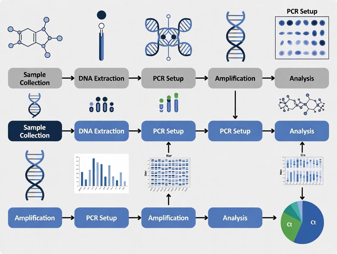
Integrating PCR for intestinal protozoa detection into clinical laboratory workflows requires careful planning and validation. The following diagram and workflow outline the key steps for implementation:
Key Implementation Considerations:
Specimen Selection and Acceptance: Establish criteria for appropriate specimen collection, transport conditions, and rejection criteria. Stool specimens should be preserved appropriately if testing cannot be performed promptly.
Nucleic Acid Extraction Optimization: Validate extraction methods for efficient recovery of protozoan DNA/RNA from stool specimens, which contain PCR inhibitors that must be removed.
assay Selection and Validation: Choose multiplex panels based on local epidemiology and validate performance characteristics (sensitivity, specificity, reproducibility) according to regulatory guidelines.
Quality Control Procedures: Implement comprehensive quality control including extraction controls, amplification controls, and periodic proficiency testing to ensure result reliability [11].
Result Interpretation and Reporting: Establish clear criteria for positivity, including Ct value cut-offs and algorithms for indeterminate results. Reports should clearly distinguish between pathogenic and non-pathogenic species when applicable.
Data Integration with Public Health Systems: Develop automated systems for transmitting reportable results to public health authorities to facilitate timely surveillance and outbreak response [12].
Essential Research Reagent Solutions
Successful implementation of PCR-based surveillance requires specific reagents and materials optimized for protozoan detection:
Table 3: Essential Research Reagents for Protozoan PCR Detection
| Reagent/Material | Function | Application Example | Considerations |
|---|---|---|---|
| Primer/Probe Sets | Species-specific detection through targeted amplification | Differentiation of E. histolytica from E. dispar [6] | Requires conserved, unique genomic regions; BLAST verification essential |
| Nucleic Acid Extraction Kits | Isolation of inhibitor-free DNA/RNA from complex matrices | Soil removal from food samples; inhibitor removal from stool [6] [14] | Must include inhibition controls; validated for sample type |
| qPCR Master Mixes | Enzymes, buffers, dNTPs for amplification | Multiplex detection of multiple protozoa in single reaction [6] | Should be compatible with multiplexing; robust against inhibitors |
| Positive Control Materials | Assay validation and quality monitoring | Quantified parasite (oo)cysts or synthetic genetic material [11] | Must mimic clinical samples; appropriate storage to maintain integrity |
| Viability Markers | Discrimination of live vs. dead parasites | Propidium monoazide (PMA) for DNA exclusion from dead cells; mRNA targets [14] | PMA concentration and exposure must be optimized for each protozoa |
| Process Controls | Monitoring extraction efficiency and inhibition | Exogenous DNA/spiked organisms added to each sample [11] | Should not cross-react with target assays; quantifiable |
The significant public health burden imposed by intestinal protozoa necessitates accurate surveillance data for effective control and prevention strategies. Traditional microscopic methods, while historically valuable, lack the sensitivity, specificity, and efficiency required for comprehensive surveillance programs. PCR-based methodologies represent a paradigm shift in protozoan detection, offering species-level differentiation, high throughput capacity, and quantitative assessment that dramatically improves surveillance accuracy.
The implementation of these molecular techniques in clinical and public health laboratories enables more precise burden estimates, enhances outbreak detection capabilities, and supports drug development efforts through better understanding of true disease epidemiology. As costs decrease and technology advances, the integration of PCR into routine surveillance workflows, particularly in resource-limited settings with high disease burden, promises to revolutionize our approach to controlling intestinal protozoan infections.
Future directions should focus on developing standardized multiplex assays, establishing external quality assessment programs, and creating data sharing platforms to maximize the public health impact of molecular surveillance. Through the widespread adoption of these advanced diagnostic tools, we can generate the accurate data necessary to guide evidence-based interventions and reduce the substantial global burden of intestinal protozoan infections.
Polymerase Chain Reaction (PCR) technologies have revolutionized diagnostic microbiology and biomedical research, offering two transformative advantages over traditional methods: the ability to differentiate between morphologically identical species and the capacity for high-throughput analysis. Within clinical laboratory workflows for protozoal detection, these capabilities are paramount. Traditional diagnostic methods, such as bright-field microscopy, are often unable to distinguish between pathogenic and non-pathogenic species that appear identical visually, potentially leading to misdiagnosis and inappropriate treatment decisions [6] [15]. Furthermore, the slow throughput of conventional techniques creates bottlenecks in laboratory workflows, delaying results and impeding large-scale surveillance studies [16] [17]. This whitepaper examines the technical foundations, experimental evidence, and practical implementations of PCR that address these critical challenges in protozoal diagnostics, providing researchers and drug development professionals with a framework for leveraging these capabilities in clinical and research settings.
Species-Level Differentiation
The Limitation of Morphological Identification
Traditional microscopy, while cost-effective and widely available, faces significant challenges in distinguishing between genetically distinct species that share morphological similarities. This limitation has profound implications for clinical management, particularly for protozoal infections where pathogenic and non-pathogenic species coexist. For instance, Entamoeba histolytica, the causative agent of amoebic dysentery and extra-intestinal abscesses, is morphologically identical to the non-pathogenic Entamoeba dispar [6] [15]. Similarly, the human Plasmodium ovale species, now recognized as two distinct species P. ovale curtisi and P. ovale wallikeri, cannot be reliably differentiated by microscopic examination [18]. Without molecular differentiation, patients colonized with non-pathogenic species may receive unnecessary treatment, while those with true pathogens might be misdiagnosed, leading to disease progression and complications.
Molecular Mechanisms for Differentiation
PCR-based methods overcome these limitations by targeting genetic sequences that exhibit variability between species, even when phenotypic differences are absent. Several molecular approaches enable this precise discrimination:
- Repetitive-Sequence-Based PCR (rep-PCR): This technique amplifies the non-coding repetitive sequences dispersed throughout microbial genomes, generating DNA fingerprint patterns that are species-specific. Studies on Aspergillus species have demonstrated 98% concordance with morphological identification and 100% agreement with internal transcribed spacer (ITS) region sequencing [19].
- Species-Specific Primers and Probes: Quantitative real-time PCR (qPCR) assays utilize primers and hydrolysis probes (e.g., TaqMan) designed to bind unique genetic regions of each species. For Plasmodium ovale differentiation, two separate real-time PCRs targeting species-specific sequences can definitively identify P. o. curtisi and P. o. wallikeri [18].
- Multiplex PCR Panels: Commercial multiplex real-time PCR assays, such as the Allplex GI-Parasite Assay, simultaneously detect and differentiate multiple intestinal protozoa in a single reaction, providing exceptional sensitivity and specificity [15].
Table 1: Performance of PCR Assays in Differentiating Protozoal Species
| Pathogen/Target | PCR Method | Discriminatory Capability | Performance Metrics |
|---|---|---|---|
| Entamoeba histolytica vs. E. dispar | Multiplex qPCR | Distinguishes pathogenic from non-pathogenic species | Sensitivity: 100%, Specificity: 100% for E. histolytica [15] |
| Plasmodium ovale curtisi vs. P. ovale wallikeri | Duplex & separate qPCRs | Differentiates two sympatric species | 100% concordance between two different PCR protocols [18] |
| Aspergillus fumigatus, A. flavus, A. terreus | rep-PCR (DiversiLab) | Species identification and strain-level differentiation | 98% concordance with morphology; 100% agreement with ITS sequencing [19] |
| Giardia duodenalis, Cryptosporidium spp., Dientamoeba fragilis | High-throughput multiplex qPCR | Simultaneous detection and identification of multiple pathogens | Sensitivity: 100% (Giardia), 97.2% (D. fragilis); Specificity: 99.2% (Giardia), 100% (D. fragilis) [15] |
Experimental Protocol: Differentiation ofPlasmodium ovaleSpecies
The following protocol, adapted from a study comparing two real-time PCR methods for discriminating P. ovale curtisi and P. ovale wallikeri, outlines the experimental workflow [18]:
- Nucleic Acid Extraction: Extract genomic DNA from 200 µL of EDTA-blood using the QIAamp DNA Blood Mini Kit (Qiagen). Elute DNA in a final volume of 50-100 µL.
- PCR Protocol 1 (Duplex Real-time PCR):
- Reaction Mix: Prepare 20 µL reactions containing HotStarTaq Mastermix (Qiagen), 3 mmol/L MgCl₂, 1.6 pmol/µL of each primer, 0.1 pmol/µL of each species-specific probe, and 5 µL of template DNA.
- Thermal Cycling: Conduct on a Corbett RotorGene 6000/Q cycler with: Initial denaturation at 95°C for 10 min; 45 cycles of denaturation at 95°C for 10 s, and combined annealing/extension at 60°C for 30 s; final cooling to 40°C for 30 s.
- PCR Protocol 2 (Separate Singleplex Reactions):
- Reaction Setup: Prepare separate 20 µL reactions for P. o. curtisi and P. o. wallikeri with HotStarTaq Mastermix, 3 mmol/L MgCl₂, 1.2 pmol/µL of species-specific primers, 0.25 pmol/µL of species-specific probe, and 5 µL of template DNA.
- Thermal Cycling: Use conditions identical to Protocol 1.
- Data Analysis: Analyze amplification curves. A sample is considered positive for a specific species if a characteristic exponential fluorescence curve crosses the threshold within 45 cycles.
Diagram 1: Workflow for Plasmodium ovale Species Differentiation
High-Throughput Capability
Technological Foundations for Scalability
The transition to high-throughput PCR testing is facilitated by innovations in instrumentation, reagent chemistry, and laboratory automation, enabling laboratories to process hundreds to thousands of samples daily.
- Instrumentation and Miniaturization: Modern real-time PCR instruments are available in 96, 384, and 1536-well formats, dramatically increasing throughput per run [20]. Integration with automated plate handlers and robotic liquid handling systems (e.g., CyBio Vario, Beckman Multimek) further streamlines workflow and reduces hands-on time [20].
- Fast PCR Chemistry: Advanced master mixes (e.g., TaqMan Fast Advanced Master Mix) enable significant reduction of thermal cycling times. Standard 2-hour runs can be completed in as little as 40-60 minutes, potentially tripling daily instrument throughput [16].
- Multiplexing: The ability to detect multiple targets in a single reaction by using probes labeled with different fluorophores exponentially increases data output per run. This is particularly valuable for comprehensive pathogen screening panels [16] [15].
- Automated Data Analysis: High-throughput data analysis methods, such as the "dots in boxes" approach, facilitate rapid quality assessment and interpretation of large qPCR datasets against MIQE (Minimum Information for Publication of Quantitative Real-Time PCR Experiments) guidelines [21].
Implementation in Protozoal Detection
High-throughput qPCR (HT-qPCR) has been successfully implemented for large-scale surveillance of intestinal and waterborne protozoa. One study developed an HT-qPCR assay targeting 19 waterborne protozoa and 3 helminths, validating its application on drinking water sources, wastewater sludge, and livestock manure [17]. The assay demonstrated a limit of detection of 5×10² copies/μL DNA and excellent repeatability, with coefficients of variation of 1.0%–4.6% in intra-group experiments [17]. Another study utilized a high-throughput multiplex qPCR for Giardia lamblia and Cryptosporidium spp. detection in stool samples from 44 asymptomatic infants, confirming its suitability for large-scale epidemiological studies [22].
Table 2: High-Throughput qPCR Performance Metrics in Pathogen Detection
| Application/Assay | Throughput Features | Sensitivity & Efficiency | Sample Types |
|---|---|---|---|
| Waterborne Pathogen Screening [17] | 22 targets simultaneously | LOD: 5×10² copies/μL; Efficiency: 80-107%; R²: 0.983-0.998 | Drinking water, WWTP sludge, Manure |
| Intestinal Protozoa Detection (Allplex Assay) [15] | Multiplex (6 targets) | Sensitivity: 97.2-100%; Specificity: 99.2-100% | Human stool samples |
| Giardia & Cryptosporidium Detection [22] | 96-well plate format | Efficient DNA extraction & amplification | Stool samples (asymptomatic infants) |
| Fast qPCR Workflow [16] | Reduced cycle times (40-60 min) | Equivalent performance to standard protocols | Cell culture, clinical samples |
Experimental Protocol: High-Throughput Screening for Waterborne Protozoa
The following protocol summarizes the development and validation of an HT-qPCR assay for 22 waterborne pathogens [17]:
- Assay Design: Design primer and probe sets for 19 protozoa and 3 helminths. Synthesize and clone target sequences into plasmids to generate positive controls.
- Sensitivity and Standard Curves: Serially dilute positive control plasmids from 1×10⁶ to 5×10² copies/μL. Perform HT-qPCR in triplicate to determine the limit of detection (LOD) and create standard curves. Calculate amplification efficiency using the formula: Efficiency = (10^(-1/slope) - 1) × 100%.
- Specificity Testing: Validate assay specificity using DNA from related non-target organisms and confirmed positive samples (e.g., Giardia and Cryptosporidium standards).
- Repeatability Assessment: Conduct intra- and inter-group reproducibility testing at high (1×10⁵ copies/μL) and low (1×10⁴ copies/μL) concentrations. Calculate coefficients of variation (CV).
- Environmental Sample Processing: Collect and concentrate water samples from various sources (e.g., drinking water plants, MWTPs). Extract DNA using optimized high-throughput kits (e.g., QIAamp 96 DNA QIAcube HT kit).
- HT-qPCR Run: Set up reactions in 384-well plates using a liquid handling robot. Run amplification with fast-cycling conditions. Analyze results automatically with customized software.
Diagram 2: High-Throughput Pathogen Screening Workflow
The Scientist's Toolkit: Research Reagent Solutions
Successful implementation of PCR for species differentiation and high-throughput applications relies on specialized reagents and kits. The following table details key solutions used in the cited research.
Table 3: Essential Research Reagents for Advanced PCR Applications
| Reagent/Kits | Primary Function | Application Context |
|---|---|---|
| DiversiLab System [19] | Automated rep-PCR DNA fingerprinting | Microbial identification and strain typing of fungi like Aspergillus |
| TaqMan Fast Advanced Master Mix [16] | Fast qPCR amplification | Reduces standard 2-hour run times to under 40 minutes for high-throughput screening |
| Allplex GI-Parasite Assay [15] | Multiplex detection of enteric protozoa | Simultaneous identification of Giardia, Entamoeba histolytica, Cryptosporidium, Dientamoeba fragilis, Blastocystis hominis, and Cyclospora in stool |
| QIAamp DNA Blood/Stool Mini Kits [22] [18] | Nucleic acid extraction from complex samples | Efficient DNA isolation from blood (e.g., for Plasmodium) and stool (e.g., for Giardia, Cryptosporidium) |
| HotStarTaq Mastermix [18] | High-sensitivity qPCR amplification | Used in species-differentiation protocols for Plasmodium ovale |
| Microlab Nimbus IVD System [15] | Automated nucleic acid extraction and PCR setup | Enables full walkaway automation for high-throughput laboratory workflows |
The synergistic advantages of species-level differentiation and high-throughput capability position PCR as an indispensable technology in modern parasitology and clinical diagnostics. The ability to accurately discriminate between pathogenic and non-pathogenic protozoa directly impacts patient management and therapeutic decisions, while the capacity for rapid, large-scale screening is crucial for epidemiological surveillance, outbreak investigation, and public health risk assessment. As reagent chemistries, instrumentation, and data analysis methods continue to advance, PCR-based workflows will become even more deeply integrated into clinical laboratory practice, ultimately enhancing diagnostic precision, accelerating time-to-result, and informing more effective control strategies for protozoal diseases. For researchers and drug development professionals, leveraging these capabilities is essential for advancing both fundamental understanding and clinical management of parasitic infections.
Intestinal protozoa infections present a major global public health challenge, causing significant gastrointestinal morbidity, malnutrition, and increased mortality worldwide [6]. These parasites are among the leading etiological agents of diarrheal diseases, affecting approximately 3.5 billion people and causing about 1.7 billion episodes of diarrheal disorders annually [23]. The epidemiological burden disproportionately affects areas with poor sanitation and limited access to clean water, though these pathogens also remain a critical concern in high-income nations, particularly among returning travelers, immunocompromised patients, and migrant populations [24] [25].
Among the most clinically significant protozoa are Entamoeba histolytica, the causative agent of amoebiasis which results in 40,000-100,000 deaths annually; Giardia duodenalis (also known as Giardia intestinalis or Giardia lamblia), responsible for approximately 280 million symptomatic infections yearly; and Cryptosporidium spp., which causes severe diarrheal disease particularly in children and immunocompromised individuals [6] [23]. Other protozoa such as Dientamoeba fragilis, Blastocystis spp., and Cyclospora cayetanensis contribute significantly to the global disease burden, though their pathogenicity remains debated in some cases [24].
The diagnostic landscape for these infections has traditionally been dominated by microscopic examination of stool specimens, which remains the reference method in many clinical laboratories, particularly in resource-limited settings [23]. However, this technique presents significant limitations in sensitivity, specificity, and ability to differentiate morphologically identical species, necessitating the development and implementation of more advanced diagnostic methodologies such as polymerase chain reaction (PCR) technologies [6] [26].
Diagnostic Challenges and the Role of PCR Technology
Limitations of Conventional Diagnostic Methods
Traditional microscopy, while cost-effective and widely available, suffers from several critical limitations that impact diagnostic accuracy and patient management. The technique requires significant technical expertise, is labor-intensive, and has subjective readout interpretation [6]. Furthermore, its sensitivity can be limited, especially for low-intensity infections, and it cannot differentiate between morphologically identical species such as the pathogenic Entamoeba histolytica and non-pathogenic Entamoeba dispar [24] [23]. This distinction is clinically crucial as it determines whether treatment is necessary.
Immunoassay methods such as enzyme-linked immunosorbent assay (ELISA) and immunochromatography have emerged as alternatives, offering rapid screening capabilities [23]. However, these methods frequently yield elevated rates of false positive and false negative results, constraining their utility [23]. Antigen detection tests are available for some protozoa but do not cover the full spectrum of potential enteric protozoal pathogens [25].
Advantages of Molecular Diagnostics
Molecular diagnostics, particularly real-time PCR (qPCR), have revolutionized protozoan detection by offering unparalleled sensitivity, specificity, and species differentiation capability [6]. PCR-based methods provide objective results, higher throughput capacity, and reduced turnaround time compared to conventional techniques [25]. The implementation of multiplex PCR panels allows simultaneous detection of multiple pathogens from a single sample, transforming laboratory workflows and enhancing diagnostic efficiency [24] [25].
The enhanced sensitivity of molecular methods is particularly valuable in non-endemic settings characterized by low parasitic prevalence, where the probability of detecting infections via conventional methods is reduced [23]. Additionally, molecular techniques have demonstrated utility in monitoring treatment response and detecting low-level persistent infections that might be missed by other methods [27].
PCR Methodologies and Technical Implementations
Commercial Multiplex PCR Platforms
Commercial multiplex PCR panels have gained widespread adoption in clinical laboratories, offering standardized, automated solutions for intestinal protozoa detection. The AllPlex Gastrointestinal Panel assay (Seegene) targets six protozoa: Giardia intestinalis, Cryptosporidium spp., Entamoeba histolytica, Dientamoeba fragilis, Blastocystis spp., and Cyclospora spp. [24]. This assay utilizes automated DNA extraction and amplification processes, reducing hands-on time and potential for contamination. Validation studies demonstrate high performance characteristics for most targets, though some evaluations have shown variable sensitivity for Entamoeba histolytica [25].
Another commercial system, the AusDiagnostics PCR assay, has been evaluated in multicentre studies showing complete agreement with in-house PCR methods for Giardia duodenalis detection and high specificity for Cryptosporidium spp. and Dientamoeba fragilis, though with some limitations in sensitivity potentially related to DNA extraction efficiency [23].
Table 1: Performance Characteristics of Commercial Multiplex PCR Assays
| Target Pathogen | Sensitivity (%) | Specificity (%) | PPV (%) | NPV (%) | Notes |
|---|---|---|---|---|---|
| Blastocystis hominis | 93.0 | 98.3 | 85.1 | 99.3 | [25] |
| Cryptosporidium spp. | 100 | 100 | 100 | 100 | [25] |
| Cyclospora cayetanensis | 100 | 100 | 100 | 100 | [25] |
| Dientamoeba fragilis | 100 | 99.3 | 88.5 | 100 | [25] |
| Entamoeba histolytica | 33.3-75.0 | 100 | 100 | 99.6 | Higher sensitivity with frozen specimens [25] |
| Giardia lamblia | 100 | 98.9 | 68.8 | 100 | [25] |
In-House and Custom PCR Assays
Laboratories often develop in-house PCR assays to address specific diagnostic needs or target additional pathogens not included in commercial panels. Lotz et al. (2025) implemented two duplex qPCR assays to detect Entamoeba dispar + Entamoeba histolytica and Cryptosporidium spp. + Chilomastix mesnili, along with singleplex assays for Giardia duodenalis and Blastocystis spp., using a reduced 10 µL reaction volume [6]. This approach marked the first molecular detection of Chilomastix mesnili by qPCR, demonstrating the flexibility of in-house assays to expand diagnostic capabilities [6].
The primer and probe design for such assays typically targets conserved regions of ribosomal RNA genes, which exist in high copy numbers and provide enhanced sensitivity [6] [26]. For novel targets like C. mesnili, design strategies involve retrieving sequences from databases, identifying highly conserved regions, and verifying specificity through BLAST searches against non-target organisms [6].
Sample Processing and DNA Extraction Methods
Optimal sample processing is critical for reliable PCR detection of intestinal protozoa. Studies comparing fresh versus preserved stool samples have demonstrated that molecular assays perform better with preserved specimens, likely due to improved DNA preservation [23]. Various DNA extraction methods have been employed, including manual phenol-chloroform extraction, automated magnetic bead-based systems (e.g., MagNA Pure 96 System, QIAsymphony), and commercial kits specifically designed for difficult stool matrices [27] [23].
The robust wall structure of protozoan cysts and oocysts presents a particular challenge for DNA extraction, often requiring mechanical disruption through bead-beating or enzymatic lysis to ensure efficient nucleic acid release [27] [23]. Incorporation of internal controls is essential to monitor extraction efficiency and PCR inhibition, with some laboratories using Phocine Herpes virus 1 (PhHV-1) or human 16S mitochondrial rRNA as control targets [6] [27].
Figure 1: PCR Workflow for Protozoa Detection. This diagram illustrates the standard workflow from sample collection to clinical reporting in molecular detection of intestinal protozoa.
Comparative Performance in Endemic versus Non-Endemic Settings
Application in Non-Endemic Regions
In non-endemic settings characterized by low disease prevalence, PCR technologies offer particular advantages due to their high sensitivity and specificity. Studies conducted in Italy and the Netherlands have demonstrated the utility of molecular methods for detecting imported cases in travelers, migrants, and immunocompromised patients [28] [27]. In these contexts, the ability to differentiate pathogenic from non-pathogenic species is particularly valuable for appropriate patient management and avoiding unnecessary treatment.
A cost-effectiveness analysis of congenital Chagas disease screening in Italy, a non-endemic country, demonstrated that PCR-based screening programs are economically viable, with an incremental cost-effectiveness ratio of €15,193 per quality-adjusted life year gained, well within accepted thresholds [28]. This highlights the public health value of molecular diagnostics even in low-prevalence settings.
Implementation in Endemic Areas
In endemic regions with high disease burden, PCR applications face different challenges related to infrastructure, cost, and technical expertise. A study implementing qPCR assays on Pemba Island, Tanzania, demonstrated high detection rates, with protozoa identified in 74.4% of samples and Entamoeba histolytica/dispar complex detected in 31.4% of cases [6]. Notably, one-third of these infections were caused by the pathogenic E. histolytica [6].
The high throughput and objectivity of molecular methods make them valuable for epidemiological studies and monitoring control programs in endemic areas [6]. However, logistical constraints related to laboratory infrastructure, reagent supply chains, and technical training currently limit widespread implementation in resource-limited settings [6].
Table 2: Detection Rates of Intestinal Protozoa by PCR in Different Settings
| Pathogen | Detection Rate in Non-Endemic Settings | Detection Rate in Endemic Settings | Notes |
|---|---|---|---|
| Blastocystis spp. | 19.25% (France) [24] | 50-60% (Developing countries) [6] | Often considered commensal |
| Cryptosporidium spp. | 0.85% (France) [24] | Varies widely by region | Higher in children and immunocompromised |
| Dientamoeba fragilis | 8.86% (France) [24] | Varies widely by region | Pathogenicity debated |
| Entamoeba histolytica | 0.25% (France) [24] | 10.5% of E. histolytica/dispar complex (Tanzania) [6] | Accurate differentiation from E. dispar crucial |
| Giardia duodenalis | 1.28% (France) [24] | Highly variable by region | One of most common human parasites |
Advanced Molecular Techniques and Future Directions
Innovations in PCR Technology
Recent technological innovations have enhanced the capabilities of PCR-based parasite detection. Automated high-throughput platforms, such as the Hamilton STARlet liquid handling system combined with multiplex parasitic real-time PCR panels, have significantly reduced hands-on time and improved workflow efficiency [25]. One validation study reported a 7-hour reduction in pre-analytical and analytical testing turnaround time compared to conventional methods [25].
Novel approaches like immuno-PCR (I-PCR) combine the specificity of immunoassays with the amplification power of PCR, enabling ultrasensitive detection of antigens and antibodies [29]. While still primarily a research tool, I-PCR has demonstrated detection limits reaching femtogram levels for various pathogens, though clinical translation remains limited by assay complexity and equipment requirements [29].
Multiplexing and Multipathogen Panels
The development of comprehensive multipathogen panels represents a significant advancement in molecular parasitology. These panels allow simultaneous detection of protozoa, helminths, and other enteric pathogens from a single sample, providing a comprehensive diagnostic approach particularly valuable in returning travelers with unexplained diarrhea [27]. One study implemented a multiplex helminth PCR targeting nine different helminths alongside protozoal detection [27].
Multiplexing strategies have evolved to include duplex reactions that conserve reagents and reduce costs, an important consideration for resource-limited settings. Lotz et al. successfully implemented duplex qPCR assays for Entamoeba dispar + Entamoeba histolytica and Cryptosporidium spp. + Chilomastix mesnili without compromising sensitivity or specificity [6].
Figure 2: Diagnostic Algorithm for Protozoal Infections. This flowchart illustrates the decision-making process for selecting appropriate diagnostic methods based on clinical and epidemiological context.
Automation and High-Throughput Solutions
Automation of nucleic acid extraction and PCR setup has addressed several limitations of early molecular methods, including labor intensity, contamination risk, and result variability [25]. Integrated systems combining automated extraction with multiplex PCR have demonstrated excellent performance characteristics while significantly reducing hands-on time [25]. These advancements make molecular methods increasingly feasible for routine diagnostic use even in medium-throughput laboratories.
Integration into Clinical Laboratory Workflows
Operational Considerations and Workflow Optimization
Implementing PCR for intestinal protozoa detection requires careful consideration of laboratory workflows, staffing expertise, and test utilization strategies. Studies comparing commercial and in-house PCR assays with microscopy have guided evidence-based implementation [23]. While molecular methods demonstrate superior performance for most protozoa, microscopy retains value for detecting helminths and parasites not included in PCR panels, suggesting a complementary approach may be optimal in many settings [24] [23].
Workflow optimization should consider sample triaging algorithms based on patient demographics, clinical presentation, and exposure history. For example, microscopy may be prioritized for migrants from helminth-endemic areas or immunocompromised patients at risk for Cystoisospora belli infection, which is not targeted by some commercial PCR panels [24].
Quality Assurance and Proficiency Testing
Robust quality assurance programs are essential for reliable PCR-based parasite detection. Participation in external quality assessment schemes, such as the Helminth External Molecular Quality Assessment Scheme (HEMQAS), helps ensure assay performance and inter-laboratory consistency [27]. Internal quality controls should include extraction controls, amplification controls, and periodic verification of assay sensitivity and specificity.
Standardization of pre-analytical factors such as sample collection, storage conditions, and DNA extraction methods is crucial for reproducible results across laboratories [23]. Multicentre studies have highlighted variability in performance related to these factors, emphasizing the need for standardized protocols [23].
The Scientist's Toolkit: Essential Research Reagents and Materials
Table 3: Essential Research Reagents for Protozoan PCR Detection
| Reagent/Material | Function | Examples/Specifications |
|---|---|---|
| DNA Extraction Kits | Nucleic acid purification from stool | STARMag Universal Cartridge (Seegene), MagNA Pure 96 DNA and Viral NA Small Volume Kit (Roche), QIAsymphony DSP Virus/Pathogen Midi Kit (Qiagen) |
| PCR Master Mixes | Amplification reaction components | HotStar Taq Master Mix (QIAGEN), QuantiTect Multiplex PCR NoROX Kit (QIAgen), PowerUp SYBR Green master mix (Applied Biosystems) |
| Primers and Probes | Target-specific detection | Custom designs for conserved regions (e.g., 18S rRNA gene), commercially available mixes (e.g., AllPlex GI-Parasite Assay) |
| Internal Controls | Monitoring extraction efficiency and inhibition | Phocine Herpes virus 1 (PhHV-1), human 16S mitochondrial rRNA |
| Automation Systems | High-throughput processing | Hamilton STARlet liquid handler, MagNA Pure 96 System, QIAsymphony SP system |
| Amplification Instruments | PCR execution and detection | Bio-Rad CFX96, QuantStudio 5 (Applied Biosystems), Rotor-Gene (Qiagen) |
PCR technologies have fundamentally transformed the diagnostic landscape for intestinal protozoa in both endemic and non-endemic settings. The enhanced sensitivity, specificity, and species differentiation capabilities of molecular methods address critical limitations of conventional diagnostics, enabling more accurate detection and appropriate management of these globally significant infections. Commercial multiplex panels and automated platforms have increased the feasibility of implementing molecular testing in routine clinical practice, while continued development of in-house assays expands the range of detectable pathogens.
The optimal integration of PCR into clinical laboratory workflows requires thoughtful consideration of local epidemiology, available resources, and patient populations. In non-endemic settings, PCR provides valuable diagnostic precision for imported infections and immunocompromised patients, while in endemic areas, it offers powerful tools for surveillance and control programs. Future directions will likely focus on further automation, multiplexing capabilities, and point-of-care applications to increase accessibility in resource-limited settings. As molecular technologies continue to evolve, their role in combating the global challenge of intestinal protozoa will undoubtedly expand, contributing to improved patient care and public health outcomes worldwide.
From Theory to Practice: Selecting and Implementing PCR Assays in the Lab
The diagnosis of intestinal protozoan infections, caused by pathogens such as Giardia duodenalis, Cryptosporidium spp., and Entamoeba histolytica, is undergoing a substantial transformation in clinical laboratories worldwide. For decades, microscopic examination of stool specimens has served as the reference standard, but this technique presents significant limitations including time-consuming procedures, requirement for experienced personnel, and insufficient sensitivity and specificity [30]. Molecular diagnostic technologies, particularly real-time PCR (RT-PCR), are gaining traction in non-endemic areas characterized by low parasitic prevalence due to their enhanced sensitivity and specificity [30]. This technical guide examines the critical decision between implementing commercial multiplex PCR kits versus developing laboratory-developed tests (LDTs) within the context of clinical laboratory workflow research, providing evidence-based recommendations for researchers, scientists, and drug development professionals.
Performance Comparison: Commercial Kits vs. LDTs
Analytical Sensitivity and Specificity
Recent multicenter studies directly comparing commercial multiplex PCR assays with LDTs demonstrate excellent performance characteristics for both platforms, with some context-dependent variations.
Table 1: Performance Characteristics of Commercial Multiplex PCR Kits from Recent Studies
| Commercial Kit | Target Pathogens | Sensitivity (%) | Specificity (%) | Study Details |
|---|---|---|---|---|
| AllPlex GI-Parasite Assay (Seegene) | G. duodenalis | 100 | 99.2 | Multicentric Italian study (n=368 samples) [31] |
| E. histolytica | 100 | 100 | ||
| Cryptosporidium spp. | 100 | 99.7 | ||
| D. fragilis | 97.2 | 100 | ||
| AllPlex GI-Parasite Assay (Seegene) | G. intestinalis | 100 | 98.9 | Validation study (n=461 fresh specimens) [25] |
| Cryptosporidium spp. | 100 | 100 | ||
| D. fragilis | 100 | 99.3 | ||
| E. histolytica | 33.3-75* | 100 | *Increased with frozen specimens | |
| AusDiagnostics | G. duodenalis | Complete agreement with in-house PCR | Multicentre study (n=355 samples) [30] | |
| Cryptosporidium spp. | High specificity, limited sensitivity | |||
| D. fragilis | High specificity, limited sensitivity |
A 2025 multicentric Italian study evaluating the AllPlex GI-Parasite Assay demonstrated exceptional performance, with 100% sensitivity and specificity for Entamoeba histolytica detection, and 100% sensitivity with 99.2% specificity for Giardia duodenalis [31]. Similarly, the assay showed 100% sensitivity and 99.7% specificity for Cryptosporidium spp., and 97.2% sensitivity with 100% specificity for Dientamoeba fragilis [31].
Another 2025 validation study of the same assay reported 100% sensitivity for Giardia intestinalis and Cryptosporidium spp., though sensitivity for Entamoeba histolytica was lower (33.3% on fresh specimens, increasing to 75% with frozen specimens) [25]. This highlights the impact of sample preservation on molecular detection efficiency.
Comparative studies have shown complete agreement between commercial and in-house PCR methods for detecting G. duodenalis, with both demonstrating high sensitivity and specificity comparable to conventional microscopy [30]. For other protozoa like Cryptosporidium spp. and D. fragilis, both methods may show high specificity but potentially limited sensitivity, possibly due to challenges in DNA extraction from these parasites [30].
Multiplexing Capacity and Workflow Efficiency
Commercial multiplex panels offer significant advantages in comprehensive pathogen detection and workflow standardization. The AllPlex Gastrointestinal Panel assay targets six protozoa: Giardia intestinalis, Cryptosporidium spp., Entamoeba histolytica, Dientamoeba fragilis, Blastocystis spp., and Cyclospora spp. [24]. This extensive multiplexing capability allows laboratories to detect multiple pathogens in a single reaction, conserving sample volume and reducing hands-on time.
Studies implementing automated DNA extraction and amplification platforms report substantial reductions in pre-analytical and analytical testing turnaround time—by approximately 7 hours per batch compared to conventional methods [25]. This workflow efficiency is further enhanced by standardized reagent preparation and automated result interpretation through manufacturer-provided software.
Limitations and Detection Gaps
Despite their advantages, commercial multiplex panels have specific limitations that researchers must consider:
Pathogen Coverage Gaps: Most commercial panels do not target helminths or less common protozoa like Cystoisospora belli, which remains clinically important for immunocompromised patients such as those with HIV [24].
Fixed Target Panels: Commercial assays offer limited flexibility to add emerging pathogens or region-specific targets without waiting for manufacturer updates.
Extraction Dependencies: Performance can vary based on the DNA extraction method used, with some protocols demonstrating better efficiency for certain parasites [30].
Regulatory Landscape: Implications for Laboratory Testing
The regulatory framework for LDTs recently underwent significant changes that impact laboratory implementation decisions. In a landmark March 2025 ruling, the U.S. District Court for the Eastern District of Texas vacated the FDA's Final Rule on Laboratory Developed Tests, asserting that LDTs constitute services rather than products and therefore fall outside the FDA's medical device authorities [32] [33].
This decision maintains the regulatory framework under which LDTs are primarily overseen by the Centers for Medicare & Medicaid Services (CMS) through the Clinical Laboratory Improvement Amendments (CLIA), rather than through FDA premarket review processes [32]. This regulatory environment has historically enabled laboratories to develop and implement tests quickly, particularly for emerging pathogens, rare diseases, and specialized patient populations [32].
For laboratories considering their testing options, this ruling means:
- LDTs remain accessible for specialized applications without additional FDA oversight
- CLIA compliance continues to govern LDT validation and quality assurance
- Innovation pathways remain open for tests targeting niche populations or rare pathogens
- Laboratory responsibility for thorough validation and performance verification is maintained
Implementation Considerations for Clinical Laboratories
Workflow Integration and Automation
Table 2: Implementation Considerations for Molecular Protozoan Detection
| Factor | Commercial Multiplex Kits | Laboratory-Developed Tests |
|---|---|---|
| Initial Validation | Manufacturer-provided performance data; verification required | Full validation required including sensitivity, specificity, reproducibility |
| Regulatory Pathway | FDA-cleared/CE-marked options available; standardized claims | CLIA compliance; laboratory responsibility for performance |
| Target Flexibility | Fixed panel; dependent on manufacturer updates | Customizable targets based on population needs |
| Technical Expertise | Standardized protocols; reduced technical burden | Significant expertise required for development/optimization |
| Cost Structure | Higher reagent costs; predictable budgeting | Lower reagent costs; higher development and validation costs |
| Turnaround Time | Rapid implementation; standardized workflows | Lengthy development and validation before implementation |
| Automation Potential | High; often designed for integrated platforms | Variable; depends on laboratory resources and expertise |
Commercial multiplex assays demonstrate particular strength in high-throughput settings where workflow automation and standardization are priorities. Integrated systems combining automated nucleic acid extraction with PCR setup significantly reduce hands-on time and potential for human error [25]. One validation study documented complete automation from extraction to amplification using the Hamilton STARlet liquid handler with the AllPlex GI-Parasite Assay, demonstrating the capacity for high-volume testing [25].
Sample Preparation and Preservation Considerations
Sample collection and preservation methods significantly impact molecular detection efficiency. Studies consistently show that PCR results from preserved stool samples often outperform those from fresh samples, likely due to better DNA preservation in fixed specimens [30]. The choice of transport media, such as S.T.A.R. Buffer or Cary-Blair media, should be validated for compatibility with both automated extraction systems and downstream amplification [30] [25].
For LDTs, DNA extraction protocols require particular attention, as the robust wall structure of protozoan cysts and oocysts can complicate DNA extraction efficiency [30]. Methods incorporating mechanical disruption through bead beating or vigorous vortexing with extraction buffers have demonstrated improved parasite DNA recovery [30].
Experimental Protocols for Method Evaluation
Protocol for Comparative Performance Assessment
Researchers conducting comparative studies between commercial kits and LDTs should implement rigorous experimental designs:
Sample Selection: Include both retrospective samples with known parasite status and prospective consecutive samples to evaluate real-world performance. Sample sets should include common protozoa (G. duodenalis, Cryptosporidium spp., E. histolytica) and less prevalent targets based on local epidemiology [30] [31].
Reference Method Selection: Use a composite reference standard incorporating microscopy, antigen testing, and alternative molecular methods to address imperfections in any single reference method [25] [31].
DNA Extraction Validation: Compare multiple extraction methods using the same clinical samples to identify protocol-specific variations in sensitivity. Include both manual and automated extraction systems [30] [25].
Inhibition Testing: Incorporate internal controls to identify inhibition events that may affect sensitivity, particularly for formed stool samples with complex matrices [24] [25].
Protocol for Clinical Sensitivity and Specificity Determination
To accurately determine clinical performance characteristics:
Blinded Testing: Perform all comparative testing with personnel blinded to reference method results and clinical data.
Sample Size Calculation: Ensure adequate sample sizes for each target pathogen to generate precise sensitivity and specificity estimates with narrow confidence intervals.
Discrepancy Analysis: Establish predefined algorithms for resolving discrepant results between methods, which may include additional molecular targets, sequencing confirmation, or follow-up testing.
Limit of Detection Studies: Determine analytic sensitivity for each target using dilution series of quantified parasites or synthetic controls.
The choice between commercial multiplex kits and LDTs for protozoan detection depends on multiple factors specific to each laboratory's context, resources, and patient population. The following decision pathway provides guidance for researchers and laboratory directors:
Recommendations for Laboratory Implementation
Based on current evidence and technical considerations:
For routine diagnostic laboratories with high testing volumes and standardized workflows, commercial multiplex kits provide excellent performance, regulatory simplicity, and operational efficiency.
For reference and research laboratories serving specialized populations or investigating emerging pathogens, LDTs offer essential flexibility for target customization and method adjustment.
For comprehensive parasitology services, a hybrid approach combining commercial multiplex panels for common protozoa with supplemental LDTs or microscopy for uncommon targets provides optimal coverage.
Regardless of platform selection, rigorous validation against laboratory-specific patient populations and continuous quality monitoring are essential for maintaining diagnostic accuracy.
Future Directions
The field of molecular parasitology continues to evolve, with emerging trends including:
- Expanded multiplex panels incorporating bacterial, viral, and parasitic targets
- Integration of sample-to-result automated platforms
- Development of quantitative PCR applications for parasite burden monitoring
- Implementation of next-generation sequencing for comprehensive pathogen detection and strain typing
As molecular technologies advance, the distinction between commercial kits and LDTs may blur, with laboratories potentially implementing manufacturer-developed tests with laboratory-specific customizations. The optimal approach will continue to depend on careful consideration of clinical needs, laboratory resources, and the evolving regulatory landscape.
The Scientist's Toolkit: Essential Research Reagents and Materials
Table 3: Key Research Reagent Solutions for Molecular Protozoan Detection
| Reagent/Material | Function | Examples/Considerations |
|---|---|---|
| Nucleic Acid Extraction Kits | Parasite DNA isolation from stool matrix | MagNA Pure 96 System (Roche), STARMag 96 × 4 Universal Cartridge (Seegene), NucliSENS easyMAG (BioMérieux) [30] [25] [34] |
| PCR Master Mixes | Amplification of target DNA | TaqMan Fast Universal PCR Master Mix (Thermo Fisher), Seegene AllPlex MOM [30] [25] |
| Transport Media | Sample preservation and nucleic acid stabilization | S.T.A.R. Buffer (Roche), Cary-Blair media, FecalSwab medium (COPAN) [30] [25] |
| Commercial Multiplex Kits | Multi-target detection in single reaction | AllPlex GI-Parasite Assay (Seegene), AusDiagnostics GI Panel [30] [24] [25] |
| Internal Controls | Monitoring extraction and amplification efficiency | Included in commercial kits; must be added to LDT protocols [25] |
| Positive Controls | Assay validation and quality control | Quantified parasites, synthetic oligonucleotides, or confirmed positive samples [34] |
The implementation of polymerase chain reaction (PCR) for protozoan detection represents a paradigm shift in clinical parasitology, moving away from reliance on subjective morphological identification towards precise, DNA-based diagnostics. This transition is critical for accurately understanding the epidemiology and true burden of intestinal protozoan diseases, which remain a significant cause of global gastrointestinal morbidity and mortality [6]. This technical guide details the essential workflow components for implementing a robust PCR-based detection system for intestinal protozoa within a clinical laboratory setting. It frames these components within the broader context of clinical workflow research, providing the foundational knowledge required for laboratories transitioning to molecular methods while maintaining diagnostic accuracy and operational efficiency.
Sample Collection and Preparation
The diagnostic journey begins at sample collection, a phase where accuracy is paramount. While molecular methods offer significant advantages, proper sample handling remains a critical determinant of success.
Sample Number and Workflow Optimization
Traditional microscopy algorithms often require the analysis of multiple stool samples collected on alternate days to overcome the intermittent shedding of parasites [5]. However, research demonstrates that the superior sensitivity of real-time PCR (Rt-PCR) enables a radical simplification of this workflow. A pivotal study found that a protocol using a single stool sample subjected to both a coproparasitological exam and Rt-PCR showed sensitivity comparable to the traditional approach of microscopic examination of three samples plus Rt-PCR on one sample [5]. This consolidation of diagnostics onto a single sample reduces costs, saves processing time, and improves patient compliance, representing a significant optimization for clinical workflows.
Sample Preservation and DNA Extraction
Once a sample is obtained, preservation and DNA extraction are crucial. Stool specimens can be stored in 10% formalin for microscopic examination, but for molecular analysis, 200 mg of stool is typically stored frozen in a solution of 1X PBS with 2% polyvinylpolypyrrolidone (PvPP) [5]. The DNA extraction process itself is a critical step where automation and protocol choice profoundly impact downstream results.
For complex matrices like stool, an effective DNA extraction protocol must overcome PCR inhibitors and efficiently break down robust protozoan oocyst and cyst walls. Mechanical lysis methods, such as those using the OmniLyse device, can achieve efficient lysis of Cryptosporidium oocysts within 3 minutes, providing a rapid alternative to time-consuming freeze-thaw cycles in liquid nitrogen or heat exposure that can compromise DNA integrity [35]. The inclusion of an internal control, such as Phocine Herpes Virus type-1 (PhHV-1), is essential for monitoring the efficiency of both the DNA isolation and amplification steps, ensuring the reliability of negative results [5].
Table 1: Key Sample Preparation and Storage Reagents
| Reagent/Solution | Function | Application Context |
|---|---|---|
| 10% Formalin | Preserves parasitic structures for morphological examination | Classical microscopy [5] |
| Polyvinylpolypyrrolidone (PvPP) | Binds polyphenols and other PCR inhibitors present in stool | Molecular diagnostics (DNA extraction) [5] |
| Phosphate Buffered Saline (PBS) | Provides a stable ionic and pH environment for sample storage | Sample suspension and storage buffer [5] |
| Internal Control (e.g., PhHV-1) | Monitors for PCR inhibition and extraction efficiency | Quality control in Rt-PCR [5] |
Automated Nucleic Acid Extraction
Automating the nucleic acid extraction process is a cornerstone of an efficient, high-quality molecular workflow. It minimizes human error, reduces hands-on time, and enhances the reproducibility of results, which is vital for sensitive downstream applications like qPCR and next-generation sequencing [36].
Core Principles and Chemistry
Most automated nucleic acid extraction systems are based on magnetic particle technology. The process follows four fundamental steps, regardless of the specific platform [37]:
- Lysis: A lysing solution is added to the sample to break apart cells and release nucleic acids.
- Binding: Magnetic beads, coated with a material that reversibly binds nucleic acids, are added to the lysate.
- Washing: The beads are captured by a magnet and washed with buffers to remove salts, proteins, and other contaminants.
- Elution: The purified nucleic acids are released from the beads into an aqueous elution buffer, such as water or TE buffer.
The two primary robotic platforms for automating this chemistry are particle movers and liquid handlers [37]. Particle movers, like the KingFisher systems, transport magnetic beads through a rack of pre-filled reagents using magnetic rods [36]. In contrast, liquid handlers use pipettes to transfer liquid reagents to and from a plate where magnetic beads are immobilized by an external magnet during liquid transfers [37].
Workflow Implementation and Optimization
Successful implementation begins not at the robot, but at the bench. The first step is to establish a manual magnetic particle-based extraction method that consistently meets quality control thresholds for purity, yield, and functionality in downstream PCR [37]. This manual method serves as a critical performance benchmark when optimizing the automated workflow.
Key parameters to optimize in a manual method, which will later translate to the automated system, include [37]:
- Binding Efficiency: Ensuring nucleic acids have sufficient time and mixing intensity to bind to the magnetic beads.
- Bead Washing: Achieving complete resuspension and dispersion of beads during wash steps to prevent contaminant trapping.
- Bead Drying: Allowing for adequate drying time to remove residual ethanol from wash buffers without over-drying, which can make nucleic acids difficult to elute.
When transitioning to the robotic platform, liquid handling parameters must be meticulously defined. Factors such as liquid density and viscosity must be programmed into the system's liquid classes to ensure accurate and drip-free transfers [37]. Pre-wetting pipettes for viscous liquids or drawing a small air gap before and after liquid aspiration can significantly improve volumetric accuracy.
Molecular Detection and Analysis
Following extraction, the purified nucleic acids are analyzed, typically via real-time PCR (qPCR), which provides high sensitivity, specificity, and the ability for multiplexing.
PCR Assay Design and Validation
The design of qPCR assays is critical for distinguishing between morphologically identical species. This is particularly important for pathogens like Entamoeba histolytica (pathogenic) and Entamoeba dispar (non-pathogenic), which are indistinguishable by microscopy [6] [5]. Assays can be designed as singleplex or multiplex. The latter allows for the detection of multiple targets in a single reaction, improving efficiency and reducing cost. One study implemented a 10 µL duplex qPCR to simultaneously detect E. dispar + E. histolytica and Cryptosporidium spp. + Chilomastix mesnili, alongside singleplex assays for Giardia duodenalis and Blastocystis spp. [6].
Table 2: Performance Comparison of Microscopy vs. Multiplex qPCR for Intestinal Protozoa Detection
| Parasite | Detection by Multiplex qPCR (n=3,495 samples) | Detection by Microscopy (n=3,495 samples) | Clinical and Workflow Implications |
|---|---|---|---|
| Giardia intestinalis | 45 (1.28%) | 25 (0.7%) | Higher sensitivity with qPCR leads to improved detection rates [38] |
| Cryptosporidium spp. | 30 (0.85%) | 8 (0.23%) | qPCR is significantly more sensitive; crucial for detecting this pathogen [38] |
| Entamoeba histolytica | 9 (0.25%) | 24 (0.68%)* | qPCR allows specific identification of the pathogenic E. histolytica, unlike microscopy which often detects the complex [38] |
| Dientamoeba fragilis | 310 (8.86%) | 22 (0.63%) | Massive increase in detection with qPCR, highlighting microscopy's poor sensitivity for this organism [38] |
| Blastocystis spp. | 673 (19.25%) | 229 (6.55%) | qPCR reveals a much higher carriage rate, aiding epidemiological understanding [38] |
| Note | Microscopy reported as *E. histolytica/dispar, unable to differentiate the species [38]. |
Comparative Performance in Clinical Workflows
Large-scale prospective studies have unequivocally demonstrated the superiority of multiplex qPCR over classical microscopy for detecting most intestinal protozoa. A three-year study of 3,495 stool samples found that multiplex qPCR detected a protozoan in 909 samples, compared to just 286 samples detected by microscopy [38]. The disparity was especially notable for Dientamoeba fragilis (310 vs. 22) and Blastocystis spp. (673 vs. 229) [38].
This enhanced sensitivity means that, in the vast majority of cases, PCR can detect a protozoan on the first stool sample, fundamentally changing the laboratory workflow by reducing the need for repeated sample collection and analysis [38]. However, the study also highlighted an important caveat: microscopy remains necessary for detecting parasites not included in commercial multiplex PCR panels, such as Cystoisospora belli (relevant for HIV-infected patients) and most helminths (relevant for migrants and travelers) [38]. Therefore, a reflexive algorithm, where microscopy is performed based on clinical suspicion or immunocompromised status, is often the most effective workflow.
The Scientist's Toolkit: Research Reagent Solutions
Successful implementation of a PCR workflow for protozoa relies on a suite of reliable reagents and instruments. The following table details key solutions used in the featured experiments and the broader field.
Table 3: Essential Research Reagents and Instruments for PCR-based Protozoan Detection
| Item | Function/Description | Application Example |
|---|---|---|
| KingFisher Instrument Series | Automated purification systems using magnetic rods to move beads through pre-filled reagents for nucleic acid extraction [36]. | Versatile benchtop automation for DNA/RNA extraction from complex samples like stool [36]. |
| CyBio FeliX | Liquid handling robot for automated nucleic acid extraction in a 96-well format, offering high throughput and flexibility [39]. | Processing large sample batches for epidemiological studies or clinical trials [39]. |
| MagnaPure LC.2 Instrument | Automated nucleic acid extraction system utilizing "DNA I Blood_Cells High performance II" protocol [5]. | Used with "DNA isolation kit I" for DNA extraction from stool samples prior to Rt-PCR [5]. |
| SsoFast Master Mix | Optimized buffer for fast, high-performance real-time PCR applications [5]. | Provides the enzymatic foundation for multiplex Rt-PCR reactions targeting protozoan DNA [5]. |
| AllPlex Gastrointestinal Panel Assay | Commercial multiplex real-time PCR (qPCR) assay for simultaneous detection of multiple gastrointestinal pathogens [38]. | Used in large-scale prospective studies for routine detection of protozoan parasites in stool samples [38]. |
| OmniLyse Device | Instrument for rapid and efficient mechanical lysis of robust oocysts and cysts [35]. | Rapid (3-minute) lysis of Cryptosporidium oocysts from lettuce samples for metagenomic sequencing [35]. |
The complete integration of these components into a coherent diagnostic pathway is visualized below. This workflow outlines the key decision points from sample arrival to final reporting, highlighting the complementary roles of molecular and traditional methods.
Integrated Diagnostic Workflow for Intestinal Parasites
In conclusion, the essential workflow for PCR-based detection of intestinal protozoa hinges on the seamless integration of optimized sample collection, automated nucleic acid extraction, and validated molecular detection assays. The evidence strongly supports a model where a single stool sample analyzed by a combination of multiplex qPCR and reflexive microscopy provides an optimal balance of sensitivity, specificity, and operational efficiency [38] [5]. This integrated approach, leveraging the strengths of both molecular and morphological techniques, provides the most accurate and comprehensive diagnostic outcome, forming a robust foundation for clinical decision-making, drug development research, and public health surveillance.
The laboratory diagnosis of intestinal protozoan infections has undergone a paradigm shift with the introduction of syndromic multiplex polymerase chain reaction (PCR) panels. These advanced molecular tools have revolutionized detection capabilities for pathogenic protozoa including Giardia duodenalis, Cryptosporidium spp., Entamoeba histolytica, Dientamoeba fragilis, and Blastocystis spp.—organisms responsible for significant global morbidity and challenging diagnostic dilemmas due to their often indistinguishable clinical presentations [40] [41]. Traditional diagnostic reliance on microscopic examination of stool specimens has been constrained by limitations including variable sensitivity, requirement for multiple samples, and significant technical expertise [24] [42]. Multiplex PCR panels simultaneously test for multiple pathogens in a single reaction, offering superior analytical sensitivity and specificity while dramatically reducing turnaround time [40] [43]. This technical guide explores the implementation, performance, and workflow integration of these molecular panels within the context of clinical laboratory research on protozoan detection.
The Diagnostic Targets: Clinical Significance and Conventional Challenges
The targeted protozoa in gastrointestinal panels represent a spectrum of pathogenic and potentially commensal organisms with significant public health implications. Entamoeba histolytica is an invasive pathogen capable of causing life-threatening conditions such as amoebic dysentery and liver abscesses, distinguishing it from non-pathogenic Entamoeba dispar which requires molecular differentiation [24] [44]. Giardia duodenalis (also known as G. intestinalis or G. lamblia) is a flagellate protozoan causing giardiasis, characterized by foul-smelling diarrhea, abdominal cramping, and bloating, with particular severity in immunocompromised individuals and children in developing regions [42]. Cryptosporidium spp. are intracellular apicomplexan parasites causing profuse, watery diarrhea that can become chronic and life-threatening in immunocompromised patients, especially those with AIDS [42].
The clinical significance of Dientamoeba fragilis and Blastocystis spp. remains debated, necessitating their sensitive detection for high-quality epidemiological studies to better understand their potential pathogenicity [24]. These organisms are of particular concern in specific patient populations, including returning travelers from endemic areas, immunocompromised individuals, men who have sex with men, and persons experiencing homelessness [40].
Conventional diagnostic methods present significant challenges for these targets. Microscopy, while historically the gold standard, suffers from limited sensitivity, often requiring the collection of multiple samples on different days to improve yield, and is heavily dependent on the expertise of the technologist [24] [42]. Antigen-based tests offered some improvement but have been largely abandoned for many targets due to concerns about sensitivity and specificity [40]. These limitations in conventional testing have driven the development and adoption of multiplex PCR panels that provide rapid, simultaneous detection of these challenging pathogens [40] [24].
Multiplex PCR Technology: Principles and Platforms
Fundamental Principles
Multiplex PCR is a variant of the polymerase chain reaction that enables simultaneous amplification of more than one target sequence in a single reaction tube by including multiple primer pairs [45]. This approach produces considerable savings in time, effort, and resources within the laboratory without compromising test utility. The technology has evolved significantly since its inception, with current systems employing sophisticated detection methodologies including the combined use of fluorescence color and melting temperature (Tm) as a virtual two-dimensional label that enables detection of significantly more targets than classical strategies on real-time PCR platforms [46].
The development of robust multiplex assays requires careful optimization to overcome challenges including poor sensitivity or specificity and preferential amplification of certain targets (PCR bias) [45]. Key considerations include primer design to ensure nearly identical optimum annealing temperatures, prevention of primer-dimer formation, and balancing reagent concentrations to support simultaneous amplification of multiple targets. Modern implementations often utilize hot start PCR methodology to eliminate nonspecific reactions and specialized buffer additives to prevent stalling of DNA polymerization through secondary structure formation [45].
Commercial Platform Landscape
Numerous commercial NAAT platforms for gastrointestinal pathogen detection are now available, with varying target menus that typically include the key protozoan parasites. These include the BioFire FilmArray system, xTAG GI pathogen panel, Verigene enteric pathogens panel, QIAstat-Dx GIP, BioCode GPP, and various panels for the BD MAX system [40]. The AllPlex Gastrointestinal Panel assay (Seegene) targets six protozoa: Giardia intestinalis, Cryptosporidium spp., Entamoeba histolytica, Dientamoeba fragilis, Blastocystis spp., and Cyclospora spp. [24]. Similarly, the Roche LightMix Modular Assay Gastro Parasites (LMAGP) detects Giardia duodenalis, Entamoeba histolytica, Cryptosporidium spp., Blastocystis hominis, and Dientamoeba fragilis [44].
These systems employ various technological approaches to achieve multiplexing. For instance, the AllPlex assay utilizes Seegene's proprietary Multiple Detection Temperature (MuDT) technology, which reports multiple Ct values of each pathogen in a single channel, and incorporates Dual Priming Oligonucleotide (DPO)-based real-time RT-PCR and Tagging Oligonucleotide Cleavage Extension (TOCE) technologies for enhanced specificity [43]. Other platforms like the cobas eplex system use specialized test cartridges and digital microfluidic technology for automated sample-to-answer processing [47].
Table 1: Comparison of Select Commercial Multiplex PCR Panels for Protozoan Detection
| Platform/Assay | Target Protozoa | Technology | Throughput | Sample-to-Answer Time |
|---|---|---|---|---|
| BioFire FilmArray GI Panel | Cryptosporidium, Cyclospora cayetanensis, Entamoeba histolytica, Giardia duodenalis | Nested PCR with endpoint melting curve analysis | Low | ~1 hour |
| AllPlex GI Panel (Seegene) | G. intestinalis, Cryptosporidium spp., E. histolytica, D. fragilis, Blastocystis spp., Cyclospora spp. | Multiplex real-time PCR using MuDT technology | Medium | ~3 hours |
| Roche LightMix Gastro Parasites | G. duodenalis, E. histolytica, Cryptosporidium spp., B. hominis, D. fragilis | Multiplex real-time PCR | Flexible | Varies |
| In-house triplex qPCR [42] | E. histolytica, G. lamblia, C. parvum | TaqMan-based triplex real-time PCR | Customizable | ~2 hours |
Performance Evaluation: Multiplex PCR Versus Conventional Methods
Detection Rates and Sensitivity
Prospective studies comparing multiplex PCR with conventional methods consistently demonstrate superior detection rates for intestinal protozoa. A comprehensive three-year study analyzing 3,495 stool samples found significantly higher detection rates using multiplex PCR compared to microscopic examination [24]. The PCR detected Giardia intestinalis in 1.28% of samples, Cryptosporidium spp. in 0.85%, Entamoeba histolytica in 0.25%, Dientamoeba fragilis in 8.86%, and Blastocystis spp. in 19.25% of samples. In contrast, microscopy identified these parasites in only 0.7%, 0.23%, 0.68%, 0.63%, and 6.55% of samples, respectively [24].
Another study evaluating the Roche LightMix Gastro Parasites assay on 1,062 specimens demonstrated similar advantages, with PCR identifying D. fragilis or B. hominis in 14.4% and 19.9% of samples, respectively, compared to significantly lower rates by microscopy [44]. Notably, for Giardia duodenalis, the multiplex PCR detected infections in 20 samples compared to 16 by enzyme immunoassay and only 9 by microscopy, with all PCR-positive, EIA-negative samples confirmed by in-house PCR [44]. This enhanced sensitivity is particularly valuable for detecting low-intensity infections that might be missed by conventional methods.
Diagnostic Accuracy Metrics
The analytical performance of multiplex PCR assays for protozoan detection has been rigorously evaluated in multiple studies. A triplex real-time quantitative PCR assay developed for simultaneous detection of E. histolytica, G. lamblia, and C. parvum demonstrated excellent specificity without cross-reactivity among target-specific TaqMan probes and no amplification of non-target species [42]. The assay showed a limit of detection of 500 copies/μL of standard plasmid DNA, with amplification efficiency exceeding 95% and R² values greater than 0.99, indicating good linearity across a wide dynamic range (5 × 10² to 5 × 10⁸ copies/μL) [42].
The reproducibility of these assays is generally high, with intra- and inter-assay coefficients of variation typically less than 2% [42]. This reliability, combined with the ability to detect multiple targets in a single reaction, makes multiplex PCR particularly valuable for comprehensive screening of gastrointestinal pathogens, especially in cases where clinical presentation does not point to a specific etiological agent.
Table 2: Performance Comparison of Multiplex PCR Versus Conventional Methods for Protozoan Detection
| Parasite | Detection Rate by Multiplex PCR | Detection Rate by Microscopy | Relative Improvement | Key Studies |
|---|---|---|---|---|
| Giardia intestinalis | 1.28% | 0.7% | 82.9% | [24] |
| Cryptosporidium spp. | 0.85% | 0.23% | 269.6% | [24] |
| Entamoeba histolytica | 0.25% | 0.68% | -63.2%* | [24] |
| Dientamoeba fragilis | 8.86% | 0.63% | 1306.3% | [24] |
| Blastocystis spp. | 19.25% | 6.55% | 193.9% | [24] |
Note: The lower detection rate for E. histolytica by PCR in this study reflects the method's specificity for the pathogenic E. histolytica versus microscopy which may have detected non-pathogenic E. dispar.
Laboratory Implementation: Workflow Integration and Protocols
Specimen Processing and Nucleic Acid Extraction
The implementation of multiplex PCR panels begins with proper specimen collection and processing. Fresh stool samples are typically suspended in specific transport media such as FecalSwab medium (Copan Diagnostics) to preserve nucleic acid integrity [24]. For nucleic acid extraction, automated systems are often employed to ensure consistency and minimize cross-contamination. Studies have successfully used the QIAamp DNA Mini Kit and QIAamp DNA Fast Stool Mini Kit (Qiagen) according to manufacturer's instructions [43] [42]. Automated extraction systems such as the MICROLAB STARlet (Hamilton Company) using universal cartridges have also been implemented effectively, with extraction protocols fully automated following manufacturer parameters including negative and positive controls [24].
The extraction process typically involves suspending a fecal swab in lysis buffer, incubating at room temperature, and processing 200μL of the sample with final elution in 50μL of elution buffer. The extracted nucleic acid (typically 5μL) is then used for each PCR reaction [43]. Internal controls are added to all samples prior to extraction to monitor extraction efficiency and detect potential inhibition.
Amplification and Detection Protocols
Amplification protocols vary by platform but share common elements. For the AllPlex GI Panel assay, amplification is performed using a CFX96 device (Bio-Rad) with the following cycling conditions: 20 minutes at 50°C for reverse transcription, 15 minutes at 95°C for initial denaturation, followed by 45 cycles of 10 seconds at 95°C, 1 minute at 60°C, and 30 seconds at 72°C [43]. The Seegene Viewer Software is then used for detection and data analysis, with samples typically reported as positive at a cycle threshold value of <40 [43].
For in-house developed assays such as the triplex qPCR for E. histolytica, G. lamblia, and C. parvum, amplification conditions may include an initial denaturation at 95°C for 3 minutes, followed by 40-45 cycles of denaturation at 95°C for 10-15 seconds and annealing/extension at 60°C for 30-60 seconds [42]. Melt curve analysis is sometimes incorporated post-amplification to verify specific product formation.
Diagram 1: Multiplex PCR Workflow for Protozoan Detection. This flowchart illustrates the key steps in the molecular diagnostic process from specimen collection to result reporting.
Complementary Role of Microscopy and Future Directions
Despite the superior sensitivity of multiplex PCR for detecting target protozoa, microscopy maintains an important role in comprehensive parasitological diagnosis. Microscopic examination allows detection of parasites not targeted by multiplex panels, including Cystoisospora belli, various non-pathogenic protozoa, and helminths [24]. During the three-year prospective study, microscopy detected 5 cases of C. belli, 331 samples with non-pathogenic protozoa, and 68 samples with helminths that were not identified by the multiplex PCR panel [24]. This complementary approach is particularly valuable for specific patient populations such as HIV-infected individuals (who are at risk for C. belli) and migrants or travelers from endemic areas where helminth infections are more prevalent [24].
Future directions in multiplex PCR for parasitic detection include expansion of syndromic panels to cover additional targets, development of point-of-care testing options, and integration with emerging technologies such as metagenomic next-generation sequencing [41]. The ongoing refinement of these panels will need to balance comprehensiveness with relevance, ensuring that the right test is available for the right patient at the right time [41]. Additionally, efforts to address economic challenges through demonstrating cost-effectiveness and improving reimbursement structures will be essential for wider adoption [40] [41].
Essential Research Reagent Solutions
Successful implementation of multiplex PCR for protozoan detection relies on a suite of specialized reagents and materials. The following table outlines key components essential for establishing these assays in a research setting.
Table 3: Essential Research Reagent Solutions for Protozoan Multiplex PCR
| Reagent/Material | Function | Example Products/Suppliers |
|---|---|---|
| Nucleic Acid Extraction Kits | Isolation of high-quality DNA/RNA from stool specimens | QIAamp DNA Mini Kit, QIAamp DNA Fast Stool Mini Kit (Qiagen) |
| Transport Media | Preservation of nucleic acids during specimen transport | FecalSwab medium (Copan Diagnostics) |
| Master Mix | Provides essential components for amplification | Various manufacturer-specific mixes containing polymerase, dNTPs, buffers |
| Specific Primers and Probes | Target-specific amplification and detection | Custom-designed or manufacturer-provided oligonucleotides |
| Positive Controls | Verification of assay performance | Plasmid controls, characterized positive samples |
| Internal Controls | Monitoring of extraction efficiency and inhibition | Manufacturer-provided or custom internal control systems |
| Automated Extraction Systems | Standardized nucleic acid purification | MICROLAB STARlet (Hamilton Company) |
| Real-time PCR Instruments | Amplification and detection | CFX96 (Bio-Rad), Rotor-Gene 6000, QuantStudio 5 |
Multiplex PCR panels represent a transformative advancement in the diagnosis of intestinal protozoan infections, offering unprecedented detection capabilities for Giardia, Cryptosporidium, Entamoeba histolytica, Dientamoeba fragilis, and Blastocystis spp. These assays provide superior sensitivity and specificity compared to conventional methods, significantly reduce turnaround time, and have revolutionized laboratory workflows for gastrointestinal pathogen detection [40] [24] [43]. While challenges remain regarding cost, appropriate utilization, and interpretation of results, the implementation of these molecular panels has markedly improved diagnostic accuracy and patient management [40] [41]. As the technology continues to evolve, multiplex PCR will undoubtedly play an increasingly central role in the clinical and research laboratory's arsenal against parasitic infections, particularly when integrated with traditional microscopic techniques that provide complementary diagnostic information [24] [44]. The ongoing refinement of these assays and their implementation protocols will further enhance their value in both clinical care and public health surveillance of enteric infections.
Intestinal protozoan infections represent a significant global health burden, particularly in regions with limited access to clean water and sanitation facilities [6]. Among these pathogens, Entamoeba histolytica, the causative agent of amoebiasis, is responsible for 40,000–100,000 deaths annually, while Cryptosporidium spp. causes severe diarrheal disease, especially in children and immunocompromised individuals [6] [48]. Accurate diagnosis of these pathogens is crucial for effective patient management and outbreak control.
Traditional diagnostic methods, particularly bright-field microscopy, remain widely used due to their simplicity and cost-effectiveness but suffer from significant limitations. These include an inability to differentiate morphologically identical species (such as pathogenic E. histolytica and non-pathogenic E. dispar), subjective readouts, requirement for high-level expertise, and limited sensitivity and specificity [6] [49]. Molecular diagnostics, especially real-time PCR (qPCR), have emerged as powerful alternatives that provide species-level differentiation, enhanced sensitivity and specificity, and objective data generation [6] [24].
This case study explores the implementation of a duplex qPCR assay for the simultaneous detection of Entamoeba species and Cryptosporidium spp. within a clinical laboratory workflow. The development of such assays addresses the growing need for efficient, accurate, and cost-effective diagnostic tools that can be integrated into routine parasitological testing, particularly in the context of increasing resistance to standard treatments like nitroimidazoles [6].
Background and Rationale
The Diagnostic Challenge
The accurate detection and differentiation of intestinal protozoa present substantial challenges for clinical laboratories. Microscopic examination, while widely available, misses many infections due to intermittent shedding of parasitic forms and requires skilled technicians to achieve even moderate accuracy [49] [50]. For Entamoeba species, the microscopic differentiation between the pathogenic E. histolytica and the non-pathogenic E. dispar is impossible, potentially leading to unnecessary treatment or missed opportunities for intervention [48]. Similarly, detection of Cryptosporidium oocysts by modified acid-fast staining has limited sensitivity of approximately 54.8% [48].
The implementation of molecular methods has transformed diagnostic paradigms for intestinal parasites in high-income countries, though challenges remain regarding infrastructure, cost, and technical expertise in resource-limited settings [6] [30]. Multiplex qPCR assays offer significant advantages through the simultaneous detection of multiple pathogens in a single reaction, reducing reagent costs, hands-on time, and sample volume requirements [6].
Clinical and Public Health Significance
The development of a duplex assay targeting Entamoeba species and Cryptosporidium addresses a clear clinical need. Amebiasis remains a major cause of morbidity and mortality in developing countries, while cryptosporidiosis represents an important opportunistic infection in immunocompromised patients and a common cause of waterborne outbreaks globally [49]. The World Health Organization estimates that diarrheal diseases cause 760,000 deaths annually in children under five years old, with protozoan pathogens contributing significantly to this burden [48].
Table 1: Epidemiological Significance of Target Parasites
| Parasite | Disease Burden | At-Risk Populations | Clinical Manifestations |
|---|---|---|---|
| Entamoeba histolytica | 40,000-100,000 deaths annually [6] | All age groups in endemic areas | Intestinal amoebiasis (dysentery), amoebic liver abscess [49] |
| Cryptosporidium spp. | Major cause of childhood diarrhea [6] | Children under 5, immunocompromised individuals | Watery diarrhea, abdominal pain; can be chronic in immunocompromised [51] |
| Entamoeba dispar | Morphologically identical to E. histolytica [6] | Same as E. histolytica | Generally considered non-pathogenic commensal [6] |
Materials and Methods
Research Reagent Solutions
The successful implementation of a duplex qPCR assay depends on carefully selected reagents and materials. The following table summarizes key research reagent solutions and their functions based on established protocols.
Table 2: Essential Research Reagents for Duplex qPCR Implementation
| Reagent Category | Specific Examples | Function | Implementation Considerations |
|---|---|---|---|
| Primers & Probes | Species-specific primers and TaqMan probes [6] [49] | Selective amplification and detection of target sequences | Designed for conserved regions (18S rRNA for Cryptosporidium, SSrRNA for Entamoeba); optimized concentrations critical [49] [42] |
| DNA Extraction Kits | QIAamp DNA Stool Mini Kit (Qiagen) [48] [49] | Isolation of high-quality DNA from complex stool matrices | Includes InhibitEX tablets for PCR inhibitor removal; mechanical pre-treatment enhances oocyst/wall disruption [48] [52] |
| qPCR Master Mix | Commercial master mixes (e.g., Thermo Fisher) [51] [30] | Provides enzymes, dNTPs, buffer for amplification | Optimized for multiplexing; compatible with probe chemistry; includes internal controls for inhibition monitoring [50] |
| Positive Controls | Recombinant plasmids with target sequences [49] [42] | Standard curve generation, assay validation, quantification | Cloned target fragments (e.g., PUC19 vector); enable absolute quantification; verify assay sensitivity [42] |
| Instrumentation | CFX96 (Bio-Rad), ABI 7900HT (Applied Biosystems) [6] [24] [30] | Thermal cycling, fluorescence detection | Multi-channel detection for different fluorophores; compatible with automated analysis software [50] |
Primer and Probe Design
The design of specific primers and probes forms the foundation of a robust duplex qPCR assay. For the detection of Entamoeba species and Cryptosporidium, several genomic targets have been successfully utilized:
- Entamoeba histolytica/dispar: Small subunit ribosomal RNA gene (SSrRNA) serves as a reliable target, with careful design to enable differentiation between species [6]. The 16S-like SSrRNA gene (GenBank X56991.1) has been successfully used with specific probes to distinguish E. histolytica [49] [42].
- Cryptosporidium spp.: The 18S ribosomal RNA gene provides conserved regions suitable for pan-Cryptosporidium detection, while the COWP (oocyst wall protein) gene offers an alternative target with species-specific regions [53]. The 18SrRNA gene (NC_006987.1) has been implemented in triplex assays with good results [42].
The design process involves identifying highly conserved regions through multiple sequence alignment using tools like Clustal Omega, followed by specificity verification using BLASTN searches against the NCBI database [6] [53]. Key design parameters include:
- GC content of approximately 50%
- Length between 20-24 bases
- Estimated melting temperature (Tm) of ~58°C
- Absence of secondary structures and cross-dimers [6]
For the duplex format, fluorophores must be selected with non-overlapping emission spectra (e.g., FAM, HEX/VIC, CY5) compatible with the detection capabilities of the qPCR instrument [6] [49].
Sample Preparation and DNA Extraction
Proper sample preparation is critical for successful PCR amplification from stool samples, which contain numerous PCR inhibitors. An effective protocol includes:
- Sample Pretreatment: Mechanical disruption through freeze-thaw cycling (6 cycles in liquid nitrogen and 95°C water bath) significantly improves DNA yield from robust protozoal cysts and oocysts [48] [52].
- Inhibitor Removal: Washing stool pellets with germ-free PBS and centrifugation before DNA extraction reduces inhibitors [48].
- DNA Extraction: Automated systems like the MagNA Pure 96 (Roche) or manual kits (QIAamp DNA Stool Mini Kit) provide reproducible results. The use of inhibitor removal technology is essential for complex matrices like stool [30].
The DNA extraction method significantly impacts assay performance. One study evaluating 30 different protocol combinations found that mechanical pretreatment combined with the Nuclisens Easymag extraction system and FTD Stool Parasite amplification provided optimal detection of C. parvum [52].
qPCR Optimization and Validation
Optimization of the duplex qPCR requires careful adjustment of reaction components and cycling conditions:
- Reaction Volume: 10μL reactions have been successfully implemented, reducing reagent costs while maintaining sensitivity [6].
- Primer and Probe Concentrations: Typical concentrations range from 0.3-0.5μM for each primer and probe, requiring empirical optimization to balance sensitivity and specificity in the duplex format [6].
- Thermal Cycling Conditions: Standard protocols include an initial denaturation at 95°C for 5-10 minutes, followed by 35-45 cycles of denaturation (95°C for 15 seconds) and annealing/extension (55-60°C for 1 minute) [48] [49].
Validation should include:
- Analytical Sensitivity: Determination of the limit of detection (LOD) using serial dilutions of standardized controls; optimal assays detect as little as 500 copies/μL [49] [42].
- Analytical Specificity: Evaluation against a panel of closely related parasites and human DNA to ensure no cross-reactivity.
- Reproducibility: Assessment of intra- and inter-assay coefficients of variation; well-optimized assays achieve CVs <2% [49].
- Diagnostic Performance: Comparison against reference methods (microscopy, antigen tests, or validated PCRs) to determine clinical sensitivity and specificity [50].
Diagram 1: Duplex qPCR Implementation Workflow. The process spans pre-analytical, analytical, and post-analytical phases, with primer/probe design and validation as cross-cutting concerns.
Results and Discussion
Performance Characteristics of Implemented Assays
Implementation of duplex qPCR assays for intestinal protozoa has demonstrated excellent performance characteristics in multiple studies:
Table 3: Performance Metrics of Implemented qPCR Assays
| Assay Type | Targets | Sensitivity | Specificity | Limit of Detection | Reference |
|---|---|---|---|---|---|
| Duplex qPCR | E. histolytica + E. dispar | 86.36% (for E. histolytica) | 95.74% (for E. histolytica) | Not specified | [48] |
| Duplex qPCR | Cryptosporidium spp. + C. mesnili | Reliably detected in 74.4% of field samples | Enhanced diagnostic precision | First molecular detection of C. mesnili by qPCR | [6] |
| Triplex qPCR | E. histolytica, G. lamblia, C. parvum | 90.91% (for Cryptosporidium) | 95.74% (for Cryptosporidium) | 500 copies/μL | [49] |
| Commercial Multiplex PCR | Multiple GI parasites | More efficient for protozoan detection vs microscopy | Detected parasites in 26.0% of samples vs 8.2% by microscopy | Varies by target | [24] |
The implementation of a novel duplex qPCR assay for Entamoeba species and Cryptosporidium in a study on Pemba Island, Tanzania, demonstrated the real-world utility of this approach, reliably detecting protozoa in 74.4% of samples, with Entamoeba histolytica and Entamoeba dispar found in 31.4% of cases [6]. Notably, one-third of these infections were caused by the pathogenic E. histolytica, highlighting the clinical importance of species-level differentiation [6].
Comparison with Traditional and Commercial Methods
When compared to traditional microscopy, qPCR assays demonstrate significantly higher detection rates. A prospective study comparing a commercial multiplex PCR (AllPlex Gastrointestinal Panel) with microscopic examination found that PCR detected protozoa in 26.0% of samples compared to only 8.2% by microscopy [24]. The same study reported higher detection rates by PCR for Giardia intestinalis (1.28% vs 0.7%), Cryptosporidium spp. (0.85% vs 0.23%), and Entamoeba histolytica (0.25% vs 0.68% for E. histolytica/dispar combined) [24].
Comparative studies of commercial PCR assays have shown varying performance characteristics. One evaluation of four commercial multiplex qPCR assays for detecting Cryptosporidium hominis/parvum, Giardia duodenalis, and Entamoeba histolytica found that all methods exhibited high sensitivity and specificity, though some variation existed between platforms [50]. The FTD Stool Parasites technique was identified as particularly effective, achieving 100% detection of C. parvum in optimized configurations [52].
Applications in Clinical and Research Settings
The implementation of duplex qPCR assays extends beyond routine diagnostics to include several important applications:
- Epidemiological Studies: The enhanced detection and species differentiation provided by qPCR enables more accurate assessment of parasite prevalence and distribution. In the Pemba Island study, the implemented qPCR assays provided crucial data on the high prevalence of protozoal infections in the region [6].
- Drug Efficacy Trials: qPCR offers an objective tool for monitoring treatment response. The Pemba Island study utilized their implemented qPCR assays to assess the potential antiprotozoal effects of emodepside, though no significant reduction in protozoa was observed compared to placebo [6].
- Outbreak Investigations: The speed and sensitivity of qPCR facilitate rapid identification of pathogens during diarrheal outbreaks, enabling timely public health interventions [51].
Implementation Challenges and Solutions
Technical and Resource Limitations
The implementation of duplex qPCR assays in clinical laboratories faces several challenges:
- Infrastructure Requirements: qPCR technology requires significant laboratory infrastructure, reliable electricity, thermal cyclers, and trained personnel [6]. Solution: Leverage centralized testing facilities or shared laboratory networks to maximize resource utilization.
- DNA Extraction Efficiency: The robust walls of protozoal cysts and oocysts complicate DNA extraction [30]. Solution: Implement mechanical pretreatment methods and validated extraction kits specifically designed for stool samples.
- Inhibition: Stool samples contain numerous PCR inhibitors that can affect amplification efficiency [52]. Solution: Incorporate internal controls to detect inhibition and use inhibitor removal steps in DNA extraction protocols.
Cost Considerations and Economic Viability
While the per-test cost of qPCR is higher than microscopy, economic viability can be achieved through:
- Multiplexing: Combining multiple targets in single reactions reduces per-target costs [6].
- Reaction Volume Optimization: Implementing 10μL reaction volumes significantly reduces reagent costs without compromising sensitivity [6].
- Workflow Efficiency: Automated DNA extraction and analysis reduce hands-on time and staffing requirements [24].
A study comparing commercial and in-house molecular tests found that although PCR techniques show promise for reliable and cost-effective parasite identification, further standardization is necessary for consistent results across laboratories [30].
The implementation of a duplex qPCR assay for Entamoeba species and Cryptosporidium represents a significant advancement in the diagnosis of intestinal protozoan infections. This case study demonstrates that such assays provide superior sensitivity and specificity compared to traditional microscopy, while offering species-level differentiation that is crucial for appropriate clinical management.
The successful implementation requires careful attention to primer and probe design, DNA extraction methodology, and reaction optimization. When properly validated, these assays deliver reliable detection that can be incorporated into clinical laboratory workflows to enhance diagnostic capabilities. The duplex format offers economic advantages through reduced reagent costs and processing time, making molecular testing more accessible.
Future developments in this field should focus on further standardization of protocols, development of quality control materials, and implementation in point-of-care formats to expand access to molecular diagnostics in resource-limited settings where the burden of these infections is highest. As molecular technologies continue to evolve, duplex qPCR assays will play an increasingly important role in the clinical and public health response to intestinal protozoan infections.
The diagnosis of intestinal protozoal infections, caused by pathogens such as Giardia duodenalis, Entamoeba histolytica, Cryptosporidium spp., and Dientamoeba fragilis, represents a significant challenge in clinical microbiology laboratories. For decades, the reference standard has relied on traditional microscopic examination of stool samples [15] [30]. This technique, while widely used, is labor-intensive, time-consuming, and requires a high level of expertise from well-trained operators [15] [54]. The limitations of conventional methods have prompted a shift toward molecular diagnostic technologies, particularly polymerase chain reaction (PCR)-based assays, which offer the potential for enhanced workflow efficiency, reduced turnaround times, and decreased labor requirements [54] [25]. This whitepaper evaluates the comparative workflow efficiency of PCR methodologies against traditional techniques within the context of clinical laboratory workflow research, providing a detailed analysis of turnaround times, labor demands, and procedural steps.
Limitations of Traditional Diagnostic Methods
Traditional diagnostic methods for intestinal protozoa primarily encompass microscopic examination, culture, and antigen detection tests. Despite being the historical reference standard, these techniques present substantial limitations that impact laboratory workflow efficiency.
2.1 Technical Demands and Skill Dependency Microscopic identification requires experienced microbiologists to recognize trophozoites, cysts, and oocysts in stained or unstained fecal preparations [15] [30]. The technique is notably subjective, and its accuracy is heavily dependent on the operator's skill level [6]. Furthermore, differentiating between morphologically similar species, such as the pathogenic Entamoeba histolytica and the non-pathogenic Entamoeba dispar, is impossible using conventional microscopy, potentially leading to misdiagnosis [15] [30].
2.2 Time-Consuming Processes and Throughput Constraints The workflow for traditional microscopy involves multiple manual steps, including sample concentration, staining (e.g., Giemsa, Trichrome, or acid-fast stains), and systematic examination under the microscope [15] [55]. This process is inherently slow, limiting the number of samples a single technologist can process in a day. To achieve acceptable sensitivity, the analysis of multiple stool specimens collected over several days is often recommended, further extending the time to a final result and delaying appropriate treatment [15].
The Advent of PCR-Based Diagnostics
Molecular diagnostics, particularly real-time PCR (qPCR) and multiplex PCR, have emerged as powerful tools for detecting enteric protozoa. These methods amplify and detect parasite-specific nucleic acid sequences, offering several inherent advantages over traditional techniques.
3.1 Fundamental Advantages PCR assays provide superior sensitivity and specificity, enabling the detection of low numbers of parasites and precise differentiation between closely related species [15] [30]. The high sensitivity reduces the need for analyzing multiple stool samples, streamlining the diagnostic pathway [15]. From a workflow perspective, molecular methods are less subjective than microscopy, as they rely on standardized instrument readouts rather than individual interpretation [25].
3.2 Evolution of PCR Technologies The field of PCR diagnostics has evolved significantly, with advancements including multiplex PCR, which allows for the simultaneous detection of multiple pathogens in a single reaction [56]. Digital PCR (dPCR), a more recent innovation, partitions a sample into thousands of individual reactions, enabling absolute quantification of nucleic acids without the need for a standard curve and offering exceptional precision for low-abundance targets [56] [57]. Furthermore, the integration of PCR with microfluidic technologies has paved the way for compact, rapid, and automated point-of-care testing (POCT) devices, which are poised to further revolutionize diagnostic workflows [56].
Comparative Workflow Analysis: A Step-by-Step Evaluation
A direct comparison of the procedural steps involved in traditional microscopy versus PCR-based methods reveals significant differences in labor, time, and technical requirements. The schematic below illustrates the complex, sequential workflow of traditional methods contrasted with the more streamlined, parallelizable workflow of PCR.
4.1 Traditional Microscopy Workflow The traditional pathway is characterized by multiple manual, sequential steps that are both time- and labor-intensive [15] [25]. After sample reception, the process involves macroscopic examination, followed by microscopic preparation which requires sample concentration (e.g., formalin-ethyl acetate concentration) and staining procedures (e.g., Giemsa, Trichrome) [15] [30]. The core analytical step is microscopic examination, a process that demands highly skilled and experienced personnel and is a significant bottleneck due to the time required for a thorough examination [15]. Finally, results are interpreted and reported manually. This workflow is not easily scalable and is prone to subjectivity.
4.2 PCR-Based Workflow The PCR workflow demonstrates a fundamental shift toward automation and parallel processing [25]. Following sample reception, an aliquot is taken for lysis. A critical efficiency gain is achieved in the next step: automated nucleic acid extraction, which can process batches of samples with minimal hands-on time [15] [25]. Subsequently, PCR setup and amplification can also be automated or semi-automated, further reducing manual labor and the risk of contamination [25]. The detection phase utilizes automated fluorescence detection, providing objective, software-generated results [15] [25]. This workflow integrates seamlessly with laboratory information systems for automated reporting, significantly reducing transcription errors and reporting time.
Quantitative Efficiency Metrics
The transition from traditional to molecular methods yields measurable improvements in key performance indicators, as quantified by recent studies. The following tables summarize comparative data on diagnostic performance and operational efficiency.
Table 1: Comparative Analytical Performance of PCR vs. Traditional Methods
| Parasite | Method | Sensitivity (%) | Specificity (%) | Study |
|---|---|---|---|---|
| Giardia duodenalis | PCR | 100 | 99.2 - 100 | [15] [25] |
| Microscopy/Antigen Test | Reference | Reference | ||
| Entamoeba histolytica | PCR | 100 | 100 | [15] |
| Microscopy/Antigen Test | Reference | Reference | ||
| Cryptosporidium spp. | PCR | 100 | 99.7 - 100 | [15] [25] |
| Microscopy/Antigen Test | Reference | Reference | ||
| Dientamoeba fragilis | PCR | 97.2 | 100 | [15] |
| Microscopy | Reference | Reference | ||
| Blastocystis hominis | PCR | 93 | 98.3 | [25] |
| Microscopy | Reference | Reference |
Table 2: Operational Workflow Comparison
| Parameter | Traditional Microscopy | PCR-Based Method | Reference |
|---|---|---|---|
| Turnaround Time (TAT) | Several hours to days (requires multiple samples & staining) | Same-day results (batch processing) | [15] [58] [25] |
| Hands-on Time | High (extensive manual processing) | Low (highly automated extraction & setup) | [25] |
| Expertise Requirement | Requires highly skilled microscopists | Standardized technical training | [15] [54] |
| Throughput | Low (limited by manual slide reading) | High (batch processing of 96+ samples) | [25] |
| Objectivity | Subjective (operator-dependent) | Objective (software-based interpretation) | [25] |
5.1 Turnaround Time (TAT) Studies consistently report a dramatic reduction in TAT with PCR implementation. One multicentric study highlighted that microscopy is "labor-intensive [and] time consuming," while PCR is "less time consuming" [15]. A validation study of an automated multiplex PCR system reported that the molecular platform reduced the pre-analytical and analytical testing turnaround time by 7 hours compared to traditional methods [25]. In a real-world study on point-of-care PCR testing for respiratory illnesses, patients diagnosed with a POC PCR test received results in zero days, compared to four or more days for those whose samples were sent to an external laboratory [58].
5.2 Labor and Expertise Requirements The labor requirement for microscopy is substantially higher due to its numerous manual steps and the need for expert interpretation. In contrast, PCR workflows leverage automation, such as the Hamilton STARlet or Microlab Nimbus systems, which automate both nucleic acid extraction and PCR setup [15] [25]. This automation minimizes hands-on time, reduces the risk of human error, and allows trained personnel to focus on other tasks, thereby increasing overall laboratory efficiency. Furthermore, it mitigates the challenge of finding staff with specialized parasitology microscopy skills [15].
Essential Research Reagent Solutions for PCR-Based Detection
Implementing a robust PCR workflow for protozoan detection requires a suite of specific reagents and tools. The following table details key components and their functions in the experimental protocol.
Table 3: Key Research Reagent Solutions for Protozoan PCR
| Reagent / Tool | Function | Example |
|---|---|---|
| Stool Lysis Buffer | Disrupts (oo)cyst walls and releases nucleic acids; begins inactivation of PCR inhibitors. | ASL Buffer (Qiagen) [15] |
| Automated Extraction Kit | Purifies and concentrates nucleic acids from complex stool samples; includes an internal control to monitor extraction efficiency. | STARMag 96 × 4 Universal Cartridge kit (Seegene) [25] |
| Multiplex PCR Master Mix | Contains DNA polymerase, dNTPs, buffer, and primers/probes for simultaneous detection of multiple targets. | Allplex GI-Parasite Assay (Seegene) [15] [25] |
| Internal Extraction Control | Non-competitive nucleic acid sequence added to each sample to confirm successful nucleic acid extraction and absence of PCR inhibition. | Included in automated extraction protocols [30] |
| Positive & Negative Controls | Verified nucleic acids or parasite specimens used to validate each PCR run and ensure assay specificity/sensitivity. | Included in commercial kits or prepared in-house [15] |
The body of evidence demonstrates conclusively that PCR-based diagnostics offer a substantial improvement in workflow efficiency over traditional microscopic methods for detecting intestinal protozoa. The key advantages—significantly reduced turnaround times, decreased hands-on labor through automation, and lower dependency on highly specialized expert microscopy skills—position molecular methods as a superior choice for the modern clinical laboratory. While initial investment costs for PCR platforms are non-trivial, the gains in throughput, objectivity, and overall diagnostic accuracy present a compelling case for implementation. Future developments in point-of-care PCR and further automation will continue to enhance the accessibility and efficiency of parasitic disease diagnosis, ultimately improving patient care and public health outcomes.
Navigating Technical Hurdles: Strategies for Robust and Reliable PCR Testing
The implementation of polymerase chain reaction (PCR) for the detection of intestinal protozoa represents a significant advancement over traditional microscopy, offering superior sensitivity and specificity, and the crucial ability to differentiate between pathogenic and non-pathogenic species [6] [5]. However, the accuracy of this powerful molecular tool is frequently compromised when applied directly to stool samples, a matrix notorious for its complex and inhibitory composition. PCR inhibition remains a formidable obstacle in clinical diagnostics, potentially leading to false-negative results, underestimated pathogen loads, and a consequent loss of confidence in molecular assays [59]. The effective integration of PCR into the clinical laboratory workflow for protozoan detection is therefore contingent on a thorough understanding of inhibition sources and the consistent application of robust mitigation strategies. This technical guide examines the impact of stool composition on PCR efficiency and outlines evidence-based techniques to overcome this challenge, providing a framework for reliable implementation within a clinical parasitology setting.
The complex nature of human stool introduces a heterogeneous mixture of substances that can interfere with the enzymatic amplification process of PCR. Understanding the origin and mechanism of these inhibitors is the first step in developing effective countermeasures.
Inhibitor Origin: PCR inhibitors in stool can be derived from the host, the diet, or from gut microbiota. Complex biological molecules such as bile salts, bilirubin, hemoglobin, and immunoglobulin G (IgG) are common host-derived inhibitors [60] [59]. Dietary components contribute other substances, including complex polysaccharides, phenolic compounds, and fiber. Furthermore, the processes of nucleic acid extraction can inadvertently introduce inhibitors such as ethanol, phenol, or EDTA if not thoroughly removed [59].
Mechanisms of Inhibition: These diverse compounds employ various mechanisms to disrupt PCR amplification, often targeting critical reaction components as shown in the diagram below.
The impact of these inhibitors can be profound. One study noted that untreated faecal homogenate could totally inhibit PCR even after a 1,000-fold dilution in water [60]. The presence of inhibitors is also not uniform across all patient populations; for instance, one study found PCR inhibition was more frequent in stool samples from infants aged 6-24 months (17% of samples) compared to those under 6 months (0% of samples), suggesting a potential link to dietary changes [61].
A Multi-Faceted Approach to Mitigating PCR Inhibition
Successfully overcoming PCR inhibition requires a layered strategy that encompasses sample preparation, nucleic acid extraction, and optimization of the PCR reaction itself. The following workflow integrates these key mitigation steps.
Comprehensive Workflow for Overcoming Inhibition
Sample Preparation and Pre-processing
The initial handling of stool samples is critical for minimizing the impact of inhibitors downstream. Several pre-processing techniques have proven effective:
- Appropriate Fixatives and Buffers: Using SAF (Sodium Acetate-Acetic Acid-Formalin) fixative has been shown to be compatible with both microscopic and molecular detection, allowing for DNA recovery with performance equal to unpreserved samples [62]. S.T.A.R. (Stool Transport and Recovery) Buffer is also widely used for stabilizing stool samples for PCR [30].
- Physical and Chemical Treatments: Homogenizing stool in phosphate-buffered saline (PBS) with 2% polyvinylpolypyrrolidone (PvPP) helps absorb polyphenolic compounds [5]. A heat treatment step (boiling for 10 minutes) can also be incorporated prior to nucleic acid extraction to disrupt cells and inactivate nucleases [5].
- Aqueous Two-Phase System (ATPS): This method uses polymers like polyethylene glycol (PEG) 4000 and dextran to create two phases. PCR-inhibitory substances, including bile salts, partition into the PEG-rich top phase, while the target bacteria or protozoa are recovered in the dextran-rich bottom phase. This method has been shown to improve PCR detection sensitivity by 3-5 orders of magnitude for inhibitory faecal samples [60].
Nucleic Acid Extraction and PCR Optimization
The choice of extraction method and PCR components directly influences the resilience of the assay to inhibition.
- Efficient DNA Extraction: Automated, magnetic bead-based systems like the MagNA Pure 96 System are commonly used in clinical studies and provide consistent, high-quality DNA while removing many contaminants [6] [30]. The inclusion of an internal extraction control, such as Phocine Herpes Virus (PhHV-1), is essential to monitor both extraction efficiency and the presence of PCR inhibitors in the final eluate [5].
- PCR Reaction Enhancements: The addition of amplification facilitators is a simple yet highly effective strategy.
- Bovine Serum Albumin (BSA): Adding BSA to the reaction mixture (e.g., 2.5 µg/reaction) can bind and neutralize a wide range of inhibitors, including bile salts, humic acids, and phenolics [5] [61]. One study found that the addition of BSA eliminated the effect of stool inhibitors, turning partially inhibited samples positive [61].
- Inhibitor-Resistant Polymerases: Selecting a DNA polymerase engineered for tolerance to inhibitors found in blood and stool is critical. These polymerases often have a higher affinity for primer-template complexes and are less easily denatured or blocked by inhibitory compounds [59].
- Sample Dilution: A simple dilution of the extracted DNA (e.g., 1:5 or 1:10) can reduce the concentration of inhibitors to a non-inhibitory level. However, this also dilutes the target DNA and may affect the detection limit, making it less suitable for low-load infections [59].
Table 1: Efficacy of Different Mitigation Strategies for PCR Inhibition in Stool
| Mitigation Strategy | Reported Effect | Key Considerations |
|---|---|---|
| Aqueous Two-Phase System | Improved detection sensitivity by 3-5 orders of magnitude [60] | Requires specific polymer preparation; effective for highly inhibitory samples. |
| Addition of BSA | Eliminated inhibition in all samples from a pediatric cohort [61] | Simple, low-cost additive; common concentration is 0.1-0.5 µg/µL. |
| Use of Inhibitor-Resistant Polymerase | Enables amplification in samples that completely inhibit Taq polymerase [59] | May be more expensive than standard polymerases. |
| Sample Dilution (1:10) | Can overcome mild to moderate inhibition [59] | Reduces sensitivity; not recommended for low target concentrations. |
| Automated Magnetic Bead Extraction | Provides consistent DNA purity and integrates internal controls [5] [30] | Higher initial equipment cost; high throughput. |
Experimental Protocols for Validation
For laboratories seeking to implement or validate these methods, the following detailed protocols from recent studies provide a robust starting point.
Protocol: DNA Extraction and Real-Time PCR for Intestinal Protozoa
This protocol is adapted from a 2025 study implementing qPCR for protozoa detection and a 2017 study on molecular diagnosis [6] [5].
Sample Pre-treatment:
- Suspend 200 mg of stool in 1X PBS containing 2% polyvinylpolypyrrolidone (PvPP).
- Add an internal process control (e.g., Phocine Herpes Virus, PhHV-1) to the buffer.
- Freeze the sample at -20°C overnight, then boil for 10 minutes at 100°C.
DNA Extraction:
- Extract DNA using an automated system (e.g., MagNA Pure LC.2 or MagNA Pure 96) with a dedicated kit (e.g., "DNA isolation kit I").
- Elute the nucleic acids in a final volume of 100 µL.
Real-Time PCR Setup:
- Prepare a 25 µL reaction containing:
- PCR buffer (e.g., SsoFast master mix)
- 2.5 µg of BSA
- Primers and probes targeting the protozoa of interest (e.g., Giardia duodenalis, Cryptosporidium spp., Entamoeba histolytica)
- Primers and probe for the internal control (PhHV-1)
- Run the PCR with the following cycling conditions: 3 min at 95°C; followed by 40 cycles of 15 s at 95°C and 30 s at 60°C.
- Prepare a 25 µL reaction containing:
Protocol: Aqueous Two-Phase System for Inhibitor Removal
This protocol is based on a study specifically designed to remove PCR inhibitors from human faecal samples [60].
Prepare the Two-Phase System:
- Create a system composed of 8% (w/w) Polyethylene Glycol 4000 and 11% (w/w) Dextran 40.
Sample Processing:
- Add the homogenized faecal sample to the polymer system and mix thoroughly.
- Allow the phases to separate. The PCR-inhibitory substances will partition into the PEG-rich top phase.
- Recover the dextran-rich bottom phase, which contains the target organisms with a significantly reduced inhibitor load.
- This prepared sample can then be used for standard DNA extraction or directly for PCR.
The Scientist's Toolkit: Key Reagent Solutions
The successful implementation of a inhibition-resistant PCR workflow relies on a set of key reagents, each with a specific function.
Table 2: Essential Reagents for Overcoming PCR Inhibition in Stool Samples
| Reagent / Tool | Function / Purpose |
|---|---|
| Polyvinylpolypyrrolidone (PvPP) | Binds to polyphenolic compounds in the sample pre-treatment step, preventing them from inhibiting the PCR [5]. |
| Bovine Serum Albumin (BSA) | Acts as a chemical scavenger in the PCR mix; binds to a wide range of inhibitors (e.g., bile salts, humic acids), preventing them from interacting with the DNA polymerase [5] [61]. |
| Inhibitor-Resistant DNA Polymerase | Engineered enzyme with higher stability and tolerance to PCR inhibitors present in complex biological samples like stool [59]. |
| S.T.A.R. Buffer / SAF Fixative | Preservation buffers that maintain nucleic acid integrity while inactivating nucleases and some inhibitors, making samples stable for transport and storage [62] [30]. |
| Internal Control (e.g., PhHV-1) | A non-target nucleic acid added to the sample at the start of extraction. Its failure to amplify indicates the presence of PCR inhibition or extraction problems, validating the test result [5]. |
| Polyethylene Glycol (PEG) & Dextran | Polymers used to create an Aqueous Two-Phase System for the physical separation of inhibitors from target organisms prior to DNA extraction [60]. |
Overcoming PCR inhibition is not a single-step process but a comprehensive quality management system integrated into the entire diagnostic workflow, from sample collection to data interpretation. The impact of stool composition is significant, with dietary components, host factors, and sample processing all contributing to the inhibitory profile. By adopting a layered defense strategy—employing appropriate sample pre-processing, efficient nucleic acid extraction, and the strategic use of reaction enhancers like BSA—clinical laboratories can reliably mitigate these effects.
For the implementation of PCR in a clinical parasitology workflow, this translates to robust and trustworthy results. The techniques outlined herein enable laboratories to leverage the full power of molecular diagnostics for protozoan detection, including sensitive detection and crucial species-level differentiation, such as between Entamoeba histolytica and E. dispar [6]. This reliability is fundamental for accurate patient diagnosis, effective treatment, and meaningful epidemiological surveillance, solidifying PCR as an indispensable tool in the modern clinical laboratory.
Optimizing DNA Extraction from Resilient Protozoal (Oo)cysts for Maximum Yield
The effective implementation of PCR for protozoan detection in clinical diagnostics is fundamentally constrained by the formidable physical and chemical barriers presented by oocyst and cyst walls. Parasites such as Cryptosporidium spp., Giardia duodenalis, and Entamoeba histolytica produce environmental stages encapsulated by robust, multi-layered structures that are notoriously resistant to conventional lysis methods [35] [63]. These resilience mechanisms, while evolutionarily advantageous for the parasites, create significant bottlenecks in molecular diagnostic workflows by impeding DNA recovery and introducing inhibitors that compromise downstream amplification [63]. Consequently, the development of optimized DNA extraction protocols is not merely a technical refinement but a critical prerequisite for achieving the sensitivity and specificity required for reliable PCR-based detection in clinical and public health laboratories.
The limitations of traditional methods are well-documented. Microscopic examination, long considered the gold standard, suffers from subjective interpretation, requires high technical expertise, and demonstrates variable sensitivity [5] [64]. Molecular methods like PCR and qPCR overcome some of these limitations by offering species-level differentiation and heightened sensitivity, but their efficacy is entirely dependent on the yield and purity of the extracted nucleic acids [6] [5]. Failure to effectively disrupt these resilient walls results in false negatives, undermining the diagnostic utility of these advanced molecular platforms. This guide synthesizes current evidence and methodologies to address this core challenge, providing a systematic approach to maximizing DNA yield from protozoal (oo)cysts for clinical and research applications.
Core Strategies for Enhanced DNA Yield
Optimizing DNA extraction from resilient protozoal forms requires a multi-faceted approach targeting the key stages of the process. The following strategies have demonstrated significant improvements in DNA yield and subsequent PCR detection sensitivity.
Mechanical Pretreatment: Critical Parameter Optimization
Mechanical disruption through bead beating is a highly effective method for fracturing the chitinous walls of (oo)cysts. A comprehensive multicenter study evaluating the detection of Enterocytozoon bieneusi microsporidian spores, which possess similarly resistant walls, provides robust evidence for optimizing this step [65]. The study found that the performance of DNA extraction methods varied significantly, particularly at low spore concentrations, highlighting the critical importance of the pretreatment protocol.
Table 1: Impact of Bead Beating on PCR Detection of Microsporidian Spores [65]
| Spore Concentration (per mL) | Mean Cycle Threshold (Ct) Gain with Bead Beating | Statistical Significance (p-value) |
|---|---|---|
| 1,000 | Up to -4.11 Ct (lower Ct indicates more DNA) | p < 0.05 in most conditions |
| 5,000 | Up to -4.11 Ct | p < 0.05 in most conditions |
| 50,000 | Up to -3.27 Ct | p < 0.05 in most conditions |
The study identified optimal performance using a protocol of 30 Hz for 60 seconds with commercial beads of various small sizes and materials (e.g., ZR BashingBeads or MP Lysing Matrix E) [65]. Notably, the type of bead and the specific parameters (speed and duration) must be balanced, as overly aggressive beating can shear genomic DNA. This mechanical pretreatment is a universally applicable strategy for breaking down the resilient walls of protozoal (oo)cysts.
Thermal and Chemical Lysis Enhancement
Thermal and chemical lysis methods work synergistically with mechanical disruption to maximize cell wall breakdown and DNA release.
- High-Temperature Lysis: Boiling at 100°C for 10-15 minutes is a widely adopted and effective method. Research on the QIAamp DNA Stool Mini Kit demonstrated that raising the lysis temperature to the boiling point for 10 minutes significantly increased DNA recovery, raising the sensitivity for detecting Cryptosporidium from 60% to 100% [63]. This heat shock weakens the structural integrity of the (oo)cyst wall, facilitating subsequent chemical and enzymatic lysis.
- Rapid Automated Lysis: Advanced instrumentation can further streamline this process. The OmniLyse device has been shown to achieve efficient lysis of Cryptosporidium oocysts in as little as 3 minutes, providing a rapid and standardized alternative to manual methods [35].
- Chemical Lysis Buffer Composition: The use of specialized buffers is crucial. Buffers containing guanidinium thiocyanate effectively denature proteins and disrupt cell membranes, while supplementary use of proteinase K digests structural proteins, collectively leading to more complete lysis [63] [5]. Incorporating polyvinylpolypyrrolidone (PVPP) into lysis buffers can also improve yield by adsorbing PCR inhibitors like polyphenols commonly found in stool samples [5].
DNA Binding, Elution, and Inhibitor Removal
Post-lysis steps are critical for ensuring the quantity and quality of DNA available for PCR.
- Silica-Based Binding and Elution: Silica membrane technology selectively binds DNA in the presence of high concentrations of chaotropic salts. Using a small elution volume (50-100 µL) of buffer or water increases the final concentration of DNA, making it more suitable for downstream PCR applications [63].
- Inhibitor Removal: Fecal samples contain complex mixtures of substances that can inhibit PCR. The use of InhibitEX tablets or similar compounds is a key strategy to adsorb these impurities [63]. Extending the incubation time with these agents to 5 minutes has been shown to enhance their efficacy, further reducing co-extraction of PCR inhibitors [63].
Experimental Workflows and Validation
Translating the above strategies into reliable, validated laboratory protocols is essential for clinical implementation. The following section outlines specific experimental workflows and performance data.
Detailed Optimized Protocol for Fecal Samples
This protocol, adapted from published studies [63] [5] [65], provides a step-by-step guide for extracting DNA directly from feces spiked with protozoan (oo)cysts.
- Sample Pretreatment: Weigh 180-220 mg of stool and suspend it in a lysis buffer containing 1X PBS, 2% PVPP, and an internal control (e.g., Phocine Herpes Virus type-1). Subject the sample to a bead-beating step at 30 Hz for 60 seconds using a homogenizer like the TissueLyser II and a mixture of small-sized beads [5] [65].
- Thermal and Chemical Lysis: Boil the homogenized sample for 10 minutes at 100°C [63] [5]. Following the heating step, add the sample to a lysis buffer containing guanidinium thiocyanate and proteinase K, and incubate at 70°C for 10 minutes.
- Inhibitor Removal and DNA Binding: Add an InhibitEX tablet or equivalent suspension to the lysate, vortex, and incubate at room temperature for 5 minutes to adsorb inhibitors. Centrifuge to pellet the inhibitor complex and transfer the supernatant to a silica-based column [63].
- Washing and Elution: Wash the column twice with ethanol-based buffers. Elute the bound DNA in 50-100 µL of pre-warmed elution buffer or nuclease-free water [63].
Table 2: Detection Limits of Optimized Extraction Protocols
| Target Parasite | Sample Matrix | Pretreatment & Extraction Method | Limit of Detection | Reference |
|---|---|---|---|---|
| Cryptosporidium parvum | Lettuce | OmniLyse lysis (3 min), acetate precipitation, whole genome amplification, nanopore sequencing | 100 oocysts in 25 g lettuce | [35] |
| Cryptosporidium spp. | Water | Magnetic isolation, direct heat lysis, LAMP detection (no commercial kits) | 5 oocysts per 10 mL tap water | [66] |
| Cryptosporidium spp. | Stool | QIAamp DNA Stool Mini Kit with amended protocol (boiling, 5 min InhibitEX, 50-100 µL elution) | ≈2 oocysts theoretically sufficient for detection by PCR | [63] |
Workflow Integration and Validation
The integration of an optimized extraction protocol into a clinical diagnostic pathway significantly enhances overall efficiency. The following workflow diagram illustrates the procedural evolution from traditional to modern molecular approaches, highlighting the critical role of DNA extraction.
Validated multiplex real-time PCR assays demonstrate the practical success of optimized workflows. One study of a commercial automated platform (Seegene Allplex GI-Parasite Assay) reported 100% sensitivity and specificity for detecting Cryptosporidium spp. and Cyclospora cayetanensis, and 100% sensitivity and 99.3% specificity for Dientamoeba fragilis in clinical stool samples [25]. This highlights how robust extraction and amplification can be seamlessly integrated into a high-throughput clinical laboratory setting.
The Scientist's Toolkit: Essential Research Reagents
Successful implementation of these optimized protocols depends on the selection of appropriate reagents and equipment. The following table catalogs key solutions used in the cited research.
Table 3: Essential Reagents and Kits for DNA Extraction from (Oo)cysts
| Reagent / Kit Name | Function | Key Features / Optimizations |
|---|---|---|
| QIAamp DNA Stool Mini Kit (Qiagen) | DNA isolation directly from complex fecal matrices | Amended protocol: Boiling lysis (100°C, 10 min), extended InhibitEX incubation (5 min), small elution volume (50-100 µL) [63] |
| OmniLyse (Cell Lysis Device) | Rapid mechanical and chemical disintegration of (oo)cyst walls | Achieves efficient lysis in 3 minutes [35] |
| Nuclisens easyMAG (BioMérieux) / Quick DNA Fecal/Soil Microbe Microprep Kit (ZymoResearch) | Automated and manual nucleic acid extraction | Identified as top-performing methods for breaking resilient microsporidian spores in a multicenter comparison [65] |
| STARMag 96 × 4 Universal Cartridge Kit (Seegene) | Automated, high-throughput nucleic acid extraction on liquid handling platforms | Used in validated multiplex parasitic PCR panel for clinical diagnostics [25] |
| ZR BashingBeads / MP Lysing Matrix E Beads | Mechanical pretreatment for disrupting resilient (oo)cyst walls | Optimal performance with 30 Hz for 60 s beating; small, mixed-size beads enhance lysis efficiency [65] |
| InhibitEX Tablets / Solution | Adsorption and removal of PCR inhibitors present in fecal samples | Critical for reducing false negatives; optimal with 5-minute incubation [63] |
Optimizing DNA extraction from resilient protozoan (oo)cysts is a non-negotiable foundation for implementing reliable PCR in clinical workflows. The evidence consistently shows that a multi-pronged strategy—incorporating optimized mechanical bead beating, enhanced thermal/chemical lysis, and rigorous inhibitor removal—dramatically increases DNA yield and detection sensitivity. This enables a paradigm shift in diagnostic parasitology, moving from the traditional, labor-intensive microscopy on multiple samples to a streamlined, high-throughput molecular workflow on a single sample [5]. This transition not only improves diagnostic accuracy and provides species-level differentiation but also enhances laboratory efficiency, ultimately supporting better patient care and public health outcomes. The protocols and tools detailed in this guide provide a clear roadmap for researchers and laboratory professionals to overcome a critical bottleneck and fully leverage the power of molecular diagnostics for intestinal protozoa.
The implementation of Polymerase Chain Reaction (PCR) in clinical laboratory workflows for protozoan detection represents a significant advancement over traditional microscopy, offering superior sensitivity, specificity, and throughput [62] [7]. However, the accuracy of these molecular diagnostics is fundamentally dependent on the careful design of primers and probes, a challenge particularly acute for protozoa due to their substantial genetic diversity [7]. This technical guide provides an in-depth examination of strategies for designing oligonucleotides that ensure specific detection while effectively managing the genetic variability inherent in intestinal protozoa. The core challenge lies in designing primers and probes that are sensitive enough to detect low parasite loads yet specific enough to distinguish between pathogenic and non-pathogenic species, some of which may be morphologically identical but genetically distinct [6]. Success in this endeavor is crucial for accurate diagnosis, effective patient management, and reliable epidemiological surveillance.
Foundational Principles of Primer and Probe Design
The design of primers and probes for PCR and quantitative PCR (qPCR) follows established biophysical and biochemical principles to ensure optimal annealing, extension, and signal generation.
Core Design Parameters
Table 1: General Guidelines for PCR Primer and qPCR Probe Design
| Parameter | PCR Primers | qPCR Probes |
|---|---|---|
| Length | 18-30 bases [67] | 20-30 bases for single-quenched probes [67] |
| Melting Temperature (Tm) | 60-64°C (ideal 62°C); forward and reverse primers should be within 2°C [67] | 5-10°C higher than primers [67] |
| GC Content | 35-65% (ideal 50%) [67] | 35-65%; avoid G at 5' end [67] |
| Annealing Temperature (Ta) | No more than 5°C below primer Tm [67] | Set annealing temperature no more than 5°C below lower primer Tm [67] |
| Specificity Checks | BLAST analysis against database; check for cross-hybridization [67] | Check for specificity and secondary structures [67] |
| Complementarity | Avoid dimers and hairpins (ΔG > -9.0 kcal/mol) [67] | Avoid secondary structures and overlap with primer sites [67] |
Target Selection and Amplicon Considerations
When selecting target regions for protozoan detection, the small subunit ribosomal RNA (18S rRNA) gene is frequently employed due to its high copy number and the availability of extensive databases for comparison [68] [7] [6]. For qPCR assays, amplicons should typically be kept between 70-150 base pairs to ensure efficient amplification under standard cycling conditions [67]. The strategic placement of primers to span exon-exon junctions can help reduce false positives from genomic DNA contamination when working with RNA targets [67]. It is critical that primer and probe designs are screened for self-dimers, heterodimers, and hairpin formations using tools like OligoAnalyzer, with interactions having a ΔG value weaker than -9.0 kcal/mol considered acceptable [67].
Managing Genetic Diversity in Protozoa
Genetic diversity among protozoan species presents a significant challenge for molecular diagnostics, as intraspecific variations can lead to false negatives if they occur in primer or probe binding sites [7].
Assessing and Addressing Sequence Variation
A comprehensive approach to managing genetic diversity begins with gathering extensive sequence data from public databases like NCBI GenBank, though it is important to note that for some protozoa, sequence information remains limited [7]. The design of broadly reactive primers requires targeting conserved regions identified through multiple sequence alignments of representative strains and species. For example, four signature regions within the protozoal 18S rRNA gene have been identified as useful for improving species-level identification of rumen ciliates [68]. When sequence diversity is too extensive for a single primer pair, alternative strategies include designing degenerate primers that incorporate variability at polymorphic positions or developing multiplex assays that use multiple primer sets to capture the full spectrum of diversity [68] [69].
Practical Examples from Protozoan Diagnostics
The Entamoeba complex illustrates these challenges well. Primers initially designed to distinguish Entamoeba histolytica from Entamoeba dispar required re-evaluation when additional Entamoeba ribosomal lineages were discovered that could potentially cross-react or go undetected [7]. Similarly, for gastrointestinal ciliates, researchers designed primers targeting signature regions within the 18S rRNA gene, with careful evaluation to ensure they did not amplify non-target species [68]. A recent implementation of qPCR for protozoan detection included the first molecular assay for Chilomastix mesnili, which required retrieving partial sequences of the small ribosomal subunit from NCBI, identifying conserved regions, and rigorously verifying specificity through BLAST analysis [6].
Table 2: Approaches for Managing Genetic Diversity in Protozoan PCR Assays
| Challenge | Impact on Diagnostics | Solution | Example |
|---|---|---|---|
| Intraspecific Diversity | False negatives due to sequence mismatches in binding sites [7] | Use of degenerate primers or targeting of multi-copy genes [68] [7] | Different Entamoeba ribosomal lineages requiring primer redesign [7] |
| Interspecific Similarity | False positives due to cross-reaction with non-target species [7] | Careful primer positioning to exploit variable regions; specificity testing [68] | Entamoeba histolytica vs. E. dispar differentiation [7] [6] |
| Limited Sequence Data | Incomplete coverage of true diversity in assay design [7] | Continued molecular characterization; periodic reassessment of primers [7] | Recent sequencing of previously uncharacterized parasites [7] |
| Mixed Infections | Difficulty detecting minor populations; amplification bias [7] | Specific primer design; clone library analysis; next-generation sequencing [7] | Detection of multiple Giardia assemblages in a single host [7] |
Experimental Protocols for Validation
Specificity Testing and Cross-Reactivity Assessment
Robust validation of primer and probe specificity is essential before implementing assays in clinical workflows. This process begins with in silico analysis using tools such as Primer-BLAST to check for unintended matches in database sequences [70] [67]. Following computational screening, wet laboratory testing should include cross-reactivity panels comprising closely related species, commensal organisms, and human genomic DNA. A study on Leishmania detection highlighted the importance of this step when unexpected amplification occurred in all negative control samples, revealing critical specificity failures primarily associated with the probe design [71]. The limit of detection (LOD) should be determined using serial dilutions of positive control material, with studies on foodborne protozoa demonstrating reliable detection of as few as 3-5 oocysts per gram of produce using optimized methods [72].
Comprehensive Workflow for Validation
Implementation in Clinical Laboratory Workflows
Integration with Existing Diagnostic Pathways
The implementation of PCR for protozoan detection in clinical laboratories requires thoughtful integration with existing workflows. While traditional microscopy remains valuable for its simplicity and ability to detect a broad range of parasites, PCR offers superior specificity and sensitivity for targeted detection [62] [6]. A balanced approach might employ microscopy as an initial broad screen followed by species-specific PCR confirmation, or conversely, use PCR as a primary screening tool with microscopy reserved for certain clinical scenarios [7]. The use of SAF-fixative has been shown to be compatible with both microscopic and molecular detection, allowing laboratories to use the same sample for multiple diagnostic methods [62]. Furthermore, reducing qPCR reaction volumes to 10 µL, as demonstrated in a recent study, can enhance cost-effectiveness without compromising sensitivity [6].
Reagent Solutions and Technical Tools
Table 3: Essential Research Reagents and Tools for Protozoan PCR Assay Development
| Reagent/Tool | Function | Application Example |
|---|---|---|
| Phusion High-Fidelity DNA Polymerase | High-fidelity amplification for sequencing and cloning [68] | Amplification of hypervariable regions in protozoal 18S rRNA genes [68] |
| QIAamp DNA Stool Minikit | DNA extraction from complex matrices like stool [68] | DNA extraction from rumen contents for protozoal analysis [68] |
| NCBI Primer-BLAST | In silico specificity checking of primer sequences [70] | Verification of primer specificity against non-target sequences [70] |
| Double-Quenched Probes | Reduced background fluorescence in qPCR [67] | More accurate quantification of pathogen load in clinical samples [67] |
| Glycine Buffer | Efficient elution of oocysts from produce samples [72] | Isolation of protozoan oocysts from blueberries for detection [72] |
| Neal, Novy and Nicolle (NNN) Medium | Culture of Leishmania promastigotes [71] | Generation of positive control material for assay validation [71] |
The successful implementation of PCR for protozoan detection in clinical and research settings hinges on carefully designed primers and probes that account for the substantial genetic diversity within this group of organisms. By adhering to established design parameters, employing comprehensive validation protocols, and utilizing available bioinformatic tools, researchers can develop robust assays that provide the sensitivity and specificity required for accurate diagnosis. As molecular characterization of protozoa continues to expand, primer and probe designs will require periodic reassessment to ensure they remain effective against newly discovered genetic variants. The integration of these molecular tools into diagnostic workflows promises to enhance our understanding of protozoan epidemiology and improve patient management through more accurate and timely diagnosis.
The Critical Role of Internal Controls in Monitoring Extraction and Amplification Efficiency
The implementation of polymerase chain reaction (PCR) technology in clinical parasitology has revolutionized the detection of intestinal protozoa, transforming laboratory workflows and enhancing diagnostic precision. Traditional microscopic examination, while cost-effective, suffers from limitations in sensitivity and specificity and requires significant technical expertise [24]. In contrast, molecular methods, particularly real-time PCR (qPCR), offer superior detection capabilities for pathogenic protozoa including Giardia duodenalis, Cryptosporidium spp., Entamoeba histolytica, Dientamoeba fragilis, and Blastocystis spp. [24] [30]. However, the reliability of these molecular assays is fundamentally dependent on robust internal control systems that monitor every stage of the analytical process, from nucleic acid extraction to amplification efficiency.
The complex structure of protozoan cysts and oocysts presents unique challenges for DNA extraction, potentially leading to false-negative results if not properly controlled [30]. Internal controls serve as critical quality indicators, verifying that inhibition has not compromised amplification and that extraction efficiency remains consistent across samples. Within the context of clinical laboratory workflow research for protozoan detection, implementing validated internal controls is not merely optional but essential for generating reliable, reproducible results that inform patient management and treatment decisions.
Fundamental Principles of Internal Controls
Internal controls are synthetic or naturally occurring nucleic acid sequences introduced into molecular assays to monitor technical performance. They are classified into two primary categories based on their function and design:
Extraction Controls
Extraction controls are added to patient samples at the initial processing stage, prior to nucleic acid extraction. These controls typically consist of non-human, non-target organisms or synthetic sequences that mimic natural nucleic acids. Their primary function is to verify that the extraction process has efficiently recovered nucleic acids while also detecting the presence of substances that might co-purify and inhibit downstream enzymatic reactions. The consistent recovery of extraction controls across samples provides assurance that negative results reflect true absence of the target rather than technical failure.
Amplification Controls
Amplification controls, often integrated into the master mix, monitor the efficiency of the PCR reaction itself. These can be further subdivided into:
- Endogenous controls: Targeting constitutively expressed host genes present in the sample
- Exogenous controls: Artificially introduced sequences not found in clinical specimens
- Internal amplification controls (IACs): Specifically designed synthetic sequences with primer binding sites identical to target sequences but with a different probe binding region
Properly designed IACs should be co-amplified with the target sequence using the same primers but detected through a different channel, ensuring that the entire amplification process is functioning optimally.
Experimental Protocols for Internal Control Implementation
DNA Extraction with Internal Control Spiking
The following protocol, adapted from contemporary parasitology studies, ensures comprehensive monitoring of extraction efficiency:
Materials Required:
- Stool sample in transport medium (e.g., FecalSwab, S.T.A.R. Buffer) [24] [30]
- Internal control material (non-competitive RNA/DNA sequence)
- Automated nucleic acid extraction system (e.g., MagNA Pure 96, Hamilton MICROLAB STARlet) [24] [30]
- Lysis buffer with proteinase K
- Magnetic bead-based purification reagents
- Elution buffer
Procedure:
- Aliquot 200-350 μL of stool suspension into a sterile microcentrifuge tube [30]
- Spike with 5 μL of internal control solution (containing ~1,000 copies/μL)
- Incubate at room temperature for 5 minutes to ensure proper mixing
- Centrifuge at 2,000 rpm for 2 minutes to pellet particulate matter [30]
- Transfer 250 μL of supernatant to a fresh tube containing 50 μL of internal extraction control [30]
- Proceed with automated DNA extraction according to manufacturer's protocols
- Elute in 50-100 μL of elution buffer
- Store extracted DNA at -20°C until amplification
Multiplex qPCR with Internal Amplification Control
This protocol enables simultaneous detection of protozoan targets and amplification verification:
Reaction Setup:
- Master mix: 12.5 μL of 2× TaqMan Fast Universal PCR Master Mix [30]
- Primers and probes: 2.5 μL of multiplexed primer-probe mix (0.3-0.5 μM each) [6]
- Template DNA: 5 μL of extracted nucleic acids [30]
- Nuclease-free water to final volume of 25 μL
Thermocycling Parameters:
- Initial denaturation: 95°C for 10 minutes [30]
- 45 cycles of:
- Data collection during annealing/extension phase
Quality Assessment:
- Internal control Cq values should fall within established range (typically <35 cycles)
- Deviation beyond acceptable range indicates potential inhibition or extraction failure
- All internal control replicates should show minimal variability (<1.5 Cq difference)
Data Analysis and Interpretation
Performance Metrics for Internal Controls
Internal controls must be rigorously validated to ensure they do not interfere with target amplification while providing accurate process monitoring. The following table summarizes key performance characteristics established in recent protozoa PCR studies:
Table 1: Internal Control Performance Metrics in Protozoan PCR Assays
| Parameter | Acceptable Range | Clinical Significance | Corrective Action if Failed |
|---|---|---|---|
| Extraction Efficiency | Cq value ± 2 SD from mean | Verifies nucleic acid recovery | Repeat extraction; dilute sample to reduce inhibitors |
| Amplification Efficiency | 90-110% | Confirms reaction optimization | Re-prepare master mix; check thermocycler calibration |
| Inhibition Detection | Cq shift >3 cycles | Identifies PCR inhibitors | Implement additional purification; use inhibitor removal kits |
| Inter-assay Precision | CV <5% for Cq values | Ensures run-to-run consistency | Standardize reagent aliquoting; equipment maintenance |
Quantitative Analysis of Internal Control Performance
Recent large-scale studies implementing PCR for intestinal protozoa detection provide quantitative data supporting the critical importance of internal controls:
Table 2: Internal Control Performance in Recent Protozoa PCR Studies
| Study Reference | Sample Size | Extraction Method | Internal Control Type | Inhibition Rate | Impact on Detection |
|---|---|---|---|---|---|
| PMC12077082 (2025) [24] | 3,495 stools | Automated (Hamilton STARlet) | Commercial multiplex IAC | Not specified | Enabled detection of 909 protozoan infections (26% positivity) |
| Parasites & Vectors (2025) [30] | 355 samples | MagNA Pure 96 System | Internal extraction control | Not specified | Facilitated comparison between commercial and in-house PCR methods |
| PMC11978536 (2025) [6] | 124 samples | Not specified | Custom-designed IAC | Not specified | Enabled first molecular detection of Chilomastix mesnili by qPCR |
Integration in Clinical Laboratory Workflows
The implementation of internal controls must be strategically positioned within the clinical laboratory workflow for protozoan detection. The following diagram illustrates a comprehensive diagnostic pathway with integrated quality control checkpoints:
Internal Control Monitoring in Protozoan PCR Workflow
Research Reagent Solutions for Protozoan PCR
Successful implementation of internal controls in protozoan PCR requires specific research reagents and materials. The following table details essential solutions and their functions:
Table 3: Essential Research Reagents for Internal Control Implementation
| Reagent Category | Specific Examples | Function in Protozoan PCR | Implementation Notes |
|---|---|---|---|
| Nucleic Acid Extraction Systems | MagNA Pure 96 System, Hamilton MICROLAB STARlet [24] [30] | Automated purification of DNA from complex stool matrices | Ensures consistent recovery of protozoan DNA; reduces cross-contamination |
| Extraction Buffers | S.T.A.R. Buffer, FecalSwab medium [24] [30] | Stabilizes nucleic acids and facilitates homogenization | Critical for preserving DNA integrity when processing multiple samples |
| Internal Control Materials | Non-competitive synthetic sequences, bacteriophage RNA | Monitors extraction efficiency and amplification inhibition | Must be validated to not cross-react with protozoan targets or human DNA |
| Master Mix Formulations | TaqMan Fast Universal PCR Master Mix [30] | Provides enzymes and nucleotides for amplification | Optimized for multiplex reactions detecting multiple protozoan targets |
| Inhibition Removal Reagents | Proteinase K, proprietary inhibitor removal resins | Reduces PCR inhibitors common in stool samples | Essential for maintaining sensitivity in clinical specimens |
Troubleshooting and Optimization Strategies
Even with properly designed internal controls, technical challenges may arise in protozoan PCR assays. The following troubleshooting guide addresses common issues:
Internal Control Failure Modes
- Consistently Elevated Cq Values: May indicate suboptimal primer/probe design, master mix degradation, or improper storage of internal control materials. Redesign assay components or replace reagents.
- Complete Absence of Amplification: Suggests reagent failure, incorrect preparation, or thermocycler malfunction. Systematically check each component and recalibrate equipment.
- High Variability Between Replicates: Often results from pipetting inaccuracies, inadequate mixing, or uneven heating in thermocycler. Implement rigorous technical validation and equipment maintenance.
Inhibition Management
Stool specimens present particular challenges for PCR due to the presence of complex polysaccharides, bile salts, and hemoglobin derivatives that can inhibit amplification. When internal controls indicate inhibition, consider these approaches:
- Sample Dilution: 1:5 or 1:10 dilution often reduces inhibition while maintaining detectable target levels
- Additional Purification: Secondary purification columns or bead-based cleanups can remove inhibitors
- Alternative Extraction Methods: Modified protocols with enhanced washing steps or specialized lysis conditions
Internal controls represent an indispensable component in the implementation of PCR for intestinal protozoa detection within clinical laboratory workflows. As molecular methods continue to displace traditional microscopy for parasite identification [24] [30], the rigorous validation and consistent application of extraction and amplification controls ensures diagnostic accuracy and reliability. The protocols, metrics, and troubleshooting strategies outlined in this technical guide provide researchers and laboratory professionals with a framework for quality assurance in protozoan molecular detection. Through meticulous attention to internal control performance, clinical laboratories can confidently report results that accurately inform patient management, particularly for vulnerable populations including immunocompromised individuals, children, and returning travelers where intestinal protozoa pose significant health risks [24] [73].
Molecular diagnostics, particularly Polymerase Chain Reaction (PCR), have revolutionized the detection of pathogenic protozoa, offering superior sensitivity and specificity over traditional microscopy [6] [64]. However, the implementation of PCR-based diagnostics in low-resource settings (LRS) faces significant challenges related to cost, infrastructure, and technical expertise [6] [74]. This technical guide outlines evidence-based strategies for adapting PCR protocols for protozoan detection to these settings, focusing on practical cost-reduction and workflow simplification without compromising diagnostic accuracy. The persistent burden of intestinal protozoa infections in areas with poor sanitation underscores the critical need for accessible, reliable diagnostic tools [6]. By refining reaction volumes, employing sample pooling strategies, integrating isothermal amplification methods, and leveraging novel readout systems, laboratories can significantly enhance their diagnostic capacity and contribute to improved protozoa monitoring and disease management in resource-constrained environments.
Technical Approaches for Cost Reduction
Reaction Volume Miniaturization and Multiplexing
Reducing reagent consumption represents a straightforward and highly effective strategy for cost containment. Recent research demonstrates that qPCR assays can be successfully miniaturized to 10 µL reaction volumes while maintaining robust detection capabilities for a panel of six intestinal protozoa, including Entamoeba histolytica, Cryptosporidium spp., and Giardia duodenalis [6]. This 50-75% reduction from conventional 20-40 µL volumes translates to immediate savings in expensive enzymes, probes, and master mixes.
Multiplexing further augments cost efficiency by enabling the simultaneous detection of multiple pathogens in a single reaction. The development of duplex qPCR assays—for instance, one detecting Entamoeba dispar + Entamoeba histolytica and another for Cryptosporidium spp. + Chilomastix mesnili—effectively doubles the testing throughput while halving the reagent cost per analyte [6]. Successful multiplexing requires meticulous primer and probe design to ensure compatibility and comparable amplification efficiency, with careful selection of fluorophores for distinct signal detection [6] [75].
Table 1: Cost-Saving PCR Strategies for Protozoan Detection
| Strategy | Technical Implementation | Estimated Cost/Savings Impact | Key Considerations |
|---|---|---|---|
| Volume Miniaturization | Scaling down reactions to 10 µL [6] | High (∼50-75% reagent reduction) | Requires validation of sensitivity; dependent on pipetting accuracy. |
| Multiplexing | Duplex qPCR for 2+ targets (e.g., E. histolytica + E. dispar) [6] | High (∼50% cost per analyte) | Primer/probe compatibility; potential for reduced sensitivity. |
| Sample Pooling | Information-Dependent Pooling (Indept) [76] | Very High (∼80% test reduction at <5% prevalence) | Optimal in low prevalence (<5%); complexity increases turnaround time. |
| Alternative Enzymes | Room-temperature stable TsCas12a for CRISPR-assays [77] | Medium (eliminates thermal cycler cost) | Enables equipment-free testing; one-pot reactions reduce handling. |
Information-Dependent Sample Pooling
Sample pooling is a powerful technique for expanding testing capacity in high-demand, low-prevalence scenarios, such as routine screening or outbreak surveillance. This approach involves combining multiple patient samples into a single pool, which is then tested. Individual testing is only required if a pool returns a positive result.
A novel information-dependent pooling (Indept) protocol has demonstrated superior efficiency, requiring only about 20% of the tests needed for singular testing when disease prevalence is low (≤5%) [76]. This translates to an 80% reduction in reagent use and associated costs. The protocol operates by strategically carrying forward "less informative" sequential pools into subsequent testing cycles to maximize information gained from each test [76].
While pooling introduces logistical complexity and can increase turnaround time, the IndeptSp (speed-optimized) variant minimizes terminal pools, retaining most savings with only marginally longer processing times [76]. This makes it a viable option for laboratories prioritizing cost-effectiveness over speed.
Workflow Simplification Strategies
Isothermal Amplification and Room-Temperature CRISPR Assays
Eliminating the dependency on sophisticated thermal cyclers is a critical step toward making molecular diagnostics feasible in LRS. Isothermal amplification techniques, which operate at a constant temperature, provide a promising alternative. Recombinase Polymerase Amplification (RPA) is particularly well-suited, as it functions optimally at 37-42°C, a temperature achievable with simple heating blocks or even body heat [77].
Coupling RPA with CRISPR-Cas detection (RPA-Cas12a) creates a highly sensitive and specific system that can function at room temperature. A significant advancement in this field is the identification of the TsCas12a ortholog, which maintains high nuclease activity at 25°C, unlike the more commonly used LbCas12a [77]. This enables a true one-pot, room-temperature reaction that eliminates the need for any equipment beyond a simple lateral flow strip reader or even visual fluorescence.
The development of this RPA-TsCas12a system marks a leap toward meeting the ASSURED (Affordable, Sensitive, Specific, User-friendly, Rapid and robust, Equipment-free, and Deliverable) criteria for ideal diagnostics in resource-limited settings [77]. This workflow can detect pathogens like HPV-16 with high sensitivity and specificity directly from crudely prepared samples, demonstrating its potential for adaptation to waterborne or enteric protozoa [77].
Advanced Detection and Integration with Traditional Methods
Simplified readout systems are crucial for deployment in LRS. Lateral flow assays (LFA) provide a low-cost, equipment-free visual result, similar to a pregnancy test, and have been successfully integrated with RPA-CRISPR platforms [77]. Furthermore, deep learning models like YOLOv4 can automate the detection and classification of protozoa from microscopic images in real-time with high accuracy (97%) [78]. This can serve as a triage tool or a complementary method, reducing the reliance on highly trained microscopists, whose scarcity is a known bottleneck in traditional parasitology labs [64] [78].
Table 2: Simplified vs. Traditional Workflow Components for Protozoa Detection
| Workflow Component | Traditional/Complex Method | Simplified/Low-Resource Alternative | Implementation Benefit |
|---|---|---|---|
| Nucleic Acid Amplification | Thermo-cycling PCR (multiple temperature cycles) [75] | Isothermal RPA (single temperature) [77] | Eliminates need for expensive thermal cycler. |
| Pathogen Detection | qPCR with fluorescent probe [6] | CRISPR-Cas (TsCas12a) at room temp [77] | Enables equipment-free or lateral flow readout. |
| Result Readout | Fluorescent plate reader [6] | Lateral Flow Strip or Visual Fluorescence [77] | Low-cost, rapid, user-friendly, minimal training. |
| Specimen Analysis | Manual Microscopy (O&P Exam) [64] | AI-Assisted Microscopy (YOLOv4) [78] | Reduces reliance on expert microscopists; high throughput. |
Experimental Protocols for Validation
Protocol 1: Duplex qPCR for Entamoeba and Cryptosporidium
This protocol is adapted from a study implementing volume-reduced, multiplexed qPCR for intestinal protozoa [6].
- Primary Materials: Stool samples (fresh or frozen), DNA extraction kit, primer and probe mixes for Entamoeba histolytica/dispar and Cryptosporidium spp., qPCR master mix, nuclease-free water, optical qPCR plates/tubes, real-time PCR instrument.
- Specimen Preparation: Extract genomic DNA from 180-220 mg of stool using a commercial kit. Elute DNA in 50-100 µL elution buffer.
- Primer/Probe Design:
- Entamoeba histolytica/dispar: Target small subunit ribosomal RNA gene. Primers: AGG ATT GGA TGA AAT TCA GAT GTA CA (forward), TAA GTT TCA GCC TTG TGA CCA TAC (reverse). Probe: TGA... [6].
- Cryptosporidium spp.: Target small subunit ribosomal RNA gene. Primers: ACA TGG ATA ACC GTG GTA ATT CT (forward), CAA TAC CCT ACC GTC TAA AGC TG (reverse). Probe: ACT CGA CTT TAT GGA AGG GTT GTA T [6].
- qPCR Reaction Setup:
- Prepare a 10 µL reaction containing: 5 µL of 2x qPCR master mix, 0.5 µM of each primer, 0.2 µM of each probe, and 2 µL of DNA template.
- Run in triplicate alongside positive controls (cloned target sequences) and negative controls (nuclease-free water).
- Thermocycling Conditions:
- Initial denaturation: 95°C for 3 minutes.
- 45 cycles of: Denaturation at 95°C for 15 seconds, Annealing/Extension at 60°C for 60 seconds (with fluorescence acquisition).
- Data Analysis: Determine cycle threshold (Ct) values. A sample is considered positive if it shows exponential amplification above the background threshold and the Ct value is less than a pre-determined limit (e.g., 40).
Protocol 2: One-Pot RPA-CRISPR (TsCas12a) Detection
This protocol provides a framework for developing a room-temperature assay, based on the validation for HPV-16 [77]. It can be adapted for protozoan DNA targets (e.g., Giardia beta-giardin gene).
- Primary Materials: TsCas12a enzyme, crRNA targeting protozoan DNA, RPA basic kit (TwistAmp), fluorescent reporter (e.g., FQ-C5), lateral flow strips (e.g., Milenia HybriDetect), sample buffer.
- crRNA Design: Design a 20-25 nt guide sequence complementary to the RPA amplicon of the target protozoan gene.
- RPA Primer Design: Design primers according to RPA guidelines (20-35 nt length).
- Reaction Assembly:
- Pre-complex TsCas12a (300 nM final) with crRNA (150 nM final) for 30 minutes at 25°C to form the RNP.
- Prepare a 15 µL one-pot reaction containing: 1x RPA core reaction mix, 0.4 µM each RPA primer, 10 nM RNP, 1 µM fluorescent reporter, 24 mM Magnesium acetate, and 3.1 µL of extracted DNA.
- Mix thoroughly and incubate at 25°C for 30-60 minutes.
- Detection:
- Fluorescent Readout: Monitor fluorescence in real-time in a plate reader or using a portable blue light transilluminator.
- Lateral Flow Readout: After incubation, pipette 5-10 µL of the reaction onto a lateral flow strip. A positive result is indicated by the appearance of both test and control lines.
- Interpretation: A positive fluorescent signal or test line on the lateral flow strip indicates the presence of the target protozoan DNA.
The Scientist's Toolkit: Research Reagent Solutions
Table 3: Essential Reagents and Materials for Adapted Protozoan PCR
| Item | Function/Description | Application Example |
|---|---|---|
| TsCas12a Enzyme | CRISPR-associated nuclease, highly active at room temperature (25°C) [77]. | Core enzyme for equipment-free, one-pot RPA-CRISPR detection. |
| RPA Basic Kit | Contains enzymes and reagents for isothermal Recombinase Polymerase Amplification [77]. | DNA amplification at constant low temperature (37-42°C). |
| Target-Specific crRNA | Custom-designed guide RNA that directs Cas12a to the complementary protozoan DNA sequence [77]. | Provides high specificity for the pathogen of interest (e.g., Giardia). |
| Fluorescent Quenched Reporter (FQ-C5) | Oligonucleotide reporter with a fluorophore and quencher; cleavage by activated Cas12a produces fluorescence [77]. | Real-time or end-point fluorescent detection in solution. |
| Lateral Flow Strips | Immunochromatographic strips for visual detection of labeled nucleic acids [77]. | Equipment-free, low-cost readout for RPA-CRISPR assays. |
| Multiplex qPCR Primers/Probes | Primer pairs and dye-labeled probes for simultaneous detection of multiple protozoa [6]. | Cost-effective duplex or triplex qPCR assays (e.g., E. histolytica/dispar). |
The adaptation of molecular protocols for low-resource settings is not merely a technical challenge but a necessity for global health equity. The strategies outlined herein—miniaturization, multiplexing, intelligent pooling, and the adoption of equipment-free isothermal and CRISPR-based methods—provide a concrete roadmap for enhancing diagnostic capacity. The integration of these advanced molecular techniques with emerging technologies like AI-assisted microscopy creates a powerful, multi-faceted approach to protozoan pathogen detection. Future efforts should focus on translating these proof-of-concept protocols into validated, commercially available kits for key waterborne and intestinal protozoa, coupled with robust training programs for local technicians. By continuing to innovate with a focus on affordability, simplicity, and reliability, the scientific community can ensure that the benefits of molecular diagnostics reach the populations most burdened by parasitic diseases.
Demonstrating Diagnostic Value: Performance Validation and Multicentre Study Outcomes
The implementation of polymerase chain reaction (PCR)-based methods for detecting intestinal protozoa represents a significant advancement over traditional microscopic techniques in clinical diagnostics. Establishing robust analytical performance parameters—specifically sensitivity, specificity, and limit of detection (LoD)—is fundamental to ensuring reliable test results that inform patient management and public health interventions [64] [7]. While microscopy has been the longstanding reference method for intestinal protozoa identification, it is hampered by limitations including labor-intensiveness, subjectivity, and an inability to differentiate morphologically identical species [64] [5]. Molecular methods address these shortcomings but introduce their own requirements for rigorous validation to guarantee diagnostic accuracy [30].
This technical guide provides an in-depth framework for establishing the core analytical performance parameters of PCR assays for intestinal protozoa, with emphasis on protocols relevant to clinical laboratory workflow implementation. The critical importance of these parameters is underscored by studies demonstrating that PCR consistently outperforms microscopy in detection sensitivity. For instance, one evaluation found that real-time PCR (qPCR) detected approximately 316,000 Giardia duodenalis cysts per gram (CPG) in samples where traditional formol-ethylacetate concentration (FEA) and microscopy identified only 50 CPG [79]. Such disparities highlight why analytical validation is not merely a procedural formality but a cornerstone of quality laboratory medicine.
Defining Core Analytical Performance Parameters
Sensitivity, Specificity, and Limit of Detection (LoD)
Sensitivity and specificity are fundamental measures of a diagnostic test's accuracy. In the context of PCR for protozoan detection, sensitivity refers to the lowest quantity of target organism that can be reliably detected, while specificity indicates the assay's ability to exclusively identify the target pathogen without cross-reacting with genetically similar non-target organisms or commensals [7] [79].
The Limit of Detection (LoD) is defined as the lowest amount of analyte in a sample that can be consistently detected with a stated probability (typically 95%) [80]. For quantitative PCR (qPCR), this translates to the smallest number of target DNA copies per reaction volume that the assay can identify. The Limit of Quantification (LoQ) represents the lowest analyte concentration that can be quantitatively determined with acceptable precision and accuracy [80]. The relationship between these parameters is critical—each 10-fold increase in a PCR assay's LoD can correspond to an approximately 13% loss in clinical sensitivity, potentially causing a significant number of true infections to be missed [81].
Table 1: Key Analytical Performance Parameters and Their Definitions
| Parameter | Definition | Importance in Protozoan PCR |
|---|---|---|
| Analytical Sensitivity | The lowest concentration of target that can be reliably detected | Determines ability to detect low-burden infections; crucial for asymptomatic carriers |
| Analytical Specificity | The ability to exclusively detect the target organism without cross-reaction | Essential for differentiating pathogenic vs. non-pathogenic species (e.g., E. histolytica vs. E. dispar) |
| Limit of Detection (LoD) | The lowest analyte concentration detected in ≥95% of replicate measurements | Key metric for comparing assay performance; typically reported as copies/mL or copies/reaction |
| Limit of Quantification (LoQ) | The lowest concentration that can be measured with stated precision and accuracy | Important for quantitative assays assessing parasite burden |
Statistical Foundations for LoD Determination
Determining the LoD for qPCR assays requires specialized statistical approaches because the data follows a logarithmic rather than linear distribution [80]. The standard method for calculating LoD in linear systems (LoB + 1.645×SD of low concentration sample) is inappropriate for qPCR where Cycle quantification (Cq) values are proportional to the log₂ of the target concentration [80].
The recommended approach involves logistic regression based on replicate measurements at different target concentrations. This model assumes the observed detection rate at each concentration is binomially distributed. The LoD is then derived as the concentration at which 95% of replicates test positive, calculated through maximum likelihood estimation [80]. This method properly accounts for the binary nature of detection outcomes (positive/negative) across a dilution series, providing a statistically robust LoD value that reflects the actual performance characteristics of the qPCR assay.
Experimental Protocols for Parameter Establishment
Determining Limit of Detection (LoD)
Materials and Reagents:
- Purified target DNA or quantified cultured protozoa
- Molecular grade water or buffer for serial dilutions
- qPCR master mix, primers, and probes
- Appropriate qPCR instrumentation
Procedural Workflow:
Preparation of Standard Material: Use genomic DNA calibrated against an accepted standard, such as the National Institute of Standards and Technology (NIST) reference materials [80]. Alternatively, quantified cultured protozoan cysts/oocysts can be used if DNA standards are unavailable.
Serial Dilution Series: Prepare a dilution series covering a broad concentration range (e.g., from 1 to 2,048 target copies per reaction) [80]. Use a dilution factor of 2-3 fold to adequately characterize the concentration-response relationship.
Replicate Testing: Analyze each concentration level with a sufficient number of replicates (a minimum of 20 replicates per concentration is recommended, with increased replicates at lower concentrations near the expected LoD) [80].
Data Analysis and Calculation:
- Record Cq values for all replicates
- For each concentration, calculate the proportion of positive replicates
- Apply logistic regression modeling to determine the concentration at which 95% of replicates test positive
- Verify the LoD with an independent dilution series to confirm performance
Figure 1: Experimental workflow for determining the Limit of Detection (LoD) for protozoan PCR assays.
Establishing Analytical Sensitivity and Specificity
Experimental Protocol for Sensitivity:
Clinical Specimen Comparison: Compare PCR results against a composite reference standard (e.g., microscopy, antigen testing, or clinical presentation) using well-characterized clinical samples [30] [79].
Limit of Detection Determination: Follow the LoD protocol above to establish the assay's analytical sensitivity.
Cross-Reactivity Testing: Evaluate assay performance against a panel of genetically similar non-target protozoa to verify specific detection [7].
Experimental Protocol for Specificity:
Primer/Probe Design Validation: In silico analysis using BLAST against genomic databases to ensure primers target appropriate conserved regions unique to the target protozoan [6] [7].
Experimental Verification: Test the assay against DNA from closely related protozoa (e.g., test E. histolytica primers against E. dispar, E. moshkovskii, and other commensal amoebae) [7].
Clinical Specificity Assessment: Evaluate performance on clinical samples containing non-target organisms that may be present in stool specimens.
Table 2: Example Primer and Probe Sequences for Protozoan Detection by qPCR
| Organism | Target Gene | Forward Primer (5'-3') | Reverse Primer (5'-3') | Probe Sequence (5'-3') |
|---|---|---|---|---|
| Giardia duodenalis | Small subunit ribosomal RNA | GCT GCG TCA CGC TGC TC | GAC GGC TCA GGA CAA CGG T | Not specified [6] |
| Entamoeba histolytica | Small subunit ribosomal RNA | AGG ATT GGA TGA AAT TCA GAT GTA CA | TAA GTT TCA GCC TTG TGA CCA TAC | Not specified [6] |
| Cryptosporidium spp. | Small subunit ribosomal RNA | ACA TGG ATA ACC GTG GTA ATT CT | CAA TAC CCT ACC GTC TAA AGC TG | Not specified [6] |
| Blastocystis spp. | Small subunit ribosomal RNA | GGT CCG GTG AAC ACT TTG GAT TT | CCT ACG GAA ACC TTG TTA CGA CTT CA | Not specified [6] |
Implementation in Clinical Workflows
Sample Collection, Preservation, and DNA Extraction
The pre-analytical phase critically impacts PCR performance. Studies demonstrate that sample preservation method significantly affects DNA quality and assay sensitivity. Fixed specimens in preservative media often yield better PCR results compared to fresh samples due to improved nucleic acid stability [30]. For example, one multicentre evaluation reported superior molecular detection rates from stool samples preserved in Para-Pak media versus freshly processed specimens [30].
DNA extraction efficiency represents another crucial variable. The robust wall structure of protozoan cysts and oocysts necessitates rigorous disruption methods for adequate DNA recovery [30]. Automated extraction systems, such as the MagNA Pure 96 System, provide standardized nucleic acid purification and incorporate internal controls to monitor extraction efficiency [30]. Including an internal extraction control (e.g., Phocine Herpes Virus type-1) helps identify inhibition issues that could lead to false-negative results [5].
Multiplex Assay Design and Optimization
Multiplex PCR assays that simultaneously detect multiple protozoan targets in a single reaction offer significant workflow advantages. Successful implementation requires careful optimization of several parameters:
Primer and Probe Concentrations: Vary concentrations (typically 100-400 nM for primers, 80-200 nM for probes) to balance sensitivity across targets without increasing non-specific amplification [6] [5].
Thermal Cycling Conditions: Optimize annealing temperatures and cycle numbers to accommodate multiple primer sets while maintaining efficient amplification [5].
Fluorescent Dye Selection: Choose dyes with non-overlapping emission spectra (e.g., FAM, VIC, CY5) compatible with the detection instrument [5] [82].
A properly optimized triplex RT-qPCR assay can achieve sensitivity of 97.7% while significantly reducing reagent costs and processing time compared to multiple singleplex reactions [82].
Figure 2: Comprehensive workflow for implementing validated protozoan PCR assays in clinical laboratory practice.
Research Reagent Solutions for Protozoan PCR
Table 3: Essential Research Reagents for Protozoan PCR Assay Development
| Reagent Category | Specific Examples | Function and Importance |
|---|---|---|
| Reference Materials | NIST Human DNA Quantitation Standard; SeraCare SARS-CoV-2 Reference Material (model) [80] [81] | Provides standardized quantification for assay calibration and LoD determination |
| Extraction Systems | MagNA Pure 96 System (Roche); S.T.A.R. Buffer (Roche) [5] [30] | Automated nucleic acid purification with internal controls; specialized buffers for stool samples |
| qPCR Master Mixes | SsoFast Master Mix (Bio-Rad); TATAA Probe GrandMaster Mix [80] [5] | Optimized enzyme formulations for efficient amplification with probes |
| Internal Controls | Phocine Herpes Virus (PhHV-1); RNase P (RP) [5] [82] | Monitors extraction efficiency and PCR inhibition; essential for false-negative identification |
| Primer/Probe Sets | ValidPrime assay; pathogen-specific designs [80] [6] | Target-specific oligonucleotides for protozoan detection; some target single-copy genes |
Challenges and Future Directions
Despite significant advancements, molecular detection of intestinal protozoa faces ongoing challenges that impact analytical performance. Genetic diversity within protozoan species can substantially affect assay performance if primer binding sites contain unrecognized polymorphisms [7]. This is particularly problematic for organisms where limited genomic data exists, complicating comprehensive primer design [7]. Future assay development must incorporate broader genetic representation to ensure detection of all relevant strains.
Another significant challenge involves standardization across platforms and laboratories. Comparative studies reveal performance variations between commercial and in-house PCR tests, particularly for organisms like Dientamoeba fragilis where DNA extraction efficiency varies [30]. The field would benefit from universal standards and reference materials to facilitate cross-comparison of assays between laboratories [81] [30].
Emerging solutions include sample-to-answer platforms like the BioFire FilmArray system that could make molecular testing accessible in diverse laboratory settings [64]. Additionally, ongoing efforts to reduce reaction volumes (e.g., 10 μL protocols) and enhance multiplexing capabilities promise to improve the cost-effectiveness of molecular parasitology testing without compromising analytical performance [6]. As these technologies evolve, establishing and validating analytical performance parameters will remain essential for delivering high-quality patient care and advancing clinical research into intestinal protozoan infections.
Multicentre validation studies are pivotal in translating molecular diagnostics from research into clinical practice. This whitepaper synthesizes findings from multiple clinical studies assessing the concordance between commercial multiplex PCR assays and conventional methods for detecting pathogenic protozoa. Evidence confirms that molecular platforms significantly enhance detection sensitivity for intestinal protozoa and respiratory pathogens compared to traditional microscopy and culture, while also revealing limitations that must be considered within the clinical laboratory workflow. The implementation of standardized validation protocols and a clear understanding of assay performance are essential for laboratories transitioning to molecular methods for parasite detection.
The diagnostic landscape for infectious diseases has undergone a profound transformation with the adoption of nucleic acid amplification technologies. Multiplex real-time PCR (qPCR) assays have emerged as powerful tools for the simultaneous detection of multiple pathogens, offering superior sensitivity and specificity compared to conventional methods such as microscopy and culture [6] [25]. However, the performance characteristics of these assays must be rigorously demonstrated through multicentre validation studies to ensure reliability across different laboratory settings and patient populations before integration into routine clinical workflows [83]. For enteric and respiratory protozoa, which cause significant morbidity worldwide, the limitations of conventional diagnostics are particularly pronounced. Microscopy, while cost-effective, is labor-intensive, requires high expertise, and suffers from limited sensitivity and an inability to distinguish morphologically identical species [6] [84]. This technical guide synthesizes evidence on the concordance between commercial PCR and conventional methods, providing a framework for clinical laboratories to validate and implement these assays effectively.
Performance Comparison: PCR vs. Conventional Methods
Multicentre studies consistently demonstrate that commercial multiplex PCR assays detect pathogens at significantly higher rates than conventional methods. The tables below summarize key quantitative findings from recent clinical evaluations.
Table 1: Detection Rates of Intestinal Protozoa by PCR vs. Microscopy in Multicentre Studies
| Pathogen | Detection by Microscopy | Detection by Multiplex PCR | Study Sample Size | Citation |
|---|---|---|---|---|
| Overall Protozoan Detection | 8.2% (286/3495 samples) | 26.0% (909/3495 samples) | 3,495 samples | [38] |
| Blastocystis spp. | 6.55% | 19.25% | 3,495 samples | [38] |
| Dientamoeba fragilis | 0.63% | 8.86% | 3,495 samples | [38] |
| Giardia duodenalis | 0.7% | 1.28% | 3,495 samples | [38] |
| Entamoeba histolytica | 0.68%* | 0.25% | 3,495 samples | [38] |
| Cryptosporidium spp. | 0.23% | 0.85% | 3,495 samples | [38] |
| Entamoeba histolytica/dispar | 31.4% (by qPCR) | 74.4% (overall protozoa by qPCR) | 70 patients | [6] |
Microscopy cannot distinguish between the pathogenic *E. histolytica and non-pathogenic E. dispar.
Table 2: Diagnostic Accuracy of Commercial Molecular Assays for Respiratory and Intestinal Pathogens
| Assay / Pathogen | Sensitivity (%) | Specificity (%) | Notes | Citation |
|---|---|---|---|---|
| FilmArray Pneumonia Panel | 91.7 - 100.0 | 87.5 - 99.5 | For common HAP/VAP pathogens | [85] |
| Unyvero Pneumonia Panel | 50.0 - 100.0 | 89.4 - 99.0 | For common HAP/VAP pathogens | [85] |
| FTD Malaria (Commercial PCR) | 96.0 | N/R | Compared to in-house reference PCR | [84] |
| In-house qPCR for C. difficile | 87.1 | 96.5 | Compared to cytotoxicity assay | [86] |
| Seegene AllPlex GI-Parasite (Fresh samples) | [25] | |||
| - Blastocystis hominis | 93.0 | 98.3 | [25] | |
| - Cryptosporidium spp. | 100 | 100 | [25] | |
| - Giardia lamblia | 100 | 98.9 | [25] | |
| - Entamoeba histolytica | 33.3 | 100 | Sensitivity increased to 75% with frozen specimens | [25] |
The data reveal a consistent trend: PCR demonstrates higher sensitivity, particularly for pathogens that are difficult to identify or differentiate by microscopy. For example, one prospective study on 3,500 stool samples found that multiplex PCR detected enteric protozoa in over a quarter of samples, which was more than three times the detection rate of microscopy [38]. Similarly, in pneumonia diagnostics, PCR panels identified pathogens in significantly more samples (60.4%-74.2%) compared to routine culture (44.2%) [85]. It is also critical to note that while PCR is highly sensitive, microscopy retains value in detecting organisms not included in PCR panels, such as helminths and Cystoisospora belli [38].
Experimental Protocols for Assay Validation
The validation of commercial PCR assays requires a structured approach to assess analytical and clinical performance. The following methodologies are adapted from recent multicentre studies.
Sample Collection and Preparation
- Sample Type: Unpreserved or specific transport media fecal specimens for enteric pathogens [25] [38]; lower respiratory tract samples or nasopharyngeal swabs for respiratory pathogens [85] [87].
- Processing: For stool samples, one swab full is inoculated into Cary-Blair media and vortexed prior to automated nucleic acid extraction [25]. For respiratory swabs, centrifugation may be used to remove debris before extraction [87].
- Storage: Extracted DNA/RNA is typically stored at -80°C if not tested immediately [87].
Nucleic Acid Extraction
- Automated Extraction: Studies consistently use automated nucleic acid extraction systems, such as the Hamilton STARlet liquid handler with Seegene STARMag cartridges or the MagCore Compact system, to ensure reproducibility and high throughput [84] [25].
- Elution Volume: A common elution volume is 100 µL, with 5-10 µL used as template in the subsequent PCR reaction [25] [50].
- Inhibition Control: The inclusion of an internal control within the extraction and amplification process is mandatory to identify PCR inhibition [83] [87].
PCR Amplification and Detection
- Platforms: Commonly used real-time PCR systems include the Bio-Rad CFX96, Corbett Rotor-Gene 6000, and Agilent Mx3005P [84] [50].
- Reaction Volume: Typical reactions are 20-25 µL, containing master mix, primer-probe sets, and template DNA [6] [87].
- Cycling Conditions: While variable, a common profile includes a reverse transcription step (if needed), initial denaturation, and 45 cycles of denaturation and annealing/extension [87]. For melting curve-based assays, a post-PCR melting analysis is performed [87].
- Interpretation: A cycle threshold (Ct) value of ≤43 is often used as the positive cutoff per manufacturer's instructions [25].
Discordant Analysis
- Resolution Method: In case of discrepant results between the new test and the reference method, an orthogonal method is required for resolution. This may include culture, antigen testing, or a well-validated in-house PCR [86] [83].
- Sequencing: Sanger sequencing of the amplicon can be used as a gold standard to confirm the identity of the pathogen and verify assay specificity [87].
A Framework for Validation and Implementation
Successfully implementing a PCR assay requires more than just verifying its analytical performance; it must be integrated into a clinical workflow with a clear understanding of its limitations and best use cases.
Diagram 1: The assay validation and implementation workflow, from initial clinical need to routine diagnostic use.
The Validation Workflow
The path from assay selection to clinical implementation is a multi-stage process. It begins with a precise definition of the clinical need and context of use (e.g., screening, diagnosis, or infection control) [88]. This guides the subsequent selection of a commercial assay or development of a laboratory-developed test (LDT). A comprehensive validation plan is then established, outlining the experiments needed to assess analytical sensitivity, specificity, limit of detection (LOD), and precision [83]. The analytical verification stage uses well-characterized samples to confirm these performance characteristics, followed by a clinical validation in a prospective, multicentre setting to evaluate real-world diagnostic accuracy [38]. Discrepant results between the new test and the reference standard must be resolved using an orthogonal method, such as culture or sequencing [86]. Finally, upon successful validation, the assay is integrated into the clinical workflow with ongoing quality assurance and quality control (QA/QC) measures to maintain its validated status [83].
Positioning in the Laboratory Workflow
The evidence supports a complementary diagnostic approach:
- Multiplex PCR as First-Line: For the detection of specific protozoa like Giardia, Cryptosporidium, and Dientamoeba fragilis, multiplex PCR is superior and can be used as a primary screening tool [38] [50].
- Microscopy as a Complementary Tool: Microscopy remains necessary when infection with pathogens not covered by the PCR panel is suspected (e.g., helminths, Cystoisospora belli), particularly in migrants, travelers, or immunocompromised patients [38].
- Cost-Benefit Considerations: While the reagent cost of PCR is higher, the increased sensitivity can lead to improved patient outcomes and stewardship. One study noted that a laboratory-developed FMCA-PCR assay cost only $5 per sample, representing an 86.5% reduction compared to commercial kits [87].
The Scientist's Toolkit: Research Reagent Solutions
Table 3: Essential Research Reagents and Platforms for Molecular Detection of Protozoa
| Reagent / Platform | Function | Example Use Cases | Citation |
|---|---|---|---|
| Seegene Allplex GI-Parasite Assay | Multiplex PCR for 6 enteric protozoa | Detected B. hominis, Cryptosporidium, G. lamblia with high sensitivity | [25] [38] |
| BioFire FilmArray Pneumonia Panel | Multiplex PCR for respiratory pathogens | Identified pathogens in 74.2% of pneumonia samples vs. 44.2% by culture | [85] |
| FTD Malaria PCR | Commercial molecular test for Plasmodium | Showed 96% sensitivity vs. in-house reference PCR | [84] |
| Hamilton STARlet Liquid Handler | Automated nucleic acid extraction | Used with Seegene STARMag cartridges for high-throughput DNA extraction | [25] |
| Bio-Rad CFX96 Thermal Cycler | Real-time PCR detection system | Platform for Seegene and in-house PCR assays | [6] [25] |
| Corbett Rotor-Gene 6000 | qPCR cycler | Used for evaluating multiple commercial parasitic stool panels | [50] |
| Probes with Abasic Sites (THF) | Enhances hybridization stability | Used in FMCA-based PCR to improve detection of variant strains | [87] |
Multicentre validation studies provide the critical evidence base required for the confident adoption of commercial PCR assays in clinical microbiology laboratories. The consistent finding across enteric, respiratory, and blood parasite diagnostics is that multiplex PCR offers superior sensitivity and a higher detection yield compared to conventional methods like microscopy and culture. However, this high sensitivity does not render traditional techniques obsolete; rather, a synergistic diagnostic approach that leverages the strengths of both molecular and microscopic methods is often the most effective. Successful implementation hinges on a rigorous, structured validation process that adheres to established guidelines and is tailored to the assay's specific context of use. As the field advances, the ongoing development of cost-effective, high-throughput, and comprehensive molecular panels will continue to reshape the diagnostic paradigm for protozoal infections.
The integration of molecular diagnostics into clinical parasitology represents a paradigm shift in the detection and management of parasitic diseases. This comprehensive analysis examines the comparative performance of polymerase chain reaction (PCR) methodologies versus conventional microscopy across large prospective cohort studies. By synthesizing data from recent investigations involving intestinal protozoa and malaria diagnostics, this review demonstrates that PCR-based assays consistently outperform microscopy in sensitivity, particularly for low-parasite-burden infections and asymptomatic cases. The technical protocols, performance metrics, and implementation frameworks detailed herein provide laboratory directors and clinical researchers with evidence-based guidance for transitioning to molecular workflows while acknowledging the contextual value of traditional microscopic techniques in resource-limited settings and for specific parasitic infections.
For decades, light microscopy has served as the cornerstone of parasitic infection diagnosis, offering a direct, relatively inexpensive method for pathogen identification [5]. This technique, while widely accessible, presents significant limitations including subjective interpretation, dependence on technical expertise, inadequate sensitivity for low-burden infections, and inability to differentiate morphologically identical species [6] [24]. The emergence of molecular technologies, particularly polymerase chain reaction (PCR) and its quantitative real-time variants (qPCR), has introduced transformative alternatives that address many of these limitations.
The diagnostic landscape is evolving rapidly, with multiplex real-time PCR (qPCR) assays now capable of simultaneously detecting multiple protozoan pathogens from a single sample [6] [24] [25]. These advancements coincide with growing recognition of the significant public health burden posed by intestinal protozoa and malaria, particularly in endemic regions and among immunocompromised populations [6] [25]. Entamoeba histolytica alone causes approximately 40,000-100,000 deaths annually, ranking second only to malaria in parasitic disease mortality [6].
This analysis examines evidence from prospective cohort studies to evaluate the implementation of PCR technologies within clinical laboratory workflows. By comparing diagnostic performance, operational efficiency, and clinical utility, we provide a framework for laboratories navigating the transition from microscopy to molecular-based parasitology diagnostics.
Materials and Methods
Literature Search and Study Selection
This comparative analysis synthesized data from recent prospective studies (2019-2025) that directly compared PCR and microscopy for detecting protozoan parasites. Studies were included if they met the following criteria: (1) prospective design with concurrent testing by both methods, (2) minimum sample size of 200 participants, (3) clear reporting of sensitivity and specificity measures, and (4) testing performed on human clinical samples. The primary pathogens of interest included intestinal protozoa (Giardia duodenalis, Cryptosporidium spp., Entamoeba histolytica, Dientamoeba fragilis, Blastocystis spp.) and malaria parasites (Plasmodium spp.).
Diagnostic Techniques Compared
Microscopic Examination
Traditional microscopy was performed according to established protocols across the referenced studies. For intestinal protozoa, this typically involved direct wet mount examination and concentration methods (e.g., formalin-ethyl acetate concentration, flotation techniques) followed by staining procedures [24] [5]. For malaria diagnosis, Giemsa-stained thick and thin blood films were examined, with parasite density calculated based on the number of parasites per white blood cells or red blood cells [89] [90] [91].
Molecular Detection Methods
DNA extraction was performed using commercial kits (QIAamp DNA Blood Mini Kit, STARMag 96 × 4 Universal Cartridge) from various sample types including stool, blood, and dry blood spots [89] [92] [25]. PCR methodologies varied across studies:
- Multiplex real-time PCR: Simultaneous detection of multiple targets using pathogen-specific primers and probes with different fluorescent labels [89] [6] [24]
- Nested PCR: Two-round amplification targeting 18S rRNA genes for enhanced sensitivity [92] [90]
- Singleplex real-time PCR: Individual pathogen detection with internal controls [5]
Amplification conditions followed manufacturer specifications or published protocols with cycle thresholds typically set at 40-45 cycles [24] [25].
Statistical Analysis
Data analysis from the included studies typically involved calculation of sensitivity, specificity, positive predictive value (PPV), and negative predictive value (NPV) with corresponding 95% confidence intervals. Agreement between methods was assessed using kappa statistics. Where applicable, parasite densities were log-transformed for comparison, and correlation between microscopy and qPCR was evaluated using intraclass correlation coefficients (ICC) and Passing-Bablok regression [91].
Results
Comparative Diagnostic Performance for Intestinal Protozoa
Table 1: Performance comparison of multiplex PCR versus microscopy for intestinal protozoa detection in prospective studies
| Pathogen | Sample Size | Sensitivity (%) | Specificity (%) | PPV (%) | NPV (%) | Reference |
|---|---|---|---|---|---|---|
| Giardia intestinalis | 3,495 | 100 | 98.9 | 68.8 | 100 | [24] [25] |
| Cryptosporidium spp. | 3,495 | 100 | 100 | 100 | 100 | [24] [25] |
| Entamoeba histolytica | 3,495 | 33.3-75* | 100 | 100 | 99.6 | [24] [25] |
| Dientamoeba fragilis | 3,495 | 100 | 99.3 | 88.5 | 100 | [24] [25] |
| Blastocystis spp. | 3,495 | 93 | 98.3 | 85.1 | 99.3 | [24] [25] |
*Sensitivity for E. histolytica improved from 33.3% to 75% with inclusion of frozen specimens in validation studies [25].
A comprehensive 38-month prospective analysis of 3,495 stool samples demonstrated the superior detection capability of multiplex PCR (AllPlex GI-Parasite Assay) compared to microscopy with concentration methods [24]. Multiplex PCR detected protozoa in 74.4% of samples, significantly higher than microscopy. Notably, PCR identified Dientamoeba fragilis in 8.86% and Blastocystis spp. in 19.25% of samples, while microscopy detected these pathogens in only 0.63% and 6.55% of samples, respectively [24].
The differential detection capacity was particularly evident for Entamoeba histolytica and Entamoeba dispar, which are morphologically identical by microscopy but can be distinguished by PCR. Molecular methods revealed that approximately one-third of Entamoeba infections were attributable to the pathogenic E. histolytica, with the remainder consisting of the non-pathogenic E. dispar [6]. This distinction has direct therapeutic implications, as treatment is typically reserved for E. histolytica infections.
Comparative Diagnostic Performance for Malaria
Table 2: Performance comparison of PCR versus microscopy for malaria detection in prospective studies
| Setting | Population | Sample Size | Method | Sensitivity (%) | Specificity (%) | Reference |
|---|---|---|---|---|---|---|
| Northwest Ethiopia | Pregnant women | 835 | Multiplex qPCR (peripheral) | 100 | 94.8 | [89] |
| Northwest Ethiopia | Pregnant women | 835 | Microscopy (peripheral) | 73.8 | 100 | [89] |
| Myanmar | Asymptomatic residents | 1,552 | Nested PCR | 100 | - | [92] |
| Myanmar | Asymptomatic residents | 1,552 | Microscopy | 26.4 | - | [92] |
| Vietnam | Clinical trial participants | 355 | 18S qPCR | Equivalent to microscopy | - | [91] |
In a study of 835 pregnant women in northwest Ethiopia, multiplex qPCR demonstrated significantly higher sensitivity for detecting Plasmodium infections in both peripheral (100% vs. 73.8%) and placental blood samples (100% vs. 62.2%) compared to microscopy [89]. Importantly, pooled multiplex qPCR strategy detected 34 peripheral and 12 placental blood Plasmodium infections that were negative by both microscopy and RDT, demonstrating its value for identifying low-parasitemia infections [89].
A study comparing microscopy and 18S qPCR for quantifying Plasmodium falciparum parasitemia in a clinical trial setting found excellent agreement between the methods (ICC 0.97) with no evidence of systematic or proportional differences by Passing-Bablok regression [91]. This equivalence supports the use of 18S qPCR as an appropriate alternative for parasitemia quantification in clinical trials, particularly given its superior performance at low parasite densities.
Limit of Detection and Quantitative Correlation
The limit of detection for microscopy typically ranges from 50-500 parasites/μL for malaria, while PCR-based methods can detect as few as 22 parasites/mL [91]. This 10-100 fold improvement in sensitivity is particularly relevant for asymptomatic carriers, pregnant women with placental malaria, and epidemiological surveillance where low parasite densities are common [89] [92].
For quantitative applications, log10 parasitemia values from microscopy showed close agreement with 18S qPCR (mean difference 0.04 log10 units/mL, 95% CI -0.01-0.10, p=0.088) across 355 matched samples, supporting the use of qPCR for precise parasite quantification in clinical trials [91].
Discussion
Implementation in Clinical Laboratory Workflows
The integration of PCR into routine parasitology diagnostics requires careful consideration of workflow implications. A retrospective analysis demonstrated that a modified approach utilizing both microscopic examination of a single sample and real-time PCR on the same sample maintained high sensitivity while reducing laboratory workload and improving patient compliance compared to the traditional requirement of three separate stool samples for microscopy [5].
Automated nucleic acid extraction and PCR setup platforms (e.g., Hamilton STARlet) have further streamlined molecular workflows, reducing pre-analytical and analytical turnaround time by approximately 7 hours compared to conventional microscopy with multiple staining procedures [25]. This efficiency gain is particularly valuable in high-volume diagnostic laboratories.
Diagram 1: Comparative diagnostic workflows for microscopy versus PCR-based detection of parasitic infections.
Cost-Efficiency Considerations
Pooling strategies for PCR testing present significant opportunities for cost savings in epidemiological surveillance. A study in northwest Ethiopia demonstrated that pooled multiplex qPCR obviated approximately half of the reactions and associated testing costs while maintaining sensitivity for detecting low-level infections [89]. Similarly, a malaria surveillance study in Myanmar utilized pooling of four samples prior to PCR analysis, enabling efficient screening of large populations [92].
The economic evaluation of PCR implementation must consider the clinical and public health implications of missed diagnoses. Asymptomatic carriers with submicroscopic infections contribute significantly to transmission reservoirs, particularly for malaria [92] [90]. The enhanced detection sensitivity of PCR may therefore provide indirect cost savings through improved infection control and reduced transmission.
Technical and Operational Challenges
Despite its advantages, PCR implementation faces several challenges. The requirement for specialized equipment, reagent stability, and technical expertise may limit deployment in resource-limited settings [6]. Additionally, PCR cannot differentiate between viable and non-viable organisms, potentially detecting non-infectious nucleic acids and leading to false positive results in treated patients [91]. Microscopy retains value for detecting parasites not included in multiplex PCR panels, such as Cystoisospora belli and helminths [24].
The Scientist's Toolkit: Essential Research Reagents and Platforms
Table 3: Key research reagents and platforms for PCR-based protozoa detection
| Category | Specific Product/Platform | Function | Application Example |
|---|---|---|---|
| DNA Extraction | QIAamp DNA Blood Mini Kit | Nucleic acid purification from blood samples | Malaria parasite DNA extraction [91] |
| DNA Extraction | STARMag 96 × 4 Universal Cartridge | Automated high-throughput nucleic acid extraction | Intestinal protozoa detection [25] |
| Master Mix | QIAGEN Multiplex PCR Mastermix | Amplification reaction mixture | Multiplex protozoa detection [6] |
| Master Mix | SsoFast Master Mix (Bio-Rad) | Rapid real-time PCR amplification | Protozoa identification [5] |
| Detection Chemistry | SYBR Green Master Mix | Intercalating dye for real-time detection | Simplex protozoa PCR [24] |
| Commercial Assays | AllPlex GI-Parasite Assay (Seegene) | Multiplex detection of 6 intestinal protozoa | Routine stool testing [24] [25] |
| Automated Platforms | Hamilton STARlet | Automated nucleic acid extraction and PCR setup | High-volume laboratory testing [25] |
| Detection Systems | Bio-Rad CFX96 | Real-time PCR amplification and detection | Multiplex parasite identification [6] [25] |
The evidence from large prospective cohort studies consistently demonstrates the superior sensitivity of PCR-based methods compared to microscopy for detecting protozoan infections across diverse clinical contexts. Molecular methods enable species-level differentiation of morphologically identical parasites, precise quantification of parasite density, and high-throughput testing with reduced turnaround time.
Microscopy retains diagnostic value for detecting parasites not included in molecular panels, in resource-limited settings, and when assessing parasite morphology. The optimal laboratory workflow integrates both methodologies, leveraging their complementary strengths to provide comprehensive parasitological diagnosis.
Future directions include the development of point-of-care molecular platforms, expanded multiplex panels encompassing broader pathogen targets, and refined pooling strategies to enhance cost-efficiency for large-scale surveillance programs. As molecular technologies continue to evolve and become more accessible, they are poised to redefine the standard of care for parasitic disease diagnosis, ultimately improving clinical management and public health outcomes.
- PMC Articles Database: Comparative performance of microscopy, rapid diagnostic test... (2025)
- PMC Articles Database: Implementation of real-time PCR assays for diagnosing... (2025)
- PMC Articles Database: Comparison of Analytic Methods for Quantitative Real-Time... (2015)
- PMC Articles Database: Prospective comparison of a PCR assay and a microbiological... (2015)
- PMC Articles Database: Improvement of the diagnosis of intestinal protozoa using a... (2025)
- PMC Articles Database: Comparison of Microscopy, Nested-PCR, and Real-Time-PCR... (2014)
- MDPI: Validation of an Automated High-Throughput Multiplex Real-Time PCR Assay for Detection of Enteric Protozoa (2025)
- BMC Infectious Diseases: Comparative assessment of microscopy, malaria rapid diagnostic test... (2024)
- Frontiers in Microbiology: Molecular Biology Can Change the Classic Laboratory Approach for Intestinal Protozoan Infections (2017)
- Tropical Medicine and Health: A validation study of microscopy versus quantitative PCR for measuring Plasmodium falciparum parasitemia (2019)
The diagnosis of intestinal protozoan infections has long relied on traditional methods such as bright-field microscopy, which remains widely used due to its simplicity and cost-effectiveness [6]. However, this technique faces significant challenges in sensitivity, specificity, and the ability to distinguish morphologically identical species [6] [64]. The limitations of conventional diagnostics have substantial implications for both individual patient management and public health surveillance of protozoan diseases, which account for approximately 58 million cases of diarrhea annually and present a major burden in areas with poor sanitation [6].
Molecular technologies, particularly real-time polymerase chain reaction (qPCR), have emerged as transformative tools for parasitic diagnosis, offering superior sensitivity, specificity, and the capacity for species-level differentiation [6] [93] [64]. This in-depth technical guide examines the clinical utility of PCR-based detection of intestinal protozoa, focusing on its impact on patient management and public health surveillance within the context of implementing these methodologies into routine clinical laboratory workflows.
Comparative Diagnostic Performance
The transition from traditional to molecular diagnostic methods for intestinal protozoa represents a significant advancement in laboratory medicine. Understanding the comparative performance of these techniques is essential for evaluating their clinical utility.
Limitations of Conventional Diagnostics
Traditional microscopic examination of stool specimens, the ova and parasite (O&P) exam, faces multiple challenges that impact diagnostic accuracy:
- Technical Demands and Subjectivity: Microscopy requires high-level expertise, is labor-intensive, and readout is subjective, depending heavily on the experience of the microscopist [6] [93] [64].
- Insufficient Sensitivity: The sensitivity of O&P examinations has been reported between 20-90% compared to molecular assays, with a single stool specimen detecting only 58-72% of protozoa present [64].
- Inability to Differentiate Species: Microscopy cannot distinguish morphologically identical species with different pathogenic potential, such as Entamoeba histolytica (pathogenic) and Entamoeba dispar (non-pathogenic) [6] [64].
Advantages of Molecular Detection
Numerous studies have demonstrated the superior performance of molecular methods compared to conventional techniques:
Table 1: Comparative Performance of qPCR vs. Microscopy for Protozoa Detection
| Study Population | Microscopy Positivity Rate | qPCR Positivity Rate | Key Findings | Citation |
|---|---|---|---|---|
| Hospital patients in Senegal (N=98) | 37.7% (37/98) | 73.5% (72/98) | qPCR detected significantly more parasites (P<0.001); detected more coinfections (25.5% vs 3.06%) | [93] |
| Patients on Pemba Island, Tanzania (N=70) | Not specified | 74.4% overall; 31.4% Entamoeba spp. | One-third of Entamoeba infections were pathogenic E. histolytica; first molecular detection of Chilomastix mesnili | [6] |
| Asymptomatic patients in Senegal (N=54) | 18.5% (10/54) | 57.4% (31/54) | qPCR was significantly more sensitive in asymptomatic cases (P<0.05) | [93] |
| Travelers in Germany (N=528) | Standard protocol | Enhanced detection with multiplex PCR | Demonstrated high asymptomatic carriage of enteropathogens | [94] |
The diagnostic advantages of qPCR extend beyond sensitivity improvements to include precise species differentiation, detection of mixed infections, and objective result interpretation [6] [93]. This enhanced detection capability directly impacts both clinical management and epidemiological understanding of intestinal protozoa.
Impact on Patient Management
The implementation of PCR-based detection methods for intestinal protozoa significantly influences clinical decision-making and patient outcomes across multiple dimensions.
Diagnostic Accuracy and Species Differentiation
Molecular methods provide critical differentiation between pathogenic and non-pathogenic species that appear identical by microscopy:
- Entamoeba Characterization: qPCR can distinguish the pathogenic E. histolytica from the non-pathogenic E. dispar, enabling appropriate treatment decisions [6] [64]. In one study, only one-third of Entamoeba infections detected by qPCR were caused by the pathogenic E. histolytica [6].
- Detection of Emerging Pathogens: PCR facilitates the identification of protozoa with uncertain pathogenicity, such as Blastocystis spp. and Chilomastix mesnili, allowing for better understanding of their clinical significance [6] [94].
Management of Special Populations
Accurate protozoan detection is particularly crucial for vulnerable patient groups:
- Immunocompromised Patients: Molecular diagnosis enables rapid detection of opportunistic protozoan infections like Cryptosporidium spp., which can cause severe, chronic diarrhea in immunocompromised individuals [6] [64].
- Returning Travelers and Migrants: qPCR improves detection of chronic or low-burden infections in individuals returning from endemic areas, facilitating appropriate management and preventing long-term complications [94] [27].
Treatment Monitoring
Molecular methods offer potential for monitoring treatment response:
- Assessment of Parasite Clearance: The sensitivity of qPCR allows for detection of persistent infection following treatment, though its precise role in therapeutic monitoring requires further validation [27].
- Drug Development: In the context of clinical trials, qPCR provides objective endpoints for evaluating anti-protozoal efficacy, as demonstrated in a study assessing emodepside for intestinal protozoa [6].
Figure 1: PCR Implementation Workflow and Impact on Patient Management
Public Health Surveillance
Molecular detection methods significantly enhance public health surveillance capabilities for intestinal protozoa, providing more accurate data for monitoring disease burden and implementing control measures.
Disease Burden Assessment
The enhanced sensitivity of qPCR leads to more accurate estimation of true disease prevalence:
- High Detection Rates: Studies implementing qPCR report protozoa detection rates exceeding 70% in tested populations, substantially higher than microscopy-based estimates [6] [93].
- Asymptomatic Carriage: Molecular methods detect significant rates of asymptomatic protozoan carriage, revealing that in some populations, carriage in asymptomatic individuals (33% with pathogenic potential) approaches that in symptomatic patients [94].
Outbreak Investigation and Transmission Tracking
Molecular techniques provide tools for enhanced epidemiological investigation:
- Strain Typing: Advanced PCR methods can differentiate strains of pathogenic protozoa, enabling tracking of transmission routes during outbreak investigations [64].
- Detection of Multiple Pathogens: Multiplex PCR panels simultaneously identify co-infections, providing insights into complex etiologies of diarrheal disease that inform public health responses [93] [94].
Monitoring and Evaluation of Control Programs
The objectivity and sensitivity of qPCR make it particularly valuable for assessing intervention efficacy:
- Mass Drug Administration (MDA) Programs: The high sensitivity and objectivity of qPCR make it valuable for monitoring the effectiveness of MDA programs and detecting recrudescence [93].
- Water and Sanitation Interventions: Molecular detection serves as a sensitive indicator of fecal contamination in water sources, enabling evaluation of sanitation improvements [6].
Table 2: Public Health Applications of PCR-Based Protozoa Detection
| Application Area | Traditional Approach Limitations | PCR-Enhanced Capabilities | Public Health Impact |
|---|---|---|---|
| Disease Burden Estimation | Underestimation due to low sensitivity | 1.7-2x higher detection rates [93] | More accurate resource allocation and intervention planning |
| Outbreak Investigation | Limited to morphological identification | Species and strain differentiation; detection of mixed infections | Precise source identification and targeted control measures |
| Asymptomatic Carriage Monitoring | Poor detection in absence of symptoms | High sensitivity in asymptomatic individuals [94] | Better understanding of transmission dynamics and reservoirs |
| Intervention Monitoring | Subjective and variable results | Objective, quantifiable results across time and locations | Reliable assessment of program effectiveness |
Technical Implementation
Successful integration of PCR for protozoa detection into clinical laboratory workflows requires careful consideration of technical components and methodologies.
Primer and Probe Design
Effective molecular detection begins with careful primer and probe design:
- Target Selection: Conserved genomic regions such as small subunit ribosomal RNA genes are frequently targeted due to their abundance and sequence conservation [6].
- Design Parameters: Optimal primers typically have length of 18-30 bp, GC content of approximately 50%, and melting temperature (Tm) of 55-65°C [6] [95].
- Specificity Validation: In silico specificity checks using BLASTN against database sequences ensure minimal cross-reactivity with non-target organisms [6].
Laboratory Protocols
Standardized protocols ensure consistent performance across laboratories:
- DNA Extraction: Methods vary but often involve mechanical disruption (bead beating), chemical lysis, and automated nucleic acid extraction systems [93] [27]. Internal controls should be included to monitor extraction efficiency and PCR inhibition [27].
- Reaction Setup: Multiplex qPCR assays can detect multiple protozoa simultaneously, reducing reagent costs and increasing throughput. Reaction volumes of 10-25 μL are commonly used [6].
- Amplification Conditions: Typical protocols include initial denaturation (95°C for 3-5 min), followed by 45 cycles of denaturation (95°C for 15-30 s), annealing (55-60°C for 15-30 s), and extension (72°C for 15-30 s) [95] [27].
Quality Assurance
Robust quality management is essential for reliable results:
- Internal Controls: Phocine Herpes Virus (PhHV-1) or other exogenous controls monitor extraction efficiency and PCR inhibition [27].
- External Quality Assessment: Participation in proficiency testing programs such as the Helminth External Molecular Quality Assessment Scheme (HEMQAS) ensures ongoing assay quality [27].
Essential Research Reagents and Materials
The implementation of PCR-based detection requires specific reagents and materials optimized for parasitic detection in stool specimens.
Table 3: Essential Research Reagent Solutions for Protozoan PCR
| Reagent/Material | Specification/Function | Examples/Applications |
|---|---|---|
| DNA Extraction Kits | Optimized for stool samples; includes inhibitors removal | QIAamp DNA Stool Mini Kit [93] [94]; MagNA Pure systems [27] |
| PCR Master Mix | Contains polymerase, dNTPs, buffer; enables multiplex reactions | HotStar Taq Master Mix [27]; QuantiTect Multiplex PCR Kit [27] |
| Primers/Probes | Species-specific oligonucleotides; fluorescently labeled probes | Target SSU rRNA genes for Entamoeba, Giardia, Cryptosporidium [6] |
| Internal Controls | Monitors extraction efficiency and inhibition | Phocine Herpes Virus (PhHV-1) [27]; exogenous synthetic sequences [93] |
| Inhibition Reagents | Counteracts PCR inhibitors in stool samples | Bovine Serum Albumin (BSA) [27]; polyvinylpolypyrrolidone (PVPP) [27] |
Future Directions and Implementation Considerations
The evolving landscape of molecular diagnostics for intestinal protozoa presents both opportunities and challenges for widespread implementation.
Advanced Molecular Platforms
Emerging technologies promise further enhancements in protozoan detection:
- Multiplex Panels: Expanded PCR panels detecting broader pathogen ranges (bacteria, viruses, parasites) provide comprehensive diagnostic profiles [64] [96].
- Next-Generation Sequencing: Targeted NGS approaches offer high sensitivity with reduced costs and turnaround times compared to shotgun metagenomics [96].
Implementation Challenges
Several barriers must be addressed for optimal implementation:
- Resource Limitations: Infrastructure requirements, reagent costs, and need for trained personnel present challenges in low-resource settings [6].
- Result Interpretation: Clinical significance of detecting low levels of parasite DNA or multiple pathogens requires careful consideration in context of patient symptoms [94].
Figure 2: Evolution from Microscopy to Molecular Methods and Resulting Benefits
The implementation of PCR-based detection methods for intestinal protozoa represents a significant advancement with profound implications for both clinical management and public health surveillance. The enhanced sensitivity, specificity, and species differentiation capabilities of molecular methods address critical limitations of conventional microscopy, leading to more accurate diagnosis, targeted treatment, and improved patient outcomes. From a public health perspective, PCR provides more reliable data on disease burden, enables more effective outbreak investigations, and facilitates monitoring of control interventions. While challenges remain in resource-limited settings and interpretation of multiplex results, the continued refinement and implementation of molecular diagnostics for intestinal protozoa will play an essential role in reducing the global burden of these infections. Future developments in point-of-care molecular platforms and next-generation sequencing technologies promise to further transform the diagnostic landscape, enhancing our ability to manage and control intestinal protozoan infections at both individual and population levels.
The implementation of multiplex polymerase chain reaction (PCR) panels for detecting enteric protozoa represents a significant advancement in clinical microbiology, offering enhanced throughput, objectivity, and speed compared to traditional methods [25]. These molecular assays demonstrate high sensitivity and specificity for common protozoan pathogens like Giardia duodenalis, Cryptosporidium spp., and Entamoeba histolytica [30] [38]. However, within the broader thesis of optimizing clinical laboratory workflows, a critical niche remains where conventional microscopy retains indispensable diagnostic value. This technical guide synthesizes current evidence to define the specific circumstances under which microscopy remains essential—particularly for helminth detection and identification of rare protozoa not included in standard PCR panels—and provides evidence-based protocols for integrated diagnostic workflows.
Comparative Diagnostic Performance of Microscopy Versus Molecular Methods
Detection of Intestinal Protozoa
Molecular methods have demonstrably superior sensitivity for detecting common intestinal protozoa. A large prospective study analyzing 3,495 stool samples over three years found that multiplex PCR (AllPlex Gastrointestinal Panel assay) detected significantly more protozoan infections than microscopy: Giardia intestinalis (1.28% vs. 0.7%), Cryptosporidium spp. (0.85% vs. 0.23%), and Dientamoeba fragilis (8.86% vs. 0.63%) [38]. Similarly, a validation study of the Seegene Allplex GI-Parasite Assay reported sensitivity and specificity of 100% for Cryptosporidium and Giardia detection, outperforming microscopy as the reference standard [25].
For Entamoeba histolytica, molecular methods provide the critical advantage of species-level differentiation from the morphologically identical but non-pathogenic Entamoeba dispar, which is impossible with conventional microscopy [30]. However, one study noted sensitivity issues with PCR for E. histolytica (33.3-75%), suggesting that confirmatory serology or antigen testing may still be warranted in some cases [25].
Table 1: Comparative Performance of PCR vs. Microscopy for Protozoa Detection
| Parasite | PCR Sensitivity (%) | PCR Specificity (%) | Microscopy Sensitivity (%) | Key Advantages |
|---|---|---|---|---|
| Giardia duodenalis | 93-100 [25] [38] | 98.3-98.9 [25] | ~50 [38] | Superior sensitivity, species identification |
| Cryptosporidium spp. | 100 [25] [38] | 100 [25] | ~27 [38] | No need for special staining |
| Entamoeba histolytica | 33.3-100 [25] [38] | 100 [25] | Cannot differentiate from E. dispar [30] | Species-level differentiation |
| Dientamoeba fragilis | 100 [25] | 99.3 [25] | ~7 [38] | Detection of fragile trophozoites |
| Blastocystis hominis | 93 [25] | 98.3 [25] | Variable | Higher detection rate |
Detection of Helminths and Rare Protozoa
Microscopy maintains critical importance for helminth detection, as most commercial multiplex PCR panels do not target helminth species. A Norwegian registry study found that after PCR implementation, episodes examined for helminths decreased by 51%, with a corresponding 34% decrease in positive helminth detections, raising concerns that helminth infections may be overlooked [97]. A large prospective study confirmed this limitation, noting that microscopy detected 68 helminth-positive samples that would have been missed by PCR alone [38].
For rare protozoa such as Cystoisospora belli and Cyclospora cayetanensis, microscopy with specific staining techniques remains essential, particularly for immunocompromised patients [38]. Although some advanced PCR panels include Cyclospora cayetanensis [25], many laboratories use narrower panels that omit these less common pathogens.
Table 2: Pathogens Requiring Microscopy-Based Detection
| Pathogen Category | Specific Pathogens | Clinical Significance | Detection Method |
|---|---|---|---|
| Soil-Transmitted Helminths | Ascaris lumbricoides, Trichuris trichiura, Hookworms | High morbidity in endemic areas; mass drug administration programs [98] | Kato-Katz thick smear |
| Other Helminths | Strongyloides stercoralis, Schistosoma spp., Taenia spp. | Serious complications in immunocompromised; chronic infections [38] | Concentration techniques, specific stains |
| Rare Protozoa | Cystoisospora belli | Severe diarrhea in HIV-infected patients [38] | Acid-fast staining, UV fluorescence |
| Non-pathogenic Protozoa | Entamoeba coli, Endolimax nana, Chilomastix mesnili | Indicator of fecal-oral exposure; differential diagnosis [30] [38] | Wet mounts, concentration |
Integrated Diagnostic Workflows and Protocols
Decision Algorithm for Sample Processing
The following workflow diagram outlines an evidence-based protocol for integrating molecular and microscopic methods in clinical laboratory practice:
Essential Research Reagent Solutions
Table 3: Key Reagents and Materials for Parasitology Diagnostics
| Reagent/Material | Application | Technical Function | Examples/Formulations |
|---|---|---|---|
| Nucleic Acid Extraction Kits | PCR-based detection | Lyses parasite cysts/oocysts; purifies DNA while removing inhibitors | MagNA Pure 96 DNA and Viral NA Small Volume Kit [30], STARMag 96 × 4 Universal Cartridge [25] |
| Multiplex PCR Master Mixes | Molecular detection | Contains primers, probes, DNA polymerase, dNTPs for amplification | Allplex GI-Parasite Assay [25] [38], AusDiagnostics PCR test [30] |
| Fecal Transport Media | Sample preservation | Maintains parasite morphology for microscopy; preserves nucleic acids | Cary-Blair media [25], Para-Pak media [30], SAF solution [25] |
| Microscopy Staining Reagents | Morphological identification | Differentiates parasite structures; highlights specific pathogens | Modified Ziehl-Neelsen for Cryptosporidium [97], Auramine-rhodamine for coccidia [25] |
| Concentration Solutions | Microscopy preparation | Enriches parasites by specific gravity separation | Formalin-ethyl acetate [97] [38], Sodium acetate-acetic acid-formalin (SAF) [25] |
Advanced Techniques and Emerging Technologies
Artificial Intelligence in Microscopy
Artificial intelligence (AI) systems are revitalizing microscopy's value in parasitology. A deep convolutional neural network trained on 4,049 unique parasite-positive specimens demonstrated 94.3% agreement with trained technologists for detecting parasites in concentrated wet mounts [99]. For soil-transmitted helminths in Kato-Katz thick smears, AI-supported digital microscopy significantly improved sensitivity—particularly for light-intensity infections that are frequently missed by manual microscopy [98]. The expert-verified AI approach achieved 100% sensitivity for Ascaris lumbricoides, 93.8% for Trichuris trichiura, and 92.2% for hookworms, outperforming both autonomous AI and manual microscopy [98].
Molecular Method Refinements
Recent developments in molecular techniques include the implementation of duplex qPCR assays for detecting Entamoeba dispar + Entamoeba histolytica and Cryptosporidium spp. + Chilomastix mesnili, along with singleplex assays for Giardia duodenalis and Blastocystis spp. [6]. These assays use reduced reaction volumes (10 µL) to enhance cost-effectiveness while maintaining diagnostic accuracy. Additionally, next-generation sequencing-based metataxonomic approaches show promise for broad-spectrum parasite detection, enabling species- and subtype-level classification, particularly for Blastocystis and Entamoeba species [100].
Experimental Protocols for Comprehensive Parasite Detection
Protocol 1: Multiplex PCR for Enteric Protozoa
- DNA Extraction: Use automated platforms (e.g., Hamilton STARlet) with bead-based extraction kits. Employ 50 µL of stool suspension in Cary-Blair media, eluting to 100 µL of DNA [25].
- PCR Setup: Utilize 5 µL of extracted DNA in a 25 µL reaction volume with master mix containing specific primers and probes. Implement real-time PCR with 45 cycles of denaturation (95°C for 10s), annealing (60°C for 1min), and extension (72°C for 30s) [25].
- Interpretation: Consider specimens positive at cycle threshold (Ct) values ≤43 according to manufacturer guidelines [25].
Protocol 2: Microscopy for Helminths and Rare Protozoa
- Sample Preparation: For helminths, prepare Kato-Katz thick smears for quantitative assessment of soil-transmitted helminths [98]. For general parasite detection, use formalin-ethyl acetate concentration methods [97] [38].
- Staining Techniques: Employ acid-fast staining for Cryptosporidium spp., Cyclospora cayetanensis, and Cystoisospora belli [38]. Use iron-hematoxylin staining for enhanced morphological detail of protozoa [25].
- Examination Protocol: Examine concentrated preparations systematically under appropriate magnifications (100× for helminth eggs, 400× for protozoa). For Kato-Katz smears, analyze within 30-60 minutes of preparation to prevent hookworm egg disintegration [98].
Discussion and Implementation Framework
The integration of PCR and microscopy in diagnostic workflows requires careful consideration of patient population, clinical context, and available resources. Microscopy remains indispensable for: (1) screening migrants and travelers from helminth-endemic regions [38]; (2) evaluating immunocompromised patients at risk for rare protozoa like Cystoisospora belli [38]; and (3) monitoring mass drug administration programs for soil-transmitted helminths, where quantitative assessment through egg counts is essential [98].
The implementation of multiplex PCR for protozoa detection has led to a 3.7-fold increase in diagnostic episodes and improved detection of Giardia and Cryptosporidium [97]. However, this shift has simultaneously resulted in decreased examination for helminths, potentially missing clinically relevant infections [97]. Laboratories should therefore adopt a reflexive testing approach, where initial multiplex PCR screening is supplemented with microscopy based on clinical history, patient demographics, or immunocompromised status.
Emerging technologies, particularly AI-enhanced digital microscopy, offer promising solutions by combining the comprehensive scope of traditional microscopy with the objectivity and sensitivity of molecular methods [99] [98]. These integrated approaches will ultimately define the future of parasitology diagnostics, ensuring accurate detection of both common protozoa and less frequently encountered helminths and rare protozoa.
Conclusion
The integration of PCR into the clinical laboratory workflow for intestinal protozoa diagnosis marks a significant advancement in parasitology. Evidence consistently demonstrates that molecular methods offer unparalleled sensitivity and specificity, enabling accurate species differentiation—such as between pathogenic Entamoeba histolytica and non-pathogenic E. dispar—that is impossible with microscopy. While challenges related to DNA extraction, PCR inhibition, and initial setup costs persist, the benefits of streamlined workflows, high-throughput testing, and robust data for public health surveillance are undeniable. The future of this field lies in the continued refinement of automated, cost-effective multiplex assays, the expansion of target pathogens, and the application of these powerful tools in diverse settings to better understand disease burden, monitor outbreaks, and evaluate new antiprotozoal therapies.
References
- 1. Comparison of PCR and Microscopy for Detection ... [https://pmc.ncbi.nlm.nih.gov/articles/PMC104676/]
- 2. a novel AI-based tool for early diagnosing superficial fungal ... [https://bmcinfectdis.biomedcentral.com/articles/10.1186/s12879-025-11296-5]
- 3. Diagnostic accuracy of an automated microscope solution ... [https://pmc.ncbi.nlm.nih.gov/articles/PMC11212432/]
- 4. Automated parasitological diagnosis in clinical ... [https://www.nature.com/articles/s41598-021-92441-3]
- 5. Molecular Biology Can Change the Classic Laboratory ... [https://www.frontiersin.org/journals/microbiology/articles/10.3389/fmicb.2017.02191/full]
- 6. Implementation of real-time PCR assays for diagnosing ... [https://pmc.ncbi.nlm.nih.gov/articles/PMC11978536/]
- 7. The impact of genetic diversity in protozoa on molecular ... [https://www.sciencedirect.com/science/article/abs/pii/S1471492210002345]
- 8. Burden and factors influencing intestinal parasitic infections ... [https://pmc.ncbi.nlm.nih.gov/articles/PMC11198874/]
- 9. Drug Development to Protozoan Diseases - PMC [https://pmc.ncbi.nlm.nih.gov/articles/PMC3103878/]
- 10. Global prevalence of intestinal protozoan parasites among ... [https://www.sciencedirect.com/science/article/abs/pii/S0956713522006594]
- 11. Detection of Pathogenic Protozoa in the Diagnostic ... [https://pmc.ncbi.nlm.nih.gov/articles/PMC2446938/]
- 12. How PCR Testing Enhances Surveillance Capabilities for ... [https://myadlm.org/cln/articles/2024/marchapril/how-pcr-testing-enhances-surveillance-capabilities-for-gastrointestinal-pathogens]
- 13. Prevalence of Intestinal Protozoa Infections and Associated ... [https://pmc.ncbi.nlm.nih.gov/articles/PMC5522686/]
- 14. Quantification of viable protozoan parasites on leafy ... [https://www.sciencedirect.com/science/article/abs/pii/S0740002021000812]
- 15. Evaluation of Allplex™ GI-Parasite Assay—A Multiplex Real ... [https://pmc.ncbi.nlm.nih.gov/articles/PMC12390207/]
- 16. Benefits of FAST Real-Time PCR [https://www.thermofisher.com/us/en/home/life-science/pcr/real-time-pcr/real-time-pcr-learning-center/real-time-pcr-basics/benefits-fast-real-time-pcr.html]
- 17. Development of a High-Throughput qPCR Assay for ... [https://weekly.chinacdc.cn/en/article/doi/10.46234/ccdcw2025.016]
- 18. A comparison of two PCR protocols for the differentiation of ... [https://pmc.ncbi.nlm.nih.gov/articles/PMC6688346/]
- 19. Identification to the Species Level and Differentiation ... [https://pmc.ncbi.nlm.nih.gov/articles/PMC516350/]
- 20. High-Throughput RT-PCR for small-molecule screening ... [https://pmc.ncbi.nlm.nih.gov/articles/PMC3593604/]
- 21. High-Throughput Data Analysis for qPCR [https://www.neb.com/en-us/tools-and-resources/feature-articles/development-of-a-high-throughput-data-analysis-method-for-quantitative-real-time-pcr?srsltid=AfmBOorOC7JtqqXAU3G1Vtq6JDc7kjgxGkd8LFjFidwEc057OdXopXSY]
- 22. High-Throughput Multiplex Quantitative Polymerase Chain ... [https://pmc.ncbi.nlm.nih.gov/articles/PMC4458829/]
- 23. Comparative analysis of commercial and “In-House ... [https://pmc.ncbi.nlm.nih.gov/articles/PMC12317615/]
- 24. Improvement of the diagnosis of intestinal protozoa using a ... [https://pmc.ncbi.nlm.nih.gov/articles/PMC12077082/]
- 25. Validation of an Automated High-Throughput Multiplex ... [https://www.mdpi.com/2673-947X/5/1/8]
- 26. Implementation of real-time PCR assays for diagnosing ... [https://link.springer.com/article/10.1007/s00436-025-08483-3]
- 27. Real-time PCR for diagnosing and monitoring treatment ... [https://www.frontiersin.org/journals/parasitology/articles/10.3389/fpara.2023.1277372/full]
- 28. Cost-effectiveness analysis of screening for congenital ... [https://www.nature.com/articles/s41467-025-63760-0]
- 29. Review Immuno-PCR: Advancements, and applications for ... [https://www.sciencedirect.com/science/article/abs/pii/S0009898125002888]
- 30. analysis of Comparative and “In-House” molecular commercial ... tests [https://parasitesandvectors.biomedcentral.com/articles/10.1186/s13071-025-06879-9]
- 31. Evaluation of Allplex™ GI-Parasite Assay-A Multiplex Real ... [https://pubmed.ncbi.nlm.nih.gov/40864137/]
- 32. March 2025 Court Ruling on FDA Regulation of LDTs [https://www.streamlinesci.com/blog/understanding-the-march-2025-court-ruling-regarding-ldt-regulation-by-the-fda]
- 33. Public Health Implications of Legal Challenges to the ... [https://pubmed.ncbi.nlm.nih.gov/40540283/]
- 34. Commercial Simplex and Multiplex PCR Assays for the ... [https://www.mdpi.com/2076-2607/9/11/2325]
- 35. Metagenomic detection of protozoan parasites on leafy ... [https://www.frontiersin.org/journals/microbiology/articles/10.3389/fmicb.2025.1566579/full]
- 36. Automated Extraction for DNA, RNA, Protein, & Cells [https://www.thermofisher.com/us/en/home/life-science/dna-rna-purification-analysis/automated-purification-extraction.html]
- 37. Automating Nucleic Acid Extraction [https://www.promega.com/resources/technologies/lab-automation-resource-center/automating-nucleic-acid-extraction/]
- 38. a prospective study on 3500 stool samples over 3 years [https://pubmed.ncbi.nlm.nih.gov/40162804/]
- 39. Automated Nucleic Acid Extraction with CyBio FeliX [https://www.analytik-jena.com/industries-solutions/solutions/nucleic-acid-extraction/automated-nucleic-acid-extraction/]
- 40. Multiplex Polymerase Chain Reaction Panels for ... [https://pmc.ncbi.nlm.nih.gov/articles/PMC12541719/]
- 41. Revisiting diagnostics: Multiplex infectious disease panels [https://www.sciencedirect.com/science/article/abs/pii/S1198743X25005488]
- 42. Development and Preliminary Application of a Triplex Real ... [https://www.frontiersin.org/journals/microbiology/articles/10.3389/fmicb.2022.888529/full]
- 43. Enhancing enteric pathogen detection: implementation and ... [https://bmcinfectdis.biomedcentral.com/articles/10.1186/s12879-024-09047-z]
- 44. Evaluation of the Roche LightMix Gastro parasites ... [https://www.sciencedirect.com/science/article/pii/S1198743X18302350]
- 45. Multiplex PCR: Optimization and Application in Diagnostic ... [https://pmc.ncbi.nlm.nih.gov/articles/PMC88949/]
- 46. Combination of fluorescence color and melting ... [https://pmc.ncbi.nlm.nih.gov/articles/PMC3627564/]
- 47. Multiplexing technology for efficiency [https://diagnostics.roche.com/global/en/article-listing/diagnostics-insights/multiplex-testing-efficiency-efficacy.html]
- 48. Synchronous Identification of Entamoeba histolytica ... [https://pmc.ncbi.nlm.nih.gov/articles/PMC6019599/]
- 49. Development and Preliminary Application of a Triplex Real ... [https://pmc.ncbi.nlm.nih.gov/articles/PMC9097023/]
- 50. Comparative performance evaluation of four commercial ... [https://journals.plos.org/plosone/article?id=10.1371/journal.pone.0215068]
- 51. Development, Optimisation and Validation of a Novel ... [https://pmc.ncbi.nlm.nih.gov/articles/PMC9693193/]
- 52. Performance of 30 protocol combinations for the detection ... [https://www.sciencedirect.com/science/article/pii/S1684118225000064]
- 53. Development and validation of qPCR assay targeting ... [https://www.sciencedirect.com/science/article/pii/S030440172500264X?dgcid=rss_sd_all]
- 54. Recent advancements in the diagnosis of parasitic diseases [https://www.sciencedirect.com/science/article/abs/pii/S0166685125000428]
- 55. Evaluation of Allplex™ GI-Parasite Assay—A Multiplex ... [https://www.mdpi.com/2414-6366/10/8/234]
- 56. A comprehensive review of methodological and ... [https://www.sciencedirect.com/science/article/abs/pii/S0734975025002058]
- 57. How Digital PCR Instrument Works — In One Simple Flow ... [https://www.linkedin.com/pulse/how-digital-pcr-instrument-works-one-simple-flow-2025-1tesf/]
- 58. Real-world clinical impact of in-house PCR testing [https://www.cepheid.com/en-US/insights/insight-hub/respiratory-health/2025/04/real-world-clinical-impact-of-in-house-pcr-testing.html]
- 59. Inhibit inhibition: PCR inhibition and how to prevent it [https://www.bioecho.com/blog/inhibit-inhibition-pcr-inhibition-and-how-to-prevent-it]
- 60. Removal of PCR inhibitors from human faecal samples ... [https://www.sciencedirect.com/science/article/abs/pii/S0167701297009792]
- 61. PCR inhibition in stool samples in relation to age of infants [https://pubmed.ncbi.nlm.nih.gov/19196549/]
- 62. Optimization of routine microscopic and molecular ... [https://pubmed.ncbi.nlm.nih.gov/31656115/]
- 63. DNA Extraction from Protozoan Oocysts/Cysts in Feces for ... [https://pmc.ncbi.nlm.nih.gov/articles/PMC4096637/]
- 64. Detection of Intestinal Protozoa in the Clinical Laboratory - PMC [https://pmc.ncbi.nlm.nih.gov/articles/PMC3957779/]
- 65. Multicenter comparative study of Enterocytozoon bieneusi ... [https://www.nature.com/articles/s41598-024-66154-2]
- 66. a rapid method for Cryptosporidium oocyst detection [https://www.sciencedirect.com/org/science/article/pii/S2635099825000081]
- 67. Rules and Tips for PCR & qPCR Primer Design | IDT [https://www.idtdna.com/page/support-and-education/decoded-plus/how-to-design-primers-and-probes-for-pcr-and-qpcr/]
- 68. Design and Validation of Four New Primers for Next- ... [https://pmc.ncbi.nlm.nih.gov/articles/PMC4136085/]
- 69. Capturing sequence diversity in metagenomes with ... [https://www.nature.com/articles/s41587-018-0006-x]
- 70. Primer designing tool - NCBI - NIH [https://www.ncbi.nlm.nih.gov/tools/primer-blast/]
- 71. Specificity of Primers and Probes for Molecular Diagnosis ... [https://pmc.ncbi.nlm.nih.gov/articles/PMC12566895/]
- 72. Optimization and validation of methods for isolation ... [https://www.sciencedirect.com/science/article/pii/S2405676615300299]
- 73. Detection of intestinal protozoa in paediatric patients with ... [https://www.sciencedirect.com/science/article/pii/S1198743X14641835]
- 74. Toward waterborne protozoa detection using sensing ... [https://pmc.ncbi.nlm.nih.gov/articles/PMC9999019/]
- 75. PCR-based diagnostics for infectious diseases [https://pmc.ncbi.nlm.nih.gov/articles/PMC7106425/]
- 76. Optimizing the diagnostic capacity for COVID-19 PCR ... [https://pmc.ncbi.nlm.nih.gov/articles/PMC7774501/]
- 77. Room temperature CRISPR diagnostics for low-resource ... [https://www.nature.com/articles/s41598-025-86373-5]
- 78. Real-Time Protozoa Detection from Microscopic Imaging ... [https://www.mdpi.com/2076-3417/14/2/607]
- 79. Comparison of sensitivity and specificity of 4 methods for ... [https://www.sciencedirect.com/science/article/pii/S0732889315004046]
- 80. Methods to determine limit of detection and ... [https://pmc.ncbi.nlm.nih.gov/articles/PMC5496743/]
- 81. The Limit of Detection Matters: The Case for Benchmarking ... [https://pmc.ncbi.nlm.nih.gov/articles/PMC7929140/]
- 82. Analytical sensitivity and clinical performance of a triplex ... [https://www.sciencedirect.com/science/article/pii/S1201971220322505]
- 83. Validating Real-Time Polymerase Chain Reaction (PCR) Assays - PMC [https://pmc.ncbi.nlm.nih.gov/articles/PMC7834634/]
- 84. Study of the diagnostic accuracy of microbiological techniques in the diagnosis of malaria in the immigrant population in Madrid | Malaria Journal | Full Text [https://malariajournal.biomedcentral.com/articles/10.1186/s12936-018-2459-2]
- 85. Multicentre evaluation of two multiplex PCR platforms for the rapid microbiological investigation of nosocomial pneumonia in UK ICUs: the INHALE WP1 study - PubMed [https://pubmed.ncbi.nlm.nih.gov/35027473/?dopt=Abstract]
- 86. Evaluation of real-time PCR and conventional diagnostic methods for the detection of Clostridium difficile-associated diarrhoea in a prospective multicentre study - PubMed [https://pubmed.ncbi.nlm.nih.gov/17172514/]
- 87. Development and clinical validation of a novel multiplex PCR test for detection of respiratory pathogens via fluorescence melting curve analysis | Scientific Reports [https://www.nature.com/articles/s41598-025-16434-2]
- 88. Consensus guidelines for the validation of qRT-PCR assays in clinical research by the CardioRNA consortium - PMC [https://pmc.ncbi.nlm.nih.gov/articles/PMC8792405/]
- 89. Comparative performance of microscopy, rapid diagnostic ... [https://pmc.ncbi.nlm.nih.gov/articles/PMC11748328/]
- 90. Comparative assessment of microscopy, malaria rapid ... [https://bmcinfectdis.biomedcentral.com/articles/10.1186/s12879-024-10173-x]
- 91. A validation study of microscopy versus quantitative PCR for ... [https://tropmedhealth.biomedcentral.com/articles/10.1186/s41182-019-0176-3]
- 92. Comparison of Microscopy, Nested-PCR, and Real-Time-PCR ... [https://pmc.ncbi.nlm.nih.gov/articles/PMC4042795/]
- 93. Performance of Real-Time Polymerase Chain Reaction ... [https://pmc.ncbi.nlm.nih.gov/articles/PMC5508894/]
- 94. Surveillance of enteropathogenic bacteria, protozoa and ... [https://pmc.ncbi.nlm.nih.gov/articles/PMC7592519/]
- 95. PCR Protocols & Primer Design Guide | Boster Bio [https://www.bosterbio.com/protocol-and-troubleshooting/pcr-protocol?srsltid=AfmBOoqh-MVcjS_g5Uzpds9fqQy0OLIX0B2hkQdoHq19w2byU9K8GLEj]
- 96. Enhancing lower respiratory tract infection diagnosis ... [https://www.sciencedirect.com/science/article/pii/S2352396424003438]
- 97. Impact on detection of protozoa and helminths: A register study [https://pmc.ncbi.nlm.nih.gov/articles/PMC12281236/]
- 98. AI-supported versus manual microscopy of Kato-Katz ... [https://www.nature.com/articles/s41598-025-07309-7]
- 99. Detection of protozoan and helminth parasites in ... [https://pubmed.ncbi.nlm.nih.gov/41117595/]
- 100. Next-Gen Intestinal Parasite Detection [https://www.medrxiv.org/content/10.1101/2025.08.03.25332887v1]