High-Throughput PCR for Intestinal Parasite Screening: A Comprehensive Guide for Researchers and Drug Developers
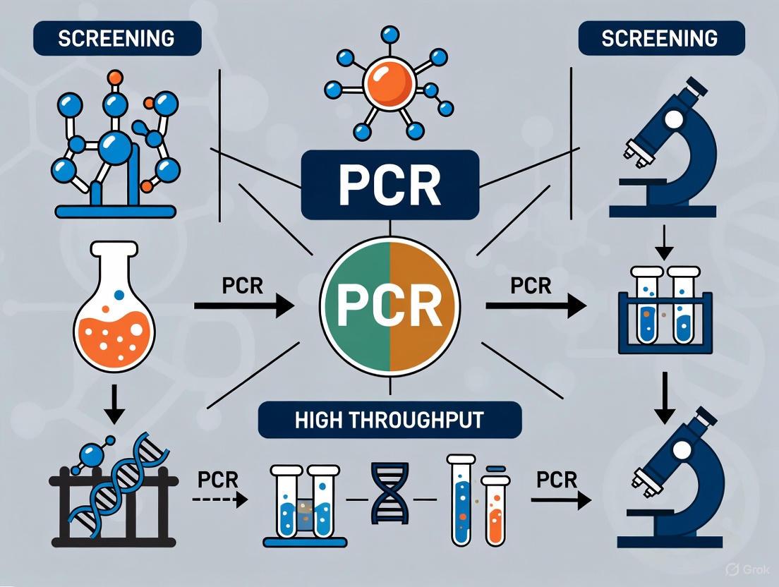
The adoption of high-throughput PCR for intestinal parasite screening represents a paradigm shift in diagnostic parasitology, offering superior sensitivity and specificity over traditional microscopy.
High-Throughput PCR for Intestinal Parasite Screening: A Comprehensive Guide for Researchers and Drug Developers
Abstract
The adoption of high-throughput PCR for intestinal parasite screening represents a paradigm shift in diagnostic parasitology, offering superior sensitivity and specificity over traditional microscopy. This article provides a comprehensive overview for researchers and drug development professionals, covering the foundational principles, methodological workflows, and rigorous validation frameworks essential for successful implementation. We explore the critical role of these advanced molecular platforms in large-scale surveillance and clinical trials, such as the DeWorm3 project, and detail common optimization and troubleshooting strategies. Furthermore, we present comparative analyses of commercial versus in-house assays and discuss the integration of these technologies within the One Health framework to improve global disease control and elimination efforts.
The Rise of Molecular Diagnostics: Why High-Throughput PCR is Replacing Microscopy for Intestinal Parasites
The Critical Need for Advanced Diagnostics in Parasitology
Parasitic infections represent a significant global public health challenge, affecting millions of people worldwide, with particularly severe impacts in underdeveloped and developing countries [1] [2]. These diseases not only cause substantial morbidity and mortality but also create considerable economic challenges due to increased healthcare expenditure and lost productivity [1] [2]. Accurate and timely diagnosis is fundamentally required to combat this global issue, enabling effective treatment, proper disease management, and implementation of public health control measures [2]. For decades, traditional diagnostic methods including microscopy, serological testing, histopathology, and culturing have served as the cornerstone of parasite identification [1]. While these methods have provided valuable service, they are increasingly recognized as insufficient for modern diagnostic needs due to limitations in sensitivity, specificity, and practicality in resource-limited settings where parasitic diseases are most prevalent [1] [2].
The transition to molecular-based diagnostics represents a paradigm shift in parasitology, offering enhanced sensitivity, specificity, and reliability in parasite detection [2]. This application note explores the critical need for advanced diagnostics in parasitology, with specific focus on high-throughput screening approaches for intestinal parasites using PCR-based methodologies. We present comprehensive experimental protocols, technical considerations, and future directions to guide researchers, scientists, and drug development professionals in implementing these advanced diagnostic platforms.
Limitations of Conventional Diagnostic Methods
Traditional diagnostic techniques for parasitic infections face significant constraints that impact their effectiveness in both clinical and public health settings. Microscopy, long considered the gold standard, requires extensive expertise for accurate performance and interpretation, with performance highly dependent on operator skill and experience [3] [4]. Furthermore, morphological differentiation between certain parasite species and strains presents considerable challenges, potentially leading to misidentification [4]. Serological assays including enzyme-linked immunosorbent assay (ELISA), immunohistochemistry (IHC), and immunoblotting (IB) are often hampered by cross-reactivity issues and variable sensitivity [4]. These methods primarily detect host immune responses rather than active infection, limiting their utility for distinguishing current from past infections [4].
The table below summarizes the key limitations of conventional diagnostic methods for parasitic infections:
Table 1: Limitations of Conventional Parasitological Diagnostic Methods
| Method | Key Limitations | Impact on Diagnostic Accuracy |
|---|---|---|
| Microscopy | Requires high expertise, time-consuming, limited sensitivity, morphological similarities between species [1] [4] | Missed infections in low parasite loads, species misidentification |
| Culture | Not applicable for many parasite species, lengthy process, specialized media requirements [2] | Limited utility for routine diagnostics, delayed results |
| Serological Tests | Cross-reactivity, cannot distinguish active from past infection, variable sensitivity/specificity [4] | False positives/negatives, limited value in endemic areas |
| Histopathology | Invasive sample collection, requires expert interpretation, not for routine screening [1] | Limited application to tissue-invasive parasites only |
These limitations are particularly problematic in endemic regions with poor infrastructure and limited access to healthcare facilities [1] [2]. The declining expertise in stool microscopy further compounds these challenges, creating an urgent need for more reliable, standardized diagnostic approaches [3].
Advanced Molecular Detection Platforms
High-Throughput Multiplex PCR for Intestinal Parasites
Molecular methods, particularly PCR-based assays, have dramatically transformed parasitic disease diagnosis by offering enhanced sensitivity and specificity compared to conventional techniques [5] [6]. The development of high-throughput multiplex PCR platforms represents a significant advancement for comprehensive screening of intestinal parasites. Taniuchi et al. (2011) developed a multiplex PCR and probe-based detection system using Luminex beads that simultaneously detects seven major intestinal parasites: Cryptosporidium spp., Giardia intestinalis, Entamoeba histolytica, Ancylostoma duodenale, Ascaris lumbricoides, Necator americanus, and Strongyloides stercoralis [5] [6].
This innovative approach utilizes two multiplex PCR reactions—one targeting protozoan parasites and another targeting helminths—followed by hybridization of PCR products to beads linked to internal oligonucleotide probes with detection on a Luminex platform [5] [6]. When validated against parent multiplex real-time PCR assays, this multiplex PCR-bead protocol demonstrated sensitivity and specificity ranging between 83% and 100% across 319 clinical specimens, establishing its utility as a sensitive diagnostic screen for a large panel of intestinal parasites [5] [6].
The evolution of parasite diagnostic methods from traditional techniques to advanced high-throughput systems is visualized below:
Comparison of Advanced Diagnostic Technologies
The landscape of advanced diagnostic technologies for parasitic infections has expanded considerably, with each platform offering distinct advantages and applications. The table below provides a comparative analysis of key advanced diagnostic technologies:
Table 2: Comparison of Advanced Diagnostic Technologies for Parasitic Infections
| Technology | Key Features | Sensitivity | Throughput | Applications |
|---|---|---|---|---|
| Multiplex PCR-Bead Arrays [5] [6] | Simultaneous detection of multiple pathogens, Luminex platform | High (83-100%) | High | Population screening, outbreak investigation |
| Next-Generation Sequencing (NGS) [1] [2] | Comprehensive pathogen detection, strain typing | Very High | Medium-High | Discovery, epidemiology, resistance detection |
| Isothermal Amplification (LAMP) [1] [2] | Constant temperature reaction, minimal equipment | High | Medium | Field applications, resource-limited settings |
| Nanobiosensors [4] | Antigen/biomarker detection, rapid results | Very High | Low-Medium | Point-of-care testing, rapid diagnosis |
| CRISPR-Cas Systems [1] [2] | High specificity, programmability | Very High | Medium | Specific detection, emerging pathogens |
Experimental Protocols and Methodologies
High-Throughput Multiplex PCR Protocol for Intestinal Parasites
This protocol adapts established real-time PCR assays for major intestinal parasites into a high-throughput format using Luminex bead technology [5] [6].
Sample Preparation and DNA Extraction
- Sample Collection: Collect 200 mg of fecal specimen and aliquot into sterile tubes.
- DNA Extraction: Use modified QIAamp DNA Stool Mini Kit protocol (Qiagen Inc., Valencia, CA) with the following adjustments [6]:
- Add 1 mL of tissue lysis buffer MDT to the stool sample
- Perform bead beating with 0.15 mm garnet beads (MO-BIO Laboratories, Inc, Carlsbad, CA) for 2 minutes
- Boil suspension for 7 minutes before extraction
- Add 100 μL of EDT solution (Proteinase K) from the kit
- Extend incubation time to 90 minutes after EDT addition
- Alternative Extraction: For automated extraction, use QuickGene-810 system with QuickGene DNA tissue kit S (Fujifilm, Tokyo, Japan) with similar modifications for stool samples [6].
- Storage: Store all DNA samples at -80°C until use in PCR reactions.
Multiplex PCR Amplification
The assay involves two separate multiplex PCR reactions: one for protozoa and another for helminths [6].
Protozoa Multiplex PCR Reaction (25 μL volume) [6]:
- 12.5 μL iQ Supermix (Bio-Rad, Hercules, CA)
- Additional 2 mM MgCl₂ (final concentration)
- Primer/Probe Mixture:
- 0.4 μM E. histolytica primers
- 0.6 μM Giardia primers
- 1.0 μM Cryptosporidium primers
- 0.08 μM E. histolytica probe
- 0.16 μM Giardia probe
- 0.4 μM Cryptosporidium probe
- 4 μL sample DNA
- Cycling Conditions:
- Initial denaturation: 95°C for 3 minutes
- 40 cycles of:
- 95°C for 30 seconds
- 55°C for 30 seconds
- 72°C for 30 seconds
- Final extension: 72°C for 7 minutes
Helminth Multiplex PCR Reaction (25 μL volume) [6]:
- 12.5 μL HotStarTaq Master Mix (Qiagen Inc.)
- Additional 3.5 mM MgCl₂ (final concentration 5 mM MgCl₂)
- 0.1 mg/mL BSA
- Primer/Probe Mixture:
- 0.2 μM Ancylostoma primers, 0.1 μM probe
- 0.2 μM Necator primers, 0.05 μM probe
- 0.08 μM Ascaris primers, 0.05 μM probe
- 0.1 μM Strongyloides primers, 0.05 μM probe
- 0.15 μM PhHV primers, 0.05 μM probe (extraction control)
- 5 μL sample DNA
- Cycling Conditions:
- Initial activation: 95°C for 15 minutes
- 40 cycles of:
- 94°C for 30 seconds
- 60°C for 90 seconds
- Final extension: 72°C for 10 minutes
Luminex Bead Hybridization and Detection
- Bead Preparation: Couple specific internal oligonucleotide probes to Luminex beads according to manufacturer's instructions.
- Hybridization: Mix PCR products with probe-coupled beads and incubate to allow specific hybridization.
- Detection: Analyze hybridized beads on Luminex platform following instrument protocols.
- Analysis: Interpret results based on fluorescence signals compared to established cutoff values and control samples.
The complete workflow for the high-throughput multiplex PCR detection system is illustrated below:
Research Reagent Solutions for High-Throughput Parasite Detection
Successful implementation of high-throughput screening for intestinal parasites requires specific research reagents and materials. The following table details essential solutions and their applications:
Table 3: Research Reagent Solutions for High-Throughput Parasite Detection by PCR
| Reagent/Material | Function/Application | Example Specifications |
|---|---|---|
| DNA Extraction Kit | Nucleic acid purification from stool samples | QIAamp DNA Stool Mini Kit (Qiagen), with modifications for parasite DNA [6] |
| PCR Master Mix | Amplification of target sequences | iQ Supermix for protozoa, HotStarTaq Master Mix for helminths [6] |
| Specific Primers/Probes | Target-specific amplification/detection | Biotinylated primers for bead capture; Taqman probes for real-time detection [6] |
| Luminex Beads | Multiplex detection platform | MagPlex-TAG beads coupled with specific oligonucleotide probes [5] |
| Positive Controls | Assay validation and quality control | Axenic cultures, purified cysts/oocysts, DNA from adult worms [6] |
| Extraction Control | Monitoring extraction efficiency | Phocine herpes virus spiked into lysis buffer [6] |
Emerging Technologies and Future Directions
The field of parasitology diagnostics continues to evolve rapidly with several emerging technologies showing significant promise. Nanobiosensors represent a revolutionary approach, utilizing nanomaterials such as gold nanoparticles (AuNPs), quantum dots (QDs), carbon nanotubes, and graphene oxide (GO) to detect parasitic antigens or genetic material with exceptional sensitivity [4]. These platforms offer rapid, accurate, and cost-effective results, with potential for point-of-care applications [4]. CRISPR-Cas systems have recently been adapted for diagnostic applications, leveraging their precision and programmability for specific detection of parasite nucleic acids [1] [2]. These systems provide sensitive, portable, and cost-effective methods for parasite detection, particularly in field settings [1].
Multi-omics integration combines data from genomics, transcriptomics, proteomics, and metabolomics to enhance diagnostic accuracy and provide comprehensive understanding of parasite biology and host-parasite interactions [1] [2]. This approach facilitates the discovery of new therapeutic targets and diagnostic biomarkers [1]. Point-of-care (POC) testing platforms continue to advance, with lateral flow immunoassays (LFIA), lab-on-a-chip (LoC) technologies, and portable molecular devices improving access to diagnosis in resource-limited settings [2] [4]. These developments are crucial for endemic regions with limited laboratory infrastructure.
The future of parasitology diagnostics will likely involve the integration of multiple advanced technologies to create comprehensive, sensitive, and accessible diagnostic platforms that can be deployed across diverse healthcare settings, from advanced laboratories to remote field clinics.
The critical need for advanced diagnostics in parasitology is unequivocal, driven by the limitations of conventional methods and the persistent global burden of parasitic diseases. High-throughput screening approaches for intestinal parasites using PCR-based methodologies represent a significant advancement in our ability to accurately detect and identify parasitic infections with enhanced sensitivity and specificity. The multiplex PCR-bead array protocol detailed in this application note provides researchers with a robust framework for implementing these advanced diagnostic platforms in their laboratories.
As the field continues to evolve, emerging technologies including nanobiosensors, CRISPR-Cas systems, and multi-omics approaches promise to further revolutionize parasitic disease diagnosis. The integration of these advanced platforms into clinical and public health practice will be essential for improving patient outcomes, enhancing disease surveillance, and ultimately reducing the global burden of parasitic infections. Researchers and drug development professionals play a critical role in advancing these technologies from proof-of-concept to practical implementation, ultimately contributing to improved global health outcomes.
For over a century, traditional light microscopy has served as the fundamental diagnostic tool for detecting intestinal parasites in clinical and research settings. Despite its longstanding role, this technique presents significant limitations in sensitivity and specificity that become particularly problematic in the context of modern high-throughput screening requirements for intestinal parasites. As research increasingly focuses on mass drug administration programs and precise prevalence mapping, the diagnostic inaccuracies of conventional microscopy create critical gaps in our understanding of parasitic disease burden. This application note examines these limitations through a systematic analysis of comparative performance data and details how molecular methods, particularly PCR-based approaches, are addressing these challenges to advance research capabilities.
Comparative Diagnostic Performance
Microscopy-based techniques, while simple and low-cost, demonstrate highly variable sensitivity that is affected by numerous factors including intermittent parasite excretion, low infection intensity, and sample storage conditions [7]. The table below summarizes the performance characteristics of common microscopy methods compared to molecular detection:
Table 1: Sensitivity Comparison of Diagnostic Methods for Key Intestinal Parasites
| Parasite | Microscopy Method | Sensitivity (%) | Molecular Method | Sensitivity (%) | Reference |
|---|---|---|---|---|---|
| Giardia intestinalis | Formol-ether concentration | 38 | Real-time PCR | 100 | [8] |
| Cryptosporidium spp. | Formol-ether concentration | 0 | Real-time PCR | 100 | [8] |
| Blastocystis sp. | Culture | 30 | Real-time PCR | 93 | [9] |
| Strongyloides stercoralis | Kato-Katz | Not recommended | PCR-Luminex | 83-100 | [7] [6] |
| Hookworm species | Kato-Katz | 64.2 | Multiplex PCR | 100 | [6] [10] |
| Ascaris lumbricoides | Direct wet mount | 83.3 | Multiplex PCR | 100 | [6] [10] |
The data demonstrate substantial sensitivity gaps across multiple parasite species, with microscopy failing to detect a significant proportion of infections, particularly at low intensity levels. This limitation has direct implications for research accuracy, especially in monitoring intervention effectiveness where parasite burdens may decline following treatment.
Methodological Limitations of Microscopic Techniques
Technical Constraints and Procedural Variability
Traditional microscopy suffers from several inherent technical limitations that directly impact diagnostic sensitivity and specificity:
Species identification challenges: Microscopy cannot reliably differentiate between hookworm species (Ancylostoma duodenale vs. Necator americanus), which have different epidemiological characteristics and pathogenicity [7] [11].
Sample degradation: Hookworm eggs have fragile shells that are easily damaged during sample processing, while Strongyloides stercoralis larvae are rarely detected in conventional Kato-Katz thick smears [7] [10].
Protocol-dependent sensitivity: Diagnostic performance varies significantly between concentration methods, with the Formol-ether concentration technique showing sensitivity of 32.5% for A. lumbricoides, 64.2% for hookworm, and 75% for T. trichiura in comparative studies [10].
Operator Dependency and Expertise Requirements
Microscopy requires substantial technical expertise that is increasingly scarce, particularly in low-prevalence settings where personnel have limited opportunity to maintain diagnostic skills [7] [11]. This operator dependency introduces significant inter-laboratory variability and compromises the reproducibility of research findings across different study sites.
High-Throughput Multiplex PCR as a Research Solution
Protocol: Automated High-Throughput Multiplex PCR for Intestinal Parasites
Multiplex PCR protocols enable simultaneous detection of multiple parasitic pathogens in a single reaction, dramatically improving throughput while maintaining species-specific differentiation [5] [6] [9].
Table 2: Research Reagent Solutions for Multiplex PCR Detection
| Reagent/Equipment | Function | Application Note |
|---|---|---|
| Seegene Allplex GI-Parasite Assay | Multiplex detection of 6 protozoa | Detects Blastocystis hominis, Cryptosporidium spp., Cyclospora cayetanensis, Dientamoeba fragilis, Entamoeba histolytica, Giardia lamblia [9] |
| STARMag 96 × 4 Universal Cartridge | Automated nucleic acid extraction | Enables high-throughput processing with minimal manual intervention [9] |
| Hamilton STARlet liquid handler | Automated sample preparation | Standardizes pre-analytical steps to reduce variability [9] |
| Luminex bead-based detection | Multiplex target identification | Allows simultaneous detection of 7+ parasites in single sample [5] [6] |
| Phocine herpes virus (PhHV) | Extraction and amplification control | Monitors inhibition and extraction efficiency in each sample [6] |
Procedure:
Sample Preparation:
Automated DNA Extraction:
PCR Setup:
- Combine 5μL extracted DNA with 20μL PCR master mix containing:
- 5μL 5X GI-P MOM primer
- 10μL RNase-free water
- 5μL EM2 (DNA polymerase, Uracil-DNA glycosylase, buffer with dNTPs) [9]
- Aliquot into PCR tubes
- Combine 5μL extracted DNA with 20μL PCR master mix containing:
Amplification and Detection:
Workflow Comparison: Traditional vs. Molecular Approaches
The following diagram illustrates the significant procedural differences between traditional microscopy and modern molecular workflows for intestinal parasite detection:
Research Implications and Applications
The transition to molecular methods addresses critical gaps in intestinal parasite research:
Polyparasitism studies: Research in Mozambique demonstrated PCR detected significantly more polyparasitism cases than microscopy, with virtually all participants (96%) harboring at least one helminth and 49% harboring three or more [11].
Drug efficacy monitoring: As mass drug administration programs reduce infection intensity, microscopy becomes increasingly unreliable for monitoring intervention success due to its poor sensitivity at low parasite burdens [7].
Species-specific epidemiology: Molecular methods enable differentiation of hookworm species, revealing unexpected distributions such as the predominance of Ancylostoma spp. over Necator americanus in some East African settings [11].
The sensitivity and specificity gaps inherent in traditional microscopy present substantial barriers to accurate intestinal parasite research, particularly in the context of high-throughput screening requirements. Molecular methods, especially automated multiplex PCR platforms, provide researchers with enhanced detection capabilities, species differentiation, and standardized protocols that overcome these limitations. While implementation challenges remain in resource-limited settings, the research advantages of molecular approaches are clear: they enable more accurate prevalence mapping, reliable monitoring of intervention effectiveness, and detailed understanding of polyparasitism dynamics that were previously obscured by methodological constraints.
Fundamental Principles of PCR and qPCR in Parasite Detection
The detection and quantification of parasitic pathogens have been revolutionized by the advent of molecular diagnostic techniques, particularly polymerase chain reaction (PCR) and its quantitative real-time counterpart (qPCR). Traditional parasitological diagnostic methods, primarily microscopic examination of stool samples, remain the reference standard in many settings but are hampered by significant limitations [12]. These techniques are labor-intensive, time-consuming, require experienced and well-trained operators, and often lack the sensitivity and specificity needed for accurate species differentiation [13] [12]. For example, microscopic methods cannot differentiate between the pathogenic Entamoeba histolytica and the non-pathogenic E. dispar, a distinction crucial for appropriate clinical management [12].
In contrast, PCR-based methods offer rapid, sensitive, and specific detection of parasite DNA, even in samples with low parasite loads [13] [12]. The application of these techniques in parasitology has expanded significantly, enabling not only the detection and quantification of parasites but also the study of gene expression and genetic diversity [13] [14]. This application note details the fundamental principles of PCR and qPCR, their application in parasite detection, and provides detailed protocols tailored for high-throughput screening of intestinal parasites.
Fundamental Principles of PCR and qPCR
Polymerase Chain Reaction (PCR)
The polymerase chain reaction (PCR) is a foundational molecular biology technique introduced by Kary Mullis in 1985 that allows for the exponential amplification of specific DNA sequences [15]. The process mimics the natural mechanism of DNA replication, utilizing a thermostable DNA polymerase (typically Taq polymerase from Thermus aquaticus) to synthesize new DNA strands complementary to a target template [16] [15].
The PCR process consists of three fundamental steps that are repeated for 25-40 cycles [16] [15]:
- Denaturation: The double-stranded DNA template is heated to 94–98°C, disrupting hydrogen bonds between complementary bases to separate the strands into single-stranded DNA.
- Annealing: The temperature is lowered to 50–65°C, allowing short, synthetic oligonucleotide primers to bind (anneal) to their complementary sequences on the single-stranded DNA template.
- Extension: The temperature is raised to 72°C, the optimal temperature for Taq polymerase activity, which extends the primers by adding deoxynucleotide triphosphates (dNTPs) to synthesize new DNA strands.
Each cycle theoretically doubles the amount of the target DNA sequence, leading to exponential amplification from a few initial copies to millions or billions after 30-40 cycles [16]. The amplified products can then be visualized using agarose gel electrophoresis, where the presence of a band at the expected size confirms successful amplification [16].
Quantitative Real-Time PCR (qPCR)
Quantitative real-time PCR (qPCR) builds upon conventional PCR by allowing the monitoring and quantification of amplified DNA as the reaction occurs, in real-time [15]. This is achieved through the incorporation of fluorescent reporter molecules that emit a signal proportional to the amount of DNA present during each cycle [14] [15]. The primary distinction from conventional PCR is that product detection is integrated into the amplification process, eliminating the need for post-PCR processing such as gel electrophoresis [15].
A key concept in qPCR is the quantification cycle (Cq), defined as the number of cycles required for the fluorescent signal to cross a predetermined threshold above background levels [15]. The Cq value is inversely proportional to the starting quantity of the target nucleic acid; a sample with a high initial target concentration will yield a low Cq value, and vice versa [17] [15].
Two principal detection chemistries are used in qPCR [14]:
- DNA-Binding Dyes (e.g., SYBR Green I): These dyes intercalate non-specifically into double-stranded DNA and fluoresce when bound. While cost-effective, they can bind to any double-stranded DNA, including non-specific products and primer-dimers, potentially leading to overestimation of the target concentration.
- Sequence-Specific Probes (e.g., TaqMan Probes): These are oligonucleotides labeled with a fluorescent reporter and a quencher. During amplification, the probe binds to its specific target sequence and is cleaved by the 5' nuclease activity of the DNA polymerase, separating the reporter from the quencher and generating a fluorescent signal. This method provides superior specificity and is ideal for multiplex assays that detect several targets simultaneously [14] [18].
qPCR is "truly quantitative, give(s) results over a range of 6–7 orders of magnitude, (is) quick to perform and require(s) no manipulations post-amplification" [13].
Applications in Parasitology
PCR and qPCR have become indispensable tools in parasitology, with applications spanning clinical diagnostics, research, and epidemiology.
Table 1: Applications of PCR and qPCR in Parasitology
| Application Area | Specific Use Cases | Key Advantages |
|---|---|---|
| Clinical Diagnostics | Detection and differentiation of intestinal protozoa (e.g., Giardia duodenalis, Entamoeba histolytica, Cryptosporidium spp., Dientamoeba fragilis) from fecal samples [12]. | High sensitivity and specificity; differentiation of morphologically identical species (e.g., E. histolytica vs. E. dispar); rapid turnaround time [12]. |
| Pathogen Quantification | Determining parasite load in infections (e.g., Plasmodium, Toxoplasma gondii) [13] [14]. | Accurate quantification over a wide dynamic range; monitoring treatment efficacy [13]. |
| Gene Expression Studies | Investigating levels of gene expression in parasites under different conditions (e.g., drug pressure) [13]. | High sensitivity to detect low-abundance transcripts; ability to work with small sample volumes. |
| Antimicrobial Resistance | Detecting single-nucleotide polymorphisms (SNPs) associated with resistance to antiparasitic drugs [18]. | High precision in discriminating genetic variants; potential for multiplexing. |
The performance of molecular methods is exemplary in the evaluation of the Allplex GI-Parasite Assay, a multiplex real-time PCR for detecting common enteric protozoa. A 2025 multicentric study of 368 samples demonstrated exceptional performance compared to conventional techniques (microscopy, antigen testing, culture), with sensitivity and specificity of 100% and 100% for Entamoeba histolytica, 100% and 99.2% for Giardia duodenalis, 97.2% and 100% for Dientamoeba fragilis, and 100% and 99.7% for Cryptosporidium spp., respectively [12].
Experimental Protocols
Standard Operating Procedure for PCR-Based Detection of Intestinal Parasites
Principle: This protocol describes the process for detecting parasitic DNA in human fecal samples using a commercial multiplex real-time PCR assay, leveraging the principles of qPCR for simultaneous, specific identification of multiple protozoan targets [12].
Workflow: The experimental workflow for sample processing and analysis is outlined below.
Table 2: Essential Research Reagent Solutions for PCR-Based Parasite Detection
| Item | Function / Description | Example / Note |
|---|---|---|
| DNA Extraction Kit | Isolates nucleic acids from complex fecal samples, removing PCR inhibitors. | Use kits designed for stool samples (e.g., ASL buffer from Qiagen) [12]. |
| Multiplex PCR Master Mix | Contains DNA polymerase, dNTPs, buffer, and MgCl₂ optimized for multiplex amplification. | Allplex GI-Parasite Assay master mix [12]. |
| Primer/Probe Mix | Target-specific primers and hydrolysis probes (e.g., TaqMan) for parasite DNA detection. | Multiplex mix for G. duodenalis, E. histolytica, Cryptosporidium spp., D. fragilis [12]. |
| Real-Time PCR Instrument | Thermocycler that performs precise temperature cycling and detects fluorescence in real-time. | CFX96 Real-time PCR system (Bio-Rad) or equivalent [12]. |
| Nuclease-Free Water | Solvent free of nucleases that could degrade primers, probes, or DNA templates. | For reconstituting and diluting reagents. |
| Positive Controls | Contains known target DNA sequences. Verifies assay functionality. | Should be included in each run [12]. |
| Negative Controls | Contains no template DNA. Monitors for contamination. | Nuclease-free water; should be included in each run [12]. |
- Sample Collection and Storage: Collect fecal samples according to clinical routine. For optimal DNA stability, freeze samples at -20°C or -80°C immediately after routine examination if they are not processed immediately.
- Homogenization: Suspend 50 to 100 mg of stool specimen in 1 mL of stool lysis buffer (e.g., ASL Buffer from Qiagen).
- Vortex and Incubate: Pulse vortex the mixture for 1 minute and incubate at room temperature for 10 minutes.
- Centrifugation: Centrifuge the tubes at full speed (approximately 14,000 rpm) for 2 minutes to pellet stool debris.
- Nucleic Acid Extraction: Transfer the supernatant for automated nucleic acid extraction. Use a dedicated system (e.g., Microlab Nimbus IVD) according to the manufacturer's instructions. The system should automatically perform nucleic acid purification and PCR setup to minimize hands-on time and cross-contamination risk.
- Prepare Reaction Mix: In a nuclease-free PCR plate or tube, combine the following components per reaction:
- Multiplex PCR Master Mix: As per manufacturer's instructions.
- Primer/Probe Mix (Allplex GI-Parasite Assay): As per manufacturer's instructions.
- DNA Template: 5-10 µL of the extracted nucleic acid.
- Nuclease-Free Water: To the final reaction volume (e.g., 20-50 µL).
- Seal the Plate: Apply an optical adhesive seal to the plate to prevent evaporation and cross-contamination.
- Thermal Cycling Protocol: Program the real-time PCR instrument with the following steps, as validated for the Allplex assay [12]:
- Initial Denaturation: 95°C for 15 minutes (also activates the hot-start polymerase).
- Amplification (45 cycles):
- Denaturation: 95°C for 15 seconds.
- Annealing/Extension & Fluorescence Acquisition: 60°C for 60 seconds. Acquire fluorescence at the appropriate wavelengths for the probes used at the end of this step.
Data Analysis and Interpretation
- Threshold and Cq Determination: Use the instrument's software (e.g., Bio-Rad's CFX Manager, Seegene Viewer) to analyze the amplification curves. Set the fluorescence threshold above the background noise but within the exponential phase of the amplification plot. The software will automatically assign a Cq value for each target in each sample.
- Result Interpretation: Interpret results using the manufacturer's software and criteria. For the Allplex assay, a positive test result is typically defined as a sharp exponential fluorescence curve that crosses the threshold at a Cq value of less than 45 for individual targets [12].
- Control Checks:
- Positive Control: Must be positive with a Cq value within the expected range.
- Negative Control (No Template Control): Must show no amplification (i.e., no Cq value) for all targets. Amplification in the negative control indicates contamination.
Advanced Development: Digital PCR
Digital PCR (dPCR) represents a third generation of PCR technology that offers absolute quantification without the need for a standard curve [18]. In dPCR, the sample is partitioned into thousands of individual nanoliter-sized reactions (water-in-oil droplets in droplet digital PCR or ddPCR), so that each contains zero, one, or a few target DNA molecules [18]. After end-point PCR amplification, the number of positive partitions is counted, and using Poisson statistics, the absolute concentration of the target in the original sample is calculated [18].
This technology offers exceptional sensitivity, making it suitable for detecting low-level parasitemia, and robust performance in the presence of PCR inhibitors that are common in complex sample types like stool, as inhibitors are diluted in the partitions [18]. Its high precision also makes it ideal for detecting minor genetic variants, such as single-nucleotide polymorphisms (SNPs) associated with drug resistance in parasites [18].
Troubleshooting Common Issues
Even with optimized protocols, users may encounter challenges. The table below summarizes common qPCR issues and recommended solutions.
Table 3: Troubleshooting Common PCR and qPCR Problems
| Problem | Potential Causes | Recommended Solutions |
|---|---|---|
| No Amplification | Inhibitors in DNA template, incorrect thermal cycler settings, failed reagents [17]. | Check positive control. Confirm thermal cycler settings match protocol. Re-purify DNA template to remove inhibitors [17] [19]. |
| High Cq Values (Late Amplification) | Low template concentration, template degradation, partial reaction inhibition, old primers/probes [17]. | Check template quality and concentration. Verify pipetting accuracy. Use fresh primer/probe aliquots [17]. |
| Non-Specific Amplification | Annealing temperature too low, primer-dimer formation, contaminated reagents [17] [19]. | Optimize annealing temperature (increase stepwise). Use hot-start DNA polymerase. Check for contamination in reagents [19]. |
| Inconsistent Replicates | Pipetting errors, inadequate mixing of reagents, uneven sealing of PCR plate [17]. | Calibrate pipettes. Mix reagents thoroughly before aliquoting. Ensure plates are evenly and properly sealed [17]. |
PCR and qPCR have fundamentally transformed the landscape of parasite detection, offering unparalleled sensitivity, specificity, and quantitative capability compared to traditional microscopic methods. The provided protocols and application examples demonstrate their robustness and suitability for high-throughput screening in both clinical and research settings. As the field advances, technologies like digital PCR and multiplexed assays are poised to further enhance diagnostic precision, support surveillance efforts, and ultimately contribute to improved control of parasitic diseases worldwide.
High-throughput screening using molecular methods has become fundamental for the accurate detection and differentiation of intestinal parasites in both clinical and research settings. This document details application notes and standardized protocols for the detection of five key parasitic targets: Giardia duodenalis (also known as G. lamblia), Cryptosporidium spp., Entamoeba histolytica, Soil-Transmitted Helminths (STHs), and Dientamoeba fragilis. The transition from traditional microscopy to PCR-based diagnostics offers superior sensitivity, specificity, and the ability to discriminate genotypes and species crucial for understanding epidemiology, pathogenesis, and treatment outcomes [20] [21] [22]. These protocols are designed for researchers, scientists, and drug development professionals engaged in large-scale screening and assay development.
Performance Comparison of Molecular Assays
The diagnostic accuracy of PCR assays is significantly influenced by the choice of the target gene. The tables below summarize the reported performance characteristics of various molecular targets for each parasite, providing a basis for assay selection.
Table 1: Comparative Performance of Giardia duodenalis Real-Time PCR Screening Assays [23]
| Target Gene | Estimated Sensitivity (%) | Estimated Specificity (%) | Notes |
|---|---|---|---|
| 18S rRNA | 100.0 | 100.0 | Recommended for screening due to high accuracy. |
| Beta-giardin (bg) | 31.7 | 100.0 | High specificity but lower sensitivity. |
| Glutamate dehydrogenase (gdh) | 17.5 | 92.3 | Lowest sensitivity among compared assays. |
Table 2: Comparative Performance of Cryptosporidium spp. Real-Time PCR Assays [24]
| Target Gene | Sensitivity (%) | Specificity (%) | Notes |
|---|---|---|---|
| SSU rRNA | 100.0 | 96.9 | Highly sensitive, suitable for initial screening. |
| COWP | 90.0 | 99.6 | High specificity, useful for confirmatory testing. |
| DnaJ-like protein (DnaJ) | 88.8 | 96.9 | Good overall performance. |
Table 3: Assay Performance for Other Key Parasites
| Parasite | Target Gene | Method | Performance | Source |
|---|---|---|---|---|
| Entamoeba histolytica | SSU rRNA | Real-time PCR (Molecular Beacon) | More sensitive than antigen detection (79%) and traditional PCR (72%). | [21] |
| Dientamoeba fragilis | SSU rRNA | 5' Nuclease (TaqMan) Real-time PCR | 100% sensitivity and specificity compared to conventional PCR and microscopy. | [22] |
| Soil-Transmitted Helminths (STHs) | Various (e.g., ITS, repetitive genomic elements) | Multi-parallel qPCR | Strong correlation between DNA quantity and egg counts for A. lumbricoides & T. trichiura. More sensitive than microscopy. | [25] |
Detailed Experimental Protocols
DNA Extraction from Stool Specimens
A critical first step for all subsequent PCR assays is the efficient isolation of inhibitor-free parasitic DNA from complex stool matrices.
Protocol: QIAamp DNA Stool Mini Kit (QIAGEN) - Standardized Protocol [20] [24] [21]
- Input Material: Use approximately 0.2 g of stool specimen (fresh or fixed).
- Inhibition Removal: Apply the sample to the InhibitEX tablet/solution provided in the kit. Vortex vigorously and incubate at room temperature for 1-3 minutes to adsorb PCR inhibitors. Centrifuge to pellet debris.
- Lysis: Transfer the supernatant to a new tube and add Proteinase K and AL (lysis) buffer. Incubate at 70°C for 10-30 minutes.
- DNA Binding: Add ethanol to the lysate and apply the mixture to the QIAamp spin column. Centrifuge to bind DNA to the silica membrane.
- Washing: Wash the membrane twice using the provided AW1 and AW2 buffers.
- Elution: Elute the purified DNA in a low-salt buffer (e.g., AE buffer) or nuclease-free water. A typical elution volume is 100-200 µL.
- Storage: Store extracted DNA at -20°C or -80°C until PCR analysis.
Real-Time PCR Assays
Protocol 1: Giardia duodenalis Detection and Genotyping [20] [26]
- Principle: Duplex real-time PCR using TaqMan probes targeting the β-giardin gene to simultaneously detect G. duodenalis and differentiate between the human-pathogenic assemblages A and B.
- Reaction Mix (25 µL typical volume):
- Master Mix (e.g., HotStarTaq Master Mix): 12.5 µL
- Forward/Reverse Primers (e.g., P241 or P434 sets): 300-900 nM each
- TaqMan Probes (FAM-labeled for Assemblage A, Cy5-labeled for Assemblage B): 100-200 nM each
- MgCl₂: Final concentration of 3-5 mM
- DNA Template: 2-5 µL
- Nuclease-free water to volume.
- Thermocycling Conditions:
- Initial Denaturation: 95°C for 10-15 min.
- 45-55 Cycles of:
- Denaturation: 95°C for 15-30 s.
- Annealing/Extension: 55-60°C for 30-60 s (with fluorescence acquisition).
Protocol 2: Cryptosporidium hominis and C. parvum Differentiation [27]
- Principle: A two-tube duplex real-time PCR system. One tube detects the Cryptosporidium genus (SSU rRNA target) and C. parvum (LIB13 locus), while the other detects C. hominis (LIB13 locus) and includes an Internal Control (IC) for inhibition monitoring.
- Reaction Mix:
- Master Mix (e.g., TaqMan Environmental Master Mix 2.0): 12.5 µL
- Primers: 300-900 nM each
- MGB TaqMan Probes (FAM for genus, VIC for species): 100-150 nM
- IC DNA & Primer/Probe Mix: As per manufacturer (e.g., 1 µL PrimerDesign mix)
- DNA Template: 2 µL
- Thermocycling Conditions:
- Hold: 95°C for 10 min.
- 55 Cycles: 95°C for 15 s, 60°C for 60 s (with fluorescence acquisition).
Protocol 3: Entamoeba histolytica-Specific Detection [21]
- Principle: Real-time PCR using a molecular-beacon probe targeting the small-subunit rRNA gene to specifically differentiate E. histolytica from the non-pathogenic E. dispar and E. moshkovskii.
- Reaction Mix:
- Master Mix (e.g., IQ Super Mix): 1X concentration
- Primers (Ehf/Ehr): 25 pmol per reaction
- Molecular-Beacon Probe (Texas Red-labeled): 6.25 pmol per reaction
- DNA Template: 2 µL
- Thermocycling Conditions (on i-Cycler):
- Initial Denaturation: 95°C for 3 min.
- 45 Cycles of: 95°C for 15 s, 55°C for 30 s, 72°C for 15 s.
Protocol 4: Dientamoeba fragilis Detection [22]
- Principle: A 5' nuclease (TaqMan) real-time PCR assay targeting the SSU rRNA gene.
- Reaction Mix (20 µL volume):
- FastStart DNA Master Hybridization Probes Mix: 2 µL
- MgCl₂: 3 mM final concentration
- Primers (DF3/DF4): 0.25 µM each
- Dual-labeled TaqMan Probe: 0.2 µM
- DNA Template: 2 µL
- Thermocycling Conditions (on LightCycler):
- Hold: 95°C for 10 min.
- 35 Cycles: 95°C for 10 s, 58°C for 10 s, 72°C for 3 s.
The Scientist's Toolkit: Research Reagent Solutions
Table 4: Essential Reagents and Kits for Parasitic DNA Detection via PCR
| Reagent / Kit Name | Function / Application | Example Use in Protocols |
|---|---|---|
| QIAamp DNA Stool Mini Kit (QIAGEN) | Standardized DNA extraction from stool; removal of PCR inhibitors. | Primary DNA extraction method cited across all protocols [20] [24] [21]. |
| HotStarTaq / FastStart Master Mix | PCR enzyme and buffer system providing hot-start fidelity. | Used in multiple real-time PCR setups for sensitivity and specificity [24] [21] [23]. |
| DNeasy Tissue Kit (QIAGEN) | DNA extraction from purified cysts/oocysts or worm tissue. | Used for extracting DNA from culture-derived or purified parasite forms [20] [26]. |
| Custom TaqMan Probes & Primers | Sequence-specific detection and quantification of target DNA. | Designed against genes like β-giardin (Giardia), COWP (Cryptosporidium), SSU rRNA (multiple parasites) [20] [26] [27]. |
| Internal Control (IC) DNA | Exogenous control to identify PCR inhibition in individual samples. | Added to the reaction to confirm result validity, especially in duplex assays [24] [27]. |
Workflow and Conceptual Diagrams
The following diagrams illustrate the high-throughput screening workflow and a key challenge in molecular diagnostics for STHs.
Diagram 1: High-throughput PCR screening workflow for intestinal parasites.
Diagram 2: Impact of genetic variation on STH molecular diagnostics.
The One Health framework is an integrated, unifying approach that aims to balance and optimize the health of people, animals, and ecosystems [28]. It recognizes the interdependent links among these fields to create new surveillance and disease control methods. This approach is particularly critical for addressing zoonotic diseases, which are infectious diseases caused by pathogens that spread between animals and people [28]. Approximately 60% of emerging infectious diseases reported globally originate from animals, both wild and domestic, and over 30 new human pathogens detected in the last three decades have predominantly animal origins [28]. The interconnectedness of human, animal, and environmental health demands close collaboration, communication, and coordination between relevant sectors to effectively manage complex health challenges including antimicrobial resistance, zoonotic diseases, and food safety issues [29] [28].
This application note explores the implementation of One Health principles specifically within the context of high-throughput molecular screening for intestinal parasites. We present detailed experimental protocols and data analysis frameworks that enable simultaneous detection of multiple zoonotic parasites across human, animal, and environmental samples, facilitating a comprehensive understanding of parasite transmission dynamics at key interfaces.
One Health Principles in Parasitology
Parasites, particularly intestinal protozoans and helminths, represent significant challenges within the One Health paradigm due to their complex life cycles that often span multiple host species and environmental reservoirs [30]. The role of parasites in One Health has been historically overshadowed by viral and bacterial pathogens, despite their significant public health and economic impacts [30]. Zoonotic parasites exemplify the interconnected nature of health across species boundaries, with transmission pathways that frequently involve environmental contamination, wildlife reservoirs, and domestic animal intermediates [30] [31].
The Norway rat (Rattus norvegicus) serves as an illustrative example of a synanthropic species that functions as both reservoir and sentinel for zoonotic parasites in urban environments. Molecular studies of urban rat populations in Barcelona, Spain, revealed significant prevalences of zoonotic intestinal protozoans, including Blastocystis (83.5%), Giardia duodenalis (37.7%), Cryptosporidium spp. (34.1%), and Dientamoeba fragilis (14.1%) [31]. These findings highlight the importance of comprehensive surveillance that includes wildlife hosts in urban ecosystems to fully understand the epidemiology of zoonotic parasites.
Table 1: Key Zoonotic Intestinal Parasites in the One Health Context
| Parasite | Human Health Impact | Animal Reservoirs | Transmission Routes | Environmental Stability |
|---|---|---|---|---|
| Cryptosporidium spp. | Gastroenteritis, severe in immunocompromised | Livestock, wildlife, companion animals | Waterborne, fecal-oral | Resistant to chlorine disinfection |
| Giardia duodenalis | Diarrhea, malabsorption | Multiple mammalian species | Waterborne, foodborne, direct contact | Cysts survive weeks in moist environments |
| Entamoeba histolytica | Dysentery, liver abscesses | Primates, potentially other mammals | Fecal-oral | Cysts survive months in suitable environments |
| Blastocystis sp. | Gastrointestinal symptoms, controversial pathogenicity | Wide host range including mammals, birds, reptiles | Fecal-oral, waterborne | Varies by subtype |
| Hookworms (Ancylostoma, Necator) | Anemia, protein deficiency | Dogs, cats, wildlife | Skin penetration, larval migration in soil | Larvae require moist, shaded soil |
High-Throughput Multiplex PCR Platform for Intestinal Parasites
The transition from traditional microscopic examination to molecular approaches represents a significant advancement in parasitological diagnostics within the One Health framework. Multiplex PCR-based assays coupled with Luminex bead-based detection provide a high-throughput platform for simultaneous detection of multiple parasitic pathogens from diverse sample types [5] [6]. This technological approach enables comprehensive surveillance across human, animal, and environmental samples using standardized methodology, facilitating direct comparison of results and identification of transmission pathways.
The core technology involves two multiplex PCR reactions—one targeting protozoan parasites and the other targeting helminths—followed by hybridization of PCR products to beads linked to internal oligonucleotide probes with detection on a Luminex platform [6]. This system demonstrates sensitivities between 83% and 100% for major intestinal parasites including Cryptosporidium spp., Giardia intestinalis, Entamoeba histolytica, Ancylostoma duodenale, Ascaris lumbricoides, Necator americanus, and Strongyloides stercoralis [6].
Experimental Protocol
Sample Collection and DNA Extraction
Sample Types:
- Human and animal fecal samples (200 mg aliquots)
- Environmental samples including water, soil, and surface swabs
- Alternative human samples: dried blood spots, saliva (for serological assays)
DNA Extraction Protocol:
- Pretreat fecal samples with PVPP (polyvinylpyrrolidone) and subject to bead beating with 0.15 mm garnet beads for 2 minutes
- Boil samples for 7 minutes before extraction
- Use QIAamp DNA Stool Mini Kit (Qiagen Inc.) with modifications for larger input volume
- Add 100 μL of EDT solution (Proteinase K) and incubate for 90 minutes
- Employ automated nucleic acid isolation systems (e.g., QuickGene-810) for processing large sample batches
- Include extraction control (phocine herpes virus) spiked into lysis buffer to monitor extraction efficiency and PCR inhibition [6]
Multiplex PCR Amplification
Protozoa PCR Reaction Setup (25 μL volume):
- 12.5 μL iQ Supermix (Bio-Rad)
- Additional 2 mM MgCl₂ (final concentration 6 mM)
- Primer concentrations: 0.4 μM E. histolytica, 0.6 μM Giardia, 1.0 μM Cryptosporidium
- Probe concentrations: 0.08 μM E. histolytica (Yakima Yellow), 0.16 μM Giardia (FAM), 0.4 μM Cryptosporidium (Texas Red)
- 4 μL template DNA
- Cycling conditions: 3 min at 95°C; 40 cycles of 30 sec at 95°C, 30 sec at 55°C, 30 sec at 72°C; final extension 7 min at 72°C [6]
Helminth PCR Reaction Setup (25 μL volume):
- 12.5 μL HotStarTaq Master Mix (Qiagen)
- Additional 3.5 mM MgCl₂ (final concentration 5 mM)
- 0.1 mg/mL BSA
- Species-specific primer and probe concentrations (see Table 2 for details)
- 5 μL template DNA
- Cycling conditions: 15 min at 95°C; 45 cycles of 30 sec at 95°C, 30 sec at 60°C, 45 sec at 72°C; final extension 7 min at 72°C [6]
Table 2: Primer and Probe Sequences for Multiplex PCR Detection of Intestinal Parasites
| Organism | Target Gene | Primer Sequences (5'→3') | Probe Sequence (5'→3') |
|---|---|---|---|
| Cryptosporidium spp. | COWP | F: CAAATTGATACCGTTTGTCCTTCT R: GGGCATGTCGATTCTAATTCAGCT | TGCCATACATTGTTGTCCTGACAAATTGAAT |
| Entamoeba histolytica | 18S rRNA | F: AACAGTAATAGTTTCTTTGGTTAGTAAA R: ACTTAGAATGTCATTTCTCAATTCATAT | TAGTACAAAATGGCCAATTCATTCA |
| Giardia lamblia | 18S rRNA | F: GACGGCTCAGGACAACGGTT R: TTGCCAGCGGTGTCCG | CCCGCGGCGGTCCCTGCTAG |
| Ascaris lumbricoides | ITS1 | F: GTAATAGCAGTCGGCGGTTTC R: TTGCCCAACATGCCACCT | ATTCTTGGCGGACAATTGCATGCGAT |
| Ancylostoma duodenale | ITS2 | F: GAATGACAGCAAACTCGTTGTT R: GATACTAGCCACTGCCGAAACG | TATCGTTTACCGACTTTAG |
| Necator americanus | ITS2 | F: CTGTTTGTCGAACGGTACTTG R: CATAACAGCGTGCACATGTTG | CCTGTACTACGCATTGTATAC |
| Strongyloides stercoralis | 18S rRNA | F: GAATTCCAAGTAAACGTAAGTCATTAGC R: TGCCTCTGGATATTGCTCAGTTC | ACACACCGGCCGTCGCTGC |
Luminex Bead Hybridization and Detection
- Couple specific oligonucleotide probes to carboxylated Luminex beads using carbodiimide chemistry
- Hybridize biotinylated PCR products to probe-coupled beads
- Detect hybridization using streptavidin-phycoerythrin reporter system
- Analyze on Luminex platform with minimum 50 beads per analyte per sample
- Use median fluorescence intensity (MFI) values for quantification with threshold determination based on negative controls [6]
Workflow Visualization
Essential Research Reagents and Materials
Table 3: Research Reagent Solutions for One Health Parasite Detection
| Reagent/Material | Function | Specifications | Application Notes |
|---|---|---|---|
| QIAamp DNA Stool Mini Kit | Nucleic acid extraction from complex matrices | Includes inhibitors removal technology | Modified protocol for 200 mg input sample [6] |
| Luminex MagPlex Microspheres | Multiplex detection platform | Carboxylated polystyrene beads with distinct fluorescent signatures | Allows simultaneous detection of 50-500 analytes [5] |
| iQ Supermix | Real-time PCR amplification | Contains iTaq DNA polymerase, dNTPs, MgCl₂ | Optimized for multiplex probe-based detection [6] |
| HotStarTaq Master Mix | Conventional PCR amplification | Includes pre-activated Taq polymerase | Reduces non-specific amplification in multiplex reactions [6] |
| Allplex GI-Parasite Assay | Commercial multiplex PCR assay | Detects 10 major human protist parasites | Validated on Bio-Rad CFX96 platform [31] |
| Species-specific Detector Antibodies | Immunodetection across species | Conjugated to phycoerythrin or other reporters | Enables cross-species application (human, canine, feline) [32] |
Data Analysis and Interpretation
The One Health approach generates complex datasets requiring specialized analytical frameworks. Network analysis has emerged as a powerful tool for visualizing and understanding the interconnected relationships between zoonotic agents, their hosts, and environmental sources [33]. This approach facilitates identification of key interfaces where zoonotic spillover is most likely to occur, enabling targeted interventions.
In a comprehensive study of zoonotic interactions in Austria, analysis of 47 years of data revealed that humans, cattle, chickens, and certain meat products functioned as the most influential nodes in the zoonotic agent-sharing network [33]. The characterization of six distinct communities of zoonotic agent sharing highlighted how highly connected infectious agents, proximity to humans, and anthropogenic activities drive parasite transmission patterns [33].
Statistical analysis of surveillance data should account for research effort bias, as sampling intensity varies across host species and environments. Binary logistic regression can identify factors associated with parasite prevalence, while chi-squared tests reveal co-infection patterns and associations between parasite species [31]. These analytical approaches help distinguish true epidemiological patterns from surveillance artifacts.
Case Study: Urban Rodent Surveillance in Barcelona
A comprehensive One Health investigation in Barcelona, Spain, demonstrated the practical application of high-throughput molecular screening for zoonotic intestinal protozoans in urban Norway rat populations [31]. The study employed multiplex real-time PCR (Allplex Gastrointestinal Panel-Parasite Assay) to screen 100 rats captured from parks and sewage systems, revealing high prevalences of multiple zoonotic parasites.
Key Findings:
- Overall prevalence of zoonotic intestinal protozoans was higher in sewage-dwelling rats (85/85 infected) compared to park rats (12/15 infected)
- Co-infections were common, with 67% of sewer rats infected with multiple zoonotic protozoans
- Statistical analysis identified significant associations between specific parasite pairs, suggesting possible synergistic interactions or shared transmission pathways
- Extrapolation to the estimated rat population of Barcelona (approximately 262,000 rats in the sewage system) indicated substantial environmental contamination with zoonotic parasites [31]
This case study illustrates how molecular surveillance of wildlife hosts in urban ecosystems can identify potential hotspots for zoonotic transmission and inform public health interventions targeting specific interfaces and transmission pathways.
The integration of high-throughput molecular diagnostics within a One Health framework provides powerful capabilities for understanding and managing zoonotic intestinal parasites. The multiplex PCR and Luminex-based detection platform described in this application note enables efficient, simultaneous screening of multiple parasite species across human, animal, and environmental samples, facilitating identification of transmission networks and targeted interventions.
Implementation of this approach requires collaborative infrastructures that bridge human medicine, veterinary science, and environmental health, addressing challenges related to standardized methodologies, data sharing, and interdisciplinary communication [34] [35]. The structural and operational barriers to One Health implementation, particularly in low- and middle-income countries, include lack of political will, weak governance, and insufficient human, financial, and logistical resources [34]. Enablers include framework documents guiding One Health activities, effective cross-sectoral coordination, and adequate funding coupled with technical support [34].
As molecular technologies continue to advance and become more accessible, their integration within One Health surveillance programs will be increasingly essential for detecting emerging threats, tracking transmission dynamics, and evaluating intervention effectiveness across the human-animal-environment interface.
Building a High-Throughput PCR Pipeline: From Sample Collection to Data Analysis
Within the framework of high-throughput screening for intestinal parasites via PCR, the pre-analytical phase of sample collection and preservation is a critical determinant of success. Pathogenic protozoa like Giardia lamblia, Cryptosporidium spp., and Entamoeba histolytica are significant causes of diarrheal diseases and nutritional disorders, particularly in endemic regions [36] [37]. The robust and often intermittent shedding of parasitic elements (cysts, oocysts) in stool, combined with their resilient structural walls, presents a formidable challenge for molecular diagnostics [36] [38]. Consequently, the methods employed from the moment of specimen collection directly impact the yield and quality of DNA, influencing the sensitivity and reliability of subsequent PCR analyses. This protocol details standardized procedures for collecting, preserving, and pretreating stool samples to ensure DNA integrity for large-scale, high-throughput molecular studies.
Key Reagents and Equipment
The following table catalogues the essential materials required for the sample handling and DNA extraction processes described in this protocol.
Table 1: Research Reagent Solutions and Essential Materials
| Item | Function/Application |
|---|---|
| FecalSwab Medium (Copan) | Liquid transport medium for stool samples; stabilizes nucleic acids for transport and storage prior to DNA extraction [37]. |
| S.T.A.R. Buffer (Roche) | Stool Transport and Recovery Buffer; used to homogenize stool samples for optimized DNA extraction [38]. |
| QIAamp Viral RNA Mini Kit (Qiagen) | Efficient DNA extraction kit for parasitic DNA from stool suspensions, outperforming stool-specific kits in some protocols [36]. |
| Proteinase K | Enzyme used in pretreatment to digest the robust oocyst wall of parasites like Cryptosporidium, facilitating DNA release [36]. |
| Para-Pak Collection Tubes | Commercial stool collection tubes containing preservative media for sample fixation and DNA preservation [38]. |
| AllPlex GIP Assay (Seegene) | Example of a commercial multiplex real-time PCR kit for the simultaneous detection of major intestinal protozoa [37]. |
Sample Collection and Initial Handling
Proper collection and immediate stabilization are the first critical steps to prevent nucleic acid degradation.
- Collection: Collect stool specimen in a clean, dry, wide-mouthed container without preservatives [39].
- Preservation Choice: For optimal DNA integrity, especially when delays between collection and processing are anticipated, immediately homogenize a portion of stool (approximately 1-2 mL) in a DNA stabilization transport medium, such as FecalSwab or S.T.A.R. Buffer [37] [38]. As an alternative, fixed specimens in preservative media like Para-Pak have also demonstrated reliable DNA preservation for molecular testing [38].
- Shipping: For transport, classify stool samples as a Category B Biological Substance (UN3373). Ship triple-packaged with absorbent material to contain any leaks, and maintain required temperature conditions (e.g., cold packs for refrigerated transport) to ensure sample integrity upon arrival at the laboratory [39].
Sample Pretreatment for DNA Release
A crucial, often overlooked step in the molecular diagnosis of intestinal parasites is the pretreatment to disrupt the resilient oocyst and cyst walls. The following workflow diagram outlines a validated protocol for this process.
Figure 1: Stool Sample Pretreatment Workflow for Parasite DNA Release.
Detailed Procedure:
- Create Stool Suspension: Prepare a 10% (weight/volume) suspension of stool in 0.2% Bovine Serum Albumin (BSA) prepared in Hank's buffer. Vortex thoroughly to homogenize [36].
- Concentration: Concentrate the parasitic forms using one of two methods:
- Centrifugation: Pellet the oocysts/cysts by centrifugation and resuspend in a smaller volume [36].
- Sedimentation: Allow large particles to sediment during a 1-hour incubation at room temperature. This is particularly useful for samples with high particulate matter (e.g., sand) that may clog extraction columns [36].
- Heat Shock: Subject the concentrated sample to a heat shock at 98°C for 10 minutes. This weakens the oocyst wall [36].
- Proteinase K Digestion: Add Proteinase K and incubate the sample overnight. This enzymatic treatment is critical for digesting the structural proteins of the oocyst wall, thereby liberating the DNA for subsequent extraction [36].
DNA Extraction and Quality Control
Selecting an efficient DNA extraction method is paramount, as performance varies significantly between kits when dealing with complex stool matrices and robust parasites.
Table 2: Comparative Performance of DNA Extraction Kits for Parasite DNA from Stool
| DNA Extraction Kit (Qiagen) | Relative Efficiency for Parasite DNA | Key Notes |
|---|---|---|
| QIAamp Viral RNA Mini Kit | Highest | Most efficient in comparative testing; recommended for sensitive detection [36]. |
| QIAamp DNA Blood Mini Kit | Moderate | Detected parasite DNA but with higher CT values and lower sensitivity than the Viral RNA kit [36]. |
| QIAamp DNA Stool Mini Kit | Lower | Least efficient in testing; performance slightly improved with the "InhibitEx" tablet [36]. |
Procedure:
- Extraction: From the pretreated sample supernatant, extract DNA using an optimized kit, such as the QIAamp Viral RNA Mini Kit, following the manufacturer's instructions. For high-throughput applications, automate this process using systems like the MICROLAB STARlet with pre-configured protocols [37].
- Inhibition Control: Always include an internal control in the PCR reaction to detect potential inhibition from co-purified stool constituents [37].
Performance Data: Microscopy vs. Multiplex PCR
The implementation of optimized collection, preservation, and extraction protocols enables highly sensitive molecular detection. The following table summarizes results from a large prospective study comparing multiplex PCR to traditional microscopy.
Table 3: Detection Rates of Intestinal Protozoa by Multiplex qPCR vs. Microscopy (n=3,495 samples)
| Parasite | Detection by Multiplex qPCR | Detection by Microscopy |
|---|---|---|
| Giardia intestinalis | 45 (1.28%) | 25 (0.7%) |
| Cryptosporidium spp. | 30 (0.85%) | 8 (0.23%) |
| Entamoeba histolytica | 9 (0.25%) | 24 (0.68%)* |
| Dientamoeba fragilis | 310 (8.86%) | 22 (0.63%) |
| Blastocystis spp. | 673 (19.25%) | 229 (6.55%) |
Note: Microscopy cannot differentiate the pathogenic *E. histolytica from non-pathogenic E. dispar [37].*
The transition to high-throughput PCR screening for intestinal parasites necessitates a foundational shift in sample management. This application note demonstrates that meticulous attention to sample collection in appropriate transport media, coupled with a robust pretreatment protocol to disrupt parasitic walls and an efficient DNA extraction method, is non-negotiable for ensuring DNA integrity. The resulting high-quality template DNA directly enables the superior sensitivity of multiplex qPCR, which consistently outperforms traditional microscopy for detecting most major protozoa. By standardizing these pre-analytical procedures, research studies and clinical trials can achieve more reliable, reproducible, and accurate data on parasite prevalence and load, ultimately advancing our understanding of their impact on global health.
The molecular diagnosis of intestinal parasites represents a significant advancement over traditional microscopy, offering enhanced sensitivity, specificity, and throughput [12] [38]. However, a primary challenge in implementing PCR-based detection lies in efficiently liberating and purifying microbial nucleic acids from complex stool matrices [40] [41]. The robust wall structures of parasite cysts and oocysts necessitate rigorous lysis procedures, while stool contains numerous substances that can inhibit downstream enzymatic reactions [42] [38]. This application note details integrated protocols combining mechanical bead-beating with automated magnetic bead-based nucleic acid extraction to overcome these challenges, providing a standardized, high-throughput workflow suitable for clinical diagnostics and research on intestinal parasites.
The Critical Role of Bead-Beating in Parasite Lysis
Mechanism and Impact on Detection Sensitivity
Mechanical lysis through bead-beating is particularly crucial for parasites with resilient life cycle stages. A study focusing on Trichuris trichiura demonstrated that a supplementary bead-beating procedure on ethanol-preserved stool samples significantly improved PCR detection rates [41]. The methodology involved:
- Sample Preparation: Stool samples were aliquoted and subjected to different pre-treatment conditions: directly frozen, preserved in 96% ethanol, bead-beating, or a combination of ethanol preservation and bead-beating [41].
- Bead-Beating Parameters: Samples were processed using a homogenizer with specific settings to ensure consistent mechanical disruption of parasite cysts and oocysts [41].
- DNA Isolation and PCR: Following bead-beating, DNA was isolated and tested using a multiplex real-time PCR assay for intestinal parasites [41].
The results demonstrated that bead-beating significantly enhanced DNA yield and detection sensitivity. PCR on directly frozen samples showed a 40% positivity rate for T. trichiura, which increased to 55.0% when a combination of ethanol preservation and bead-beating was employed [41]. This protocol underscores the necessity of mechanical disruption for accurate parasite detection.
Integration with Automated Systems
While many automated nucleic acid extractors are not equipped for bead-beating, this step can be performed as a separate, upstream sample preparation. Studies have shown that incorporating bead-beating before automated extraction systems significantly improves the recovery of Gram-positive bacteria and likely enhances the lysis of tough-walled parasites, leading to a more comprehensive representation of the microbial community in downstream analyses [40].
Automated Magnetic Bead-Based Nucleic Acid Extraction
Automated magnetic bead-based nucleic acid extraction has become the dominant technology for high-throughput molecular workflows due to several key advantages [43]. The process involves binding nucleic acids to paramagnetic beads in the presence of chaotropic salts, followed by magnetic separation and washing to remove contaminants, and finally elution in a low-salt buffer [44] [40].
This method offers significant benefits for stool processing:
- High Efficiency and Yield: Magnetic bead technology provides superior recovery of nucleic acids, which is critical for detecting parasites present in low numbers [44] [43].
- Reduced Inhibitor Carry-over: The efficient washing steps minimize the co-purification of PCR inhibitors commonly found in stool [42].
- Reproducibility and Standardization: Automation minimizes manual handling errors, reducing inter-sample variability and increasing the reproducibility of results [44] [40].
- Scalability and Throughput: Systems can process from 1 to 96 samples simultaneously, making them ideal for large-scale screening studies [44].
Comparative Performance Data
A direct comparison between boiling and magnetic bead-based extraction methods for HPV detection highlighted the superior performance of the magnetic bead approach. The magnetic bead method demonstrated greater resistance to PCR inhibitors like hemoglobin and a significantly higher detection rate (20.66% vs. 10.02%, P < 0.001) [42]. Although this study focused on a viral pathogen, the implications for inhibitor-rich stool samples are clear. The increased cost of the magnetic bead method (a 13.14% increase) was far outweighed by the 106.19% increase in detection rate, demonstrating its excellent cost-effectiveness for diagnostic applications [42].
Table 1: Comparison of Boiling vs. Magnetic Bead Nucleic Acid Extraction Methods
| Parameter | Boiling Method | Magnetic Bead Method |
|---|---|---|
| Principle | Heat-induced lysis and crude release of DNA | Chemical lysis + magnetic bead purification |
| Anti-hemoglobin Interference | Failed at hemoglobin >30 g/L | Effective even at 60 g/L hemoglobin [42] |
| HPV Detection Rate (n=639) | 10.02% | 20.66% (P < 0.001) [42] |
| Throughput | Low to moderate | High, easily scalable and automatable [44] |
| Cost-Benefit | Lower cost per test | 13.14% higher cost, but 106.19% higher detection rate [42] |
| Reproducibility | Prone to user variability | High, due to process standardization [44] [40] |
Integrated Protocol for High-Throughput Screening of Intestinal Parasites
This protocol combines optimized bead-beating with automated extraction, validated for the detection of protozoa like Giardia duodenalis, Cryptosporidium spp., Entamoeba histolytica, and Dientamoeba fragilis [12] [9] [38].
Sample Preparation and Bead-Beating Lysis
- Sample Collection and Preservation: Collect fresh stool samples and either process immediately or preserve. For preserved samples, use 96% ethanol or commercial stool preservation buffers [41]. For automated systems like the Hamilton STARlet, stool can be swabbed and inoculated into FecalSwab tubes containing Cary-Blair media [9].
- Homogenization: Vortex samples thoroughly to ensure homogeneity.
- Bead-Beating Lysis:
- Transfer 300 µL of stool suspension to a tube containing lysing matrix (e.g., Lysing Matrix E) [40].
- Add appropriate lysis buffer. For tough-walled parasites, a buffer containing Guanidine Thiocyanate is often effective.
- Process the samples using a high-speed bead beater (e.g., FastPrep-24) at 6.0 m/s for 40-60 seconds [40] [41].
- Centrifuge the lysate at 14,000 x g for 5-15 minutes to pellet debris [40].
Automated Nucleic Acid Extraction
This protocol is described for the Hamilton STARlet system but can be adapted to other magnetic bead-based automators.
- Instrument Setup: Load the automated liquid handling platform (e.g., Hamilton STARlet) with the extraction cartridge (e.g., STARMag 96 × 4 Universal Cartridge) and required reagents [9].
- Sample Loading: Transfer 50-100 µL of the clarified supernatant from the bead-beating step to the designated wells of the sample plate [9].
- Automated Extraction: Execute the extraction protocol. A typical program includes:
- Lysis/Binding: Further chemical lysis and binding of nucleic acids to magnetic beads.
- Washes: Multiple wash steps with ethanol-based buffers to remove impurities.
- Elution: Elution of purified nucleic acids in a low-salt buffer (e.g., Tris-EDTA) or nuclease-free water. The final elution volume is typically 50-100 µL [9].
- Output: The system outputs a plate containing purified DNA/RNA, ready for downstream PCR applications.
Downstream PCR Detection
The extracted DNA is suitable for various PCR assays. Multiplex real-time PCR panels, such as the Seegene Allplex GI-Parasite Assay, have been validated with this workflow and show excellent performance for detecting major intestinal protozoa [12] [9].
- PCR Setup: Use 5 µL of extracted DNA in a 25 µL real-time PCR reaction [9].
- Cycling Conditions: Follow manufacturer recommendations, typically involving 45 cycles of amplification [9] [45].
- Results Interpretation: Analyze fluorescence curves and cycle threshold (Ct) values. A sample is typically considered positive if the Ct value is below a defined limit (e.g., ≤43) [9].
Essential Research Reagent Solutions
Table 2: Key Materials and Reagents for Bead-Beating and Automated NA Extraction
| Item | Function | Example Products & Specifications |
|---|---|---|
| Automated Extractor | High-throughput, reproducible nucleic acid purification | Hamilton STARlet [9], Insta NX Mag 16Plus [44], KingFisher Apex [40], MagNA Pure 96 [38] |
| Magnetic Bead Kits | Provide reagents for binding, washing, and eluting NA | STARMag Universal Cartridge [9], HiPurA Pre-filled Plates [44], qEx-DNA/RNA virus kits [42] |
| Bead Beater | Mechanical disruption of tough cyst/oocyst walls | FastPrep-24 5G Homogenizer [40] |
| Lysing Matrix | Contains ceramic/silica beads for efficient lysis | Lysing Matrix E (1.4 mm ceramic and silica spheres) [40] |
| Lysis Buffer | Chemical lysis and stabilization of nucleic acids | ASL Buffer (Qiagen) [12], STARR Buffer (Roche) [38], Guanidine-based buffers |
| Sample Transport Media | Preserves nucleic acid integrity during storage/transport | FecalSwab with Cary-Blair [9], DNA/RNA Shield [40], 96% Ethanol [41] |
| PCR Master Mix | Enzymes and reagents for multiplex real-time PCR | Allplex GI-Parasite Assay [12] [9], Lab-developed multiplex assays [45] [38] |
Workflow Visualization
The following diagram illustrates the integrated workflow for sample processing, from collection to final PCR result, highlighting the critical steps of bead-beating and automated extraction.
The integration of mechanical bead-beating with automated magnetic bead-based nucleic acid extraction creates a robust and reliable workflow for the molecular detection of intestinal parasites. This approach directly addresses the primary challenges of efficient lysis of resilient parasitic forms and the removal of PCR inhibitors. The resulting high-quality DNA enables highly sensitive and specific multiplex PCR assays, making this combined protocol a powerful tool for high-throughput screening in both clinical diagnostics and public health research on intestinal parasitic diseases.
Within the framework of high-throughput screening for intestinal parasites by PCR, the transition from single-plex to multiplex molecular assays represents a critical advancement for large-scale public health interventions and drug development studies. The accurate detection and quantification of polyparasitism are essential, as the combined burden of multiple parasites significantly impacts morbidity and influences treatment efficacy outcomes [46]. Conventional microscopy, while widely used, faces limitations in sensitivity, throughput, and the ability to provide species-level differentiation, particularly in low-intensity infections common in post-treatment scenarios [25] [46]. Molecular methods, particularly multiplex real-time PCR, have demonstrated superior sensitivity for detecting intestinal helminths and protozoa, especially in mixed infections, and offer a more accurate determination of infection intensity [46]. This application note details the strategic design and validation of multiplex PCR assays for the simultaneous detection of a broad panel of intestinal parasites, with a focus on high-throughput applications in research and therapeutic development.
Multiplexing Strategy and Workflow Design
A core strategy for high-throughput multi-parasite detection involves partitioning the parasite panel into logical multiplex reactions. A proven approach is to create separate reaction mixes: one for major protozoa and another for helminths [5]. This division helps manage primer compatibility and ensures robust amplification across phylogenetically diverse targets. Following amplification, products can be detected using various platforms. The Luminex bead-based system allows for the hybridization of PCR products to beads linked to internal oligonucleotide probes, facilitating the detection of a large panel of parasites in a high-throughput format [5]. More recently, automated, commercial multiplex real-time PCR panels have been developed that integrate DNA extraction and amplification into a streamlined workflow, significantly reducing hands-on time and increasing laboratory efficiency [9].
The following workflow diagram outlines the key stages of a high-throughput multiplex PCR assay for intestinal parasite detection.
Target Gene Selection and Comparative Analysis
The choice of target DNA region is a cornerstone of a specific and sensitive multiplex PCR assay. Different types of genomic targets offer distinct advantages and limitations, which must be balanced based on the application's requirements.
Table 1: Comparison of Genomic Targets for Multiplex PCR Detection of Parasites
| Target Type | Examples | Advantages | Considerations | Application Reference |
|---|---|---|---|---|
| Ribosomal DNA | Internal Transcribed Spacer 1 (ITS1), ITS2, 18S rRNA [25] | Multi-copy, enhancing sensitivity; well-conserved for primer design [25] | Can be too conserved for species-level differentiation; copy number variation may affect quantification [25] | Detection of Ascaris lumbricoides, Trichuris trichiura, Giardia [25] [46] |
| Highly Repetitive Non-Coding Sequences | Putative satellite sequences [25] | Very high copy number, potentially offering superior sensitivity [25] | Functional role and variability may be less characterized [25] | Detection of Strongyloides stercoralis and hookworms [25] |
| Protein-Coding Genes | msp1 (malaria) [47], bexA (H. influenzae) [48] | Species-specific sequence variability allows for precise differentiation [47] [48] | Often single-copy, potentially lower sensitivity [47] | Differentiation of Plasmodium species [47]; bacterial detection [48] |
The selection of a specific target must be validated against the intended parasite panel. For instance, one study found a strong correlation between egg counts and qPCR results for Ascaris lumbricoides and Trichuris trichiura using both ribosomal and repetitive DNA targets. In contrast, the correlation was weaker for Ancylostoma duodenale and Strongyloides stercoralis, highlighting that optimal target performance is parasite-dependent [25]. Furthermore, when different qPCR assays targeting various DNA regions were compared on field samples, they showed only fair-to-moderate agreement for most soil-transmitted helminths, underscoring the importance of consistent target selection across comparative studies [25].
Detailed Experimental Protocol
Multiplex qPCR for Intestinal Protozoa and Helminths
This protocol is adapted from methods validated in controlled clinical trials for detecting a broad panel of intestinal parasites [46].
4.1.1 Sample Preparation and DNA Extraction
- Sample Collection: Collect fresh fecal samples. For longitudinal studies, store samples at 4°C and process within a few days. Alternatively, preserve samples in appropriate nucleic acid stabilization buffers.
- DNA Extraction: Use a mechanical homogenizer (e.g., FastPrep-24) with a bead-based DNA extraction kit (e.g., FastDNA Spin Kit for Soil) for efficient cell lysis. For high-throughput settings, employ automated nucleic acid extraction platforms (e.g., Hamilton STARlet with StarMag Universal Cartridge) [9]. Elute DNA in a final volume of 50-100 µL.
4.1.2 Multiplex qPCR Assay
- Primer and Probe Design: Design primers and probes based on species-specific sequences of target genes (see Table 1). For probe-based qPCR, use fluorophores with non-overlapping emission spectra (e.g., FAM, HEX, ROX, Cy5).
- Reaction Setup: Prepare two separate multiplex reactions as summarized below [5] [46].
Table 2: Example Multiplex qPCR Reaction Setups
| Component | Reaction 1: Helminths | Reaction 2: Protozoa |
|---|---|---|
| Master Mix | 10 µL of 2x Quantitec SYBR Green or Probe Master Mix | 10 µL of 2x Quantitec SYBR Green or Probe Master Mix |
| Primers (each) | 0.5 µM (final conc.) | 0.5 µM (final conc.) |
| Targets | Necator americanus, Ancylostoma spp., Ascaris spp., Trichuris trichiura [46] | Entamoeba histolytica, Cryptosporidium spp., Giardia duodenalis, Strongyloides stercoralis [46] |
| Template DNA | 2-5 µL | 2-5 µL |
| Nuclease-free H₂O | To a final volume of 20 µL | To a final volume of 20 µL |
- Thermal Cycling Conditions (for Probe-based qPCR):
- UDG Incubation (if using dUTP): 50°C for 2 minutes [48].
- Polymerase Activation: 95°C for 3-10 minutes.
- Amplification (45 cycles):
- Denature: 95°C for 10 seconds.
- Anneal/Extend: 60°C for 20-60 seconds (acquire fluorescence at this step).
- Data Analysis: Determine cycle threshold (Ct) values. A sample is typically considered positive if the Ct value is below a validated cutoff (e.g., ≤43) [9]. For quantification, generate standard curves using plasmids of known copy number or standardized genomic DNA.
High-Throughput Automated Protocol
For large-scale screening, automated workflows are essential.
- Platform: Seegene Allplex GI-Parasite Assay or equivalent automated system [9].
- Procedure:
- Sample Loading: Inoculate a fecal swab into a transport medium like Cary-Blair.
- Automated Processing: Load samples into an automated liquid handler (e.g., Hamilton STARlet). The system performs DNA extraction and PCR setup in a single, closed-tube operation to minimize contamination.
- Detection: Run real-time PCR and analyze results with integrated software. The entire process from sample to result can be completed in a fraction of the time required for manual microscopy [9].
Validation and Performance Metrics
Robust validation is critical for deploying a multiplex PCR assay in a research or drug development setting. Key performance characteristics must be established.
Table 3: Assay Performance Metrics from Validated Studies
| Assay / Target | Sensitivity (%) | Specificity (%) | Limit of Detection (LOD) | Key Finding |
|---|---|---|---|---|
| Automated GI-Parasite PCR [9] | 93-100 (for most targets) | 98.3-100 (for most targets) | Varies by target | Reduced pre-analytical and analytical turnaround time by ~7 hours. |
| Multiplex qPCR vs. Microscopy [46] | Higher for hookworms (2.9x), Giardia (1.6x) | High, with superior polyparasitism detection | -- | All STH-positive samples were low-intensity by microscopy, but PCR suggested higher intensity. |
| SYBR Green msp1 assay (Plasmodium spp.) [47] | -- | -- | 10 copies/µL | Excellent reproducibility (CV for Tm: 0.34-0.37%). |
| On-chip LAMP assay [49] | 98.08 | 97.59 | 10⁻² to 10⁻³ pg/µL | Enabled parallel analysis of 5 targets from 4 samples simultaneously. |
The Scientist's Toolkit: Essential Research Reagents and Materials
Table 4: Key Reagents and Equipment for High-Throughput Multiplex Parasite PCR
| Item | Function / Application | Example Products / Notes |
|---|---|---|
| Automated Nucleic Acid Extractor | Standardizes and accelerates DNA extraction from fecal samples. | Hamilton STARlet, QIAcube (coupled with bead-beating for efficient lysis) [9]. |
| Bead-Based DNA Extraction Kit | Efficiently lyses hardy parasite cysts and eggs in complex fecal matrices. | FastDNA Spin Kit for Soil [25], StarMag 96 × 4 Universal Cartridge [9]. |
| Multiplex PCR Master Mix | Supports simultaneous amplification of multiple targets with high efficiency and specificity. | Quantitec SYBR Green PCR Master Mix [50], Seegene Allplex GI-Parasite MOM [9]. |
| Real-time PCR Thermocycler | Performs amplification and fluorescence detection for quantification. | Rotorgene 6000 [50], Bio-Rad CFX96 [9]. |
| Luminex Platform | High-throughput, post-PCR detection system using bead-based hybridization. | Enables detection of dozens of targets in a single sample [5]. |
| Validated Primer-Probe Sets | Core reagents for specific parasite detection; designed from conserved, species-specific regions. | Targets include ITS1/2, 18S rRNA, or highly repetitive genomic sequences [25] [47] [46]. |
The strategic design of multiplex PCR assays, from informed target gene selection to the implementation of automated high-throughput workflows, is fundamental for advancing research and control programs for intestinal parasites. The protocols and data summarized in this application note provide a framework for developing and validating sensitive, specific, and efficient detection systems. By moving beyond traditional microscopy, these molecular tools enable a more accurate assessment of parasite prevalence, intensity, and polyparasitism, which is crucial for evaluating the impact of public health interventions and the efficacy of new therapeutic agents in drug development pipelines.
The molecular diagnosis of intestinal parasites is undergoing a transformative shift from manual, low-throughput microscopy to fully automated, high-throughput nucleic acid testing. This transition addresses critical limitations of conventional methods, including operator dependency, low sensitivity, and inability to differentiate morphologically identical species [12]. Within this diagnostic evolution, automated liquid handlers and high-throughput thermal cyclers serve as the foundational technologies enabling the rapid, precise, and reproducible processing required for large-scale parasitology studies and public health interventions [51] [52]. This application note details integrated protocols and performance data for automated high-throughput screening of intestinal parasites via PCR, providing a framework for implementation in research and diagnostic settings.
Essential Research Reagent Solutions
The following reagents and materials constitute the core components for establishing automated high-throughput PCR workflows for intestinal parasite detection.
- Nucleic Acid Extraction Kits: Reagents designed for efficient lysis of resilient parasite (oo)cysts and removal of PCR inhibitors prevalent in stool samples, compatible with automated extraction systems [12].
- Multiplex PCR Master Mix: Optimized enzymatic formulations containing polymerase, dNTPs, and buffers stabilized for robotic dispensing, enabling simultaneous detection of multiple parasite targets in a single reaction [12] [52].
- Assay-Specific Primers and Probes: Lyophilized or stabilized oligonucleotides targeting conserved genomic regions of protozoa (e.g., Giardia duodenalis, Entamoeba histolytica, Cryptosporidium spp.) and helminths (e.g., Ascaris lumbricoides, Trichuris trichiura) [12] [52] [53].
- Positive and Negative Controls: Plasmid constructs or synthetic oligonucleotides containing target sequences for all parasites in the panel, essential for run validation and quantitative accuracy [52].
- Magnetic Bead Purification Reagents: Paramagnetic particles and associated wash buffers used in automated cleanup and normalization of nucleic acid extracts, crucial for assay consistency [54].
Instrumentation Specifications and Selection
Selecting appropriate instrumentation is critical for balancing throughput, precision, and operational efficiency. Key specifications for liquid handlers and thermal cyclers are summarized below.
Table 1: Automated Liquid Handler Comparison for Parasite PCR Workflows
| Model/Feature | Formulatrix Mantis | IGT-AS12 | Microlab Nimbus IVD |
|---|---|---|---|
| Pipetting Precision | <2% CV at 100 nL [51] | ≤5% CV at 1 μL [54] | Not Specified |
| Dead Volume | As low as 6 μL [51] | Not Specified | Not Specified |
| Throughput Compatibility | 384- and 1536-well plates [51] | 16-48 reactions per run [54] | Automated PCR setup [12] |
| Key Application Features | PCR component transfer, serial dilution, sample pooling [51] | NGS library construction, magnetic bead purification, thermal cycling module [54] | Fully automated nucleic acid processing and PCR setup [12] |
Table 2: High-Throughput Thermal Cycler Performance Metrics
| Performance Metric | Target Value | Application Significance |
|---|---|---|
| Thermal Uniformity | <0.5°C variation across block | Ensures consistent amplification efficiency across all samples [52]. |
| Speed of Cycling | ≤ 3 hours for 45-cycle qPCR | Increases daily throughput for large-scale screening studies [52]. |
| Well Format | 96-, 384-well | Matches output of automated liquid handlers; 384-well format reduces reagent costs by 75% per sample. |
| Multiplex Detection Capability | 4-5 colors | Allows for simultaneous detection of multiple parasite targets in a single well, improving efficiency [12]. |
Application Protocol: High-Throughput qPCR for Intestinal Parasites
This validated protocol is adapted from multicentric studies for detecting enteric protozoa and soil-transmitted helminths [12] [52].
The following diagram illustrates the complete automated workflow for the detection of intestinal parasites, from sample preparation to final analysis.
Materials and Equipment
- Automated Liquid Handler: e.g., Formulatrix Mantis, IGT-AS12, or Hamilton Microlab Nimbus [51] [54] [12].
- High-Throughput Thermal Cycler: 384-well capable real-time PCR instrument [52].
- Multiplex qPCR Assay: e.g., Allplex GI-Parasite Assay or custom-designed assays [12] [52].
- Stool Lysis Buffer (e.g., ASL buffer from Qiagen) [12].
- Nucleic Acid Extraction Kit compatible with the automated system.
- qPCR Plates: 384-well optical plates and seals.
- Positive Controls: Synthetic DNA controls for each target parasite (e.g., 100 copies/μL) [52].
- Negative Control: Molecular grade water.
Step-by-Step Procedure
Sample Preparation and Lysis
- Manual Pre-processing: Homogenize 50-100 mg of stool specimen in 1 mL of stool lysis buffer [12].
- Pulse vortex for 1 minute and incubate at room temperature for 10 minutes.
- Centrifuge at 14,000 rpm for 2 minutes; the supernatant is used for automated extraction [12].
Automated Nucleic Acid Extraction
- Program the liquid handling system (e.g., Microlab Nimbus IVD) to automatically transfer supernatant from the previous step to the extraction plate [12].
- Execute the automated nucleic acid extraction protocol according to the manufacturer's instructions.
- Elute DNA in a final volume of 50-100 μL of elution buffer.
Automated qPCR Reaction Setup
- Plate Layout Definition: Using the liquid handler's software, define the plate map for samples, positive controls, and negative controls [51] [54].
- Reagent Dispensing: Program the system to dispense the following components per 10 μL reaction:
- 5 μL of 2X Multiplex PCR Master Mix
- 1 μL of Primer-Probe Mix (containing assays for target parasites)
- 4 μL of DNA template [12]
- Liquid Handler Execution: Run the automated protocol for reagent distribution.
- Utilize active tip washing to prevent amplicon contamination.
- Implement liquid level detection for low-volume reagents to ensure pipetting accuracy [51].
High-Throughput Thermal Cycling
- Seal the plate and centrifuge briefly.
- Load the plate into the thermal cycler and run the following cycling conditions:
- Initial Denaturation: 95°C for 5 minutes
- 45 Cycles of:
- Denaturation: 95°C for 15 seconds
- Annealing/Extension: 60°C for 60 seconds (with fluorescence acquisition) [12]
- For multiplex detection, ensure the instrument is configured to read fluorescence at the appropriate wavelengths for all probes used.
Data Analysis
- Set the cycle threshold (Ct) using the exponential phase of the amplification curves.
- A positive result is defined as a fluorescence curve crossing the threshold before Ct 45 [12].
- For quantitative analysis, use a standard curve generated from serial dilutions of positive controls [52].
Performance Validation and Troubleshooting
Validation Data from Multicentric Studies
Implementation of automated platforms for parasite detection has demonstrated excellent performance characteristics in validation studies.
Table 3: Performance Metrics of Automated PCR for Parasite Detection
| Parasite Target | Sensitivity (%) | Specificity (%) | Reference |
|---|---|---|---|
| Entamoeba histolytica | 100 | 100 | [12] |
| Giardia duodenalis | 100 | 99.2 | [12] |
| Dientamoeba fragilis | 97.2 | 100 | [12] |
| Cryptosporidium spp. | 100 | 99.7 | [12] |
| Soil-Transmitted Helminths | Accuracy ≥99.5% (technical replicate) | Accuracy ≥98.1% (individual extraction) | [52] |
Troubleshooting Common Issues
- High CV Between Replicates: Confirm liquid handler calibration, especially for volumes <5 μL. Check for partial tip clogging and ensure proper mixing of master mix components during dispensing [51].
- Inhibition in Stool Samples: Implement automated magnetic bead purification steps to remove PCR inhibitors. Increase centrifugation speed and time during sample pre-processing to pellet debris [12].
- Poor Amplification Efficiency: Verify the stability of primer and probe stocks used for automated dispensing. Check thermal cycler calibration and block temperature uniformity [52].
- Cross-Contamination: Incorporate UV decontamination cycles in the liquid handler [54] and use filter tips for all liquid handling steps. Maintain unidirectional workflow from sample preparation to PCR setup.
The DeWorm3 Project is a series of cluster randomized controlled trials conducted in Benin, India, and Malawi to test the feasibility of interrupting the transmission of soil-transmitted helminths (STH) [55]. STH infections affect an estimated 1.45 billion people globally and are associated with significant morbidity including malnutrition, iron-deficiency anemia, and impaired cognitive development in children [55]. The current World Health Organization (WHO) control strategy focuses on targeted mass drug administration (MDA) primarily to school-aged children and other high-risk groups, which effectively reduces morbidity but is unlikely to interrupt transmission due to persistent adult reservoirs of infection [55]. DeWorm3 tests the hypothesis that expanding MDA to entire communities with high coverage can achieve transmission interruption, thereby offering a more sustainable approach to STH control.
Core Trial Design and Objectives
DeWorm3 employs a hybrid trial design that integrates both clinical and implementation science outcomes to speed the translation of research findings into evidence-based policy and practice [56]. Each study site encompasses a minimum population of 80,000 individuals, divided into 40 clusters randomized 1:1 to either the intervention or control strategy for three consecutive years [55].
Table 1.1: DeWorm3 Trial Design Overview
| Aspect | Intervention Arm | Control Arm |
|---|---|---|
| Target Population | All community members aged ≥24 months [55] | Pre-school and school-aged children (targeted per national guidelines) [55] |
| MDA Frequency | Twice-annual [55] | According to national standard of care (typically annual) [55] |
| MDA Delivery | Door-to-door [55] | School-based or through national deworming days [55] |
| Duration | 3 years [55] | 3 years [55] |
The primary objective is to compare the prevalence of each STH species (Ascaris lumbricoides, Ancylostoma duodenale, Necator americanus, and Trichuris trichiura) measured by quantitative PCR (qPCR) 24 months after the final round of MDA, between intervention and control clusters [55]. A key goal is to assess transmission interruption, defined as a weighted cluster-level prevalence of ≤2% for each STH species at the 24-month post-MDA time point [55].
Implementation Science Framework
The implementation science component of DeWorm3 is designed to contextualize clinical findings and provide practical guidance for optimizing and scaling up STH interventions [56] [57]. This research occurs at three stages: baseline (formative research), midline (process research), and endline (summative research) [56].
Implementation Science Research Aims and Methods
DeWorm3 implementation science employs five key methodological approaches to address its research aims [56] [57]:
Table 2.1: DeWorm3 Implementation Science Aims and Methods
| Research Aim | Methodology | Application in DeWorm3 |
|---|---|---|
| Identify key stakeholders and network dynamics [57] | Stakeholder mapping and network analysis [56] | Systematically identifies individuals and organizations influencing standard of care and community-wide MDA; evaluates network dynamics affecting study outcomes and policy development [56]. |
| Understand barriers and facilitators to community-wide MDA [57] | Qualitative research (individual interviews and focus groups) [56] | Generates qualitative data to identify factors that shape, contextualize, and explain trial outputs and outcomes from multiple stakeholder perspectives [56]. |
| Assess health system readiness [57] | Structural readiness surveys [56] | Quantifies factors driving health system readiness to implement community-wide MDA; identifies opportunities for change management and system strengthening [56]. |
| Optimize intervention delivery processes [57] | Process mapping [56] | Maps intervention delivery process to identify discrepancies between planned and implemented activities; pinpoints contextually-relevant modifiable bottlenecks [56]. |
| Evaluate economic efficiency [57] | Economic evaluation (costing and cost-effectiveness) [56] | Compares financial and economic costs and incremental cost-effectiveness of community-wide versus targeted MDA in both short-term and long-term elimination horizons [56]. |
Figure 2.1: DeWorm3 Implementation Science Research Framework
High-Throughput Molecular Detection of Intestinal Parasites
A critical component of the DeWorm3 Project is the accurate detection and monitoring of STH prevalence throughout the trial. The project utilizes quantitative polymerase chain reaction (qPCR) as its primary diagnostic tool, which offers enhanced sensitivity and specificity compared to traditional microscopy [55]. For large-scale trials like DeWorm3, high-throughput molecular methods are essential for processing the thousands of samples collected during baseline, monitoring, and endline assessments.
Multiplex PCR-Bead Detection Platform
Taniuchi et al. (2011) developed a high-throughput multiplex PCR and probe-based detection system using Luminex beads that can simultaneously detect seven intestinal parasites [6] [5]. This platform addresses the challenge of detecting diverse protozoan and helminth parasites in a single protocol, making it particularly suitable for large-scale epidemiological studies and trials like DeWorm3.
The assay involves two multiplex PCR reactions: one with specific primers for protozoa (Cryptosporidium spp., Giardia intestinalis, Entamoeba histolytica) and another with specific primers for helminths (Ancylostoma duodenale, Ascaris lumbricoides, Necator americanus, Strongyloides stercoralis) [6]. Following PCR amplification, products are hybridized to beads linked to internal oligonucleotide probes and detected on a Luminex platform [6].
Table 3.1: High-Throughput Multiplex PCR Targets and Parameters
| Parasite | Target Gene | GenBank Accession | Sensitivity | Specificity |
|---|---|---|---|---|
| Cryptosporidium spp. | COWP [6] | AF248743 [6] | 83-100% [6] | 83-100% [6] |
| Entamoeba histolytica | 18S rRNA [6] | X64142 [6] | 83-100% [6] | 83-100% [6] |
| Giardia intestinalis | 18S rRNA [6] | M54878 [6] | 83-100% [6] | 83-100% [6] |
| Ascaris lumbricoides | ITS1 [6] | ALJ000895 [6] | 83-100% [6] | 83-100% [6] |
| Ancylostoma duodenale | ITS2 [6] | AJ001594 [6] | 83-100% [6] | 83-100% [6] |
| Necator americanus | ITS2 [6] | AJ001599 [6] | 83-100% [6] | 83-100% [6] |
| Strongyloides stercoralis | 18S rRNA [6] | AF279916 [6] | 83-100% [6] | 83-100% [6] |
Protocol: High-Throughput Multiplex PCR-Bead Detection
DNA Extraction
- Use 200 mg of stool sample for DNA extraction [6]
- Employ a modified QIAamp DNA Stool Mini Kit protocol (Qiagen Inc.) [6]
- Include pretreatment with bead beating using 0.15 mm garnet beads for 2 minutes followed by boiling for 7 minutes before extraction [6]
- Add proteinase K during extraction with extended incubation time (90 minutes) [6]
- Include an exogenous phocine herpes virus spiked into the lysis buffer as an extraction and amplification control [6]
Multiplex PCR Amplification
- Perform two separate multiplex PCR reactions: one for protozoa and one for helminths [6]
- For protozoa (3-plex) reaction:
- Use iQ Supermix (Bio-Rad) containing dNTPs, MgCl₂, and iTaq DNA polymerase [6]
- Primer concentrations: 0.4 μM E. histolytica, 0.6 μM Giardia, 1.0 μM Cryptosporidium primers [6]
- Cycling conditions: initial 3 min at 95°C followed by 40 cycles of 30 sec at 95°C, 30 sec at 55°C, and 30 sec at 72°C, with final extension for 7 min at 72°C [6]
- For helminth reaction:
Hybridization and Detection
- Hybridize biotinylated PCR products to beads linked to internal oligonucleotide probes [6]
- Detect hybridized products on a Luminex platform [6]
- Analyze data using appropriate software to determine presence/absence of each parasite target [6]
Figure 3.1: High-Throughput Multiplex PCR Workflow for Parasite Detection
The Scientist's Toolkit: Research Reagent Solutions
Table 4.1: Essential Research Reagents for High-Throughput Parasite Detection
| Reagent/Kit | Manufacturer | Function | Application in DeWorm3 |
|---|---|---|---|
| QIAamp DNA Stool Mini Kit | Qiagen Inc. [6] | DNA extraction from stool samples | Isolation of high-quality DNA from clinical specimens for PCR amplification [6] |
| iQ Supermix | Bio-Rad [6] | Real-time PCR amplification | Multiplex PCR detection of protozoan parasites (Cryptosporidium spp., G. intestinalis, E. histolytica) [6] |
| HotStarTaq Master Mix | Qiagen Inc. [6] | PCR amplification | Multiplex PCR detection of helminth parasites (A. duodenale, A. lumbricoides, N. americanus, S. stercoralis) [6] |
| iScript qRT-PCR Sample Preparation Reagent | Bio-Rad [58] | Cell lysis and sample preparation | Preparation of cell lysates directly usable in downstream qRT-PCR analysis; enables high-throughput screening [58] |
| qScript One-Step SYBR Green qRT-PCR Kit | Quanta Biosciences [58] | Quantitative reverse transcription PCR | Detection of parasite rRNA transcripts for quantitation of parasite load; adapted for 384-well format [58] |
| Luminex Beads | Luminex Corporation [6] | Multiplex detection platform | Bead-based hybridization and detection of multiple parasite targets in a single reaction [6] |
Discussion and Implementation Considerations
The DeWorm3 Project represents a pioneering approach to evaluating both the efficacy and implementation of community-wide MDA for STH transmission interruption. The hybrid trial design enables researchers to simultaneously answer clinical questions about intervention effectiveness while gathering critical data on implementation barriers and facilitators [56]. This approach is particularly valuable for informing potential scale-up decisions, as it provides policymakers with evidence not only on whether an intervention works under trial conditions, but also on how it might work in real-world programmatic contexts.
The integration of high-throughput molecular diagnostics addresses a critical need in large-scale STH trials. Traditional microscopy for STH diagnosis requires specialized expertise, multiple sampling methods, and species-specific concentration and staining techniques [6]. The shift to molecular approaches, particularly multiplex platforms capable of detecting multiple parasites simultaneously, offers significant advantages for large-scale studies in terms of standardization, sensitivity, and throughput [6] [5]. The qPCR methods employed in DeWorm3 provide the sensitivity needed to assess the low prevalence targets (≤2%) defining transmission interruption [55].
The implementation science component of DeWorm3 addresses key translational research questions that often create bottlenecks between evidence generation and policy adoption. By systematically examining stakeholder networks, structural readiness, implementation processes, and economic efficiency, DeWorm3 generates insights that can accelerate the adoption of effective interventions and improve their design for scale-up [56] [57]. This comprehensive approach to studying implementation alongside efficacy makes DeWorm3 a model for future large-scale trials of infectious disease interventions.
Optimizing Assay Performance: A Troubleshooting Guide for Robust High-Throughput PCR
This article provides a structured guide to troubleshooting common PCR challenges—no product, non-specific amplification, and primer-dimer formation—within the context of high-throughput screening (HTS) for intestinal parasites. Efficient and reliable PCR is fundamental to such diagnostic and drug development pipelines, where the integrity of results directly impacts downstream analyses.
In the field of parasitology, the shift from traditional microscopy to molecular diagnostics is well underway. High-throughput multiplex PCR assays are increasingly being used as a sensitive and specific alternative to stool ova and parasite examinations for detecting a diverse panel of protozoan and helminth parasites [6]. These panels often target major intestinal parasites such as Cryptosporidium spp., Giardia intestinalis, Entamoeba histolytica, and soil-transmitted helminths [6]. The adaptation of these assays onto platforms like the Luminex bead-based system underscores the need for robust and error-free PCR amplification, where common pitfalls can compromise the throughput and accuracy essential for large-scale screening and drug efficacy studies [6] [58].
Troubleshooting Common PCR Problems: A Systematic Approach
The following sections detail the primary PCR challenges, their common causes, and targeted solutions. A summary of these issues and recommended actions is provided in the table below.
Table 1: Summary of Common PCR Pitfalls and Solutions
| PCR Problem | Common Causes | Recommended Solutions |
|---|---|---|
| No Product | Suboptimal primer design, insufficient template quality/quantity, incorrect thermal cycling conditions [19] [59] | Redesign primers, check template integrity and concentration, optimize Mg²⁺ concentration and annealing temperature [19] [59] [60]. |
| Non-Specific Amplification | Low annealing temperature, excess primers/Mg²⁺, non-optimal template quantity, primer mispriming [19] [61] [59] | Increase annealing temperature, use hot-start polymerase, optimize reagent concentrations, use touchdown PCR [19] [61] [59]. |
| Primer-Dimers | Primer 3'-end complementarity, low annealing temperature, high primer concentration, polymerase activity during setup [61] [62] | Redesign primers to minimize 3' complementarity, increase annealing temperature, lower primer concentration, use hot-start polymerase [61] [62]. |
No PCR Product
A complete absence of the desired amplicon can stem from issues related to the template, primers, or reaction conditions.
- Template DNA: Factors include poor integrity, low purity (carrying over inhibitors like phenol or EDTA), or insufficient quantity [19]. Visually assess integrity by gel electrophoresis and use spectrophotometry to determine concentration and purity. For complex samples like stool, ensure use of validated DNA extraction kits (e.g., QIAamp DNA Stool Mini Kit) and consider diluting the template to reduce inhibitors [6] [19].
- Primers: Poor design is a frequent cause. Primers should be 15-30 bases long with a GC content of 40-60%, and the 3' ends should avoid complementarity to prevent hairpin loops or primer-dimer formation [60]. Verify specificity to the target sequence using tools like NCBI Primer-BLAST.
- Reaction Components and Cycling Conditions: Suboptimal Mg²⁺ concentration is a key factor; it can be optimized in 0.2-1 mM increments [59]. The annealing temperature is critical and should be approximately 3-5°C below the primer Tm [19]. Using a temperature gradient on the thermal cycler is highly recommended for empirical optimization.
Non-Specific Amplification
Non-specific amplification results in multiple unwanted bands or smears on a gel, competing with the target amplicon [61]. This is particularly problematic in multiplex assays for parasites, where distinguishing between different species is the goal [6].
- Thermal Cycling: An annealing temperature that is too low is a primary cause [59]. Increase the annealing temperature in a stepwise manner (1-2°C increments) to enhance stringency. Additionally, ensure the denaturation temperature and time are sufficient to fully separate the DNA strands [19].
- Reaction Composition: Excess primers, Mg²⁺, or DNA polymerase can all promote mispriming [19] [59]. Follow manufacturer-recommended concentrations and use hot-start DNA polymerases to prevent activity during reaction setup, thereby suppressing non-specific priming [19] [59].
- Template and Primer Specificity: Overloading template DNA can increase the chance of non-specific binding [61]. Furthermore, primers with homologies to non-target regions in the template genome must be re-designed for greater specificity.
Primer-Dimers
Primer-dimers are short, unintended amplification artifacts formed when primers anneal to each other instead of the template. They typically appear as a fuzzy band or smear around 20-100 bp on an agarose gel [61] [62]. While often unavoidable, they can outcompete target amplification, especially in low-template reactions.
- Primer Design: The most effective solution is preventive. Design primers with minimal complementarity, especially at the 3' ends, to prevent cross-dimerization [60] [62].
- Reaction Setup: Using a hot-start polymerase is crucial to prevent enzymatic activity during room temperature setup, a period when primer-dimer formation is highly likely [62].
- Optimization: Lowering primer concentration reduces the chance of primers interacting. Furthermore, increasing the annealing temperature promotes more specific primer-template binding [62].
Table 2: Essential Research Reagent Solutions for Parasite PCR
| Reagent / Material | Function / Application Note |
|---|---|
| Hot-Start DNA Polymerase | Suppresses enzyme activity until initial denaturation, reducing non-specific amplification and primer-dimer formation. Essential for complex multiplex reactions [19] [59]. |
| MgCl₂ or MgSO₄ Solution | Cofactor for DNA polymerase; concentration requires precise optimization (e.g., 0.5-5.0 mM) for each primer-template system to maximize specificity and yield [19] [60]. |
| dNTP Mix | Building blocks for DNA synthesis. Use balanced equimolar concentrations to prevent misincorporation errors that increase PCR error rate [19] [59]. |
| PCR Additives (e.g., DMSO, BSA) | Enhancers that help denature GC-rich templates or secondary structures (DMSO) or counteract inhibitors in complex samples like stool (BSA) [19] [60]. |
| High-Fidelity DNA Polymerase (e.g., Q5, Phusion) | For applications requiring low error rates (e.g., cloning, sequencing). These enzymes possess proofreading (3'→5' exonuclease) activity [59]. |
| DNA Extraction Kits (e.g., QIAamp Stool Mini Kit) | Standardized, reliable isolation of nucleic acids from complex sample matrices like stool, often incorporating steps to remove PCR inhibitors [6]. |
| Bead-Based Hybridization Platform (e.g., Luminex) | Enables high-throughput, multiplex detection of PCR products from various parasites by hybridizing biotinylated amplicons to probe-coated beads [6]. |
High-Throughput Protocol: Multiplex PCR for Intestinal Parasites
The following protocol is adapted from a published high-throughput method for detecting common intestinal parasites, which combines multiplex PCR with bead-based detection on a Luminex platform [6]. This protocol exemplifies how optimized conditions are applied in a real-world screening context.
Experimental Workflow
The following diagram outlines the key stages of the high-throughput multiplex PCR and detection protocol.
Detailed Methodology
1. DNA Extraction:
- Use approximately 200 mg of stool sample.
- Extract genomic DNA using a commercial kit such as the QIAamp DNA Stool Mini Kit, following the manufacturer's protocol with potential modifications (e.g., a bead-beating step for mechanical disruption and a boiling step to enhance lysis) [6].
- Include an exogenous internal control (e.g., phocine herpes virus) spiked into the lysis buffer to monitor extraction efficiency and detect PCR inhibition [6].
- Store eluted DNA at -80°C until use.
2. Multiplex PCR Reaction Setup: The protocol involves two separate multiplex PCRs: one for protozoa and one for helminths [6].
- Protozoa Multiplex (25 µL reaction):
- Template: 4 µL of sample DNA.
- Master Mix: 12.5 µL iQ Supermix (Bio-Rad), additional 2 mM MgCl₂ (final concentration ~6 mM).
- Primers/Probes: 0.4 µM E. histolytica primers, 0.6 µM Giardia primers, 1.0 µM Cryptosporidium primers, and corresponding TaqMan probes [6].
- Helminth Multiplex (25 µL reaction):
- Template: 5 µL of sample DNA.
- Master Mix: 12.5 µL HotStarTaq Master Mix (Qiagen), additional 3.5 mM MgCl₂ (final concentration 5 mM), 0.1 mg/mL BSA.
- Primers/Probes: Primer and probe concentrations vary by target (e.g., 0.2 µM for Ancylostoma, 0.08 µM for Ascaris) [6].
- Control: 0.15 µM PhHV primers and probe for the extraction control [6].
3. Thermal Cycling Conditions:
- Protozoa PCR:
- Initial Denaturation: 95°C for 3 min.
- 40 Cycles: Denature at 95°C for 30 sec, Anneal/Extend at 55°C for 30 sec, Extend at 72°C for 30 sec.
- Final Extension: 72°C for 7 min [6].
- Helminth PCR:
- Use conditions optimized for the HotStarTaq polymerase, typically involving an initial activation step at 95°C, followed by 40 cycles of denaturation, annealing (temperature target-specific), and extension [6].
4. Post-PCR Analysis & Detection:
- Hybridize PCR products to beads coupled with species-specific internal oligonucleotide probes.
- Detect hybridized products on a Luminex platform, which allows for the simultaneous, quantitative detection of multiple targets in a single well [6].
Advanced Considerations for Screening and Drug Development
The transition to even more advanced molecular technologies is shaping the future of parasite diagnostics. Digital PCR (dPCR), particularly droplet digital PCR (ddPCR), offers absolute quantification of nucleic acids without the need for standard curves, superior sensitivity for detecting low-level infections, and higher tolerance to PCR inhibitors—a common issue with complex samples like stool [18]. This makes it exceptionally suitable for assessing parasite burden and for applications in drug development, where precise measurement of pathogen load is critical for evaluating compound efficacy [58] [18].
In high-throughput drug screening, qRT-PCR assays have been adapted to 384-well formats using simplified cell lysis protocols (e.g., Bio-Rad iScript sample preparation reagent) that bypass traditional RNA extraction. This facilitates the efficient evaluation of hundreds of compounds against parasites like Cryptosporidium parvum by quantifying parasite 18S rRNA levels, providing a robust and reproducible measure of drug effect [58]. Maintaining rigorous validation through intra-plate, inter-plate, and inter-day tests is paramount in these settings to ensure data reliability [58].
The application of polymerase chain reaction (PCR) for the high-throughput screening of intestinal parasites represents a significant advancement over conventional microscopic methods [12]. However, the complex composition of stool matrices presents a formidable barrier to reliable molecular diagnostics. Stool is a heterogeneous mixture containing a diverse range of microorganisms, host cells, dietary components, and inherent inhibitory substances such as bile salts, complex polysaccharides, bilirubin, and humic acids [63]. These compounds can chelate magnesium ions, interfere with DNA polymerases, or disrupt the amplification process, leading to false-negative results, reduced sensitivity, and erroneous quantification [12] [64]. This application note details systematic strategies and optimized protocols to overcome PCR inhibition, ensuring robust, reproducible results in high-throughput screening environments for intestinal parasite detection.
Understanding the Challenge of Stool-Associated Inhibitors
The efficacy of PCR-based detection in stool samples is critically dependent on the success of nucleic acid extraction in removing inhibitors. The challenges are multifaceted:
- Matrix Variability: The physical consistency of stool (liquid vs. semi-solid) significantly influences inhibitor concentration and extraction efficiency. Studies have demonstrated that semi-solid stools with more particulate matter result in poorer removal of non-viable cell DNA and reduced detection of target DNA in viability PCR (vPCR) assays [65].
- Inhibitor Diversity: A wide array of substances found in stool can inhibit PCR. These include complex polysaccharides, bilirubin, bile salts, and various metabolic byproducts [63]. Their presence can lead to partial or complete amplification failure.
- Impact on Downstream Applications: The presence of inhibitors directly affects Cycle Threshold (Ct) values in real-time PCR, reducing assay sensitivity and the accuracy of viral load quantification [63]. This is particularly critical when detecting low-abundance pathogens or when precise quantification is required.
Evaluating and Comparing RNA Extraction Methods
Selecting an appropriate nucleic acid extraction method is the most critical step in overcoming PCR inhibition. A comparative study evaluated three distinct RNA extraction methodologies for stool samples spiked with SARS-CoV-2, using Ct values and RNA purity as key performance metrics [63].
Table 1: Comparative Performance of RNA Extraction Methods from Stool Samples
| Extraction Method | Principle | Relative RNA Purity | PCR Efficiency (Ct Value) | Key Advantages |
|---|---|---|---|---|
| Fe-MSN Nanoparticle Column | Adsorption to doped mesoporous silica | Highest | Lowest (5-fold decrease vs. commercial kit) | Superior inhibitor removal, high surface area |
| Silica Membrane Column | Selective binding under chaotropic conditions | Low | High | Widespread availability, familiar protocols |
| Automated Magnetic Beads | Binding to paramagnetic particles | Moderate | Intermediate | Suited for high-throughput, automated workflow |
The data conclusively indicates that the Fe-doped Mesoporous Silica Nanoparticle (Fe-MSN) column outperforms conventional methods, providing the highest RNA purity and most favorable Ct values, making it a superior choice for sensitive detection of pathogens in complex stool matrices [63].
Optimized Experimental Protocols
High-Quality DNA Extraction from Stool for Parasite Detection
This protocol is adapted from a high-throughput qPCR platform validated for the detection of soil-transmitted helminths (STH), demonstrating high accuracy (≥98.1%) at the individual extraction level [52].
Procedure:
- Sample Homogenization: Resuspend 100-200 mg of stool specimen in 1 mL of sterile, DNase/RNase-free water or a dedicated lysis buffer. Vortex thoroughly until a homogeneous suspension is achieved.
- Clarification: Centrifuge the homogenate at 5,000 rpm for 5 minutes. Carefully transfer 300 µL of the clarified supernatant to a new tube for extraction [63].
- Lysis: Add 600 µL of a commercial lysis buffer (e.g., containing guanidinium thiocyanate) and 60 µL of proteinase K to the supernatant. Mix by pulse-vortexing and incubate at 57°C for 10 minutes to ensure complete digestion and pathogen lysis [63].
- Nucleic Acid Purification: Purify the DNA using a high-performance method such as:
- Fe-MSN Column: Transfer the lysate to a custom column containing Fe-MSN nanoparticles and allow it to pass through by gravity. Proceed with wash steps [63].
- Automated Magnetic Beads: For high-throughput applications, use an automated nucleic acid extraction system (e.g., Tianlong Libex, Microlab Nimbus) following the manufacturer's instructions for stool samples [52] [12].
- Elution: Elute the purified DNA in 50-100 µL of DNase/RNase-free elution buffer. Store the eluate at -80°C until PCR analysis.
Viability PCR (vPCR) for Differentiating Live Pathogens
Distinguishing between viable and non-viable pathogens is crucial for assessing infection status. Viability PCR uses viability dyes like PMAxx to selectively inhibit the amplification of DNA from dead cells [65].
Procedure:
- Sample Preparation: Prepare a 5% (w/v) stool suspension in buffer. This low concentration minimizes assay interference from stool particulates [65].
- Dye Treatment: Add PMAxx dye to the sample to a final concentration of 100 µM. Mix thoroughly and incubate in the dark for 10 minutes.
- Photoactivation: Place the sample on ice and expose it to a high-intensity light source for the manufacturer-recommended duration. This cross-links the dye to DNA in membrane-compromised (dead) cells.
- DNA Extraction and PCR: Proceed with DNA extraction and PCR amplification as described in Section 4.1. The DNA from viable cells (with intact membranes) will be preferentially amplified.
The following workflow diagram illustrates the critical steps for differentiating viable pathogens using vPCR:
Multiplex Real-Time PCR for Intestinal Protozoa
Multiplex PCR allows for the simultaneous detection of multiple parasites in a single reaction, essential for high-throughput screening.
Procedure:
- DNA Template: Use 5-10 µL of DNA extracted via the protocol in Section 4.1.
- Reaction Setup: Prepare a multiplex real-time PCR master mix using a validated commercial assay (e.g., Allplex GI-Parasite Assay). This assay typically contains primers and probes for targets like Giardia duodenalis, Entamoeba histolytica, Cryptosporidium spp., and Dientamoeba fragilis [12].
- Amplification: Run the PCR on a real-time thermocycler (e.g., CFX96, Bio-Rad) using the manufacturer's recommended cycling conditions.
- Analysis: Interpret results using the instrument's software and a validated analysis tool (e.g., Seegene Viewer). A positive result is typically defined by a Ct value < 45 [12].
Table 2: Performance of a Multiplex PCR Assay for Common Intestinal Protozoa
| Parasite | Sensitivity (%) | Specificity (%) | Clinical Significance |
|---|---|---|---|
| Entamoeba histolytica | 100 | 100 | Pathogenic; causes amoebic dysentery |
| Giardia duodenalis | 100 | 99.2 | Major cause of watery diarrhea |
| _Cryptosporidium spp.* | 100 | 99.7 | Causes severe diarrheal disease |
| Dientamoeba fragilis | 97.2 | 100 | Associated with gastrointestinal symptoms |
The Scientist's Toolkit: Essential Research Reagents
Table 3: Key Research Reagent Solutions for Stool PCR
| Reagent / Kit | Function / Application | Specific Example(s) |
|---|---|---|
| Fe-MSN Nanoparticles | High-efficiency RNA/DNA binding and inhibitor removal from complex matrices [63] | Custom-made Fe-doped mesoporous silica nanoparticle columns [63] |
| Viability Dyes (PMAxx) | Selective detection of viable pathogens by inhibiting DNA amplification from dead cells [65] | PMAxx (Propidium Monoazide derivative) [65] |
| Multiplex PCR Assays | Simultaneous detection of multiple intestinal parasites in a single reaction [12] | Allplex GI-Parasite Assay (Seegene Inc.) [12] |
| Automated Extraction Systems | High-throughput, reproducible nucleic acid purification minimizing cross-contamination [12] [66] | Microlab Nimbus IVD system; Tianlong Libex system [12] [66] |
| Inhibition-Resistant Polymerases | Enhanced polymerase enzymes less susceptible to common PCR inhibitors found in stool | Not specified in results, but commonly used in the field. |
The transition to high-throughput PCR screening for intestinal parasites necessitates robust solutions to the technical challenge of PCR inhibition. The strategies outlined herein—employing advanced extraction materials like Fe-MSN nanoparticles, optimizing sample consistency and concentration, and integrating validated multiplex assays—provide a comprehensive framework for generating reliable and actionable diagnostic data. By adhering to these optimized protocols, researchers and diagnostic professionals can significantly enhance the sensitivity and specificity of their molecular assays, ultimately advancing public health efforts in the control and surveillance of intestinal parasitic infections.
Within high-throughput screening programs for intestinal parasites, the reliability of polymerase chain reaction (PCR) results is paramount. Consistent and accurate detection of pathogens like Giardia duodenalis, Cryptosporidium spp., and Entamoeba histolytica depends on a meticulously optimized PCR environment [67] [9]. This document provides detailed application notes and protocols for optimizing three critical reaction components: Mg2+ concentration, PCR additives, and polymerase selection. These optimizations are specifically framed within the needs of a high-throughput, multiplex PCR workflow for intestinal parasite detection, aiming to maximize sensitivity, specificity, and efficiency while reducing turnaround time in diagnostic and research settings [67] [68].
Reaction Component Optimization
Mg2+ Concentration
Magnesium ion (Mg2+) concentration is a critical determinant of PCR success. It acts as a cofactor for DNA polymerase, stabilizes the DNA duplex, and influences primer annealing efficiency [69]. Suboptimal Mg2+ levels are a common source of PCR failure, leading to nonspecific amplification, reduced yield, or false negatives in parasite detection assays.
A recent advanced modeling study has provided a robust predictive framework for determining the optimal MgCl2 concentration. The model, which integrates thermodynamic principles and a third-order multivariate Taylor series expansion, achieved a coefficient of determination (R²) of 0.9942 [70]. The resulting equation for prediction is:
(MgCl2) ≈ 1.5625 + (-0.0073 × Tm) + (-0.0629 × GC%) + (0.0273 × L) + (0.0013 × dNTP) + (-0.0120 × Primers) + (0.0007 × Polymerase) + (0.0012 × log(L)) + (0.0016 × TmGC) + (0.0639 × dNTPPrimers) + (0.0056 × pH_Polymerase) [70]
Table 1: Variable Importance in MgCl2 Prediction Model
| Variable | Relative Importance (%) |
|---|---|
| dNTP_Primers Interaction | 28.5% |
| GC Content | 22.1% |
| Amplicon Length (L) | 15.7% |
| Melting Temperature (Tm) | 12.3% |
| Primer Concentration | 8.9% |
| pH_Polymerase Interaction | 5.6% |
| Tm_GC Interaction | 3.2% |
| log(Amplicon Length) | 2.1% |
| dNTP Concentration | 1.1% |
| Polymerase Concentration | 0.5% |
This model highlights that the interaction between dNTP and primer concentrations is the most significant factor, followed by GC content and amplicon length [70]. For initial empirical optimization, a standard starting point is 2.0 mM MgCl2, with fine-tuning recommended between 0.5 mM and 5.0 mM [69].
PCR Additives and Enhancers
PCR additives can be incorporated to ameliorate challenges posed by complex sample matrices, such as stool-derived DNA, which may contain inhibitors. They work by stabilizing DNA polymerase, altering melting temperatures, or reducing nonspecific binding.
Research has demonstrated that low molecular weight carbohydrates, particularly sucrose, can significantly enhance PCR specificity and yield [69]. Mono- and disaccharides improve amplification efficiency and product reliability without relying on reducing properties. The enhancing effect is more pronounced for smaller amplicon sizes [69].
Other common additives include dimethyl sulfoxide (DMSO), formamide, and bovine serum albumin (BSA). These can help in overcoming secondary structures in GC-rich templates or neutralizing inhibitors common in clinical samples.
Polymerase Selection
The choice of DNA polymerase directly influences the key performance metrics of an assay: specificity, yield, and fidelity (copying accuracy).
Table 2: Polymerase Selection Guide for Parasite Detection
| Polymerase | Best For | Fidelity (Relative to Taq) | 3'→5' Exonuclease (Proofreading) | Considerations for Parasite Detection |
|---|---|---|---|---|
| Taq | High yield; routine detection | Baseline (Lower) | No | Ideal for high-throughput screening where ultimate fidelity is less critical than robust amplification [69]. |
| Vent or Pfu | High-fidelity applications; sequencing | Higher | Yes | Preferred when accurate sequence data is crucial, e.g., for genotyping or resistance marker identification [69]. |
| T4 or T7 | Maximum fidelity and efficiency | Highest | Yes | Not thermostable, limiting utility in standard PCR protocols [69]. |
The selection hinges on the application's primary goal. For high-throughput screening where detection sensitivity and throughput are paramount, Taq polymerase is often adequate. If the PCR product is intended for downstream sequencing or when detecting single-nucleotide polymorphisms (SNPs) related to drug resistance, a high-fidelity enzyme like Vent or Pfu is necessary [69] [18].
Integrated Experimental Protocols
Protocol: Mathematical Modeling and Empirical Validation of MgCl2 Concentration
This protocol combines in silico prediction with laboratory validation to rapidly determine the optimal MgCl2 concentration for a specific parasite detection assay.
Materials:
- Primer pair targeting parasite DNA (e.g., Giardia 18S rDNA)
- DNA template (genomic DNA from parasite culture or clinical sample)
- PCR reagents: dNTPs, reaction buffer, DNA polymerase
- MgCl2 stock solution (e.g., 25 mM)
- Thermal cycler
Method:
- Parameter Calculation: Input the primer sequence, amplicon length, GC%, and planned reagent concentrations into the predictive equation from Section 2.1 to derive a theoretical optimal MgCl2 concentration [70].
- Reaction Setup: Prepare a master mix containing all standard PCR components except MgCl2. Aliquot the master mix into multiple tubes.
- MgCl2 Titration: Add MgCl2 to the aliquots to create a concentration series (e.g., 0.5, 1.0, 1.5, 2.0, 2.5, 3.0, 4.0, 5.0 mM). Include the predicted concentration from Step 1 in this series.
- Amplification: Run the PCR using standard cycling conditions.
- Analysis: Analyze the PCR products via gel electrophoresis. The optimal condition is the MgCl2 concentration that produces a single, intense band of the expected size with minimal to no nonspecific amplification or primer-dimer.
Protocol: Evaluating PCR Additives for Inhibitor-Rich Stool Samples
This protocol tests the efficacy of additives like sucrose in improving assay robustness for complex clinical samples.
Materials:
- Stool-derived DNA samples (known positive and negative for target parasite)
- Standard PCR reagents
- Additives: Sucrose (1M stock), DMSO, BSA (10 mg/mL stock)
Method:
- Master Mix Preparation: Prepare a standard PCR master mix with previously determined optimal MgCl2 concentration.
- Additive Introduction: Aliquot the master mix and supplement individual tubes with different additives:
- Tube A: No additive (control)
- Tube B: Sucrose (final concentration 100-200 mM)
- Tube C: DMSO (final concentration 2-5%)
- Tube D: BSA (final concentration 0.1-0.5 μg/μL)
- Amplification and Evaluation: Perform PCR and analyze results via gel electrophoresis or qPCR. Compare the signal intensity and Ct values (for qPCR) between the control and additive-supplemented reactions. The best additive provides the strongest specific signal for the positive sample without amplifying the negative.
Protocol: High-Throughput Workflow for Multiplex Parasite Detection
This protocol outlines a streamlined, automated process for detecting multiple intestinal protozoa in a single reaction, suitable for a high-volume laboratory.
Materials:
- Automated liquid handling platform (e.g., Hamilton STARlet)
- Bead-based DNA extraction kit (e.g., STARMag 96 × 4 Universal Cartridge)
- Commercial or custom-designed multiplex real-time PCR panel (e.g., Seegene Allplex GI-Parasite Assay)
- Real-time PCR thermal cycler (e.g., Bio-Rad CFX96)
Method:
- Sample Preparation: Vortex fecal swab suspensions in Cary-Blair media for 10 seconds [9].
- Automated Nucleic Acid Extraction: Load sample tubes into the automated platform. The system extracts DNA from 50 μL of stool suspension and elutes it in 100 μL [9].
- PCR Setup: The automated system aliquots 20 μL of a pre-configured master mix (containing primers, probes, and enzymes) into PCR plates and adds 5 μL of extracted DNA [9].
- Real-Time PCR Amplification: Run the plate on the real-time PCR instrument with a cycling protocol such as: 45 cycles of 95°C for 10 s, 60°C for 1 min, and 72°C for 30 s. A sample is considered positive at a cycle threshold (Ct) ≤ 43 [9].
The Scientist's Toolkit: Research Reagent Solutions
Table 3: Essential Reagents and Kits for High-Throughput Parasite PCR
| Item | Function/Description | Example Use Case |
|---|---|---|
| Multiplex PCR Panels | Simultaneously detects multiple pathogens from a single sample [67]. | BioFire FilmArray GI Panel, Seegene Allplex GI-Parasite Assay for comprehensive stool testing [67] [9]. |
| Automated Extraction Systems | High-throughput, bead-based nucleic acid purification; reduces hands-on time and contamination [9]. | Hamilton STARlet with STARMag cartridges for processing 96+ stool samples [9]. |
| dPCR/ddPCR Platforms | Third-generation PCR for absolute quantification without standard curves; partitions samples into thousands of nano-reactions [18]. | Bio-Rad QX600 for sensitive detection/low-level parasite load monitoring and resistance SNP detection [18]. |
| TaqMan Probes | Hydrolysis probes for specific target detection in qPCR and dPCR; provide high specificity [18]. | Species-specific probe for Entamoeba histolytica in a multiplex qPCR assay [71] [9]. |
| Proofreading Polymerases | High-fidelity enzymes (e.g., Vent, Pfu) with 3'→5' exonuclease activity to correct misincorporated nucleotides [69]. | Amplification of parasite genes for subsequent sequencing or SNP analysis [69]. |
Optimizing Mg2+ concentration, utilizing strategic additives, and selecting the appropriate DNA polymerase are foundational to developing robust, high-throughput PCR assays for intestinal parasite screening. The integration of predictive modeling for Mg2+ adjustment, the application of enhancers like sucrose for difficult samples, and the strategic choice between high-yield and high-fidelity polymerases empower researchers to achieve new levels of diagnostic accuracy and operational efficiency. These optimized protocols and reagents are critical for advancing public health responses to gastrointestinal parasitism, enabling faster, more precise detection that directly improves patient outcomes and outbreak management.
In high-throughput screening for intestinal parasites by PCR, the robustness and reproducibility of results are paramount. The reliability of your diagnostic data depends on precise thermal cycling conditions, which directly govern the specificity and efficiency of DNA amplification. Fine-tuning annealing temperatures and denaturation times is not merely a procedural step but a critical foundation for accurate, high-confidence detection of low-abundance pathogen DNA in complex sample matrices. This protocol provides a detailed guide for optimizing these key parameters to establish robust, high-throughput PCR assays.
The Critical Role of Thermal Cycling in High-Throughput PCR
Thermal cycling conditions form the operational backbone of any PCR-based screening program. In high-throughput environments where hundreds of samples are processed simultaneously, consistency in amplification across all wells is essential for reliable data comparison. The annealing temperature dictates the stringency of primer-template binding, directly impacting whether your reaction amplifies only the target parasite DNA or produces non-specific artifacts that compromise results. Similarly, denaturation efficiency ensures complete strand separation of target DNA, particularly challenging with GC-rich regions common in parasitic genomes. Optimizing these parameters is especially crucial when working with clinical samples containing PCR inhibitors or minimal parasite DNA, where suboptimal conditions can mean the difference between detection and missed infection.
Optimizing Annealing Temperature
Theoretical Calculation and Empirical Verification
The annealing temperature (Ta) is arguably the most critical variable for PCR specificity. Begin by calculating the melting temperature (Tm) of your primers. While several calculation methods exist, a common formula is:
Tm = 4(G + C) + 2(A + T) [72]
For more accuracy, particularly considering salt concentrations, use the formula: Tm = 81.5 + 16.6(log[Na+]) + 0.41(%GC) - 675/primer length [72]
Initially, set the Ta 3-5°C below the calculated Tm of your primers [72] [73]. However, theoretical calculations provide only a starting point, as the optimal Ta must be determined empirically to account for actual reaction conditions and template characteristics.
Gradient PCR for Empirical Optimization
Gradient thermal cyclers are indispensable tools for efficient Ta optimization, allowing parallel testing of a temperature range across the sample block in a single run [74].
Table 1: Gradient PCR Optimization Parameters
| Parameter | Standard Thermal Cycler | Gradient Thermal Cycler |
|---|---|---|
| Annealing Temperature | Uniform (1 setting/run) | Variable (multiple settings/run) |
| Screening Efficiency | Low (sequential runs needed) | High (parallel screening) |
| Reagent Consumption | High (multiple reactions) | Low (single preparation) |
| Protocol Development Time | Days to weeks | Hours to days |
Protocol: Annealing Temperature Optimization via Gradient PCR
- Define Gradient Range: Set the gradient span based on the calculated Tm of your primers. A typical initial range is ±5°C around the calculated Tm, often spanning 50-65°C [74] [75].
- Prepare Reaction Mix: Create a master mix containing all PCR components—buffer, dNTPs, magnesium chloride, DNA polymerase, primers, and template. Divide this mix evenly across the gradient block wells.
- High-Throughput Tip: Use automated liquid handlers for consistent reagent distribution across 384 or 96-well plates [76].
- Execute PCR Program: Run the amplification with the gradient function activated only during the annealing step. Maintain consistent denaturation and extension temperatures across all wells.
- Analyze Results: Post-amplification, analyze products using gel electrophoresis or capillary electrophoresis. The optimal Ta produces the brightest, single band of the expected amplicon size with minimal non-specific bands or primer-dimers [74].
- Refine Temperature: If the optimal temperature is at the extreme end of your initial gradient, perform a second, narrower gradient run to pinpoint the exact Ta with greater precision.
Optimizing Denaturation Conditions
Initial and Cyclic Denaturation Parameters
Complete denaturation of template DNA is essential for efficient primer binding and amplification. Standard protocols often recommend an initial denaturation at 94-98°C for 1-3 minutes to ensure complete strand separation and activate hot-start polymerases [72] [75]. Subsequent cyclic denaturation typically occurs at 94-98°C for 15-30 seconds [77] [75].
Protocol: Denaturation Time Optimization
- Baseline Establishment: Begin with standard conditions (e.g., 95°C for 30 seconds) for cyclic denaturation.
- Systematic Variation: Test a range of denaturation times (e.g., 15, 30, 45, 60 seconds) while keeping other parameters constant.
- Evaluate Complex Templates: For GC-rich genomes or templates with strong secondary structures, increase denaturation time to 2-3 minutes initially or raise the temperature to 98°C [72] [78] [75].
- Assess Amplification Efficiency: Compare PCR yields across different denaturation conditions. Inadequate denaturation manifests as reduced yield or complete amplification failure, while excessive denaturation can damage polymerase activity over multiple cycles [72].
Table 2: Denaturation Optimization Guide for Challenging Templates
| Template Challenge | Recommended Adjustment | Rationale |
|---|---|---|
| High GC content (>65%) | Increase temperature to 98°C and/or extend time | GC base pairs have stronger hydrogen bonding, requiring more energy for separation |
| Strong secondary structure | Extend initial denaturation to 2-5 minutes | Disrupts stable hairpin loops and complex structures |
| Long amplicons (>3 kb) | Moderate time increase (10-20% longer) | Ensures complete strand separation for polymerase access |
| Inhibitors present in sample | Consider additive incorporation (DMSO, etc.) | Additives can lower melting temperature, improving denaturation efficiency |
Advanced Optimization Strategies
Integrated Workflow for High-Throughput Applications
For high-throughput screening of intestinal parasites, combine annealing and denaturation optimization into a systematic workflow:
Troubleshooting Common Issues
- Low Yield Across All Temperatures: Indicates issues independent of annealing temperature, potentially related to primer quality, failed template extraction, or PCR inhibitors [74]. Verify template quality and concentration, and consider re-designing primers.
- Smear/Multiple Bands at Low Temperatures: Results from non-specific binding due to insufficiently stringent conditions [74]. Increase annealing temperature in 2-3°C increments.
- No Product at High Temperatures: Suggests overly stringent conditions preventing primer binding [74]. Slightly decrease annealing temperature or re-evaluate primer design.
Research Reagent Solutions
Table 3: Essential Reagents for Thermal Cycling Optimization
| Reagent/Category | Function in Optimization | Application Notes |
|---|---|---|
| Gradient Thermal Cycler | Enables parallel temperature screening | Critical for efficient Ta determination; look for precise temperature control across blocks [74] |
| Hot-Start DNA Polymerase | Reduces non-specific amplification by inhibiting activity until initial denaturation | Essential for high-throughput applications; improves specificity [78] [75] |
| Magnesium Salts (MgCl₂) | Cofactor for DNA polymerase; concentration affects primer binding and specificity | Optimize between 1.5-2.0 mM for Taq polymerase; titrate in 0.5 mM increments [77] [75] |
| PCR Additives (DMSO, BSA) | Improves amplification of difficult templates | DMSO (1-10%) helps denature GC-rich regions; BSA (400ng/μL) counteracts inhibitors [78] [75] |
| dNTP Mix | Building blocks for DNA synthesis | Use balanced 200μM each dNTP; higher concentrations can reduce fidelity [77] [75] |
In high-throughput screening for intestinal parasites, meticulous optimization of annealing temperature and denaturation times establishes the foundation for reliable, reproducible results. By implementing these systematic protocols—leveraging gradient PCR for empirical Ta determination and carefully adjusting denaturation parameters for specific template challenges—researchers can develop robust assays capable of detecting low-abundance pathogens in complex clinical samples. These optimized thermal cycling conditions ensure that your high-throughput screening platform delivers the sensitivity and specificity required for accurate diagnostic outcomes.
Primer and Probe Design Best Practices for Specificity and Efficiency
In high-throughput screening for intestinal parasites, robust primer and probe design forms the foundational element of reliable, reproducible molecular diagnostics. Effective design directly influences key assay parameters including specificity, efficiency, and multiplexing capability—factors that determine success in large-scale surveillance studies and drug development programs. The transition from traditional microscopy to PCR-based methods has emphasized the need for designs that enable species-level differentiation of morphologically identical organisms, such as Entamoeba histolytica and Entamoeba dispar, while maintaining consistency across thousands of reactions [79]. This application note details evidence-based protocols and best practices to achieve these critical objectives, with specific application to intestinal parasite detection.
Core Principles of Primer and Probe Design
Foundational Guidelines for Primer Design
Adherence to established design parameters ensures optimal primer binding and amplification efficiency, which is particularly crucial in high-throughput environments where reaction consistency is paramount.
Table 1: Optimal Design Characteristics for PCR Primers
| Parameter | Optimal Range | Rationale |
|---|---|---|
| Primer Length | 18–30 bases [80] [81] [82] | Balances binding specificity with efficient hybridization. |
| Melting Temperature (Tm) | 60–75°C; primers within 1–2°C of each other [83] [80] [82] | Ensures simultaneous binding of both primers to the template. |
| GC Content | 40–60% [81] [82] [84] | Provides sufficient sequence complexity for specificity. |
| GC Clamp | Presence of G or C at the 3' end [81] [82] | Strengthens end-binding stability due to stronger hydrogen bonding. |
| Annealing Temperature (Ta) | 3–5°C below the primer Tm [80] [84] | Facilitates specific and efficient primer binding. |
Advanced Considerations for High-Throughput Applications
- Specificity Validation: Always check primer sequences for cross-homology using tools like NCBI BLAST to ensure they are unique to the target parasite sequence [81] [84]. This is critical when detecting parasites within complex stool samples containing human and microbial DNA.
- Secondary Structures: Screen candidates for self-dimers, hairpins, and cross-dimers. The ΔG value for any secondary structure should be weaker (more positive) than –9.0 kcal/mol to prevent stable, non-productive structures from forming [80].
- SNP Interference: Check that primer and probe binding sites do not contain common single nucleotide polymorphisms (SNPs), which can be done using resources like UCSC's Genome Browser, as a single mismatch can drastically reduce amplification efficiency [84].
Hydrolysis (TaqMan) Probe Design Guidelines
For quantitative real-time PCR assays, hydrolysis probes require their own set of design rules that work in concert with the primer parameters.
Table 2: Optimal Design Characteristics for Hydrolysis Probes
| Parameter | Optimal Range/Guideline | Rationale |
|---|---|---|
| Probe Tm | 5–10°C higher than primer Tm [83] [80] | Ensures the probe is bound before primer extension begins. |
| Probe Length | 20–30 bases [80] [84] | Achieves the required higher Tm without compromising specificity. |
| Location | Close to, but not overlapping, a primer-binding site [80] | Prevents physical interference during binding and extension. |
| 5' End Base | Avoid a Guanine (G) [84] | Prevents quenching of the reporter fluorophore, which would reduce signal. |
| Quencher Type | Double-quenched probes (e.g., with ZEN/TAO) recommended [80] | Provides lower background and higher signal-to-noise ratios. |
Experimental Protocol: Implementing a Duplex qPCR for Intestinal Parasites
The following protocol adapts and extends a validated methodology for the detection of Entamoeba histolytica and Entamoeba dispar in a single duplex reaction, a common requirement in high-throughput screening [79].
Primer and Probe Design and Validation
- Target Selection: Identify a genetically conserved and unique region within the target organism's genome. The small subunit ribosomal RNA (ssrRNA) gene is a frequently used, reliable target for intestinal parasites [85] [79].
- Sequence Retrieval and Alignment: Retrieve multiple target sequences from public databases (e.g., NCBI Nucleotide) for the parasites of interest. Perform a multiple sequence alignment to pinpoint conserved regions suitable for broad detection of the target species or genus.
- In Silico Design:
- Use design software (e.g., Primer3, IDT PrimerQuest) with parameters from Table 1 and Table 2.
- For duplexing, design all primers and probes to have compatible Tms and avoid any cross-complementarity between the four oligonucleotides.
- Perform in silico specificity checks using BLAST against the host genome and common gut flora.
- Dry-Lab Validation:
- Confirm all oligonucleotides are free of stable secondary structures (ΔG > -9.0 kcal/mol) using tools like the IDT OligoAnalyzer [80].
- For multiplex assays, verify that the resulting amplicons for different targets can be distinguished by size (if using gel electrophoresis) or by probe fluorescence channel (if using multiplex qPCR).
Wet-Lab Validation and Optimization
- Reaction Setup:
- Prepare a 10 µL reaction mixture containing:
- 1X qPCR Mastermix (e.g., Roche Probes Master)
- Forward and Reverse Primers (each at 0.3–0.5 µM final concentration, see Table 1)
- TaqMan Probes (each at a concentration optimized for your instrument, typically 0.1–0.3 µM)
- 1 µL of template DNA
- Use a positive control (e.g., cloned plasmid with target sequence) and a no-template control (NTC) in each run.
- Prepare a 10 µL reaction mixture containing:
- Thermal Cycling:
- Use the following conditions on a real-time PCR instrument:
- Initial Denaturation: 95°C for 10 minutes
- 40–50 Cycles of:
- Denaturation: 94°C for 30 seconds
- Annealing/Extension: 59–62°C for 1 minute (optimize temperature gradient)
- Use the following conditions on a real-time PCR instrument:
- Efficiency and Sensitivity Determination:
- Run a standard curve with at least 5 serial 10-fold dilutions of a known quantity of target DNA (e.g., gBlock or plasmid).
- Calculate PCR efficiency using the formula: Efficiency % = (10(-1/slope) - 1) × 100%. Aim for 90–105%.
- Determine the limit of detection (LOD) via probit analysis on serial dilutions, as demonstrated in a 5-plex parasite assay which achieved LODs as low as 8.78 copies/µL [86].
- Specificity Testing:
- Test the assay against a panel of DNA from closely related non-target parasites (e.g., Entamoeba coli, Entamoeba hartmanni) and other common gut organisms to confirm no cross-reactivity [86].
The following workflow diagrams the complete process from design to validation.
The Scientist's Toolkit: Essential Reagents and Instruments
Successful implementation of a high-throughput screening protocol for intestinal parasites requires specific, quality-controlled reagents and instrumentation.
Table 3: Essential Research Reagent Solutions for High-Throughput qPCR
| Category | Specific Examples | Function & Importance |
|---|---|---|
| Nucleic Acid Extraction | QIAamp Fast DNA Stool Mini Kit [85], Quick-DNA Kits [84] | Removes PCR inhibitors common in stool samples and yields high-quality template DNA. Critical for assay sensitivity. |
| Reverse Transcription | ZymoScript RT PreMix Kit [84] | For RT-qPCR applications; converts RNA to cDNA for detection of RNA viruses or gene expression studies. |
| qPCR Mastermix | Roche Probes Master [76], ZymoTaq Polymerase [84] | Provides optimized buffer, dNTPs, and hot-start polymerase for robust and specific amplification. |
| Assay Design Software | PrimerScore2 [87], IDT SciTools [80], Primer3 [86] | Uses sophisticated algorithms to design primers and probes based on piecewise logistic models or other parameters, scoring candidates to avoid design failure. |
| Specificity Tools | NCBI BLAST [80] [81], IDT OligoAnalyzer [80] | Validates primer/probe specificity against host and microbial genomes to prevent off-target amplification. |
| High-Throughput Instrumentation | Roche LightCycler 480 (384-well), Roche 1536 LightCycler [76], Bio-Rad CFX Maestro [79] | Enables rapid, parallel processing of hundreds to thousands of samples, essential for screening scale. |
| Automated Liquid Handling | Beckman Multimek, CyBio Vario, Labcyte Echo [76] | Provides precision and reproducibility for reagent dispensing and sample transfer in 96-, 384-, or 1536-well formats. |
Data Analysis and Troubleshooting
Establishing a Cut-Off Value for Clinical Significance
In diagnostic screening, defining a valid cut-off Cycle threshold (Ct) is essential to differentiate true low-level infections from false positives. One optimized approach for Entamoeba histolytica used droplet digital PCR (ddPCR) to correlate Ct values with absolute parasite counts, establishing a specific cut-off Ct of 36 cycles. This logical strategy helps interpret low-titer positive results often encountered in asymptomatic carriers or post-treatment samples [85].
Primer Scoring to Mitigate Design Failure
Traditional filtration-based design tools often fail, requiring tedious parameter loosening and re-design. Modern tools like PrimerScore2 employ a piecewise logistic model to score primers based on multiple features (Tm, GC, self-complementarity, SNPs, etc.), selecting the highest-scored pairs to avoid design failure. This method was validated in a 57-plex NGS library, where 94.7% of high-scoring primer pairs successfully produced high amplification depth [87]. The scoring function can be conceptualized as follows.
Troubleshooting Common Pitfalls
- Poor Amplification Efficiency: Confirm primer Tm and Ta are correctly matched. Re-design primers with a stable GC clamp and check for secondary structures [81] [82].
- Non-Specific Amplification: Increase the annealing temperature in 1–2°C increments. Verify primer specificity with BLAST and ensure primers are not binding to non-target sequences in the human genome or common gut flora [83] [80].
- Inconsistent Multiplexing Results: Ensure all probes are labeled with distinct, instrument-compatible fluorophores. Titrate primer and probe concentrations to balance signal strength across all channels and check for cross-dimers between all oligonucleotides in the multiplex pool [86].
Ensuring Diagnostic Rigor: Validation Frameworks and Comparative Assay Analysis
Adhering to MIQE Guidelines for Publication-Quality qPCR
In the field of molecular parasitology, quantitative PCR (qPCR) has become an indispensable tool for high-throughput screening of intestinal parasites. The Minimum Information for Publication of Quantitative Real-Time PCR Experiments (MIQE) guidelines provide a standardized framework to ensure the reproducibility and credibility of these experiments [88]. The original MIQE guidelines, established in 2009, have recently been updated to MIQE 2.0, reflecting advances in qPCR technology and addressing the complexities of contemporary applications [89]. For researchers engaged in drug development and large-scale surveillance of soil-transmitted helminths (STH), adherence to these guidelines is not merely a publication formality but a fundamental requirement for generating scientifically valid and comparable data.
The transition to high-throughput molecular diagnostics, as demonstrated in the DeWorm3 cluster randomized trial, necessitates rigorous validation and standardization that MIQE guidelines provide [52]. This document outlines practical strategies for implementing MIQE principles specifically within the context of parasite screening programs, providing detailed protocols and resources to facilitate compliance.
Core MIQE 2.0 Principles for Parasitology Research
Key Updates and Their Application to Parasite Detection
The MIQE 2.0 guidelines emphasize transparent, clear, and comprehensive description of all experimental details to ensure repeatability and reproducibility of qPCR results [89]. Several key updates are particularly relevant to parasitology research:
- Sample Handling and Storage: Specific recommendations for fecal sample processing, storage conditions, and nucleic acid extraction methods are crucial for detecting labile parasite targets.
- Assay Design and Validation: Guidelines for target selection, particularly for distinguishing between closely related parasite species or strains, require rigorous in silico and empirical testing.
- Data Analysis and Reporting: Cq values must be converted into efficiency-corrected target quantities and reported with prediction intervals. Detection limits and dynamic ranges for each parasite target must be established based on the chosen quantification method [89].
For high-throughput screening of intestinal parasites, these principles ensure that data from different laboratories and across longitudinal studies can be directly compared, which is essential for monitoring intervention efficacy and disease transmission dynamics.
Comparison of MIQE Requirements for Different PCR Formats
Table 1: Essential MIQE Requirements for qPCR and Digital PCR in Parasite Detection
| Requirement Category | qPCR Applications | Digital PCR Applications | Parasitology-Specific Considerations |
|---|---|---|---|
| Experimental Design | Controls including no-template controls (NTC) and positive controls | Partition number and volume specifications | Use of parasite-negative stool samples and confirmed positive controls |
| Sample Quality Assessment | DNA quantification and quality metrics (A260/A280) | DNA fragment length integrity assessment | Inhibition controls for complex stool matrices |
| Target Information | Accession number, amplicon location | In silico screening against parasite genomes | Validation against closely related non-target parasites |
| Assay Validation | Calibration curves, linear dynamic range, limit of detection | Mean copies per partition, experimental variance | Specificity testing against common stool microbiota |
| Data Analysis | PCR efficiency, Cq values, normalization to reference genes | Absolute quantification without standard curves, Poisson confidence intervals | Accounting for parasite genetic variation in quantification |
Implementing MIQE Guidelines in High-Throughput Parasite Screening
Experimental Workflow for MIQE-Compliant Parasite Detection
The following diagram illustrates the complete workflow for MIQE-compliant qPCR in intestinal parasite screening:
Detailed Protocol: MIQE-Compliant qPCR for Soil-Transmitted Helminths
Sample Collection and Nucleic Acid Extraction
Materials:
- Fecal collection containers (DNA/RNA free)
- Preservation buffer (e.g., RNA later for RNA targets)
- Commercial stool DNA extraction kits with bead-beating step
- DNase/RNase-free consumables
Protocol:
- Collect fecal samples following ethical guidelines and with appropriate institutional review board approval [90].
- For longitudinal studies, standardize collection time and conditions to minimize variability.
- Preserve samples immediately either by freezing at -80°C or in appropriate buffer.
- Extract DNA using validated commercial kits with incorporated inhibition removal steps.
- Quantify DNA using fluorometric methods (e.g., Qubit) rather than spectrophotometry alone, as the latter may be affected by contaminants common in fecal extracts.
- Assess DNA quality by measuring A260/A280 ratios (acceptable range: 1.8-2.0) and optionally by PCR amplification of a conserved host or microbial gene.
MIQE Documentation Requirements:
- Exact sample storage conditions and duration
- Detailed extraction protocol including manufacturer and kit lot numbers
- DNA quantification method and results for each sample
- Quality metrics and any inclusion/exclusion criteria based on quality thresholds
Assay Design and Validation for Parasite Targets
Materials:
- Primer design software (e.g., Primer3, NCBI Primer-BLAST)
- In silico specificity validation resources (NCBI BLAST, parasite genome databases)
- Synthetic gBlocks or control plasmids for standard curves
- Confirmed positive control samples
Protocol:
- Target Selection: Identify unique genetic targets for each parasite species. Ribosomal DNA clusters are commonly used due to their multi-copy nature, which increases detection sensitivity [18]. For STH screening, established targets include:
- Ascaris lumbricoides: ITS1 region
- Trichuris trichiura: ITS2 region
- Hookworms (Necator americanus, Ancylostoma duodenale): ITS1, ITS2, or COX1 genes
Primer/Probe Design:
- Design amplicons of 80-200 bp for optimal efficiency
- Validate specificity in silico against comprehensive databases
- Avoid regions with known polymorphisms that could affect quantification
- For TaqMan assays, provide amplicon context sequences as required by MIQE [88]
Experimental Validation:
- Establish standard curves using 10-fold serial dilutions of control DNA
- Determine amplification efficiency (acceptable range: 90-110%)
- Calculate linear dynamic range (minimum 5 orders of magnitude)
- Assess limit of detection (LOD) and limit of quantification (LOQ) using diluted positive samples
- Test specificity against related non-target parasites and common stool microbiota
MIQE Documentation Requirements:
- Gene accession numbers and amplicon locations
- Primer and probe sequences (unless proprietary)
- Amplification efficiency with confidence intervals
- Dynamic range and LOD/LOQ for each target
- Specificity testing results
qPCR Setup and Data Acquisition
Materials:
- Validated primer/probe sets
- qPCR master mix (commercial, with proven batch-to-batch consistency)
- DNase/RNase-free plates and seals
- Calibrated pipettes with regular maintenance records
- qPCR instrument with calibrated optical systems
Protocol:
- Prepare reactions in a pre-PCR clean area to prevent contamination
- Include necessary controls:
- No-template controls (NTC) for each primer set
- Positive controls for each target
- Inhibition controls (spiked internal controls)
- Inter-plate calibrators for multi-plate experiments
- Use at least three technical replicates for each sample
- Set up reaction conditions according to manufacturer recommendations with optimization as needed
- Run amplification protocol with standard cycling conditions
- Verify NTCs show no amplification and positive controls give expected Cq values
MIQE Documentation Requirements:
- Complete reaction mix composition including all components and their concentrations
- Manufacturer and catalog numbers for all reagents
- Reaction volume and well location in plate
- Thermocycling conditions with precise temperatures and times
- Instrument manufacturer and model
- Software version for data acquisition
Data Analysis and Reporting
Protocol:
- Set baseline and threshold consistently across all runs according to MIQE recommendations
- Use quantification cycle (Cq) as the preferred terminology [91]
- Convert Cq values to absolute quantities using efficiency-corrected calculations [89]
- Normalize data to account for extraction efficiency and inhibition:
- Use external controls spiked during extraction
- Consider reference genes when appropriate (e.g., for host gene expression in parasite-infected tissues)
- Apply statistical methods to calculate confidence intervals or prediction intervals
- Report any data exclusion with justification
MIQE Documentation Requirements:
- Method for Cq determination and threshold setting
- Normalization strategy with justification
- PCR efficiency for each assay
- Data analysis software and version
- Complete results of all controls including NTCs
- Statistical methods for determining significance
Table 2: Key Research Reagent Solutions for MIQE-Compliant Parasite qPCR
| Reagent/Resource | Function | MIQE Compliance Consideration | Example Products |
|---|---|---|---|
| Nucleic Acid Extraction Kits | Isolation of inhibitor-free DNA from complex fecal samples | Document manufacturer, catalog number, lot number, and protocol modifications | QIAamp PowerFecal Pro DNA Kit, Norgen Stool DNA Isolation Kit |
| qPCR Master Mixes | Provides enzymes, buffers, dNTPs for amplification | Report complete composition including concentrations; maintain batch consistency | TaqMan Environmental Master Mix, TaqPath ProAMP |
| Primer/Probe Sets | Target-specific amplification and detection | Provide sequences, locations, modifications; validate specificity | Custom TaqMan assays, PrimeTime qPCR assays |
| Quantification Standards | Generation of standard curves for absolute quantification | Use characterized, traceable materials; document source and preparation | gBlocks Gene Fragments, synthetic oligonucleotides |
| Reference Materials | Positive and negative controls for assay validation | Use well-characterized biological or synthetic materials | ATCC parasite DNA standards, clinically confirmed samples |
| Quality Control Assays | Assessment of sample quality and PCR inhibition | Implement internal controls; document results | Exogenous internal positive controls, inhibition tests |
Advanced Applications: Digital PCR in Parasitology
Digital PCR (dPCR) represents a significant advancement for parasite detection, offering absolute quantification without standard curves and improved tolerance to inhibitors [18]. The dMIQE guidelines provide specific recommendations for dPCR experiments, including partition number and volume specifications [92].
For parasite screening, dPCR offers particular advantages:
- Detection of Low Abundance Targets: Essential for verifying elimination in post-intervalidation settings
- Accurate Quantification: Absolute measurement of parasite load without reference standards
- Improved Precision: Reduced impact of amplification efficiency variations
- Inhibitor Tolerance: Better performance with complex matrices like stool
The implementation of dPCR in STH screening follows similar principles to qPCR but requires additional validation of partitioning efficiency and optimization of droplet generation or chip-based platforms.
Adherence to MIQE guidelines in high-throughput screening for intestinal parasites is fundamental to generating publication-quality data that withstands scientific scrutiny. By implementing the detailed protocols outlined in this document, researchers can ensure their qPCR data meets current standards for reproducibility and reliability. As qPCR technology continues to evolve, with emerging applications in digital PCR and high-throughput automation, the MIQE principles provide a stable foundation for methodological rigor in parasitology research and drug development.
The MIQE 2.0 update [89] reinforces the importance of comprehensive reporting and rigorous validation, which is particularly crucial in the context of large-scale operational research such as the DeWorm3 trial [52]. By standardizing methodologies and reporting practices, the parasitology research community can enhance data comparability across studies and accelerate progress toward effective parasite control and elimination.
Within the framework of a thesis on high-throughput screening for intestinal parasites by PCR, the reliability of diagnostic data is paramount. Validation metrics provide the essential foundation for trusting these results, ensuring that molecular assays are fit for purpose. For any high-throughput qPCR platform, three analytical performance metrics are particularly critical: inclusivity, which confirms the assay detects all target strains; exclusivity, which ensures it does not react with non-targets; and linear dynamic range, which defines the quantitative scope of the assay [93]. The rigorous assessment of these metrics separates research-grade assays from those capable of generating publication-quality, reliable data for large-scale studies, such as clinical trials evaluating new anthelmintic drugs or mass drug administration programs [94].
This document provides detailed application notes and protocols for evaluating these key metrics, contextualized specifically for high-throughput PCR-based detection of intestinal parasites.
Theoretical Foundations
Definitions and Importance in Parasitology Research
- Inclusivity measures the ability of a qPCR assay to detect the genetic diversity within the target parasite population. For example, an assay for Ascaris lumbricoides must detect all known genetic variants to prevent false negatives, which is crucial for accurately determining infection prevalence in a community [93].
- Exclusivity (or cross-reactivity) assesses the assay's ability to avoid amplification of genetically similar non-target organisms. A hookworm-specific assay, for instance, must not amplify DNA from Strongyloides stercoralis or host gut microbiota, as this would lead to false positives and an overestimation of infection rates [93].
- Linear Dynamic Range is the range of template concentrations over which the fluorescent signal from the qPCR is directly proportional to the amount of target DNA. This allows for accurate quantification of parasite load from stool samples, a factor that can be correlated with infection intensity and the efficacy of an intervention [93].
Consequences of Inadequate Validation
Failure to properly validate these metrics can lead to erroneous conclusions with significant scientific and public health impacts. In a clinical trial setting, an assay with poor inclusivity may fail to detect true infections, underestimating the prevalence of a parasite and overestimating the efficacy of a tested drug. Conversely, poor exclusivity can lead to false positives, wasting resources on follow-up and potentially leading to unnecessary treatment. A narrow linear dynamic range can mask changes in infection intensity following treatment, compromising the assessment of a drug's effect [93].
Experimental Protocols
The following protocols are adapted from established validation workflows used in developing high-throughput qPCR platforms for soil-transmitted helminths [94].
Protocol for Assessing Inclusivity
Objective: To verify the assay detects all relevant genetic variants of the target intestinal parasite.
- Strain Selection: Assemble a well-characterized collection of target parasite isolates. The number and diversity should reflect the genetic population the assay will encounter. International standards recommend using up to 50 certified strains of the target organism if possible [93].
- In Silico Analysis: Perform a sequence alignment of the target gene region from all available genetic databases for the parasite. Verify that the primer and probe binding sites are 100% conserved or that mismatches do not impact hybridization [93] [95].
- Wet-Bench Testing:
- Extract DNA from each isolate in the inclusivity panel.
- Run the qPCR assay in replicate (e.g., n=3) for each DNA sample.
- Acceptance Criterion: The assay must produce a positive detection signal (Ct value below the determined cut-off) for 100% of the target strains [95].
Protocol for Assessing Exclusivity
Objective: To confirm the assay does not cross-react with non-target organisms.
- Panel Creation: Assemble a panel of non-target organisms. This should include:
- Genetically related parasites (e.g., other nematodes in the same family).
- Common commensal gut flora.
- Human genomic DNA.
- In Silico Analysis: Use BLAST or similar tools to check primer and probe sequences for homology with genomes of non-target organisms. Significant homology may necessitate redesign [96] [95].
- Wet-Bench Testing:
- Extract DNA from each non-target organism in the exclusivity panel.
- Run the qPCR assay in replicate using a standard amount of DNA (e.g., 50 ng).
- Acceptance Criterion: The assay must yield a negative result (no amplification, or Ct value undetermined) for 100% of the non-target strains [95].
Protocol for Determining Linear Dynamic Range
Objective: To establish the range of DNA concentrations over which the assay provides reliable quantification.
- Standard Preparation: Use a commercial standard or a sample of known concentration (e.g., a plasmid containing the target sequence, or quantified genomic DNA from the target parasite).
- Dilution Series: Prepare a minimum of seven 10-fold serial dilutions of the standard in triplicate [93].
- qPCR Run: Amplify the entire dilution series in a single qPCR run.
- Data Analysis:
- Plot the mean Ct value for each dilution against the logarithm of its starting concentration.
- Perform linear regression on the data points that form a straight line.
- Calculate the amplification efficiency (E) using the slope of the line: E = [10^(-1/slope)] - 1.
- Acceptance Criteria: The coefficient of determination (R²) should be ≥ 0.980, and the amplification efficiency should be between 90% and 110% [93]. The linear dynamic range is the range of concentrations that meet these criteria.
Data Presentation and Analysis
The table below summarizes performance data from recent studies validating molecular assays for parasitic and bacterial pathogens, illustrating the high standards achievable.
Table 1: Representative Validation Metrics from Diagnostic Assay Development Studies
| Study Target | Assay Type | Inclusivity | Exclusivity | Linear Dynamic Range | Amplification Efficiency | Citation |
|---|---|---|---|---|---|---|
| Soil-transmitted helminths | High-throughput qPCR | N/A | N/A | N/A | Accuracy: ≥98.1% (extraction level) | [94] |
| Bordetella avium | TaqMan qPCR | 100% (all isolates) | 100% (no cross-reactivity) | LOD: ~1x10³ copies/µL | High (superior to prior assay) | [96] |
| Xanthomonas citri pv. citri | qPCR (XAC1051-2qPCR) | 100% | 97.2% | LOD₉₅%: 754 CFU/mL | Meets validation criteria | [95] |
| Enteric Protozoa | Multiplex Real-time PCR | Varies by target (e.g., 93% for B. hominis, 100% for Cryptosporidium) | High specificity for all targets | Established for each target | Implied by sensitivity/specificity | [9] |
Workflow Visualization
The following diagram illustrates the logical sequence and decision points for validating a qPCR assay for intestinal parasite detection.
Figure 1: A sequential workflow for the key validation phases of a diagnostic qPCR assay.
The Scientist's Toolkit
The table below lists essential reagents and materials required to perform the validation experiments described in this protocol.
Table 2: Essential Research Reagents and Materials for qPCR Validation
| Item | Function/Description | Example Use Case |
|---|---|---|
| Primers & Probes | Species-specific oligonucleotides for DNA amplification. | Targeting a conserved region in the β-tubulin gene of hookworms. |
| qPCR Master Mix | Contains DNA polymerase, dNTPs, buffer, and salts. | Preparing the reaction mixture for amplification; often includes a passive reference dye. |
| DNA Extraction Kit | For purifying nucleic acids from complex stool samples. | Using bead-based mechanical lysis (e.g., QIamp DNA Stool Mini Kit) to ensure efficient extraction [94] [97]. |
| Automated Liquid Handler | For high-throughput, reproducible pipetting. | Enabling semi-automated, high-throughput setup of 96-well or 384-well qPCR plates [94]. |
| Quantified DNA Standards | Known concentration of target DNA for generating a standard curve. | A plasmid containing a cloned fragment of the Trichuris trichiura ITS-1 gene to establish linear dynamic range. |
| Inclusivity Panel | A collection of well-characterized target strains. | Genomic DNA from multiple geographically diverse isolates of Giardia lamblia assemblages. |
| Exclusivity Panel | A collection of non-target genomic DNA. | DNA from commensal gut bacteria, human cells, and related non-pathogenic protozoa to test for cross-reactivity. |
Determining Limits of Detection and Quantification for Low-Intensity Infections
In the context of high-throughput screening for intestinal parasites by PCR, accurately determining the Limit of Detection (LoD) and Limit of Quantification (LoQ) is paramount for identifying low-intensity infections. These infections, often missed by conventional microscopy, are a critical focus for effective public health interventions, drug discovery programs, and evaluating mass drug administration efficacy [97] [98]. LoD is defined as the lowest amount of analyte that can be detected with a stated probability (typically 95%), while LoQ is the lowest amount that can be quantitatively determined with stated acceptable precision and accuracy [99]. For gastrointestinal parasites, which infect over a billion people globally, molecular methods like real-time PCR offer superior sensitivity compared to traditional microscopy, especially in asymptomatic cases and for detecting polyparasitism [97] [100]. This document outlines standardized protocols and application notes for establishing these critical analytical parameters in a high-throughput PCR setting.
Key Definitions and Statistical Foundations
Understanding the precise definitions and statistical underpinnings of LoD and LoQ is essential for robust assay validation. These parameters are foundational for assessing the analytical sensitivity of diagnostic methods targeting low-intensity helminth and protozoan infections.
- Limit of Detection (LoD): The lowest concentration of parasite nucleic acids in a sample that can be reliably distinguished from a negative sample, with typically 95% confidence [101] [99]. It is a qualitative measure, answering whether the target is present or not.
- Limit of Quantification (LoQ): The lowest concentration of parasite nucleic acids that can be quantitatively determined with stated acceptable precision (e.g., coefficient of variation) and accuracy [99]. This is crucial for monitoring infection intensity and treatment efficacy.
For qPCR, which produces a logarithmic (Cq) response, standard linear models for LoD calculation are not appropriate. A probit or logistic regression model is instead applied to binary (detected/not detected) results from a dilution series tested with high replication [99]. The model plots the probability of detection against the logarithm of the concentration, and the LoD is often derived as the concentration at which 95% of replicates test positive [101] [99].
Table 1: Key Definitions for LoD and LoQ in Molecular Diagnostics
| Term | Definition | Application in Parasite PCR |
|---|---|---|
| Limit of Detection (LoD) | The lowest amount of analyte that can be detected with a stated probability (e.g., 95%) [99]. | Determines the minimum number of parasite genomes or cells per reaction that can be reliably detected. |
| Limit of Quantification (LoQ) | The lowest amount of analyte that can be quantified with stated acceptable precision and accuracy [99]. | The lowest parasite load that can be accurately measured, important for assessing infection intensity. |
| Analytical Sensitivity | The ability of an assay to correctly identify true positives; often used interchangeably with LoD in diagnostics [101] [99]. | A measure of how effectively the assay detects low-concentration target strains. |
| Logistic Regression | A statistical model used to predict the probability of a binary outcome based on one or more variables [99]. | The preferred method for calculating LoD in qPCR due to its logarithmic data output (Cq values). |
Experimental Protocol for LoD/LoQ Determination
This section provides a detailed step-by-step protocol for establishing the LoD and LoQ of a real-time PCR assay for intestinal parasites.
Sample Preparation and Dilution Series
- Obtain Quantified Reference Material: Acquire authenticated and accurately quantified reference standards, such as cultured parasites, genomic DNA, or synthetic nucleic acids. These materials should be fully characterized for identity and concentration using methods like Droplet Digital PCR, PicoGreen, or spectrophotometry [101].
- Conduct a Range-Finding Study: Perform an initial coarse dilution series to identify the approximate concentration range where the target transitions from always detected to never detected.
- Prepare the Dilution Series: Create a fine serial dilution (e.g., 2-fold or 5-fold) in a matrix that matches the clinical sample (e.g., negative stool extract). The series should bracket the anticipated LoD, with concentrations both above and below it [101].
DNA Extraction from Stool Specimens
- Specimen Preservation: Collect stool specimens in a preservative compatible with molecular detection, such as TotalFix, Unifix, modified Zn- or Cu-based PVA, Ecofix, potassium dichromate 2.5%, or absolute ethanol. Formalin, SAF, LV-PVA, and Protofix are not recommended as they inhibit PCR [102].
- Extraction Protocol:
- Use approximately 200 mg of stool (or 200 µL for liquid stools).
- Employ a commercial kit (e.g., QIAamp DNA Stool Mini Kit) with a rigorous mechanical lysis step using glass beads in an agitator to ensure complete disruption of hardy parasite cysts and oocysts [97].
- Include a heating step (e.g., 95°C for 10 minutes) and a proteinase K digestion (e.g., 2 hours at 55°C) to lyse cells and inactivate nucleases.
- Complete the purification according to the manufacturer's instructions.
- Inhibition Control: Spike each sample with a known quantity of an exogenous synthetic oligonucleotide or an internal control before extraction to test for the presence of PCR inhibitors [97].
Real-Time PCR Amplification and Data Collection
- Assay Design: Use species-specific primers and TaqMan hydrolysis probes for high specificity and the ability to perform multiplex detection [97] [102].
- Reaction Setup: Test each dilution from the series in a high number of replicates. A minimum of 20 replicates is essential, but 60 or more replicates are recommended for a statistically robust LoD determination, particularly at concentrations near the detection limit [101] [99].
- Thermal Cycling: Run the real-time PCR using an appropriate protocol (e.g., a two-step protocol: 95°C for enzyme activation, followed by 40-50 cycles of denaturation at 95°C and annealing/extension at 60°C) [99].
- Data Collection: Manually set the fluorescence threshold in the region of exponential amplification for all plots. Record the Cq value for each well. A sample is considered positive if its Cq value is below a predefined cut-off (Co) [99].
Figure 1: Experimental workflow for determining the Limit of Detection (LoD) for intestinal parasite PCR assays.
Data Analysis and Calculation of LoD/LoQ
The analysis of the data collected in Section 3 requires specific statistical approaches tailored to the nature of qPCR data.
- Data Formatting: For each tested concentration, calculate the proportion of positive replicates (e.g., 5/20, 10/20, etc.).
- Logistic Regression Analysis: Use statistical software (e.g., GenEx, R) to fit a logistic regression model to the data. The independent variable (x) is the log2 of the concentration, and the dependent variable is the binary detection outcome [99]. The model is defined by: ( fi = \frac{1}{1 + e^{-(\beta0 + \beta1 xi)}} ) where ( fi ) is the probability of detection at concentration ( i ), and ( \beta0 ) and ( \beta_1 ) are parameters estimated by maximum likelihood [99].
- Determining LoD: From the fitted logistic curve, calculate the concentration at which the probability of detection reaches 95%. This is your experimentally determined LoD [99].
- Determining LoQ: The LoQ is the lowest concentration at which the quantitative results (e.g., Cq values or calculated concentrations) meet predefined criteria for precision, such as a coefficient of variation (CV) of less than 20-35% [99]. For qPCR data, which is log-normally distributed, the CV is calculated as ( \sqrt{\exp(SD_{\ln(\text{conc})}^2) - 1} ) [99].
Table 2: Illustrative LoD/LoQ Data for Gastrointestinal Parasite PCR Assays
| Parasite Target | Input DNA (Copies/Reaction) | Number of Replicates | Positive Replicates | Detection Probability (%) | Estimated LoD (Copies/Reaction) |
|---|---|---|---|---|---|
| Giardia lamblia | 10 | 60 | 60 | 100 | ~2-3 |
| 5 | 60 | 58 | 96.7 | ||
| 2 | 60 | 48 | 80.0 | ||
| 1 | 60 | 25 | 41.7 | ||
| Entamoeba histolytica | 20 | 40 | 40 | 100 | ~5 |
| 10 | 40 | 40 | 100 | ||
| 5 | 40 | 36 | 90.0 | ||
| 2 | 40 | 15 | 37.5 | ||
| Ancylostoma duodenale | 50 | 32 | 32 | 100 | ~10 |
| 20 | 32 | 31 | 96.9 | ||
| 10 | 32 | 26 | 81.3 | ||
| 5 | 32 | 10 | 31.3 |
The Scientist's Toolkit: Research Reagent Solutions
Selecting the appropriate reagents and materials is critical for the success and reproducibility of LoD/LoQ studies in high-throughput screening environments.
Table 3: Essential Research Reagents for LoD/LoQ Determination in Parasite PCR
| Reagent / Material | Function / Description | Key Considerations |
|---|---|---|
| Authenticated Reference Standards (e.g., ATCC Genuine Cultures/Nucleics) | Provide well-characterized, quantitated parasite material or nucleic acids for assay development and validation [101]. | Ensure strains are authenticated and quantified by methods like ddPCR or PicoGreen. Essential for a reliable dilution series. |
| Molecular-Grade Stool Preservation (e.g., TotalFix, Ecofix, Cu-PVA) | Preserves nucleic acids in stool specimens for molecular testing while inhibiting RNases and DNases [102]. | Avoid formalin and SAF, which are not compatible with PCR. |
| Inhibition Control (Exogenous Synthetic Oligo) | A non-biological DNA sequence spiked into each sample to monitor for the presence of PCR inhibitors [97]. | Crucial for validating negative results and ensuring extraction efficiency, especially in complex matrices like stool. |
| Species-Specific TaqMan Assays | Primers and hydrolysis probes designed to target a unique genetic sequence of the intestinal parasite [97] [102]. | Enables specific detection and multiplexing. Superior specificity over SYBR Green for complex samples. |
| Robust DNA Extraction Kit (e.g., QIAamp DNA Stool Mini Kit) | Isolates high-quality, inhibitor-free DNA from complex and challenging stool samples [97]. | Must include a mechanical beating step for efficient disruption of parasite oocysts and cysts. |
Visualizing the Statistical Determination of LoD
The following diagram illustrates the logical flow and statistical relationship used to calculate the LoD from the replicate data, culminating in the logistic regression curve.
Figure 2: Statistical workflow for calculating the Limit of Detection (LoD) using logistic regression.
The shift toward molecular diagnostics for intestinal parasite detection necessitates a critical evaluation of assay implementation strategies. This analysis compares the performance of a commercial multiplex real-time PCR assay (Allplex GI-Parasite) against conventional parasitological methods across 12 Italian laboratories. The commercial assay demonstrated exceptional performance, with sensitivity and specificity of 100% and 100% for Entamoeba histolytica, 100% and 99.2% for Giardia duodenalis, 97.2% and 100% for Dientamoeba fragilis, and 100% and 99.7% for Cryptosporidium spp., respectively [12]. These findings, framed within high-throughput screening requirements for intestinal parasites, provide actionable insights for laboratories developing PCR-based surveillance programs.
Intestinal parasitic infections affect an estimated 3.5 billion people annually worldwide, with protozoan parasites causing significant morbidity and mortality [12]. Traditional diagnostic reliance on microscopic examination of stool samples presents substantial limitations in high-throughput settings: it is labor-intensive, time-consuming, requires highly skilled operators, and exhibits poor sensitivity for low-level infections [12]. Molecular techniques, particularly PCR, have emerged as superior alternatives, offering enhanced sensitivity, specificity, and throughput [12].
Clinical laboratories implementing molecular diagnostics face a critical decision between adopting commercially developed assays or establishing in-house (laboratory-developed) tests. Commercial assays provide standardized protocols and regulatory compliance, while in-house methods offer customization and potential cost efficiencies. This application note provides a balanced analysis of both approaches through performance data and detailed protocols to guide implementation decisions within intestinal parasite screening programs.
Performance Comparison: Commercial Multiplex PCR vs. Conventional Methods
A multicenter study evaluating the Allplex GI-Parasite Assay compared its performance to conventional parasitological techniques (microscopy, antigen testing, and culture) using 368 stool samples [12].
Table 1: Performance Metrics of Commercial Multiplex PCR for Intestinal Protozoa Detection
| Parasite | Sensitivity (%) | Specificity (%) | Kappa Value (κ) | Agreement Interpretation |
|---|---|---|---|---|
| Entamoeba histolytica | 100 | 100 | N/A | Perfect |
| Giardia duodenalis | 100 | 99.2 | N/A | Near-perfect |
| Dientamoeba fragilis | 97.2 | 100 | N/A | Excellent |
| Cryptosporidium spp. | 100 | 99.7 | N/A | Near-perfect |
| Overall vs. Conventional Methods | N/A | N/A | 0.61-0.80 | Substantial to Perfect |
The commercial multiplex PCR demonstrated superior diagnostic accuracy compared to conventional methods, with perfect (100%) sensitivity for three of the four pathogens evaluated and near-perfect specificity [12]. This performance is particularly significant for differentiating pathogenic Entamoeba histolytica from non-pathogenic E. dispar, which is impossible with conventional microscopy [12].
Table 2: Practical Considerations for Commercial vs. In-House Assays
| Parameter | Commercial Assays | In-House Assays |
|---|---|---|
| Standardization | Pre-validated protocols, standardized reagents | Laboratory-specific protocols, variable reagent quality |
| Regulatory Compliance | Often includes CE-IVD/FDA clearance | Requires extensive internal validation |
| Customization | Limited to manufacturer's specifications | Highly customizable to specific research needs |
| Cost Structure | Higher per-test reagent costs | Lower per-test cost but significant development overhead |
| Technical Expertise | Minimal development expertise needed | Requires significant molecular biology expertise |
| Throughput Time | Potentially faster with automated systems | Variable depending on protocol complexity |
| Example | Allplex GI-Parasite Assay [12] | In-house ELISA for SARS-CoV-2 [103] |
Experimental Protocols
Protocol 1: Conventional Parasitological Methods as Reference Standard
Principle: Microscopic identification of parasitic trophozoites, cysts, and oocysts remains the reference method for diagnosing intestinal protozoal infections despite limitations in sensitivity and specificity [12].
Materials:
- Stool collection container
- Physiological saline
- Formalin-ethyl acetate concentration reagents
- Microscope slides and coverslips
- Trichrome or Giemsa stain
- Antigen test kits for specific pathogens
Procedure:
- Macroscopic Examination: Examine stool sample consistency and note presence of blood or mucus.
- Wet Mount Preparation:
- Emulsify small portion of stool in saline on microscope slide.
- Apply coverslip and examine systematically under microscope (10× and 40× objectives).
- Formalin-Ethyl Acetate Concentration:
- Filter stool through sieve to remove debris.
- Centrifuge formalin-fixed sample at 500 × g for 10 minutes.
- Add ethyl acetate, vortex, and recentrifuge.
- Examine sediment microscopically.
- Staining:
- Prepare smears for Trichrome or Giemsa staining.
- Fix slides in appropriate fixative.
- Follow manufacturer's staining protocol.
- Examine under oil immersion (100× objective).
- Antigen Testing:
- Process samples according to manufacturer's instructions for Giardia duodenalis, Entamoeba histolytica/dispar, or Cryptosporidium spp. antigen detection.
Quality Control: Include positive controls with known parasites for staining procedures. Perform proficiency testing regularly for microscopic identification.
Protocol 2: Commercial Multiplex Real-Time PCR for Intestinal Parasites
Principle: The Allplex GI-Parasite Assay uses multiplex real-time PCR technology to simultaneously detect and differentiate DNA from major intestinal protozoa in fecal samples [12].
Materials:
- Allplex GI-Parasite Assay kit (Seegene Inc.)
- Stool lysis buffer (ASL buffer; Qiagen)
- Microlab Nimbus IVD system (Hamilton) or equivalent automated extractor
- CFX96 Real-time PCR system (Bio-Rad) or equivalent thermocycler
- Seegene Viewer software for results interpretation
Procedure:
- Sample Preparation:
- Collect 50-100 mg of stool specimen and suspend in 1 mL of stool lysis buffer.
- Vortex thoroughly for 1 minute and incubate at room temperature for 10 minutes.
- Centrifuge at 14,000 rpm for 2 minutes.
- Nucleic Acid Extraction:
- Transfer supernatant to automated extraction system.
- Perform nucleic acid extraction using Microlab Nimbus IVD system according to manufacturer's instructions.
- PCR Setup:
- The system automatically aliquots eluted DNA into PCR plates pre-loaded with Allplex GI-Parasite assay reagents.
- Amplification and Detection:
- Run the PCR using the following cycling parameters:
- Initial denaturation: 95°C for 15 minutes
- 45 cycles of: 95°C for 15 seconds, 60°C for 30 seconds (with fluorescence acquisition)
- Perform melting curve analysis if required.
- Run the PCR using the following cycling parameters:
- Result Interpretation:
- Analyze fluorescence data using Seegene Viewer software.
- A positive result is defined as a sharp exponential fluorescence curve crossing the threshold (Ct) at <45 cycles for individual targets.
Quality Control: Include positive and negative controls in each run. Verify internal control amplification for all samples.
Workflow and Decision Pathway
The following diagram illustrates the procedural workflow and decision pathway for implementing intestinal parasite diagnostic methods:
The Scientist's Toolkit: Essential Research Reagent Solutions
Table 3: Key Reagents and Materials for Intestinal Parasite PCR Detection
| Reagent/Material | Function | Example/Specification |
|---|---|---|
| Nucleic Acid Extraction Kit | Isolation of high-quality DNA from complex stool matrices | Compatible with automated systems (e.g., Microlab Nimbus IVD) |
| Multiplex PCR Master Mix | Amplification of multiple targets in single reaction | Contains polymerase, dNTPs, buffers optimized for multiplexing |
| Primer-Probe Mix | Specific detection of target parasite DNA | Pathogen-specific primers and fluorescence-labeled probes |
| Positive Controls | Verification of assay performance | Contains target sequences for all parasites in panel |
| Internal Control | Monitoring extraction and amplification efficiency | Non-competitive RNA/DNA sequence detected in separate channel |
| Stool Transport Buffer | Preservation of nucleic acids during storage/transport | Contains stabilizers to prevent degradation |
The superior sensitivity and specificity of commercial multiplex PCR assays for intestinal parasite detection present a compelling case for their adoption in high-throughput screening settings. The Allplex GI-Parasite Assay demonstrated perfect (100%) sensitivity for most targets, substantially outperforming conventional microscopy [12]. While in-house assays offer customization benefits, commercial kits provide standardized, regulatory-compliant solutions with reduced validation burden—critical factors in clinical and research environments requiring reproducible, high-quality results across multiple testing sites.
The Role of Molecular Diagnostics in the Era of Transmission Interruption Goals
Molecular diagnostics have become a cornerstone of modern public health, providing the tools necessary for the accurate detection and surveillance of infectious diseases. In the context of intestinal parasites, the shift from traditional microscopy to molecular techniques like polymerase chain reaction (PCR) has been pivotal. This transition is driven by the need for higher sensitivity, greater specificity, and the capacity for high-throughput screening, which are essential for achieving transmission interruption goals [6] [18]. The limitations of conventional methods—including labor-intensive procedures, reliance on experienced personnel, and challenges in differentiating species—have made the adoption of molecular approaches not just beneficial but necessary for effective parasite control programs [18].
This application note details the implementation of a high-throughput multiplex PCR-bead assay for the simultaneous detection of major intestinal parasites. The protocol is designed to support researchers and public health professionals in large-scale screening efforts, which are fundamental to tracking transmission and evaluating the impact of intervention strategies.
Key Technological Advances
The evolution of PCR technology has been instrumental in advancing parasitic diagnostics. While conventional PCR and real-time quantitative PCR (qPCR) are established methods, digital droplet PCR (ddPCR) represents a significant technological leap. ddPCR offers absolute quantification of nucleic acids without the need for external standard curves, minimizes the impact of PCR inhibitors through sample partitioning, provides a large dynamic range, and is characterized by high sensitivity and robust quantification [18]. These attributes make it particularly suitable for detecting low-level infections and for use in drug efficacy trials.
Furthermore, the development of multiplex assays allows for the detection of a diverse panel of protozoan and helminth parasites in a single reaction, thereby improving screening efficiency and providing a comprehensive view of parasite communities [6].
Application Note: High-Throughput Multiplex PCR-Bead Assay for Intestinal Parasites
Experimental Aims
This application note describes a validated, high-throughput protocol for detecting common intestinal parasites—Cryptosporidium spp., Giardia intestinalis, Entamoeba histolytica, Ancylostoma duodenale, Ascaris lumbricoides, Necator americanus, and Strongyloides stercoralis—using a combination of multiplex PCR and bead-based hybridization on a Luminex platform [6].
The following diagram illustrates the integrated workflow from sample collection to final analysis, showcasing the high-throughput pathway enabled by modern molecular platforms.
Detailed Experimental Protocol
Sample Collection and DNA Extraction
- Sample Type: 200 mg of fresh or preserved human stool sample.
- DNA Extraction Kits: The protocol has been successfully used with slightly modified versions of the QIAamp DNA Stool Mini Kit (Qiagen) and automated systems like the QuickGene-810 with QuickGene DNA tissue kit S (Fujifilm) [6].
- Critical Modifications:
- Bead Beating: Suspend the stool sample in lysis buffer and subject it to bead beating with 0.15 mm garnet beads for 2 minutes to mechanically disrupt hardy parasite cysts and oocysts [6].
- Boiling: Follow bead beating with a 7-minute boiling step to further ensure lysis [6].
- Enzymatic Digestion: Add proteinase K and incubate for 90 minutes to digest proteins and release DNA [6].
- Extraction Control: Spike an exogenous control (e.g., phocine herpes virus) into the lysis buffer to monitor extraction efficiency and detect PCR inhibition [6].
Multiplex PCR Amplification
The assay involves two separate multiplex PCR reactions: one for protozoa and one for helminths.
Table 1: Multiplex PCR Master Mix Formulations
| Component | Protozoa 3-plex Reaction | Helminth 4-plex Reaction |
|---|---|---|
| Master Mix | 12.5 µL iQ Supermix (Bio-Rad) | 12.5 µL HotStarTaq Master Mix (Qiagen) |
| Additional MgCl₂ | 2 mM (final conc.) | 3.5 mM (final conc. 5 mM) |
| BSA | Not added | 0.1 mg/mL (final conc.) |
| Sample DNA | 4 µL | 5 µL |
| Final Volume | 25 µL | 25 µL |
Table 2: Primer and Probe Sequences and Concentrations
| Organism | Target Gene | Primer Sequences (5' → 3') | Probe Sequence (5' → 3') | Final Conc. |
|---|---|---|---|---|
| Cryptosporidium spp. | COWP | F: CAAATTGATACCGTTTGTCCTTCTR: GGCATGTCGATTCTAATTCAGCT | CATACATTGTTGTCCTGACAAATTGAAT | 1.0 µM / 0.4 µM |
| G. intestinalis | 18S rRNA | F: GACGGCTCAGGACAACGGTTR: TTGCCAGCGGTGTCCG | CGCGGCGGTCCCTGCTAG | 0.6 µM / 0.16 µM |
| E. histolytica | 18S rRNA | F: AACAGTAATAGTTTCTTTGGTTAGTAAAR: CTTAGAATGTCATTTCTCAATTCATAT | TAGTACAAAATGGCCAATTCATTCA | 0.4 µM / 0.08 µM |
| A. lumbricoides | ITS1 | F: GTAATAGCAGTCGGCGGTTTR: CTTGCCCAACATGCCACCT | ATTCTTGGCGGACAATTGCATGCGAT | 0.08 µM / 0.05 µM |
| S. stercoralis | 18S rRNA | F: GAATTCCAAGTAAACGTAAGTCATTAGCR: TGCCTCTGGATATTGCTCAGTTC | ACACCGGCCGTCGCTGC | 0.1 µM / 0.05 µM |
| Extraction Control | Glycoprotein B | F: GGGCGAATCACAGATTGAATCR: GCGGTTCCAAACGTACCAA | TTTTATGTGTCCGCCACCATCTGGATC | 0.15 µM / 0.05 µM |
- Thermocycling Conditions:
- Protozoa PCR: Initial denaturation at 95°C for 3 min; 40 cycles of 95°C for 30 sec, 55°C for 30 sec, 72°C for 30 sec; final extension at 72°C for 7 min [6].
- Helminth PCR: Use the same cycling conditions but with an initial activation step for HotStarTaq as per manufacturer's instructions.
Bead Hybridization and Luminex Detection
- Principle: PCR products are hybridized to magnetic beads, each covalently linked to a unique, pathogen-specific, internal oligonucleotide probe.
- Procedure:
- Combine PCR products with the bead mixture.
- Denature and hybridize according to Luminex protocol specifications.
- Detect hybridized products on a Luminex analyzer.
- Output: The analyzer identifies the specific bead set (and therefore the target pathogen) based on fluorescence, providing a multiplexed result for each sample.
Performance and Validation
This multiplex PCR-bead protocol demonstrated high diagnostic performance when validated against parent real-time PCR assays on 319 clinical specimens, showing sensitivity between 83% and 100% and high specificity [6]. The bead-based hybridisation step adds a layer of specificity by confirming the identity of the PCR amplicon.
The Scientist's Toolkit: Essential Research Reagents
Table 3: Key Research Reagent Solutions for High-Throughput Parasite PCR
| Reagent / Kit | Function | Specific Example / Note |
|---|---|---|
| DNA Extraction Kits | Nucleic acid purification from complex stool matrices. | QIAamp DNA Stool Mini Kit (Qiagen); manual but excellent outcomes. Automated: QuickGene-810 [6] [104]. |
| Nucleic Acid Lysis Buffer | Cell lysis for direct use in qRT-PCR; enables high-throughput. | Bio-Rad iScript sample preparation reagent [58]. |
| PCR Master Mixes | Enzymes, dNTPs, and buffer for DNA amplification. | iQ Supermix for probe-based; HotStarTaq Master Mix for complex templates [6]. |
| Pathogen-Specific Primers/Probes | Selective amplification and detection of target DNA. | Target multi-copy genes (18S rRNA, ITS regions) for sensitivity [6] [18]. |
| Digital PCR (dPCR) Systems | Absolute quantification without standard curves; superior sensitivity. | Bio-Rad QX600 ddPCR System; allows 12-plex detection [18]. |
| Bead-Based Hybridization Arrays | Multiplexed detection of PCR amplicons. | Luminex xMAP Technology for high-throughput screening [6]. |
Discussion and Future Perspectives
The protocol outlined herein is a powerful tool for public health programs aiming for transmission interruption. Its high-throughput capacity and multiplexing capability enable efficient mapping of parasite prevalence and monitoring of intervention success. As the field advances, the integration of point-of-care (POC) molecular devices and technologies like CRISPR will be crucial for decentralizing testing and bringing diagnostics closer to communities in need [105] [106]. Furthermore, the use of artificial intelligence (AI) in analyzing complex diagnostic data holds promise for predicting outbreaks and optimizing resource allocation [106].
However, achieving global transmission interruption goals requires overcoming significant barriers, including inequitable access to advanced diagnostics, fragmented regulatory systems, and suboptimal financing models [107]. Future efforts must focus on strengthening laboratory infrastructure, fostering international collaboration, and developing sustainable, cost-effective diagnostic solutions that can be deployed rapidly in response to emerging threats.
Conclusion
High-throughput PCR has unequivocally established itself as the cornerstone of modern intestinal parasite screening, providing the sensitivity, throughput, and quantitative data required for ambitious public health goals like transmission interruption. The successful implementation of these platforms, as demonstrated in large-scale trials, hinges on meticulous methodological execution, rigorous validation, and continuous optimization. For researchers and drug developers, the future lies in further standardizing these assays, expanding multiplexing capabilities, and integrating them with next-generation sequencing and data analytics. This evolution will be critical for advancing drug discovery, monitoring intervention efficacy, and ultimately achieving lasting control of parasitic diseases on a global scale within a One Health context.
References
- 1. Recent Advancements in the Diagnosis of Parasitic Diseases [https://pubmed.ncbi.nlm.nih.gov/41138779/]
- 2. Recent advancements in the diagnosis of parasitic diseases [https://www.sciencedirect.com/science/article/abs/pii/S0166685125000428]
- 3. Practical Guidance for Clinical Microbiology Laboratories [https://pmc.ncbi.nlm.nih.gov/articles/PMC5740970/]
- 4. Nanobiosensors for revolutionizing parasitic infections diagnosis [https://eurjmedres.biomedcentral.com/articles/10.1186/s40001-025-02685-2]
- 5. High throughput multiplex PCR and probe-based detection ... [https://pubmed.ncbi.nlm.nih.gov/21292910/]
- 6. High Throughput Multiplex PCR and Probe-based ... [https://pmc.ncbi.nlm.nih.gov/articles/PMC3029193/]
- 7. Advantages and Limitations of Microscopy and Molecular ... [https://pmc.ncbi.nlm.nih.gov/articles/PMC9979072/]
- 8. Comparison of Microscopy and PCR for Detection ... [https://pmc.ncbi.nlm.nih.gov/articles/PMC3264173/]
- 9. Validation of an Automated High-Throughput Multiplex ... [https://www.mdpi.com/2673-947X/5/1/8]
- 10. Advantages and limitations of microscopy and molecular... [https://sciendo.com/fr/article/10.2478/helm-2022-0034?tab=article]
- 11. Detection of Intestinal Parasites in Fecal Samples by ... [https://journals.plos.org/plosntds/article?id=10.1371/journal.pntd.0005310]
- 12. Evaluation of Allplex™ GI-Parasite Assay—A Multiplex Real ... [https://pmc.ncbi.nlm.nih.gov/articles/PMC12390207/]
- 13. Real-time quantitative PCR in parasitology [https://pubmed.ncbi.nlm.nih.gov/12380021/]
- 14. Real-time quantitative PCR in parasitology [https://www.sciencedirect.com/science/article/abs/pii/S1471492202023310]
- 15. Polymerase Chain Reaction (PCR) - StatPearls - NCBI - NIH [https://www.ncbi.nlm.nih.gov/books/NBK589663/]
- 16. Standard Operating Procedure for Polymerase Chain ... [https://www.genfollower.com/standard-operating-procedure-for-pcr/]
- 17. PCR/qPCR Troubleshooting Quick Reference [https://biopathogenix.com/pcr-qpcr-troubleshooting-quick-reference/]
- 18. Digital PCR: modern solution to parasite diagnostics and ... [https://parasitesandvectors.biomedcentral.com/articles/10.1186/s13071-023-05756-7]
- 19. PCR Troubleshooting Guide | Thermo Fisher Scientific - US [https://www.thermofisher.com/us/en/home/life-science/cloning/cloning-learning-center/invitrogen-school-of-molecular-biology/pcr-education/pcr-reagents-enzymes/pcr-troubleshooting.html]
- 20. Real-Time PCR Assay for Detection and Genotype Differentiation of Giardia lamblia in Stool Specimens - PMC [https://pmc.ncbi.nlm.nih.gov/articles/PMC446313/]
- 21. Real-Time-PCR Assay for Diagnosis of Entamoeba ... [https://pmc.ncbi.nlm.nih.gov/articles/PMC1153781/]
- 22. Evaluation of Three Diagnostic Methods, Including Real-Time ... [https://pmc.ncbi.nlm.nih.gov/articles/PMC1351980/]
- 23. Comparative Evaluation of Real-Time Screening PCR Assays for Giardia duodenalis and of Assays Discriminating the Assemblages A and B [https://www.mdpi.com/2076-2607/10/7/1310]
- 24. Comparison of Three Real-Time PCR Assays Targeting ... [https://pmc.ncbi.nlm.nih.gov/articles/PMC8472038/]
- 25. Comparison of multi-parallel quantitative real-time PCRs ... [https://parasitesandvectors.biomedcentral.com/articles/10.1186/s13071-024-06464-6]
- 26. Real-Time PCR for Quantification of Giardia and Cryptosporidium in Environmental Water Samples and Sewage - PMC [https://www.ncbi.nlm.nih.gov/pmc/articles/PMC194944/]
- 27. Detection and Differentiation of Cryptosporidium spp. in ... [https://pmc.ncbi.nlm.nih.gov/articles/PMC3067739/]
- 28. One Health [https://www.who.int/news-room/fact-sheets/detail/one-health]
- 29. Exploring Disease Management and Control through ... [https://pmc.ncbi.nlm.nih.gov/articles/PMC10812768/]
- 30. Parasitology and One Health - PMC [https://pmc.ncbi.nlm.nih.gov/articles/PMC4241537/]
- 31. One Health Approach to Zoonotic Parasites: Molecular ... [https://www.mdpi.com/2076-0817/10/3/311]
- 32. A flexible, pan-species, multi-antigen platform for the ... [https://pmc.ncbi.nlm.nih.gov/articles/PMC7852250/]
- 33. A One Health framework for exploring zoonotic interactions ... [https://www.nature.com/articles/s41467-024-49967-7]
- 34. Barriers and enablers to the implementation of one health ... [https://pmc.ncbi.nlm.nih.gov/articles/PMC10701386/]
- 35. Overcoming challenges for designing and implementing ... [https://pmc.ncbi.nlm.nih.gov/articles/PMC6475629/]
- 36. High-Throughput Multiplex Quantitative Polymerase Chain ... [https://pmc.ncbi.nlm.nih.gov/articles/PMC4458829/]
- 37. Improvement of the diagnosis of intestinal protozoa using a ... [https://pmc.ncbi.nlm.nih.gov/articles/PMC12077082/]
- 38. Comparative analysis of commercial and “In-House ... [https://parasitesandvectors.biomedcentral.com/articles/10.1186/s13071-025-06879-9]
- 39. Guidelines for Specimen Shipping and Mailing | Texas DSHS [https://www.dshs.texas.gov/laboratory-services/laboratory-testing-services-manual-guidelines-specimen-shipping-mailing]
- 40. A Comparison of Three Automated Nucleic Acid Extraction ... [https://www.mdpi.com/2076-2607/12/12/2417]
- 41. (PDF) Improved diagnosis of Trichuris trichiura by using... [https://www.academia.edu/51120221/Improved_diagnosis_of_Trichuris_trichiura_by_using_a_bead_beating_procedure_on_ethanol_preserved_stool_samples_prior_to_DNA_isolation_and_the_performance_of_multiplex_real_time_PCR_for_intestinal_parasites]
- 42. Comparison of boiling versus magnetic bead techniques in ... [https://pmc.ncbi.nlm.nih.gov/articles/PMC12613369/]
- 43. Automated Nucleic Acid Extraction Market Analysis and ... [https://www.insightaceanalytic.com/report/automated-nucleic-acid-extraction-market/3281]
- 44. Automated magnetic bead technology [https://www.news-medical.net/whitepaper/20250424/Automated-magnetic-bead-technology-for-high-throughput-viral-nucleic-acid-isolation.aspx]
- 45. Adaptation and validation of a gastrointestinal panel to ... [https://pmc.ncbi.nlm.nih.gov/articles/PMC12134015/]
- 46. Application of a Multiplex Quantitative PCR to Assess ... [https://journals.plos.org/plosntds/article?id=10.1371/journal.pntd.0004380]
- 47. Development and validation of a multiplex PCR assay with ... [https://www.nature.com/articles/s41598-025-07337-3]
- 48. Application of Multiplex Fluorescence Polymerase Chain ... [https://pmc.ncbi.nlm.nih.gov/articles/PMC10625741/]
- 49. Parallel detection of multiple zoonotic parasites using a ... [https://www.frontiersin.org/journals/microbiology/articles/10.3389/fmicb.2023.1238376/full]
- 50. Positive Selection of Plasmodium falciparum Parasites ... [https://pmc.ncbi.nlm.nih.gov/articles/PMC3096795/]
- 51. Automated Liquid Handling for Polymerase Chain Reaction [https://formulatrix.com/automated-liquid-handling-for-polymerase-chain-reaction/]
- 52. Development and validation of a high-throughput qPCR ... [https://journals.plos.org/plosntds/article?id=10.1371/journal.pntd.0012760]
- 53. Development of PCR, qPCR and LAMP methods for the ... [https://www.sciencedirect.com/science/article/pii/S2405676625000381]
- 54. IGT-AS12 Automated Liquid Handling Workstation Grand ... [https://www.igenetech.com/igtas12-automated-liquid-handling-workstation-grand-launch.html]
- 55. The DeWorm3 cluster randomized trial protocol - PMC [https://pmc.ncbi.nlm.nih.gov/articles/PMC5773085/]
- 56. The DeWorm3 implementation science protocol [https://journals.plos.org/plosntds/article?id=10.1371/journal.pntd.0005988]
- 57. DeWorm3 Implementation Science [https://depts.washington.edu/deworm3/2021/10/12/deworm3-implementation-science/]
- 58. Quantitative RT-PCR assay for high-throughput screening ... [https://www.frontiersin.org/journals/microbiology/articles/10.3389/fmicb.2015.00991/full]
- 59. PCR Troubleshooting Guide [https://www.neb.com/en-us/tools-and-resources/troubleshooting-guides/pcr-troubleshooting-guide?srsltid=AfmBOorh_r77GMEhLp7yYsVZilHg5o4F-su2p6J4Z1ukxBkKEBXrrdyP]
- 60. Polymerase Chain Reaction: Basic Protocol Plus ... [https://pmc.ncbi.nlm.nih.gov/articles/PMC4846334/]
- 61. Troubleshooting Non-Specific Amplification [https://bento.bio/resources/bento-lab-advice/troubleshooting-non-specific-amplification-with-bento-lab/]
- 62. Troubleshooting primer dimer in PCR [https://www.minipcr.com/primer-dimer-pcr/]
- 63. Enhanced RNA Extraction from Stool Samples Using Fe- ... [https://brieflands.com/journals/apid/articles/164719]
- 64. Evaluation of a PCR Primer Based on the Isocitrate ... [https://pmc.ncbi.nlm.nih.gov/articles/PMC87470/]
- 65. The application of a viability real-time PCR assay to detect ... [https://www.sciencedirect.com/science/article/pii/S2950194625000743]
- 66. Comparative Performance of Digital PCR and Real-Time RT ... [https://pmc.ncbi.nlm.nih.gov/articles/PMC12474457/]
- 67. Multiplex Polymerase Chain Reaction Panels for ... [https://pmc.ncbi.nlm.nih.gov/articles/PMC12541719/]
- 68. Precision in Gastrointestinal Parasitology Testing [https://geneticsignatures.com/us/precision-in-gastrointestinal-parasitology-testing/]
- 69. PCR Optimization: Factors Affecting Reaction Specificity ... [https://www.mybiosource.com/learn/pcr-optimization-factors-affecting-reaction-specificity-and-efficien/]
- 70. Mathematical Modeling and Thermodynamic Integration for ... [https://pmc.ncbi.nlm.nih.gov/articles/PMC12620865/]
- 71. Implementation of real-time PCR assays for diagnosing ... [https://link.springer.com/article/10.1007/s00436-025-08483-3]
- 72. PCR Cycling Parameters—Six Key Considerations for ... [https://www.thermofisher.com/us/en/home/life-science/cloning/cloning-learning-center/invitrogen-school-of-molecular-biology/pcr-education/pcr-reagents-enzymes/pcr-cycling-considerations.html]
- 73. : How Optimising ... | Psychreg PCR Thermal Cycling Conditions [https://www.psychreg.org/optimising-pcr-how-thermal-cycling-conditions-influence-pcr-protocol-efficiency/]
- 74. Gradient Thermal : When and How to Use Them Cyclers [https://www.labx.com/resources/gradient-thermal-cyclers-when-and-how-to-use-them/5580]
- 75. : Proven Tips and Troubleshooting Tricks | The Scientist Optimizing PCR [https://www.the-scientist.com/optimizing-pcr-proven-tips-and-troubleshooting-tricks-71660]
- 76. High-Throughput RT-PCR for small-molecule screening ... [https://pmc.ncbi.nlm.nih.gov/articles/PMC3593604/]
- 77. Guidelines for PCR Optimization with Taq DNA Polymerase [https://www.neb.com/en-us/tools-and-resources/usage-guidelines/guidelines-for-pcr-optimization-with-taq-dna-polymerase?srsltid=AfmBOopfmsqnconbH4n4abNbsdC9n9BtF4iXu2AQKxEfeJfZehkzws6P]
- 78. PCR Methods - Top Ten Strategies [https://www.thermofisher.com/us/en/home/life-science/cloning/cloning-learning-center/invitrogen-school-of-molecular-biology/pcr-education/pcr-reagents-enzymes/pcr-methods.html]
- 79. Implementation of real-time PCR assays for diagnosing ... [https://pmc.ncbi.nlm.nih.gov/articles/PMC11978536/]
- 80. Rules and Tips for PCR & qPCR Primer | IDT Design [https://www.idtdna.com/page/support-and-education/decoded-plus/how-to-design-primers-and-probes-for-pcr-and-qpcr/]
- 81. Guide to Primer for Design PCR [https://www.benchling.com/primer-design-for-pcr]
- 82. PCR Primer Design Tips - Behind the Bench [https://www.thermofisher.com/blog/behindthebench/pcr-primer-design-tips/]
- 83. Top Ten Pitfalls in Quantitative Real-time PCR Primer ... [https://www.thermofisher.com/us/en/home/references/ambion-tech-support/rtpcr-analysis/general-articles/top-ten-pitfalls-in-quantitative-real-time-pcr-primer.html]
- 84. How to Design | ZYMO RESEARCH Primers [https://www.zymoresearch.com/blogs/blog/how-to-design-primers-for-pcr-experiments]
- 85. Optimization of TaqMan-based quantitative PCR diagnosis ... [https://pmc.ncbi.nlm.nih.gov/articles/PMC12165414/]
- 86. A novel 5-Plex qPCR-HRM assay detecting human diarrheal ... [https://gutpathogens.biomedcentral.com/articles/10.1186/s13099-020-00365-6]
- 87. High-throughput primer design by scoring in piecewise ... [https://www.nature.com/articles/s41598-022-25561-z]
- 88. MIQE Guidelines | Thermo Fisher Scientific - US [https://www.thermofisher.com/us/en/home/life-science/pcr/real-time-pcr/real-time-pcr-assays/why-choose-taqman-assays/publish-real-time-pcr-results.html]
- 89. MIQE 2.0: Revision of the Minimum Information for ... [https://pubmed.ncbi.nlm.nih.gov/40272429/]
- 90. High-Throughput Sequencing-Based Assessment of Intestinal ... [https://pmc.ncbi.nlm.nih.gov/articles/PMC12649779/]
- 91. MIQE Guidelines: Do Your RT-qPCRs Make The Grade? [https://bitesizebio.com/1809/miqe-guidelines-rt-qpcrs-grade/]
- 92. MIQE Guidelines for Digital PCR [https://bitesizebio.com/20356/miqe-guidelines-for-digital-pcr/]
- 93. Quantitative PCR validation for research scientists: Part 1 of 2 [https://virologyresearchservices.com/2023/01/13/quantitative-pcr-validation-part-1-of-2/]
- 94. Development and validation of a high-throughput qPCR ... [https://pubmed.ncbi.nlm.nih.gov/39792930/]
- 95. Development and comparative validation of genomic-driven ... [https://bmcmicrobiol.biomedcentral.com/articles/10.1186/s12866-020-01972-8]
- 96. Development and Validation of Two Diagnostic Real-Time ... [https://pmc.ncbi.nlm.nih.gov/articles/PMC8619015/]
- 97. of Real-Time Performance for... Polymerase Chain Reaction Assays [https://pmc.ncbi.nlm.nih.gov/articles/PMC5508894/]
- 98. The detection limits for estimates of infection intensity in ... [https://www.sciencedirect.com/science/article/abs/pii/S0020751906002591]
- 99. Methods to determine limit of detection and ... [https://pmc.ncbi.nlm.nih.gov/articles/PMC5496743/]
- 100. High-Throughput Screening of More Than 30000 Compounds ... [https://pubmed.ncbi.nlm.nih.gov/39653369/]
- 101. Infectious disease assay development determining the limit ... [https://www.atcc.org/resources/white-papers/infectious-disease-assay-development-determining-the-limit-of-detection]
- 102. Molecular diagnosis - Stool Specimens [https://www.cdc.gov/dpdx/diagnosticprocedures/stool/moleculardx.html]
- 103. Comparative evaluation of in-house ELISA and two ... [https://www.nature.com/articles/s41598-025-97050-y]
- 104. Performance of 30 protocol combinations for the detection ... [https://www.sciencedirect.com/science/article/pii/S1684118225000064]
- 105. Molecular Diagnostics in the Post-COVID-19 Era [https://irispublishers.com/ajmi/fulltext/molecular-diagnostics-in-the-post-covid-19.ID.000513.php]
- 106. Advancing Laboratory Diagnostics for Future Pandemics [https://www.mdpi.com/2076-0817/14/11/1135]
- 107. New global assessment reveals persistent barriers in ... [https://ippsecretariat.org/news/2025-diagnostics-gap-assessment/]