High-Throughput Detection of Parasites and Ova in Stool: Automating and Optimizing Diagnostic Pathways for Research and Drug Development
This article provides a comprehensive analysis of advanced high-throughput technologies for detecting parasites and ova in stool samples, tailored for researchers, scientists, and drug development professionals.
High-Throughput Detection of Parasites and Ova in Stool: Automating and Optimizing Diagnostic Pathways for Research and Drug Development
Abstract
This article provides a comprehensive analysis of advanced high-throughput technologies for detecting parasites and ova in stool samples, tailored for researchers, scientists, and drug development professionals. It explores the foundational need to move beyond labor-intensive manual microscopy, details the operational principles of fully automated digital feces analyzers and molecular panels, addresses key troubleshooting and optimization challenges in implementation, and offers a critical validation of these technologies against conventional methods. By synthesizing current research and performance data, this resource aims to guide the selection and refinement of diagnostic tools for enhanced efficiency, reproducibility, and sensitivity in parasitology research and therapeutic development.
The Urgent Need for High-Throughput Parasitology: Overcoming the Limitations of Traditional Microscopy
The Global Health Burden of Intestinal Parasitic Infections
Application Notes and Protocols for High-Throughput Detection in Stool Samples
Abstract Intestinal parasitic infections (IPIs) constitute a major global health challenge, affecting billions of people and contributing significantly to morbidity, particularly in developing regions [1] [2]. Immunocompromised individuals, such as those with diabetes mellitus, are at an elevated risk of infection and severe complications [1]. Traditional diagnostic methods, like microscopic examination, are labor-intensive and operator-dependent [3]. This document outlines the global burden of IPIs and provides detailed application notes and protocols for high-throughput, automated detection methods to advance research and diagnostic capabilities.
1. The Global Health Burden: A Quantitative Overview Intestinal parasitic infections impose a substantial burden on global health systems and economies. The following tables summarize key quantitative data on their prevalence and impact.
Table 1: Global Prevalence and Impact of Intestinal Parasitic Infections
| Metric | Estimated Figure | Population/Context | Source/Reference |
|---|---|---|---|
| Global Population at Risk | 3.5 billion people | Global | [1] [3] |
| Global Population Ill | 450 million people | Global | [1] [2] |
| Overall Prevalence in Diabetic Patients | 20.6% (95% CI: 15.9-26.0) | Diabetic patients, Northcentral Ethiopia | [1] |
| Prevalence of Entamoeba histolytica/dispar | 9.9% | Diabetic patients, Northcentral Ethiopia | [1] |
| Prevalence of Cryptosporidium spp. | 5.7% | Diabetic patients, Northcentral Ethiopia | [1] |
| Prevalence of Giardia lamblia | 3.4% | Diabetic patients, Northcentral Ethiopia | [1] |
| Annual Malaria Deaths | >600,000 | Global (mostly children under 5) | [2] |
| DALYs for Malaria (2019) | 46 million | Global | [2] |
Table 2: Significant Risk Factors for Intestinal Parasitic Infections
| Risk Factor | Adjusted Odds Ratio (AOR) | 95% Confidence Interval | Study Context |
|---|---|---|---|
| Consumption of unwashed vegetables/fruits | 3.62 | 1.14 - 7.70 | Diabetic patients, Ethiopia [1] |
| Drinking well or spring water | 2.76 | 1.45 - 5.27 | Diabetic patients, Ethiopia [1] |
| Presence of domestic animals | 2.17 | 1.18 - 3.98 | Diabetic patients, Ethiopia [1] |
| Improper latrine utilization | 2.08 | 1.13 - 3.81 | Diabetic patients, Ethiopia [1] |
2. High-Throughput Detection: Core Methodologies Moving beyond traditional microscopy, high-throughput solutions are critical for efficient, large-scale screening. The following protocols detail two advanced approaches.
2.1. Protocol: Fully Automatic Digital Feces Analysis (Orienter Model FA280) The FA280 system uses digital imaging and artificial intelligence (AI) to automate the detection of parasites and ova in stool samples [3].
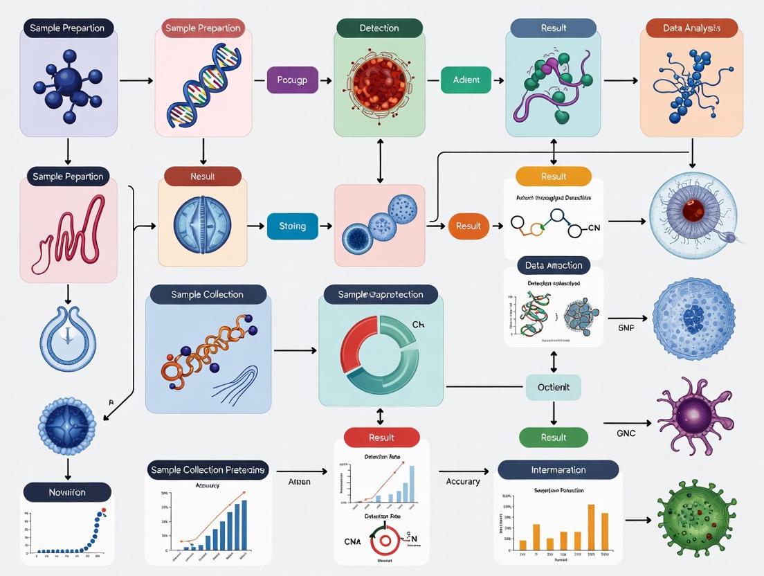
Experimental Workflow: The entire process, from sample preparation to result generation, is visualized below.
Figure 1: High-Throughput Automated Stool Analysis Workflow. This diagram illustrates the automated process of the Orienter Model FA280 analyzer.
Detailed Methodology:
- Sample Collection and Preparation:
- Automated Processing and Analysis:
- The sample carrier track loads the specimen into the analyzer [3].
- A pneumatic mixing system thoroughly homogenizes the sample with diluent [3].
- A high-resolution camera captures images of the sample's macroscopic characteristics [3].
- The microscope unit uses high- and low-power objectives with multifield tomography to capture detailed images [3].
- Captured images are automatically analyzed by an AI program for parasite detection and species identification [3].
- Result Validation:
- A skilled medical technologist should perform a user audit of the AI-generated findings to ensure accuracy. One study showed that while the AI report alone had fair agreement with the formalin-ethyl acetate concentration technique (FECT), the user audit achieved perfect agreement (κ = 1.00) [3].
2.2. Protocol: High-Throughput Molecular Detection (MagNA Pure 96 System) This protocol is adapted for the detection of Helicobacter pylori and clarithromycin resistance mutations from stool, demonstrating the application of automated nucleic acid extraction for molecular parasitology [4].
Detailed Methodology:
- Sample Lysis:
- Automated Nucleic Acid Extraction:
- Downstream qPCR and Analysis:
- Use 10 μL of the eluted sample in a TaqMan real-time PCR reaction [4].
- The qPCR master mix should include primers and probes specific to the target parasite (e.g., the rrl gene for H. pylori), an exogenous control, and BSA to reduce interference from stool-derived inhibitors [4].
- Analyze results using melt curve analysis and cycle threshold (Ct) shifts to detect the presence of the parasite and associated drug-resistance mutations [4].
3. Comparative Analysis of Detection Methods Researchers must select the appropriate method based on throughput, sensitivity, and application.
Table 3: Comparison of Parasite Detection Methodologies
| Method | Throughput | Key Advantage | Key Limitation | Best Application |
|---|---|---|---|---|
| Formalin-Ethyl Acetate Concentration Technique (FECT) | Low | High sensitivity; considered a gold standard [3] | Time-consuming, labor-intensive, requires expertise [3] | Low-volume settings, reference standard validation |
| Fully Automatic Digital Feces Analyzer (FA280) | High (40 samples/30 min) [3] | Reduced labor, minimal operator skill, reduced contamination [3] | Higher per-test cost, lower sensitivity vs. FECT [3] | High-volume clinical screening, routine diagnostics |
| High-Throughput Molecular (MagNA Pure 96) | High (96 samples/8 hrs) [4] | Detects parasite DNA and drug-resistance mutations [4] | Requires specific reagents, higher instrumentation cost | Drug-resistance monitoring, specific pathogen detection |
| Antigen Detection (EIA, DFA, Rapid Tests) | Medium | Rapid, does not require skilled morphologist [5] | May miss novel species or low-level infections | Point-of-care testing, specific pathogen screening (e.g., Giardia, Cryptosporidium) |
4. The Scientist's Toolkit: Research Reagent Solutions Key reagents and kits are fundamental for successful high-throughput detection.
Table 4: Essential Research Reagents for Parasite Detection
| Reagent / Kit Name | Function / Target | Manufacturer / Distributor | Test Type |
|---|---|---|---|
| Merifluor | Simultaneous detection of Cryptosporidium oocysts and Giardia cysts | Meridian Bioscience | Direct Fluorescent Antibody (DFA) [5] |
| ProSpecT | Microplate EIA for Giardia, Cryptosporidium, or Entamoeba histolytica/dispar | Remel | Enzyme Immunoassay (EIA) [5] |
| ImmunoCard STAT! | Rapid combined detection of Cryptosporidium and Giardia | Meridian | Rapid Immunochromatographic [5] |
| Triage | Rapid panel for Cryptosporidium, Giardia, and Entamoeba histolytica/dispar | BioSite | Rapid Immunochromatographic [5] |
| E. histolytica II | Detection of pathogenic E. histolytica (vs. non-pathogenic E. dispar) | TechLab | Enzyme Immunoassay (EIA) [5] |
| MagNA Pure 96 DNA and Viral NA Small Volume Kit | Automated nucleic acid extraction from stool and biopsies | Roche | Automated Sample Prep [4] |
| H. pylori & HPCR Primer/Probe Set | qPCR detection of H. pylori and clarithromycin resistance | Meridian Bioscience | Analyte Specific Reagents (ASR) [4] |
5. Method Selection and Logical Workflow Choosing the right method is a critical first step in the research process. The following diagram outlines a decision-making framework.
Figure 2: A Framework for Selecting a High-Throughput Detection Method. This chart guides the selection of an appropriate method based on key research requirements.
Conclusions The significant global burden of intestinal parasitic infections necessitates a shift from traditional, low-throughput diagnostic methods toward automated, high-throughput solutions. Platforms like the Orienter FA280 for automated digital morphology and the MagNA Pure 96 for automated molecular extraction represent critical tools for researchers and drug development professionals. These technologies enable large-scale screening, enhance reproducibility, and accelerate the pace of discovery and intervention in the fight against parasitic diseases.
Manual microscopy remains a cornerstone for the diagnosis of parasitic infections, particularly in the detection of parasites and ova in stool samples. However, this method faces significant challenges including labor-intensive procedures, subjective interpretation, and low throughput, which impede efficiency and compromise diagnostic accuracy. This application note details these inherent limitations through quantitative data comparisons and provides validated experimental protocols that leverage artificial intelligence (AI) and digital imaging to transition towards high-throughput, objective analysis. Framed within the context of stool sample research, this document serves as a guide for researchers and drug development professionals seeking to modernize parasitic diagnostics.
In parasitology, manual microscopy of stool samples using techniques like the formalin-ethyl acetate concentration technique (FECT) and Kato-Katz thick smears has long been the standard for detecting soil-transmitted helminths (STHs) and other intestinal parasites [3] [6] [7]. Despite its widespread use, the manual process is inherently constrained by its dependence on highly trained technicians, the subjective nature of visual analysis, and its low-throughput capacity, making it unsuitable for large-scale surveillance or drug efficacy studies [3] [7]. The global decline in STH prevalence has led to a higher proportion of light-intensity infections, which are frequently missed by manual microscopy, thereby creating an urgent need for more sensitive and scalable diagnostic solutions [6]. This application note delineates the core limitations of manual microscopy and presents advanced, high-throughput protocols to address these challenges.
Quantitative Comparison: Manual vs. Automated Methods
The limitations of manual microscopy become evident when its performance is quantitatively compared with emerging automated and AI-supported methods. The following table summarizes key metrics from recent studies, highlighting differences in sensitivity, throughput, and operational efficiency.
Table 1: Performance Comparison of Diagnostic Methods for Parasites in Stool Samples
| Diagnostic Method | Sensitivity for A. lumbricoides (%) | Sensitivity for T. trichiura (%) | Sensitivity for Hookworms (%) | Sample Processing Time | Throughput (Samples per Run) |
|---|---|---|---|---|---|
| Manual Microscopy (Kato-Katz) [6] | 50.0 | 31.2 | 77.8 | 30-60 min per smear (limited viability) [6] | Low (Individual) |
| Manual Microscopy (FECT) [3] | - | - | - | Labor-intensive, time-consuming [3] [7] | Low (Individual) |
| Autonomous AI (Digital) [6] | 50.0 | 84.4 | 87.4 | Minutes for digital analysis [6] | High (Batch) |
| Expert-Verified AI (Digital) [6] | 100.0 | 93.8 | 92.2 | Includes expert audit time [6] | High (Batch) |
| Fully Automatic Digital Feces Analyzer (FA280) [3] | - | - | - | ~30 min for 40 samples [3] | High (40 samples/run) |
Experimental Protocols
To overcome the limitations of manual microscopy, the following protocols outline steps for AI-supported digital diagnosis and the use of a fully automated analyzer.
Protocol: AI-Supported Digital Microscopy for Soil-Transmitted Helminths
This protocol utilizes whole-slide imaging and deep learning for the high-throughput detection of STH eggs in Kato-Katz thick smears [6].
1. Sample Preparation (Kato-Katz Smear):
- Materials: Stool sample, template (hole volume ~41.7 mg), microscope slide, cellophane strips soaked in glycerol-malachite green solution.
- Procedure: a. Place a small amount of stool on a piece of absorbent paper. b. Press the template onto the stool sample to fill the hole completely. c. Transfer the fecal material from the template onto the center of a clean microscope slide. d. Place a glycerol-soaked cellophane strip over the sample and press firmly with another slide to create a uniform, thick smear. e. Allow the preparation to clear for 30-60 minutes before examination or scanning. Note: Hookworm eggs disintegrate rapidly, so timing is critical [6].
2. Whole-Slide Digitization:
- Materials: Portable whole-slide scanner.
- Procedure: a. Place the prepared Kato-Katz slide into the portable scanner. b. Use the scanner's software to automatically capture high-resolution digital images of the entire smear area using a high-power objective lens. c. Save the whole-slide image (WSI) in a standard digital format (e.g., SVS, TIFF) for subsequent analysis [6].
3. AI-Based Analysis and Expert Verification:
- Materials: Computer workstation with AI analysis software.
- Procedure: a. Autonomous Analysis: Process the WSI through a deep learning algorithm (e.g., a convolutional neural network) trained to detect and classify STH eggs. The software autonomously generates a report of suspected parasites and their counts. b. Expert Verification: A skilled microscopist reviews the AI-generated findings within the digital image. The expert can confirm, reject, or add to the detections. This "expert-verified AI" approach combines high-throughput automation with expert oversight for maximum accuracy [6].
Protocol: High-Throughput Analysis Using a Fully Automatic Digital Feces Analyzer
This protocol describes the operation of the Orienter Model FA280, a fully automated system that integrates sampling, imaging, and AI-based evaluation [3].
1. System Setup and Sample Loading:
- Materials: Orienter FA280 analyzer, filtered sample collection tubes, stool samples.
- Procedure: a. Ensure the analyzer is powered on and the reagent reservoirs are filled. b. Aliquot approximately 0.5 g of stool into a filtered sample collection tube. c. Load up to 40 sample tubes into the automatic in-sample unit's track-type carrier in a single batch [3].
2. Automated Processing and Analysis:
- Procedure: a. Initiate the batch run. The system will automatically: i. Mix and Prepare: The sampling unit uses a pneumatic system to mix the stool with a diluent thoroughly. ii. Characterize and Image: A high-resolution camera captures the sample's macroscopic characteristics (color, consistency). The microscope unit then automatically captures multifield tomographic images at different magnifications. iii. AI Evaluation: The captured digital images are automatically analyzed by the onboard AI program to detect and identify parasitic elements [3]. b. The entire batch of 40 samples is processed in approximately 30 minutes [3].
3. Result Auditing and Reporting:
- Procedure: a. Access the initial report generated by the AI program. b. A skilled medical technologist performs a user audit by reviewing the digital microscope images associated with the AI findings. c. The final, audited report is generated, which shows perfect agreement with traditional FECT for species identification in fresh samples [3].
Workflow and System Diagrams
Manual vs. Automated Diagnostic Workflow
AI Verification and Integration Pathway
The Scientist's Toolkit: Research Reagent Solutions
The transition to high-throughput diagnostics relies on specific reagents and materials. The following table details essential components for the featured protocols.
Table 2: Essential Research Reagents and Materials for High-Throughput Parasite Detection
| Item | Function/Application | Protocol |
|---|---|---|
| Formalin-Ethyl Acetate | Used in the FECT method to concentrate parasites and ova from stool samples by separating debris and fats [3]. | FECT, Manual Microscopy |
| Glycerol-Malachite Green Solution | Used to clear and preserve Kato-Katz smears, providing contrast for microscopic visualization of helminth eggs [6]. | Kato-Katz, AI-Supported Digital Microscopy |
| Cellophane Strips | Coverslip alternative in Kato-Katz technique, soaked in glycerol to clear the fecal smear for better egg visibility [6]. | Kato-Katz, AI-Supported Digital Microscopy |
| 'Total Bile Acids 21 FS' Reagent (DiaSys) | Enzymatic cycling assay for quantifying total bile acids in stool; validated for use on fully automated clinical chemistry analyzers [8]. | Automated Clinical Chemistry Analysis |
| High-Resolution Digital Camera (e.g., See3CAM_5OCUG) | Captures high-quality digital images of specimens for subsequent analysis by AI algorithms in automated microscopes [9]. | Automated Digital Microscopy |
| Portable Whole-Slide Scanner | Digitizes entire microscope slides at high resolution, enabling remote analysis and AI processing of samples [6]. | AI-Supported Digital Microscopy |
| Bead-Based HRP2 Assay | High-throughput multiplex immunoassay for detecting malaria antigens (e.g., HRP2) in dried blood spots; used for sensitive surveillance [10]. | Multiplex Bead-Based Antigen Detection |
Defining High-Throughput Screening (HTS) in the Context of Stool Parasitology
High-Throughput Screening (HTS) in stool parasitology represents a paradigm shift from traditional, labor-intensive microscopic methods toward automated, rapid, and efficient diagnostic systems. These technologies are designed to process large volumes of samples with minimal manual intervention, addressing critical limitations of conventional approaches. Intestinal parasitic infections affect approximately 3.5 billion people globally, causing significant health burdens including malnutrition, anemia, impaired growth and cognitive development, and alterations in microbiota composition and immune responses [3]. Traditional diagnostic methods like the formalin-ethyl acetate concentration technique (FECT) and Kato-Katz thick smears, while considered gold standards, are time-consuming, labor-intensive, and heavily dependent on technician expertise [3]. The emerging HTS platforms leverage technologies such as digital imaging, artificial intelligence (AI), and lab-on-a-chip microfluidics to revolutionize parasite detection in stool samples, offering unprecedented efficiency in both clinical and research settings.
Key HTS Platforms and Technologies
Fully Automatic Digital Feces Analyzers
The Orienter Model FA280 represents a cutting-edge HTS platform that fully automates the stool analysis process. This system performs complete processing of 40 stool samples in a single 30-minute run, dramatically increasing throughput compared to manual methods [3]. The FA280 operates on a simple sedimentation principle and integrates several automated units: an automatic in-sample unit with track-type sample carrier, a pneumatic sampling unit for mixing samples with diluent, a high-resolution camera for sample characterization, and a microscope unit with high- and low-power objective lenses that automatically captures images using multifield tomography [3]. Approximately 0.5g of stool is processed per sample, and the system's AI program automatically evaluates digital microscope images to identify parasites and ova. A key feature is the option for user audit by skilled technologists, which has demonstrated perfect agreement (κ = 1.00) with FECT for species identification in fresh samples, significantly outperforming the AI report alone (κ = 0.367) [3].
Lab-on-a-Disk and Microfluidic Platforms
The SIMPAQ (Single-Image Parasite Quantification) device exemplifies the microfluidic approach to HTS in parasitology. This portable, point-of-care capable device utilizes lab-on-a-disk (LoD) technology that employs centrifugal, Coriolis, and Euler pseudo-forces generated during disk rotation to concentrate and trap parasite eggs [11]. The system uses a two-dimensional flotation technique by adding a saturated sodium chloride flotation solution to the stool sample, causing parasite eggs to float while most stool particles sediment. During centrifugation, eggs move toward the disk's center and are packed into a monolayer on a converging imaging zone, enabling single-image capture and immediate digitalization [11]. The SIMPAQ device requires only 1g of stool and demonstrates strong correlation (0.91) with the Mini-FLOTAC method, showing particular strength in detecting low-intensity infections with as few as 30-100 eggs per gram of feces [11]. Recent protocol modifications have focused on minimizing egg loss during preparation and reducing debris for clearer imaging.
Molecular and Microarray-Based Approaches
While not yet widely implemented for routine parasite detection, molecular methods offer complementary HTS capabilities for comprehensive stool analysis. Phylogenetic microarrays represent a powerful tool for analyzing the human intestinal microbiota, including parasitic eukaryotes. One such custom microarray, built on the Affymetrix GeneChip platform, contains probes for 775 different bacterial phylospecies and can detect bacteria present at a 0.00025% level of overall sample [12]. Additionally, environmental DNA (eDNA) methods provide non-invasive techniques for assessing parasite diversities and abundances through nucleic acid extraction and sequencing of genes from environmental samples, offering potential for comprehensive parasite community analysis [13]. Though these molecular approaches currently serve research purposes more than routine diagnostics, they expand the HTS landscape by enabling detection of cryptic species and providing insights into host-associated microbiomes and broader ecosystem processes.
Comparative Performance Analysis of HTS Platforms
Table 1: Performance Metrics of High-Throughput Screening Platforms for Stool Parasitology
| Platform | Throughput | Sample Volume | Sensitivity | Key Advantages | Limitations |
|---|---|---|---|---|---|
| Orienter FA280 | 40 samples/30 min | 0.5g | Lower than FECT in preserved samples [3] | Simplicity, reduced contamination, user audit capability [3] | Higher cost per test, lower sensitivity with preserved samples [3] |
| SIMPAQ LoD | Not specified | 1g | 91.39-95.63% vs. McMaster [11] | Portability, point-of-care use, detects low-intensity infections [11] | Egg loss during preparation, debris interference [11] |
| FECT (Traditional) | Low | 2g | Considered reference standard [3] | High sensitivity with larger sample size [3] | Time-consuming, labor-intensive, requires expertise [3] |
| Kato-Katz | Low | Minimal | Low for low-intensity infections [11] | WHO-recommended, cost-effective [11] | Low sensitivity for low-intensity infections [11] |
Table 2: Agreement in Species Identification Between HTS Platforms and Reference Methods
| Comparison | Overall Agreement | Kappa Statistic (κ) | Sample Type | Notes |
|---|---|---|---|---|
| FA280 (AI) vs. FECT | 75.5% | 0.367 (fair) [3] | Fresh stool | Significant difference (P < 0.001) [3] |
| FA280 (User Audit) vs. FECT | 100% | 1.00 (perfect) [3] | Fresh stool | No significant difference (P = 1) [3] |
| FA280 (User Audit) vs. FECT | Not specified | 0.857 (strong) for helminths [3] | Preserved stool | FECT detected more positive samples [3] |
| FA280 (User Audit) vs. FECT | Not specified | 1.00 (perfect) for protozoa [3] | Preserved stool | Disparity may be due to sample size differences [3] |
Experimental Protocols for HTS in Stool Parasitology
Protocol for Fully Automatic Digital Feces Analysis (FA280 System)
Principle: The method is based on simple sedimentation technique with automated digital imaging and AI analysis [3].
Materials and Reagents:
- Orienter Model FA280 fully automatic digital feces analyzer
- Filtered sample collection tubes
- Appropriate diluents
- Quality control materials
Procedure:
- Sample Preparation: Place approximately 0.5g of fresh stool into a filtered sample collection tube [3].
- Instrument Loading: Load up to 40 sample tubes into the automatic in-sample unit with track-type sample carrier [3].
- Automated Processing: Initiate the automated run. The system will:
- Mix samples thoroughly with diluent using a pneumatic mixing system
- Capture high-resolution images of sample character and color
- Perform multifield tomography imaging at different magnifications using high- and low-power objective lenses
- Transfer digital images to the AI analysis program [3]
- AI Analysis: The built-in artificial intelligence program automatically evaluates images for parasite detection and species identification.
- User Audit: A skilled medical technologist reviews and verifies the AI findings (recommended for improved accuracy) [3].
- Result Interpretation: Review the automated report for parasite identification and quantification.
Quality Control: Run appropriate quality control samples according to manufacturer specifications and laboratory protocols.
Modified SIMPAQ Protocol for Lab-on-a-Disk Analysis
Principle: This protocol uses two-dimensional flotation combining centrifugation and flotation forces to concentrate parasite eggs in a single imaging zone [11].
Materials and Reagents:
- SIMPAQ lab-on-a-disk device
- Saturated sodium chloride flotation solution
- Surfactant (to reduce egg adhesion)
- 200μm filter membrane
- Digital camera
- Centrifuge compatible with the LoD device
Procedure:
- Sample Preparation: Mix 1g of stool sample with saturated sodium chloride flotation solution [11].
- Surfactant Addition: Add surfactant to the flotation solution to reduce adherence of eggs to the walls of syringes and disk [11].
- Filtration: Filter the mixture through a 200μm filter membrane to remove larger debris that could obstruct egg trapping [11].
- Disk Loading: Infuse the prepared sample into the LoD device.
- Centrifugation: Centrifuge the disk at the optimized rotation speed to direct eggs toward the center while minimizing effects of Coriolis and Euler forces [11].
- Image Capture: Capture a single digital image of the Field of View (FOV) where eggs are concentrated in a monolayer.
- Image Analysis: Analyze the digital image for parasite egg identification and quantification.
Technical Notes: The modified protocol specifically addresses previous limitations of egg loss during preparation and low capture efficiency in the FOV. Channel length reduction from 37mm to 27mm in updated disk designs helps minimize the effects of additional inertial forces [11].
Formalin-Ethyl Acetate Concentration Technique (Reference Method)
Principle: This concentration method uses formalin for preservation and ethyl acetate for extraction of debris, concentrating parasites in the sediment [3].
Materials and Reagents:
- 10% formalin
- Ethyl acetate
- 15ml conical centrifuge tubes
- 2-layer gauze
- Applicator sticks
- Cotton-tipped applicators
- Centrifuge
- Light microscope
Procedure:
- Sample Preparation: Mix 2g of stool sample with 10ml of 10% formalin [3].
- Filtration: Strain the fecal suspension through a 2-layer gauze into a 15ml conical centrifuge tube.
- Solvent Addition: Add 3ml of ethyl acetate to the tube, close tightly, and shake vigorously in an inverted position for 1 minute [3].
- Centrifugation: Centrifuge at 2500rpm for 2 minutes.
- Debris Removal:
- Free the plug of debris at the top of the tube by ringing the sides with an applicator stick
- Decant the top layer of supernatant
- Remove debris on the sides of the tube using a cotton-tipped applicator [3]
- Microscopy: Pipette the sediment onto a clean glass slide and examine for ova and parasites under a light microscope.
Workflow Visualization of HTS Methods
HTS Workflow Comparison
This diagram illustrates the operational workflows for three stool parasitology methods, highlighting the automated, high-throughput nature of the FA280 and SIMPAQ systems compared to the manual traditional approach. The FA280 pathway demonstrates complete automation from sample preparation through AI analysis, with the critical user audit step that significantly improves accuracy. The SIMPAQ pathway shows the microfluidic approach utilizing flotation and centrifugation forces to concentrate parasites for single-image quantification. In contrast, the traditional FECT method relies heavily on manual processing and expert microscopy, creating a bottleneck for high-throughput applications.
Research Reagent Solutions for HTS Parasitology
Table 3: Essential Research Reagents for High-Throughput Stool Parasitology
| Reagent/Material | Function | Application Notes |
|---|---|---|
| Saturated Sodium Chloride Solution | Flotation medium for parasite eggs | Used in SIMPAQ protocol; density causes eggs to float while debris sediments [11] |
| Formalin (10%) | Sample preservation and fixation | Maintains parasite morphology in reference FECT method and preserved samples [3] |
| Ethyl Acetate | Debris extraction and concentration | Used in FECT to extract fat and debris from stool samples, concentrating parasites [3] |
| Surfactant Solutions | Reduce egg adhesion to surfaces | Added to flotation solution in SIMPAQ to minimize egg loss to walls of syringes and disk [11] |
| Filter Membranes (200μm) | Removal of large particulate debris | Critical for SIMPAQ protocol to prevent obstruction of egg trapping and imaging [11] |
| Quality Control Materials | Verification of assay performance | Essential for validating automated systems like FA280; should include positive and negative samples |
High-Throughput Screening technologies represent a transformative advancement in stool parasitology, addressing critical limitations of traditional methods while enabling rapid processing of large sample volumes. The FA280 automated digital feces analyzer and SIMPAQ lab-on-a-disk system exemplify two complementary approaches to HTS, each with distinct advantages. The FA280 offers complete automation with integrated AI analysis, while the SIMPAQ platform provides portability and point-of-care potential with innovative microfluidics. Current evidence demonstrates that these technologies can achieve excellent agreement with reference methods when combined with expert verification, as shown by the perfect agreement (κ = 1.00) between FA280 with user audit and FECT [3]. Future developments should focus on improving AI algorithms for greater autonomous accuracy, optimizing protocols to minimize egg loss, and reducing costs to enhance accessibility in resource-limited settings where parasitic infections are most prevalent.
The shift toward automated systems in the high-throughput detection of intestinal parasites represents a fundamental change in diagnostic parasitology. This transition is primarily driven by the critical needs for enhanced standardization, improved reproducibility, and sophisticated data management. Traditional microscopy-based methods, while foundational, are hampered by their labor-intensive nature, operator dependency, and proneness to human error [3] [14]. This document details the application of these core drivers through specific protocols and analytical frameworks, providing researchers with practical guidance for implementing automated detection systems in intestinal parasitosis research.
Core Driver 1: Standardization
Standardization in stool sample analysis is paramount for generating reliable, comparable data across different studies and laboratories. Automated systems address key variability points from sample collection through to final analysis.
Standardized Sample Collection and Preprocessing
The initial phase of standardization involves consistent sample handling, which directly influences downstream analytical outcomes.
- Calibrated Sampling Devices: Devices like the bioMérieux Stool Preprocessing Device (SPD) incorporate a calibrated spoon to aliquot a consistent volume of stool (approximately 220 mg) into a buffer containing glass beads for homogenization [15]. This eliminates the variability introduced by manual weighing and dilution.
- Integrated Filtration Systems: The SPD and similar devices often include built-in multi-stage filtration systems (e.g., <150 μm and <30 μm filters) to clarify suspensions, remove particulate debris, and produce a uniform filtrate for nucleic acid extraction or other assays [15].
- Automated Digital Analyzers: Systems like the Orienter Model FA280 fully automate the process from sample introduction to analysis. They use a track-type sample carrier and pneumatic mixing system to ensure every sample is processed with identical parameters, including dilution, homogenization, and volume dispensed onto slides or test kits [3].
Analytical Standardization and Reference Materials
Standardizing the analytical process itself is crucial for inter-laboratory reproducibility.
- Reference Materials: The National Institute of Standards and Technology (NIST) is developing standardized fecal reference materials. These identical tubes of stool allow labs to benchmark their analytical methods against a universal control, correcting for inter-experimental and inter-laboratory variability [16].
- Consistent Imaging and Analysis: Automated digital systems capture images under standardized lighting, focus, and magnification. This ensures that the input data for subsequent algorithm-based identification is consistent, unlike manual microscopy which can vary based on the technologist's technique [3] [17].
Table 1: Impact of Standardization on Preprocessing Metrics
| Parameter | Traditional Manual Method | Automated/Standardized Method | Impact of Standardization |
|---|---|---|---|
| Sample Volume | Variable (e.g., "pea-sized") | Fixed (e.g., 220 mg via calibrated spoon [15] or ~0.5 g in FA280 [3]) | Reduces pre-analytical variability, improves quantification accuracy. |
| Homogenization | Manual vortexing (variable intensity/duration) | Automated pneumatic [3] or bead-based [15] mixing | Produces a more consistent and representative suspension. |
| Filtration/Clarification | Manual, multi-step centrifugation | Integrated, automated filtration [15] | Reduces hands-on time and improves consistency of filtrate clarity. |
| Process Duration | Highly variable, ~30+ minutes | Consistent, <5 minutes for SPD [15], ~30 min for 40 samples on FA280 [3] | Enables predictable throughput and workflow scheduling. |
Core Driver 2: Reproducibility
Reproducibility is the cornerstone of the scientific method. Automation enhances reproducibility by minimizing human-induced variability in both sample processing and result interpretation.
Protocol: Assessing Reproducibility in a Multiplex PCR-Bead Assay
The following protocol, adapted from Taniuchi et al., outlines a high-throughput method for the reproducible detection of seven major intestinal parasites [18].
1. Objective: To evaluate the reproducibility of a multiplex PCR-bead assay for detecting Cryptosporidium spp., Giardia intestinalis, Entamoeba histolytica, Ancylostoma duodenale, Ascaris lumbricoides, Necator americanus, and Strongyloides stercoralis.
2. Materials:
- Stool Samples: Preserved in 10% formalin or fresh-frozen.
- DNA Extraction Kit: Compatible with stool samples (e.g., QIAamp DNA Stool Mini Kit).
- PCR Reagents: Primer mixes for two multiplex PCRs (Protozoa and Helminths), hot-start DNA polymerase, dNTPs, PCR buffer.
- Luminex Beads: MagPlex-TAG microspheres coupled to specific internal oligonucleotide probes.
- Equipment: Thermal cycler, Luminex MAGPIX or equivalent instrument.
3. Methodology: 1. Nucleic Acid Extraction: Extract genomic DNA from approximately 200 mg of stool using a standardized kit protocol. Include an internal control to monitor for inhibition. 2. Multiplex PCR Amplification: * Set up two separate multiplex PCR reactions: one for protozoa and one for helminths. * Use previously published and validated primer sequences specific to each target [18]. * Cycling conditions: Initial denaturation at 95°C for 5 min; 45 cycles of 95°C for 30 s, 60°C for 30 s, 72°C for 30 s; final extension at 72°C for 5 min. 3. Bead-Based Hybridization: * Mix PCR products with a suspension of probe-coated Luminex beads. * Denature at 95°C for 2 min and hybridize at 52°C for 30 min. 4. Detection and Analysis: * Analyze the bead mixture on the Luminex instrument. The instrument identifies the bead set (and thus the target) based on its internal dye and quantifies the signal from the hybridized PCR product. 5. Reproducibility Assessment: * Test a panel of clinical specimens (e.g., n=319 [18]) in duplicate or triplicate across multiple runs. * Calculate the intra-assay and inter-assay coefficients of variation for the Median Fluorescence Intensity (MFI) for each target. * Compare results to a reference method (e.g., microscopy or monoplex real-time PCR) to determine concordance, sensitivity, and specificity.
Quantitative Evidence of Improved Reproducibility
Automated systems demonstrate superior reproducibility, as quantified in comparative studies.
Table 2: Reproducibility Metrics of Automated vs. Manual Methods
| Method / System | Metric | Performance | Context |
|---|---|---|---|
| Manual Microscopy (FECT) | Diagnostic Agreement | Variable, user-dependent [3] | Gold standard but suffers from inter-observer variability. |
| Digital Feces Analyzer (FA280) with User Audit | Species Identification Agreement (κ) | Perfect agreement with FECT for protozoa (κ=1.00) [3] | AI-assisted human audit achieves maximal reproducibility. |
| Multiplex PCR-Bead Assay [18] | Sensitivity/Specificity | 83-100% vs. parent real-time PCR assays | High-throughput method maintains analytical reproducibility. |
| Stool Preprocessing Device (SPD) [15] | Coefficient of Variation (CV) for HAdV DNA Quantification | 1.79% - 1.83% | Extremely low variability in quantitative output across extractions. |
Figure 1: Workflow for Reproducibility Assessment in Multiplex PCR-Bead Assay.
Core Driver 3: Data Management
High-throughput parasite detection generates vast amounts of complex data, including digital images, quantitative PCR values, and patient metadata. Automated data management is essential for transforming this raw data into actionable insights.
Automated Data Management in Diagnostic Parasitology
- Data Integration and Cleansing: Automated systems like the FA280 seamlessly integrate sample characterization data (color, consistency), digital microscope images, and test kit results into a unified digital record [3]. This process automatically formats and cleanses data, removing inconsistencies and preparing it for analysis [19] [20].
- Data Analysis and Reporting: Artificial Intelligence (AI) programs automatically analyze digital microscope images, classifying and counting parasitic elements [3] [17]. This automation accelerates the discovery of data-driven insights and generates reports with minimal manual intervention [19].
- Enhanced Data Security and Scalability: Automated data management systems provide robust security features, including access controls and audit trails, which are critical for handling sensitive patient data [19]. Furthermore, these systems are inherently scalable, capable of handling increasing volumes of data from large-scale studies without a corresponding increase in errors or processing time [20].
Protocol: Implementing an Automated Image Analysis and Expert System
This protocol is based on a system that integrates automated image processing with an expert knowledge base for diagnosing intestinal parasitosis [17].
1. Objective: To automate the detection and identification of parasites from stool sample microscopy images and integrate findings with a clinical expert system for diagnosis and therapy recommendation.
2. Materials:
- Stool Samples: Fresh or preserved stool samples.
- Microscope with Digital Camera: For acquiring high-resolution images of wet mounts or stained smears.
- Computational System: Pre-trained neuro-fuzzy classifier and image processing software.
- Expert System Knowledge Base: Database containing information on parasitic diseases, symptoms, and treatments.
3. Methodology: 1. Sample Preparation and Imaging: Prepare standard wet mounts or permanent stained smears from stool samples. Capture multiple digital images per sample using a standardized microscope and camera setup. 2. Automated Image Analysis: * Segmentation: Apply a combined Distance Regularized Level Set Evolution (DRLSE), automatically initialized by a Circular Hough Transform, to isolate potential parasites and ova from the background [17]. * Feature Extraction: For each segmented object, compute morphological features (size, shape, texture) and staining characteristics. * Classification: Input the extracted features into a pre-trained neuro-fuzzy classifier. The classifier is trained to recognize up to twenty different species of human intestinal parasites [17]. 3. Data Integration and Expert Reasoning: * Input patient symptoms and clinical history into the expert system interface. * The system uses a knowledge-based decision algorithm to propose a suspicious parasitic disease. * The automated image analysis results are used to confirm the presence of the suspected parasite. 4. Final Reporting: * The system combines the clinical and microscopic findings to generate a final diagnostic recommendation, including a proposed therapy [17].
Figure 2: Automated Image Analysis and Expert System Workflow.
The Scientist's Toolkit: Research Reagent Solutions
Table 3: Essential Reagents and Materials for Automated Parasite Detection
| Item | Function/Description | Application Example |
|---|---|---|
| Formalin-Ethyl Acetate (FECT) | Concentration technique for parasites; separates debris from ova/cysts via density gradient [3] [14]. | Gold standard for manual concentration prior to microscopy [3]. |
| Stool Preprocessing Device (SPD) | Integrated device with calibrated spoon, buffer, glass beads, and filters for standardized sample preparation [15]. | Production of a clarified, homogeneous stool filtrate for nucleic acid extraction and molecular assays [15]. |
| Luminex MagPlex-TAG Microspheres | Magnetic beads with unique spectral signatures, coated with oligonucleotide probes for multiplexed target detection [18]. | High-throughput, multiplex PCR-based detection of 7 intestinal parasites in a single assay [18]. |
| Digital Feces Analyzer (e.g., FA280) | Fully automated system for sample mixing, imaging, and AI-based analysis of stool for parasites [3]. | High-throughput, automated stool examination; reduces technician time and subjective error [3]. |
| NIST Stool Reference Material | Well-characterized, homogeneous fecal material for inter-laboratory standardization and quality control [16]. | Serves as a universal baseline to normalize metabolomic and metagenomic measurements across studies [16]. |
| Wheatley's Trichrome Stain | Polychromatic stain for permanent smears; provides contrast to differentiate protozoal structures from artifacts [14]. | Permanent stained smear preparation for definitive identification of protozoan cysts and trophozoites [14]. |
Next-Generation Diagnostic Technologies: From Automated Digital Analyzers to Molecular Panels
Intestinal parasitic infections affect billions globally, causing malnutrition, anemia, and impaired cognitive development [3]. Traditional microscopic diagnosis methods, while considered a gold standard, are labor-intensive, time-consuming, and subject to human error [21] [22]. Fully automated digital feces analyzers represent a technological advancement that addresses these limitations by integrating robotics, high-resolution digital imaging, and artificial intelligence (AI) to standardize and accelerate the detection of parasites and ova in stool samples [23] [24]. This document details the operational principles, protocols, and application of two such systems—the Orienter FA280 and the KU-F40—within the context of high-throughput parasitology research.
Operational Principles and Technological Comparison
The FA280 and KU-F40 analyzers transform fecal parasitology through automation and digitalization, though they employ distinct technical approaches.
Orienter FA280 Operational Principles
The FA280 utilizes an automatic sedimentation and concentration technique as its core physical principle [21] [3]. The system processes approximately 0.5 grams of feces placed in a filtered collection tube. The workflow involves intelligent sample dilution and high-frequency pneumatic mixing to create a homogeneous suspension [21]. This suspension is then subjected to microscopy using high- and low-power objective lenses that automatically capture hundreds of high-resolution images through multi-field tomography [21] [3]. The acquired images are analyzed by a deep learning AI algorithm trained to identify the color, shape, and structural features of various parasite eggs and protozoa [21]. This process minimizes manual intervention and reduces biohazard risks.
KU-F40 Operational Principles
The KU-F40 employs a broader range of physical detection methods, including image analysis of fecal formed elements, and offers both flotation and sedimentation modes [25] [26] [23]. Its key differentiator is an automatic iodine staining function that enhances the detection rate of specific ova and parasites [25] [23]. For a standard test using the normal mode, the instrument automatically dilutes and mixes a soybean-sized (approximately 200 mg) specimen, filters it, and draws 2.3 ml into a flow counting chamber for precipitation [22] [26]. It captures up to 520 low-magnification and 20 high-magnification images. A notable feature is its auto-tracking function, where the high-magnification lens automatically targets and re-images potential eggs located by the low-magnification lens, providing clearer diagnostic images [23].
Table 1: Technical Comparison of the FA280 and KU-F40 Analyzers
| Feature | Orienter FA280 | KU-F40 |
|---|---|---|
| Core Physical Principle | Automated sedimentation & concentration [21] [3] | Formed element image analysis; Flotation & Sedimentation modes [22] [25] |
| Sample Throughput | ~40 samples per 30-minute run [3] | 15 - 60 samples per hour [23] |
| AI & Imaging | Multi-field tomography; AI analysis of color, shape, and consistency [21] | Auto-tracking of eggs; AI identification; Iodine staining capability [25] [23] |
| Sample Volume | ~0.5 g [21] | ~200 mg (soybean-sized) [22] |
| Key Differentiator | High-throughput community screening [21] | Multi-mode testing and integrated colloidal gold immunoassays [23] |
Performance Data from Validation Studies
Independent studies have validated the performance of these analyzers against traditional methods, with key quantitative findings summarized below.
FA280 Performance Data
A cross-sectional study of 1,000 participants compared the FA280 with the Kato-Katz (KK) method. Both methods reported a positive rate of 10.0%, demonstrating a 96.8% agreement and no statistically significant difference (P > 0.999) [21]. The kappa value of 0.82 (95% CI: 0.76–0.88) indicates strong agreement. The study noted that agreement was significantly higher in high-infection-intensity groups [21]. Another study comparing the FA280 with the Formalin-ethyl acetate concentration technique (FECT) showed perfect agreement (κ = 1.00) for species identification after a user audit of the AI findings [3].
KU-F40 Performance Data
A large-sample retrospective study compared the KU-F40 (n=50,606) to manual microscopy (n=51,627). The KU-F40 group had a significantly higher parasite detection level (8.74% vs. 2.81%, χ² = 1661.333, P < 0.05) and detected nine parasite species compared to five with manual microscopy [22]. A separate prospective study on 1,030 specimens reported that the KU-F40 normal mode had a sensitivity of 71.2% and a specificity of 94.7%, with a diagnostic concordance of 90.78% (Kappa = 0.633) with reference methods [26].
Table 2: Comparative Diagnostic Performance of Automated Analyzers vs. Traditional Methods
| Study & Metric | FA280 vs. Kato-Katz [21] | KU-F40 vs. Manual Microscopy [22] |
|---|---|---|
| Positive Detection Rate (Analyzer) | 10.0% | 8.74% |
| Positive Detection Rate (Reference) | 10.0% | 2.81% |
| Statistical Agreement | 96.8% | N/A |
| Kappa Statistic (κ) | 0.82 (Strong Agreement) | N/A |
| P-Value | > 0.999 (Not Significant) | < 0.05 (Significant) |
| Key Finding | No significant difference in detection rate; strong agreement. | Significantly higher detection rate and more species identified. |
Detailed Experimental Protocols
For researchers aiming to implement these technologies, the following protocols are essential.
Protocol for the Orienter FA280
1. Sample Preparation: Collect approximately 0.5 grams of fresh or preserved (10% formalin) stool specimen into the dedicated filtered sample collection tube [21] [3].
2. Instrument Setup: Power on the analyzer and log into the software system. Place the collection cup into a dedicated specimen rack and load it onto the instrument's sample tray [3].
3. Automated Analysis Initiation: In the software interface, select the appropriate test parameters (e.g., physical character, morphological detection). Click "Start" to begin the automated run [3]. The instrument will then: - Automatically add diluent and perform high-frequency pneumatic mixing [21]. - Capture images of the sample's physical traits [3]. - Perform microscopic analysis using multi-field layered scanning to capture hundreds of high- and low-magnification images [21].
4. AI Analysis and Reporting: The built-in AI software analyzes all captured images for the presence of parasites and ova, generating a preliminary report [21] [3].
5. User Audit and Result Validation: A skilled technologist must review the AI-flagged images and positive findings to confirm the results. This manual audit is critical for achieving the highest diagnostic accuracy, as studies show it can raise agreement with FECT to 100% [3].
Protocol for the KU-F40
1. Sample Preparation: Collect a soybean-sized fecal specimen (approximately 200 mg) in the special collection cup with a rotating threaded screw cap to ensure airtightness [22] [23].
2. Mode Selection: Choose the appropriate detection mode via the software: - Normal Mode: For routine, high-sensitivity screening [26]. - Floating-Sedimentation Mode: For enhanced detection of specific ova and parasites, which uses high-concentration saline [26].
3. Instrument Operation: Place the sample cup on the dedicated rack and load it into the sample tray. Initiate the test sequence. The instrument automatically handles dilution, mixing, filtration, and transfer of the sample to the flow cell [22] [26].
4. Imaging and AI Identification: The system captures over 300 images under low and high magnification. The AI algorithm automatically identifies and classifies parasitic elements. The auto-tracking function can be engaged for high-magnification follow-up on suspected targets [23].
5. Colloidal Gold Testing (Optional): The KU-F40 can simultaneously run up to six different fecal immunoassays (e.g., FOB, Calprotectin, H. Pylori) from the same sample cup, as the instrument automatically dispenses sample onto the reagent cards [25] [23].
6. Review and Verification: Laboratory personnel manually review all AI-identified suspected parasites before finalizing and issuing the report [22].
The Scientist's Toolkit: Essential Research Reagent Solutions
Successful implementation of automated fecal analyzers in a research setting requires specific reagents and materials.
Table 3: Essential Research Reagents and Materials for Automated Fecal Analysis
| Item | Function/Application | Example/Note |
|---|---|---|
| Filtered Sample Collection Tubes | Ensures limited, standardized sampling and prevents clogging of the analytical pathway. | FA280 uses a specific filtered tube for its sedimentation method [21]. |
| Special Collection Cups | Designed for airtight transport and limited quantitative sampling. | KU-F40 uses a cup with a rotating threaded screw cap [23]. |
| Intelligent Diluent | Automated dilution of stool samples to an optimal concentration for imaging and analysis. | Critical for creating a homogenous suspension without obscuring elements [21] [3]. |
| High-Concentration Saline | Used in specific instrument modes (e.g., KU-F40 flotation mode) to separate parasites based on density. | Enhances the recovery of certain ova and parasites [26]. |
| Iodine Staining Solution | Automatically added by the instrument to stain samples, improving detection of protozoa and other delicate structures. | A key feature of the KU-F40 to improve diagnostic yield [25] [23]. |
| Colloidal Gold Reagent Cards | For simultaneous, automated quantification of fecal biomarkers (e.g., FOB, Calprotectin, H. Pylori). | KU-F40 can host up to 6 different tests, integrating morphological and immunoassay data [25] [23]. |
| Quality Control Materials (QCM) | For verifying the performance of both morphological (cells, parasites) and immunoassay components. | Essential for ensuring daily analytical accuracy and precision [23]. |
Fully automated digital feces analyzers like the Orienter FA280 and KU-F40 represent a paradigm shift in parasitology diagnostics. By leveraging AI, advanced imaging, and automated fluidics, they offer a solution to the bottlenecks of traditional microscopy—standardizing the process, increasing throughput, and improving detection consistency. For the research community, these systems enable large-scale epidemiological studies, high-throughput drug efficacy screening, and robust longitudinal monitoring of intervention programs. While an initial investment is required and the AI algorithms require expert validation, the integration of these analyzers into research workflows promises to accelerate progress toward the control and elimination of neglected tropical diseases caused by intestinal parasites.
The Role of Artificial Intelligence (AI) and Deep Learning in Parasite Image Recognition
The diagnosis of intestinal parasitic infections (IPIs), which affect billions globally, has long relied on conventional microscopy techniques such as the formalin-ethyl acetate concentration technique (FECT) and Kato-Katz thick smears [3] [27]. While these methods are considered gold standards, they are labor-intensive, time-consuming, and their accuracy is heavily dependent on the expertise and training of the microscopist [3] [7]. The need for low-complexity, high-throughput, and cost-effective alternatives has driven the integration of artificial intelligence (AI) and deep learning into parasitology diagnostics. AI-based systems, particularly those utilizing convolutional neural networks (CNNs) and object detection models like YOLO (You Only Look Once), are revolutionizing the field by enabling rapid, accurate, and automated detection and classification of parasites in stool samples [28] [27] [29]. This document details the application notes and experimental protocols for implementing AI in high-throughput detection of parasites and ova, providing a resource for researchers and drug development professionals.
Performance Comparison of AI Models and Human Technologists
Recent validation studies demonstrate that AI-assisted diagnostic tools can match or surpass human technologists in detection sensitivity and accuracy for a wide range of intestinal parasites.
The following table summarizes key performance metrics from recent studies comparing AI models and human experts in parasite image recognition.
Table 1: Performance Metrics of AI Models in Parasite Detection
| Model/System | Application | Accuracy | Sensitivity/Recall | Precision | Specificity | F1 Score | Remarks |
|---|---|---|---|---|---|---|---|
| DINOv2-large [27] | Intestinal Parasite ID | 98.93% | 78.00% | 84.52% | 99.57% | 81.13% | SSL model; high accuracy |
| YOLOv8-m [27] | Intestinal Parasite ID | 97.59% | 46.78% | 62.02% | 99.13% | 53.33% | Object detection model |
| ARUP AI (CNN) [28] [29] | Wet Mount Stool Analysis | - | > Human Tech | - | - | - | 98.6% agreement with humans; found 169 missed organisms |
| YOLOv3 [30] | P. falciparum in Blood | 94.41% | - | - | - | - | False negative rate: 1.68% |
A study at ARUP Laboratories, which utilized a CNN trained on over 4,000 parasite-positive samples encompassing 27 species, demonstrated a 98.6% agreement with human assessment. Notably, the AI system identified an additional 169 organisms that had been missed during manual inspection, showcasing superior clinical sensitivity [28] [29]. Similarly, a comprehensive evaluation of deep learning models for intestinal parasite identification found that the DINOv2-large model achieved an accuracy of 98.93%, a sensitivity of 78.00%, and a specificity of 99.57% [27]. Object detection models like YOLOv8-m also showed high accuracy (97.59%) and specificity (99.13%), though with more variable sensitivity for different parasite classes [27]. These results highlight the potential of AI to enhance diagnostic precision, reduce human error, and improve detection rates, particularly in low-prevalence settings where most samples are negative [31].
Detailed Experimental Protocols
Protocol 1: AI-Assisted Analysis of Stool Samples Using a Fully Automatic Digital Feces Analyzer
This protocol outlines the procedure for using the Orienter Model FA280, a fully automatic digital feces analyzer, for high-throughput detection of intestinal parasites [3].
Principle: The system automates sample processing, digital imaging, and AI-based analysis to identify parasitic elements in stool samples, significantly reducing hands-on time and technical workload [3].
Materials and Reagents:
- Orienter Model FA280 Analyzer: Comprises an automatic in-sample unit, sampling unit, character/color photographing unit, microscope unit, and test kit unit [3].
- Stool Sample: Approximately 0.5 grams of fresh or preserved (10% formalin) stool is required per test [3].
- Sample Collection Tube: A filtered sample collection tube is used for homogenization.
- Supported Reagents and Diluents: As specified by the manufacturer.
Procedure:
- Sample Preparation: Place approximately 0.5 g of stool into the provided filtered sample collection tube.
- Instrument Loading: Load a batch of up to 40 sample tubes into the FA280's track-type sample carrier.
- Automated Processing:
- The pneumatic sampling unit mixes the stool sample with diluent.
- The character and color unit captures macroscopic images.
- The microscope unit uses high- and low-power objectives with multifield tomography to capture detailed microscopic images.
- AI Analysis: Captured digital images are automatically analyzed by the integrated AI program for the detection and classification of parasites.
- User Audit (Optional): A skilled medical technologist can review the AI-generated findings for verification. Studies show this user audit can achieve perfect agreement (κ = 1.00) with traditional FECT results [3].
- Results and Reporting: The system generates a report of identified parasites. The entire process for 40 samples takes approximately 30 minutes [3].
Protocol 2: Deep Learning-Based Parasite Identification from Microscope Slides
This protocol describes a digital workflow for AI-assisted screening of stained fecal slides, as implemented in platforms like the Techcyte Fusion Parasitology Suite [31].
Principle: Whole-slide imaging combined with CNN-based AI analysis locates, pre-classifies, and counts parasitic structures, presenting them to a technologist for final review, thereby improving efficiency and accuracy [27] [31].
Materials and Reagents:
- Stool Sample: Fresh stool specimen.
- Fecal Concentration Device: Such as Apacor Mini or Midi Parasep [31].
- Staining Solutions: Iodine for wet mounts or Trichrome stain for permanent smears, prepared per standard laboratory protocols [31].
- Supported Slide Scanner: Examples include Hamamatsu S360, Grundium Ocus 40, or Pramana M Pro [31].
- AI Software Platform: Such as Techcyte's Fusion Parasitology Suite [31].
Procedure:
- Slide Preparation:
- Concentrate the stool sample using a fecal concentration device.
- For wet mounts, use a specialized mounting media to extend slide life and improve parasite visibility. For permanent smears, stain using the standard Trichrome protocol [31].
- Apply a coverslip.
- Slide Scanning:
- Load the prepared slides into a compatible whole-slide scanner.
- The scanner will produce high-resolution digital images (typically at 40x or 80x equivalent magnification) and automatically upload them to the AI platform.
- AI Image Processing:
- The platform's AI algorithm, typically a CNN, processes the images to detect, classify, and group objects of interest (e.g., ova, cysts, trophozoites) by class and confidence level.
- Technologist Review:
- The technologist logs into the web-based platform to review the AI-proposed findings.
- Negative samples can be eliminated rapidly, often in 15-30 seconds [31].
- For positive samples, the technologist confirms the identity, prevalence, and size of the parasites. The original slide can be checked under a microscope for confirmation if required.
- Reporting: Final results are reported into the Laboratory Information System (LIS).
Diagram 1: AI-assisted digital pathology workflow for parasite detection.
The Scientist's Toolkit: Key Research Reagent Solutions
Successful implementation of AI-based parasite diagnostics relies on a suite of specialized reagents, instruments, and software.
Table 2: Essential Materials for AI-Based Parasitology Research
| Item Name | Function/Application | Specific Examples / Notes |
|---|---|---|
| Automatic Feces Analyzer | Fully automated sample processing, imaging, and analysis. | Orienter Model FA280; processes 40 samples in ~30 min [3]. |
| Whole-Slide Scanners | Digitizes microscope slides for AI analysis. | Hamamatsu S360, Grundium Ocus 40, Pramana M Pro/HT2/HT4 [31]. |
| AI Diagnostic Software | Cloud-based platform for image analysis and review. | Techcyte Fusion Parasitology Suite (for wet mount, Trichrome, Acid Fast) [31]. |
| Fecal Concentration Kits | Prepares stool samples for microscopic examination. | Apacor Mini or Midi Parasep devices are recommended [31]. |
| Staining Solutions | Enhances contrast for visual and AI-based identification. | Iodine for wet mounts; Trichrome stain for protozoa; Modified Acid Fast for coccidia [31]. |
| Specialized Mounting Media | Extends slide life and improves clarity for wet mounts. | Used with Techcyte's wet mount protocol to lengthen slide life to two hours [31]. |
Discussion and Future Perspectives
The integration of AI into parasite image recognition represents a paradigm shift in diagnostic parasitology. The primary advantages include increased throughput, reduced analysis time, diminished labor intensity, and enhanced detection sensitivity, especially for low-level infections that are easily missed by human readers [3] [28] [29]. Furthermore, these systems can help standardize diagnoses across laboratories, mitigating the variability associated with human expertise [27].
However, challenges remain. The initial capital investment for automated scanners and systems can be high, and the cost per test may be greater than traditional microscopy [3]. Some studies note that conventional methods like FECT can still show higher sensitivity, potentially due to the use of larger sample volumes [3]. Therefore, current best practices often position AI as a powerful tool to assist technologists rather than fully replace them, creating a hybrid workflow that maximizes both efficiency and accuracy [27] [31].
Future developments will likely focus on refining AI algorithms to improve sensitivity for morphologically similar parasites and protozoan trophozoites, expanding digital libraries with rare parasites, and integrating AI platforms with other high-throughput methods like multiplex PCR [18] [7]. As these technologies mature, they promise to make high-quality parasitological diagnosis more accessible and efficient, ultimately benefiting public health efforts worldwide.
Multiplex molecular panels represent a significant advancement in the diagnosis of infectious gastroenteritis, allowing for the simultaneous detection of protozoan, bacterial, and viral pathogens in a single assay [32]. These nucleic acid amplification tests (NAATs) have been widely adopted as the cornerstone of laboratory diagnostics for infectious diarrhea since the first multiplex PCR panel for stool samples became available in the United States in 2015 [32]. For researchers focused on high-throughput detection of parasites and ova in stool samples, these panels offer unparalleled efficiency compared to conventional methods like microscopic examination, which suffers from limited sensitivity, requires the collection of multiple samples, and demands experienced technologists for accurate interpretation [32]. The implementation of multiplex PCR has revealed a complex etiology of persistent digestive disorders with considerable local idiosyncrasies, enabling direct comparison between different geographical settings and revealing significant setting-specificity in pathogen distributions [33].
Key Applications in Stool Sample Research
Multiplex PCR panels have proven particularly valuable in studying persistent digestive disorders (≥14 days) in low-resource settings, where conventional diagnostic approaches lack accuracy [33]. Large-scale multi-country studies have demonstrated the capability of these panels to detect multiple enteric pathogens simultaneously, providing crucial insights into the epidemiological landscape of gastrointestinal infections.
In a comprehensive study across Côte d'Ivoire, Mali, and Nepal analyzing 1,826 stool samples, multiplex real-time PCR revealed striking geographical variations in pathogen prevalence [33]. The prevalence of most pathogens was highest in Mali, reaching up to threefold higher than in Côte d'Ivoire and up to tenfold higher than in Nepal [33]. Among the protozoans, Giardia intestinalis (also known as G. lamblia or G. duodenalis) was the predominant intestinal protozoon detected (2.9-20.5%), while enteroaggregative E. coli (EAEC) (13.0-39.9%) and Campylobacter spp. (3.9-35.3%) were the most prevalent bacteria [33]. Adenovirus 40/41 emerged as the most frequently observed viral pathogen (6.3-25.1%) [33]. Multiple species pathogen infections were common in Côte d'Ivoire and Mali but rarely found in Nepal, highlighting the importance of setting-specific considerations in research on parasitic and other enteric infections [33].
Table 1: Pathogen Prevalence in Persistent Digestive Disorders Across Study Sites
| Pathogen Category | Specific Pathogen | Côte d'Ivoire (%) | Mali (%) | Nepal (%) |
|---|---|---|---|---|
| Bacterial | EAEC | 13.0 | 39.9 | 22.7 |
| Campylobacter spp. | 3.9 | 35.3 | 6.9 | |
| EIEC | 2.3 | 10.3 | 0.8 | |
| ETEC | 5.6 | 16.5 | 4.1 | |
| Protozoan | Giardia intestinalis | 2.9 | 20.5 | 10.3 |
| Cryptosporidium spp. | 0.3 | 0.9 | 0.3 | |
| Entamoeba histolytica | 0.3 | 1.5 | 0.3 | |
| Viral | Adenovirus 40/41 | 6.3 | 25.1 | 7.3 |
| Norovirus | 1.3 | 3.3 | 4.1 | |
| Rotavirus | 0.8 | 2.0 | 0.8 | |
| Astrovirus | 0.3 | 2.0 | 0.3 |
Experimental Protocols
Sample Collection and Pretreatment
For stool sample analysis, proper collection and pretreatment are crucial for accurate pathogen detection. Research protocols typically involve collecting stool samples in pre-labelled containers and transferring 500 mg of solid or 500 μL of fluid sample into 1 mL Eppendorf tubes [33]. The samples are gently vortexed with 1-2 mL of 96% ethanol and stored at 4°C before transfer to regional diagnostic centers for freezing at -20°C [33].
For optimal DNA extraction from parasitic oocysts, which have firm structures resistant to detergents, specific pretreatment strategies are required to disrupt the oocyst wall [34]. A protocol combining heat shock (10 minutes at 98°C) followed by overnight proteinase K treatment has proven effective for Giardia lamblia and Cryptosporidium spp. detection [34]. For samples with large particulate matter, such as those containing sand, a short sedimentation step can be added to prevent clogging of extraction columns [34].
Nucleic Acid Extraction
Automated nucleic acid extraction systems provide consistency for high-throughput applications. The Promega Maxwell 16 instrument with the Tissue LEV Blood DNA Purification Kit has been successfully used in multiplex PCR studies of stool samples [33]. During extraction, 1 μL of internal control RNA (for viral stool panels) or DNA (for bacterial and parasitic panels) should be added to monitor extraction efficiency and PCR inhibition [33].
Comparative studies have evaluated different extraction kits for parasite detection, finding that the QIAamp Viral RNA Mini Kit demonstrated superior efficiency for extracting parasite DNA for qPCR compared to the QIAamp DNA Blood Mini Kit and QIAamp DNA Stool Mini Kit [34].
Multiplex PCR Amplification
Multiplex real-time PCR for stool pathogens typically employs commercially available panels targeting the most common enteric pathogens. The following panels from R-Biopharm have been used in large-scale studies [33]:
- RIDAGENE Bacterial Stool Panel: Detects Salmonella spp., Campylobacter spp., and Yersinia enterocolitica
- RIDAGENE EAEC Stool Panel: Detects enteroaggregative Escherichia coli
- RIDAGENE ETEC/EIEC Stool Panel: Detects enteroinvasive E. coli (EIEC)/Shigella spp. (via ipaH gene) and enterotoxigenic E. coli (ETEC) with subtypes LT and ST
- RIDAGENE Parasitic Stool Panel I: Targets Cryptosporidium spp., Dientamoeba fragilis, Entamoeba histolytica, and Giardia intestinalis
- RIDAGENE Viral Stool Panel I: Detects adenovirus 40/41 (hexon gene), astrovirus (CAP gene), norovirus (ORF1/ORF2 junction region), and rotavirus (NSP3 gene)
For PCR setup, 5 μL of each extracted sample is added to a PCR mix containing 19.9 μL of reaction mix and 0.1 μL of Taq polymerase for bacterial and parasitic panels [33]. For viral detection, 5 μL of sample extraction is added to 20 μL of master mix comprising 12.5 μL of reaction mix, 6.9 μL of primer-probe-mix, and 0.7 μL of enzyme mix [33].
Data Analysis and Interpretation
Samples are typically analyzed using software such as MxPro QPCR Data Analysis Software [33]. Infection intensity can be classified based on cycle threshold (Ct) values into four categories: high intensity (Ct ≤24.9), medium (Ct 25.0-29.9), low (Ct 30.0-34.9), and very low (Ct ≥35.0) [33]. This semi-quantitative approach helps researchers distinguish between active infections and incidental detections, which is particularly important in endemic areas where asymptomatic carriage is common.
Table 2: Commercially Available Multiplex GI Panel Platforms and Their Target Pathogens
| Platform | Bacterial Targets | Parasitic Targets | Viral Targets |
|---|---|---|---|
| BioFire FilmArray GIP | Campylobacter (C. jejuni, C. coli, C. upsaliensis), C. difficile, Plesiomonas shigelloides, Salmonella, Yersinia enterocolitica, Vibrio species, EAEC, EPEC, ETEC, STEC, Shigella/EIEC | Cryptosporidium, Cyclospora cayetanensis, Entamoeba histolytica | Adenovirus F40/41, Astrovirus, Norovirus, Rotavirus A, Sapovirus |
| BD MAX Assays | Salmonella spp., Campylobacter spp., Shigella/EIEC, STEC, Plesiomonas shigelloides, Vibrio species, ETEC, Yersinia enterocolitica | Giardia duodenalis, Cryptosporidium, Entamoeba histolytica | Norovirus, Rotavirus A, Adenovirus F40/41, Sapovirus, Astrovirus |
| QIAstat-Dx GIP | C. difficile, EAEC, EPEC, ETEC, STEC, EIEC/Shigella, Campylobacter spp., Plesiomonas shigelloides, Salmonella spp., Vibrio spp., Yersinia enterocolitica | Cyclospora cayetanensis, Cryptosporidium spp., Entamoeba histolytica | Adenovirus F40/41, Astrovirus, Norovirus, Rotavirus, Sapovirus |
| xTAG GPP | Campylobacter, C. difficile, E. coli O157, ETEC, STEC, Salmonella, Shigella, Vibrio cholerae | Cryptosporidium, Giardia, Entamoeba histolytica | Adenovirus 40/41, Norovirus, Rotavirus A |
Workflow Visualization
Sample Analysis Workflow
Research Reagent Solutions
Table 3: Essential Research Reagents for Multiplex PCR Detection of Enteric Pathogens
| Reagent Category | Specific Product | Research Application |
|---|---|---|
| Nucleic Acid Extraction Kits | QIAamp Viral RNA Mini Kit | Efficient DNA/RNA co-extraction for comprehensive pathogen detection |
| Promega Maxwell Tissue LEV Blood DNA Purification Kit | Automated nucleic acid extraction for high-throughput applications | |
| Multiplex PCR Panels | RIDAGENE Parasitic Stool Panel I | Simultaneous detection of Cryptosporidium, Giardia, Entamoeba histolytica, Dientamoeba |
| RIDAGENE Bacterial Stool Panel | Detection of Salmonella, Campylobacter, Yersinia enterocolitica | |
| RIDAGENE Viral Stool Panel I | Identification of major enteric viruses (adenovirus, norovirus, rotavirus, astrovirus) | |
| Enzymes and Master Mixes | QuantiTect Probe PCR Kit | Optimized for multiplex real-time PCR with probe-based detection |
| Positive Controls | Internal Control DNA (ICD) | Monitoring extraction efficiency and PCR inhibition for DNA targets |
| Internal Control RNA (ICR) | Monitoring RNA extraction, reverse transcription, and amplification efficiency |
Technical Considerations
Sensitivity and Specificity
Multiplex molecular panels demonstrate superior analytical sensitivity compared to conventional methods, with detection limits as low as 100 copies/mL for some viral pathogens [35]. This high sensitivity is particularly advantageous for detecting parasitic pathogens like Cryptosporidium and Giardia, which may be present in low numbers and are difficult to identify by microscopy [34]. The specificity of these panels is ensured through careful primer and probe design, with many commercially available tests successfully passing external quality control assessments [34].
Challenges and Limitations
Despite their advantages, multiplex PCR panels present several challenges for researchers. Asymptomatic carriage of enteric pathogens is common in endemic areas, making it difficult to establish causal relationships between pathogen detection and clinical symptoms [33]. Multiple pathogen infections are frequently detected in high-transmission settings, further complicating clinical interpretation [33]. Additionally, the detection of nucleic acid does not distinguish between viable and non-viable organisms, potentially leading to false positive results in patients with recent infections [32].
Another significant consideration is that multiplex panels may not detect emerging or uncommon pathogens not included in the panel design [36]. For public health surveillance and antibiotic susceptibility testing, culture-based methods remain necessary despite the superior sensitivity of molecular techniques [32]. Researchers must also consider that PCR inhibition can occur in stool samples, necessitating the use of internal controls to monitor reaction efficiency [34] [33].
Multiplex molecular panels represent a transformative technology for high-throughput detection of protozoa, bacteria, and viruses in stool samples. These panels offer researchers unprecedented capability to comprehensively characterize the etiological spectrum of gastrointestinal infections across diverse geographical settings. The standardized protocols, combined with automated nucleic acid extraction and analysis systems, enable efficient processing of large sample volumes while maintaining sensitivity and specificity superior to conventional diagnostic methods. As research continues to refine these technologies and establish pathogen-specific thresholds for clinical significance, multiplex PCR panels will play an increasingly vital role in understanding the complex epidemiology of enteric infections, particularly in resource-limited settings where the burden of parasitic and other gastrointestinal infections remains highest.
In the field of high-throughput detection of parasites and ova in stool samples, laboratory efficiency is paramount for both clinical diagnostics and large-scale public health research. The transition from traditional, manual microscopy to automated, integrated platforms represents a significant shift in operational workflows. This application note provides a comparative analysis of different integration platforms, focusing on the critical metrics of hands-on time, batch processing capability, and overall throughput. The objective is to outline clear protocols and data-driven comparisons to guide researchers and scientists in selecting and optimizing platforms that enhance productivity, ensure biosafety, and maintain diagnostic accuracy in parasitology research.
Platform Comparison Tables
The following tables summarize the quantitative and qualitative performance of various technological platforms relevant to modern research laboratories. This includes specialized medical analyzers for core research functions and data integration platforms for managing the resulting data streams.
Table 1: Performance Comparison of Automated Fecal Analyzers vs. Manual Microscopy This table compares the core analytical platforms for stool sample processing, based on recent peer-reviewed studies.
| Platform / Metric | Sample Processing Time | Batch Size | Hands-on Time | Throughput (Samples) | Parasite Detection Rate | Key Advantage |
|---|---|---|---|---|---|---|
| KU-F40 Automated Analyzer | ~30 minutes for 40 samples [22] | 40 samples per run [22] | Minimal (predominantly loading) | 50,606 samples over 6 months [22] | 8.74% [22] | High sensitivity, full automation, biosafety |
| Orienter Model FA280 | ~30 minutes for 40 samples [3] | 40 samples per run [3] | Minimal (loading & audit) | 200 fresh samples in study [3] | Varies; requires user audit for accuracy [3] | AI-powered imaging, streamlined workflow |
| Manual Microscopy (FECT) | ~15-20 minutes per sample [3] | 1 sample | High (entire process) | 51,627 samples over 6 months [22] | 2.81% [22] | Considered gold standard, high skill requirement |
Table 2: Characteristics of Data Integration & Workflow Automation Platforms For laboratories managing data from automated analyzers and other sources, these platforms facilitate data pipeline creation and workflow automation.
| Platform / Characteristic | Primary Processing Mode | Key Strength | Deployment | Usability | Best For |
|---|---|---|---|---|---|
| SnapLogic | Batch, Real-time, Streaming [37] | AI-assisted pipeline creation, unified data/API integration [37] | Cloud-native [37] | Low-code/No-code [37] | Organizations seeking composable, AI-ready architectures [37] |
| n8n | Event-driven workflows [38] | Extensive pre-built integrations (~400), custom code injection [38] | Self-hosted or Cloud [38] | Low-code [38] | Technical teams needing a flexible, open-source automation tool [38] |
| Estuary | Real-time (CDC) & Batch [39] | Unified batch and streaming ingestion, exactly-once delivery [39] | Cloud [39] | Low-Medium (UI & CLI) [39] | Real-time data pipelines with strong reliability guarantees [39] |
| Apache NiFi | Batch & Real-time [39] | Visual flow-based data routing and transformation [39] | Self-hosted [39] | Medium-High [39] | Visual design of complex data flows across diverse sources [39] |
| Talend | Batch, ELT, some Real-time [37] | Strong data quality, cleansing, and governance [37] | Cloud / On-premises [37] | Moderate [37] | Enterprises prioritizing data governance for analytics [37] |
Experimental Protocols
Protocol 1: Parasite Detection Using the KU-F40 Fully Automatic Fecal Analyzer
This protocol is adapted from the large-sample retrospective study published in Scientific Reports [22].
2.1 Instrument and Reagents
- KU-F40 fully automatic fecal analyzer.
- Corresponding sample collection cups and reagents.
- 0.9% saline solution.
2.2 Specimen Preparation
- Collect a fresh stool specimen in a clean, sterile container.
- Obtain a soybean-sized sample (approximately 200 mg) using the provided collection tool and place it into the dedicated sample collection cup [22].
2.3 Instrument Operation
- Load up to 40 sample collection cups into the automated feeder.
- Initiate the automated run. The instrument will perform the following steps without user intervention:
- Dilution and Mixing: Automatically dilutes and mixes the sample with a diluent in a closed system [22].
- Filtration: Filters the diluted sample to remove large debris.
- Sedimentation: Draws the sample into a flow cell and allows formed elements to settle [22].
- Image Acquisition and AI Analysis: Uses high-definition cameras to capture multiple digital images of the sediment. An integrated artificial intelligence program analyzes the images to identify and classify parasites and ova [22].
- The total processing time for a full batch of 40 samples is approximately 30 minutes [22].
2.4 Data Review and Output
Protocol 2: Traditional Parasite Detection via Formalin-Ethyl Acetate Concentration Technique (FECT)
This protocol is included as the traditional gold standard for comparison and is described in both evaluated studies [3] [22].
3.1 Reagents
- 10% Formalin
- Ethyl Acetate
- 0.9% Saline solution
3.2 Specimen Preparation and Concentration
- Emulsify 2 g of stool sample in 10 mL of 10% formalin in a 15-mL conical centrifuge tube [3].
- Filter the suspension through a 2-layer gauze into a new centrifuge tube to remove large particulate matter.
- Add 3 mL of ethyl acetate to the filtered suspension.
- Securely cap the tube and shake it vigorously in an inverted position for 1 minute.
- Carefully remove the cap and centrifuge the tube at 2500 rpm for 2 minutes.
- After centrifugation, four layers will form. Free the debris plug from the sides of the tube with an applicator stick, decant the top three layers (supernatant, debris plug, and ethyl acetate), leaving the sediment at the bottom.
3.3 Microscopic Examination
- Using a pipette, transfer a portion of the sediment to a clean glass slide and apply a coverslip.
- Systematically examine the entire slide under a light microscope:
- First, use a 10x objective to scan for large parasites and ova (observing more than 10 fields of view).
- Then, use a 40x objective for detailed identification and confirmation of suspected parasitic elements (observing more than 20 fields of view) [3].
- The process is highly dependent on the technologist's skill and is typically performed on one sample at a time.
Workflow Visualization Diagrams
The following diagrams illustrate the logical flow and data integration within automated and manual parasite detection workflows.
Diagram 1: Automated Fecal Analysis with Data Integration Workflow
Diagram 2: Manual Microscopy Workflow
The Scientist's Toolkit: Research Reagent Solutions
This table details the essential materials and software solutions used in the featured experiments and for subsequent data integration.
Table 3: Key Research Reagents and Solutions
| Item | Function / Application |
|---|---|
| KU-F40 Fully Automatic Fecal Analyzer | Integrated platform for automated sample processing, digital imaging, and AI-based analysis of stool samples for parasite detection [22]. |
| Formalin-Ethyl Acetate Solution | Used in the FECT method to fix specimens and concentrate parasitic elements via differential density separation, facilitating microscopic identification [3]. |
| Specialized Sample Collection Cups | Designed for use with automated analyzers to ensure precise sample measurement and maintain a closed system, reducing contamination and biosafety risks [22]. |
| iPaaS/Data Integration Platform (e.g., Estuary, Talend) | Connects laboratory instruments to downstream data systems; automates the flow of diagnostic results into data warehouses for aggregation, analysis, and reporting [37] [39]. |
| AI-Based Digital Image Analysis Software | Core component of automated analyzers that classifies objects in digitized sample images to identify potential parasites, flagging them for technologist review [3] [22]. |
Maximizing Assay Performance: Tackling Variability, Sensitivity, and Cost Challenges
High-throughput detection of parasites and ova in stool samples is critical for large-scale clinical studies, epidemiological surveys, and drug development programs. However, the accuracy and reproducibility of these analyses are compromised by multiple sources of variability throughout the experimental workflow. This application note systematically addresses three principal sources of variability: sample preparation, DNA extraction, and operator error. By implementing standardized protocols and quality control measures, researchers can significantly enhance data reliability, cross-study comparability, and diagnostic accuracy in parasitology research.
The gut microbiome and parasitology research have expanded dramatically, revealing crucial links between intestinal parasites and conditions including malnutrition, anemia, impaired growth, and cognitive development [40]. Despite technological advances, microscopic detection remains the gold standard for many parasitic infections, though it is time-consuming, labor-intensive, and subject to operator expertise [40] [41]. Molecular approaches offer higher throughput but introduce variability at multiple stages. This note provides detailed methodologies and validation data to control these variables within high-throughput research contexts.
Sample Preparation and Preservation
Proper sample collection and preservation are critical first steps in maintaining sample integrity from collection to analysis. Variations in preservation buffers and storage conditions significantly impact downstream microbial community composition and metabolomic profiles.
Impact of Preservation Methods on Microbial Composition
A systematic evaluation of preservation buffers compared stool samples stored under different conditions against immediately snap-frozen samples (gold standard) [42]. The study measured effects on 16S rRNA sequencing composition and short-chain fatty acid profiles, revealing that preservation buffer choice had the largest effect on resulting microbial community profiles.
Table 1: Comparison of Stool Preservation Buffer Performance
| Preservation Buffer | DNA Yield | Microbial Diversity Similarity to Fresh Frozen | Advantages | Limitations |
|---|---|---|---|---|
| PSP Buffer | Similar to dry stool (p=0.065) | Closest recapitulation of original diversity | High DNA yield, stable community profile | - |
| RNAlater | Significantly lower without PBS wash (p<0.0001); comparable after washing | Close to original profile with washing step | Effective for RNA and DNA with modification | Requires additional PBS washing step for optimal DNA yield |
| 95% Ethanol | Significantly lower (p=0.022) | Substantial deviation from original profile | Widely available | Poor DNA yield, multiple sequencing failures |
| Dry Stool (Unbuffered) | Baseline for comparison | Moderate similarity to original | No buffer required | Significant changes in microbial community after 2 days at room temperature |
Storage Condition Optimization
Storage temperature and duration significantly impact sample integrity. Storing stool samples at room temperature has been associated with significant changes in microbial community after 2 days, primarily attributed to ongoing microbial fermentation [42]. For large-scale studies where immediate processing is impossible, preservation buffers like PSP or RNAlater (with PBS washing) are recommended to maintain sample integrity during transport and storage.
DNA Extraction Methodologies
DNA extraction represents a critical source of variability in microbiome and parasitology studies. Different extraction protocols yield varying quantities and qualities of DNA, directly impacting downstream sequencing results and potentially introducing bias.
Comparison of Extraction Protocols for Microbial Community Analysis
A comprehensive comparison of nucleic acid extraction protocols evaluated their relative performance for DNA and RNA yield, microbial community composition, limit of detection, and well-to-well contamination [43]. The study included a diverse panel of environmental and host-associated sample types, focusing on applications requiring simultaneous detection of DNA and RNA targets (e.g., SARS-CoV-2 and microbial communities).
Table 2: DNA Extraction Protocol Performance Comparison
| Extraction Protocol | Processing Time | DNA Yield | Community Composition Accuracy | Well-to-Well Contamination | Suitable for High-Throughput |
|---|---|---|---|---|---|
| MagAttract PowerSoil DNA Kit | Standard with 20-min bead-beating | High | High similarity to established benchmarks | Low | Yes, with automation |
| MagMAX Microbiome Ultra (20-min) | Extended with 20-min bead-beating | High | Equivalent or better than DNA-only protocols | Low | Yes, 96-sample magnetic bead format |
| MagMAX Microbiome Ultra (2-min) | Faster with 2-min bead-beating | Moderate | Slight differences in community composition | Low | Yes, 96-sample magnetic bead format |
The MagMAX microbiome ultra nucleic acid isolation kit with modified 20-minute bead-beating performed equivalently or better than established DNA-based protocols across diverse sample types, enabling robust microbial community analyses while allowing for RNA-based pathogen detection [43].
Extraction Methods for Degraded DNA from Archival Specimens
Museum specimens represent valuable resources for parasitology research, though DNA degradation presents unique challenges. A comparison of DNA extraction and library building methods for degraded DNA found that while selected DNA extraction methods did not significantly differ in DNA yield, library preparation methods substantially impacted data quality [44]. The Santa Cruz Reaction (SCR) library build method was most effective for retrieving degraded DNA and easily implemented for high-throughput applications at low cost.
High-Throughput Detection Technologies
Advanced detection technologies are revolutionizing parasitology diagnostics by addressing limitations of traditional microscopic examination, which remains labor-intensive and operator-dependent.
Automated Digital Detection Systems
Fully automatic digital feces analyzers represent promising alternatives to conventional microscopy. The Orienter Model FA280 employs digital imaging and artificial intelligence (AI) to detect parasitic structures in stool samples [40]. This system processes batches of 40 samples in approximately 30 minutes, significantly increasing throughput compared to manual methods.
In validation studies comparing the FA280 with the formalin-ethyl acetate concentration technique (FECT), the system demonstrated several advantages:
- Simplicity and reduced contamination risk
- Shorter processing time (30 minutes for 40 samples)
- Reduced manual labor
However, limitations included higher cost per sample and lower sensitivity compared to FECT, particularly for helminth detection (κ = 0.857) [40]. The disparity was attributed to larger sample sizes used in FECT (2g vs. smaller amounts in FA280), highlighting how sample input variability affects detection sensitivity.
Machine Learning and Deep Learning Approaches
Machine learning and deep learning techniques are increasingly applied to intestinal parasite detection. A comparison of fuzzy c-Mean (FCM) machine learning segmentation versus convolutional neural network (CNN) deep learning segmentation for helminth ova detection demonstrated high accuracy for both techniques (97-100%) [45]. However, Intersection over Union (IoU) analysis revealed CNN based on ResNet technique outperformed FCM for most helminth species, making deep learning more suitable for segmenting human intestinal parasite ova [45].
Recent validation of a digital microscopy/CNN workflow using the Grundium Ocus 40 scanner combined with the Techcyte Human Fecal Wet Mount algorithm demonstrated strong diagnostic performance [41]. In prospective testing on 208 routine samples, overall agreement with light microscopy was 98.1% (95% CI: 95.2-99.2%) with a Cohen's Kappa coefficient of κ = 0.915 [41].
Operator Error and Quality Control
Human error represents a significant source of variability in laboratory settings, particularly in complex workflows requiring precise execution. Addressing these errors requires a systematic approach beyond simply retraining operators.
Root Cause Analysis Framework
When performance problems occur, comprehensively investigating root causes beyond "operator error" is essential. The "4-M" framework (Man, Machine, Methods, Materials) provides a structured approach to identify true root causes [46]:
Root Cause Analysis Diagram
Strategies for Error Reduction
Implementing a combination of technological solutions, procedural improvements, and cultural changes significantly reduces laboratory errors:
- Automation: Liquid handling robots and automated sample processing systems minimize manual handling errors [47]
- Laboratory Information Management Systems (LIMS): Provide traceability and accurate record-keeping [47]
- Standard Operating Procedures (SOPs): Well-documented, rigorously followed procedures standardize processes [47]
- Ongoing Training and Education: Continuous professional development equips personnel with skills to avoid common mistakes [47] [46]
- Non-Punitive Error Reporting Culture: Encouraging error reporting without penalty leads to process improvements [47]
Statistical errors represent another significant category of operator error. A case study describing a programming error in a clinical trial highlights the importance of robust data handling practices [48]. The error, involving reversed coding of study groups, led to incorrect interpretation of results and required study retraction and republication [48].
Integrated High-Throughput Workflow
Combining optimized protocols across the entire experimental pipeline maximizes data quality and throughput while minimizing variability. The following workflow integrates the key components discussed in this application note:
Integrated Parasite Detection Workflow
Research Reagent Solutions
Selecting appropriate reagents is critical for maintaining consistency across high-throughput studies. The following table details key reagents and their applications in stool-based parasitology research:
Table 3: Essential Research Reagents for Stool Parasitology
| Reagent/Category | Function | Application Notes |
|---|---|---|
| PSP Stool Stabilizing Buffer | Preserves microbial community composition and metabolic function | Maintains DNA yield similar to dry stool; closest recapitulation of original microbial diversity [42] |
| RNAlater | Preserves RNA and DNA integrity | Requires PBS washing step for optimal DNA yield; effective for multi-omic applications [42] |
| MagMAX Microbiome Ultra Kit | Simultaneous DNA/RNA extraction | 96-sample magnetic bead format; suitable for high-throughput applications [43] |
| Formalin-Ethyl Acetate | Parasite concentration and preservation | Gold standard for microscopic detection; higher sensitivity than automated systems [40] |
| Santa Cruz Reaction (SCR) Reagents | Library preparation for degraded DNA | Most effective for retrieving DNA from archival specimens; low-cost, high-throughput compatible [44] |
| Lugol's Iodine with Glycerol/PBS | Stain and mounting medium for wet mounts | Used in digital microscopy workflows; preserves structural details for AI analysis [41] |
Minimizing variability in high-throughput detection of parasites and ova in stool samples requires integrated optimization across sample preparation, nucleic acid extraction, and detection phases. Key recommendations include: utilizing PSP or RNAlater with PBS washing for sample preservation; implementing magnetic bead-based nucleic acid extraction methods like MagMAX for consistent results; incorporating automated detection systems with AI-assistance to reduce operator dependency; and establishing comprehensive error reduction strategies addressing all aspects of the 4M framework.
By adopting these standardized protocols and quality control measures, researchers can significantly enhance the reliability, reproducibility, and comparability of parasitology data in large-scale studies, ultimately accelerating discoveries in microbiome research and therapeutic development.
In high-throughput detection of parasites and ova in stool samples, sensitivity remains a critical challenge for accurate diagnosis and effective public health interventions. This application note explores the direct relationship between analytical sensitivity, sample volume, and pathogen concentration methodologies. Through comparative data analysis and detailed protocols, we demonstrate how strategic approaches to sample processing can significantly enhance detection capabilities for intestinal parasites, enabling more reliable identification during periods of low pathogen burden and improving overall diagnostic accuracy in clinical and research settings.
The accurate detection of intestinal parasites through stool examination represents a fundamental diagnostic tool in clinical parasitology, yet conventional methods face significant sensitivity limitations. Traditional microscopic detection, while considered the gold standard, is notoriously time-consuming, labor-intensive, and heavily dependent on technician expertise, leading to potential diagnostic inconsistencies [3]. The core of the sensitivity problem often stems from two fundamental factors: the limited volume of stool samples processed in standard assays and the dilution effect inherent in many testing protocols.
During the COVID-19 pandemic, extensive research on SARS-CoV-2 detection highlighted a universal diagnostic principle: analyzing larger sample volumes significantly improves assay sensitivity, especially during periods of low target prevalence [49]. This principle directly translates to parasitology, where methods like the formalin-ethyl acetate concentration technique (FECT) that process larger sample quantities (typically 2 grams) demonstrate superior sensitivity compared to direct wet smear methods that use only 0.2 grams of stool [3]. The dilution effect caused by pooling or sample preparation can dramatically reduce detection capability; a 10-fold dilution can increase the cycle threshold (Ct) value in PCR assays by approximately 3.33, potentially rendering low-burden infections undetectable [50]. Understanding and mitigating these sensitivity gaps through volumetric and concentration strategies is therefore essential for advancing high-throughput parasite detection systems.
Quantitative Comparison of Detection Methods
The relationship between sample volume, processing methodology, and detection sensitivity can be quantitatively demonstrated through comparative studies of various diagnostic approaches. The table below summarizes key performance metrics for different parasite detection methods, highlighting the impact of volumetric and concentration strategies.
Table 1: Sensitivity and Performance Metrics of Parasite Detection Methods
| Method | Sample Volume | Sensitivity/Recovery | Key Advantages | Key Limitations |
|---|---|---|---|---|
| Direct Wet Smear [3] | ~0.2 g | Low (due to small sample size) | Rapid, cost-effective, simple | Low sensitivity, high expertise dependency |
| Formalin-Ethyl Acetate Concentration Technique (FECT) [3] | ~2 g | Higher sensitivity | Considered gold standard, detects more parasites | Labor-intensive, time-consuming |
| Fully Automatic Digital Feces Analyzer (FA280) with User Audit [3] | ~0.5 g | 100% agreement with FECT for species ID (κ=1.00) | Simplicity, shorter performance time, reduced contamination | Higher cost per test, lower absolute sensitivity than FECT |
| FECT vs FA280 with AI [3] | 2 g vs 0.5 g | Fair agreement (75.5%, κ=0.367) | Automated, reduced labor | Lower detection rate due to smaller sample volume |
| Large Volume Wastewater Concentration (D-HFUF) [49] | 2 L | Significantly improved sensitivity for low targets | Ideal for low prevalence periods | More complex instrumentation |
| Small Volume Wastewater Concentration (CP Select) [49] | 100 mL | Lower sensitivity compared to 2L method | Faster processing | Limited detection during low prevalence |
| AI-Based Parasite Detection System [24] | Variable | 98.6% positive agreement, detected 169 additional missed organisms | Superior to human detection, works well on diluted samples | Requires extensive training data |
The data reveals a clear trend: methods processing larger sample volumes generally achieve higher sensitivity, though often at the cost of increased complexity, time, or resources. The FECT method's superiority stems directly from its use of significantly more stool (2g versus 0.5g in the FA280), increasing the probability of capturing rare parasites [3]. Similarly, in wastewater surveillance, concentrating 2L samples provided significantly improved SARS-CoV-2 detection sensitivity compared to 100mL samples, particularly during periods of low community disease prevalence [49]. This volumetric principle is crucial for adapting these methodologies to parasite detection in stool samples, where target distribution may be heterogeneous and infection burdens variable.
Experimental Protocols for Enhanced Sensitivity
Large Volume Concentration Protocol for Low-Abundance Targets
This protocol adapts the dead-end hollow fiber ultrafiltration (D-HFUF) methodology, validated for SARS-CoV-2 wastewater surveillance [49], for concentrating parasitic elements from larger stool sample volumes. The approach is particularly valuable for detecting low-abundance parasites or during surveillance studies where sensitivity is critical.
Table 2: Research Reagent Solutions for Large Volume Concentration
| Reagent/Equipment | Function | Specifications |
|---|---|---|
| Dead-End Hollow Fiber Ultrafilter (D-HFUF) [49] | Primary concentration | Repurposed medical dialysis filters |
| Formalin Solution (10%) [3] | Sample preservation | Maintains parasite morphology |
| Elution Solution [51] | Recovery of captured organisms | 0.01% Tween 80, 0.01% sodium hexametaphosphate, 0.001% Antifoam Y-20 |
| Phosphate Buffered Saline (PBS) [51] | Dilution and washing | Maintains pH and osmolarity |
| Centrifuge [3] | Sedimentation | 2500 rpm capability |
| CP Select System [49] | Secondary concentration | Processes 100 mL primary eluates in <25 min |
Step-by-Step Procedure:
Sample Preparation: Suspend 2-5 grams of stool specimen in 10-50 mL of 10% formalin solution to preserve parasite morphology and maintain pathogen viability for detection [3]. For liquid stools, a larger initial volume may be processed.
Primary Concentration with D-HFUF:
- Pre-rinse the hollow fiber ultrafilter with 500 mL of PBS to condition the filter.
- Pass the stool suspension through the D-HFUF system using a peristaltic pump at a flow rate of 100-200 mL/min.
- The ultrafilter captures parasites, ova, and cysts while allowing smaller molecules and liquids to pass through.
- Recover captured organisms by back-flushing the filter with 100-200 mL of elution solution [49].
Secondary Concentration:
- Further concentrate the primary eluate (100 mL) using the CP Select system with a hand-driven syringe elution method, which has demonstrated superior recovery (48 ± 2%) compared to automated elution (31 ± 3%) [49].
- Alternatively, employ centrifugation at 2500 rpm for 2 minutes to pellet the concentrated organisms [3].
Detection Preparation:
- Resuspend the final concentrate in 1-2 mL of PBS for downstream analysis.
- Proceed with microscopic examination, molecular detection, or automated system analysis.
This combined concentration approach has demonstrated significantly higher recovery of infectious agents and genetic targets compared to small-volume methods, with large-volume wastewater concentration showing marked sensitivity improvements (P < 0.0001) during periods of low target prevalence [49].
Sensitivity-Preserving Pooled Testing Protocol
This protocol describes a novel pooling strategy that compensates for the initial dilution effect through subsequent concentration steps, maintaining sensitivity while expanding testing capacity. Originally developed for SARS-CoV-2 surveillance [50], this approach can be adapted for parasitic disease screening in endemic areas or institutional settings.
Sensitivity-Preserving Pooled Testing Workflow
Materials and Reagents:
- MagMAX Viral/Pathogen Nucleic Acid Isolation Kit [50]
- TaqMan Fast Virus 1-step Master Mix (shown to provide better sensitivity for some targets) [50]
- Proteinase K solution [50]
- Primers/Probes specific to target parasite DNA sequences
- MS2 bacteriophage (5 × 10^6 copies/mL) as extraction control [50]
Procedure:
Sample Pooling:
- Combine equal aliquots from 10 individual stool samples into a single pool.
- For solid stools, use suspension in formalin or other appropriate buffer to create uniform aliquots.
Nucleic Acid Extraction with Concentration Compensation:
- Extract nucleic acids from the pooled sample using the MagMAX Viral/Pathogen Nucleic Acid Isolation Kit or equivalent.
- Implement volume compensation during the extraction process to counteract the 10-fold dilution from pooling.
- Spike with MS2 bacteriophage (2 μL containing ~10,000 copies) as an extraction control to monitor process efficiency [50].
Real-Time PCR Amplification:
- Perform real-time PCR using sensitivity-optimized master mixes such as TaqMan Fast Virus 1-step Master Mix.
- Include appropriate controls: positive control (quantitative PCR control RNA), negative controls, and the MS2 extraction control.
- Use the following cycling conditions: 50°C for 15 minutes, 95°C for 5 minutes, 40 cycles of 95°C for 30 seconds, 60°C for 1 minute [50].
This method has demonstrated no observable cycle threshold (Ct) difference between 10-sample pools with one positive and corresponding individually analyzed positive samples, indicating no detectable loss of sensitivity despite the initial dilution [50]. The process efficiencies for this approach range between 95%-103% for different targets, making it highly suitable for large-scale surveillance screening.
Advanced Sensitivity Enhancement Strategies
Artificial Intelligence-Enhanced Detection
The integration of artificial intelligence (AI) systems in parasite detection represents a paradigm shift in diagnostic sensitivity. A deep-learning convolutional neural network (CNN) developed by ARUP Laboratories demonstrated 98.6% positive agreement with manual review and identified 169 additional organisms that had been missed during initial manual examinations [24]. This AI system was trained on more than 4,000 parasite-positive samples representing 27 classes of parasites from diverse geographical regions, enabling exceptional recognition capabilities across rare and common species.
The AI system particularly excels in detecting parasites in highly diluted samples, suggesting improved detection capabilities at early infection stages or low parasite levels [24]. This enhanced performance with limited target availability directly addresses sensitivity gaps in traditional microscopy, where technologist fatigue and rare organism encounter frequency can impact detection rates. Implementation of such AI systems in high-throughput laboratory settings has demonstrated practical benefits, with one laboratory reporting maintained diagnostic quality despite record specimen volumes through AI-enhanced efficiency [24].
Highly Multiplexed Pathogen Detection
For comprehensive screening applications, highly multiplexed detection systems offer advantages in sensitivity by enabling simultaneous testing for multiple targets from a single sample aliquot. The NanoString nCounter platform has been utilized for broad pathogen detection, targeting 164 different viruses, bacteria, and parasites through a highly multiplexed assay [52]. This approach incorporates a multiplexed target enrichment (MTE) step prior to detection to enhance sensitivity across multiple targets simultaneously.
The methodology involves:
Sample Preparation: Extraction of total nucleic acid using systems such as the EZ1 Virus Mini Kit with robotic automation to ensure consistency [52].
cDNA Synthesis: For RNA targets, generate cDNA using SuperScript VILO MasterMix with incubation at 25°C for 10 minutes, 42°C for 60 minutes, and 85°C for 5 minutes [52].
Multiplexed Target Enrichment:
- Combine sample with TaqMan PreAmp Master Mix and a primer mixture (500 nM) containing all target-specific primers.
- Perform 18 cycles of pre-amplification with the following parameters: 94°C for 15 seconds, 60°C for 4 minutes [52].
Detection:
- Denature enriched sample at 95°C for 5 minutes.
- Hybridize with reporter and capture probes from the NanoString system.
- Process on the nCounter platform for digital quantification of all targets simultaneously.
This highly multiplexed approach successfully detected 113 of 126 available organisms, including medically relevant parasites such as Plasmodium falciparum, demonstrating utility as a broad screening tool when initial targeted testing yields negative results [52].
Overcoming sensitivity gaps in high-throughput parasite detection requires a multifaceted approach addressing both sample volume considerations and pathogen concentration methodologies. The strategies outlined in this application note—including large-volume processing techniques, sensitivity-preserving pooling algorithms, AI-enhanced detection, and multiplexed screening platforms—provide practical pathways to significantly improved detection capabilities. As parasitic diagnostics continue to evolve, integration of these volumetric and concentration principles will be essential for advancing clinical sensitivity, particularly for low-burden infections, rare pathogens, and surveillance applications where maximum detection sensitivity is paramount.
Within clinical diagnostics and pharmaceutical research, the high-throughput detection of parasites and ova in stool samples represents a critical frontline in the global fight against parasitic diseases. Traditional methods, particularly the formalin-ethyl acetate concentration technique (FECT), have long been the gold standard but are increasingly challenged by their inherent labor-intensiveness and operator dependency [3]. The emergence of automated technologies, especially those leveraging artificial intelligence (AI), presents a transformative opportunity for laboratories. However, their adoption necessitates a rigorous cost-benefit analysis that critically balances three fundamental economic and operational variables: analytical throughput, reagent consumption, and capital instrumentation expense. This application note provides a structured framework for this analysis, supported by experimental data and detailed protocols, to guide decision-making for researchers and drug development professionals.
Quantitative Comparison of Detection Platforms
A comprehensive understanding of the performance and cost structure of available platforms is the foundation of any sound economic decision. The following table summarizes key metrics for traditional and automated parasitology detection systems.
Table 1: Comparative Analysis of Parasite Detection Platforms
| Parameter | Traditional FECT | Automated FA280 (AI Report) | Automated FA280 (User Audit) |
|---|---|---|---|
| Overall Agreement with FECT | Gold Standard | 75.5% (Fair agreement) | 100% (Perfect agreement) [3] |
| Kappa (κ) Statistic | - | 0.367 (95% CI: 0.248–0.486) | 1.00 (95% CI: 1.00–1.00) [3] |
| Sample Processing Time | High (Manual, time-consuming) | ~30 min for a batch of 40 samples [3] | Includes additional audit time |
| Sample Quantity Required | ~2 g [3] | ~0.5 g [3] | ~0.5 g [3] |
| Labor Requirement | High (Skilled technologist) | Low (Automated operation) | Medium (Automated operation + audit) |
| Key Cost Consideration | Labor cost, technician training | Higher cost per test, initial instrument investment [3] | Higher cost per test than AI-alone, lower than pure manual |
The data reveals a critical trade-off. The Orienter Model FA280 automated analyzer offers significant advantages in speed and reduced manual labor, processing 40 samples in approximately 30 minutes [3]. However, its independent AI interpretation showed only fair agreement (κ=0.367) with the traditional gold standard, though this can be resolved with a user audit to achieve perfect agreement (κ=1.00) [3]. A key differentiator is reagent and sample consumption; the FECT requires a larger stool sample (2g vs. 0.5g), which can contribute to its higher detection sensitivity, as a larger volume increases the probability of finding parasites [3].
Economic Analysis of Instrumentation and Testing
The financial implications of adopting new technology extend beyond the initial purchase price. A total cost of ownership (TCO) analysis is essential.
Table 2: Cost Analysis Framework for Diagnostic Instruments
| Cost Component | Traditional Manual Microscopy | Semi-Automated Analyzer | Fully Automated Analyzer |
|---|---|---|---|
| Capital Instrument Cost | Low (Microscope) | \$3,000 - \$15,000 USD [53] | \$20,000 - >\$100,000 USD [53] |
| Cost Per Test | Low (Reagents & labor) | Moderate | Higher cost per test [3] |
| Primary Cost Driver | Skilled labor, time | Reagents, calibration materials [53] | Reagents, maintenance contracts [53] |
| Labor Cost Impact | Very High | Reduced | Significantly Reduced |
| Throughput | Low | Medium | High (e.g., 40 samples/run) [3] |
The pricing models for advanced diagnostic tools can vary. For instance, AI-based software may use a perpetual license or a cost-per-license for a restricted number of scans [54] [55]. The per-test cost is highly sensitive to throughput; one study on AI for tuberculosis triage showed the per-screen cost could range from \$0.19 to \$2.78 depending on the software and volume, becoming more economical than a radiologist at high volumes [54] [55]. Similarly, the cost-benefit of an automated stool analyzer improves as sample volume increases, amortizing the higher initial investment.
The Scientist's Toolkit: Research Reagent Solutions
Table 3: Essential Materials for High-Throughput Parasitology Detection
| Item | Function/Application |
|---|---|
| Formalin-Ethyl Acetate | Used in the FECT method for parasite concentration and preservation of ova and cysts [3]. |
| FA280 Test Kit | A proprietary consumable for the Orienter analyzer; facilitates sample quantification and reaction imaging [3]. |
| 10% Formalin | A common preservative for stool specimens for subsequent microscopic or automated analysis [3]. |
| FIT Buffer Medium | Preservation buffer used in fecal immunochemical test tubes; enables large-scale microbiome and stability studies [56]. |
| Microplates (96-/384-well) | Standard platform for miniaturized, parallel bioassays; typical well volumes are ~300 µL [57]. |
| Quality Control (QC) Materials | Essential for daily calibration and ensuring the reliability of both automated and manual test results [53]. |
Experimental Protocols for High-Throughput Detection
Protocol: Formalin-Ethyl Acetate Concentration Technique (FECT)
This is the manual gold standard method used for performance comparison [3].
- Sample Preparation: Mix 2 g of stool sample with 10 mL of 10% formalin.
- Filtration: Strain the fecal suspension through a 2-layer gauze into a 15-mL conical centrifuge tube.
- Solvent Addition: Add 3 mL of ethyl acetate to the tube. Close the tube tightly and shake vigorously in an inverted position for 1 minute.
- Centrifugation: Centrifuge the tube at 2500 rpm for 2 minutes.
- Separation: Ring the sides of the tube with an applicator stick to free the debris plug. Decant the top layer of supernatant, including the debris plug.
- Examination: Pipette the sediment onto a clean glass slide and examine for ova and parasites under a light microscope.
Protocol: Operation of the Orienter Model FA280 Fully Automatic Digital Feces Analyzer
This protocol details the operation of an automated system [3].
- Sample Loading: Place approximately 0.5 g of a stool sample into the system's designated container. A batch of 40 samples can be processed in a single run.
- Automated Processing:
- Pneumatic Mixing: The sampling unit automatically mixes the sample with a diluent.
- Macroscopic Imaging: A high-resolution camera captures the sample's character and color.
- Microscopic Imaging: The microscope unit automatically captures multifield tomographic images at different magnifications using high- and low-power objectives.
- Sedimentation & Analysis: The test principle is based on a simple sedimentation technique. The test kit unit quantifies samples and rotates them to the image collection area for periodic photo capture.
- AI Interpretation: Digital microscope images are automatically uploaded and evaluated by the integrated AI program.
- User Audit (Optional): A skilled medical technologist can review the AI-generated findings to verify and amend results, a step shown to significantly improve accuracy [3].
Workflow and Decision Pathway Analysis
The following diagram illustrates the logical workflow and cost-benefit trade-offs between the manual and automated parasite detection pathways.
Figure 1. Parasite Detection Workflow and Cost-Benefit Pathways
The decision pathway highlights the fundamental trade-off: the manual FECT method offers high sensitivity but is characterized by lower throughput and higher operational labor costs. The automated FA280 pathway introduces a secondary decision node where laboratories must choose between maximizing cost-efficiency (using the AI report alone, which has fair agreement) or maximizing diagnostic accuracy (adding a user audit to achieve perfect agreement, which increases labor input marginally) [3].
The choice between traditional and automated high-throughput platforms for parasite detection is not a simple binary decision. It requires a nuanced analysis of a laboratory's specific economic and operational priorities. The traditional FECT method remains the sensitivity benchmark and is financially viable for low-volume settings where labor costs are manageable. In contrast, fully automated AI-driven systems like the Orienter FA280 present a compelling value proposition for medium-to-high volume laboratories by drastically reducing hands-on time and increasing throughput, despite a higher per-test cost and initial capital outlay [3]. The key to a successful implementation lies in aligning the technology with strategic goals. If maximizing throughput and minimizing labor are the primary drivers, automation is advantageous. If achieving the absolute highest diagnostic yield regardless of cost or labor is paramount, the traditional FECT or an automated system with a user audit is superior. Ultimately, this cost-benefit analysis provides a structured framework for researchers and drug development professionals to make an evidence-based investment that balances financial constraints with diagnostic performance and operational efficiency.
Within the context of high-throughput detection of parasites and ova in stool samples, the pre-analytical phase of sample collection and preservation is a critical determinant of experimental success. The choice between fresh and preserved specimens directly impacts the sensitivity and specificity of downstream diagnostic platforms, particularly as research moves toward large-scale surveillance and intervention trials [58]. This application note provides a detailed framework for selecting and implementing appropriate stool specimen protocols based on specific research objectives, with a focus on maintaining sample integrity for modern molecular and automated detection methods.
Comparative Analysis of Stool Preservation Methods
The selection of a preservation method involves trade-offs between morphological preservation, nucleic acid integrity, compatibility with downstream assays, and practical field constraints. The following table summarizes the key characteristics of common preservatives used in parasitology research.
Table 1: Comparison of Stool Specimen Preservation Methods for Parasite Detection
| Preservative Type | Primary Advantages | Primary Disadvantages | Optimal Use Cases |
|---|---|---|---|
| 10% Formalin | Good morphology of helminth eggs/larvae; suitable for concentration procedures & immunoassays [59] | Inadequate for trophozoites; can interfere with PCR after extended fixation [59] | Large-scale surveys using microscopy or antigen detection |
| LV-PVA | Excellent preservation of protozoan trophozoites/cysts; ideal for permanent stained smears [59] | Contains mercuric chloride; poor for helminth eggs & concentration; expensive disposal [59] | Reference lab diagnosis of protozoan infections |
| SAF | Suitable for both concentration and permanent stains; no mercury [59] | Requires additive for slide adhesion; permanent stains not as good as PVA [59] | General purpose parasitology |
| 95% Ethanol | Effective DNA preservation at ambient temperatures; pragmatic for field collection [60] | Not suitable for traditional microscopy or immunoassays [60] | Molecular studies (PCR, qPCR, NGS) |
| One-Vial Fixatives | Single vial for concentration and smear; no mercury; immunoassay compatible [59] | May require specific stains; variable staining consistency [59] | Streamlined clinical workflows |
For molecular detection of soil-transmitted helminths (STH), 95% Ethanol has been identified as a pragmatically optimal preservative for field conditions, effectively maintaining DNA amplifiability even at simulated tropical temperatures (32°C) for up to 60 days [60]. When microscopy remains necessary, the combined use of 10% formalin and PVA is recommended, as their advantages are complementary, allowing for both concentration procedures and permanent staining [59].
Recommended Protocols for High-Throughput Research
Universal Collection Guidelines
- Container: Collect stool in a clean, dry, leak-proof container. Carefully avoid contamination with urine, water, or soil [59].
- Timing: Collect before administration of substances that interfere with analysis (e.g., barium, bismuth, antibiotics, antacids). If initial examination is negative, collect up to three additional specimens at 2-3 day intervals [59].
- Preservation: Preserve specimen as soon as possible after passage. Add one volume of stool to three volumes of preservative and mix thoroughly, ensuring formed stool is well broken up [59].
Protocol A: Preservation for Molecular Detection (qPCR)
This protocol is optimized for the detection of parasite DNA in large-scale operational research, such as the DeWorm3 cluster randomized trial [58].
- Collection: Using a provided collection kit, obtain a fresh stool sample.
- Preservation: Immediately add approximately 1 gram of stool to a tube containing at least 3 volumes of 95% ethanol. For a 1g sample, this would be 3-5 mL of ethanol. Vortex or shake vigorously to ensure full homogenization and contact with the preservative [60].
- Storage: Store specimens at 4°C for optimal long-term DNA stability. For field conditions without a cold chain, 95% ethanol preserves DNA sufficiently at ambient temperatures (up to 32°C) for at least 60 days [60].
- DNA Extraction: Use a mechanical lysis step (e.g., bead beating) in the DNA extraction protocol to ensure rupture of hardy helminth eggs [58].
- Quality Control: Validate DNA extraction and amplification efficiency using a multiplexed qPCR platform with appropriate positive controls [58].
Protocol B: Preservation for Microscopy and Antigen Testing
This protocol ensures the stability of parasite morphology and antigens, suitable for traditional microscopy, fluorescent staining, and rapid diagnostic tests.
- Collection: Collect stool as described in the universal guidelines.
- Preservation:
- For Microscopy: Divide the specimen. Preserve one portion in 10% formalin for concentration procedures. Preserve a second portion in LV-PVA for permanent stained smears (e.g., trichrome) for protozoan identification [59].
- For Antigen Testing: If preservatives are unavailable, fresh specimens can be refrigerated and are suitable for antigen testing. Preserved specimens (10% formalin) are also compatible with many immunoassay kits [59].
- Storage: Preserved specimens in formalin or PVA can be stored for several months at room temperature. Refrigerated, unpreserved specimens for antigen testing should be processed as soon as possible [59].
Protocol C: Processing for Automated Analysis
Emerging automated systems like the OvaCyte and KU-F40 analyzers leverage image analysis and artificial intelligence to increase throughput and sensitivity [61] [22].
- Collection: Collect stool in a clean, sterile container.
- Preparation (OvaCyte):
- Place 2g of well-mixed faeces into the specialized tube and seal with the filter cap.
- Use a syringe to add 12 mL of the proprietary OvaCyte flotation fluid through the filter cap.
- Homogenize thoroughly by gently squeezing the tube. Draw the homogenized slurry into a syringe and transfer it to the analysis cassette [61].
- Preparation (KU-F40):
- A soybean-sized fecal specimen (approx. 200 mg) is placed in the sample cup.
- The instrument automatically performs dilution, mixing, filtration, and transfer to a flow counting chamber [22].
- Analysis: The automated system captures high-definition images and uses AI to identify and count parasite eggs/oocysts. All suspected findings require manual review by a trained technologist before reporting [22].
The following workflow diagram illustrates the decision-making process for selecting the appropriate protocol based on research objectives.
The Scientist's Toolkit: Essential Research Reagents & Materials
Table 2: Key Reagents and Materials for Stool Parasitology Research
| Item | Function/Application | Notes |
|---|---|---|
| 95% Ethanol | Preserves nucleic acid integrity for PCR-based detection [60] | Pragmatic choice for field collection; effective at ambient temperature [60] |
| 10% Formalin & PVA | Dual-preservation system for comprehensive parasitology [59] | Formalin for concentration; PVA for permanent stained smears [59] |
| Zinc Sulfate (ZnSO₄) | Flotation solution for microscopic concentration techniques [61] | Specific gravity of 1.18-1.20 is optimal for many parasite eggs [61] |
| KU-F40 / OvaCyte Systems | Automated digital imaging and AI-based parasite identification [22] [61] | Increases throughput, sensitivity, and standardizes detection [22] |
| Multiplex qPCR Assays | Simultaneous, species-specific detection of multiple soil-transmitted helminths [58] | High-throughput platform validated for large-scale trials (e.g., DeWorm3) [58] |
Optimizing the collection and preservation of stool specimens is a foundational step in high-throughput parasite detection research. The choice between fresh and preserved specimens, and the selection of specific preservatives, must be driven by the primary downstream analytical platform. Molecular assays demand preservatives like 95% ethanol that maintain DNA integrity, whereas traditional microscopy relies on formalin and PVA for morphological stability. Emerging automated systems offer a high-throughput alternative but require specific proprietary reagents. By adhering to these structured protocols, researchers can ensure sample integrity, maximize detection sensitivity, and generate reliable, comparable data in large-scale studies.
Benchmarking Performance: Clinical Sensitivity, Specificity, and Diagnostic Impact
The detection of parasites and ova in stool samples remains a fundamental diagnostic procedure in clinical parasitology, with the formalin-ethyl acetate concentration technique (FECT) serving as the long-standing reference method [3]. FECT enhances parasite detection through concentration and purification, but is characterized by being time-consuming, labor-intensive, and highly dependent on technician expertise [3]. The growing demand for high-throughput diagnostic solutions has driven the development of fully automated systems that leverage artificial intelligence (AI) and digital imaging to revolutionize traditional microscopy-based methods [3] [62]. This application note provides a detailed comparative analysis of these methodologies, presenting structured quantitative data, experimental protocols, and essential resource information to guide researchers and laboratory scientists in evaluating these technological advancements for parasitic diagnostics.
Performance Comparison: Automated Systems vs. FECT
Table 1: Comparative Performance Metrics of FECT and Automated Detection Systems
| Performance Parameter | FECT (Reference) | Orienter FA280 (AI Report) | Orienter FA280 (User Audit) | Deep Learning Models (DINOv2-large) |
|---|---|---|---|---|
| Overall Sensitivity | 100% (Reference) | Significantly Lower (P < 0.001) | No Significant Difference (P = 1) | 78.0% |
| Overall Specificity | 100% (Reference) | Not Reported | 100% | 99.6% |
| Helminth Identification Agreement (κ) | 100% (Reference) | Not Reported | 85.7% (κ = 0.857) | Not Reported |
| Protozoa Identification Agreement (κ) | 100% (Reference) | Not Reported | 100% (κ = 1.00) | Not Reported |
| Sample Processing Time | Manual (≥ 30 minutes) | ~30 minutes for 40 samples | ~30 minutes for 40 samples | Automated Analysis |
| Sample Volume Required | 2 grams | 0.5 grams | 0.5 grams | Varies by preparation method |
Key Findings: The FA280 automated system with user audit demonstrated perfect agreement with FECT for protozoa identification and strong agreement for helminths (κ = 0.857), showing no statistically significant difference in detection capability (exact binomial test, P = 1) [3]. However, the AI-generated report without human verification showed significantly lower performance (McNemar's test, P < 0.001) [3]. Independent validation of deep learning models revealed that the DINOv2-large algorithm achieved high accuracy (98.93%) and exceptional specificity (99.57%) for intestinal parasite identification, though with more moderate sensitivity (78.00%) [62].
Experimental Protocols
Protocol 1: Formalin-Ethyl Acetate Concentration Technique (FECT)
Principle: This sedimentation technique concentrates parasitic elements through formalin fixation and ethyl acetate extraction of debris, enhancing detection sensitivity [3].
Materials:
- 10% formalin
- Ethyl acetate
- 15-mL conical centrifuge tubes
- Gauze (2-layer) or strainers
- Centrifuge
- Microscope slides and coverslips
- Light microscope
Procedure:
- Sample Preparation: Emulsify 2 g of stool specimen in 10 mL of 10% formalin [3].
- Filtration: Strain the fecal suspension through a 2-layer gauze into a 15-mL conical centrifuge tube [3].
- Solvent Addition: Add 3 mL of ethyl acetate to the filtered suspension [3].
- Mixing: Securely close the tube and shake vigorously in an inverted position for 1 minute [3].
- Centrifugation: Centrifuge at 500 × g (approximately 2500 rpm) for 2 minutes [3].
- Separation:
- Ring the debris plug at the tube interface with an applicator stick
- Decant the top layer of supernatant
- Wipe the inner tube walls with a cotton-tipped applicator to remove residual debris [3]
- Microscopy: Transfer the sediment to a clean glass slide, apply a coverslip, and examine under a light microscope for ova and parasites [3].
Protocol 2: Fully Automated Digital Feces Analysis (Orienter Model FA280)
Principle: This fully automated system combines sedimentation concentration with digital imaging and AI analysis to identify and classify parasitic elements [3].
Materials:
- Orienter Model FA280 automated digital feces analyzer
- Filtered sample collection tubes
- Appropriate diluents
- Quality control materials
Procedure:
- Sample Loading: Dispense approximately 0.5 g of stool into a filtered sample collection tube [3].
- Instrument Setup: Load the batch of up to 40 samples into the automated system [3].
- Automated Processing: Initiate the analysis sequence, which includes:
- Pneumatic mixing of sample with diluent
- Macroscopic imaging for character and color assessment
- Automated sedimentation concentration
- Multifield tomography imaging with high- and low-power objectives [3]
- AI Analysis: Digital images are automatically evaluated using the integrated AI program [3].
- Result Verification: A skilled medical technologist conducts a user audit of the AI findings [3].
- Data Reporting: Results are compiled and reported through the system interface.
Protocol 3: Deep-Learning Based Parasite Identification
Principle: Self-supervised learning models analyze digital images of stool samples to identify parasitic elements through pattern recognition [62].
Materials:
- Deep learning models (YOLOv8-m, DINOv2-large)
- Modified direct smear preparations
- Digital imaging system
- CIRA CORE platform or similar computational environment
Procedure:
- Sample Preparation: Create modified direct smears from stool specimens [62].
- Dataset Division: Allocate 80% of images for training and 20% for testing [62].
- Model Training: Employ state-of-the-art models including YOLOv4-tiny, YOLOv7-tiny, YOLOv8-m, ResNet-50, and DINOv2 variants [62].
- Performance Evaluation: Assess models using:
- Confusion matrices with one-versus-rest and micro-averaging approaches
- Receiver operating characteristic (ROC) and precision-recall (PR) curves
- Cohen's Kappa and Bland-Altman analyses for agreement with human experts [62]
- Validation: Compare model performance against FECT and Merthiolate-iodine-formalin (MIF) techniques performed by medical technologists [62].
Workflow Visualization
Diagram 1: Comparative Method Workflows
The Scientist's Toolkit: Essential Research Reagents and Materials
Table 2: Key Research Reagent Solutions for Parasitological Analysis
| Reagent/Material | Function | Application Context |
|---|---|---|
| 10% Formalin | Fixation and preservation of parasitic elements | FECT sample preparation; suitable for preserved specimens [3] |
| Ethyl Acetate | Extraction of debris and fats from fecal suspension | FECT purification step to improve microscopic visibility [3] |
| Merthiolate-Iodine-Formalin (MIF) | Fixation and staining solution for protozoa | Alternative concentration technique; suitable for field surveys [62] |
| Filtered Sample Collection Tubes | Standardized sample containment and filtration | Automated system sample preparation; ensures consistent input [3] |
| Modified Direct Smear Preparations | Thin-layer sample presentation for digital imaging | Deep learning model training and validation [62] |
| AI Model Architectures (YOLOv8-m, DINOv2-large) | Automated pattern recognition and classification | Digital parasite identification; algorithm performance comparison [62] |
Automated detection systems represent a transformative advancement in parasitological diagnostics, offering substantial improvements in processing efficiency and standardization while maintaining detection capabilities comparable to the traditional FECT method for many parasitic forms. The critical importance of human verification in current automated systems is demonstrated by the significantly superior performance of audited results compared to AI-only reports. For high-throughput laboratory environments, automated systems present a compelling alternative to manual techniques, particularly when implemented with appropriate quality control measures and technical expertise. Future developments in AI algorithms and imaging technologies will likely further bridge the sensitivity gaps observed in current systems, potentially establishing fully automated platforms as the new standard in parasitological diagnostics.
The diagnosis of infectious diseases, particularly in the context of parasitic and ova detection in stool samples, has long relied on conventional methods such as microscopy and culture. These techniques, while established, face significant challenges in terms of sensitivity, turnaround time, and the ability to detect fastidious or multiple pathogens simultaneously. Within the broader thesis on high-throughput detection of parasites and ova in stool samples, this application note explores the transformative role of molecular Polymerase Chain Reaction (PCR) panels. We provide a structured comparison of pathogen detection rates, detailed experimental protocols from key studies, and a scientific toolkit for implementing these advanced diagnostic solutions. The data demonstrate that syndromic multiplex PCR panels significantly outperform conventional methods, offering higher sensitivity, faster results, and improved patient management capabilities [32] [63].
Comparative Data Analysis: Detection Rates and Turnaround Times
The transition from conventional methods to molecular panels is driven by quantifiable improvements in diagnostic performance. The tables below summarize comparative data from recent studies across various types of infections.
Table 1: Comparative Pathogen Detection Rates (Sensitivity) in Stool Samples
| Pathogen | Microscopy Sensitivity | Culture Sensitivity | PCR Panel Sensitivity | Reference/Notes |
|---|---|---|---|---|
| Blastocystis sp. | 48% | 68-70% (Xenic Culture) | 94% (Conventional PCR) | [64] |
| Giardia duodenalis | High (Variable) | Not Applicable | High (Complete agreement with in-house PCR) | Commercial and in-house PCR showed high sensitivity and specificity [63]. |
| Cryptosporidium spp. | Variable, requires expertise | Not Applicable | High Specificity, Limited Sensitivity | Sensitivity limited potentially by DNA extraction [63]. |
| Dientamoeba fragilis | Variable, requires expertise | Not Applicable | High Specificity, Limited Sensitivity | Detection was inconsistent; sensitivity potentially limited by DNA extraction [63]. |
| Overall Protozoa Detection | Reference Standard | Not Applicable | Superior to microscopy | Molecular methods are promising but require standardized DNA extraction [63]. |
Table 2: Turnaround Time (TAT) Comparison Across Diagnostic Methods and Sample Types
| Infection / Sample Type | Conventional Method TAT | Molecular/Novel Method TAT | Methodology & Key Findings |
|---|---|---|---|
| Pneumonia (Respiratory Samples) | 48 - 50 hours [65] | 12 - 14 hours [65] | Seasonal PCR panel vs. traditional culture. |
| Sepsis (Blood Samples) | 24 - 72 hours (culture incubation) + additional ID/AST time [66] | 7 - 9 hours (total TAT) [66] | Short incubation (2-5h) + nanopore sequencing. |
| Urinary Tract Infection (Urine Samples) | 18 - 24 hours (for negativity) + 24h for AST [67] | 4 - 5 hours (for culture negativity and positivity) [67] | Automated urine culture system (Uroquattro-HB&L). |
| Upper Respiratory Infection (Nasal Swab) | ~36 minutes (Cepheid Xpress) [68] | < 10 minutes [68] | AMDI Fast PCR Mini Respiratory Panel. |
Table 3: Impact on Clinical Decision-Making and Workflow
| Outcome Measure | Conventional Methods | PCR Panels | Study Context |
|---|---|---|---|
| Time to Pathogen Result | 48-50 hours [65] | 12-14 hours (≈4x faster) [65] | Pneumonia in ED; median difference -36 hrs. |
| Diagnostic Yield (≥1 pathogen) | 56.8% - 61.6% [65] | 80.0% - 80.6% (≈22% increase) [65] | Pneumonia in ED; risk difference +19.0 to +22.3 pp. |
| Appropriate Empiric Therapy | 64.9% [65] | 78.7% (+13.8 pp) [65] | Pneumonia in ED; winter cohort. |
| Favorable Clinical Outcome | 78.11% [69] | 88.08% [69] | Complicated UTI management. |
| Antibiotic Changes ≤72h | 28.4% [65] | 14.7% (-13.7 pp) [65] | Pneumonia in ED; winter cohort. |
Experimental Protocols for Stool Sample Analysis
Multicenter Comparison of Molecular Tests for Intestinal Protozoa
Objective: To evaluate the performance of a commercial RT-PCR test and an in-house RT-PCR assay against traditional microscopy for identifying key intestinal protozoa [63].
Materials:
- Stool Samples: 355 samples (230 fresh, 125 preserved in Para-Pak media) collected from 18 Italian laboratories.
- DNA Extraction Kit: MagNA Pure 96 DNA and Viral NA Small Volume Kit on the MagNA Pure 96 System (Roche).
- Commercial PCR Kit: AusDiagnostics intestinal parasite PCR kit.
- In-house PCR: Validated multiplex tandem PCR assay.
- PCR Platform: ABI 7900HT Fast Real-Time PCR System.
Methodology:
- Sample Preparation: A sterile loop was used to mix approximately 1 µl of fecal sample with 350 µl of S.T.A.R Buffer. The mixture was centrifuged, and the supernatant was used for automated DNA extraction with an internal extraction control [63].
- Microscopy (Comparator): All samples were examined by conventional microscopy per WHO/CDC guidelines. Fresh samples were stained with Giemsa, and fixed samples were processed using the formalin-ethyl acetate (FEA) concentration technique [63].
- PCR Amplification:
- Reaction Setup: Each 25 µl reaction contained 5 µl of extracted DNA, 12.5 µl of 2× TaqMan Fast Universal PCR Master Mix, a primers and probe mix (2.5 µl), and sterile water.
- Cycling Conditions: 1 cycle of 95°C for 10 min; followed by 45 cycles of 95°C for 15 s and 60°C for 1 min [63].
- Data Analysis: Results from commercial and in-house PCR were compared against the reference microscopy results to calculate sensitivity, specificity, and percent agreement.
Protocol for Detection of Blastocystis sp. via Conventional PCR
Objective: To compare the sensitivity of five diagnostic techniques for detecting Blastocystis sp. [64].
Materials:
- Stool Specimens: 513 samples from patients.
- Stains: Modified iron-hematoxylin stain (Fronine).
- Culture Media: TYGM-9 medium and a modified Boeck and Drbohlav's diphasic medium.
- DNA Extraction Kit: QIAamp DNA Stool Minikit (Qiagen).
- PCR Reagents: PureTaq Ready-To-Go PCR beads (Amersham Pharmacia Biotech).
- Primers: Two published primer sets (e.g., F1 and BHCRseq3 for PCR 2) specific for the Small Subunit Ribosomal DNA [64].
Methodology:
- Microscopy: SAF-fixed samples were permanently stained using a modified iron-hematoxylin stain and examined by oil-immersion microscopy [64].
- Xenic Culture: Approximately 10 mg of fresh stool was inoculated into two culture media (TYGM-9 and diphasic). Tubes were incubated at 35°C and examined every two days for one week by phase-contrast microscopy [64].
- DNA Extraction: DNA was extracted from frozen stool samples using the QIAamp kit according to the manufacturer's instructions [64].
- PCR Amplification:
- Reaction Setup: A 25 µl reaction volume was used, containing a Ready-To-Go PCR bead, 2 µl of genomic DNA, and 0.5 µM of each primer.
- Cycling Conditions for PCR 2: Initial denaturation at 95°C for 7 min; 35 cycles of 94°C for 60 s, 56°C for 45 s, and 72°C for 60 s; final extension at 72°C for 7 min [64].
- Analysis: PCR products were visualized on agarose gels. Positive amplicons (550–585 bp) were purified and sequenced for confirmation [64].
Figure 1: Molecular Workflow for Stool Pathogen Detection. This diagram outlines the parallel processing of stool samples for traditional microscopy and modern molecular PCR analysis, highlighting the key steps in nucleic acid amplification testing (NAAT).
The Scientist's Toolkit: Research Reagent Solutions
The following table details essential materials and their functions for implementing molecular detection protocols for intestinal pathogens.
Table 4: Key Research Reagents and Materials for Stool PCR
| Item | Function/Application | Specific Example(s) |
|---|---|---|
| Nucleic Acid Extraction Kit | Isolates microbial DNA from complex stool matrices; critical for sensitivity. | QIAamp DNA Stool Minikit (Qiagen) [64], MagNA Pure 96 System (Roche) [63]. |
| Stool Transport Buffer | Preserves nucleic acid integrity during sample storage and transport. | S.T.A.R. Buffer (Roche) [63], Para-Pak preservation media [63]. |
| PCR Master Mix | Provides enzymes, dNTPs, and buffers for efficient DNA amplification. | TaqMan Fast Universal PCR Master Mix (Thermo Fisher) [63], PureTaq Ready-To-Go PCR Beads [64]. |
| Primer/Probe Mix | Target-specific oligonucleotides for amplification and detection of pathogen DNA. | Commercial primer sets for SSU rDNA of Blastocystis [64]; multiplex assays for Giardia, Cryptosporidium, E. histolytica [63]. |
| Positive Control DNA | Verifies the integrity of the entire PCR process, from extraction to amplification. | Genomic DNA from known positive controls or cloned targets [64]. |
| Real-Time PCR System | Instrument platform for amplification and fluorescent detection of PCR products. | ABI 7900HT Fast Real-Time PCR System [63]. |
The data and protocols presented herein robustly support the thesis that high-throughput molecular PCR panels represent a significant advancement over conventional culture and microscopy for the detection of pathogens, with specific emphasis on parasites and ova in stool samples. The quantitative evidence demonstrates superior sensitivity and drastically reduced turnaround times, which directly translate into improved clinical decision-making and antibiotic stewardship. While challenges such as DNA extraction efficiency from certain protozoa and cost considerations remain, the implementation of standardized, multiplexed molecular panels is indispensable for the future of clinical parasitology and diagnostic microbiology.
In high-throughput detection of parasites and ova in stool samples, the reliability of diagnostic measurements is paramount. Multi-center studies introduce additional variability, making rigorous assessment of agreement between raters, methods, and sites an essential component of research quality control. Agreement statistics provide quantitative measures of this reliability, ensuring that diagnostic interpretations are consistent and reproducible across different observers, laboratories, and time points.
The fundamental forms of reliability in diagnostic research include inter-rater reliability (agreement between different raters), intra-rater reliability (consistency of a single rater over time), and test agreement (concordance between different diagnostic methods). Within the context of parasitic infection detection, where microscopic identification of ova and parasites in stool samples remains a common diagnostic approach despite advancements in molecular methods, these statistics are particularly valuable for validating both conventional and emerging diagnostic technologies [62].
Conceptual Foundations of Agreement Measures
Kappa Statistics
Kappa statistics measure agreement between two or more raters for categorical data, accounting for agreement occurring by chance. Developed by Jacob Cohen in 1960, Cohen's kappa addresses a critical limitation of simple percent agreement calculations by accounting for the possibility that raters guess correctly on some assessments due to uncertainty [70]. The kappa statistic ranges from -1 to +1, where values ≤ 0 indicate no agreement beyond chance, 0.01-0.20 indicate slight agreement, 0.21-0.40 fair agreement, 0.41-0.60 moderate agreement, 0.61-0.80 substantial agreement, and 0.81-1.00 almost perfect agreement [71] [72].
The mathematical foundation of Cohen's kappa is:
Where P₀ is the observed proportion of agreement, and Pₑ is the expected proportion of agreement due to chance [70] [73]. For research involving multiple raters, Fleiss' kappa extends this approach to accommodate three or more raters, making it suitable for multi-center studies where multiple technicians may be interpreting samples [70] [72].
When diagnostic categories are ordinal (e.g., classifying infection severity as mild, moderate, or severe), weighted kappa is appropriate as it assigns varying weights to disagreements based on their magnitude, with quadratic weighted kappa often preferred for ordered categories as it penalizes large disagreements more heavily than small ones [74].
Positive Percent Agreement (PPA)
Positive Percent Agreement (PPA), also known as sensitivity or true positive rate, measures the proportion of actual positive cases that a test correctly identifies [75]. Unlike kappa, which measures agreement between raters, PPA typically assesses agreement between a new diagnostic method and a reference standard when evaluating the presence or absence of a condition.
PPA is calculated as:
In the context of parasitic infection detection, PPA would measure a diagnostic test's ability to correctly identify samples containing parasites or ova when they are truly present [75]. It is crucial to distinguish PPA from diagnostic sensitivity, as PPA describes agreement between tests when the true disease status is unknown, whereas sensitivity requires knowledge of the true disease status [76].
Table 1: Key Characteristics of Agreement Measures
| Measure | Statistical Foundation | Application Context | Key Interpretation |
|---|---|---|---|
| Cohen's Kappa | Agreement between two raters for categorical variables | Inter-rater reliability for dichotomous or nominal classifications | Accounts for chance agreement; range -1 to 1 |
| Fleiss' Kappa | Extension of Cohen's kappa for multiple raters | Multi-center studies with multiple raters | Measures agreement among ≥3 raters for nominal categories |
| Weighted Kappa | Weighted agreement based on disagreement magnitude | Ordinal scales with meaningful hierarchy | Differentiates minor vs. major disagreements |
| Positive Percent Agreement (PPA) | Proportion of positive cases correctly identified | Test method compared to reference standard | Measures detection capability for true positive cases |
Application in Parasite and Ova Detection Research
Experimental Protocol for Assessing Inter-Rater Reliability
Objective: To evaluate inter-rater reliability for microscopic identification of helminth ova in stool samples across multiple laboratory sites.
Materials and Reagents:
- Stool samples preserved in formalin-ethyl acetate concentration technique (FECT) or Merthiolate-iodine-formalin (MIF) solution [62] [42]
- Standardized microscope equipment with consistent magnification settings
- Data collection forms for recording categorical diagnoses
- Quality control samples with known parasite status
Procedure:
- Sample Preparation: Prepare 100 stool samples representing diverse parasitic infections (including helminth ova and protozoan cysts) and negative controls. Preserve using FECT or MIF techniques to maintain morphological integrity [62].
- Rater Training: Conduct standardized training for all raters across participating centers using identical training materials and criteria for identification. Include mock sessions with feedback to align interpretation standards [73].
- Blinded Assessment: Distribute samples to raters in random order with blinding to sample identity and other raters' assessments.
- Data Collection: Each rater independently classifies samples according to predetermined categorical outcomes (e.g., positive/negative for specific parasites, or ordinal infection intensity scales).
- Statistical Analysis: Calculate Cohen's kappa for pairwise comparisons between raters, Fleiss' kappa for overall agreement across all raters, and weighted kappa if using ordinal infection intensity scales [74] [72].
Interpretation: Apply Landis and Koch's benchmark scale for kappa interpretation, where values below 0.40 indicate poor agreement, 0.40-0.75 fair to good agreement, and above 0.75 excellent agreement [71]. Identify specific parasites with suboptimal agreement for targeted retraining.
Experimental Protocol for Evaluating Diagnostic Tests with PPA
Objective: To determine the Positive Percent Agreement of a deep-learning based detection system compared to conventional microscopy for intestinal parasitic infections.
Materials and Reagents:
- Stool samples with reference standard established through consensus expert microscopy
- Deep-learning detection system (e.g., YOLOv8-m or DINOv2-large models) [62]
- Standard microscopy equipment and stains
- Data management system for recording results
Procedure:
- Reference Standard Establishment: Select 200 stool samples with predetermined parasite status confirmed by consensus of at least three expert microscopists using established concentration techniques [62].
- Test Method Application: Process all samples using the deep-learning detection system following manufacturer protocols or established computational methods.
- Blinded Comparison: Compare test method results to reference standard without knowledge of reference results.
- Data Analysis: Calculate PPA by dividing the number of true positive identifications by the total number of reference standard positive cases [75] [76].
- Supplementary Analysis: Compute negative percent agreement (NPA) and overall accuracy to provide comprehensive test performance characteristics.
Interpretation: Report PPA with 95% confidence intervals. For parasitic infection detection, prioritize tests with PPA >90% to minimize false negatives in clinical or research settings [75] [62].
Statistical Analysis and Data Interpretation
Calculating and Interpreting Kappa Statistics
For a hypothetical study comparing two microbiologists identifying Giardia lamblia cysts in 100 stool samples:
Table 2: Example Calculation for Cohen's Kappa
| Rater B: Positive | Rater B: Negative | Total | |
|---|---|---|---|
| Rater A: Positive | 40 (a) | 10 (b) | 50 |
| Rater A: Negative | 20 (c) | 30 (d) | 50 |
| Total | 60 | 40 | 100 |
Interpretation: According to standard benchmarks, κ=0.20 indicates "slight" agreement, suggesting significant discrepancy in identification criteria between raters requiring further training and standardization [72].
For multi-center studies with multiple raters, Fleiss' kappa is more appropriate. The calculation involves:
- Creating a matrix where rows represent subjects and columns represent categories
- Calculating the proportion of all assignments to each category (p_j)
- Computing the extent to which raters agree for each subject (P_i)
- Deriving the overall kappa statistic based on observed versus expected agreement [72]
Visualization of Statistical Relationships
Diagram 1: Agreement Statistics Classification
Research Reagent Solutions for Stool-Based Studies
Table 3: Essential Research Reagents for Parasite Detection Studies
| Reagent/Material | Function | Application Notes |
|---|---|---|
| Formalin-ethyl acetate (FECT) | Stool preservation and concentration | Maintains parasite morphology; enables concentration of low-abundance targets [62] |
| Merthiolate-iodine-formalin (MIF) | Fixation and staining | Simultaneously fixes and stains parasites for improved visibility [62] |
| RNAlater | Nucleic acid preservation | Preserves RNA/DNA for molecular assays; requires PBS washing for optimal DNA yield [42] |
| PSP buffer | Stool stabilizer | Maintains microbial community composition for multi-omic studies [42] |
| 95% Ethanol | Fixation and preservation | Cost-effective preservative; may yield lower DNA quantities [42] |
| Selective culture media | Parasite isolation | Enables growth of specific parasites while inhibiting others [77] |
Implementation Workflow for Multi-Center Studies
Diagram 2: Multi-Center Agreement Study Workflow
In high-throughput detection of parasites and ova in stool samples, appropriate application of kappa statistics and positive percent agreement provides critical validation of diagnostic reliability. Kappa statistics offer robust measures of inter-rater agreement that account for chance, making them essential for standardizing morphological identification across multiple raters and centers. Meanwhile, PPA serves as a key metric when comparing new diagnostic methods to reference standards, particularly for deep-learning approaches that show increasing promise in parasitology [62].
Implementation of these agreement measures requires careful study design, standardized protocols, and appropriate interpretation based on established benchmarks. When properly applied, these statistical tools enhance research quality, facilitate method comparison, and ultimately strengthen the evidence base for diagnostic methods in parasite detection.
The diagnosis of intestinal parasitic infections has long relied on conventional microscopic techniques, which are labor-intensive, time-consuming, and highly dependent on skilled personnel [3] [14]. These limitations have significant implications for patient management, particularly through delays in diagnosis and therapy escalation. The advent of high-throughput, automated detection systems represents a paradigm shift in diagnostic parasitology. This application note examines the clinical utility and impact of these advanced technologies, focusing on their effect on streamlining patient management and guiding therapeutic decisions. Framed within broader research on high-throughput parasite detection, this document provides detailed protocols and analytical frameworks for researchers, scientists, and drug development professionals seeking to implement or study these transformative technologies.
Advanced stool analysis systems, particularly fully automatic digital feces analyzers and AI-powered platforms, utilize digital imaging and machine learning algorithms to identify parasitic elements in stool samples. The Orienter Model FA280 is one such system that operates on a sedimentation principle and can process a batch of 40 samples in approximately 30 minutes [3]. It employs a high-resolution camera for sample characterization and multifield tomography imaging at different magnifications, with results evaluated by an artificial intelligence (AI) program and verified by user audit [3].
Another significant advancement comes from ARUP Laboratories, which developed a deep-learning convolutional neural network (CNN) trained on over 4,000 parasite-positive samples representing 27 classes of parasites from global sources [78] [24]. This system analyzes wet mounts of stool samples and has demonstrated superior performance compared to manual microscopy.
Table 1: Performance Comparison of Parasite Detection Methods
| Method | Sensitivity / Agreement | Sample Processing Time | Sample Throughput | Key Advantages |
|---|---|---|---|---|
| Traditional Microscopy (FECT) | Reference Standard [3] | High (manual) [3] | Low [3] | Gold standard; handles larger sample size (2g) [3] |
| Digital Analyzer (FA280) with AI | 75.5% overall agreement with FECT (κ=0.367) [3] | ~30 min for 40 samples [3] | High (batch processing) [3] | High-throughput; reduced contamination; minimal hands-on time [3] |
| Digital Analyzer (FA280) with User Audit | 100% overall agreement with FECT for fresh samples (κ=1.00) [3] | ~30 min for 40 samples + audit [3] | High (batch processing) [3] | Maintains high accuracy; improves workflow efficiency [3] |
| AI CNN (ARUP) | 98.6% positive agreement with manual review; detected 169 additional organisms [78] [24] | Rapid analysis post-training [78] | High [78] | Superior sensitivity; detects low-level infections [78] [24] |
The quantitative data summarized in Table 1 reveals a critical trend: while the AI-alone interpretation may show variable agreement with the gold standard, the combination of automated digital analysis with expert user audit achieves perfect agreement for species identification in controlled studies [3]. Furthermore, AI systems demonstrate enhanced sensitivity, identifying pathogens missed by manual review and detecting infections at earlier stages or lower parasite levels [78] [24].
Impact on Patient Management and Therapy Escalation
Streamlining Diagnostic Pathways
The integration of high-throughput systems significantly shortens the diagnostic timeline. Traditional microscopy is time-consuming and tedious, creating bottlenecks in laboratory workflows [3]. In contrast, automated systems like the FA280 can process batches of samples with minimal hands-on time, reducing reporting times and accelerating the initiation of targeted therapy [3]. This efficiency is crucial during peak demand periods, as demonstrated by ARUP's experience where AI implementation enabled the handling of a record number of specimens without compromising quality [78] [24].
Enhancing Diagnostic Accuracy and Sensitivity
Improved detection sensitivity directly impacts patient management. The ARUP AI system identified 169 additional organisms that were missed during earlier manual reviews, directly improving diagnosis and treatment for affected patients [78] [24]. This enhanced detection capability is particularly important for parasites shed intermittently, such as Giardia lamblia and Strongyloides stercoralis, where sensitivity with a single conventional specimen can be as low as 60% [14]. Furthermore, the ability of AI systems to detect parasites in highly diluted samples suggests they can identify infections at earlier stages or with lower parasitic loads, enabling earlier therapeutic intervention [78] [24].
Informing Targeted Treatment Decisions
Accurate species identification is paramount for selecting appropriate antiparasitic therapy. The FA280 system with user audit demonstrated perfect agreement (κ = 1.00) with FECT for protozoa species identification and strong agreement for helminths (κ = 0.857) [3]. This precision ensures patients receive the correct medication, avoiding inappropriate use of broad-spectrum antiparasitics and reducing the risk of drug-related side effects. This is especially critical for differentiating pathogenic from non-pathogenic species, such as distinguishing Entamoeba histolytica from non-pathogenic E. dispar, which requires specific antigen detection tests when morphological identification is inconclusive [5] [14].
Experimental Protocols
Protocol 1: Operation of the Fully Automatic Digital Feces Analyzer
This protocol outlines the procedure for using the Orienter Model FA280 for high-throughput detection of parasites and ova in stool samples [3].
4.1.1 Research Reagent Solutions and Essential Materials
Table 2: Key Research Reagent Solutions for Automated Fecal Analysis
| Item | Function / Description |
|---|---|
| Orienter Model FA280 | Fully automatic digital feces analyzer performing sampling, imaging, and AI analysis [3]. |
| Filtered Sample Collection Tubes | Specimen containers designed for use with the automated sampling unit [3]. |
| Diluent | Liquid for pneumatic mixing with stool sample to create a uniform suspension [3]. |
| Formalin-Ethyl Acetate | Used in the reference method (FECT) for sample concentration and preservation [3] [14]. |
| Polyvinyl Alcohol (PVA)-based Preservative | Used for permanent stained smears in traditional microscopy [14]. |
4.1.2 Step-by-Step Procedure
- Sample Preparation: Place approximately 0.5 g of fresh or preserved stool into a filtered sample collection tube [3].
- Instrument Loading: Load the sample tube onto the automated in-sample unit, which uses a track-type carrier for consistent loading [3].
- Automated Processing:
- The sampling unit uses a pneumatic system to mix the stool thoroughly with a diluent [3].
- A high-resolution camera captures images of the sample's character and color [3].
- The microscope unit, with high- and low-power objectives, automatically captures multifield tomographic images of the sample [3].
- AI Analysis: The captured digital microscope images are automatically evaluated and classified by the integrated AI program [3].
- User Audit (Validation): A skilled medical technologist reviews the AI findings and digital images to generate a final report [3].
- Results Interpretation: The final report indicates the presence or absence of parasites, with species identification based on the audited AI analysis.
Protocol 2: AI-Assisted Detection via Convolutional Neural Network
This protocol details the methodology for implementing a deep-learning CNN for parasite detection, as validated by ARUP Laboratories [78] [24].
4.2.1 Research Reagent Solutions and Essential Materials
- Microscope with Digital Camera: For creating high-resolution images of wet mounts.
- Concentrated Wet Mounts of Stool: Prepared according to standard concentration techniques (e.g., formalin-ethyl acetate concentration technique) [14].
- Computational Infrastructure: Workstation with sufficient GPU capability for running CNN algorithms.
- Trained CNN Model: A deep-learning model, such as the one trained on >4,000 positive samples encompassing target parasite classes [78] [24].
4.2.2 Step-by-Step Procedure
- Sample Preparation and Digitization: Prepare concentrated wet mounts from stool samples. Use a digital microscope to capture comprehensive image data from each wet mount [78] [24].
- Image Pre-processing: Standardize and pre-process the digital images to fit the input requirements of the CNN model.
- AI Analysis: Feed the pre-processed images into the trained CNN. The algorithm will analyze the images to detect and classify parasitic structures (cysts, eggs, larvae) [78].
- Discrepancy Analysis & Review: Compare the AI findings with initial manual reviews. Have expert parasitologists re-examine images where there is a disagreement to resolve discrepancies [78] [24].
- Result Integration: Incorporate the validated AI results into the final laboratory report. The system can also flag samples requiring additional manual technologist review.
Workflow Visualization
The following diagram illustrates the logical workflow and decision points in the AI-assisted diagnostic pathway, highlighting its impact on patient management.
Discussion and Future Perspectives
The implementation of high-throughput, AI-driven technologies in diagnostic parasitology has a direct and profound impact on patient management and therapy escalation. By providing faster, more accurate, and sensitive detection, these systems enable clinicians to make timely and targeted treatment decisions, improving patient outcomes and optimizing healthcare resources.
Future developments in this field are likely to focus on the integration of multi-omics data, further refinement of AI algorithms to include rare parasites and prognostic markers, and the expansion of point-of-care applications. As these technologies mature and become more accessible, they will play an increasingly vital role in global efforts to control and eliminate parasitic diseases, ultimately reshaping the therapeutic landscape for these pervasive infections.
Conclusion
The integration of high-throughput technologies, including fully automated digital analyzers and multiplex molecular panels, marks a transformative shift in stool parasitology. These methods demonstrably enhance detection sensitivity, standardize workflows, and generate rich, analyzable data, addressing critical limitations of traditional microscopy. Key takeaways indicate that while initial costs and sensitivity nuances require careful consideration, the benefits of automation—improved reproducibility, superior pathogen detection rates, and significant time savings—are substantial. For researchers and drug developers, this evolution enables more robust epidemiological studies, efficient screening of patient cohorts for clinical trials, and accelerated therapeutic discovery. Future directions will focus on refining AI algorithms for greater accuracy, developing integrated platforms that combine digital morphology with molecular confirmation, and establishing standardized validation protocols to ensure consistent performance across diverse laboratory settings, ultimately paving the way for more precise and personalized diagnostic and therapeutic interventions.
References
- 1. Prevalence and Determinants of Intestinal Parasitic Infections ... [https://pmc.ncbi.nlm.nih.gov/articles/PMC12538233/]
- 2. Global impact of parasitic infections and the importance ... [https://www.frontiersin.org/journals/parasitology/articles/10.3389/fpara.2025.1546195/full]
- 3. High-throughput detection of parasites and ova in stool using ... [https://parasitesandvectors.biomedcentral.com/articles/10.1186/s13071-023-06108-1]
- 4. Development of a high throughput human stool specimen ... [https://pmc.ncbi.nlm.nih.gov/articles/PMC6913962/]
- 5. Stool Specimens – Detection of Parasite Antigens [https://www.cdc.gov/dpdx/diagnosticprocedures/stool/antigendetection.html]
- 6. AI-supported versus manual microscopy of Kato-Katz ... [https://www.nature.com/articles/s41598-025-07309-7]
- 7. Recent advancements in the diagnosis of parasitic diseases [https://www.sciencedirect.com/science/article/abs/pii/S0166685125000428]
- 8. Towards routine high-throughput analysis of fecal bile acids [https://pubmed.ncbi.nlm.nih.gov/39840591/]
- 9. Manual microscopes vs. AI-based automated digital ... [https://www.e-consystems.com/blog/camera/applications/medical-life-sciences/manual-microscopes-vs-ai-based-automated-digital-microscopes/]
- 10. RDT performance through high-throughput bead-based ... [https://www.frontiersin.org/journals/parasitology/articles/10.3389/fpara.2025.1598280/full]
- 11. A Modified Sample Preparation Protocol for High-Efficiency ... [https://www.mdpi.com/2072-666X/16/8/847]
- 12. High-throughput Quantitative Analysis of the Human ... - PubMed [https://pubmed.ncbi.nlm.nih.gov/19363078/]
- 13. Diverse Applications of Environmental DNA Methods in ... [https://www.sciencedirect.com/science/article/pii/S1471492215001506]
- 14. Stool Ova and Parasite Test [https://emedicine.medscape.com/article/2117799-overview]
- 15. Evaluation of a New Device for Simplifying and ... [https://pmc.ncbi.nlm.nih.gov/articles/PMC4809953/]
- 16. Setting Standards for Stool | The Scientist [https://www.the-scientist.com/setting-standards-for-stool-71597]
- 17. Automating the clinical stools exam using image ... [https://www.sciencedirect.com/science/article/pii/S2352914818302235]
- 18. High throughput multiplex PCR and probe-based detection ... [https://pubmed.ncbi.nlm.nih.gov/21292910/]
- 19. Automated Data Management: What It Is & Benefits [https://www.matillion.com/learn/blog/automated-data-management]
- 20. What Is Data Automation? [https://www.ibm.com/think/topics/data-automation]
- 21. Assessment of the application of the FA280—a fully ... [https://pmc.ncbi.nlm.nih.gov/articles/PMC11702166/]
- 22. The clinical application value of the KU-F40 fully automatic ... [https://www.nature.com/articles/s41598-025-09935-7]
- 23. KU-F40 Fully Automated Feces Analyzer [https://bashmedical.com/ku-f40]
- 24. AI System Outperforms Human Detection of Parasites in ... [https://clpmag.com/disease-states/infectious-diseases/gastrointestinal-infections/ai-system-outperforms-human-detection-parasites-stool-samples/]
- 25. Parasitology Diagnostics | KEYU Fully Automated Feces ... [https://www.analis.com/categ/automated-parasite-diagnostics]
- 26. Evaluation of alarm notification of artificial intelligence in ... [https://pmc.ncbi.nlm.nih.gov/articles/PMC11441955/]
- 27. Performance validation of deep-learning-based approach in ... [https://parasitesandvectors.biomedcentral.com/articles/10.1186/s13071-025-06878-w]
- 28. AI recognises parasites faster and more accurately than lab ... [https://www.icthealth.org/news/ai-recognises-parasites-faster-and-more-accurately-than-lab-techs]
- 29. Utah study shows AI can quickly and accurately detect ... [https://www.ksl.com/article/51394549/utah-study-shows-ai-can-quickly-and-accurately-detect-parasites-in-stool-samples]
- 30. Rapid and accurate recognition of erythrocytic stage parasites ... [https://pmc.ncbi.nlm.nih.gov/articles/PMC12611930/]
- 31. Fusion Parasitology Suite [https://techcyte.com/products/fusion-parasitology-suite/]
- 32. Multiplex Polymerase Chain Reaction Panels for ... [https://pmc.ncbi.nlm.nih.gov/articles/PMC12541719/]
- 33. Multiplex PCR for bacterial, viral and protozoal pathogens ... [https://www.nature.com/articles/s41598-024-60491-y]
- 34. High-Throughput Multiplex Quantitative Polymerase Chain ... [https://pmc.ncbi.nlm.nih.gov/articles/PMC4458829/]
- 35. Multiplex detection and identification of viral, bacterial ... [https://pmc.ncbi.nlm.nih.gov/articles/PMC11222771/]
- 36. The role of rapid multiplex molecular syndromic panels in the ... [https://ccforum.biomedcentral.com/articles/10.1186/s13054-024-05224-3]
- 37. Top 10 Data Integration Platforms in 2025 [https://www.snaplogic.com/blog/top-data-integration-platforms]
- 38. Top 9 Workflow Automation Tools as of November 2025 [https://www.shakudo.io/blog/top-9-workflow-automation-tools]
- 39. 13 Best Data Ingestion Tools for 2025 Compared [Batch, Real ... [https://estuary.dev/blog/data-ingestion-tools/]
- 40. High-throughput detection of parasites and ova in stool ... [https://pmc.ncbi.nlm.nih.gov/articles/PMC10771706/]
- 41. Validation of Digital Slide Scanning and a Convolutional ... [https://www.mdpi.com/2075-4418/15/23/2974]
- 42. Optimised human stool sample collection for multi-omic ... [https://www.nature.com/articles/s41598-024-67499-4]
- 43. A comparison of DNA/RNA extraction protocols for high- ... [https://pmc.ncbi.nlm.nih.gov/articles/PMC7931620/]
- 44. a comparison of DNA extraction and library building methods [https://www.nature.com/articles/s41598-025-88443-0]
- 45. Comparison of Human Intestinal Parasite Ova ... [https://www.mdpi.com/2076-3417/12/15/7542]
- 46. Quality Control: Operator Error: Is It Really the Root Cause ... [https://www.biopharminternational.com/view/quality-control-operator-error-it-really-root-cause-performance-problems]
- 47. Reducing Human Error in the Lab [https://highresbio.com/blog/lab-automation-software/reducing-human-error]
- 48. Perspective on reducing errors in research - PMC [https://pmc.ncbi.nlm.nih.gov/articles/PMC8390521/]
- 49. Assessment of two volumetrically different concentration ... [https://pmc.ncbi.nlm.nih.gov/articles/PMC9624105/]
- 50. A novel strategy to avoid sensitivity loss in pooled testing ... [https://pmc.ncbi.nlm.nih.gov/articles/PMC10450028/]
- 51. Development of a large volume concentration method for ... [https://pmc.ncbi.nlm.nih.gov/articles/PMC7870434/]
- 52. A highly multiplexed broad pathogen detection assay for ... [https://journals.plos.org/plosntds/article?id=10.1371/journal.pntd.0006889]
- 53. How Much Does a Coagulation Analyzer Cost? [https://www.excedr.com/blog/coagulation-analyzer-cost]
- 54. Economic analysis of different throughput scenarios and ... [https://pubmed.ncbi.nlm.nih.gov/36584194/]
- 55. Economic analysis of different throughput scenarios and ... [https://journals.plos.org/plosone/article?id=10.1371/journal.pone.0277393]
- 56. Fecal microbiomes from screening sampling tubes are ... [https://www.nature.com/articles/s41598-025-12506-5]
- 57. Basics of Assay Equipment and Instrumentation for High ... [https://www.ncbi.nlm.nih.gov/books/NBK92014/]
- 58. Development and validation of a high-throughput qPCR ... [https://journals.plos.org/plosntds/article?id=10.1371/journal.pntd.0012760]
- 59. CDC - DPDx - Diagnostic Procedures - Stool Specimens [https://www.cdc.gov/dpdx/diagnosticprocedures/stool/specimencoll.html]
- 60. A comparative analysis of preservation techniques for the ... [https://pmc.ncbi.nlm.nih.gov/articles/PMC5773002/]
- 61. Comparing the performance of OvaCyte and traditional ... [https://parasitesandvectors.biomedcentral.com/articles/10.1186/s13071-025-06935-4]
- 62. Performance validation of deep-learning-based approach in ... [https://pmc.ncbi.nlm.nih.gov/articles/PMC12317604/]
- 63. Comparative analysis of commercial and “In-House ... [https://parasitesandvectors.biomedcentral.com/articles/10.1186/s13071-025-06879-9]
- 64. Comparison of Microscopy, Culture, and Conventional ... [https://pmc.ncbi.nlm.nih.gov/articles/PMC3029188/]
- 65. Rapid, season-specific PCR testing versus traditional ... [https://pmc.ncbi.nlm.nih.gov/articles/PMC12318422/]
- 66. Short turnaround time of seven to nine hours from sample ... [https://www.nature.com/articles/s41598-024-55635-z]
- 67. Automated urine culture system with reduced turnaround time [https://www.sciencedirect.com/science/article/abs/pii/S073288932500149X]
- 68. Multicenter Clinical Comparison of the 10 Minute AMDI™ ... [https://www.medrxiv.org/content/10.1101/2025.11.04.25339522v1.full]
- 69. Clinical utility of PCR compared to conventional culture ... [https://www.sciencedirect.com/science/article/pii/S0732889324004255]
- 70. Interrater reliability: the kappa statistic - PMC [https://pmc.ncbi.nlm.nih.gov/articles/PMC3900052/]
- 71. Kappa statistics – Knowledge and References [https://taylorandfrancis.com/knowledge/Medicine_and_healthcare/Medical_statistics_%26_computing/Kappa_statistics/]
- 72. A Method of Measuring Agreement in Dental Examinations [https://openpublichealthjournal.com/VOLUME/16/ELOCATOR/e18749445259818/FULLTEXT/]
- 73. Inter-rater Reliability: Definition, Examples, Calculation [https://encord.com/blog/inter-rater-reliability/]
- 74. Kappa statistic considerations in evaluating inter-rater ... [https://bmccancer.biomedcentral.com/articles/10.1186/s12885-023-11325-z]
- 75. What is Positive Percent Agreement? [http://mokimoki.de/2023/05/21/positive-percent-agreement/]
- 76. Diagnostic accuracy (sensitivity/specificity) versus ... [https://analyse-it.com/blog/2020/4/diagnostic-accuracy-sensitivity-specificity-versus-agreement-ppa-npa-statistics]
- 77. Advancements in analytical methods for studying the ... [https://pmc.ncbi.nlm.nih.gov/articles/PMC11973056/]
- 78. AI tool detects intestinal parasites in stool samples more ... [https://www.news-medical.net/news/20251023/AI-tool-detects-intestinal-parasites-in-stool-samples-more-accurately-than-traditional-methods.aspx]