From Lens to PCR: The Molecular Revolution in Parasite Diagnosis and Its Impact on Drug Discovery
This article provides a comprehensive analysis of the paradigm shift from traditional microscopy to advanced molecular methods in parasitic disease diagnosis.
From Lens to PCR: The Molecular Revolution in Parasite Diagnosis and Its Impact on Drug Discovery
Abstract
This article provides a comprehensive analysis of the paradigm shift from traditional microscopy to advanced molecular methods in parasitic disease diagnosis. Tailored for researchers, scientists, and drug development professionals, it explores the foundational principles of both approaches, delves into specific molecular applications like PCR, NGS, and CRISPR, and addresses critical troubleshooting and optimization strategies for laboratory implementation. Furthermore, it establishes a rigorous validation framework based on ICH/FDA guidelines, offering a comparative evaluation of diagnostic performance to guide method selection and future innovation in biomedical research and clinical practice.
The Diagnostic Paradigm Shift: From Microscopic Foundations to Molecular Precision in Parasitology
For over a century, traditional light microscopy has served as the foundational tool for parasitic diagnosis, providing the benchmark against which newer technologies are measured. This technical guide examines the enduring role and significant limitations of conventional microscopy within the broader context of evolving diagnostic paradigms. While microscopy remains the widely accessible, cost-effective cornerstone for initial parasite detection—particularly in resource-limited settings—its constraints in sensitivity, specificity, and operator dependency are increasingly apparent in an era of precision medicine. We comprehensively analyze microscopy's technical parameters, present structured comparative data, and detail standardized protocols, framing this established method in relation to emerging molecular diagnostics that offer transformative potential for parasite detection and research.
The invention of the microscope in the 17th century marked a revolutionary turning point in parasitology, enabling researchers to visualize the previously invisible world of parasitic organisms [1]. For the first time, scientists could observe the intricate morphological forms of parasites, moving diagnosis beyond speculation and symptom observation. This technological breakthrough established a foundation that would dominate parasitic diagnosis for the next three centuries, with light microscopy evolving into the established gold standard for detecting and identifying parasitic infections worldwide [1].
Traditional microscopy remains deeply embedded in diagnostic parasitology due to its direct visualization capabilities, relatively low cost, and immediate results when performed by skilled technicians [2]. The method operates on the principle of observing the phenotypic effects of disease—whether through parasite eggs, larvae, cysts, or adult forms—in various clinical samples [3]. In clinical practice, this typically involves examining stool, blood, tissue, or other bodily fluids for the presence of parasitic elements, with identification based on characteristic morphological features [2] [1]. This approach has formed the backbone of national surveillance programs and treatment efficacy monitoring, particularly for soil-transmitted helminths (STH) and other neglected tropical diseases [2].
However, the diagnostic landscape is undergoing a fundamental shift from this generalized approach toward precision medicine, which demands higher levels of accuracy, specificity, and quantitation [3]. Within this evolving context, this technical guide examines the enduring role and acknowledged limitations of traditional microscopy, providing researchers and drug development professionals with a comprehensive analysis of where this established method fits within the modern diagnostic arsenal.
Traditional Microscopy in Parasitology: Core Methodologies and Workflows
Fundamental Techniques and Applications
Traditional microscopy encompasses several established techniques for parasite detection, each with specific applications and procedural requirements. The selection of methodology depends on the parasite of interest, sample type, and available laboratory resources.
Table 1: Core Microscopy Techniques in Parasitic Diagnosis
| Technique | Procedure Overview | Target Parasites | Sensitivity Range | Key Limitations |
|---|---|---|---|---|
| Direct Wet Mount | Stool sample mixed with saline/iodine on slide with cover glass [2] | Motile trophozoites, eggs, larvae [2] | 37.9%-85.7% (varies by parasite) [2] | Low sensitivity, requires immediate examination [2] |
| Formol-Ether Concentration (FEC) | Stool suspension in formalin, filtered, ether added, centrifuged, sediment examined [2] | STH eggs, protozoan cysts [2] | 32.5%-81.4% (varies by parasite) [2] | Chemical handling, multi-step process [2] |
| Kato-Katz Thick Smear | Stool pressed through mesh, transferred to template on slide, glycerin-soaked cellophane added [2] | Quantifying STH egg counts [2] | Lower for low-intensity infections [2] | Not recommended for Strongyloides [2] |
Standardized Experimental Protocol: Formol-Ether Concentration Technique
The Formol-Ether Concentration (FEC) method represents a standardized approach for enhancing parasite detection in stool samples through concentration of parasitic elements. The following detailed protocol ensures consistent, reproducible results:
Sample Preparation Phase
- Specimen Collection: Collect approximately 1-2 grams of fresh stool in a clean, dry, leak-proof container. Process within 1-2 hours of passage for optimal trophozoite preservation, or preserve in 10% formalin if delayed processing is anticipated [2].
- Emulsification: Transfer 1 gram of stool to a 15 mL conical centrifuge tube containing 7 mL of 10% formalin. Mix thoroughly using a wooden applicator stick or vortex mixer until a homogeneous suspension is achieved [2].
- Filtration: Pour the emulsified sample through a single layer of gauze or a commercial sieve (425-500μm pore size) into a second 15 mL conical centrifuge tube to remove large particulate matter and debris [2].
Concentration Phase
- Solvent Addition: Add 4 mL of diethyl ether to the filtered formalin suspension. Securely cap the tube and shake vigorously for 30 seconds to ensure complete mixing of the aqueous and organic phases [2].
- Centrifugation: Place the tube in a balanced centrifuge and spin at 500 × g for 2 minutes. After centrifugation, four distinct layers will form: an ether layer at the top, a debris plug, a formalin layer, and sediment at the bottom [2].
- Sediment Recovery: Carefully loosen the debris plug with an applicator stick and decant the top three layers. Properly dispose of the solvent and formalin layers according to laboratory safety protocols. The remaining sediment contains the concentrated parasitic elements [2].
Microscopic Examination Phase
- Slide Preparation: Using a Pasteur pipette, transfer one drop of the sediment to a clean glass slide. Add a coverslip (22×22 mm or 24×24 mm) and examine systematically under the microscope [2].
- Microscopy Parameters: Begin with 10× objective for initial screening, progressing to 40× for morphological detail. Scan the entire coverslip area using a systematic pattern (e.g., meandering or horizontal sweeps) to ensure comprehensive examination [2].
- Documentation: Record all observed parasitic elements (eggs, larvae, cysts, trophozoites) with quantification when applicable (rare, few, moderate, many). Note any atypical forms or diagnostic challenges for further review [2].
Diagnostic Decision Pathway
The following workflow diagram illustrates the standard diagnostic pathway for microscopic identification of parasites, highlighting key decision points and technical considerations:
Critical Analysis: Technical Limitations and Diagnostic Challenges
Sensitivity and Specificity Constraints
Traditional microscopy faces significant limitations in both sensitivity and specificity that directly impact diagnostic accuracy and clinical decision-making. The sensitivity of microscopic methods varies considerably based on parasite species, infection intensity, and technique employed.
Table 2: Sensitivity Limitations of Microscopy-Based Diagnostics
| Parasite/Group | Microscopy Method | Reported Sensitivity | Key Factors Affecting Sensitivity |
|---|---|---|---|
| Hookworm | Direct wet mount | 37.9%-85.7% [2] | Intermittent egg excretion, low infection intensity [2] |
| Ascaris lumbricoides | Direct wet mount | 52%-83.3% [2] | Sample homogeneity, stirring technique affects egg distribution [2] |
| Trichuris trichiura | Direct wet mount | 12.5% [2] | Low egg output, uneven distribution in sample [2] |
| Soil-transmitted helminths | Kato-Katz | Lower for low-intensity infections [2] | Not recommended for Strongyloides [2] |
Specificity challenges are equally problematic. Morphologically similar parasites present substantial identification challenges. For example, the eggs of Echinococcus multilocularis (a zoonotic species) are morphologically indistinguishable from those of other members of the family Taeniidae, preventing definitive species identification without molecular methods [4]. Similarly, the oocysts of Toxoplasma gondii resemble those of Hammondia hammondi, while Giardia cysts cannot be differentiated to the assemblage level using microscopy alone, despite the significant implications for zoonotic potential [4].
Operator Dependency and Technical Variability
Microscopic diagnosis is inherently dependent on technician expertise, introducing significant variability in results. This operator dependency manifests in several critical areas:
Observer Bias and Technical Consistency Manual microscopy involves numerous subjective decisions including exposure settings, focus, region of interest selection, and thresholding determinations that vary between users and even within the same user over time [5]. This variability complicates comparisons across experiments, batches, or research sites, resulting in higher variance, lower statistical power, and conclusions that are difficult to replicate [5]. Extended eyepiece work induces operator fatigue, further compromising consistency, while training new operators to match expert proficiency requires substantial time investment [5].
Quantitation Limitations Manual region of interest drawing and ad-hoc thresholding approaches are both time-consuming and inconsistent [5]. Intensity drift, non-uniform illumination, and variable background fluorescence make truly quantitative measurements difficult to achieve [5]. Consequently, microscopy often produces semi-quantitative outputs that undermine robust trend detection and dose-response modeling essential for drug development research [5].
Technological and Throughput Constraints
Traditional microscopy faces inherent technological limitations that restrict its application in modern high-throughput research environments:
Resolution and Contrast Boundaries Conventional light microscopy is constrained by the diffraction limit, which restricts the level of detail that can be resolved [6]. Staining methods used to enhance contrast and differentiate structures can sometimes alter the sample, potentially leading to misinterpretation of morphological features [6]. Furthermore, these techniques typically generate two-dimensional images that lack the depth information needed to fully understand complex structural relationships and interactions [6].
Throughput and Scalability Manually scanning multiple fields, Z-planes, or time points is inherently slow and operator-intensive [5]. Scaling from a few images to plate-scale experiments (e.g., 24-384 wells) quickly becomes impractical, limiting sample size, reducing experimental breadth, and extending timelines for critical decision-making in drug development pipelines [5].
Dynamic Process Limitations Traditional microscopy typically requires samples to be static or fixed, preventing real-time observation of dynamic biological processes such as parasite invasion, cell division, or host-pathogen interactions [6]. This limitation obstructs the study of crucial parasitic behaviors and their temporal dynamics.
Essential Research Reagents and Materials
Successful implementation of traditional microscopy requires specific reagents and materials optimized for parasite preservation, staining, and visualization.
Table 3: Essential Research Reagent Solutions for Parasitology Microscopy
| Reagent/Material | Function/Application | Technical Considerations |
|---|---|---|
| 10% Formalin | Sample preservation; fixes parasitic elements [2] | Maintains morphology but eliminates motility; appropriate disposal required [2] |
| Diethyl Ether | Organic solvent for debris separation in FEC [2] | Highly flammable; proper ventilation needed; forms separation layer [2] |
| Saline Solution (0.85%) | Isotonic medium for wet mounts [2] | Preserves trophozoite motility for immediate examination [2] |
| Iodine Solution (Lugol's) | Staining agent for enhanced visualization [2] | Highlights nuclear and cytoplasmic details; kills motile organisms [2] |
| Merthiolate-Iodine-Formaldehyde (MIF) | Multipurpose preservative and stain [2] | Combines preservation with staining; suitable for field studies [2] |
| Kato-Katz Glycerin | Clears debris for egg visualization [2] | Requires specific timing to prevent over-clearing (egg disintegration) [2] |
| Gram Stain Reagents | Bacterial morphology assessment in Nugent scoring [7] | Differentiates normal flora from dysbiosis in vaginal samples [7] |
The Evolving Diagnostic Landscape: Integration with Molecular Methods
The limitations of traditional microscopy have accelerated the development and adoption of molecular diagnostics that offer fundamentally different approaches to parasite detection. Molecular diagnostics represent a paradigm shift from observing the effects of disease to analyzing its genetic origins by examining nucleic acids (DNA and RNA) [3].
This transition offers significant advantages in sensitivity, specificity, and speed. Molecular methods can detect minute amounts of pathogen DNA—as little as a single genetic mutation from small tissue samples—delivering results in hours rather than days or weeks required for culture-based methods [3]. Techniques like polymerase chain reaction (PCR) provide unprecedented specificity, enabling differentiation between morphologically identical species and detection of specific markers that predict treatment response [3] [4].
Molecular testing is strategically deployed to complement traditional techniques, primarily addressing diagnostic limitations where morphological differentiation is impossible [4]. Two primary approaches have emerged: species-specific PCR assays that target unique genetic regions of particular parasites, and universal assays that amplify variable genetic regions flanked by conserved sequences, allowing identification of multiple related species through sequencing [4].
In vaginitis diagnosis, for example, molecular assays like the Hologic Panther Aptima BV and CV/TV assays demonstrate significantly higher sensitivity (97.5-100%) compared to traditional microscopy and culture methods [7]. Similarly, for soil-transmitted helminths, molecular methods show superior sensitivity and the capability to differentiate hookworm species, providing a significant advantage over the Kato-Katz technique [2].
Traditional microscopy maintains its fundamental position in parasitic diagnosis due to its direct visualization capabilities, relatively low operational costs, and immediate results when performed by experienced personnel. Its enduring role in resource-limited settings and as an initial screening tool is secure in the foreseeable future. However, the technical limitations of sensitivity, specificity, operator dependency, and throughput constrain its utility in modern research and drug development contexts where precision, quantification, and standardization are paramount.
The future of parasitic diagnosis lies not in the displacement of traditional methods but in their strategic integration with molecular technologies. Microscopy continues to provide invaluable initial detection and morphological context, while molecular methods offer definitive species identification and sensitivity for low-intensity infections. This complementary approach leverages the strengths of both methodologies, creating a comprehensive diagnostic framework that addresses the evolving needs of clinical parasitology and therapeutic development. As the field advances, traditional microscopy will increasingly serve as an essential component within a diversified diagnostic arsenal rather than maintaining its historical status as the standalone gold standard.
For over a century, conventional methods, particularly microscopy, have served as the cornerstone of pathological and parasitological diagnosis. These techniques have formed the backbone of clinical and research laboratories, providing the fundamental means to detect and identify pathogens, including parasites. However, in the modern diagnostic landscape, these traditional approaches face significant challenges that impact their efficacy and reliability. Within the broader context of comparing traditional microscopy with molecular methods in parasite diagnosis research, it becomes crucial to understand these inherent limitations—specifically relating to time consumption, expertise dependency, and sensitivity constraints. These challenges not only affect diagnostic accuracy but also have profound implications for patient care, public health interventions, and drug development processes. This technical guide provides a comprehensive analysis of these constraints, supported by experimental data and comparative methodologies, to inform researchers, scientists, and drug development professionals about the critical limitations of conventional diagnostic approaches.
Quantitative Analysis of Sensitivity Limitations
The sensitivity of conventional methods, particularly microscopy, has been extensively evaluated against molecular standards across various pathogens. The following table summarizes key performance metrics from recent studies:
Table 1: Sensitivity Comparison Between Conventional and Molecular Methods
| Pathogen/Context | Conventional Method | Reference Molecular Method | Sensitivity of Conventional Method | Specificity of Conventional Method | Study Citation |
|---|---|---|---|---|---|
| Soil-transmitted helminths (STH) | Microscopy (wet mount, concentration) | Singleplex PCR (ITS1, ITS2 targets) | 22.4% | 94.3% | [8] |
| Plasmodium falciparum (malaria) | Microscopy | varATS qPCR | 39.3% | 98.3% | [9] |
| Plasmodium falciparum (malaria) | Rapid Diagnostic Test (RDT) | varATS qPCR | 55.7% | 98.2% | [9] |
| Aerobic catalase-negative gram-positive cocci | Phenotypic API 20 Strep | 16S rDNA sequencing | 39% (species level) | N/A | [10] |
The data demonstrates a consistent pattern of significantly lower sensitivity for conventional methods compared to molecular techniques. For soil-transmitted helminth infections, microscopy failed to detect approximately 78% of true positive cases identified by PCR [8]. Similarly, in malaria diagnosis, both microscopy and RDTs missed over 40% of infections detected by the highly sensitive varATS qPCR method [9]. This substantial rate of false negatives poses serious challenges for accurate disease surveillance, particularly in low-prevalence settings or elimination campaigns where identifying every infection is critical.
The Expertise Dilemma: Operator Dependency and Subjectivity
Conventional methods suffer from significant operator dependency, where diagnostic accuracy varies substantially based on technician skill and experience.
Technical Expertise Requirements
The reliability of microscopic diagnosis is heavily dependent on the personnel's skill and expertise to differentiate parasites from other fecal elements [8]. In malaria diagnosis, the accuracy of microscopy is variable and largely dependent on the expertise of the microscopist and the quality of staining reagents [9]. Even with optimal staining, the limit of detection for microscopy typically ranges between 50-500 parasites/μL of blood, though an expert microscopist may detect as few as 10 parasites/μL [9]. This wide variation in detection capability based on operator skill introduces substantial inconsistency in diagnostic outcomes.
Interpretation Subjectivity
The subjective interpretation of visual patterns under microscopy represents another significant challenge. In histopathology, the accuracy of diagnosis is dependent majorly on the pathologist's expertise, experience, and subjective interpretation of visual patterns [11]. This subjectivity is particularly problematic for conditions with subtle histological features, such as grading epithelial dysplasia, where pathologists may interpret cytological and architectural changes differently based on their training and diagnostic threshold [11]. The inherent variability in human interpretation undermines the reproducibility and standardization of conventional methods across different laboratories and settings.
Time and Workflow Constraints
The temporal demands of conventional methods create significant bottlenecks in both clinical and research settings, impacting diagnostic turnaround times and operational efficiency.
Table 2: Time and Workflow Constraints of Conventional Methods
| Constraint Category | Specific Limitations | Impact | Citation |
|---|---|---|---|
| Diagnostic Time | Manual slide preparation, staining, and examination | Labor-intensive process unsuitable for high-throughput surveillance | [9] |
| Multiple Sample Requirements | Need for 3 consecutive stool samples for accurate STH detection | Increases hospital visits, associated expenditure, and patient burden | [8] |
| Culture-Based Methods | Culture requires 10 days for cryptococcal growth; >2 weeks for tuberculous meningitis | Delays critical treatment decisions for life-threatening infections | [12] |
| Result Reporting | Individual slide examination and manual data recording | Creates workflow bottlenecks in high-volume settings | [13] |
The time-intensive nature of these methods is particularly problematic for acute infections where timely diagnosis directly impacts patient outcomes. For central nervous system infections, which are life-threatening conditions, traditional identification methods are time-consuming and lack sensitivity and specificity [12]. Culture methods, while considered gold standard for many bacterial and fungal infections, require several days for microbiology reporting, significantly delaying critical treatment decisions [12].
Molecular Solutions: Bridging the Diagnostic Gaps
Molecular techniques address many limitations of conventional methods through enhanced sensitivity, objectivity, and workflow efficiency.
Enhanced Sensitivity through Molecular Detection
Molecular methods demonstrate significantly improved detection capabilities across various pathogens:
- Parasitic Infections: PCR showed higher prevalence of soil-transmitted helminths (8.9% vs 7.2% by microscopy), with particularly better detection of Necator americanus (6.3% vs 1.8% by microscopy) [8].
- Bacterial Identification: 16S rDNA sequencing identified 81% of aerobic catalase-negative gram-positive cocci to species level compared to only 39% with phenotypic API 20 Strep system [10].
- Malaria Detection: Molecular methods like varATS qPCR can detect as low as 0.03 parasites/μL of blood, approximately 1,000 times more sensitive than conventional microscopy [9].
Reduced Operator Dependency
Molecular techniques offer more standardized, objective analysis through:
- Automated Analysis: AI-driven tools can pre-screen and triage digital whole-slide images, automatically identifying regions needing expert assessment [11].
- Quantitative Metrics: Automated scoring systems ensure consistent feature detection and measurement according to predefined computational criteria, minimizing inter-observer variability [11].
- Standardized Protocols: PCR-based methods follow standardized protocols with minimal interpretation variability compared to subjective microscopic examination [14] [10].
Experimental Protocols and Methodologies
Protocol: Comparative Detection of Soil-Transmitted Helminths
Objective: Compare diagnostic performance of microscopy versus singleplex PCR for detecting STH infections [8].
Table 3: Research Reagent Solutions for STH Detection
| Reagent/Material | Function/Application | Specification |
|---|---|---|
| Stool Samples | Primary clinical material for analysis | Collected in leak-proof plastic containers |
| PCR Primers | Species-specific amplification | Target ITS1 and ITS2 regions of 5.8S rRNA |
| Thermostable DNA Polymerase | DNA amplification | PCR reactions |
| Agarose Gel | Electrophoretic separation | Visualization of amplified products (420, 662, 515 bp for different species) |
Methodology:
- Sample Collection: Stool samples collected from pregnant women in leak-proof plastic containers [8].
- Microscopic Examination: Direct wet mount and concentration techniques performed on fresh samples [8].
- DNA Extraction: From stool samples using appropriate commercial kits [8].
- PCR Amplification: Singleplex PCR targeting species-specific internal transcribed spacer regions (ITS1 and ITS2) of 5.8S rRNA generating 420 bp for Ascaris lumbricoides, 662 bp for Necator americanus, and 515 bp for Ancylostoma duodenale [8].
- Analysis: PCR products visualized through gel electrophoresis; diagnostic performance calculated with PCR as reference standard [8].
Protocol: Malaria Diagnosis Comparison Study
Objective: Assess clinical performance of microscopy and RDT using highly sensitive varATS qPCR as gold standard [9].
Methodology:
- Sample Collection: 2 mL venous blood collected in EDTA tubes from 1040 suspected malaria patients [9].
- RDT Testing: CareStart Malaria Pf (HRP2) Ag RDT performed immediately with whole blood [9].
- Blood Smear Preparation: Duplicate thick and thin films prepared for microscopic diagnosis [9].
- DNA Extraction: From blood aliquots transported in cold boxes for qPCR analysis [9].
- varATS qPCR: Highly sensitive quantitative PCR targeting var gene family with limit of detection of 0.03 parasites/μL [9].
- Statistical Analysis: Sensitivity, specificity, predictive values, and kappa statistics calculated using varATS qPCR as reference [9].
Malaria Diagnostic Comparison Workflow
Implications for Research and Drug Development
The limitations of conventional methods have profound implications for parasite diagnosis research and therapeutic development:
Impact on Epidemiological Studies and Control Programs
The poor sensitivity of conventional methods in low-intensity infections leads to substantial underestimation of true prevalence, potentially compromising the effectiveness of control programs [8]. As elimination targets are pursued and transmission decreases, infection intensities decline, making conventional methods increasingly inadequate for accurate surveillance. This necessitates molecular methods for reliable monitoring of intervention effectiveness and detecting residual transmission reservoirs.
Consequences for Clinical Trials and Therapeutic Development
In drug development, inaccurate diagnostics can significantly impact trial outcomes:
- False Negatives: Missing active infections leads to underestimation of drug efficacy [8] [9].
- False Positives: Including uninfected participants dilutes treatment effect measurements [8].
- Endpoint Assessment: Insensitive diagnostics provide unreliable metrics for treatment success or failure [8].
The inherent challenges of conventional methods—particularly their sensitivity limitations, expertise dependency, and time constraints—present significant obstacles in parasite diagnosis research and clinical practice. Quantitative evidence demonstrates that microscopy misses a substantial proportion of infections detected by molecular methods, with sensitivity as low as 22.4% for soil-transmitted helminths in low-prevalence settings [8]. The operational constraints further compound these limitations, creating diagnostic delays that impact patient management and public health interventions.
While conventional methods retain value in resource-limited settings and for specific applications, the research and drug development communities must acknowledge their limitations. Molecular techniques offer enhanced sensitivity, objectivity, and workflow efficiency, though they present their own challenges regarding cost, infrastructure, and technical expertise [14] [12]. A balanced diagnostic approach, leveraging the complementary strengths of both conventional and molecular methods, will best serve the evolving needs of parasite diagnosis research and therapeutic development. As the field advances, the integration of novel technologies—including AI-enhanced imaging [11] [15], isothermal amplification methods [16], and portable molecular platforms [17]—will further address these inherent challenges, ultimately improving diagnostic accuracy and patient care outcomes.
The field of diagnostic parasitology is experiencing a revolutionary transition, moving from a reliance on observing phenotypic effects to directly decoding the genetic blueprints of pathogens. For decades, traditional diagnostic methods such as microscopy, serological testing, histopathology, and culturing have served as the cornerstone of parasite detection [18]. While these methods have been foundational, they often consume significant time, require elevated expertise, and demonstrate limited application in resource-limited endemic regions where parasitic diseases are most prevalent [19]. The emergence of molecular diagnostics represents a fundamental shift from watching the impacts of disease to reading its direct source in the genetic code, offering unprecedented precision in the detection and characterization of parasitic infections [3].
This transition mirrors broader trends in medicine toward precision healthcare, where treatments are increasingly tailored to the specific genetic makeup of both the patient and their pathogen. The global molecular diagnostics market reflects this shift, projected to grow from USD 15.9 billion in 2025 to USD 30.9 billion by 2035, driven by the increasing prevalence of chronic disorders and the demand for more accurate diagnostic solutions [20]. In parasitology, this molecular dawn is enabling not just more accurate detection but also deeper understanding of parasite biology, host-parasite interactions, and the discovery of new therapeutic targets and diagnostic biomarkers [19].
Traditional Diagnostic Methods: Limitations and Challenges
Traditional diagnostic methods for parasitic infections have long depended on the visual identification of parasites, their components, or the host's immune response to infection. Microscopy, particularly for diseases like malaria, remains widely used due to its low cost and immediate availability, but suffers from significant limitations including variable sensitivity and operator dependency [19]. Similarly, serological tests that detect antibodies or antigens associated with parasitic infections can be helpful but cannot always distinguish between past and current infections, limiting their utility in endemic areas [19].
The constraints of traditional methods become particularly problematic in the context of periprosthetic joint infections (PJI), where culture-based techniques frequently fail to identify causative microorganisms. Studies indicate that standard cultures yield negative results in up to 50% of PJI cases, significantly complicating treatment decisions [21]. This diagnostic shortfall underscores the critical need for more reliable detection methods that can guide appropriate therapeutic interventions.
Table 1: Key Limitations of Traditional Diagnostic Methods in Parasitology
| Limitation | Impact on Diagnostic Accuracy | Clinical Consequences |
|---|---|---|
| Time Consumption | Culture-based methods require days to weeks for pathogen growth [3]. | Delayed treatment initiation and potential disease progression |
| Expertise Dependency | Requires highly trained personnel for accurate interpretation [18]. | Inconsistent results across different settings and operators |
| Low Sensitivity | Requires large amounts of pathogen or marker for positive results [3]. | High false-negative rates, particularly in low-parasitemia infections |
| Limited Scope | Cannot detect genetic markers or identify non-culturable pathogens [3]. | Inability to guide targeted therapies or detect antimicrobial resistance |
Advanced Molecular Techniques in Parasitology
Molecular diagnostics represents a transformative approach that analyzes nucleic acids (DNA and RNA) to identify pathogens with exceptional precision. By targeting the genetic material unique to each parasite, these methods provide a direct window into the fundamental mechanisms of disease, overcoming the principal limitations of traditional techniques [3]. The triple advantage of molecular methods—speed, sensitivity, and specificity—has positioned them as indispensable tools in modern parasitology [3].
Core Molecular Technologies
Polymerase Chain Reaction (PCR) and its advanced variants have dramatically enhanced the sensitivity and accuracy of parasite detection and identification [18]. Real-time quantitative PCR (qPCR) represents a significant advancement over conventional PCR, allowing for both quantitative analysis and multiplexing where multiple bacterial targets can be amplified simultaneously [21]. In the diagnosis of periprosthetic joint infections, for instance, qPCR demonstrates a pooled sensitivity of 0.70 and specificity of 0.92 in synovial fluid analysis, offering a valuable complement to traditional culture methods [21].
Next-Generation Sequencing (NGS) technologies provide comprehensive insights into parasite genomes, enabling not only species identification but also the detection of genetic markers associated with virulence and drug resistance [19]. The ability of NGS to query entire genetic databases allows for rapid identification of antibiotic resistance, making it particularly valuable in clinical settings where treatment decisions must be made quickly [21]. This technology has proven instrumental in understanding parasite diversity, evolution, and the mechanisms underlying treatment failures.
Isothermal Amplification Methods, including loop-mediated isothermal amplification (LAMP), provide molecular diagnostic capabilities without requiring sophisticated thermal cycling equipment [18]. These techniques are particularly valuable in resource-limited settings where parasitic diseases are often endemic, as they can be deployed at or near the point of care while maintaining the sensitivity and specificity of laboratory-based molecular methods [19].
Table 2: Advanced Molecular Techniques in Parasitic Disease Diagnosis
| Technique | Key Principle | Advantages in Parasitology |
|---|---|---|
| PCR/qPCR | Amplification of target DNA sequences using thermal cycling | High sensitivity and specificity; quantitative capabilities (qPCR); rapid results [18] [21] |
| Next-Generation Sequencing (NGS) | High-throughput sequencing of entire genomes | Comprehensive pathogen identification; detection of resistance markers; discovery of novel parasites [19] [21] |
| Isothermal Amplification (e.g., LAMP) | Amplification at constant temperature without thermal cycling | Suitable for field use; minimal equipment requirements; rapid detection [18] [19] |
| CRISPR-Cas Systems | Programmable nucleic acid detection using Cas enzymes | High specificity; portable; cost-effective; rapid detection capabilities [19] |
Emerging Molecular Approaches
CRISPR-Cas Methods have recently emerged as powerful diagnostic tools due to their precision, programmability, and rapid detection capabilities [19]. By leveraging the specificity of CRISPR-associated (Cas) proteins, these systems can identify nucleic acid sequences associated with parasites, offering a sensitive, portable, and cost-effective method for pathogen detection [19]. The adaptability of CRISPR-based diagnostics makes them particularly promising for deployment in diverse healthcare settings, from advanced laboratories to field clinics in endemic regions.
Multi-omics Data Integration represents the cutting edge of parasitology diagnostics, combining information from genomics, transcriptomics, proteomics, and metabolomics to provide a comprehensive understanding of parasite biology and host-parasite interactions [19]. This holistic approach enhances diagnostic accuracy while simultaneously identifying novel therapeutic targets and diagnostic biomarkers. For instance, multi-omics studies have utilized proteomic data to identify stage-specific proteins in parasites like Plasmodium and Leishmania, enabling more precise staging of infections and monitoring of treatment responses [19].
Experimental Protocols in Molecular Parasitology
Next-Generation Sequencing Workflow for Parasite Identification
The application of NGS in parasitology involves a standardized workflow that ensures reliable and reproducible results:
Sample Collection and Nucleic Acid Extraction: Clinical samples (e.g., blood, tissue, synovial fluid) are collected under sterile conditions. Automated nucleic acid extraction systems, such as the Celnovte Nucleic Acid Extractor, are employed to obtain high-purity DNA and RNA, minimizing human error and ensuring consistent quality for downstream applications [3]. The quality and quantity of extracted nucleic acids are verified using spectrophotometric or fluorometric methods.
Library Preparation: Fragmentation of DNA followed by the attachment of platform-specific adapters. This step may include enrichment techniques to capture specific genomic regions of interest, particularly when targeting drug resistance genes or specific parasite markers.
Sequencing: The prepared libraries are loaded onto sequencing platforms (e.g., Illumina, Oxford Nanopore) for high-throughput sequencing. The choice of platform depends on the required read length, depth of coverage, and turnaround time considerations.
Bioinformatic Analysis: Raw sequencing data undergoes quality control, followed by alignment to reference genomes or de novo assembly. Specialized algorithms identify single-nucleotide polymorphisms, insertions/deletions, and structural variations that may correlate with parasite species, virulence, or drug resistance.
Interpretation and Reporting: Identified genetic variants are interpreted in the context of clinical significance, with results integrated into comprehensive diagnostic reports that guide treatment decisions.
CRISPR-Cas Based Detection Protocol
CRISPR-based diagnostics offer a rapid and specific alternative for parasite detection, with a typical protocol including:
Nucleic Acid Extraction: Isolation of DNA/RNA from clinical samples using appropriate extraction kits.
Isothermal Pre-amplification (where required): Implementation of recombinase polymerase amplification (RPA) or LAMP to amplify target sequences, enhancing detection sensitivity.
CRISPR-Cas Reaction: The amplified product is incubated with a Cas enzyme (commonly Cas12a or Cas13a) complexed with a guide RNA specific to the target parasite sequence. Upon recognition of the target, the Cas enzyme exhibits collateral cleavage activity.
Signal Detection: The collateral cleavage is linked to a reporter molecule (typically fluorescent or colorimetric) that produces a detectable signal. Results can be visualized using portable readers or even by eye in some formats, facilitating point-of-care applications.
Visualizing the Molecular Diagnostic Revolution
The following diagrams illustrate key concepts, workflows, and technological relationships in molecular diagnostics for parasitic diseases.
Diagnostic Evolution: From Phenotype to Genotype
Molecular Diagnostic Technology Integration
The Scientist's Toolkit: Essential Research Reagents and Solutions
Table 3: Essential Research Reagents for Molecular Parasitology
| Research Reagent | Function | Application Examples |
|---|---|---|
| Automated Nucleic Acid Extraction Systems | Provides high-purity DNA and RNA from various sample types; reduces human error and ensures consistent quality [3]. | Celnovte Nucleic Acid Extractor for preparing samples for downstream applications like sequencing and ISH [3]. |
| PCR Reagent Kits | Contain enzymes, buffers, and nucleotides for amplification of target DNA sequences. | Seegene PCR reagent kits for syndromic PCR diagnostic tests; multiplex PCR for pathogen identification [22] [21]. |
| Next-Generation Sequencing Kits | Include library preparation reagents, sequencing chemicals, and sometimes capture probes for target enrichment. | Whole genome sequencing of parasites; identification of resistance genes; metagenomic studies [19] [21]. |
| CRISPR-Cas Reagents | Programmable Cas enzymes, guide RNAs, and reporter systems for specific nucleic acid detection. | Rapid detection of parasite DNA/RNA; point-of-care diagnostic development [19]. |
| In Situ Hybridization (ISH) Probes | Labeled nucleic acid probes for visualizing specific DNA/RNA sequences within tissue context. | Celnovte's CISH, FISH, and SUPER-ISH for spatial localization of parasites in tissues [3]. |
| Nanoparticles and Biosensors | Signal amplification and detection platforms for enhanced sensitivity. | Gold nanoparticles, magnetic nanoparticles for parasite antigen or DNA detection [19]. |
Quantitative Comparison: Traditional vs. Molecular Diagnostics
Table 4: Comprehensive Comparison of Diagnostic Approaches in Parasitology
| Feature | Traditional Diagnostics | Molecular Diagnostics |
|---|---|---|
| Analyte | Phenotype (proteins, cell morphology) [3] | Genotype (DNA, RNA) [3] |
| Turnaround Time | Days to weeks (e.g., cultures) [3] | Hours to a few days [3] |
| Sensitivity | Often low to moderate [3] | Extremely high [3] |
| Specificity | Variable, often limited by cross-reactivity | High, can distinguish between similar pathogens [3] |
| Automation Potential | Low to moderate | High (e.g., automated nucleic acid extraction, ISH processors) [3] |
| Application in Personalized Medicine | Limited | Extensive (guiding targeted therapies based on genetic markers) [3] [23] |
| Expertise Requirements | High for interpretation | Shifting from technical interpretation to bioinformatic analysis |
| Cost Considerations | Lower initial cost, potential for higher long-term costs due to inaccuracies | Higher initial investment, potentially cost-effective through improved outcomes [3] |
| Point-of-Care Suitability | Limited for most techniques | Growing with isothermal amplification, CRISPR, and rapid tests [19] |
The molecular dawn in diagnostic parasitology represents more than just technological advancement—it signifies a fundamental reshaping of our approach to disease detection and management. The integration of artificial intelligence with advanced imaging techniques is further revolutionizing parasite diagnosis, enabling automated image recognition that enhances both the speed and accuracy of identification [19]. Similarly, developments in super-resolution microscopy are pushing the boundaries of what can be visualized at the cellular and molecular levels, providing new insights into host-parasite interactions [24] [25].
The future trajectory of molecular diagnostics in parasitology will likely be characterized by several key developments. Point-of-care testing will continue to expand, bringing sophisticated diagnostic capabilities to resource-limited settings where parasitic diseases are most prevalent [19]. The ongoing integration of multi-omics approaches will provide increasingly comprehensive understanding of parasite biology, facilitating the discovery of novel therapeutic targets and diagnostic biomarkers [19]. Additionally, the growing emphasis on pandemic preparedness and antimicrobial resistance will drive innovation in rapid, multiplexed diagnostic platforms capable of detecting a broad spectrum of pathogens and resistance markers [26].
While traditional diagnostics will maintain a role in certain applications and settings, the trajectory of medical advancement is unequivocally molecular. The precision, speed, and depth of information provided by genetic analysis are redefining diagnostic possibilities in parasitology, enabling more targeted interventions and personalized treatment approaches. As these technologies continue to evolve and become more accessible, they hold the promise of significantly reducing the global burden of parasitic diseases through earlier detection, more precise treatment, and improved understanding of disease mechanisms. The molecular dawn in parasitology diagnostics represents not just a technological revolution, but a fundamental transformation in our ability to understand, detect, and ultimately control these ancient afflictions of humankind.
Parasitic infections remain a profound global health challenge, affecting nearly one-quarter of the world's population and contributing significantly to the burden of disease in tropical and subtropical regions. [1] The World Health Organization notes that 13 of the 20 listed neglected tropical diseases are parasitic in origin, underscoring the critical need for effective diagnostic strategies. [1] For decades, conventional microscopy has served as the cornerstone of parasitic diagnosis, providing a low-cost method accessible even in resource-limited settings. However, this technique faces significant limitations in sensitivity, specificity, and the expertise required for accurate identification, particularly for closely related species. [19] [27] The evolution of diagnostic parasitology has now ushered in molecular methods that offer transformative potential for specific parasitic infections where traditional approaches fall short. This technical guide examines the key parasitic targets where molecular techniques provide the most substantial clinical advantages, detailing the experimental protocols and methodological considerations essential for researchers and drug development professionals.
Limitations of Conventional Microscopy in Modern Parasitology
Despite its longstanding utility, conventional microscopy exhibits considerable limitations that impact diagnostic accuracy and clinical decision-making. The technique is inherently time-consuming, requires a high level of technical expertise, and suffers from variable sensitivity depending on parasite burden and technician skill. [19] Perhaps most significantly, microscopy often cannot differentiate between morphologically identical species with vastly different pathogenic potential, such as the Entamoeba histolytica (pathogenic) and Entamoeba dispar (non-pathogenic) complex. [27] This limitation can lead to either unnecessary treatment or failure to treat a potentially lethal infection. Furthermore, in low-prevalence non-endemic areas, maintaining microscopic expertise becomes increasingly challenging, compounding these diagnostic shortcomings. [27]
Key Parasitic Targets for Molecular Diagnosis
Molecular methods demonstrate superior diagnostic performance for specific parasitic infections where microscopy, serology, or culture provide insufficient clinical information. The table below summarizes the primary parasitic targets where molecular techniques offer decisive advantages.
Table 1: Key Parasitic Targets for Molecular Methods and Their Clinical Impact
| Parasite | Limitations of Conventional Methods | Advantages of Molecular Methods | Primary Clinical Impact |
|---|---|---|---|
| Entamoeba histolytica | Cannot be differentiated microscopically from non-pathogenic E. dispar and E. moshkovskii. [27] | Specific identification of the pathogenic species, preventing misdiagnosis and unnecessary treatment. [27] | Accurate diagnosis of invasive amoebiasis, guiding appropriate antimicrobial therapy. |
| Intestinal Protozoa (Giardia duodenalis, Cryptosporidium spp., Dientamoeba fragilis) | Variable sensitivity; requires multiple samples; expertise-dependent; cannot differentiate Giardia assemblages. [27] | High sensitivity and specificity; species and genotype differentiation; detection in single sample. [27] | Rapid identification of diarrhea etiology, especially in immunocompromised patients. |
| Protozoa in Drug Resistance Monitoring | Unable to detect genetic markers associated with drug resistance. [1] [28] | Identification of single nucleotide polymorphisms (SNPs) and other molecular resistance markers. [28] | Informs treatment protocols and public health surveillance for drug-resistant malaria. |
| Parasites with Complex Life Cycles | Limited value in understanding transmission dynamics and genetic diversity. [1] | Genotyping to elucidate transmission patterns, zoonotic potential, and population structure. [28] | Enhances outbreak investigation and supports targeted control measures. |
Detailed Molecular Protocols for Key Targets
Real-Time PCR (RT-PCR) for Intestinal Protozoa
Molecular diagnostic technologies, particularly real-time PCR (RT-PCR), are gaining traction due to their enhanced sensitivity and specificity. [27] The following protocol, adapted from a multicentre study, outlines a validated approach for detecting major intestinal protozoa.
Protocol: Multiplex RT-PCR for Giardia duodenalis, Cryptosporidium spp., and Entamoeba histolytica
Principle: This protocol uses a multiplex tandem real-time PCR assay to detect and differentiate specific DNA sequences from major pathogenic intestinal protozoa in stool samples.
Research Reagent Solutions:
Table 2: Essential Research Reagents for Molecular Detection of Intestinal Protozoa
| Reagent/Material | Function | Example/Note |
|---|---|---|
| Stool Transport and Recovery (S.T.A.R.) Buffer | Preserves nucleic acids and inactivates pathogens for safe transport and storage. [27] | Commercially available (e.g., Roche Applied Sciences). |
| MagNA Pure 96 DNA and Viral NA Small Volume Kit | Automated nucleic acid extraction based on magnetic bead technology. [27] | Ensures high yield and purity of DNA, critical for PCR efficiency. |
| TaqMan Fast Universal PCR Master Mix | Provides enzymes, dNTPs, and optimized buffer for efficient PCR amplification. [27] | Contains probe-based chemistry for high specificity in multiplex reactions. |
| Species-Specific Primers and TaqMan Probes | Bind to unique genetic regions of each target parasite for amplification and detection. [27] | Must be designed for compatibility in a multiplex reaction. |
| Internal Extraction Control | Monitors the efficiency of DNA extraction and identifies PCR inhibition. [27] | Non-competitive synthetic DNA sequence added during extraction. |
Experimental Workflow:
Step-by-Step Methodology:
DNA Extraction:
- Mix 350 µL of S.T.A.R. buffer with approximately 1 µL of fecal sample using a sterile loop. [27]
- Incubate for 5 minutes at room temperature and centrifuge at 2000 rpm for 2 minutes. [27]
- Carefully collect 250 µL of the supernatant and transfer it to a fresh tube. Add 50 µL of the internal extraction control. [27]
- Perform DNA extraction using an automated system (e.g., MagNA Pure 96 System) according to the manufacturer's instructions. [27]
PCR Amplification:
- Prepare the reaction mixture with the following components per reaction:
- Perform amplification on a real-time PCR instrument (e.g., ABI 7900HT) using the following cycling conditions:
Interpretation: Analyze the amplification curves and cycle threshold (Ct) values. A sample is considered positive for a specific parasite if a characteristic sigmoidal amplification curve is generated, with the Ct value falling within the linear range of the standard curve.
Next-Generation Sequencing for Parasite Genotyping and Resistance
Principle: Next-Generation Sequencing (NGS) allows for high-throughput sequencing of entire parasite genomes or target genes, enabling comprehensive analysis of genetic diversity, transmission patterns, and molecular markers of drug resistance.
Experimental Workflow:
Methodology Overview:
- Sample Preparation: Isolate high-quality genomic DNA or RNA from parasite cultures or directly from clinical samples like blood (for malaria). [28]
- Library Preparation and Sequencing: Fragment the DNA, ligate platform-specific adapters, and sequence using an NGS platform (e.g., Illumina). [19] For transcriptomics (RNA-seq), RNA is first reverse-transcribed into cDNA.
- Bioinformatic Analysis: This critical step involves:
- Quality Control: Assessing raw read quality using tools like FastQC.
- Alignment: Mapping sequence reads to a reference genome.
- Variant Calling: Identifying single nucleotide polymorphisms (SNPs) and insertions/deletions (indels) compared to the reference. This is used to find mutations in genes associated with drug resistance (e.g., pfcrth for chloroquine in malaria). [28]
- Phylogenetics/Population Genetics: Analyzing genetic relatedness between parasite isolates to infer transmission dynamics and population structure. [28]
Comparative Performance of Diagnostic Methods
The superior performance of molecular methods compared to microscopy and other traditional techniques has been quantitatively demonstrated in several studies. The following table summarizes key findings from a recent multicentre comparative analysis.
Table 3: Comparative Analysis of Diagnostic Methods for Intestinal Protozoa [27]
| Parasite | Method | Sensitivity | Specificity | Notes |
|---|---|---|---|---|
| Giardia duodenalis | Microscopy | Reference | Reference | Performance is highly dependent on parasite load and examiner skill. [27] |
| Commercial PCR | High | High | Complete agreement with in-house PCR methods. [27] | |
| In-house PCR | High | High | ||
| Cryptosporidium spp. | Microscopy | Reference | Reference | |
| Commercial PCR | High Specificity | Limited Sensitivity | Limited sensitivity potentially due to inadequate DNA extraction from robust oocysts. [27] | |
| In-house PCR | High Specificity | Limited Sensitivity | ||
| Entamoeba histolytica | Microscopy | Not Applicable | Not Applicable | Cannot differentiate from non-pathogenic Entamoeba species. [27] |
| Molecular Assays | Critical for accurate diagnosis | Critical for accurate diagnosis | Essential for specific identification of the pathogenic species. [27] | |
| Dientamoeba fragilis | Microscopy | Reference | Reference | Detection is challenging and often neglected. [27] |
| Molecular Assays | High Specificity | Limited Sensitivity | Inconsistent detection, requires protocol standardization. [27] |
Molecular diagnostic methods have unequivocally redefined the standard of care for diagnosing specific parasitic infections. For key targets such as Entamoeba histolytica, intestinal protozoa like Giardia and Cryptosporidium, and for monitoring drug resistance in malaria parasites, techniques including RT-PCR and NGS provide a level of sensitivity, specificity, and discriminatory power that is unattainable by conventional microscopy. [1] [19] [27] The detailed protocols and comparative data presented in this guide furnish researchers and drug development professionals with the evidence and methodological framework necessary to implement these advanced techniques. As the field continues to evolve, the integration of these molecular tools with emerging technologies like CRISPR-Cas and nanotechnology will further enhance our ability to accurately diagnose, effectively treat, and ultimately control parasitic diseases of global significance. [19]
A Technical Deep Dive: Deploying PCR, NGS, and CRISPR for Parasite Detection
The diagnosis of parasitic infections has long relied on traditional microscopy. However, the limitations of this method—particularly its dependency on operator skill, poor sensitivity in low-parasite burden infections, and inability to speciate in mixed infections—have driven the adoption of molecular techniques [8] [29]. Polymerase chain reaction (PCR) and its advanced variants now offer unprecedented sensitivity, specificity, and quantitative capability, revolutionizing parasite identification in research and drug development [29]. This shift is critical for accurate disease surveillance, understanding true infection prevalence, and evaluating therapeutic efficacy, particularly in the context of eliminating neglected tropical diseases and malaria [8] [30].
Molecular methods target the genetic material (DNA or RNA) of pathogens, allowing for the detection of even minimal amounts of parasite nucleic acids [29]. This technical guide provides an in-depth analysis of key PCR variants, their applications in parasite identification, and detailed experimental protocols, framed within the ongoing paradigm shift from traditional microscopy to molecular diagnostics.
Core Principles of PCR and its Variants
Fundamental Workflow of Molecular Parasite Detection
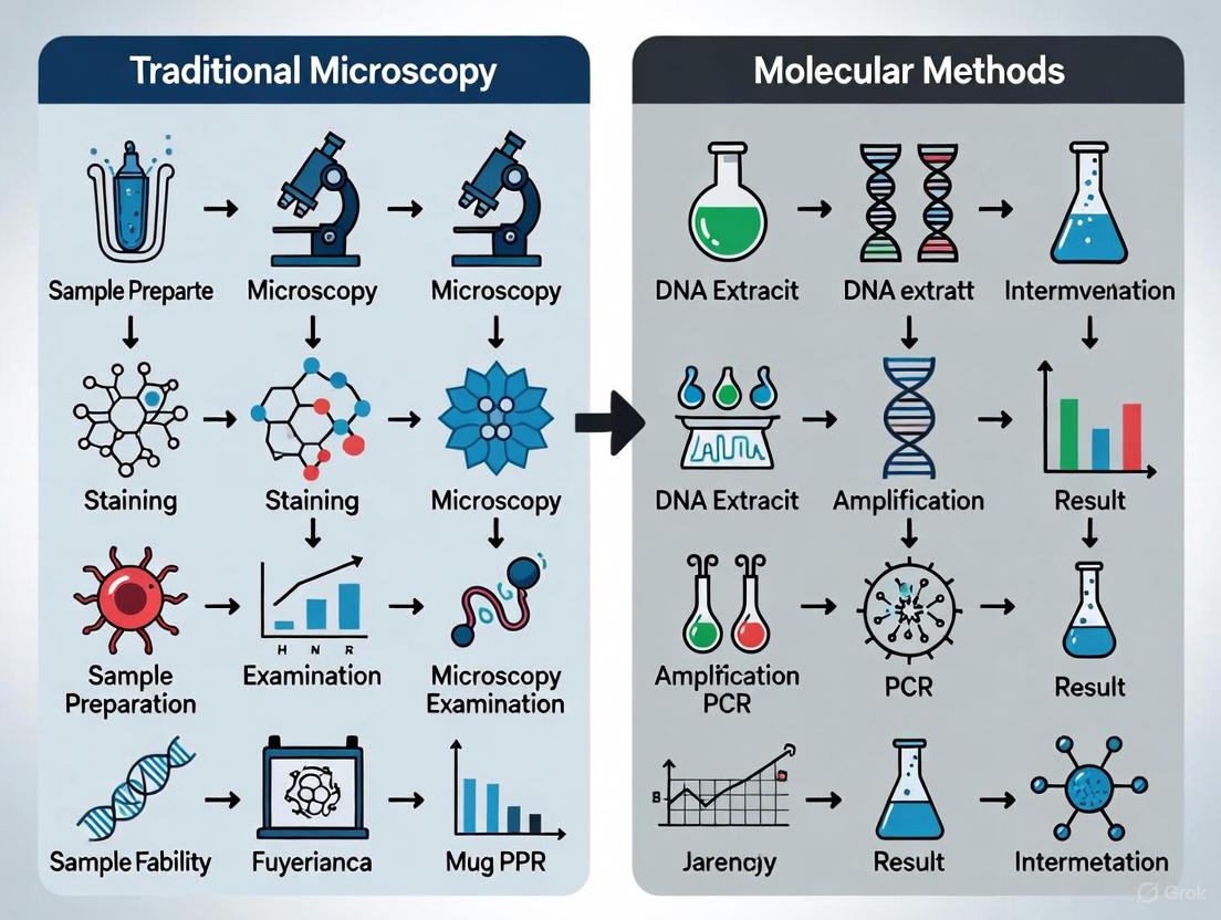
The transition from traditional microscopy to molecular diagnosis involves a standardized sequence of steps, from sample collection to final result interpretation. The following workflow delineates this core process.
Comparative Analysis of PCR Technologies
Table 1: Performance Comparison of Diagnostic Methods for Parasite Detection
| Method | Sensitivity | Specificity | Key Advantages | Primary Limitations |
|---|---|---|---|---|
| Microscopy | 22.4-73.8% [8] [31] | 94.3-100% [8] [31] | Low cost, species identification, assesses parasite stage [30] | Low sensitivity in low-burden infections, operator-dependent [8] |
| Conventional PCR | Higher than microscopy [8] | Higher than microscopy [8] | Detects DNA, more sensitive than microscopy [8] [29] | Qualitative only, post-amplification processing required [29] |
| qPCR | 74.6-97.6% [30] [32] | 95.2-100% [30] [32] | Quantitative, rapid, high throughput [30] [33] | Requires specialized equipment, cost [30] |
| Multiplex PCR | 33.3-100% (target-dependent) [34] | 98.3-100% (target-dependent) [34] | Simultaneous multi-pathogen detection [35] [34] | Optimization complexity, potential signal interference [35] |
| Digital PCR (ddPCR) | Superior for low parasitemia [36] [37] | High [36] [37] | Absolute quantification, high precision, resistant to inhibitors [36] | High cost, specialized equipment, lower throughput [36] |
Quantitative PCR (qPCR) in Parasitology
Principles and Applications
Quantitative PCR (qPCR), also known as real-time PCR, represents a significant advancement over conventional PCR by enabling simultaneous amplification and quantification of target DNA. This technology employs fluorescent reporters to monitor amplification in real-time, with the cycle threshold (Ct) value inversely correlating with the initial target concentration [29]. In malaria research, qPCR has demonstrated superior sensitivity (94.0-97.6%) compared to microscopy (74.6%) [30] [32], making it invaluable for detecting low-density infections often missed by conventional methods [30].
The quantitative capability of qPCR is particularly crucial for drug development, as it allows researchers to monitor parasite clearance kinetics and assess treatment efficacy [33]. Furthermore, qPCR can differentiate between Plasmodium species with high accuracy, identifying mixed infections that frequently evade correct diagnosis by microscopy or RDTs [32].
Experimental Protocol: qPCR for Malaria Parasite Detection
Protocol Title: Quantitative PCR for Detection and Quantification of Plasmodium falciparum in Whole Blood [33]
Sample Preparation:
- Collect venous blood in EDTA tubes.
- Extract DNA using automated systems (e.g., QIAsymphony) or manual kits.
- Use blood volumes of 200-500 μL for optimal sensitivity and precision [33].
- Elute DNA in 50-100 μL of elution buffer.
Primer and Probe Design:
- Target multi-copy genes such as 18S ribosomal RNA (rRNA) for enhanced sensitivity [33].
- Example P. falciparum 18S rRNA primers and probe:
- Forward: 5'-GTAATTGGAATGATAGGAATTTACAAGGT-3'
- Reverse: 5'-TCAACTACGAACGTTTTAACTGCAAC-3'
- Probe: 5'-FAM-AACAATTGGAGGGCAAG-NFQ-MGB-3' [33]
qPCR Reaction Setup:
- Prepare 50 μL reactions containing:
- 1× universal PCR Master Mix
- 10 pmol/μL of each primer
- 10 μmol of NFQ-MGB probe
- 10 μL of template DNA
- Run in triplicate for experimental samples, standards, and controls.
Thermal Cycling Conditions:
- Initial denaturation: 95°C for 10 minutes
- 45 cycles of:
- Denaturation: 95°C for 15 seconds
- Annealing/Extension: 60°C for 1 minute
Quantification:
- Generate standard curve using serial dilutions of cultured parasites with known concentrations.
- Calculate parasite density based on Ct values and standard curve.
- Report results as parasites/μL of blood [33].
Multiplex PCR for Parallel Pathogen Detection
Principles and Applications
Multiplex PCR enables the simultaneous amplification of multiple target sequences in a single reaction by incorporating different primer sets [29]. This approach is particularly valuable in parasitology for identifying co-infections with multiple parasite species or distinguishing between morphologically similar organisms [34]. Commercial multiplex panels, such as the Seegene Allplex GI-Parasite Assay, can detect up to six protozoal pathogens (Blastocystis hominis, Cryptosporidium spp., Cyclospora cayetanensis, Dientamoeba fragilis, Entamoeba histolytica, and Giardia lamblia) from a single stool specimen [34].
The diagnostic efficiency of multiplex PCR was demonstrated in a study on enteric protozoa, which showed sensitivity and specificity of 100% for Cryptosporidium and Cyclospora cayetanensis, and 100% sensitivity with 98.9% specificity for Giardia lamblia [34]. Additionally, multiplex PCR significantly reduces laboratory turnaround time compared to traditional methods that require multiple staining procedures and microscopic examinations [34].
Experimental Protocol: Automated High-Throughput Multiplex PCR for Enteric Protozoa
Protocol Title: Automated Multiplex Real-Time PCR for Detection of Enteric Protozoa in Stool Specimens [34]
Sample Preparation:
- Collect fresh, unpreserved stool samples.
- Inoculate one swab of stool into FecalSwab tubes containing 2 mL of Cary-Blair media.
- Vortex for 10 seconds to homogenize.
Automated DNA Extraction:
- Use Hamilton STARlet automated liquid handling platform.
- Employ STARMag 96 × 4 Universal Cartridge kit for DNA extraction.
- Process 50 μL of stool suspension, eluting in 100 μL of DNA elution buffer.
Multiplex PCR Setup:
- Prepare 25 μL reactions containing:
- 5 μL of 5X GI-P MOM (MuDT Oligo Mix) primer
- 10 μL RNase-free water
- 5 μL EM2 (DNA polymerase, Uracil-DNA glycosylase, buffer with dNTPs)
- 5 μL of extracted sample nucleic acid
- Utilize four fluorophores (FAM, HEX, Cal Red 610, Quasar 670) for target discrimination.
Real-Time PCR Amplification:
- Perform on Bio-Rad CFX96 real-time PCR detection system.
- Cycling conditions:
- Denaturation: 95°C for 10 seconds
- 45 cycles of: 95°C for 10 seconds, 60°C for 1 minute, 72°C for 30 seconds
- Interpret positive results at cycle threshold (Ct) value of ≤43.
Analysis:
- Analyze amplification curves for each target channel.
- Determine species identification based on fluorescence signature.
Digital PCR for Absolute Quantification
Principles and Applications
Digital PCR (dPCR), including droplet digital PCR (ddPCR), represents the most recent evolution in PCR technology. This method partitions a sample into thousands of nanoliter-sized reactions, with each partition undergoing PCR amplification independently [29]. After amplification, the system counts the positive and negative reactions to provide absolute quantification of the target DNA without requiring a standard curve [36].
In parasitology, ddPCR has demonstrated exceptional performance in detecting asymptomatic malaria infections with low parasite densities that evade microscopy and even qPCR detection [36] [37]. One study comparing ddPCR with microscopy found that ddPCR detected a significantly higher proportion of asymptomatic Plasmodium infections (7.41% vs. 2.59% by microscopy) in samples from Thai workers returning from Sudan [37]. The technology's ability to provide absolute quantification of parasite load using multicopy genomic targets (such as 18S rRNA) makes it particularly valuable for monitoring treatment response in clinical trials and understanding transmission dynamics in elimination settings [36].
Experimental Protocol: Droplet Digital PCR for Asymptomatic Malaria Detection
Protocol Title: Droplet Digital PCR for Detection of Asymptomatic Plasmodium Infections [36] [37]
Sample Collection and DNA Extraction:
- Collect dried blood spots (DBS) or venous blood specimens.
- Extract DNA using commercial kits suitable for blood samples.
- For DBS, punch 3-6 mm discs and incubate in lysis buffer.
ddPCR Reaction Preparation:
- Prepare 20-22 μL reactions containing:
- 1× ddPCR Supermix for Probes
- 900 nM of each primer
- 250 nM of probe
- 5-10 μL of template DNA
- Design assays to target:
- Genus-level Plasmodium 18S rRNA gene
- Species-specific markers for P. falciparum, P. vivax, P. malariae, P. ovale, and P. knowlesi
Droplet Generation:
- Load reaction mixture into DG8 Cartridges with appropriate oil.
- Generate droplets using Automated Droplet Generator.
- Transfer emulsified samples to 96-well PCR plates.
PCR Amplification:
- Seal plates and perform amplification in a thermal cycler.
- Standard cycling conditions:
- Enzyme activation: 95°C for 10 minutes
- 40 cycles of: 94°C for 30 seconds, 55-60°C for 60 seconds
- Enzyme deactivation: 98°C for 10 minutes
Droplet Reading and Analysis:
- Read plates in Droplet Reader.
- Analyze using companion software to determine:
- Concentration (copies/μL)
- Poisson confidence intervals
- Fraction of positive droplets
Quantification:
- Calculate absolute parasite density based on copies/μL and sample input volume.
- Report results as copies/μL of blood with 95% confidence intervals.
The Scientist's Toolkit: Essential Research Reagents
Table 2: Key Research Reagent Solutions for Molecular Parasitology
| Reagent/Equipment | Function | Example Applications |
|---|---|---|
| DNA Extraction Kits (e.g., QIAsymphony, STARMag) | Isolation of high-quality parasite DNA from complex samples | Extraction from whole blood, stool specimens [33] [34] |
| 18S rRNA Primers/Probes | Amplification of multi-copy target for enhanced sensitivity | Detection of Plasmodium species, soil-transmitted helminths [8] [33] |
| TaqMan Probes (MGB-NFQ) | Sequence-specific fluorescence detection in qPCR | Species-specific identification of parasites in multiplex assays [33] [34] |
| ddPCR Supermix | Reaction mixture optimized for droplet digital PCR | Absolute quantification of low-density malaria infections [36] [37] |
| Automated Liquid Handlers (e.g., Hamilton STARlet) | High-throughput, reproducible sample processing | Automated DNA extraction and PCR setup for large-scale studies [34] |
| Multiplex PCR Master Mix | Simultaneous detection of multiple targets | Identification of enteric protozoa co-infections [34] |
The evolution of PCR technologies—from conventional to qPCR, multiplex, and digital PCR—has fundamentally transformed parasite identification in research and drug development. These molecular methods have consistently demonstrated superior sensitivity compared to traditional microscopy, particularly in detecting low-density infections critical for disease surveillance and elimination efforts [8] [30] [36]. While each platform offers distinct advantages, the collective advancement provides researchers with an unprecedented toolkit for precise parasite detection, species differentiation, and quantification.
The choice of molecular platform depends on specific research objectives, infrastructure, and budgetary considerations. qPCR remains the workhorse for high-throughput screening, while multiplex PCR excels in comprehensive pathogen detection, and digital PCR provides ultimate sensitivity for absolute quantification [29]. As these technologies continue to evolve and become more accessible, they will undoubtedly play an increasingly central role in parasitology research, clinical diagnostics, and the global effort to control and eliminate parasitic diseases.
For decades, the diagnosis of parasitic infections has relied predominantly on traditional methods such as light microscopy, serological testing, and histopathology [19]. While these techniques have been foundational to parasitology, they present significant limitations including time-consuming processes, subjective interpretation requiring highly trained experts, and notably poor sensitivity for detecting low-level infections [19] [9]. The emergence of molecular methods, particularly Next-Generation Sequencing (NGS), has revolutionized parasitic disease diagnostics by enabling unprecedented resolution in detecting and characterizing parasites [38] [19]. This whitepaper explores how NGS technologies provide comprehensive genomic analysis and sophisticated strain tracking capabilities, representing a fundamental shift from phenotypic to genotypic identification methods in parasitology research and drug development.
Limitations of Conventional Diagnostic Methods
Traditional microscopy remains widely used but suffers from substantial diagnostic limitations. Microscopy has an estimated detection limit of 50-500 parasites/μL of blood, causing it to miss low-density infections that can maintain transmission and complicate control efforts [9]. Similarly, microscopy cannot differentiate between morphologically identical species, such as pathogenic Entamoeba histolytica from non-pathogenic E. dispar and E. moshkovskii, potentially leading to misdiagnosis and unnecessary treatment [39].
Rapid Diagnostic Tests (RDTs), while improving access to testing, also demonstrate sensitivity limitations. A 2023 study comparing microscopy, RDT, and quantitative PCR for malaria diagnosis revealed that both microscopy and RDT missed over 40% of infections detected by the more sensitive molecular method [9]. Table 1 quantitatively compares the performance characteristics of conventional versus molecular diagnostic methods.
Table 1: Performance Comparison of Parasite Diagnostic Methods
| Method | Sensitivity | Specificity | Time to Result | Key Limitations |
|---|---|---|---|---|
| Light Microscopy | 39.3% (vs. qPCR) [9] | 98.3% (vs. qPCR) [9] | 30-60 minutes | Limited sensitivity, requires expertise, cannot distinguish some species |
| Rapid Diagnostic Tests (RDTs) | 55.7% (vs. qPCR) [9] | 98.2% (vs. qPCR) [9] | 15-20 minutes | Limited sensitivity at low parasite density, HRP-2 gene deletions |
| Conventional PCR | Moderate | High | 4-6 hours | Requires specialized equipment, moderate throughput |
| NGS-Based Methods | Very High | Very High | ~30 min computation [40] | Higher cost, bioinformatics complexity |
Serological methods like ELISA, IHA, and IIF have improved detection capabilities but still face challenges. These include the inability to distinguish between past and current infections in endemic areas, and for some tests, the requirement for fresh or unpreserved fecal samples [39]. The cumulative limitations of these conventional approaches have accelerated the adoption of nucleic acid-based detection methods, with NGS emerging as the most comprehensive solution.
Next-Generation Sequencing Technologies: Core Principles and Platforms
Next-Generation Sequencing represents a paradigm shift in genomic analysis, enabling the parallel sequencing of millions to billions of DNA fragments in a single run [38]. This high-throughput capability provides researchers with unprecedented insights into genome structure, genetic variations, gene expression profiles, and epigenetic modifications [38]. Several NGS platforms have been developed, each with distinct technological approaches and applications relevant to parasitology research.
NGS Platform Technologies
The Illumina platform utilizes a sequencing-by-synthesis approach with reversible dye terminators, making it the current workhorse for large-scale genomic studies due to its high accuracy and throughput [38]. This technology is particularly valuable for population genomics studies of parasites and host-pathogen interactions.
Pacific Biosciences (PacBio) Single-Molecule Real-Time (SMRT) technology and Oxford Nanopore Technologies (ONT) represent third-generation sequencing platforms that generate long reads [38]. PacBio SMRT sequencing employs zero-mode waveguides (ZMWs) to monitor nucleotide incorporation in real-time, producing average read lengths of 10,000-25,000 base pairs [38]. Oxford Nanopore technology detects nucleotide sequences by measuring changes in electrical current as DNA molecules pass through protein nanopores, with average read lengths of 10,000-30,000 base pairs [38]. These long-read technologies are particularly advantageous for resolving complex genomic regions, structural variations, and for assembling complete parasite genomes without gaps.
Comprehensive Variant Detection with Advanced Bioinformatics
The DRAGEN (Dynamic Read Analysis for Genomics) platform exemplifies how integrated bioinformatics solutions leverage NGS data for comprehensive variant detection [40]. This framework uses pangenome references and specialized algorithms to simultaneously identify single-nucleotide variations (SNVs), insertions/deletions (indels), structural variations (SVs), copy number variations (CNVs), and short tandem repeats (STRs) [40]. This comprehensive approach is particularly valuable for detecting drug resistance markers and virulence factors in parasitic genomes.
Table 2: NGS Platform Comparison for Parasitology Research
| Platform | Technology | Read Length | Key Applications in Parasitology | Limitations |
|---|---|---|---|---|
| Illumina | Sequencing-by-synthesis | 36-300 bp | Population genomics, transcriptomics, variant detection | Short reads limit assembly of repetitive regions |
| PacBio SMRT | Single-molecule real-time | 10,000-25,000 bp (average) | Complete genome assembly, structural variation discovery | Higher cost, requires more DNA input |
| Oxford Nanopore | Nanopore sensing | 10,000-30,000 bp (average) | Rapid field sequencing, metagenomic identification | Higher error rate (~5-15%) [38] |
| Ion Torrent | Semiconductor sequencing | 200-400 bp | Targeted sequencing, rapid turnaround | Homopolymer sequencing errors |
Diagram 1: NGS Parasite Detection Workflow. The process from sample collection to pathogen identification and strain characterization.
Strain-Level Analysis with NGS: Techniques and Applications
Strain Tracking in Complex Communities
Strain-level analysis represents one of the most significant advantages of NGS over conventional methods. The Strain Genome Explorer (StrainGE) toolkit enables researchers to track and characterize low-abundance strains in complex microbial communities, a common scenario in parasitic infections [41]. StrainGE employs two key components: the Strain Genome Search Tool (StrainGST) for identifying reference genomes similar to strains in a sample, and Strain Genome Recovery (StrainGR) for identifying single nucleotide variants (SNVs) and large deletions relative to reference genomes [41].
This toolkit is particularly valuable for detecting minor variant populations that may indicate emerging drug resistance or mixed infections. Remarkably, StrainGE can identify strains at coverages as low as 0.1x and detect variants from coverages as low as 0.5x, enabling characterization of low-abundance parasites that would be undetectable by conventional means [41]. The ability to resolve strain mixtures is crucial for understanding transmission dynamics, tissue tropism, and differential drug susceptibility in parasitic diseases.
Taxonomic Identification of Parasite Genomes
For clinical and public health applications, the Parasite Genome Identification Platform (PGIP) provides a user-friendly web server specifically designed for taxonomic identification of parasite genomes from metagenomic NGS data [42]. PGIP integrates a curated database of 280 high-quality parasite genomes that have been rigorously filtered and deduplicated to ensure accurate species-level resolution [42].
The platform automates the analytical workflow through host DNA depletion, quality control, and dual-approach parasite identification using both read mapping and assembly-based methods [42]. This comprehensive approach is particularly valuable for diagnosing rare parasites and opportunistic infections in immunocompromised patients, where traditional methods often fail due to low parasite loads or atypical presentations.
Diagram 2: Strain Tracking Methodology. Computational workflow for disentangling mixed parasite strains from metagenomic samples.
Research Reagent Solutions for NGS-Based Parasitology
Table 3: Essential Research Reagents and Platforms for NGS Parasitology Studies
| Reagent/Platform | Function | Application in Parasite Research |
|---|---|---|
| DRAGEN Bio-IT Platform [40] | Comprehensive variant calling | Simultaneous detection of SNVs, indels, SVs, CNVs, and STRs in parasite genomes |
| StrainGE Toolkit [41] | Strain-level characterization | Tracking low-abundance strains in mixed parasite infections |
| PGIP Web Server [42] | Taxonomic identification | Rapid species-level identification of parasites from mNGS data |
| CareStart Malaria RDT [9] | Rapid antigen detection | Comparative traditional method for validation studies |
| Trimmomatic [42] | Sequence quality control | Adapter trimming and quality filtering in NGS workflows |
| Kraken2 [42] | Taxonomic classification | k-mer-based alignment for parasite identification |
| MEGAHIT [42] | Metagenomic assembly | De novo assembly of parasite genomes from complex samples |
Experimental Protocols for NGS-Based Parasite Detection
Metagenomic NGS Protocol for Parasite Detection
The following protocol outlines the standard workflow for detecting and characterizing parasites from clinical samples using metagenomic NGS:
Sample Collection and Nucleic Acid Extraction: Collect appropriate clinical samples (stool, blood, tissue biopsies) in sterile containers. Extract total nucleic acids using standardized kits, incorporating mechanical lysis for parasites with robust cysts or egg shells. For blood samples, prioritize extraction methods that efficiently recover parasite DNA while reducing host background [42].
Library Preparation: Fragment extracted DNA to appropriate size distributions (typically 200-500bp). Repair fragment ends and ligate with platform-specific adapters. For low-biomass samples, incorporate whole-genome amplification steps while recognizing potential amplification biases [42].
Sequencing: Dilute libraries to appropriate concentrations and load onto NGS platforms. For Illumina systems, sequence with paired-end reads (2×150bp or 2×250bp) to improve mapping accuracy and assembly. Adjust sequencing depth based on application: 5-10 million reads per sample for detection, 20-30 million reads for strain characterization, and higher coverage for de novo assembly projects [42].
Bioinformatic Analysis:
- Quality Control: Process raw FASTQ files with Trimmomatic to remove adapters and low-quality reads [42].
- Host Depletion: Align reads to host reference genome (e.g., GRCh38 for human samples) using Bowtie2 and retain unmapped reads for parasite analysis [42].
- Parasite Identification: Use Kraken2 with a curated parasite database for taxonomic classification [42].
- Strain-Level Analysis: Apply StrainGE toolkit for characterizing strain mixtures and identifying variants [41].
- Variant Calling: Use DRAGEN for comprehensive variant detection across all variant classes [40].
Validation and Quality Control
Establish rigorous quality control measures including:
- Extraction negative controls to monitor contamination
- Positive control samples with known parasite DNA
- Spike-in synthetic controls for quantifying detection limits
- Replicate sequencing to assess technical variability
- Comparison with conventional methods (microscopy, PCR) where available [9]
Next-Generation Sequencing has fundamentally transformed the landscape of parasitic disease research and diagnosis by providing comprehensive genomic analysis and sophisticated strain tracking capabilities. The transition from traditional microscopy to molecular methods addresses critical limitations in sensitivity, specificity, and discriminatory power, enabling researchers to detect low-level infections, identify mixed strains, and uncover genetic determinants of virulence and drug resistance. As NGS technologies continue to evolve with improvements in cost, scalability, and analytical frameworks, their integration into routine parasitology practice will accelerate drug discovery efforts and enhance our understanding of parasite biology and host-parasite interactions. For researchers and drug development professionals, adopting these advanced genomic approaches is no longer optional but essential for advancing the field of parasitology into the precision medicine era.
The diagnosis of parasitic diseases has long relied on traditional microscopy, a method fraught with limitations in sensitivity, specificity, and scalability. This whitepaper examines the paradigm shift driven by CRISPR-based molecular diagnostics, which leverage programmable nucleases for the precise detection of pathogen nucleic acids. We explore the core mechanisms of CRISPR-Cas systems, detail experimental protocols for field-deployable assays, and present a comparative analysis of their performance against conventional methods. Framed within a broader thesis on the transition from traditional microscopy to molecular techniques, this technical guide provides researchers and drug development professionals with the foundational knowledge and practical methodologies underpinning this transformative technology.
For decades, the diagnosis of parasitic infections has depended heavily on traditional microscopy. While considered a gold standard for its ability to visually confirm parasites and differentiate species, microscopy is labor-intensive, requires a high level of technical expertise, and suffers from poor sensitivity, particularly in cases of low-parasite-density infections or mixed infections [43] [19]. These limitations are especially pronounced in resource-limited endemic regions, where the burden of parasitic diseases is highest. The emergence and spread of Plasmodium falciparum with deletions in the pfhrp2 and pfhrp3 genes further undermine the reliability of widely used rapid diagnostic tests (RDTs), which are often based on the detection of the HRP2 protein [44].
Molecular methods, beginning with polymerase chain reaction (PCR), offered a significant leap in sensitivity and specificity. However, PCR-based techniques require sophisticated thermocycling equipment, stable electrical power, and trained personnel, making them impractical for point-of-care (POC) testing and field deployment [43] [45]. The critical need for diagnostic tools that are both highly accurate and field-adaptable has catalyzed the development of innovative solutions, chief among them being diagnostics based on the Clustered Regularly Interspaced Short Palindromic Repeats (CRISPR) system.
Originally characterized as an adaptive immune system in bacteria, the CRISPR-Cas system has been repurposed as a powerful tool for genome editing and, more recently, for molecular diagnostics [46] [47]. CRISPR-based diagnostics combine the programmability of CRISPR RNA (crRNA) with the enzymatic activities of Cas proteins to achieve unparalleled specificity and sensitivity. This technology can be coupled with isothermal amplification methods, enabling rapid detection of nucleic acids at a constant temperature without the need for complex instrumentation [48]. This positions CRISPR diagnostics as a disruptive technology capable of bridging the gap between the sensitivity of molecular methods and the practicality required for field-deployable testing in parasitic disease surveillance and control.
Core Mechanisms of CRISPR-Cas Systems in Diagnostics
The diagnostic application of CRISPR technology primarily harnesses the properties of Class 2 CRISPR-Cas systems, which utilize single effector proteins such as Cas9, Cas12, Cas13, and Cas14 [46] [45]. These systems function through a two-step mechanism: target recognition and enzymatic activity trigger.
Target Recognition and Enzymatic Activation
The process is initiated by the design of a specific CRISPR RNA (crRNA) or guide RNA, which is programmed to be complementary to a target nucleic acid sequence from a pathogen. Upon hybridization of the crRNA to its target, the associated Cas protein undergoes a conformational change that activates its nuclease activity [46]. This activation is a pivotal step that differentiates various Cas proteins used in diagnostics.
- Cas9 was the first Cas protein harnessed for gene editing. It functions as a duplexed nuclease, creating double-strand breaks in target DNA. However, its lack of trans-cleavage activity limits its utility in diagnostic applications compared to more recent discoveries [45].
- Cas12 (including Cas12a) targets DNA sequences. Once activated by binding to its target DNA, it exhibits nonspecific trans-cleavage activity, indiscriminately degrading nearby single-stranded DNA (ssDNA) molecules [46] [48]. This collateral cleavage is highly effective for signal amplification.
- Cas13 targets RNA sequences. Similar to Cas12, upon target recognition, it activates and cleaves surrounding non-target single-stranded RNA (ssRNA), providing a powerful mechanism for RNA virus detection [48] [47].
The following diagram illustrates the core mechanism of target-specific cleavage and subsequent trans-cleavage activity for Cas12 and Cas13, which is the cornerstone of CRISPR-based diagnostics:
Signal Generation and Readout
The trans-cleavage activity is harnessed for detection by introducing reporter molecules into the reaction. These are short nucleic acid strands (ssDNA for Cas12, ssRNA for Cas13) labeled with a fluorophore and a quencher. When intact, the quencher suppresses the fluorophore's signal. Upon collateral cleavage by the activated Cas protein, the fluorophore is separated from the quencher, generating a fluorescent signal [46] [47]. This signal can be measured with portable fluorometers or visualized using lateral flow assays, where cleavage of a labeled reporter produces a visible test line, making the results easy to interpret in field settings [48] [45].
Comparative Analysis: Traditional Methods vs. Molecular Diagnostics
The evolution from traditional microscopy to advanced molecular methods represents a significant advancement in diagnostic capabilities. The following table summarizes the key performance characteristics of these diagnostic approaches.
Table 1: Performance Comparison of Diagnostic Methods for Parasitic Diseases
| Diagnostic Method | Sensitivity | Specificity | Turnaround Time | Equipment Needs | Expertise Required | POC Suitability |
|---|---|---|---|---|---|---|
| Microscopy | Low (requires high parasite density) | Moderate (species differentiation possible) | 30-60 minutes | Microscope | High | Limited |
| Immunological Assays (RDTs) | Moderate | Variable (cross-reactivity issues) | 15-20 minutes | None | Low | Good |
| PCR/qPCR | Very High (aM-zM level) | Very High | 1.5 - 4 hours | Thermocycler, lab equipment | High | Poor |
| Isothermal Amplification (LAMP/RPA) | High | Moderate to High | 15-60 minutes | Water bath/heat block | Moderate | Moderate |
| CRISPR-Based Assays | Very High (aM level) | Very High (crRNA-guided) | 30 - 90 minutes | Minimal (constant temperature) | Moderate | Excellent |
The market growth for CRISPR-based diagnostics underscores its impact. The global market, valued at USD 3.25 billion in 2024, is projected to expand at a compound annual growth rate (CAGR) of 16.63% from 2025 to 2034, reaching approximately USD 15.14 billion [49]. This growth is driven by the increasing burden of infectious diseases, demand for point-of-care testing, and continuous innovation in CRISPR platforms.
Experimental Protocols for CRISPR-Based Pathogen Detection
Translating the molecular mechanism into a reliable diagnostic assay requires robust experimental protocols. The two primary approaches are two-step and one-pot assays, with the latter being particularly suited for field deployment.
Two-Step Assay Protocol (e.g., DETECTR for DNA Targets)
This protocol involves a separate amplification step followed by CRISPR detection.
I. Materials and Reagents
- Sample: Patient blood, stool, or urine.
- Nucleic Acid Extraction Kit: For DNA/RNA purification.
- Isothermal Amplification Master Mix: Contains recombinase, primers, polymerase, and nucleotides for Recombinase Polymerase Amplification (RPA).
- CRISPR Reaction Master Mix:
- Cas12a (or Cas13) enzyme
- Designed crRNA targeting the pathogen of interest
- Fluorescent or lateral flow reporter probe (e.g., ssDNA-FQ reporter for Cas12a)
- Suitable reaction buffer
II. Step-by-Step Procedure
- Nucleic Acid Extraction: Extract DNA from the patient sample using a commercial kit or simplified lysis protocol. For RNA targets, include a reverse transcription step.
- Target Amplification:
- Prepare the RPA reaction mix on ice, containing the extracted DNA template and pathogen-specific primers.
- Incubate the RPA tube at a constant temperature (37-42°C) for 15-20 minutes to amplify the target sequence.
- CRISPR Detection:
- In a separate tube, prepare the CRISPR master mix containing Cas12a, crRNA, and the reporter probe.
- Add a portion of the amplified RPA product to the CRISPR reaction tube.
- Incubate at 37°C for 10-30 minutes.
- Result Visualization:
- Fluorescent Readout: Measure fluorescence intensity with a portable fluorometer. A significant increase over the negative control indicates a positive result.
- Lateral Flow Readout: Apply the reaction mixture to a lateral flow strip. The appearance of both control and test lines indicates a positive result.
One-Pot Assay Protocol
The one-pot strategy integrates amplification and detection in a single, sealed tube, minimizing contamination risk and simplifying operation [48].
I. Materials and Reagents
- All reagents from the two-step protocol.
- Additives to create a transient physical or chemical barrier (e.g., wax capsules, phase-separation systems) to separate amplification and CRISPR reagents until a specific trigger (e.g., heat, mechanical pressure) is applied.
II. Step-by-Step Procedure
- Reagent Assembly: In a single tube, sequentially layer or compartmentalize the RPA reagents and the CRISPR detection reagents, using a barrier to prevent premature mixing.
- Unified Incubation: Place the tube in a heat block or water bath at 37-42°C. The barrier dissolves or is breached after the amplification reaction has progressed, allowing the amplicons to activate the Cas protein.
- Result Visualization: After a total incubation of 30-60 minutes, visually inspect the tube for fluorescence under a blue-light transilluminator or dip a lateral flow strip directly into the reaction mixture.
The workflow for this integrated, one-pot system is outlined below:
The Scientist's Toolkit: Essential Research Reagent Solutions
Implementing CRISPR-based diagnostics requires a specific set of reagents and tools. The following table details the key components and their functions in a typical assay.
Table 2: Essential Reagents and Materials for CRISPR-Dx Research
| Item Category | Specific Examples | Function in the Assay |
|---|---|---|
| Cas Enzymes | Purified Cas12a (Cpf1), Cas13a | The core effector protein that provides target-specific binding and trans-cleavage activity upon activation. |
| Guide RNA | Synthetic crRNA | Programmable RNA molecule that confers specificity by guiding the Cas complex to the target pathogen nucleic acid sequence. |
| Amplification Enzymes/Mixes | RPA pellets, LAMP master mix | Provides the enzymes and reagents for isothermal amplification of the target to detectable levels. |
| Reporter Probes | ssDNA-FQ reporters, FAM/Biotin-labeled ssDNA for LFA | The substrate for trans-cleavage; its cleavage generates a fluorescent or colorimetric signal for detection. |
| Lateral Flow Strips | Commercial LFA strips (e.g., Milenia HybriDetect) | For visual, instrument-free readout of the assay results. |
| Portable Readers | Small fluorometers, blue-light transilluminators | For quantitative or highly sensitive qualitative fluorescence readout in field settings. |
Challenges and Future Outlook
Despite its transformative potential, the widespread deployment of CRISPR diagnostics faces several hurdles. Technical challenges include the need for refined reaction kinetics in one-pot systems to prevent premature activation of the Cas enzyme and suboptimal sensitivity in amplification-free formats [48] [47]. Economic and logistical barriers, such as the relatively high cost of Cas enzymes and the need for a cold chain for reagent storage, can limit adoption in remote areas [49] [43]. Furthermore, the development of robust regulatory frameworks is essential to ensure quality control and facilitate the approval and commercialization of these tests [46] [44].
Future directions are focused on overcoming these challenges. The discovery of novel, more efficient Cas effectors and the integration of artificial intelligence for guide RNA design and signal interpretation promise to enhance accuracy and ease of use [49]. The development of lyophilized, temperature-stable reagents will be crucial for true point-of-care applicability in tropical field settings [43] [48]. Finally, the creation of highly multiplexed platforms capable of simultaneously detecting dozens of pathogens from a single sample will revolutionize epidemiological surveillance and comprehensive patient diagnosis [46] [47].
CRISPR-based diagnostics represent a monumental leap forward in the detection of parasitic and other infectious diseases. By offering a combination of high sensitivity, exceptional specificity, rapid turnaround, and field-deployable simplicity, they effectively address the critical limitations of both traditional microscopy and earlier molecular techniques like PCR. As research tackles existing challenges related to cost, regulation, and workflow optimization, CRISPR diagnostics are poised to become an indispensable tool. They will not only empower frontline healthcare workers with precise diagnostic capabilities but also provide researchers and public health officials with powerful data to track outbreaks, monitor drug resistance, and ultimately accelerate progress toward the elimination of parasitic diseases globally.
The diagnosis of parasitic infections has long been dominated by traditional microscopy techniques, which remain the cornerstone of laboratory identification for many gastrointestinal parasites globally [50]. These conventional methods, while foundational, present significant limitations including time consumption, expertise requirements, and impracticality in resource-limited endemic regions where parasitic disease burdens are often highest [19]. The subjective nature of microscopic analysis introduces variability, while the low throughput impedes large-scale surveillance efforts and rapid response during outbreaks.
The integration of automation and artificial intelligence represents a paradigm shift, bridging the gap between traditional microscopy and modern molecular methods while enhancing the capabilities of both approaches. This transformation is particularly critical in parasitology, where accurate, rapid diagnosis directly impacts treatment outcomes and public health interventions. Automated molecular diagnostics and AI-enhanced imaging frameworks are creating unprecedented opportunities for high-throughput workflows that maintain diagnostic accuracy while significantly expanding testing capacity [51] [52]. This technical guide examines the core technologies driving this integration and their practical implementation within parasite diagnosis research and drug development.
Automation Technologies in Diagnostic Workflows
Automated Molecular Diagnostics Platforms
Automated molecular diagnostic systems have revolutionized parasite detection by integrating multiple laboratory processes onto unified platforms. These systems typically combine sample extraction, PCR preparation, and next-generation sequencing (NGS) library preparation into streamlined workflows that minimize manual intervention and reduce error-prone processes [51] [53].
The implementation of automated liquid handling platforms demonstrates measurable benefits in parasitology. For example, the transition to automated workflows for HIV-1 genotyping assays reduced hands-on time from more than 17 hours to under 2 hours while maintaining analytical precision [51]. Similar efficiency gains are achievable in parasitic disease diagnosis, particularly for high-volume testing of common pathogens like Giardia lamblia, Cryptosporidium species, and Entamoeba histolytica [50].
Table 1: Comparative Workflow Analysis of Automated Molecular Diagnostic Systems
| System Component | Manual Workflow | Semi-Automated Workflow | Fully Automated Workflow |
|---|---|---|---|
| Sample Preparation | 2-3 hours (96 samples) | 1-2 hours (96 samples) | <30 minutes (96 samples) |
| Hands-on Time (PCR setup) | 60-90 minutes | 30-45 minutes | <10 minutes |
| Result Interpretation | 30-60 minutes (subjective) | 15-30 minutes | <5 minutes (automated analysis) |
| Total Turnaround Time | 4-6 hours | 3-4 hours | 2-3 hours |
| Error Rate | 5-10% (operator-dependent) | 2-5% | <1% (with barcode verification) |
High-Throughput Screening (HTS) Systems
High-throughput screening technologies enable the rapid conduct of millions of chemical, genetic, or pharmacological tests, making them invaluable for antiparasitic drug discovery [54]. HTS leverages robotics, data processing software, liquid handling devices, and sensitive detectors to accelerate the identification of active compounds, antibodies, or genes that modulate specific biomolecular pathways in parasites [54].
The core of HTS technology is the microtiter plate, with modern systems utilizing 384, 1536, or even 3456 wells to maximize throughput while minimizing reagent volumes [54]. For parasitology research, this enables massive parallel testing of compound libraries against parasitic targets, such as screening for inhibitors of Plasmodium falciparum growth or compounds that disrupt parasite-specific metabolic pathways. Integrated robot systems transport assay microplates between stations for sample addition, mixing, incubation, and detection with minimal operator intervention [54].
Table 2: Technical Specifications for High-Throughput Screening in Parasite Research
| Parameter | Conventional Screening | High-Throughput Screening | Ultra-High-Throughput Screening |
|---|---|---|---|
| Daily Throughput | 1,000-10,000 compounds | 10,000-100,000 compounds | >100,000 compounds |
| Reagent Volume | Microliters (μL) | Nanoliters (nL) | Picoliters (pL) |
| Assay Platforms | 96-well plates | 384/1536-well plates | 3456-well plates / microfluidics |
| Data Points Generated | Hundreds per day | Thousands per hour | Millions per day |
| Automation Level | Manual/Semi-automated | Fully automated with robotics | Integrated systems with microfluidics |
Artificial Intelligence and Advanced Analytics
AI-Assisted Microscopy Frameworks
Artificial intelligence has dramatically transformed microscopic analysis of parasitic infections through the development of sophisticated deep learning algorithms for image segmentation and classification. AI-assisted microscopy frameworks address the fundamental limitations of traditional diagnostic microscopy by enabling high-throughput, automated analysis of samples with consistent accuracy [52] [55].
The pySTED simulation platform exemplifies this advancement, providing a realistic environment for developing AI strategies for super-resolution microscopy by integrating theoretically and empirically validated models for photobleaching and point spread function generation [52]. Such frameworks are particularly valuable in parasitology, where access to large, annotated datasets of parasite images is often limited. The U-Net architecture has demonstrated remarkable effectiveness in predicting underlying structures from microscopy images, enabling accurate segmentation of parasitic forms even in complex biological samples [52].
Deep Learning Architectures for Parasite Detection
Several specialized deep neural network architectures have been adapted for parasite detection and analysis in microscopy images:
Semantic Segmentation (U-Net architectures): Provides pixel-level classification to differentiate parasitic structures from background tissue [55]. This approach is particularly effective for identifying parasite distribution patterns within host tissues.
Instance Segmentation (Mask R-CNN): Combines object detection with semantic segmentation to generate pixel-level segmentation of individual parasites, enabling accurate counting and morphological analysis even in dense aggregations [55].
Generative Adversarial Networks (GANs): Used for image conversion and normalization to mitigate staining variability across samples and institutions, improving algorithm generalizability [55].
These AI implementations have demonstrated remarkable accuracy in detecting and quantifying parasitic infections that traditionally required expert microscopic examination. For example, AI systems can now automatically identify and count malaria parasites in blood smears, classify helminth eggs in stool samples, and detect intracellular parasites in tissue sections with accuracy comparable to experienced parasitologists [50] [55].
Integrated Workflows: From Sample to Result
End-to-End Automated Diagnostic Pathways
Fully integrated diagnostic systems combine automated specimen processing with AI-powered analysis to create seamless workflows from sample intake to result reporting. The Roche Molecular Work Area exemplifies this integration, connecting pre-analytical processing with amplification/detection systems and automated sample archiving through physical connection tracks and laboratory information system integration [53].
These integrated systems demonstrate measurable improvements in operational efficiency. Workflow studies comparing automated platforms show substantial differences in hands-on time requirements, with the most efficient systems reducing total hands-on time by up to 70% compared to manual processing [56]. For parasite diagnosis, this translates to the ability to process larger sample volumes with existing staff while maintaining rapid turnaround times critical for patient management and public health response.
Experimental Protocols for Automated Parasite Diagnosis
Protocol 1: Automated Molecular Detection of Gastrointestinal Parasites
Sample Preparation: Transfer 200μL of stool sample to proprietary collection tube with DNA stabilization buffer [53].
Automated Nucleic Acid Extraction:
- Load samples onto the pre-analytical system (e.g., cobas prime)
- System performs automated sample quality inspection and volume verification
- Integrated barcode tracking maintains sample identification throughout the process [53]
PCR Setup and Amplification:
- Automated transfer of eluted nucleic acids to reaction plates
- Addition of master mix and target-specific primers/probes
- Transfer to amplification/detection module (e.g., cobas 6800) [53]
Result Analysis and Reporting:
- Automated analysis of amplification curves
- Classification of results as positive/negative based on validated cutoffs
- Electronic transmission to laboratory information system
Protocol 2: AI-Assisted Microscopy for Bloodborne Parasites
Slide Preparation:
- Prepare thin and thick blood smears following standard parasitology protocols
- Stain with appropriate method (Giemsa, Field's, or other relevant stain)
Digital Imaging:
- Scan slides at 100x oil immersion using automated digital microscopy system
- Capture multiple fields to ensure adequate representation
AI Analysis:
- Process images through pre-trained convolutional neural network
- Algorithm performs semantic segmentation to identify potential parasitic structures
- Instance segmentation differentiates individual parasites in clustered infections
Result Verification:
- System flags images with probability scores above predetermined threshold
- Human expert reviews flagged images for final verification
- Results integrated with patient data in laboratory information system
The Scientist's Toolkit: Essential Research Reagents and Materials
Table 3: Key Research Reagent Solutions for Automated Parasite Diagnostics
| Reagent/Material | Function | Application Examples |
|---|---|---|
| Automated Nucleic Acid Extraction Kits | Purification of parasite DNA/RNA from clinical samples | Detection of Giardia, Cryptosporidium, Entamoeba in stool samples |
| Multiplex PCR Master Mixes | Simultaneous amplification of multiple parasite targets | Differentiating morphologically similar parasites in gastrointestinal panels |
| Microtiter Plates (384-well) | High-density format for compound screening | Antiparasitic drug discovery against Plasmodium, Leishmania, Trypanosoma |
| Stable-Light Luminescence Substrates | Detection of viability endpoints in screening assays | Measuring compound efficacy against parasite proliferation |
| Immunofluorescence Reagents | Multiplexed staining of parasite antigens | Automated imaging and analysis of intracellular parasites |
| DNA Stabilization Buffers | Preservation of nucleic acids during storage/transport | Maintenance of sample integrity in field studies |
| Cryopreservation Media | Long-term storage of parasite cultures | Maintaining compound screening libraries for high-throughput assays |
Emerging Technologies and Future Directions
CRISPR-Cas Integration in Parasite Diagnostics
The integration of CRISPR-Cas technologies with automated platforms represents the next frontier in parasite diagnosis. CRISPR-based systems provide unprecedented specificity in detecting parasite DNA/RNA sequences, enabling discrimination between closely related species and even detection of drug-resistance mutations [19]. When combined with automated nucleic acid extraction and readout systems, CRISPR diagnostics can be deployed in high-throughput formats for large-scale surveillance of parasitic diseases.
Recent advancements have demonstrated the effectiveness of CRISPR systems for detecting Plasmodium species, Trypanosoma cruzi, and leishmaniasis parasites with sensitivity comparable to PCR but with reduced technical complexity [19]. The programmability of CRISPR systems allows rapid adaptation to emerging parasite variants, making this technology particularly valuable for monitoring the spread of drug-resistant parasitic strains.
Multi-Omics Data Integration
The integration of multi-omics approaches—genomics, transcriptomics, proteomics, and metabolomics—provides comprehensive insights into parasite biology and host-parasite interactions [19]. Automated workflows enable the parallel processing of samples for multiple omics technologies, generating rich datasets that reveal novel therapeutic targets and diagnostic biomarkers.
For example, multi-omics studies have identified specific metabolic pathways essential for parasite survival, secreted proteins that modulate host immune responses, and genetic markers associated with treatment failure [19]. The application of AI and machine learning to these integrated datasets accelerates the discovery of novel intervention strategies and the development of more accurate diagnostic algorithms that can predict disease progression and treatment response.
The integration of automation and artificial intelligence has fundamentally transformed high-throughput workflows in parasite diagnosis and research. By bridging the capabilities of traditional microscopy with advanced molecular methods, these technologies enable more efficient, accurate, and scalable approaches to understanding, detecting, and treating parasitic diseases. The continued evolution of automated platforms, coupled with increasingly sophisticated AI algorithms, promises to further accelerate antiparasitic drug discovery and improve diagnostic capabilities in both clinical and public health settings. As these technologies become more accessible and integrated into routine practice, they hold the potential to significantly reduce the global burden of parasitic diseases through earlier detection, more targeted treatments, and more effective surveillance systems.
Navigating Implementation Hurdles: Cost, Workflow, and Data Management Strategies
The shift from traditional microscopy to molecular methods represents a paradigm shift in parasite diagnosis, presenting both unprecedented opportunities and significant economic challenges for low- and middle-income countries (LMICs). Traditional diagnostic methods, particularly smear microscopy for diseases like tuberculosis, detect fewer than half of active pulmonary cases and require time-consuming processes that delay critical treatment decisions [57]. While molecular methods such as PCR and next-generation sequencing offer revolutionary advances in speed and accuracy, their implementation in LMICs is hampered by substantial infrastructure requirements, high reagent costs, and need for specialized training [29]. This technical guide examines the economic models that can bridge this technological divide, enabling researchers and healthcare policymakers to make evidence-based decisions that balance diagnostic capability with financial sustainability within the context of parasitic disease research and control programs.
The economic burden of neglected tropical diseases in LMICs underscores the urgent need for cost-effective diagnostic solutions. Diseases like schistosomiasis create a significant macroeconomic burden, with studies estimating costs of approximately INT$49,504 million across 25 endemic countries [58]. This economic impact manifests through reduced labor supply, decreased productivity, and diversion of limited household resources to healthcare expenditures. Molecular diagnostics offer the potential to reduce this burden through earlier detection, targeted treatment, and improved surveillance, but require careful economic modeling to ensure sustainable implementation.
Economic Evaluation Frameworks for Diagnostic Technologies
Core Economic Evaluation Methodologies
Health economic evaluations provide essential frameworks for comparing the value of molecular diagnostics against conventional methods. These analyses typically fall into several categories, each with distinct advantages for resource allocation decisions in LMICs.
Cost-effectiveness analysis (CEA) measures the cost per health outcome gained, typically expressed as cost per disability-adjusted life year (DALY) averted or quality-adjusted life year (QALY) gained. For molecular diagnostics, this involves comparing the incremental cost-effectiveness ratio (ICER) of the new technology against existing methods. Recent economic evaluations of molecular testing for pulmonary tuberculosis demonstrate that rapid molecular assays like Xpert MTB/RIF and TB-LAMP can be either cost-saving or highly cost-effective compared to country-specific thresholds [57].
Cost-benefit analysis (CBA) quantifies both costs and benefits in monetary terms, allowing direct comparison of different health interventions. This approach is particularly valuable for demonstrating the economic return on investment to finance ministries and treasury departments.
Health-augmented macroeconomic (HAM) modeling represents an advanced approach that integrates health impacts into broader economic analysis. This model considers how diseases affect labor supply, human capital development through education, and physical capital accumulation through treatment costs [58]. The HAM framework is especially relevant for parasitic diseases, which often have long-term effects on cognitive development, worker productivity, and economic growth.
Decision Thresholds and Willingness-to-Pay Parameters
A critical component of economic evaluation involves establishing cost-effectiveness thresholds specific to LMIC contexts. The World Health Organization suggests that interventions costing less than one times the gross domestic product (GDP) per capita per DALY averted are highly cost-effective, while those costing less than three times GDP per capita are cost-effective. However, country-specific thresholds often provide more relevant guidance for local decision-making.
Table 1: Key Outcome Measures in Economic Evaluations of Diagnostic Tests
| Outcome Measure | Description | Application in Diagnostic Evaluation |
|---|---|---|
| DALYs Averted | Combines years of life lost to premature mortality and years lived with disability | Measures population health impact of earlier/more accurate diagnosis |
| QALYs Gained | Quantifies both quantity and quality of life generated by an intervention | Values improvements in health-related quality of life from better diagnosis |
| Life Years Saved | Focuses specifically on mortality reduction | Particularly relevant for fatal parasitic diseases |
| Cases Detected | Measures diagnostic yield | Important for evaluating surveillance capabilities |
Comparative Analysis of Diagnostic Approaches
Traditional Microscopy: Established but Limited
Traditional microscopy remains the foundation of parasite diagnosis in many LMICs due to its low direct costs and minimal infrastructure requirements. The technique employs light microscopy with stained slides to visually identify parasites in clinical samples. Standard stains include Giemsa for malaria parasites, Gram staining for bacteria, and acid-fast staining for mycobacteria.
The limitations of microscopy are well-documented: sensitivity for pulmonary tuberculosis detection is below 50%, and similar challenges exist for low-density malaria parasitemia and soil-transmitted helminth infections [57]. Furthermore, diagnostic performance is highly dependent on technician expertise and sample quality, creating significant variability in real-world settings. While the direct costs of microscopy are low, the programmatic costs of missed diagnoses and delayed treatment can be substantial, including continued disease transmission, drug resistance development, and progression to more severe disease states.
Molecular Methods: Advanced Capabilities at Higher Costs
Molecular diagnostic technologies have transformed parasitic disease detection through their superior sensitivity and specificity. Polymerase chain reaction (PCR) and its variants represent the most widely implemented molecular methods in LMICs.
Real-time PCR (qPCR) enables both qualitative and quantitative detection of pathogen DNA or RNA, providing crucial information about parasite burden and enabling monitoring of treatment response. The technique uses fluorescent probes or DNA-binding dyes to monitor amplification in real-time, combining high sensitivity with quantification capabilities [29].
Multiplex PCR allows simultaneous detection of multiple pathogens in a single reaction, making it particularly valuable for syndromic approaches to diagnosis. This technology uses multiple primer sets and differentiated detection systems (such as TaqMan probes with distinct fluorophores) to identify co-infections and detect antimicrobial resistance genes concurrently [29].
Isothermal amplification methods, including loop-mediated isothermal amplification (LAMP) and nucleic acid sequence-based amplification (NASBA), offer advantages for resource-limited settings by eliminating the need for thermal cyclers. These systems can operate with minimal equipment—often just a water bath or heating block—making them potentially more suitable for decentralized testing [59].
Table 2: Technical and Economic Comparison of Diagnostic Methods for Parasitic Diseases
| Diagnostic Method | Sensitivity Range | Time to Result | Equipment Requirements | Approximate Cost per Test | Key Limitations |
|---|---|---|---|---|---|
| Light Microscopy | 45-70% (varies by pathogen) | 30-60 minutes | Microscope, stains | $1-3 | Low sensitivity, operator-dependent |
| Rapid Diagnostic Tests | 60-95% | 15-20 minutes | None | $0.50-2.50 | Limited multiplexing, quality variability |
| Conventional PCR | 80-98% | 3-4 hours | Thermal cycler, electrophoresis | $5-15 | Contamination risk, semi-quantitative |
| Real-time PCR | 90-99% | 1-2 hours | Real-time PCR instrument | $10-25 | Higher equipment costs, technical expertise |
| Multiplex PCR | 85-98% | 2-3 hours | Multiplex PCR platform | $15-40 | Panel design complexity, optimization needed |
| LAMP | 85-95% | 30-90 minutes | Heating block, water bath | $5-15 | Primer design complexity, limited quantification |
Innovative Economic Models for Sustainable Implementation
Tiered Diagnostic Networks and Hub-and-Spoke Models
A strategic approach to balancing cost and capability involves structuring diagnostic networks across multiple tiers of the healthcare system. In this model, simple, low-cost tests (such as microscopy or rapid diagnostic tests) are deployed at peripheral health facilities, while more complex molecular methods are centralized at reference laboratories serving larger populations.
The hub-and-spoke model optimizes resource utilization by concentrating expensive equipment and technical expertise at central hubs that process samples from multiple peripheral sites. This approach requires efficient sample referral systems but maximizes the utilization of molecular platforms, improving cost-effectiveness. Molecular methods like PCR and LAMP are implemented at central laboratories, while rapid diagnostic tests and microscopy remain at peripheral clinics [57] [29].
Cross-Subsidization and Multipurpose Platforms
Molecular platforms capable of diagnosing multiple diseases offer significant economic advantages through shared fixed costs. Platforms initially established for high-burden diseases like tuberculosis can be expanded to include parasitic diseases, distributing capital and maintenance costs across multiple disease programs.
This cross-subsidization model is particularly relevant for syndromic approaches to diagnosis, where multiplex panels can simultaneously test for multiple pathogens causing similar clinical presentations. For central nervous system infections, multiplex PCR panels can detect up to 14 pathogens including bacteria, viruses, and fungi from a single cerebrospinal fluid sample [12]. Similar approaches could be developed for febrile illnesses or gastrointestinal syndromes where parasitic infections may be part of a broader differential diagnosis.
Total Cost of Ownership Analysis
Economic evaluations must consider the total cost of ownership rather than just initial purchase price. This comprehensive accounting includes:
- Capital equipment costs (purchase, installation, maintenance contracts)
- Reagent and consumable costs (test-specific reagents, general laboratory supplies)
- Personnel costs (training, salaries, technical supervision)
- Infrastructure costs (space, utilities, quality control systems)
- Sample transport and logistics (especially in hub-and-spoke models)
- Waste management (disposal of hazardous materials)
For molecular diagnostics, the ongoing reagent costs typically represent the largest component of total costs, particularly at lower testing volumes. Strategies to reduce cartridge and reagent costs through bulk purchasing, local manufacturing, or negotiated price reductions are therefore essential for sustainable implementation [57].
Experimental Protocols and Implementation Frameworks
Protocol for Cost-Effectiveness Analysis of Diagnostic Tests
Objective: To evaluate the incremental cost-effectiveness of molecular diagnostic tests compared to standard methods for parasitic disease detection in LMIC settings.
Methodology:
- Model Structure Selection: Choose appropriate modeling approach (decision tree, Markov model, or dynamic transmission model) based on disease characteristics and intervention timeframe.
- Parameter Estimation:
- Test Characteristics: Determine sensitivity and specificity of new and comparator tests through systematic literature review or primary studies.
- Cost Data: Collect micro-costing data for all resource inputs using bottom-up costing methods.
- Epidemiological Parameters: Establish disease prevalence, progression rates, and mortality based on local data.
- Health State Utilities: Measure or extract from literature quality of life weights for different health states.
- Time Horizon and Perspective: Set appropriate time horizon to capture all relevant costs and effects (typically lifetime for chronic parasitic diseases). Adopt societal perspective to include all costs regardless of payer.
- Analysis:
- Calculate incremental cost-effectiveness ratios (ICERs)
- Conduct deterministic and probabilistic sensitivity analyses
- Assess budget impact for healthcare system
Implementation Considerations:
- Validate models with local experts to ensure relevance to specific settings
- Incorporate capacity constraints that may limit implementation
- Consider equity implications across different population subgroups
Protocol for Molecular Detection of Parasitic Diseases Using Multiplex PCR
Sample Preparation:
- Collect appropriate clinical samples (stool, blood, tissue biopsies) in designated containers
- Extract DNA using validated extraction kits suitable for the sample type
- Quantify DNA concentration using spectrophotometry and normalize concentrations across samples
Primer and Probe Design:
- Identify target sequences specific to the parasite of interest
- Design primers and TaqMan probes with appropriate melting temperatures
- Validate specificity against closely related species and human DNA
- Label different probes with distinct fluorophores for multiplex detection
Amplification Reaction:
- Prepare master mix containing:
- 10 μL of 2X PCR buffer
- 0.8 μL of each primer (10 μM stock)
- 0.4 μL of each probe (10 μM stock)
- 2 μL of DNA template
- Nuclease-free water to 20 μL total volume
- Run amplification with cycling conditions:
- Initial denaturation: 95°C for 3 minutes
- 45 cycles of: 95°C for 15 seconds, 60°C for 60 seconds
- Analyze fluorescence data to determine presence/absence of target parasites
Quality Control:
- Include positive extraction controls
- Include no-template controls to detect contamination
- Implement internal amplification controls to identify inhibition
The Scientist's Toolkit: Essential Research Reagent Solutions
Table 3: Key Research Reagent Solutions for Molecular Parasite Diagnosis
| Reagent/Material | Function | Implementation Considerations |
|---|---|---|
| Nucleic Acid Extraction Kits | Isolate DNA/RNA from clinical samples | Select kits validated for specific sample types; consider manual vs automated systems |
| PCR Master Mixes | Provide enzymes, buffers, nucleotides for amplification | Choose mixes with additives to overcome inhibitors in complex samples |
| Specific Primers/Probes | Target parasite-specific genetic sequences | Design for highly conserved, species-specific regions; validate cross-reactivity |
| Internal Controls | Monitor for inhibition and extraction efficiency | Use non-competitive synthetic sequences or human housekeeping genes |
| Reference Standards | Quantify parasite load and validate assays | Create dilution series from known quantities of parasite DNA |
| Rapid Diagnostic Tests | Initial screening and triage | Select WHO-prequalified tests; integrate with confirmatory testing algorithms |
Visualization of Diagnostic Pathways and Economic Decision-Making
Diagnostic Technology Selection Algorithm
Economic Evaluation Framework for Diagnostic Tests
Balancing cost and capability in parasite diagnosis requires sophisticated economic models that extend beyond simple price comparisons to encompass total system costs, health outcomes, and broader economic impacts. The successful implementation of molecular diagnostics in LMICs depends on strategic approaches that match diagnostic technologies to specific use cases, optimize available resources through networked models, and leverage platforms across multiple disease programs.
Future directions should focus on developing more affordable molecular platforms, expanding the menu of multiplex assays for syndromic diagnosis, and strengthening the economic evidence base for diagnostic investment. As molecular technologies continue to evolve and decrease in cost, they offer the potential to transform parasite diagnosis and control in the world's most resource-limited settings, ultimately reducing the disproportionate burden of these diseases on vulnerable populations.
Researchers and policymakers must collaborate to build sustainable diagnostic networks that combine the accessibility of simple tests with the precision of advanced molecular methods, creating integrated systems that maximize health outcomes within economic constraints. Through careful economic modeling and strategic implementation, the gap between diagnostic capability and resource limitations can be bridged, advancing both global health equity and scientific progress in parasitic disease management.
The diagnosis of parasitic infections represents a significant global health challenge, particularly in tropical and subtropical regions where these diseases affect nearly a quarter of the global population and contribute substantially to the burden of neglected tropical diseases (NTDs) [1]. The diagnostic journey for parasitic infections has evolved substantially from its origins in 17th-century microscopy to encompass increasingly sophisticated molecular techniques [1]. This evolution has created a critical need for integrated workflows that seamlessly guide samples from collection through final result interpretation. Efficient workflow integration is particularly crucial in parasitology due to the complex life cycles of many parasites, the varying morphological forms present in clinical specimens, and the technical challenges associated with detecting low-intensity infections [60].
The traditional approach to parasitic diagnosis has relied heavily on microscopy-based techniques, which remain the cornerstone of diagnosis in many resource-limited settings where parasitic diseases are endemic [2]. However, technological advancements have introduced molecular methods that offer enhanced sensitivity and specificity, creating opportunities for workflow optimization but also introducing integration challenges [60]. This technical guide examines the current state of workflow integration in parasitic diagnosis, focusing on the transition from collection to result within the context of comparing traditional microscopy and molecular methodologies. By providing a structured analysis of integrated workflows, experimental protocols, and emerging technologies, this review serves as a resource for researchers, scientists, and drug development professionals working to advance the field of parasitological diagnostics.
Comparative Analysis: Microscopy vs. Molecular Diagnostics
The diagnostic workflow for parasitic infections primarily operates through two parallel pathways: traditional microscopy and molecular diagnostics. Each pathway presents distinct advantages, limitations, and integration points that influence overall workflow efficiency and diagnostic accuracy.
Traditional microscopy-based workflows represent the historical standard for parasitic diagnosis. These techniques include direct wet mounts, formalin-ether concentration (FEC), Kato-Katz thick smears, and various staining procedures [2]. The fundamental principle underlying microscopic methods is the visual identification of parasite eggs, larvae, cysts, or trophozoites in clinical specimens, primarily stool samples for soil-transmitted helminths (STH), but also blood, tissue, and other bodily fluids for other parasitic infections [1]. The workflow for microscopy is generally linear, involving sample collection, processing (such as concentration or staining), microscopic examination, and result interpretation.
The key advantage of microscopy-based workflows is their relatively low cost and technical simplicity, making them accessible in resource-limited settings where parasitic diseases are often endemic [2]. Additionally, microscopy allows for the quantification of parasite burden (e.g., eggs per gram of feces), which has clinical and epidemiological significance for determining infection intensity and guiding treatment decisions [2]. Microscopy also offers the benefit of detecting multiple parasites simultaneously without requiring specialized equipment beyond a microscope, which is often already available in basic clinical laboratories.
However, microscopy-based workflows face significant limitations that impact their efficiency and reliability. The sensitivity of microscopy is highly dependent on parasite burden, with significantly reduced detection capability in low-intensity infections [2]. Furthermore, microscopic identification requires substantial expertise and trained personnel, as misidentification of morphologically similar species remains a common challenge [60]. The technique is also labor-intensive, time-consuming for processing large sample volumes, and subject to inter-observer variability, all of which create bottlenecks in integrated workflows [1] [2].
Molecular diagnostic workflows have emerged as powerful alternatives or complements to traditional microscopy. These techniques primarily utilize nucleic acid amplification tests (NAATs), such as polymerase chain reaction (PCR), multiplex assays, and isothermal amplification methods [60]. The workflow for molecular diagnostics involves sample collection, nucleic acid extraction, amplification, detection, and result interpretation. The fundamental principle is the identification of parasite-specific genetic sequences rather than visual morphological characteristics.
The primary advantages of molecular workflows include significantly enhanced sensitivity and specificity compared to microscopy [60]. Molecular methods can detect low-level infections that might be missed by microscopic examination and can differentiate between morphologically identical species (e.g., Entamoeba histolytica vs. Entamoeba dispar), which has important clinical implications [60]. Molecular techniques also offer higher throughput potential through automation and multiplexing capabilities, allowing simultaneous detection of multiple parasites in a single reaction [60].
The limitations of molecular workflows present integration challenges, particularly in resource-limited settings. These methods require sophisticated equipment, reliable electricity, temperature controls, and highly trained personnel [60]. The costs associated with reagents and equipment are substantially higher than for microscopy, creating economic barriers to implementation [2]. Additionally, the detection of parasite DNA does not necessarily distinguish between active infection and residual nucleic acid from resolved infections, which can complicate clinical interpretation [60].
Table 1: Comparative Analysis of Microscopy and Molecular Diagnostic Workflows
| Parameter | Microscopy-Based Workflows | Molecular Diagnostic Workflows |
|---|---|---|
| Sensitivity | Variable; highly dependent on parasite burden and technician skill [2] | Generally high; capable of detecting low-intensity infections [60] |
| Specificity | Moderate; limited by morphological similarities between species [60] | High; can differentiate morphologically identical species [60] |
| Cost | Low equipment and reagent costs [2] | High equipment and reagent costs [60] |
| Technical Requirements | Basic laboratory equipment; trained microscopists [2] | Sophisticated equipment; molecular biology expertise [60] |
| Throughput Capacity | Low to moderate; labor-intensive for large volumes [1] | Moderate to high; potential for automation and multiplexing [60] |
| Time to Result | 1-4 hours (varies by method and sample processing) [2] | 2-8 hours (including nucleic acid extraction and amplification) [60] |
| Quantification Capability | Yes (e.g., eggs per gram) [2] | Yes (with quantitative PCR methods) [60] |
| Infrastructure Requirements | Basic laboratory with microscope [2] | Advanced laboratory with controlled environment [60] |
Table 2: Performance Characteristics of Specific Diagnostic Methods for Soil-Transmitted Helminths
| Method | Target Parasites | Sensitivity | Advantages | Limitations |
|---|---|---|---|---|
| Direct Wet Mount | A. lumbricoides, Hookworm, T. trichiura | A. lumbricoides: 83.3%, Hookworm: 85.7% [2] | Low cost, rapid, detects motile trophozoites | Low sensitivity [2] |
| Formol-Ether Concentration | A. lumbricoides, Hookworm, T. trichiura | A. lumbricoides: 32.5-81.4%, Hookworm: 64.2-72.4% [2] | Improves detection rate through concentration | Requires multiple steps, chemical handling [2] |
| Kato-Katz | A. lumbricoides, Hookworm, T. trichiura | Varies with infection intensity; lower for light infections [2] | Quantitative, recommended by WHO | Sensitivity decreases for low-intensity infections [2] |
| Real-Time PCR | Species-specific detection and differentiation | High; significantly superior to microscopy for low-intensity infections [60] | High sensitivity and specificity, quantification possible | Requires specialized equipment and training [60] |
Workflow Integration: From Sample Collection to Analytical Result
Effective workflow integration in parasitic diagnosis requires a systematic approach that guides samples through pre-analytical, analytical, and post-analytical phases while maintaining sample integrity and traceability. The integration of microscopy and molecular methods can occur at various points in this workflow, creating synergistic diagnostic pathways that leverage the strengths of both approaches.
Pre-analytical Phase: Sample Collection and Processing
The pre-analytical phase encompasses all steps from sample collection to preparation for analysis. For stool-based parasitology, this includes collection container specifications, preservation methods (fixation), transportation conditions, and sample processing techniques [2]. Integrated workflows must account for the different requirements of microscopy and molecular methods at this stage. For instance, molecular methods typically require specific nucleic acid preservatives rather than formalin-based fixatives, which can degrade DNA and inhibit amplification reactions [60]. This creates a critical integration point where sample collection protocols must be standardized based on the anticipated testing pathway.
Modern workflow integration strategies increasingly employ multimodal approaches that combine complementary analytical techniques. For example, integrating microscopy with spectroscopic methods can add molecular or chemical functional group information to visual observations, enhancing verification capabilities [61]. Similarly, correlative imaging approaches that combine immunofluorescence microscopy with mass spectrometry imaging enable the assignment of spatial lipidomic data to specific cell populations in tissue samples [62]. These integrated workflows require careful planning at the pre-analytical phase to ensure sample preparation compatibility across multiple analytical platforms.
Analytical Phase: Diagnostic Testing and Integration
The analytical phase represents the core diagnostic testing process, where samples undergo examination through microscopy, molecular testing, or both. Workflow integration in this phase can follow several models: parallel testing (where both methods are performed independently), sequential testing (where one method informs the need for another), or reflexive testing (where results from one method automatically trigger specific additional testing) [60].
Integrated microscopy workflows combine traditional imaging with complementary analytical methods to generate more complete, directly correlated datasets [61]. For parasitic diagnosis, this might include integrating microscopy with spectroscopy to add chemical identification to morphological observations, or with elemental mapping to pinpoint specific elements or compounds within parasite structures [61]. Successful implementation requires selecting the right combination of techniques to achieve a balance of performance, practicality, and purpose [61].
Molecular workflow integration faces distinct challenges, particularly regarding standardization and reproducibility. Most molecular parasitology tests are based on non-standardized laboratory-developed methods, leading to significant variability in performance characteristics between laboratories [60]. Integrated workflows must address these inconsistencies through rigorous validation and quality control measures. Automated workflow systems like msiFlow, originally developed for multimodal mass spectrometry imaging and microscopy data analysis, offer models for standardizing complex analytical pipelines through open-source, platform-independent software that integrates all necessary steps from raw data import to analytical visualization [62].
Post-analytical Phase: Result Interpretation and Reporting
The post-analytical phase encompasses result interpretation, reporting, and integration into clinical decision-making. Effective workflow integration in this phase requires harmonizing data from multiple sources into a coherent diagnostic report. This is particularly challenging when combining subjective morphological assessments from microscopy with quantitative molecular data [60].
Software solutions that can handle and harmonize data from multiple sources are essential for integrated workflows [61]. These systems must manage spatial, spectral, and physical data formats while maintaining traceability and audit readiness, especially in regulated environments [61]. Additionally, integrated workflows must establish clear interpretation guidelines that account for scenarios where microscopy and molecular results may be discordant, such as when molecular methods detect nucleic acid at levels below clinical significance or from non-viable organisms [60].
Experimental Protocols and Methodologies
Standardized Microscopy Protocol for Soil-Transmitted Helminths
Principle: This protocol outlines the formalin-ether concentration (FEC) technique, a standardized method for enhancing the detection of helminth eggs and larvae in stool samples through concentration and purification [2].
Materials:
- Fresh stool sample
- 10% formalin solution
- Diethyl ether
- Centrifuge tubes (15 mL conical)
- Gauze or sieve
- Centrifuge
- Microscope slides and coverslips
- Iodine solution
- Microscope with 10x and 40x objectives
Procedure:
- Emulsify approximately 1 g of stool in 7 mL of 10% formalin in a centrifuge tube.
- Filter the suspension through gauze or a sieve into a clean 15 mL centrifuge tube to remove large debris.
- Add 4 mL of diethyl ether to the formalin solution.
- Close the tube tightly and shake vigorously for 30 seconds.
- Centrifuge at 500 × g for 2 minutes.
- Four layers will form: ether top layer, plug of debris, formalin layer, and sediment.
- Loosen the debris plug with an applicator stick and carefully decant the top three layers.
- Use the sediment to prepare wet mounts with and without iodine staining.
- Examine the entire coverslip area systematically under microscope.
- Identify and quantify helminth eggs based on morphological characteristics.
Quality Control:
- Include positive control samples if available
- Document egg counts per gram for quantification
- Have uncertain identifications reviewed by a second trained microscopist
Molecular Detection Protocol for Multiplex Parasite Detection
Principle: This protocol describes a real-time PCR approach for the simultaneous detection of multiple intestinal parasites in stool samples, offering higher sensitivity and species differentiation compared to microscopy [60].
Materials:
- Stool sample preserved in nucleic acid preservative
- Commercial DNA extraction kit
- Real-time PCR instrument
- Species-specific primers and probes
- PCR master mix
- Nuclease-free water
- Microcentrifuge tubes and PCR plates
Procedure:
- Extract DNA from 200 mg of stool using a commercial kit according to manufacturer's instructions.
- Include an internal control to monitor for inhibition.
- Designate separate reactions for each target parasite or use a multiplex approach.
- Prepare PCR reaction mix containing:
- 10 μL of 2× master mix
- 1 μL of primer-probe mix (containing species-specific primers and probes)
- 4 μL of nuclease-free water
- 5 μL of template DNA
- Run the real-time PCR with the following cycling conditions:
- Initial denaturation: 95°C for 10 minutes
- 45 cycles of:
- Denaturation: 95°C for 15 seconds
- Annealing/Extension: 60°C for 60 seconds (with fluorescence acquisition)
- Analyze amplification curves and set threshold for cycle threshold (Ct) determination.
- Interpret results based on Ct values and internal control performance.
Quality Control:
- Include no-template controls (NTC) to monitor contamination
- Include positive controls for each target
- Establish Ct value cutoffs based on validation studies
- Implement measures to prevent amplicon contamination
Table 3: Research Reagent Solutions for Parasite Diagnostic Workflows
| Reagent/Category | Function/Application | Examples/Specific Uses |
|---|---|---|
| Nucleic Acid Preservation Buffers | Stabilize DNA/RNA in clinical samples during transport and storage | Specific preservatives for molecular work; alternative to formalin-based fixatives that degrade DNA [60] |
| Nucleic Acid Extraction Kits | Isolate high-quality DNA/RNA from various sample matrices | Commercial kits optimized for stool samples; includes internal controls for process monitoring [60] |
| Species-Specific Primers and Probes | Enable targeted amplification of parasite-specific genetic sequences | Designed to differentiate morphologically similar species (e.g., E. histolytica vs. E. dispar) [60] |
| Multiplex PCR Master Mixes | Support simultaneous amplification of multiple targets in single reaction | Enable detection of several parasites in one test, improving workflow efficiency [60] |
| Microscopy Stains and Fixatives | Enhance visual contrast and preserve morphological features | Iodine solution, trichrome stain, formalin-ether for concentration [2] |
| Quality Control Materials | Monitor assay performance and reliability | Positive control samples, no-template controls, internal amplification controls [60] |
| Image Analysis Software | Automate and standardize microscopic examination | Algorithms for parasite egg counting; convolutional neural networks for identification [1] |
Emerging Technologies and Future Directions
Workflow integration in parasitic diagnosis is evolving rapidly with advancements in automation, artificial intelligence, and point-of-care technologies. These innovations promise to address current bottlenecks and enhance the synergy between microscopy and molecular methods.
AI-powered workflow automation represents a transformative approach to streamlining diagnostic processes. Modern AI workflow automation leverages machine learning, natural language processing, and computer vision to create intelligent systems that understand context, make adaptive decisions, and handle unstructured data autonomously [63]. In parasitology, this translates to automated image analysis systems that can identify and count parasite eggs in microscopic images, reducing labor requirements and improving consistency [1]. These systems continuously improve through pattern recognition and performance analytics, identifying efficiency opportunities and bottlenecks in existing workflows [63].
Point-of-care testing (POCT) technologies are reshaping diagnostic workflows by moving testing closer to patients. During the COVID-19 pandemic, POCT played a crucial role in controlling virus spread and has since gained significant attention [64]. Molecular POCT, which enables timely and accurate diagnosis outside central laboratories, is particularly promising for parasitic diagnosis in resource-limited settings [64]. However, challenges remain in making these technologies sufficiently rapid, convenient, and cost-effective for widespread adoption [64]. Future innovations will need to address these limitations while maintaining diagnostic accuracy.
Integrated multimodal platforms represent the future of parasitic diagnosis workflows. Systems like msiFlow, which integrate all necessary steps from raw data import to analytical visualization into automated workflows, provide a model for future developments [62]. These platforms combine multiple analytical techniques, such as mass spectrometry imaging and microscopy, to unravel molecular heterogeneity within tissue microenvironments [62]. For parasitology, such approaches could provide unprecedented insights into host-parasite interactions at the cellular level while streamlining the analytical workflow.
Lab-on-a-chip technologies offer potential for workflow miniaturization and integration. While initially envisioned to replace traditional lab practices, these technologies are increasingly integrating into established workflows [65]. The shift from traditional "macrofluidic" testing to microfluidic methodologies operating at nano-, pico-, and femtoliter scales presents opportunities for reduced sample volumes, smaller laboratory footprints, lower costs, and less invasive collection procedures [65]. However, challenges remain in precision and representation of very small volume specimens that must be addressed for successful integration [65].
The integration of workflows from sample collection to result in parasitic diagnosis is evolving from sequential, isolated processes to interconnected, multimodal systems. The synergy between traditional microscopy and emerging molecular methods creates opportunities for enhanced diagnostic accuracy and efficiency, but also presents challenges in standardization, validation, and implementation. Future success will depend on developing flexible workflow architectures that can incorporate new technologies while maintaining reliability and accessibility across diverse healthcare settings. As artificial intelligence, automation, and point-of-care technologies continue to advance, they promise to further transform parasitic diagnosis workflows, ultimately leading to more timely and accurate detection of these impactful infections. The integration of these technologies within a structured framework that addresses pre-analytical, analytical, and post-analytical considerations will be essential for realizing their full potential in both clinical and public health settings.
The field of parasitic disease diagnosis is undergoing a significant paradigm shift, moving from reliance on traditional microscopy to the adoption of advanced molecular techniques. For decades, traditional diagnostic methods such as microscopy, serological testing, and histopathology have served as the cornerstone of parasite identification [19]. While these methods can be effective, they are often characterized by being time-consuming, requiring elevated expertise, and having limited application in resource-limited endemic regions [19]. These limitations have driven the search for more sophisticated solutions.
Molecular methods, including Polymerase Chain Reaction (PCR), Next-Generation Sequencing (NGS), and isothermal amplification, have remarkably enhanced the sensitivity and accuracy of parasite detection and identification [19]. A recent multicentre study on intestinal protozoa confirmed that molecular assays are critical for the accurate diagnosis of species like Entamoeba histolytica and demonstrate high agreement with conventional methods for others like Giardia duodenalis [66]. However, the implementation of these advanced techniques in both research and clinical settings faces significant technical hurdles related to infrastructure requirements, specialized personnel training, and maintaining reagent stability throughout the analytical process. This guide addresses these critical barriers within the context of a broader thesis comparing traditional microscopy versus molecular methods in parasite diagnosis research.
Infrastructure Barriers and Solutions
The transition from microscopy to molecular diagnostics necessitates a fundamental shift in laboratory infrastructure. While microscopy requires relatively minimal equipment beyond the microscope itself, molecular techniques demand a more sophisticated and controlled environment.
Core Infrastructure Requirements
Molecular diagnostics infrastructure can be broadly categorized into several critical areas:
- Nucleic Acid Extraction and Amplification Zones: Dedicated, physically separated spaces are required to prevent amplicon contamination. This includes equipment for nucleic acid extraction (e.g., centrifuges, bead-based homogenizers) and amplification (e.g., thermal cyclers, real-time PCR machines).
- Climate and Environmental Controls: Consistent temperature and humidity control are essential for both equipment operation and reagent stability. Air filtration systems help maintain sterile conditions and prevent sample contamination.
- Power and Data Management: Uninterruptible power supplies (UPS) protect sensitive equipment from voltage fluctuations and outages. Robust data management systems are necessary for handling large volumes of genetic data generated by techniques like NGS.
Advanced and Emerging Platforms
Beyond basic PCR capabilities, advanced molecular platforms offer varying levels of complexity and infrastructure demands:
Table: Comparison of Molecular Diagnostic Platform Infrastructure Needs
| Platform Type | Infrastructure Requirements | Typical Footprint | Power & Environmental Needs |
|---|---|---|---|
| Conventional PCR | Thermal cycler, gel electrophoresis, UV transilluminator | Medium (multiple devices) | Standard electrical, benchtop operation |
| Real-time PCR | Integrated thermal cycler with fluorescence detection | Compact (single device) | Standard electrical, benchtop operation |
| Next-Generation Sequencing | Library prep station, sequencer, high-performance computing | Large (dedicated space) | High power, specialized cooling, stable networking |
| Low-Complexity Point-of-Care | Integrated sample-to-answer systems (e.g., GeneXpert) | Very compact | Minimal, often battery-compatible |
For resource-limited settings, low-complexity molecular testing systems that require no special infrastructure or laboratory skills present a promising alternative [67]. These systems, which saw increased utilization during the COVID-19 pandemic, allow for unprecedented sensitivity and specificity when making a diagnosis of an infectious disease in communities without access to dedicated laboratories [67].
The following workflow diagram illustrates the infrastructure and process differences between traditional and molecular methods:
Training and Workforce Development
The successful implementation of molecular diagnostics requires addressing significant gaps in specialized training and workforce development. While traditional microscopy demands expertise in parasite morphology and taxonomy, molecular methods require a fundamentally different skill set encompassing molecular biology techniques, instrumentation operation, and data interpretation.
Certification Pathways for Molecular Diagnostics
Structured certification programs have been established to standardize expertise in molecular diagnostics. The following table outlines key certification requirements:
Table: Molecular Diagnostics Certification Requirements
| Certifying Body | Certification | Education Requirements | Experience Requirements |
|---|---|---|---|
| American Medical Technologists (AMT) [68] | Molecular Diagnostics Technologist (MDT) | Baccalaureate degree + specific science coursework | 6 months of acceptable molecular science experience within last 5 years |
| American Association of Bioanalysts (AAB) [69] | Molecular Diagnostics Technologist - MDxT(AAB) | Degree from associate to doctoral level with specific science coursework | 3 months of documented full-time training in molecular diagnostics |
These certification pathways emphasize comprehensive understanding of molecular biology techniques, genetics/genomics, infectious disease testing, and oncology/hematology applications [68]. The experience requirements ensure practical proficiency in core methodologies essential for reliable parasite diagnosis.
Core Competencies and Skill Requirements
Beyond formal certifications, molecular diagnostics professionals must develop several core competencies:
- Nucleic Acid Extraction and Purification: Expertise in various extraction methods and quality control assessment of nucleic acids.
- Amplification Techniques: Proficiency in PCR, real-time PCR, multiplex assays, and isothermal amplification methods.
- Analytical Instrument Operation: Competence in operating and maintaining thermal cyclers, real-time PCR systems, and potentially sequencing platforms.
- Data Analysis and Interpretation: Ability to analyze complex molecular data, including understanding amplification curves, sequence data, and bioinformatics outputs.
The implementation of community-based molecular testing highlights that success depends not only on technical skills but also on appropriate training of frontline clinical health workers who may lack traditional laboratory backgrounds [67]. This requires developing simplified protocols and comprehensive training programs that address both technical operations and result interpretation.
Reagent Stability and Quality Assurance
Reagent stability represents a critical, often underestimated barrier in molecular diagnostics, particularly when compared to the more stable stains and reagents used in traditional microscopy. Ensuring analyte stability is crucial as biological samples are typically not analyzed immediately after collection but after processing and storage [70].
Key Stability Assessment Parameters
Stability in molecular diagnostics encompasses not just chemical integrity but also factors affecting concentration during sampling and storage, including solvent evaporation, adsorption to containers, precipitation, and non-homogeneous distribution [70]. The Global Bioanalysis Consortium recommends science-based best practices for stability assessment:
Table: Stability Assessment Recommendations for Molecular Diagnostics
| Stability Type | Assessment Parameters | Acceptance Criteria | Key Considerations |
|---|---|---|---|
| Bench-Top Stability [70] | Room temperature exposure in matrix | ±15% deviation (chromatography)±20% (binding assays) | Should mimic actual sample processing conditions |
| Freeze/Thaw Stability [70] | Multiple freeze-thaw cycles | ±15% deviation (chromatography)±20% (binding assays) | Typically 3 cycles; should match study sample handling |
| Long-Term Frozen Stability [70] | Storage at intended temperature | ±15% deviation (chromatography)±20% (binding assays) | Duration should match maximum study sample storage |
| Stock Solution Stability [70] | Storage at use and storage conditions | ±10% deviation | Assess at lowest and highest concentrations used |
| Extract Stability [70] | Post-processing storage | ±15% deviation (chromatography)±20% (binding assays) | Relative stability against stored calibrators suffices |
Experimental Protocols for Stability Assessment
For researchers establishing molecular diagnostics for parasite detection, implementing rigorous stability testing is essential. The following protocols provide guidance for key stability assessments:
Protocol for Bench-Top Stability Assessment
- Sample Preparation: Prepare quality control (QC) samples at low and high concentrations in the appropriate biological matrix (e.g., stool preservative, blood, serum). Use a minimum of three replicates per concentration.
- Storage Conditions: Expose samples to anticipated bench-top conditions (typically 15-30°C) for the maximum expected processing time.
- Analysis: Analyze stored samples alongside freshly prepared calibrators and QCs.
- Acceptance Criteria: The mean concentration of stored samples should not deviate by more than ±15% from nominal values for chromatographic methods or ±20% for ligand-binding assays [70].
- Documentation: Record temperature, humidity, and precise exposure time. Investigate any failures through duplicate re-analysis.
Protocol for Freeze/Thaw Stability Assessment
- Sample Preparation: Prepare QC samples at low and high concentrations with a minimum of three replicates per concentration.
- Cycle Definition: Subject samples to a minimum of three complete freeze/thaw cycles. A complete cycle includes freezing at the intended storage temperature (typically -70°C) for at least 12 hours, followed by complete thawing at room temperature.
- Analysis: After the final cycle, analyze samples alongside freshly prepared calibrators and non-cycled QCs.
- Acceptance Criteria: The mean concentration of cycled samples should not deviate by more than ±15% from nominal values for chromatographic methods or ±20% for ligand-binding assays [70].
- Temperature Monitoring: Document actual temperatures during both freezing and thawing phases.
The following diagram illustrates the relationship between different stability types and their impact on the molecular diagnostic workflow:
The Scientist's Toolkit: Research Reagent Solutions
Successful implementation of molecular diagnostics for parasite detection requires careful selection and management of research reagents. The following table details essential materials and their functions:
Table: Essential Research Reagent Solutions for Parasite Molecular Diagnostics
| Reagent Category | Specific Examples | Function | Stability Considerations |
|---|---|---|---|
| Nucleic Acid Extraction Kits | Silica-membrane columns, magnetic beads, manual vs. automated systems | Isolation of high-quality DNA/RNA from diverse sample matrices | Store at recommended temperatures; monitor kit performance with extraction controls |
| PCR Master Mixes | Hot-start Taq polymerases, dNTPs, buffer components, MgCl₂ | Provides optimized environment for specific amplification | Aliquot to avoid freeze-thaw cycles; protect from light; verify performance with each new lot |
| Primers and Probes | Species-specific primers, TaqMan probes, molecular beacons | Target sequence recognition and amplification detection | Store lyophilized at -20°C; minimize freeze-thaw cycles of working solutions |
| Positive Controls | Synthetic genes, plasmid constructs, cultured parasite DNA | Verification of assay performance and detection limits | Store in single-use aliquots; avoid repeated testing of same control material |
| Inhibition Controls | Internal amplification controls, spike-in sequences | Detection of PCR inhibitors in clinical samples | Incorporate into extraction process; must be distinguishable from target amplicon |
For specialized applications, emerging technologies are creating new reagent categories:
- CRISPR-Cas Reagents: Cas enzymes, guide RNAs, and reporter systems enabling highly specific parasite detection [19].
- Nanoparticle-Based Detection Systems: Functionalized gold nanoparticles, magnetic beads, and quantum dots for signal amplification and rapid detection [19].
- Multiplex PCR Panels: Pre-optimized primer-probe sets for simultaneous detection of multiple parasite species [66].
- Stool Preservation Buffers: Commercial formulations that maintain nucleic acid integrity during sample transport and storage [66].
The transition from traditional microscopy to molecular methods in parasite diagnosis represents a significant advancement in diagnostic capabilities, but it requires carefully addressing the interconnected technical barriers of infrastructure, training, and reagent stability. While molecular techniques offer enhanced sensitivity, specificity, and the ability to differentiate closely related species [66] [19], their successful implementation depends on creating appropriate physical environments, developing specialized workforce expertise through structured certification pathways [68] [69], and maintaining rigorous stability assessment protocols throughout the analytical process [70].
Future directions in the field point toward increased adoption of low-complexity molecular testing systems that can function outside traditional laboratory settings [67], integration of artificial intelligence for data interpretation [1] [19], and the development of point-of-care molecular platforms that maintain the sensitivity of laboratory-based methods while overcoming resource limitations. As these technologies evolve, the fundamental requirements for proper infrastructure, comprehensive training, and stringent quality control of reagents will remain essential for realizing the full potential of molecular diagnostics in parasite research and clinical management.
The field of parasitic disease diagnosis is undergoing a profound transformation, moving from a reliance on traditional, observer-dependent methods toward an era of data-rich, molecular-based techniques. For decades, conventional microscopy has served as the cornerstone of parasite identification, offering a low-cost, accessible method for detecting pathogens in clinical samples [71]. However, this approach is limited by its dependence on specialized technician expertise, variable sensitivity, and its inability to detect low-level or cryptic infections [71] [19]. The integration of molecular methods, such as polymerase chain reaction (PCR) and next-generation sequencing (NGS), has dramatically enhanced detection sensitivity and specificity, but simultaneously generates vast amounts of complex data [18] [19]. This shift creates an urgent need for robust Laboratory Information Systems (LIS) and sophisticated bioinformatics pipelines to manage, interpret, and translate this data into actionable clinical and research insights. Effective data interpretation and management have therefore become as critical as the diagnostic assays themselves, forming the backbone of modern parasitology research and drug development.
Comparative Analysis of Diagnostic Methods
The choice of diagnostic method significantly impacts the type and volume of data generated, which in turn dictates the required data management strategy. The following table summarizes the core characteristics of traditional versus molecular techniques.
Table 1: Comparative Analysis of Parasitic Diagnostic Methods and Their Data Outputs
| Diagnostic Method | Key Data Output | Typical Data Format/Volume | Primary Data Management Challenge |
|---|---|---|---|
| Light Microscopy | Visual images, parasite counts, morphological descriptions [71] | Image files (JPEG, TIFF); low to medium data volume | Storage and management of image libraries; lack of digital standardization [72] |
| Rapid Diagnostic Tests (RDTs) | Binary (positive/negative) or categorical (e.g., Pf/Pv) results [71] | Alphanumeric text; very low data volume | Integration of simple results into electronic health records; quality control tracking |
| PCR / qPCR | Amplification curves, cycle threshold (Ct) values, melt curves [71] [19] | Numerical and graphical data; medium data volume | Managing assay parameters, standard curves, and ensuring reproducibility across runs |
| Next-Generation Sequencing (NGS) | Millions of short DNA sequences (reads) [19] | FASTQ, BAM, VCF files; very high data volume (GBs-TBs per run) | Storage, computational processing, sequence alignment, variant calling, and annotation |
| CRISPR-Cas Diagnostics | Fluorescent, colorimetric, or lateral flow signals [73] | Images or binary results; low to medium data volume | Managing protospacer adjacent motif (PAM) requirements and guide RNA designs for multiplexing |
Performance Metrics in Practice
The theoretical advantages of molecular methods are borne out in empirical performance data. A 2024 prospective study comparing malaria diagnostics with PCR as the gold standard provides quantitative insight into the superior accuracy of advanced techniques, even when compared to skilled microscopy.
Table 2: Performance Metrics of Malaria Diagnostic Tests Against PCR [71]
| Diagnostic Method | Sensitivity (%) | Specificity (%) | False Negative Rate (%) | False Positive Rate (%) |
|---|---|---|---|---|
| Peripheral Blood Smear (PBS) | 93.4 | 100.0 | 6.6 | 0.0 |
| Quantitative Buffy Coat (QBC) | 96.7 | ~91.3* | 3.3 | ~8.7* |
| Rapid Diagnostic Test (RDT) | 92.4 | ~88.0* | 7.6 | ~12.0* |
| AI-Assisted Microscopy [72] | 98.3 | 96.1 | 1.7 | 3.9 |
Calculated specificity and false positive rates based on reported false positives in the negative control group [71]. *Reported as "recognition accuracy" of 94.41% for Plasmodium falciparum-infected red blood cells; values represent derived performance from the study's false negative/positive rates [72].
LIS and Bioinformatics Pipelines for Data Integration
The Central Role of the Laboratory Information System (LIS)
A modern LIS is the central nervous system of the diagnostic laboratory, crucial for managing the workflow from sample accessioning to final reporting. In the context of parasitology, an LIS must be tailored to handle diverse data types, from the simple results of an RDT to the complex genomic data from an NGS run [74]. Its core functions include:
- Sample Tracking and Chain of Custody: Logging patient and sample metadata, tracking its journey through all analytical steps, and maintaining audit trails for regulatory compliance.
- Workflow Management: Orchestrating tasks and routing data for different diagnostic pathways, such as triaging a sample from initial microscopy to subsequent molecular confirmation.
- Result Integration and Validation: Correlating results from multiple platforms (e.g., microscopy, PCR, serology) to provide a consolidated diagnostic report. This is critical for identifying discrepancies that may indicate co-infections or novel pathogens [75].
- Data Archiving and Retrieval: Securely storing all raw data, analyzed results, and patient information for long-term access, which is essential for retrospective research and tracking disease trends.
Bioinformatics Pipelines for Molecular Data
Bioinformatics pipelines are specialized computational workflows that transform raw molecular data into interpretable biological information. The specific pipeline varies by technology.
Figure 1: Generalized NGS Bioinformatics Pipeline for Parasite Detection
For PCR-based diagnostics, the data pipeline is less computationally intensive but still requires rigorous management. An LIS must store and link the amplification curves, cycle threshold (Ct) values, and primer/probe sequences used in the assay. For quantitative assays, it must manage standard curves and efficiency calculations to ensure result accuracy across different instrument runs [71].
Experimental Protocols for Method Comparison
To rigorously validate new diagnostic methods or bioinformatics tools, researchers require standardized experimental protocols. The following provides a detailed methodology for a comparative study, as exemplified in recent literature.
Protocol: Comparative Evaluation of Malaria Diagnostics with PCR Validation
This protocol is adapted from a 2024 prospective study comparing PBS, QBC, RDT, and PCR [71].
1. Sample Collection and Preparation:
- Patient Cohort: Enroll 117 patients presenting with clinical symptoms suggestive of malaria (fever, chills, rigor, headache) [71].
- Blood Collection: Collect venous blood into EDTA vacutainers.
- Sample Processing: Split each sample for parallel testing by all four methods to enable direct comparison.
2. Peripheral Blood Smear (PBS) Examination:
- Smear Preparation: Prepare thick and thin blood films on glass slides. Air-dry, and fix thin films with absolute methanol.
- Staining: Stain slides with Leishman's stain (or Giemsa stain, pH 7.2) for 30 minutes [71] [72].
- Microscopy: Examine smears under 100x oil immersion objective. Scan at least 100 fields on the thick smear before declaring a sample negative.
- Data Recording: Record parasite species and life stage, and quantify parasitemia (parasites/μL).
3. Quantitative Buffy Coat (QBC) Analysis:
- Procedure: Draw 60 μL of blood into a pre-coated acridine orange capillary tube. Centrifuge at 12,000 rpm for 5 minutes.
- Examination: Examine the buffy coat layer under UV fluorescence microscopy for the presence of fluorescing parasites [71].
4. Rapid Diagnostic Test (RDT):
- Kit: Use a commercially available malaria Ag Pf/Pv immunochromatographic cassette.
- Procedure: Apply blood and buffer according to manufacturer instructions. Read results after 15 minutes.
- Interpretation: Record lines for Plasmodium lactate dehydrogenase (pLDH) and histidine-rich protein 2 (HRP-2) to differentiate P. vivax and P. falciparum [71].
5. Polymerase Chain Reaction (PCR) Analysis:
- DNA Extraction: Use a commercial DNA extraction kit (e.g., Qiagen Blood Mini kit) to purify nucleic acids from whole blood.
- PCR Setup: Use a real-time PCR malaria differentiation kit capable of detecting P. vivax, P. falciparum, P. malariae, and P. ovale. Perform amplification on a thermocycler (e.g., ABI 7500).
- Analysis: A cycle threshold (Ct) of <36 is considered positive for Plasmodium species. PCR serves as the gold standard for calculating sensitivity and specificity of the other methods [71].
Protocol: Deep Learning-Based Recognition of Plasmodium falciparum
This protocol outlines the steps for developing an AI-assisted diagnostic tool for malaria microscopy, as demonstrated in a 2025 study [72].
1. Image Acquisition and Dataset Creation:
- Sample Source: Use peripheral blood samples from patients with PCR-confirmed P. falciparum infection.
- Slide Preparation: Create Giemsa-stained thin blood smears using standard protocols [72].
- Imaging: Capture images using a microscope (e.g., Olympus CX31) equipped with a high-resolution camera (e.g., Hamamatsu ORCA-Flash4.0) under 100x oil immersion. Use a resolution of 2,592 × 1,944 pixels.
- Data Preprocessing:
- Cropping: Use a sliding window to crop large source images into smaller, non-overlapping sub-images (e.g., 518 x 486 pixels) to preserve fine morphological features.
- Resizing: Resize sub-images to the input dimensions required by the deep learning model (e.g., 416 x 416 for YOLOv3), using padding to maintain aspect ratio and prevent distortion.
- Annotation: Manually label all infected red blood cells (iRBCs) in each image by experts, drawing bounding boxes around each iRBC. This creates the "ground truth" for model training.
2. Model Training and Evaluation:
- Model Selection: Employ a pre-existing object detection architecture such as YOLOv3, which uses a Darknet-53 backbone and multi-scale prediction for detecting objects of different sizes [72].
- Dataset Splitting: Divide the annotated image dataset into training (80%), validation (10%), and test (10%) sets.
- Training: Train the model on the training set, using the validation set to tune hyperparameters and avoid overfitting.
- Performance Assessment: Evaluate the final model on the held-out test set. Calculate performance metrics including false negative rate, false positive rate, and overall recognition accuracy [72].
The Scientist's Toolkit: Essential Research Reagents and Materials
Table 3: Key Research Reagent Solutions for Parasite Diagnosis Research
| Item | Function/Application | Example from Literature |
|---|---|---|
| Giemsa Stain | Stains nucleic acids in parasites, allowing visualization of morphology and life stages in blood smears [72]. | Used for staining thin blood films for both manual microscopy and AI-based image analysis [71] [72]. |
| QBC Capillary Tubes | Tubes pre-coated with acridine orange fluorescent dye; centrifugation concentrates parasites for fluorescent detection [71]. | Lilac Medicare Q.B.C.T.M. Capillary tubes used for sensitive, rapid malaria diagnosis [71]. |
| Commercial RDT Kits | Immunochromatographic cassettes that detect parasite-specific antigens (e.g., HRP-2, pLDH) in whole blood [71]. | SD Bioline Malaria Ag Pf/Pv kit used for rapid, field-based species differentiation [71]. |
| Nucleic Acid Extraction Kits | Purify and isolate parasite DNA/RNA from clinical samples (blood, stool, tissue) for molecular assays [71] [73]. | Qiagen Blood Mini kit used for DNA extraction prior to PCR analysis [71]. |
| Real-Time PCR Kits | Master mixes and reagents optimized for the sensitive and specific amplification of parasite DNA with fluorescence-based detection. | Fast-track diagnostics real-time PCR malaria differentiation kit used for gold-standard species identification [71]. |
| CRISPR-Cas Reagents | Includes specific Cas proteins (e.g., Cas12, Cas13), custom guide RNAs, and isothermal amplification reagents for novel diagnostics [73]. | Used in developing next-generation tools for sensitive, amplification-dependent detection of parasitic nucleic acids [73] [19]. |
Visualizing the Diagnostic Workflow and Data Integration
A critical function of an LIS is to manage the complex logical workflow that guides a sample from receipt to final diagnosis, incorporating results from various platforms.
Figure 2: Integrated Diagnostic Workflow for Parasitic Disease Identification
The ongoing evolution from traditional microscopy to molecular techniques in parasite diagnosis represents a fundamental shift in scale and complexity. The ability to generate data has outpaced the capacity for interpretation without the proper infrastructure. Effective data interpretation and management, facilitated by a robust LIS and specialized bioinformatics pipelines, are no longer ancillary concerns but central prerequisites for progress in parasitology research and drug development. These integrated systems enable researchers to validate new diagnostics against established standards, uncover subtle patterns in parasite biology, and ultimately accelerate the translation of laboratory findings into clinical tools and therapies that can alleviate the global burden of parasitic diseases.
Benchmarking Diagnostic Performance: A Rigorous Framework for Method Selection
The field of diagnostic parasitology is undergoing a significant transformation, moving from reliance on traditional microscopy toward molecular detection methods. This evolution demands rigorous validation of new analytical procedures to ensure their reliability in clinical and research settings. The International Council for Harmonisation (ICH) Q2(R2) guideline on the validation of analytical procedures, along with complementary U.S. Food and Drug Administration (FDA) guidance, provides a critical framework for establishing this reliability [76] [77]. These guidelines define core validation parameters—accuracy, precision, and specificity—which are essential for demonstrating that any method is fit for its intended purpose, whether for drug development, clinical diagnostics, or comparative research.
This technical guide explores the application of these validation principles within a specific research context: comparing traditional microscopy with molecular methods for parasite diagnosis. As large-scale prospective studies reveal the strengths and limitations of each technique [78], a standardized approach to validation becomes paramount for generating comparable and trustworthy data. This document provides researchers, scientists, and drug development professionals with detailed methodologies and experimental protocols for validating these critical parameters.
Core Principles of ICH Q2(R2) and FDA Guidelines
The ICH Q2(R2) guideline, officially adopted by the FDA in March 2024, outlines a comprehensive framework for the validation of analytical procedures [76] [77]. It emphasizes a science- and risk-based approach that spans the entire method lifecycle, from initial development through post-approval changes. A key modernized concept introduced in the companion ICH Q14 guideline is the Analytical Target Profile (ATP)—a prospective summary of the method's intended purpose and its required performance characteristics [77]. Defining the ATP at the outset ensures the validation study is designed to prove the method is "fit-for-purpose."
For quantitative assays, such as qPCR used in parasite detection, ICH Q2(R2) mandates the evaluation of several performance characteristics, with accuracy, precision, and specificity being foundational. The FDA's guidance on bioanalytical method validation further reinforces these principles, detailing specific requirements and acceptance criteria for each parameter [79].
The Validation Workflow
The following diagram illustrates the integrated, lifecycle-based approach to method validation as per modern ICH and FDA guidelines:
Validating Accuracy, Precision, and Specificity
Accuracy
Accuracy is defined as the closeness of agreement between the measured value obtained from a series of replicate tests and an accepted reference value (the "true" value) [79] [77]. It is typically expressed as percent recovery of the known, spiked amount of analyte.
Experimental Protocol for qPCR Accuracy in Parasite Detection:
- Sample Preparation: Spike a known, quantified amount of parasite DNA (e.g., from a cloned target gene sequence or a commercial standard) into a negative stool matrix (confirmed via a validated method). Prepare a dilution series covering the expected quantitative range (e.g., 10^1 to 10^5 parasites per reaction).
- Analysis: Analyze each spiked concentration level with a minimum of five replicates per level in a single run.
- Data Analysis: Calculate the mean measured concentration for each level. Determine percent recovery using the formula:
(Mean Measured Concentration / Spiked Concentration) * 100. - Acceptance Criteria: Based on FDA and ICH guidelines, mean recovery should typically be within ±15% of the actual value for the majority of concentration levels, except at the Lower Limit of Quantification (LLOQ), where ±20% may be acceptable [79].
Precision
Precision describes the closeness of agreement between a series of measurements obtained from multiple sampling of the same homogeneous sample under prescribed conditions [77]. It is subdivided into three tiers:
- Repeatability: Precision under the same operating conditions over a short interval of time (intra-assay).
- Intermediate Precision: Precision within the same laboratory, incorporating variations like different days, different analysts, or different equipment.
- Reproducibility: Precision between different laboratories (often assessed during method transfer).
Experimental Protocol for Precision (Repeatability and Intermediate Precision):
- Sample Preparation: Create quality control (QC) samples containing parasite DNA at three concentrations: low (near LLOQ), medium (mid-range), and high (near the upper limit of quantification, ULOQ).
- Repeatability: A single analyst tests the three QC levels, with a minimum of five replicates per level, in a single analytical run.
- Intermediate Precision: A second analyst repeats the procedure on a different day, using different reagent lots and potentially different equipment.
- Data Analysis: Calculate the coefficient of variation (%CV) for the measured concentrations at each QC level for both repeatability and intermediate precision studies. The formula is:
(Standard Deviation / Mean) * 100. - Acceptance Criteria: The %CV should generally be ≤15% for all QC levels, except at the LLOQ, where ≤20% is acceptable [79].
Specificity and Selectivity
Specificity is the ability of the analytical procedure to unequivocally assess the analyte in the presence of other components, such as impurities, degradation products, or matrix components [79] [77]. In molecular parasitology, this ensures the assay detects only the target parasite and does not cross-react with other organisms.
Experimental Protocol for Specificity in a Multiplex qPCR Assay:
- Sample Panel: Test the assay against a panel of nucleic acid samples. This panel should include:
- The target parasite at a medium concentration (positive control).
- Closely related parasite species (e.g., Entamoeba histolytica vs. E. dispar).
- Other common enteric parasites, bacteria, and viruses that may be present in the same clinical matrix (e.g., stool).
- Negative control (e.g., nuclease-free water).
- Negative matrix control (DNA from a confirmed negative stool sample).
- Analysis: Run the entire panel in the multiplex qPCR assay.
- Data Analysis: Examine amplification curves and Cq values. The assay should yield positive signals only for the target parasite. No amplification should occur in negative controls, and no cross-reactivity should be observed with non-target organisms.
- Acceptance Criteria: All positive controls must be positive. All negative controls and non-target samples must show no amplification or a Cq value above a pre-defined, validated cutoff, demonstrating a lack of cross-reactivity [78].
Application in Parasitology: Microscopy vs. Molecular Methods
Applying these validation parameters reveals the distinct performance profiles of traditional microscopy and molecular methods like qPCR and Next-Generation Sequencing (NGS).
Comparative Performance Data
The table below summarizes quantitative data from a large-scale prospective study comparing microscopy and a multiplex qPCR strategy for intestinal protozoan diagnosis [78].
Table 1: Detection Rates of Intestinal Protozoa by Microscopy vs. Multiplex qPCR (n=3,495 samples)
| Parasite | Microscopy Detection Rate (%) | Multiplex qPCR Detection Rate (%) | Implied Sensitivity of Microscopy* |
|---|---|---|---|
| Giardia intestinalis | 0.70% | 1.28% | 56% |
| Cryptosporidium spp. | 0.23% | 0.85% | 27% |
| Entamoeba histolytica | 0.68% | 0.25% | |
| Dientamoeba fragilis | 0.63% | 8.86% | 7% |
| Blastocystis spp. | 6.55% | 19.25% | 33% |
Implied sensitivity is calculated as (Microscopy Rate / qPCR Rate) * 100, assuming qPCR is the more sensitive reference method. *The higher microscopy rate for E. histolytica is likely due to the inability of microscopy to distinguish it from non-pathogenic Entamoeba species, a key strength of qPCR's specificity [78].
Comparative Workflows and Strengths
The fundamental differences between the two methodologies are illustrated in the following workflow diagram, which highlights points where errors may occur or specific advantages are realized:
The Scientist's Toolkit: Essential Reagents and Materials
Successful validation and implementation of diagnostic methods require carefully selected reagents and materials. The following table details key components for setting up a validated qPCR assay for parasite detection.
Table 2: Key Research Reagent Solutions for Molecular Parasite Diagnosis
| Item | Function in Validation/Testing | Example in Parasite Detection |
|---|---|---|
| Reference Standard | Provides the "true value" for accuracy studies. | Cloned plasmid containing target gene (e.g., Giardia SSU-rRNA gene) or commercial quantified genomic DNA. |
| Negative Matrix | Assesses specificity and matrix effects. | Stool samples confirmed negative for target parasites by a validated gold-standard method. |
| Primers & Probes | Specifically amplify and detect target parasite DNA. | Hydrolysis (TaqMan) probes designed for Cryptosporidium COWP gene or Entamoeba histolytica 18S rRNA gene. |
| PCR Master Mix | Provides enzymes, dNTPs, and buffer for efficient and specific amplification. | Commercial qPCR master mix, often selected for inhibitor resistance for use with complex stool samples. |
| Internal Control | Distinguishes true negatives from PCR inhibition (part of specificity). | Exogenous DNA spiked into every sample to monitor extraction and amplification efficiency. |
| Quality Control (QC) Samples | Monitor precision and accuracy across runs. | DNA extracts from spiked samples or characterized clinical samples at low, medium, and high concentrations. |
Experimental Protocols for Comparative Studies
When designing a study to compare microscopy and molecular methods, the validation principles guide the experimental setup to ensure robust, interpretable results.
Sample Selection and Preparation
A prospective study design should be employed. As exemplified in a 3-year study of 3,495 stool samples, samples should be collected consecutively according to a predefined protocol [78]. Each sample must be processed in parallel for both microscopy and molecular analysis. For molecular methods, DNA extraction should be performed using a validated kit, with the inclusion of an internal control to detect inhibition.
Parallel Testing and Discrepancy Analysis
- Blinded Analysis: Personnel performing microscopy should be blinded to the qPCR results and vice versa.
- Resolution of Discrepancies: Samples with discordant results require a third, validated method for arbitration. This could be a different molecular target (singleplex qPCR) or DNA sequencing of the amplified product [78]. This process is critical for determining the true clinical sensitivity and specificity of each method.
Data Analysis and Reporting
- Statistical Measures: Calculate sensitivity, specificity, positive predictive value (PPV), and negative predictive value (NPV) for each method, using the discrepancy analysis results to define the "true" infection status.
- Precision Metrics: Report the %CV for replicate measurements in the molecular assay.
- Quantitative Correlation: For quantitative molecular methods, report the Cq values and their correlation with parasite load, if applicable.
The rigorous application of ICH Q2(R2) and FDA guidelines for validating accuracy, precision, and specificity is not a mere regulatory exercise; it is the foundation of reliable and meaningful diagnostic research. In the evolving landscape of parasite diagnosis, this validated framework allows for a clear-eyed comparison of traditional and molecular methods. While molecular techniques like qPCR demonstrate superior analytical sensitivity and specificity for targeted protozoa, traditional microscopy retains its value in detecting a broader range of organisms, including worms and non-target protists [78]. The choice of method, therefore, depends on the clinical or research question. Ultimately, a validated, method-appropriate approach ensures that the data generated—whether supporting drug development, clinical trials, or diagnostic accuracy studies—is of the highest quality and integrity, enabling truly informed decisions in science and medicine.
The diagnosis of parasitic infections stands at a critical crossroads, balancing traditional techniques with rapid molecular advancements. For decades, microscopic examination of stained specimens has served as the reference standard for parasite identification, particularly for intestinal protozoa [27]. However, this method faces significant challenges regarding sensitivity, specificity, and technical expertise requirements. The emergence of molecular diagnostics offers a paradigm shift in parasitology, promising enhanced detection capabilities and faster results [80]. This technical guide provides a comprehensive comparison between conventional microscopy and molecular methods, evaluating core performance metrics—sensitivity, turnaround time, and quantitative capabilities—within the broader thesis of optimizing parasite diagnosis for research and clinical practice.
Performance Metrics Comparison
The following table summarizes a direct comparison of key performance characteristics between traditional microscopy and modern molecular methods for parasite diagnosis.
Table 1: Head-to-Head Comparison of Microscopy vs. Molecular Methods in Parasite Diagnosis
| Performance Metric | Traditional Microscopy | Molecular Methods (PCR/qPCR) |
|---|---|---|
| Sensitivity | Lower; misses low-burden & cryptic infections [80] | Significantly higher; detects low parasite loads & differentiates species [80] [81] [27] |
| Specificity | Lower; relies on morphological expertise [27] | High; based on specific genetic target sequences [27] |
| Turnaround Time | ~120 mins (specimen preparation, staining, examination) [82] | ~45-120 mins post-DNA extraction; faster platforms emerging [82] [83] |
| Quantitative Capability | Semi-quantitative (e.g., cysts/oocysts per field) | Excellent with qPCR; precise DNA quantification via Cycle Threshold (Ct) [81] [83] |
| Key Limitation | Inability to differentiate pathogenic vs. non-pathogenic species (e.g., E. histolytica vs. E. dispar) [27] | Higher cost, requires DNA extraction, risk of detecting non-viable parasites [81] [27] |
| Cost (Per Test) | Low (e.g., ~$2.50) [81] | Higher (e.g., ~$14.80 for qPCR) [81] |
Experimental Insights from Comparative Studies
Intestinal Protozoa Detection
A 2023 Spanish study directly compared microscopic examination and molecular PCR for intestinal protozoa diagnosis in 274 stool samples [80]. The molecular technique employed a multiplex PCR panel (Seegene Allplex Parasite Assay) following sample concentration, while microscopy used the MiniParasep SF concentration method.
Molecular diagnosis identified parasites in 27% (n=74) of samples, whereas microscopy detected only 9.5% (n=26). Blastocystis hominis was the most frequently detected parasite (85% of positive samples by PCR), followed by Dientamoeba fragilis (20%) and Giardia lamblia (11%). Notably, D. fragilis was not detected in any case by microscopy, and molecular methods identified 13 co-infections, compared to just one by microscopy [80]. This study demonstrates the superior sensitivity of PCR, especially for detecting fragile trophozoites like D. fragilis and identifying polymicrobial infections.
Acanthamoeba Keratitis Diagnosis
A comprehensive study evaluated two gel-based PCR and two real-time quantitative PCR (qPCR) methods against microscopy and/or culture for detecting Acanthamoeba in 128 clinical specimens from patients with suspected keratitis [81]. The reference Riviere-QPCR method demonstrated a sensitivity of 89.3%, significantly outperforming both culture and gel-based PCR methods, which showed sensitivities of 50-54% [81]. The Riviere-QPCR method also showed a superior limit of detection (LOD) down to 0.1 organism per microliter and a strong linear correlation with parasite density (R² = 0.9965). Furthermore, 13 specimens that were negative by both culture and microscopy were positive by this QPCR method, highlighting its potential to reduce misdiagnosis and associated complications [81].
Workflow and Decision Pathway
The diagnostic journey for parasitic infections can follow traditional or molecular paths, as illustrated in the workflow below. This diagram synthesizes the general procedures for processing stool samples to highlight key decision points and procedural differences.
Molecular Pathway Evolution demonstrates the standard nucleic acid extraction process and the emerging Direct-to-PCR (D2P) method that bypasses this step. D2P uses proprietary antimicrobial peptide-based lysis buffers to enable direct amplification from clinical specimens, reducing total processing time from approximately 120 minutes to 45 minutes [82].
The Scientist's Toolkit: Essential Research Reagents
Successful implementation of molecular parasitology diagnostics requires specific reagents and tools. The following table details key components for a standard PCR-based workflow.
Table 2: Essential Research Reagents for Molecular Parasite Detection
| Reagent/Material | Function | Example from Literature |
|---|---|---|
| Stool Transport Buffer | Preserves nucleic acids during sample storage/transport. | S.T.A.R. Buffer (Stool Transport and Recovery Buffer) [27] |
| Nucleic Acid Extraction Kit | Isolves DNA/RNA from complex samples; critical for robust results. | MagNA Pure 96 DNA and Viral NA Small Volume Kit [27] |
| PCR Master Mix | Contains enzymes, dNTPs, buffers for DNA amplification. | TaqMan Fast Universal PCR Master Mix (2X) [27] |
| Primers & Probes | Specifically target parasite DNA sequences for amplification/detection. | Primers/Probes for 18S rRNA gene (Acanthamoeba), various targets in multiplex panels [81] [27] |
| Internal Extraction Control | Monitors extraction efficiency, identifies PCR inhibition. | Included in automated extraction systems [27] |
| Positive Control DNA | Verifies assay performance and confirms reaction success. | Cloned plasmids, genomic DNA from reference strains [81] |
The evidence demonstrates a clear diagnostic advantage of molecular methods over traditional microscopy in parasite detection. Molecular techniques, particularly qPCR, offer superior sensitivity, faster turnaround times, and reliable quantification [80] [81] [82]. While microscopy remains a valuable, low-cost tool for initial screening in resource-limited settings, its role is increasingly complemented by molecular assays. The future of parasitology diagnosis lies in the strategic integration of these technologies, leveraging the speed and sensitivity of molecular methods while addressing challenges of cost and result interpretation through continued innovation and standardization [84] [27].
Advanced Molecular Detection (AMD) has revolutionized the diagnostic process for schistosomiasis, achieving a dramatic reduction in analysis time from approximately 10 days to just a few hours [85]. This transformation addresses a critical bottleneck in combating a disease that affects over 250 million people worldwide [85] [86]. By replacing laborious manual processes with automated, high-throughput systems, AMD enables researchers to rapidly identify viable protein targets for diagnostic test development, fundamentally changing the paradigm for schistosomiasis control and elimination efforts.
Schistosomiasis remains one of the most devastating neglected tropical diseases, with traditional diagnostic methods presenting significant limitations for effective disease management and surveillance. The conventional gold standard for detection has long relied on microscopic identification of parasite eggs in stool or urine specimens using techniques such as the Kato-Katz (KK) method for intestinal schistosomiasis and urine filtration for urogenital schistosomiasis [87] [88]. While these methods benefit from being relatively inexpensive and providing a quantitative measure of infection intensity, they suffer from critically low sensitivity, particularly in low-transmission settings and low-intensity infections [87] [89].
The limitations of conventional microscopy have become increasingly problematic as global schistosomiasis control programs progress toward elimination targets. The sensitivity of the Kato-Katz technique can decline to as low as 54.6% in low-transmission areas, compared to 88.6% in high-endemic settings [87]. This poor performance stems from several factors: day-to-day variation in egg excretion, non-uniform distribution of eggs in stool specimens, the inability to detect pre-patent and single-sex infections, and the declining egg output as parasite populations age [89]. Furthermore, microscopy-based methods are labor-intensive, time-consuming, and require specialized expertise that may be unavailable in resource-limited settings where schistosomiasis is endemic [88].
These diagnostic challenges have stimulated the development and evaluation of numerous alternative approaches, each with distinct advantages and limitations. Point-of-care circulating cathodic antigen (POC-CCA) tests demonstrate superior sensitivity to Kato-Katz (93.5% vs. 33.4% prevalence detection in one study) but exhibit variable specificity that declines in low-endemicity settings [87]. Molecular methods such as real-time polymerase chain reaction (RT-PCR) show excellent sensitivity (97.2% against latent class analysis) but require sophisticated laboratory infrastructure, trained personnel, and are too costly for routine field deployment [87]. Serological assays that detect Schistosoma-specific antibodies offer high sensitivity (100% for the Schistosoma ICT IgG-IgM test) but cannot distinguish between active and past infections, limiting their utility in monitoring treatment efficacy [90].
It was within this challenging diagnostic context that AMD emerged as a transformative approach, offering the potential to overcome the limitations of existing methodologies while accelerating the development of next-generation diagnostic tools.
Comparative Analysis of Diagnostic Methods for Schistosomiasis
Table 1: Performance Characteristics of Schistosomiasis Diagnostic Methods
| Diagnostic Method | Sensitivity Range | Specificity Range | Time Requirements | Infrastructure Needs | Key Limitations |
|---|---|---|---|---|---|
| Kato-Katz (KK) | 54.6%-88.6% [87] | High (varies) | Hours to days (specimen processing + microscopy) | Basic microscopy | Low sensitivity in light infections; affected by daily egg excretion variation |
| Urine Filtration/Microscopy | Variable (species-dependent) | High | Hours to days | Basic microscopy | Low sensitivity in light infections; requires multiple samples for reliable detection |
| POC-CCA | 93.4%-100% [87] | 62.5%-86.0% [87] | ~20 minutes | Minimal (lateral flow) | Specificity declines in low-endemic areas; mainly for S. mansoni |
| RT-PCR | 93.5%-97.2% [87] | 28.0%-84.2% [87] | 4-6 hours (after sample prep) | Advanced molecular lab | Expensive; complex; requires specialized equipment/training |
| Antibody Detection (ICT) | 86.6%-100% [90] [91] | 85.0%-100% [90] [91] | ~20 minutes | Minimal (lateral flow) | Cannot distinguish active from past infection |
| AI-Assisted Microscopy | 88% (pooled) [92] | 89% (pooled) [92] | Minutes (after digitization) | Smartphone/computer + AI algorithm | Requires initial digital capture; validation ongoing |
| AMD-Based Target Discovery | N/A (development tool) | N/A (development tool) | Hours (vs. 10 days manually) [85] | Bioinformatics infrastructure | Development tool rather than direct diagnostic |
Table 2: Operational Characteristics of Diagnostic Methods in Different Settings
| Diagnostic Method | Ideal Use Case | Cost Considerations | Sample Type | ASSURED Criteria Compliance |
|---|---|---|---|---|
| Kato-Katz | High-transmission areas; drug efficacy monitoring | Low reagent cost; high labor cost | Stool | Partial (affordable, sensitive?; not rapid, equipment-free, delivered) |
| POC-CCA | Community-based screening; rapid mapping | Moderate test cost; low operational cost | Urine | Good (improves on speed, delivery) |
| RT-PCR | Reference testing; research; sensitivity validation | High (equipment, reagents, training) | Stool, tissue, FFPE | Poor (not affordable, equipment-free, or delivered) |
| Antibody Detection (ICT) | Non-endemic settings; returned travelers; surveillance | Moderate test cost; minimal operational cost | Blood (serum/fingerprick) | Good for screening |
| AI-Assisted Microscopy | Digital pathology; remote consultation; training | Medium (device + algorithm) | Urine (digitized images) | Emerging potential |
| AMD Platform | Diagnostic development; biomarker discovery | High initial investment; long-term efficiency | In silico (protein sequences) | Development tool |
The performance characteristics outlined in Tables 1 and 2 highlight the significant trade-offs inherent in current schistosomiasis diagnostic methods. The declining specificity of RT-PCR in high-endemic areas (28.0% versus 84.2% in low-endemic areas) underscores the complex relationship between test performance and epidemiological context [87]. Similarly, the variable performance of POC-CCA based on transmission intensity illustrates the challenges of implementing standardized tests across diverse settings [87].
The emergence of AI-assisted diagnostic tools for urogenital schistosomiasis caused by Schistosoma haematobium demonstrates how technology can enhance conventional approaches. A recent meta-analysis of 10 studies found pooled sensitivity of 88% and specificity of 89% for AI-assisted tools, with the AiDx platform outperforming SchistoScope (93% sensitivity/91% specificity versus 86%/86% respectively) [92]. These technologies leverage convolutional neural networks (CNN) and You Only Look Once (YOLO) algorithms to automate egg detection in urine microscopy, potentially overcoming operator dependency and improving standardization [92].
The AMD Breakthrough: From Manual Curation to Automated Discovery
The Traditional Diagnostic Development Pathway
The conventional process for developing diagnostic tests for schistosomiasis was protracted and labor-intensive, creating significant bottlenecks in the pipeline from basic research to deployable solutions. Before AMD, scientists manually analyzed genetic data to identify potential protein targets suitable for diagnostic assays. This process required meticulous inspection of hundreds of potential protein targets—a task that was not only time-consuming but also highly susceptible to human error and inconsistency [85].
The manual approach presented several critical limitations:
- Capacity constraints: Researchers could only evaluate a limited number of potential targets due to time and resource limitations
- Subjectivity: Manual curation introduced variability based on individual researcher expertise and focus
- Reproducibility challenges: The lack of standardized evaluation criteria made it difficult to consistently apply selection thresholds across different targets
- Timeline extension: The slow pace of manual analysis delayed the transition from discovery to validation and eventual field deployment
These limitations were particularly problematic for schistosomiasis, given the complex biology of the parasite and the pressing need for improved diagnostics in elimination settings.
The AMD Technological Framework
The Advanced Molecular Detection approach implemented by CDC experts addressed these challenges through a fundamental reengineering of the diagnostic development workflow. The core innovation involved updating existing informatics software and creating novel analytics specifically designed to rapidly process and evaluate potential diagnostic targets [85].
Key components of the AMD framework include:
- Automated sequence analysis: Development of advanced analytics capable of automatically sorting results based on key features of protein sequences
- High-throughput filtering: Implementation of specific filtering criteria to systematically identify promising targets while eliminating false positives
- Streamlined workflow integration: Creation of a cohesive system that transformed a traditionally sequential, manual process into a parallelized, automated pipeline
- Quality assurance protocols: Incorporation of rigorous validation metrics to ensure the automated system reliably replicated and enhanced human expertise
This automated system demonstrated remarkable efficiency gains, enabling research teams to examine approximately 500 potential targets in just a few hours—a process that previously required approximately 10 days of manual work [85]. This represents an improvement of nearly two orders of magnitude in processing capacity, fundamentally altering the feasibility landscape for comprehensive diagnostic target discovery.
Implementation and Workflow
Diagram Title: AMD vs Traditional Diagnostic Development Workflow
The implementation of AMD technologies follows a structured workflow that integrates laboratory procedures with computational analysis:
Sample Processing and Sequencing: Clinical samples from schistosomiasis patients undergo nucleic acid extraction followed by high-throughput sequencing on platforms such as Illumina systems [93]
Data Generation and Quality Control: Sequencing produces millions of reads that undergo quality control including adapter removal, trimming of low-quality bases, and filtering of low-complexity sequences [93]
Bioinformatic Analysis: Quality-controlled sequences are analyzed using two complementary approaches:
Target Identification and Prioritization: The AMD system applies specific filtering criteria to protein sequences to identify promising diagnostic targets, automatically prioritizing candidates based on predefined characteristics indicative of ideal diagnostic markers [85]
Validation and Optimization: Promising targets undergo in silico validation followed by laboratory confirmation to verify their utility for diagnostic applications
This workflow demonstrates how AMD has transformed a linear, time-intensive process into a parallelized, high-throughput pipeline capable of accelerating diagnostic development while maintaining rigorous quality standards.
Impact Assessment and Research Applications
Efficiency Metrics and Quantitative Outcomes
The implementation of AMD technologies has yielded substantial quantitative improvements across multiple dimensions of the diagnostic development process:
Timeline Acceleration: The most dramatic impact has been the reduction of analysis time from 10 days to a few hours for evaluating approximately 500 potential protein targets [85]. This represents a 95% reduction in processing time for equivalent workloads
Increased Processing Capacity: The automated system enables researchers to examine a significantly larger number of parasite proteins in less time, broadening the scope of potential diagnostic targets that can be systematically evaluated [85]
Resource Optimization: By reducing manual labor requirements, AMD allows scientific personnel to focus more energy on developing and refining new diagnostic approaches rather than performing repetitive screening tasks [85]
Broader Research Implications
Beyond immediate efficiency gains, the AMD approach has created transformative opportunities across multiple research domains:
Novel Serological Test Development: The accelerated target discovery process has facilitated the creation of new serological tests that detect disease-specific antibodies [85]. These tests have demonstrated high sensitivity (86.6-100%) for schistosomiasis screening in both endemic and non-endemic settings [90] [91]
Cross-Parasitic Applications: The methodologies developed for schistosomiasis have proven applicable to other parasitic diseases, including cysticercosis, Chagas disease, and strongyloidiasis [85]. This demonstrates the platform approach's versatility for neglected tropical disease diagnostics
Enhanced Surveillance Capabilities: The speed and efficiency of AMD-enabled diagnostics support more responsive surveillance systems, crucial for monitoring disease transmission in elimination settings and detecting outbreaks in non-endemic areas [93]
Resolution of Historically Suspicious Cases: AMD technologies have enabled the molecular confirmation of infections in cases where conventional diagnostics had failed. For example, next-generation sequencing of formalin-fixed paraffin-embedded tissues has provided the first molecular evidence of Schistosoma japonicum infection in non-endemic areas of China, resolving long-standing diagnostic uncertainties [93]
The Scientist's Toolkit: Essential Research Reagents and Materials
Table 3: Key Research Reagent Solutions for Schistosomiasis Diagnostic Development
| Reagent/Material | Function | Application Context | Considerations |
|---|---|---|---|
| Formalin-Fixed Paraffin-Embedded (FFPE) Tissues | Preserves tissue morphology for histological examination and DNA extraction | Retrospective studies; resolution of historical cases [93] | DNA degradation from long-term storage requires specialized extraction methods |
| CTAB Extraction Buffer | DNA extraction from challenging samples (FFPE, stool) | Nucleic acid isolation for molecular assays [93] | More effective than conventional methods for degraded samples |
| Proteinase K | Digests proteins and nucleases that could degrade DNA | Sample preparation for molecular diagnostics [93] | Essential for recovering DNA from fixed tissues |
| Illumina Sequencing Reagents | High-throughput sequencing of parasite DNA | Whole genome analysis; target discovery [93] | Enables comprehensive pathogen detection |
| Schistosoma spp. Recombinant Antigens | Target antigens for antibody detection assays | Serological test development (LFIA, ELISA) [86] | Specificity varies based on antigen selection |
| Colloidal Gold Conjugates | Visual signal generation in lateral flow assays | POC test development [86] | Stability at ambient temperature crucial for field use |
| Q5 High-Fidelity DNA Polymerase | PCR amplification with minimal errors | Molecular assay development; target validation [93] | Superior accuracy for diagnostic applications |
| AMPure XP Beads | Nucleic acid purification and size selection | Library preparation for sequencing [93] | Critical for obtaining high-quality sequencing data |
Future Directions and Implementation Considerations
The integration of AMD methodologies into routine schistosomiasis diagnostic development represents a paradigm shift with far-reaching implications for global control efforts. Several key considerations will shape future implementation:
Field Validation and Standardization: While AMD-accelerated development shows tremendous promise, further field validation is essential to ensure reliability and scalability in real-world settings [92]. This is particularly important for AI-assisted diagnostic tools, which demonstrated substantial heterogeneity (I² = 99%) across studies in a recent meta-analysis [92]
Integration with Existing Healthcare Infrastructure: Successful implementation requires careful consideration of how advanced diagnostics will be integrated into routine workflows in resource-limited settings. The REASSURED criteria (replacing the original ASSURED criteria) emphasize real-time connectivity, ease of specimen collection, and environmental friendliness requirements that must be addressed for sustainable deployment [86]
Economic Modeling and Cost-Effectiveness: Economic analyses suggest that new diagnostic approaches can be cost-saving when used in precision mapping strategies. Modeling studies indicate that RDTs with comparable performance to microscopy can reduce over- and undertreatment at the sub-district level while optimizing resource allocation [88]
Complementary Diagnostic Approaches: Rather than replacing all conventional methods, AMD-enhanced diagnostics will likely function as valuable complements to existing approaches [92]. The future diagnostic landscape may feature a tiered system wherein rapid screening tests identify suspected cases that are subsequently confirmed with more specific assays
Two-Step Diagnostic Processes: In areas approaching interruption of transmission, the WHO has recommended implementing two-step diagnostic processes beginning with high-sensitivity tests followed by high-specificity confirmation [86]. AMD technologies can accelerate the development of both components of such systems
The application of Advanced Molecular Detection to schistosomiasis diagnostics has fundamentally transformed the development timeline, reducing target analysis from days to hours while simultaneously expanding research capacity. This case study demonstrates how technological innovation can overcome critical bottlenecks in neglected tropical disease management. By integrating computational automation with biological expertise, AMD platforms have not only accelerated the diagnostic development process but also enhanced the quality and scope of target identification.
The implications extend far beyond schistosomiasis, establishing a paradigm applicable to numerous parasitic diseases that continue to burden vulnerable populations worldwide. As these technologies evolve and become more accessible, they hold the potential to dramatically improve disease surveillance, optimize treatment allocation, and ultimately contribute to the global elimination of schistosomiasis as a public health problem. The transformation from manual microscopy to automated molecular analysis represents more than just a technical improvement—it embodies the convergence of biology and data science to address one of the world's most persistent neglected tropical diseases.
In the rapidly evolving field of medical diagnostics, the concept of fitness-for-purpose has emerged as a critical framework for selecting and validating analytical methods. This approach prioritizes the creation of a defined fit between the diagnostic technique, the clinical or research question at hand, and the context of its application, rather than seeking a universal gold standard [94] [95]. Within parasitic disease research, this paradigm is particularly relevant when navigating the choice between established traditional microscopy and advanced molecular methods. Fitness-for-purpose validation is defined as "the confirmation by examination and the provision of objective evidence that the particular requirements for a specific intended use are fulfilled" [94]. This means that an assay deemed fit-for-purpose for initial parasite screening in a field setting may be entirely unsuitable for definitive species identification in a drug development trial.
The drive toward this framework stems from the need to optimize resource allocation and enhance the practical impact of diagnostic data. As research into parasitic diseases increasingly focuses on personalized medicine and the discovery of new therapeutic targets, the limitations of conventional, one-size-fits-all diagnostic approaches have become apparent [18]. The fitness-for-purpose model offers a strategic pathway to ensure that diagnostic investments—in terms of cost, time, and expertise—directly support the overarching goals of the clinical or research program, whether that is rapid outbreak containment, basic biological investigation, or rigorous drug efficacy testing.
Core Principles of Fitness-for-Purpose Assay Validation
Foundational Concepts and Definitions
The fitness-for-purpose approach is built on the understanding that the stringency of analytical validation should be commensurate with the intended application of the data [94]. This principle creates a sliding scale of validation requirements, bridging the gap between early-stage research and late-stage clinical trials. In practice, this means that an assay used for exploratory biomarker discovery requires a different level of validation than one used to make regulatory decisions for a new drug application [96].
This validation philosophy progresses through five distinct stages, ensuring continual alignment with project objectives:
- Definition of Purpose and Assay Selection: The critical first step involves precisely defining the scientific question and selecting a candidate assay.
- Reagent Assembly and Validation Planning: All necessary reagents are assembled, a method validation plan is written, and the final assay classification is determined.
- Performance Verification and Evaluation: The experimental phase where analytical performance is verified against pre-defined acceptance criteria, leading to the fitness-for-purpose evaluation.
- In-Study Validation: The assay's performance is assessed in the actual clinical or research context, identifying real-world variables like sample collection and stability.
- Routine Use and Quality Monitoring: The assay enters operational use, with quality control monitoring and proficiency testing to ensure ongoing robustness [94].
Classifying Biomarker Assays for Different Purposes
A core component of implementing this framework is the correct categorization of the biomarker assay itself. The American Association of Pharmaceutical Scientists (AAPS) and the US Clinical Ligand Society have identified five general classes, each with distinct validation requirements [94]:
- Definitive Quantitative: Uses calibrators and a regression model to calculate absolute quantitative values. The reference standard is fully characterized and representative of the biomarker.
- Relative Quantitative: Uses a response-concentration calibration with reference standards that are not fully representative of the biomarker.
- Quasi-Quantitative: Does not employ a calibration standard but has a continuous response expressed in terms of a sample characteristic.
- Qualitative (Categorical): Includes ordinal (discrete scoring scales) and nominal (yes/no) assays.
The recommended performance parameters for each category are summarized in Table 1, providing a clear roadmap for validation.
Table 1: Performance Parameters for Fitness-for-Purpose Biomarker Assay Validation
| Performance Characteristic | Definitive Quantitative | Relative Quantitative | Quasi-Quantitative | Qualitative |
|---|---|---|---|---|
| Accuracy | + | |||
| Trueness (Bias) | + | + | ||
| Precision | + | + | + | |
| Reproducibility | + | |||
| Sensitivity | + | + | + | + |
| Specificity | + | + | + | + |
| Dilution Linearity | + | + | ||
| Parallelism | + | + | ||
| Assay Range | + | + | + | |
| LLOQ/ULOQ | + | + |
Abbreviations: LLOQ = lower limit of quantitation; ULOQ = upper limit of quantitation [94].
Traditional Microscopy vs. Molecular Methods in Parasitology
The contrast between traditional microscopy and molecular techniques provides a compelling case study for the application of fitness-for-purpose principles.
The Role of Traditional Microscopy
Traditional diagnostics, including microscopy, have served as the bedrock of parasitology for decades. These methods identify disease by observing its phenotypic effects, such as parasite morphology in stained blood smears or stool samples [3]. Their enduring strengths include low operational cost and widespread familiarity among trained personnel in endemic regions. However, they also possess critical limitations: low sensitivity (requiring a high parasite burden for detection), slow turnaround time, and a limited scope that cannot identify genetic markers of drug resistance or specific pathogenic strains without additional tests [18] [3]. Furthermore, their accuracy is heavily dependent on technician expertise, leading to potential variability.
The Molecular Diagnostic Revolution
Molecular diagnostics represent a fundamental shift from observing effects to decoding the genetic source of disease. Techniques like polymerase chain reaction (PCR), isothermal loop-mediated amplification, and next-generation sequencing analyze parasitic DNA and RNA to deliver unparalleled specificity and sensitivity [18]. The advantages of molecular methods are transformative:
- Speed: Results can be obtained in hours, not days, enabling rapid clinical decision-making [3].
- Sensitivity: Capable of detecting low-level or subclinical infections from minimal sample material [18] [3].
- Specificity: Can differentiate between morphologically identical species and pinpoint genetic markers predictive of drug response [18].
These advancements are invaluable for the comprehensive understanding of parasite biology and the discovery of new therapeutic targets and diagnostic biomarkers [18].
The following table provides a direct comparison of these two diagnostic approaches, highlighting their distinct fitness for different purposes.
Table 2: Comparative Analysis of Traditional and Molecular Diagnostic Methods for Parasitology
| Feature | Traditional Microscopy | Molecular Diagnostics |
|---|---|---|
| Analyte | Phenotype (morphology, proteins) | Genotype (DNA, RNA) |
| Turnaround Time | Days | Hours to a day |
| Sensitivity | Low to moderate | Extremely high |
| Specificity | Moderate (species-level) | Very high (strain-/genotype-level) |
| Throughput | Low to moderate | High to very high |
| Cost per Test | Low | High |
| Infrastructure Requirement | Low | High |
| Expertise Required | High (morphology) | High (technical, bioinformatics) |
| Application Examples | Initial screening in endemic regions, species ID where morphology is distinct | Drug resistance testing, outbreak strain typing, species complex differentiation, biomarker discovery [18] [3] |
Implementing Fitness-for-Purpose: A Strategic Workflow
Choosing the right diagnostic tool requires a systematic evaluation of the project's goals, context, and constraints. The following workflow and decision matrix provide a practical guide for researchers and drug developers.
Diagram 1: Diagnostic Method Selection Workflow.
This decision tree outlines the key questions for selecting a fit-for-purpose diagnostic approach, balancing factors like speed, sensitivity, resource availability, and regulatory requirements.
Aligning Methods with Project Phases
The drug and diagnostic development pipeline demands different levels of evidence and validation at each stage, making it an ideal scenario for applying fitness-for-purpose principles.
Table 3: Diagnostic Method Alignment with Drug Development Phases
| Project Phase | Recommended Approach | Purpose & Rationale |
|---|---|---|
| Early Discovery | Fit-for-Purpose Assay (e.g., exploratory PCR) | Flexible, rapid profiling of biomarker candidates; guides lead compound selection without full validation [96]. |
| Preclinical Research | Fit-for-Purpose or Relative Quantitative Assay | Provides initial PK/PD insights and understands biological pathways; balances reliability with flexibility [94] [96]. |
| Proof-of-Concept | Advanced Molecular Methods (e.g., validated qPCR, NGS) | Generates robust data on mechanism of action and early efficacy; begins transition to standardized protocols [18]. |
| Late-Stage Clinical Trials & Regulatory Submission | Fully Validated Assay | Required for definitive patient stratification, efficacy endpoints, and regulatory filings (e.g., IND, NDA); ensures data integrity and compliance [96]. |
The Scientist's Toolkit: Essential Reagents and Materials
Implementing a fit-for-purpose diagnostic strategy, particularly for molecular parasitology, requires a suite of core reagents and instruments. The following toolkit details essential components for establishing a reliable workflow.
Table 4: Research Reagent Solutions for Molecular Parasite Diagnostics
| Tool / Reagent | Function | Application in Workflow |
|---|---|---|
| Nucleic Acid Extractor | Automated isolation of high-purity DNA/RNA from diverse clinical samples (e.g., blood, stool). | The critical first step; ensures quality of starting material for all downstream assays [3]. |
| PCR Master Mix | Pre-mixed solution containing enzymes, dNTPs, and buffer for DNA amplification. | Core reagent for endpoint PCR, qPCR, and isothermal amplification to detect parasite DNA/RNA [18]. |
| ISH Probes (CISH/FISH) | Labeled nucleic acid probes to visually localize specific DNA/RNA sequences within tissue sections. | Allows spatial visualization of parasites in host tissue for pathology and research [3]. |
| Next-Generation Sequencing Kits | Reagents for library preparation, template amplification, and sequencing. | Enables whole-genome analysis, strain typing, and discovery of new parasite genes [18]. |
| ISH Processor | Automated instrument that standardizes the multi-step in situ hybridization procedure. | Ensures reproducibility and high-throughput in slide-based molecular assays [3]. |
The paradigm of fitness-for-purpose provides a powerful and pragmatic framework for navigating the complex landscape of modern parasitology diagnostics. By moving beyond the simplistic notion of a single "best" test and instead focusing on creating a strategic alignment between the diagnostic method, the clinical or research objective, and the operational context, researchers and drug developers can significantly enhance the efficiency and impact of their work. As the field continues to advance, with new technologies like CRISPR-Cas and multi-omics emerging, the disciplined application of this principle will be indispensable for translating scientific innovation into tangible improvements in patient care and public health outcomes [18]. The future of parasite diagnosis is not about the wholesale replacement of traditional methods, but about making informed, fit-for-purpose choices that leverage the strengths of both conventional and molecular tools to meet clearly defined goals.
Conclusion
The evolution from traditional microscopy to molecular methods represents a fundamental advancement in parasitology, offering unprecedented gains in speed, sensitivity, and specificity. While molecular techniques like PCR and NGS are establishing new gold standards, a synergistic diagnostic approach that leverages the strengths of both paradigms is often most powerful. The future of parasite diagnosis lies in the continued refinement of these molecular tools, the development of affordable, point-of-care solutions, and the deeper integration of multi-omics data and artificial intelligence. For researchers and drug developers, embracing this molecular revolution is crucial for accelerating therapeutic discovery, advancing precision medicine, and ultimately improving global health outcomes in the fight against parasitic diseases.
References
- 1. Parasitic diagnosis: A journey from basic microscopy to cutting ... [https://pmc.ncbi.nlm.nih.gov/articles/PMC12617590/]
- 2. Advantages and Limitations of Microscopy and Molecular ... [https://pmc.ncbi.nlm.nih.gov/articles/PMC9979072/]
- 3. Molecular Diagnostics vs Traditional Diagnostics - Celnovte [https://www.celnovte.com/solution/molecular-diagnostics-vs-traditional-diagnostics-a-complete-comparison/]
- 4. Molecular Testing for Parasite Detection and Disease ... [https://todaysveterinarypractice.com/parasitology/molecular-testing-for-parasite-detection-and-disease-diagnosis/]
- 5. Innovative Digital Solutions to the Limitations of Manual ... [https://www.labx.com/resources/innovative-digital-solutions-to-the-limitations-of-manual-fluorescence-microscopy/5501]
- 6. The Emergence of Advanced Microscopy Techniques - Gener8 [https://www.gener8.net/the-emergence-of-advanced-microscopy-technique/]
- 7. From microscopy to molecular diagnostics [https://pmc.ncbi.nlm.nih.gov/articles/PMC12502618/]
- 8. Performance of microscopy compared to conventional PCR ... [https://www.sciencedirect.com/science/article/abs/pii/S0255085723001433]
- 9. comparing microscopy, RDT and a highly sensitive ... [https://malariajournal.biomedcentral.com/articles/10.1186/s12936-023-04506-5]
- 10. Comparison of Conventional and Molecular Methods for ... [https://pmc.ncbi.nlm.nih.gov/articles/PMC404636/]
- 11. Strength, weakness, opportunities and challenges (SWOC ... [https://pmc.ncbi.nlm.nih.gov/articles/PMC12305316/]
- 12. Molecular diagnostic methods for rapid diagnosis of central ... [https://www.frontiersin.org/journals/medical-technology/articles/10.3389/fmedt.2025.1497512/full]
- 13. Microscope Slide Scanner vs. Traditional Microscopy [https://www.grundium.com/blog/microscope-slide-scanner-vs-traditional-microscopy-which-one-wins-for-labs/]
- 14. Molecular methods for rapid detection and identification of ... [https://pubmed.ncbi.nlm.nih.gov/40369382/]
- 15. The Evolving World of Microscopy: Trends Driving ... [https://blog.bccresearch.com/the-evolving-world-of-microscopy-trends-driving-innovation-in-2024]
- 16. Advances in point-of-care and molecular techniques to ... [https://www.nature.com/articles/s41545-024-00368-9]
- 17. Revolutionizing Respiratory Care: The Power of Rapid ... [https://www.cepheid.com/en-US/insights/insight-hub/respiratory-health/2025/09/revolutionizing-respiratory-care-the-power-of-rapid-molecular-testing.html]
- 18. Recent Advancements in the Diagnosis of Parasitic Diseases [https://pubmed.ncbi.nlm.nih.gov/41138779/]
- 19. Recent advancements in the diagnosis of parasitic diseases [https://www.sciencedirect.com/science/article/abs/pii/S0166685125000428]
- 20. Molecular Diagnostics Market Industry Report 2025-2035 [https://www.businesswire.com/news/home/20250930129015/en/Molecular-Diagnostics-Market-Industry-Report-2025-2035-Global-Molecular-Diagnostics-Market-Set-to-Grow-to-USD-30.9-Billion-by-2035---ResearchAndMarkets.com]
- 21. A scoping review and proposal of a diagnostic flow chart [https://pmc.ncbi.nlm.nih.gov/articles/PMC12051374/]
- 22. For Applicants [https://www.nature.com/immersive/mdximpactgrants/for-applicants/index.html]
- 23. Modeling Complex Workflow in Molecular Diagnostics [https://www.sciencedirect.com/science/article/pii/S1525157810600293]
- 24. Quantitative Single-Molecule Localization Microscopy - PMC [https://pmc.ncbi.nlm.nih.gov/articles/PMC11268362/]
- 25. The new era of quantitative cell imaging—challenges and ... [https://www.sciencedirect.com/science/article/pii/S1097276521010868]
- 26. Molecular Diagnostics in 2025 [https://www.iqvia.com/blogs/2025/05/molecular-diagnostics-in-2025]
- 27. Comparative analysis of commercial and “In-House ... [https://parasitesandvectors.biomedcentral.com/articles/10.1186/s13071-025-06879-9]
- 28. Decoding malaria: A molecular journey to outsmart a parasite [https://www.unesco.org/en/articles/decoding-malaria-molecular-journey-outsmart-parasite]
- 29. From Tradition to Innovation: Diverse Molecular ... [https://www.mdpi.com/2075-4418/14/24/2876]
- 30. Performance of rapid diagnostic tests, microscopy, and qPCR ... [https://pmc.ncbi.nlm.nih.gov/articles/PMC11984112/]
- 31. Comparative performance of microscopy, rapid diagnostic ... [https://malariajournal.biomedcentral.com/articles/10.1186/s12936-025-05256-2]
- 32. A Comparative Study With Nested PCR and Microscopy [https://pubmed.ncbi.nlm.nih.gov/40041801/]
- 33. Quantification of Plasmodium falciparum: validation ... [https://www.frontiersin.org/journals/malaria/articles/10.3389/fmala.2025.1497613/full]
- 34. Validation of an Automated High-Throughput Multiplex ... [https://www.mdpi.com/2673-947X/5/1/8]
- 35. Diagnostic accuracy of multiplex real-time PCR for detecting ... [https://pmc.ncbi.nlm.nih.gov/articles/PMC11906059/]
- 36. High-sensitivity detection of asymptomatic malaria in ... [https://pubmed.ncbi.nlm.nih.gov/40935005/]
- 37. High-sensitivity detection of asymptomatic malaria in ... [https://www.sciencedirect.com/science/article/pii/S0001706X25003018]
- 38. Next-Generation Sequencing Technology: Current Trends ... [https://pmc.ncbi.nlm.nih.gov/articles/PMC10376292/]
- 39. Methods of Common Intestinal Protozoa: Current and... Diagnostic [https://pmc.ncbi.nlm.nih.gov/articles/PMC9606991/]
- 40. Comprehensive genome analysis and variant detection at ... [https://www.nature.com/articles/s41587-024-02382-1]
- 41. StrainGE: a toolkit to track and characterize low-abundance ... [https://genomebiology.biomedcentral.com/articles/10.1186/s13059-022-02630-0]
- 42. PGIP: a web server for the rapid taxonomic identification of ... [https://parasitesandvectors.biomedcentral.com/articles/10.1186/s13071-025-07007-3]
- 43. Detection of Parasites in the Field - PubMed Central - NIH [https://pmc.ncbi.nlm.nih.gov/articles/PMC10968006/]
- 44. Potential Opportunities and Challenges of Deploying Next ... [https://www.frontiersin.org/journals/cellular-and-infection-microbiology/articles/10.3389/fcimb.2022.757844/full]
- 45. Progress in applying CRISPR in pathogen detection [https://peerj.com/articles/achem-36/]
- 46. CRISPR‐driven diagnostics: Molecular mechanisms, clinical ... [https://pmc.ncbi.nlm.nih.gov/articles/PMC12464570/]
- 47. Advances in the application of CRISPR technology ... [https://www.frontiersin.org/journals/cellular-and-infection-microbiology/articles/10.3389/fcimb.2025.1645699/full]
- 48. CRISPR-based one-pot detection: A game-changer in ... [https://www.sciencedirect.com/science/article/abs/pii/S0956566325006621]
- 49. CRISPR-Based Diagnostics Market Size to Hit USD 15.14 ... [https://www.precedenceresearch.com/crispr-based-diagnostics-market]
- 50. Effective Laboratory Diagnosis of Parasitic Infections of the ... [https://www.mdpi.com/2075-4418/14/19/2148]
- 51. 5 Advantages of Automating Molecular Diagnostics ... [https://www.veladx.com/blog/blog-1.html]
- 52. Development of AI-assisted microscopy frameworks ... [https://www.nature.com/articles/s42256-024-00903-w]
- 53. Automation in Molecular Diagnostic Testing [https://diagnostics.roche.com/se/sv/article-listing/automation-in-molecular-diagnostic-testing.html]
- 54. High-throughput screening [https://en.wikipedia.org/wiki/High-throughput_screening]
- 55. Artificial Intelligence and Cellular Segmentation in Tissue ... [https://pmc.ncbi.nlm.nih.gov/articles/PMC8485056/]
- 56. Workflow and Maintenance Characteristics of Five ... [https://pmc.ncbi.nlm.nih.gov/articles/PMC4097743/]
- 57. Economic Evaluation of Molecular Testing for Pulmonary ... [https://pmc.ncbi.nlm.nih.gov/articles/PMC12497363/]
- 58. Estimation and prediction on the economic burden of ... [https://pmc.ncbi.nlm.nih.gov/articles/PMC12168328/]
- 59. Potentials and limitations of molecular diagnostic methods ... [https://pmc.ncbi.nlm.nih.gov/articles/PMC2654054/]
- 60. Molecular Diagnostics in the Diagnosis of Parasitic Infection [https://www.sciencedirect.com/science/article/abs/pii/S0580951715000070]
- 61. From Separate Systems to Single Workflows [https://www.labmanager.com/from-separate-systems-to-single-workflows-making-microscopy-integration-work-34121]
- 62. msiFlow: automated workflows for reproducible and ... [https://www.nature.com/articles/s41467-024-55306-7]
- 63. AI Workflow Automation: Complete Guide [2025] [https://hypestudio.org/ai-workflow-automation-the-complete-guide-2025/]
- 64. Molecular point-of-care testing technologies: Current status ... [https://www.sciencedirect.com/science/article/pii/S2950160125000063]
- 65. a perspective of clinical laboratory medicine for the ... [https://pubs.rsc.org/en/content/articlehtml/2025/lc/d4lc00614c]
- 66. analysis of commercial and "In-House" Comparative tests... molecular [https://pubmed.ncbi.nlm.nih.gov/40751255/]
- 67. A qualitative systematic review of barriers and facilitators to ... [https://journals.plos.org/plosone/article?id=10.1371/journal.pone.0321690]
- 68. AMT Molecular Diagnostics Technologist (MDT) [https://americanmedtech.org/molecular-diagnostics-technologist]
- 69. Molecular Diagnostics Technologist - MDxT(AAB) - ABOR [https://www.aab.org/aab/MDxT_Molecular_Diag.asp]
- 70. Stability: Recommendation for Best Practices and ... [https://pmc.ncbi.nlm.nih.gov/articles/PMC4012051/]
- 71. Comparative evaluation of traditional and molecular ... [https://pmc.ncbi.nlm.nih.gov/articles/PMC10911188/]
- 72. Rapid and accurate recognition of erythrocytic stage ... [https://www.frontiersin.org/journals/microbiology/articles/10.3389/fmicb.2025.1471436/full]
- 73. Review CRISPR-Cas systems: Pioneering next-generation ... [https://www.sciencedirect.com/science/article/abs/pii/S0166685125000441]
- 74. Parasitic Disease Laboratory Information [https://www.cdc.gov/parasites/hcp/laboratories/index.html]
- 75. Effective Laboratory Diagnosis of Parasitic Infections of the ... [https://pmc.ncbi.nlm.nih.gov/articles/PMC11475984/]
- 76. Q2(R2) Validation of Analytical Procedures March 2024 [https://www.fda.gov/regulatory-information/search-fda-guidance-documents/q2r2-validation-analytical-procedures]
- 77. ICH and FDA Guidelines for Analytical Method Validation [https://www.labmanager.com/ich-and-fda-guidelines-for-analytical-method-validation-34252]
- 78. 多重qPCR 策略与传统显微镜检查相比对肠道原虫诊断的改进 [https://www.ebiotrade.com/newsf/2025-4/20250401071901610.htm]
- 79. FDA生物分析方法验证指南 [http://m.anytesting.com/news/1917653.html]
- 80. Microscopy vs. molecular biology in the diagnosis of ... [https://pubmed.ncbi.nlm.nih.gov/36453171/]
- 81. Comparison of molecular diagnostic methods for the ... [https://www.sciencedirect.com/science/article/abs/pii/S0732889311001362#!]
- 82. Evaluation of direct-to-PCR (D2P) method for molecular ... [https://www.sciencedirect.com/science/article/pii/S001448002500022X]
- 83. A Comparison of Molecular Methods for Microbial Testing [https://krakensense.com/blog/a-comparison-of-molecular-methods-for-microbial-testing]
- 84. Molecular diagnostic technology: beyond analytical accuracy [https://ccforum.biomedcentral.com/articles/10.1186/s13054-025-05620-3]
- 85. AMD speeds up testing times for better disease diagnosis. | CDC [https://www.cdc.gov/advanced-molecular-detection/php/success-stories/improving-parasite-tests.html]
- 86. Current and upcoming point-of-care diagnostics for ... [https://www.sciencedirect.com/science/article/abs/pii/S1471492223002805]
- 87. Comparative performance of Kato–Katz, POC-CCA and ... [https://pmc.ncbi.nlm.nih.gov/articles/PMC12323140/]
- 88. The impact and cost of a new rapid diagnostic test for school ... [https://journals.plos.org/plosntds/article?id=10.1371/journal.pntd.0013071]
- 89. The Advances in Molecular and New Point-of-Care (POC ... [https://pmc.ncbi.nlm.nih.gov/articles/PMC6546849/]
- 90. Performance of a rapid immuno-chromatographic test ... [https://pubmed.ncbi.nlm.nih.gov/35622871/]
- 91. Accuracy of a rapid antibody test using finger prick blood [https://www.sciencedirect.com/science/article/pii/S1477893925000134]
- 92. Accuracy of AI-assisted diagnostic tools for Schistosoma ... [https://www.medrxiv.org/content/10.1101/2025.10.31.25339255v1]
- 93. diagnosis of suspected intestinal Molecular in... schistosomiasis [https://idpjournal.biomedcentral.com/articles/10.1186/s40249-025-01372-y]
- 94. Fit-for-purpose biomarker method validation for application ... [https://pmc.ncbi.nlm.nih.gov/articles/PMC2990602/]
- 95. Alignment in implementation of evidence-based interventions [https://pmc.ncbi.nlm.nih.gov/articles/PMC8554825/]
- 96. Fit-for-Purpose Assays vs. Validated Assays [https://www.socalbiosci.com/fit-for-purpose-assays-vs-validated-assays-whats-the-difference/]