From Ancient Latrines to Modern Labs: The Evolution and Impact of Archaeoparasitology Research
This article provides a comprehensive overview of the field of archaeoparasitology, the study of parasites in archaeological contexts.
From Ancient Latrines to Modern Labs: The Evolution and Impact of Archaeoparasitology Research
Abstract
This article provides a comprehensive overview of the field of archaeoparasitology, the study of parasites in archaeological contexts. It traces the discipline's foundational history and global expansion, detailing the evolution of its methodological toolkit from basic microscopy to advanced molecular techniques like sedimentary ancient DNA (sedaDNA) and ELISA. For researchers and drug development professionals, the article critically examines common analytical challenges and outlines optimization strategies for accurate parasite identification. Furthermore, it synthesizes key findings on historical parasite prevalence, explores interdisciplinary applications for understanding past human health and migration, and discusses the implications of this ancient data for informing modern parasitic disease control and therapeutic development.
Unearthing Origins: The Foundation and Global Expansion of Archaeoparasitology
Paleoparasitology, also referred to as archaeoparasitology, is a multidisciplinary scientific field dedicated to the detection and study of parasitic infections in ancient contexts [1] [2] [3]. This discipline analyzes parasites preserved in archaeological materials such as coprolites (fossilized or desiccated feces), latrine sediments, the sacral region of buried individuals, and mummified intestinal contents [1] [4]. The primary objectives are to understand the health, diet, sanitation, migration patterns, and ecological interactions of past populations [2]. The field has evolved from simple microscopic identification of parasite eggs to incorporating sophisticated paleogenetic analyses, enabling higher-resolution interpretations of past human-animal-environment relationships [1] [5].
The foundation of paleoparasitology in Brazil was established by pioneers like Dr. Luiz Fernando Ferreira and Dr. Adauto Araújo, with early studies documenting parasite eggs in animal remains dating back to 9,000 BP at the Santana do Riacho archaeological site [1]. The discipline has since expanded globally, with research projects now conducted on every continent, providing a truly global perspective on ancient human-parasite relationships [3]. The integration of parasitology, archaeology, physical anthropology, and ethnography has transformed paleoparasitology into a powerful tool for exploring historical events, cultural practices, and epidemiological transitions that have shaped human societies [5] [2].
Methodological Evolution: From Microscopy to Molecular Analysis
Conventional Paleoparasitological Techniques
Traditional paleoparasitology relies on the microscopic examination of archaeological samples to identify parasite eggs based on their distinctive morphological characteristics. The standard methodological workflow involves several key stages as illustrated in the following diagram:
The methodology begins with careful sample collection from archaeological contexts, prioritizing materials with preserved organic matter, such as permafrost regions or desiccated caves [4]. Samples are then subjected to chemical rehydration using solutions such as 0.5% trisodium phosphate or hydrochloric acid to restore the original shape of collapsed parasite eggs [1] [4]. Subsequent processing involves deflocculation to break apart soil aggregates, micro-sieving through 200μm mesh to concentrate parasitic elements and density separation using glycerin to float parasite eggs for easier recovery [6] [4]. Finally, microscopic examination under brightfield microscopy allows for genus-level identification of parasites based on egg size, shape, and surface features [5] [4].
The Molecular Revolution: Paleogenetic Applications
The introduction of paleogenetic analyses marked a revolutionary advancement in paleoparasitology, enabling species-level diagnosis and deeper insights into parasite evolution and epidemiology [1] [5]. Ancient DNA (aDNA) analysis from coprolites and sediments provides information not only about parasites but also about host species, diets, microbiomes, and surrounding environments [1].
Table 1: Molecular Targets for Paleogenetic Identification of Parasites
| Parasite Group | Genetic Targets | Identification Level | Application Example |
|---|---|---|---|
| Nematodes (Trichuris, Ascaris) | ITS-1, β-tubulin, COX1, CytB | Species-level differentiation | Distinguishing T. trichiura (human) from T. suis (pig) [5] |
| Cestodes (Taenia, Diphyllobothrium) | CytB, COX1 | Species confirmation | Identifying T. saginata and D. latum in medieval Lübeck [5] |
| Trematodes (Echinostoma, Opisthorchis) | Various mitochondrial and ribosomal markers | Species-level diagnosis | Confirming O. felineus in Western Siberia [4] |
| Host Identification | Mitochondrial DNA | Coprolite origin | Determining human, feline, or marsupial sources [1] |
The molecular approach is particularly valuable when morphological identification is challenging due to egg preservation status or when differentiating between closely related species that have different epidemiological implications [5]. For example, genetic analysis can distinguish between Trichuris trichiura (human whipworm) and Trichuris suis (pig whipworm), providing crucial information about human-animal interactions and sanitation practices in past societies [5].
Key Research Applications and Global Case Studies
Reconstructing Paleoecological Scenarios
The integration of paleoparasitological, paleogenetic, and archaeological data enables researchers to propose comprehensive paleoecological scenarios of prehistoric sites. A landmark study from the Gruta do Gentio II (GGII) archaeological site in Brazil demonstrates this integrative approach [1]. Researchers identified five taxa of parasites (Ancylostomidae, Echinostoma sp., Spirometra sp., and Trichostrongylus sp., and three Capillariidae morphotypes) in multiple coprolites distributed across stratigraphical layers [1]. Paleogenetic analysis further revealed the coprolites originated from five mammalian species, including humans, felines (Panthera onca and Leopardus pardalis), and marsupials (Didelphis albiventris and Philander opossum) [1]. This multifaceted approach illuminated the complex ecological interactions between humans, animals, and parasites in pre-Columbian Brazil.
Tracing Cultural Changes, Diet, and Trade
Parasite evidence serves as an artefact-independent source of historical information about dietary habits, culinary practices, and trade networks [5] [2]. A comprehensive molecular archaeoparasitological study of 152 samples from six European sites dating between Neolithic and Post-Medieval periods revealed distinctive epidemiological signatures correlated with cultural practices [5]. While faecal-oral transmitted nematodes (Ascaris lumbricoides and Trichuris trichiura) were ubiquitous across time and space, food-associated cestodes showed location-specific distributions [5].
Table 2: Parasites as Indicators of Dietary and Cultural Practices
| Parasite Species | Transmission Route | Cultural Interpretation | Archaeological Context |
|---|---|---|---|
| Diphyllobothrium latum | Consumption of raw/undercooked freshwater fish | Fish-based diet; culinary traditions | Medieval Lübeck; Siberian settlements [5] [4] |
| Taenia saginata | Consumption of undercooked beef | Cattle farming; meat consumption | Medieval Lübeck (increasing prevalence c. 1300 CE) [5] |
| Echinostoma sp. | Consumption of tadpoles, planarians, fish | Consumption of intermediate hosts | Pre-Columbian Brazil (600-1,200 BP) [1] |
| Opisthorchis felineus | Consumption of raw/undercooked fish | Fish preparation methods; migration | Western Siberia (13th-18th century) [4] |
In medieval Lübeck, high numbers of D. latum (fish tapeworm) and T. saginata (beef tapeworm) indicated significant consumption of raw or undercooked freshwater fish and beef [5]. Temporal analysis revealed a shift in prevalence around 1300 CE, with D. latum more common in earlier samples and Taenia predominating in later periods, suggesting substantial alterations in diet or food availability [5]. Similarly, in Western Siberia, the persistent finding of Diphyllobothrium sp. eggs throughout 14th to 18th-century specimens from Nadym Gorodok indicated that raw or undercooked fish remained a dietary staple for at least 400 years [4].
Mapping Human Migration and Cultural Interactions
The detection of parasite species outside their endemic ranges provides compelling evidence for human migration, trade connections, and cultural interactions [2]. The discovery of Opisthorchis felineus (a liver fluke with a complex life cycle requiring specific intermediate hosts) in Western Siberia demonstrated migratory interactions and strong economic ties between people and societies in this region [4]. Genetic analysis of Trichuris trichiura ITS-1 sequences from medieval Europe revealed two distinct clades, one ubiquitous and one restricted to medieval Lübeck and Bristol, with the high sequence diversity in Lübeck consistent with its importance as a Hanseatic trading center [5]. This parasitological evidence provides independent confirmation of historical trade networks and population movements that complement traditional archaeological artefacts.
The Scientist's Toolkit: Essential Research Reagents and Materials
Successful paleoparasitological research requires specific laboratory reagents and materials carefully selected to optimize the recovery and identification of ancient parasitic remains.
Table 3: Essential Research Reagents and Materials for Paleoparasitology
| Reagent/Material | Function | Application Notes |
|---|---|---|
| Trisodium Phosphate (0.5% solution) | Rehydration of desiccated coprolites and sediments | Restores original morphology of parasite eggs; critical for microscopic analysis [4] |
| Hydrochloric Acid (HCl) | Rehydration and dissolution of mineral matrix | Alternative rehydration agent; concentration must be carefully controlled [1] |
| Glycerin | Density separation medium | Floats parasite eggs during centrifugation for easier recovery [4] |
| Micro-sieves (200μm, 300μm) | Size-based separation of parasitic elements | Retains large debris while allowing parasite eggs to pass through [4] |
| PCR Reagents and Primers | Amplification of parasite aDNA | Species-specific primers enable genetic identification; requires aDNA-optimized protocols [5] |
| Phenol-Chloroform Reagents | aDNA extraction from archaeological samples | Removes PCR inhibitors common in archaeological sediments [5] |
Integrated Workflow for Modern Paleoparasitology
Contemporary paleoparasitology employs an integrated workflow that combines conventional and molecular approaches to maximize data recovery from precious archaeological samples. The following diagram illustrates this comprehensive methodology:
This integrated approach allows researchers to correlate parasitological evidence with archaeological context, generating more robust interpretations of past human life. For example, at the Gruta do Gentio II site, this methodology revealed not only the parasite species present but also the complex interactions between human and animal inhabitants of the cave system across different temporal layers [1].
Paleoparasitology has evolved from a descriptive discipline focused on simply identifying parasites in ancient materials to an analytical science that integrates multiple lines of evidence to address complex questions about human history [1] [5] [2]. The incorporation of paleogenetic approaches has been particularly transformative, enabling species-level diagnosis and revealing epidemiological patterns that were previously inaccessible [5]. As the field continues to develop, next-generation sequencing technologies promise to further revolutionize paleoparasitology by enabling metagenomic studies of entire parasite communities and their interactions with host microbiomes [1].
The global perspective in paleoparasitology continues to expand through international collaborations and specialized conferences, such as the sessions organized under the theme "A global perspective on ancient parasites: current research projects" at the International Congress of Parasitology [3]. This global network of researchers is generating comparable datasets across continents and time periods, enabling broad-scale analyses of parasite biogeography, host-parasite coevolution, and the impact of environmental change on disease patterns. As methodological innovations continue to enhance the resolution of paleoparasitological analyses, this discipline will remain an essential source of historical evidence, providing unique insights into the complex relationships between humans, parasites, and their shared environments throughout history.
The field of archaeoparasitology, which reconstructs the history of parasitic infections through the analysis of archaeological remains, has undergone a profound transformation over the past century. This discipline has evolved from initial descriptive case studies of Egyptian mummies to a sophisticated, quantitative science that informs modern public health and epidemiological understanding. The integration of advanced molecular techniques, standardized quantification methods, and a robust theoretical framework has enabled researchers to trace temporal shifts in parasite prevalence, illuminate historical disease burdens, and contextualize modern parasitic challenges within a long-term perspective. This review synthesizes a century of methodological advancement, from foundational microscopic identifications to the cutting-edge research topics featured in contemporary fora such as the International Congress of Parasitology (ICOPA), highlighting how the study of ancient parasites provides an indispensable historical context for current disease control efforts [7] [8].
The Foundational Era: Evidence from Early Mummy Studies
The seminal discovery of Schistosoma haematobium in Egyptian mummies from the period of 1,250-1,100 BC marked the birth of paleoparasitology, demonstrating the potential for recovering pathogen data from ancient human remains [9]. These early studies established the biological reality of parasitic diseases in antiquity and provided direct evidence against which historical texts could be corroborated.
A comprehensive analysis of 31 studies on mummies from Egypt and Nubia revealed a distinct disease landscape shaped by the Nile River environment [10]. The findings indicated a high burden of vector-borne diseases, with 22% of mummies testing positive for Plasmodium falciparum malaria and approximately 10% for leishmaniasis [10]. Contrary to patterns in other ancient societies, sanitation-related helminths like whipworm and roundworm were relatively scarce, a phenomenon attributed to the Nile's reliable annual flooding, which reduced the dependency on human feces for fertilization [10]. The culture of cat mummification likely contributed to the spread of toxoplasmosis through close human-feline contact [10]. These early findings highlighted the critical role of local environment and cultural practices in shaping historical disease patterns.
Table 1: Parasite Prevalence in Egyptian and Nubian Mummies
| Parasite/Disease | Prevalence in Mummies | Historical Context & Implications |
|---|---|---|
| Any Parasitic Worms | Up to 65% in one study [10] | Indicates widespread burden of chronic helminth infections |
| Head Lice | 40% in one study [10] | Reflects on personal hygiene and close social contact |
| Plasmodium falciparum (Malaria) | 22% (of those tested) [10] | Linked to mosquito breeding in Nile marshlands; caused chronic anemia |
| Leishmaniasis | ~10% (estimated) [10] | Endemic in Nubia; Egyptians likely infected during expeditions for gold and slaves |
| Toxoplasmosis | Detected via DNA analysis [10] | Associated with the religious practice of mummifying cats |
Methodological Evolution in Archaeoparasitology
The advancement of archaeoparasitology has been driven by the continuous refinement of its technical toolkit. The discipline has progressed from purely morphological identification to incorporate precise quantitative measures and molecular analyses, each adding a new dimension to the interpretation of ancient parasitic infection.
Standardized Microscopy and Egg Identification
The cornerstone of archaeological parasite recovery is the microscopic identification of environmentally resilient helminth eggs from mummies, coprolites, and sediment samples associated with human remains [7] [8]. The standard protocol involves the rehydration of soil or coprolite samples in a 0.5% trisodium phosphate solution, followed by micro-sieving and microscopic examination [9]. Egg identification relies on meticulous morphological analysis, including size, shape, and surface characteristics. For instance, Ascaris lumbricoides eggs measure 60-70 μm by 30-35 μm, while Trichuris trichiura eggs are smaller at 50-56 μm by 21-26 μm [9]. The differentiation between species, such as human T. trichiura and the larger canine T. vulpis (72-90 μm by 32-40 μm), provides critical insights into human-animal interactions and sanitation practices in past populations [9].
The Shift to Quantitative Paleoepidemiology
A significant methodological breakthrough was the adoption of Eggs Per Gram (EPG) quantification [8]. This technique moved the field beyond mere presence/absence reporting to estimating infection intensity, a key epidemiological measure. By calculating the number of parasite eggs per gram of archaeological sediment, researchers can approximate the parasitic load within an individual, allowing for assessments of the pathological potential of infections in different historical periods [8]. This quantitative approach also enabled the identification of parasite overdispersion—the ecological principle where the majority of parasites are aggregated in a minority of the host population [8]. Studies of coprolites from La Cueva de los Muertos Chiquitos demonstrated this pattern, with 76% of pinworm eggs found in just 10 samples, mirroring the negative binomial distribution observed in modern clinical studies [8].
Molecular and Immunological Techniques
The late 20th and early 21st centuries saw the incorporation of biomolecular methods. Fragmented DNA analysis of soft tissue from mummies has been successfully used to identify pathogens like malaria and leishmaniasis, which leave no morphologically distinct eggs [10]. Furthermore, techniques such as immunofluorescence and in situ hybridization, while more common in modern pathogen detection, hold potential for detecting specific parasitic antigens or nucleic acids in well-preserved archaeological specimens [11]. The ongoing integration of these tools promises to expand the detectable parasite spectrum and improve diagnostic specificity.
Table 2: Key Research Reagents and Their Functions in Archaeoparasitology
| Research Reagent/Solution | Primary Function in Experimental Protocol |
|---|---|
| Trisodium Phosphate (0.5% Solution) | Rehydration of desiccated coprolites and soil samples to restore egg morphology for microscopy [9]. |
| Micro-sieving Filters | Physical separation of parasite eggs from finer particulate matter during sample processing [8]. |
| Specific Stains (e.g., for microscopy) | Enhancement of microscopic contrast for identifying parasite structures; autofluorescence can also be utilized [11]. |
| Lysis Buffer | Breakdown of cellular structures for the subsequent extraction of ancient DNA (aDNA) for molecular analysis [11]. |
| Probes for In Situ Hybridization | Targeting of specific parasite DNA or RNA sequences within tissue samples for precise identification [11]. |
Case Studies: Regional Histories of Helminth Infection
The application of these refined methodologies has enabled detailed regional histories of parasitic disease, providing a diachronic perspective on the interplay between parasites, their human hosts, and environmental and social factors.
Temporal Prevalence in England
A landmark study of 464 human burials from 17 sites in England, dating from Prehistoric to Industrial periods, documented a fluctuating prevalence of helminth infections over time [7]. The nematodes Ascaris and Trichuris and the cestodes Taenia and Diphyllobothrium latum were identified, with Ascaris being the most frequent [7]. Prevalence was highest during the Roman and Late-Medieval periods, likely reflecting urbanization and population density. The Industrial period presented a complex picture: while sites in Oxford and Birmingham showed very few parasites, London continued to experience high infection levels [7]. This variation suggests that the benefits of sanitation and hygiene improvements were not uniformly experienced during the initial phases of industrialization, offering a nuanced view of parasite disappearance in Europe.
Dietary and Cultural Practices in East Asia
Analysis of soil samples from 15th-century Yi Dynasty sites in Seoul, Korea, revealed a diverse parasite fauna, providing a unique window into historical food habits [9]. The recovery of 662 eggs from seven species, including the liver fluke Clonorchis sinensis, the lung fluke Paragonimus westermani, and the intestinal fluke Metagonimus yokogawai, strongly indicates a cultural preference for consuming raw or undercooked freshwater fish and crustaceans [9]. The first archaeological finding of Fasciola hepatica eggs in Korea suggested contamination from domestic animals or the consumption of contaminated aquatic plants [9]. The presence of the canine whipworm, Trichuris vulpis, in multiple household yards confirmed the cohabitation of dogs and humans, further illustrating the pathoecology of the time [9].
Table 3: Quantitative Data from Key Archaeoparasitology Case Studies
| Study Focus / Location | Sample Size & Type | Key Parasites Identified (Prevalence/Count) | Main Historical Interpretation |
|---|---|---|---|
| England (Prehistoric to Industrial) [7] | 464 human burials from 17 sites | Ascaris sp. (Most frequent), Trichuris sp., Taenia spp., Diphyllobothrium latum | Changing prevalence linked to sanitation, urbanization, and social conditions; industrial-era benefits were not uniform. |
| Seoul, Korea (15th Century Yi Dynasty) [9] | Soil samples from 19 locations in 4 houses | Ascaris lumbricoides (483 eggs), Trichuris trichiura (138), Clonorchis sinensis (6), Paragonimus westermani (4) | Diet included raw freshwater fish and crustaceans; dogs were common in households. |
| Egypt & Nubia (2000 BCE onwards) [10] | 31 studies of mummies | P. falciparum (22%), Leishmaniasis (~10%), Head Lice (40% in one study) | Disease burden shaped by the Nile; vector-borne diseases were common, while fecal-oral parasites were less so. |
Modern Synthesis and Future Directions at ICOPA
The state of the art in parasitology, including archaeoparasitology, is showcased at the International Congress of Parasitology (ICOPA). The forthcoming ICOPA 2026 in Montréal, under the theme "Parasites in a Changing World," highlights the field's trajectory toward interdisciplinary and technologically advanced research [12] [13]. The congress agenda reflects a synthesis where historical inquiry directly informs contemporary challenges.
Key session topics include "Climate Change and Parasite Distribution," which resonates with studies of how historical environmental shifts affected parasitic disease landscapes [13]. The integration of "Artificial Intelligence and Machine Learning in Parasitology" promises to revolutionize the analysis of large archaeological datasets, potentially identifying subtle patterns in parasite prevalence across time and space that were previously undetectable [13]. Furthermore, the "One Health Approaches to Zoonotic Parasitic Diseases" is a direct extension of the pathoecology concept applied in archaeoparasitology, recognizing the interconnected health of humans, animals, and ecosystems—a connection evident in historical sites where animal and human parasites co-mingled [13] [9]. The continued focus on "Drug Discovery and Development Pipelines" and "Vaccine Development" is grounded in an understanding of long-term host-parasite relationships, providing a historical context for the evolution of drug resistance and the identification of enduring therapeutic targets [13] [14].
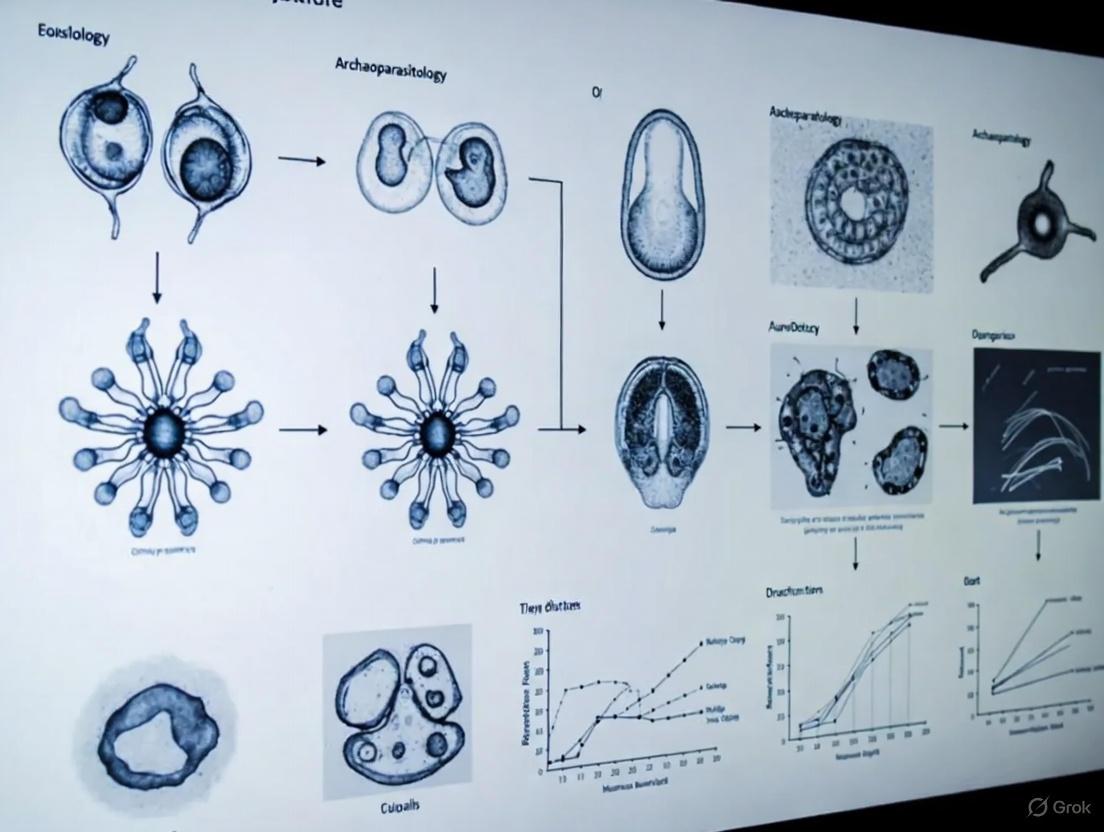
Diagram 1: Evolution of Archaeoparasitology
Over the past century, archaeoparasitology has matured from a descriptive auxiliary science into a dynamic, quantitative discipline that is integral to a holistic understanding of parasitic disease. By tracing the journey from the initial identification of parasites in Egyptian mummies to the complex discussions at modern ICOPA sessions, this review underscores the field's critical role. The historical baseline provided by archaeoparasitology enriches our comprehension of contemporary epidemiological patterns and offers invaluable, time-tested insights for guiding future public health initiatives, drug discovery programs, and global strategies for the control of neglected tropical diseases.
Archaeoparasitology, the study of ancient parasites from archaeological contexts, provides a unique lens through which to understand human health, migration, and cultural practices throughout history. This scientific discipline bridges the gap between archaeology and parasitology, offering direct evidence of infectious diseases that affected past populations. By analyzing parasite remains preserved in archaeological specimens such as latrine soils, coprolites, mummified tissues, and hygiene artifacts, researchers can reconstruct the temporal and spatial distribution of pathogens across different civilizations. This whitepaper synthesizes key findings from major regions including East Asia, Europe, Siberia, and North America, framed within the context of a broader thesis on the history of archaeoparasitology research.
The development of archaeoparasitology has progressed from initial microscopic identifications to sophisticated molecular analyses, enabling more precise taxonomic classifications and understanding of parasite evolution. This field has demonstrated that parasitic infections have been constant companions throughout human history, with their prevalence and distribution shaped by factors including human migration, dietary practices, urbanization, and technological developments. The integration of ancient DNA analysis, paleogenomics, and advanced biomolecular techniques has revolutionized the field, allowing researchers to track the origin and spread of specific pathogens across continents and millennia.
Major Regional Findings in Archaeoparasitology
East Asia
East Asian archaeoparasitology research has yielded particularly rich findings due to excellent preservation conditions and extensive archaeological investigations.
China: Evidence from the Xuanquanzhi Relay Station (111 BCE–CE 109) along the Silk Road revealed intestinal parasites including Chinese liver fluke (Clonorchis sinensis), Taenia sp. tapeworm, roundworm (Ascaris lumbricoides), and whipworm (Trichuris trichiura) preserved on personal hygiene sticks [15]. The presence of Chinese liver fluke was especially significant as this species requires wet marshy environments to complete its life cycle and could not have been endemic to the arid Tarim Basin where the site is located. This finding provides the earliest concrete evidence that travelers carried infectious diseases along the Silk Road, likely from well-watered areas of eastern or southern China [15].
Integration of traditional Chinese medical texts with archaeological findings has provided complementary evidence for parasitic loads in early China, documenting roundworm (Ascaris lumbricoides), Asian schistosoma (Schistosoma japonicum), and tapeworm (Taenia sp.) [16]. These textual sources help fill gaps in the archaeological record caused by taphonomic and environmental factors that limit parasite preservation.
Korean Peninsula: A comprehensive paleoparasitological survey of archaeological sites revealed distinctive patterns of parasite infection related to population density [17]. Examinations of strata soil samples from capital cities including Hansung (Joseon period), Buyeo (Baekje period), and Gyeongju (Silla period) showed heavy contamination with parasite eggs, while contemporary rural sites showed significantly lower levels. Species identified included Ascaris, Trichuris, Clonorchis, and Pygidiopsis summa [17]. This urban-rural disparity demonstrates how population concentration facilitated parasite transmission in ancient urban centers.
Table 1: Ancient Parasites Documented in East Asian Archaeological Contexts
| Parasite Species | Region/Period | Archaeological Context | Significance |
|---|---|---|---|
| Clonorchis sinensis (Chinese liver fluke) | Han Dynasty Silk Road (111 BCE–CE 109) | Personal hygiene sticks from latrine [15] | Evidence of long-distance travel with parasites; non-endemic species |
| Taenia sp. (tapeworm) | Han Dynasty China; multiple Korean periods | Latrine sediments; soil samples [15] [17] | Indicates consumption of undercooked meat (pork/beef) |
| Ascaris lumbricoides (roundworm) | Widespread in China & Korea | Hygiene sticks; soil samples; medical texts [16] [15] [17] | Fecal-oral transmission; indicates sanitation levels |
| Trichuris trichiura (whipworm) | Han Dynasty China; Three Kingdoms Korea | Latrine sediments; strata soils [15] [17] | Fecal-oral transmission; poor sanitation indicator |
| Schistosoma japonicum (Asian schistosoma) | Early China | Medical texts; limited archaeological evidence [16] | Water-borne transmission; agricultural practices |
| Pygidiopsis summa | Baekje Kingdom, Korea | Stratified soil samples [17] | Food-borne trematode; dietary practices |
Methodological Insight: The Korean research program established a standardized protocol for analyzing strata soil samples: re-hydration in 0.5% trisodium phosphate solution for 1 week, filtration through multiple-layered gauze, precipitation for 1 additional day, followed by dissolution in 10% neutral buffered formalin and microscopic examination [17]. This systematic approach enabled quantitative comparison of parasite egg concentrations across different sites and time periods.
Europe
European archaeoparasitology has benefited from well-preserved archaeological sites and recent technological advances in ancient DNA analysis.
Large-Scale Pathogen Genomics: A groundbreaking 2025 study published in Nature screened shotgun-sequencing data from 1,313 ancient humans covering 37,000 years of Eurasian history [18]. The research identified 5,486 individual hits against 492 pathogen species from 136 genera, with 3,384 involving known human pathogens. This study provided direct evidence that zoonotic pathogens (transmitted from animals to humans) were only detected from around 6,500 years ago, peaking roughly 5,000 years ago – coinciding with the widespread domestication of livestock [18]. The research further demonstrated that pathogen spread increased substantially during subsequent millennia, coinciding with pastoralist migrations from the Eurasian Steppe.
British Isles: Analysis of a 6th-century Byzantine bucket at Sutton Hoo revealed it had been repurposed as a cremation urn, evidenced by horse remains and funeral pyre evidence [19]. Recent analysis identified parasite evidence, shedding light on elite burial practices. The discovery of a vast Roman basilica beneath modern London, dating to 78–84 AD, provides context for understanding urban development and potential disease spread in Roman Britain [19].
Mediterranean Region: The discovery of a remarkably intact Roman-era shipwreck off the coast of Antalya, Türkiye, provided a "time capsule" of maritime trade [19]. The vessel contained hundreds of sealed amphorae that had been perfectly preserved for nearly 2,000 years, likely used to transport goods such as wine, olive oil, and tableware. Such trade routes potentially facilitated the spread of parasites and pathogens across the Mediterranean world.
Table 2: Quantitative Data from Large-Scale Eurasian Pathogen Study [18]
| Parameter | Result | Temporal Span | Significance |
|---|---|---|---|
| Ancient humans analyzed | 1,313 individuals | 37,000 years | Extensive temporal coverage |
| Pathogen species identified | 492 species | 37,000 years | Diversity of ancient pathogens |
| Individual pathogen hits | 5,486 hits | 37,000 years | Scale of infection evidence |
| Known human pathogens | 3,384 hits | 37,000 years | Direct evidence of human infection |
| Genera represented | 136 genera | 37,000 years | Taxonomic breadth |
| Zoonotic pathogen emergence | ~6,500 years ago | After animal domestication | Impact of lifestyle change |
Siberia
Siberian research contributions to archaeoparasitology come primarily from the exceptional preservation conditions of permafrost contexts.
Pazyryk Culture: The discovery of a 2,500-year-old Siberian 'ice mummy' from the Pazyryk culture revealed intricate tattoos depicting leopards, a stag, a rooster, and a mythical griffin [20]. While this finding does not provide direct evidence of parasites, the preservation quality in permafrost conditions offers potential for future parasitological analysis. The Pazyryk people were horse-riding warriors who lived on the vast steppe between China and Europe in the 5th century BC, positioning them at the crossroads of potential disease transmission between East and West [20].
Methodological Innovation: Archaeologists collaborated with a tattooist who reproduces ancient skin decorations to understand the techniques involved. They determined that the tattoos were first stenciled onto the skin, then applied using two needle-like tools probably made from animal horn or bone, with pigment likely made from burned plants or soot [20]. Similar interdisciplinary approaches could be applied to identify potential ritual scarification practices that might have introduced pathogens.
North America
While the provided search results contain limited specific information about ancient infections in North America, comparative methodology can be applied from other regions. Standard approaches include:
- Analysis of latrine soils from prehistoric settlements
- Examination of coprolites from cave sites and dry shelters
- Investigation of mummified remains from arctic and desert environments
- Sediment sampling from ancient village sites
The experimental protocols established in East Asian and European contexts can be adapted for North American archaeological contexts to identify parasite introductions and health impacts associated with different subsistence strategies and population density patterns.
Experimental Protocols in Archaeoparasitology
Soil Sediment Analysis Protocol
The standardized protocol for analyzing archaeological soil sediments, as implemented in Korean research [17], involves:
- Re-hydration: Soil samples are re-hydrated in 0.5% trisodium phosphate solution for 1 week
- Filtration: Samples are filtered through multiple-layered gauze
- Precipitation: Filtered samples are precipitated for 1 additional day
- Processing: The upper turbid layer is discarded, and precipitates are dissolved in 10% neutral buffered formalin
- Microscopy: Processed samples are dropped onto slides for light-microscopic examination
- Quantification: Eggs per gram (EPG) of soil and eggs per slide (EPS) are calculated
Biomolecular Analysis Protocol
Advanced biomolecular techniques have significantly enhanced parasite identification:
Enzyme Immunoassay (ELISA): Research on 2,500-year-old stone toilets in Jerusalem utilized ELISA to detect Giardia duodenalis (the parasite causing "traveler's diarrhea") [21]. This technique uses antibodies that bind to species-specific proteins, allowing identification even when morphological preservation is poor.
Shotgun Sequencing: Large-scale pathogen screening, as implemented in the 2025 Eurasian study [18], involves:
- DNA extraction from dental calculus and skeletal remains
- Shotgun sequencing without target enrichment
- Bioinformatic screening against pathogen databases
- Taxonomic classification and phylogenetic analysis
Figure 1: Archaeoparasitology Experimental Workflow - Integrated multidisciplinary approach combining traditional and molecular methods
The Scientist's Toolkit: Research Reagent Solutions
Table 3: Essential Research Reagents and Materials in Archaeoparasitology
| Reagent/Material | Composition/Type | Function in Analysis | Application Examples |
|---|---|---|---|
| Trisodium phosphate solution | 0.5% aqueous solution | Re-hydration of desiccated samples | Re-constitution of ancient feces and soil samples [17] |
| Neutral buffered formalin | 10% formaldehyde with buffer | Preservation of microscopic structures | Fixation of parasite eggs for morphological study [17] |
| ELISA kits | Antibody-based detection systems | Biomolecular detection of parasite antigens | Identification of Giardia duodenalis in Jerusalem latrines [21] |
| DNA extraction kits | Silica-based or chemical methods | Isolation of ancient DNA from samples | Pathogen screening from dental calculus and bones [18] |
| Sequencing reagents | Next-generation sequencing chemistry | Generation of DNA sequence data | Shotgun sequencing of ancient microbial DNA [18] |
| Reference databases | Curated genomic databases | Taxonomic classification of sequences | Pathogen identification from metagenomic data [18] |
Discussion: Implications for Understanding Ancient Disease Dynamics
The synthesis of archaeoparasitological findings across regions reveals several key patterns in ancient disease distribution and transmission:
The evidence from East Asia demonstrates how human migration, particularly along trade routes like the Silk Road, facilitated the long-distance spread of parasites beyond their endemic regions [15]. The discovery of Chinese liver fluke eggs at the Xuanquanzhi Relay Station, located over 1,000 km from endemic areas, provides concrete evidence for this phenomenon. Similarly, the identification of Giardia duodenalis in Iron Age Jerusalem (2,500 years ago) highlights how densely populated urban centers served as hotspots for disease transmission, with the "traveler's diarrhea" pathogen reflecting the city's role as a political and religious center with significant population movement [21].
European findings, particularly the large-scale genomic study of Eurasian pathogens [18], provide direct evidence for the impact of animal domestication on human disease burden. The detection of zoonotic pathogens from approximately 6,500 years ago, peaking around 5,000 years ago, correlates with the timeline of widespread livestock domestication. This represents a fundamental epidemiological transition in human history, where close contact with domesticated animals introduced novel pathogens into human populations.
The Korean research demonstrating higher parasite prevalence in urban versus rural settings [17] illustrates how population density and sanitation infrastructure influenced disease patterns. This urban-rural disparity in ancient parasitism mirrors patterns observed in modern developing contexts, suggesting consistent ecological relationships between settlement patterns and disease transmission.
Archaeoparasitology has evolved from isolated observations of parasite remains to a sophisticated interdisciplinary science that integrates microscopy, biochemistry, genomics, and archaeology. The regional findings synthesized in this whitepaper demonstrate how parasitic infections have shaped and been shaped by human activities including migration, urbanization, animal domestication, and trade. The development of standardized protocols and biomolecular techniques has enabled researchers to address fundamental questions about the origin, spread, and evolution of infectious diseases throughout human history.
Future directions in the field will likely include more extensive application of ancient DNA metagenomics to reconstruct complete pathogen communities, increased temporal and geographical sampling to fill gaps in current understanding, and greater integration of historical textual sources with archaeological evidence. As methods continue to advance, archaeoparasitology will provide increasingly nuanced insights into the long-term relationship between humans and their parasites, with potential implications for understanding contemporary disease dynamics and predicting future pathogen emergence.
The field of archaeoparasitology, which integrates parasitology with archaeological science, emerged as a formal discipline largely through the groundbreaking work of Adauto Araújo and Luiz Fernando Ferreira. Their pioneering research established the methodological and theoretical foundations for studying parasites in archaeological contexts, creating a new lens through which to view human history, migration, and health. As one contemporary publication notes, "Dr. Luiz Fernando Ferreira and Dr. Adauto Araújo were pioneers in paleoparasitology, contributing significantly to the findings of parasites in archaeological materials from the Northeast to the Southeast regions of Brazil" [22]. Their work transformed parasitic remains from archaeological curiosities into valuable sources of historical evidence, enabling researchers to reconstruct aspects of ancient life inaccessible through traditional archaeological methods alone.
The significance of their contribution lies in establishing parasites as historical artefacts that can provide insights into past human behaviors, dietary practices, migration patterns, and health status. By developing and refining techniques for recovering parasite evidence from coprolites, mummies, and latrine sediments, they created a new subfield that continues to yield important discoveries about the human past. This paper examines their lasting impact on archaeoparasitology, tracing the evolution of methodology from their foundational work to contemporary applications, and analyzing how their paradigms continue to shape current research directions.
Foundational Contributions and Established Paradigms
Conceptual and Methodological Foundations
Araújo and Ferreira were instrumental in moving archaeoparasitology from incidental findings to a systematic scientific discipline. In their seminal 2000 publication, they articulated how paleoparasitology could serve as a powerful new tool for studying parasite evolution, noting that "DNA sequences dated thousand years ago, recovered from archaeological material, means the possibility to study parasite-host relationship coevolution through time" [23]. This perspective fundamentally shifted how researchers viewed parasitic remains, transforming them from mere indicators of disease into biological markers for understanding broader historical processes.
Their work established several key paradigms that continue to guide the field:
- Parasites as proxies for human behavior: They demonstrated that parasite species assemblages could reveal information about dietary practices, sanitation, and subsistence strategies [23]
- Parasite dispersal as migration marker: They recognized that the distribution of parasite species could trace human migration routes and cultural contacts [23]
- Methodological standardization: They helped establish standardized protocols for coprolite analysis and parasite recovery that enabled comparative studies across sites and regions [22]
A particularly significant conceptual contribution was their work on the "heirloom vs. souvenir" parasite paradigm, which distinguished between parasites co-evolving with human lineages over millennia ("heirlooms") versus those acquired from local environments through ecological exposure ("souvenirs") [23]. This framework provided researchers with a powerful tool for interpreting the significance of parasite findings in archaeological contexts and understanding the deep history of human-parasite relationships.
Key Research Contributions
Araújo and Ferreira's empirical research yielded several landmark findings that demonstrated the potential of archaeoparasitology to address important historical questions. Their investigation of pre-Columbian hookworm and whipworm infections in the Americas sparked significant scholarly debate about prehistoric human migration patterns [23]. The discovery of these geohelminths in South American archaeological sites dated to 7,200 years before present challenged existing models of the peopling of the Americas, since the Bering Land Bridge crossing would have presented cold conditions incompatible with the transmission of these temperature-sensitive parasites [23].
Their research also provided crucial insights into the antiquity of specific human-parasite relationships. For instance, they documented Trichuris trichiura eggs in human coprolites from the Furna do Estrago archaeological site in northeastern Brazil dated to approximately 2,000 BP, expanding the known paleodistribution of this parasite beyond the Brazilian southeastern region [22]. Similarly, their work on Enterobius vermicularis (pinworm) demonstrated the deep coevolutionary history between this parasite and human hosts, with records spanning from 10,000 years before present in North America through colonial times [23].
Table 1: Key Empirical Contributions of Araújo and Ferreira's Research
| Parasite Species | Archaeological Context | Significance |
|---|---|---|
| Trichuris trichiura | Furna do Estrago, Brazil (~2,000 BP) | Established parasite presence in pre-Columbian northeastern Brazil [22] |
| Human hookworm | South American coprolites (7,200 years BP) | Challenged Bering Land Bridge migration models due to temperature sensitivity [23] |
| Enterobius vermicularis | Global distribution (up to 10,000 years BP) | Demonstrated deep coevolutionary history with humans [23] |
| Trypanosoma cruzi | South American mummies | Developed molecular methods for ancient pathogen detection [23] |
Evolution of Methodological Approaches
Traditional Techniques and Their Development
The pioneering work of Araújo and Ferreira established core laboratory methodologies that became standard practice in archaeoparasitology. They perfected techniques for coprolite rehydration and microscopic analysis, using trisodium phosphate solutions to rehydrate desiccated specimens while preserving parasitic structures [23]. For fossilized coprolites, they adapted modified pollen analysis techniques that allowed for the simultaneous recovery of parasitic and dietary evidence from the same sample [23].
The basic workflow they helped establish involved:
- Coprolite rehydration using trisodium phosphate solutions
- Microscopic analysis after parasite concentration techniques
- Morphological identification of parasite eggs and larvae based on size and shape characteristics
- Differential preservation assessment to account for taphonomic factors
This methodological foundation enabled systematic recovery of parasite evidence from diverse archaeological contexts, from latrine soils to mummified tissues. As Reinhard and Araújo later noted, these techniques allowed the field to progress from simple presence/absence recording to more quantitative approaches that could address questions about parasite prevalence in ancient populations [8].
The Molecular Revolution
Perhaps the most significant methodological contribution of Araújo and Ferreira was their early recognition of the potential for ancient DNA (aDNA) analysis to revolutionize archaeoparasitology. They foresaw that "molecular paleoparasitology constitutes a powerful tool to the research of parasitic diseases in the past" [23] and helped pioneer the application of polymerase chain reaction (PCR) to detect parasite DNA fragments in archaeological specimens.
Their work on Chagas disease provides an exemplary case study of this molecular approach. After initial histological examinations of suspected amastigote nests in mummified tissues yielded inconclusive results, they developed a PCR-based method for detecting Trypanosoma cruzi DNA in mummified remains [23]. This began with experimental approaches using laboratory-desiccated infected mice to optimize techniques before applying them to archaeological specimens [23]. This methodological innovation enabled definitive diagnosis of Chagas disease in South American mummies and demonstrated the potential of molecular approaches to overcome the limitations of traditional morphological analysis.
The current state of the field reflects their vision, with integrated paleoparasitological, paleogenetic, and archaeological analyses now producing comprehensive paleoecological reconstructions [22]. As Iñiguez and colleagues noted, "To obtain high-resolution results, paleogenetics became essential to study parasitic infections in archaeological individuals" [22], confirming Araújo and Ferreira's early recognition of molecular biology's transformative potential.
Diagram 1: Evolution of archaeoparasitology methods
The Scientist's Toolkit: Essential Research Reagents and Materials
Table 2: Essential Research Reagents and Materials in Archaeoparasitology
| Reagent/Material | Function | Application Context |
|---|---|---|
| Trisodium phosphate solution | Coprolite rehydration | Rehydrates desiccated specimens while preserving parasitic structures [23] |
| PCR reagents | DNA amplification | Amplifies parasite DNA fragments from archaeological specimens [23] |
| Sedimentation chambers | Parasite concentration | Concentrates parasite eggs for microscopic examination [23] [24] |
| Confocal laser scanning microscope | Enhanced visualization | Highlights subtle morphological features of ancient parasite eggs [25] |
| Light microscope | Initial screening | Basic identification and quantification of parasite eggs [24] [8] |
| Ancient DNA extraction kits | Nucleic acid recovery | Isolves degraded DNA from archaeological specimens [22] |
Contemporary Applications and Evolving Research Paradigms
Quantitative Approaches and Paleoepidemiology
Building on the foundations laid by Araújo and Ferreira, contemporary archaeoparasitology has increasingly embraced quantitative approaches that enable more sophisticated analysis of ancient parasitic infections. The development of eggs per gram (EPG) quantification methods represents a significant methodological advancement, allowing researchers to estimate infection intensity and its pathological implications [8]. This approach has revealed patterns of parasite overdispersion in ancient populations, where the majority of parasites are concentrated in a minority of hosts [8].
A striking example of this quantitative approach comes from the analysis of medieval burials in Nivelles, Belgium, where researchers documented an unprecedented case of extreme parasitism. One individual (Burial 122) hosted an extraordinarily high parasite burden, with calculated concentrations of 1,577,679 total Trichuris trichiura eggs and 202,350 total Ascaris lumbricoides eggs [24]. Statistical analysis revealed a positive and significant correlation between the presence of these two parasite species, enabling researchers to hypothesize about synergistic health impacts and potential cause of death [24]. This case demonstrates how contemporary research has built upon foundational methods to generate more nuanced understanding of how parasites affected individual and population health in the past.
Molecular Archaeoparasitology and Pathoecology
The molecular approaches championed by Araújo and Ferreira have matured into sophisticated paleogenetic analyses that can simultaneously identify parasite species and determine the host origin of coprolites. Recent work at the Gruta do Gentio II (GGII) archaeological site in Brazil exemplifies this integrated approach, where analysis of coprolites "identified both parasites and species of animals in Pleistocene/Holocene producers of coprolites with geographical and temporal information" [22]. This study identified five taxa of parasites in multiple coprolites genetically linked to five mammal species, including humans, felines, and marsupials [22].
This integrated approach has given rise to the field of pathoecology, which unites archaeological reconstruction of cultural patterns with parasite life cycles to define infection risk factors in past societies [8]. Pathoecology applies Pavlovsky's nidus concept to archaeology, viewing parasitic infections as products of specific ecological foci containing pathogens, vectors, reservoir hosts, and recipient hosts [8]. This theoretical framework enables researchers to generate testable hypotheses about parasite transmission dynamics in ancient communities based on knowledge of both archaeological contexts and parasite biology.
Table 3: Evolution of Research Paradigms in Archaeoparasitology
| Paradigm | Key Focus | Methodological Approach |
|---|---|---|
| Descriptive Phase | Presence/absence recording | Basic microscopy and morphological identification [23] |
| Cultural Reconstruction | Parasites as proxies for human behavior | Correlation with archaeological features and subsistence data [23] |
| Molecular Revolution | Species identification and phylogenetics | aDNA analysis and PCR [23] [22] |
| Pathoecology | Ecological context of infection | Integration of parasite life cycles with archaeological reconstruction [8] |
| Paleoepidemiology | Population-level patterns | Quantitative analysis and statistical modeling [24] [8] |
The lasting impact of Adauto Araújo and Luiz Fernando Ferreira on archaeoparasitology is evident in the continued development of the methodological approaches and research paradigms they helped establish. Their vision of parasites as biological artefacts capable of illuminating aspects of human history inaccessible through other means has been thoroughly validated by decades of subsequent research. As the field continues to evolve, their foundational contributions remain relevant to emerging research directions.
Contemporary studies increasingly recognize their pioneering role, noting that "before this special section was compiled, Adauto Araújo and Luiz Fernando Ferreira, the first cohort of scholars who dedicated their lives to investigating the origin and spread of parasite infections in antecedent human cultures, sadly passed away. We deeply missed these incomparable colleagues and mentors who provided inspiration and guidance to many of archaeoparasitologists around the world" [25]. This tribute underscores their status as foundational figures whose intellectual legacy continues to guide the field.
Future research in archaeoparasitology will likely build upon Araújo and Ferreira's work through continued methodological refinement, particularly in the areas of paleogenetics and proteomics, and through expanded application of quantitative and statistical approaches to analyze parasite distribution in ancient populations. Their vision of archaeoparasitology as an interdisciplinary enterprise that integrates biological, archaeological, and historical approaches remains central to the field's identity and continues to generate novel insights into the shared history of humans and their parasites.
The Archaeoparasitologist's Toolkit: Evolving Methods and Their Archaeological Applications
Within the multidisciplinary field of archaeoparasitology, which is the study of parasites in archaeological contexts, light microscopy (LM) has served as a foundational analytical tool [26]. Since the first report of calcified parasite eggs in an Egyptian mummy in 1910, the identification of helminth eggs via microscopy has provided a direct window into the health, diet, and sanitary conditions of past human societies [25] [26]. Archaeoparasitology has rapidly developed in recent years, embracing novel research techniques, yet traditional light microscopy remains the bedrock for diagnosing parasitic infections in ancient materials [25]. This enduring role is attributed to the method's accessibility, non-destructive nature, and its proven efficacy in detecting the robust eggs of parasites such as Ascaris lumbricoides (roundworm), Trichuris trichiura (whipworm), and cestodes (tapeworms) from a variety of archaeological sources, including coprolites, mummified tissues, and latrine sediments [5] [26]. This technical guide outlines the core microscopic techniques and protocols that continue to underpin egg identification in archaeoparasitological research.
Core Principles and Workflow of Microscopic Analysis
The identification of parasite eggs in archaeological samples relies on the distinctive morphological characteristics of the eggs, which can remain intact for thousands of years [26]. The analytical process is methodical, progressing from initial sample preparation to definitive observation.
Diagnostic Characteristics and Workflow
Parasite eggs are identified based on several key features observable under light microscopy, including:
- Size: Measured precisely using a calibrated ocular micrometer [27].
- Shape: For example, the lemon shape of Trichuris eggs versus the more rounded or oval shapes of others [5].
- Shell Thickness and Surface Morphology: Some eggs have distinctive ridges or mammillated coats.
- Internal Structures: The presence and developmental stage of larvae within the egg can be diagnostic.
- Color: Eggs can vary in their coloration, which aids in differentiation [27].
The following diagram illustrates the standard workflow for processing and analyzing archaeological samples for parasite egg identification:
Essential Laboratory Techniques and Protocols
Microscope Calibration
A correctly calibrated microscope is crucial because size is a primary characteristic for parasite identification [27].
Detailed Protocol:
- Equipment: Ensure an ocular micrometer disk is installed in one ocular and a stage micrometer is available.
- Alignment: Place the stage micrometer on the stage and focus until the scale is clear. Superimpose the "0" line of the ocular micrometer with the "0" line of the stage micrometer.
- Measurement: Without moving the stage, find a distant point where two other lines are perfectly superimposed.
- Calculation:
- Note the number of ocular divisions and the number of millimeters on the stage micrometer between the two superimposed points.
- Calculate the calibration factor:
mm per ocular division = (stage micrometer mm) / (ocular divisions). - Convert to micrometers (µm):
µm per ocular division = (mm per ocular division) * 1000[27].
- Documentation: Calibration factors must be posted on each microscope and recalibrated after every cleaning or component change [27].
Wet Mount Preparation
This technique is used for the initial detection of protozoan cysts and helminth eggs and larvae [27].
Detailed Protocol:
- Slide Preparation: Place a small amount of the processed specimen on a microscope slide. If the sample is solid, add a drop or two of saline and mix.
- Staining (Optional): Ideally, prepare two smears on one slide; one can be stained with iodine to highlight internal structures.
- Coverslip Sealing (Optional): To prevent drying and allow for oil immersion, the coverslip can be sealed. A 1:1 petroleum jelly and paraffin mixture is heated to ~70°C and applied around the coverslip edges with a swab [27].
- Examination: Systematically scan the entire coverslip area using a 10x objective. Switch to higher magnifications for detailed observation of suspicious objects [27].
Stained Slide Preparation
Permanent stained slides are valuable for the identification of protozoa and for creating a permanent record for consultation and future reference [27].
Detailed Protocol:
- Smearing: For unpreserved specimens, prepare a thin, even smear on a 3" x 1" slide using an applicator stick. For polyvinyl alcohol (PVA)-fixed specimens, apply 2-3 drops and spread evenly to cover an area the size of a 22 x 22 mm coverslip.
- Staining: Follow the specific procedure for the chosen stain (e.g., Trichrome).
- Examination: Systematically examine the smear using the 100x oil immersion objective. At least 200 to 300 oil immersion fields should be examined [27].
Quantitative Data from Archaeoparasitological Studies
Light microscopy not only identifies parasites but also provides crucial quantitative data on infection intensity, revealing patterns of public health in past populations. The table below summarizes egg density data from two medieval sites, demonstrating the power of simple microscopic quantification.
Table 1: Quantitative Data on Helminth Egg Density from Medieval Archaeological Sites
| Site (Period) | Sample Type | Parasite | Egg Density (eggs/gram) | Prevalence | Source |
|---|---|---|---|---|---|
| Lübeck, Germany (Medieval) | Latrine Sediments | Trichuris trichiura | 107 – 4,935 /g | 25 of 31 samples | [5] |
| Ascaris lumbricoides | 45 – 1,645 /g | 31 of 31 samples | [5] | ||
| Diphyllobothrium latum | 49 – 1,414 /g | 14 of 31 samples | [5] | ||
| Taenia saginata | 133 – 8,310 /g | 19 of 31 samples | [5] | ||
| Bristol, UK (Medieval) | Communal Waste Ditch | Trichuris trichiura | 78 – 8,559 /g | 25 of 26 samples | [5] |
| Ascaris lumbricoides | 76 – 1,162 /g | 26 of 26 samples | [5] |
The Scientist's Toolkit: Key Research Reagents and Materials
Successful microscopic analysis in archaeoparasitology depends on a suite of essential reagents and materials. The following table details the core components of the research toolkit.
Table 2: Essential Research Reagent Solutions and Materials for Archaeoparasitology
| Item | Function / Application | Technical Notes |
|---|---|---|
| Ocular Micrometer | A glass disk with a scale inserted into a microscope eyepiece, used for measuring the size of parasite eggs. | Must be calibrated for each objective lens using a stage micrometer; size is a critical diagnostic feature [27]. |
| Stage Micrometer | A precision microscope slide with a known scale, used exclusively for calibrating the ocular micrometer. | Typically graded in 0.1 mm and 0.01 mm divisions [27]. |
| Saline Solution (0.85%) | A suspension medium for preparing wet mounts; maintains osmotic balance to preserve egg morphology. | Prevents the rapid disintegration of delicate structures in distilled water [27]. |
| Iodine Stain (e.g., Lugol's) | A chemical stain used in wet mounts to highlight internal structures of protozoan cysts (e.g., nuclei, glycogen). | Does not permanently stain the slide; preparation should be examined promptly [27]. |
| Permanent Stains (e.g., Trichrome) | Stains used to create permanent slide preparations of specimens, allowing for detailed study and archiving. | Essential for the definitive identification and confirmation of many protozoan species [27]. |
| Mounting Media & Sealants | Substances used to seal coverslips on wet mounts, preventing evaporation and enabling use of oil immersion objectives. | A 1:1 mixture of petroleum jelly and paraffin, heated to 70°C, is a common choice [27]. |
Advanced Microscopic Techniques and Future Directions
While traditional light microscopy remains the core technique, archaeoparasitology is increasingly enhanced by advanced microscopic technologies. Confocal Laser Scanning Microscopy (CLSM) has emerged as a powerful complementary tool [28]. It examines the intrinsic autofluorescence of parasite eggs, highlighting subtle morphological features and enhancing the visualization of egg anatomy without the destructive sample preparation required for scanning electron microscopy (SEM) [28]. This allows the same specimen to be used for subsequent molecular analyses. Similarly, the use of UV fluorescence microscopy has proven highly effective for detecting specific parasites like Cyclospora, whose oocysts exhibit intense autofluorescence under UV light, providing another layer of diagnostic certainty [27]. These advanced methods build upon the foundation of light microscopy, pushing the boundaries of what can be identified and understood from ancient parasitic remains. The relationship between core and advanced techniques is shown below.
Despite the emergence of sophisticated molecular and geometric morphometric techniques, light microscopy retains its status as an indispensable first step in the archaeoparasitological workflow [25] [5] [29]. Its capacity to provide rapid, cost-effective, and reliable genus- or species-level identification of parasite eggs from a wide range of archaeological contexts ensures its continued relevance. The quantitative data it generates on egg density and prevalence forms the foundational evidence for interpreting the health, dietary habits, and living conditions of ancient populations [5]. As the field continues to globalize and its research questions become more nuanced, the enduring role of light microscopy as a core technique for egg identification is firmly secured, providing an artefact-independent source of historical evidence that continues to illuminate the hidden lives of our ancestors.
The field of archaeology is currently experiencing its "third scientific revolution," driven by innovative technologies that allow artifacts to speak at the molecular level [30]. This transformation is particularly evident in archaeoparasitology, the study of parasites in ancient remains, which has evolved from microscopic identification to sophisticated molecular analyses. Where researchers once documented parasite distributions simply through presence/absence studies, the integration of sedimentary ancient DNA (sedaDNA) analysis with targeted enrichment strategies now enables unprecedented insights into past human health, diet, and migration patterns [31] [5] [8]. This whitepaper examines how these molecular methodologies are reshaping archaeoparasitology, providing researchers with powerful tools to reconstruct historical disease dynamics and human-parasite relationships across millennia.
Historical Context: The Evolution of Archaeoparasitology
The development of archaeoparasitology reflects a journey from basic morphological identification to complex quantitative molecular analysis, characterized by several distinct phases:
- 1955-1969: Pioneering Era - Researchers developed methods for parasite recovery from mummies and coprolites, with landmark studies reporting the oldest pinworm and thorny-headed worm infections [8].
- 1970s: Expansion and Prevalence Studies - Analysis of large coprolite collections from museums intensified, with researchers establishing parasite prevalence as a key metric and developing provenience-based sampling strategies [8].
- 1980-2000: Geographic and Cultural Exploration - Studies expanded globally, investigating cultural influences on parasitism and beginning to integrate parasite data with bone pathology evidence [8].
- 21st Century: Molecular Transformation - Pathoecology and paleoepidemiological approaches emerged, incorporating quantification methods and ancient DNA analysis to explore infection intensity and ecological relationships [5] [8].
This historical progression demonstrates how technological advances have progressively enhanced our ability to extract meaningful biological information from archaeological specimens, culminating in today's molecular approaches.
Technical Foundations: SedaDNA and Targeted Enrichment
Sedimentary Ancient DNA (sedaDNA) Fundamentals
Sedimentary ancient DNA (sedaDNA) refers to ancient DNA fragments obtained from sediment samples that preserve genetic material from a wide range of organisms that interacted with a given archaeological context [32]. Unlike DNA from macro-remains, sedaDNA is typically highly fragmented and degraded, with fragments usually <100 base pairs (bp) in length [33]. This material can originate from both extra- and intracellular sources, preserved through binding to mineral surfaces in the soil or within micro-remains and coprolites [32].
The power of sedaDNA in archaeoparasitology lies in its ability to provide holistic biodiversity assessments beyond traditionally fossilized taxa [34]. This allows researchers to reconstruct comprehensive paleo-food webs and investigate parasite-host interactions without dependence on visible fossil remains. However, working with sedaDNA presents significant challenges, including minuscule preservation amounts, potential contamination from modern DNA, and the complexity of analyzing mixed genetic signals from multiple organisms [32] [33].
Targeted Enrichment Strategies
Targeted enrichment (also called target capture) is a laboratory method for selecting DNA fragments belonging to specific taxa of interest from a complex DNA library [31] [32]. This approach is particularly valuable for sedaDNA studies where the endogenous DNA content is often extremely low [35].
The process involves designing custom capture probes complementary to target genomic regions, which are then used to hybridize and isolate these fragments from the total DNA pool [35]. Common targets in archaeoparasitology include mitochondrial genes (Cytochrome B, COX1), ribosomal ITS regions, and other taxon-specific genetic markers that enable precise species identification [5].
Table 1: Common Genetic Targets for Parasite Identification
| Parasite Group | Genetic Targets | Identification Level | Application Examples |
|---|---|---|---|
| Nematodes (e.g., Trichuris) | ITS-1, β-tubulin | Species-level | Distinguishing T. trichiura from animal whipworms [5] |
| Nematodes (e.g., Ascaris) | CytB, COX1 | Species-level | Confirming A. lumbricoides in latrine sediments [5] |
| Cestodes (e.g., Taenia) | CytB | Species-level | Identifying T. saginata in medieval deposits [5] |
| Cestodes (e.g., Diphyllobothrium) | COX1 | Species-level | Detecting D. latum in trading centers [5] |
Methodological Workflow: From Sampling to Authentication
Implementing sedaDNA analysis with targeted enrichment requires a rigorous, multi-stage workflow with stringent contamination controls at each step. The diagram below illustrates this comprehensive process:
Sample Collection and DNA Extraction
Sample Collection: Archaeological sediment sampling for sedaDNA requires extreme precautions to avoid modern contamination. Best practices include:
- Using sterile disposable materials and specialized protective clothing [32]
- Removing air-exposed top layers before sampling [32]
- Taking samples from the interior of sediment cores or freshly cleaned sections [33]
- Immediate freezing or cold storage of samples [33]
Context selection is critical, as factors like sediment type, organic matter content, and potential leaching affect DNA preservation. Clay-rich soils with high organic content typically demonstrate superior DNA preservation [32] [33].
DNA Extraction: Protocols must be optimized for sedaDNA's fragmented nature and to remove PCR inhibitors like humic acids, heavy metals, and complex proteins [32]. Effective approaches include:
- Silica-based extraction methods that preferentially bind short DNA fragments [33]
- Bead-beating to break robust resting cells and facilitate intracellular DNA recovery [33]
- Phosphate-containing buffers to competitively displace DNA from mineral surfaces [33]
- Specialized protocols recovering fragments as short as 27bp [33]
Library Preparation and Targeted Enrichment
Library Preparation: Double-stranded libraries are typically prepared without additional shearing, adapting ancient DNA to modern sequencing platforms [35]. Key considerations include determining optimal PCR cycle numbers through qPCR and using double-indexing strategies to monitor cross-contamination [35].
Targeted Enrichment: This critical step enhances the recovery of specific genomic targets from complex sedaDNA backgrounds:
- Capture probe design focuses on mitochondrial genomes or specific nuclear markers for target taxa [35]
- Hybridization conditions (typically 16-24 hours at 65°C) optimize specificity [35]
- Post-capture amplification enriches recovered fragments while maintaining representative diversity
Recent innovations include pooled testing approaches where multiple extracts are combined before enrichment, significantly reducing costs and hands-on time while maintaining detectable signals even when pooled with four negative samples [35].
Sequencing, Analysis and Authentication
High-Throughput Sequencing: Illumina platforms (e.g., NovaSeq) are standard, with sequencing depth adjusted based on endogenous DNA content [35].
Bioinformatic Processing: Specialized ancient DNA pipelines address sedaDNA's unique challenges:
- Alignment to comprehensive reference databases
- Metabarcoding for taxonomic classification using marker genes [31]
- Shotgun metagenomics for broader taxonomic profiling [31]
Authentication: Rigorous authentication is essential to confirm ancient origin:
- DNA damage pattern analysis (e.g., mapDamage) assessing cytosine deamination patterns [35] [33]
- Fragment length distribution evaluation (expecting <100bp fragments) [33]
- Contamination screening using negative controls and comparative analysis [32]
Research Reagent Solutions and Essential Materials
Table 2: Key Research Reagents for SedaDNA and Targeted Enrichment
| Reagent/Material | Function | Application Notes |
|---|---|---|
| Silica-based Extraction Kits | DNA binding and purification | Optimized for short fragment recovery; critical for sedaDNA [32] |
| Phosphate Buffers | Competitive displacement of DNA from minerals | Enhances DNA yield from mineral-rich sediments [33] |
| Bead-Beating Matrix | Mechanical cell disruption | Releases intracellular DNA from robust cysts and spores [33] |
| Double-Stranded Library Prep Kits | Library construction for sequencing | Adapted for ancient DNA without shearing step [35] |
| Custom Capture Probes | Target-specific enrichment | Designed for mitochondrial genomes or specific parasites [35] |
| Hybridization Reagents | Facilitates probe-target binding | Critical for target capture efficiency [35] |
| Indexing Primers | Sample multiplexing | Enables pooling of multiple libraries pre-sequencing [35] |
| DNA Damage Enzymes | Authentication | Uracil-DNA-glycosylase treatment detects deamination patterns [33] |
Case Study: Molecular Archaeoparasitology in Medieval Lübeck
A landmark study demonstrating sedaDNA with targeted enrichment analyzed 152 samples from Neolithic to Early Modern periods across European sites, with extensive sampling from medieval Lübeck, a key Hanseatic trading center [5]. The experimental workflow and key findings are summarized below:
Key Findings and Quantitative Results
The study generated compelling quantitative data demonstrating how molecular methods transform archaeological interpretation:
Table 3: Quantitative Parasite Data from Medieval Lübeck
| Parasite | Transmission Route | Prevalence | Egg Concentration Range | Molecular Identification |
|---|---|---|---|---|
| Trichuris trichiura | Faecal-oral | 100% (31/31 samples) | 107-4,935 eggs/gram | ITS-1 and β-tubulin sequencing [5] |
| Ascaris lumbricoides | Faecal-oral | 100% (31/31 samples) | 45-1,645 eggs/gram | CytB and COX1 sequencing [5] |
| Diphyllobothrium latum | Food-borne (fish) | 45% (14/31 samples) | 49-1,414 eggs/gram | COX1 sequencing [5] |
| Taenia saginata | Food-borne (beef) | 61% (19/31 samples) | 133-8,310 eggs/gram | CytB sequencing [5] |
The molecular analysis provided unprecedented insights:
- Species-level identification confirmed all tapeworms as human-specific varieties rather than zoonotic forms [5]
- Temporal shifts in cestode prevalence indicated dietary changes around 1300 CE [5]
- Genetic diversity of T. trichiura in Lübeck reflected its role as a trading center with multiple parasite introductions [5]
- Epidemiological patterns showed strong positive correlation between nematode infections, reflecting similar transmission routes [5]
Future Directions and Research Agenda
The integration of sedaDNA and targeted enrichment in archaeoparasitology continues to evolve, with several promising frontiers:
- Palaeo-phylogenetics: Tracking evolutionary relationships and geographic spread of parasites through time [31]
- Functional palaeogenetics: Reconstructing genetic adaptations and virulence factors in ancient parasites [31]
- sedaRNA analysis: Potential for studying RNA viruses and gene expression in ancient contexts [31]
- Efficiency improvements: Pooled extraction approaches reducing costs by up to 70% and hands-on time to one-fifth [35]
- Reference database expansion: Enhanced taxonomic resolution through improved genetic reference collections [32] [33]
As these methodologies mature, they will further illuminate the long and intimate association between humans and parasites, providing valuable evolutionary context for modern infectious disease challenges [36]. The molecular revolution in archaeoparasitology exemplifies how interdisciplinary collaboration between archaeology, genetics, and parasitology can transform our understanding of the human past.
Archaeoparasitology, the study of parasites in archaeological contexts, seeks to understand the history of parasitic infections and their impact on human societies through the analysis of ancient materials [26]. While traditional microscopy has long been the cornerstone of this field, enabling the identification of durable helminth eggs in coprolites and latrine soils, the detection of protozoan antigens has presented a significant challenge [37] [38]. Unlike the robust eggs of many worms, protozoan cysts are less likely to preserve morphologically, and their unambiguous identification to the species level (e.g., distinguishing pathogenic Entamoeba histolytica from non-pathogenic Entamoeba dispar) is impossible by morphology alone [38]. The integration of enzyme-linked immunosorbent assay (ELISA) into the archaeoparasitological toolkit has therefore represented a methodological leap forward. This sensitive immunological technique allows researchers to detect species-specific protein antigens, even in degraded ancient samples, providing a powerful tool to investigate the historical epidemiology of protozoan diseases such as giardiasis, cryptosporidiosis, and amoebic dysentery [39] [38]. This guide details the technical principles, protocols, and applications of ELISA for protozoan antigen detection, framing it as a critical advancement in the evolving methodology of archaeoparasitological research.
Technical Principles of ELISA
The enzyme-linked immunosorbent assay (ELISA) is a plate-based technique designed to detect and quantify soluble substances such as peptides, proteins, antibodies, and hormones [40]. The fundamental principle involves immobilizing a target antigen on a solid surface (typically a microplate) and complexing it with an antibody that is linked to a reporter enzyme. The detection is achieved by incubating the enzyme-antibody complex with a substrate to generate a measurable product, with the intensity of the signal being proportional to the quantity of antigen present in the sample [40] [41]. The assay's specificity is derived from the highly specific antibody-antigen interaction.
Key ELISA Formats for Antigen Detection
Several ELISA formats can be employed for antigen detection, each with distinct advantages and applications in protozoan research.
Direct ELISA: This format uses a single enzyme-conjugated primary antibody that binds directly to the target antigen adsorbed onto the plate surface. While it is a rapid procedure with minimal steps, it can suffer from potential interference with the antibody's immunoreactivity due to the labeling process and offers limited signal amplification [40].
Indirect ELISA: In this format, an unlabeled primary antibody binds to the antigen, and an enzyme-conjugated secondary antibody, which is specific to the primary antibody, is then used for detection. The indirect method offers greater flexibility and signal amplification, as multiple secondary antibodies can bind to a single primary antibody. This enhances the assay's sensitivity, which is crucial for detecting low-abundance antigens in complex sample matrices [40] [42] [41].
Sandwich ELISA: This highly sensitive and specific format requires two antibodies that bind to different epitopes on the target antigen. The first antibody (capture antibody) is immobilized on the plate and binds the antigen from the solution. The second antibody (detection antibody) is then added to complete the "sandwich." The detection antibody can be enzyme-conjugated directly (direct sandwich) or detected via an enzyme-conjugated secondary antibody (indirect sandwich) [40]. This method is particularly advantageous for complex samples, as the capture step helps isolate the antigen from other components.
Competitive ELISA: Also known as inhibition ELISA, this format is often used for smaller antigens with single epitopes. In one common configuration, the sample antigen and a labeled reference antigen compete for binding to a limited amount of capture antibody. The signal generated is inversely proportional to the amount of antigen in the sample, meaning a lower signal indicates a higher concentration of the target antigen in the test sample [40].
Table 1: Comparison of Major ELISA Formats for Antigen Detection [40] [41]
| Format | Key Feature | Sensitivity | Complexity | Typical Use Case |
|---|---|---|---|---|
| Direct | Single, labeled primary antibody | Lower | Low | High-concentration antigen; rapid screening |
| Indirect | Labeled secondary antibody provides signal amplification | Higher | Medium | General purpose; enhanced sensitivity needed |
| Sandwich | Two antibodies bind different antigen epitopes | Highest | High | Complex samples (e.g., stool, sediment); low-abundance antigens |
| Competitive | Sample and labeled antigen compete for antibody binding | Variable (good for small molecules) | High | Small antigens with single epitope; haptens |
ELISA Protocols for Protozoan Antigen Detection
The following section provides a detailed methodology for conducting an indirect ELISA, which is a common and versatile format used in parasitology, and a specific protocol for a multiplexed protozoan screening assay.
General Indirect ELISA Protocol
This protocol is adapted from established methods for detecting viral antibodies and can be modified for protozoan antigen detection by using anti-protozoan antibodies [42].
Materials and Reagents:
- Coating Antigen: Purified protozoan antigen (e.g., recombinant protein) or capture antibody.
- Blocking Buffer: 1% Bovine Serum Albumin (BSA) in DPBS with 0.05% Tween-20 [42].
- Wash Buffer: Phosphate-Buffered Saline with 0.05% Tween-20 (PBST) [42] [41].
- Sample Diluent: DPBS with 1% BSA and 0.05% Tween-20, or 5% goat serum in PBST [42].
- Primary Antibody: Specific antibody against the target protozoan antigen.
- Secondary Antibody: Enzyme-conjugated antibody (e.g., HRP-conjugated) specific to the host species of the primary antibody.
- Substrate: Tetramethyl benzidine (TMB) or other HRP-compatible substrate [42] [41].
- Stop Solution: Acidic solution (e.g., 1M H₂SO₄) to halt the enzyme-substrate reaction.
- Microplate: 96-well medium-binding, flat-bottom polystyrene plate [42].
Procedure:
- Coating: Dilute the capture antigen or antibody in a carbonate/bicarbonate coating buffer (pH 9.4) to a concentration of 2-10 µg/mL. Add 100 µL per well to the microplate. Seal the plate and incubate for 2 hours at room temperature or overnight at 4°C [40] [41].
- Washing: Discard the coating solution. Wash each well three times with approximately 300 µL of wash buffer (PBST) per wash. Remove residual liquid by blotting the plate on paper towels [42] [41].
- Blocking: Add 200 µL of blocking buffer to each well. Seal the plate and incubate for 1-2 hours at room temperature to cover any remaining protein-binding sites on the plastic. Wash the plate three times as before [40] [41].
- Primary Antibody Incubation: Dilute the test samples and controls in sample diluent. Add 100 µL per well. Seal the plate and incubate for 2 hours at room temperature or overnight at 4°C. Wash the plate three times [42] [41].
- Secondary Antibody Incubation: Dilute the enzyme-conjugated secondary antibody in blocking buffer as per the manufacturer's recommendation. Add 100 µL per well. Seal the plate and incubate for 1-2 hours at room temperature (protected from light if necessary). Wash the plate three times [42] [41].
- Signal Detection: Add 100 µL of substrate solution (e.g., TMB) to each well. Incubate the plate in the dark at room temperature for 10-30 minutes, or until color development is sufficient.
- Stop Reaction and Read: Add 50-100 µL of stop solution to each well. The blue TMB color will turn yellow. Read the optical density (OD) of each well immediately using a plate reader at the appropriate wavelength (e.g., 450 nm for TMB) [42] [41].
Protocol for Multiplex Protozoan Screening (TRI-COMBO ELISA)
A prototype TRI-COMBO ELISA has been developed for the simultaneous detection of Giardia lamblia, Cryptosporidium parvum, and Entamoeba histolytica antigens in a single stool sample, demonstrating the utility of ELISA for field and clinical studies [39].
Materials:
- TRI-COMBO PARASITE SCREEN kit (TechLab, Inc.) or equivalent components.
- Stool samples, unfixed and not refrigerated.
- Microplate reader (visual interpretation is possible but not optimal).
Procedure:
- Sample Preparation: Stool samples are collected and should be tested immediately upon arrival at the lab. If a short delay is unavoidable, samples can be stored at room temperature and tested the next business day [39].
- Assay Execution: The test is performed according to the manufacturer's instructions. The specific steps are proprietary, but they follow a standard sandwich or indirect ELISA format on a 96-well microplate pre-coated with antibodies specific to the three protozoa.
- Reading and Interpretation: Results can be read visually or with an optical density reader. A positive result indicates the presence of one or more of the three protozoan antigens, though the initial test may not differentiate between them without further testing [39].
Performance Data and Validation
The diagnostic performance of ELISA for protozoan antigens has been extensively validated against other standard techniques like microscopy and PCR.
Table 2: Performance of ELISA vs. Other Diagnostic Methods for Protozoan Detection [39] [38]
| Parasite | Assay Type | Sensitivity | Specificity | Comparison Method | Key Findings |
|---|---|---|---|---|---|
| Giardia lamblia | TRI-COMBO ELISA | 91% agreement with individual ELISAs (Kappa=0.90) | High (specific value not stated) | Individual Giardia II ELISA | Only 40% of ELISA-positive samples were detected by microscopy [39]. |
| Giardia lamblia | Commercial RDTs (Various) | 58% - 100% [38] | 94% - 100% [38] | Microscopy + ELISA + PCR | Performance varies significantly by brand and study [38]. |
| Cryptosporidium spp. | TRI-COMBO ELISA | High agreement with individual ELISAs | High (specific value not stated) | Individual Cryptosporidium II ELISA | - |
| Cryptosporidium spp. | Commercial RDTs (Various) | 67% - 100% [38] | 95% - 100% [38] | Microscopy + ELISA + PCR | Most brands showed high sensitivity and specificity [38]. |
| Entamoeba histolytica | TRI-COMBO ELISA | High agreement with individual ELISAs | High (specific value not stated) | Individual E. HISTOLYTICA II ELISA | Does not cross-react with non-pathogenic E. dispar [39]. |
| Entamoeba histolytica | Commercial RDTs (Various) | 100% [38] | 80% - 88% [38] | Microscopy + ELISA + PCR | High sensitivity, but specificity can be a concern [38]. |
Table 3: Application of Recombinant Antigens in ELISA for Improved Specificity [43]
| Recombinant Antigen | Target Parasite | Assay Format | Advantage |
|---|---|---|---|
| rSjTPx-1 (Thioredoxin Peroxidase-1) | Schistosoma japonicum | Indirect ELISA | No cross-reaction with Fasciola hepatica in water buffaloes [43]. |
| rSj1TR (Tandem Repeat Protein) | Schistosoma japonicum | Indirect ELISA | Highest agreement with PCR; superior sensitivity and specificity for animal surveillance [43]. |
The Scientist's Toolkit: Key Research Reagent Solutions
Table 4: Essential Reagents for Protozoan Antigen ELISA [42] [41]
| Reagent / Material | Function / Description | Example Products / Components |
|---|---|---|
| Coating Buffer | Alkaline buffer (pH ~9.4) for optimal adsorption of antigens/antibodies to polystyrene plate. | Carbonate-Bicarbonate Buffer [41] |
| Blocking Buffer | Contains irrelevant proteins to saturate unused binding sites on the plate, reducing background noise. | 1% BSA in PBST; 5% Goat Serum in PBST; Casein Buffer [42] [41] |
| Wash Buffer | Removes unbound reagents; contains detergent to minimize non-specific binding. | PBS with 0.05% Tween-20 (PBST) [42] [41] |
| Detection Antibodies | Primary antibody binds antigen; enzyme-conjugated secondary antibody enables signal generation. | Host-specific conjugates (e.g., Goat anti-human IgG-HRP) [42] |
| Enzyme Substrate | Chromogenic compound converted by the reporter enzyme to a colored, measurable product. | Tetramethylbenzidine (TMB) [42] [41] |
| Microplates | Solid surface for the assay; high protein-binding plates are standard. | 96-well polystyrene plates (e.g., Nunc Maxisorp, Greiner Bio-One) [42] [43] |
Application in Archaeoparasitology and Concluding Perspective
The adoption of ELISA in archaeoparasitology has fundamentally expanded the scope of research into ancient protozoan diseases. While microscopy remains essential for detecting helminth eggs, ELISA provides a complementary tool for investigating parasites like Giardia, Cryptosporidium, and Entamoeba histolytica, which leave less conspicuous morphological evidence [39] [38]. The technique's ability to differentiate between pathogenic and non-pathogenic species using species-specific antigens—a task impossible with traditional microscopy—has clarified interpretations of health and disease in past populations [38]. For instance, confirming the presence of the pathogenic E. histolytica in a latrine sample provides direct evidence for the potential occurrence of amoebic dysentery in that society.
Furthermore, the move towards recombinant antigens, as demonstrated in veterinary parasitology with Schistosoma japonicum, points to the future of immunological analysis in archaeoparasitology [43]. Recombinant proteins offer superior specificity by eliminating cross-reactivity that can occur with crude antigen extracts, leading to more accurate diagnoses. As these techniques continue to be refined, their application to archaeological material, potentially through the detection of conserved protein fragments, holds the promise of mapping the historical distribution and evolution of protozoal infections with unprecedented precision. This aligns with the broader goals of archaeoparasitology, which include understanding the health consequences of societal shifts like the advent of agriculture, urbanization, and long-distance trade [25] [26] [37]. By providing a direct link to the pathogenic agents themselves, ELISA and related immunoassays add a critical molecular dimension to the archaeological reconstruction of past human health.
This technical guide examines the critical interplay between sanitation, cooking practices, and human health through the specialized lens of archaeoparasitology. By integrating paleoparasitological data with modern nutritional science, we elucidate how historical patterns of disease transmission and dietary practices inform contemporary public health challenges. We present standardized methodologies for analyzing parasite remains and food preparation behaviors, alongside quantitative data demonstrating the persistent relationship between environmental contamination, foodborne pathogens, and chronic disease. This multidisciplinary approach provides researchers with robust analytical frameworks for investigating the complex relationships between human behavior, environmental factors, and health outcomes across temporal and cultural contexts.
Paleoparasitology, the study of ancient parasites from archaeological contexts, has emerged as a critical discipline for understanding historical disease patterns, dietary practices, and human-environment interactions [44]. This field operates at the intersection of archaeology, biology, and paleopathology, providing unique insights into how past communities managed sanitation, food preparation, and health challenges. The analysis of parasite eggs and DNA from sediments, latrines, and burial sites enables researchers to reconstruct past hygiene standards, waste management practices, and dietary patterns [44] [45].
Within the broader context of archaeoparasitology research, this guide addresses the fundamental connections between data derived from parasite analysis, historical cooking practices, and disease prevalence. The discipline has evolved from basic microscopic identification to incorporate sophisticated molecular and immunological techniques, allowing for more comprehensive reconstructions of past human health [45]. By examining how past societies managed the challenges of sanitation and food preparation, researchers can identify persistent patterns of disease transmission and identify potential interventions for contemporary public health issues.
Analytical Methods in Paleoparasitology
A multimethod approach is essential for comprehensive parasite detection in archaeological samples. The most effective strategy combines microscopy, enzyme-linked immunosorbent assay (ELISA), and sedimentary ancient DNA (sedaDNA) analysis, as each technique has complementary strengths and limitations [45].
Standardized Experimental Protocols
Microscopy Protocol for Helminth Detection
- Sample Preparation: Disaggregate 0.2g sediment in 0.5% trisodium phosphate [45]
- Micro-sieving: Sieve sample to collect material between 20-160μm [45]
- Microscopic Analysis: View fraction mixed with glycerol under light microscope at 200x and 400x magnification [45]
- Identification: Identify helminth eggs based on morphological characteristics [45]
ELISA Protocol for Protozoan Detection
- Sample Preparation: Disaggregate 1g sediment in 0.5% trisodium phosphate and micro-sieve [45]
- Collection: Collect material in catchment container below 20μm sieve [45]
- Concentration: Concentrate material for commercial ELISA kits [45]
- Analysis: Follow manufacturer protocols for GIARDIA II, E. HISTOLYTICA II, and CRYPTOSPORIDIUM II kits [45]
sedaDNA Extraction and Analysis
- Subsampling: Use 0.25g of material in dedicated ancient DNA facilities [45]
- Lysis: Chemically and physically disintegrate material using lysis buffer in garnet PowerBead tubes [45]
- Vortexing: Mechanically break down content with 15 minutes of vortexing [45]
- Digestion: Add Proteinase K and rotate tubes continuously at 35°C overnight [45]
- Purification: Mix supernatant with high-volume Dabney binding buffer and centrifuge at 4500 rpm at 4°C for 6-24 hours [45]
- Library Preparation: Use double-stranded method for Illumina sequencing with targeted enrichment for parasite DNA [45]
Method Efficacy Comparison
Table 1: Comparative Efficacy of Paleoparasitological Methods
| Method | Target Pathogens | Sensitivity | Key Advantages | Limitations |
|---|---|---|---|---|
| Microscopy | Helminth eggs (Trichuris, Ascaris, etc.) | High for intact eggs | Cost-effective; provides morphological data | Cannot identify species; requires intact eggs |
| ELISA | Protozoa (Giardia, Entamoeba, Cryptosporidium) | Highest for protozoa | Detects antigenic proteins; effective for diarrhea-causing protozoa | Limited to specific targeted pathogens |
| sedaDNA with Targeted Enrichment | Broad parasite diversity via DNA | Variable; complementary | Species identification; detects multiple taxa simultaneously | Higher cost; requires specialized facilities |
Historical and Contemporary Case Studies
19th Century Québec City Privy Analysis
A seminal study of 19th century privy structures from a wealthy Québec City household demonstrated the value of multiproxy analysis in archaeoparasitology [6]. The research revealed:
- Parasite Findings: Samples tested positive for Trichuris trichiura (whipworm), Ascaris lumbricoides (roundworm), and capillariids, indicating significant fecal-oral transmission despite the household's wealthy status [6]
- Dietary Reconstruction: Pollen spectra evidenced a mixed diet including cereals, fruits, green leaves, and potential medicinal ingredients from the Myrtaceae family [6]
- Health Implications: These findings demonstrate that even affluent urban households suffered from intestinal parasites due to deficient sanitation and hygiene practices, with medicinal plants potentially used to manage symptoms [6]
Temporal Patterns in Parasite Prevalence
Research examining samples from 6400 BCE to 1500 CE has revealed significant temporal shifts in parasite infections associated with changing settlement patterns and sanitation practices [45]:
- Pre-Roman Period: Taxonomic diversity included a mixed spectrum of zoonotic parasites alongside whipworm [45]
- Roman and Medieval Periods: Marked increase in parasites transmitted by ineffective sanitation, especially roundworm, whipworm, and diarrhea-causing protozoa [45]
- Modern Implications: This pattern demonstrates how urbanization without adequate sanitation infrastructure creates environments conducive to fecal-oral parasite transmission, a challenge still relevant in developing regions today [45]
Modern Food Preparation Assessment Methods
The Healthy Cooking Index (HCI) has emerged as a validated system for quantifying nutrition-optimizing home cooking practices through multiple data collection methods [46]. This methodology is particularly relevant for understanding how food preparation behaviors influence dietary quality and disease risk.
Healthy Cooking Index Assessment Protocol
Direct Observation Method
- Setup: Position digital video camera on tripod to capture entire kitchen area [46]
- Audio Recording: Participants wear wireless lapel microphone and verbalize actions [46]
- Documentation: Trained observers take detailed notes on all ingredients, amounts, and preparation techniques [46]
- Analysis: Code observed behaviors using HCI system focusing on meat preparation and health-enhancing practices [46]
eButton Wearable Camera Protocol
- Device Placement: Participants wear eButton on shirt collar during food preparation [46]
- Image Capture: Device takes forward-facing photographs at 4-second intervals [46]
- Data Analysis: Use specialized activity categorization software to cluster images into food preparation categories [46]
- Coding: Apply HCI coding system to categorized images [46]
Validation studies have demonstrated that the eButton method shows no significant difference from direct observation (p=0.187), while self-report questionnaires show significant differences (p<0.001), supporting the use of technological aids for objective behavioral assessment [46].
Foodborne Disease Transmission Pathways
Contemporary research indicates that approximately 70% of diarrheal cases in developing countries may be food-borne, with major contributing factors including [47]:
- Not washing hands before cooking and feeding
- Environmental contamination from poor sanitation
- Using contaminated water to cook and wash utensils
- Poor food storage practices
- Insufficient cooking time
- Excessive time between meal preparation and consumption
These factors create transmission pathways remarkably similar to those identified in historical contexts, demonstrating the persistent challenges in breaking the cycle of environmental contamination and foodborne illness.
Quantitative Data on Dietary Patterns and Health Outcomes
Global Dietary Patterns and Disease Burden
Table 2: Global Dietary Intake Changes and Health Impacts (2010-2018) [48]
| Food Category | Global Change 2010-2018 | Regional Variation | Associated Health Impacts |
|---|---|---|---|
| Fruits & Vegetables | +2% | Decreased in Africa (-4%) and Oceania (-13%) | Low intake associated with cardiovascular disease |
| Nuts & Seeds | +17% | Increased from low baseline | Protective for heart disease; decreased all-cause mortality |
| Red & Processed Meat | +2-3% | Increased in Oceania (+59%), Latin America/Caribbean (+7%) | Associated with colorectal cancer, cardiovascular disease |
| Sugary Drinks | +4% | Regional data not specified | Contributes to type 2 diabetes, obesity |
| Whole Grains | +2% | Minimal regional variation | Protective against multiple chronic diseases |
Poor diets are responsible for approximately 25% of all adult deaths globally, with imbalanced consumption of the above food categories contributing significantly to coronary heart disease, stroke, type 2 diabetes, and multiple cancers [48]. The limited progress in improving dietary patterns over the past decade underscores the persistent challenges in translating nutritional knowledge into behavioral change.
Modern "Food is Medicine" Interventions
Table 3: Evidence-Based Food and Nutrition Interventions in Healthcare [49]
| Intervention | Target Population | Documented Health Outcomes | Implementation Considerations |
|---|---|---|---|
| Medically Tailored Meals | Patients with complex conditions unable to shop/cook | 16% net reduction in healthcare costs; 49% fewer hospital admissions [49] | Highest intensity intervention; requires professional nutrition planning |
| Medically Tailored Groceries | Food-insecure patients with diet-related conditions able to cook at home | Decreased HbA1c in diabetes; increased medication adherence [49] | Appropriate for broader patient population; requires nutrition professional selection |
| Produce Prescriptions | Patients at risk for diet-related chronic conditions | Decreased HbA1c, BMI; increased fruit/vegetable consumption [49] | Broadest applicability; useful for both prevention and disease management |
These interventions represent modern approaches to addressing the same fundamental connections between diet and disease that archaeoparasitology reveals in historical contexts, demonstrating how providing appropriate food resources can significantly impact health outcomes.
Research Reagent Solutions
Table 4: Essential Research Materials for Paleoparasitology and Dietary Analysis
| Research Reagent | Application | Function | Example Use Case |
|---|---|---|---|
| Trisodium Phosphate Solution (0.5%) | Sample disaggregation | Breaks down sediment matrix to release parasite eggs | Rehydration and disaggregation of coprolites and privy sediments [45] |
| Commercial ELISA Kits | Protozoan antigen detection | Identifies specific pathogen proteins through antibody binding | Detection of Giardia duodenalis in latrine sediments [45] |
| Garnet PowerBead Tubes | sedaDNA extraction | Physically disrupts tough parasite eggs through bead beating | Releasing DNA from whipworm eggs in coprolites [45] |
| Dabney Binding Buffer | sedaDNA purification | Binds DNA to silica columns while removing inhibitors | Isolating parasite DNA from complex sediment samples [45] |
| Healthy Cooking Index Coding System | Cooking behavior assessment | Standardized quantification of nutrition-optimizing practices | Objectively comparing food preparation methods across populations [46] |
Integrated Analysis and Interpretation Frameworks
The integration of paleoparasitological data with modern nutritional science reveals persistent patterns in the relationship between sanitation, food preparation, and health. Historical evidence demonstrates that parasites spread through fecal-contaminated soils have consistently plagued human populations with inadequate sanitation, while contemporary research shows that poor dietary patterns contribute significantly to the global burden of chronic disease [6] [48].
Analytical Workflow for Integrated Diet-Disease Research
The following diagram illustrates the comprehensive workflow for connecting archaeological evidence with contemporary health data:
This integrated approach demonstrates how historical data informs contemporary interventions, creating a continuous feedback loop that enhances our understanding of the complex relationships between human behavior, environment, and health.
The connection between data on diet, sanitation, and disease reveals consistent patterns across human history. From the parasite eggs in 19th century privies to modern dietary interventions, the evidence demonstrates that environmental contamination, food preparation practices, and nutritional quality remain fundamental determinants of population health. Archaeoparasitology provides critical historical context for understanding these persistent challenges, while modern methodological advances enable increasingly precise assessment of both historical and contemporary patterns.
Future research in this field should prioritize the expansion of multisite comparative studies, the development of more sensitive molecular techniques for pathogen detection, and the rigorous evaluation of food-focused interventions in healthcare systems. By maintaining this interdisciplinary approach, researchers can continue to unravel the complex relationships between human behavior, environmental factors, and health outcomes across time and cultures.
The field of archaeoparasitology investigates prehistoric parasitism through the analysis of coprolites, mummies, skeletons, and latrine soils, establishing an interdisciplinary foundation at the intersection of physical anthropology, parasitology, and archaeology [50]. Research has demonstrated that prehistoric peoples in North America hosted a variety of parasitic infections, with parasitic remnants identified in coprolites and mummified human remains dating back approximately 1.5 million years [51] [50]. These parasitic associations have persisted throughout human evolution, with the human body known to harbor approximately 300 helminth and 70 protozoan parasite species of ancestral or zoonotic origin [51].
This historical context provides the fundamental basis for utilizing parasites as biological indicators to reconstruct human and animal movement patterns. The core premise is that parasite assemblages found in archaeological or contemporary contexts can reveal origins, migration routes, and contact patterns because many parasites have specific geographical distributions or host affiliations. By combining traditional morphological identification with advanced molecular techniques, researchers can now extract unprecedented information from parasitic evidence, transforming our understanding of historical migration and trade networks.
Theoretical Framework: Parasites as Biological Recorders
The utilization of parasites as biomarkers for migration and trade operates on several well-established biological and ecological principles:
Geographic Specificity: Many parasite species have restricted geographical ranges due to specific environmental requirements or limited distributions of intermediate hosts. Detection of such parasites in archaeological contexts or in traded animals indicates contact with endemic regions.
Host-Parasite Co-evolution: Parasites often evolve with specific host species, creating distinctive genetic markers that can trace host origins and migration pathways.
Parasite Assemblage Analysis: The combination of multiple parasite species found in a single host can provide a more precise geographical fingerprint than single species analysis.
The theoretical foundation rests on the understanding that parasites represent biological tags that can reveal origins and pathways because their complex life cycles often require specific ecological conditions, intermediate hosts, or transmission dynamics unique to particular geographical regions. When found outside their endemic regions, they serve as evidence of movement between locations.
Table: Types of Parasite Biomarkers and Their Applications
| Biomarker Type | Application in Migration/Trade Studies | Temporal Range |
|---|---|---|
| Geographically Restricted Helminths | Tracing human migration patterns | Prehistoric to modern |
| Host-Specific Ectoparasites | Determining animal trade origins | Historic to contemporary |
| Parasite Population Genetics | Reconstructing migration routes | Ancient DNA to modern |
| Stable Isotopes in Parasites | Identifying geographical origins | Modern applications |
Case Studies in Animal Trade and Migration Reconstruction
Molecular Tracing of Sarcoptes Mites in Wildlife Trade
A pivotal study demonstrated the application of molecular markers to determine the origin of Sarcoptes mite infections in wildebeest imported by the United Arab Emirates from Tanzania [52]. Researchers developed a multiplex of seven microsatellite markers to genetically characterize mite populations, using control samples from UAE, Kenya, and Italy for comparative analysis. This STR-typing methodology successfully distinguished the geographical origin of the parasitic infections, providing a forensic tool to determine whether infections occurred before export (in the country of origin) or after import (in the destination country) [52].
The implications for wildlife trade management are substantial, as this approach offers a non-manipulative molecular tool to verify compliance with international trade regulations and implement targeted disease control measures. The methodology has applications beyond wildlife trade, including forensic identification, wildlife preservation, veterinary public health protection, and food safety [52].
Avian Blood Parasites as Migration Markers
Research on migratory birds has revealed complex relationships between migration patterns and blood parasite distributions. A study of reed-living birds in Germany found that European Acrocephalidae species showed greater prevalence and diversity of blood parasites compared with partially migratory Emberiza schoeniclus [53]. By determining the age of migratory birds, researchers could ascertain the transmission area of detected parasites, identifying three distinct transmission patterns: limited transmission in Europe, limited transmission in Africa, and active transmission in both regions [53].
However, the relationship between migration distance and parasitism is not always straightforward. A study on willow warblers wintering in Zambia found no correlation between migration distance and Haemoproteus infection prevalence or intensity, though spatial variation in breeding ground origins was detected, with infected birds originating from more northern sites [54]. This complexity highlights the importance of considering multiple factors when interpreting parasite biomarker data.
Table: Avian Blood Parasite Studies and Migration Implications
| Bird Species | Parasites Detected | Migration Correlation | Study Location |
|---|---|---|---|
| Acrocephalidae | Haemosporidian parasites, trypanosomes | Higher prevalence in long-distance migrants | Hamburg, Germany |
| Emberiza schoeniclus | Haemosporidian parasites | Lower prevalence in partial migrants | Hamburg, Germany |
| Willow Warblers | Haemoproteus palloris | No distance-prevalence correlation | Zambia |
| Locustella species | Various blood parasites | No infection detected (possible resistance) | Hamburg, Germany |
Molecular Techniques and Diagnostic Evolution
The field of parasite biomarker research has undergone a significant transformation with the advent of molecular diagnostic technologies. The progression from traditional morphological identification to contemporary molecular methods represents a fundamental shift in analytical capabilities.
Historical Diagnostic Methods
Traditional parasitological diagnosis relied heavily on microscopic examination of feces, blood smears, and tissue samples [55]. While this approach established the foundation of parasitology, it presented significant limitations:
- Labor and time-intensive procedures requiring specialized expertise
- Dependence on intact parasite forms in biological samples
- Limited sensitivity and specificity for similar-looking species
- Inability to determine geographical origins or population relationships
Immunodiagnostic methods developed subsequently, including enzyme-linked immunosorbent assays and indirect fluorescent antibody tests, offered improved sensitivity for certain applications but still lacked the precision needed for detailed migration studies [51] [55].
Contemporary Molecular Tools
The introduction of molecular biology revolutionized parasite detection and characterization:
- Nucleic Acid Amplification Tests: Conventional PCR, nested PCR, and quantitative PCR targeting specific genetic markers [51]
- DNA Sequencing: Sanger sequencing of specific genes and next-generation sequencing platforms [51]
- Microsatellite Analysis: Multiplex STR typing for population studies and origin determination [52]
- Mitochondrial Markers: Cytochrome c oxidase (CO1) and other mitochondrial genes for barcoding and phylogenetic analysis [56]
- Whole Genome Sequencing: Comprehensive genomic analysis for marker development and population genetics [55]
These molecular tools have enabled researchers to overcome the limitations of morphological identification, particularly for cryptic species and for determining geographical origins of parasitic infections.
Molecular Workflow for Parasite Biomarker Analysis
Essential Research Reagents and Methodologies
Core Laboratory Protocols
Successful implementation of parasite biomarker studies requires standardized protocols and specialized reagents. The following experimental workflows represent current best practices in the field:
Microsatellite Analysis for Geographical Origin Determination [52]:
- Sample Preparation: Parasite specimens collected from host animals and preserved in 70% ethanol or other appropriate preservatives
- DNA Extraction: Commercial kits (e.g., Qiagen DNeasy Blood & Tissue Kit) for high-quality genomic DNA isolation
- Multiplex PCR: Simultaneous amplification of 7 microsatellite markers with fluorescently labeled primers
- Fragment Analysis: Capillary electrophoresis on genetic analyzers with size standards for accurate allele calling
- Population Assignment: Comparison with reference databases from potential geographical origins using assignment tests
DNA Barcoding for Parasite Identification [56]:
- Primer Selection: Taxonomically informative markers including mitochondrial CO1, CytB, and nuclear ITS1, ITS2, and 28S regions
- PCR Optimization: Temperature gradient testing (44°C to 58°C) to establish optimal annealing conditions
- Sequencing: Bidirectional Sanger sequencing of amplified products
- Phylogenetic Analysis: Comparison with reference sequences in public databases (GenBank) for species identification
Age-Determination for Transmission Area Analysis [53]:
- Bird Age Classification: Morphological criteria (plumage characteristics) or molecular methods to distinguish juveniles from adults
- Parasite Screening: PCR-based detection of haemosporidian parasites, trypanosomes, and filarioid nematodes
- Spatial Analysis: Mapping parasite lineages to geographical regions based on age-specific infection patterns
- Vector Competence Assessment: Examination of potential arthropod vectors in identified transmission areas
Research Reagent Solutions
Table: Essential Research Reagents for Parasite Biomarker Studies
| Reagent/Category | Specific Examples | Function/Application |
|---|---|---|
| DNA Extraction Kits | Qiagen DNeasy Blood & Tissue Kit | High-quality genomic DNA isolation from various sample types |
| PCR Reagents | Taq polymerase, dNTPs, buffer systems | Amplification of target DNA sequences |
| Molecular Markers | Microsatellite primers, mitochondrial CO1, ITS sequences | Species identification and population studies |
| Sequencing Kits | Sanger sequencing reagents, NGS library prep kits | Genetic sequence determination |
| Preservation Solutions | 70% ethanol, RNAlater, specialized fixatives | Sample integrity maintenance for molecular analysis |
| Positive Controls | Reference DNA from known parasite species | Assay validation and quality assurance |
Future Directions and Research Applications
The future of parasites as biomarkers for migration and trade lies in the integration of emerging technologies and interdisciplinary approaches. Several promising directions are shaping the next generation of research:
Genomic and Multi-Omics Approaches
Whole genome sequencing of parasitic species is becoming increasingly accessible with decreasing sequencing costs, promising to deliver comprehensive genomic data for identifying specific genetic markers and variations associated with geographical origins [55]. The emerging field of multi-omics integrates proteomics, transcriptomics, metabolomics, and lipidomics to capture the full complexity of parasite biology and enhance biomarker discovery [57]. These approaches enable researchers to move beyond single genetic markers to develop multidimensional signatures for more precise origin determination.
Archaeological Integration and Historical Reconstruction
The integration of ancient DNA techniques with archaeoparasitology enables direct analysis of parasitic remains from archaeological contexts, providing unprecedented insight into historical human and animal movements. This approach can:
- Trace the spread of parasitic diseases along historical trade routes
- Reconstruct animal domestication and management practices
- Verify historical accounts of migration patterns with biological evidence
- Establish baselines for understanding modern parasite distributions
Conservation and Wildlife Management Applications
The use of parasites as biomarkers has significant implications for wildlife conservation and management:
- Combatting Illegal Wildlife Trade: Verify declared origins of confiscated animals and products
- Disease Surveillance: Monitor pathogen introduction through legal and illegal animal trade
- Population Management: Understand migration patterns for threatened species conservation
- Protected Area Management: Identify connectivity between populations through parasite flow
The utilization of parasites as biomarkers for migration and trade represents a powerful interdisciplinary approach that bridges historical archaeology with contemporary molecular ecology. By leveraging the biological relationships between parasites and their hosts, researchers can reconstruct movement patterns and trade networks with increasing precision. The evolution from morphological identification to sophisticated molecular analysis has transformed parasites from mere pathogens to valuable biological recorders that provide unique insights into human and animal mobility.
As genomic technologies continue to advance and multi-omics approaches become more accessible, the resolution and applicability of parasite biomarkers will further expand. This methodology not only enhances our understanding of historical migration and trade but also addresses contemporary challenges in wildlife management, disease control, and conservation biology. The integration of parasite biomarker analysis into broader archaeological and ecological research frameworks promises to yield increasingly sophisticated understanding of the complex interrelationships between human activities, animal movement, and parasitic infections throughout history.
Overcoming Analytical Pitfalls: Ensuring Accuracy and Rigor in Parasite Identification
Archaeoparasitology, the study of parasites in archaeological contexts, has revolutionized our understanding of ancient health, diet, and migration patterns [26]. Since the first report of calcified parasite eggs in an Egyptian mummy, researchers have consistently extracted biological narratives from microscopic remains found in coprolites, mummies, and latrine sediments [26]. However, the field's interpretive power is entirely dependent on diagnostic accuracy. Misidentification of parasite remains represents a critical challenge, potentially distorting our reconstruction of historical diseases, dietary practices, and human-environment interactions [58] [59]. This paper examines the common risks of misidentification, analyzes their root causes in methodological and taphonomic processes, and provides a framework for rigorous differential diagnosis, contextualized within the evolution of archaeoparasitological research.
The foundational goal of archaeoparasitology is to translate static biological evidence into dynamic cultural and epidemiological understanding. Parasites are host-selective organisms, and their presence can infer specific dietary habits, food preparation methods, sanitation practices, and even trade relationships with a high degree of probability [58] [2]. For instance, the discovery of the fish tapeworm, Diphyllobothrium pacificum, in Chinchorro mummies provides direct evidence of raw marine fish consumption in ancient Chile [60]. Conversely, the soil-transmitted whipworm (Trichuris trichiura) and giant roundworm (Ascaris lumbricoides) reflect sanitation levels and the use of human feces as fertilizer, a practice widespread in Japan since the Yayoi period [58]. When these identifications are erroneous, the historical narratives built upon them collapse. A retrospective of archaeoparasitological work in Iran underscores that misidentification due to incomplete differential diagnosis remains a persistent issue, often compounded by insufficient morphological description and problematic imagery [59]. This paper aims to equip researchers with the tools to mitigate these risks, thereby strengthening the scientific basis for historical interpretation.
Major Misidentification Risks and Contributing Factors
The path to accurate diagnosis is fraught with pitfalls, ranging from analytical oversights to complex taphonomic alterations. Understanding these risks is the first step toward developing robust diagnostic protocols.
Analytical Oversights and Methodological Inconsistencies
A stark example of analytical oversight comes from Hachinohe Castle in Japan. An initial analysis of soil samples from castle toilet remains reported only Trichuris trichiura eggs in one layer and no parasites in an adjacent layer [58]. A subsequent reanalysis, however, discovered a much broader spectrum of parasitic infections, including Metagonimus yokogawai and Dibothriocephalus nihonkaienesis in the first layer, and T. trichiura, Ascaris lumbricoides, and M. yokogawai in the layer previously deemed sterile [58]. This case highlights several systemic issues:
- Rushed Analysis: Deadline-driven, cursory examination can lead to significant oversights, causing researchers to miss vital information about diet and health [58].
- Insufficient Training: Analysts may lack the ongoing training and knowledge updates required to identify a diverse array of parasite eggs [58].
- Inadequate Sample Quantity: The efficacy of analysis is partially dependent on processing a sufficient amount of sediment to detect parasite eggs that are not uniformly distributed [58].
Furthermore, the choice of laboratory methodology significantly impacts diagnostic outcomes. Studies have demonstrated that different processing techniques can yield varying results in terms of both egg counts and preservation of diagnostic features [61]. For example, simplified methods that eliminate the use of hydrofluoric acid (HF) can make analysis accessible to more laboratories, but must be validated against established palynological methods to ensure they do not compromise morphological integrity [61].
Taphonomic and Preservation Artifacts
Taphonomic processes—the chemical and physical changes that occur after deposition—can dramatically alter the appearance of parasite eggs, leading to misdiagnosis. A primary challenge is the phenomenon of "decortication" in Ascaris lumbricoides eggs, where the diagnostic, knobby outer layer is lost [61]. Without this characteristic surface, decorticated Ascaris eggs can be mistaken for other nematode species.
Table 1: Taphonomic Alterations of Common Parasite Eggs
| Parasite | Key Diagnostic Feature | Common Taphonomic Alteration | Misidentification Risk |
|---|---|---|---|
| Ascaris lumbricoides | Thick, knobby albuminous outer layer | Loss of outer layer (decortication) | Other nematode eggs |
| Trichuris trichiura | Lemon shape, prominent bipolar plugs | Collapse of shell, distortion of shape | Unidentifiable or other species |
| Diphyllobothrium spp. | Operculated, oval shape | Collapse, size reduction | Other trematodes or cestodes |
The context of the archaeological find also influences preservation. Research on Chinchorro mummies revealed that tapeworm decomposition after host death can release immature eggs into the intestinal tract [60]. These eggs are notably smaller than mature eggs recovered from coprolites, potentially leading to misidentification or an inaccurate assessment of the parasite species if size is a key diagnostic criterion [60]. This underscores the necessity of considering the unique preservation circumstances within mummies when making diagnoses.
Host Specificity and Incidental Parasitism
A critical step in diagnosis is aligning the parasite with its correct host. This is complicated by the potential for incidental parasitism, where a parasite that does not normally utilize a human host is found in human remains [26]. It is estimated that a significant percentage of the "parasite" species reported from modern humans are actually only incidental [26]. This can occur through several mechanisms:
- Consumption of Infected Viscera: Eating the raw or undercooked intestines of an infected animal can lead to the passage of that animal's parasites through the human digestive system without causing a true infection [60]. The finding of Cryptocotyle lingua (a fish parasite) in an Eskimo mummy is one such example [26].
- Transport Hosting: Ingesting an insect or other small organism that itself contains parasite eggs can lead to the temporary presence of those eggs in human feces [59].
Therefore, identifying a parasite egg in a human coprolite does not automatically indicate a true human infection. The diagnosis must be corroborated with knowledge of the parasite's known host range and life cycle [59]. Differential diagnosis must also consider the intentional consumption of parasites for medicinal or ritual purposes, which would also result in their presence in archaeological materials without representing disease [59].
Established and Emerging Methodological Solutions
To combat these risks, the field is refining both established laboratory protocols and broader diagnostic frameworks.
Optimized Experimental Protocols for Sediment Analysis
Effective analysis of archaeological sediments requires a meticulous process to liberate, concentrate, and identify parasite eggs without destroying their diagnostic features. The following workflow, synthesized from established methods, provides a robust approach [58] [61] [62].
Detailed Protocol Steps:
- Sample Rehydration: Place approximately 1-5 grams of sediment in a 0.5% trisodium phosphate solution for 48 hours to soften the matrix and release parasite eggs [60].
- Chemical Processing:
- Palynological Method: This gold-standard method uses hydrochloric acid (HCl) to dissolve carbonates and hydrofluoric acid (HF) to dissolve silicates. It is highly efficacious in preserving egg morphology intact but requires a specialized laboratory equipped for HF handling [61].
- Simplified Method: For labs without HF capacity, a simplified technique using HCl alone can be a viable alternative. Comparative studies show it is effective, though may yield slightly lower egg counts [61].
- Concentration via Flotation: Transfer the processed sample to a centrifuge tube and fill it 2/3 with a high-specific-gravity sucrose solution (e.g., Sheather's solution, specific gravity 1.3). Vortex mix for 15 minutes to separate eggs from the soil, then centrifuge. The parasite eggs will float to the surface due to their lower density and can be collected using a cover glass [58] [61].
- Microscopic Diagnosis: Prepare microscope slides from the concentrated sample, using a mounting medium like Hoyer's solution. Observe at 400x magnification. Identification must be based on strict morphological and morphometric criteria, including size, shape, wall thickness, and special structures like opercula or bipolar plugs [58] [60].
A Rigorous Framework for Differential Diagnosis
Beyond laboratory techniques, a systematic diagnostic framework is essential. This involves a step-by-step process of elimination and contextualization.
Table 2: Key Research Reagent Solutions and Their Functions
| Research Reagent | Primary Function in Analysis |
|---|---|
| Trisodium Phosphate Solution | Rehydrates and disaggregates dried sediment and coprolites. |
| Hydrochloric Acid (HCl) | Dissolves carbonates and other mineral components in the sediment. |
| Hydrofluoric Acid (HF) | Dissolves silicate minerals; used in advanced palynological processing. |
| Sucrose Solution (Specific Gravity 1.3) | Flotation medium to concentrate parasite eggs via centrifugation. |
| Hoyer's Solution | Microscopy mounting medium; helps clarify specimens for viewing. |
The decision tree above outlines this logical flow:
- Detailed Morphometric Analysis: Precisely measure and describe the egg's physical characteristics. Compare these against known reference ranges for potential species, acknowledging that taphonomic factors like shrinkage can occur [60].
- Assess Host Specificity and Lifecycle: Determine if the suspected parasite is a true human pathogen or if its presence could be incidental. Cross-reference the finding with the known host animals from the archaeological site [59].
- Evaluate Archaeological Context: Is the sample from a human latrine, a mummy's intestines, or a midden? The context is critical for associating the parasite with a human host [58] [26].
- Review Environmental Evidence: Consider supplementary data, such as the presence of intermediate hosts (e.g., specific mollusks for schistosomiasis) at the site, which would support the feasibility of a parasite's life cycle [26].
The Role of Molecular and Immunological Techniques
Emerging methods are providing new tools to circumvent the challenges of morphological diagnosis. Ancient DNA (aDNA) analysis can be applied to parasite eggs recovered from latrines and coprolites, providing species-level identification that is unambiguous [63]. For example, a study of medieval latrines in Lübeck, Germany, used aDNA to clearly distinguish between beef-derived and fish-derived tapeworms, revealing precise dietary shifts [63]. Similarly, immunological assays like Enzyme-Linked Immunosorbent Assay (ELISA) can detect species-specific parasite antigens in ancient samples [64]. While one study attempting to detect T. gondii and T. cruzi antibodies in ancient quids was unsuccessful, the method has proven effective for detecting protozoan parasites like Cryptosporidium and Giardia in coprolites, offering a way to identify parasites that lack robust, diagnostic eggs [64]. These molecular methods are becoming an indispensable part of the archaeoparasitologist's toolkit, providing an independent line of evidence to confirm morphological diagnoses.
The critical challenges of misidentification in archaeoparasitology are not insurmountable. They demand a disciplined, multi-faceted approach that integrates rigorous microscopy, optimized and validated laboratory protocols, and a comprehensive differential diagnosis framework that incorporates archaeological and environmental context. The case studies from Japan, Chile, and Iran consistently demonstrate that oversights, whether due to rushed analysis, inadequate methodology, or a failure to account for taphonomic change, can distort our view of history [58] [59] [60]. As the field evolves, the incorporation of molecular tools provides a powerful means of verification. By adhering to stringent diagnostic criteria and embracing a collaborative, interdisciplinary model, archaeoparasitologists can ensure that the stories of ancient health and diet told by these microscopic parasites are not only compelling but also scientifically unequivocal. This commitment to diagnostic precision is fundamental to fulfilling the field's potential as a key tool for understanding the human past.
The Imperative of Rigorous Differential Diagnosis
Within the history of archaeoparasitology research, the establishment and maintenance of diagnostic rigor represent a cornerstone for generating reliable data about health, diet, and trade in past societies. This field, which intersects with archaeology, parasitology, and paleopathology, has undergone significant methodological evolution [65] [2]. Initially rooted in interdisciplinary collaborations between archaeologists and parasitologists, the discipline has increasingly embraced molecular techniques to confirm species identifications and explore genetic diversity of ancient parasites [65] [5]. The core challenge that persists, however, is the accurate differentiation of parasitic remains from other microscopic structures recovered from archaeological contexts, a process fundamental to all subsequent interpretation [66].
The imperative of rigorous differential diagnosis stems from the high potential for misdiagnosis in archaeological samples. Structures of geological, plant, and fungal origin—such as sand grains, fungal conidia, mycorrhizal sporocarps, and phytoliths—can easily be confused with parasite eggs to the untrained eye [65] [66]. Such errors compromise the validity of broader historical inferences, whether about disease burden, dietary practices, or human migration patterns. Therefore, a methodical and critical approach to identification is not merely beneficial but essential for the scientific integrity of the field.
Foundational Principles of Differential Diagnosis
The Diagnostic Hierarchy
A rigorous diagnostic protocol follows a systematic sequence of elimination to ensure accurate identification. This hierarchical approach requires researchers to methodically exclude other possibilities before confirming a parasitic origin.
- Eliminate Vegetal and Mineral Origins: The initial step requires convincing evidence that a recovered structure is not fragments of plant tissues, phytoliths, starches, or sand grains, all of which might resemble parasite eggs [66]. This necessitates a thorough knowledge of micro-remains that can be encountered in archaeological deposits.
- Establish Animal Origins: Only after confidently excluding non-animal origins should a structure be considered as a potential parasite [66]. This shift in consideration marks a critical transition in the analytical process.
- Differential Diagnosis via Morphology: The final stage involves using length-width measurements and morphological features (such as spine presence, opercula, and surface texture) to narrow down the potential parasite species [66]. This must be done in conjunction with an understanding of the parasite's natural history and life cycle.
The Role of Archaeological Context
A finding cannot be interpreted accurately in isolation from its archaeological context. Several taphonomic and behavioral factors must be considered:
- Human vs. Animal Coprolites: Determining whether a coprolite is of human origin is fundamental. This is assessed through analyses of dietary reconstruction and faunal association [65]. The high-fiber diet of ancient peoples can sometimes complicate identification.
- Control Samples: Analysis of sediment samples from areas surrounding the primary context (e.g., from around a burial) is crucial. These controls help determine if microfossils have migrated from their point of origin due to water movement or other taphonomic processes [65].
- Taphonomic Agents: Researchers must critically assess whether putative parasites entered the context during life (e.g., through mastication or infection) or after death as part of the necrobiome—the community of organisms involved in decomposition [66].
Methodological Framework for Analysis
Core Analytical Workflow
The following diagram illustrates the comprehensive diagnostic pathway that integrates morphological and molecular techniques to achieve a definitive diagnosis.
Quantitative Morphological Criteria
Precise morphometric analysis forms the foundation of parasite identification. The table below summarizes key diagnostic characteristics for common helminths encountered in archaeological contexts.
Table 1: Diagnostic Characteristics of Common Ancient Helminth Eggs
| Parasite | Size Range (micrometers) | Key Morphological Features | Common Confounders |
|---|---|---|---|
| Trichuris trichiura | 50-54 x 22-23 | Barrel-shaped, prominent bipolar plugs, thick smooth shell [5] | Fungal conidia, elongated phytoliths |
| Ascaris lumbricoides | 45-75 x 35-50 | Round or oval, mammillated coat (often lost in archaeological specimens), thick shell [5] | Air bubbles, spherical pollen grains |
| Diphyllobothrium latum | 58-75 x 40-50 | Oval with an operculum at one end, small knob at opposite pole [5] | Plant spores, damaged starch grains |
| Taenia saginata | 30-40 x 20-30 | Spherical, thick radially-striated shell, contains oncosphere with 6 hooks [5] | Arthropod segments, small mineral particles |
Molecular Verification Protocols
The advent of ancient DNA (aDNA) analysis has revolutionized diagnostic rigor in archaeoparasitology. Molecular methods provide unequivocal species-level identification and can reveal epidemiological patterns through genetic diversity studies [5].
- DNA Extraction and Amplification: Protocols must be adapted for ancient parasitic remains, which typically contain degraded DNA. The analysis typically involves DNA extraction from individual or pooled eggs, followed by PCR amplification of specific genetic targets.
- Key Genetic Targets: Different genetic markers are used for various parasite groups:
- Phylogenetic Confirmation: Generated sequences are compared against modern databases via BLAST analysis and incorporated into maximum-likelihood phylogenies to confirm their identity and relationships [5].
Essential Research Reagents and Materials
The experimental workflow in archaeoparasitology relies on specialized reagents and materials for the recovery, processing, and analysis of ancient parasitic remains.
Table 2: Key Research Reagent Solutions for Archaeoparasitology
| Reagent/Material | Function | Application Notes |
|---|---|---|
| Phosphate Buffered Saline (PBS) | Washing and rehydration of samples | Used to gently process fragile archaeological sediments without damaging microfossils |
| Hydrogen Peroxide | Disaggregation of sediment matrices | Used in controlled concentrations to break down organic components without destroying parasite eggs |
| Microsieves (100-300μm) | Size-based separation of contents | Critical for concentrating parasite eggs while excluding larger debris and smaller particles |
| Glycerol Mounting Medium | Microscopic slide preparation | Provides appropriate refractive index for visualizing morphological details of parasite eggs |
| PCR Reagents for aDNA | Molecular amplification | Specialized kits designed for degraded ancient DNA, often used with parasite-specific primers |
| Sodium Polytungstate | Density gradient separation | Heavy liquid solution used to separate parasite eggs from sediment based on specific gravity |
Case Studies in Diagnostic Rigor
The Schistosoma Controversy in Dental Calculus
A reported finding of a Schistosoma mansoni egg in dental calculus from a 9th-century individual in France exemplifies the critical need for comprehensive differential diagnosis. The initial report was questioned due to several methodological shortcomings [66]:
- Insufficient Diagnostic Evidence: The report provided only a single length measurement and referenced only one morphological feature (a lateral spine), without photomicrographic evidence in the original publication [66].
- Lack of Alternative Explanations: The authors did not adequately address how they excluded plant or mineral origins for the structure, nor did they discuss the possibility of post-depositional contamination from the necrobiome [66].
- Unusual Pathobiological Pathway: The location of the egg in dental calculus required explanation, as S. mansoni eggs are typically expelled in feces via the inferior mesenteric veins. The proposers suggested vomiting as a potential mechanism for ectopic localization, but this remains biologically exceptional [66].
This case highlights that single-egg reports are generally insufficient to establish a parasite's presence in a historical population and underscores the necessity of thorough documentation and consideration of alternative pathways.
Molecular Corroboration in Medieval Lübeck
Analysis of sediment samples from medieval Lübeck, Germany, demonstrates the powerful synergy between microscopy and molecular techniques. Microscopic examination revealed eggs of multiple parasites, including Trichuris, Ascaris, and cestodes [5]. Molecular analyses then provided:
- Species-Level Confirmation: Genetic sequencing confirmed the identities as Trichuris trichiura, Ascaris lumbricoides, Taenia saginata, and Diphyllobothrium latum [5].
- Epidemiological Insights: The high genetic diversity of T. trichiura ITS-1 sequences in Lübeck correlated with its history as a major Hanseatic trading center, suggesting introductions of different parasite strains through trade networks [5].
- Dietary Reconstruction: The temporal shift in prevalence between fish-borne (D. latum) and meat-borne (T. saginata) cestodes around 1300 CE indicated substantial alterations in diet or food availability, which would be less confidently inferred from morphology alone [5].
Rigorous differential diagnosis remains non-negotiable for producing valid archaeoparasitological data. It requires a systematic, hierarchical approach that eliminates alternative origins before confirming parasitic identification, supported by both traditional morphological analysis and modern molecular techniques. As the field continues to globalize and diversify its research questions, maintaining these diagnostic standards is paramount [25]. The imperative for rigor extends beyond mere technical exercise—it forms the very foundation upon which archaeologists, parasitologists, and paleopathologists can collaboratively reconstruct authentic narratives about human health, diet, and migration throughout history [65] [2]. Future advances will undoubtedly introduce new analytical technologies, but the fundamental principle of critical diagnosis must remain the bedrock of the discipline.
This whitepaper examines a pivotal case study in Japanese archaeoparasitology that underscores critical methodological challenges within the field. The re-analysis of soil samples from the Edo-period Hachinohe Castle site revealed multiple parasite species previously undetected in initial commercial analyses. This technical analysis documents the divergent findings, provides detailed experimental protocols for reliable parasite egg identification, and proposes standardized methodologies to enhance research rigor. The Hachinohe case serves as a compelling paradigm for understanding how analytical precision directly impacts reconstructions of historical diet, health, and parasitic disease burden in past populations.
Archaeoparasitology, the study of ancient parasites from archaeological contexts, has emerged as a vital interdisciplinary field for understanding historical human health, dietary practices, and cultural transformations [58]. In Japan, the discipline was formally established in the 1990s and 2000s by Kanehara Masaaki, Kanehara Masako, and Matsui Akira, who pioneered the identification of parasite eggs from archaeological toilet sediments [58]. These investigations have provided invaluable insights into historical parasite infections and their public health impacts, particularly within the context of Japan's long-standing practice of using human feces as agricultural fertilizer since the Yayoi period (c. 700 BCE–300 CE) [58].
The case of Hachinohe Castle represents a critical juncture in methodological refinement for the discipline. Initially analyzed by a commercial entity, the soil samples were subsequently re-examined by research scientists, yielding significantly different results that underscore fundamental methodological challenges in current practices [58] [67]. This case study illuminates both the potential of archaeoparasitology to reconstruct historical disease patterns and the consequences of analytical inconsistencies that threaten the validity of research conclusions.
The Hachinohe Castle Site and Initial Findings
Historical Significance
Hachinohe Castle served as the residence of the Nanbu clan, the lords of the Hachinohe domain, functioning simultaneously as both fortress and local government office from 1664 to 1871 during Japan's Edo period [58]. The castle was strategically located in the center of what is now Hachinohe City in Aomori Prefecture [58]. The toilet remains excavated at this site presented a valuable opportunity to investigate the parasite ecology of a high-status Edo-period population.
Initial Parasitological Analysis
The initial commercial analysis of soil samples from the castle's toilet remains reported limited findings [58] [67]. According to published accounts:
- Layer 21: The initial analysis detected only Trichuris trichiura (whipworm) eggs, with reports of over 500 eggs found in a clay pit [58].
- Layer 20: The original analysis concluded an complete absence of parasitic organisms in this layer [58].
These initial findings, while valuable, presented an incomplete picture of the parasitic infections affecting the castle inhabitants and potentially led to oversimplified reconstructions of their health status and dietary practices.
Re-analysis Methodology and Divergent Findings
Sample Acquisition and Preparation
The re-examination conducted by Fujita et al. utilized soil samples from the same context housed at the Korekawa Archaeological Institution [58]. The research team obtained:
- 2.46 g of residual sample from Layer 21
- 4.34 g of soil sample from Layer 20 [58]
The sample from Layer 21 was solidified, likely due to the presence of various chemicals, requiring it to be wrapped in medicine paper and gently crushed with a pestle into small pieces prior to processing [58].
Experimental Protocol for Parasite Egg Recovery
The methodological approach for the re-analysis followed established protocols for paleoparasitological investigation with specific modifications:
- Sample Rehydration and Separation: Each sample was placed in a 15-mL centrifuge tube, filled approximately two-thirds with sucrose water (specific gravity 1.3) [58].
- Vortex Mixing: Tubes were vibrated with a vortex mixer for 15 minutes to separate parasite eggs from the soil matrix [58].
- Flotation and Concentration: Centrifuge tubes were filled with additional sucrose water until surface tension was generated, then allowed to stand for 30 minutes [58].
- Microscopy Preparation: A cover glass (18×18 mm) was placed on top of each centrifuge tube with tweezers and allowed to stand for an additional 30 minutes [58].
- Mounting and Examination: A small drop of Hoyer's solution was added as an encapsulant, and the cover glass was placed on a preparate and allowed to dry for 28 days before examination [58].
- Identification and Documentation: Prepared slides were observed in transmission mode with an Olympus BX50 microscope and photographed with a WRAYCAM-noa2000 microscope camera when necessary [58].
The following diagram illustrates this experimental workflow:
Comparative Results of Original vs. Re-analysis
The meticulous re-examination revealed a significantly more diverse parasite assemblage than originally reported, highlighting critical oversights in the initial analysis. The table below summarizes the divergent findings:
Table 1: Comparative Results of Parasite Egg Identification in Hachinohe Castle Samples
| Soil Layer | Original Analysis Findings | Re-analysis Findings | Clinical Significance of Newly Identified Parasites |
|---|---|---|---|
| Layer 21 | Trichuris trichiura only [58] | Trichuris trichiura, Metagonimus yokogawai, and Dibothriocephalus nihonkaiensis [58] | M. yokogawai: Diarrhea, abdominal pain, chronic ileum inflammation [58]D. nihonkaiensis: Abdominal pain, diarrhea, distention, nausea [58] |
| Layer 20 | No parasite eggs detected [58] | Trichuris trichiura, Ascaris lumbricoides, and Metagonimus yokogawai [58] | A. lumbricoides: Intestinal obstruction, pancreatic/appendix invasion, potential fatality [58] |
The discovery of Metagonimus yokogawai and Dibothriocephalus nihonkaiensis in Layer 21 provides crucial evidence of dietary practices, specifically the consumption of undercooked or raw fish, that was entirely missed in the original analysis [58]. Similarly, the finding of Ascaris lumbricoides in Layer 20, previously reported as parasite-free, significantly alters our understanding of the health challenges faced by the castle inhabitants.
Critical Research Reagents and Methodological Solutions
The Hachinohe case study underscores the importance of specific research reagents and methodologies for reliable archaeoparasitological analysis. The following table details essential solutions and their functions:
Table 2: Essential Research Reagent Solutions for Archaeoparasitology
| Reagent/Solution | Composition/Specifications | Primary Function | Critical Parameters |
|---|---|---|---|
| Sucrose Water | Sucrose solution with specific gravity of 1.3 [58] | Flotation medium for separating parasite eggs from soil matrices | Specific gravity optimized for egg flotation; preserves egg morphology |
| Hoyer's Solution | Aqueous mounting medium with specific composition [58] | Encapsulant for microscopic slides; clarifies specimens for identification | Requires 28-day drying period for optimal clarity [58] |
| Trisodium Phosphate Solution | 0.5% aqueous solution [68] | Rehydration and dissolution of ancient fecal samples in alternative protocols | Used in filtration-based methods; concentration critical for preservation |
Implications for Methodological Standards in Archaeoparasitology
Identified Analytical Challenges
The discrepant findings from Hachinohe Castle reveal several systemic challenges within Japanese archaeoparasitology:
- Analytical Oversights: The failure to identify multiple parasite species in the original analysis suggests fundamental deficiencies in analytical thoroughness [58]. This represents a significant loss of historical information regarding diet and health.
- Insufficient Training and Knowledge Maintenance: The field lacks mechanisms for ensuring continuous professional development and knowledge updating among analysts [58].
- Time and Resource Constraints: Rushed, deadline-driven analyses conducted under commercial pressures compromise analytical completeness and accuracy [58].
- Methodological Inconsistency: The absence of standardized protocols across laboratories and analysts leads to irreproducible results [58].
Recommended Methodological Improvements
Based on the Hachinohe findings, the following improvements are essential for advancing archaeoparasitological research:
- Multiple Independent Analyses: Critical samples should undergo analysis by multiple independent researchers to minimize observational bias and oversight [58].
- Comprehensive Analyst Training: Regular training programs, conference participation, and publication requirements should be implemented to maintain and enhance analytical expertise [58].
- Sufficient Analytical Timeframes: Research design must allocate adequate time for thorough microscopic examination and careful data interpretation, avoiding premature conclusions [58].
- Standardized Validation Protocols: Development and implementation of standardized identification and validation protocols across research institutions and commercial entities [58].
- Quality Control Measures: Establishment of rigorous quality control procedures, including blind re-analysis of samples and inter-laboratory comparisons.
The following diagram outlines a proposed quality assurance framework for archaeoparasitology:
Broader Implications for Historical Understanding
Reconstructing Edo-Period Health and Diet
The corrected parasite profile from Hachinohe Castle enables more accurate reconstruction of historical conditions:
- Dietary Practices: The presence of Metagonimus yokogawai and Dibothriocephalus nihonkaiensis indicates consumption of undercooked fish, reflecting specific food preparation practices and potential cultural preferences among the castle inhabitants [58].
- Health Challenges: The diverse parasite assemblage suggests the castle population faced multiple concurrent health challenges, including potential nutritional deficiencies from chronic parasite infections [58].
- Disease Burden: The revised findings indicate a higher disease burden than initially apparent, with potential impacts on mortality rates through conditions like intestinal obstruction, peritonitis, and chronic inflammation [58].
Parallels in Global Archaeoparasitology
The methodological issues identified at Hachinohe find parallels in other regional contexts. In Korea, similar challenges have emerged in studies of early 20th-century toilet ruins, where identification of Trichuris trichiura, Ascaris lumbricoides, and Taenia species has provided crucial data on parasite infection rates before modern antihelminthic campaigns [68]. The discovery of Chinese liver fluke (Clonorchis sinensis) at the Xuanquanzhi Relay Station along the Silk Road similarly demonstrates how parasite evidence can reveal long-distance travel and disease transmission patterns [69].
The re-analysis of Hachinohe Castle specimens represents a critical case study in methodological rigor for archaeoparasitology. The significant discrepancies between original and subsequent findings underscore the profound consequences of analytical approaches on historical interpretation. This case highlights an urgent need for standardized protocols, enhanced training, and quality assurance measures across the discipline.
Future research must prioritize methodological transparency and implement the rigorous analytical frameworks outlined in this whitepaper. Only through such systematic approaches can archaeoparasitology fully realize its potential to reconstruct accurate pictures of historical health, diet, and disease ecology. The lessons from Hachinohe Castle provide a roadmap for strengthening the scientific foundations of archaeoparasitology globally, ensuring that future analyses yield reliable, reproducible data worthy of informing our understanding of human history.
The field of archaeoparasitology, dedicated to studying parasites in ancient contexts, has undergone a profound methodological evolution. Founded upon the seminal 1910 identification of Schistosoma haematobium eggs in Egyptian mummies [70], the discipline has matured from relying on a single line of evidence to embracing a multimethod paradigm [45] [70]. This shift recognizes that the complex, fragmented, and biased nature of archaeological data [71] demands a synthetic approach for robust inference. Furthermore, the field now acknowledges that technological sophistication must be matched by analyst training to ensure the accurate application and interpretation of these diverse methods. This guide outlines best practices for implementing integrated methodologies and cultivating the necessary expertise, framing them as essential components of a modern research framework capable of reconstructing the intimate history of human-parasite relationships [36].
The Core Multimethod Framework: Principles and Workflows
A robust archaeoparasitological analysis no longer depends on a single technique but on the triangulation of evidence from complementary methods. The core principle is that each technique has unique strengths and sensitivities, and their combined application provides a more comprehensive and accurate reconstruction of past parasite diversity than any could achieve alone [45].
The Integrated Workflow
The following diagram illustrates the synergistic relationship between the three primary methods in a modern multimethod analysis.
Comparative Technical Specifications of Core Methods
The table below provides a detailed comparison of the core techniques, highlighting their specific roles and capabilities within the multimethod framework.
Table 1: Core Method Comparison in Multimethod Archaeoparasitology
| Method | Target(s) | Key Protocol Steps | Strengths | Limitations |
|---|---|---|---|---|
| Microscopy [45] [72] | Helminth eggs (30-160 µm) | 1. Rehydration: 0.5% trisodium phosphate [45].2. Homogenization: Mortar/ultrasonic bath [72].3. Micro-sieving: 20-160 µm mesh to concentrate eggs [45].4. Microscopic identification via morphology. | - Cost-effective screening tool [45].- High sensitivity for abundant helminths [45].- Identifies 8+ taxa based on morphology [45]. | - Cannot distinguish closely related species [45].- Insensitive to protozoa [45].- Susceptible to observer bias. |
| Immunoassay (ELISA) [45] | Protozoan antigens (e.g., Giardia, Cryptosporidium) | 1. Disaggregation & Sieving: Collect material <20 µm [45].2. Concentration: Centrifugation of fine fraction.3. Antigen Detection: Commercial ELISA kits following modern protocols [45]. | - Highly sensitive for fragile protozoan cysts [45].- Specific for pathogenic species. | - Limited to a few, predefined pathogens.- Requires specific, commercial kits. |
| Sedimentary Ancient DNA (sedaDNA) [45] | Parasite DNA | 1. Lysis & Digestion: Bead beating in lysis buffer, then Proteinase K [45].2. DNA Extraction: Silica-column based in cleanroom facilities [45].3. Library Prep & Sequencing: Double-stranded library for Illumina [45].4. Enrichment: Parasite-specific targeted capture bait set [45]. | - Confirms species-level identification [45].- Detects low-abundance/unknown parasites [45].- Reveals genetic diversity [73]. | - High cost and technical demand.- Extreme contamination control required.- DNA preservation is variable [45]. |
Essential Reagents and Materials for the Archaeoparasitology Toolkit
Successful implementation of the multimethod framework depends on access to specialized reagents and equipment. The following table details the essential components of the research toolkit.
Table 2: Key Research Reagent Solutions in Archaeoparasitology
| Item | Function/Application | Specific Examples & Notes |
|---|---|---|
| Trisodium Phosphate (0.5% Aqueous) | Rehydration solution for desiccated sediments and coprolites to restore egg morphology for microscopy [45] [72]. | Standard solution for initial processing; often combined with glycerol [72]. |
| Micro-sieve Column (20-160 µm) | Physical separation and concentration of helminth eggs based on size from rehydrated samples [45]. | A 20 µm sieve is critical for concentrating eggs; a 160 µm sieve removes large debris [45]. |
| Commercial ELISA Kits | Immunological detection of protozoan-specific antigens in archaeological sediments [45]. | Kits for Giardia duodenalis, Entamoeba histolytica, and Cryptosporidium spp. (e.g., from TECHLAB, Inc.) [45]. |
| Lysis/Binding Buffer (High-Volume) | Chemical and physical disintegration of sediment/eggs to release DNA; subsequent binding of DNA to silica [45]. | Contains guanidinium isothiocyanate and NaPO₄; used with garnet PowerBead tubes for mechanical disruption [45]. |
| Parasite-Specific Targeted Capture Bait Set | In-solution hybridization enrichment to selectively sequence parasite DNA from total sedaDNA extracts, reducing sequencing costs [45]. | A comprehensive set of biotinylated RNA or DNA baits designed to target a wide range of known human parasites [45]. |
| Lycopodium Spore Tablets | Quantitative paleoparasitology; adding a known number of exotic marker spores allows for estimation of original egg concentration in the sample [72]. | Used for standardization and egg-counting methods to compare results between samples and studies [72]. |
Detailed Experimental Protocol for a Multimethod Study
This section provides a step-by-step protocol for processing archaeological sediments, integrating microscopy, ELISA, and sedaDNA from sample preparation to data analysis.
Sample Preparation and Subsplitting
- Homogenization: Gently homogenize the entire soil sample (e.g., from a latrine, pelvic soil, or coprolite) using a sterile spatula.
- Subsampling: Precisely weigh out three separate subsamples for the different analyses:
Parallel Methodological Tracks
The three subsamples are processed in parallel according to the following workflows.
Critical Controls and Data Interpretation
- Microscopy: Include a negative control (reagent only) to check for cross-contamination. Use morphological keys and morphometric analysis for identification [70] [72].
- ELISA: Run positive and negative controls provided in the commercial kit with each batch to validate the assay's performance [45].
- sedaDNA: Process extraction and library blanks alongside samples to monitor for laboratory contamination. Use bioinformatic tools to check for characteristic aDNA damage patterns to authenticate ancient sequences [45] [73].
- Data Integration: Synthesize results by comparing positive findings across all methods. For example, aDNA can confirm the species of eggs identified by microscopy, while ELISA can explain diarrheal disease burden in a population where only helminth eggs are visible under a microscope [45].
Training and Analyst Proficiency
The multimethod approach necessitates a high level of interdisciplinary training to ensure analytical rigor and accurate interpretation.
- Microscopy Skills: Analysts must undergo extensive training to recognize the morphological diversity of helminth eggs, distinguish them from pollen and other microfossils, and identify key diagnostic features despite taphonomic alterations [70] [72].
- Molecular Workflows: Training in sedaDNA must emphasize a deep understanding of aDNA cleanroom procedures, unidirectional workflows, and contamination control policies [45]. Proficiency in molecular biology techniques and basic bioinformatics for analyzing high-throughput sequencing data is also essential [73].
- Integrated Interpretation: The most critical training outcome is the ability to synthesize conflicting or complementary data from all methods. For instance, an analyst must be able to interpret a context where sedaDNA identifies a parasite species not found via microscopy, which may indicate a low-intensity infection or differential preservation [45].
- Field and CRM Training: Foundational skills in archaeology and Cultural Resource Management (CRM), such as pedestrian survey, excavation, and artifact identification, are often prerequisites for understanding site formation processes and sample context [74] [75]. These are best obtained through formal field schools and specialized training programs [74].
The implementation of a multimethod framework in archaeoparasitology, combining microscopy, ELISA, and sedaDNA, represents the current gold standard for reconstructing past parasite infections. This approach, as demonstrated by recent research, provides a more complete and nuanced understanding, revealing temporal trends such as the shift in parasite diversity from the pre-Roman to the Roman period [45]. However, the power of this technological synergy can only be fully realized with a concurrent investment in specialized and interdisciplinary analyst training. By rigorously applying these best practices in both methodology and professional development, researchers can continue to unlock the vast potential of archaeoparasitology to illuminate the history of human health, disease, and lifeways.
Validating the Past: Multimethod Approaches and Comparative Case Studies
The field of archaeoparasitology has undergone a profound transformation, evolving from purely morphological identification to sophisticated molecular analyses. This transition has fundamentally expanded our understanding of historical diseases, dietary practices, and human migration patterns. For decades, microscopic examination of archaeological specimens provided the sole evidence of parasitic infections in past populations [2]. However, the inherent limitations of morphology-based identification prompted researchers to seek complementary techniques. The integration of enzyme-linked immunosorbent assay (ELISA) in the late 20th century introduced immunodetection capabilities, allowing for the identification of protozoan antigens that lack distinctive morphological features [76]. Most recently, the application of sedimentary ancient DNA (sedaDNA) analysis has ushered in a new era of molecular archaeoparasitology, enabling species-level diagnosis and genetic characterization of ancient parasites [5] [45]. This whitepaper explores the power of methodological triangulation—the combined application of microscopy, ELISA, and sedaDNA—in reconstructing a more comprehensive and accurate picture of parasite infection in past societies. This multimethod approach is not merely additive; it leverages the unique strengths of each technique to compensate for their individual limitations, creating a synergistic analytical framework that provides unprecedented insights into the history of human-parasite interactions [45].
Technical Principles and Methodologies
Microscopy: The Foundational Technique
Principle: Microscopy relies on the morphological identification of parasite eggs, larvae, or cysts based on size, shape, wall structure, and other visual characteristics. The robust chitinous shells of helminth eggs preserve remarkably well in archaeological contexts, making them visually identifiable for millennia [5] [2].
Standard Protocol:
- Sample Preparation: A 0.2 g sediment subsample is disaggregated in 0.5% trisodium phosphate solution [45].
- Microsieving: The sample is passed through a series of sieves, typically collecting the fraction between 20 and 160 µm, which contains most helminth eggs [45].
- Microscopic Examination: The concentrated fraction is mixed with glycerol and viewed under a light microscope at 200x and 400x magnification [45].
- Identification: Eggs are identified and quantified based on established morphological criteria.
Common variants include the Kato-Katz thick smear for quantification and Formol-ether concentration (FEC) techniques to increase detection sensitivity [77].
ELISA: Immunological Detection
Principle: ELISA detects species-specific parasite antigens through antibody-antigen interactions. Commercial kits designed for modern clinical diagnostics can be adapted for archaeological samples to detect protozoan cysts that are often invisible under standard microscopy [45].
Standard Protocol:
- Sample Processing: A 1 g subsample is disaggregated and microsieved. The material below 20 µm is collected to capture smaller protozoan cysts [45].
- Antigen Extraction: The concentrated material is processed according to the specific commercial ELISA kit protocol.
- Immunoassay: Samples are added to wells pre-coated with capture antibodies. After incubation and washing, enzyme-conjugated detection antibodies are added, followed by a substrate that produces a colorimetric change measurable via spectrophotometry [76] [78].
- Validation: Results are interpreted against positive and negative controls.
SedaDNA: Molecular Identification
Principle: SedaDNA analysis involves extracting, sequencing, and analyzing ancient DNA preserved in archaeological sediments. When combined with targeted enrichment, it can identify parasite DNA to the species level and reveal genetic diversity [5] [45].
Standard Protocol (Dedicated aDNA Facility):
- Subsampling: 0.25 g of material is subsampled in a cleanroom environment to prevent contamination [45].
- DNA Extraction: Samples undergo chemical and physical disintegration in a lysis buffer using garnet bead beating tubes to break down organic material and parasite eggs [45].
- Purification: Supernatant is mixed with binding buffer and centrifuged for a minimum of 6 hours at 4°C to precipitate inhibitors. DNA is then purified via silica columns [45].
- Library Preparation & Sequencing: Double-stranded DNA libraries are prepared for Illumina sequencing, often with targeted enrichment using parasite-specific baits to increase the recovery of target DNA [45].
- Bioinformatic Analysis: Sequences are processed through a pipeline involving BLAST against genomic databases and phylogenetic analysis to confirm identity [5].
Comparative Performance Analysis
Diagnostic Sensitivity and Specificity
The triangulation of methods reveals significant differences in sensitivity and specificity across parasite types. The table below summarizes the comparative effectiveness of each technique for detecting major parasite groups.
Table 1: Comparative Sensitivity of Detection Methods by Parasite Type
| Parasite/Parasite Group | Microscopy | ELISA | sedaDNA |
|---|---|---|---|
| Soil-Transmitted Helminths (e.g., Ascaris, Trichuris) | High sensitivity; Effective for genus-level identification [77] [5] | Not typically used | Confirms species-level identification; Detects genetic diversity [5] |
| Cestodes (e.g., Taenia, Diphyllobothrium) | Effective for egg detection [5] | Not typically used | Provides species-level diagnosis (e.g., T. saginata vs T. solium) [5] |
| Protozoa (e.g., Giardia, Cryptosporidium) | Low sensitivity; Cysts are small and fragile [45] | High sensitivity for target protozoa [45] | Can confirm presence but may be less sensitive than ELISA for some taxa [45] |
| Zoonotic Parasites | Can detect eggs but limited species identification [45] | Limited unless species-specific kits are available | Can distinguish human-specific from zoonotic strains [45] |
Quantitative and Qualitative Data Output
Each technique provides distinct types of data, from simple presence/absence to complex genetic information.
Table 2: Capabilities and Output of Each Diagnostic Method
| Parameter | Microscopy | ELISA | sedaDNA |
|---|---|---|---|
| Primary Output | Visual identification and egg count (eggs per gram) [77] [5] | Optical density measurement indicating antigen presence [78] | DNA sequences allowing for phylogenetic analysis [5] |
| Identification Level | Genus-level (sometimes species if morphology is distinct) [77] | Species-level for targeted protozoa [45] | Species-level and strain-level differentiation [5] [45] |
| Quantification | Direct counting possible; provides infection intensity [77] [5] | Semi-quantitative based on antigen concentration [78] | Relative abundance based on sequence reads; not directly quantitative |
| Key Advantage | Provides infection intensity data; low cost [77] | High sensitivity for specific protozoa [45] | Unambiguous species identification; reveals genetic diversity [5] |
Integrated Workflows and Visual Synthesis
Triangulation Workflow for Comprehensive Analysis
The strategic integration of microscopy, ELISA, and sedaDNA follows a logical sequence that maximizes efficiency and information recovery. The following diagram illustrates this complementary workflow:
Method Capabilities Across Parasite Taxa
Different parasite groups exhibit varying detectability across the three methods. The following chart visualizes the relative strengths of each technique for major parasite categories:
Essential Research Reagents and Materials
Successful implementation of the triangulation approach requires specific reagents and materials optimized for archaeological samples.
Table 3: Essential Research Reagents for Archaeoparasitology Methods
| Reagent/Material | Function | Application |
|---|---|---|
| Trisodium Phosphate (0.5%) | Disaggregates consolidated sediments and rehydrates desiccated samples without damaging parasite eggs. | Microscopy, ELISA, sedaDNA sample preparation [45] |
| Microsieves (20-160 µm) | Size-based separation to concentrate parasite eggs while excluding larger debris and smaller particles. | Microscopy sample processing [45] |
| Glycerol | Clearing agent that renders eggs transparent for better visualization of internal structures. | Microscopy slide preparation [45] |
| Commercial ELISA Kits (e.g., GIARDIA II, E. HISTOLYTICA II) | Contain species-specific antibodies and enzyme conjugates for detecting protozoan antigens. | ELISA for protozoan detection [45] |
| Garnet Bead Tubes | Physically disrupts tough parasite egg shells and sediment matrices to release internal DNA. | sedaDNA extraction [45] |
| Silica Column Purification | Binds and purifies DNA while removing PCR inhibitors common in archaeological samples. | sedaDNA extraction and purification [45] |
| Parasite-Specific Biotinylated Baits | Hybridizes with and enriches target parasite DNA from complex environmental mixtures. | sedaDNA targeted enrichment [45] |
Case Study: Multimethod Application in Medieval Lübeck
A comprehensive study of medieval Lübeck, a key Hanseatic trading city, demonstrates the power of methodological triangulation [5]. Microscopy initially revealed high prevalence of nematode eggs (Ascaris and Trichuris) and unexpectedly high numbers of cestode eggs (Diphyllobothrium and Taenia). ELISA analysis, though not reported in the specific study, could have been applied to investigate potential protozoan co-infections common in densely populated medieval urban centers [45]. Subsequent sedaDNA analysis provided species-level diagnosis, identifying the tapeworms as Taenia saginata (beef tapeworm) and Diphyllobothrium latum (fish tapeworm) [5]. Furthermore, genetic analysis of Trichuris trichiura (whipworm) revealed two distinct clades, one ubiquitous and another restricted to Lübeck and Bristol, suggesting trade-related parasite introductions [5]. This molecular evidence provided unprecedented insight into how medieval trading activities influenced parasite diversity and distribution. The temporal analysis of parasite prevalence showed a significant shift around 1300 CE, with D. latum declining and T. saginata increasing, indicating substantial alterations in dietary practices or food availability [5]. This case study exemplifies how triangulation moves beyond simple parasite detection to reconstruct nuanced historical narratives about cultural practices, trade networks, and living conditions.
The triangulation of microscopy, ELISA, and sedaDNA represents a paradigm shift in archaeoparasitology, transforming it from a descriptive discipline to an analytical science capable of generating sophisticated historical interpretations. Microscopy serves as an efficient screening tool for helminths and provides crucial data on infection intensity [77] [5]. ELISA delivers high sensitivity for detecting protozoa that often evade microscopic detection [45]. SedaDNA provides unambiguous species-level diagnosis and reveals genetic relationships that illuminate historical patterns of migration and trade [5] [45]. Individually, each method offers valuable but incomplete insights; together, they form a complementary toolkit that enables researchers to overcome the limitations inherent in any single approach. As the field continues to evolve, this multimethod framework will undoubtedly expand, incorporating additional analytical techniques to further enhance our understanding of the complex relationships between humans and parasites throughout history. The strategic integration of these methods establishes a new standard for rigorous, evidence-based reconstruction of past human health and behavior.
This whitepaper provides an in-depth technical guide to the methodologies and findings of archaeoparasitology, a discipline that operates at the crossroads of archaeology, biology, and paleopathology [44]. The study of ancient parasites offers a unique, artefact-independent source of historical evidence, yielding valuable insights into past human hygiene, dietary practices, waste management, and the complex interactions between humans, animals, and their environment [44] [5]. By analyzing parasite remains in archaeological contexts, researchers can reconstruct temporal trends in parasite prevalence, providing a historical context for modern public health challenges and anthelmintic programs [7]. This paper details the quantitative findings, experimental protocols, and essential research tools that define this evolving field, framing them within the broader context of its contribution to historical research.
Quantitative Trends in Parasite Prevalence
Large-scale studies of soil samples from human burials across England have provided quantifiable data on helminth prevalence from the Prehistoric to the Industrial Era. The table below summarizes the prevalence of key parasites across different time periods, based on the analysis of 464 human burials from 17 sites [7].
Table 1: Helminth Prevalence in England from Prehistoric to Industrial Periods
| Time Period | Total Samples (n) | Ascaris sp. Prevalence | Trichuris sp. Prevalence | Taenia spp. Prevalence | Diphyllobothrium latum Prevalence |
|---|---|---|---|---|---|
| Prehistoric | 15 | 6.7% (1/15) | 0% (0/15) | 6.7% (1/15) | 0% (0/15) |
| Roman | 94 | 38.3% (36/94) | 0% (0/94) | 4.3% (4/94) | 1.1% (1/94) |
| Anglo-Saxon/Early Medieval | 79 | 31.6% (25/79) | 0% (0/79) | 0% (0/79) | 0% (0/79) |
| High/Late Medieval | 163 | 39.9% (65/163) | 0% (0/163) | 0% (0/163) | 0% (0/163) |
| Industrial Era | 116 | 29.3% (34/116) | 0% (0/116) | 0% (0/116) | 0% (0/116) |
The data reveals several key trends. Infections with the faecal-oral transmitted nematode Ascaris were a persistent health issue across all periods, with prevalence rates highest during the Roman and Late-Medieval periods [7]. In contrast, the food-derived cestodes (Taenia spp. and Diphyllobothrium latum) were far less common and, in this dataset, were primarily restricted to the Roman period [7]. A notable finding from the Industrial period was significant variation between sites; while two of the three sites showed very few or no parasites, the third site, London, contained high levels of infection, underscoring the variable impact of sanitation and urbanization [7].
Molecular archaeoparasitology has further refined our understanding of these trends. One multi-site study confirmed the ubiquitous presence of faecal-oral transmitted nematodes (Ascaris lumbricoides and Trichuris trichiura) across time and space [5]. In contrast, it found that food-associated cestodes like Diphyllobothrium latum (fish tapeworm) and Taenia saginata (beef tapeworm) could be highly localized; for example, they were found in high numbers restricted to medieval Lübeck, a major Hanseatic trading centre [5]. Within Lübeck, the prevalence of these cestodes shifted over time, with D. latum more common in earlier samples and Taenia superseding it in later samples, indicating substantial alterations in diet or food availability around the 13th century [5].
Experimental Protocols in Archaeoparasitology
The reliability of temporal trend reconstruction hinges on robust and replicable experimental protocols. The following section details the core methodologies employed in the field.
Sediment Sampling from Skeletal Remains
For calculating prevalence rates, sediment samples are systematically collected from individual human burials [7].
- Sample Location: The primary sample is collected from the sediment immediately ventral to the sacrum (pelvic region) of the skeleton, which corresponds to the original location of the lower intestines [7].
- Control Samples: To confirm that detected parasites are not from general soil contamination, control samples (e.g., sediment from the skull) are collected from a subset of graves. In validated studies, these control samples have tested negative for parasite eggs [7].
- Handling: Samples are carefully bagged and tagged during excavation for transport to the specialist laboratory.
Microscopic Analysis of Helminth Eggs
This is the standard method for the initial detection and identification of parasite eggs in sediments and coprolites [79].
- Disaggregation: A subsample (typically 1.0 g) of the sediment or coprolite is added to a test tube with 5 mL of a 0.5% trisodium phosphate (TSP) solution. This solution disaggregates the matrix into a liquid suspension over a period of several days [79].
- Micro-sieving: The disaggregated sample is poured through a stack of microsieves with progressively smaller mesh sizes (e.g., 300 μm, 160 μm, and 20 μm). The 20 μm sieve isolates material, including most helminth eggs, which typically range from 20–150 μm in size [79].
- Microscopy: The material retained on the 20 μm sieve is washed into a tube, centrifuged, and the resulting pellet is mixed with glycerol on a microscope slide. The entire subsample is then systematically examined using digital light microscopy at magnifications of 100x to 400x [79] [7].
- Identification: Helminth eggs are identified to the genus level based on their distinctive size, shape, and morphological features (e.g., Ascaris has a characteristic knobbly outer mammillated layer) [7] [80].
Molecular Identification of Parasite Species
Ancient DNA (aDNA) analysis provides unequivocal species-level diagnosis, which is crucial for accurate historical interpretation [5].
- DNA Extraction: aDNA is extracted from the isolated parasite eggs or sediment samples containing eggs, using specialized protocols designed for degraded ancient biomolecules in a dedicated aDNA facility to prevent contamination [5].
- PCR Amplification and Sequencing: Polymerase Chain Reaction (PCR) is used to target and amplify specific genetic markers. Common targets include:
- Phylogenetic Analysis: The obtained sequences are compared to modern reference sequences in genomic databases (e.g., NCBI GenBank) using BLAST. Their identity is confirmed by constructing maximum-likelihood phylogenies to determine their evolutionary relationships [5].
Biomolecular Faecal Analysis (BFA)
To determine the species origin of a coprolite, biomolecular analysis of faecal biomarkers is employed.
- Analysis of Bile Acids and Sterols: A subsample of the coprolite is processed for the analysis of bile acids and stanols via Gas Chromatography-Mass Spectrometry (GC-MS) [79].
- Interpretation: The relative proportions of specific 5β-stanols (e.g., coprostanol, from humans; 24-ethylcoprostanol, from herbivores) provide a chemical fingerprint to distinguish between human and animal (e.g., canine) faeces [79].
The workflow for a comprehensive analysis integrating these methods is as follows:
The Scientist's Toolkit: Key Research Reagents and Materials
Successful archaeoparasitological research requires a suite of specialized reagents and materials. The following table details essential solutions and their specific functions in the experimental workflow.
Table 2: Essential Research Reagent Solutions in Archaeoparasitology
| Reagent/Material | Function/Application | Technical Notes |
|---|---|---|
| 0.5% Trisodium Phosphate (TSP) | Disaggregation of coprolites and sediment samples for microscopic analysis. | Aqueous solution; gently breaks down the mineral and organic matrix without destroying robust helminth eggs [79]. |
| Glycerol | Mounting medium for microscopy slides. | A clear, viscous liquid that helps suspend and clarify the sample, facilitating the identification of parasite eggs under light microscopy [79]. |
| Micro-sieve Stack | Isolation of parasite eggs by size. | Typically includes meshes of 300 μm, 160 μm, and 20 μm. The 20 μm mesh is critical for retaining helminth eggs [79]. |
| PCR Reagents | Amplification of ancient DNA (aDNA) from parasite eggs. | Includes primers specific for parasite genetic markers (e.g., ITS-1, COX1, CytB), polymerase, dNTPs, and buffer [5]. |
| aDNA Extraction Kits | Isolation of degraded DNA from archaeological specimens. | Specialized silica-column or solvent-based kits designed to recover short, damaged DNA fragments while removing inhibitors [5]. |
| GC-MS Solvents & Standards | Analysis of faecal biomarkers (bile acids, sterols). | High-purity organic solvents and authentic chemical standards are required for the extraction, derivation, quantification of biomarkers [79]. |
The reconstruction of temporal trends in parasite prevalence from the Neolithic to the Industrial Era demonstrates the powerful synergy between archaeology and the biological sciences. The quantitative data reveal long-term patterns in human health, sanitation, and diet, while the developed experimental protocols provide a rigorous framework for generating robust, comparable data. The integration of microscopic, molecular, and biomolecular methods has transformed archaeoparasitology from a descriptive exercise into a sophisticated analytical tool. This technical guide outlines the standards and practices that allow researchers to decode the microscopic traces of the past, providing an evidence-based narrative of how human decisions and environmental changes have shaped disease patterns across millennia. This historical perspective, in turn, offers a unique context for understanding and addressing modern challenges in epidemiology and public health.
Archaeoparasitology, a specialized field within paleopathology, is the study of parasites recovered from archaeological contexts. It provides a unique lens through which to understand human health, diet, migration, and sanitary practices in past populations [26]. The field has evolved from initial microscopic examinations of parasite eggs to incorporating sophisticated molecular and immunological techniques, enabling more precise species identification and a richer understanding of parasite evolution and distribution [45] [25]. This technical guide synthesizes current archaeoparasitological research to contrast and compare the spectra of intestinal parasites that afflicted human populations in three critical historical spheres: the Roman Empire, Medieval Europe, and the Silk Road trade routes. By integrating data from multiple studies and methodologies, this analysis aims to provide researchers with a comprehensive framework for interpreting parasite data within a broader historical context.
Parasite Spectra in Historical Regions
The analysis of ancient latrines, coprolites, and pelvic soil from burials has revealed distinct patterns of parasitic infection across different regions and time periods. The tables below summarize the key parasites identified and their prevalence.
Table 1: Helminth (Worm) Parasites Identified Across Regions
| Parasite (Helminth) | Roman Empire | Medieval Europe | Silk Road (Xuanquanzhi) |
|---|---|---|---|
| Roundworm (Ascaris lumbricoides) | Dominant [45] [81] | Dominant [82] | Present [15] |
| Whipworm (Trichuris trichiura) | Dominant [45] [81] | Dominant [82] | Present [15] |
| Taenia sp. Tapeworm | Present (likely T. saginata) [81] | Present (Beef/Pork) [82] | Present [15] |
| Chinese Liver Fluke (Clonorchis sinensis) | Not Reported | Not Reported | Present [15] |
| Fasciola hepatica | Not Reported | Present [82] | Not Reported |
| Dicrocoelium dendriticum | Not Reported | Present [82] | Not Reported |
| Capillaria sp. | Not Reported | Present [82] | Not Reported |
Table 2: Protozoan and Other Parasites Identified Across Regions
| Parasite (Protozoan) | Roman Empire | Medieval Europe | Silk Road (Xuanquanzhi) |
|---|---|---|---|
| Giardia duodenalis | Present [45] | Present [82] | Not Reported |
| Entamoeba histolytica | Present [81] | Present [82] | Not Reported |
| Cryptosporidium spp. | Not Reported in Results | Tested for [82] | Not Reported |
| Fish Tapeworm | Present [81] | Not Reported | Not Reported |
The Roman Empire (c. 2nd Century BCE – 5th Century CE)
Despite renowned sanitation infrastructure such as aqueducts, public baths, and latrines, the Roman Empire was characterized by a high prevalence of fecal-oral transmitted parasites. Roundworm (Ascaris lumbricoides) and whipworm (Trichuris trichiura) were dominant, a pattern confirmed by both microscopic and sedimentary ancient DNA (sedaDNA) analysis [45] [81]. This paradox of poor health despite advanced technology is explained by several factors: the use of human excrement as crop fertilizer (night soil), which facilitated reinfection; the constant flow of people into urban centers; and the potential for contaminated, stagnant water in communal baths to spread parasites [81]. Molecular evidence from a multi-method study revealed that whipworm eggs at one Roman site came from two different species, Trichuris trichiura (human) and Trichuris muris (mouse), highlighting the zoonotic potential and the power of sedaDNA for precise species identification [45]. Dietary preferences also played a role, as evidenced by the presence of fish tapeworm, linked to the consumption of raw or undercooked fish in sauces like garum [81].
Medieval Europe (c. 14th – 17th Century CE)
Medieval European populations, as studied in sites like Brussels, displayed a parasite spectrum heavily dominated by the same fecal-oral parasites as the Romans: whipworm, roundworm, and protozoa that cause dysentery, such as Giardia duodenalis and Entamoeba histolytica [82]. This indicates a persistent problem of sanitation across centuries. Comparative studies of latrines from different households in Brussels showed a broadly consistent pattern, though some variation existed [82]. The presence of food-borne zoonotic parasites like Fasciola hepatica (sheep/liver fluke) and Dicrocoelium dendriticum (ant/deer fluke) points to dietary and agricultural practices that involved consuming raw or undercooked plants from pasturelands or infected animal liver [82]. Key factors sustaining this parasite burden included the manuring of market gardens with human feces and the flooding of polluted rivers like the Senne in Brussels, which would have spread pathogens through the environment [82].
The Silk Road (c. 111 BCE – 109 CE)
Analysis of 2000-year-old personal hygiene sticks from a latrine at the Xuanquanzhi relay station in northwestern China provides a snapshot of the parasites carried by travelers along the Silk Road [15]. The findings included whipworm, roundworm, and Taenia sp. tapeworm, which were likely endemic to the local population. The most significant finding, however, was the egg of the Chinese liver fluke (Clonorchis sinensis). This parasite is endemic to well-watered areas of southern and eastern China and cannot complete its life cycle in the arid environment of the Tarim Basin where the site is located [15]. Its presence constitutes the earliest direct archaeological evidence for long-distance travel with infectious diseases along the Silk Road. It indicates that merchants or officials from endemic regions carried the parasite with them over a thousand kilometers, facilitating the spread of diseases across continents [15].
Experimental Protocols in Archaeoparasitology
A modern archaeoparasitological study relies on a multi-method approach to maximize the recovery and identification of parasites. The following protocols are considered standard in the field.
Sample Collection and Pre-processing
- Source Material: Samples are typically collected from archaeological contexts rich in preserved fecal matter, including latrine fill, soil from the pelvic region of skeletons, sewer drain sediment, and coprolites (preserved feces) [45] [82].
- Subsampling: For analysis, small subsamples (typically 0.2g to 1.0g) are carefully extracted from the bulk archaeological sediment in a controlled environment to prevent cross-contamination [45] [82].
Microscopy for Helminth Eggs
Function: The classical method for identifying the eggs of parasitic worms (helminths) based on size and morphological characteristics [45] [82].
- Disaggregation: A 0.2 g subsample is soaked in a 0.5% trisodium phosphate (Na₃PO₄) solution to form an aqueous suspension [45] [82].
- Microsieving: The suspension is passed through a stack of microsieves (e.g., with mesh sizes of 300 μm, 160 μm, and 20 μm) to concentrate material in the size range of most helminth eggs (20-160 μm) [45] [82].
- Microscopy: The material retained on the 20 μm sieve is centrifuged, mixed with glycerol, and examined under a light microscope at 200x and 400x magnification. Eggs are identified by comparing their size, shape, and shell morphology to reference materials [45] [82].
Limitations: Cannot distinguish between eggs of closely related species (e.g., human vs. pig whipworm) and may miss damaged or distorted eggs [82].
Enzyme-Linked Immunosorbent Assay (ELISA) for Protozoa
Function: An immunological method highly sensitive for detecting antigens from specific protozoan parasites that cause diarrheal diseases, whose cysts are too small to be reliably found via microscopy [45].
- Sample Preparation: A 1 g subsample is disaggregated in 0.5% trisodium phosphate and microsieved. The material in the catchment container below the 20 μm sieve is collected and concentrated, as it contains the smaller protozoan cysts [45] [82].
- Antigen Detection: The processed sample is tested using commercial, species-specific ELISA kits (e.g., for Giardia duodenalis, Entamoeba histolytica, and Cryptosporidium spp.) following the manufacturer's protocols. These kits use antibodies to detect unique parasite proteins (antigens) [45] [82].
Sedimentary Ancient DNA (sedaDNA) Analysis
Function: To recover and identify parasite DNA, allowing for species confirmation, detection of parasites that do not preserve well as eggs, and phylogenetic studies [45].
- DNA Extraction (Dedicated aDNA Facility):
- All work is conducted in a cleanroom facility dedicated to ancient DNA to prevent contamination with modern DNA [45].
- A 0.25 g subsample is placed in a garnet PowerBead tube with a lysis buffer and guanidinium isothiocyanate. The tube is vortexed for 15 minutes (bead beating) to physically disrupt the sediment and parasite eggs [45].
- Proteinase K is added, and the sample is incubated overnight at 35°C to digest proteins and release DNA [45].
- The supernatant is mixed with a high-volume binding buffer and centrifuged at 4°C for 6-24 hours to precipitate and remove enzymatic inhibitors common in sediments and feces [45].
- The DNA is purified by passing the supernatant through a silica column and eluting in a small volume of buffer [45].
- Library Preparation and Sequencing:
- DNA libraries are prepared for high-throughput sequencing, typically using a double-stranded method adapted for damaged ancient DNA [45].
- Targeted Enrichment:
- Due to the low abundance of parasite DNA, a targeted capture approach is often used. DNA libraries are hybridized with RNA baits designed to complement the DNA of a comprehensive set of human parasites. This enriches the library for parasite DNA before sequencing, making the process more cost-effective and sensitive than deep shotgun sequencing [45].
The following workflow diagram illustrates the integration of these three core methodologies:
The Scientist's Toolkit: Key Research Reagents and Materials
Table 3: Essential Materials for Archaeoparasitology Research
| Reagent/Material | Function in Analysis |
|---|---|
| Trisodium Phosphate (0.5% solution) | A chemical dispersant used to disaggregate sediment samples and rehydrate desiccated fecal material, releasing parasite eggs into suspension [45] [82]. |
| Microsieves (20 μm, 160 μm, 300 μm) | Stacked sieves used to filter sediment suspensions and concentrate parasite eggs based on their size in the 20-160 μm range [45] [82]. |
| Glycerol | A mounting medium mixed with the processed sample for microscopic slides. It clarifies the eggs, making internal structures more visible for identification [82]. |
| Commercial ELISA Kits | Pre-packaged immunoassays containing all necessary reagents (e.g., antibodies, substrates) to detect specific protozoan antigens (e.g., for Giardia, Entamoeba, Cryptosporidium) [45] [82]. |
| Garnet PowerBead Tubes | Tubes containing garnet beads used in conjunction with a vortexer for the physical disruption (bead beating) of tough sediment matrices and resilient parasite eggs to release DNA [45]. |
| Guanidinium Isothiocyanate | A chaotropic salt used in the lysis buffer during DNA extraction. It denatures proteins, inactivates nucleases, and helps to separate DNA from other cellular components [45]. |
| Proteinase K | A broad-spectrum protease enzyme used to digest proteins and degrade nucleases that could otherwise degrade DNA during the extraction process [45]. |
| Silica Columns | Used for DNA purification. DNA binds to the silica membrane in the presence of a high-salt buffer, allowing contaminants to be washed away, resulting in a pure eluted DNA sample [45]. |
| RNA Baits (Parasite-specific) | Synthetic RNA sequences designed to match the genomes of target parasites. They are used in targeted enrichment to selectively capture and amplify parasite DNA from a total DNA library [45]. |
The contrasting parasite spectra of the Roman Empire, Medieval Europe, and the Silk Road reveal a complex narrative of human history written in parasite eggs and DNA. The Roman world, for all its engineering prowess, maintained a parasite ecology dominated by sanitation-related helminths, a pattern that persisted into Medieval Europe. The Silk Road data provides direct evidence for the role of long-distance trade routes as conduits not just for goods, but for pathogens, with the Chinese liver fluke serving as a biological marker of human migration. The advancement of archaeoparasitology, driven by a multi-method approach that synergistically combines microscopy, immunology, and molecular genetics, has been crucial in uncovering these details. This technical guide underscores the power of archaeoparasitology to move beyond simple presence/absence lists and towards a nuanced understanding of past human health, diet, migration, and cultural practices, providing an essential tool for testing hypotheses within broader historical and archaeological research.
Sedimentary ancient DNA (sedaDNA) analysis has emerged as a powerful tool for resolving diagnostic ambiguities in paleoparasitology and archaeoparasitology. Traditional methods, such as microscopy, often struggle to differentiate between species with morphologically similar remains. This technical guide details how sedaDNA methodologies, particularly within a multimethod framework, can definitively confirm species identity in complex cases. We present experimental protocols, key reagent solutions, and quantitative data demonstrating sedaDNA's unique capability to uncover hidden taxonomic diversity, thereby providing a more precise understanding of parasitic infection in past populations.
The history of archaeoparasitology research has long relied on the microscopic identification of parasite eggs and cysts recovered from archaeological sediments, latrines, and coprolites. While microscopy remains a highly effective screening tool for helminths, it encounters significant limitations in diagnostic ambiguity [45]. The eggs of different species within the same genus can be virtually indistinguishable, preventing a full understanding of parasite diversity, host specificity, and disease burden in past human populations [83].
The integration of sedimentary ancient DNA (sedaDNA) into a multimethod paleoparasitology workflow is revolutionizing the field by resolving these ambiguities. This approach combines the strengths of microscopy and immunoassays (ELISA) with the high taxonomic precision of ancient DNA analysis [45]. For instance, sedaDNA can differentiate between human-specific parasites and those from other animals, clarifying zoonotic transmission pathways and providing unprecedented insight into past human health, sanitation, and lifeways [83].
The Core Workflow: A Multimethod Approach to Species Confirmation
SedaDNA analysis follows a rigorous workflow designed to maximize the recovery of authentic ancient DNA while minimizing contamination. The process is most powerful when used alongside traditional techniques.
- Sampling: Samples are taken from the interior of soil cores or archaeological sections after removing the air-exposed top layers to avoid contemporary contamination. This requires sterile materials, protective clothing, and meticulous handling, often by trained DNA specialists [32] [84].
- Laboratory Process: DNA extraction occurs in dedicated, ultra-clean ancient DNA laboratories to prevent contamination. Sediments often contain substances that inhibit later analysis, requiring specialized protocols for their removal [32] [84]. The extracted DNA is then converted into a sequencing library.
- Target Enrichment and Sequencing: Given the minuscule amounts of parasite DNA relative to the total environmental DNA, a targeted enrichment approach using parasite-specific capture probes is essential. This step selectively isolates the target DNA, which is then sequenced using high-throughput platforms [45] [35].
Key Experimental Protocols & Reagents
The successful recovery of parasite sedaDNA relies on tailored laboratory protocols designed to handle the degraded nature of ancient DNA and the complexity of sediment matrices.
Detailed Methodology: sedaDNA Extraction and Targeted Enrichment
The following protocol, adapted from Ledger et al. (2025) and Murchie et al. (2025), is optimized for recovering parasite DNA from paleofecal samples and complex sediments [45] [35]:
- Lysis and Disruption: A 0.25 g sediment subsample is placed in a garnet PowerBead tube containing a lysis buffer with 181 mM NaPO4 and 121 mM guanidinium isothiocyanate. The sample is vortexed for 15 minutes for mechanical disruption (bead-beating) to break down organo-mineralized content and robust parasite eggs [45].
- Enzymatic Digestion: Proteinase K is added, and the tubes are continuously rotated in an oven at 35°C overnight to digest proteins and release DNA [45].
- Binding and Purification: The supernatant is mixed with a high-volume Dabney binding buffer. To remove enzymatic inhibitors common in sediment and fecal samples, the solution is centrifuged at 4500 rpm at 4°C for a minimum of 6 hours (up to 24 hours). The DNA is then purified using silica columns and eluted in 50 µL of elution buffer [45].
- Library Preparation and Target Enrichment: Double-stranded DNA libraries are prepared. For parasite detection, a targeted enrichment approach is used. Libraries are hybridized with custom-designed capture probes (baits) complementary to the parasite DNA of interest. After hybridization, the target DNA is isolated, amplified, and prepared for sequencing [45] [35].
The Scientist's Toolkit: Essential Research Reagents
Table 1: Key Reagent Solutions for sedaDNA Analysis
| Research Reagent | Function | Key Considerations |
|---|---|---|
| Garnet PowerBead Tubes | Mechanical disruption of sediment and robust parasite eggs (e.g., Trichuris) during lysis. | Garnet beads are more effective than glass or ceramic for breaking down tough biological structures [45]. |
| Guanidinium Isothiocyanate | A chaotropic salt that denatures proteins, inhibits nucleases, and aids in the separation of DNA from sediment particles. | Helps protect released DNA from further degradation [45]. |
| Phosphate Buffer (NaPO4) | Creates a competitive environment that aids DNA isolation by keeping fragments in solution, improving yield. | Particularly beneficial for recovering highly fragmented sedaDNA [45] [33]. |
| Dabney Binding Buffer | A high-volume binding buffer that facilitates the attachment of minute DNA fragments to silica matrices. | Crucial for recovering the short, degraded DNA fragments characteristic of sedaDNA [45]. |
| Silica Columns | Purify DNA by selectively binding it in the presence of chaotropic salts, allowing contaminants to be washed away. | A standard method for purifying DNA from complex environmental samples [45]. |
| Custom Capture Probes (Baits) | Synthetic biotinylated DNA strands that hybridize to and enrich for target parasite DNA from complex metagenomic libraries. | Essential for enriching low-abundance parasite DNA; designed from known pathogen genomes [45] [35]. |
Resolving Ambiguity: Quantitative Data and Case Studies
The power of sedaDNA is demonstrated through its ability to provide species-level data where microscopy reaches its limits. The following data, derived from a multimethod study of 26 archaeological samples, illustrates this capacity [45].
Table 2: Comparative Effectiveness of Paleoparasitology Methods
| Method | Primary Strength | Identified Taxa in Ledger et al. Study | Limitation / Resolved Ambiguity |
|---|---|---|---|
| Microscopy | Most effective for identifying helminth eggs based on morphology. | 8 helminth taxa | Cannot reliably distinguish between species with similar egg morphology. |
| ELISA | Most sensitive for detecting protozoan antigens (e.g., Giardia). | Protozoa causing diarrhea (e.g., Giardia duodenalis) | Cannot provide species-level genetic information. |
| sedaDNA (Targeted Capture) | Confirms species identity and detects cryptic diversity. | Identified Trichuris trichiura (human whipworm) and Trichuris muris (mouse whipworm) in the same context. | Resolved a case of morphological ambiguity, revealing a zoonotic infection that was invisible to microscopy [45]. |
The data in Table 2 highlights a key case study: sedaDNA analysis not only confirmed the presence of whipworm (Trichuris sp.) at a site where microscopy had only identified roundworm but also revealed that the whipworm eggs at another site came from two different species—the human-infecting Trichuris trichiura and the mouse-infecting Trichuris muris [45]. This level of discrimination is impossible using microscopy alone and provides critical insight into human-animal interactions and sanitation practices in the Roman world.
Discussion and Future Directions
The integration of sedaDNA into the archaeoparasitology toolkit marks a significant methodological advance. By resolving diagnostic ambiguities, it enables a more nuanced understanding of historical disease landscapes. The shift from a mixed spectrum of zoonotic parasites in the pre-Roman period to a dominance of sanitation-related parasites in the Roman period, as revealed by this multimethod approach, exemplifies the profound historical insights this technique can unlock [45] [83].
Future developments, including the optimization of high-throughput screening via sample pooling to reduce costs and hands-on time, will make sedaDNA analysis more accessible [35]. Furthermore, continued improvements in genetic reference databases, enrichment techniques, and computational tools for analyzing metagenomic data will only enhance the resolving power of sedaDNA, solidifying its role as an essential technology for writing a more precise history of human health and disease.
Conclusion
Archaeoparasitology has matured from a niche interest into a sophisticated, interdisciplinary science that provides a unique lens on human history, health, and adaptation. The integration of traditional morphological analysis with cutting-edge molecular methods has not only enriched the resolution of past parasite communities but has also solidified the validity of the field's findings. For contemporary researchers and drug development professionals, this historical baseline offers profound insights. It reveals long-term patterns of host-parasite co-evolution, traces the success or failure of past public health interventions like sanitation, and provides a deep-time context for the spread of parasitic diseases. Future research directions will be powered by even more sensitive genomic tools, expanded biomolecular databases, and large-scale temporal studies. These advances will further bridge the past and present, enabling the identification of historic disease hotspots and informing the development of targeted antiparasitic therapies for the world's most neglected diseases.
References
- 1. Integrating paleoparasitological, paleogenetic, and ... [https://www.frontiersin.org/journals/microbiology/articles/10.3389/fmicb.2024.1505059/full]
- 2. Ancient parasite analysis: Exploring infectious diseases in ... [https://www.sciencedirect.com/science/article/pii/S0305440324001353]
- 3. "Preface for Special Section on Archaeoparasitology: A Global ... [https://digitalcommons.unl.edu/natresreinhard/81/]
- 4. Archaeoparasitological Analysis of Samples from the ... [https://pmc.ncbi.nlm.nih.gov/articles/PMC6960246/]
- 5. Molecular archaeoparasitology identifies cultural changes ... [https://pmc.ncbi.nlm.nih.gov/articles/PMC6191690/]
- 6. Parasites and pollen from 19th century Québec City [https://www.sciencedirect.com/science/article/pii/S187998172500018X]
- 7. Reconstructing the history of helminth prevalence in the UK [https://pmc.ncbi.nlm.nih.gov/articles/PMC9022885/]
- 8. Recovering parasites from mummies and coprolites [https://parasitesandvectors.biomedcentral.com/articles/10.1186/s13071-018-2729-4]
- 9. Discovery of Parasite Eggs in Archeological Residence ... [https://pmc.ncbi.nlm.nih.gov/articles/PMC5523905/]
- 10. Ancient Egyptian Mummies Reveal What Diseases ... [https://www.scientificamerican.com/article/ancient-egyptian-mummies-reveal-what-diseases-plagued-the-civilization/]
- 11. Modern Techniques for Pathogen Detection [https://campusstore.miamioh.edu/modern-techniques-pathogen-detection-1st/bk/9783527335169]
- 12. ICOPA 2026: Parasitology Congress, August 16-21, Montreal [https://icopa2026.org/]
- 13. Congress Topics | ICOPA 2026 Parasitology Congress [https://icopa2026.org/congress-topics/]
- 14. ICOPA XIII [https://dndi.org/events/2014/icopa-13/]
- 15. Early evidence for travel with infectious diseases along the ... [https://www.sciencedirect.com/science/article/abs/pii/S2352409X1630164X]
- 16. A comparison of ancient parasites as seen from ... [https://www.sciencedirect.com/science/article/pii/S1879981718301736]
- 17. Cited By [https://www.parahostdis.org/m/journal/view.php?viewtype=cited&number=2060]
- 18. 古代欧亚大陆人类病原体的时空分布—小柯机器人 - 新闻 [https://news.sciencenet.cn/htmlpaper/2025/7/20257101562976135884.shtm?id=135884]
- 19. The Top 8 Archaeological Discoveries of 2025 (So Far) [https://www.thecollector.com/top-archaeological-discoveries-of-2025/]
- 20. Ancient Siberian 'ice mummy' had intricate tattoos, imaging ... [https://www.bbc.co.uk/learningenglish/chinese/features/media-english/ep-250901]
- 21. 茅坑乃考古宝库2500年前“旅人病”证据找到了 [https://www.sinchew.com.my/news/20230527/international/4708100]
- 22. Integrating paleoparasitological, paleogenetic, and ... [https://pmc.ncbi.nlm.nih.gov/articles/PMC11788412/]
- 23. Paleoparasitology and the Antiquity of Human Host ... [https://memorias.ioc.fiocruz.br/article/3704/paleoparasitology-and-the-antiquity-of-human-host-parasite-relationships]
- 24. analysis of medieval burials in Nivelles, Belgium [https://www.sciencedirect.com/science/article/abs/pii/S0305440314003938]
- 25. Preface for Special Section on Archaeoparasitology [https://pmc.ncbi.nlm.nih.gov/articles/PMC6960256/]
- 26. Archaeoparasitology - Wikipedia [https://en.wikipedia.org/wiki/Archaeoparasitology]
- 27. Stool Specimens - Microscopic Examination [https://www.cdc.gov/dpdx/diagnosticprocedures/stool/microexam.html]
- 28. Application of Autofluorescence for Confocal Microscopy to Aid in Archaeoparasitological Analyses - PubMed [https://pubmed.ncbi.nlm.nih.gov/31914508/]
- 29. State-of-the-Art Techniques for Diagnosis of Medical ... [https://pmc.ncbi.nlm.nih.gov/articles/PMC8470585/]
- 30. Biomolecular revolution in archaeology | University of Tartu [https://chem.ut.ee/en/content/biomolecular-revolution-archaeology]
- 31. The sedaDNA revolution and archaeology [https://www.sciencedirect.com/science/article/pii/S0305440324002000]
- 32. implementing sedaDNA in the study of the human past - PMC [https://pmc.ncbi.nlm.nih.gov/articles/PMC11213777/]
- 33. The Potential of Sedimentary Ancient DNA to Reconstruct ... [https://tos.org/oceanography/article/the-potential-of-sedimentary-ancient-dna-to-reconstruct-past-ocean-ecosystems]
- 34. Sedimentary ancient DNA: a new paleogenomic tool for ... [https://www.frontiersin.org/journals/marine-science/articles/10.3389/fmars.2023.1185435/full]
- 35. Maximizing efficiency in sedimentary ancient DNA analysis [https://www.nature.com/articles/s41598-024-69741-5]
- 36. The long and intimate association between humans ... [https://doi.org/10.1017/S0031182025101030]
- 37. Editorial The importance of research into ancient parasites [https://www.sciencedirect.com/science/article/abs/pii/S1879981713000818]
- 38. Comparison of four rapid diagnostic tests, ELISA ... [https://www.sciencedirect.com/science/article/abs/pii/S0167701215000305]
- 39. Diagnosis of Multiple Enteric Protozoan Infections by ... [https://pmc.ncbi.nlm.nih.gov/articles/PMC3541730/]
- 40. ELISA是什么-ELISA法概述 - 赛默飞 [https://www.thermofisher.cn/cn/zh/home/life-science/protein-biology/protein-biology-learning-center/protein-biology-resource-library/pierce-protein-methods/overview-elisa.html]
- 41. Indirect and Direct ELISA [https://www.abcam.com/en-us/technical-resources/protocols/indirect-and-direct-elisa]
- 42. An Optimised Indirect ELISA Protocol for Detection and ... [https://pmc.ncbi.nlm.nih.gov/articles/PMC10751243/]
- 43. Field Evaluation of Recombinant Antigen ELISA in ... [https://www.frontiersin.org/journals/veterinary-science/articles/10.3389/fvets.2020.592783/full]
- 44. Tracing Ancient Parasites to Understand the Past [https://www.dainst.blog/crossing-borders/2025/11/10/tracing-ancient-parasites-to-understand-the-past-parasitology-research-at-the-rgk/]
- 45. Sedimentary ancient DNA as part of a multimethod ... [https://pmc.ncbi.nlm.nih.gov/articles/PMC12151383/]
- 46. The Healthy Cooking Index: nutrition optimizing home food ... [https://pmc.ncbi.nlm.nih.gov/articles/PMC7321919/]
- 47. a recipe for health: linking hand hygiene and food hygiene [https://www.frontlinehealthworkers.org/blog/clean-hands-recipe-health-linking-hand-hygiene-and-food-hygiene]
- 48. Health and environmental impacts of diets worldwide [https://globalnutritionreport.org/reports/2021-global-nutrition-report/health-and-environmental-impacts-of-diets-worldwide/]
- 49. Food is medicine: actions to integrate food and nutrition ... [https://pmc.ncbi.nlm.nih.gov/articles/PMC7322667/]
- 50. Archaeoparasitology in North America [https://pubmed.ncbi.nlm.nih.gov/2193523/]
- 51. Parasitology of the twenty-first century: are we moving in the ... [https://pmc.ncbi.nlm.nih.gov/articles/PMC12451759/]
- 52. Applicability of molecular markers to determine parasitic ... [https://pubmed.ncbi.nlm.nih.gov/21814832/]
- 53. mapping the distribution and transmission of avian blood ... [https://www.sciencedirect.com/science/article/pii/S002075192400136X]
- 54. Migration distance does not predict blood parasitism in a ... [https://pmc.ncbi.nlm.nih.gov/articles/PMC6662322/]
- 55. Progress in serology and molecular biology of equine ... [https://www.frontiersin.org/journals/veterinary-science/articles/10.3389/fvets.2025.1663577/full]
- 56. Research @ Eagle X 2025 Abstracts [https://www.fgcu.edu/sisr/researcheaglex2025abstracts]
- 57. Insights from Biomarkers & Precision Medicine 2025 [https://www.signifyresearch.net/insights/biomarkers-billions-and-breakthroughs-insights-from-biomarkers-precision-medicine-2025/]
- 58. Analysis of paleo-parasitic eggs from Hachinohe Castle [https://pmc.ncbi.nlm.nih.gov/articles/PMC12400072/]
- 59. Paleoparasitology and archaeoparasitology in Iran [https://pubmed.ncbi.nlm.nih.gov/33352520/]
- 60. Diagnosing Ancient Diphyllobothriasis from Chinchorro ... [https://memorias.ioc.fiocruz.br/article/3615/diagnosing-ancient-diphyllobothriasis-from-chinchorro-mummies]
- 61. Attempting to simplify methods in parasitology of ... [https://www.sciencedirect.com/science/article/abs/pii/S2352409X20303138]
- 62. Quantitative paleoparasitology applied to archaeological ... [https://pubmed.ncbi.nlm.nih.gov/17308806/]
- 63. Parasites from medieval latrines unlock secrets of human ... [https://www.ox.ac.uk/news/2018-10-15-parasites-medieval-latrines-unlock-secrets-human-history-0]
- 64. Assessing the Archaeoparasitological Potential of Quids As ... [https://www.parahostdis.org/m/journal/view.php?number=2065]
- 65. Review Reestablishing rigor in archaeological parasitology [https://www.sciencedirect.com/science/article/abs/pii/S1879981717300815]
- 66. Dental archaeoparasitology: Rigorous analysis [https://www.nature.com/articles/sj.bdj.2016.434]
- 67. Analysis of paleo-parasitic eggs from Hachinohe Castle [https://pubmed.ncbi.nlm.nih.gov/40888023/]
- 68. 1 [https://parahostdis.org/m/journal/view.php?viewtype=cited&number=2626]
- 69. 【考古研究】廁所考古發現古代疾病傳播的佐證- CASE 報科學 [https://case.ntu.edu.tw/blog/?p=25369]
- 70. Accessing Ancient Population Lifeways through the Study ... [https://www.mdpi.com/2076-3417/11/11/4868]
- 71. Assessing quantitative methods in archaeology via ... [https://www.sciencedirect.com/science/article/pii/S0305440325000287]
- 72. Testing new parasite egg extraction methods in ... [https://www.sciencedirect.com/science/article/abs/pii/S1879981713000302]
- 73. A New High-Throughput Approach to Genotype Ancient Human ... [https://www.semanticscholar.org/paper/A-New-High-Throughput-Approach-to-Genotype-Ancient-C%C3%B4t%C3%A9-Daligault/ec9810470e1cb0e3d4a93349cd4ff9927ea7c62d]
- 74. Field Technician Training Program [https://www.archaeologyinwashington.com/techtraining.html]
- 75. Fieldwork and Field School Opportunities List – United States [https://www.bu.edu/archaeology/research/field-school-internships/list-united-states/]
- 76. Assessing the Archaeoparasitological Potential of Quids As a ... [https://pmc.ncbi.nlm.nih.gov/articles/PMC5127539/]
- 77. Advantages and Limitations of Microscopy and Molecular ... [https://pmc.ncbi.nlm.nih.gov/articles/PMC9979072/]
- 78. Comparison of ELISA and Microscopy for detection ... [https://pubmed.ncbi.nlm.nih.gov/25584216/]
- 79. Intestinal parasites in the Neolithic population who built ... [https://www.cambridge.org/core/journals/parasitology/article/intestinal-parasites-in-the-neolithic-population-who-built-stonehenge-durrington-walls-2500-bce/DD77DA2B94FD3C482A466819CD50EFD7]
- 80. Google New Color Palette [https://www.color-hex.com/color-palette/51381]
- 81. Ancient Rome Was Infested with Human Parasites, Poop ... [https://www.livescience.com/53303-ancient-rome-infested-with-parasites.html]
- 82. A comparative study of parasites in three latrines from ... [https://pmc.ncbi.nlm.nih.gov/articles/PMC7677898/]
- 83. Sedimentary ancient DNA as part of a multimethod ... [https://journals.plos.org/plosntds/article?id=10.1371/journal.pntd.0013135]
- 84. implementing sedaDNA in the study of the human past [https://link.springer.com/article/10.1007/s12520-024-01999-2]