FEA Stool Concentration: Enhanced Detection of Intestinal Parasites in Clinical and Research Settings
This article provides a comprehensive analysis of the Formalin-Ethyl Acetate (FEA) concentration technique for stool specimen examination, a critical diagnostic tool for intestinal parasitic infections (IPIs).
FEA Stool Concentration: Enhanced Detection of Intestinal Parasites in Clinical and Research Settings
Abstract
This article provides a comprehensive analysis of the Formalin-Ethyl Acetate (FEA) concentration technique for stool specimen examination, a critical diagnostic tool for intestinal parasitic infections (IPIs). Tailored for researchers, scientists, and drug development professionals, we explore the foundational principles of FEA, detail optimized methodological protocols, and address common troubleshooting scenarios. Furthermore, we present a rigorous comparative validation of FEA against direct smear microscopy and emerging molecular and AI-based diagnostic technologies. The synthesis of these core intents offers a definitive resource for improving diagnostic accuracy, streamlining laboratory workflows, and informing the development of next-generation parasitological assays.
Understanding FEA Stool Concentration: Principles and Clinical Significance in Parasitology
The Global Burden of Intestinal Parasitic Infections (IPIs) and Diagnostic Needs
Intestinal parasitic infections (IPIs) remain a significant global health burden, disproportionately affecting developing nations and causing substantial morbidity worldwide [1] [2]. It is estimated that over 2 billion people are infected with intestinal parasites, with approximately 4 billion people at risk of acquiring these infections globally [1]. These infections contribute to impaired physical and mental development, particularly in children, and result in considerable socioeconomic losses in endemic regions [1] [3].
The World Health Organization (WHO) indicates that IPIs are among the most common infections worldwide, with a quarter of the world's population infected and 450 million people ill as a result of these infections [3]. The prevalence remains high in many regions, as demonstrated by recent studies from Ethiopia showing infection rates of 31.93% among HIV-infected patients and 41.09% among general patients requesting stool examination [2] [4].
Global Epidemiology and Impact
Intestinal parasitic infections are caused by various helminths and protozoa. Helminths include species such as Ascaris lumbricoides, Trichuris trichiura, and hookworms, while common pathogenic protozoa include Giardia duodenalis, Entamoeba histolytica, Cryptosporidium species, and Blastocystis hominis [1] [5]. The geographic distribution of these parasites varies significantly, with highest prevalence in tropical and subtropical regions where sanitation, clean water, and healthcare access are limited [5].
Table 1: Prevalence of Common Intestinal Parasites in Recent Studies
| Parasite | Prevalence in General Population (%) | Prevalence in HIV+ Patients (%) | Major Geographic Regions Affected |
|---|---|---|---|
| Entamoeba histolytica/dispar | 16.34 [2] | 27.82 [4] | Global, highest in developing regions |
| Ascaris lumbricoides | 7.2 [2] | 42.47 [4] | Tropical, subtropical areas |
| Giardia lamblia | Not specified | 9.20 [4] | Global |
| Trichuris trichiura | Not specified | 5.86 [4] | Tropical areas with poor sanitation |
| Taenia species | Not specified | 5.02 [4] | Sub-Saharan Africa, Asia, Latin America |
| Hookworms | Not specified | Not specified | Tropical, subtropical areas |
The clinical impact of IPIs extends beyond gastrointestinal symptoms. Chronic infections are associated with malnourishment, iron deficiency anemia, and compromised physical and intellectual development in children [1] [5]. In immunocompromised individuals, including HIV/AIDS patients, parasitic infections can lead to more severe, chronic, or disseminated disease [4] [5].
Diagnostic Landscape for Intestinal Parasites
Current Diagnostic Methods
Laboratory diagnosis of IPIs employs various techniques, each with distinct advantages and limitations. The choice of method depends on available resources, technical expertise, and the specific parasites of interest.
Table 2: Diagnostic Approaches for Intestinal Parasitic Infections
| Method Category | Specific Techniques | Primary Applications | Sensitivity & Limitations |
|---|---|---|---|
| Microscopy-based | Direct smear, Kato-Katz, Formalin-ether concentration (FEC), Sedimentation techniques | Routine detection of eggs, cysts, larvae in stool | Varies by technique; requires expertise, may miss low-intensity infections [1] [6] |
| Serology-based | ELISA, IFA, Immunoblotting, Rapid diagnostic tests (RDTs) | Detection of parasite-specific antibodies; useful for extra-intestinal infections | Indicates exposure but not necessarily active infection [1] [7] |
| Molecular-based | PCR, Real-time PCR, Multiplex PCR, LAMP | Species-specific identification, differentiation of morphologically similar parasites, high sensitivity | Higher cost, requires specialized equipment and training [1] |
| Proteomics | Mass spectrometry (LC-MS/MS) | Research applications, biomarker discovery | Specialized facilities required, not routinely available [1] |
Formalin-Ether Concentration Technique (FECT): A Cornerstone Method
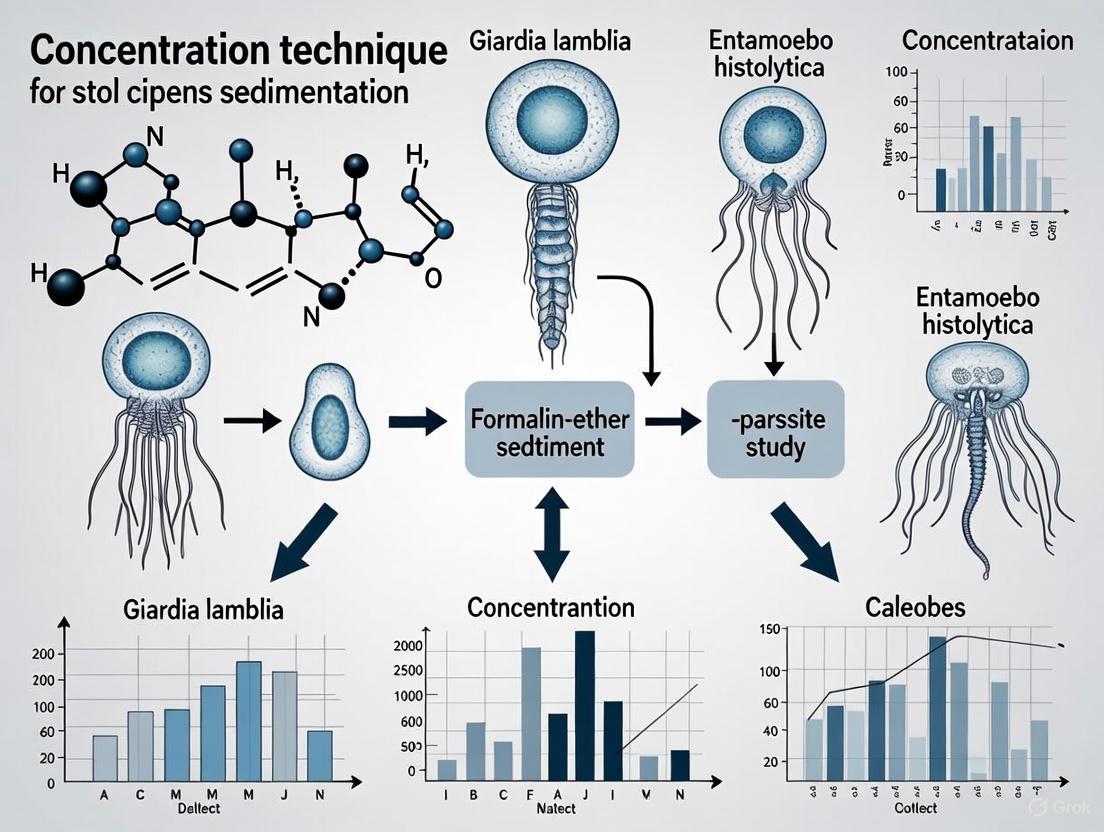
The formalin-ether concentration technique (FECT), also known as the formalin-ethyl acetate concentration technique, remains a widely used method in parasitology diagnostics due to its effectiveness in recovering parasites across different developmental stages [8] [6]. This sedimentation technique is particularly valuable for concentrating eggs, cysts, and larvae present in low numbers in fecal specimens, thereby improving detection sensitivity compared to direct smear methods [6].
The principle behind FECT involves fixing the specimen with formalin to preserve parasite morphology while using ethyl acetate or ether as an extractant to remove fats, debris, and other soluble substances that might interfere with microscopic examination [6]. A comparative evaluation of a newer fixation and concentration system (Proto-Fix with CONSED reagent) demonstrated 85% detection efficiency compared to 46% with conventional FEA-Lugol's method for identifying unknown parasite species in proficiency testing samples [8].
Detailed Protocol: Formalin-Ether Concentration Technique
Reagents and Materials
Table 3: Essential Reagents for FECT Protocol
| Reagent/Material | Specification | Function | Safety Considerations |
|---|---|---|---|
| 10% Formalin | Neutral buffered | Fixation and preservation of parasites; eliminates infectious agents | Use in well-ventilated area; avoid skin contact |
| Ethyl acetate or Diethyl ether | Laboratory grade | Extraction of fats, soluble debris, and impurities | Highly flammable; use away from ignition sources |
| Saline solution | 0.85% NaCl | Washing and suspension medium | None significant |
| Lugol's iodine | 1-2% solution | Staining for enhanced visualization of protozoan cysts | May be irritating; handle with care |
| Centrifuge tubes | Conical, 15 ml | Container for concentration procedure | Disposable recommended |
| Gauze or strainer | 2-3 layers | Filtration of large particulate matter | None significant |
| Centrifuge | Swing-out rotor capable of 500 × g | Sedimentation of parasites | Proper balance of tubes required |
| Microscope slides and coverslips | Standard (75 × 25 mm) and 22 × 22 mm | Preparation for microscopic examination | None significant |
Step-by-Step Procedure
Specimen Preparation: Emulsify approximately 1-2 g of fresh stool or preserved specimen in 10 mL of 10% formalin in a centrifuge tube. For preserved specimens, ensure adequate fixation time (at least 30 minutes).
Filtration: Filter the suspension through 2-3 layers of wet gauze or a commercial strainer into a clean centrifuge tube to remove large particulate matter.
Centrifugation: Centrifuge the filtered suspension at 500 × g for 5 minutes. Carefully decant the supernatant without disturbing the sediment.
Resuspension: Resuspend the sediment in 10 mL of 10% formalin and mix thoroughly.
Solvent Addition: Add 3-4 mL of ethyl acetate (or diethyl ether) to the suspension. Securely cap the tube and shake vigorously for 30 seconds, releasing pressure carefully if necessary.
Second Centrifugation: Recentrifuge at 500 × g for 5 minutes. This will result in four distinct layers: an ethyl acetate layer at the top, a plug of debris at the interface, a formalin layer, and the sediment at the bottom.
Separation: Loosen the debris plug from the sides of the tube using an applicator stick. Carefully decant the top three layers, leaving the sediment undisturbed.
Sediment Preparation: Mix the remaining sediment thoroughly. If excess formalin remains, add saline, centrifuge again, and decant the supernatant.
Microscopic Examination: Prepare wet mounts of the sediment with saline and Lugol's iodine. Examine systematically under 100× and 400× magnification for parasite eggs, cysts, and larvae.
Quality Control and Limitations
Quality Control Measures:
- Check reagents for expiration and contamination
- Standardize centrifugation speed and time
- Include positive control samples when possible
- Ensure proper training of personnel in parasite identification
Technical Limitations:
- FECT is suboptimal for detecting Cryptosporidium and Cyclospora, which require specialized acid-fast stains
- Protozoan trophozoites are often destroyed during the procedure
- Operator expertise significantly impacts identification accuracy
- Sensitivity may be reduced for low-burden infections
Advanced Diagnostic Approaches
While conventional techniques like FECT remain foundational, advanced diagnostic tools are increasingly important for species-specific identification, differentiation of morphologically similar parasites, and detection of low-intensity infections [1]. Molecular methods such as PCR, real-time PCR, and multiplex PCR offer enhanced sensitivity and specificity, particularly for differentiating pathogenic and non-pathogenic species like Entamoeba histolytica and E. dispar [1]. Proteomic approaches using liquid chromatography-tandem mass spectrometry (LC-MS/MS) represent cutting-edge applications for parasite biomarker discovery, though these remain primarily research tools [1].
Intestinal parasitic infections continue to pose significant global health challenges, particularly in resource-limited settings. The formalin-ether concentration technique remains a fundamental diagnostic tool in parasitology laboratories worldwide, providing a cost-effective method for concentrating and identifying intestinal parasites. However, the evolving diagnostic landscape increasingly incorporates molecular and proteomic approaches to address limitations of conventional microscopy.
Future directions in IPI diagnostics should focus on developing rapid, sensitive, and field-deployable tests that can be implemented in diverse healthcare settings. The integration of multiple diagnostic approaches, guided by clinical presentation and epidemiological context, offers the most comprehensive strategy for accurate parasite detection and identification, ultimately contributing to improved patient management and control of intestinal parasitic infections.
The Formalin-Ethyl Acetate (FEA) concentration technique, also known as the formalin-ether sedimentation method, represents a fundamental diagnostic procedure in parasitology laboratories worldwide. Within the broader context of stool specimen research, this method serves as a critical tool for enhancing the detection of intestinal parasites that would otherwise remain undetected by direct examination methods. The technique's fundamental principle leverages differential density and chemical properties to physically separate parasite elements from obscuring fecal debris, thereby concentrating the target organisms for more reliable microscopic identification. For researchers and drug development professionals, understanding the nuanced performance characteristics of FEA concentration is paramount, as detection sensitivity varies significantly based on parasite load and stool consistency [9]. Evidence suggests that current diagnostic methods may fail to detect cryptosporidiosis in many individuals, both immunocompromised and immunocompetent, highlighting the necessity for optimized protocols [9]. This application note provides a comprehensive examination of the FEA concentration technique, detailing its core principles, practical execution, and performance characteristics to standardize and improve diagnostic outcomes in research settings.
Core Principles of Parasite-Debris Separation
The FEA concentration method achieves parasite purification through a sophisticated interplay of physical and chemical separation mechanisms. The fundamental principle relies on differential density and immiscible solvent partitioning to isolate buoyant parasite elements from denser fecal contaminants.
Physico-Chemical Basis of Separation
Parasitic elements, including eggs, cysts, and larvae, possess a specific gravity intermediate between the formalin suspension medium and the organic solvent layer. The specific gravity of most parasite cysts and eggs ranges from 1.05 to 1.20, which is less than the saturated salt solutions used in flotation techniques (e.g., zinc sulphate with S.G. 1.18-1.20) but greater than water [10]. During centrifugation, this density differential causes parasite forms to sediment while lighter fecal debris is carried upward into the solvent plug. The addition of ethyl acetate (or ether in earlier formulations) serves multiple critical functions: it dissolves fecal fats and lipids that would otherwise obscure microscopic examination, acts as a detaching agent to liberate parasites trapped in fiber, and creates a sharp density gradient that enhances separation efficiency during centrifugation [10]. Formalin simultaneously fixes parasite morphology, preserving structural integrity for accurate identification while eliminating biohazard risks.
Limitations and Method Performance
Research indicates that the FEA method, while vastly superior to direct smear examination, still entails significant oocyst loss during processing. Quantitative studies have demonstrated mean oocyst recovery rates as low as 0.4% to 48.8%, depending on stool consistency, representing a substantial potential for false-negative diagnoses [9]. The detection threshold varies considerably between liquid and formed stools; for watery diarrheal specimens, a 100% detection rate requires approximately 10,000 oocysts per gram of stool, whereas formed specimens may require 50,000 to 500,000 oocysts per gram for reliable detection depending on whether immunofluorescence or acid-fast staining is employed [9]. This performance characteristic has profound implications for epidemiological studies and drug efficacy trials where accurate quantification of parasite burden is essential.
Detailed Experimental Protocol
The following section provides a comprehensive, step-by-step methodology for the FEA concentration technique, adapted from the modified Ritchie's method by Ridley and Hawgood (1956) [10].
Sample and Reagent Preparation
- Sample Collection and Preservation: Collect approximately 1 gram of fresh stool specimen. If immediate processing is not possible, preserve the sample in 10% buffered formalin (1 part stool to 3 parts formalin) or polyvinyl alcohol (PVA). For mucoid specimens, omit the filtration step to prevent loss of parasite elements trapped in the mucus [10].
- Reagent Preparation: Prepare 10% formol-saline by adding 100 mL of formalin to 900 mL of 0.85% saline solution. Ensure ethyl acetate is fresh and stored in a explosion-proof refrigerator. Have available gauze squares, centrifuge tubes, applicator sticks, and microscope slides.
Step-by-Step Procedural Workflow
- Emulsification and Fixation: Place 1 gram of feces in a disposable container and emulsify thoroughly with 7 mL of 10% formol-saline. Allow the mixture to stand for 10 minutes for fixation. For formalin-preserved stools, begin at step 2. For PVA-preserved specimens, proceed immediately to filtration without the 10-minute waiting period [10].
- Filtration and Transfer: Pour the emulsified stool through two layers of wet gauze into a 15 mL conical centrifuge tube. For mucoid specimens, skip this filtration step to prevent the loss of parasites [10].
- Solvent Addition: Add 3 mL of ethyl acetate to the filtrate in the centrifuge tube. Securely stopper the tube and shake vigorously for 30 seconds to ensure complete mixing. Exercise caution as pressure may build up within the tube.
- Centrifugation: Remove the stopper and centrifuge at 500 × g (approximately 2000 rpm for standard clinical centrifuges) for 2 minutes. Allow the tube to rest undisturbed after centrifugation; four distinct layers will form.
- Layer Separation and Examination: Loosen the debris plug (Layer 2) from the tube wall using an applicator stick. Carefully decant the top three layers (ether, debris plug, and formalin) in a single motion, leaving only the sediment. Mix the remaining sediment and prepare wet mounts using both saline and iodine for microscopic examination.
Workflow Visualization
The following diagram illustrates the key procedural stages and the layered separation outcome after centrifugation:
Diagram Title: FEA Concentration Method Workflow
Performance Data and Method Comparison
Understanding the quantitative performance characteristics of the FEA method is essential for proper interpretation of diagnostic results, particularly in research contexts requiring high sensitivity.
Detection Thresholds by Stool Consistency
Table 1: Detection Thresholds of Cryptosporidium Oocysts Using FEA Concentration
| Stool Consistency | Detection Method | Detection Threshold (oocysts/g) for 100% Detection | Reference |
|---|---|---|---|
| Watery Diarrheal Stool | Acid-fast Staining | 10,000 | [9] |
| Watery Diarrheal Stool | Immunofluorescence | 10,000 | [9] |
| Formed Stool | Acid-fast Staining | 500,000 | [9] |
| Formed Stool | Immunofluorescence | 50,000 | [9] |
Comparative Method Performance
Table 2: Comparison of Stool Concentration Techniques
| Method | Principle | Target Parasites | Advantages | Limitations |
|---|---|---|---|---|
| FEA Concentration | Sedimentation via density differential | Most eggs, cysts, larvae; preserves morphology | Fixation kills pathogens; maintains parasite size/shape | Significant oocyst loss (51.2-99.6%); explosive solvents [9] [10] |
| Zinc Sulfate Flotation | Flotation via high specific gravity solution | Most cysts and eggs; not operculated eggs | Simpler procedure; good for delicate cysts | Denser eggs (unfertilized Ascaris, Taenia) do not float [10] |
| Baermann Technique | Migration toward heat and water | Strongyloides larvae | Specific for motile larvae | Limited to specific parasites; time-consuming (1-2 hours) [10] |
| Sheather's Sugar Flotation | Flotation via high viscosity solution | Cryptosporidium oocysts | Specific for Cryptosporidium | Limited application spectrum [10] |
The Scientist's Toolkit: Essential Research Reagents
Table 3: Key Research Reagents for FEA Concentration Protocol
| Reagent/Material | Function | Research-Grade Considerations |
|---|---|---|
| 10% Buffered Formalin | Fixes parasite morphology; eliminates biohazards; preserves specimens for batch processing | Use reagent-grade formaldehyde with neutral pH buffer to prevent artifact formation |
| Ethyl Acetate | Organic solvent dissolves fecal fats; creates density gradient; liberates trapped parasites | High-purity solvent minimizes residue; less explosive than diethyl ether |
| Ethyl Acetate Alternatives | Safer solvent options with comparable efficacy | Acetone or Hemo-de (commercial clearing agent) reduce explosion risk [10] |
| Gauze Squares | Filters coarse fecal debris while allowing parasite passage | Standardized mesh size ensures consistent filtration across research batches |
| Conical Centrifuge Tubes | Facilitates layered separation during centrifugation | Graduated 15mL tubes enable standardized volumes; chemical-resistant |
| Saline and Iodine Mounts | Microscopic examination media for different visualization needs | Iodine stains internal structures; saline preserves motility if present |
Troubleshooting and Method Optimization
Even when following standardized protocols, researchers may encounter specific challenges that affect method performance. This section addresses common issues and provides evidence-based solutions.
- Low Parasite Recovery: Studies indicate oocyst loss during FEA concentration can reach 99.6% [9]. To optimize recovery, consider omitting the filtration step for mucoid specimens [10], extending centrifugation time to 3-5 minutes, and carefully resuspending the entire sediment for examination. For critical applications, combine FEA with detection methods offering higher sensitivity, such as immunofluorescence, which demonstrates lower detection thresholds than acid-fast staining, particularly in formed stools [9].
- Solvent Safety Considerations: While ether effectively separates parasites from debris, its explosive nature presents significant laboratory hazards. Ethyl acetate represents a safer alternative with comparable efficacy [10]. When ether must be used, implement strict safety protocols including explosion-proof refrigerators, chemical fume hoods, and grounded containers to prevent static discharge.
- Specimen-Specific Modifications: For formalin-preserved stools, begin processing at the filtration step since fixation has already occurred [10]. With PVA-preserved specimens, proceed immediately to filtration without the initial 10-minute waiting period to maintain optimal parasite morphology. These adjustments ensure consistent performance across different preservation methods commonly encountered in research settings.
The FEA concentration technique serves as a cornerstone method in parasitology research, with specific applications in epidemiological studies where accurate parasite prevalence data is essential, drug efficacy trials requiring sensitive detection of treatment effects, and vaccine development where precise quantification of parasite burden is critical. The method's ability to concentrate low-density infections makes it particularly valuable for surveillance studies and eradication monitoring programs.
Despite its widespread use, researchers must remain cognizant of the technique's limitations, particularly the substantial and variable oocyst loss that can impact quantitative studies [9]. The integration of FEA concentration with emerging detection technologies, including molecular methods and advanced immunofluorescence, represents a promising direction for enhancing diagnostic sensitivity in research settings. Through strict adherence to standardized protocols, careful attention to specimen-specific modifications, and understanding of performance characteristics detailed in this application note, researchers can maximize the utility of this essential technique in advancing our understanding of intestinal parasitology.
Application Note
This document provides detailed application notes and protocols for the detection of key intestinal parasites, with a specific focus on the Formalin-Ethyl Acetate (FEA) concentration technique. The information is structured to support researchers, scientists, and drug development professionals in the fields of parasitology and diagnostic development.
Intestinal parasitic infections remain a significant global health burden. The Formalin-Ethyl Acetate (FEA) sedimentation technique is a cornerstone method for concentrating parasite elements (eggs, larvae, cysts, and oocysts) from stool specimens, thereby enhancing detection sensitivity for both common protozoa and soil-transmitted helminths (STH) [10]. This protocol is vital for accurate epidemiological surveys, patient diagnosis, and the assessment of new therapeutic agents. Its principle relies on the differential density of parasitic elements compared to the fecal suspending medium, causing them to settle at the bottom of a centrifuge tube during processing [10]. The technique effectively preserves morphological details, which is crucial for species identification.
Quantitative Data on Parasite Detection
The sensitivity of diagnostic methods is a critical factor in parasitology. The table below summarizes key quantitative data on detection thresholds and prevalence for major intestinal parasites.
Table 1: Key Quantitative Data on Parasite Detection
| Parasite | Detection Method | Key Quantitative Findings | Epidemiological Context |
|---|---|---|---|
| Cryptosporidium spp. | FEA concentration + IF staining [9] | 100% detection rate in formed stool at ≥50,000 oocysts/g; 100% detection in watery stool at ≥10,000 oocysts/g. Mean oocyst loss during FEA: 51.2-99.6%. [9] | A major cause of diarrhea; incidence higher in low-income countries (5–10%) vs. developed nations (~1%). [11] |
| Giardia duodenalis | Microscopy with DFA, ELISA, qPCR [12] [13] | Sensitivity/Specificity: DFA (~91-100%/89-100%), ELISA (76.5%/68%), qPCR (89.9%/82.9%). [13] Cure rates: Tinidazole (>90%), Nitazoxanide (85%). [12] | Childhood prevalence: 20-30% in developing vs. 2-3% in developed countries. [11] |
| Soil-Transmitted Helminths (STH) | Kato-Katz technique [11] | Overall STH prevalence in Zhejiang, China (2014-15): 1.71% (Hookworm: 1.58%). Significant decline from 19.56% national prevalence in 2004. [11] | >880 million children infected yearly; ~69% of infected individuals live in Asia. [11] |
| Enterobius vermicularis (Pinworm) | Transparent adhesive paper anal swab [11] | Prevalence in rural Chinese children: 2.79%. Most common intestinal parasite in children in the U.S. [12] [11] | Not a nationally notifiable condition in the U.S.; spreads easily in group settings. [12] |
Detailed Experimental Protocols
Formalin-Ethyl Acetate (FEA) Concentration Technique
The following is the modified Ritchie's method, a standard sedimentation protocol for concentrating parasites from stool specimens [10].
Workflow Overview of Stool Parasitological Analysis
Materials and Reagents:
- Formalin (10% formol-saline)
- Ethyl Acetate (alternatives: Diethyl Ether, Acetone, Hemo-de) [10]
- Conical Centrifuge Tube
- Gauze
- Centrifuge
- Glass rod
Step-by-Step Procedure:
- Fixation: Emulsify 1-2 grams of feces in 7-10 ml of 10% formol-saline. Let it stand for 10 minutes for fixation. For PVA-preserved stool, proceed immediately to the next step. For formalin-preserved stool, skip this step. [10]
- Filtration: Pour the mixture through two layers of wet gauze into a conical centrifuge tube to remove large debris. If the stool contains excessive mucus, this step may be omitted. [10]
- Solvent Addition: Add 3-4 ml of ethyl acetate to the filtrate. Close the tube with a stopper and shake vigorously for one minute. Note: Ethyl acetate is preferred over diethyl ether as it is less explosive. [10]
- Centrifugation: Centrifuge the tube at 2000 rpm for 2 minutes. Ensure the stopper is removed before centrifugation. [10]
- Separation: After centrifugation, four distinct layers will form [10]:
- Top Layer: Ethyl acetate
- Second Layer: Plug of debris
- Third Layer: Clear formalin
- Bottom Layer: Sediment containing the concentrated parasites
- Supernatant Removal: Loosen the debris plug from the tube's side with a glass rod. Carefully decant the top three layers (supernatant).
- Examination: Use a pipette to mix the remaining sediment with the small amount of fluid that drains down. Transfer a drop of the sediment to a microscope slide, add a coverslip, and examine. Staining (e.g., Iodine, modified acid-fast) can be applied for better visualization of specific parasites.
Specialized Techniques for Specific Parasites
Sheather's Sugar Flotation for Cryptosporidium: This technique uses a high-density sugar solution to float the delicate oocysts of Cryptosporidium, which can be detected under microscopy [10].
Baermann Concentration for Strongyloides Larvae: This technique is ideal for detecting motile Strongyloides larvae in stools with low parasite burden. It uses a warm water setup that encourages larvae to migrate out of the fecal material and settle at the bottom of a funnel or tube, from where they can be collected for examination [10].
The Scientist's Toolkit: Research Reagent Solutions
Table 2: Essential Reagents and Materials for FEA Concentration
| Reagent/Material | Function in Protocol | Key Considerations |
|---|---|---|
| 10% Formol-Saline | Fixative that preserves parasite morphology and kills infectious agents. | Maintains egg/cyst structure; eliminates biohazard risk. [10] |
| Ethyl Acetate | Solvent that dissolves fecal fats and removes debris. | Preferred over ether for safety (less explosive/volatile). [10] |
| Gauze | Filters coarse fecal particles during processing. | Use two layers for effective filtration of formed stools. [10] |
| Modified Acid-Fast (AF) Stain | Stains oocysts of Cryptosporidium and Cyclospora. | Lower sensitivity compared to immunofluorescence. [9] |
| Fluorescein-tagged Monoclonal Antibody (IF) | Provides high sensitivity and specificity for Cryptosporidium and Giardia. | Recommended for highest detection rate, especially in formed stool. [9] [12] |
| Iodine Stain | Highlights internal structures of protozoan cysts (e.g., Giardia). | Used for wet mount examination of concentrated sediment. [10] |
The Role of Environmental and Socioeconomic Factors in IPI Prevalence
Intestinal parasitic infections (IPIs) remain a significant global health burden, disproportionately affecting developing nations. Recent epidemiological studies reveal a high prevalence of IPIs, with rates of 41.09% in North Wollo, Ethiopia [14] and 52.9% among schoolchildren in Debre Berhan, Ethiopia [15]. Protozoans like Entamoeba histolytica/dispar and helminths such as Ascaris lumbricoides predominated these infections [14] [15]. Key risk factors significantly associated with IPI transmission include inadequate sanitation, infrequent handwashing, untreated water sources, low socioeconomic status, and limited education [14] [15] [16]. This analysis synthesizes current prevalence data and contextualizes it within a framework utilizing the formalin-ether concentration technique for enhanced stool specimen analysis.
Intestinal parasitic infections represent a major public health challenge, particularly in resource-limited settings where environmental conditions and socioeconomic factors create ideal transmission conditions. The formalin-ether concentration (FEC) technique, referenced in current literature as a gold standard for parasite detection [14] [15], provides the methodological foundation for accurate prevalence data. Understanding the intersection of parasitic ecology with human behavior and socioeconomic determinants is essential for developing targeted interventions. This document presents application notes and experimental protocols to standardize the investigation of environmental and socioeconomic determinants of IPI prevalence within FEC-based research frameworks.
Quantitative Prevalence Data and Associated Risk Factors
Epidemiological studies across different geographical regions consistently demonstrate how environmental and socioeconomic variables influence IPI transmission rates.
Table 1: Documented IPI Prevalence Rates and Dominant Parasite Species
| Location | Population | Overall Prevalence | Predominant Parasites | Reference |
|---|---|---|---|---|
| North Wollo, Ethiopia | Health Center Patients | 41.09% (166/404) | E. histolytica/dispar (16.34%), A. lumbricoides (7.2%) | [14] |
| Debre Berhan, Ethiopia | Primary Schoolchildren | 52.9% (341/645) | A. lumbricoides (22.6%), E. histolytica/dispar/moshkovskii (18.1%) | [15] |
| Quetta, Pakistan | Rural & Urban Population | 21% | E. histolytica (48%), H. nana (26%), G. intestinalis (17%) | [16] |
Table 2: Statistically Significant Risk Factors for IPI Acquisition
| Risk Factor Category | Specific Variables | Measures of Association (AOR/OR) | Reference |
|---|---|---|---|
| Socioeconomic Factors | Mother's Illiteracy | Significant association (p<0.05) | [15] |
| Large Family Size (>9 members) | AOR=2.860, 95%CI=0.619-13.206, p=0.008 | [14] | |
| Farming Occupation | AOR=2.447, 95%CI=0.816-7.337, p=0.04 | [14] | |
| Hygiene Practices | Infrequent Handwashing | Significant association (p<0.05) | [14] [15] |
| Infrequent Shoe Wearing | Significant association (p<0.05) | [14] | |
| Unclean Fingers | Significant association (p<0.05) | [15] | |
| Environmental & Behavioral | Untreated Water Sources | Significant association (p<0.05) | [14] |
| Open Defecation Sites Near Residence | Significant association (p<0.05) | [15] | |
| Habit of Playing with Soil | Significant association (p<0.05) | [15] | |
| Raw Meat Consumption | Significant association (p<0.05) | [14] |
Experimental Protocols for FEC-Based IPI Research
Stool Specimen Collection and Macroscopic Examination
Purpose: To ensure standardized collection and initial examination of stool samples for IPI diagnosis. Procedure:
- Sample Collection: Provide participants with clean, leak-proof, labeled plastic containers with applicator sticks. Instruct them to collect approximately 5g of fresh stool (equivalent to the size of two matchsticks) [14] [15].
- Transport: Collect samples immediately and process within 30 minutes for direct smear examination. Preserve a portion in 10% formalin for concentration techniques [15].
- Macroscopic Examination: Visually inspect samples for consistency, color, presence of blood, mucus, or adult worms. Record observations in a standardized format [14]. Quality Control: Train personnel in proper sample handling. Use standardized labeling systems to maintain chain of custody.
Formal-Ether Concentration Technique (FEC)
Purpose: To concentrate parasitic elements for improved detection sensitivity, particularly in low-intensity infections. Reagents and Materials:
- 10% Formalin saline solution
- Diethyl ether
- Centrifuge tubes (15ml conical tubes)
- Centrifuge
- Applicator sticks
- Disposable pipettes
- Microscope slides and coverslips
- Lugol's iodine
Procedure:
- Specimen Preparation: Emulsify 1-2g of stool in 10% formalin in a centrifuge tube. Filter the suspension through a sieve or gauze to remove large debris [15].
- First Centrifugation: Centrifuge the filtrate at 500 × g for 10 minutes. Decant the supernatant.
- Ether Addition: Resuspend the sediment in 10% formalin, add 3-4ml of diethyl ether. Cap the tube and shake vigorously for 30 seconds.
- Second Centrifugation: Centrifuge at 500 × g for 5 minutes. Four layers will form: ether at the top, a plug of debris, formalin, and sediment at the bottom.
- Sediment Examination: Loosen the debris plug, pour off the top three layers. Mix the remaining sediment and examine microscopically with saline and iodine preparations [15]. Quality Control: Include known positive and negative samples with each batch. Calibrate centrifuge regularly.
Sociodemographic and Environmental Risk Factor Assessment
Purpose: To systematically collect data on potential IPI risk factors for correlation with laboratory findings. Procedure:
- Questionnaire Development: Develop structured questionnaires covering:
- Sociodemographics: age, gender, education, occupation, family size [14]
- Water sources and treatment methods [14]
- Sanitation facilities and waste disposal practices [14] [15]
- Handwashing habits and frequency [14] [15] [16]
- Footwear use patterns [14]
- Dietary habits including raw meat consumption [14]
- Child-specific behaviors: soil playing, finger sucking [15]
- Data Collection: Train interviewers to administer questionnaires consistently. Conduct interviews in local languages with back-translation to ensure accuracy [14].
- Environmental Observation: Use checklists to record observable factors like fingernail cleanliness, presence of feces in compound, and latrine conditions [15]. Quality Control: Pre-test questionnaires, conduct regular data quality checks, and use standardized training for all data collectors.
Visualization of IPI Risk Factor Pathways and Diagnostic Workflow
Diagram 1: IPI Risk Factor Pathway Map. This diagram illustrates the complex interplay between socioeconomic determinants, environmental conditions, and behavioral practices that facilitate intestinal parasitic infection transmission, along with potential intervention points.
Diagram 2: Integrated IPI Research Workflow. This diagram outlines the standardized operational procedure from specimen collection through data analysis, integrating laboratory diagnostics with socioeconomic and environmental assessment.
The Scientist's Toolkit: Research Reagent Solutions
Table 3: Essential Materials for FEC-Based IPI Research
| Item | Function/Specification | Application Notes |
|---|---|---|
| Formalin (10%) | Preservation of stool specimens; kills pathogens and fixes parasitic elements | Use fresh solutions; maintain proper ventilation; neutral buffered formalin preferred for morphology |
| Diethyl Ether | Lipid extraction and debris separation in FEC | Highly volatile; fire hazard; use in well-ventilated areas; store properly |
| Centrifuge | Sedimentation of parasitic elements; standard model with 500 × g capability | Calibrate regularly; use balanced loads; follow safety protocols for aerosol prevention |
| Microscope | Parasite identification; light microscope with 10x, 40x objectives | Regular maintenance; clean optics; ensure proper lighting for morphology assessment |
| Saline Solution (0.85%) | Isotonic medium for wet mount preparations | Prepare fresh daily; ensure correct pH and osmolarity to maintain parasite integrity |
| Lugol's Iodine | Stains glycogen and nuclei of protozoa; enhances visualization | Light-sensitive; store in amber bottles; fresh preparation every 2-3 weeks |
| Stool Collection Kits | Standardized containers with lids and applicators | Leak-proof; properly labeled; include instructions for participants |
| Structured Questionnaires | Systematic data collection on risk factors | Pre-tested; translated to local language; include observational checklists |
The integration of robust laboratory techniques like the formalin-ether concentration method with comprehensive environmental and socioeconomic assessment provides a powerful approach for understanding and combating intestinal parasitic infections. The high prevalence rates documented across multiple studies underscore the urgent need for targeted interventions addressing the specific risk factors identified. Future research should focus on longitudinal studies tracking how modifications in these determinants affect IPI transmission dynamics, ultimately informing more effective public health strategies for vulnerable populations.
Application Notes
The Formalin-Ethyl Acetate (FEA) concentration technique is a cornerstone diagnostic procedure for the detection of intestinal parasites in human stool specimens. Its primary function is to increase the likelihood of parasite recovery by concentrating oocysts, cysts, and helminth eggs from a stool sample into a purified sediment, thereby enhancing the sensitivity of subsequent microscopic examination [17] [9]. This method is particularly vital for detecting pathogens like Cryptosporidium spp., where sensitivity of direct examination is notoriously low [9].
Quantitative studies have demonstrated that the diagnostic sensitivity of the FEA method is significantly influenced by stool consistency. Evidence indicates that for formed stool specimens, a 100% detection rate for Cryptosporidium parvum oocysts was only achieved at a concentration of 50,000 oocysts per gram of stool when using the immunofluorescence (IF) technique post-concentration, and a staggering 500,000 oocysts per gram for the acid-fast (AF) staining technique [9]. In contrast, for watery diarrheal stool, a 100% detection rate was possible at 10,000 oocysts per gram for both AF and IF techniques, underscoring the profound impact of stool consistency on the method's performance [9]. Furthermore, studies quantifying oocyst loss through the FEA procedure have reported mean losses ranging from 51.2% to 99.6%, highlighting a critical limitation and the need for meticulous technique to maximize recovery [9].
Table 1: Threshold of Detection for Cryptosporidium Oocysts Post-FEA Concentration
| Stool Consistency | Diagnostic Technique | Detection Threshold (Oocysts/g) for 100% Detection Rate |
|---|---|---|
| Formed | Immunofluorescence (IF) | 50,000 |
| Formed | Acid-Fast Staining (AF) | 500,000 |
| Watery Diarrheal | Immunofluorescence (IF) | 10,000 |
| Watery Diarrheal | Acid-Fast Staining (AF) | 10,000 |
The choice of preservative for the initial stool sample is a critical pre-analytical variable that directly impacts the success of the FEA concentration and downstream staining. No single preservative is ideal for all situations, and their selection involves trade-offs between morphological preservation and suitability for various staining techniques or molecular assays [17].
Table 2: Common Stool Preservatives and Their Impact on FEA and Downstream Analysis
| Preservative | Key Advantages | Key Disadvantages / Compatibility Notes |
|---|---|---|
| 10% Formalin | Good for helminth eggs/larvae; suitable for concentration, immunoassays, and UV fluorescence [17]. | Inadequate for protozoan trophozoites; can interfere with PCR [17]. |
| Low-Viscosity Polyvinyl-Alcohol (LV-PVA) | Excellent for protozoan trophozoites/cysts; ideal for permanent stained smears [17]. | Contains mercuric chloride; not suitable for concentration or immunoassays [17]. |
| Sodium Acetate-Acetic Acid-Formalin (SAF) | Suitable for both concentration and permanent stains; no mercury [17]. | Requires an additive for smear adhesion; stains not as high quality as PVA [17]. |
| Merthiolate-Iodine-Formaldehyde (MIF) | Fixes and stains simultaneously; useful for field surveys [17]. | Iodine distorts protozoa and interferes with other stains [17]. |
| One-Vial Fixatives (e.g., EcoFix, Para-Safe) | No mercury; concentrate and smear can be made from one vial; suitable for most immunoassays [17]. | May require specific stains; staining consistency can be variable [17]. |
Given their complementary advantages, standard practice recommends dividing the stool specimen into two portions preserved in both 10% formalin and PVA [17].
Experimental Protocols
Protocol 1: Stool Specimen Collection and Preservation
Principle: To collect a stool sample that minimizes contamination and preserves parasite morphology for accurate FEA concentration and microscopic diagnosis [17].
Materials:
- Clean, dry, leak-proof container.
- Commercial stool collection kit (typically containing one vial of 10% formalin and one vial of PVA).
- Disposable gloves.
Procedure:
- Collect the stool specimen in the clean, leak-proof container. Take care to ensure no urine, water, or soil contaminates the sample [17].
- Using applicator sticks, transfer a portion of the stool (approximately 1-2 g or 1-2 mL if liquid) into the vial containing 10% formalin. The recommended ratio is one volume of stool to three volumes of preservative [17].
- Thoroughly mix the stool and formalin to ensure complete fixation and to break up formed stool [17].
- Repeat steps 2-3, transferring an equal portion of stool into the vial containing PVA preservative and mixing thoroughly [17].
- Seal the vials securely. It is recommended to reinforce the seal with parafilm and place the vials in a plastic bag for transport [17].
- Label all containers clearly with patient identifiers.
Notes:
- Specimens should be preserved as soon as possible after passage. If preservatives are unavailable, the unpreserved specimen must be refrigerated and is suitable only for antigen testing, not for FEA concentration or stained smears [17].
- Numerous substances can interfere with microscopic examination. Specimen collection should occur before, or be delayed until after the effects of the following have passed: barium or bismuth (7-10 days), antimicrobial agents (2-3 weeks), antacids, kaolin, mineral oil, and non-absorbable antidiarrheal preparations [17].
Protocol 2: Formalin-Ethyl Acetate (FEA) Concentration Sedimentation Technique
Principle: To concentrate parasites by filtering out large debris, dissolving fats with ethyl acetate, and concentrating cysts, oocysts, and helminth eggs into a sediment via centrifugation [9].
Materials:
- Preserved stool sample in 10% formalin.
- Conical centrifuge tubes (15 mL).
- Gauze or a single-use filtration device.
- Ethyl Acetate.
- Centrifuge.
- Saline or 10% formalin.
- Applicator sticks.
- Disposable pipettes.
- Microscope slides and coverslips.
Procedure:
- For preserved samples, thoroughly mix the vial. Transfer approximately 3-5 mL of the formalin-stool mixture into a 15 mL conical centrifuge tube. If dealing with an unpreserved sample, emulsify 1-2 g of stool in 10 mL of saline or 10% formalin [9].
- Filter the suspension through two layers of moistened gauze or a single-use filtration device into a new 15 mL centrifuge tube to remove large particulate matter.
- Add saline or 10% formalin to the filtered suspension to bring the total volume to 10-12 mL.
- Centrifuge at 500 × g for 10 minutes. Carefully decant the supernatant.
- Resuspend the sediment in the tube by adding 10 mL of saline or 10% formalin.
- Add 3-4 mL of ethyl acetate to the tube. Cap the tube tightly and shake it vigorously for 30 seconds. Ensure to vent the tube periodically to release pressure.
- Centrifuge again at 500 × g for 10 minutes. This will result in four distinct layers:
- A layer of ethyl acetate at the top.
- A plug of debris.
- A layer of formalin/saline.
- A sediment of parasites at the very bottom of the tube.
- Loosen the debris plug by ringing it with an applicator stick. Carefully decant the top three layers (ethyl acetate, debris plug, and formalin/saline), leaving the sediment undisturbed.
- Using a disposable pipette, transfer a drop of the remaining sediment onto a microscope slide, add a coverslip, and examine microscopically. If the sediment is thick, a drop of iodine can be added for wet mount examination.
Protocol 3: Microscopic Examination and Staining of FEA Sediment
Principle: To identify parasites in the FEA concentrate using brightfield microscopy and specialized stains to enhance morphological features [9].
Materials:
- FEA sediment.
- Microscope slides and coverslips.
- Iodine solution.
- Staining reagents (e.g., Kinyoun's Carbol Fuchsin for acid-fast staining, or fluorescent antibodies for IF).
- Brightfield and/or fluorescence microscope.
Procedure: A. Wet Mount Examination
- Place a drop of saline on one end of a microscope slide and a drop of iodine solution on the other.
- Using an applicator stick, transfer a small amount of the FEA sediment to each drop and mix. Apply coverslips.
- Systematically scan the entire area under the coverslip using the 10x objective to locate potential parasites. Switch to the 40x objective for detailed morphological examination. Saline wet mounts are best for observing motility (in fresh samples) and the internal structures of cysts. Iodine wet mounts stain glycogen and nuclei, aiding in cyst identification.
B. Modified Acid-Fast Staining (for Cryptosporidium, Cyclospora, Cystoisospora)
- Prepare a thin smear of the FEA sediment on a microscope slide and allow it to air dry.
- Flood the slide with Kinyoun's Carbol Fuchsin stain and allow it to stain for 5-10 minutes.
- Rinse briefly with tap water and decolorize with 1% acid-alcohol for 30 seconds or until the stain no longer runs off the slide.
- Rinse with tap water.
- Flood the slide with a counterstain (e.g., Methylene Blue or Malachite Green) for 1 minute.
- Rinse with tap water, air dry, and examine under oil immersion (100x objective). Acid-fast organisms will stain a bright pinkish-red, while background material and non-acid-fast organisms will stain the color of the counterstain [9].
Workflow and Signaling Pathway Diagrams
The Scientist's Toolkit: Research Reagent Solutions
Table 3: Essential Materials for FEA Concentration and Analysis
| Item | Function / Application Note |
|---|---|
| 10% Formalin Solution | All-purpose fixative for helminth eggs, larvae, and protozoan cysts. Suitable for FEA concentration and immunoassays [17]. |
| LV-PVA Preservative | Provides excellent morphological preservation of protozoan trophozoites and cysts for permanent stained smears (e.g., Trichrome) [17]. |
| Ethyl Acetate | Organic solvent used in the FEA concentration technique to dissolve fats and debris, forming a plug for easy removal during sedimentation [9]. |
| Kinyoun's Carbol Fuchsin | Primary stain in the modified acid-fast staining procedure; identifies Cryptosporidium, Cyclospora, and Cystoisospora [9]. |
| Fluorescein-Tagged Antibodies | Monoclonal antibodies used in the Immunofluorescence (IF) technique for highly sensitive and specific detection of target parasites like Giardia and Cryptosporidium [9]. |
| Saline and Iodine Solution | Used for the preparation of wet mounts from the FEA sediment for preliminary microscopic examination and cyst identification [17]. |
Step-by-Step FEA Protocol: Standard and Modified Techniques for Maximum Yield
Essential Reagents and Equipment for the FEA Concentration Technique
Within the context of stool specimen research, the Finite Element Analysis (FEA) concentration technique represents a computational approach to optimize the design and performance of equipment used in fecal sample processing. This protocol details the essential reagents and equipment, framed not for the direct analysis of stool, but for employing FEA to enhance the mechanical components—such as centrifuges, mixers, and chambers—involved in concentration procedures like the formalin-ethyl acetate sedimentation method [18]. By simulating physical effects, FEA predicts how materials will react to forces, fluid flow, and other physical effects, enabling the design of more durable and efficient laboratory equipment [19]. This application is critical for improving diagnostic accuracy and reliability in microbial community analysis [20] [21].
The Scientist's Toolkit: Essential Reagents & Equipment for FEA
The virtual prototyping of stool concentration equipment requires a specific suite of software tools and computational resources. The table below summarizes the key solutions for conducting FEA in this field.
Table 1: Key Research Reagent Solutions for FEA in Equipment Design
| Solution Category | Specific Examples | Function in FEA |
|---|---|---|
| FEA Software Platforms | ANSYS, ABAQUS, NASTRAN, LS-Dyna [19] [22] | Core software used to create virtual models, apply loads and constraints, and solve complex mathematical equations to predict structural and fluid behavior. |
| Computer-Aided Design (CAD) Software | Solidworks, Catia, NX [23] | Used to create the precise 3D geometry of the equipment (e.g., centrifuge rotors, mixer impellers) which is then imported into FEA software. |
| Computational Fluid Dynamics (CFD) Software | Ansys Fluent, StarCCM+, MSC Cradle [22] | Used to simulate fluid flow within mixing vessels or other components, providing load data for subsequent structural-mechanical FEA. |
Core Equipment for Virtual Analysis
The physical hardware and ancillary tools form the backbone of the computational analysis process.
- High-Performance Computing (HPC) Workstations: FEA and CFD simulations involve millions of complex calculations. Powerful computers with significant processing power (CPU), memory (RAM), and graphics capabilities (GPU) are essential for solving these models in a reasonable time [23].
- Pre-Processing Tools: The process begins with creating a mesh, which discretizes the complex CAD geometry into small, simple elements. The quality and refinement of this mesh are critical for result accuracy [23].
Experimental Protocols: Key FEA Methodologies
This section outlines detailed methodologies for fundamental types of FEA that are critical for analyzing and optimizing equipment used in stool concentration.
Protocol 1: Static Structural Analysis
This analysis establishes the static deformation and stiffness of a structure under a constant load, such as the force on a centrifuge arm [24].
1. Geometry Creation: Develop a high-fidelity 3D model of the component (e.g., a spindle or rotor) using CAD software [24]. 2. Material Property Assignment: Define the material properties for all parts, including Young's modulus (stiffness), Poisson's ratio, and density [23]. 3. Meshing: Generate a finite element mesh. A convergence analysis should be performed by refining the mesh until stress values stabilize, ensuring result accuracy [25]. 4. Applying Loads and Boundary Conditions: Apply the realistic physical constraints (e.g., fixed supports where the part is bolted down) and operational loads (e.g., 5000 N force on a spindle nose) [24]. 5. Solving: The FEA software solves the underlying equations to compute stresses and deformations. 6. Post-Processing and Verification: Analyze results using contour plots of stress (e.g., von Mises stress) and deformation. Experimentally validate the FEA results with physical tests to verify the model's accuracy; an error of less than 1.56% is achievable [24].
Protocol 2: Modal Analysis
This analysis determines the inherent vibration characteristics (natural frequencies and mode shapes) of a structure to avoid resonant failure [24].
1. Model Setup: Utilize the same geometry and material properties from the static analysis, as these directly influence dynamic behavior. 2. Defining Contact Conditions: Specify how different parts interact (e.g., bonded, sliding). 3. Solving for Mode Shapes: The solver computes the first several mode shapes and their corresponding frequencies (e.g., the first four modes at 22.5, 28.9, 40.6, and 47.4 Hz for a machine tool) [24]. 4. Interpretation: Identify the weak links in the structure by analyzing the mode shapes. If operational frequencies (e.g., from an imbalanced centrifuge) coincide with these natural frequencies, design changes are necessary to shift them.
Protocol 3: Topology Optimization
This is an advanced structural design protocol used to achieve a lightweight, rigid, and efficient design [24].
1. Defining the Design Space: Specify the entire volume of material that the final part can occupy. 2. Applying Loads and Constraints: Input all the operational load cases and boundary conditions from the static and modal analyses. 3. Setting Optimization Goals and Constraints: Typically, the goal is to minimize mass or maximize stiffness, while constraining the maximum allowable stress or deformation [24]. 4. Running the Optimization Solver: The software iteratively removes non-critical material to generate an ideal, often organic-looking, structure. 5. Interpreting and Redesigning: The optimized shape serves as a guide for redesigning the component, potentially achieving over 16% reduction in static deformation and 3% reduction in weight [24].
Methodological Considerations in Stool Processing & FEA
The laboratory methods for stool concentration provide the real-world physical requirements that inform the FEA simulations.
The Role of Bead-Beating and Homogenization
In stool processing, mechanical disruption through bead-beating is a critical step for the proper lysis of Gram-positive bacterial cell walls [20]. Without it, the detection of key genera like Blautia and Bifidobacterium can be significantly reduced, biasing microbial community analysis [20]. From an FEA perspective, this translates into simulating mixers or homogenizers that can generate sufficient force and ensure consistent processing. Homogenization of the entire stool sample before aliquoting is also crucial, as it significantly reduces intra-individual variation and ensures a representative sample is taken, despite being an unpleasant task [21].
Addressing Stress Concentrations in Design
Stress concentrations are regions of high stress caused by geometric discontinuities like sharp corners or holes, which can initiate failure [25]. They are a key challenge in FEA.
- Identification: Use contour plots and stress gradients from FEA results to locate these areas [25].
- Mitigation: Modify the geometry by adding fillets, rounding sharp corners, or increasing hole diameters. Convergence analysis (mesh refinement) and experimental validation are essential to verify that the stress values are accurate and not a numerical artifact [25].
Workflow Visualization
The following diagram illustrates the integrated workflow for applying FEA to the development of stool concentration equipment, connecting computational analysis with laboratory validation.
FEA-Driven Equipment Development Workflow
The workflow demonstrates the iterative cycle of designing equipment virtually with FEA, fabricating a prototype, and validating its performance against the rigorous demands of established stool processing protocols [18] [24] [21]. This ensures the final product is both mechanically robust and fit-for-purpose in a diagnostic or research setting.
The FEA concentration technique, as applied to the development of stool processing equipment, relies on a sophisticated toolkit of software and rigorous protocols for static, modal, and optimization analysis. By integrating these computational methods, engineers can create optimized, reliable, and efficient devices that directly support the standardized and accurate analysis of the human gut microbiome, ultimately advancing research and drug development. The synergy between virtual prototyping and laboratory validation is key to producing high-quality scientific outcomes.
The Formalin-Ethyl Acetate (FEA) concentration technique is a standardized parasitological method used for the detection of parasite eggs in human and animal feces. This procedure is a cornerstone in clinical diagnostics and research, enabling the purification and microscopic identification of parasitic elements. The method operates on the principle of differential density separation, where fecal debris is separated from parasite eggs through a series of chemical treatments and centrifugation steps. The use of the organic solvent ethyl acetate is critical, as it acts as an extractive agent for fecal fats and oils, resulting in a cleaner sediment background for subsequent microscopic analysis [26]. This protocol outlines a detailed, step-by-step procedure for performing the FEA concentration technique, providing researchers and laboratory professionals with a standardized framework to ensure diagnostic consistency, reproducibility, and accuracy in stool specimens research.
Principle of the Method
The FEA technique enhances the detection of parasite eggs by concentrating them from a larger stool sample into a purified sediment. The process involves fixation, emulsification, filtration, and density-based separation. 10% formalin serves a dual purpose: it preserves the morphological integrity of protozoan cysts, helminth eggs, and larvae, and it fixes the stool specimen [17]. Ethyl acetate is the key organic solvent that dissolves and extracts non-parasitic fecal components, particularly fats and oils, which interfere with microscopic examination. During centrifugation, a density gradient is established. Parasite eggs, being denser, pellet at the bottom of the tube, while dissolved debris remains suspended in the ethyl acetate and formalin layers above, which are subsequently discarded. This process yields a sediment enriched with parasite eggs, facilitating their identification under a microscope [26].
Materials and Equipment
Research Reagent Solutions
The following table details the essential materials and reagents required to perform the FEA concentration technique.
Table 1: Essential Reagents and Materials for the FEA Concentration Technique
| Item | Function/Description |
|---|---|
| 10% Aqueous Formalin | Primary fixative and preservative; maintains the morphology of parasites [17]. |
| Ethyl Acetate (EA) | Organic solvent for extraction of fecal fats, oils, and debris; crucial for achieving a clean background [26]. |
| Saline (0.9% Sodium Chloride) | Isotonic solution used for diluting and suspending stool samples. |
| Sieve or Gauze | For filtering coarse, particulate fecal matter from the sample suspension. |
| Centrifuge Tubes (Conical, 15 ml) | Tubes for sample processing and centrifugation. |
| Centrifuge | For density-based separation of parasite eggs from fecal debris. |
| Applicator Sticks | For handling and suspending stool samples. |
| Disposable Pipettes | For transferring supernatants and adding reagents. |
| Microscope Slides and Coverslips | For preparing specimens for microscopic examination. |
Specialized Solutions and Additives
Recent research has investigated improvements to the standard FEA method. The addition of small amounts of acid, such as hydrochloric acid (HCl) or acetic acid, can be considered in cases where fecal bulk fails to float adequately after EA treatment. The acid helps dissolve cellulose fibers from undigested plant matter in the stool, which can trap oil droplets and prevent effective separation, thereby improving the efficacy of the bulk float and egg recovery [26].
Step-by-Step Experimental Protocol
Specimen Collection and Preparation
1. Collection: Collect fresh stool in a clean, dry, leak-proof container. Take extreme care to avoid contamination with urine, water, soil, or disinfectants, as these can destroy parasitic elements and render the specimen unsatisfactory [17] [27]. 2. Preservation: If immediate processing is not possible, preserve the specimen. For the FEA method, fixation in 10% formalin is standard. Mix one volume of stool with three volumes of 10% formalin to ensure adequate preservation [17]. 3. Timing: For optimal bacterial culture from stool, collect specimens from patients who have had diarrhea for less than four days and have not received antibiotics to ensure a high bacterial load [27]. For parasitological examination, collection may need to be repeated if the first examination is negative; ideally, three specimens passed at intervals of 2-3 days should be examined [17].
The workflow below summarizes the key stages of the FEA concentration protocol.
Detailed FEA Concentration Procedure
- Emulsify and Filter: Transfer approximately 1-2 grams of fresh or formalin-preserved stool into a 15 ml conical centrifuge tube. Add 10 ml of 10% formalin if the sample is unpreserved, or saline if already formalin-fixed. Emulsify the stool completely using an applicator stick. Pour the suspension through a sieve or gauze into another container to remove large, coarse debris.
- First Centrifugation: Transfer the filtered suspension back to a 15 ml centrifuge tube. Centrifuge at 500 x g for 10 minutes. After centrifugation, carefully decant and discard the supernatant.
- Solvent Addition: Resuspend the sediment in the tube with 5-10 ml of saline (or fresh 10% formalin). Add 4-5 ml of ethyl acetate to the suspension. Securely cap the tube and shake it vigorously for at least 30 seconds. Ensure the tube is vented periodically to release pressure buildup from the volatile ethyl acetate.
- Second Centrifugation: Recentrifuge the tube at 500 x g for 10 minutes. This step creates four distinct layers:
- Top Layer: Ethyl acetate with dissolved fecal fats and oils.
- Plug: Debris at the interface of the solvent and formalin.
- Middle Layer: Formalin.
- Bottom Layer: Sediment containing the concentrated parasite eggs.
- Sediment Recovery: Loosen the debris "plug" from the tube walls by running an applicator stick along the inside. Carefully decant the top three layers (ethyl acetate, debris plug, and formalin) without disturbing the sediment pellet at the bottom. A small amount of fluid may remain; allow the tube to drain upside down on a paper towel for 30-60 seconds.
- Microscopic Examination: Mix the remaining sediment with the fluid that drains back. Using a disposable pipette, transfer a drop of the sediment to a clean microscope slide, apply a coverslip, and systematically examine the entire area under the coverslip using the 10x and 40x objectives. Low light adjustment is often necessary to visualize translucent parasite eggs.
Troubleshooting and Quality Control
Despite being a standardized technique, the FEA method can present challenges that require troubleshooting.
- Problem: Incomplete Flotation of Fecal Bulk. Some samples, particularly those with high cellulose content from undigested plant matter, may not separate cleanly. The cellulose fibers can trap oil droplets, preventing bulk float.
- Solution: As demonstrated in recent studies, the addition of a small volume of acid (e.g., HCl or acetic acid) to the sediment resuspension step can help dissolve the cellulose structure, improving the efficacy of the ethyl acetate extraction and promoting bulk float [26].
- Problem: Unsatisfactory Specimen.
- Preventative Measures: Ensure patients avoid certain substances before specimen collection, as they can interfere with analysis. These include: barium or bismuth (wait 7-10 days), antimicrobial agents (wait 2-3 weeks), mineral oil, kaolin, and non-absorbable antidiarrheal preparations [17].
- Problem: Low Diagnostic Yield.
- Solution: A single negative examination does not rule out a parasitic infection. If clinical suspicion remains high, repeat the FEA concentration on two more specimens collected 2-3 days apart [17].
Data Interpretation and Reporting
Accurate reporting of FEA studies in biomedical research requires meticulous documentation to ensure reproducibility and reliability.
- Microscopic Findings: Report the identification of any parasite eggs, larvae, or cysts. Note the relative abundance if semi-quantitative reporting is used.
- Methodology Reporting: Clearly state the specific concentration technique used (e.g., Formal-Ethyl Acetate Sedimentation), including details on centrifugation speed and time, reagents, and any modifications (e.g., acid addition) [26].
- Specimen Quality: Note the quality of the specimen and the resulting sediment, as this impacts the confidence of a negative result.
The following table outlines key reporting parameters to enhance the quality and transparency of FEA studies in biomechanics and related fields, which can be analogously applied to stool specimen research methodology.
Table 2: Key Reporting Considerations for FEA-based Research Studies
| Reporting Category | Specific Parameters to Document |
|---|---|
| Model/Method Identification | Detailed protocol description, including modifications to standard methods. |
| Material & Reagent Specification | Source and concentration of all reagents (e.g., formalin, ethyl acetate). |
| Process & Simulation Parameters | Centrifuge specifications (g-force, time), number of washes, and sample volume. |
| Verification & Validation | Steps taken to ensure methodological accuracy, such as comparison with known positive controls or other diagnostic methods. |
| Result Interpretation | Clear description of findings, limitations of the method, and confidence in negative results. |
| Data Availability | Willingness to share raw data or detailed protocols upon request to facilitate peer review and replication. |
Modified FEA Techniques for Challenging Pathogens like Cryptosporidium
Cryptosporidium represents a significant diagnostic challenge in clinical parasitology due to the low infectious dose and suboptimal sensitivity of many conventional diagnostic methods. The Formalin-Ethyl Acetate (FEA) concentration technique serves as a fundamental tool in diagnostic laboratories for detecting Cryptosporidium oocysts in stool specimens. This protocol details modified FEA approaches designed to enhance recovery and detection of challenging pathogens, particularly Cryptosporidium species, within the broader context of stool specimen research. These optimized methods address the critical need for improved diagnostic sensitivity in both clinical and research settings, especially for immunocompromised patients where accurate detection can significantly impact treatment outcomes.
Comparative Performance of Diagnostic Methods
Table 1: Quantitative Comparison of Cryptosporidium Detection Techniques
| Method | Sensitivity Range | Detection Limit (Oocysts/g) | Key Advantages | Noted Limitations |
|---|---|---|---|---|
| FEA/MZN Staining [28] [29] | 71.4% [29] | 10,000 (Watery Stool); 50,000 (Formed Stool) [28] | Highest diagnostic performance; effective for asymptomatic cases [29] | Significant oocyst loss (51.2-99.6%) during concentration [28] |
| FEA/IF Staining [28] | Not explicitly quantified | 10,000 (Watery Stool); 50,000 (Formed Stool) [28] | Higher detection rate in formed stools vs. AF staining [28] | Requires fluorescent microscope |
| Acid-Fast (AF) Staining [28] | Lower than IF [28] | 10,000 (Watery Stool); 500,000 (Formed Stool) [28] | Widely available; low cost | Poor sensitivity in formed stools [28] |
| ELISA [29] | 42.9% [29] | Varies by kit | High-throughput; minimal technical expertise | Moderate sensitivity and NPV (96%) [29] |
| Percoll/MZN [29] | 14.29% [29] | Not specified | Purifies intact/viable oocysts [30] | Lowest sensitivity and NPV (93.9%) in diagnostics [29] |
| Conventional PCR [30] | Most effective [30] | Not specified | High sensitivity and specificity | Requires specialized lab facilities |
| Carbol Fuchsin Staining [30] | Comparable to Ziehl-Neelsen [30] | Not specified | Standard method; comparable to Ziehl-Neelsen [30] | Moderate sensitivity |
Table 2: Comparison of Oocyst Purification Methods
| Method | Recovery Efficacy | Purification Quality | Reduction of Degenerated Oocysts | Primary Use |
|---|---|---|---|---|
| Sodium Chloride Flotation [30] | Superior | Superior | Superior | Routine purification |
| Sheather's Sucrose Solution [30] | Not specified | Inferior to NaCl | Not specified | Routine purification |
| Percoll Gradient Centrifugation [30] | Inferior to NaCl | Inferior to NaCl | Inferior to NaCl | Research: separation of viable/degenerated oocysts [30] |
Detailed Experimental Protocols
Modified FEA Concentration Technique
- Objective: To concentrate Cryptosporidium oocysts from human stool specimens for microscopic detection, maximizing recovery and subsequent diagnostic sensitivity.
- Principle: Formalin fixes the stool specimen and preserves parasite morphology, while ethyl acetate acts as a lipid solvent and extraction medium, separating parasitic elements from stool debris.
- Reagents:
- 10% Formalin (v/v)
- Ethyl Acetate
- Phosphate-Buffered Saline (PBS), pH 7.2
- Equipment:
- Centrifuge with swing-bucket rotor
- Conical centrifuge tubes (15 mL)
- Disposable applicator sticks
- Pipettes
- Biological safety cabinet
- Procedure:
- Specimen Preparation: Emulsify approximately 1 g of fresh or preserved stool in 10 mL of 10% formalin in a centrifuge tube. Filter the suspension through gauze or a sieve to remove large particulate matter.
- Initial Centrifugation: Centrifuge the filtered suspension at 500 × g for 10 minutes. Carefully decant the supernatant.
- Ethyl Acetate Extraction: Resuspend the sediment in 5-7 mL of 10% formalin. Add 3-4 mL of ethyl acetate. Securely cap the tube and shake it vigorously for 30 seconds. Vent the tube carefully to release pressure.
- Second Centrifugation: Centrifuge at 500 × g for 5 minutes. This will result in four distinct layers: an ethyl acetate plug at the top, a plug of debris beneath it, a formalin layer, and the sediment at the bottom.
- Sediment Collection: Loosen the debris plug by ringing it with an applicator stick. Carefully decant the top three layers. The final sediment contains the concentrated parasitic elements, including Cryptosporidium oocysts.
- Wash: Resuspend the sediment in a small volume of PBS or formalin for downstream processing.
- Reagents:
Immunofluorescence (IF) Staining for Detection
- Objective: To specifically identify Cryptosporidium oocysts in FEA concentrates using fluorescein-tagged monoclonal antibodies.
- Principle: Fluorescein-labeled monoclonal antibodies bind specifically to surface antigens on Cryptosporidium oocysts, enabling highly sensitive and specific detection under a fluorescence microscope.
- Reagents:
- Fluorescein-tagged anti-Cryptosporidium monoclonal antibody
- Mounting medium
- PBS, pH 7.2
- Equipment:
- Fluorescence microscope with appropriate filter set
- Glass slides and coverslips
- 37°C incubator (optional)
- Procedure:
- Apply 10-50 µL of the FEA-concentrated sediment to a glass slide and allow it to air dry.
- Fix the smear with absolute methanol for 5 minutes. Allow to air dry.
- Add an appropriate volume of the fluorescent antibody preparation to cover the smear entirely.
- Incubate the slide in a humidified chamber at 37°C for 30 minutes.
- Rinse the slide gently with PBS, then immerse in a PBS bath for 10 minutes to remove unbound antibody.
- Mount the slide with a coverslip using mounting medium.
- Examine using a fluorescence microscope. Cryptosporidium oocysts will appear as bright, apple-green spherical structures against a dark background.
- Reagents:
Modified Ziehl-Neelsen (MZN) Staining for Detection
- Objective: To detect acid-fast Cryptosporidium oocysts in FEA concentrates using a carbol fuchsin-based stain.
- Principle: The mycolic acid in the oocyst wall binds to carbol fuchsin and resists decolorization with acid-alcohol, resulting in red-stained oocysts against a blue or green counterstained background.
- Reagents:
- Strong carbol fuchsin solution
- Acid-Alcohol (3% HCl in 95% Ethanol)
- Methylene Blue or Malachite Green counterstain
- Equipment:
- Light microscope
- Glass slides and coverslips
- Slide warmer or Bunsen burner
- Procedure:
- Prepare a thin smear from the FEA sediment on a glass slide and allow it to air dry. Heat-fix the smear.
- Flood the slide with strong carbol fuchsin and heat gently until steam rises for 5-10 minutes. Do not boil. Alternatively, stain without heating for 20-30 minutes.
- Rinse the slide briefly with tap water.
- Decolorize with acid-alcohol until the stain no longer runs from the smear.
- Rinse thoroughly with tap water.
- Apply a counterstain (e.g., Methylene Blue) for 1 minute.
- Rinse, air dry, and examine under oil immersion (1000x magnification). Cryptosporidium oocysts stain bright red, and background material stains blue. Non-acid fast "ghost" oocysts may also be visible [29].
- Reagents:
Workflow Visualization
The Scientist's Toolkit: Research Reagent Solutions
Table 3: Essential Research Reagents and Materials
| Item | Function/Application | Key Characteristics |
|---|---|---|
| 10% Formalin | Specimen preservation and fixation in FEA concentration [28] [29] | Maintains parasite morphology; inactivates pathogens |
| Ethyl Acetate | Lipid solvent and extraction fluid in FEA concentration [28] [29] | Separates parasitic elements from stool debris |
| Fluorescein-Tagged Monoclonal Antibody | Specific detection of oocysts in IF staining [28] | High specificity for Cryptosporidium surface antigens; requires fluorescence microscope |
| Carbol Fuchsin Stain | Primary stain in Modified Ziehl-Neelsen (MZN) method [30] [29] | Binds to acid-fast oocyst wall |
| Sodium Hypochlorite Solution | Excystation and surface sterilization of oocysts for cell culture [31] | ~20% concentration used to treat oocysts prior to infection studies [31] |
| Percoll | Silica colloid for density gradient centrifugation [30] | Used to separate intact/viable oocysts from degenerated forms for research [30] |
| Sheather's Sucrose Solution | High-density flotation medium for oocyst purification [30] | Alternative to sodium chloride flotation |
| Cell Culture Media (e.g., DMEM F-12) | Maintenance of host cell lines (e.g., HCT-8) for in vitro infection models [31] | Supports growth of intestinal epithelial cells for host-parasite interaction studies |
Macroscopic and Microscopic Examination of FEA-Concentrated Sediment
The Formalin-Ethyl Acetate (FEA) concentration technique is a cornerstone sedimentation method for the detection of intestinal parasites in fecal specimens. Its principle is based on the differential specific gravity between parasitic elements (ova, cysts, larvae) and fecal debris, using formalin as a fixative and preservative and ethyl acetate as an extractor of fats and debris [18] [32]. This process concentrates the parasites in the sediment, significantly enhancing detection sensitivity compared to direct smear examination [33]. The subsequent macroscopic and microscopic examination of the resulting sediment is a critical step, providing vital diagnostic information for researchers and clinicians in the field of parasitology and drug development. This protocol details the standardized procedures for this examination, contextualized within ongoing research to optimize the FEA technique.
Macroscopic Examination of FEA-Concentrated Sediment
The initial visual inspection of the concentrated sediment provides immediate, valuable data regarding the sample's characteristics.
2.1. Procedural Workflow for Macroscopic Assessment
The following diagram outlines the key decision points and observations in the macroscopic examination workflow.
2.2. Key Macroscopic Parameters and Their Significance
After concentration, the sediment should be assessed for the following features:
- Color and Appearance: The color can indicate various physiological and pathological states. Normal sediment is typically brown. Unusual colors, such as pale or clay-colored sediment, may suggest biliary obstruction, while a reddish hue can indicate lower gastrointestinal bleeding [32].
- Consistency: The physical nature of the original stool sample influences the types of parasites present. Formed stools are more likely to contain cyst forms, whereas liquid or soft stools are associated with trophozoites and are more likely to contain oocysts of parasites like Cryptosporidium [34] [18]. The consistency of the sediment itself can reflect the original stool's fat content or the presence of excess mucus.
- Presence of Adult Parasites: Although less common post-concentration, visible structures like tapeworm proglottids or adult pinworms may sometimes be found in or beneath the sediment [34]. Their presence provides a definitive diagnosis.
Microscopic Examination of FEA-Concentrated Sediment
Microscopy is the definitive step for identifying parasitic elements. A systematic approach using different preparations is essential for accurate diagnosis.
3.1. Comprehensive Microscopic Workflow
The microscopic examination involves a multi-step procedure to ensure all parasitic forms are identified. The workflow is summarized in the following diagram.
3.2. Detailed Wet Mount Preparation and Examination
Saline Wet Mount:
- Place one drop of 0.85% saline on a clean glass slide [32].
- Using an applicator stick, emulsify a small portion (approximately 2 mg or 1-2 drops) of the FEA-concentrated sediment in the saline [34] [18].
- Apply a 22mm x 22mm coverslip.
- Purpose: This mount is used primarily for the initial detection of helminth eggs, larvae, and protozoan cysts based on their size, shape, and refractility. It can also reveal motile organisms in unpreserved specimens and allows for the detection of other structures like red blood cells (RBCs) and white blood cells (WBCs) [34] [32].
Iodine Wet Mount:
- Place one drop of Lugol's iodine solution on a second glass slide.
- Emulsify the same amount of sediment in the iodine and apply a coverslip.
- Purpose: Iodine stains the glycogen vacuoles and nuclei of protozoan cysts, providing critical detail for species differentiation [34] [32]. It does not enhance the visualization of helminth eggs and renders trophozoites non-motile.
3.3. Systematic Microscopic Scanning
- Initially scan the entire wet mount under the 10x objective with low light intensity to locate suspicious objects [34].
- Switch to the 40x objective for detailed observation of morphology. The use of an oil immersion objective (100x) on wet mounts is generally not recommended unless the coverslip is sealed [34].
- Examine at least 15 seconds per field to detect slow motility in fresh specimens [34].
3.4. Key Microscopic Findings in FEA Sediment
The concentrated sediment may reveal a variety of diagnostic structures, as cataloged below.
| Category | Element | Key Identifying Features in Sediment | Research & Diagnostic Significance |
|---|---|---|---|
| Protozoa | Giardia lamblia cyst | Oval, refractile wall, axostyle, 2-4 nuclei [34] | Common cause of waterborne diarrheal disease; drug efficacy studies. |
| Entamoeba histolytica cyst | Spherical, 1-4 nuclei, fine chromatin, rounded chromatoid bodies [34] | Target for vaccine and drug development; requires differentiation from non-pathogenic amoebae. | |
| Blastocystis hominis | Central body form, vacuolated appearance; may be deformed by water [34] [18] | Role in human disease and gut microbiome is a key research area. | |
| Coccidia | Cryptosporidium oocyst | Small (4-6μm), spherical, may be difficult to see without special stains [35] | Important in immunocompromised populations; improved FEA techniques enhance detection [36]. |
| Helminths | Ascaris lumbricoides egg | Thick, mammillated coat (fertile) or elongated without coating (infertile) [34] | Soil-transmitted helminth; monitoring mass drug administration programs. |
| Hookworm egg | Thin-walled, oval, often in cleavage stages [33] | ||
| Hymenolepis nana egg | Oval, thin outer and inner membranes, 6 hooks in oncosphere [33] | ||
| Other Findings | Red Blood Cells (RBCs) | Suggest ulceration or hemorrhagic problems [34] | Indicates tissue invasion, e.g., in amebic dysentery. |
| White Blood Cells (WBCs) | Polymorphonuclear neutrophils indicate inflammation; Eosinophils suggest immune response [34] | Differentiates invasive from toxin-mediated diarrhea. | |
| Charcot-Leyden Crystals | Slender, diamond-shaped; from disintegration of eosinophils [34] | Marker for allergic or parasitic inflammatory response. |
The Scientist's Toolkit: Essential Research Reagents & Materials
The following table details key reagents and materials required for the FEA concentration and examination protocol.
| Research Reagent/Material | Function & Application in FEA Protocol |
|---|---|
| 10% Formalin | Primary fixative and preservative; kills pathogens and stabilizes parasitic morphology for long-term storage and safe handling [18] [32]. |
| Ethyl Acetate | Organic solvent; extracts fats, oils, and debris from the fecal suspension, resulting in a cleaner sediment for microscopic examination [26] [18]. Recent research explores its interaction with cellulose to improve flotation [26]. |
| 0.85% Saline (NaCl) | Isotonic solution; used for preparing direct wet mounts to maintain organism structure and, in fresh samples, observe motility without distortion [34] [32]. |
| Lugol's Iodine Solution | Staining reagent; selectively stains glycogen and nuclei of protozoan cysts, enabling detailed morphological study and species identification [34] [32]. |
| Conical Centrifuge Tubes (15ml) | Sample processing; used for the sedimentation steps during concentration. Tubes with conical bases ensure efficient packing of the sediment [18]. |
| Gauze or Strainer | Filtration; removes large, coarse fecal debris before centrifugation to prevent clogging and ensure a smooth suspension [18]. |
| Microscope Slides & Coverslips | Sample presentation; #1 thickness coverslips are recommended for optimal clarity and potential use with oil immersion [34]. |
Quantitative Performance Data of FEA Concentration
The diagnostic efficacy of the FEA technique is well-established. The following table summarizes its performance compared to other common methods, as demonstrated in recent and historical studies.
| Study & Reference | Methodology Comparison | Key Quantitative Findings on Detection Sensitivity |
|---|---|---|
| AIIMS Gorakhpur (2025) [33] | Compared Formalin-Ethyl Acetate (FAC), Formol-Ether (FEC), and Direct Wet Mount in 110 pediatric diarrhea samples. | Overall Parasite Detection: • FAC: 75% • FEC: 62% • Wet Mount: 41% Protozoan cysts (e.g., *E. histolytica, G. lamblia) and helminth eggs (e.g., A. lumbricoides, H. nana) were most frequently detected by FAC [33]. |
| Weber et al. (1992) [36] | Compared a modified FEA technique with standard FEA for Cryptosporidium oocyst detection in seeded stool samples. | Formed Stools (5,000 oocysts/g): • Modified FEA: 70-90% • Standard FEA: 0% Formed Stools (10,000 oocysts/g): • Modified FEA: 100% • Standard FEA: 0-60% *Highlights potential for technique optimization for specific parasites [36]. |
Troubleshooting and Research Considerations
- Incomplete Debris Separation: If the sediment remains overly fatty or bulky, impeding examination, recent research suggests the addition of small amounts of acid (e.g., HCl or acetic acid) can help dissolve cellulose fibers from plant material in the diet, improving the efficacy of ethyl acetate extraction and bulk flotation [26].
- Suboptimal Cryptosporidium Detection: For specific research on Cryptosporidium, consider modifying the standard FEA protocol by incorporating a subsequent flotation step over a hypertonic sodium chloride solution to further separate oocysts from debris [36].
- Quality Control: Adherence to centrifugation speeds and times is critical. Variations can significantly impact the yield of certain parasites, such as Cryptosporidium oocysts [35].
Quality Control and Safety Considerations in the Parasitology Laboratory
Within the context of research on the Formalin-Ether Acetate (FEA) concentration technique for stool specimens, maintaining rigorous quality control (QC) and safety is paramount. This protocol outlines the essential procedures to ensure the accuracy of parasitological diagnostics and the safety of laboratory personnel. The FEA technique, a sedimentation method, is a fundamental diagnostic tool used to concentrate parasitic elements like eggs, cysts, and larvae in stool samples for microscopic identification [6]. Its effectiveness, however, is highly dependent on strict adherence to standardized protocols and safety measures, as the inherent low sensitivity of some coprodiagnostic techniques means that minor deviations can lead to false-negative results [9]. This document provides detailed application notes and protocols to integrate quality assurance and biosafety into every stage of the laboratory workflow.
Safety Considerations in the Parasitology Laboratory
Working in a parasitology laboratory involves exposure to biological, chemical, and physical hazards. Laboratory safety requires the use of specific rules, methods, and devices to protect personnel and ensure the integrity of laboratory work [37].
Primary Hazards and Exposure Routes
- Biological Hazards: The main hazard is exposure to pathogenic parasites present in human stool specimens. Exposure can occur through:
- Inoculation: Accidental sticks with contaminated needles or broken glass.
- Ingestion: Hand-to-mouth transfer from contaminated surfaces or gloves.
- Inhalation: Aerosols created during centrifugation, mixing, or decanting of specimens [37].
- Chemical Hazards: The FEA protocol specifically involves hazardous chemicals such as formalin (a irritant and fixative) and diethyl ether (highly flammable and an anesthetic). Ethyl acetate is often used as a safer alternative to diethyl ether [6].
Core Safety Protocols and Protective Measures
- Personal Protective Equipment (PPE): Lab coats, gloves, and safety goggles must be worn at all times. Face shields are recommended for procedures with splash risk.
- Engineering Controls: Work should be conducted within a Class II biological safety cabinet (BSC) to protect the user and the environment from aerosols and splashes.
- Specimen Handling: All stool specimens must be treated as potentially infectious. Techniques to minimize aerosol generation are mandatory.
- Chemical Handling: Flammable liquids like ether must be used away from ignition sources. Formalin should be used in well-ventilated areas or within a BSC.
- Waste Disposal: All contaminated materials, including specimen containers, gloves, and slides, must be decontaminated by autoclaving before disposal.
Quality Assurance Framework
Quality Assurance (QA) in parasitology encompasses all aspects of the testing process to ensure reliable results. It is structured into three key stages [38].
The Three Stages of Quality Assurance
A robust QA system is divided into pre-analytical, analytical, and post-analytical phases, each with specific controls.
- Pre-analytical Stage: This involves procedures before testing. Critical factors include correct patient identification, appropriate specimen collection in clean, leak-proof containers, proper labeling with patient details and date of collection, and timely transport to the laboratory. Specimen rejection criteria (e.g., unlabeled, leaking, preserved incorrectly) must be established and followed [38].
- Analytical Stage: This covers the actual testing process. It requires the use of calibrated equipment, quality-controlled reagents, and standardized, validated techniques like the FEA protocol. Staff training and competency assessment are crucial for consistent recognition and detection of parasites [38].
- Post-analytical Stage: This involves the accurate reporting of results. Reports should be clear, standardized, and include defined abbreviations. A senior staff member should verify all results before they are dispatched [38].
The following diagram illustrates the integrated workflow of a parasitology laboratory, highlighting the critical quality control checkpoints and safety procedures throughout the process.
Diagram 1: Integrated Workflow and Quality Control in Parasitology Laboratory. This diagram outlines the key stages of specimen processing, highlighting critical Quality Control (QC) checkpoints and mandatory Safety procedures at each phase. BSC: Biological Safety Cabinet; PPE: Personal Protective Equipment; SOP: Standard Operating Procedure; ID: Identification.
Detailed Protocol: Formalin-Ether Acetate (FEA) Concentration Technique
The FEA concentration technique is a sedimentation method that allows for the recovery of most protozoan cysts, oocysts, helminth eggs, and larvae. It is considered one of the most reliable concentration procedures [6].
Experimental Workflow
The following diagram details the step-by-step procedure for the FEA concentration technique.
Diagram 2: FEA Concentration Technique Workflow. This flowchart details the step-by-step procedure for concentrating parasitic elements from stool specimens. Critical steps, safety warnings, and quality control notes are included. BSC: Biological Safety Cabinet.
Step-by-Step Methodology
- Specimen Emulsification: Transfer approximately 1-2 g of stool (or 3-5 mL if liquid) to a container. Add 10-12 mL of 10% formalin, and emulsify the specimen thoroughly. For formed stool, let the mixture stand for 30 minutes to 1 hour to allow adequate fixation [6].
- Filtration and Transfer: Filter the emulsified stool through two layers of wet gauze (or a sieve) into a 15 mL conical centrifuge tube. This step removes large debris.
- Dilution and Fixation: Add additional 10% formalin to the filtrate to fill the tube to about three-fourths full. Mix the contents well.
- Solvent Addition: Add 3-4 mL of diethyl ether (or ethyl acetate) to the tube. Securely cap the tube and shake it vigorously for at least 30 seconds. Hold the tube with the cap facing away from you and occasionally release pressure by gently loosening the cap.
- Centrifugation: Centrifuge the tube at 1500 rpm (approximately 500 × g) for 5 minutes. This will result in four distinct layers: a small sediment of debris at the bottom, a layer of formalin, a plug of debris at the interface, and an ether layer at the top [6].
- Separation: Loosen the debris plug at the interface by running an applicator stick around the inside of the tube. Carefully decant the top three layers (ether, plug, and formalin) in one smooth motion, leaving the sediment at the bottom.
- Preparation for Microscopy: The remaining sediment contains the concentrated parasitic elements. Use a swab or pipette to prepare a smear from this sediment on a microscope slide. The smear can be examined as a wet mount or stained (e.g., modified acid-fast stain for Cryptosporidium) before microscopic examination.
Quantitative Data and Performance Metrics
Understanding the performance characteristics and limitations of diagnostic techniques is crucial for accurate interpretation and quality control.
Detection Threshold of FEA-Based Methods
A key study investigating the sensitivity of FEA concentration followed by staining techniques revealed significant detection limits, particularly for formed stool [9].
Table 1: Detection Threshold of Cryptosporidium Oocysts Using FEA Concentration and Different Staining Techniques [9]
| Stool Consistency | Staining Technique | Oocyst Concentration for 100% Detection Rate (per gram) |
|---|---|---|
| Watery Diarrheal | Acid-Fast (AF) Staining | 10,000 |
| Watery Diarrheal | Immunofluorescence (IF) | 10,000 |
| Formed Stool | Immunofluorescence (IF) | 50,000 |
| Formed Stool | Acid-Fast (AF) Staining | 500,000 |
This data underscores the low sensitivity of current methods and the potential for false negatives, especially when analyzing formed stool specimens and using AF staining over IF. It highlights the importance of technique selection based on clinical context.
Impact of Stool Weight on Assay Performance
Recent optimization studies for stool processing methods have provided quantitative data on the impact of stool input amount on assay reliability, which is directly applicable to FEA technique optimization.
Table 2: Impact of Stool Sample Weight on Xpert-Ultra Assay Performance (Applicable to Processing Robustness) [39]
| Stool Weight (g) | Rate of Processing Errors | Recommendation |
|---|---|---|
| 0.3 | 3.7% | Acceptable |
| 0.8 | Baseline (Optimal) | Optimal |
| 1.2 | 20.2% | Unacceptable |
This evidence suggests that using a standardized stool amount within a specific range (e.g., 0.3 g to 0.8 g) is critical for minimizing processing errors and ensuring consistent results, a principle that can be applied to the FEA protocol to improve its robustness [39].
The Scientist's Toolkit: Essential Research Reagents and Materials
The following table lists key reagents and materials essential for performing the FEA concentration technique and associated quality control activities.
Table 3: Research Reagent Solutions and Essential Materials for FEA Concentration Technique
| Item | Function / Application |
|---|---|
| 10% Formalin | To preserve and fix parasitic elements (cysts, eggs, larvae) in the stool specimen, ensuring morphological integrity for identification [6]. |
| Diethyl Ether or Ethyl Acetate | Solvent used to extract and remove fecal fats, debris, and other unwanted organic materials, resulting in a cleaner sediment for examination. Ethyl acetate is a safer, less flammable alternative [6]. |
| Physiological Saline (NSS) | Used for initial emulsification, washing steps, and preparing direct wet mounts for microscopy [6]. |
| Gauze or Strainer | To filter large particulate matter and debris from the fecal suspension before centrifugation [6]. |
| Conical Centrifuge Tubes | Tubes used for the concentration steps, allowing for efficient separation of layers during centrifugation. |
| Modified Acid-Fast (AF) Stain | A special stain used to identify oocysts of Cryptosporidium spp., Cyclospora, and Isospora, which are often difficult to see in unstained preparations [9]. |
| Fluorescein-Tagged Monoclonal Antibody (IF) | Used in immunofluorescence assays for highly sensitive and specific detection of parasites like Cryptosporidium and Giardia [9]. |
| Quality Control Specimens | Known positive and negative control samples that are processed alongside patient specimens to verify the accuracy and performance of the entire testing procedure [38]. |
Troubleshooting FEA: Solving Common Problems and Optimizing Recovery Rates
Parasitological diagnosis of gastrointestinal infections remains a cornerstone of public health and clinical microbiology, particularly in resource-limited settings. The formalin-ethyl acetate (FEA) concentration technique is widely employed for detecting intestinal parasites in stool specimens, yet laboratories frequently encounter challenges with low parasite recovery, especially in cases of low-intensity infections. This application note systematically addresses how specimen consistency and key processing variables—including centrifugation parameters, solvent selection, and filtration methods—impact diagnostic sensitivity. Based on critical analysis of current literature and standardized protocols, we provide evidence-based optimization strategies and detailed experimental protocols to enhance parasite recovery, ensuring improved detection of clinically significant pathogens such as Cryptosporidium spp., Giardia spp., and soil-transmitted helminths.
The detection of gastrointestinal parasites through stool examination is a fundamental diagnostic procedure in clinical microbiology and epidemiological surveys. Despite technological advances, conventional microscopy of concentrated stool specimens remains the most accessible method in many settings worldwide [40]. The formalin-ethyl acetate (FEA) sedimentation technique, recommended by the Centers for Disease Control and Prevention (CDC), has become the standard concentration method in many laboratories due to its reliability and relative simplicity [18]. However, significant variability in recovery rates persists across laboratories, particularly for specimens with low parasite loads or challenging consistency.
Low parasite recovery can directly impact patient care and public health interventions, leading to false-negative results, underestimated infection intensities, and inadequate treatment. This technical challenge stems from multiple factors related to both inherent specimen characteristics and methodological variations in processing protocols. This application note examines the critical variables affecting parasite recovery in FEA concentration methods and provides evidence-based guidance for protocol optimization within the broader context of improving diagnostic accuracy for gastrointestinal parasitic infections.
Technical Principles of Fecal Parasite Concentration
Concentration techniques enhance parasite detection by separating parasitic elements (cysts, ova, larvae) from fecal debris through physicochemical processes. The FEA technique employs a diphasic sedimentation approach that leverages differences in specific gravity between parasitic structures and fecal matter [18]. The procedural foundation involves:
- Specimen homogenization in formalin for preservation and fixation
- Filtration to remove large particulate matter
- Solvent addition (ethyl acetate) to extract fats, oils, and debris
- Centrifugation to sediment parasitic elements
The efficacy of this process depends on multiple interconnected variables that must be carefully controlled to maximize recovery of diverse parasite species across various specimen consistencies.
Critical Variables Affecting Parasite Recovery
Specimen Consistency and Preservation
Stool specimen consistency directly influences the selection of appropriate processing methods and significantly impacts recovery efficiency:
Liquid Specimens:
- Primarily contain trophozoite forms of protozoa
- Should be examined within 30 minutes of passage or preserved immediately
- Require prompt processing as trophozoites disintegrate rapidly [18]
Soft Specimens:
- May contain both trophozoites and cysts
- Should be examined within one hour of passage [18]
Formed Specimens:
- Primarily contain cyst and egg forms
- Can be refrigerated for up to 24 hours before processing
- Benefit most from concentration techniques [18]
Preservation Considerations:
- 10% formalin effectively preserves most parasitic elements
- Formalin diluted in water demonstrates superior recovery compared to formalin in saline for certain parasites [41]
- The recommended preservative-to-fecal material ratio is 3:1 for optimal preservation [40]
Centrifugation Parameters
Centrifugation force and duration significantly impact parasite sedimentation and recovery rates. Evidence demonstrates that suboptimal centrifugation parameters substantially affect detection sensitivity:
Table 1: Impact of Centrifugation Parameters on Parasite Recovery
| Centrifugation Force | Centrifugation Time | Recovery Efficiency | Key Findings |
|---|---|---|---|
| 500 × g | 10 minutes | Enhanced Recovery | CDC-recommended protocol; significantly improves Cryptosporidium oocyst detection compared to 400 × g for 2 minutes [42] [18] |
| 400 × g | 2 minutes | Standard Recovery | Traditional protocol; demonstrates lower sensitivity for Cryptosporidium (86% sensitivity) [42] |
| Variable (500-3,500 rpm) | 1 minute | Variable Recovery | Suboptimal for low parasite concentrations; increased false negatives [41] |
| 3000 rpm (~1,200 × g) | 3 minutes | Optimized Recovery | Modified Ridley-Allen method with ethyl acetate + Triton X shows improved recovery [41] |
Solvent Selection and Filtration
The choice of solvent and filtration methods directly influences the cleanliness of the final sediment and parasite visibility:
Solvent Comparison:
- Ethyl acetate: Less flammable and more stable than ether; produces comparable recovery when used with surfactant [41]
- Ether: Traditional solvent with excellent fat extraction but significant flammability risk [41]
- Triton X-100 addition: 0.1% concentration significantly improves ethyl acetate's efficacy by emulsifying fecal matter and reducing sediment density [41]
Filtration Considerations:
- Pore size of 425μm is standard in commercial concentration kits
- Larger pore sizes (up to 1.5mm) may allow excessive debris passage
- Smaller pore sizes may trap larger parasite eggs, reducing recovery [41]
Optimized FEA Concentration Protocol
Materials and Reagents
Table 2: Essential Research Reagents and Materials
| Item | Specification | Function/Application |
|---|---|---|
| 10% Formalin | In water, not saline | Primary fixative and preservative |
| Ethyl Acetate | Laboratory grade | Solvent for extraction of fats and debris |
| Triton X-100 | 0.1% in formalin | Surfactant to improve emulsification |
| Centrifuge | Swing-bucket rotor capable of 500 × g | Parasite sedimentation |
| Centrifuge Tubes | 15mL conical tubes | Sample processing |
| Filtration Mesh | 425μm pore size | Removal of large particulate matter |
| Saline (0.85%) | For suspension | Resuspension of final sediment |
Step-by-Step Procedure
Detailed Protocol:
- Specimen Preparation: Thoroughly mix stool specimen. For formed stools, emulsify 3-5mL in 10% formalin. For liquid stools, reduce volume to 1-2mL.
- Filtration: Strain the fecal suspension through wetted gauze (425μm pore size) into a 15mL conical centrifuge tube. Add 0.85% saline or 10% formalin through the debris to bring volume to 15mL.
- Primary Centrifugation: Centrifuge at 500 × g for 10 minutes. Decant supernatant completely.
- Solvent Extraction: Add 10mL of 10% formalin to the sediment and mix thoroughly. Add 4mL of ethyl acetate, stopper the tube, and shake vigorously in inverted position for 30 seconds.
- Secondary Centrifugation: Centrifuge at 500 × g for 10 minutes. The resulting layers should include (from top to bottom): ethyl acetate, debris plug, formalin, and sediment.
- Sediment Recovery: Free the debris plug from the tube sides using an applicator stick. Decant all supernatant layers. Use a cotton-tipped applicator to remove residual debris from tube sides.
- Final Preparation: Add several drops of 10% formalin or saline to resuspend the sediment for microscopic examination.
Quality Control Considerations
- Process known positive samples periodically to validate technique
- Monitor centrifugation speed and time calibration regularly
- Ensure proper disposal of formalin and ethyl acetate according to safety regulations
- Examine entire sediment preparation systematically under microscope
Experimental Validation and Data
Protocol Comparison Studies
Recent methodological comparisons demonstrate the impact of optimized parameters:
Table 3: Comparative Recovery Rates Across Methodological Variations
| Methodological Variable | Standard Protocol | Optimized Protocol | % Improvement |
|---|---|---|---|
| Cryptosporidium Centrifugation | 400 × g for 2 min (86% sensitivity) | 500 × g for 10 min (99% sensitivity) | 13% [42] |
| Solvent System | Ether (flammable hazard) | Ethyl acetate + 0.1% Triton X | Comparable recovery with improved safety [41] |
| Formalin Diluent | Saline solution | Water solution | Significant improvement for specific parasites [41] |
| Centrifugation Time | 1 minute | 3 minutes | Marked improvement for low-concentration samples [41] |
Impact on Specific Parasites
Optimized FEA concentration particularly benefits detection of:
- Cryptosporidium parvum/hominis: Enhanced oocyst recovery with modified centrifugation [42]
- Giardia lamblia: Improved cyst preservation with formalin in water versus saline [41]
- Soil-transmitted helminths: Better egg recovery with extended centrifugation time [41]
- Microsporidium spp.: Requires specific staining techniques but benefits from cleaner sediment [40]
Discussion and Implementation Guidelines
The cumulative evidence indicates that seemingly minor modifications to the standard FEA concentration protocol can substantially impact parasite recovery rates. Laboratories should prioritize the following evidence-based optimizations:
Critical Modifications:
- Increase centrifugation force and duration to 500 × g for 10 minutes to enhance sedimentation of smaller parasitic elements, particularly Cryptosporidium oocysts [42] [18].
- Implement ethyl acetate with Triton X-100 instead of ether for improved safety profile without compromising recovery efficiency [41].
- Use formalin diluted in water rather than saline as a preservative to enhance recovery of specific parasite species [41].
- Standardize filtration practices using 425μm pore size to balance debris removal and parasite retention [41].
Specimen-Specific Considerations: Laboratories should adapt processing protocols based on specimen consistency and clinical context. Liquid specimens require immediate processing or preservation to maintain trophozoite integrity, while formed specimens benefit most from rigorous concentration techniques. For epidemiological studies targeting specific parasites, further customization of centrifugation parameters may be warranted based on the target organism's size and specific gravity.
Implementation of these optimized protocols requires validation within each laboratory setting, but the consistent findings across multiple studies provide compelling evidence for their efficacy in addressing the challenge of low parasite recovery in stool concentration techniques.
Optimizing parasite recovery in FEA concentration techniques requires systematic attention to specimen consistency and processing variables. Evidence demonstrates that modified centrifugation parameters (500 × g for 10 minutes), solvent selection (ethyl acetate with Triton X-100), and preservative composition (formalin in water) significantly enhance detection sensitivity without substantial increases in procedural complexity or cost. Implementation of these evidence-based modifications addresses the critical challenge of low parasite recovery, particularly for low-intensity infections and diagnostically challenging specimens. Standardization of these optimized protocols across laboratories will improve diagnostic accuracy, enhance patient care, and strengthen epidemiological monitoring of gastrointestinal parasitic infections.
Optimizing Centrifugation Speed and Time for Different Specimen Types
Within the framework of research on the Formalin-Ether Acetate (FEA) concentration technique for stool specimens, the optimization of centrifugation parameters is a critical determinant of diagnostic accuracy and operational efficiency. This protocol details the precise centrifugation speeds, durations, and conditions required for processing various clinical specimens, with a particular emphasis on stool concentration for parasitological diagnosis. The FEA sedimentation technique is a cornerstone method for concentrating parasite eggs, larvae, and cysts from fecal samples, significantly enhancing detection sensitivity compared to direct smear examinations [10] [43]. Proper centrifugation is not merely a preparatory step but a fundamental process that directly influences the purity, integrity, and yield of the target organisms, thereby impacting the reliability of subsequent analyses in drug development and clinical diagnostics.
Centrifugation Parameters for Different Specimen Types
Optimizing centrifugation conditions is essential for specific applications. The tables below summarize recommended protocols for various specimen and processing types.
Table 1: Centrifugation Parameters for General Cell Processing
| Application | Centrifugation Speed | Centrifugation Time | Temperature | Brake Setting |
|---|---|---|---|---|
| Regular Cell Wash | 300 x g | 5 - 10 min | Room Temperature | On |
| Gentle Cell Wash | 100 x g | 5 - 6 min | Room Temperature | On |
| Thawed Cell Wash | 300 x g | 5 - 10 min | Room Temperature | On |
| Cell Pelleting | 200 - 2,000 x g | Varies | 4°C or Room Temp | On [44] |
| Platelet Removal | 120 x g | 10 min | Room Temperature | Off [45] |
Table 2: Centrifugation Parameters for Specific Cell Separation Methods
| Method | Centrifugation Speed | Centrifugation Time | Temperature | Brake Setting |
|---|---|---|---|---|
| Ficoll-Paque / Lymphoprep | 400 x g | 30 min | Room Temperature | Off |
| Ficoll-Paque / Lymphoprep | 1200 x g | 20 min | Room Temperature | Off [45] |
| RosetteSep (MSC) | 300 x g | 25 min | Room Temperature | Off [45] |
| SepMate (Fresh Sample) | 1200 x g | 10 min | Room Temperature | On [45] |
| Protein Purification | 10,000 - 20,000 x g | Varies | 4°C | On/Off [44] |
Table 3: Centrifugation Parameters for Stool Processing and Parasitology
| Method / Application | Centrifugation Speed | Centrifugation Time | Notes |
|---|---|---|---|
| FEA Concentration | 500 - 2,000 x g [10] [43] | 2 - 10 min [10] [43] | Speed varies; see detailed protocol. |
| General Centrifugal Flotation (Swinging Bucket) | ~800 rpm (Max) | 10 min | Allow rotor to stop without brake [46] [47]. |
| General Centrifugal Flotation (Fixed-Angle) | ~1,200 - 1,500 rpm | 5 min | Stand for 10+ min after centrifugation [47] [48]. |
| Zinc Sulfate Flotation (Faust) | 2,500 rpm | 1 min | Repeat until supernatant is clear [10]. |
Relationship Between RPM and RCF
When calculating centrifuge speed, it is crucial to understand the difference between Revolutions Per Minute (RPM) and Relative Centrifugal Force (RCF, or g-force). RCF, which takes into account the rotational speed and the rotor's radius, provides a standardized measure of the actual force applied to the sample. This is more reproducible than RPM between different centrifuges [44]. The formula for converting between RCF and RPM is:
RPM = √[RCF/(r × 1.118)] × 1,000
Where "r" is the rotor radius in millimeters [44].
Detailed Experimental Protocols
Protocol: Formalin-Ether Acetate (FEA) Sedimentation Technique
The FEA method is a standardized sedimentation procedure for concentrating parasitic elements in stool samples.
Research Reagent Solutions
Table 4: Essential Reagents for FEA Concentration
| Reagent | Function |
|---|---|
| 10% Formalin | Fixes and preserves parasite eggs, cysts, and larvae; also kills pathogens, reducing biohazard risk [10] [43]. |
| Ether or Ethyl Acetate | Dissolves fecal fats and debris, reducing particle clutter in the final sediment and improving microscopic clarity. Ethyl acetate is a safer, less explosive alternative [10] [43]. |
| Saline or Distilled Water | Used for initial washing and suspension of the fecal sample. |
| Gauze or Cheesecloth | Filters coarse fecal debris from the sample mixture before centrifugation [47] [43]. |
| Lugol's Iodine (optional) | Used for staining the final sediment to enhance visualization of parasitic structures [10]. |
Step-by-Step Workflow
Step-by-Step Instructions:
- Sample Preparation: Emulsify 1-2 grams of fresh or preserved stool in 7 mL of 10% formalin in a small container. Let it stand for 10 minutes for fixation [10] [43]. For formalin-preserved stool, begin at the filtration step.
- Filtration: Pour the mixture through two layers of wet gauze or a cheesecloth-lined funnel into a 15 mL conical centrifuge tube. This removes large, coarse debris [47] [43].
- Solvent Extraction: Add 3 mL of ether or ethyl acetate to the filtrate in the tube. Cap the tube securely and shake it vigorously for 30-60 seconds. Note: Ensure the tube cap is secure and release pressure intermittently if necessary [10] [43].
- Centrifugation: Centrifuge the tube at 500-2,000 x g for 2-10 minutes. Allow the centrifuge to come to a complete stop without using the brake [10] [43]. After centrifugation, four distinct layers will form:
- Top Layer: Ether (solvent)
- Second Layer: Plug of debris (fecal debris)
- Third Layer: Clear formalin
- Bottom Layer: Sediment (contains parasites)
- Sediment Recovery: Loosen the debris plug from the tube wall with a wooden applicator stick. Carefully decant the top three layers in one smooth motion without disturbing the sediment at the bottom [10] [43].
- Examination: Use a pipette to mix the remaining sediment with the small amount of fluid left in the tube. Transfer a drop to a microscope slide, add a coverslip, and examine systematically. A drop of iodine can be added to enhance morphological details [10].
Protocol: Centrifugal Fecal Flotation
This method uses a solution of high specific gravity to float parasite elements for recovery.
Research Reagent Solutions
Table 5: Essential Reagents for Centrifugal Flotation
| Reagent | Function |
|---|---|
| Sheather's Sugar Solution (SG=1.275) | High-specific gravity solution optimal for floating a wide range of parasite eggs, including heavier whipworm eggs [47] [48]. |
| Zinc Sulfate Solution (SG=1.18-1.20) | Lower specific gravity solution preferred for floating delicate protozoal cysts (e.g., Giardia), as it causes less distortion [47] [48]. |
| Sodium Nitrate Solution (SG=1.18-1.20) | A common, cost-effective flotation solution [46]. |
| Hydrometer | An essential quality control tool to routinely verify the specific gravity of flotation solutions [48]. |
Step-by-Step Workflow
Step-by-Step Instructions (Swinging Bucket Rotor):
- Sample Preparation: Weigh out 4-5 grams of feces [48]. Mix it with 10-15 mL of flotation solution (e.g., Sheather's sugar or Zinc sulfate) in a container. Pour the mixture through a tea strainer or cheesecloth to remove large debris [47] [48].
- Tube Preparation: Pour the strained mixture into a centrifuge tube. Add more flotation solution to create a reverse meniscus (a convex dome of fluid above the rim of the tube) [48].
- Coverslip Application: Gently place a clean coverslip on top of the tube, allowing it to contact the meniscus. Avoid trapping air bubbles [47].
- Centrifugation: Place the tube in a balanced configuration in the centrifuge. Gradually increase the speed to a maximum of ~800 rpm to prevent dislodging the coverslip. Centrifuge for 10 minutes. Allow the rotor to stop completely without braking [46] [47].
- Post-Centrifugation Standing Time: Let the tube stand for 5-10 minutes after centrifugation when using sugar solutions, as this increases sensitivity for heavier eggs [48]. This step is not recommended for Zinc Sulfate, which crystallizes rapidly.
- Examination: Carefully remove the coverslip from the tube in one vertical motion and place it on a clean microscope slide for examination [47].
Discussion
The Critical Role of Centrifugation in FEA and Flotation Techniques
Centrifugation is not a mere supporting step but a fundamental driver of separation efficiency in stool concentration protocols. The FEA technique relies on centrifugation to create a compact pellet of parasitic elements free from dissolved fats and fine debris, which is achieved through the solvent action of ether/ethyl acetate and the force of centrifugation [10] [43]. In contrast, flotation techniques leverage centrifugation to rapidly drive particles through a viscous medium, forcing those with lower density than the medium to rise to the surface much more efficiently than passive gravity alone would allow [47]. The superiority of centrifugal flotation over passive flotation is well-documented, with studies showing significantly higher recovery rates for parasites like Toxocara canis (95% vs 60%) and Trichuris vulpis (96% vs 38%) [47] [48].
Optimizing for Specific Diagnostic Outcomes
Parameter optimization must be guided by the diagnostic target. The choice of flotation solution specific gravity presents a key trade-off: high-specific gravity solutions (~1.275) are excellent for floating a broad spectrum of helminth eggs but can collapse or distort delicate cysts like Giardia [48]. Conversely, lower specific gravity solutions (~1.18) preserve protozoal cyst morphology but may fail to float heavier eggs like those of whipworms [47] [48]. Therefore, selecting the correct solution and corresponding centrifugation protocol is critical. Furthermore, while the FEA method is a robust all-rounder, centrifuge-free methods like the Simple One-Step (SOS) and Optimised Sucrose Flotation (OSF) have been developed and endorsed by the WHO for tuberculosis testing in resource-limited settings, demonstrating that protocol adaptation is essential for real-world implementation without completely sacrificing diagnostic accuracy [49].
The meticulous optimization of centrifugation speed, time, and related parameters is a non-negotiable component of reliable specimen processing, particularly within stool diagnostics research employing the FEA technique. The protocols detailed herein provide a standardized framework to ensure maximum recovery of pathogenic organisms. Researchers and laboratory scientists are encouraged to use these guidelines as a foundation, while continuously validating and refining parameters in their specific operational context to achieve consistent, high-quality diagnostic results that inform both patient care and public health initiatives.
Technique Modifications for Formed vs. Liquid Stool Specimens
The Formalin-Ether Acetate (FEA) concentration technique is a cornerstone of parasitological diagnosis, enabling the detection of intestinal parasites by separating parasitic elements from fecal debris. The efficacy of this procedure, however, is significantly influenced by the physical consistency of the stool specimen. Formed stools possess a compact structure that can entrap parasites, while liquid stools present challenges related to the dilution of parasitic forms and the rapid disintegration of motile trophozoites [36] [18]. This application note delineates evidence-based modifications to the standard FEA protocol, framed within broader research on optimizing diagnostic yield. These procedural adjustments are critical for researchers and drug development professionals aiming to maximize sensitivity in epidemiological studies, vaccine trials, and therapeutic efficacy monitoring.
Comparative Performance Data in Stool Types
Research consistently demonstrates that stool consistency directly impacts the sensitivity of parasite detection. A seminal study on Cryptosporidium oocysts revealed that while the standard FEA technique identified 90% of seeded watery stools, it failed completely (0% sensitivity) in identifying seeded formed stools with an equivalent oocyst load [36]. This underscores a profound limitation of unmodified protocols in formed specimens. The data in Table 1 quantifies the performance of various techniques across stool types.
Table 1: Comparative Performance of Diagnostic Techniques in Stool Specimens
| Diagnostic Technique | Typical Application | Reported Sensitivity in Formed Stool | Reported Sensitivity in Liquid/Soft Stool | Key Advantages & Limitations |
|---|---|---|---|---|
| Direct Wet Mount | Routine screening | Very Low (13.1% prevalence reported) [50] | Moderate (allows trophozoite detection) [18] | Advantage: Rapid, detects motile trophozoites.Limitation: Poor sensitivity, requires immediate processing [50]. |
| Standard FEA Sedimentation | General parasite concentration | Lower (fails with low parasite burdens) [36] | Higher for cysts/ova; trophozoites destroyed [18] | Advantage: Good for a range of cysts and eggs.Limitation: Suboptimal for formed stools and trophozoites [36] [18]. |
| Modified FEA (Hypertonic NaCl) | Targeted concentration for formed stools & Cryptosporidium | High (70-100% for seeded specimens) [36] | High (100% for seeded specimens) [36] | Advantage: Significantly enhanced recovery from formed stools.Limitation: Additional processing step required [36]. |
| Formalin-Ethyl Acetate (FAC) | High-sensitivity concentration | High (75% detection rate reported) [33] | High (superior to wet mount) [33] | Advantage: High recovery rate for protozoa and helminths.Limitation: Requires centrifugation and specific reagents [33]. |
The following workflow diagram illustrates the critical decision points and procedural modifications for processing different stool types, from specimen arrival to final examination.
Diagram 1: Diagnostic workflow for stool consistency. Key modifications for formed stools include thorough emulsification and an optional hypertonic flotation step to enhance parasitic yield.
Modified Experimental Protocols
Standard Formalin-Ether Acetate (FEA) Sedimentation
This foundational protocol is suitable for preserved liquid and soft stools but requires modification for formed specimens [18].
Detailed Methodology:
- Specimen Preparation: Mix the stool specimen thoroughly. For unpreserved liquid stools, process immediately upon passage (within 30 minutes) to observe trophozoites [18]. For soft or formed stools, preserve in 10% formalin.
- Filtration: Strain approximately 5 mL of the fecal suspension through wetted cheesecloth or gauze placed over a funnel into a 15 mL conical centrifuge tube. Add 0.85% saline or 10% formalin through the debris to bring the volume to 15 mL [18].
- Primary Centrifugation: Centrifuge at 500 × g for 10 minutes. Decant the supernatant completely [18].
- Formalin Wash: Resuspend the sediment in 10 mL of 10% formalin and mix thoroughly with an applicator stick [18].
- Solvent Extraction: Add 4 mL of ethyl acetate (or diethyl ether), stopper the tube, and shake vigorously for 30 seconds. Carefully remove the stopper to release pressure [18].
- Secondary Centrifugation: Centrifuge again at 500 × g for 10 minutes. This creates four layers: an ether plug (top), a formalin layer, debris, and the sediment containing parasites [18].
- Sediment Recovery: Free the debris plug by ringing the tube with an applicator stick. Decant the top three layers. Use a cotton-tipped applicator to wipe down the tube sides to remove residual debris. The final sediment is ready for examination or staining [18].
Enhanced Protocol for Formed Stools
This protocol, validated for improved recovery of Cryptosporidium and other parasites from formed stools, integrates a flotation step after standard FEA sedimentation [36].
Detailed Methodology:
- Initial Processing: Complete steps 1-7 of the Standard FEA Sedimentation protocol to obtain a purified sediment.
- Hypertonic Flotation: Resuspend the final sediment in 2-3 mL of hypertonic sodium chloride solution (specific gravity ~1.200).
- Layer and Centrifuge: Transfer the suspension to a clean centrifuge tube. Carefully layer additional hypertonic saline to fill the tube, then centrifuge at 500 × g for 10 minutes.
- Surface Film Recovery: Oocysts, cysts, and eggs will rise to the surface. Using a wire loop or pipette, transfer the surface film to a clean slide for microscopic examination [36].
This two-step method synergizes the debris-clearing action of sedimentation with the high recovery efficiency of flotation for compacted specimens.
The Scientist's Toolkit: Essential Research Reagents
The consistent application of these techniques relies on specific, high-quality reagents. Table 2 outlines key materials and their functions in the FEA protocol.
Table 2: Key Research Reagent Solutions for FEA Concentration Technique
| Reagent/Material | Function in Protocol | Technical Considerations |
|---|---|---|
| 10% Buffered Formalin | Preservative and fixative. Kills trophozoites, hardens cysts/ova, and prevents degradation. | Buffering prevents acidity that can distort parasitic morphology. Essential for sample storage and transport [18]. |
| Ethyl Acetate / Diethyl Ether | Solvent and extractant. Dissolves fats, removes debris, and reduces adherence to fecal particles. | Ethyl acetate is preferred over ether due to lower flammability [18]. |
| Hypertonic Sodium Chloride Solution | Flotation medium. Creates a high-specific-gravity fluid that causes parasites to float to the surface. | Critical for the modified protocol for formed stools. Specific gravity must be calibrated (e.g., 1.180-1.200) [36]. |
| Ethylenediaminetetraacetic Acid (EDTA) | Anticoagulant for blood specimens. Chelates calcium to prevent clotting in filarial diagnosis. | Used in specific contexts like blood smear preparation for blood-borne parasites, not typically in stool processing [51]. |
| Saturated Salt Solution | Flotation medium for simple flotation techniques. | Used in techniques like brine flotation, but can collapse delicate cysts; not ideal for general FEA [6] [52]. |
Discussion and Research Implications
The methodological refinements for stool consistency are not merely procedural details but are fundamental to data integrity in parasitology research. The failure of the standard FEA technique to detect Cryptosporidium in seeded formed stools [36] highlights a significant risk of false negatives in studies relying on a one-size-fits-all diagnostic approach. This is particularly crucial in clinical trials for antiparasitic drugs or vaccines, where accurately measuring infection prevalence and parasite clearance is the primary endpoint.
The integration of a hypertonic flotation step post-sedimentation represents a robust solution for formed stools, leveraging the strengths of both technical principles. Furthermore, the consistent superiority of concentration techniques over direct wet mounts—demonstrated by sensitivities of 73.5% for FEC versus 37.1% for wet mount [50]—validates their non-negotiable role in high-quality research. For liquid stools, the imperative for immediate processing underscores the need for standardized, rapid laboratory workflows to capture labile trophozoites.
Future research should focus on quantitative comparisons of emerging solvents, automated filtration systems, and the integration of molecular assays with these optimized concentration methods to further push the boundaries of detection sensitivity and specificity.
Overcoming Challenges in Detecting Dual and Low-Intensity Infections
The accurate diagnosis of intestinal parasitic infections, particularly in cases of polyparasitism and low-intensity infections, remains a significant challenge in parasitology research and clinical diagnostics. This application note delves into the advanced utility of the Formalin-Ethyl Acetate Concentration Technique (FECT) and its modifications for enhancing detection capabilities. We present structured quantitative comparisons of diagnostic performance, detailed experimental protocols for standardized implementation, and visual workflows to support researchers and scientists in drug development and epidemiologic studies. Framed within broader thesis research on FECT, this document provides critical methodologies for improving diagnostic accuracy in both laboratory and field settings.
Quantitative Performance Comparison of Diagnostic Techniques
The diagnostic sensitivity for detecting parasitic infections varies considerably across techniques, particularly for low-intensity infections. The tables below summarize key performance data from recent studies, providing a basis for method selection.
Table 1: Comparative Sensitivity of Diagnostic Methods for Opisthorchis viverrini Detection [53]
| Diagnostic Method | Sensitivity (%) | Minimum Detection Limit (eggs per gram) |
|---|---|---|
| One-step FECT (Modified) | 95.6 | 18 |
| Conventional FECT | 87.9 | 34 |
| Kato-Katz Technique | 55.5 | Not Reported |
| Aquisfek SF-FIX | 48.3 | Not Reported |
| Simple Smear | 42.3 | Not Reported |
| Mini Parasep SF | 35.1 | Not Reported |
Table 2: Prevalence Comparison of Intestinal Protozoa: Flotac-400 Dual vs. FECT [54]
| Parasite Species | Prevalence by Flotac-400 Dual (%) | Prevalence by FECT (%) | P-value |
|---|---|---|---|
| Entamoeba coli | 77.8 | 71.3 | <0.001 |
| Blastocystis hominis | 20.4 | 13.0 | 0.458 |
| Giardia intestinalis | 8.3 | 6.5 | <0.001 |
| Entamoeba histolytica/Entamoeba dispar | 20.4 | 27.8 | 0.019 |
Table 3: Overall Detection Rate of Concentration Techniques for Intestinal Parasites [52]
| Diagnostic Method | Detection Rate (%) |
|---|---|
| Modified Formol-Ether Sedimentation | 64.85 |
| Formol-Ether Concentration | 58.70 |
| Zinc Sulphate Centrifugal Floatation | 52.17 |
| Simple Salt Floatation | 46.38 |
| Routine Saline/Iodine Mount | 38.04 |
Experimental Protocols
This protocol is adapted from the CDC's standard procedure for concentrating parasites from stool specimens.
Materials Required:
- 10% formalin
- Ethyl acetate
- 0.85% saline or distilled water
- Cheesecloth or gauze
- Disposable paper funnel
- 15 ml conical centrifuge tubes
- Centrifuge
- Wooden applicator sticks
- Cotton-tipped applicators
Procedure:
- Specimen Preparation: Mix the stool specimen thoroughly. For preserved specimens, skip to step 3.
- Fixation: Emulsify 1-2 g of fresh stool in 10 ml of 10% formalin for fixation.
- Filtration: Strain approximately 5 ml of the fecal suspension through wetted cheesecloth placed over a disposable paper funnel into a 15 ml conical centrifuge tube.
- Dilution: Add 0.85% saline or 10% formalin through the debris on the gauze to bring the volume to 15 ml. Note: Distilled water may deform Blastocystis hominis.
- Initial Centrifugation: Centrifuge at 500 × g for 10 minutes.
- Supernatant Decanting: Decant supernatant completely.
- Resuspension: Add 10 ml of 10% formalin to the sediment and mix thoroughly with wooden applicator sticks.
- Solvent Addition: Add 4 ml of ethyl acetate, stopper the tube, and shake vigorously in an inverted position for 30 seconds.
- Secondary Centrifugation: Carefully remove the stopper and centrifuge at 500 × g for 10 minutes.
- Debris Removal: Four layers will form. Free the debris plug from the top by ringing the sides with an applicator stick, then decant the top three layers (ethyl acetate, debris plug, and formalin).
- Final Preparation: Use a cotton-tipped applicator to remove residual debris from tube sides. Add several drops of 10% formalin to resuspend the concentrated sediment for microscopic examination.
This modification enhances detection of low-intensity infections, particularly for Opisthorchis viverrini.
Key Modifications:
- Centrifugation Parameters: Increased relative centrifugal force or duration compared to conventional FECT
- Processing Volume: Potential adjustment of sample to reagent ratios
- Sedimentation Time: Optimization of settling periods for improved recovery
Performance Note: This modification demonstrated 95.6% sensitivity for Opisthorchis viverrini detection with a minimum detection limit of 18 eggs per gram, significantly outperforming conventional FECT (87.9% sensitivity, 34 EPG limit) in low-intensity settings [53].
Workflow Visualization
FECT Procedural Workflow: This diagram outlines the sequential steps for the Formalin-Ethyl Acetate Concentration Technique, highlighting key stages including specimen processing, centrifugation, and final preparation for microscopic analysis.
The Scientist's Toolkit: Essential Research Reagents and Materials
Table 4: Key Research Reagent Solutions for FECT Protocols
| Reagent/Material | Function | Application Notes |
|---|---|---|
| 10% Formalin | Fixative and preservative | Maintains parasite morphology; prevents degradation of cysts and ova [18] |
| Ethyl Acetate | Solvent for extraction | Replaces diethyl ether (reduced flammability); removes fats and debris from sample [18] |
| Formalin-Ether | Alternative sedimentation solvent | Traditional combination; ether poses flammability concerns [54] |
| Zinc Sulfate Solution (33%) | Flotation medium | Specific gravity of 1.18-1.20; used in floatation techniques [10] |
| Sodium Chloride Solution | Saturated salt floatation medium | Specific gravity of 1.200; low-cost alternative for floatation [52] |
| Polyvinyl Alcohol (PVA) | Preservative for protozoan trophozoites | Used for permanent staining; often combined with formalin [18] |
| Phosphate Buffered Saline | Washing and dilution medium | Maintains osmotic balance; preserves parasite integrity [18] |
Technical Discussion
The comparative data demonstrates that FECT and its modifications offer significant advantages for detecting low-intensity and dual infections. The modified one-step FECT shows particular promise for resource-limited settings where low-intensity infections are prevalent but often undiagnosed [53]. The technique's enhanced sensitivity (95.6% vs. 87.9% for conventional FECT) for Opisthorchis viverrini detection underscores the importance of protocol optimization.
For polyparasitism studies, the Flotac-400 dual technique shows complementary strengths with FECT, detecting higher prevalences of certain protozoa like Entamoeba coli and Giardia intestinalis, while FECT performed better for Entamoeba histolytica/Entamoeba dispar [54]. This suggests that method selection should be guided by target parasites and study objectives.
The formalin-ethyl acetate sedimentation technique remains recommended for general diagnostic laboratories due to its ease of performance and reduced technical error compared to flotation methods [18]. The replacement of ether with ethyl acetate enhances safety while maintaining diagnostic efficiency, making it suitable for both research and clinical applications.
Future directions for FECT research include further optimization for specific low-prevalence parasites, standardization across laboratory settings, and integration with molecular detection methods for comprehensive parasite surveillance and drug development applications.
Comparing FEA with Formol-Ether and Formol-Ether Acetate (FAC) Variations
Within the broader scope of research on stool concentration techniques, the Formol-Ether (FE) and Formol-Ether Acetate (FEA) sedimentation methods remain foundational for the parasitological examination of human feces. These techniques are designed to increase the detection sensitivity of intestinal parasites by concentrating protozoan cysts and helminth eggs through the removal of fecal debris. Despite their widespread use in clinical and research laboratories, a nuanced understanding of their procedural variations, operational characteristics, and limitations is crucial for accurate diagnosis and effective drug development. This application note provides a detailed comparative analysis of the FE and FEA techniques, presenting structured quantitative data, standardized protocols, and visual workflows to guide researchers and scientists in their application.
Principle of the Sedimentation Technique
Sedimentation techniques capitalize on differences in specific gravity to separate parasitic elements from fecal debris [55]. Solutions with a lower specific gravity than the target parasites are used, causing the cysts, oocysts, and eggs to settle at the bottom of a centrifuge tube during centrifugation. Formalin serves a dual purpose: it fixes the parasitic stages, rendering them non-infectious and preserving their morphological integrity, while also deodorizing the specimen [10]. A solvent—either ether or ethyl acetate—is then added to extract fecal fats, dissolved pigments, and other debris into an upper organic layer, which is subsequently discarded, leaving a cleaned sediment enriched with parasites for microscopic examination [18] [55].
Figure 1: Core workflow of the formol-ether sedimentation technique for stool concentration.
Comparative Performance Data
The diagnostic sensitivity of any concentration technique is influenced by multiple factors, including parasite species, infection intensity, and stool consistency. The tables below summarize key performance metrics for FEA and related techniques from published studies.
Table 1: Comparative Sensitivity of Diagnostic Techniques for Various Parasites
| Parasite Species | Wet Mount | Formol-Ether Concentration (FEC) | Kato-Katz | Reference Standard |
|---|---|---|---|---|
| Schistosoma mansoni | 22.1% | 58.4% | 96.1% | Combined Results [56] |
| Ascaris lumbricoides | 52.0% | 81.4% | 93.1% | Combined Results [56] |
| Trichuris trichiura | 12.5% | 57.8% | 90.6% | Combined Results [56] |
| Hookworm | Information Incomplete | 69.0%* | Information Incomplete | Combined Results [56] |
| Schistosoma japonicum | Not Reported | 28.6% | Not Reported | IHA + KK/Hatching [57] |
Value for Kato-Katz technique; *Sensitivity of Formol-Ethyl Acetate technique in low-intensity infections.
Table 2: Detection Threshold of Cryptosporidium Oocysts by Stool Consistency
| Stool Consistency | Acid-Fast Staining (oocysts/g) | Immunofluorescence (oocysts/g) |
|---|---|---|
| Watery Diarrheal Stool | 10,000 | 10,000 |
| Formed Stool | 500,000 | 50,000 |
Data derived from [9]. The study also reported significant oocyst loss (51.2-99.6%) during the FEA concentration procedure.
Detailed Experimental Protocols
Standardized Formol-Ethyl Acetate Sedimentation Protocol
The following protocol, adapted from the CDC and other sources, is considered the gold standard for stool concentration [18] [55].
Materials Required:
- 10% Buffered Formalin: Serves as a fixative and preservative.
- Ethyl Acetate (or Diethyl Ether): Acts as an extractant for fecal debris and fats.
- Physiological Saline (0.85% NaCl): Used for emulsifying and washing the specimen.
- Gauze or Strainer: For filtering coarse fecal material.
- Conical Centrifuge Tubes (15 mL): For centrifugation and layer separation.
- Centrifuge: Capable of achieving 500 × g.
- Microscope Slides and Coverslips: For final examination.
Procedure:
- Emulsification: Thoroughly mix approximately 1-2 grams of stool (walnut-sized) in 10 mL of saline. For formalin-preserved specimens, begin with step 4 [55].
- Filtration: Pour the emulsion through wetted gauze into a 15 mL conical centrifuge tube to remove large, coarse particles.
- First Centrifugation: Top up the tube with saline or 10% formalin to 15 mL. Centrifuge at 500 × g for 10 minutes. Decant the supernatant.
- Formalin Fixation: Resuspend the sediment in 10 mL of 10% formalin. Mix thoroughly and let it stand for 5 minutes.
- Solvent Addition: Add 4 mL of ethyl acetate to the tube. Stopper the tube tightly and shake vigorously for 30 seconds. Ensure the stopper is vented carefully after shaking to release pressure.
- Second Centrifugation: Centrifuge again at 500 × g for 10 minutes. Four distinct layers will form: a top layer of ethyl acetate, a plug of debris, a layer of formalin, and the sediment at the bottom.
- Separation: Free the debris plug from the tube wall with an applicator stick. Decant the top three layers (supernatant) completely.
- Preparation for Microscopy: Use a cotton-tipped applicator to wipe the tube walls clean of residual debris. Resuspend the final sediment in a small volume of 10% formalin or a drop of saline. Prepare wet mounts with and without iodine for microscopic examination [18] [55].
Critical Protocol Variations and Considerations
- Solvent Substitution: Ethyl acetate is now widely preferred over diethyl ether due to its lower flammability and reduced explosion risk, while maintaining comparable efficacy [10] [18].
- Specimen Preservation: The protocol can be effectively performed on specimens already preserved in 10% formalin or Polyvinyl Alcohol (PVA). For PVA-preserved specimens, the initial formalin fixation step is omitted [55].
- Mucous-Rich Specimens: For stools containing excessive mucus, the filtration step may be skipped to avoid trapping parasitic forms, though this may result in a dirtier final preparation [10].
The Scientist's Toolkit: Essential Research Reagents
Table 3: Essential Reagents and Materials for FEA Concentration
| Item | Function in Protocol | Key Considerations for Research Use |
|---|---|---|
| 10% Buffered Formalin | Fixes and preserves parasitic morphology; renders specimens non-infectious. | Maintains structural integrity of parasites for reliable identification. |
| Ethyl Acetate | Extracts and removes fecal fats, pigments, and debris. | Preferred over ether for safety; requires adequate laboratory ventilation. |
| Physiological Saline (0.85%) | Initial suspending medium for stool emulsification and washing. | Isotonic solution helps prevent distortion of parasitic forms. |
| Gauze / Fecal Concentrator | Filters coarse, undigested material from the stool suspension. | Commercial concentrator kits can standardize and streamline this step [18]. |
| Conical Centrifuge Tubes | Holds sample during centrifugation for layer separation. | Clear tubes are essential for visualizing the layer separation post-centrifugation. |
| Iodine Solution (e.g., Lugol's) | Stains internal structures of cysts in wet mounts for identification. | Aids in differentiating species but kills motile trophozoites. |
Limitations and Research Implications
Despite its utility, the FEA technique has significant limitations that researchers must account for in study design and data interpretation. A primary concern is its variable and often low sensitivity for detecting low-intensity infections, which are common in advanced control programs or post-treatment scenarios [9] [57]. Furthermore, the process itself can lead to substantial, variable loss of oocysts (e.g., 51.2% to 99.6% for Cryptosporidium), potentially resulting in false negatives [9]. The technique also demonstrates poor concentration efficiency for certain parasites, such as Giardia cysts and hookworm eggs, from PVA-preserved specimens, and is unreliable for detecting the oocysts of Isospora belli and Cryptosporidium without specific modifications [10] [55].
Figure 2: Key limitations of the FEA technique and corresponding research considerations.
These limitations underscore the necessity of a multi-methodological approach in high-quality research. The FEA method should not be used as a standalone diagnostic. Its results must be interpreted in conjunction with other techniques, such as the more sensitive Kato-Katz thick smear for soil-transmitted helminths and Schistosoma mansoni, permanent staining (e.g., Trichrome) for protozoan identification, and increasingly, molecular assays like PCR for maximum sensitivity and specificity in drug efficacy studies and surveillance [56] [57].
FEA Performance Validation: Benchmarking Against Microscopy, PCR, and AI
This application note provides a detailed comparative analysis of the Formol-Ether Acetate (FEA) concentration technique and Direct Wet Mount Microscopy for the diagnosis of intestinal parasitic infections. Within the broader context of advancing stool specimen research, we present comprehensive performance metrics, standardized protocols, and practical resources to guide researchers and laboratory professionals in selecting appropriate diagnostic methodologies. Data synthesized from recent studies consistently demonstrates the superior sensitivity of the FEA technique, which detects significantly more parasitic infections compared to the direct wet mount method. The implementation of detailed workflows and reagent specifications aims to enhance diagnostic accuracy in both research and clinical settings.
Intestinal parasitic infections (IPIs) remain a significant global health burden, particularly in resource-limited settings. Accurate diagnosis is fundamental to epidemiological research, effective patient management, and morbidity control. Direct wet mount microscopy is widely used for the preliminary detection of parasites in stool specimens due to its rapidity, simplicity, and low cost. However, its utility is limited by poor sensitivity, especially in cases of low-intensity infections. The Formol-Ether Acetate (FEA) concentration technique serves as a more robust diagnostic method, enhancing detection through specimen purification and parasite concentration. This document delineates the performance characteristics of both methods and provides standardized protocols for their application in research on stool specimens.
Comparative Performance Data
The following tables summarize quantitative data on the diagnostic performance of Direct Wet Mount Microscopy and the FEA technique from recent studies.
Table 1: Overall Diagnostic Performance for Detecting Intestinal Parasites
| Diagnostic Metric | Direct Wet Mount | FEC/FEA Technique | Study Context |
|---|---|---|---|
| Overall Prevalence | 13.1% [58] / 18.8% [59] | 25.7% [58] / 24.7% [59] | Pregnant women, Ethiopia [59] [58] |
| Sensitivity | 37.1% [58] / 76% [59] | 73.5% [58] | Pregnant women, Ethiopia [59] [58] |
| Specificity | 100% [59] [58] | 100% [58] | Pregnant women, Ethiopia [59] [58] |
| Negative Predictive Value (NPV) | 74.6% [58] / 92.7% [59] | 87.5% [58] | Pregnant women, Ethiopia [59] [58] |
| Test Efficiency | 77.9% [58] / 94% [59] | 90.7% [58] | Pregnant women, Ethiopia [59] [58] |
| Agreement with Gold Standard | Moderate (κ=0.434) [58] / Perfect (κ>0.81) [59] | Perfect (κ=0.783) [58] | Pregnant women, Ethiopia [59] [58] |
Table 2: Sensitivity by Parasite Species (FEA as Reference)
| Parasite Species | Sensitivity of Direct Wet Mount | Remarks |
|---|---|---|
| Hookworm | 85.7% [59] | Excellent agreement with FEA (Kappa >0.81) [59]. |
| Ascaris lumbricoides | 83.3% [59] | Excellent agreement with FEA (Kappa >0.81) [59]. |
| Hymenolepis nana | 33.3% [59] | Fair agreement with FEA (Kappa=0.39) [59]. |
| Taenia species | 0% (Not detected) [59] | Detected by FEC at 1.34% prevalence [59]. |
| Strongyloides stercoralis | 0% (Not detected) [59] | Detected by FEC at 0.27% prevalence [59]. |
Experimental Protocols
Protocol: Direct Wet Mount Microscopy
Principle: A thin smear of fresh stool is examined microscopically in a saline or iodine suspension to visualize parasites, eggs, larvae, or cysts [59] [58].
Materials:
- Fresh stool specimen
- Microscope slides and cover slips
- Applicator sticks (wooden or plastic)
- Normal saline (0.85-0.9% sodium chloride)
- Lugol's iodine solution
- Light microscope
Procedure:
- Specimen Preparation: Place one drop of normal saline on the left side of a clean microscope slide. Place one drop of Lugol's iodine on the right side of the same slide [58].
- Emulsification: Using an applicator stick, take a small amount of stool (approximately 2 mg, the size of a match head) and emulsify it in the drop of saline. Repeat this for the drop of iodine to prepare two separate smears [59] [58].
- Mounting: Carefully place a cover slip over each drop, avoiding air bubbles.
- Microscopic Examination:
- Systematically scan the entire area under the cover slip using the 10x objective to detect helminth eggs or larvae and protozoan cysts.
- Switch to the 40x objective for detailed morphological examination and to identify protozoan trophozoites (motile forms), which are best visualized in the saline preparation.
- The iodine stain aids in the visualization of protozoan cysts by highlighting internal structures like nuclei and glycogen masses [58].
- Quality Assurance: Examine the specimen promptly, ideally within 30-60 minutes of collection, especially to observe motile trophozoites [58].
Protocol: Formol-Ether Acetate (FEA) Concentration Technique
Principle: Formalin fixes the stool specimen and preserves parasite morphology, while ethyl acetate dissolves fats and oils, reducing debris and concentrating parasites in the sediment via centrifugation [59] [26].
Materials:
- Stool specimen (fresh or preserved in 10% formalin)
- Conical centrifuge tubes (15 mL)
- Centrifuge
- Gauze or sieve
- Applicator sticks
- 10% Formalin
- Diethyl ether or ethyl acetate
- Disposable pipettes
- Microscope slides and cover slips
Procedure:
- Specimen Transfer: Add approximately 1 gram of stool to a centrifuge tube containing 7 mL of 10% formalin. Mix thoroughly with an applicator stick [59] [58].
- Filtration: Pour the mixture through gauze or a sieve into a second, clean centrifuge tube to remove large particulate matter.
- Solvent Addition: Add 3-4 mL of diethyl ether (or ethyl acetate) to the filtered suspension. Stopper the tube and shake it vigorously for 10 seconds [59] [58].
- Centrifugation: Centrifuge the tube at 3000 rpm for 1-2 minutes. This will result in four distinct layers: an ether layer (top), a plug of debris, a formalin layer, and a sediment layer containing the concentrated parasites [59] [58].
- Separation: Loosen the debris plug by ringing it with an applicator stick. Carefully decant the top three layers (ether, debris, and formalin) in one smooth motion, leaving the sediment undisturbed.
- Smear Preparation: Using a disposable pipette, resuspend the remaining sediment. Transfer a drop of the sediment to a microscope slide, place a cover slip, and examine microscopically as described in the direct wet mount protocol [59] [58].
- Modification for Problematic Specimens: For samples where fecal sediment does not float effectively after ethyl acetate treatment, recent research suggests the addition of a small volume of HCl or acetic acid can help dissolve cellulose fibers from undigested plant material, improving the efficacy of the concentration [26].
Workflow Visualization
The Scientist's Toolkit: Research Reagent Solutions
Table 3: Essential Reagents and Materials for Stool Parasitology
| Item | Function/Application | Notes |
|---|---|---|
| 10% Formalin | Fixes and preserves parasitic elements (eggs, larvae, cysts) in stool specimens for FEA and long-term storage. | Inactivates most pathogens; ensures safety during handling [59] [58]. |
| Ethyl Acetate / Diethyl Ether | Organic solvent used in FEA to dissolve fats, oils, and debris, yielding a cleaner sediment for examination. | Ethyl acetate is less flammable and safer than diethyl ether [59] [26]. |
| Normal Saline (0.85%) | Isotonic solution for direct wet mounts; maintains parasite morphology and trophozoite motility. | Essential for immediate examination of fresh samples [58]. |
| Lugol's Iodine Solution | Stains glycogen and nuclei of protozoan cysts, enhancing visualization and identification in direct wet mounts. | Can kill motile trophozoites; use on a separate smear [58]. |
| Conical Centrifuge Tubes | Used for the FEA concentration procedure to facilitate layering and sediment formation during centrifugation. | A 15 mL capacity is standard [59] [58]. |
| Gauze or Sieve | Filters coarse debris from the stool-formalin mixture prior to centrifugation in the FEA technique. | Critical for obtaining a clean final sediment [59] [58]. |
Comparative Analysis of FEA with Molecular Methods (PCR) for Protozoa
The accurate diagnosis of intestinal protozoan infections remains a critical challenge in clinical and research settings. Traditional methods, particularly the Formalin-Ethyl Acetate (FEA) concentration technique, have long served as fundamental coprodiagnostic tools for parasite identification [18]. However, evolving diagnostic landscapes necessitate a rigorous comparison with modern molecular technologies, specifically Polymerase Chain Reaction (PCR)-based assays [60]. This application note provides a detailed comparative framework for these methodologies, contextualized within broader thesis research on stool specimen analysis. We present standardized protocols, quantitative performance data, and practical guidance to inform researchers, scientists, and drug development professionals in selecting and implementing the most appropriate diagnostic strategy for their specific applications, particularly for key diarrhoea-causing protozoa like Cryptosporidium spp., Giardia duodenalis, and Entamoeba histolytica [61].
Methodological Workflows
The core diagnostic pathways for protozoan detection—FEA concentration and PCR-based methods—follow distinct technical and philosophical approaches. The schematic below illustrates the logical workflow and key decision points for each method.
Detailed FEA Concentration Protocol
The FEA sedimentation technique is recommended for general diagnostic laboratories due to its relative ease of execution and reduced susceptibility to technical errors compared to flotation methods [18]. The following protocol is adapted from the CDC's standard procedure.
Materials and Reagents
- 10% Formalin Solution
- Ethyl Acetate
- 0.85% Saline or 10% Formalin (Note: Distilled water may deform Blastocystis hominis)
- Conical Centrifuge Tubes (15 mL)
- Cheesecloth-type Gauze or commercial fecal concentration tubes (e.g., Fecal Parasite Concentrator, Evergreen Scientific)
- Centrifuge capable of 500 × g
- Wooden Applicator Sticks
Procedural Steps
- Specimen Preparation: Mix the stool specimen thoroughly. For preserved specimens, begin at this step. For fresh specimens, emulsify in 10% formalin.
- Filtration: Strain approximately 5 mL of the fecal suspension through wetted gauze into a 15 mL conical centrifuge tube.
- Rinsing: Add 0.85% saline or 10% formalin through the debris on the gauze to bring the volume in the tube to 15 mL.
- First Centrifugation: Centrifuge at 500 × g for 10 minutes. Decant the supernatant.
- Resuspension: Add 10 mL of 10% formalin to the sediment and mix thoroughly with applicator sticks.
- Solvent Addition: Add 4 mL of ethyl acetate. Stopper the tube and shake vigorously in an inverted position for 30 seconds. Carefully remove the stopper.
- Second Centrifugation: Centrifuge at 500 × g for 10 minutes. Four layers will form: a top layer of ethyl acetate, a plug of debris, a layer of formalin, and the sediment containing parasites.
- Debris Removal: Ring the sides of the tube with an applicator stick to free the debris plug. Decant the top three layers. Use a cotton-tipped applicator to remove any residual debris from the tube walls.
- Final Preparation: Add a few drops of 10% formalin to resuspend the sediment for subsequent examination (e.g., wet mount, staining) [18].
Detailed Multiplex qPCR Protocol for Protozoan Detection
Multiplex quantitative PCR (qPCR) allows for the simultaneous detection and quantification of multiple protozoan pathogens in a single reaction, significantly improving throughput and efficiency for epidemiological studies and intervention trials [60].
Materials and Reagents
- DNA Extraction Kit: e.g., Powersoil DNA Isolation Kit (Mo Bio)
- Commercial Multiplex qPCR Assay: e.g., RIDAGENE Parasitic Stool Panel (R-Biopharm), Allplex Gastrointestinal Parasite Panel 4 (Seegene), BD MAX Enteric Parasite Panel (Dickinson and Company)
- Real-time PCR Thermocycler: e.g., Corbett Rotor-Gene 6000, CFX96 (Bio-Rad), Mx3005P (Agilent)
- Microcentrifuge Tubes and Pipette Tips (sterile, nuclease-free)
Procedural Steps
- Stool Preservation and Pre-treatment: Preserve stool sample in 5% potassium dichromate or similar DNA-friendly preservative. Prior to extraction, centrifuge preserved samples at 2,000 × g for 3 minutes, remove the preservative supernatant, and resuspend the pellet in phosphate-buffered saline (PBS) for washing [60].
- Nucleic Acid Extraction: Perform DNA extraction according to the manufacturer's instructions, with potential modifications for optimal parasite DNA yield (e.g., increased incubation time with lysis buffer). Include an internal control during extraction to monitor for PCR inhibition.
- qPCR Reaction Setup: Prepare the master mix according to the selected commercial kit's instructions. Typically, 5-10 µL of extracted DNA template is used in a 25 µL total reaction volume. Include negative (no-template) and positive controls in each run.
- Amplification and Detection: Run the qPCR on an appropriate thermocycler using the cycling conditions specified by the kit manufacturer. A typical program includes: initial denaturation (95°C for 2-15 min), followed by 40-50 cycles of denaturation (95°C for 10-30 s), and combined annealing/extension (55-60°C for 30-60 s) with fluorescence acquisition.
- Inhibition Management: If PCR inhibition is suspected (e.g., no signal from internal control, delayed amplification in positive controls), dilute the DNA template 1:10 and retest [61].
- Data Analysis: Analyze the amplification curves and Cycle threshold (Ct) values using the instrument's software. Determine the presence or absence of target pathogens based on kit-specific Ct cut-offs. For quantitative assays, use a standard curve to estimate pathogen load.
Comparative Performance Analysis
Diagnostic Sensitivity and Specificity
A critical evaluation of diagnostic performance reveals significant differences between FEA concentration and PCR-based methods, particularly regarding analytical sensitivity.
Table 1: Comparative Sensitivity of FEA and PCR for Key Protozoa
| Parasite | FEA Concentration (Detection Limit) | Multiplex qPCR (Sensitivity %) | Key Findings |
|---|---|---|---|
| Cryptosporidium spp. | 10,000 oocysts/g (watery stool); 50,000-500,000 oocysts/g (formed stool) [9] | 53.1% - 87.5% (varies by commercial kit) [61] | FEA sensitivity is highly dependent on stool consistency and oocyst burden. PCR shows variable but often superior sensitivity, though kit selection is crucial. |
| Giardia duodenalis | Varies with cyst burden; generally lower than PCR | 41% - 89% (varies by commercial kit) [62] | Multiplex PCR demonstrates significantly higher sensitivity for Giardia compared to microscopy, though performance varies substantially among assays. |
| Entamoeba histolytica | Limited by inability to morphologically distinguish from non-pathogenic E. dispar | 100% (specific detection) [61] | PCR is unequivocally superior for the specific identification of the pathogenic E. histolytica, which is critical for correct diagnosis and treatment. |
| Polyparasitism | Less effective; sensitivity decreases as number of parasites increases [60] | Highly effective; superior detection in mixed infections [60] | Multiplex PCR is uniquely suited for detecting polyparasitism, a common scenario in epidemiological studies. |
Operational and Practical Considerations
Beyond raw sensitivity, the choice between FEA and PCR is influenced by various operational factors that impact workflow, cost, and data utility.
Table 2: Operational Comparison of FEA Concentration and PCR Methods
| Characteristic | FEA Concentration | Multiplex qPCR |
|---|---|---|
| Principle | Sedimentation of parasites based on density; morphological identification [18] | Amplification and detection of parasite-specific DNA sequences [63] |
| Throughput | Low to moderate; labor-intensive and time-consuming [61] | High; amenable to automation, allowing parallel processing of many samples [60] |
| Time to Result | Several hours (processing, staining, microscopy) | Several hours (DNA extraction + amplification ~3-4 hours) [63] |
| Species Differentiation | Poor (e.g., cannot differentiate E. histolytica from E. dispar; hookworm species) [60] | Excellent (e.g., specific detection of E. histolytica, C. parvum/hominis) [61] |
| Quantification | Semi-quantitative (e.g., eggs per gram) | Fully quantitative (pathogen load) [60] |
| Viability Assessment | Can observe motile trophozoites in fresh samples [18] | Requires additional steps (e.g., RNA detection via RT-PCR) to assess viability [63] |
| Cost per Test | Low (reagents and equipment) | High (commercial kits, specialized equipment) |
| Expertise Required | Skilled in parasite morphology | Skilled in molecular biology techniques |
| Key Advantage | Low cost, visual confirmation, can detect a wide range of parasites | High sensitivity, specificity, quantification, high throughput, species-specificity |
The Scientist's Toolkit: Essential Research Reagents and Materials
Successful implementation of either diagnostic pathway requires specific reagents and tools. The following table details key solutions for the core experiments described herein.
Table 3: Essential Research Reagents and Materials
| Item | Function/Application | Examples / Notes |
|---|---|---|
| 10% Formalin Solution | Fixation and preservation of stool specimens for FEA; stabilizes parasite morphology. | Essential for the FEA sedimentation protocol [18]. |
| Ethyl Acetate | Solvent used in FEA concentration to dissolve fecal fats and debris, clearing the sample. | A less flammable and safer alternative to diethyl ether [18]. |
| Potassium Dichromate (5%) | Preservation of stool specimens intended for molecular analysis; maintains DNA integrity. | Used before DNA extraction to preserve samples for PCR [60]. |
| DNA Extraction Kit | Isolation and purification of pathogen DNA from complex stool matrices. | Powersoil DNA Isolation Kit; critical step influencing PCR sensitivity [60] [62]. |
| Commercial Multiplex PCR Kits | Integrated reagents for the simultaneous detection of multiple protozoan parasites. | RIDAGENE, Allplex, BD MAX; contain primers, probes, and master mix for standardized testing [62] [61]. |
| Real-time PCR Thermocycler | Instrument that amplifies DNA and monitors fluorescence in real-time for detection and quantification. | Corbett Rotor-Gene 6000, Bio-Rad CFX96; essential platform for running qPCR assays [61]. |
Application in Research and Drug Development
The choice between FEA and molecular methods should be guided by the specific research objective.
- Large-Scale Epidemiological Surveys and Intervention Trials: Multiplex qPCR is the superior tool due to its high throughput, superior sensitivity for low-intensity infections, and ability to accurately quantify pathogen load and detect polyparasitism [60]. This is vital for assessing the true prevalence of infection and the efficacy of new anti-parasitic drugs or public health interventions.
- Diagnostic Algorithm Development: Research aimed at developing cost-effective diagnostic pathways for clinical settings may leverage the high sensitivity of PCR as a gold standard to evaluate and refine simpler, cheaper methods like FEA.
- Baseline Surveillance in Resource-Limited Settings: Where cost and infrastructure are primary constraints, the FEA concentration technique remains a valuable and reliable method for initial specimen screening and parasite surveillance [18].
Concluding Remarks
This comparative analysis underscores a paradigm shift in protozoan diagnostics. While the FEA concentration technique offers an inexpensive, accessible, and morphologically informative method, its limitations in sensitivity, specificity, and throughput are considerable [9]. Multiplex qPCR technologies address these limitations, providing robust, quantitative, and high-fidelity data essential for modern parasitology research and drug development [60] [61]. The optimal approach for a given research question may involve a complementary strategy, using FEA for broad initial screening and PCR for confirmatory, species-specific, and quantitative analysis.
The Emerging Role of Deep Learning and AI in Automated Parasite Detection
The diagnosis of intestinal parasitic infections (IPIs) has traditionally relied on conventional microscopy techniques, which, despite being the gold standard, are labor-intensive, time-consuming, and subject to human error and variability [64] [65]. The Kato-Katz technique, for instance, a mainstay for diagnosing soil-transmitted helminths and schistosomiasis, is known to suffer from low sensitivity, particularly in low-intensity infections, and cannot be used for hookworm diagnosis due to the rapid collapse of eggs [66] [65]. Similarly, the formalin-ethyl acetate centrifugation technique (FECT), while effective, produces results that can vary based on the analyst [64]. These limitations pose significant challenges for global parasite control and elimination programs, which require highly accurate and efficient diagnostic tools for epidemiological surveys, patient management, and monitoring intervention studies [66] [64].
The field is now experiencing a transformative shift with the integration of artificial intelligence (AI), particularly deep learning (DL), into the diagnostic workflow. AI-powered microscopy image analysis is emerging as a powerful solution to address the unmet needs for high-throughput, accurate, and reproducible parasite screening [67]. By automating the detection and identification of parasites from digital microscopy images, these technologies minimize human error, enhance diagnostic consistency, and enable the processing of large sample volumes, thereby facilitating faster and more targeted interventions [64] [68]. This document provides detailed application notes and protocols on the emerging role of deep learning in automated parasite detection, framed within the context of advancing stool specimen research.
The Evolution of Parasite Diagnostic Techniques
Conventional Microscopy Techniques
For decades, the diagnosis of IPIs has been dominated by a few key microscopic techniques, each with its own strengths and limitations. The following table summarizes the primary traditional methods.
Table 1: Conventional Microscopy Techniques for Stool Parasitology
| Technique | Principle | Primary Applications | Key Limitations |
|---|---|---|---|
| Kato-Katz [65] | Preparation of a standardized thick smear from fresh stool for quantitative egg counts. | Diagnosis of Schistosoma mansoni and soil-transmitted helminths (e.g., Ascaris, Trichuris). | Low sensitivity; poor reproducibility; not suitable for hookworm; exposes technicians to infection risk. |
| Formalin-Ethyl Acetate Centrifugation Technique (FECT) [64] | Concentration of parasites by mixing stool with formalin-ethyl acetate followed by centrifugation. | Broad detection of helminth eggs and larvae, and protozoan cysts in preserved samples. | Results vary based on the analyst; requires centrifugation. |
| Merthiolate-Iodine-Formalin (MIF) [64] | Simultaneous fixation and staining of stool samples. | Effective for field surveys and evaluation of a wide range of IPIs. | Can distort trophozoite morphology; incompatible with some stains. |
The Analytical Gap and the Case for Innovation
While these conventional techniques have been foundational, their limitations are analytically significant. The sensitivity of the Kato-Katz technique is notoriously low, often requiring multiple slides and samples to achieve a reasonable detection rate, which is impractical for large-scale surveys [66]. Furthermore, its inability to diagnose hookworm—a major soil-transmitted helminth—represents a critical diagnostic gap [65]. Even when using alternative methods like FECT, the results remain dependent on the skill and experience of the human microscopist, leading to inter-observer variability [64]. This lack of standardization and reproducibility, coupled with the high workload involved, creates an urgent need for innovative, automated solutions that can deliver objective, high-quality, and scalable diagnostic outcomes.
Deep Learning Approaches to Parasite Detection
Deep learning, a subset of AI, uses artificial neural networks with multiple layers to learn complex patterns directly from data. In the context of parasite microscopy, several DL architectures and learning strategies have been successfully applied.
Core Deep Learning Tasks in Microscopy Image Analysis
AI-powered analysis of parasitology images can be broken down into four primary tasks [67]:
- Classification: Assigning an entire image to one or more predefined categories (e.g., "infected" or "uninfected").
- Object Detection: Locating and identifying multiple parasitic objects within an image, often by drawing bounding boxes around them and assigning class labels.
- Image Segmentation: Partitioning an image into multiple segments to precisely outline the shape and boundaries of each parasite at the pixel level.
- Image Reconstruction: Enhancing image quality, such as restoring low-resolution images, to improve analytical clarity.
Key Deep Learning Models and Architectures
Different DL model architectures are suited to these various tasks. Convolutional Neural Networks (CNNs) are particularly effective for image-based tasks and form the backbone of many current applications [67].
Table 2: Key Deep Learning Models in Parasitology
| Model Category | Example Models | Key Characteristics & Applications |
|---|---|---|
| Object Detection | YOLOv4-tiny, YOLOv7-tiny, YOLOv8-m [64] | Single-stage detectors that balance high speed and accuracy, ideal for real-time parasite identification in images. |
| Classification | ResNet-50 [64] | A deep CNN that uses residual connections to facilitate the training of very deep networks for image classification. |
| Self-Supervised Learning (SSL) | DINOv2 (small, base, large) [64] | Vision Transformer (ViT)-based models that can learn powerful features from unlabeled data, reducing dependency on large, manually annotated datasets. |
Recent research has demonstrated the remarkable performance of these models. One study validated several DL models for intestinal parasite identification, with DINOv2-large achieving an accuracy of 98.93%, a sensitivity of 78.00%, and a specificity of 99.57%. The YOLOv8-m model also performed well, achieving 97.59% accuracy [64]. In a real-world clinical implementation, ARUP Laboratories and Techcyte developed a CNN-based AI tool that demonstrated 98.6% positive agreement with manual review and even identified 169 organisms that human experts had missed [68].
Experimental Protocols for AI-Assisted Parasite Detection
This section outlines a generalized workflow for developing and validating a deep learning model for parasite detection, synthesizing methodologies from recent studies.
Protocol 1: Sample Preparation and Image Acquisition
Objective: To consistently prepare high-quality stool specimens and generate a robust digital image dataset for model training and validation.
Materials:
- Research Reagent Solutions:
Procedure:
- Sample Collection and Processing: Collect fresh stool samples. For concentration techniques, process a portion (~2g) using FECT or preserve in SAF or MIF fixatives [66] [64].
- Microscopy Slide Preparation: Prepare slides according to the chosen method (e.g., Kato-Katz thick smears, direct smears from concentrated samples). For MIF-stained samples, ensure proper staining time is allowed [64].
- Digital Image Acquisition: Use a microscope equipped with a digital camera. Standardize the imaging protocol, including magnification (typically 100x or 400x), lighting, and resolution, to ensure consistency across the image dataset.
- Data Annotation (Ground Truth Labeling): Have human experts (e.g., medical technologists) meticulously examine the acquired images and label them. For object detection, this involves drawing bounding boxes around each parasite and labeling them with the correct species. For classification, each image is assigned a category. This annotated dataset serves as the "ground truth" for training the AI model [64] [69].
Protocol 2: Developing a Deep Learning Model for Detection
Objective: To train and validate a deep learning model to automatically detect and identify parasites from digital microscopy images.
Materials:
- Computational Resources: A computer with a powerful Graphics Processing Unit (GPU).
- Software Frameworks: Python with deep learning libraries such as PyTorch or TensorFlow.
- Dataset: The annotated image dataset from Protocol 1, split into training (e.g., 80%) and testing (e.g., 20%) sets.
Procedure:
- Model Selection: Choose an appropriate model architecture based on the task (e.g., YOLOv8 for object detection, DINOv2 for classification) [64].
- Transfer Learning: Initialize the model with weights pre-trained on a large general image dataset (e.g., ImageNet). This provides a strong starting point and often leads to better performance than training from scratch, especially with limited medical data [69].
- Model Training: Feed the training images into the model. The model's parameters are iteratively adjusted to minimize the difference between its predictions and the human-annotated ground truth.
- Model Validation and Testing: Evaluate the trained model's performance on the held-out test set using metrics such as accuracy, precision, sensitivity (recall), specificity, and F1-score [64].
- Deployment: Integrate the validated model into a user-friendly software platform that can accept new digital images and output detection results, often highlighting identified parasites with bounding boxes and labels [68].
The following workflow diagram illustrates the integrated process from sample preparation to AI-driven diagnosis.
Quantitative Performance of Deep Learning Models
The performance of various deep learning models in parasite detection has been quantitatively evaluated against human expert performance. The tables below summarize key metrics from recent studies.
Table 3: Performance Metrics of Deep Learning Models for Intestinal Parasite Identification
| Deep Learning Model | Accuracy (%) | Precision (%) | Sensitivity/Recall (%) | Specificity (%) | F1 Score (%) | AUROC |
|---|---|---|---|---|---|---|
| DINOv2-large [64] | 98.93 | 84.52 | 78.00 | 99.57 | 81.13 | 0.97 |
| YOLOv8-m [64] | 97.59 | 62.02 | 46.78 | 99.13 | 53.33 | 0.76 |
| ResNet-50 [64] | Data not available in search results | |||||
| ARUP/Techcyte CNN [68] | 98.6 (Positive Agreement) | Not specified | Identified 169 human-missed parasites | Not specified | Not specified | Not specified |
Table 4: Model Performance in Specific Parasite Detection Tasks
| Parasite / Task | Model Used | Key Performance Metric | Result |
|---|---|---|---|
| Plasmodium spp. Detection [70] | Custom CNN | Accuracy | 99.51% |
| Reptile Parasite Identification [69] | MobileNet (Transfer Learning) | Average Accuracy | 94.26% |
| Human Intestinal Parasites [64] | DINOv2-large | Specificity | 99.57% |
The Scientist's Toolkit: Essential Research Reagents and Materials
Successful implementation of AI-assisted parasite detection relies on a foundation of well-established laboratory techniques and computational tools.
Table 5: Essential Research Reagents and Materials for AI-Parasitology
| Item Name | Type | Function/Application in Research |
|---|---|---|
| SAF Fixative [66] | Chemical Reagent | Preserves parasite morphology in stool samples for long-term storage and later analysis, enabling flexible lab workflows. |
| MIF Solution [64] | Staining & Fixation Reagent | Simultaneously fixes and stains parasites in stool samples, enhancing contrast for both human and digital microscopy. |
| Formalin-Ethyl Acetate [64] | Chemical Reagent | Used in the FECT concentration method to enrich parasitic elements in a stool sample, improving detection sensitivity. |
| Annotated Image Dataset [69] | Digital Resource | A collection of digitally labeled microscopy images that serves as the ground truth for training and validating deep learning models. |
| Pre-trained Model Weights [64] [69] | Computational Resource | Parameters from a model previously trained on a large dataset (e.g., ImageNet); used as a starting point via transfer learning to accelerate and improve model training on parasite images. |
Connecting to a Broader Research Context: The FEA Analogy
The integration of AI into parasitology diagnostics shares a profound conceptual synergy with Finite Element Analysis (FEA) in engineering and biomedical research. FEA is a computational technique that breaks down complex physical structures (e.g., bones, mechanical components) into smaller, manageable discrete elements to simulate and analyze their behavior under various stresses [71] [72]. Similarly, deep learning models for object detection perform a form of computational image discretization by breaking down a complex microscopy image into a grid of regions. The model then analyzes each region and its relationships to neighboring regions to predict the presence, location, and type of parasites, effectively simulating the analytical process of a human expert [67].
This analogous approach to problem-solving—deconstructing a complex system into simpler elements to understand its overall behavior—highlights a unifying principle in computational science. Just as QCT-based FEA constructs patient-specific models to predict bone fracture risk [72], deep learning models build a computational representation of visual features to predict diagnostic outcomes. This connection underscores how advanced computational methods, whether applied to structural biology or diagnostic parasitology, are driving a new era of predictive, personalized, and highly quantitative analysis.
Evaluating Commercial vs. In-House Molecular Tests in a Diagnostic Context
Molecular diagnostic techniques have transformed the identification of pathogenic intestinal protozoa, offering enhanced sensitivity and specificity over traditional microscopic methods [73]. This application note provides a structured evaluation of commercial and in-house real-time PCR (RT-PCR) tests within the context of formalin-ethyl acetate (FEA) stool concentration research. We present comparative performance data, detailed experimental protocols for molecular assays, and resource guidance to assist diagnostic laboratories in selecting and implementing optimal testing strategies for detecting common intestinal parasites including Giardia duodenalis, Cryptosporidium spp., Entamoeba histolytica, and Dientamoeba fragilis.
The diagnosis of intestinal protozoan infections presents significant challenges for clinical laboratories worldwide. Pathogenic intestinal protozoa infect approximately 3.5 billion people annually, causing nearly 1.7 billion episodes of diarrheal disease [74]. Microscopic examination of concentrated fecal specimens remains the reference method in many settings, but it suffers from limitations in sensitivity, specificity, and the ability to differentiate closely related species [75]. Molecular diagnostic technologies, particularly RT-PCR, are gaining traction in non-endemic areas characterized by low parasitic prevalence due to their enhanced sensitivity and specificity [75] [73].
The FEA concentration technique serves as a foundational processing step for stool specimens prior to both traditional and molecular testing. This method enhances detection capability by concentrating parasitic forms, though studies indicate that even optimized FEA protocols may still miss low-burden infections, particularly with robust-walled organisms like Cryptosporidium oocysts where significant oocyst loss can occur during processing [42] [28]. Within this diagnostic framework, laboratories must evaluate whether commercial molecular tests or laboratory-developed in-house assays provide better performance characteristics for their specific patient populations and technical capabilities.
Comparative Performance Data
A recent multicenter study comparing a commercial RT-PCR test (AusDiagnostics) with an in-house RT-PCR assay and traditional microscopy across 18 Italian laboratories revealed important performance characteristics for common intestinal protozoa [75] [74]. The study analyzed 355 stool samples (230 fresh, 125 preserved) and yielded the following quantitative results:
Table 1: Comparative Performance of Diagnostic Methods for Intestinal Protozoa
| Parasite | Microscopy Sensitivity | Commercial PCR Sensitivity | In-House PCR Sensitivity | Commercial/In-House PCR Specificity | Notes |
|---|---|---|---|---|---|
| Giardia duodenalis | Moderate | High | High | High | Complete agreement between both PCR methods [74] |
| Cryptosporidium spp. | Variable (depends on oocyst burden) | High Specificity, Limited Sensitivity | High Specificity, Limited Sensitivity | High | Sensitivity limitations potentially due to DNA extraction efficiency [75] |
| Entamoeba histolytica | Cannot differentiate from non-pathogenic Entamoeba species | Critical for accurate diagnosis | Critical for accurate diagnosis | High | Molecular methods essential for species differentiation [75] [74] |
| Dientamoeba fragilis | Limited | High Specificity, Limited Sensitivity | High Specificity, Limited Sensitivity | High | Inconsistent detection across methods [75] |
Table 2: Impact of Sample Preservation on PCR Results
| Sample Type | Number of Samples | DNA Quality | PCR Performance | Key Findings |
|---|---|---|---|---|
| Fresh Stool | 230 | Variable | Lower overall performance | More susceptible to inhibition and degradation [75] |
| Preserved Stool (Para-Pak media) | 125 | Superior | Enhanced performance | Better DNA preservation, more reliable results [75] [74] |
Experimental Protocols
FEA Concentration Technique for Stool Specimens
The FEA concentration method serves as a critical pre-analytical step for both microscopic and molecular detection of intestinal protozoa.
Reagents and Materials:
- 10% Formalin solution
- Ethyl acetate
- Centrifuge tubes (15 mL conical)
- Centrifuge with swing-bucket rotor
- Disposable pipettes
- Microscope slides and coverslips
- Staining reagents (Modified Kinyoun acid-fast stain for Cryptosporidium)
Procedure:
- Emulsify approximately 1 g of stool in 10 mL of 10% formalin in a centrifuge tube.
- Filter the suspension through gauze or a sieve to remove large particulate matter.
- Transfer the filtered material to a 15 mL conical tube and add additional formalin to reach 10 mL if necessary.
- Add 3-4 mL of ethyl acetate to the tube. Securely cap the tube and shake vigorously for 30 seconds.
- Centrifuge at 500 × g for 10 minutes [42].
- Loosen the cap and carefully decant the top layers (ethyl acetate and debris).
- Use the sediment for microscopic examination or molecular testing.
Note: The modified centrifugation protocol (500 × g for 10 minutes) has demonstrated significantly higher recovery of Cryptosporidium oocysts compared to standard protocols (400 × g for 2 minutes), with sensitivity increasing from 86% to 99% [42].
DNA Extraction Protocol
Reagents and Materials:
- S.T.A.R. Buffer (Stool Transport and Recovery Buffer; Roche)
- MagNA Pure 96 DNA and Viral NA Small Volume Kit (Roche)
- Internal extraction control
- Sterile loops and microcentrifuge tubes
Procedure:
- Mix 350 μL of S.T.A.R. buffer with approximately 1 mg of fecal sample using a sterile loop.
- Incubate for 5 minutes at room temperature.
- Centrifuge at 2000 rpm for 2 minutes.
- Carefully collect 250 μL of supernatant and transfer to a fresh tube.
- Add 50 μL of internal extraction control.
- Extract DNA using the MagNA Pure 96 System according to manufacturer's instructions [74].
In-House RT-PCR Amplification Protocol
Reaction Setup:
- 5 μL of extracted DNA
- 12.5 μL of 2× TaqMan Fast Universal PCR Master Mix
- 2.5 μL of primer and probe mix
- Sterile water to a final volume of 25 μL
Thermal Cycling Conditions (ABI 7900HT System):
- Initial denaturation: 95°C for 10 minutes
- 45 cycles of:
- Denaturation: 95°C for 15 seconds
- Annealing/Extension: 60°C for 1 minute [74]
Workflow Visualization
Molecular Diagnostics Workflow for Intestinal Protozoa
Test Selection Decision Framework
Research Reagent Solutions
Table 3: Essential Research Reagents and Materials
| Reagent/Material | Function | Example Product/Supplier |
|---|---|---|
| S.T.A.R. Buffer | Stool transport and recovery; stabilizes nucleic acids | Roche Applied Sciences [74] |
| MagNA Pure 96 System | Automated nucleic acid extraction | Roche Applied Sciences [74] |
| TaqMan Fast Universal PCR Master Mix | Real-time PCR amplification | Thermo Fisher Scientific [74] |
| Para-Pak Preservation Media | Maintains parasite integrity and DNA stability for transport | Para-Pak (Meridian Bioscience) [74] |
| Formalin-Ethyl Acetate (FEA) | Concentration of parasitic elements in stool | Laboratory-prepared or commercial kits [42] |
| Specific Primers/Probes | Target amplification of protozoan DNA | Custom-designed or commercial sets [75] [74] |
| Internal Extraction Control | Monitoring extraction efficiency and inhibition | Commercially available or laboratory-developed [74] |
Molecular diagnostic methods show significant promise for the detection of intestinal protozoan infections, offering advantages in sensitivity and specificity over traditional microscopic techniques. Both commercial and in-house RT-PCR assays perform well for Giardia duodenalis and Cryptosporidium spp. in fixed fecal specimens, though detection of D. fragilis remains challenging across platforms [75]. The FEA concentration technique serves as an important pre-analytical step that can enhance detection capability when optimized with appropriate centrifugation parameters [42].
Successful implementation of molecular testing for intestinal protozoa requires careful consideration of sample collection methods, with preserved specimens generally providing superior DNA quality and more reliable PCR results [74]. Laboratories must weigh factors including technical expertise, sample volume, regulatory requirements, and budget constraints when selecting between commercial and in-house molecular tests. Further standardization of sample collection, storage, and DNA extraction procedures will be necessary to ensure consistent results across different laboratory settings [75].
Application Note: Evaluating FEA Modifications for Enhanced Diagnostic Yield
The Formalin-Ethyl Acetate (FEA) concentration technique is a standard sedimentation method used in parasitology diagnostics to detect parasitic organisms in stool specimens [18]. In resource-variable settings, laboratories face significant challenges balancing diagnostic accuracy with operational constraints. This application note analyzes a modified FEA centrifugation protocol demonstrating enhanced sensitivity for detecting Cryptosporidium oocysts, providing a framework for cost-benefit decision-making in resource-limited environments [42].
Quantitative Cost-Benefit Analysis
The table below summarizes the comparative performance and resource implications of standard versus modified FEA centrifugation procedures:
Table 1: Comparative Analysis of Standard vs. Modified FEA Centrifugation Procedures
| Parameter | Standard FEA Protocol | Modified FEA Protocol |
|---|---|---|
| Centrifugation Force | 400 g | 500 g |
| Centrifugation Time | 2 minutes | 10 minutes |
| Relative Sensitivity | 86% | 99% |
| Statistical Significance | Reference | P = 0.0045 |
| False Negative Rate | Higher | Significantly lower |
| Sample Processing Time | Shorter | Extended |
| Equipment Utilization | Lower | Higher |
| Energy Consumption | Lower | Higher |
| Diagnostic Reliability | Moderate | High |
This comparative data is derived from a study of 73 formalin-preserved stool specimens known to be positive for Cryptosporidium, where both methods produced false-negative results, but the modified protocol detected a higher number of oocysts and positive samples [42].
Methodological Considerations for Implementation
The enhanced diagnostic yield of the modified protocol must be balanced against increased resource consumption. Laboratories must consider:
- Equipment Capability: Not all centrifuges in resource-limited settings can maintain 500 g for 10 minutes
- Throughput Impact: Extended centrifugation time reduces daily processing capacity
- Operator Time: Increased hands-on time per sample affects personnel allocation
- Clinical Impact: The significantly higher sensitivity (99% vs. 86%) may justify resource reallocation in high-prevalence areas
Experimental Protocol: Modified FEA Concentration Technique
Principle
Sedimentation techniques use solutions of lower specific gravity than parasitic organisms, thus concentrating the latter in the sediment. The formalin-ethyl acetate technique is a diphasic sedimentation procedure that avoids the flammability problems of ether and can be used with specimens preserved in formalin, MIF, or SAF [18].
Specimen Requirements
- Fresh Specimens: Liquid (diarrheic) specimens should be examined within 30 minutes of passage; soft specimens within one hour [18]
- Preserved Specimens: Formalin-preserved specimens can be concentrated using this method [18]
- Specimen Consistency: The method is suitable for various consistencies, though straining may be required for formed specimens
Materials and Equipment
Table 2: Research Reagent Solutions and Essential Materials
| Item | Function/Application |
|---|---|
| 10% Formalin | Primary preservative and processing solution [18] |
| Ethyl Acet | Organic solvent for extraction of fecal debris and fats [18] [26] |
| 0.85% Saline | Washing and suspension medium [18] |
| Centrifuge | Equipment for sedimentation processing [42] |
| Conical Centrifuge Tubes | Vessels for concentration procedure [18] |
| Cheesecloth-type Gauze | Filtration medium for removing large particulate matter [18] |
| Wooden Applicator Sticks | For mixing sediment during processing [18] |
| Disposable Paper Funnels | For strained specimen transfer [18] |
Step-by-Step Procedure
- Specimen Preparation: Mix the specimen well [18]
- Filtration: Strain approximately 5ml of fecal suspension through wetted cheesecloth-type gauze placed over a disposable paper funnel into a 15ml conical centrifuge tube [18]
- Dilution: Add 0.85% saline or 10% formalin through the debris on the gauze to bring the volume to 15ml [18]
- Primary Centrifugation: Centrifuge at 500 × g for 10 minutes [42] [18]
- Supernatant Removal: Decant supernatant completely [18]
- Resuspension: Add 10ml of 10% formalin to the sediment and mix thoroughly with wooden applicator sticks [18]
- Solvent Addition: Add 4ml of ethyl acetate, stopper the tube, and shake vigorously in an inverted position for 30 seconds [18]
- Secondary Centrifugation: Centrifuge at 500 × g for 10 minutes [42] [18]
- Debris Removal: Free the plug of debris from the top by ringing the sides with an applicator stick, then decant the top layers of supernatant [18]
- Final Preparation: Use a cotton-tipped applicator to remove debris from sides of the tube, then add several drops of 10% formalin to resuspend the concentrated specimen [18]
Quality Control
- Timing: Process preserved specimens promptly to avoid diagnostic degradation
- Centrifuge Calibration: Regular verification of centrifugation speed and timing is critical
- Reagent Quality: Use fresh, uncontaminated ethyl acetate and formalin solutions
- Microscopy: Proceed with applicable testing (wet mount, staining) immediately after concentration
Workflow Visualization
FEA Concentration Technique Decision Pathway
Cost-Benefit Decision Framework for Protocol Selection
Discussion and Implementation Guidelines
Strategic Positioning in Resource-Variable Settings
The modified FEA protocol with enhanced centrifugation parameters demonstrates a statistically significant improvement in sensitivity (99% compared to 86%, P = 0.0045) for Cryptosporidium detection [42]. This diagnostic advantage must be strategically weighed against increased resource consumption in variable settings.
Protocol Selection Recommendations
- High-Prevalence Settings: Implement modified protocol despite resource costs due to clinical impact
- Equipment-Limited Laboratories: Maintain standard protocol with quality control measures
- Hybrid Approach: Use modified protocol for high-risk patients and standard for routine screening
- Quality Monitoring: Track diagnostic yield metrics to inform protocol adjustments
Recent research on FEA methodology has identified that the interaction of cellulose with ethyl acetate in fecal solution affects bulk flotation, providing opportunities for further protocol optimization [26]. These findings may lead to future refinements that enhance cost-effectiveness in resource-variable environments.
Conclusion
The Formalin-Ethyl Acetate (FEA) stool concentration technique remains a cornerstone of parasitological diagnosis, offering a robust, cost-effective, and highly sensitive method for detecting a wide spectrum of intestinal parasites. As validated by recent studies, its performance, particularly with FAC modification, surpasses direct smear microscopy and serves as a reliable benchmark. The future of stool diagnostics lies in a hybrid model, where the established utility of FEA is complemented by the high throughput of automated AI-based image analysis and the definitive specificity of molecular assays. For researchers and drug developers, this synergy presents a pathway toward integrated diagnostic platforms that enhance early detection, enable large-scale epidemiological surveillance, and ultimately contribute to reducing the global burden of intestinal parasitic infections through more effective and targeted interventions.
References
- 1. Advancements in Parasite and Challenges in the... Diagnosis [https://pmc.ncbi.nlm.nih.gov/articles/PMC7121306/]
- 2. and Associated Risk Factors of Prevalence ... Intestinal Parasitic [https://pubmed.ncbi.nlm.nih.gov/40313928/]
- 3. Frontiers | Global impact of parasitic and the importance of... infections [https://www.frontiersin.org/journals/parasitology/articles/10.3389/fpara.2025.1546195/full]
- 4. of Prevalence Among... | medRxiv Intestinal Parasitic Infections [https://www.medrxiv.org/content/10.1101/2025.06.01.25328736v1]
- 5. Parasites and Infection: Strategies to Control, Diagnose, and Treat Parasitic Diseases [https://www.mdpi.com/2076-2607/13/6/1254]
- 6. for feacal examination | PPTX Concentration techniques [https://www.slideshare.net/slideshow/concentration-techniques-for-feacal-examination/28439241]
- 7. Best Practice No 174: Best practice guidelines for the examination of specimens for the diagnosis of parasitic infections in routine diagnostic laboratories - PMC [https://pmc.ncbi.nlm.nih.gov/articles/PMC1770126/]
- 8. Evaluation of a new system for the fixation, concentration , and... [https://pubmed.ncbi.nlm.nih.gov/10576702/]
- 9. Threshold of detection of Cryptosporidium oocysts in human stool ... [https://pubmed.ncbi.nlm.nih.gov/1715881/]
- 10. methods | pptx Stool concentration [https://www.slideshare.net/slideshow/stool-concentration-methods/258715342]
- 11. - Soil , intestinal transmitted and Clonorchis... helminths protozoa [https://pmc.ncbi.nlm.nih.gov/articles/PMC8626871/]
- 12. Intestinal Common | AAFP Parasites [https://www.aafp.org/pubs/afp/issues/2023/1100/intestinal-parasites.html]
- 13. of Comparison , ELISA and sequencing and a novel... nested PCR [https://pubmed.ncbi.nlm.nih.gov/29309786/]
- 14. and Associated Risk Prevalence of Intestinal Parasitic... Factors [https://pmc.ncbi.nlm.nih.gov/articles/PMC12043435/]
- 15. Assessment of the prevalence of intestinal parasitic infections and... [https://bmcpublichealth.biomedcentral.com/articles/10.1186/s12889-020-10148-y]
- 16. (PDF) Prevalence and risk factors associated with human Intestinal... [https://www.academia.edu/115460736/Prevalence_and_risk_factors_associated_with_human_Intestinal_Parasitic_Infections_IPIs_in_rural_and_urban_areas_of_Quetta_Pakistan]
- 17. CDC - DPDx - Diagnostic Procedures - Stool Specimens [https://www.cdc.gov/dpdx/diagnosticprocedures/stool/specimencoll.html]
- 18. CDC - DPDx - Diagnostic Procedures - Stool Specimens [https://www.cdc.gov/dpdx/diagnosticprocedures/stool/specimenproc.html]
- 19. of Polymer Nanocomposites Finite Element Analysis [https://www.azonano.com/article.aspx?ArticleID=6099]
- 20. Processing faecal samples: a step forward for standards in microbial community analysis | BMC Microbiology | Full Text [https://bmcmicrobiol.biomedcentral.com/articles/10.1186/1471-2180-14-112]
- 21. Impact of Different Fecal Processing Methods on Assessments of Bacterial Diversity in the Human Intestine - PMC [https://pmc.ncbi.nlm.nih.gov/articles/PMC5071325/]
- 22. Mixer Design and Analysis with FEA and CFD based-Simulation in Chemical and Petrochemical Processing - Enteknograte [https://enteknograte.com/cfd-fea-materials-chemical-processing/mixer-simulation-design/]
- 23. Industrial Machinery & Equipment, Finite Element Analysis (FEA) [https://www.milestonetech.net/industrial-machinery-equipment-finite-element-analysis/]
- 24. Finite element analysis and structure optimization of a gantry-type high-precision machine tool | Scientific Reports [https://www.nature.com/articles/s41598-023-40214-5]
- 25. Handling Stress Concentrations in Finite Element Analysis [https://www.linkedin.com/advice/0/how-do-you-handle-stress-concentrations]
- 26. Improvement of the Diagnostic Method for the Detection of Parasite... [https://link.springer.com/article/10.1007/s11686-024-00871-6]
- 27. Appendix 1. Stool for culture | MSF Medical Guidelines specimens [https://medicalguidelines.msf.org/en/viewport/CHOL/english/appendix-1-stool-specimens-for-culture-25297190.html]
- 28. Threshold of detection of Cryptosporidium oocysts in... | Sigma-Aldrich [https://www.sigmaaldrich.com/NP/en/tech-docs/paper/515672]
- 29. Performance of formalin ethyl acetate, percoll sedimentation and ELISA... [https://link.springer.com/article/10.1007/s12639-021-01349-2]
- 30. (PDF) Quantitative of different purification and detection... comparison [https://www.academia.edu/13697170/Quantitative_comparison_of_different_purification_and_detection_methods_for_Cryptosporidium_parvum_oocysts]
- 31. proteomics reveals Comparative parvum... Cryptosporidium [https://journals.plos.org/plosntds/article?id=10.1371/journal.pntd.0009949]
- 32. Stool Examination | PPTX [https://www.slideshare.net/VarugheseGeorge/stool-examination-233086049]
- 33. Evaluation of different concentration techniques for microscopic diagnosis of protozoa and helminths in stool samples of children - PMC [https://pmc.ncbi.nlm.nih.gov/articles/PMC11922389/]
- 34. and microscopic Macroscopic notes by sanju sah.pptx examination [https://www.slideshare.net/slideshow/macroscopic-and-microscopic-examination-notes-by-sanju-sah-pptx/270770550]
- 35. Comparison of 2 centrifugation procedures in the formalin-ethyl acetate stool concentration technique for the detection of Cryptosporidium oocysts - ScienceDirect [https://www.sciencedirect.com/science/article/abs/pii/0020751996000574]
- 36. Improved stool concentration procedure for detection of Cryptosporidium oocysts in fecal specimens - PubMed [https://pubmed.ncbi.nlm.nih.gov/1452656/]
- 37. in Laboratory Safety Parasitology Laboratory [https://pubmed.ncbi.nlm.nih.gov/30070646/]
- 38. (doc) quality assurance and quality in control parasitology [https://www.academia.edu/30990242/QUALITY_ASSURANCE_AND_QUALITY_CONTROL_IN_PARASITOLOGY]
- 39. Optimization of the Simple One-Step Stool Processing Method to... [https://pmc.ncbi.nlm.nih.gov/articles/PMC10434014/]
- 40. A historical review of the techniques of recovery of parasites for their detection in human stools - PMC [https://pmc.ncbi.nlm.nih.gov/articles/PMC7269538/]
- 41. Faecal Parasitology: Concentration Methodology Needs to be Better... [https://journals.plos.org/plosntds/article?id=10.1371/journal.pntd.0004579]
- 42. Comparison of 2 centrifugation procedures in the formalin-ethyl acetate... [https://pubmed.ncbi.nlm.nih.gov/8875314/]
- 43. explained procedures for stool analysis | PPTX sample [https://www.slideshare.net/slideshow/explained-procedures-for-stool-sample-analysis/269997838]
- 44. : Importance & Calculation | TOMY Optimizing Centrifuge Speed [https://tomy.amuzainc.com/centrifuges/optimizing-centrifuge-speed/]
- 45. Cell Centrifugation Guide | STEMCELL Technologies [https://www.stemcell.com/centrifugation-speed-and-time.html]
- 46. Why fecal centrifugation is better | dvm360 [https://www.dvm360.com/view/why-fecal-centrifugation-better]
- 47. Optimize intestinal parasite detection with centrifugal fecal flotation | dvm360 [https://www.dvm360.com/view/optimize-intestinal-parasite-detection-with-centrifugal-fecal-flotation]
- 48. Steps to Increase the Value of Fecal Diagnostics [https://ksvdl.org/resources/news/diagnostic_insights_for_technicians/december2017/fecal_diagnostics.html]
- 49. -free Centrifuge processing methods for Xpert MTB/RIF Ultra... stool [https://research-repository.uwa.edu.au/en/publications/centrifuge-free-stool-processing-methods-for-xpert-mtbrif-ultra-t]
- 50. Wet mount and concentration techniques of stool examination | IDR [https://www.dovepress.com/evaluation-of-wet-mount-and-concentration-techniques-of-stool-examinat-peer-reviewed-fulltext-article-IDR]
- 51. Table: Collecting and Handling Specimens for Microscopic Diagnosis... [https://www.msdmanuals.com/professional/multimedia/table/collecting-and-handling-specimens-for-microscopic-diagnosis-of-parasitic-infections]
- 52. JCDR - Concentration techniques, Simple salt floatation, Zinc... [https://www.jcdr.net/article_fulltext.asp?id=2442]
- 53. Enhanced detection of Opisthorchis viverrini infection : A comparative... [https://pubmed.ncbi.nlm.nih.gov/39687917/]
- 54. Comparison of the Flotac-400 Dual and the Formalin-Ether... Technique [https://pmc.ncbi.nlm.nih.gov/articles/PMC3122742/]
- 55. Formal Ether Sedimentation Technique • Microbe Online [https://microbeonline.com/formal-ether-sedimentation-techniques/]
- 56. of the Kato-Katz, Wet Mount, and Comparison - Formol ... Ether [https://pmc.ncbi.nlm.nih.gov/articles/PMC4890854/]
- 57. Low Sensitivity of the Formol-Ethyl Acetate Sedimentation Concentration Technique in Low-Intensity Schistosoma japonicum Infections | PLOS Neglected Tropical Diseases [https://journals.plos.org/plosntds/article?id=10.1371/journal.pntd.0000386]
- 58. Evaluation of Wet and Concentration Techniques of Stool... Mount [https://pmc.ncbi.nlm.nih.gov/articles/PMC8043790/]
- 59. Diagnostic performance of direct in detecting... wet mount microscopy [https://bmcresnotes.biomedcentral.com/articles/10.1186/s13104-018-3380-z]
- 60. Application of a Multiplex Quantitative PCR to Assess Prevalence and Intensity Of Intestinal Parasite Infections in a Controlled Clinical Trial - PMC [https://pmc.ncbi.nlm.nih.gov/articles/PMC4731196/]
- 61. performance evaluation of four commercial... | PLOS One Comparative [https://journals.plos.org/plosone/article?id=10.1371/journal.pone.0215068]
- 62. of three commercial multiplex Comparison assays for the... PCR [https://pubmed.ncbi.nlm.nih.gov/30230444/]
- 63. Application of quantitative PCR for the detection of microorganisms in water - PMC [https://pmc.ncbi.nlm.nih.gov/articles/PMC7079929/]
- 64. Performance validation of deep- learning -based approach in stool ... [https://parasitesandvectors.biomedcentral.com/articles/10.1186/s13071-025-06878-w]
- 65. Kato technique - Wikipedia [https://en.wikipedia.org/wiki/Kato_technique]
- 66. Comparison of sensitivity and faecal of Mini-FLOTAC... egg counts [https://pubmed.ncbi.nlm.nih.gov/27591137/]
- 67. AI-powered microscopy image analysis for parasitology: integrating human expertise - ScienceDirect [https://www.sciencedirect.com/science/article/abs/pii/S147149222400134X]
- 68. ARUP and Techcyte Unveil Groundbreaking AI for Parasite Detection [https://hoodline.com/2025/10/arup-and-techcyte-unveil-groundbreaking-ai-for-parasite-detection-surpassing-human-accuracy/]
- 69. Automatic identification of intestinal parasites in reptiles... | PLOS One [https://journals.plos.org/plosone/article?id=10.1371/journal.pone.0271529]
- 70. -based malaria Deep : convolutional neural... learning parasite detection [https://pubmed.ncbi.nlm.nih.gov/39885248/]
- 71. Finite Element Analysis (FEA) Research Papers - Academia.edu [https://www.academia.edu/Documents/in/Finite_Element_Analysis_FEA_]
- 72. Finite Element Analysis in Bone Fracture Risk and Osteoporosis | Nature Research Intelligence [https://www.nature.com/research-intelligence/nri-topic-summaries/finite-element-analysis-in-bone-fracture-risk-and-osteoporosis-micro-570]
- 73. Molecular diagnostic techniques - PMC [https://pmc.ncbi.nlm.nih.gov/articles/PMC7108329/]
- 74. analysis of Comparative and “ commercial - In ” House ... molecular tests [https://parasitesandvectors.biomedcentral.com/articles/10.1186/s13071-025-06879-9]
- 75. analysis of Comparative and " commercial - In " House ... molecular [https://pubmed.ncbi.nlm.nih.gov/40751255/]