FEA Concentration vs. Direct Wet Mount: A Sensitivity and Methodological Comparison for Advanced Parasite Diagnostics
This article provides a critical analysis for researchers and drug development professionals on the diagnostic performance of Formol-Ether/Ethyl Acetate (FEA) concentration techniques versus the direct wet mount method for detecting...
FEA Concentration vs. Direct Wet Mount: A Sensitivity and Methodological Comparison for Advanced Parasite Diagnostics
Abstract
This article provides a critical analysis for researchers and drug development professionals on the diagnostic performance of Formol-Ether/Ethyl Acetate (FEA) concentration techniques versus the direct wet mount method for detecting intestinal parasites. Synthesizing recent evidence, we explore the foundational principles of each method, detail standardized application protocols, and present comparative data on sensitivity, specificity, and detection rates. The content addresses common diagnostic challenges and offers optimization strategies, concluding with a validation framework that incorporates emerging molecular and automated technologies to guide future diagnostic development and clinical research.
Understanding the Core Principles and Limitations of Traditional Parasitological Methods
For decades, the direct wet mount has served as a fundamental technique in clinical parasitology for the initial examination of stool specimens. Its enduring presence in laboratories is attributed to its rapid execution and straightforward methodology. However, within the context of modern diagnostic research, particularly when comparing it with automated methods like Fecal Analyzers (FEA), a clear understanding of its well-documented limitations is crucial. This guide provides an objective comparison of direct wet mount and FEA performance, supported by experimental data and detailed methodologies, to inform research and development professionals.
Quantitative Performance Comparison
The following tables summarize key performance metrics from recent comparative studies, highlighting the sensitivity and operational characteristics of direct wet mount versus concentration and automated methods.
Table 1: Comparative Sensitivity of Stool Examination Methods for Parasite Detection
| Diagnostic Method | Reported Sensitivity | Reported Specificity | Key Study Findings |
|---|---|---|---|
| Direct Wet Mount | 37.1% - 50% [1] [2] | 97% - 100% [1] [2] | Significant under-reporting of intestinal parasites; highly dependent on technician skill [1] [3]. |
| Formol-Ether Concentration (FEC) | 73.5% [1] | 96.2% [1] | Detected 61% more positive samples than wet mount in a study of 350 samples [3]. |
| Automatic Fecal Analyzer (AI Report) | 84.31% - 94.3% [4] [5] | 98.71% - 94.0% [4] [5] | AI consistently detected more organisms than human technologists across experience levels [5]. |
| Automatic Fecal Analyzer (User Audit) | 94.12% [4] | 99.69% [4] | Technician review of AI pre-classifications enhances accuracy and reliability [4]. |
Table 2: Operational and Workflow Characteristics
| Characteristic | Direct Wet Mount | Automated Fecal Analyzer (with AI) |
|---|---|---|
| Speed of Analysis | Rapid (minutes per slide) [6] | Fast (a few minutes of scanning and processing) [7] |
| Labor Intensity | High (manual, microscope-based) [4] | Reduced (automated scanning and AI pre-screening) [8] [7] |
| Technical Complexity & Skill Reliance | Very High; subjective and dependent on operator expertise [4] [2] | Lower; standardizes process, reduces reliance on continuous high-level expertise [4] [5] |
| Sample Viability Constraint | Critical; must be examined within 10-60 minutes of collection [2] | Mitigated; digitization allows for deferred review and creates a permanent record [8] |
Experimental Protocols in Focus
To critically evaluate the data, understanding the underlying experimental methodologies is essential.
Protocol for Direct Wet Mount and Formol-Ether Concentration
This traditional methodology is commonly used as a benchmark in comparison studies [1].
- Sample Preparation: A small amount (approximately 2 mg) of fresh stool is emulsified on a microscope slide with a drop of physiological saline for diarrheic samples or iodine for formed stools [1].
- Examination: The slide is covered with a coverslip and examined microscopically, first at 10x objective to locate potential structures, then at 40x for detailed morphological identification [1]. Motile trophozoites can be observed.
- Formol-Ether Concentration (FEC): One gram of stool is mixed with 10% formol water, filtered, and treated with diethyl ether before centrifugation. The sediment is then used to prepare a smear for microscopic examination, which concentrates parasites and improves detection [1].
Protocol for Automated Fecal Analyzer with AI
This protocol describes the workflow for AI-powered digital pathology platforms [5] [7].
- Sample Preparation and Scanning: Stool samples are prepared within a closed concentration system. The resulting sediment is applied to a slide, often using a specialized mounting media to extend slide life and improve clarity, and is coverslipped. The slide is loaded into a compatible whole slide imaging scanner (e.g., Hamamatsu, Grundium Ocus) that produces high-magnification (40x) digital images [7].
- AI Analysis and Review: The digital image is automatically uploaded to an AI platform. A deep convolutional neural network (CNN) analyzes the image to locate, count, and pre-classify parasitic objects (cysts, ova, trophozoites), presenting them to a technologist for review grouped by class and confidence level [5] [7]. The technologist confirms the findings and reports the results.
The Scientist's Toolkit: Key Research Reagent Solutions
The following table details essential materials and their functions in parasitology diagnostics.
Table 3: Essential Reagents and Materials for Parasitology Diagnostics
| Item | Primary Function in Diagnosis |
|---|---|
| Physiological Saline (0.85%) | Maintains osmolarity to preserve protozoan trophozoite motility for observation in direct wet mounts [1]. |
| Iodine Solution (e.g., Lugol's) | Stains glycogen vacuoles and nuclei of protozoan cysts, enhancing structural visibility for identification [1]. |
| 10% Formalin & Diethyl Ether | Key reagents for the Formol-Ether Concentration Technique; formalin preserves organisms, while ether dissolves fats and debris to clean the sample [1]. |
| Concentration Devices (e.g., Parasep) | Closed-system filters used to concentrate parasites from a larger stool sample into a purified sediment, improving detection yield [7]. |
| Trichrome & Modified Acid-Fast Stains | Permanent stains for detailed morphological study of protozoa and detection of crypto-sporidia and other acid-fast organisms, respectively [7]. |
| AI-Powered Digital Pathology Platform | Software that uses convolutional neural networks to automatically detect and pre-classify parasites in digitized slide images, aiding screening and quantification [5] [7]. |
The direct wet mount method retains its role in diagnostics due to its unparalleled speed and simplicity for initial assessment. However, experimental data consistently confirms its inherent limitation: low and variable sensitivity. For research and drug development requiring the highest diagnostic accuracy and robust, quantifiable data, automated FEA with AI assistance represents a transformative advancement. These systems offer superior detection rates, standardize analytical workflows, and create a foundation for high-throughput, data-rich parasitological analysis.
Formol-Ether Concentration (FEC) and Formol-Ethyl Acetate (FAC) as Gold-Enhancement Techniques
Intestinal parasitic infections (IPIs) remain a significant global health burden, particularly in tropical and subtropical regions, affecting billions of people worldwide and causing substantial morbidity [9] [10]. Accurate diagnosis is fundamental for effective treatment, surveillance, and control programs, yet it poses considerable challenges in resource-limited settings where these infections are most prevalent. Microscopic examination of stool samples, despite its limitations, continues to be the most widely used diagnostic approach in these regions due to its simplicity and cost-effectiveness [9].
Among the various copromicroscopic techniques, concentration methods significantly improve detection sensitivity by enriching parasitic elements in the stool sediment. The Formol-Ether Concentration (FEC) technique, established in the 1940s and later modified to use ethyl-acetate (forming the Formol-Ethyl Acetate Concentration Technique, FECT), has been considered a reference standard for decades [11] [12]. Meanwhile, the Formol-Acetone Concentration (FAC) technique has emerged as a promising alternative, with studies suggesting comparable or superior performance for certain parasites [13] [14].
This guide provides a comprehensive, evidence-based comparison of FEC and FAC techniques, evaluating their diagnostic performance, practical implementation, and role within the broader diagnostic landscape. We focus on providing researchers, scientists, and drug development professionals with objective experimental data and detailed methodologies to inform diagnostic selection and protocol development in both research and clinical settings.
Experimental Protocols & Methodologies
Standardized Protocol for Formol-Ether Concentration (FEC)
The FEC technique is a centrifugation-sedimentation method that uses ether to extract fats and debris from the fecal sample, concentrating parasitic elements in the sediment.
Detailed Procedure:
- Sample Preparation: Emulsify approximately 1-2 grams of fresh or formalin-preserved stool in 5-10 mL of 10% formalin in a centrifuge tube.
- Filtration: Strain the suspension through two layers of wet gauze or a specialized sieve (e.g., with 0.6 mm x 0.6 mm openings) into a new 15-mL conical centrifuge tube to remove large particulate matter.
- Initial Centrifugation: Centrifuge the filtered suspension at 500 × g for 2-5 minutes. Decant the supernatant carefully.
- Solvent Addition: Re-suspend the sediment in 5-10 mL of 10% formalin. Add 3-4 mL of diethyl ether (or ethyl-acetate) to the tube. Securely cap the tube and shake it vigorously for 30-60 seconds to form an emulsion, ensuring thorough mixing of the solvent.
- Second Centrifugation: Centrifuge the mixture at 500 × g for 5 minutes. This results in four distinct layers: a thin top layer of ether, a plug of debris, a layer of formalin, and the sediment at the bottom containing the concentrated parasites.
- Separation: Free the debris plug from the tube's sides using an applicator stick. Carefully decant the top three layers (ether, debris, and formalin) without disturbing the sediment.
- Microscopy: Re-suspend the remaining sediment in a small volume of saline or formalin. Prepare wet mounts for microscopic examination, with or without iodine staining. Examine systematically under 100x and 400x magnifications [13] [12] [10].
Standardized Protocol for Formol-Acetone Concentration (FAC)
The FAC technique replaces ether with acetone, offering a safer and more stable alternative while following a similar principle of concentrating parasites via centrifugation.
Detailed Procedure:
- Sample Preparation: Emulsify 1-2 grams of stool in 5-10 mL of 10% formalin and strain through gauze into a centrifuge tube, as in the FEC method.
- Centrifugation: Centrifuge the filtered suspension at 500 × g for 2-5 minutes. Discard the supernatant.
- Solvent Addition: Re-suspend the sediment in a small volume of formalin. Add 3-4 mL of acetone to the tube, cap it, and shake vigorously for 30 seconds.
- Second Centrifugation and Separation: Centrifuge the mixture again at 500 × g for 5 minutes. Decant the supernatant layers, leaving the concentrated sediment.
- Microscopy: Re-suspend the sediment for microscopic examination as described for FEC [13].
The following workflow diagram illustrates the key steps and decision points for both the FEC and FAC techniques:
Comparative Diagnostic Performance
Multiple studies have directly compared the efficiency of FEC and FAC. A comprehensive laboratory-based evaluation of 800 suspension specimens found that the FAC technique demonstrated significantly higher overall sensitivity compared to FEC (70.0% vs. 55.8%) [13]. This trend was confirmed in a 2025 hospital-based study on pediatric diarrhea samples, which reported that FAC detected parasites in 75% of positive cases, outperforming FEC (62%) and direct wet mount (41%) [14].
Detection of Specific Parasitic Groups
The performance of both techniques varies considerably between helminth eggs and protozoan cysts.
Helminth Infections: Both FAC and FEC are effective for detecting soil-transmitted helminths. The FAC and Formol-Tween Concentration (FTC) techniques showed "substantial" agreement and were "significantly more sensitive" than FEC for diagnosing helminth eggs overall [13]. For specific helminths like Opisthorchis viverrini, FECT (a variant using ethyl-acetate) showed high sensitivity (75.5%) and was superior to the crude formalin concentration method for detecting hookworm, Trichuris trichiura, and small liver flukes [11] [12].
Protozoan Infections: The same study found that the pattern reversed for protozoan cysts, with FEC and FGC (Formol-Gasoline Concentration) performing better for these organisms [13]. This suggests that the choice of solvent (ether vs. acetone) can influence the recovery of different parasitic structures.
Table 1: Comparative Diagnostic Performance of FEC and FAC Techniques
| Performance Metric | FEC (Formol-Ether) | FAC (Formol-Acetone) | References |
|---|---|---|---|
| Overall Sensitivity | 55.8% - 62% | 70.0% - 75% | [13] [14] |
| Negative Predictive Value (NPV) | 60.2% - 60.6% | 69.0% | [13] |
| Agreement with Benchmark (κ) | Moderate | Substantial | [13] |
| Helminth Egg Detection | Lower sensitivity compared to FAC/FTC | Significantly higher sensitivity | [13] |
| Protozoan Cyst Detection | Superior performance compared to FAC | Lower performance compared to FEC | [13] |
| Key Advantage | Better for protozoan cysts | Better for helminth eggs; safer reagent | [13] [14] |
Practical Implementation in Research and Clinical Settings
Safety, Cost, and Operational Considerations
Beyond raw diagnostic performance, practical considerations are crucial for selecting a technique, especially in field studies or low-resource laboratories.
- Reagent Safety: Ether is highly volatile, flammable, and can form explosive peroxides upon storage. It also has a strong, unpleasant odor. Acetone, gasoline, and Tween are significantly more stable, less flammable, and safer for routine use [13].
- Cost and Accessibility: Ether, acetone, and Tween are all generally low-cost reagents. However, the enhanced safety profile of acetone and Tween can reduce the costs associated with specialized storage and handling, making them more feasible for rural or remote settings with minimal infrastructure [13] [14].
- Infrastructure Requirements: Both techniques require a centrifuge with a horizontal rotor (for creating a firm sediment pellet), microscope, and basic laboratory consumables. The FAC technique does not impose additional requirements compared to FEC.
Integration with Broader Diagnostic Strategies
No single parasitological technique is universally superior for detecting all parasites. The evidence suggests that the combined use of methods is important for comprehensive diagnosis [13]. In practice:
- For general purpose use, particularly in helminth-endemic areas, FAC offers a strong balance of performance, safety, and cost.
- In contexts where protozoan infections are the primary concern, FEC might be the more appropriate choice.
- For high-throughput surveys and surveillance, the choice may also be influenced by the availability of trained personnel, with safer techniques like FAC being preferable.
Table 2: Practical Comparison for Laboratory Implementation
| Consideration | FEC (Formol-Ether) | FAC (Formol-Acetone) |
|---|---|---|
| Reagent Hazard | High (flammable, explosive peroxides) | Moderate (flammable but more stable) |
| Odor | Strong, unpleasant | Characteristic, but less pungent |
| Reagent Cost | Low | Low |
| Infrastructure Needs | Centrifuge, fume hood (recommended) | Centrifuge |
| Feasibility in Rural Settings | Lower due to safety concerns | Higher (safer, requires minimal infrastructure) |
| Recommended Use Case | Labs with safety infrastructure; focus on protozoa | Field studies, rural labs; focus on helminths |
The Scientist's Toolkit: Essential Research Reagents and Materials
Successful implementation of fecal concentration techniques relies on specific laboratory reagents and equipment. The following table details key components and their functions for the protocols described.
Table 3: Essential Materials for FEC and FAC Techniques
| Item | Function/Role in Protocol | Technical Notes |
|---|---|---|
| 10% Formalin Solution | Fixative and preservative; kills pathogens and stabilizes parasitic morphology for examination. | Essential for both FEC and FAC. |
| Diethyl Ether or Ethyl Acetate | (For FEC) Organic solvent that dissolves fats, removes debris, and reduces adherence to debris. | Ether is highly flammable. Ethyl-acetate is a safer, less flammable alternative with similar efficacy [11]. |
| Acetone | (For FAC) Organic solvent alternative to ether; performs a similar function in extracting fats and debris. | More stable and safer than ether [13]. |
| Conical Centrifuge Tubes (15 mL) | Used for sample suspension, centrifugation, and separation of layers. | Tubes must withstand centrifugation forces. |
| Gauze or Specimen Strainer | Removes large, coarse fecal debris to prevent clogging during subsequent steps. | A molded strainer (e.g., 0.6 mm sieve) is highly effective [11]. |
| Centrifuge | Concentrates parasitic elements by sedimentation through applied centrifugal force. | Requires a horizontal rotor for creating a firm pellet. |
| Microscope with 10x, 40x Objectives | For final identification and quantification of parasites in the concentrated sediment. | The primary tool for readout and diagnosis. |
Emerging Techniques and Future Directions
While FEC and FAC remain cornerstone techniques, the field of parasitology diagnostics is evolving. Future directions aim to address limitations in sensitivity, objectivity, and throughput.
Automated Digital Feces Analyzers: Instruments like the Orienter Model FA280 fully automatic digital feces analyzer automate sample processing and imaging. They use artificial intelligence (AI) to identify parasites, reducing technician time and subjectivity. However, current versions may have lower sensitivity than FECT due to the smaller stool sample processed and higher per-test cost [10].
Deep-Learning-Based Image Analysis: Advanced AI models, including YOLOv8 and DINOv2, are being trained to identify parasite eggs and cysts in digital images of stool samples with high accuracy (e.g., DINOv2-large achieving 98.93% accuracy) [9]. These systems can serve as a "second pair of eyes" to assist technologists, improving diagnostic consistency.
Molecular Methods (PCR): Real-time PCR assays offer high sensitivity and specificity, particularly for differentiating morphologically similar species (e.g., Entamoeba histolytica from non-pathogenic Entamoeba dispar). They are becoming more common in non-endemic countries but require specialized equipment, expertise, and face challenges with DNA extraction from tough parasite cysts [15].
The relationship between conventional methods and these emerging technologies can be visualized as a diagnostic workflow and evolution path:
The accurate diagnosis of parasitic infections remains a cornerstone of effective clinical management and public health control. Despite technological advancements, the direct wet mount method persists as a widely used diagnostic tool in many settings due to its simplicity and low cost, though it faces significant challenges related to sensitivity [2]. This guide provides a comparative analysis of diagnostic performance between emerging automated technologies and traditional methods, focusing on the critical factors that influence sensitivity: parasite load, operator skill, and sample timeliness. The objective data presented herein are intended to inform researchers, scientists, and drug development professionals in their evaluation of diagnostic platforms and their efforts to improve parasitic disease management.
Comparative Diagnostic Performance Data
The transition from traditional manual microscopy to automated systems and enhanced techniques represents a significant shift in parasitology diagnostics. The following sections and comparative tables summarize key performance metrics from published studies.
Intestinal Parasite Detection Methods
A study in Northwest Ethiopia comparing common stool examination techniques revealed considerable variation in sensitivity for detecting intestinal parasitic infections. The study used a composite of three methods as a reference standard for evaluation [16].
Table 1: Operational characteristics of intestinal parasite diagnostic methods (n=354).
| Diagnostic Method | Sensitivity (%) | Negative Predictive Value (NPV) (%) | Overall Prevalence (%) |
|---|---|---|---|
| Direct Wet Mount | 52.7 | 44.0 | 38.4 |
| Formol-Ether Concentration (FEC) | 78.3 | 63.2 | 57.1 |
| Kato-Katz Thick Smear | 81.0 | 66.2 | 59.0 |
Table 2: Sensitivity for specific helminths by diagnostic method.
| Parasite | Wet Mount Sensitivity | Kato-Katz Sensitivity | FEC Sensitivity |
|---|---|---|---|
| S. mansoni | 22.1% | 96.1% | 58.4% |
| A. lumbricoides | 52.0% | 93.1% | 81.4% |
| T. trichiura | 12.5% | 90.6% | 57.8% |
| Hookworm | Information Incomplete | 69.0% | Information Incomplete |
The data demonstrates the notably lower sensitivity of the single wet mount across all parasites, particularly for T. trichiura and S. mansoni [16]. The Kato-Katz method showed high sensitivity for most helminths, while the FEC technique also performed substantially better than the wet mount.
Automated and Rapid Technologies
Recent developments focus on automating microscopy or incorporating immunochromatographic and molecular techniques to overcome the limitations of manual methods.
Table 3: Performance of automated and rapid diagnostic tests.
| Diagnostic Method / Technology | Target | Sensitivity | Specificity | Reported Cause of Performance Change |
|---|---|---|---|---|
| Automated Fecal Analyzer (AI Report) | Intestinal Parasites | 84.31% | 98.71% | Automated image analysis & machine learning [4] |
| Automated Fecal Analyzer (User Audit) | Intestinal Parasites | 94.12% | 99.69% | AI report reviewed by experienced technician [4] |
| OSOM Trichomonas Rapid Test | T. vaginalis | 83.3% | 98.8% | Immunochromatographic capillary flow assay [17] |
| Wet Mount (for comparison) | T. vaginalis | 71.4% | 100% | Subjective, requires immediate analysis [17] |
| Quantitative PCR (qPCR) on Serum | T. cruzi | 95.0% | 100% | Molecular detection of parasite DNA [18] |
The automated fecal analyzer with a user audit step demonstrated a significant sensitivity improvement, nearly 10 percentage points higher than the AI-only report, highlighting the continued role of human expertise even in automated systems [4]. For T. vaginalis, the rapid, point-of-care OSOM test showed a clear sensitivity advantage over traditional wet mount microscopy [17].
Detailed Experimental Protocols
To ensure reproducibility and critical evaluation, the methodologies of key cited studies are detailed below.
Protocol: Comparison of Kato-Katz, FEC, and Wet Mount
This protocol is adapted from a study evaluating techniques for intestinal helminths in Ethiopia [16].
- Sample Collection: Approximately 3 grams of fresh stool are collected from participants in a preservative-free plastic cup.
- Sample Processing:
- Wet Mount: About 20 mg of stool is placed on a slide, mixed with a drop of saline, and covered with a coverslip.
- Kato-Katz Thick Smear: Approximately 41.7 mg of stool is pressed through a mesh screen to remove large debris, transferred to the template hole on a slide, filled, and the template is removed. The sample is covered with a glycerol-soaked cellophane cover slip to clear the background.
- Formol-Ether Concentration (FEC): One gram of stool is emulsified in 10% formol water. The suspension is filtered into a centrifuge tube, and 3 mL of diethyl ether is added. The tube is shaken vigorously and centrifuged. The debris plug at the ether-formol interface is loosened, and the supernatant is decanted. The sediment is used for microscopy.
- Microscopy and Quality Control: All wet mount and Kato-Katz smears are examined immediately by two experienced laboratory technicians blinded to the health status of participants. Discrepant results are resolved by a third expert reader. The Kato-Katz smears for helminths (except hookworm) are read at a central laboratory.
Protocol: Automated Fecal Analyzer with Complete Filtration
This protocol outlines the operation of the Sciendox Feces Analysis System-50 automated feces analyzer [6].
- Sample Preparation: A fresh stool sample is collected and introduced into the automated analyzer's closed system.
- Complete Filtration: Within the closed system, the stool sample undergoes automated homogenization and filtration. This process separates particulate matter and concentrates potential parasites and other elements in a filtered sediment.
- Microscopic Analysis: The filtered sediment is automatically prepared for microscopic examination. The system uses a light microscope connected to the analyzer to capture digital microscopic images at high magnification.
- Result Generation: The digital images are analyzed by machine learning algorithms to identify and report components like parasites, eggs, white blood cells, red blood cells, fat globules, and yeast cells (the "AI Report"). For the "User Audit" mode, these AI-generated findings are reviewed and verified by an experienced laboratory technician before the final result is reported.
The Impact of Key Factors on Diagnostic Sensitivity
The performance disparities between methods can be largely attributed to three interdependent factors.
Parasite Load
The concentration of parasites in a sample is a fundamental driver of detection. The formol-ether concentration (FEC) technique is explicitly designed to address this by using centrifugation and chemical steps to remove debris and concentrate parasitic elements into a sediment, thereby increasing the relative parasite load in the final examined preparation [16] [6]. This process is a key reason for its higher sensitivity (78.3%) compared to the direct wet mount (52.7%), which examines a small, unconcentrated sample where low-level infections can be easily missed [16]. Similarly, the complete filtration method in automated systems mimics this principle within a closed system, improving the likelihood of detection [6].
Operator Skill and Experience
The human element in microscopy is a significant source of variability. Wet mount microscopy is a subjective test that relies heavily on the observer's clinical experience and ability to identify parasites based on morphology and motility [2] [17]. This dependency is demonstrated by the improvement in sensitivity when an automated system's AI report (84.31%) is audited by an experienced technician (94.12%) [4]. Furthermore, a meta-epidemiological study confirmed that sensitivity and specificity can vary in both direction and magnitude between different healthcare settings, which often differ in staff training and expertise [19]. Standardizing training and procedures is therefore critical for reducing diagnostic error.
Sample Timeliness
The integrity of the sample between collection and analysis is paramount, especially for motile trophozoites. For the detection of T. vaginalis, wet mount examination must be performed immediately after collection, ideally within 10 minutes, because the trophozoites rapidly lose their characteristic motility and lyse due to temperature changes and desiccation ex vivo [2]. A delay of even an hour can drastically reduce sensitivity. In contrast, methods like culture, PCR, and rapid tests use preserved or stabilized samples or detect non-viable antigen/DNA, which reduces the critical dependence on immediate analysis and expands the window for accurate diagnosis [2] [17].
Diagram: Diagnostic Pathways and Sensitivity Factors. This workflow contrasts the direct wet mount method, heavily influenced by timeliness and operator skill, with alternative methods that mitigate these factors to achieve higher sensitivity.
The Scientist's Toolkit: Key Research Reagents and Materials
The following table details essential materials and their functions as derived from the experimental protocols cited in this guide.
Table 4: Essential research reagents and materials for diagnostic parasitology.
| Item | Function/Application | Example Use Case |
|---|---|---|
| Formol-Ether (Ethyl Acetate) | Sediment concentration for microscopy; preserves parasite morphology and removes debris. | Formol-ether concentration technique (FEC) for stool samples [16] [6]. |
| Kato-Katz Template & Glycerol | Standardized preparation of thick smear; glycerol clears debris for better egg visibility. | Kato-Katz thick smear for quantifying soil-transmitted helminth eggs [16]. |
| Selective Culture Media (e.g., InPouch TV) | Supports growth and viability of specific parasites, enhancing detection. | Culture as a reference standard for T. vaginalis diagnosis [17]. |
| Immunochromatographic Rapid Test Strips | Point-of-care detection of parasite-specific antigens via capillary flow and labeled antibodies. | OSOM Trichomonas Rapid Test for T. vaginalis [17]. |
| Nucleic Acid Amplification Test (NAAT) Kits | Highly sensitive and specific detection of parasite DNA/RNA; used for quantification (qPCR). | qPCR for T. cruzi load quantification in serum [18]. |
| Automated Feces Analyzer | Integrated system for sample filtration, digital imaging, and AI-assisted analysis. | Sciendox Feces Analysis System-50 for complete filtration analysis [6] [4]. |
The evidence consistently demonstrates that the sensitivity of the direct wet mount is fundamentally limited by the interplay of low parasite load, high operator dependency, and critical time constraints. While it remains a useful tool for rapid, point-of-care assessment in resource-limited settings, its performance is substantially outperformed by concentration techniques, automated systems, and molecular methods. Future research and development in diagnostic parasitology should focus on making these higher-sensitivity technologies more accessible, affordable, and easy to use, thereby mitigating the key factors that currently compromise diagnostic accuracy on a global scale.
The Impact of Socio-Economic and Geographical Factors on Diagnostic Method Selection
The selection and effectiveness of diagnostic methods are influenced by a complex interplay of technical performance, socio-economic conditions, and geographical accessibility. This review objectively compares the performance of automated diagnostic systems, particularly artificial intelligence (AI)-enhanced fecal analyzers and finite element analysis (FEA) modeling, against traditional methods like direct wet mount microscopy. Within the broader context of FEA versus direct wet mount sensitivity comparison research, we analyze how socioeconomic status (SES) and geographical variations create disparities in diagnostic tool utilization and outcomes. Experimental data demonstrate that AI-enhanced fecal analysis achieves significantly higher sensitivity (94.12%) than traditional microscopy, while FEA modeling shows strong correlation (MAPE 7.20-8.88%) with experimental results in structural diagnostics. Concurrently, socioeconomic factors including insurance status, income level, and geographic location profoundly impact diagnostic accessibility and accuracy, creating substantial disparities in healthcare outcomes across different populations.
Diagnostic methodologies represent a critical junction between technological advancement and healthcare delivery. While technical performance metrics traditionally dominate method selection criteria, socio-economic and geographical factors increasingly demonstrate significant influence on real-world diagnostic implementation and effectiveness. This review examines diagnostic method selection through two parallel lenses: technical performance comparison between traditional, AI-enhanced, and FEA-modeled approaches; and the contextual impact of socioeconomic determinants on their practical application.
The persistent global burden of parasitic diseases, despite socioeconomic development, underscores the need for both improved diagnostic technologies and equitable access [4]. Similarly, in structural and materials diagnostics, the transition from traditional experimental methods to computational approaches like FEA presents opportunities for enhanced accuracy and efficiency, though adoption barriers remain tied to resource availability [20]. This analysis frames diagnostic method selection within a comprehensive framework that acknowledges both technical capabilities and implementation contexts.
Performance Comparison of Diagnostic Methods
Traditional vs. AI-Enhanced Parasitology Diagnostics
Traditional direct wet smear microscopy has served as the cornerstone of parasitological diagnosis for decades, despite being labor-intensive, prone to contamination, and highly dependent on technician expertise [4]. The introduction of automated fecal analyzers with artificial intelligence represents a paradigm shift in diagnostic parasitology.
Table 1: Performance Comparison of Fecal Diagnostic Methods
| Diagnostic Method | Sensitivity (%) | Specificity (%) | Throughput | Technical Dependency |
|---|---|---|---|---|
| Direct Wet Smear Microscopy | Not quantified (lower than AI) | Not quantified (lower than AI) | Low | High (expert dependent) |
| Automatic Fecal Analyzer (AI Report) | 84.31 | 98.71 | High | Moderate (initial setup) |
| Automatic Fecal Analyzer (User Audit) | 94.12 | 99.69 | Moderate-High | Moderate |
| Deep Convolutional Neural Network (CNN) | 98.6 (after discrepant resolution) | 94.0 (negative agreement) | High | Low (once trained) |
Recent validation of a deep convolutional neural network (CNN) for parasite detection demonstrated remarkable performance improvements. The AI system correctly identified 250 of 265 positive specimens (94.3% agreement) and 94 of 100 negative specimens (94.0%) before discrepant resolution. After further analysis, positive agreement reached 98.6% (472/477) [5]. The AI tool additionally detected 169 organisms that had been missed during initial manual review, suggesting superior sensitivity even at low parasite concentrations [21] [5].
A limit of detection study comparing AI to three technologists of varying experience using serial dilutions of specimens containing various parasites revealed that "AI consistently detected more organisms and at lower dilutions of parasites than humans, regardless of the technologist's experience" [5]. This consistent performance advantage underscores the potential of AI systems to reduce human error and variability in parasitological diagnosis.
Experimental vs. FEA Modeling in Structural Diagnostics
In structural diagnostics, finite element analysis has emerged as a powerful computational alternative to traditional experimental methods. Comparative studies on geopolymer concrete (GPC) columns demonstrate the strong correlation between FEA modeling and experimental results.
Table 2: FEA vs. Experimental Results in Structural Diagnostics
| Parameter | Experimental Results | FEA Results | Error (MAPE) | Performance Advantage |
|---|---|---|---|---|
| Axial Load Capacity | Baseline measurement | Strong correlation | 8.88% | GPC columns have 7% more moment capacity than OPC |
| Moment Capacity | Baseline measurement | Strong correlation | 7.20% | GPC columns have 30% more curvature values than OPC |
| Energy Absorption | Baseline measurement | Strong correlation | Not specified | GPC columns absorbed more energy than OPC columns |
| Stress Distribution | Physical measurement | Accurate modeling | Not specified | FEA provides deeper insights into stress distribution |
Error analysis between FEA and experimental data revealed a strong correlation, with mean absolute percentage error (MAPE) values of 8.88% for axial load and 7.20% for moment capacity for GPC columns, confirming the reliability of the numerical model [20]. The study further established that "GPC columns have 7% more moment capacity and 30% more curvature values than OPC" based on average numerical results, with GPC columns also absorbing more energy than OPC columns [20].
The methodology for FEA modeling involved creating finite element models of 16 GPC and 4 OPC columns using ABAQUS software after physical laboratory testing. Material models for geopolymer concrete were developed using cylinder compressive strength test data to validate the experimental results. The comparisons included load-displacement curves, axial load-moment interaction diagrams, moment-curvature responses, and absorbed energy [20].
Socioeconomic Determinants of Diagnostic Access and Utilization
Insurance Status and Economic Barriers
Socioeconomic status, particularly insurance coverage, significantly influences diagnostic pathways and treatment outcomes. Research demonstrates that "insurance status, marital status, median household income, educational level, and residence" are closely associated with survival of breast cancer, affecting stage at diagnosis and treatment compliance [22]. The intricate relationship between SES and insurance coverage creates substantial barriers, as "more than 37 million Americans do not have health insurance today, and 41 million have inadequate access to care" [23].
Medicaid enrollment is specifically associated with lower hematopoietic stem cell transplantation (HSCT) use and outcome disparities among both adult and pediatric recipients [23]. Uninsured or underinsured individuals often struggle to gain approval for complex diagnostic procedures or may find themselves limited in their choice of facilities, creating a cascade effect that delays diagnosis and reduces treatment success.
Income and Educational Disparities
Income level and educational attainment create significant disparities in diagnostic utilization. Patients with lower SES are "less likely to engage in preventive screening" creating "a perfect storm for late diagnoses, reducing the likelihood of successful treatment outcomes" [23]. The financial burden of diagnostic procedures, including imaging studies, laboratory tests, and specialist consultations, often leads to delays or avoidance of crucial steps in the diagnostic pathway.
Research on COVID-19 testing disparities revealed that socioeconomic status accounted for 37.84% of the variation in total testing rates, with "every 1 standard deviation (SD) increase in Gross Domestic Product per capita and the proportion of people aged ≥ 70, the total testing rate increased by 88% and 31%" [24]. The same study found the total testing rate of COVID-19 per 1000 people in high socio-demographic index (SDI) regions was 72 times higher than that in low SDI regions [24].
Geographical Variations in Diagnostic Utilization
Regional Disparities in Diagnostic Services
Geographical variations in the utilization of diagnostic imaging reveal significant inequities in healthcare access. Research from Norway demonstrates "high geographical variation for PET/CT and PET/MRI and moderate variation for neuroradiological outpatient examinations" with specific procedures showing extreme disparities [25]. The study found "high high-to-low ratios in CT—face (9.7), MRI—elbow joint (8.5), CT of the neck, thorax, abdomen, and pelvis (6.5) as well as MRI—prostate (6.2)" indicating that residents in highest-utilization regions received up to 9.7 times more specific diagnostic imaging than those in lowest-utilization regions [25].
These geographical disparities directly impact diagnostic quality and appropriateness. The Norwegian study concluded that these variations "raise concern with respect to appropriateness, quality of care, equity, and justice" in radiological services [25]. Similar global disparities emerged during the COVID-19 pandemic, with the European region having the highest total testing rate (2102.25 per 1000 people) while the African region had the lowest (73.84 per 1000 people) [24].
Urban-Rural Diagnostic Divides
The urban-rural divide significantly impacts diagnostic method selection and accessibility. Patients in "underserved or economically disadvantaged regions may encounter logistic obstacles in reaching healthcare centers equipped with advanced diagnostic tools and treatment services" [23]. Research on hematopoietic stem cell transplantation revealed that only "48% and 79% of the U.S. adult population and 43% and 72% of the pediatric population have access to an HSCT facility within 30 and 90 minutes' travel time from their homes, respectively" [23].
These geographic limitations create delays that adversely affect diagnostic success and subsequent treatment outcomes. The cumulative effect of these geographical barriers is reflected in cancer survival statistics, where place of residence "affects their access to screening and medical resources" directly impacting outcomes [22].
Experimental Protocols and Methodologies
AI Parasite Detection Workflow
The development and validation of AI diagnostic tools for parasitology followed rigorous experimental protocols. Researchers trained a deep convolutional neural network using "more than 4,000 parasite-positive specimens collected from laboratories across the United States, Europe, Africa and Asia" representing 27 classes of parasites [21] [5].
Table 3: Research Reagent Solutions for Parasitology Diagnostics
| Reagent/Equipment | Function | Specification |
|---|---|---|
| Sodium Nitrate Solution | Stool concentration | Specific gravity adjustment for parasite flotation |
| Trichrome Stain | Permanent staining | Differentiation of protozoan cysts and trophozoites |
| Ethyl Acetate | Sediment processing | Lipid removal and debris clearance |
| Deep Convolutional Neural Network | Image analysis and classification | 27 parasite class detection |
| Digital Slide Scanner | Image acquisition | High-resolution whole slide imaging |
The specimen preparation protocol involved:
- Collection and fixation of stool samples in various preservatives
- Concentration using formalin-ethyl acetate sedimentation technique
- Preparation of wet mounts using appropriate microscopy techniques
- Digital scanning of prepared slides
- AI analysis using the trained convolutional neural network
- Discrepancy analysis for results differing from manual microscopy
For clinical validation, classes were combined based on species or morphological similarities, resulting in 25 final classes. The model was validated using a unique holdout set with subsequent discrepant analysis to adjudicate results by scan review and microscopy [5].
FEA Modeling Methodology
The finite element analysis of geopolymer concrete columns followed established computational mechanics protocols:
Material Model Development: Material models for geopolymer concrete were developed using cylinder compressive strength test data. For the A6061 aluminum alloy in related structural studies, the combined isotropic-kinematic hardening model was adopted to capture notable strain hardening behavior [26].
Finite Element Model Creation: Models of 16 GPC and 4 OPC columns were created using ABAQUS software with an analytical approach. The program is "widely preferred in the finite element analysis of RC column elements" [20].
Boundary Condition Application: In structural analyses, one end of the model was fully constrained (U1=U2=U3=UR1=UR2=UR3=0) while the other end was restrained except for axial displacement freedom (U1=U3=UR1=UR2=UR3=0) to maintain consistency with experimental boundary conditions [26].
Loading Protocol: The models employed displacement-controlled loading, with specific loading history designed to simulate experimental conditions.
Validation: The numerical results were evaluated by comparing experimental data such as load-displacement curves, axial load-moment interaction diagrams, moment-curvature responses, and absorbed energy with corresponding outputs from numerical simulations [20].
Limitations and Future Directions
Technical and Implementation Challenges
Despite promising results, both AI-enhanced diagnostics and FEA modeling face significant implementation challenges. For AI parasitology tools, limitations include the need for extensive training datasets encompassing rare parasites and variations in specimen preparation techniques [5]. For FEA modeling, researchers noted that while existing design codes could be safely applied to new materials like geopolymer concrete, "further research to establish more realistic and refined design guidelines" is necessary [20].
The integration of these advanced diagnostic methods into diverse healthcare and engineering settings requires addressing issues of standardization, validation, and technical training. Future development should focus on creating more adaptable systems that can accommodate variations in input quality and resource constraints.
Addressing Socioeconomic and Geographical Disparities
Bridging the diagnostic gap created by socioeconomic and geographical factors requires multifaceted approaches. Research suggests several strategic interventions:
- Policy Initiatives: "Policies to reduce financial barriers, such as subsidizing diagnostic tests and treatment costs" [23]
- Workforce Development: Strengthening "community health workforce and laboratory capacity in low- and middle-income countries (LMICs)" [24]
- Local Manufacturing: Promoting "local manufacturing, regulatory reliance" to ensure sustainable and equitable access to diagnostic tools [24]
- Telemedicine and Digital Solutions: Implementing remote diagnostic capabilities to overcome geographical barriers
Oncology nurses and healthcare providers can optimize healthcare delivery by "improving care coordination among primary care physicians, referring specialists, and diagnostic centers" and "referring patients to financial counseling, assistance programs, and community resources" [23].
The selection and implementation of diagnostic methods is fundamentally influenced by both technical performance characteristics and contextual socioeconomic and geographical factors. While advanced methodologies like AI-enhanced parasitology detection and FEA modeling demonstrate superior performance metrics compared to traditional approaches, their real-world application remains constrained by insurance status, income levels, educational attainment, and geographic accessibility.
The substantial disparities in diagnostic utilization revealed across socioeconomic strata and geographic regions highlight the critical need for equitable implementation strategies. Future developments in diagnostic technologies must therefore address not only technical accuracy and efficiency but also accessibility, affordability, and adaptability to diverse resource settings. Only through this comprehensive approach can the full potential of advanced diagnostic methodologies be realized across all population segments.
Standardized Protocols and Advanced Applications in Research and Clinical Settings
Step-by-Step Protocol for Direct Wet Mount and FEA Concentration Methods
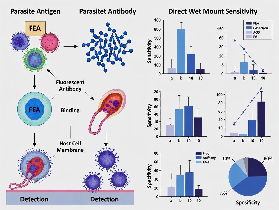
The diagnosis of intestinal parasitic infections remains a significant global health challenge, particularly in resource-limited settings. For over a century, microscopy-based techniques have formed the cornerstone of parasitological diagnosis, with the direct wet mount and formalin-ethyl acetate (FEA) concentration methods being the most widely employed procedures in clinical laboratories worldwide [5]. These techniques are essential for detecting a broad spectrum of protozoan cysts, helminth eggs, and larvae in stool specimens, providing critical information for patient management and public health interventions.
The ongoing debate regarding the comparative sensitivity of these methods is not merely academic; it has direct implications for diagnostic accuracy, patient care, and resource allocation in clinical laboratories. While molecular diagnostic technologies have emerged with enhanced sensitivity and specificity, they face technical challenges related to DNA extraction from robust parasite structures and remain inaccessible in many endemic regions due to cost and infrastructure requirements [15]. This guide provides a comprehensive comparison of the direct wet mount and FEA concentration methods, presenting standardized protocols, performance data, and technical specifications to inform researchers and laboratory professionals in their diagnostic workflows.
Theoretical Framework: Principles and Applications
Fundamental Differences in Methodological Approach
The direct wet mount technique is a rapid preparation method that involves examining a minimally processed stool sample suspended in a liquid medium. This approach preserves parasite motility and natural morphology, allowing for immediate observation of trophozoites and other motile forms. However, its diagnostic sensitivity is limited by factors including parasite density, sample volume examined, and examiner expertise [4] [27].
The FEA concentration method (also known as the Ritchie method) employs chemical and mechanical procedures to separate parasites from fecal debris. Formalin fixes the parasitic elements, preserving morphological characteristics while eliminating infectious potential, while ethyl acetate acts as an extractant of fats and debris, effectively concentrating the parasites into a sediment for microscopic examination [15] [27]. This process significantly enhances detection sensitivity by increasing the relative density of parasitic elements in the examined material.
Comparative Workflow Analysis
The diagram below illustrates the procedural differences between the direct wet mount and FEA concentration methods:
Experimental Protocols: Standardized Laboratory Procedures
Direct Wet Mount Microscopy Method
The direct wet mount technique provides a rapid assessment for motile trophozoites and parasitic elements, though with limited concentration power compared to FEA methods [27].
Materials Required:
- Fresh stool specimen (processed within 30-60 minutes of passage for trophozoite preservation)
- Physiological saline (0.85-0.90% NaCl solution)
- Lugol's iodine solution (1-2% for stained preparations)
- Microscope slides (75 × 25 mm) and 22 × 22 mm coverslips
- Applicator sticks or sterile loops
- Compound light microscope with 10×, 40× objectives
Step-by-Step Procedure:
- Sample Preparation: Using an applicator stick, emulsify a small portion of stool (approximately 2 mg, or the size of a match head) in a drop of physiological saline placed on the left side of a clean microscope slide.
- Duplicate Preparation: Similarly, prepare a second emulsion on the right side of the same slide using a drop of Lugol's iodine solution.
- Coverslip Application: Gently place coverslips over both suspensions, avoiding air bubbles.
- Systematic Microscopic Examination:
- Begin with the 10× objective to systematically scan the entire preparation.
- Switch to the 40× objective for detailed morphological assessment of suspicious structures.
- Examine saline preparation for motile trophozoites and other parasitic elements.
- Examine iodine preparation for enhanced nuclear and cytoplasmic detail of cysts.
- Documentation: Record all observed parasitic elements, noting their quantity and morphological characteristics.
Technical Notes:
- Optimal slide thickness allows newspaper print to be faintly visible through the preparation.
- Examine preparations within 10-15 minutes to observe motile trophozoites before desiccation occurs.
- Avoid excessive iodine concentration, which can inhibit motility and obscure details.
FEA Concentration Method
The FEA concentration technique significantly enhances detection sensitivity by concentrating parasitic elements through centrifugation and chemical processing [15] [27].
Materials Required:
- Fresh or formalin-preserved stool specimen
- 10% formalin solution
- Ethyl acetate solvent
- Centrifuge tubes (15 mL conical tubes)
- Gauze or strainer (100-150 µm mesh)
- Centrifuge with swing-out rotor
- Applicator sticks, pipettes, microscope slides, and coverslips
Step-by-Step Procedure:
- Sample Fixation:
- For fresh specimens: Emulsify 1-2 g of stool in 10 mL of 10% formalin in a centrifuge tube.
- For formalin-preserved specimens: directly use 1-2 mL of preserved material.
- Allow fixation for 30 minutes or proceed immediately to next step.
- Filtration and Concentration:
- Strain the suspension through gauze or a mesh strainer into a clean centrifuge tube to remove large particulate matter.
- Centrifuge at 500 × g for 3 minutes to form a sediment.
- Ethyl Acetate Extraction:
- Decant the supernatant, resuspend the sediment in 5-7 mL of 10% formalin.
- Add 3-4 mL of ethyl acetate to the tube.
- Cap the tube securely and shake vigorously for 30 seconds, periodically venting to release pressure.
- Final Processing:
- Recentrifuge at 500 × g for 3-5 minutes.
- Four layers will form: ethyl acetate (top), plug of debris, formalin, and sediment (bottom).
- Loosen the debris plug with an applicator stick and carefully decant the top three layers.
- Microscopic Examination:
- Resuspend the remaining sediment with a drop of formalin or saline.
- Prepare wet mounts with both saline and iodine as described in the direct method.
- Systematically examine the entire coverslip area under 10× and 40× objectives.
Technical Notes:
- The FEA method is particularly effective for detecting light infections of protozoan cysts and helminth eggs.
- Proper sealing during the ethyl acetate step prevents leakage of hazardous vapors.
- Some laboratories substitute ether for ethyl acetate with comparable results.
Comparative Performance Analysis: Sensitivity and Detection Rates
Quantitative Detection Performance
Table 1: Comparative Sensitivity of Diagnostic Methods for Hookworm Detection (n=530) [27]
| Diagnostic Method | Detection Rate (%) | Sensitivity (%) | Test Efficiency (%) | Agreement with CRS (κ-value) |
|---|---|---|---|---|
| Spontaneous Tube Sedimentation (STS) | 30.2 | 86.5 | 95.3 | 0.893 (Perfect) |
| Richie's (FEA) Method | 27.0 | 77.3 | 92.1 | 0.816 (Perfect) |
| Kato-Katz (KK) | 22.3 | 63.8 | 87.4 | 0.696 (Substantial) |
| Direct Wet Mount (DWM) | 15.1 | 43.2 | 80.2 | 0.498 (Moderate) |
Table 2: Overall Detection Rates of Intestinal Parasites Across Methods (n=150) [28]
| Diagnostic Method | Positive Samples Detected | Overall Sensitivity (%) | Remarks |
|---|---|---|---|
| Mini Parasep SF | 80/150 (53.3%) | 98.7 | Clearer background, better yield for H. nana, T. trichiura, E. coli, G. lamblia |
| Formol-Ether (FEA) | 77/150 (51.3%) | 95.0 | Conventional concentration standard |
| Direct Wet Mount | 72/150 (48.6%) | 90.1 | Limited concentration power |
Diagnostic Performance Characteristics
Table 3: Automated Fecal Analyzer Performance with AI Integration [4]
| Methodology | Sensitivity (%) | Specificity (%) | Remarks |
|---|---|---|---|
| Automatic Fecal Analyzer (AI Report) | 84.31 | 98.71 | Fully automated image analysis and machine learning algorithms |
| Automatic Fecal Analyzer (User Audit) | 94.12 | 99.69 | AI analysis with experienced technician review |
| Traditional Direct Wet Smear | Comparative baseline | Comparative baseline | Labor-intensive, operator-dependent |
The Scientist's Toolkit: Essential Research Reagents and Materials
Table 4: Essential Reagents and Materials for Parasitological Diagnostics
| Item | Function/Application | Technical Specifications | Considerations |
|---|---|---|---|
| 10% Formalin Solution | Fixation and preservation of parasitic elements | 1 part formalin (37-40% formaldehyde) to 9 parts water | Maintains morphology but eliminates motility |
| Ethyl Acetate Solvent | Extraction of fats and debris from fecal sample | Laboratory-grade, high purity | Flammable; proper ventilation required |
| Physiological Saline | Isotonic suspension medium for wet mounts | 0.85-0.90% NaCl in distilled water | Preserves trophozoite motility temporarily |
| Lugol's Iodine Solution | Staining protozoan cysts for enhanced visualization | 1-2% iodine in potassium iodide solution | Strong solutions can obscure details |
| Conical Centrifuge Tubes | Sample processing and concentration | 15 mL capacity, graduated | Compatible with swing-out centrifuge rotors |
| Parasep SF Faecal Concentrator | Integrated single-vial concentration system | Enclosed, solvent-free design | Reduced biohazard risk, simplified workflow |
| Microscope Slides and Coverslips | Preparation of specimens for microscopy | 75 × 25 mm slides, 22 × 22 mm coverslips | Optimal thickness prevents crushing specimens |
Methodological Variations and Emerging Technologies
Alternative Concentration Methods
The Spontaneous Tube Sedimentation (STS) technique has demonstrated superior performance in hookworm detection, with 86.5% sensitivity and 95.3% test efficiency according to recent field studies [27]. This method relies on gravity sedimentation rather than centrifugation, making it particularly suitable for resource-limited settings. The technique involves emulsifying stool in formalin, filtering through a mesh, and allowing the suspension to settle in a conical container for several hours before examining the sediment.
The Mini Parasep SF faecal concentrator represents an advancement in concentration technology with its enclosed, solvent-free design that minimizes biohazard exposure while maintaining high sensitivity (98.7%) [28]. This system integrates filtration and concentration into a single device, simplifying laboratory workflow while providing clearer background visualization compared to conventional FEA methods.
Integration of Artificial Intelligence and Automation
Recent technological innovations have introduced automated fecal analyzers that combine digital microscopy with artificial intelligence algorithms for parasite detection. These systems demonstrate 84.31% sensitivity in fully automated mode, increasing to 94.12% when combined with expert technician review [4]. The AI models are trained on diverse specimen collections from multiple continents, enabling detection of 27 different parasite classes with higher sensitivity than human technologists across experience levels [5].
Deep convolutional neural networks (CNNs) represent a breakthrough in parasitology diagnostics, with validation studies showing 94.3% agreement with traditional microscopy for positive specimens and 94.0% for negative specimens before discrepant resolution [5]. These systems consistently detected more organisms at lower parasite concentrations than human examiners, regardless of technologist experience level, suggesting a paradigm shift in diagnostic sensitivity and consistency.
The comparative analysis of direct wet mount and FEA concentration methods reveals a consistent pattern of superior performance for concentration techniques across parasite species and infection intensities. The FEA method demonstrates substantially higher sensitivity (77.3% versus 43.2% for hookworm) and test efficiency (92.1% versus 80.2%) compared to direct wet mount microscopy [27]. This performance advantage, coupled with better background clearance and morphological preservation, establishes FEA concentration as the methodological foundation for comprehensive parasitological examination.
For contemporary laboratory practice, the strategic integration of both methods provides optimal diagnostic coverage: direct wet mounts for initial assessment of motile trophozoites and FEA concentration for enhanced detection of cysts, ova, and light infections. Emerging technologies including automated concentration systems and AI-assisted microscopy promise further improvements in diagnostic sensitivity, workflow efficiency, and operational consistency [5] [4]. These advancements represent a significant evolution in the century-old practice of stool microscopy, potentially addressing longstanding challenges in parasitology diagnostics while maintaining the comprehensive parasite detection capability that remains a limitation of targeted molecular assays [15].
Within the field of parasitology, the accurate diagnosis of intestinal parasites remains a cornerstone of effective public health intervention and individual patient care. The microscopic examination of stool samples, while a long-standing reference method, is enhanced by concentration techniques that increase the likelihood of detecting parasitic elements. This guide provides an objective comparison of two such formalin-based concentration methods: the Formalin-Ether Concentration (FEC) technique and the Formalin-Acetone Concentration (FAC) technique. Framed within broader research comparing fecal concentration methods to direct wet mount microscopy, this analysis summarizes key experimental data on their diagnostic performance, details standardized protocols, and outlines essential laboratory reagents. The information is intended to assist researchers, scientists, and laboratory professionals in selecting and optimizing diagnostic methodologies for intestinal parasites.
Performance Data at a Glance
The following tables consolidate quantitative data from a controlled study that parallel-processed 200 samples for each technique to evaluate their diagnostic efficiency [13].
Table 1: Overall Diagnostic Performance of FEC and FAC Techniques
| Performance Metric | FEC Technique | FAC Technique |
|---|---|---|
| Sensitivity | 55.8% | 70.0% |
| Negative Predictive Value (NPV) | 60.2% | 69.0% |
| Overall Diagnostic Agreement (κ statistic) | Moderate | Substantial |
Table 2: Performance Breakdown by Parasite Type
| Parasite Type | FEC Technique | FAC Technique |
|---|---|---|
| Helminth Ova | Significantly less sensitive | Significantly more sensitive |
| Protozoan Cysts | More sensitive | Less sensitive |
Detailed Experimental Protocols
To ensure reproducibility, the standard operating procedures for the FEC and FAC techniques are described below. These protocols are adapted from the comparative study that evaluated their efficiency [13].
Formalin-Ether (FEC) Concentration Technique
The FEC technique is a sedimentation method that uses ether to separate parasitic elements from fecal debris.
- Sample Preparation: Emulsify approximately 1-2 grams of fresh stool in 10 mL of 10% formalin in a centrifuge tube. Allow the mixture to stand for 30 minutes to fix the specimen.
- Straining: Filter the suspension through a sieve or gauze into a clean centrifuge tube to remove large particulate matter.
- Centrifugation: Centrifuge the filtered suspension at 500 x g for 1 minute. Decant the supernatant.
- Resuspension: Resuspend the sediment in fresh 10% formalin, filling the tube to about half its volume. Add 3-4 mL of ethyl acetate (ether) to the suspension.
- Vigorous Mixing: Stopper the tube and shake it vigorously for 30 seconds. Ensure the stopper is secure to prevent leakage of volatile solvents.
- Final Centrifugation: Centrifuge again at 500 x g for 2-3 minutes. This will result in four distinct layers: an ether plug at the top, a fecal debris plug, a formalin layer, and the sediment at the bottom.
- Sediment Collection: Carefully free the debris plug from the tube's sides using an applicator stick. Decant the top three layers in a single motion. Use a swab to wipe excess debris from the tube's inner wall.
- Microscopy: Mix the remaining sediment and transfer a drop to a microscope slide for examination. Add a coverslip and systematically scan under low and high-power objectives.
Formalin-Acetone (FAC) Concentration Technique
The FAC technique follows a similar principle but substitutes ether with acetone, which is often preferred for its safety profile.
- Initial Emulsification and Filtration: Emulsify 1 gram of stool in 5 mL of saline in a centrifuge tube. Filter the suspension through a sieve into a second centrifuge tube.
- First Centrifugation: Centrifuge the filtered suspension at 450 x g for 1 minute. Decant the supernatant.
- Formalin and Acetone Addition: Add 7 mL of 10% formalin to the sediment, mix, and let stand for 5 minutes. Then, add 3 mL of acetone and mix immediately.
- Second Centrifugation: Centrifuge at 450 x g for 3 minutes. Decant the supernatant.
- Sediment Preparation: The resulting sediment is ready for microscopic examination.
- Microscopy: Prepare a wet mount from the sediment and examine it under the microscope.
Workflow Visualization
The following diagram illustrates the logical sequence and key differences between the FEC and FAC procedures.
The Scientist's Toolkit: Key Research Reagent Solutions
The successful implementation of the FEC and FAC protocols relies on specific reagents, each with a distinct function.
Table 3: Essential Reagents for FEC and FAC Techniques
| Reagent | Function in the Protocol | Safety and Handling Notes |
|---|---|---|
| 10% Formalin | Fixes and preserves parasitic cysts, oocysts, and ova, preventing further development or degradation. | A known irritant and hazardous substance; use with appropriate personal protective equipment (PPE) and in a well-ventilated area. |
| Ethyl Acetate (Ether) | Acts as a fat solvent and dehydrating agent, effectively concentrating parasitic elements by cementing debris into a plug. | Highly flammable and volatile; requires careful storage and handling away from ignition sources. |
| Acetone | Serves as a substitute for ether; effectively removes fats and debris while being less flammable and safer to handle. | Flammable but generally considered a safer alternative to ether in laboratory settings. |
| Saline Solution | Used as an initial diluent to emulsify the stool specimen without damaging parasitic structures. | Low hazard; standard laboratory solution. |
The accurate diagnosis of intestinal parasitic infections remains a cornerstone of public health and clinical microbiology, directly impacting patient treatment and disease control. Billions of people are affected by these infections globally, causing significant morbidity. The macroscopic and microscopic examination of stool samples, often referred to as the ova and parasite (O&P) test, is a fundamental diagnostic approach. This guide provides a detailed, objective comparison of the performance of various diagnostic techniques, with a specific focus on the Formalin-Ethyl Acetate (FEA) concentration method and Direct Wet Smear Microscopy. The sensitivity and specificity of these methods are critical for researchers and drug development professionals who rely on accurate data for epidemiological studies, clinical trials, and the development of new diagnostic reagents and therapeutic agents. This comparison is framed within broader research on diagnostic sensitivity, providing experimental data and protocols to inform laboratory practices and research directions.
Performance Comparison of Diagnostic Methods
The choice of diagnostic technique significantly impacts the detection rate of intestinal parasites. The following tables summarize quantitative performance data from recent studies, comparing traditional and automated methods.
Table 1: Comparative Sensitivity of Stool Examination Techniques
| Diagnostic Method | Reported Sensitivity | Key Advantages | Key Limitations |
|---|---|---|---|
| Direct Wet Smear Microscopy [29] [28] | ~48.6% - 90.1% | Simple, rapid, low-cost, allows observation of motile trophozoites. [29] | Low sensitivity; small sample volume; labor-intensive; highly dependent on technician skill. [4] [29] |
| FEA Concentration Technique [15] [28] | ~51.3% - 98.7% | Increases sensitivity by concentrating parasites; removes debris. [29] [28] | Requires specific chemicals and procedures; destroys trophozoites. [29] |
| Formalin-Tween Concentration (FTC) [13] | 71.7% | Superior for diagnosing helminth ova. [13] | Less sensitive for protozoan cysts compared to other methods. [13] |
| Automatic Fecal Analyzer (AI Report) [4] | 84.31% | Automated, rapid, clean; processes large sample volumes quickly. [4] | May require user audit for highest accuracy. [4] |
| Automatic Fecal Analyzer (User Audit) [4] | 94.12% | High sensitivity and specificity; combines AI efficiency with expert verification. [4] | Requires experienced technicians for the audit step. [4] |
| Molecular Methods (RT-PCR) [15] | Varies by parasite (e.g., high for G. duodenalis) | High sensitivity and specificity; differentiates morphologically identical species. [15] | DNA extraction can be challenging; higher cost; requires specialized equipment. [15] |
Table 2: Specificity and Agreement of Various Techniques
| Diagnostic Method | Reported Specificity | Negative Predictive Value (NPV) | Overall Agreement (κ statistic) |
|---|---|---|---|
| Direct Wet Smear Microscopy [28] | Not explicitly quantified | Not explicitly quantified | Not explicitly quantified |
| FEA Concentration Technique [28] | Not explicitly quantified | Not explicitly quantified | Not explicitly quantified |
| Formalin-Tween Concentration (FTC) [13] | Not explicitly quantified | 70.2% | Substantial [13] |
| Formalin-Ether Concentration (FEC) [13] | Not explicitly quantified | 60.2% | Moderate [13] |
| Automatic Fecal Analyzer (AI Report) [4] | 98.71% | Not explicitly quantified | Not explicitly quantified |
| Automatic Fecal Analyzer (User Audit) [4] | 99.69% | Not explicitly quantified | Not explicitly quantified |
| Deep Convolutional Neural Network (AI) [30] | 94.0% (before discrepant resolution) | Not explicitly quantified | Not explicitly quantified |
Experimental Protocols for Key Studies
To ensure reproducibility and critical evaluation, the methodologies of key cited experiments are detailed below.
Protocol: Multicenter Comparison of Molecular vs. Microscopic Methods
This multicenter study compared a commercial RT-PCR test, an in-house RT-PCR assay, and conventional microscopy for detecting major intestinal protozoa. [15]
- Sample Collection: A total of 355 stool samples were collected across 18 Italian laboratories. Among these, 230 were freshly collected, and 125 were preserved in Para-Pak media. [15]
- Microscopic Examination: All samples were first examined using conventional microscopy according to WHO and CDC guidelines. Fresh samples were stained with Giemsa, while preserved samples were processed using the FEA concentration technique. [15]
- DNA Extraction: DNA was extracted from stool samples using the MagNA Pure 96 System (Roche). A stool sample was mixed with Stool Transport and Recovery Buffer (S.T.A.R. Buffer), centrifuged, and the supernatant was used for automated nucleic acid extraction. [15]
- PCR Amplification: Both the commercial (AusDiagnostics) and in-house RT-PCR assays were performed. The in-house reaction used a mix of extracted DNA, TaqMan Fast Universal PCR Master Mix, primer-probe mixes, and sterile water. Amplification was carried out on an ABI 7900HT system with 45 cycles. [15]
Protocol: Comparative Analysis of an Automatic Fecal Analyzer
This study evaluated the performance of an automatic fecal analyzer against the traditional direct wet smear method. [4]
- Methods Compared: Three methods were run in parallel:
- Direct Wet Smear Microscopy: The traditional, manual method.
- Automatic Fecal Analyzer (AI Report): Fully automated processing and analysis using machine learning algorithms.
- Automatic Fecal Analyzer (User Audit): Automated analysis followed by a review of the results by an experienced technician. [4]
- Performance Metrics: The sensitivity and specificity of each method were calculated and compared to determine the value of automation and expert verification in the diagnostic workflow. [4]
Protocol: Evaluation of the Mini Parasep SF Concentration Technique
This study assessed the diagnostic performance of the enclosed, single-vial Mini Parasep SF faecal concentrator. [28]
- Sample Processing: A total of 150 stool samples were processed using three methods: direct wet mount, the formalin-ether method (FEM), and the Mini Parasep SF technique. [28]
- Microscopy: All concentrated and unconcentrated samples were subjected to wet mount and iodine mount microscopy. Modified acid-fast staining was also used for specific parasites. [28]
- Analysis: The number of positive samples detected by each method was recorded. The sensitivity of each technique was calculated, and observations were made regarding the clarity of the background and the yield of specific parasites. [28]
Workflow and Relationship Diagrams
The following diagrams illustrate the logical workflows of the key diagnostic processes discussed.
Traditional Stool O&P Examination Workflow
Integrated Modern Diagnostic Pathway
The Scientist's Toolkit: Essential Research Reagents & Materials
Successful diagnosis and research in intestinal parasitology rely on a suite of specific reagents and materials. The following table details key items and their functions.
Table 3: Key Research Reagent Solutions for Stool Parasitology
| Reagent/Material | Function/Application | Key Characteristics |
|---|---|---|
| Formalin (5-10%) [29] | Primary preservative for stool samples; used in concentration techniques like FEA. [29] | Maintains parasite morphology; inhibits further maturation of helminth ova/larvae. [29] |
| Ethyl Acetate [15] | Solvent used in the FEA concentration method. | Acts as a lipid solvent and extractor, helping to clear fecal debris during concentration. [15] |
| Polyvinyl Alcohol (PVA) [29] | Preservative for permanent stained smears. | Adheres stool material to slide and preserves protozoan morphology for staining. [29] |
| Tween [13] | Detergent used in Formalin-Tween Concentration (FTC). | A safer, more stable alternative to ether; superior for helminth ova recovery. [13] |
| S.T.A.R. Buffer [15] | Stool Transport and Recovery Buffer for molecular assays. | Stabilizes nucleic acids in stool samples prior to DNA extraction for PCR. [15] |
| TaqMan Master Mix [15] | Reagent for real-time PCR (RT-PCR). | Contains enzymes, dNTPs, and optimized buffers for sensitive and specific DNA amplification. [15] |
| Trichrome Stain [29] | Polychromatic stain for permanent smears. | Provides contrast to differentiate protozoan cysts/trophozoites from background debris. [29] |
| Modified Acid-Fast Stain [29] | Special stain for coccidian parasites. | Stains oocysts of Cryptosporidium spp. and Cyclospora for visualization. [29] |
The data presented in this guide underscores a critical evolution in the diagnosis of intestinal parasites. While traditional methods like direct wet smear and FEA concentration remain foundational, their limitations in sensitivity and operational efficiency are clear. The evidence demonstrates that FEA concentration is consistently more sensitive than direct wet smear microscopy, but both are being supplemented or surpassed by newer technologies. Automated systems with AI, especially when combined with a user audit, show remarkable sensitivity and specificity, reducing reliance on manual skill. Furthermore, molecular methods like RT-PCR offer unparalleled specificity and are becoming essential for differentiating species and detecting low-burden infections. The optimal diagnostic approach, particularly for research and drug development requiring high precision, often involves a complementary strategy, utilizing the strengths of each method to achieve the most accurate and comprehensive result.
The accurate diagnosis of gastrointestinal parasitic infections is a critical public health concern, particularly within special populations where clinical manifestations and test performance can vary significantly. This guide objectively compares the diagnostic performance of the Formol-Ethyl Acetate Concentration (FEA) technique against Direct Wet Mount Microscopy, with a specific focus on pediatric, immunocompromised, and asymptomatic cases. The evaluation is framed within broader research on their relative sensitivity, providing researchers and drug development professionals with synthesized experimental data and methodologies to inform diagnostic choices and future assay development.
Comparative Performance Data in Special Populations
The diagnostic sensitivity of any method is not absolute but is influenced by host factors. The following table summarizes key performance metrics for FEA and Direct Wet Mount across the populations of interest, synthesized from recent studies.
Table 1: Diagnostic Sensitivity Comparison in Special Populations
| Special Population | Direct Wet Mount Sensitivity | FEA Concentration Sensitivity | Key Supporting Findings |
|---|---|---|---|
| Pediatric Patients | 41% [31] | 75% [31] | FEA detected 75% of parasites in children with diarrhea, significantly outperforming wet mount (41%) and Formol-Ether Concentration (62%) [31]. |
| Immunocompromised Patients | Limited sensitivity for low-level infections [15] | Higher yield for opportunistic protozoa [15] [32] | Molecular methods (e.g., PCR) are particularly critical for detecting Cryptosporidium spp. and differentiating pathogenic species in immunocompromised individuals, though FEA offers a reliable microscopic alternative [15] [32]. |
| Asymptomatic Cases | Lower sensitivity due to low parasite load [33] | Higher detection rate for cysts [32] | Immunoassays demonstrate a marked drop in sensitivity in asymptomatic individuals (79%) compared to symptomatic ones (92%), a trend that is likely applicable to microscopy-based methods [33]. |
Detailed Experimental Protocols
Understanding the methodologies behind the data is crucial for their interpretation and replication. Below are the standardized protocols for the key techniques discussed.
Formol-Ethyl Acetate Concentration (FEC/FAC) Technique
The FEA concentration method is designed to maximize parasite recovery from stool samples.
- Sample Emulsification: Approximately 1 gram of fresh stool is emulsified in 7 mL of 10% formol saline for a 10-minute fixation period [31].
- Filtration: The mixture is strained through a sieve or three folds of surgical gauze to remove large debris [31].
- Solvent Concentration: The filtrate is combined with 3 mL of ethyl acetate in a centrifuge tube. The tube is then shaken vigorously and centrifuged at 1500 rpm for 5 minutes [31]. This process creates four layers: a sediment containing parasites, a layer of formol saline, a plug of debris, and a top layer of ethyl acetate.
- Examination: The supernatant is carefully decanted, and the sediment is used to prepare slides for microscopic examination at 10x and 40x magnifications [31].
Direct Wet Mount Microscopy
This is a rapid but less sensitive method for direct examination.
- Sample Preparation: A small portion of stool (1-2 mg) is mixed with a drop of 0.9% normal saline on a microscope slide. A second preparation can be made using iodine stain for better visualization of cysts [31] [6].
- Examination: A coverslip is applied, and the slide is examined immediately under the microscope at 10x and 40x objectives for motile trophozoites, cysts, eggs, or larvae [31].
Molecular Detection (RT-PCR)
Molecular techniques offer high specificity and sensitivity, especially in immunocompromised patients.
- DNA Extraction: Stool samples are mixed with a transport buffer. DNA is then extracted, often using automated systems like the MagNA Pure 96 with magnetic bead technology [15].
- PCR Amplification: Each reaction mixture contains the extracted DNA, primers and probes specific for the target parasite (e.g., Giardia duodenalis, Cryptosporidium spp.), a master mix, and water. Real-time PCR is performed on a system like the ABI 7900HT, typically with a cycling regimen of 95°C for 10 min, followed by 45 cycles of 95°C for 15 sec and 60°C for 1 min [15].
Diagnostic Workflow and Logical Pathways
The following diagram illustrates the logical decision pathway for selecting a diagnostic method based on the patient population and clinical context.
The Scientist's Toolkit: Key Research Reagents
The following table details essential reagents and their functions as derived from the cited experimental protocols.
Table 2: Essential Research Reagents for Parasitology Diagnostics
| Reagent / Kit | Primary Function in Protocol |
|---|---|
| 10% Formol Saline | Fixes and preserves parasitic elements (cysts, eggs, larvae) in stool specimens for concentration techniques [31]. |
| Ethyl Acetate / Diethyl Ether | Organic solvent used in concentration methods to clear debris and fat, concentrating parasites in the sediment [31]. |
| MagNA Pure 96 DNA/\nViral NA Small Volume Kit | Automated, magnetic bead-based system for extracting nucleic acids from stool samples for subsequent molecular analysis [15]. |
| TaqMan Fast Universal\nPCR Master Mix | Ready-to-use reaction mix containing enzymes, dNTPs, and buffer for efficient real-time PCR amplification [15]. |
| Parasite-Specific Primers/Probes | Oligonucleotides designed to bind to unique genetic sequences of target parasites (e.g., Giardia, Cryptosporidium) for specific identification via PCR [15]. |
| S.T.A.R. Buffer | Stool Transport and Recovery Buffer that stabilizes nucleic acids in stool samples during storage and transport [15]. |
Overcoming Diagnostic Challenges and Implementing Workflow Optimizations
Addressing Low Sensitivity in Low-Intensity Infections and Asymptomatic Carriers
The accurate detection of pathogens in individuals with low-intensity infections or asymptomatic carriage presents a formidable challenge in clinical and public health microbiology. These hidden reservoirs are crucial for the persistence and silent transmission of many infectious diseases, yet they often evade conventional diagnostic methods. Asymptomatic carriers can harbor pathogen levels several orders of magnitude below the detection threshold of traditional techniques, leading to false-negative results and undermining control efforts [34] [35]. This diagnostic gap is particularly problematic for diseases like malaria, where asymptomatic individuals contribute significantly to transmission dynamics, accounting for approximately 30% of the basic reproduction number (R₀) according to recent mathematical modeling [34].
The limited sensitivity of traditional methods such as direct wet mount microscopy has become increasingly apparent when compared to enhanced concentration techniques and molecular assays. Within the specific context of intestinal parasite diagnostics, the formalin-ethyl acetate concentration (FEA) method represents a significant improvement over direct wet mount examination, though both techniques remain foundational in laboratory practice. This guide provides a systematic comparison of these methods and emerging alternatives, offering experimental data and protocols to inform researchers and drug development professionals working to overcome the critical challenge of low-sensitivity diagnostics.
Comparative Performance of Diagnostic Methods
Quantitative Comparison of Detection Methods
Table 1: Performance characteristics of different diagnostic methods for parasitic infections
| Diagnostic Method | Target Pathogens | Sensitivity | Specificity | Remarks / Application Context |
|---|---|---|---|---|
| Direct Wet Mount Microscopy | Intestinal protozoa, helminths, vaginal parasites | 48.6%-50% [28] [2] | Not specifically quantified | Rapid but limited by low pathogen density and technician skill |
| FEA Concentration Method | Intestinal protozoa, helminths | 51.3%-95% [15] [28] | High | Enhanced detection through parasite concentration; reference standard |
| Mini Parasep SF Concentration | Intestinal protozoa, helminths | 53.3%-98.7% [28] | High | Superior background clearance, enclosed system reduces contamination |
| Automated Fecal Analyzer (AI Report) | Parasites and eggs in stool | 84.31% [4] | 98.71% [4] | Automated processing with machine learning algorithms |
| Automated Fecal Analyzer (User Audit) | Parasites and eggs in stool | 94.12% [4] | 99.69% [4] | Combines AI with technician review for optimal accuracy |
| Wet Mount Microscopy for T. vaginalis | Trichomonas vaginalis | 50-70% [2] | ~100% [2] | Highly dependent on immediate sample processing and examiner expertise |
| Molecular NAAT for T. vaginalis | Trichomonas vaginalis | Significantly higher than wet mount [2] | ~99.3% [2] | Higher cost but superior sensitivity, especially for asymptomatic cases |
Table 2: Performance of diagnostic methods for non-parasitic infections with asymptomatic presentations
| Diagnostic Method | Target Pathogens/Conditions | Sensitivity | Specificity | Remarks / Application Context |
|---|---|---|---|---|
| Automated Vaginal Microscopy System | Bacterial vaginosis, Candida albicans, cytolytic vaginosis | 84.1%-90.9% [36] | 65.9%-99.4% [36] | Machine learning-based automated microscopy |
| Molecular Assays (Aptima BV) | Bacterial vaginosis | 97.5% [37] | 96.3% [37] | High sensitivity but may detect colonization without disease |
| Molecular Assays (Aptima CV/TV) | Candida and Trichomonas vaginalis | 100% for TV [37] | 83.5%-100% [37] | Excellent for Trichomonas; may over-call Candida colonization |
| Commercial RT-PCR (AusDiagnostics) | Giardia duodenalis, Cryptosporidium spp. | High, comparable to microscopy [15] | High, comparable to microscopy [15] | Performs well with fixed fecal specimens |
| In-house RT-PCR | Giardia duodenalis, Cryptosporidium spp., Entamoeba histolytica | High for most targets [15] | High for most targets [15] | Limited sensitivity for Dientamoeba fragilis |
Impact of Low Sensitivity on Disease Management
The clinical consequences of inadequate diagnostic sensitivity are profound across multiple disease domains. In malaria endemic regions, asymptomatic individuals maintaining low-level Plasmodium falciparum infections serve as reservoir hosts that sustain transmission during dry seasons when clinical cases are rare [35]. Molecular studies have revealed that these asymptomatic carriers exhibit distinct parasite profiles with fewer var genes expressed at lower levels compared to clinical malaria cases, suggesting adaptation for persistence rather than acute disease [35].
Similarly, in sexually transmitted infections, undiagnosed asymptomatic carriers contribute significantly to ongoing transmission. For Trichomonas vaginalis, approximately 80% of infections are asymptomatic, yet can lead to serious complications including pelvic inflammatory disease, cervical and prostate cancer, and enhanced HIV transmission [2]. The common reliance on wet mount microscopy with sensitivity of 50-70% means a substantial proportion of these infections remain undiagnosed and untreated [2].
In intestinal protozoan infections, the World Health Organization estimates approximately 3.5 billion people are affected annually, causing nearly 1.7 billion episodes of diarrheal disease [15]. The inadequate detection of low-intensity infections, particularly in developed countries with low prevalence, leads to underreporting and failure to implement appropriate control measures.
Experimental Protocols and Methodologies
Standardized FEA Concentration Protocol
The formalin-ethyl acetate concentration (FEA) method represents a significant improvement over direct wet mount microscopy for intestinal parasites. The following protocol is adapted from multicentre studies comparing diagnostic techniques [15]:
Sample Preparation:
- Emulsify 1-2 g of fresh stool in 10 mL of 10% formalin to create a uniform suspension.
- Filter the suspension through gauze or a sieve to remove large particulate matter.
- Transfer the filtered material to a 15 mL conical centrifuge tube.
Concentration Steps:
- Centrifuge the filtered suspension at 500 × g for 10 minutes.
- Decant the supernatant completely, leaving approximately 0.5 mL of sediment.
- Resuspend the sediment in 10 mL of 10% formalin and mix thoroughly.
- Add 3 mL of ethyl acetate to the suspension, cap the tube securely, and shake vigorously for 30 seconds.
- Centrifuge again at 500 × g for 10 minutes.
- Separate the resulting layers: (1) ethyl acetate plug, (2) debris plug, (3) formalin, and (4) sediment.
- Free the debris plug from the tube side by ringing with an applicator stick.
- Decant all supernatant layers carefully, leaving the sediment at the bottom.
- Mix the remaining sediment and prepare slides for microscopic examination.
Microscopic Examination:
- Prepare wet mounts from the concentrated sediment using iodine and without staining.
- Systematically examine the entire coverslip area (22 × 22 mm) under 100× and 400× magnification.
- Identify parasites based on morphological characteristics.
This protocol typically requires 30-45 minutes processing time per sample and demonstrates sensitivity of 51.3-95% depending on the pathogen and technician expertise [15] [28].
Molecular Detection Protocol for Intestinal Protozoa
For comparison, here is a standardized protocol for molecular detection of intestinal protozoa using real-time PCR [15]:
DNA Extraction:
- Mix 350 μL of Stool Transport and Recovery Buffer (S.T.A.R. Buffer) with approximately 1 μL of fecal sample.
- Incubate for 5 minutes at room temperature, then centrifuge at 2000 rpm for 2 minutes.
- Transfer 250 μL of supernatant to a fresh tube and add 50 μL of internal extraction control.
- Extract DNA using the MagNA Pure 96 DNA and Viral NA Small Volume Kit on the MagNA Pure 96 System.
PCR Amplification:
- Reaction mixture: 5 μL of DNA extract, 12.5 μL of 2× TaqMan Fast Universal PCR Master Mix, 2.5 μL of primers and probe mix, and sterile water to a final volume of 25 μL.
- Cycling conditions: 1 cycle of 95°C for 10 minutes; followed by 45 cycles of 95°C for 15 seconds and 60°C for 1 minute.
- Perform on ABI 7900HT Fast Real-Time PCR System or equivalent.
This protocol offers enhanced sensitivity for specific targets like *Giardia duodenalis and Cryptosporidium spp., but may miss unexpected parasites not included in the PCR panel [15].*
Automated Microscopy Protocol for Vaginitis
Sample Processing with Automated System [36]:
- Collect vaginal swab samples using standardized collection kits.
- Insert the swab into the automated microscopy system (e.g., GYNI system).
- The system automatically prepares slides, performs bright-field microscopy, and captures digital images.
- Machine learning algorithms analyze the images for key diagnostic elements:
- Clue cells for bacterial vaginosis
- Hyphae or budding yeast for candidiasis
- Trichomonads for trichomoniasis
- Background flora and inflammatory cells
- The system provides a computer-aided suggested diagnosis based on the analysis.
This automated approach demonstrates sensitivities of 84.1-90.9% for various vaginitis pathogens while reducing reliance on expert microscopists [36].
Diagnostic Workflows and Method Selection
Essential Research Reagents and Materials
Table 3: Essential research reagents and materials for parasitology diagnostics
| Reagent/Material | Application | Function | Example Product/Reference |
|---|---|---|---|
| Formalin-Ethyl Acetate | Fecal concentration | Preserves parasites and separates debris from specimens | Standard FEA protocol [15] [28] |
| S.T.A.R. Buffer (Stool Transport and Recovery Buffer) | Molecular diagnostics | Stabilizes nucleic acids in stool samples during transport and storage | Roche Applied Sciences [15] |
| MagNA Pure 96 DNA and Viral NA Small Volume Kit | Nucleic acid extraction | Automated purification of DNA from clinical samples | Roche Applied Sciences [15] |
| TaqMan Fast Universal PCR Master Mix | Real-time PCR | Provides enzymes, dNTPs, and optimized buffer for amplification | Thermo Fisher Scientific [15] |
| Mini Parasep SF Faecal Concentrator | Fecal concentration | Single-vial, solvent-free parasite concentration system | Mini Parasep [28] |
| Chromagar Candida | Fungal culture | Selective medium for differentiation of Candida species | CHROMagar [36] |
| Nucleic Acid Amplification Tests (NAAT) | Molecular detection | Amplification of pathogen-specific nucleic acid sequences | Aptima BV and CV/TV assays [37] |
The critical challenge of low diagnostic sensitivity in detecting low-intensity infections and asymptomatic carriers demands a multifaceted approach combining enhanced traditional methods with advanced molecular techniques. While FEA concentration methods demonstrate clear superiority over direct wet mount microscopy with sensitivity improvements from approximately 50% to over 95% for some parasites [28], even these enhanced microscopic methods have limitations in detecting the lowest levels of infection.
Emerging technologies including automated microscopy systems and molecular amplification techniques represent the next frontier in diagnostic sensitivity, offering significant improvements particularly for asymptomatic carriers who typically harbor lower pathogen loads [4] [36]. However, these advanced methods come with trade-offs in cost, technical requirements, and potential over-detection of colonization without clinical disease [37].
The optimal diagnostic approach varies by clinical context, pathogen, and population. In resource-limited settings with high disease prevalence, enhanced concentration methods like FEA provide the best balance of performance and practicality. In settings where asymptomatic carriers are the primary transmission concern or for drug efficacy studies, molecular methods offer the sensitivity required despite higher costs. Future directions should focus on developing more accessible molecular platforms, standardizing extraction protocols across sample types, and establishing pathogen load thresholds that differentiate clinical disease from asymptomatic carriage.
The accurate diagnosis of gastrointestinal parasitic infections is a cornerstone of public health and clinical practice, yet laboratory procedures are fraught with technical challenges that can compromise results. Key among these are the issues of sample contamination, excessive fecal debris, and the use of hazardous reagents, all of which directly impact diagnostic accuracy, laboratory safety, and operational efficiency. Within this context, the choice between the Formalin-Ethyl Acetate Sedimentation Concentration (FEC) technique and direct wet mount microscopy represents a critical decision point for laboratories. This guide provides an objective, data-driven comparison of these two methods, framing the analysis within broader research on their comparative sensitivity. It is designed to equip researchers, scientists, and drug development professionals with the detailed experimental data and protocols needed to select and optimize diagnostic methods effectively, while navigating the associated technical hurdles.
Quantitative Method Comparison: FEC vs. Direct Wet Mount
Extensive research has quantified the performance differences between the formalin-ether (or formalin-ethyl acetate) concentration technique and direct wet mount microscopy. The following tables consolidate key experimental findings on their diagnostic sensitivity and operational characteristics.
Table 1: Overall Diagnostic Sensitivity and Accuracy
| Evaluation Metric | Direct Wet Mount | Formalin-Ether Concentration (FEC) | Reference |
|---|---|---|---|
| Overall Sensitivity for Helminths | 76.0% | 100% (as reference) | [38] [39] |
| Overall Test Efficiency (TE) | 94.0% | Not Specified | [38] [39] |
| Negative Predictive Value (NPV) | 92.7% | Not Specified | [38] [39] |
| Sensitivity for Hookworm | 85.7% | 100% (as reference) | [38] [39] |
| Sensitivity for Ascaris lumbricoides | 83.3% | 100% (as reference) | [38] [39] |
| Sensitivity for Hymenolepis nana | 33.3% | 100% (as reference) | [38] [39] |
Table 2: Comparison of Detection Rates in a Routine Laboratory Setting
| Parasite Species | Direct Wet Mount | Kato's Thick Smear | Formalin-Ethyl Acetate (FEC) | Complete Filtration (Automated) |
|---|---|---|---|---|
| All Parasites | Significantly Lower | Comparable | Benchmark | Significantly Lower |
| Ascaris lumbricoides | Detected | Detected | Detected | Detected |
| Hookworm | Detected | Detected | Detected | Detected |
| Trichuris trichiura | Detected | Detected | Detected | Detected |
| Strongyloides stercoralis | Detected | Detected | Detected | Detected |
| Entamoeba histolytica/dispar | Detected | Detected | Detected | Detected |
| Blastocystis spp. | Detected | Detected | Detected | Detected |
| Giardia intestinalis | Detected | Detected | Detected | Detected |
Table 3: Operational and Safety Considerations
| Characteristic | Direct Wet Mount | Formalin-Ethyl Acetate Concentration |
|---|---|---|
| Handling of Debris | Minimal; debris can obscure parasites | Effective; separates parasites from fecal debris |
| Risk of Sample Contamination | Lower (simpler process) | Higher (multiple processing steps) |
| Use of Hazardous Reagents | No (only saline) | Yes (formalin, ethyl acetate/diethyl ether) |
| Required Labor Time | Low (<10 minutes) | Moderate to High (20-30 minutes) |
| Sensitivity to Operator Skill | High | Moderate |
| Distinction of Active Infection | Yes (motile trophozoites observable) | Limited (cyst and egg-based) |
Experimental Protocols and Workflows
To ensure reproducibility and a clear understanding of the technical procedures, this section outlines the standardized protocols for both diagnostic methods as utilized in the cited comparative studies.
Direct Wet Mount Microscopy Protocol
The direct wet mount is a rapid, unstained preparation used for the initial examination of fresh stool specimens.
- Sample Collection and Timing: A freshly voided stool specimen is collected. Liquid or diarrheic specimens must be examined within 30 minutes of passage to observe motile trophozoites. Soft specimens should be examined within one hour, and formed specimens can be held for up to one day with refrigeration [41].
- Smear Preparation:
- Microscopic Examination:
- Systematically scan the entire smear under the microscope using a 10x objective for detecting helminth eggs, larvae, and protozoan cysts.
- Switch to a 40x objective to confirm identification and, in fresh samples, to observe the characteristic motility of trophozoites (e.g., Giardia) [41].
- Quality Assurance: To increase recovery rates, especially for delicate organisms like Trichomonas vaginalis, preparing and examining multiple slides from the same specimen is recommended [42].
Formalin-Ethyl Acetate Sedimentation Concentration Protocol
The FEC technique is a sedimentation method that concentrates parasitic elements, thereby increasing detection sensitivity. The CDC-recommended procedure is detailed below [41].
CDC Standard Operational Workflow
- Steps:
- Straining: Mix the formalin-preserved specimen well. Strain approximately 5 mL of the fecal suspension through wetted gauze (cheesecloth) into a 15 mL conical centrifuge tube.
- Dilution: Add 0.85% saline or 10% formalin through the debris on the gauze to bring the volume in the tube to 15 mL. (Note: Distilled water may deform Blastocystis hominis).
- First Centrifugation: Centrifuge at 500 × g for 10 minutes. Decant the supernatant.
- Resuspension: Add 10 mL of 10% formalin to the sediment and mix thoroughly with wooden applicator sticks.
- Solvent Addition: Add 4 mL of ethyl acetate. Stopper the tube securely.
- Shaking: Shake the tube vigorously in an inverted position for 30 seconds. Caution: Carefully release pressure by removing the stopper afterward.
- Second Centrifugation: Centrifuge at 500 × g for 10 minutes. This results in four layers: a thin plug of debris (top), a layer of ethyl acetate, a formalin layer, and the sediment (bottom).
- Debris Removal: Free the debris plug by running an applicator stick between the plug and the tube wall. Decant the top three layers (debris, ethyl acetate, formalin).
- Final Preparation: Use a cotton-tipped applicator to remove any residual debris from the tube's sides. Add a few drops of 10% formalin to resuspend the final sediment for microscopic examination (wet mount, staining) [41].
- Safety Notes: This protocol uses ethyl acetate, which is less flammable and safer than diethyl ether. However, proper personal protective equipment (PPE) including gloves, lab coat, and eye protection should be worn. Procedures should be conducted in a well-ventilated area or fume hood [41].
The Scientist's Toolkit: Essential Research Reagents and Materials
Successful diagnosis and research in parasitology depend on the specific reagents and materials used. The table below details key items referenced in the featured experiments, along with their critical functions and notes on handling.
Table 4: Key Research Reagent Solutions and Materials
| Item | Function / Application | Key Considerations & Hazards |
|---|---|---|
| 10% Formalin | Primary preservative for stool specimens; fixes parasitic stages and maintains morphology. | Hazardous; fixative and irritant. Requires PPE and proper ventilation [41]. |
| Ethyl Acetate | Organic solvent in FEC; extracts fats and debris, forming a plug for removal. | Less flammable than ether but still hazardous. Use in well-ventilated areas [41]. |
| Diethyl Ether | Traditional solvent for FEC; functions similarly to ethyl acetate. | Highly flammable and explosive hazard. Requires extreme caution; largely replaced by ethyl acetate [13] [41]. |
| 0.85% Saline | Isotonic solution for direct wet mounts; maintains parasite motility and integrity. | Low hazard. Must be fresh for optimal trophozoite viability [41]. |
| Toluidine Blue (TolB) | Wet mount stain for specific pathogens (e.g., Cryptosporidium oocysts). | Provides high sensitivity as a temporary stain; requires fresh samples [43]. |
| Modified Ziehl-Neelsen (mZN) Stain | Acid-fast permanent stain for Cryptosporidium spp. and Cyclospora. | Considered a gold standard but time-consuming; requires training to interpret "ghost" oocysts [43]. |
| SYBR Gold Stain | Fluorescent nucleic acid dye for virus enumeration in wet-mount methods (research). | Used in research settings; requires epifluorescence microscopy and antifade agents [44]. |
| Silica Microspheres | Internal standard beads for quantitative wet-mount microscopy (research). | Allows for precise concentration calculations; requires thorough vortexing before use [44]. |
The Role of Technician Training and Quality Control in Maximizing Diagnostic Yield
In clinical diagnostics, the balance between sophisticated new technologies and established, accessible methods is delicate. The pursuit of this balance is central to a growing body of research comparing automated diagnostic systems with traditional direct wet mount microscopy. While advanced technologies like automated fecal analyzers and nucleic acid amplification techniques offer superior sensitivity, their implementation remains impractical in many resource-limited settings where microscopic examination remains the diagnostic cornerstone. Within this context, the expertise of laboratory technicians and robust quality control protocols emerge as critical variables that significantly influence diagnostic accuracy regardless of the method employed. This review synthesizes evidence from contemporary comparative studies to delineate the specific role of technician training and quality assurance in maximizing diagnostic yield across parasitology and microbiology laboratories. By examining experimental data across multiple diagnostic platforms and specimen types, we aim to provide laboratory professionals and researchers with evidence-based strategies for optimizing diagnostic performance through human resource development and quality management systems.
Comparative Methodologies and Experimental Approaches
Diagnostic Performance Metrics in Comparative Studies
Research comparing diagnostic methodologies employs standardized metrics to quantify performance. Sensitivity measures the proportion of true positives correctly identified, while specificity measures the proportion of true negatives correctly identified. Positive Predictive Value (PPV) and Negative Predictive Value (NPV) indicate the probability that positive or negative test results are correct, and Test Efficiency (TE) reflects the overall correctness of the method [45]. Understanding these metrics is essential for interpreting the experimental data presented in subsequent sections.
Experimental Designs for Method Comparison
Comparative studies typically employ cross-sectional designs where identical clinical specimens are tested via multiple methods simultaneously. For intestinal parasite detection, studies often use formol-ether concentration (FEC) technique as a reference standard when comparing methods [45]. For protozoan detection, research designs frequently incorporate culture methods and molecular techniques like PCR as comparators against direct wet mount examinations [46] [47]. These experimental approaches generate quantitative data on relative performance that can be analyzed to determine the impact of technical variables on diagnostic outcomes.
Table 1: Key Performance Metrics in Diagnostic Method Comparisons
| Metric | Definition | Formula | Interpretation |
|---|---|---|---|
| Sensitivity | Ability to correctly identify true positives | TP/(TP+FN) | Higher values indicate better detection of infected cases |
| Specificity | Ability to correctly identify true negatives | TN/(TN+FP) | Higher values indicate fewer false positives |
| Positive Predictive Value (PPV) | Probability that positive results are truly infected | TP/(TP+FP) | Depends on disease prevalence |
| Negative Predictive Value (NPV) | Probability that negative results are truly uninfected | TN/(TN+FN) | Depends on disease prevalence |
| Test Efficiency (TE) | Overall ability to correctly classify samples | (TP+TN)/(TP+TN+FP+FN) | Overall diagnostic accuracy |
Direct Wet Mount Microscopy: Limitations and Technical Dependencies
Established Performance Characteristics
Direct wet mount microscopy remains widely employed despite documented limitations in sensitivity. For intestinal helminth detection, studies demonstrate a sensitivity of 76% compared to the formol-ether concentration technique, with particularly low detection rates for certain parasites like Hymenolepis nana (33.3% sensitivity) [45]. In Trichomonas vaginalis diagnosis, wet mount sensitivity declines further to 60-70% when compared to culture or PCR methods [46] [2]. This performance variability underscores the technique's dependency on operator skill and specimen quality.
Critical Technical Variables Influencing Accuracy
Several technical factors directly impact wet mount reliability. Specimen freshness is paramount, as parasite motility—a key diagnostic feature—diminishes rapidly ex vivo, with microscopy recommended within 10-30 minutes of collection [2] [47]. Sample preparation quality, including appropriate emulsification and coverslipping, affects diagnostic clarity. Additionally, microscopy technique, including systematic scanning patterns and optimal use of magnification, influences detection rates. These variables collectively represent training-dependent factors that contribute significantly to inter-operator variability.
Advanced Diagnostic Platforms: Automation and Augmentation
Automated Fecal Analyzers with AI Integration
Recent technological advances include the development of automated fecal analyzers that incorporate artificial intelligence (AI) for image analysis. These systems demonstrate significantly improved sensitivity (84.31% for AI report; 94.12% for user audit) and specificity (98.71% for AI report; 99.69% for user audit) compared to traditional direct wet smear microscopy for parasitic detection [4]. The "user audit" function, where technicians review AI-generated reports, proves particularly effective, highlighting the value of human-machine collaboration in diagnostic excellence.
Molecular Detection Methods
Nucleic acid amplification techniques represent the current gold standard for sensitivity in protozoan detection. PCR assays for Trichomonas vaginalis demonstrate 30% higher detection rates compared to wet mount microscopy (30% vs. 18% positivity in symptomatic women) [46]. Similarly, molecular methods enable differentiation of pathogenic and non-pathogenic Entamoeba species, which is impossible by morphology alone [15]. However, these techniques require sophisticated instrumentation, specialized reagent handling, and technical expertise in molecular biology, creating new training imperatives.
Table 2: Comparative Performance of Diagnostic Methods for Various Pathogens
| Pathogen | Direct Wet Mount | Concentration Methods | Culture | Molecular Methods |
|---|---|---|---|---|
| Intestinal Helminths | 76% sensitivity [45] | 24.7% prevalence detection vs. 18.8% by wet mount [45] | N/A | N/A |
| Trichomonas vaginalis | 60% sensitivity [46] | N/A | 73.33% sensitivity [46] | 100% sensitivity [46] |
| Cryptosporidium spp. | Variable, quality-dependent [48] | mZN: 79% sensitivity at high concentration [48] | N/A | High sensitivity and specificity [15] |
| Giardia duodenalis | Moderate, operator-dependent | Improved with concentration [13] | N/A | High agreement between methods [15] |
Quality Control Framework for Diagnostic Parasitology
Pre-analytical Controls
The diagnostic pathway begins with appropriate specimen collection and handling. Standardized collection protocols must specify acceptable specimen types, collection devices, transport media, and stability conditions. For wet mount microscopy, strict time-to-processing limits (within 30 minutes for Trichomonas detection) must be established and monitored [47]. Specimen rejection criteria should be clearly defined and implemented consistently across all collection points.
Analytical Quality Assurance
During the testing phase, several quality assurance mechanisms ensure result reliability. Procedural standardization through detailed, step-by-step protocols minimizes inter-technician variability. For staining procedures, the use of control slides with known positivity status verifies staining quality [48]. Blinded re-examination of a subset of slides (e.g., 10% of negatives and all positives) by a senior technologist provides continuous quality assessment and opportunities for corrective action.
Post-analytical Monitoring
Following testing, systematic monitoring of diagnostic yield by individual technologists can identify performance outliers requiring additional training. Correlation of results with clinical findings and comparison between different test methods performed on the same patient provide real-world quality assessment. Laboratory information systems should facilitate tracking of key quality indicators over time to identify trends and measure improvement initiatives.
Technical Training Protocols for Enhanced Diagnostic Accuracy
Competency-Based Training Frameworks
Effective training programs for diagnostic parasitology should adopt structured competency assessment across three domains: cognitive knowledge, technical skills, and interpretive ability. Initial training should combine theoretical instruction covering parasite morphology, life cycles, and clinical manifestations with supervised practical experience using known positive and negative specimens. Progressive responsibility should be granted only after demonstrated competency with standardized testing panels.
Continuous Proficiency Assessment
Ongoing competency maintenance requires regular proficiency testing with blinded panels. Studies demonstrate that participation in formal external quality assessment programs improves individual and laboratory performance. For microscopic techniques, digital image libraries with validated examples create objective reference standards for continuous learning. Technical workshops focusing on challenging identifications (e.g., differentiating Cryptosporidium from yeast) address specific knowledge gaps [48].
Cross-Training in Multiple Methodologies
Technicians trained in both traditional and advanced methodologies provide maximum flexibility and understanding of result correlations. Understanding the principles and limitations of each method enables appropriate test selection and interpretation. For example, technicians performing PCR should understand microscopic morphology to recognize potential discrepancies, while those performing wet mounts should understand when reflex testing to more sensitive methods is indicated.
Essential Research Reagent Solutions
Table 3: Key Reagents and Their Applications in Diagnostic Parasitology
| Reagent/Kit | Primary Application | Function/Role in Diagnosis |
|---|---|---|
| Kupferberg Culture Medium | Trichomonas vaginalis cultivation [47] | Supports growth and viability of T. vaginalis for enhanced detection |
| Formalin-Ethyl Acetate | Stool concentration [45] [13] | Separates parasites from fecal debris for improved microscopic detection |
| Toluidine Blue Stain | Cryptosporidium detection [48] | Wet mount stain providing superior sensitivity vs. modified Ziehl-Neelsen |
| Modified Ziehl-Neelsen Stain | Cryptosporidium detection [48] | Traditional permanent stain for acid-fast oocysts |
| In Pouch TV System | T. vaginalis culture & transport [46] | Dual-function system for both specimen transport and culture |
| MagNA Pure 96 System | Nucleic acid extraction [15] | Automated DNA purification for molecular detection of intestinal protozoa |
| S.T.A.R Buffer | Stool transport & preservation [15] | Maintains DNA integrity for molecular testing from fecal specimens |
Diagnostic Workflows and Method Integration
The following diagnostic workflow illustrates the integration of methods and quality control checkpoints in a comprehensive parasitology diagnostic pathway:
Diagram 1: Comprehensive Diagnostic Pathway with Quality Control Checkpoints
Impact of Technical Expertise on Diagnostic Yield
Quantifying the Value of Experience
The critical role of technician expertise is demonstrated in studies where experienced technician review significantly enhances automated system performance. In automated fecal analysis, the transition from AI-only reporting (84.31% sensitivity) to user-audited reporting (94.12% sensitivity) represents a 9.81% absolute increase in detection capability [4]. Similarly, studies of intestinal helminth diagnosis show that concentration techniques detect 5.9% more positive cases than direct wet mount alone, with the difference being clinically significant (p<0.001) [45]. These findings quantitatively validate the importance of human expertise in the diagnostic process.
Economic Considerations in Training Investment
While advanced molecular techniques offer superior sensitivity, their implementation costs may be prohibitive in resource-limited settings. In such environments, strategic investments in technical training and quality management of conventional microscopy may yield the optimal balance of cost and diagnostic accuracy. Research demonstrates that relatively simple interventions—such as structured training programs combined with regular proficiency testing—can improve microscopic detection rates by 15-25%, representing a highly cost-effective quality improvement [45] [47].
The comparative analysis of diagnostic methods reveals that while technological advancements steadily improve detection capabilities, the human element remains irreplaceable in parasitological diagnosis. Neither the most sophisticated automated system nor the most experienced technician alone achieves optimal diagnostic yield; rather, their synergistic combination produces superior outcomes. Effective training programs that develop not only technical skills but also critical thinking and quality awareness represent essential investments for any laboratory seeking to maximize diagnostic accuracy. As diagnostic technologies continue to evolve, the principles of rigorous training, systematic quality control, and method-appropriate application will remain fundamental to reliable patient care and accurate epidemiological monitoring.
Integrating Concentration Methods into Routine Lab Workflows for Enhanced Efficiency
The diagnosis of gastrointestinal parasitic infections remains a formidable challenge in clinical laboratories worldwide. Microscopic examination of stool samples is a fundamental diagnostic tool, yet the choice of technique significantly impacts diagnostic accuracy and workflow efficiency. The Formol-Ether Acetate Concentration (FAC) method, a sedimentation technique, and the Direct Wet Mount, a simple smear method, represent two common approaches with vastly different performance characteristics [31] [32]. Within the context of a broader thesis comparing FEA (a closely related concentration method) and direct wet mount sensitivity, this guide objectively compares these techniques to provide researchers, scientists, and drug development professionals with the experimental data necessary for informed methodological selection. The imperative for this comparison stems from the significant limitations of conventional manual methods, including variable sensitivity, labor-intensive processes, and potential for contamination [6]. This guide synthesizes recent comparative studies to delineate the performance, protocols, and practical integration of these methods into modern laboratory workflows, providing a clear pathway for enhancing diagnostic efficiency and reliability.
Performance Comparison: Quantitative Data Analysis
Recent empirical studies provide robust quantitative data demonstrating the superior performance of concentration techniques over direct wet mount microscopy. The table below summarizes key performance metrics from a hospital-based cross-sectional study conducted at AIIMS, Gorakhpur, which examined 110 stool samples from children with diarrhea [31].
Table 1: Detection Rates of Stool Examination Methods for Intestinal Parasites
| Parasite Identified | Direct Wet Mount (n=45) | Formol-Ether Concentration (FEC) (n=68) | Formol-Ether Acetate Concentration (FAC) (n=82) |
|---|---|---|---|
| Blastocystis hominis | 4 (9%) | 10 (15%) | 12 (15%) |
| Entamoeba histolytica | 13 (31%) | 18 (26%) | 20 (24%) |
| Giardia lamblia | 9 (20%) | 12 (18%) | 13 (16%) |
| Ascaris lumbricoides | 4 (10%) | 4 (6%) | 7 (8%) |
| Taenia species | 5 (11%) | 7 (10%) | 10 (12%) |
| Overall Detection Rate | 41% | 62% | 75% |
The data unequivocally demonstrates the enhanced sensitivity of concentration methods. The FAC method detected parasites in 75% of samples, substantially outperforming both the Formol-Ether Concentration (FEC) method at 62% and the direct wet mount at just 41% [31]. This pattern holds true across most parasite species, with FAC showing particular advantage in detecting protozoan cysts and helminth eggs. Furthermore, the study highlighted that concentration methods were superior for identifying dual infections, a scenario where direct wet mount often fails [31].
Table 2: Diagnostic Performance Metrics Against Combined Results
| Parameter | Sensitivity | Specificity | Negative Predictive Value (NPV) | Accuracy |
|---|---|---|---|---|
| Parasites | 70% | - | >95% | >95% |
| White Blood Cells (WBCs) | 81.82% | - | >95% | >95% |
| Red Blood Cells (RBCs) | 77.27% | - | >95% | >95% |
| Fat Globules | 100% | - | >95% | >95% |
| Yeast Cells | 95% | - | >95% | >95% |
Automated fecal analyzers that utilize complete filtration methods (an advanced concentration principle) have demonstrated strong diagnostic performance with sensitivities of 70% for parasites and 81.82% for White Blood Cells (WBCs), while maintaining negative predictive values (NPVs) and accuracies greater than 95% for all parameters [6]. When enhanced with artificial intelligence and user audit, these automated systems can achieve sensitivities as high as 94.12% and specificities of 99.69% [4], bridging the gap between manual techniques and full laboratory automation.
Experimental Protocols: Detailed Methodologies
Direct Wet Mount Microscopy
The direct wet mount remains a fundamental, though less sensitive, technique for the rapid assessment of stool samples [31].
Materials Required: Fresh stool sample, physiological saline (0.9% NaCl), iodine solution, glass slides, cover slips, and a microscope.
Procedure:
- Sample Preparation: Emulsify a small portion of stool (approximately 1-2 mg or the size of a match head) in a drop of physiological saline on a clean glass slide.
- Iodine Preparation: Similarly, prepare a separate emulsion in a drop of iodine solution on the same slide.
- Coverslip Application: Gently place a coverslip over each preparation, avoiding air bubbles.
- Microscopic Examination: Systematically scan the entire coverslip area under 10x objective for detection and 40x objective for identification of parasites, eggs, larvae, or cysts.
- Timing: Examination should be performed immediately after collection, as delays can degrade motile trophozoites.
This method is primarily limited by low sensitivity, as a very small amount of stool is examined, and the lack of concentration steps means scarce parasites can be easily missed [32].
Formol-Ether Acetate Concentration (FAC) Technique
The FAC method is a sedimentation technique that concentrates parasitic elements from a larger stool sample, significantly enhancing detection capabilities [31].
Materials Required: 10% formol saline, ethyl acetate, conical centrifuge tubes, gauze or strainer, centrifuge, microscope, and glass slides.
Procedure:
- Emulsification: Emulsify approximately 1 gram of stool in 7 mL of 10% formol saline in a centrifuge tube.
- Fixation: Allow the mixture to fix for 10 minutes.
- Filtration: Strain the mixture through three layers of gauze into a clean 15 mL conical centrifuge tube to remove large debris.
- Solvent Addition: Add 3 mL of ethyl acetate to the filtrate.
- Centrifugation: Securely cap the tube and centrifuge at 1500 rpm for 5 minutes.
- Separation: After centrifugation, four distinct layers will form: a thin ether layer at the top, a plug of debris, a formalin layer, and a sediment containing concentrated parasites.
- Sediment Collection: Free the debris plug by ringing it with an applicator stick and carefully decant the top three layers.
- Examination: Transfer a drop of the sediment to a glass slide, add a coverslip, and examine microscopically at 10x and 40x objectives.
This procedure effectively concentrates parasites from 1 gram of stool into a small sediment volume, dramatically improving the probability of detection, especially in low-intensity infections [31].
Workflow Integration and Technological Advancements
The integration of concentration methods into routine laboratory workflows represents a critical step in enhancing diagnostic efficiency. The following diagram illustrates the comparative workflows and logical relationship between direct and concentration methods, highlighting key decision points for optimal diagnostic efficiency.
This workflow optimization is supported by studies showing that automated fecal analyzers, which incorporate concentration principles, demonstrate sensitivity of 84.31% for AI reports and 94.12% for user audits, with specificity of 98.71% and 99.69% respectively [4]. These systems address fundamental limitations of traditional methods by automating sample processing, reducing labor intensity, minimizing contamination risk, and standardizing results interpretation [4] [6]. For laboratories considering technological upgrades, automated systems represent a viable option that incorporates the sensitivity benefits of concentration while addressing workflow efficiency constraints.
The Scientist's Toolkit: Essential Research Reagent Solutions
Successful implementation of parasitological diagnostic methods requires specific reagents and materials. The table below details key components and their functions for both direct and concentration techniques.
Table 3: Essential Research Reagents and Materials for Stool Parasitology
| Reagent/Material | Function | Application |
|---|---|---|
| 10% Formol Saline | Preserves parasitic morphology and fixes the sample. | Concentration Methods (FAC/FEC) |
| Ethyl Acetate / Diethyl Ether | Acts as a solvent and detergent; extracts fat and debris. | Concentration Methods (FAC/FEC) |
| Physiological Saline (0.9% NaCl) | Maintains osmotic balance; allows motility observation. | Direct Wet Mount |
| Iodine Solution (e.g., Lugol's) | Stains glycogen and nuclei of cysts for better visualization. | Direct Wet Mount |
| Gauze or Strainer | Removes large particulate debris from the stool suspension. | Concentration Methods (FAC/FEC) |
| Conical Centrifuge Tubes | Allows for efficient sedimentation during centrifugation. | Concentration Methods (FAC/FEC) |
These foundational materials are complemented by emerging solutions in automated platforms. For instance, the Sciendox Feces Analysis System-50 employs a complete filtration method within a closed system, producing a filtration sediment for examination [6]. Furthermore, molecular diagnostic technologies, particularly real-time PCR (RT-PCR), are gaining traction in non-endemic areas with low parasitic prevalence due to enhanced sensitivity and specificity, though they require specialized reagents for DNA extraction and amplification [15]. For laboratories implementing concentration methods, the formol-ether acetate technique is recommended for its higher recovery rate, safety, and feasibility in settings with minimal infrastructure [31].
The integration of concentration methods, particularly the Formol-Ether Acetate Concentration technique, into routine laboratory workflows represents a scientifically validated strategy for significantly enhancing diagnostic efficiency. The empirical evidence demonstrates a clear superiority of concentration methods over direct wet mount, with FAC detecting 75% of infections compared to just 41% for direct smear [31]. This enhanced sensitivity is crucial for accurate patient management, public health surveillance, and clinical research. The strategic selection of methods should be guided by clinical context, available resources, and diagnostic requirements. While direct wet mount offers rapidity for urgent screening or observing motile trophozoites, concentration methods provide the necessary sensitivity for definitive diagnosis, treatment monitoring, and detection of low-intensity infections. For high-volume laboratories, automated systems that incorporate concentration principles present a compelling option, combining high sensitivity with workflow efficiency and standardized result interpretation [4] [6]. Ultimately, the integration of these enhanced techniques into laboratory protocols ensures more reliable detection of gastrointestinal parasites, directly contributing to improved patient outcomes and advancing the accuracy of parasitological research.
Empirical Performance Metrics and Comparative Analysis with Modern Technologies
The accurate diagnosis of infectious and structural pathologies remains a cornerstone of effective clinical intervention and scientific research. This guide provides a systematic, data-driven comparison of the performance of advanced diagnostic and modeling techniques against traditional, well-established methods. The central thesis explores the critical trade-offs between sophistication and accessibility, precision and practicality. By synthesizing recent, quantitative evidence, this analysis offers researchers, scientists, and drug development professionals a clear-eyed view of the technological landscape, empowering informed decisions on tool selection for specific applications. The focus spans two distinct domains: the laboratory diagnosis of parasitic and vaginitis infections, and the engineering modeling of complex biological structures, with Finite Element Analysis (FEA) representing the advanced computational approach in the latter.
Quantitative Performance Comparison of Diagnostic and Modeling Methods
The following tables consolidate key performance metrics from recent studies, enabling a direct comparison between newer and conventional methodologies.
Performance of Fecal Parasite Diagnostic Methods
Table 1: Comparative performance of methods for diagnosing intestinal parasitic infections.
| Diagnostic Method | Target | Reported Sensitivity (%) | Reported Specificity (%) | Citation |
|---|---|---|---|---|
| Automatic Fecal Analyzer (AI Report) | Parasites in stool | 84.31 | 98.71 | [4] |
| Automatic Fecal Analyzer (User Audit) | Parasites in stool | 94.12 | 99.69 | [4] |
| Mini Parasep SF Fecal Concentrator | Parasites in stool | 98.7 | Not Specified | [28] |
| Formol-Ether Method (FEM) | Parasites in stool | 95.0 | Not Specified | [28] |
| Direct Wet Mount Microscopy | Parasites in stool | 90.1 | Not Specified | [28] |
| Commercial RT-PCR (AusDiagnostics) | Giardia duodenalis | High (similar to microscopy) | High (similar to microscopy) | [15] |
| In-house RT-PCR | Giardia duodenalis | High (similar to microscopy) | High (similar to microscopy) | [15] |
| Commercial/In-house RT-PCR | Cryptosporidium spp. & D. fragilis | Limited | High | [15] |
Performance of Vaginitis Diagnostic Methods and FEA Modeling
Table 2: Performance of vaginitis diagnostics and FEA modeling sensitivity.
| Method / Model | Application / Target | Key Performance Metric | Citation |
|---|---|---|---|
| Vaginitis Diagnostics | |||
| Machine Learning (MobileNetV2) | Gardnerella vaginalis (Clue Cells) | F1 Score > 0.90, AUC-PR > 0.90 | [49] |
| Machine Learning (MobileNetV2) | Mixed Pathogens (Group B) | F1 Score > 0.75, AUC-PR > 0.80 | [49] |
| Nucleic Acid Amplification Test (NAAT) Panel | Bacterial Vaginosis, Vulvovaginal Candidiasis, Trichomoniasis | Associated with fewer repeat visits vs. other tests | [50] [51] |
| Modeling Techniques | |||
| High-Fidelity Anatomically Detailed FEA | Bone Strength & Fracture Risk | Superior sensitivity to detect minor changes vs. voxel-based FEA | [52] |
| Continuum Damage Mechanics FEA (Ply-based) | Composite Material Fracture | Only fibre-related properties influential; transverse properties non-influential | [53] |
| Finite Element Method (PLAXIS 2D) | Deep Excavation Deformations | High sensitivity to shear strength parameters (e.g., internal friction angle) | [54] |
Detailed Experimental Protocols
A critical understanding of the data requires insight into the methodologies that generated it.
Protocol: Automated vs. Traditional Fecal Parasite Diagnosis
A comparative study evaluated an Automatic Fecal Analyzer against the traditional Direct Wet Smear Microscopy method [4].
- Sample Collection: Stool samples were collected from patients for routine parasitological examination.
- Direct Wet Smear Microscopy: A standard wet mount was prepared by emulsifying a small portion of the stool sample in a drop of saline on a microscope slide. The preparation was examined microscopically by a technician for the presence of parasites, eggs, or larvae [4].
- Automatic Fecal Analyzer (AI Report): Samples were processed using an automated system that prepares slides. The system then uses automated image analysis and machine learning algorithms to identify and report parasitic components without human intervention [4].
- Automatic Fecal Analyzer (User Audit): This arm followed the same automated process as the AI report. However, the AI-generated report was subsequently reviewed and verified by an experienced laboratory technician, adding a layer of expert validation [4].
Protocol: FEA for Bone Strength Sensitivity
A study designed to assess the sensitivity of different FEA techniques in detecting bone tissue changes in older adults with obesity provides a clear protocol for high-fidelity modeling [52].
- Image Acquisition: The foundation of the models was computed tomography (CT) scans taken at two different time points for ten human volunteers undergoing a lifestyle intervention [52].
- Model Generation - Voxel-based FEA: This is a state-of-the-art technique where the 3D medical image (e.g., from CT) is directly converted into a mesh of cubic elements (voxels). The material properties are assigned based on the image intensity (e.g., bone density) within each voxel [52].
- Model Generation - High-Fidelity Anatomically Detailed FEA: This technique employs a high-fidelity segmentation and modeling approach. It involves creating a precise, smooth mesh that closely follows the anatomical contours of the bone, rather than being constrained by the image's voxel grid. This allows for more accurate representation of geometry and material properties [52].
- Analysis and Comparison: Finite element models from both techniques were used to compute bone strength and fracture risk at the hip and spine before and after the lifestyle intervention. The ability of each technique to detect a statistically significant change in these biomechanical outcomes was then compared [52].
Workflow Diagram: Comparative Diagnostic & Modeling Analysis
The following diagram illustrates the logical workflow common to the head-to-head comparison studies cited in this guide.
The Scientist's Toolkit: Key Research Reagents & Materials
Table 3: Essential reagents, materials, and software for implementing the discussed methods.
| Item | Function / Application | Relevant Context |
|---|---|---|
| Reagents & Kits | ||
| Mini Parasep SF Faecal Concentrator | Single-vial, solvent-free system for concentrating parasites in stool samples to improve microscopic detection sensitivity. [28] | Parasitology |
| S.T.A.R. Buffer | Stool Transport and Recovery Buffer used to stabilize nucleic acids in stool samples prior to DNA extraction for molecular tests. [15] | Molecular Diagnostics |
| Commercial RT-PCR Kits (e.g., AusDiagnostics) | Ready-to-use reagent kits for the sensitive and specific detection of protozoan DNA via real-time PCR. [15] | Molecular Diagnostics |
| MagNA Pure 96 DNA Kit | Automated, high-throughput nucleic acid extraction kit used to purify DNA from clinical samples. [15] | Molecular Diagnostics |
| Software & Algorithms | ||
| PLAXIS 2D | Finite Element Analysis software specialized for geotechnical and civil engineering applications, used for modeling soil-structure interaction. [54] | FEA Modeling |
| MobileNetV2 | A deep learning model designed for efficient image classification on mobile devices, applied to automated microscopy image analysis. [49] | AI Diagnostics |
| Analytical Methods | ||
| Random Sampling-High Dimensional Model Representation (RS-HDMR) | A global sensitivity analysis technique used to determine the influence of and correlations between input parameters in complex models. [53] | FEA Modeling |
| Continuum Damage Mechanics (CDM) | A framework within FEA used to model the progressive failure and fracture of materials, such as bone or composites. [53] | FEA Modeling |
The quantitative data from recent studies consistently demonstrates a significant performance advantage of advanced methods over traditional techniques. In diagnostics, automation and AI enhance sensitivity and specificity, as seen in automated fecal analyzers and AI-based vaginitis screening. Molecular methods like PCR offer superior specificity for specific pathogens. Similarly, in modeling, high-fidelity FEA techniques provide greater sensitivity for detecting subtle biomechanical changes compared to standard voxel-based approaches. However, the choice of method must be guided by the specific research question, required precision, and available resources. The continued integration of AI and refined computational models promises to further push the boundaries of sensitivity and specificity across scientific disciplines.
Detection Rate Comparison for Common Protozoa and Helminths
The diagnosis of intestinal parasitic infections remains a significant global health challenge, particularly impacting resource-limited and developing regions. For over a century, microscopic examination of stool specimens has served as the cornerstone of parasitological diagnosis, primarily through direct wet mount and concentration techniques [5]. Despite its longstanding use, traditional microscopy faces substantial limitations including labor-intensiveness, operator dependency, and variable sensitivity [55]. This comprehensive analysis objectively compares the detection efficacy of various diagnostic methodologies for common protozoan and helminth parasites, with particular emphasis on formalin-ethyl acetate (FEA) concentration versus direct wet mount microscopy. As diagnostic technologies evolve toward molecular techniques and automated platforms, understanding the precise performance characteristics of conventional methods becomes increasingly critical for laboratories navigating this transition [56] [15]. This review synthesizes current experimental data to provide evidence-based guidance for researchers, clinical laboratory scientists, and public health professionals engaged in parasitic disease diagnosis and surveillance.
Comparative Performance of Diagnostic Methods
Quantitative Detection Rate Analysis
Table 1: Sensitivity and specificity comparison of diagnostic methods for protozoan parasites
| Parasite | Direct Wet Mount | FEA Concentration | PCR-Based Methods | Automated Fecal Analyzer | Study Reference |
|---|---|---|---|---|---|
| Giardia duodenalis | 38-65% [55] | ~70% (vs. Thebault) [57] | 91.7-100% [55] [15] | 84.31% (AI), 94.12% (User Audit) [4] | |
| Cryptosporidium spp. | Not reliably detected [56] | Requires specific staining [56] | 95.3-100% [55] | Not specified | |
| Entamoeba histolytica | Cannot differentiate from non-pathogenic species [15] | Cannot differentiate from non-pathogenic species [15] | Specific identification [15] | Not specified | |
| Trichomonas vaginalis | 25-82% [55] [58] | Not applicable | 89-98% [55] | Not applicable | |
| Dientamoeba fragilis | Limited sensitivity [15] | Limited sensitivity [15] | High specificity, variable sensitivity [15] | Not specified | |
| Overall Specificity | ~99.8% (wet mount for TV) [58] | 91.8-100% [5] | 95.2-100% [55] [15] | 98.71-99.69% [4] |
Table 2: Detection rate comparison for soil-transmitted helminths across study settings
| Helminth Species | Zhejiang, China 2014-15 (Rural) [59] | Zhejiang, China 2014-15 (Urban) [59] | AI Digital Microscopy [5] | ParaFlo Commercial Methods [57] |
|---|---|---|---|---|
| Any STH Infection | 1.94% | 0.44% | Not specified | No statistical difference vs. in-house |
| Hookworm | 1.79% | 0.44% | High detection rate | Comparable to in-house methods |
| Ascaris lumbricoides | Present (low prevalence) | Present (very low) | Detected at low dilutions | Detected |
| Trichuris trichiura | Present (low prevalence) | Present (very low) | High detection rate | Detected |
| Strongyloides spp. | Not specified | Not specified | High detection rate | Not specified |
| Schistosoma mansoni | Not specified | Not specified | High detection rate | Detected [57] |
Impact of Diagnostic Transitions on Detection Rates
The introduction of multiplex PCR panels for protozoa detection has dramatically altered testing patterns and positivity rates in clinical laboratories. A comprehensive Norwegian register study analyzing 114,839 faecal samples found that the transition from microscopy to PCR led to a 3.7-fold increase in diagnostic episodes for parasitic infections [56]. This testing expansion yielded significantly different outcomes for various parasites: Giardia-positive episodes doubled despite a decreased positivity rate (from 2.0% to 1.3%), while Cryptosporidium detection increased substantially from nearly zero to a positivity rate of 1.2% [56]. Conversely, episodes examined for helminths decreased by 51%, with a corresponding 34% reduction in positive helminth episodes, raising concerns that helminth infections may be overlooked in the PCR-based testing paradigm [56].
Experimental Protocols and Methodologies
Conventional Microscopy Techniques
Direct Wet Mount Examination: For protozoan detection, particularly Trichomonas vaginalis, the wet mount procedure requires emulsification of a vaginal swab in 0.9% saline followed by microscopic examination under 100× and 400× magnification within 30 minutes of collection to identify motile trophozoites [55]. For stool specimens, a small sample is mixed with saline or iodine on a glass slide and examined for cysts, trophozoites, eggs, or larvae [4].
Formalin-Ether/Ethyl-Acetate Concentration (FEA): The FEA concentration method represents a significant improvement over direct wet mount for parasite concentration from stool specimens. The standard protocol involves suspending a nut-sized stool sample in 100 mL of acetyl-acetate buffer or 10% formalin, followed by filtration through a sieve to remove particulate debris [57] [15]. An equal volume of ether or ethyl acetate is added to the filtrate, followed by vigorous agitation and centrifugation at 1100× g for 3 minutes [57]. This process concentrates parasites in the sediment while debris and fats are extracted into the ether and formalin layers. The resulting pellet is examined microscopically, significantly improving detection rates compared to direct wet mount [15].
Diphasic Concentration (DC) Method: The merthiolate-iodin-formalin (MIF) diphasic concentration technique utilizes 40 mL of MIF solution to suspend a stool sample, followed by sieving and the addition of 2 mL of ether [57]. After thorough mixing, degassing, and centrifugation at 1100× g for 3 minutes, the supernatant is discarded and the pellet examined microscopically [57].
Molecular Detection Protocols
DNA Extraction: Consistent DNA extraction represents a critical challenge in protozoan detection due to the robust wall structure of cysts and oocysts [15]. The standardized protocol involves mixing 350 μL of Stool Transport and Recovery Buffer (S.T.A.R) with approximately 1 μL of faecal sample, incubation for 5 minutes at room temperature, and centrifugation at 2000 rpm for 2 minutes [15]. The supernatant (250 μL) is collected, combined with an internal extraction control, and processed using automated nucleic acid extraction systems such as the MagNA Pure 96 System with the DNA and Viral NA Small Volume Kit [15].
Real-Time PCR Amplification: PCR reaction mixtures typically include 5 μL of extracted DNA, 12.5 μL of 2× TaqMan Fast Universal PCR Master Mix, primer and probe mixes (2.5 μL), and sterile water to a final volume of 25 μL [15]. Amplification utilizes the ABI 7900HT Fast Real-Time PCR System with cycling conditions of 95°C for 10 minutes, followed by 45 cycles of 95°C for 15 seconds and 60°C for 1 minute [15]. This protocol has demonstrated high sensitivity and specificity for detecting Giardia duodenalis, Cryptosporidium spp., and Entamoeba histolytica [15].
Automated Digital Analysis and Artificial Intelligence
AI-Assisted Microscopy: A groundbreaking deep convolutional neural network (CNN) model was trained on 4,049 unique parasite-positive specimens representing 27 different parasite species, validated with a unique holdout set [5]. The system demonstrated 94.3% agreement with positive specimens and 94.0% with negative specimens before discrepant resolution [5]. In a comparative limit of detection study, the AI system consistently identified more organisms at lower parasite dilutions than human technologists, regardless of experience level [5].
Automatic Fecal Analyzer: Automated systems process stool samples through standardized preparation, digital imaging, and machine learning algorithms to identify parasitic elements [4]. The process can operate in fully automated mode (AI report) or with user audit, where experienced technicians review the AI-generated findings [4]. These systems demonstrate sensitivity of 84.31% for AI report and 94.12% for user audit, with specificity of 98.71% and 99.69% respectively [4].
Diagnostic Workflow and Method Selection
The Scientist's Toolkit: Essential Research Reagents and Materials
Table 3: Key research reagent solutions for parasitological diagnostics
| Reagent/Kit | Application | Function | Example Use Case |
|---|---|---|---|
| Formalin-Ether/Acetate | Fecal concentration | Preserves parasites and concentrates them by sedimentation | FEA concentration method for enhanced detection [15] |
| Merthiolate-Iodin-Formalin (MIF) | Diphasic concentration | Fixation, preservation, and staining in a single solution | ParaFlo DC commercial concentration kit [57] |
| S.T.A.R Buffer | Molecular diagnostics | Stabilizes nucleic acids during transport and storage | DNA extraction for PCR-based parasite detection [15] |
| MagNA Pure 96 DNA Kit | Automated nucleic acid extraction | Magnetic bead-based nucleic acid purification | Standardized DNA extraction from stool samples [15] |
| TaqMan Fast Universal PCR Master Mix | Real-time PCR amplification | Provides enzymes, dNTPs, and optimized buffer for qPCR | Detection of Giardia, Cryptosporidium, E. histolytica [15] |
| InPouch TV Culture System | Parasite culture | Provides medium for viable organism growth and detection | Reference standard for Trichomonas vaginalis [55] |
The comparative analysis of detection methods for protozoan and helminth parasites reveals a clear diagnostic evolution from traditional microscopy toward molecular and automated technologies. Direct wet mount microscopy, while rapid and inexpensive, demonstrates significantly lower sensitivity (25-65%) across multiple parasite species compared to concentration techniques and molecular methods [55]. FEA concentration methods provide measurable improvements over direct wet mount but still fall short of the sensitivity achieved by PCR-based detection (91-100%) [55] [15]. The emerging integration of artificial intelligence and automated digital microscopy represents a promising intermediate approach, combining the morphological familiarity of microscopy with enhanced sensitivity (84-94%) and reduced operator dependency [5] [4]. As diagnostic paradigms shift toward molecular-first algorithms, maintaining capability for helminth detection remains a significant concern, with studies showing a 34% reduction in positive helminth episodes following transition to protozoa-focused PCR panels [56]. Optimal diagnostic strategy depends heavily on clinical context, available resources, and the specific parasite targets, with multiplexed approaches likely providing the most comprehensive detection capability.
FEA and Wet Mount Performance Against Molecular Assays (PCR) and Automated Analyzers
The accurate diagnosis of pathogens, from enteric parasites to viral infections, is a cornerstone of effective clinical treatment and public health management. For decades, traditional diagnostic methods like the Formol-Ethyl Acetate Concentration (FEC or FEA) technique and direct wet mount microscopy have been staples in clinical laboratories, prized for their low cost and technical simplicity. However, the rising adoption of molecular assays such as Polymerase Chain Reaction (PCR) and fully automated analyzer systems presents a paradigm shift, offering the potential for superior sensitivity and automation. This guide objectively compares the performance of these diagnostic approaches, presenting synthesized experimental data to provide researchers, scientists, and drug development professionals with a clear evidence-based resource.
Comparative Diagnostic Performance Data
Extensive studies have directly compared the sensitivity and specificity of traditional and modern diagnostic techniques. The quantitative data below, compiled from recent research, highlights the performance gaps.
Table 1: Comparative Sensitivity of Diagnostic Methods for Enteric Parasites
| Parasite / Context | Wet Mount Sensitivity | FEA Concentration Sensitivity | Molecular/IFA/AI Sensitivity | Key Findings & Citation |
|---|---|---|---|---|
| General Intestinal Parasites (Pregnant Women, Ethiopia) | 37.1% | 73.5% | Combined result as gold standard | FEC showed "perfect" agreement with gold standard (κ=0.783), while wet mount showed only "moderate" agreement (κ=0.434). [60] |
| Giardia duodenalis (Human Feces) | ~50 cysts per gram (CPG) via FEA | 350 CPG via SSF* | IFA: 76,700 CPGqPCR: 316,000 CPG | qPCR and IFA were significantly more sensitive than microscopy of iodine-stained concentrates. [61] |
| Enteric Parasites (AI vs. Human Microscopy) | Varied by technologist experience | Not Applicable | AI Model: 94.3% agreement pre-resolution; 98.6% after resolution | AI consistently detected more organisms at lower dilutions than human technologists, regardless of experience level. [5] |
| SSF: Salt-Sugar Flotation, another concentration technique. |
Table 2: Comparative Performance of Molecular and Automated Assays for Various Pathogens
| Pathogen / Assay Type | Comparative Method Performance | Key Findings & Citation |
|---|---|---|
| Trichomonas vaginalis (Wet Mount) | Sensitivity: 50-70%Specificity: ~100% | Sensitivity is highly operator-dependent and requires immediate examination as organisms lose motility ex vivo. [2] |
| Trichomonas vaginalis (Urine PCR-ELISA) | Sensitivity: 90.8%Specificity: 93.4% | This urine-based method is a useful alternative when vaginal specimens are unavailable or culture is not feasible. [62] |
| SARS-CoV-2 (Molecular Assays) | Abbott, Roche, Xpert Xpress: Detected 100% of replicates at lowest concentration (N1=126.7 copies/mL).Other EUA assays: Showed variable detection at this low concentration. | Demonstrates the high analytical sensitivity of fully automated, integrated molecular systems like the Cobas 6800. [63] |
| SARS-CoV-2 (Cobas 6800 vs. Lab-Developed rRT-PCR) | Overall Agreement: 88% (Validation) → 99% (Head-to-Head)Kappa: 0.76 → 0.98 | The Cobas 6800 system demonstrated high reliability and sensitivity, with discordance often due to its lower limit of detection. [64] |
Detailed Experimental Protocols
To ensure the reproducibility of comparative studies, the following core methodologies are detailed.
Formol-Ethyl Acetate Concentration (FEA) and Wet Mount
This protocol is adapted from standardized procedures for stool examination [60].
- Specimen Preparation: Emulsify 1 gram of stool in 7 mL of 10% formol water in a conical centrifuge tube.
- Filtration and Ether Addition: Filter the suspension through a sieve into a new 15 mL conical tube. Add 4 mL of diethyl ether to the formalin solution.
- Centrifugation: Centrifuge the mixture at 3000 rpm for 1 minute. This creates a layered solution.
- Sediment Preparation: Discard the top layers of ether, debris, and formol water by decanting. Use the remaining sediment to prepare a microscope slide.
- Microscopy: Examine the slide under a microscope at 10x and 40x objectives. For the direct wet mount, a separate 2 mg sample of fresh stool is emulsified in physiological saline or iodine and examined immediately.
PCR-ELISA for Trichomonas vaginalis
This protocol outlines the method used to achieve high sensitivity and specificity for T. vaginalis detection in urine [62].
- DNA Extraction: Process 1 mL of first-void urine using a commercial specimen preparation kit (e.g., Amplicor CT Urine Specimen Prep kit, Roche).
- PCR Amplification: Perform PCR using specific primers (e.g., TVK3 and TVK7) that amplify a 312-bp repetitive DNA sequence from T. vaginalis. The reaction mixture includes dUTP, primers, Taq DNA polymerase, and AmpErase (uracil N-glycosylase) to prevent carryover contamination.
- ELISA Product Detection:
- Probe Coating: Coat microtiter plates with a specific, unlabeled capture probe.
- Hybridization and Detection: Denature the PCR products and hybridize them to the probe. Detect the captured biotinylated product using a streptavidin-horseradish peroxidase conjugate and a colorimetric substrate.
- Reading: Measure the absorbance at 450 nm. A signal above a predetermined cutoff is considered positive.
AI-Based Wet Mount Analysis
This protocol describes the novel use of artificial intelligence for analyzing concentrated wet mounts [5].
- Model Training: Train a deep convolutional neural network (CNN) using thousands of digital microscope scans of parasite-positive specimens. The model learns to recognize 30 distinct classes of parasites, from protozoan cysts to helminth eggs.
- Specimen Preparation and Scanning: Prepare clinical stool samples using standard wet mount concentration techniques. Digitally scan the entire slide at high magnification to create a whole-slide image.
- AI Analysis and Detection: Process the digital slide image through the trained CNN model. The AI algorithm identifies and flags potential parasites, providing a presumptive classification.
- Validation: Technologists review the AI-flagged images to confirm the findings, significantly reducing the manual screening burden.
Visualizing Diagnostic Workflows
The following diagram illustrates the key steps and decision points in the traditional and AI-assisted wet mount diagnostic pathways.
The Scientist's Toolkit: Key Research Reagents and Materials
Table 3: Essential Reagents and Materials for Diagnostic Comparison Studies
| Item | Function in Research | Example / Citation |
|---|---|---|
| Formol-Ethyl Acetate | Used in the FEA concentration procedure to preserve cysts/ova and separate debris from parasites via density. | Standard laboratory reagent [60]. |
| Specific Primers & Probes | Target unique genomic sequences of pathogens for amplification and detection in PCR and NAATs. | TVK3/TVK7 primers for T. vaginalis [62]; IMRS-based primers for enhanced sensitivity [65]. |
| Digital Slide Scanners | Create high-resolution whole-slide images from wet mount or stained slides for digital analysis and archiving. | Used in AI model training and validation [5]. |
| Automated Nucleic Acid Extractors | Standardize and automate the purification of DNA/RNA from clinical samples, reducing hands-on time and variability. | NucliSENS easyMag system; platforms integrated into Cobas 6800, Abbott, etc. [63] [64]. |
| Reference Materials & Controls | Quantified positive controls (e.g., genomic DNA, inactivated virus) are essential for assay validation and determining limits of detection. | ATCC Quantitative Genomic DNA; clinical specimens quantified by ddPCR [65] [63]. |
The accurate diagnosis of gastrointestinal parasites remains a cornerstone of public health and clinical practice, particularly in resource-limited settings. Despite advancements in diagnostic technology, microscopic examination of stool specimens, including direct wet mount and concentration techniques, continues to be widely used globally. However, these methods exhibit significant variability in performance characteristics, necessitating rigorous statistical validation to guide their appropriate application in clinical and research settings.
Statistical measures including Kappa agreement, predictive values, and test efficiency provide critical frameworks for evaluating diagnostic performance beyond simple percent agreement. Kappa statistic quantifies inter-rater reliability beyond chance agreement, while positive and negative predictive values offer clinically relevant information about the probability of disease given test results. Test efficiency represents the overall proportion of correctly classified cases. Together, these metrics form a comprehensive validation framework essential for comparing diagnostic methods, particularly when evaluating new technologies against established reference standards.
This guide objectively compares the performance of various stool examination techniques, with particular focus on the comparison between formal-ether concentration and direct wet mount methods, providing researchers with validated experimental data and methodologies for diagnostic test evaluation.
Comparative Performance of Diagnostic Methods
Quantitative Comparison of Diagnostic Techniques
Table 1: Performance characteristics of various stool examination methods for intestinal parasite detection
| Diagnostic Method | Sensitivity (%) | Specificity (%) | PPV (%) | NPV (%) | Test Efficiency (%) | Kappa Value |
|---|---|---|---|---|---|---|
| Direct Wet Mount | 37.1 | 100 | 100 | 74.6 | 77.9 | 0.434 (Moderate) |
| Formol-Ether Concentration | 73.5 | 100 | 100 | 87.5 | 90.7 | 0.783 (Perfect) |
| Combined WM & FEC | - | - | - | - | - | - |
| Mini Parasep SF | 98.7 | - | - | - | - | - |
| Automatic Fecal Analyzer (AI) | 84.31 | 98.71 | - | - | - | - |
| Automatic Fecal Analyzer (User Audit) | 94.12 | 99.69 | - | - | - | - |
The performance data reveal substantial differences between diagnostic approaches. The Formol-Ether Concentration technique demonstrates markedly superior sensitivity (73.5%) compared to Direct Wet Mount (37.1%) while maintaining perfect specificity [60]. This performance advantage translates into substantially higher test efficiency (90.7% vs. 77.9%) and stronger Kappa agreement (0.783 vs. 0.434), reflecting more reliable detection of intestinal parasites [60]. The Mini Parasep SF system shows exceptional sensitivity (98.7%), outperforming both conventional FEC and direct microscopy while offering practical advantages including clearer background with less fecal debris [28].
Statistical Framework for Test Comparison
The comparison of operational characteristics for two diagnostic tests requires specialized statistical approaches, particularly when tests are measured on all subjects with outcomes from multiple sites. For sensitivity and specificity comparison, McNemar's test can assess equality when tests are evaluated on the same subjects. However, for predictive values, more complex methodologies are required due to the dependency on disease prevalence [66].
The variance estimation for comparing sensitivity between two tests must account for the clustered nature of the data when multiple sites are assessed per subject. An unbiased estimator for sensitivity of diagnostic test k is given by:
p̂ₖ = ΣΣxₖᵢⱼdᵢⱼ / ΣΣdᵢⱼ
where xₖᵢⱼ denotes the outcome of diagnostic test k for site j of subject i, and dᵢⱼ represents the disease status. The variance must be adjusted for within-subject correlation using appropriate inflation factors [66].
For predictive values, comparison becomes more complex because these parameters are prevalence-dependent. Statistical methods must account for this dependency through approaches such as the delta method for deriving asymptotic normality of the log ratio or difference of two PPVs, or through generalized estimating equations to address the correlated data structure [66].
Experimental Protocols and Methodologies
Standardized Stool Processing Protocols
Direct Wet Mount Method
Approximately 2mg of fresh stool is emulsified with a drop of physiological saline (0.85%) for diarrheic and semi-solid samples. For formed stools, iodine is used as a stain. The mixture is covered with a cover slide and examined microscopically using 10x objectives followed by 40x objectives. This method enables identification of motile trophozoite stages of protozoan parasites but suffers from low sensitivity due to limited sample volume and brief examination time [60].
Formol-Ether Concentration Method
One gram of stool is added to a clean conical centrifuge tube containing 7mL of 10% formol water. The suspension is filtered through a sieve into a 15mL conical centrifuge tube. Then 4mL of diethyl ether is added to the formalin solution, and the content is centrifuged at 300 rpm for 1 minute. The supernatant is discarded and a smear is prepared from the sediment for microscopic examination. This technique concentrates parasites, increasing detection sensitivity particularly for low-burden infections [60].
Mini Parasep SF Method
The enclosed, single-vial, solvent-free fecal concentrator system simplifies the concentration process while maintaining high sensitivity. Stool samples are processed according to manufacturer instructions, with concentrated material examined microscopically using wet mount, iodine mount, and modified acid-fast staining for specific pathogens [28].
Protocol for Diagnostic Test Comparison Studies
Table 2: Essential research reagents and materials for stool parasitology studies
| Research Reagent/Material | Function/Application |
|---|---|
| Physiological Saline (0.85%) | Emulsification medium for wet mount preparations |
| Formol Water (10%) | Preservation and fixation of stool specimens |
| Diethyl Ether | Solvent for extraction of fecal debris and fats |
| Iodine Solution | Staining of protozoan cysts for structural visibility |
| S.T.A.R Buffer | Stool transport and recovery for molecular assays |
| MagNA Pure 96 System | Automated nucleic acid extraction for PCR |
| Para-Pak Preservation Media | Long-term stool specimen preservation |
| Modified Acid-Fast Stains | Detection of Cryptosporidium, Cyclospora, Cystoisospora |
A standardized approach for method comparison involves collecting fresh stool samples with appropriate ethical approvals and participant consent. Each specimen undergoes processing by all compared methods (e.g., direct wet mount, FEC, molecular techniques) by experienced technicians blinded to other results. For quality control, each sample should be examined immediately by two independent technicians, with discordant results resolved by a third examiner or principal investigator [60].
Sample size calculation must account for the clustered nature of data when multiple sites are assessed per subject. Statistical analysis typically involves calculation of sensitivity, specificity, PPV, NPV, test efficiency, and Kappa agreement against a composite reference standard. Kappa values are interpreted as slight (0.01-0.20), fair (0.21-0.40), moderate (0.41-0.60), substantial (0.61-0.80), and perfect (0.81-0.99) agreement [60] [67].
Visualization of Diagnostic Pathways and Workflows
Statistical Validation Pathway for Diagnostic Tests
Diagram Title: Statistical Validation Pathway for Diagnostic Tests
Stool Processing and Analysis Workflow
Diagram Title: Comparative Stool Processing and Analysis Workflow
Interpretation of Statistical Measures
Kappa Agreement Interpretation
Cohen's Kappa measures agreement between diagnostic methods beyond chance, calculated as κ = (p₀ - pₑ)/(1 - pₑ), where p₀ represents overall accuracy and pₑ represents chance agreement [67]. Kappa values have defined interpretation ranges: 0.01-0.20 (slight), 0.21-0.40 (fair), 0.41-0.60 (moderate), 0.61-0.80 (substantial), and 0.81-0.99 (perfect) [60] [67].
The Formol-Ether Concentration technique demonstrates perfect agreement (κ=0.783) with the reference standard, significantly outperforming direct wet mount which shows only moderate agreement (κ=0.434) [60]. This substantial difference in kappa values reflects the superior reliability of concentration methods for parasite detection, particularly important in clinical settings where false negatives can impact patient management and public health interventions.
Predictive Values and Test Efficiency
Positive Predictive Value (PPV) represents the probability that a subject with a positive test truly has the disease, while Negative Predictive Value (NPV) indicates the probability that a subject with a negative test is truly disease-free [66]. Both direct wet mount and FEC demonstrate perfect PPV (100%) in validation studies, indicating that positive results reliably confirm parasitic infection [60]. However, NPV differs substantially between methods (74.6% for wet mount vs. 87.5% for FEC), highlighting the superior ability of concentration techniques to correctly identify true negative cases.
Test efficiency, representing the overall proportion of correctly classified cases, is markedly higher for FEC (90.7%) compared to direct wet mount (77.9%) [60]. This comprehensive performance metric incorporates both sensitivity and specificity, providing a single measure of overall diagnostic accuracy that reflects the practical utility of each method in clinical practice.
The statistical validation of diagnostic techniques for intestinal parasite detection demonstrates significant performance differences between methods. Formol-Ether Concentration techniques show substantially better sensitivity, test efficiency, and inter-rater agreement compared to direct wet mount microscopy. The choice of method should be guided by clinical context, available resources, and required performance characteristics, with concentration methods preferred when maximal detection sensitivity is required. Molecular methods continue to emerge as promising alternatives but require further standardization before widespread adoption as primary diagnostic tools. Researchers should employ comprehensive statistical validation including kappa agreement, predictive values, and test efficiency when evaluating new diagnostic technologies against established methods.
Conclusion
The comparative analysis unequivocally establishes that FEA concentration techniques offer a substantial diagnostic advantage over direct wet mount microscopy, with demonstrated sensitivities increasing from as low as 37.1% to over 73.5% for intestinal parasites. This enhanced performance is critical for accurate prevalence studies, effective patient management, and reliable drug efficacy trials. For the research and pharmaceutical development community, these findings underscore the necessity of adopting concentration methods as a baseline standard in diagnostic protocols. Future directions should focus on the integration of these improved microscopic techniques with emerging automated fecal analyzers and highly sensitive molecular assays (e.g., RT-PCR) to create robust, multi-modal diagnostic pipelines. Such advancements will be pivotal in the global effort to control and eliminate neglected tropical diseases, requiring ongoing innovation in assay development, standardization, and implementation.
References
- 1. Evaluation of Wet Mount and Concentration Techniques ... [https://pmc.ncbi.nlm.nih.gov/articles/PMC8043790/]
- 2. From Wet Mount to Nucleic Acid Amplification Techniques [https://www.mdpi.com/2674-0710/3/1/4]
- 3. Comparative Analysis of Wet Mount and Concentration ... [https://hal.science/hal-05221404v1]
- 4. Comparative Analysis of Automatic Fecal Analyzer versus ... [https://pubmed.ncbi.nlm.nih.gov/40354235/]
- 5. Detection of protozoan and helminth parasites in concentrated ... [https://pmc.ncbi.nlm.nih.gov/articles/PMC12607898/]
- 6. Comparison of the complete filtration method using an ... [https://www.sciencedirect.com/science/article/abs/pii/S0167701221002621]
- 7. Fusion Parasitology Suite [https://techcyte.com/products/fusion-parasitology-suite/]
- 8. Revolutionizing Wet Mount Slide Handling for Digital ... [https://clpmag.com/diagnostic-technologies/digital-pathology/digital-imaging/revolutionizing-wet-mount-slide-handling-for-digital-pathology/]
- 9. Performance validation of deep-learning-based approach ... [https://parasitesandvectors.biomedcentral.com/articles/10.1186/s13071-025-06878-w]
- 10. High-throughput detection of parasites and ova in stool using ... [https://parasitesandvectors.biomedcentral.com/articles/10.1186/s13071-023-06108-1]
- 11. Improved Detection of Intestinal Helminth Infections with a ... [https://www.mdpi.com/2414-6366/6/2/51]
- 12. Performance of Mini Parasep® SF stool concentrator kit, Kato ... [https://pmc.ncbi.nlm.nih.gov/articles/PMC9235228/]
- 13. Comparing the efficiency of four diagnostic concentration ... [https://www.sciencedirect.com/science/article/pii/S2090506817302580]
- 14. Evaluation of different concentration techniques for ... [https://pubmed.ncbi.nlm.nih.gov/40115560/]
- 15. Comparative analysis of commercial and “In-House ... [https://parasitesandvectors.biomedcentral.com/articles/10.1186/s13071-025-06879-9]
- 16. Comparison of the Kato-Katz, Wet Mount, and Formol-Ether ... [https://pmc.ncbi.nlm.nih.gov/articles/PMC4890854/]
- 17. Use of an Immunochromatographic Assay for Rapid ... [https://pmc.ncbi.nlm.nih.gov/articles/PMC548056/]
- 18. Usefulness of real time PCR to quantify parasite in serum... load [https://parasitesandvectors.biomedcentral.com/articles/10.1186/s13071-015-0770-0]
- 19. Variation in sensitivity and specificity of diverse diagnostic ... [https://www.sciencedirect.com/science/article/pii/S0895435625001490]
- 20. Numerical analysis and experimental comparison of stress ... [https://www.sciencedirect.com/science/article/abs/pii/S0263822325009985]
- 21. AI tool detects intestinal parasites in stool samples more ... [https://www.news-medical.net/news/20251023/AI-tool-detects-intestinal-parasites-in-stool-samples-more-accurately-than-traditional-methods.aspx]
- 22. Association between socioeconomic factors at diagnosis ... [https://pmc.ncbi.nlm.nih.gov/articles/PMC7050085/]
- 23. How Socioeconomic Status Affects Diagnosis and Referral ... [https://www.ons.org/publications-research/voice/news-views/06-2024/how-socioeconomic-status-affects-diagnosis-and]
- 24. Factors associated with the utilization of diagnostic tools ... [https://ghrp.biomedcentral.com/articles/10.1186/s41256-023-00330-1]
- 25. Geographical variations in the use of outpatient diagnostic ... [https://pmc.ncbi.nlm.nih.gov/articles/PMC8891857/]
- 26. Analysis of key parameters influencing performance for ... [https://www.nature.com/articles/s41598-025-17043-9]
- 27. Evaluation of the diagnostic techniques in the detection ... [https://www.sciencedirect.com/science/article/pii/S2405844024159671]
- 28. Diagnostic Performance of Mini Parasep® Solvent-Free ... [https://www.sciencedirect.com/science/article/pii/S0255085720312895]
- 29. Stool Ova and Parasite Test [https://emedicine.medscape.com/article/2117799-overview]
- 30. Detection of protozoan and helminth parasites in ... [https://pubmed.ncbi.nlm.nih.gov/41117595/]
- 31. Evaluation of different concentration techniques for ... [https://pmc.ncbi.nlm.nih.gov/articles/PMC11922389/]
- 32. Effective Laboratory Diagnosis of Parasitic Infections of the ... [https://www.mdpi.com/2075-4418/14/19/2148]
- 33. Giardia lamblia Immunoassay: Systematic review and meta ... [https://www.sciencedirect.com/science/article/abs/pii/S0009898124020916]
- 34. Impact of asymptomatic infections on malaria transmission ... [https://www.sciencedirect.com/science/article/pii/S2468042725000715]
- 35. Plasmodium falciparum expresses fewer var genes at lower ... [https://journals.plos.org/plospathogens/article?id=10.1371/journal.ppat.1013210]
- 36. Diagnostic performance of an automated microscopy and ... [https://www.nature.com/articles/s41746-023-00815-w]
- 37. From microscopy to molecular diagnostics [https://pmc.ncbi.nlm.nih.gov/articles/PMC12502618/]
- 38. Diagnostic performance of direct microscopy in detecting... wet mount [https://bmcresnotes.biomedcentral.com/articles/10.1186/s13104-018-3380-z]
- 39. Diagnostic performance of direct microscopy in detecting... wet mount [https://pubmed.ncbi.nlm.nih.gov/29728136/]
- 40. Effective Laboratory Diagnosis of Parasitic Infections of the ... [https://pmc.ncbi.nlm.nih.gov/articles/PMC11475984/]
- 41. CDC - DPDx - Diagnostic Procedures - Stool Specimens [https://www.cdc.gov/dpdx/diagnosticprocedures/stool/specimenproc.html]
- 42. Prevalence and the evaluation of culture, wet mount, and ... [https://pmc.ncbi.nlm.nih.gov/articles/PMC6521517/]
- 43. Diagnostic Performance of Toluidine Blue Stain for Direct ... [https://pmc.ncbi.nlm.nih.gov/articles/PMC10416936/]
- 44. An Inexpensive, Accurate, and Precise Wet-Mount Method ... [https://pmc.ncbi.nlm.nih.gov/articles/PMC4393455/]
- 45. Diagnostic performance of direct wet mount microscopy in ... [https://pmc.ncbi.nlm.nih.gov/articles/PMC5935916/]
- 46. Diagnosis of Trichomonas Vaginalis from Vaginal ... [https://pmc.ncbi.nlm.nih.gov/articles/PMC3326953/]
- 47. Prevalence and the evaluation of culture, wet mount, and ... [https://tropmedhealth.biomedcentral.com/articles/10.1186/s41182-019-0162-9]
- 48. Diagnostic Performance of Toluidine Blue Stain for Direct ... [https://www.mdpi.com/2075-4418/13/15/2557]
- 49. Applying machine learning with MobileNetV2 model for ... [https://www.nature.com/articles/s41598-025-04626-9]
- 50. The State of Vaginitis Testing in the U.S.: Insights from Real ... [https://www.cepheid.com/en-US/insights/insight-hub/community-and-global-health/2025/08/the-state-of-vaginitis-testing-in-the-us.html]
- 51. Real-world clinical burden of patients presenting with ... [https://www.sciencedirect.com/science/article/pii/S2666577825000656]
- 52. High-Fidelity Finite Element Modeling Technique to Improve ... [https://pubmed.ncbi.nlm.nih.gov/40481292/]
- 53. Higher-order sensitivity analyses to understand the role of ... [https://www.sciencedirect.com/science/article/pii/S026382232400713X]
- 54. Finite Element Modeling with Sensitivity and Parameter ... [https://www.mdpi.com/2075-5309/15/5/658]
- 55. Very low sensitivity of wet mount microscopy compared to ... [https://pmc.ncbi.nlm.nih.gov/articles/PMC5501264/]
- 56. Transitioning from microscopy to PCR for protozoa in ... [https://www.cambridge.org/core/journals/epidemiology-and-infection/article/transitioning-from-microscopy-to-pcr-for-protozoa-in-norway-impact-on-detection-of-protozoa-and-helminths-a-register-study/A8CC13B5C0728BA2B347605A705483A0?utm_date=20250716&utm_id=1752659143&utm_campaign=HYG,MEDI,STM,Usage&utm_source=bluesky&utm_medium=social&utm_content]
- 57. Evaluation of Commercial Concentration Methods for ... [https://pmc.ncbi.nlm.nih.gov/articles/PMC9230666/]
- 58. A meta-analysis of the Papanicolaou smear and wet mount ... [https://www.sciencedirect.com/science/article/abs/pii/S0002934399004660]
- 59. Soil-transmitted helminths, intestinal protozoa and Clonorchis ... [https://bmcinfectdis.biomedcentral.com/articles/10.1186/s12879-021-06879-x]
- 60. Wet mount and concentration techniques of stool ... [https://www.dovepress.com/evaluation-of-wet-mount-and-concentration-techniques-of-stool-examinat-peer-reviewed-fulltext-article-IDR]
- 61. Comparison of sensitivity and specificity of 4 methods for ... [https://www.sciencedirect.com/science/article/pii/S0732889315004046]
- 62. Development and Validation of a PCR-Based Enzyme-Linked ... [https://pmc.ncbi.nlm.nih.gov/articles/PMC120133/]
- 63. Comparison of the analytical sensitivity of seven commonly ... [https://pmc.ncbi.nlm.nih.gov/articles/PMC7405824/]
- 64. Comparative analysis of specificity and sensitivity between ... [https://link.springer.com/article/10.1007/s00203-022-03118-y]
- 65. Highly sensitive molecular assay based on Identical Multi ... [https://journals.plos.org/plosone/article?id=10.1371/journal.pone.0317958]
- 66. Comparison of operational characteristics for binary tests with ... [https://pmc.ncbi.nlm.nih.gov/articles/PMC4632652/]
- 67. Cohen's kappa: Learn it, use it, judge it [https://www.knime.com/blog/cohens-kappa-an-overview]