FA280 Fully Automatic Digital Feces Analyzer: Protocol, Performance, and Application in Biomedical Research
This article provides a comprehensive analysis of the Orienter Model FA280, a fully automatic digital feces analyzer, detailing its operational protocol, diagnostic performance, and application in parasitology and clinical research.
FA280 Fully Automatic Digital Feces Analyzer: Protocol, Performance, and Application in Biomedical Research
Abstract
This article provides a comprehensive analysis of the Orienter Model FA280, a fully automatic digital feces analyzer, detailing its operational protocol, diagnostic performance, and application in parasitology and clinical research. It covers the foundational technology integrating AI and automated microscopy, a step-by-step methodological protocol for stool sample processing, troubleshooting and optimization strategies based on empirical studies, and a critical validation against traditional methods like Kato-Katz and FECT. Aimed at researchers, scientists, and drug development professionals, this review synthesizes current evidence to evaluate the FA280's role in enhancing high-throughput, accurate diagnostics for intestinal parasitic diseases, including clonorchiasis.
Understanding the FA280: Core Technology and Workflow Principles
The FA280 Fully Automated Digital Feces Analyzer represents a transformative advancement in parasitological diagnostics, shifting a traditionally manual and operator-dependent process into a standardized, automated procedure [1]. This system integrates advanced technologies including high-resolution digital optics, automated sample processing, and artificial intelligence (AI) for morphological recognition to accurately detect eggs, larvae, cysts, and trophozoites in stool samples [2] [1].
The FA280 system addresses critical limitations of conventional microscopy, such as labor intensity, time consumption, and limited user acceptance, which are particularly problematic in large-scale epidemiological surveys [3]. By automating the entire workflow—from sample preparation to analysis and interpretation—the FA280 significantly reduces operator exposure to biological samples, eliminates direct contact through a patented closed system, and minimizes inter- and intra-operator variability [1].
Technical Specifications and Operational Workflow
Key Technical Characteristics
The FA280 system incorporates several innovative technical features that enable its automated diagnostic capabilities [3] [1] [4]:
- Dual High-Resolution Digital Microscopy: Integrated optical system with double lens (LPL and HPL), automatic focus, and detailed image acquisition through multi-field tomography.
- Patented Sealed Cartridge: Sterile, watertight, and disposable container ensuring maximum safety and standardization throughout the process.
- Automated Sample Preparation: System automatically doses, mixes by air flow (preserving pathogen integrity), intelligently dilutes samples, and selects the precipitated fraction richest in diagnostic elements.
- AI-Powered Morphological Recognition: Algorithms automatically identify primary parasitic structures, with uncertain cases flagged for operator review of high-definition images.
- High Throughput Capacity: Features cyclic loading, allowing batch loading of up to 50 samples, with a test kit system supporting batches up to 300 pieces [2].
Operational Workflow
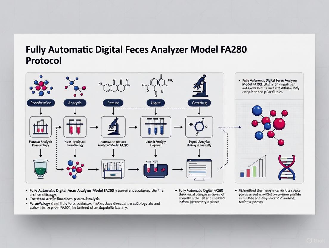
The analytical process follows a defined sequence, visualized in the workflow diagram below:
Figure 1: FA280 Automated Fecal Analysis Workflow. The process transforms a raw sample into a diagnostic report through sequential automated steps, with potential operator intervention only at the verification stage when the AI system identifies uncertain structures.
Performance Evaluation and Comparative Analysis
Diagnostic Agreement with Conventional Methods
Recent studies have demonstrated strong performance characteristics for the FA280 system in clinical and field settings. A mixed-methods study integrating quantitative and qualitative approaches evaluated the FA280's diagnostic performance for clonorchiasis using the Kato-Katz (KK) method as reference [3] [5].
Table 1: Diagnostic Performance of FA280 vs. Kato-Katz Method for Clonorchiasis Detection (n=1,000 participants)
| Performance Metric | FA280 Result | Statistical Significance |
|---|---|---|
| Positive Rate | 10.0% (identical to KK) | P > 0.999 (McNemar's test) |
| Overall Agreement | 96.8% | - |
| Kappa Statistic | 0.82 (95% CI: 0.76-0.88) | "Strong agreement" |
| Agreement in High Infection Intensity | Significantly higher | P < 0.05 |
| Agreement in Low Infection Intensity | Lower than high-intensity group | P < 0.05 |
The qualitative component of the study, involving interviews with medical staff and administrators, revealed that the FA280 outperformed the KK method in testing procedures, detection results, and user acceptance [3].
Comparative Performance with Other Automated Systems
The landscape of automated fecal analyzers includes several systems with varying capabilities. The following table compares the FA280 with another prominent automated system, the KU-F40:
Table 2: Comparative Analysis of Automated Fecal Analyzer Performance Characteristics
| Analyzer / Metric | FA280 | KU-F40 | Manual Microscopy |
|---|---|---|---|
| Overall Parasite Detection Rate | Comparable to KK (10.0% vs 10.0%) [3] | 8.74% [6] | 2.81% [6] |
| Number of Parasite Species Detected | 16+ species [1] | 9 species [6] | 5 species [6] |
| Key Advantages | Sealed system (odorless, leak-proof) [2], Strong agreement with KK (κ=0.82) [3] | Higher sensitivity for C. sinensis, hookworm, B. hominis [6] | Traditional gold standard, Low equipment cost |
| Automation Level | Full process: dilution, mixing, imaging, AI analysis [1] | Instrumental analysis with manual re-examination [6] | Fully manual |
| Sample Throughput | High (batch loading: 50 samples, kit system: up to 300) [2] | Not specified | Low (labor-intensive) |
Research Application Protocols
Standardized Protocol for Clonorchiasis Detection
For researchers investigating Clonorchis sinensis and other intestinal parasites, the following protocol details the validated methodology for using the FA280 system in community-based studies [3]:
Sample Collection and Preparation
- Collection Method: Use the manufacturer's patented sealed cartridge for sample collection.
- Sample Quantity: Approximately 0.5g of fecal sample is collected in a filtered sample collection tube.
- Sample Type: Fresh samples are preferred; preserved samples may be used if morphological integrity is maintained and the medium is compatible with the optical system.
- Transport: Maintain appropriate cold chain conditions during transport to the laboratory if immediate processing is not possible.
Instrument Setup and Calibration
- Quality Control: Execute built-in independent quality control systems to ensure accuracy and reliability.
- Reagent Preparation: Ensure adequate supply of diluents and processing solutions as per manufacturer specifications.
- System Check: Verify proper functioning of the high-frequency pneumatic mixing system, constant temperature incubation, and CMOS microscope imaging systems.
Sample Processing and Analysis
- Loading: Place samples in the cyclic loading system (batch loading of up to 50 samples supported).
- Automated Processing: The system automatically executes intelligent dilution appropriate for various sample consistencies, followed by constant temperature incubation for stable reactions.
- Imaging: The instrument employs three-channel multi-field imaging using a CMOS microscope and multi-field tomography, enabling rapid and rich imaging.
- AI Analysis: Artificial intelligence algorithms locate and track fecal components, automatically identifying parasite eggs with clear differentiation of internal structures.
Data Interpretation and Validation
- Result Review: The system generates reports automatically; however, expert review of flagged images is recommended for uncertain identifications.
- Validation: For research purposes, a subset of samples should undergo parallel testing with reference methods (e.g., Kato-Katz) to ensure ongoing quality assurance.
- Data Export: Results can be exported in formats compatible with statistical analysis software for further research analysis.
Quality Assurance and Control Measures
Implementation of a rigorous quality assurance program is essential for research applications:
- Pre-analytical Phase: Train personnel in proper sample collection techniques to ensure specimen adequacy.
- Analytical Phase: Utilize the built-in quality control systems and perform regular maintenance as specified in the service manual [4].
- Post-analytical Phase: Implement routine verification of AI identifications by parasitology experts, particularly for rare species or unusual morphological presentations.
Parasite Spectrum and Detection Capabilities
The FA280 system demonstrates detection capabilities for a broad spectrum of parasitic organisms, making it suitable for comprehensive parasitological surveys [1]:
Helminths
- Ascaris lumbricoides (both fertilized and unfertilized eggs)
- Hookworm eggs
- Trichuris trichiura eggs
- Strongyloides stercoralis larvae
- Taenia spp. eggs
- Opisthorchis viverrini / Liver fluke eggs
- Schistosoma japonicum eggs
- Hymenolepis nana eggs
- Enterobius vermicularis eggs
Protozoa
- Giardia lamblia (cysts and trophozoites)
- Blastocystis hominis (vacuolated form)
- Entamoeba histolytica
- Entamoeba coli
- Chilomastix mesnili (cysts and trophozoites)
Cells and Other Elements
- Red Blood Cells (RBC)
- White Blood Cells (WBC)
- Pus cells
- Yeast
- Charcot-Leyden crystals
- Fat globules
Essential Research Reagent Solutions
Successful implementation of the FA280 system in research settings requires specific reagents and materials. The following table outlines essential solutions and their functions:
Table 3: Essential Research Reagent Solutions for FA280 System Operation
| Reagent/Material | Function | Application Notes |
|---|---|---|
| Patented Sealed Cartridges | Sample containment; ensures biosafety and prevents leakage | Single-use, sterile; maintains sample integrity during processing [1] |
| Intelligent Diluent Solution | Standardizes sample viscosity; enables automated dilution | Adapts to various sample consistencies; formulation optimized for parasite preservation [2] |
| Quality Control Materials | Verifies instrument performance and AI recognition accuracy | Should include known positive and negative samples; used for daily QC protocols [4] |
| System Cleaning Solutions | Prevents cross-contamination between samples | Automated cleaning cycles between samples; specific formulations for optical components [4] |
| Calibration Standards | Ensures optical and imaging system precision | Microsphere-based and morphological standards; used during routine maintenance [4] |
AI Algorithm Architecture and Decision Pathway
The artificial intelligence component of the FA280 system employs a sophisticated decision-making process for parasite identification, which can be visualized as follows:
Figure 2: AI Decision Pathway for Parasite Identification. The algorithm progresses from image acquisition through feature extraction to classification, with a critical decision point at confidence threshold evaluation that determines whether automated identification proceeds or expert review is required.
Research Applications and Implementation Considerations
Applications in Epidemiological Studies
The FA280 system demonstrates particular utility in several research contexts:
- Large-Scale Epidemiological Surveys: The high throughput capacity (up to 300 samples with test kit system) enables efficient population-level screening [2].
- Drug Efficacy Trials: The quantitative capabilities and standardized detection allow for precise monitoring of infection intensity changes following intervention.
- Transmission Dynamics Studies: The broad parasite spectrum supports investigation of polyparasitism and co-infection patterns.
- Morphological Studies: High-resolution imaging with clear differentiation of internal structures facilitates detailed morphological analysis [2].
Practical Implementation Considerations
Researchers should address several practical considerations when implementing the FA280 system:
- Training Requirements: Although the interface is designed for simple use, technical staff require initial training, particularly for the review of AI-flagged images [1].
- Infrastructure Needs: The system requires appropriate laboratory space with stable power supply and environmental controls.
- Data Management: The system's ability to export results in formats compatible with laboratory information systems facilitates data integration and analysis [1].
- Maintenance Protocols: Regular maintenance as outlined in the service manual is essential for sustained optimal performance [4].
The FA280 Fully Automated Fecal Analyzer represents a significant technological advancement in parasitological diagnostics, offering researchers a standardized, high-throughput tool for intestinal parasite detection. Its strong agreement with conventional methods, combined with enhanced biosafety and reduced operator dependency, positions it as a valuable asset for epidemiological research, drug development, and public health surveillance. The integration of artificial intelligence with automated sample processing creates new opportunities for large-scale, standardized parasitological studies that were previously limited by the constraints of manual microscopy.
The Orienter Model FA280 represents a significant advancement in the diagnosis of intestinal parasitic infections by integrating full-process automation, high-resolution imaging, and artificial intelligence (AI). This fully automatic digital feces analyzer addresses critical limitations of traditional microscopic methods, which are labor-intensive, time-consuming, and heavily reliant on technician expertise [3] [7]. This document details the core technological components and experimental protocols for the FA280, providing a framework for researchers and drug development professionals engaged in diagnostic tool evaluation and implementation.
The diagnostic performance of the FA280 has been evaluated against established manual methods in multiple studies. The following tables summarize key quantitative findings.
Table 1: Comparison of FA280 Detection Performance against Reference Methods
| Evaluation Metric | vs. Kato-Katz (KK) for Clonorchiasis [3] | vs. Formalin-Ethyl Acetate Concentration Technique (FECT) [7] | vs. Normal Saline Staining (NSS) [8] |
|---|---|---|---|
| Sample Size | 1,000 participants | 200 fresh & 800 preserved samples | 350 patients |
| Positive Rate Agreement | 10.0% for both methods (P > 0.999) | Significant difference with AI report (P < 0.001) | Higher false-positive rate (PPV: 16.13%) |
| Overall Agreement | 96.8% | Perfect agreement with user audit (100%) | Low-to-moderate correlation (r = 0.39) |
| Kappa (κ) Statistic | 0.82 (95% CI: 0.76–0.88) | κ = 1.00 (with user audit) | N/A |
| Key Finding | Strong agreement, no significant difference | User audit crucial for optimal performance | High sensitivity of NSS (100%) |
Table 2: Strengths and Limitations of the FA280 System
| Aspect | Strengths | Limitations |
|---|---|---|
| Operational Efficiency | High-throughput; batch processing of 40-50 samples; ~30 min/run [7] [2] | Higher cost per test compared to manual methods [7] |
| Standardization & Safety | Fully sealed, automated process reduces biohazard risk and operator-to-operator variability [3] [9] | Performance can vary by parasite species and infection intensity [3] [8] |
| Detection Capability | AI can identify multiple parasite species (e.g., liver fluke, hookworm, roundworm) [2] [9] | May have lower sensitivity than methods using larger stool samples (e.g., FECT) [7] |
| Result Verification | High-resolution imaging allows for user audit and confirmation of AI findings [7] [6] | AI report alone may require manual verification for maximum accuracy [7] |
Experimental Protocols
Protocol 1: FA280 Operation for Parasite Detection
Principle: The FA280 uses automated sedimentation and concentration technology, combined with AI-driven image analysis, to identify parasite eggs and other fecal components in stool samples [3] [7].
Materials:
- Orienter FA280 Fully Automatic Digital Feces Analyzer
- Filtered sample collection tubes
- Appropriate diluents (instrument-specific)
Procedure:
- Sample Collection: Approximately 0.5 g of a fresh or preserved (10% formalin) stool sample is placed into a filtered sample collection tube [3] [7].
- Loading: The sample tube is loaded into the instrument's track-type sample carrier, which allows for cyclic loading of up to 50 samples per batch [2] [9].
- Automated Processing:
- Dilution and Mixing: The instrument automatically adds a diluent and uses a high-frequency pneumatic mixing system to create a homogeneous suspension [3] [9].
- Macroscopic Imaging: A high-resolution camera captures images of the sample's general characteristics (color, consistency) [7].
- Microscopic Imaging: The diluted sample is transferred to a flow cell or similar chamber. The integrated microscope, equipped with high- and low-power objectives, automatically captures multiple high-resolution images through multi-field tomography [3] [7].
- AI Analysis: Captured images are analyzed by the integrated AI software, which is trained to locate, identify, and classify parasite eggs based on features like color, shape, and internal structures [2].
- Result Reporting & Audit: The software generates a report. For optimal accuracy, a trained medical technologist should perform a user audit by reviewing the captured images to confirm or correct the AI's findings before finalizing the report [7] [6].
Protocol 2: Reference Method - Kato-Katz Technique
Principle: This manual method involves preparing a standardized thick smear of sieved stool to clear debris, allowing for the microscopic detection and quantification of helminth eggs [3].
Materials:
- Plastic template (hole size of 41.7 mg)
- Glass slides
- Cellophane strips soaked in glycerol-malachite green solution
- Microscope
Procedure:
- Place the template on a glass slide.
- Fill the template hole with sieved stool, ensuring no gaps or air bubbles.
- Carefully remove the template, leaving a standardized fecal smear on the slide.
- Cover the smear with a glycerol-soaked cellophane strip.
- Invert the slide and press gently on absorbent paper to spread the glycerol and clear the sample.
- Allow the slide to clear for a recommended time (e.g., 30-60 minutes) before microscopic examination.
- Examine the entire smear systematically under a microscope using a 10x objective. Identify and count parasite eggs (e.g., C. sinensis). The count multiplied by 24 gives the eggs per gram (EPG) of feces [3].
System Workflow and AI Analysis
The FA280 integrates several subsystems into a cohesive diagnostic workflow. The following diagram illustrates the logical flow from sample input to final report.
Diagram 1: FA280 Operational and Analysis Workflow
The Scientist's Toolkit: Essential Research Reagents & Materials
The following table lists key materials and their functions for conducting research with the FA280 system.
Table 3: Key Research Reagent Solutions and Materials
| Item Name | Function/Application | Specification Notes |
|---|---|---|
| Filtered Sample Collection Tubes | Primary container for stool specimen submission. | Designed for use with the FA280's automated sampling system [3]. |
| Proprietary Diluent | Standardizes stool consistency for optimal imaging and analysis. | Volume is automatically dispensed by the instrument; composition not specified in public literature [7] [9]. |
| Quality Control (QC) Materials | Verifies instrument and AI algorithm performance. | Part of the built-in independent QC system to ensure accuracy [2] [9]. |
| Formalin (10%) | Preservation of stool samples for delayed testing. | Used in studies evaluating performance on preserved samples [7]. |
| Kato-Katz Reagents | Reference method validation. | Glycerol and malachite green for cellophane strips [3]. |
| FECT Reagents | Reference method validation. | Formalin and ethyl acetate for concentration technique [7] [10]. |
The Orienter FA280 fully automated digital feces analyzer leverages integrated automation, advanced imaging, and AI to modernize stool analysis. While its performance is robust and shows strong agreement with traditional methods, the literature consistently emphasizes that expert user audit of AI-generated results is critical for achieving maximal diagnostic accuracy [7] [6] [8]. This combination of technological innovation and expert oversight makes the FA280 a valuable tool for high-throughput clinical and research applications in parasitology.
The Role of Artificial Intelligence in Parasite Egg Identification and Classification
The integration of artificial intelligence (AI) into parasitology diagnostics is transforming traditional microscopy, offering solutions to long-standing challenges of labor-intensity, time consumption, and operator dependency. This article details the application of AI, with a focus on the fully automatic digital feces analyzer Orienter Model FA280, for the identification and classification of parasitic eggs. We present standardized protocols for evaluating the FA280 system, quantitative performance data compared to reference methods, and essential reagent solutions required for implementation. Framed within broader research on automated fecal analyzers, this document provides researchers, scientists, and drug development professionals with the technical foundation for adopting and advancing AI-driven diagnostic technologies in parasitology.
Parasitic infections, particularly intestinal parasites, remain a significant global health burden, affecting billions of people and causing morbidity through malnutrition, anemia, and impaired growth [3] [7]. Diagnosis traditionally relies on manual microscopic examination of stool samples, a method plagued by high labor costs, substantial time requirements, and critical dependence on the expertise and training of the microscopist [3] [7] [11]. These limitations hinder large-scale screening and surveillance efforts, which are essential for effective public health interventions.
The advent of fully automated digital feces analyzers represents a paradigm shift in diagnostic parasitology. These systems, such as the Orienter Model FA280, leverage AI-powered image analysis to automate the detection and classification of parasite eggs in stool samples [3] [7] [2]. By combining high-throughput imaging with deep learning algorithms, they offer a compelling alternative that enhances standardization, increases efficiency, and reduces the operational burden associated with traditional methods [3] [2]. This document outlines the application, performance, and protocols for utilizing the FA280 system, situating it as a cornerstone technology in the future of parasitic disease management and research.
Performance Evaluation: Quantitative Data
The diagnostic performance of the FA280 has been evaluated against established manual methods in multiple studies. The following tables summarize key quantitative findings, providing a clear comparison of its capabilities.
Table 1: Diagnostic Agreement of the FA280 vs. Reference Methods for Clonorchis sinensis Detection (Community-Based Survey, n=1000) [3]
| Metric | FA280 vs. Kato-Katz (KK) | Statistical Value |
|---|---|---|
| Positive Rate | Both Methods | 10.0% |
| Overall Agreement | 96.8% | |
| Kappa (κ) Statistic | 0.82 | |
| 95% CI for Kappa | 0.76 - 0.88 | |
| McNemar's Test P-value | > 0.999 |
Table 2: Performance of the FA280 with User Audit vs. Formalin-Ethyl Acetate Concentration Technique (FECT) [7]
| Parasite Type | Sample Set | Agreement for Species Identification (κ) | Remarks |
|---|---|---|---|
| General Parasites | 200 Fresh Samples | 1.00 (Perfect) | FECT and FA280 with user audit showed no statistically significant difference (P=1). |
| Helminths | 800 Preserved Samples | 0.857 (Strong) | FECT detected more positives, potentially due to larger sample size used. |
| Protozoa | 800 Preserved Samples | 1.00 (Perfect) |
Table 3: Performance of Other AI-Based Platforms for Parasite Egg Detection
| Platform / Model | Application | Key Performance Metric | Value |
|---|---|---|---|
| Expert-Verified AI [11] | Soil-transmitted helminths in human stool | Sensitivity (Hookworm / T. trichiura / A. lumbricoides) | 92% / 94% / 100% |
| Vetscan Imagyst [12] | Strongyles in equine feces | Diagnostic Sensitivity (vs. Mini-FLOTAC) | 99.2% (NaNO3) - 100% (Sheather's) |
| YCBAM Model [13] | Pinworm eggs in microscopic images | Mean Average Precision (mAP@0.50) | 0.995 |
Experimental Protocols
Protocol: Cross-Sectional Evaluation of the FA280 vs. Kato-Katz Method
This protocol is adapted from a community-based study evaluating the FA280 for the diagnosis of Clonorchis sinensis [3].
I. Objective To evaluate the diagnostic performance, including positive rate and agreement, of the FA280 fully automated fecal analyzer against the Kato-Katz (KK) method for detecting Clonorchis sinensis infections in a community-based population.
II. Materials and Reagents
- Orienter Model FA280 analyzer and its consumables (filtered sample collection tubes, diluent)
- Stool collection boxes
- Kato-Katz materials: plastic templates (41.7 mg), glass slides, cellophane coverslips, glycerol, malachite green, gauze for sieving
- Light microscopes (e.g., Olympus CX23)
- Quality control reagents
III. Experimental Workflow
IV. Procedure
- Sample Collection & Preparation: Recruit participants using a multi-stage cluster sampling method. Distribute stool collection boxes and instruct participants to provide a single stool sample. Collect samples and transport them to the laboratory under appropriate conditions.
- Kato-Katz Method: a. For each stool sample, prepare two Kato-Katz thick smears using a 41.7 mg template. b. Cover each smear with glycerol-soaked, malachite-green-stained cellophane. c. Allow slides to clear for a predetermined time. d. Examine slides under a microscope by experienced technicians who count and record the number of C. sinensis eggs. e. Perform quality control by having a senior professional re-examine a random subset of samples (e.g., 10 per village).
- FA280 Method: a. Transfer approximately 0.5 g of the same stool sample into a filtered sample collection tube. b. Load the tube into the FA280 analyzer. c. Initiate the automated process, which includes intelligent dilution, pneumatic mixing, high-resolution imaging via multi-field tomography, and AI-based egg identification and reporting.
- Data Analysis: a. Compare positive rates between the two methods using McNemar's test (P < 0.05 considered significant). b. Calculate the agreement between the two methods using the Kappa (κ) statistic. Interpret κ values as follows: 0-0.20 (slight), 0.21-0.40 (fair), 0.41-0.60 (moderate), 0.61-0.80 (substantial), 0.81-1.00 (almost perfect).
Protocol: Performance Assessment of AI Algorithm with User Audit
This protocol outlines the procedure for validating the FA280's AI report against a expert user audit, which is critical for ensuring diagnostic accuracy [7].
I. Objective To assess the agreement between the AI-generated report of the FA280 and a subsequent audit by a skilled medical technologist, and to compare both against a reference method (e.g., FECT).
II. Materials and Reagents
- Orienter Model FA280 system
- Materials for FECT: 10% formalin, ethyl acetate, centrifuge tubes, gauze, conical centrifuge tubes, applicator sticks, pipettes
- Preserved stool samples (e.g., in 10% formalin)
III. Procedure
- Sample Preparation: For a set of stool samples (e.g., n=200 fresh or n=800 preserved), perform the FECT as the reference standard. a. Emulsify 2 g of stool in 10 ml of 10% formalin. b. Filter the suspension through gauze into a conical tube. c. Add 3 ml of ethyl acetate, shake vigorously, and centrifuge. d. Examine the sediment under a microscope for parasite eggs [7].
- FA280 Testing with AI Report: Process the same samples using the FA280 and collect the initial report generated solely by the AI algorithm.
- User Audit: A skilled medical technologist then reviews the digital images captured by the FA280, reclassifying and confirming any findings. This generates a second, audited report.
- Data Comparison: a. Calculate the agreement (using κ statistics) between the FA280 AI report and the FECT. b. Calculate the agreement between the FA280 user audit report and the FECT. c. Compare the κ values to determine the added value of expert verification.
The Scientist's Toolkit: Research Reagent Solutions
The following table catalogues essential materials and reagents for conducting research with the FA280 and related parasitological methods.
Table 4: Essential Research Reagents and Materials for FA280 Protocol Research
| Reagent/Material | Function/Application | Research Context |
|---|---|---|
| Filtered Sample Collection Tubes (FA280) | Standardized containment and initial filtration of stool specimen. | Ensures consistent sample input and prevents large particulates from interfering with the FA280's automated fluidics and imaging system [3] [7]. |
| Proprietary Diluent (FA280) | Standardization of stool consistency and preparation for imaging. | Critical for creating a uniform suspension for optimal high-resolution imaging and AI analysis [2]. |
| Formalin (10%) | Preservation of stool samples and inactivation of pathogens. | Used for preserving stool samples for later batch testing with FECT or other methods; allows for safe transport and storage [7]. |
| Ethyl Acetate | Solvent for extraction and purification in concentration techniques. | Key component in FECT; helps to extract debris and fat, leaving parasite eggs in the sediment for easier microscopic identification [7]. |
| Glycerol & Malachite Green | Clearing and staining agents for Kato-Katz smears. | Glycerol clears debris for visual egg detection, while malachite green aids in staining and visualization [3]. |
| Sodium Nitrate (NaNO3) or Sheather's Sugar Solution | Flotation solutions for parasite egg concentration. | Creates a specific gravity solution that causes parasite eggs to float to the surface, enriching them for detection in various manual and automated methods [12]. |
AI Technology Workflow and Classification Logic
The core of systems like the FA280 is a deep learning-based image analysis pipeline. The following diagram and description detail this logical process.
AI Parasite Egg Identification and Classification Logic
Description of the AI Logical Process:
- Digital Image Acquisition: The system captures high-resolution, multi-field tomographic images of the prepared fecal sample [3] [2].
- Image Preprocessing: Images are normalized and enhanced to standardize lighting and improve feature clarity.
- Scene Splitting: The scanned image is broken down into smaller, manageable scenes for detailed analysis [12].
- Feature Extraction: A deep learning algorithm, typically a convolutional neural network (CNN), analyzes the scenes. It converts pixels into discriminative features such as egg shape, edge characteristics, color gradients, and internal structures [13] [12]. This process is repeated through multiple layers to create abstract, high-level feature representations.
- Classification & Object Detection: The extracted features are used to calculate a probability score for each potential parasite egg type the algorithm has been trained to recognize (e.g., liver fluke, hookworm, roundworm) [2] [12].
- Probability Threshold: Only detections above a pre-set confidence threshold are reported, reducing false positives.
- Report Generation: The system compiles the findings into a report, which may include the identity of detected parasites, egg counts (for quantification), and the captured images [7].
- Expert Verification (Optional but Recommended): In the "expert-verified AI" paradigm, a human expert reviews the AI-generated findings, typically by auditing a shortlist of candidate objects presented by the AI. This hybrid approach has been shown to achieve higher sensitivity than either fully autonomous AI or manual microscopy alone, while drastically reducing the expert's workload [11].
This application note details the standardized operational protocol for the Orienter Model FA280 Fully Automatic Digital Feces Analyzer, an integrated system designed to automate and enhance the efficiency and accuracy of stool analysis for parasitic infection diagnosis.
The FA280 Fully Automatic Digital Feces Analyzer represents a significant advancement in parasitology diagnostics, transforming the traditionally labor-intensive and subjective manual microscopy into a streamlined, automated, high-throughput workflow [3]. By integrating automated sample processing, high-resolution digital imaging, and artificial intelligence (AI), the system minimizes manual intervention, reduces biosafety risks, and standardizes result reporting [7] [2]. This document provides a detailed protocol for the operation of the FA280, from sample preparation to the final reporting of results, providing researchers and laboratory professionals with a clear framework for its application in clinical and research settings.
Operational Workflow
The FA280 workflow is a continuous, automated process that begins with sample loading and concludes with a digitally generated report. The following diagram and table outline the key stages from start to finish.
Figure 1: The FA280 Fully Automated Workflow. This process from sample loading to reporting takes approximately 30 minutes for a batch of 40 samples [7].
Table 1: FA280 Workflow Stage Descriptions
| Stage | Process Name | Description | Key Technical Features |
|---|---|---|---|
| A | Sample Loading & ID Registration | Sample cups are placed on the track. | Cyclic loading allows batch processing of up to 50 samples [2]. |
| B | Automated Sampling & Intelligent Dilution | The instrument automatically aspirates and dilutes the sample. | Pneumatic mixing ensures homogenization; intelligent dilution standardizes varying consistencies [7] [2]. |
| C | Macroscopic Imaging | A high-resolution camera captures images of the sample's physical attributes. | Determines color, form, and consistency automatically [7]. |
| D | Automated Sedimentation & Concentration | The sample undergoes concentration within a sealed system. | Uses automatic sedimentation and concentration technology to prepare the sample for microscopic examination [3]. |
| E | High-Resolution Microscopic Imaging | The microscope automatically captures multiple digital images. | Utilizes multi-field tomography with high- and low-power objectives for detailed sectional imaging [7] [2]. |
| F | AI-Powered Image Analysis | Software analyzes images to identify and classify parasite eggs. | AI algorithm locates and tracks fecal components, identifying parasite eggs (e.g., liver fluke, hookworm) with clear differentiation [2]. |
| G | Result Auditing & Validation | A technician reviews the AI-generated findings. | User audit of digital images and AI report by skilled personnel ensures accuracy and reliability [3] [7]. |
| H | Automated Report Generation | The system compiles findings into a final report. | Report includes sample attributes, detected parasites, and captured images for clinical interpretation [7]. |
Performance Validation & Comparative Data
The diagnostic performance of the FA280 has been rigorously evaluated in comparative studies against established manual methods. The following table summarizes key quantitative findings from recent research.
Table 2: Comparative Performance of the FA280 vs. Traditional Methods
| Evaluation Metric | Comparison Method | Key Quantitative Findings | Study Context |
|---|---|---|---|
| Agreement & Positive Rate | Kato-Katz (KK) Method | 96.8% agreement (κ=0.82); No significant difference in positive rate (10.0% for both) [3] [5]. | Cross-sectional survey of 1,000 participants for Clonorchis sinensis diagnosis [3]. |
| Detection Rate | Formalin-Ether Concentration Technique (FECT) | FECT detected significantly more positive samples (P<0.001), attributed to its use of a larger stool sample (2g vs. ~0.5g) [7]. | Analysis of 800 preserved stool samples for a broad range of parasites [7]. |
| Species Identification Agreement | FECT with User Audit | Perfect agreement for protozoa (κ=1.00) and strong agreement for helminths (κ=0.857) in species identification [7]. | Evaluation of 200 fresh stool samples [7]. |
| Operational Throughput | Manual Microscopy | Processes a batch of 40 samples in ~30 minutes, significantly faster than manual methods [7]. | Standard operational procedure of the FA280 system [7]. |
The Scientist's Toolkit: Essential Research Reagents & Materials
Table 3: Key Research Reagent Solutions for FA280 Operation
| Item | Function & Application in Protocol |
|---|---|
| Filtered Sample Collection Tube | A specialized container for collecting and pre-filtering approximately 0.5g of stool sample, ensuring compatibility with the automated sampling system [3]. |
| Proprietary Diluent Solution | A liquid reagent used for the intelligent dilution and pneumatic mixing of the stool sample, standardizing its consistency for optimal imaging and analysis [7] [2]. |
| Sealed Test Kit Cartridge | A disposable, sealed cartridge that houses the sample during processing and imaging. This is part of the enclosed system that prevents odor and leaks, enhancing laboratory safety [7] [2]. |
| Quality Control (QC) Materials | Built-in independent QC systems and likely external control samples to ensure the analyzer's accuracy and reliability over time [2]. |
| Formalin (10%) for Preservation | Used for preserving stool samples in research studies involving delayed or batched testing, as evaluated in validation studies [7]. |
Experimental Protocol for Method Comparison
For researchers aiming to validate the FA280 against a reference method, the following detailed protocol for a cross-sectional comparison study is provided.
Sample Collection and Preparation
- Sample Size Calculation: Determine the required sample size using statistical software. A prior study with 1000 participants was calculated based on an assumed kappa (κ) of 0.9, a 95% confidence level, and 90% statistical power, with an added buffer for dropouts [3].
- Sample Acquisition: Collect fresh stool samples from participants in clean, sterile containers. For preserved sample analysis, mix a portion of the sample with 10% formalin [7].
- Sample Division: For method comparison, each stool sample must be subdivided for parallel testing with the FA280 and the chosen reference method (e.g., KK or FECT).
Parallel Detection Procedures
- FA280 Analysis:
- Weigh approximately 0.5g of feces into the provided filtered sample collection tube [3].
- Load the tube onto the FA280 sample track and register the sample ID.
- Initiate the automated process. The instrument will handle dilution, mixing, imaging, and AI analysis.
- Upon completion, a qualified medical technologist must conduct a user audit of the AI-generated findings and images before finalizing the report [7].
- Kato-Katz Method (Reference):
- Place a plastic template with a 41.7 mg hole on a glass slide.
- Fill the template with sieved stool and remove the template.
- Cover the sample with glycerol-soaked malachite green cellophane.
- Have experienced technicians examine the smear under a microscope and count parasite eggs [3].
- Implement quality control by having a senior staff member re-examine a random subset of slides (e.g., 10 per village) [3].
Data Analysis
- Statistical Agreement: Use McNemar's test to compare positive rates and Cohen's kappa statistic (κ) to evaluate agreement between the two methods. A kappa value above 0.8 indicates strong agreement [3].
- Infection Intensity Analysis: Categorize positive samples into low and high infection intensity groups based on eggs per gram (EPG) and use Pearson's Chi-square test to analyze consistency between methods across these groups [3].
The FA280 Fully Automatic Digital Feces Analyzer offers a robust, high-throughput, and standardized solution for fecal parasite testing. Its integrated workflow from sample loading to automated reporting, backed by AI and high-resolution imaging, provides a reliable and efficient alternative to traditional methods, demonstrating high agreement with established techniques like the Kato-Katz method [3] [5].
Standardized FA280 Operational Protocol: A Step-by-Step Guide
The fully automatic digital feces analyzer model FA280 represents a significant advancement in parasitology diagnostics, integrating automation, high-resolution digital imaging, and artificial intelligence (AI) to modernize stool analysis [2] [7]. This automated system addresses critical limitations of traditional microscopic methods, including labor intensity, time consumption, and reliance on technical expertise [3] [7]. Within a broader thesis on FA280 protocol research, this application note provides detailed methodologies for sample collection and preparation, which are fundamental to obtaining reliable diagnostic results. Proper pre-analytical procedures ensure the system's AI can accurately identify parasitic elements, thereby maximizing diagnostic performance across clinical and research applications.
Technical Specifications and Performance Data
The table below summarizes key technical specifications of the FA280 system and its comparative performance against traditional methods, as validated in recent studies.
Table 1: FA280 Technical Specifications and Performance Metrics
| Parameter | Specification / Finding | Source / Context |
|---|---|---|
| Sample Throughput | Batch processing of 40-50 samples per run; complete in ~30 minutes [7]. | Operational workflow |
| Sample Weight | Approximately 0.5 grams of stool per test [3] [7]. | Standard test requirement |
| Detection Principle | Automated sedimentation and concentration; CMOS microscope with multi-field tomography [3] [2]. | Core technology |
| Image Analysis | AI-based software for automatic identification; allows for user audit of captured images [7] [14]. | Analytical method |
| Agreement with Kato-Katz | 96.8% overall agreement (κ=0.82) for Clonorchis sinensis detection [3]. | Performance in community survey (n=1000) |
| Agreement with FECT (Protozoa) | Perfect agreement (κ=1.00) with user audit for species identification [7] [15]. | Performance on preserved samples |
| Agreement with FECT (Helminths) | Strong agreement (κ=0.857) with user audit for species identification [7] [14]. | Performance on preserved samples |
Experimental Protocols for Method Validation
The following protocols are derived from studies that validated the FA280 against established reference methods.
Protocol 1: Comparative Cross-Sectional Survey for Clonorchiasis
This methodology was used to evaluate the diagnostic performance of the FA280 for detecting Clonorchis sinensis in a community setting [3].
3.1.1 Sample Collection
- Design: A cross-sectional survey was conducted in an endemic area.
- Participants: 1,000 participants were enrolled via a multi-stage cluster sampling method.
- Procedure: Collection boxes were distributed to participants one day before the scheduled stool collection.
3.1.2 Sample Analysis
- FA280 Method: Approximately 0.5 g of each fecal sample was placed in a filtered sample collection tube. The device automatically performed dilution, mixing, microscopic observation, and AI-based image analysis [3].
- Reference Method (Kato-Katz): For each sample, two smears were prepared using a 41.7 mg template. Experienced technicians examined the smears under a microscope for C. sinensis eggs [3].
- Quality Control: Ten stool samples from each study village were re-examined by a senior professional to ensure quality.
3.1.3 Data Analysis
- Statistical analysis compared the positive rates and agreement between the two methods using McNemar's test and the kappa (κ) statistic.
Protocol 2: Laboratory Comparison with FECT for Intestinal Parasites
This protocol compared the FA280 with the Formalin-Ethyl Acetate Concentration Technique (FECT) for detecting a broad range of intestinal parasites [7] [14].
3.2.1 Sample Sets
- The study utilized two sets of samples:
- 200 fresh stool samples collected routinely.
- 800 stool samples preserved in 10% formalin.
3.2.2 Sample Analysis
- FECT Method: 2 g of stool was mixed with 10 ml of 10% formalin. The suspension was strained, mixed with ethyl acetate, centrifuged, and the sediment examined under a light microscope [7].
- FA280 Method: The analyzer processed approx. 0.5 g of stool. Results were generated both by the built-in AI program and through a user audit, where a skilled technologist reviewed the digital images captured by the device [7] [15].
Workflow Diagram: FA280 Sample Processing
The following diagram illustrates the logical workflow of sample processing and analysis using the FA280 system, from collection to result reporting.
FA280 Sample Processing Workflow: This diagram outlines the sequential steps from sample collection to result generation, highlighting key automated processes and optional user verification.
The Scientist's Toolkit: Research Reagent Solutions
The table below lists essential materials and reagents used in conjunction with the FA280 analyzer and related comparative methods.
Table 2: Essential Research Reagents and Materials for Fecal Analysis
| Item | Function / Application |
|---|---|
| Filtered Sample Collection Tubes | Designed for the FA280; used to hold the ~0.5g stool sample and facilitate automated filtration and dilution [3] [7]. |
| Intelligent Diluent | Specific diluent used by the FA280; standardizes various sample consistencies for uniform analysis [2]. |
| Formalin (10%) | Preservative used for storing stool samples prior to processing with FECT or other reference methods [7] [14]. |
| Ethyl Acetate | Solvent used in the FECT method to separate parasitic elements from fecal debris through centrifugation [7]. |
| Ethyl Alcohol | Used in preparatory protocols for automated diagnosis to fix and prepare samples on microscopy slides [16]. |
| Cationic Surfactants (e.g., CTAB) | Charge-modifying reagents shown in advanced protocols to improve parasite recovery from stool samples during processing [16]. |
Discussion and Best Practices
Adherence to standardized sample collection and preparation protocols is critical for ensuring the high performance of the FA280 analyzer. Key considerations include:
- Sample Quantity: Consistently using the recommended 0.5 g sample size is crucial. Studies note that the lower sensitivity compared to FECT can be attributed to the FECT's use of a larger sample mass (2 g), which increases the probability of detecting parasites, particularly in low-intensity infections [7] [14].
- Sample Quality and Handling: The FA280 offers a significant safety advantage by performing the entire detection process in a fully sealed, leak-proof system, thereby reducing operator exposure to odors and potential pathogens [2].
- The Critical Role of User Audits: While the FA280's AI provides automated identification, the diagnostic agreement with reference methods like FECT improves markedly—from fair to perfect for protozoa—when a skilled technologist audits the AI-generated images [7] [15]. This underscores that the technology serves as a powerful aid rather than a complete replacement for expert morphological knowledge.
- Method Selection for Target Parasites: The FA280 demonstrates high agreement with the Kato-Katz method for helminths like C. sinensis and strong agreement with FECT for common protozoa when audited [3] [14]. The choice of reference method for validation should align with the target parasites and the study context (e.g., community survey vs. clinical lab).
In conclusion, the Orienter FA280 fully automatic digital feces analyzer, when used with the precise sample collection and preparation guidelines outlined in this document, provides a rapid, safe, and reliable platform for the diagnosis of intestinal parasitic infections.
Step-by-Step FA280 Instrument Operation and Sample Processing Protocol
The FA280 Fully Automatic Digital Feces Analyzer represents a significant advancement in parasitology diagnostics, enabling automated processing, imaging, and analysis of fecal samples. This protocol details the comprehensive operation of the FA280 system within the broader research context of automated fecal analysis technology. The instrument utilizes digital imaging technology and machine learning algorithms to identify parasites and eggs in fecal samples, substantially improving efficiency compared to traditional manual methods [17]. The system is designed with several integrated units: automatic sampling, puncture sampling, sample characteristics and color observation, imaging, and reagent card units [17]. This automation reduces labor intensity, minimizes cross-contamination risks, and standardizes the diagnostic process for more consistent results in research and clinical settings.
Principle of Operation
The FA280 system operates on the principle of automated digital microscopy combined with intelligent image analysis. The instrument employs automated pre-processing, imaging, and detection algorithms to identify pathological components in fecal samples [4]. The operational workflow involves:
- Automated sample preparation with intelligent dilution and filtration systems
- High-resolution digital imaging through integrated microscopy systems
- Machine learning-based analysis of captured images for pathogen detection
- Result verification through user audit functionality
The system utilizes automatic sedimentation filtration and concentration methods, which fall under the category of sedimentation techniques, enhancing the detection of parasites and eggs compared to direct smear methods [18]. This technical approach demonstrates superior sensitivity (96.7%) for detecting Clonorchis sinensis compared to traditional醛醚离心沉淀法(Aldehyde Ether Centrifugal Precipitation, AECP) (82.9%) and ELISA (86.2%) methods [18].
Materials and Equipment
Required Equipment
Table 1: Essential Equipment for FA280 Operation
| Equipment Name | Specifications/Type | Primary Function |
|---|---|---|
| FA280 Fully Automatic Feces Analyzer | Main analysis unit | Automated processing and analysis |
| Computer System | With installed software | Instrument control and data management |
| Specialized Specimen Collection Tubes | Manufacturer-provided | Standardized sample collection |
| Disposable Pipettes | - | Liquid fecal sample collection |
| Laboratory Information System (LIS) | - | Result storage and management |
Research Reagent Solutions
Table 2: Key Research Reagents and Their Functions
| Reagent/Material | Function | Application Notes |
|---|---|---|
| Dilution Solution | 5 mL volume | Sample homogenization and preparation |
| Concentration Transparent Solution | System flushing | Post-analysis instrument cleaning |
| Formalin (10%) | Parasite fixation | Used in AECP method [18] |
| Ethyl Ether | Lipid dissolution | Used in AECP method [18] |
| 专用粪便样本收集容器 | Sample containment | Ensures proper sample integrity |
Sample Collection and Preparation
Specimen Collection Protocol
- Collection Tube Selection: Use manufacturer-provided specimen collection tubes that are sealed, clean, dry, leak-proof, with appropriate openings and volume for multi-point sampling [17].
- Sample Quantity: Collect approximately 0.5 g of fresh fecal matter from abnormal areas containing mucus, pus, or blood [17].
- Liquid Stool Handling: For watery or loose stools, use a disposable pipette to collect the necessary sample volume [17].
- Contamination Prevention: Avoid samples contaminated with non-fecal matter including enema or oily laxative feces, samples from bedpans or toilets, or specimens contaminated with plant matter, soil, sewage, disinfectants, cleaners, urine, menstrual blood, or leukorrhea [17].
Sample Quality Assessment
Ensure samples are:
- Fresh and properly preserved
- Representative of abnormal areas when present
- Free of external contaminants
- Adequate in volume (approximately 0.5 g)
Step-by-Step Operational Protocol
Instrument Startup and Initialization
- Power Activation: Turn on the computer and FA280 analyzer power switches.
- Software Launch: Click to start the software and log in with username and password.
- System Self-Test: Wait for the instrument to complete automatic self-check procedures [17].
Sample Processing and Analysis
- Sample Loading: Place the qualified specimen collection tube into the analyzer's sample rack and position the rack on the automatic feed inlet.
- Analysis Initiation: Click "Start" for the first test after system self-test completion.
- Automated Processing: The instrument automatically:
- Performs robotic photography of the sample for documentation
- Adds 5 mL of diluent to the sample tube
- Conducts pneumatic mixing for homogenization
- Allows sample sedimentation before analysis
- Filters samples through specialized filters to remove non-pathological residues (plant fibers, seeds, undigested food particles)
- Transfers samples to counting chamber for analysis [17]
- Image Capture and Analysis: The analyzer captures high-resolution microscopic images and processes them through recognition software.
Result Verification and Data Management
- Result Review: Switch to the results interface to examine test images.
- Data Export: Send results to the Laboratory Information System (LIS) or print reports.
- Analysis Completion: Click "Stop" to end the testing session.
- System Shutdown: Select "Flush and Shutdown" option, then load the concentrated transparent solution rack. The instrument will automatically flush and shut down [17].
Methodology Comparison and Performance Evaluation
Detection Time Analysis
Table 3: Comparative Analysis of Detection Times Across Methodologies
| Methodology | Average Testing Time | Key Process Steps | Personnel Requirements |
|---|---|---|---|
| Direct Wet Smear Microscopy | Manual calculation required | Sample placement on slide, smear preparation, microscopic examination | Labor-intensive, requires technical expertise |
| FA280 Analyzer (AI Report) | Automated process | Fully automated processing and analysis | Minimal operator intervention |
| FA280 Analyzer (User Audit) | Automated process + manual image review time | Automated analysis + manual verification of images | Requires experienced technician for audit |
Diagnostic Performance Metrics
Table 4: Performance Characteristics of FA280 Analysis Modalities
| Performance Metric | FA280 (AI Report) | FA280 (User Audit) | Traditional Microscopy |
|---|---|---|---|
| Sensitivity | 84.31% [17] | 94.12% [17] | Varies with technician skill |
| Specificity | 98.71% [17] | 99.69% [17] | Varies with technician skill |
| Positive Rate (C. sinensis) | 87.8% [18] | - | 76.3% (AECP method) [18] |
| Efficiency | High-throughput capability | Moderate throughput with enhanced accuracy | Low-throughput, labor-intensive |
Quality Control and Maintenance
Routine Quality Assurance
- Regular Calibration: Follow manufacturer recommendations for system calibration
- Image Verification: Implement periodic review of AI-generated results by experienced technicians
- Sample Integrity Checks: Verify collection tube quality and sample adequacy
System Maintenance Protocol
- Post-Analysis Flushing: Execute proper flushing procedures after each use
- Component Inspection: Regular checking of sampling units, imaging systems, and reagent delivery mechanisms
- Software Updates: Maintain current software version for optimal algorithm performance
Troubleshooting Common Issues
- Image Quality Problems: Check camera alignment, focus calibration, and sample preparation consistency
- Sample Processing Errors: Verify proper sample viscosity, dilution ratios, and filtration functionality
- Inconsistent Results: Implement user audit function to verify AI findings, particularly for ambiguous cases
The FA280 Fully Automatic Digital Feces Analyzer represents a significant technological advancement in parasitology diagnostics, providing researchers with a standardized, efficient, and accurate platform for fecal analysis. The combination of automated processing with optional user audit functionality enables both high-throughput screening and precise diagnostic confirmation, making it particularly valuable for large-scale research studies and clinical trials requiring consistent fecal analysis methodology.
FA280 Operational Workflow Diagram
Methodology Performance Comparison Diagram
The FA280 Fully Automatic Digital Feces Analyzer represents a significant advancement in parasitic diagnostics, utilizing automated sedimentation and concentration as its core detection principle. This technology addresses critical limitations of traditional manual methods, which are labor-intensive, time-consuming, and heavily reliant on technical expertise [3]. The system integrates intelligent sample dilution, high-frequency pneumatic mixing, and AI-driven parasite egg identification to streamline the diagnostic process while maintaining accuracy comparable to established techniques [3].
This automated approach is particularly valuable for diagnosing clonorchiasis and other intestinal parasitic infections, which affect approximately 3.5 billion people globally and can lead to serious health complications including malnutrition, anemia, impaired growth, and cognitive development [7]. By implementing a consistent, automated sedimentation and concentration protocol, the FA280 reduces operator dependency and variability while increasing throughput to approximately 40 samples per 30-minute run [7].
Core Technological Principle: Automated Sedimentation and Concentration
Fundamental Mechanism
The FA280's detection principle centers on an automated sedimentation and concentration technique that enhances parasite egg detection through standardized mechanical processes. Unlike traditional formalin-ether concentration technique (FECT) that requires manual centrifugation and 2g stool samples, the FA280 employs intelligent sample dilution and automatic sedimentation with approximately 0.5g of fecal material [3] [7]. This reduction in sample requirement, combined with automated processing, significantly decreases manual handling while maintaining diagnostic accuracy.
The system operates on simple sedimentation principles but enhances them through precision instrumentation. After sample collection in filtered collection tubes, the instrument automatically adds diluent and employs high-frequency pneumatic mixing to create a homogeneous suspension [3]. This standardized approach minimizes human error in preparation steps that often affect traditional methods. The automated process ensures consistent mixing intensity and duration, critical factors for reliable sedimentation outcomes.
Integrated Detection System
Following sedimentation, the FA280 utilizes a digital imaging system with high-resolution cameras that capture multiple focal planes through multi-field tomography [3]. This comprehensive imaging approach ensures that parasite eggs at different levels in the sediment are visualized, overcoming a limitation of single-plane manual microscopy. The acquired images are then analyzed by artificial intelligence algorithms trained to identify characteristic features of various parasites based on color, shape, and size attributes [3] [7].
The system incorporates a track-type sample carrier that ensures precise positioning and movement of samples through the entire workflow, from initial preparation to final imaging [7]. This automation creates a continuous processing stream that maximizes efficiency while minimizing cross-contamination risks between specimens. The complete integration of sedimentation, concentration, and digital imaging within a single platform represents a significant advancement over the discrete, manual steps required by conventional methods.
Experimental Protocols and Methodologies
Sample Preparation Protocol
Table 1: Sample Collection and Preparation Specifications
| Parameter | Specification | Purpose/Rationale |
|---|---|---|
| Sample Amount | Approximately 0.5g | Standardized quantity for consistent processing [3] |
| Collection Container | Filtered sample collection tube | Enables automatic filtration and mixing [3] |
| Sample State | Fresh or preserved in 10% formalin | Flexibility for different laboratory settings [7] |
| Processing Batch Size | 40 samples per run | Optimal throughput efficiency [7] |
| Dilution System | Intelligent automatic dilution | Standardized consistency across samples [3] |
The sample preparation protocol begins with the collection of approximately 0.5g of fecal sample in the specialized filtered collection tubes provided with the FA280 system [3]. For preserved specimens, samples fixed in 10% formalin are compatible with the automated processing system, though studies have noted that formalin-preserved samples may yield different detection rates compared to fresh samples [7]. The filtered tubes are crucial to the process as they allow for the initial separation of larger particulate matter while retaining parasite eggs in the analyzable fraction.
Once loaded onto the system's track-type carrier, samples proceed through the automated workflow without manual intervention. The instrument's pneumatic mixing system thoroughly agitates the sample-diluent mixture to ensure homogeneity, a critical step that directly impacts sedimentation efficiency [3] [7]. This represents a significant improvement over manual mixing methods which often show substantial variability between technologists. The entire preparation phase for a batch of 40 samples requires approximately 10 minutes of hands-off processing time, compared to the 30-45 minutes typically needed for manual preparation of similar numbers of samples using traditional concentration techniques.
Sedimentation and Concentration Protocol
Table 2: Sedimentation and Imaging Parameters
| Process Step | Technical Specifications | Quality Control Measures |
|---|---|---|
| Sedimentation Method | Automatic sedimentation | No manual transfer steps [3] |
| Mixing Mechanism | High-frequency pneumatic mixing | Consistent homogenization [3] |
| Imaging Technology | Multi-field tomography | Multiple focal planes for comprehensive detection [3] |
| Microscope Objectives | High- and low-power lenses | Adaptable magnification for different parasite stages [7] |
| Image Analysis | AI-based parasite identification | Reduced subjective interpretation [3] [7] |
The sedimentation process in the FA280 occurs within specialized chambers that maintain consistent environmental conditions throughout the concentration phase. Unlike conventional methods that rely on gravitational sedimentation alone, the system employs optimized protocols that enhance egg recovery while reducing processing time. The automatic sedimentation technology eliminates the need for manual transfer of sediment between tubes, a common source of egg loss in traditional techniques [3].
Following sedimentation, the concentrated material is automatically presented to the imaging system. The FA280 utilizes both high- and low-power objective lenses to capture images at different magnifications, ensuring appropriate visualization of both large helminth eggs and smaller protozoan cysts [7]. The multi-field tomography capability captures images at multiple focal depths through the sediment, effectively creating a three-dimensional representation that minimizes the risk of missing eggs due to focusing errors [3]. This comprehensive imaging approach addresses a significant limitation of manual microscopy where technologists might examine only a limited number of focal planes due to time constraints.
Detection and Analysis Protocol
The detection phase begins once the imaging system has captured the multi-field images. The FA280's AI-driven software analyzes each image using algorithms trained to recognize morphological characteristics of various parasites [3] [7]. The system evaluates attributes including color, shape, size, and texture to distinguish parasite eggs from artifact material. For quality assurance, the platform allows for user audit of findings where technologists can review the AI-generated identifications and make corrections if necessary [7].
The software generates comprehensive reports that include both quantitative data (egg counts) and qualitative assessments (species identification). In comparative studies, the FA280 with user audit has demonstrated perfect agreement (κ = 1.00) with the formalin-ether concentration technique for species identification of protozoa and strong agreement for helminths (κ = 0.857) [7]. This high level of concordance with reference methods validates the reliability of the automated detection system while providing the benefits of standardized interpretation and digital archiving of results for future reference.
Performance Data and Validation
Comparative Performance Metrics
Table 3: Quantitative Performance Comparison Between FA280 and Traditional Methods
| Performance Metric | FA280 vs. Kato-Katz (n=1000) | FA280 vs. FECT (n=1000) | Traditional Method Disadvantages |
|---|---|---|---|
| Positive Rate Agreement | 10.0% for both methods [3] | FECT detected significantly more positives in preserved samples [7] | Labor-intensive procedures [3] |
| Statistical Agreement | κ = 0.82 (95% CI: 0.76-0.88) [3] | User audit: perfect protozoa ID (κ=1.00) [7] | Time-consuming processing [3] [7] |
| Overall Concordance | 96.8% agreement [3] | Strong helminth ID agreement (κ=0.857) [7] | Dependent on technician expertise [3] |
| Infection Intensity Impact | Higher agreement in high infection intensity groups (P<0.05) [3] | Sample size difference affects comparison [7] | Monotonous and unappealing to staff [3] |
Validation studies conducted with the FA280 have demonstrated its strong performance relative to established diagnostic methods. In a community-based study of 1,000 participants in China, the FA280 showed no significant difference (P > 0.999) in detection rates compared to the Kato-Katz method, with both methods identifying a positive rate of 10.0% for clonorchiasis [3]. The almost perfect agreement (κ = 0.82) between the methods indicates that the automated system can reliably replace manual techniques for field surveillance and clinical diagnosis.
The system's performance varies with infection intensity, showing significantly higher agreement with reference methods in high infection intensity groups compared to low-intensity infections (P < 0.05) [3]. This pattern mirrors the performance characteristics of conventional microscopy methods, where detection sensitivity naturally decreases with lower egg counts. For preserved stool specimens, studies have noted that the FECT method may detect more positive samples, potentially due to the larger sample size used (2g vs. 0.5g) [7]. This highlights an important consideration for laboratories implementing the FA280—while it offers efficiency advantages, the smaller sample requirement may affect sensitivity for very low-intensity infections.
Operational Efficiency Metrics
The FA280 significantly enhances laboratory efficiency through its automated workflow. The system processes batches of 40 samples in approximately 30 minutes, representing a substantial throughput improvement over manual methods [7]. This high-throughput capability makes the system particularly valuable for large-scale epidemiological surveys and screening programs where hundreds of samples may require processing within limited timeframes.
User experience assessments with medical staff and institutional administrators have revealed strong preference for the FA280 compared to traditional methods across multiple domains, including testing procedures, detection results, and overall user acceptance [3]. Qualitative evaluations highlight the system's advantages in reducing technical workload, minimizing exposure to unpleasant specimens, and standardizing result interpretation [3]. These ergonomic and workplace satisfaction benefits contribute to more sustainable parasitology diagnostic services, particularly in settings where trained microscopists are in short supply.
Research Reagent Solutions and Essential Materials
Table 4: Key Research Reagents and Materials for FA280 Operation
| Item | Specification | Function in Protocol |
|---|---|---|
| Filtered Sample Collection Tubes | Manufacturer-specified tubes with integrated filters | Standardized sample containment and initial particulate filtration [3] |
| Diluent Solution | Proprietary formulation | Creates optimal suspension for sedimentation and imaging [3] |
| Formalin Preservation Solution | 10% formalin | Sample preservation for delayed processing [7] |
| System Cleaning Solutions | Manufacturer-recommended disinfectants | Prevention of cross-contamination between batches [4] |
| Calibration Materials | Quality control slides with reference specimens | System performance verification and standardization [4] |
The FA280 system requires specific reagents and materials to ensure optimal performance. The filtered sample collection tubes are particularly critical as they serve both as collection vessels and initial processing containers [3]. Their integrated filter system allows for the removal of large debris that might interfere with the automated imaging while retaining parasite elements in the analyzable fraction. These specialized tubes represent a proprietary component that must be sourced from the manufacturer or approved suppliers.
The diluent solution used in the system is formulated to maintain parasite morphological integrity while creating appropriate refractive index properties for optical imaging [3]. Unlike the formalin-ethyl acetate used in conventional concentration methods, the specific composition of the FA280 diluent is proprietary and optimized for the system's sedimentation characteristics. For laboratories processing preserved specimens, 10% formalin serves as the recommended preservation medium, though studies note that preservation may impact detection rates for some parasite species [7]. Regular use of calibration materials and systematic cleaning with approved disinfectant solutions are essential maintenance requirements that ensure consistent performance and prevent carry-over contamination between batches [4].
Workflow Diagram
Technical Specifications and System Components
The FA280 system integrates several advanced technological components to execute its automated sedimentation and concentration principle. The automatic in-sample unit utilizes a track-type sample carrier that ensures precise positioning and movement throughout the process [7]. This mechanical handling system provides the physical framework for the automated workflow, transferring samples between processing stations without manual intervention.
The sampling unit employs a high-frequency pneumatic mixing system that creates a homogeneous suspension of the fecal sample in diluent [3] [7]. This represents a critical improvement over manual mixing methods, as it ensures consistent homogenization across all samples regardless of their initial consistency. The sample character and color photographing unit utilizes a high-resolution camera to document macroscopic features of the specimen, capturing attributes that might have diagnostic relevance [7].
The core detection capability resides in the microscope unit, which incorporates both high- and low-power objective lenses and employs multi-field tomography to capture comprehensive images of the sediment [3] [7]. This automated microscopy system eliminates the need for manual slide scanning and focusing, significantly reducing the technical expertise required for specimen examination. The integration of these components within a single platform creates a seamless workflow from sample input to result reporting, establishing the FA280 as a comprehensive solution for automated parasitic diagnosis based on sedimentation and concentration principles.
The integration of artificial intelligence (AI) in diagnostic devices represents a significant advancement in laboratory medicine. For the fully automatic digital feces analyzer Model FA280, the core of its diagnostic capability lies in the sophisticated interplay between its AI-generated reports and the crucial user audit function. This Application Note details the protocols for interpreting the AI-generated data and for performing a user audit, which is essential for verifying the AI's findings. These processes are fundamental to ensuring the accuracy and reliability of parasitic infection diagnoses, particularly for the detection of Clonorchis sinensis and other intestinal parasites, thereby providing researchers and clinicians with trusted results for drug development and clinical studies [3] [7].
Key Concepts and Terminology
- AI-Generated Report: The primary output of the FA280 analyzer, produced when its internal AI program automatically analyzes digitally captured images of stool samples to identify and count parasite eggs [7].
- User Audit Function: A verification step wherein a skilled medical technologist or researcher reviews the same digital images analyzed by the AI. The auditor can confirm, reject, or correct the AI's findings, adding a critical layer of human expertise [7].
- FA280 Fully Automatic Digital Feces Analyzer: A diagnostic system that automates stool examination using automatic sedimentation and concentration technology, high-resolution digital imaging, and AI-based image analysis for parasite detection [3] [7].
Performance Data: AI Report vs. User Audit
The following tables summarize quantitative data on the performance of the FA280's AI report compared to the user-audited results, using traditional methods as a reference standard.
Table 1: Diagnostic Agreement of FA280 AI and User Audit with Traditional Methods for C. sinensis Detection (Community-Based Survey, n=1000)
| Method Comparison | Positive Rate | Overall Agreement | Kappa (κ) Statistic | P-value |
|---|---|---|---|---|
| FA280 (AI or Audit) vs. Kato-Katz | 10.0% (for both) | 96.8% | 0.82 (95% CI: 0.76–0.88) | > 0.999 [3] |
| FA280 with User Audit vs. FECT (Fresh Samples, n=200) | Not Specified | 100% | 1.00 (95% CI: 1.00–1.00) | 1.00 [7] |
| FA280 with AI Report vs. FECT (Fresh Samples, n=200) | Not Specified | 75.5% | 0.367 (95% CI: 0.248–0.486) | < 0.001 [7] |
Table 2: Species Identification Agreement between FA280 with User Audit and FECT (Preserved Samples, n=800)
| Parasite Category | Kappa (κ) Statistic | Agreement Strength |
|---|---|---|
| Helminths | 0.857 (95% CI: 0.82–0.894) | Strong [7] |
| Protozoa | 1.00 (95% CI: 1.00–1.00) | Perfect [7] |
Experimental Protocols
Protocol 1: Routine Operation and AI Report Generation for the FA280
This protocol covers the standard procedure for running samples on the FA280 to generate an AI-based diagnostic report [3] [7].
1. Sample Preparation:
- Collect approximately 0.5 g of fresh or preserved (10% formalin) stool sample in the provided filtered sample collection tube [3] [7].
- Ensure the sample is properly labeled and the tube is securely closed.
2. Instrument Setup:
- Load the sample tubes onto the FA280's track-type sample carrier.
- Initiate the testing batch (capable of processing up to 40 samples per run). The instrument will automatically proceed through the following steps [7]:
- Pneumatic Mixing: The sample is thoroughly mixed with a diluent to create a homogeneous suspension [3] [7].
- Macroscopic Imaging: A high-resolution camera captures images of the sample's character and color [7].
- Microscopic Imaging: The microscope unit automatically captures high-resolution images through multi-field tomography of the prepared sample [3] [7].
3. AI Analysis and Report Generation:
- The captured digital images are automatically analyzed by the built-in AI program.
- The software identifies and classifies parasite eggs based on attributes like color, shape, and size.
- The AI generates a final report detailing the findings, which may include parasite species identification and egg counts [7].
Protocol 2: User Audit Procedure for FA280 Results
This protocol is to be followed when verification of the AI-generated report is required for quality control or confirmation of ambiguous results [7].
1. Access Digital Images:
- Retrieve the digital images captured by the FA280 microscope unit that are associated with the sample in question. These images are stored within the system's software [7].
2. Independent Microscopic Review:
- A skilled medical technologist or researcher reviews all captured images independently of the AI's initial analysis.
- The auditor identifies and counts parasite eggs based on their expertise, using the same digital images the AI analyzed.
3. Reconcile and Finalize Report:
- Compare the auditor's findings with the AI-generated report.
- The final report is issued based on the auditor's confirmed findings. This may involve:
- Confirming the AI's correct identifications.
- Correcting misidentified parasite species or egg counts.
- Identifying parasites or ova that the AI missed (false negatives).
- Rejecting structures incorrectly identified as parasites by the AI (false positives) [7].
Workflow and System Diagrams
The following diagram illustrates the integrated workflow of the FA280, encompassing both AI report generation and the critical user audit function.
Diagram Title: Integrated Workflow of FA280 AI Reporting and User Audit
This second diagram details the logical decision process and potential outcomes during the user audit phase.
Diagram Title: User Audit Decision and Outcome Logic
The Scientist's Toolkit: Research Reagent Solutions
Table 3: Essential Materials and Reagents for FA280-Based Parasitology Experiments
| Item | Function / Application |
|---|---|
| Filtered Sample Collection Tubes | Specifically designed for the FA280; used to collect and hold the ~0.5 g stool sample while filtering out large debris during the automated dilution process [3] [7]. |
| Specified Diluent Solution | A proprietary solution used by the FA280 to automatically dilute and mix the stool sample, creating a homogeneous suspension optimal for digital imaging and analysis [7]. |
| Formalin (10%) | A common preservative for storing stool samples prior to analysis. Used in validation studies comparing the FA280 to the Formalin-Ether Concentration Technique (FECT) [7]. |
| Ethyl Acetate | Solvent used in the reference method FECT for parasite concentration, serving as a point of comparison for validating the FA280's performance [7]. |
| Quality Control (QC) Samples | Known positive and negative samples used to routinely verify the proper functioning of both the FA280 instrument and its AI algorithm, as well as the competency of the user auditor. |
Optimizing FA280 Performance: Troubleshooting and Advanced Applications
Common Technical Challenges and Solutions in Routine FA280 Operation
The Orienter Model FA280 fully automatic digital feces analyzer represents a significant advancement in the diagnosis of intestinal parasitic infections. By integrating automation, high-resolution imaging, and artificial intelligence (AI), it modernizes the traditionally labor-intensive process of stool microscopy [7]. Its application is particularly valuable in both clinical laboratories and large-scale epidemiological surveys for efficient parasite detection [3] [2]. However, integrating this sophisticated instrument into routine laboratory practice presents several technical challenges. This document outlines these common operational hurdles and provides evidence-based solutions and detailed protocols to ensure optimal performance, framed within broader research on the FA280 protocol.
Technical Challenge 1: Suboptimal Sensitivity in Parasite Detection
A primary challenge is achieving a diagnostic sensitivity comparable to established manual concentration techniques, particularly with low parasite loads.
Evidence and Quantitative Comparison
Studies directly comparing the FA280 to conventional methods highlight a sensitivity gap, largely attributable to differences in sample size processed.
Table 1: Comparison of FA280 Performance Against Reference Methods
| Comparison Parameter | FA280 vs. Kato-Katz (KK) [3] | FA280 (AI Report) vs. FECT [7] [14] | FA280 (User Audit) vs. FECT [7] [14] |
|---|---|---|---|
| Sample Size | 1,000 participants | 200 fresh samples | 800 preserved samples |
| Positive Rate Agreement | 10.0% by both methods | Statistically significant difference (P < 0.001) | Statistically significant difference (P < 0.001) |
| Overall Agreement | 96.8% | Fair (Overall agreement = 75.5%) | Strong for helminths, perfect for protozoa |
| Kappa (κ) Statistic | 0.82 (Strong) | κ = 0.367 (Fair) | κ = 0.857 (Helminths), κ = 1.00 (Protozoa) |
| Noted Reason for Discrepancy | Not specified | Smaller sample size processed by FA280 | FECT uses ~2g stool vs. ~0.5g in FA280 |
Solution: Protocol Optimization and Mandatory User Audit
The data indicates that while the AI's standalone performance is lower, a mandatory user audit of the generated images brings results into near-perfect agreement with reference methods [7] [14].
- Detailed Experimental Protocol: User Audit Procedure
- Objective: To verify and correct the AI's preliminary identification of parasitic elements.
- Procedure:
- After the FA280 completes its automated run, access the image gallery and report generated by the AI software.
- A trained medical technologist must systematically review all images flagged as "positive" or "suspicious" by the AI.
- The technologist should also review a subset of negative results (e.g., 10% of all negatives) for quality assurance.
- The technologist confirms, corrects, or rejects the AI's identification based on morphological criteria (e.g., size, shape, internal structure of eggs, cysts, or larvae).
- The final report is issued only after the user audit is complete.
- Key Reagent: High-resolution digital images captured by the FA280's CMOS microscope and multi-field tomography system [2].
Technical Challenge 2: Workflow Integration and Sample Throughput Management
Laboratories must balance the FA280's high-throughput potential with the practicalities of workflow integration and batch processing.
Solution: Strategic Batch Processing and Workflow Mapping
The FA280 can process batches of 40-50 samples per run, with a total cycle time of approximately 30 minutes for a full batch [7] [2]. Efficient operation requires strategic planning.
The following workflow diagram outlines the optimized operational procedure for the FA280, integrating both automated and critical manual steps.
Technical Challenge 3: Operational Cost and Resource Allocation
The FA280 reduces labor but introduces costs from consumables and requires trained personnel for the audit phase.
Evidence and Solution Analysis
One study explicitly notes a "higher cost per sample testing" for the FA280 compared to conventional methods [7] [14]. Furthermore, the need for a skilled technologist to perform the user audit requires investment in human resources [8].
Solution: Cost-Benefit Optimization and Staff Training
- Detailed Protocol: Cost-Benefit Analysis Framework
- Inputs: Calculate costs of consumables (reagents, collection tubes), instrument depreciation, and technologist time per sample (including audit).
- Benefits: Quantify savings from reduced manual labor, higher throughput, and improved biosafety. Factor in the value of standardized reporting and digital archiving.
- Application: Use this analysis to justify the investment and optimize pricing for laboratory services.
- Staff Training Protocol: Implement a continuous training program where technologists regularly audit FA280 images alongside parallel manual microscopy to maintain and enhance diagnostic proficiency.
The Scientist's Toolkit: Essential Research Reagent Solutions
Successful operation of the FA280 relies on a suite of specific reagents and materials. The following table details key components and their functions within the system.
Table 2: Key Research Reagent Solutions for the FA280 System
| Item Name | Function & Application | Specific Use in FA280 Protocol |
|---|---|---|
| Filtered Sample Collection Tube | To contain and pre-filter the fecal sample at the initial stage of loading. | Enables initial homogenization and removal of large debris; part of the sealed, odorless workflow [3] [7]. |
| Proprietary Diluent Solution | To standardize various stool consistencies for uniform analysis. | Used in the intelligent dilution and high-frequency pneumatic mixing step to create a homogeneous suspension for imaging [3] [2]. |
| Sealed Test Kit Cassette | To hold the prepared sample for digital imaging within the analyzer. | Supports batch loading (up to 300 pieces); ensures a fully sealed pathway to prevent contamination and odor leakage [2]. |
| Quality Control (QC) Materials | To verify the accuracy and reliability of the entire system (AI and hardware). | Used with the built-in independent QC systems to monitor diagnostic performance [2]. |
| Ethyl Alcohol (for preserved samples) | To fix and preserve parasitic structures in stool samples. | Used in processing preserved samples, as per protocols in comparative studies [7]. |
Routine operation of the Orienter FA280 presents definable technical challenges related to diagnostic sensitivity, workflow integration, and cost management. The evidence indicates that these challenges can be effectively mitigated through a rigorous, multi-faceted approach. Key solutions include the mandatory implementation of a user audit to maximize diagnostic accuracy, strategic workflow planning to leverage the instrument's high-throughput capability, and a clear understanding of the operational cost-benefit balance. Adherence to detailed protocols for these solutions ensures that the FA280 fulfills its potential as a rapid, safe, and efficient tool for the diagnosis of intestinal parasites in modern clinical and research settings.
Strategies for Enhancing Detection Sensitivity in Low-Intensity Infections
The diagnosis of low-intensity parasitic infections remains a significant challenge in clinical parasitology. Conventional microscopy methods, while specific, often lack the sensitivity required to detect scant numbers of eggs, larvae, or cysts in stool samples, leading to false-negative results and underestimation of disease prevalence [3]. The advent of fully automated digital feces analyzers like the Orienter Model FA280 offers promising solutions through technological innovations in sample processing, digital imaging, and artificial intelligence (AI)-assisted analysis [19] [14].
This application note provides detailed protocols and evidence-based strategies to optimize the FA280 system for detecting low-intensity intestinal parasitic infections, with a specific focus on method enhancements that improve diagnostic sensitivity without compromising specificity.
Performance Characteristics of the FA280 System
Comparative Diagnostic Performance
Evaluation of the FA280 analyzer against established manual techniques reveals its core strengths and limitations for detecting low-burden infections. The following table summarizes key performance metrics from validation studies:
Table 1: Comparative Performance of FA280 Against Reference Methods
| Comparison | Sample Size | Sensitivity | Specificity | Agreement (κ) | Key Findings | Citation |
|---|---|---|---|---|---|---|
| FA280 vs. Kato-Katz (Clonorchis) | 1,000 participants | 96.8% agreement on positive rate | No significant difference (P > 0.999) | 0.82 (Strong) | Significantly higher agreement in high infection intensity groups (P < 0.05). | [3] |
| FA280 (AI) vs. FECT | 200 fresh samples | Lower than FECT (P < 0.001) | N/R | 0.367 (Fair) | AI report alone showed fair agreement with FECT. | [19] [14] |
| FA280 (User Audit) vs. FECT | 200 fresh samples | No significant difference (P = 1) | N/R | 1.00 (Perfect) | User audit of digital images achieved perfect agreement with FECT. | [19] [14] |
| FA280 (User Audit) vs. FECT (Helminths) | 800 preserved samples | Lower than FECT (P < 0.001) | N/R | 0.857 (Strong) | FECT's use of larger sample size (2g vs ~0.5g) likely contributed to its higher detection rate. | [19] [14] |
| FA280 vs. Normal Saline | 350 patients | Lower than NSS | 92.42% (for NSS) | Low-to-moderate correlation (r=0.39) | The FA280 demonstrated rapid detection but had a high false-positive rate (PPV: 16.13%). | [8] |
Abbreviations: FECT: Formalin-ethyl acetate concentration technique; N/R: Not Reported; PPV: Positive Predictive Value.
Limitations in Low-Intensity Detection
The primary challenge with the FA280 in low-intensity scenarios is its reduced analytical sensitivity compared to concentration techniques like FECT. This is largely attributable to the smaller stool sample size processed by the system (~0.5 g) compared to the 2 g typically used in FECT [19] [14]. Furthermore, the fully automated AI algorithm, while efficient, may miss scarce parasitic elements, indicating that the current AI model requires further refinement for low-prevalence settings [19].
Enhanced Protocols for Maximum Sensitivity
To overcome these limitations, the following optimized protocols integrate procedural modifications with the FA280's technological capabilities.
Protocol 1: Pre-Analytical Sample Optimization
This protocol aims to increase the probability of parasite recovery before the sample is loaded into the analyzer.
- Principle: Maximize the concentration of parasitic forms in the submitted aliquot.
- Procedure:
- Sample Homogenization: Thoroughly mix the entire stool specimen before sampling to ensure even distribution of parasitic elements.
- Strategic Sampling: Collect the aliquot from multiple sites within the stool specimen, prioritizing areas with mucus or visible abnormalities.
- Sample Concentration (Optional Pre-Processing): For preserved samples, consider pre-processing a larger volume (e.g., 2-3 g) using a standardized concentration method (e.g., formalin-ethyl acetate). The resulting sediment can then be re-suspended in a smaller volume of liquid for loading into the FA280's collection tube [19]. Note: This step requires validation in your own laboratory.
- Application: Essential for all samples, particularly when a low-intensity infection is suspected.
Protocol 2: Two-Tiered Analytical Workflow with Expert Verification
This workflow leverages the FA280's speed for initial screening but mandates expert review for final diagnosis in low-intensity settings, mitigating the current limitations of the AI software.
- Principle: Combine high-throughput automated screening with the superior pattern recognition of a trained human expert.
- Procedure:
- Automated Analysis: Run the sample using the standard FA280 automated protocol.
- AI Triage: All samples flagged as "positive" by the AI algorithm proceed directly to reporting.
- Mandatory User Audit for Negatives: All samples reported as "negative" by the AI must undergo a manual audit of the captured digital images by a skilled technologist. This step is crucial for identifying scarce eggs or cysts that the AI may have missed [19] [14].
- Result Integration: The final report is based on the findings from the user audit.
The following workflow diagram illustrates this optimized, two-tiered analytical process:
Protocol 3: Leveraging the Dissolved Air Flotation (DAF) Technique
Integrating advanced sample processing methods like DAF before automated analysis can significantly enhance parasite recovery.
- Principle: Use microbubbles to separate parasites from fecal debris based on differences in surface charge and density, creating a cleaner sample with concentrated parasitic structures [20].
- Procedure (Adapted from Soares et al.):
- Saturation: Fill the DAF chamber with water and a surfactant like 7% CTAB (Hexadecyltrimethylammonium bromide). Pressurize to 5 bar for 15 minutes.
- Filtration: Homogenize and filter the stool sample through a 400 μm/200 μm mesh set.
- Flotation: Transfer the filtered sample to a tube. Inject the pressurized air-saturated solution to release microbubbles.
- Recovery: After 3 minutes, collect 0.5 mL from the supernatant (where parasites are concentrated).
- Slide Preparation: Mix the recovered sample with ethyl alcohol, prepare a smear, and stain with Lugol's solution.
- Analysis: The prepared slide can be analyzed manually or loaded into the FA280 for digital imaging and analysis [20].
- Performance: This DAF protocol, combined with automated image analysis, has demonstrated a sensitivity of 94% and substantial agreement (κ = 0.80) with reference standards [20].
The Scientist's Toolkit: Essential Research Reagents and Materials
The following reagents are critical for implementing the sensitivity enhancement strategies described in this note.
Table 2: Key Research Reagent Solutions for Enhanced Detection
| Reagent/Material | Function | Application Note |
|---|---|---|
| Formalin (10%) | Preservative for stool samples; fixes parasitic elements and maintains morphology. | Essential for preserving samples for batch testing and safe transport. Used in FECT and for preserving samples before FA280 analysis [19] [14]. |
| Ethyl Acetate | Solvent used in concentration techniques to extract fats and debris from the sample. | A key component of FECT. Its use in a pre-processing step can create a cleaner sediment for FA280 analysis [19]. |
| Surfactants (e.g., CTAB, CPC) | Modify surface charge of particles, improving separation of parasites from debris in flotation techniques. | Critical for the DAF protocol. CTAB at 7% concentration showed high efficiency in parasite recovery for slide preparation [20]. |
| Lugol's Iodine Solution | Staining agent that enhances contrast of protozoan cysts (chromatin and cytoplasm). | Applied to smears for user audit to facilitate identification of protozoa during microscopic verification [20]. |
| Malachite Green Glycerol | Clears debris and lightly stains the background in Kato-Katz thick smears, improving egg visibility. | Used in the reference Kato-Katz method. Understanding its function aids in comparing and validating FA280 results [3] [21]. |
Enhancing the detection sensitivity of the FA280 fully automatic digital feces analyzer for low-intensity infections requires a multi-faceted approach. Relying solely on the fully automated AI mode is insufficient for this application. The optimal strategy involves a combination of robust pre-analytical practices to maximize parasite yield, implementation of a two-tiered analytical workflow with mandatory expert verification of AI-negative results, and exploration of advanced sample processing techniques like DAF. By integrating these protocols, researchers and laboratory professionals can significantly improve the diagnostic accuracy of the FA280 system in low-intensity settings, thereby strengthening disease surveillance, drug efficacy evaluations, and control programs for parasitic infections.
The Impact of Sample Preservation Methods on Analytical Performance
Within clinical diagnostics and research, the analytical performance of fully automated systems like the Orienter Model FA280 digital feces analyzer is fundamentally linked to pre-analytical sample handling procedures [19]. Sample preservation is a critical pre-analytical step that maintains the morphological integrity of parasites, cells, and microbial DNA, directly influencing the sensitivity, specificity, and overall reliability of subsequent analyses [22] [23]. Variations in preservation methods—including the use of fixatives, stabilizers, and temperature conditions—can introduce significant variability, impacting parasite detection rates in clinical parasitology and microbial community profiles in metagenomic studies [19] [22]. This application note delineates the impact of different stool sample preservation techniques on analytical outcomes, providing validated protocols to ensure data integrity for research and diagnostic applications utilizing the FA280 system.
Quantitative Impact of Preservation Methods on Detection Performance
The choice of preservation method significantly affects the detection capability of intestinal parasites and the stability of gut microbiome profiles. The tables below summarize key quantitative findings from comparative studies.
Table 1: Impact of Preservation on Parasite Detection with the FA280 Analyzer vs. FECT
| Sample Set & Preservation | Comparison | Statistical Outcome | Key Metric | Implication |
|---|---|---|---|---|
| 200 Fresh Samples (No preservative, processed fresh) [19] | FA280 (AI report) vs. FECT | McNemar’s test, P < 0.001 [19] | Overall agreement: 75.5% (κ = 0.367) [19] | Fair agreement; AI alone may miss positives. |
| 200 Fresh Samples (No preservative, processed fresh) [19] | FA280 (User audit) vs. FECT | Exact binomial test, P = 1 [19] | Overall agreement: 100% (κ = 1.00) [19] | Perfect agreement with expert review. |
| 800 Preserved Samples (10% Formalin) [19] | FA280 (User audit) vs. FECT | McNemar’s test, P < 0.001 [19] | FECT detected more positives [19] | Larger sample size in FECT increases sensitivity. |
Table 2: Effect of Long-Term Storage on Gut Microbiota Composition and SCFA Integrity
| Preservation Method | Storage Condition | Impact on Gut Microbiota (vs. Fresh Sample) | Impact on Short-Chain Fatty Acids (SCFAs) | Recommended Application |
|---|---|---|---|---|
| None (Snap-Freezing) | -80°C / Liquid N₂ | Gold standard for diversity and composition [22] [23] | Optimal preservation of metabolic profile [23] | Gold standard; required for metabolomics. |
| Stool DNA Stabilizer | Room Temperature (up to 3 days) | Most closely recapitulates snap-frozen microbial diversity [23] | Most robust preservation across time/temperature [23] | Ideal for room-temperature shipping for DNA-based studies. |
| RNAlater | Room Temperature (up to 3 days) | Closely matches snap-frozen diversity profile [23] | Good preservation, less robust than DNA Stabilizer [23] | Suitable for DNA and potentially RNA studies. |
| 95% Ethanol | Room Temperature | Significant changes in microbial community [23] | Less reliable preservation [23] | Less recommended for compositional studies. |
| 10% Glycerol | -80°C / Liquid N₂ | Maintains stable microbiota for at least 12 months [22] | Information not specified in search results | Effective for long-term cryopreservation of microbial viability. |
Experimental Protocols for Sample Preservation and Processing
Protocol 1: Formalin-Ethyl Acetate Concentration Technique (FECT) for Parasite Detection
The FECT is a widely used reference method for concentrating parasites and is often compared against automated systems [19] [10].
- Principle: Formalin fixes and preserves parasite morphology, while ethyl acetate extracts fatty debris and concentrates parasitic elements in the sediment [19].
- Materials: 10% formalin, ethyl acetate, 15-ml conical centrifuge tubes, gauze, centrifuge, applicator sticks [19].
- Procedure:
- Emulsification: Mix 2 g of stool sample with 10 mL of 10% formalin in a test tube [19].
- Filtration: Strain the fecal suspension through a 2-layer gauze into a 15-ml conical centrifuge tube [19].
- Solvent Addition: Add 3 mL of ethyl acetate to the filtered suspension. Close the tube tightly and shake vigorously for 1 minute [19].
- Centrifugation: Centrifuge at 2500 rpm for 2 minutes. This results in a layered structure: an ethyl acetate plug (debris) at the top, a formalin layer, and a sediment pellet at the bottom [19].
- Sediment Recovery: Free the debris plug by ringing the tube with an applicator stick. Decant the top layers. Use a cotton-tipped applicator to wipe debris from the tube walls [19].
- Examination: Pipette the final sediment onto a glass slide for microscopic examination or for processing with the FA280 analyzer [19].
The following workflow diagram illustrates the FECT procedure:
Protocol 2: Stabilization of Stool Samples for Metagenomic and Metabolomic Analysis
Preserving stool for microbiome studies requires halting microbial activity to maintain the original community structure and metabolite concentrations [23].
- Principle: DNA stabilizers and cryoprotectants inactivate microbes and inhibit nucleases, preserving the molecular profile of the sample at room temperature or during freezing [22] [23].
- Materials: Pre-filled Stool Collection Tubes with DNA Stabilizer (e.g., Invitek), RNAlater, 95% Ethanol, 10% Glycerol, cryovials, -80°C freezer [22] [23].
- Procedure:
- Collection: Use the integrated spoon in pre-filled collection kits to transfer a representative portion of stool into the stabilizing buffer. Ensure the sample is fully submerged [23].
- Homogenization: Close the tube tightly and vortex thoroughly until the sample is homogeneously suspended in the buffer [23].
- Short-term Storage/Shipping:
- Long-term Storage:
Protocol 3: Automated Analysis using the FA280 System
The FA280 analyzer automates the process of sample preparation, digital imaging, and AI-assisted analysis [19].
- Principle: The system uses a track-type carrier and pneumatic mixing to prepare a standardized sediment via a simple sedimentation technique, which is then imaged using high-resolution microscopy for AI-based evaluation [19].
- Materials: Orienter Model FA280 analyzer, proprietary test kits and diluents [19].
- Procedure:
- Loading: Place preservative-free or formalin-preserved stool samples in the automatic in-sample unit [19].
- Automated Processing: The system automatically performs mixing, dilution, sample characterization, and filtration [19].
- Imaging & Analysis: High- and low-power objective lenses capture multifield digital images of the sediment. The built-in AI software analyzes these images to detect and identify parasites and other elements [19].
- User Audit: A skilled medical technologist should review the AI-generated findings. Studies show this user audit achieves near-perfect agreement with reference methods like FECT, significantly outperforming the AI report alone [19].
The Scientist's Toolkit: Essential Research Reagent Solutions
Table 3: Key Reagents and Materials for Fecal Sample Preservation and Analysis
| Item | Function/Application | Key Characteristics |
|---|---|---|
| 10% Neutral Buffered Formalin | Fixation and preservation of parasite morphology for microscopic examination (e.g., in FECT) [19]. | Excellent for preserving helminth eggs and protozoan cysts; suitable for concentration techniques. |
| Stool DNA Stabilizer (e.g., Invitek) | Stabilizes microbial genomic DNA and metabolites at room temperature for microbiome and metabolomics studies [23]. | Enables room-temperature storage and shipping for up to 3 months; maintains microbial diversity and SCFA profiles. |
| RNAlater | Stabilizes and protects RNA and DNA in diverse biological samples. | Prevents degradation of nucleic acids by RNases and DNases; suitable for multi-omic studies. |
| Ethyl Acetate | Solvent used in concentration techniques (e.g., FECT) to extract fats and debris from stool samples [19]. | Helps clear the sample of organic waste, concentrating parasitic elements in the sediment. |
| 10% Glycerol | Cryoprotectant for long-term storage of stool samples intended for microbial culture or viability assays [22]. | Prevents ice crystal formation during freezing, helping to maintain bacterial viability. |
The evidence demonstrates that sample preservation is a pivotal factor determining the analytical performance of the FA280 system and the validity of downstream research data. For clinical parasitology using the FA280, while the system can process fresh samples, 10% formalin preservation remains a robust method, though technologist review of AI results is critical. For microbiome and metabolomics research, snap-freezing is optimal, but dedicated DNA stabilizers offer a reliable alternative for room-temperature storage and transport, outperforming ethanol.
To ensure data quality, laboratories should adopt the following best practices:
- Define the Analytical Goal: Match the preservation method to the primary analytical endpoint (e.g., parasite morphology vs. microbial DNA).
- Standardize Protocols: Implement and rigorously adhere to a single, validated preservation and processing protocol to minimize pre-analytical variation.
- Prioritize Expert Review: For clinical diagnostics with automated systems like the FA280, always incorporate a user audit by skilled personnel to maximize diagnostic agreement with reference methods [19].
- Document Pre-analytical Conditions: Meticulously record all preservation and storage parameters, as they are critical for interpreting analytical results and reconciling data across studies.
The integration of Dissolved Air Flotation (DAF), a proven physicochemical wastewater treatment technology, with fully automated digital fecal analyzers like the FA280, represents a pioneering frontier in diagnostic parasitology and public health surveillance. This synergy aims to address a critical bottleneck in high-volume clinical and research settings: the efficient and standardized pre-analytical processing of complex fecal samples. The FA280 analyzer has already demonstrated its capability to modernize stool analysis by combining artificial intelligence (AI), automation, and high-throughput imaging to automatically identify parasite eggs (e.g., liver fluke, hookworm, roundworm) with clear differentiation of internal structures [2]. Its performance is on par with traditional methods like the Kato-Katz (KK) technique, showing a strong agreement (96.8%, kappa=0.82) in diagnostic studies [5].
However, the efficacy of any automated analyzer is contingent upon the quality and consistency of the sample introduced into the system. DAF technology offers a robust solution for clarifying and concentrating parasitic targets from bulk fecal samples. DAF operates by dissolving air under pressure and releasing millions of micro-sized bubbles that attach to suspended particles, causing them to float to the surface for efficient removal or collection [24] [25]. By leveraging this principle, DAF can be calibrated to separate and concentrate Clonorchis sinensis eggs and other helminths from fecal debris, thereby providing a purified and concentrated input for the FA280 analyzer. This integration is poised to enhance diagnostic accuracy, reduce operator exposure to hazardous samples, and significantly increase laboratory throughput, creating a more robust and efficient workflow for epidemiological studies and drug development pipelines.
Technical Specifications and Comparative Analysis
A critical step in designing an integrated system is understanding the operational parameters of both the DAF component and the FA280 analyzer. The table below summarizes the key technical characteristics of the FA280 fecal analyzer, which processes samples in a fully sealed, odorless, and leak-proof environment, supporting batch loading of up to 50 samples with a test kit system extending capacity to 300 [2].
Table 1: Technical Specifications of the FA280 Fully Automated Digital Feces Analyzer
| Parameter | Specification |
|---|---|
| Sample Throughput | Batch loading for 50 samples; test kit system for up to 300. |
| Core Technology | AI, automation, high-throughput CMOS microscope imaging. |
| Analytical Capability | Automated identification of parasite eggs (e.g., liver fluke, hookworm, roundworm, tapeworm, pinworm). |
| Sample Processing | Intelligent dilution; constant temperature incubation; multi-field tomography. |
| Safety Features | Fully sealed, odorless, and leak-proof detection process. |
| Quality Control | Built-in independent quality control systems. |
| Supported Tests | Fecal occult blood, transferrin, calprotectin, H. pylori antigen, rotavirus/adenovirus antigens. |
The DAF system must be selected or designed to complement these specifications. DAF systems are available in various configurations and capacities, from compact units to large-scale systems. Their performance is often gauged by surface loading rates (SLR). While traditional DAF systems operated at SLRs of 4-6 gpm/ft², advanced high-rate DAF technologies can operate at 12-20 gpm/ft², making them suitable for space-constrained laboratory settings [25]. The following table outlines the characteristics of different DAF system types relevant to laboratory-scale integration.
Table 2: Dissolved Air Flotation (DAF) System Characteristics for Laboratory Integration
| DAF System Type | Key Characteristics | Relevance to FA280 Integration |
|---|---|---|
| Recycle-Flow DAF Systems | Cost-effective; reduced energy consumption; suitable for higher solids concentration [26]. | Ideal for intermittent, batch-wise processing of viscous fecal samples. |
| Compact/Skid-Mounted DAF | Reduced footprint; modular and movable skid design [24]. | Fits within laboratory environments; offers flexibility in setup. |
| High-Rate DAF (e.g., AquaDAF) | High surface loading rates (up to 20 gpm/ft²); compact footprint [25]. | Enables rapid processing of sample volumes, supporting high throughput. |
| Integrated DAF Systems | Fully packaged solution including control panels and chemical feed pumps [24]. | Simplifies integration, providing a pre-assembled and tested wastewater treatment unit. |
Proposed Integrated Experimental Protocol: DAF-Augmented Fecal Analysis
This protocol details the methodology for integrating a compact Dissolved Air Flotation (DAF) system as a pre-concentration step prior to analysis with the FA280 Fully Automated Digital Feces Analyzer. The objective is to enhance the recovery of helminth eggs, particularly Clonorchis sinensis, from fecal samples.
Materials and Reagents
- Sample Collection: Sterile, leak-proof containers for fecal sample collection.
- DAF System: A compact, skid-mounted DAF unit (e.g., analogous to Ecologix E-405 model [24]) with a saturation tank, air compressor, and skimming mechanism.
- Coagulation/Flocculation Reagents:
- Coagulant: Stock solution of Polyaluminum Chloride (PACl) or Ferric Chloride (FeCl₃).
- Flocculant: Stock solution of high-molecular-weight Cationic Polyacrylamide.
- FA280 Analyzer: Orienter FA280 system with requisite test kits and consumables [2].
- General Lab Equipment: pH meter, magnetic stirrer, graduated cylinders, and pipettes.
Step-by-Step Procedure
Sample Homogenization and Dilution:
- Weigh 10 grams of fresh fecal sample.
- Homogenize with 100 mL of distilled water in a blender to create a 10% w/v suspension.
- Filter the homogenate through a 500 μm sieve to remove large, coarse debris.
Chemical Conditioning (Coagulation/Flocculation):
- Transfer 500 mL of the filtered homogenate into a standard 1 L beaker.
- Place the beaker on a magnetic stirrer.
- Adjust the pH to an optimal range of 6.5-7.5 using dilute NaOH or HCl.
- Rapid Mixing: While stirring rapidly (100-150 rpm), add a predetermined optimal dose of the coagulant (e.g., 50 mg/L of PACl). Continue rapid mixing for 1 minute.
- Slow Mixing: Reduce the stirring speed to 20-40 rpm. Add the flocculant (e.g., 1 mg/L of cationic polyacrylamide). Continue slow mixing for 15 minutes to form large, stable flocs that can entrap parasite eggs.
DAF Clarification and Concentration:
- Transfer the flocculated sample to the feed tank of the DAF system.
- Operate the DAF unit with a defined recycle ratio (e.g., 20-30%) and an air injection rate suitable for the system's capacity [24].
- The pressurized recycle stream, saturated with air, is released into the flocculated sample, generating microbubbles.
- Allow the DAF process to run for 15-20 minutes. The microbubbles will attach to the flocs, causing them to float and form a sludge layer on the surface.
- Sludge Harvest: Mechanically skim the floating sludge layer. This sludge is the concentrated fraction containing the target helminth eggs.
Post-Processing for FA280 Analysis:
- The harvested sludge may require further dilution or re-suspension in a small, defined volume of a suitable buffer to achieve a consistency compatible with the FA280's intelligent dilution and fluidics system [2].
- The final suspension is then loaded into the FA280 analyzer for fully automated, AI-powered detection and identification of parasite eggs.
The following workflow diagram illustrates this integrated experimental protocol:
The Scientist's Toolkit: Research Reagent Solutions
The successful implementation of the DAF pre-concentration protocol relies on specific reagents. The table below details these essential materials and their functions.
Table 3: Key Research Reagents for DAF-Augmented Fecal Analysis
| Reagent/Material | Function/Explanation |
|---|---|
| Polyaluminum Chloride (PACl) | A highly effective coagulant that neutralizes the negative surface charges of colloidal particles and fecal debris, enabling them to destabilize and begin aggregating. |
| Cationic Polyacrylamide | A high-molecular-weight polymer that acts as a flocculant. It bridges the microflocs formed during coagulation, creating larger, stronger, and more buoyant flocs that can efficiently entrap parasite eggs. |
| pH Adjustment Buffers (e.g., NaOH, HCl) | The efficiency of coagulation and flocculation is highly pH-dependent. These reagents are used to adjust the sample to the optimal pH range for the chosen coagulant, typically near-neutral. |
| Saturated Air/Whitewater Stream | The core of the DAF process. Water supersaturated with air under pressure is introduced to the flocculated sample, creating microbubbles (20-100 μm) that attach to flocs, providing buoyancy. |
| FA280 Test Kits | Manufacturer-specific consumables for the automated analyzer. They include reagents and cartridges for standardized testing of biomarkers like fecal occult blood, H. pylori antigen, and others [2]. |
Anticipated Workflow Advantages and Future Research Directions
The integration of DAF is projected to create a more streamlined and efficient workflow compared to standalone automated analysis or traditional methods. The following diagram contrasts the standard and proposed integrated workflows, highlighting the reduction in manual intervention.
Future research will focus on optimizing critical parameters such as coagulant and flocculant types and dosages specifically for parasitic elements, the DAF system's air-to-solids ratio, and hydraulic retention time. A key objective will be to quantitatively compare the diagnostic sensitivity and specificity of the integrated DAF-FA280 system against the standard FA280 protocol and traditional microscopy across a range of parasite infection intensities, particularly in low-intensity scenarios where concentration is most beneficial [5]. Furthermore, the scalability of this integrated system for large-scale population screening in endemic areas and its application in monitoring the efficacy of novel anthelmintic drugs in development represent promising future directions. The ongoing trends in DAF, such as increased automation, IoT integration for real-time monitoring, and the development of more energy-efficient and compact systems, will further facilitate its seamless incorporation into the modern diagnostic laboratory [27] [25].
FA280 Performance Validation: Comparative Analysis with Gold-Standard Methods
Within the framework of research on fully automatic digital feces analyzers, the assessment of diagnostic agreement between novel automated systems and established manual techniques is a critical step toward validation and potential adoption in both clinical and research settings. The Orienter Model FA280 represents an advanced technological solution, employing digital imaging and artificial intelligence (AI) to automate stool examination for parasitic infections. This application note synthesizes findings from recent studies to evaluate the diagnostic performance of the FA280 system against two widely recognized manual methods: the Kato-Katz (KK) thick smear and the formalin-ether concentration technique (FECT). The data presented herein provides researchers, scientists, and drug development professionals with a comprehensive evidence base regarding the FA280's capabilities, limitations, and optimal implementation protocols.
Multiple studies have demonstrated that the FA280 achieves a high level of diagnostic agreement with traditional methods, though its performance varies depending on the comparator method and specific parasitic target.
Table 1: Diagnostic Agreement of the FA280 Against Standard Methods
| Comparator Method | Parasite / Disease Focus | Sample Size | Agreement (κ statistic) | Key Findings |
|---|---|---|---|---|
| Kato-Katz (KK) | Clonorchis sinensis [28] [5] | 1,000 participants | κ = 0.82 (95% CI: 0.76–0.88) [28] [5] | - No significant difference in positive rate (10.0% for both) [28].- Strong agreement (96.8%) [28] [5].- Higher agreement in high-infection intensity groups [28]. |
| Formalin-Ether Concentration Technique (FECT) | Mixed intestinal parasites (Helminths) [14] [7] | 800 preserved samples | κ = 0.857 (95% CI: 0.82–0.894) [14] [7] | - Strong agreement for species identification of helminths [14] [7].- FECT detected significantly more positive samples (P < 0.001) [14] [7]. |
| Formalin-Ether Concentration Technique (FECT) | Mixed intestinal parasites (Protozoa) [14] [7] | 200 fresh samples | κ = 1.00 (with user audit) [14] [7] | - Perfect agreement for protozoan species identification when FA280 results underwent a user audit [14] [7].- AI-only report showed lower agreement (κ = 0.367) with FECT [14] [7]. |
Table 2: Comparative Advantages and Limitations
| Aspect | FA280 Automated Analyzer | Kato-Katz (KK) Method | Formalin-Ether Concentration Technique (FECT) |
|---|---|---|---|
| Throughput & Speed | High-throughput; batch of 40 samples in ~30 min [7] | Low-throughput; labor-intensive and time-consuming [28] [14] | Moderate throughput; complex, multi-step process [28] [10] |
| Objectivity & Standardization | AI-driven identification; reduced operator dependency [28] [7] | Subjective; highly dependent on technician skill and experience [28] [29] | Subjective; requires trained microscopists [14] [7] |
| Sensitivity | Lower sensitivity than FECT [14] [7] | Sub-optimal sensitivity, especially in low-intensity infections [30] [29] [31] | High sensitivity; considered a gold standard for parasite detection [14] [7] |
| Sample Processing | Uses ~0.5 g of stool [28] [7] | Uses 41.7 mg of stool per smear [28] [30] | Uses ~2 g of stool, enabling higher detection rate [14] [7] |
| User Safety & Workflow | Closed system; reduced contamination and biohazard risk [28] [7] | Direct handling of stool samples; chemical exposure [28] [10] | Handling of formalin and ethyl acetate; hazardous chemicals [14] [10] |
| Cost Considerations | Higher cost per test [14] [7] | Low cost per test [28] | Low to moderate cost per test [14] |
Detailed Experimental Protocols
To ensure reproducibility and a clear understanding of the comparative studies, the core methodologies are outlined below.
Protocol 1: FA280 Fully Automatic Digital Feces Analysis
The following protocol is synthesized from the manufacturer's instructions and applied methodologies in the cited studies [28] [14] [7].
- Step 1: Sample Preparation. Approximately 0.5 grams of fresh or preserved stool sample is collected using a dedicated probe and placed into a filtered sample collection tube containing a specific diluent [28] [7].
- Step 2: Automated Processing. The loaded tube is placed into the FA280 analyzer. The instrument automatically performs:
- Pneumatic Mixing: High-frequency agitation ensures a homogeneous suspension [28] [7].
- Sedimentation & Concentration: The sample undergoes automatic sedimentation and concentration within a closed system [28].
- Digital Imaging: The instrument's microscope unit captures high-resolution, multi-field tomographic images of the sediment using both low- and high-power objectives [28] [7].
- Step 3: Image Analysis & Reporting. The captured digital images are analyzed by the integrated AI software to identify and classify parasite eggs, cysts, or other elements [14] [7]. A report is automatically generated. It is critical to note that a User Audit step, where a skilled technologist reviews the AI-generated images and results, is recommended to achieve maximum accuracy [14] [7].
Protocol 2: Kato-Katz Thick Smear Technique
This protocol is based on the WHO standard method used in the comparative studies [28] [30].
- Step 1: Template Filling. A plastic template with a 6-mm diameter hole (holding approximately 41.7 mg of stool) is placed on a clean glass slide. Sieved stool is pressed into the template to fill the hole completely [28] [30].
- Step 2: Smear Preparation. The template is carefully removed, leaving a cylindrical fecal sample on the slide. A piece of glycerol-soaked cellophane cover slip is placed over the sample and pressed firmly with another slide to create a uniform, thick smear [28].
- Step 3: Microscopy. The slide is allowed to clear for 30-60 minutes before being examined under a light microscope by a trained technician. The number of eggs for each helminth species is counted. The count is multiplied by 24 to calculate the eggs per gram (EPG) of stool [28] [30].
Protocol 3: Formalin-Ether Concentration Technique (FECT)
This protocol follows the standardized procedure described in the comparative research [14] [7].
- Step 1: Emulsification. Approximately 2 grams of stool is emulsified in 10 mL of 10% formalin in a 15-mL conical centrifuge tube [14] [7].
- Step 2: Filtration and Solvent Addition. The suspension is filtered through 2-layer gauze into a new tube. Then, 3 mL of ethyl acetate is added. The tube is tightly capped and shaken vigorously for 1 minute [14] [7].
- Step 3: Centrifugation and Examination. The tube is centrifuged at 500 x g (approx. 2500 rpm) for 2 minutes. The debris plug formed at the top of the tube is loosened and the top layers of supernatant are decanted. The final sediment is pipetted onto a glass slide for microscopic examination [14] [7].
Workflow and System Diagram
The following diagram illustrates the integrated workflow of the FA280 analyzer and its comparative position against manual methods.
FA280 vs Manual Methods Workflow
The Scientist's Toolkit: Essential Research Reagents & Materials
Table 3: Key Research Reagent Solutions for FA280 Protocol Implementation
| Item | Function / Application | Notes |
|---|---|---|
| FA280 Fully Automatic Feces Analyzer (Orienter) | Core instrument for automated sample processing, digital imaging, and AI-based analysis. | Essential for the protocol. Utilizes a track-type sample carrier and high-resolution cameras [28] [7]. |
| Filtered Sample Collection Tubes with Diluent | Specially designed consumable for hygienic sample loading and preparation within the closed system. | Proprietary to the FA280 system; ensures consistent sample volume and dilution [28] [7]. |
| Formalin (10%) | Preservative for stool samples intended for FECT or delayed testing on the FA280. | Hazardous chemical. Required for FECT and used in studies evaluating preserved samples on the FA280 [14] [7]. |
| Ethyl Acetate | Solvent used in FECT to extract debris and fat from the fecal suspension. | Flammable and hazardous. Critical for the FECT manual method [14] [7]. |
| Glycerol-Malachite Green Cellophane | Used in KK method for slide clearing and stain. | Allows for transparency of the thick smear for egg visualization [28]. |
| FastDNA Spin Kit for Soil | DNA extraction kit for parallel molecular validation (e.g., qPCR). | Used in comparative studies to benchmark KK and automated methods against more sensitive molecular techniques [30] [31]. |
The body of evidence indicates that the FA280 fully automatic digital feces analyzer demonstrates strong diagnostic agreement with the Kato-Katz method for helminth infections like clonorchiasis and provides reliable results for helminth identification when compared to FECT, especially when its AI findings are verified by a trained technologist. Its primary advantages lie in high-throughput processing, standardized workflow, and enhanced user safety. A key limitation is its potentially lower sensitivity compared to FECT, partly attributable to the smaller stool sample size processed. For research applications, particularly in drug efficacy trials and large-scale surveillance, the FA280 presents a viable and efficient alternative to manual microscopy, provided its results are interpreted with an understanding of its performance characteristics relative to established techniques.
Sensitivity and Specificity Analysis for Key Parasites like Clonorchis sinensis
Clonorchiasis, caused by the liver fluke Clonorchis sinensis, is a significant foodborne parasitic disease, with over 82% of global cases found in China [3] [28]. Accurate diagnosis is crucial for treatment and control, yet traditional methods like the Kato-Katz (KK) thick smear technique are labor-intensive, time-consuming, and reliant on skilled microscopist [3] [28]. The FA280 fully automated digital feces analyzer represents an advancement in parasitic diagnosis, utilizing automated sedimentation, concentration, AI-driven egg identification, and high-resolution imaging to increase efficiency and reduce operator dependency [3] [28]. This document details the performance evaluation and application protocols for the FA280 analyzer in detecting C. sinensis, providing a standardized framework for researchers and clinical laboratory professionals.
Performance Evaluation: Sensitivity, Specificity, and Agreement
A mixed-method study conducted in Xinhui District, Guangdong, China, provides core quantitative data on the FA280's diagnostic performance against the reference KK method [3] [5] [28].
Table 1: Overall Detection Agreement Between FA280 and Kato-Katz Method (n=1000)
| Metric | FA280 Result | Kato-Katz Result | Value |
|---|---|---|---|
| Positive Rate | 10.0% | 10.0% | 100/1000 participants [3] [28] |
| Overall Agreement | 96.8% | - | 968/1000 participants [3] [28] |
| McNemar's Test P-value | > 0.999 | - | No significant difference [3] [28] |
| Kappa Statistic (κ) | 0.82 (95% CI: 0.76-0.88) | - | "Almost perfect" agreement [3] [28] |
Performance by Infection Intensity
The agreement between methods was significantly higher in high infection intensity groups compared to low infection intensity groups (P < 0.05) [3] [28]. This indicates that the FA280 is highly reliable for detecting moderate to heavy infections, which are critical from a clinical and public health perspective.
Experimental Protocols
Protocol 1: Stool Sample Collection and Preparation
Principle: To ensure the integrity and representativeness of the stool sample for accurate analysis [3] [28].
Materials:
- Pre-labeled, clean, dry, leak-proof stool collection container
- Disposable spatula or spoon
- Patient identification labels
- Cooler with ice packs (if transport delay exceeds 1 hour)
Procedure:
- Instruction: Provide clear verbal and written instructions to the participant to avoid contamination with water, urine, or toilet paper.
- Collection: Collect approximately 30-50 grams of stool into the provided container [32]. The sample should be taken from different parts of the stool.
- Labeling: Securely affix the patient identification label on the container.
- Transport: Transport the sample to the laboratory promptly. If analysis is delayed beyond one hour, store the sample at 4°C for a maximum of 24 hours [3].
Protocol 2: Detection ofC. sinensisusing the FA280 Analyzer
Principle: The FA280 automates sample dilution, mixing, microscopic imaging, and AI-based identification of parasite eggs [3] [28].
Materials:
- FA280 Fully Automated Fecal Analyzer (Sichuan Orienter Bioengineering Co., Ltd.)
- Manufacturer-specific filtered sample collection tubes
- Compatible diluent
- Disposable gloves
- Biosafety waste container
Procedure:
- Sample Loading: Using a disposable spatula, transfer approximately 0.5 grams of feces into the manufacturer's filtered sample collection tube [3] [28].
- Instrument Setup: Ensure the FA280 analyzer is powered on and the diluent reservoirs are filled. Initialize the system according to the manufacturer's instructions.
- Analysis: Load the prepared sample tube into the instrument carousel. The FA280 automatically performs:
- Intelligent Dilution and Mixing: Adds diluent and employs high-frequency pneumatic mixing to homogenize the sample [3] [28].
- Automated Microscopy: Transfers a sample aliquot to a slide, automatically focuses, and captures high-resolution images via multi-field tomography [3] [28].
- AI Identification: Software analyzes the images based on color, shape, and consistency attributes to identify and report C. sinensis eggs [3] [28].
- Result Interpretation: Review the generated report, which includes qualitative (positive/negative) and quantitative (egg count if available) results.
- Waste Disposal: Dispose of all used tubes and gloves according to standard biosafety protocols.
Protocol 3: Reference Detection using the Kato-Katz Thick Smear Method
Principle: To qualitatively and quantitatively detect C. sinensis eggs through microscopic examination of a standardized stool smear [3] [32].
Materials:
- Kato-Katz template (hole size of 41.7 mg)
- Microscope slides
- Cellophane strips soaked in glycerol-malachite green solution
- Plastic or wooden spatula
- Microscope (e.g., CX-23, Olympus)
- Timer
Procedure:
- Slide Preparation:
- Place a small, sieved portion of stool on a newspaper or absorbent paper.
- Place the template hole on a clean microscope slide.
- Using a spatula, fill the template hole completely with stool, ensuring no voids or overflow.
- Remove the template carefully, leaving a uniform fecal smear on the slide.
- Covering:
- Place a glycerol-soaked cellophane strip over the entire smear, avoiding air bubbles.
- Clearing:
- Invert the slide and press gently against absorbent paper to clear the smear.
- Allow the slide to clear at room temperature for 30-60 minutes until transparent. Do not over-clear.
- Microscopy:
- Examine the entire smear systematically under a microscope using a 10x objective.
- Identify and count C. sinensis eggs based on morphological characteristics.
- Calculation:
- Quality Control: A professional staff member should re-examine a random subset of slides (e.g., 10 per 100) for quality assurance [3] [28].
The Scientist's Toolkit: Research Reagent Solutions
Table 2: Essential Materials and Reagents for FA280 and Kato-Katz Protocols
| Item Name | Function/Application | Protocol |
|---|---|---|
| Filtered Sample Collection Tube (FA280-specific) | Holds and filters the stool sample for automated processing within the FA280 analyzer. | FA280 |
| Proprietary Diluent | Dilutes and homogenizes the stool sample for optimal imaging and analysis in the FA280. | FA280 |
| Kato-Katz Template (41.7 mg) | Standardizes the volume of stool transferred to the slide for quantitative analysis. | Kato-Katz |
| Glycerol-Malachite Green Soaked Cellophane | Clears the fecal debris for egg visibility and stains the smear for easier identification. | Kato-Katz |
| Microscope Slide | Support medium for the fecal smear in the Kato-Katz method. | Kato-Katz |
Workflow and Decision Pathway
The following diagram summarizes the logical workflow for evaluating and implementing the FA280 analyzer in a diagnostic or research setting.
Within clinical and research laboratories, optimizing workflow efficiency is paramount for timely and reliable diagnostics. This analysis details a standardized protocol for evaluating the workflow efficiency of the Orienter Model FA280, a fully automatic digital feces analyzer. The protocol quantitatively compares hands-on time and sample throughput against traditional manual methods, providing researchers and drug development professionals with robust data to inform laboratory process optimization.
Comparative Workflow Efficiency Analysis
Quantitative Comparison of Manual vs. Automated Methods
The following tables summarize key efficiency metrics derived from comparative studies between the FA280 automated analyzer and traditional manual techniques [3] [7].
Table 1: Overall Workflow and Detection Efficiency Metrics
| Metric | Kato-Katz (KK) Method | Formalin-Ethyl Acetate Concentration Technique (FECT) | FA280 Automated Analyzer |
|---|---|---|---|
| Typical Process Duration | Labor-intensive and time-consuming [3] | Complex centrifugation steps; impractical for mass screening [3] | Approximately 30 minutes for a batch of 40 samples [7] |
| Hands-on Time | High (monotonous, labor-intensive) [3] | High (labor-intensive) [7] | Significantly reduced; ~2.5 hours hands-on time for 96 samples [33] |
| Parasite Detection Level | 10.0% positive rate (in community study) [3] | Considered a high-sensitivity gold standard [7] | 10.0% positive rate (in community study); strong agreement with KK (κ=0.82) [3] |
| Key Limitations | Suboptimal accuracy and agreement in some studies [3] | Uses larger stool samples, potentially increasing detection rate [7] | Higher cost per test; lower sensitivity vs. FECT [7] |
Table 2: Throughput and Operational Capacity
| Operational Parameter | FA280 Automated Analyzer Performance |
|---|---|
| Batch Processing Capacity | 40 samples per run [7] |
| Sample Processing Rate | 48 samples can be analyzed in parallel, scalable to 96 samples [33] |
| Total Processing Time for 96 Samples | ~2.5 hours of hands-on time [33] |
| Throughput in High-Volume Setting | Enables rapid, convenient, and safe stool examination for parasitic infections [7] |
Experimental Protocol for Workflow Efficiency Comparison
Objective: To quantitatively compare the hands-on time and sample throughput of the FA280 automated analyzer against the formalin-ethyl acetate concentration technique (FECT).
Materials:
- Orienter Model FA280 Fully Automatic Digital Feces Analyzer
- Standard reagents for FECT (10% formalin, ethyl acetate, etc.)
- Fresh or preserved (10% formalin) stool samples
- Timers
- Data recording sheets
Methodology:
- Sample Preparation: Collect and homogenize a sufficient number of stool samples. Split each sample for parallel processing by both FECT and the FA280.
- FECT Procedure: Perform the FECT as described by Garcia [7]. Precisely record the hands-on time required for each step.
- FA280 Procedure: Process samples according to the manufacturer's SOP [6]. Record the total hands-on time and total run time.
- Data Collection: For both methods, record:
- Total hands-on time (operator-active time)
- Total process completion time
- Number of samples processed per batch
- Number of positive identifications
- Data Analysis: Calculate average hands-on time per sample, throughput (samples per hour), and detection rates for each method. Use statistical tests (e.g., McNemar's test) to compare detection rates and agreements.
Workflow Visualization
The following diagram illustrates the core operational workflow of the FA280 automated analyzer, highlighting the stages that contribute to its efficiency.
The Scientist's Toolkit: Essential Research Reagent Solutions
Table 3: Key Consumables and Reagents for FA280 Operation
| Item | Function / Description |
|---|---|
| Filtered Sample Collection Tubes | Specially designed containers for hygienic and standardized sample introduction into the analyzer [7]. |
| Proprietary Diluent Solution | Used to automatically dilute and mix the stool specimen to a consistent viscosity for optimal imaging and analysis [7]. |
| System Cleaning & Decontamination Solutions | Essential for maintaining instrument integrity and preventing cross-contamination between sample runs [34]. |
| Calibration Materials | Quality control standards used to ensure the analyzer's optical and AI components are functioning correctly and providing accurate results. |
| Formalin (10%) | For sample preservation in studies requiring batch analysis or delayed processing, ensuring sample integrity [7]. |
Assessing Clinical Applicability and Scalability in Different Laboratory Settings
The fully automatic digital feces analyzer model FA280 (Sichuan Orienter Bioengineering Co., Ltd.) represents a technological advancement in the diagnosis of intestinal parasitic infections. This automated system integrates artificial intelligence (AI), high-throughput processing, and automated digital microscopy to address limitations of conventional stool analysis methods, which are labor-intensive, time-consuming, and reliant on expert microscopist training [3] [14]. Clonorchiasis, an important foodborne parasitic disease caused by Clonorchis sinensis, affects over 10 million people in China alone and necessitates accurate diagnosis for effective treatment and control [3]. Traditional diagnostic methods, including the Kato-Katz (KK) technique and formalin-ether concentration technique (FECT), while established, present significant challenges for large-scale screening programs due to their procedural complexity and subjective interpretation [3] [14]. The FA280 analyzer offers a potential solution through standardized, automated processing and AI-driven egg identification, presenting new possibilities for clinical laboratories and public health surveillance systems [2]. This application note systematically evaluates the FA280's diagnostic performance, laboratory applicability, and scalability across diverse settings to inform implementation decisions for researchers and laboratory professionals.
Performance Evaluation: Comparative Data Analysis
Diagnostic Agreement with Reference Methods
Table 1: Comparative Performance of FA280 Against Standard Parasitological Techniques
| Comparison Metric | FA280 vs. Kato-Katz (n=1000) [3] | FA280 vs. FECT (Fresh Stools, n=200) [14] | FA280 vs. FECT (Preserved Stools, n=800) [14] | FA280 vs. Normal Saline Staining (n=350) [8] |
|---|---|---|---|---|
| Positive Rate | 10.0% (both methods) | Not specified | FECT detected significantly more positives (P < 0.001) | Normal saline staining showed higher sensitivity |
| Overall Agreement | 96.8% | 100% (with user audit) | Not specified | Low-to-moderate positive correlation (r = 0.39) |
| Kappa Value (κ) | 0.82 (95% CI: 0.76-0.88) | 1.00 (with user audit) | Strong for helminths (κ = 0.857), perfect for protozoa (κ = 1.00) with user audit | Not specified |
| Statistical Significance | P > 0.999 (McNemar's test) | P = 1 (with user audit) | P < 0.001 | Not specified |
| Sensitivity | Not specified | Not specified | Not specified | Lower than conventional method |
| Specificity | Not specified | Not specified | Not specified | Not specified |
| PPV/NPV | Not specified | Not specified | Not specified | PPV: 16.13% (high false positives) |
The FA280 demonstrates varying performance characteristics depending on the reference method and study conditions. When compared directly with the KK method in a community-based survey of 1,000 participants, the FA280 showed strong agreement (κ = 0.82) with no statistically significant difference in detection rates [3]. This indicates that for field surveys of clonorchiasis, the FA280 can perform comparably to this widely used field technique.
However, comparisons against FECT revealed more variable outcomes. While one study component showed perfect agreement (κ = 1.00) between FA280 (with user audit) and FECT for fresh stool samples [14], a larger study using 800 preserved stool samples found that FECT detected significantly more positive samples (P < 0.001) [14]. The researchers noted this discrepancy might be attributed to FECT's use of larger stool samples, potentially increasing detection sensitivity for low-intensity infections.
When evaluated against routine laboratory methods, the FA280 demonstrated a low-to-moderate correlation (r = 0.39) with normal saline staining, with particularly concerning false-positive rates (PPV of 16.13%) [8]. This suggests that while the automated system offers rapid screening capability, confirmatory testing remains essential for diagnostic accuracy in clinical settings.
Infection Intensity and Detection Performance
The FA280 demonstrates varying performance based on infection intensity. In clonorchiasis detection, agreement rates with the KK method were significantly higher in high infection intensity groups compared to low infection intensity groups (P < 0.05) [3]. This pattern suggests that the FA280 may be particularly reliable in moderate to high transmission settings where infection intensities tend to be greater, while having limitations for detecting low-intensity infections that are common in surveillance after treatment campaigns or in low-transmission areas.
Experimental Protocols and Workflows
FA280 Operational Protocol
Diagram 1: FA280 Automated Workflow for Parasite Detection
Sample Collection and Preparation
- Collect approximately 0.5 grams of fresh stool specimen in the specialized filtered collection tube provided with the FA280 test kit [3]
- Samples should be processed preferably within 24 hours of collection, though preserved specimens (e.g., 10% formalin) can also be analyzed with potential modifications in sensitivity [14]
- For batch processing, up to 50 samples can be loaded cyclically, with the test kit system supporting batches up to 300 specimens [2]
Instrument Operation
- The FA280 executes intelligent dilution to standardize various sample consistencies, followed by high-frequency pneumatic mixing to ensure homogeneous suspension [3] [2]
- The system maintains constant temperature incubation for stable reactions during processing [2]
- Automated microscopy utilizes three-channel multi-field imaging with a CMOS microscope and multi-field tomography, capturing high-resolution images through automatic focusing [3] [2]
- The AI-driven parasite identification algorithm analyzes images based on morphological characteristics of eggs, including color, shape, and internal structures [2]
Quality Assurance
- The entire detection process is fully sealed, minimizing odor and leakage risks while enhancing operator safety [2]
- Built-in independent quality control systems monitor analytical performance and ensure reliability [2]
- User audit functionality allows trained technicians to review AI-generated findings, significantly improving accuracy when implemented [14]
Comparative Method Protocols
Kato-Katz Method (Reference Protocol)
- Prepare two smears per fecal sample using 41.7 mg of sieved stool placed in a plastic template on a glass slide [3]
- Cover with glycerol-malachite green soaked cellophane to clear debris and enhance egg visibility [3]
- Examine slides under microscope (e.g., CX-23, Olympus) by experienced technicians who count C. sinensis eggs [3]
- Implement quality control measures including re-examination of 10% of samples by senior staff [3]
Formalin-Ether Concentration Technique (FECT)
- Utilize larger stool samples compared to FA280 (contributing to potentially higher sensitivity) [14]
- Process through formalin-ethyl acetate sedimentation and centrifugation steps to concentrate parasites [14]
- Examine sediment microscopically for parasite eggs and cysts by trained technologists [14]
Laboratory Implementation Guide
Scalability Assessment Across Laboratory Settings
Table 2: Scalability Analysis of FA280 in Different Laboratory Contexts
| Laboratory Setting | Throughput Capacity | Infrastructure Requirements | Personnel Needs | Implementation Considerations |
|---|---|---|---|---|
| High-Volume Reference Labs | Optimal: 50 samples/batch, up to 300 tests/kit [2] | Benchtop instrument; standard electrical; minimal specialized lab space | Minimal technical staff for operation; reduced reliance on expert microscopists | Highest efficiency gain; rapid ROI from labor savings; suitable for regional parasitology centers |
| Routine Clinical Laboratories | Moderate: Batch processing efficient for daily workload | Standard laboratory environment; biosafety level II precautions | Basic technical training required; reduced expertise in parasitology needed | Improved standardization; constant throughput regardless of staff expertise |
| Field Surveys & Public Health Campaigns | Portable applications possible but requires stable power | Potential for generator use in remote areas; environmental controls for temperature | Rapid training of field technicians; reduced need for expert microscopists in each team | Useful for large-scale screening; enables centralized expert review via digital images |
| Low-Resource Settings | Potential for shared resource across multiple facilities | Cost may be prohibitive; requires reliable electricity and maintenance pathways | Local capacity building for operation and basic maintenance | Higher cost per test than conventional methods; sustainability concerns [14] |
The Scientist's Toolkit: Essential Research Reagents and Materials
Table 3: Key Research Reagent Solutions for FA280 Implementation
| Reagent/Component | Function | Application Notes |
|---|---|---|
| Filtered Collection Tubes | Standardized sample containment and filtration | Enables automated sampling; ensures consistent sample volume (≈0.5g) [3] |
| Proprietary Diluent Solution | Sample homogenization and standardization | Facilitates intelligent dilution for various stool consistencies [2] |
| FA280 Test Kits | Integrated reagents and materials for batch processing | Supports batches up to 300 tests; includes quality control materials [2] |
| Sealed Reaction Chambers | Containment system for odor prevention and biosafety | Minimizes operator exposure to pathogens; essential for laboratory safety [2] |
| AI Training Database | Algorithm reference for parasite identification | Continuously updated library of parasite egg images; requires periodic updates [2] |
| Quality Control Panels | Performance verification and proficiency testing | Essential for maintaining diagnostic accuracy; should include weak positives [2] |
Technical Specifications and Analytical Parameters
The FA280 system incorporates several advanced technological features that underpin its analytical capabilities:
AI Algorithm Performance: The system utilizes deep learning algorithms trained to differentiate various parasites, including soil-transmitted helminths, Taenia spp., and particularly Clonorchis sinensis eggs, with clear differentiation of internal structures [2]. The algorithm demonstrates varying performance across parasite species, with higher accuracy for helminths compared to some protozoa [14].
Imaging System: Employs high-resolution CMOS microscopy with multi-field tomography to capture multiple focal planes, enhancing detection of parasites in different orientations and within debris [3] [2]. This capability is crucial for examining heterogeneous stool samples.
Throughput and Efficiency: Processes samples with significantly reduced hands-on time compared to conventional methods. One study noted the system increases efficiency while significantly reducing labor load [3], making it particularly advantageous in settings with high sample volumes or limited technical staff.
Safety Features: The fully sealed detection process provides substantial improvement in laboratory safety by minimizing aerosol formation and direct handling of potentially infectious specimens [2].
Applicability Assessment Across Use Cases
Clinical Diagnostic Applications
The FA280 demonstrates particular value in routine screening of high-risk populations where infection intensities tend to be moderate to high. Its strong agreement with KK method in community-based surveys (96.8% agreement, κ = 0.82) supports its use for prevalence studies and mapping of endemic areas [3]. In clinical laboratories, the system addresses challenges related to declining expertise in microscopic parasitology by standardizing the identification process and creating digital archives for expert review when needed [2].
Public Health and Surveillance Implementation
For national control programs targeting clonorchiasis or soil-transmitted helminthiases, the FA280 offers scalable screening capacity that can enhance surveillance efficiency. The system's ability to process large sample batches with minimal technical supervision makes it suitable for integrated prevention and control campaigns [3]. The digital nature of results also facilitates centralized data management and trend analysis for monitoring intervention effectiveness.
Research and Drug Development Applications
In clinical trials for antiparasitic drugs, the FA280 provides standardized outcome measurement that can reduce inter-laboratory variability. The system's quantitative capabilities (eggs per gram counts) and digital archiving support robust efficacy endpoints and allow retrospective verification [3]. The platform's throughput efficiency enables larger sample sizes in field trials, potentially increasing statistical power for detecting intervention effects.
Implementation Considerations and Limitations
Despite its advantages, the FA280 system presents several implementation challenges that require consideration:
Cost Factors: The system involves higher cost per sample testing compared to conventional methods [14], which may be prohibitive for low-resource settings with limited budgets for consumables and equipment maintenance.
Sensitivity Limitations: Studies consistently note reduced sensitivity compared to concentration techniques like FECT, particularly for low-intensity infections and preserved specimens [14] [8]. This limitation necessitates careful consideration of the clinical or research context.
Expert Review Requirements: While the AI algorithm provides excellent initial screening, user audit functionality significantly improves diagnostic accuracy [14]. This implies that laboratories still require access to parasitology expertise, though potentially in a more efficient centralized model.
Operational Constraints: Batch processing may create workflow considerations for laboratories with intermittent sample receipt, potentially delaying results for individual specimens until sufficient batch size is achieved.
The Orienter Model FA280 Fully Automated Digital Feces Analyzer represents a significant advancement in parasitic disease diagnosis, offering substantial improvements in efficiency, standardization, and operator safety compared to conventional microscopic methods. The system demonstrates strong agreement with reference methods like Kato-Katz for community-based clonorchiasis screening, positioning it as a valuable tool for large-scale public health initiatives and clinical laboratories processing moderate to high sample volumes.
Implementation success depends on careful consideration of local prevalence patterns, available expertise, and economic factors. In appropriate settings, the FA280 can transform parasitic diagnostics by reducing labor intensity, minimizing operational variability, and creating digital workflows that support quality assurance and remote consultation. Future developments in AI algorithms and cost-reduction strategies will likely expand the accessibility and applications of this technology in global health contexts.
Conclusion
The Orienter FA280 fully automatic digital feces analyzer represents a significant advancement in parasitological diagnostics, offering a standardized, high-throughput protocol that demonstrates strong agreement with traditional methods like Kato-Katz. Its integration of AI and automation reduces labor intensity and subjective error, making it particularly valuable for large-scale screening and research. However, considerations regarding cost per test and optimal sensitivity, especially for low-intensity infections, remain. Future developments should focus on refining AI algorithms for broader parasite speciation, integrating with sample processing techniques like DAF for enhanced recovery, and expanding applications in gut microbiome and chronic disease research. For the biomedical research community, the FA280 provides a robust platform to accelerate epidemiological studies and drug efficacy trials, marking a pivotal step towards fully digitized and data-rich diagnostic workflows.
References
- 1. FA280 Feces Analyzer - Orienter [https://liondx.com/en/fa280-feces-analyzer-orienter-2/]
- 2. Introducing Orienter FA280: AI-Powered Feces Analyzer for ... [https://www.linkedin.com/posts/dcllabs_innovation-youronestoplaboratoryequipmentprovider-activity-7359195862465396740-WCqw]
- 3. Assessment of the application of the FA280—a fully ... [https://pmc.ncbi.nlm.nih.gov/articles/PMC11702166/]
- 4. FA280 Service Manual V2.07.25 | PDF | Pump | Microscope [https://www.scribd.com/document/911191281/FA280-Service-Manual-V2-07-25]
- 5. Assessment of the application of the FA280-a fully ... [https://pubmed.ncbi.nlm.nih.gov/39757228/]
- 6. The clinical application value of the KU-F40 fully automatic ... [https://www.nature.com/articles/s41598-025-09935-7]
- 7. High-throughput detection of parasites and ova in stool using ... [https://parasitesandvectors.biomedcentral.com/articles/10.1186/s13071-023-06108-1]
- 8. Evaluation of the Performance of the Orienter FA280 Feces ... [https://li01.tci-thaijo.org/index.php/SRIMEDJ/article/view/265584]
- 9. The Orienter FA280 Fully Automated Digital Feces ... [https://www.facebook.com/dcllabs/posts/the-orienter-fa280-fully-automated-digital-feces-analyzer-modernizes-stool-analy/1306418634615448/]
- 10. Comparison of the complete filtration method using an ... [https://www.sciencedirect.com/science/article/abs/pii/S0167701221002621]
- 11. AI-supported Microscopy Enhances the Detection of Intestinal ... [https://communities.springernature.com/posts/ai-supported-microscopy-enhances-the-detection-of-intestinal-parasitic-infections-in-primary-healthcare]
- 12. Validation of Vetscan Imagyst®, a diagnostic test utilizing an ... [https://parasitesandvectors.biomedcentral.com/articles/10.1186/s13071-024-06525-w]
- 13. Automated detection of pinworm parasite eggs using YOLO ... [https://www.frontiersin.org/journals/bioengineering-and-biotechnology/articles/10.3389/fbioe.2025.1559987/full]
- 14. High-throughput detection of parasites and ova in stool ... [https://pubmed.ncbi.nlm.nih.gov/38185634/]
- 15. High-throughput detection of parasites and ova in stool ... [https://read.qxmd.com/read/38185634/high-throughput-detection-of-parasites-and-ova-in-stool-using-the-fully-automatic-digital-feces-analyzer-orienter-model-fa280]
- 16. Laboratory validation of the automated diagnosis of intestinal ... [https://pmc.ncbi.nlm.nih.gov/articles/PMC11363502/]
- 17. 自动粪便分析仪与直接湿涂片显微镜检测粪便样本寄生虫 ... [https://www.jove.com/cn/t/67706/comparative-analysis-automatic-fecal-analyzer-versus-direct-wet-smear]
- 18. FA280S全自动粪便分析仪对早期华支睾吸虫病检测效果对比 ... [https://image.hanspub.org/Html/51-1578336_78953.htm]
- 19. High-throughput detection of parasites and ova in stool ... [https://pmc.ncbi.nlm.nih.gov/articles/PMC10771706/]
- 20. Laboratory validation of the automated diagnosis of intestinal ... [https://parasitesandvectors.biomedcentral.com/articles/10.1186/s13071-024-06434-y]
- 21. Evaluation of the AiDx Assist device for automated ... [https://pmc.ncbi.nlm.nih.gov/articles/PMC11955702/]
- 22. Effects of Stool Sample Preservation Methods on Gut ... [https://pmc.ncbi.nlm.nih.gov/articles/PMC10269478/]
- 23. How to Ensure Stool Sample Integrity for Gut Metagenomics ... [https://blog.invitek.com/articles/microbiome/how-to-ensure-stool-sample-integrity-for-gut-metagenomics-metabolomics-analysis]
- 24. Dissolved Air Flotation (DAF) Systems [https://ecologixsystems.com/dissolved-air-flotation-system]
- 25. Advances in Dissolved Air Flotation (DAF) Technology [https://mg.aquaenergyexpo.com/dissolved-air-flotation-wastewater-treatment/]
- 26. Dissolved Air Flotation(DAF) System Industry Insights and ... [https://www.datainsightsmarket.com/reports/dissolved-air-flotationdaf-system-644257]
- 27. Dissolved Air Flotation(DAF) System 2025-2033 Trends [https://www.archivemarketresearch.com/reports/dissolved-air-flotationdaf-system-208904]
- 28. Assessment of the application of the FA280—a fully automated fecal analyzer for diagnosing clonorchiasis: a mixed-method study | Infectious Diseases of Poverty | Full Text [https://idpjournal.biomedcentral.com/articles/10.1186/s40249-024-01271-8]
- 29. Performance of the Kato-Katz method and real time polymerase chain reaction for the diagnosis of soil-transmitted helminthiasis in the framework of a randomised controlled trial: treatment efficacy and day-to-day variation | Parasites & Vectors | Full Text [https://parasitesandvectors.biomedcentral.com/articles/10.1186/s13071-020-04401-x]
- 30. A comparison of the diagnostic capability of Kato-Katz and real-time PCR for the assessment of treatment efficacy of ivermectin and albendazole combination against T. trichiura infections - PMC [https://pmc.ncbi.nlm.nih.gov/articles/PMC11614246/]
- 31. A comparison of the diagnostic capability of Kato-Katz and real-time PCR for the assessment of treatment efficacy of ivermectin and albendazole combination against T. trichiura infections | PLOS Neglected Tropical Diseases [https://journals.plos.org/plosntds/article?id=10.1371/journal.pntd.0012677]
- 32. Epidemiology and risk factors of Clonorchis sinensis ... [https://link.springer.com/article/10.1007/s00436-025-08470-8]
- 33. High-Throughput Analysis and Automation for Glycomics ... [https://pmc.ncbi.nlm.nih.gov/articles/PMC4363487/]
- 34. A Guide to Sample Throughput for Labs [https://www.apbiocode.com/sample-throughput/]