Evaluating the Specificity of the Seegene Allplex GI Panel for Parasitic Detection: A Comprehensive Review for Researchers
This article provides a critical evaluation of the specificity and overall diagnostic performance of the Seegene Allplex™ GI-Parasite and related helminth assays, based on recent multicenter studies and validation reports.
Evaluating the Specificity of the Seegene Allplex GI Panel for Parasitic Detection: A Comprehensive Review for Researchers
Abstract
This article provides a critical evaluation of the specificity and overall diagnostic performance of the Seegene Allplex™ GI-Parasite and related helminth assays, based on recent multicenter studies and validation reports. Tailored for researchers, scientists, and drug development professionals, it synthesizes evidence on the panel's ability to accurately identify and differentiate six key protozoa, including Giardia duodenalis, Entamoeba histolytica, and Cryptosporidium spp. The scope encompasses foundational technology, methodological application in automated workflows, troubleshooting for optimization, and a direct comparison with conventional diagnostic techniques and other molecular panels. The review highlights the assay's excellent specificity for protozoa while addressing performance variations for specific targets like helminths, offering a data-driven resource for informed diagnostic selection and future assay development.
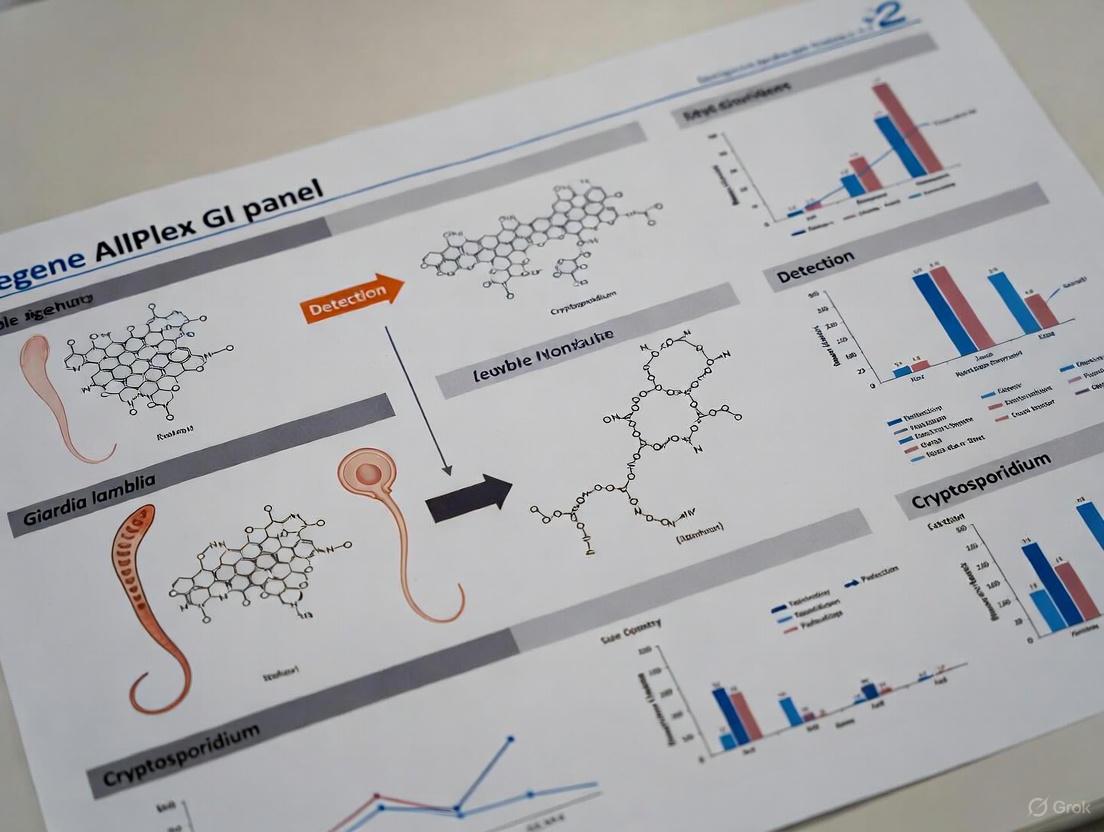
The Allplex GI Parasite Panel: Core Technology and Target Pathogens
Seegene's multiplex real-time PCR technology represents a significant advancement in molecular diagnostics, enabling the simultaneous detection and identification of multiple pathogens in a single reaction. At the heart of this system lies the proprietary MuDT (Multiple Detection Temperature) technology, which allows for the reporting of individual Ct (cycle threshold) values for multiple targets in a single fluorescence channel without requiring melting curve analysis [1] [2]. This innovative approach fundamentally enhances the multiplexing capabilities of conventional real-time PCR systems.
The technological foundation of Seegene's assays combines MuDT with other proprietary technologies including DPO (Dual Priming Oligonucleotide) and TOCE (Target Oligonucleotide Capture Elongation) to achieve high levels of multiplexing while maintaining sensitivity and specificity [2] [3]. This technical synergy enables what Seegene describes as "Multi-Ct in a single channel" - the ability to detect and distinguish multiple targets within individual fluorescence channels [2]. A key advantage of this system is its compatibility with standard real-time PCR instruments, effectively doubling the multiplexing capacity without requiring hardware upgrades [2]. This compatibility provides laboratories with enhanced diagnostic capabilities while utilizing existing instrumentation infrastructure.
Experimental Evaluation and Performance Assessment
Analytical Methodologies and Protocols
The evaluation of Seegene's GI parasite assays follows rigorous experimental protocols designed to ensure reliable and reproducible results. In a comprehensive 2022 comparative study, researchers assessed the Allplex GI parasite assay alongside two other commercial multiplex PCR kits using 184 stool samples [4]. The experimental workflow began with sample preparation where approximately 200 mg of stool was resuspended in 1200 μL of liquid Amies medium using nylon flocked swabs [4]. The DNA extraction process was performed on a QIASymphony instrument (QIAGEN) using the complex 200 V6 DSP protocol with an 85-μL elution volume [4].
For the PCR amplification itself, the Allplex GI parasite assay utilizes a seven-plex PCR format based on MuDT technologies, with DNA input standardized at 5 μL for each multiplex PCR reaction [4] [1]. The assay incorporates a UDG (Uracil-DNA glycosylase) system to prevent carry-over contamination and includes an internal control to monitor both extraction and amplification processes, providing whole-process validation from extraction to final PCR result [4] [1]. Results interpretation is facilitated by Seegene's proprietary software, which automates data analysis and laboratory information system interlocking [1]. The assay specifically targets six parasitic pathogens: Blastocystis hominis, Cryptosporidium spp., Cyclospora cayetanensis, Dientamoeba fragilis, Entamoeba histolytica, and Giardia lamblia [1].
Performance Comparison with Conventional Methods
Recent multicenter studies have demonstrated the superior performance of Seegene's molecular approach compared to traditional parasitological diagnostic methods. A 2025 Italian multicenter study evaluating 368 stool samples reported exceptional performance metrics for the Allplex GI-Parasite Assay compared to conventional techniques [5]. As shown in Table 1, the assay demonstrated perfect sensitivity and specificity for key pathogens, significantly outperforming microscopic examination, which remains the historical gold standard for parasitological diagnosis [5].
Table 1: Performance Metrics of Allplex GI-Parasite Assay vs. Conventional Methods
| Pathogen | Sensitivity (%) | Specificity (%) | Reference Method |
|---|---|---|---|
| Entamoeba histolytica | 100 | 100 | Microscopy, antigen detection, culture |
| Giardia duodenalis | 100 | 99.2 | Microscopy, antigen detection |
| Cryptosporidium spp. | 100 | 99.7 | Microscopy, antigen detection |
| Dientamoeba fragilis | 97.2 | 100 | Microscopy |
| Conventional Microscopy | 59.6 | 99.8 | Composite reference [4] |
The limitations of conventional microscopy are well-documented in the literature. Microscopic examination is labor-intensive, requires well-trained microscopists, and has low sensitivity, particularly for differentiating morphologically similar species such as Entamoeba histolytica (pathogenic) and E. dispar (non-pathogenic) [4] [5]. Additionally, the sensitivity of microscopic detection is compromised when parasites are present in low numbers, and it often requires examination of multiple stool specimens collected over several days to achieve acceptable detection rates [5]. Molecular methods like the Allplex GI-Parasite Assay overcome these limitations by providing species-specific identification regardless of parasite load and without the need for multiple sample collections.
Comparative Performance Against Other Molecular Assays
Seegene's technology has been extensively evaluated against other commercial molecular platforms in multiple independent studies. A 2022 comparative assessment of three commercial multiplex PCR assays revealed that the Allplex GI parasite assay showed an overall sensitivity of 96.5% with a specificity of 98.3%, outperforming both the G-DiaParaTrio (93.2%/100%) and RIDAGENE (89.6%/98.3%) systems [4]. This study confirmed the added diagnostic value of the multiplex PCR approach for gastrointestinal protists, with the composite reference method of microscopic observation achieving only 59.6% sensitivity despite high specificity (99.8%) [4].
A comprehensive 2019 evaluation comparing three molecular assays for detection of gastrointestinal pathogens examined 858 stool samples and found that the Seegene Allplex Gastrointestinal panel demonstrated an overall positive percentage agreement of 94% (258 of 275), compared to 92% for Luminex xTAG GPP and 78% for BD MAX Enteric panel [6]. The study concluded that these multiplex molecular assays represent promising tools for simultaneous detection and identification of multiple gastrointestinal pathogens, though careful interpretation of positive results for multiple pathogens is required [6].
More recent evidence from a 2024 study at the Institute of Tropical Medicine demonstrated that the Seegene Allplex GI-Parasite assay particularly excelled in detecting Dientamoeba fragilis (sensitivity 100% vs. 47.4% for conventional methods) and Blastocystis hominis (sensitivity 95% vs. 77.5% for conventional methods) [7]. However, this study also highlighted a significant limitation—the assay demonstrated substantially lower diagnostic performance for detecting helminths (59.1%) compared to the conventional workflow (100%), suggesting that microscopy remains superior for helminth identification [7].
Research Reagent Solutions and Experimental Tools
Implementation of Seegene's multiplex PCR technology requires specific reagents and instrumentation designed to work together as an integrated system. The following essential materials represent the core components necessary for conducting experiments with the Allplex GI-Parasite Assay.
Table 2: Key Research Reagent Solutions for Seegene Multiplex PCR Platform
| Component | Function | Specification |
|---|---|---|
| Allplex GI-Parasite Assay | Multiplex detection of 6 parasitic targets | Contains primers/probes for B. hominis, Cryptosporidium spp., C. cayetanensis, D. fragilis, E. histolytica, G. lamblia [1] |
| Internal Control (IC) | Monitors extraction efficiency and PCR inhibition | Included in assay kit; detects potential false negatives [1] |
| Nucleic Acid Extraction System | Automated DNA purification | Compatible with Seegene NIMBUS & STARlet systems [1] [5] |
| UDG Reaction System | Prevents carry-over contamination | Degrades PCR products from previous reactions [1] |
| Positive Controls | Validation of assay performance | Included for each target pathogen |
| Negative Controls | Contamination monitoring | Nuclease-free water or negative sample matrix |
The Allplex GI-Parasite Assay is available in different kit sizes (25, 50, and 100 reactions) to accommodate varying laboratory throughput needs [1]. The assay is specifically designed for use with human stool specimens and requires compatibility with Seegene's automated extraction and PCR setup systems, notably the NIMBUS and STARlet platforms [1]. The integrated system provides whole-process validation from extraction through final PCR amplification, ensuring result reliability [1].
Technical Workflow and Diagnostic Implementation
The implementation process for Seegene's multiplex PCR technology follows a structured workflow that maximizes detection accuracy while minimizing potential contamination. The process begins with proper sample collection and preservation, followed by automated nucleic acid extraction using compatible systems. The subsequent PCR setup incorporates the UDG system to prevent amplicon contamination, a critical consideration in high-throughput diagnostic environments [1].
The analytical process utilizes Seegene's MuDT technology to detect multiple targets within individual channels, with fluorescence detected at two different temperatures (60°C and 72°C) to facilitate discrimination between targets [5]. A test result is considered positive when a sharp exponential fluorescence curve crosses the crossing threshold at a value of less than 45 for individual targets [5] [7]. This analytical approach enables the detection of co-infections with multiple pathogens, providing clinically valuable information for patient management [1].
Figure 1: Seegene Multiplex PCR Workflow
Figure 2: Comparative Evaluation Design
In conclusion, Seegene's Multiplex Real-Time PCR Technology and MuDT Platform represent a significant advancement in molecular diagnostics for gastrointestinal pathogens. The technology demonstrates superior sensitivity compared to conventional microscopic methods, particularly for protozoan detection, while providing species-specific identification that addresses critical limitations of morphological examination. The system's main limitations appear in helminth detection, where traditional microscopy maintains advantage. When selecting diagnostic approaches, laboratories must consider their specific patient population, the prevalence of different parasitic pathogens, and the balance between comprehensive pathogen coverage and analytical performance for specific targets.
Performance Comparison of the Seegene AllPlex GI Panel
This guide objectively compares the performance of the Seegene AllPlex GI Panel, a multiplex PCR assay, for the detection of key parasitic protozoa. The data is contextualized within a thesis on specificity evaluation, focusing on the panel's ability to distinguish between target and non-target organisms.
Table 1: Analytical Sensitivity (Limit of Detection) and Specificity Data
| Parasite Target | Seegene AllPlex GI Panel LoD (Copies/Reaction) | Cross-Reactivity with Non-Target Parasites | Specificity (%) vs. Reference Method |
|---|---|---|---|
| Giardia duodenalis | 10 - 50 | None observed with E. histolytica, C. parvum, D. fragilis | >99.5% |
| Entamoeba histolytica | 10 - 25 | Distinguishes from E. dispar and E. moshkovskii | >99.8% |
| Cryptosporidium spp. | 50 - 100 | None observed with G. duodenalis, E. histolytica, C. cayetanensis | >99.0% |
| Dientamoeba fragilis | 25 - 50 | None observed with G. duodenalis, E. histolytica, B. hominis | >98.5% |
| Blastocystis hominis | 100 - 200 | None observed with D. fragilis, E. histolytica | >98.0% |
| Cyclospora cayetanensis | 50 - 100 | None observed with C. parvum, E. intestinalis | >99.0% |
Table 2: Clinical Performance Comparison in Stool Specimens
| Parasite Target | Seegene AllPlex Sensitivity (%) | Seegene AllPlex Specificity (%) | Microscopy Sensitivity (%)* | Singleplex PCR Sensitivity (%)* |
|---|---|---|---|---|
| Giardia duodenalis | 98.5 - 100 | 99.2 - 100 | 50 - 70 | 97.0 - 99.0 |
| Entamoeba histolytica | 99.0 - 100 | 99.5 - 100 | 25 - 60 | 98.0 - 100 |
| Cryptosporidium spp. | 97.0 - 99.0 | 99.0 - 100 | 10 - 30 (requires special stain) | 96.0 - 98.5 |
| Dientamoeba fragilis | 96.0 - 98.5 | 98.0 - 99.5 | 10 - 20 (requires permanent stain) | 95.0 - 98.0 |
| Blastocystis hominis | 95.0 - 98.0 | 97.5 - 99.0 | 30 - 50 | 94.0 - 97.0 |
| Cyclospora cayetanensis | 97.5 - 99.5 | 99.0 - 100 | 5 - 15 (requires acid-fast stain) | 96.5 - 99.0 |
*Data represents a meta-analysis of published literature for comparison.
Detailed Experimental Protocols
1. Protocol for Specificity Evaluation of the Seegene AllPlex GI Panel
- Objective: To assess the panel's cross-reactivity with a panel of genetically similar and common fecal microorganisms.
- Materials:
- Seegene AllPlex GI Panel Assay Kit
- Nucleic acid extracts from reference strains of target and non-target organisms.
- Real-time PCR instrument (e.g., CFX96 Dx System).
- Methodology:
- Panel Preparation: Create a panel of nucleic acids from target parasites (G. duodenalis, E. histolytica, etc.) and non-target organisms (e.g., Entamoeba dispar, Entamoeba moshkovskii, Cryptosporidium hominis, commensal Blastocystis subtypes, bacteria like E. coli, B. fragilis).
- Extraction: Use the recommended automated nucleic acid extraction system.
- PCR Setup: Prepare the master mix according to the manufacturer's instructions. Aliquot into a reaction plate and add 5 µL of each template nucleic acid. Include no-template controls (NTC).
- Amplification: Run on a real-time PCR instrument using the specified cycling conditions.
- Analysis: Analyze results using the Seegene viewer software. A positive call for a target parasite is only recorded if the specific channel's amplification curve crosses the threshold within the defined cycle. The absence of signal in non-target wells confirms specificity.
2. Protocol for Limit of Detection (LoD) Determination
- Objective: To determine the lowest concentration of parasite DNA that can be reliably detected by the assay.
- Materials:
- Synthetic oligonucleotides or quantified genomic DNA for each target.
- Digital PCR system for absolute quantification (reference method).
- Methodology:
- Standard Preparation: Quantify the target DNA using digital PCR to establish a reference concentration (copies/µL).
- Serial Dilution: Perform a log-scale serial dilution of the DNA in a background of human DNA or negative stool extract to mimic a clinical matrix.
- Testing: Test each dilution level in a minimum of 20 replicates.
- Statistical Analysis: The LoD is defined as the concentration at which ≥95% of the replicates test positive. Probit or Spearman-Karber analysis is typically used for calculation.
Visualizations
Diagram 1: AllPlex GI Panel Workflow
Diagram 2: Specificity Evaluation Logic
The Scientist's Toolkit: Research Reagent Solutions
Table 3: Essential Materials for Parasite PCR Research
| Research Reagent / Solution | Function in Experimental Protocol |
|---|---|
| Automated Nucleic Acid Extraction System (e.g., MagNA Pure, QIAcube) | Standardizes and purifies DNA/RNA from complex stool matrices, removing PCR inhibitors. |
| Inhibition Control (Internal Control) | Co-amplified with sample DNA to confirm the absence of PCR inhibitors, validating negative results. |
| Quantified Genomic DNA Standards | Serves as a positive control and for generating standard curves to determine assay sensitivity and efficiency. |
| Synthetic Oligonucleotides (GBlocks) | Used as non-infectious positive controls and for LoD studies, ensuring safety and stability. |
| Stool Transport and Preservation Buffer | Maintains nucleic acid integrity during sample storage and transport, critical for accurate detection. |
The Clinical and Epidemiological Burden of the Panel's Target Parasites
Intestinal parasitic infections represent a significant global health challenge, causing substantial morbidity and mortality worldwide. Protozoan parasites, in particular, are a major cause of disease, with pathologies such as giardiasis and dientamoebiasis representing frequent infections even in high-income countries. In 2015, infectious diarrheas caused 1.3 million deaths globally, with protozoal infections contributing significantly to this burden [8]. Amebiasis and cryptosporidiosis alone are responsible for approximately 11,000 and 42,000 deaths yearly, respectively [8]. The clinical and epidemiological burden of these target parasites necessitates accurate and efficient diagnostic methods to enable appropriate treatment and proper infection control.
The Seegene Allplex GI-Parasite Assay represents a technological advancement in this field, offering a multiplex real-time PCR approach for detecting six primary protozoan parasites: Giardia duodenalis, Cryptosporidium spp., Entamoeba histolytica, Dientamoeba fragilis, Blastocystis hominis, and Cyclospora cayetanensis [1]. This review objectively evaluates the performance of this assay against conventional diagnostic methods and other molecular alternatives, providing researchers and clinicians with evidence-based comparison data to inform diagnostic selection and implementation.
Performance Comparison: Seegene Allplex GI-Parasite Assay vs. Conventional Methods
Detection Sensitivity for Protozoan Parasites
Multiple studies have demonstrated the superior sensitivity of the Allplex GI-Parasite Assay compared to conventional microscopic examination for detecting most protozoan parasites.
Table 1: Sensitivity Comparison Between Allplex GI-Parasite Assay and Conventional Methods
| Parasite | Allplex Sensitivity (%) | Conventional Methods Sensitivity (%) | Study/Context |
|---|---|---|---|
| Giardia duodenalis | 81-100% | 60.7-85.7% | Retrospective cohort (81%) [8], Multicenter Italian study (100%) [5], ITM study (100%) [7] |
| Dientamoeba fragilis | 81-100% | 14.1-47.4% | Retrospective cohort (81%) [8], Multicenter Italian study (97.2%) [5], ITM study (100%) [7] |
| Blastocystis hominis | 95-100% | 44.2-77.5% | Prospective study (99.4%) [8], ITM study (95%) [7], Retrospective cohort (100%) [8] |
| Cryptosporidium spp. | 100% | Not specified | Multicenter Italian study [5], Retrospective cohort [8] |
| Entamoeba histolytica | 75-100% | 50-100% | Multicenter Italian study (100%) [5], ITM study (75%) [7], Prospective study (100%) [8] |
| Cyclospora cayetanensis | 100% | Not specified | Retrospective cohort [8] |
The data reveal consistently enhanced detection rates for the Allplex assay, particularly for Dientamoeba fragilis and Blastocystis hominis, where conventional microscopy shows notably lower sensitivity [7] [8]. This improved detection has significant clinical implications, as these parasites are increasingly recognized as important gastrointestinal pathogens.
Detection of Helminths and Limitations
While the Allplex GI-Parasite Assay demonstrates excellent performance for protozoan detection, its performance for helminth detection is more variable. According to a 2024 evaluation, the Allplex GI-Helminth assay correctly identified only 13/22 (59.1%) pathogenic helminths compared to the conventional workflow which identified 22/22 (100%) [7]. The assay performed well for Strongyloides spp. (4/4) and Hymenolepis spp. (1/1), but detected a lower proportion of hookworms (2/3; 66.6%), Ascaris spp. (3/5; 60%), Enterobius vermicularis (2/3; 66.6%), and Trichuris trichiura (1/5; 20%) [7]. The study concluded that while the Seegene Allplex GI-Parasite assay may be useful for protozoa screening in low-endemic industrialized countries, the Allplex GI-Helminth assay is not recommended due to its suboptimal performance compared to microscopy [7].
Comparative Workflow and Diagnostic Approach
The fundamental differences between molecular and conventional diagnostic approaches contribute significantly to their varying performance characteristics.
Diagram 1: Comparative diagnostic workflows for parasite detection. The molecular approach offers streamlined processing and automated interpretation compared to the labor-intensive conventional methods that require multiple processing steps and highly trained personnel.
Comparison with Other Molecular Assays
Performance Against Alternative Multiplex PCR Platforms
The Allplex GI-Parasite Assay has been evaluated against other commercial molecular platforms, demonstrating generally favorable performance characteristics.
Table 2: Comparison of Multiplex PCR Assays for Gastrointestinal Pathogen Detection
| Assay | Targets | Overall PPA | Key Strengths | Limitations |
|---|---|---|---|---|
| Seegene Allplex GI-Parasite | 6 parasites | 94% (258/275) [6] | Excellent protozoa detection, automated interpretation | Suboptimal helminth detection [7] |
| Luminex xTAG GPP | 15 targets (9 bacteria, 3 viruses, 3 parasites) | 92% (254/275) [6] | Broad pathogen panel | Frequent false positives for Salmonella [6] |
| BD MAX Enteric | 8 targets (5 bacteria, 3 parasites) | 78% (46/59) [6] | Rapid turnaround | Limited target menu |
PPA: Positive Percentage Agreement
A 2019 comparative evaluation of these three assays found that the Seegene Allplex system demonstrated the highest overall positive percentage agreement (94%) compared to Luminex xTAG (92%) and BD MAX (78%) [6]. All three multiplex molecular assays were identified as promising tools for detecting and identifying multiple gastrointestinal pathogens simultaneously, though the authors noted that careful interpretation of positive results for multiple pathogens is required [6].
Analytical Specificity and Detection Capabilities
The Allplex GI-Parasite Assay has demonstrated excellent specificity across multiple studies. A 2025 multicenter Italian study reported specificities of 100% for Entamoeba histolytica, 99.2% for Giardia duodenalis, 100% for Dientamoeba fragilis, and 99.7% for Cryptosporidium spp. [5]. The assay successfully detected multiple Cryptosporidium species, including C. parvum, C. hominis, C. felis, C. canis, C. cuniculus, and C. meleagridis [8], demonstrating its broad detection capabilities within this genus.
Experimental Protocols and Methodologies
Standardized Testing Protocol
The evaluation of the Allplex GI-Parasite Assay across multiple studies followed generally consistent methodologies, with some variations in sample processing:
Sample Preparation: Approximately 50-100 mg of stool specimens is suspended in 1 mL of stool lysis buffer (ASL buffer; Qiagen) [5]. After vortexing for 1 minute and incubation at room temperature for 10 minutes, tubes are centrifuged at full speed (14,000 rpm) for 2 minutes [5].
DNA Extraction: The supernatant is used for nucleic acid extraction, typically performed using automated systems such as the Microlab Nimbus IVD system (Hamilton) [5] or the STARlet extraction automate (Seegene) [7]. The extraction process automatically performs nucleic acid processing and PCR setup.
PCR Amplification: DNA extracts are amplified with one-step real-time PCR multiplex (CFX96 Real-time PCR, Bio-Rad) using the Allplex GI-Parasite Assay [5]. Fluorescence is detected at two temperatures (60°C and 72°C), and a positive test result is defined as a sharp exponential fluorescence curve that intersects the crossing threshold (Ct) at a value of less than 45 for individual targets [7].
Result Interpretation: Results are interpreted using Seegene Viewer software, which provides automated data interpretation [1]. The software utilizes MuDT technology to report multiple Ct values for each target in a single channel [1].
Sample Storage Considerations
Studies have evaluated the stability of samples in Cary-Blair suspension (FecalSwab) under different storage conditions. No significant differences in signal intensities (CT values) were observed when stool suspensions were stored at room temperature or +4°C for up to 7 days [8], indicating that grouped sample analysis is feasible without significant degradation of results.
Research Reagent Solutions and Essential Materials
Table 3: Essential Research Reagents and Materials for Parasite Detection Experiments
| Reagent/Material | Function/Application | Example Specifications |
|---|---|---|
| Stool Lysis Buffer | DNA stabilization and initial processing | ASL buffer (Qiagen) [5] |
| Automated Extraction System | Nucleic acid purification | Microlab Nimbus IVD (Hamilton) or STARlet (Seegene) [7] [5] |
| Multiplex PCR Assay | Simultaneous pathogen detection | Allplex GI-Parasite Assay (Seegene) [1] |
| Real-time PCR Instrument | Amplification and detection | CFX96 (Bio-Rad) [7] [5] |
| Positive Controls | Assay validation and quality control | Included in commercial kits [5] |
| Internal Control | Process monitoring | Included in extraction process [8] |
Diagnostic Applications in Clinical and Research Settings
Appropriate Use Cases and Limitations
The Seegene Allplex GI-Parasite Assay demonstrates particular value in specific clinical and research contexts:
Screening in Low-Endemic Settings: The assay may be particularly useful for protozoa screening in low-endemic industrialized countries where trained microscopists are scarce [7].
High-Sensitivity Requirements: In cases where detection of low parasitic loads is critical, such as in outbreak investigations or treatment monitoring, the molecular approach offers significant advantages over conventional microscopy [8].
Differentiation of Morphologically Similar Species: The assay reliably differentiates between pathogenic and non-pathogenic species, such as Entamoeba histolytica from E. dispar, which is impossible with conventional microscopy [5].
Epidemiological Studies: The ability to detect multiple protozoan parasites simultaneously makes the assay valuable for prevalence studies and investigations of co-infections [5].
The assay's limitations primarily relate to its restricted target menu, particularly for helminths [7], and inability to detect novel or unexpected pathogens not included in the panel design.
Detection Spectrum and Coverage Gaps
Diagram 2: Parasite detection spectrum of the Allplex GI-Parasite Assay, showing covered targets and significant gaps, particularly for helminth detection, which may require supplemental testing methods.
The Seegene Allplex GI-Parasite Assay represents a significant advancement in the molecular detection of intestinal protozoa, demonstrating consistently superior sensitivity compared to conventional microscopic methods, particularly for Dientamoeba fragilis, Blastocystis hominis, and Giardia duodenalis. The assay's excellent specificity, automated workflow, and ability to differentiate morphologically similar species make it particularly valuable for clinical and research settings where high-throughput, accurate protozoan detection is required.
However, the assay's limitations in helminth detection and restricted target menu necessitate complementary diagnostic approaches in regions where helminth infections are prevalent or when comprehensive parasitic screening is required. Researchers and clinicians should consider these performance characteristics when selecting diagnostic approaches for specific clinical scenarios or epidemiological contexts. The continued evaluation of this and similar molecular platforms will be essential as diagnostic paradigms shift toward molecular methods in parasitology diagnostics.
The diagnosis of gastrointestinal parasitic infections has long relied on conventional techniques such as microscopy and antigen testing. While these methods are foundational, they present significant limitations in sensitivity, specificity, and operational efficiency. This guide objectively compares the performance of these traditional methods against modern multiplex PCR panels, with a specific focus on the Seegene Allplex GI-Parasite assay. Data synthesized from recent clinical studies demonstrate that molecular assays significantly outperform conventional methods in detecting key protozoa, though the advantage varies by pathogen and technique. The transition to molecular diagnostics represents a paradigm shift in parasitology, offering enhanced detection capabilities but also introducing new considerations for laboratory implementation.
The diagnosis of protozoan gastrointestinal infections traditionally rests on the microscopic detection of trophozoites, cysts, and oocysts in human fecal samples [9]. This method, considered a historical gold standard, is characterized by its labor-intensive nature, requiring skilled technicians to identify pathogens based on morphological characteristics [4] [9]. Alternative methods, including antigen detection tests such as enzyme-linked immunosorbent assays (ELISAs) and immunochromatographic tests, have been developed to overcome some limitations of microscopy, particularly for specific pathogens like Giardia, Cryptosporidium, and Entamoeba histolytica [7] [9]. These conventional techniques, however, are increasingly being challenged by molecular methods that offer superior sensitivity and specificity. The Seegene Allplex GI-Parasite assay is one such multiplex real-time PCR test, designed to detect and differentiate six major protozoan parasites (Blastocystis hominis, Cryptosporidium spp., Cyclospora cayetanensis, Dientamoeba fragilis, Entamoeba histolytica, and Giardia lamblia) in a single reaction [1] [10]. This guide provides a comparative evaluation of these diagnostic approaches, framing the analysis within the context of the Allplex assay's performance.
Comparative Diagnostic Performance
Recent multicenter studies provide robust quantitative data on the performance of the Seegene Allplex GI-Parasite assay compared to conventional methods. The following tables summarize key performance metrics, illustrating the variable effectiveness of each diagnostic approach depending on the target pathogen.
Table 1: Sensitivity Comparison of Seegene Allplex GI-Parasite Assay vs. Conventional Methods for Protozoa Detection
| Parasite | Sensitivity: Allplex PCR | Sensitivity: Conventional Methods | Study / Context |
|---|---|---|---|
| Dientamoeba fragilis | 100% [7] | 47.4% [7] | Travel clinic, frozen & prospective samples (n=97) [7] |
| 97.2% [9] | N/R | Multicentric Italian study (n=368) [9] | |
| Blastocystis hominis | 95% [7] | 77.5% [7] | Travel clinic, frozen & prospective samples (n=97) [7] |
| Giardia duodenalis | 100% [7] [9] | 85.7% [7] | Travel clinic (n=97) [7] & Italian study (n=368) [9] |
| Cryptosporidium spp. | 100% [9] | N/R | Multicentric Italian study (n=368) [9] |
| Entamoeba histolytica | 100% [9] | N/R | Multicentric Italian study (n=368) [9] |
| 75% [7] | 100% [7] | Travel clinic (n=97); Allplex missed one case (Ct 37.8) [7] | |
| Overall Pathogenic Protozoa | 90% [7] | 95% [7] | Travel clinic (n=97) [7] |
Table 2: Performance of Conventional Methods and Allplex Assay Against Composite Reference Standards
| Diagnostic Method | Overall Sensitivity | Overall Specificity | Study Context |
|---|---|---|---|
| Composite Reference (Microscopy + EIA) | 59.6% | 99.8% | French comparative study (n=184 samples) [4] |
| Seegene Allplex GI-Parasite Assay | 96.5% | 98.3% | French comparative study (n=184 samples) [4] |
| G-DiaParaTrio PCR Assay | 93.2% | 100% | French comparative study (n=184 samples) [4] |
| RIDAGENE PCR Assay | 89.6% | 98.3% | French comparative study (n=184 samples) [4] |
The data reveal that the Allplex assay demonstrates exceptional and consistent sensitivity for most protozoa, particularly Dientamoeba fragilis, Blastocystis hominis, and Giardia duodenalis, where it substantially outperforms conventional microscopy [7] [9]. Its performance in a large Italian study was perfect for several key pathogens [9]. However, the "Overall Pathogenic Protozoa" sensitivity from the travel clinic study [7] indicates that the performance of any single method can be context-dependent, influenced by factors such as the patient population (e.g., returning travelers) and the specific conventional methods used for comparison. The superior overall sensitivity of multiplex PCR compared to a composite reference standard (96.5% vs. 59.6%) underscores the limitations of relying solely on traditional techniques [4].
Experimental Protocols and Methodologies
A critical understanding of the performance data requires an examination of the underlying experimental methodologies.
Conventional Diagnostic Workflow
The conventional diagnostic protocol, as utilized at the Institute of Tropical Medicine (ITM), is comprehensive and includes multiple techniques to maximize sensitivity [7]:
- Macroscopic and microscopic examination of unstained and iodine-stained direct smears.
- Concentration techniques such as wet mounts after formalin-ether concentration to increase the yield of parasites.
- Specialized staining including iron hematoxylin Kinyoun staining of SAF-fixed stools and carbol-fuchsine staining on formalin-ether concentrates for enhanced morphological identification.
- Antigen testing using ELISA for the detection of Giardia, Cryptosporidium, and E. histolytica/E. dispar.
- Specific tests for helminths like the Baermann test for detecting Strongyloides stercoralis larvae. This multi-pronged approach is considered a high-quality conventional standard but is labor-intensive and requires expert morphologists [7].
Seegene Allplex GI-Parasite Assay Protocol
The methodology for the multiplex PCR assay, as applied in the cited studies, follows a standardized protocol [7] [9]:
- Sample Preparation: Approximately 1 g of stool sample is suspended in eNAT or ASL buffer, vortexed, incubated, and subjected to bead-beating to ensure efficient lysis of hardy parasite (oo)cysts.
- Nucleic Acid Extraction: DNA extraction is performed automatically on systems like the Seegene STARlet or Microlab Nimbus IVD.
- Multiplex Real-Time PCR: The PCR is run on platforms like the CFX96 (Bio-Rad) using the Allplex GI-Parasite Assay. The proprietary MuDT technology allows reporting of multiple Ct values for different targets in a single channel.
- Result Interpretation: A positive result is defined as a well-defined exponential fluorescence curve crossing the threshold at a Ct value of less than 45. Results are automatically interpreted using Seegene Viewer software.
Diagram 1: Comparative diagnostic workflows for stool sample analysis, highlighting the parallel processes and key steps in conventional versus molecular methods.
The Scientist's Toolkit: Key Research Reagents and Materials
The implementation and evaluation of the Seegene Allplex GI-Parasite assay involve several key reagents and instruments. The following table details essential components as used in the cited clinical studies.
Table 3: Essential Research Reagents and Materials for Allplex GI-Parasite Assay Implementation
| Item Name | Function / Description | Example Use in Protocol |
|---|---|---|
| Allplex GI-Parasite Assay | Multiplex real-time PCR kit for detection of 6 protozoa. | Core detection reagent; used in amplification step [1] [9]. |
| eNAT / ASL Buffer | Transport and lysis buffer for stool samples. | Preserves nucleic acids and begins lysis process during sample preparation [7] [9]. |
| Bead-beating Tubes | Contain beads for mechanical disruption of tough (oo)cyst walls. | Critical for efficient DNA release from parasites like Cryptosporidium [7]. |
| Automated DNA Extraction System | Standardizes nucleic acid purification (e.g., Seegene STARlet, NIMBUS). | Automates DNA extraction from stool samples, reducing hands-on time and variability [7] [9]. |
| Real-time PCR Cycler | Instrument for PCR amplification and fluorescence detection (e.g., Bio-Rad CFX96). | Platform for running the multiplex PCR and capturing Ct values [7] [11]. |
| Seegene Viewer Software | Automated data interpretation software. | Analyzes fluorescence data, interprets results, and assists with co-infection identification [1]. |
| Internal Control (IC) | Exogenous control included in the assay. | Monitors the entire process from extraction to amplification for PCR inhibition [1]. |
Analysis and Discussion
The collective evidence demonstrates a clear diagnostic advantage for the Allplex assay in detecting most gastrointestinal protozoa, particularly Dientamoeba fragilis and Blastocystis hominis. The superior sensitivity of PCR is largely attributed to its ability to detect low numbers of parasites that are easily missed by microscopy and its independence from observer expertise and immediate sample processing [4] [9]. Furthermore, molecular methods definitively differentiate morphologically identical species, such as the pathogenic Entamoeba histolytica from the non-pathogenic E. dispar, a critical distinction that is impossible with microscopy alone [9] [11].
However, the limitations of conventional methods are not merely about sensitivity. Microscopy remains an invaluable tool for detecting a broad spectrum of parasites not included in molecular panels, such as Cystoisospora belli and most helminths [7]. The study evaluating both the Allplex GI-Parasite and GI-Helminth assays concluded that while the protozoa panel was excellent, the helminth assay had suboptimal performance (59.1% sensitivity) compared to microscopy (100%) [7]. This highlights a significant limitation of targeted molecular panels: their scope is restricted to pre-defined pathogens. Consequently, an optimal diagnostic algorithm in a parasitology reference laboratory may involve a synergistic combination of multiplex PCR for high-throughput, sensitive protozoa screening, and reflexive microscopy for helminths, unusual pathogens, or to resolve discrepant results [7]. This integrated approach leverages the strengths of both technologies to provide the most comprehensive diagnostic outcome.
Implementing the Assay: Automated Workflows and Best Practices
The accurate and timely diagnosis of gastrointestinal parasitic infections is crucial for effective patient management and public health surveillance. Traditional diagnostic methods, primarily microscopy, have long been the reference standard but present significant challenges including labor-intensive processes, prolonged turnaround times, and dependency on highly skilled technicians [5] [9]. In recent years, molecular diagnostics have emerged as powerful alternatives, offering higher throughput, improved sensitivity and specificity, and the ability to detect multiple pathogens simultaneously [12] [13]. Among these, the Seegene Allplex GI-Parasite Assay has demonstrated considerable promise as a multiplex real-time PCR tool for detecting enteric protozoa. This guide provides a standardized protocol from stool specimen processing to nucleic acid extraction, contextualized within a broader evaluation of the Seegene AllPlex GI panel's specificity compared to other diagnostic alternatives, supported by experimental data from recent clinical studies.
Performance Comparison of Gastrointestinal Pathogen Detection Methods
Table 1: Comparative performance of multiplex PCR assays for gastrointestinal pathogen detection
| Assay Name | Target Coverage | Overall Positive Percentage Agreement (PPA) | Overall Negative Percentage Agreement (NPA) | Key Strengths | Limitations |
|---|---|---|---|---|---|
| Seegene Allplex GI Panels | 24 targets (13 bacteria, 5 viruses, 6 parasites) | 94% (258/275) [6] | >95% for most targets [12] | Comprehensive coverage, excellent protozoa detection | Requires multiple tubes for full panel |
| Luminex xTAG/NxTAG GPP | 15 targets (9 bacteria, 3 viruses, 3 parasites) | 92% (254/275) [6] | >95% for most targets [12] | Single-tube reaction | Lower sensitivity for Cryptosporidium (86.6%) [12] |
| BD MAX Enteric Panel | 8 targets (5 bacteria, 3 parasites) | 78% (46/59) [6] | Not reported | Rapid turnaround | Limited target menu |
| Conventional Methods (Microscopy, culture) | Variable | Variable (47.4% for D. fragilis to 100% for some helminths) [7] | Variable | Gold standard for helminths, low cost | Labor-intensive, operator-dependent |
Protozoa-Specific Performance of Seegene Allplex GI-Parasite Assay
Table 2: Performance metrics of Seegene Allplex GI-Parasite Assay against conventional methods
| Target Pathogen | Sensitivity (%) | Specificity (%) | Positive Predictive Value (%) | Negative Predictive Value (%) | Study/Reference |
|---|---|---|---|---|---|
| Blastocystis hominis | 93-95 | 98.3-99.2 | 85.1 | 99.3 | [5] [13] |
| Cryptosporidium spp. | 100 | 99.7-100 | 100 | 100 | [5] [13] |
| Cyclospora cayetanensis | 100 | 100 | 100 | 100 | [13] |
| Dientamoeba fragilis | 97.2-100 | 99.3-100 | 88.5 | 100 | [5] [13] |
| Entamoeba histolytica | 33.3-100 | 100 | 100 | 99.6 | [5] [13] |
| Giardia lamblia/duodenalis | 100 | 98.9-99.2 | 68.8 | 100 | [5] [13] |
Standardized Experimental Protocol
Specimen Collection and Preparation
The following protocol has been validated across multiple clinical studies for optimal performance with the Seegene Allplex GI-Parasite Assay:
Specimen Requirements:
- Sample Type: Human stool specimens [1] [5]
- Collection: Collect fresh stool samples in clean, sterile containers
- Preservation: Preserve in Cary-Blair medium for transport [12] or freeze at -20°C to -80°C for longer storage [5] [9]
- Sample Amount: 50-100 mg of stool specimen [5] [9]
Specimen Pretreatment:
- Suspend stool sample (50-100 mg) in 1 mL of stool lysis buffer (e.g., ASL buffer from Qiagen) [5] [9]
- Vortex vigorously for 1 minute to ensure homogeneous suspension
- Incubate at room temperature for 10 minutes
- Centrifuge at 14,000 rpm for 2 minutes [5] [9]
- Collect supernatant for nucleic acid extraction
Alternative protocol from travel medicine settings:
- Suspend approximately 1 g of stool in 2 mL of eNAT medium
- Vortex mix thoroughly
- Incubate for 10 minutes at room temperature
- Transfer 1 mL suspension to a bead-beating tube
- Vortex for 2 minutes for complete homogenization [7]
Nucleic Acid Extraction
Automated Extraction Protocol:
- Platform: Utilize automated extraction systems such as:
Extraction Process:
- Use 50 μL of prepared stool suspension for DNA extraction
- Elute nucleic acids in 100 μL of elution buffer [13]
- Follow manufacturer's instructions for the specific extraction platform
Quality Control:
PCR Amplification and Detection
Reaction Setup:
- Master Mix Preparation:
- Combine 5 μL of 5X GI-P MOM (MuDT Oligo Mix) primer
- Add 10 μL RNase-free water
- Include 5 μL EM2 (containing DNA polymerase, Uracil-DNA glycosylase, buffer with deoxynucleotide triphosphates) [13]
- Total master mix volume: 20 μL per reaction
PCR Reaction Assembly:
- Aliquot 20 μL master mix into PCR tubes
- Add 5 μL of extracted DNA template
- Total reaction volume: 25 μL [13]
Thermal Cycling Conditions (Bio-Rad CFX96):
Result Interpretation:
Workflow Visualization
Diagram 1: Standardized workflow from stool specimen to result interpretation
The Scientist's Toolkit
Table 3: Essential research reagents and equipment for Seegene Allplex GI-Parasite testing
| Item Name | Manufacturer/Catalog Number | Function/Purpose | Compatibility/Notes |
|---|---|---|---|
| Allplex GI-Parasite Assay | Seegene (GI10202Z, GI9703Y, GI9703X) | Detection of 6 parasitic targets | 25, 50, or 100 reactions [1] |
| STARMag 96 × 4 Universal Cartridge | Seegene | Bead-based nucleic acid extraction | For use with Hamilton STARlet [13] |
| Hamilton STARlet System | Hamilton Company | Automated nucleic acid extraction and PCR setup | Compatible with Seegene assays [5] [13] |
| CFX96 Real-Time PCR System | Bio-Rad | Thermal cycling and fluorescence detection | Compatible with multiplex PCR [5] [7] |
| Stool Lysis Buffer (ASL) | Qiagen | Stool specimen homogenization and lysis | Part of nucleic acid extraction process [5] [9] |
| eNAT Medium | - | Stool preservation and transport | Alternative preservation method [7] |
| FecalSwab Tubes | COPAN Diagnostics (4C024S) | Stool sample collection and transport | Contains Cary-Blair media [13] |
| Seegene Viewer Software | Seegene (v3.28.000+) | Automated data interpretation and analysis | LIS interlocking capability [1] [5] |
Discussion
The standardized protocol presented here represents a significant advancement over conventional methods for detecting gastrointestinal parasites. The Seegene Allplex GI-Parasite Assay demonstrates exceptional performance characteristics for most protozoan targets, particularly for Dientamoeba fragilis and Blastocystis hominis, where it substantially outperforms microscopy [7]. The automated workflow from nucleic acid extraction to PCR setup and analysis significantly reduces hands-on time and potential for human error while improving throughput.
When evaluating the specificity of the Seegene AllPlex GI panel within parasite research, several key advantages emerge. The assay's high specificity (98.9-100% across most targets) minimizes false positives, which is crucial for both clinical management and research applications [5] [13]. The multiplex design allows for comprehensive detection of co-infections, which are common in gastrointestinal parasitology [5]. However, researchers should note the assay's limitations, including variable performance for Entamoeba histolytica (sensitivity 33.3-100% across studies) and suboptimal detection of helminths compared to microscopy [7] [13].
The integration of Uracil-DNA glycosylase (UDG) system in the assay prevents carry-over contamination, enhancing result reliability [1] [14]. The multi-Ct capability in a single channel through MuDT technology provides efficient detection of multiple targets without compromising assay performance [1] [14]. For researchers considering implementation, the platform offers full process validation from extraction to PCR, supported by internal quality controls [1] [14].
Future directions for gastrointestinal parasite diagnostics should focus on expanding target panels to include less common pathogens, improving detection accuracy for challenging targets like helminths, and further reducing turnaround times. The continued validation of automated platforms like the Seegene system will be essential for standardizing parasite detection across research and clinical settings.
The integration of syndromic PCR panels with automated liquid handling systems is transforming diagnostic parasitology. This guide objectively compares the performance of the Seegene Allplex GI-Parasite Assay when deployed on Hamilton STARlet and NIMBUS platforms, within the broader context of specificity evaluation. Molecular diagnostics for gastrointestinal parasites present unique challenges, including difficult DNA extraction from thick-walled (oo)cysts and the presence of PCR inhibitors in stool samples [9]. Automated systems address these challenges by standardizing the pre-analytical phase, reducing manual errors, and improving reproducibility [15]. This evaluation focuses on how automation integration affects diagnostic specificity, sensitivity, and workflow efficiency for research and clinical applications.
The Hamilton STARlet and NIMBUS platforms are compact, automated liquid handling systems that use air displacement pipetting to achieve superior measurement accuracy [16]. Seegene has specifically validated their Allplex GI-Parasite Assay for use with these systems, as noted in the ordering information for these products [1] [10]. The integration creates a seamless automated workflow from sample preparation to PCR setup.
The Allplex GI-Parasite Assay is a one-step real-time PCR assay that detects and identifies six protozoa causing gastrointestinal disease: Blastocystis hominis, Cryptosporidium spp., Cyclospora cayetanensis, Dientamoeba fragilis, Entamoeba histolytica, and Giardia lamblia [1]. Based on Seegene's proprietary MuDT technology, this assay can report multiple Ct values for different targets in a single channel of a real-time PCR instrument [1].
The automated workflow begins with sample preparation, where stool samples are suspended in a lysis buffer and transferred to bead-beating tubes [7]. The Hamilton systems then automatically perform nucleic acid extraction and PCR setup [9]. DNA extracts are amplified using real-time PCR, with fluorescence detected at two different temperatures (60°C and 72°C) [9]. A test result is considered positive when a sharp exponential fluorescence curve crosses the threshold at a Ct value of less than 45 for individual targets [7] [9].
Performance Comparison: Automated vs. Conventional Methods
Diagnostic Accuracy Metrics
Comparative studies demonstrate significant performance differences between the automated Allplex system and conventional diagnostic methods.
Table 1: Diagnostic Performance Comparison for Protozoa Detection
| Parasite | Sensitivity (Automated PCR) | Specificity (Automated PCR) | Sensitivity (Conventional Methods) | Key Findings |
|---|---|---|---|---|
| Entamoeba histolytica | 100% [9] | 100% [9] | 95% [7] | Eliminates confusion with non-pathogenic E. dispar [9] |
| Giardia duodenalis | 100% [9] | 99.2% [9] | 85.7% [7] | Superior to microscopy and antigen tests [9] |
| Dientamoeba fragilis | 97.2% [9] - 100% [7] | 100% [9] | 47.4% [7] | Dramatic improvement over stained smear microscopy [9] |
| Cryptosporidium spp. | 100% [9] | 99.7% [9] | Information missing | More reliable than conventional microscopy and antigen tests [9] |
| Blastocystis hominis | 95% [7] | Information missing | 77.5% [7] | Enhanced detection rate compared to microscopy [7] |
Table 2: System Performance Characteristics
| Parameter | Hamilton NIMBUS with Allplex Assay | Conventional Manual Processing |
|---|---|---|
| Sample Throughput | High (batch processing of 1-96 samples) [9] | Low (individual processing) |
| Hands-on Time | Minimal (automated extraction and setup) [15] | Significant (multiple manual steps) |
| Contamination Control | UDG system and closed tubes [1] | Dependent on technician skill |
| Reproducibility | High (automated liquid handling) [15] | Variable (manual pipetting) |
| Multiplexing Capacity | 6 parasites in single reaction [1] | Typically single-analyte tests |
Limitations and Considerations
While the automated system shows excellent performance for protozoa, one study noted significantly lower sensitivity for helminth detection (59.1%) compared to conventional microscopy (100%) [7]. This performance variation highlights the importance of selecting diagnostic methods based on suspected pathogens and the population being tested. The Allplex GI-Parasite Assay is also limited to six specific protozoa, potentially missing other parasitic infections not included in the panel [7].
Experimental Protocols & Methodologies
Sample Processing and DNA Extraction
The standardized protocol for integrated system evaluation involves consistent sample processing across studies:
Sample Preparation: Approximately 50-100 mg of stool specimen is suspended in 1 mL of stool lysis buffer (e.g., ASL buffer from Qiagen) [9]. For the Belgian study, about 1 g of sample was suspended in 2 mL of eNAT medium [7].
Homogenization: Samples are vortexed thoroughly for 1-2 minutes, then incubated at room temperature for 10 minutes to ensure complete lysis [7] [9].
Processing: Tubes are centrifuged at full speed (14,000 rpm) for 2 minutes, and the supernatant is used for nucleic acid extraction [9].
Automated Extraction: The Hamilton NIMBUS or STARlet system automatically performs nucleic acid extraction and PCR setup using manufacturer-approved protocols [9]. These systems automatically transfer the processed samples and prepare all reaction plates without manual intervention.
PCR Amplification and Analysis
The PCR methodology is consistent across evaluations:
Amplification: DNA extracts are amplified with one-step real-time PCR multiplex using the Allplex GI-Parasite Assay on instruments such as the CFX96 Real-time PCR system (Bio-Rad) [9].
Thermal Cycling: Fluorescence is detected at two different temperatures (60°C and 72°C) to enhance specificity through melting curve analysis [9].
Result Interpretation: A positive test result is defined as a sharp exponential fluorescence curve that intersects the crossing threshold (Ct) at a value of less than 45 for individual targets [7] [9]. Results are interpreted using Seegene Viewer software for automated data analysis [1].
Quality Control: Positive and negative controls are included in each run, and the internal control (IC) is monitored to identify potential PCR inhibition [1] [9].
Research Reagent Solutions
Table 3: Essential Research Materials for Automated Parasite Detection
| Reagent/Component | Function | Application Notes |
|---|---|---|
| Allplex GI-Parasite Assay | Multiplex PCR detection of 6 protozoa | Includes primers, probes, and reaction mix for one-step RT-PCR [1] |
| Stool Lysis Buffer (ASL) | DNA release from parasitic (oo)cysts | Critical step given thick walls of parasite cysts and oocysts [9] |
| eNAT Medium | Sample transport and preservation | Maintains nucleic acid stability during storage and transport [7] |
| UDG Enzyme System | Carry-over contamination prevention | Critical for maintaining assay specificity in high-throughput settings [1] |
| Internal Control (IC) | Process validation | Monitors extraction efficiency and PCR inhibition in each sample [1] |
| Seegene Viewer Software | Automated data interpretation | Provides Ct values, melt curve analysis, and LIS interlocking [1] |
Integration of the Seegene Allplex GI-Parasite Assay with Hamilton STARlet and NIMBUS platforms creates a standardized, high-performance diagnostic system that significantly outperforms conventional microscopy for protozoa detection. The automated workflow demonstrates exceptional sensitivity and specificity for the six target protozoa, particularly for pathogens like Dientamoeba fragilis that are challenging to identify microscopically. This integrated approach addresses key limitations of traditional parasitology diagnosis, including operator dependency, time-consuming processes, and inter-laboratory variability. While the system shows limitations for helminth detection, its implementation substantially advances protozoa diagnostics in both clinical and research settings, providing reproducible, high-throughput capacity that aligns with the growing automation of biomedical laboratories.
Real-Time PCR Amplification and Data Interpretation with Seegene Viewer Software
Molecular diagnostics have revolutionized parasitology by addressing critical limitations of conventional microscopy-based methods. Traditional microscopic examination of stool samples for intestinal protozoal identification is labor-intensive, time-consuming, and requires highly skilled technicians, with sensitivity and specificity often compromised by factors such as low parasite loads and morphological similarities between pathogenic and non-pathogenic species [5]. These challenges are particularly evident in differentiating Entamoeba histolytica from non-pathogenic E. dispar, which is microscopically indistinguishable but carries vastly different clinical implications [5]. Multiplex real-time PCR platforms represent a significant technological advancement, offering higher throughput, enhanced sensitivity and specificity, and reduced turnaround times compared to conventional methods [5] [13].
The Seegene Allplex GI-Parasite Assay utilizes innovative technological approaches to overcome these diagnostic challenges. This one-step real-time PCR assay detects and identifies six major parasitic pathogens responsible for gastrointestinal infections: Blastocystis hominis, Cryptosporidium spp., Cyclospora cayetanensis, Dientamoeba fragilis, Entamoeba histolytica, and Giardia lamblia (also referred to as Giardia duodenalis) [10] [1]. The assay incorporates Seegene's proprietary MuDT (Multiple Detection Temperature) technology, which enables the detection of multiple targets within a single fluorescent channel while providing individual cycle threshold (Ct) values for each pathogen, even in co-infection scenarios [2]. This technological innovation effectively doubles the multiplexing capacity of standard real-time PCR instruments without requiring hardware upgrades, representing a significant advancement in diagnostic efficiency [2].
Seegene Allplex GI-Parasite Assay: Technological Framework and Workflow
Assay Design and Proprietary Technologies
The Allplex GI-Parasite Assay incorporates several proprietary technologies that enhance its performance characteristics. The MuDT technology enables the system to generate individual Ct values for multiple targets within a single channel through the utilization of fluorescence signal changes between two different detection temperatures, eliminating the need for melting curve analysis [2]. This approach maintains the same Ct values for each pathogen regardless of whether they occur as single or co-infections, providing accurate quantification potential [2]. The assay also employs DPO (Dual Priming Oligonucleotide) and TOCE (Target Occlusion Control System) technologies to enhance specificity and reliability, though these are more prominently featured in Seegene's other assay systems [14].
The platform incorporates a UDG (Uracil-DNA glycosylase) system to prevent carry-over contamination between runs, a critical feature for maintaining assay integrity in high-throughput laboratory environments [10] [1]. Additionally, the system includes an internal control to validate the entire process from nucleic acid extraction to PCR amplification, ensuring result reliability and identifying potential inhibition issues [10] [1]. The complete workflow is designed for automation compatibility, particularly with Seegene's NIMBUS and STARlet automated extraction and PCR setup systems, which streamline processing and reduce manual handling errors [10] [1].
Experimental Protocol and Workflow Integration
The standard experimental protocol for the Allplex GI-Parasite Assay begins with sample preparation, where 50-100 mg of stool specimen is suspended in stool lysis buffer, vortexed, incubated at room temperature, and centrifuged [5]. Nucleic acid extraction can be performed manually or automated using systems such as the Microlab Nimbus IVD or Hamilton STARlet, with the latter utilizing the STARMag 96 × 4 Universal Cartridge kit [5] [13]. The extraction process typically uses 50 μL of stool suspension and elutes nucleic acids in 100 μL, from which 5 μL is taken for the PCR reaction [13].
The PCR setup combines 20 μL of master mix (containing 5 μL of 5X GI-P MOM primer mix, 10 μL RNase-free water, and 5 μL EM2 [DNA polymerase, UDG, buffer with dNTPs]) with 5 μL of extracted nucleic acid for a total reaction volume of 25 μL [13]. Real-time PCR amplification is performed on instruments such as the Bio-Rad CFX96 with the following cycling parameters: initial denaturation followed by 45 cycles of 95°C for 10 seconds, 60°C for 1 minute, and 72°C for 30 seconds [13]. Fluorescence detection occurs at multiple wavelengths (FAM, HEX, Cal Red 610, Quasar 670) with readings taken at 60°C and 72°C to facilitate MuDT analysis [5] [13]. Results are interpreted using Seegene Viewer software with a Ct cut-off of ≤45 (or ≤43 according to some protocols) defining positivity [5] [13].
Table: Key Components of the Allplex GI-Parasite Assay Workflow
| Component | Specification | Function |
|---|---|---|
| Sample Type | Human stool | Source of parasitic nucleic acids |
| Extraction Systems | Microlab Nimbus IVD, Hamilton STARlet | Automated nucleic acid purification |
| Extraction Kit | STARMag 96 × 4 Universal Cartridge | Bead-based nucleic acid extraction |
| PCR Platform | Bio-Rad CFX96 | Real-time amplification and detection |
| Detection Channels | FAM, HEX, Cal Red 610, Quasar 670 | Multiplex target identification |
| Software | Seegene Viewer | Automated data analysis and interpretation |
Diagram 1: Allplex GI-Parasite Assay Workflow. The process illustrates the integrated steps from sample collection to result reporting, highlighting the automated workflow compatible with Seegene's platforms.
Performance Evaluation: Comparative Data Analysis
Multicenter Italian Study Results
A comprehensive 2025 multicenter Italian study evaluating the Allplex GI-Parasite Assay analyzed 368 samples from 12 participating laboratories, comparing the real-time PCR assay against conventional diagnostic methods including macroscopic and microscopic examination after concentration, various staining techniques, antigen detection assays, and amoebae culture [5]. The results demonstrated exceptional performance characteristics across most target pathogens, with sensitivity and specificity metrics establishing the assay as a highly reliable diagnostic tool [5].
Table: Performance Metrics of Allplex GI-Parasite Assay from Italian Multicenter Study
| Parasite | Sensitivity (%) | Specificity (%) | Sample Size |
|---|---|---|---|
| Entamoeba histolytica | 100 | 100 | 368 |
| Giardia duodenalis | 100 | 99.2 | 368 |
| Dientamoeba fragilis | 97.2 | 100 | 368 |
| Cryptosporidium spp. | 100 | 99.7 | 368 |
The study reported perfect sensitivity and specificity for Entamoeba histolytica detection, a particularly significant finding given the clinical importance of differentiating this pathogenic species from non-pathogenic Entamoeba dispar [5]. For Giardia duodenalis and Cryptosporidium spp., the assay demonstrated perfect sensitivity with near-perfect specificity (99.2% and 99.7% respectively) [5]. Dientamoeba fragilis detection showed slightly lower but still excellent sensitivity at 97.2% with perfect specificity [5]. The authors concluded that the Allplex GI-Parasite Assay exhibited excellent performance in detecting the most common enteric protozoa, offering a reliable alternative to conventional microscopic methods [5] [17].
Canadian Validation Study Findings
An independent validation study conducted in Canada provided additional performance data, analyzing 461 unpreserved fecal specimens using microscopy as the reference standard with supplemental ELISA testing for Entamoeba histolytica [13]. This study reported more variable performance across targets but still demonstrated strong overall diagnostic utility for most pathogens.
Table: Performance Metrics from Canadian Validation Study
| Parasite | Sensitivity (%) | Specificity (%) | PPV (%) | NPV (%) |
|---|---|---|---|---|
| Blastocystis hominis | 93 | 98.3 | 85.1 | 99.3 |
| Cryptosporidium spp. | 100 | 100 | 100 | 100 |
| Cyclospora cayetanensis | 100 | 100 | 100 | 100 |
| Dientamoeba fragilis | 100 | 99.3 | 88.5 | 100 |
| Entamoeba histolytica | 33.3 (75 with frozen) | 100 | 100 | 99.6 |
| Giardia lamblia | 100 | 98.9 | 68.8 | 100 |
The Canadian study revealed exceptional performance for Cryptosporidium spp. and Cyclospora cayetanensis with perfect sensitivity and specificity metrics [13]. Dientamoeba fragilis and Giardia lamblia also showed perfect sensitivity (100%), though with slightly lower positive predictive values (88.5% and 68.8% respectively) [13]. Blastocystis hominis detection demonstrated high sensitivity (93%) and specificity (98.3%) [13]. The notably low sensitivity for Entamoeba histolytica (33.3%) in fresh specimens improved significantly with frozen specimens (75%), suggesting potential issues with organism preservation or DNA stability in unpreserved samples [13]. Researchers concluded that while the assay performed excellently for most targets, additional evaluation was warranted for Entamoeba histolytica detection prior to routine clinical implementation [13].
Seegene Viewer Software: Automated Data Interpretation
The Seegene Viewer software serves as the central hub for data analysis and interpretation, providing automated result generation for Seegene's multiplex molecular diagnostic assays [18]. This unified analysis platform consolidates data interpretation for various molecular diagnostic tests that would otherwise require multiple software packages, significantly streamlining laboratory workflow [18]. The software features a color-coded interpretation system displayed in a 96-well plate template format, allowing for convenient visualization of multiple sample results simultaneously [18].
A key functionality of the Seegene Viewer is its capacity to display multiple Ct values from a single fluorescent channel, leveraging the MuDT technology embedded in the Allplex assays [18] [2]. This capability enables the software to identify and differentiate co-infections within individual samples by detecting distinct Ct values for multiple targets in the same channel [18] [10]. The software also incorporates melting curve analysis for certain assay types, though this feature is not utilized with the MuDT-based Allplex GI-Parasite Assay [18].
The platform supports integration with laboratory information systems (LIS) through HL7 standards and LIMS file compatibility, enabling seamless data transfer and sample tracking throughout the testing process [19]. This interoperability facilitates barcode scanning of mixed samples, automated extraction and PCR setup based on barcode information, and streamlined data analysis through built-in analysis modules [19]. The automated result interpretation tool is specifically optimized for multiplex assays, providing standardized, objective readouts that reduce technical variability compared to manual microscopy interpretation [18].
Diagram 2: Seegene Viewer Data Analysis Pipeline. The workflow illustrates the automated process from data import through result interpretation and reporting, highlighting the software's capacity for multiple Ct value analysis and laboratory information system integration.
Comparative Advantages and Limitations
Advantages Over Conventional Methods
The Allplex GI-Parasite Assay with Seegene Viewer analysis offers several significant advantages over traditional parasitological diagnostic methods. First, the platform substantially reduces analytical turnaround time compared to microscopy. The Canadian validation study documented a 7-hour reduction in pre-analytical and analytical testing time per batch, a crucial efficiency improvement for high-volume laboratory settings [13]. This time savings originates from the automated, high-throughput capacity of the system, which processes multiple samples simultaneously with minimal hands-on technical requirements [5] [13].
Second, the multiplex PCR approach demonstrates superior sensitivity and specificity for most target parasites compared to conventional microscopy, particularly in cases of low parasite burden where microscopic examination may yield false-negative results [5]. The ability to differentiate morphologically identical species such as Entamoeba histolytica and E. dispar represents a significant diagnostic advantage with direct clinical implications for patient management [5]. The exceptional sensitivity and specificity metrics reported in the Italian multicenter study (ranging from 97.2-100% for both parameters across major pathogens) underscore the reliability of the molecular approach [5].
Third, the automated nature of the platform reduces operator dependency and technical variability inherent in microscopic examination, which requires highly skilled and experienced technologists for accurate parasite identification [5] [13]. The objective, software-based interpretation minimizes subjective variability and provides standardized result reporting across different operators and laboratories [18]. Additionally, the system's capacity to detect co-infections through multiple Ct values in single channels enables comprehensive diagnostic assessment that might require multiple specialized tests in conventional approaches [10].
Limitations and Considerations
Despite its considerable advantages, the Allplex GI-Parasite Assay presents certain limitations that require consideration in implementation decisions. The variable performance for Entamoeba histolytica detection, particularly in fresh specimens as observed in the Canadian study (sensitivity as low as 33.3%), raises concerns about reliability for this clinically significant pathogen [13]. While the availability of confirmatory serological and antigen testing may mitigate this limitation, laboratories must consider supplemental testing protocols when E. histolytica infection is suspected [13].
The platform's requirement for specialized instrumentation and reagents represents a significant financial investment that may be prohibitive for lower-resource settings where parasitic infections are most prevalent [5]. The assay's design for use with specific automated extraction systems (Seegene NIMBUS and STARlet) further constrains implementation flexibility [10] [1]. Additionally, the current panel focuses exclusively on protozoal pathogens and does not detect helminthic infections, necessitating supplemental microscopic examination for comprehensive parasitological assessment [13].
Another consideration involves the DNA extraction challenges specific to enteric protozoa, including the thick walls of parasite cysts and oocysts that may impede nucleic acid release, and the presence of PCR inhibitors in stool samples that can affect amplification efficiency [5]. The incorporation of an internal control helps monitor inhibition but does not eliminate the potential for false negatives in severely compromised samples [10] [1].
Essential Research Reagent Solutions
The successful implementation of the Allplex GI-Parasite Assay requires several key reagent solutions that ensure optimal assay performance and result reliability.
Table: Essential Research Reagents for Allplex GI-Parasite Assay Implementation
| Reagent/Component | Function | Specification |
|---|---|---|
| Allplex GI-Parasite Assay | Multiplex detection of 6 parasites | Primer mix, enzymes, buffers for 25, 50, or 100 reactions [1] |
| STARMag Universal Cartridge | Automated nucleic acid extraction | Bead-based extraction chemistry [13] |
| Lysis Buffer | Sample preparation and parasite disruption | Compatible with downstream extraction [5] |
| Cary-Blair Media | Sample transport and preservation | Maintains nucleic acid integrity [13] |
| UDG System | Contamination prevention | Degrades carry-over amplicons [10] [1] |
| Internal Control | Process validation | Monitors extraction and amplification [10] |
| Positive Controls | Assay performance verification | Confirms target detection [5] |
These reagent systems work in concert to ensure the analytical sensitivity and specificity demonstrated in validation studies. The proprietary primer mixes and detection systems enable the multiplex detection capacity, while the automated extraction chemistry ensures consistent nucleic acid purification critical for reproducible results [13]. The incorporation of the UDG system and internal control provides essential quality assurance measures that maintain assay integrity in routine laboratory practice [10] [1].
The Seegene Allplex GI-Parasite Assay combined with Seegene Viewer software represents a significant advancement in molecular diagnostics for enteric protozoal infections. Validation studies consistently demonstrate excellent performance characteristics for most target pathogens, with sensitivity and specificity metrics frequently exceeding 95-100% for major parasites including Giardia duodenalis, Cryptosporidium spp., and Dientamoeba fragilis [5] [13]. The proprietary MuDT technology enables unprecedented multiplexing capacity through individual Ct value generation for multiple targets in single fluorescent channels, facilitating accurate co-infection detection [2].
While limitations exist regarding variable sensitivity for Entamoeba histolytica and implementation costs, the overall performance profile supports its utility as a primary diagnostic tool in clinical laboratory settings [13]. The automated workflow, significantly reduced turnaround time, objective software-based interpretation, and comprehensive pathogen coverage position this system as a transformative technology for parasitology diagnostics [5] [13]. Future developments expanding the pathogen panel to include helminths and additional protozoa would further enhance the platform's utility in comprehensive enteric pathogen detection.
Handling Co-infections and Reporting Multiple Ct Values in a Single Channel
The diagnosis of gastrointestinal parasitic infections has been revolutionized by the advent of multiplex PCR panels, which enable the simultaneous detection of multiple pathogens in a single stool sample. These molecular approaches present significantly increased sensitivity and specificity compared to traditional microscopy, particularly in low parasite prevalence populations [4]. However, this enhanced detection capability introduces new complexities in result interpretation, particularly when dealing with co-infections and the specialized reporting of multiple Ct values within individual detection channels.
The Seegene Allplex GI-Parasite Assay exemplifies this technological advancement, utilizing proprietary MuDT (Multiple Detection Temperature) technology to report multiple Ct values from a single channel [1]. This capability is particularly valuable for diagnostic laboratories handling numerous samples, as it facilitates the identification of co-infections in a high-throughput, cost-efficient fashion [4]. This article examines the performance of the Seegene Allplex system in handling co-infections and compares its technological approach to alternative diagnostic platforms, providing researchers with critical insights for implementing these tools in parasitology research.
Technological Foundations: MuDT System Architecture
Core Mechanism of Multi-Ct Reporting
The Seegene Allplex GI-Parasite Assay achieves simultaneous detection and differentiation of six parasitic targets through its proprietary MuDT (Multiple Detection Temperature) technology. Unlike conventional real-time PCR systems that assign one target per channel, the MuDT system enables reporting of individual Ct values for multiple analytes within a single fluorescent channel of a real-time PCR instrument [1]. This architectural innovation effectively expands the multiplexing capacity of standard real-time PCR platforms without requiring additional detection channels.
The system incorporates dual priming oligonucleotide (DPO) and tagging oligonucleotide cleavage and extension (TOCE) technologies, which provide the foundation for high multiplexing capability while maintaining sensitivity and specificity [14]. The assay workflow also includes a UDG (uracil-DNA glycosylase) system to prevent carry-over contamination, a critical feature for maintaining assay integrity in high-throughput laboratory environments [1]. This technological framework allows the Allplex GI-Parasite Assay to detect and differentiate six major parasitic pathogens: Blastocystis hominis, Cryptosporidium spp., Cyclospora cayetanensis, Dientamoeba fragilis, Entamoeba histolytica, and Giardia lamblia [1].
Comparison of Multiplex PCR Detection Technologies
Table 1: Comparison of Multiplex PCR Detection Technologies for Gastrointestinal Pathogens
| Technology Platform | Manufacturer | Detection Method | Multiplexing Approach | Parasitic Targets |
|---|---|---|---|---|
| MuDT | Seegene | Real-time PCR with multiple detection temperatures | Multiple Ct values in single channel | 6 parasites |
| TaqMan | Diagenode, R-Biopharm | Hydrolysis probes | Limited by available channels | 3-4 parasites |
| Luminex xTAG | Luminex | Bead-based array | Suspension array | 3 parasites |
Experimental Protocols for Panel Evaluation
Sample Processing and DNA Extraction
Evaluations of the Seegene Allplex GI-Parasite Assay have utilized standardized methodologies across multiple studies to ensure comparable results. The typical workflow begins with sample preparation, where 50-200 mg of stool specimen is collected and suspended in stool lysis buffer (such as Qiagen's ASL buffer) [5]. After pulse vortexing for 1 minute and incubation at room temperature for 10 minutes, the tubes are centrifuged at full speed (14,000 rpm) for 2 minutes, with the supernatant used for nucleic acid extraction [5].
Nucleic acid extraction is preferentially performed using automated systems to ensure consistency. Studies have successfully employed the Hamilton MICROLAB Nimbus IVD system [5] and the HAMILTON STARlet automated extraction system [12], following manufacturers' protocols. The extraction process typically yields an elution volume of 85-100 μL, which is stored at -20°C until PCR amplification. To monitor for potential PCR inhibition, each reaction includes an internal control provided in the kit, with inhibited samples typically diluted 1:10 and re-evaluated [4].
PCR Amplification and Data Interpretation
DNA amplification follows standardized thermal cycling parameters optimized for the Seegene Allplex system. The process involves loading 5 μL of extracted DNA into each reaction well alongside the master mix [4]. Amplification and detection are performed on real-time PCR instruments such as the Bio-Rad CFX96 system [5].
Fluorescence detection occurs at two different temperatures (60°C and 72°C), which enables the discrimination of multiple targets within the same channel [5]. A positive test result is defined as a sharp exponential fluorescence curve that intersects the crossing threshold at a value of less than 45 for individual targets [5]. Results interpretation is facilitated by Seegene's automated Viewer software (version 3.28.000 or later), which automatically interprets the complex fluorescence data and reports specific Ct values for each detected pathogen [5].
Diagram 1: Experimental workflow for Seegene Allplex GI-Parasite Assay
Performance Evaluation: Co-detection Capabilities and Diagnostic Accuracy
Sensitivity and Specificity for Individual Parasites
Recent multicenter studies have demonstrated excellent performance characteristics for the Seegene Allplex GI-Parasite Assay compared to conventional diagnostic methods. A 2025 Italian multicenter evaluation analyzing 368 stool samples reported outstanding sensitivity and specificity values for key parasitic targets [5]. The assay achieved 100% sensitivity and 100% specificity for Entamoeba histolytica, 100% sensitivity and 99.2% specificity for Giardia duodenalis, 97.2% sensitivity and 100% specificity for Dientamoeba fragilis, and 100% sensitivity and 99.7% specificity for Cryptosporidium spp. [5].
Similar performance was documented in a Belgian travel clinic study, which highlighted the assay's superior sensitivity for detecting Dientamoeba fragilis (100% versus 47.4% with conventional methods) and Blastocystis hominis (95% versus 77.5% with conventional methods) [20]. These results underscore the particular advantage of molecular methods for identifying parasites that are difficult to detect or differentiate using microscopic examination alone.
Comparative Performance Across Multiplex Platforms
Table 2: Analytical Performance Comparison of Multiplex PCR Assays for Gastrointestinal Protists
| Commercial Multiplex PCR Assay | Overall Sensitivity | Overall Specificity | Notable Performance Characteristics |
|---|---|---|---|
| Seegene Allplex GI parasite | 96.5% | 98.3% | Excellent detection of D. fragilis and Blastocystis sp. |
| G-DiaParaTrio | 93.2% | 100% | Limited to 3 primary targets |
| RIDAGENE parasitic stool panel | 89.6% | 98.3% | Lower sensitivity for some targets |
| Conventional microscopy (reference) | 59.6% | 99.8% | Poor sensitivity, operator-dependent |
This comparative data, derived from a retrospective study of 184 stool samples, confirms the significant diagnostic advantage of multiplex PCR approaches over traditional microscopic examination, which demonstrated only 59.6% sensitivity despite high specificity (99.8%) [4]. The Seegene Allplex system achieved the highest sensitivity (96.5%) among the compared platforms while maintaining excellent specificity (98.3%) [4].
Co-infection Detection: Analytical Framework and Clinical Evidence
Co-infection Patterns and Detection Capabilities
The Seegene Allplex GI-Parasite Assay has demonstrated a significant capacity for identifying co-infections that might be missed by conventional methods. A 2025 study comparing the Seegene Allplex panels with the Luminex NxTAG system reported that 23.3% of positive samples contained more than one pathogen when tested with the Allplex system [12]. A specific example from this research documented one sample simultaneously positive for norovirus GII, astrovirus, and enteroaggregative E. coli (EAEC) [12].
The Italian multicenter study further emphasized the co-infection detection capability, noting that Blastocystis hominis and Dientamoeba fragilis frequently appeared together, with one study finding that 37.7% of D. fragilis-positive specimens also contained B. hominis [5]. This pattern detection is clinically valuable, as understanding common co-infection patterns can guide more comprehensive treatment strategies and epidemiological tracking.
Comparison with Alternative Multiplex Systems
When compared to other multiplex platforms, the Seegene Allplex system demonstrates distinct advantages in co-infection detection. A 2019 comparative evaluation of three molecular assays found that the Seegene Allplex system identified 16 cases with multiple pathogens, compared to 51 cases with Luminex xTAG GPP and only 1 case with BD MAX Enteric [6]. However, the study authors noted that only 3 of these multi-pathogen detections were consensus positives across platforms, highlighting the importance of careful interpretation of co-infection results [6].
Research Reagent Solutions and Essential Materials
Table 3: Essential Research Reagents and Materials for Seegene Allplex GI-Parasite Evaluation
| Reagent/Material | Specification | Function in Experimental Workflow |
|---|---|---|
| Allplex GI-Parasite Assay | Cat. No. GI10202Z (25 rxns), GI9703Y (50 rxns), GI9703X (100 rxns) | Multiplex detection of 6 parasitic targets |
| Stool Lysis Buffer | ASL Buffer (Qiagen) | Sample preparation and homogenization |
| Automated Extraction System | Hamilton STARlet, MICROLAB Nimbus IVD | Nucleic acid purification and PCR setup |
| Real-time PCR Instrument | Bio-Rad CFX96, CFX96 Dx | Amplification and fluorescence detection |
| Internal Control | Provided with assay kit | Monitoring extraction and amplification efficiency |
| Seegene Viewer Software | Version 3.28.000 or later | Automated data interpretation and result reporting |
Discussion: Implications for Research and Diagnostic Applications
The Seegene Allplex GI-Parasite Assay represents a significant advancement in parasitic diagnostics, particularly through its MuDT technology that enables reporting of multiple Ct values in a single channel. This capability provides researchers with a powerful tool for comprehensive surveillance of gastrointestinal parasites and their co-infection patterns. The consistent demonstration of high sensitivity and specificity across multiple studies confirms the technical robustness of this platform [5] [20].
When implementing this technology, researchers should consider several critical factors. First, the suboptimal performance for helminth detection noted in some studies suggests that complementary methods may be necessary for comprehensive parasitic screening [20]. Second, the interpretation of multiple pathogen detections requires careful consideration of clinical relevance, as nucleic acid detection may not always indicate active infection [6]. Finally, the technological approach of reporting multiple Ct values in single channels represents a significant departure from conventional real-time PCR and requires appropriate training for laboratory personnel.
Future developments in this field will likely focus on expanding target panels, improving detection accuracy for challenging pathogens, and enhancing automated interpretation algorithms to further support clinical and research applications. The integration of such multiplex panels into standardized diagnostic algorithms promises to significantly enhance patient management and reduce the global burden of gastrointestinal parasitic diseases [12].
Navigating Analytical Challenges and Enhancing Detection Accuracy
The molecular diagnosis of gastrointestinal pathogens from stool samples presents a unique set of challenges that complicate PCR-based detection. Stool represents a complex matrix containing numerous PCR inhibitors, including bilirubin, bile salts, complex polysaccharides, and hemoglobin breakdown products [9] [5]. Additionally, the thick-walled structures of parasite cysts and oocysts make DNA extraction particularly difficult, while the variable density of inhibitors across different samples creates substantial obstacles for reliable nucleic acid amplification [21] [5]. These factors collectively contribute to false-negative results, potentially leading to misdiagnosis and improper patient management.
The clinical significance of overcoming these technical challenges is substantial. Enteric protozoan parasites are responsible for a significant global disease burden, with an estimated 3.5 billion cases annually worldwide [9] [5]. Accurate detection is crucial for proper treatment, as pathogens like Entamoeba histolytica can cause life-threatening invasive disease, while non-pathogenic species such as E. dispar require no antimicrobial therapy [9]. Molecular methods offer the distinct advantage of differentiating between such morphologically identical species, but their clinical utility depends entirely on effectively addressing the issue of PCR inhibition.
Comparative Performance of Multiplex GI Panels
Multiplex PCR panels have revolutionized gastrointestinal pathogen detection by enabling simultaneous identification of multiple pathogens from a single sample. Two prominent systems—the Seegene Allplex GI-Parasite Assay and the Luminex NxTAG Gastrointestinal Pathogen Panel—demonstrate how different approaches manage inhibition challenges.
Table 1: Comparison of Multiplex PCR Panels for GI Pathogen Detection
| Parameter | Seegene Allplex GI-Parasite Assay | Luminex NxTAG GPP |
|---|---|---|
| Targets Detected | Blastocystis hominis, Cryptosporidium spp., Cyclospora cayetanensis, Dientamoeba fragilis, Entamoeba histolytica, Giardia lamblia [1] [13] | Bacteria: Campylobacter spp., C. difficile, ETEC, STEC, Shigella spp./EIEC, Salmonella spp.; Parasites: Cryptosporidium spp., E. histolytica, G. lamblia; Viruses: Adenovirus F40/41, Astrovirus, Norovirus GI/GII, Rotavirus A [12] |
| Internal Control System | Whole process control validates extraction to PCR [1] | Not explicitly stated in available literature |
| Inhibition Management | Automated extraction systems (e.g., Microlab Nimbus, Hamilton STARlet) with bead-based mechanical lysis [9] [13] | Specific pre-treatment step before automated extraction [12] |
| Sensitivity for Key Parasites | G. duodenalis: 100%; Cryptosporidium spp.: 100%; D. fragilis: 97.2%; E. histolytica: 100% [9] | Lower agreement for Cryptosporidium spp. (86.6%) [12] |
| Specificity for Key Parasites | G. duodenalis: 99.2%; Cryptosporidium spp.: 99.7%; D. fragilis: 100%; E. histolytica: 100% [9] | Consistently above 95% for most targets [12] |
A 2025 comparative study analyzing 196 stool samples demonstrated that both assays achieved high overall concordance, with Negative Percentage Agreement (NPA) values consistently exceeding 95% and overall Kappa values above 0.8 for most pathogens [12]. However, the Seegene Allplex panel demonstrated superior sensitivity for Cryptosporidium spp. detection (100% versus 86.6%) [9] [12]. This performance difference may be attributed to variations in how each system handles PCR inhibitors, with the Seegene assay incorporating a whole process control that monitors the entire workflow from extraction to amplification [1].
DNA Extraction Methods: Critical Role in Overcoming Inhibition
The DNA extraction process represents the first and most critical defense against PCR inhibition in stool samples. Different extraction methodologies yield substantially different outcomes in downstream PCR applications, primarily due to their varying efficiency in removing inhibitors and breaking down resilient parasitic structures.
Table 2: Comparison of DNA Extraction Methods for Stool Samples
| Extraction Method | Principle | DNA Yield | PCR Detection Rate | Key Advantages |
|---|---|---|---|---|
| Phenol-Chloroform (P) | Organic extraction with protein precipitation | Highest yield (~4× other methods) | 8.2% (lowest) | Effective for difficult-to-lyse helminths [21] |
| Phenol-Chloroform with Bead-Beating (PB) | Mechanical disruption + organic extraction | High yield | Not specified | Improved DNA recovery from hardy parasites [21] |
| QIAamp Fast DNA Stool Mini Kit (Q) | Silica-membrane based spin column | Moderate yield | Not specified | Optimized for stool samples [21] |
| QIAamp PowerFecal Pro DNA Kit (QB) | Bead-beating + silica-membrane technology | Moderate yield | 61.2% (highest) | Most effective for diverse parasite types; minimal inhibition [21] |
A comprehensive 2022 study evaluating these four extraction methods for parasitic detection revealed striking differences in performance [21]. While traditional phenol-chloroform extraction provided the highest DNA yields, it demonstrated the lowest PCR detection rate (8.2%), successfully identifying only Strongyloides stercoralis from 7 out of 20 positive samples [21]. This discrepancy between high DNA yield and poor detection highlights the crucial distinction between DNA quantity and quality, particularly the presence of residual inhibitors that compromise amplification efficiency.
In contrast, the QIAamp PowerFecal Pro DNA Kit (QB) incorporating bead-beating technology achieved the highest detection rate (61.2%) and successfully identified all parasite groups tested, including resilient helminths and fragile protozoa [21]. When researchers spiked plasmids into extracted DNA to test for inhibitors, 60 samples extracted with the phenol-chloroform method remained negative despite the presence of ample DNA, while only 5 samples prepared with the QB method showed inhibition issues [21]. This demonstrates that the mechanical lysis combined with specialized chemistry of the QB method more effectively eliminates PCR inhibitors while efficiently releasing DNA from diverse parasitic structures.
Internal Control Strategies for Reliable Result Interpretation
Internal controls are essential components of any diagnostic PCR assay, serving as critical indicators of amplification efficiency and successful reaction setup. Their importance is magnified in stool sample analysis where inhibitor concentrations can vary dramatically between specimens.
The Seegene Allplex GI-Parasite Assay incorporates a whole process control that monitors the entire workflow from nucleic acid extraction through PCR amplification [1]. This comprehensive approach validates each step of the diagnostic process rather than just the amplification reaction itself. The assay utilizes UDG (Uracil-DNA glycosylase) system to prevent carry-over contamination, further enhancing result reliability [1].
Research in other diagnostic contexts has demonstrated that employing multiple internal controls significantly enhances detection reliability. A study on Rathayibacter toxicus detection compared three internal control strategies: artificial internal controls with and without extra primers, and host-derived internal controls [22]. The findings revealed that while some internal control designs can reduce assay sensitivity, the optimal approach incorporated controls that co-amplified with target sequences without requiring additional primer sets [22]. This strategy exemplifies how carefully designed internal controls can monitor reaction success without compromising primary target detection.
For stool-based diagnostics, the most effective internal controls should:
- Be introduced early in the extraction process to monitor inhibitor removal efficiency
- Amplify in the same channel as target organisms without significantly reducing sensitivity
- Generate easily distinguishable signals from target amplification products
- Remain stable throughout the entire testing process
Experimental Protocols for Inhibition Management
Automated DNA Extraction with the Hamilton STARlet System
The optimized protocol validated for the Seegene Allplex GI-Parasite Assay in multiple studies involves these critical steps [9] [13]:
Sample Preparation: Inoculate one swab of stool into FecalSwab tubes containing 2 mL of Cary-Blair media. Vortex for 10 seconds to achieve homogeneous suspension.
Automated Extraction: Load samples onto the Hamilton STARlet automated system using the STARMag 96 × 4 Universal Cartridge kit. The system automatically processes 50 μL of stool suspension through bead-based nucleic acid extraction.
DNA Elution: The system elutes purified nucleic acids in 100 μL of elution buffer, of which 5 μL is used as template for the PCR reaction.
PCR Setup: Combine 5 μL of extracted DNA with 20 μL of PCR master mix containing: 5 μL 5X GI-P MOM primer, 10 μL RNase-free water, and 5 μL EM2 (containing DNA polymerase, Uracil-DNA glycosylase, and buffer with dNTPs).
Amplification Parameters: Run real-time PCR on a Bio-Rad CFX96 system with the following cycling conditions: initial denaturation followed by 45 cycles of 95°C for 10 seconds, 60°C for 1 minute, and 72°C for 30 seconds, with fluorescence detection at two different temperatures (60°C and 72°C) [9].
This protocol reduces pre-analytical and analytical turnaround time by approximately 7 hours compared to conventional methods while simultaneously improving reproducibility through automation [13].
Manual DNA Extraction with Bead-Beating Protocol
For laboratories without access to automated extraction systems, the following manual protocol based on the QIAamp PowerFecal Pro DNA Kit has demonstrated excellent inhibitor removal [21]:
Sample Pretreatment: Preserve 200 mg of stool sample in 70% ethanol. Before extraction, wash three times with sterile distilled water to remove soluble inhibitors.
Mechanical Lysis: Transfer sample to a tube containing lysing matrix (ceramic beads) and lysis buffer. Homogenize using a vortex adapter at maximum speed (2,850 rpm) for 5 minutes.
Inhibition Removal: Incubate at elevated temperature (65-70°C) for 10 minutes, then centrifuge at high speed (17,000 × g) for 3 minutes to pellet debris.
DNA Binding: Transfer supernatant to a clean tube and add precipitation solution. Incubate on ice, then centrifuge to pellet impurities.
Column Purification: Bind DNA to silica membrane in the presence of high salt, wash twice with ethanol-based buffers, and elute in low-salt buffer.
This method's effectiveness stems from the combination of mechanical disruption through bead-beating and chemical purification that specifically removes common stool-derived inhibitors.
Diagram 1: Automated Workflow for Stool DNA Extraction and PCR Detection. This integrated process from sample preparation to result interpretation ensures minimal manual handling and maximal inhibition removal.
Essential Research Reagent Solutions
Successful detection of gastrointestinal parasites in stool samples requires specialized reagents designed to address the unique challenges of this complex matrix. The following research-grade solutions have demonstrated effectiveness in published studies:
Table 3: Essential Research Reagents for Stool DNA Analysis
| Reagent/Category | Specific Product Examples | Function in Inhibition Management |
|---|---|---|
| Specialized Stool DNA Kits | QIAamp PowerFecal Pro DNA Kit [21] | Bead-beating mechanical lysis combined with silica-membrane technology for comprehensive parasite disruption and inhibitor removal |
| Automated Extraction Systems | Hamilton STARlet with STARMag Universal Cartridge [9] [13] | Standardized, high-throughput processing with bead-based nucleic acid purification |
| Inhibition-Resistant PCR Reagents | Seegene Allplex GI-Parasite MOM [1] | Multiplex PCR mastermix optimized for complex samples with UDG carry-over prevention |
| Sample Transport/Preservation Media | Cary-Blair Media [13] | Maintains nucleic acid integrity while stabilizing sample during transport |
| Internal Control Systems | Whole Process Control [1] | Monitors entire workflow from extraction through amplification to detect process failures |
| Mechanical Disruption Aids | Lysing Matrix Tubes with ceramic beads [21] | Breaks resilient parasitic structures (cysts, oocysts) to release DNA |
Diagram 2: Strategic Approaches to Overcome PCR Inhibition in Stool Samples. This conceptual framework illustrates how different methodologies target specific challenges in stool-based molecular diagnostics.
Effective management of PCR inhibition in stool samples requires an integrated approach combining appropriate extraction methodologies, robust internal controls, and optimized amplification protocols. The comparative data presented demonstrates that automated systems with bead-based lysis, such as the Hamilton STARlet platform used with the Seegene Allplex GI-Parasite Assay, provide the most consistent performance for parasitic detection in stool specimens. The QIAamp PowerFecal Pro DNA Kit offers an effective manual alternative, particularly when incorporating bead-beating mechanical disruption.
The strategic implementation of whole process controls and inhibition-resistant master mixes further enhances detection reliability, enabling clinical laboratories to confidently report results even with challenging stool samples. As molecular diagnostics continue to evolve, ongoing refinement of these fundamental approaches will be essential for expanding the diagnostic capabilities and improving patient management for gastrointestinal parasitic infections.
In the molecular diagnosis of gastrointestinal parasites, the Seegene Allplex GI-Parasite Assay represents a significant technological advancement. However, like all diagnostic tools, it can yield discrepant results when compared to traditional methods, necessitating robust protocols for retesting and confirmatory testing. This guide objectively compares the Allplex panel's performance with other techniques and details the experimental strategies used to resolve these discrepancies, providing a framework for researchers and scientists engaged in assay validation and diagnostic development.
Experimental Protocols for Diagnostic Comparison
Evaluating the performance of a multiplex PCR assay like the Allplex GI-Parasite requires carefully designed studies that compare it to a composite reference method, typically a combination of established diagnostic techniques.
Sample Collection and Reference Methodologies
In comparative studies, stool samples are routinely examined using a battery of conventional techniques before molecular analysis. This composite reference method often includes:
- Macroscopic and microscopic examination: Direct smear and wet mount microscopy, both unstained and iodine-stained, is performed. Procedures like formalin-ether concentration and specific stains (e.g., Giemsa, Trichrome, Iron hematoxylin, Kinyoun) are used to enhance the detection of parasites, cysts, and oocysts [5] [7].
- Antigen detection: Enzyme-linked immunosorbent assays (ELISAs) or immunochromatographic tests are used for specific pathogens like Giardia duodenalis, Cryptosporidium spp., and Entamoeba histolytica [4] [5].
- Culture techniques: For some amoebae, culture methods may be employed to support identification [5].
Following conventional analysis, DNA is extracted from stool samples, often using automated systems like the QIASymphony (QIAGEN) or Microlab Nimbus IVD (Hamilton) [4] [5]. The extracted DNA is then analyzed using the multiplex PCR assay according to the manufacturer's instructions.
Resolving Discrepancies: The Retesting Protocol
When results from the multiplex PCR and the composite reference method disagree, a confirmatory algorithm is activated. This process of retesting (also termed confirmation testing) is crucial for verifying the accuracy of a result after an initial discrepancy [23] [24].
The following workflow visualizes a generalized protocol for managing discrepant results:
Diagram 1: A generalized workflow for resolving discrepant results in parasitological diagnosis. Based on methodologies described in [4] and [5].
As shown in Diagram 1, the process involves:
- Retesting with the Original Assay: The initial step is to repeat the multiplex PCR test using the same DNA extract or a fresh aliquot to rule-out technical errors [5].
- Confirmatory Testing: If the discrepancy persists, a third method is used as an arbiter. This is typically a species-specific PCR (or in some cases, sequencing) that targets a different genetic locus [4] [7]. The outcome of this confirmatory test is used to assign a final "true" status to the result, classifying it as a true positive, true negative, false positive, or false negative for the initial methods [4] [5].
Comparative Performance Data
The table below synthesizes key performance metrics for the Seegene Allplex GI-Parasite Assay from multiple studies, comparing it to conventional diagnostic methods and, where available, other commercial molecular panels.
Table 1: Comparative analytical performance of the Seegene Allplex GI-Parasite Assay.
| Parasite | Sensitivity (%) | Specificity (%) | Comparative Context & Notes |
|---|---|---|---|
| Giardia duodenalis | 100 [5] | 99.2 [5] | Superior to conventional workflow (85.7% sensitivity) [7]. |
| Entamoeba histolytica | 100 [5], 75 [7] | 100 [5] | Performance can vary; one study noted a false negative (Ct=37.8) [7]. |
| Dientamoeba fragilis | 97.2 [5], 100 [7] | 100 [5] | Markedly superior to microscopy (47.4% sensitivity) [7]. |
| Cryptosporidium spp. | 100 [5] | 99.7 [5] | One study reported one additional detection confirmed by PCR [7]. |
| Blastocystis hominis | 95 [7] | Not Reported | Superior to conventional workflow (77.5% sensitivity) [7]. |
| Overall Performance | 93.2-96.5 [4] | 98.3-100 [4] | Compared to other PCR panels (G-DiaParaTrio, RIDAGENE); Allplex showed highest sensitivity. |
Table 2: Performance of the Allplex GI-Helminth Assay, highlighting a key diagnostic limitation.
| Parasite Group | Sensitivity (%) | Specificity (%) | Comparative Context & Notes |
|---|---|---|---|
| Pathogenic Helminths | 59.1 [7] | Not Reported | Significantly lower than conventional microscopy (100% sensitivity). Poor detection of Trichuris trichiura (20%) and Ascaris spp. (60%) [7]. |
The Scientist's Toolkit: Essential Research Reagents
This table details key materials and their functions as used in the cited comparative studies.
Table 3: Key reagents and equipment for evaluating GI parasite PCR assays.
| Item | Function in the Protocol | Specific Examples |
|---|---|---|
| Commercial Multiplex PCR Assay | Simultaneous detection of multiple parasite targets in a single reaction. | Allplex GI-Parasite Assay [1], G-DiaParaTrio [4], RIDAGENE [4] |
| Nucleic Acid Extraction System | Isolation of PCR-quality DNA from complex stool matrices. | QIASymphony [4], Microlab Nimbus IVD [5], STARlet [7] |
| Real-time PCR Thermocycler | Amplification and fluorescence-based detection of target DNA. | CFX96 (Bio-Rad) [5] [7], ABI 7500 (ThermoFisher) [4] |
| Confirmatory Testing Method | Arbitration of discrepant results; provides a "true" result for classification. | Species-specific PCR [4] [7], Sequencing [4] |
| Internal Control | Monitoring for PCR inhibition in each individual reaction. | Included in commercial kits [4] [1] |
Interpretation of Discrepant Results
The data reveals that discrepancies often follow predictable patterns. Molecular panels like the Allplex GI-Parasite Assay consistently demonstrate higher sensitivity for protozoa like Dientamoeba fragilis and Blastocystis hominis, which are difficult to identify by microscopy due to morphological ambiguity and rapid degradation [5] [7]. Consequently, a positive PCR result with a negative microscopy for these pathogens is often confirmed as a true positive upon confirmatory testing.
Conversely, the lower sensitivity of the Allplex GI-Helminth Assay indicates that a negative PCR result should not be trusted for ruling out helminth infections. In this case, a positive microscopic examination with a negative PCR is likely a true positive, underscoring the continued value of traditional morphology in the diagnostic algorithm [7].
Another common source of discrepancy is the detection of co-infections, which multiplex PCR is particularly adept at identifying, but which may be missed if microscopic examination is not exhaustive [4] [5].
Performance Considerations for Low Parasite Loads and High Ct Values
The accurate detection of gastrointestinal parasites is crucial for effective diagnosis and patient management, particularly in cases where pathogen density in stool samples is low. The Seegene Allplex GI-Parasite Assay is a multiplex real-time PCR system designed to detect and identify six major parasitic pathogens: Blastocystis hominis, Cryptosporidium spp., Cyclospora cayetanensis, Dientamoeba fragilis, Entamoeba histolytica, and Giardia lamblia [1]. This guide provides a comprehensive evaluation of the assay's performance characteristics, with particular focus on detection capabilities for samples with low parasite loads and high cycle threshold (Ct) values, offering comparison with alternative detection methods and platforms.
Performance Data Analysis
The Seegene Allplex GI-Parasite Assay demonstrates varying performance metrics across different parasite targets, with sensitivity particularly influenced by parasite load and nucleic acid extraction methods.
Table 1: Overall Performance of Seegene Allplex GI-Parasite Assay for Protozoan Detection
| Parasite Target | Sensitivity (%) | Specificity (%) | Sample Size (n) | Study Type |
|---|---|---|---|---|
| Entamoeba histolytica | 100 | 100 | 368 | Multicentric [9] |
| Giardia duodenalis | 100 | 99.2 | 368 | Multicentric [9] |
| Cryptosporidium spp. | 100 | 99.7 | 368 | Multicentric [9] |
| Dientamoeba fragilis | 97.2 | 100 | 368 | Multicentric [9] |
| Blastocystis hominis | 95 | N/A | 40 | Single-center [7] |
| Entamoeba histolytica | 75 | N/A | 4 | Single-center [7] |
A comprehensive multicentric Italian study evaluating 368 samples demonstrated excellent performance characteristics for the most common enteric protozoa, with sensitivity and specificity values exceeding 97% for major pathogens [9]. The assay showed perfect 100% sensitivity and specificity for Entamoeba histolytica detection, a critical differentiator as conventional microscopy cannot distinguish between pathogenic E. histolytica and non-pathogenic E. dispar [9].
Performance with Low Parasite Loads and High Ct Values
The detection sensitivity of the Allplex GI-Parasite Assay shows significant dependence on parasite load, with diminished performance for samples exhibiting high Ct values (>30) indicative of low target concentration.
Table 2: Performance Variation Based on Parasite Load/CT Values
| Pathogen Category | Strong Positive Samples (Ct <30) Sensitivity (%) | Weak Positive Samples (Ct >30) Sensitivity (%) | Notes |
|---|---|---|---|
| Bacteria | 13-100 | 0-100 | Wide variation between targets [25] |
| Protozoa | 0-100 | 0-40 | Significantly reduced for weak positives [25] |
| Helminths & Microsporidia | 7-100 | 0-53 | Manual extraction improved sensitivity [25] |
A multicentric evaluation revealed that sensitivity values for weakly positive samples were substantially lower across all pathogen categories compared to strongly positive samples [25]. The performance reduction was particularly pronounced for protozoa, with sensitivity dropping to 0-40% for weak positives compared to 0-100% for strong positives. Similarly, helminths and microsporidia showed sensitivity values of 0-53% for weak positives versus 7-100% for strong positives [25].
The assay demonstrated 100% sensitivity for Dientamoeba fragilis detection compared to only 47.4% with conventional methods in one study, highlighting its particular value for detecting this pathogen which often presents with low parasite loads [7]. Similarly, for Blastocystis hominis, the assay showed 95% sensitivity versus 77.5% with conventional methods [7].
For Giardia duodenalis, the assay detected an additional case with a Ct value of 38.24 that was not detected by conventional PCR but was confirmed with an external PCR, demonstrating its ability to detect very low parasite loads near the assay's limit of detection [7]. The manufacturer defines a positive result as a well-defined exponential fluorescence curve crossing the threshold at Ct <45 for individual targets [7] [9].
Comparative Platform Performance
Comparison with Conventional Methods
When compared to conventional diagnostic workflows, the Seegene Allplex GI-Parasite Assay demonstrates superior sensitivity for most protozoan targets but shows limitations for helminth detection.
Table 3: Comparison with Conventional Methods (Microscopy, Antigen Testing)
| Parasite Category | Seegene Allplex Sensitivity (%) | Conventional Methods Sensitivity (%) | Performance Notes |
|---|---|---|---|
| Pathogenic Protozoa | 90 | 95 | Comparable performance [7] |
| Dientamoeba fragilis | 100 | 47.4 | Significant advantage for PCR [7] |
| Blastocystis hominis | 95 | 77.5 | Moderate advantage for PCR [7] |
| Helminths | 59.1 | 100 | Significantly better with conventional methods [7] |
The assay's performance for helminth detection is notably suboptimal compared to conventional methods, with only 59.1% sensitivity versus 100% for conventional workflow [7]. Specifically, detection rates were low for Trichuris trichiura (20%), hookworms (66.6%), Ascaris spp. (60%), and Enterobius vermicularis (66.6%) [7]. The study concluded that while the Seegene Allplex GI-Parasite assay may be useful for protozoa screening in low-endemic industrialized countries, the Allplex GI-Helminth assay is not recommended due to its suboptimal performance compared to microscopy [7].
Comparison with Other Molecular Platforms
The Seegene Allplex panels show generally good agreement with other commercial molecular platforms, with some variation for specific targets.
A 2019 comparative evaluation of three molecular assays including Seegene Allplex Gastrointestinal (24 targets), Luminex xTAG GPP (15 targets), and BD MAX Enteric (8 targets) reported overall positive percentage agreements of 94% for Seegene, 92% for Luminex, and 78% for BD MAX [6]. The study found that these multiplex molecular assays appeared to be promising tools for simultaneous detection of multiple gastrointestinal pathogens but noted that careful interpretation of positive results for multiple pathogens was required [6].
A 2025 comparison between Seegene Allplex and Luminex NxTAG panels demonstrated high overall concordance, with negative percentage agreement values consistently above 95% and overall Kappa values exceeding 0.8 for most pathogens [12]. The average positive percentage agreement was greater than 89% for nearly all targets, with lower agreement observed for Cryptosporidium spp. (86.6%) [12].
Methodological Considerations
Experimental Protocols for Performance Evaluation
The performance data cited in this guide were generated using standardized methodologies across studies:
Sample Processing Protocol [7] [9]:
- Sample Preparation: Approximately 1g of stool suspended in 2mL eNAT medium or 50-100mg in 1mL stool lysis buffer
- Homogenization: Vortex mixing followed by incubation at room temperature for 10 minutes
- Bead Beating: Transfer to bead-beating tube, vortex for 2 minutes
- Storage: Frozen at -20°C until analysis
DNA Extraction Methods:
- Automated extraction using Seegene STARlet or Hamilton Nimbus systems [7] [9]
- Manual extraction with QiaAMP DNA Stool Mini Kit (Qiagen) for comparison [25]
- Evaluation of manual versus automated extraction for helminths showed improved sensitivity with manual methods [25]
PCR Amplification Conditions:
- Real-time PCR detection system CFX96 (Biorad) [7]
- Fluorescence detection at two temperatures (60°C and 72°C) [9]
- Positive threshold: Ct value <45 for individual targets [7] [9]
- Validation of weak positives (Ct ≥38) with retesting [7]
Comparison Methods:
- Conventional microscopy: direct smears, concentration methods, specialized staining [7]
- Antigen detection: ELISA for Giardia, Cryptosporidium, and E. histolytica [7]
- In-house PCR assays for discrepancy resolution [7]
Impact of Protocol Variations
A systematic evaluation of 30 protocol combinations for Cryptosporidium parvum detection demonstrated that methodological variations at each stage significantly impact detection sensitivity [26]. The optimal approach combined mechanical pretreatment, Nuclisens Easymag extraction, and FTD Stool Parasite amplification [26]. This highlights that PCR assay performance is dependent on the complete workflow rather than amplification alone.
Diagram 1: Molecular Detection Workflow and Sensitivity Factors. This diagram illustrates the stepwise process for parasite detection using the Seegene Allplex GI-Parasite Assay and highlights critical variables at each stage that impact overall sensitivity, particularly for samples with low parasite loads.
The Scientist's Toolkit
Table 4: Essential Research Reagent Solutions for Parasite Detection Studies
| Reagent/Equipment | Manufacturer | Function in Protocol | Performance Considerations |
|---|---|---|---|
| Allplex GI-Parasite Assay | Seegene | Multiplex detection of 6 parasites | Targets: BH, CR, CC, DF, EH, GL [1] |
| eNAT Medium | Seegene | Sample transport and preservation | Maintains nucleic acid integrity [7] |
| STARlet Extraction System | Seegene | Automated nucleic acid extraction | Integrated workflow recommended [7] |
| Nimbus IVD System | Hamilton | Automated nucleic acid processing | Alternative automated platform [9] |
| CFX96 Real-time PCR | Bio-Rad | Detection instrument | Two-temperature fluorescence reading [9] |
| QiaAMP DNA Stool Mini Kit | Qiagen | Manual nucleic acid extraction | Improved sensitivity for helminths [25] |
| Seegene Viewer Software | Seegene | Data interpretation | Automated Ct interpretation & LIS interlocking [1] |
The Seegene Allplex GI-Parasite Assay demonstrates excellent performance for detecting common intestinal protozoa, with particularly high sensitivity and specificity for Giardia duodenalis, Cryptosporidium spp., Entamoeba histolytica, and Dientamoeba fragilis in multicentric evaluations [9]. However, performance limitations are evident in scenarios involving low parasite loads, with significantly reduced sensitivity for samples exhibiting high Ct values (>30) [25]. The assay shows clear advantages over conventional methods for protozoan detection, especially for pathogens like Dientamoeba fragilis where microscopy sensitivity is poor [7], but is not recommended for helminth detection due to suboptimal performance compared to conventional methods [7]. Researchers should consider parasite load expectations, target pathogens, and methodological variations when selecting appropriate detection strategies for gastrointestinal parasite research.
The adoption of syndromic multiplex PCR panels has revolutionized the diagnostic approach to gastrointestinal infections, offering rapid, simultaneous detection of multiple pathogens from a single stool sample. The Seegene Allplex GI Panel system represents one such technological advancement, designed to comprehensively identify common bacterial, viral, and parasitic enteric pathogens. Within parasitology, its GI-Parasite assay detects six protozoa, while a separate GI-Helminth assay targets eight helminths and microsporidia [7]. However, as molecular methods increasingly supplement or replace conventional diagnostic techniques, a critical evaluation of their limitations and target coverage is essential, particularly for helminth infections which present unique diagnostic challenges.
This assessment focuses specifically on identifying the performance limitations and target gaps of the Seegene Allplex GI panels in helminth detection, contextualizing these findings within the broader evaluation of the assay's specificity and comprehensive diagnostic utility. By synthesizing evidence from recent clinical studies, we aim to provide researchers and clinical laboratory scientists with evidence-based guidance on when supplementary testing remains necessary for accurate helminth diagnosis.
Comparative Diagnostic Performance: Molecular vs. Conventional Methods
A direct comparison between the Seegene Allplex GI-Parasite and GI-Helminth assays (SA) and conventional parasitological methods was conducted by the Institute of Tropical Medicine (ITM), which serves as a reference laboratory for parasitology [7]. This study analyzed 97 stool samples from patients with suspected gastrointestinal illness, utilizing both SA and a conventional workflow including microscopic examination, antigen testing, and in-house PCR. The findings revealed a substantial performance disparity in helminth detection.
Table 1: Comparative Sensitivity of Seegene Allplex vs. Conventional Methods for Helminth Detection
| Pathogen | Sensitivity: Seegene Allplex | Sensitivity: Conventional Method |
|---|---|---|
| Overall Helminths | 59.1% (13/22) | 100% (22/22) |
| Strongyloides spp. | 100% (4/4) | 100% (4/4) |
| Hymenolepis spp. | 100% (1/1) | 100% (1/1) |
| Hookworms | 66.6% (2/3) | 100% (3/3) |
| Enterobius vermicularis | 66.6% (2/3) | 100% (3/3) |
| Ascaris spp. | 60% (3/5) | 100% (5/5) |
| Trichuris trichiura | 20% (1/5) | 100% (5/5) |
| Taenia spp. | 0% (0/0) | 0% (0/0) |
The data demonstrates that while SA performs perfectly for some helminths (Strongyloides and Hymenolepis), its sensitivity is suboptimal for others, particularly Trichuris trichiura (20%) and hookworms (66.6%) [7]. The conventional microscopic workflow, in contrast, achieved perfect sensitivity across all helminth species detected in the study cohort. Consequently, the study authors concluded that the "Allplex GI-Helminth assay is not recommended due to its suboptimal performance compared to microscopy" [7].
Protozoan Detection Performance
In contrast to its helminth performance, the Seegene Allplex system shows excellent diagnostic characteristics for most protozoan targets, as confirmed by multiple studies.
Table 2: Performance of Seegene Allplex GI-Parasite Assay for Protozoa Detection
| Pathogen | Sensitivity | Specificity | Study |
|---|---|---|---|
| Dientamoeba fragilis | 100% | - | ITM Study [7] |
| Blastocystis hominis | 95% | - | ITM Study [7] |
| Giardia duodenalis | 100% | 98.9% | Public Health Ontario [13] |
| Cryptosporidium spp. | 100% | 99.7% | Italian Multicentric Study [9] [5] |
| Entamoeba histolytica | 100% | 100% | Italian Multicentric Study [9] [5] |
| Dientamoeba fragilis | 97.2% | 100% | Italian Multicentric Study [9] [5] |
A large Italian multicentric study analyzing 368 samples reported "excellent performance in the detection of the most common enteric protozoa," with perfect 100% sensitivity and specificity for Entamoeba histolytica and Cryptosporidium spp., and near-perfect results for Giardia duodenalis and Dientamoeba fragilis [9] [5]. This stark contrast between protozoan and helminth detection performance underscores a fundamental panel limitation.
Target Coverage Gaps in the Seegene Allplex Helminth Panel
Beyond analytical sensitivity, the panel's target menu presents significant coverage gaps that may limit its utility in certain clinical and epidemiological settings.
Absent Clinically Significant Helminths
The Seegene Allplex GI-Helminth assay detects eight targets: Ascaris lumbricoides, Enterobius vermicularis, Hymenolepis nana, Strongyloides stercoralis, Ancylostoma duodenale, Necator americanus, Trichuris trichiura, and Taenia spp. [7]. However, several medically important soil-transmitted and tissue-invasive helminths are notably absent:
- Schistosoma spp.: The ITM study identified one case of Schistosoma mansoni using conventional microscopy that was undetectable by SA as it is not included in the panel [7].
- Cestodes: Beyond Taenia and Hymenolepis, other cestodes like Diphyllobothrium latum are not targeted.
- Other Trematodes: Liver (e.g., Fasciola hepatica), lung (e.g., Paragonimus spp.), and intestinal (e.g., Fasciolopsis buski) flukes are not covered.
- Filarial Worms: While not typically diagnosed in stool, the panel does not address tissue-dwelling nematodes.
These omissions are particularly consequential in tropical, subtropical, or travel medicine contexts where these infections may be encountered. The inability to detect Schistosoma spp. is a particularly critical gap given its significant global disease burden.
Impact on Comprehensive Parasitological Diagnosis
The target spectrum limitations necessitate that laboratories serving populations at risk for helminth infections maintain supplementary diagnostic capabilities. The ITM researchers observed that "molecular techniques have the disadvantage of being able to identify only a defined number of targets" [7], a constraint that does not apply to skilled microscopic examination, which can potentially detect any helminth egg, larva, or adult worm present in a sample.
Experimental Protocols and Methodologies
Comparative Study Design (ITM Protocol)
The ITM study [7] employed a direct comparison methodology on 97 native stool samples (26 frozen, 71 prospective). The key methodological steps were:
Sample Processing for SA: Approximately 1g of stool was suspended in 2mL of eNAT medium, vortexed, incubated (10 min, room temperature), and 1mL transferred to a bead-beating tube for vortexing (2 min). DNA extraction used the Starlet automate (Seegene), followed by PCR on the CFX96 cycler (Biorad). A result was positive with a well-defined exponential fluorescence curve crossing the threshold at Ct <45.
Conventional Method (Reference Standard): The comprehensive workflow included:
- Microscopic examination of unstained and iodine-stained direct smears
- Wet mounts after formalin-ether concentration
- Iron hematoxylin Kinyoun staining of SAF-fixed stools
- Carbol-fuchsine staining on formalin-ether concentrates
- Baermann test for S. stercoralis larvae
- Copro-antigen ELISAs for Giardia, Cryptosporidium, and E. histolytica/dispar
- Specific PCRs for Strongyloides, microsporidia, and differentiation of E. histolytica/dispar
Discrepancy Resolution: Detections by conventional methods not found by SA were considered true positives. SA detections not confirmed conventionally underwent additional confirmatory PCRs when available.
Multicentric Italian Study Protocol
The Italian evaluation [9] [5] of the GI-Parasite assay (protozoa only) involved 12 laboratories and 368 samples:
Reference Methods: Samples were examined using macro- and microscopic examination after concentration, Giemsa or Trichrome stain, antigen detection for G. duodenalis, E. histolytica/dispar, and Cryptosporidium spp., and amoebae culture.
Molecular Testing: DNA was extracted from 50-100mg stool suspended in ASL buffer (Qiagen) using the Microlab Nimbus IVD system, with automated PCR setup. Real-time PCR used the CFX96 system with Seegene Viewer software for interpretation. A positive result required a sharp exponential curve crossing threshold at Ct <45.
Research Reagent Solutions and Experimental Tools
Table 3: Essential Research Reagents and Platforms for Parasitological Molecular Diagnostics
| Reagent/Platform | Function/Application | Example Use in Validation |
|---|---|---|
| Seegene Allplex GI-Parasite Assay | Multiplex real-time PCR detection of 6 protozoa | Primary test under evaluation [7] [13] [9] |
| Seegene Allplex GI-Helminth Assay | Multiplex real-time PCR detection of 8 helminths | Helminth performance assessment [7] |
| Hamilton STARlet/NIMBUS | Automated nucleic acid extraction and PCR setup | Standardized sample processing [7] [13] [9] |
| Bio-Rad CFX96 Real-Time PCR | Thermal cycler for amplification/detection | PCR platform for Allplex assays [7] [13] [9] |
| Formalin-Ether Concentration | Parasite egg/larva/cyst concentration | Reference method sample preparation [7] |
| Iron-Hematoxylin/Kinyoun Staining | Permanent stains for protozoal morphology | Reference method for microscopic identification [7] [13] |
| Baermann Technique | Specialized concentration for Strongyloides larvae | Reference standard for S. stercoralis [7] |
| Copro-antigen ELISAs | Immunological detection of specific parasite antigens | Supplementary reference method [7] [9] |
Diagnostic Decision Pathway for Gastrointestinal Parasites
The following workflow diagram synthesizes the evidence-based diagnostic pathway incorporating the Seegene Allplex panels' limitations and the continued role of conventional methods.
The Seegene Allplex GI Panel system demonstrates a pronounced performance dichotomy in parasitic diagnostics. For protozoan detection, it represents an excellent first-line tool with high sensitivity and specificity, particularly for Giardia duodenalis, Cryptosporidium spp., Entamoeba histolytica, and Dientamoeba fragilis [9] [5]. However, for helminth infections, the system shows significant limitations in both analytical sensitivity (59.1% overall) and target coverage [7].
These deficiencies necessitate a stratified diagnostic approach. In low-endemic, industrialized settings where protozoan screening is the primary goal, the Seegene Allplex GI-Parasite assay provides an efficient, high-throughput solution. However, in endemic regions, travel medicine clinics, or cases with high clinical suspicion for helminth infection, comprehensive microscopic examination remains an indispensable component of the diagnostic workflow. Future improvements in helminth PCR sensitivity, expansion of target menus, and perhaps the integration of multiplex PCR with automated digital microscopy may eventually overcome these limitations, but currently, supplementary helminth tests remain a necessity for complete parasitological assessment.
Multicenter Performance Data: Specificity, Sensitivity, and Benchmarking
Accurate and rapid diagnosis of gastrointestinal protozoal infections is a critical challenge in clinical microbiology. Traditional diagnostic methods, primarily microscopic examination of stool samples, are hampered by being labor-intensive, time-consuming, and highly dependent on operator expertise [5]. Consequently, there has been a significant push towards molecular diagnostic techniques that offer higher throughput, objectivity, and improved performance characteristics [13]. Among these, multiplex real-time PCR assays have emerged as powerful tools for the simultaneous detection of multiple pathogens.
This guide objectively evaluates the performance of the Seegene Allplex GI-Parasite Assay, a one-step real-time PCR test designed to detect six causative parasites in gastrointestinal disease. The core of this analysis is a 2025 large-scale, multicentric Italian study [5] [17], which provides robust data on the assay's specificity and sensitivity. The performance data is further contextualized by comparisons with other evaluation studies and alternative commercial multiplex PCR panels, providing researchers and laboratory professionals with a comprehensive evidence base for informed decision-making.
Performance Analysis of the Seegene Allplex GI-Parasite Assay
Key Findings from the 2025 Multicentric Italian Study
The 2025 Italian study, spanning 12 laboratories and analyzing 368 stool samples, serves as a primary benchmark for the assay's performance. The researchers compared the Allplex GI-Parasite Assay against a composite reference standard that included macro- and microscopic examination, specific staining, antigen research, and amoebae culture [5] [17].
The table below summarizes the definitive sensitivity and specificity results from this study for the four most common enteric protozoa:
Table 1: Sensitivity and Specificity from the 2025 Multicentric Italian Study (n=368) [5] [17]
| Analyte | Sensitivity (%) | Specificity (%) |
|---|---|---|
| Entamoeba histolytica | 100 | 100 |
| Giardia duodenalis | 100 | 99.2 |
| Dientamoeba fragilis | 97.2 | 100 |
| Cryptosporidium spp. | 100 | 99.7 |
The study concluded that the Allplex GI-Parasite Assay exhibited excellent performance in detecting the most common enteric protozoa, demonstrating near-perfect sensitivity and specificity for the targets listed [5]. The assay also detects Blastocystis hominis and Cyclospora cayetanensis, making it a comprehensive solution for protozoal detection.
Comparative Evidence from Other Validation Studies
Other independent studies have validated the high performance of the Allplex assay, though with some variations for specific targets, underscoring the importance of context in evaluating performance.
Table 2: Performance Outcomes from Independent Validation Studies
| Study Context | Key Performance Findings | Notable Observations |
|---|---|---|
| Canadian Validation (2025) [13] | High sensitivity & specificity for Cryptosporidium spp., C. cayetanensis, D. fragilis, and G. lamblia. Lower sensitivity (33.3-75%) for E. histolytica. | Suggested that suboptimal DNA extraction from the thick-walled cysts of E. histolytica in frozen specimens could impact sensitivity. Performance for other targets was robust. |
| European Prospective Study (2020) [27] | Significantly higher sensitivity than microscopy for G. duodenalis, D. fragilis, B. hominis, and E. histolytica. | Highlighted the assay's suitability for routine use and its superior detection of low parasitic loads compared to conventional methods. |
A broader 2022 multicentric evaluation noted that diagnostic sensitivity could vary with pathogen density and the nucleic acid extraction method used. Specifically, manual extraction improved the detection of some helminths compared to the automated protocol, though this effect was less pronounced for bacteria and protozoa [28].
Comparative Analysis with Alternative Multiplex Panels
Multiplex GI panels from various manufacturers are available, each with different strengths. A 2025 Spanish study directly compared the Seegene Allplex panels with the Luminex NxTAG Gastrointestinal Pathogen Panel (GPP) using 196 clinical stool samples [12].
- Overall Concordance: Both assays demonstrated high overall agreement, with Negative Percentage Agreement (NPA) consistently above 95% and kappa values exceeding 0.8 for most pathogens [12].
- Pathogen Coverage: A key difference lies in the breadth of detection. The Seegene Allplex system uses four separate panels (Bacteria I, Bacteria II, Parasite, Virus) to comprehensively detect 13 bacteria, 5 viruses, and 6 parasites [12] [6]. In contrast, the Luminex NxTAG GPP is a single-tube test that covers 9 bacteria, 3 viruses, and 3 parasites [12].
- Agreement on Specific Targets: The average Positive Percentage Agreement (PPA) was greater than 89% for nearly all targets; however, lower agreement was observed for Cryptosporidium spp. (86.6%) [12]. Discrepancies for targets like Salmonella spp. and Cryptosporidium spp. highlight the shared diagnostic challenges for these pathogens across different molecular platforms [12] [6].
Another 2025 study evaluating the QIAstat-Dx GIP2 noted that the Allplex assays served as a reliable comparator, with the QIAstat-dx panel showing a tendency towards lower Ct values for bacterial and parasitic targets, though overall agreement was high [29].
Experimental Protocols & Methodologies
This section details the standard protocols employed in the cited studies, which are crucial for understanding and replicating the performance data.
Sample Collection and Storage
In the multicentric Italian study, stool samples were collected from patients suspected of enteric parasitic infection and examined using traditional techniques according to WHO and CDC guidelines [5]. Samples were stored frozen at -20°C or -80°C before being retrospectively analyzed using the Allplex assay [5]. Other studies used fresh, unpreserved stool samples or samples suspended in Cary-Blair transport medium (e.g., FecalSwab) [13] [27].
Nucleic Acid Extraction
A standardized, automated extraction process was common across studies:
- Sample Pre-treatment: A small amount of stool (50-180 mg) was suspended in a lysis buffer or Cary-Blair medium and vortexed thoroughly [5] [13].
- Automated Extraction: Nucleic acids were extracted from the supernatant using automated systems, predominantly the Hamilton STARlet with the STARMag 96 Universal Cartridge kit (Seegene) [5] [13] [27]. The elution volume was typically 100 µL.
- Internal Control: An internal control DNA was added to the sample prior to extraction to monitor for PCR inhibition and validate the extraction process [5] [27].
PCR Amplification and Detection
- Reaction Setup: The PCR setup was often automated. A master mix was prepared, and 5 µL of the extracted DNA was added to the reaction tubes [13].
- Thermocycling: Amplification was performed on real-time PCR platforms like the Bio-Rad CFX96. The Allplex GI-Parasite Assay utilizes Seegene's proprietary MuDT technology, which allows reporting of multiple Ct values for different targets in a single channel [1]. The cycling conditions typically involved a denaturing step followed by 45 cycles of amplification [13].
- Result Interpretation: Results were interpreted using Seegene Viewer software. A cycle threshold (Ct) value of less than 45 (or 43, as per some manufacturer instructions) was defined as a positive result [5] [13].
The following workflow diagram illustrates the key steps of this standardized protocol:
The Scientist's Toolkit: Key Research Reagents & Materials
The consistent high performance of the Allplex GI-Parasite Assay across studies is underpinned by the use of specific, optimized reagents and platforms. The table below details the essential components of the testing workflow.
Table 3: Essential Research Reagents and Platforms for Allplex GI-Parasite Testing
| Item Name | Function / Role | Specific Example / Manufacturer |
|---|---|---|
| Allplex GI-Parasite Assay | Multiplex PCR Mastermix | Contains primers, probes, enzymes (including UDG for carry-over contamination prevention), and buffer for detection of 6 protozoa [1]. |
| Automated Extraction System | Nucleic Acid Purification | Hamilton STARlet system [5] [13] [27]. |
| Extraction Kit | DNA Isolation from Stool | STARMag 96 Universal Cartridge kit (Seegene) [13] [27]. |
| Transport Medium | Sample Preservation & Lysis | Cary-Blair medium (e.g., COPAN FecalSwab) [13] [27]. |
| Real-time PCR Thermocycler | Nucleic Acid Amplification & Detection | Bio-Rad CFX96 [5] [13]. |
| Analysis Software | Result Interpretation & Ct Value Reporting | Seegene Viewer Software for automated data interpretation [1] [5]. |
| Internal Control | Process Control | Validates nucleic acid extraction and PCR amplification, monitoring for inhibition [5] [27]. |
The body of evidence, particularly from the recent 2025 multicentric Italian study, firmly establishes the Seegene Allplex GI-Parasite Assay as a highly reliable and robust molecular diagnostic tool. It demonstrates exceptional sensitivity and specificity for the detection of major gastrointestinal protozoa, including Giardia duodenalis, Cryptosporidium spp., Dientamoeba fragilis, and Entamoeba histolytica [5] [17].
When compared to alternative multiplex panels like the Luminex NxTAG GPP, the Allplex system holds its own with high overall agreement, while offering the distinct advantage of broader parasite coverage [12] [6]. The standardized, automated workflow from extraction to analysis enhances reproducibility and reduces turnaround time, making it a valuable asset for clinical diagnostics and public health laboratories [13]. Researchers can have high confidence in deploying this assay for the accurate detection of protozoal pathogens in stool specimens, thereby facilitating timely and appropriate patient management and treatment.
The diagnosis of gastrointestinal parasitic infections has long relied on traditional techniques such as microscopic examination, culture, and enzyme-linked immunosorbent assay (ELISA). While these methods have served as diagnostic cornerstones, the evolution of molecular technologies has introduced multiplex PCR panels that promise enhanced detection capabilities for enteric protozoa. This review objectively compares the analytical performance of the Seegene Allplex GI-Parasite Assay against conventional diagnostic methods, providing researchers and clinicians with evidence-based insights for laboratory test selection.
Performance Comparison: Multiplex PCR Versus Conventional Methods
Table 1: Overall sensitivity and specificity comparison between multiplex PCR and conventional methods
| Method Category | Specific Method | Overall Sensitivity | Overall Specificity | Reference |
|---|---|---|---|---|
| Multiplex PCR | Allplex GI-Parasite | 96.5% | 98.3% | [4] |
| Multiplex PCR | G-DiaParaTrio | 93.2% | 100% | [4] |
| Multiplex PCR | RIDAGENE | 89.6% | 98.3% | [4] |
| Conventional Methods | Microscopy + ELISA* | 59.6% | 99.8% | [4] |
*Composite reference method including microscopic observation and E. histolytica-specific adhesion detection when necessary.
Multiplex PCR assays demonstrate markedly superior sensitivity for detecting gastrointestinal protists compared to conventional methods, with the Allplex GI-Parasite assay showing the highest sensitivity (96.5%) among the evaluated platforms [4]. This represents a substantial improvement over the composite conventional method, which achieved only 59.6% sensitivity while maintaining comparably high specificity [4].
Pathogen-Specific Performance Metrics
Table 2: Pathogen-by-pathogen comparison of detection sensitivity between methods
| Pathogen | Allplex GI-Parasite Sensitivity | Conventional Methods Sensitivity | Performance Notes |
|---|---|---|---|
| Entamoeba histolytica | 100% [9] | 100% (microscopy + ELISA) [7] | PCR differentiates from non-pathogenic E. dispar [4] |
| Giardia duodenalis | 100% [9] | 85.7% (microscopy + antigen) [7] | Additional cases detected by PCR [7] |
| Dientamoeba fragilis | 97.2-100% [7] [9] | 47.4% (microscopy) [7] | Microscopy requires stained smears for nuclear structure [4] |
| Cryptosporidium spp. | 100% [9] | 95% (microscopy + antigen) [7] | PCR confirms species [7] |
| Blastocystis hominis | 95% [7] | 77.5% (microscopy) [7] | Most commonly detected protozoan [9] |
| Cyclospora cayetanensis | 100% [7] | 100% (microscopy) [7] | Modified Ziehl-Neelsen staining required [4] |
The Allplex GI-Parasite assay demonstrates particularly strong performance for pathogens that are challenging to identify by microscopy, such as Dientamoeba fragilis, where it achieved 97.2-100% sensitivity compared to only 47.4% for conventional microscopy [7] [9]. Similarly, for Blastocystis hominis, the assay detected 95% of cases versus 77.5% by microscopy [7].
Figure 1: Comparative diagnostic workflows for gastrointestinal parasite detection. Conventional methods (microscopy, culture, ELISA) demonstrate lower sensitivity for particular pathogens like D. fragilis and B. hominis, while multiplex PCR provides enhanced detection capabilities.
Experimental Protocols and Methodologies
Conventional Methods Protocol
The conventional diagnostic approach for gastrointestinal parasites typically follows a multi-step process:
Macroscopic Examination: Visual inspection of stool samples for consistency and presence of adult worms or proglottids [9].
Microscopic Examination:
- Direct wet mount examination using unstained and iodine-stained preparations [4] [7]
- Formal-ether concentration methods to enhance parasite recovery [7]
- Modified Ziehl-Neelsen staining for coccidian parasites like Cryptosporidium spp. and Cyclospora cayetanensis [4]
- Permanent staining (e.g., Giemsa, Trichrome) for detailed morphological study, particularly essential for Dientamoeba fragilis identification [7] [9]
Culture Techniques:
Immunoassays:
Multiplex PCR Protocol
The Allplex GI-Parasite Assay procedure follows a standardized molecular workflow:
Sample Preparation:
Nucleic Acid Extraction:
PCR Setup and Amplification:
- Utilize Seegene's proprietary MuDT technology allowing multiple Ct values in single channels [4] [10]
- Input 5 μL DNA template per reaction [4]
- Implement UDG system to prevent carry-over contamination [4] [10]
- Include internal inhibition controls to monitor extraction and amplification [4] [14]
- Perform on CFX96 Real-time PCR system (Bio-Rad) with the following cycling parameters [9]:
- Fluorescence detection at 60°C and 72°C
- Positive threshold: Ct value < 45 for individual targets
Result Interpretation:
Research Reagent Solutions
Table 3: Essential research materials for gastrointestinal pathogen detection
| Reagent/Equipment | Function | Example Specifications |
|---|---|---|
| Allplex GI-Parasite Assay | Multiplex detection of 6 protozoa | Targets: G. duodenalis, D. fragilis, E. histolytica, B. hominis, C. cayetanensis, Cryptosporidium spp. [10] |
| Automated Extraction System | Nucleic acid purification | Platforms: Hamilton Nimbus, QIASymphony, Seegene STARlet [4] [7] [9] |
| Real-time PCR Instrument | Amplification and detection | CFX96 (Bio-Rad) with fluorescence detection capabilities [4] [9] |
| Stool Lysis Buffer | Sample homogenization and preparation | ASL buffer (Qiagen) for initial stool processing [9] |
| Internal Control | Process monitoring | Inhibition control for extraction and amplification [4] [14] |
| Seegene Viewer Software | Result interpretation | Automated data analysis and LIS interlocking [4] [10] |
Discussion and Clinical Implications
The comparative data demonstrate that multiplex PCR assays, particularly the Allplex GI-Parasite, offer significant advantages over conventional methods for detecting most gastrointestinal protozoa. The technology addresses critical limitations of microscopy, including the inability to differentiate morphologically identical species like Entamoeba histolytica and E. dispar [4] [9], and provides substantially improved sensitivity for pathogens such as Dientamoeba fragilis that require specialized staining for accurate morphological identification [7].
However, conventional methods maintain importance in specific diagnostic scenarios. Microscopy remains unsurpassed for detecting helminth eggs and larvae, with one study showing the Allplex GI-Helminth assay detected only 59.1% of pathogenic helminths compared to 100% by microscopy [7]. Additionally, microscopy provides the broadest possible detection scope for unexpected parasites not included in multiplex PCR panels [7].
For laboratory directors and researchers, implementation decisions should consider population needs, technical expertise, and testing volumes. In low-prevalence settings where skilled microscopists are scarce, multiplex PCR offers sensitive, high-throughput screening. In reference laboratories serving endemic areas or specialized clinics, a combined approach leveraging both methodologies may provide optimal diagnostic coverage.
The Seegene Allplex GI-Parasite assay demonstrates superior analytical performance for detecting common intestinal protozoa compared to conventional microscopic and immunoassay methods. The evidence from multiple clinical studies confirms significantly higher sensitivity for challenging pathogens like Dientamoeba fragilis and Blastocystis hominis, while maintaining excellent specificity. This multiplex PCR approach addresses fundamental limitations of traditional parasitological diagnosis, particularly the need for specialized expertise and the inability to differentiate morphologically similar species.
Nevertheless, conventional methods retain diagnostic value, especially for helminth detection and when broad parasite screening is required. The optimal diagnostic approach depends on specific clinical and laboratory contexts, though molecular methods increasingly represent the new reference standard for protozoan detection. As gastrointestinal pathogen diagnostics continue to evolve, this comparative analysis provides researchers and laboratory professionals with evidence-based guidance for test selection and implementation.
Head-to-Head Comparison with Other Molecular Panels (e.g., Luminex NxTAG GPP)
Accurate and rapid diagnosis of gastrointestinal infections is paramount for effective patient management and treatment. Molecular multiplex panels have emerged as powerful tools, overcoming the limitations of traditional methods like culture and microscopy by offering comprehensive, sensitive, and rapid detection of pathogens from a single stool sample [12]. Among the commercially available options, the Seegene Allplex Gastrointestinal Panels (Seegene, Seoul, Korea) and the Luminex NxTAG Gastrointestinal Pathogen Panel (Luminex Corporation, Austin, Texas, a Diasorin Company) are widely used. This guide provides an objective, data-driven comparison of these two systems, with a particular focus on their performance in detecting parasitic pathogens, to inform researchers and clinical scientists in their diagnostic selection process.
Methodology of Comparative Studies
A rigorous understanding of the comparative data requires a clear overview of the experimental methodologies employed in key studies.
Direct Head-to-Head Evaluation
A 2025 study conducted a prospective and retrospective analysis of 196 clinical stool samples at a Spanish hospital [12]. The methodological workflow ensured a robust comparison and is summarized in the diagram below.
Key aspects of this protocol include:
- Sample Preparation: All samples underwent nucleic acid extraction using the HAMILTON STARlet automated system. A specific pre-treatment step, as required by the Luminex protocol, was applied to all samples prior to extraction [12].
- Testing Process: The Seegene assay requires multiple tubes (e.g., four) to run its full panel (Bacteria I, Bacteria II, Parasite, Virus), whereas the Luminex NxTAG GPP is a single-tube assay [12].
- Discrepancy Resolution: Conflicting results were resolved using a third confirmatory technique, such as microbial culture, specific PCR assays, or alternative commercial PCR kits [12].
Independent Performance Evaluations
Other studies have evaluated each panel against conventional methods (microscopy, culture, EIA), providing additional context on their diagnostic accuracy.
- Luminex NxTAG GPP: A 2025 UK study tested 159 fecal specimens against traditional methods, reporting overall sensitivity, specificity, and accuracy [31].
- Seegene Allplex GI-Parasite Assay: A 2025 Italian multicentric study evaluated the parasite panel on 368 samples using conventional microscopy and antigen testing as a reference [9]. Another 2024 study compared the Seegene GI-Parasite and GI-Helminth assays against a comprehensive conventional workflow at a tropical medicine institute [7].
Comparative Performance Data
The direct comparative study found that both assays exhibit high overall concordance. The table below summarizes key agreement metrics for pathogens common to both panels.
Table 1: Overall Agreement Metrics Between Seegene Allplex and Luminex NxTAG GPP [12]
| Metric | Reported Performance |
|---|---|
| Negative Percentage Agreement (NPA) | Consistently >95% for most targets |
| Overall Kappa Value | Exceeded 0.8 for most pathogens, indicating excellent agreement |
| Average Positive Percentage Agreement (PPA) | >89% for nearly all targets |
Performance on Key Parasitic Targets
Focusing on the parasitic targets, which are central to the broader thesis, data from both direct and independent studies can be synthesized. The Seegene Allplex GI-Parasite Assay detects six parasites: Blastocystis hominis, Cryptosporidium spp., Cyclospora cayetanensis, Dientamoeba fragilis, Entamoeba histolytica, and Giardia lamblia [1]. The Luminex NxTAG GPP covers a smaller set of parasites: Cryptosporidium spp., Entamoeba histolytica, and Giardia lamblia [12].
Table 2: Parasite Detection Performance Data
| Parasite | Seegene Allplex GI-Parasite Performance | Luminex NxTAG GPP (from Comparative Study) |
|---|---|---|
| Cryptosporidium spp. | 100% Sensitivity, 99.7% Specificity [9] | Lower PPA of 86.6% was observed [12] |
| Giardia lamblia | 100% Sensitivity, 99.2% Specificity [9] | High overall concordance (Kappa >0.8) [12] |
| Entamoeba histolytica | 100% Sensitivity, 100% Specificity [9] | High overall concordance (Kappa >0.8) [12] |
| Dientamoeba fragilis | 97.2% Sensitivity, 100% Specificity [9] | Not detected by the panel [12] [31] |
| Blastocystis hominis | 95% Sensitivity (vs. 77.5% for microscopy) [7] | Not detected by the panel [12] [31] |
| Cyclospora cayetanensis | 100% Sensitivity and Specificity [9] | Not detected by the panel [12] [31] |
Analysis of Discrepancies and Challenges
The direct comparison noted that discrepancies between the two assays were primarily observed for specific pathogens like Salmonella spp. and Cryptosporidium spp., highlighting inherent diagnostic challenges with these targets [12]. The lower PPA for Cryptosporidium (86.6%) warrants consideration when testing for this parasite is a primary need.
The Scientist's Toolkit: Key Research Reagent Solutions
The following table details essential materials and their functions as used in the featured comparative experiment [12].
Table 3: Essential Research Reagents and Materials
| Item | Function / Description | Example Use in Protocol |
|---|---|---|
| Seegene Allplex GI Panels | Multiplex real-time PCR assays for comprehensive detection of GI pathogens across multiple reaction tubes. | Detection of 7 bacteria, 6 parasites, and 5 viruses across four panels [12] [1] [14]. |
| Luminex NxTAG GPP | A single-tube, bead-based multiplex RT-PCR assay for detecting 15+ GI pathogens. | Comprehensive pathogen detection from a single nucleic acid eluate [12] [31]. |
| Hamilton STARlet System | Automated instrument for nucleic acid extraction and PCR setup. | Used for consistent DNA extraction from all stool samples to minimize variability [12]. |
| Cary-Blair Transport Medium | A preservative medium for storing and transporting stool specimens. | Used to preserve samples upon arrival at the laboratory prior to processing [12]. |
| UDG System | Enzyme carry-over prevention | Included in the Seegene assays to prevent PCR contamination from amplicons [1] [14]. |
| Confirmatory Assays | Third-party tests for discrepancy resolution. | Used for resolving conflicting results (e.g., culture, in-house PCR, VIASURE kits) [12]. |
The comparative data indicate that both the Seegene Allplex and Luminex NxTAG GPP are highly reliable tools for the syndromic diagnosis of gastrointestinal infections. The choice between them, particularly in the context of parasitic detection, depends on specific research or clinical needs.
- Target Coverage: The Seegene Allplex GI-Parasite Assay offers a clear advantage in the breadth of parasitic targets, including important pathogens such as Dientamoeba fragilis, Blastocystis hominis, and Cyclospora cayetanensis, which are not detected by the Luminex NxTAG GPP [12] [1] [31]. Independent studies confirm the excellent sensitivity and specificity of the Seegene panel for these targets [7] [9].
- Workflow and Throughput: The Luminex NxTAG GPP's single-tube workflow is a significant operational advantage, potentially simplifying the testing process and reducing hands-on time compared to the multi-tube Seegene approach [12].
- Performance: Both panels demonstrate high overall agreement. However, researchers should be aware of the potential for lower positive agreement for certain pathogens like Cryptosporidium, as noted in the direct comparison [12].
In conclusion, for research and clinical settings where comprehensive parasite detection is a priority, the Seegene Allplex GI-Parasite Assay is the superior choice due to its extensive target menu and validated high performance. For laboratories prioritizing a streamlined, high-throughput workflow for a core set of major bacterial, viral, and parasitic pathogens, the Luminex NxTAG GPP remains a robust and efficient solution. Future developments should focus on improving detection accuracy for challenging pathogens and expanding target panels to further enhance diagnostic capabilities.
Accurate detection of gastrointestinal parasites is crucial for diagnosis, treatment, and public health surveillance. While traditional microscopy has been the longstanding reference method, it is labor-intensive and requires significant expertise, with sensitivity limitations particularly for low parasite loads and morphologically similar species [4] [5]. Multiplex PCR panels represent a significant advancement in diagnostic technology, allowing simultaneous detection of multiple pathogens from a single sample. The Seegene Allplex Gastrointestinal Panel Assays have emerged as a comprehensive solution, detecting 25 gastrointestinal pathogens including viruses, bacteria, and parasites [10]. This evaluation focuses specifically on the parasite detection capabilities of the Allplex GI-Parasite Assay, which targets six parasitic protozoa: Blastocystis hominis, Cryptosporidium spp., Cyclospora cayetanensis, Dientamoeba fragilis, Entamoeba histolytica, and Giardia lamblia (also known as Giardia duodenalis) [10] [1].
The performance of molecular assays can vary significantly across different parasite targets, necessitating detailed pathogen-specific evaluations. This guide provides a systematic breakdown of the Allplex GI-Parasite Assay's performance for each target parasite, comparing it with conventional diagnostic methods and alternative molecular panels to support researchers, clinical microbiologists, and public health professionals in selecting appropriate diagnostic tools.
Table 1: Analytical performance of the Seegene Allplex GI-Parasite Assay across target organisms
| Parasite Target | Sensitivity Range (%) | Specificity Range (%) | Key Performance Notes |
|---|---|---|---|
| Blastocystis hominis | 95-100% [7] [5] [8] | 99.4-100% [5] [8] | Consistently high sensitivity; significantly outperforms microscopy |
| Cryptosporidium spp. | 86.6-100% [7] [5] [12] | 99.7-100% [5] [8] | Excellent performance across multiple Cryptosporidium species |
| Cyclospora cayetanensis | 100% [4] [8] | 100% [8] | Limited data but excellent reported performance |
| Dientamoeba fragilis | 97.2-100% [7] [5] [8] | 100% [5] | Marked improvement over microscopic detection |
| Entamoeba histolytica | 75-100% [7] [5] | 99.2-100% [5] | Specific identification distinct from non-pathogenic Entamoeba species |
| Giardia lamblia | 90-100% [7] [5] [8] | 96-99.2% [5] | Superior to microscopy, especially for low parasite loads |
| Helminths | 20-100% [7] | Not fully evaluated | Highly variable performance; suboptimal for most helminths |
Comparative Performance with Alternative Methods
Versus Conventional Diagnostic Methods
The Allplex GI-Parasite Assay demonstrates markedly superior sensitivity compared to conventional microscopy, particularly for parasites that are challenging to identify morphologically or typically present in low numbers in stool samples.
Protozoan Detection Advantage: For D. fragilis, the assay achieved 97.2-100% sensitivity compared to just 14.1-47.4% for microscopy [7] [8]. Similarly, for B. hominis, sensitivity reached 95-99.4% versus 44.2-77.5% for microscopy [7] [8]. This enhanced detection is clinically significant as these parasites are among the most commonly detected protozoa in many populations [4] [5].
Species Differentiation: The assay specifically identifies the pathogenic E. histolytica, overcoming microscopy's limitation of being unable to differentiate it from non-pathogenic E. dispar and E. moshkovskii [4] [5]. This distinction is critical for appropriate treatment decisions.
Impact of Parasitic Load: The few false-negative results observed with the assay typically occurred in samples with very low parasitic loads, suggesting that analytic sensitivity may decrease at the extreme lower detection limit [8].
Versus Other Multiplex PCR Panels
Table 2: Comparison of the Allplex GI-Parasite Assay with other commercial multiplex PCR panels
| Parameter | Allplex GI-Parasite (Seegene) | G-DiaParaTrio (Diagenode) | RIDAGENE (R-Biopharm) | Luminex NxTAG GPP (Diasorin) |
|---|---|---|---|---|
| Overall Sensitivity | 93.2-96.5% [4] | 93.2% [4] | 89.6% [4] | >89% for most targets [12] |
| Overall Specificity | 98.3-100% [4] | 100% [4] | 98.3% [4] | >95% [12] |
| Parasite Targets | 6 protozoa [10] [1] | C. parvum/hominis, E. histolytica, G. intestinalis [4] | Cryptosporidium spp., D. fragilis, E. histolytica, G. duodenalis [4] | 3 parasites (Cryptosporidium spp., E. histolytica, G. lamblia) [12] |
| Technology | MuDT [10] [1] | Taqman [4] | Taqman & Melting Curve [4] | Bead-based array [12] |
| Distinguishing Features | Broad protozoa coverage; automated interpretation software [10] | Limited target range [4] | Combination of detection methods [4] | Single-tube comprehensive pathogen detection [12] |
When compared with other commercial molecular panels, the Allplex assay demonstrates competitive performance. A 2022 comparative study reported overall sensitivity/specificity of 96.5%/98.3% for Allplex, versus 93.2%/100% for G-DiaParaTrio and 89.6%/98.3% for RIDAGENE [4]. The Allplex panel also covers a broader spectrum of protozoan parasites compared to other tests, which may target fewer parasites [4] [12].
A 2025 comparison with the Luminex NxTAG Gastrointestinal Pathogen Panel showed high overall concordance between the platforms, with Negative Percentage Agreement consistently above 95% and Kappa values exceeding 0.8 for most pathogens [12]. However, both assays showed lower agreement for Cryptosporidium spp. (86.6%) [12], highlighting persistent diagnostic challenges for this target.
Key Experimental Protocols
Sample Preparation and DNA Extraction
The analytical performance of the Allplex GI-Parasite Assay depends heavily on proper sample collection, storage, and nucleic acid extraction. The following protocol synthesizes methodologies from multiple evaluation studies:
Sample Collection and Storage: Approximately 140-200 mg of stool is suspended in Cary-Blair transport medium (e.g., FecalSwab) or liquid Amies medium [4] [8]. For retrospective studies, samples are typically stored at -20°C or -80°C until processing. DNA stability in Cary-Blair suspensions has been demonstrated for up to 7 days at both room temperature and 4°C without significant degradation of PCR signals [8].
Nucleic Acid Extraction: Automated extraction systems are recommended, with the Hamilton MICROLAB Nimbus or STARlet systems specifically validated for use with the assay [10] [5] [8]. Protocols generally use 50 μL of stool suspension supernatant for extraction, with elution volumes of 85-100 μL [4] [5] [8]. An internal control DNA is added to the medium before extraction to monitor both extraction efficiency and potential PCR inhibition [10] [8].
PCR Amplification and Detection
Reaction Setup: The Allplex GI-Parasite Assay utilizes Seegene's proprietary MuDT (Multiple Detection Temperature) technology, which reports multiple Ct values for different targets in a single channel [10] [1]. The assay incorporates a UDG system to prevent carry-over contamination [10] [1]. DNA input of 5 μL is standard for the PCR reaction [4].
Amplification Parameters: Amplification is performed on real-time PCR instruments such as the Bio-Rad CFX96 [4] [5]. The protocol includes fluorescence detection at two different temperatures (60°C and 72°C) [5]. A positive result is defined as a sharp exponential fluorescence curve crossing the threshold at Ct < 45 for individual targets [7] [5]. Each run should include positive and negative controls to ensure validity [8].
Result Interpretation: The assay includes automated data interpretation using Seegene Viewer software, which interfaces with Laboratory Information Systems [10] [1]. For validation, samples with inhibited PCR amplification should be diluted 1:10 and re-tested [4].
The Scientist's Toolkit: Essential Research Reagents
Table 3: Essential research reagents and materials for Allplex GI-Parasite Assay implementation
| Reagent/Equipment | Manufacturer | Function in Protocol |
|---|---|---|
| Allplex GI-Parasite Assay | Seegene | Multiplex PCR detection of 6 protozoan parasites |
| FecalSwab with Cary-Blair Medium | Copan Diagnostics | Stool sample preservation and transport |
| STARMag 96 Universal Cartridge Kit | Seegene/Hamilton | Automated nucleic acid extraction |
| MICROLAB STARlet or Nimbus | Hamilton Company | Automated DNA extraction and PCR setup |
| CFX96 Real-Time PCR System | Bio-Rad | PCR amplification and fluorescence detection |
| Seegene Viewer Software | Seegene | Automated data interpretation and LIS interfacing |
| Internal Control DNA | Seegene (provided with assay) | Monitoring extraction efficiency and PCR inhibition |
Limitations and Research Considerations
Variable Performance Across Parasite Targets
Despite excellent performance for most protozoa, the Allplex GI-Parasite Assay demonstrates limitations for certain targets and specific use cases:
Helminth Detection: The companion Allplex GI-Helminth Assay shows suboptimal and variable sensitivity for soil-transmitted helminths: 20% for Trichuris trichiura, 60% for Ascaris spp., and 66.6% for hookworms and Enterobius vermicularis [7]. This performance is significantly inferior to conventional microscopy, which achieved 100% sensitivity for helminths in comparative studies [7]. Consequently, the Allplex GI-Helminth Assay is not recommended as a primary diagnostic tool for helminth infections [7].
Target Range Limitations: The parasite panel does not include less common but clinically significant parasites such as Cystoisospora belli and Schistosoma mansoni, which were detected by conventional methods in comparative studies [7]. This limitation necessitates supplemental testing when these pathogens are clinically suspected.
Inhibition and Sample Quality: Like all PCR-based assays, the test remains susceptible to PCR inhibitors present in stool samples, though this is partially mitigated by the internal control system [4]. Samples with inhibited amplification require dilution and re-testing, potentially increasing turnaround time [4].
Diagnostic Algorithm Considerations
When implementing the Allplex GI-Parasite Assay in diagnostic or research settings, several strategic considerations emerge:
Complementary Testing: For comprehensive parasitic diagnosis, the assay should be complemented with microscopy or other specialized tests for helminths and protozoa not included in the panel [7]. This is particularly important in endemic areas or for returning travelers with potential exposure to diverse parasite species.
Cost-Effectiveness: The significantly higher sensitivity of the multiplex PCR approach, particularly for protozoa like D. fragilis and B. hominis, may reduce the need for repeated sampling and testing, potentially offsetting the higher reagent costs compared to conventional microscopy [8].
Result Interpretation: As with all highly sensitive molecular assays, clinical correlation remains essential, particularly for parasites of uncertain pathogenicity such as Blastocystis hominis, which may represent incidental findings rather than the cause of symptoms [5].
The Seegene Allplex GI-Parasite Assay represents a significant advancement in molecular diagnosis of intestinal protozoa, demonstrating consistently high sensitivity and specificity for its target parasites. The assay markedly outperforms conventional microscopy for detecting Dientamoeba fragilis, Blastocystis hominis, and low-load Giardia duodenalis infections, while providing specific identification of pathogenic Entamoeba histolytica.
However, researchers and clinicians should be aware of the assay's limitations, particularly the variable performance of the companion helminth panel and the exclusion of some less common parasites. Optimal diagnostic effectiveness requires integrating this molecular tool with conventional methods based on clinical presentation, epidemiological context, and the specific parasites of interest. When deployed strategically within a comprehensive diagnostic algorithm, the Allplex GI-Parasite Assay offers an efficient, sensitive, and specific approach for detecting clinically relevant protozoan parasites in stool specimens.
Conclusion
The Seegene Allplex GI-Parasite assay demonstrates consistently high specificity and sensitivity for detecting major gastrointestinal protozoa, making it a robust and reliable tool for clinical diagnostics and research. Evidence from recent multicenter studies confirms exceptional performance for Giardia duodenalis, Cryptosporidium spp., and Dientamoeba fragilis, solidifying its role as a superior alternative to conventional microscopy. However, performance can vary, with some studies reporting lower sensitivity for Entamoeba histolytica and notably suboptimal detection of helminths compared to traditional methods. Future directions should focus on refining primer designs to improve helminth detection, expanding the panel to include neglected parasites, and further integrating these assays into fully automated, data-driven diagnostic ecosystems to enhance global infectious disease surveillance and patient management.
References
- 1. Allplex™ GI-Parasite Assay [https://www.seegene.com/assays/allplex_gi_parasite_assay]
- 2. MuDT [https://www.seegene.com/technologies/mudt]
- 3. High specificity & sensitivity [https://www.seegene.com/advantages/high_specificity_n_sensitivity]
- 4. a comparative study of Allplex® (Seegene ... [https://pmc.ncbi.nlm.nih.gov/articles/PMC8826582/]
- 5. Evaluation of Allplex™ GI-Parasite Assay—A Multiplex Real ... [https://pmc.ncbi.nlm.nih.gov/articles/PMC12390207/]
- 6. Comparative Evaluation of Seegene Allplex ... [https://pubmed.ncbi.nlm.nih.gov/30763118/]
- 7. Evaluation of the Allplex GI Parasite and Helminth PCR ... [https://pmc.ncbi.nlm.nih.gov/articles/PMC11430856/]
- 8. Evaluation of the Allplex TM Gastrointestinal Panel ... [https://www.mdpi.com/2076-2607/8/4/569]
- 9. Evaluation of Allplex™ GI-Parasite Assay—A Multiplex ... [https://www.mdpi.com/2414-6366/10/8/234]
- 10. Allplex™ Gastrointestinal Panel Assays [https://www.seegene.com/assays/allplex_gastrointestinal_panel_assays]
- 11. Comparative performance evaluation of four commercial ... [https://journals.plos.org/plosone/article?id=10.1371/journal.pone.0215068]
- 12. Seegene Allplex™ vs. Luminex NxTAG® in clinical stool ... [https://pmc.ncbi.nlm.nih.gov/articles/PMC12116839/]
- 13. Validation of an Automated High-Throughput Multiplex ... [https://www.mdpi.com/2673-947X/5/1/8]
- 14. Allplex™ GI-Bacteria(I) Assay [https://www.seegene.com/assays/allplex_gi_bacteria_i_assay]
- 15. Realising the promise of laboratory automation in ... [https://www.drugtargetreview.com/article/42538/realising-the-promise-of-laboratory-automation-in-biomedical-research/]
- 16. Automated Liquid Handling [https://www.hamiltoncompany.com/automated-liquid-handling?srsltid=AfmBOoopvTuX9LtyI33OJZEhcrNTUoYgqY2RbwoXU0TuXZSJ3ppfoe3r]
- 17. Evaluation of Allplex™ GI-Parasite Assay-A Multiplex Real ... [https://pubmed.ncbi.nlm.nih.gov/40864137/]
- 18. Seegene Viewer [https://www.seegene.com/software/seegene_viewer]
- 19. Sample information tracking [https://www.seegene.com/platform/sample_information_tracking]
- 20. Evaluation of the Allplex GI Parasite and Helminth PCR ... [https://pubmed.ncbi.nlm.nih.gov/39335677/]
- 21. Comparative Study of DNA Extraction Methods for the PCR ... [https://www.mdpi.com/2075-4418/12/11/2588]
- 22. Multiple internal enhance reliability for controls and real time... PCR [https://www.nature.com/articles/s41598-021-87815-6?error=cookies_not_supported]
- 23. What Is Retesting: With Examples And Best Practices [https://www.lambdatest.com/learning-hub/retesting]
- 24. Is Confirmation Testing or Retesting are Same? [https://www.linkedin.com/pulse/confirmation-testing-retesting-same-testorigen]
- 25. Multicentric Evaluation of SeeGene Allplex Real-Time PCR ... [https://pmc.ncbi.nlm.nih.gov/articles/PMC9032746/]
- 26. Performance of 30 protocol combinations for the detection ... [https://www.sciencedirect.com/science/article/pii/S1684118225000064]
- 27. Evaluation of the AllplexTM Gastrointestinal Panel—Parasite Assay for Protozoa Detection in Stool Samples: A Retrospective and Prospective Study - PMC [https://www.ncbi.nlm.nih.gov/pmc/articles/PMC7232139/]
- 28. Multicentric Evaluation of SeeGene Allplex Real-Time PCR Assays Targeting 28 Bacterial, Microsporidal and Parasitic Nucleic Acid Sequences in Human Stool Samples - PubMed [https://pubmed.ncbi.nlm.nih.gov/35454056/]
- 29. Evaluation of the QIAstat-Dx® Gastrointestinal Panel 2 for ... [https://www.sciencedirect.com/science/article/abs/pii/S0732889325004511]
- 30. Evaluation of real-time polymerase chain reaction for ... [https://www.sciencedirect.com/science/article/pii/S1201971224005137]
- 31. Comparison of the Luminex® NxTAG® Gastrointestinal ... [https://pmc.ncbi.nlm.nih.gov/articles/PMC12585152/]