Ensuring Diagnostic Accuracy: A Comprehensive Guide to Inter-Rater Reliability in Parasite Morphology Identification
Accurate parasite identification is foundational to effective disease diagnosis, treatment, and research.
Ensuring Diagnostic Accuracy: A Comprehensive Guide to Inter-Rater Reliability in Parasite Morphology Identification
Abstract
Accurate parasite identification is foundational to effective disease diagnosis, treatment, and research. However, traditional morphological methods, reliant on expert microscopy, are inherently challenged by subjective interpretation, leading to significant variability between technologists. This article explores the critical issue of inter-rater reliability in parasite morphology identification, examining its impact on diagnostic consistency and patient care. We delve into foundational concepts, including the sources of human error and the complex life cycles of parasites that complicate identification. The review then investigates methodological advancements, with a particular focus on the emerging role of artificial intelligence and deep learning models in standardizing identification and achieving expert-level agreement. Furthermore, we address practical strategies for troubleshooting and optimizing laboratory workflows to enhance consistency. Finally, we present a comparative analysis of validation techniques, from statistical measures like Cohen's Kappa to advanced molecular methods, providing a holistic framework for researchers, scientists, and drug development professionals to assess and improve diagnostic accuracy in parasitology.
The Human Factor in Parasite ID: Understanding the Foundations of Diagnostic Variability
Defining Inter-Rater Reliability in the Context of Parasite Morphology
Inter-rater reliability (IRR) represents a fundamental metric in parasitology, quantifying the degree of agreement among independent observers when identifying and classifying parasites based on morphological characteristics. In both research and clinical diagnostics, morphological identification serves as a cornerstone for disease surveillance, treatment decisions, and understanding parasite epidemiology. However, this traditional approach is inherently susceptible to subjective interpretation, leading to potential inconsistencies that can undermine data quality and reproducibility.
The implications of unreliable morphological identification extend across multiple domains. For veterinary medicine, misidentification can lead to inappropriate anthelmintic treatment strategies in livestock and companion animals. In public health, it can compromise disease surveillance accuracy and outbreak response for parasitic diseases affecting human populations. Furthermore, in pharmaceutical development, inconsistent parasite identification can introduce variability into drug efficacy assessments, potentially obscuring treatment effects or leading to false conclusions about compound activity.
This guide objectively compares the performance of traditional morphological identification against emerging molecular and artificial intelligence (AI) technologies, providing researchers with experimental data to inform their methodological choices. The evaluation is framed within the critical context of improving IRR to enhance the rigor and reproducibility of parasitology research.
Comparative Analysis of Identification Methods
Table 1: Methodological Comparison of Parasite Identification Techniques
| Method | Theoretical Basis | Typical IRR Report | Key Advantages | Key Limitations |
|---|---|---|---|---|
| Morphological Identification | Visual analysis of structural features (size, shape, internal structures) | Variable; often "slight" to "fair" (e.g., κ for S. vulgaris: "poor") [1] | Low cost, equipment simplicity, provides immediate data | Subject to observer expertise and subjective interpretation |
| Molecular Identification | Detection of species-specific genetic markers via PCR/HRM analysis | High (gold standard for validation) [1] | High specificity, not reliant on morphological expertise | Requires specialized equipment, higher cost, complex sample preparation |
| AI-Assisted Identification | Deep learning algorithms trained on image datasets | Exceptional (e.g., >99% accuracy in model validation) [2] [3] | High throughput, consistency, eliminates observer fatigue | Requires extensive training datasets, computational resources |
Table 2: Quantitative Performance Comparison Across Parasite Groups
| Parasite Group | Morphological Identification Accuracy/IRR | Molecular Identification Accuracy | AI-Assisted Identification Accuracy |
|---|---|---|---|
| Strongylus spp. (Equine) | "Slight" to "poor" IRR for species [1] | 97-99% for species differentiation [1] | Not specifically reported for Strongylus |
| Plasmodium spp. (Avian) | Subject to inter-examiner variability [3] | Gold standard via PCR [3] | 99% accuracy with Darknet model [3] |
| Intestinal Parasites (Human) | Limited by morphological similarity [2] | High specificity/sensitivity [2] | 98.93% accuracy with DINOv2-large [2] |
| Schistosoma mansoni | Labor-intensive, subjective [4] | Not specifically reported | 96.6% mAP with YOLOv5 [4] |
Experimental Protocols for Assessing Inter-Rater Reliability
Protocol 1: Morphological Versus Molecular Identification in Strongylus Species
Background and Objectives: A 2025 comparative study aimed to evaluate the reliability of morphological larval identification for equine Strongylus species by using molecular techniques as a reference standard. The research sought to quantify discrepancies between these methods in routine diagnostic settings [1].
Methodology:
- Sample Collection and Preparation: 712 strongyle egg-positive equine fecal samples were collected during routine diagnostic investigations in Germany. Larval cultures were performed according to standard parasitological techniques to obtain third-stage larvae (L3) for analysis [1].
- Morphological Differentiation: Trained technicians examined L3 larvae under microscopy, identifying species based on established morphological characteristics including larval size, intestinal cell structure, and tail features. Specimens were categorized as Strongylus vulgaris, S. edentatus, S. equinus, or other Strongylus species [1].
- Molecular Validation: DNA was extracted from larval cultures. Samples were analyzed using a S. vulgaris-specific real-time PCR assay followed by high-resolution melting (HRM) PCR for differentiation of S. edentatus, S. equinus, and S. asini. The 28S rRNA gene target was amplified to confirm nematode DNA presence [1].
- IRR Statistical Analysis: Inter-rater reliability between morphological and molecular identification was calculated using standard statistical measures for diagnostic agreement. The frequency of identification for each species was compared across methods [1].
Protocol 2: AI-Assisted Identification of Intestinal Parasites
Background and Objectives: A 2025 study developed and validated deep learning models for automated identification of human intestinal parasites in stool samples, comparing model performance against human expert microscopy as the reference standard [2].
Methodology:
- Sample Processing and Reference Standard: Stool samples were processed using formalin-ethyl acetate centrifugation technique (FECT) and Merthiolate-iodine-formalin (MIF) staining performed by experienced medical technologists to establish the reference standard diagnosis [2].
- Image Dataset Preparation: Modified direct smears were prepared from samples, with images captured for model training (80% of dataset) and testing (20% of dataset). The dataset included diverse intestinal parasites with annotation by species [2].
- Model Training and Validation: Multiple state-of-the-art deep learning architectures were implemented including YOLOv4-tiny, YOLOv7-tiny, YOLOv8-m, ResNet-50, and DINOv2 variants. Models were trained using an in-house CIRA CORE platform with optimization of hyperparameters [2].
- Performance Evaluation: Model performance was assessed using confusion matrices with metrics including accuracy, precision, sensitivity, specificity, and F1-score calculated via one-versus-rest and micro-averaging approaches. Agreement between AI and human raters was quantified using Cohen's Kappa and Bland-Altman analysis [2].
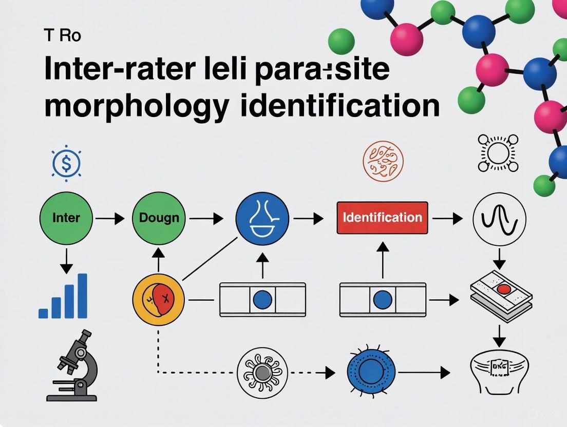
Figure 1: Experimental workflow for assessing parasite identification reliability, comparing traditional morphological and advanced AI-assisted pathways with molecular validation.
Key Research Reagents and Materials
Table 3: Essential Research Reagents for Parasite Identification Studies
| Reagent/Equipment | Specific Application | Function in Experimental Protocol |
|---|---|---|
| Formalin-ethyl acetate | Stool sample processing [2] | Concentration and preservation of parasitic elements for microscopy |
| Giemsa stain | Blood film and larval staining [5] [3] | Enhances visual contrast of parasitic structures for morphological analysis |
| PCR reagents | Molecular identification [1] | Amplification of species-specific genetic markers for definitive identification |
| High-resolution melting PCR | Species differentiation [1] | Discrimination of closely related species based on melt curve analysis |
| YOLOv5 algorithm | AI-assisted detection [4] | Object detection and classification of parasites in digital images |
| DINOv2 models | AI-based classification [2] | Self-supervised learning for parasite identification without extensive labeling |
Technological Advancements in Reliability Assessment
Artificial Intelligence and Machine Learning Applications
Recent advances in artificial intelligence have transformed approaches to parasite identification, offering solutions to the inherent variability of human-based morphological assessment. Deep learning models, particularly convolutional neural networks (CNNs) and vision transformers, have demonstrated remarkable performance in automated parasite detection and classification.
In avian malaria research, Darknet models achieved exceptional accuracy exceeding 99% for classifying Plasmodium gallinaceum blood stages, significantly reducing misclassification rates compared to traditional microscopy [3]. Similarly, for human intestinal parasites, DINOv2-large models attained 98.93% overall accuracy with 78.00% sensitivity and 99.57% specificity, demonstrating strong agreement with expert microscopists (κ > 0.90) [2]. These AI systems not only enhance identification consistency but also address challenges associated with expertise scarcity in resource-limited settings.
For drug discovery applications, YOLOv5 implementation in schistosomiasis research enabled high-throughput screening of compound efficacy against Schistosoma mansoni schistosomula. The model achieved 96.6% mean average precision in distinguishing healthy from damaged parasites, while significantly reducing analysis time compared to manual assessment [4]. This approach minimizes subjective viability assessments that traditionally introduce variability into drug efficacy studies.
Figure 2: Evolution of parasite identification methods demonstrating progressive improvement in reliability through technological integration.
Molecular Validation Frameworks
Molecular techniques have established themselves as reference standards for validating morphological identification, with PCR-based methods providing definitive species determination when morphological features are ambiguous or overlapping. The 2025 Strongylus study exemplifies this validation framework, where HRM-PCR revealed significant discrepancies in species-specific identification frequencies between morphological and molecular approaches [1].
Notably, molecular methods enabled the first report of a patent Strongylus asini infection in a domestic horse, a finding that morphological examination alone failed to detect [1]. This demonstrates how molecular techniques not only validate morphological identification but also expand our understanding of parasite epidemiology through detection of cryptic species or variants.
Implications for Research and Drug Development
The methodological comparisons presented in this guide carry significant implications for parasitology research and anti-parasitic drug development. Consistent and accurate parasite identification forms the foundation of reliable efficacy assessment for novel compounds. The integration of AI-assisted methods and molecular validation into screening pipelines addresses critical sources of variability that can compromise drug development efforts.
For veterinary parasitology, improved IRR directly enhances surveillance data quality, enabling more targeted anthelmintic intervention strategies and better resistance management. In human public health, reliable parasite identification strengthens disease burden assessments and treatment monitoring programs. Future methodological development should focus on integrated systems that leverage the respective strengths of morphological, molecular, and computational approaches while addressing limitations of individual methods through strategic combination.
As technological advancements continue to transform parasitology, maintaining focus on methodological reliability will remain essential for generating reproducible research and effective clinical interventions. The experimental frameworks and comparative data presented here provide researchers with evidence-based guidance for selecting identification methods appropriate to their specific research contexts and reliability requirements.
The High-Complexity Nature of Microscopic Parasitology and its Inherent Subjectivity
Microscopic morphology remains the cornerstone of parasitic disease diagnosis, yet it is characterized by significant technical complexity and inherent diagnostic subjectivity. This guide objectively compares established and emerging parasitological methods, framing the analysis within a critical thesis on inter-rater reliability in parasite identification. Data from controlled experiments quantifying variability between expert microscopists are presented alongside emerging computational solutions designed to mitigate these challenges. The analysis is structured to provide researchers, scientists, and drug development professionals with a clear evidence-based overview of methodological performance, experimental protocols, and the evolving toolkit for parasitological research.
In clinical diagnostics, microscopic parasitology is formally categorized as a high-complexity testing domain under the Clinical Laboratory Improvement Amendments (CLIA) [6]. This classification reflects the extensive knowledge and skill required for accurate morphological identification, which encompasses understanding parasite life cycles, taxonomic classification, and microscopic analysis across diverse specimen types [7]. Despite advancements in molecular techniques, microscopy persists as the gold standard for many parasitic infections, enabling direct parasite observation, species differentiation, and quantification crucial for treatment and research [5] [7].
However, this dependence on morphological expertise is paradoxically threatened by a widespread decline in these very skills. The parasitology community has raised concerns that increased reliance on non-morphology-based diagnostics like rapid antigen tests and nucleic acid amplification tests has led to a progressive loss of morphology expertise [7]. This loss directly impacts diagnostic reliability, potentially leading to missed diagnoses, inappropriate treatment, and mischaracterization of emerging pathogens [7]. The core of this problem lies in the field's inherent subjectivity, where identification accuracy is intrinsically linked to the observer's training and experience, resulting in substantial inter-rater variability.
Experimental Comparisons of Microscopic Methods and Inter-Rater Reliability
Quantifying Variability in Malaria Parasite Counting
A critical study directly compared the established methods for estimating malaria parasitaemia to determine which yields the least inter-rater and inter-method variation [5]. Experienced malaria microscopists counted asexual parasitaemia in 31 Plasmodium falciparum samples using three distinct methods.
Table 1: Comparison of Malaria Parasite Counting Methods and Their Reliability
| Counting Method | Principle | Reported Parasite Density vs. True Count | Sensitivity at Low Parasitaemia (<500/μL) | Inter-Rater Reliability |
|---|---|---|---|---|
| Thin Film Method | Parasites per 5000 erythrocytes, adjusted for total RBC count [5] | ~30% higher than thick film methods [5] | Low (loss of sensitivity) [5] | Not quantified in ANOVA model |
| Thick Film Method | Parasites per 500 white blood cells, adjusted for total WBC count [5] | Closer to true count at high parasitaemia [5] | High [5] | Best among the methods [5] |
| Earle and Perez Method | Number of parasites in fields containing 500 WBCs [5] | Similar to thick film method (little to no bias) [5] | High [5] | Good, but slightly lower than thick film [5] |
The statistical analysis, using ANOVA models on log-transformed counts, revealed that the thick film method demonstrated the best inter-rater reliability [5]. While the thin film method gave counts closer to the true parasite density, it was deemed impractical for low parasitaemias. The study concluded that the thick film method was both reproducible and practical, emphasizing that "the determination of malarial parasitaemia must be applied by skilled operators using standardized techniques" [5].
Detailed Experimental Protocol: Malaria Parasite Counting
The following workflow details the key experimental steps from the comparative study of malaria parasite counting methods [5].
Key Methodological Details:
- Sample Collection: Blood samples were collected from patients in Cambodia during a high transmission season after obtaining informed consent. Samples were transported on ice and processed upon receipt at the laboratory [5].
- Slide Preparation and Staining: Thick and thin smears were made in the field. Additional Earle and Perez slides and thin films were prepared from EDTA blood upon laboratory receipt. All slides were stained with Giemsa at pH 7.2 [5].
- Microscopy and Counting: Seven pre-qualified microscopists across four international sites performed counts. Raters were blinded to each other's results to prevent bias. Counts were performed using the three defined methods [5].
- Statistical Analysis: Parasite counts were log-transformed for analysis to assess proportional differences. Analysis of Variance (ANOVA) models were used to estimate inter-rater reliability, and paired t-tests and regression analyses assessed systematic bias between methods [5].
The Evolving Toolkit: Addressing Complexity and Subjectivity with Technology
Automated Computational Methods for Malaria Diagnosis
To address the challenges of manual microscopy—time consumption, tedium, and observer variability—researchers are developing automated computational methods [8]. These systems typically follow a multi-stage pipeline to diagnose malaria from digital blood smear images.
Table 2: Research Reagent Solutions for Parasitology Analysis
| Reagent/Material | Function/Application | Example Use-Case |
|---|---|---|
| Giemsa Stain (pH 7.2) | Staining malaria parasites in blood smears for microscopic visualization [5] | Differentiation of parasite stages (ring, trophozoite, schizont, gametocyte) in thin and thick films [5] [8] |
| EDTA Blood Tubes | Anticoagulant preservation of blood samples for subsequent smear preparation and cell counting [5] | Maintaining cell integrity for accurate parasite quantification and molecular analysis [5] |
| Block-Matching and 3D Filtering (BM3D) | Computational image denoising to enhance clarity of microscopic fecal images [9] | Preprocessing step in AI-based parasite egg segmentation to improve downstream analysis accuracy [9] |
| Contrast-Limited Adaptive Histogram Equalization (CLAHE) | Enhancing contrast in medical images to improve feature discrimination [9] | Improving distinction between parasite eggs and background in fecal specimen images [9] |
| U-Net Model | Deep learning architecture for precise image segmentation tasks [9] | Segmenting regions of interest (e.g., individual parasite eggs) from complex backgrounds [9] |
| Convolutional Neural Network (CNN) | Deep learning model for image classification through automatic feature learning [9] | Classifying parasite species from segmented image regions with high accuracy [9] |
These automated systems can achieve high accuracy, with one study reporting 97.38% accuracy for an AI-based intestinal parasite egg classifier [9]. This demonstrates the potential of computational methods to provide a standardized, objective approach, reducing reliance on expert morphological skill.
Digital Databases and Genomics to Supplement Morphology
Other technological approaches are being developed to combat the erosion of morphological expertise and provide additional, objective identification tools.
- Digital Parasite Specimen Databases: Initiatives are creating high-quality, digital repositories of parasite specimens to serve as enduring educational and reference resources. These virtual slides prevent deterioration and provide global access to rare specimens, aiding in the training of morphologists [10].
- Genomic Identification Platforms: Tools like the Parasite Genome Identification Platform (PGIP) leverage metagenomic next-generation sequencing (mNGS) for unbiased parasite detection. PGIP uses a curated database of 280 parasite genomes and a standardized bioinformatics pipeline to offer species-level resolution, reducing dependency on morphological expertise [11].
Microscopic parasitology remains a high-complexity field whose gold-standard status is challenged by inherent subjectivity and inter-rater variability, as quantitatively demonstrated in malaria parasite counting studies. While traditional methods like the thick film offer the best reproducibility among skilled operators, the declining pool of expertise poses a significant risk to diagnostic consistency and patient care. The path forward lies in a synergistic approach: preserving and propagating core morphological skills through digital reference databases, while actively integrating advanced computational and genomic methods. AI-based image analysis and platforms like PGIP represent a paradigm shift towards more objective, scalable, and accessible parasitological diagnostics, offering researchers and clinicians powerful tools to supplement and enhance traditional morphological expertise.
The accurate morphological identification of parasites remains a cornerstone of parasitology, crucial for both clinical diagnosis and research. This process, however, is fraught with challenges that can compromise the reliability and reproducibility of results. Inter-rater reliability—the degree of agreement among different microscopists—is a key metric for assessing the consistency of morphological identification in research settings. This guide objectively compares how different methodologies and technologies perform in addressing three pervasive challenges: the morphological similarity of closely related species, variations in sample preparation and staining, and the degradation of sample quality. By synthesizing current experimental data, we provide researchers, scientists, and drug development professionals with a clear comparison of conventional and emerging approaches, highlighting protocols and tools that enhance diagnostic precision and research validity.
Quantitative Comparison of Method Performance
The following tables summarize experimental data from key studies, providing a direct comparison of how different methods address core challenges in parasite morphology.
Table 1: Performance Comparison of Microscopy-Based Counting Methods for Malaria Parasitaemia [5] [12] [13]
| Counting Method | Systematic Bias | Inter-Rater Reliability | Optimal Use Case / Sensitivity |
|---|---|---|---|
| Thin Blood Film | ~30% higher counts than thick film/Earle & Perez [5] | Lower reliability due to counting fatigue [5] | High parasitaemia (>500 parasites/μL); species identification [5] |
| Thick Blood Film | Little to no bias vs. Earle & Perez [5] | Best reliability among methods [5] | Routine diagnosis; low parasitaemia detection [5] |
| Earle & Perez | Little to no bias vs. thick film [5] | Good, but slightly lower than thick film [5] | Historical and specialist comparison [5] |
Table 2: Efficacy of Molecular vs. Morphological Identification for Closely Related Species [14]
| Identification Method | Identification Accuracy | Key Findings & Limitations |
|---|---|---|
| Morphology (Male Spicule Length) | Prone to misidentification due to overlapping traits [14] | Body length/width aided differentiation; female traits were less reliable [14]. |
| Morphology (Female Posterior End) | Unreliable; minimal projection not a robust diagnostic character [14] | Misidentification common between A. cantonensis and A. malaysiensis [14]. |
| Molecular (Nuclear ITS2 Region) | High accuracy; resolved morphological ambiguity [14] | Revealed 8.2% hybrid forms and 1.9% mito-nuclear discordance [14]. |
Table 3: Impact of Sample Preservation Medium on Morphological Analysis [15]
| Preservation Medium | Morphotype Diversity Recovered | Preservation Quality (Larvae) | Suitability |
|---|---|---|---|
| 10% Formalin | Higher number of parasitic morphotypes identified [15] | Superior preservation of larval cuticle and internal structures [15] | Optimal for long-term morphological studies only [15]. |
| 96% Ethanol | Lower morphotype diversity vs. formalin [15] | Increased degradation; cuticle shrinking/puckering [15] | Ideal for combined molecular/morphological work; adequate for morphology [15]. |
Detailed Experimental Protocols
To ensure the reproducibility of the comparative data presented, this section outlines the key methodologies employed in the cited studies.
- Sample Preparation: Thirty-one Plasmodium falciparum-infected blood samples were collected in Cambodia. From each sample, thick and thin blood films were prepared on glass slides and stained with Giemsa at pH 7.2.
- Microscopy and Counting: Seven experienced microscopists across four international laboratories performed blinded counts of asexual parasitaemia using three established methods:
- Thin Film Method: Parasites were counted against a total of 5,000 erythrocytes, with the result adjusted using the patient's actual red blood cell count.
- Thick Film Method: Parasites were counted against 500 white blood cells, with the result adjusted using the patient's actual white blood cell count.
- Earle and Perez Method: A specific method utilizing a standardized slide format, with parasites also counted per white blood cell and adjusted.
- Statistical Analysis: Log-transformed parasite counts were analyzed using ANOVA models to determine inter-rater reliability for each method. The paired t-test was used to assess systematic bias between methods.
- Sample Collection: 257 archived adult worm specimens from five zoogeographical regions in Thailand were used.
- Morphological Identification: Worms were identified based on key diagnostic characters: for males, spicule length and Δ bursal rays; for females, the distance of the anus and vulva from the posterior end, and the presence of a minute terminal projection. General morphometrics (body length/width, esophagus length/width) were also recorded.
- Molecular Identification: DNA was extracted from individual worms. Two molecular targets were used:
- Mitochondrial Cytochrome b (cytb): A SYBR Green-based qPCR assay with species-specific primers was used to differentiate A. cantonensis from A. malaysiensis.
- Nuclear ITS2 Region: PCR-RFLP, using the BtsI-v2 restriction enzyme, was employed for species differentiation and to detect potential hybrids based on banding patterns.
- Data Validation: Morphological character measurements were statistically compared across the molecularly defined groups (pure A. cantonensis, pure A. malaysiensis, and hybrids) to validate the reliability of morphological diagnoses.
- Sample Collection and Storage: Fresh fecal samples from capuchin monkeys were collected and immediately split into two equal parts. One part was stored in 10% buffered formalin, and the other in 96% ethanol. Samples were stored at ambient temperature for 8-19 months before analysis.
- Microscopic Screening: Samples were processed using a modified Wisconsin sedimentation technique. The resulting pellets were screened microscopically for parasites, which were morphologically identified.
- Preservation Grading: A standardized 3-point grading scale was developed for both eggs and larvae in each preservative. A score of 3 indicated excellent preservation with intact structures, 2 indicated moderate degradation partially interfering with identification, and 1 indicated severe degradation making identification difficult or impossible.
- Statistical Comparison: Wilcoxon-Signed Rank tests were used to compare the morphotype diversity, parasites per gram (PFG), and average preservation rating between the paired formalin and ethanol samples.
Visualization of Workflows and Relationships
Experimental Workflow for Species Identification
The following diagram illustrates the integrated workflow for resolving species identity, combining traditional morphological and modern molecular approaches as described in the experimental protocols [14].
Diagnostic Pathway for Morphological Challenges
This flowchart outlines the decision-making process for diagnosing parasitic infections when faced with key challenges of similarity, staining, and sample quality, leading to different classes of solutions [16] [5] [17].
The Scientist's Toolkit: Key Research Reagents & Solutions
This table details essential reagents, tools, and technologies used in the featured experiments to address morphological identification challenges.
Table 4: Key Research Reagent Solutions for Parasite Morphology Studies
| Reagent / Solution | Function / Application | Experimental Context |
|---|---|---|
| Giemsa Stain (pH 7.2) | Standard staining for malaria blood films; highlights parasite chromatin and cytoplasm. | Used across all microscopy methods for malaria parasite counting to ensure consistent staining [5] [12]. |
| 10% Buffered Formalin | Tissue fixative; cross-links proteins to preserve morphological integrity long-term. | Preserved fecal samples for superior recovery of parasite morphotypes and larval structure [15]. |
| 96% Ethanol | Dehydrating fixative; preserves samples adequately for morphology and optimally for DNA. | Used for parallel sample preservation, enabling both morphological and downstream molecular analysis [15]. |
| BtsI-v2 Restriction Enzyme | Endonuclease for PCR-RFLP; cuts specific DNA sequences to generate species-specific band patterns. | Key reagent for differentiating A. cantonensis and A. malaysiensis using the nuclear ITS2 region [14]. |
| Species-specific qPCR Primers (cytb) | Targets mitochondrial gene for sensitive and quantitative species detection. | Enabled specific identification and quantification of Angiostrongylus species, revealing hybrids [14]. |
| Lightweight Deep Learning Models (e.g., DANet, Hybrid CapNet) | AI-based analysis of blood smear images; automates detection and classification. | Provides a computational solution to challenges of human fatigue and subjectivity in microscopy [16] [17]. |
Impact of Parasite Life Cycle and Pre-patency on Identification Consistency
The accurate identification of parasitic infections is a cornerstone of effective disease control, drug development, and epidemiological research. However, this process is fundamentally influenced by two intrinsic biological factors: the parasite's life cycle and the pre-patent period. The life cycle encompasses the distinct morphological and developmental stages a parasite undergoes, while the pre-patency refers to the initial period after infection during which diagnostic signs, such as eggs or specific antigens, are not yet detectable. Within the context of research on inter-rater reliability in parasite morphology identification, these factors introduce significant variability that can affect the consistency of observations between different scientists. This guide objectively compares the performance of traditional and novel diagnostic methodologies in managing this variability, providing supporting experimental data to inform researchers, scientists, and drug development professionals.
Parasite Life Cycle Stages and Identification Challenges
The complex life cycles of parasites present a primary challenge for consistent identification. For Plasmodium species, the causative agents of malaria, the intra-erythrocytic stages include the ring, trophozoite, schizont, and gametocyte, each with distinct morphologies [18]. The progression through these stages requires a tightly orchestrated transcriptional program, and fundamental changes in chromatin structure and epigenetic modifications during life cycle progression suggest a central role for these mechanisms in regulating the transcriptional program of malaria parasite development [19]. The protein PfSnf2L, an ISWI-related ATPase, has been identified as a key just-in-time regulator of gene expression, spatiotemporally determining nucleosome positioning at the promoters of stage-specific genes [19]. The functional absence of such regulators can phenocopy the loss of correct gene expression timing, disrupting development [19].
For intestinal parasites, the challenge often lies in differentiating eggs of species like Taenia sp., Trichuris trichiura, Diphyllobothrium latum, and Fasciola hepatica from artifacts in fecal smears [20]. Their identification relies on an expert's ability to recognize subtle morphological characteristics, a process susceptible to human error and subjectivity, especially under conditions of mental and physical exhaustion [21] [18]. The variability in staining uptake and the refractivity of parasites further complicates this manual process [21].
Table: Impact of Parasite Life Cycle on Diagnostic Consistency
| Parasite | Key Life Cycle Stages | Impact on Identification Consistency | Supporting Evidence |
|---|---|---|---|
| Plasmodium falciparum | Ring, Trophozoite, Schizont, Gametocyte [18] | Stage-dependent chromatin accessibility regulates gene expression; incorrect timing disrupts development [19]. | Depletion of PfSnf2L led to a global opening of chromatin and mis-timed gene expression, killing parasites [19]. |
| Intestinal Helminths (e.g., Taenia sp., F. hepatica) | Egg, Larval, Adult | Egg morphology is the primary diagnostic feature, but manual identification is variable and requires specialist training [20]. | An automated algorithm achieved near-perfect sensitivity (99.1-100%) and specificity (98.1-98.4%), highlighting human inconsistency [20]. |
| Pinworm (Enterobius vermicularis) | Egg, Larva, Adult | Small egg size (50-60 μm) and similarity to other particles lead to false negatives in manual exams [22]. | The scotch tape test has limited sensitivity and relies heavily on examiner ability [22]. |
The Pre-patent Period and its Implications for Research
The pre-patent period directly impacts the sensitivity of diagnostic tests and the timing of intervention studies. For equine parasites like Parascaris equorum and Anoplocephala perfoliata, eggs are only expelled with feces after the larvae have matured and the infection load becomes substantial [23]. During the larval migration stage in the host, or when no signs of infection are found on the body surface, serological detection becomes a simple and effective method for rapid diagnosis of parasitic infection [23]. This underscores the necessity of selecting the appropriate diagnostic tool based on the timing post-infection.
In malaria research, the biology of gametocytes is particularly relevant. Stage V gametocytes are the only forms infectious to mosquitoes and can circulate quiescently for several weeks [24]. A significant challenge in developing transmission-blocking drugs is that most current antimalarials are ineffective against these quiescent stages [24]. Consequently, individuals can remain infectious for weeks after treatment has cleared the asexual blood stages [24]. This extended pre-patency of transmissibility is a major hurdle for eradication campaigns and requires specialized assays for drug discovery.
Comparative Analysis of Diagnostic Methodologies
Different diagnostic protocols offer varying levels of performance in managing the variability introduced by life cycle and pre-patency. The table below compares the experimental protocols and quantitative performance of several key approaches.
Table: Comparison of Diagnostic Method Performance and Protocols
| Methodology | Experimental Protocol Summary | Key Performance Metrics | Consistency Advantages |
|---|---|---|---|
| Manual Microscopy | Stained blood or fecal smears are examined by a technician for morphological identification of parasites/eggs [21] [18]. | Time-consuming, labor-intensive, and susceptible to human error and subjectivity [22] [18]. | Low inter-rater reliability; consistency is affected by examiner expertise and fatigue [21]. |
| Automated Image Analysis (Mathematical Algorithm) | Digital images are processed through a 14-step SCILAB algorithm: gray-scale conversion, contrast enhancement, Gaussian smoothing, binarization, border smoothing, labeling, boundary object exclusion, image closing, holes filtering, area filtering, skeletonization, border identification, and recoloring. Features are extracted for logistic regression classification [20]. | Sensitivity: 99.10% - 100% Specificity: 98.13% - 98.38% for helminth eggs [20]. | High consistency; eliminates human subjectivity and fatigue; provides a standardized, objective assessment [20]. |
| Deep Learning (YOLO-CBAM) | The YOLOv8 architecture is integrated with a Convolutional Block Attention Module (CBAM) and self-attention mechanisms. The model is trained on datasets of labeled microscopic images to automatically detect and localize parasite eggs [22]. | Precision: 0.9971 Recall: 0.9934 mAP@0.5: 0.9950 [22]. | Superior at detecting small objects in complex backgrounds; reduces false negatives/positives; highly scalable and consistent [22]. |
| Staining-Independent AI Classification | Blood smear images are converted to grayscale to lessen staining impact. For detection, a YOLO-based model is used. For life stage classification, single-cell images are cropped and classified using a CNN (e.g., LeNet-5) architecture [18]. | Detection Accuracy: 0.79 - 0.92 (across species) Classification Accuracy: 0.93 - 0.96 (across stages) [18]. | Reduces variability from inconsistent staining; enables accurate life stage classification, crucial for research on stage-specific biology [18]. |
Research Reagent Solutions for Parasite Identification
The following reagents and tools are essential for conducting research in parasite identification and managing the challenges of life cycle and pre-patency.
Table: Essential Research Reagents and Tools
| Research Reagent / Tool | Function in Experimental Protocol |
|---|---|
| Giemsa Stain | A classical dye used to highlight parasites in blood smears for microscopic identification of malaria life stages [18]. |
| Lugol's Iodine | A temporary stain used on fecal smears to enhance the visibility of protozoan cysts and helminth eggs [20]. |
| NF54/iGP1_RE9Hulg8 Transgenic Parasites | Genetically engineered P. falciparum parasites expressing a red-shifted firefly luciferase reporter; used in viability assays for high-throughput screening of gametocytocidal compounds [24]. |
| Scilab Open-Source Platform | A computational environment used to implement custom image processing and pattern recognition algorithms for the automated identification of parasite eggs [20]. |
| YOLO-CBAM Deep Learning Framework | An integrated object detection architecture that uses attention mechanisms to improve feature extraction from complex microscopic images, enabling high-accuracy automated detection [22]. |
| N-Acetyl-glucosamine (GlcNAc) | A chemical used in Plasmodium culture to eliminate asexual blood stage parasites, enabling the production of synchronous gametocyte populations for stage-specific drug assays [24]. |
Visualizing Experimental Workflows
The following diagrams illustrate the logical workflows for key experimental protocols discussed in this guide, highlighting how they address identification challenges.
Automated Parasite Egg Recognition Workflow
AI Malaria Detection & Staging Pipeline
The consistency of parasite identification is intrinsically linked to a deep understanding of parasite life cycles and the pre-patent period. Traditional manual methods, while foundational, are inherently variable and struggle to account for these biological complexities objectively. The comparative data presented in this guide demonstrates that automated methodologies, particularly those leveraging sophisticated mathematical algorithms and deep learning, offer a significant performance advantage. They provide higher sensitivity, specificity, and overall accuracy while establishing a standardized, objective framework that minimizes inter-rater variability. For researchers and drug developers, the adoption of these advanced tools and the carefully designed experimental protocols that account for stage-specific biology are critical for generating reliable, reproducible data essential for advancing the field of parasitology.
In scientific and clinical practice, reliability refers to the consistency of a measurement—the extent to which it can be reproduced when repeated under the same conditions [25]. In the specific context of parasite morphology identification, inter-rater reliability measures the degree of agreement between different scientists when identifying the same parasite specimens. When this reliability is low, the consequences cascade through healthcare systems and research enterprises, leading to misdiagnosis, delayed patient treatment, and compromised research integrity.
The challenges in parasite morphology identification exemplify these high-stakes reliability concerns. As molecular methods increasingly supplement or replace traditional microscopy, the morphological expertise necessary for accurate identification is diminishing across the scientific community [7]. This loss of expertise threatens diagnostic accuracy, as morphological identification remains the gold standard for many parasitic infections and is often the most appropriate, cost-effective, and sometimes the only accurate identification method in many settings [7]. This article examines the consequences of low inter-rater reliability through the lens of parasite morphology research, comparing diagnostic approaches and providing actionable methodologies for enhancing reliability in scientific practice.
Defining the Problem: Low Reliability in Parasite Morphology Identification
The Decline of Morphological Expertise
The field of parasitology is experiencing a paradoxical situation: while advanced diagnostic techniques like rapid antigen detection tests (RDTs), nucleic acid amplification tests (NAATs), and metagenomic next-generation sequencing (mNGS) have expanded diagnostic capabilities, they have simultaneously contributed to a progressive, widespread loss of morphology expertise for parasite identification [7]. This skill deficit is not easily remedied, as becoming an effective parasite morphologist requires several years of training in practical and theoretical knowledge of anatomy, biology, zoology, taxonomy, and epidemiology across the vast array of parasite taxa capable of infecting humans [7].
This erosion of morphological skills has direct implications for diagnostic accuracy and patient outcomes. As noted in parasitology literature, "Inadequate morphology experience may lead to missed and inaccurate diagnoses and erroneous descriptions of new human parasitic diseases" [7]. The problem is particularly acute for less common parasites and in resource-limited settings where advanced molecular diagnostics may be unavailable or cost-prohibitive.
Quantifying Reliability in Scientific Research
In research methodology, reliability is distinct from validity: while reliability concerns the consistency of a measure, validity refers to how accurately a method measures what it is intended to measure [25]. A measurement can be reliable without being valid, but if a measurement is valid, it is usually also reliable.
Several statistical approaches are used to quantify inter-rater reliability:
- Cohen's Kappa: Used for two raters, accounts for chance agreement [26] [27]
- Fleiss' Kappa: Extends Cohen's Kappa to multiple raters [27]
- Intraclass Correlation Coefficient (ICC): Commonly used for continuous measures, with values closer to 1 indicating higher agreement [28] [29]
Each metric has strengths and limitations, and the appropriate choice depends on the research design, number of raters, and type of data being analyzed [28].
Table 1: Inter-Rater Reliability Metrics and Their Interpretation
| Metric | Appropriate Use Cases | Interpretation Range | Strengths |
|---|---|---|---|
| Cohen's Kappa | Two raters, categorical data | -1 to 1 (≤0: No agreement, 0.01-0.20: Slight, 0.21-0.40: Fair, 0.41-0.60: Moderate, 0.61-0.80: Substantial, 0.81-1.0: Almost Perfect) | Accounts for chance agreement |
| Fleiss' Kappa | Multiple raters, categorical data | Same as Cohen's Kappa | Extends Cohen's to multiple raters |
| Intraclass Correlation Coefficient (ICC) | Multiple raters, continuous measures | 0 to 1 (Higher values indicate better reliability) | Can be used for various experimental designs |
Direct Consequences: Misdiagnosis and Delayed Treatment
Diagnostic Error Categories and Patient Impact
Low reliability in parasite identification directly contributes to diagnostic errors, which the National Academies of Sciences, Engineering, and Medicine categorizes as the failure to establish an accurate and timely explanation of the patient's health problem or to communicate that explanation to the patient [30]. These errors manifest in three primary forms:
- Missed Diagnosis: The medical condition is entirely overlooked, leading to an absence of necessary treatment [31]
- Delayed Diagnosis: The condition is recognized but not promptly, allowing progression without intervention [30] [31]
- Misdiagnosis: The condition is incorrectly identified, leading to inappropriate treatment that may exacerbate the health concern [31]
The prevalence of these errors is substantial. Globally, diagnostic errors affect an estimated 12 million people annually in the United States alone, with conditions such as cancer, cardiovascular diseases, and infections being particularly prone to diagnostic challenges due to their complex nature and subtle early symptoms [31].
Case Study: Angiostrongylus Identification Challenges
The difficulties in differentiating between closely related parasite species demonstrate how low reliability leads to diagnostic errors. Research on Angiostrongylus cantonensis and Angiostrongylus malaysiensis in Thailand reveals that morphological misidentifications between these two closely related species are common due to overlapping morphological characters [14].
A study analyzing 257 archived specimens found that while certain male traits (body length and width) aided species differentiation, female traits were less reliable for accurate identification [14]. Furthermore, the research revealed hybrid forms (8.2% of specimens) through nuclear ITS2 region analysis, complicating morphological identification even for experienced parasitologists [14]. This case illustrates how taxonomic complexities can undermine diagnostic reliability even before considering observer variability.
Patient Harm from Diagnostic Errors
The consequences of these diagnostic failures extend beyond academic concern to tangible patient harm:
- Emotional and Psychological Impact: Patients often experience stress, anxiety, and diminished trust in the healthcare system when faced with diagnostic uncertainty [31]
- Clinical Deterioration: Delayed or incorrect treatment allows disease progression, potentially transforming manageable conditions into life-threatening situations
- Economic Consequences: Prolonged illnesses lead to increased healthcare expenses through additional testing, extended care, or unnecessary treatments based on incorrect initial diagnoses [31]
Research Implications: Compromised Data Quality and Validity
Threats to Research Integrity
Low inter-rater reliability introduces significant threats to research integrity across multiple domains:
- Compromised Internal Validity: When reliability is low, the consistency of measurements within a study is undermined, making it difficult to distinguish true effects from measurement error [25]
- Reduced Statistical Power: Unreliable measurements introduce noise that obscures genuine relationships or treatment effects, potentially leading to Type II errors (false negatives)
- Impaired Reproducibility: The fundamental principle of scientific reproducibility is threatened when measurements cannot be consistently replicated across different research teams [28]
The problem is particularly acute in studies of neurodegenerative disorders and psychiatric conditions where clinical judgment plays a significant role in diagnosis and assessment. As one study noted, "variability in clinical judgment can hinder reliability and complicate the interpretation of findings" [26].
Limitations of Reliability Metrics in Complex Tasks
While statistical measures of inter-rater reliability are necessary, they are insufficient guarantees of data quality, particularly for complex annotation tasks [27]. High inter-rater reliability scores can sometimes be misleading when:
- Annotators Share the Same Biases: Multiple raters may consistently make the same errors due to common misconceptions or shared blind spots
- Task Complexity Exceeds Expertise: Difficult cases may be systematically misclassified by non-expert raters while still producing high agreement rates
- Simplistic Scoring Rubrics: Measurement instruments that fail to capture nuanced distinctions may produce high reliability at the cost of validity [27]
These limitations highlight why sophisticated research protocols incorporate multiple quality control mechanisms beyond simple reliability metrics.
Comparative Analysis: Reliability Across Diagnostic Methods
Morphological vs. Molecular Identification
The ongoing transition from morphological to molecular identification methods in parasitology offers a instructive case study for comparing reliability across diagnostic approaches.
Table 2: Comparison of Diagnostic Methods in Parasitology
| Diagnostic Characteristic | Morphology-Based Diagnostics | PCR-Based Diagnostics | Sequencing-Based Diagnostics |
|---|---|---|---|
| Sensitivity | ++ | +++ | +++ |
| Specificity | +++ | +++ | +++ |
| Quantification Capacity | +++ | ++ | - |
| Turnaround Time | +++ (except histology) | ++ | + |
| Cost-Effectiveness | +++ | ++ | + |
| Genus-Level Identification | +++ | +++ | +++ |
| Species-Level Identification | ++ | +++ | +++ |
| Capacity to Detect Novel/Zoonotic Agents | +++ | - | +++ |
| Adaptability to Resource-Poor Settings | +++ | - | - |
Note: -, +, ++, +++ represent no, limited, moderate, or high capacity/efficacy respectively [7]
Methodological Limitations and Complementarity
Each diagnostic approach faces distinct limitations that can affect reliability:
- Morphological Methods: Subject to interpreter variability, require significant expertise, challenging for closely related species or atypical presentations [7] [14]
- Molecular Methods: Limited target range (not all parasites have available assays), specimen incompatibility (e.g., formalin fixation degrades DNA), inadequate reference databases for novel organisms [7]
- Hybrid Approaches: Combining morphological and molecular methods can enhance overall reliability but requires additional resources and expertise [14]
The Angiostrongylus study concluded that "nuclear ITS2 is a reliable marker for species identification of A. cantonensis and A. malaysiensis, especially in regions where both species coexist," suggesting a complementary approach rather than complete replacement of morphological methods [14].
Methodological Framework: Enhancing Reliability in Research Practice
Experimental Protocols for Reliability Assessment
Well-designed reliability studies require careful planning and execution. Key methodological considerations include:
- Clear Purpose Statement: The study purpose should clearly distinguish between hypothesis testing and parameter estimation, as this determines appropriate sample size calculations and statistical analyses [28]
- Rater Selection and Training: Clearly state whether raters represent the only raters of interest or a larger population, as this affects the choice of statistical model [28]
- Structured Diagnostic Processes: Implement standardized diagnostic criteria and consensus procedures, as exemplified by the Asian Cohort for Alzheimer's Disease (ACAD) study, which achieved almost perfect agreement (Cohen's Kappa = 0.835) through rigorous protocol harmonization [26]
Experimental Protocol for Reliability Assessment
Quality Control Mechanisms Beyond Reliability Metrics
Sophisticated research protocols employ multiple quality control strategies:
- Task Decomposition: Complex identification tasks are broken into simpler subtasks assigned to annotators with appropriate skill levels [27]
- Skill-Based Annotation: Annotator skills are assessed through initial tests and continuously monitored using control tasks mixed with actual study tasks [27]
- Confidence-Based Aggregation: Multiple annotations are aggregated with weighting based on demonstrated annotator skill levels, with low-confidence cases receiving additional review [27]
- Comprehensive Auditing: Final datasets undergo sampling-based audits using quality metrics such as accuracy, precision, recall, and F-score rather than relying solely on inter-rater reliability [27]
Essential Research Reagents and Methodological Tools
The Scientist's Toolkit for Reliability Enhancement
Implementing robust reliability assessment requires specific methodological tools and approaches:
Table 3: Essential Research Reagents and Tools for Reliability Studies
| Tool/Reagent | Primary Function | Application Context |
|---|---|---|
| Standardized Diagnostic Criteria | Provides consistent framework for classification | Essential for multi-center studies; enables comparison across research sites [26] |
| Molecular Markers (e.g., ITS2, cytb) | Validates morphological identifications; detects hybridization | Critical for resolving difficult taxonomic distinctions; identifies cryptic species [14] |
| Statistical Software Packages | Calculates reliability metrics (ICC, Kappa, etc.) | Required for quantitative reliability assessment; must be validated for appropriate application [28] |
| Reference Collections | Serves as ground truth for training and validation | Provides validated specimens for comparator studies; essential for method validation [7] |
| Structured Consensus Protocols | Formalizes diagnostic decision-making | Reduces individual bias; enhances transparency and reproducibility [26] |
| Control Tasks/Specimens | Monitors ongoing rater performance | Enables continuous quality assessment during data collection; identifies rater drift [27] |
The consequences of low reliability in parasite morphology identification—and scientific research more broadly—extend from compromised patient care to undermined research validity. Addressing these challenges requires a multifaceted approach that acknowledges the complementary strengths of traditional and modern methods while implementing rigorous methodological safeguards.
Maintaining morphological expertise remains essential even as molecular methods advance, particularly for detecting novel pathogens, working in resource-limited settings, and providing cost-effective diagnostics [7]. Simultaneously, molecular methods offer crucial validation for morphologically challenging distinctions and can identify hybridization events that complicate traditional classification [14].
Enhancing reliability in both research and clinical practice requires moving beyond simple reliability metrics to implement comprehensive quality frameworks that include careful study design, appropriate statistical application, ongoing rater training, and multimodal verification. By adopting these approaches, the scientific community can better ensure that diagnostic decisions and research findings rest on a foundation of methodological rigor and reproducible observation.
From Microscope to Machine: Modern Methods to Standardize Parasite Identification
In the field of parasitology, the accurate diagnosis of intestinal parasitic infections (IPIs) relies heavily on proven microscopic techniques. Despite advancements in molecular and automated technologies, conventional methods remain the cornerstone for routine diagnosis, particularly in resource-limited settings where the burden of these diseases is highest [32] [33]. The Formalin-Ethyl Acetate Centrifugation Technique (FECT), the Merthiolate-Iodine-Formalin (MIF) technique, and the Direct Smear method represent such foundational approaches. Their utility, however, must be understood within a critical research context: the ongoing investigation into inter-rater reliability in parasite morphology identification. The identification of parasitic elements is inherently dependent on the expertise of the microscopist, introducing a variable that can significantly impact diagnostic consistency, epidemiological data, and the evaluation of new technologies [32] [34]. This guide provides a detailed, objective comparison of these three techniques, framing their performance data and protocols within the broader thesis of analytical variability and standardization in parasitological research.
The following diagram illustrates the procedural relationships and key decision points leading to the use of FECT, MIF, and Direct Smear techniques in a research context focused on morphological identification.
Comparative Performance Analysis
Diagnostic Sensitivity and Operational Characteristics
The choice of diagnostic technique directly influences the detection capability for various parasitic elements and the operational workflow of a laboratory. The table below summarizes the key performance characteristics and comparative advantages of FECT, MIF, and Direct Smear, providing a basis for their application in research settings.
| Characteristic | Formalin-Ethyl Acetate Centrifugation Technique (FECT) | Merthiolate-Iodine-Formalin (MIF) | Direct Smear |
|---|---|---|---|
| Primary Principle | Sedimentation concentration [35] | Staining and sedimentation [32] | Direct wet mount [33] |
| Sensitivity (General) | High; considered a reference standard [34] | Competitive with FECT for IPI evaluation [32] | Low; suitable for high-intensity infections only [33] |
| Sensitivity (Opisthorchis viverrini) | 75.5% [34] | Information Not Specified in Search Results | 67.3% (Stool Kit, a commercial concentrator) [34] |
| Key Advantage | Concentrates a wide range of parasites; suitable for preserved samples [33] | Effective fixation and staining; long shelf life, good for field use [32] | Rapid; preserves motile trophozoites [33] [36] |
| Key Disadvantage | Requires centrifugation; logistical complexity [34] | Can distort trophozoite morphology [32] | Poor sensitivity for low-level infections [33] |
| Quantification Capability | Yes ( Eggs Per Gram (EPG) can be calculated) [34] | Information Not Specified in Search Results | Semi-quantitative only [33] |
| Best For (Parasite Stages) | Helminth eggs, larvae, cysts, and oocysts [33] [35] | Broad-spectrum of helminths and protozoa [32] | Motile trophozoites and poorly floating stages [36] |
Performance in Inter-Rater Reliability Context
The reliability of morphological identification is a central concern when using these techniques. A 2025 study evaluating deep-learning models against human experts using FECT and MIF as the ground truth provides insightful data. The models demonstrated a strong level of agreement with medical technologists, with Cohen's Kappa scores exceeding 0.90 for all models tested [32]. This high kappa score, achieved when a standardized reference is used, underscores that the analytical method itself can be highly reliable. However, it also highlights that the major source of variability in diagnosis often lies in human interpretation, a critical factor for designing studies on inter-rater reliability.
Furthermore, the distinct morphology of different parasites affects identification consistency. The same 2025 study noted that deep-learning models achieved high precision, sensitivity, and F1 scores for helminthic eggs and larvae due to their more distinct and uniform morphology compared to protozoans [32]. This finding can be extrapolated to human raters: techniques like FECT that are particularly strong at concentrating helminth eggs (e.g., Ascaris, Trichuris, hookworm) may inherently facilitate higher inter-rater agreement for these species.
Detailed Experimental Protocols
Formalin-Ethyl Acetate Centrifugation Technique (FECT)
The FECT protocol is a sedimentation method designed to concentrate parasitic elements by removing debris and fats [35].
Workflow Diagram: FECT Protocol
- Step-by-Step Procedure [34] [35]:
- Sample Preparation: Homogenize approximately 2 grams of fresh stool and emulsify in 10% formalin. The suspension is then strained through multiple layers of gauze into a 15 ml conical centrifuge tube.
- First Centrifugation: Top up the tube with saline or 10% formalin and centrifuge at 500 × g for 10 minutes. The supernatant is decanted.
- Solvent Extraction: Resuspend the sediment in 10 ml of 10% formalin. Add 4 ml of ethyl acetate, stopper the tube, and shake vigorously for 30 seconds to dissolve fecal fats.
- Second Centrifugation: Centrifuge again at 500 × g for 10 minutes. This results in four layers: a plug of debris at the top, a layer of ethyl acetate, a formalin layer, and the sediment containing parasites at the bottom.
- Sediment Examination: Free the debris plug, decant the top three layers, and use a swab to clean the tube walls. The final sediment is resuspended in a small volume of formalin and examined microscopically under low and high power.
Merthiolate-Iodine-Formalin (MIF) Technique
The MIF technique serves as a combined fixative and stain, making it suitable for field surveys and for highlighting protozoan cysts [32].
- Solution Preparation: The MIF solution itself is a combination of merthiolate (a preservative), iodine (a stain), and formalin (a fixative). It is noted for its easy preparation and long shelf life [32].
- Staining Mechanism: The iodine component of the solution stains glycogen and nuclei of protozoan cysts, enhancing their visibility and aiding in species differentiation based on internal structures.
- Procedure: While the specific sequence was not detailed in the search results, the technique involves mixing a stool sample with the MIF solution. The fixation preserves the parasites, while the staining allows for immediate examination. It can be used as a direct stain or followed by a sedimentation step to concentrate the parasitic elements [32] [36].
- Limitation for Research: A key consideration for morphology studies is that the iodine can potentially distort trophozoite morphology, and the fixation may be inadequate for certain trichrome stains used for permanent slides [32].
Direct Smear Microscopy
The direct smear is the simplest and fastest technique, primarily used for the initial assessment or when motility must be observed.
Step-by-Step Procedure [33] [36]:
- Smear Preparation: A small amount of fresh, unpreserved feces (approximately 2 mg) is emulsified in a drop of 0.9% saline on a microscope slide. The use of saline is critical, as water can destroy motile trophozoites.
- Coverslip: A large coverslip (22x22 mm) is placed over the suspension.
- Examination: The entire preparation is examined systematically under the microscope, first with a 10x objective and then with a 40x objective.
- Iodine Staining: For enhanced visualization of protozoan cysts, a separate smear can be prepared with a drop of Lugol's iodine instead of saline. This kills trophozoites but stains cyst walls and internal structures.
Key Application in Research: The primary research value of the direct smear is its utility in detecting motile trophozoites (e.g., of Giardia or Entamoeba), which can be lost or distorted in concentration procedures. It is also adequate for observing heavy infections with helminths like Ascaris lumbricoides [33].
The Scientist's Toolkit: Key Research Reagent Solutions
The table below lists essential materials and their functions for implementing the discussed techniques in a research setting.
| Research Reagent / Material | Primary Function in Protocol |
|---|---|
| 10% Formalin Solution | Universal fixative and preservative; inactivates pathogens for safe handling in FECT and MIF [33] [35]. |
| Ethyl Acetate | Organic solvent used in FECT to dissolve fats and debris, clearing the sample for easier microscopy [35]. |
| Merthiolate-Iodine-Formalin (MIF) Solution | All-in-one fixative and stain; preserves morphology and stains internal structures of cysts for identification [32]. |
| Lugol's Iodine Solution | Staining solution used in Direct Smear and other methods to contrast protozoan cysts and reveal nuclei [36]. |
| 0.9% Saline Solution | Isotonic diluent for Direct Smear; maintains viability and motility of trophozoites during examination [36]. |
| Cellophane Coverslips / Glycerol | Used in the Kato-Katz method (a related quantitative technique) to clear debris for better visualization of helminth eggs [32] [33]. |
| Conical Centrifuge Tubes & Gauze | Essential for the concentration steps in FECT; gauze is used to filter out large particulate matter [34] [35]. |
The Role of Digital Imaging and Whole-Slide Scanning in Standardizing Visual Data
In scientific fields reliant on visual data, such as parasite morphology identification research, inter-rater reliability remains a significant challenge. Studies consistently demonstrate that subjective visual interpretation introduces variability, even among experienced professionals. For instance, research on stress signatures in dentition found that more experience in assessment does not necessarily produce higher reliability between raters, with disagreements occurring frequently in intensity categorization [37]. This variability directly impacts diagnostic consistency, research reproducibility, and ultimately, scientific progress in parasitology and drug development.
Digital imaging and whole-slide imaging (WSI) technologies are transforming morphological sciences by addressing these standardization challenges. WSI systems create high-resolution digital reproductions of entire glass slides, enabling pathologists and researchers to examine tissue specimens on computer displays rather than through traditional microscopy [38]. The fundamental value proposition of these technologies lies in their potential to standardize visual data acquisition, management, and interpretation across institutions, research groups, and time periods.
The clinical validation of WSI systems for primary diagnosis has established their non-inferiority to conventional microscopy [38] [39] [40]. However, for research applications, particularly in specialized fields like parasite morphology, understanding the technical variations between systems and their implications for data standardization is crucial. This guide provides an objective comparison of WSI technologies, supported by experimental data, to inform researchers and drug development professionals in selecting and implementing digital pathology solutions.
Whole-Slide Imaging Technology Comparison
Scanner Platforms and Key Characteristics
Whole-slide imaging systems consist of three core components: the slide scanner, viewing software, and display monitor [38]. The market offers diverse scanner options with varying capabilities suited to different laboratory needs and throughput requirements. The following table summarizes major digital pathology scanners and their key characteristics:
Table: Comparison of Whole-Slide Imaging Scanners
| Manufacturer | Model Examples | Key Features | Capacity | Target Use Cases |
|---|---|---|---|---|
| 3DHISTECH | PANNORAMIC Flash DESK DX, PANNORAMIC 1000 DX | Affordable entry-level to high-speed models; standardized optical system; self-calibration | Entry-level to 1000 slides | Routine pathology; basic clinical diagnoses to high-volume labs [41] |
| Grundium | Ocus20, Ocus40, Ocus M 40 | Browser-based platform; high-resolution imaging; precision engineering | Varies by model | Clinical/research settings; remote consultations; intraoperative frozen sections [41] |
| Hamamatsu | NanoZoomer Series | Remarkable image quality; high-speed scanning; fluorescence capabilities | Not specified | Clinical and research applications requiring exceptional image quality [41] |
| Huron | TissueScope iQ, LE, LE120 | Broad file format compatibility; patented MSIA technology; fast scanning (≈60s/slide) | 120-400 slides | High-volume labs; versatile research applications [41] |
| Leica Biosystems | Aperio GT 450 DX, CS2, LV1 | Custom optics; no-touch scanning; secure IT infrastructure | 450 slides (GT 450 DX) | High-volume clinical settings; medium-volume use; remote viewing [41] |
| Roche | VENTANA DP 200, DP 600 | Built-in calibration; dynamic focus technology; user-friendly interface | 240 slides (DP 600) | Frozen sections; urgent cases; labs scaling toward full digitization [41] |
Performance Validation and Equivalency Data
Multiple rigorous studies have validated the diagnostic equivalence between digital pathology and traditional microscopy. The following table summarizes key performance metrics from recent validation studies:
Table: Performance Metrics from WSI Validation Studies
| Study | Sample Size | Concordance Rate | Efficiency Findings | Notable Limitations |
|---|---|---|---|---|
| Roche FDA Validation Study [38] | 2,047 clinical cases | Difference in accuracy between digital reads and manual microscopy: -0.61% (lower bound of 95% CI: -1.59%) | Mean case reading times similar: 2.33 min (digital) vs. 2.34 min (manual) | Higher disagreement rates for longer sign-out diagnoses |
| Memorial Sloan Kettering Study [39] | 204 cases (2,091 glass slides) | Overall diagnostic equivalency: 99.3% | 19% decrease in efficiency per case with digital | Efficiency needs improvement for wider adoption |
| Forensic Pathology Multicenter Validation [40] | 100 forensic slides | Mean concordance: 97.8% | Scan times averaged 44 seconds per slide | First formal validation in forensic pathology setting |
The Roche Digital Pathology Dx system demonstrated precision metrics between 89.3% and 90.3% across different testing conditions, meeting all predetermined primary endpoints for FDA clearance [38]. Similarly, a forensic histopathology study achieved a mean concordance of 97.8% between digital and glass slide diagnoses, surpassing the College of American Pathologists' recommended threshold of 95% [40].
Experimental Protocols for WSI Validation
Interlaboratory Reproducibility and Precision Assessment
The precision study for Roche Digital Pathology Dx followed a rigorous protocol to assess feature identification consistency [38]. Researchers evaluated 23 histopathologic features across three sites, with a single screening pathologist identifying three different slides for each feature. Each slide contained three regions of interest (ROIs) with at least one example of the primary feature. The slide set (69 cases plus 12 "wildcard" cases) was scanned on three nonconsecutive days at each site, generating 729 whole-slide images and 2,187 ROIs for analysis. Statistical analysis measured precision between systems/sites (89.3%), between days (90.3%), and between readers (90.1%), with the lower bound of the 95% confidence interval for each exceeding the predetermined threshold of 85% [38].
Method Comparison and Accuracy Assessment
The method comparison study for Roche Digital Pathology Dx evaluated diagnostic accuracy against the reference standard of manual microscopy [38]. Researchers assessed 2,047 clinical cases, with pathologists rendering diagnoses using both digital reads and manual microscopy. The primary endpoint was the difference in accuracy between digital and manual reads compared to the reference sign-out diagnosis. The study design included exploratory analyses of subgroup-specific diagnostic discrepancy rates and review of cases from multiple organ systems (breast, lung, bladder, kidney, and stomach) to identify potential modality-specific root causes for major diagnostic disagreements [38].
Diagram: WSI validation methodology for standardized visual data. The workflow progresses from study design through slide selection, standardized scanning, pathologist evaluation, and data analysis to reach validation endpoints.
Technical Considerations for Standardized Imaging
Scanner-Induced Variability in Quantitative Analysis
A critical technical consideration for standardization is that different slide scanners can introduce variations in downstream image analysis. A 2023 study directly compared three different slide scanners (Nikon, Olympus, and Huron) using identical prostate cancer tissue samples [42]. Researchers found that each mean color channel intensity (Red, Green, Blue) differed significantly between scanners (all P<.001). After color deconvolution, only the hematoxylin channel was similar across all three scanners. These optical differences translated to variations in computed pathomic features, with lumen and stroma densities showing significant differences between most scanner comparisons [42].
This demonstrates that for quantitative morphology studies, such as parasite feature measurement, scanner selection and consistent imaging protocols are essential for data standardization. The researchers implemented histogram-matching techniques to align intensity distributions between scanners, suggesting that computational harmonization may help mitigate inter-scanner variability [42].
Computational Approaches for Enhanced Standardization
Emerging computational approaches offer promising pathways for overcoming standardization challenges in whole-slide imaging. Foundation models like TITAN (Transformer-based pathology Image and Text Alignment Network) represent a significant advancement [43]. These models are pretrained on hundreds of thousands of whole-slide images through visual self-supervised learning and vision-language alignment. Once trained, they can extract general-purpose slide representations that generalize well to resource-limited scenarios, including rare conditions [43].
For parasite morphology research, such technologies could enable more consistent feature extraction across different laboratories and imaging platforms. The TITAN model demonstrates that multimodal pretraining with both images and corresponding textual reports produces slide representations that outperform supervised baselines and existing multimodal slide foundation models across diverse clinical tasks [43].
Implementation Framework for Standardized Imaging
The Scientist's Toolkit: Essential Research Reagent Solutions
Successfully implementing digital pathology for standardized morphological research requires specific tools and reagents. The following table details essential components:
Table: Research Reagent Solutions for Digital Pathology Implementation
| Item Category | Specific Examples | Function in Standardization |
|---|---|---|
| Slide Scanners | Roche VENTANA DP 200/600, Leica Aperio GT 450 DX, Huron TissueScope | Converts glass slides to high-resolution digital images with consistent quality [41] |
| Staining Reagents | Hematoxylin & Eosin, Special Stains (PTAH, PAS, Masson Trichrome), IHC markers | Provides consistent tissue and morphological contrast for visual analysis [40] |
| Image Management Software | uPath/navify Digital Pathology, O3 viewer, MSK Slide Viewer | Enables slide viewing, annotation, analysis, and sharing with standardized tools [38] |
| Display Monitors | ASUS ProArt Display PA248QV, other professional displays | Ensures consistent color reproduction and resolution for interpretation [38] |
| Quality Control Tools | Calibration slides, color standards, focus verification tools | Maintains consistent scanner performance and image quality over time [41] |
Diagram: Digital pathology system architecture. The framework progresses from hardware components through software layers and data standardization processes to generate analytical outputs.
Workflow Integration and Best Practices
Successful implementation of whole-slide imaging for standardized research requires careful attention to workflow integration. Studies indicate that while diagnostic equivalence is achievable, efficiency considerations must be addressed. The Memorial Sloan Kettering experience found a 19% decrease in efficiency per case when using digital pathology compared to conventional microscopy [39]. This highlights the importance of workflow optimization when transitioning to digital platforms.
Based on the reviewed studies, recommended best practices include:
- Implementing regular scanner calibration and quality control procedures [41]
- Establishing standardized scanning protocols for consistent image acquisition [42]
- Providing adequate training for pathologists and researchers in digital slide navigation [39]
- Allowing appropriate washout periods (e.g., 2 weeks) between glass and digital review of the same cases in validation studies [40]
- Utilizing computational harmonization techniques when combining data from multiple scanner types [42]
The validation of whole-slide imaging for clinical diagnostics establishes a strong foundation for its application in parasite morphology research. The technology's capacity to standardize visual data acquisition, enable remote collaborative review, and facilitate quantitative morphological analysis addresses core challenges in inter-rater reliability. As scanner technologies continue to evolve and computational methods advance, digital pathology platforms offer increasingly robust solutions for standardizing visual data in parasitology and drug development research.
The experimental data presented in this guide demonstrates that while technical variations exist between platforms, standardized protocols and computational harmonization can mitigate these differences. For researchers studying parasite morphology, implementing a carefully validated digital pathology system with appropriate quality control measures can significantly enhance reproducibility and reliability in morphological identification and classification.
The identification of parasites based on morphological characteristics is a cornerstone of medical diagnosis and research, particularly in resource-limited settings where parasitic infections are most prevalent. Traditional diagnosis, relying on manual microscopy, is susceptible to significant inter-rater variability—differences in interpretation and identification between different human experts. This inconsistency can impact patient care and the accuracy of prevalence studies. Artificial Intelligence (AI), particularly deep learning and Convolutional Neural Networks (CNNs), is emerging as a transformative force, offering tools to standardize and enhance diagnostic precision. This guide provides an objective comparison of different deep learning models, with a specific focus on their application in parasite morphology identification, presenting experimental data and methodologies to inform researchers and drug development professionals.
Deep Learning and CNNs: A Primer for Morphological Analysis
Deep Learning, a subset of machine learning, utilizes artificial neural networks with multiple hidden layers to mimic the human brain's ability to learn from complex data [44]. In image-based tasks like parasite identification, Convolutional Neural Networks (CNNs) are the most prominent architecture. CNNs are specifically designed to process pixel data with a strong spatial hierarchy, making them exceptionally suited for image analysis [45] [46].
A typical CNN architecture is composed of several specialized layers:
- Convolutional Layers: These layers apply filters to the input image to detect low-level features like edges and curves, progressing to higher-level features such as textures and specific morphological patterns of parasite eggs [44] [45].
- Pooling Layers (e.g., MaxPooling): These layers reduce the spatial size of the feature maps, decreasing computational load and providing a form of translation invariance [44].
- Fully Connected (Dense) Layers: Towards the end of the network, these layers integrate the extracted features to perform the final classification, for instance, determining the species of a parasite [44].
The following diagram illustrates the standard workflow of a CNN for image-based classification, such as identifying parasitic eggs from a microscopic image.
Other common deep learning models include Deep Neural Networks (DNNs), which are feed-forward networks with many hidden layers but lack the convolutional filters that make CNNs efficient for images, and Long Short-Term Memory (LSTM) networks, which are a type of Recurrent Neural Network (RNN) designed for sequential data and are less relevant for static image analysis [44].
Comparative Performance in Parasite Identification
The efficacy of deep learning models is best demonstrated through direct experimental application. The table below summarizes key performance metrics from recent studies that applied these models, particularly CNNs, to the task of parasite egg identification and other related diagnostic tasks.
Table 1: Performance comparison of deep learning models in parasite identification and related diagnostic tasks.
| Study / Model | Task / Dataset | Key Performance Metrics | Comparative Human Performance |
|---|---|---|---|
| YOLOv4 (CNN) [47] | Recognition of 9 helminth egg species from microscope images. | • 100% accuracy for C. sinensis & S. japonicum• 84.85%-100% accuracy range across species• 75%-98.1% accuracy on mixed egg smears | Traditional microscopy is prone to false/missed detections due to labor-intensive nature [47]. |
| CNN (EfficientNetB5) [48] | Classification of Knee Osteoarthritis (KOA) severity from X-rays (5-class). | • 82.07% overall accuracy• (Benchmark: ResNet-101 achieved 69% accuracy) | KL grading by radiologists shows "inherent subjectivity" and "variable agreement" [48]. |
| CNN vs. Human Experts [49] | Detection of wound maceration from 30 chronic wound images. | • 90% accuracy (CNN)• 79.3% average accuracy (Human participants)• 85% max accuracy (Formally qualified human group) | Human interrater reliability was "fair" (Kappa=0.391), showing significant heterogeneity in clinical judgment [49]. |
| Faster R-CNN [50] | General object detection (benchmark for small objects like traffic lights). | High accuracy, especially with small objects; used as a benchmark in modern object detection papers. | N/A |
Beyond parasitology, CNNs have demonstrated superior performance in other medical image analysis tasks. For instance, in classifying the severity of Knee Osteoarthritis (KOA) from X-rays, an EfficientNetB5 CNN model achieved 82.07% accuracy, significantly outperforming a ResNet-101 benchmark which achieved 69% accuracy [48]. This underscores the capability of advanced CNNs to not only match but exceed the performance of other deep learning architectures in complex, nuanced image classification tasks.
A direct comparison between AI and human diagnostic abilities was conducted in a study on wound image assessment [49]. The CNN model achieved a 90% accuracy in detecting wound maceration, outperforming the 79.3% average accuracy of 481 healthcare professionals. The maximum accuracy in the most qualified human group was 85%. This study directly links to the core issue of inter-rater reliability, finding that human diagnostic accuracy was significantly predicted by formal qualification and self-confidence, while overall interrater reliability was only "fair" (Kappa = 0.391) [49]. This provides strong evidence that AI can mitigate the inconsistencies inherent in human-based visual diagnosis.
Experimental Protocols and Methodologies
To ensure the reproducibility of deep learning models in parasitology research, a clear understanding of the standard experimental workflow is essential. The following diagram and detailed breakdown outline the protocol used in a seminal study on helminth egg recognition [47].
Sample Collection and Preparation:
- Helminth egg suspensions for nine species (e.g., Ascaris lumbricoides, Trichuris trichiura) were obtained.
- Both single-species and mixed-species egg smears were prepared on microscope slides to simulate real-world diagnostic scenarios.
Image Acquisition:
- All sample slides were photographed using a light microscope (Nikon E100) to create the primary image dataset.
Data Preprocessing:
- Cropping: A sliding-window approach was used to automatically crop original images into 20 smaller images (518x486 pixels) to increase the number of data samples and facilitate detection.
- Background Consideration: Images with inconsistent background colors were included in the test set to ensure model robustness.
- Dataset Splitting: The dataset was divided into a training set (80%), a validation set (10%), and a test set (10%).
Model Training (YOLOv4):
- Environment: Training was conducted in Python 3.8 using the PyTorch framework on an NVIDIA GeForce RTX 3090 GPU.
- Parameters:
- Initial learning rate: 0.01
- Optimizer: Adam (momentum=0.937)
- Batch size: 64
- Epochs: 300 (with early stopping)
- Data Augmentation: Techniques like Mosaic and Mixup augmentation were employed to artificially expand the dataset and improve model generalization.
- Anchor Sizes: The k-means algorithm was used to cluster and determine optimal anchor box sizes for the specific morphology of the parasite eggs.
Model Evaluation:
- The final model was evaluated on the held-out test set, reporting accuracy metrics for each parasite species and for the mixed smears, as shown in Table 1.
The Scientist's Toolkit: Research Reagent Solutions
Implementing a deep learning project for parasite identification requires a suite of computational tools and reagents. The following table details essential components based on the featured experiments.
Table 2: Key research reagents, tools, and their functions for deep learning in parasite identification.
| Tool / Reagent | Function / Description | Example in Use |
|---|---|---|
| Microscope & Camera | Acquires high-quality digital images of samples for model input. | Nikon E100 light microscope [47]. |
| GPU (Graphics Processing Unit) | Accelerates the computationally intensive process of model training. | NVIDIA GeForce RTX 3090 [47]. |
| Deep Learning Framework | Provides libraries and APIs to build, train, and deploy neural networks. | PyTorch [47], TensorFlow [44]. |
| Pre-trained Models (YOLO) | Offers a starting point for custom object detection, reducing training time and data requirements. | YOLOv4 model for parasite egg detection [47]. |
| Data Augmentation Algorithms | Generates variations of training images to improve model robustness and prevent overfitting. | Mosaic and Mixup augmentation [47]. |
| Clustering Algorithm (k-means) | Determines optimal initial bounding box sizes (anchors) for the specific object morphology. | k-means for calculating anchor sizes for helminth eggs [47]. |
| Optimizer (Adam) | An algorithm that adjusts network weights during training to minimize error. | Adam optimizer with momentum of 0.937 [47]. |
The experimental data and comparisons presented in this guide compellingly demonstrate that deep learning, particularly CNN-based models like YOLOv4, can significantly enhance the accuracy and reliability of parasite morphology identification. By achieving performance levels that meet or exceed those of human experts, these models offer a powerful solution to the long-standing challenge of inter-rater variability in microscopic diagnosis. As these technologies continue to evolve and become more accessible, they hold the potential to standardize diagnostics, improve patient outcomes in parasitic diseases, and free up expert time for more complex tasks, truly solidifying their role as a game-changer in biomedical research and global public health.
The microscopic examination of stool samples remains a cornerstone for diagnosing parasitic infections, a significant global health burden affecting billions [2]. However, this traditional method is highly dependent on the expertise of the microscopist, leading to challenges in inter-rater reliability due to subjective morphological interpretation. Variations in technician training and experience can result in diagnostic inconsistencies, which in turn impact patient care, public health reporting, and the efficacy of deworming programs [51].
Advances in artificial intelligence (AI) are poised to address these challenges by providing objective, automated detection of intestinal parasites. This case study evaluates the performance of two leading classes of AI models—the self-supervised DINOv2 and the supervised YOLO series—in the identification of helminth eggs and protozoan cysts. By comparing their quantitative performance and experimental protocols, this analysis aims to inform researchers and drug development professionals on the potential of these technologies to standardize diagnostics and enhance morphological research.
Model Performance Comparison
In a direct performance comparison on intestinal parasite identification, DINOv2 and YOLO models demonstrated complementary strengths. The table below summarizes key quantitative metrics from a recent validation study [2].
Table 1: Performance Metrics of Deep Learning Models in Parasite Identification
| Model | Accuracy (%) | Precision (%) | Sensitivity (%) | Specificity (%) | F1 Score (%) | AUROC |
|---|---|---|---|---|---|---|
| DINOv2-Large | 98.93 | 84.52 | 78.00 | 99.57 | 81.13 | 0.97 |
| YOLOv8-m | 97.59 | 62.02 | 46.78 | 99.13 | 53.33 | 0.755 |
| ResNet-50 | - | - | - | - | - | - |
The DINOv2-large model emerged as a top performer, achieving high scores across all metrics, particularly a notable 98.93% accuracy and 0.97 AUROC, indicating excellent overall discriminative ability [2]. Its high specificity of 99.57% is crucial for minimizing false positives in a diagnostic setting.
In contrast, the YOLOv8-m model, while achieving high accuracy and specificity, showed comparatively lower precision, sensitivity, and F1 score. This pattern suggests that while the object-detection model is highly reliable when it identifies a parasite (high precision), it may miss more targets (lower sensitivity) than the classification-focused DINOv2 [2].
Class-wise analysis revealed that both architectures achieved higher precision, sensitivity, and F1 scores for helminth eggs and larvae than for protozoans. This is likely attributable to the larger size and more distinct and consistent morphological features of helminth eggs compared to protozoan cysts and trophozoites [2].
A separate, large-scale study developed a convolutional neural network (CNN) for wet-mount analysis, validating it on a diverse set of 4,049 unique parasite-positive specimens. This model demonstrated a 94.3% agreement with traditional microscopy for positive specimens before discrepant resolution. Furthermore, the AI model detected an additional 169 organisms missed by initial human examination, and after resolution, its positive agreement rose to 98.6% [51]. This study also highlighted the AI's superior analytical sensitivity, as it consistently detected parasites at lower dilution levels than human technologists, regardless of their experience [51].
Detailed Experimental Protocols
Specimen Preparation and Ground Truth Establishment
The performance data in Table 1 were derived from a rigorous experimental protocol designed to benchmark AI models against human expertise [2].
- Sample Processing and Imaging: Human experts first processed stool samples using the formalin-ethyl acetate centrifugation technique (FECT) and Merthiolate-iodine-formalin (MIF) technique to establish a robust ground truth. A modified direct smear was then prepared from each sample. These smears were imaged to create a dataset, which was split into 80% for training and 20% for testing the models [2].
- AI Model Training and Operation: The study employed several state-of-the-art models, including YOLOv4-tiny, YOLOv7-tiny, YOLOv8-m, ResNet-50, and the DINOv2 family (base, small, large). These models were operated on an in-house platform called CIRA CORE. The use of both object-detection (YOLO) and classification (ResNet, DINOv2) models allowed for a comprehensive comparison of different AI approaches [2].
- Statistical Validation: To measure agreement between AI and human experts—a key aspect of inter-rater reliability—the study used Cohen’s Kappa and Bland-Altman analyses. All models achieved a Kappa score greater than 0.90, indicating an almost perfect level of agreement with the medical technologists. Bland-Altman analysis visualized the bias and limits of agreement between the methods, with YOLOv4-tiny and DINOv2-small showing the best agreement with the human experts [2].
Large-Scale CNN Model Development
A second protocol focused on building a comprehensive detection model for 27 different parasites from concentrated wet mounts [51].
- Dataset Curation: The model was trained on a vast and diverse dataset of 4,049 unique parasite-positive specimens, collected from the USA, Europe, Africa, and Asia. This geographical diversity was crucial for ensuring the model's robustness and ability to generalize. The dataset included a wide variety of fixatives and preparation techniques [51].
- Validation and Discrepant Analysis: The model was validated on a unique holdout set of specimens. A key step was the discrepant analysis: when the AI detected organisms that were not initially identified by traditional microscopy, the results were adjudicated through a rescan of the slide and additional microscopic review. This process confirmed that many of the AI's "extra" detections were true positives, thereby refining the accuracy assessment and highlighting the potential for AI to improve diagnostic yield [51].
Diagram 1: AI Validation Workflow. This flowchart outlines the key steps for benchmarking AI models against human expert microscopy.
The Scientist's Toolkit: Research Reagent Solutions
Successful implementation of AI-based parasitology diagnostics relies on a foundation of well-established laboratory techniques and reagents. The following table details key materials and their functions as derived from the cited experimental protocols [2] [52].
Table 2: Essential Research Reagents and Materials for Parasitology AI Studies
| Reagent / Material | Function in Experimental Protocol |
|---|---|
| 10% Formalin Solution | Primary fixative and preservative for stool samples; stabilizes parasitic morphology for later analysis [52]. |
| Ethyl-Acetate | Solvent used in the FECT procedure to extract fats and debris from the fecal suspension, concentrating parasites in the sediment [52]. |
| Merthiolate-Iodine-Formalin (MIF) | A combined fixative and staining solution that preserves and simultaneously stains parasites, enhancing contrast for microscopic examination [2]. |
| Saline Solution (0.85%) | Isotonic solution used to resuspend concentrated sediment for creating wet mounts suitable for microscopy and imaging [52]. |
| Moulded Fecal Strainer | Device with precise sieve openings (e.g., 0.6mm) used to filter out large fecal debris while allowing parasite eggs and cysts to pass through [52]. |
Architectural Insights and Workflows
The superior performance of models like DINOv2 can be attributed to their underlying architecture and training methodology. DINOv2 (Distillation of knowledge with NO labels) employs a self-supervised learning paradigm based on Vision Transformers (ViT). It learns robust visual features from a large, diverse, and curated image dataset without requiring manual labels, making it an excellent all-purpose feature extractor [53]. This is particularly beneficial in parasitology, where obtaining large, expertly labeled datasets can be a bottleneck.
In contrast, YOLO (You Only Look Once) is a well-established, supervised object detection model that frames detection as a single regression problem, directly predicting bounding boxes and class probabilities from image pixels. Its speed and efficiency make it suitable for real-time applications [2] [54]. Recent advancements have explored hybrid approaches, such as integrating the DINOv2 backbone into the YOLO framework, aiming to combine DINOv2's powerful feature extraction with YOLO's efficient detection capabilities, especially in few-shot learning scenarios [55].
Diagram 2: AI Model Architectures. This diagram contrasts the core structures of self-supervised, supervised, and hybrid models used in parasite detection.
This case study demonstrates that high-performance AI models, particularly DINOv2 and YOLO, offer a viable and superior alternative to traditional microscopy for the detection of helminths and protozoans. The quantitative evidence shows that these models can achieve high levels of agreement with human experts and, in some cases, even surpass human performance in sensitivity [2] [51].
The integration of these technologies into diagnostic and research workflows holds significant promise for addressing the critical issue of inter-rater reliability in parasite morphology identification. By providing an objective and consistent standard, AI can reduce diagnostic discrepancies stemming from subjective human interpretation. For researchers and drug development professionals, the adoption of these tools can lead to more standardized efficacy evaluations in clinical trials and stronger longitudinal data for monitoring the impact of public health interventions. The future of parasitology diagnostics lies in a hybrid approach, leveraging the complementary strengths of human expertise and automated AI analysis to improve global health outcomes.
Integrating AI-Assisted Tools into Existing Clinical and Research Laboratory Workflows
The integration of Artificial Intelligence (AI) into clinical and research laboratories represents a paradigm shift, moving from purely human-driven processes to collaborative human-AI workflows. A critical lens through which to evaluate this integration, particularly in domains like parasite morphology identification, is inter-rater reliability (IRR). IRR measures the degree of agreement among different human annotators, and serves as a benchmark for assessing the consistency and reliability of AI tools [56]. Inconsistent human annotation can compromise the "ground truth" data used to train and evaluate AI models, thereby undermining benchmark reliability and making it difficult to determine true model performance [56]. This guide objectively compares the performance of AI-assisted tools against traditional methods and human experts, with a specific focus on quantitative data and experimental protocols relevant to laboratory scientists and drug development professionals.
Performance Comparison: AI vs. Human Experts and Traditional Methods
Evaluations across various laboratory domains consistently demonstrate that AI can match, and in some cases surpass, human performance in specific, well-defined tasks. The tables below summarize key comparative data.
Table 1: Performance Comparison in Diagnostic Tasks
| Task | AI Model / System | Human Performance | AI Performance | Notes |
|---|---|---|---|---|
| Parasite Identification [2] | DINOv2-large | Medical Technologists (Reference) | Accuracy: 98.93%Precision: 84.52%Sensitivity: 78.00%Specificity: 99.57%F1 Score: 81.13% | Strong agreement with experts (Cohen's Kappa >0.90) |
| Parasite Identification [57] | Convolutional Neural Network (CNN) | Manual Microscopy Review | Positive Agreement: 98.6%Additional Organisms Detected: 169 | AI detected parasites missed in initial manual review |
| Breast Cancer Screening [58] | AI-Assisted Mammography | Radiologists Alone | Cancer Detection Rate: Increased by 17.6% | No increase in false positives |
| Clinical Note Generation [59] | LLM Ambient Scribe | Physician-Authored Notes | Overall Quality: 4.20/5 vs 4.25/5 (Physician)Thoroughness: Higher than PhysicianHallucinations: 31% vs 20% (Physician) | AI notes were more thorough but less succinct |
Table 2: AI Model Performance in Evaluation and Summarization Tasks
| Task | AI Model | Key Metric | Performance |
|---|---|---|---|
| Evaluating Clinical Summaries [60] | GPT-4o-mini (5-shot) | Intraclass Correlation Coefficient (ICC) | 0.818 (Strong agreement with human evaluators) |
| Diagnostic Reasoning Collaboration [61] | Custom GPT-4 (AI-Second Opinion) | Diagnostic Accuracy | 82% (vs. 75% with traditional resources only) |
| Extracting Ethical Protocol Data [62] | GPT-4o with Custom Prompts | Agreement in Data Extraction | 80-100% (across research objectives, background, and design) |
Detailed Experimental Protocols and Methodologies
Deep-Learning-Based Parasite Identification
A 2025 study provides a robust protocol for evaluating deep learning models in stool examination, offering a template for validation in a morphology-rich domain [2].
- Sample Preparation and Ground Truth: Stool samples were processed using the formalin-ethyl acetate centrifugation technique (FECT) and the Merthiolate-iodine-formalin (MIF) technique by human medical technologists. This established the ground truth for parasite species present [2].
- Image Dataset Curation: Modified direct smears were prepared from the samples. Images were captured and split into an 80% training set and a 20% testing set [2].
- Model Training and Evaluation: Multiple state-of-the-art models were trained and operated on an in-house platform (CIRA CORE). The evaluated models included:
- Object Detection Models: YOLOv4-tiny, YOLOv7-tiny, YOLOv8-m
- Classification Models: ResNet-50
- Self-Supervised Learning (SSL) Models: DINOv2 (base, small, large)
- Performance and Reliability Metrics: Model performance was evaluated using confusion matrices, with metrics (accuracy, precision, sensitivity, specificity, F1 score, AUROC) calculated via one-versus-rest and micro-averaging approaches. Critically, inter-rater reliability was statistically measured using Cohen's Kappa and Bland-Altman analysis to visualize the association levels between human experts and the deep learning models [2].
This protocol addresses the challenge of scalably evaluating AI-generated clinical text, a process integral to refining AI tools.
- Benchmark Instrument: The Provider Documentation Summarization Quality Instrument (PDSQI-9), a psychometrically validated tool for evaluating clinical summaries, was used as the gold standard. It includes nine attributes: Cited, Accurate, Thorough, Useful, Organized, Comprehensible, Succinct, Synthesized, and Stigmatizing [60].
- Human Baseline: Seven human evaluators scored 200 AI-generated summaries using the PDSQI-9, establishing a human benchmark [60].
- LLM-as-Judge Setup: Various LLMs (both open- and closed-source) were configured as "judges." They were provided with the original patient notes, the AI-generated summary, and the PDSQI-9 rubric [60].
- Prompting Strategies: Multiple prompting strategies were tested, including zero-shot, few-shot (e.g., 5-shot), supervised fine-tuning (SFT), and direct preference optimization (DPO) [60].
- Agreement Analysis: The primary outcome was the Intraclass Correlation Coefficient (ICC) between the median scores of the seven human evaluators and the median scores from multiple iterations of the LLM-as-a-Judge. This quantitatively measured how well the AI's evaluations aligned with human consensus [60].
Visualizing Workflows and Reliability Assessments
Parasite Identification Model Validation Workflow
The following diagram illustrates the experimental pathway for training and validating a deep-learning model for parasite identification, highlighting steps crucial for ensuring reliability.
Diagram 1: Parasite identification model validation workflow.
Inter-Rater Reliability in AI Evaluation
This diagram outlines the process of using IRR metrics to validate an AI model's performance against human raters, a core concept for benchmarking.
Diagram 2: Inter-rater reliability assessment process.
The Scientist's Toolkit: Key Research Reagents and Materials
Table 3: Essential Research Reagents and Materials for AI-Assisted Parasitology
| Item / Solution | Function in the Experiment |
|---|---|
| Formalin-Ethyl Acetate Centrifugation Technique (FECT) [2] | A concentration method used to establish the gold standard ground truth by isolating and identifying parasites in stool samples. |
| Merthiolate-Iodine-Formalin (MIF) Technique [2] | A fixation and staining solution used for preserving and visualizing parasites, providing a complementary reference standard. |
| Modified Direct Smear [2] | A slide preparation method from stool samples optimized for acquiring high-quality digital images for model training and testing. |
| CIRA CORE Platform [2] | An in-house software platform for operating and managing deep learning models (YOLO, ResNet, DINOv2). |
| Provider Documentation Summarization Quality Instrument (PDSQI-9) [60] | A validated evaluation instrument with nine attributes for consistently scoring the quality of clinical summaries. |
| Cohen's Kappa Statistic [2] [56] | A statistical metric used to measure the agreement between two raters (e.g., human vs. AI) that accounts for chance. |
| Intraclass Correlation Coefficient (ICC) [60] | A reliability metric used when measurements are quantitative, assessing the consistency of ratings across multiple human and AI evaluators. |
| Bland-Altman Analysis [2] | A statistical method to visualize the agreement between two quantitative measurement techniques by plotting differences against averages. |
The integration of AI into laboratory workflows is most effective when framed as a collaborative partnership between human expertise and machine efficiency [63]. The experimental data confirms that AI tools can achieve performance levels comparable to, and sometimes exceeding, human experts in specific diagnostic and documentation tasks [2] [58] [59]. However, the key to successful integration lies in rigorous validation using methodologies that prioritize inter-rater reliability [56]. Metrics like Cohen's Kappa and ICC are not merely statistical formalities; they are essential tools for establishing trustworthy benchmarks and ensuring that AI tools augment human judgment reliably and safely, ultimately leading to more precise, efficient, and data-driven clinical and research outcomes.
Optimizing for Consistency: Strategies to Improve Agreement in Parasite Diagnostics
Best Practices for Sample Collection, Transport, and Fixation to Minimize Pre-Analytical Error
In parasite morphology identification research, the reliability of findings is fundamentally anchored in the integrity of specimens before they even reach the microscope. Pre-analytical errors—those introduced during sample collection, transport, and fixation—represent a significant threat to data quality and inter-rater reliability. These errors can manifest as morphological distortions, the introduction of artifacts, or the degradation of critical diagnostic features, leading to inconsistent identification between different researchers (raters). It is estimated that a substantial portion of laboratory errors, ranging from 46% to 70%, occur in this pre-analytical phase [64] [65] [66]. This guide objectively compares established and novel methodologies within a broader thesis on enhancing inter-rater reliability, providing a structured framework for researchers to minimize pre-analytical variables and ensure the morphological consistency essential for robust scientific discovery.
Understanding Pre-Analytical Variables and Their Impact on Morphology
The pre-analytical process is a cascade of critical steps, each with the potential to introduce variation. For research dependent on precise morphological assessment, such as identifying parasite species based on egg, cyst, or adult characteristics, these variations directly compromise inter-rater agreement.
- Sample Quality: Poor sample quality is the core of pre-analytical error. In parasitology, this includes issues like inadequate sample volume, which reduces the probability of detecting low-intensity infections, or the presence of contaminating substances that obscure visualization [64] [67]. For instance, hemolysis in blood samples can interfere with molecular assays, while improper stool consistency affects parasite concentration efficiency.
- Collection Techniques: The method of collection is paramount. The use of an incorrect container or preservative can render a sample useless. For example, certain fixatives are unsuitable for specific staining techniques, leading to poor contrast and ambiguous morphology [2]. Patient or host variables, such as diet, recent medication, or the timing of sample collection relative to the infection cycle, can also alter the presence and appearance of parasitic elements [64] [66].
- Transport and Handling: Temperature fluctuations during transport can accelerate the degradation of parasitic forms, particularly fragile trophozoites. Excessive delays between collection and processing can allow bacterial overgrowth, which consumes parasitic material and creates confounding debris, making identification challenging [67]. One study noted that aging feces reduced egg counts in quantitative assessments, highlighting how timing affects results [68].
- Fixation and Processing: Fixation is meant to preserve morphology, but inappropriate choice of fixative or protocol can distort it. Errors in centrifugation time or speed during concentration techniques can lead to the loss of parasitic elements or their uneven distribution on slides, directly impacting the consistency of observations between different raters [67] [2].
Comparative Analysis of Detection Methodologies
The following section compares traditional and advanced detection methods, summarizing key performance metrics to guide protocol selection.
Table 1: Performance Comparison of Parasite Detection Methods
| Method Category | Specific Technique | Key Performance Metrics (Reported) | Strengths | Limitations for Inter-Rater Reliability |
|---|---|---|---|---|
| Traditional Microscopy | Formalin-ethyl acetate centrifugation technique (FECT) | Considered a gold standard; sensitivity varies by analyst [2]. | Cost-effective, widely established. | Subject to analyst fatigue and expertise, leading to variable sensitivity and inter-rater disagreement [2]. |
| Traditional Microscopy | Merthiolate-iodine-formalin (MIF) | Effective fixation and staining; competitive performance for IPI evaluation [2]. | Long shelf life, suitable for field surveys. | Potential distortion of morphology due to iodine; may not preserve trophozoites well [2]. |
| Molecular Biology | Novel PCR for Raillietiella orientalis | 100% specificity, 98% sensitivity for eggs/adults in feces, 22% for cloacal swabs [68]. | High specificity and sensitivity for targeted species; reduces reliance on morphological integrity. | Requires specific equipment and expertise; does not provide morphological data; sensitivity is sample-type dependent [68]. |
| Deep Learning (Classification) | DINOv2-large model | Accuracy: 98.93%, Precision: 84.52%, Sensitivity: 78.00%, Specificity: 99.57% [2]. | High accuracy and specificity, strong agreement with human experts (Kappa >0.90) [2]. | "Black box" nature; performance depends on training data quality and diversity. |
| Deep Learning (Object Detection) | YOLOv8-m model | Accuracy: 97.59%, Precision: 62.02%, Sensitivity: 46.78%, Specificity: 99.13% [2]. | Real-time detection of multiple objects in an image; suitable for mixed infections. | Lower precision and sensitivity compared to classification models in cited study [2]. |
| Lightweight Deep Learning | DANet (Malaria detection) | Accuracy: 97.95%, F1-score: 97.86% with only ~2.3 million parameters [17]. | High performance optimized for deployment on low-resource edge devices. | Developed specifically for blood smears; generalizability to other parasite types requires validation. |
Detailed Experimental Protocols for Validating Methods
To ensure the reproducibility of findings and facilitate fair comparisons between methodologies, detailed experimental protocols are essential. The following outlines key procedures from recent research.
Protocol for Comparative Performance Validation of Deep Learning Models
This protocol, adapted from a 2025 study, validates AI models against human experts [2].
- Step 1: Establish Ground Truth. Human experts perform established techniques (e.g., FECT, MIF) to identify parasites in stool samples. This curated dataset serves as the reference standard.
- Step 2: Image Acquisition and Dataset Preparation. A modified direct smear is conducted to gather a large set of microscopy images. The images are split into training (e.g., 80%) and testing (e.g., 20%) datasets. Data augmentation techniques, including rotation (90°, 180°, 270°) and horizontal/vertical flipping, are applied to increase dataset diversity and model robustness [2] [69].
- Step 3: Model Training and Evaluation. Multiple state-of-the-art models (e.g., YOLOv4-tiny, YOLOv8-m, DINOv2 variants) are trained on the dataset. Their performance is evaluated using the testing set and metrics such as accuracy, precision, sensitivity, specificity, F1 score, and Area Under the Receiver Operating Characteristic Curve (AUROC).
- Step 4: Statistical Agreement Analysis. Cohen’s Kappa and Bland-Altman analyses are used to statistically measure the level of agreement between the deep learning models' classifications and those of the human experts, providing a direct measure of consistency relevant to inter-rater reliability.
Protocol for Optimizing Non-Invasive Pentastome Sampling in Snakes
This protocol details the development of a novel PCR assay and sampling technique for a specific parasite, highlighting the interplay between sample type and detection efficacy [68].
- Step 1: Sample Collection from Control Groups. Cloacal swabs and fecal samples are collected from live snakes, alongside samples from deceased snakes with confirmed infection status via lung dissection.
- Step 2: Comparison of Flotation Techniques and Sample Aging. Three fecal flotation techniques are compared against wet mount microscopy for egg detection counts. Separately, the impact of aging and drying feces on detection effectiveness is assessed to establish sample stability windows.
- Step 3: PCR Assay Development and Validation. A novel PCR assay targeting a specific gene (e.g., cytochrome c oxidase subunit I - CO1) is designed. The assay is validated across all sample types (cloacal swabs, feces, adult parasites) using confirmed positive and negative controls.
- Step 4: Sensitivity/Specificity Calculation. The sensitivity and specificity of the PCR assay are calculated for each sample type, providing quantitative data to recommend the best non-invasive sampling method (e.g., fecal samples over cloacal swabs based on 98% vs. 22% sensitivity) [68].
Workflow Visualization of an Integrated Quality Assurance System
The following diagram illustrates a comprehensive, closed-loop system for managing pre-analytical quality, integrating both traditional practices and digital solutions.
The Researcher's Toolkit: Essential Reagents and Materials
A standardized set of reagents and materials is fundamental to minimizing pre-analytical variability across experiments and research groups.
Table 2: Essential Research Reagent Solutions for Parasitology
| Item | Function/Brief Explanation | Example in Context |
|---|---|---|
| Formalin-Ethyl Acetate Solution | A preservative and concentration solution for stool samples; formalin preserves morphology, while ethyl acetate helps separate parasitic elements from debris. | Used in the FECT protocol, a common ground-truth method for diagnosing intestinal parasites [2]. |
| Merthiolate-Iodine-Formalin (MIF) | A combined fixative and staining solution; merthiolate acts as a preservative, iodine stains glycogen and nuclei, and formalin fixes structures. | Valued in field surveys for its long shelf life and ability to provide immediate staining for microscopy [2]. |
| Specific PCR Primers | Short, single-stranded DNA molecules designed to bind to and amplify a unique, species-specific sequence of the parasite's DNA. | The novel CO1 gene primers developed for specific detection of Raillietiella orientalis [68]. |
| Roboflow Platform | A computer vision platform used for precisely labeling and annotating images of parasites (e.g., drawing bounding boxes) to create datasets for training AI models [69]. | Used to prepare labeled images of Myxobolus and Henneguya genera for training the YOLOv5 network [69]. |
| YOLOv5 Neural Network | A state-of-the-art, open-source deep learning algorithm designed for real-time object detection in images, capable of identifying and locating multiple parasites in a single microscopy image [69]. | Employed in the MLens WebApp to automatically detect and classify myxozoan parasites with high average precision [69]. |
The pursuit of high inter-rater reliability in parasite morphology research is inextricably linked to the rigorous control of the pre-analytical phase. While traditional microscopy remains a cornerstone, its limitations in consistency are clear. The data presented reveals that emerging methodologies, particularly deeply validated molecular assays and robust AI-based detection systems, offer promising pathways to objectify identification and reduce analyst-derived variation. However, the efficacy of any analytical method, no matter how advanced, is contingent upon the quality of the sample it processes. By adopting the best practices outlined for collection, transport, and fixation—and integrating digital quality tracking systems—researchers can significantly minimize pre-analytical errors. This foundational work ensures that morphological data is reliable, reproducible, and capable of supporting the collaborative efforts essential for breakthroughs in parasitology and drug development.
Developing and Implementing Standardized Operating Procedures (SOPs) for Microscopy
Within parasitology research, the accurate identification of parasites based on morphological characteristics is a fundamental yet challenging task. Inter-rater reliability—the degree of agreement among different scientists examining the same specimen—is paramount for generating reproducible and trustworthy data. In morphological identification, this reliability is frequently compromised by subjective interpretation, varying levels of analyst experience, and inconsistencies in laboratory procedures. Standardized Operating Procedures (SOPs) serve as a critical tool to mitigate these sources of error.
SOPs are detailed, written instructions that ensure tasks are performed consistently and correctly by all personnel, regardless of experience level [70] [71]. In the context of microscopy for parasite identification, they provide a structured framework for every stage of the workflow, from sample preparation and staining to microscopic examination and morphological interpretation. By minimizing arbitrary decisions and standardizing the criteria for identification, well-crafted SOPs directly enhance inter-rater reliability. This guide objectively compares the performance of different diagnostic approaches and provides the experimental methodologies and data that underscore the value of rigorous standardization in research.
Performance Comparison: Standardized vs. Non-Standardized Microscopy
The implementation of a structured SOP for diagnostic microscopy can be evaluated against non-standardized practices. The key metrics for comparison include diagnostic concordance, inter-observer agreement, and procedural error rates. The following table summarizes the performance outcomes observed when a formal SOP is implemented.
Table 1: Performance Comparison of Standardized vs. Non-Standardized Microscopy for Parasite Identification
| Performance Metric | Non-Standardized Practice | SOP-Based Practice | Experimental Basis |
|---|---|---|---|
| Diagnostic Concordance Rate | Lower and highly variable | High (>95% reported in validation studies) | Digital vs. light microscopy validation studies [72] |
| Inter-Observer Agreement | Subject to high variability | Improved consistency across technicians | SOP-driven workflow reduces individual interpretation differences [72] [71] |
| Procedure Time Variance | Unpredictable, skill-dependent | More consistent and predictable | Monte Carlo simulations of SOPs show reduced completion time variability [73] |
| Error Rate in Identification | Higher risk of misidentification | Reduced through explicit morphological criteria | Use of comparative morphology tables as SOP references [74] |
| Training & Onboarding Efficiency | Lengthy, reliant on informal knowledge | Streamlined, with a consistent benchmark | SOPs provide clear, step-by-step instructions for all personnel [70] |
Core Components of a Microscopy SOP for Parasite Identification
Essential Research Reagents and Materials
A reliable microscopy SOP depends on the consistent use of specific, high-quality reagents. The following toolkit is essential for procedures involving the identification of gastrointestinal parasites from stool specimens.
Table 2: Research Reagent Solutions for Parasite Microscopy
| Reagent/Material | Function in the Protocol | Example Application |
|---|---|---|
| Formalin (10%) | Fixative for preserving parasite morphology | Used in formalin-ethyl acetate sedimentation concentration technique [74] |
| Iodine Stain (e.g., Lugol's) | Temporary stain for visualizing cysts | Highlights glycogen vacuoles and nucleus details in protozoan cysts [74] |
| Permanent Stains (e.g., Trichrome) | Permanent staining for detailed structure analysis | Critical for definitive identification of intestinal amoebae trophozoites and cysts [75] [74] |
| Ethyl Acetate | Solvent for fecal debris extraction | Used in concentration procedures to separate parasites from fecal matter [76] |
| Saline (0.85%) | Isotonic suspension medium | For preparing wet mounts to observe motile trophozoites [74] |
| Buffered Methylene Blue/Neutral Red | Vital stains for temporary mounts | Aids in viewing nuclear and other structures in trophozoites [74] |
Workflow for Microscopy-Based Parasite Identification
A comprehensive SOP must define the end-to-end process, from sample receipt to final reporting. The following workflow diagram outlines the core stages of this procedure.
Experimental Protocol: Validation of Diagnostic Concordance
To generate the performance data comparable to that in Table 1, a rigorous validation study must be conducted. The following protocol is adapted from guidelines for validating digital microscopy systems, which provide a robust framework for assessing any diagnostic methodology [72].
Objective: To ensure that the SOP for microscopy performs as reliably as the established "gold standard" for rendering specific parasite diagnoses, thereby ensuring high inter-rater concordance.
Methodology:
- Case Selection: Select a panel of 50-100 retrospective stool samples. This panel should encompass a range of parasites relevant to the laboratory's focus (e.g., Giardia duodenalis, Entamoeba histolytica, Cryptosporidium spp., and helminth eggs) and include both positive and negative samples. Case complexity should be varied to challenge the SOP adequately [72].
- Pathologist/Technician Recruitment: Involve multiple trained pathologists or technicians in the study. Each participant should be trained on the SOP prior to the study to ensure familiarity with the new workflow and identification criteria [72].
- Study Execution:
- Each participant independently examines the same set of samples using the SOP.
- The viewing order of samples should be randomized to prevent case recall bias.
- For each sample, the analyst records the identified parasite(s) based on the morphological criteria defined in the SOP.
- Data Analysis:
- Calculate the diagnostic concordance rate between each pair of analysts. This is the percentage of cases where both analysts agree on the identification (both positive for the same parasite or both negative).
- Calculate inter-observer agreement using a statistical measure like Fleiss' Kappa (κ) to account for agreement occurring by chance. A κ value >0.6 is generally considered good, and >0.8 is very good.
- The overall reliability of the SOP is demonstrated by a high average concordance rate and a high κ value across all study participants.
Analysis of Methodology & Study Design
The experimental protocol outlined above is based on a non-inferiority study design, which tests whether the new SOP performs at least as well as the established standard practice [72]. The key to a successful validation lies in controlling for bias.
- Complexity of Cases: The selected sample set must be representative of real-world diagnostic challenges. A panel containing only classic, easy-to-identify morphologies will inflate concordance rates and not validate the SOP for routine use [72].
- Mitigating Case Recall: When possible, a washout period should be incorporated into the study design if the same analysts re-read samples. Randomizing the order of sample analysis further reduces the potential for analysts to remember specific cases [72].
- Quantifying Performance: Relying solely on percent agreement can be misleading. Statistical measures like the Kappa coefficient provide a more robust estimate of inter-rater reliability by correcting for random agreement.
Advanced Analysis: Quantifying SOP Robustness with Monte Carlo Simulation
Beyond initial validation, the performance and reliability of SOPs under variable real-world conditions can be modeled using Monte Carlo simulations. This method is particularly valuable for identifying potential failure points in a procedure [77] [73].
Simulation Protocol:
- Define Time Variables: Model the time required for key steps in the microscopy workflow (e.g., sample preparation, slide scanning, morphological analysis). Assign each step a probability distribution based on observed data (e.g., a normal distribution with a mean and standard deviation).
- Define the Allowable Operational Time Window (AOTW): For time-sensitive procedures, establish the maximum safe completion time. In microscopy, this could relate to stain stability or the viability of motile organisms.
- Run Iterations: Execute thousands of computational trials (e.g., 10,000), each time randomly selecting completion times for each step from their defined probability distributions. This generates a distribution of total Time on Procedure (ToP).
- Analyze Results: The simulation outputs the probability that the total ToP will exceed the AOTW, indicating procedural failure. It also identifies which steps contribute most to time variability, allowing for targeted SOP optimization [73].
Table 3: Simulated Failure Probability of a Microscopy SOP Under Time Constraints
| SOP Step with Highest Time Variability | Contribution to Total ToP Variance | Simulated Probability of ToP > AOTW |
|---|---|---|
| Morphological Identification & Verification | High | 5.72% |
| Sample Concentration & Preparation | Medium | 2.15% |
| Slide Staining Process | Low | 1.05% |
The insights from such a simulation, visualized in the output diagram below, allow researchers to preemptively strengthen SOPs. For instance, if the "Morphological Identification" step is a major source of delay and error, the SOP can be improved by incorporating more detailed decision trees and reference images, thereby enhancing inter-rater reliability and efficiency.
The objective data and experimental protocols presented in this guide demonstrate that the implementation of a detailed, rigorously validated SOP for microscopy is not merely an administrative task but a critical scientific imperative. The transition from non-standardized practice to an SOP-driven workflow yields measurable improvements in diagnostic concordance, inter-rater agreement, and procedural robustness. For research in parasite morphology identification—a field inherently dependent on precise observation and interpretation—a well-designed SOP is the cornerstone of reliability and reproducibility. It transforms subjective analysis into a standardized, quantifiable process, thereby increasing the confidence and credibility of research outcomes for scientists and drug development professionals alike.
The Critical Role of Continuous Training and Proficiency Testing for Technologists
In the specialized field of parasite morphology identification, the reliability of microscopic analysis forms the cornerstone of accurate diagnosis and subsequent therapeutic decisions. Inter-rater reliability (IRR)—the degree to which different technologists consistently identify the same morphological features—is critically dependent on continuous training and structured proficiency testing. These processes ensure that technologists maintain and enhance their skills over time, reducing subjective variability and systematic bias in morphological assessments. The consistency of morphological identification is particularly crucial in drug development research, where precise parasitological data can influence clinical trial outcomes and treatment efficacy evaluations.
Proficiency testing programs provide an external validation mechanism, allowing laboratories to benchmark their performance against peer institutions and established standards. The College of American Pathologists (CAP) Parasitology proficiency testing program, for instance, delivers formalin-preserved fecal suspensions and stained slides for analysis, creating a standardized framework for skill assessment [78]. Similarly, studies in surgical education demonstrate that structured training interventions, such as frame-of-reference (FOR) training, can improve the consistency of technical skill assessments—a finding with direct parallels to morphological identification in parasitology [79]. This article examines how these complementary approaches—continuous training and proficiency testing—collectively enhance inter-rater reliability in parasite morphology identification, with significant implications for research quality and drug development.
Current Landscape of Proficiency Testing Programs
Proficiency testing represents a critical quality assurance mechanism, providing external validation of a laboratory's technical capabilities. These programs distribute standardized samples to multiple participating laboratories, allowing for comparative analysis of testing accuracy and consistency. The structural elements of these programs directly influence their effectiveness in maintaining technical standards across institutions.
Table 1: Comparison of Proficiency Testing Program Structures
| Program Feature | CAP Parasitology Program [78] | Collaborative Testing Services Color Program [80] |
|---|---|---|
| Testing Frequency | Three shipments per year | Four cycles per year |
| Sample Types | Fecal suspensions, Giemsa-stained blood smears, preserved slides | Color measurement standards |
| Key Analytes | Giardia, Cryptosporidium, various parasites via immunoassays and stains | Color consistency and agreement |
| Regulatory Status | CMS-regulated for specific procedures | Industry standard for color measurement |
| Primary Focus | Morphological identification accuracy | Instrument calibration and measurement agreement |
The CAP Parasitology Program exemplifies a regulated approach to proficiency testing in morphological analysis. This program provides participants with five specimens per shipment, including thin and thick blood films for parasite identification, preserved slides for permanent stain, and fecal suspensions for direct wet mount examination [78]. The materials contain formalin as a preservative, and the program specifically notes that modified acid-fast stain results do not meet CLIA requirements for parasite identification—an important limitation that underscores the need for complementary training approaches [78].
The scheduling of these programs creates a continuous assessment cycle. The CAP Parasitology Program follows a triannual shipment schedule (February, June, October), while programs like the CTS Color Program offer quarterly testing cycles [80] [78]. This regular assessment interval ensures that technologists receive periodic external validation of their skills, helping to identify and address deficiencies before they compromise research or diagnostic quality.
Experimental Evidence: Measuring the Impact of Training on Reliability
The relationship between structured training and assessment reliability has been quantitatively demonstrated across multiple technical domains. A 2018 randomized controlled study examining rater training for technical skill assessments provides particularly relevant insights into how training interventions affect consistency in observational ratings.
Table 2: Impact of Rater Training on Interrater Reliability (IRR) [79]
| Assessment Tool | Training Group IRR | No-Training Group IRR | Interpretation |
|---|---|---|---|
| Visual Analogue Scale | 0.71 | 0.46 | "Good" vs. "Moderate" reliability |
| Global Rating Scale | 0.71 | 0.61 | "Good" vs. "Moderate" reliability |
| Task-Specific Checklist | 0.46 | 0.33 | "Moderate" vs. "Poor" reliability |
In this study, 47 surgeons were randomly allocated to either a rater training group or a no-training control group. The training intervention consisted of a 7-minute video incorporating frame-of-reference (FOR) training elements, which explicitly defined assessment terms and provided examples of performance levels corresponding to specific ratings [79]. The trained group demonstrated substantially higher interrater reliability across all three assessment tools, with the most significant improvement observed in visual analogue scale ratings (IRR 0.71 versus 0.46).
Despite these improvements, the study authors noted that reliability remained below the desired threshold of 0.8 for high-stakes testing, highlighting the need for more extensive or repeated training interventions [79]. This finding has direct implications for parasite morphology training, suggesting that single, brief training sessions may be insufficient to achieve optimal reliability. The concept of FOR training—building a shared understanding of rating standards among evaluators—appears particularly applicable to morphological identification, where consistent interpretation of visual criteria is essential.
Experimental Protocol: Frame-of-Reference Training Methodology
The FOR training approach used in the surgical skills study provides a replicable model for parasitology training programs. The experimental protocol involved:
Training Video Development: A 7-minute video was created incorporating FOR training elements, reviewed for face validity by three surgeons with graduate degrees in education [79].
Definition of Assessment Criteria: The training explicitly defined terms on the assessment tools and provided examples of performance levels expected for given ratings [79].
Error Definition: Common errors were defined and described for each assessment domain. For instance, in tissue handling, "unnecessary force" was specifically defined as grasping edges too roughly or jamming instruments through tissue without following natural curves [79].
Blinded Assessment: Participants assessed trainee performances presented in random sequence, with only the gloved hands visible in the videos to eliminate potential biases [79].
This methodological framework could be directly adapted for parasite morphology training by creating standardized image libraries with exemplars of different parasite species and developmental stages, accompanied by clear definitions of distinguishing morphological features.
Essential Research Reagent Solutions for Proficiency Assessment
Standardized reagents and materials form the foundation of reliable morphological identification and proficiency testing. The consistency of these materials directly influences the reproducibility of findings across different laboratories and technologists.
Table 3: Essential Research Reagents for Parasitology Proficiency Testing
| Reagent/Material | Function in Proficiency Testing | Application Example |
|---|---|---|
| Formalin-Preserved Fecal Suspensions [78] | Provides stable, standardized samples for wet mount examination and morphological identification | Direct wet mount preparation for parasite identification |
| Giemsa-Stained Blood Smears [78] | Enables identification of blood-borne parasites through standardized staining | Detection and differentiation of malaria species |
| Preserved Slides for Permanent Stain [78] | Allows consistent morphological assessment across laboratories using standardized staining techniques | Permanent stained slides for detailed parasite morphology study |
| Immunoassay Components [78] | Facilitates specific detection of target parasites through antibody-based methods | Giardia and Cryptosporidium detection in fecal samples |
| Color Calibration Standards [80] | Ensures instrument agreement and measurement consistency across platforms | Standardization of microscope imaging systems for consistent morphology documentation |
The CAP Parasitology Program utilizes formalin-preserved fecal suspensions to maintain sample stability across the testing cycle, acknowledging that this preservative may affect morphological appearance [78]. This highlights the importance of technologist familiarity with preservative-specific morphological changes—knowledge that must be reinforced through continuous training. Similarly, the program's inclusion of both immunoassays and traditional staining methods reflects the need for proficiency across multiple detection modalities [78].
The emphasis on measurement agreement in other proficiency testing domains, such as color measurement, underscores the universal importance of standardized reagents and calibration [80]. Just as color measurement programs coordinate across instrument manufacturers and models to enable consistent color communication worldwide, parasitology programs must establish standards that enable consistent morphological identification across different microscope models and laboratory settings.
Integrated Training-Proficiency Testing Workflow
The relationship between continuous training and proficiency testing represents a cyclical process of skill development, assessment, and refinement. This integrated approach creates a feedback loop that progressively enhances technical consistency across individual technologists and laboratory teams.
Diagram 1: Integrated training and proficiency testing cycle for skill development
This workflow illustrates how proficiency testing data directly informs subsequent training interventions, creating a continuous improvement cycle. The process begins with baseline assessment to establish current capability levels, followed by structured training interventions incorporating FOR methodology [79]. Technologists then apply these refined skills to practical morphological identification tasks, the consistency of which is validated through external proficiency testing programs [78]. Performance analysis identifies specific discrepancies and trends, which in turn guide targeted skill refinement. This cyclical process progressively enhances inter-rater reliability through iterative improvement.
Implications for Drug Development and Clinical Research
The reliability of parasitological data has far-reaching consequences throughout the drug development pipeline. In clinical trials for novel therapeutic agents, particularly in tropical diseases and parasitology, consistent morphological identification serves as a critical endpoint for evaluating treatment efficacy. Variability in parasite identification between research sites can introduce significant noise into efficacy data, potentially obscuring treatment effects or leading to inaccurate conclusions about drug performance.
Quantitative Systems Pharmacology (QSP) represents an emerging approach that leverages computational modeling to optimize drug development decisions [81] [82]. These models depend on high-quality, consistent input data, including accurate parasitological measurements. As noted in recent perspectives on QSP education, successful implementation requires scientists who can effectively communicate technical excellence and biomedical impact—a skill set directly reinforced through proficiency testing and training frameworks [81]. Certara's QSP consulting services, for instance, utilize mechanistic modeling to predict clinical outcomes for novel targets and modalities, processes that depend on reliable underlying data [82].
The statistical methods central to pharmaceutical research—including regression analysis, survival analysis, and cluster analysis—all depend on consistent, high-quality input data to generate valid conclusions [83]. Proficiency testing provides the quality assurance needed to ensure that morphological identification data meets the rigorous standards required for regulatory submissions and treatment decisions. This is particularly crucial in rare disease research, where small patient populations magnify the impact of measurement variability [83].
The integration of continuous training with structured proficiency testing creates a powerful framework for enhancing inter-rater reliability in parasite morphology identification. The experimental evidence demonstrates that even brief, focused training interventions can significantly improve assessment consistency, while proficiency testing provides the external validation needed to maintain standards across institutions and over time. For drug development professionals and researchers, this technical consistency translates directly into more reliable data, more confident therapeutic decisions, and ultimately, more effective treatments for parasitic diseases.
The ongoing challenge lies in developing more effective training methodologies that can achieve the reliability levels required for high-stakes testing while remaining practical for implementation across diverse laboratory settings. As technical standards evolve and new identification methodologies emerge, the complementary relationship between training and proficiency testing will continue to ensure that technologists maintain the skills necessary to support advanced parasitology research and clinical applications.
Addressing Dataset Bias and Device Variability in AI Model Development
In the specialized field of parasite morphology identification, the reliability of AI models fundamentally depends on the quality and consistency of the training data. Dataset bias and device variability represent two pervasive challenges that can compromise model performance and translational research outcomes. Dataset bias occurs when machine learning algorithms produce systematically prejudiced results due to flawed training data, algorithmic assumptions, or inadequate model development processes [84]. In morphological research, this often manifests as sampling bias when datasets overrepresent certain parasite species or developmental stages, or measurement bias when imaging protocols inconsistently capture critical diagnostic features [84].
Device variability introduces additional complexity, as differences in imaging equipment, magnification settings, staining techniques, and capture parameters can create domain shifts that degrade model performance across laboratories [85] [86]. When AI models learn these inconsistent patterns instead of genuine morphological features, they fail to generalize to new data—a phenomenon known as shortcut learning [87]. For researchers and drug development professionals, these limitations directly impact the validity of experimental findings and the development of reliable diagnostic tools.
Table 1: Common Types of Dataset Bias in Morphological Research
| Bias Type | Definition | Impact on Morphology Identification |
|---|---|---|
| Sampling Bias | Training datasets don't represent the full population diversity | Overrepresentation of common species leads to poor rare pathogen detection |
| Measurement Bias | Inconsistent data collection methods across sources | Varying staining protocols create artificial feature differences |
| Historical Bias | Past data reflects existing inequalities or limitations | Legacy classifications may perpetuate taxonomic inaccuracies |
| Evaluation Bias | Benchmark datasets don't represent real-world deployment conditions | Performance metrics overestimate real-world utility |
The Foundation: Inter-Rater Reliability in Morphology Data Annotation
Defining Inter-Rater Reliability for Parasitology Research
Inter-rater reliability (IRR) provides a crucial statistical framework for quantifying consistency among multiple experts labeling the same morphological data. In parasite identification, IRR measures the degree to which different parasitologists agree when classifying the same specimen, ensuring that training labels reflect consistent diagnostic criteria rather than individual interpretation variances [88] [89]. High IRR is essential for creating trustworthy datasets that enable AI models to learn genuine morphological patterns rather than annotator-specific preferences.
The consequences of low IRR in parasitology datasets are significant. Inconsistent labeling introduces noise that confuses model learning, potentially leading to misidentification of pathogenic species, incorrect staging of life cycles, and inaccurate quantification of infection intensity [89]. These errors directly impact drug development pipelines that rely on precise morphological quantification to assess treatment efficacy.
Statistical Frameworks for Measuring Annotation Consistency
Researchers employ several statistical measures to quantify IRR, each with specific applications and interpretations:
Cohen's Kappa: Measures agreement between two raters while accounting for chance agreement, producing scores from -1 (complete disagreement) to 1 (perfect agreement) [88] [89]. This is particularly useful for binary classification tasks in parasitology, such as infected versus uninfected samples.
Fleiss' Kappa: Extends Cohen's Kappa to accommodate multiple raters, making it suitable for studies involving several domain experts [88]. This metric is valuable when establishing consensus across multiple research institutions.
Krippendorff's Alpha: Handles multiple raters, missing data, and different measurement levels (nominal, ordinal, interval) [88]. This flexibility is advantageous for complex morphological classifications with hierarchical taxonomic structures.
Intraclass Correlation Coefficient (ICC): Assesses consistency for continuous measurements, such as parasite counts or morphological dimensions [89].
Table 2: Statistical Measures for Inter-Rater Reliability in Morphological Studies
| Metric | Rater Scope | Data Type | Interpretation Guidelines | Parasitology Application Example |
|---|---|---|---|---|
| Cohen's Kappa | 2 raters | Categorical | <0: Poor; 0-0.2: Slight; 0.21-0.4: Fair; 0.41-0.6: Moderate; 0.61-0.8: Substantial; 0.81-1: Almost Perfect [88] | Binary classification of malaria-positive blood smears |
| Fleiss' Kappa | 3+ raters | Categorical | Same interpretation as Cohen's Kappa | Multi-expert validation of helminth egg identification |
| Krippendorff's Alpha | 3+ raters | All types | α≥0.8: Reliable; 0.67≤α<0.8: Moderate; α<0.67: Unreliable | Complex life stage classification with missing annotations |
| Intraclass Correlation (ICC) | 2+ raters | Continuous | <0.5: Poor; 0.5-0.75: Moderate; 0.75-0.9: Good; >0.9: Excellent | Measurement consistency of parasite dimensions |
Technical Approaches: Mitigating Dataset Bias in AI Development
Bias Detection Frameworks and Tools
Researchers can leverage several open-source tools specifically designed to identify and quantify dataset bias:
AI Fairness 360 (AIF360): IBM's extensible toolkit provides comprehensive algorithms and metrics for bias detection, explanation, and mitigation [90]. The toolkit includes disparate impact remover, a pre-processing algorithm that edits feature values to increase group fairness while preserving rank ordering [91].
Fairlearn: Microsoft's library offers metrics and algorithms for assessing and improving fairness of AI systems, including disparity constraints for model training [90].
Themis-ml: This library implements fairness-aware machine learning with specific metrics and mitigation methods suitable for healthcare and biological applications [90].
Unsupervised Bias Detection Tool: An emerging approach that identifies potential bias without requiring protected attribute labels using Hierarchical Bias-Aware Clustering (HBAC) [92]. This is particularly valuable when sensitive attributes are unavailable or difficult to collect.
Advanced Technical Solutions: Shortcut Hull Learning
Recent research introduces Shortcut Hull Learning (SHL) as a diagnostic paradigm for addressing the "curse of shortcuts" in high-dimensional biological data [87]. SHL unifies shortcut representations in probability space and utilizes diverse models with different inductive biases to efficiently learn and identify shortcuts. This approach establishes a comprehensive, shortcut-free evaluation framework that enables researchers to assess true model capabilities beyond architectural preferences [87].
The SHL methodology involves formalizing a unified representation theory of data shortcuts within a probability space, defining a fundamental indicator called the shortcut hull (SH)—the minimal set of shortcut features [87]. By incorporating a model suite composed of models with different inductive biases with a collaborative mechanism, SHL facilitates efficient learning of the SH of high-dimensional datasets, enabling robust diagnosis of dataset shortcuts.
Addressing the Missing Sensitive Attribute Problem
In many real-world morphological datasets, sensitive attributes (e.g., geographic origin, host species) may be unavailable due to privacy concerns or collection limitations. Recent research investigates bias mitigation using inferred sensitive attributes, comparing pre-processing, in-processing, and post-processing approaches [91]. Studies demonstrate that the disparate impact remover shows the least sensitivity to inference inaccuracies, and that applying bias mitigation with reasonably accurate inferred attributes still improves fairness over unmitigated models [91].
Managing Device Variability in Imaging Pipelines
In parasite morphology research, device variability arises from multiple technical sources:
- Image Acquisition Differences: Variations in microscope optics, camera sensors, magnification settings, and illumination create domain shifts that affect feature extraction [86].
- Staining Protocol Inconsistencies: Differences in staining techniques, reagent batches, and incubation times introduce color variations and texture differences [93].
- Environmental Factors: Temperature, humidity, and sample preparation variations can alter morphological appearances [93].
Calibration and Standardization Approaches
Implementing rigorous calibration protocols is essential for mitigating device variability:
- IoT-Enabled Monitoring: Continuous environmental monitoring using wireless sensors tracks temperature, humidity, and other critical parameters in imaging environments [93].
- AI-Powered Predictive Calibration: Machine learning algorithms analyze historical calibration data to predict sensor drift and optimize calibration schedules [93].
- Digital Calibration Certificates (DCCs): Digitized calibration certificates enhance traceability and integrity of device calibration records [93].
Table 3: Technical Solutions for Device Variability in Morphological Imaging
| Solution Category | Specific Technologies | Implementation Requirements | Impact on Model Generalization |
|---|---|---|---|
| Device Calibration | IoT sensors, AI-powered predictive maintenance, Digital Calibration Certificates [93] | Infrastructure for continuous monitoring, Historical calibration datasets | High impact: Directly addresses domain shift at source |
| Image Standardization | Color normalization, Contrast enhancement, Resolution standardization | Reference standards, Computational resources | Medium-High impact: Corrects acquisition variations |
| Data Augmentation | Synthetic data generation, Domain randomization, Style transfer | Advanced ML expertise, Computational resources | Medium impact: Increases dataset diversity |
| Domain Adaptation | Feature alignment, Adversarial training, Transfer learning | Multi-domain datasets, ML optimization expertise | High impact: Explicitly addresses domain shift |
Comparative Analysis: Bias Mitigation Algorithms and Their Performance
Experimental Framework for Algorithm Evaluation
To objectively compare bias mitigation approaches, researchers should implement a standardized evaluation protocol:
- Dataset Preparation: Curate morphological datasets with documented ground truth labels and metadata about acquisition devices and conditions.
- Bias Introduction: Systematically introduce controlled biases through sampling strategies or image manipulations.
- Mitigation Application: Apply different bias mitigation algorithms across pre-processing, in-processing, and post-processing categories.
- Performance Assessment: Evaluate using both fairness metrics (demographic parity, equalized odds) and accuracy metrics (balanced accuracy, F1-score) [91].
Performance Comparison Across Mitigation Strategies
Recent research evaluating bias mitigation algorithms with varying levels of sensitive attribute accuracy reveals important performance patterns [91]:
- Disparate Impact Remover (pre-processing): Shows least sensitivity to inaccuracies in inferred sensitive attributes
- Exponentiated Gradient Reduction (in-processing): Maintains better accuracy-fairness tradeoffs with imperfect group information
- Adversarial Debiasing (in-processing): Effective but more sensitive to attribute inaccuracies
- Threshold Optimization (post-processing): Provides practical fairness improvements with minimal computational overhead
Across all strategies, applying bias mitigation with reasonably accurate inferred sensitive attributes (70-80% accuracy) yields fairness improvements over unmitigated models while maintaining comparable accuracy [91].
The Researcher's Toolkit: Essential Solutions for Reliable Morphology AI
Table 4: Research Reagent Solutions for Bias-Resistant Morphology Studies
| Solution Category | Specific Tools/Techniques | Primary Function | Implementation Complexity |
|---|---|---|---|
| Bias Detection | AI Fairness 360, Fairlearn, Themis-ml [90] | Identify and quantify dataset biases | Low-Medium (Python libraries) |
| IRR Assessment | Cohen's Kappa, Fleiss' Kappa, Krippendorff's Alpha [88] [89] | Measure annotation consistency across experts | Low (Statistical packages) |
| Device Calibration | IoT sensors, Predictive calibration algorithms [93] | Maintain imaging consistency across devices | Medium-High (Hardware+software) |
| Bias Mitigation | Disparate Impact Remover, Adversarial Debiasing [91] | Algorithmically reduce model bias | Medium (Requires ML expertise) |
| Shortcut Detection | Shortcut Hull Learning framework [87] | Identify and eliminate shortcut features | High (Advanced ML research) |
Addressing dataset bias and device variability is not merely a technical exercise but a fundamental requirement for developing AI models that genuinely advance parasite morphology research and drug development. The integration of rigorous inter-rater reliability assessment with comprehensive bias mitigation frameworks creates a foundation for trustworthy AI systems that can generalize across diverse real-world conditions. As the field progresses, approaches like Shortcut Hull Learning and unsupervised bias detection offer promising pathways for more robust model evaluation and development. For researchers and drug development professionals, adopting these methodologies ensures that AI-powered morphological analysis delivers on its promise of accelerated discovery and reliable diagnostics.
The landscape of infectious disease diagnosis is undergoing a profound transformation, moving from isolated diagnostic silos toward integrated methodologies that provide a more comprehensive pathological picture. This guide examines the evolving paradigm of hybrid diagnostics, which combines the traditional strength of morphological analysis with the precision of serological and molecular techniques. The integration of these approaches is particularly crucial in parasitology, where morphological identification has long been the gold standard yet faces challenges in inter-rater reliability and quantification.
Historically, light microscopy of blood smears, stool samples, or tissue sections has served as the cornerstone for parasite identification. While this approach provides valuable information about parasite structure and tissue context, studies have demonstrated significant variability in interpretation between different microscopists [5] [94]. This diagnostic inconsistency has accelerated the adoption of molecular techniques, which offer greater standardization and objectivity. The contemporary diagnostic framework now strategically combines morphological, serological, and molecular data to overcome the limitations of any single method, creating a synergistic system that enhances overall diagnostic accuracy, enables precise pathogen identification, and supports personalized treatment strategies across diverse clinical and research settings.
Performance Comparison of Diagnostic Modalities
Quantitative Comparison of Diagnostic Techniques
Table 1: Performance metrics of morphological, molecular, and serological diagnostic methods across various pathogens.
| Pathogen Category | Diagnostic Method | Sensitivity Range | Specificity Range | Key Applications & Limitations |
|---|---|---|---|---|
| Malaria Parasites | Thin Film Microscopy | Varies with parasitaemia; loses sensitivity <500 parasites/μL [5] | High for species identification [95] | Allows species identification but time-consuming and expertise-dependent [95] |
| Thick Film Microscopy | Higher than thin film for low parasitaemia [5] [94] | Lower than thin film for species differentiation [95] | Efficient for rapid screening but requires experienced microscopists [5] | |
| Deep Learning (Thick Smear) | 97.0% [95] | 99.57% [95] | Automated, rapid detection suitable for endemic regions [95] | |
| Intestinal Parasites | Conventional Microscopy (FECT) | Variable; affected by parasite load and technician skill [2] | Variable; morphological similarities cause errors [2] | Gold standard but labor-intensive and subjective [2] |
| Deep Learning (DINOv2-large) | 78.0% [2] | 99.57% [2] | High-throughput automated detection; excels with helminth eggs [2] | |
| SARS-CoV-2 | RT-qPCR | >95% for validated assays [96] | ~99% for specific gene targets [96] | Gold standard; detects active infection; requires specialized equipment [96] |
| Serological Tests | Lower in early infection [96] | High for past exposure [96] | Detects immune response; not suitable for early acute phase diagnosis [96] | |
| Cervical Dysplasia | PCR MY09/11 | High for LSIL detection [97] | Lower (32.8-14.4%) [97] | Sensitive but less specific; more positive results than HCII [97] |
| Hybrid Capture II (HCII) | Comparable to PCR for HSIL [97] | Higher (88.7-46.3%) [97] | More specific for high-grade lesions; FDA-approved method [97] |
Inter-Rater Reliability in Morphological Diagnosis
The subjective nature of morphological diagnosis presents significant challenges for standardization. Studies on malaria microscopy reveal that even experienced microscopists demonstrate variation in parasite counting and identification. Research comparing different blood film counting methods found that while thin blood films provided counts approximately 30% higher than thick film methods, they exhibited significantly reduced sensitivity at parasitaemia levels below 500 parasites per microlitre [5] [94]. Statistical analysis of inter-rater reliability showed slightly better consistency with the thick film method, though all morphological approaches required skilled operators and standardized techniques to achieve acceptable reproducibility [94].
This variability extends beyond parasitology to other morphological assessments. In histopathology, studies have documented inter-observer variability in the interpretation of complex morphological features, prompting the development of computational approaches to standardize analysis [98]. These challenges highlight the critical need for integration with more objective diagnostic modalities to improve overall reliability.
Experimental Protocols and Methodologies
Standardized Microscopy Protocols for Parasite Detection
Malaria Blood Smear Preparation and Staining:
- Thick Smear Preparation: Place a small drop of blood (2-3 μL) on a clean slide. Using a corner of another slide, spread the drop in a circular pattern to approximately 1-1.5 cm diameter. Do not fix thick smears with methanol or heat before staining [5] [94].
- Thin Smear Preparation: Place a smaller drop of blood (1-2 μL) on a slide. Use a spreader slide held at a 30-45° angle to draw the blood drop across the slide, creating a monolayer of cells. Air dry completely, then fix with absolute methanol for 15-30 seconds [95].
- Giemsa Staining: Prepare a 10% Giemsa working solution in buffered water (pH 7.2). Flood the slide with stain for 20-45 minutes (thick smears require longer staining). Rine gently with buffered water and air dry vertically [5] [94].
- Microscopic Examination: Examine smears under 100x oil immersion objective. For thick smears, count parasites against 200-500 white blood cells and calculate parasites/μL using the patient's WBC count. For thin smears, count infected RBCs among 2,000-10,000 RBCs and calculate percentage parasitaemia [5].
Intestinal Parasite Concentration Technique (FECT):
- Sample Preparation: Emulsify 1-2 g of stool in 10 mL of 10% formalin in a 15 mL centrifuge tube. Filter through gauze or a sieve to remove large debris [2].
- Concentration: Add 3 mL of ethyl acetate to the filtered suspension. Stopper the tube and shake vigorously for 30 seconds. Centrifuge at 500 x g for 2 minutes [2].
- Examination: Loosen the debris plug between the formalin and ethyl acetate layers. Decant the top layers. Use sediment to prepare wet mounts for microscopic examination with and without iodine staining [2].
Molecular Detection Methods
RT-qPCR for SARS-CoV-2 Detection:
- RNA Extraction: Use commercial nucleic acid extraction kits (e.g., QIAamp DNA Mini Kit) to isolate viral RNA from nasopharyngeal swab specimens placed in viral transport media [96].
- PCR Setup: Prepare reaction mix with primers and probes targeting SARS-CoV-2 specific genes (E, N, RdRP, ORF1ab). Include appropriate positive and negative controls [96].
- Amplification: Run reactions on real-time PCR instruments with the following typical cycling conditions: reverse transcription at 50°C for 15 minutes, initial denaturation at 95°C for 2 minutes, followed by 40-45 cycles of denaturation at 95°C for 15 seconds and annealing/extension at 60°C for 1 minute [96].
- Interpretation: Analyze amplification curves. Samples with cycle threshold (Ct) values ≤40 for two or more SARS-CoV-2 specific targets are considered positive [96].
HPV Detection Using Hybrid Capture II:
- Specimen Preparation: Denature samples and incubate with RNA probes for high-risk and low-risk HPV types to form RNA-DNA hybrids [97].
- Capture and Detection: Capture hybrids onto a solid phase coated with antibodies specific for RNA-DNA hybrids. Detect captured hybrids with alkaline phosphatase-conjugated antibodies and chemiluminescent substrates [97].
- Quantification: Measure chemiluminescence as relative light units (RLUs). Samples with RLUs equal to or greater than the positive control (1 pg/mL) are classified as positive [97].
The Integrated Diagnostic Workflow
Conceptual Framework for Hybrid Diagnostics
The integration of morphological, serological, and molecular data follows a systematic workflow that leverages the strengths of each methodology while compensating for their individual limitations. This approach begins with initial morphological screening, proceeds to targeted molecular confirmation, and incorporates serological data for epidemiological context and immune status assessment.
Diagram 1: Hybrid Diagnostic Workflow showing the integration of morphological, molecular, and serological data pathways.
Computational Integration of Multi-Modal Data
Advanced computational methods are increasingly important for integrating complex diagnostic data. Frameworks like MorphLink systematically link cellular morphological features with molecular measurements in spatial omics analyses [99]. These approaches utilize spatially aware segmentation to extract interpretable morphological features, then quantify relationships between morphology and molecular profiles using statistical metrics like the Curve-based Pattern Similarity Index (CPSI) [99].
Similar computational integration is being applied in digital pathology platforms, where whole-slide images of histology specimens are aligned with immunohistochemical staining patterns using scale-invariant feature transform (SIFT) algorithms [98]. This enables pathologists to correlate morphological patterns with molecular markers within precisely matched tissue regions, improving diagnostic accuracy in complex cases such as cancer subtyping and grading [98] [99].
Essential Research Reagents and Materials
Key Research Reagent Solutions
Table 2: Essential research reagents and materials for implementing hybrid diagnostic approaches.
| Reagent/Material | Primary Function | Application Examples | Key Considerations |
|---|---|---|---|
| Giemsa Stain | Differential staining of cellular components and parasites | Malaria blood smears, general parasitology [5] [94] | Requires precise pH (7.2) for optimal results; staining time varies by specimen type |
| Formalin-Ethyl Acetate | Parasite concentration and preservation | Stool specimen processing for intestinal parasites [2] | Effective for preserving cysts, oocysts, and helminth eggs; requires proper ventilation |
| Proteinase K | Protein digestion for nucleic acid extraction | Molecular protocols for PCR-based detection [97] [96] | Essential for efficient DNA/RNA release; concentration and incubation time critical |
| PCR Master Mixes | Amplification of target nucleic acid sequences | SARS-CoV-2 RT-qPCR, HPV detection, parasite genotyping [97] [96] | Contains enzymes, dNTPs, buffers; formulation specific to application (e.g., with/without reverse transcriptase) |
| Specific Primers/Probes | Target recognition in molecular assays | Detection of specific pathogens or genetic markers [97] [96] | Design critical for specificity; must be validated against relevant pathogen variants |
| Immunohistochemical Antibodies | Visualizing specific protein targets in tissue | Tumor marker identification, pathogen detection in tissues [98] | Specificity validation essential; optimal dilution and antigen retrieval conditions required |
| Digital Slide Scanning Systems | Whole slide image acquisition for computational analysis | Digital pathology, automated image analysis [98] [99] | Resolution requirements depend on application (2-40x magnification typically used) |
The convergence of morphological, serological, and molecular methodologies represents a paradigm shift in diagnostic medicine, offering a more comprehensive approach to pathogen detection and characterization. The experimental data and performance comparisons presented in this guide demonstrate that while each method has distinct strengths and limitations, their strategic integration creates a synergistic diagnostic system that surpasses any single approach.
This hybrid model directly addresses the historical challenge of inter-rater reliability in morphological identification by augmenting human expertise with objective molecular confirmation and computational standardization. The future of this field lies in the continued development of integrated platforms that seamlessly combine these modalities, supported by advanced computational tools for data synthesis and interpretation. Such approaches will enable more precise pathogen identification, earlier detection of infectious diseases, and more personalized treatment strategies, ultimately enhancing patient outcomes across diverse clinical contexts.
For researchers and drug development professionals, embracing this integrated diagnostic framework provides opportunities to develop more targeted therapeutic interventions and establish robust biomarkers for treatment response monitoring. As these technologies continue to evolve, they will undoubtedly reshape both diagnostic practice and therapeutic development in the ongoing battle against infectious diseases.
Measuring Diagnostic Concordance: Validation Techniques and Comparative Performance Metrics
In parasitology, accurate morphological identification is foundational to diagnosis, surveillance, and treatment. Inter-rater reliability (IRR) quantifies the consistency between different scientists or between human experts and automated systems when identifying parasite species. High IRR is critical; inconsistencies can lead to misdiagnosis, flawed prevalence data, and ineffective interventions. Two statistical methodologies are paramount for this validation: Cohen's Kappa for categorical identifications (e.g., species present or absent) and the Bland-Altman analysis for continuous measurements (e.g., parasite egg counts or morphological dimensions). This guide objectively compares these methods, underpinned by a thesis that robust IRR assessment is indispensable for validating both human expertise and novel diagnostic technologies in parasite research.
Methodological Frameworks for Reliability Analysis
Cohen's Kappa: Analysis of Categorical Agreement
Core Principle: Cohen's Kappa (κ) is a statistical measure that evaluates the level of agreement between two raters for categorical items, while accounting for the agreement expected by chance alone [100] [101] [102]. It is the most commonly used statistic for inter-rater reliability when the outcome is nominal or ordinal, such as classifying a sample as containing Strongylus vulgaris or not.
Calculation and Interpretation: The formula for Cohen's Kappa is:
κ = (Po - Pe) / (1 - Pe)
where Po is the observed proportion of agreement, and Pe is the expected proportion of agreement by chance [100] [102]. The resulting κ value can range from -1 (complete disagreement) to +1 (perfect agreement). Landis and Koch's widely adopted benchmarks for interpretation are: slight (0.01–0.20), fair (0.21–0.40), moderate (0.41–0.60), substantial (0.61–0.80), and almost perfect agreement (0.81–1.00) [102].
Experimental Protocol for Application:
- Study Design: Two independent, trained raters assess the same set of samples. Each sample is classified into one of multiple mutually exclusive categories (e.g., parasite species).
- Data Collection: Results are organized into a contingency table, cross-tabulating the classifications from Rater A against those from Rater B.
- Statistical Analysis: The κ statistic is calculated from the contingency table. Its statistical significance or confidence interval is often reported to quantify uncertainty [102].
- Interpretation: The κ value is interpreted using established benchmarks, considering the study context. Researchers must be aware of limitations, particularly the Kappa Paradox, where high observed agreement can yield a low κ value if one category is highly prevalent [103].
Bland-Altman Analysis: Analysis of Continuous Measurement Agreement
Core Principle: The Bland-Altman plot is a graphical method to assess the agreement between two quantitative measurements of the same variable, such as parasite egg counts from two different techniques [100] [104]. It focuses on the differences between the measurements rather than their correlation.
Calculation and Interpretation: The analysis involves:
- Plotting: For each sample, the average of the two measurements (
(Measurement₁ + Measurement₂)/2) is plotted on the x-axis, and the difference between them (Measurement₁ - Measurement₂) is plotted on the y-axis. - Bias and Limits of Agreement: A horizontal line is drawn at the mean difference (
d̄), representing the systematic bias between the two methods. Two other lines, the 95% Limits of Agreement (LOA), are drawn atd̄ ± 1.96 * SDof the differences, where SD is the standard deviation. It is expected that 95% of the differences will lie between these limits [100]. - Clinical Decision: Agreement is assessed by examining whether the 95% LOA are within a pre-defined, clinically acceptable margin of error.
Experimental Protocol for Application:
- Study Design: The same set of samples is measured by two different methods or raters using a continuous scale.
- Data Collection: Pairwise measurements are recorded for each sample.
- Statistical Analysis & Visualization: The Bland-Altman plot is generated. The mean bias and 95% LOA are calculated and displayed on the plot. For studies with more than two raters, an extension plotting the within-subject standard deviation against the mean can be used [104].
- Interpretation: The plot is visually inspected for patterns. If the bias is close to zero and the LOA are sufficiently tight for the clinical context, the two methods are considered interchangeable.
The following workflow summarizes the decision process for applying these two key statistical measures in a research context:
Comparative Analysis in Parasitology Research
Comparative Performance Data
The table below summarizes the core characteristics and application outcomes of Cohen's Kappa and Bland-Altman analysis based on empirical studies.
Table 1: Comparative Performance of Cohen's Kappa and Bland-Altman Analysis
| Feature | Cohen's Kappa | Bland-Altman Analysis |
|---|---|---|
| Data Type | Categorical (binary, nominal, ordinal) [105] | Continuous (interval, ratio) [105] |
| Primary Output | Kappa statistic (κ); a single number [100] | Plot of differences vs. averages; mean bias and limits of agreement [100] |
| Correction for Chance | Yes, inherently corrects for chance agreement [101] | No, focuses on direct measurement differences |
| Application in Parasitology | Species identification agreement [1] | Agreement in quantitative counts or measurements [2] |
| Key Strength | Provides a chance-corrected, standardized metric for categorical agreement. | Visually intuitive; quantifies bias and range of differences between methods. |
| Key Limitation | Susceptible to prevalence effects and paradoxes [103]. Does not indicate the magnitude of disagreement. | Does not provide a single reliability index; acceptability of LOA is a clinical judgment [100]. |
Experimental Data from Case Studies
Case Study 1: Morphological vs. Molecular Identification of Strongylus spp. A 2025 German study directly compared morphological examination and PCR for identifying Strongylus species in 594 equine fecal samples [1]. The study served as a real-world test of inter-rater reliability, where the "raters" were two different diagnostic techniques.
Table 2: Inter-Rater Reliability (Cohen's Kappa) between Morphological and Molecular Identification
| Parasite Species | Inter-Rater Reliability (Kappa) | Interpretation |
|---|---|---|
| Strongylus vulgaris | Poor | Major discrepancies between morphology and PCR. |
| Strongylus edentatus | Fair | Moderate level of agreement between methods. |
| Strongylus equinus | Slight | Low level of agreement between methods. |
| Strongylus spp. (no species ID) | Fair | Moderate agreement on genus-level identification. |
This data underscores a critical point: even in expert settings, morphological identification can show substantial disagreement with a molecular gold standard, varying significantly by species [1]. The Kappa statistic provided a clear, quantifiable measure of this discordance.
Case Study 2: Validating Deep-Learning Models for Intestinal Parasite Identification A 2025 study evaluated the performance of deep-learning models (like YOLOv8 and DINOv2) against human experts in identifying parasites from stool samples [2]. The study utilized both statistical measures:
- Cohen's Kappa: All models achieved a κ > 0.90 compared to medical technologists, indicating an "almost perfect" level of agreement in categorical classification [2].
- Bland-Altman Analysis: Used to visualize the agreement between the quantitative outputs of the human experts and the models. The best agreement was found between a medical technologist using FECT and the YOLOv4-tiny model, with a mean difference (bias) of 0.0199 and a standard deviation of 0.6012 [2].
This dual-method approach provided a comprehensive validation: Kappa confirmed high categorical accuracy, while the Bland-Altman plot verified that the model's quantitative measurements were unbiased and tightly distributed around the human expert's results.
Essential Research Reagent Solutions
Successful reliability studies in parasitology depend on specific materials and reagents. The following table details key solutions required for the experimental protocols cited in this guide.
Table 3: Key Research Reagents and Materials for Parasite Identification Studies
| Reagent/Material | Function in Experimental Protocol | Example Application |
|---|---|---|
| Formalin-ethyl acetate centrifugation technique (FECT) | Concentration and preservation of parasitic elements (eggs, larvae, cysts) in stool samples for microscopic examination; used as a gold standard [2]. | Serves as the reference method for human expert identification in deep-learning model validation [2]. |
| Merthiolate-iodine-formalin (MIF) technique | Fixation and staining of stool samples to enhance visibility and preservation of parasites, particularly protozoa [2]. | Used as an alternative reference method in diagnostic comparison studies [2]. |
| Polymerase Chain Reaction (PCR) Reagents | Molecular identification of parasite species via DNA amplification. Provides high specificity and sensitivity, often serving as a molecular gold standard [1]. | Used for molecular validation of morphological identifications in Strongylus species comparison [1]. |
| Larval Culture Materials | In vitro cultivation of parasite larvae to third stage (L3) for easier morphological differentiation of species [1]. | Essential for obtaining Strongylus spp. larvae for both morphological and subsequent molecular analysis [1]. |
| Deep-Learning Models (YOLO, DINOv2, ResNet-50) | Automated image analysis and object detection for high-throughput, standardized parasite identification [2]. | Act as the "rater" in reliability studies comparing algorithmic performance to human experts [2]. |
The objective comparison of Cohen's Kappa and Bland-Altman analysis reveals that they are complementary tools, each indispensable for different facets of inter-rater reliability in parasitology. Cohen's Kappa is the definitive choice for validating categorical identifications, such as parasite species, providing a crucial chance-corrected metric. In contrast, Bland-Altman analysis is the superior method for assessing agreement on continuous measurements, such as quantitative egg counts, by directly visualizing bias and variability.
Empirical data demonstrates that morphological identification, while foundational, can show significant disagreement with molecular standards, as evidenced by fair to poor Kappa values. Meanwhile, the integration of deep-learning models presents a promising frontier, with studies showing near-perfect agreement (κ > 0.90) with human experts and minimal quantitative bias on Bland-Altman plots. For researchers and drug development professionals, the consistent application of both measures is paramount for robustly validating new diagnostic tools, training personnel, and ensuring the highest data quality in surveillance and clinical trials. The choice between them is not a matter of superiority but of alignment with the fundamental data type of the research question.
In the field of parasite morphology identification, establishing a reliable "ground truth" is the cornerstone for validating any new diagnostic technology, particularly those leveraging artificial intelligence (AI). This ground truth is fundamentally built upon the consensus of human experts, a metric formally measured as inter-rater reliability (IRR). The consistency, or lack thereof, between human microscopists directly impacts the quality of the benchmark datasets used to train and evaluate AI models. When human annotators disagree, the foundational data becomes unstable, compromising the entire validation pipeline [56].
The challenge is particularly acute in parasitology. Traditional microscopic examination, while the gold standard in many settings, is known to be time-consuming, labor-intensive, and prone to false or missed detections due to its reliance on highly skilled technicians [47]. As AI and deep learning models emerge as promising tools for automating parasite detection and classification, the question becomes: how do their performance metrics truly compare to the established benchmark of human expertise? This guide provides an objective comparison of human and AI performance in parasite identification, detailing the experimental protocols and quantitative data that underpin this critical validation process.
Performance Comparison: Human Expertise vs. AI Alternatives
The following tables synthesize quantitative data from recent studies to compare the performance of human experts, individual AI models, and collaborative human-AI approaches.
Performance Metrics in Evidence Appraisal and Parasite Identification
Table 1: Comparative accuracy of humans, individual AI models, and human-AI collaboration in evidence appraisal tasks. "Deferred" refers to cases where a definitive rating could not be made automatically and required human judgment.
| Rater Type | Specific Model / Approach | PRISMA Accuracy | AMSTAR Accuracy | PRECIS-2 Accuracy | Deferred Rate |
|---|---|---|---|---|---|
| Human Consensus | Human Raters | 89% | 89% | 75% | Not Applicable |
| Individual AI | Claude-3-Opus | 70% | 74% | N/A | Not Applicable |
| Individual AI | GPT-3.5 | 63% | 53% | 55% | Not Applicable |
| Combined AI | Varies by Tool | 75%-88% | 74%-89% | 64%-79% | 4%-88% |
| Human-AI Collaboration | Human + AI | 89%-96% | 91%-95% | 80%-86% | 25%-76% |
Table 2: Performance of deep learning models in detecting and classifying parasitic organisms from microscopy images.
| Model | Task Focus | Key Metric | Performance |
|---|---|---|---|
| InceptionResNetV2 + Adam Optimizer | Parasite Organism Classification | Accuracy | 99.96% [107] |
| DINOv2-large | Intestinal Parasite Identification | Accuracy | 98.93% [2] |
| Specificity | 99.57% | ||
| YOLOv8-m | Intestinal Parasite Identification | Accuracy | 97.59% [2] |
| YOLOv4 | Helminth Egg Recognition | Accuracy (C. sinensis & S. japonicum) | 100% [47] |
| Accuracy (T. trichiura) | 84.85% [47] | ||
| Support Vector Machine (SVM) | Differentiating Parasitized Cells | Accuracy | 94% [107] |
Inter-Rater Reliability Benchmarks
Table 3: Reported inter-rater reliability (IRR) metrics for human reviewers in systematic literature reviews, as measured by Cohen's Kappa.
| Review Phase | Average Cohen's Kappa | Standard Deviation | Agreement Level |
|---|---|---|---|
| Abstract Screening | 0.82 | ± 0.11 | Strong Agreement [108] |
| Full-Text Screening | 0.77 | ± 0.18 | Strong Agreement [108] |
| Data Extraction | 0.88 | ± 0.08 | Almost Perfect Agreement [108] |
Experimental Protocols for Benchmarking
To ensure fair and reproducible comparisons between human experts and AI models, rigorous experimental protocols must be followed.
Protocol for Establishing Human Ground Truth in Parasite Identification
This protocol outlines the steps for creating a benchmark dataset of parasite images with expert-verified labels, which serves as the gold standard for AI validation [2] [47].
- Sample Collection and Preparation: Collect stool samples or parasite egg suspensions. Prepare microscope slides using techniques such as formalin-ethyl acetate centrifugation technique (FECT) or Merthiolate-iodine-formalin (MIF) smears to ensure accurate preservation and staining of parasitic structures [2].
- Image Acquisition: Photograph prepared slides under a light microscope using standardized magnification and lighting conditions to create a digital image dataset.
- Expert Annotation and IRR Calculation:
- Multiple trained medical technologists or parasitologists independently examine the same set of images.
- Each expert identifies and labels the parasites present, often using bounding boxes for object detection models.
- Inter-Rater Reliability (IRR) Calculation: Cohen's Kappa is computed to measure the agreement between the experts, correcting for chance. A Kappa score above 0.80 is generally considered "strong" or "almost perfect" agreement [108] [56]. This metric quantifies the consistency of the human ground truth.
- Adjudication: In cases of disagreement between experts, a senior specialist reviews the image to make a final, consensus label. This curated dataset becomes the validated ground truth.
Protocol for Training and Validating Deep Learning Models
This protocol describes the workflow for developing an AI model for parasite detection, using the human-annotated dataset as its training source and benchmark [107] [2] [47].
- Data Preprocessing and Augmentation:
- Image Processing: Convert images to grayscale and compute morphological features (area, perimeter). Apply Otsu thresholding and watershed techniques to differentiate foreground from background [107].
- Data Augmentation: Use techniques like Mosaic and Mixup augmentation to artificially expand the dataset, improving model robustness [47].
- Model Training:
- Dataset Splitting: Randomly split the ground truth dataset into a training set (typically 80%), a validation set (10%), and a hold-out test set (10%) [47].
- Model Selection: Choose a model architecture (e.g., YOLOv4, YOLOv8, ResNet-50, DINOv2).
- Parameter Tuning: Train the model using an optimizer (e.g., Adam, SGD) with defined hyperparameters such as learning rate (e.g., 0.01) and batch size (e.g., 64). The validation set is used to tune these parameters and avoid overfitting.
- Model Evaluation:
- The final model is evaluated on the unseen test set.
- Performance metrics (Accuracy, Precision, Sensitivity, Specificity, F1-score) are calculated by comparing the model's predictions against the human ground truth labels.
- Agreement Analysis: Cohen's Kappa is again used to statistically measure the level of agreement between the model's classifications and those of the human experts, providing a direct benchmark of the AI's performance relative to human capability [2].
Diagram 1: Benchmarking workflow for parasite identification, showing the parallel processes of establishing human ground truth and developing AI models, which converge at the validation stage.
The Scientist's Toolkit: Key Research Reagents and Materials
Table 4: Essential reagents, tools, and software used in parasite morphology identification research.
| Item Name | Category | Function in Research |
|---|---|---|
| Formalin-ethyl acetate centrifugation technique (FECT) | Laboratory Technique | A concentration method used as a gold standard for routine diagnosis of intestinal parasites from stool samples [2]. |
| Merthiolate-iodine-formalin (MIF) | Staining Technique | An effective fixation and staining solution for microscopic examination of stools, suitable for field surveys [2]. |
| YOLOv4 / YOLOv8 | Deep Learning Model | A family of one-stage object detection algorithms used for real-time recognition and bounding box placement of parasite eggs in images [2] [47]. |
| DINOv2 | Deep Learning Model | A self-supervised learning (SSL) model based on Vision Transformers (ViT) effective for image classification even with limited labeled data [2]. |
| ResNet-50 | Deep Learning Model | A convolutional neural network (CNN) model used for image classification tasks, often applied through transfer learning [107] [2]. |
| Cohen's Kappa | Statistical Metric | Measures the level of agreement between two raters (e.g., human experts or human vs. AI), correcting for chance agreement. Critical for establishing IRR [108] [2] [56]. |
| Python & PyTorch | Programming Tools | The primary programming language and deep learning framework used for developing, training, and evaluating AI models in parasitology [47]. |
Diagram 2: A human-AI collaboration model for parasite identification, where the AI handles clear cases and defers low-confidence images to human experts, optimizing overall accuracy and efficiency [106].
The establishment of a robust ground truth through rigorous benchmarking of human expertise is not merely an academic exercise; it is the foundational step that determines the validity and future utility of AI in parasitology. Current data indicates that while standalone AI models can achieve remarkably high accuracy, often surpassing individual human raters in specific tasks, they do not universally exceed the consensus performance of expert humans, particularly in complex or ambiguous cases. The most promising path forward is a collaborative human-AI framework [106]. In this model, AI acts as a powerful tool for initial, high-throughput screening, handling clear-cut cases with high confidence and deferring difficult images to human experts. This synergy leverages the scalability of AI and the nuanced judgment of human experts, ultimately leading to a more efficient, accurate, and reliable diagnostic ecosystem for parasitic diseases.
For decades, the diagnosis of gastrointestinal parasitic infections has relied heavily on traditional microscopy, a process that requires highly trained laboratory personnel to manually examine stool samples for parasite cysts, eggs, or larvae [109]. This method is not only labor-intensive and time-consuming but also subject to significant variability depending on the technician's expertise and attention to detail [109]. Such limitations often result in missed infections, especially when parasite levels are low or infections are in early stages, highlighting a fundamental challenge with inter-rater reliability in parasite morphology identification [109].
The subjectivity inherent in manual diagnosis presents a substantial obstacle in both clinical and research settings. Traditional methods are fraught with challenges, including subjectivity and low throughput, often leading to misdiagnosis [110]. Even highly trained experts can exhibit variability in their assessments, which in turn affects the consistency and reliability of diagnostic outcomes. This problem is particularly acute in resource-limited settings, where access to specialized expertise is often constrained [111]. It is within this context that artificial intelligence (AI) has emerged as a transformative tool, offering the potential to augment human expertise and introduce a new level of objectivity and scalability to parasitic disease diagnostics [112].
Comparative Performance Data: AI vs. Human Experts
Recent validation studies have directly compared the diagnostic accuracy of AI systems against human experts, with results demonstrating that AI can meet and even surpass human performance in specific diagnostic tasks.
Performance in Parasite Detection from Stool Samples
A groundbreaking study led by ARUP Laboratories and Techcyte demonstrated that a deep-learning model could detect intestinal parasites in stool samples with greater accuracy than human experts [109]. After discrepancy analysis, the positive agreement between the AI and manual review was 98.6% [109] [57]. Impressively, the AI system detected 169 additional parasite organisms that had been missed during earlier manual examinations, highlighting its superior sensitivity, particularly for infections with low parasite concentrations [109].
A separate study published in 2025 evaluated multiple deep learning models for intestinal parasite identification, comparing their performance against human experts using metrics including accuracy, precision, sensitivity, and specificity [2]. The results further substantiate the strong performance of AI in this domain.
Table 1: Performance Metrics of Deep Learning Models in Stool Parasite Identification (2025 Study)
| Model | Accuracy (%) | Precision (%) | Sensitivity (%) | Specificity (%) | F1 Score (%) | AUROC |
|---|---|---|---|---|---|---|
| DINOv2-large | 98.93 | 84.52 | 78.00 | 99.57 | 81.13 | 0.97 |
| YOLOv8-m | 97.59 | 62.02 | 46.78 | 99.13 | 53.33 | 0.755 |
| Human Expert Benchmark | - | - | - | - | - | - |
The study also reported that all models achieved a Cohen’s Kappa score of >0.90, indicating a strong level of agreement with the assessments made by medical technologists, thereby reinforcing the reliability of AI-driven diagnoses [2].
Performance in Helminth Egg Recognition
Research focusing specifically on helminth egg recognition has further validated the efficacy of AI. One study applied the YOLOv4 deep learning algorithm to detect and classify eggs from nine common human helminths [111]. The model demonstrated high recognition accuracy, achieving 100% for Clonorchis sinensis and Schistosoma japonicum, with slightly lower but still substantial accuracies for other species such as E. vermicularis (89.31%), F. buski (88.00%), and T. trichiura (84.85%) [111].
Another study from 2025 concentrated on classifying Ascaris lumbricoides and Taenia saginata eggs, achieving remarkable performance with modern deep-learning models [110].
Table 2: Model Performance in Helminth Egg Classification (Ascaris and Taenia)
| Deep Learning Model | F1-Score (%) |
|---|---|
| ConvNeXt Tiny | 98.6 |
| MobileNet V3 S | 98.2 |
| EfficientNet V2 S | 97.5 |
Evidence from Related Medical Fields
The trend of AI matching or exceeding human expert performance is also evident in adjacent medical fields. A randomized controlled trial evaluating the diagnosis of dental caries from intraoral radiographs found that AI-based software demonstrated an overall accuracy of 89%, compared to 86% for human interpretation [113]. Similarly, in veterinary science, an AI system for acute pain assessment in sheep significantly outperformed human experts using facial expression scales and effectively equaled human performance on behavioral scoring [114].
Detailed Experimental Protocols
To understand the results of the key studies cited, it is essential to examine their methodological frameworks.
AI-Assisted Stool Parasite Detection (ARUP Laboratories & Techcyte)
- Sample Preparation and Imaging: The study utilized over 4,000 parasite-positive stool samples collected from laboratories across the United States, Europe, Africa, and Asia, representing 27 different parasite classes [109] [57]. Samples were prepared as concentrated wet mounts and examined under a microscope for imaging [109].
- AI Model and Training: The system utilized a deep-learning model known as a Convolutional Neural Network (CNN) [109] [57]. The model was trained on a vast and globally diverse dataset of digital wet-mount stool slide images [109].
- Validation and Comparison: The model's performance was validated against manual reviews conducted by human experts. A comprehensive discrepancy analysis was performed to resolve differences between AI and human readings [109]. A limit of detection study was also conducted, where samples were progressively diluted to evaluate the sensitivity of the AI compared to technologists in detecting low-level infections [57].
Helminth Egg Recognition via YOLOv4
- Sample Collection: Eggs from nine human parasite species, including Ascaris lumbricoides and Trichuris trichiura, were purchased as suspensions [111]. These were prepared as both single-species and mixed egg smears on slides [111].
- Data Processing: Sample slides were photographed via a light microscope. The collected images were divided into training, validation, and test sets at a ratio of 8:1:1 [111]. Images were automatically cropped into smaller segments to facilitate detection in the model [111].
- Model Training and Evaluation: The YOLOv4 (You Only Look Once v4) object detection algorithm was implemented using Python and the PyTorch framework [111]. The model was trained with an initial learning rate of 0.01, using the Adam optimizer [111]. Performance was evaluated using standard metrics such as precision, recall, and mean average precision (mAP) [111].
Diagram 1: Generalized AI Parasite Recognition Workflow. This flowchart illustrates the common experimental pathway from sample collection to outcome analysis, as described in multiple cited studies [109] [111] [2].
The Scientist's Toolkit: Key Research Reagent Solutions
The advancement and implementation of AI-based parasite diagnostics rely on a suite of specific reagents, tools, and computational resources.
Table 3: Essential Research Reagents and Tools for AI-Based Parasitology
| Item Name | Function/Application | Example Use Case |
|---|---|---|
| Formalin-Ethyl Acetate Centrifugation Technique (FECT) | Stool sample processing and parasite concentration to improve detection. | Used as a gold standard and ground truth in validation studies [2]. |
| Merthiolate-Iodine-Formalin (MIF) Technique | Fixation and staining of stool samples for enhanced morphological clarity. | Employed for sample preservation and staining in comparative studies [2]. |
| Parasite Egg Suspensions | Standardized samples for training and validating AI models. | Commercially sourced suspensions used to create controlled image datasets [111]. |
| Convolutional Neural Network (CNN) | Deep learning algorithm for image analysis and pattern recognition. | Core AI architecture for detecting parasites in digital slide images [109] [112]. |
| YOLO (You Only Look Once) Models | Real-time object detection system for identifying multiple parasites in a single image. | Used for rapid detection and classification of helminth eggs in microscopic images [111] [2]. |
| DINOv2 Models | Self-supervised learning models that require less labeled data for training. | Achieved state-of-the-art accuracy in parasite identification tasks [2]. |
| Python & PyTorch/TensorFlow | Programming language and frameworks for developing and training AI models. | Standard software environment for implementing deep learning algorithms [111]. |
| High-Performance GPU (e.g., NVIDIA RTX 3090) | Accelerates the training of complex deep learning models. | Essential computational hardware for processing large image datasets [111]. |
The collective evidence from recent studies indicates that AI models are not merely complementary tools but are beginning to match and, in some cases, surpass human experts in the accuracy, sensitivity, and efficiency of parasite recognition [109] [2] [110]. This has profound implications for the field of parasitology, particularly concerning the long-standing issue of inter-rater reliability. The objectivity and consistency offered by AI can help standardize diagnostic criteria across different laboratories and settings, reducing the variability introduced by human fatigue, expertise differentials, and subjective interpretation [112].
For researchers, scientists, and drug development professionals, the integration of AI into diagnostic workflows promises more reliable data for clinical trials and epidemiological studies. Furthermore, the ability of AI to detect low-level infections often missed by humans can lead to earlier interventions and more accurate assessments of drug efficacy [109]. While challenges remain, including the need for extensive, curated datasets and model refinement for complex mixed infections, the paradigm is unequivocally shifting. AI-assisted diagnostics are poised to become an indispensable asset in the global effort to control and eliminate parasitic diseases.
Within scientific research, particularly in fields requiring precise classification such as parasite morphology identification, the validation of novel tools is paramount. Establishing diagnostic accuracy through robust statistical measures—primarily sensitivity, specificity, and overall accuracy—is a fundamental step in translating new methodologies from the laboratory to clinical and research practice. This process is intrinsically linked to the concept of inter-rater reliability (IRR), which quantifies the agreement between different raters or methods when assessing the same samples. High IRR is indicative of a consistent and reproducible tool, a non-negotiable prerequisite for its widespread adoption. This guide provides a structured framework for comparing the performance of novel diagnostic tools against existing alternatives, using established experimental protocols and data presentation standards.
Core Accuracy Metrics and Assessment Frameworks
Before comparing tools, it is essential to define the metrics that constitute a comprehensive accuracy assessment. The core validation metrics for any classification tool, including those for parasite identification, are derived from a confusion matrix, which cross-tabulates the tool's predictions with a reference standard [115].
- Sensitivity (or Recall): The proportion of true positive cases that are correctly identified by the tool. It measures the tool's ability to detect the target condition (e.g., a specific parasite). Calculated as: True Positives / (True Positives + False Negatives). A high sensitivity is critical for ruling out a disease.
- Specificity: The proportion of true negative cases that are correctly identified. It measures the tool's ability to correctly exclude cases without the target condition. Calculated as: True Negatives / (True Negatives + False Positives). A high specificity is critical for ruling in a disease.
- Overall Accuracy: The proportion of all cases that are correctly classified, both positive and negative. Calculated as: (True Positives + True Negatives) / Total Cases.
Beyond these primary metrics, two other important concepts are often reported:
- User's vs. Producer's Accuracy: In some fields, these terms are used. User's Accuracy is synonymous with the positive predictive value (the probability that a positive prediction is correct), while Producer's Accuracy is synonymous with sensitivity [115].
- Kappa Statistic: A measure of agreement that corrects for chance agreement between the raters and the reference standard. It is a more robust measure than overall accuracy when class distributions are unbalanced [115].
Recent methodological reviews emphasize that a single metric is insufficient for a complete assessment. For a holistic view, it is necessary to use a combination of model-level metrics (like AUROC) and outcome-level metrics (like Utility Score) to avoid overestimating real-world performance [116]. Furthermore, validation should progress from internal checks to external validation on datasets from multiple centers to ensure generalizability, as performance often declines in external settings [116].
Quantitative Performance Comparison of Diagnostic Methods
The following tables summarize experimental data from validation studies across different diagnostic fields, illustrating how performance metrics are reported and compared.
Table 1: Performance Comparison of AI Models for Lung Cancer Diagnosis from Meta-Analyses
| Application | Sensitivity (Pooled) | Specificity (Pooled) | AUROC | Notes |
|---|---|---|---|---|
| Lung Cancer Diagnosis [117] | 0.86 (0.84-0.87) | 0.86 (0.84-0.87) | 0.93 | Based on 315 studies; high diagnostic accuracy. |
| Nodule Detection [117] | 0.86-0.98 | 0.77-0.87 | N/A | Higher sensitivity but lower specificity than radiologists. |
| Histopathology Classification [117] | N/A | N/A | ~0.97 | Exceptional performance in classifying tissue types. |
Table 2: Performance of a Cognitive Screening Tool (TRACK-MS-R) in Multiple Sclerosis [118]
| Assessment Tool | Sensitivity | Specificity (vs. BICAMS-M) | Specificity (vs. Healthy Controls) | Administration Time |
|---|---|---|---|---|
| TRACK-MS-R | 97.44% | 62.9% | 82.98% | ~5 minutes |
| BICAMS-M (Gold Standard) | N/A | N/A | N/A | 15-20 minutes |
Table 3: Comparison of Malaria Parasite Counting Methods [5] [94]
| Counting Method | Relative Parasite Count | Inter-Rater Reliability | Key Characteristics |
|---|---|---|---|
| Thin Film Method | ~30% higher | Not reported | Closer to true count at high parasitaemia; loses sensitivity below 500 parasites/μL. |
| Thick Film Method | Baseline | Slightly better | Most reproducible and practical for a wide range of parasitaemia. |
| Earle and Perez Method | Little/no bias vs. thick film | Good | Shows little to no systematic bias compared to the thick film method. |
Experimental Protocols for Accuracy Assessment
A rigorous validation protocol is essential for generating reliable and comparable performance data. The following methodologies, drawn from cited studies, provide a template for designing validation experiments.
Protocol for Microscopy-Based Parasite Count Comparison
This protocol, adapted from a study on malaria parasite counting, outlines a robust design for comparing manual diagnostic methods and assessing inter-rater reliability [5] [94].
- 1. Sample Collection and Preparation: Collect patient blood samples with informed consent under ethical approval. For malaria, this involves finger-prick samples or venous blood drawn into EDTA tubes. Prepare paired thick and thin blood smears from each sample on standard glass slides [5] [94].
- 2. Staining and Slide Processing: Stain all slides using a standardized method, such as Giemsa stain at pH 7.2. To ensure blinding and prevent bias, slides can be mounted with coverslips and distributed to different raters or laboratories [5] [94].
- 3. Parasite Counting by Multiple Raters: Engage multiple experienced microscopists (raters) who have been pre-qualified through relevant accreditation programs. Each rater counts the parasites in each sample using the methods under investigation:
- Thin Film Method: Count the number of parasites per a set number of erythrocytes (e.g., 5,000) and adjust using the total erythrocyte count from a cell counter [5].
- Thick Film Method: Count the number of parasites per a set number of white blood cells (e.g., 500) and adjust using the total white blood cell count [5].
- Earle and Perez Method: A specific quantitative method where parasites are counted against white blood cells on a standardized slide [5] [94].
- 4. Data Collection and Analysis: Collect raw counts from all raters. Perform statistical analysis using:
- Analysis of Variance (ANOVA): To estimate inter-rater reliability and partition variance components [5] [94].
- Paired t-tests: To assess systematic bias between different counting methods.
- Regression Analysis: To determine if bias changes with the level of parasite density [5] [94].
- Bland-Altman Plots: A graphical method to assess agreement between two methods of clinical measurement [5].
Protocol for AI-Based Tool Validation
This protocol outlines key steps for validating AI-based diagnostic tools, incorporating insights from systematic reviews on AI in medicine [117] [116].
- 1. Dataset Curation: Assemble a large, multi-center dataset that is representative of the target population. The dataset should be split into training, validation, and hold-out test sets. Using public datasets (e.g., from PhysioNet) facilitates benchmarking but requires careful assessment of generalizability [116].
- 2. Model Training and Tuning: Train the AI model (e.g., a Convolutional Neural Network for image-based diagnosis) on the training set. Use the validation set for hyperparameter tuning and to avoid overfitting. The use of hand-crafted features (e.g., specific morphological characteristics) in addition to raw data has been shown to improve performance [116].
- 3. Validation Framework: Conduct validation under a full-window framework rather than a partial-window one. This means the model is evaluated on all available data points (e.g., all patient time-series windows), not just those immediately preceding an event. This provides a more realistic estimate of real-world performance and reduces the inflation of accuracy metrics [116].
- 4. Performance Assessment with Multiple Metrics: Evaluate the model using a suite of metrics to get a complete picture. This should include:
- Model-level metrics: Area Under the Receiver Operating Characteristic Curve (AUROC) to assess overall discriminative ability.
- Outcome-level metrics: Sensitivity, Specificity, Positive Predictive Value (PPV), and metrics like the Utility Score that weigh clinical benefits against the costs of false alarms [116].
- External Validation: The gold standard is to test the final model on a completely external dataset from a different institution or population to assess true generalizability [117] [116].
Diagram 1: Generic workflow for validating a novel diagnostic tool, highlighting key stages from design to reporting.
Visualizing Statistical Analysis for Tool Validation
The statistical evaluation of a novel tool's accuracy and reliability involves a logical sequence of steps to ensure the findings are robust and trustworthy. The diagram below maps this process.
Diagram 2: Statistical analysis workflow for diagnostic tool validation, from data input to synthesis.
The Scientist's Toolkit: Key Reagents and Materials
The following table details essential solutions and materials required for conducting validation studies, particularly in morphology-based fields like parasitology.
Table 4: Essential Research Reagent Solutions for Diagnostic Validation
| Reagent/Material | Function | Example from Literature |
|---|---|---|
| Giemsa Stain | A Romanowsky-type stain used to differentiate parasitic organisms in blood smears, highlighting nuclear and cytoplasmic details. | Standard stain for malaria parasite identification and counting in thick and thin blood films [5] [94]. |
| EDTA Blood Collection Tubes | Prevents blood coagulation by chelating calcium, preserving cell morphology for extended analysis and automated cell counting. | Used for collecting venous blood samples for malaria parasite counting and reference cell counts [5] [94]. |
| Reference Standard Assays | Provides a "gold standard" against which the novel tool is validated. | Nested PCR for Plasmodium species was used as a molecular reference to confirm microscopy findings [5]. |
| Automated Cell Counter | Provides accurate and precise total white blood cell (WBC) and red blood cell (RBC) counts, which are essential for calculating parasite density. | Used to obtain WBC and RBC counts for converting relative parasite counts to absolute densities per microliter [5]. |
| Standardized Scoring Sheets/Software | Ensures consistent, structured, and blinded data capture from all raters, minimizing transcription errors. | Implicit in studies using multiple raters across different sites to ensure data is collected uniformly [5] [116]. |
For decades, parasite identification has relied on morphological examination, a method fraught with challenges related to inter-rater reliability, subjectivity, and limited sensitivity. The emergence of molecular diagnostics has fundamentally shifted this paradigm, offering objective, nucleic acid-based detection. This guide compares the performance of conventional PCR, quantitative real-time PCR (qPCR), and digital PCR (dPCR) as confirmatory standards in parasitology. Supported by experimental data and structured protocols, we demonstrate how these tools overcome the limitations of morphology, providing researchers and drug development professionals with robust frameworks for definitive pathogen identification.
Traditional parasite diagnosis through microscopic morphology is highly dependent on technician expertise and sample quality, leading to significant variability and inter-rater reliability issues [119]. These challenges have catalyzed the adoption of molecular methods, which provide a direct, objective measure of a parasite's presence by targeting its unique genetic signature.
Polymerase chain reaction (PCR) and its advanced derivatives have emerged as powerful confirmatory tools. Their capacity for high sensitivity, specificity, and quantification is transforming parasitology, enabling definitive detection even in pre-patent, low-intensity, or mixed infections where morphology fails [120] [119]. This guide provides a detailed comparison of these molecular methods, framing them within the critical need for reliable and standardized diagnostics in research and drug development.
Comparative Analysis of Molecular Methods
The evolution from conventional PCR to qPCR and dPCR represents a journey toward greater precision, sensitivity, and quantitative accuracy. The table below summarizes the core performance characteristics of these three key technologies.
Table 1: Key Performance Characteristics of Major PCR Technologies
| Feature | Conventional PCR | Quantitative PCR (qPCR) | Digital PCR (dPCR) |
|---|---|---|---|
| Quantification | Qualitative/Semi-Quantitative | Relative Quantification | Absolute Quantification |
| Detection Mechanism | End-point gel electrophoresis | Real-time fluorescence | End-point fluorescence in partitions |
| Sensitivity | Moderate | High | Very High |
| Reliability & Precision | Lower (requires replicates) | High | Highest (reduces need for replicates) [119] |
| Tolerance to Inhibitors | Low | Moderate | High [119] |
| Throughput & Cost | High throughput, low cost | High throughput, moderate cost | Lower throughput, higher cost [121] |
| Key Advantage | Cost-effective for presence/absence | High-throughput quantification | Absolute quantification without standards; superior for low-abundance targets [121] [119] |
Inter-Laboratory Reliability: A Critical Measure for a Gold Standard
The reliability of a diagnostic method across different laboratories is a cornerstone of its validity as a gold standard. Ring trials assessing multiple laboratories reveal that while molecular methods are powerful, their agreement is not automatic and requires harmonization.
A study of six international laboratories using qPCR to detect Bovine Leukemia Virus (BLV) proviral DNA found only moderate overall agreement in qualitative results. Quantitatively, there was significant variability in measured proviral DNA copy numbers between labs. The study concluded that further standardization of protocols and calibrators is essential to achieve high inter-laboratory agreement [122].
A larger follow-up study with 11 laboratories using qPCR and dPCR for BLV showed improved performance, with all methods exhibiting diagnostic sensitivity between 74% and 100%. Agreement was strongly linked to the target copy number in the sample and the specific assay design, underscoring the continuous need for international calibrators to harmonize results [123].
Experimental Protocols for Method Validation
Adopting these methods requires rigorous validation. The following protocols detail key experiments for establishing and confirming assay performance.
Protocol: Determining Limit of Detection (LoD) and Sensitivity
This protocol is fundamental for establishing the lowest level of parasite DNA your assay can reliably detect.
- Standard Preparation: Create a standardized DNA template, such as a recombinant plasmid containing the target parasite gene sequence [124]. Precisely determine its concentration and calculate the copy number/μL.
- Serial Dilution: Perform a log-scale serial dilution (e.g., 10e-1 to 10e-8) of the standard to create a panel with known copy numbers.
- Amplification: Run each dilution in replicate (e.g., n=6-8) using the established PCR method (qPCR or dPCR).
- Data Analysis:
- For qPCR, the LoD is the lowest concentration where ≥95% of replicates are positive. A standard curve with an amplification efficiency of 90-110% (R² > 0.99) indicates optimal performance [120].
- For dPCR, the LoD is determined statistically based on the Poisson distribution applied to the number of positive partitions. It can reliably detect down to a single copy of the target gene [124] [119].
Protocol: Analytical Specificity Testing
This protocol ensures the assay detects only the intended parasite and does not cross-react with genetically similar species or host DNA.
- Panel Assembly: Compile a DNA panel containing the target parasite, a range of non-target parasites (especially closely related species), and host DNA (e.g., bovine, canine, or human, as relevant).
- Blinded Testing: Run the panel using the molecular assay under validation.
- Analysis: The assay must return positive results only for the target parasite and negative results for all non-targets, demonstrating 100% analytical specificity [120].
Protocol: Diagnostic Accuracy Against a Reference Standard
This evaluates the assay's performance in a real-world context compared to an existing method.
- Sample Collection: Collect a large set of clinical samples (e.g., stool, blood).
- Parallel Testing: Test all samples with both the new molecular method and the current reference standard (e.g., morphology, culture, or a previously validated PCR).
- Statistical Calculation: Calculate key diagnostic metrics:
- Sensitivity: (True Positives / (True Positives + False Negatives)) * 100
- Specificity: (True Negatives / (True Negatives + False Positives)) * 100
- Positive Predictive Value (PPV) & Negative Predictive Value (NPV) [125].
Table 2: Example Diagnostic Accuracy Data from Peer-Reviewed Studies
| Pathogen / Application | Method | Sensitivity | Specificity | Key Finding |
|---|---|---|---|---|
| Helicobacter pylori [126] | RT-PCR from stool | 99.1% | 100% | Demonstrates high accuracy from non-invasive samples. |
| Spirometra mansoni [120] | qPCR from feces | 100 copies/μL | 100% (no cross-reaction) | High specificity against other common parasites. |
| Community-Based Water Monitoring [127] | qPCR for Enterococcus | N/A | N/A | 72.8% management decision agreement with gold standard EPA method. |
| Bovine Leukemia Virus (BLV) [123] | 11x qPCR/dPCR assays | 74 - 100% | N/A | Highlights variability and the need for harmonization. |
The Scientist's Toolkit: Essential Research Reagent Solutions
Successful implementation of molecular diagnostics relies on a suite of critical reagents and tools.
Table 3: Essential Reagents and Tools for Molecular Parasitology
| Item | Function | Example & Note |
|---|---|---|
| Nucleic Acid Extraction Kit | Isolates high-quality DNA/RNA from complex samples. | Kits with inhibitors removal steps (e.g., DNeasy Blood & Tissue Kit [122] [123]) are crucial for fecal samples. |
| PCR Polymerase Master Mix | Enzymatic engine of the amplification reaction. | Selection depends on PCR type (e.g., TaqMan probe-based for qPCR [122]). |
| Primers & Probes | Confers specificity by binding unique parasite DNA sequences. | Often target multi-copy genes (e.g., rDNA ITS regions) for high sensitivity [119]. |
| Quantified Standard | Enables calibration and quantification. | Recombinant plasmid DNA with cloned target sequence [124]. |
| Internal Control | Detects PCR inhibition and confirms reaction validity. | A non-target DNA sequence spiked into each reaction [125]. |
Workflow & Logical Pathways
The following diagram illustrates the logical decision-making pathway for selecting and implementing a molecular confirmatory method, from assessing the limitations of traditional morphology to the final application of the chosen PCR technology.
Molecular methods, particularly qPCR and dPCR, have unequivocally established themselves as the confirmatory gold standard in modern parasitology, overcoming the inherent limitations of morphological identification. While the choice between qPCR and dPCR depends on specific needs—qPCR for high-throughput relative quantification and dPCR for absolute quantification of rare targets or in difficult samples—both offer unparalleled objectivity and sensitivity. The path forward requires a continued focus on international standardization and assay harmonization to ensure that these powerful tools deliver consistent and reliable results across the global scientific community, thereby accelerating research and drug development efforts.
Conclusion
The pursuit of high inter-rater reliability in parasite morphology identification is evolving from a reliance solely on expert human judgment to a new paradigm of technology-enhanced diagnostics. While traditional microscopy remains a cornerstone, its limitations are being effectively addressed by the integration of artificial intelligence. Deep learning models have demonstrated remarkable performance, achieving strong levels of agreement with expert technologists and offering a path toward standardized, objective identification. Success, however, hinges on a holistic strategy that combines rigorous training, optimized laboratory procedures, and robust validation using statistical measures like Cohen's Kappa. The future of parasitology diagnostics lies in hybrid models that leverage the strengths of both human expertise and AI's computational power. For biomedical and clinical research, this enhanced reliability is paramount. It ensures the integrity of epidemiological data, facilitates the accurate assessment of drug efficacy in clinical trials, and ultimately leads to more precise diagnoses and effective patient management, thereby strengthening global efforts to control parasitic diseases.
References
- 1. Comparison of morphological and molecular Strongylus ... [https://pubmed.ncbi.nlm.nih.gov/39012065/]
- 2. Performance validation of deep-learning-based approach in ... [https://parasitesandvectors.biomedcentral.com/articles/10.1186/s13071-025-06878-w]
- 3. Classification for avian malaria parasite Plasmodium ... [https://www.nature.com/articles/s41598-021-96475-5]
- 4. Development and Application of an In Vitro Drug Screening ... [https://www.mdpi.com/2227-9059/12/12/2894]
- 5. Inter-rater reliability of malaria parasite counts and ... [https://pmc.ncbi.nlm.nih.gov/articles/PMC2789092/]
- 6. Test Complexities | Clinical Laboratory Improvement ... [https://www.cdc.gov/clia/php/test-complexities/index.html]
- 7. Where Have All the Diagnostic Morphological Parasitologists ... [https://pmc.ncbi.nlm.nih.gov/articles/PMC9667774/]
- 8. Computational Methods for Automated Analysis of Malaria ... [https://pmc.ncbi.nlm.nih.gov/articles/PMC9017520/]
- 9. An artificial intelligence-based approach for human ... [https://www.sciencedirect.com/science/article/pii/S2772442525000516]
- 10. Construction of a preliminary digital parasite specimen ... [https://www.nature.com/articles/s41598-025-07673-4]
- 11. PGIP: a web server for the rapid taxonomic identification of ... [https://parasitesandvectors.biomedcentral.com/articles/10.1186/s13071-025-07007-3]
- 12. (PDF) Inter-rater reliability of malaria parasite counts and ... [https://www.academia.edu/16973441/Inter_rater_reliability_of_malaria_parasite_counts_and_comparison_of_methods]
- 13. Inter-rater reliability of malaria parasite counts and ... [https://www.springermedicine.com/inter-rater-reliability-of-malaria-parasite-counts-and-compariso/22453018]
- 14. Molecular insights versus morphological traits: rethinking ... [https://parasitesandvectors.biomedcentral.com/articles/10.1186/s13071-024-06140-9]
- 15. Assessing morphological preservation of gastrointestinal ... [https://www.nature.com/articles/s41598-024-53915-2]
- 16. Hybrid Capsule Network for precise and interpretable ... [https://www.frontiersin.org/journals/cellular-and-infection-microbiology/articles/10.3389/fcimb.2025.1615993/full]
- 17. DANet a lightweight dilated attention network for malaria ... [https://www.nature.com/articles/s41598-025-20402-1]
- 18. Staining-Independent Malaria Parasite Detection and Life ... [https://www.mdpi.com/2076-3417/14/18/8402]
- 19. Plasmodium blood stage development requires the chromatin ... [https://pmc.ncbi.nlm.nih.gov/articles/PMC11946908/]
- 20. Mathematical algorithm for the automatic recognition of ... [https://journals.plos.org/plosone/article?id=10.1371/journal.pone.0175646]
- 21. Staining blindly: an update on coloring techniques for fecal ... [https://pmc.ncbi.nlm.nih.gov/articles/PMC11229586/]
- 22. Automated detection of pinworm parasite eggs using YOLO ... [https://pmc.ncbi.nlm.nih.gov/articles/PMC12568454/]
- 23. Progress in serology and molecular biology of equine ... [https://www.frontiersin.org/journals/veterinary-science/articles/10.3389/fvets.2025.1663577/full]
- 24. An all-in-one pipeline for the in vitro discovery and in vivo ... [https://www.nature.com/articles/s41467-025-62014-3]
- 25. Reliability vs. Validity in Research | Difference, Types and ... [https://www.scribbr.com/methodology/reliability-vs-validity/]
- 26. High inter-rater reliability in consensus diagnoses and ... [https://www.nature.com/articles/s44400-025-00015-1]
- 27. Why inter-rater reliability isn't enough: enhancing data ... [https://toloka.ai/blog/inter-rater-reliablility/]
- 28. Considerations when designing, analyzing, and reporting ... [https://pmc.ncbi.nlm.nih.gov/articles/PMC11923622/]
- 29. Assessing the inter-rater reliability of the Schizophrenia ... [https://pmc.ncbi.nlm.nih.gov/articles/PMC12037850/]
- 30. Overview of Diagnostic Error in Health Care - NCBI - NIH [https://www.ncbi.nlm.nih.gov/books/NBK338594/]
- 31. Failure to Diagnose: The Silent Danger Behind Delayed ... [https://www.edwardsragatz.com/blog/2025/november/failure-to-diagnose-the-silent-danger-behind-del/]
- 32. Performance validation of deep-learning-based approach in ... [https://pmc.ncbi.nlm.nih.gov/articles/PMC12317604/]
- 33. Diagnostic Techniques of Soil-Transmitted Helminths [https://pmc.ncbi.nlm.nih.gov/articles/PMC7344795/]
- 34. Performance of Mini Parasep® SF stool concentrator kit, Kato ... [https://parasitesandvectors.biomedcentral.com/articles/10.1186/s13071-022-05338-z]
- 35. CDC - DPDx - Diagnostic Procedures - Stool Specimens [https://www.cdc.gov/dpdx/diagnosticprocedures/stool/specimenproc.html]
- 36. Diagnosis of gastrointestinal parasites in reptiles: comparison ... [https://actavetscand.biomedcentral.com/articles/10.1186/s13028-014-0044-4]
- 37. Inter-rater reliability of stress signatures in exfoliated primary ... [https://journals.plos.org/plosone/article?id=10.1371/journal.pone.0318700]
- 38. Roche Digital Pathology Dx whole slide imaging system is ... [https://pmc.ncbi.nlm.nih.gov/articles/PMC12421238/]
- 39. Article Whole slide imaging equivalency and efficiency study [https://www.sciencedirect.com/science/article/pii/S0893395222010419]
- 40. iForensic, multicentric validation of digital whole slide ... [https://link.springer.com/article/10.1007/s00414-025-03421-5]
- 41. The 10 Best Digital Pathology Scanners [https://lumeadigital.com/the-10-best-digital-pathology-scanners/]
- 42. Whole slide imaging (WSI) scanner differences influence ... [https://pmc.ncbi.nlm.nih.gov/articles/PMC10365953/]
- 43. A multimodal whole-slide foundation model for pathology [https://www.nature.com/articles/s41591-025-03982-3]
- 44. Analyzing and Comparing Deep Learning Models 2025 [https://www.analyticsvidhya.com/blog/2022/11/analyzing-and-comparing-deep-learning-models/]
- 45. Computer Vision in 2025: Trends & Applications [https://www.ultralytics.com/blog/everything-you-need-to-know-about-computer-vision-in-2025]
- 46. Performance Analysis of Convolutional Neural Network By ... [https://arxiv.org/html/2506.00247v1]
- 47. Recognition of parasitic helminth eggs via a deep learning- ... [https://www.frontiersin.org/journals/microbiology/articles/10.3389/fmicb.2024.1485001/full]
- 48. Optimizing CNN-Based Diagnosis of Knee Osteoarthritis [https://www.mdpi.com/2075-4418/15/11/1332]
- 49. Diagnostic accuracy differences in detecting wound ... [https://pubmed.ncbi.nlm.nih.gov/40668943/]
- 50. Faster RCNN in 2025: How it works and why it's still the ... [https://www.thinkautonomous.ai/blog/faster-rcnn/]
- 51. Detection of protozoan and helminth parasites in concentrated ... [https://pmc.ncbi.nlm.nih.gov/articles/PMC12607898/]
- 52. Improved Detection of Intestinal Helminth Infections with a ... [https://www.mdpi.com/2414-6366/6/2/51]
- 53. : Learning Robust Visual Features without Supervision DINOv 2 [https://arxiv.org/abs/2304.07193]
- 54. Best Object Detection Models 2025: RF-DETR, YOLOv12 & ... [https://blog.roboflow.com/best-object-detection-models/]
- 55. GitHub - itsprakhar/ Yolo - DinoV : 2 Backbone for DinoV 2 YOLO [https://github.com/itsprakhar/Yolo-DinoV2]
- 56. A Guide to Inter-rater Reliability and Annotator Agreement ... [https://imerit.net/resources/blog/human-vs-model-agreement-how-inter-rater-consistency-shapes-benchmark-reliability/]
- 57. AI Surpasses Humans in Parasite Detection, Utah Study [https://www.miragenews.com/ai-surpasses-humans-in-parasite-detection-utah-1556014/]
- 58. AI Automation in Healthcare: 2025 Guide to Smarter ... [https://www.flowforma.com/blog/ai-automation-in-healthcare]
- 59. Assessing the quality of AI-generated clinical notes [https://www.frontiersin.org/journals/artificial-intelligence/articles/10.3389/frai.2025.1691499/full]
- 60. Evaluating clinical AI summaries with large language ... [https://www.nature.com/articles/s41746-025-02005-2]
- 61. A Randomized Controlled Trial of Clinician-AI Collaborative ... [https://pmc.ncbi.nlm.nih.gov/articles/PMC12155023/]
- 62. A pilot study on ChatGPT's accuracy and reproducibility [https://journals.plos.org/digitalhealth/article?id=10.1371/journal.pdig.0000695]
- 63. Revolutionizing the lab with AI in laboratory workflows [https://diagnostics.roche.com/global/en/article-listing/lab-leaders/article/machine-learning-ai-in-laboratory.html]
- 64. Preanalytical Errors in Clinical Laboratory Testing at a ... [https://pmc.ncbi.nlm.nih.gov/articles/PMC10981510/]
- 65. Taking Aim at Pre-analytical Errors [https://www.clinicallab.com/taking-aim-at-pre-analytical-errors-23640]
- 66. Preparation for blood tests: what can go wrong before the ... [https://australianprescriber.tg.org.au/articles/preparation-for-blood-tests-what-can-go-wrong-before-the-sample-reaches-the-lab.html]
- 67. Putting labs to the test: Closing the gap in preanalytical ... [https://www.cardinalhealth.com/en/product-solutions/medical/laboratory-products/specimen-collection/specimen-integrity-resources/closing-the-gap-in-preanalytical-specimen-quality.html]
- 68. A novel PCR assay and sampling techniques for the ... [https://www.frontiersin.org/journals/amphibian-and-reptile-science/articles/10.3389/famrs.2025.1531792/full]
- 69. MLens: Advancing the Real-Time Detection, Identification ... [https://www.mdpi.com/2673-6772/5/4/50]
- 70. Mastering Standard Operating Procedures in the Lab [https://www.igorlabapp.com/post/mastering-standard-operating-procedures-in-the-lab]
- 71. SOP Impact On Performance [https://www.meegle.com/en_us/topics/sop/sop-impact-on-performance]
- 72. Validation of digital microscopy: Review ... [https://pmc.ncbi.nlm.nih.gov/articles/PMC8761960/]
- 73. The Role of Monte Carlo Simulations in SOP Performance ... [https://specinnovations.com/blog/the-role-of-monte-carlo-simulations-in-sop-performance-testing]
- 74. Morphologic comparison of intestinal parasites [https://www.cdc.gov/dpdx/diagnosticprocedures/stool/morphcomp.html]
- 75. Practical Guidance for Clinical Microbiology Laboratories [https://pmc.ncbi.nlm.nih.gov/articles/PMC5740970/]
- 76. SOP For Identification Techniques For Parasite Assays in [https://www.scribd.com/presentation/3777768/SOP-for-Identification-Techniques-for-Parasite-Assays-In]
- 77. Comparative Analysis of Standard Operating Procedures ... [https://www.mdpi.com/2079-8954/13/8/717]
- 78. PARASITOLOGY-P - CAP - College of American Pathologists [https://estore.cap.org/OA_HTML/xxCAPibeCCtpItmDspRte.jsp?item=798795&sitex=10020:22372:US]
- 79. Effect of rater training on the reliability of technical skill ... [https://pmc.ncbi.nlm.nih.gov/articles/PMC6281450/]
- 80. Color Interlaboratory Testing Program [https://collaborative-testing.com/program-2.php]
- 81. Perspectives on training quantitative systems ... [https://pmc.ncbi.nlm.nih.gov/articles/PMC9197534/]
- 82. Quantitative Systems Pharmacology Consulting Services [https://www.certara.com/services/quantitative-systems-pharmacology/]
- 83. Quantitative Research Techniques for Drug Development [https://esr-research.com/quantitative-research-techniques-for-drug-development-surveys-and-statistical-analysis/]
- 84. Bias in AI | Examples, Causes & Mitigation Strategies 2025 [https://kodexolabs.com/bias-in-ai/]
- 85. AI for Medical Devices and SaMD in 2025 [https://vlinkinfo.com/blog/ai-for-medical-devices-and-samd]
- 86. AI Medical Devices: 2025 Status, Regulation & Challenges [https://intuitionlabs.ai/articles/ai-medical-devices-regulation-2025]
- 87. Mitigating data bias and ensuring reliable evaluation of AI ... [https://www.nature.com/articles/s41467-025-60801-6]
- 88. Unraveling the Mysteries of Inter-Rater Reliability [https://scale.com/blog/irr]
- 89. Inter-Rater Reliability in Data Labeling and AI [https://objectways.com/blog/what-is-inter-rater-reliability-in-data-labeling-and-ai/]
- 90. Essential Open-Source Tools for Bias Detection and ... [https://www.turingpost.com/p/ai-fairness-tools]
- 91. Impact on bias mitigation algorithms to variations in ... [https://www.frontiersin.org/journals/artificial-intelligence/articles/10.3389/frai.2025.1520330/full]
- 92. Unsupervised bias detection tool [https://algorithmaudit.eu/technical-tools/bdt/]
- 93. Technology Trends in Medical Device Calibration [https://techmaster.us/technology-trends-in-medical-device-calibration/]
- 94. Inter-rater reliability of malaria parasite counts and ... [https://pubmed.ncbi.nlm.nih.gov/19939271/]
- 95. Evaluating the Performance of Deep Learning Frameworks ... [https://pmc.ncbi.nlm.nih.gov/articles/PMC9689376/]
- 96. Molecular and Serological Tests for COVID-19. A ... [https://www.mdpi.com/2075-4418/10/6/434]
- 97. Comparing the Performance of Hybrid Capture II and ... [https://pmc.ncbi.nlm.nih.gov/articles/PMC3795532/]
- 98. Integrating morphologic and molecular histopathological ... [https://pmc.ncbi.nlm.nih.gov/articles/PMC8826810/]
- 99. Bridging cell morphological behaviors and molecular ... [https://www.nature.com/articles/s41467-025-61142-0]
- 100. Common pitfalls in statistical analysis: Measures of ... [https://pmc.ncbi.nlm.nih.gov/articles/PMC5654219/]
- 101. Interrater reliability: the kappa statistic - PMC [https://pmc.ncbi.nlm.nih.gov/articles/PMC3900052/]
- 102. Cohen's kappa [https://en.wikipedia.org/wiki/Cohen%27s_kappa]
- 103. What is Kappa and How Does It Measure Inter-rater ... [https://www.theanalysisfactor.com/kappa-measures-inter-rater-reliability/]
- 104. An Extension of the Bland–Altman Plot for Analyzing ... [https://pmc.ncbi.nlm.nih.gov/articles/PMC7824071/]
- 105. Some common statistical methods for assessing rater ... [https://pmc.ncbi.nlm.nih.gov/articles/PMC12163189/]
- 106. Benchmarking Human–AI collaboration for common ... [https://www.sciencedirect.com/science/article/pii/S0895435624002890]
- 107. Enhancing parasitic organism detection in microscopy ... [https://www.nature.com/articles/s41598-024-56323-8]
- 108. Inter-reviewer reliability of human literature ... [https://pmc.ncbi.nlm.nih.gov/articles/PMC10952858/]
- 109. AI Surpasses Human Accuracy in Detecting Stool Parasites [https://www.ai-daily.news/articles/ai-surpasses-human-accuracy-in-detecting-stool-parasites]
- 110. Comparative Evaluation of Deep Learning Models for ... [https://pmc.ncbi.nlm.nih.gov/articles/PMC11943284/]
- 111. Recognition of parasitic helminth eggs via a deep learning- ... [https://pmc.ncbi.nlm.nih.gov/articles/PMC11614813/]
- 112. Artificial intelligence in parasitic disease control: A paradigm ... [https://pmc.ncbi.nlm.nih.gov/articles/PMC10911181/]
- 113. Evaluating the Accuracy of AI-Based Software vs Human ... [https://pmc.ncbi.nlm.nih.gov/articles/PMC11001121/]
- 114. Comparison between AI and human expert performance in ... [https://www.nature.com/articles/s41598-024-83950-y]
- 115. Accuracy Assessment—ArcGIS Pro | Documentation [https://pro.arcgis.com/en/pro-app/3.3/help/analysis/image-analyst/accuracy-assessment.htm]
- 116. A methodological systematic review of validation and ... [https://www.nature.com/articles/s41746-025-01587-1]
- 117. Artificial Intelligence Models for Lung Cancer Diagnosis [https://www.sciencedirect.com/science/article/abs/pii/S0363018825001987]
- 118. Sensitivity and Specificity of a Revised Version ... [https://www.mdpi.com/2227-9059/13/8/1902]
- 119. Digital PCR: modern solution to parasite diagnostics and ... [https://parasitesandvectors.biomedcentral.com/articles/10.1186/s13071-023-05756-7]
- 120. Development of PCR, qPCR and LAMP methods for the ... [https://www.sciencedirect.com/science/article/pii/S2405676625000381]
- 121. Quantitative or digital PCR? A comparative analysis for ... [https://www.sciencedirect.com/science/article/abs/pii/S0165993624001584]
- 122. Interlaboratory Comparison of Six Real-Time PCR Assays ... [https://pmc.ncbi.nlm.nih.gov/articles/PMC6018327/]
- 123. Inter-laboratory comparison of eleven quantitative or digital ... [https://bmcvetres.biomedcentral.com/articles/10.1186/s12917-024-04228-z]
- 124. Development of a novel strategy to reduce diagnostic ... [https://www.nature.com/articles/s41598-024-78654-2]
- 125. a large-scale diagnostic accuracy study in China [https://www.sciencedirect.com/science/article/pii/S0732889324001160]
- 126. Diagnostic accuracy of a real-time PCR assay for detection ... [https://pubmed.ncbi.nlm.nih.gov/38946381/]
- 127. Assessing the reliability of community-based qPCR monitoring ... [https://journals.plos.org/water/article?id=10.1371/journal.pwat.0000309]