Ensuring Accuracy in Genetic Analysis: A Comprehensive Guide to DNA Barcoding Quality Control and Sequence Validation
This article provides a complete framework for implementing robust quality control and validation protocols in DNA barcoding workflows.
Ensuring Accuracy in Genetic Analysis: A Comprehensive Guide to DNA Barcoding Quality Control and Sequence Validation
Abstract
This article provides a complete framework for implementing robust quality control and validation protocols in DNA barcoding workflows. Tailored for researchers and drug development professionals, it covers foundational principles, methodological applications, troubleshooting strategies, and comparative validation techniques. By synthesizing current best practices and data-driven guidelines, this guide addresses critical challenges in sequence reliability, database selection, and error mitigation to ensure the integrity of genetic data for biomedical research, species identification, and diagnostic applications. The content emphasizes practical implementation across various sample types and technological platforms, from conventional Sanger sequencing to high-throughput NGS workflows.
The Building Blocks of Reliable DNA Barcoding: Understanding Quality Fundamentals and Common Pitfalls
Fundamental Concepts: Understanding Q Scores and Error Rates
What is a Q Score in next-generation sequencing?
A Quality Score (Q Score) in sequencing is a logarithmic measure that represents the probability that a given base was called incorrectly by the sequencing instrument. It is defined by the equation: Q = -10log₁₀(e), where e is the estimated probability of an incorrect base call [1]. Higher Q scores indicate a much lower probability of error and therefore higher base-calling accuracy.
How are Q Scores and error rates practically related?
The relationship between Q scores, error probabilities, and base call accuracy is standardized. The following table summarizes key benchmarks [1]:
| Quality Score (Q) | Probability of Incorrect Base Call | Inferred Base Call Accuracy |
|---|---|---|
| Q20 | 1 in 100 | 99% |
| Q30 | 1 in 1,000 | 99.9% |
| Q10 | 1 in 10 | 90% |
In practice, Q30 is a common benchmark for high-quality data in next-generation sequencing (NGS), as this level of accuracy ensures that virtually all reads are perfect, with no errors or ambiguities [1]. A Q20 score, representing 99% accuracy, is often considered the minimum for many analytical applications.
Troubleshooting Guides
FAQ: Addressing Common Data Quality Issues
1. Why is a high percentage of my data failing the quality filter (e.g., low Q scores)?
Several factors can lead to poor overall read quality [2]:
- Degraded or Impure Starting Material: The quality of your input DNA or RNA is critical. Assess sample integrity and purity before library preparation. For DNA, use spectrophotometry (e.g., NanoDrop) and look for an A260/A280 ratio of ~1.8 or higher. For RNA, an A260/A280 ratio of ~2.0 and a high RNA Integrity Number (RIN) are desirable [2].
- Issues During Library Preparation: Inefficient adapter ligation, over- or under-amplification during PCR, and contamination can all introduce errors and reduce quality. Ensure your library preparation kit is compatible with your sample type and follow protocols meticulously to minimize cross-contamination [2].
- Technical Sequencing Errors: Sequencing instruments can have hardware or software issues mid-run. Monitor run metrics in real-time if possible and contact your platform provider for troubleshooting if you suspect instrument failure [2].
2. My overall yield (number of reads) is lower than expected. What could be the cause?
Low yield can stem from problems at various stages [2]:
- Insufficient or Degraded Input DNA: Quantify your input DNA accurately using fluorescent methods (e.g., Qubit) rather than spectrophotometry alone, as the latter can overestimate concentration in the presence of contaminants.
- Inefficient Library Preparation: Errors during the library prep process, such as incomplete adapter ligation or poor PCR amplification efficiency, will result in a low-concentration final library. Always perform quality control checks on your final library to determine its size, distribution, and concentration before sequencing [2].
- Suboptimal Flow Cell Performance: For platforms like Illumina and Nanopore, the flow cell must have a sufficient number of active pores or clusters. Always check your flow cell quality before starting a run. For example, Oxford Nanopore recommends checking that a MinION/GridION flow cell has at least 800 active pores [3].
3. How can I improve the detection of low-frequency variants (e.g., below 0.1% allele frequency)?
Standard NGS protocols struggle with variant detection at very low frequencies due to background noise from DNA damage and polymerase errors. To achieve this sensitivity [4]:
- Implement Unique Molecular Identifiers (UMIs): Also known as barcodes, UMIs are short random sequences used to tag individual DNA molecules before PCR amplification. After sequencing, bioinformatic analysis can group reads originating from the same original molecule (creating a "consensus read"), which effectively filters out errors introduced in later PCR cycles or during sequencing.
- Use High-Fidelity Polymerases: Employing high-fidelity DNA polymerases during the initial library amplification steps, especially during the UMI-barcoding step, can further suppress background error rates. One study found that using a high-fidelity polymerase in the barcoding PCR led to a 3.9-fold error reduction compared to a standard fidelity polymerase [4].
Experimental Protocol: Evaluating Polymerase Fidelity in Barcoded NGS
This protocol is adapted from research investigating how polymerase fidelity impacts error rates in sequencing experiments that use molecular barcodes (UMIs) [4].
1. Objective: To quantify the effect of polymerase fidelity on background error rates in a barcoded NGS library preparation workflow.
2. Materials and Reagents:
- DNA Sample: High-quality genomic DNA.
- Barcoding Kit/Primers: Oligonucleotides containing unique molecular identifiers (UMIs).
- Polymerases: A set of DNA polymerases with a range of documented fidelities (e.g., from 1X to >100X relative to Taq polymerase).
- PCR Reagents: dNTPs, appropriate buffers.
- Agarose Gel Electrophoresis or TapeStation for size verification.
- Library Quantification Kit (e.g., Qubit dsDNA HS Assay Kit [3]).
- Next-Generation Sequencer and associated library preparation reagents.
3. Methodology:
- Step 1: Initial Barcoding PCR. For each test polymerase, set up an identical PCR reaction containing the DNA template and UMI-containing primers. Use a low cycle number (e.g., 3 cycles) to minimally amplify the target while attaching the barcodes [4].
- Step 2: Adapter PCR. In a second PCR step, amplify the barcoded products from Step 1 using primers that add the full sequencing adapters (e.g., Illumina P5 and P7 sequences). This can be done with a standard, high-efficiency polymerase [4].
- Step 3: Library Completion. Purify the final PCR products, quantify the library, and load onto a sequencer.
- Step 4: Data Analysis. After sequencing, process the data to separate reads by their UMIs and generate consensus sequences for each original DNA molecule. Calculate and compare both the raw error rate (all reads) and the consensus error rate (after UMI-based correction) for the data generated with each polymerase [4].
4. Expected Outcome: The use of UMIs will dramatically reduce the error rate regardless of polymerase. However, using a high-fidelity polymerase in the initial barcoding step will provide a further, significant reduction in the consensus error rate, enabling more sensitive detection of true low-frequency variants [4].
Visual Guides and Workflows
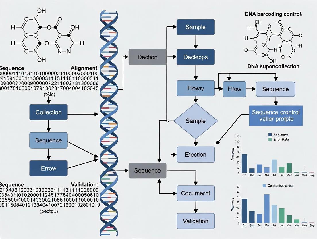
Sequencing Quality Control Workflow
The following diagram illustrates a generalized workflow for NGS quality control, from sample preparation to data filtering, incorporating best practices from the literature [2] [4].
Error Correction with Unique Molecular Identifiers (UMIs)
This diagram outlines the core process of using UMIs to distinguish true biological variants from errors introduced during sequencing.
The Scientist's Toolkit: Essential Research Reagents and Materials
The following table details key reagents and materials used in modern sequencing and barcoding workflows, as cited in the literature.
| Item | Function / Explanation | Example Context |
|---|---|---|
| High-Fidelity Polymerase | DNA polymerase with superior accuracy due to proofreading activity, reducing errors during PCR amplification. | Essential for barcoding NGS to enable detection of variants below 0.1% allele frequency [4]. |
| Unique Molecular Identifiers (UMIs) | Short random nucleotide sequences used to uniquely tag individual DNA molecules before amplification. | Allows bioinformatic error correction by generating consensus sequences from reads sharing a UMI [4]. |
| Rapid Barcoding Kit | A commercial kit that streamlines the process of attaching sample-specific barcodes for multiplexing. | Enables simultaneous sequencing of 1-96 samples with a library prep time of ~60 minutes [3]. |
| AMPure XP Beads | Magnetic beads used for the size-selective purification and clean-up of DNA fragments. | Used in library preparation protocols to remove short fragments, unincorporated nucleotides, and salts [3]. |
| Flow Cell | The consumable device where the sequencing reaction occurs, containing nanopores or patterned lawns of primers. | Must be checked for sufficient active pores (e.g., >800 for MinION) before a sequencing run [3]. |
| Qubit dsDNA HS Assay Kit | A fluorescent-based method for accurate quantification of double-stranded DNA concentration. | Used for quantifying input DNA and final library concentration, more specific than spectrophotometry [3] [2]. |
| Agilent TapeStation | An automated electrophoresis system that assesses DNA/RNA integrity, size distribution, and concentration. | Provides RNA Integrity Number (RIN) for sample QC and checks library fragment size post-preparation [2]. |
This technical support center provides troubleshooting guides and FAQs for researchers and scientists working on DNA barcoding for species identification. The content is framed within broader thesis research on DNA barcoding quality control and sequence validation.
Troubleshooting Guide: Common DNA Barcoding Workflow Issues
Low DNA Yield or Quality from Extraction
Problem: Inconsistent or low-quality DNA extraction from source material, leading to failed PCR amplification.
Solutions:
- Verify tissue preservation: For long-term storage of critical samples, tissues should be stored at -80°C. For short-term storage, preserve in fresh 95% ethanol [5].
- Check sample handling: Use flame-sterilized scalpels and forceps for each sample to prevent cross-contamination [5].
- Assess DNA quality: Use spectrophotometry to confirm a 260 nm/280 nm ratio of approximately 1.8. A lower ratio may indicate protein contamination [5]. Gel electrophoresis can assess DNA fragmentation, which is critical for processed products [6].
PCR Amplification Failure
Problem: The polymerase chain reaction (PCR) fails to amplify the target COI gene fragment, showing no or faint bands on a gel.
Solutions:
- Confirm DNA quantity: Ensure you have at least 5 ng/µL of DNA template for the PCR reaction [5].
- Use supportive molecular targets: If the standard ~650 bp COI fragment fails to amplify, especially from processed samples with fragmented DNA, use a "mini-barcoding" approach with shorter targets (e.g., a 139 bp COI fragment) [6].
- Switch polymerase for difficult samples: For bivalves or other challenging samples, substitute the standard Taq polymerase with a 5'-exonuclease deficient Taq polymerase (e.g., KlenTaq LA) to improve amplification [6].
Poor-Quality Sequencing Chromatograms
Problem: The resulting sequencing chromatogram (AB1 file) shows overlapping peaks or a high background signal, making base calls unreliable.
Solutions:
- Purify PCR products: Always perform a cleanup step on your PCR product before the sequencing reaction to remove excess primers and dNTPs [5].
- Confirm successful PCR: Perform a check (e.g., gel electrophoresis) to ensure a single, strong band of the correct size is present before proceeding to sequencing [5].
- Sequence both strands: Use both forward and reverse primers for cycle sequencing. Align the resulting sequences with a program like Clustal W to resolve ambiguities [6] [5].
Inconclusive Species Identification from Sequence Data
Problem: After sequencing, the data does not lead to a clear, unambiguous species match in reference databases.
Solutions:
- Employ a multi-target approach: If the primary target (e.g., COI) does not provide species-level resolution, amplify and sequence supportive genetic markers like cytochrome b (cytb) or 16S rRNA [6].
- Use multiple databases and parameters: Query your final sequence against both GenBank (using BLAST) and the Barcode of Life Data (BOLD) Systems (using the IDS engine). Use a validated identity score cut-off and Neighbour-Joining analysis for identification [6].
- Check for database gaps: Be aware that a lack of reference sequences for a particular species in public databases can prevent identification. This is a known limitation of the method [6].
Frequently Asked Questions (FAQs)
Q1: What are the minimum quality thresholds for DNA to be suitable for barcoding? A: Success criteria from an FDA single laboratory validation (SLV) state you should obtain a DNA concentration of ≥5 ng/µL and a 260 nm/280 nm ratio of ~1.8, measured via spectrophotometry. A negative control should read ~0 ng/µL [5].
Q2: My sample is highly processed (e.g., cooked, canned). Can I still use DNA barcoding? A: Yes, but it requires protocol adjustments. For samples with medium-to-high DNA fragmentation, you must shift from a full-length barcode (FLB) approach to a mini-barcoding strategy, which targets much shorter DNA fragments (under 500 bp) that are more likely to survive processing [6].
Q3: What constitutes a "positive" species identification from a sequence? A: Identification relies on comparing your unknown sequence to a validated reference library. A positive identification is made when your sequence shows a high percentage match (exceeding a pre-defined identity score cut-off) to a sequence from a vouchered specimen in databases like BOLD or GenBank [6]. Statistical methods like Neighbour-Joining trees are often used to support the identification [6].
Q4: How can I design a self-checking program for my supply chain using DNA barcoding? A: DNA barcoding has been proven as an effective tool for verifying supplier compliance within a company's self-checking activities. You can apply a decision-tree protocol to analyze samples from incoming goods. This involves using a standard COI barcode first, followed by a multi-target approach if needed, to verify that the species identified matches the species declared on the label [6].
Workflow and Decision Pathway
The following diagram illustrates the critical control points (CCPs) and key decision points in the DNA barcoding workflow for species identification, based on established laboratory protocols [6] [5].
DNA Barcoding Workflow with Critical Control Points
Quantitative Performance Data
The following table summarizes key performance metrics from relevant DNA barcoding studies, providing benchmarks for your own quality control.
Table 1: Performance Metrics from DNA Barcoding Studies
| Study Focus | Total Samples Analyzed | Success Rate of Species ID | Primary Reason for Failure | Non-Compliance / Substitution Rate |
|---|---|---|---|---|
| Seafood Identification (Fish & Molluscs) [6] | 182 | 96.2% (175/182) | Lack of reference sequences; low resolution of molecular targets [6] | 18.1% (33/182) [6] |
| Poultry Meat Products (Metabarcoding) [7] | 13 | 100% (for detecting declared species) | Not Applicable (Method was successful) | 61.5% (8/13 contained undeclared species) [7] |
Research Reagent Solutions
This table details essential materials and reagents used in the DNA barcoding workflow, as cited in the validated protocols.
Table 2: Key Reagents and Materials for DNA Barcoding Experiments
| Item | Function in Protocol | Example from Literature |
|---|---|---|
| DNeasy Blood & Tissue Kit (Qiagen) | DNA extraction and purification from various tissue types [5]. | Used in the FDA SLV for tissue lysis and DNA extraction [5]. |
| Primers for COI (e.g., FishF1/FishR1) | Amplification of the standard ~650 bp cytochrome c oxidase subunit I barcode region from fish DNA [6] [5]. | Used as the first-choice target for fish and mollusk identification [6]. |
| Primers for Mini-Barcode | Amplification of a short (~139 bp) COI fragment from degraded or processed samples where the full-length barcode fails [6]. | Applied when DNA fragmentation is detected to cope with processed products [6]. |
| Primers for Alternative Targets (cytb, 16S rRNA) | Provide supportive data for species identification when the COI gene alone is not conclusive [6]. | Used in a multi-target approach to resolve ambiguous identifications [6]. |
| KlenTaq LA DNA Polymerase | A 5'-exonuclease deficient Taq polymerase used for improved amplification of difficult templates, such as bivalves [6]. | Substituted for standard Taq to amplify DNA from bivalves [6]. |
In DNA barcoding research, the reliability of your findings is directly dependent on the quality of your underlying sequence data. Poor-quality data can stem from a myriad of sources—biological, technical, and computational—leading to misidentification, failed experiments, and invalid conclusions. This technical support center is designed to help you, the researcher, diagnose and resolve these issues efficiently. The following guides and FAQs are framed within the critical context of DNA barcoding quality control and sequence validation, providing targeted solutions for the problems you might encounter in the lab or during data analysis [8].
Database Quality Assessment for DNA Barcoding
The reference database you select is a primary factor in the success and accuracy of DNA barcoding. The table below summarizes a comparative evaluation of two major databases, highlighting common quality issues you need to be aware of.
Table 1: Evaluation of COI Barcode Reference Databases for DNA Barcoding
| Evaluation Criteria | NCBI (Nucleotide Database) | BOLD (Barcode of Life Data System) |
|---|---|---|
| Barcode Coverage | Generally higher coverage for marine metazoan species in the WCPO [9] | Lower public barcode coverage, partly due to stricter submission requirements [9] |
| Sequence Quality | Lower overall sequence quality; more prone to errors and inconsistencies [9] | Higher sequence quality due to stricter quality control and curation [9] |
| Common Quality Issues | Over- or under-represented species; short sequences; ambiguous nucleotides; incomplete taxonomy; conflicting records [9] | Quality issues are less common but can include over-represented species and conflicting records [9] |
| Key Quality Feature | Lacks an integrated, automated quality evaluation system [9] | Features the Barcode Index Number (BIN) system to cluster sequences and flag problematic records [9] |
| Primary Weakness | Reliability is debated due to less robust curation of user-submitted data [9] | Lack of barcode records can reduce taxonomic resolution [9] |
Troubleshooting Guides & FAQs
▷ PCR Failure Playbook
Symptom: No band or a very faint band on the gel.
- Likely Causes: Inhibitor carryover from the sample, low DNA template concentration, primer mismatch, or suboptimal PCR cycling conditions [10].
- First Fixes:
- Dilute the DNA template (1:5 to 1:10) to reduce the concentration of potential inhibitors.
- Add Bovine Serum Albumin (BSA) to the reaction to mitigate inhibitors from complex sample matrices.
- Optimize the reaction by running a small annealing temperature gradient and modestly increasing the cycle number [10].
- Advanced Protocol: If initial fixes fail, consider a validated mini-barcode primer set, especially when working with degraded DNA, as it targets a shorter, more amplifiable region [10].
Symptom: Smears or non-specific bands on the gel.
- Likely Causes: Excessive template DNA input, high Mg²⁺ concentration, low annealing stringency, or primer-dimer formation [10].
- First Fixes:
- Reduce the amount of DNA template used in the reaction.
- Optimize the Mg²⁺ concentration and increase the annealing temperature.
- Use touchdown PCR to improve amplification specificity [10].
Symptom: Clean PCR product but a messy Sanger trace (e.g., double peaks).
- Likely Causes: Mixed template (contamination), leftover primers/dNTPs due to poor cleanup, heteroplasmy, or nuclear mitochondrial DNA segments (NUMTs) [10].
- First Fixes:
- Perform a thorough cleanup of the PCR product using enzymatic (e.g., EXO-SAP) or bead-based methods to remove residual primers and dNTPs before sequencing.
- Re-amplify from a diluted template to reduce co-amplification of non-target products.
- Sequence in both forward and reverse directions. If traces still disagree, suspect NUMTs and validate with a second, independent genetic locus [10].
▷ Sequencing Issues: Sanger and NGS
FAQ: How can I resolve low signal or mixed peaks in Sanger sequencing?
- Re-clean amplicons to thoroughly remove primers and dNTPs.
- If you observed smearing or multiple bands on the gel, gel-purify the correct band before sequencing.
- Use sequencing primers with an appropriate melting temperature (Tm) and avoid ends with extreme GC content.
- Always sequence both directions for confirmation, especially when heterozygous indels or ambiguous regions are suspected [10].
FAQ: What should I do when my NGS amplicon run yields low reads per sample?
- Likely Causes: Over-pooling of libraries, presence of adapter or primer dimers, low diversity of amplicons, or index misassignment [10].
- First Fixes:
- Re-quantify your libraries accurately using qPCR or fluorometry.
- Repeat bead cleanup to remove dimer artifacts and verify the result using fragment analysis.
- Spike in a higher percentage of PhiX control (e.g., 5-20%) to stabilize clustering with low-diversity amplicon libraries.
- Review your index design and pooling strategy to ensure even representation [10].
FAQ: How can I recognize and avoid NUMTs in COI barcoding?
- Red Flags: Frameshift mutations in the coding sequence, presence of stop codons in the translated amino acid sequence, unusual GC content, or disagreement between forward and reverse reads [10].
- Mitigation Strategies:
- Translate your nucleotide sequence to check for premature stop codons.
- Cross-validate species identification with a second, independent genetic locus.
- If NUMTs are suspected, report identification conservatively at the genus level and seek confirmation [10].
▷ Contamination Control
FAQ: My no-template controls (NTCs) are showing amplification. What should I do?
- Likely Cause: Aerosolized amplicon contamination (carryover) or shared equipment between pre- and post-PCR areas [10].
- Immediate Actions:
- Quarantine the entire batch of reagents and samples from the affected run.
- Hard-separate your pre-PCR and post-PCR laboratory spaces, ensuring dedicated equipment, reagents, and personnel flow for each. Never use post-PCR equipment in pre-PCR areas.
- Decontaminate workspaces and equipment with UV light and fresh bleach.
- Implement a chemical carryover control system using dUTP/UNG. This involves using dUTP instead of dTTP in PCR mixes. A subsequent treatment with Uracil-DNA Glycosylase (UNG) before thermal cycling will degrade any contaminating uracil-containing amplicons from previous runs, preventing their amplification [10].
Table 2: Essential Controls for Contamination Detection
| Control Type | Purpose | Action if Positive |
|---|---|---|
| Extraction Blank | Detects contamination introduced during DNA extraction and purification. | Quarantine the batch and repeat the extraction from the last known clean step. |
| No-Template Control (NTC) | Detects contamination in the PCR reagents or from aerosolized amplicons. | Discard the affected reagent batch, decontaminate the workspace, and repeat the assay. |
| Positive Control | Confirms that the entire PCR and sequencing workflow is functioning correctly. | N/A |
Experimental Protocols for Quality Assurance
▷ Protocol: Evaluation of DNA Extraction Methods for Processed Foods
Background: This protocol is adapted from a study on DNA barcoding for food authenticity, which is directly relevant to obtaining high-quality data from challenging, processed samples where DNA is often degraded [11].
Sample Homogenization:
- For dried products (legumes, seeds, pasta), use a grinder to create a fine, homogeneous powder.
- For frozen, canned, or raw products, homogenize the material using a mortar and pestle under liquid nitrogen.
- Store all homogenized samples at -20°C or lower.
Inhibitor Removal (Pre-wash):
- To mitigate the effects of PCR-inhibiting compounds (e.g., polyphenols, polysaccharides), wash all samples twice with a Sorbitol Washing Buffer prior to DNA extraction [11].
DNA Extraction Comparison:
- Test multiple extraction methods in parallel to identify the most effective one for your specific sample matrix. The cited study compared:
- Two commercial silica column-based kits.
- A CTAB-based protocol with modifications.
- CTAB Protocol Modifications: After incubation with CTAB buffer and RNase treatment, add half a volume of 5 M NaCl, followed by three volumes of ice-cold absolute ethanol to precipitate the DNA. Incubate at -20°C for one hour, then centrifuge to pellet the DNA [11].
- Test multiple extraction methods in parallel to identify the most effective one for your specific sample matrix. The cited study compared:
DNA Quality Assessment:
- Quantify DNA using fluorometry for accuracy.
- Check DNA integrity and the presence of inhibitors by attempting to amplify a short, quality control locus (e.g., a mini-barcode) before proceeding with the full barcode assay.
▷ Protocol: In Silico Error Suppression for Deep NGS Data
Background: This methodology is crucial for detecting low-frequency genetic variants in deep next-generation sequencing data, as it computationally suppresses substitution errors that can mimic true biological signals [12].
Establish a Benchmark Dataset:
- Use a dilution series of a known sample (e.g., a cell line with known somatic mutations) to create a "truth set" for benchmarking. This allows you to characterize false-positive calls [12].
Measure Substitution Error Rates:
- For each genomic site i in a region known to be devoid of true genetic variation, calculate the error rate for each possible nucleotide substitution using the formula:
Error Rate_i (g>m) = (Number of reads with nucleotide m at position i) / (Total number of reads at position i)[12]. - This provides a baseline for the background error rate in your dataset.
- For each genomic site i in a region known to be devoid of true genetic variation, calculate the error rate for each possible nucleotide substitution using the formula:
Identify and Filter Low-Quality Reads:
- Trim 5 bp from both ends of each read to remove potentially low-quality bases and adapter contamination [12].
- Filter out reads with low overall mapping quality.
- Evaluate the per-cycle base quality score distribution. A gradual decline in quality with increasing cycle number is expected, but random dips or peaks may indicate technical issues [12] [13].
Error Suppression:
- Use the calculated error profiles and quality filters to set appropriate thresholds for variant calling. This computational suppression can reduce substitution error rates to the range of 10⁻⁵ to 10⁻⁴, significantly improving the sensitivity and specificity of low-frequency variant detection [12].
Visualization of Workflows
▷ DNA Barcoding and Data Quality Assessment Workflow
The diagram below outlines the core DNA barcoding process and key points where data quality must be assessed and validated.
This diagram categorizes the major sources of experimental error throughout a conventional NGS workflow, from sample to sequence.
The Scientist's Toolkit: Key Research Reagent Solutions
Table 3: Essential Reagents and Kits for DNA Barcoding Quality Control
| Item | Function | Application Notes |
|---|---|---|
| BSA (Bovine Serum Albumin) | Mitigates the effects of PCR inhibitors commonly found in complex biological samples (e.g., plant polyphenols). | Add to PCR reactions when amplification from difficult matrices is failing [10]. |
| Sorbitol Washing Buffer | Pre-wash buffer used to remove phenolic compounds and other contaminants from samples prior to DNA extraction. | Critical for improving DNA yield and purity from plant and food materials [11]. |
| Silica Column-Based Kits | For efficient purification of DNA, separating it from proteins, salts, and other impurities. | Commercial kits offer standardized, reliable protocols for obtaining high-quality DNA [11]. |
| CTAB Buffer | A detergent-based lysis buffer effective at breaking down plant cell walls and denaturing proteins. | A key component in classical plant DNA extraction protocols; useful for a wide range of tough samples [11]. |
| dUTP/UNG Carryover Control System | Prevents amplification of contaminating amplicons from previous PCR reactions. | dUTP is used in place of dTTP; UNG enzyme degrades uracil-containing DNA before PCR [10]. |
| PhiX Control Library | Used as a spike-in control for NGS runs to monitor sequencing quality and improve base calling for low-diversity libraries. | Particularly important for amplicon sequencing (e.g., DNA barcoding) where library diversity is low [10]. |
| High-Fidelity DNA Polymerase | Enzyme with proofreading activity for accurate DNA amplification, reducing errors introduced during PCR. | Essential for generating high-quality sequences for barcode reference libraries [12]. |
FASTQ Quality Score FAQs
What is a sequencing quality score?
A quality score (Q-score) is a numerical value that represents the probability that a base was called incorrectly by the sequencing instrument. It is defined by the equation: Q = -10log₁₀(e), where e is the estimated probability of an incorrect base call [1]. Higher Q-scores indicate higher accuracy.
What do the different Q-score values mean?
The table below shows how quality scores translate into base-calling accuracy:
| Quality Score | Probability of Incorrect Base Call | Base Call Accuracy |
|---|---|---|
| Q10 | 1 in 10 | 90% |
| Q20 | 1 in 100 | 99% |
| Q30 | 1 in 1000 | 99.9% |
| Q40 | 1 in 10,000 | 99.99% |
In practice, Q30 is considered a benchmark for high-quality data in next-generation sequencing, as virtually all reads will be perfect at this level [1].
How are quality scores encoded in a FASTQ file?
In FASTQ files, quality scores are encoded into a compact form using ASCII characters to represent numerical values. In the standard Phred+33 encoding, the quality score is represented as the character with an ASCII code equal to its value + 33 [14] [15].
The first few characters in this encoding scheme are [14] [15]:
| Symbol | ASCII Code | Q-Score |
|---|---|---|
! |
33 | 0 |
" |
34 | 1 |
# |
35 | 2 |
$ |
36 | 3 |
% |
37 | 4 |
Higher ASCII characters represent higher quality scores, with the full range extending from ! (lowest quality) to ~ (highest quality) [16].
Why does my FastQC report show "FAIL" for some modules when the data looks fine?
This is a common occurrence and doesn't necessarily indicate problematic data. Some FastQC warnings and failures can be safely ignored because [17]:
- FastQC applies assumptions designed for genomic libraries, so specialized libraries like RNAseq may naturally trigger failures
- Per base sequence content frequently fails for Illumina TruSeq RNAseq libraries due to hexamer priming bias
- Kmer content often fails in real-world datasets
- The key is to interpret results in the context of your experiment and sample type rather than treating all flags as critical errors
How can I resolve quality score encoding format issues?
If tools cannot process your FASTQ files, you may have a format/encoding mismatch. The solution is to ensure your data is in Sanger Phred+33 format (designated as fastqsanger in Galaxy) as this is what most tools expect [18].
You can [18]:
- Convert files using standardization tools
- Download reads from NCBI SRA already in
fastqsangerformat using specialized download tools - Check that the
+quality score lines are properly annotated
| Resource | Function | Relevance to DNA Barcoding QC |
|---|---|---|
| FastQC | Quality control tool for high throughput sequence data | Provides initial assessment of read quality, adapter contamination, and potential issues [17] |
| Trimmomatic/cutadapt | Read trimming and adapter removal | Improves overall data quality by removing poor quality bases and adapter sequences [17] |
| Dorado Basecaller | Converts raw electrical signals to nucleotide sequences | Oxford Nanopore's production basecaller; uses neural networks for accurate basecalling [19] |
| BOLD Systems | Barcode of Life Data repository | Curated reference database for validating DNA barcode sequences [20] |
| Remora/modkit | Modified base detection tools | Specialized tools for calling base modifications like 5mC, 5hmC [19] |
| GEANS Reference Library | Curated DNA barcode library for North Sea macrobenthos | Example of taxonomically reliable reference library for biodiversity monitoring [20] |
Quality Score Encoding Reference Table
This comprehensive table shows the complete Phred+33 encoding scheme used in FASTQ files:
| Symbol | ASCII Code | Q-Score | Symbol | ASCII Code | Q-Score |
|---|---|---|---|---|---|
! |
33 | 0 | 0 |
48 | 15 |
" |
34 | 1 | 1 |
49 | 16 |
# |
35 | 2 | 2 |
50 | 17 |
$ |
36 | 3 | 3 |
51 | 18 |
% |
37 | 4 | 4 |
52 | 19 |
& |
38 | 5 | 5 |
53 | 20 |
' |
39 | 6 | 6 |
54 | 21 |
( |
40 | 7 | 7 |
55 | 22 |
) |
41 | 8 | 8 |
56 | 23 |
* |
42 | 9 | 9 |
57 | 24 |
+ |
43 | 10 | : |
58 | 25 |
, |
44 | 11 | ; |
59 | 26 |
- |
45 | 12 | < |
60 | 27 |
. |
46 | 13 | = |
61 | 28 |
/ |
47 | 14 | > |
62 | 29 |
The encoding continues through uppercase letters, with A=65=Q32, up to I=73=Q40 [14] [15].
DNA Barcoding Context: Why Quality Scores Matter
In DNA barcoding research, quality scores are critical for reliable species identification. High-quality sequencing ensures:
- Accurate reference libraries: The GEANS project created a curated DNA reference library containing 4,005 COI barcode sequences from 715 North Sea macrobenthic species, where data quality was essential for reliable biodiversity monitoring [20]
- Valid species identification: Poor quality scores can lead to misidentification, particularly problematic for detecting non-indigenous species or cryptic species complexes [20]
- Reliable biodiversity assessment: Massive DNA barcoding approaches enable monitoring of soil macrofauna where traditional morphological identification is difficult, but only with high-quality sequence data [21]
Troubleshooting Workflow
FASTQ Quality Control Decision Pathway
This workflow guides researchers through systematic quality assessment, highlighting critical checkpoints for encoding verification and quality trimming that are essential for producing reliable DNA barcoding data.
Impact of Starting Material Quality on Downstream Analysis Success
Frequently Asked Questions (FAQs)
1. What are the most common consequences of poor-quality starting materials in DNA barcoding? Poor-quality starting materials lead to several common downstream problems:
- Misidentification: Contamination or sample mix-ups can result in sequences being assigned to the wrong species, compromising the entire dataset [22] [23]. One study on Hemiptera insects found that errors in public barcode databases are "not rare," often stemming from these initial issues [22].
- Failed Analyses: Poor DNA purity can inhibit the Polymerase Chain Reaction (PCR), preventing the amplification of the target barcode region [24] [5]. The FDA's protocol emphasizes that success depends on obtaining DNA of sufficient quality and concentration [5].
- Unreliable Data: Low-quality sequencing reads, often reflected in low Q-scores, increase the probability of base-calling errors and false-positive variant calls, leading to inaccurate conclusions [1] [2].
2. How can I quickly assess the quality of my nucleic acid starting material before sequencing? A quick assessment can be made using the following methods and metrics:
Table 1: Quick Assessment Methods for Nucleic Acid Quality
| Method | Metric | Target Value for High Quality | Indication of Problem |
|---|---|---|---|
| Spectrophotometry (e.g., NanoDrop) | A260/A280 Ratio | ~1.8 (DNA), ~2.0 (RNA) [2] | Significant deviation suggests protein or other contamination. |
| Spectrophotometry | A260/A230 Ratio | >2.0 | Indicates chemical contamination (e.g., salts, solvents) [2]. |
| Electrophoresis (e.g., TapeStation) | RNA Integrity Number (RIN) | 8-10 (RNA) [2] | A low RIN (e.g., <7) indicates RNA degradation. |
| Fluorometry (e.g., Qubit) | DNA/RNA Concentration | Varies | Provides a more accurate quantification of nucleic acids than spectrophotometry. |
3. My NGS data has a sudden drop in quality scores partway through the reads. What is the likely cause? A steady decrease in quality scores, particularly towards the 3' end of reads, is a normal artifact of sequencing-by-synthesis technologies [2]. However, an abrupt or abnormal drop in quality is often indicative of a technical error during the sequencing run, such as an issue with the sequencing instrument or its associated hardware [2]. This can also be caused by over-clustering on the flow cell, which leads to signal impurities [2].
4. A high percentage of my reads are unusable or cannot be mapped. What steps should I take? First, use quality control tools like FastQC to visualize your raw read data [2]. The likely culprit and solution involve read trimming and filtering:
- Problem: The presence of low-quality bases and adapter sequences.
- Solution: Use trimming tools (e.g., CutAdapt, Trimmomatic) to remove adapter sequences and trim low-quality bases (typically with a quality threshold below Q20) from the 3' end of reads [2]. After trimming, filter out reads that fall below a minimum length (e.g., <50 bp) to ensure only high-quality data proceeds to alignment [2].
5. My DNA barcode results conflict with the morphological identification of my specimen. What should I do? This discrepancy is a key application of DNA barcoding for quality control [23]. You should:
- Re-inspect the specimen: Re-examine the morphological characteristics, ideally with input from a trained taxonomist [22].
- Re-audit your workflow: Retrace all steps from specimen collection to DNA extraction and sequencing to check for potential sample mix-ups or contamination [22] [23].
- Re-sequence: Repeat the DNA extraction and barcoding process to rule out a one-off error.
- Check reference databases: Verify that the reference sequences in databases like BOLD and GenBank for your expected species are themselves based on correctly identified specimens, as errors in public repositories are a known issue [22] [23].
Troubleshooting Guides
Troubleshooting Failed PCR Amplification in DNA Barcoding
Problem: The target COI gene region fails to amplify during PCR.
Table 2: Troubleshooting PCR Amplification Failure
| Observed Issue | Potential Root Cause | Recommended Corrective Action |
|---|---|---|
| No PCR product on gel. | Degraded or low-quality DNA template. | Re-assess DNA quality (see Table 1). Extract new DNA, optimizing tissue lysis [5]. |
| No PCR product on gel. | PCR inhibitors present in DNA sample. | Dilute the DNA template. Use a cleanup kit to re-purify the DNA, or add bovine serum albumin (BSA) to the PCR reaction to counteract inhibitors. |
| Faint or smeared bands. | Suboptimal PCR conditions. | Optimize annealing temperature using a gradient PCR. Check primer specificity and concentration. |
| Amplification in negative control. | Contamination at some stage of the process. | Use dedicated pre- and post-PCR lab areas. Use UV irradiation and bleach to decontaminate surfaces. Prepare fresh reagents [22]. |
Troubleshooting Poor NGS Data Quality
Problem: Initial quality control of sequencing data (e.g., via FastQC) shows poor per-base quality scores.
NGS Quality Troubleshooting Flow
The workflow above, guided by the following actions, helps diagnose and resolve common issues:
- If quality drops at read ends: This is expected. Use trimming tools (e.g., Trimmomatic, CutAdapt) to remove low-quality bases from the ends, which will improve overall mapping rates [2].
- If there is an abrupt quality drop mid-read: This may indicate a temporary hardware or fluidics issue during the sequencing run. It is advisable to contact your sequencing facility for diagnostics and troubleshooting [2].
- If adapter content is high: Adapter sequences need to be removed from the reads prior to alignment. Use tools like CutAdapt or Porechop (for Oxford Nanopore data) to trim these artifacts, which is an essential step for data usability [2].
Key Experimental Protocols
Detailed Protocol: DNA Extraction and Barcoding for Species Identification
This protocol is adapted from the FDA's single laboratory validated method for DNA barcoding of fish [5].
Goal: To consistently generate high-quality COI (Cytochrome c Oxidase subunit I) DNA barcodes from tissue samples for species identification.
Critical Materials and Reagents:
- DNeasy Blood & Tissue Kit (Qiagen) or equivalent for DNA extraction.
- PCR Reagents: Taq DNA polymerase, dNTPs, PCR buffer, MgCl2.
- COI Primers: Vertebrate-specific primers (e.g., FishF1, FishR1) [5].
- Agarose Gel materials for electrophoresis.
- Cycle Sequencing Kit (e.g., BigDye Terminator v1.1).
- Ethanol (96-100%) for precipitation and cleaning steps.
Step-by-Step Method:
- Tissue Sampling:
- Use a small cube (5-7 mm) of muscle tissue or a fin clip.
- Critical: Flame-sterilize forceps and scalpel between each sample to prevent cross-contamination [5].
- Preserve tissue in 95-100% ethanol or freeze at -20°C for short-term storage.
Tissue Lysis and DNA Extraction:
- Follow the protocol of your selected DNA extraction kit (e.g., DNeasy Blood & Tissue Kit).
- Success Criteria: DNA concentration should be ≥5 ng/µL, with a A260/A280 ratio of ~1.8, measured on a spectrophotometer [5]. A negative control should yield ~0 ng/µL.
PCR Amplification of COI:
- Set up a 50 µL PCR reaction containing: 1x PCR buffer, 2.5 mM MgCl2, 0.2 mM each dNTP, 0.2 µM each primer, 1.25 U Taq polymerase, and 2-100 ng of DNA template.
- Thermocycler Conditions: Initial denaturation at 95°C for 2 min; 35 cycles of 95°C for 30 s, 50-54°C for 30 s, 72°C for 1 min; final extension at 72°C for 10 min [5].
PCR Product Check and Cleanup:
- Verify successful amplification by running 5 µL of the PCR product on a 1-2% agarose gel. A single, bright band at the expected size (~650 bp) should be visible.
- Clean up the remaining PCR product using a commercial cleanup kit to remove excess primers and dNTPs.
DNA Sequencing and Analysis:
- Perform cycle sequencing in both forward and reverse directions using the same primers as in the PCR step.
- Clean up the sequencing reactions to remove unincorporated dyes.
- Run the samples on a DNA sequencer and analyze the resulting chromatograms. Assemble forward and reverse reads and compare the consensus sequence to a validated reference database like BOLD.
Research Reagent Solutions
Table 3: Essential Materials for DNA Barcoding and NGS Workflows
| Item | Function/Application | Example Products/Brands |
|---|---|---|
| DNA/RNA Extraction Kits | Isolate high-purity nucleic acids from diverse tissue types. Critical for successful downstream applications. | DNeasy Blood & Tissue Kit (Qiagen) [5] |
| Spectrophotometer / Fluorometer | Quantify nucleic acid concentration and assess purity (A260/280 ratio). Fluorometers provide more accurate quantification. | NanoDrop (Thermo Fisher), Qubit (Thermo Fisher) [2] [5] |
| Electrophoresis System | Visually assess RNA integrity (RIN) or check size and quality of PCR products and sequencing libraries. | Agilent TapeStation, standard agarose gel systems [2] |
| NGS Library Prep Kits | Prepare DNA or RNA samples for next-generation sequencing by fragmenting, size-selecting, and adding platform-specific adapters. | Illumina DNA Prep, KAPA HyperPrep |
| Quality Control Software | Analyze raw sequencing data to evaluate quality scores, GC content, adapter contamination, and more. | FastQC [2] |
| Read Trimming & Filtering Tools | Programmatically remove low-quality bases, adapter sequences, and poor-quality reads from NGS data. | CutAdapt, Trimmomatic, Nanofilt [2] |
# Core Concepts: Why Size and Adapter Content Matter
In next-generation sequencing (NGS), the integrity of your library preparation is paramount. Two of the most critical quality control (QC) checkpoints are the size distribution of your DNA fragments and the adapter content of the final library. Proper assessment of these parameters is essential for a successful sequencing run, as failures here can lead to wasted reagents, poor data quality, and inaccurate downstream bioinformatics analysis [25] [26].
Assessing the average insert size and the tightness of the size distribution ensures optimal clustering on the flow cell and prevents issues like overlapping reads. Similarly, monitoring for excess adapter content or adapter dimers is crucial, as these can dominate the sequencing run, drastically reducing the yield of useful data [26]. Within the context of DNA barcoding research, where the goal is accurate species identification, these quality checks are non-negotiable. A compromised library can lead to failed barcode amplification or misassignment of sequences, undermining the validity of the entire study [9] [20].
# Frequently Asked Questions (FAQs) & Troubleshooting
FAQ 1: My Bioanalyzer trace shows a sharp peak around 70-90 bp. What is this and how do I fix it?
- Problem: A sharp peak at ~70-90 bp is a classic signature of adapter dimers [26]. These are short fragments formed by the self-ligation of free adapters. They contain very little to no genomic insert and can outcompete your target library during cluster generation, leading to a very high proportion of useless sequences.
- Causes: This is typically caused by an suboptimal adapter-to-insert molar ratio during ligation, where adapters are in excess [26]. It can also result from inefficient purification after ligation, failing to remove unligated adapters.
- Solutions:
- Re-optimize Ligation: Titrate the adapter concentration to find the optimal ratio for your input DNA [25] [26].
- Improve Cleanup: Use a rigorous size selection method, such as magnetic beads with adjusted ratios (e.g., AMPure XP beads) or gel extraction, to specifically remove short fragments [25] [27]. Always follow cleanup protocols precisely to avoid sample loss or carryover of contaminants [26].
- Verify Input DNA: Ensure your starting DNA is not severely degraded, as low-molecular-weight DNA can also contribute to short fragments.
FAQ 2: My library yield is acceptable, but the fragment size distribution is very broad and uneven. What does this indicate?
- Problem: A wide or multi-peaked size distribution indicates inefficient or inconsistent fragmentation [25]. This can lead to uneven coverage, where some genomic regions are overrepresented and others are missed.
- Causes:
- Mechanical Shearing: Inconsistent sonication or acoustic shearing parameters (time, energy) [25].
- Enzymatic Fragmentation: Fluctuations in enzyme-to-DNA ratio or reaction time, or potential sequence bias in enzymatic methods [25].
- Over-amplification: Too many PCR cycles during library amplification can skew the representation of fragments, amplifying some sizes more than others [25] [26].
- Solutions:
- Calibrate Fragmentation: If using mechanical shearing, re-optimize the settings (e.g., duration, peak incident power) for your specific instrument and DNA quality. For enzymatic methods, standardize the reaction conditions and enzyme lots [25].
- Minimize PCR Cycles: Use the minimum number of PCR cycles necessary for your input material to avoid amplification bias [25].
- Implement Size Selection: Introduce a stringent size selection step (e.g., double-sided bead cleanup) to narrow the fragment size range before sequencing [25].
FAQ 3: My sequencing data shows high levels of adapter contamination in the FastQC report. How did this happen?
- Problem: High adapter content in your raw sequencing reads means that the sequencer was reading into the adapter sequence because the DNA fragment was shorter than the read length.
- Causes: This is often a result of insufficient removal of adapter dimers prior to sequencing or starting with overly short DNA fragments after fragmentation [2].
- Solutions:
- Pre-Sequencing QC: Always check your final library on a Bioanalyzer or TapeStation to confirm the absence of the adapter-dimer peak before loading the flow cell [2].
- Post-Processing: While not ideal, adapter sequences can be removed bioinformatically from the raw data using tools like CutAdapt or Trimmomatic [2]. However, this reduces the usable read length and is a corrective, not a preventive, measure.
- Preventive Action: The most robust solution is to address the issue during library prep by ensuring proper size selection and optimizing fragmentation to yield fragments longer than your intended read length.
Table 1: Common Library Prep Issues and Diagnostic Signals
| Problem | Primary Failure Signal | Common Root Cause |
|---|---|---|
| Adapter Dimer Contamination | Sharp ~70-90 bp peak on Bioanalyzer; high adapter content in FastQC [26] [2] | Excess adapters; inefficient post-ligation cleanup [26] |
| Skewed Size Distribution | Broad, multi-peaked, or shifted profile on Bioanalyzer [25] | Inefficient fragmentation (over/under-shearing); over-amplification [25] [26] |
| Low Library Yield | Low concentration via qPCR/fluorometry; faint electropherogram peaks [26] | Poor input DNA quality; suboptimal ligation; sample loss during cleanup [26] |
| Uneven Coverage / High Duplication | Bioinformatics analysis reveals biased read distribution | Over-amplification; low library complexity starting material [26] |
# Detailed Experimental Protocols for Assessment
Protocol 1: Assessing Library Size Distribution with a Fragment Analyzer
This protocol details the use of an Agilent Bioanalyzer or TapeStation system, the gold standard for assessing library size distribution.
- Preparation: Prime the instrument and prepare the gel-dye mix according to the manufacturer's instructions for the appropriate DNA sensitivity kit (e.g., High Sensitivity DNA kit).
- Sample Loading: Dilute 1 µL of your final library according to the kit's recommendations (typically to a concentration within 0.1-50 ng/µL). Load this volume into the specified well on the chip.
- Run: Start the analysis run. The instrument electrophoretically separates the DNA fragments by size.
- Data Interpretation:
- The software will generate an electropherogram (a trace plot) and a virtual gel image.
- The main peak represents your average library insert size plus the adapter length.
- A tight, single peak indicates a well-size-selected library. A broad peak or shoulder peaks suggest heterogeneous fragment sizes.
- Crucially, inspect the region around 70-90 bp for any sign of a peak, which indicates adapter dimer [26] [2].
Protocol 2: Bioinformatic Assessment of Adapter Content with FastQC
After sequencing, FastQC provides a direct assessment of adapter contamination in your data.
- Input: Provide your raw sequencing data in FASTQ format to the FastQC tool. This can be run from the command line or via a web portal like Galaxy [2].
- Analysis: Execute FastQC. It will generate a comprehensive HTML report with multiple modules.
- Interpretation: Navigate to the "Adapter Content" plot. This graph shows the cumulative percentage of reads in which an adapter sequence was detected at each position.
- A good result shows lines at or near 0%.
- Lines that rise, especially towards the end of the read, indicate significant adapter contamination, meaning your library contained short fragments or adapter dimers [2].
# Research Reagent Solutions
Table 2: Essential Kits and Reagents for Library QC
| Reagent / Kit | Function | Application in DNA Barcoding |
|---|---|---|
| AMPure XP Beads | Magnetic beads for post-ligation cleanup and size selection. | Critical for removing adapter dimers and selecting the optimal barcode amplicon size, ensuring clean barcode libraries [25] [26]. |
| Agilent High Sensitivity DNA Kit | Microfluidic capillary electrophoresis for precise sizing and quantification of DNA libraries. | The primary tool for visually confirming library integrity and the absence of adapter dimers before costly sequencing [2]. |
| Qubit dsDNA HS Assay Kit | Fluorometric quantification of double-stranded DNA. | Provides accurate concentration measurement of amplifiable library molecules, superior to UV absorbance for precious barcoding samples [26] [2]. |
| Illumina Tagment DNA TDE1 Enzyme | Transposase for tagmentation (combined fragmentation and adapter tagging). | Used in streamlined protocols like Nextera for efficient library prep, though requires optimization to avoid bias [25] [28]. |
| CutAdapt / Trimmomatic | Bioinformatics software tools. | Used post-sequencing to trim adapter sequences from raw reads, a corrective action for contaminated barcode data [2]. |
# Workflow Diagram: Ensuring Library Integrity
The following diagram outlines the key steps and decision points for assessing and ensuring library preparation integrity, from sample to sequence.
Implementing Robust DNA Barcoding Protocols: From Wet Lab to Bioinformatics
Standardized DNA Extraction Protocols for Diverse Sample Types
The reliability of DNA barcoding and metabarcoding, powerful tools for species identification in research and drug development, is fundamentally dependent on the quality of input DNA. These techniques require reliable reference databases to ensure accurate assignment of DNA sequences to specific taxa, and the entire process begins with effective nucleic acid extraction [9] [20]. The integrity, purity, and yield of extracted DNA directly influence downstream applications, including PCR amplification and sequencing success. Standardized extraction protocols are therefore not merely preliminary steps but foundational components of rigorous DNA barcoding quality control and sequence validation research. This guide addresses the key technical challenges and provides standardized, reproducible methods for researchers working with diverse sample types.
Troubleshooting Guides for Common DNA Extraction Issues
Problem: Low DNA Yield
Low yield can halt projects and compromise data quality. Below are the common causes and their solutions.
Table 1: Troubleshooting Low DNA Yield
| Potential Cause | Sample Type | Solution |
|---|---|---|
| Incomplete cell lysis | All types | Increase incubation time with lysis buffer; increase speed/time of agitation; use a more aggressive lysing matrix or bead-beating [29] [30]. |
| Input amount too low | Cells, Blood | Use recommended input amounts. For cells, working with < 1 x 10^5 cells is not recommended. For low inputs, use a reduced lysis volume protocol [31]. |
| DNA did not attach to beads | All types (bead-based kits) | Ensure proper technique during binding. For precipitated DNA not attaching, twist the tube to create contact. If unsuccessful, spin down the precipitate and resuspend manually [31]. |
| Frozen blood sample thawed | Blood | Add Proteinase K and Lysis Buffer directly to frozen samples, allowing them to thaw during incubation to inhibit nuclease activity [29] [31]. |
| Protein precipitates clogged membrane | Blood, Tissue | Reduce Proteinase K lysis time to prevent insoluble hemoglobin complexes. Pellet protein precipitates by centrifuging at 12,000 × g for 10+ minutes before applying lysate to spin filter [29]. |
Problem: DNA Degradation
Degraded DNA is unsuitable for long-range PCR or high-molecular-weight applications.
Table 2: Troubleshooting DNA Degradation
| Potential Cause | Sample Type | Solution |
|---|---|---|
| Sample age or improper storage | Blood, Tissue | Use fresh whole blood within one week. For tissues, process immediately or snap-freeze in liquid nitrogen. Store at -80°C for long-term preservation [29] [31]. |
| Nuclease activity post-homogenization | Tissue | Place homogenized samples in lysis buffer into a thermal mixer immediately after homogenization to inactivate nucleases. Process samples individually to minimize delays [31]. |
| Improper handling of UHMW DNA | All types (HMW prep) | Always use wide-bore pipette tips. Avoid vortexing. Limit extended heating periods (e.g., do not exceed 15-30 minutes at 56°C) [31]. |
| Sample thawed before processing | Blood | Never thaw frozen blood before adding RBC Lysis Buffer. Add cold lysis buffer directly to the frozen sample [31]. |
Problem: Contamination and Impurities
| Potential Cause | Sample Type | Solution |
|---|---|---|
| High hemoglobin content | Blood | Indicated by a dark red color after lysis. Extend lysis incubation time by 3–5 minutes to improve purity [29]. |
| Cross-contamination | All types | Use designated equipment and reagents. Thoroughly clean workspace. Use positive and negative controls to detect contamination early [29]. |
| Co-precipitation of polysaccharides/polyphenols | Plant Tissues | For plant tissues, use the CTAB method and add 2-5% PVP (polyvinylpyrrolidone) to the lysis buffer to adsorb polyphenols [30]. |
| Inhibitors in processed samples | Food, TCM | Pre-wash samples with Sorbitol Washing Buffer before extraction to remove PCR inhibitors like phenolics [11]. |
Frequently Asked Questions (FAQs)
Q1: What is the most critical factor for successful DNA extraction from plant-based materials used in drug development? The most critical factor is effectively counteracting secondary metabolites like polysaccharides and polyphenols, which can co-precipitate with DNA and inhibit downstream enzymes. The gold-standard method is the CTAB (cetyltrimethylammonium bromide) protocol, often optimized with polyvinylpyrrolidone (PVP) to bind polyphenols and β-mercaptoethanol to prevent oxidation [30]. This is especially important for authenticating Traditional Chinese Medicine species where PCR inhibition can lead to misidentification [32].
Q2: How does the level of food processing impact DNA extraction efficiency for barcoding, and how can this be mitigated? Processing (e.g., thermal treatment, canning, drying) fragments and degrades DNA. To mitigate this:
- Use a Robust Lysis Protocol: The CTAB method with a pre-wash step is often more effective than simple commercial kits for breaking down processed matrices [11].
- Target Shorter Barcodes: If standard barcodes (~650 bp) fail to amplify, switch to "mini-barcoding" which targets shorter genetic regions (<300 bp) from degraded DNA [32].
- Increase Starting Material: Use 100-200 mg of processed sample to increase the chance of obtaining sufficient intact DNA molecules [11].
Q3: For high-throughput drug discovery projects, should I use manual or automated DNA extraction? Automation is highly recommended. Automated platforms using magnetic bead technology provide more consistency between samples, eliminate human error, save manual working time, and are ideal for processing 96-well plates or more. While upfront costs are higher, the time- and cost-savings are significant for large-scale projects like genomic sequencing or population studies [30].
Q4: Why is my extracted DNA difficult to resuspend, and how can I fix it? This is typically caused by overdrying the DNA pellet, especially after ethanol precipitation. To fix this:
- Air-dry pellets instead of using a vacuum centrifuge.
- Rehydrate by heating the pellet in a buffer like TE (pH 7-8) at 55-65°C for about 5 minutes. Do not exceed 1 hour.
- Gently pipette up and down with a wide-bore tip to homogenize without shearing the DNA [29] [31].
Standardized Experimental Protocols for Key Sample Types
Protocol: CTAB-Based DNA Extraction from Plant Leaves
This is a foundational method for challenging plant tissues, critical for building reliable DNA barcode libraries for medicinal plants [30] [32].
- Grinding: Grind 100 mg of leaf tissue to a fine powder in liquid nitrogen using a mortar and pestle.
- Lysis: Transfer the powder to a tube with 1 mL of preheated CTAB buffer (2% CTAB, 1.4 M NaCl, 100 mM Tris-Cl, 20 mM EDTA, 0.2% β-mercaptoethanol). Incubate at 65°C for 30-60 minutes with occasional gentle mixing.
- Deproteinization: Add an equal volume of chloroform-isoamyl alcohol (24:1). Mix thoroughly by inversion to form an emulsion. Centrifuge at 12,000 × g for 15 minutes.
- DNA Precipitation: Transfer the upper aqueous phase to a new tube. Add 1/10 volume of 5 M NaCl and an equal volume of isopropanol. Mix by inversion until DNA is visible as a stringy precipitate.
- Wash: Pellet the DNA by centrifugation. Wash the pellet with 1 mL of 70% ethanol. Centrifuge again and carefully discard the ethanol.
- Resuspension: Air-dry the pellet briefly and resuspend in 100 µL of TE buffer or nuclease-free water.
Protocol: Silica Column-Based Extraction from Whole Blood
A common method for obtaining high-quality DNA from human subjects in pharmacogenomic studies.
- Erythrocyte Lysis: Lyse red blood cells by mixing 1-10 mL of whole blood (with EDTA anticoagulant) with 3-4 volumes of RBC Lysis Buffer. Incubate on ice for 10-15 minutes, then centrifuge to pellet leukocytes. For frozen blood, add cold lysis buffer directly to the frozen sample. [31]
- Lysis of Leukocytes: Resuspend the leukocyte pellet in a cell lysis buffer containing Proteinase K. Incubate at 56°C until the solution is clear.
- Binding: Add ethanol to the lysate and mix. Load the mixture onto a silica membrane column.
- Washing: Centrifuge and pass wash buffers containing ethanol through the column to remove salts and impurities.
- Elution: Elute the pure DNA in a low-salt buffer like TE or nuclease-free water [30].
Workflow Visualization: From Sample to Validated Barcode
The following diagram illustrates the integrated workflow of standardized DNA extraction and its pivotal role in ensuring the quality of DNA barcoding data for research and drug development.
The Scientist's Toolkit: Essential Research Reagent Solutions
Table 3: Key Reagents for DNA Extraction and Their Functions
| Reagent / Material | Function | Application Note |
|---|---|---|
| CTAB (Cetyltrimethylammonium bromide) | A cationic detergent that effectively lyses plant cell walls and membranes and complexes with polysaccharides to separate them from DNA. | Essential for starchy or polysaccharide-rich plant tissues. The high-salt (1.4 M NaCl) condition prevents co-precipitation of polysaccharides with DNA [30]. |
| Proteinase K | A broad-spectrum serine protease that degrades nucleases and other proteins, protecting DNA and facilitating lysis. | Critical for digesting tough tissues and inactivating DNases. Incubation is typically done at 56°C for 30 minutes to several hours [29] [30]. |
| Silica Columns / Magnetic Beads | Binds DNA under high-salt, low-pH conditions, allowing impurities to be washed away. DNA is eluted in a low-salt buffer. | The basis for most commercial kits. Ideal for high-throughput, automated workflows and provides consistent purity [30]. |
| PVP (Polyvinylpyrrolidone) | Binds to polyphenols and tannins in plant samples, preventing them from oxidizing and inhibiting downstream PCR. | Add 2-5% to lysis buffers when working with polyphenol-rich plants like tea, grapes, or conifers [30]. |
| β-mercaptoethanol | A reducing agent that denatures proteins and helps to inhibit polyphenol oxidation by scavenging oxygen. | Added to CTAB lysis buffer for plant samples. Note: Toxic and must be used in a fume hood. |
| EDTA (Ethylenediaminetetraacetic acid) | A chelating agent that binds magnesium ions, which are essential cofactors for DNase enzymes, thus inhibiting DNA degradation. | Used as an anticoagulant in blood collection (preferable over heparin, which inhibits PCR) and as a component of most lysis and storage buffers [29] [30]. |
Primer Selection and PCR Optimization for Target Amplification
Troubleshooting Guides
FAQ: Addressing Common PCR Challenges in DNA Barcoding
1. What are the first steps when my PCR reaction produces no amplification or a low yield? First, verify the presence, integrity, and purity of your DNA template using gel electrophoresis or spectrophotometry [33]. If the template is degraded or contaminated, re-purify it. Then, optimize your PCR conditions by adjusting the annealing temperature (typically 3–5°C below the primer Tm) and ensuring critical component concentrations are correct [33] [34]. Increase the amount of DNA polymerase or dNTPs if they are insufficient, and consider using polymerases with high sensitivity for challenging samples [33].
2. How can I reduce non-specific amplification and primer-dimer formation? Non-specific products often result from low reaction stringency. Increase the annealing temperature stepwise in 1–2°C increments and review your Mg2+ concentration, as excess Mg2+ can promote nonspecific binding [33]. To prevent primer-dimer formation, which is exacerbated by high primer concentrations and self-complementary primers, carefully redesign primers to avoid complementary sequences, especially at the 3' ends [33] [35]. Using hot-start DNA polymerases is highly effective, as they remain inactive at room temperature, preventing spurious amplification during reaction setup [33] [34].
3. Why is primer optimization critical for multi-assay panels in quantitative applications? When running multiple RT-qPCR assays under identical thermal cycling conditions, optimizing primer concentration is essential for achieving high sensitivity and specificity. A study optimizing 60 RT-qPCR assays found that performance was highly dependent on primer concentration, with 65% of assays performing best with asymmetric primer concentrations [36]. This optimization significantly reduced Cq values and minimized primer-dimer formation, ensuring accurate and reproducible gene expression data [36].
Troubleshooting Common PCR Problems
Table: Common PCR Issues, Causes, and Solutions
| Problem | Possible Causes | Recommended Solutions |
|---|---|---|
| No/Low Amplification [33] [34] | Poor template quality/quantity, suboptimal cycling conditions, insufficient reagents. | Repurify/concentrate DNA template. Optimize annealing temperature and Mg2+ concentration. Increase polymerase/dNTPs or cycle number. |
| Non-Specific Bands [33] [34] | Low annealing temperature, excess Mg2+, primer concentration too high, problematic primer design. | Increase annealing temperature. Optimize Mg2+ and primer concentrations. Use hot-start polymerase. Redesign primers for better specificity. |
| Primer-Dimer Formation [33] [35] | High primer concentration; primers with 3' complementarity. | Lower primer concentration (0.1–1 µM). Increase annealing temperature. Redesign primers to avoid self-complementarity. |
| Smeared Bands on Gel [34] | Degraded DNA template, contaminants, non-specific products from low stringency. | Repurify template DNA. Optimize PCR stringency (Mg2+, Ta). Separate pre- and post-PCR workspaces to prevent contamination. |
Experimental Protocols
Protocol 1: Standardized DNA Barcoding for Crustaceans Using 5' and 3' COI Fragments
Background: This protocol is optimized for identifying commercial decapod crustaceans, where the standard 5' COI barcode fragment may not efficiently amplify all shrimp species. Amplifying a non-overlapping 3' COI fragment can provide successful identification [37].
Primers:
- 5' COI Fragment: Use primers from established studies (e.g., Folmer et al., 1994) [38] [37].
- 3' COI Fragment: Requires a separate primer set targeting a 475 bp region near the 3' end of the COI gene [37].
Methodology:
- DNA Extraction: Isolate genomic DNA from tissue samples using a standard purification kit, ensuring final DNA is free of PCR inhibitors like phenol or EDTA [33].
- PCR Optimization: The key to success is optimizing MgCl2 and dNTP concentrations, which may need to be 2–4 fold higher than standard protocols [37].
- PCR Amplification: Perform two separate PCR reactions for the 5' and 3' COI fragments using optimized conditions.
- Capillary Sequencing: Purify PCR products and sequence them.
- Sequence Analysis: Trim sequences against appropriate reference sequences for each fragment and use BLAST or BOLD systems for species identification [37].
Protocol 2: Primer Optimization Matrix for Multiplex RT-qPCR Assays
Background: For profiling multiple gene transcripts simultaneously under uniform thermal cycling conditions, optimizing primer concentrations is crucial for assay sensitivity and specificity [36].
Methodology:
- Primer Design: Design gene-specific primers according to standard guidelines (e.g., 18-30 bp, Tm ~60-64°C) [35] [39].
- Matrix Setup: Prepare a primer optimization matrix by testing a range of forward and reverse primer concentrations (e.g., 100 nM, 200 nM, and 300 nM) in all possible combinations, keeping all other reaction components constant [36].
- qPCR Run and Analysis: Run the qPCR reactions with the different primer combinations. The optimal combination is identified by the lowest Cq value, lowest standard deviation between replicates, and the absence of primer-dimer peaks in melt curves or gel analysis [36].
- Probe Concentration (if applicable): For probe-based assays, further optimize by testing probe concentrations (e.g., 100 nM vs. 200 nM) using the optimal primer combination [36].
The Scientist's Toolkit: Research Reagent Solutions
Table: Essential Reagents for DNA Barcoding and PCR Optimization
| Item | Function/Application |
|---|---|
| Hot-Start DNA Polymerase | Reduces non-specific amplification and primer-dimer formation by remaining inactive until a high-temperature activation step [33] [34]. |
| PCR Additives (BSA, Betaine) | Helps amplify difficult targets (e.g., GC-rich sequences). BSA can bind inhibitors common in complex samples, while betaine destabilizes secondary structures [33] [34]. |
| dNTP Mix | The building blocks for DNA synthesis. Use balanced, high-purity dNTPs to prevent incorporation errors and ensure efficient amplification [33]. |
| Magnesium Salt (MgCl₂/MgSO₄) | A critical cofactor for DNA polymerase activity. Its concentration must be optimized, as it directly affects reaction stringency, yield, and specificity [33] [39]. |
| Universal Primers (e.g., LCO1490/HCO2198) | Well-established primers for amplifying the standard 5' region of the COI gene across a wide range of metazoan taxa for DNA barcoding [38] [20]. |
Workflow Diagrams
Primer Selection and Validation Workflow
Systematic PCR Troubleshooting Pathway
Frequently Asked Questions (FAQs)
Q1: My MultiQC report is missing results for some of my samples, even though the log files (e.g., from FastQC) are present. What could be the cause?
This is a common issue, often resulting from clashing sample names [40]. When multiple input files resolve to the same sample name, MultiQC will only display the last one processed. To investigate:
- Run MultiQC with the
-v(verbose) flag or check themultiqc_data/multiqc.logfile for warnings about duplicated sample names [40]. - Inspect the
multiqc_data/multiqc_sources.txtfile to see which source file was ultimately used for each sample [40]. - Use the
-d(debug) and-s(print files to stdout) flags for a more detailed report on file parsing [40].
Q2: MultiQC runs successfully but finds no logs for a tool I know ran and produced output. How can I fix this?
This can occur for several reasons [40]:
- Tool Support: First, verify the tool is officially supported by MultiQC.
- File Size Limits: By default, MultiQC skips files larger than 50MB. You may see a debug message like
Ignoring file as too large: filename.txt. Increase this limit via the config optionlog_filesize_limitin your MultiQC configuration file [40]. - Search Line Limits: MultiQC only scans the first 1000 lines of each file for content-matching patterns. If your relevant log entry is beyond this, increase the limit using the
filesearch_lines_limitconfig option [40]. - Concatenated Logs: If logs from multiple tools are in one file, a log might be "consumed" by one module and ignored by another. This can be resolved by configuring the
filesearch_file_sharedsetting [40].
Q3: Can I include both raw and trimmed FastQC results in the same MultiQC report?
Yes. A common challenge is that the raw and trimmed FastQC outputs often have identical filenames, causing one to overwrite the other. The solution in a pipeline context (like Nextflow) is to stage the files in separate subdirectories (e.g., file('fastqc_raw/*') and file('fastqc_trimmed/*')). This prevents filename clashes and allows MultiQC to process both sets of results independently [41] [42].
Q4: How can I add custom information, like my lab's logo and project details, to the MultiQC report?
MultiQC supports extensive customization through a configuration file [43]:
- Branding: Use
custom_logo,custom_logo_url, andcustom_logo_titleto add your logo [43]. - Report Info: Set a custom
title,subtitle, andintro_textfor the report [43]. - Project Metadata: Add key-value pairs (e.g., "Contact E-mail", "Sequencing Platform") under
report_header_infoto display project-level details at the top of the report [43].
Troubleshooting Guides
Issue: Incomplete Sample Results in Report
Problem MultiQC generates a report, but it does not include all samples that were processed.
Diagnosis and Solutions This is typically caused by sample name collisions or issues with file parsing.
Solution A: Diagnose Name Clashes
- Run
multiqc . -vand examine the log for warnings about duplicate sample names [40]. - Use the
--forceflag to see all overwrite warnings interactively.
- Run
Solution B: Optimize for Large Files
- If log files are very large, MultiQC might skip them. The following configuration can be added to a MultiQC config file to adjust these limits [40]:
Issue: Pipeline-Specific MultiQC Configuration
Problem Integrating MultiQC into a bioinformatics pipeline (e.g., Nextflow, Snakemake) requires careful handling of file channels and naming.
Diagnosis and Solutions
Solution A: Nextflow Integration
- Collect Inputs: Use
.collect()on file channels to ensure MultiQC runs once for all samples [41]. - Avoid Empty Channels: Use
.ifEmpty([])to prevent MultiQC from failing if an optional process produces no output [41]. - Prevent Filename Clashes: Stage files from different tools in separate subdirectories within the MultiQC process (e.g.,
file('fastqc/*')andfile('star/*')) [41].
- Collect Inputs: Use
Solution B: Custom Report Titles
- Dynamically pass a pipeline run name to MultiQC for use in the report title. This can be done in the MultiQC command within your pipeline script [41]:
DNA Barcoding Quality Control Workflow
The following diagram illustrates a generalized quality control and validation workflow for DNA barcoding research, integrating FastQC and MultiQC, and highlighting critical checkpoints to minimize errors.
MultiQC Pipeline Integration Patterns
The diagram below outlines the common patterns for integrating MultiQC into Nextflow and Snakemake pipelines, highlighting key configuration steps for robust operation.
Research Reagent Solutions for DNA Barcoding
The following table details essential reagents and materials used in a validated FDA protocol for DNA barcoding of fish species, which can be adapted for general DNA barcoding work [5].
| Reagent/Material | Function in Experiment | Specification |
|---|---|---|
| DNeasy Blood & Tissue Kit | DNA extraction and purification from tissue samples. | Qiagen Catalog No. 69504 (50 preps) or 69506 (250 preps) [5]. |
| Tissue Sampling Consumables | Aseptic collection and preservation of specimen tissue. | Scalpels, forceps, 2.0 ml cryogenic vials (e.g., Nalgene, Fisher Scientific) [5]. |
| Tissue Preservation Reagent | Long-term preservation of tissue integrity and DNA. | Reagent Alcohol, Histological (EtOH 96%; e.g., Fisher Scientific A962-4) [5]. |
| PCR Reagents | Amplification of the COI barcode region. | Specific primers, DNA polymerase, dNTPs, and buffer solutions [5]. |
| Cycle Sequencing Reagents | Preparation of the PCR product for sequencing. | BigDye Terminator mix or equivalent, sequencing buffer [5]. |
Common DNA Barcoding Errors and MultiQC Checks
An analysis of public barcode data reveals several common error sources. A rigorous QC pipeline using FastQC and MultiQC can help detect issues early [22].
| Error Type | Potential Consequence | MultiQC/FastQC Check |
|---|---|---|
| Specimen Misidentification | Incorrect reference sequence in database, leading to cascading errors [22]. | FastQC's "Per sequence quality" and "Kmer Content" can hint at contamination. Requires morphological validation [22]. |
| Sample Contamination | Mixed or incorrect barcode sequence from non-target DNA [22]. | FastQC's "Overrepresented sequences" module can flag adapter contamination or foreign DNA. |
| Low-Quality Sequences | Ambiguous base calls, making species identification unreliable [22]. | FastQC's "Per base sequence quality" is critical. MultiQC aggregates this across all samples. |
| Insufficient Overlap | Failure to generate the full, standardized barcode length. | Check sequence length distribution in FastQC/MultiQC reports. |
Frequently Asked Questions (FAQs)
1. What is adapter contamination and why is it a problem? Adapter contamination occurs when sequences from the artificial adapters ligated during library preparation are mistakenly sequenced alongside your target DNA. This happens primarily in two scenarios: if adapter dimers form and are sequenced, or, more commonly, when the DNA fragment is shorter than the read length, causing the sequencer to "read-through" into the adapter sequence at the end of the fragment [44]. This contamination can hinder correct mapping of reads to the reference genome, lead to misleading increases in mismatch counts at read ends, and ultimately cause errors in downstream analyses like SNP calling and genotyping [45].
2. When is read trimming absolutely necessary for my DNA barcoding project? Trimming is crucial for DNA barcoding and other applications where accurate sequence ends are vital. This includes:
- Variant analysis: To ensure precise base calling.
- De novo genome or transcriptome assembly: To prevent misassemblies caused by adapter sequences.
- DNA barcoding: Where the exact sequence of the barcode region is fundamental for correct species identification [46]. For counting applications like differential gene expression RNA-seq, modern aligners may handle non-trimmed reads better, but trimming is still recommended for the above cases [46].
3. How do I choose between different adapter trimming tools? The choice depends on your data type and specific needs. The table below summarizes key tools and their strengths:
| Tool | Best For | Key Features / Strengths |
|---|---|---|
| Trimmomatic | Flexible, paired-end Illumina data [47]. | PE "palindrome mode" for high-sensitivity adapter detection; multiple integrated trimming steps [44] [47]. |
| Cutadapt | Single-end reads, versatile adapter types [48]. | Finds adapter sequences in any location or orientation; highly configurable search parameters [48]. |
| AdapterRemoval | Single-end and paired-end data, overlapping reads [45]. | Can combine overlapping paired-end reads into a single consensus sequence; checks for adapters at both 5' and 3' ends [45]. |
| BBduk / Skewer | Fast, modern paired-end trimming [49] [46]. | High speed and performance; recommended for ease of use and efficiency with paired-end data [46]. |
| DRAGEN | Integrated, fast trimming during alignment [50]. | Hardware-accelerated; offers both hard-trimming and lossless soft-trimming modes [50]. |
4. What are the standard adapter sequences I should use for trimming? Using the correct adapter sequence is critical. Common Illumina adapter sequences are listed below.
| Library Type | Adapter Sequence (5' to 3') |
|---|---|
| TruSeq DNA/RNA (Read 1) | AGATCGGAAGAGCACACGTCTGAACTCCAGTCA [46] |
| TruSeq DNA/RNA (Read 2) | AGATCGGAAGAGCGTCGTGTAGGGAAAGAGTGT [46] |
| Nextera | CTGTCTCTTATACACATCT [46] |
| TruSeq Small RNA | TGGAATTCTCGGGTGCCAAGG [46] |
5. My reads are still failing to map after adapter trimming. What could be wrong?
- Incorrect adapter sequence: Verify you are using the adapter sequence that matches your library preparation kit (see Table 2).
- Poor read quality: Adapter trimming alone may not be sufficient. Combine it with quality trimming to remove low-quality bases that also prevent alignment [44].
- Short fragment discard: After rigorous trimming, some reads may become shorter than the minimum length required by the aligner. Check the distribution of read lengths post-trimming [44].
Troubleshooting Guides
Problem: Low Trimming Efficiency / Adapters Persist
Symptoms: A large proportion of reads are reported as untrimmed by your tool, and visual inspection (e.g., with FastQC) continues to show adapter contamination.
| Possible Cause | Solution |
|---|---|
| Using the wrong adapter sequence | Confirm your library prep kit and use the corresponding standard sequences provided in Table 2. |
| Overly strict trimming parameters | Slightly increase the allowed error rate (e.g., in Trimmomatic's ILLUMINACLIP, increase the palindrome and simple clip thresholds) [44]. |
| Partial/adapter dimers not detected | For paired-end data, ensure you are using a tool's "palindrome" or paired-end mode, which is highly sensitive to even single-nucleotide adapter remnants [44] [51]. |
| 5' adapter contamination | Standard trimming often targets 3' adapters. If you suspect 5' adapter contamination, use a tool like Cutadapt with its -g option for 5' adapters or AdapterRemoval which checks both ends [45] [48]. |
Problem: Excessive Loss of Reads Post-Trimming
Symptoms: A very high percentage of your reads are being filtered out and discarded during the trimming process.
| Possible Cause | Solution |
|---|---|
| Minimum length threshold is too high | Lower the MINLEN parameter (e.g., to 36 or 25) to retain shorter valid fragments [44]. |
| Overly aggressive quality trimming | Relax the quality thresholds (e.g., LEADING and TRAILING in Trimmomatic) or use a sliding window approach (SLIDINGWINDOW) for more nuanced trimming [44]. |
| General poor library quality | If the raw data is of low quality, high loss may be unavoidable. Re-assess the quality of your original fastq files. |
The Scientist's Toolkit: Essential Reagents and Materials
For reliable DNA barcoding and sequencing quality control, having the right laboratory tools is as important as the bioinformatic tools.
| Item | Function in DNA Barcoding QC |
|---|---|
| Silica-column DNA extraction kits | Efficiently isolate high-quality DNA from tissue samples with minimal inhibitors, which is the foundation for successful library prep [11]. |
| CTAB-based extraction buffers | An alternative extraction method, particularly effective for plant or other challenging tissues high in polysaccharides and polyphenols [11]. |
| TruSeq, Nextera, or other Library Prep Kits | Provide the specific adapter sequences that will be ligated to your DNA fragments. Knowing the exact sequence is mandatory for adapter trimming. |
| Quality & Quantification Assays | Bioanalyzer/TapeStation and fluorometers (e.g., Qubit) are essential for assessing DNA integrity and accurately quantifying library concentration before sequencing. |
Experimental Protocols and Workflows
Detailed Workflow: Adapter Trimming with Trimmomatic for Paired-End Data
This protocol is adapted from a standard Trimmomatic workflow for processing Illumina paired-end reads [44].
- Setup: Create an output directory and ensure the paths to your input FASTQ files and the Trimmomatic JAR file are correct.
- Command Execution: Run a command with the following structure, which incorporates multiple trimming steps:
- Parameter Explanation:
ILLUMINACLIP: Specifies the adapter FASTA file, allows 2 seed mismatches, a 30-score palindrome threshold, and a 10-score simple clip threshold.LEADING:5/TRAILING:5: Removes bases from the start/end of the read if quality is below 5.SLIDINGWINDOW:5:10: Scans the read with a 5-base window, cutting when the average quality per base in the window drops below 10.MINLEN:50: Discards any reads shorter than 50 bases after all trimming steps.
DNA Barcoding Reference Library Curation Workflow
The following diagram outlines the key steps in creating a curated DNA barcode library, a critical process for sequence validation in DNA barcoding research [20].
Adapter Trimming Decision & Execution Workflow
This workflow guides you through the key decisions and steps for performing effective read trimming, integrating advice from multiple sources [44] [46].
By following these guidelines, protocols, and troubleshooting steps, researchers can effectively clean their NGS data, ensuring the high sequence quality required for robust DNA barcoding and other sensitive genomic analyses.
Sequencing Platform Comparison
The table below summarizes the core technical characteristics and recommended applications for Illumina, Oxford Nanopore, and Sanger sequencing technologies to inform platform selection.
| Feature | Illumina | Oxford Nanopore (ONT) | Sanger |
|---|---|---|---|
| Technology Principle | Sequencing by Synthesis (SBS) with reversible dye-terminators [52] [53] | Nanopore electrical current sensing [52] [53] | Dideoxy chain-termination [54] |
| Typical Read Length | Short-read (50-500 bp) [52] [53] | Long-read (5,000 bp - 4 Mb+; capable of ultra-long reads) [55] [52] [53] | Long-read (500-1000 bp) [54] |
| Throughput | Very High (Gb - Tb per run) [54] [52] | Scalable (Mb - Tb depending on device) [52] | Very Low (One sequence per reaction) |
| Typical Raw Accuracy | >99.9% (Q30 and above) [52] | ~92-99.75% (Q10 to Q26+; improving with new models) [52] [53] | >99.99% (Q40) |
| Primary Error Mode | Substitution errors [52] | Insertion/Deletion (Indel) errors, particularly in homopolymeric regions [52] | Low error rate |
| Key Strengths | High accuracy, high throughput, low cost per base, established infrastructure [56] [52] [53] | Long reads, real-time analysis, portability, detection of base modifications [56] [55] [52] | Gold-standard accuracy, simple data analysis |
| Typical DNA Barcoding Application | Amplicon sequencing (e.g., 16S rRNA V3-V4), metagenomic profiling, high-throughput species identification [56] | Full-length gene sequencing (e.g., full 16S rRNA), rapid in-field species identification, resolving complex regions [56] | Validating reference barcodes, confirming ambiguous NGS results, small-scale projects |
Frequently Asked Questions (FAQs)
Q1: My Illumina 16S rRNA amplicon sequencing results show low species-level resolution. What went wrong? This is a common limitation, not necessarily an error. Illumina's short reads (e.g., 300 bp from the V3-V4 region) often lack the genetic variation needed for species-level discrimination [56]. For higher resolution, consider using the Oxford Nanopore platform, which can sequence the full-length ~1,500 bp 16S rRNA gene, providing significantly better taxonomic classification [56].
Q2: My Nanopore sequencing run has a high error rate. How can I improve accuracy? While ONT is historically associated with higher error rates (5-15%), accuracy has improved dramatically [56] [55]. To enhance accuracy:
- Use the latest basecalling models: The ONT Dorado basecaller with High Accuracy (HAC) models can now achieve Q26 (99.75%) and higher [56] [52].
- Employ consensus sequencing: For amplicon sequencing like 16S rRNA, the circular consensus sequencing (CCS) approach available in PacBio (another long-read technology) can achieve >99.9% accuracy [55]. For ONT, generating a consensus from multiple reads of the same molecule also drastically reduces errors [55].
- Ensure high-quality input DNA: Contaminants can exacerbate error rates [26].
Q3: My NGS library yield is low. What are the most common causes? Low library yield is a frequent issue in NGS preparation. The primary causes and fixes are summarized below [26]:
| Root Cause | Mechanism of Yield Loss | Corrective Action |
|---|---|---|
| Poor Input DNA Quality | Enzyme inhibition from contaminants (salts, phenol) or degraded DNA [26]. | Re-purify input DNA; check purity via 260/280 and 260/230 ratios; use fluorometric quantification (e.g., Qubit) over absorbance [26]. |
| Inefficient Adapter Ligation | Poor ligase performance or incorrect adapter-to-insert molar ratio [26]. | Titrate adapter concentration; ensure fresh ligase and optimal reaction conditions [26]. |
| Overly Aggressive Purification | Desired DNA fragments are accidentally removed during cleanup or size selection [26]. | Optimize bead-based cleanup ratios; avoid over-drying beads [26]. |
Q4: For DNA barcoding, which reference database is more reliable: NCBI or BOLD? Both databases have complementary strengths and weaknesses [9] [57]:
- NCBI (GenBank): Generally has higher barcode coverage (more sequence records) but often has lower sequence quality due to less stringent curation, which can lead to taxonomic misassignment [9] [57].
- BOLD (Barcode of Life Data System): Generally has higher sequence quality due to strict quality control and the Barcode Index Number (BIN) system that helps identify and flag problematic records. However, it may have lower public barcode coverage [9] [57].
- Recommendation: For critical taxonomic assignments, using a curated, region-specific database is ideal. If using public databases, cross-validate results between NCBI and BOLD and be aware of potential quality issues [20].
Troubleshooting Common Workflow Failures
Problem: High Adapter Dimer Contamination in Illumina Libraries
- Failure Signal: A sharp peak around 70-90 bp on an electropherogram (e.g., BioAnalyzer trace) [26].
- Root Causes:
- Solutions:
- Re-optimize the adapter-to-insert molar ratio during library prep [26].
- Use a more stringent double-sided size selection with magnetic beads to exclude small fragments [26].
- For 16S amplicon protocols, consider switching from a one-step to a two-step PCR indexing approach, which can reduce artifact formation [26].
Problem: Low Sequencing Depth/Output on Nanopore Flow Cells
- Failure Signal: Few active pores, low number of reads, or data output well below the flow cell's specification.
- Root Causes:
- Flow cell degradation: Improper storage or handling of the flow cell.
- Poor library quality: Contaminants in the library can block pores.
- Inadequate library loading: Not enough library was loaded onto the flow cell.
- Solutions:
- Check flow cell quality upon arrival and store it correctly.
- Ensure the library is clean and purified properly. Re-clean the library if necessary.
- Follow manufacturer's guidelines for library loading volume and concentration.
Problem: Failed Sanger Sequencing Reactions
- Failure Signal: Unreadable or noisy chromatogram, or no sequence data.
- Root Causes:
- Poor template quality/purity: This is the most common cause. Contaminants like salts, ethanol, or proteins inhibit the polymerase [26].
- Low template quantity: Too much or too little DNA template.
- Primer issues: Degraded primers or miscalculated primer concentration.
- Solutions:
- Re-purify the DNA template. Use ethanol precipitation or column-based purification.
- Accurately quantify the template and use the recommended amount for your sequencing platform.
- Prepare a fresh primer dilution.
Experimental Protocol: Comparative 16S rRNA Profiling
This protocol outlines a methodology for comparing respiratory microbial communities using both Illumina and Oxford Nanopore platforms, as described in a 2025 study [56].
Sample Collection and DNA Extraction
- Sample Type: Respiratory samples (e.g., from ventilator-associated pneumonia patients) [56].
- Collection: Store samples immediately at -80°C [56].
- DNA Extraction: Use a commercial Sputum DNA Isolation Kit. Assess DNA concentration and purity using a fluorometer (e.g., Qubit) and spectrophotometer (e.g., Nanodrop) [56].
Library Preparation and Sequencing
- Illumina NextSeq Library:
- Target Region: Amplify the V3-V4 hypervariable region of the 16S rRNA gene (~300-500 bp) [56].
- Kit: Use a region-specific panel (e.g., QIAseq 16S/ITS Region Panel) [56].
- Protocol: Follow a two-step amplification process (target amplification followed by index attachment) with ~20 cycles [56].
- Sequencing: Run on Illumina NextSeq for 2x300 bp paired-end reads [56].
- Oxford Nanopore MinION Library:
- Target Region: Amplify the full-length 16S rRNA gene (~1,500 bp) [56].
- Kit: Use the ONT 16S Barcoding Kit (e.g., SQK-16S114.24) [56].
- Protocol: Follow the manufacturer's protocol for barcoding and adapter ligation [56].
- Sequencing: Load onto a MinION flow cell (R10.4.1) and sequence for up to 72 hours using MinKNOW software [56].
Data Analysis Workflow
The following diagram illustrates the core bioinformatic processing steps for data from both platforms.
Expected Results and Platform-Specific Biases
As per the comparative study, you should anticipate the following outcomes [56]:
- Alpha Diversity: Illumina may capture greater species richness, while community evenness is comparable between platforms.
- Beta Diversity: Significant differences between platforms may be more pronounced in complex microbiomes (e.g., pig samples) than in simpler ones (e.g., human samples).
- Taxonomic Profiling: Illumina may detect a broader range of taxa, while ONT will provide improved species-level resolution for dominant bacterial species.
- Differential Abundance: Platform-specific biases are expected. ONT may overrepresent taxa like Enterococcus and Klebsiella, while underrepresenting others like Prevotella and Bacteroides [56].
The Scientist's Toolkit: Essential Research Reagents & Materials
The table below lists key reagents and materials required for the comparative 16S rRNA sequencing protocol outlined above.
| Item | Function/Application | Example/Note |
|---|---|---|
| Sputum DNA Isolation Kit | Extraction of high-quality genomic DNA from low-biomass respiratory samples [56]. | e.g., Norgen Biotek Sputum DNA Isolation Kit [56]. |
| QIAseq 16S/ITS Region Panel | Targeted amplification and library preparation for Illumina sequencing of the V3-V4 region [56]. | Includes primers and buffers for a standardized workflow [56]. |
| ONT 16S Barcoding Kit | Preparation of barcoded libraries for full-length 16S rRNA sequencing on Nanopore platforms [56]. | e.g., SQK-16S114.24 [56]. |
| SILVA SSU rRNA Database | A curated taxonomic reference database for classifying 16S rRNA sequences [56]. | Version 138.1 is commonly used [56]. |
| Nanodrop / Qubit Fluorometer | Spectrophotometric and fluorometric quantification of DNA concentration and purity [56]. | Essential for quality control before library prep [56]. |
| nf-core/ampliseq Pipeline | A standardized, reproducible bioinformatics pipeline for processing amplicon sequencing data [56]. | Part of the nf-core collection; uses DADA2 for ASV inference [56]. |
DNA barcoding has revolutionized species identification across diverse fields, from forensic wildlife analysis to food authenticity testing. However, the limitations of single-marker approaches become apparent when dealing with complex samples, degraded DNA, or taxa with insufficient genetic variation in standard barcode regions. Multi-locus barcoding strategies overcome these limitations by combining information from multiple genetic markers, providing improved resolution for species identification and enhanced quality assurance through verification with independent DNA barcodes [58].
This technical support center addresses the specific experimental challenges researchers face when implementing multi-locus approaches, providing troubleshooting guidance and validated protocols to ensure reliable results in DNA barcoding quality control and sequence validation research.
Troubleshooting Guides & FAQs
Experimental Design and Marker Selection
FAQ: How do I select the optimal combination of barcode markers for my specific sample type?
The choice of barcode markers depends on your target taxa, sample quality, and required taxonomic resolution. A multi-locus approach that integrates information from multiple markers consistently outperforms single-marker methods [59]. Consider the following evidence-based combinations:
- For broad-range plant identification: Combine the nuclear ribosomal ITS2 region with the chloroplast gene rbcL. ITS2 provides higher taxonomic resolution, while rbcL offers more reliable quantitative representation in mixed samples [59] [11]. This combination has been successfully validated for characterizing mixed-pollen samples and commercial plant-based products [59] [11].
- For forensic wildlife and traditional medicines: Implement a wider panel of markers. A validated multi-locus method uses 12 DNA barcode markers, including COI, cyt b, matK, and rbcL, to identify both plant and animal species in complex mixtures. This approach is sensitive enough to detect species present at 1% dry weight content [58].
- For highly processed samples: Utilize "mini-barcode" markers. These shorter barcode regions facilitate the identification of species in samples containing heavily degraded DNA, though they may contain less information and have more restrictive primers [58].
FAQ: My sample contains degraded DNA. How can I improve amplification success?
- Solution: Use mini-barcode markers. These are shorter DNA barcode regions specifically designed for successful amplification from degraded DNA templates, which are common in processed foods, traditional medicines, and fossil materials [58].
- Solution: Optimize DNA extraction. The CTAB (cetyltrimethylammonium bromide) isolation method often yields better DNA purity and PCR amplification success from complex plant and animal mixtures compared to some commercial silica column-based kits [58] [11]. A pre-wash with Sorbitol Washing Buffer can help mitigate interference from phenolic compounds that inhibit DNA isolation [11].
Wet-Lab Procedures
FAQ: Why am I getting non-specific amplification or primer dimers in my multiplex PCR?
- Troubleshooting Step: In-silico evaluate primer interactions. Use software tools like BARCRAWL to design barcoded primers that are robust to sequencing errors and to check for potential heteroduplex formation between primers and hairpin structures within the primers themselves [60]. BARCRAWL ensures barcodes are separated by a minimum number of base substitutions (default: 3) to prevent cross-identification [60].
- Troubleshooting Step: Optimize PCR conditions. This may involve adjusting annealing temperature, magnesium chloride concentration, and template DNA quantity. Using a touchdown PCR protocol or adding Bovine Serum Albumin (BSA) can help overcome inhibitors in the reaction.
FAQ: What is the best method to isolate DNA from complex, processed products?
Follow this CTAB-based protocol, validated for plant-based food products and complex mixtures [58] [11]:
- Homogenization: For dried products (e.g., seeds, legumes), use a grinder. For frozen or canned products, use a mortar and pestle in the presence of liquid nitrogen [11].
- Pre-wash (Optional but Recommended): Wash the sample twice with Sorbitol Washing Buffer to remove phenolic compounds and other PCR inhibitors [11].
- Cell Lysis: Homogenize 100 mg of tissue with 1 mL of CTAB buffer and incubate at 65°C for 20 minutes with agitation.
- RNA Removal: Add 5 µL of RNase (10 mg/mL) and incubate at room temperature for 15 minutes.
- Purification: Add 700 µL of phenol-chloroform-isoamyl alcohol (25:24:1), vortex vigorously, and centrifuge. Collect the upper aqueous phase.
- Precipitation: Add 0.5 volumes of 5 M NaCl and 3 volumes of ice-cold 100% ethanol to precipitate the DNA. Centrifuge to pellet the DNA, wash with 70% ethanol, and resuspend in nuclease-free water [11].
Bioinformatics and Data Validation
FAQ: How do I handle conflicting species identifications from different barcode markers?
- Action: This discrepancy highlights the need for a multi-locus approach and curated databases. First, verify the quality of the reference sequences for each marker in databases like BOLD or NCBI. BOLD generally has higher sequence quality due to stricter curation, while NCBI may have higher coverage but also more errors [9].
- Action: Use a consensus approach. The identification is most reliable when supported by multiple, independent barcode markers. Discard identifications from a single marker that conflict with the consensus from other markers in your panel [58] [59].
FAQ: What are the common sequence quality issues, and how can I identify them?
Common sequence editing issues you may encounter include [61]:
- Dye blobs: If at the beginning of the trace, trim the sequence before the blob. If in the middle, leave the nucleotide sequence ambiguous or use bidirectional sequencing to rescue the final sequence.
- Double-peaks: If few are present, they can be left as ambiguous bases (e.g., Y, R, W, S, K, M). If pervasive, this may indicate co-amplification of contaminants or multiple similar sequences.
- Indels (Insertions/Deletions): These can be natural or alignment errors. True indels in protein-coding genes like COI often occur in multiples of three nucleotides and should not create stop codons or frameshifts [61].
- Stop codons: The presence of a stop codon in a protein-coding barcode (e.g., COI) usually indicates a sequencing error, contamination, or a reading frame shift and should be investigated [61].
Diagram 1: Bioinformatic workflow for multi-locus barcode validation.
FAQ: The similarity cutoffs for species identification seem arbitrary. Is there a better way?
Yes, using fixed similarity cutoffs (e.g., 97-98.5%) is problematic because genetic variation differs across clades. For more accurate identification:
- Use local similarity cutoffs. Tools like dnabarcoder can predict optimal, clade-specific similarity cutoffs for your reference dataset, significantly improving classification accuracy and precision compared to traditional fixed cutoffs [62].
- Leverage the Barcode Index Number (BIN) system on the BOLD database. The BIN system automatically clusters sequences into operational taxonomic units (OTUs) based on genetic similarity, which typically correspond to species-level groupings. This system helps delimit species and identify problematic records [63] [9].
Research Reagent Solutions
Table 1: Essential reagents and materials for multi-locus DNA barcoding experiments.
| Reagent/Material | Function/Application | Key Considerations |
|---|---|---|
| CTAB (Cetyltrimethylammonium Bromide) Buffer | DNA isolation from complex and processed samples, particularly effective for plants. | Yields better DNA purity and PCR success from complex matrices compared to some commercial kits [58] [11]. |
| Sorbitol Washing Buffer | Pre-wash step to remove phenolic compounds and PCR inhibitors from difficult samples. | Critical for improving DNA yield and quality from plant and food materials [11]. |
| Barcoded PCR Primers | Amplifying multiple target loci; enabling sample multiplexing in high-throughput sequencing. | Must be designed to avoid cross-hybridization and primer-dimers. Tools like BARCRAWL assist in design [60]. |
| Silica Column-based Kits | Rapid DNA purification, often suitable for high-throughput workflows. | Performance may vary with sample type. Validation against CTAB is recommended for complex samples [11]. |
| Phenol-Chloroform-Isoamyl Alcohol | Organic purification of DNA after cell lysis, removing proteins and lipids. | A standard step in CTAB protocols. Requires careful handling due to toxicity [11]. |
Experimental Protocols
Validated Multi-Locus PCR Amplification Protocol
This protocol is adapted from methods used for identifying endangered species in complex mixtures [58].
Workflow Overview:
Diagram 2: Workflow for multi-locus amplicon sequencing.
Procedure:
- DNA Template Preparation: Use DNA extracted via the CTAB method or a validated commercial kit. Determine DNA concentration and purity using a spectrophotometer (e.g., NanoDrop).
- Multi-Locus PCR: For each sample, perform separate PCR amplifications for each of the 12 DNA barcode primer sets. A standard 25 µL reaction volume is recommended.
- A universal annealing temperature of 52°C has been successfully used for a diverse panel of markers including COI, matK, rbcL, and cyt b [58].
- Include negative controls (no-template) for each primer set to detect contamination.
- Amplicon Quality Control: Verify successful amplification and amplicon size by running a portion of each PCR product on an agarose gel.
- Library Preparation: Pool purified amplicons from a single sample in equimolar ratios. Prepare sequencing libraries following the Illumina MiSeq system instructions for paired-end 300 bp sequencing [58].
- Data Analysis: Process raw NGS data through a dedicated bioinformatics pipeline. The CITESspeciesDetect pipeline, which has a user-friendly web interface, was specifically developed for this multi-locus approach and allows for accurate identification of CITES-listed species [58].
Protocol for Validating Multi-Locus Performance
This methodology is used to compare metabarcoding results against a gold standard, such as microscopic analysis (melissopalynology) [59].
Procedure:
- Sample Set Preparation: Obtain a set of well-defined mixed samples (e.g., experimental mixtures of known species or samples with characterized composition).
- Parallel Analysis: Process all samples using both the multi-locus DNA metabarcoding method and the standard identification method (e.g., morphology, chromatography).
- Data Correlation: Compare the results at genus and family levels.
- Calculate Spearman's rank-based correlation (ρ) between the relative abundance data from metabarcoding and the standard method.
- Fit general linear models to assess the predictive power of metabarcoding data.
- Performance Assessment: A successful multi-locus method should show strong rank-based correlation (e.g., ρ > 0.8 at family level and ρ > 0.65 at genus level) and good model fit when integrating data from multiple markers [59].
Reference Database Quality Control
The reliability of your identifications is directly dependent on the quality of the reference databases.
Table 2: Comparison of major reference databases for DNA barcoding.
| Database | Key Features | Advantages | Disadvantages | Recommended Use |
|---|---|---|---|---|
| BOLD (Barcode of Life) [63] | Curated database focused on COI and other barcodes. | Strict quality control, BIN system for OTU clustering, standardized metadata, reliable identifications [63] [9]. | Lower public barcode coverage for some groups due to stricter submission requirements [9]. | Primary database for animal identification and for assessing sequence quality. |
| NCBI GenBank [9] | Comprehensive, general-purpose nucleotide database. | Extensive sequence coverage, broader taxonomic range. | Variable sequence quality, potential for misidentifications, less consistent metadata [9]. | Supplementary database; use with caution and cross-verify identifications with BOLD. |
Best Practice: Always cross-reference your sequences against both BOLD and NCBI. If a sequence identification from NCBI conflicts with BOLD and the BIN system, the BOLD identification is generally more reliable. Be aware that significant barcode gaps and quality problems exist in both databases for understudied regions and taxa like Porifera and Platyhelminthes [9].
The reliability of DNA barcoding in research and diagnostics is fundamentally dependent on the quality of the extracted DNA. Challenging sample types, such as heavily processed materials and specimens with inherently low DNA content, present significant obstacles that can compromise downstream sequencing and analysis. Within the broader context of DNA barcoding quality control and sequence validation research, effectively handling these samples is paramount. Failures at this initial stage can introduce artifacts, reduce sensitivity, and lead to false identifications, undermining the validity of the entire study [10] [64]. This guide provides targeted troubleshooting and FAQs to help researchers navigate these specific challenges, ensuring data integrity from the bench to the database.
Troubleshooting Guide: Common Issues and Solutions
Problem: PCR Amplification Failure from Processed Materials
Likely Causes & First-Line Diagnostics: Processed materials often contain PCR inhibitors or have highly fragmented DNA. The first diagnostic step is to run a 1:5 and 1:10 dilution of the DNA extract alongside the neat sample. If the diluted samples yield a product where the neat sample does not, inhibitor carryover is the likely culprit [10]. Quantification with a fluorescence-based method (e.g., Qubit) is preferable to spectrophotometry (e.g., Nanodrop) for degraded/contaminated samples, as the latter can overestimate concentration due to residual contaminants [64].
Mitigation Strategies:
- Chemical Mitigation: Add Bovine Serum Albumin (BSA) to the PCR reaction. BSA can bind to and neutralize common inhibitors like polyphenols and humic acids found in processed samples [10].
- Template Dilution: As confirmed by the diagnostic step, diluting the template reduces inhibitor concentration to a level that allows amplification [10].
- Alternative Primers: Switch to a validated mini-barcode primer set. Full-length barcodes (e.g., ~650 bp for COI) will often fail with fragmented DNA, whereas mini-barcodes (shorter, ~100-300 bp targets) are more likely to amplify successfully [10].
Problem: Low DNA Yield from Low-Biomass Specimens
Likely Causes: This problem can stem from pre-analytical factors (e.g., specimen collection and storage) or issues during extraction. For pediatric, geriatric, or immunocompromised patient samples, a low white blood cell count means the starting material is inherently low in DNA [64].
Optimization Strategies:
- Maximize Input: If sample volume allows, increase the starting volume of the specimen (e.g., double the blood input from 200 µL to 400 µL) [64].
- Optimize Lysis: Ensure complete lysis by extending the incubation time with Proteinase K to 30 minutes at 56°C with adequate mixing [64].
- Re-evaluate Extraction Chemistry: Magnetic bead-based extraction protocols often recover more DNA from challenging samples than traditional silica spin columns and are better suited for automation [64].
- Reagent Quality: Always use fresh, high-quality reagents. Enzymes like Proteinase K lose activity over time or with improper storage, directly impacting yield [64].
Problem: Sequencing Artifacts and Mixed Sanger Traces
Likely Causes:
- Mixed Template: The DNA extract may contain DNA from multiple organisms or, in the case of COI barcoding, nuclear mitochondrial pseudogenes (NUMTs) [10].
- PCR Artifacts: Incomplete primer extension can generate heterogenous products.
- Library Prep Artifacts (NGS): Enzymatic fragmentation during NGS library prep can generate chimeric reads, leading to false low-frequency variant calls [65].
Mitigation Strategies:
- Rigorous Cleanup: Perform post-PCR cleanup using EXO-SAP or magnetic beads to remove primers, dNTPs, and primer-dimers before Sanger sequencing [10].
- Bidirectional Sequencing: Always sequence the amplicon from both directions. If forward and reverse traces disagree, this strongly suggests a problem like NUMTs [10].
- Bioinformatic Filtering: For NGS data, employ bioinformatic tools (e.g., ArtifactsFinder) to create custom "blacklists" of artifact-prone sites caused by specific sequence structures like inverted repeats and palindromes [65].
- NUMT Identification: Translate the COI sequence to check for the presence of stop codons, which are indicative of non-functional NUMTs. Validate species-level identifications with a second, independent genetic locus [10].
Table 1: Summary of Common Problems and Direct Solutions
| Problem | Primary Cause | Diagnostic Test | Solution |
|---|---|---|---|
| PCR Failure | Inhibitor carryover | Template dilution (1:5, 1:10) | Dilute template, add BSA, use mini-barcodes [10] |
| Low DNA Yield | Incomplete lysis, low input | Fluorometric quantification (Qubit), check A260/230 | Increase lysis time/temp, increase sample input volume [64] |
| Mixed Sanger Traces | NUMTs / Mixed template | Bidirectional sequencing, sequence translation | Post-PCR cleanup, sequence both strands, use a second locus [10] |
| NGS Artifacts | Enzymatic fragmentation | IGV review of soft-clipped reads | Use unique dual indexes, bioinformatic filtering (ArtifactsFinder) [65] |
Experimental Protocols for Reliable Results
Protocol: Mini-Barcode Rescue for Degraded DNA
Principle: This protocol uses short, overlapping primer pairs to generate a high-quality sequence from fragmented DNA templates that fail to amplify with full-length barcode primers [10].
Procedure:
- Primer Selection: Choose a validated mini-barcode primer set for your target gene (e.g., COI, rbcL, ITS). Amplicon size should be tailored to the level of fragmentation, typically 150-250 bp.
- PCR Setup:
- Use a PCR master mix known to be compatible with your sample type.
- Include positive control (DNA from a fresh specimen) and negative control (no-template water).
- Template: Use 2-5 µL of diluted (1:5) DNA extract.
- Additives: Include 0.1-0.4 µg/µL of BSA in the reaction.
- Thermal Cycling:
- Initial Denaturation: 95°C for 5 min.
- 35-40 Cycles of:
- Denaturation: 95°C for 30 sec.
- Annealing: Use a gradient or touchdown program to optimize specificity.
- Extension: 72°C for 30-45 sec (adjusted for amplicon length).
- Final Extension: 72°C for 7 min.
- Verification & Sequencing: Run the PCR product on an agarose gel to confirm a single, clean band of the expected size. Purify the amplicon and sequence bidirectionally.
Protocol: Contamination Control and Workflow Segregation
Principle: Preventing contamination, particularly from amplicon carryover, is non-negotiable for generating trustworthy data, especially when working with low-copy-number samples [10].
Procedure:
- Physical Separation: Establish and enforce strictly separate pre-PCR and post-PCR laboratories. Dedicate equipment, pipettes, and personal protective equipment (PPE) for each area. Enforce a one-way movement of personnel and materials (from pre-PCR to post-PCR, never the reverse) [10].
- Chemical Control (UNG/dUTP System):
- Incorporate dUTP in place of dTTP in all PCR master mixes.
- Prior to each PCR thermal cycling, include an incubation step (e.g., 50°C for 10 min) with Uracil-DNA Glycosylase (UNG). The UNG enzyme will degrade any uracil-containing contaminating amplicons from previous reactions, preventing their amplification [10].
- Essential Controls: Include the following controls in every batch of extractions and PCRs:
- Extraction Blank: A tube with no sample added, taken through the entire extraction process to monitor contamination introduced during extraction.
- No-Template Control (NTC): A PCR reaction with water instead of DNA template to monitor reagent contamination.
- Positive Control: A known, validated sample to confirm the entire process is working correctly [10].
The following workflow diagram illustrates the recommended one-way path for processing samples to minimize contamination risk.
Frequently Asked Questions (FAQs)
FAQ 1: What is the fastest way to determine if my PCR failure is due to inhibition or truly low DNA template?
Run a side-by-side PCR with your neat DNA sample and a 1:5 or 1:10 dilution of the same sample. If the diluted sample produces a band and the neat sample does not, you are dealing with PCR inhibition. If both fail, the issue is more likely to be extremely low template quantity or complete degradation. Adding BSA to the reaction of the neat sample can provide further confirmation; if it then works, inhibition is confirmed [10] [64].
FAQ 2: Our lab is setting up a new NGS workflow for low-input samples. How can we mitigate low-diversity and index hopping issues?
- Low-Diversity Libraries: Amplicon libraries have low nucleotide diversity in initial sequencing cycles, which can cause poor cluster detection on Illumina platforms. Mitigate this by spiking in a high percentage (e.g., 5-20%) of PhiX control library, which has a balanced, diverse genome, to stabilize cluster generation and improve base calling [10].
- Index Hopping (Tag-Jumping): This misassignment of reads to samples occurs more frequently with single-indexing strategies. To minimize it, adopt unique dual indexes (UDI). UDIs use two unique barcodes per sample, making misassignment statistically negligible. Furthermore, perform stringent bead-based cleanup after adapter ligation to remove free adapters that contribute to this phenomenon [10].
FAQ 3: How do we recognize and handle nuclear mitochondrial pseudogenes (NUMTs) in COI barcoding?
NUMTs are non-functional copies of mitochondrial DNA in the nucleus that can be co-amplified and sequenced, leading to false identifications. Red flags include:
- Frameshifts and Stop Codons: Translate your nucleotide sequence to an amino acid sequence; the presence of stop codons in the middle of the sequence is a strong indicator of a NUMT.
- Conflicting Phylogenetic Signal: The sequence may produce a bizarre phylogenetic placement or have a significantly different GC content.
- Read Disagreement: Forward and reverse Sanger sequences may be difficult to reconcile into a clean consensus.
If you suspect a NUMT, the best practice is to report your identification conservatively (e.g., at the genus level) and confirm the result by amplifying and sequencing a second, independent barcode locus [10].
FAQ 4: We obtained a sequence from a degraded sample using a mini-barcode. How should we report its reliability?
Transparency is key. In your report, you should state: "Full-length barcode amplification failed, consistent with DNA degradation in the processed material. A validated mini-barcode primer set yielded a high-quality sequence. The sequence matched records in both BOLD and GenBank; top hits and coverage are reported. Species-level confidence remains moderate due to the shorter sequence overlap and should be interpreted with caution." This accurately communicates the success and its limitations [10].
The Scientist's Toolkit: Essential Reagents and Materials
Table 2: Key Research Reagent Solutions for Challenging Samples
| Reagent / Material | Function | Application Note |
|---|---|---|
| BSA (Bovine Serum Albumin) | Binds to and neutralizes common PCR inhibitors (e.g., polyphenols, humics, hematin). | Critical for PCR success with processed food, plant, and forensic samples [10]. |
| Mini-Barcode Primers | Short, overlapping primer sets designed to amplify a reduced-length barcode region. | Primary tool for recovering sequence data from degraded or formalin-fixed samples [10]. |
| Magnetic Bead Extraction Kits | Bind and purify nucleic acids using surface-charged magnetic beads in a solution. | Often provides higher yields and better purity from low-biomass and complex samples than column-based methods [64]. |
| UNG/dUTP System | A enzymatic carryover prevention system. UNG degrades any PCR product containing dUTP from previous runs. | Should be a default in high-throughput labs to prevent amplicon contamination. Heat-labile UNG is preferred to avoid residual activity [10]. |
| PhiX Control Library | A well-characterized, genetically diverse control library for Illumina sequencers. | Spiking in PhiX (5-20%) is essential for sequencing low-diversity amplicon libraries to improve data quality and yield [10]. |
| Unique Dual Indexes (UDI) | Pairs of unique molecular barcodes used to label each sample in an NGS library. | Gold standard for multiplexing, as it virtually eliminates the problem of index hopping (tag-jumping) between samples [10]. |
The following decision tree outlines a systematic approach to troubleshooting failed DNA barcoding experiments, integrating the solutions and protocols detailed in this guide.
Diagnosing and Resolving DNA Barcoding Failures: A Systematic Troubleshooting Approach
FAQ: Troubleshooting Common Chromatogram Artifacts
Q1: What causes mixed or overlapping peaks in a sequencing chromatogram, and how can I resolve this?
Mixed peaks, where a single position shows two different colored peaks, most commonly indicate a heterozygous single-nucleotide polymorphism (SNP) in a sample derived from diploid genomic DNA [66]. The basecaller may label this position as an 'N' or call the larger of the two peaks, potentially missing the polymorphism [66]. To resolve this:
- Confirm the Template: Verify if your template source (e.g., PCR product from genomic DNA) is expected to be heterozygous.
- Manual Inspection: Carefully scan the chromatogram, as heterozygous peaks can be missed by automated software. Both peaks will typically be present at roughly half the height of a homozygous peak [66].
- Use Specialized Software: For large-scale projects, employ software specifically designed for SNP detection to automatically identify these positions [66].
Q2: I see broad, multicolored peaks around base 80 in my trace. What are these "dye blobs" and how do I fix them?
Dye blobs are artifacts caused by aggregates of unincorporated dye terminators that co-migrate with DNA fragments during capillary electrophoresis [67]. While most post-sequencing cleanup protocols remove these leftovers, no method is 100% effective [67]. To mitigate their impact:
- Improve Cleanup: Ensure your sequencing reaction cleanup protocol is rigorous and efficient.
- Design Primers Strategically: Design primers so that critical bases (e.g., for SNP confirmation) are at least 100 bp away from the primer binding site, avoiding the common dye blob region around position 80 [67].
- Manual Base Calling: The sequence in this region can often be determined by manual inspection, even if the software calls Ns [67].
Q3: Why does the signal quality deteriorate significantly at the beginning and end of my chromatogram?
Signal degradation at the terminal regions of a chromatogram is a normal phenomenon of Sanger sequencing chemistry and capillary electrophoresis [66] [67] [68].
- Start of Trace (Bases 1-40): Short sequencing products do not migrate predictably, leading to poor resolution and unreliable base calling. Design primers to start at least 60-100 bp upstream of your region of interest [67].
- End of Trace: Signal intensity drops as fewer long fragments are generated. Peak broadening and loss of resolution occur because it becomes increasingly difficult for the capillary to resolve single-base differences between large fragments [67]. Base calling becomes less reliable, and peaks may be mis-counted or obscured [66].
Q4: What does a sudden, single-color "signal drop-out" indicate?
A sudden drop in signal, often followed by an abrupt end to the readable sequence, is frequently observed when sequencing PCR products [67]. This is typically caused by the non-template-dependent addition of a single adenosine (A) by Taq polymerase at the 3' end of the newly synthesized strand, a process known as "tailing" [67]. Some analysis software can detect this terminal "A peak" and stop base calling, which is a normal termination point for such templates.
Troubleshooting Guide: Symptoms, Causes, and Solutions
The table below summarizes common artifacts, their root causes, and recommended corrective actions for robust sequencing data, which is critical for building reliable DNA barcode reference libraries [9] [20].
Table 1: Troubleshooting Guide for Sanger Sequencing Chromatogram Artifacts
| Symptom | Probable Cause | Solution |
|---|---|---|
| Mixed/Overlapping Peaks [66] | Heterozygous sample (SNP); Mixed template (contamination). | Confirm template source; Manually inspect chromatogram; Use SNP detection software. |
| Dye Blobs (Broad peaks ~base 80) [67] | Unincorporated dye terminators co-migrating with DNA. | Optimize post-sequencing cleanup; Design primers to place key regions >100 bp from primer. |
| Signal Drop-Out / Terminal A Peak [67] | Non-templated nucleotide addition by Taq polymerase (PCR products). | Consider it a normal termination point; Use software that recognizes this artifact. |
| High Baseline Noise [66] | Weak sequencing reaction; Impure template or primer. | Improve template quality/quantity; Re-purify primers; Ensure optimal reaction conditions. |
| Poor Signal at Sequence Start [67] | Unpredictable migration of very short fragments. | Design primers to start sequencing >60 bp upstream from the region of interest. |
| Poor Resolution at Sequence End [66] [67] | Fewer long fragments; declining capillary resolution. | Design amplicons so key regions are within the high-quality middle section (bases ~100-500). |
| Mis-spaced Peaks / Basecalling Errors [66] | Noisy baseline; inherent spacing issues (e.g., in G-A dinucleotides). | Manually inspect and correct sequence; Improve template quality to reduce noise. |
Experimental Protocol for Sequencing and Artifact Mitigation
This protocol outlines key steps for generating high-quality Sanger sequencing data, which is foundational for DNA barcoding initiatives aimed at creating taxonomically reliable reference libraries [20].
Objective: To generate high-fidelity DNA sequence data from a purified PCR product or plasmid while minimizing common chromatogram artifacts.
Materials:
- Purified DNA template (PCR product or plasmid, 5-50 ng/µl)
- Sequencing primer (3-10 pmol/µl)
- BigDye Terminator v3.1 Cycle Sequencing Kit (or equivalent)
- Ethanol/EDTA precipitation solutions or spin columns for cleanup
- Hi-Di Formamide
- Thermal cycler
- Capillary Sequencer (e.g., Applied Biosystems 3130xl)
Procedure:
- Sequencing Reaction Setup: In a PCR tube, combine:
- 1-5 µl of purified DNA template.
- 1 µl of sequencing primer.
- 2 µl of BigDye Ready Reaction Mix.
- Nuclease-free water to a total volume of 10 µl.
- Cycle Sequencing:
- Denaturation: 96°C for 1 minute.
- Cycling (25 cycles): 96°C for 10 seconds, 50°C for 5 seconds, 60°C for 4 minutes.
- Post-Reaction Cleanup: Purify the sequencing products to remove unincorporated dye terminators and salts, which is critical for reducing dye blobs and background noise [67]. This can be done via ethanol/EDTA precipitation or using a commercial spin-column kit according to the manufacturer's instructions.
- Sample Loading: Resuspend the cleaned pellet in 10-15 µl of Hi-Di Formamide. Denature at 95°C for 3-5 minutes, then immediately place on ice. Load onto the sequencer plate.
- Data Collection: Run the sample on the capillary sequencer using the appropriate instrument protocol and polymer.
Quality Control: After the run, visually inspect the chromatogram file (.ab1) using viewer software. Assess the Quality Score (QS) and Continuous Read Length (CRL) metrics provided by the basecaller [67]. A QS ≥ 40 and a long CRL are indicators of high-quality data. Systematically check for the artifacts described in the troubleshooting guide above.
Workflow for Diagnosing Chromatogram Artifacts
The following diagram illustrates a systematic approach to diagnosing the artifacts discussed in this guide.
Research Reagent Solutions for Sequencing and Barcoding
The following table lists essential reagents and materials used in DNA sequencing and barcoding workflows, along with their critical functions in ensuring data quality.
Table 2: Essential Reagents for DNA Sequencing and Barcoding Experiments
| Reagent / Material | Function / Application | Quality Consideration |
|---|---|---|
| BigDye Terminators [69] | Fluorescently labeled dideoxy nucleotides for chain termination in cycle sequencing. | Use latest versions (e.g., v3.1) for balanced peak heights and reduced artifacts. |
| High-Fidelity DNA Polymerase [68] | Accurate amplification of target barcode region (e.g., COI) prior to sequencing. | Reduces PCR errors that can lead to ambiguous or incorrect sequences. |
| Silica Column Kits / CTAB [11] | Isolation of high-quality, inhibitor-free genomic DNA from diverse biological samples. | Purity is critical for successful PCR and sequencing reactions; pre-washes may be needed [11]. |
| COI Primers (e.g., LCO1490/HCO2198) [20] | Universal primers for amplifying the standard animal DNA barcode region. | Specificity and purity are vital for clean amplification without off-target products. |
| Hi-Di Formamide | Denaturing agent for preparing purified sequencing products for capillary electrophoresis. | Ensures samples are single-stranded before injection into the sequencer. |
| POP-7 Polymer | Separation matrix used in capillary electrophoresis for high-resolution fragment sizing. | Essential for resolving single-base differences across the read length. |
In DNA barcoding research, the reliability of species identification is fundamentally dependent on the quality of the underlying sequence data. Low-quality sequences can introduce errors in reference databases, leading to misidentification and compromising biodiversity assessments [9]. This technical support guide addresses common experimental challenges related to template, enzyme, and matrix issues that degrade sequence quality, providing researchers with practical solutions to enhance data reliability for downstream applications in drug development and scientific research.
Troubleshooting Guides
Template DNA Issues
FAQ: What are the most common template-related causes of sequencing failure?
Template DNA quality and quantity are the most frequent sources of sequencing problems. Poor template purity or incorrect concentration can result in failed reactions, noisy data, or early termination of reads [70] [71].
Table 1: Troubleshooting Template DNA Issues
| Problem Symptom | Potential Cause | Solution | Preventive Measures |
|---|---|---|---|
| Failed reaction (mostly N's in sequence) | Low template concentration; Poor DNA quality/purity [70] [71] | Quantify DNA with fluorometer or NanoDrop; Repurify DNA [70] | Use silica-column kits or CTAB-based protocols for cleaner DNA [11] |
| Sequence terminates abruptly | Secondary structures (hairpins); High GC content; Long homopolymer stretches [72] [70] | Use "difficult template" protocols with additives like DMSO; Redesign primer after problematic region [72] [70] | Check template sequence for GC-rich regions (>60-65%) and repeats beforehand [72] |
| High background noise throughout chromatogram | Low signal intensity; Contaminants (salts, organics) [70] [71] | Ensure template concentration is 100-200 ng/µL; Clean up DNA with ethanol precipitation or kits [70] | Assess sample purity via A260/A280 ratio (~1.8 for DNA, ~2.0 for RNA) [2] |
| Poor data after mononucleotide stretch | Polymerase slippage on homopolymer runs [70] | Design primer just after the homopolymer region or sequence from opposite direction [70] | Sequence both strands to ensure complete coverage of problematic regions |
| Gradual signal decay causing short read length | Excessive template DNA [70] | Reduce template amount to 100-200 ng/µL (lower for PCR products <400bp) [70] | Accurately measure concentration with specialized instruments like NanoDrop [71] |
Experimental Protocol: High-Quality Plasmid DNA Preparation for Sequencing
This protocol adapted from microplate-based purification ensures consistent template quality [73]:
- Culture and Harvest: Grow bacterial cells in 200 µL of Terrific Broth (TB) with antibiotics in a DNA-binding 96-well microplate at 37°C with shaking (300 rpm) for 17-19 hours. Centrifuge at 850 g for 3 minutes to pellet cells.
- Lysis and Binding: Resuspend pellet in 100 µL enzyme mix/lysis buffer. Shake for 5 minutes to lyse cells and bind plasmid DNA to the plate matrix.
- Washing: Wash twice—first with 150 µL Wash Buffer I, then with 150 µL Wash Buffer II—mixing for 1 minute per wash.
- Elution: Centrifuge plate inverted on absorbent pad to remove residual liquid. Elute DNA with 40 µL of 10 mM Tris-HCl (pH 7.5-8.0) or water.
- Quality Control: Treat DNA with RNase A (30 µg/mL, 37°C for 30 min) and quantify using PicoGreen assay. Use 3 µL of purified plasmid per 10 µL sequencing reaction with 0.25 µL BigDye Terminator v3.1 [73].
Enzyme and Chemistry Issues
FAQ: How can enzyme-related problems affect sequencing, and how are they addressed?
Polymerase enzymes can struggle with difficult templates, leading to premature termination or incomplete synthesis. Specific enzyme formulations and reaction modifications can overcome these challenges [72] [70].
Table 2: Troubleshooting Enzyme and Chemistry Issues
| Problem Symptom | Potential Cause | Solution | Application Context |
|---|---|---|---|
| Polymerase cannot pass through secondary structures | Standard polymerase inhibited by hairpins or strong secondary structures [70] | Use specialized "difficult template" chemistry (e.g., ABI's alternative dye terminers); Add enhancing reagents [72] [70] | Sanger sequencing of GC-rich regions, viral vectors, or shRNA constructs [72] |
| Inefficient nucleotide incorporation in template-independent synthesis | TdT enzyme kinetics affected by initiator sequence and buffer conditions [74] | Optimize Co²⁺ concentration; Use initiators ending in purines; Adjust apyrase concentration to control extension length [74] | Enzymatic DNA synthesis for digital information storage [74] |
| Heterogeneous extension lengths in enzymatic synthesis | Uncontrolled TdT polymerization; Suboptimal cation composition [74] | Incorporate apyrase for controlled substrate degradation; Use Mg²⁺ instead of Co²⁺ for more uniform lengths [74] | Enzymatic DNA synthesis for data storage applications [74] |
| Band compression artifacts | Specific sequence motifs (5'-YGN₁₋₂AR) causing migration abnormalities [72] | Use nucleotide analogs (dGTP/dITP mix); Optimize sequencing gel conditions | Traditional Sanger sequencing with gel electrophoresis |
Experimental Protocol: Modified Sequencing for Difficult Templates
This protocol incorporates heat denaturation and additives to sequence through challenging regions [72]:
- Denaturation: Combine DNA, primer, and 10 mM Tris (pH 8.0) buffer. Heat-denature at 98°C for 5 minutes. For plasmids >3.2 kbp, adjust time: subtract 1 minute per 2.5 kbp multiple from 7.5 minutes. For GC-rich templates, extend to 20-30 minutes.
- Additive Incorporation: Include DMSO, NP-40/Tween-20 detergents, or commercial sequencing enhancers during denaturation.
- Reaction Setup: After denaturation, add dye terminator mix directly to the heat-treated sample.
- Cycling Conditions: Perform standard cycle sequencing (25 cycles of: 96°C for 10 sec, 50°C for 5 sec, 60°C for 4 min).
- Purification and Analysis: Remove excess dye terminators by gel filtration before capillary electrophoresis.
Matrix and Data Quality Issues
FAQ: What metrics and tools are available to assess sequence data quality?
Quality control metrics help researchers identify and quantify issues in sequencing data, enabling informed decisions about data usability for DNA barcoding applications [2].
Table 3: NGS Quality Control Metrics and Standards
| Quality Metric | Target Value | Interpretation | Tool/Method for Assessment |
|---|---|---|---|
| Q Score | >30 (Q30) | Probability of incorrect base call is 1 in 1000; considered high quality [2] | FastQC, GA4GH WGS QC Standards [75] [2] |
| % Clusters Passing Filter (PF) | Varies by platform | Percentage of clusters with pure signals; lower PF = lower yield [2] | Illumina sequencing instruments |
| Phasing/Prephasing | <0.5% per cycle | % of clusters falling behind (phasing) or ahead (prephasing) during sequencing [2] | Illumina sequencing instruments |
| Adapter Content | <5% | High adapter content indicates fragments shorter than read length [2] | FastQC, CutAdapt, Trimmomatic |
| Error Rate | Platform-dependent | Percentage of incorrectly called bases per cycle; typically increases with read length [2] | GA4GH WGS QC Standards [75] |
Experimental Protocol: NGS Data Quality Assessment and Trimming
This workflow ensures high-quality data for DNA barcoding database submission [2]:
- Initial Quality Check: Run FastQC on raw FASTQ files to assess per-base sequence quality, adapter content, and GC distribution.
- Adapter Trimming: Use CutAdapt or Trimmomatic with platform-specific adapter sequences (e.g., Illumina TruSeq adapters).
- Quality Trimming: Trim bases with quality scores below Q20 (1% error rate) using FASTQ Quality Trimmer. Filter reads shorter than 20 bases after trimming.
- Post-Trimming Verification: Re-run FastQC to confirm quality improvement and adapter removal.
- Standardized Reporting: Document key metrics (Q-score distribution, yield, error rates) following GA4GH WGS QC Standards for cross-study comparability [75].
Research Reagent Solutions
Table 4: Essential Reagents for Sequencing Quality Control
| Reagent/Kit | Function | Application Context |
|---|---|---|
| DMSO | Disrupts secondary structures; improves sequencing through GC-rich regions [72] | Sanger sequencing of difficult templates |
| Apyrase | Degrades unincorporated dNTPs; controls extension length in enzymatic synthesis [74] | Template-independent DNA synthesis (TdT-based) |
| Silica-column purification kits | Removes contaminants, salts, and enzymes; produces high-purity DNA [73] [11] | Template preparation for both Sanger and NGS |
| CTAB-based extraction buffers | Effective for plant tissues; reduces polysaccharide and polyphenol contamination [11] | DNA barcoding from plant-based food products |
| BigDye Terminator v3.1 | Fluorescent dye-terminator chemistry for cycle sequencing [73] | Standard Sanger sequencing reactions |
| PicoGreen dsDNA assay | Accurate quantification of double-stranded DNA concentration [73] | Template quantification before sequencing |
| Sorbitol Washing Buffer | Removes phenolic compounds that inhibit DNA isolation [11] | DNA extraction from plant and food materials |
Workflow Diagrams
Sequencing Issue Resolution Workflow
DNA Barcode Quality Validation Pipeline
Why are High GC Content and Secondary Structures Problematic?
A: GC-rich DNA sequences (typically >60% GC) and sequences prone to forming secondary structures (like hairpins and stem-loops) are major challenges in molecular biology. Their inherent stability, primarily due to base stacking interactions, makes them difficult to denature and amplify using standard protocols [76]. In PCR, this leads to poor primer binding, inefficient amplification, and truncated products [76]. In sequencing, these regions can cause polymerase stalling, sudden stops, and rapid signal degradation, resulting in short or failed reads [77]. In DNA barcoding and metagenomics, these issues introduce GC bias, leading to inaccurate coverage and skewed abundance estimates, which severely compromises sequence validation and quality control [78].
Troubleshooting Guides
Troubleshooting PCR Amplification
Q: My PCR reactions for a GC-rich target are consistently failing. What steps can I take?
A: GC-rich templates require optimized conditions to disrupt the strong hydrogen bonding and base stacking. A systematic approach is recommended.
Table: Optimization Strategies for GC-Rich PCR
| Strategy | Protocol/Method | Key Parameter to Adjust | Expected Outcome |
|---|---|---|---|
| Increase Denaturation Efficiency [76] | Use a higher denaturation temperature (e.g., 95-98°C) for the first few cycles. | Denaturation temperature and time. | Improved melting of template and secondary structures. |
| Optimize Buffer Composition [76] | Use a commercial buffer specifically formulated for GC-rich targets or perform a magnesium (Mg²⁺) titration. | Mg²⁺ concentration; use of specialized buffers. | Finding the optimal co-factor concentration to enhance polymerase processivity. |
| Use PCR Additives [76] | Add co-solvents like DMSO, glycerol, or betaine to the reaction mix. | Concentration of additive (e.g., 5-10% DMSO). | Destabilization of secondary structures, leading to more uniform amplification. |
| Change DNA Polymerase [76] | Switch to a polymerase known for high processivity with difficult templates (e.g., from Pyrococcus species). | Polymerase type. | More efficient strand displacement and traversal through stable structures. |
| Employ Slow-Down PCR [76] | Incorporate dGTP analogs (e.g., 7-deaza-2'-deoxyguanosine) and use slower temperature ramp rates. | Ramp rate and cycle number. | Reduced secondary structure formation during cycling, improving yield. |
Troubleshooting Sequencing Difficulties
Q: My Sanger sequencing chromatogram shows a rapid drop in signal quality or an abrupt stop. What is the cause and solution?
A: This is a classic symptom of a difficult template, often due to high GC content or secondary structure that the sequencing polymerase cannot melt through [77].
Table: Addressing Sequencing Issues for Problematic Templates
| Symptom | Likely Cause | Solutions to Consider |
|---|---|---|
| Rapid signal decline and short read length [77] | High GC-content throughout the sequence. | Increase sequencing reaction temperature; use specialty polymerases for GC-rich DNA; employ PCR additives (DMSO, betaine) in the sequencing reaction. |
| Abrupt stop in the sequence trace [77] | Localized secondary structure (e.g., a stable hairpin). | Sequence from the opposite strand; use a denaturing temperature above 95°C; incorporate 7-deaza-dGTP to disrupt base pairing. |
| "Stutter" or wave-like pattern in the trace [77] | Homopolymeric regions (e.g., poly-A tracts) causing polymerase slippage. | This is inherently difficult; ensure polymerase and buffer are optimized for homopolymers; design primers to avoid sequencing through these regions. |
The following workflow outlines a systematic approach to diagnosing and resolving these sequencing issues:
Mitigating GC Bias in High-Throughput Sequencing
Q: For DNA barcoding and metagenomic studies, how can we account for GC bias to ensure accurate species abundance estimates?
A: GC bias, where sequences with extremely high or low GC content are under-represented, is a critical issue for quantitative applications [78]. The bias profile depends on the sequencing platform and library preparation protocol.
Table: GC Bias Profiles Across Sequencing Platforms
| Sequencing Platform | Typical GC Bias Profile | Recommendations for Mitigation |
|---|---|---|
| Illumina MiSeq/NextSeq | Major bias; severe under-coverage outside 45-65% GC range [78]. | Use PCR-free library prep if possible; optimize PCR polymerase and additives; use bioinformatic correction tools. |
| Illumina HiSeq | Shows bias, but profile differs from MiSeq/NextSeq [78]. | Similar to MiSeq; understand platform-specific bias profile for data interpretation. |
| PacBio | Exhibits GC bias, with a profile similar to HiSeq [78]. | Leverage long reads to span difficult regions; be aware of bias in quantitative studies. |
| Oxford Nanopore | Demonstrated to have no significant GC bias in studied workflows [78]. | A strong option for sequencing extremes of GC content without introducing coverage bias. |
Experimental Protocols
Protocol: Slow-Down PCR for GC-Rich Templates
This protocol is adapted from Frey et al. (2008) and is designed to minimize secondary structure formation during amplification [76].
Reaction Mixture:
- Template DNA: 10-100 ng
- Forward/Reverse Primers: 0.2-0.5 µM each
- dNTP Mix: 200 µM each dNTP
- 7-deaza-2'-deoxyguanosine: 150 µM (added as a partial substitute for dGTP)
- PCR Buffer: As supplied with the polymerase (may include additives)
- Betaine: Add to a final concentration of 1 M
- DNA Polymerase: 1-2 units of a high-fidelity, GC-insensitive polymerase
- Add nuclease-free water to the final volume.
Thermal Cycling Conditions:
- Initial Denaturation: 95°C for 5 minutes.
- 35-40 Cycles of:
- Denaturation: 95°C for 30 seconds.
- Slow Ramp: Use a reduced ramp rate (e.g., 1-2°C per second) to reach the annealing temperature.
- Annealing: 55-65°C (gradient recommended) for 30 seconds.
- Slow Ramp: Use a reduced ramp rate to reach the extension temperature.
- Extension: 72°C for 1 minute per kb.
- Final Extension: 72°C for 7 minutes.
- Hold at 4°C.
Protocol: Computational Screening for Secondary Structures
For DNA storage applications and critical primer design, screening sequences for secondary structure propensity is essential [79] [80]. This protocol uses freely available software like NUPACK [81] [79].
- Sequence Preparation: Compile a list of DNA sequences (e.g., potential barcodes or primers) in FASTA format.
- Free Energy Calculation:
- Use the NUPACK web application or command-line tool.
- Input your sequence list.
- Set the analysis parameters: temperature (e.g., 25°C or 37°C), and sodium ion concentration (e.g., 1 M as a standard condition) [81].
- Run the "analysis" function to compute the minimum free energy (MFE) secondary structure and its predicted free energy change (ΔG).
- Interpretation of Results:
- A more negative ΔG indicates a more stable secondary structure.
- Set a threshold ΔG value (e.g., -5 kcal/mol) to flag high-risk sequences that are prone to forming stable structures like hairpins [79].
- For large-scale screening (e.g., in DNA storage), a machine learning model can be trained to predict free energy and rapidly screen millions of sequences [79].
- Action: Remove or redesign sequences flagged as high-risk to ensure robust synthesis, amplification, and sequencing.
The Scientist's Toolkit
Key Research Reagent Solutions
Table: Essential Reagents for Problematic Template Analysis
| Reagent / Material | Function | Example Use Case |
|---|---|---|
| Betaine | A chemical additive that equalizes the thermodynamic stability of GC and AT base pairs. | Added to PCR mixes to improve amplification efficiency through GC-rich regions [76]. |
| DMSO (Dimethyl Sulfoxide) | A co-solvent that reduces DNA secondary structure by disrupting base pairing. | Used in both PCR and sequencing reactions to prevent hairpin formation and improve read-through [76]. |
| 7-deaza-2'-deoxyguanosine | A dGTP analog that incorporates into DNA and disrupts Hoogsteen base pairing, reducing secondary structure stability. | Critical component of "Slow-down PCR" for amplifying highly structured templates [76]. |
| GC-Rich Specific Polymerase | Polymerases from hyperthermophilic organisms with enhanced processivity and strand-displacement activity. | Essential for replicating through stable, GC-rich secondary structures (e.g., AccuPrime GC-Rich DNA Polymerase) [76]. |
| Specialized GC Buffers | Commercial PCR buffers often supplemented with enhancers that destabilize secondary structures. | Used as a direct replacement for standard buffer systems to optimize yield from difficult templates [76]. |
| NUPACK Software | A publicly available software suite for the analysis and design of nucleic acid systems. | Predicting the secondary structure formation and folding free energy of DNA barcodes or primers [81] [79]. |
The logical relationship between the core problems, their biochemical causes, and the appropriate toolkit to address them is summarized below:
Frequently Asked Questions (FAQs)
Q: What exactly defines a "GC-rich" sequence? A: While there is no absolute threshold, a DNA region is generally considered GC-rich when ≥60% of its bases are guanine (G) or cytosine (C) [76].
Q: Can a sequence with a balanced overall GC content still be problematic? A: Yes. Localized patches of very high GC content or short reversal-complementary subsequences can form stable secondary structures (like hairpins) that block polymerase progression, even if the overall GC content is around 50% [77] [80].
Q: How does GC bias impact DNA barcoding quality control? A: GC bias causes the under-representation of species with high- or low-GC genomes in sequencing data. This leads to inaccurate estimates of species abundance in a community and can create gaps in reference databases, ultimately causing misidentification or failed taxonomic assignments [78] [57]. Curated databases like BOLD, which have stricter quality control, are generally more reliable for barcoding than global repositories [57].
Q: Are there any sequencing technologies that are immune to GC bias? A: According to current research, Oxford Nanopore Technology (ONT) has been shown to sequence without significant GC bias in the studied workflows. This makes it a powerful tool for applications requiring quantitative accuracy across diverse genomic GC contents [78].
DNA barcoding has become an indispensable tool for species identification, biodiversity assessment, and environmental monitoring. However, its reliability is fundamentally dependent on the quality of the underlying genetic data and reference libraries. Research indicates that error rates in public barcode databases are not insignificant, with one study finding issues in a substantial portion of examined Hemiptera COI barcodes [22]. Similarly, an evaluation of marine species in the Western and Central Pacific Ocean identified significant barcode gaps and quality problems in both NCBI and BOLD reference databases [9].
These quality issues directly impact the accuracy of species identification. A comprehensive study on cowrie marine gastropods revealed that DNA barcoding achieved the lowest overall error rate of 4% for species identification in thoroughly sampled phylogenies, but performance was considerably poorer in incompletely sampled groups [82]. The same study highlighted substantial overlap between intraspecific variation and interspecific divergence in many cases, complicating the use of fixed genetic distance thresholds.
These findings underscore the critical need for laboratory-specific, data-driven quality thresholds that can account for local variations in instrumentation, reagents, and sample types. Establishing such thresholds is not merely a technical formality but a fundamental requirement for producing reliable, reproducible genetic data that can support high-stakes applications in drug discovery, ecological monitoring, and taxonomic research.
Establishing Your Analytical Threshold: A Step-by-Step Methodology
Understanding the Analytical Threshold (AT)
The analytical threshold (AT) defines the minimum peak height requirement at and above which detected peaks can be reliably distinguished from background noise in electrophoretic data [83]. Peaks above the AT are generally not considered noise and are either artifacts or true alleles. This threshold is particularly critical when analyzing challenging samples such as low-template DNA, where analysts aim to maximize information while minimizing noise [84].
Experimental Protocol for AT Determination
Sample Preparation and Data Collection
- Collect negative control samples: Gather a minimum of 30-50 negative control samples from your routine laboratory operations to ensure statistical robustness [84].
- Maintain consistent amplification conditions: Use your standard PCR amplification protocols and kits throughout data collection to ensure relevance to your specific laboratory context.
- Capillary electrophoresis: Analyze all samples using your standard instrumentation and separation conditions.
- Data export: Export all signal data from your analysis software (e.g., GeneMapper ID-X) with the AT set to 1 RFU to capture the complete noise profile [84].
Data Analysis and Threshold Calculation
- Filter signals: Remove signals outside the read region recommended by the manufacturer and exclude those within 2 bases of the internal lane standard to avoid pull-up effects [84].
- Group data by relevant factors: Organize your negative control data by testing quarters, reagent kits, and environmental conditions, as these factors contribute to differences in baseline signal patterns [84].
- Calculate AT using multiple statistical methods: Apply the following established formulas to determine optimal thresholds:
| Method | Calculation Formula | Key Parameters |
|---|---|---|
| AT1 | ( AT1 = Yn + k \cdot s{Y,n} ) | ( Yn ): mean of negative signals( s{Y,n} ): standard deviation( k ): constant (typically 3) [84] |
| AT2 | ( AT2 = Yn + t{α,υ} \cdot \frac{s{Y,n}}{\sqrt{nn}} ) | ( t{α,υ} ): one-sided t-distribution critical value( nn ): number of negative samples [84] |
| AT3 | ( AT3 = Yn + t{α,υ} \cdot \left(1 + \frac{1}{nn}\right)^{\frac{1}{2}} \cdot s{Y,n} ) | Parameters as in AT2 [84] |
Validation and Implementation
- Compare error rates: Apply each calculated AT to low-template DNA samples with known profiles and statistically compare Type I (false positive) and Type II (false negative) error rates [84].
- Select optimal AT: Choose the threshold that minimizes both error types for your specific laboratory context and application requirements.
- Document and review: Establish a schedule for quarterly review of your AT based on ongoing collection of negative control data, particularly after instrument maintenance or reagent lot changes [84].
Implementation Workflow
The following diagram illustrates the complete workflow for establishing and maintaining laboratory-specific quality thresholds:
Troubleshooting FAQs: Addressing Common Threshold Challenges
How should we respond when negative controls show elevated baseline signals? Elevated baseline signals in negative controls often indicate environmental contamination or reagent degradation. Immediately quarantine affected batches, reclean workspaces and equipment, and recalculate your AT using fresh negative controls before resuming sample processing. Document the incident and the corrective actions taken for quality assurance records [84].
What is the optimal approach for setting thresholds in low-template DNA analysis? For low-template DNA analysis, a balanced approach that minimizes both false positives and false negatives is essential. Research indicates that applying ATs derived from baseline analysis of negatives can reduce the probability of allele dropout by a factor of 100 without significantly increasing the probability of erroneous noise detection when analyzing samples amplified with less than 0.5 ng DNA [84]. Avoid using manufacturer-recommended thresholds as universal standards without validation for your specific low-template applications.
Why do we need laboratory-specific thresholds when kit manufacturers provide recommendations? Manufacturer recommendations are generalized for broad applications, while local conditions vary significantly. Studies show that variations in reagent kits, testing quarters, environmental conditions, and amplification cycles all contribute to differences in baseline signal patterns [84]. These local factors mean that a threshold optimal for one laboratory may be suboptimal for another, even when using identical kits and protocols.
How often should we reassess our established quality thresholds? Regular quarterly assessment is recommended, with additional evaluations triggered by specific events including instrument maintenance, reagent lot changes, laboratory relocation, or when negative controls demonstrate systematic deviation from established baselines [84]. Maintain ongoing collection of negative control data to support these periodic assessments.
What are the limitations of using fixed genetic distance thresholds in DNA barcoding? Fixed thresholds frequently fail to account for the substantial overlap between intraspecific variation and interspecific divergence present in many taxa. Research on marine gastropods demonstrated that the use of thresholds for species discovery in partially known groups resulted in error rates of approximately 17% due to this overlap [82]. This problem is exacerbated in taxonomically understudied groups where a genuine "barcoding gap" may not exist.
Experimental Protocols for Quality Threshold Research
Protocol 1: Comprehensive Evaluation of Barcode Gap
This protocol enables researchers to assess the effectiveness of DNA barcoding for specific taxonomic groups and establish appropriate genetic distance thresholds.
Sample Selection and Data Collection
- Compile reference sequences: Download all available COI sequences for your target taxon from both BOLD and NCBI databases to ensure comprehensive coverage [9] [22].
- Apply rigorous filtering: Retain only sequences with complete species-level taxonomy and remove sequences shorter than 500 bp to maintain data quality [22].
- Verify sequence alignment: Use MAFFT or comparable alignment tools, then manually inspect and refine to guarantee accurate positional homology [22].
Genetic Distance Analysis
- Calculate genetic distances: Compute both intra- and interspecific genetic distances using the Kimura-2-Parameter (K2P) model in specialized software [22].
- Generate summary statistics: For each species, calculate mean, minimum, and maximum intraspecific distances, and determine the minimum interspecific distance to congeners [22] [82].
- Visualize the barcode gap: Create histograms that overlay the distributions of intra- and interspecific distances to assess the degree of overlap [82].
Threshold Optimization
- Test threshold values: Systematically evaluate different threshold values (1-3%) to determine which maximizes correct identification while minimizing errors [22] [82].
- Calculate identification success rates: For each threshold, compute the percentage of sequences correctly identified to species level [82].
- Document uncertainty: Clearly report cases where no threshold provides clear separation due to deep intraspecific divergence or shallow interspecific divergence [82].
Protocol 2: Reference Database Quality Assessment
This protocol allows systematic evaluation of sequence quality in public reference databases to inform quality threshold setting for laboratory data.
Data Acquisition and Processing
- Database comparison: Download comparable datasets from both BOLD and NCBI for your target taxonomic group [9].
- Assess sequence quality: Evaluate sequences for length inconsistencies, ambiguous bases, and questionable taxonomic assignments [9].
- Examine BIN conflicts: In BOLD, review Barcode Index Number (BIN) records to identify cases where single BINs contain multiple species or single species are split across multiple BINs, as these indicate potential taxonomic issues [9].
Quality Metric Development
- Quantify coverage and quality: Calculate the proportion of species in your region of interest with barcode coverage and the percentage of sequences meeting quality criteria [9] [85].
- Identify taxonomic biases: Document which phylogenetic groups (e.g., Bryozoa, Platyhelminthes) show particularly poor coverage or quality [9].
- Establish laboratory standards: Based on the assessment, define minimum sequence quality standards for data to be included in your laboratory's reference database [9] [85].
The Scientist's Toolkit: Essential Reagents and Materials
| Category | Specific Items | Function in Quality Control |
|---|---|---|
| QC Instruments | Qubit Fluorometer, BioAnalyzer, ABI 3500 Genetic Analyzer | Precise nucleic acid quantification and fragment separation [84] |
| Amplification Kits | AGCU EX22, PowerPlex 21, VeriFiler Plus | Standardized STR amplification with consistent baseline performance [84] |
| Library Prep Kits | Rapid Barcoding Kit V14 (SQK-RBK114.24/96) | Efficient DNA barcoding with minimized adapter dimer formation [3] |
| Purification Reagents | AMPure XP Beads, Freshly prepared 80% ethanol | Effective removal of contaminants and size selection [3] |
| Software Tools | GeneMapper ID-X, MAFFT, MEGA, Custom Python scripts | Data analysis, sequence alignment, and genetic distance calculation [22] [84] |
Advanced Applications: From Theory to Practice
Implementing the Barcode Index Number (BIN) System
The BIN system automatically clusters sequences into operational taxonomic units based on genetic similarity, typically corresponding to species-level groupings [9]. This system facilitates species delimitation and helps identify problematic records, thereby enhancing sequence and taxonomy data reliability.
Practical Implementation:
- Cluster validation: Use BINs to cross-validate your laboratory's species identifications and flag specimens with discordant morphological and molecular data.
- Quality filtering: Treat sequences that cause BIN conflicts (multiple species in one BIN or one species across multiple BINs) as requiring special verification.
- Threshold refinement: Incorporate BIN boundaries as additional data points when establishing genetic distance thresholds for specific taxonomic groups.
Curated Reference Library Development
Following the model of the GEANS project, which created a curated DNA reference library for North Sea macrobenthos, laboratories can develop specialized reference resources for their focal taxa [85].
Key Steps:
- Target species prioritization: Focus sequencing efforts on species relevant to your monitoring or research programs.
- Multi-marker approach: While COI remains the standard for metazoans, consider incorporating additional markers for problematic groups.
- Voucher specimen preservation: Maintain voucher specimens with proper documentation to enable future verification.
- Metadata standardization: Collect and store consistent metadata including geographic coordinates, habitat data, and morphological documentation.
Establishing data-driven, laboratory-specific quality thresholds is not a one-time exercise but an ongoing commitment to data integrity. By implementing the protocols and guidelines outlined in this technical resource, laboratories can significantly enhance the reliability of their DNA barcoding data. This approach transforms quality control from a passive, compliance-based activity into an active, evidence-based practice that directly supports research excellence and analytical credibility.
The continuous refinement of quality thresholds based on empirical laboratory data, comprehensive database evaluations, and thoughtful consideration of taxonomic context ensures that DNA barcoding remains a robust tool for scientific discovery, environmental monitoring, and drug development applications.
Contamination Identification and Prevention Protocols
Troubleshooting Guides
FAQ: Common Contamination Issues
Q1: My DNA barcoding results show unexpected sequences or multiple peaks. How can I determine if this is due to sample contamination?
Unexpected sequences in DNA barcoding can result from several contamination sources. First, examine your laboratory environment: cross-contamination from previously amplified PCR products is a common culprit, alongside contaminated reagents, consumables, or surfaces [86]. Biological contaminants from your sample, such as mycoplasma in cell cultures or microbial growth, can also introduce foreign DNA [87]. To identify the source, run negative controls at each stage (extraction, PCR, sequencing). If controls are clean, the issue likely originates from the sample itself. Utilize bioinformatics tools to compare unexpected sequences against contamination databases. For persistent issues, implement UV irradiation of workstations and enzymatic pre-treatment of reagents to degrade contaminating DNA.
Q2: What are the definitive signs of biological contamination in my cell cultures, and how does this affect DNA barcoding quality?
Biological contamination manifests through specific visual and microscopic cues. Bacterial contamination often causes sudden medium turbidity and a rapid pH drop [87]. Under microscopy, bacteria appear as tiny, moving granules between cells. Yeast contamination presents as ovoid or spherical particles that may bud off smaller particles, while molds appear as thin, filamentous hyphae [87]. Viral contamination requires specialized detection like PCR or ELISA [87]. These contaminants compete for nutrients, alter cell physiology, and introduce foreign genetic material, severely compromising DNA barcoding results by introducing non-target sequences, reducing read quality, and leading to misidentification. Regular morphological checks and rigorous aseptic technique are essential for prevention.
Q3: My reference database matches are inconsistent or of low quality. Could this be a database contamination issue, and how should I proceed?
Yes, reference database quality directly impacts identification reliability. Studies comparing NCBI and BOLD systems found that while NCBI may have higher barcode coverage, it can also contain more sequences with quality issues like ambiguous nucleotides, incomplete taxonomy, and potential contamination [57]. BOLD generally offers higher sequence quality due to stricter curation but may have fewer records [57]. To mitigate this, cross-validate identifications across multiple databases, prioritize records from curated databases like BOLD, and check for high-quality sequence features (e.g., full-length barcodes, no ambiguous bases, complete taxonomic metadata). When possible, sequence well-identified voucher specimens from your study to add high-quality records to public databases.
Q4: What specific cleaning protocols are most effective for decontaminating laboratory surfaces after processing samples containing multidrug-resistant organisms?
Environmental contamination with organisms like Vancomycin-Resistant Enterococci (VRE) and multidrug-resistant Enterobacteriaceae (MDRE) is common in laboratories processing patient samples [86]. One study found that 10% of surfaces were contaminated with VRE and 2% with MDRE during a routine workday [86]. However, a thorough cleaning protocol using a surface decontaminant cleaner (e.g., MediGuard) successfully eliminated contamination from all previously positive surfaces [86]. Key steps include: 1) Cleaning all high-touch surfaces (bench tops, keyboards, door handles, pipettors) at the end of each day; 2) Using validated disinfectants effective against a broad spectrum of pathogens; and 3) Establishing a routine cleaning schedule with documentation. This is crucial for preventing cross-contamination in sequencing workflows.
Quantitative Data on Laboratory Contamination
Table 1: Environmental Contamination Prevalence in a Clinical Microbiology Laboratory [86]
| Surface Type | VRE Contamination | MDRE Contamination | Decontamination Efficacy |
|---|---|---|---|
| Bench surfaces | Present | Present | 100% effective when cleaned |
| Keyboards | Present | Not specified | 100% effective when cleaned |
| Telephones | Present | Not specified | 100% effective when cleaned |
| Pipettors | Present | Not specified | 100% effective when cleaned |
| Biohazard waste containers | Present | Present | 100% effective when cleaned |
| Lab coat sleeves | Present | Not specified | 100% effective when cleaned |
| Overall Prevalence | 10% (20/193 surfaces) | 2% (4/193 surfaces) | 100% (0/24 surfaces positive post-cleaning) |
Table 2: Comparison of DNA Barcode Database Quality Issues [57]
| Quality Issue | NCBI Nucleotide | BOLD System | Potential Impact on Research |
|---|---|---|---|
| Sequence quality | Lower overall quality | Higher quality due to curation | Misidentification, failed analyses |
| Taxonomic completeness | Inconsistent | More complete metadata | Inability to assign species-level IDs |
| Ambiguous nucleotides | More prevalent | Less prevalent | Reduced sequence alignment accuracy |
| Barcode coverage | Higher | Lower | Fewer reference sequences available |
| Intraspecific distance | High in some records | Standardized analysis | Over-splitting of species |
| Barcode gap | Less defined | Better defined | Ambiguous species boundaries |
Experimental Protocols
This protocol provides a step-by-step methodology for tracing and confirming contamination sources in DNA barcoding experiments, incorporating both laboratory and bioinformatic approaches.
Materials and Reagents:
- DNA extraction kits
- PCR reagents (polymerase, dNTPs, buffers)
- Agarose gel electrophoresis equipment
- Sequencing reagents (library preparation kits, flow cells)
- Sterile, nuclease-free water
- UV irradiation cabinet
- DNA degradation enzymes (e.g., DNase I)
Methodology:
- Environmental Monitoring: Use RODAC contact plates or swabs to sample laboratory surfaces (bench tops, equipment, keyboards) weekly [86]. Culture on selective media to detect bacterial or fungal contaminants.
- Process Controls: Include negative controls at each stage:
- Extraction negative control (no template)
- PCR negative control (water instead of DNA)
- Library preparation negative control
- Sample Analysis: Monitor cell cultures daily for signs of contamination (turbidity, pH changes, morphological changes) [87].
- Bioinformatic Screening: Compare unexpected sequences in results against:
- Common contaminant databases
- Previous samples processed in the same laboratory
- Laboratory personnel DNA profiles (if available)
- Decontamination Verification: After implementing cleaning protocols, repeat environmental monitoring to verify efficacy.
Protocol 2: Validation of DNA Barcode Reference Database Quality
This protocol outlines a systematic approach for evaluating and selecting high-quality reference sequences from public databases, critical for accurate species identification.
Materials and Reagents:
- Computer with internet access
- R software environment with packages (dplyr, ggplot2) [57]
- Access to NCBI and BOLD database APIs
Methodology:
- Data Retrieval: Download COI barcode records for your target taxa from both NCBI and BOLD using systematic search queries [57].
- Quality Filtering: Apply sequential filters:
- Sequence length (>500 bp for full barcodes)
- Absence of ambiguous nucleotides (N's)
- Completeness of taxonomic annotation (phylum to species level)
- Barcode Gap Analysis: Calculate intra- and interspecific genetic distances for each species to verify the presence of a clear barcode gap [57].
- Database Comparison: Compare sequence quality metrics between databases:
- Percentage of records passing quality filters
- Taxonomic resolution achieved
- Presence of conflicting records
- Curation: Create a custom, curated reference database by selecting the highest-quality records from both databases based on the above analyses.
Workflow Visualization
Diagram 1: Contamination Identification Workflow (Width: 760px)
Diagram 2: Contamination Prevention Protocol (Width: 760px)
The Scientist's Toolkit: Research Reagent Solutions
Table 3: Essential Materials for Contamination Control in DNA Barcoding
| Item | Function | Application Notes |
|---|---|---|
| AMPure XP Beads | DNA clean-up and size selection | Removes contaminants, enzymes, and salts; critical post-fragmentation [3] |
| Nuclease-free Water | Molecular biology reactions | Prevents enzymatic degradation of DNA/RNA samples |
| UV Irradiation Cabinet | Surface decontamination | Effectively degrades contaminating DNA on equipment and consumables |
| RODAC Contact Plates | Environmental monitoring | Contains selective media for detecting specific contaminants on surfaces [86] |
| Surface Decontaminant Cleaner | Laboratory cleaning | Validated for eliminating multidrug-resistant organisms [86] |
| Elution Buffer | DNA elution after clean-up | Optimized for DNA stability; nuclease-free formulation [3] |
| DNase I Enzyme | DNA degradation | Treatment of reagents and surfaces to remove contaminating DNA |
| Molecular Grade Ethanol (80%) | Precipitation and cleaning | Freshly prepared for DNA precipitation and surface decontamination [3] |
| Rapid Adapters | Library preparation | Contains molecular barcodes for multiplexing samples [3] |
Remediation Techniques for Poor-Quality Reference Database Records
FAQ: Identifying and Addressing Common Database Issues
What are the most common types of errors found in DNA barcode reference databases?
Errors in DNA barcode databases are not rare and can significantly impact the reliability of species identification [22]. The most common issues can be categorized as follows:
- Taxonomic Misidentification: Specimens are incorrectly identified during morphological assessment before sequencing, leading to wrongly labeled sequences in databases [22].
- Sample Contamination: Cross-contamination during DNA extraction or amplification introduces non-target sequences, which are then uploaded with incorrect taxonomic information [22] [10].
- Sequence Quality Issues: This includes short sequences, ambiguous nucleotides, and sequences derived from nuclear mitochondrial pseudogenes (NUMTs) that can be mistaken for authentic mitochondrial COI [9] [10].
- Incomplete or Inconsistent Metadata: Records lack vital information such as precise collection location, collector, or voucher specimen details, reducing their utility for validation [9] [22].
How do global databases like NCBI GenBank and curated databases like BOLD compare in terms of data quality?
A comparative analysis of COI barcode records for marine metazoans revealed a key trade-off between data coverage and data quality [9].
- NCBI GenBank often exhibits higher barcode coverage (more sequences) but lower sequence quality on average. This is due to its open-submission model with less stringent curation of user-submitted sequences and metadata [9].
- BOLD Systems generally has higher sequence quality and more consistent metadata but may have lower public barcode coverage. This is a result of its stricter quality control protocols, voucher specimen standards, and sequence curation processes [9].
Table 1: Common Data Quality Issues and Their Impact
| Issue Type | Primary Cause | Impact on Research |
|---|---|---|
| Specimen Misidentification [22] | Human error in morphological identification; reliance on molecular data alone without morphological validation. | Incorrect sequence-taxon association; propagation of errors in downstream analyses. |
| Sample Contamination [22] [10] | Aerosolized amplicons; shared tools between pre- and post-PCR workflows; co-amplification of parasite/symbiont DNA. | Introduction of false positive records; ambiguous or chimeric sequence data. |
| Sequence Quality Problems [9] [10] | Sequencing errors; submission of short sequences; amplification of NUMTs. | Reduced species-level resolution; failed taxonomic assignments; frameshifts and stop codons in sequences. |
| Inconsistent Metadata [9] | Lack of standardized submission protocols; incomplete data entry. | Hinders data validation and reproducibility; limits geographic and ecological context. |
What practical steps can I take to verify the quality of a barcode record before using it?
- Cross-Reference Databases: Compare the sequence and its taxonomic assignment in both BOLD and NCBI. Discrepancies warrant caution [9].
- Check for BIN Incongruence: On BOLD, use the Barcode Index Number (BIN) system. A single BIN that contains multiple species names, or a single species spread across multiple BINs, can indicate misidentification or cryptic diversity [9].
- Analyze Genetic Distances: Calculate intra- and interspecific genetic distances. An abnormally high intraspecific distance (e.g., >2-3% for insects) or a very low interspecific distance suggests a potential data problem [22].
- Inspect Sequence Quality: Check for the presence of stop codons or frameshifts in the protein-coding COI gene, which may signal NUMTs [10].
- Review Metadata Completeness: Prefer records with detailed collection data, voucher specimen information, and images, which are more common and standardized on BOLD [9] [20].
Troubleshooting Guide: Remediation Protocols
Protocol 1: Systematic Workflow for Evaluating and Curating Barcode Records
This protocol outlines a method for assessing COI barcode coverage and sequence quality, as adapted from studies on marine and insect species [9] [22]. The process identifies significant barcode gaps and quality problems, providing insights to guide future barcoding efforts.
Workflow for Database Curation
Key Experimental Steps:
Data Acquisition and Filtering:
- Download all COI barcode records for your target taxon and geographic region from both NCBI and BOLD [9].
- Filter sequences to retain only those from the standardized barcode region (e.g., COI-5P) and those identified to the species level. Remove sequences from species or genera represented by only a single record, as genetic distances cannot be calculated [22].
Genetic Distance Calculation:
- Perform multiple sequence alignment using a tool like MAFFT [22].
- Calculate intraspecific and interspecific genetic distances using a model such as Kimura-2-Parameter (K2P) [22]. This helps identify abnormal patterns, such as very high intraspecific variation (suggesting misidentification) or very low interspecific variation (suggesting cryptic species or errors).
Sequence Quality Control:
Taxonomic Validation:
- Use the Barcode Index Number (BIN) system on BOLD to automatically cluster sequences into operational taxonomic units. Incongruence between BINs and species names is a strong indicator of problematic records requiring further investigation [9].
- Where possible, original specimen identifications should be verified by a taxonomic expert, re-examining voucher specimens if available [22].
Protocol 2: Wet-Lab Procedures for Minimizing Contamination and Errors
This protocol addresses common pitfalls in the DNA barcoding workflow that lead to poor-quality data, based on an analysis of Hemiptera barcodes and troubleshooting guides [22] [10].
Specimen to Submission Workflow
Key Experimental Steps:
Specimen Collection and Identification:
- Detailed Metadata Recording: Record geographic coordinates, altitude, microenvironment, and host plant (for insects) at the time of collection. This information is crucial for downstream validation [22].
- Expert Morphological Identification: An experienced taxonomist should perform the initial species identification based on morphological characters. This step should not be skipped in favor of molecular identification alone [22].
- Voucher Specimen Preservation: Preserve a voucher specimen and deposit it in a accessible museum or collection. Photograph the specimen to document key identifying features [22].
Laboratory Workflow to Minimize Contamination:
- Physical Separation: Strictly separate pre-PCR (DNA extraction, PCR setup) and post-PCR (gel electrophoresis, sequencing preparation) workspaces, using dedicated equipment and PPE for each to prevent amplicon contamination [10].
- Control Reactions: Include both extraction blanks (to detect contamination during DNA extraction) and no-template controls (NTCs, to detect contamination in the PCR master mix) in every batch of samples [10].
- Chemical Carryover Control: For high-throughput labs, adopt dUTP/UNG (Uracil-DNA Glycosylase) carryover prevention protocols. This method fragments contaminating amplicons from previous PCRs before new amplification begins [10].
PCR and Sequencing Troubleshooting:
- Inhibition: If PCR fails, dilute the DNA template 1:5–1:10 or add BSA to the reaction to mitigate the effects of common inhibitors [10].
- NUMTs: If Sanger sequencing traces show double peaks or frameshifts, suspect NUMTs. Re-amplify with more specific primers, sequence a different locus, or translate the COI sequence to check for stop codons [10].
The Scientist's Toolkit: Essential Research Reagents and Materials
Table 2: Key Reagents and Tools for DNA Barcoding Quality Control
| Item | Function/Description | Application in Quality Control |
|---|---|---|
| BSA (Bovine Serum Albumin) [10] | PCR additive that neutralizes common inhibitors. | Rescues amplification from difficult samples (e.g., plants, sediments). |
| dUTP/UNG System [10] | Carryover prevention technique. | dUTP incorporated into amplicons; UNG enzyme degrades them before subsequent PCRs, preventing false positives. |
| Validated Primer Sets [10] | Optimized primers for COI, rbcL, matK, ITS, etc. | Increases specificity and success rate; reduces trial-and-error. Mini-barcode primers are available for degraded DNA. |
| PhiX Control Library [10] | A balanced, high-diversity library used for sequencing calibration. | Spiked into low-diversity amplicon sequencing runs on Illumina platforms to improve base calling and cluster identification. |
| Unique Dual Indexes (UDIs) [88] [10] | Unique molecular barcodes for sample multiplexing. | Minimizes index hopping (tag-jumping) between samples in NGS runs, reducing sample cross-contamination. |
| Barcode Index Number (BIN) [9] | An automated OTU clustering system on BOLD. | Flags taxonomic inconsistencies and potential misidentifications by grouping sequences based on genetic similarity. |
Troubleshooting Guides
DNA Barcoding Experimental Failures: Rapid Triage Guide
This guide maps common experimental symptoms to their likely causes and provides actionable fixes to restore data quality.
| Symptom | Likely Causes | First Fixes & Solutions |
|---|---|---|
| No band or very faint band on gel [10] | Inhibitor carryover, low template DNA, primer mismatch, suboptimal PCR cycling [10] | Dilute template 1:5–1:10 to reduce inhibitors. Add BSA for challenging matrices. Run a small annealing gradient or try a validated mini-barcode primer set [10]. |
| Smears or non-specific bands [10] | Excessive template input, high Mg²⁺ concentration, low annealing stringency, primer-dimer formation [10] | Reduce template input; optimize Mg²⁺ concentration and annealing temperature. Use touchdown PCR to improve specificity [10]. |
| Clean PCR but messy Sanger trace (double peaks) [10] | Mixed template, leftover primers/dNTPs, heteroplasmy, NUMTs, or poor cleanup [10] | Perform EXO-SAP or bead cleanup and re-sequence. Sequence both directions; if traces disagree, suspect NUMTs (nuclear mitochondrial sequences) and confirm with a second locus [10]. |
| NGS: Low reads per sample [10] | Over-pooling, adapter/primer dimers, low-diversity amplicons, index misassignment [10] | Re-quantify with qPCR or fluorometry. Repeat bead cleanup to remove dimers. Spike in PhiX to stabilize clustering. Review index design [10]. |
| Contamination flags in controls [10] | Aerosolized amplicons, shared tools across pre-/post-PCR areas, template carryover [10] | Enforce physical separation of pre-PCR and post-PCR workspaces. Adopt dUTP/UNG carryover control protocols. Rerun with fresh reagents [10]. |
Sequence Data Validation and Curation Issues
Effective DNA barcoding relies on high-quality reference databases. The table below compares two major databases and outlines common sequence quality issues.
| Database Aspect | NCBI Nucleotide | Barcode of Life Data System (BOLD) |
|---|---|---|
| General Comparison | Higher barcode coverage but lower sequence quality due to less stringent curation [9]. | Lower public barcode coverage but higher sequence quality due to strict QC protocols and standardized metadata [9]. |
| Common Sequence Issues | Over- or under-represented species: Leads to biased reference data [9].Short sequences: Compromises the standard barcode region [9].Ambiguous nucleotides: Results from sequencing or editing errors [9].Incomplete taxonomy: Hinders accurate species assignment [9].Conflicting records: Arises from inconsistent taxonomic identification [9]. | |
| Validation Tools | Relies on external tools and manual inspection; no integrated quality evaluation system [9]. | Barcode Index Number (BIN) System: Automatically clusters sequences into operational taxonomic units (OTUs), helping to delimit species and flag problematic records [9].Taxon ID Tree: A visual tool for identifying outliers and contaminants within a project [61]. |
Pipeline Processing Failures
Automated bioinformatics pipelines can fail at initial quality control (QC) checks. The table below lists common pre-flight check failures.
| Pipeline Failure Error | Description | Solution |
|---|---|---|
| GZIP Integrity Failure [89] | FASTQ files are corrupt, either from the source or during upload [89]. | Check the integrity of local files, upload again, and restart the run with new files [89]. |
| Read Number Mismatch [89] | The R1 and R2 FASTQ files have a different number of reads [89]. | Upload or assign the correct R1/R2 file pair [89]. |
| Panel Genome Mismatch [89] | The reference genome is missing chromosomes/contigs present in the panel file [89]. | Select or upload the correct genome file that contains all necessary contigs [89]. |
| Read Name Mismatch [89] | R1 and R2 files are from different sequencing runs or were not merged correctly [89]. | Upload the correctly paired FASTQ files [89]. |
| Oversequencing Coverage [89] | The estimated coverage exceeds the pipeline's maximum threshold (e.g., 320x), potentially affecting downstream results [89]. | Downsample the FASTQ files and restart the run with the downsampled data [89]. |
Frequently Asked Questions (FAQs)
Q1: How can I distinguish between PCR inhibition and low template DNA? [10]
Run a 1:5 dilution of your DNA extract alongside the neat sample and include BSA. If the diluted sample produces a clean band while the neat sample fails, the issue is inhibition, not low template quantity [10].
Q2: What should I do if my COI barcode sequence has frameshifts or stop codons?
First, ensure the sequence is in the correct reading frame. Translate the sequence using the appropriate genetic code table (e.g., invertebrate mitochondrial for most invertebrates). If stop codons persist, check for nuclear mitochondrial sequences (NUMTs), which are common in COI barcoding. Look for conflicting forward/reverse reads, unusual GC content, and validate the identification with a second, independent genetic locus [10] [61].
Q3: How much PhiX should I spike in for low-diversity amplicon libraries? [10]
Start with 5–20% PhiX on platforms like MiSeq, following the manufacturer's recommendations. The goal is to stabilize cluster identification during sequencing. Once Q30 scores are stable, you can titrate down the percentage to reclaim sequencing capacity [10].
Q4: Our lab is new to automation. What is a key consideration for implementing a scalable QC system?
A major benefit of automated QC systems is traceability. A well-designed system provides a time-stamped QC audit trail, allowing you to review and retrieve archival assay data by date or QC lot number for troubleshooting. This minimizes human error and creates a reproducible data stream [90] [91].
Q5: How can I identify and handle a potential contaminant sequence in my BOLD project? [61]
Use the BOLD ID Engine. On the Sequence Page for the record in question, select "Species DB" in the nucleotide sequence box. If the top match has 99% similarity or higher but does not agree with your specimen's taxonomic identification, it is likely a contaminant. You should then add a "Contaminated" tag to the record's annotation [61].
Experimental Protocols & Workflows
Detailed Protocol: Validation of Barcode Sequences on BOLD
This protocol ensures the quality and accuracy of DNA barcode sequences before publication or use in analysis [61].
- Sequence Assembly and Alignment: Assemble forward and reverse traces. Manually inspect the chromatogram (trace file), paying close attention to the beginning and end where signal intensity is weakest. Correct base calls if necessary.
- Check for Common Issues:
- PCR Primers: Ensure all PCR primer sequences have been trimmed from the barcode sequence.
- Stop Codons: Translate the COI sequence to amino acids. The presence of a stop codon indicates a sequencing error (e.g., frameshift) or a NUMT. Re-inspect the chromatogram and alignment.
- Homopolymer Tracts: Regions with repetitive bases can cause sequencing errors. Use bidirectional sequencing to resolve uncertainties.
- Utilize the Taxon ID Tree: In your BOLD project console, generate a Taxon ID tree. This tree visually clusters sequences by similarity.
- Investigate Outgroups: Select sequences that branch far away from the main cluster. Use the BOLD ID Engine to BLAST these sequences. If the top match is to a different species, the sample is likely contaminated [61].
- Inspect Single Branches: Check the identification of unique branches that cannot be compared to others in the cluster. Use the associated Barcode Index Number (BIN) page to see if it clusters with other conspecific specimens from other projects [61].
- Annotate and Flag: Use BOLD's annotation system to tag records with issues like "Contaminated" or "Needs Revision" based on your findings.
DNA Barcoding Quality Control Workflow
The diagram below outlines a logical workflow for ensuring data quality throughout a DNA barcoding experiment, from sample preparation to final data submission.
The Scientist's Toolkit: Research Reagent Solutions
| Item | Function / Application | Key Considerations |
|---|---|---|
| Mini-Barcode Primers [10] | Amplify a shorter, targeted region of the standard barcode gene from degraded or low-quality DNA templates. | Essential for working with processed samples or ancient DNA where the full-length barcode is unavailable [10]. |
| BSA (Bovine Serum Albumin) [10] | A PCR additive that binds inhibitors commonly found in biological samples (e.g., polyphenols, humic acids), improving amplification success. | A first-line fix for suspected PCR inhibition. Use alongside template dilution [10]. |
| dUTP/UNG Carryover Control System [10] | Prevents contamination from previous PCR amplicons. dUTP is incorporated during PCR, and Uracil-DNA Glycosylase (UNG) treatment before the next reaction degrades any carryover uracil-containing DNA. | Critical for high-throughput labs to prevent false positives. Heat-labile UNG variants are available to avoid residual activity [10]. |
| PhiX Control Library [10] | A well-characterized, high-diversity library spiked into low-diversity amplicon sequencing runs on Illumina platforms. Provides balanced nucleotide representation for optimal cluster detection and base calling. | Typically spiked at 5-20%. Titrate to the lowest effective concentration to maximize sample sequencing capacity [10]. |
| Error-Correcting DNA Barcodes (e.g., FREE Barcodes) [92] | Specialized barcode sequences designed to correct for synthesis and sequencing errors (substitutions, insertions, deletions), reducing data loss and misidentification in pooled assays. | Superior to traditional Hamming codes, which do not efficiently handle indels—the most common synthesis error [92]. |
| Validated Primer Sets (COI, rbcL, matK, ITS) [10] | Standardized, taxon-specific primer pairs for DNA barcoding that reduce optimization time and increase reproducibility across studies. | Using validated primers is a primary strategy to avoid PCR failure due to primer mismatch [10]. |
Benchmarking and Validating DNA Barcoding Systems: Database Comparisons and Method Assessments
Frequently Asked Questions (FAQs)
FAQ 1: What are the fundamental trade-offs between using NCBI and BOLD for my DNA barcoding study?
The primary trade-off lies between sequence coverage and sequence quality. Analyses show that the NCBI database often exhibits higher barcode coverage for many taxa, meaning you are more likely to find a sequence for a given species. However, BOLD generally provides higher sequence quality and more reliable metadata due to its stricter curation protocols and standardized data submission requirements [57]. Therefore, if your priority is maximizing the chance of finding a sequence, NCBI might be preferable. If data quality and taxonomic reliability are more critical for your study, BOLD is the recommended choice.
FAQ 2: What specific quality issues should I look for in these databases?
Researchers should be aware of several common data quality problems present in both databases, though to varying degrees [57]:
- Contamination and sequencing errors: Often manifest as sequences containing ambiguous nucleotides.
- Cryptic species and misidentifications: Can lead to high intraspecific genetic distances or low interspecific distances, undermining the "barcoding gap".
- Inconsistent taxonomy: Includes records with incomplete or conflicting taxonomic information.
- Sequence length issues: The use of short sequences that fall below the standard barcode length.
- Over- or under-represented species: Biases in database coverage for certain taxonomic groups or geographic regions.
FAQ 3: How can BOLD's BIN system help improve my analysis?
The Barcode Index Number (BIN) system is a unique feature of BOLD that automatically clusters sequences into Operational Taxonomic Units (OTUs) based on genetic similarity, which often correspond to species-level groupings [57]. This system is a powerful tool for:
- Species Delimitation: Providing a preliminary hypothesis of species boundaries.
- Identifying Problematic Records: Highlighting potential cases of cryptic diversity, sequencing errors, or inconsistent taxonomic assignments [57] [93].
- Enhancing Reliability: The curated nature of BOLD and the BIN system contribute to more robust sequence and taxonomy data.
FAQ 4: For which taxa or regions are barcode references most lacking?
Significant barcode deficiencies and quality issues have been identified in certain taxonomic groups and geographic areas [57]:
- Taxonomic Gaps: Phyla such as Porifera (sponges), Bryozoa, and Platyhelminthes (flatworms) often have poorer representation and data quality.
- Geographic Gaps: The south temperate region of the Western and Central Pacific Ocean (WCPO) is an example of an area with notable barcode gaps.
- Limited Resolution: The COI barcode itself may have limited species-level resolution for certain fish taxa like Scombridae (mackerels and tunas) and Lutjanidae (snappers) [57].
Troubleshooting Guides
Issue 1: Handling Failed or Ambiguous Species Identification
Problem: Your query sequence returns a weak match, multiple conflicting species matches, or no match at all.
Solution:
- Cross-Validate with Both Databases: Query your sequence against both BOLD and NCBI. A match shared across both databases is more reliable. The comparative data below can help set expectations for different taxa [57].
- Check for BINs: If using BOLD, examine the BIN of your match. A well-supported BIN with multiple sequences adds confidence. Check if all sequences in the BIN agree on the species designation [57] [93].
- Assess Sequence Quality: Manually inspect the top matches for quality issues. Be wary of sequences that are very short (e.g., significantly less than the standard 658 bp COI fragment), contain ambiguous base calls (N's), or lack associated voucher specimen information [57].
- Investigate Genetic Distances: Calculate the intraspecific and interspecific distances for your match. A very high intraspecific distance or a very low interspecific distance (a small or non-existent barcoding gap) can indicate a misidentified sequence or the presence of cryptic species [57].
Issue 2: Addressing Biases in Taxonomic and Geographic Coverage
Problem: Your study focuses on a taxonomic group or geographic region that is poorly represented in reference databases.
Solution:
- Conduct a Preliminary Gap Analysis: Before starting your project, use existing species checklists (e.g., from OBIS) to query BOLD and NCBI for your target taxa and region. This will quantify the coverage gap [57].
- Lower Taxonomic Expectations: If species-level references are missing, you may need to perform identifications at the genus or family level. Report your results with the appropriate level of taxonomic precision.
- Contribute to the Databases: If you generate reliable barcode data for under-represented species, consider submitting your validated sequences and associated voucher specimens to BOLD, which subsequently can be pushed to NCBI. This strengthens the database for all researchers [57] [94].
Database Comparison & Experimental Data
Table 1: Quantitative Comparison of NCBI and BOLD Databases
This table summarizes key performance metrics from a systematic evaluation of COI barcodes for marine metazoans in the Western and Central Pacific Ocean [57].
| Evaluation Metric | NCBI Nucleotide | BOLD Systems | Implications for Researchers |
|---|---|---|---|
| Barcode Coverage | Generally Higher | Lower (due to stricter data submission rules) | Higher chance of finding a sequence for a given species in NCBI. |
| Sequence Quality | Generally Lower | Higher | BOLD records are typically more reliable with fewer errors. |
| Metadata Completeness | Variable, often lower | Higher and standardized | BOLD provides more consistent specimen and collection data. |
| Quality Control | Less stringent, automated | Strict curation and validation protocols | BOLD is less susceptible to contamination and mislabeling. |
| Unique Features | Extensive, general-purpose | Barcode Index Number (BIN) system | BOLD's BIN system aids in species delimitation and flagging problematic records [57] [93]. |
| Data Availability | Immediate | May be delayed due to curation | BOLD data may be slower to become publicly available. |
Table 2: Database Performance Across Selected Marine Taxa
This table illustrates how database reliability can vary significantly across different taxonomic groups, based on the same regional study [57].
| Taxonomic Group | Key Coverage/Quality Issues | Recommended Primary Database |
|---|---|---|
| Porifera (Sponges) | Significant barcode deficiencies and quality issues. | Use both, but expect gaps; prioritize cross-validation. |
| Bryozoa | Significant barcode deficiencies and quality issues. | Use both, but expect gaps; prioritize cross-validation. |
| Platyhelminthes | Significant barcode deficiencies and quality issues. | Use both, but expect gaps; prioritize cross-validation. |
| Scombridae (Tunas) | COI barcode shows limited species-level resolution. | Use both; be cautious with species-level IDs. |
| Lutjanidae (Snappers) | COI barcode shows limited species-level resolution. | Use both; be cautious with species-level IDs. |
| General Chordata | Relatively better covered, but quality issues persist. | BOLD for quality; NCBI for maximum coverage. |
Experimental Protocols for Database Evaluation
The following workflow was adapted from a published systematic evaluation to assess COI barcode coverage and quality in reference databases [57].
Objective: To systematically evaluate the quantity and quality of COI barcode records in NCBI and BOLD for a defined set of species.
Workflow: Database Evaluation
Step-by-Step Procedure:
Define Study Scope and Retrieve Species Checklist:
- Define the taxonomic group (e.g., phylum Arthropoda, family Scombridae) and geographic region of interest.
- Retrieve a list of known species for your defined scope from a biodiversity database such as the Ocean Biodiversity Information System (OBIS). This serves as your validation checklist [57].
Query Reference Databases:
- BOLD: Use the BOLD API or public data packages (available on the BOLD website) to download all COI sequences associated with the species on your checklist and the specified geographic region [94].
- NCBI: Use the Nucleotide database via the NCBI Entrez API or web interface to perform the same retrieval. Search terms should include the scientific names and the gene (COI).
Data Cleaning and Curation:
- Combine datasets and remove duplicate records. Standardize taxonomic nomenclature to a single authority to resolve synonyms and spelling variations.
- Filter sequences based on length (e.g., retain sequences >500 bp) and check for the presence of ambiguous nucleotides (e.g., N's). This step is crucial for ensuring data quality [57].
Quantitative and Qualitative Assessment:
- Quantitative: Calculate barcode coverage as the percentage of species on your checklist with at least one corresponding COI barcode in each database [57].
- Qualitative: Analyze sequence quality by checking for short sequences, ambiguous bases, and conflicting taxonomic information. Calculate intra- and interspecific genetic distances to identify potential barcode gaps or their collapse [57].
Synthesis:
- Integrate the quantitative and qualitative findings to produce a comparative evaluation of the two databases for your specific research context. Identify taxa or regions with critical data gaps.
The Scientist's Toolkit: Research Reagent Solutions
Table 3: Essential Materials for DNA Barcoding and Database Research
| Item | Function/Application |
|---|---|
| BOLD Public Data Packages | Provides structured, downloadable snapshots of the global DNA barcode library for standardized analysis [94]. |
| NCBI Nucleotide Database | A comprehensive, general-purpose repository for accessing a vast number of sequence records, including COI barcodes. |
| OBIS (Ocean Biodiversity Info System) | A global source for species occurrence data, useful for generating validated species checklists for gap analysis [57]. |
R Studio with dplyr, robis |
The R programming environment and specific packages (dplyr for data manipulation, robis to access OBIS data) are key for automating data retrieval and analysis workflows [57]. |
| HAPP Pipeline | A high-accuracy bioinformatics pipeline for processing deep metabarcoding data, integrating chimera removal, taxonomic annotation, and noise filtering [95]. |
Frequently Asked Questions (FAQs) & Troubleshooting Guides
This technical support resource addresses common challenges researchers face when applying machine learning (ML) for quality classification tasks, specifically within the context of DNA barcoding and sequence validation. The guidance is structured around the typical ML workflow to provide actionable solutions.
FAQ 1: What is the fundamental difference between a classification and a regression model in my analysis?
Understanding the type of problem you are solving is the first step in selecting the appropriate algorithm and evaluation metrics.
- Answer: A classification model is used to predict a categorical output, meaning it assigns your input data to discrete classes or categories. In contrast, a regression model is used to predict a continuous numerical output [96].
- Troubleshooting Guide:
- Scenario: You are building a model to categorize DNA barcode sequences into species (e.g., Species A, Species B, Species C). This is a classification problem.
- Scenario: You are trying to predict the exact concentration of a DNA sample (a continuous value) based on spectroscopic features. This is a regression problem.
- Impact: The choice between classification and regression directly determines the performance metrics you will use (e.g., Accuracy or F1-score for classification vs. R-squared or Mean Squared Error for regression) [96].
FAQ 2: How do I evaluate the performance of my quality classification model?
A model's performance cannot be assessed by a single number; it requires a set of metrics that provide different viewpoints on its strengths and weaknesses.
- Answer: For classification models, performance is most commonly evaluated using metrics derived from a confusion matrix, which tracks correct and incorrect classifications. For regression models, the difference between observed and predicted continuous values is measured [96].
- Troubleshooting Guide:
- Problem: Relying solely on "accuracy" for a dataset where one class is much more common than others (class imbalance).
- Solution: Use a suite of metrics. The table below summarizes the key metrics for classification and their interpretations.
Table 1: Key Performance Metrics for Classification Models
| Metric | Definition | Interpretation & Use Case |
|---|---|---|
| Accuracy | (True Positives + True Negatives) / Total Predictions | Best when classes are balanced. Can be misleading if one class dominates [97]. |
| Precision | True Positives / (True Positives + False Positives) | Measures the reliability of a positive classification. High precision means fewer false positives [97]. |
| Recall (Sensitivity) | True Positives / (True Positives + False Negatives) | Measures the ability to find all positive samples. High recall means fewer false negatives [97]. |
| F1-Score | 2 * (Precision * Recall) / (Precision + Recall) | The harmonic mean of precision and recall. Useful when you need a single balance between the two [97]. |
| Area Under the Curve (AUC) | Area under the Receiver Operating Characteristic (ROC) curve | Measures the model's ability to distinguish between classes. A value of 1 indicates perfect separation [96]. |
FAQ 3: My model performs well on training data but poorly on new data. What is happening?
This is a classic sign of overfitting, where the model has learned the noise and specific details of the training data rather than the general underlying patterns.
- Answer: Overfitting occurs when a model becomes too complex. It essentially "memorizes" the training data but fails to generalize to unseen data [96].
- Troubleshooting Guide:
- Step 1: Data Splitting. Ensure you properly split your data into training, validation, and test sets [98]. The training set is for model learning, the validation set for tuning model parameters, and the test set for a final, unbiased evaluation.
- Step 2: Cross-Validation. Use techniques like k-fold cross-validation to get a more robust estimate of model performance by training and validating on different data subsets [99].
- Step 3: Hyperparameter Tuning. Systematically adjust model settings (hyperparameters) using the validation set to find a balance between simplicity and performance. Techniques like Grid Search or Random Search can automate this process [97].
- Step 4: Simplify the Model. If overfitting persists, consider using a simpler model with fewer parameters or employing regularization techniques that penalize model complexity [96].
FAQ 4: In DNA barcoding, what are the consequences of using different reference databases?
The choice of reference database is not neutral; it directly impacts the accuracy and reliability of your species identification.
- Answer: Different databases have varying levels of barcode coverage (number of species represented) and sequence quality, which can lead to misidentification or failed assignments [9].
- Troubleshooting Guide:
- Problem: Identifying a marine species using a database with poor coverage for that taxonomic group or region.
- Solution:
- Compare Databases: Be aware that global databases like NCBI may have higher coverage but lower sequence quality due to less stringent curation. Specialized, curated databases like BOLD often have higher quality and features like the Barcode Index Number (BIN) system for identifying problematic records, but may have lower public coverage [9].
- Validate Your Pipeline: Test your ML classification pipeline against a set of samples with known identities from both database types to understand potential biases.
- Acknowledge Limitations: In your research, explicitly state which database was used and discuss how database-specific gaps or errors might affect your results [9].
The Scientist's Toolkit: Research Reagent Solutions
This table details essential materials and computational tools used in developing ML models for DNA barcoding quality control.
Table 2: Essential Research Reagents & Tools for ML in DNA Barcoding
| Item Name | Function / Explanation |
|---|---|
| BOLD Systems Database | A curated database focused on COI DNA barcodes. Its BIN system helps delimit species and identify potentially erroneous records, providing a high-quality reference for model training [9]. |
| NCBI Nucleotide Database | A global, extensive repository of DNA sequences. Often used for its high coverage but requires careful quality control to filter out mislabeled or low-quality sequences when building a reference set [9]. |
| CTAB DNA Extraction Protocol | A established method for isolating high-quality DNA from complex samples, including processed foods. Reliable DNA extraction is critical for generating the input data for subsequent sequencing and ML analysis [11]. |
| ITS & rbcL Genetic Markers | Standard DNA barcode regions for plants. The combination of a conserved (rbcL) and a variable (ITS) marker allows for both broad taxonomic identification and species-level resolution, providing features for classification models [11]. |
| MLflow | An open-source platform for managing the machine learning lifecycle. It helps track experiments, package code, and manage model versions, which is essential for reproducible research [98]. |
| TensorFlow Extended (TFX) | An end-to-end platform for deploying production ML pipelines. It provides robust tools for data validation, model training, and evaluation, ensuring model reliability before deployment [98]. |
Experimental Protocol: Building a Quality Classification Model
This protocol outlines the key methodological steps for constructing a machine learning model to classify DNA barcode sequences, integrating best practices from the field.
Problem Formulation & Data Preparation:
- Define the Clinical/Biological Problem: Clearly specify the classification task (e.g., "Identify species from processed surimi products using DNA metabarcoding data") and the intended use context [99].
- Data Collection & Curation: Collect DNA barcode sequences from relevant databases (e.g., BOLD, NCBI). Perform rigorous quality control: check for short sequences, ambiguous nucleotides, and conflicting taxonomic labels [9].
- Feature Engineering: Extract meaningful features from the raw sequence data. This can include k-mer frequencies, sequence composition, or alignment scores against reference sequences.
- Data Splitting: Split the curated dataset into three parts: Training Set (~70%, for model learning), Validation Set (~15%, for hyperparameter tuning), and Test Set (~15%, for final performance assessment) [98].
Model Training & Hyperparameter Tuning:
- Algorithm Selection: Choose one or more appropriate classification algorithms (e.g., Logistic Regression, Random Forests, Support Vector Machines) based on your data size and complexity.
- Model Training: Train the selected models on the training set.
- Hyperparameter Tuning: Use the validation set and techniques like Grid Search to find the optimal model parameters that maximize performance without causing overfitting [97].
Model Evaluation & Validation:
- Performance Assessment: Use the test set—which the model has never seen during training or tuning—to calculate final performance metrics. Refer to Table 1 for appropriate metrics (e.g., Precision, Recall, F1-Score) [96] [97].
- Error Analysis: Examine the confusion matrix to identify if the model is consistently misclassifying specific classes, which may indicate issues with data quality or feature representation.
Machine Learning Workflow for Quality Classification
The following diagram illustrates the end-to-end workflow for developing a machine learning model, highlighting the iterative nature of training and tuning.
DNA barcoding is a method of species identification that uses a short, standardized section of DNA from a specific gene or genes, functioning much like a supermarket scanner uses a UPC barcode to identify products [100]. The effectiveness of this method hinges on the existence of a "DNA barcode gap"—the clear separation between the maximum within-species (intraspecific) genetic distance and the minimum between-species (interspecific) genetic distance for a given DNA region [101] [102]. A pronounced gap allows for reliable species discrimination, while a narrow or absent gap indicates that a particular barcode region may not resolve species effectively for the taxa in question. It is crucial to understand that the presence and size of this gap are not universal; they depend on factors such as taxonomic group, specific barcode marker, sampling effort, and the evolutionary history of the species, including recent radiations or hybridization events [101] [103].
This technical support guide provides researchers with a framework for performing robust barcode gap analyses, addressing common challenges, and implementing best practices for sequence validation within the context of DNA barcoding quality control.
Core Concepts & Key Challenges
Defining the Barcode Gap
The DNA barcode gap is a foundational concept for DNA-based identification. Its successful application requires an understanding of several key principles:
- Principled Foundation: The core premise is that genetic variation within a species is less than the genetic variation between different species. The barcode gap is the measurable manifestation of this principle for a specific DNA region [102].
- Marker Dependence: Different barcode regions exhibit different evolutionary rates and thus, different barcode gap profiles. For example:
- Animals: The mitochondrial gene cytochrome c oxidase I (COI) is the standard barcode [100] [103].
- Fungi: The internal transcribed spacer (ITS) region of nuclear ribosomal DNA is the primary barcode, though its performance can vary [102] [100].
- Plants: A combination of chloroplast genes, such as
matKandrbcL, is often used, sometimes with ITS for better resolution [100] [104].
- Taxon-Specific Variation: Barcode gaps are not consistent across all life. Some well-defined taxonomic groups may show minimal barcode gaps due to recent speciation or hybridization, making DNA barcoding alone insufficient for identification [101] [103].
Quantitative Metrics for Gap Analysis
Simply visualizing genetic distances is often inadequate for rigorous research. A novel, nonparametric evaluation approach involves calculating a set of metrics that quantify the proportional overlap between intraspecific and interspecific distributions of pairwise genetic differences. This method counts the number of overlapping records for a species that fall within the zone bounded by the maximum intraspecific distance and the minimum interspecific distance, taking advantage of the inherent asymmetry in these distributions [101].
The following workflow outlines the core process for conducting a barcode gap analysis, from data collection to final validation:
Troubleshooting Guide & FAQs
FAQ 1: What should I do if my analysis shows no clear barcode gap?
A narrow or absent barcode gap is a common challenge, particularly in certain taxonomic groups.
Potential Causes:
- Incomplete Lineage Sorting: Recent divergence of species means ancestral genetic variation has not yet sorted into distinct lineages.
- Hybridization/Introgression: Gene flow between species blurs genetic boundaries [103].
- Inadequate Sampling: Poor sampling of either intraspecific diversity or closely related (congeneric) species can artificially compress the perceived gap [101].
- Unsuitable Barcode Marker: The chosen DNA region may not evolve at a rate suitable for distinguishing the target species.
Solutions:
- Increase Sampling Effort: Sequence more individuals per species and include more closely related species in your analysis [101].
- Incorporate a Second Marker: Use a complementary barcode region from a different part of the genome (e.g., a nuclear marker if COI fails, or combine
matKwithITSin plants) [104]. - Apply Nonparametric Metrics: Use quantitative measures of distribution overlap to objectively assess the degree of separation, even when a complete gap is absent [101].
- Report with Caution: Clearly state the lack of a gap and use operational labels like Barcode Index Numbers (BINs) for animal COI, which provide stable cluster-based identification while species names are resolved [103].
FAQ 2: How do I choose the best barcode region for my study group?
The choice of barcode region is critical and varies by kingdom.
- General Principle: Select a region with low intraspecific variation but high interspecific divergence [100].
Established Standards:
- Animals & Some Protists: Use the COI (COX1) gene [100].
- Fungi: Use the ITS region. Note that studies show variance is greater in ITS1 than ITS2, but the combined nrITS region often provides the most robust data [102].
- Plants: A combination of chloroplast genes is standard. Research on Trillium govanianum found that ITS was the most effective single region, but combining
matK + ITSachieved 100% species discrimination [104]. - Bacteria: The 16S rRNA gene is most commonly used [100].
Recommendation: Always consult recent, taxon-specific literature to confirm the best barcode(s) for your group, as efficacy can vary.
FAQ 3: How do I interpret sequence match scores and identity percentages?
Relying solely on a percent identity score from a database search is a common pitfall.
- The Problem with a Single Threshold: There is no universal percent identity cutoff for species identification. Divergence rates vary across taxa, and a single threshold can cause false positives in one group and false negatives in another [103].
- Defensible Interpretation: A reliable identification integrates multiple lines of evidence [103]:
- Percent Identity + Aligned Length: A 99.5% match over a 150-base pair alignment is far less reliable than a 97.8% match over a full 658-bp barcode.
- E-value: Consider the probability of a match occurring by chance, though this is influenced by database size.
- Barcode Gap Context: Use the distribution of intra- and interspecific distances for your specific dataset to inform identification.
- Voucher Linkage: Prefer matches to sequences derived from authoritatively identified voucher specimens.
- Geographic Plausibility: A strong sequence match to a species found on a different continent is a red flag.
Experimental Protocols & Data Analysis
Standardized Protocol for Barcode Gap Analysis
The following protocol provides a detailed methodology for performing a barcode gap analysis, as drawn from current research practices [101] [102] [104].
Dataset Assembly:
- Curate sequence data for the target species and their close relatives from expertly validated sources or conduct original sequencing.
- Include ≥3 sequences per species where possible to capture intraspecific variation.
- Assign sequences to species based on conclusions from comprehensive taxonomic studies that integrate morphological and phylogenetic data.
Sequence Processing and Alignment:
- Assemble sequence reads from chromatograms. Manually inspect and edit trace files to correct errors, especially at sequence ends where signal intensity is weakest. Be alert for dye blobs, homopolymeric tracts, and potential contaminants [61].
- Perform multiple sequence alignment using tools like Muscle or MAFFT.
- Manually adjust alignments to correct misalignments, ensuring reading frame is correct for protein-coding genes.
Genetic Distance Calculation:
- Calculate pairwise genetic distances between all sequences in the dataset using an appropriate model (e.g., K2P).
- For each species, separate the pairwise distances into two sets:
- Intraspecific distances: All pairwise distances between individuals of the same species.
- Interspecific distances: All pairwise distances between individuals of a given species and all individuals of other species.
Gap Assessment and Metric Calculation:
- For each species, determine:
- Maximum intraspecific distance.
- Minimum interspecific distance (the distance to the nearest neighbor species).
- Visually inspect the distribution of intra- vs. interspecific distances using histograms.
- Apply nonparametric metrics to quantify the extent of proportional overlap between the intraspecific and interspecific distributions [101].
- For each species, determine:
Quantitative Comparison of Barcode Regions
The performance of DNA barcode regions can vary significantly. The table below summarizes findings from a study on macrofungi, illustrating the differing properties of common barcode regions [102].
Table 1: Comparison of DNA Barcode Regions in Macrofungi (adapted from [102])
| Barcode Region | Relative Variance | Barcode Gap Performance | Key Considerations |
|---|---|---|---|
| ITS1 | Highest | Smaller gap than ITS2 | Higher rate of variation, but can be challenging to amplify and align in some groups due to length heterogeneity. |
| ITS2 | High | Larger gap than ITS1 | Often more successfully sequenced in metabarcoding studies of mixed communities. |
| Combined nrITS | Intermediate | Most robust overall | Combining both spacers generally provides the most reliable identification but can be difficult to obtain from degraded material. |
The table below provides an example from plant research, showing how different barcode regions can yield different resolutions for the same species [104].
Table 2: Barcode Performance in Trillium govanianum (adapted from [104])
| Barcode Region | Genetic Distance (Intraspecific) | Genetic Distance to Nearest Neighbor | Species Resolution |
|---|---|---|---|
matK |
0.006 | >0.006 | Effective |
rbcL |
0.003 | >0.003 | Limited, low divergence |
ITS |
0.043 | >0.043 | Most effective single region |
matK + ITS |
N/A | N/A | 100% |
The Scientist's Toolkit: Essential Research Reagents & Materials
A successful barcode gap analysis relies on a foundation of high-quality laboratory work and bioinformatics resources. The following table details key reagents and materials used in the featured experiments.
Table 3: Essential Research Reagents and Materials for DNA Barcoding Analysis
| Item | Function/Application | Examples & Notes |
|---|---|---|
| Silica-column DNA Kits / CTAB Protocol | DNA extraction from tissue, bulk samples, or environmental DNA. | CTAB method is effective for plants and processed materials; silica kits offer speed and consistency [11] [104]. |
| Taxon-specific PCR Primers | Amplification of the target barcode region. | Primers for COI, ITS, matK, rbcL, etc. Must be removed from the final sequence during editing [61]. |
| PCR Reagents | Enzymatic amplification of the DNA barcode region. | Includes DNA polymerase, dNTPs, buffer, and MgCl₂. |
| Sanger or Next-Generation Sequencing Platforms | Determining the nucleotide sequence of the amplified barcode. | Choice depends on throughput needs, read length, and cost. Sanger is common for single specimens; NGS for bulk samples [102] [100]. |
| Sequence Editing & Alignment Software | Processing raw sequence data (chromatograms) and creating multiple sequence alignments. | Examples: Geneious, AliView, Mesquite; Alignment: Muscle, MAFFT [102] [61]. |
| Genetic Distance Calculation Software | Computing pairwise distances between sequences using evolutionary models. | Implemented in platforms like MEGA, BOLD, and custom scripts. |
| Reference Databases | Taxonomic identification of newly generated sequences by comparison to validated records. | BOLD (Barcode of Life Data System) for animals, fungi, and protists; GenBank for broader searches [100] [103]. |
Sequence Validation and Quality Control
Ensuring the quality of input sequences is paramount for a valid barcode gap analysis. Common sequence editing issues must be identified and corrected.
- Critical QC Checks:
- Trim PCR Primers: Ensure primer sequences are removed from the final barcode sequence to maintain the correct reading frame and length [61].
- Inspect for Stop Codons: For protein-coding genes like COI, the translated sequence should not contain stop codons. Their presence may indicate a sequencing error, a reading frame shift, or a NUMT (nuclear mitochondrial pseudogene) [61] [103].
- Check for Indels: Insertions or deletions should be inspected. In protein-coding genes, naturally occurring indels are typically in multiples of three nucleotides to avoid frameshifts [61].
- Validate with Taxon ID Trees: Use phylogenetic trees (e.g., on the BOLD platform) to identify outliers that may represent contamination or misidentification. Sequences from the same species should cluster together [61].
Evaluating Taxonomic Resolution Across Different Genetic Markers
Troubleshooting Guides
Guide 1: Addressing Poor Sequencing Data Quality in Sanger Sequencing
| Problem Symptom | Possible Causes | Recommended Solutions |
|---|---|---|
| Failed reactions (mostly N's, messy trace) [70] | - Low template DNA concentration [70]- Poor DNA quality or contaminants [70]- Bad primer or incorrect primer [70] | - Confirm DNA concentration is 100-200 ng/µL using a Nanodrop instrument [70]- Clean up DNA to remove salts, contaminants, or PCR primers [70]- Verify primer quality and binding site on template [70] |
| High background noise along trace bottom [70] | - Low sample signal intensity [70]- Low primer binding efficiency [70] | - Increase template concentration to recommended range [70]- Check primer for degradation, redesign if necessary for better binding [70] |
| Good data that suddenly stops [70] | - Secondary structure (e.g., hairpins) in template [70]- Long stretches of Gs or Cs [70] | - Use "difficult template" chemistry (e.g., ABI alternate protocols) [70]- Design new primer that sits on or after the problematic region [70] |
| Double sequence (mixed peaks from start) [70] | - Multiple templates in reaction [70]- Multiple priming sites on template [70]- Unpurified PCR reaction [70] | - Ensure single template per reaction [70]- Verify template has only one priming site for your primer [70]- Clean up PCR reaction to remove residual salts and primers [70] |
| Sequence gradually dies out [105] | - Excessive starting template DNA [105]- Unbalanced sequencing reaction [105] | - Lower template concentration to 100-200 ng/µL (lower end for short PCR products <400bp) [70] [105] |
| Poorly resolved, broad peaks [70] | - Unknown contaminant in DNA [70]- Polymer breakdown on sequencer (rare) [70] | - Try alternative DNA cleanup method [70]- Contact sequencing facility to check instrument performance [70] |
Guide 2: Resolving Taxonomic Classification Issues in DNA Barcoding
| Problem Symptom | Possible Causes | Recommended Solutions |
|---|---|---|
| Low species-level assignment in 16S rRNA data [106] | - Limited species-level resolution of 16S variable regions [106]- Poor reference database coverage for species [106] | - Accept genus-level classification for 16S V4 region analysis [106]- Use q2-clawback to guide species-level classifications where possible [106] |
| Abundant OTUs with unassigned taxonomy [106] | - Non-target DNA (e.g., plant chloroplast from host) [106]- Poor reference database coverage at genus level [106] | - Check for non-target DNA amplification based on sample type [106]- Consider coarser taxonomic resolution for biomonitoring applications [107] |
| Inconsistent species identification across markers | - Different resolution power of genetic markers [108]- Multicopy gene heterogeneity (e.g., rRNA) [108] | - Employ Multi-Locus Sequence Typing (MLST) with single-copy protein-encoding genes [108]- Use Mean Taxonomic Resolution (MeTRe) index to compare marker efficacy [108] |
| Database reliability concerns | - Variable quality in global databases (e.g., NCBI) [9]- Insufficient curated records in specialized databases (e.g., BOLD) [9] | - Use BOLD's BIN system to identify problematic records and cryptic diversity [9]- Implement cross-verification across multiple databases [9] |
Frequently Asked Questions (FAQs)
FAQ 1: Experimental Design and Marker Selection
Q1: Which genetic markers provide the best taxonomic resolution for fungal species delimitation?
Research indicates that single-copy protein-encoding genes (such as RPB1, RPB2, TEF1α, and ACT1) often provide better resolution for fungal species delimitation compared to traditional ribosomal RNA genes (ITS and LSU). The multicopy nature of rRNA genes can lead to heterogeneity that complicates sequencing and analysis, particularly with NGS techniques. For optimal results, consider Multi-Locus Sequence Typing (MLST) approaches that combine multiple single-copy markers [108].
Q2: How do I choose between COI and ITS for animal versus plant barcoding?
For animal species, the mitochondrial cytochrome c oxidase subunit I (COI) gene is the established standard barcode due to its sufficient variability and broad taxonomic coverage. For plants, a combination of chloroplast genes (e.g., rbcL) and nuclear markers (e.g., ITS) is recommended because no single region provides adequate resolution across all plant taxa. The rbcL gene is highly conserved for broad identification, while ITS offers higher variability for species-level discrimination [11].
FAQ 2: Data Analysis and Interpretation
Q3: How reliable are species-level classifications from short 16S rRNA amplicons?
Species-level classification using short 16S rRNA gene regions (e.g., V4) is often unreliable. It is common to have a significant proportion of sequences that cannot be assigned at the species level, even for abundant taxa. Current best practices suggest analyzing data at the genus level instead, as the error rate for species-level classification can reach 25% with standard methods. Techniques that incorporate environmental abundance information can reduce error rates to approximately 14% [109].
Q4: How can I improve the accuracy of my taxonomic classifications?
Incorporating environment-specific taxonomic abundance information significantly improves classification accuracy. Using tools like q2-clawback to apply "bespoke weights" (habitat-specific taxonomic distributions) rather than assuming all species are equally likely can reduce species-level error rates from 25% to 14%. This approach enables species-level classification with accuracy comparable to genus-level classification using standard methods [109].
Q5: Which reference database is more reliable for DNA barcoding: NCBI or BOLD?
Comparative analyses reveal a trade-off between these databases. NCBI typically offers higher barcode coverage but lower sequence quality, including issues like ambiguous nucleotides and inconsistent taxonomy. BOLD, while having stricter submission requirements that limit record numbers, generally provides higher sequence quality and offers the Barcode Index Number (BIN) system that helps identify problematic records and cryptic diversity. For critical applications, cross-verification using both databases is recommended [9].
Experimental Protocols
Protocol 1: DNA Barcoding for Biodiversity Assessment in Plant-Based Products
Methodology for DNA Extraction and Barcoding from Food Matrices [11]
Sample Preparation:
- Homogenize dried products (e.g., legumes, seeds, pasta) using a grinder.
- Homogenize frozen or canned products with a mortar and pestle in the presence of liquid nitrogen.
- Store all homogenized samples at -20°C.
DNA Extraction:
- Use 10-30 mg of dried product or 100-200 mg of frozen/canned product.
- Perform a pre-wash with Sorbitol Washing Buffer to mitigate phenolic compound interference.
- Extract DNA using a silica column-based kit or a CTAB-based protocol.
- For the CTAB method: incubate sample in CTAB buffer at 65°C, add RNase, perform phenol-chloroform-isoamyl alcohol purification, and precipitate DNA with isopropanol.
DNA Amplification and Sequencing:
- Amplify the ITS and rbcL barcode regions using standard PCR protocols.
- Sequence amplified products using Sanger sequencing.
- Compare sequences against reference databases (e.g., NCBI, BOLD) for taxonomic identification.
Protocol 2: Evaluating Marker Efficacy Using the MeTRe Index
Methodology for Comparative Analysis of Genetic Markers [108]
Sequence Collection:
- Obtain genomic data from public databases (e.g., NCBI Assembly).
- Select representative species groups (e.g., well-separated taxa like Candida pathogens and closely-related taxa like Saccharomyces sensu stricto group).
- Extract sequences for target markers (e.g., ITS, LSU, RPB1, RPB2, ACT1, TEF1α) from genomes using probe sequences.
Data Analysis:
- Calculate the Mean Taxonomic Resolution (MeTRe) index for each marker.
- Compare the efficacy of markers obtained via amplicon-based versus genome-derived approaches.
- Assess the resolution power of each marker to separate species within the test groups.
Interpretation:
- Use MeTRe values to rank markers by their discriminatory power.
- Determine the optimal marker or combination for specific taxonomic groups.
Data Presentation
| Classifier | Sensitivity (Genus Level, 16% Divergence) | Precision (Genus Level, 16% Divergence) | Computational Speed (10M read pairs) |
|---|---|---|---|
| taxMaps | 0.951 | 0.995 | 31-131 minutes |
| MegaBLAST | 0.470 | 0.971 | >3 orders slower than taxMaps |
| Kraken | 0.303 | 0.961 | <5 minutes |
| Centrifuge | 0.414 | 0.817 | <5 minutes |
| Database | Barcode Coverage | Sequence Quality | Common Issues |
|---|---|---|---|
| NCBI | Higher | Lower | Ambiguous nucleotides, incomplete taxonomy, conflicting records |
| BOLD | Lower | Higher | Limited record availability, but features like BIN system help identify problematic data |
Workflow Visualization
Marker Selection for Taxonomic Resolution
Taxonomic Classification Improvement Workflow
The Scientist's Toolkit: Research Reagent Solutions
Essential Materials for DNA Barcoding and Taxonomic Resolution Experiments
| Reagent/Kit | Function | Application Notes |
|---|---|---|
| CTAB Buffer | DNA extraction from complex matrices | Particularly useful for plant tissues high in polysaccharides and polyphenols [11] |
| Sorbitol Washing Buffer | Pre-wash to remove phenolic compounds | Reduces inhibition in downstream PCR applications; critical for processed food samples [11] |
| Silica Column-Based Kits | DNA purification and concentration | Provide high-quality DNA for sequencing; follow manufacturer's protocols [11] |
| ITS & rbcL Primers | Amplification of plant barcode regions | Combined use provides reliable species-level identification in plants [11] |
| COI Primers | Amplification of animal barcode regions | Standard marker for metazoan identification; check specificity for target taxa [9] |
| RPB1, RPB2, TEF1α, ACT1 Primers | Amplification of fungal single-copy genes | Superior to rRNA genes for fungal species delimitation; may require multistep PCR [108] |
| "Difficult Template" Kits | Sequencing through complex regions | Alternate chemistry to overcome secondary structures in GC-rich regions [70] |
Single-Laboratory Validation Protocols for Regulatory Compliance
Troubleshooting Guide: Common DNA Barcoding Issues and Solutions
FAQ 1: My PCR amplification failed. What are the primary causes and solutions?
Failed PCR amplification is often related to DNA quality or primer compatibility. Follow this systematic troubleshooting approach [110]:
| Potential Cause | Diagnostic Check | Corrective Action |
|---|---|---|
| Low DNA Quality/Degradation | Check A260/280 ratio via spectrophotometry; run gel electrophoresis for smearing. [110] | Re-optimize extraction protocol for specific tissue (e.g., add extra lysis steps for bone or chitin); re-extract if possible. [110] |
| PCR Inhibitors | A260/230 ratio may indicate salts or organic contaminants. [110] | Perform additional purification steps using silica columns or magnetic beads; add BSA (0.1-0.5 µg/µL) to PCR mix to bind inhibitors. [110] |
| Incorrect Primer Binding | In silico check of primer-template match; check for positive control failure. | Redesign primers for specific taxon; use validated, universal primer sets (e.g., FishF1/FishR1 for fish COI); try lowering annealing temperature in gradient PCR. [5] |
| Insufficient DNA Quantity | Quantify DNA with fluorometer for accuracy. | Concentrate DNA sample; use 5-50 ng of DNA per 50 µL PCR reaction as a starting point. [110] |
FAQ 2: I have a weak or noisy sequencing chromatogram. How can I improve sequence quality?
Poor sequence quality can lead to ambiguous base calls and unreliable identifications [110].
| Symptom | Possible Cause | Solution |
|---|---|---|
| Signal Deterioration After ~500 bp | Polymerase fatigue; incomplete cleanup of sequencing reaction. | Re-sequence with fresh BigDye terminator mix; ensure proper EDTA or sodium acetate/ethanol cleanup to remove unincorporated dyes. [110] |
| High Background Noise/Multiple Peaks | Contaminated PCR product; primer dimers; non-specific amplification. | Re-run PCR product on gel, excise, and purify the correct band; perform a second round of PCR product cleanup. [110] [5] |
| Double Peaks at Specific Positions | Heterozygous nuclear loci (e.g., ITS); NUMTs (nuclear mitochondrial pseudogenes); sample contamination. | For animals, use specific primers to avoid NUMTs; for fungi/plants, this may be expected—clone PCR product before sequencing. Re-extract from original specimen if contamination is suspected. [110] |
FAQ 3: My sequence matches multiple species in the database with high similarity. How do I report the identity?
High similarity to multiple species indicates a need for careful, conservative interpretation [110].
| Scenario | Interpretation | Recommended Reporting Action |
|---|---|---|
| >99% identity to multiple species in same genus | Possible a) recently diverged species, b) incomplete lineage sorting, or c) mislabeled reference sequences. | Report identity to the genus level with a note on the ambiguity. If possible, use additional genetic markers or morphological data for confirmation. [110] |
| High match to a BIN (Barcode Index Number) that contains multiple species | The BIN cluster may represent a species complex or a taxon requiring revision. | Report the BIN identifier and all associated species names. State that identification is resolved to a BIN cluster that includes several species. [110] |
| Discrepancy between BOLD and GenBank top hits | GenBank may have higher sequence breadth but less curation than the voucher-based BOLD system. [110] | Report results from both databases. Cross-reference the top matches for geographic and taxonomic plausibility. Favor results from curated, voucher-supported records. [110] |
Experimental Protocol: Validated Workflow for Regulatory DNA Barcoding
This detailed protocol for DNA barcoding of fish tissue, based on the FDA's single-laboratory validated method, provides a template for rigorous, compliance-focused analysis [5].
Tissue Sampling and Preservation
Goal: To obtain a tissue sample without cross-contamination and preserve DNA integrity [5].
- Muscle tissue is preferred. Using flame-sterilized forceps and scalpel, remove a 5-7 mm cube of lateral muscle (skin removed). [5]
- For small specimens or when the animal must remain alive, a fin clip (pectoral or pelvic fin) is acceptable. [5]
- Preservation: Immediately place tissue in a sterile cryovial. For short-term storage, freeze at -20°C or preserve in fresh 95% ethanol. For long-term storage of critical samples (e.g., reference standards), store at -80°C. [5]
Tissue Lysis and DNA Extraction
Goal: To extract DNA of sufficient quantity and purity for PCR amplification [5].
- Use a commercial kit such as the DNeasy Blood & Tissue Kit (Qiagen) according to the manufacturer's instructions, with optional extended proteinase K digestion for tough tissue. [5]
- Success Criteria: DNA concentration should be ≥5 ng/µL, measured on a spectrophotometer. The A260/280 ratio should be approximately 1.8, indicating minimal protein contamination. A negative control (no tissue) must yield a reading of ~0 ng/µL. [110] [5]
PCR Amplification of Barcode Locus
Goal: To specifically amplify the target barcode region (e.g., COI for fish) with high fidelity [5].
- Primers: For fish COI, use the well-established pair: FishF1 (5'-TCA ACC AAC CAC AAA GAC ATT GGC AC-3') and FishR1 (5'-TAG ACT TCT GGG TGG CCA AAG AAT CA-3'). [5]
- Reaction Setup (50 µL):
- 5.0 µL 10X PCR Buffer
- 4.0 µL MgCl2 (25 mM)
- 1.0 µL dNTPs (10 mM each)
- 1.0 µL each primer (10 µM)
- 0.3 µL Platinum Taq DNA Polymerase (5 U/µL)
- 2.0 µL genomic DNA (5-50 ng)
- Nuclease-free water to 50 µL
- Thermocycling Conditions:
- 94°C for 2 min (initial denaturation)
- 35 cycles of: 94°C for 30 sec, 52°C for 30 sec, 72°C for 1 min
- 72°C for 10 min (final extension)
- Controls: Always include a positive control (DNA from a known species) and a no-template control (water) to monitor for contamination. [110] [5]
PCR Product Cleanup and Sequencing
Goal: To remove excess primers and dNTPs to obtain a clean sequence read [110] [5].
- Verify successful amplification by running 5 µL of PCR product on an agarose gel. A single, bright band of the expected size (~650 bp for fish COI) should be visible. [5]
- Cleanup the remaining PCR product using an exonuclease I and shrimp alkaline phosphatase (Exo-SAP) treatment or a commercial PCR purification kit. [5]
- The cleaned PCR product is then used as a template in a cycle sequencing reaction with BigDye Terminator v3.1 chemistry, followed by purification and sequencing on a capillary instrument. [5]
Sequence Analysis and Identification
Goal: To convert raw sequence data into a reliable species identification [110].
- Quality Control & Trimming: Use software (e.g., Geneious, CodonCode Aligner) to inspect chromatograms. Trim low-quality bases from the ends and resolve any ambiguous base calls manually.
- Database Query: BLAST the consensus sequence against two primary databases:
- Interpretation: A sequence is considered a definitive match to a species if the pairwise identity is ≥98% and the next closest match is at least 1% more distant. For lower identities or overlapping match scores, report identification to the genus level only. [110]
Workflow Visualization
Research Reagent Solutions
Essential materials and reagents for establishing a compliant DNA barcoding workflow [110] [5].
| Item | Function & Importance | Example & Notes |
|---|---|---|
| Tissue Lysis & DNA Extraction Kit | Breaks down cellular and nuclear membranes to release DNA, while removing proteins and other contaminants. Critical for PCR-amplifiable DNA. | DNeasy Blood & Tissue Kit (Qiagen). Validated for a wide range of animal tissues. Other kits can be used but require validation. [5] |
| Validated Primer Pairs | Short oligonucleotides that bind to conserved regions flanking the barcode locus to initiate amplification. Specificity is key to success. | Animals (COI): FishF1/FishR1. [5] Plants (rbcL+matK): Recommended by CBOL. [110] Fungi (ITS): ITS1/ITS4. [110] |
| High-Fidelity DNA Polymerase | Enzyme that synthesizes new DNA strands during PCR. Thermal stability and fidelity reduce amplification errors. | Platinum Taq DNA Polymerase. Pre-mixed master mixes often include buffers and MgCl₂ for convenience and consistency. [5] |
| PCR Cleanup Reagents | Remove excess primers, dNTPs, and salts from the PCR product post-amplification. Essential for clean sequencing reactions. | Exonuclease I / Shrimp Alkaline Phosphatase (Exo-SAP) or column-based purification kits. [110] [5] |
| Cycle Sequencing Kit | Utilizes dye-labeled terminators in a linear amplification reaction to generate fragments for capillary electrophoresis sequencing. | BigDye Terminator v3.1 Cycle Sequencing Kit. The industry standard for Sanger sequencing. [5] |
| Positive Control DNA | DNA from a known species. Verifies that the entire workflow from PCR to sequencing is functioning correctly in each run. | A stable, well-characterized DNA extract from a common species (e.g., trout or salmon for fish barcoding). [110] [5] |
Frequently Asked Questions (FAQs)
FAQ 1: What are the most common causes of inconsistent results between Sanger and NGS platforms? Inconsistencies often stem from primer mismatches in amplification-based NGS methods, which can cause allele dropout, a issue less common in hybrid capture-based NGS. Sample contamination or the presence of nuclear mitochondrial pseudogenes (NUMTs) can also affect platforms differently. For critical results, confirmatory sequencing from a separate DNA extraction or using a different gene locus is recommended [111] [10] [22].
FAQ 2: How can I improve low sequencing read yields on my NGS platform? Low reads are frequently caused by adapter or primer dimers and insufficient library diversity. To fix this:
- Re-quantify your library using qPCR or fluorometry.
- Perform stringent bead-based cleanup to remove dimers.
- Spike in PhiX control (5-20%) to improve base diversity during clustering on Illumina platforms [10].
FAQ 3: What is an acceptable threshold for genetic distance to confirm species identity? While thresholds can vary by taxonomic group, a 2-3% Kimura 2-Parameter (K2P) genetic distance is often used as a rule of thumb for species delimitation in insects like Hemiptera. A significant portion of barcode data in public databases lacks a clear "barcoding gap," so laboratory-specific validation of this threshold for your target species is crucial [22].
FAQ 4: Our negative controls show contamination. How do we resolve this? Contamination requires immediate action to prevent persistent issues:
- Physically separate pre-PCR and post-PCR workspaces, with dedicated equipment and unidirectional personnel flow.
- Implement chemical carryover control using dUTP/UNG treatment, which degrades PCR products from previous amplifications.
- Quarantine the affected batch and repeat the analysis from the last known clean step, using fresh reagents [10].
Troubleshooting Guides
Table 1: Troubleshooting Common Cross-Platform Issues
| Symptom | Likely Causes | Corrective Actions |
|---|---|---|
| No PCR amplification on one platform | Inhibitor carryover, low DNA quality/quantity, primer mismatch [10]. | Dilute template (1:5-1:10) to reduce inhibitors; add BSA; re-assess DNA quality (A260/280); try a validated "mini-barcode" primer set for degraded DNA [10]. |
| Low read counts/depth on NGS | Over-pooling, adapter dimers, low library diversity, inaccurate quantification [10]. | Re-quantify library with qPCR; perform bead cleanup; spike in PhiX (5-20%); review pooling calculations [10]. |
| Discordant species calls between platforms | Misidentified reference sequences in public databases; sample mix-up; contamination; NUMTs [22]. | Verify specimen morphology against sequence data; re-extract DNA from original specimen; sequence a second locus (e.g., rbcL for plants, 16S for animals) [10] [22]. |
| High intra-specific variance (>3% K2P) | Incorrect specimen pooling; misidentification; cryptic species diversity; contamination [22]. | Re-inspect specimen vouchers and collection records; re-sequence from original sample; confirm absence of contamination in negative controls [22]. |
Table 2: Addressing Data Quality and Bioinformatics Challenges
| Symptom | Likely Causes | Corrective Actions |
|---|---|---|
| Double peaks in Sanger sequencing | Mixed template (contamination), heteroplasmy, poor amplicon cleanup [10]. | Re-clean amplicons (e.g., EXO-SAP); re-sequence from diluted template; sequence both forward and reverse strands; if issues persist, suspect NUMTs [10]. |
| Index hopping in multiplexed NGS | Free adapters in pool; single indexing instead of dual indexing [10]. | Use unique dual indexes (UDI); perform stringent bead cleanups to minimize free adapters; monitor blank samples for cross-assignment [10]. |
| Missing "barcoding gap" | High intraspecific variation or low interspecific divergence due to misidentification or taxonomic issues [22]. | Critically assess the reference database; calculate intra- and interspecific distances in-house; use an integrative taxonomic approach combining morphology and molecular data [22]. |
Experimental Protocols for Validation
Protocol 1: Single-Laboratory Validation for DNA Barcoding
This protocol, adapted from the FDA's validated method for fish identification, provides a benchmark for establishing a robust in-house barcoding workflow [5].
1. Tissue Sampling and DNA Extraction:
- Goal: Obtain high-quality, contaminant-free DNA.
- Procedure: For fish muscle tissue, remove a 5-7 mm cube using flame-sterilized tools. Preserve in 95% ethanol or freeze at -80°C for long-term storage. Extract DNA using a commercial kit (e.g., Qiagen DNeasy Blood & Tissue Kit).
- Success Criteria: DNA concentration ≥5 ng/µL, with a 260/280 ratio of ~1.8. A negative control (no tissue) should yield ~0 ng/µL [5].
2. PCR Amplification and Cleanup:
- Goal: Specifically amplify the target barcode region (e.g., COI).
- Procedure: Use validated primers for your taxonomic group. Perform PCR with positive and negative controls. Clean PCR products to remove excess primers and dNTPs.
- Success Criteria: A single, bright band of the correct size on an agarose gel. Negative control should show no amplification [5].
3. Sequencing and Data Analysis:
- Goal: Generate high-quality sequences for analysis.
- Procedure: Perform cycle sequencing in both forward and reverse directions. Clean up sequencing reactions. Assemble contigs and perform BLAST searches against curated databases like BOLD.
- Success Criteria: Sequencing chromatograms with single, clean peaks (Phred score >20) and unambiguous base calls in both directions [5].
Protocol 2: Cross-Platform Consistency Check
Use this protocol to validate results across Sanger and NGS sequencers.
1. Sample Selection and Replication:
- Select a subset of samples (e.g., n=10-20) representing different expected levels of genetic diversity.
- Process each sample through both your Sanger and NGS workflows independently, starting from the same DNA extraction.
2. Data Comparison and Metric Tracking:
- Sequence Identity: Align sequences from both platforms for each sample. The consensus sequences should be 100% identical.
- Quality Metrics: Track and compare key metrics from each platform.
- Analysis: Any discrepancies require investigation. Check for systematic errors (e.g., at sequence ends), potential heteroplasmy, or contamination.
Table 3: Key Reagent Solutions for DNA Barcoding
| Reagent / Kit | Function | Consideration |
|---|---|---|
| DNeasy Blood & Tissue Kit (Qiagen) | DNA extraction from various tissue types. | Validated in the FDA SLV for fish tissue; ensures high-quality, amplifiable DNA [5]. |
| BSA (Bovine Serum Albumin) | PCR additive that binds inhibitors. | Essential for amplifying samples containing inhibitors (e.g., plant polyphenols, humic acids) [10]. |
| dUTP/UNG Carryover Control | Prevents contamination from previous PCR products. | Incorporation of dUTP allows UNG enzyme to degrade amplicons from earlier runs before new PCR [10]. |
| PhiX Control Library | Improves base calling for low-diversity libraries on Illumina NGS. | Spiking in PhiX (5-20%) provides nucleotide diversity during initial sequencing cycles [10]. |
| Validated Primer Sets | Amplification of standard barcode regions (e.g., COI, rbcL, ITS). | Using previously validated primers reduces optimization time and risk of primer mismatch [10] [5]. |
Workflow Diagrams
Cross-Platform Validation Workflow
Decision Tree for Platform Selection
Assessing Database Completeness and Quality Across Geographic Regions
Frequently Asked Questions
What are the most common issues affecting DNA barcode data quality in reference databases? Common issues include sequence quality problems (short sequences, ambiguous nucleotides), taxonomic inaccuracies (misidentifications, synonym conflicts), and data gaps (incomplete taxonomic information, under-represented geographic regions or phyla) [9]. For instance, a study on marine species in the Western and Central Pacific Ocean identified significant barcode deficiencies in south temperate regions and for phyla like Porifera (sponges) and Bryozoa [9].
How do global databases like NCBI compare to curated databases like BOLD in terms of data quality? A comparative analysis reveals a trade-off between coverage and quality. The NCBI database often exhibits higher barcode coverage for many taxa, providing a broader starting point for analysis. However, the Barcode of Life Data System (BOLD) generally demonstrates higher sequence quality and reliability due to its stricter quality control protocols, standardized metadata requirements, and features like the Barcode Index Number (BIN) system that helps identify and cluster operational taxonomic units [9].
My Sanger sequencing results are noisy or show double peaks. What could be the cause? A mixed signal or double peaks can result from several issues. Common causes include colony contamination (picking more than one bacterial colony when sequencing cloned DNA), the presence of a toxic sequence in a high-copy vector affecting E. coli, or multiple priming sites on the template DNA [112]. Ensuring you pick a single colony and verifying your template and primer specificity can resolve this.
My sequencing reaction fails completely, returning a sequence of mostly N's. What should I check first? The most common reason for a complete reaction failure is low template DNA concentration or poor quality [112]. You should:
- Precisely quantify your DNA using a method designed for small volumes, like a NanoDrop.
- Ensure your DNA is clean, with a 260/280 OD ratio of 1.8 or greater, indicating minimal contamination from salts, proteins, or residual PCR primers [112].
What does it mean if my sequence data is of good quality but suddenly stops? Sudden termination of otherwise good sequence data is often a sign of secondary structure in the template DNA, such as hairpin formations, or long stretches of Gs or Cs that are difficult for the polymerase to pass through [112]. Using an alternate sequencing chemistry designed for "difficult templates" or re-designing your primer to sequence through the problematic region can help.
Troubleshooting Guides
Guide 1: Troubleshooting Database-Driven Species Identification Failures
Problem: Inability to reliably assign DNA barcode sequences to a species-level identity, or receiving conflicting taxonomic information.
| Step | Action | Details and Rationale |
|---|---|---|
| 1 | Verify Sequence Quality | Inspect your chromatograms for ambiguous bases (N's), double peaks, or high background noise. Re-sequence low-quality samples [112]. |
| 2 | Check for Barcode Gaps | Calculate intra- and interspecific distances for your taxon. A small or absent barcode gap limits species-level resolution, as seen in families like Scombridae (tunas and mackerels) [9]. |
| 3 | Cross-Reference Databases | Query your sequence against both NCBI and BOLD. Use BOLD's BIN system to check for consistent clustering and to identify potential cryptic species or mislabeled records [9]. |
| 4 | Assess Geographic Coverage | Check if your sample's geographic region is well-represented in reference databases. Significant gaps exist, such as for the south temperate Western and Central Pacific Ocean [9]. |
| 5 | Validate Taxonomy | Confirm the accepted species name and synonyms using authoritative taxonomic sources. Database records may contain outdated or conflicting taxonomic assignments [9]. |
Guide 2: Addressing Common Sanger Sequencing Data Quality Issues
Problem: Poor-quality chromatograms that are difficult to interpret or base-call accurately. The table below outlines common symptoms and their solutions.
| Symptom | Possible Cause | Recommended Solution |
|---|---|---|
| High background noise | Low signal intensity due to poor amplification from low template concentration or inefficient primer binding [112]. | Increase template concentration to 100-200 ng/µL. Re-design primer for higher binding efficiency [112]. |
| Sequence stops abruptly | Secondary structure (e.g., hairpins) or difficult templates (e.g., long homopolymer runs) blocking polymerase [112]. | Use "difficult template" sequencing chemistry. Design a new primer to sequence past or from the other side of the structure [112]. |
| Double peaks from the start | Mixed template (e.g., colony contamination, multiple primers, or unclean PCR products) [112]. | Re-pick a single colony. Ensure only one primer per reaction. Clean up PCR product thoroughly before sequencing [112]. |
| Sequence gradually dies out | Excessive starting template DNA, leading to over-amplification and premature dye terminator consumption [112]. | Lower your template concentration, especially for short PCR products (<400 bp) [112]. |
Experimental Protocols
Protocol 1: Workflow for Evaluating Regional Barcode Coverage and Quality
This protocol is adapted from a study evaluating marine species in the Western and Central Pacific Ocean [9].
1. Objective: To systematically assess the completeness and quality of DNA barcode records for a specific taxonomic group and geographic region of interest.
2. Materials:
- Data Sources: Access to NCBI Nucleotide and BOLD databases.
- Computing Environment: R software environment (with RStudio) and packages like
dplyrfor data manipulation [9]. - Taxonomic List: A validated list of species known to occur in your target geographic region.
3. Procedure:
- Step 1: Data Retrieval Download all COI barcode records for your target taxa and region from both NCBI and BOLD using their respective search interfaces and APIs.
- Step 2: Data Cleaning and Filtering Standardize taxonomic names to account for synonyms. Filter sequences based on length (e.g., remove sequences <500 bp) and the presence of ambiguous nucleotides.
- Step 3: Metric Calculation
- Coverage: Calculate the percentage of species in your regional list with at least one corresponding barcode record in each database.
- Completeness: Assess the number of sequences per species to identify over- or under-represented taxa.
- Quality: Check for the presence of full taxonomic information (phylum to species), sequence length, and ambiguous bases.
- Step 4: Barcode Gap Analysis For well-represented species, calculate maximum intraspecific distance and minimum interspecific distance to assess the barcoding gap.
- Step 5: Data Synthesis Summarize findings, highlighting geographic and taxonomic gaps, and quality issues. Compare the performance of NCBI and BOLD.
Protocol 2: DNA Extraction and Barcoding for Processed Food Products
This protocol is based on a study investigating biodiversity in plant-based food products [11].
1. Objective: To extract and amplify DNA from processed food products for species identification via DNA barcoding to assess authenticity and biodiversity.
2. Materials:
- Samples: Commercial plant-based food products (e.g., legume mixes, pasta, tomato-based products).
- Reagents: Sorbitol Washing Buffer, commercial silica column-based DNA extraction kits or CTAB buffer, PCR reagents.
- Primers: For plant barcoding, target the nuclear ITS region and the chloroplastic rbcL gene [11].
- Equipment: Thermo-mixer, centrifuge, PCR machine, Sanger sequencing capabilities.
3. Procedure:
- Step 1: Sample Homogenization Grind dried products (e.g., legumes, seeds) into a fine powder. For frozen or canned products, homogenize with a mortar and pestle in the presence of liquid nitrogen [11].
- Step 2: DNA Extraction (with pre-wash) To mitigate the effects of PCR inhibitors like phenolic compounds in processed foods, pre-wash all samples twice with Sorbitol Washing Buffer before proceeding with a standard CTAB-based or commercial kit extraction protocol [11].
- Step 3: PCR Amplification Amplify the ITS and rbcL barcode regions using standard PCR protocols. The combination of a highly variable region (ITS) and a more conserved one (rbcL) increases the chance of species-level identification [11].
- Step 4: Sequencing and Data Analysis Purify PCR products and submit for Sanger sequencing in both directions. Assemble sequences, quality-check chromatograms, and query them against reference databases (NCBI, BOLD) for identification.
Research Reagent Solutions
Essential materials and reagents for DNA barcoding and database validation research.
| Item | Function / Application |
|---|---|
| CTAB Buffer | A classical DNA extraction buffer, particularly effective for plant tissues and samples high in polysaccharides and polyphenols [11]. |
| Sorbitol Washing Buffer | Used in a pre-wash step to remove PCR inhibitors, such as phenolic compounds, from complex food or environmental samples before DNA extraction [11]. |
| Silica Column-Based Kits | Commercial kits for rapid and efficient purification of high-quality DNA, suitable for most sample types and downstream PCR applications. |
| ITS & rbcL Primers | Standard primer pairs for plant DNA barcoding. ITS provides high variability for species-level identification, while rbcL offers a conserved, reliable backbone for broader taxonomic placement [11]. |
| COI Primers | Standard primer pairs (e.g., LCO1490, HCO2198) for metazoan DNA barcoding, targeting a ~658 bp region of the cytochrome c oxidase I gene [9]. |
Table 1: Database Comparison for Marine Metazoans in the Western and Central Pacific Ocean
Data derived from a systematic evaluation of COI barcodes, highlighting key differences between NCBI and BOLD [9].
| Metric | NCBI | BOLD |
|---|---|---|
| Barcode Coverage | Higher | Lower |
| Sequence Quality | Lower | Higher |
| Common Quality Issues | Short sequences, ambiguous nucleotides, incomplete taxonomy | Conflict records, high intraspecific distance |
| Unique Features | Extensive collection, rapid public access | BIN system for OTU clustering, strict curation, standardized metadata |
Table 2: DNA Barcoding Findings in Food Product Studies
Empirical results from DNA barcoding studies assessing label accuracy in seafood and biodiversity in plant-based products [113] [11].
| Product Category | Study Finding | Quantitative Result |
|---|---|---|
| Frozen Squid | Mislabeling rate | 0% [113] |
| Imitation Crab | Contained at least one undeclared species | 95% of samples [113] |
| Imitation Crab | Contained at least one listed ingredient | 72% of samples [113] |
| Plant-Based Products | Concordance between label claims and sequencing results | High in most cases (specific % not provided) [11] |
Workflow Visualization
Conclusion
Robust quality control and sequence validation are not optional additions but fundamental requirements for reliable DNA barcoding in biomedical and clinical research. This comprehensive analysis demonstrates that effective quality management spans the entire workflow—from meticulous sample preparation and appropriate marker selection to rigorous bioinformatic processing and careful database curation. The integration of data-driven statistical guidelines, automated quality assessment tools, and systematic validation protocols significantly enhances result reliability. Future directions should focus on developing standardized, condition-specific quality metrics that can be universally adopted, expanding curated reference databases for underrepresented taxa, and creating integrated validation frameworks that combine traditional phylogenetic methods with modern machine learning approaches. For drug development professionals, these advancements will enable more accurate natural product authentication, contaminant detection, and reliable genetic identification critical for regulatory compliance and research reproducibility. The ongoing development of international standards and quality benchmarks will further strengthen DNA barcoding as an indispensable tool in modern biological research and diagnostic applications.
References
- 1. Sequencing Quality Scores [https://www.illumina.com/science/technology/next-generation-sequencing/plan-experiments/quality-scores.html]
- 2. How-to: NGS Quality Control [https://frontlinegenomics.com/how-to-ngs-quality-control/]
- 3. Rapid sequencing DNA V14 - barcoding (SQK-RBK114.24 ... [https://nanoporetech.com/document/rapid-sequencing-gdna-barcoding-sqk-rbk114]
- 4. Impact of Polymerase Fidelity on Background Error Rates ... [https://www.nature.com/articles/s41598-019-39762-6]
- 5. Single Laboratory Validated Method for DNA-Barcoding [https://www.fda.gov/food/reference-databases-and-monitoring-programs-food/single-laboratory-validated-method-dna-barcoding-species-identification-fish]
- 6. DNA barcoding for the verification of supplier's compliance ... [https://pmc.ncbi.nlm.nih.gov/articles/PMC6037000/]
- 7. Preliminary study on species authentication in poultry meat ... [https://www.sciencedirect.com/science/article/abs/pii/S0956713522006521]
- 8. Research progress of DNA barcoding in precision ... [https://www.sciencedirect.com/science/article/abs/pii/S0003267025008864]
- 9. Evaluation of DNA barcoding reference databases for ... [https://peerj.com/articles/19674/]
- 10. DNA Barcoding Troubleshooting: Fix PCR, Low Reads & ... [https://www.cd-genomics.com/pop-genomics/resources/dna-barcoding-troubleshooting.html]
- 11. DNA Barcoding for Tracing Biodiversity in Mixed Crop Food ... [https://pmc.ncbi.nlm.nih.gov/articles/PMC12469653/]
- 12. Analysis of error profiles in deep next-generation sequencing ... [https://genomebiology.biomedcentral.com/articles/10.1186/s13059-019-1659-6]
- 13. Computational Errors and Biases in Short Read Next ... [https://www.longdom.org/open-access/computational-errors-and-biases-in-short-read-next-generation-sequencing-36158.html]
- 14. Quality Score Encoding [https://support.illumina.com/help/BaseSpace_Sequence_Hub_OLH_009008_2/Source/Informatics/BS/QualityScoreEncoding_swBS.htm]
- 15. Quality Scores | BaseSpace Sequence Hub - Illumina [https://help.basespace.illumina.com/files-used-by-basespace/quality-scores]
- 16. FASTQ format [https://en.wikipedia.org/wiki/FASTQ_format]
- 17. How to repair *all* problems identified by FastQC? [https://www.biostars.org/p/288792/]
- 18. FastQC Troubleshooting [https://help.galaxyproject.org/t/fastqc-troubleshooting/13310]
- 19. Data analysis [https://nanoporetech.com/document/data-analysis]
- 20. A taxonomically reliable DNA barcode reference library for ... [https://www.nature.com/articles/s41597-025-05500-z]
- 21. Use of massive DNA barcoding to monitor biodiversity [https://www.sciencedirect.com/science/article/pii/S0378112725005122]
- 22. The devil is in the details: Problems in DNA barcoding ... [https://www.frontiersin.org/journals/ecology-and-evolution/articles/10.3389/fevo.2023.1149839/full]
- 23. Assessing DNA Barcoding as a Tool for Species Identification ... [https://journals.plos.org/plosone/article?id=10.1371/journal.pone.0057125]
- 24. DNA Barcoding for Industrial Quality Assurance [https://pubmed.ncbi.nlm.nih.gov/28662530/]
- 25. NGS Library Preparation [https://www.cd-genomics.com/resource-library-preparation-for-ngs.html]
- 26. How to Troubleshoot Sequencing Preparation Errors (NGS ... [https://www.cd-genomics.com/resource-sequencing-preparation-troubleshooting.html]
- 27. Troubleshooting Tips for cDNA Library Preparation in Next ... [https://www.stackwave.com/antibody-discovery-faqs/troubleshooting-tips-for-cdna-library-preparation-in-next-generation-sequencing]
- 28. Protocol for RNA-seq library preparation from low-volume ... [https://pmc.ncbi.nlm.nih.gov/articles/PMC12630345/]
- 29. Troubleshooting DNA Extraction from Blood [https://www.mpbio.com/troubleshooting-dna-extraction-from-blood?srsltid=AfmBOooXiOEJSbZTM-_aXj8MhPjWeeZE1eV1WoOWcgX0OGeecvFL9UWa]
- 30. Advances in DNA Extraction: Methods, Improvement and ... [https://www.cd-genomics.com/blog/dna-extraction-methods-optimization-troubleshooting/]
- 31. Troubleshooting Guide for Monarch HMW DNA Extraction ... [https://www.neb.com/en-us/tools-and-resources/troubleshooting-guides/troubleshooting-guide-for-monarch-hmw-dna-extraction-kits-neb-t3050-and-t3060?srsltid=AfmBOop5MkK3UdUfRfkQ4EAhrYZQqDcz1XASS6hl7N2oIcCYVdou4UD0]
- 32. DNA barcoding: an efficient technology to authenticate plant ... [https://cmjournal.biomedcentral.com/articles/10.1186/s13020-022-00655-y]
- 33. PCR Troubleshooting Guide | Thermo Fisher Scientific - US [https://www.thermofisher.com/us/en/home/life-science/cloning/cloning-learning-center/invitrogen-school-of-molecular-biology/pcr-education/pcr-reagents-enzymes/pcr-troubleshooting.html]
- 34. PCR Troubleshooting: Common Problems and Solutions [https://www.mybiosource.com/learn/pcr-troubleshooting-common-problems-and-solutions/]
- 35. PCR Primer Design Tips - Behind the Bench [https://www.thermofisher.com/blog/behindthebench/pcr-primer-design-tips/]
- 36. Validation of a primer optimisation matrix to improve the ... [https://pmc.ncbi.nlm.nih.gov/articles/PMC2706253/]
- 37. Optimization and evaluation of a method for the generation ... [https://www.sciencedirect.com/science/article/abs/pii/S002364381630367X]
- 38. PCR: Finding or Designing Primers for your Species of ... [https://edna.dnabarcoding101.org/pcr.html]
- 39. Rules and Tips for PCR & qPCR Primer Design | IDT [https://www.idtdna.com/page/support-and-education/decoded-plus/how-to-design-primers-and-probes-for-pcr-and-qpcr/]
- 40. Common problems [https://docs.seqera.io/multiqc/usage/troubleshooting]
- 41. Using MultiQC in pipelines [https://docs.seqera.io/multiqc/usage/pipelines]
- 42. Report with raw and trimmed fastqc results · Issue #620 [https://github.com/ewels/MultiQC/issues/620]
- 43. Customising reports [https://docs.seqera.io/multiqc/reports/customisation]
- 44. Trimming reads and removing adapter sequences [https://speciationgenomics.github.io/Trimmomatic/]
- 45. AdapterRemoval: easy cleaning of next-generation ... [https://bmcresnotes.biomedcentral.com/articles/10.1186/1756-0500-5-337]
- 46. When should I trim my Illumina reads and how should I do it? [https://dnatech.ucdavis.edu/faqs/when-should-i-trim-my-illumina-reads-and-how-should-i-do-it]
- 47. Trimmomatic: a flexible trimmer for Illumina sequence data [https://pmc.ncbi.nlm.nih.gov/articles/PMC4103590/]
- 48. User guide — Cutadapt 5.2 documentation - Read the Docs [https://cutadapt.readthedocs.io/en/stable/guide.html]
- 49. Trimming adapter sequences - is it necessary? [https://www.biostars.org/p/212136/]
- 50. Read Trimming | DRAGEN [https://help.dragen.illumina.com/product-guide/dragen-v4.4/dragen-dna-pipeline/read-trimming]
- 51. Adapter trimming [https://resources.qiagenbioinformatics.com/manuals/clcgenomicsworkbench/2202/index.php?manual=Adapter_trimming.html]
- 52. Nanopore vs. Illumina: Which DNA sequencing technology ... [https://mynucleus.com/blog/nanopore-vs-illumina]
- 53. A Comprehensive Comparison of Illumina and Nanopore ... [https://www.pharmiweb.com/article/a-comprehensive-comparison-of-illumina-and-nanopore-sequencing-technologies]
- 54. Next Generation Sequencing Platform: Ultimate Guide 2025 [https://lifebit.ai/blog/next-generation-sequencing-platform/]
- 55. Long-read sequencing vs short-read sequencing [https://frontlinegenomics.com/long-read-sequencing-vs-short-read-sequencing/]
- 56. Comparative analysis of illumina and oxford nanopore ... [https://www.nature.com/articles/s41598-025-18768-3]
- 57. Evaluation of DNA barcoding reference databases for marine ... [https://pmc.ncbi.nlm.nih.gov/articles/PMC12269782/]
- 58. Development and validation of a multi-locus DNA ... [https://pmc.ncbi.nlm.nih.gov/articles/PMC5632295/]
- 59. Validating a multi-locus metabarcoding approach for ... [https://plantmethods.biomedcentral.com/articles/10.1186/s13007-023-01097-9]
- 60. BARCRAWL and BARTAB: software tools for the design and ... [https://bmcbioinformatics.biomedcentral.com/articles/10.1186/1471-2105-10-362]
- 61. Barcode Data Validation [https://v3.boldsystems.org/index.php/resources/handbook?chapter=7_validation.html]
- 62. Dnabarcoder: An open‐source software package ... [https://pmc.ncbi.nlm.nih.gov/articles/PMC9542245/]
- 63. BOLD – The Barcode of Life Data Systems [https://boldsystems.org/]
- 64. Troubleshooting Low DNA Yield from Blood Samples [https://www.cambrianbioworks.com/blog/troubleshooting-low-dna-yield-from-blood-samples-a-guide-for-clinical-labs]
- 65. Characterization and mitigation of artifacts derived from NGS ... [https://pmc.ncbi.nlm.nih.gov/articles/PMC10908179/]
- 66. Interpretation of Sequencing Chromatograms - UNT Biology [https://biology.unt.edu/~jajohnson/Chromatogram_Interpretation]
- 67. Analyzing Sanger Sequencing Data [https://blog.genewiz.com/analyzing-sanger-sequencing-data]
- 68. Mastering DNA chromatogram analysis in Sanger ... [https://pmc.ncbi.nlm.nih.gov/articles/PMC10643650/]
- 69. Fluorescent Dye Sequencing: Chemistry, Signals, and ... [https://www.cd-genomics.com/resource-fluorescent-dye-sequencing.html]
- 70. Sanger DNA Sequencing: Troubleshooting [https://dnacore.mgh.harvard.edu/new-cgi-bin/site/pages/sequencing_pages/seq_troubleshooting.jsp]
- 71. Why did my sequence fail? - Yale Research [https://research.yale.edu/why-did-my-sequence-fail]
- 72. Fundamentals of Sequencing of Difficult Templates—An ... [https://pmc.ncbi.nlm.nih.gov/articles/PMC2291785/]
- 73. A Method for Preparing DNA Sequencing Templates Using ... [https://pmc.ncbi.nlm.nih.gov/articles/PMC2700467/]
- 74. Terminator-free template-independent enzymatic DNA ... [https://www.nature.com/articles/s41467-019-10258-1]
- 75. Whole Genome Sequencing (WGS) Quality Control (QC) ... [https://www.ga4gh.org/news_item/whole-genome-sequencing-wgs-quality-control-qc-standards-approved-as-an-official-ga4gh-product/]
- 76. 5 easy tips to address problems amplifying GC-rich regions [https://bitesizebio.com/24002/problems-amplifying-gc-rich-regions-problem-solved/]
- 77. Sanger Sequencing Services | Troubleshooting Guide [https://mugenomicscore.missouri.edu/difficulttemplates.html]
- 78. GC bias affects genomic and metagenomic reconstructions ... [https://pmc.ncbi.nlm.nih.gov/articles/PMC7016772/]
- 79. Predict the degree of secondary structures of the encoding ... [https://www.nature.com/articles/s41598-025-05717-3]
- 80. On secondary structure avoidance of codes for DNA storage [https://www.sciencedirect.com/science/article/pii/S2001037023004506]
- 81. Effects of Template Sequence and Secondary Structure on ... [https://pmc.ncbi.nlm.nih.gov/articles/PMC2533274/]
- 82. DNA Barcoding: Error Rates Based on Comprehensive ... [https://pmc.ncbi.nlm.nih.gov/articles/PMC1287506/]
- 83. STR Data Analysis and Interpretation for Forensic Analysts [https://nij.ojp.gov/nij-hosted-online-training-courses/str-data-analysis-and-interpretation-forensic-analysts/data-troubleshooting/thresholds]
- 84. Optimizing Analytical Thresholds for Low-Template DNA ... [https://pmc.ncbi.nlm.nih.gov/articles/PMC10815623/]
- 85. A taxonomically reliable DNA barcode reference library for ... [https://pmc.ncbi.nlm.nih.gov/articles/PMC12254315/]
- 86. Contamination of the Clinical Microbiology Laboratory with ... [https://pmc.ncbi.nlm.nih.gov/articles/PMC88431/]
- 87. Cell Culture Contamination | Thermo Fisher Scientific - US [https://www.thermofisher.com/us/en/home/references/gibco-cell-culture-basics/biological-contamination.html]
- 88. Extracting, filtering and simulating cellular barcodes using ... [https://www.nature.com/articles/s43588-024-00595-7]
- 89. How to troubleshoot V3 DNA/DNA+Protein pipeline failures? [https://support.missionbio.com/hc/en-us/articles/20293256930327-How-to-troubleshoot-V3-DNA-DNA-Protein-pipeline-failures]
- 90. Development of an automated quality control and assay ... [https://pmc.ncbi.nlm.nih.gov/articles/PMC12396560/]
- 91. System for automated Quality Control (SaQC) to enable ... [https://www.sciencedirect.com/science/article/pii/S1364815223001950]
- 92. Indel-correcting DNA barcodes for high-throughput sequencing [https://pmc.ncbi.nlm.nih.gov/articles/PMC6142223/]
- 93. Delimiting Species—Prospects and Challenges for DNA ... [https://pmc.ncbi.nlm.nih.gov/articles/PMC11842946/]
- 94. Data Packages [https://boldsystems.org/data/data-packages/]
- 95. HAPP: High-accuracy pipeline for processing deep ... [https://pmc.ncbi.nlm.nih.gov/articles/PMC12622834/]
- 96. Common statistical concepts in the supervised Machine ... [https://pmc.ncbi.nlm.nih.gov/articles/PMC9949554/]
- 97. Breaking Down the Machine Learning Workflow [https://medium.com/@deolesopan/breaking-down-the-machine-learning-workflow-a-beginners-guide-d4f3756544c8]
- 98. Mastering Machine Learning Workflow: A Step-By- ... [https://www.almabetter.com/bytes/articles/machine-learning-workflow]
- 99. Guidelines and quality criteria for artificial intelligence ... [https://www.nature.com/articles/s41746-021-00549-7]
- 100. DNA barcoding [https://en.wikipedia.org/wiki/DNA_barcoding]
- 101. A Measure of the DNA Barcode Gap for Applied and Basic ... [https://pubmed.ncbi.nlm.nih.gov/38683332/]
- 102. Does One Size Fit All? Variations in the DNA Barcode ... [https://pmc.ncbi.nlm.nih.gov/articles/PMC10455624/]
- 103. DNA Barcoding Reporting: Match Scores, BINs & Thresholds [https://www.cd-genomics.com/pop-genomics/resources/dna-barcoding-reporting-interpretation.html]
- 104. Development of a standardized methodology for selecting ... [https://www.sciencedirect.com/science/article/pii/S2666937423000161]
- 105. Troubleshooting | DNA Sequencing Facility - UC Davis [https://dnaseq.ucdavis.edu/troubleshooting]
- 106. how reliable are my taxonomic classifications if I did not ... [https://forum.qiime2.org/t/how-reliable-are-my-taxonomic-classifications-if-i-did-not-identify-species/17696]
- 107. Implications of taxonomic and numerical resolution on DNA ... [https://www.sciencedirect.com/science/article/pii/S1470160X21011730]
- 108. What Is the Best Lens? Comparing the Resolution Power of ... [https://pmc.ncbi.nlm.nih.gov/articles/PMC7912933/]
- 109. Species abundance information improves sequence ... [https://www.nature.com/articles/s41467-019-12669-6]
- 110. How Does DNA Barcoding Work? A Practical Workflow Guide [https://www.cd-genomics.com/pop-genomics/pop-genomics/resources/dna-barcoding-workflow-guide.html]
- 111. Guidelines for Validation of Next-Generation Sequencing– ... [https://pmc.ncbi.nlm.nih.gov/articles/PMC6941185/]
- 112. Sanger sequencing troubleshooting guide - Research at Yale [https://research.yale.edu/sanger-sequencing-troubleshooting-guide]
- 113. DNA barcoding and metabarcoding reveal labeling ... [https://www.sciencedirect.com/science/article/abs/pii/S0956713525006024]