Double Centrifugation Fecal Flotation: A Complete Protocol for Enhanced Parasite Diagnosis in Research and Drug Development
This article provides a comprehensive guide to the double centrifugation concentration fecal flotation technique, a gold-standard method for detecting helminth eggs and protozoan cysts in fecal samples.
Double Centrifugation Fecal Flotation: A Complete Protocol for Enhanced Parasite Diagnosis in Research and Drug Development
Abstract
This article provides a comprehensive guide to the double centrifugation concentration fecal flotation technique, a gold-standard method for detecting helminth eggs and protozoan cysts in fecal samples. Tailored for researchers, scientists, and drug development professionals, the content covers the foundational principles of parasite buoyancy and specific gravity, delivers a detailed step-by-step methodological protocol, and addresses critical troubleshooting and optimization strategies. Furthermore, it presents a rigorous comparative analysis with other diagnostic modalities, including simple flotation, antigen tests, and qPCR, evaluating sensitivity, limitations, and applications in monitoring anthelmintic efficacy and resistance for preclinical and clinical studies.
Principles and Diagnostic Power of Double Centrifugal Flotation
The accurate detection of parasitic elements in fecal samples is a cornerstone of veterinary parasitology and biomedical research. The double centrifugation concentration fecal flotation technique is a refined diagnostic method that leverages the fundamental physical principles of buoyancy, specific gravity, and centrifugal force to isolate and identify helminth eggs, protozoan oocysts, and other parasitic stages. This protocol enhances the sensitivity of parasite recovery compared to simple passive flotation or single centrifugation methods, making it particularly valuable for detecting low-level infections, confirming therapeutic efficacy, and conducting precise surveillance studies [1] [2]. This application note provides a detailed experimental framework for researchers and scientists aiming to implement this robust technique in a controlled laboratory setting, complete with quantitative data and standardized workflows.
Theoretical Foundations
The double centrifugation flotation technique operates on core principles of separation physics.
Buoyancy and Specific Gravity: Parasite eggs, oocysts, and larvae have characteristic densities, expressed as specific gravity (the ratio of the particle's density to that of water) [3]. When a fecal suspension is placed in a flotation solution with a specific gravity higher than that of the target parasites (typically between 1.20 and 1.30), a buoyant force acts upon the parasitic elements, causing them to float to the surface [3] [4]. The optimal specific gravity of the solution is a critical parameter; it must be high enough to float the target parasites but not so high as to cause osmotic collapse or distortion that impedes identification [5] [4].
Centrifugal Force: Centrifugation dramatically accelerates the separation process by applying a sustained centrifugal force. This force rapidly sediments denser fecal debris while simultaneously driving the lighter parasitic elements to the surface of the flotation solution more reliably and completely than gravity alone [3] [6]. The relative centrifugal force (RCF or g-force) is calculated as
RCF = 1,118 × R × (rpm/1000)², where R is the rotor radius in centimeters [6]. Standardizing protocols by RCF, rather than revolutions per minute (RPM) alone, is essential for reproducibility across different laboratory equipment.
The "double centrifugation" aspect of this protocol introduces an initial washing step with water or a low-specific-gravity solution. This first centrifugation sediments and concentrates the parasitic stages while washing away soluble and low-density debris that could obscure microscopic examination. The subsequent resuspension and centrifugation in a high-specific-gravity flotation solution then efficiently isolates the parasites from the remaining particulate matter [1].
Quantitative Data and Performance
The diagnostic performance of fecal flotation methods has been quantitatively assessed in comparative studies. The tables below summarize key metrics and solution properties critical for experimental design.
Table 1: Comparative Analytical Performance of Fecal Examination Techniques
| Technique | Reported Diagnostic Sensitivity for Toxocara spp. | Key Advantages | Key Limitations |
|---|---|---|---|
| Passive Flotation | Lower (up to 50.5% of infections missed) [7] | Simple, minimal equipment [4] | Low sensitivity, prone to technician error [7] |
| Centrifugal Flotation | Higher than passive flotation [3] [7] | Standardized, improved sensitivity for common parasites [3] [4] | Requires a centrifuge [4] |
| Double Centrifugal Flotation | High (considered a reference method) [1] | Superior debris removal, concentrated sample, high sensitivity [1] | More time-consuming, requires multiple steps [1] |
| Sequential Sieving (SF-SSV) | Highest analytical sensitivity [2] | Excellent purification from inhibitors, optimal for PCR [2] | Complex protocol, specialized sieves [2] |
| Fecal Antigen Testing | Detects up to 2x more infections than flotation alone [8] | Detects prepatent and single-sex infections, automatable [8] [4] | Does not provide direct morphological identification [8] |
Table 2: Properties of Common Flotation Solutions
| Flotation Solution | Typical Specific Gravity | Optimal for Recovering | Notes |
|---|---|---|---|
| Sheather's Sugar Solution | ~1.27-1.33 [1] [5] | Most nematode eggs, coccidian oocysts [5] | Preserves morphology well; is viscous and can be messy [3] |
| Zinc Sulfate | 1.18-1.20 [9] [5] | Giardia cysts, nematode larvae [9] [5] | Less viscous; good for delicate structures [1] |
| Sodium Nitrate | 1.20-1.30 [4] | Broad range of parasite eggs [4] | Commonly used in commercial kits [4] |
Detailed Experimental Protocol
Research Reagent Solutions and Materials
Table 3: Essential Materials for Double Centrifugation Fecal Flotation
| Item | Specification/Function |
|---|---|
| Centrifuge | Swinging-bucket rotor preferred; capable of achieving ~1,200-1,800 g [3] [5]. |
| Centrifuge Tubes | 15 ml conical tubes recommended. |
| Flotation Solution | Sheather's sugar (SG 1.27) for routine use, or Zinc Sulfate (SG 1.18) for Giardia [1] [5]. |
| Strainer/Sieves | Tea strainer, gauze sponges, or specialized sieves (e.g., 105µm, 40µm) for filtration [3] [2]. |
| Coverslips & Microscope Slides | 22mm x 22mm coverslips for sample collection [1]. |
| Hydrometer | For verifying and adjusting the specific gravity of flotation solutions [4]. |
Step-by-Step Methodology
Step 1: Sample Preparation and Initial Filtration
- Weigh 2-5 grams of fresh feces [3] [5].
- In a beaker, thoroughly comminute the sample in approximately 10-15 ml of flotation solution or tap water [3] [5].
- Pour the homogenized mixture through a strainer (e.g., tea strainer, gauze) into a second container to remove large, coarse debris [3].
Step 2: First Centrifugation (Washing and Concentration)
- Transfer the filtered suspension into a centrifuge tube.
- Centrifuge at a target force of 1,200-1,800 g for 5-10 minutes [5] [2]. This pellets the parasitic stages and heavier debris.
- Carefully decant and discard the supernatant.
- Note: For a true "washing" step, this pellet can be resuspended in water and centrifuged again before proceeding.
Step 3: Second Centrifugation (Flotation)
- Resuspend the resulting pellet in the high-specific-gravity flotation solution (e.g., Sheather's sugar solution) by vortexing or vigorous stirring. Fill the tube such that the solution forms a positive meniscus (a convex dome above the rim) [3] [5].
- Gently place a coverslip directly onto the meniscus, ensuring contact without trapping air bubbles.
- Return the tube with the coverslip to the centrifuge. For swinging-bucket rotors, gradually accelerate to the target speed to prevent dislodging the coverslip [3].
- Centrifuge at 1,200-1,800 g for 5-10 minutes [5].
Step 4: Sample Harvesting and Microscopic Examination
- After centrifugation, carefully remove the tube. The parasitic elements are now concentrated on the underside of the coverslip.
- Lift the coverslip vertically from the tube in one smooth motion [3].
- Place the coverslip onto a clean microscope slide. The prepared slide is now ready for systematic examination under a microscope, typically starting at 100x magnification for scanning and 200x-400x for identification [1].
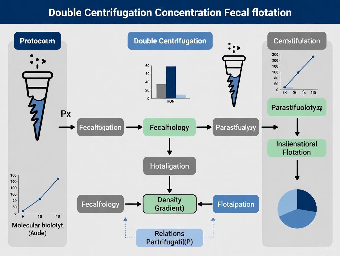
Diagram 1: Double Centrifugation Workflow.
Advanced Research Applications and Method Integration
The double centrifugation protocol serves as a foundational step for more complex diagnostic and research pipelines. Its utility is enhanced when integrated with other methodologies.
Fecal Egg Count Reduction Test (FECRT): The double centrifugation method can be adapted for quantitative fecal egg counts (FEC) by standardizing the initial fecal mass and using a defined volume of flotation solution. This quantitative output is essential for calculating the FECRT, the gold standard for monitoring anthelmintic resistance in herds. The formula is:
Percent Egg Reduction = [(Pre-treatment FEC - Post-treatment FEC) / Pre-treatment FEC] × 100[1]. Resistance is suspected for benzimidazoles if the reduction is<90%, for pyrantel if<85%, and for macrocyclic lactones (e.g., ivermectin) if<95%[1].Synergy with Molecular Diagnostics: While highly sensitive for intact eggs, flotation can miss pre-patent infections or be confounded by morphologically similar species. Coupling flotation with coproantigen detection (ELISA) can identify infections before egg shedding begins [8] [4]. Furthermore, the purified pellet from the double centrifugation protocol, especially one incorporating a sequential sieving (SF-SSV) step, provides an excellent sample input for PCR, as it reduces the presence of copro-inhibitors [2]. One study found that for large sample sets (n=100), a qPCR-based approach offered similar costs and faster processing compared to advanced microscopy, while also providing species-specific results [2].
Diagram 2: Integrated Diagnostic Pathways.
The double centrifugation concentration fecal flotation protocol is a powerful, physics-based technique that offers researchers and diagnosticians a highly sensitive and reliable method for the detection of parasitic elements in feces. Its effectiveness is rooted in the precise application of buoyancy, specific gravity, and centrifugal force. By adhering to the detailed protocols and quality control measures outlined in this document—including the use of standardized centrifugal forces and verified flotation solutions—research laboratories can ensure the generation of robust, reproducible, and high-quality data. This method remains a cornerstone technique, both as a standalone diagnostic and as a critical preparatory step for advanced molecular and immunological assays in parasitology research.
The accurate detection and identification of gastrointestinal parasites, specifically helminth eggs, protozoan oocysts, and cysts, are fundamental to parasitology research and diagnostic drug development. Traditional methods, particularly the double centrifugation concentration fecal flotation, remain widely used for parasite concentration and microscopic examination. This document details the key parasite targets and provides refined protocols to enhance the sensitivity and efficiency of their detection in a research context.
Key Parasite Targets and Morphology
The following table catalogues primary parasitic targets, summarizing their morphological characteristics for identification.
Table 1: Key Parasitic Targets for Microscopic Detection [10] [11]
| Category | Parasite Name | Form | Key Morphological Features |
|---|---|---|---|
| Protozoan Cysts | Entamoeba histolytica | Cyst | Spherical, 1-4 nuclei, chromatoid bodies [11] |
| Giardia lamblia | Cyst | Ellipsoidal, refractile wall, axostyles [10] [11] | |
| Blastocystis hominis | Cyst | Variable size, central body form[vacuole] [11] | |
| Entamoeba coli | Cyst | Spherical, typically >4 nuclei [11] | |
| Protozoan Oocysts | Cyclospora cayetanensis | Oocyst | Spherical, 8-10 μm, autofluorescent [10] |
| Cystoisospora belli | Oocyst | Ellipsoidal, large size (20-30 μm) [10] | |
| Helminth Eggs | Ascaris lumbricoides (Fertile) | Egg | Mammillated coat, brownish [10] |
| Ascaris lumbricoides (Infertile) | Egg | Elongated, filled with disorganized material [10] | |
| Trichuris trichiura | Egg | Barrel-shaped, bipolar plugs [10] | |
| Hookworm (Ancylostoma / Necator) | Egg | Thin-walled, oval, often in cleavage stage [10] | |
| Taenia species | Egg | Radially striated shell, contains oncosphere [10] | |
| Hymenolepis nana | Egg | Oval, inner membrane with polar filaments [10] [11] | |
| Strongyloides stercoralis | Larvae | Rhabditiform larvae (L1), not an egg [10] [11] | |
| Schistosoma mansoni | Egg | Large, oval with lateral spine [10] |
Experimental Protocols for Parasite Concentration
Formalin-Ethyl Acetate Concentration (FAC) Technique
The Formalin-Ethyl Acetate Concentration (FAC) technique is a sedimentation-flotation method considered highly sensitive for recovering a broad range of parasites [11].
Detailed Protocol:
- Emulsification: Emulsify approximately 1 gram of stool specimen with 7 mL of 10% formol saline in a container. Allow the mixture to fix for 10 minutes [11].
- Filtration: Strain the fixed suspension through three folds of gauze or a sieve (500-600 µm mesh) into a 15 mL conical centrifuge tube [11].
- Solvent Addition: Add 3 mL of ethyl acetate to the formalin-filtrate in the tube. Securely stopper the tube and shake it vigorously for at least 30 seconds to create an emulsion [11].
- Centrifugation: Centrifuge the tube at 1500 rpm (approximately 500 g) for 5 minutes. This step creates four distinct layers:
- Top layer: Ethyl acetate
- Plug: Debris
- Middle layer: Formalin
- Pellet: Sediment containing parasites [11]
- Sediment Harvest: Free the debris plug from the sides of the tube by ringing it with an applicator stick. Decant the top three layers (ethyl acetate, debris, and formalin) carefully. Use a swab to wipe the inner walls of the tube to remove residual debris and fluid [11].
- Microscopy: Re-suspend the remaining sediment in a small amount of formalin or saline. Transfer one drop to a microscope slide, add a coverslip, and examine systematically under 10x and 40x objectives. The entire coverslip area should be scanned for parasite forms [11].
Sequential Sieving Sedimentation-Flotation (SF-SSV) Protocol
This protocol enhances the recovery of specific parasite eggs, such as Toxocara species, by purifying and concentrating them from fecal debris through sequential filtration [12].
Detailed Protocol:
- Initial Processing: Begin with a standard Sedimentation-Flotation (SF) procedure. After the final centrifugation step in a high-specific-gravity solution (e.g., zinc chloride or sugar solution), retain the supernatant [12].
- Sequential Sieving: Decant the supernatant (approximately 45 mL) and pass it sequentially through a series of three nylon sieves [12]:
- First, through a 105-µm sieve to remove large particulate matter.
- Second, the filtrate is drawn through a 40-µm sieve to capture particles in the 40–105 µm range (this captures most Toxocara spp. eggs).
- Third, pass the filtrate through a 20-µm sieve to capture fragmented eggs or smaller parasites.
- Sediment Recovery: The material captured on the 40-µm and 20-µm sieves is washed and can be re-suspended for microscopic examination or downstream molecular analysis [12].
Comparative Performance of Diagnostic Methods
The selection of a concentration method significantly impacts diagnostic yield. The table below compares the performance of different techniques based on validation studies.
Table 2: Comparative Performance of Diagnostic Methods [12] [11]
| Method | Diagnostic Sensitivity (for specified parasites) | Key Advantages | Key Limitations |
|---|---|---|---|
| Direct Wet Mount | 41% [11] | Rapid, requires minimal processing; allows observation of motile trophozoites. | Low sensitivity; highly dependent on operator skill and parasite load. |
| Formalin-Ether Concentration (FEC) | 62% [11] | Good recovery of a wide range of parasites; clear background for microscopy. | Use of ether is a fire hazard and requires proper ventilation [11]. |
| Formalin-Ethyl Acetate Concentration (FAC) | 75% [11] | Higher recovery rate than FEC; safer than ether [11]. | Requires multiple steps; some distortion of protozoan morphology may occur. |
| Sequential Sieving (SF-SSV) | Highest analytical sensitivity for Toxocara spp. eggs [12] | Superior sensitivity; purifies eggs from PCR inhibitors; ideal for downstream molecular work [12]. | More time-consuming for single samples; requires specialized sieves. |
| Multiplex qPCR | Substantial agreement with microscopy; species-specific identification [12] | High-throughput potential; objective; provides species-level data [12]. | Higher cost per sample if run individually; requires specific equipment and expertise [12]. |
Research Reagent Solutions
A list of essential materials and reagents required for the protocols described is provided below.
Table 3: Essential Research Reagents and Materials [12] [11]
| Item | Function/Application |
|---|---|
| 10% Formol Saline | Fixative and preservative for stool specimens; kills pathogens and stabilizes morphology for microscopy [11]. |
| Ethyl Acetate | Solvent used in the FAC method to extract debris and fat from the fecal suspension, resulting in a cleaner sediment [11]. |
| Diethyl Ether | Alternative solvent for the FEC method; extracts debris and fat (note: higher flammability hazard) [11]. |
| High-Density Flotation Solution | Solutions like Zinc Chloride or Sheather's Sugar are used to float parasite elements to the surface during centrifugation [12]. |
| Nylon Sieves (105µm, 40µm, 20µm) | For the SF-SSV protocol; used to sequentially filter and size-select parasite eggs from fecal debris [12]. |
| Conical Centrifuge Tubes (15mL) | For sample processing, centrifugation, and separation of layers in concentration protocols [11]. |
| Species-Specific qPCR Assays | For molecular detection and differentiation of parasites (e.g., T. canis vs. T. cati) following DNA extraction [12]. |
Workflow and Data Analysis Diagrams
FAC Concentration Workflow
Method Sensitivity Comparison
Centrifugal fecal flotation, particularly double centrifugation or double-spin methods, represents a significant advancement in parasitological diagnostics by markedly improving the recovery of heavy helminth eggs compared to simple flotation techniques. Eggs from parasites such as trematodes, pseudophyllidean cestodes, spirurids, and acanthocephalans often possess higher specific gravity or operculated structures that impede their reliable flotation in passive, gravity-dependent methods. This application note details the experimental protocols, reagent specifications, and quantitative data demonstrating the superior efficacy of centrifugal techniques, providing researchers and drug development professionals with a standardized framework for maximizing diagnostic sensitivity in gastrointestinal parasite surveillance and anthelmintic efficacy trials.
The diagnosis of gastrointestinal parasitism fundamentally relies on the coproscopic detection of eggs, larvae, and cysts, with flotation techniques serving as a cornerstone for concentrating these parasitic elements. Simple flotation, which relies solely on gravity to bring parasitic elements to the surface of a high-specific-gravity solution, is a common but limited diagnostic tool [13]. Its major limitation is the incomplete recovery of "heavy" eggs—those with a high specific gravity or structural features that prevent them from floating efficiently. These include the operculate eggs of trematodes (e.g., Paragonimus kellicotti) and pseudophyllidean cestodes (e.g., Diphyllobothrium spp.), as well as the eggs of spirurids (e.g., Physaloptera spp.) and acanthocephalans [13].
Centrifugal flotation addresses this limitation by applying a controlled centrifugal force, driving parasitic elements into the flotation solution and resulting in a denser concentrate at the surface. The double centrifugation method further refines this process by incorporating an initial purification step, yielding a sample with reduced debris and a higher concentration of target organisms [14] [15]. This protocol is essential for research and diagnostic scenarios requiring maximum sensitivity, such as confirming parasite-free status, monitoring the spread of anthelmintic resistance, and conducting rigorous clinical trials for novel parasiticides.
Comparative Data Analysis
The enhanced sensitivity of centrifugal flotation, particularly modified double-centrifugation methods, is supported by empirical data across multiple host species. The following tables summarize key findings from recent comparative studies.
Table 1: Comparative sensitivity of flotation techniques for detecting various helminth eggs in different host species.
| Host Species | Parasite Taxa | Simple Flotation | Centrifugal Flotation | Notes | Study |
|---|---|---|---|---|---|
| General (Small Animals) | Whipworms (Trichuris spp.) / Capillarids | Low / Variable Sensitivity | High Sensitivity | The most dramatic improvement in detection is for bipolar-plugged eggs. | [13] |
| Camels | Moniezia spp. (cestode) | 4.5% positive | 7.7% positive | Mini-FLOTAC, an advanced quantitative centrifugal method, detected a 71% higher positivity rate. | [16] |
| Dogs | Ancylostoma spp. (hookworm) | Lower Sensitivity | Significantly Higher Sensitivity (P < 0.01) | Centrifugal flotation was more accurate in detecting a range of parasites. | [17] |
| Alpacas | Trichuris sp. & Nematodirus sp. | Not Reported | 11.7% & 33.9% prevalence | Detected using a modified Willis centrifugal flotation method. | [18] |
Table 2: Comparison of quantitative fecal egg count (FEC) results between techniques.
| Technique | Principle | Key Advantage | Limit of Detection (EPG/OPG) | Ideal Use Case |
|---|---|---|---|---|
| Simple Flotation | Passive gravity flotation | Low cost, rapid, simple to perform | Not standardized; generally high | Preliminary, low-sensitivity field screening. |
| Centrifugal Flotation | Active force concentration | Maximizes recovery of heavy eggs; superior overall sensitivity [13] | Varies with sample volume and chamber | Routine clinical diagnosis and herd-level surveillance. |
| Mini-FLOTAC | Standardized centrifugal flotation | High accuracy and precision; sensitivity of 5 EPG/OPG [19] | 5 EPG/OPG | Anthelmintic efficacy trials (FECRT) and high-precision research. |
| McMaster | Chamber-based count under microscope | Standardized quantitative results | 33.3 - 50 EPG (depending on modification) | Quantitative assessment where high precision is less critical. |
Experimental Protocols
Detailed Protocol: Double Centrifugal Flotation
This protocol, optimized from standard veterinary parasitological practices and research publications, is designed for the maximum recovery of a broad spectrum of parasitic elements, including heavy eggs [18] [13].
Workflow Overview:
Materials & Reagents:
- Faecal Sample: 3-5 grams of fresh or refrigerated feces.
- Flotation Solution: Saturated sucrose (specific gravity ~1.27-1.33) or sodium nitrate (SG ~1.20-1.25). Sucrose is preferred for its ability to prevent rapid crystallization, allowing for slide re-examination [18] [13].
- Laboratory Equipment: Centrifuge, test tubes (15 mL), sieve or cheesecloth, glass slides and coverslips, microscope.
Step-by-Step Procedure:
- Sample Preparation: Weigh 3-5 grams of feces and thoroughly mix with approximately 10-15 mL of flotation solution in a beaker. Pour the mixture through a sieve or cheesecloth into a second container to remove large debris.
- First Centrifugation (Purification Spin): Transfer the filtered suspension to a labeled centrifuge tube. Centrifuge at 650-2000 x g for 10 minutes. This initial spin forms a pellet of fecal debris and heavy particles, including any dense parasite eggs.
- Supernatant Removal: Carefully decant the supernatant, ensuring the pellet at the bottom of the tube is not disturbed.
- Pellet Resuspension: Add a high-specific-gravity flotation solution (e.g., SG 1.27 sucrose) to the tube and resuspend the pellet thoroughly using a vortex mixer or applicator stick.
- Second Centrifugation (Flotation Spin): Fill the tube with more flotation solution to create a slightly convex meniscus. Centrifuge again at 650-2000 x g for 5-10 minutes. This second spin forces the parasitic elements through the dense solution to the surface.
- Sample Collection: After centrifugation, carefully place a clean coverslip on top of the meniscus and allow it to stand for 5-10 minutes.
- Microscopy: Gently lift the coverslip, place it on a glass slide, and examine the entire area under the coverslip systematically using the 10X objective of a compound microscope. The 40X objective can be used for morphological confirmation.
Protocol for Simple Sedimentation
For the heaviest operculated eggs (e.g., trematodes like Paragonimus or Nanophyetus), which may not float even with centrifugation, simple sedimentation is the gold standard [13].
Workflow Overview:
Procedure:
- Mix 1-2 grams of feces with water and strain through a sieve into a test tube or beaker.
- Allow the suspension to stand undisturbed for 5 minutes. Operculated eggs, being heavier, will sink to the bottom.
- Carefully decant the supernatant, refill the container with clean water, and resuspend the sediment. Repeat this washing process 2-3 times to reduce fecal debris.
- After the final wash, decant the supernatant and examine the remaining sediment microscopically by placing a few drops on a slide with a coverslip.
Note: If infection with the blood fluke Heterobilharzia americana is suspected, use saline instead of water to prevent eggs from hatching and releasing miracidia, which would lead to a false-negative result [13].
The Scientist's Toolkit: Research Reagent Solutions
The selection of appropriate reagents is critical for the success of any flotation protocol. The following table details key solutions and their applications.
Table 3: Essential reagents for centrifugal flotation protocols.
| Reagent Solution | Typical Specific Gravity | Function & Application | Research Considerations |
|---|---|---|---|
| Sucrose Solution | 1.27 - 1.33 | Excellent for general flotation; does not crystallize rapidly, allowing for delayed examination [18]. | High viscosity can slow the flotation of some elements. Ideal for preserving samples for reference or quality control. |
| Zinc Sulfate (ZnSO₄) | ~1.18 - 1.20 | Considered optimal for recovering delicate cysts like Giardia duodenalis [13]. | Lower SG may reduce recovery of some heavier nematode eggs. Modifications (e.g., ZnSO₄ with SG 1.27) are used in specific research contexts [18]. |
| Sodium Chloride (NaCl) | ~1.20 - 1.22 | Low-cost, readily available solution for routine diagnostics. | Crystallizes quickly, requiring immediate reading. Not suitable for sample storage. |
| Sodium Nitrate (NaNO₃) | ~1.20 - 1.25 | Common and effective for a wide range of nematode eggs and coccidian oocysts. | Less expensive than sucrose but can crystallize over time. |
The transition from simple to double centrifugal flotation represents a critical methodological evolution in parasitology research. The forced provided by centrifugation is non-negotiable for the reliable recovery of heavy eggs, which are frequently underrepresented or entirely missed by passive flotation methods. The protocols and data outlined herein provide a validated framework for scientists to enhance the accuracy and reliability of fecal examinations. Adopting these refined techniques is fundamental for advancing research in parasite epidemiology, drug development, and anthelmintic resistance monitoring, ensuring that diagnostic outcomes are a true reflection of parasitic burden.
Essential Equipment and Flotation Solution Selection Criteria
The Scientist's Toolkit: Key Research Reagents and Materials
Table 1: Essential Research Materials for Fecal Flotation Protocols
| Item | Function & Specification |
|---|---|
| Flotation Solutions | Separates parasites from fecal debris based on density [9] [1]. |
| Sucrose (Sugar) Solution | High specific gravity (≥1.27); preserves delicate eggs [20] [1]. |
| Zinc Sulfate Solution | Specific gravity 1.18–1.20; optimal for recovering delicate protozoa (e.g., Giardia cysts) [9]. |
| Centrifuge | Enforces separation; swing-bucket type is preferred for consistent meniscus formation [20]. |
| Centrifuge Tubes | Conical-bottomed tubes (15ml) enhance egg recovery efficiency [20]. |
| Microscope with 10X, 40X Objectives | Systematic examination of coverslipped samples [21] [20]. |
| Coverslips & Glass Slides | Standard slides (22mm x 22mm coverslips) for preparing samples [21] [20]. |
| Fecal Strainers | Mesh (105µm, 40µm, 20µm) or gauze in tea strainer removes large debris [12] [21]. |
Quantitative Comparison of Flotation Solutions and Methods
Table 2: Performance Characteristics of Diagnostic Methods for Gastrointestinal Parasites
| Method | Key Performance Metrics | Advantages | Limitations |
|---|---|---|---|
| Double-Centrifugation Flotation [9] [1] | - | Recovers broad spectrum of eggs, larvae, cysts [1]. | Operator expertise dependent [22]. |
| Sequential Sieving (SF-SSV) [12] | Highest analytical & diagnostic sensitivity for Toxocara spp. [12] | Effective egg enrichment, removes PCR inhibitors [12]. | - |
| qPCR Panels [22] | Detected 2.6x more co-infections vs. ZCF [22] | Species-specific diagnosis, detects markers [12] [22]. | Higher cost for single samples [12]. |
| Fecal Antigen Testing [23] | Detects up to 2x more infections vs. flotation alone [23] | Detects prepatent, single-sex, immature infections [23]. | - |
Detailed Experimental Protocols
Protocol 1: Standard Double-Centrifugation Fecal Flotation
This core method concentrates parasite elements through density-based separation [9] [1].
- Step 1: Sample Preparation. Weigh 2-5g of feces. Mix thoroughly with 10-15ml of selected flotation solution to create a slurry [21] [20].
- Step 2: Strain and Load. Pour slurry through a strainer lined with gauze into a clean cup. Transfer strained liquid into a 15ml conical-bottom centrifuge tube [21] [20].
- Step 3: Primary Centrifugation. Centrifuge at 1200-1800 g for 5-10 minutes [21] [1].
- Step 4: Create Meniscus. After decanting supernatant, add fresh flotation solution to form a slightly positive meniscus [21] [20].
- Step 5: Secondary Flotation. Place a coverslip on tube top. Let stand for 10 minutes [21] [20].
- Step 6: Sample Examination. Lift coverslip vertically, place on glass slide. Systematically examine entire area under coverslip at 10X magnification, using 40X for confirmation [21] [1].
Protocol 2: Sequential Sieving Protocol (SF-SSV) for Egg Enrichment
This method enhances sensitivity for specific parasites like Toxocara spp. and cleans samples for downstream PCR [12].
- Step 1: Initial Processing. Process 3g feces per standard SF protocol [12].
- Step 2: Sequential Filtration. Decant SF supernatant through a stack of nylon sieves:
- 105µm mesh: Removes large particulate matter.
- 40µm mesh: Captures target eggs (e.g., Toxocara spp.).
- 20µm mesh: Captures fragmented eggs or smaller stages [12].
- Step 3: Sample Recovery. Retain material from 40µm and 20µm meshes for microscopic examination or DNA extraction [12].
Protocol 3: DNA Extraction and qPCR for Molecular Detection
Molecular methods complement traditional techniques by enabling species-specific diagnosis and resistance marker detection [12] [22].
- Step 1: Nucleic Acid Extraction. Homogenize 150-250mg fecal material in guanidinium-based lysis solution using mechanical bead beating. Extract total nucleic acid on an automated platform (e.g., KingFisher Apex) [22].
- Step 2: qPCR Setup and Run. Utilize species-specific TaqMan qPCR assays. Include internal controls: Internal Sample Control (ISC) and Internal Positive Control (IPC) to monitor inhibition [22].
- Step 3: Data Analysis. Analyze amplification curves. Determine presence of parasite DNA and specific genetic markers (e.g., F167Y for hookworm BZ resistance) [22].
A Standardized Step-by-Step Protocol for Research-Grade Results
The diagnostic accuracy of parasitic investigations, particularly those employing the double centrifugation concentration fecal flotation technique, is fundamentally dependent on the initial steps of sample collection and preservation. The integrity of parasitic elements, including eggs, larvae, and cysts, can be significantly compromised by improper handling, leading to false-negative results and an underestimation of parasite burden [24] [25]. This application note details standardized protocols for the collection, preservation, and storage of fecal samples to ensure optimal morphological preservation for microscopic analysis. Adherence to these guidelines is crucial for generating reliable data in research settings, including drug efficacy trials and faecal egg count reduction tests (FECRT) [1].
Sample Collection Protocols
Collection Guidelines
Proper collection is the first critical step in preserving parasite integrity.
- Sample Source: For definitive results, collect samples from individual animals. Composite samples from multiple animals can indicate a problem but will not identify the specific affected individuals [1].
- Collection Method: Collect feces immediately after defecation. For pets, instruct owners to collect a sample directly after deposit to ensure correct identification of origin. For wildlife, non-invasive collection from the environment is common, but note that species identification via scat morphology can be prone to error [13] [25].
- Sample Freshness: Fecal samples should be as fresh as possible. Process samples within 2 hours of collection if possible. If immediate processing is not feasible, refrigerate samples at 4°C [13]. Samples exposed to the environment for extended periods or incubated at room temperature for more than 6 hours are unsuitable due to the hatching of helminth eggs and degradation of protozoal stages [13].
- Sample Volume: Collect a sufficient volume for analysis. A walnut-sized portion of solid stool or 5–10 mL of liquid stool is generally adequate [24]. For specific techniques like the Baermann examination, 10-20 grams of fresh feces are required [1] [13].
- Containers: Use clean, wide-mouth, leak-proof plastic containers. Containers should not contain any preservatives or disinfectants unless the sample is to be preserved immediately [24].
- Avoid Contaminants: Take care to avoid contamination of the sample with urine, water, or soil [24] [13].
Preservation Methods and Comparisons
If microscopic analysis is delayed beyond 24 hours, chemical preservation is necessary to maintain the morphological integrity of parasites. The choice of preservative depends on the primary analytical method (morphological vs. molecular) and the target parasites.
Table 1: Comparison of Fecal Sample Preservation Methods
| Preservative | Recommended Use | Advantages | Disadvantages | Suitability for DNA Analysis |
|---|---|---|---|---|
| 10% Formalin [26] [24] | General morphological preservation; long-term storage for microscopy. | Excellent preservation of helminth eggs, larvae, and protozoan cysts; prevents degradation and autolysis [26]. | Toxic; requires careful handling; causes DNA fragmentation, impairing genetic analyses [26]. | Poor |
| 70-96% Ethanol [26] | Studies requiring subsequent molecular analysis; morphological studies. | Less toxic; maintains stable DNA for long-term storage; suitable for molecular studies [26]. | Dehydrates tissues, which can lead to morphological alterations and brittle specimens [26]. | Excellent |
| Refrigeration (4°C) [24] [13] | Short-term storage (up to 24 hours). | Simple and cost-effective; no chemicals required. | Only a short-term solution; parasite stages degrade over time. | Good for short periods |
A 2024 comparative study on wild capuchin monkey feces found that while formalin-preserved samples yielded a higher diversity of identifiable parasitic morphotypes, both formalin and ethanol were suitable for morphological identification after more than one year of storage [26]. The study also noted that the preservation quality can vary by parasite type; for instance, Filariopsis larvae were better preserved in formalin, whereas strongyle-type eggs showed no significant difference between the two mediums [26].
The Double Centrifugation Fecal Flotation Protocol
The double centrifugation concentration technique is a sensitive, broad-based test for evaluating feces for parasitic infections and is considered a gold standard in many diagnostic settings [1] [26]. The following is a detailed step-by-step protocol.
Experimental Workflow
The following diagram illustrates the complete double centrifugation fecal flotation procedure.
Step-by-Step Methodology
Sample Preparation and Straining:
- Weigh approximately 2 to 5 grams of feces and mix it with 10 mL of an appropriate flotation solution (e.g., sugar solution with a specific gravity of 1.33 or zinc sulfate at 1.18 for delicate parasites like Giardia) [21] [1].
- Pour the mixture through a tea strainer or cheesecloth (which can be lined with a 4x4 inch gauze square for easier cleaning) into a cup. This step removes large debris and fiber [21].
First Centrifugation:
- Pour the strained solution into a 15 mL centrifuge tube.
- Centrifuge the tube at 1,200 rpm (approximately 280 g) for 5 minutes. This pellets the fecal debris and parasitic elements at the bottom of the tube [21].
Creating the Meniscus:
- After centrifugation, carefully decant the supernatant.
- Insert the tip of a disposable pipette below the surface of the mixture and add fresh flotation solution. Gently fill the tube until a slightly inverted, positive (rounded) meniscus forms. Take care not to overfill, as this can cause spillover and loss of floated material when the coverslip is applied [21].
Coverslip Placement and Standing Time:
- Gently place a coverslip on top of the tube so it contacts the meniscus.
- Let the sample stand for 10 minutes. This allows light parasitic elements (eggs, cysts, oocysts) to float upward and adhere to the coverslip [21].
Sample Transfer and Microscopy:
Complementary Diagnostic Techniques
While double centrifugation flotation is a highly effective general diagnostic tool, certain parasites are not reliably detected by flotation and require specialized techniques [13].
Table 2: Guide to Complementary Parasitological Techniques
| Technique | Primary Indication | Brief Protocol Summary | Key Parasites Detected |
|---|---|---|---|
| Baermann Technique [1] [13] | Detection of motile nematode larvae. | Feces placed in gauze, suspended in water in a funnel setup for ≥8 hours; larvae migrate out and sink to be collected from tubing. | Lungworms (Dictyocaulus, Aelurostrongylus), Strongyloides larvae. |
| Simple Sedimentation [13] | Detection of heavy, operculated, or non-buoyant eggs. | Fecal suspension in water is repeatedly sedimented by gravity or low-speed centrifugation; sediment is examined microscopically. | Trematode (fluke) eggs, pseudophyllidean cestode eggs (e.g., Diphyllobothrium). |
| Direct Smear [13] | Detection of motile trophozoites. | A tiny speck of fresh feces (<20 min old) is mixed with saline on a slide and examined immediately under a coverslip. | Giardia duodenalis, Tritrichomonas blagburni trophozoites. |
| Mini-FLOTAC [16] [27] | Quantitative faecal egg count (FEC) with high sensitivity. | A quantitative method using a special chamber that allows examination of a fixed volume of fecal suspension, improving sensitivity and precision. | Superior sensitivity for strongyles, Strongyloides, Moniezia [16]. |
The Scientist's Toolkit: Essential Research Reagents and Materials
The following table details key materials and reagents required for the protocols described in this note.
Table 3: Essential Research Reagents and Materials for Fecal Parasitology
| Item | Specification / Function | Application Notes |
|---|---|---|
| Flotation Solutions | Zinc Sulfate (sp. g. 1.18), Sugar (sp. g. 1.28) | Zinc sulfate is preferred for delicate cysts (e.g., Giardia). Sugar solution is less prone to crystallization [1] [13]. |
| Preservatives | 10% Buffered Formalin, 96% Ethanol | Formalin for morphology, ethanol for combined morphological/molecular studies. Handle formalin with appropriate PPE [26] [24]. |
| Centrifuge | Capable of 280 g (approx. 1200 rpm) | Essential for the concentration step in double centrifugation flotation [21]. |
| Centrifuge Tubes | 15 mL conical tubes | Standard volume for fecal suspension processing [21]. |
| Microscope | Compound light microscope with 10X and 40X objectives | Systematic examination of the entire coverslip area is critical for sensitivity [21]. |
| Strainers/Gauze | Tea strainer, 4x4 inch gauze squares | For removing large particulate matter from the fecal suspension prior to centrifugation [21]. |
| Sample Containers | Leak-proof, wide-mouth plastic containers | For primary sample collection and transport; must be sealable [24] [13]. |
Within parasitology research, particularly in protocols for double centrifugation concentration fecal flotation, the initial sample preparation steps are critical for determining the accuracy and reliability of downstream results. Fecal homogenization and strategic filtration constitute the foundational first step in this process, directly influencing the efficacy of parasite egg and cyst recovery. Inconsistent or inadequate homogenization can lead to false negatives, while poor filtration may permit obstructive debris to compromise microscopic examination. This application note details a standardized, reproducible protocol for this essential first phase, designed to meet the rigorous demands of research and drug development scientists.
The principle of this step is to create a homogeneous fecal suspension devoid of large, disruptive particulates. This ensures a uniform distribution of parasitic elements for subsequent diagnostic steps, thereby minimizing sample bias and maximizing detection sensitivity. Research confirms that failure to use best-practice techniques during initial sample preparation can result in a failure to detect parasite stages in fecal samples [3]. The methodology outlined herein is adapted from established veterinary diagnostic procedures and optimized for research-grade precision [21] [3] [28].
Experimental Protocol
Materials and Equipment
Research Reagent Solutions & Essential Materials
| Item | Specification/Function |
|---|---|
| Flotation Solution | Zinc Sulfate (ZnSO₄, specific gravity 1.18-1.20) or Sheather's Sugar (specific gravity 1.27). Creates a buoyant medium for parasite flotation [3] [28]. |
| Gauze Strainer | Single or double layer of 4x4 inch gauze squares [21] or tea strainer lined with gauze [21] [28]. Filters large fibrous debris. |
| Centrifuge Tubes | 15 mL conical tubes [21] [28]. Compatible with standard bench-top centrifuges. |
| Homogenization Vessel | Disposable cup or beaker (50-100 mL). Provides space for initial sample mixing. |
| Balanced Centrifuge | Swinging bucket or fixed-angle rotor, capable of 1,200 rpm (280 g) [21] or 180 x g [28]. Provides the force for concentration. |
Step-by-Step Methodology
- Sample Measurement: Using an analytical balance, weigh out 2 to 5 grams of fresh feces and place it into the homogenization vessel [21] [3]. For diarrheic samples, consider increasing the sample volume due to the dilutive effect of high water content [3].
- Primary Homogenization: Add approximately 10 mL of flotation solution to the feces [21] [28]. Using an applicator stick or spatula, stir the mixture vigorously to achieve a consistent, smooth slurry. Ensure the fecal material is distributed evenly throughout the solution.
- Strategic Strainer Filtration:
- Line a tea strainer or funnel with a 4x4-inch gauze square [21]. Alternatively, pour the mixture through one or two layers of a gauze sponge held over a clean container [3].
- Slowly pour the homogenized fecal slurry through the lined strainer into a second clean cup or beaker.
- To maximize yield, use the flat end of a spatula to gently press the residual solid matter in the strainer. This action facilitates the passage of the fluid containing parasitic elements while retaining coarse debris.
- Tube Transfer: Carefully pour the filtered filtrate from the clean container into a 15 mL centrifuge tube [21] [28]. The tube should be filled to approximately 80% of its capacity to allow for liquid displacement during subsequent steps [3].
Workflow Diagram
The following diagram illustrates the logical sequence and output of the homogenization and filtration process.
Performance Data
Quantitative Impact of Filtration on Sample Quality
Comparative studies demonstrate that effective initial processing significantly improves the clarity of the final sample for microscopic analysis. The data below, derived from related methodological comparisons, underscore the importance of rigorous initial steps in achieving high-quality diagnostic results [29] [28] [30].
| Analysis Metric | Sedimentation/Flotation [29] | Mini-FLOTAC [29] | Centrifugal Flotation (Reference) [28] |
|---|---|---|---|
| Variance (Precision) | Highest | No significant difference from other methods | Lower variance |
| Strongyle Egg Detection (Sensitivity) | Highest detection rate | High detection rate | High sensitivity (benchmark) |
| Key Advantage | High sensitivity for simple detection [29] | More precise for quantitative tests (FECRT) [29] | Improved recovery of heavier eggs (e.g., Trichuris, Taenia) [3] |
Discussion
The homogenization and filtration protocol described serves as the critical control point for the entire double centrifugation process. The use of 2-5g of feces provides an adequate sample size to overcome the patchy distribution of parasitic elements, a factor crucial for detecting low-level infections often encountered in controlled studies or post-treatment monitoring [3]. The strategic use of gauze filtration is a low-tech but vital step to remove debris that can obscure visualization during microscopic examination. This is particularly important for automated image-analysis systems like the OvaCyte or FECPAKG2, where debris can lead to false positives or analytical errors [29] [28].
It is important to note that while this step is foundational, its performance is interdependent with subsequent stages. The choice of flotation solution specific gravity, for instance, initiated here, will directly impact the buoyancy and recovery of different parasite species in the following centrifugation steps [3] [31]. Adherence to this standardized protocol ensures sample integrity and data reproducibility, forming a reliable basis for sensitive parasite detection and quantitative fecal egg count reduction tests (FECRT) in pharmaceutical development and resistance monitoring [3] [30].
Within the broader research on the double centrifugation concentration fecal flotation protocol, the first centrifugation and pellet formation is a critical preparative step. This phase is dedicated to separating diagnostically significant elements, specifically parasite eggs, oocysts, and cysts, from fecal debris. The precision and consistency of this step directly influence the purity of the resulting sample and the subsequent efficacy of the diagnostic flotation. This application note details a standardized methodology for this key step, providing researchers and drug development professionals with a reliable framework for producing high-quality samples for parasitological analysis.
## Experimental Protocol
### Materials and Reagents
- Feces Sample: 2 to 5 grams of fresh feces [21].
- Flotation Solution: Saturated sugar solution (specific gravity ~1.33) or Zinc Sulfate solution (specific gravity 1.18–1.20). The choice of solution should be validated for the target parasite [9] [1].
- Laboratory Equipment: A swing-bucket centrifuge capable of achieving 280 g, 15 ml centrifuge tubes, a tea strainer or mesh sieve (approximately 150–200 µm), 4x4 inch gauze squares, and a disposable pipette [21].
### Methodology
- Sample Homogenization and Filtration: Combine 2–5 g of feces with approximately 10 ml of flotation solution in a disposable cup. Line a tea strainer with a 4x4 inch gauze square and pour the mixture through it into a second cup to remove large, coarse debris [21].
- Tube Loading: Transfer the homogenized and filtered solution into a 15 ml centrifuge tube [21].
- First Centrifugation: Load the tubes into a clinical centrifuge, ensuring a balanced load. Centrifuge at 1,200 revolutions per minute (RPM) for five minutes, which equates to a relative centrifugal force (RCF) of 280 g [21]. This force will sediment the dense parasite forms into a pellet at the bottom of the tube while lipids and some fine debris may form a surface layer.
### Workflow Diagram
The following flowchart illustrates the procedural sequence for the first centrifugation step.
## Research Reagent Solutions
The table below catalogues the essential reagents and materials required for the execution of this protocol step.
| Item | Function / Rationale |
|---|---|
| Zinc Sulfate Solution (spg 1.18–1.20) | A standard flotation medium ideal for recovering delicate structures like Giardia cysts and nematode larvae without causing distortion [9] [1]. |
| Saturated Sugar Solution (spg 1.28) | A high-specific gravity solution used for general broad-based fecal flotation to recover a wide spectrum of parasite eggs and oocysts [1]. |
| 15 ml Centrifuge Tubes | Standard vessels for sample preparation and centrifugation, compatible with clinical centrifuges [21]. |
| Gauze Square / Tea Strainer | A physical filtration system for the removal of large, coarse particulate matter from the fecal suspension, preventing clogging and ensuring a smooth homogenate [21]. |
## Quantitative Specifications
Critical parameters for the first centrifugation step are summarized in the following table.
| Parameter | Specification | Technical Rationale |
|---|---|---|
| Sample Mass | 2–5 g [21] | Provides sufficient material for analysis without overloading the tube. |
| Flotation Solution Volume | ~10 ml [21] | Establishes an optimal ratio for homogenization and subsequent density separation. |
| Centrifugation Speed | 1,200 RPM [21] | Generates sufficient force (280 g) to pelletize target parasites efficiently. |
| Relative Centrifugal Force (RCF) | 280 g [21] | Standardized force for diagnostic parasitology protocols. |
| Centrifugation Time | 5 minutes [21] | Duration required for complete sedimentation of parasitic elements. |
In the double centrifugation concentration fecal flotation protocol, Step 3 represents a critical transition from sample purification to diagnostic concentration. This step involves the careful discarding of the initial supernatant following the first centrifugation cycle, and the subsequent re-suspension of the sediment in a high-specific-gravity flotation solution. The precision with which this step is executed directly influences the efficiency of parasite egg and cyst recovery by creating optimal conditions for buoyancy-driven separation during the second centrifugation. This application note details the methodologies, quantitative parameters, and material requirements essential for researchers and drug development professionals to standardize this procedure for both diagnostic and research applications.
Experimental Protocols & Methodologies
Core Procedural Workflow
The following step-by-step protocol is compiled from standardized veterinary diagnostic procedures and recent parasitology research [1] [3] [21].
- Initial Centrifugation Completion: After the first centrifugation cycle (typically at 650-1,200 rpm or 280-1,800 g for 5-10 minutes), carefully remove the sample tube from the centrifuge without disturbing the sedimented pellet [3] [21].
- Supernatant Discarding: Sharply decant the supernatant into a waste container containing appropriate disinfectant. Alternatively, use a vacuum aspiration system, ensuring the tip does not contact or disturb the sediment pellet. Exercise caution to avoid losing any of the sediment where the parasite stages are concentrated [13] [12].
- Sediment Re-suspension: Add a predetermined volume of flotation solution (specific gravity 1.18-1.27) directly onto the sediment. The volume should be sufficient to fill the centrifuge tube to about 80% of its capacity for fixed-angle rotors, or to create a positive meniscus for swinging-bucket rotors [1] [32]. Using a vortex mixer or an applicator stick, thoroughly re-suspend the sediment until a homogeneous mixture is achieved, ensuring no compacted material remains at the bottom of the tube [3].
- Secondary Flotation Preparation: For fixed-angle centrifuges, the tube is now ready for the second centrifugation. For swinging-bucket centrifuges, additional flotation solution is added to form a convex meniscus, a coverslip is gently applied, and the tube is centrifuged [32] [5].
Key Experimental Variations in Research
Recent comparative studies have elaborated on protocol modifications to enhance diagnostic sensitivity:
- Sequential Sieving (SF-SSV): Following initial sedimentation and decanting, one study implemented a sequential sieving protocol using nylon meshes of 105-μm, 40-μm, and 20-μm sizes to further purify and concentrate Toxocara spp. eggs before flotation, resulting in significantly higher diagnostic sensitivity compared to standard sedimentation-flotation (SF) [12].
- Solution Additives: To reduce egg adhesion to equipment surfaces during re-suspension and transfer, the addition of surfactants (e.g., Tween 20) to the flotation solution has been investigated, demonstrating reduced egg loss and improved recovery rates in Lab-on-a-Disk (LoD) systems [33].
Data Presentation & Quantitative Analysis
Impact of Procedural Fidelity on Diagnostic Yield
Adherence to the re-suspension protocol is crucial for maximizing recovery. The table below summarizes the performance gains of centrifugal flotation over passive techniques.
Table 1: Comparative Sensitivity of Fecal Flotation Techniques for Detecting Helminth Eggs
| Flotation Technique | Force Applied | Relative Diagnostic Sensitivity | Key Supporting Evidence |
|---|---|---|---|
| Centrifugal Flotation | Centrifugal Force | High | 100% recovery of hookworm eggs in a controlled study; significantly superior for heavier eggs (e.g., Trichuris, taeniid eggs) [3] [32]. |
| Simple/Passive Flotation | Gravity Alone | Moderate | Approximately 70% recovery of hookworm eggs in a controlled study; performance variable for low-intensity infections [32]. |
Technical Parameters for Optimization
Table 2: Standardized Technical Parameters for Centrifugal Flotation
| Parameter | Typical Range | Research Context & Impact |
|---|---|---|
| Centrifugation Force (1st Centrifugation) | 280 - 1,800 g [12] [21] | Lower forces (e.g., 280 g) are common in clinic; research protocols may use higher forces for specific sedimentation. |
| Centrifugation Time | 5 - 10 minutes [3] [21] | Sufficient time for debris sedimentation; longer durations may be used in research for complex samples. |
| Flotation Solution Specific Gravity | 1.18 - 1.27 [1] [32] | Higher SG (1.27) floats denser eggs; lower SG (1.18) is optimal for delicate stages like Giardia cysts [13] [5]. |
| Post-Re-suspension Standing Time | 5 - 20 minutes [13] [21] | Allows for egg flotation before coverslip removal; duration depends on solution viscosity. |
Workflow Visualization
The following diagram illustrates the logical workflow and decision points for Step 3 of the double centrifugation protocol.
The Scientist's Toolkit: Research Reagent Solutions
Table 3: Essential Materials and Reagents for Flotation Procedures
| Reagent/Material | Specification/Function | Research Application Notes |
|---|---|---|
| Sucrose (Sheather's) Solution | SG = 1.27; High viscosity delays crystallization. | Optimal for broad-spectrum nematode and coccidian oocyst recovery; suitable for slide storage [32] [5]. |
| Zinc Sulfate (ZnSO₄) Solution | SG = 1.18-1.20; Lower specific gravity. | Solution of choice for recovering delicate stages (e.g., Giardia cysts, nematode larvae) without distortion [13] [5]. |
| Sodium Nitrate Solution | SG = 1.18-1.20; Readily available. | Common commercial solution; crystallizes faster than sucrose, requiring prompt examination [32]. |
| Centrifuge Tubes (15 ml) | Leak-proof, conical bottom. | Standardized volume for consistent processing; conical base aids in sediment formation and supernatant decanting [3] [21]. |
| Nylon Sieve Meshes | 105-μm, 40-μm, 20-μm mesh sizes. | Used in advanced sequential sieving (SF-SSV) protocols to purify eggs from debris, significantly increasing sensitivity [12]. |
| Surfactant (e.g., Tween 20) | Additive to reduce surface tension. | Minimizes egg adhesion to plasticware (tubes, pipettes, disks) during re-suspension and transfer, reducing egg loss [33]. |
The double centrifugation concentration fecal flotation technique is a cornerstone diagnostic method in parasitology research, providing superior sensitivity for detecting parasite eggs, larvae, and protozoan cysts in fecal samples [1] [13]. This protocol details the critical second centrifugation and coverslip application step, which finalizes the parasite concentration process prior to microscopic examination. The procedure significantly enhances diagnostic yield by leveraging centrifugal force to maximize parasite recovery, making it indispensable for accurate epidemiological studies, anthelmintic efficacy trials, and pathogen surveillance in drug development programs [1] [34].
Detailed Experimental Protocol
Second Centrifugation Procedure
Following the initial centrifugation and supernatant decanting, the second centrifugation step is performed to concentrate parasites into a detectable range.
- Tube Preparation: After decanting the supernatant from the first centrifugation, ensure the fecal pellet remains undisturbed at the bottom of a 15-ml centrifuge tube [21].
- Flotation Solution Addition: Using a disposable pipette, carefully add fresh flotation solution to the tube. The specific gravity of the solution should be appropriate for the target parasites (e.g., zinc sulfate, specific gravity 1.18, for detecting Giardia cysts, or sugar solution, specific gravity 1.33, for general purposes) [9] [13].
- Resuspension: Gently resuspend the fecal pellet in the fresh flotation solution by stirring with an applicator stick or by vortexing at low speed to create a homogeneous suspension.
- Centrifugation Parameters:
Coverslip Application and Parasite Harvesting
After the second centrifugation, parasite forms are concentrated at the meniscus of the solution and must be properly collected for analysis.
- Meniscus Formation: Following centrifugation, insert the tip of a clean disposable pipette below the liquid surface and slowly add more flotation solution until a slightly inverted, positive (rounded) meniscus forms. Take care not to overfill the tube, as this can cause spillage and sample loss when applying the coverslip [21].
- Coverslip Placement: Gently place a glass coverslip directly on top of the tube, ensuring it makes full contact with the meniscus. The parasite stages will float up and adhere to the coverslip during the standing period [21] [13].
- Incubation Time: Let the sample stand for 5–20 minutes to allow adequate time for parasites to float to the surface [13]. A 10-minute standing period is commonly used [21].
- Coverslip Removal: Carefully lift the coverslip straight up from the tube in a vertical motion to prevent sample mixing.
- Microscopic Preparation: Place the coverslip, liquid side down, onto a clean glass slide. The sample is now ready for systematic microscopic examination [21].
Data Presentation and Comparative Analysis
The diagnostic superiority of centrifugal flotation methods, which include the detailed second centrifugation step, is well-established in parasitology research. The following tables summarize key performance data and parameters.
Table 1: Comparative Detection Performance of Centrifugal Flotation vs. Other Methods
| Methodology | Detection Level/Positivity Rate | Key Findings | Sample Size (n) |
|---|---|---|---|
| Centrifugal Flotation (Double Centrifugation) | 20.8% overall positivity in canine samples [34] | Detected a wide range of nematodes, cestodes, trematodes, and protozoans; considered highly sensitive for most helminth eggs and protozoan cysts [13] [34] | 4,692 [34] |
| Simple Flotation (Passive) | Not directly quantified, but significantly lower sensitivity [13] | Considered inferior to centrifugal flotation; particularly poor for detecting heavy eggs (e.g., whipworm and capillarid eggs) [13] | - |
| Fecal Antigen Testing | Detected up to 2x more infections than centrifugal flotation alone for specific parasites [23] | Identifies infections even when egg production is absent or low; excellent for screening, but does not provide parasite identification [23] | 898,300 [23] |
Table 2: Optimized Centrifugation and Flotation Parameters for Fecal Analysis
| Parameter | Recommended Specifications | Research & Clinical Rationale |
|---|---|---|
| Centrifuge Speed | 650 g [13] or 1,200 rpm (280 g) [21] | Standardized force to balance pellet formation and parasite integrity. |
| Centrifuge Time | 5–10 minutes [21] [13] | Adequate time for parasite stages to concentrate at the meniscus. |
| Standing Time after Coverslip | 5–20 minutes [13] | Allows buoyant parasite elements to float and adhere to the coverslip. |
| Flotation Solution (General) | Sheather's sugar solution (sp. gr. 1.33) [1] [13] | High specific gravity floats most parasite stages; does not crystallize quickly, allowing slide re-examination [13]. |
| Flotation Solution (Giardia) | Zinc Sulfate (ZnSO₄, sp. gr. 1.18) [9] [13] | Optimal for detecting delicate Giardia cysts without causing distortion [9] [13]. |
Workflow Visualization
The following diagram illustrates the logical sequence and decision points in the second centrifugation and coverslip application protocol.
The Scientist's Toolkit: Research Reagent Solutions
Table 3: Essential Materials for Double Centrifugation Fecal Flotation
| Research Reagent & Material | Critical Function in Protocol |
|---|---|
| High-Specific-Gravity Flotation Solution (e.g., Zinc Sulfate, Sheather's Sugar) | Creates a density gradient enabling buoyant parasite stages (eggs, cysts, oocysts) to float to the surface while fecal debris sediments [9] [13]. |
| Standard Laboratory Centrifuge | Provides the controlled centrifugal force necessary to concentrate parasite elements rapidly and efficiently into the flotation medium, a key factor over passive techniques [13]. |
| 15-ml Centrifuge Tubes | Serve as the primary container for mixing feces with flotation solution and withstanding the forces of centrifugation [21]. |
| Glass Coverslips | Placed on the meniscus of the centrifuged sample to capture and secure the concentrated parasite forms for transfer to a microscope slide [21] [13]. |
| Disposable Pipettes | Allow for precise addition of flotation solution to form the critical positive meniscus without disrupting the concentrated sample [21]. |
Research Implications and Methodological Considerations
The meticulous execution of the second centrifugation and coverslip application is vital for research accuracy. This step directly impacts the sensitivity and reliability of fecal egg count reduction tests (FECRT), the gold standard for monitoring anthelmintic resistance in herds [1]. Inaccurate concentration can lead to false-negative results or underestimation of egg shedding levels, misclassifying resistance status.
Furthermore, the choice of flotation solution specific gravity is paramount for specific research objectives. While sugar solutions (sp. gr. 1.33) are excellent for general purposes, zinc sulfate (sp. gr. 1.18) is specifically recommended for recovering delicate stages like Giardia cysts and nematode larvae without distortion, which is crucial for prevalence studies and species identification [9] [13]. Researchers must also account for the limitations of flotation methods, as they are not reliable for detecting all parasite stages (e.g., trematode eggs, nematode larvae, and protozoal trophozoites), often necessitating complementary techniques like sedimentation or Baermann examination for comprehensive analysis [1] [13].
Following the double centrifugation concentration fecal flotation, the critical final phase is the systematic microscopic examination of the prepared sample for parasite identification. This step transforms the concentrated diagnostic material into meaningful data, enabling researchers to detect, quantify, and classify parasitic elements. The accuracy of this stage is paramount, as it directly influences experimental results, diagnostic conclusions, and subsequent research directions. Proper technique ensures the identification of helminth eggs, protozoan oocysts, and other parasitic stages, each with distinct morphological characteristics. This protocol details the standardized procedures for microscopic examination, parasite identification, and data recording essential for rigorous scientific inquiry in parasitology research.
Systematic Microscopic Examination Protocol
Sample Preparation and Handling
After the second centrifugation step, carefully transfer the coverslip from the centrifuge tube onto a clean glass microscope slide. If using a fixed-angle centrifuge where the coverslip is applied after spinning, ensure a positive meniscus is formed before placing the coverslip and allowing it to stand for 10 minutes before transfer [5] [4]. The entire area under the coverslip must be examined methodically [32]. For sucrose-based flotation solutions, which are less prone to rapid crystallization, samples can be stored in high humidity in a refrigerator for hours to days without significant alteration to the morphology of most common helminth eggs. In contrast, salt preparations crystallize quickly and must be examined promptly to avoid obscuring observation [32].
Microscopy Techniques and Settings
Initial examination should be performed using the 10X objective lens to systematically scan the entire coverslip area. The condenser should be in the down position with low light optimization to enhance contrast and visualization of translucent parasitic structures [35]. Focusing on a small air bubble can help quickly obtain the correct focal plane [32]. Suspect objects or potential parasites identified at lower magnification should be confirmed using the 40X objective lens for detailed morphological assessment [21]. For permanent records or further analysis, the edge of the coverslip can be sealed with clear nail polish, which also enables examination of the specimen under oil immersion [32].
Parasite Identification Criteria
Morphological Characteristics of Common Parasites
Accurate parasite identification relies on the recognition of key morphological features. The table below summarizes the distinguishing characteristics of common parasites encountered in veterinary and research settings.
Table 1: Morphological Characteristics of Common Parasite Eggs and Oocysts
| Parasite | Size (micrometers) | Shape | Color | Distinguishing Features | Notes |
|---|---|---|---|---|---|
| Hookworms | 40-60 × 30-40 [1] | Oval | Colorless | Thin-shelled, 8-16 cell morula in fresh samples | Similar across Ancylostoma and Uncinaria species |
| Roundworms (Toxocara) | 80-90 × 70-75 [1] | Spherical | Brownish | Albuminous, mammillated outer coat, single cell | Thick, pitted shell |
| Whipworms (Trichuris) | 70-80 × 30-40 [1] | Barrel-shaped | Brownish | Bipolar plugs, unsegmented embryo | Lemon-shaped appearance |
| Coccidia | Varies by species | Oval/Spherical | Colorless | Smooth, thin wall | Cystoisospora oocysts may contain sporoblasts |
| Giardia cysts | 8-12 × 7-10 | Oval | - | "Smiling face" appearance with two nuclei [1] | Internal axostyles and median bodies; best identified with zinc sulfate flotation [5] |
Flotation Solutions and Diagnostic Efficacy
The choice of flotation solution significantly impacts parasite recovery and identification. Different solutions have varying specific gravities and properties that affect their ability to float certain parasites while preserving morphological integrity.
Table 2: Properties and Applications of Common Flotation Solutions
| Solution Type | Specific Gravity | Optimal Use Cases | Advantages | Limitations |
|---|---|---|---|---|
| Sheather's Sugar | 1.25-1.27 [5] [35] | Routine fecal diagnostics, most nematode eggs | Excellent flotation efficiency, preserves most eggs well, viscous nature retains coverslip during centrifugation [5] [32] | Distorts Giardia cysts and some delicate protozoa [35], can be sticky and attract contaminants |
| Zinc Sulfate | 1.18-1.20 [5] [1] | Giardia detection, small animals (<6 months), delicate protozoa | Preserves Giardia cyst morphology, suitable for nematode larvae [5] [1] | Less effective for floating whipworm eggs compared to sugar solutions [5] |
| Sodium Nitrate | 1.18-1.20 [4] [32] | General purpose, commercial kits (e.g., Fecasol) | Readily available commercially, floats most common eggs and oocysts [35] | Crystallizes quickly, distorting samples and requiring rapid examination [32] |
Research Reagent Solutions and Materials
Table 3: Essential Research Materials for Fecal Flotation and Microscopic Identification
| Reagent/Material | Function/Application | Research-Grade Considerations |
|---|---|---|
| Sheather's Sugar Solution (Sucrose) | High specific gravity flotation medium for optimal recovery of most helminth eggs | Prepare at specific gravity 1.27; check periodically with hydrometer; filter to prevent microbial growth [5] [32] |
| Zinc Sulfate Solution | Preservation of delicate protozoal cysts for accurate identification | Specific gravity of 1.18-1.20; essential for Giardia research; store in airtight container to prevent evaporation [5] [1] |
| Lugol's Iodine | Staining agent for enhanced visualization of protozoal cysts | Apply drop to slide before placing coverslip to enhance identification of Giardia cysts; use weak solution (1-2%) to avoid over-staining [35] [1] |
| Formalin (10%) | Sample preservation for delayed processing | Maintains structural integrity of most parasites for long-term studies; may damage some protozoan trophozoites and interfere with PCR [4] [1] |
| Sodium Nitrate Solution | General-purpose flotation medium with intermediate specific gravity | Commercial preparations ensure consistency; specific gravity typically 1.20; crystallizes rapidly requiring prompt examination [32] [35] |
Workflow Visualization
Quality Control and Methodological Considerations
Sensitivity and Specificity Optimization
The double centrifugation concentration technique significantly enhances detection sensitivity compared to passive flotation methods. Controlled studies demonstrate that centrifugal flotation achieves nearly 100% recovery rates for hookworm eggs, compared to 70% with passive flotation and only 25% with direct smear techniques [32]. This enhanced sensitivity is crucial for research requiring accurate prevalence data and low-level detection. To maintain specificity and avoid misidentification, researchers should differentiate true parasites from pseudoparasites and environmental contaminants such as pollen, grass fragments, and organic debris that may be present in samples [4]. Regular calibration of equipment, including verification of centrifuge speed and periodic checking of flotation solution specific gravity with a hydrometer, ensures consistent performance and reproducible results across experiments [4] [32].
Quantitative Assessment and Data Recording
For research requiring quantification of parasitic load, the quantitative fecal flotation provides estimates of worm eggs or larvae, and protozoan cysts per gram of feces [1]. This methodology is particularly valuable for assessing infection intensity, monitoring treatment efficacy, and investigating anthelmintic resistance through fecal egg count reduction tests (FECRT) [1]. All examinations should document the specific flotation solution used, centrifugation parameters, sample quality, and any preservatives employed, as these factors significantly impact results interpretation and experimental reproducibility. Computer-assisted imaging and digital analysis platforms are increasingly being validated for automated parasite egg counting and morphological analysis, offering potential for enhanced standardization in high-throughput research settings.
Application in Quantitative Fecal Egg Count (FEC) and Fecal Egg Count Reduction Tests (FECRT)
Quantitative Fecal Egg Count (FEC) and the Fecal Egg Count Reduction Test (FECRT) constitute fundamental methodologies in veterinary parasitology for diagnosing parasitic infections and monitoring anthelmintic efficacy. These techniques provide critical data for parasite management programs in livestock, companion animals, and wildlife species [36] [37]. The double centrifugation concentration fecal flotation technique serves as a gold standard for many of these applications, offering enhanced sensitivity for parasite egg detection compared to passive flotation methods [1] [32]. Within research contexts, particularly in drug development and resistance monitoring, standardized application of these protocols is imperative for generating comparable and reliable data across studies [38] [39]. This article details the standardized protocols and applications of FEC and FECRT within a research framework focused on double centrifugation concentration fecal flotation.
Core Principles and Definitions
Quantitative Fecal Egg Count (FEC)
FEC quantifies the number of parasite eggs, larvae, or cysts per unit mass of feces, typically expressed as eggs per gram (EPG) [36] [40]. It provides an estimate of parasite burden, though it does not directly correlate to actual worm numbers due to factors such as parasite fecundity, host immunity, and seasonal variations [36] [37]. The primary output is a quantitative measure used for assessing infection intensity, identifying high shedders within populations, and establishing baseline data for anthelmintic efficacy trials [1] [37].
Fecal Egg Count Reduction Test (FECRT)
FECRT is the gold standard for evaluating anthelmintic drug efficacy and detecting emerging resistance [38] [1] [37]. The test calculates the percentage reduction in FEC following drug administration using the formula:
FECR = (1 - (Mean Post-Treatment FEC / Mean Pre-Treatment FEC)) × 100
Interpretation of FECRT results varies by host species and drug class, with specific thresholds indicating resistance. The table below outlines standard interpretation criteria for cattle and equine strongyles based on current guidelines [1] [41].
Table 1: Interpretation of Fecal Egg Count Reduction Test (FECRT) Results
| Host Species | Anthelmintic Drug Class | Expected Efficacy (No Resistance) | Suspected Resistance | Confirmed Resistance |
|---|---|---|---|---|
| Cattle | All Classes | >95% | 90-95% | <90% [41] |
| Horses | Benzimidazoles | >99% | 90-95% | <90% [1] |
| Horses | Pyrantel | 94-99% | 85-90% | <85% [1] |
| Horses | Macrocyclic Lactones (Ivermectin/Moxidectin) | >99.9% | 95-98% | <95% [1] |
Materials and Reagents
The Scientist's Toolkit: Essential Research Reagents and Materials
Successful implementation of FEC and FECRT protocols requires specific laboratory materials and reagents. The following table details the essential components for performing double centrifugation fecal flotation and related tests [36] [32] [13].
Table 2: Essential Research Reagents and Materials for FEC and FECRT
| Category | Item | Specification/Function |
|---|---|---|
| Sample Collection | Leak-proof plastic containers | Prevents sample dehydration and cross-contamination [13]. |
| Disposable exam gloves | Ensures biosafety during handling [36]. | |
| Obstetrical lubricant | For collecting rectal samples [36]. | |
| Sample Processing | Digital scale | Capable of weighing in 0.1-gram increments for precise measurements [36]. |
| Tea strainer or cheesecloth | Lined with gauze to remove large fecal debris [21] [36]. | |
| Disposable cups and tongue depressors | For mixing and homogenizing fecal samples [36]. | |
| Centrifugation | Centrifuge | Swinging bucket rotor capable of achieving 650-1200 rpm (280 g) [21] [32] [13]. |
| 15-ml centrifuge tubes | To hold samples during centrifugation [21]. | |
| Flotation Solutions | Sodium Nitrate (Fecasol) | Common salt solution with specific gravity (SPG) of ~1.20 [36] [32]. |
| Sheather’s Sugar Solution | Sucrose solution (SPG 1.20-1.25); does not crystallize rapidly [36] [32]. | |
| Zinc Sulfate Solution | SPG 1.18; preferred for recovering delicate stages like Giardia cysts [36] [13]. | |
| Microscopy | Coverslips and glass slides | For preparing samples for microscopic examination [21] [32]. |
| McMaster egg counting slides | Specialized chambers for quantitative counts in modified McMaster technique [36] [37]. | |
| Compound microscope | With 10X and 40X objectives for identification and confirmation [21] [36]. |
Methodological Protocols
Protocol 1: Double Centrifugation Fecal Flotation
This protocol is adapted for high-sensitivity qualitative and quantitative analysis in a research setting [21] [1] [32].
- Sample Preparation: Weigh 3-5 grams of fresh feces. Mix thoroughly with approximately 10-15 ml of flotation solution (e.g., Sheather's sugar solution, SPG 1.27) in a disposable cup [21] [32].
- Straining: Pour the mixture through a tea strainer or cheesecloth lined with gauze into a second cup to remove large particulate debris [21] [36].
- Primary Centrifugation: Pour the strained mixture into a 15-ml centrifuge tube. Centrifuge at 1200 rpm (280 g) for 5-10 minutes [21] [13].
- Forming the Meniscus: After centrifugation, carefully add more flotation solution to the tube to create a slightly inverted, positive meniscus. Avoid overfilling, which can displace floated material [21].
- Coverslip Application: Gently place a coverslip on top of the tube, ensuring contact with the meniscus. Let it stand for 5-10 minutes to allow parasite eggs to float and adhere [21] [32].
- Microscopic Examination: Remove the coverslip and place it liquid-side down on a glass slide. Systematically examine the entire area under the coverslip using a 10X objective. Use 40X magnification for morphological confirmation [21] [32].
The following workflow diagram illustrates the key steps in this protocol.
Diagram 1: Double Centrifugation Fecal Flotation Workflow
Protocol 2: Modified McMaster Quantitative FEC
This technique provides a quantitative EPG and is widely used for its simplicity and cost-effectiveness, though it has a higher detection limit (e.g., 25 or 50 EPG) [36] [37].
- Sample Preparation: Precisely weigh 4 grams of feces. Add 56 ml of flotation solution (e.g., saturated salt solution). This creates a 1:15 dilution factor [36].
- Homogenization and Straining: Thoroughly mix and crush the sample with a tongue depressor to achieve a homogenous suspension. Strain the mixture to remove debris [36].
- Loading Chamber: Using a disposable pipette, carefully fill both chambers of a McMaster slide with the strained suspension. Avoid introducing air bubbles. Each chamber holds a specific volume (e.g., 0.15 ml) [36].
- Microscopic Counting: Allow the slide to sit for 5 minutes, then examine under a microscope (10X objective). Count only the eggs within the engraved grid lines of both chambers [36].
- Calculation: Calculate the EPG using the formula: EPG = Total number of eggs counted × (Total volume of flotation solution / Volume of chamber) / Weight of feces For the 4g feces in 56ml solution and a chamber volume of 0.15ml per chamber, the multiplication factor is 50 [36].
Protocol 3: Standardized Fecal Egg Count Reduction Test (FECRT)
The FECRT is a multi-step process critical for anthelmintic resistance monitoring [1] [41].
- Pre-Treatment Sampling (Day 0): Identify and tag a cohort of animals. Collect fresh fecal samples directly from the rectum of at least 15-20 individual animals. For herd-level tests, samples can be composited on an equal-weight basis. Perform a quantitative FEC (e.g., using double centrifugation or McMaster) on these samples [41].
- Anthelmintic Administration: Adminulate the anthelmintic drug at the recommended therapeutic dose to the test group. Ensure accurate dosing based on animal weight to avoid under-dosing [41].
- Post-Treatment Sampling: Collect fecal samples again from the same individual animals at the appropriate interval post-treatment. This interval is drug-specific:
- Post-Treatment FEC: Perform quantitative FEC on the post-treatment samples using the same method as for the pre-treatment samples [1].
- Calculation and Interpretation: Calculate the percent reduction for each animal and the group mean. Compare the mean reduction to established thresholds (see Table 1) to assess efficacy and resistance status [1] [41].
The logical flow of the FECRT protocol and its key decision points are summarized below.
Diagram 2: Fecal Egg Count Reduction Test (FECRT) Logic Flow
Critical Performance Parameters in Research Applications
When applied in research and drug development, understanding the performance characteristics of FEC techniques is crucial for data integrity and interpretation [39].
- Precision: Refers to the reproducibility and repeatability of counts. It is arguably the most important quantitative performance parameter and is often expressed as the Coefficient of Variation (CV). Low egg counts are typically associated with lower precision [39].
- Accuracy: The closeness of a measurement to the true value. Absolute accuracy is difficult to determine but can be relatively ranked by comparing techniques using samples from naturally infected animals. Spiking samples with known egg numbers is an alternative, though it may not mimic natural distribution [39].
- Detection Limit: The theoretical minimum number of eggs detectable by a method, often defined by its multiplication factor (e.g., 50 EPG for a standard McMaster). It is critical for FECRT, as a high detection limit can overestimate drug efficacy by failing to detect low-level post-treatment egg shedding [37] [39].
- Diagnostic Sensitivity/Specificity: These qualitative parameters are most relevant at low egg count levels. High diagnostic sensitivity is required to avoid false negatives in surveillance and resistance monitoring [39].
Table 3: Impact of Study Design Factors on FECRT Reliability [38]
| Factor | Impact on FECRT Result Reliability | Recommendation for Research Design |
|---|---|---|
| Sample Size | Small sample sizes (<10-15) yield unsatisfactory sensitivity and specificity for detecting reduced efficacy. | A sample size of 200 subjects (independent of infection status) provides the most discriminatory power and minimizes the impact of other factors [38]. |
| Mean Baseline FEC | Low mean pre-treatment FEC (<150 EPG) can reduce the test's discriminatory power, especially when using a 90% efficacy threshold. | Select cohorts with adequate baseline FEC or increase sample size to compensate [38]. |
| Detection Limit of FEC Method | A high detection limit can falsely inflate efficacy calculations by reporting low post-treatment counts as zero. | Use a method with a low detection limit (e.g., FLOTAC, 1 EPG) for FECRT, especially when testing highly effective drugs [37]. |
| Aggregation of FEC (k-value) | Highly aggregated FEC (where few hosts shed most eggs) combined with small sample sizes can lead to inconclusive results. | With a sufficient sample size (e.g., n=200), the level of aggregation has minimal impact on FECRT interpretation [38]. |
Considerations for Pre-Analytical Variables in Research
Robust research protocols must account for and control pre-analytical variables to ensure data quality [40] [13].
- Sample Freshness and Storage: Fecal samples should be analyzed within 1-2 hours of collection or refrigerated (4°C) for up to 7 days. Freezing samples is not recommended as it distorts parasite eggs. Storage in formalin or formol saline can significantly decrease egg recovery and should be avoided if quantitative counts are required [36] [40] [13].
- Sample Representativeness: For hosts that produce large fecal volumes (e.g., elephants, cattle), studies indicate that helminth egg distribution can be relatively homogeneous. A single fresh sample from any bolus is often representative, but establishing host-specific protocols is recommended [40].
- Time of Day: For many host species, the time of defecation has not been shown to significantly affect FEC, allowing for flexible sampling windows during the day [40].
- Multiple Sampling: Due to sporadic shedding of some parasite stages, a single negative fecal examination is insufficient to rule out parasitism. To confirm a negative status, examinations should be conducted on three samples collected over 7-10 days [13].
The rigorous application of standardized FEC and FECRT protocols, with particular attention to the double centrifugation flotation method, is indispensable for generating high-quality data in veterinary parasitology research. Key to this is the adherence to detailed methodologies, understanding of performance parameters, and control of pre-analytical variables as outlined in this article. Properly executed, these techniques form the backbone of evidence-based anthelmintic drug development, efficacy testing, and sustainable parasite management strategies, directly contributing to the mitigation of anthelmintic resistance worldwide.
Solving Common Challenges and Optimizing Detection Sensitivity
This application note addresses critical challenges in double centrifugation concentration fecal flotation protocols, a gold standard technique for intestinal parasite diagnosis in clinical and research settings. We systematically analyze the sources of debris obscuration, air bubble formation, and false-negative results, providing evidence-based troubleshooting methodologies and optimized workflows. Within the broader thesis on protocol refinement for double centrifugation techniques, this document serves as a practical guide for enhancing diagnostic sensitivity, specificity, and reproducibility in parasitological research and drug development efficacy studies.
The double centrifugation fecal flotation technique is a cornerstone procedure for detecting and quantifying helminth eggs, protozoan cysts, and oocysts in both clinical and research contexts. Despite its widespread use, technical challenges including debris obscuration, air bubble entrapment, and subsequent false-negative results persistently compromise diagnostic accuracy and experimental integrity. The persistence of these issues is particularly problematic in drug development, where precise fecal egg count reduction test (FECRT) results are critical for evaluating anthelmintic efficacy. This document synthesizes current methodological research to provide researchers with standardized protocols, quantitative performance data, and targeted troubleshooting strategies to overcome these limitations, thereby improving the reliability of parasitological data in scientific investigations.
Quantitative Comparison of Flotation Method Performance
The selection of flotation method and solution directly impacts diagnostic sensitivity and the rate of false negatives. The tables below summarize key performance characteristics.
Table 1: Comparative Analytical Performance of Diagnostic Methods for Toxocara spp. Egg Detection [12]
| Diagnostic Method | Analytical Sensitivity (Egg Detection Limit) | Diagnostic Sensitivity (%) | Key Advantages | Key Limitations |
|---|---|---|---|---|
| Sedimentation-Flotation with Sequential Sieving (SF-SSV) | Highest | Significantly Higher | Optimal egg recovery; cleans from copro-inhibitors | Time-consuming for single samples |
| Multiplex qPCR (96-well plate extraction) | Lower than SF-SSV | Lower than SF-SSV | Species-specific diagnosis; high-throughput potential | Higher cost; requires specialized equipment |
| Standard Sedimentation-Flotation (SF) | Not specified | Baseline (Reference) | Well-established; low cost | Time-consuming; requires experienced personnel |
Table 2: Flotation Solution Properties and Efficacy [32] [42]
| Flotation Solution | Target Specific Gravity | Parasite Stages Effectively Recovered | Key Considerations |
|---|---|---|---|
| Sodium Nitrate | 1.18 - 1.20 | Common nematode eggs (e.g., hookworm, Trichuris) | Readily available; may crystallize quickly, obscuring slides [32] |
| Sheather's Sucrose | 1.27 | Most helminth eggs, including denser types; cysts | Excellent for centrifugation; less distorting; allows longer examination [32] |
| Zinc Sulfate | 1.20 | Protozoan cysts, some helminth eggs | Can collapse thin-shelled parasite stages at higher densities [32] |
| Magnesium Sulfate | 1.28 | Denser parasite stages | Very high density may float excessive debris [42] |
Detailed Experimental Protocols
Standardized Double Centrifugal Flotation Protocol
This protocol, adapted from CDC guidelines and CAPC recommendations, forms the baseline for reliable parasite egg recovery [32] [42].
Step 1: Gross Examination and Sample Preparation
- Examine feces visually for intact worms, tapeworm segments, blood, or mucus [32].
- Weigh 1-4 grams of feces based on consistency: 1-2 g for formed, up to 4-6 g for liquid or semi-liquid stools [32] [3]. Inadequate sample size is a primary source of false negatives [43].
- Comminute the sample in a small volume of flotation solution or water.
Step 2: Sieving and Initial Centrifugation
- Pour the homogenized suspension through a single layer of cheesecloth or a tea strainer (mesh size ~150µm) into a 15 mL conical centrifuge tube to remove large, obstructive debris [32] [3].
- Add 0.85% saline or 10% formalin to fill the tube to 15 mL. Note: Water may deform Blastocystis spp. [42].
- Centrifuge at 500 × g for 10 minutes [42]. Use a smooth start and avoid the brake function to prevent sample disturbance.
- Decant the supernatant completely.
Step 3: Flotation and Second Centrifugation
- Add flotation solution (e.g., Sheather's sucrose, specific gravity 1.27) to the sediment, filling the tube about 3/4 full. Mix thoroughly with an applicator stick to resuspend the pellet [42].
- Add more flotation solution to create a reverse (convex) meniscus.
- Gently place a coverslip on top of the tube by first contacting one edge and slowly lowering it to minimize air bubble introduction [32] [3].
- Centrifuge at 500 × g for 5-10 minutes [32] [42]. Allow the centrifuge to stop without braking.
Step 4: Sample Harvesting and Microscopy
- After centrifugation, let the tube stand for 10 minutes if using a fixed-angle rotor [3].
- Carefully remove the coverslip in one vertical motion and place it on a microscope slide.
- Systematically examine the entire area under the coverslip. Using a mechanical stage is critical. Focus on a small air bubble to find the correct focal plane [32].
- For sucrose solutions, the edge of the coverslip can be sealed with nail polish to prevent drying, permitting examination under oil immersion [32].
Advanced Debris Reduction Protocol: Sequential Sieving (SF-SSV)
For studies requiring maximum sensitivity for specific parasites like Toxocara spp., the SF-SSV protocol offers superior performance by reducing obscuring debris and concentrating eggs [12].
- Procedure:
- Perform Steps 1 and 2 of the standard protocol above.
- Instead of discarding the supernatant after the first centrifugation, decant it sequentially through a series of nylon sieves:
- First through a 105-µm sieve to remove large particulate matter.
- Then through a 40-µm sieve to capture target eggs (e.g., Toxocara spp. eggs are typically 75-90 µm).
- Finally through a 20-µm sieve to capture smaller fragments and eggs [12].
- The material retained on the 40-µm sieve, now enriched with target eggs and significantly cleaner, can be back-washed into a small volume for the second flotation step (Step 3 of the standard protocol) or for DNA extraction.
- Application: This method is particularly suited for research requiring high analytical sensitivity, purification of eggs for molecular work, or when processing samples with high debris content.
The Scientist's Toolkit: Research Reagent Solutions
Table 3: Essential Materials for Double Centrifugation Fecal Flotation
| Item | Function & Specification | Research-Grade Considerations |
|---|---|---|
| Centrifuge | Force application to separate particles by density. Swinging bucket rotor (preferred) or fixed-angle. Capable of ~500-1500 g. | Ensure calibration certificates for RPM/RCF accuracy. Models with gradual acceleration prevent coverslip dislodgement [3]. |
| Flotation Solutions | Medium of specific density to buoy parasite stages. Sheather's Sucrose (SG 1.27), ZnSO₄ (SG 1.20), NaNO₃ (SG 1.20). | Use a hydrometer for weekly QC of specific gravity [42]. SG choice balances egg recovery vs. debris flotation and specimen distortion [32]. |
| Sieves / Strainers | Physical removal of large debris. Cheesecloth, tea strainers (~150µm), or nylon mesh sieves (105µm, 40µm). | Nylon mesh sieves offer precise, reusable pore sizes for advanced debris reduction protocols like SF-SSV [12]. |
| Microscope | Detection and identification of floated parasites. Standard compound microscope with 10x and 40x objectives. | Mechanical stage is essential for systematic examination of the entire coverslip area to avoid missing scarce eggs [32]. |
Troubleshooting Common Issues
Debris Obscuration
- Primary Cause: Inadequate removal of large, dense fecal particles during sample preparation.
- Impact: Obscures visualization of parasite eggs, leading to false negatives and misidentification [43].
- Solutions:
- Optimized Sieving: Ensure thorough comminution and straining through cheesecloth or a dedicated sieve. For stubborn samples, gently press the slurry with a gloved finger or the flat end of a syringe plunger.
- Flotation Solution Selection: Solutions with very high specific gravity (e.g., >1.30) float more debris. If debris is persistent, consider using a solution with a lower specific gravity (e.g., 1.18-1.25), acknowledging a potential trade-off in the recovery of denser eggs [32].
- Advanced Filtration: Implement the sequential sieving (SF-SSV) protocol described in section 3.2 for a significant reduction in background debris [12].
Air Bubble Entrapment
- Primary Cause: Improper placement of the coverslip or overly vigorous mixing of viscous solutions like sucrose.
- Impact: Bubbles can physically trap eggs and disrupt the systematic scanning of the microscope slide.
- Solutions:
- Proper Coverslip Placement: The "first-contact, slow-lowering" technique is critical. Place one edge of the coverslip on the rim of the tube and gently lower it onto the meniscus, minimizing turbulence [3].
- Viscosity Management: When using Sheather's sucrose, avoid creating excessive foam during the initial mixing step.
- Post-Centrifugation Inspection: Before removing the coverslip, visually inspect the tube. If large bubbles are visible at the surface, carefully remove the coverslip and replace it using the correct technique.
False Negatives
False negatives undermine research integrity and can stem from multiple factors beyond simple obscuration.
- Non-Centrifugation: Passive flotation is markedly less sensitive. One study demonstrated recovery of ~300 roundworm eggs post-centrifugation versus only 17-22 eggs with passive flotation [43]. Centrifugation is non-negotiable for research-grade sensitivity.
- Suboptimal Sample Size: Fecal loop samples are insufficient. Use 1-4g of feces as recommended to ensure a representative sample is analyzed [32] [43].
- Parasite Biology:
- Prepatent Period & Single-Sex Infections: Animals may be infected but not shedding eggs [43].
- Intermittent Shedding: Egg output can vary daily.
- Egg Density: Eggs of some species (e.g., Taenia spp., operculated trematode eggs) are denser than standard flotation solutions and will not float, requiring sedimentation techniques [32] [42].
- Mitigation Strategy: Employ a multi-method diagnostic approach. Combine centrifugal flotation with fecal antigen testing or PCR where appropriate to cover diagnostic gaps left by biology or technique [44] [43].
Workflow and Troubleshooting Visualization
Double Centrifugation Flotation and Troubleshooting Workflow
The following diagram illustrates the core procedural workflow and integrates key decision points for addressing common issues.
The double centrifugation concentration fecal flotation remains a cornerstone technique in veterinary parasitology research for the detection and quantification of helminth eggs and protozoan oocysts. The efficacy of this diagnostic procedure is fundamentally governed by the specific gravity (SG) of the flotation solution, which must be carefully matched to the buoyant density of the target parasite stages. Specific gravity optimization is not merely a procedural detail but a critical determinant of diagnostic sensitivity, impacting everything from anthelmintic resistance monitoring to epidemiological studies. The principle relies on creating a density gradient that allows parasitic elements, with a typical SG between 1.05 and 1.23, to ascend while heavier fecal debris sediments [35]. However, different parasite taxa have distinct specific gravities, and no single solution is universally optimal. This protocol details the methodology for selecting and validating flotation solutions to maximize recovery of target parasites in a research setting focused on the double centrifugation technique.
Specific Gravity Optimization and Target Parasites
The choice of flotation solution directly influences which parasite stages will be recovered and in what condition. Using a solution with an inappropriate SG can lead to false negatives, morphological distortion, or inadequate debris separation, compromising result interpretation and quantitative accuracy.
Table 1: Common Flotation Solutions and Their Properties
| Solution | Specific Gravity | Target Parasites and Applications | Performance Notes |
|---|---|---|---|
| Zinc Sulfate (ZnSO₄) | 1.18-1.20 [9] [13] | Considered best for detecting Giardia duodenalis cysts; also used for delicate protozoa and nematode larvae in young animals [1] [13]. | Preserves morphology of delicate cysts but may reduce yield of heavier helminth eggs [35]. |
| Sodium Nitrate (NaNO₃) | 1.20 [42] [35] | Effective for most common nematode eggs (e.g., ascarids, hookworms, whipworms) and coccidian oocysts [35]. | A common general-purpose solution; may distort Giardia cysts [35]. |
| Sheather’s Sucrose | 1.27-1.33 [42] [1] | Used in broad qualitative fecal flotation; high SG improves yield of many helminth eggs [1]. | High SG can distort Giardia cysts and cause crystallisation; slides can be refrigerated and re-examined [35] [13]. |
| Saturated Sodium Chloride (NaCl) | 1.20 [42] | Effective for fertilized Ascaris eggs, Trichuris, hookworms, and Hymenolepis [42]. | Crystallises readily, making immediate microscopy necessary [13]. |
| Magnesium Sulfate (MgSO₄) | 1.28 [42] | Similar application to high SG solutions for recovering heavier eggs. | Efficacy is parasite-specific; references should be consulted before use [42]. |
Table 2: Research Evidence on Parasite-Solution Matching
| Parasite Taxon | Optimal Solution (SG) | Research Basis and Evidence |
|---|---|---|
| Trematodes (e.g., Zalophotrema) | High SG Solutions (e.g., 1.25) | Sugar-gradient centrifugation in pinnipeds found trematode eggs in significantly higher numbers in the 1.25 SG fraction [14]. |
| Ascarids | Lower SG Solutions (1.00-1.15) | The same pinniped study found ascarid eggs were more numerous in lower SG fractions (1.00-1.15) [14]. |
| Fasciola hepatica & Calicophoron daubneyi | FLOTAC/Mini-FLOTAC | AI-driven digital microscopy (Kubic FLOTAC) shows high accuracy for these trematodes when combined with appropriate flotation [45]. |
| General Strongyle Eggs (Equines) | Sugar-based (SG ≥1.20) | A systematic review identified sugar-based solutions with SG ≥1.20 as optimal for recovering strongyle, Parascaris, and cestode eggs in equines [46]. |
| Giardia spp. Cysts | Zinc Sulfate (SG 1.18) | Recommended for preserving cyst morphology, which is critical for reliable microscopic identification [9] [13]. |
Experimental Protocol: Double Centrifugation Concentration Flotation
This standardized protocol is designed for high recovery and consistency in a research context.
Materials and Equipment
- Sample: 2-5 grams of fresh feces [21] [35].
- Flotation Solution: Selected based on Table 1.
- Laboratory Equipment: Clinical centrifuge, 15 mL conical centrifuge tubes, test tube rack, hydrometer.
- Consumables: Disposable pipettes, wooden applicator sticks, gauze or cheesecloth, microscope slides (75 x 25 mm), coverslips (22 x 22 mm or 15 mm x 15 mm [42]), tea strainer, and leak-proof sample containers.
Step-by-Step Procedure
- Sample Preparation: Weigh 2-5 g of feces and mix thoroughly with approximately 10 mL of flotation solution or water to create a fluid suspension [21] [35].
- Filtration: Pour the fecal suspension through a tea strainer or a single layer of gauze lined in a strainer into a clean cup to remove large particulate matter [21] [35].
- Primary Centrifugation (Wash Step):
- Flotation Solution Addition: Add 5-10 mL of the selected flotation solution to the pellet and mix thoroughly with an applicator stick to resuspend the sediment [35].
- Secondary Centrifugation (Flotation Step):
- Top up the tube with more flotation solution if needed, then centrifuge again at 500 × g for 5-10 minutes. Critical: Allow the centrifuge to stop without using the brake to prevent disturbing the floated layer [42].
- Sample Harvesting:
- After centrifugation, without disturbing the tube, place it in a rack.
- Add more flotation solution to form a slightly convex (positive) meniscus [21].
- Carefully place a coverslip directly onto the meniscus of the tube.
- Let the sample stand for 10 minutes to allow parasites to float and adhere to the coverslip [42].
- Microscopy: Vertically remove the coverslip and place it on a microscope slide. Systematically examine the entire area under the coverslip using 10x objective for detection and 40x for confirmation [21]. Perform microscopy within 15 minutes of preparation to prevent distortion, especially when using salt solutions [42].
Quality Control and Validation
- Specific Gravity Verification: Check the SG of flotation solutions weekly or when preparing a new batch using a hydrometer [42] [35].
- Method Comparison: For comprehensive studies, validate results by comparing with ancillary techniques such as fecal antigen tests, which can detect up to twice as many infections as centrifugal flotation alone by identifying non-egg-shedding stages [23].
- Quantitative Controls: For Faecal Egg Count Reduction Tests (FECRT), ensure consistent SG and processing time across all pre- and post-treatment samples to maintain comparability [1].
The Scientist's Toolkit: Research Reagent Solutions
Table 3: Essential Reagents for Fecal Flotation Research
| Research Reagent | Function in Protocol | Key Research Application |
|---|---|---|
| Zinc Sulfate (ZnSO₄) Solution | Flotation medium for delicate cysts and larvae. | Preserves integrity of Giardia cysts and nematode larvae for morphological studies [9] [1]. |
| Sheather’s Sucrose Solution | High-specific-gravity flotation medium. | Maximizes recovery of a wide range of helminth eggs in broad qualitative surveys [1]. |
| Sodium Nitrate (NaNO₃) Solution | General-purpose flotation medium. | Standardized recovery of common nematode eggs (e.g., in McMaster techniques) [35] [46]. |
| Lugol’s Iodine | Staining agent for protozoa. | Enhances identification of Giardia cysts by staining internal structures [35]. |
| 10% Formalin / 70% Alcohol | Fecal preservative. | Preserves samples for long-term storage or delayed analysis, though may affect some parasite morphology [1]. |
| FLOTAC / Mini-FLOTAC System | Precision counting and digital analysis chamber. | Provides highly sensitive, accurate, and precise egg counts; compatible with AI-based automated detection systems [45] [46]. |
Workflow and Decision Pathways
The following diagram illustrates the experimental workflow for the double centrifugation fecal flotation protocol.
Experimental Workflow for DCCF
This decision pathway guides the selection of the optimal flotation solution based on research objectives.
Flotation Solution Selection Guide
The rigorous optimization of specific gravity in double centrifugation fecal flotation is a fundamental prerequisite for generating reliable and reproducible data in parasitology research. By systematically matching flotation solutions to the specific gravity profiles of target parasites, researchers can significantly enhance the sensitivity and accuracy of their diagnostic and monitoring efforts. The protocols and data tables provided herein serve as a standardized framework for applying this critical technique, from anthelmintic resistance surveillance using FECRT to the validation of novel diagnostic technologies. Future advancements will likely integrate these classical methods with automated imaging and artificial intelligence, as seen with the Kubic FLOTAC system [45], further underscoring the enduring importance of a precisely optimized flotation foundation.
The Impact of Centrifugation Speed, Time, and Post-Centrifugation Standing Time
The diagnostic accuracy of double centrifugation concentration fecal flotation is fundamentally governed by several critical physical parameters. Among these, centrifugation speed, centrifugation time, and post-centrifugation standing time are pivotal in determining the recovery efficiency of parasite elements from fecal samples. These parameters directly influence the forces acting on parasite eggs, oocysts, and cysts, affecting their migration through the flotation solution and subsequent presentation for microscopic examination. Within the broader context of standardized parasitology research, precise optimization and reporting of these variables are essential for achieving reproducible, sensitive, and reliable results in both clinical diagnostics and anthelmintic efficacy studies [47] [48].
This document provides detailed application notes and experimental protocols to guide researchers in systematically evaluating and implementing these key parameters, with the goal of maximizing parasite recovery rates.
A synthesis of established methodologies reveals common operational ranges for centrifugal fecal flotation. The specific combination of speed and time determines the relative centrifugal force (RCF) applied, which is a key factor in separating parasitic elements from fecal debris.
Table 1: Comparative Centrifugation Parameters from Standardized Protocols
| Protocol Source | Recommended Speed | Recommended Time | Centrifuge Type | Post-Centrifugation Standing Time |
|---|---|---|---|---|
| CDC Diagnostic Procedures [42] | 500 × g | 10 min (first spin), 5 min (second spin) | Not Specified | 10 minutes |
| Veterinary Protocol (Swinging Bucket) [3] | ~800 rpm (Max) | 10 minutes | Swinging Bucket | Optional; if used, add time to centrifugation |
| Veterinary Protocol (Fixed-Angle) [3] [5] | ~1,200 - 1,300 rpm | 5 minutes | Fixed-Angle | 10 minutes |
| Veterinary Nurse Guide [4] | 1,000 - 1,500 rpm | 3 - 5 minutes | Free arm swinging | Not Specified |
Impact of Post-Centrifugation Standing Time
The period immediately following centrifugation, where the sample tube is left undisturbed with a coverslip in place, is critical for allowing buoyant parasite stages to complete their ascent to the meniscus.
- Fixed-Angle Centrifuges: A standing time of at least 10 minutes is explicitly recommended after the coverslip is placed following centrifugation [3] [42]. When using more viscous solutions like Sheather's sucrose, this period may need to be extended to 15-20 minutes to ensure maximal recovery of all parasite stages [3].
- Swinging Bucket Centrifuges: For protocols where the coverslip is applied before centrifugation, the necessity of an additional standing period is debated. Some research suggests it may increase recovery, while other protocols recommend that if extra time is to be added, it should be incorporated directly into the centrifugation duration [3].
Experimental Protocols for Parameter Optimization
The following protocols provide a framework for systematically investigating the impact of speed, time, and standing time on diagnostic sensitivity.
Protocol 1: Determining Optimal Centrifugation Force and Duration
Objective: To identify the combination of centrifugal speed (RPM/RCF) and time that maximizes the recovery of target parasite eggs/oocysts while minimizing morphological distortion.
Materials:
- Prepared fecal suspension with known, quantified parasite eggs (e.g., using a standardized spike-in model)
- Centrifuge with swinging bucket or fixed-angle rotor
- Centrifuge tubes
- Flotation solution of defined Specific Gravity (e.g., SG 1.27 sucrose)
- Microscope slides and coverslips
- Microscope
Methodology:
- Sample Standardization: Homogenize a large quantity of negative feces and divide it into aliquots. Spike each aliquot with a known number of viable, morphologically intact parasite eggs or oocysts from a purified stock.
- Experimental Setup: Prepare identical test samples from the spiked fecal aliquots. Process each sample using a double centrifugation flotation method [42], but systematically vary the centrifugation speed (e.g., 500, 800, 1000 × g) and centrifugation time (e.g., 5, 10, 15 minutes) across samples.
- Flotation and Collection: After the second centrifugation and addition of flotation solution to form a meniscus, place a coverslip and allow a standardized standing time of 10 minutes for all samples [42].
- Data Collection: Carefully remove the coverslip, place it on a slide, and perform a quantitative count of all recovered parasite stages. Note any observable morphological changes to the eggs/oocysts.
Table 2: Key Research Reagent Solutions for Fecal Flotation
| Reagent Solution | Composition | Specific Gravity | Primary Function & Research Application |
|---|---|---|---|
| Sheather's Sucrose [5] [32] | Sucrose in water | ~1.27 | High-efficiency flotation for most nematode eggs and coccidian oocysts; preferred for centrifugal flotation due to viscosity. |
| Zinc Sulfate [42] [5] | ZnSO₄ in water | ~1.18 - 1.20 | Ideal for recovering delicate cysts (e.g., Giardia) as it causes less distortion. Less effective for heavier eggs (e.g., whipworms). |
| Sodium Nitrate [42] [32] | NaNO₃ in water | ~1.20 - 1.25 | Common commercial solution; good for general diagnostics but may crystallize quickly. |
| Saturated Sodium Chloride [42] [48] | NaCl in water | ~1.20 | A readily available solution used in lab-on-a-chip and other novel diagnostic devices. |
Protocol 2: Quantifying the Effect of Post-Centrifugation Standing Time
Objective: To evaluate the effect of different post-centrifugation standing times on the recovery efficiency of parasites with varying specific gravities.
Materials: (As in Protocol 1)
Methodology:
- Sample Preparation: Prepare a large, homogenized, and spiked fecal sample as in Protocol 1. Subdivide into multiple identical aliquots.
- Standardized Centrifugation: Process all aliquots using the optimized centrifugation speed and time determined from Protocol 1.
- Variable Standing Time: After centrifugation and coverslip application, assign samples to different standing time groups (e.g., 0, 5, 10, 15, 20 minutes) [3].
- Analysis: Quantify the number of parasites recovered at each time point. Plot recovery rate against time to identify the point of diminishing returns for different parasite types (e.g., lighter Giardia cysts vs. heavier Trichuris eggs).
Workflow and Decision Pathway for Method Optimization
The following diagram illustrates the logical sequence for optimizing centrifugation and standing time parameters in a research setting.
Experimental Optimization Workflow - A sequential pathway for optimizing centrifugation and standing time parameters to maximize parasite recovery.
Discussion and Research Implications
The interplay between centrifugation speed, time, and standing time is a fundamental determinant of analytical sensitivity. Inconsistent application of these parameters is a significant source of variation in research findings, particularly in studies quantifying egg shedding or assessing anthelmintic resistance via the Fecal Egg Count Reduction Test (FECRT) [47] [1]. Research indicates that centrifugal flotation consistently provides higher sensitivity than passive (gravitational) flotation, primarily due to the greater force applied, which more efficiently overcomes the viscosity of the flotation solution and drives heavier debris to the pellet [3] [32].
Future research should focus on correlating specific relative centrifugal force (RCF) values, rather than RPM, with recovery rates for specific parasites, as RCF is a more standardized and reproducible metric. Furthermore, the interaction of these kinetic parameters with different flotation solution chemistries and specific gravities warrants deeper investigation to establish universally optimized protocols for specific research applications, from routine diagnostics to advanced drug development.
Special Considerations for Difficult-to-Detect Parasites (e.g., Giardia, Tritrichomonas)
The accurate detection of intestinal protozoan parasites such as Giardia duodenalis and Tritrichomonas blagburni (formerly T. foetus) presents significant challenges in veterinary parasitology and drug development research. These organisms exhibit biological characteristics that complicate their identification in routine fecal examinations, including intermittent shedding, fragile morphological forms, and susceptibility to degradation under suboptimal conditions. Within research focused on optimizing double centrifugation concentration fecal flotation protocols, understanding these constraints is fundamental to developing reliable diagnostic assays and evaluating antiprotozoal drug efficacy. The double centrifugation concentration fecal flotation technique serves as a critical methodological foundation for parasite recovery, yet requires specific modifications and complementary approaches to address the unique attributes of these difficult-to-detect parasites [1] [49].
Giardia and Tritrichomonas represent model organisms for studying diagnostic limitations due to their complex life cycles, variable shedding patterns, and morphological similarities to other fecal components. Giardia exists in two primary forms: the motile trophozoite that colonizes the small intestine, and the environmentally stable cyst that is shed in feces and enables transmission [50] [49]. Tritrichomonas blagburni, primarily affecting the feline large intestine, exists only as a trophozoite or pseudocyst, with no durable cyst stage, making its survival outside the host brief and detection time-sensitive [51] [52]. For researchers developing novel therapeutic agents or diagnostic platforms, accounting for these biological differences is essential for creating validated, reproducible experimental systems that accurately reflect infection dynamics.
Comparative Parasitology and Detection Limitations
Biological Characteristics Impacting Detection
The fundamental biological differences between Giardia and Tritrichomonas directly influence their detection capabilities in research settings using flotation-based concentration methods. Understanding these distinctions is crucial for designing appropriate experimental protocols and interpreting drug efficacy studies.
Table 1: Comparative Biology of Giardia and Tritrichomonas Relevant to Diagnostic Detection
| Characteristic | Giardia duodenalis | Tritrichomonas blagburni |
|---|---|---|
| Infective Stage | Cyst | Trophozoite/Pseudocyst |
| Site of Infection | Small intestine (duodenum, jejunum) | Large intestine (cecum, colon) |
| Primary Detection Form in Feces | Cysts (8-12 μm × 7-10 μm) | Trophozoites (10-25 μm × 3-15 μm) |
| Cyst Wall | Thick, refractile | No true cyst stage |
| Motility | "Falling leaf" trophozoite motion | Jerky, erratic trophozoite motion |
| Flagella | 8 flagella (4 lateral, 2 ventral, 2 caudal) | 3 anterior flagella, 1 recurrent flagellum |
| Nuclei | 2 nuclei per trophozoite | 1 nucleus per trophozoite |
| Environmental Survival | Cysts survive for months in moist, cool conditions | Trophozoites survive hours to days in moist feces |
| Optimal Flotation Solution | Zinc sulfate (ZnSO₄, SG 1.18) | Not reliably detected by flotation methods |
Diagnostic Sensitivity Constraints
The detection sensitivity for these parasites is constrained by multiple biological and methodological factors that researchers must account for in experimental design. Giardia cyst shedding is notably intermittent, with significant day-to-day variation in cyst numbers excreted in feces [53] [54]. This intermittency means that a single fecal examination may miss up to 30-50% of infections, requiring multiple sample collections over 3-5 days to achieve reliable detection rates exceeding 90% in research settings [13] [54]. For Tritrichomonas, the absence of a cyst stage and rapid degeneration of trophozoites once excreted creates an exceptionally narrow window for accurate detection, typically requiring examination of freshly voided feces within minutes to hours of collection [51] [52].
The morphological confusion with other fecal components further complicates microscopic identification. Giardia cysts can be mistaken for yeast due to similar size and shape, though yeast often show evidence of budding and lack the internal structures characteristic of Giardia (median bodies, two to four nuclei) [50]. Tritrichomonas trophozoites are frequently misidentified as Giardia trophozoites despite distinct motility patterns: Tritrichomonas exhibits jerky, erratic movement compared to Giardia's characteristic "falling leaf" motion [52] [53]. These distinctions are critical for researchers validating new detection methods or assessing parasite burden in drug efficacy trials.
Advanced Diagnostic Methodologies
Double Centrifugation Flotation Protocol for Giardia Cysts
The double centrifugation concentration fecal flotation technique represents the current gold standard for Giardia cyst recovery in research environments. This method maximizes cyst yield through sequential processing steps that concentrate parasites while minimizing debris and fecal inhibitors that can interfere with downstream analyses.
Table 2: Double Centrifugation Flotation Protocol for Giardia Cyst Recovery
| Protocol Step | Specification | Rationale |
|---|---|---|
| Sample Preparation | 3-5 g feces emulsified in 10-15 mL ZnSO₄ solution (SG 1.18) | Optimal specific gravity floats Giardia cysts while preserving morphology |
| Initial Centrifugation | 650 × g for 10 minutes | Concentrates parasitic elements while removing soluble fecal contaminants |
| Primary Processing | Strain through cheesecloth or tea strainer (150-200 μm mesh) | Removes large particulate matter that impedes cyst flotation |
| Secondary Flotation | Transfer to flotation tube, fill to form positive meniscus | Creates hydrostatic conditions for cyst migration to surface |
| Coverslip Application | Apply coverslip, stand for 10-20 minutes | Allows time for cysts to float upward and adhere to coverslip |
| Final Recovery | Transfer coverslip to microscope slide for examination | Enables microscopic examination with minimal debris interference |
| Staining Option | Lugol's iodine addition post-flotation | Enhances visualization of internal cyst structures |
The selection of zinc sulfate solution with a specific gravity of 1.18 is critical for Giardia cyst recovery, as higher specific gravity solutions (e.g., sugar solutions with SG 1.27-1.33) may collapse or distort cysts, complicating morphological identification [1] [49]. For Tritrichomonas blagburni, standard flotation methods are not recommended due to the inability of trophozoites to survive the hyperosmotic conditions and centrifugation forces [52]. Research focused on Tritrichomonas detection should employ alternative methods such as direct smear examination of fresh feces, fecal culture in specialized media (e.g., InPouch TF-Feline system), or molecular detection by PCR [51] [52].
Complementary Detection Assays
Beyond flotation techniques, several complementary diagnostic approaches provide enhanced sensitivity and specificity for research applications requiring precise parasite detection and quantification.
Fecal Antigen Detection by enzyme-linked immunosorbent assay (ELISA) provides an alternative detection method that identifies Giardia-specific proteins rather than relying on cyst morphology. Commercial patient-side tests such as the SNAP Giardia Test (IDEXX) and laboratory-based ELISA platforms demonstrate sensitivity of 92-99% and specificity exceeding 99% compared to zinc sulfate flotation [50] [49]. These assays are particularly valuable in drug development studies for monitoring treatment response, though researchers should note that antigen tests may remain positive for days to weeks following successful treatment due to persistent antigen shedding [50] [49].
Molecular Detection by polymerase chain reaction (PCR) assays offers the highest sensitivity and specificity for both Giardia and Tritrichomonas detection, with the additional advantage of genotyping capability. PCR assays can detect as few as 10-50 cysts in a fecal sample and can differentiate between Giardia assemblages, providing critical information for zoonotic transmission studies [50] [49]. For Tritrichomonas, PCR represents the diagnostic method of choice with sensitivity estimates of 90-95% compared to 60-70% for direct smear and 70-85% for culture methods [51] [52]. The ability to use refrigerated (4°C) fecal samples for up to 10 days without significant DNA degradation makes PCR particularly practical for multi-center clinical trials [52].
Diagram 1: Diagnostic workflow for Giardia and Tritrichomonas detection in research settings.
Therapeutic Approaches and Resistance Assessment
Antiparasitic Agents and Treatment Protocols
The development and evaluation of therapeutic agents against Giardia and Tritrichomonas require standardized treatment protocols and efficacy assessment criteria. Current treatment options vary in efficacy, safety profiles, and regulatory status, presenting multiple considerations for clinical trial design.
Table 3: Antiparasitic Treatment Options and Research Considerations
| Parasite | First-Line Treatment | Alternative Options | Efficacy Assessment | Resistance Concerns |
|---|---|---|---|---|
| Giardia duodenalis | Fenbendazole (50 mg/kg SID, 3-5 days) | Febantel-pyrantel-praziquantel combination (3-5 days); Metronidazole (10-25 mg/kg BID, 5-8 days) | Fecal cyst clearance 24-48h post-treatment; Antigen test conversion; Clinical sign resolution | Metronidazole resistance in 30-40% of isolates; Fenbendazole resistance emerging |
| Tritrichomonas blagburni | Ronidazole (30 mg/kg SID, 14 days) | Tinidazole (experimental); Propolis extract (in vitro) | PCR negativity at 14-21 days post-treatment; Resolution of diarrhea; Histologic improvement | Ronidazole failure in ~25% of cases; Neurotoxicity at higher doses |
For Giardia, fenbendazole and the febantel-pyrantel-praziquantel combination (Drontal Plus) are considered first-line treatments due to their efficacy and safety profiles [50] [49]. Fenbendazole acts through disruption of microtubule function in trophozoites, while metronidazole, a nitroimidazole, induces cytotoxic free radical formation in anaerobic parasites [53] [49]. In Tritrichomonas infections, ronidazole remains the most effective treatment despite its unlicensed status for cats in many regions and narrow safety margin [51] [52]. Neurotoxicity manifested as lethargy, ataxia, and seizures may occur at doses exceeding 30 mg/kg/day, requiring careful monitoring in clinical trials [52].
Antiparasitic Resistance Evaluation
The emergence of antiparasitic resistance represents a significant challenge in protozoan parasite management and requires specialized methodologies for detection and quantification in research settings. For Giardia, in vitro susceptibility testing using cultured trophozoites exposed to serial dilutions of antiparasitic compounds provides quantitative data on minimum inhibitory concentrations (MICs) and resistance patterns [49]. Molecular methods detecting mutations in genes encoding tubulin (for benzimidazoles) and nitroreductases (for nitroimidazoles) offer complementary approaches for resistance monitoring [49].
For Tritrichomonas, resistance assessment is more complex due to difficulties in axenic culture and the lack of standardized susceptibility testing methods. The fecal egg count reduction test (FECRT) methodology, well-established for helminth resistance detection, has been adapted for protozoan studies by quantifying cyst or trophozoite shedding before and after treatment [1]. A percent reduction calculation follows the formula:
% Reduction = (Pre-treatment count - Post-treatment count) / Pre-treatment count × 100
Interpretive criteria for resistance have been established for Giardia, with <90% reduction indicating benzimidazole resistance and <95% reduction suggesting nitroimidazole resistance [1] [49]. For Tritrichomonas, no standardized criteria exist, though ronidazole treatment failure is typically defined as persistent PCR positivity and clinical signs after a 14-day course [51] [52].
The Scientist's Toolkit: Essential Research Reagents
Table 4: Key Research Reagents for Protozoan Parasite Studies
| Reagent/Equipment | Application | Research Utility | Technical Notes |
|---|---|---|---|
| ZnSO₄ Solution (SG 1.18) | Giardia cyst flotation | Optimal cyst recovery with morphological preservation | Prefer over sugar solutions which collapse cysts |
| InPouch TF-Feline System | Tritrichomonas culture | Trophozoite propagation for in vitro studies | Enables parasite amplification from low-shedding hosts |
| CPLM Medium | Trichomonad culture | Axenic culture maintenance | Supplemented with horse serum and antibiotics |
| Anti-Giardia Monoclonal Antibodies | IFA/ELISA development | Species-specific antigen detection | Enables quantitation of cyst numbers in research samples |
| Giardia Assemblage-Specific Primers | PCR genotyping | Zoonotic potential assessment | Critical for transmission dynamics studies |
| Ronidazole Reference Standard | Drug efficacy studies | Quality control for in vitro and in vivo trials | Enables dose-response curve generation |
| Propolis Tincture | Alternative therapy research | Natural product efficacy screening | Hungarian propolis shows in vitro efficacy against T. blagburni [55] |
The detection and management of Giardia and Tritrichomonas infections require specialized methodological approaches that account for their unique biological characteristics and limitations of conventional diagnostic techniques. The double centrifugation concentration fecal flotation method serves as a foundational technique for Giardia cyst recovery, particularly when optimized with zinc sulfate solution and appropriate processing protocols. For Tritrichomonas, alternative detection strategies including direct smear examination, specialized culture systems, and molecular methods are essential due to the inability of flotation techniques to recover fragile trophozoites.
These methodological considerations have direct implications for drug development research and clinical trial design. Standardized efficacy assessment incorporating multiple diagnostic modalities provides the most comprehensive evaluation of antiprotozoal compounds, while accounting for biological variables such as intermittent shedding and parasite strain diversity. The growing challenge of antiparasitic resistance necessitates integrated monitoring approaches combining in vitro susceptibility testing, molecular resistance marker detection, and clinical outcome measures. Through application of these specialized protocols and research tools, investigators can advance the development of novel therapeutic agents and detection platforms for these clinically significant yet difficult-to-detect parasitic pathogens.
Benchmarking Performance Against Modern Diagnostic Technologies
The diagnosis of gastrointestinal parasites remains a cornerstone of veterinary and human medicine, relying heavily on the detection of eggs, oocysts, and cysts in fecal samples. The sensitivity of diagnostic techniques is paramount, influencing treatment efficacy, animal welfare, and public health outcomes related to zoonotic diseases [34]. Among the available methods, fecal flotation techniques are the most frequently employed for recovering parasitic elements, leveraging differences in specific gravity (SG) between parasite stages, fecal debris, and flotation solutions [35] [34].
These techniques are broadly categorized into passive (simple) flotation and centrifugal flotation. Passive flotation relies entirely on gravity and the density of the flotation solution to allow parasitic elements to rise to the surface over a period of 15-20 minutes [35]. In contrast, centrifugal flotation uses centripetal force to accelerate this process, forcing eggs and oocysts into the flotation solution and up to the meniscus more rapidly and efficiently [56] [35]. A specific refinement, the double centrifugation technique, incorporates an initial washing step to reduce fecal debris, thereby enhancing diagnostic clarity and potential sensitivity [57] [28].
This application note provides a structured comparison of the diagnostic sensitivity of double centrifugation and simple flotation methods, presenting quantitative data, detailed experimental protocols, and key reagent information to support researchers and diagnosticians in selecting optimal techniques for parasite recovery.
Comparative Sensitivity Data
A synthesis of recent and historical studies consistently demonstrates the superior performance of centrifugation-based flotation methods over passive flotation. The table below summarizes key comparative findings for the detection of various helminth eggs and protozoan oocysts.
Table 1: Comparative sensitivity of fecal flotation techniques for parasite detection
| Parasite | Double Centrifugation Flotation (DCF) | Simple (Passive) Flotation | Study Context | Citation |
|---|---|---|---|---|
| Platynosomum fastosum | 97.1% (33/34) | 47.1% (MPF Kit)32.4% (MPS Kit) | Naturally infected cats; Sheather's sugar (SPG 1.28) | [57] |
| Ancylostoma tubaeforme | 95.5% (42/44) | 65.9% (Sedimentation) | Naturally infected cats; Sheather's sugar (SPG 1.28) | [57] |
| Trichuris species | 97.0% (32/33) | 21.2% (Sedimentation) | Naturally infected cats; Sheather's sugar (SPG 1.28) | [57] |
| Toxocara cati | 100% (8/8) | 25.0% (Sedimentation) | Naturally infected cats; Sheather's sugar (SPG 1.28) | [57] |
| Anoplocephala perfoliata | 72.8% (15g feces/Sugar) | Not Directly Tested | Horses from known infected farms; Sugar (SPG 1.26) | [58] |
| General Parasite Recovery | Consistently recovered more eggs | Recovered fewer eggs | Comparative review of common techniques | [56] |
The data unequivocally establishes that double centrifugation flotation is significantly more sensitive than simple flotation or sedimentation methods across a broad spectrum of parasites. The sensitivity advantage is particularly pronounced for trematodes like Platynosomum fastosum and heavy eggs such as those from Trichuris spp. [57]. The initial "wash" step in the double centrifugation protocol is critical as it reduces obscuring debris, thereby facilitating easier and more accurate microscopic identification [35] [28].
Detailed Experimental Protocols
Protocol for Double Centrifugation Flotation
The following protocol, adapted from standard veterinary diagnostic procedures, details the steps for the highly sensitive double centrifugation technique [57] [35].
Table 2: Key reagents and materials for double centrifugation flotation
| Item | Specification/Function |
|---|---|
| Flotation Solution | Sheather's sugar solution (SPG 1.27-1.28) is recommended for general purpose use. Zinc sulfate (ZnSO₄, SPG 1.18-1.20) is superior for Giardia cyst preservation [57] [5]. |
| Centrifuge | Swing-bucket head preferred; capable of ~500 g relative centrifugal force [57] [5]. |
| Test Tubes | 15 mL conical centrifuge tubes. |
| Strainer | Tea strainer or specialized fecal filter (150-300 µm mesh) to remove large debris [35]. |
| Balancer Tube | Tube filled with water to balance the centrifuge. |
| Coverslips | 22 x 22 mm glass coverslips. |
| Microscope | Standard light microscope with 10x and 40x objectives. |
Procedure:
- Sample Preparation: Weigh 2-5 grams of fresh feces. Mix thoroughly with approximately 10 mL of water in a cup to create a fluid suspension [35] [5].
- Filtration: Pour the homogenized mixture through a tea strainer into a second clean container to remove large particulate matter [35].
- First Centrifugation (Wash Step): Swirl the container and pour the filtrate into a 15 mL centrifuge tube. Fill the tube to within 2 inches of the top with water. Counterbalance the centrifuge and spin at approximately 500 g (or 1500-2000 rpm) for 5 minutes [57] [35].
- Supernatant Discard: Decant the supernatant completely. Resuspend the resulting pellet in a small volume (~5 mL) of flotation solution (e.g., Sheather's sugar solution) by mixing vigorously with a wooden applicator stick [35].
- Second Centrifugation (Flotation Step): Add more flotation solution to the tube to create a positive meniscus (a dome-shaped surface above the rim). Carefully place a coverslip on top of the tube. Centrifuge again at 500 g for 5 minutes [57] [5].
- Sample Recovery: After centrifugation, allow the tube to stand in a rack for 5-10 minutes to permit additional eggs to float. Then, carefully remove the coverslip—now holding the concentrated parasitic elements from the meniscus—and place it on a clean glass slide for immediate microscopic examination [57] [5].
- Microscopy: Examine the entire area under the coverslip systematically using 10x objective. Use 40x objective for morphological confirmation. Reduce light and lower the condenser for improved contrast [35].
Protocol for Simple (Passive) Flotation
This protocol outlines the standard passive flotation technique, which is less sensitive but requires less equipment [35].
Procedure:
- Sample Preparation: Weigh 2-5 grams of feces and mix with 20 mL of flotation solution (e.g., ZnSO₄ or NaNO₃ with SPG ~1.20) in a cup [35].
- Filtration: Pour the mixture through a tea strainer into a second cup to remove coarse debris.
- Filling: Swirl the filtered suspension and decant it into a straight-sided vial or centrifuge tube. Add more flotation solution until a positive meniscus forms.
- Flotation: Place a glass slide or coverslip on top of the tube, ensuring it contacts the meniscus. Let the assembly stand undisturbed for at least 15-20 minutes to allow eggs/oocysts to float to the surface [35].
- Sample Recovery: Carefully lift the slide or coverslip straight up, invert it, and place it on a clean glass slide for microscopic examination.
Workflow Diagram of Key Techniques
The following diagram illustrates the logical workflow and critical differences between the Simple Flotation, Single Centrifugal Flotation, and Double Centrifugation Flotation techniques.
Diagram 1: A comparative workflow of three fecal flotation techniques. The double centrifugation method incorporates a crucial wash step that removes debris, contributing to its higher sensitivity.
The Scientist's Toolkit: Research Reagent Solutions
The choice of flotation solution, with its specific gravity and chemical properties, is a critical factor influencing diagnostic success. The table below details common solutions and their applications.
Table 3: Essential research reagents for fecal flotation diagnostics
| Reagent Solution | Specific Gravity | Primary Function and Application |
|---|---|---|
| Sheather's Sugar Solution | 1.27 - 1.33 | High specific gravity excels at floating heavier eggs (e.g., trematodes, whipworms). It is considered one of the best for routine general-purpose use, though it can distort some protozoan cysts [57] [35] [5]. |
| Zinc Sulfate (ZnSO₄) | 1.18 - 1.20 | The solution of choice for recovering Giardia cysts and other protozoans as it causes less distortion. Its lower SG may not float heavier helminth eggs effectively [35] [5]. |
| Sodium Nitrate (NaNO₃) | ~1.20 | A common, economical solution used in many commercial kits. It floats most common parasite eggs but may also distort Giardia cysts [35]. |
| Saturated Sodium Chloride (NaCl) | ~1.18 - 1.20 | An inexpensive and readily available option. Its low SG limits its ability to float heavier eggs like unfertilized Ascaris or trematode eggs [58] [16]. |
The collective data from controlled studies and retrospective analyses provide compelling evidence for the superior diagnostic performance of double centrifugation flotation. The significantly higher sensitivity of this technique, as demonstrated for parasites like Platynosomum fastosum (97.1% vs. ≤47.1% for other methods) and Trichuris spp. (97.0% vs. 21.2%), underscores its reliability for critical diagnostic and research applications [57].
The enhanced performance is attributed to two key factors: the mechanical force of centrifugation, which actively drives parasitic elements into the flotation medium, and the initial wash step, which reduces fecal debris that can obscure visualization or trap eggs [56] [35]. While simple flotation remains a rapid, low-equipment option for field use or initial screening, its use in settings requiring high diagnostic confidence, such as drug efficacy trials, surveillance programs, or clinical cases with low parasite burdens, is not recommended.
In conclusion, for researchers and drug development professionals requiring maximum detection sensitivity in fecal parasite diagnosis, the double centrifugation flotation technique with an appropriate high-specific-gravity solution, such as Sheather's sugar, should be established as the reference standard protocol. This approach ensures the accurate data necessary for informing treatment decisions, monitoring parasite prevalence, and validating the efficacy of novel anthelmintic compounds.
Anthelmintic resistance (AR) poses a significant threat to livestock health and productivity globally. Monitoring AR reliably requires diagnostic methods whose accuracy has been verified against established benchmarks, known as "gold standards." In the context of gastrointestinal nematode control, validation ensures that fecal egg count reduction test (FECRT) results and supporting diagnostic techniques accurately reflect the true resistance status on a farm. This protocol details the role of validation against gold standards within a research program focused on double centrifugation concentration fecal flotation, providing application notes for researchers, scientists, and drug development professionals. The procedures are framed within a broader thesis on standardized parasitological diagnostics.
The Critical Role of Gold Standards in Anthelmintic Resistance Monitoring
A "gold standard" method provides the best available benchmark under current technology and knowledge for measuring a particular outcome. In AR monitoring, the in vivo Fecal Egg Count Reduction Test (FECRT) is the widely accepted gold standard for detecting resistance in the field [59] [60]. It quantitatively measures the reduction in fecal egg counts after anthelmintic treatment.
Validation involves demonstrating that a new, rapid, or alternative diagnostic method (e.g., an in vitro assay or a molecular test) produces results that are comparable and reliable against this gold standard. For the core fecal egg counting method itself—double centrifugation fecal flotation—validation ensures that the technique itself is performed to a high standard of sensitivity and reproducibility, providing accurate data for the FECRT calculation. Key reasons for this rigorous validation include:
- Accurate Diagnosis of Resistance Status: Misclassification of a farm's AR status due to an unreliable test can lead to inappropriate control strategies, treatment failures, and economic losses.
- Standardization Across Studies: Validated methods allow for meaningful comparison of AR data between different laboratories, regions, and over time.
- Informing Treatment Decisions: Farmers and veterinarians rely on these test results to choose effective anthelmintics.
- Supporting Drug Development: Pharmaceutical companies require robust, validated diagnostic data during clinical trials for new anthelmintic compounds.
Gold Standard Frameworks and Validation Metrics
The Fecal Egg Count Reduction Test (FECRT) as a Gold Standard
The FECRT is the cornerstone for in vivo assessment of anthelmintic efficacy. The standardized formula for calculating the percent reduction is [59]:
FECR = [(Avg FEC Pre-treatment) - (Avg FEC Post-treatment)] / (Avg FEC Pre-treatment) × 100
Where "Avg FEC" is the average fecal egg count from a group of animals.
The interpretation of FECRT results relies on established thresholds. The World Association for the Advancement of Veterinary Parasitology (WAAVP) provides guidelines, wherein a reduction of less than 90% is often indicative of anthelmintic resistance, and a reduction below 95% suggests emerging resistance, with the specific threshold depending on the anthelmintic class and nematode species [59] [60].
In Vitro and Molecular Validation
While FECRT is the field gold standard, other techniques serve as valuable tools for confirmation and deeper investigation. Their results must be validated against the FECRT outcome:
- Egg Hatch Assay (EHA): Used to validate resistance to benzimidazoles. The efficacy calculated from the EHA should correlate with a low FECR for benzimidazole drugs [60].
- Polymerase Chain Reaction (PCR): Used for genus-specific identification of nematode eggs and the detection of resistance-associated alleles. PCR validation involves demonstrating that the presence of resistance alleles (e.g., beta-tubulin alleles F200Y, F167Y, and E198A for benzimidazole resistance) correlates with a reduced FECR for that drug class [59] [61].
Table 1: Key Gold Standard Tests and Interpretation for Anthelmintic Resistance
| Test Type | Primary Function | Validates Resistance To | Key Validation Metric/Gold Standard | Interpretation of Resistance |
|---|---|---|---|---|
| FECRT (In Vivo) | Field detection of efficacy | All anthelmintic classes | Self-validating (The Gold Standard) | FECR < 90% [59] |
| Egg Hatch Assay (In Vitro) | Confirm BZ resistance | Benzimidazoles (BZ) | Correlation with FECR for BZ | Efficacy < 90% in confirmatory tests [60] |
| PCR (Molecular) | Identify resistance alleles | Specific drug classes (e.g., BZ) | Correlation of allele frequency with FECR | Detection of known resistance alleles (e.g., F200Y) [61] |
Detailed Protocol: Validating Diagnostic Accuracy within a Double Centrifugation Fecal Flotation Workflow
The following protocol integrates validation checkpoints into the standard double centrifugation fecal flotation procedure to ensure the generated data is reliable for FECRT calculation.
Experimental Workflow
The diagram below outlines the integrated workflow for sample processing and method validation.
Materials and Equipment
Table 2: The Scientist's Toolkit: Essential Reagents and Materials for Double Centrifugation Flotation
| Item | Specification/Function | Application Note |
|---|---|---|
| Flotation Solution | Sugar solution (SG 1.27) for general nematode eggs; Zinc sulfate (SG 1.18) for Giardia [5]. | High specific gravity solution suspends and floats parasite eggs for recovery. |
| Centrifuge | Swing-head bucket type preferred; capable of ~1300 rpm (280 g) [5] [21]. | Ensures even distribution of eggs at the meniscus. Critical for reproducibility. |
| Centrifuge Tubes | 15 ml conical tubes. | Standard size for balancing in clinical centrifuges. |
| Coverslips & Glass Slides | Standard microscope slides and 22x22 mm coverslips. | For preparing samples for microscopic examination. |
| Microscope | Compound microscope with 10x and 40x objectives. | For identification and enumeration of parasite eggs/oocysts. |
| Fecal Strainer | Tea strainer or gauze (4x4 inch) [21]. | Removes large fecal debris to create a homogenous suspension. |
Step-by-Step Procedural Details
Step 1: Pre-Treatment Sample Collection and Processing
- Collect fresh fecal samples (2-5 grams, ~golf ball size) directly from the rectum of at least 10-20 animals [59] [60].
- Process samples via double centrifugation flotation:
- Homogenize and Strain: Mix feces with ~10 ml flotation solution and strain through a tea strainer or gauze into a cup to remove large debris [21].
- First Centrifugation: Pour strained mixture into a 15 ml centrifuge tube. Centrifuge at 1200-1300 rpm for 5 minutes [5] [21].
- Create Meniscus: After centrifugation, add more flotation solution to form a positive meniscus. For a swinging head centrifuge, fill until a rounded meniscus forms above the tube rim before adding a coverslip. For a fixed head centrifuge, fill to 1 cm below the rim, spin again, then carefully add more solution to form a meniscus and add the coverslip [5].
- Egg Flotation: Let the tube stand for 10 minutes after adding the coverslip [5] [21].
- Microscopy: Systematically examine the entire area under the coverslip at 10x magnification, using 40x for confirmation [21]. Record eggs per gram (EPG) for each sample.
Step 2: Anthelmintic Treatment and Post-Treatment Sampling
- Treat the sampled animals with a precisely weighed and calculated dose of anthelmintic (e.g., ivermectin at 0.2 mg/kg or albendazole at 3.8 mg/kg) [60].
- After 14 days (D14), collect fecal samples from the same animals and process them using the identical double centrifugation flotation method [59] [60].
Step 3: FECRT Calculation and Initial Validation
- Calculate the average EPG for both pre-treatment and post-treatment groups.
- Input these averages into the FECR formula (Section 3.1). A result below 90% suggests anthelmintic resistance [59].
Step 4: Confirmatory Validation Using Supplementary Gold Standards
- If FECR indicates resistance, validate the finding with a complementary gold-standard test.
- For Benzimidazole Resistance: Perform an Egg Hatch Assay (EHA) on the post-treatment eggs. An EHA efficacy result below 90% confirms benzimidazole resistance and validates the initial FECRT result [60].
- For Molecular Validation: Isolate eggs or larvae from post-treatment samples and perform PCR. For benzimidazoles, the detection of resistance-conferring beta-tubulin alleles (e.g., F200Y) in the surviving parasite population provides genetic validation of the resistance observed in the FECRT [61].
Application Notes and Data Interpretation
- Quality Control in Flotation: The sensitivity of the double centrifugation technique is superior to simple flotation [5]. Consistent technique in creating the meniscus and handling coverslips is critical for reproducible egg recovery, which directly impacts the accuracy of the FECRT.
- Managing Complex Results: In cases of multi-species infections, PCR analysis is invaluable for determining which genera (e.g., Cooperia, Haemonchus, Ostertagia) have survived treatment, as different genera may have different resistance profiles [59].
- Beyond the Protocol: Advanced statistical models and machine learning are now being applied to predict resistance status based on management practices. However, these models themselves require validation against the gold-standard FECRT to be considered reliable [62].
This application note provides experimental evidence and detailed protocols demonstrating the superior capabilities of quantitative PCR (qPCR) compared to traditional centrifugal flotation methods for detecting parasitic co-infections and zoonotic markers in clinical samples. We present a direct comparative analysis of qPCR versus zinc sulfate centrifugal fecal flotation microscopy (ZCF) for gastrointestinal parasite screening, along with methodological frameworks for implementing these advanced molecular diagnostics in research settings.
Comparative Performance: qPCR vs. Traditional Flotation
Detection Sensitivity and Co-infection Analysis
Table 1: Comparative Detection of Gastrointestinal Parasites: qPCR vs. Zinc Sulfate Centrifugal Flotation (ZCF)
| Parameter | qPCR | ZCF | Statistical Significance |
|---|---|---|---|
| Overall Parasite Detection | 679 | 437 | p < 0.0001 [22] |
| Total Co-infections | 172 | 66 | p < 0.0001 [22] |
| Hookworm Detection | 31 | 29 | Substantial agreement (Kappa = 0.74) [22] |
| Roundworm Detection | 69 | 53 | Substantial agreement (Kappa = 0.74) [22] |
| Whipworm Detection | 103 | 88 | Substantial agreement (Kappa = 0.74) [22] |
| Giardia Detection | 242 | 235 | Substantial agreement (Kappa = 0.74) [22] |
| Cystoisospora Detection | 63 | 32 | Substantial agreement (Kappa = 0.74) [22] |
| Toxoplasma gondii Detection | 1 | 0 | Detected only by qPCR [22] |
| Tritrichomonas blagburni Detection | 1 | 0 | Detected only by qPCR [22] |
The data unequivocally demonstrates qPCR's significantly higher detection frequency for gastrointestinal parasites, detecting 2.6 times more co-infections than ZCF [22]. While overall agreement between methods is substantial (Kappa = 0.74), this metric masks qPCR's critical advantage in detecting low-level infections and multiple concurrent pathogens that ZCF misses [22].
Advanced Pathogen Discrimination
qPCR provides capabilities far beyond simple detection, enabling identification of zoonotic potential and anthelmintic resistance markers:
Table 2: qPCR Detection of Zoonotic and Resistance Markers
| Marker Type | Pathogen | qPCR Detection | Clinical/Research Significance |
|---|---|---|---|
| Benzimidazole (BZ) Resistance | Ancylostoma caninum | 5/31 (16.1%) | Detection of F167Y genetic marker associated with treatment failure [22] |
| Zoonotic Assemblages | Giardia duodenalis | 22/242 (9.1%) | Identification of Assemblage A and B with human infection potential [22] |
| Novel Species Identification | Toxocara spp. | Via Sanger sequencing | Differentiation of species beyond morphological capabilities [22] |
Experimental Protocols
Protocol A: Broad qPCR Panel for GI Parasite Detection
This protocol is adapted from a comparative study of 931 canine/feline fecal samples [22].
Sample Collection and Nucleic Acid Extraction
- Collection: Collect 150-250 mg of fresh fecal material.
- Lysis: Incubate sample in guanidinium-based lysis solution.
- Homogenization: Mechanically homogenize using pre-loaded bead vials (Spex SamplePrep, Metuchen, NJ).
- Extraction: Extract total nucleic acid on a KingFisher Apex platform (Thermo Fisher, Waltham, MA, USA).
- Storage: Store extracted nucleic acids at -20°C if not used immediately.
qPCR Reaction Setup
- Platform: Roche LC480 real-time PCR system.
- Reaction Volume: 20 μL per reaction.
- Components:
- 10 μL of proprietary master mix
- 3 μL of primer-probe mix (targeting specific parasites)
- 2 μL of internal positive control (IPC)
- 5 μL of template nucleic acid
- Cycling Conditions:
- Initial denaturation: 95°C for 5 minutes
- 45 cycles of:
- Denaturation: 95°C for 15 seconds
- Annealing/Extension: 60°C for 60 seconds
Quality Control Measures
- Internal Sample Control (ISC): Pan-bacterial qPCR test based on 16S ribosomal RNA gene sequences.
- Internal Positive Control (IPC): Spike-in control to detect PCR inhibitors.
- Quantification Standards: Include in each run for absolute quantification.
Protocol B: Multiplex qPCR for Simultaneous Pathogen Detection
This protocol framework is adapted from a one-step multiplex qPCR assay for Leishmania species and other trypanosomatids [63].
Assay Design Principles
- Target Selection: Internal Transcribed Spacer 1 (ITS1) is ideal for parasitic detection due to high copy number (20-400 copies) and variable sequences for species discrimination [63].
- Primer/Probe Design:
- Design primers flanking conserved regions.
- Develop probes with different fluorophores (FAM, HEX, CY5) for multiplex detection.
- Include human RNase P as an internal control gene for clinical samples [63].
- Optimization: Empirically optimize primer concentrations (0.1-0.4 μM), probe concentrations (0.25-1 μM), and annealing temperature (gradient 55-65°C) [64].
Analytical Validation
- Limit of Detection (LOD): Determine via serial dilution of standardized samples. A well-designed assay can achieve detection limits of 1.699 fg/reaction for L. martiniquensis and 1.717 fg/reaction for L. orientalis [63].
- Specificity Testing: Test against a panel of related pathogens to ensure no cross-reactivity.
- Repeatability: Assess intra-assay and inter-assay coefficients of variation (CV < 5% indicates good reproducibility) [64].
Research Reagent Solutions
Table 3: Essential Research Reagents for Advanced Parasite Detection
| Reagent/Category | Specific Examples | Research Function |
|---|---|---|
| Nucleic Acid Extraction | MagMAX Viral/Pathogen Nucleic Acid Isolation Kit [65]; Guandinium-based lysis buffer [22] | Efficient recovery of inhibitor-free, amplifiable parasite DNA/RNA from complex matrices like feces. |
| Reverse Transcription | SuperScript IV First-Strand Synthesis System [65] | High-efficiency conversion of parasite RNA to cDNA for RT-qPCR applications. |
| qPCR Master Mix | Proprietary multiplex mixes; TaqMan MGB probes [65] | Sensitive, specific amplification with minimal background in multiplex reactions. |
| Primer/Probe Sets | Custom designs targeting 5'UTR [65], ITS1 [63] [66], cox1 [64], cytb [64] | Target conserved regions for broad detection or variable regions for species differentiation. |
| Internal Controls | Human RNase P [63]; 16S ribosomal RNA gene [22] | Monitor sample quality, extraction efficiency, and PCR inhibition. |
| Positive Controls | Plasmid standards (e.g., pMD18-T-BVDV1) [65]; Characterized clinical isolates | Quantification standard and run-to-run quality control. |
Methodological Workflows
The experimental workflow for implementing a comparative study between qPCR and traditional methods, along with the additional data layers provided by qPCR, can be visualized as follows:
Discussion and Implementation
The comparative data presented herein validates qPCR as a significantly more comprehensive diagnostic and research tool compared to traditional flotation methods. The technology's ability to detect 2.6× more co-infections provides critical insights into parasite ecology and disease complexity that were previously obscured by methodological limitations [22].
The capacity to simultaneously detect zoonotic markers and anthelmintic resistance mutations represents a paradigm shift in parasitology research, enabling proactive public health surveillance and antimicrobial stewardship [22]. The F167Y benzimidazole resistance marker detection in Ancylostoma caninum demonstrates how qPCR transforms passive diagnostic testing into active therapeutic guidance [22].
For research implementation, the protocols outlined provide a framework for establishing qPCR capabilities in laboratory settings. The critical success factors include: (1) proper nucleic acid extraction to overcome PCR inhibitors in fecal samples, (2) rigorous validation of primer-probe combinations for local parasite populations, and (3) implementation of comprehensive quality control measures including internal controls for sample adequacy and amplification inhibition.
Future directions should focus on expanding multiplex capabilities to include emerging parasite threats, standardizing quantification methods across platforms, and developing point-of-care adaptations that bring these advanced detection capabilities to field settings.
Intestinal parasitic infections represent a significant challenge in veterinary medicine, particularly during the prepatent period when parasites are present but not yet shedding eggs. Traditional diagnostic methods, primarily centrifugal fecal flotation, rely on the microscopic detection of eggs or oocysts, rendering them ineffective for identifying prepatent, single-sex, or low-burden infections. This application note provides a comparative analysis of fecal antigen testing against double centrifugation fecal flotation for detecting non-egg-shedding infections, framed within a broader research protocol on optimized flotation techniques. We present quantitative data, detailed experimental methodologies, and essential research reagents to guide scientists and drug development professionals in advancing diagnostic capabilities.
Quantitative Data Comparison: Fecal Antigen Tests vs. Fecal Flotation
The following tables summarize key performance metrics and detection capabilities of both diagnostic approaches, based on recent multicenter studies and empirical data.
Table 1: Overall Performance Metrics of Diagnostic Techniques
| Performance Metric | Fecal Antigen Testing | Centrifugal Fecal Flotation |
|---|---|---|
| Overall Sensitivity | Detected up to 2x more infections than flotation alone [67] [23] | Lower sensitivity; missed infections detected by antigen testing [23] |
| Detection Mechanism | Detects parasite-specific proteins (antigens) [67] | Detects parasite eggs (ova) [67] |
| Key Advantage | Identifies infections during prepatent periods and with low parasite burdens [67] | Allows for morphological confirmation of eggs [67] |
| Correlation with Gold Standard (Necropsy) | Not directly assessed in cited studies | Moderate agreement for some cestodes (k=0.42); poor for D. caninum [68] |
Table 2: Detection of Specific Parasites in Canine and Feline Samples
| Parasite | Fecal Antigen Test Detection | Fecal Flotation Detection | Notes on Non-Egg-Shedding Stages |
|---|---|---|---|
| Dipylidium caninum (flea tapeworm) | Detects antigen [67] | Poor sensitivity; failed to detect infections confirmed by necropsy [68] | Intermittent egg shedding limits flotation efficacy [68] |
| Hydatigera taeniaeformis | Data not specified in antigen study | Low to moderate sensitivity; detected 2/4 infections (50%) vs. necropsy [68] | PCR showed higher sensitivity (75%) than microscopy [68] |
| Roundworms, Hookworms, Whipworms | Detects antigen for all [67] | Detects eggs for all [67] | Antigen testing is critical for prepatent and low-burden infections [67] |
| Giardia | Detects antigen [67] | Cyst detection possible [67] | Intermittent shedding can lead to false negatives with flotation |
Experimental Protocols
Protocol for Double Centrifugation Fecal Flotation
This protocol, optimized for maximum egg recovery, is adapted from established veterinary diagnostic procedures [5].
Research Reagent Solutions
- Flotation Solution (Sugar): Sheather's sugar solution with a specific gravity (SpG) of 1.27. This is optimal for floating most nematode and cestode eggs [5].
- Flotation Solution (Zinc Sulfate): ZnSO₄ solution with an SpG of 1.18. This is the solution of choice for detecting Giardia sp. cysts, though it floats whipworm eggs less effectively [5].
Step-by-Step Procedure
- Sample Preparation: Weigh 2-5 grams of fresh feces. Mix thoroughly with approximately 10 ml of the selected flotation solution in a beaker or paper cup until a uniform suspension is achieved [5].
- Centrifuge Tube Filling:
- For Swinging-Bucket Rotors: Fill the centrifuge tube with more flotation solution until a positive meniscus forms above the rim. Carefully place a coverslip onto the top, ensuring a seal. This is the preferred method [5].
- For Fixed-Angle Rotors: Fill the tube only to approximately 1 cm below the rim to prevent spillage during centrifugation [5].
- Centrifugation: Place the tube in a balanced centrifuge and spin at approximately 1300 RPM (~ 250-300 x g) for 5 minutes [5].
- Post-Centrifugation Handling:
- Remove the tube and place it in a rack.
- For Fixed-Angle Rotors: After centrifugation, carefully add more flotation solution to create a positive meniscus. Use a squirt bottle to avoid creating turbulence that could dislodge eggs. Then, place the coverslip [5].
- Allow the tube to stand for 10 minutes to enable eggs to float and adhere to the coverslip [5].
- Microscopic Examination: Carefully remove the coverslip and place it on a glass slide. Examine the entire area under the coverslip systematically at 100x and 400x magnification for the identification of parasite eggs and oocysts [5].
Protocol for Fecal Antigen Testing via Enzyme-Linked Immunosorbent Assay (ELISA)
This protocol outlines the general principles for coproantigen detection, as utilized in commercial assays for parasites like Trichuris vulpis and Dipylidium caninum [23].
Research Reagent Solutions
- Coating Antibody: Monoclonal or polyclonal antibody specific to the target parasite antigen, diluted in carbonate-bicarbonate coating buffer.
- Blocking Buffer: Protein-based solution, such as Bovine Serum Albumin (BSA) or casein, to block non-specific binding sites.
- Detection Antibody: Enzyme-conjugated antibody (e.g., Horseradish Peroxidase-conjugated) specific to the target coproantigen.
- Substrate Solution: Chromogenic substrate (e.g., TMB for HRP) that produces a measurable color change upon enzyme reaction.
- Stop Solution: Acid solution to terminate the enzyme-substrate reaction.
Step-by-Step Procedure
- Sample Preparation: Homogenize a portion of fecal sample. A supernatant is typically obtained through centrifugation and used as the test sample.
- Antibody Coating: Adsorb the capture antibody to the wells of a microtiter plate by incubating overnight. Wash the plate to remove unbound antibody.
- Blocking: Incubate the wells with a blocking buffer to prevent non-specific binding of other proteins to the plate.
- Sample Incubation: Add the prepared fecal supernatant to the antibody-coated wells. Incubate to allow target antigens to bind to the capture antibody. Wash thoroughly to remove unbound material.
- Detection Antibody Incubation: Add the enzyme-conjugated detection antibody to the wells. Incubate, then wash to remove unbound conjugate.
- Signal Development: Add the substrate solution to the wells. Incubate in the dark for a specified time to allow color development.
- Reaction Stopping and Reading: Add the stop solution to halt the reaction. Measure the absorbance of the solution in each well using a spectrophotometric plate reader. The absorbance value is proportional to the amount of antigen present in the sample.
Visualized Workflows and Pathways
The following diagrams illustrate the logical workflow for diagnosing non-egg-shedding infections and the conceptual signaling pathway underlying biomarker discovery.
Diagnostic Decision Workflow
Biomarker Discovery Pathway
The Scientist's Toolkit: Research Reagent Solutions
Table 3: Essential Reagents and Materials for Parasitology Diagnostics Research
| Item | Function/Application | Research Notes |
|---|---|---|
| Sheather's Sugar Solution | Flotation medium for concentrating parasite eggs. | Optimal specific gravity of 1.27 for routine diagnostics; floats most nematode and cestode eggs effectively [5]. |
| Zinc Sulfate Solution | Flotation medium for delicate cysts. | Specific gravity of 1.18; recommended for Giardia detection, though less effective for some helminths [5]. |
| Parasite-Specific Antibodies | Core component of ELISA for coproantigen detection. | Key to test specificity; monoclonal antibodies are often used for high specificity against targets like D. caninum [23]. |
| Enzyme-Conjugated Antibodies | Signal generation in ELISA. | HRP or AP conjugates allow colorimetric detection of bound antigen, enabling quantification [23]. |
| Chromogenic Substrate | Visualizing antibody binding in ELISA. | Produces a measurable color change (e.g., TMB turns blue), which is stopped and read spectrophotometrically [23]. |
| Synthetic Surfactants | Reducing particle adhesion in microfluidic devices. | Added to flotation solution to minimize egg loss by preventing adherence to walls of syringes and disks [33]. |
| Microfluidic Disk (LoD) | Automated egg concentration and imaging. | Platform for technologies like SIMPAQ, using centrifugal and flotation forces to trap eggs for digital imaging [33]. |
Inter-laboratory Validation and Standardization for Multi-Center Trials
Inter-laboratory validation studies are critical for establishing reproducibility and reliability of diagnostic methods across multiple research sites, ensuring consistent data quality in multi-center trials. This process is particularly essential in veterinary parasitology, where fecal flotation techniques serve as fundamental diagnostic tools for detecting gastrointestinal parasites. The standardization of these methods minimizes technical variability and enhances the comparability of research outcomes across different laboratories [30] [69].
The double centrifugation concentration fecal flotation method represents a refined approach that improves detection sensitivity for helminth eggs and protozoan cysts. Recent studies have highlighted the necessity of optimizing specific parameters such as flotation solution specific gravity, centrifugation forces, and procedural steps to maximize recovery of parasitic elements [14] [5]. This application note outlines standardized protocols and validation data to support the implementation of this technique in multi-center research settings.
Experimental Protocols
Standardized Double Centrifugal Fecal Flotation Protocol
Principle: This method utilizes double centrifugation to enhance the sensitivity of parasite egg detection by first concentrating parasitic elements and then floating them using a solution with optimized specific gravity.
Materials and Reagents:
- Sucrose solution (specific gravity 1.27) or zinc sulfate (specific gravity 1.18-1.20)
- Centrifuge with swinging bucket rotor
- Test tubes or centrifuge tubes
- Mesh sieve or cheesecloth
- Glass slides and coverslips
- Microscope
- Digital balance (2-5g measurement range)
- Timer
Procedure:
- Sample Preparation: Weigh 2-5 grams of fresh feces and mix with approximately 10mL of flotation solution in a beaker [5].
- Initial Processing: Crush and mix feces thoroughly until a uniform consistency is achieved. Pour the mixture through a sieve into a new container to remove large debris [70].
- First Centrifugation: Transfer the filtered suspension to a centrifuge tube. For swinging-bucket rotors, fill to create a positive meniscus; for fixed-head rotors, fill to 1cm below the rim. Centrifuge at 1300 RPM for 5 minutes [5].
- Post-Centrifugation: After centrifugation, let tubes stand for 10 minutes to allow eggs to rise to the surface [70] [5].
- Second Flotation: For fixed-head centrifuges, add more flotation solution after the first centrifugation to create a positive meniscus without disturbing the surface. Place a coverslip on the tube rim and let stand for 10 minutes [5].
- Sample Collection: Carefully remove the coverslip and place it on a glass slide for microscopic examination [5].
- Microscopic Analysis: Systematically scan the entire coverslip area at 100-400x magnification. Identify parasite eggs based on morphological characteristics.
Quality Control: Include known positive and negative samples in each batch to verify procedure effectiveness. Record all observations including egg counts and types.
Inter-laboratory Validation Design
Study Design: A ring trial involving multiple laboratories following the same standardized protocol provides essential data on method transferability and reproducibility [69].
Procedure:
- Protocol Distribution: Provide all participating laboratories with detailed standard operating procedures (SOPs) including all specifications for reagents, equipment, and procedural steps.
- Sample Preparation: Distribute identical, homogeneous fecal samples to all participating laboratories. Include samples with varying parasite loads and types.
- Blinded Testing: Conduct testing under blinded conditions to prevent bias in interpretation.
- Data Collection: Use standardized reporting forms to capture quantitative egg counts, specific gravity measurements, centrifugation parameters, and detection results.
- Statistical Analysis: Calculate within-laboratory and between-laboratory concordance rates, Cohen's kappa statistics for agreement, and diagnostic sensitivity/specificity where applicable [71] [69].
Results and Data Presentation
Performance Comparison of Diagnostic Methods
Table 1: Comparative detection frequency between qPCR and zinc sulfate centrifugal flotation (ZCF) in canine/feline samples (n=931)
| Parameter | qPCR | ZCF | Statistical Significance |
|---|---|---|---|
| Overall parasite detection | 679 | 437 | p < 0.0001 |
| Co-infections detected | 172 | 66 | p < 0.0001 |
| Agreement (Kappa) | 0.74 (0.69-0.78) | - | - |
| Hookworm BZ resistance markers | 5 (16.1%) | Not detectable | - |
| Zoonotic Giardia assemblages | 22 (9.1%) | Not detectable | - |
Data adapted from a comparative study of 931 canine/feline fecal samples, demonstrating the significantly higher detection frequency of qPCR compared to ZCF, particularly for co-infections and genetic markers [71].
Optimization of Flotation Solution Specific Gravity
Table 2: Optimal specific gravity of flotation solutions for various parasite eggs
| Parasite Type | Host | Optimal Specific Gravity | Solution Type |
|---|---|---|---|
| Trematode (Zalophotrema) | Pinnipeds | 1.25 | Sugar solution |
| Ascarid | Pinnipeds | 1.00-1.15 | Sugar solution |
| General routine screening | Canines | 1.27 | Sugar solution |
| Giardia sp. | Canines | 1.18 | Zinc sulfate |
| Cestode | Pinnipeds | Similar to terrestrial hosts | Varies |
Data from optimization studies showing parasite-specific flotation requirements [14] [5].
Inter-laboratory Validation Metrics
Table 3: Key performance metrics from inter-laboratory validation studies
| Validation Parameter | Result | Interpretation |
|---|---|---|
| Between-lab concordance | 97% | High reproducibility |
| Within-lab concordance | 99.6% | Excellent internal consistency |
| Qualitative agreement | 100% for known activators | Expected results achieved |
Data from a four-laboratory ring trial validation of an in vitro bioassay, demonstrating high reproducibility across sites [69].
Visualization of Workflows
Inter-laboratory Validation Workflow
Diagram 1: Inter-lab validation workflow. This workflow outlines the sequential steps for conducting a multi-center validation study, from initial design to final reporting.
Diagnostic Pathway Comparison
Diagram 2: Diagnostic pathway comparison. This diagram compares traditional flotation microscopy with molecular qPCR approaches, highlighting complementary advantages of each method.
The Scientist's Toolkit
Table 4: Essential research reagent solutions for fecal flotation studies
| Reagent Solution | Specific Gravity | Primary Application | Advantages | Limitations |
|---|---|---|---|---|
| Sucrose (Sugar) Solution | 1.27 | Routine fecal diagnostics, especially whipworm eggs [5] | Optimal for most nematode eggs, minimal distortion | Viscosity can slow flotation; microbial growth if stored |
| Zinc Sulfate | 1.18-1.20 | Giardia sp. detection, general parasitology [71] [5] | Clear background, good for protozoan cysts | Less effective for heavier eggs like whipworms |
| Sodium Chloride | 1.20 | Field applications, basic screening [70] | Low cost, readily available | Crystallization can interfere with reading |
| High-Density Sugar Solutions | ≥1.25 | Trematode eggs in marine mammals [14] | Effective for heavier parasite eggs | Requires precise preparation and quality control |
Discussion
The inter-laboratory validation data presented demonstrates that standardized double centrifugal fecal flotation protocols can achieve high reproducibility across multiple testing sites when specific gravity parameters are optimized for target parasites. The consistency between laboratories (97% concordance) in validation studies highlights the importance of detailed SOPs and proper training [69].
Molecular methods such as qPCR offer complementary advantages to traditional flotation techniques, particularly for detecting genetic markers of anthelmintic resistance and zoonotic potential that are undetectable by microscopy [71]. However, flotation methods remain valuable for their low cost, simplicity, and immediate results, especially in resource-limited settings.
The selection of appropriate flotation solutions with specific gravity optimized for target parasites significantly impacts detection sensitivity. For instance, sugar solutions with SpG 1.27 are optimal for routine diagnostics, while zinc sulfate (SpG 1.18) is preferable for Giardia detection [5]. Recent research in marine parasitology has revealed that trematode eggs from pinnipeds require higher specific gravity solutions (1.25) for optimal flotation, while ascarid eggs float efficiently at lower specific gravities (1.00-1.15) [14].
Successful multi-center trials require careful attention to protocol standardization, personnel training, and quality control measures. The implementation of these validated methods supports reliable data generation across research sites and contributes to robust, reproducible scientific outcomes in veterinary parasitology research.
Conclusion
Double centrifugation fecal flotation remains an indispensable, validated technique in parasitology research and drug development due to its high sensitivity for detecting a broad spectrum of parasites and its critical role in quantitative FECRTs for monitoring anthelmintic efficacy. However, its limitations in detecting non-shedding infections, low-burden infestations, and specific zoonotic markers are now evident when compared to advanced molecular and antigen detection methods. The future of parasitic diagnosis in biomedical research lies in a multi-method approach, strategically integrating the morphological confirmation provided by double centrifugation with the superior sensitivity and genetic insights offered by qPCR and the early detection capabilities of antigen tests. This synergistic paradigm will accelerate the development of novel therapeutics and enhance the precision of resistance surveillance.
References
- 1. Parasitology [https://www.vet.cornell.edu/animal-health-diagnostic-center/testing/testing-protocols-interpretations/parasitology]
- 2. Comparison of different diagnostic protocols for the detection ... [https://parasitesandvectors.biomedcentral.com/articles/10.1186/s13071-024-06524-x]
- 3. Optimize intestinal parasite detection with centrifugal fecal ... [https://www.dvm360.com/view/optimize-intestinal-parasite-detection-with-centrifugal-fecal-flotation]
- 4. The Veterinary Nurse's Guide to Fecal Flotation Techniques [https://todaysveterinarynurse.com/clinical-pathology/the-veterinary-nurses-guide-to-fecal-flotation-techniques/]
- 5. Fecal Centrifugation [https://www.ksvdl.org/resources/news/diagnostic_insights_for_technicians/june2018/fecal_centrifugation.html]
- 6. Centrifugation Theory - Fisher Scientific [https://www.fishersci.se/se/en/scientific-products/centrifuge-guide/centrifugation-theory.html]
- 7. Comparison of Passive Fecal Flotation Run by... [https://www.ovid.com/journals/jopras/abstract/10.1645/ge-2058.1~comparison-of-passive-fecal-flotation-run-by-veterinary]
- 8. The Evolution of Fecal Testing in Veterinary Medicine [https://www.thevetiverse.com/en/latest/the-evolution-of-fecal-testing-in-veterinary-medicine-where-it-started-and-what-s-coming/]
- 9. Parasitology [https://www.vetmed.ucdavis.edu/hospital/support-services/lab-services/clinical-laboratory-services/parasitology]
- 10. Detection of protozoan and helminth parasites in concentrated ... [https://pmc.ncbi.nlm.nih.gov/articles/PMC12607898/]
- 11. Evaluation of different concentration techniques for ... [https://pmc.ncbi.nlm.nih.gov/articles/PMC11922389/]
- 12. Comparison of different diagnostic protocols for the ... [https://pmc.ncbi.nlm.nih.gov/articles/PMC11515329/]
- 13. Parasitology in Veterinary Practice - Clinical Pathology and ... [https://www.merckvetmanual.com/clinical-pathology-and-procedures/parasitology/parasitology-in-veterinary-practice]
- 14. OPTIMIZATION OF FECAL FLOTATIONS IN MARINE ... [https://pubmed.ncbi.nlm.nih.gov/34687511/]
- 15. A Simple Double-Spin Closed Method for Preparing ... [https://pmc.ncbi.nlm.nih.gov/articles/PMC8807432/]
- 16. Comparative assessment of Mini-FLOTAC, McMaster and ... [https://parasitesandvectors.biomedcentral.com/articles/10.1186/s13071-024-06637-3]
- 17. Comparison of three concentration methods for the ... [https://www.sciencedirect.com/science/article/abs/pii/S0014489410001542]
- 18. Two Centrifugal Flotation Techniques for Counting ... [https://pmc.ncbi.nlm.nih.gov/articles/PMC9597935/]
- 19. A comparison of Mini-FLOTAC and McMaster techniques ... [https://pmc.ncbi.nlm.nih.gov/articles/PMC9684701/]
- 20. Fecal Testing [https://www.lightlivestockequipment.com/product-category/fecal-testing/]
- 21. Procedure for centrifugal fecal flotation [https://www.dvm360.com/view/procedure-centrifugal-fecal-flotation]
- 22. Comparative study of a broad qPCR panel and centrifugal ... [https://parasitesandvectors.biomedcentral.com/articles/10.1186/s13071-023-05904-z]
- 23. Fecal Antigen Testing Alone Finds 2x More Parasitic ... [https://www.thevetiverse.com/en/latest/fecal-antigen-testing-alone-finds-2x-more-parasitic-infections/]
- 24. Collection, Preservation, and Processing of Samples for ... [https://alliedguru.com/collection-preservation-and-processing-of-samples-for-parasitic-diagnosis/]
- 25. Wildlife parasitology: sample collection and processing ... [https://parasitesandvectors.biomedcentral.com/articles/10.1186/s13071-024-06226-4]
- 26. Assessing morphological preservation of gastrointestinal ... [https://www.nature.com/articles/s41598-024-53915-2]
- 27. Comparative assessment of Mini-FLOTAC, McMaster and ... [https://pubmed.ncbi.nlm.nih.gov/39800725/]
- 28. Comparing the performance of OvaCyte and traditional ... [https://parasitesandvectors.biomedcentral.com/articles/10.1186/s13071-025-06935-4]
- 29. Comparison of FECPAK G2 , a modified Mini-FLOTAC ... [https://parasitesandvectors.biomedcentral.com/articles/10.1186/s13071-022-05266-y]
- 30. Comparative studies on faecal egg counting techniques ... [https://www.sciencedirect.com/science/article/pii/S2667114X21000406]
- 31. Practical tips for reptile fecal exams [https://www.veterinarypracticenews.ca/practical-tips-for-reptile-fecal-exams/]
- 32. Microscopic Fecal Exam Procedures [https://capcvet.org/articles/fecal-exam-procedures/]
- 33. A Modified Sample Preparation Protocol for High-Efficiency ... [https://pmc.ncbi.nlm.nih.gov/articles/PMC12388490/]
- 34. Retrospective study of canine endoparasites diagnosed by ... [https://parasitesandvectors.biomedcentral.com/articles/10.1186/s13071-021-04960-7]
- 35. Module 2.3: Fecal Flotation – Clinical Veterinary Diagnostic ... [https://pressbooks.umn.edu/cvdl/chapter/module-2-3-fecal-procedure-2-fecal-flotation/]
- 36. Guide to Performing McMaster's Fecal Egg Count for Small ... [https://edis.ifas.ufl.edu/publication/VM266]
- 37. Fecal Egg Count - an overview [https://www.sciencedirect.com/topics/immunology-and-microbiology/fecal-egg-count]
- 38. Novel Insights in the Fecal Egg Count Reduction Test for ... [https://pmc.ncbi.nlm.nih.gov/articles/PMC3236725/]
- 39. What makes a good fecal egg count technique? [https://pubmed.ncbi.nlm.nih.gov/34218175/]
- 40. A standardised faecal collection protocol for intestinal ... [https://pmc.ncbi.nlm.nih.gov/articles/PMC4501537/]
- 41. The Fecal Egg Count Reduction Test (FS-1175) [https://extension.umd.edu/resource/gastrointestinal-parasite-control-cattle-fecal-egg-count-reduction-test-fs-1175]
- 42. Stool Specimens – Centrifugal Flotation for Intestinal ... [https://www.cdc.gov/dpdx/diagnosticprocedures/stool/specimen_cf.html]
- 43. The Risk of False Negatives With Fecal Testing [https://www.thevetiverse.com/en/latest/the-perils-of-parasites-in-veterinary-practice-the-risk-of-false-negatives-with-fecal-testing/]
- 44. Performance validation of deep-learning-based approach in ... [https://pmc.ncbi.nlm.nih.gov/articles/PMC12317604/]
- 45. A dedicated deep learning workflow for automatic Fasciola ... [https://www.sciencedirect.com/science/article/pii/S0020751925001419]
- 46. Comparative studies on faecal egg counting techniques used ... [https://pmc.ncbi.nlm.nih.gov/articles/PMC8906068/]
- 47. American Association of Veterinary Parasitologists' review ... [https://www.sciencedirect.com/science/article/abs/pii/S030440171400288X]
- 48. A Modified Sample Preparation Protocol for High-Efficiency ... [https://www.mdpi.com/2072-666X/16/8/847]
- 49. Update on Giardiasis: Diagnostics, Treatment, and ... [https://todaysveterinarypractice.com/parasitology/update-on-giardiasis-diagnostics-treatment-and-management/]
- 50. Giardia [https://capcvet.org/guidelines/giardia/]
- 51. GUIDELINE for Trichomonosis [https://www.abcdcatsvets.org/guideline-for-trichomonosis/]
- 52. Tritrichomonas blagburni [https://capcvet.org/guidelines/trichomoniasis/]
- 53. Giardia, cryptosporidia, tritrichomonas (Proceedings) [https://www.dvm360.com/view/giardia-cryptosporidia-tritrichomonas-proceedings]
- 54. Giardia and Tritrichomonas infections (Proceedings) [https://www.dvm360.com/view/giardia-and-tritrichomonas-infections-proceedings]
- 55. Antimicrobial resistance and natural alternatives [https://www.frontiersin.org/journals/veterinary-science/articles/10.3389/fvets.2025.1635358/full]
- 56. Comparison of common fecal flotation techniques for the ... [https://pubmed.ncbi.nlm.nih.gov/15906267/]
- 57. Comparison of fecal analysis methods for the detection ... [https://pmc.ncbi.nlm.nih.gov/articles/PMC10814661/]
- 58. Evaluation of a double centrifugation technique for the ... [https://pubmed.ncbi.nlm.nih.gov/21138608/]
- 59. Effectiveness of current anthelmintic treatment programs on ... [https://pmc.ncbi.nlm.nih.gov/articles/PMC4581674/]
- 60. Surveillance of anthelmintic treatments on ruminant farms to ... [https://bmcvetres.biomedcentral.com/articles/10.1186/s12917-025-04894-7]
- 61. Quantitative benzimidazole resistance and fitness effects of ... [https://www.sciencedirect.com/science/article/pii/S2211320720300233]
- 62. Machine learning prediction of multiple anthelmintic ... [https://pmc.ncbi.nlm.nih.gov/articles/PMC10954252/]
- 63. A one-step multiplex qPCR assay for simultaneous ... [https://pmc.ncbi.nlm.nih.gov/articles/PMC12187069/]
- 64. Development of PCR, qPCR and LAMP methods for the ... [https://pmc.ncbi.nlm.nih.gov/articles/PMC12508883/]
- 65. Establishment of a TaqMan qPCR method with MGB probe ... [https://www.frontiersin.org/journals/veterinary-science/articles/10.3389/fvets.2025.1634429/full]
- 66. Comparative evaluation of real-time PCR and ELISA for the ... [https://www.nature.com/articles/s41598-024-54602-y]
- 67. Why It's Outperforming Fecal Flotation in Parasite Diagnosis [https://www.thevetiverse.com/en/latest/fecal-antigen-testing-why-it-s-outperforming-fecal-flotation-in-parasite-diagnosis/]
- 68. Improving cestode diagnosis in domestic dogs and cats [https://www.sciencedirect.com/science/article/abs/pii/S0167587725002399]
- 69. Inter-laboratory validation study of an in vitro glucocorticoid ... [https://www.sciencedirect.com/science/article/abs/pii/S0887233325001031]
- 70. A Simple Fecal Flotation Method for Diagnosing Zoonotic ... [https://www.jove.com/t/66110/a-simple-fecal-flotation-method-for-diagnosing-zoonotic-nematodes]
- 71. Comparative study of a broad qPCR panel and centrifugal ... [https://pmc.ncbi.nlm.nih.gov/articles/PMC10433665/]