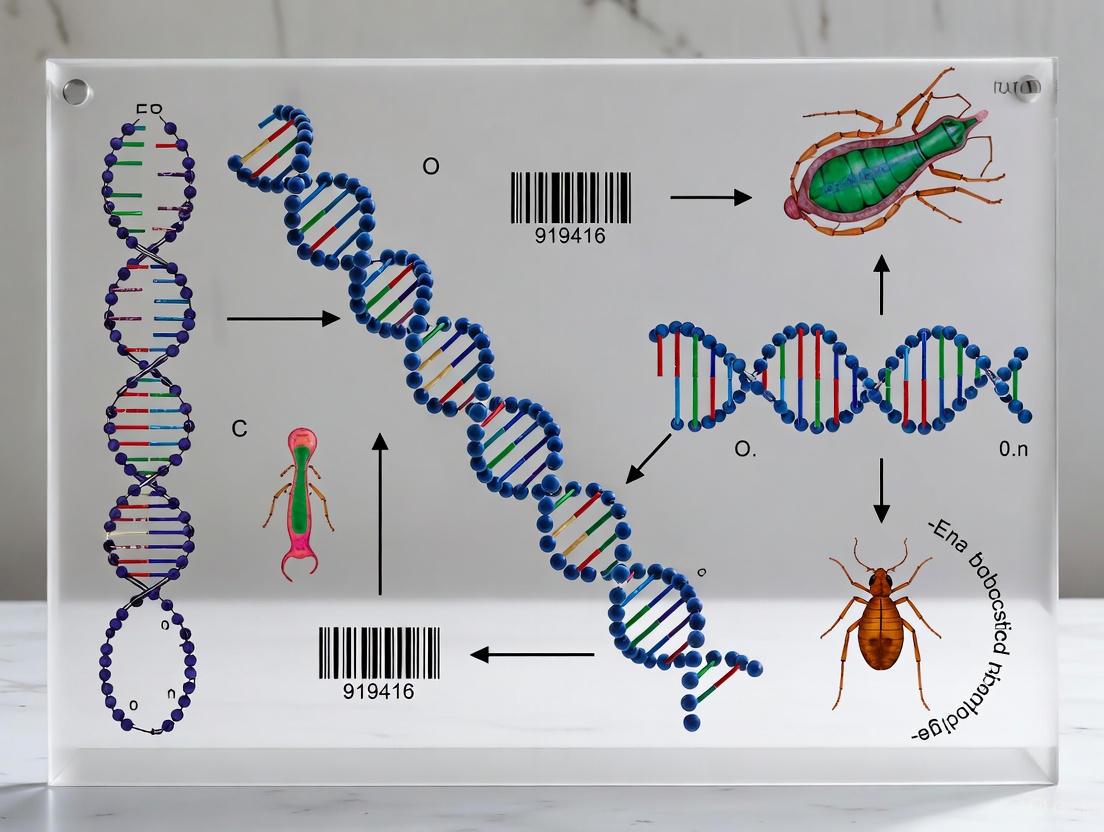
DNA Barcoding for Parasite Surveillance in Arthropod Vectors: Methods, Applications, and Frontiers in Disease Control
This article provides a comprehensive resource for researchers and scientists on the application of DNA barcoding for identifying parasites within arthropod vectors.
DNA Barcoding for Parasite Surveillance in Arthropod Vectors: Methods, Applications, and Frontiers in Disease Control
Abstract
This article provides a comprehensive resource for researchers and scientists on the application of DNA barcoding for identifying parasites within arthropod vectors. It covers the foundational principles of using the cytochrome c oxidase subunit I (COI) gene for species discrimination, explores advanced methodological workflows for field and laboratory settings, and addresses common troubleshooting scenarios for low-quality samples. By comparing the performance of DNA barcoding with other identification techniques and validating its accuracy, this review synthesizes current best practices. The content is designed to support efforts in vector-borne disease surveillance, drug discovery, and the development of targeted vector control strategies by enhancing the precision and efficiency of parasite detection in complex arthropod hosts.
The Foundation of Vector-Parasite Surveillance: Core Principles and Genetic Targets
DNA barcoding is a molecular method that uses a short, standardized genetic marker to identify biological specimens and assign them to a known species [1]. For animals, the most common barcode region is a 648-base pair fragment of the mitochondrial Cytochrome c Oxidase Subunit I (COI) gene [2] [3]. This genomic region provides sufficient sequence variation to discriminate between species due to its model of molecular evolution, which offers better resolution for deeper taxonomic affinities than other molecular markers [2]. The emergence of international initiatives like the Consortium for the Barcode of Life has been crucial in establishing standardized practices and expanding reference libraries, making DNA barcoding an invaluable tool for biodiversity research [2].
The fundamental principle behind DNA barcoding is the presence of a "barcoding gap"—the difference between intraspecific genetic variation and interspecific genetic divergence [1]. When the COI sequence from an unknown specimen is obtained, it can be compared to a curated reference database of known species, such as the Barcode of Life Data System (BOLD), facilitating rapid and reliable species-level identification [2] [3]. This approach has revolutionized taxonomy and biodiversity assessment, particularly for diverse and morphologically cryptic groups like arthropods.
DNA Barcoding in Vector-Borne Disease Ecology
Relevance and Applications
Vector-borne diseases account for approximately 17% of all infectious diseases globally, resulting in more than 700,000 deaths annually [4]. Arthropod vectors, particularly mosquitoes, are responsible for transmitting pathogens that cause malaria, dengue, chikungunya, Zika, West Nile virus, and other diseases with significant public health impacts [2] [4]. Understanding vector-host interactions and pathogen transmission cycles is crucial for developing effective control strategies, and DNA barcoding has emerged as a powerful tool to elucidate these complex ecological relationships.
Key applications of DNA barcoding in vector-borne disease ecology include:
- Vector Identification: Accurate species identification of arthropod vectors, including cryptic species complexes [4] [5].
- Host Blood Meal Analysis: Identification of vertebrate hosts from arthropod blood meals to understand feeding preferences and disease transmission dynamics [2] [6].
- Pathogen Detection: Surveillance of pathogens within vector populations [6].
- Biodiversity Monitoring: Documenting changes in vector communities in response to environmental factors and climate change [3] [7].
Technical Advances and Methodologies
The field has evolved from traditional DNA barcoding of individual specimens to high-throughput approaches like DNA metabarcoding, which enables the simultaneous species identification of multiple specimens in a bulk sample [4]. Next-Generation Sequencing (NGS) platforms, including Illumina and portable MinION sequencers, have dramatically increased processing capacity while reducing costs [6] [4]. These technological advances allow researchers to process large-scale vector surveillance samples efficiently, providing critical data for public health interventions.
*dot DNA barcoding workflow for vector ecology { graph [bgcolor=transparent] node [shape=rectangle style=filled fillcolor="#F1F3F4" fontcolor="#202124" fontname=Arial] edge [color="#5F6368" fontcolor="#5F6368" fontname=Arial]
} Figure 1: Generalized DNA barcoding workflow for vector-borne disease ecology studies.
Key Research Applications and Quantitative Findings
Host Blood Meal Analysis
Identifying the vertebrate hosts of blood-feeding arthropods is essential for understanding disease transmission cycles. A study in southwestern Spain demonstrated the effectiveness of DNA barcoding for this application, using a eukaryote-universal forward primer and a vertebrate-specific reverse primer to selectively amplify 758 bp of the vertebrate mitochondrial COI gene from arthropod blood meals [2]. This method successfully identified up to 40 vertebrate hosts across 16 mammalian, 23 avian, and one reptilian species from various vector species including mosquitoes, ticks, sandflies, and biting bugs [2].
Table 1: Vertebrate hosts identified from arthropod blood meals using DNA barcoding in a Spanish study [2]
| Vector Species | Mammalian Hosts Identified | Avian Hosts Identified |
|---|---|---|
| Culex pipiens | Homo sapiens, Herpestes ichneumon, Felis catus, Canis familiaris | Passer domesticus, Turdus merula, Streptopelia decaocto, Galerida cristata, Sturnus vulgaris, Cairina moschata, Grus grus, Sylvia melanocephala, Alectoris rufa |
| Culex theileri | Bos taurus, Cervus elaphus, Dama dama, Equus caballus, Homo sapiens, Lepus granatensis, Oryctolagus cuniculus, Sus scrofa | Bubulcus ibis, Meleagris gallopavo |
| Anopheles atroparvus | Bos taurus, Oryctolagus cuniculus | - |
| Culex perexiguus | Rattus norvegicus, Canis familiaris | Alectoris rufa, Streptopelia decaocto |
Vector Surveillance and Diversity Assessment
DNA barcoding has revealed remarkable arthropod diversity in various ecosystems, providing baseline data crucial for monitoring changes in vector communities. In the southern Atlantic Forest, a comprehensive survey using Malaise traps and DNA barcoding recorded 8,651 Barcode Index Number (BIN) clusters (used as a proxy for species) from 75,500 arthropods, with nearly 81% representing first records for the database [3]. This highlights both the high diversity and the limited prior knowledge of arthropods in this biodiversity hotspot.
In the Arctic, a DNA barcoding survey in the Ikaluktutiak (Cambridge Bay) area documented 1,264 BINs from terrestrial arthropods, establishing an important baseline for monitoring climate change impacts on arthropod communities [7]. The study also evaluated sampling methods, finding that yellow pan traps captured 62% of the total BIN diversity, while complementing with soil and leaf litter sifting increased coverage to 74.6% [7].
Methodological Comparisons for Vector Surveillance
A 2024 study directly compared MinION nanopore sequencing against Illumina MiSeq for metabarcoding mosquito bulk samples [4]. The results showed 93% congruence in mosquito species-level identifications between the two platforms, demonstrating the reliability of portable sequencing technologies for vector surveillance [4]. The study also found that CO₂ gas cylinders outperformed biogenic CO₂ sources by two-fold in trapping efficiency, providing valuable insights for optimizing surveillance protocols [4].
Table 2: Comparison of sequencing platforms for mosquito metabarcoding [4]
| Parameter | MinION Nanopore Sequencing | Illumina MiSeq Sequencing |
|---|---|---|
| Platform Portability | High (USB-sized device) | Low (Benchtop instrument) |
| Sequencing Run Time | Real-time data generation; faster turnaround | Longer turnaround times (weeks to months) |
| Cost Considerations | Becoming more affordable; in-house sequencing feasible | Often requires external sequencing services |
| Sequence Accuracy | Improving with newer chemistries and flow cells | Historically higher accuracy |
| Species Identification Congruence | 93% overlap with Illumina platform | Reference standard for comparison |
Detailed Experimental Protocols
DNA Barcoding Protocol for Arthropods
This protocol provides a standardized method for DNA extraction and COI amplification from small arthropods, such as mosquitoes and ticks [8].
Sample Preparation and DNA Extraction
- Sample Preparation: Using clean, sterile forceps, remove one leg from the specimen (for small insects) or dissect a small tissue section. Return the remainder of the specimen to the freezer for voucher preservation. Air-dry the sample for 5-10 minutes to remove residual ethanol.
- Cell Lysis: Transfer the tissue to a 1.5 mL tube containing 250 µL of Guanidine Hydrochloride (6M). Grind the sample with a sterile pestle until broken into tiny pieces. Incubate the tube in a 65°C water bath for 10 minutes. Centrifuge at maximum speed for 1 minute to pellet debris.
- DNA Binding: Transfer 150 µL of supernatant to a clean 1.5 mL tube. Add 3 µL of silica resin, mix by pipetting, and incubate for 5 minutes in a 57°C water bath. Centrifuge for 30 seconds at maximum speed and carefully remove the supernatant without disturbing the pellet.
- Washing: Add 500 µL of ice-cold wash buffer to the pellet and resuspend the silica resin by pipetting. Centrifuge for 30 seconds and remove the supernatant. Repeat this wash step once.
- DNA Elution: Add 100 µL of molecular grade water to the silica resin and mix by pipetting. Incubate at 57°C for 5 minutes. Centrifuge for 30 seconds, then transfer 90 µL of the supernatant to a clean tube, avoiding the pellet.
PCR Amplification of COI Gene
- Reaction Setup: For each DNA sample, prepare a PCR mixture containing:
- 32 µL molecular grade water
- 1.5 µL forward primer LCO1490 (10 µM: GGTCAACAAATCATAAAGATATTGG)
- 1.5 µL reverse primer HCO2198 (10 µM: TAAACTTCAGGGTGACCAAAAAATCA)
- 5 µL template DNA
- 10 µL PCR master mix
- Touchdown PCR Conditions:
- Initial denaturation: 95°C for 30 seconds
- 8 cycles of touchdown annealing: 95°C for 30 seconds, 60-52°C for 30 seconds (decreasing 1°C per cycle), 72°C for 45 seconds
- 28 additional cycles with annealing at 52°C for 30 seconds
- Final extension: 72°C for 5 minutes
- PCR Product Verification: Verify successful amplification using gel electrophoresis before proceeding to sequencing.
Vertebrate Host Identification from Blood Meals
This specialized protocol enables identification of vertebrate hosts from arthropod blood meals [2].
Primer Design and PCR Amplification
- Primer Selection: Use vertebrate-specific primers targeting the COI gene:
- Forward primer: M13BC-FW (eukaryote-universal)
- Reverse primer: BCV-RV1 (vertebrate-specific)
- Primary PCR: Perform the first PCR reaction with primers M13BC-FW and BCV-RV1.
- Nested PCR (if needed): For samples with low DNA concentration, perform a nested PCR using M13 and BCV-RV2 primers to increase sensitivity and specificity.
Sequence Analysis and Host Identification
- Sequencing: Purify PCR products and sequence using Sanger sequencing or next-generation sequencing platforms.
- Bioinformatic Analysis: Compare resulting sequences to reference databases using the Barcode of Life Data Systems (BOLD) platform for species identification.
- Mixed Blood Meal Analysis: Inspect sequencing electropherograms for double peaks or sequence heterogeneity that may indicate multiple host species in a single blood meal.
Metabarcoding of Bulk Mosquito Samples
This protocol uses high-throughput sequencing for large-scale vector surveillance [4].
Sample Collection and Processing
- Trap Deployment: Collect mosquitoes using BG-Sentinel traps or similar methods. Compare CO₂ sources (gas cylinders vs. biogenic sources) for trapping efficiency.
- Specimen Storage: Test different preservation methods (cold storage alone vs. ethanol preservation) to optimize DNA recovery.
- Tissue Processing: For consistent biomass representation across specimens, consider using only mosquito heads for DNA extraction to minimize size variation effects.
Library Preparation and Sequencing
- DNA Extraction: Use silica-based extraction methods or commercial kits for consistent DNA yield from bulk samples.
- PCR Amplification: Amplify COI mini-barcodes using metazoan-universal primers suitable for short-read sequencing platforms.
- Library Preparation: Prepare sequencing libraries following manufacturer protocols for either Illumina or MinION platforms.
- Sequencing: Run sequences on the chosen platform. For MinION, perform real-time basecalling and analysis.
Bioinformatic Analysis
- Data Processing: Use standardized pipelines like VecTreeID for sequence similarity assessment (BLAST) and evolutionary placement algorithms (EPA-ng) for taxonomic assignments [6].
- Taxonomic Identification: Compare sequences to curated reference libraries of locally relevant mosquito species identified by expert taxonomists.
- Quality Control: Implement strict thresholds for species assignments and account for potential misidentifications in public databases through manual verification.
*dot Metabarcoding bulk samples { graph [bgcolor=transparent] node [shape=rectangle style=filled fillcolor="#F1F3F4" fontcolor="#202124" fontname=Arial] edge [color="#5F6368" fontcolor="#5F6368" fontname=Arial]
} Figure 2: Metabarcoding workflow for bulk mosquito sample analysis.
The Scientist's Toolkit: Essential Research Reagents
Table 3: Essential reagents and materials for DNA barcoding in vector research
| Reagent/Material | Function | Examples/Specifications |
|---|---|---|
| Guanidine Hydrochloride (6M) | Cell lysis and nucleic acid protection | Carolina Biological Supply #C33427 [8] |
| Silica Resin | DNA binding and purification | Carolina Biological Supply #C33426 [8] |
| Wash Buffer | Removing impurities during DNA purification | Ice-cold; Carolina Biological Supply #C33428 [8] |
| PCR Master Mix | Enzymatic amplification of target DNA | Contains DNA polymerase, dNTPs, buffers; EZ PCR Master Mix 5X [8] |
| COI Primers | Target-specific amplification | LCO1490/HCO2198 for arthropods; vertebrate-specific primers for blood meal analysis [2] [8] |
| DNA Sequencing Kits | Platform-specific sequencing | Illumina chemistry kits; MinION flow cells and sequencing kits [4] |
| Reference Databases | Species identification | BOLD Systems; NCBI GenBank; curated local libraries [2] [4] |
DNA barcoding has transformed approaches to vector-borne disease ecology by providing reliable, high-throughput methods for species identification. The technology enables researchers to accurately identify arthropod vectors, determine their vertebrate hosts, detect pathogens, and monitor changes in vector communities at scales not previously possible. As sequencing technologies continue to advance and become more accessible, DNA barcoding will play an increasingly vital role in global efforts to understand and control vector-borne diseases. The standardized protocols and applications outlined in this article provide a foundation for researchers to implement these powerful tools in their vector surveillance and ecological studies.
The Cytochrome c Oxidase subunit I (COI) gene, a mitochondrial marker, has been established as the core of DNA barcoding for animal species identification. Its properties as an essential gene for cellular respiration, presence in most eukaryotes, high copy number per cell, and a mutation rate that is typically slow enough for consistency within a species yet fast enough for discrimination between species, make it a powerful molecular tool [9]. Within parasitology and vector research, the COI gene provides a standardized, sequence-based method to accurately identify arthropod vectors, the vertebrate hosts they feed on, and the parasites they carry, thereby disentangling complex transmission networks [10] [11]. This Application Note details the experimental protocols and applications of COI DNA barcoding within the context of a broader thesis on identifying parasites in arthropod vectors.
Application in Parasite and Vector Research
The COI gene is instrumental in addressing key challenges in the ecology of vector-borne diseases, offering high-resolution identification where traditional morphological methods fall short.
Discriminating Parasite Species and Genotypes
COI barcoding effectively differentiates between closely related parasite species and intraspecific genetic variants. A study on Trypanosoma cruzi, the agent of Chagas disease, demonstrated that the COI gene could identify the main discrete typing units (DTUs) - TcI, TcII, TcIII, and TcIV - and distinguish T. cruzi from closely related species like Trypanosoma cruzi marinkellei, Trypanosoma dionisii, and Trypanosoma rangeli [12]. The analysis of single nucleotide polymorphisms (SNPs) in the COI sequence was particularly informative for DTU differentiation. When combined with the nuclear gene glucose-6-phosphate isomerase (GPI), COI sequencing helped evaluate the occurrence of mitochondrial introgression and hybrid genotypes, providing a more comprehensive understanding of the parasite's population structure [12].
Identifying Vertebrate Hosts from Vector Bloodmeals
Understanding vector-host interactions is vital for mapping disease transmission cycles. A universal DNA barcoding method using COI has been developed to identify the vertebrate source of arthropod bloodmeals [11]. This method employs a eukaryote-universal forward primer and a vertebrate-specific reverse primer to selectively amplify a 758-base pair (bp) fragment of the vertebrate mitochondrial COI gene. This protocol has been successfully validated on bloodmeals from mosquitoes, culicoids, phlebotomine sand flies, sucking bugs, and ticks, identifying hosts across Mammalia, Aves, and Reptilia. The method is sensitive enough to resolve mixed bloodmeals through the inspection of direct sequencing electropherograms [11].
Delimiting Arthropod Vector Species
Morphological identification of arthropod vectors can be hampered by cryptic diversity, phenotypic plasticity, and damage to specimens. COI barcoding has proven highly effective in delimiting vector species. For example, in Neotropical phlebotomine sand flies, COI barcoding correctly associated isomorphic females with morphologically identified males and uncovered significant cryptic diversity within several species, including Psychodopygus panamensis and Pintomyia evansi [13]. The method showed a clear barcode gap for most species, where the maximum intraspecific genetic distance was lower than the minimum interspecific distance to the nearest neighbor, confirming its utility for species identification.
Table 1: Performance of COI DNA Barcoding in Various Research Applications
| Application Focus | Target Organisms | Key Outcome | Reference |
|---|---|---|---|
| Parasite Discrimination | Trypanosoma cruzi DTUs | COI successfully identified main DTUs (TcI-TcIV) and distinguished T. cruzi from related species. | [12] |
| Host Identification | Vertebrate hosts in mosquito, tick, and sand fly bloodmeals | A universal primer set identified up to 40 vertebrate host species from various blood-feeding arthropods. | [11] |
| Vector Delimitation | Neotropical phlebotomine sand flies | COI associated isomorphic females with males and detected cryptic diversity in multiple species; >97% identification success. | [13] |
| Larval Fish Identification | Larval fish in Ing River, Thailand | 76 of 78 larval samples were identified to 30 species, aiding in spawning ground conservation. | [14] |
Critical Experimental Protocols
Workflow for COI DNA Barcoding
The general workflow for a COI barcoding study, from specimen collection to data analysis, is summarized below. This workflow forms the backbone of the specific protocols detailed in the subsequent sections.
Protocol 1: Universal Identification of Vertebrate Hosts from Bloodmeals
This protocol is adapted from a study designed to identify vertebrate hosts from the bloodmeals of various arthropods [11].
- Sample Preparation: Engorged arthropods (e.g., mosquitoes, ticks) are collected and stored in 70% ethanol or frozen at -20°C. The abdomen of the engorged arthropod is used for DNA extraction.
- DNA Extraction: Use a high salt concentration protocol or commercial kit to extract total DNA from the dissected abdomen.
- PCR Amplification:
- Primers: Use the vertebrate-specific primer set.
- Forward: M13BC-FW (5'-TGT AAA ACG ACG GCC AGT GGT CAA CAA ATC ATA AAG ATA TTG G-3')
- Reverse: BCV-RV1 (5'-ACG GAA TCA GAA TCA CGT AGA T-3')
- First PCR: Perform the initial amplification with primers M13BC-FW and BCV-RV1.
- Nested PCR (if required): For samples with low DNA quantity or quality (e.g., digested bloodmeals), a nested PCR significantly increases success. Use the M13 forward primer (5'-TGT AAA ACG ACG GCC AGT-3') and a nested reverse primer BCV-RV2 (5'-ACG GAA TCA GAA TCA CGT AGA T-3') with 1 µL of the first PCR product as a template.
- PCR Conditions: Initial denaturation at 94°C for 3 min; followed by 35 cycles of 94°C for 30 s, 52°C for 40 s, and 72°C for 1 min; with a final extension at 72°C for 10 min.
- Primers: Use the vertebrate-specific primer set.
- Sequencing and Analysis: Purify PCR products and perform Sanger sequencing in both directions. Compare the resulting sequences to reference databases like the Barcode of Life Data Systems (BOLD) or GenBank for species identification. A sequence similarity of ≥99% typically confirms species-level identification.
Table 2: Key Research Reagent Solutions for COI Barcoding
| Reagent / Material | Function / Application | Example / Notes |
|---|---|---|
| LCO1490 / HCO2198 Primers | Amplification of the ~658 bp "Folmer region" of COI. | Standard "universal" invertebrate primers; may require modification for specific taxa. [13] |
| Vertebrate-Specific Primer Set | Selective amplification of vertebrate COI from mixed bloodmeals. | Preferentially amplifies host DNA over vector DNA. [11] |
| I3-M11 Primer Sets (e.g., JB3-JB5) | Amplification of an alternative COI partition in nematodes. | Used when universal Folmer primers fail. [15] |
| BOLD Systems Database | Reference database for sequence identification and data management. | Contains taxonomically verified COI barcodes. [11] |
| High-Salt DNA Extraction Protocol | Efficient DNA extraction from small or degraded samples. | Suitable for single arthropods or bloodmeal remnants. [13] [11] |
Protocol 2: Discriminating Parasite Genotypes
This protocol is derived from a study that successfully used COI to discriminate Trypanosoma cruzi DTUs [12].
- Parasite DNA Source: DNA is extracted from parasite cultures, infected host tissues, or vector guts.
- PCR Amplification:
- Target: A fragment of the COI gene.
- Primers: The study does not specify the exact primers used but highlights that careful primer design is crucial for specific amplification from trypanosomatids.
- PCR Conditions: Standard conditions for mitochondrial gene amplification are used, often requiring optimization for the specific parasite group.
- Sequence Analysis:
- Phylogenetic Analysis: Reconstruct phylogenetic trees using methods like Neighbor-Joining, Maximum Likelihood, or Bayesian Inference to visualize the relationships between sequences and assign them to known DTUs or species.
- Species Delimitation: Use analytical methods like Automatic Barcode Gap Discovery (ABGD) and Poisson Tree Processes (PTP) to aid in species delimitation by identifying the "barcoding gap."
- Single Nucleotide Polymorphism (SNP) Detection: Manually inspect alignments or use software to identify informative SNPs that are diagnostic for specific parasite genotypes.
The Scientist's Toolkit: Research Reagent Solutions
Table 3: Essential Materials and Databases for COI Barcoding Workflows
| Category | Item | Function and Importance |
|---|---|---|
| Wet Lab Materials | Single-use sterile pestles | Homogenizing small tissue samples (e.g., insect legs, parasite material). |
| Proteinase K | Critical for lysing cells and degrading nucleases during DNA extraction. | |
| PCR reagents (dNTPs, Taq polymerase, buffer) | Essential components for the polymerase chain reaction. | |
| Agarose gel electrophoresis equipment | To visualize and confirm successful PCR amplification. | |
| Bioinformatics Tools | Sequence Alignment Software (e.g., MEGA, BioEdit) | For editing raw sequence data and creating multiple sequence alignments. [12] [13] |
| BLAST (NCBI) / BOLD Identification Engine | For comparing unknown sequences against massive public reference databases. [14] | |
| Phylogenetic Analysis Software (e.g., MEGA, MrBayes) | For constructing trees to visualize relationships and test species boundaries. [12] |
Considerations and Limitations
While powerful, the COI barcoding approach has limitations that researchers must consider:
- Primer Bias: The COI gene does not contain perfectly conserved regions for primer binding across all animal taxa. Primer-template mismatches can lead to unpredictable and inefficient amplification, causing false negatives and biasing the representation of species in mixed DNA samples (metabarcoding) [16].
- Reference Database Gaps: Accurate identification depends on comprehensive reference databases. The absence of a sequence from a particular species in these databases can prevent its identification, as was the case for a larval fish species (Rasbora sp. and Monopterus sp.) in a Thai river study [14].
- Nuclear Mitochondrial Pseudogenes (numts): These are non-functional copies of mitochondrial DNA that have been transferred to the nuclear genome. Their inadvertent amplification can lead to incorrect sequence data and an overestimation of diversity [17] [15].
- Intraspecific vs. Interspecific Variation: In some groups, such as certain sand fly species, the minimum interspecific genetic distance can be very low (<3%), making it challenging to delineate closely related species without complementary data [13]. For organisms like plants and fungi, COI is not a suitable barcode, and other markers must be used [9].
The relationships between the core concepts, applications, and necessary quality controls in a COI barcoding study can be visualized as follows:
DNA barcoding has revolutionized the identification of parasites and their arthropod vectors, offering a powerful tool for understanding disease transmission dynamics. This molecular technique, typically targeting a 658-base pair region of the mitochondrial cytochrome c oxidase subunit I (COI) gene, provides a standardized method for species identification and discovery [18] [2]. For researchers investigating parasitic diseases transmitted by arthropod vectors, accurate species identification is crucial for predicting transmission patterns, understanding ecological parameters, and developing targeted control strategies [18] [2]. The utility of DNA barcoding in medical parasitology is well-established, with studies demonstrating it provides highly accurate species identification in 94-95% of cases, surpassing the limitations of traditional morphological methods alone [18] [19]. However, the reliability of this powerful tool is fundamentally constrained by a critical factor: the completeness and quality of reference libraries against which unknown sequences are compared [20]. Significant taxonomic gaps in these libraries undermine their diagnostic utility, presenting a substantial obstacle to advancing research on parasitic diseases and their vectors.
The Current State of Reference Libraries: A Quantitative Gap Analysis
Coverage Disparities Across Taxa and Regions
The coverage of DNA barcode reference libraries is markedly uneven across different taxonomic groups and geographic regions. An analysis of medically important parasites and vectors revealed that barcodes were available for only 43% of 1,403 species affecting human health, despite encouraging coverage of over half of 429 species considered of greater medical importance [18]. Similar disparities are evident in other ecosystems; for North Sea macrobenthos, a curated DNA reference library covers approximately 29% of known species, with phylum-level coverage varying dramatically from 93% for Echinodermata to just 8% for Bryozoa [21]. Marine data further highlights these inconsistencies, revealing significant barcode deficiencies in the south temperate region of the Western and Central Pacific Ocean and for specific phyla including Porifera, Bryozoa, and Platyhelminthes [20].
Table 1: DNA Barcode Coverage Across Different Taxonomic Groups
| Taxonomic Group | Number of Species | Barcode Coverage | Key References |
|---|---|---|---|
| Medically Important Parasites & Vectors | 1,403 | 43% | [18] |
| North Sea Macrobenthos | 2,514 | 29% | [21] |
| North Sea Echinodermata | 84 | 93% | [21] |
| North Sea Bryozoa | Not Specified | 8% | [21] |
| Neotropical Sand Flies | 555 | ~25% | [13] |
Database-Specific Limitations and Quality Concerns
The two primary repositories for DNA barcode sequences—the Barcode of Life Data System (BOLD) and the National Center for Biotechnology Information (NCBI)—each present distinct advantages and limitations. Comparative analyses reveal that NCBI generally exhibits higher barcode coverage but lower sequence quality compared to BOLD [20]. Both databases contend with quality issues including over- or under-represented species, short sequences, ambiguous nucleotides, incomplete taxonomic information, conflicting records, high intraspecific distances, and low interspecific distances, potentially resulting from contamination, cryptic species, sequencing errors, or inconsistent taxonomic assignment [20]. The BOLD system incorporates a valuable quality control feature through its Barcode Index Number (BIN) system, which automatically clusters sequences into operational taxonomic units (OTUs) that typically correspond to species-level groupings, thereby facilitating species delimitation and highlighting potential cryptic diversity [20] [7].
Table 2: Comparison of Major DNA Barcode Databases
| Database | Coverage | Sequence Quality | Key Features | Primary Limitations |
|---|---|---|---|---|
| BOLD Systems | Lower public coverage | Higher quality, curated | BIN system for OTU clustering, voucher specimen standards, strict metadata requirements | Limited immediate availability of submissions due to curation protocols |
| NCBI GenBank | Higher coverage | Variable quality | Extensive sequence collection, rapid submission | Redundancies, inconsistent metadata, less robust validation systems |
Implications for Parasite and Vector Research: Critical Consequences of Incomplete Libraries
Incomplete reference libraries directly impact parasite and vector research in several critical ways. The limited species coverage impedes the accurate identification of disease vectors and parasites, potentially leading to misdiagnosis and flawed epidemiological data [19]. This limitation is particularly problematic in biodiversity-rich regions where many species remain uncharacterized, and in clinical settings where precise identification informs treatment decisions [19]. Furthermore, the lack of comprehensive reference data hinders the detection of cryptic species complexes, which are prevalent among both parasites and vectors [13]. For example, studies on Neotropical phlebotomine sand flies have revealed significant cryptic diversity within morphologically similar species, with maximum intraspecific genetic distances ranging up to 8.92% for some taxa [13]. Such undetected cryptic diversity can obscure important differences in vector competence, host preference, and insecticide resistance, fundamentally undermining the effectiveness of disease control programs.
Building Comprehensive Libraries: Standardized Protocols and Community Engagement
DNA Barcoding Protocol for Arthropod Vectors
Standardized laboratory protocols are essential for generating high-quality, comparable barcode data. The following protocol is adapted for arthropod vectors, such as mosquitoes, sand flies, and ticks, which are relevant to parasitic disease transmission:
Sample Preparation:
- Dissect a small tissue sample (typically one leg) from the specimen and return the remainder to long-term storage.
- Air-dry the tissue for 5-10 minutes to remove residual ethanol.
- Transfer the tissue to a 1.5 mL tube containing 250 µL of Guanidine Hydrochloride [8].
DNA Extraction and Purification:
- Grind the tissue using a sterile pestle to disrupt cells.
- Incubate the sample at 65°C for 10 minutes to complete lysis.
- Centrifuge at maximum speed for 1 minute to pellet debris.
- Transfer 150 µL of supernatant to a new tube.
- Add 3 µL of silica resin to bind DNA and incubate at 57°C for 5 minutes.
- Pellet resin by centrifuging for 30 seconds and remove supernatant.
- Wash resin twice with 500 µL of ice-cold wash buffer, centrifuging and removing supernatant each time.
- Elute DNA by adding 100 µL of molecular grade water, incubating at 57°C for 5 minutes, centrifuging, and transferring the supernatant to a new tube [8].
PCR Amplification of COI Gene:
- Prepare a PCR master mix for each sample containing:
- 32 µL molecular grade water
- 1.5 µL forward primer LCO1490 (10 µM)
- 1.5 µL reverse primer HCO2198 (10 µM)
- 5 µL template DNA
- 10 µL PCR master mix
- Perform touchdown PCR with the following cycling parameters:
Sequencing and Data Management:
- Verify PCR success using gel electrophoresis.
- Sequence PCR products in both directions.
- Submit sequences to both BOLD and NCBI databases with complete metadata including collection location, date, and voucher specimen details [21] [13].
Workflow for Building Curated Reference Libraries
Diagram Title: Workflow for Building Curated DNA Barcode Libraries
The workflow illustrated above outlines a systematic approach for constructing curated DNA barcode reference libraries, emphasizing the critical steps from specimen collection to data publication. This process highlights the importance of integrating morphological identification with molecular data and implementing rigorous quality control measures through the BIN system available on BOLD [21].
Essential Research Reagents and Materials for DNA Barcoding
Table 3: Essential Research Reagents for DNA Barcoding of Parasites and Vectors
| Reagent/Material | Function | Application Notes |
|---|---|---|
| Guanidine Hydrochloride | Cell lysis and nucleic acid protection | Effective for breaking down tissues and inactivating nucleases [8] |
| Silica Resin | DNA binding and purification | Selective binding of DNA in presence of chaotropic salts [8] |
| Ice-cold Wash Buffer | Removal of contaminants and salts | Maintains DNA binding while removing impurities [8] |
| Molecular Grade Water | DNA elution and reagent preparation | Nuclease-free to prevent DNA degradation [8] |
| LCO1490/HCO2198 Primers | Amplification of COI barcode region | Universal primers for a 658 bp fragment of COI gene [8] [13] |
| PCR Master Mix | DNA amplification | Contains DNA polymerase, dNTPs, and buffer components [8] |
Addressing taxonomic gaps in DNA barcode reference libraries requires a coordinated, multinational effort that combines standardized laboratory protocols, rigorous data curation, and community engagement. The development of comprehensive libraries for parasites and their vectors will significantly enhance our capacity to monitor and respond to emerging infectious diseases, track the spread of insecticide resistance, and understand the complex ecological relationships that underpin disease transmission cycles [18] [22]. As climate change and globalization continue to alter the distribution of both vectors and parasites, building robust DNA barcode reference libraries becomes increasingly urgent for effective disease surveillance and control [18] [7]. By adopting standardized protocols, promoting data sharing, and targeting sequencing efforts toward underrepresented taxa and regions, the research community can transform DNA barcoding from a promising tool into a reliable resource for tackling the ongoing challenges posed by vector-borne parasitic diseases.
Taxonomy, the scientific discipline of species classification, is fundamental to all organismic research, including the study of arthropod vectors and the parasites they transmit [23]. However, traditional morphology-based taxonomy faces significant challenges when dealing with cryptic species complexes—groups of morphologically identical but genetically distinct species. This is particularly problematic in medical entomology and parasitology, where different cryptic species may exhibit varying vector competencies, host preferences, and parasite susceptibilities, leading to important implications for disease control strategies [24] [25]. The limitations of morphological identification are compounded in arthropods like ants and mosquitoes, where factors including phenotypic plasticity, adaptive convergence, and developmental dimorphism weaken the correlation between morphological traits and phylogenetic relationships [23].
DNA barcoding, a method using short genetic markers for species identification, has emerged as a powerful tool to overcome these challenges [23] [24]. Since its proposal in 2003, this molecular approach has provided taxonomists with an objective, rapid, and accurate method for species delineation that is particularly valuable for characterizing biodiversity in understudied groups and regions [24]. For researchers studying parasite-vector systems, DNA barcoding enables more precise identification of both arthropod vectors and their associated parasites, facilitating a deeper understanding of transmission dynamics and host-pathogen interactions [25] [26]. This Application Note provides detailed protocols and current data on applying DNA barcoding to uncover hidden diversity in arthropod vectors and parasites, with specific focus on practical implementation for research and drug development professionals.
Current Landscape of DNA Barcoding Data
The cytochrome c oxidase subunit I (COI) gene remains the most prevalent molecular marker for animal DNA barcoding, including arthropods and many parasites [23] [24]. Analysis of current sequence databases reveals both progress and significant gaps in our molecular characterization of these organisms.
Table 1: DNA Barcoding Sequence Analysis for Ants (Hymenoptera: Formicidae)
| Metric | COI Sequences | 28S rRNA Sequences | Cytb Sequences |
|---|---|---|---|
| Total Sequences | 337,887 | 4,560 | 3,509 |
| Species Coverage | 4,317 species | 1,396 species | 623 species |
| Genus Coverage | 270 genera | 304 genera | 73 genera |
| Subfamily Coverage | 15 subfamilies | Information Missing | Information Missing |
| Undetermined Species (sp.) | 32,444 (9.60%) | Information Missing | Information Missing |
| Sequences ≥ Standard Length | 190,880 (67%) | Information Missing | Information Missing |
Data compiled from analysis of NCBI and BOLD databases [23].
As shown in Table 1, molecular data for even well-studied invertebrate groups like ants remains extremely limited, with COI sequences covering only approximately 4,317 species of the over 14,000 described ant species [23]. Furthermore, existing data exhibits significant spatial and taxonomic biases, with sequences from Europe and North America dominating databases (60%), while tropical biodiversity hotspots like China are exceptionally scarce (0.35% of COI sequences) [23]. This spatial bias is particularly problematic for vector-borne disease research, as tropical regions often harbor the greatest diversity of both vectors and parasites.
The length distribution of COI sequences also presents challenges for standardization. While the standard barcode length is 658 base pairs (bp), current data shows extensive variation (72–6,883 bp), with only 67% of sequences meeting or exceeding the standard length [23]. This variation complicates sequence alignment and analysis, highlighting the need for standardized protocols in sequence submission.
DNA Barcoding Workflow for Vector and Parasite Research
The following section outlines comprehensive protocols for implementing DNA barcoding in research on arthropod vectors and their associated parasites.
Field Collection and Specimen Processing
Effective DNA barcoding begins with proper specimen collection and preservation. Collection methods must be tailored to the target species' biology and ecology.
Table 2: Collection Methods for Arthropod Vectors
| Method/Device | Target Organisms | Key Attractants | Applications |
|---|---|---|---|
| BG-Sentinel Trap | Aedes aegypti, Ae. albopictus, other Stegomya subgenus species | CO₂, BG-Lure (human skin odor), visual cues | Dengue vector surveillance; collecting host-seeking females [26] |
| CDC Light Trap | Generalist mosquito species, particularly anophelines | Light (incandescent or LED), CO₂ | Nocturnal mosquito surveillance; collecting unfed females [26] |
| Entomological Aspirator | Adult mosquitoes (both sexes) | Direct collection from resting sites | Vector competence studies; transovarial pathogen detection [26] |
Protocol: Field Collection and Preservation
- Select appropriate collection methods based on research objectives and target species ecology (refer to Table 2).
- Deploy traps in suitable microhabitats, using appropriate attractants to maximize capture efficiency.
- Collect specimens at regular intervals (typically 24 hours) to prevent DNA degradation.
- Preserve specimens immediately in 95-100% ethanol for DNA analysis. Alternatively, freeze at -20°C or lower for long-term storage.
- Record essential metadata including collection date, location (GPS coordinates), habitat type, and collector information.
- Perform preliminary morphological identification to lowest possible taxonomic level before molecular analysis.
Laboratory Processing and DNA Barcoding
Protocol: DNA Extraction, Amplification, and Sequencing
- DNA Extraction
- Select individual specimens or specific tissue (legs, thorax) to preserve voucher specimens.
- Use commercial DNA extraction kits (e.g., DNeasy Blood & Tissue Kit) following manufacturer protocols.
- Validate DNA quality and quantity using spectrophotometry (NanoDrop) or fluorometry (Qubit).
PCR Amplification
- Prepare PCR master mix containing:
- 10-50 ng genomic DNA
- 1X PCR buffer
- 2.5 mM MgCl₂
- 0.2 mM each dNTP
- 0.5 µM each primer (e.g., LCO1490/HCO2198 for COI)
- 1.25 U DNA polymerase
- Apply thermal cycling conditions:
- Initial denaturation: 94°C for 2-4 minutes
- 35-40 cycles of: 94°C for 30-45 seconds, 45-52°C for 30-60 seconds, 72°C for 45-60 seconds
- Final extension: 72°C for 5-10 minutes
- Verify amplification success via agarose gel electrophoresis.
- Prepare PCR master mix containing:
Sequencing and Data Management
- Purify PCR products using enzymatic (ExoSAP-IT) or column-based methods.
- Prepare sequencing reactions using BigDye Terminator kits.
- Perform bidirectional Sanger sequencing on appropriate platform.
- Assemble contigs from forward and reverse sequences, verify base calls, and export consensus sequences in FASTA format.
Data Analysis and Species Delineation
Protocol: Molecular Data Analysis and Species Identification
- Sequence Quality Control
- Trim low-quality bases (typically Phred score <20) from sequence ends.
- Verify absence of stop codons in protein-coding genes to confirm functional sequences.
- Check for contamination using BLAST against non-target organisms.
Sequence Alignment and Dataset Construction
- Perform multiple sequence alignment using MUSCLE or MAFFT algorithms.
- Visually inspect alignments for obvious misalignments or frame shifts.
- Construct datasets including both query sequences and reference sequences from validated databases (BOLD, NCBI).
Genetic Distance Analysis
- Calculate intra-specific and inter-specific distances using Kimura 2-parameter (K2P) model.
- Generate distance matrices for all sequence pairs.
- Assess barcode gap presence – the separation between maximum intra-specific and minimum inter-specific distances.
Phylogenetic Analysis and MOTU Delineation
- Construct phylogenetic trees using maximum likelihood (IQ-TREE) or Bayesian inference (MrBayes) methods.
- Perform 1000 bootstrap replicates to assess node support.
- Apply Molecular Operational Taxonomic Unit (MOTU) delineation methods:
- ASAP (Assemble Species by Automatic Partitioning): Set K2P distance model with maximum intraspecific divergence threshold of 0.05–0.10.
- ABGD (Automatic Barcode Gap Discovery): Use default parameters with relative gap width of 1.5.
- Compare MOTU composition with morphological species assignments to identify potential cryptic diversity.
Essential Research Reagent Solutions
Table 3: Key Research Reagents for DNA Barcoding Studies
| Reagent/Category | Specific Examples | Function/Application |
|---|---|---|
| DNA Extraction Kits | DNeasy Blood & Tissue Kit (Qiagen), Maxwell RSC Blood DNA Kit | High-quality genomic DNA extraction from various specimen types |
| PCR Reagents | AmpliTaq Gold DNA Polymerase, Platinum Taq DNA Polymerase | Robust amplification of barcode regions |
| Universal Primers | LCO1490 (5′-GGTCAACAAATCATAAAGATATTGG-3′)HCO2198 (5′-TAAACTTCAGGGTGACCAAAAAATCA-3′) | Amplification of standard COI barcode region |
| Sequencing Chemistry | BigDye Terminator v3.1 Cycle Sequencing Kit | Sanger sequencing reaction preparation |
| Genetic Markers | COI (cytochrome c oxidase I), ITS2 (internal transcribed spacer 2) | Standard DNA barcodes for animals and parasites |
| Analysis Software | IQ-TREE (phylogenetics), ASAP (species delimitation) | Molecular data analysis and interpretation |
Case Study: DNA Barcoding of Malagasy Ants
A landmark study demonstrating DNA barcoding's power for biodiversity assessment compared traditional morphological taxonomy with sequence-based methods for ants in Madagascar [24]. Researchers surveyed four localities in northeastern Madagascar, collecting ants using standardized methods. The study revealed that:
- Patterns of richness were not significantly different between morphological and molecular methods.
- Sequence-based methods tended to yield greater richness estimates with significantly lower similarity indices between sites.
- MOTUs were highly localized, indicating restricted dispersal and long-term isolation.
- Morphological estimates were consistently more conservative, with some morphospecies containing distinct molecular groups averaging 16% sequence divergence.
This study demonstrated that DNA barcoding could accelerate biodiversity assessment while providing fine-scale resolution of diversity patterns essential for conservation planning in threatened ecosystems [24].
DNA barcoding has proven to be an indispensable tool for unveiling hidden diversity in arthropod vectors and parasites, providing researchers with powerful methods to overcome limitations of morphological identification. The protocols outlined in this Application Note provide a framework for implementing DNA barcoding in vector and parasite research, from field collection through data analysis. As molecular databases continue to expand and methods refine, DNA barcoding will play an increasingly critical role in disease vector surveillance, parasite identification, and understanding the complex interactions that drive pathogen transmission. Future efforts should focus on filling geographical and taxonomic gaps in reference databases, developing standardized protocols for specific vector-parasite systems, and integrating DNA barcoding with other molecular and morphological approaches for comprehensive species characterization.
From Sample to Sequence: A Step-by-Step Guide to Field and Laboratory Protocols
Best Practices for Arthropod Vector Collection, Preservation, and DNA Extraction
Arthropod vectors play a critical role in transmitting pathogens that cause diseases in humans and animals. Accurate species identification through DNA barcoding is fundamental to understanding disease ecology, tracking pathogen life cycles, and developing effective control strategies [2] [18]. This protocol outlines comprehensive best practices for the collection, preservation, and DNA extraction of arthropod vectors, specifically framed within research aimed at DNA barcoding for parasite identification. Implementing standardized methods ensures the generation of high-quality genetic data suitable for robust phylogenetic analysis and reliable molecular identification, which is particularly valuable for monitoring vector populations in the context of changing climate conditions and emerging infectious diseases [27] [7].
Field Collection Techniques
Selecting appropriate collection methods is essential for capturing a representative spectrum of the arthropod vector community. The choice of technique depends on the target species, life stage, habitat, and research objectives.
Passive Trapping Methods
Passive traps are highly effective for collecting flying insects and should be deployed at monitoring sites for extended periods.
- Malaise Traps: Townes-style traps intercept flying insects. Samples are typically collected in bottles filled with 95% ethanol and serviced on a weekly basis [7]. Secure anchoring is critical, as one study reported damage by wildlife; using galvanized aircraft cables with metal pegs and reinforced stones prevented trap collapse [7].
- Pan Traps: Shallow yellow plastic bowls (approximately 9 inches in diameter) are half-filled with soapy water and checked every 48 hours [7]. The two-day catch from each trap is pooled into a single bulk sample. Specimens are strained through a Nitex nylon fabric (50 µm mesh) and transferred to 95% ethanol [7]. This method is particularly efficient, capturing a high percentage of local Barcode Index Number (BIN) diversity.
- Pitfall Traps: Lines of 10 translucent wide-mouth 500 mL plastic cups are installed at 3-meter intervals, half-filled with soapy water, and capped with a steel mesh (e.g., 10 mm) to exclude vertebrate by-catch [7]. The checking schedule and specimen processing are identical to those for pan traps.
Active Collection Methods
Active methods complement passive trapping by targeting specific microhabitats or behaviors.
- Sweep Netting: Effective for collecting vectors from vegetation.
- Soil and Leaf Litter Sifting: Used to collect questing ticks, larvae, and other cryptic arthropods. When combined with yellow pan traps, this method can significantly increase the coverage of total BIN diversity recovered from a site [7].
The table below summarizes the performance of different collection methods based on an Arctic arthropod community survey, providing a guideline for method selection.
Table 1: Efficacy of Different Arthropod Collection Methods in Recovering BIN Diversity
| Collection Method | Key Characteristics | BIN Diversity Recovery | Target Arthropods |
|---|---|---|---|
| Yellow Pan Traps | Passive, soapy water, checked every 48 hours | 62% of total BINs [7] | Flying insects |
| Malaise Traps | Intercepts flight paths, weekly servicing | Specific percentage not isolated in study [7] | Flying insects |
| Pitfall Traps | Ground-level, cup arrays, mesh covers | Specific percentage not isolated in study [7] | Ground-dwelling arthropods |
| Soil & Litter Sifting | Active collection from microhabitats | Increased total coverage to 74.6% when combined with pan traps [7] | Ticks, larvae, cryptic arthropods |
Preservation Protocols
Proper preservation immediately after collection is crucial for maintaining DNA integrity for subsequent barcoding efforts.
- Ethanol Preservation: 95% ethanol is the recommended preservative for DNA analysis. It should be used for all samples collected via Malaise, pan, and pitfall traps [7]. For bulk samples collected in soapy water, specimens must be promptly strained and transferred to 95% ethanol [7].
- Cold Chain Management: While not explicitly detailed in the sources, best practice dictates that preserved samples should be stored cool and protected from direct sunlight during transport from the field to the laboratory. For long-term storage, samples should be kept at -20°C to prevent DNA degradation.
- Specimen Vouchering: Preserving morphological vouchers is a standard and critical practice in DNA barcoding. Specimens should be archived in a designated collection facility, as this allows for taxonomic verification and links molecular data to physical specimens [18].
DNA Extraction and Optimization
The choice of DNA extraction method significantly impacts DNA yield, purity, and its subsequent utility in PCR amplification for DNA barcoding.
Methods for Challenging Specimens
Hard-bodied vectors like ticks present specific challenges due to their chitinous exoskeleton.
- Tick Homogenization: A simple modified method involves optimized homogenization of the tick specimen prior to extraction. This step is critical for breaking down the chitinous exoskeleton and significantly improves both DNA yield and purity [28]. This approach is cost-effective and ideal for resource-limited settings.
- Modified Alkaline Lysis: For ethanol-preserved hard ticks, a Modified Simple Alkaline Lysis method has been developed. This protocol yields DNA with comparable concentration and purity across all life stages (adult, nymph, and larva) and is suitable for PCR amplification of markers like ITS-1 and ITS-2 [28].
- SPRI Bead-Based Extraction: For museum specimens or samples with degraded DNA, a low-cost extraction method using in-house formulated Solid Phase Reversible Immobilisation (SPRI) beads has been optimized. This method is gentle and effective, performing nearly as well as more expensive commercial kits like the Qiagen DNeasy kit, while being unsuitable for the harsh conditions of HotSHOT protocol [29]. The cost is economical, ranging from 4 to 11.6 cents per specimen [29].
Method Comparison and Selection
The table below provides a comparative overview of DNA extraction methods relevant to arthropod vectors.
Table 2: Comparison of DNA Extraction Methods for Arthropod Vectors
| Extraction Method | Key Features | Estimated Cost/Sample | Ideal Use Case |
|---|---|---|---|
| Modified Alkaline Lysis | Cost-effective, no specialized kit required [28] | Very Low | Field applications, resource-limited settings, hard ticks [28] |
| SPRI Bead Protocol | High-throughput, gentle on degraded DNA [29] | $0.04 - $0.116 [29] | Museum specimens, historical samples, diverse insect taxa [29] |
| Commercial Kits (e.g., Qiagen DNeasy) | Standardized, reliable performance [29] | High (relative to other methods) | Standard extractions with sufficient funding [28] [29] |
| HotSHOT Method | Rapid, uses hot NaOH [29] | Very Low | Less effective compared to SPRI and kit methods [29] |
DNA Barcoding and PCR Amplification
Primer Design for Host Identification
A universal DNA barcoding method can be employed to identify vertebrate hosts from vector bloodmeals. This involves using a eukaryote-universal forward primer and a vertebrate-specific reverse primer to selectively amplify a 758 bp fragment of the vertebrate mitochondrial Cytochrome c Oxidase Subunit I (COI) gene [2]. This method is highly specific and can resolve mixed bloodmeals by analyzing direct sequencing electropherograms [2].
PCR Amplification and Sequencing
The extracted DNA is quantified and used as a template for PCR amplification of standard molecular markers.
- Common Markers: For tick identification and phylogenetic studies, the internal transcribed spacer regions ITS-1 and ITS-2 are commonly amplified and sequenced [28].
- Protocol Validation: The vertebrate-specific COI primer set should be validated using high-quality control DNA from various vertebrate classes (Mammalia, Aves, Reptilia, Amphibia) and confirmed to fail amplification with non-engorged arthropod DNA [2]. For samples with low DNA concentration, a nested PCR protocol can significantly increase success rates [2].
- Sequence Analysis: Amplified products are sequenced, and the resulting sequences are compared against databases like the Barcode of Life Data System (BOLD) for species identification and phylogenetic analysis [2] [7].
The Scientist's Toolkit: Research Reagent Solutions
Table 3: Essential Reagents and Materials for Vector DNA Barcoding Research
| Reagent/Material | Function/Application | Specification Notes |
|---|---|---|
| 95% Ethanol | Specimen preservation and DNA storage [7] | Preferred concentration for long-term DNA integrity. |
| EDTA Blood Collection Tubes | Collection of vertebrate host blood for pathogen detection [30] | K3 EDTA tubes prevent coagulation for downstream DNA extraction. |
| Nitex Nylon Fabric | Straining specimens from soapy water in pan/pitfall traps [7] | 50 µm mesh size is effective for retaining small arthropods. |
| Wright-Giemsa Stain | Microscopic examination of blood smears for pathogen screening [30] | Used for morphological identification of blood parasites. |
| Solid Phase Reversible Immobilisation (SPRI) Beads | Cost-effective DNA purification from diverse specimens [29] | Can be formulated in-house for large-scale, low-cost studies. |
| Novel UTR Sequences | mRNA sequence optimization for vaccine development [31] | Enhances protein expression in mRNA vaccine platforms. |
| Thiolactone-based Ionizable Lipids | Key component of Lipid Nanoparticles (LNPs) for mRNA vaccine delivery [31] | Determines transfection efficacy and endosomal escape. |
Workflow Visualization
The following diagram illustrates the complete integrated workflow from field collection to data analysis.
Integrated Workflow for Vector DNA Barcoding
Within arthropod vector research, molecular techniques for identifying parasites in vectors are foundational for understanding transmission dynamics of diseases like malaria and other vector-borne illnesses. This document provides detailed application notes and protocols for DNA barcoding, focusing on the critical steps of primer selection and PCR amplification to detect and identify parasite DNA within vector blood meals and tissues. The protocols are framed within a broader thesis on using DNA barcoding to elucidate vector-parasite interactions, enabling targeted disease surveillance and control strategies.
Primer Design and Selection
The selection of appropriate PCR primers is a critical first step that determines the success of downstream DNA barcoding applications. Ideal primers must balance several, often competing, requirements.
Core Principles for Primer Design
Primers for this application should fulfill three main criteria [32]:
- Universal Amplification across Diverse Taxa: The primer binding sites must be conserved across the target taxonomic range (e.g., diverse vertebrates for blood meal analysis or a wide range of parasites) to ensure broad detection capability.
- Avoidance of Non-Target Co-Amplification: Primers should be designed to avoid amplifying DNA from the vector itself (e.g., mosquito or midge DNA) or other non-target organisms (e.g., symbionts). This is achieved by ensuring nucleotide mismatches at the 3' ends between the primer and non-target DNA sequences [32].
- Short Amplicon Length: DNA from blood meals or parasite tissues is often degraded. Shorter amplicons (e.g., 200-400 bp) have a much higher probability of successful amplification than longer ones [33].
Key Primer Sets for Blood Meal and Parasite Analysis
The table below summarizes several primer sets used in vector and parasite research, targeting different genetic markers.
Table 1: Selected Primer Sets for Blood Meal and Parasite Analysis
| Target | Gene | Primer Name | Sequence (5' to 3') | Amplicon Size | Specificity & Application |
|---|---|---|---|---|---|
| Vertebrate Host | COI | ModRepCOIF [32] | TNT TYT CMA CYA ACC ACA AAG A | 244 - 664 bp | Vertebrate universal; avoids mosquito co-amplification. |
| ModRepCOIR [32] | TTC DGG RTG NCC RAA RAA TCA | Universal reverse primer. | |||
| VertCOI7194F [32] | CGM ATR AAY AAY ATR AGC TTC TGA Y | 395 bp | Vertebrate universal; used in combination with ModRepCOIR. | ||
| VertCOI7216R [32] | CAR AAG CTY ATG TTR TTY ATD CG | 244 bp | Vertebrate universal; used in combination with ModRepCOIF. | ||
| Vertebrate Host | 16S rRNA | Custom 16S [33] | Not fully detailed | ~200 bp | General vertebrate primers for biting midge (Culicoides) blood meal analysis. |
| Parasite Screening | Cyt b | Haemosporidian Nested PCR [34] | Various (Nested protocol) | ~480 bp | Detects Plasmodium, Haemoproteus, and Leucocytozoon parasites. |
| Trypanosoma | SSU rRNA | Trypanosoma Nested PCR [34] | S762/S763 (1st step), TR-F2/TR-R2 (2nd step) | Varies | Broad detection of Trypanosoma parasites in vectors. |
Degenerate bases in primer sequences are essential for versatility across diverse species. The IUPAC codes are: R (A/G), Y (C/T), M (A/C), K (G/T), S (G/C), W (A/T), H (A/T/C), B (G/T/C), V (G/A/C), D (G/A/T), N (A/G/C/T).
Experimental Protocols
Standard Protocol for Blood Meal Analysis via DNA Barcoding
This protocol outlines the process from sample collection to host identification, using vertebrate-specific COI primers as an example [32] [35].
Workflow: Blood Meal Analysis
Materials & Reagents:
- Engorged mosquito or biting midge specimens
- 95% ethanol
- DNA extraction kit (e.g., Qiagen DNeasy Blood and Tissue Kit)
- PCR reagents: Taq polymerase, dNTPs, reaction buffer
- Vertebrate-specific primers (e.g., from Table 1)
- Agarose gel equipment
- Sanger sequencing services
Step-by-Step Procedure:
Sample Collection and Preservation:
- Collect blood-engorged female vectors using appropriate methods (e.g., human landing catch, CDC light traps, aspirators) [35].
- Immediately preserve individual specimens in 95% ethanol. Storage at room temperature in ethanol is sufficient to maintain DNA integrity for months, making it suitable for field conditions [33].
DNA Extraction:
- Homogenize the entire mosquito or dissect the abdomen to isolate the blood meal.
- Extract total DNA using a commercial kit, following the manufacturer's protocol. Include a final elution step with 60 µL of Buffer AE to increase DNA concentration [33].
- Quantify DNA using a fluorometer (e.g., Qubit).
PCR Amplification:
- Set up a 25 µL PCR reaction mixture:
- 1X PCR buffer
- 2.5 mM MgCl₂
- 0.2 mM each dNTP
- 0.4 µM each forward and reverse primer (e.g., VertCOI7194F and ModRepCOIR for a 395 bp amplicon)
- 1 U of Taq DNA polymerase
- 2 µL of template DNA
- Use the following thermocycling conditions [32]:
- Initial Denaturation: 94°C for 2-5 minutes
- 35-40 Cycles of:
- Denaturation: 94°C for 30-45 seconds
- Annealing: 50-55°C for 30-60 seconds (optimize based on primer Tm)
- Extension: 72°C for 45-60 seconds
- Final Extension: 72°C for 5-10 minutes
- Set up a 25 µL PCR reaction mixture:
Gel Electrophoresis and Sequencing:
- Visualize 5 µL of the PCR product on a 1.5-2% agarose gel to confirm successful amplification of a single band of the expected size.
- Purify the remaining PCR product and submit it for Sanger sequencing in both directions.
Bioinformatic Analysis:
- Trim and assemble the forward and reverse sequence reads.
- Perform a BLAST (Basic Local Alignment Search Tool) search against a reference database (e.g., NCBI GenBank or BOLD) to identify the vertebrate host species with the highest sequence similarity.
Protocol for Parasite Detection in Vectors
This protocol describes the detection of haemosporidian parasites (e.g., Plasmodium, Haemoproteus) in mosquitoes and biting midges using a nested PCR approach targeting the cytochrome b gene [34].
Workflow: Parasite Detection
Materials & Reagents:
- DNA from individual or pooled vectors (up to 10 individuals per pool).
- PCR reagents for nested PCR.
- Outer and inner primer sets for the cytochrome b gene [34].
- Agarose gel equipment.
Step-by-Step Procedure:
DNA Extraction: Extract DNA from entire vectors or dissected guts as described in Section 3.1.
Nested PCR Amplification:
- First PCR Round: Set up a reaction with outer primers. Use 1-2 µL of template DNA.
- Second PCR Round: Use 1-2 µL of the product from the first PCR as the template for a new reaction with inner (nested) primers. This significantly enhances sensitivity and specificity.
- Include negative controls (no DNA) every ten samples to monitor for contamination.
Detection and Identification:
- Visualize the final PCR product on an agarose gel.
- Sequence the amplified product and identify the parasite lineage by comparing it to curated databases like MalAvi (for avian haemosporidia).
Critical Experimental Parameters and Validation
Impact of Digestion Time and Storage
The success of blood meal analysis is highly dependent on the time since feeding and sample preservation.
Table 2: Effect of Digestion Time and Storage on PCR Success
| Parameter | Experimental Findings | Practical Recommendation |
|---|---|---|
| Digestion Time | Host DNA amplification success drops sharply after 48-60 hours, becoming undetectable by 72-96 hours post-feeding [33] [35]. | Process samples or preserve blood-fed vectors within 48 hours of feeding for optimal results. |
| Storage Condition | No significant difference in PCR success was found between samples stored in 95% ethanol at room temperature vs. -20°C for up to 9 months [33]. | 95% ethanol is an effective and practical preservative for field collections, even without immediate freezing. |
Detecting Multiple Blood Meals
Some vector species take multiple blood meals within a single gonotrophic cycle. PCR-based assays can detect these mixed meals, though the signal from the first meal becomes fainter with time due to digestion [35]. This is a crucial consideration for understanding vector feeding behavior and pathogen transmission potential.
Advanced Applications: Integrated Approaches
Combining Blood Meal Analysis and Parasite Detection
Integrating direct blood meal identification with parasite screening provides a more comprehensive understanding of vector-host dynamics [34].
- Blood meal analysis identifies the most recent host with high specificity.
- Parasite detection can reveal previous feeding events on different host classes (e.g., detecting avian parasites in a mosquito that recently fed on a mammal), extending the window of detectability beyond blood meal digestion.
Next-Generation Sequencing (NGS) in Parasitology
While PCR and Sanger sequencing are workhorses for specific identification, NGS is transforming the field by allowing for:
- Metabarcoding: Simultaneous identification of multiple species from a single sample (e.g., all vertebrate hosts in a batch of mosquitoes or mixed parasite infections) [33] [36].
- Detection of Unknown Pathogens: Unbiased sequencing can reveal unexpected or novel parasites [36].
- Analysis of Drug Resistance and Genetic Diversity: Whole-genome sequencing of parasites provides insights into resistance mechanisms and population structures [36].
The Scientist's Toolkit: Essential Research Reagents
Table 3: Key Reagent Solutions for Vector-Parasite Molecular Research
| Reagent / Kit | Function | Example Use Case |
|---|---|---|
| DNeasy Blood & Tissue Kit (Qiagen) | Extraction of high-quality genomic DNA from insect vectors and blood meals. | Standardized DNA extraction for PCR-based blood meal analysis and parasite detection [33]. |
| High Pure PCR Template Preparation Kit (Roche) | Rapid purification of nucleic acids from small volumes or pooled samples. | DNA extraction for high-throughput screening of vector pools for parasites [34]. |
| Taq DNA Polymerase | Enzyme for PCR amplification of target DNA sequences. | Standard and nested PCR protocols for amplifying vertebrate or parasite barcode genes. |
| Custom Oligonucleotide Primers | Sequence-specific primers for PCR. | Targeting vertebrate COI, 16S rRNA, or parasite cyt b genes (see Table 1). |
| SYBR Green / TaqMan Probes | Fluorescent detection of PCR products in real-time PCR. | Quantitative analysis of parasite load or checking primer efficiency [37]. |
| Agarose | Matrix for gel electrophoresis to separate and visualize DNA fragments by size. | Confirmation of successful PCR amplification and product size before sequencing. |
The meticulous selection of primers and optimization of PCR protocols are paramount for successful DNA barcoding of parasites in vector blood meals and tissues. The protocols outlined here, covering blood meal analysis, parasite screening, and the integration of complementary methods, provide a robust framework for research within a thesis on arthropod vector research. Adherence to these detailed protocols, with careful attention to critical parameters like digestion time and the use of recommended reagents, will yield reliable data that can significantly advance our understanding of disease transmission cycles. The field is moving toward more holistic approaches, such as combining multiple molecular methods and leveraging NGS, to build a more complete picture of complex vector-host-parasite interactions.
The accurate identification of parasites within arthropod vectors is a cornerstone of epidemiological research and vector-borne disease control. Traditional methods often face challenges, including morphological similarities between species and the need for extensive taxonomic expertise. This application note details advanced, integrated workflows that combine DNA barcoding, geometric morphometrics, and machine learning to create robust, high-throughput identification systems for parasites and their vectors. These protocols are designed for researchers and drug development professionals seeking to enhance the precision and scale of their entomological and parasitological studies.
Integrated Experimental Workflow
The synergy between DNA barcoding, geometric morphometrics, and machine learning creates a powerful framework for species identification. The diagram below illustrates the integrated workflow.
Detailed Protocols
DNA Barcoding for Arthropod Vectors and Parasite Detection
DNA barcoding provides a standardized genetic method for identifying species and can also detect parasitic symbionts within vectors.
DNA Extraction from Small Arthropods
This protocol is adapted for small insects and spiders, such as mosquitoes or sandflies, where non-destructive sampling is often required [8].
Materials:
- Guanidine Hydrochloride (6M)
- Silica Resin
- Ice-cold Wash Buffer
- Molecular grade water
- 1.5 mL hinged tubes
- Micro-pestle
- Water baths (65°C and 57°C)
- Centrifuge
Step-by-Step Protocol:
- Sample Preparation: Remove a single leg from the ethanol-preserved specimen using sterile forceps. Allow the leg to air-dry for 5-10 minutes to remove residual ethanol.
- Cell Lysis: Place the tissue in a 1.5 mL tube containing 250 µL of Guanidine Hydrochloride. Homogenize thoroughly with a micro-pestle. Incubate the tube in a 65°C water bath for 10 minutes.
- Pellet Debris: Centrifuge the tube at maximum speed for 1 minute to pellet debris.
- Bind DNA: Transfer 150 µL of the supernatant to a new, labeled tube. Add 3 µL of silica resin, mix by pipetting, and incubate for 5 minutes in a 57°C water bath.
- Wash: Centrifuge for 30 seconds to pellet the resin. Carefully remove the supernatant. Resuspend the pellet in 500 µL of ice-cold wash buffer, centrifuge, and remove the supernatant. Repeat this wash step a second time.
- Elute DNA: Add 100 µL of molecular grade water to the silica pellet. Mix by pipetting and incubate at 57°C for 5 minutes. Centrifuge for 30 seconds and transfer 90 µL of the supernatant containing the purified DNA to a clean tube.
PCR Amplification of COI Barcode
- Primers: Use universal primers LCO1490 (Forward:
GGTCAACAAATCATAAAGATATTGG) and HCO2198 (Reverse:TAAACTTCAGGGTGACCAAAAAATCA), both at 10 µM concentration [8]. - PCR Reaction Setup (50 µL total volume):
- Molecular grade water: 32 µL
- Forward Primer (LCO1490): 1.5 µL
- Reverse Primer (HCO2198): 1.5 µL
- Template DNA: 5 µL
- PCR Master Mix (2X): 10 µL
- Touchdown PCR Cycling Conditions [8]:
- Steps 1-8: Denature at 95°C for 30 sec; Anneal for 30 sec (starting at 60°C and decreasing by ~1°C per step to 52°C); Extend at 72°C for 45 sec.
- Cycle 9: Repeat the 52°C annealing step for 28 cycles.
- Final Extension: 72°C for 5 minutes.
Protocol for Vertebrate Host Identification from Bloodmeals
Identifying the vertebrate host of a vector is crucial for understanding disease transmission cycles [2].
- Primer Design: Use a eukaryote-universal forward primer paired with a vertebrate-specific reverse primer to selectively amplify a ~758 bp fragment of the host COI gene from vector bloodmeals.
- PCR and Sequencing: A nested PCR approach is recommended to enhance sensitivity and success rate. The resulting sequences are queried against reference databases like BOLD for host identification [2].
Table 1: Key Research Reagent Solutions for DNA Barcoding
| Item | Function / Description | Example Catalog # |
|---|---|---|
| Guanidine Hydrochloride (6M) | Cell lysis and nucleic acid protection | Carolina C33427 [8] |
| Silica Resin | Binding and purification of DNA | Carolina C33426 [8] |
| Wash Buffer | Removing impurities and salts during DNA purification | Carolina C33428 [8] |
| PCR Master Mix | Pre-mixed solution for PCR amplification | e.g., EZ PCR Master Mix 5X [8] |
| LCO1490 / HCO2198 Primers | Amplification of COI DNA barcode region | Custom synthesis [8] |
Geometric Morphometrics for Vector Discrimination
Geometric morphometrics (GM) quantifies shape variation and is highly effective for distinguishing cryptic vector species and populations.
Wing Landmarking Protocol
Wings are ideal for GM as they are flat structures with numerous homologous vein intersections [38].
Materials:
- Stereomicroscope with digital camera and multifocus capability (e.g., LEICA M205C with DFC450 camera)
- Specimen slides and glycerin
- tpsDig2 software (or similar)
Step-by-Step Protocol:
- Slide Preparation: Carefully remove both wings from the specimen. Mount them on a microscope slide using glycerin and a coverslip.
- Image Capture: Use the multifocus function on the camera to capture a stack of images at different focal planes, creating a completely sharp composite image.
- Landmark Digitization: Digitize 15 Type I or Type II landmarks at the intersections of wing veins. The sequence of landmark digitization must be consistent across all specimens to ensure homology [38].
- Statistical Analysis:
- Use software like MorphoJ to perform a Procrustes fit, which superimposes landmark configurations by scaling, translating, and rotating them to remove non-shape differences.
- Perform Discriminant Analysis to test for shape differences between pre-defined groups (e.g., species or populations).
Machine Learning Integration
Machine learning (ML) models can analyze complex DNA sequence data and morphometric data to automate and enhance classification.
DNA Sequence Representation for Deep Learning
Converting DNA sequences into a numerical format is a critical first step for ML. The following methods have shown state-of-the-art performance [39].
- 1-Hot Encoding: Represents each nucleotide (A, C, G, T) as a binary vector (e.g., A=
[1,0,0,0], C=[0,1,0,0]). - 2-Mer with Physicochemical Properties (2-Mer-p): This high-performing method represents each overlapping pair of DNA bases (e.g., AA, AC, AG...) with a numerical value derived from a physicochemical property (e.g., enthalpy, entropy). Using different properties creates diverse feature sets for building ensemble models [39].
Ensemble Deep Learning Model
- Architecture: An ensemble of Convolutional Neural Networks (CNNs) is trained, where each network in the ensemble is fed DNA sequences represented using a different physicochemical property.
- Training: The ensemble model is trained on a reference library of known DNA barcodes. This approach has been shown to achieve high accuracy in species classification tasks [39].
The workflow for processing DNA barcodes with deep learning is illustrated below.
Applications and Performance Data
Performance Metrics of Individual Techniques
Table 2: Performance Comparison of Identification Techniques
| Method | Application Example | Reported Performance / Outcome |
|---|---|---|
| DNA Barcoding | Identification of medically important parasites and vectors [18] | 94-95% accuracy in accord with author identifications; Barcodes available for 43% of 1403 medically important species. |
| Geometric Morphometrics (Landmarks) | Discrimination of nine flesh fly (Sarcophaga) species [38] | Effective differentiation among seven species based on 15 wing landmarks. |
| Geometric Morphometrics (Outlines) | Discrimination of close/cryptic species (e.g., Rhodnius spp.) [40] | Provided similar or higher discrimination scores (avg. 86% correct assignment) compared to landmarks (avg. 78%). |
| Machine Learning (Ensemble DNN) | Species classification using DNA barcodes [39] | State-of-the-art performance on both simulated and real datasets. |
| Integrated eDNA & Remote Sensing | Mapping 76 arthropod species in a forest landscape [41] | Generated distribution maps showing higher richness in old-growth forests; identified areas of high conservation value. |
Case Study: Serendipitous Parasite Discovery via BOLD
Secondary analysis of DNA barcode data can yield unexpected discoveries with direct relevance to parasitology. A survey of the Barcode of Life Data System (BOLD) revealed widespread Torix Rickettsia amplicons in arthropod barcode projects [42]. This was due to the incidental amplification of this bacterial endosymbiont's COI gene during standard insect barcoding protocols. This discovery:
- Revealed hundreds of new host associations for Torix Rickettsia in parasitoid wasps, spiders, and insect vectors like mosquitoes and black flies.
- Highlights the potential of this endosymbiont to alter vectorial capacity for pathogens.
- Showcases the critical importance of archiving all data, including "contaminant" sequences, in repositories like BOLD, as they can be a rich resource for "research parasitism" and open new avenues of study [42].
The Scientist's Toolkit: Essential Research Reagents & Materials
Table 3: Comprehensive Toolkit for Integrated Vector/Parasite Research
| Category | Item | Critical Function |
|---|---|---|
| Molecular Biology | Guanidine Lysis Buffer, Silica Resin, Wash Buffer | DNA extraction and purification from small arthropod tissues [8]. |
| Molecular Biology | COI Primers (LCO1490/HCO2198), PCR Master Mix | Target amplification of the standard DNA barcode region [8]. |
| Molecular Biology | Vertebrate-Specific COI Primers | Identification of vertebrate host from vector bloodmeals [2]. |
| Morphometrics | Stereomicroscope with Digital Camera & Multifocus | High-resolution imaging of morphological structures (wings, genitalia) [38]. |
| Morphometrics | Geometric Morphometrics Software (e.g., MorphoJ, tpsSuite) | Digitization of landmarks and statistical shape analysis [40] [38]. |
| Bioinformatics & ML | BOLD/NCBI Databases | Reference sequences for specimen identification and host assignment [42] [18] [2]. |
| Bioinformatics & ML | Machine Learning Libraries (e.g., TensorFlow, PyTorch) | Building and training custom deep learning models for sequence classification [39]. |
| Field Collection | Malaise Traps, Pan Traps, Pitfall Traps | Standardized and efficient collection of arthropod specimens for community analysis [7]. |
DNA barcoding has revolutionized the tracking of parasites in arthropod vectors, providing researchers with a powerful tool for accurate species identification. This technique uses short, standardized genetic sequences from a universal marker, the mitochondrial cytochrome c oxidase subunit 1 (COI) gene, to create unique identifiers for species, much like a supermarket barcode identifies products [43]. For researchers and drug development professionals working on vector-borne diseases, this method offers a reliable way to overcome the limitations of morphological identification, especially for cryptic species, damaged field specimens, or early life stages [44]. The application of DNA barcoding extends beyond simple identification, enabling the unraveling of complex vector-parasite interaction networks and contributing significantly to disease surveillance and control strategies.
Application Case Studies
Case Study 1: Culex Mosquito Surveillance in Thailand
An integrative approach combining DNA barcoding with geometric morphometrics and machine learning was employed to accurately identify 12 medically important Culex mosquito species in Thailand [44]. This study addressed the critical challenge of distinguishing between morphologically similar Culex species, which are vectors for Japanese encephalitis virus, Rift Valley fever virus, West Nile virus, and the filarial parasite Wuchereria bancrofti [44].
Experimental Protocol:
- Field Collection: Mosquitoes were collected from various habitats in Thailand using standardized methods such as CDC light traps and human landing catches.
- Morphological Identification: Preliminary species identification was performed using taxonomic keys based on morphological characteristics.
- DNA Barcoding:
- DNA Extraction: Genomic DNA was isolated from leg or wing tissues of individual specimens.
- PCR Amplification: The ~658 bp region of the COI gene was amplified using standard barcoding primers (e.g., LCO1490 and HCO2198).
- Sequencing and Analysis: PCR products were sequenced bidirectionally. Resulting sequences were aligned and compared against reference libraries in GenBank and the Barcode of Life Data Systems (BOLD) to confirm species identity [44].
- Geometric Morphometrics: The left wing of each mosquito was imaged, and 18 landmark points at vein junctions were digitized. Wing shape variables were analyzed using multivariate statistics.
- Machine Learning Classification: A Random Forest algorithm was trained on the wing shape data to automate species classification.
Key Results and Quantitative Data: The study demonstrated strong concordance (≥96%) between DNA barcodes and reference databases, validating the morphological identifications [44]. The integrative approach yielded high accuracy, as summarized below:
Table 1: Performance Metrics of Identification Methods for Culex Mosquitoes
| Method | Discriminatory Power | Classification Accuracy | Key Findings |
|---|---|---|---|
| DNA Barcoding | High | ~96% concordance with databases | Reliably validated morphological diagnoses; required reference sequences. |
| Wing Geometric Morphometrics | Very High (Mahalanobis distance, p<0.05) | 82.18% (cross-validated) | All 12 species were significantly different in wing shape. |
| Random Forest (Machine Learning) | High | 80–100% for 8 species | Provided a rapid, cost-effective method for field identification. |
Case Study 2: Unveiling Pathogens in Cat Ectoparasites in Sub-Saharan Africa
A continental-scale surveillance study utilized a community approach to identify pathogens in ticks and fleas collected from cats across six sub-Saharan African countries (Ghana, Kenya, Nigeria, Tanzania, Uganda, and Namibia) [45]. This research highlights the role of companion animals as reservoirs for zoonotic pathogens and the utility of molecular methods in mapping disease risk.
Experimental Protocol:
- Ectoparasite Collection and Burden Assessment: Cats were systematically examined. Up to 14 ticks and all fleas were collected from each infested animal, preserved in 70% ethanol, and identified morphologically [45].
- Pathogen Detection in Ectoparasites:
- Ticks and fleas from the same animal were pooled (60 tick pools and 118 flea pools total).
- Pools were homogenized, and DNA was extracted.
- Pathogen screening was performed using PCR or multiplexed assays targeting specific vector-borne pathogens.
- Blood Collection and Serology: Blood was collected from cats, spotted on FTA cards for DNA preservation, and serum was tested with commercial kits (e.g., IDEXX 4Dx Plus) to detect exposure to additional pathogens [45].
Key Results and Quantitative Data: The study revealed a high degree of co-parasitism and identified key pathogens circulating in ectoparasite populations. The most dominant ectoparasite was Ctenocephalides felis (flea), while Haemaphysalis spp. were the most common ticks [45]. The prevalence of pathogens varied by sample type:
Table 2: Major Pathogens Detected in Cat Ectoparasites and Blood in Sub-Saharan Africa
| Sample Type | Most Prevalent Pathogens Identified | Implications for Human and Animal Health |
|---|---|---|
| Flea Pools | Bartonella hensela, Mycoplasma haemofelis | B. henselae is the primary agent of cat-scratch disease in humans, indicating zoonotic risk. |
| Tick Pools | Hepatozoon canis (a dog-associated protozoan) | Highlights cross-species transmission potential and unexpected host-parasite relationships. |
| Cat Blood | Bartonella henselae, Mycoplasma haemofelis | Confirms active infection in cats and their role as reservoirs for these pathogens. |
Case Study 3: Metabarcoding for Ecological Interaction Networks in Invasive Pests
Research on invasive insects like the spongy moth (Lymantria dispar) and the emerald ash borer (Agrilus planipennis) has advanced the use of metabarcoding—the large-scale amplification of multiple DNA barcode regions from a single sample—to uncover broad ecological interaction networks [46]. This approach identifies potential parasites, predators, pathogens, and food sources associated with the target insect, providing a systems-level understanding of its ecology.
Experimental Protocol:
- Sample Collection: Target insects (e.g., spongy moth larvae or emerald ash borer adults) are collected from the field.
- DNA Extraction: Total DNA is extracted from the entire specimen or specific body parts.
- Multi-Marker Metabarcoding:
- A panel of several primer pairs targeting different gene regions is used in parallel PCRs. The study evaluated seven primer pairs for five markers [46]:
- COI: For identifying the host insect itself and other interacting arthropods.
- ITS and rbcL: For identifying fungal and plant interactions, respectively.
- Other markers: For targeting bacteria, protists, and parasitic phyla like nematodes.
- The resulting PCR products from each marker are sequenced on high-throughput platforms like Illumina MiSeq or Oxford Nanopore MinION [46].
- A panel of several primer pairs targeting different gene regions is used in parallel PCRs. The study evaluated seven primer pairs for five markers [46]:
- Bioinformatic Analysis: Sequences are processed, clustered into operational taxonomic units (OTUs), and matched against databases to identify the taxa present in or on the host insect.
Key Results and Conceptual Workflow: This method revealed hundreds of potential ecological interactions for the spongy moth and emerald ash borer, including associations with parasitic wasps, nematodes, and fungi [46]. A major challenge noted is differentiating true biological interactions (e.g., parasitism) from casual environmental DNA (eDNA) co-occurrence [46]. The workflow integrates multiple steps to map the "symbiome" of an organism.
Essential Protocols and Methodologies
Standardized DNA Barcoding Protocol for Vectors
A robust DNA barcoding protocol is fundamental for generating comparable and reliable data across studies. The following provides a detailed, step-by-step methodology.
Step 1: Sample Collection and Preservation
- Collect arthropod vectors (mosquitoes, ticks, sandflies) using appropriate methods (light traps, biting collections, flagging for ticks).
- Preserve specimens immediately in 95-100% ethanol or store at -20°C for DNA preservation. For morphological vouchers, also store some specimens in ethanol or on pins.
Step 2: Morphological Identification
- Identify specimens to the lowest possible taxonomic level using stereomicroscopes and validated morphological keys. This provides a preliminary dataset to compare with molecular results.
Step 3: DNA Extraction
- Use a single leg (for insects) or a portion of the body (for ticks) to avoid total destruction of the voucher specimen.
- Use commercial DNA extraction kits (e.g., DNeasy Blood & Tissue Kit from Qiagen) following the manufacturer's protocol. Include negative extraction controls.
Step 4: PCR Amplification of the Barcode Region
- Use universal primers to amplify the COI barcode region. A standard primer pair is:
- LCO1490: 5'-GGTCAACAAATCATAAAGATATTGG-3'
- HCO2198: 5'-TAAACTTCAGGGTGACCAAAAAATCA-3'
- Set up a 25-50 µL PCR reaction mixture containing:
- PCR buffer, MgCl₂, dNTPs, forward and reverse primers, DNA template, and Taq DNA polymerase.
- Use the following thermocycling conditions:
- Initial denaturation: 94°C for 1-3 minutes.
- 35-40 cycles of: Denaturation (94°C, 30-45s), Annealing (48-52°C, 45-60s), Extension (72°C, 60-90s).
- Final extension: 72°C for 5-10 minutes.
- Use universal primers to amplify the COI barcode region. A standard primer pair is:
Step 5: Sequencing and Data Analysis
- Verify PCR success by running amplicons on an agarose gel.
- Purify PCR products and perform Sanger sequencing bidirectionally.
- Assemble forward and reverse sequences, and check for base-calling errors.
- Compare the final barcode sequence against public databases like BOLD (Barcode of Life Data Systems) and GenBank using identification engines (e.g., BOLD Identification) to assign a species identity.
A Minimum Data Standard for Reporting
To ensure data reusability and synthesis, particularly for vector competence experiments, a minimum data standard has been proposed, aligning with FAIR (Findability, Accessibility, Interoperability, and Reusability) principles [47]. Adopting this standard is crucial for creating meaningful, comparable datasets.
Table 3: Minimum Data Standard Checklist for Vector-Pathogen Studies
| Category | Essential Data Fields | Purpose and Importance |
|---|---|---|
| Vector Metadata | Species identification (morphological & molecular), Life stage, Sex, Colony origin (if lab-reared), Geographic origin coordinates, Collection date. | Provides biological context and enables assessment of geographic and population variability. |
| Pathogen Metadata | Pathogen species/strain, Quantification of exposure dose (e.g., viral titer), Inoculation route (e.g., oral, injection). | Allows for replication of experiments and understanding of dose-response relationships. |
| Experimental Conditions | Incubation temperature, Photoperiod, Humidity, Blood meal source (if applicable). | Critical as environmental conditions significantly influence vector competence outcomes [47]. |
| Raw Outcome Data | Number of vectors exposed, Number of vectors with infected body, Number with disseminated infection, Number with transmission potential. | Enables accurate calculation of rates (e.g., infection rate) and prevents confusion from derived terminologies [47]. |
The Scientist's Toolkit: Research Reagent Solutions
Successful implementation of DNA barcoding and pathogen tracking relies on a suite of essential reagents and tools. The following table details key solutions for researchers in this field.
Table 4: Essential Research Reagents and Materials for DNA Barcoding and Pathogen Tracking
| Item | Function/Application | Examples and Notes |
|---|---|---|
| DNA Extraction Kits | Isolation of high-quality genomic DNA from diverse arthropod samples. | Qiagen DNeasy Blood & Tissue Kit, Macherey-Nagel NucleoSpin Tissue. Optimized for challenging samples like chitinous exoskeletons. |
| Universal COI Primers | PCR amplification of the standard DNA barcode region for metazoans. | LCO1490/HCO2198; jgLCO1490/jgHCO2198 (for degraded samples). Critical for generating standardized, comparable barcodes. |
| PCR Master Mix | Provides optimized buffer, enzymes, and dNTPs for efficient DNA amplification. | Thermo Scientific DreamTaq Green, Promega GoTaq G2. Includes Taq polymerase, MgCl₂, and reaction buffer. |
| Sanger Sequencing Reagents | Determining the nucleotide sequence of the amplified COI PCR product. | BigDye Terminator v3.1 Cycle Sequencing Kit (Applied Biosystems). Used for bidirectional sequencing. |
| High-Throughput Sequencing Platforms | Enables metabarcoding of complex samples to detect multiple species and interactions simultaneously. | Illumina MiSeq (for high-depth, short reads); Oxford Nanopore MinION (for long-read, real-time sequencing) [46]. |
| Reference Databases | Online repositories for sequence comparison and species identification. | Barcode of Life Data Systems (BOLD), GenBank. Essential for assigning taxonomic identity to unknown sequences [44]. |
| Field Collection Supplies | Preserving specimen integrity and DNA for later molecular analysis. | 95-100% ethanol, cryovials, forceps, and coolers. Proper preservation is the first critical step for successful barcoding. |
Overcoming Practical Hurdles: Strategies for Challenging Samples and Complex Data
Maximizing Success with Degraded DNA from Host-Seeking and Unengorged Vectors
Within the framework of a broader thesis on DNA barcoding for identifying parasites in arthropod vectors, this application note addresses a critical technical challenge: obtaining reliable genetic data from degraded DNA samples. Such degradation is a common obstacle when working with host-seeking and unengorged vectors, which yield minimal or partially digested host material. Successfully analyzing this material is paramount for unraveling host-vector-pathogen interactions and understanding disease transmission dynamics. This document provides detailed protocols and data analysis strategies to maximize the success of these investigations, enabling researchers to convert challenging samples into robust, publishable data.
Background
DNA barcoding has emerged as a powerful tool for specimen identification and biodiversity assessment, revolutionizing the field of vector biology [48]. It utilizes short, standardized gene regions, such as the cytochrome c oxidase subunit I (COI) gene for arthropods, to discriminate between species [48]. The Barcode of Life Data System (BOLD) serves as a global repository and analysis platform for these data, employing algorithms like the Refined Single Linkage (RESL) to cluster sequences into Barcode Index Numbers (BINs), which act as a proxy for species [48]. This approach is particularly valuable for overcoming the Linnaean shortfall—the gap between described and existing species—and the taxonomic impediment, which is the global shortage of taxonomic expertise [48].
The application of DNA barcoding in vector research extends beyond simple species identification. It is instrumental in:
- Elucidating Host-Vector-Pathogen Interactions: By identifying the blood meals of vectors, researchers can determine host preferences (anthropophily vs. zoophily), a critical factor in understanding transmission cycles [49] [50] [51].
- Revealing Cryptic Diversity: DNA barcoding often uncovers hidden species complexes that are morphologically indistinguishable but may differ in their vector competence [51].
- Establishing Baseline Biodiversity: Conducting surveys of arthropod communities in various regions provides essential baseline data for monitoring changes in vector populations over time, including range shifts due to climate change [7].
However, the analysis of host-seeking and unengorged vectors presents unique challenges. These specimens contain trace amounts of host DNA that are often highly degraded due to the initial stages of digestion, leading to low amplification success and incomplete genetic data.
Experimental Protocols
Sample Collection and Preservation for Optimal DNA Recovery
Proper collection and preservation are the first and most critical steps in ensuring the integrity of DNA from delicate samples.
Collection Methods for Host-Seeking Vectors: A combination of methods is recommended to capture a representative sample of the vector population.
- Human Landing Catches (HLC): Collects anthropophilic mosquitoes by having a collector capture those that land on exposed skin. This method directly targets host-seeking behavior [49].
- Baited Traps: Utilizing CDC light traps or cow-baited traps allows for the collection of zoophilic and generalist species. Studies have shown that yellow pan traps can capture over 60% of arthropod BIN diversity in a given area [49] [7].
- Active Collection: Techniques such as sweep netting and aspiration from resting sites can supplement trap collections [7] [51].
Preservation: Immediate preservation is non-negotiable. Specimens should be placed directly into 95% ethanol upon collection. For longer-term storage, a temperature of -20°C is recommended. The use of FTA cards is also a viable option for preserving genetic material in the field while mitigating biosafety concerns [51].
Nucleic Acid Co-Extraction Protocol
This protocol is designed to simultaneously recover both vector and trace host DNA/RNA from a single specimen, maximizing the utility of precious samples.
- Homogenization: Individually homogenize each unengorged vector specimen in a microcentrifuge tube containing 180 µL of ATL buffer from the DNeasy Blood & Tissue Kit (Qiagen), using sterile plastic pestles.
- Lysis: Add 20 µL of Proteinase K to the homogenate. Vortex thoroughly and incubate at 56°C for 3 hours (or overnight for maximum yield), with agitation.
- RNA Separation (Optional): For concurrent RNA extraction for pathogen screening, add an appropriate volume of RLT buffer (from the RNeasy Kit) and transfer half of the lysate to a new tube. The RNA extract can be processed separately, and the residual DNA in the RNA extract can later be used for host identification [51].
- DNA Binding: Add 200 µL of AL buffer and 200 µL of 96-100% ethanol to the remaining lysate. Mix by vortexing and transfer the mixture to a DNeasy Mini spin column.
- Washing: Wash the column with 500 µL of AW1 buffer, followed by 500 µL of AW2 buffer, centrifuging as per the manufacturer's instructions.
- Elution: Elute the DNA in 50-100 µL of AE buffer pre-heated to 56°C. Store the eluted DNA at -20°C.
Targeted PCR Amplification for Degraded DNA
Standard barcoding primers may fail with degraded DNA. This protocol utilizes short, overlapping amplicons to reconstruct the target barcode region.
- Primer Design: Design multiple primer pairs to generate short amplicons (150-250 bp) that tiled across the full-length barcode region (e.g., the 658 bp Folmer region of COI) [48] [51].
- PCR Reaction Setup:
- 1X PCR Buffer
- 2.5 mM MgCl₂
- 0.2 mM each dNTP
- 0.4 µM each forward and reverse primer
- 1.0 U of a high-fidelity, hot-start DNA polymerase
- 2-5 µL of template DNA
- Nuclease-free water to 25 µL
- Thermal Cycling Conditions:
- Initial Denaturation: 95°C for 5 min
- 40 Cycles:
- Denaturation: 95°C for 30 sec
- Annealing: 48-52°C (gradient recommended) for 30 sec
- Extension: 72°C for 45 sec
- Final Extension: 72°C for 7 min
- Hold: 4°C
- Verification: Analyze 5 µL of the PCR product on a 1.5% agarose gel to confirm amplification success and specificity.
High-Throughput Sequencing (HTS) for Mixed Templates
For samples where Sanger sequencing fails due to mixed templates (e.g., vector and host DNA), HTS with vertebrate-specific primer cocktails is the preferred method [51].
- Library Preparation: Use the PCR products from the previous step. If amplification is faint, a limited number of additional PCR cycles can be used with indexing primers to attach unique sample barcodes and sequencing adapters.
- Sequencing Platform: Utilize an Illumina MiSeq platform for its ability to generate millions of paired-end reads, sufficient for deep sequencing of multiple samples.
- Bioinformatic Processing: Process the raw reads through a bioinformatic pipeline to:
Table 1: Efficacy of Different Sampling Methods in Recovering Arthropod Diversity
| Sampling Method | Key Principle | BIN Recovery Rate (Example from Arctic Survey) | Best For |
|---|---|---|---|
| Yellow Pan Traps | Visual attraction to color | 62% of total BIN diversity | Generalist flying insects |
| Soil & Litter Sifting | Extraction from substrate | Increases total coverage to 74.6% (when combined with pans) | Cryptic, ground-dwelling arthropods |
| Malaise Trap | Interception of flight paths | N/A (varies widely) | Flying Hymenoptera, Diptera |
| CDC Light Trap | Attraction to light | N/A (varies widely) | Nocturnal flying insects |
| Human Landing Catch | Direct host attraction | Targets anthropophilic species | Host-seeking anthropophilic mosquitoes |
Data Analysis and Quality Control
Species Delimitation and Identification
- Barcode Index Number (BIN) System: The primary method for species delimitation in DNA barcoding studies is the BIN system on BOLD. The RESL algorithm clusters sequences into BINs based on a threshold of 2.2% divergence, providing a robust operational taxonomic unit [48] [7].
- Handling Cryptic Diversity: High intraspecific divergence (>2%) in barcode sequences often indicates a cryptic species complex [51]. In such cases, a conservative approach should be taken, reporting the findings as a species complex and complementing the study with additional morphological or genomic data where possible.
Blood Meal Analysis and Host Identification
- From RNA Extracts: A novel and efficient approach is to use the residual DNA present in RNA extracts, originally intended for pathogen screening, for host identification via DNA barcoding [51].
- HTS with Vertebrate Primers: When dealing with degraded host DNA, using HTS and primers targeting short vertebrate mitochondrial regions (e.g., ~130 bp of COI) significantly increases the success rate of host identification compared to traditional Sanger sequencing [51].
Table 2: Essential Research Reagent Solutions and Materials
| Item | Function/Application | Example/Note |
|---|---|---|
| DNeasy Blood & Tissue Kit (Qiagen) | Standardized silica-membrane-based DNA purification. | Ensures consistent yield and purity from small arthropods. |
| RNeasy Kit (Qiagen) | Concurrent RNA extraction for pathogen screening. | Allows for residual DNA in RNA eluate to be used for host ID [51]. |
| FTA Cards | Solid-phase nucleic acid preservation in the field. | Enhances biosafety and stabilizes DNA for transport. |
| Hot-Start DNA Polymerase | PCR amplification of degraded/low-concentration DNA. | Reduces non-specific amplification and primer-dimers. |
| Vertebrate-Specific Primer Cocktails | Targeted amplification of host DNA in mixed samples. | Crucial for HTS-based blood meal analysis [51]. |
| BOLD Systems Database | Data storage, analysis, and BIN-based species delimitation. | Global hub for DNA barcoding data and analysis [48]. |
Workflow and Signaling Pathways
The following workflow diagram outlines the complete integrated process from sample collection to data analysis, highlighting critical decision points for handling degraded DNA.
Integrated Workflow for Degraded DNA Analysis
Troubleshooting and Technical Notes
- Low DNA Yield: Increase lysis incubation time to overnight. Ensure specimens are thoroughly homogenized. Consider eluting in a smaller volume (e.g., 30 µL) but be aware of potential inhibitor concentration.
- PCR Failure: Titrate annealing temperatures using a thermal gradient. Include a positive control (known DNA) and a negative control (no template) in every run. If using HTS, the high sensitivity can often overcome PCR failure in individual reactions by detecting minute amounts of target.
- Mixed Chromatograms in Sanger Sequencing: This is a classic indicator of a mixed template (e.g., vector and host DNA, or multiple hosts). In this case, abort Sanger sequencing and switch to the HTS protocol outlined in Section 3.4.
- High-Toxicity Reagents: Always handle reagents like ethidium bromide and phenol-chloroform with appropriate personal protective equipment (PPE) and in accordance with institutional safety protocols. Where possible, use safer alternatives.
The successful genetic analysis of host-seeking and unengorged vectors is a cornerstone of modern vector-borne disease research. By implementing the specialized collection, co-extraction, and targeted amplification protocols detailed in this document, researchers can reliably overcome the challenge of degraded DNA. The integrated use of DNA barcoding, the BIN system, and high-throughput sequencing provides a powerful framework for simultaneously identifying vectors, their hosts, and the pathogens they carry. This holistic approach is critical for mapping transmission cycles, detecting cryptic vector species, and ultimately informing effective public health interventions.
Understanding vertebrate-vector-parasite interactions is fundamental to elucidating the transmission dynamics of arthropod-vectored pathogens. A critical aspect of this research involves identifying the sources of arthropod bloodmeals and detecting the parasites they carry [34] [52]. The challenges compound when dealing with mixed bloodmeals (blood from multiple vertebrate hosts in a single arthropod) and co-infections (multiple pathogen species in a single vector), scenarios increasingly recognized as common in natural systems rather than exceptions [53]. These complex infections can significantly influence pathogen transmission dynamics and disease severity, yet they present substantial technical challenges for resolution.
This protocol details integrated bioinformatic and laboratory methodologies for the simultaneous identification of vertebrate hosts and parasites from individual arthropod vectors. The approaches are framed within the broader context of using DNA barcoding to study parasite ecology in arthropods, leveraging advances in molecular biology and bioinformatics to address the complexities of mixed samples. We present a standardized workflow from sample preservation to data interpretation, enabling researchers to accurately decipher complex vector-host-parasite interactions.
Technical Challenges and Key Considerations
Successfully resolving mixed bloodmeals and co-infections requires navigating several technical obstacles. The following table summarizes the primary challenges and corresponding strategic considerations for experimental design.
Table 1: Key Technical Challenges and Strategic Considerations
| Technical Challenge | Impact on Analysis | Strategic Consideration |
|---|---|---|
| Host DNA Degradation | Rapid digestion of blood meal drastically reduces PCR amplification success over time [33]. | Optimize timely sample collection/preservation; use mini-barcode targets (<300 bp) for degraded DNA [54]. |
| Low Abundance Templates | Minority components in mixed infections may fall below detection limits. | Employ highly sensitive nested/semi-nested PCR protocols; utilize high-throughput sequencing for unbiased detection [34]. |
| Co-amplification of Non-Target DNA | Vector and microbial DNA can compete with target host/parasite DNA in PCR. | Design vertebrate/parasite-specific primers with 3' mismatches to vector DNA to suppress non-target amplification [55]. |
| Reference Database Limitations | Incomplete reference sequences prevent definitive taxonomic assignment. | Use well-curated databases (BOLD, GenBank); target genes with extensive coverage (e.g., COI, Cyt b) [54]. |
Experimental Protocols
Sample Collection and Preservation
Field Collection:
- Trapping: Utilize CDC light traps baited with dry ice (CO~2~) to attract host-seeking females. Set traps overnight and collect specimens in the early morning [34].
- Sorting: Anesthetize collected arthropods on a chill table. Under a stereomicroscope, separate blood-engorged individuals from others.
- Initial Preservation: Individually place blood-fed specimens in cryovials containing 95% ethanol. Label vials with unique identifiers linking to collection data (date, location, trap ID) [33].
Optimal Storage:
- For short-term storage (≤9 months), 95% ethanol is sufficient even at ambient temperature [33].
- For long-term archival, store samples at -20°C or -80°C to maximize DNA integrity.
- Note: The window for successful host DNA amplification is limited. For Culicoides midges, success rates drop from >95% (freshly fed) to <15% after 96 hours post-feeding [33].
DNA Extraction
Reagent Solutions:
- Qiagen DNeasy Blood & Tissue Kit
- Buffer ATL (Tissue Lysis Buffer)
- Proteinase K
- Buffer AE (Elution Buffer) or nuclease-free water
Protocol:
- Homogenization: Transfer entire arthropod abdomen to a 1.5 mL microcentrifuge tube with 180 µL Buffer ATL. Add a single sterile zirconia/silica bead (2.3 mm) and 2 µL of Reagent DX (antifoam). Homogenize using a high-speed benchtop homogenizer (e.g., MP Biomedicals FastPrep-24) for 60 seconds at 6 m/s [33].
- Digestion: Add 20 µL Proteinase K to the homogenate. Vortex thoroughly and incubate at 56°C overnight or until the tissue is completely lysed.
- Extraction: Follow the standard protocol for the DNeasy Blood and Tissue Kit.
- Elution: To increase final DNA concentration, pipette 60 µL of pre-warmed (42°C) Buffer AE directly onto the spin column membrane. Incubate at room temperature for 5 minutes before centrifugation [33].
- Quantification: Quantify double-stranded DNA concentration using a fluorometer (e.g., Qubit 3.0). Store extracted DNA at -20°C.
Molecular Analysis of Bloodmeals
This section describes a multi-faceted PCR approach to identify vertebrate hosts, utilizing several mitochondrial gene targets for robust results.
Table 2: PCR Primer Sets for Vertebrate Blood Meal Identification
| Target Gene | Primer Name | Sequence (5' → 3') | Amplicon Size | Key Feature | Citation |
|---|---|---|---|---|---|
| COI | VertCOI7194F | (Designed with degenerate bases) | ~244-664 bp | High taxonomic coverage; avoids co-amplification of mosquito DNA. | [55] |
| VertCOI7216R | (Designed with degenerate bases) | ||||
| COI (Mini-barcode) | Custom Mini-barcode F/R | Varies by design (~100-300 bp) | <300 bp | Optimal for highly degraded DNA. | [54] |
| Cyt b | Cyt bBF1 / Cyt bBR1 | AACCATGACAAAATCTCAAAAAC / CCCCTCAGAATGATATTTGTCCTCA | ~400 bp | High discrimination power; well-suited for mammalian hosts. | [54] |
| 16S rRNA | 16SSF / 16SSR | (Designed with vertebrate-specific mismatches) | ~200 bp | Effective for birds, amphibians, and fish; useful secondary marker. | [33] |
PCR Amplification Protocol for COI:
- Reaction Setup:
- 2-10 ng genomic DNA extract
- 1X PCR buffer
- 2.5 mM MgCl~2~
- 0.2 mM each dNTP
- 0.2 µM each forward and reverse primer (e.g., VertCOI7194F/R)
- 1.25 U DNA polymerase
- Nuclease-free water to 25 µL
- Thermocycling Conditions:
- Initial Denaturation: 95°C for 5 min
- 35-40 Cycles:
- Denature: 95°C for 30 sec
- Anneal: 50-55°C (gradient recommended for new primers) for 45 sec
- Extend: 72°C for 60 sec
- Final Extension: 72°C for 7 min
- Hold: 4°C
- Verification: Analyze 5 µL of PCR product by gel electrophoresis (1.5% agarose) to confirm successful amplification.
Detection of Parasite Co-Infections
Nested PCR for Haemosporidians (Plasmodium, Haemoproteus):
- Primary PCR: Use external primers targeting the Cytochrome b gene (e.g., HAEMNF/HAEMNR2). Use 1-2 µL DNA template in a 25 µL reaction. Thermocycling: 94°C for 3 min; 20 cycles of 94°C for 30s, 50°C for 30s, 72°C for 45s; final extension 72°C for 10 min [34].
- Secondary (Nested) PCR: Use 1 µL of the primary PCR product as template with internal primers (e.g., HAEMF/HAEMR2). Thermocycling: 94°C for 3 min; 35 cycles of 94°C for 30s, 52°C for 30s, 72°C for 45s; final extension 72°C for 10 min [34].
Nested PCR for Trypanosomes:
- Primary PCR: Use primers S762/S763 targeting the SSU rRNA gene.
- Secondary PCR: Use 1 µL of primary product with nested primers TR-F2/TR-R2 [34].
Metabarcoding for Co-infection Screening: For a non-targeted approach to detect multiple parasite genera simultaneously, next-generation sequencing (NGS) platforms (e.g., Illumina MiSeq) can be used with the above PCR primers, incorporating platform-specific adapters and barcodes for multiplexing.
Bioinformatic Analysis Workflow
The following diagram illustrates the integrated bioinformatic workflow for resolving mixed bloodmeals and co-infections from sequencing data.
Bioinformatic Workflow for Mixed Sample Analysis
Implementation Steps:
Sequence Pre-processing:
- Use tools like FastQC for quality assessment and Trimmomatic or Cutadapt to remove low-quality bases and adapter sequences.
- Demultiplex pooled samples if sequenced together.
Host Bloodmeal Identification:
- For Sanger data: Perform BLASTn searches against curated mitochondrial databases (e.g., BOLD, GenBank).
- For NGS data: Use specialized classifiers like MetaBIT or Kraken2 with a custom database of vertebrate COI/Cyt b sequences.
- Assign taxonomic identity based on highest percent identity (typically ≥98% for species-level, 95-97% for genus-level). The presence of multiple high-quality matches to different vertebrates indicates a mixed bloodmeal.
Parasite Co-infection Identification:
- ASV-like Pipeline: For NGS data, use an Amplicon Sequence Variant (ASV) inference tool like DADA2 or deblur to identify unique haplotypes with single-nucleotide resolution [53].
- Map ASVs to a custom database of parasite sequences (e.g., from MalAvi for avian haemosporidians). ASVs carrying specific mutations will map uniquely to different parasite lineages, enabling co-infection detection.
- Variant Calling: In mixed infections, visualize sequence chromatograms from Sanger data for double peaks, or use a tool like QuRe for haplotype reconstruction from NGS data.
Data Integration: Combine host and parasite results to build an interaction network, identifying which host species are linked to which parasite lineages.
The Scientist's Toolkit: Research Reagent Solutions
Table 3: Essential Reagents and Kits for Blood Meal and Co-infection Analysis
| Item | Function/Application | Example Product/Code |
|---|---|---|
| DNA Extraction Kit | Isolation of high-quality genomic DNA from arthropod abdomens. | Qiagen DNeasy Blood & Tissue Kit |
| Vertebrate COI Primers | Amplification of host DNA for species barcoding; avoids vector DNA. | VertCOI7194F / VertCOI7216R [55] |
| Haemosporidian Nested PCR Primers | Highly sensitive detection of Plasmodium/Haemoproteus. | HAEMNF/HAEMNR2 (outer) & HAEMF/HAEMR2 (inner) [34] |
| Trypanosome Nested PCR Primers | Highly sensitive detection of Trypanosoma species. | S762/S763 (outer) & TR-F2/TR-R2 (inner) [34] |
| Gel Extraction Kit | Purification of specific PCR amplicons from agarose gels. | Qiagen QIAquick Gel Extraction Kit |
| High-Fidelity DNA Polymerase | Accurate amplification for sequencing; reduces errors in barcoding. | Platinum SuperFi II DNA Polymerase |
| NGS Library Prep Kit | Preparation of amplicon libraries for metabarcoding. | Illumina MiSeq Reagent Kit v3 |
Troubleshooting and Quality Control
- No PCR Amplification: Verify DNA quality and concentration. Optimize MgCl~2~ concentration and annealing temperature. Re-prepare primers.
- High Background in Sanger Chromatograms: This may indicate a mixed meal. In this case, switch to cloning the PCR product before sequencing or use NGS metabarcoding for clearer resolution.
- Inconclusive BLAST Results: Ensure you are using the appropriate database (BOLD is preferred for COI). Consider using multiple gene targets (Cyt b, 16S) to confirm identification.
- Prevention of Contamination: Always include negative controls (no-DNA) in every PCR run. Perform pre- and post-PCR work in physically separated labs. Use UV irradiation in hoods when possible.
Data Interpretation and Application
The integrated data on bloodmeal sources and parasite co-infections can be used to:
- Construct vector-host interaction networks to identify key reservoir species.
- Calculate forage ratios to determine host feeding preferences of vectors.
- Investigate specific parasite lineage associations with particular vertebrate hosts.
- Model pathogen transmission dynamics and identify hotspots of transmission risk.
This combined methodological approach provides a powerful toolkit for resolving the complexity of mixed bloodmeals and co-infections, thereby offering critical insights into the ecology and transmission of vector-borne diseases.
Tackling Contamination and Differentiating True Ecological Interactions from Environmental DNA
Environmental DNA (eDNA) analysis has revolutionized the detection of parasites in arthropod vectors by allowing researchers to identify organisms through genetic material they shed into their environment (e.g., mucus, feces, urine, gametes, and skin cells) [56]. This sensitive, efficient, and non-invasive method is particularly valuable for monitoring biodiversity and detecting low-density populations of parasites and vectors that are difficult to observe through traditional visual or microscopic methods [19] [56]. However, the power of eDNA is tempered by significant challenges, including the risk of false results from contamination and the difficulty in distinguishing active biological interactions from transient environmental presence [56]. Within DNA barcoding research focused on identifying parasites in arthropods, ensuring the authenticity of results is paramount, as contamination can lead to erroneous conclusions about vector-host interactions and pathogen life cycles [18] [11]. This document outlines standardized protocols and analytical frameworks to mitigate these risks and enhance the reliability of eDNA-based ecological inferences.
Understanding Contamination and Spurious Signals in eDNA Workflows
Contamination in eDNA research can originate from multiple sources throughout the sampling and analytical process, potentially compromising data integrity. Cross-contamination can occur between samples during collection, storage, or in the laboratory, while background environmental DNA from the same species, transported from other locations via water currents or organisms, can create false positive detections in aquatic ecosystems [56]. Furthermore, laboratory contamination from PCR amplicons or previously processed samples is a persistent risk. The distribution dynamics of eDNA complicate these issues; in aquatic environments, eDNA can be suspended in the water column and spread over large areas by currents, meaning detected DNA may not indicate current local presence of the organism [56]. In terrestrial ecosystems, eDNA tends to be more localized in soil and vegetation, but its persistence varies with soil composition, organic matter, pH levels, and microbial activity [56].
Table 1: Sources and Types of eDNA Contamination
| Contamination Type | Source | Impact on Data Interpretation |
|---|---|---|
| Cross-Contamination | Improper sampling techniques, shared equipment | False positive detection of species |
| Spatial Transport | Water currents, animal movements | Incorrect inference of species distribution |
| Temporal Persistence | DNA degradation rates (up to 60 days in water) | Difficulty distinguishing current vs. historical presence |
| Laboratory Contamination | PCR amplicons, sample carryover | False positives, requiring rigorous controls |
Differentiating True Ecological Interactions from Ambient eDNA
Differentiating genuine ecological interactions, such as parasite-vector relationships, from incidental co-occurrence requires multi-faceted approaches. Vector bloodmeal analysis using vertebrate-specific DNA barcoding can confirm feeding relationships and identify reservoir hosts in disease transmission networks [11]. This method employs carefully designed primers to selectively amplify vertebrate host DNA from arthropod midguts, followed by sequencing and comparison to reference databases like BOLD (Barcode of Life Data Systems) [11]. Quantitative assessment of eDNA concentration can help distinguish active infestation from environmental background, though factors like shedding rates vary considerably among individuals even when biomass is accounted for [56]. Multi-marker approaches that target several genomic regions provide greater confidence when confirming species interactions, reducing the risk of false positives from single-locus artifacts. Integration with morphological data remains crucial, as traditional identification methods can validate molecular findings and provide context for eDNA results [19].
eDNA Analysis and Validation Workflow
Experimental Protocols for Contamination Control and Interaction Verification
Protocol: Vertebrate Host Identification from Arthropod Bloodmeals
This protocol enables the identification of vertebrate hosts in vector-borne disease studies while minimizing contamination risk [11].
- Step 1: Sample Collection - Collect blood-fed arthropods using appropriate trapping methods. Store specimens in 95% ethanol or at -20°C until DNA extraction.
- Step 2: DNA Extraction - Perform DNA extraction from the abdominal portion of blood-fed arthropods using a commercial DNA extraction kit. Include negative controls (extraction blanks) to monitor contamination.
- Step 3: Vertebrate-Specific PCR Amplification - Prepare PCR reactions using vertebrate-specific primers. For universal vertebrate COI amplification, use eukaryote-universal forward primer and vertebrate-specific reverse primer to selectively amplify 758 bp of the vertebrate mitochondrial Cytochrome c Oxidase Subunit I (COI) gene [11].
- Step 4: Nested PCR (if required) - For samples with low DNA concentration, perform nested PCR using internal primers to increase sensitivity and yield.
- Step 5: Sequencing and Sequence Analysis - Purify PCR products and perform bidirectional sequencing. Compare resulting sequences to reference databases (e.g., BOLD Systems) for identification, requiring >99% similarity for species-level assignment [11].
Table 2: Essential Research Reagent Solutions for eDNA Bloodmeal Analysis
| Reagent/Equipment | Function | Specifications |
|---|---|---|
| Vertebrate-Specific Primers | Selective amplification of host DNA from bloodmeals | Targets 758 bp fragment of COI gene; avoids vector DNA amplification [11] |
| DNA Extraction Kit | Isolation of high-quality DNA from arthropod abdomens | Commercial kit suitable for small quantities; includes inhibitors removal |
| PCR Reagents | Amplification of target DNA sequences | Includes high-fidelity polymerase to reduce amplification errors |
| Negative Controls | Monitoring cross-contamination | Extraction blanks and PCR blanks included in each batch |
| Reference Databases | Species identification of sequences | BOLD Systems or GenBank for sequence comparison [11] |
Protocol: Environmental DNA Sampling and Processing for Parasite Detection
This protocol outlines procedures for detecting parasite DNA in environmental samples from vector habitats while controlling for contamination.
- Step 1: Field Sampling - Collect water, soil, or sediment samples from vector habitats using sterile equipment. Wear gloves and change between each sample to prevent cross-contamination. Record environmental parameters (pH, temperature) that may affect eDNA persistence [56].
- Step 2: Sample Preservation - Preserve water samples by filtering through sterile membranes and storing filters in DNA preservation buffer. Soil and sediment samples should be frozen at -20°C or preserved in ethanol.
- Step 3: Laboratory Processing - Process samples in a dedicated pre-PCR laboratory space. Include field blanks (sterile water exposed to air during sampling) and extraction blanks in each batch.
- Step 4: DNA Extraction and Purification - Extract DNA using kits designed for complex environmental samples. Include purification steps to remove PCR inhibitors common in environmental samples.
- Step 5: Quantitative PCR (qPCR) - Perform qPCR assays with species-specific primers and probes to detect and quantify parasite DNA. Use standard curves for absolute quantification and assess inhibition through internal positive controls.
eDNA Contamination Control Protocol
Data Interpretation and Quality Control Framework
Robust interpretation of eDNA data requires careful consideration of detection uncertainties and implementation of rigorous quality control measures. Establishing detection thresholds is essential; while DNA barcoding provides highly accurate information (approximately 95% accuracy in parasite and vector studies), the interpretation of positive results must consider the ecological context [18] [19]. Statistical confidence assessment should be applied to sequence matches, with species-level identification typically requiring >99% similarity to reference sequences in databases like BOLD [11]. Reporting standards must include complete documentation of negative controls, replication results, and any atypical findings. When interpreting results, researchers should consider that eDNA detection does not necessarily confirm the current presence of living organisms, as DNA can persist in aquatic environments for up to approximately 60 days after the organism has departed [56]. Integration of eDNA findings with complementary data sources, such as traditional morphological identification or ecological observations, provides the most robust basis for inferring true ecological interactions [19].
Table 3: Quality Control Measures for eDNA Studies in Parasite-Vector Research
| QC Measure | Implementation | Acceptance Criteria |
|---|---|---|
| Field Blanks | Collect sterile water/soil samples using same protocols | No amplification in PCR assays |
| Extraction Negatives | Include samples without biological material in extraction batch | No detectable DNA in quantification |
| PCR Negatives | Include reaction mix without template DNA in amplification | No amplification products |
| Inhibition Assessment | Add internal positive controls to sample extracts | Amplification efficiency comparable to standards |
| Technical Replicates | Process multiple aliquots of selected samples | Consistent detection across replicates |
| Database Quality | Use curated reference sequences for identification | >99% similarity for species-level assignment [11] |
Optimizing Primer Specificity and PCR Conditions for Low-Biomass Parasite Detection
Within arthropod vector research, the accurate detection and identification of parasites is foundational to understanding disease transmission dynamics. This application note addresses the critical challenge of detecting parasite DNA in low-biomass samples, such as single arthropod vectors or their blood meals, where template concentration is exceptionally limited [57]. The content is framed within a broader thesis on DNA barcoding, which uses a short, standardized genetic marker to identify species [58] [59]. While DNA barcoding of the mitochondrial cytochrome c oxidase subunit I (COI) gene is a powerful tool for species identification, its application to low-biomass parasite detection requires meticulous optimization of primer specificity and PCR conditions to overcome sensitivity hurdles and ensure reliable results.
Core Principles and Methodological Comparisons
Choice of Molecular Method
Selecting the appropriate molecular technique depends on the research objective: whether it is the definitive identification of a single parasite (species-specific PCR), the discovery of multiple or unknown parasites (universal PCR followed by sequencing), or the detection of multiple targets in a single reaction (multiplex PCR) [60].
Species-Specific PCR uses primers designed to amplify a unique DNA region of a particular parasite species. The presence of an amplification product itself confirms the identity of the parasite, making it a rapid, confirmatory test that does not require sequencing. Its primary drawback is the inability to detect unexpected or co-infecting species [60].
Universal PCR employs primers that bind to conserved DNA regions flanking a variable sequence, allowing amplification of a broad range of related organisms. The resulting PCR product must be sequenced and compared to databases (like NCBI GenBank) for identification. This approach is ideal for investigative diagnostics and detecting novel or mixed infections but has a longer turnaround time [60].
Multiplex PCR is a variant where multiple primer sets are combined in a single reaction to amplify distinct targets simultaneously. This is highly advantageous for screening samples, like mosquito eggs from ovitraps, for several invasive species at once, saving time and reagents [61].
Quantitative Comparison of Diagnostic Techniques
The table below summarizes the performance characteristics of different molecular and conventional techniques used in parasite and vector identification.
Table 1: Performance Comparison of Diagnostic Techniques for Parasites and Vectors
| Technique | Reported Accuracy/Precision | Key Advantage | Primary Limitation |
|---|---|---|---|
| DNA Barcoding [59] | 95.0% | Standardized species identification | Requires costly reagents and equipment |
| Geometric Morphometrics [59] | 94.0–100.0% | No costly reagents or equipment needed | Limited species coverage in databases |
| Artificial Intelligence [59] | 98.8–99.0% precision | High-throughput image analysis | Limited species coverage in algorithms |
| Microscopy [59] | Varies/Low Cost | Gold standard, low cost | Low sensitivity, requires high skill |
| kDNA PCR for T. cruzi [62] | High Sensitivity | Recommended for resource-limited settings | Conventional PCR (gel-based) |
| satDNA qPCR for T. cruzi [62] | High Sensitivity, Quantification | Enables parasite load quantification | Requires real-time PCR equipment |
Optimized Protocols for Low-Biomass Detection
Universal DNA Barcoding PCR Protocol
This protocol is adapted for the universal amplification of the COI barcode region from animal samples, such as parasites or arthropod vectors, and is a critical first step for DNA barcoding identification [58].
I. Research Reagent Solutions
Table 2: Essential Reagents for DNA Barcoding PCR
| Reagent/Material | Function | Example/Note |
|---|---|---|
| LCO1490/HCO2198 Primers | Amplifies COI barcode region in animals | Final concentration: 0.2 µM each [58] |
| 5x FIREPol Master Mix | Contains DNA polymerase, dNTPs, Mg²⁺, buffer | Pre-mixed concentrate ensures consistency [58] |
| PCR Grade Water | Solvent; ensures no enzymatic contaminants | Critical for avoiding non-specific amplification |
| Thermal Cycler | Automated temperature cycling | Essential for precise PCR protocol execution |
| Agarose Gel Electrophoresis System | Post-PCR amplification verification | Validates amplicon presence and size before sequencing |
II. Step-by-Step Procedure
- PCR Mix Preparation: Calculate the required volumes for a batch PCR mix to include all samples plus at least 10% excess to account for pipetting error. For a single 20 µL reaction, the components are:
- 4.0 µL of 5x FIREPol Master Mix
- 13.2 µL of PCR Grade Water
- 0.4 µL of forward primer (LCO1490)
- 0.4 µL of reverse primer (HCO2198)
- 2.0 µL of DNA template Prepare a master mix of all components except the DNA template, then aliquot 18 µL into individual PCR tubes.
- DNA Template Addition: Add 2 µL of extracted DNA from the parasite or vector sample to each PCR tube. Include a negative control (PCR grade water) and, if available, a positive control (DNA from a known species).
- Thermal Cycling: Place tubes in a thermal cycler and run the following program [58]:
- Initial Denaturation: 95°C for 15 minutes.
- 35 Cycles of:
- Denaturation: 95°C for 60 seconds.
- Annealing: 50°C for 60 seconds.
- Extension: 72°C for 90 seconds.
- Final Extension: 72°C for 7 minutes.
- Hold: 15°C forever.
- Post-Amplification Analysis: Verify successful amplification by running 2 µL of the PCR product on a 2% agarose gel. A clear band of the expected size (~658 bp for COI) should be visible. This product can then be purified and sent for Sanger sequencing.
Figure 1: DNA barcoding workflow for parasite identification.
Adapted Multiplex PCR for Container-Breeding Mosquitoes
This protocol, adapted from a study on Austrian monitoring programmes, demonstrates how a multiplex PCR can be optimized to detect and differentiate several related species in a single reaction, a common low-biomass scenario [61].
I. Research Reagent Solutions
- Species-Specific Reverse Primers: Designed to yield amplicons of distinct sizes for Ae. albopictus, Ae. japonicus, Ae. koreicus, and Ae. geniculatus [61].
- Universal Forward Primer: Binds to a conserved region in all target species.
- DNA Extraction Kit: Efficient recovery of inhibitor-free DNA is critical. The innuPREP DNA Mini Kit or BioExtract SuperBall Kit have been used successfully [61].
II. Key Optimization Steps
- Primer Balancing: The concentration of each species-specific primer must be empirically tested and balanced to ensure all targets amplify with similar efficiency and do not out-compete each other.
- Annealing Temperature Optimization: A temperature gradient on the thermal cycler should be used to identify the annealing temperature that provides strong, specific amplification for all targets with minimal non-specific background.
- Validation: The protocol must be validated against known reference samples and compared to other methods like DNA barcoding to confirm specificity. In the original study, this multiplex PCR identified 1990 out of 2271 samples and detected mixed-species infections in 47 samples, which were missed by Sanger sequencing-based DNA barcoding [61].
Nested PCR for Enhanced Sensitivity
For targets with very low parasite density, a nested PCR protocol can significantly enhance detection sensitivity. This is a common method for detecting avian malaria parasites (Plasmodium and Haemoproteus) in insect vectors [57].
I. Procedure Overview
- First Round PCR: A universal primer pair (e.g., HaemNFI/HaemNR3) is used in the first PCR to amplify a broad group of haemosporidian parasites from the sample DNA.
- Second Round PCR: The product from the first PCR is diluted (e.g., 1:1000) and used as a template for a second PCR reaction using primers (e.g., HAEMF/HAEMR2) that are internal to the first amplicon and specific to Haemoproteus and Plasmodium [57]. This two-step process exponentially increases specificity and sensitivity.
Figure 2: Nested PCR workflow for high-sensitivity detection.
Advanced Techniques and Applications
High-Resolution Melting (HRM) Analysis
HRM is a powerful, closed-tube technique that can distinguish between PCR amplicons based on their dissociation (melting) behavior, which is influenced by nucleotide sequence, length, and GC content. In malaria diagnostics, HRM analysis targeting the 18S SSU rRNA gene has been optimized to differentiate Plasmodium species with high sensitivity and specificity, showing complete agreement with sequencing results in some studies [63]. This method is particularly useful for rapid screening and identifying single-nucleotide polymorphisms (SNPs).
Integrated Surveillance and Detection of Spurious Parasitism
Molecular techniques are pivotal in large-scale integrated surveillance of vectors and pathogens. For example, monitoring mosquitoes, rodents, and ticks for pathogen infection (e.g., hantavirus, Leptospira spp.) provides an early-warning system for vector-borne disease outbreaks [64]. Furthermore, PCR is invaluable for identifying "spurious parasitism," where a parasite is detected in a host (e.g., a dog) that is not its definitive host, simply because the host consumed the infected prey. Morphologically similar eggs (e.g., hookworm) can be differentiated to species using universal PCR targeting the ITS-1 or ITS-2 markers, preventing misdiagnosis and unnecessary treatment [60].
Optimizing PCR for low-biomass parasite detection in arthropod vectors is a multi-faceted process. The choice between species-specific, universal, multiplex, or nested PCR must be guided by the research question. Key to success are the careful design and validation of primers, meticulous optimization of reaction conditions, and the use of controls to ensure specificity and sensitivity. When rigorously applied, these molecular methods, particularly when integrated with DNA barcoding databases, provide a powerful toolkit for advancing research in parasite ecology, vector biology, and disease epidemiology.
Benchmarking Performance: Accuracy, Limitations, and Synergy with Other Techniques
Application Note
This application note evaluates the performance of DNA barcoding across six major insect orders to guide researchers in identifying parasites within arthropod vectors. DNA barcoding using the cytochrome c oxidase subunit I (COI) gene has become an essential tool for species identification, particularly for cryptic species, immature life stages, and specimens damaged during collection [65]. For researchers studying pathogen-vector relationships, accurate identification of the arthropod host is as critical as identifying the parasite itself. The reliability of this method, however, varies significantly across different insect orders due to factors such as recent speciation events, prevalence of endosymbiotic bacteria like Wolbachia, and the completeness of reference libraries [66]. This analysis provides a comparative assessment of DNA barcoding success rates to inform protocol selection for vector-parasite research, highlighting the method's strengths and limitations for different taxonomic groups.
A comprehensive study analyzing 15,948 DNA barcodes from 1,995 insect species revealed that identification success is highly dependent on the insect order and the data analysis method employed [67] [66]. The findings are particularly relevant for parasitology research where dipteran insects (flies, mosquitoes) and hymenopterans (parasitoid wasps) serve as major disease vectors and parasitic agents. The performance variation across orders underscores the need for order-specific validation in research programs focused on detecting and monitoring parasites in arthropod vectors.
Comparative Success Rates of DNA Barcoding Across Insect Orders (Based on NJT Criterion)
| Insect Order | Proportion of Correctly Identified Queries | Key Considerations for Vector/Parasite Research |
|---|---|---|
| Diptera (flies, mosquitoes) | Lowest performance | Critical for disease vectors; requires enhanced protocols |
| Lepidoptera (moths, butterflies) | Intermediate performance | Less relevant for human parasites |
| Coleoptera (beetles) | Intermediate performance | Vectors for some pathogens |
| Hemiptera (true bugs) | Intermediate performance | Includes triatomine bugs (Chagas disease vectors) |
| Hymenoptera | Highest performance | Includes parasitoid wasps and ants |
| Orthoptera | Highest performance | Limited importance as disease vectors |
Table 1: Performance variation of DNA barcoding across major insect orders based on neighbor-joining tree (NJT) identification criterion. Data derived from analysis of 15,948 DNA barcodes [67] [66].
The effectiveness of DNA barcoding is further influenced by the analytical method used for species identification. Best Match (BM) and Best Close Match (BCM) identification criteria demonstrated consistently high performance across insect orders (94.6-94.8% success rate), whereas tree-based approaches (NJT) showed significantly lower and more variable identification success (65.6% average) [67] [68]. This has practical implications for research workflows, as BM and BCM methods provide more reliable identification for screening arthropod vectors.
Despite these generally high success rates, a critical limitation for vector research is the incomplete reference libraries for insect species. Current DNA barcode databases cover less than 2% of described insect species, making Type II errors (misidentification of queries without conspecifics in the database) a significant concern [66]. This challenge can be mitigated by using DNA barcoding to verify the lack of correspondence between a query and a list of properly referenced target species, such as known insect pests or vectors [67]. This "negative identification" approach is particularly valuable in quarantine procedures and for detecting novel vector species in ecological surveys [7].
Experimental Protocols
Standardized DNA Barcoding Protocol for Insect Vectors
This protocol is adapted from the FDA's standardized method for DNA barcoding [69] and optimized for arthropod vectors, which may contain parasites or be preserved in various field conditions.
Tissue Sampling and Preservation
Goal: To obtain insect tissue suitable for DNA extraction while preventing cross-contamination or DNA degradation.
Reagents and Materials:
- 95-96% ethanol for tissue preservation
- Sterile forceps and scalpels
- 2.0 ml cryogenic vials
- Gloves and laboratory coat
Procedure:
- For small insects (<5 mm), use the entire specimen excluding digestive contents if parasite analysis is required.
- For larger insects, remove leg or thoracic muscle tissue using flame-sterilized forceps and scalpels.
- Place tissue in cryogenic vial with 95% ethanol for preservation.
- Store at -20°C for short-term storage or -80°C for long-term preservation.
- For specimens collected for parasite analysis, document the dissection to separate vector tissue from potential parasites.
Criteria for Success: Tissue remains intact without visible degradation; adequate material for DNA extraction and potential parasite detection.
Tissue Lysis and DNA Extraction
Goal: To extract high-quality DNA from insect tissue for PCR amplification of the COI gene.
Reagents and Materials (Qiagen DNeasy Blood & Tissue Kit):
- DNeasy Blood & Tissue Kit
- Proteinase K
- Ethanol (96-100%)
- Microcentrifuge tubes
- Water bath or incubator set at 56°C
Procedure:
- Transfer up to 25 mg of tissue to a 1.5 ml microcentrifuge tube.
- Add 180 µl Buffer ATL and 20 µl Proteinase K.
- Incubate at 56°C overnight (or until tissue is completely lysed).
- Add 200 µl Buffer AL, mix thoroughly, then add 200 µl ethanol (96-100%).
- Transfer mixture to DNeasy Mini spin column and centrifuge at 8000 rpm for 1 minute.
- Wash with 500 µl Buffer AW1, centrifuge at 8000 rpm for 1 minute.
- Wash with 500 µl Buffer AW2, centrifuge at 14,000 rpm for 3 minutes.
- Elute DNA with 100-200 µl Buffer AE.
Criteria for Success: DNA concentration ≥5 ng/µL measured spectrophotometrically with 260/280 nm ratio ≈1.8 [69].
PCR Amplification of COI Gene
Goal: To specifically amplify the 658 bp barcode region of the COI gene.
Reagents and Materials:
- Folmer et al. universal primers: LCO1490 (5'-GGTCAACAAATCATAAAGATATTGG-3') and HCO2198 (5'-TAAACTTCAGGGTGACCAAAAAATCA-3') [67]
- PCR reaction mix (dNTPs, buffer, MgCl₂)
- Taq DNA polymerase
- Thermal cycler
Procedure:
- Prepare 25 µL reaction mixture:
- 2.5 µL 10× PCR buffer
- 2.5 µL dNTPs (2 mM)
- 1.5 µL MgCl₂ (25 mM)
- 1.0 µL each primer (10 µM)
- 0.2 µL Taq DNA polymerase
- 2.0 µL DNA template
- 14.3 µL nuclease-free water
- Perform PCR amplification with the following conditions:
- Initial denaturation: 94°C for 2 minutes
- 35 cycles of:
- Denaturation: 94°C for 30 seconds
- Annealing: 50°C for 30 seconds
- Extension: 72°C for 1 minute
- Final extension: 72°C for 5 minutes
- Confirm amplification by running 5 µL PCR product on 1.5% agarose gel.
Criteria for Success: Single band of approximately 658 bp visible on agarose gel.
Sequencing and Analysis
Goal: To generate bidirectional sequences of the COI amplicon for species identification.
Reagents and Materials:
- ExoSAP-IT or similar PCR purification reagent
- BigDye Terminator v3.1 Cycle Sequencing Kit
- Sequencing primers (same as PCR primers)
- Capillary sequencer
Procedure:
- Purify PCR products using ExoSAP-IT according to manufacturer's instructions.
- Prepare sequencing reaction:
- 1.0 µL BigDye Terminator mix
- 1.0 µL sequencing primer (3.2 µM)
- 1.0 µL purified PCR product
- 7.0 µL nuclease-free water
- Perform cycle sequencing:
- 25 cycles of: 96°C for 10 seconds, 50°C for 5 seconds, 60°C for 4 minutes
- Purify sequencing reactions and run on capillary sequencer.
- Assemble forward and reverse sequences, edit ambiguities.
- Compare to reference databases (BOLD, GenBank) using BLAST or BOLD identification tools.
Criteria for Success: High-quality sequence with ≥500 bp read length, minimal ambiguities (<1%), and clear chromatogram peaks.
Workflow Visualization
Figure 1: DNA barcoding workflow for insect vector identification, integrating molecular and morphological approaches.
Research Reagent Solutions
Essential Materials for DNA Barcoding of Insect Vectors
| Reagent/Equipment | Function | Specific Examples/Notes |
|---|---|---|
| DNA Extraction Kit | Isolation of genomic DNA from insect tissue | Qiagen DNeasy Blood & Tissue Kit; silica-based methods [69] |
| COI Universal Primers | Amplification of barcode region | Folmer primers (LCO1490/HCO2198) [67] |
| PCR Reagents | Amplification of target DNA region | dNTPs, PCR buffer, MgCl₂, Taq polymerase [69] |
| Agarose Gel Electrophoresis | Verification of PCR amplification | 1.5% agarose gel, DNA ladder (100 bp - 1 kbp) |
| Sequencing Reagents | Generation of sequence data | BigDye Terminator v3.1, sequencing primers [69] |
| Reference Databases | Species identification | BOLD (Barcode of Life Data System), GenBank [7] |
| BIN System | Species proxy for uncharacterized taxa | Barcode Index Number for operational taxonomic units [7] |
Table 2: Essential research reagents and resources for DNA barcoding of insect vectors.
Order-Specific Recommendations for Vector Research
Diptera: Mosquitoes and other dipteran vectors require special consideration due to the lower performance of DNA barcoding in this order [66]. Supplement COI barcoding with additional markers (e.g., ITS2, COII) for critical vector species identification. This is particularly important when distinguishing cryptic species complexes in genera such as Anopheles and Aedes, which may have different vector competencies.
Hymenoptera: Parasitoid wasps used in biological control programs show high DNA barcoding success rates [66]. The method is reliable for identifying both the parasitoid and its host associations, making it valuable for studying parasitoid-vector relationships. For highly degraded DNA from minute specimens, consider Next-Generation Sequencing (NGS) approaches using multiple overlapping short amplicons [70].
Handling Specimens with Parasites: When barcoding arthropod vectors, coordinate DNA extraction with parasite detection protocols. Non-destructive DNA extraction methods or leg-based sampling preserves the specimen for morphological validation and allows the body to be used for pathogen screening.
DNA barcoding represents a powerful tool for researchers identifying parasites in arthropod vectors, with overall success rates exceeding 94% when using BM and BCM identification criteria [67]. The method shows order-dependent performance variation, necessitating appropriate selection of analytical methods and complementary identification approaches. Implementation of the standardized protocols outlined here, coupled with appropriate quality control measures, will enhance the accuracy and reliability of vector species identification in parasitology research. As reference libraries continue to expand through museum specimen harvesting [70] and comprehensive regional surveys [65], the application of DNA barcoding in vector-parasite studies will become increasingly precise and valuable for disease monitoring and control programs.
Validation against established methods is a critical step in confirming the reliability of DNA barcoding for identifying parasites in arthropod vectors. This protocol outlines comprehensive procedures for assessing the concordance of DNA barcoding results with morphological identification and other molecular markers, providing researchers with a framework for validating their findings in vector-parasite research. The standardized nature of DNA barcoding makes it particularly suitable for developing unified identification systems across broad ranges of arthropod vectors and their parasitic inhabitants [71]. As traditional morphological identification faces challenges including declining taxonomic expertise and labor-intensive processes, DNA barcoding emerges as a complementary approach that can enhance diagnostic accuracy and throughput in surveillance programs [72] [73].
Performance Comparison of Identification Methods
The table below summarizes quantitative data on the performance of DNA barcoding compared to traditional morphological identification and other molecular methods across various taxa.
Table 1: Performance comparison of identification methods across different study systems
| Study System / Taxa | Comparison Method | DNA Barcoding Marker | Concordance Rate/Performance | Key Findings | Citation |
|---|---|---|---|---|---|
| Marine Copepods | Morphological identification | COI | Genus-level concordance: Rho = 0.70, p < 0.001; Species-level concordance lower | DNA metabarcoding and morphology captured complementary aspects of community structure. | [72] |
| Medical Parasites & Arthropods | Morphology (Gold Standard) | COI | 95.0% accuracy for diagnosing medical parasites and arthropods | Outperformed conventional microscopy in sensitivity, specificity, and accuracy. | [73] |
| Southwestern Atlantic Skates | Morphology & Multi-marker Analysis | COI | 24 out of 26 species resolved successfully | Effective for discriminating species and identifying egg cases; flagged cryptic diversity. | [74] |
| Arthropod Communities (Malaise Trapping) | Barcode Index Numbers (BINs) as species proxy | COI | 8,651 BINs detected from 75,500 arthropods | High-throughput method for biodiversity assessment and seasonal pattern analysis. | [75] |
Experimental Protocols for Method Validation
Protocol for Concordance Assessment with Morphological Identification
This protocol is adapted from integrated studies on marine zooplankton and arthropod diversity [72] [75].
I. Sample Collection and Preparation
- Parallel Sampling: Collect specimens from the same location and time point. For arthropod vectors, this may involve using light traps, aspiration, or sweeping vegetation.
- Sample Splitting: Randomly split each sample into two aliquots. One aliquot is preserved in 95% ethanol for DNA barcoding, while the other is preserved using appropriate methods (e.g., pinning, slide-mounting) for morphological identification [72].
- Voucher Specimens: For specimens subjected to DNA barcoding, designate and store physical voucher specimens linked to their DNA extract and sequence data. This is crucial for resolving discrepancies and for future reference [74].
II. Morphological Identification (Gold Standard)
- Procedure: Examine specimens under a stereomicroscope. Use established taxonomic keys and morphological characteristics for species-level identification.
- Documentation: Record all identifying characteristics and take high-resolution micrographs of key morphological features. This process requires considerable expertise and is labor-intensive [72] [73].
III. DNA Barcoding Workflow
- DNA Extraction: Use a standard DNA extraction kit (e.g., DNeasy Blood & Tissue Kit, Qiagen) on the ethanol-preserved aliquot, following the manufacturer's protocol. Extract DNA from a single leg or the thorax to preserve the voucher specimen's morphology [75].
- PCR Amplification:
- Primers: Use standard primer pairs for the COI gene. For most insects, the primer pair CLepFolF and CLepFolR is effective. For Hemiptera, use LepF2_t1 and LepR1 [75].
- Reaction Mix: 12.5 µL of PCR master mix, 1 µL of each primer (10 µM), 2 µL of DNA template, and nuclease-free water to a final volume of 25 µL.
- Cycling Conditions: Initial denaturation at 94°C for 2 min; 35 cycles of 94°C for 30 s, 52°C for 30 s, and 72°C for 1 min; final extension at 72°C for 5 min [75].
- Sequencing: Purify PCR products and perform Sanger sequencing in one direction (or both for confirmation) using standard protocols at a dedicated sequencing facility [75].
IV. Data Analysis and Concordance Checking
- Sequence Processing: Assemble and trim sequences using bioinformatics software (e.g., Geneious, BOLD workbench).
- Species Assignment: Compare generated COI sequences against reference databases like BOLD (Barcode of Life Data Systems) and GenBank using similarity-based algorithms (e.g., BLAST).
- Concordance Calculation: Create a contingency table comparing morphological IDs with DNA barcode IDs. Calculate the percentage concordance. Investigate and document all discrepancies, which may indicate cryptic species, morphological misidentification, or contamination [72].
Protocol for Validation Against Other Molecular Markers
This protocol is derived from methodologies used in skate species identification and parasite diagnostics [74] [73].
I. Multi-Locus DNA Analysis
- Marker Selection: Select established molecular markers for your target organism that are different from the standard barcode region. Common markers include:
- Parallel DNA Extraction: Use the same DNA extract prepared for the COI barcoding for a fair comparison.
- PCR and Sequencing: Perform PCR and sequencing for each additional marker using published primer sets and protocols specific to those markers [74].
II. Data Analysis and Phylogenetic Assessment
- Sequence Alignment: Align sequences for each marker (COI and the additional markers) separately using multiple sequence alignment software (e.g., MEGA, MUSCLE).
- Tree Construction: Construct phylogenetic trees (Neighbor-Joining or Maximum Likelihood) for each marker dataset and a combined dataset.
- Support Values: Use bootstrap analysis (e.g., 1000 replicates) to assess node support.
- Concordance Metric: Evaluate whether the COI barcode tree produces species-level clades that are congruent (monophyletic with high support) with the trees generated from other markers. The goal is a concordance of >95% for well-established species [74].
- Distance-Based Analysis: Calculate intra- and inter-species genetic distances for COI and the other markers. The presence of a "barcode gap" (where intraspecific variation is less than interspecific divergence) in COI data should correspond with species boundaries defined by other markers [74].
Diagram 1: Workflow for DNA barcoding validation against gold standard methods.
The Scientist's Toolkit: Research Reagent Solutions
The following table details essential reagents and materials required for executing the validation protocols.
Table 2: Key research reagents and materials for validation experiments
| Reagent/Material | Function/Application | Examples & Notes |
|---|---|---|
| DNA Extraction Kit | Isolation of high-quality genomic DNA from tissue samples. | DNeasy Blood & Tissue Kit (Qiagen); Silica column-based methods are preferred for consistent yield and purity. |
| PCR Master Mix | Amplification of the target DNA barcode region. | Thermo Scientific DreamTaq Green PCR Master Mix; contains Taq polymerase, dNTPs, and optimized buffer. |
| Standard COI Primers | Specific amplification of the COI barcode region. | CLepFolF/CLepFolR for most insects; LepF2_t1/LepR1 for Hemiptera. [75] |
| Primers for Other Markers | Amplification of additional molecular markers for validation. | 18S rRNA primers for protozoan parasites; ITS2 primers for fungi and plants; selection is taxon-specific. [71] [73] |
| Agarose Gel | Visualization and confirmation of successful PCR amplification. | Standard 1-2% agarose gel in TAE buffer, stained with GelRed or ethidium bromide. |
| Sanger Sequencing Service | Determination of the nucleotide sequence of PCR amplicons. | Outsourced to specialized companies (e.g., Eurofins Genomics, Macrogen). |
| Reference Databases | Assignment of species identity via sequence similarity search. | BOLD (Barcode of Life Data Systems); NCBI GenBank. Critical for accurate ID. [2] [74] |
| Bioinformatics Software | Sequence editing, alignment, and phylogenetic analysis. | Geneious, MEGA, BOLD workbench. Necessary for data analysis and concordance checking. [74] |
DNA barcoding, using a short, standardized genetic marker, has become an indispensable tool for identifying parasite species within their arthropod vectors, a critical step for understanding and controlling vector-borne diseases [18] [19]. The mitochondrial cytochrome c oxidase I (COI) gene is the most prevalent marker, prized for its high copy number and mutation rate, which often provides clear distinction between species [76] [77]. For researchers in parasitology and drug development, this technique offers a potential pathway to high-throughput, accurate surveillance of pathogens in vector populations.
However, an over-reliance on this method without acknowledging its constraints can lead to flawed data and misguided conclusions. This application note details the specific technical and biological limitations of DNA barcoding in vector-parasite systems. We synthesize recent findings on error rates and pitfalls, provide validated protocols for mitigating these issues, and present an integrative framework to bolster the reliability of species identification in a research context.
Performance and Quantitative Limitations
Understanding the empirical performance of DNA barcoding is crucial for interpreting results. The following table summarizes key quantitative data on its accuracy and coverage in medical parasitology.
Table 1: DNA Barcoding Performance Metrics in Parasitology
| Metric | Reported Value | Context / Notes | Source |
|---|---|---|---|
| Species Identification Accuracy | 94–95% | Accordance with author identifications based on morphology/other markers | [18] |
| Barcode Coverage | 43% (of 1,403 species) | Coverage for a checklist of medically important parasites and vectors | [18] |
| Coverage for High-Importance Species | >50% (of 429 species) | Species of greater medical importance | [18] |
| Insect ID Accuracy in Public DBs | 35–53% | Species-level identification accuracy in BOLD/GenBank for insects | [78] |
| Primary Source of Errors | Human errors | Specimen misidentification, sample confusion, and contamination | [78] |
A significant challenge is the incomplete reference libraries, as evidenced by the lack of barcodes for more than half of the medically important species [18]. This coverage is uneven, with countries hosting higher biodiversity often having lower reference sequence coverage, creating a significant geographical bias [76]. Furthermore, the quality of existing records is not guaranteed; one systematic evaluation of Hemiptera barcodes found that a significant portion of errors in public databases stem from human-induced mistakes such as specimen misidentification and sample contamination [78].
Key Technical and Biological Failure Points
Database and Workflow Deficiencies
The utility of DNA barcoding is entirely dependent on the quality and comprehensiveness of the reference database. Relying on an incomplete or error-filled database can lead to misidentifications or a failure to assign any identity [78] [76]. A related pitfall is the lack of voucher specimens, which prevents the retrospective verification of morphological identity, a cornerstone of reliable taxonomy [18].
Biological and Genetic Complexities
Several intrinsic biological factors can confound barcoding results:
- Cryptic Species Complexes: Morphologically identical but genetically distinct species can be overlooked, leading to an underestimation of diversity and a misunderstanding of vector capacity [48].
- Introgression and Hybridization: The exchange of genetic material between species, as documented in African schistosomes, can blur species boundaries and make COI-based identification unreliable [18].
- Nuclear Mitochondrial DNA (Numts): Non-functional copies of the mitochondrial COI gene inserted into the nuclear genome can be co-amplified, resulting in sequencing of pseudogenes and incorrect identifications [78].
- Insufficient Genetic Divergence: Some closely related parasite or vector species may not have accumulated enough sequence divergence in the COI gene, leading to a collapsed or non-existent "barcoding gap" [78] [77].
Integrated Experimental Protocol for Robust Diagnostics
To counter the limitations above, the following integrative protocol is recommended for definitive species identification.
Objective: To accurately identify parasite and vector species while diagnosing common barcoding failures. Principle: Combine morphological, molecular, and sequence analysis techniques to cross-validate results.
Step 1: Specimen Collection & Vouchering
- Procedure: Collect vector specimens (e.g., mosquitoes, ticks) from the field using standard methods (e.g., light traps, human landing catches). Presect specimens meticulously.
- Critical Step: For a subset of specimens, perform morphological identification using validated taxonomic keys. Preserve these specimens as voucher specimens in a designated collection (e.g., 70-100% ethanol, pinned) with a unique identifier. This allows for future verification and is considered a best practice in DNA barcoding [18] [78].
- Note: For bulk samples, this may be done on a representative subset.
Step 2: DNA Extraction & Barcode Amplification
- Procedure:
- Extract genomic DNA from individual specimens or parasite isolates using a commercial kit suitable for animal tissues.
- Amplify the COI barcode region using standard pan-vector primers (e.g., LCO1490/HCO2198) or parasite-specific primers in a standard PCR protocol [76] [77].
- Include both negative (no-template) and positive (known species) controls in the PCR run to detect contamination and confirm reagent efficacy.
- Troubleshooting: If amplification fails, optimize PCR conditions (e.g., annealing temperature, MgCl₂ concentration) or try alternative primer sets.
Step 3: Data Analysis and Failure Diagnosis
- Procedure:
- Sequence and Compare: Clean and sequence the PCR product. Query the sequence against multiple databases (e.g., BOLD, GenBank) using BLAST.
- Check for Numts:
- Assess the sequence for the presence of indels, stop codons, or an unusually high number of base substitutions, which are hallmarks of numts.
- If numts are suspected, repeat the PCR with a proof-reading polymerase or use a different genetic marker [78].
- Assess the "Barcoding Gap":
- Calculate intra- and interspecific genetic distances (e.g., using K2P model in MEGA software).
- Failure is indicated by high intraspecific divergence (overlap with interspecific distances) or low interspecific divergence (no barcoding gap), which can signal cryptic diversity, hybridization, or misidentification [78] [77].
- Construct a Phylogenetic Tree: Build a neighbor-joining tree with reference sequences. Clustering with sequences from multiple morphospecies may indicate a need for taxonomic revision or the presence of database errors.
Step 4: Integrative Confirmation
- Procedure: Do not rely on DNA barcoding alone.
- Outcome: Species identity is confirmed only when molecular data (from one or more markers) is consistent with morphological data or other complementary analyses.
The following workflow diagram visualizes this integrative protocol and key decision points.
Workflow for Integrative Species Identification
The Scientist's Toolkit: Essential Research Reagents & Materials
The following table catalogues key reagents and materials required for the experiments described in this protocol.
Table 2: Essential Research Reagents and Solutions for DNA Barcoding
| Reagent / Material | Function / Application | Notes / Considerations |
|---|---|---|
| DNA Extraction Kit | Isolation of genomic DNA from vectors/parasites. | Select kits optimized for chitinous (insects) or complex (parasites) tissues. |
| Pan-vector COI Primers | PCR amplification of the barcode region. | e.g., LCO1490/HCO2198; test for taxonomic coverage. |
| Parasite-specific Primers | Targeted amplification from parasite material. | Required for specific groups (e.g., Plasmodium, Schistosoma). |
| Proof-reading Polymerase | High-fidelity PCR amplification. | Reduces amplification errors; helpful for avoiding numts. |
| Agarose Gel Electrophoresis System | Visualization of PCR products. | Standard quality control step. |
| Sanger Sequencing Service | Determination of DNA sequence. | Outsourced to specialized facilities. |
| Reference Databases | Sequence comparison and identity assignment. | BOLD Systems, GenBank (must be used critically). |
DNA barcoding is a powerful but imperfect tool. Its failures in vector-parasite systems are not random but stem from specific technical and biological challenges, including database gaps, cryptic diversity, and introgression. A critical and integrative approach, as outlined in this protocol, is non-negotiable for generating robust, reproducible data. By combining DNA barcoding with morphological vouchering, complementary molecular markers, and emerging techniques like geometric morphometrics, researchers can overcome these limitations, thereby strengthening disease surveillance and drug development efforts.
In the field of parasitology and vector-borne disease research, accurate species identification is a cornerstone for understanding transmission dynamics, yet it is often hampered by the morphological challenges posed by small parasites and arthropod vectors [18]. DNA-based methods have revolutionized this field, and among them, DNA barcoding and DNA metabarcoding have emerged as core techniques. Although both are grounded in the analysis of standardized genetic markers, they serve distinct purposes and together form a powerful, integrated approach for species identification and biodiversity assessment [79]. DNA barcoding functions as the foundational tool for building reference libraries and authenticating individual specimens, whereas DNA metabarcoding scales this power to the community level, enabling the simultaneous profiling of complex samples [80] [79]. When combined with deeper phylogenetic analyses, this integrated molecular approach provides unparalleled resolution in characterizing parasite and vector communities. This article details the synergistic application of these methods within arthropod vector research, providing practical protocols and resources for scientists.
Core Definitions and Synergistic Relationships
DNA Barcoding: The Molecular Identification of Individuals
DNA barcoding is a technique designed for the identification of individual specimens at the species level. Its core principle is the use of a short, standardized gene fragment to assign taxonomic classifications [79]. The efficacy of this method depends on the selection of a suitable genetic marker, which must meet three key criteria: exhibit high conservation within a species (low intraspecific variation), show significant divergence between different species (high interspecific variation), and be readily amplifiable with universal primers [79].
For animals, the mitochondrial gene Cytochrome c Oxidase Subunit I (COI) is the standard barcode. It is approximately 650 base pairs long, with an interspecific variation rate of 10-20%, enabling the distinction of over 90% of animal species [79]. This marker has proven highly effective for identifying parasites and vectors, with studies reporting a 94-95% accuracy rate in matching morphological identifications for medically important species [18].
DNA Metabarcoding: Profiling Complex Communities
DNA metabarcoding expands upon the principles of DNA barcoding to assess species diversity within complex, mixed samples. Instead of analyzing a single individual, it involves the high-throughput sequencing of barcode genes from the total DNA extracted from environmental samples like soil, water, or entire arthropod pools [80] [79]. This technique generates a comprehensive list of the species present in a given sample.
The fundamental difference in their application logic is that DNA barcoding answers "What is this individual?" while DNA metabarcoding answers "Which species are in this mixture?" [79]. Metabarcoding is particularly powerful for biodiversity monitoring and authenticating complex herbal preparations, but its accuracy is fundamentally dependent on the completeness and quality of the reference barcode libraries built through individual DNA barcoding [80] [81].
Phylogenetic Analysis: Providing Evolutionary Context
Phylogenetic analysis uses DNA sequence data to infer the evolutionary relationships among species or populations. While barcoding and metabarcoding are primarily used for identification, phylogenetic analyses place these findings within an evolutionary framework. This is crucial for understanding the population structure of vector species, resolving complexes of cryptic species, and tracing the origins of pathogens or adulterants in herbal products [13]. These analyses often use the same core barcode regions but employ more complex evolutionary models and multi-gene approaches to build robust phylogenetic trees.
Table 1: Core Concepts and Their Roles in Integrated Research
| Concept | Core Definition | Primary Research Question | Key Application in Parasite/Vector Research |
|---|---|---|---|
| DNA Barcoding | Species identification of a single specimen using a standardized gene fragment. | What species is this individual? | Building reference libraries; authenticating vector species; associating morphologically cryptic life stages [18] [13]. |
| DNA Metabarcoding | Simultaneous identification of multiple species from a mixed DNA sample. | Which species are present in this community? | Profiling total parasite diversity in a host; identifying blood meals in vectors; detecting adulteration in herbal medicines [80] [81]. |
| Phylogenetic Analysis | Inference of evolutionary relationships among taxa based on genetic data. | How are these species or populations evolutionarily related? | Delimiting cryptic species complexes; understanding vector population structure and spread [13]. |
Applications in Parasite and Vector Research
The integration of these methods is particularly impactful in medical entomology and parasitology, where they help overcome long-standing taxonomic challenges.
- Building Reference Libraries and Biodiversity Baselines: Comprehensive DNA barcode libraries are a prerequisite for accurate metabarcoding. Studies have successfully used DNA barcoding to establish baseline data for arthropod communities in critical regions, such as the Arctic, where climate change is altering species distributions. One survey in the Canadian Arctic recorded 1,264 Barcode Index Numbers (BINs, a proxy for species), providing a crucial benchmark for future monitoring [7].
- Detecting Cryptic Diversity and Resolving Species Complexes: Morphologically identical but genetically distinct cryptic species are common among parasites and vectors, and they often differ in vector competence. DNA barcoding can uncover this hidden diversity. For example, analysis of the COI gene in Neotropical sand flies revealed significant cryptic diversity within species like Psychodopygus panamensis and Pintomyia evansi, suggesting the presence of potential cryptic species that warrant further taxonomic investigation [13].
- Linking Life Stages and Genders: Many parasites and insects have life stages (e.g., larvae, nymphs) or genders (e.g., isomorphic females) that are difficult or impossible to identify morphologically. DNA barcoding allows for the correct association of these different stages, as demonstrated in sand fly studies where females were reliably matched to conspecific males using their COI sequences [13].
- Authenticating Herbal Medicines and Detecting Adulteration: The global herbal medicine market is vulnerable to substitution and adulteration, which can introduce unsafe materials. DNA metabarcoding is increasingly used to authenticate complex polyherbal preparations. A recent study of Renshen Jianpi Wan, a medicine containing 11 botanical drugs, used ITS2 and psbA-trnH barcodes to detect the prescribed ingredients and identify frequent non-prescribed contaminants from families like Fabaceae and Apiaceae [81].
Experimental Protocols
This section provides detailed methodologies for implementing an integrated DNA barcoding and metabarcoding workflow in a research setting.
Integrated Workflow for Vector and Parasite Analysis
The following diagram illustrates the synergistic relationship between DNA barcoding, metabarcoding, and phylogenetic analysis in a typical research pipeline.
Protocol 1: DNA Barcoding of Individual Specimens
This protocol is adapted from studies on sand flies and aquatic macroinvertebrates [82] [13].
1. Sample Collection and Preservation
- Collect individual specimens (e.g., whole insects, parasites) using appropriate methods (light traps, sweep nets, etc.).
- Preserve specimens immediately in 95-100% ethanol to prevent DNA degradation. Storage at -20°C is recommended for long-term preservation.
2. DNA Extraction
- Use the salt extraction protocol or commercial kits (e.g., DNeasy Blood & Tissue Kit, Qiagen) for single specimens.
- Use the thorax and legs of insects to avoid gut contents that may contain PCR inhibitors.
3. PCR Amplification
- Prepare a 25 µL PCR reaction containing: ~50 ng of genomic DNA, 1X PCR buffer, 2.5 mM MgCl₂, 0.2 mM of each dNTP, 0.2 µM of each primer, and 1 unit of DNA polymerase.
- Primers for COI (animals): LCO1490 (5'-GGTCACAAATCATAAAGATATTGG-3') and HCO2198 (5'-TAAACTTCAGGGTGACCAAAAAATCA-3') [13].
- Cycling conditions: Initial denaturation at 94°C for 2-5 min; 35-40 cycles of 94°C for 30-45 s, 45-52°C annealing for 30-60 s, 72°C extension for 45-60 s; final extension at 72°C for 5-10 min.
4. Sequencing and Analysis
- Verify PCR products on a 1.5% agarose gel. Purify and sequence using Sanger sequencing.
- Assemble and quality-check sequences using software like BioEdit or Geneious.
- Identify specimens by comparing sequences to reference databases (BOLD, GenBank) using BLAST or BOLD's identification engine.
Protocol 2: DNA Metabarcoding for Community Analysis
This protocol is informed by methods used in nematode community studies and herbal medicine authentication [83] [81].
1. Bulk DNA Extraction
- Grind the mixed sample (e.g., soil, sediment, powdered herbal product) to a homogeneous powder under liquid nitrogen.
- Extract total DNA using a kit designed for complex samples (e.g., DNeasy PowerSoil Kit, Qiagen).
2. Library Preparation and High-Throughput Sequencing
- Perform a dual-indexing PCR approach to allow for multiplexing of samples.
- First PCR: Amplify the target barcode region (e.g., COI, ITS2, 18S) with primers containing universal adapter sequences.
- Second PCR (Indexing PCR): Add unique sample-specific index sequences and full sequencing adapters.
- Purify the final PCR products, quantify, and pool equimolar amounts of each library. Sequence on an Illumina MiSeq or NovaSeq platform.
3. Bioinformatic Processing
- Use a pipeline such as QIIME 2 or DADA2 for data processing.
- Key steps: Demultiplex samples, quality filter and trim reads, denoise sequences to correct errors and generate Amplicon Sequence Variants (ASVs) or cluster into Operational Taxonomic Units (OTUs) at 97% similarity.
- Taxonomic assignment: Compare ASVs/OTUs against a curated reference database (e.g., BOLD) to assign taxonomy.
Research Reagent Solutions
The following table lists essential reagents and materials required for the protocols described above.
Table 2: Essential Research Reagents and Materials
| Item Name | Function/Application | Specific Example/Note |
|---|---|---|
| DNA Extraction Kit (Individual) | Isolation of high-quality genomic DNA from single specimens. | DNeasy Blood & Tissue Kit (Qiagen); high-salt extraction protocol [13]. |
| DNA Extraction Kit (Bulk/Soil) | Isolation of total DNA from complex, inhibitor-rich samples. | DNeasy PowerSoil Pro Kit (Qiagen) [83]. |
| COI Primers (LCO1490/HCO2198) | Universal amplification of the COI barcode region for animals. | Standard primers for barcoding arthropods, fish, and other metazoans [13]. |
| ITS2/psbA-trnH Primers | Standard barcode markers for plant identification. | Used for authenticating botanical ingredients in herbal products [81]. |
| Taq DNA Polymerase | Enzymatic amplification of target DNA regions via PCR. | Requires high fidelity for Sanger sequencing and metabarcoding library prep. |
| Agarose | Matrix for gel electrophoresis to visualize and verify PCR products. | Standard 1-2% gels for checking amplicon size and quality. |
| Sanger Sequencing Service | Generation of single, high-quality DNA sequences for barcoding. | Outsourced to commercial providers (e.g., Macrogen). |
| Illumina Sequencing Platform | High-throughput sequencing for metabarcoding libraries. | MiSeq or NovaSeq systems for generating millions of short reads. |
| BOLD Systems Database | Centralized repository for managing, analyzing, and annotating barcode data. | Essential for sequence storage, BIN assignment, and identification [7]. |
Data Analysis and Interpretation
Species Delimitation and Barcode Gap Analysis
A critical step in DNA barcoding is determining whether the genetic distance between sequences reflects intraspecific variation or interspecific divergence. This is assessed by calculating the "barcode gap"—the difference between the maximum intraspecific distance and the minimum interspecific distance (nearest neighbor) for a given species [13]. For example, a study on Neotropical sand flies found that while most species showed a clear barcode gap, a few, like Psychodopygus panamensis, exhibited high intraspecific distances (>3%), indicating potential cryptic species [13]. Analytical tools on the BOLD platform can automate these calculations using both p-distances and the Kimura 2-parameter (K2P) model.
Quantitative Comparison of Methodological Performance
Different identification methods can yield varying results. A comparative study on nematode communities provides a clear quantitative perspective on the performance of morphology, barcoding, and metabarcoding.
Table 3: Comparison of Species Identification Methods in a Nematode Community Study [83]
| Method | Target Gene/Marker | Number of Taxa Identified | Key Advantages | Key Limitations |
|---|---|---|---|---|
| Morphology | Physical traits | 22 species | Gold standard; provides visual confirmation. | Time-consuming; requires expert taxonomists; cannot identify all life stages. |
| DNA Barcoding (Sanger) | 28S rDNA | 20 OTUs | High accuracy for individual specimens; links all life stages. | Lower throughput; higher cost per specimen. |
| DNA Metabarcoding (HTS) | 28S rDNA | 48 OTUs (17 ASVs) | High-throughput; captures total community diversity. | PCR bias; affected by DNA extraction efficiency; database-dependent. |
This table underscores a critical point: the methods are complementary. Morphology identified species that molecular methods missed, and vice-versa. Furthermore, the choice of genetic marker influences the outcome, as 18S rDNA (a more conserved gene) resulted in fewer OTUs than 28S rDNA in the same study [83].
The integration of DNA barcoding, metabarcoding, and phylogenetic analysis represents a paradigm shift in how researchers identify and monitor parasites and vectors. Future developments will likely focus on standardizing protocols to ensure data consistency across labs and studies [80]. Furthermore, the expansion of comprehensive, curated reference libraries, particularly for neglected tropical regions and cryptic species, remains a critical priority [18] [13].
Emerging technologies like long-read sequencing (e.g., PacBio, Oxford Nanopore) promise to overcome current limitations in barcode length, potentially allowing for full-length COI sequencing directly from complex mixtures. The trend towards multi-analytical approaches is also clear, where DNA-based authentication is combined with chemical techniques like NMR metabolomics to provide a more comprehensive quality assessment of products like herbal medicines [80] [81].
In conclusion, the power of this integrated molecular toolkit lies in the unique and complementary strengths of each component. DNA barcoding provides the foundational reference data and precise individual identification, metabarcoding offers a panoramic view of community diversity, and phylogenetic analysis supplies the evolutionary context. Together, they provide a robust framework for tackling complex challenges in parasitology, vector biology, and beyond, enabling more effective disease surveillance, biodiversity conservation, and product safety.
Conclusion
DNA barcoding has firmly established itself as an indispensable, high-throughput tool for disentangling the complex networks linking arthropod vectors, their parasites, and vertebrate hosts. It provides an objective and scalable method for species identification that is critical for accurate disease surveillance, revealing transmission pathways, and monitoring the spread of invasive species. Future progress hinges on filling critical spatial and taxonomic gaps in reference databases, particularly for understudied vectors and parasites. The integration of DNA barcoding with emerging technologies like long-read sequencing, machine learning algorithms for pattern recognition, and large-scale metabarcoding studies promises to further revolutionize the field. For biomedical research, these advancements will directly contribute to more precise risk assessment, the evaluation of vector control interventions, and the identification of novel targets for drug and vaccine development against vector-borne diseases.
References
- 1. Enhancing the Identification of Cymbidium Species ... [https://www.mdpi.com/2223-7747/14/4/619]
- 2. A Universal DNA Barcoding Method to Identify Vertebrate ... [https://pmc.ncbi.nlm.nih.gov/articles/PMC2740869/]
- 3. DNA barcodes reveal striking arthropod diversity and unveil ... [https://journals.plos.org/plosone/article?id=10.1371/journal.pone.0267390]
- 4. Metabarcoding mosquitoes: MinION sequencing of bulk ... [https://www.frontiersin.org/journals/tropical-diseases/articles/10.3389/fitd.2024.1223435/full]
- 5. DNA Barcoding Reveals an Increased Diversity Within the ... [https://muse.jhu.edu/pub/423/article/955351]
- 6. A Short-Read Amplicon Sequencing Protocol and ... - PubMed [https://pubmed.ncbi.nlm.nih.gov/39963041/]
- 7. A DNA Barcoding Survey of an Arctic Arthropod ... [https://www.mdpi.com/2075-4450/11/1/46]
- 8. DNA Barcoding Protocol for Arthropods [https://www.protocols.io/view/dna-barcoding-protocol-for-arthropods-dawi2fce.html]
- 9. The ideal barcoding gene [https://www.sciencelearn.org.nz/resources/1937-the-ideal-barcoding-gene]
- 10. Modernizing the Toolkit for Arthropod Bloodmeal Identification [https://pmc.ncbi.nlm.nih.gov/articles/PMC7825046/]
- 11. A Universal DNA Barcoding Method to Identify Vertebrate ... [https://journals.plos.org/plosone/article?id=10.1371/journal.pone.0007092]
- 12. Cytochrome c oxidase subunit 1 gene as a DNA barcode for ... [https://parasitesandvectors.biomedcentral.com/articles/10.1186/s13071-017-2457-1]
- 13. Improving the COI DNA barcoding library for Neotropical ... [https://parasitesandvectors.biomedcentral.com/articles/10.1186/s13071-023-05807-z]
- 14. Efficacy of DNA barcoding for the identification of larval fish ... [https://www.sciencedirect.com/science/article/abs/pii/S245201442100042X]
- 15. Exploring the Use of Cytochrome Oxidase c Subunit 1 (COI ... [https://journals.plos.org/plosone/article?id=10.1371/journal.pone.0013716]
- 16. DNA metabarcoding and the cytochrome c oxidase subunit ... [https://pmc.ncbi.nlm.nih.gov/articles/PMC4190964/]
- 17. A database of metazoan cytochrome c oxidase subunit I ... [https://www.nature.com/articles/sdata2018156]
- 18. Status and prospects of DNA barcoding in medically ... [https://www.sciencedirect.com/science/article/abs/pii/S1471492214001603]
- 19. State-of-the-Art Techniques for Diagnosis of Medical ... [https://pmc.ncbi.nlm.nih.gov/articles/PMC8470585/]
- 20. Evaluation of DNA barcoding reference databases for marine ... [https://pmc.ncbi.nlm.nih.gov/articles/PMC12269782/]
- 21. A taxonomically reliable DNA barcode reference library for ... [https://www.nature.com/articles/s41597-025-05500-z]
- 22. High-throughput barcoding method for the genetic ... [https://www.nature.com/articles/s41598-022-17822-8]
- 23. DNA Barcoding Applications in Ants (Hymenoptera [https://www.mdpi.com/1424-2818/17/11/783]
- 24. DNA barcoding for effective biodiversity assessment of a ... [https://pmc.ncbi.nlm.nih.gov/articles/PMC1609228/]
- 25. (PDF) Status and prospects of DNA barcoding in medically ... [https://www.academia.edu/144773992/Status_and_prospects_of_DNA_barcoding_in_medically_important_parasites_and_vectors]
- 26. The toolbox for mosquito vector research - Parasites & Vectors [https://parasitesandvectors.biomedcentral.com/articles/10.1186/s13071-025-07008-2]
- 27. The Main Arboviruses and Virus Detection Methods in Vectors [https://pmc.ncbi.nlm.nih.gov/articles/PMC12114347/]
- 28. A simple improved method for extracting DNA from ethanol ... [https://www.sciencedirect.com/science/article/pii/S1567134824001618]
- 29. Optimising Extraction of DNA From Museum Insect ... [https://pmc.ncbi.nlm.nih.gov/articles/PMC12550460/]
- 30. Comparative analysis of diagnostic methods and genetic ... [https://www.nature.com/articles/s41598-025-23966-0]
- 31. Preserved efficacy of lyophilized SARS-CoV-2 mRNA ... [https://www.nature.com/articles/s41541-025-01201-1]
- 32. Barcoding blood meals: New vertebrate-specific primer sets ... [https://pmc.ncbi.nlm.nih.gov/articles/PMC6135518/]
- 33. Effects of storage conditions and digestion time on DNA ... [https://parasitesandvectors.biomedcentral.com/articles/10.1186/s13071-022-05607-x]
- 34. Combining blood meal analysis and parasite detection yields ... [https://pmc.ncbi.nlm.nih.gov/articles/PMC12276692/]
- 35. Blood meal analysis of Anopheles vectors of simian ... [https://www.nature.com/articles/s41598-021-04106-w]
- 36. Applications of the powerful next-generation sequencing ... [https://www.sciencedirect.com/science/article/abs/pii/S0882401025004711]
- 37. Introduction to Quantitative PCR (qPCR) Gene Expression ... [https://www.thermofisher.com/us/en/home/life-science/pcr/real-time-pcr/real-time-pcr-learning-center/gene-expression-analysis-real-time-pcr-information/introduction-gene-expression.html]
- 38. Geometric morphometrics analysis of forensically important ... [https://pmc.ncbi.nlm.nih.gov/articles/PMC11910162/]
- 39. Species Classification via DNA Barcoding and Deep ... [https://www.mdpi.com/2227-7080/12/12/240]
- 40. Outline-based morphometrics, an overlooked method in ... [https://www.sciencedirect.com/science/article/abs/pii/S1567134814002871]
- 41. Combining environmental DNA and remote sensing for ... [https://pmc.ncbi.nlm.nih.gov/articles/PMC11070265/]
- 42. A case study using the Barcode of Life Data System (BOLD) [https://pmc.ncbi.nlm.nih.gov/articles/PMC9724551/]
- 43. How is DNA barcoding used in research? [https://nanostring.com/blog/how-is-dna-barcoding-used-in-research/]
- 44. An integrative approach to DNA barcoding, geometric ... [https://www.sciencedirect.com/science/article/abs/pii/S0001706X25003559]
- 45. A community approach for pathogens and their arthropod ... [https://parasitesandvectors.biomedcentral.com/articles/10.1186/s13071-022-05436-y]
- 46. Identification of potential insect ecological interactions ... [https://pmc.ncbi.nlm.nih.gov/articles/PMC11841592/]
- 47. A minimum data standard for vector competence experiments [https://www.nature.com/articles/s41597-022-01741-4]
- 48. Delimiting Species—Prospects and Challenges for DNA ... [https://pmc.ncbi.nlm.nih.gov/articles/PMC11842946/]
- 49. Genetic variation in olfactory pathways associated with host ... [https://parasitesandvectors.biomedcentral.com/articles/10.1186/s13071-025-07029-x]
- 50. The Genetic Basis of Host Preference and Resting ... [https://pmc.ncbi.nlm.nih.gov/articles/PMC5025075/]
- 51. An Integrated Molecular Approach to Untangling Host ... [https://www.frontiersin.org/journals/veterinary-science/articles/10.3389/fvets.2020.564791/full]
- 52. Arthropod Vectors and Disease Transmission [https://pmc.ncbi.nlm.nih.gov/articles/PMC4652900/]
- 53. Metagenomic pipeline for identifying co-infections among ... [https://pmc.ncbi.nlm.nih.gov/articles/PMC9172093/]
- 54. Modernizing the Toolkit for Arthropod Bloodmeal ... [https://www.mdpi.com/2075-4450/12/1/37]
- 55. Barcoding blood meals: New vertebrate-specific primer sets ... [https://journals.plos.org/plosntds/article?id=10.1371/journal.pntd.0006767]
- 56. Environmental DNA (eDNA): A review of ecosystem ... [https://link.springer.com/article/10.1007/s10531-025-03112-y]
- 57. PCR detection of malaria parasites and related ... [https://pmc.ncbi.nlm.nih.gov/articles/PMC4875587/]
- 58. DNA Barcoding PCR Protocol (Primer Pairs) | Bento Lab [https://bento.bio/protocol/dna-barcoding/dna-barcoding-pcr-protocol-primer-pairs/]
- 59. State-of-the-Art Techniques for Diagnosis of Medical Parasites and Arthropods - PMC [https://www.ncbi.nlm.nih.gov/pmc/articles/PMC8470585/]
- 60. Molecular Testing for Parasite Detection and Disease ... [https://todaysveterinarypractice.com/parasitology/molecular-testing-for-parasite-detection-and-disease-diagnosis/]
- 61. Comparison of a multiplex PCR with DNA barcoding for identification of container breeding mosquito species | Parasites & Vectors | Full Text [https://parasitesandvectors.biomedcentral.com/articles/10.1186/s13071-024-06255-z]
- 62. Comparison of four PCR methods for efficient detection ... [https://www.sciencedirect.com/science/article/pii/S0732889317301141]
- 63. Optimization of the real-time PCR platform method using ... [https://www.nature.com/articles/s41598-025-16972-9]
- 64. Practice of integrated vector surveillance of arthropod ... [https://www.frontiersin.org/journals/veterinary-science/articles/10.3389/fvets.2022.1003550/full]
- 65. DNA barcoding of insects from India: Current status and future ... [https://pmc.ncbi.nlm.nih.gov/articles/PMC9206398/]
- 66. Comparative performances of DNA barcoding across insect orders - PMC [https://pmc.ncbi.nlm.nih.gov/articles/PMC2885370/]
- 67. Comparative performances of DNA barcoding across insect orders - PubMed [https://pubmed.ncbi.nlm.nih.gov/20420717/]
- 68. Comparative performances of DNA barcoding across insect ... [https://bmcbioinformatics.biomedcentral.com/articles/10.1186/1471-2105-11-206]
- 69. Single Laboratory Validated Method for DNA-Barcoding [https://www.fda.gov/food/reference-databases-and-monitoring-programs-food/single-laboratory-validated-method-dna-barcoding-species-identification-fish]
- 70. Enhancing DNA barcode reference libraries by harvesting ... [https://pmc.ncbi.nlm.nih.gov/articles/PMC10848724/]
- 71. DNA-Barcoding for Cultivar Identification and Intraspecific ... [https://pmc.ncbi.nlm.nih.gov/articles/PMC12295430/]
- 72. Integrating DNA metabarcoding and morphological ... [https://www.nature.com/articles/s41598-025-89684-9]
- 73. State-of-the-Art Techniques for Diagnosis of Medical ... [https://www.mdpi.com/2075-4418/11/9/1545]
- 74. DNA Barcoding Southwestern Atlantic Skates: A 20-Year ... [https://www.mdpi.com/1424-2818/17/5/311]
- 75. DNA barcodes reveal striking arthropod diversity and unveil ... [https://pmc.ncbi.nlm.nih.gov/articles/PMC9049551/]
- 76. Barcoding of Italian mosquitoes (BITMO): generation and ... [https://parasitesandvectors.biomedcentral.com/articles/10.1186/s13071-024-06478-0]
- 77. DNA barcoding and molecular identification of field-collected ... [https://parasitesandvectors.biomedcentral.com/articles/10.1186/s13071-018-3176-y]
- 78. The devil is in the details: Problems in DNA barcoding ... [https://www.frontiersin.org/journals/ecology-and-evolution/articles/10.3389/fevo.2023.1149839/full]
- 79. DNA Barcoding vs. Metabarcoding: Five Comparing Parts [https://www.cd-genomics.com/microbioseq/resource-dna-barcording-metabarcoding-comparison.html]
- 80. Benefits and Limitations of DNA Barcoding ... [https://pmc.ncbi.nlm.nih.gov/articles/PMC5836936/]
- 81. DNA metabarcoding unveils authenticity and adulteration ... [https://www.frontiersin.org/journals/pharmacology/articles/10.3389/fphar.2025.1584065/full]
- 82. The efficacy of DNA barcoding in the classification, genetic ... [https://pmc.ncbi.nlm.nih.gov/articles/PMC8131818/]
- 83. Comparison of morphological, DNA barcoding, and ... [https://pmc.ncbi.nlm.nih.gov/articles/PMC7083658/]