DNA Barcoding for Parasite Identification: Principles, Protocols, and Advanced Applications in Biomedical Research
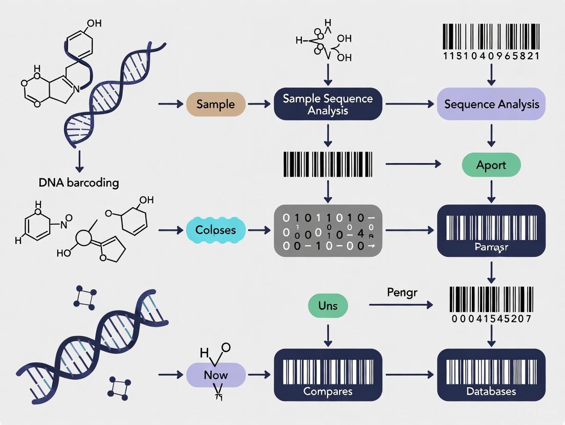
This article provides a comprehensive overview of DNA barcoding methodologies for parasite identification, tailored for researchers, scientists, and drug development professionals.
DNA Barcoding for Parasite Identification: Principles, Protocols, and Advanced Applications in Biomedical Research
Abstract
This article provides a comprehensive overview of DNA barcoding methodologies for parasite identification, tailored for researchers, scientists, and drug development professionals. It explores the foundational principles of DNA barcoding, detailing the selection of appropriate genetic markers such as COI for helminths and 18S rDNA for broad eukaryotic parasite detection. The content covers advanced methodological applications including next-generation sequencing platforms like nanopore technology and Illumina systems, alongside optimization strategies for overcoming common challenges like host DNA contamination and amplification biases. Through comparative analysis with traditional diagnostic methods and validation studies, the article demonstrates the superior sensitivity, specificity, and taxonomic resolution of DNA barcoding approaches. This resource serves as both an introductory guide and technical reference for implementing DNA barcoding in parasitological research and diagnostic development.
Core Principles and Genetic Markers for Parasite DNA Barcoding
Core Concepts and Definitions
DNA barcoding is a method of species identification that uses a short, standardized section of DNA from a specific gene or genes, functioning as a molecular "barcode" [1]. The fundamental premise is that by comparing this DNA section to a reference library of sequences from known species, an individual organism can be identified to the species level [1]. This method was proposed as a standardized system by Paul D.N. Hebert et al. in 2003, drawing on earlier DNA sequencing work [1]. In its essential form, DNA barcoding focuses on the identification of a single individual organism from a single tissue sample [2].
Metabarcoding represents a scale expansion of this core principle. It is a community-level molecular tool that focuses on the composition analysis of complex biological communities [2]. Instead of a single specimen, metabarcoding involves extracting total DNA from mixed samples containing multiple organisms—such as soil, water, or intestinal contents—and using high-throughput sequencing to identify all detectable biological groups within the sample simultaneously, thereby generating a list of community species composition [2]. The paradigm difference is clear: DNA barcoding answers "What species is this one individual?", while metabarcoding answers "Which species are present in this entire community sample?" [2].
Table 1: Fundamental Differences Between DNA Barcoding and Metabarcoding
| Feature | DNA Barcoding | DNA Metabarcoding |
|---|---|---|
| Research Scale | Single individual organism [2] | Complex community of organisms [2] |
| Core Question | "What species is this one?" [2] | "Which species are in this sample?" [2] |
| Sample Input | Single biological individual or tissue [2] | Mixed environmental sample (e.g., soil, water) [2] |
| Sequencing Technology | Sanger sequencing [2] | High-throughput sequencing (e.g., Illumina) [2] |
| Primary Output | A single, high-quality barcode sequence [2] | Sample-sequence-abundance matrix (e.g., OTU/ASV table) [2] |
Standard Markers and Taxonomic Application
The accuracy of DNA barcoding relies on selecting appropriate genetic markers. An ideal DNA barcode should possess low intra-specific variation (small differences within a species) and high inter-specific variation (large differences between species), and it must be flanked by conserved regions to allow universal PCR primer binding [2] [1]. There is no single universal gene region for all life; different marker genes are used for different taxonomic groups [1].
For animals, the most common barcode is a 658-base pair region of the mitochondrial cytochrome c oxidase subunit I (COI or COX1) gene [2] [1]. Mitochondrial genes are preferred for animal barcoding due to their lack of introns, haploid mode of inheritance, and high copy number per cell [1]. The COI gene typically shows an interspecific variation rate of 10-20%, enabling the distinction of over 90% of animal species [2].
In plants, mitochondrial genes like COI evolve too slowly. Therefore, chloroplast genes are used, most commonly a combination of matK and rbcL [2] [1]. Multi-locus markers, including the ribosomal internal transcribed spacer (ITS), are also employed for better discrimination [1].
For fungi, the standard barcode is the ITS region of ribosomal RNA [2] [3]. This region has a high copy number and a fast evolution rate, allowing for effective distinction between closely related fungal species [2]. Its utility is such that it has been formally designated the universal fungal barcode [3].
Parasites and other groups may require tailored approaches. Bacteria and archaea are often identified using the 16S rRNA gene, while the 18S rRNA gene is used for microbial eukaryotes, including some protists and parasites [1] [4].
Table 2: Standard DNA Barcode Markers for Major Taxonomic Groups
| Organism Group | Primary Barcode Marker(s) | Key Characteristics of Marker |
|---|---|---|
| Animals | Cytochrome c oxidase I (COI) [2] [1] | High inter-specific variation; maternal inheritance; no introns [1] |
| Plants | matK and rbcL (chloroplast genes) [2] [1] | Required for sufficient discrimination; mitochondrial genes evolve too slowly [1] |
| Fungi | Internal Transcribed Spacer (ITS) [2] [3] | High copy number; fast evolution rate; universal fungal barcode [2] [3] |
| Bacteria & Archaea | 16S rRNA gene [1] | Highly conserved gene with variable regions [1] |
| Protists | 18S rRNA gene, COI, ITS [1] [4] | Varies by subgroup; 18S is common for microbial eukaryotes [1] |
Experimental Workflows and Protocols
The workflows for DNA barcoding and metabarcoding are distinct, reflecting their adaptation to different research objectives and scales. The following diagram illustrates the core procedural differences.
DNA Barcoding Workflow for a Single Specimen
Sample Input and DNA Extraction: The process begins with a single biological individual or a piece of tissue from a single organism [2]. It is critical to avoid cross-contamination from other organisms. Genomic DNA is then extracted using standard methods such as the CTAB protocol or commercial kits [2] [5]. For valuable specimens, a non-destructive extraction protocol can be employed, allowing the specimen to be preserved for morphological study [6].
PCR Amplification and Sanger Sequencing: The extracted DNA is used as a template for a polymerase chain reaction (PCR) using universal primers designed to amplify the target barcode region (e.g., COI for animals, ITS for fungi) [2] [7]. The success of the PCR is typically verified by visualizing the product on an agarose gel [7]. The amplified product is then purified and sequenced using the Sanger sequencing method (dideoxy chain termination), which produces a single, long (500-1000 bp), high-quality sequence read that ideally covers the entire barcode region [2].
Sequence Analysis and Species Identification: The resulting sequence undergoes quality control to ensure it contains no ambiguous bases or frameshift mutations [2]. This quality-controlled sequence is then compared to reference databases such as the Barcode of Life Data Systems (BOLD) or GenBank using tools like BLAST [2] [1]. A sequence similarity of ≥98% to a reference sequence from a vouchered specimen is often used as a threshold for species-level identification, though this can vary across taxonomic groups [2] [3].
Metabarcoding Workflow for Complex Communities
Sample Input and Total DNA Extraction: The process starts with a mixed environmental sample containing DNA from many organisms, such as soil, water, feces, or entire collections of small arthropods [2] [8]. The goal is to extract the total DNA from this complex matrix. The choice of extraction method is crucial to ensure lysis of a broad range of organisms and to remove inhibitors that may affect downstream steps; the CTAB method is often selected for this purpose [5].
Library Preparation and High-Throughput Sequencing: Unlike single-plex PCR, metabarcoding uses a two-step PCR approach to prepare sequencing libraries [2] [8]. The first PCR uses universal primers to amplify the target barcode region from all the DNA in the sample. The second PCR adds unique sample-specific index sequences (barcodes) and sequencing adapters to the amplicons from the first PCR [2]. This dual-indexing strategy allows multiple samples to be pooled and sequenced simultaneously in a single sequencing run on a high-throughput platform like the Illumina MiSeq or NovaSeq [2] [5]. This process generates millions of short sequence reads (150-300 bp) [2].
Bioinformatic Processing and Community Profiling: The raw sequencing data undergoes a multi-step bioinformatic pipeline. First, sequences are demultiplexed (assigned to their sample of origin based on indexes) and quality-filtered [2] [4]. Then, error-correction algorithms are applied to generate Amplicon Sequence Variants (ASVs), or sequences are clustered into Operational Taxonomic Units (OTUs) based on a similarity threshold (e.g., 97%) [2] [3]. The final output is a sample-by-ASV/OTU table that details the sequence count (abundance) of each taxonomic unit in each sample [2]. These ASVs/OTUs are then taxonomically classified by comparing them to a reference database [2].
The Scientist's Toolkit: Essential Research Reagents and Materials
Successful implementation of DNA (meta)barcoding relies on a suite of specific reagents, consumables, and equipment. The following table details key components of the research toolkit.
Table 3: Essential Research Reagents and Materials for DNA Barcoding and Metabarcoding
| Category | Item | Specific Example / Function |
|---|---|---|
| Sample Collection & Preservation | Specimen collection tools | Sterile forceps, scalpels, Malaise traps, kick-nets [1] [9] |
| Preservation reagents | 99% Ethanol (for tissue storage), EDTA (anticoagulant/preservative) [2] [1] | |
| Nucleic Acid Extraction | Lysis buffers & kits | CTAB buffer, DNeasy Blood & Tissue Kit, BioSprint 96 extraction robot [8] [6] [5] |
| Inhibitor removal | AMPure XP Beads for post-PCR cleanup [8] | |
| PCR Amplification | Polymerase & master mixes | Qiagen Multiplex PCR Kit (for multiplexing loci) [8] |
| Universal primer sets | LCO1490/HCO2198 (COI), ITS1F/ITS4 (Fungal ITS), mlCOIintF/Fol-degen-rev (COI) [8] [6] [5] | |
| Nucleotides (dNTPs) | dNTP solution (e.g., Promega) [6] | |
| Sequencing | Sanger sequencing | Offered as a service by commercial providers [7] |
| High-throughput sequencing | Illumina MiSeq/NovaSeq platforms [2] [5] | |
| Bioinformatics | Analysis pipelines & software | DADA2 (for ASV generation), BLAST (for sequence comparison), Dnabarcoder (for cutoff prediction) [3] [4] |
| Reference databases | BOLD, GenBank, UNITE (for fungi) [2] [3] [1] |
Advanced Applications in Parasite and Vector Research
DNA barcoding and metabarcoding have profound applications in parasitology, offering solutions to long-standing challenges. The difficulties of parasite identification are extraordinary due to their small size, complex multi-host life cycles, and existence as cryptic species complexes within host assemblages [10]. These methods provide a powerful scaffold for discovery and guidance.
Identifying Parasites and Disease Vectors: Barcoding is invaluable for identifying individual parasite specimens and invertebrate disease vectors, such as mosquitoes, especially when morphological characters are scarce or unreliable [10]. For example, a 2024 study used DNA barcoding of the 18S rRNA gene to identify tick-borne protists like Hepatozoon canis and Theileria luwenshuni in the Republic of Korea, even discovering H. canis and Toxoplasma gondii in Ixodes nipponensis ticks for the first time [4]. This demonstrates the method's power to reveal novel parasite-vector associations.
Dissecting Host-Associated Communities: Metabarcoding enables the comprehensive characterization of entire symbiotic communities associated with a host. This approach has been used to study the gut bacteria of ants [9], arthropod inquilines in pitcher plants [9], and myrmecophile communities in ant-plant domatia [9]. Applied to parasitology, this allows researchers to move beyond single-parasite identification to profile the entire community of parasites, commensals, and mutualists within a host, parsing the substantial variation among individual hosts and revealing ecological patterns [9].
Detection in Complex Matrices and Enforcement: Metabarcoding is particularly suited for identifying species in complex, processed samples. This is crucial for diagnosing parasitic infections from patient samples and for forensic applications, such as detecting ingredients from endangered species (CITES-listed) in traditional medicines and food supplements [5]. One validated multi-locus DNA metabarcoding method was shown to be highly reproducible and sensitive enough to identify species present in a mixture at a level of 1% dry weight content, providing a reliable tool for customs and enforcement agencies [5].
The accurate identification of parasites is a cornerstone of medical diagnosis, epidemiological surveillance, and biological research. For years, traditional methods, primarily based on microscopy, have been the standard for parasite identification. However, these techniques are often labor-intensive, require highly skilled technicians, and can suffer from limitations in sensitivity, specificity, and taxonomic resolution [11]. The need to overcome these pitfalls has catalyzed the development of molecular diagnostic approaches, among which DNA barcoding has emerged as a transformative technology [11].
DNA barcoding utilizes short, standardized genetic markers to assign specimens to a known species. In the context of parasite identification, it provides a powerful tool to complement and sometimes surpass the capabilities of morphological analysis. This whitepaper provides an in-depth technical guide to three universal genetic markers—COI, 18S rDNA, and ITS regions—detailing their principles, applications, and experimental protocols within the framework of DNA barcoding principles for parasite identification research. These markers enable researchers and drug development professionals to achieve high-throughput, accurate identification of parasitic organisms, with reported accuracy for DNA barcoding reaching approximately 95.0% in diagnosing medical parasites and arthropods [11].
Principles and Applications of Universal Genetic Markers
The utility of a genetic marker for DNA barcoding depends on its ability to exhibit conserved regions for reliable priming and variable regions for species discrimination. The following sections dissect the core characteristics of the key markers.
Cytochrome c Oxidase Subunit I (COI)
The COI gene, a mitochondrial marker, is the most established barcode for animals and many protists. Its utility stems from a generally higher mutation rate compared to nuclear ribosomal genes, which provides sufficient sequence variation to distinguish between closely related species.
- Principle and Utility: The COI gene is favored for species-level identification due to the presence of a "barcoding gap," where intraspecific genetic variation is typically much lower than interspecific divergence. A review of DNA barcoding in medical parasitology found the technique accords with author identifications based on morphology or other markers in 94–95% of cases [12] [13]. It is particularly useful for identifying parasitic helminths and arthropod vectors. Furthermore, unlike some nuclear ribosomal markers, COI is generally present as a single copy in the mitochondrial genome, avoiding complications from paralogous sequences [14].
- Typical Workflow: A ~658 base pair (bp) region of COI is typically amplified using universal metazoan primers such as LCO1490 and HCO2198, followed by Sanger sequencing or next-generation sequencing (NGS) platforms [15] [16].
- Limitations: COI can face challenges in some contexts, such as introgressive hybridization or the presence of nuclear mitochondrial pseudogenes (NUMTs), which can lead to erroneous sequences and overestimation of species diversity [15].
Small Subunit Ribosomal DNA (18S rDNA)
The 18S rDNA gene is a nuclear ribosomal marker highly conserved across eukaryotes, making it an excellent tool for phylogenetic studies at higher taxonomic levels (e.g., phylum, class) and for broad-spectrum detection of eukaryotic pathogens.
- Principle and Utility: The high conservation of 18S rDNA allows for the design of universal primers that can amplify a wide range of eukaryotic organisms from a single sample. This is invaluable for metabarcoding studies, where the goal is to characterize an entire parasitic community without prior knowledge of its composition [17] [16] [18]. It is especially effective for detecting protozoan parasites like Plasmodium and Eimeria [17] [19]. A key advantage is its ability to detect "unexpected" or novel parasites that would be missed by targeted assays [17].
- Typical Workflow: Amplification targets variable regions (e.g., V4–V9) within the 18S gene. For enhanced species-level resolution on error-prone sequencing platforms like nanopore, longer barcodes spanning from V4 to V9 ( >1 kb) are more effective than shorter ones like the V9 region alone [17]. In samples with high host DNA contamination (e.g., blood), blocking primers (e.g., C3 spacer-modified oligos or peptide nucleic acids) can be used to selectively inhibit host DNA amplification [17].
- Limitations: The high conservation of 18S rDNA can limit its resolution for distinguishing between closely related species. A significant limitation is the presence of highly divergent paralogous gene copies within a single organism's genome. For instance, in the turkey coccidium Eimeria meleagrimitis, intraspecific variation between two 18S rDNA types (2.6%) was found to exceed the interspecific variation between two well-recognized chicken Eimeria species (1.1%), complicating species identification [14].
Internal Transcribed Spacer (ITS) Regions
The Internal Transcribed Spacer regions, comprising ITS-1 and ITS-2, are non-coding segments located between the small subunit (18S), the 5.8S, and the large subunit (28S) ribosomal RNA genes. They evolve rapidly and are among the most frequently used markers for fungal and plant phylogenetics, and are increasingly applied in parasitology.
- Principle and Utility: The high degree of sequence variation in ITS regions makes them ideal for differentiating species at and below the species level. They have proven effective for the molecular characterization and identification of various Eimeria species in goats [19]. Phylogenetic analysis based on ITS-1 and ITS-2 can resolve Eimeria species, though it may not always effectively distinguish between species from different but closely related hosts, such as sheep and goats [19].
- Typical Workflow: DNA is extracted from purified oocysts or other parasitic stages, and the ITS regions are amplified using primers anchored in the flanking conserved ribosomal genes (18S and 5.8S for ITS-1; 5.8S and 28S for ITS-2) [19].
- Limitations: Like 18S rDNA, the ITS regions are multi-copy, which can lead to intragenomic variation. Additionally, their high variability can sometimes make alignment difficult for very distantly related taxa, and the presence of indels can complicate sequence analysis.
Table 1: Comparative Analysis of Universal Genetic Markers for Parasite Identification
| Feature | COI | 18S rDNA | ITS Regions |
|---|---|---|---|
| Genomic Location | Mitochondrial | Nuclear (ribosomal) | Nuclear (ribosomal) |
| Primary Utility | Species-level identification | Broad taxonomic surveys, higher-level phylogenetics | Species-level and intra-species differentiation |
| Evolutionary Rate | Relatively fast | Slow and conserved | Very fast and variable |
| Sequence Length (Typical) | ~658 bp (barcode region) | ~1,700-1,800 bp (full); V4-V9 >1,000 bp | ITS-1: ~386-403 bp; ITS-2: ~565-584 bp [19] |
| Key Advantage | High resolution for closely related species; well-established reference libraries | Universal primers for wide eukaryote coverage; good for detecting novel parasites | High variability for fine-scale differentiation |
| Key Challenge | NUMTs; primer specificity across diverse taxa | Intraspecific paralog variation; low species-level resolution in some cases | Intragenomic variation; alignment difficulty across deep divergences |
Experimental Protocols and Methodologies
This section outlines detailed methodologies for DNA barcoding experiments, from sample preparation to data analysis, providing a practical guide for researchers.
Sample Collection and DNA Extraction
The initial steps are critical for obtaining high-quality, amplifiable DNA.
- Sample Types: Parasitic organisms can be identified from various sample types, including:
- Fecal matter: The most common sample for gastrointestinal parasites [16] [18].
- Whole blood: For haemoparasites like Plasmodium, Trypanosoma, and Babesia [17].
- Purified oocysts or parasites: For specific molecular characterization, as seen in Eimeria studies [19] [14].
- Host tissue or arthropod vectors: For tissue-dwelling parasites or vector identification [11] [12].
- Preservation: Fresh samples can be stored in >70% ethanol or at -80°C to prevent DNA degradation. For fecal samples collected in the field, immediate preservation in ethanol is standard [18].
- DNA Extraction: Commercial kits, such as the DNeasy Blood and Tissue Kit (Qiagen), are widely used and provide reliable results [15] [19]. The choice of kit may be optimized for the specific sample type (e.g., stools vs. blood). For samples with overwhelming host DNA, such as blood, additional steps like blocking primers are incorporated during PCR [17].
PCR Amplification and Sequencing
The following protocols exemplify standard approaches for amplifying the universal markers.
This protocol demonstrates a robust method for amplifying COI from animal tissue.
- Primers: Use universal metazoan primers LCO1490 (5'-GGTCAACAAATCATAAAGATATTGG-3') and HCO2198 (5'-TAAACTTCAGGGTGACCAAAAAATCA-3').
- PCR Reaction: A typical 25 µL reaction contains:
- 10-100 ng of genomic DNA
- 1X PCR Buffer
- 2.0 mM MgCl₂
- 0.2 mM of each dNTP
- 0.5 µM of each primer
- 1.0 U of Taq DNA Polymerase
- Thermocycling Conditions:
- Initial Denaturation: 94°C for 2 minutes
- 35-40 Cycles of:
- Denaturation: 94°C for 30 seconds
- Annealing: 42-50°C for 30 seconds
- Extension: 72°C for 60 seconds
- Final Extension: 72°C for 10 minutes
- Sequencing: Purified PCR products are sequenced bidirectionally using Sanger sequencing or prepared for NGS libraries.
This protocol highlights a targeted NGS approach for comprehensive parasite detection in blood.
- Primers: Use universal eukaryotic primers targeting a >1 kb fragment for better species resolution on nanopore sequencers. Example primers are F566 and 1776R.
- Host DNA Suppression: To overcome the challenge of high host DNA background, incorporate blocking primers:
- C3 Spacer-Modified Oligo: Designed to overlap with the universal reverse primer, with a C3 spacer at the 3' end to halt polymerase extension.
- Peptide Nucleic Acid (PNA): Binds tightly to the host 18S rDNA template and inhibits polymerase elongation.
- PCR and Sequencing: Perform a multiplexed PCR with the universal and blocking primers. The amplicons are then used to prepare a sequencing library for a portable nanopore platform (e.g., MinION).
This protocol is used for genotyping coccidian parasites.
- DNA Source: Genomic DNA is extracted from purified oocysts of Eimeria species.
- PCR Amplification: Multiple PCRs are run to amplify the different loci:
- 18S rDNA: Using universal primers (e.g., Medlin A/B) to generate a ~1.8 kb fragment.
- ITS-1 and ITS-2: Using primers anchored in the flanking 18S, 5.8S, and 28S genes.
- COI: Using specific primers for the mitochondrial gene.
- Cloning and Sequencing: Due to potential intragenomic variation, PCR products for 18S and ITS may be cloned, and multiple clones are sequenced to capture the diversity of paralogous copies [14].
Data Analysis and Interpretation
After sequencing, the data must be processed to assign taxonomic identities.
- Sanger Sequencing Data: Sequences are assembled, trimmed, and subjected to a similarity search using tools like BLAST (NCBI) against public databases (GenBank) or curated databases like the Barcode of Life Data Systems (BOLD) [12] [15].
- Metabarcoding Data (NGS): Raw reads are demultiplexed, quality-filtered, and clustered into Amplicon Sequence Variants (ASVs) or Operational Taxonomic Units (OTUs). These are then classified against a reference database using specialized classifiers [17] [16] [18].
- Phylogenetic Analysis: For definitive identification or to resolve ambiguous classifications, sequences can be aligned with reference data, and phylogenetic trees (e.g., Neighbor-Joining, Maximum Likelihood) can be constructed. High bootstrap values (e.g., ≥99%) support the reliability of species clusters [15] [19].
The Scientist's Toolkit: Essential Research Reagents and Materials
Successful DNA barcoding relies on a suite of carefully selected reagents and tools. The following table details key components for a typical workflow.
Table 2: Essential Research Reagent Solutions for DNA Barcoding
| Reagent/Material | Function | Examples & Notes |
|---|---|---|
| DNA Extraction Kit | Isolates high-quality genomic DNA from complex samples. | DNeasy Blood & Tissue Kit (Qiagen); NucleoSpin Tissue Kit (Macherey-Nagel) [15] [18]. |
| Universal PCR Primers | Amplifies the target barcode region from a wide range of organisms. | COI: LCO1490/HCO2198 [15]; 18S: F566/1776R [17], 563F/1132R [18]. |
| Blocking Primers | Suppresses amplification of non-target DNA (e.g., host). | C3 spacer-modified oligos; Peptide Nucleic Acid (PNA) clamps [17]. |
| High-Fidelity DNA Polymerase | Performs PCR with low error rates for accurate sequencing. | Important for preparing high-quality NGS libraries. |
| NGS Library Prep Kit | Prepares amplicon libraries for high-throughput sequencing. | Kits compatible with Illumina, Oxford Nanopore, etc. |
| Sequencing Platform | Determines the nucleotide sequence of the amplified DNA. | Illumina (high accuracy); Oxford Nanopore (portability, long reads) [17] [16]. |
| Bioinformatics Software | Analyzes raw sequence data for quality control and taxonomic assignment. | QIIME2, DADA2 (for metabarcoding); BLAST, MEGA (for phylogenetics) [16]. |
| Reference Database | Curated collection of reference barcodes for species identification. | BOLD, GenBank, SILVA [12] [15]. |
Workflow Visualization
The following diagram summarizes the two primary DNA barcoding workflows for parasite identification.
The integration of universal genetic markers—COI, 18S rDNA, and ITS—into parasitological research has fundamentally enhanced our ability to identify and characterize parasites with unprecedented precision and scale. While COI remains the gold standard for species-level identification of many metazoan parasites, 18S rDNA is indispensable for broad-spectrum eukaryotic detection and phylogenetic placement, and ITS regions provide the high resolution needed for differentiating closely related species. As sequencing technologies continue to evolve, becoming more portable and affordable, the application of these DNA barcoding principles will undoubtedly expand. This will lead to more rapid diagnosis of parasitic diseases, more effective surveillance and control programs, and a deeper understanding of parasite biodiversity and ecology, ultimately contributing to improved global health outcomes.
DNA barcoding is a powerful molecular tool that uses a short, standardized genetic marker to identify species and assist in their discovery [20]. For animals, the most commonly used barcode is a 658-base pair fragment of the mitochondrial cytochrome c oxidase I (COI) gene [20] [21]. The core principle underlying DNA barcoding is the "barcoding gap"—the disparity between genetic variation within a species (intraspecific variation) and genetic differences between species (interspecific divergence) [22] [23]. In an ideal system, the maximum intraspecific variation is significantly less than the minimum interspecific divergence, creating a clear gap that allows for unambiguous species identification [23]. This technical guide explores the conceptual and practical aspects of the barcoding gap, with a specific focus on its application in parasite identification research, which is critical for diagnostics, treatment, and drug development.
The utility of DNA barcoding is particularly evident in parasitology, where traditional morphological identification can be challenging due to the small size of many parasites, their complex life cycles, and the existence of cryptic species [11] [24] [25]. Molecular methods have revolutionized the field, providing a reliable means to identify species regardless of their morphological diagnosability [22]. Furthermore, DNA barcoding facilitates the discovery of previously unrecognized parasite diversity and enables high-throughput, comprehensive surveys of parasite communities through techniques like DNA metabarcoding [16].
Conceptual Foundation of the Barcode Gap
Defining Intra- and Interspecific Genetic Distances
The barcoding gap is quantified by comparing two key population genetic parameters: intraspecific variation and interspecific divergence.
- Intraspecific Variation: This measures the genetic distance between individuals belonging to the same species. It reflects population-level processes such as mutations, genetic drift, and geographic structure [23].
- Interspecific Divergence: This measures the genetic distance between individuals from different, closely related species. It accumulates after populations become reproductively isolated and diverge from their last common ancestor [23].
The effectiveness of DNA barcoding hinges on the relationship between these two measures. A pronounced barcoding gap indicates that the genetic marker has sufficient resolution to distinguish between species, while significant overlap suggests the marker may be unreliable for certain taxa [23].
The Idealized Model vs. Biological Reality
Proponents of DNA barcoding initially envisioned a world with discrete distributions of intra- and interspecific genetic distances and minimal overlap—a clear "barcoding gap" [23]. However, comprehensive empirical studies have revealed a more complex reality. In thoroughly sampled groups, substantial overlap between intraspecific variation and interspecific divergence is common, making the use of fixed genetic distance thresholds problematic [23].
Several biological phenomena can erode or eliminate the barcoding gap:
- Incomplete Lineage Sorting: When the coalescence of gene lineages (the point at which all copies of a gene trace back to a single ancestral copy) has not occurred by the time a new species forms, one species may be paraphyletic or polyphyletic with respect to another. This results in members of one species being more closely related to members of a different species than to their own [23] [16].
- Hybridization and Introgression: The exchange of genes between species can lead to shared genetic material, blurring species boundaries [16].
- Young Species Divergence: Recently diverged sister species may not have accumulated enough genetic differences to create a gap, as their genomes are still very similar [23].
- Cryptic Species: The presence of morphologically similar but genetically distinct species can artificially inflate intraspecific variation if they are misidentified and grouped under a single species name [24].
Table 1: Factors Affecting the Barcoding Gap and Their Implications for Parasite Research.
| Factor | Effect on Barcoding Gap | Relevance in Parasitology |
|---|---|---|
| Incomplete Lineage Sorting | Creates overlap, leading to paraphyly/polyphyly | Common in rapidly evolving parasite genera and recent radiations [23]. |
| Cryptic Species Complexes | Reveals hidden diversity; reduces gap if unrecognized | Prevalent in parasites (e.g., trypanosomes, helminths); DNA barcoding is key to their discovery [24]. |
| Taxonomic Under-sampling | Inflates perceived intraspecific variation | A major issue for poorly studied parasite groups from diverse hosts and regions [23]. |
| Geographic Sampling Scale | Wider sampling can uncover greater intraspecific variation | Critical for parasites with wide distributions or those in isolated host populations [22]. |
Quantitative Analysis of the Barcoding Gap
Empirical Data on Genetic Distances
The size of the barcoding gap varies considerably across taxonomic groups. Early studies, which often undersampled intra- and interspecific diversity, reported near-100% success rates [23]. However, more comprehensive analyses provide a nuanced picture. A landmark study on marine gastropods (cowries), which included over 2,000 individuals from 263 species, found an overall error rate of 4% for species identification in this well-sampled clade. In contrast, when simulating species discovery in incompletely sampled groups using genetic distance thresholds, the error rate rose to at least 17% [23].
In parasitology, the barcoding gap has been evaluated for various groups. For instance, in spiders, DNA barcodes were effective for species identification across geographical scales and regardless of morphological diagnosability, though the size of the barcoding gap was dependent on taxonomic group and practices [22]. In Hemiptera, a study of over 68,000 barcode sequences suggested that a 2-3% Kimura 2-parameter (K2P) genetic distance threshold is often appropriate for species identification, with deviations from this indicating potential misidentifications or taxonomic issues [20].
Table 2: Reported Genetic Distances and Barcoding Gap Efficacy in Selected Parasite and Vector Groups.
| Organism Group | Common Genetic Marker | Reported Intraspecific Variation (K2P%) | Reported Interspecific Divergence (K2P%) | Suggested Threshold | Identification Efficacy |
|---|---|---|---|---|---|
| Mosquitoes [21] | COI | Generally low | Significantly higher | N/A | 100% success in identifying 45 Singapore species; useful complement to morphology. |
| Hemiptera [20] | COI | <2% in 90% of taxa | >3% in 77% of congeneric pairs | 2-3% | Appropriate for most species, but errors (misIDs, contamination) are not rare. |
| Gastrointestinal Helminths [16] | ITS-2, COI, 18S | Varies by genus and marker | Varies by genus and marker | N/A | Metabarcoding provides high taxonomic resolution and high throughput. |
| Plasmodium falciparum [26] | Circumsporozoite (CS) gene | Low variation in tandem repeats | High variation in tandem repeats | N/A | Hypervariable tandem repeats can "barcode" isolates for epidemiological tracking. |
Error Rates and Limitations
The reliability of DNA barcoding is not universal. Error rates are influenced by the taxonomic group and, crucially, by the quality of the reference database. The cowrie study demonstrated that error rates for threshold-based identification doubled when using traditionally recognized species versus evolutionarily significant units (ESUs) defined by integrative taxonomy [23]. This highlights that DNA barcoding performs best when built upon solid taxonomic foundations [23].
Common sources of error in DNA barcoding databases include:
- Specimen Misidentification: Incorrect morphological identification prior to sequencing propagates errors in reference libraries [20].
- Sample Contamination: Cross-contamination during DNA extraction or amplification can lead to erroneous sequences [20].
- Inadequate Genetic Resolution: Some closely related parasite species may not be distinguishable by the COI gene alone, necessitating a multi-locus approach [21] [23].
Methodological Protocols for Barcoding Gap Research
Standard DNA Barcoding Protocol for Parasites
The following protocol outlines the key steps for generating DNA barcodes from parasite specimens, integrating best practices to minimize errors [20] [21].
1. Specimen Collection and Preservation:
- Collect parasites from their host or environment, ensuring proper ethical and safety guidelines are followed.
- Record detailed collection data: geographic location, host species, date, and microhabitat. This biogeographic information is invaluable for downstream analysis [22] [20].
- Preserve specimens appropriately, typically in >95% ethanol or at -80°C for DNA work. Avoid formalin fixation, which degrades DNA.
2. Morphological Identification:
- Perform initial species identification based on morphological characters by an experienced taxonomist [20] [21]. This step is crucial for creating reliable reference sequences.
- Voucher specimens should be deposited in a recognized museum or collection for future verification.
3. DNA Extraction:
- Extract genomic DNA from a piece of tissue (e.g., a leg from an arthropod, a proglottid from a cestode) to preserve the voucher specimen [21].
- Use commercial DNA extraction kits (e.g., DNeasy Blood & Tissue Kit, Qiagen) following the manufacturer's protocol. Include negative controls to monitor for contamination [20].
4. PCR Amplification of the Barcode Region:
- Amplify the COI barcode region using universal or group-specific primers. For example, primers LCO1490 and HCO2198 are widely used for metazoans [21].
- A typical 50 μL PCR reaction mix includes:
- 5 μL of template DNA
- 1x PCR buffer
- 1.5 mM MgCl₂
- 0.2 mM of each dNTP
- 0.3 μM of each primer
- 1.5 U of Taq DNA polymerase
- PCR cycling conditions often involve an initial denaturation (e.g., 95°C for 5 min), followed by 35-40 cycles of denaturation (e.g., 94°C for 30-60 s), annealing (45-55°C for 30-60 s), and extension (72°C for 60 s), with a final extension at 72°C for 5-10 min [21].
5. Sequencing and Data Analysis:
- Purify PCR products and perform Sanger sequencing in both directions.
- Assemble contiguous sequences, align them using software like ClustalW or MAFFT, and check for stop codons or frameshifts that may indicate pseudogenes (NUMTs) [20] [21].
- Calculate genetic distances using a model like the Kimura 2-parameter (K2P) in programs such as MEGA [20] [21].
- Upload sequences to public databases (e.g., GenBank, BOLD) with associated specimen data and trace files.
Advanced Protocol: Metabarcoding for Parasite Communities
For identifying entire communities of gastrointestinal helminths from host feces, DNA metabarcoding is the state-of-the-art method [16]. This protocol differs from standard barcoding.
1. Sample Collection and DNA Extraction:
- Use fecal matter, intestinal contents, or cloacal swabs. Feces are most common (89% of studies) as they allow non-invasive sampling [16].
- Extract total genomic DNA directly from the sample. Homogenize the sample to ensure a representative subsample is taken.
2. PCR with Blocking Primers:
- To overcome the challenge of overwhelming host DNA, use blocking primers. These are oligonucleotides with a 3'-end modification (e.g., C3 spacer) or peptide nucleic acid (PNA) that bind specifically to the host DNA template and inhibit its amplification during PCR, thereby enriching for parasite DNA [17].
3. Library Preparation and High-Throughput Sequencing:
- Amplify a barcode region (e.g., COI, ITS-2, 18S rDNA) using primers with unique sample-indexing tags.
- Pool the amplified products from multiple samples and sequence them on a high-throughput platform (e.g., Illumina MiSeq, Nanopore) [16].
4. Bioinformatic Analysis:
- Process raw sequences: demultiplex samples, merge paired-end reads, and quality filter.
- Cluster sequences into Operational Taxonomic Units (OTUs) or Amplicon Sequence Variants (ASVs).
- Assign taxonomy by comparing clusters to a curated reference database. The Nemabiome system is a prominent example for gastrointestinal nematodes [16].
The Scientist's Toolkit: Essential Reagents and Materials
Table 3: Key Research Reagent Solutions for DNA Barcoding and Metabarcoding.
| Reagent/Material | Function | Example Use Case |
|---|---|---|
| DNA Extraction Kit (e.g., DNeasy Blood & Tissue Kit) | Purifies genomic DNA from tissue or fecal samples. | Standardized extraction from parasite specimens for consistent PCR results [21]. |
| Universal COI Primers (e.g., LCO1490/HCO2198) | Amplifies the barcode region from a wide range of metazoans. | Initial screening and barcoding of diverse parasite collections [21]. |
| Blocking Primers (C3-spacer or PNA modified) | Suppresses amplification of non-target DNA (e.g., host). | Enriching parasite 18S rDNA from whole blood samples for targeted NGS [17]. |
| High-Fidelity DNA Polymerase | Reduces errors during PCR amplification. | Critical for generating accurate sequence data for reference barcodes. |
| Curated Reference Database (e.g., BOLD, Nemabiome) | Repository of validated barcode sequences for comparison. | Essential for accurate taxonomic assignment of unknown sequences [16]. |
The barcoding gap remains a foundational concept for species identification and discovery using DNA sequences. While its application is powerful, it is not without limitations. Success is highest in taxonomically well-understood and thoroughly sampled groups, where it provides a reliable tool for identifying known species. However, its utility for discovering new species in poorly studied groups using simple genetic distance thresholds is more error-prone due to the frequent overlap between intra- and interspecific genetic variation [23]. In parasitology, DNA barcoding and its high-throughput extension, metabarcoding, have transformed our ability to identify parasites, discover cryptic diversity, and conduct comprehensive community surveys [24] [16]. The continued growth of curated, high-quality reference libraries, coupled with integrative approaches that combine molecular, morphological, and ecological data, will ensure that the promise of DNA barcoding is fully realized in the fight against parasitic diseases.
Within the framework of DNA barcoding principles for parasite identification, reference databases serve as the critical cornerstone for accurate species determination. The Barcode of Life Data Systems (BOLD) and the National Center for Biotechnology Information (NCBI) platforms provide complementary resources that enable researchers to address taxonomic challenges, particularly with morphologically cryptic parasite complexes. The utility of these databases is exemplified in recent parasitological research, such as the discovery that Toxocara cati infecting domestic and wild felids constitutes a species complex, a finding substantiated through DNA barcoding that revealed substantial genetic differences (6.68%–10.84%) in cox1 sequences between variants from different hosts [27]. Such findings underscore the transformative role of reference databases in revealing hidden biodiversity and refining our understanding of parasite taxonomy, which directly impacts diagnosis, treatment, and control strategies for parasitic diseases affecting human and animal health.
The Barcode of Life Data Systems (BOLD)
BOLD represents a centralized bioinformatics platform specifically designed for the collection, management, and analysis of DNA-based biodiversity data [28]. This specialized system provides an integrated environment that supports the entire DNA barcoding workflow, from specimen data collection to sequence publication and analysis. The platform has evolved significantly since its initial launch [28], with the current BOLD v4 architecture offering enhanced analytical capabilities and data management tools that are particularly valuable for parasite researchers dealing with complex taxonomic assignments.
The system incorporates the Barcode Index Number (BIN) system, which provides a species-level taxonomic framework that operates independently of Linnaean taxonomy, serving as a powerful tool for revealing cryptic species and assigning unknown queries to known genetic clusters [28]. This feature is particularly valuable in parasitology, where morphological similarities often mask significant genetic divergence, as demonstrated in the T. cati complex study [27].
Data Structure and Content
BOLD hosts comprehensive data packages that offer flexible, up-to-date, and structured data solutions tailored to research demands [29]. The system employs Frictionless Data Standards to ensure data is easily accessible, integrable, and reusable across different platforms, enhancing research tool interoperability and promoting reproducible biodiversity research [29].
Table 1: BOLD Public Data Package Snapshots (2024-2025)
| Snapshot Date | Specimens | Sequences | Data Formats |
|---|---|---|---|
| 26-SEP-2025 | 20,616,372 | 20,966,629 | TSV, FASTA, JSON (BCDM metadata) |
| 27-JUN-2025 | 19,759,311 | 20,104,972 | TSV, FASTA, JSON (BCDM metadata) |
| 28-MAR-2025 | 19,220,950 | 19,561,427 | TSV, FASTA, JSON (BCDM metadata) |
| 27-DEC-2024 | 17,568,910 | 17,912,216 | TSV, FASTA, JSON (BCDM metadata) |
In addition to these comprehensive public data snapshots, BOLD also hosts specialized project data, such as the Centre for Biodiversity Genomics (CBG) releases, which provide curated datasets specifically relevant to parasite researchers. For instance, the CBG.R4.01-Sep-2025 release contains 807,389 specimens and sequences originating from over 200 countries and representing 24,000 species [29].
Building and Contributing Data to BOLD
Data Submission Workflow
Contributing data to BOLD follows a structured pipeline that ensures data quality and integrity:
- Specimen Registration: Each specimen receives a unique identifier with detailed collection and morphological data
- Laboratory Processing: DNA extraction, PCR amplification, and sequencing following standardized protocols
- Sequence Assembly and Annotation: Forward and reverse sequences are assembled into contigs and linked to specimen records
- Data Validation: Automated and manual checks ensure data quality before publication
- Publication: Data is made available through public data packages or specialized project releases
For educational and small-scale research initiatives, the BOLD Student Data Portal (BOLD-UNI) provides a streamlined interface for data submission, complete with video tutorials and a quick start guide to facilitate researcher training [30].
Sequence Submission Protocols
The technical process of submitting sequences to BOLD involves specific methodologies optimized for DNA barcoding:
- Bi-directional Sequencing: Using dye terminator cycle sequencing for both forward and reverse strands to ensure accuracy [30]
- Sequence Purification: Removal of unincorporated nucleotides, primers, and enzymes prior to sequencing to prevent adverse effects on sequencing reactions [30]
- M13-Tailed Primers: Implementation of primers with M13 sequences to facilitate universal sequencing primer binding [30]
- Quality Control: Assessment of electropherograms for base calling accuracy and identification of ambiguous regions
Experimental Protocol: DNA Barcoding of Parasites Using BOLD
Materials and Reagents:
- Tissue samples from parasite specimens
- DNA extraction kit (e.g., DNeasy Blood & Tissue Kit)
- PCR reagents: Taq polymerase, dNTPs, buffer, MgCl₂
- M13-tailed primer sets (e.g., LCO1490/HCO2198 for COI)
- Agarose gel electrophoresis equipment
- PCR purification kit
- Sequencing facility access
Procedure:
- DNA Extraction: Isolate genomic DNA from parasite tissue samples using standardized protocols
- PCR Amplification: Amplify the barcode region (e.g., COI for helminths) using M13-tailed primers under the following conditions:
- Initial denaturation: 94°C for 2 minutes
- 35 cycles of: 94°C for 30s, 50°C for 30s, 72°C for 1 minute
- Final extension: 72°C for 5 minutes
- Amplicon Verification: Confirm amplification success and specificity via agarose gel electrophoresis
- Sample Purification: Purify PCR products to remove enzymes and unincorporated nucleotides
- Sequencing Submission: Submit purified amplicons for bi-directional Sanger sequencing with M13 forward and reverse primers
- Sequence Assembly: Assemble forward and reverse sequences into contigs using BOLD's assembly tools
- Data Annotation: Link sequences to specimen data including collection locality, host information, and voucher specimen details
- BIN Assignment: Upload completed records to obtain Barcode Index Numbers and identify concordant clusters
Diagram 1: BOLD data workflow for parasite identification.
NCBI Databases and Tools
The NCBI provides a comprehensive suite of databases and analytical tools that support parasite identification and characterization through DNA sequence analysis. The platform's significance in parasitology research stems from its extensive repository of sequence data and powerful comparison utilities that facilitate evolutionary and functional analyses of parasite genomes.
The Basic Local Alignment Search Tool (BLAST) serves as the cornerstone of NCBI's analytical suite, finding regions of similarity between biological sequences and calculating the statistical significance of matches [31] [32]. BLAST can be used to infer functional and evolutionary relationships between sequences—a critical capability when studying parasite evolution, host adaptation, and drug resistance mechanisms.
BLAST Search Variants and Applications
NCBI's BLAST suite offers several search types, each with specific applications in parasite research:
- BLASTn (Nucleotide BLAST): Compares nucleotide query sequences against nucleotide databases; ideal for identifying unknown parasite sequences by similarity to known references [32]
- BLASTp (Protein BLAST): Compares protein query sequences against protein databases; useful for identifying functional elements in parasite genomes and predicting gene function [32]
- BLASTx: Translates nucleotide query in six reading frames and searches against protein sequences; particularly valuable when analyzing sequences with potential errors or unknown reading frames, such as novel parasite genes [32]
- tBLASTn: Compares protein query sequences against translated nucleotide databases; effective for finding homologous coding regions in unannotated nucleotide sequences from parasite genomics projects [32]
Multiple Sequence Alignment and Analysis
The Multiple Sequence Alignment Viewer (MSA) within NCBI provides sophisticated visualization of alignments created by programs such as MUSCLE or CLUSTAL, including alignments from BLAST results [33]. This tool enables researchers to:
- Identify conserved and variable regions across parasite strains or species
- Visualize phylogenetic relationships through sequence similarity
- Detect diagnostic positions that differentiate cryptic parasite species
- Analyze sequence features and annotations across multiple taxa
Key functionalities include setting anchor sequences for comparison, calculating percent identity and coverage metrics, and sorting sequences by metadata such as host organism or collection country—all valuable features for comparative analyses of parasite populations [33].
Experimental Protocol: Parasite Identification Using NCBI BLAST
Materials and Reagents:
- Unknown parasite DNA sequence (e.g., from sequencing core facility)
- Computer with internet access
- NCBI user account (for saving searches)
Procedure:
- Sequence Preparation: Obtain quality-trimmed sequence data from sequencing facility. For Sanger sequences, ensure proper base calling and trim low-quality ends.
- BLAST Database Selection:
- Access NCBI BLAST through https://blast.ncbi.nlm.nih.gov/
- Select appropriate BLAST algorithm:
- Use BLASTn for nucleotide queries against nucleotide databases
- Use BLASTx if query may contain coding regions with unknown reading frame
- Choose specialized databases when applicable:
- nr/nt for comprehensive searches
- RefSeq for curated reference sequences
- Barcode of Life COI records for specific marker searches
- Parameter Optimization:
- Adjust expected significance threshold (E-value) based on search specificity needs
- For short barcode sequences, disable low-complexity filters
- Select appropriate algorithm parameters (e.g., -task blastn for somewhat similar sequences) [17]
- Query Submission:
- Paste sequence in FASTA format or upload sequence file
- Provide descriptive job title for tracking
- Click "BLAST" to submit search
- Results Interpretation:
- Examine "Descriptions" tab for significant alignments sorted by E-value
- Assess Query Coverage and Percent Identity for top hits
- Review "Alignments" tab for pairwise comparison with subject sequences
- Check taxonomic information of top matches for consistency
- Evolutionary Analysis:
- Click "Distance tree of results" to visualize phylogenetic relationships
- Use "Multiple Sequence Alignment" viewer for detailed comparison of top hits
- Data Management:
- Save search strategies and results via My NCBI account
- Export significant hits for further analysis
Table 2: Interpretation of BLAST Results for Parasite Identification
| Result Metric | Interpretation | Threshold for Reliable Identification |
|---|---|---|
| E-value | Number of alignments expected by chance with the calculated score or better | <0.001 for significant match; closer to zero indicates greater significance |
| Query Coverage | Percent of query length included in aligned segments | >90% for comprehensive matching of barcode region |
| Percent Identity | Degree of sequence similarity between query and subject | >97-99% for conspecific matches; varies by parasite group |
| Max Score | Highest alignment score from sum of rewards for matches and penalties for mismatches/gaps | Higher values indicate better quality alignments |
Comparative Analysis of Database Utility
Applications in Parasite Identification Research
Both BOLD and NCBI provide essential resources for parasite identification, but they offer complementary strengths that researchers can leverage for comprehensive analysis:
BOLD specializes in standardized DNA barcodes with tightly coupled specimen metadata, making it particularly valuable for initial species identification and discovering cryptic diversity. The integration of the BIN system provides a robust framework for species delineation that has proven effective in revealing cryptic parasite species complexes, as demonstrated in the T. cati study [27]. The platform's curated data packages and specialized analytical tools for barcode data make it the preferred starting point for barcode-based identification.
NCBI offers broader sequence diversity and more extensive analytical tools for functional and evolutionary analyses. The platform's strength lies in its comprehensive genomic data, which enables researchers to place barcode sequences within broader genomic contexts and investigate functional implications of sequence variations. This is particularly valuable when studying parasite adaptations, drug resistance mechanisms, or evolutionary relationships.
Integration in Metabarcoding Studies
For metabarcoding approaches to parasite identification—increasingly used for gastrointestinal helminth communities in vertebrate hosts [16]—both databases play critical roles. BOLD provides the reference barcodes necessary for assigning taxonomic identities to sequence variants, while NCBI offers additional verification through broader sequence comparisons and tools for analyzing marker genes beyond the standard barcode regions.
Recent advances in parasite detection through metabarcoding highlight the importance of comprehensive reference databases. For instance, a 2024 systematic review of gastrointestinal helminth identification using metabarcoding emphasized that database choice significantly impacts identification success, with different genetic marker regions (COI, ITS, 18S) requiring different reference resources [16].
Table 3: Key Research Reagents and Computational Tools for Database-Driven Parasite Identification
| Resource Category | Specific Examples | Function in Parasite Identification |
|---|---|---|
| Wet Lab Reagents | M13-tailed PCR primers (e.g., LCO1490/HCO2198) | Amplification of barcode regions with universal sequencing primer sites [30] |
| DNA purification kits (e.g., DNeasy Blood & Tissue) | High-quality DNA extraction from various parasite sample types | |
| Blocking primers (C3 spacer-modified or PNA oligos) | Selective inhibition of host DNA amplification in mixed samples [17] | |
| Sequencing Platforms | Sanger sequencing | Gold standard for reference barcode generation [30] |
| Oxford Nanopore MinION | Portable sequencing for field applications; enables long reads for better species resolution [17] [34] | |
| Illumina MiSeq | High-throughput sequencing for metabarcoding studies [34] | |
| Bioinformatic Tools | BLAST Suite (NCBI) | Sequence similarity searching and functional inference [31] [32] |
| Multiple Sequence Alignment Viewer (NCBI) | Visualization and comparison of sequence alignments [33] | |
| BOLD Identification Engine | Species identification based on barcode sequence similarity | |
| Reference Databases | BOLD Data Packages | Curated barcode records with specimen metadata [29] |
| NCBI nr/nt database | Comprehensive nucleotide sequence repository | |
| Specialized databases (RefSeq, Barcode of Life) | Curated reference sequences for specific applications |
Building and utilizing the reference databases provided by BOLD and NCBI represents a fundamental competency in modern parasite identification research. Each platform offers distinct advantages: BOLD provides specialized tools and curated data specifically designed for DNA barcoding applications, while NCBI delivers comprehensive sequence resources and powerful analytical tools for broader genomic analyses. The integration of both platforms, along with appropriate laboratory protocols and bioinformatic workflows, creates a robust framework for advancing our understanding of parasite diversity, evolution, and ecology. As DNA-based identification continues to transform parasitology, these reference databases will play increasingly critical roles in diagnostic development, biodiversity assessment, and research on host-parasite interactions with implications for drug discovery and disease control strategies.
For over a century, morphological identification has served as the cornerstone of parasite taxonomy and diagnostics. However, this approach presents significant challenges, including reliance on highly specialized expertise, difficulties in identifying cryptic species, and the time-consuming nature of the process. DNA barcoding has emerged as a powerful alternative that surmounts these limitations through a standardized, sequence-based identification system. This technical guide examines the core advantages of DNA barcoding—enhanced resolution, accelerated speed, and greater objectivity—within the context of modern parasite identification research, providing experimental frameworks and technical specifications for implementation.
Core Technical Advantages of DNA Barcoding
Enhanced Resolution: Unveiling Hidden Diversity
Superior Species Discrimination: DNA barcoding achieves significantly higher taxonomic resolution than morphological methods by targeting genetically variable regions in specific marker genes. This enables discrimination of cryptic species—morphologically similar but genetically distinct organisms that are frequently misidentified using traditional techniques [35].
Parasite Case Study: Research on Toxocara cati infecting domestic and wild felids revealed substantial genetic divergence (6.68–10.84%) in the cox1 barcode region, providing evidence that this parasite constitutes a species complex with at least five distinct clades correlated to host specificity. Morphological examination had previously failed to detect this hidden diversity [27].
Methodology: The standard approach involves PCR amplification of the cytochrome c oxidase subunit I (COI) gene region using universal primers, followed by sequencing and phylogenetic analysis to delineate species boundaries based on genetic distance thresholds and monophyletic clustering [27] [35].
Comprehensive Detection Capability: Unlike targeted molecular assays, DNA barcoding with universal primers can detect unexpected or novel parasites without prior knowledge of potential pathogens. This comprehensive approach is particularly valuable for diagnostic surprises and emerging parasitic diseases [17].
Accelerated Speed: From Days to Hours
Streamlined Workflow Efficiency: DNA barcoding significantly compresses identification timelines by eliminating the most labor-intensive aspects of morphological analysis. The process can be completed within hours compared to days or weeks for traditional methods.
Table 1: Time Comparison Between Identification Methods
| Process Step | Morphological Identification | DNA Barcoding |
|---|---|---|
| Sample Processing | Hours to days (fixation, staining, slide preparation) | Minutes to hours (DNA extraction) |
| Expert Analysis | Hours to days (microscopic examination by taxonomist) | Minutes (automated sequencing) |
| Data Interpretation | Subjective (comparison to taxonomic keys) | Objective (bioinformatic alignment) |
| Total Time | Days to weeks | Hours to 2 days |
High-Throughput Application: When combined with DNA metabarcoding, the technique enables simultaneous identification of multiple species from complex samples or environmental DNA. This approach transforms ecological monitoring and biodiversity assessments by processing hundreds of samples concurrently [36] [37].
- Experimental Protocol for Bulk Processing:
- DNA Extraction: Use standardized kits for high-throughput nucleic acid purification from multiple samples
- PCR Amplification: Employ universal barcoding primers with sample-specific indexes for multiplexing
- Library Preparation: Pool amplified products in equimolar ratios for sequencing
- Sequencing: Run on high-throughput platforms (Illumina, Nanopore)
- Bioinformatic Analysis: Process sequences through automated pipelines (BOLD, QIIME2) for species assignment [36] [37]
Unbiased Objectivity: Quantifiable and Reproducible Results
Standardized Metric System: DNA barcoding replaces subjective morphological assessments with quantifiable genetic distance measurements, typically using Kimura-2-Parameter (K2P) model calculations. This provides a reproducible standard for species delimitation across laboratories and researchers [35].
Reference Library Dependency: The reliability of barcoding identifications depends on comprehensive reference databases such as the Barcode of Life Data System (BOLD), which contains curated barcode sequences with voucher specimen information and photographic documentation [36].
- Implementation Example: The GEANS project established a curated DNA reference library for North Sea macrobenthos containing 4,005 COI barcode sequences from 715 species, enabling objective identification of 29% of known North Sea macrobenthic species [36] [37].
Reduced Expert Dependency: While morphological identification requires years of specialized training, DNA barcoding can be implemented by technical staff following standardized protocols, making sophisticated parasite identification accessible to non-specialist laboratories [36].
Technical Implementation Framework
Research Reagent Solutions
Table 2: Essential Research Reagents for Parasite DNA Barcoding
| Reagent Category | Specific Examples | Function & Application |
|---|---|---|
| Universal Primers | F566/R1776 (18S rDNA), LCO1490/HCO2198 (COI) | Amplify barcode regions across diverse parasite taxa [17] [36] |
| Blocking Primers | C3 spacer-modified oligos, Peptide Nucleic Acid (PNA) clamps | Suppress host DNA amplification to enrich parasite targets [17] [38] |
| Polymerase Systems | High-fidelity DNA polymerases, Multiplex PCR kits | Reliable amplification from minimal parasite DNA in complex samples |
| Sequencing Kits | Nanopore 18S rDNA sequencing kits, Illumina metabarcoding kits | Platform-specific sequencing of barcode amplicons [17] |
| Reference Databases | BOLD, NCBI GenBank, SILVA | Species identification through sequence comparison [36] |
Workflow Visualization: Morphological vs. DNA Barcoding Identification
Advanced Technical Considerations
Marker Selection for Parasite Identification:
- 18S rDNA V4-V9 Region: Provides broader taxonomic coverage for diverse parasites; demonstrated enhanced species identification over shorter V9 region on portable nanopore sequencers [17] [38]
- COI (Cytochrome c oxidase I): Standard for metazoan parasites; offers high discrimination for helminths and arthropods [27] [39]
- Multi-Locus Approaches: Increase resolution for challenging taxa; combine 18S rDNA, COI, and ITS regions [40]
Host DNA Suppression Techniques:
- C3 Spacer-Modified Oligos: Competitively inhibit host DNA amplification by blocking primer binding sites [17]
- PNA (Peptide Nucleic Acid) Clamps: Specifically bind to host DNA and block polymerase elongation without inhibiting parasite target amplification [17] [38]
- Experimental Results: These blocking primers enabled detection of Trypanosoma brucei rhodesiense, Plasmodium falciparum, and Babesia bovis in human blood with sensitivities as low as 1-4 parasites/μL despite high host DNA background [17]
DNA barcoding represents a paradigm shift in parasite identification, offering resolutive, rapid, and objective alternatives to traditional morphological methods. The technical frameworks outlined herein provide researchers with practical implementation guidelines to leverage these advantages in diverse scientific contexts, from clinical diagnostics to biodiversity monitoring. As reference databases expand and sequencing technologies become more accessible, DNA barcoding is poised to become the standard for precise parasite identification in research and applied settings.
Advanced Workflows: From Sample Collection to Sequencing
Sample preparation is a critical first step in DNA barcoding workflows for parasite identification, directly determining the success of downstream genetic analyses. Effective strategies must address dual challenges of optimizing target DNA yield while minimizing contaminants and inhibitors that compromise assay sensitivity and specificity. Within parasitology research, specimen types present unique handling requirements—blood contains overwhelming host DNA background, feces incorporates complex inhibitory substances, and tissues vary widely in parasite density and distribution. This technical guide details current, optimized protocols for these specimen types, contextualized within the rigorous demands of parasite identification research using targeted next-generation sequencing platforms. The methodologies presented support the broader thesis that refined sample preparation is not merely a preliminary step but a foundational component determining the accuracy, sensitivity, and overall success of DNA barcoding principles in parasitology.
Blood Specimen Protocols
Challenge: Host DNA Background
Blood specimens present a significant analytical challenge due to the overwhelming presence of host DNA, which can obscure parasite DNA signals during sequencing. Conventional molecular tests targeting specific parasites require prior knowledge of the pathogen and demonstrate limited utility for detecting novel or unexpected parasitic species [17]. Microscopic analysis, while broadly applicable for parasite detection, suffers from poor species-level identification and requires specialized expertise [17]. A targeted next-generation sequencing (NGS) approach using a portable nanopore platform has been developed to overcome these limitations, enabling comprehensive parasite detection with enhanced species-level resolution [17].
V4–V9 18S rDNA Barcoding with Host Depletion
This protocol employs a DNA barcoding strategy targeting the 18S rDNA V4–V9 region, which provides superior species identification compared to the shorter V9 region alone, especially on error-prone nanopore sequencers [17]. The method incorporates blocking primers to selectively inhibit host DNA amplification, significantly improving parasite DNA detection sensitivity.
- Universal Primers: The primer pair F566 and 1776R amplifies a >1 kb region from V4 to V9 of the 18S rDNA, providing a robust barcode for a wide range of eukaryotic parasites [17].
- Host Depletion with Blocking Primers: Two blocking primers are used to reduce amplification of human host 18S rDNA [17]:
- 3SpC3_Hs1829R: A C3 spacer-modified oligo that competes with the universal reverse primer.
- PNA Oligo: A peptide nucleic acid (PNA) oligo that inhibits polymerase elongation at its binding site.
- Sensitivity Validation: This targeted NGS test successfully detected Trypanosoma brucei rhodesiense, Plasmodium falciparum, and Babesia bovis in human blood samples spiked with as few as 1, 4, and 4 parasites per microliter, respectively [17]. The method also identified multiple Theileria species co-infections in field cattle blood samples [17].
Table 1: Blood Specimen Protocol Components
| Component | Type | Function in Protocol |
|---|---|---|
| Primer F566 | Oligonucleotide | Forward universal primer binding before V4 region |
| Primer 1776R | Oligonucleotide | Reverse universal primer binding after V9 region |
| 3SpC3_Hs1829R | C3 spacer-modified blocking primer | Competes with 1776R to suppress host DNA amplification |
| PNA Oligo | Peptide Nucleic Acid oligo | Binds host DNA and inhibits polymerase elongation |
| PowerBead Pro Tubes | Sample homogenization tube | Contains beads for mechanical lysis of cells |
Feces Specimen Protocols
Challenge: Inhibitors and Sample Integrity
Fecal material serves as a critical sample for gastrointestinal parasites but introduces complex challenges including PCR inhibitors, variable pathogen loads, and rapid nucleic acid degradation. Preservation method significantly impacts downstream DNA yield and quality, particularly for large-scale field studies where cold chain logistics may be impractical [41].
Dried Blood Spot (DBS) Card Workflow
The DBS card method provides a practical solution for fecal sample preservation, enabling room temperature storage and transportation while maintaining DNA integrity. This protocol was validated for animal fecal material in the HUNT One Health study [41].
- Sample Collection: Fecal material is thinly smeared onto two sampling fields (1.7 x 3.5 cm) of DBS filter paper and air-dried for at least two hours [41].
- Storage: Dried cards are stored at -20°C until processing. For DNA extraction, four 8-mm diameter circles are aseptically punched from areas with evenly spread fecal material [41].
- DNA Extraction: Punched discs are transferred to PowerBead Pro Tubes for mechanical lysis. The optimized protocol achieves sufficient yield and quality for shotgun metagenomic sequencing, critical for comprehensive parasite detection [41].
- Quality Consideration: While DBS cards preserve DNA effectively, yields are typically lower than liquid preservation methods, requiring consideration in library preparation and sequencing depth [41].
Table 2: Fecal Specimen Protocol Performance
| Parameter | DBS Card Method | Conventional Frozen |
|---|---|---|
| Preservation | Room temperature (after drying) | -80°C required |
| Transportation | Ambient temperature, no cold chain | Cold chain dependent |
| DNA Yield | Lower but sufficient for metagenomics | Higher |
| Suitability for Field Studies | Excellent | Poor |
| Risk of Degradation | Low when properly dried | Low with consistent freezing |
General DNA Barcoding Workflow and Quality Control
Comprehensive Barcoding Pipeline
A standardized workflow is essential for reliable parasite identification across all specimen types. DNA barcoding uses short, standardized gene sequences as a global standard for species identification [42] [10], with the cytochrome c oxidase I (COI) gene typically serving as the standard barcode for animals [20].
Quality Assurance in Practice
Despite its utility, DNA barcoding faces significant data quality challenges. Systematic evaluation of Hemiptera COI barcodes revealed that errors in public databases are not rare, with most attributable to human errors including specimen misidentification, sample confusion, and contamination [20].
- Critical Quality Checkpoints:
- Specimen Collection: Detailed recording of geographic data and habitat information is crucial [20].
- Morphological Identification: Requires experienced taxonomists to compare characters between species [20].
- Molecular Validation: Interactive validation between morphological characters and barcode sequences is ideal but frequently disregarded [20].
- Data Upload: Accurate specimen information must accompany sequence data in public repositories [20].
- Genetic Distance Thresholds: For insect identification, a threshold value of 2% K2P genetic distance is generally accepted for Lepidopteran species, while Hemiptera typically requires 2-3% [20]. Abnormal intraspecific distances greater than these thresholds often indicate misidentifications or taxonomic issues [20].
Research Reagent Solutions
Table 3: Essential Research Reagents for Parasite DNA Barcoding
| Reagent/Kit | Application | Function |
|---|---|---|
| PowerBead Pro Tubes (QIAGEN) | Sample homogenization | Mechanical lysis of tough specimens including spores and cysts |
| DBS Cards (Lipidx) | Feces specimen collection | Room-temperature preservation of nucleic acids |
| Blocking Primers (C3 spacer/PNA) | Blood specimen host depletion | Selective inhibition of host DNA amplification |
| ZymoBIOMICS Microbial Community Standard | Protocol validation | Positive control for extraction and amplification efficiency |
| Universal 18S rDNA Primers (F566/1776R) | Broad-range parasite detection | Amplification of V4-V9 region for enhanced species ID |
Effective sample preparation strategies for blood, feces, and tissue specimens form the cornerstone of successful DNA barcoding applications in parasite identification. The protocols detailed herein address specimen-specific challenges through optimized preservation, specialized host DNA depletion, and rigorous quality control measures. When integrated within a comprehensive DNA barcoding workflow, these methods enable highly sensitive and specific parasite detection, species-level resolution, and discovery of co-infections—advancing both clinical diagnostics and fundamental parasitology research. The continued refinement of these preparatory techniques will further enhance the utility of DNA barcoding principles in understanding and combating parasitic diseases.
DNA barcoding has emerged as a critical tool in parasitology, enabling precise species identification that often eludes traditional morphological methods due to the small size and cryptic nature of many parasites [43]. The foundation of any successful DNA barcoding initiative rests on effective primer design, which determines both the taxonomic breadth and specificity of detection. This technical guide examines the core strategic decision in primer selection: whether to employ universal pan-eukaryotic primers that target a wide taxonomic range or to utilize phylum-specific primers that offer greater specificity within narrower taxonomic groups. Each approach presents distinct advantages and limitations that researchers must carefully consider within the context of their specific parasitological research goals, whether focused on disease ecology, biodiversity assessment, or diagnostic development [44].
The principle of DNA barcoding relies on amplifying and sequencing a standardized short genetic marker from an organism to facilitate identification. For parasites, this approach has revolutionized detection capabilities, particularly for cryptic species, life stages with minimal morphological features, and specimens with degraded DNA [43]. The selection between universal and targeted primer strategies influences every subsequent aspect of the research workflow, from sample processing to data interpretation, making this fundamental choice critical to project success.
Core Principles of Primer Design
Before examining the specific applications of universal versus phylum-specific primers, it is essential to understand the fundamental biochemical properties that constitute effective primer design, regardless of application scope. These parameters ensure efficient and specific amplification during polymerase chain reaction (PCR) processes.
Table 1: Fundamental Primer Design Parameters and Their Optimal Ranges
| Parameter | Optimal Range | Technical Rationale |
|---|---|---|
| Length | 18-24 bases | Balances specificity (longer) with hybridization efficiency (shorter) [45] |
| GC Content | 40-60% | Ensances stable priming without promoting non-specific binding [45] |
| Melting Temperature (Tm) | 50-60°C | Provides optimal annealing under standard PCR conditions [45] |
| Tm Difference Between Primers | ≤5°C | Ensures both primers anneal efficiently at the same temperature [46] |
| 3' End Sequence | 1-2 G/C pairs | Increases priming specificity due to stronger hydrogen bonding [45] |
Secondary structure formation represents another critical consideration in primer design. Primers must be screened for potential hairpin loops (internal folding) and primer-dimer formations (inter-primer annealing), both of which can drastically reduce amplification efficiency by competing with template binding [46]. Computational tools like OligoAnalyzer facilitate this assessment by calculating thermodynamic parameters and visualizing potential secondary structures. Additionally, researchers must verify primer specificity against genomic databases to minimize off-target amplification, a process efficiently accomplished using tools such as NCBI Primer-BLAST [47].
Universal Pan-Eukaryotic Primer Approach
Strategic Rationale and Design Considerations
Universal pan-eukaryotic primers target conserved genomic regions across broad taxonomic ranges, enabling simultaneous detection of diverse parasite taxa without prior knowledge of the specific parasites present in a sample. This approach is particularly valuable in exploratory research, environmental DNA (eDNA) studies, and diagnostic scenarios where the complete parasite community composition is unknown [44]. The primary advantage of this method lies in its comprehensiveness, as it can reveal unexpected or novel parasites that might be missed by targeted approaches [17].
The design of universal primers typically focuses on conserved genomic regions that flank variable sequences suitable for species discrimination. For eukaryotic parasites, the small subunit ribosomal RNA gene (18S rDNA) serves as the most common target due to its presence in all eukaryotes and a structure comprising conserved regions ideal for primer binding alongside hypervariable domains that provide taxonomic resolution [17]. Effective universal primer design requires identifying sequences with minimal degeneracy while maintaining broad taxonomic coverage, often achieved through bioinformatic analysis of aligned sequences from diverse eukaryotic lineages.
Implementation with Blocking Primers
A significant technical challenge in universal primer applications, particularly with clinical or environmental samples containing host DNA, is the preferential amplification of abundant host sequences that can overwhelm target parasite signals. To address this, researchers have developed blocking primers—modified oligonucleotides that specifically inhibit the amplification of non-target DNA [17] [48].
Blocking primers employ two primary mechanisms:
- Annealing inhibition: The blocking primer binds to the host template at the same site as the universal primer, physically preventing primer annealing through competitive binding [48].
- Elongation arrest: The blocking primer binds downstream of the primer site and contains modifications that halt polymerase progression, effectively terminating amplification [17].
These primers typically incorporate 3'-end modifications such as C3 spacers or peptide nucleic acid (PNA) chemistry to prevent their own extension during PCR [17] [48]. For example, a recent study on blood parasite detection designed two blocking primers (3SpC3Hs1829R and PNAHs1316) that, when combined with universal 18S rDNA primers (F566 and 1776R), successfully reduced host DNA amplification by over 99.9%, enabling detection of low-abundance parasites including Trypanosoma brucei rhodesiense, Plasmodium falciparum, and Babesia bovis in human blood samples [17].
Figure 1: Blocking Primer Mechanism for Selective Amplification
Enhanced Barcode Regions for Improved Resolution
While shorter barcode regions (∼200-400 bp) often suffice for preliminary identification, recent research demonstrates that longer barcodes (∼1000-1500 bp) spanning multiple variable regions significantly improve species-level resolution, particularly when using error-prone sequencing platforms like portable nanopore sequencers [17]. One study designed universal primers amplifying the V4–V9 regions of 18S rDNA (∼1,200 bp), which outperformed shorter V9-only barcodes (∼300 bp) in species discrimination accuracy for malaria parasites, reducing misidentification from 1.7% to near zero even with sequencing errors [17].
Table 2: Performance Comparison of 18S rDNA Barcode Regions for Blood Parasite Detection
| Barcode Region | Amplicon Size | Species Discrimination Accuracy | Sequencing Platform | Key Advantages |
|---|---|---|---|---|
| V9 only | ~300 bp | Moderate (misidentification up to 1.7% with errors) | Nanopore | Rapid sequencing, lower cost |
| V4–V9 | ~1,200 bp | High (near zero misidentification) | Nanopore | Superior species resolution, better error tolerance |
| COI-5P (mini-barcode) | ~130 bp | Moderate (90% species resolution) | Illumina, Sanger | Effective for degraded DNA, universal applicability [49] |
Phylum-Specific Primer Approach
Strategic Rationale and Design Methodology
Phylum-specific primers offer enhanced sensitivity and specificity for targeted parasite groups by exploiting conserved sequences unique to particular taxonomic lineages. This approach proves particularly valuable when researchers have prior knowledge of the likely parasites present or when working with challenging samples where universal primers exhibit limited efficacy [50] [51]. The method involves designing primers with selective binding affinity for specific phyla, often through the creation of hybrid primers that incorporate both conserved and degenerate nucleotides to accommodate sequence variation within the target group [50].
The design process for phylum-specific primers typically begins with compiling and aligning target gene sequences from multiple species within the phylum of interest, alongside sequences from closely related non-target organisms. Conserved regions unique to the target phylum serve as candidate primer binding sites. A study on echinoderm COI primers demonstrated that hybrid primers could achieve significantly lower degeneracy (4-fold) compared to standard degenerate primers (≥48-fold), while maintaining successful amplification across all tested taxa within the phylum (123 species across all echinoderm classes) [50].
Implementation and Taxonomic Specificity
Phylum-specific primers address a key limitation of universal primers: their frequent failure to amplify particular taxonomic groups despite being labeled "universal" [51]. For instance, the widely-used Folmer primers (LCO1490/HCO2198) fail to amplify COI in various marine taxa, including gastropods, decapod crustaceans, and certain bird groups [51]. This amplification failure has prompted the development of specialized primers for specific phyla, such as enhanced primers for marine metazoans that successfully amplified COI from eight animal phyla (Annelida, Arthropoda, Chordata, Cnidaria, Echinodermata, Mollusca, Nemertea, and Platyhelminthes) where conventional universal primers failed [51].
The enhanced specificity of these primers comes with a trade-off in breadth of detection, making them ideal for focused studies targeting specific parasite groups. For example, in echinoderms where COI amplification is particularly challenging due to either low success rates or amplification of pseudogenes, phylum-specific hybrid primers have demonstrated remarkable efficacy, producing high-quality sequences useful for DNA barcoding across all major echinoderm classes [50]. This approach ensures that research resources are directed specifically toward parasites of interest, increasing detection sensitivity while reducing sequencing costs and computational burden associated with non-target amplification.
Figure 2: Phylum-Specific Primer Development Workflow
Comparative Analysis and Application Selection
Performance Metrics and Practical Considerations
The choice between universal and phylum-specific primer strategies depends on multiple factors, including research objectives, sample type, available genomic resources, and technical constraints. The following comparative analysis outlines key performance metrics to guide this decision-making process.
Table 3: Strategic Comparison of Primer Design Approaches for Parasite Identification
| Parameter | Universal Pan-Eukaryotic Primers | Phylum-Specific Primers |
|---|---|---|
| Taxonomic Breadth | Comprehensive detection across diverse eukaryote lineages [17] | Restricted to target phylum/class with minimal cross-reactivity [50] |
| Detection Sensitivity | Moderate (may require blocking primers for host-dominated samples) [17] | High (optimized for specific targets, less background) [51] |
| Prior Knowledge Requirement | Minimal (ideal for exploratory studies) [44] | Substantial (requires validated primer sets for target taxa) [50] |
| Novel Taxon Discovery | Excellent (can detect unexpected parasites) [17] | Limited (only within target phylum) |
| Experimental Workflow | Single PCR reaction possible with blocking primers [48] | May require multiple parallel reactions for comprehensive coverage [48] |
| Data Analysis Complexity | Higher (bioinformatic separation of diverse sequences) [43] | Lower (focused on predefined targets) |
| Best-Suited Applications | Environmental DNA surveys, clinical diagnostics with unknown pathogens, biodiversity discovery [44] | Targeted surveillance, specific parasite detection in host tissues, phylogenetic studies [50] |
Selection Guidelines for Parasitology Research
Based on the comparative performance metrics, researchers can apply the following guidelines to select the optimal primer strategy for specific parasitological applications:
Choose universal pan-eukaryotic primers when:
- Conducting initial biodiversity assessments of parasite communities
- Analyzing environmental samples (water, soil) with diverse eukaryotic assemblages [44]
- Working with clinical specimens where the causative parasite is unknown [17]
- Resources allow for comprehensive sequencing and bioinformatic analysis
Opt for phylum-specific primers when:
- Targeting specific parasite groups known to be present in samples
- Maximizing detection sensitivity for low-abundance parasites is critical
- Working with problematic taxa that fail amplification with universal primers [51]
- Processing large sample volumes where cost-efficiency is paramount
- Conducting phylogenetic studies within defined taxonomic groups [50]
Consider hybrid approaches when:
- Combining initial screening with universal primers followed by confirmation with specific primers
- Using multiplex PCR systems that incorporate both universal and specific primers
- Employing primer cocktails that mixture multiple specific primers for broader coverage [51]
The Scientist's Toolkit: Essential Research Reagents
Table 4: Key Research Reagents for DNA Barcoding in Parasitology
| Reagent Category | Specific Examples | Function and Application |
|---|---|---|
| Universal Primer Sets | F566/1776R (18S rDNA V4-V9) [17]; LCO1490/HCO2198 (COI) [51] | Broad amplification across eukaryotic taxa; foundation for comprehensive parasite detection |
| Blocking Primers | C3 spacer-modified oligos; PNA oligos [17] [48] | Suppress amplification of host DNA in clinical and environmental samples; significantly improve target signal |
| Phylum-Specific Primers | Echinoderm COI primers [50]; Marine metazoan primers LoboF1/R1 [51] | Targeted amplification of specific parasite groups with enhanced sensitivity and specificity |
| PCR Additives | MgCl₂; BSA; Betaine | Enhance amplification efficiency of difficult templates; stabilize reaction conditions |
| Positive Control Templates | Genomic DNA from reference parasite strains | Validate primer performance and reaction efficiency; ensure experimental reliability |
| Sequencing Standards | Mock communities with known composition | Assess sequencing accuracy and detect potential biases in amplification |
The strategic selection between universal pan-eukaryotic and phylum-specific primer approaches represents a fundamental decision point in DNA barcoding research for parasite identification. Universal primers offer unparalleled comprehensiveness, making them indispensable for exploratory studies and environmental DNA applications where the complete diversity of parasites remains unknown. Conversely, phylum-specific primers provide enhanced sensitivity and efficiency for targeted investigations where the parasites of interest are predefined. Recent methodological advances, particularly the development of sophisticated blocking primers and optimized long-range barcodes, have significantly enhanced the capabilities of universal approaches in complex sample matrices like blood and tissues. As DNA barcoding continues to evolve within parasitology, researchers must remain informed about these developments to select the most appropriate primer strategy for their specific research context, balancing the competing priorities of breadth, sensitivity, and practical efficiency.
The accurate identification of parasites and other microorganisms through DNA metabarcoding is often complicated by the presence of abundant host DNA in samples. This predominance of host genetic material can significantly reduce sequencing depth for target organisms, leading to underestimations of microbial diversity and potential failure to detect rare taxa [52] [53]. This challenge is particularly pronounced in specific research contexts, such as characterizing the fungal communities within plant roots [52], analyzing the gut contents of hematophagous species [48], or studying seed endophytes [54]. To address these limitations, researchers have developed specialized biochemical techniques to selectively inhibit the amplification of host DNA during polymerase chain reaction (PCR). Two of the most prominent methods are Peptide Nucleic Acid (PNA) clamps and blocking primers [52] [55] [56]. This guide provides an in-depth technical examination of these techniques, focusing on their principles, applications, and optimized protocols for researchers in parasitology and related fields.
Technical Foundations of PNA Clamps and Blocking Primers
Peptide Nucleic Acid (PNA) Clamps
PNA clamps are synthetic oligonucleotide analogs in which the standard sugar-phosphate backbone of DNA is replaced by a structurally similar but achiral polyamide backbone composed of N-(2-aminoethyl) glycine units [54]. This fundamental modification confers several critical properties:
- Enhanced Binding Affinity and Specificity: The neutral PNA backbone eliminates electrostatic repulsion with the negatively charged DNA backbone, resulting in higher thermal stability upon binding to complementary DNA sequences [54].
- PCR Inhibition Mechanism: When a PNA clamp binds to its specific target site within a host DNA template (e.g., on a chloroplast or mitochondrial gene), it creates a steric blockade that inhibits the procession of DNA polymerase, effectively suppressing amplification [55] [54].
- Resistance to Nucleases and Proteases: PNA molecules are not recognized by enzymes that degrade DNA or peptides, making them stable under standard PCR conditions [54].
Blocking Primers
Blocking primers are conventional DNA oligonucleotides designed to be complementary to host DNA sequences. They are modified to be non-extendable during PCR, thereby competitively inhibiting the amplification of host templates [48] [56].
- Mechanisms of Action:
- Annealing Inhibition: The blocking primer is designed to bind to a site that overlaps with the universal primer's binding site. Its physical presence prevents the amplification primer from annealing [48].
- Elongation Arrest: The blocking primer binds downstream of the primer annealing site, physically obstructing the elongation process of DNA polymerase [56].
- Common Modifications: A standard modification involves adding a C3 spacer (1-dimethoxytrityloxy-propanediol-3-succinoyl-long chain alkylamino) at the 3′-end, which prevents enzymatic elongation by DNA polymerase without significantly affecting annealing properties [56]. Other designs may incorporate internal deoxyinosine molecules to enhance specificity [56].
Table 1: Comparative Analysis of PNA Clamps and Blocking Primers
| Feature | PNA Clamps | Blocking Primers |
|---|---|---|
| Chemical Structure | Synthetic peptide-like backbone | Standard DNA oligonucleotide |
| Binding Affinity | Higher due to neutral backbone [54] | Standard |
| Typical Suppression Efficiency | High (e.g., >99% for fish 18S rRNA) [55] | Variable (e.g., 3-33% to >99%) [55] [48] |
| Specificity | Generally high, but off-target binding possible [54] | High, but requires careful design to avoid non-target blocking [48] |
| Cost | Higher | Lower |
| Ease of Design/Optimization | Requires careful optimization of concentration and PCR conditions [54] | Requires optimization, particularly of concentration [56] |
Experimental Protocols and Workflows
The effective application of host DNA suppression techniques follows a structured workflow, from initial design to final validation in metabarcoding experiments.
Diagram 1: Experimental workflow for developing and validating PNA clamps or blocking primers.
Oligonucleotide Design Protocol
Step 1: Target Sequence Alignment
- Collect reference DNA sequences for the host organism's target gene (e.g., 18S rRNA, chloroplast 16S, COI) from databases like NCBI GenBank [56].
- Include sequences from non-target organisms you wish to amplify (e.g., parasites, prey items, endophytes) and from closely related species that should not be blocked.
- Perform multiple sequence alignment using software like Geneious or MEGA to identify host-specific sequence regions [55] [56].
Step 2: Primer and Clamp Design
- For PNA Clamps: Design a short sequence (typically 15-18 bases) that is perfectly complementary to a conserved, host-specific region within the amplicon. The binding site is often chosen to overlap with the universal primer binding site for annealing inhibition [55].
- For Blocking Primers: Design a sequence that is complementary to the host DNA. For annealing inhibition, the 3' end should overlap with the universal primer binding site. The primer is then synthesized with a 3' C3 spacer or other modification to prevent elongation [56].
Wet-Lab Validation and Optimization
Step 1: Initial Efficiency Testing
- Perform conventional PCR with and without the blocker on pure host DNA.
- Analyze PCR products using gel electrophoresis. Effective suppression is indicated by a visible reduction or disappearance of the host amplicon band [55] [56].
- Quantitative PCR (qPCR) can provide a more precise measurement of the inhibition rate [48].
Step 2: Concentration Optimization
- Test a range of concentrations for the PNA clamp or blocking primer. A typical starting point for PNA clamps is 1 µM, but effective concentrations as low as 0.25 µM have been reported [54].
- The optimal concentration must be determined empirically, as it is critical for performance. Excessive concentrations can cause off-target effects or inhibit the entire PCR [56].
Step 3: Validation with Mock Communities
- Create a synthetic mock community containing DNA from the host and known non-target organisms (e.g., a defined mix of parasites or microbes) in controlled ratios [52] [55].
- Conduct metabarcoding on the mock community with and without the suppression technique.
- Evaluate performance by calculating the percentage reduction in host reads and ensuring the recovery of all expected non-target organisms is not adversely affected [55].
Table 2: Key Reagents and Materials for Host DNA Suppression Experiments
| Category | Specific Item/Reagent | Function/Application |
|---|---|---|
| Design & Bioinformatics | NCBI GenBank / BOLD Databases | Source for reference DNA sequences [56] [57] |
| Sequence Alignment Software (e.g., Geneious, MEGA) | Identifying host-specific target regions [55] [57] | |
| Sample Preparation | DNA Extraction Kit (e.g., DNeasy PowerSoil) | Isolating total DNA from host-associated samples [52] |
| Qubit dsDNA HS Assay Kit | Accurate quantification of DNA concentration [52] | |
| PCR Amplification | Universal Primers (e.g., for 18S, ITS, COI) | Amplifying the target barcode region from all organisms [52] [55] |
| Custom PNA Clamps / Blocking Primers | Selective inhibition of host DNA amplification [52] [55] | |
| High-Fidelity DNA Polymerase | Ensuring accurate amplification for sequencing | |
| Validation & Analysis | Gel Electrophoresis System | Initial visual assessment of PCR suppression [55] [56] |
| Quantitative PCR (qPCR) Instrument | Precise quantification of suppression efficiency [48] | |
| High-Throughput Sequencer (e.g., Illumina MiSeq) | Final metabarcoding application and validation [52] [53] |
Application in DNA Barcoding for Parasite Identification
The use of PNA clamps and blocking primers is a significant advancement for DNA barcoding in parasite research. Traditional morphological identification of gastrointestinal helminths and other parasites is often time-consuming, requires high expertise, and can struggle with cryptic species [53] [57]. DNA metabarcoding circumvents these issues but is hampered by high levels of host DNA in samples like feces, gut contents, or tissues [53].
Key Applications and Benefits:
- Enhanced Detection Sensitivity: By suppressing host DNA, these techniques increase the sequencing reads available for parasitic organisms, improving the detection of rare species and providing a more accurate picture of parasite community diversity [52] [53].
- Accurate Association of Life Stages: They enable the correct molecular identification of morphologically cryptic or isomorphic stages (e.g., female sand flies) by allowing the amplification of their DNA without competition from abundant host DNA [57].
- Discovery of Cryptic Diversity: The improved resolution can reveal hidden genetic variation within morphologically identical parasite species, which may have implications for understanding transmission dynamics and host specificity [57].
Considerations and Limitations:
- Taxonomic Bias: Suppression oligos may inadvertently block non-target taxa if their sequences are similar to the host's, potentially biasing the perceived community composition [52] [54].
- Requirement for Optimization: The efficacy of these blockers can vary significantly across host species and sample types. Protocols often require re-optimization for new systems, as demonstrated in seed microbiome studies [54].
- Cost and Expertise: PNA clamps, in particular, are more expensive than standard primers and require expertise to design and use effectively [54].
PNA clamps and blocking primers are powerful tools in the molecular ecologist's and parasitologist's toolkit. They directly address one of the major technical challenges in metabarcoding host-associated communities: the overwhelming abundance of host DNA. While PNA clamps generally offer higher suppression efficiency and binding affinity, blocking primers provide a more cost-effective alternative that can be highly effective after careful optimization. The choice between them depends on the specific research requirements, available budget, and the need for maximum suppression versus practicality. As DNA barcoding continues to be integral to parasite identification, biodiversity assessment, and diet analysis, the strategic implementation of these host DNA suppression techniques will be crucial for generating robust, high-resolution data that accurately reflects the true diversity of symbiotic and parasitic organisms.
Next-generation sequencing (NGS) has revolutionized genomics research, offering unparalleled capabilities for analyzing DNA and RNA molecules in a high-throughput and cost-effective manner [58]. This transformative technology has swiftly propelled genomics advancements across diverse domains, from basic biology to clinical diagnostics. The evolution of DNA sequencing—from Sanger to cutting-edge approaches like Illumina and Nanopore—has brought remarkable advances in speed, cost, and accessibility [59]. For researchers focused on parasite identification and related drug development, selecting the appropriate sequencing technology is crucial for achieving accurate, reliable, and actionable results. Each major sequencing platform—Sanger, Illumina, and Nanopore—offers distinct advantages and limitations that must be carefully considered within the experimental context. This technical guide provides an in-depth comparison of these three foundational technologies, with special emphasis on their applications in DNA barcoding principles for parasite identification research, enabling scientists to make informed decisions tailored to their specific research requirements.
Sanger Sequencing: The Gold Standard for Accuracy
Sanger sequencing, developed in 1977, operates on the principle of chain termination using dideoxynucleotides (ddNTPs) [60]. This method became the foundation of modern genomics and played a crucial role in the Human Genome Project. The technology has evolved significantly from its original format, with capillary electrophoresis now replacing plate gel electrophoresis, resulting in automated processes with enhanced speed and throughput [60]. Sanger sequencing achieves remarkable accuracy exceeding 99.99%, making it the gold standard for confirming genetic variants identified through other methods [59]. Its applications span verification of gene editing results, plasmid sequencing, and clinical diagnostics where utmost precision is required for individual genes or small targeted regions.
Illumina Sequencing: High-Throughput Short-Read Technology
Illumina's sequencing-by-synthesis (SBS) technology utilizes reversible dye-terminators to enable massive parallel sequencing of DNA fragments [58]. This approach involves bridge amplification on a flow cell surface, creating clusters of identical DNA fragments that are sequentially sequenced through fluorescent nucleotide incorporation [61]. The technology excels in high-throughput applications, generating enormous volumes of data with exceptional base-level accuracy. Illumina systems can detect rare variants present at frequencies as low as 1% within mixed populations, providing exceptional resolution for complex samples [59]. This sensitivity, combined with robust performance across diverse applications from whole-genome sequencing to transcriptomics, has established Illumina as the dominant platform in research and clinical genomics.
Nanopore Sequencing: Real-Time Long-Read Technology
Nanopore technology, developed by Oxford Nanopore Technologies, represents a fundamentally different approach to sequencing by measuring changes in electrical current as DNA or RNA molecules pass through protein nanopores [62]. This methodology enables direct sequencing of native DNA/RNA without prior amplification, preserving epigenetic modifications and eliminating PCR biases [62] [58]. The platform's key advantage lies in its ability to generate extremely long reads (averaging 10,000-30,000 bases), which are invaluable for resolving complex genomic regions, detecting structural variants, and completing genome assemblies [58]. Recent advancements with R10.4.1 flow cells and Q20+ chemistry have pushed raw read accuracy above 99% (Q20), making the technology increasingly competitive for applications requiring high accuracy [62].
Table 1: Technical Specifications Comparison of Major Sequencing Platforms
| Parameter | Sanger Sequencing | Illumina Sequencing | Nanopore Sequencing |
|---|---|---|---|
| Sequencing Principle | Chain termination with ddNTPs [60] | Sequencing-by-synthesis with reversible dye-terminators [61] [58] | Nanopore electrical current detection [62] [58] |
| Maximum Read Length | 400-900 base pairs [63] | 50-500 base pairs (short-read) [58] | Average 10,000-30,000 bases (up to megabase lengths) [58] |
| Single-Read Accuracy | >99.99% [59] | >99.9% (Q30) [61] [64] | ~99% with latest chemistry (Q20+) [62] |
| Error Profile | Minimal systematic errors | Substitution errors [58] | Higher random error rate (~5%), improved with duplex sequencing [58] [63] |
| Epigenetic Modification Detection | No | Requires special treatments | Direct detection of base modifications [62] |
| Typical Run Time | 20 minutes - 3 hours [63] | 4 - 24 hours (platform-dependent) [64] | 1 minute - 48 hours (real-time) [63] |
| Key Applications | Variant confirmation, targeted sequencing, clinical diagnostics | Whole genome sequencing, transcriptomics, population studies | De novo assembly, structural variant detection, direct RNA sequencing |
Table 2: Platform Selection Guide for Parasite Identification Research
| Research Goal | Recommended Technology | Rationale | Considerations |
|---|---|---|---|
| Targeted species identification with known markers | Sanger Sequencing | Unmatched accuracy for single gene sequencing; ideal for confirming specific parasite species [60] | Limited to single targets per reaction; lower throughput |
| Multiplexed detection of parasite communities | Illumina Sequencing | High sensitivity (1% detection limit) enables identification of rare species and co-infections [59] | Short reads may struggle with highly similar regions between species |
| Discovery of novel parasite species | Nanopore Sequencing | Long reads enable resolution of complex taxonomic relationships; portable for field use [65] | Higher error rate may require confirmation with other methods |
| Rapid field diagnostics | Nanopore Sequencing (portable devices) | Real-time analysis enables immediate results; minimal infrastructure requirements [65] | Accuracy limitations for low-abundance targets |
| Large-scale population studies | Illumina Sequencing | Cost-effective for high sample numbers; standardized analysis pipelines | Limited to known genomic regions; reference bias |
DNA Barcoding Principles for Parasite Identification
DNA barcoding has emerged as a powerful tool for parasite identification, offering species-level resolution that complements and often surpasses morphological classification. The fundamental principle involves sequencing standardized genetic markers to create reference libraries that enable unambiguous species identification [65]. For eukaryotic parasites including those from Apicomplexa (malaria, piroplasmosis) and Euglenozoa (trypanosomiasis), the 18S ribosomal RNA gene serves as the primary barcode region due to its conserved flanking sequences and hypervariable regions that provide taxonomic discrimination [65].
The effectiveness of DNA barcoding depends critically on selecting appropriate genomic targets and sequencing platforms. Short barcode regions (e.g., V9 region of 18S rDNA) can be efficiently sequenced on Illumina platforms but may lack sufficient discriminatory power for closely related species. Conversely, longer barcodes (e.g., V4-V9 region spanning >1kb) provide enhanced taxonomic resolution but require sequencing technologies capable of generating long reads [65]. Recent research demonstrates that the V4-V9 region of 18S rDNA significantly outperforms the shorter V9 region for accurate species identification, particularly when using error-prone portable nanopore sequencing [65].
Experimental Protocols for Parasite Identification
Targeted NGS Approach for Blood Parasite Detection
A recently developed targeted NGS approach enables comprehensive parasite detection with high sensitivity and accurate species identification, optimized for portable nanopore sequencing platforms [65]. This protocol addresses the critical challenge of host DNA contamination in blood samples through strategic application of blocking primers while leveraging long-read capabilities for enhanced taxonomic resolution.
Sample Preparation and Host DNA Depletion:
- Begin with DNA extraction from whole blood samples using standard commercial kits
- Implement host DNA depletion using two complementary blocking strategies:
- C3 spacer-modified oligos: Design sequence-specific oligonucleotides with 3'-terminal C3 spacer modification that competes with universal reverse primers but halts polymerase extension [65]
- Peptide nucleic acid (PNA) oligos: Utilize PNA oligos that bind complementary DNA sequences and inhibit polymerase elongation due to their non-natural backbone [65]
- Combine both blocking strategies to achieve synergistic reduction of host 18S rDNA amplification
PCR Amplification and Barcode Selection:
- Use universal eukaryotic primers targeting the 18S rDNA V4-V9 region (e.g., F566 and 1776R) to generate >1kb amplicons suitable for species-level identification [65]
- Include blocking primers during amplification to selectively enrich parasite DNA:
- 3SpC3Hs1829R: C3-spacer modified oligo targeting human 18S rDNA
- PNAHs733F: PNA oligo targeting human 18S rDNA
- Perform PCR with optimized cycling conditions to maintain amplification efficiency while maximizing host DNA suppression
Library Preparation and Sequencing:
- Prepare sequencing libraries using ligation sequencing kits appropriate for the nanopore platform
- Load libraries onto flow cells (MinION/GridION/PromethION) for sequencing
- Base-call raw signals using super-accuracy models (SUP) in Dorado to achieve highest possible read accuracy [62]
- Generate real-time sequencing data, enabling analysis initiation within minutes of sequencing start
Bioinformatic Analysis Pipeline
The computational workflow for parasite identification from nanopore sequencing data involves several critical steps to overcome the platform's higher error rate while leveraging its long-read advantages:
Read Processing and Quality Control:
- Filter reads based on quality scores (Q-score >7) and length (>800bp)
- Identify and extract barcode regions from filtered reads
- Demultiplex samples if multiplexed sequencing was performed
Taxonomic Classification:
- Align processed reads to curated 18S rDNA reference databases using alignment tools optimized for long reads
- Implement error-correction strategies utilizing unique molecular identifiers (UMIs) when available to generate consensus sequences [66]
- Apply taxonomic classification using ribosomal database project (RDP) naive Bayesian classifier or BLAST against specialized parasite databases
- Apply bootstrap confidence thresholds (>50%) for reliable species assignment [65]
Sensitivity and Specificity Assessment:
- Calculate limit of detection using serial dilutions of known parasite DNA
- Validate method against microscopic examination and alternative molecular assays
- Establish quantitative thresholds for positive detection in clinical samples
Table 3: Research Reagent Solutions for Parasite DNA Barcoding
| Reagent/Chemical | Function | Application Notes |
|---|---|---|
| Universal 18S rDNA Primers (F566/1776R) | Amplification of eukaryotic barcode region | Targets V4-V9 regions (>1kb) for enhanced species resolution [65] |
| C3 Spacer-Modified Blocking Oligos | Host DNA depletion by polymerase extension blocking | Specifically designed against host 18S rDNA; 3' modification prevents elongation [65] |
| Peptide Nucleic Acid (PNA) Oligos | Host DNA depletion by sequence hybridization | Non-natural backbone provides strong binding and polymerase inhibition [65] |
| High-Fidelity DNA Polymerase | PCR amplification with minimal errors | Essential for maintaining sequence accuracy in amplification [60] |
| Ligation Sequencing Kit | Library preparation for nanopore sequencing | Optimized for long fragment sequencing; preserves read length [62] |
| Dorado Basecaller | Signal processing to base calls | SUP models provide >99% accuracy for sensitive detection [62] |
Advanced Applications and Future Directions
The integration of sequencing technologies with parasite research continues to evolve, offering increasingly sophisticated applications. Third-generation sequencing platforms like Oxford Nanopore have demonstrated particular utility in field-based parasitology due to their portability and real-time analysis capabilities. Recent studies have successfully employed MinION sequencers for detecting Trypanosoma brucei rhodesiense, Plasmodium falciparum, and Babesia bovis in human blood samples with sensitivities as low as 1-4 parasites per microliter [65]. This level of performance enables comprehensive parasite detection in resource-limited settings where traditional microscopy may be unavailable or unreliable.
Future developments in sequencing technology will likely focus on enhancing accuracy while reducing costs and complexity. For Sanger sequencing, innovations in microfluidic chip technology and detection systems promise to further automate the process and reduce reagent consumption [60]. Illumina continues to refine its SBS chemistry to increase read lengths and reduce amplification requirements. Nanopore technology is rapidly improving in base-calling accuracy through enhanced algorithms and new chemistry developments [62] [63]. The convergence of these technologies—using long-read data for assembly and scaffolding complemented by short-read or Sanger sequencing for validation—represents the most powerful approach for comprehensive parasite genomics and diagnostics.
The strategic selection of sequencing platforms is paramount for successful parasite identification research. Sanger sequencing remains indispensable for validation and small-scale targeted studies where maximum accuracy is required. Illumina platforms provide the workhorse solution for high-throughput screening and quantitative applications with excellent sensitivity for detecting low-abundance parasites. Nanopore technology offers unparalleled advantages for field deployment, discovery of novel pathogens, and resolution of complex taxonomic relationships through long-read sequencing. The most robust research approaches creatively combine these technologies, leveraging their complementary strengths to achieve comprehensive parasite characterization. As sequencing technologies continue to evolve, further integration of these platforms will undoubtedly enhance our ability to detect, identify, and understand parasitic organisms, ultimately advancing both basic research and clinical applications in parasitology.
The accurate identification and classification of parasites are fundamental to understanding their biology, epidemiology, and ultimately controlling the diseases they cause. DNA barcoding has emerged as a powerful tool in parasitology, complementing traditional morphological identification methods by utilizing standardized short genetic markers to discriminate between species [20]. The core principle of DNA barcoding hinges on the existence of a "barcoding gap"—where intra-species genetic variation is significantly less than inter-species variation, allowing for reliable species differentiation [20]. For parasitic organisms, the 18S ribosomal RNA (18S rDNA) gene serves as a highly useful barcode region due to its presence in all eukaryotes and its mosaic of conserved and variable regions, which provides both universal priming sites and taxonomic resolution [17].
The integration of high-throughput sequencing technologies with robust bioinformatic pipelines has revolutionized parasite detection and identification, enabling comprehensive analysis of complex samples. Among these pipelines, QIIME 2 (Quantitative Insights Into Microbial Ecology 2) has established itself as a leading platform for processing and analyzing microbiome data, including amplicon-based sequencing data applicable to parasite research [67]. When combined with the error-correction capabilities of DADA2 and sophisticated taxonomic assignment methods, researchers can achieve unprecedented accuracy in parasite characterization. This technical guide explores the synergy of these tools within the specific context of parasite identification, providing detailed methodologies and frameworks to enhance research reproducibility and accuracy in parasitology and drug development.
QIIME 2 Framework and Ecosystem
QIIME 2 is a completely reengineered microbiome bioinformatics platform designed to facilitate comprehensive and fully reproducible microbiome data science [67]. Unlike its predecessor, QIIME 2 provides multiple user interfaces and enhances accessibility to diverse users while maintaining rigorous provenance tracking throughout all analysis steps. The platform's core architecture revolves around semantic types that prevent data misuse, a powerful plugin system that extends functionality, and decentralized provenance tracking that ensures complete analytical reproducibility [67].
The QIIME 2 framework operates on a modular system where different analytical steps are performed by specialized plugins. For parasitology research, relevant plugins include q2-demux for demultiplexing sequence data, q2-dada2 for quality control and denoising, q2-feature-classifier for taxonomic assignment, and q2-taxa for taxonomic analysis and visualization [68] [69]. The platform supports various data types beyond 16S rRNA gene sequencing, including 18S rRNA, ITS, and COI markers, making it particularly suitable for parasite identification studies that often rely on 18S rDNA barcoding [67]. Recent updates in the 2025 releases have introduced significant enhancements, including improved visualization capabilities, more efficient filtering operations, and the introduction of cryptographic signing for results to enable secure sharing of analytical outputs [69] [70].
Table: Key QIIME 2 Plugins for Parasite Research
| Plugin Name | Primary Function | Relevant Actions for Parasite Research |
|---|---|---|
q2-demux |
Demultiplexing and quality assessment of raw sequence data | emp-single, emp-paired, summarize |
q2-dada2 |
Sequence quality control, error correction, and Amplicon Sequence Variant (ASV) inference | denoise-single, denoise-paired |
q2-feature-classifier |
Taxonomic classification of feature sequences | classify-sklearn, classify-consensus-blast, fit-classifier-naive-bayes |
q2-taxa |
Taxonomic analysis and visualization | barplot, filter-table |
q2-quality-filter |
Quality filtering of sequences | q-score |
q2-phylogeny |
Phylogenetic tree generation | align-to-tree-mafft-fasttree, fasttree |
DADA2: Error Correction and Sequence Variant Inference
DADA2 (Divisive Amplicon Denoising Algorithm) represents a significant advancement over traditional OTU (Operational Taxonomic Unit) clustering methods by providing a parametric model of sequencing errors that enables the inference of exact amplicon sequence variants (ASVs) from sequencing data. Unlike OTU methods that cluster sequences based on an arbitrary similarity threshold (typically 97%), DADA2 resolves true biological sequences at single-nucleotide resolution, providing substantially higher precision for distinguishing closely related parasite species [71].
The algorithm operates by modeling the rates at which sequencing errors convert each DNA base to another, building an error model specific to each dataset. It then uses this model to denoise the sequencing data, distinguishing between true biological sequences and erroneous reads. This approach is particularly valuable in parasite research where single nucleotide polymorphisms may differentiate between pathogenic and non-pathogenic strains or closely related species with different drug susceptibility profiles. Benchmarking studies have demonstrated that DADA2 outperforms traditional OTU-picking methods in both specificity and sensitivity, making it particularly suitable for detailed parasite discrimination [71].
Within the QIIME 2 ecosystem, DADA2 is implemented through the q2-dada2 plugin, which provides actions for both single-end and paired-end read processing. Recent updates have enhanced its functionality, including the exposure of parameters like maxMismatch and trimOverhang for more flexible read merging, and the addition of new outputs that record base transition error rates with corresponding visualizations [70]. For parasite identification using longer barcode regions (such as the V4-V9 region of 18S rDNA), the precision offered by DADA2 is particularly valuable for discriminating between closely related species that may differ by only a few nucleotides.
Taxonomic Assignment Strategies in Parasite Research
Taxonomic assignment represents a critical step in the bioinformatic pipeline, where inferred ASVs are classified to their taxonomic origins. QIIME 2 supports multiple approaches for taxonomic assignment, each with distinct advantages for parasite research.
Classification Methods
The primary methods for taxonomic classification in QIIME 2 include:
- Naive Bayes classifier (
classify-sklearn): This method typically provides the highest accuracy for 16S and 18S rRNA data when compared to other classifiers [71]. It operates by training a machine learning model on reference sequences of known taxonomy, which can then be applied to classify query sequences. - BLAST+ and VSEARCH consensus classifiers: These methods perform sequence similarity searches against reference databases and assign taxonomy based on the best hits or consensus of top matches. While slightly less accurate than the Naive Bayes classifier for rRNA markers, they remain valuable for certain applications [71].
For parasite identification, the selection of an appropriate reference database is equally important as the choice of classification method. Curated databases specifically designed for parasitic organisms, such as the MIDORI Reference 2 database for mitochondrial DNA sequences (accessible through the q2-feature-classifier plugin's get-midori2-data action [70]) or customized 18S rDNA databases for specific parasite groups, significantly enhance classification accuracy.
Reference Database Curation and Training
The accuracy of any taxonomic classification method depends heavily on the quality and comprehensiveness of the reference database. For parasite research, this often requires creating custom reference datasets due to the diverse taxonomic range of parasitic organisms. The process involves:
- Sequence Collection: Gathering comprehensive sequence data for target parasite groups and related non-parasitic organisms from public databases (e.g., SILVA, Greengenes, or specialized parasite databases).
- Primer Region Extraction: Trimming reference sequences to match the exact region targeted by the primers used in the study. This step is crucial for classification accuracy, though its impact may be modest compared to using full-length references [71].
- Classifier Training: Using the
fit-classifier-naive-bayesaction inq2-feature-classifierto train a classification model on the extracted reference sequences.
Table: Comparison of Taxonomic Assignment Methods in QIIME 2
| Method | Algorithm Type | Advantages | Limitations | Recommended Use Cases |
|---|---|---|---|---|
classify-sklearn |
Machine Learning (Naive Bayes) | Highest accuracy for rRNA markers; Fast classification | Requires training on specific region | Primary choice for most parasite 18S studies |
classify-consensus-blast |
Sequence Similarity (BLAST) | No training required; Familiar algorithm | Slower than sklearn; Lower accuracy | When reference databases are frequently updated |
classify-consensus-vsearch |
Sequence Similarity (VSEARCH) | Faster than BLAST; No training required | Slightly lower accuracy than sklearn | Large datasets with limited computational resources |
Integrated Workflow for Parasite Identification
This section outlines a comprehensive workflow for parasite identification using QIIME 2, DADA2, and taxonomic assignment, specifically tailored to 18S rDNA barcoding of parasitic organisms.
Experimental Design and Sample Preparation
Effective parasite detection begins with appropriate experimental design and sample preparation. For blood samples or tissues with high host DNA contamination, implementing blocking primers is essential to enrich parasite DNA. These primers, such as C3 spacer-modified oligos or peptide nucleic acid (PNA) oligos, are designed to bind specifically to host 18S rDNA and inhibit its amplification during PCR, thereby significantly improving the detection sensitivity for parasite DNA [17]. Research has demonstrated that this approach can detect parasites in human blood samples spiked with as few as 1-4 parasites per microliter, providing sensitivity comparable or superior to microscopic examination [17].
When selecting the target barcode region, longer fragments (such as the V4-V9 region of 18S rDNA spanning >1 kb) generally provide better species-level resolution compared to shorter regions (e.g., V9 alone), which is particularly important for distinguishing between closely related parasite species [17]. However, the choice must balance resolution with sequencing technology constraints and DNA quality.
Bioinformatics Analysis Pipeline
The computational workflow for parasite identification follows a structured pathway from raw sequencing data to biological interpretation, with quality control and provenance tracking at each step.
Diagram: Complete bioinformatic workflow for parasite identification using QIIME 2 and DADA2, showing the progression from raw data to biological interpretation.
Step 1: Data Import and Demultiplexing
Raw sequencing data in FASTQ format is imported into QIIME 2 using the qiime tools import command with the appropriate import type (e.g., EMPSingleEndSequences or EMPPairedEndSequences). Demultiplexing is then performed using qiime demux emp-single or qiime demux emp-paired to assign sequences to their respective samples based on barcode sequences, generating a visualization that summarizes sequence quality profiles [68].
Step 2: Quality Control and Denoising with DADA2
The demultiplexed sequences undergo quality filtering and denoising using DADA2 via qiime dada2 denoise-single or qiime dada2 denoise-paired. Critical parameters include:
--p-trunc-len: The position at which to truncate sequences based on quality score degradation--p-trim-left: The number of nucleotides to remove from the start of sequences to eliminate primer remnants--p-max-ee: Maximum expected errors threshold for read filtering
This step produces three key artifacts: a feature table (ASV counts per sample), representative sequences (the exact ASV sequences), and feature statistics. Recent DADA2 implementations in QIIME 2 also generate visualizations of base transition error rates, providing additional quality assessment metrics [69].
Step 3: Taxonomic Classification
Representative sequences are classified using a pre-trained classifier specific to the target barcode region and parasite taxa of interest. The command qiime feature-classifier classify-sklearn applies the Naive Bayes classifier to assign taxonomy to each ASV. For parasite detection, it's essential to use a classifier trained on a comprehensive 18S rDNA database that includes relevant parasitic organisms. The resulting taxonomy table can then be combined with the feature table for downstream analysis.
Step 4: Analysis and Visualization The classified feature table serves as the basis for biological interpretation through various QIIME 2 visualizers:
qiime taxa barplot: Creates interactive bar charts showing taxonomic composition across samplesqiime feature-table tabulate-seqs: Generates a visualization exploring representative sequences and their taxonomic assignmentsqiime diversity core-metrics: Performs alpha and beta diversity analyses to compare parasite communities across sample groups
The newer barplot2 visualizer in q2-taxa offers enhanced features for parasite studies, including taxon and sample filtering capabilities, which is particularly useful for focusing on specific parasite groups of interest [69].
Research Reagent Solutions for Parasite DNA Barcoding
Table: Essential Research Reagents and Materials for Parasite DNA Barcoding Studies
| Reagent/Material | Function | Application Notes |
|---|---|---|
| Universal 18S rDNA Primers (e.g., F566 & 1776R) | Amplification of target barcode region from eukaryotic DNA | Primers should cover a broad range of eukaryotic organisms; F566 and 1776R target the V4-V9 region [17] |
| Blocking Primers (C3 spacer-modified or PNA oligos) | Suppression of host DNA amplification during PCR | Critical for blood samples with high host DNA contamination; significantly improves parasite detection sensitivity [17] |
| Reference Database Sequences | Taxonomic classification of ASVs | Custom databases curated for specific parasite groups improve classification accuracy [71] |
| QIIME 2 Classifier Artifacts | Pre-trained classification models for taxonomic assignment | Can be trained using qiime feature-classifier fit-classifier-naive-bayes on reference sequences [71] |
| Positive Control DNA | Quality control for DNA extraction and amplification | Genomic DNA from known parasite species validates the entire workflow |
Quality Control and Validation in Parasite Barcoding
Ensuring the accuracy of parasite identification requires rigorous quality control throughout the bioinformatic pipeline. Common issues in DNA barcoding include specimen misidentification, sample contamination, and sequencing errors, all of which can compromise data reliability [20]. Implementation of several validation strategies is essential:
First, morphological validation of specimens used for reference sequences should be performed by experienced taxonomists whenever possible, as molecular information alone may be insufficient for accurate species identification [20]. Second, negative controls should be included in laboratory workflows to detect contamination, while positive controls verify the effectiveness of DNA extraction and amplification procedures. Third, genetic distance analysis should be conducted to identify anomalously high intra-species variation or unusually low inter-species divergence, which may indicate misidentification or other data quality issues [20].
In QIIME 2, provenance tracking automatically records all processing steps and parameters, enabling full reproducibility of analyses. Additionally, the recently introduced cryptographic signing of QIIME 2 results allows researchers to verify the authenticity of shared data and analytical outputs, enhancing collaborative research integrity [69].
The integration of QIIME 2, DADA2, and robust taxonomic assignment methods provides a powerful framework for parasite identification using DNA barcoding approaches. This combination offers significant advantages over traditional methods, including higher resolution for distinguishing closely related parasite species, reproducibility through complete provenance tracking, and sensitivity for detecting low-abundance parasites in complex samples.
Future developments in parasite bioinformatics will likely focus on improving reference databases with more complete genomic representation of diverse parasite species, enhancing the accuracy of taxonomic classifiers through machine learning advances, and expanding multi-omics integration to correlate parasite composition with functional potential [72]. The ongoing advancements in the QIIME 2 platform, including improved visualization capabilities and more efficient algorithms, will continue to strengthen its utility for parasite research.
For researchers in parasitology and drug development, mastering these bioinformatic tools provides a critical capability for comprehensive parasite detection, understanding parasite community dynamics, and identifying potential targets for therapeutic intervention. The workflow detailed in this guide offers a robust foundation for applying these powerful bioinformatic approaches to advance parasite research and control.
Solving Technical Challenges in Parasite DNA Barcoding
In the application of DNA barcoding for parasite identification, PCR inhibition remains a significant technical challenge that can compromise the accuracy and sensitivity of molecular diagnostics. Inhibitory substances co-extracted from complex biological and environmental samples can disrupt polymerase activity, leading to false-negative results and inaccurate quantification. This technical guide provides an in-depth analysis of three principal strategies for overcoming PCR inhibition: the use of bovine serum albumin (BSA) as a reaction enhancer, sample dilution approaches, and specialized inhibitor-removal kits. Within the specific context of parasite research, where sample types range from blood to environmental concentrates, we evaluate the performance characteristics, experimental protocols, and practical applications of each method. The data presented herein will enable researchers to select appropriate inhibition-mitigation strategies to enhance the reliability of DNA barcoding and other molecular assays in parasitology and related fields.
The accurate identification of parasites through DNA barcoding depends on efficient amplification of target genetic regions, such as the 18S ribosomal RNA gene. However, clinical and environmental samples routinely contain substances that inhibit PCR amplification, potentially reducing sensitivity and leading to false-negative results [73] [11]. These inhibitory compounds—including humic acids, polysaccharides, hematin, and immunoglobulin G—can originate from the sample matrix itself or be introduced during nucleic acid extraction [74] [75].
In parasitology, the challenge is particularly acute when working with complex sample types such as whole blood, feces, soil, and water concentrates, which contain abundant inhibitory substances [76] [17]. For example, in one comprehensive study of 3,193 large-volume water samples from diverse sources, 1,074 (34%) exhibited significant PCR inhibition, with the potential for false-negative results ranging from 0.3% to 71% depending on the water source [73]. Similarly, blood samples present unique challenges for parasite DNA barcoding due to the presence of heme compounds and overwhelming host DNA that can interfere with target amplification [17].
The mechanisms of inhibition vary substantially. Inhibitors may interfere with nucleic acid extraction through degradation or capture of target DNA, disrupt polymerase activity during amplification, or interfere with fluorescent signaling in real-time PCR applications [73] [75]. Understanding these mechanisms is essential for selecting appropriate countermeasures that preserve target DNA while maintaining amplification efficiency.
Detecting PCR Inhibition
Recognizing the presence of PCR inhibitors is a critical first step in developing effective mitigation strategies. Several telltale indicators can signal inhibition issues in qPCR assays:
- Delayed Quantification Cycle (Cq) Values: A general increase in Cq values across samples, including controls, suggests the presence of inhibitors affecting the reaction. The use of an internal PCR control (IPC) is particularly valuable here—if the IPC also shows a delayed Cq, inhibition is likely [75].
- Reduced Amplification Efficiency: Optimal qPCR reactions typically demonstrate efficiency between 90-110%, corresponding to a standard curve slope of -3.1 to -3.6. Significantly steeper or shallower slopes indicate inhibition affecting polymerase function, primer binding, or fluorescence detection [75].
- Abnormal Amplification Curves: Flattened, inconsistent, or non-exponential amplification curves suggest interference with enzyme activity, template accessibility, or fluorescent signal detection [75].
For DNA barcoding applications specifically, the use of an exogenous control, such as a standard RNA or DNA sequence spiked into the reaction, provides a reliable method to detect inhibition by comparing the expected and observed Cq values [73]. A significant shift (increase) in the Cq value of the control indicates the presence of inhibitors that must be addressed before proceeding with diagnostic applications.
Comparative Analysis of Inhibition Mitigation Strategies
Bovine Serum Albumin (BSA)
BSA functions as a PCR enhancer by binding to inhibitory compounds present in reaction mixtures, thereby preventing them from interfering with polymerase activity [73] [74]. The effectiveness of BSA has been demonstrated across multiple sample types relevant to parasite research.
In surveillance of high pathogenicity avian influenza viruses (HPAIVs) using dust samples from poultry farms—an environmentally complex matrix—the addition of BSA (at a final concentration of 1 μg/μL) significantly improved detection sensitivity. Statistical modeling revealed that for hemagglutinin (HA) RNA detection, the sensitivity of the protocol with BSA was 0.97 (95% CrI: 0.85-1.0), compared to 0.75 (95% CrI: 0.57-0.89) for the control protocol without enhancement [77]. This enhanced sensitivity enabled the identification of 5.6% of samples that were positive only when BSA was added to the reaction mix [77].
Similarly, in wastewater analysis—another challenging matrix with applications to environmental parasitology—BSA demonstrated efficacy in mitigating inhibition. When evaluated alongside seven other PCR enhancement approaches, BSA successfully eliminated false-negative results, though it was slightly less effective than T4 gene 32 protein (gp32) in some applications [74].
Table 1: Performance Characteristics of BSA in Different Sample Matrices
| Sample Type | Final Concentration | Effect on Sensitivity | Key Findings | Reference |
|---|---|---|---|---|
| Poultry Farm Dust | 1 μg/μL | Increased from 0.75 to 0.97 | Detected 5.6% additional positive samples | [77] |
| Wastewater | Not specified | Eliminated false negatives | Less effective than gp32 but superior to dilution | [74] |
| Environmental Water Concentrates | Various | Improved detection | Binds humic acids and protects against proteinases | [73] |
Dilution Approach
The dilution method mitigates inhibition by simply reducing the concentration of inhibitory substances in the reaction mixture through template dilution. While conceptually straightforward, this approach carries the significant drawback of concurrently diluting the target nucleic acid, potentially reducing assay sensitivity [74].
In wastewater analysis, a 10-fold dilution of extracted nucleic acids successfully eliminated false-negative results in inhibited samples [74]. However, in the avian influenza surveillance study, dilution to 1:10 resulted in significantly reduced viral RNA copy numbers for both HA and M genes compared to both control conditions and BSA-enhanced protocols [77]. The sensitivity of the dilution protocol for HA RNA detection was estimated at 0.61 (95% CrI: 0.43-0.77), substantially lower than both the control protocol (0.75) and the BSA protocol (0.97) [77].
The effectiveness of dilution depends critically on the initial concentration of target nucleic acids. For samples with low target abundance, such as those containing low parasite loads, dilution may render the target undetectable, making it unsuitable for sensitive applications in parasite detection and DNA barcoding.
Table 2: Efficacy of Dilution Method Across Sample Types
| Sample Type | Dilution Factor | Effect on Inhibition | Impact on Sensitivity | Reference |
|---|---|---|---|---|
| Wastewater | 10-fold | Eliminated false negatives | Reduced target concentration | [74] |
| Poultry Farm Dust | 10-fold | Partial reduction | Significant decrease (sensitivity: 0.61) | [77] |
| Environmental Samples | Varies | Variable reduction | Risk of false negatives at low target levels | [73] |
Inhibitor-Removal Kits
Inhibitor-removal kits employ various biochemical principles to physically separate or neutralize inhibitory compounds before PCR amplification. These kits typically utilize specialized column matrices or magnetic bead-based technologies designed to selectively remove common inhibitors such as humic acids, polyphenolics, tannins, and polysaccharides [76] [74].
Magnetic bead-based nucleic acid extraction kits, such as the MagMAX series, have been specifically optimized for challenging sample types. These systems use magnetic particles with surface functional groups that selectively bind nucleic acids while excluding inhibitory substances through successive wash steps [76] [78]. The automated compatibility of these systems with platforms like the KingFisher purification system enables standardized processing while minimizing cross-contamination [78].
For complex samples in parasitology research, specialized reagent kits have been developed that incorporate multiple mechanisms for inhibitor removal. One such kit employs a precipitation step using aluminum salts (e.g., sulfate, chloride) to simultaneously remove PCR inhibitors while preserving both DNA and RNA molecules—a crucial advantage for protocols requiring concurrent detection of different parasite types [76]. The incorporation of specific wash buffers containing chaotropic salts and surfactants further enhances the removal of inhibitory compounds [76].
Commercial inhibitor removal kits have demonstrated efficacy in wastewater analysis, successfully eliminating false-negative results in RT-qPCR assays [74]. However, researchers should note that some inhibitor removal methods may involve selective precipitation that potentially sacrifices RNA molecules while preserving DNA, which could impact the detection of RNA parasites or require separate processing workflows [76].
Experimental Protocols for Inhibition Management
BSA-Enhanced PCR Protocol
The following protocol is adapted from methods successfully employed for detecting avian influenza viruses in dust samples [77]:
- Prepare Nucleic Acid Template: Extract RNA/DNA from samples using your preferred method. For dust samples, this typically involves wiping surfaces with dry wipes, followed by nucleic acid extraction using commercial kits.
- Formulate Reaction Mix:
- GoTaq Endure qPCR Master Mix: 12.5 μL
- Forward Primer (10 μM): 0.5 μL
- Reverse Primer (10 μM): 0.5 μL
- Probe (10 μM): 0.5 μL
- Nuclease-free water: variable
- BSA Solution: Add to achieve final concentration of 1 μg/μL
- Template RNA/DNA: 5 μL
- Total Reaction Volume: 25 μL
- Thermocycling Conditions:
- Reverse Transcription (if needed): 45°C for 15 minutes
- Initial Denaturation: 95°C for 2 minutes
- 45 Cycles of:
- Denaturation: 95°C for 15 seconds
- Annealing/Extension: 60°C for 1 minute
Standardized Dilution Protocol
For systematic evaluation of dilution effects [74] [77]:
- Extract Nucleic Acids: Use standardized extraction protocols appropriate for your sample type.
- Prepare Dilution Series: Create serial dilutions of extracted nucleic acids (e.g., 1:2, 1:5, 1:10) in nuclease-free water or low-EDTA TE buffer.
- Include Controls: Always include undiluted samples as controls alongside diluted samples.
- Amplification: Run qPCR with both diluted and undiluted samples using consistent reaction conditions.
- Interpretation: Compare Cq values between diluted and undiluted samples. A significant decrease (typically >3 cycles) in Cq value for diluted samples indicates successful mitigation of inhibition.
Magnetic Bead-Based Nucleic Extraction Protocol
This protocol is adapted from methods used for complex samples like feces, soil, and water concentrates [76] [78]:
- Sample Lysis:
- Combine sample (25-300 mg solid or 50-300 μL liquid) with 400-700 μL lysis buffer and hard-particle lysis matrix (0.1-3 mm diameter).
- Vortex vigorously or use mechanical disruption for 3-5 minutes.
- PCR Inhibitor Precipitation:
- Add precipitation solution containing aluminum salts (e.g., sulfate, chloride).
- Mix thoroughly and incubate at room temperature for 5 minutes.
- Centrifuge at 12,000 × g for 5 minutes.
- Nucleic Acid Binding:
- Transfer supernatant to new tube containing binding buffer and magnetic beads.
- Incubate with mixing for 10 minutes to allow nucleic acid binding.
- Washing:
- Separate beads using magnetic stand and discard supernatant.
- Wash twice with wash buffer 1 (containing chaotropic salts and surfactants).
- Wash once with wash buffer 2 (containing high-concentration organic solvent).
- Elution:
- Air-dry beads briefly and resuspend in elution buffer (low-salt buffer with mild surfactant).
- Incubate at 65°C for 5 minutes to elute nucleic acids.
- Separate and collect supernatant containing purified nucleic acids.
Diagram 1: Strategic workflow for selecting appropriate inhibition mitigation methods in parasite DNA barcoding
Research Reagent Solutions for Parasite DNA Barcoding
Table 3: Essential Research Reagents for Overcoming PCR Inhibition
| Reagent/Category | Specific Examples | Mechanism of Action | Application Context |
|---|---|---|---|
| Protein Enhancers | Bovine Serum Albumin (BSA) | Binds inhibitory compounds; stabilizes enzymes | Dust, fecal, and environmental samples [74] [77] |
| Protein Enhancers | T4 Gene 32 Protein (gp32) | Binds single-stranded DNA; protects from humic acids | Wastewater, complex environmental samples [73] [74] |
| Nucleic Acid Purification Kits | MagMAX series | Magnetic bead-based separation with inhibitor removal | Blood, feces, soil, water samples [76] [78] |
| Sample Processing Aids | Aluminum salt solutions | Selective precipitation of inhibitors | Complex samples requiring DNA/RNA co-extraction [76] |
| Polymerase Systems | Inhibitor-resistant master mixes | Engineered enzymes with enhanced tolerance | Multiple sample types without additional processing [75] |
| Blocking Primers | C3-spacer modified oligos | Inhibits amplification of non-target DNA (e.g., host) | Blood samples with high host DNA background [17] |
Effective management of PCR inhibition is essential for reliable DNA barcoding and molecular identification of parasites. Based on current evidence:
- BSA enhancement provides a cost-effective, easily implemented solution for mild to moderate inhibition, particularly suited for large-scale screening applications where sensitivity must be maintained.
- Template dilution offers rapid inhibition mitigation but should be reserved for samples with abundant target DNA, as it proportionally reduces sensitivity.
- Inhibitor-removal kits deliver superior performance for severely inhibited samples and precious specimens, though at higher per-sample cost and processing time.
For parasite research utilizing DNA barcoding, we recommend a stratified approach: initial screening for inhibition followed by application of appropriate mitigation strategies based on inhibition severity and target abundance. The integration of these methods into standardized protocols will significantly enhance the reliability of parasite detection and identification, ultimately strengthening disease surveillance and research outcomes.
The accurate identification of parasites is a cornerstone of effective disease control, drug discovery, and ecological studies. DNA barcoding, which uses a short, standardized genetic marker to identify species, has become an indispensable tool in this endeavor. However, a significant technical challenge persists: the frequent degradation of DNA in samples critical to parasitology research. Degraded DNA from archival museum specimens, processed medical or environmental samples, and formalin-fixed tissues often renders the amplification of full-length barcode regions (e.g., ~650 bp of COI for animals) impossible [49]. This limitation severely impedes the application of DNA barcoding in real-world scenarios, from identifying historical parasite vectors to authenticating species in traditional medicines.
The mini-barcode approach directly addresses this impediment. A mini-barcode is a short fragment (typically 100-250 bp) located within the standard barcode region. Its reduced length confers a substantial advantage for PCR amplification from damaged or low-quantity DNA templates, while still retaining sufficient sequence variation for species-level identification [49] [79]. For researchers working with parasites, this technique dramatically broadens the scope of analyzable samples, enabling species identification from a wider array of sources, thereby directly supporting the broader thesis that advanced DNA barcoding principles are essential for modern parasitological research.
The Principles and Efficacy of Mini-Barcodes
Bioinformatic Foundations and Resolution Power
The theoretical foundation of mini-barcoding is rooted in bioinformatics. Research analyzing all COI barcode sequences from GenBank has quantified the relationship between sequence length and identification success. While full-length barcodes provide the highest resolution, shorter fragments retain a remarkable capacity for species discrimination. A 100 bp mini-barcode can achieve 90% identification success, and this increases to 95% with a 250 bp fragment [49]. This high level of performance is possible because, in most species, these short sequences still contain nucleotide substitutions that are specific to that species, allowing them to be distinguished from close relatives.
The selection of the barcode locus is taxon-specific. The mitochondrial Cytochrome c Oxidase I (COI) gene is the standard for animals and many protists, including numerous parasites [57]. For organisms like fungi and other parasites, the Internal Transcribed Spacer (ITS) region of ribosomal DNA is often the marker of choice [80]. Furthermore, the 18S rDNA gene, particularly its variable regions (e.g., V4-V9), can be employed for broader eukaryotic pathogen detection, offering a different profile of conservation and variability [17]. The mini-barcode strategy can be applied to any of these standard loci by designing primers to amplify an internal, shorter segment.
Performance Comparison: Full-Length vs. Mini-Barcodes
The utility of mini-barcodes is best demonstrated by comparing their performance against full-length barcodes across key metrics relevant to parasite research. The following table summarizes this comparative analysis.
Table 1: A comparison of full-length DNA barcodes and mini-barcodes for parasite identification.
| Feature | Full-Length Barcode (~650 bp) | Mini-Barcode (100-250 bp) |
|---|---|---|
| Species Resolution | High (~97% success) [49] | Moderate to High (90-95% success) [49] |
| Amplification Success from Degraded DNA | Low | High [49] [79] |
| Ideal for Processed Medicinal Samples | Poor | Excellent [79] |
| Suitability for Archival Specimens | Low | High (e.g., museum specimens from 1871-1944) [49] |
| Application in Environmental (eDNA) Mixtures | Challenging | Ideal [49] |
| Compatibility with High-Throughput Sequencing | Standard | Excellent (optimized for short reads) [49] [17] |
Experimental Workflows and Protocols
Implementing mini-barcoding requires a modified workflow that prioritizes the challenges of working with degraded DNA. The following diagram and subsequent sections detail this process.
DNA Extraction and Quality Assessment
The initial and critical step is DNA extraction. For highly degraded or processed samples, such as medicinal leech products, a column-based purification kit is superior to simple one-tube extraction methods, yielding DNA with higher purity and amplifiability [79]. The presence of PCR inhibitors is a common problem; therefore, extracts from complex matrices like blood may require additional purification steps. Assessment of DNA quality using spectrophotometric ratios (A260/280 and A260/230) is recommended, but the true test of quality is successful PCR amplification.
Primer Design and Selection for Parasites
The core of the mini-barcode approach is the design of primers that bind to conserved regions flanking a variable, informative segment of the target barcode locus. For COI, a universal primer set has been developed:
- Uni-MinibarF1:
5'-TCCACTAATCACAARGATATTGGTAC-3' - Uni-MinibarR1:
5'-GAAAATCATAATGAAGGCATGAGC-3'
This set produces a ~130 bp amplicon and has been successfully tested on a comprehensive set of taxa, including mammals, fishes, birds, and insects [49]. For broader eukaryotic parasite detection targeting 18S rDNA, primers such as F566 and R1776 can be used to generate a >1 kb amplicon spanning the V4-V9 regions, which provides enhanced species resolution on error-prone sequencing platforms like Nanopore [17].
To overcome the challenge of overwhelming host DNA in blood samples, blocking primers can be employed. These are oligonucleotides with a 3'-end modification (e.g., C3 spacer) or peptide nucleic acid (PNA) that bind specifically to the host DNA template and inhibit polymerase elongation, thereby selectively enriching parasite DNA during PCR [17].
PCR Amplification and Sequencing
PCR conditions must be optimized for the shorter fragments and potentially damaged DNA. A touch-down PCR program is often effective. A proven protocol for the universal COI mini-barcode is [49]:
- Initial Denaturation: 95°C for 2 minutes.
- 5 Cycles of: 95°C for 1 minute, 46°C for 1 minute, 72°C for 30 seconds.
- 35 Cycles of: 95°C for 1 minute, 53°C for 1 minute, 72°C for 30 seconds.
- Final Extension: 72°C for 5 minutes.
The use of PCR additives like Bovine Serum Albumin (BSA) can help neutralize common inhibitors. For samples with extremely low template DNA, running duplicate or triplicate PCR reactions is advised to reduce stochastic dropouts. Finally, the amplicons are cleaned, quantified, and sequenced using Sanger methods for single-specimen identification or Next-Generation Sequencing (NGS) for mixed or environmental samples [80].
Applications in Parasitology and Drug Discovery
Species Identification and Biodiversity Studies
Mini-barcodes have proven invaluable for identifying and delimiting parasite species, especially in complexes of morphologically similar or cryptic species. For example, a 2023 study on Neotropical phlebotomine sand flies (vectors of Leishmania) used COI mini-barcoding to correctly associate isomorphic females with morphologically identified males and to detect cryptic diversity within species like Psychodopygus panamensis and Pintomyia evansi [57]. This application is critical for accurately defining vector distributions and understanding transmission dynamics.
Furthermore, mini-barcoding enables the genetic screening of archival collections. The successful sequencing of 15 dried museum specimens of Coleophora moths (collected from 1871 to 1944, including type specimens) demonstrates the power of this technique to unlock historical biological data that is inaccessible with standard barcoding [49].
Authentication of Medicinal Products and Drug Discovery Pipelines
The authentication of parasitic organisms used in traditional medicines is essential for safety and efficacy. A 2025 study developed specific mini-barcodes for medicinal leech species listed in the Chinese Pharmacopoeia (Whitmania pigra, Whitmania acranulata, and Hirudo nipponia). The assay successfully identified species in 13 out of 16 commercial leech products, uncovering mislabeling where the claimed Hirudo nipponia was replaced by W. pigra [79]. This highlights the role of mini-barcoding in quality control for products containing processed biological material.
In drug discovery, the initial screening of compound libraries often uses in vitro culture models of parasites. While target-based drug design is prominent, whole-organism screening remains a primary source of new drug candidates [81] [82]. The ability to rapidly and accurately identify parasites from various sample states using mini-barcoding supports robust screening protocols and ensures the validity of experimental models.
The Scientist's Toolkit: Essential Reagents and Materials
Table 2: Key research reagents and materials for mini-barcode experiments on degraded samples.
| Reagent/Material | Function/Description | Example Use Case |
|---|---|---|
| Column-based DNA Purification Kit | Extracts high-purity DNA, crucial for removing inhibitors from complex samples. | DNA extraction from processed medicinal leeches or blood samples [79]. |
| Universal Mini-Barcode Primers | Primer pairs (e.g., Uni-MinibarF1/R1) designed to amplify short, informative regions across diverse taxa. | Initial screening of unknown or degraded animal/parasite samples [49]. |
| Blocking Primers (C3 spacer/PNA) | Suppresses amplification of non-target DNA (e.g., host) by blocking polymerase elongation. | Enriching parasite 18S rDNA from whole blood samples with high host background [17]. |
| PCR Additives (BSA) | Binds to inhibitors present in the DNA extract, increasing amplification success. | PCR amplification from samples rich in humic acids, hematin, or other PCR inhibitors. |
| Touch-down PCR Protocol | A PCR program that starts with a low annealing temperature and increases specificity over cycles. | Improving yield and specificity when primer binding may be suboptimal due to DNA damage [49]. |
| NGS Library Prep Kit | Prepares short amplicons for high-throughput sequencing on platforms like Illumina or Nanopore. | Analyzing mixed environmental samples or conducting large-scale authenticity testing [17] [80]. |
The challenge of low template and degraded DNA is a significant bottleneck in parasitology research, hindering species identification from the most valuable and logistically challenging samples. The mini-barcode technique provides a robust and effective solution, enabling high-fidelity species identification from archival specimens, processed products, and environmental mixtures. By integrating tailored wet-lab protocols—including specialized DNA extraction, the use of universal and blocking primers, and optimized PCR—with careful bioinformatic analysis, researchers can overcome the limitations of degraded DNA. As a cornerstone of modern DNA barcoding principles, the mini-barcode approach empowers research in parasite biodiversity, disease vector surveillance, drug discovery, and the quality control of medicinal products, ensuring that scientific inquiry is no longer constrained by the physical state of DNA.
In parasite identification research, the accuracy of DNA barcoding is paramount for reliable species identification, understanding transmission dynamics, and guiding treatment decisions. However, the exquisite sensitivity of Polymerase Chain Reaction (PCR)-based methods, including the DNA barcoding used for identifying medically important parasites, makes these techniques particularly vulnerable to false positives resulting from amplicon carryover contamination. This technical guide details an integrated defense strategy, combining chemical decontamination using Uracil-N-Glycosylase (UNG) with robust physical separation protocols to safeguard the integrity of molecular diagnostics and research.
The necessity of such rigorous controls is underscored by studies in medical parasitology, where DNA barcoding has demonstrated approximately 95% accuracy in diagnosing medical parasites and arthropods [11]. Maintaining this level of accuracy requires proactive measures to prevent contamination, which can otherwise lead to misdiagnosis and flawed research conclusions.
The UNG/dUTP Contamination Control System
The UNG/dUTP system provides a powerful proactive biochemical defense against one of the most common sources of contamination in PCR laboratories: amplified DNA fragments (amplicons) from previous reactions.
Core Principles and Mechanism
The method functions through a simple yet elegant two-step process:
- Incorporation: During the PCR amplification for DNA barcoding, deoxyuridine triphosphate (dUTP) is used in the reaction mix instead of deoxythymidine triphosphate (dTTP). As a result, all newly synthesized amplicons incorporate uracil bases in place of thymine [83] [84].
- Decontamination: In all subsequent PCR setups, the reaction mixture is treated with the enzyme uracil-N-glycosylase (UNG) prior to thermal cycling. UNG selectively cleaves uracil bases from the sugar-phosphate backbone of any contaminating, uracil-containing DNA [83] [85]. This enzymatic cleavage creates abasic sites that prevent the DNA polymerase from amplifying the contaminating DNA, effectively neutralizing it. The native, thymine-containing target DNA from the clinical or environmental sample remains untouched and is available for amplification [85].
A key advancement in this field is the use of Cod UNG, derived from the Atlantic cod. Unlike conventional UNG, Cod UNG can be completely and irreversibly heat-inactivated [83]. This is a critical feature for preamplification protocols and quantitative analyses, as any residual UNG activity could degrade newly formed amplicons and lead to inaccurate quantification [83].
Quantitative Efficacy of the UNG/dUTP System
Research has systematically quantified the effectiveness of this system in controlled settings. The following table summarizes key performance metrics from empirical studies:
Table 1: Efficacy Metrics of the UNG/dUTP Contamination Control System
| Metric | Performance Result | Experimental Context |
|---|---|---|
| Contamination Removal | Completely removed all uracil-containing template for 34 of 45 assays (76%); degraded an average of 97% of all uracil-containing template [83]. | Targeted preamplification using dUTP and Cod UNG, followed by qPCR analysis. |
| Amplification Efficiency | Average efficiency was 94% with dUTP, compared to 102% with dTTP [83]. | Preamplification of 96 target assays using dUTP vs. dTTP. |
| Sensitivity | No significant difference in the ability to amplify few template molecules was observed when using dUTP instead of dTTP [83]. | Comparison of positive replicates at the lowest template concentration. |
| Reproducibility | The use of dUTP displayed improved reproducibility for three of the six concentrations tested [83]. | Evaluation of variability across multiple template concentrations. |
Practical Implementation and Protocols
Implementing the UNG/dUTP system requires optimization of standard laboratory protocols. A typical workflow for a 50 µL PCR reaction using a system like GoTaq DNA Polymerase is outlined below.
Table 2: Example Reaction Setup for UNG/dUTP PCR [84]
| Reaction Component | Volume per 50µl Reaction | Final Concentration/Note |
|---|---|---|
| 5X Green GoTaq Reaction Buffer | 10 µL | - |
| dNTP Mix (dATP, dCTP, dGTP at 2mM each) | 5 µL | - |
| dTTP (2mM) | 0.7 µL | 25 µM (trace amount) |
| dUTP (2mM) | 4.3 µL | 175 µM |
| UNG Enzyme | 0.1-0.5 µL | Vendor-specific |
| Primers (50µM) | 1 µL | - |
| GoTaq DNA Polymerase | 0.25 µL | - |
| Template DNA | Variable | - |
| Nuclease-Free Water | To 50 µL | - |
Critical Protocol Steps:
- Master Mix Preparation: Assemble all components, ensuring dUTP is substituted for dTTP and UNG is included.
- UNG Incubation: Prior to thermal cycling, incubate the reaction mix at 50°C for 2 minutes (or as specified by the enzyme vendor) to allow UNG to degrade any uracil-containing contaminants [85].
- Enzyme Inactivation & PCR Amplification: Heat the reaction to 95°C for 2-10 minutes. This step simultaneously inactivates the UNG (crucial for Cod UNG to prevent degradation of new amplicons) and activates the hot-start DNA polymerase, initiating the PCR cycles [83] [84].
Physical and Workflow Contamination Controls
While the UNG/dUTP system effectively addresses amplicon carryover, it is ineffective against contamination from native DNA or errors occurring during sample handling. Therefore, it must be integrated into a broader framework of physical containment and procedural discipline.
Laboratory Zoning and Workflow Separation
The most fundamental physical control is the strict, one-way separation of pre- and post-PCR activities.
- Dedicated Areas: Establish physically separated laboratories or dedicated, contained spaces for 1) sample preparation, 2) PCR setup, and 3) post-PCR analysis [86].
- Unidirectional Workflow: Personnel and materials must move in one direction only: from the clean-area (pre-PCR) to the post-PCR area. Never bring reagents, equipment, or lab coats from the post-PCR area back into the clean pre-PCR areas [86].
- Dedicated Equipment: Each zone must have its own set of pipettes, centrifuges, coolers, and lab coats. Pipettes used in the post-PCR area should never be used for setting up reactions [86].
Procedural and Environmental Best Practices
- Consumables and PPE: Use aerosol-resistant filter tips for all liquid handling to prevent cross-contamination between samples and pipette contamination. Always wear gloves and change them frequently [86].
- Surface Decontamination: Regularly decontaminate work surfaces, equipment, and glassware with a 10-15% bleach solution or DNA-decontamination reagents, as these are effective at breaking down DNA [86].
- Control Reactions: Always include negative control reactions (no-template controls) in every PCR run. These are critical for monitoring the presence of low-level contamination in reagents or during setup [86].
An Integrated Workflow for Parasite DNA Barcoding
For parasite identification research, combining these strategies into a single, seamless workflow is essential for generating reliable DNA barcode sequences. The following diagram and protocol outline this integrated approach, from sample collection to sequence analysis.
Table 3: Research Reagent Solutions for Contamination-Control DNA Barcoding
| Item | Function in Workflow | Specific Examples & Notes |
|---|---|---|
| dUTP Nucleotide | Biochemical incorporation into amplicons, making them susceptible to UNG degradation. | Used at ~175 µM with a trace of dTTP (e.g., 25 µM) for robust amplification [84]. |
| UNG Enzyme | Enzymatic degradation of uracil-containing contaminating amplicons from previous runs. | Cod UNG is preferred for preamplification due to its complete heat inactivation [83]. |
| Silica-Membrane Columns / Magnetic Beads | Purification of high-quality genomic DNA from complex biological samples (e.g., parasite tissue, blood). | Effective removal of PCR inhibitors like heme or humic acids is critical [87] [88]. |
| Aerosol-Resistant Filter Tips | Physical prevention of aerosol-based cross-contamination during liquid handling. | Essential for all pipetting steps, especially in pre-PCR areas [86]. |
| Blocking Primers (PNA / C3-spacer) | Selective inhibition of host DNA amplification to enrich for parasite DNA in host-rich samples. | Improves sensitivity in blood samples by suppressing overwhelming host 18S rDNA [17]. |
| DNA Polymerase & Buffer System | Enzymatic amplification of the target DNA barcode locus. | Must be compatible with dUTP incorporation and UNG treatment (e.g., GoTaq) [84]. |
Integrated Workflow Steps:
- Sample Collection to DNA Extraction: Collect parasite material (e.g., from blood, tissues, or vectors) using appropriate stabilizers. Extract genomic DNA using methods that prioritize the removal of enzymatic inhibitors, such as silica-based purification, which is crucial for downstream efficiency [87] [86] [88].
- Pre-PCR Workflow:
- In a dedicated pre-PCR lab area, prepare the PCR master mix containing dUTP, Cod UNG, polymerase, and primers targeting the barcode locus (e.g., COI for metazoans, 18S rDNA for broader eukaryotes) [83] [17] [21].
- For complex samples like blood, include blocking primers (PNA or C3-spacer) at this stage to suppress host DNA amplification and enrich for parasite target sequences [17].
- Incubate the complete reaction mix at 50°C for 2 minutes to activate UNG, then proceed with PCR cycling. The initial denaturation step (95°C) will permanently inactivate Cod UNG [83].
- Post-PCR Workflow: Transfer the PCR plates to a separate post-PCR laboratory for downstream analysis, such as DNA barcode sequencing via Sanger or NGS methods [17]. Never return these amplicons to the pre-PCR area.
In the context of DNA barcoding for parasite identification, where accuracy directly impacts diagnostic and research outcomes, a rigorous, multi-layered defense against contamination is non-negotiable. The UNG/dUTP biochemical system provides a specific and efficient safeguard against amplicon carryover, while unidirectional workflow and physical separation address broader contamination vectors. When integrated into a single, disciplined workflow—from optimized sample preparation through final analysis—these protocols form a comprehensive strategy to ensure the generation of reliable, reproducible, and accurate DNA barcode data, thereby upholding the highest standards of quality in parasitology research.
In parasite identification research, precise genetic amplification is the cornerstone of reliable DNA barcoding. The annealing temperature during polymerase chain reaction (PCR) fundamentally dictates the specificity and efficiency of DNA amplification, directly impacting the accuracy of pathogen detection and differentiation. Annealing temperature optimization is not merely a technical refinement but a crucial prerequisite for generating reproducible, high-quality data in molecular parasitology. When primers anneal at non-optimal temperatures, the consequences include non-specific amplification and reduced yield, which can lead to misinterpretation of parasitic infections, especially when dealing with complex multi-species samples or genetically diverse parasites like Giardia duodenalis [89] [90].
This technical guide examines two powerful, complementary strategies for optimizing amplification conditions: annealing temperature gradients and touchdown PCR. Within DNA barcoding workflows for parasites, these methods enhance the specificity of primer binding, which is paramount when distinguishing between closely related parasite assemblages or species with high genetic similarity. The implementation of these techniques enables researchers to achieve the precision required for robust parasite identification, directly supporting developments in molecular epidemiology, drug discovery, and diagnostic innovation [89].
Fundamental Principles of Annealing Temperature
Melting Temperature (Tm) and Primer Design
The melting temperature (Tm) of a primer is the temperature at which 50% of the primer-DNA duplexes dissociate. Accurate Tm calculation is the foundational step for any PCR optimization, as it predicts the theoretical optimal annealing temperature. Several formulas exist for Tm calculation, with varying complexity and accuracy [91] [92].
- Basic Calculation: The simplest method estimates Tm based on primer length and nucleotide composition:
Tm = 4°C × (number of G+C bases) + 2°C × (number of A+T bases). - Salt-Adjusted Calculation: A more accurate formula accounts for monovalent ion concentration:
Tm = 81.5 + 16.6(log10[Na+]) + 0.41(%GC) - (675/primer length). - Nearest-Neighbor Method: This is the most accurate calculation, as it considers the thermodynamic stability of every adjacent dinucleotide pair in the oligonucleotide sequence, along with salt and primer concentrations. This method is used by modern online oligonucleotide analysis tools [91] [92].
For robust PCR assays, primers should be designed to have a Tm between 60–64°C, with an ideal target of 62°C. The two primers in a pair should have Tms within 2°C of each other to ensure both bind with similar efficiency during the annealing step. Furthermore, the GC content should be maintained between 35–65% (ideal 50%) to provide sufficient sequence complexity while avoiding stable secondary structures. Primers should not contain runs of four or more consecutive G residues [91].
The Consequences of Non-Optimal Annealing
Deviating from the optimal annealing temperature has direct and detrimental effects on PCR results [90] [92]:
- Annealing Temperature Too Low: Excessively permissive annealing allows primers to bind to sequences with partial homology. This results in the amplification of non-specific products and the formation of primer-dimers, which consume reaction components and can outcompete the desired amplicon. This is a critical issue in multiplex PCR or when amplifying targets from complex genomic DNA.
- Annealing Temperature Too High: Excessively stringent conditions reduce the likelihood of primer annealing, as the thermal energy disrupts the primer-template duplex. This leads to a dramatic reduction in product yield or a complete failure of the amplification, compromising assay sensitivity.
Table 1: Troubleshooting PCR Amplification Issues
| Observed Problem | Potential Cause | Solution |
|---|---|---|
| Non-specific bands/background smear | Annealing temperature too low | Increase Ta in 2–3°C increments; use touchdown PCR or hot-start polymerase |
| Low or no yield of desired product | Annealing temperature too high; primer Tm miscalculation | Decrease Ta in 2–3°C increments; recalculate Tm with nearest-neighbor method |
| Primer-dimer formation | Annealing temperature too low; primer 3'-end complementarity | Increase Ta; re-design primers to avoid 3'-end self-complementarity |
Method 1: Annealing Temperature Gradients
Principles and Applications
An annealing temperature gradient is a powerful empirical method for determining the optimal annealing temperature (Ta) for a primer pair. Instead of performing multiple separate PCR reactions at different temperatures, a thermal cycler with a gradient function is used to create a temperature ramp across the block, allowing simultaneous testing of a range of annealing temperatures in a single run [93] [92]. This approach significantly reduces the time, reagents, and sample material required for optimization.
The primary application is for assay development and validation. When designing a new PCR test for a specific parasite target—such as a novel barcode region for a trematode—a gradient provides the fastest path to identifying the Ta that offers the best balance of high specific yield and minimal background. It is also indispensable for optimizing multiplex PCRs, where several primer pairs must function efficiently under a single, universal annealing temperature [94].
Protocol for Gradient Optimization
The following protocol outlines a systematic approach for using an annealing temperature gradient [92]:
- Calculate Primer Tms: Use an online tool (e.g., IDT OligoAnalyzer) employing the nearest-neighbor method to determine the Tm for each primer. Input your specific reaction conditions (e.g., 50 mM K+, 3 mM Mg2+) for accuracy [91].
- Define the Gradient Range: Set the thermal cycler's gradient to span a range of approximately 10°C, centered on the lower primer's Tm. For example, if your primer Tms are 60°C and 62°C, a suitable gradient would be from 55°C to 65°C.
- Prepare the Reaction Mix: Create a master mix containing all necessary components: buffer, dNTPs, DNA polymerase (preferably a hot-start enzyme to prevent non-specific amplification during setup), primers, and template DNA. Distribute equal aliquots into the PCR tubes or wells that will be subjected to the different temperatures [94].
- Execute PCR Amplification: Run the standard three-step PCR protocol with the defined annealing temperature gradient. The denaturation and extension steps remain constant across all reactions.
- Analyze Results: Separate the PCR products by gel electrophoresis. Identify the well(s) that show a single, intense band of the expected size. The optimal Ta is often the highest temperature within the range that still produces a strong, specific amplicon.
Advanced thermal cyclers offer "better-than-gradient" or 2D-gradient functionality. This innovative feature allows for the simultaneous optimization of two different temperatures, such as testing different denaturation temperatures along one axis and different annealing temperatures along the other. This can be particularly useful for challenging templates like GC-rich sequences, enabling the identification of the perfect combination of denaturation and annealing temperatures for maximum yield and specificity in a single run [93].
Method 2: Touchdown PCR
Principles and Mechanism
Touchdown PCR is a sophisticated modification of PCR designed to enhance amplification specificity by progressively lowering the annealing temperature during the early cycles of the reaction. The process begins with an initial annealing temperature set 10°C above the calculated Tm of the primers. This high, stringent temperature ensures that only the most perfectly matched primer-template hybrids—the intended target—are stable enough for the polymerase to initiate extension. Non-specific binding, which relies on less stable interactions, is effectively suppressed [90] [94].
Over the subsequent cycles, the annealing temperature is decreased in a stepwise fashion (e.g., by 1°C per cycle) until it reaches the calculated, optimal Tm or a temperature a few degrees below it. By the time this "touchdown" temperature is reached, the specific target amplicon has been preferentially amplified and accumulates to a much higher concentration than any potential non-specific products. During the remaining cycles, this specific product dominates the reaction and is amplified efficiently at the lower, permissive temperature, while non-specific products are outcompeted [90].
Protocol for Touchdown PCR
The following table provides a detailed methodology for performing touchdown PCR, based on a primer Tm of 57°C [90].
Table 2: Detailed Touchdown PCR Protocol
| Step | Temperature (°C) | Time | Stage and Number of Cycles | Purpose |
|---|---|---|---|---|
| 1. Initial Denaturation | 95 | 3:00 | 1 cycle | Fully denature complex template DNA; activate hot-start polymerase. |
| 2. Denature | 95 | 0:30 | Stage 1: Touchdown (10 cycles) | Separate double-stranded DNA. |
| 3. Anneal | 67 (Tm +10) | 0:45 | High-stringency annealing to favor specific product. | |
| 4. Extension | 72 | 0:45 | Synthesize new DNA strand. | |
| 5. Denature | 95 | 0:30 | Stage 2: Amplification (15-20 cycles) | Separate double-stranded DNA. |
| 6. Anneal | Decreases by 1°C/cycle from 66°C to 57°C | 0:45 | Stepwise reduction of Ta to favor accumulated specific product. | |
| 7. Extension | 72 | 0:45 | Synthesize new DNA strand. | |
| 8. Final Extension | 72 | 5:00 | 1 cycle | Ensure all amplicons are fully extended. |
Applications in Parasite Research
Touchdown PCR has proven highly effective in molecular parasitology. A recent study on Giardia duodenalis successfully adapted standard LAMP (Loop-Mediated Isothermal Amplification) assays to a touchdown LAMP format. This optimization increased the analytical sensitivity by 7.8- and 8-fold for G. duodenalis assemblages A and B, respectively, achieving detection limits of 20 and 19.5 fg/assay. Furthermore, the detection time was reduced to less than 49 and 35 minutes for the two assemblages, demonstrating that touchdown principles can enhance both the sensitivity and speed of isothermal amplification methods used in field diagnostics [89].
The technique is particularly valuable for several scenarios in a parasite identification context [90] [94]:
- Amplifying complex gene families with high sequence homology.
- Using degenerate primers for broad-range detection of parasite groups.
- When the precise Tm is uncertain or difficult to calculate due to complex template secondary structure.
- Maximizing specificity in multiplex assays or when template quality is suboptimal.
Diagram 1: Touchdown PCR workflow. The process starts with high-stringency annealing, then temperature decreases each cycle until the optimal temperature is reached for remaining cycles.
Comparative Analysis and Implementation
Choosing the Right Method
The choice between a simple gradient and touchdown PCR depends on the specific requirements of the project, available resources, and the nature of the amplification challenge.
Table 3: Comparison of Gradient PCR and Touchdown PCR
| Feature | Annealing Temperature Gradient | Touchdown PCR |
|---|---|---|
| Primary Goal | Empirical determination of a single, optimal Ta. | To favor the amplification of a specific target over non-specific ones. |
| Best Use Case | Optimizing new primer sets; multiplex PCR assay development. | Amplifying difficult templates; when primer specificity is suboptimal; with complex genomic DNA. |
| Specificity | Good, once the optimal Ta is identified and used. | Very high, due to the initial high-stringency cycles. |
| Resource & Time Requirement | Requires a thermal cycler with a gradient function. Optimization is fast (one run). | Can be performed on any standard thermal cycler. No special block required. |
| Ease of Setup | Simple; set a single gradient range. | More complex; requires programming a series of annealing temperatures. |
Advanced Optimization: Integration and Best Practices
For the most challenging applications, such as developing a critical diagnostic assay, these methods can be integrated. A researcher might first use a temperature gradient to narrow down the approximate optimal Ta for a new primer set. Subsequently, they could employ a touchdown PCR protocol that starts several degrees above this identified temperature and "touches down" to it, thereby combining the benefits of both techniques.
Additional best practices are crucial for success [90] [94] [92]:
- Use Hot-Start DNA Polymerases: These enzymes remain inactive until the initial high-temperature denaturation step, preventing primer-dimer formation and mispriming during reaction setup. This is universally recommended for both gradient and touchdown PCR.
- Keep Cycle Numbers in Check: The total number of amplification cycles (including the touchdown phase) should generally be kept below 35. Excessive cycling can lead to the appearance of non-specific products.
- Employ PCR Additives: For difficult templates like GC-rich regions (common in some parasite genomes), additives such as betaine, DMSO, or glycerol can help denature secondary structures and improve amplification efficiency and specificity. The Giardia touchdown LAMP study, for instance, used betaine in its optimal protocol [89].
- Maintain Reaction Coolness: Until PCR cycling begins, keep all reaction components on ice to prevent non-specific activity.
Diagram 2: A combined optimization workflow. Start with a gradient to find the optimal temperature, then use touchdown starting above that temperature for highest specificity.
The Scientist's Toolkit: Essential Reagents for PCR Optimization
The following table details key reagents and their specific functions in optimizing PCR for DNA barcoding and parasite identification.
Table 4: Key Research Reagent Solutions for PCR Optimization
| Reagent / Tool | Function / Principle | Application in Parasite Research |
|---|---|---|
| Hot-Start DNA Polymerase | Antibody- or chemically-modified enzyme inactive at room temperature; reduces primer-dimer and non-specific amplification. | Essential for all diagnostic PCRs; improves reliability of multiplex assays for simultaneous detection of multiple parasites [94]. |
| Betaine | A chemical additive that destabilizes DNA secondary structures by reducing the melting temperature difference between GC- and AT-rich regions. | Used in optimizing LAMP and PCR for GC-rich targets; was critical in the optimized Giardia touchdown LAMP protocol [89]. |
| DMSO (Dimethyl Sulfoxide) | A co-solvent that aids in denaturing DNA with strong secondary structures or high GC content. | Helpful for amplifying difficult genomic regions from parasites like Cryptosporidium [94] [92]. |
| Gradient Thermal Cycler | Instrument capable of generating a precise temperature ramp across its block for simultaneous testing of annealing temperatures. | Indispensable for high-throughput assay development and for labs designing multiple new barcoding assays [93] [92]. |
| Online Tm Calculator (e.g., OligoAnalyzer) | Uses nearest-neighbor method and user-defined buffer conditions to calculate accurate primer Tm. | First step in any assay design to ensure primers meet design criteria (Tm, GC%, no secondary structures) [91]. |
The meticulous optimization of amplification conditions is a non-negotiable step in developing robust DNA barcoding assays for parasite identification. Annealing temperature gradients provide a rapid, empirical method for determining the optimal working temperature for a primer set, while touchdown PCR offers a powerful strategic approach to inherently favor specific amplification, even under suboptimal primer conditions. By understanding the principles, protocols, and applications of these two techniques—and knowing how to combine them with essential reagents like hot-start polymerases and additives—researchers and drug developers can significantly enhance the accuracy, sensitivity, and reliability of their molecular diagnostics. This rigor is fundamental to advancing research in molecular epidemiology and the discovery of novel therapeutic targets.
The accurate identification of species within mixed parasitic infections represents a significant challenge in both clinical diagnostics and research. Traditional methods, such as microscopy, are often inadequate for resolving complex multi-species infections due to their limited taxonomic resolution and reliance on expert morphological analysis [17] [16]. Bioinformatic deconvolution has emerged as a powerful computational approach to dissect these complex biological mixtures by analyzing sequencing data to determine the composition and abundance of constituent parasites. Within the broader framework of DNA barcoding principles, deconvolution strategies leverage genetic markers to resolve species identities from mixed samples, enabling researchers to detect co-infections, uncover novel parasite interactions, and understand parasite community dynamics with unprecedented resolution [95].
The fundamental challenge addressed by deconvolution methods is the mathematical separation of mixed signals into their individual components. In the context of parasite genomics, this involves analyzing data where genetic material from multiple parasite species and potentially the host is intermixed. This process is particularly crucial for blood parasites and gastrointestinal helminths, where mixed infections are common and can lead to exacerbated disease outcomes or complicated treatment regimens [17] [16]. Advances in high-throughput sequencing technologies have enabled the generation of massive datasets that, when coupled with appropriate bioinformatic deconvolution tools, provide unprecedented insights into parasite ecology, evolution, and transmission dynamics.
DNA Barcoding Fundamentals for Parasite Identification
DNA barcoding utilizes standardized genetic regions as molecular markers for species identification. For parasites, several marker genes have been established as effective barcodes, each with different taxonomic coverage and resolution capabilities. The 18S ribosomal RNA (18S rDNA) gene is particularly valuable for broad eukaryotic parasite identification, spanning phyla including Apicomplexa, Euglenozoa, Nematoda, and Platyhelminthes [17]. The cytochrome c oxidase I (COI) mitochondrial gene serves as the standard barcode for animal species, including helminths, while internal transcribed spacer (ITS) regions provide resolution for fungal and protist parasites [34] [16].
A critical consideration in parasite barcoding is the selection of an appropriate genomic region that provides sufficient variability to distinguish between closely related species while maintaining conserved regions for primer binding across diverse taxa. The V4-V9 region of 18S rDNA has demonstrated enhanced species identification performance compared to the shorter V9 region alone, particularly when using error-prone sequencing platforms like Oxford Nanopore [17]. For gastrointestinal helminths, multi-locus approaches utilizing both COI and ITS markers have shown improved resolution over single-marker systems [16].
Table 1: Key Genetic Markers for Parasite DNA Barcoding
| Genetic Marker | Primary Applications | Advantages | Limitations |
|---|---|---|---|
| 18S rDNA (V4-V9) | Broad eukaryotic parasite detection | Wide taxonomic coverage; conserved primer sites | May lack resolution for some closely-related species |
| COI (COX1) | Animal parasites, particularly helminths | Standardized for animals; good species resolution | Limited utility for non-animal parasites |
| ITS regions | Fungal and protist parasites | High variability for species discrimination | Difficult to design universal primers |
Experimental Design and Wet-Lab Protocols
Sample Preparation and DNA Extraction
The initial phase of deconvolution workflows requires careful sample handling to preserve the integrity of parasite DNA while minimizing host contamination. For blood parasites, selective host DNA depletion techniques significantly improve the detection of low-abundance parasites. Blocking primers modified with C3 spacers or peptide nucleic acid (PNA) oligos can be employed to selectively inhibit the amplification of host DNA during PCR, thereby enriching parasite sequences [17]. For gastrointestinal parasites, non-invasive fecal sampling is commonly used, though care must be taken to address potential environmental DNA contamination [16].
DNA extraction methods must be optimized for the specific sample type and parasite taxa of interest. Mechanical lysis combined with commercial silica-based extraction kits generally provides high yields of quality DNA from most sample types. For mixed infections with parasites at different life stages (eggs, larvae, adults), extraction protocols should include steps to ensure uniform lysis across all stages. The inclusion of extraction controls is essential to monitor for cross-contamination between samples.
Amplification and Sequencing Strategies
Targeted amplification of barcode regions using universal primers followed by high-throughput sequencing forms the core of most deconvolution approaches. A two-step PCR protocol with dual indexing is recommended to minimize index hopping and cross-contamination. For the 18S rDNA V4-V9 region, primers F566 (5'-CAGCAGCCGCGGTAATTCC-3') and 1776R (5'-AATTCACCTCTAGCGGCAC-3') have demonstrated broad coverage across eukaryotic parasites [17].
The selection of sequencing platform involves trade-offs between read length, accuracy, throughput, and cost. Oxford Nanopore Technologies (ONT) platforms offer portability and long reads that can span multiple variable regions, enhancing species identification, though with higher error rates than Illumina systems [17]. Illumina platforms provide higher accuracy for short reads, making them suitable for well-established barcode regions. Recent studies have successfully implemented a nanopore-based approach that detected Trypanosoma brucei rhodesiense, Plasmodium falciparum, and Babesia bovis in human blood samples with sensitivities as low as 1-4 parasites per microliter [17].
Table 2: Comparison of Sequencing Platforms for Parasite Barcoding
| Platform | Read Length | Error Rate | Best Applications |
|---|---|---|---|
| Illumina | Short (75-300 bp) | Low (<0.1%) | Targeted barcodes; high-throughput screening |
| Oxford Nanopore | Long (up to 2 Mb) | High (~5%) | Complex mixtures; novel parasite discovery |
| PacBio | Long (10-25 kb) | Intermediate (~1%) | Full-length barcodes; strain differentiation |
Computational Deconvolution Methods
Reference-Based Approaches
Reference-based deconvolution methods utilize curated databases of known parasite sequences to identify species present in mixed samples. These approaches typically employ k-mer based classification or read mapping to assign sequences to taxonomic groups. The Parasite Genome Identification Platform (PGIP) exemplifies this approach, integrating a quality-controlled database of 280 parasite genomes from sources including NCBI, WormBase, and VEuPathDB [95]. Tools like Kraken2 implement k-mer based classification against predefined reference libraries, offering rapid taxonomic assignment of sequencing reads [95].
For more precise quantification of parasite abundances in mixtures, CIBERSORTx employs ν-support vector regression to estimate cell-type proportions in bulk tissue RNA-seq data [96]. This method has been adapted for parasite deconvolution by creating signature matrices based on parasite-specific marker genes. Similarly, MuSiC utilizes weighted least squares regression with cross-subject single-cell RNA-seq data to improve estimation accuracy in complex mixtures [96]. These methods require high-quality reference data that encompass the expected parasite diversity in samples, highlighting the importance of comprehensive, well-curated parasite genome databases.
Reference-Free Methods
In situations where reference genomes are incomplete or unavailable for target parasites, reference-free deconvolution methods offer viable alternatives. These approaches identify inherent patterns in the sequence data to partition mixtures without prior knowledge. Non-negative matrix factorization (NMF) techniques, such as those implemented in CDSeq and GS-NMF, decompose bulk sequence data into constituent components representing different parasite species [96]. Linseed applies convex geometry principles to estimate cell-type proportions by identifying extreme points in the expression space [96].
Reference-free methods are particularly valuable for discovering novel parasites or detecting known parasites that have significant genomic divergence from reference sequences. However, they typically provide lower taxonomic resolution than reference-based methods and may struggle to distinguish closely related species. The integration of both approaches in a hybrid framework often yields the most comprehensive results, with reference-based methods providing identification of known parasites and reference-free approaches detecting unexpected species.
Research Reagent Solutions
Table 3: Essential Research Reagents for Deconvolution Experiments
| Reagent/Category | Specific Examples | Function in Workflow |
|---|---|---|
| Blocking Primers | C3 spacer-modified oligos, PNA clamps | Suppress host DNA amplification during PCR to enrich parasite targets |
| Universal Primers | F566/1776R for 18S rDNA V4-V9 | Amplify barcode regions across diverse parasite taxa |
| DNA Extraction Kits | Commercial silica-based kits | High-quality DNA extraction from various sample types |
| Library Prep Kits | ONT ligation sequencing kits, Illumina DNA Prep | Prepare sequencing libraries from amplified or genomic DNA |
| Reference Databases | PGIP, BOLD, NCBI NT, Silva | Curated sequences for reference-based identification |
| Bioinformatic Tools | Kraken2, CIBERSORTx, CDSeq, Linseed | Computational deconvolution of mixed sequencing data |
Visualization of Deconvolution Workflows
The following diagram illustrates the complete experimental and computational workflow for bioinformatic deconvolution of mixed parasite infections:
The workflow integrates both laboratory and computational phases, with parallel reference-based and reference-free analysis paths that converge for comprehensive results.
Performance Benchmarking and Validation
Rigorous benchmarking of deconvolution methods is essential for assessing their performance characteristics. Studies evaluating computational deconvolution methods typically employ metrics including Pearson's correlation coefficient (r), root mean square deviation (RMSD), and mean absolute deviation (MAD) to compare estimated proportions with ground truth values [96] [97]. A systematic evaluation of spatial transcriptomics deconvolution methods revealed that RCTD showed the best performance accuracy in cardiovascular disease samples, while Cell2location achieved the highest average performance across all test experiments, though with higher computational requirements [97].
For parasite-specific applications, validation should include samples with known compositions across the expected dynamic range of parasite abundances. The established targeted NGS test using nanopore sequencing successfully detected multiple Theileria species co-infections in field cattle blood samples, demonstrating the method's utility in real-world scenarios [17]. Importantly, performance varies significantly based on data quality, with recommendations that reference-based methods be prioritized when reliable reference data are available, while reference-free methods excel in scenarios lacking suitable references [96].
Applications in Parasite Research and Drug Development
Bioinformatic deconvolution strategies have transformative applications across multiple domains of parasite research and therapeutic development. In epidemiological studies, these methods enable large-scale screening of parasite co-infections and interactions, revealing patterns that influence disease transmission and severity. For example, metabarcoding approaches have identified complex helminth communities in vertebrate hosts that were previously undetectable through morphological methods alone [16].
In drug discovery and development, deconvolution methods aid in identifying novel drug targets by resolving species-specific gene expression patterns in mixed infections. The PGIP platform has been specifically designed to support drug discovery workflows by enabling rapid taxonomic identification of parasite genomes from sequencing data [95]. Furthermore, these approaches facilitate antimalarial resistance monitoring by distinguishing mixed-genotype infections and tracking resistant strains within patient populations [98].
For ecological parasitology, DNA barcoding and deconvolution methods have uncovered intricate host-parasite interaction networks. Studies applying multi-marker metabarcoding to insect vectors have revealed hundreds of potential ecological interactions, providing insights into transmission cycles and potential biocontrol agents [34]. These applications demonstrate how deconvolution technologies are advancing our understanding of parasite biology and host-parasite relationships across diverse ecosystems.
Challenges and Future Directions
Despite significant advances, several challenges remain in the bioinformatic deconvolution of mixed parasite infections. Database completeness and quality continue to limit reference-based methods, as many parasite lineages remain underrepresented in public databases [95] [20]. Quantification accuracy represents another challenge, as sequence read counts may not reliably reflect true parasite abundances due to variation in genome size, gene copy number, and amplification efficiency [16]. The differentiation of co-occurring DNA signals from true ecological interactions remains problematic, as environmental DNA can contaminate samples and lead to false positives [34].
Future developments will likely focus on hybrid approaches that integrate multiple deconvolution methods with complementary strengths. The increasing adoption of long-read sequencing technologies will improve the resolution of complex mixtures by spanning multiple genetic markers in single reads. Machine learning approaches are being developed to better model technical artifacts and biological variations that affect quantification accuracy. As these technologies mature, standardized benchmarking frameworks and validation protocols will be essential for translating deconvolution methods from research tools to clinical applications.
The integration of bioinformatic deconvolution strategies with DNA barcoding principles represents a paradigm shift in parasitology, enabling researchers to dissect complex parasite communities with unprecedented resolution. As reference databases expand and computational methods mature, these approaches will continue to transform our understanding of parasite biodiversity, evolution, and host interactions, ultimately supporting improved disease management and therapeutic development.
Assessing Performance: Sensitivity, Specificity, and Diagnostic Accuracy
Limit of Detection (LoD) represents a fundamental metric in diagnostic science, defining the lowest concentration of an analyte that can be reliably distinguished from a blank sample with a specified confidence level [99] [100]. In parasite identification research, establishing precise LoD values is critical for evaluating diagnostic performance, particularly for detecting low-density infections that are common in asymptomatic carriers and crucial for transmission dynamics [101] [102]. The principles of LoD determination are anchored in statistical methodologies that account for both false positives (Type I error, α) and false negatives (Type II error, β) [100]. According to established clinical guidelines, LoD is calculated as LoB + 1.645(SD low concentration sample), where LoB (Limit of Blank) represents the highest apparent analyte concentration expected from blank samples [99]. This statistical framework ensures that diagnostic methods can reliably detect parasite concentrations at levels that are clinically and epidemiologically relevant, especially in the context of malaria elimination programs where identifying subclinical reservoirs is essential [102].
The integration of DNA barcoding principles with LoD assessment has revolutionized parasite detection by providing standardized genetic targets for amplification and identification. DNA barcoding utilizes short, standardized gene regions as unique species identifiers, enabling precise taxonomic classification through comparison with reference databases [103] [104]. This approach is particularly valuable for parasite identification, as traditional morphological methods often cannot distinguish between closely related species or detect low-density infections [17] [16]. When combined with sophisticated molecular detection platforms, DNA barcoding forms the foundation for highly sensitive LoD determinations that push the boundaries of what concentrations can be reliably detected in clinical and research settings.
Fundamental Concepts and Definitions
Statistical Foundation of LoD
The analytical determination of LoD requires careful consideration of statistical distributions and error probabilities. The critical level (LC), also known as the decision threshold, is established to minimize false positive results and is calculated as meanblank + 1.645(SDblank) when assuming a Gaussian distribution of blank measurements [99] [100]. This establishes a threshold where only 5% of blank measurements would exceed this value by chance. The LoD then incorporates both the false positive concern and the need to minimize false negatives, resulting in a higher value than LC [100]. The distinction between LoD and Limit of Quantitation (LoQ) is equally important; while LoD represents the lowest concentration that can be detected, LoQ is the lowest concentration at which the analyte can be reliably quantified with predefined goals for bias and imprecision [99]. In practice, LoQ is always greater than or equal to LoD, as quantification requires greater analytical precision than mere detection.
Experimental Parameters for LoD Determination
Establishing valid LoD values requires careful experimental design with appropriate replication and matrix considerations. Clinical laboratory standards recommend testing a minimum of 60 replicates for manufacturer-established LoD and 20 replicates for verification studies [99]. These replicates should encompass multiple instruments and reagent lots to capture expected performance across the analytical system. The sample matrix is equally crucial; ideally, the blank solution should have the same matrix as regular patient samples, and spiked samples should be prepared using the blank solution fortified with known concentrations of the target analyte [105]. For parasite detection, this often involves spiked blood samples with known parasite densities determined by reference methods. The concentration of the spiked sample should be near the expected detection limit, and multiple spiked samples at different concentrations may be necessary to adequately establish the LoD [105].
LoD Performance Across Parasite Detection Platforms
Rapid Diagnostic Tests (RDTs) for Malaria
Recent evaluations of malaria RDTs demonstrate significant variability in LoD performance across different products and target antigens. A comprehensive 2022 study evaluated ten RDTs against Plasmodium knowlesi infections, revealing substantial differences in sensitivity based on both the test platform and parasite concentration [101]. The performance characteristics of selected RDTs are summarized in Table 1.
Table 1: Performance Characteristics of Malaria Rapid Diagnostic Tests for Plasmodium knowlesi Detection
| RDT Name | Manufacturer | Target Antigen(s) | Clinical Sensitivity (%) | Limit of Detection (parasites/μL) |
|---|---|---|---|---|
| First Response | Premier Medical Corporation | Pan-pLDH, Pf-HRP2 | 87.0 (95% CI 75.1–94.6) | Not specified |
| CareStart PAN | Access Bio, Inc. | Pan-pLDH | 87.0 (95% CI 75.1–94.6) | 25 |
| STANDARD Q Malaria | SD Biosensors Inc. | Pan-pLDH, Pf-HRP2 | Not specified | Not specified |
| CareStart Malaria HRP2/pLDH | Access Bio, Inc. | Pan-pLDH, Pf-HRP2 | Not specified | Not specified |
| SD BIOLINE | Standard Diagnostics | Pan-pLDH | 50.6 (95% CI 39.6–61.5) | >2000 |
| Biocredit | Rapigen Inc. | Pv-pLDH | 92.0 (95% CI 84.3–96.7) | 49 |
The data reveal that pan-pLDH-based RDTs exhibited LoDs ranging from 25 parasites/μL (CareStart PAN) to >2000 parasites/μL (SD BIOLINE), while the Pv-pLDH-based test (Biocredit) demonstrated both high clinical sensitivity (92.0%) and a relatively low LoD (49 parasites/μL) for P. knowlesi detection [101]. Importantly, sensitivity exceeded 95% for parasite counts ≥200/μL for some RDTs, highlighting the concentration-dependent nature of test performance [101]. None of the tests evaluated showed false-positive results in the P. falciparum-specific channels (Pf-HRP2 or P. falciparum-pLDH), demonstrating good specificity despite the cross-reactivity with P. knowlesi [101].
Molecular Detection Platforms
Molecular methods offer significantly improved LoD compared to antigen-based tests, with quantitative PCR (qPCR) and digital droplet PCR (ddPCR) platforms pushing detection limits to submicroscopic levels. A 2015 study developed ultra-sensitive qPCR assays targeting multi-copy genomic elements for Plasmodium falciparum detection, achieving remarkable LoDs of 0.03–0.15 parasites/μL blood [102]. This represents a 10-fold improvement over standard 18S rRNA qPCR, which typically targets regions with 5-8 copies per genome [102]. The enhanced sensitivity stems from targeting the telomere-associated repetitive element 2 (TARE-2), with approximately 250 copies/genome, and the var gene acidic terminal sequence (varATS), with 59 copies/genome [102].
Table 2: Comparison of Molecular Detection Methods for Malaria Parasites
| Method | Target | Copy Number/ Genome | Limit of Detection (parasites/μL) | Applications |
|---|---|---|---|---|
| Light Microscopy | Morphological features | N/A | 50-100 | Clinical diagnosis, species identification |
| Standard 18S rRNA qPCR | 18S ribosomal RNA | 5-8 | 1-5 | Research, confirmatory testing |
| varATS qPCR | var gene acidic terminal sequence | 59 | 0.15 | Surveillance, elimination programs |
| TARE-2 qPCR | Telomere-associated repetitive element 2 | ~250 | 0.03 | Surveillance, elimination programs |
| ddPCR (18S rRNA) | 18S ribosomal RNA | 5-8 | <1 | Absolute quantification, research |
The development of droplet digital PCR (ddPCR) has further advanced quantification capabilities, providing absolute quantification without standard curves and demonstrating high reproducibility across five orders of magnitude [106]. In comparative studies, ddPCR showed significantly higher sensitivity for detecting P. falciparum (38 vs. 26 positive samples, p=0.006) and mixed infections (14 vs. 6, p=0.024) compared to qPCR in a cohort of 150 samples from Papua New Guinea [106]. The precision of ddPCR was notably superior, with quantification between technical replicates differing only 1.5–1.7-fold compared to 2.4–6.2-fold by qPCR [106].
DNA Barcoding Principles in Parasite Identification
Fundamental Concepts and Genetic Targets
DNA barcoding represents a standardized approach to species identification using short, conserved genomic regions that contain sufficient sequence variation to discriminate between species [103] [104]. The core principle involves amplifying and sequencing a standardized genetic locus from an unknown specimen and comparing it to a reference database of authenticated sequences [104]. For parasite identification, several genetic markers have been established as effective barcodes, each with specific advantages for different taxonomic groups.
The selection of appropriate barcode regions is critical for achieving both robust amplification across diverse parasites and sufficient sequence variation for species discrimination. For apicomplexan parasites like Plasmodium and Babesia, the 18S ribosomal RNA gene has emerged as the most widely used barcode, offering a balance between conservation for primer binding and variability for species differentiation [17] [106]. A 2025 study enhanced this approach by designing a barcoding strategy targeting the V4–V9 region of 18S rDNA, which demonstrated superior species identification compared to the commonly used V9 region alone [17]. This expanded barcode region provides greater sequence information to overcome the higher error rates of portable nanopore sequencers while maintaining comprehensive coverage of diverse parasite taxa [17].
Advanced Barcoding Strategies for Enhanced Sensitivity
Recent innovations in DNA barcoding have addressed the significant challenge of host DNA contamination, which can overwhelm parasite signals in blood samples. Researchers have developed blocking primers specifically designed to suppress host DNA amplification during PCR, substantially improving the detection of parasite DNA [17]. Two types of blocking primers have shown particular efficacy: a C3 spacer-modified oligo that competes with the universal reverse primer, and a peptide nucleic acid (PNA) oligo that inhibits polymerase elongation at the binding site [17]. When combined with universal primers amplifying the V4–V9 region of 18S rDNA, these blocking primers enabled detection of Trypanosoma brucei rhodesiense, Plasmodium falciparum, and Babesia bovis in human blood samples spiked with as few as 1, 4, and 4 parasites per microliter, respectively [17].
The integration of DNA barcoding with metabarcoding approaches allows simultaneous detection of multiple parasite species within a single sample, revolutionizing community ecology studies and diagnostic screenings [16]. This approach has been successfully applied to gastrointestinal helminth communities, revealing complex multi-species infections that were previously undetectable through morphological methods alone [16]. The methodological workflow for parasite metabarcoding involves sample collection (typically fecal matter, gastrointestinal tissue, or cloacal swabs), DNA extraction, PCR amplification of the barcode region with appropriate blocking primers, high-throughput sequencing, and bioinformatic analysis against reference databases [16].
Experimental Protocols for LoD Determination
Establishing LoD for Molecular Assays
Determining the LoD for molecular parasite detection assays requires a systematic approach using both blank and spiked samples. The following protocol outlines the key steps for establishing LoD based on Clinical and Laboratory Standards Institute (CLSI) guidelines [99]:
Sample Preparation: Prepare a blank sample (ideally with the same matrix as patient samples) containing no analyte and a spiked sample with a low concentration of the target parasite. For parasite detection, this typically involves using negative blood samples spiked with cultured parasites at known concentrations.
Replicate Measurements: Analyze a minimum of 20 replicates of both blank and spiked samples following the complete analytical procedure. Precision conditions (repeatability or intermediate conditions) should be specified and maintained throughout.
Concentration Conversion: Convert the instrument responses to concentrations using the analytical calibration curve, subtracting blank signals and applying the appropriate slope factor.
Statistical Analysis: Calculate the mean and standard deviation (SD) for both blank and low concentration samples in concentration units. Compute the LoB as meanblank + 1.645(SDblank). Then calculate the LoD as LoB + 1.645(SD_low concentration sample).
Verification: Confirm the provisional LoD by testing additional samples at the estimated LoD concentration. No more than 5% of these values (approximately 1 in 20) should fall below the LoB.
This empirical approach provides objective evidence that a low concentration of parasite DNA can be reliably distinguished from blank samples, addressing the limitation of methods that only characterize analytical noise without actual analyte presence [99] [105].
Workflow for Parasite Detection and Identification
The integration of LoD determination with DNA barcoding principles creates a comprehensive workflow for sensitive parasite detection and identification. This process can be visualized as follows:
Diagram 1: Integrated Workflow for Parasite Detection and LoD Determination
This workflow highlights the integration of molecular biology techniques with statistical validation approaches. The critical barcoding steps (yellow nodes) ensure specific parasite identification, while the validation steps (green nodes) establish the detection limits and performance characteristics of the assay.
Research Reagent Solutions for LoD Studies
The implementation of sensitive parasite detection assays requires specific research reagents optimized for molecular applications. The following table outlines essential materials and their functions in LoD studies:
Table 3: Essential Research Reagents for Parasite Detection and LoD Studies
| Reagent Category | Specific Examples | Function in LoD Studies |
|---|---|---|
| DNA Extraction Kits | QIAamp DNA Blood Mini Kit, DNeasy Blood & Tissue Kit | High-efficiency recovery of parasite DNA from clinical samples |
| PCR Master Mixes | TaqMan Environmental Master Mix, Q5 High-Fidelity Master Mix | Reliable amplification of barcode regions with minimal inhibition |
| Barcode Primers | 18S rDNA primers (F566/1776R), COI primers | Specific amplification of target regions across parasite taxa |
| Blocking Oligos | C3 spacer-modified oligos, PNA clamps | Suppression of host DNA amplification to enhance sensitivity |
| Quantitative Standards | Plasmid DNA controls, cultured parasite dilutions | Generation of standard curves for absolute quantification |
| Sequencing Reagents | Nanopore sequencing kits, Illumina sequencing chemistry | Determination of barcode sequences for species identification |
The selection of appropriate reagents significantly impacts LoD performance. For instance, the use of blocking primers designed against host 18S rDNA sequences can dramatically improve detection sensitivity by reducing background amplification [17]. Similarly, high-fidelity DNA polymerases with proofreading capability enhance sequence accuracy for reliable species identification, while environmental master mixes contain additives that improve amplification efficiency from complex biological samples [17] [106].
Limit of Detection studies represent a critical component in the validation of parasite identification methods, providing essential metrics for comparing diagnostic performance across platforms and concentrations. The integration of DNA barcoding principles with advanced molecular detection platforms has dramatically improved our ability to detect low-density infections that were previously undetectable. Current technologies span a remarkable sensitivity range, from RDTs detecting hundreds of parasites/μL to ultra-sensitive qPCR assays detecting 0.03 parasites/μL [101] [102].
The continuing evolution of LoD methodologies emphasizes several key trends: the development of multi-copy genomic targets to enhance sensitivity [102], the implementation of host DNA blocking strategies to improve signal-to-noise ratios [17], and the adoption of digital PCR platforms for absolute quantification without standard curves [106]. These advances, coupled with standardized DNA barcoding approaches, are providing unprecedented insights into parasite ecology, transmission dynamics, and the true prevalence of infections in endemic populations. As elimination efforts intensify globally, these sensitive detection methods will become increasingly vital for identifying residual transmission reservoirs and guiding targeted interventions.
Within parasitology research, accurate species identification is a cornerstone for diagnosis, understanding epidemiology, and developing control measures. For decades, the field has relied heavily on traditional techniques such as microscopy and immunoassays. However, the emergence of DNA barcoding presents a paradigm shift, offering a molecular-based approach for precise species classification. This whitepaper provides a comparative analysis of these technologies, evaluating their principles, performance, and practical applications to guide researchers and drug development professionals in selecting appropriate tools for parasite identification. The analysis is framed within the broader thesis that DNA barcoding principles offer a transformative, high-resolution methodology for modern parasite research, complementing and in some contexts surpassing the capabilities of conventional methods.
Principles and Methodologies
DNA Barcoding
DNA barcoding is a technique for species identification using a short, standardized DNA sequence from a specific gene region [107]. The core principle is that genetic variation within this region is minimal between individuals of the same species but significant between different species, creating a unique "molecular signature" [2]. For parasites, common barcode regions include the cytochrome c oxidase I (COI) gene for animals and the 18S ribosomal RNA (18S rDNA) gene for broader eukaryotic pathogen identification [17] [11]. A advanced iteration, DNA metabarcoding, extends this principle to complex samples, enabling simultaneous identification of multiple species in a community from a single environmental sample (e.g., blood, water, soil) via high-throughput sequencing [2].
A key methodological advancement for sensitive detection in host-rich samples like blood involves using blocking primers. These are oligonucleotides designed to bind specifically to host DNA (e.g., mammalian 18S rDNA) and suppress its amplification during PCR, thereby enriching for parasite DNA. Blocking primers typically feature a C3 spacer or peptide nucleic acid (PNA) modification at their 3' end to halt polymerase extension [17].
Microscopy
Microscopy is the conventional gold standard for diagnosing many parasitic infections [11]. It involves the visual examination of clinical samples (e.g., feces, blood, tissue) under a microscope to identify parasites based on morphological characteristics such as size, shape, and structural features [11]. While straightforward and low-cost, its accuracy is heavily dependent on the skill of the microscopist and can be hampered by low parasite loads and morphological similarities between species [11].
Immunoassays
Immunoassays detect parasites by leveraging the specific binding between an antibody and a parasite antigen (for active infection) or host antibodies against the parasite (for exposure). Techniques like the Enzyme-Linked Immunosorbent Assay (ELISA) are widely used [11]. These methods provide indirect evidence of infection and can be highly sensitive and automatable, but they may cross-react with antigens from related species and require prior knowledge of the target to develop specific antibodies [17].
Comparative Performance Analysis
The following tables summarize the quantitative and qualitative performance of these techniques.
Table 1: Quantitative Comparison of Diagnostic Techniques for Parasite Identification
| Performance Metric | DNA Barcoding | Microscopy | Immunoassays |
|---|---|---|---|
| Reported Accuracy | ~95.0% [11] | Lower than advanced methods; species-level identification is poor [17] | Sensitivity: 89.7-93.1%; Specificity: 93.3-96.7% (for CS diagnosis) [108] [109] |
| Sensitivity | High; detects low-abundance biomolecules [107]. Can detect 1-4 parasites/μL in blood with targeted NGS [17] | Low sensitivity; requires high parasite load, misses low-grade infections [11] | High sensitivity and specificity for targeted pathogens [11] |
| Multiplexing Capacity | Very High; Metabarcoding can profile entire communities [2]. MaMBA enables multiplexed protein detection [110] | Very Low; limited to visual differentiation of a few organisms [110] | Moderate; typically limited to a few pre-defined targets per test [17] |
| Throughput | High (NGS); can process dozens to hundreds of samples simultaneously [2] | Low; labor-intensive and time-consuming [11] | High; amenable to automation on commercial platforms [108] [109] |
Table 2: Qualitative Comparison of Diagnostic Techniques for Parasite Identification
| Characteristic | DNA Barcoding | Microscopy | Immunoassays |
|---|---|---|---|
| Core Principle | Analysis of standardized genetic marker sequences [107] [2] | Visual identification based on morphology [11] | Antigen-Antibody binding detection [11] |
| Key Advantage | High specificity, ability to discover novel species, not reliant on phenotypic expression [11] [107] | Low cost, can detect unrecognized parasites without prior knowledge [17] | Speed, cost-effectiveness for single targets, can indicate active infection vs. exposure [17] [11] |
| Key Limitation | Requires costly reagents and equipment, limited species coverage in databases [11] | Requires skilled technician, low sensitivity and accuracy, labor-intensive [11] | Requires prior knowledge and specific reagents, potential for cross-reactivity [17] |
| Best Use Case | High-resolution species ID, biodiversity studies, detecting cryptic species, outbreak tracing [17] [2] | First-line screening in resource-limited settings, broad detection of recognizable parasites [17] | Rapid, high-throughput screening for specific, known pathogens in clinical settings [108] |
Experimental Protocols
Detailed Protocol: 18S rDNA Metabarcoding with Host DNA Blocking
This protocol, adapted from recent research, is designed for comprehensive parasite detection in blood samples [17].
1. Sample Collection and DNA Extraction:
- Collect whole blood samples using EDTA as an anticoagulant to prevent DNA degradation.
- Extract total DNA from the blood sample using a commercial kit suitable for recovering DNA from a wide range of organisms (e.g., animals, protozoa).
2. PCR Amplification with Blocking Primers:
- Perform a PCR reaction using universal primers targeting the 18S rDNA V4-V9 hypervariable regions (e.g., F566 and 1776R) to amplify DNA from diverse eukaryotic parasites.
- Include two blocking primers in the reaction mix:
- 3SpC3Hs1829R: A C3-spacer modified oligo that competes with the universal reverse primer by binding to the host 18S rDNA sequence, preventing its amplification.
- PNAHsBlock: A Peptide Nucleic Acid (PNA) oligomer that binds tightly to host DNA and inhibits polymerase elongation.
- The combination of these blockers selectively enriches parasite DNA by suppressing the amplification of overwhelming host DNA.
3. Library Preparation and Sequencing:
- Purify the PCR amplicons.
- For metabarcoding, a second round of PCR is performed to add unique sample barcodes (indexes) and sequencing adapters to each sample.
- Quantify the barcoded products, pool them in equimolar concentrations, and sequence the library on a high-throughput platform (e.g., Nanopore or Illumina).
4. Bioinformatic Analysis:
- Process raw sequencing data through quality filtering, demultiplexing (splitting samples by barcode), and dereplication.
- Cluster sequences into Operational Taxonomic Units (OTUs) or Amplicon Sequence Variants (ASVs) at a 97% similarity threshold.
- Compare these OTUs/ASVs against reference databases (e.g., NCBI nt, SILVA) using BLAST or a naive Bayesian classifier to assign species-level taxonomy [17] [2].
Detailed Protocol: MaMBA-based Barcode-Linked Immunosorbent Assay (BLISA)
This protocol demonstrates how DNA barcoding principles can be integrated with immunoassay formats for multiplexed protein detection, relevant for detecting parasite-specific antigens or host immune responses [110].
1. Conjugation of DNA Barcodes to Antibodies via MaMBA:
- Enzymatic Modification: Use the enzyme OaAEP1 to catalyze the site-specific ligation of an azide-functionalized dipeptide (Gly-Val) to a nanobody at a recognition motif (Asn-Gly-Leu) fused to its C-terminus.
- Click Reaction: Conjugate the azide-modified nanobody with a DNA oligonucleotide containing a unique barcode sequence and a dibenzocyclooctyne (DBCO) group via a copper-free click reaction. This creates a stable Nb-DNA oligo conjugate.
- Form Complexes: Incubate the DNA-barcoded nanobodies with their corresponding off-the-shelf primary IgG antibodies (specific to target antigens) to form Ab-HCR initiator complexes.
2. BLISA Procedure:
- Coat a plate with capture molecules (antigens or antibodies).
- Block the plate to prevent non-specific binding.
- Add the sample (e.g., serum) and the pooled MaMBA-barcoded antibody complex. If target molecules are present, the complex will bind.
- After washing, the presence of each target is reported by its unique DNA barcode, which can be detected and quantified using various methods:
- Hybridization Chain Reaction (HCR): For fluorescent signal amplification and readout.
- Next-Generation Sequencing (NGS): For ultra-multiplexed, high-throughput quantification of all targets simultaneously.
Technology Integration and Workflow Visualization
The following diagrams illustrate the core workflows for DNA barcoding and the integrated MaMBA-BLISA protocol.
DNA Barcoding Core Workflow
MaMBA-BLISA Integrated Workflow
The Scientist's Toolkit: Essential Research Reagents
Table 3: Key Reagent Solutions for DNA Barcoding and Immunoassays in Parasite Research
| Reagent / Solution | Function | Example Application |
|---|---|---|
| Universal 18S rDNA Primers | Amplify a conserved gene region across diverse eukaryotic parasites for barcoding [17]. | Broad-spectrum detection of Apicomplexa, Euglenozoa, and other blood parasites [17]. |
| Blocking Primers (C3/PNA) | Suppress amplification of host DNA during PCR, enriching parasite DNA in the sample [17]. | Enhancing sensitivity of parasite detection in host-rich samples like blood [17]. |
| OaAEP1 Ligase | Enzyme that enables site-specific, efficient conjugation of DNA to proteins like nanobodies [110]. | Creating DNA-barcoded antibody complexes for MaMBA with minimal functional loss [110]. |
| Nanobodies (e.g., TP1107) | Small, recombinant antibody fragments that bind specifically to the Fc region of host IgGs [110]. | Acting as modular adapters to barcode any off-the-shelf IgG antibody for BLISA [110]. |
| HCR Initiators | Short DNA sequences that trigger a hybridization chain reaction for fluorescent signal amplification [110]. | Amplifying detection signals in multiplexed imaging or assay formats like misHCR [110]. |
This analysis demonstrates that DNA barcoding, microscopy, and immunoassays each occupy a critical and complementary niche in parasite research. Microscopy remains an accessible, first-line tool for broad detection, while immunoassays excel at rapid, high-throughput screening for specific known targets. DNA barcoding, however, offers unparalleled specificity, the ability to discriminate between cryptic species, and the power to conduct unbiased community profiling through metabarcoding. The integration of DNA barcoding principles with immunoassays, as exemplified by the MaMBA-BLISA platform, further blurs the lines between these technologies, enabling ultra-multiplexed biomarker detection. For researchers and drug developers, the choice of technique should be guided by the specific question—whether it is initial surveillance, targeted diagnosis, or in-depth ecological and taxonomic investigation. The continued refinement and integration of these methods will undoubtedly accelerate progress in understanding and combating parasitic diseases.
Within the framework of DNA barcoding principles for parasite identification, the accurate detection of multi-species infections presents a distinct and complex challenge. Co-infections, where a host is simultaneously infected with two or more pathogen species, are common in natural populations and can significantly alter disease severity, complicate treatment outcomes, and influence transmission dynamics [111]. Traditional diagnostic methods, such as microscopy, often lack the specificity to reliably identify and differentiate species in mixed infections, leading to underestimation of co-infection prevalence and potential misdiagnosis [38] [17].
DNA barcoding and its high-throughput extension, DNA metabarcoding, have emerged as powerful tools to overcome these limitations. By targeting standardized, taxonomically informative gene regions, these methods can theoretically identify all species present within a sample. However, validating these techniques for co-infection scenarios requires overcoming specific technical hurdles, including primer bias, differential amplification efficiency, and overwhelming host DNA, to ensure that the detected species composition accurately reflects the true infection profile [38] [16]. This guide explores the core principles, experimental protocols, and validation metrics essential for reliable multi-species detection in co-infection contexts.
Core Principles and Technological Approaches
The foundation of multi-species detection lies in the ability to simultaneously identify multiple pathogens from a single sample. Several advanced technological approaches have been developed for this purpose, each with its own strengths and applications.
Targeted Next-Generation Sequencing (NGS) for Parasites: For eukaryotic parasite detection, a targeted NGS approach on portable nanopore sequencers has been validated. This method uses universal primers (F566 and 1776R) to amplify a ~1.2 kb fragment of the 18S ribosomal DNA (rDNA) gene, spanning the V4 to V9 variable regions. This longer barcode region provides greater taxonomic resolution for species-level identification compared to shorter fragments like the V9 region alone, which is crucial for distinguishing between closely related parasite species in a co-infection [38] [17]. A key innovation for analyzing blood samples is the use of blocking primers (e.g., a C3 spacer-modified oligo or a Peptide Nucleic Acid (PNA) oligo) that selectively bind to host (e.g., human or cattle) 18S rDNA. These blockers inhibit polymerase elongation during PCR, thereby selectively suppressing host DNA amplification and enriching for parasite DNA, which dramatically improves sensitivity in detecting low-abundance parasites in a background of host genetic material [38] [17].
Metabarcoding for Gastrointestinal Helminths: In gastrointestinal parasitology, DNA metabarcoding has become an established method for characterizing complex communities of helminths (nematodes, cestodes, and trematodes) from fecal or intestinal samples. This approach bypasses the need for skilled microscopic identification and achieves a higher taxonomic resolution. The standard workflow involves DNA extraction from samples, PCR amplification using primer sets specific to a barcode gene (e.g., COI for nematodes), high-throughput sequencing, and bioinformatic analysis against reference databases [16]. The primary challenge is that quantitative abundance data from sequence read counts may not always reliably reflect the true biomass of each parasite species in the host [16].
Surface-Enhanced Raman Spectroscopy (SERS) with Deep Learning for Viruses: For viral coinfections, a label-free platform combining SERS with a deep learning model (MultiplexCR) has been developed for rapid classification and quantification. This method uses sensitive silica-coated silver nanorod array substrates to capture molecular vibration "fingerprints" from virus particles. The MultiplexCR model is then trained on over 1.2 million SERS spectra from single viruses and mixtures to simultaneously predict virus identity and concentration. This platform can complete the detection process in just 15 minutes, offering significant potential for rapid point-of-care diagnostics of respiratory virus coinfections [112].
Performance and Validation Data
The following tables summarize key performance metrics from validated multi-species detection studies, providing a benchmark for sensitivity and accuracy in co-infection scenarios.
Table 1: Analytical Sensitivity of Parasite Detection via Targeted 18S rDNA NGS in Spiked Human Blood Samples [38] [17]
| Parasite Species | Limit of Detection (parasites/μL of blood) |
|---|---|
| Trypanosoma brucei rhodesiense | 1 |
| Plasmodium falciparum | 4 |
| Babesia bovis | 4 |
Table 2: Validation of SERS-Deep Learning Platform for Virus Coinfection Detection [112]
| Validation Metric | Performance | Details |
|---|---|---|
| Classification Accuracy | 98.6% ± 0.3% | For classifying 11 viruses, 9 two-virus mixtures, and 4 three-virus mixtures. |
| Quantification Error | Mean Absolute Error (MAE) of 0.028 ± 0.004 | For regression of virus concentrations in mixtures. |
| Assay Time | 15 minutes | Total time from sample to result. |
Table 3: Efficacy of DNA Barcoding for Species Identification in European Gracillariid Moths [113]
| Analysis Category | Result | Implication for Co-infection Detection |
|---|---|---|
| Species Monophyly | 91.3% (221/242 species) | High congruence between morphology and barcodes enables accurate identification. |
| BIN Sharing | 7% of species shared BINs | Potential for misidentification in complex mixtures; requires additional markers. |
| Candidate Species Discovery | 21 undescribed candidate species | Demonstrates power for revealing hidden diversity in complex samples. |
Detailed Experimental Protocols
Protocol 1: Parasite Detection in Blood via Targeted 18S rDNA NGS
This protocol is designed for comprehensive detection of blood parasites using a nanopore sequencer [38] [17].
Sample Preparation:
- Collect whole blood samples in EDTA tubes.
- For validation, spike known quantities of parasite cultures (e.g., Plasmodium falciparum, Trypanosoma brucei) into healthy human blood to create a standard curve.
DNA Extraction and Host DNA Suppression:
- Extract genomic DNA from 200 μL of blood using a commercial kit suitable for whole blood.
- Perform a multiplex PCR using the following mixture:
- Universal Primers: F566 (5'-GGC AAG TCT GGT GCC AGC-3') and 1776R (5'-CAA TTC CTT TAA GTT TCA GC-3') to amplify the 18S rDNA V4-V9 region.
- Blocking Primers: Include a C3 spacer-modified oligo (3SpC3_Hs1829R) and/or a PNA oligo specific to the host's 18S rDNA sequence to inhibit host DNA amplification.
- PCR conditions: Initial denaturation at 94°C for 2 min; followed by 35-40 cycles of 98°C for 10 s, 68°C for 30 s, and 65°C for 2 min; final extension at 65°C for 5 min.
Library Preparation and Sequencing:
- Purify the PCR amplicons using magnetic beads.
- Prepare the sequencing library using a native barcoding kit (e.g., from Oxford Nanopore Technologies) following the manufacturer's instructions.
- Load the library onto a MinION flow cell (e.g., R9.4.1) and sequence for up to 24 hours using the MinKNOW software.
Bioinformatic Analysis:
- Base-call the raw signal data (e.g., using Guppy).
- Demultiplex the reads by sample barcode.
- Perform taxonomic classification by aligning reads to a curated database of 18S rDNA sequences (e.g., from SILVA or NCBI) using BLASTn or a Naive Bayesian classifier. The use of the entire V4-V9 region is critical for achieving species-level resolution.
Protocol 2: DNA Metabarcoding of Gastrointestinal Helminths from Feces
This protocol outlines the steps for characterizing gastrointestinal helminth communities from fecal samples [16].
Sample Collection and Preservation:
- Collect fresh fecal material from the host. Avoid samples that are overly contaminated with soil or debris.
- Preserve samples immediately upon collection. Optimal methods include:
- 95% Ethanol
- Saturated Salt (NaCl) solution (validated as a cost-effective preservative that also preserves morphology) [114]
- Silica gel beads
- Store preserved samples at -20°C until DNA extraction.
DNA Extraction and Metabarcoding PCR:
- homogenize approximately 0.25 g of fecal material.
- Extract total DNA using a commercial stool DNA extraction kit, including a mechanical lysis step (e.g., bead-beating) to ensure rupture of tough parasite eggs.
- Amplify the target barcode region. The cytochrome c oxidase I (COI) gene is the standard marker for nematodes. Use primers with proven efficacy for helminths, such as those from the Nemabiome system (e.g., JB3/JB5). For broader eukaryotic coverage, the 18S rDNA V4-V9 region can be used.
- Perform PCR in triplicate to mitigate stochastic amplification bias. Pool replicates after PCR.
Sequencing and Data Processing:
- Purify the pooled amplicons and prepare a library for Illumina MiSeq or HiSeq sequencing (2x250 bp or 2x300 bp chemistry).
- Process the raw sequence data using a bioinformatic pipeline such as QIIME 2 or mothur. Steps include:
- Paired-end read merging, quality filtering, and denoising.
- Clustering of sequences into Operational Taxonomic Units (OTUs) or Amplicon Sequence Variants (ASVs).
- Taxonomic assignment of OTUs/ASVs by comparing to a reference database (e.g., BOLD, GenBank) using BLAST or an RDP classifier.
Diagram 1: Generic DNA Metabarcoding Workflow.
The Scientist's Toolkit: Research Reagent Solutions
Table 4: Essential Reagents and Materials for Multi-Species Detection Experiments
| Reagent/Material | Function | Example Use Case |
|---|---|---|
| Universal 18S rDNA Primers (F566/1776R) | Amplifies a broad range of eukaryotic parasite DNA from sample. | Detection of apicomplexan parasites and trypanosomes in blood [38]. |
| Host-Blocking Primers (C3-spacer, PNA) | Suppresses amplification of host DNA, enriching for pathogen sequences. | Enhancing sensitivity for blood parasites where host DNA dominates [17]. |
| COI Primers (e.g., JB3/JB5) | Standard barcode marker for nematodes and other metazoans. | Metabarcoding of gastrointestinal helminth communities [16]. |
| Saturated Salt (NaCl) Solution | Cost-effective, non-toxic preservative for field samples. | Preserving insects and eDNA in trap collection jars for biosurveillance [114]. |
| Silica-coated SERS Substrates | Enhances Raman signal for label-free viral detection. | Generating fingerprint spectra from respiratory viruses in co-infections [112]. |
| Curated Reference Database (BOLD, SILVA) | Essential for accurate taxonomic assignment of sequence reads. | Classifying NGS or metabarcoding outputs to species level [113] [114]. |
Critical Considerations and Limitations
While powerful, multi-species detection methods require careful consideration of their limitations. The accuracy of DNA barcoding is contingent on the quality and completeness of reference databases; misidentified sequences in public repositories can lead to cascading errors in identification [20]. Quantitative interpretation is also challenging, as sequence read counts from amplicon-based methods are influenced by factors like genome copy number and PCR efficiency, and thus may not directly correlate with pathogen biomass or intensity of infection [16]. Furthermore, the discovery of divergent mitochondrial lineages within a single morphospecies—a common outcome of barcoding studies—presents a dilemma. Without complementary data (e.g., nuclear genomics, morphology, ecology), it is difficult to determine whether these lineages represent cryptic species or merely intraspecific variation [113]. Finally, the choice of primer set is critical, as it inherently biases which taxa can be detected, potentially missing species with primer-template mismatches [38] [16].
Diagram 2: Key Factors Influencing Detection Accuracy.
The validation of multi-species detection methods in co-infection scenarios represents a significant advancement in parasitology and infectious disease diagnostics. Techniques like targeted NGS with host depletion and eDNA metabarcoding have demonstrated high sensitivity and specificity for identifying complex parasite communities, moving beyond the constraints of traditional microscopy. The integration of novel technologies such as SERS with deep learning further expands the toolkit, offering rapid, quantitative solutions for viral coinfection detection. Successful implementation requires a rigorous, validated workflow from sample collection through bioinformatic analysis, with a clear understanding of the methodological limitations. As reference databases continue to improve and sequencing technologies become more accessible, these DNA-based approaches are poised to become the standard for comprehensive pathogen surveillance and co-infection research, providing critical insights into disease ecology and therapeutic management.
DNA barcoding has emerged as a transformative tool for precise species identification, revolutionizing the diagnosis of parasites and other pathogens in both veterinary and human medicine. The core principle of this technique involves using short, standardized segments of DNA to classify and identify organisms based on sequence variation within specific genetic loci [107]. This method provides a powerful alternative to traditional diagnostic techniques like microscopic examination, which often requires expert microscopists and suffers from poor species-level resolution, particularly for morphologically similar parasites [17] [16].
The application of DNA barcoding in clinical settings has expanded significantly with advancements in sequencing technologies, especially next-generation sequencing (NGS) platforms. Targeted NGS approaches now enable comprehensive parasite detection with high sensitivity and accurate species identification, even in resource-limited settings through portable sequencing platforms like the nanopore sequencer [17]. This technical guide explores the current methodologies, applications, and experimental protocols for implementing DNA barcoding in veterinary and human clinical sample testing, with a specific focus on parasite identification.
Technical Principles and Workflow
Fundamental Genetic Markers
DNA barcoding relies on specific genetic markers that provide sufficient variability for species discrimination while maintaining conserved regions for primer binding. The selection of appropriate barcode regions depends on the target organisms and sample type.
- For Blood Parasites: The 18S ribosomal RNA gene (18S rDNA), particularly the V4–V9 hypervariable regions, serves as an effective barcode for eukaryotic pathogens. This region outperforms shorter segments (e.g., V9 alone) for species identification on error-prone portable sequencers, providing enhanced resolution for detecting trypanosomes, plasmodium, babesia, and theileria species [17].
- For Gastrointestinal Helminths: The mitochondrial cytochrome c oxidase I (COI) gene represents the standard barcode region, offering reliable discrimination of nematodes, cestodes, and trematodes. The internal transcribed spacer (ITS) regions, particularly ITS-2, also provide high resolution for closely related helminth species [16].
- For Mosquito Vectors: The COI gene enables accurate identification of mosquito species, including cryptic species and invasive vectors like Aedes albopictus, Ae. japonicus, and Ae. koreicus, which are relevant for disease monitoring programs [115].
Core Technical Workflow
The standard DNA barcoding workflow for clinical samples involves sequential steps from sample collection to data analysis, with specific considerations for different sample matrices. The following diagram illustrates this generalized workflow, highlighting key decision points.
Advanced Methodological Adaptations
Host DNA Suppression Techniques
Clinical samples, particularly blood, often contain overwhelming amounts of host DNA that can mask parasite signals. Effective host DNA suppression is critical for sensitive pathogen detection:
- Blocking Primers: Sequence-specific oligonucleotides with 3′-terminal modifications (e.g., C3 spacer) compete with universal primers and inhibit polymerase elongation of host DNA templates [17].
- Peptide Nucleic Acids (PNA): PNA oligos bind complementary host DNA sequences with high affinity and specificity, physically blocking polymerase progression during PCR amplification [17].
- Restriction Enzyme Digestion: Selective cleavage of host 18S rDNA sequences enriches parasite DNA in the sample before amplification [17].
Multiplex PCR Approaches
For targeted surveillance of specific pathogens, multiplex PCR offers advantages over universal barcoding:
- Container-Breeding Mosquito Identification: A single multiplex PCR reaction can simultaneously detect and differentiate four Aedes species (Ae. albopictus, Ae. japonicus, Ae. koreicus, and Ae. geniculatus) in ovitrap samples, outperforming standard DNA barcoding in mixed infections [115].
- High-Throughput Screening: Multiplex approaches enable rapid processing of large sample volumes in monitoring programs, providing species-specific amplicons distinguishable by size on electrophoretic gels or through probe-based detection [115].
Experimental Protocols
Protocol 1: Blood Parasite Detection Using Nanopore Sequencing
This protocol enables sensitive detection of blood-borne parasites (Plasmodium, Trypanosoma, Babesia) from human blood samples using a portable nanopore sequencer [17].
Sample Preparation and DNA Extraction
- Sample Collection: Collect 1-3 mL of whole blood in EDTA-containing vacuum tubes to prevent coagulation.
- DNA Extraction: Use commercial DNA extraction kits (e.g., innuPREP DNA Mini Kit) according to manufacturer's instructions with the following modification: include a pre-extraction enzymatic lysis step with proteinase K to ensure complete parasite cell disruption.
- DNA Quantification: Measure DNA concentration using fluorometric methods (e.g., Qubit dsDNA HS Assay) to ensure sufficient material for library preparation (minimum 10 ng/μL recommended).
Host DNA Depletion
- Prepare Blocking Primer Mix:
- C3-Modified Oligo (3SpC3_Hs1829R): 5'-C3Spacer-XXXXXXXXX-3' (10 μM final concentration)
- PNA Oligo: 5'-PNA-XXXXXXXXX-3' (5 μM final concentration)
- Incubation: Add blocking primer mix to extracted DNA, incubate at 65°C for 10 minutes, then slowly cool to room temperature (decrease 1°C per minute).
18S rDNA Amplification
- Primer Sequences:
- Forward Primer F566: 5'-XXXXXXXXXXXXXXX-3'
- Reverse Primer 1776R: 5'-XXXXXXXXXXXXXXX-3'
- PCR Reaction Setup:
- Template DNA: 5-50 ng
- Primers: 0.5 μM each
- PCR Master Mix: 1X
- Water to 50 μL final volume
- Thermocycling Conditions:
- Initial Denaturation: 95°C for 3 minutes
- 35 Cycles: 95°C for 30 seconds, 55°C for 30 seconds, 72°C for 90 seconds
- Final Extension: 72°C for 5 minutes
- Hold at 4°C
Nanopore Library Preparation and Sequencing
- Library Preparation: Use the Native Barcoding Kit (Oxford Nanopore Technologies) following manufacturer's instructions.
- Sequencing: Load prepared library onto MinION R9.4.1 or newer flow cells. Run sequencing for 12-24 hours using MinKNOW software with basecalling enabled.
Protocol 2: Gastrointestinal Helminth Metabarcoding from Fecal Samples
This protocol identifies diverse gastrointestinal helminth communities (nematodes, cestodes, trematodes) from vertebrate host fecal samples [16].
Sample Collection and Preservation
- Fresh Fecal Collection: Collect fresh fecal samples using sterile containers, avoiding soil contamination.
- Preservation: Immediately preserve samples in 95% ethanol or DNA/RNA shield solution at a 1:3 sample:preservative ratio. Store at -20°C until DNA extraction.
DNA Extraction and Quality Control
- Extraction Method: Use commercial stool DNA extraction kits (e.g., QIAamp PowerFecal Pro DNA Kit) with bead beating step for mechanical lysis of resistant helminth eggs.
- Inhibition Check: Perform quantitative PCR with universal eukaryotic primers to detect PCR inhibitors. Re-purify samples if amplification is suppressed.
COI Gene Amplification for Metabarcoding
- Primer Selection: Use degenerate COI primers (e.g., mlCOIintF/jgHC02198) that amplify a 313 bp fragment suitable for Illumina sequencing.
- PCR Setup:
- Template DNA: 10-20 ng
- Primers with Illumina Adapters: 0.3 μM each
- High-Fidelity DNA Polymerase: 1X
- dNTPs: 200 μM each
- MgSO₄: 2 mM
- Water to 25 μL final volume
- Thermocycling Conditions:
- Initial Denaturation: 94°C for 2 minutes
- 35-40 Cycles: 94°C for 30 seconds, 45-50°C for 40 seconds, 68°C for 60 seconds
- Final Extension: 68°C for 5 minutes
Illumina Library Preparation and Sequencing
- Indexing PCR: Add dual indices and Illumina sequencing adapters using a limited cycle PCR (typically 8 cycles).
- Library Quantification: Pool indexed libraries in equimolar ratios after quantification by fluorometry.
- Sequencing: Run on Illumina MiSeq or iSeq platforms with V2/V3 chemistry (2×150 bp or 2×250 bp cycles).
Performance Data and Validation
Sensitivity and Specificity of Detection Methods
Table 1: Performance Characteristics of DNA Barcoding Methods for Pathogen Detection
| Target Pathogen | Sample Type | Genetic Marker | Sensitivity | Specificity | Limit of Detection |
|---|---|---|---|---|---|
| Plasmodium falciparum | Human blood | 18S rDNA (V4-V9) | 98.5% | 99.2% | 4 parasites/μL [17] |
| Trypanosoma brucei rhodesiense | Human blood | 18S rDNA (V4-V9) | 97.8% | 99.5% | 1 parasite/μL [17] |
| Babesia bovis | Human blood | 18S rDNA (V4-V9) | 96.9% | 98.7% | 4 parasites/μL [17] |
| Aedes species | Mosquito eggs | COI / Multiplex PCR | 99.1% | 98.3% | Single egg detection [115] |
| Gastrointestinal helminths | Animal feces | COI / ITS-2 | 94.7% | 97.5% | Varies by species [16] |
Comparison of Methodological Approaches
Table 2: Technical Comparison of DNA-Based Identification Methods
| Parameter | Microscopy | DNA Barcoding (Sanger) | Metabarcoding (HTS) | Multiplex PCR |
|---|---|---|---|---|
| Species Resolution | Low to moderate | High | Very high | Targeted only |
| Multiplex Capacity | Limited | Single species per run | Hundreds of species | 4-10 targets |
| Throughput | Low | Low to moderate | High | High |
| Cost per Sample | Low | Moderate | Moderate to high | Low |
| Expertise Required | Taxonomic specialist | Molecular biology | Bioinformatics & molecular biology | Molecular biology |
| Detection of Unknowns | Possible | Possible | Possible | Not possible |
| Mixed Infection Detection | Challenging | Difficult | Excellent | Excellent |
| Equipment Needs | Microscope | Thermocycler, sequencer | HTS platform, bioinformatics | Thermocycler, gel system |
Essential Research Reagents and Materials
Table 3: Key Research Reagents for DNA Barcoding Applications
| Reagent Category | Specific Examples | Application Notes |
|---|---|---|
| DNA Extraction Kits | innuPREP DNA Mini Kit, QIAamp PowerFecal Pro DNA Kit, BioExtract SuperBall Kit | Selection depends on sample type (blood, feces, tissue) [17] [115] [16] |
| Blocking Primers | C3 spacer-modified oligos, PNA oligomers | Critical for host DNA depletion in blood samples [17] |
| Universal Primers | F566/1776R (18S rDNA), mlCOIintF/jgHC02198 (COI) | Target specific barcode regions with broad taxonomic coverage [17] [16] |
| Polymerase Systems | High-fidelity DNA polymerases, Hot-start Taq | Ensure amplification accuracy and specificity [17] [115] |
| Sequencing Platforms | Oxford Nanopore MinION, Illumina MiSeq, iSeq | Platform selection balances portability, cost, and throughput needs [17] [115] [16] |
| Bioinformatics Tools | BLAST, RDP classifier, QIIME2, DADA2 | Essential for sequence analysis, taxonomy assignment, and data visualization [17] [16] |
Applications in Disease Surveillance and Control
Veterinary and One Health Applications
DNA barcoding enables comprehensive monitoring of parasites in animal populations with significant implications for both veterinary health and human disease surveillance:
- Detection of Multiple Theileria Species Co-infections: Targeted NGS approaches reveal complex co-infection patterns in cattle blood samples that would be missed by conventional microscopy, informing treatment strategies and understanding disease dynamics [17].
- Wildlife Parasite Community Assessment: Non-invasive sampling through fecal metabarcoding provides insights into gastrointestinal helminth communities in wild animal populations, monitoring ecosystem health and identifying emerging zoonotic threats [16].
- Vector Surveillance: Multiplex PCR assays enable high-throughput screening of mosquito vectors from ovitrap samples, tracking the spread of invasive species like Ae. albopictus, Ae. japonicus, and Ae. koreicus in monitoring programs [115].
Human Clinical Applications
In human medicine, DNA barcoding addresses critical diagnostic challenges for parasitic diseases:
- Febrile Illness Diagnosis: Rapid identification of malaria parasites and other hemoparasites from blood samples enables appropriate treatment selection, particularly in regions where malaria mimics other febrile illnesses [17].
- Detection of Unrecognized Pathogens: The comprehensive nature of DNA barcoding allows detection of novel or unexpected parasites, such as Colpodella-like parasites, that would not be targeted by specific PCR assays [17].
- Monitoring Neglected Tropical Diseases: Sensitive detection of trypanosomes and other neglected pathogens facilitates disease surveillance and control program evaluation in endemic regions [17].
Implementation Considerations and Limitations
While DNA barcoding offers significant advantages for pathogen identification, several practical considerations impact implementation:
- Reference Database Completeness: Accurate species identification depends on comprehensive reference databases. Many parasite species, particularly from wildlife hosts, remain underrepresented in public databases [16].
- Quantification Challenges: Sequence read counts in metabarcoding approaches may not reliably reflect true parasite abundance due to amplification biases and variation in gene copy numbers [16] [116].
- Differentiation of True Infections from Environmental Contamination: Environmental DNA (eDNA) present on samples can lead to false positive detections, requiring careful interpretation of results, particularly for fecal samples [34].
- Resource Requirements: While portable sequencers increase accessibility, bioinformatic analysis still requires computational resources and expertise that may be limited in some settings [17] [16].
Future methodological advancements will likely address these limitations through improved reference databases, standardized protocols, and integrated bioinformatic solutions, further establishing DNA barcoding as an essential tool for clinical and veterinary parasitology.
Within the framework of DNA barcoding principles for parasite identification, a significant advancement lies in transitioning from qualitative detection to quantitative measurement. The core hypothesis is that the number of DNA barcode sequencing reads derived from a sample can be correlated with the original parasite burden, enabling researchers to move beyond mere presence/absence data to obtain quantitative estimates of infection intensity [107]. This quantitative potential is critically dependent on the specific DNA barcoding approach employed. While traditional metabarcoding provides a comprehensive view of biodiversity, targeted next-generation sequencing (NGS) approaches, which use universal primers to amplify a specific barcode region from a defined group of organisms, are particularly well-suited for quantification because they reduce host DNA contamination and improve amplification efficiency of target parasite DNA [17].
The fundamental principle connecting read counts to parasite burden is that each parasite cell contains a theoretically constant number of copies of the barcoded gene (e.g., the 18S ribosomal RNA gene). During a carefully controlled laboratory workflow, the number of DNA sequencing reads generated for a specific parasite's barcode is expected to be proportional to the initial number of target DNA molecules in the sample, and thus, to the number of parasite cells [17]. However, this relationship is not automatic; it is influenced by multiple factors including the DNA extraction efficiency, PCR amplification biases, and the selection of an appropriate barcoding region [117]. This technical guide details the methodologies and experimental protocols necessary to robustly establish this quantitative link, focusing on the specific application of parasite burden estimation in blood samples, a context where accurate quantification is vital for diagnosing severity and monitoring treatment efficacy.
Core Quantitative Barcoding Methodology
The selection of the DNA barcode region is the first critical step in designing a quantitative assay. For blood parasites, a segment of the 18S ribosomal RNA (rDNA) gene is the marker of choice because it is present in all eukaryotic parasites and contains highly conserved regions flanking variable regions that allow for species-level identification [17]. A longer barcode region, such as the ~1,600 base pair span from the V4 to V9 variable regions of the 18S rDNA, has been demonstrated to provide superior species resolution compared to shorter regions (e.g., the V9 region alone), especially when using error-prone portable sequencers like the Oxford Nanopore platform [17]. The longer region provides more sequence information, which mitigates the impact of random sequencing errors and enables more accurate bioinformatic classification.
A major technical challenge in quantifying blood parasites is the overwhelming abundance of host DNA, which can constitute over 99% of the total DNA in a blood sample. This host background can severely dilute the parasite-derived barcode sequences, impairing detection sensitivity and skewing the proportionality between read counts and parasite burden. To overcome this, a targeted NGS approach incorporating blocking primers is essential [17].
- C3 Spacer-Modified Oligo: This is a sequence-specific oligonucleotide designed to bind complementarily to the host's 18S rDNA sequence at a site overlapping with the universal reverse primer. The oligo is modified at its 3' end with a C3 spacer, which halts polymerase extension during PCR. By competing with the universal primer, it selectively suppresses the amplification of host DNA [17].
- Peptide Nucleic Acid (PNA) Oligo: A PNA oligo is a synthetic molecule in which the sugar-phosphate backbone of DNA is replaced by a peptide-like backbone. This allows for stronger and more specific binding to complementary DNA sequences. A PNA oligo designed to target the host 18S rDNA is used to physically block the polymerase from elongating during PCR, providing a second, robust mechanism to inhibit host DNA amplification [17].
When used in combination, these two blocking primers create a powerful selective amplification environment, dramatically enriching the parasite DNA in the final sequencing library and ensuring that the resulting read counts more accurately reflect the true parasite burden.
Experimental Protocol: Targeted NGS for Parasite Quantification
The following detailed protocol is adapted from a recent study that successfully detected and quantified Trypanosoma brucei rhodesiense, Plasmodium falciparum, and Babesia bovis in spiked human blood samples [17].
Step 1: DNA Extraction
- Begin with a whole blood sample (e.g., 200 µL). Use a commercial DNA extraction kit designed for blood samples to ensure high yield and purity. The extraction should be performed according to the manufacturer's protocol, with the inclusion of negative control samples (nuclease-free water) to monitor for cross-contamination throughout the workflow. The final DNA eluate should be quantified using a fluorescent assay suitable for low-concentration DNA.
Step 2: PCR Amplification with Blocking Primers
- Set up a PCR reaction to amplify the 18S rDNA V4–V9 barcode region using universal primers. A typical reaction mixture includes:
- Template DNA: Extracted DNA from the blood sample.
- Universal Primers: Forward primer F566 (5'-CAGCAGCCGCGGTAATTCC-3') and reverse primer 1776R (5'-CCTTCTGCAGGTTCACCTAC-3') [17].
- Blocking Primers: Include both the C3 spacer-modified oligo and the PNA oligo specific to the host 18S rDNA sequence.
- PCR Master Mix: Contains DNA polymerase, dNTPs, and buffer.
- PCR cycling conditions are optimized for the specific primer set and typically involve an initial denaturation, followed by 35-45 cycles of denaturation, annealing, and extension, with a final hold. The use of blocking primers does not require a separate or modified thermocycling program.
Step 3: Library Preparation and Sequencing
- Purify the resulting PCR amplicons using magnetic beads or columns to remove primers, enzymes, and salts. Prepare a sequencing library for the nanopore platform using a ligation-based kit. This involves repairing the ends of the amplicons, ligating sequencing adapters, and cleaning up the final library. The library is then loaded onto a MinION flow cell for sequencing on the portable nanopore device [17].
Step 4: Bioinformatic Analysis
- The raw electrical signal data from the nanopore sequencer is basecalled into FASTQ format using Guppy or similar software.
- Demultiplexing (if multiple samples were pooled): Assign reads to individual samples based on their barcodes.
- Quality Filtering: Remove low-quality reads.
- Taxonomic Classification: Use a alignment tool (e.g., BLAST) or a naive Bayesian classifier (e.g., RDP) to compare the barcode sequences against a curated reference database of 18S rDNA sequences from parasites and other eukaryotes. The database must be tailored to the expected pathogens.
- Read Counting: For each classified taxon (e.g., Plasmodium falciparum), count the number of sequencing reads assigned to it. This raw read count is the primary quantitative data used for subsequent analysis.
Establishing the Quantitative Relationship
The relationship between sequencing read counts and the absolute parasite burden must be empirically established and validated for each specific assay. The most direct method is to use a standard curve generated from samples with a known parasite concentration.
Generating a Standard Curve
Researchers spiked human blood samples with a known quantity of parasites, specifically Trypanosoma brucei rhodesiense, Plasmodium falciparum, and Babesia bovis [17]. The parasite concentration was meticulously determined using microscopy and serial dilution, creating samples with defined parasite burdens per microliter of blood. The entire DNA barcoding workflow was then applied to these samples, and the resulting sequencing reads for each parasite were counted. The data from this experiment is summarized in the table below.
Table 1: Experimentally Determined Detection Limits for Blood Parasites Using Targeted NGS with Blocking Primers
| Parasite Species | Spiked Concentration (Parasites/µL Blood) | Resulting Read Counts (Approximate) | Detection Outcome |
|---|---|---|---|
| Trypanosoma brucei rhodesiense | 1 | Low but significant | Positive Detection |
| Plasmodium falciparum | 4 | Moderate | Positive Detection |
| Babesia bovis | 4 | Moderate | Positive Detection |
This data demonstrates that the assay is capable of detecting parasites at low concentrations, establishing a lower limit of quantification. A strong, positive correlation between the spiked parasite count and the resulting read counts was observed, confirming the quantitative potential of the method [17].
From Relative to Absolute Quantification
In the standard targeted NGS approach described, the output is typically relative abundance—the proportion of reads assigned to a parasite relative to all reads in the sample. While useful, this can be influenced by the total number of reads and the presence of other organisms.
To achieve absolute quantification, where the read count directly corresponds to the number of parasite cells, a refined technique can be employed. This involves adding a known quantity of synthetic DNA molecules, known as synthetic spikes or external standards, to each sample during the DNA extraction step [117]. These spikes are unique DNA sequences that are not found in nature and can be amplified with the same universal primers. By knowing the exact number of spike molecules added and measuring the number of spike-derived reads after sequencing, a calibration factor can be calculated. This factor is then used to convert the read counts of the target parasite into an absolute estimate of its initial DNA copy number, and consequently, the parasite burden.
The Research Toolkit: Essential Reagents and Materials
The successful implementation of this quantitative DNA barcoding protocol relies on a set of key research reagents and tools.
Table 2: Research Reagent Solutions for Quantitative Parasite DNA Barcoding
| Item | Function in the Protocol |
|---|---|
| Universal 18S rDNA Primers (e.g., F566 & 1776R) | To amplify the target DNA barcode region from a wide range of eukaryotic blood parasites in a single PCR reaction [17]. |
| Host-Specific Blocking Primers (C3 & PNA) | To selectively inhibit the amplification of abundant host (e.g., human or cattle) 18S rDNA, thereby enriching the parasite DNA and dramatically improving detection sensitivity and quantitative accuracy [17]. |
| Portable Nanopore Sequencer (e.g., MinION) | To enable rapid, in-field sequencing of the long (~1.6 kb) 18S rDNA barcode amplicons. This platform is key to obtaining the read counts used for quantification [17]. |
| Synthetic DNA Spikes (External Standards) | To be added in a known concentration to the sample, allowing for the conversion of relative read counts into an absolute estimate of the initial parasite DNA copy number [117]. |
| Curated 18S rDNA Reference Database | A high-quality database of reference sequences from known parasites and other eukaryotes is essential for the bioinformatic step of accurately classifying sequencing reads and assigning them to a specific parasite species [17]. |
Workflow Visualization
The following diagram illustrates the complete experimental and bioinformatic workflow for relating DNA barcode read counts to parasite burden.
Diagram 1: Workflow for Quantifying Parasite Burden via DNA Barcoding. The process integrates wet-lab procedures (green/red) to generate quantitative data (blue), which is then mathematically transformed into a biological estimate (yellow).
The integration of targeted NGS, employing long 18S rDNA barcodes and sophisticated host-DNA blocking techniques, provides a powerful framework for moving beyond simple parasite detection to true quantification. The empirical data demonstrates a direct correlation between sequencing read counts and known parasite concentrations, validating the core premise that DNA barcoding read counts can serve as a reliable proxy for parasite burden [17]. For the research and drug development community, this quantitative potential is transformative. It enables precise monitoring of infection dynamics in pre-clinical models, provides a sensitive metric for assessing drug efficacy in clinical trials by tracking parasite load reduction, and offers a powerful tool for ecological and epidemiological studies of parasite transmission. By adhering to the detailed protocols and considerations outlined in this guide—particularly the critical steps of host DNA suppression and standard curve generation—scientists can robustly leverage DNA barcoding to unlock rich, quantitative data from their parasite research.
Conclusion
DNA barcoding represents a transformative approach for parasite identification, offering unprecedented resolution, sensitivity, and throughput compared to traditional morphological methods. The integration of advanced sequencing technologies like portable nanopore platforms enables rapid, comprehensive parasite detection even in resource-limited settings. While challenges remain in standardization, quantitative accuracy, and reference database completeness, the methodology's proven success in detecting co-infections, cryptic species, and low-abundance parasites positions it as an essential tool for modern parasitology. Future directions should focus on developing portable field-deployable systems, expanding reference libraries for neglected parasites, and integrating barcoding data with clinical outcomes to enhance drug discovery and diagnostic precision. As these technologies become more accessible and refined, DNA barcoding promises to revolutionize parasite surveillance, treatment monitoring, and fundamental research in biomedical science.
References
- 1. DNA barcoding [https://en.wikipedia.org/wiki/DNA_barcoding]
- 2. DNA Barcoding vs. Metabarcoding: Five Comparing Parts [https://www.cd-genomics.com/microbioseq/resource-dna-barcording-metabarcoding-comparison.html]
- 3. Dnabarcoder: An open‐source software package for ... - PMC [https://pmc.ncbi.nlm.nih.gov/articles/PMC9542245/]
- 4. DNA Barcoding Using 18S rRNA Gene Fragments for ... [https://pmc.ncbi.nlm.nih.gov/articles/PMC11597262/]
- 5. Development and validation of a multi-locus DNA ... [https://pmc.ncbi.nlm.nih.gov/articles/PMC5632295/]
- 6. A comprehensive DNA barcoding reference database for ... [https://www.nature.com/articles/s41598-024-56930-5]
- 7. DNA barcoding for species identification [https://bento.bio/protocol/dna-barcoding/]
- 8. Rapid and cost-effective generation of single specimen ... [https://www.nature.com/articles/s41598-019-54927-z]
- 9. Dissecting host-associated communities with DNA barcodes [https://pmc.ncbi.nlm.nih.gov/articles/PMC4971180/]
- 10. DNA barcoding of parasites and invertebrate disease vectors [https://www.sciencedirect.com/science/article/abs/pii/S147149220300271X]
- 11. State-of-the-Art Techniques for Diagnosis of Medical ... [https://pmc.ncbi.nlm.nih.gov/articles/PMC8470585/]
- 12. Status and prospects of DNA barcoding in medically ... [https://www.sciencedirect.com/science/article/abs/pii/S1471492214001603]
- 13. Status and prospects of DNA barcoding in medically ... [https://pubmed.ncbi.nlm.nih.gov/25447202/]
- 14. Divergent nuclear 18S rDNA paralogs in a turkey ... [https://www.sciencedirect.com/science/article/abs/pii/S0020751913001112]
- 15. Identification of cephalopod species from the North and Baltic Seas... [https://hmr.biomedcentral.com/articles/10.1007/s10152-015-0434-7]
- 16. Transforming gastrointestinal helminth parasite identification ... [https://parasitesandvectors.biomedcentral.com/articles/10.1186/s13071-024-06388-1]
- 17. Enhanced blood parasite species identification using V4– ... [https://www.nature.com/articles/s41598-025-25108-y]
- 18. Promise and limitations of 18S genetic screening ... [https://www.frontiersin.org/journals/ecology-and-evolution/articles/10.3389/fevo.2023.1176681/full]
- 19. Molecular Characterization of 18S rDNA, ITS-1, ITS-2, and ... [https://pmc.ncbi.nlm.nih.gov/articles/PMC9179911/]
- 20. The devil is in the details: Problems in DNA barcoding ... [https://www.frontiersin.org/journals/ecology-and-evolution/articles/10.3389/fevo.2023.1149839/full]
- 21. DNA barcoding: complementing morphological identification ... [https://parasitesandvectors.biomedcentral.com/articles/10.1186/s13071-014-0569-4]
- 22. DNA barcoding gap: reliable species identification over ... [https://pubmed.ncbi.nlm.nih.gov/25042335/]
- 23. Error Rates Based on Comprehensive Sampling | PLOS Biology [https://journals.plos.org/plosbiology/article?id=10.1371/journal.pbio.0030422]
- 24. Modern Strategies for Diagnosis and Treatment of Parasitic ... [https://www.mdpi.com/1422-0067/25/12/6373]
- 25. Advancement in the Identification of Parasites and ... [https://www.intechopen.com/chapters/1187919]
- 26. Digital codes from hypervariable tandemly repeated DNA ... [https://www.sciencedirect.com/science/article/abs/pii/016668519390154P]
- 27. DNA barcoding shows that Toxocara cati infecting ... [https://www.sciencedirect.com/science/article/pii/S0304401725001256]
- 28. Citation – BOLD [https://boldsystems.org/about/citation/]
- 29. Data Packages [https://boldsystems.org/data/data-packages/]
- 30. BOLD Systems: Student Portal - Quick Start Guide [http://uni.boldsystems.org/index.php/resources/sdphandbook?chapter=5_SequenceSubmission.html]
- 31. BLAST: Basic Local Alignment Search Tool [https://blast.ncbi.nlm.nih.gov/Blast.cgi]
- 32. BLAST: Compare & identify sequences - NCBI ... [https://guides.lib.berkeley.edu/ncbi/blast]
- 33. Guide to Using the Multiple Sequence Alignment Viewer - NCBI [https://www.ncbi.nlm.nih.gov/tools/msaviewer/tutorial1]
- 34. Identification of potential insect ecological interactions ... [https://pmc.ncbi.nlm.nih.gov/articles/PMC11841592/]
- 35. Advancements in DNA Barcoding: Revolutionizing ... [https://www.preprints.org/manuscript/202403.0405]
- 36. A taxonomically reliable DNA barcode reference library for ... [https://www.nature.com/articles/s41597-025-05500-z]
- 37. A taxonomically reliable DNA barcode reference library for ... [https://pmc.ncbi.nlm.nih.gov/articles/PMC12254315/]
- 38. Enhanced blood parasite species identification using V4- ... [https://pubmed.ncbi.nlm.nih.gov/41271826/?utm_source=FeedFetcher&utm_medium=rss&utm_campaign=None&utm_content=0GaYCevddEDAfC5HA0wlElkFqaKqOUgRmbLrsDcYiaD&fc=None&ff=20251122004650&v=2.18.0.post22+67771e2]
- 39. Lernaeopodidae), a parasite of bream, Abramis brama ... [https://www.sciencedirect.com/science/article/pii/S2213224425000732]
- 40. DNA barcoding of museum-vouchered samples collected ... [https://www.sciencedirect.com/science/article/pii/S2666833525001510]
- 41. DNA extraction protocols for animal fecal material on blood ... [https://journals.plos.org/plosone/article?id=10.1371/journal.pone.0313808]
- 42. DNA Barcode Data Analysis Initiative: Tools for a New ... [https://www.researchwithrutgers.com/en/projects/dna-barcode-data-analysis-initiative-tools-for-a-new-generation-o/]
- 43. Advances and challenges in barcoding of microbes ... [https://pubmed.ncbi.nlm.nih.gov/29900810/]
- 44. Diverse Applications of Environmental DNA Methods in ... [https://www.sciencedirect.com/science/article/pii/S1471492215001506]
- 45. Protocol - How to Design Primers [https://www.addgene.org/protocols/primer-design/]
- 46. A Definitive, Practical Guide to Designing Primers for PCR ... [https://medium.com/@zweyehtut/a-definitive-practical-guide-to-designing-primers-for-pcr-qpcr-88d7bf6e2d93]
- 47. Primer designing tool - NCBI - NIH [https://www.ncbi.nlm.nih.gov/tools/primer-blast/]
- 48. Development of PCR Blocking Primers Enabling DNA ... [https://pmc.ncbi.nlm.nih.gov/articles/PMC12367867/]
- 49. A universal DNA mini-barcode for biodiversity analysis - PMC [https://pmc.ncbi.nlm.nih.gov/articles/PMC2396642/]
- 50. Design of phylum-specific hybrid primers for DNA barcoding [https://pubmed.ncbi.nlm.nih.gov/21565105/]
- 51. Enhanced primers for amplification of DNA barcodes from a ... [https://bmcecol.biomedcentral.com/articles/10.1186/1472-6785-13-34]
- 52. Primer pairs, PCR conditions, and peptide nucleic acid ... [https://pmc.ncbi.nlm.nih.gov/articles/PMC11132971/]
- 53. Transforming gastrointestinal helminth parasite ... [https://pmc.ncbi.nlm.nih.gov/articles/PMC11265005/]
- 54. To Clamp or Not to Clamp: Enhancing Seed Endophyte ... [https://www.mdpi.com/2674-1024/4/3/28]
- 55. Effectiveness of blocking primers and a peptide nucleic acid ... [https://pmc.ncbi.nlm.nih.gov/articles/PMC9020718/]
- 56. Development of a Blocking Primer to Inhibit the PCR ... [https://pmc.ncbi.nlm.nih.gov/articles/PMC6489566/]
- 57. Improving the COI DNA barcoding library for Neotropical ... [https://parasitesandvectors.biomedcentral.com/articles/10.1186/s13071-023-05807-z]
- 58. Next-Generation Sequencing Technology: Current Trends ... [https://pmc.ncbi.nlm.nih.gov/articles/PMC10376292/]
- 59. The evolution and future of DNA sequencing [https://www.iptonline.com/articles/the-evolution-and-future-of-dna-sequencing-spring-2025]
- 60. The Future of Sanger Sequencing: Technical Development ... [https://www.cd-genomics.com/blog/sanger-sequencing-technology-innovation/]
- 61. iSeq 100 Sequencing System specifications [https://www.illumina.com/systems/sequencing-platforms/iseq/specifications.html]
- 62. Nanopore sequencing accuracy [https://nanoporetech.com/platform/accuracy]
- 63. From Sanger to Oxford Nanopore MinION Technology [https://pmc.ncbi.nlm.nih.gov/articles/PMC12153771/]
- 64. MiSeq i100 Series Specifications [https://www.illumina.com/systems/sequencing-platforms/miseq-i100/specifications.html]
- 65. Enhanced blood parasite species identification using V4–V9 ... [https://pmc.ncbi.nlm.nih.gov/articles/PMC12639079/]
- 66. High throughput error corrected Nanopore single cell ... [https://www.nature.com/articles/s41467-020-17800-6]
- 67. Comprehensive end-to-end microbiome analysis ... - qiime 2 [https://curr-protoc-bioinformatics.qiime2.org/]
- 68. Overview of QIIME 2 Plugin Workflows [https://docs.qiime2.org/2024.10/tutorials/overview/]
- 69. QIIME 2 2025.10 is now available! - Announcements [https://forum.qiime2.org/t/qiime-2-2025-10-is-now-available/33760]
- 70. QIIME 2 2025.7 is now available! [https://qiime2.org/news/qiime-2-2025-7-is-now-available-33447]
- 71. Which strategy for taxonomy assign - User Support [https://forum.qiime2.org/t/which-strategy-for-taxonomy-assign/2468]
- 72. Parasite OMICS, the grand challenges ahead [https://www.frontiersin.org/journals/parasitology/articles/10.3389/fpara.2022.995302/full]
- 73. Measuring and mitigating inhibition during quantitative real ... [https://www.sciencedirect.com/science/article/abs/pii/S0043135412002850]
- 74. Evaluation of PCR-enhancing approaches to reduce ... [https://www.sciencedirect.com/science/article/abs/pii/S0048969724069250]
- 75. Overcoming qPCR Inhibitors: Strategies for Reliable ... [https://www.promegaconnections.com/overcoming-qpcr-inhibitors-strategies-for-reliable-quantification/]
- 76. 高效提取复杂样品中dna和/或rna的试剂盒及其应用 [https://patents.google.com/patent/CN119351396B/zh]
- 77. bovine serum albumin increases RT-qPCR sensitivity ... [https://pmc.ncbi.nlm.nih.gov/articles/PMC10715206/]
- 78. 磁珠法核酸提取的原理、步骤及注意事项 [https://www.thermofisher.cn/cn/en/home/life-science/dna-rna-purification-analysis/viral-rna-dna-purification/magnetic-nucleic-acid-extraction-solution.html]
- 79. The development and application of mini-barcodes from ... [https://www.nature.com/articles/s41598-025-04610-3]
- 80. How Does DNA Barcoding Work? A Practical Workflow Guide [https://www.cd-genomics.com/pop-genomics/pop-genomics/resources/dna-barcoding-workflow-guide.html]
- 81. Drug target identification in protozoan parasites - PMC [https://pmc.ncbi.nlm.nih.gov/articles/PMC4983455/]
- 82. Drug discovery for parasitic diseases: powered by ... [https://www.sciencedirect.com/science/article/pii/S1471492223000144]
- 83. Preamplification with dUTP and Cod UNG Enables ... [https://pmc.ncbi.nlm.nih.gov/articles/PMC6214100/]
- 84. Minimizing PCR Cross-Contamination using GoTaq® DNA ... [https://www.promega.com/resources/pubhub/enotes/minimizing-pcr-crosscontamination-using-gotaq-dna-polymerase-with-dutp-and-ung/]
- 85. Eliminate PCR Amplicon (but not real-time PCR?) Carry- ... [https://bitesizebio.com/4813/eliminate-pcr-amplicon-but-not-rtpcr-carry-over-with-ung/]
- 86. Sample Preparation for High-Quality Sequencing Results [https://www.cd-genomics.com/resource-sample-preparation-for-sequencing.html]
- 87. DNA Purification | DNA Extraction Methods [https://www.promega.com/resources/guides/nucleic-acid-analysis/dna-purification/]
- 88. DNA Extraction Guide: Mastering 7 Essential Sample Types [https://www.thermofisher.com/blog/life-in-the-lab/dna-extraction-guide-mastering-7-essential-sample-types/]
- 89. Evaluation of Touchdown Loop-Mediated Isothermal ... [https://www.mdpi.com/2673-6772/5/2/25]
- 90. Touchdown PCR: Easy Intro Plus 5 Expert Tips [https://bitesizebio.com/2203/touchdown-pcr-a-primer-and-some-tips/]
- 91. Rules and Tips for PCR & qPCR Primer Design | IDT [https://www.idtdna.com/page/support-and-education/decoded-plus/how-to-design-primers-and-probes-for-pcr-and-qpcr/]
- 92. PCR Cycling Parameters—Six Key Considerations for ... [https://www.thermofisher.com/us/en/home/life-science/cloning/cloning-learning-center/invitrogen-school-of-molecular-biology/pcr-education/pcr-reagents-enzymes/pcr-cycling-considerations.html]
- 93. Improve Yield and Specificity of Your PCR Using the 2D- ... [https://www.eppendorf.com/it-en/lab-academy/life-science/molecular-biology/improve-yield-and-specificity-of-your-pcr-using-the-2d-gradient-function/]
- 94. PCR Methods - Top Ten Strategies [https://www.thermofisher.com/us/en/home/life-science/cloning/cloning-learning-center/invitrogen-school-of-molecular-biology/pcr-education/pcr-reagents-enzymes/pcr-methods.html]
- 95. PGIP: a web server for the rapid taxonomic identification of ... [https://parasitesandvectors.biomedcentral.com/articles/10.1186/s13071-025-07007-3]
- 96. Robustness and resilience of computational deconvolution ... [https://pmc.ncbi.nlm.nih.gov/articles/PMC12159287/]
- 97. A systematic evaluation of state-of-the-art deconvolution ... [https://www.frontiersin.org/journals/bioinformatics/articles/10.3389/fbinf.2024.1352594/full]
- 98. Parasite genomics - Latest research and news [https://www.nature.com/subjects/parasite-genomics]
- 99. Limit of Blank, Limit of Detection and Limit of Quantitation [https://pmc.ncbi.nlm.nih.gov/articles/PMC2556583/]
- 100. The Limit of Detection [https://www.chromatographyonline.com/view/limit-detection]
- 101. Diagnostic accuracy and limit of detection of ten malaria ... [https://pmc.ncbi.nlm.nih.gov/articles/PMC9618705/]
- 102. Ultra-Sensitive Detection of Plasmodium falciparum by ... [https://journals.plos.org/plosmedicine/article?id=10.1371/journal.pmed.1001788]
- 103. DNA Barcoding for Beginners [https://www.minipcr.com/dna-barcoding-for-beginners/]
- 104. dna barcoding | PPTX [https://www.slideshare.net/slideshow/dna-barcoding-109693704/109693704]
- 105. Method Validation - The Detection Limit Experiment [https://www.westgard.com/lessons/basic-method-validation/lesson29.html]
- 106. Sensitive and accurate quantification of human malaria ... [https://www.nature.com/articles/srep39183]
- 107. Research progress of DNA barcoding in precision ... [https://www.sciencedirect.com/science/article/abs/pii/S0003267025008864]
- 108. Comparative evaluation of four new immunoassays and LC ... [https://pubmed.ncbi.nlm.nih.gov/40584217/]
- 109. Comparative evaluation of four new immunoassays and LC ... [https://www.sciencedirect.com/science/article/pii/S235255172500037X]
- 110. Modular DNA Barcoding of Nanobodies Enables ... [https://elifesciences.org/reviewed-preprints/105225]
- 111. A systematic review and guide for using multi-response ... [https://pmc.ncbi.nlm.nih.gov/articles/PMC11451405/]
- 112. Multiplex Detection and Quantification of Virus Co ... [https://pmc.ncbi.nlm.nih.gov/articles/PMC11877629/]
- 113. Evaluating DNA Barcoding for Species Identification and ... [https://www.frontiersin.org/journals/ecology-and-evolution/articles/10.3389/fevo.2021.626752/full]
- 114. Optimization and validation of a cost‐effective protocol for ... [https://pmc.ncbi.nlm.nih.gov/articles/PMC7920766/]
- 115. Comparison of a multiplex PCR with DNA barcoding for ... [https://parasitesandvectors.biomedcentral.com/articles/10.1186/s13071-024-06255-z]
- 116. DNA Metabarcoding: Current Applications and Challenges [https://pubmed.ncbi.nlm.nih.gov/40791109/]
- 117. Experimental Design: Deciding on qPCR vs. Metabarcoding [https://edna.dnabarcoding101.org/experimental-design.html]