Digital PCR vs. qPCR for Parasite Detection: A Comprehensive Comparison of Sensitivity, Limits of Detection, and Clinical Application
This article provides a critical comparison of digital PCR (dPCR) and quantitative real-time PCR (qPCR) for the detection and quantification of parasitic infections.
Digital PCR vs. qPCR for Parasite Detection: A Comprehensive Comparison of Sensitivity, Limits of Detection, and Clinical Application
Abstract
This article provides a critical comparison of digital PCR (dPCR) and quantitative real-time PCR (qPCR) for the detection and quantification of parasitic infections. Targeted at researchers, scientists, and drug development professionals, it explores the foundational principles of both technologies, details methodological applications in parasitology, offers troubleshooting guidance, and presents validation data. Synthesizing recent evidence, we demonstrate that dPCR consistently offers superior sensitivity for low-abundance targets, minimizes false negatives, and provides absolute quantification without standard curves, making it particularly transformative for detecting low-intensity infections, cryptic species, and monitoring treatment response.
Understanding the Core Technologies: From qPCR Fundamentals to dPCR Partitioning
The Polymerase Chain Reaction (PCR) has revolutionized molecular biology since its inception, evolving from a method for simple DNA amplification into sophisticated quantitative and digital technologies. Conventional PCR established the foundational technique for exponentially amplifying target DNA sequences through repeated heating and cooling cycles, but its primary limitation was providing only qualitative, end-point analysis typically visualized through gel electrophoresis [1]. The development of quantitative real-time PCR (qPCR) addressed this by enabling researchers to monitor DNA amplification in real-time using fluorescent probes, allowing for relative quantification of target nucleic acids against a standard curve [2] [1]. Most recently, digital PCR (dPCR) has emerged by partitioning samples into thousands of individual reactions, enabling absolute quantification of DNA molecules without requiring external standards and providing superior sensitivity for low-abundance targets [2] [3] [4].
This technological evolution has proven particularly impactful in parasitology research, where detecting low-level infections and accurately quantifying pathogen load are critical for diagnosis, treatment monitoring, and understanding disease dynamics. The progression from conventional to qPCR and dPCR represents significant advances in detection limits, quantification precision, and operational robustness, each offering distinct advantages for specific research applications.
Technical Comparisons and Performance Metrics
Principle of Operation and Quantification Approach
The fundamental differences between conventional PCR, qPCR, and dPCR lie in their operational principles and quantification methodologies. Conventional PCR relies on thermal cycling to amplify DNA, with detection performed after amplification is complete through gel electrophoresis, providing only qualitative or semi-quantitative results about the presence or absence of a target sequence [1]. qPCR builds on this foundation by incorporating fluorescently-labeled probes or DNA-binding dyes that emit signals proportional to the amount of amplified DNA during each cycle, enabling real-time monitoring of the amplification process. However, qPCR remains a relative quantification method that depends on comparison to standard curves of known concentrations, making it susceptible to variations in amplification efficiency [2] [1].
dPCR represents a paradigm shift by dividing the sample into thousands of nanoliter-sized partitions, effectively creating individual reaction chambers where amplification occurs independently. After endpoint PCR amplification, each partition is analyzed as positive or negative for the target sequence, and the absolute quantity of target molecules is calculated using Poisson statistical analysis without reference to standards [3] [4]. This partitioning approach provides dPCR with inherent advantages for detecting rare mutations, quantifying copy number variations, and accurately measuring low-abundance targets in complex biological samples where traditional qPCR might struggle with precision and accuracy.
Performance Comparison in Pathogen Detection
Extensive comparative studies have quantified the performance differences between these PCR technologies, particularly in the context of pathogen and parasite detection. The table below summarizes key performance characteristics based on recent experimental data:
Table 1: Performance Comparison of PCR Technologies in Pathogen Detection
| Parameter | Conventional PCR | Quantitative PCR (qPCR) | Digital PCR (dPCR) |
|---|---|---|---|
| Quantification Type | Qualitative/Semi-quantitative | Relative quantification | Absolute quantification |
| Detection Limit | Varies; ~0.7 ng/μL for Spirometra mansoni [5] | 100 copies/μL for Spirometra mansoni [5]; 1.60×10¹ copies/μL for E. coli [6] | 6 mutant copies among 6,000 wild-type (0.1%) for EGFR [3] |
| Sensitivity Advantage | Baseline | 10-100x more sensitive than conventional PCR [7] | 10-100x more sensitive than qPCR for low-abundance targets [3] [4] |
| Precision (CV%) | Not typically measured | >5% for low concentration targets [2] [4] | <5% (median 4.5%) [4]; better repeatability and reproducibility [2] |
| Dynamic Range | Limited | Wider than dPCR [2] | Narrower than qPCR but better for low targets [2] |
| Resistance to Inhibitors | Low | Moderate | High [4] |
| Multiplexing Capacity | Limited | Moderate | High [4] |
The superior sensitivity of dPCR is particularly evident in parasite research. A 2025 study on Echinococcus granulosus detection in dogs found that both qPCR and dPCR consistently detected parasite DNA from day 1 to day 50 post-infection, but dPCR identified copy numbers even when qPCR Ct values were undetectable during post-treatment monitoring [8]. Similarly, in periodontal pathogen detection, dPCR demonstrated enhanced sensitivity for low bacterial loads, identifying a 5-fold higher prevalence of Aggregatibacter actinomycetemcomitans compared to qPCR due to its ability to accurately quantify targets at concentrations below 3 log₁₀ genomic equivalents/mL [4].
Applications in Parasitology Research
The evolution of PCR technologies has progressively enhanced research capabilities across various parasitology applications. Conventional PCR remains valuable for initial species identification and presence/absence detection, as demonstrated in Spirometra mansoni identification using cox1 gene amplification [5]. qPCR has enabled more precise quantification of parasite load in clinical samples, with studies showing effective detection of Giardia duodenalis, Cryptosporidium spp., and Entamoeba histolytica in stool samples, though with limitations in sensitivity compared to newer methods [9].
dPCR has emerged as particularly powerful for scenarios requiring ultra-sensitive detection and absolute quantification. In Echinococcus granulosus research, dPCR provided critical insights into post-treatment monitoring, detecting parasite DNA even after successful treatment eliminated viable egg shedding [8]. This heightened sensitivity enables researchers to detect prepatent infections, monitor treatment efficacy with greater precision, and identify reservoir hosts with low parasite burdens that might be missed by conventional methods. The technology's resistance to PCR inhibitors also makes it valuable for working with complex sample matrices like stool and environmental samples common in parasite research [4].
Experimental Protocols and Methodologies
Representative qPCR Protocol for Parasite Detection
The following protocol for detecting Spirometra mansoni in fecal samples illustrates a typical qPCR approach in parasitology research [5]:
DNA Extraction: Approximately 200 mg of fecal sample is processed using a commercial fecal genomic DNA extraction kit. The DNA concentration is determined using a NanoDrop 2000 ultramicro spectrophotometer, and extracts are stored at -80°C until analysis.
qPCR Reaction Setup: The 20-25 μL reaction mixture contains:
- 2× TaqMan Universal PCR Master Mix
- Optimized primer concentrations (typically 0.1-0.4 μM each)
- TaqMan probe (typically 0.25-1 μM) labeled with FAM/BHQ1
- 2-5 μL template DNA
- Nuclease-free water to volume
Thermal Cycling Conditions:
- Initial denaturation: 95°C for 10 minutes
- 40-45 cycles of:
- Denaturation: 95°C for 15 seconds
- Annealing/Extension: 60°C for 1 minute
Data Analysis: Results are quantified against a standard curve of known copy numbers, with amplification efficiency calculated from the slope of the standard curve. Samples with amplification curves crossing the threshold within the linear range of the standard curve are considered positive, with copy numbers determined by comparison to the standard [5] [6].
Representative dPCR Protocol for Parasite Detection
The following protocol adapted from periodontal pathogen detection demonstrates dPCR methodology applicable to parasite research [4]:
Sample Preparation and DNA Extraction: DNA is extracted using commercial kits with inclusion of an internal extraction control. DNA concentration and purity are assessed via spectrophotometry (OD260/OD280 ratio 1.8-2.0).
dPCR Reaction Setup: The 40 μL reaction mixture contains:
- 10 μL of sample DNA
- 10 μL of 4× Probe PCR Master Mix
- 0.4 μM of each specific primer
- 0.2 μM of each specific probe
- 0.025 U/μL of restriction enzyme (e.g., Anza 52 PvuII)
- Nuclease-free water to volume
Partitioning and Amplification:
- Reaction mixture is loaded into nanoplate wells for partitioning into approximately 26,000 partitions
- Thermal cycling:
- Enzyme activation: 95°C for 2 minutes
- 45 cycles of:
- Denaturation: 95°C for 15 seconds
- Annealing/Extension: 58°C for 1 minute
Endpoint Detection and Analysis: Partitions are analyzed for fluorescence in multiple channels (FAM, HEX, ROX). Positive and negative partitions are counted, and absolute copy numbers are calculated using Poisson statistics with software automation [4].
Table 2: Essential Research Reagent Solutions for PCR-Based Parasite Detection
| Reagent Category | Specific Examples | Function in Parasite Detection |
|---|---|---|
| Nucleic Acid Extraction Kits | Fecal DNA extraction kits; QIAamp DNA Mini kit [9] [4] | Efficient isolation of inhibitor-free DNA from complex samples |
| PCR Master Mixes | TaqMan Universal PCR Master Mix; ddPCR Master Mix [5] [4] | Provides optimized buffer, enzymes, dNTPs for amplification |
| Specific Primers/Probes | cox1, cytb gene targets for parasites [5]; 16S rRNA for bacteria [4] | Target-specific amplification with high specificity |
| Digital PCR Reagents | QIAcuity Nanoplate 26k; Droplet Generation Oil [3] [4] | Enables sample partitioning for absolute quantification |
| Inhibition Resistance Additives | Restriction enzymes (PvuII); BSA [4] | Counteracts PCR inhibitors common in clinical samples |
| Quantification Standards | Synthetic DNA standards; cloned plasmid controls [5] [6] | Enables standard curve generation for qPCR |
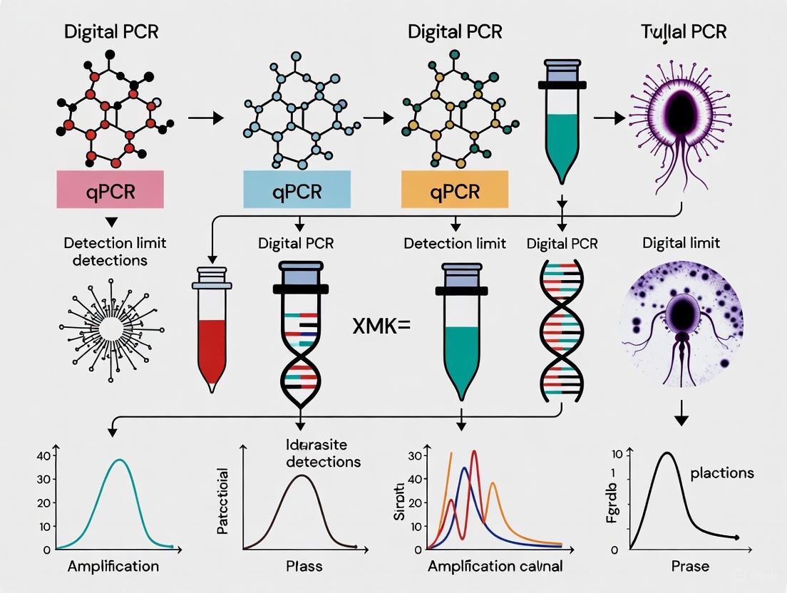
Visualization of Methodological Workflows
qPCR Workflow for Relative Quantification
dPCR Workflow for Absolute Quantification
Comparative Sensitivity in Parasite Detection
The evolution from conventional PCR through qPCR to dPCR represents a continuous trajectory toward greater sensitivity, precision, and quantification capability in molecular detection. In parasitology research, each technology maintains relevance for specific applications: conventional PCR for basic species identification and presence/absence detection, qPCR for robust quantification of parasite load in clinical samples, and dPCR for scenarios requiring ultimate sensitivity and absolute quantification of low-abundance targets. The experimental data consistently demonstrate dPCR's superior performance for detecting low-level infections, quantifying minor genetic variants, and monitoring treatment response with precision unattainable by earlier technologies.
While qPCR remains the workhorse for many diagnostic applications due to its established protocols, wider dynamic range, and lower cost per reaction, dPCR's exceptional sensitivity and standardization advantages position it as transformative for advancing parasite research. The technology enables earlier detection of infections, more precise monitoring of treatment efficacy, and identification of reservoir hosts with low parasite burdens. As dPCR platforms become more accessible and cost-effective, their implementation is expected to expand, potentially enabling new research paradigms in parasitology and significantly enhancing capabilities for controlling parasitic diseases of public health importance.
Digital PCR (dPCR) represents a transformative advancement in nucleic acid quantification technology, fundamentally differing from quantitative PCR (qPCR) through its combination of sample partitioning and Poisson statistical analysis. This technology enables absolute quantification of target sequences without requiring standard curves, demonstrating particular utility in applications demanding high sensitivity and precision, including parasite research and pathogen detection. This guide provides an objective comparison of dPCR versus qPCR performance, focusing on detection limit comparisons through synthesized experimental data from recent studies. The analysis reveals that dPCR consistently outperforms qPCR in detecting low-abundance targets, with superior precision, accuracy, and tolerance to inhibitors, making it exceptionally valuable for drug development and clinical diagnostics where minimal residual disease or low-level infections must be monitored.
Digital PCR (dPCR) constitutes a third-generation nucleic acid amplification technology that enables absolute quantification of DNA or RNA targets through a fundamentally different approach than quantitative real-time PCR (qPCR). While qPCR relies on monitoring amplification fluorescence in real-time during the exponential phase and comparing results to standard curves, dPCR employs sample partitioning, endpoint amplification, and Poisson statistical analysis to directly calculate target concentration [10] [11]. This methodological divergence addresses several limitations inherent to qPCR, particularly regarding sensitivity, precision, and resistance to inhibitors.
The historical development of dPCR spans more than three decades, with seminal concepts first described in 1988 and the term "digital PCR" formally coined in 1999 [10]. The technology truly flourished with commercial advancements, beginning with chip-based systems in 2006-2007, followed by droplet digital PCR (ddPCR) in 2011, and culminating in contemporary nanoplate-based systems introduced around 2020 [10]. This evolution has progressively enhanced throughput, ease of use, and accessibility, positioning dPCR as an indispensable tool in modern molecular diagnostics and research.
In the context of parasite research—where detecting low-abundance targets in complex matrices is often paramount—dPCR's capabilities offer distinct advantages. Parasitic infections frequently present diagnostic challenges due to intermittent shedding, low pathogen loads, and complex sample backgrounds that can inhibit amplification [12]. The partitioning principle of dPCR effectively dilutes inhibitors across thousands of reactions, maintaining amplification efficiency where qPCR would falter, thereby providing more reliable results for low-level parasitic infections and monitoring treatment efficacy [12] [13].
Core Principles: Partitioning and Poisson Statistics
The Partitioning Process
The foundational principle of dPCR involves physically partitioning a conventional PCR reaction mixture into thousands to millions of nanoliter-scale reactions, creating individual amplification chambers where each contains zero, one, or several target nucleic acid molecules [10] [11]. This partitioning is achieved through various microfluidic technologies, including:
- Nanoplate-based systems: Utilize etched microfluidic chips with fixed wells [4] [14]
- Droplet-based systems (ddPCR): Generate water-in-oil emulsion droplets [12] [15]
- Chip-based arrays: Employ microfluidic chambers with defined volumes [16]
Following partitioning, standard PCR amplification occurs within each individual reaction unit using target-specific primers and probes. Crucially, amplification is performed to endpoint rather than monitored in real-time, with each partition functioning as a discrete digital reaction vessel [10]. Partitions containing at least one target molecule generate a positive fluorescence signal, while those without targets remain negative. This binary (positive/negative) outcome forms the digital signature that gives the technology its name [10] [11].
Poisson Statistical Analysis
The quantification methodology in dPCR relies fundamentally on Poisson statistics, which accounts for the random distribution of target molecules across partitions [10] [16]. The Poisson model calculates the probability of a partition receiving zero, one, or multiple target molecules based on the observed ratio of positive to negative partitions.
The standard Poisson equation is applied as follows:
λ = -ln(1 - p)
Where λ represents the average number of target molecules per partition, and p represents the proportion of positive partitions [10] [16]. This calculation accommodates the fact that some partitions may contain multiple target molecules, preventing underestimation of concentration.
The absolute concentration in copies per microliter is then derived using the formula:
Concentration (copies/μL) = λ / partition volume (μL)
This statistical approach eliminates the requirement for standard curves and reference materials that are essential for qPCR quantification, enabling true absolute quantification [10] [14]. The accuracy of this method depends on having sufficient partitions to ensure statistical robustness, with modern dPCR systems typically generating 20,000-30,000 partitions per reaction [4] [17].
The following diagram illustrates the complete dPCR workflow from sample partitioning through data analysis:
Figure 1: dPCR Workflow from Partitioning to Quantification
Performance Comparison: dPCR vs. qPCR
Direct Performance Metrics Comparison
Numerous studies have directly compared the analytical performance of dPCR and qPCR across various applications. The following table synthesizes key performance metrics from recent research:
Table 1: Comparative Performance Metrics of dPCR vs. qPCR
| Performance Parameter | dPCR Performance | qPCR Performance | Experimental Context |
|---|---|---|---|
| Limit of Detection (LoD) | 1.6 IU/mL for HBV DNA [15] | Higher than dPCR (specific values not provided) | Hepatitis B virus detection in serum [15] |
| Precision (Intra-assay Variability) | Median CV%: 4.5% [4] | Higher variability than dPCR (p=0.020) [4] | Periodontal pathobiont detection [4] |
| Dynamic Range | 6 logs [18] | 8 logs [18] | CAR-T manufacturing validation [18] |
| Sensitivity for Low Bacterial Loads | Superior detection, fewer false negatives [4] | 5-fold underestimation of A. actinomycetemcomitans prevalence [4] | Periodontal pathogen quantification [4] |
| Tolerance to PCR Inhibitors | High tolerance due to partitioning [10] [12] | Susceptible to inhibition [10] [14] | Complex clinical samples [12] |
| Quantification Reference | Absolute quantification without standards [10] [14] | Requires standard curves for quantification [10] [14] | Fundamental methodological difference [10] |
| Data Variation in Sample Analysis | Lower variation (R² = 0.99 for linked genes) [18] | Higher variation (R² = 0.78 for linked genes) [18] | CAR-T manufacturing [18] |
Detection Limit Comparisons in Pathogen Research
The superior sensitivity of dPCR is particularly evident in pathogen detection research, where identifying low-abundance targets is critical for accurate diagnosis and monitoring. In periodontal microbiology, dPCR demonstrated significantly enhanced sensitivity for detecting Porphyromonas gingivalis and Aggregatibacter actinomycetemcomitans, identifying bacterial loads that yielded false negatives in qPCR assays [4]. Bland-Altman analysis revealed that qPCR systematically underestimated the prevalence of A. actinomycetemcomitans by approximately 5-fold in periodontitis patients compared to dPCR, particularly at concentrations below 3 log₁₀ genome equivalents/mL [4].
In virology applications, a droplet digital PCR assay for hepatitis B virus (HBV) DNA detection achieved an exceptional limit of detection of 1.6 IU/mL using only 200μL of serum, substantially surpassing the sensitivity of conventional real-time PCR assays [15]. This enhanced sensitivity enables detection of residual viremia in chronic hepatitis B patients undergoing antiviral therapy, representing a significant advancement for treatment monitoring [15].
Similarly, in respiratory virus diagnostics during the 2023-2024 "tripledemic," dPCR demonstrated superior quantification accuracy for influenza A, influenza B, RSV, and SARS-CoV-2, particularly in samples with medium to high viral loads [17]. The precision of dPCR quantification showed greater consistency across replicates compared to the variability often observed with qPCR Ct values [17].
Experimental Protocols for Method Comparison
Protocol 1: Periodontal Pathobiont Detection
Objective: Compare multiplex dPCR and qPCR assays for simultaneous detection and quantification of Porphyromonas gingivalis, Aggregatibacter actinomycetemcomitans, and Fusobacterium nucleatum in subgingival plaque samples [4].
Sample Collection:
- Subgingival plaque samples collected from 20 periodontitis patients and 20 healthy controls
- Four sites per subject pooled into sample tubes containing 1 mL of reduced transport fluid with 10% glycerol
- Immediate storage at -20°C until processing [4]
DNA Extraction:
- QIAamp DNA Mini kit (Qiagen) following manufacturer's instructions [4]
dPCR Assay:
- Instrument: QIAcuity Four (Qiagen) with Nanoplate 26k 24-well plates
- Reaction volume: 40μL containing 10μL sample DNA, 10μL 4× Probe PCR Master Mix, 0.4μM of each specific primer, 0.2μM of each specific probe, 0.025 U/μL restriction enzyme
- Thermocycling: Initial denaturation/enzyme activation at 95°C for 2 min; 45 cycles of 95°C for 15s and 58°C for 1min
- Imaging: Three channels with specific thresholds (30 RFU for A. actinomycetemcomitans, 40 RFU for P. gingivalis, 40 RFU for F. nucleatum)
- Data analysis: QIAcuity Software Suite v2.5.0.1 with automatic Poisson-based concentration calculation [4]
qPCR Assay:
- Standard quantitative PCR performed for comparison
- Same primer/probe sets as dPCR assay
- Quantification via standard curve method [4]
Statistical Analysis:
- Mann-Whitney U test, Wilcoxon test, McNemar's test
- Bland-Altman plots for agreement analysis
- CV% calculation for precision comparison [4]
Protocol 2: Respiratory Virus Detection and Quantification
Objective: Compare detection and quantification of influenza A, influenza B, RSV, and SARS-CoV-2 using dPCR and Real-Time RT-PCR [17].
Sample Collection and Stratification:
- 123 respiratory samples collected November 2023-April 2024
- Stratification by Ct values: high (≤25), medium (25.1-30), low (>30)
- Includes nasopharyngeal swabs and bronchoalveolar lavage samples [17]
RNA Extraction for Real-Time RT-PCR:
- Platform: STARlet Seegene automated system
- Kit: STARMag 96 × 4 Universal Cartridge Kit
- Multiplex Real-Time RT-PCR: Allplex Respiratory Panel 1A, 2, and 3
- Detection: CFX96 thermocycler with internal controls [17]
dPCR Workflow:
- RNA extraction: KingFisher Flex system with MagMax Viral/Pathogen kit
- Instrument: QIAcuity platform (Qiagen)
- Format: Five-target multiplex (influenza A, influenza B, RSV, SARS-CoV-2, internal control)
- Partitioning: ~26,000 nanowells per reaction
- Data analysis: QIAcuity Suite software v.0.1 for absolute copy number calculation [17]
Statistical Analysis:
- Descriptive analysis of RNA concentration values
- Outlier identification via boxplot visualization (IQR × 1.5)
- Kruskal-Wallis test for between-group comparisons [17]
Essential Research Reagent Solutions
Successful implementation of dPCR technology requires specific reagent systems optimized for partitioned amplification. The following table details essential research reagents and their functions:
Table 2: Essential Research Reagent Solutions for dPCR Experiments
| Reagent/Material | Function | Application Examples |
|---|---|---|
| Nanoplate-based dPCR Plates | Microfluidic chips with fixed partitions for reaction compartmentalization | QIAcuity Nanoplate 26k 24-well plates for periodontal pathobiont detection [4] |
| Probe-based dPCR Master Mix | Optimized reaction mix containing polymerase, nucleotides, buffer for partitioned amplification | QIAcuity Probe PCR Kit for multiplex pathogen detection [4] |
| Restriction Enzymes | Enhance assay efficiency by cutting complex DNA structures | Anza 52 PvuII restriction enzyme in periodontal pathogen assay [4] |
| Sequence-Specific Primers/Probes | Target-specific oligonucleotides for amplification detection; often double-quenched hydrolysis probes | 16S rRNA-targeted primers/probes for P. gingivalis, A. actinomycetemcomitans, F. nucleatum [4] |
| Nucleic Acid Extraction Kits | High-purity DNA/RNA isolation from various sample matrices | QIAamp DNA Mini Kit for subgingival plaque [4]; MagMax Viral/Pathogen Kit for respiratory samples [17] |
| Fluorescence Reference Dyes | Internal partition volume calibration | ROX fluorescence level as proxy for effective load volume in partition size variation correction [16] |
Application in Parasite Research and Diagnostics
Parasitology has emerged as a particularly promising field for dPCR implementation, addressing longstanding diagnostic challenges associated with conventional methods. Traditional parasitological techniques based on microscopic examination suffer from limitations including inadequate sensitivity, labor-intensive procedures, and subjective interpretation [12]. dPCR technology offers solutions to these limitations through its exceptional sensitivity and objective quantification capabilities.
Recent applications in parasite research demonstrate dPCR's versatility across different parasite groups and sample types. In helminth research, dPCR assays have been developed for:
- Species-specific detection of Cooperia and Ostertagia using ITS2 targets [12]
- Anthelmintic resistance monitoring in Haemonchus contortus via β-tubulin (F200Y) discrimination [12]
- Parasite quantification in environmental DNA (eDNA) from water samples for Chilodonella hexasticha and Gyrodactylis salaris detection [12]
For protozoan parasites, dPCR applications include:
- Plasmodium falciparum detection in blood samples using hrp2, hrp3 tRNA targets [12]
- Drug resistance monitoring through quantification of mdr1, plasmepsin2, and other resistance-associated genes [12]
- Toxoplasma gondii detection in tissue samples using the Toxo-529 repeat element [12]
The environmental DNA (eDNA) applications represent a particularly innovative approach, enabling parasite detection in water and soil samples without direct host examination. This methodology has been successfully applied for Fasciola and Taenia solium detection in environmental samples, offering new possibilities for ecosystem-level parasite surveillance [12].
A critical advantage of dPCR in parasite diagnostics is its capacity to maintain detection sensitivity in the presence of PCR inhibitors commonly found in complex sample matrices. The partitioning principle effectively dilutes inhibitors across thousands of reactions, preventing the generalized amplification suppression that frequently affects qPCR assays [12]. This robustness makes dPCR especially valuable for field-collected samples where ideal preservation conditions may be difficult to maintain.
Technical Considerations and Limitations
Despite its considerable advantages, dPCR technology presents certain limitations that researchers must consider when selecting an appropriate quantification platform. The dynamic range of dPCR is inherently constrained by the number of partitions generated, typically spanning 4-6 orders of magnitude compared to 8-10 orders for qPCR [18] [13]. This limitation necessitates sample dilution for accurately quantifying high-concentration targets, adding an extra processing step.
The throughput and operational cost considerations also favor qPCR for routine high-volume testing. While dPCR provides superior data quality for low-abundance targets, the per-reaction cost remains higher than qPCR, and processing throughput is generally lower [13]. This economic factor becomes particularly relevant in large-scale screening scenarios where extreme sensitivity is not required.
dPCR also presents specific technical challenges including:
- Volume variation effects: Partition volume inconsistencies can cause quantification inaccuracies, especially at higher concentrations, necessitating Poisson-Plus modeling for correction [16]
- Amplicon size limitations: dPCR is less suitable for very large amplicons compared to qPCR [10]
- Potential biases: DNA denaturation during partitioning may cause strand separation into different partitions, potentially leading to overestimation [10]
- Template linkage: Sample inhomogeneity and molecular dropout effects can cause underestimation in some scenarios [10]
The following diagram illustrates the relative advantages and limitations of dPCR versus qPCR across key performance parameters:
Figure 2: Comparative Advantages and Limitations of dPCR vs. qPCR
Digital PCR represents a paradigm shift in nucleic acid quantification technology, with its fundamental principles of sample partitioning and Poisson statistical analysis enabling absolute quantification without standard curves. The technology demonstrates consistent advantages over qPCR for detecting low-abundance targets, with superior sensitivity, precision, and robustness against amplification inhibitors.
In parasite research and diagnostics, these capabilities address critical challenges in detecting minimal residual infection, monitoring treatment efficacy, and identifying drug-resistant strains. The exceptional sensitivity of dPCR—evidenced by its ability to detect HBV DNA at 1.6 IU/mL and periodontal pathogens at concentrations yielding qPCR false negatives—makes it particularly valuable for scenarios where target scarcity compromises diagnostic accuracy [4] [15].
Despite its limitations in dynamic range and throughput, dPCR has established an indispensable niche in applications requiring ultimate sensitivity and precise quantification. As the technology continues to evolve with improvements in multiplexing capacity, throughput, and cost-effectiveness, its implementation in research and clinical diagnostics is poised to expand, particularly for challenging applications in parasitology, oncology, and infectious disease monitoring where quantitative accuracy at low target concentrations directly impacts research conclusions and clinical decisions.
Molecular diagnostics for parasitic and other pathogen research increasingly rely on sophisticated amplification technologies to detect and quantify infectious agents. Quantitative PCR (qPCR) has served as the long-standing gold standard for nucleic acid detection, providing relative quantification based on comparison to standard curves generated from known concentrations [19] [20]. This technique monitors amplification in real-time using fluorescent reporters, offering high throughput and established protocols familiar to most laboratories. However, its dependence on external calibration and relative quantification introduces limitations for applications requiring absolute quantification or detection of rare targets.
In contrast, digital PCR (dPCR) represents a technological evolution that enables absolute nucleic acid quantification without standard curves [19]. This method partitions a PCR reaction into thousands of individual reactions, with each partition serving as a separate amplification event. After endpoint amplification, the ratio of positive to negative partitions allows absolute quantification using Poisson statistics [21] [19]. This fundamental difference in approach provides dPCR with distinct advantages in sensitivity, precision, and accuracy for specific applications, particularly in pathogen detection where low target numbers are common.
Performance Metrics: Direct Comparison of dPCR and qPCR
Sensitivity and Limit of Detection
Sensitivity in molecular diagnostics refers to the lowest concentration of a target that can be reliably detected. The Limit of Detection (LOD) defines the lowest concentration at which a target can be detected with specified confidence, while the Limit of Quantification (LOQ) represents the lowest concentration that can be accurately quantified [22]. Multiple studies demonstrate dPCR's enhanced sensitivity compared to qPCR, particularly at low target concentrations.
In SARS-CoV-2 detection, ddPCR demonstrated superior sensitivity compared to RT-qPCR in clinical samples. One study found ddPCR detected 93 positive cases versus 89 with RT-qPCR from the same 130 clinical samples, indicating its ability to identify infections with lower viral loads [23]. Similarly, for infectious bronchitis virus (IBV) in avian samples, dPCR showed higher sensitivity compared to qPCR assays [2]. This enhanced sensitivity stems from dPCR's ability to detect single molecules and its reduced susceptibility to amplification inhibitors present in complex sample matrices [24] [23].
Platform-specific LOD evaluations reveal precise detection capabilities. In a comparative study of dPCR systems, the LOD for nanoplate-based dPCR (ndPCR) was approximately 0.39 copies/µL input, while droplet-based dPCR (ddPCR) showed an LOD of approximately 0.17 copies/µL input [22]. The same study determined LOQ values of 1.35 copies/µL for ndPCR and 4.26 copies/µL for ddPCR, highlighting the importance of platform selection based on specific application requirements.
Precision and Reproducibility
Precision refers to the reproducibility and repeatability of measurements, typically expressed as the coefficient of variation (%CV) between technical replicates. dPCR consistently demonstrates superior precision compared to qPCR across multiple applications and sample types.
A direct comparison using human genomic DNA spiked at 175 copies/µl showed dPCR had a 2.3% CV versus 5.0% CV for qPCR—more than a two-fold improvement in measurement variability [24]. When dPCR replicates were pooled, variability decreased further to 1.5% CV, nearly three-fold lower than qPCR duplicate averages (4.4% CV) [24]. This enhanced precision stems from dPCR's digital nature and endpoint quantification, which eliminates variability associated with amplification efficiency differences in qPCR.
In copy number variation (CNV) analysis of the DEFA1A3 gene, ddPCR showed 95% concordance with pulsed-field gel electrophoresis (PFGE, considered a gold standard), while qPCR results were only 60% concordant with PFGE [21]. The Spearman correlation was significantly stronger for ddPCR versus PFGE (r = 0.90) than for qPCR versus PFGE (r = 0.57), with ddPCR copy numbers differing only 5% on average from PFGE, compared to 22% for qPCR [21].
Accuracy and Dynamic Range
Accuracy represents how close measured values are to true values. dPCR provides superior accuracy for absolute quantification because it does not rely on external standards that can introduce variability [19] [20]. However, qPCR typically offers a wider dynamic range of quantification [2].
In a study comparing quantification of synthetic oligonucleotides, both dPCR platforms (nanoplate and droplet-based) showed high correlation with expected values (R²adj = 0.98-0.99), though measured copies were consistently slightly lower than expected for both platforms [22]. This systematic underestimation may relate to matrix effects or partitioning efficiency, but the high correlation demonstrates dPCR's quantification accuracy across a broad concentration range.
For viral load monitoring, dPCR's accuracy provides significant advantages. In respiratory virus detection during the 2023-2024 "tripledemic," dPCR demonstrated superior accuracy particularly for high viral loads of influenza A, influenza B, and SARS-CoV-2, and for medium loads of RSV compared to Real-Time RT-PCR [17]. This accurate quantification is crucial for understanding infection dynamics, treatment response monitoring, and public health decision-making.
Table 1: Comparative Performance Metrics of dPCR and qPCR
| Performance Metric | Digital PCR (dPCR) | Quantitative PCR (qPCR) | Experimental Context |
|---|---|---|---|
| Limit of Detection (LOD) | 0.17-0.39 copies/µL [22] | Higher than dPCR [23] [2] | SARS-CoV-2 detection [23] |
| Limit of Quantification (LOQ) | 1.35-4.26 copies/µL [22] | Higher than dPCR [2] | Synthetic oligonucleotides [22] |
| Precision (%CV) | 2.3% [24] | 5.0% [24] | Human genomic DNA (175 cp/μL) |
| Concordance with Gold Standard | 95% with PFGE [21] | 60% with PFGE [21] | DEFA1A3 CNV analysis |
| Correlation with Reference | r = 0.90 [21] | r = 0.57 [21] | DEFA1A3 CNV analysis |
| Quantification Method | Absolute, without standard curves [19] [20] | Relative, requires standard curves [19] [20] | Fundamental methodology |
Table 2: Platform-Specific Performance Characteristics
| Platform Parameter | Nanoplate dPCR (QIAcuity) | Droplet dPCR (QX200) | Experimental Context |
|---|---|---|---|
| LOD (copies/µL input) | 0.39 [22] | 0.17 [22] | Synthetic oligonucleotides |
| LOQ (copies/µL input) | 1.35 [22] | 4.26 [22] | Synthetic oligonucleotides |
| Partitioning Mechanism | Fixed nanowells [19] [17] | Water-in-oil droplets [25] [19] | Platform design |
| Typical Partitions | ~26,000 [17] | ~20,000+ [24] | Standard reaction |
| Precision with HaeIII Enzyme | CV: 1.6-14.6% [22] | CV: <5% [22] | Paramecium tetraurelia DNA |
Experimental Protocols for Performance Validation
Sample Preparation and Nucleic Acid Extraction
Proper sample preparation is critical for reproducible PCR results. For viral RNA extraction from clinical samples (e.g., oropharyngeal swabs), protocols typically involve initial sample inactivation at 56°C for 30 minutes, followed by extraction using commercial kits such as the Prefilled Viral Total NA Kit [23]. Automated extraction systems like the KingFisher Flex system with MagMax Viral/Pathogen kit provide consistent results for dPCR applications [17]. For copy number variation studies, DNA quality is paramount, with recommendations for fluorometric quantification and purity assessment (A260/280 ratios) before dPCR analysis [21].
Restriction enzyme digestion can significantly impact dPCR precision, especially for targets with potential tandem repeats or complex secondary structures. In studies using Paramecium tetraurelia DNA, precision improved markedly with HaeIII compared to EcoRI, particularly for the QX200 ddPCR system, which showed CV values reduced to <5% with HaeIII versus up to 62.1% with EcoRI [22]. Enzyme selection should be optimized for each specific target to ensure complete digestion and access to target sequences.
Reaction Setup and Partitioning
dPCR reaction setup follows similar principles to qPCR but requires optimization of partitioning parameters. Typical 25µL reactions contain 10µL of template RNA/DNA, enzyme mix, and primer-probe combinations [23]. For the QIAcuity nanoplate system, samples are loaded into plates that partition reactions into approximately 26,000 nanowells [17]. For droplet-based systems like the QX200 or RainSure DropX-2000, reactions are partitioned into ~20,000 nanoliter-sized droplets through microfluidic emulsion generation [25] [23].
Optimal template concentration is critical for accurate dPCR quantification. The ideal range is approximately 100-1,000 copies per reaction to ensure sufficient positive partitions while avoiding saturation effects [22]. Too many targets per partition violates the Poisson distribution assumption, while too few reduces quantification precision. Sample dilution series may be necessary to determine optimal loading concentrations for unknown samples.
Thermal Cycling and Data Analysis
dPCR employs standard PCR thermal cycling protocols but uses endpoint detection rather than real-time monitoring. A typical SARS-CoV-2 detection protocol includes: reverse transcription at 49°C for 20 minutes; DNA polymerase activation at 97°C for 12 minutes; 40 cycles of denaturation at 95.3°C for 20 seconds and annealing at 52°C for 1 minute; followed by a final cooling hold at 20°C [23].
Data analysis utilizes Poisson statistics to calculate absolute target concentration based on the fraction of positive partitions: Concentration = −ln(1−p) / V, where p is the fraction of positive partitions and V is the partition volume [19]. Commercial platforms include proprietary software that automatically performs these calculations (e.g., GeneCount, QIAcuity Suite) while providing visualization of amplification clusters [23] [17]. Threshold setting between positive and negative partitions is crucial and may require manual adjustment in cases of ambiguous clustering.
PCR Workflow Comparison: dPCR vs. qPCR
Essential Research Reagent Solutions
Successful implementation of dPCR and qPCR assays requires careful selection of reagents and consumables. The following table outlines key solutions and their functions in parasite and pathogen detection research.
Table 3: Essential Research Reagent Solutions for PCR-Based Detection
| Reagent/Consumable | Function | Application Notes |
|---|---|---|
| Nucleic Acid Extraction Kits (e.g., MagMax Viral/Pathogen, QIAamp Viral RNA Mini) | Isolation of high-quality DNA/RNA from clinical/environmental samples | Automated systems (KingFisher Flex, STARlet) improve reproducibility [23] [17] |
| One-Step RT-dPCR/ddPCR Master Mix | Combined reverse transcription and PCR amplification | Includes reverse transcriptase, DNA polymerase, dNTPs in optimized buffer [23] |
| Restriction Enzymes (e.g., HaeIII, EcoRI) | Digest complex DNA to improve target accessibility | Enzyme choice significantly impacts precision, especially for high-copy targets [22] |
| Sequence-Specific Primers & Probes | Target-specific amplification and detection | Dual-labeled hydrolysis probes (FAM/HEX) enable multiplexing; design following dMIQE guidelines [23] [17] |
| Partitioning Oil/Consumables | Create stable water-in-oil emulsions (ddPCR) or load nanowells | Critical for consistent partition generation; use manufacturer-recommended formulations [19] |
| Positive Control Templates | Assay validation and run controls | Synthetic oligonucleotides or characterized positive samples [22] |
The comparative analysis of dPCR and qPCR technologies reveals a clear distinction in their performance characteristics and optimal application scenarios. dPCR demonstrates superior sensitivity, precision, and accuracy for absolute quantification, particularly at low target concentrations and in complex sample matrices [25] [21] [24]. These advantages make it particularly valuable for parasite and pathogen detection research where low abundance targets, precise quantification, and detection of rare mutations are critical.
However, qPCR maintains important advantages in throughput, dynamic range, and established infrastructure [2] [20]. The choice between technologies should be guided by specific research requirements, with dPCR preferred for applications demanding absolute quantification and high precision, and qPCR remaining suitable for high-throughput screening where relative quantification suffices. As dPCR technology continues to evolve with improved automation and reduced costs, its implementation in routine parasite research and clinical diagnostics is likely to expand, particularly for challenging detection scenarios where its performance advantages provide significant scientific and clinical value.
Digital PCR (dPCR) represents a significant advancement in molecular diagnostics, offering unparalleled precision for nucleic acid quantification. This guide explores the core technical advantage of dPCR: the robustness conferred by its end-point analysis. Unlike quantitative PCR (qPCR), which relies on real-time amplification kinetics, dPCR's endpoint approach and sample partitioning confer superior tolerance to PCR inhibitors and reduced susceptibility to amplification efficiency variations. Within parasite research and broader microbiological contexts, this translates to enhanced detection capabilities, particularly for low-abundance targets in complex sample matrices. This article provides a detailed comparison with qPCR, supported by experimental data and methodologies relevant to researchers and drug development professionals.
The evolution of polymerase chain reaction (PCR) technology has progressed from conventional end-point PCR to quantitative real-time PCR (qPCR) and now to third-generation digital PCR (dPCR). While qPCR monitors fluorescence intensity during the exponential phase of amplification, dPCR utilizes an end-point detection method after partitioning the sample into thousands of individual reactions [26] [27]. In dPCR, the bulk reaction mixture is partitioned into numerous nanoscale reactions, each containing zero, one, or a few target molecules. After thermal cycling is complete, each partition is analyzed as a discrete positive or negative event based on fluorescence, eliminating the need to monitor amplification kinetics in real-time [28] [29]. This fundamental difference in detection methodology underpins dPCR's enhanced performance characteristics.
The concept of dPCR was first developed in the 1990s and has since evolved into robust commercial platforms [22] [27]. The technique is often referred to as a "digital" method because it converts the continuous measurement of nucleic acid concentration into a binary readout of positive and negative partitions [30] [29]. The proportion of positive partitions follows Poisson statistics, enabling absolute quantification of the target nucleic acid without reference to standard curves [28] [26]. This independence from external calibrators and reduced sensitivity to amplification variables makes dPCR particularly valuable for applications requiring high precision, such as detection of low-level pathogens, copy number variation analysis, and rare mutation detection [2] [31] [4].
Fundamental Mechanisms: How End-point Analysis Confers Robustness
The Partitioning Principle and Dilution of Inhibitors
The core mechanism behind dPCR's robustness lies in its physical partitioning of PCR reactions. By dividing a single bulk reaction into tens of thousands of nanoscale partitions, dPCR effectively dilutes PCR inhibitors across these numerous microreactions [28]. Substances that inhibit polymerase activity—such as humic acids in environmental samples, hemoglobin in blood, or mucins in respiratory specimens—are randomly distributed throughout the partitions. Consequently, their concentration within any single partition becomes negligible, minimizing their impact on amplification [28] [29]. This natural dilution effect preserves amplification efficiency in the majority of partitions, enabling accurate target quantification even in samples where qPCR would fail due to inhibition.
In contrast to qPCR's reliance on amplification kinetics, dPCR utilizes endpoint detection, which further enhances its tolerance to inhibitors [28] [29]. While inhibitors may delay or slow amplification in affected partitions, dPCR only requires that amplification reaches a detectable fluorescence threshold by the end of the thermal cycling process. This binary endpoint measurement contrasts sharply with qPCR, where the precise cycle at which fluorescence crosses the threshold (Cq value) is critical for quantification [26]. Even significantly delayed amplification will still yield a positive partition in dPCR, whereas the same delay would substantially alter the Cq value in qPCR, leading to inaccurate quantification [29].
Independence from Amplification Efficiency
qPCR quantification is fundamentally dependent on consistent and optimal amplification efficiency between the target and any reference materials used for calibration. Variations in efficiency due to inhibitor presence, primer quality, or sample matrix effects introduce significant quantification errors [26]. dPCR eliminates this dependency because quantification is based on the statistical distribution of positive and negative partitions at the endpoint, not the rate or efficiency of amplification [26] [30]. Each partition effectively functions as a separate PCR microreactor with its own amplification characteristics, yet the final count depends only on the initial presence or absence of the target molecule, not how efficiently it was amplified.
This independence from amplification efficiency makes dPCR exceptionally robust for comparing dissimilar samples or analyzing targets with different amplification kinetics. The technology provides absolute quantification without the need for standard curves, eliminating a major source of inter-laboratory variability [26] [29]. For research on parasites and other microorganisms with variable gene copy numbers, this characteristic is particularly valuable, as it enables direct comparison across different species, strains, and sample types without optimization for each new target [22].
Comparative Experimental Data: dPCR vs. qPCR
Performance in Parasite and Protist Research
Recent research on protists and parasites demonstrates dPCR's superior performance characteristics. A 2025 study comparing dPCR platforms for gene copy number analysis in the ciliate Paramecium tetraurelia found both nanoplate-based and droplet-based dPCR systems exhibited high precision across most analyses [22]. The research revealed that restriction enzyme selection significantly impacted precision, with HaeIII demonstrating superior performance over EcoRI, particularly for the QX200 ddPCR system [22]. When analyzing DNA from varying cell numbers of P. tetraurelia, both dPCR platforms showed reproducible gene copy number estimates and a linear response to increasing cell numbers, confirming the method's reliability for quantifying unicellular eukaryotes with variable gene copy numbers [22].
Table 1: Comparative Performance of dPCR and qPCR in Microbial Detection
| Performance Metric | Digital PCR | Quantitative PCR | Experimental Context |
|---|---|---|---|
| Sensitivity | Higher sensitivity; detects lower bacterial loads [4] | Lower sensitivity; false negatives at low concentrations [4] | Periodontal pathobiont detection |
| Precision (CV%) | Lower intra-assay variability (median CV%: 4.5%) [4] | Higher intra-assay variability [4] | Periodontal pathobiont detection |
| Inhibitor Tolerance | High tolerance due to partitioning and endpoint detection [28] [29] | Sensitive to inhibitors affecting amplification efficiency [28] | Environmental pathogen quantification |
| Quantification Range | Limited by partition number; may require dilution [26] [2] | Wider dynamic range [26] [2] | Viral genome quantification |
| Accuracy at Low Concentration | Superior accuracy and consistency [17] [4] | Less accurate at low concentrations [17] | Respiratory virus detection |
Detection of Viral Pathogens and Periodontal Bacteria
Comparative studies across diverse pathological contexts consistently demonstrate dPCR's advantages. In respiratory virus detection during the 2023-2024 tripledemic, dPCR showed superior accuracy, particularly for high viral loads of influenza A, influenza B, and SARS-CoV-2, and for medium loads of RSV [17]. The technology demonstrated greater consistency and precision than Real-Time RT-PCR, especially in quantifying intermediate viral levels in complex respiratory matrices containing mucus and cellular debris [17].
Similarly, a 2025 study on periodontal pathobionts found dPCR outperformed qPCR for quantifying Porphyromonas gingivalis, Aggregatibacter actinomycetemcomitans, and Fusobacterium nucleatum [4]. dPCR demonstrated superior sensitivity, detecting lower bacterial loads that qPCR missed, resulting in a 5-fold underestimation of A. actinomycetemcomitans prevalence by qPCR in periodontitis patients [4]. The Bland-Altman plots from this study revealed good agreement between the technologies at medium/high bacterial loads but significant discrepancies at low concentrations (< 3 log10Geq/mL), where qPCR produced false negatives [4].
Table 2: Comparison of dPCR and qPCR Fundamental Characteristics
| Characteristic | Digital PCR | Quantitative PCR |
|---|---|---|
| Quantification Method | Absolute quantification using Poisson statistics [26] [30] | Relative quantification based on standard curves [26] |
| Amplification Monitoring | End-point detection [26] | Real-time monitoring [26] |
| Precision | High [26] | Moderate [26] |
| Effect of Inhibitors | Reduced impact due to partitioning [28] [29] | Significant impact on Cq values [28] |
| Dependence on Amplification Efficiency | Low [26] | High [26] |
| Multiplexing Capability | High (using different fluorophores) [28] [26] | Limited [26] |
Experimental Protocols for dPCR Applications
Protocol 1: Pathogen Detection in Complex Matrices
The following protocol is adapted from methodologies used for environmental pathogen quantification and periodontal pathogen detection, particularly relevant for parasite research in complex sample matrices [28] [4]:
Sample Preparation and DNA Extraction: For environmental or clinical samples, begin with thorough homogenization. Extract DNA using validated kits (e.g., QIAamp DNA Mini kit). Include appropriate negative controls throughout the process. For difficult samples containing PCR inhibitors, additional purification steps may be beneficial, though dPCR is generally more tolerant than qPCR [4].
Reaction Mixture Preparation: Prepare dPCR reaction mixtures containing:
- 1X dPCR Master Mix (e.g., QIAcuity Probe PCR Kit)
- 0.4-0.9 µM of each primer
- 0.2-0.25 µM of each fluorescent probe (FAM, VIC/HEX, or equivalent)
- Optional: Restriction enzyme (e.g., 0.025 U/µL Anza 52 PvuII or HaeIII) to improve accessibility to tandemly repeated genes [22]
- 10-20 µL of sample DNA
- Nuclease-free water to final volume (20-40 µL depending on platform)
Partitioning and Thermocycling: Load reaction mixtures into the appropriate dPCR platform (nanoplate-based or droplet-based). Execute partitioning according to manufacturer specifications. Perform thermocycling with conditions typically including:
- Initial activation: 2-10 minutes at 95°C
- 40-45 cycles of: 15-30 seconds denaturation at 95°C, 30-60 seconds annealing/extension at 55-60°C
- Final hold at 4-10°C
Endpoint Imaging and Analysis: Following thermocycling, perform fluorescence reading of all partitions. Set appropriate fluorescence thresholds for each channel based on positive and negative control clusters. Apply volume precision factors if available for the platform. Use Poisson correction for precise concentration calculation [4].
Protocol 2: Copy Number Variation Analysis in Parasites
For parasite research involving gene copy number variations, the following protocol adapted from protist studies provides optimal results [22]:
Standard Curve Generation: Prepare serial dilutions of synthetic oligonucleotides or reference DNA with known copy numbers. Include a range that covers expected concentrations in test samples.
Restriction Enzyme Optimization: Test different restriction enzymes (e.g., EcoRI vs. HaeIII) to determine which provides optimal precision for your target organism. Research indicates enzyme choice can significantly impact precision, especially for organisms with high gene copy numbers or tandem repeats [22].
Multiplex dPCR Setup: For simultaneous quantification of target and reference genes, design primer-probe sets with different fluorophores. Optimize concentrations to minimize channel crosstalk. Use a 2D plot visualization to properly set thresholds for each target [28].
Data Analysis: Calculate absolute copy numbers using the platform's software with Poisson statistics. For precision assessment, run multiple technical replicates. Determine the limit of detection (LOD) and limit of quantification (LOQ) using statistical methods appropriate for dPCR [22].
Visualization of dPCR Workflow and Advantage Mechanisms
dPCR Workflow and End-point Detection
The fundamental dPCR workflow demonstrates how sample partitioning and endpoint detection create a robust quantification system resistant to inhibitors and amplification efficiency variations.
Comparative Advantage Mechanism
This comparative diagram illustrates the differential impact of PCR inhibitors on qPCR versus dPCR, highlighting how dPCR's partitioning and endpoint analysis maintain quantification accuracy despite inhibition.
Essential Research Reagent Solutions
Table 3: Key Research Reagents for dPCR Applications in Parasitology
| Reagent Category | Specific Examples | Function and Importance |
|---|---|---|
| dPCR Master Mixes | QIAcuity Probe PCR Kit, QuantStudio 3D Digital PCR Master Mix | Optimized buffer systems for partitioned amplification; critical for assay performance and inhibitor tolerance [4]. |
| Restriction Enzymes | HaeIII, EcoRI, PvuII | Improve access to target sequences in complex genomes; enhance precision for organisms with tandem repeats [22]. |
| Fluorescent Probes | FAM, VIC/HEX, Cy5-labeled TaqMan probes | Enable multiplex detection; proper probe design is essential for specific target identification [28] [4]. |
| Nucleic Acid Extraction Kits | QIAamp DNA Mini Kit, MagMax Viral/Pathogen Kit | Ensure high-quality template DNA; optimized for complex sample matrices [17] [4]. |
| Reference Materials | Synthetic oligonucleotides, genomic DNA from reference strains | Essential for assay validation and determining limits of detection [22]. |
End-point analysis in dPCR provides fundamental advantages for nucleic acid quantification, particularly in challenging research contexts such as parasite detection where sample inhibitors and variable amplification efficiency compromise qPCR results. The partitioning of reactions and statistical approach to quantification makes dPCR uniquely robust, enabling precise measurement even in suboptimal conditions. As molecular diagnostics continue to evolve, dPCR's exceptional performance characteristics position it as an indispensable tool for researchers requiring absolute quantification, especially when working with complex samples, low-abundance targets, or difficult-to-amplify templates. While factors such as dynamic range and throughput may still favor qPCR for some applications, dPCR's superior precision and robustness make it particularly valuable for definitive quantification in parasite research and drug development.
Implementing dPCR and qPCR in Parasitology: Protocols and Workflows
Molecular diagnostics have become indispensable in parasitology, enabling the detection and quantification of pathogens with high specificity and sensitivity. For researchers and drug development professionals, choosing the right polymerase chain reaction (PCR) technology is crucial for assay performance, particularly when dealing with low-abundance parasites in complex clinical samples. This guide provides an objective comparison between quantitative real-time PCR (qPCR) and digital PCR (dPCR) platforms, focusing on their application in parasite detection. The core thesis examines how this technological selection impacts key performance parameters, especially the limit of detection (LOD), which directly influences diagnostic accuracy and research outcomes in parasitic disease management.
The fundamental difference between these technologies lies in their quantification approach. qPCR relies on extrapolating target quantity from amplification curves against a standard curve, while dPCR uses endpoint detection and Poisson statistics to provide absolute quantification without external calibration [22]. This methodological distinction underpins their differing performance characteristics in parasite detection assays.
Performance Comparison: dPCR vs. qPCR for Parasite Detection
Analytical Performance Metrics
Table 1: Comparative analytical performance of dPCR versus qPCR based on experimental studies.
| Performance Metric | Digital PCR (dPCR) | Quantitative PCR (qPCR) | Experimental Context |
|---|---|---|---|
| Limit of Detection (LOD) | 0.17–0.39 copies/µL input [22] | Varies by assay; ~0.02 parasites/µL for optimized real-time PCR [32] | Synthetic oligonucleotides and Plasmodium detection [22] [32] |
| Precision (Coefficient of Variation) | Median CV%: 4.5% [4] | Higher variation; up to 20% difference in copy number ratio [18] | Periodontal pathobiont quantification; CAR-T manufacturing validation [4] [18] |
| Dynamic Range | 6 logs [18] | 8 logs [18] | gBlock DNA standards [18] |
| Sensitivity at Low Bacterial Loads | Superior; detects lower bacterial loads, reduces false negatives [4] | Less effective; 5-fold underestimation of pathogen prevalence [4] | Periodontal pathobiont detection in subgingival plaque [4] |
| Accuracy at Low Concentrations | Good agreement with expected values [22] | Underestimation of copies at low concentrations [4] | Synthetic oligonucleotides; bacterial quantification [22] [4] |
| Tolerance to Inhibitors | Higher tolerance due to partitioning [4] | More susceptible to inhibition [4] | Complex clinical samples (subgingival plaque) [4] |
Practical Implementation Considerations
Table 2: Practical considerations for implementing dPCR and qPCR in parasite research.
| Consideration | Digital PCR (dPCR) | Quantitative PCR (qPCR) |
|---|---|---|
| Multiplexing Capability | Suitable for multiplex analyses; quadruplex demonstrated [18] [4] | Typically single-plex; separate runs often needed for species differentiation [33] [18] |
| Quantification Method | Absolute quantification without standard curves [22] [4] | Relative quantification requiring standard curves [4] |
| Throughput | High-throughput compatible [33] | Well-established high-throughput protocols |
| Assay Development | Requires optimization of partitioning [22] | Established primer/probe design protocols [34] |
| Data Analysis Complexity | Poisson statistics-based [22] [4] | Cycle threshold (Ct) analysis against standard curve |
| Cost Considerations | Higher per-run costs | Lower per-run costs, but requires standards |
Experimental Protocols for Parasite Detection Assays
Protocol 1: Development of a Novel Real-Time PCR Assay for Plasmodium Ovale Subspecies
A 2024 study developed new real-time PCR assays for detecting and differentiating Plasmodium ovalecurtisi and Plasmodium ovalewallikeri, demonstrating a systematic approach to parasite assay development [33].
Methodology:
- Target Selection: Identified repetitive sequence motifs (100 bp length with ≥6 copies) in available P. ovalecurtisi (PocGH01) and P. ovalewallikeri (PowCR01) reference genomes using Jellyfish software [33].
- Specificity Filtering: Excluded sequences with low GC content (<25%) and those aligning to other Plasmodium parasites via BLASTn against NCBI nt database [33].
- Primer/Probe Design: Designed manually using Oligo Calc and DNAMAN to estimate melting temperatures and avoid self-complementarity and primer dimers [33].
- Assay Validation: Evaluated analytical sensitivity using synthetic plasmids and specificity using a panel of 55 P. ovale samples and 40 non-ovale Plasmodium samples from the Democratic Republic of the Congo [33].
Results: The best-performing P. ovalecurtisi target had 9 copies in the reference genome with LOD of 3.6 parasite genome equivalents/μL, while the P. ovalewallikeri target had 8 copies with LOD of 25.9 parasite genome equivalents/μL. The duplex assay showed 100% specificity [33].
Protocol 2: Comparative Performance Evaluation of dPCR vs. qPCR
A 2025 study compared dPCR and qPCR for detecting periodontal pathobionts, providing a methodological framework applicable to parasite detection [4].
Methodology:
- Sample Collection: Subgingival plaque samples from 20 periodontitis patients and 20 healthy controls collected using absorbent paper points [4].
- DNA Extraction: Used QIAamp DNA Mini kit (Qiagen) following manufacturer's instructions [4].
- dPCR Setup: Performed nanoplate-based microfluidic multiplex dPCR assays using QIAcuity Probe PCR Kit in 40 μL reaction mixtures with restriction enzyme Anza 52 PvuII [4].
- Thermocycling: Initial denaturation at 95°C for 2 min, followed by 45 cycles of 15s at 95°C and 1 min at 58°C [4].
- Data Analysis: Concentrations calculated automatically according to Poisson distribution principle using QIAcuity Software Suite [4].
Results: dPCR showed superior sensitivity with lower intra-assay variability (median CV%: 4.5%) than qPCR and detected lower bacterial loads, particularly for low-abundance targets [4].
The Scientist's Toolkit: Essential Research Reagents and Materials
Table 3: Key research reagents and their applications in parasite detection assays.
| Reagent/Material | Function/Application | Example Use Case |
|---|---|---|
| QIAamp DNA Mini Kit (Qiagen) | DNA extraction from clinical samples | DNA extraction from subgingival plaque and blood samples [33] [4] |
| Chelex 100 (Bio-Rad) | DNA extraction, particularly from DBS | DNA extraction from dried blood spots (DBS) in Plasmodium studies [33] |
| Restriction Enzymes (e.g., HaeIII, EcoRI) | Enhance DNA accessibility for amplification | Improving precision in gene copy number estimation, especially for tandem repeats [22] |
| Hydrolysis Probes (e.g., TaqMan) | Sequence-specific detection in real-time PCR | Target-specific detection in qPCR and dPCR assays [33] [4] |
| Mediator Probes (MPs) | Label-free hydrolysis probes for real-time PCR | Probe optimization using design of experiments approach [34] |
| DARQ Probes | Fluorogenic probes for multiplex LAMP | Detection of amplification by release of quenching in isothermal assays [35] |
| Synthetic Oligonucleotides | Assay standardization and control material | Evaluating LOD and LOQ in dPCR and qPCR assays [22] |
Technological Workflows: dPCR vs. qPCR
The following diagram illustrates the key procedural differences between dPCR and qPCR workflows in parasite detection:
The choice between dPCR and qPCR for parasite detection involves strategic trade-offs. dPCR offers superior sensitivity, precision, and absolute quantification for low-abundance targets, making it ideal for detecting latent parasites, monitoring treatment efficacy, and quantifying minor genetic variants. qPCR remains valuable for high-throughput screening where extreme sensitivity is less critical and cost considerations are paramount.
For researchers designing parasite detection assays, target selection and probe optimization remain critical regardless of platform. Multi-copy targets provide enhanced sensitivity [33], while careful probe design using statistical approaches can significantly improve assay performance [34]. The emerging evidence suggests dPCR particularly outperforms for difficult detection scenarios involving low parasite loads or complex sample matrices, making it an increasingly valuable technology in the parasitologist's molecular toolkit.
Sample Processing and DNA Extraction for Complex Clinical Matrices
The reliability of any molecular diagnostic assay, particularly in parasite research and drug development, is fundamentally dependent on the initial steps of sample processing and nucleic acid extraction. Inconsistent or suboptimal DNA isolation can introduce significant bias, affecting downstream quantification and ultimately, the interpretation of experimental results. This guide provides an objective comparison of methods and technologies critical for researchers working with complex clinical samples, focusing on the impact of these choices on the performance of quantitative PCR (qPCR) and digital PCR (dPCR).
The transition from qPCR to dPCR represents a significant methodological shift. While qPCR relies on the principle of relative quantification against a standard curve, dPCR achieves absolute quantification by partitioning a sample into thousands of individual reactions and applying Poisson statistics to count target molecules directly [36]. This fundamental difference has profound implications for sensitivity, precision, and robustness, especially when quantifying low-abundance pathogens or subtle genetic variations in complex matrices.
Critical Comparison: DNA Extraction Method Performance
The selection of a DNA extraction method is a primary determinant of success. Different kits and protocols vary in their efficiency at lysing diverse cell types, their ability to remove PCR inhibitors, and the final quality and quantity of the DNA they yield.
Performance Across Sample Types and Kits
A comprehensive study comparing five commercial DNA extraction kits across various terrestrial ecosystem samples—which share similarities with complex clinical matrices like bulk soil (tissue), invertebrate taxa (parasites), and mammalian feces—revealed that performance is highly dependent on sample type [37].
Table 1: Comparison of DNA Extraction Kit Performance Across Sample Types [37]
| DNA Extraction Kit | Key Lysis Method | Performance in Soil/Tissue Samples | Performance in Fecal Samples | Notable Characteristics |
|---|---|---|---|---|
| NucleoSpin Soil (MNS) | Bead beating (with lysozyme) | Associated with the highest alpha diversity estimates | Consistent performance | Recommended for large-scale studies of diverse sample types |
| DNeasy PowerSoil Pro (QPS) | Bead beating | High performance | Good performance | Widely used for environmental samples |
| QIAamp DNA Stool Mini (QST) | Chemical lysis (with bead-beating option) | Lower DNA quantity for some sample types | Best for certain feces (e.g., hare); high 260/280 ratio | Sample-type specific performance |
| DNeasy Blood & Tissue (QBT) | Enzymatic & chemical lysis | Good yield for invertebrates/soil | Lower yield for feces | Lowest efficiency for Gram-positive bacteria |
| QIAamp DNA Micro (QMC) | Optimized for small samples | High DNA concentration from small samples | Variable yield | Suitable for low-biomass applications |
The study found that the MACHEREY–NAGEL NucleoSpin Soil kit was associated with the highest microbial alpha diversity estimates and provided the highest contribution to overall sample diversity across a range of sample types, making it a robust choice for studies involving multiple complex matrices [37]. Furthermore, the inclusion of a mechanical lysis step, such as bead-beating, was consistently identified as critical for comprehensive profiling, as it ensures the efficient disruption of tough cell walls, such as those of Gram-positive bacteria [37] [38].
Impact of Lysis Efficiency on Community Representation
The choice of lysis method directly impacts the observed microbial community. Kits that incorporated bead-beating resulted in higher degrees of microbial diversity and had the greatest effect on gut microbiome composition compared to methods relying solely on chemical or enzymatic lysis [38]. This is because mechanical disruption is more effective at lysing difficult-to-break cells, preventing under-representation of certain taxa like Gram-positive bacteria. The bias introduced by inefficient lysis can be quantified using mock communities; one study showed that the ratio of Gram-positive to Gram-negative bacteria in the results varied significantly with the DNA extraction kit used, directly linking the use of lysozyme to improved Gram-positive lysis efficiency [37].
Digital PCR vs. Quantitative PCR: A Performance Analysis
Digital PCR offers several theoretical advantages over qPCR, and recent comparative studies provide quantitative data to support these claims, particularly in the context of complex samples.
Key Performance Metrics
Table 2: Experimental Comparison of qPCR and dPCR Performance [39] [24] [22]
| Performance Parameter | Quantitative PCR (qPCR) | Digital PCR (dPCR) | Experimental Context & Evidence |
|---|---|---|---|
| Quantification Method | Relative (requires standard curve) | Absolute (direct count) | Fundamental difference in technology [36] [24] |
| Precision (Variability) | Higher data variation (CV up to 20%) [18] | 2- to 3-fold lower variability (%CV 2.3 vs 5.0 for qPCR) [24] | Measured from 23 technical replicates of a single master mix [24] |
| Sensitivity (Limit of Detection) | LoD 32 copies for RCR assay [18] | 10- to 100-fold lower LoD [39]; LoD 10 copies for RCR [18] | Demonstrated in probiotic detection in feces [39] and CAR-T manufacturing [18] |
| Dynamic Range | Wider (e.g., 8 logs) [18] | Limited (e.g., 6 logs) [18] | Comparison using gBlocks [18] |
| Robustness to Inhibitors | Prone to inhibition by sample matrices | Less sensitive to PCR inhibitors | dPCR's endpoint detection is less affected [39] [24] |
| Multiplexing Data Quality | Lower correlation for linked genes (R²=0.78) [18] | High correlation for linked genes (R²=0.99) [18] | Comparison in a quadruplex dPCR assay [18] |
| Accuracy | Dependent on standard curve quality | Consistently closer to expected values | Measurements of synthetic oligonucleotides showed dPCR had better agreement [22] |
Implications for Parasite Research
The enhanced precision and sensitivity of dPCR make it particularly suitable for applications in parasite research where target copy numbers may be low or sample inhibitors are prevalent. For example, when detecting a multi-strain probiotic in human fecal samples—a matrix analogous to many clinical parasitology samples—ddPCR demonstrated a 10- to 100-fold lower limit of detection compared to qRT-PCR [39]. This increased sensitivity directly improves the ability to detect true positives (sensitivity) without compromising the rate of true negatives (specificity). Furthermore, the superior precision of dPCR, with a reported 2-fold lower measurement variability, allows for more reliable detection of low-fold changes, which is essential for monitoring treatment efficacy or parasite load fluctuations in clinical trials [24].
Detailed Experimental Protocols
To ensure reproducible results, adherence to detailed, validated protocols is essential. Below are summaries of key methodologies from the cited literature.
Protocol for DNA Extraction from Complex Matrices Using Bead-Beating
This protocol is adapted from methods used for human gut microbiota profiling and terrestrial ecosystem samples [37] [38].
- Sample Homogenization: Weigh 200 mg of fecal sample or an equivalent biomass of tissue/parasite material.
- Lysis: Add the sample to a tube containing lysis/binding buffer and garnet beads for mechanical disruption.
- Mechanical Disruption: Perform bead-beating using a homogenizer (e.g., Precellys 24) with a regimen of 2 cycles of 3 pulses for 30 seconds at 6800 RPM.
- Incubation: For comprehensive lysis of diverse cells, include an incubation step with lysozyme.
- DNA Purification: Use a magnetic particle processor (e.g., MagMax Express 96) with a commercial nucleic acid isolation kit (e.g., AM1840 MagMax Total Nucleic Acid Isolation kit) for automated binding, washing, and elution.
- DNA Quantification and Quality Control: Quantify the extracted DNA using a fluorometric method (e.g., Qubit HS kit) and check purity by measuring 260/280 and 260/230 ratios.
Protocol for Digital PCR Assay for Absolute Quantification
This protocol is based on optimized workflows for the QIAcuity and QX200 platforms [40] [22] [39].
- Reaction Mix Preparation: Prepare a 20-40 μL reaction volume containing dPCR supermix, primers and probes at optimized concentrations (e.g., 900 nM primers and 250 nM probe), and the template DNA (e.g., 10 ng of extracted fecal DNA).
- Partitioning:
- Nanoplate-based (QIAcuity): Load the reaction mix into a 26k nanoplate, which is then sealed and placed in the integrated instrument for partitioning, thermocycling, and imaging.
- Droplet-based (QX200): Transfer the reaction mix to a droplet generation cartridge to generate ~20,000 nanodroplets in a water-oil emulsion. Transfer the droplets to a 96-well plate for thermocycling.
- Thermal Cycling: Run an endpoint PCR with a protocol tailored to the assay (e.g., 10 min at 95°C, 45 cycles of 15 sec at 95°C and 1 min at 60°C, followed by a 10 min hold at 98°C).
- Imaging and Analysis: After cycling, the partitions are read by a droplet reader or imaged directly. The software (e.g., QIAcuity Software Suite, Quantasoft) analyzes the fluorescence of each partition to determine the number of positive and negative reactions.
- Absolute Quantification: The software uses Poisson statistics to calculate the absolute concentration of the target sequence in copies per microliter of the original reaction.
Visual Workflows and Logical Pathways
The following diagrams illustrate the core workflows and decision-making processes for the methodologies discussed.
DNA Extraction and PCR Selection Workflow
dPCR vs qPCR Fundamental Principle Comparison
The Scientist's Toolkit: Essential Research Reagents and Materials
Table 3: Key Reagent Solutions for Sample Processing and PCR [37] [41] [38]
| Reagent/Material | Function | Example Products & Kits |
|---|---|---|
| Bead-Beating Kits | Mechanical cell disruption for robust lysis of Gram-positive bacteria and tough cells. | NucleoSpin Soil Kit [37], MagMax Total Nucleic Acid Kit [39], Quick-DNA HMW MagBead Kit [41] |
| Magnetic Particle Processors | Automation of nucleic acid purification, improving throughput and reproducibility. | MagMax Express 96 [39], Maxwell RSC Instrument [40] |
| dPCR Supermixes | Optimized reagents for partition-based PCR, available for probe and EvaGreen chemistry. | ddPCR Supermix for Probes [39], QIAcuity PCR Master Mix [40] |
| Restriction Enzymes | Enhance access to target sequences, particularly in complex or repetitive genomes; choice affects precision. | HaeIII, EcoRI [22] |
| Certified Reference Materials (CRMs) | Critical for method validation, calibration, and determining accuracy and limit of detection. | ERM-BF410 series (GMO) [40], ZymoBIOMICS Microbial Community Standard [41] |
The accurate detection and quantification of parasitic pathogens are fundamental to disease control, treatment, and eradication programs. For decades, quantitative real-time PCR (qPCR) has been the molecular method of choice, offering significant improvements in sensitivity and speed over traditional microscopic and culture-based techniques [12]. However, the emergence of digital PCR (dPCR) and its droplet-based variant, droplet digital PCR (ddPCR), represents a significant technological shift. As a third-generation PCR technology, dPCR provides absolute quantification of nucleic acids without requiring standard curves and demonstrates enhanced resilience to PCR inhibitors [12] [42]. This guide objectively compares the performance of dPCR and qPCR through specific experimental case studies on parasites including Perkinsus spp. and Leishmania spp., providing researchers with critical data to inform their methodological choices.
Quantitative PCR (qPCR) monitors the amplification of a targeted DNA molecule in real-time, using fluorescent reporters. The cycle threshold (Cq) at which the fluorescence crosses a predefined threshold is used to determine the initial quantity of the target, relative to a standard curve [43].
Digital PCR (dPCR) takes a different approach by partitioning a single PCR reaction into thousands of nanoliter-scale reactions. Each partition acts as an individual PCR reaction. After endpoint amplification, the number of positive and negative partitions is counted, and the original target concentration is absolutely quantified using Poisson statistics [12] [42]. The most common format is droplet digital PCR (ddPCR), which creates partitions as water-in-oil droplets [12].
Table 1: Core Technical Differences Between qPCR and dPCR
| Feature | Quantitative PCR (qPCR) | Digital PCR (dPCR/ddPCR) |
|---|---|---|
| Quantification Method | Relative (requires a standard curve) | Absolute (based on Poisson statistics) |
| Sensitivity | High | Very High to Ultra-High |
| Resistance to Inhibitors | Moderate | High (inhibitors are diluted during partitioning) |
| Precision | Good, but requires technical replicates | Excellent, often eliminating the need for replicates |
| Throughput | High | High |
| Data Output | Cq value (Cycle threshold) | Copies per microliter |
The following diagram illustrates the fundamental workflow difference between the two technologies, leading to their distinct quantification methods.
Case Study 1: Detection and Quantification ofPerkinsusspp. in Clams
Experimental Protocol
A comparative study evaluated duplex qPCR and dPCR for quantifying two sympatric parasites, Perkinsus olseni and Perkinsus chesapeaki, within clam tissue samples [44]. The methodology was as follows:
- Sample Origin: Clam tissues were collected from two French environments with contrasting infection profiles: Noirmoutier (low prevalence/intensity) and Arcachon Bay (high prevalence/intensity).
- DNA Extraction: Not specified in detail, but standard tissue DNA extraction kits were implied.
- PCR Methods: Both duplex real-time qPCR and dPCR assays were run and compared. The specific primers and probes were not listed, but the assays were designed to simultaneously detect and differentiate the two Perkinsus species.
- Inhibition Testing: The impact of potential PCR inhibitors was evaluated using both methodologies across varying DNA concentrations.
- Data Analysis: The detection capabilities and quantification results from both methods were compared across the different sample sets.
Performance Data and Analysis
The study revealed that the superior performance of each technology was context-dependent, hinging on the level of parasitic infection intensity [44].
Table 2: Performance Comparison for Perkinsus spp. Detection
| Infection Intensity (copies/μL) | qPCR Performance | dPCR Performance | Recommendation |
|---|---|---|---|
| Low (10^1 - 10^2) | Prone to false-negative results | Minimized false negatives; revealed cryptic infections | dPCR is superior for surveillance in low-prevalence areas [44] |
| Moderate to High (≥ 10^3) | Accurate quantification | Underestimated infection intensity relative to qPCR | qPCR is more suitable for monitoring in heavily infected areas [44] |
This data indicates that dPCR should be prioritized for detecting low-level infections critical for early surveillance and resource management, while qPCR remains robust for quantifying higher parasite loads.
Case Study 2: Simultaneous Detection ofLeishmania infantumandLeishmania tarentolae
Experimental Protocol
A novel ddPCR assay was developed for the simultaneous and differential detection of the pathogenic Leishmania infantum and the non-pathogenic Leishmania tarentolae in both canine hosts and sand fly vectors [45]. The protocol details are as follows:
- Target and Assay Design: The assay targets the kinetoplast minicircle DNA (kDNA). Primers were designed to bind a conserved region in both species, while probes were designed to be species-specific. The amplicon length was 56 bp.
- Oligonucleotides:
- Primers: Sequences not explicitly listed but designed for broad amplification.
- Probes: L. infantum-specific probe labeled with HEX fluorophore; L. tarentolae-specific probe labeled with FAM fluorophore.
- Reaction Setup: 11 μL of Bio-Rad ddPCR Supermix for Probes (No dUTP), 900 nM primers, 250 nM probes, 0.78 μL Milli-Q water, and 5 μL DNA template.
- Thermal Cycling: 10 min at 95°C; 50 cycles of annealing at 50°C and elongation at 72°C.
- Validation: The assay was validated on cultured isolates spiked into Leishmania-negative dog blood and on field-collected, Leishmania-positive sand flies.
Performance Data and Analysis
The ddPCR assay demonstrated exceptional performance characteristics for a complex diagnostic scenario [45].
Table 3: Performance of the kDNA ddPCR Assay for Leishmania spp.
| Performance Metric | Result |
|---|---|
| Limit of Detection (LOD) | One Leishmania cell in the reaction mix for both L. infantum and L. tarentolae |
| Specificity | High, with only limited cross-reactivity of the L. tarentolae probe with L. infantum isolates. No cross-reaction with negative controls. |
| Application | Effective for comprehensive surveillance in canine hosts and sand fly vectors in areas where the species occur in sympatry. |
This case highlights ddPCR's utility in sensitive surveillance and ecological studies where detecting and differentiating between co-circulating species, even at very low loads, is essential for understanding transmission dynamics.
Case Study 3: Broader Applications in Parasitology
The advantages of dPCR have been demonstrated across a wide range of other parasitic organisms, reinforcing the trends observed in the specific case studies.
Table 4: dPCR Performance for a Range of Parasitic Pathogens
| Parasite | Sample Type | Key Finding (vs. qPCR) | Significance |
|---|---|---|---|
| Trypanosoma cruzi [46] | Blood | 100% clinical sensitivity and specificity, but a higher limit of detection (1 parasite/mL vs 0.46 parasites/mL). No significant diagnostic benefit for chronic Chagas disease. | Demonstrates that dPCR does not always provide a clear advantage; performance must be validated for each specific application. |
| Schistosoma japonicum [42] | Various | Higher sensitivity. | Useful for surveillance in low-transmission settings and elimination programs. |
| Cryptosporidium spp. [42] | Stool | Higher stability to inhibitors from stool samples. | Improved reliability for direct testing of complex clinical samples. |
| Plasmodium falciparum [42] | Blood | Higher sensitivity. | Potential for malaria screening and diagnosis, particularly for low-parasitemia infections. |
The Scientist's Toolkit: Essential Research Reagents
The following table lists key reagents and their functions as derived from the protocols cited in this guide.
Table 5: Key Research Reagent Solutions for Parasite dPCR/ddPCR
| Reagent / Material | Function / Application | Example from Case Studies |
|---|---|---|
| ddPCR Supermix for Probes | Provides optimal buffer, enzymes, and dNTPs for probe-based ddPCR reactions. | Bio-Rad ddPCR Supermix for Probes (No dUTP) was used in the Leishmania ddPCR assay [45]. |
| Species-Specific TaqMan Probes | Enable multiplexed, specific detection of different species or strains in a single reaction. | HEX-labeled probe for L. infantum and FAM-labeled probe for L. tarentolae [45]. |
| DNA Extraction Kits | High-quality DNA extraction is critical for all PCR-based methods. | DNeasy Blood & Tissue Kit (QIAGEN) was used for DNA extraction from Perkinsus cultures and oyster gills [47]. |
| kDNA Primers | Target the multi-copy kinetoplast minicircle DNA for ultra-sensitive detection of kinetoplastid parasites. | Used in Leishmania qPCR [43] [48] and ddPCR [45] assays for maximum sensitivity. |
The choice between dPCR and qPCR for parasite detection is not a simple matter of one being universally better than the other. The experimental data from these case studies clearly show that the optimal technology depends on the specific research or diagnostic question:
- Digital PCR (dPCR/ddPCR) is the superior tool for applications requiring the highest possible sensitivity, such as detecting cryptic or low-intensity infections (e.g., Perkinsus in Noirmoutier), differentiating sympatric species at low loads (e.g., Leishmania spp.), and working with samples prone to PCR inhibition [44] [45] [12]. Its ability to provide absolute quantification without a standard curve is also a major advantage.
- Quantitative PCR (qPCR) remains a highly robust, reliable, and often more accessible technology for quantifying moderate to high parasite burdens (e.g., Perkinsus in Arcachon Bay) [44]. In some cases, its performance can be equivalent to dPCR for direct diagnostic purposes, as seen with T. cruzi [46].
Researchers must therefore weigh factors such as expected pathogen load, required precision, sample type complexity, and available resources when selecting the most appropriate molecular platform. As dPCR technology continues to evolve and become more integrated into laboratory workflows, it is poised to become an increasingly indispensable tool in the parasitologist's arsenal, particularly for surveillance in elimination settings and for understanding the ecology of low-level infections.
Overcoming Challenges: Optimizing Assay Performance for Low-Abundance Targets
False negative results in molecular diagnostics present a significant challenge in disease control, particularly for infectious diseases where undetected low-level infections can sustain transmission chains and delay appropriate treatment. Digital PCR (dPCR) has emerged as a powerful solution to this problem, offering enhanced sensitivity for detecting low-abundance pathogens that often evade conventional detection methods. This technology represents the third generation of PCR technology, following conventional PCR and real-time quantitative PCR (qPCR) [19]. By enabling absolute quantification of nucleic acids without requiring standard curves, dPCR addresses critical limitations of qPCR that contribute to false negatives, especially in cases of low pathogen load [49] [50]. This review examines how dPCR minimizes undetected low-level infections across various disease contexts, with particular attention to parasitic infections where submicroscopic reservoirs play a crucial role in disease persistence.
The Technical Basis of dPCR's Enhanced Sensitivity
Fundamental Principles of dPCR
Digital PCR employs a fundamentally different approach to detection compared to qPCR. The technique partitions a PCR reaction mixture into thousands to millions of discrete nanoliter-volume reactions, effectively creating a matrix where template molecules are randomly distributed according to Poisson statistics [19]. Following endpoint amplification, each partition is analyzed for fluorescence, allowing direct counting of target-positive partitions rather than relying on amplification cycle thresholds. This partitioning approach provides dPCR with several advantages for low-abundance target detection:
- Direct absolute quantification without standard curves eliminates inaccuracies associated with qPCR calibration [49] [36]
- Reduced effects of PCR inhibitors through sample "digitalization" [51] [50]
- Enhanced precision for low-concentration targets due to effective template concentration within partitions [49]
The statistical power of dPCR stems from its massive partitioning strategy. Whereas traditional limited dilution PCR used 96-well plates (creating only 96 partitions), modern dPCR systems generate thousands to millions of partitions, dramatically improving the probability of detecting rare targets and providing more reliable quantification through robust Poisson statistics [19].
Limitations of qPCR Contributing to False Negatives
Quantitative PCR measures amplification in real-time, with quantification based on the cycle threshold (Ct) at which fluorescence crosses a predetermined level. This approach presents several vulnerabilities for low-abundance targets:
- Dependence on amplification efficiency which can vary between samples and targets
- Susceptibility to PCR inhibitors that delay Ct values [51]
- Reliance on external standard curves that may not accurately reflect sample conditions [49]
- Higher variability between technical replicates at low template concentrations [49]
These limitations become particularly problematic when analyzing samples with low pathogen loads, where small variations in amplification efficiency or inhibitor effects can push Ct values beyond detection thresholds, resulting in false negatives.
Performance Comparison: dPCR vs. qPCR for Low-Level Infection Detection
Detection Sensitivity in Malaria
Multiple studies have demonstrated dPCR's superior sensitivity for detecting malaria parasites, particularly in cases of low parasitemia and mixed infections.
Table 1: Comparison of dPCR and qPCR Performance in Malaria Detection
| Metric | dPCR Performance | qPCR Performance | Study Details |
|---|---|---|---|
| P. falciparum detection | 38/150 samples positive | 26/150 samples positive | Asymptomatic infections in Papua New Guinea [49] |
| Mixed infection detection | 14/27 samples | 6/27 samples | Asymptomatic carriers [49] |
| Reproducibility | 1.5-1.7-fold variation between replicates | 2.4-6.2-fold variation between replicates | Low-density samples [49] |
| Saliva sample detection | 73% sensitivity | 77% sensitivity | Non-invasive sampling [52] |
A 2016 study evaluating dPCR for human malaria quantification found it detected significantly more P. falciparum infections in asymptomatic individuals (38/150) compared to qPCR (26/150, McNemar's test P = 0.006) [49]. The same study demonstrated dPCR's enhanced ability to identify mixed infections, correctly classifying 14 of 27 co-infections compared to only 6 by qPCR (P = 0.024) [49]. This improved detection is particularly valuable for identifying submicroscopic reservoir carriers who play crucial roles in malaria transmission.
Enhanced Detection in Other Infectious Diseases
The superior sensitivity of dPCR extends beyond parasitic infections to viral and bacterial pathogens:
Table 2: dPCR Performance Across Various Pathogens
| Pathogen | dPCR Advantage | Application Context | Reference |
|---|---|---|---|
| SARS-CoV-2 | 94% sensitivity vs. 40% for RT-qPCR | Clinical samples with false negatives [51] | |
| Periodontal bacteria | 5-fold higher A. actinomycetemcomitans detection | Subgingival plaque samples [4] | |
| Toxoplasma gondii | 97% concordance with qPCR | Congenital and immunocompromised patients [53] |
In COVID-19 diagnostics, dPCR demonstrated remarkable utility in resolving false-negative cases. One study reported dPCR achieved 94% sensitivity compared to just 40% for RT-qPCR in detecting SARS-CoV-2, correctly identifying 26 patients with negative RT-qPCR reports as positive [51]. Similarly, for periodontal pathogens, dPCR detected a 5-fold higher prevalence of Aggregatibacter actinomycetemcomitans in periodontitis patients compared to qPCR, indicating significant underdetection by the conventional method [4].
Methodological Workflows for Low-Abundance Pathogen Detection
Sample Processing and DNA Extraction
Proper sample collection and processing are critical for maximizing detection sensitivity. The methodology varies depending on the sample type and target pathogen:
- Blood samples: Typically collected via venipuncture with EDTA anticoagulant for malaria detection [52] [49]
- Saliva and buccal swabs: Non-invasive alternatives requiring collection without food or drink intake for 30 minutes prior [52]
- Subgingival plaque: Collected using absorbent paper points inserted into periodontal pockets [4]
- RNA viruses: Immediate stabilization of RNA using appropriate preservation buffers [50]
DNA extraction generally follows manufacturer protocols for commercial kits (e.g., QIAamp DNA Mini Kit), with careful attention to elution volume to avoid excessive dilution of low-concentration targets [4].
dPCR Assay Configuration
The droplet digital PCR workflow involves several standardized steps:
- Reaction mixture preparation: Combining sample DNA with supermix, primers, and probes
- Droplet generation: Creating thousands of nanoliter-sized partitions
- PCR amplification: Endpoint amplification with 40-45 cycles
- Droplet reading: Fluorescence detection for each partition
- Data analysis: Absolute quantification based on Poisson statistics [52] [49]
Figure 1: Digital PCR Workflow for Enhanced Pathogen Detection
Critical Experimental Parameters
Several parameters require optimization for sensitive low-abundance detection:
- Partition number: Higher partitions improve detection limits and precision [36]
- Annealing temperature: Typically optimized via gradient PCR [36]
- Cycle number: Generally 40-45 cycles to enhance low-template amplification [49] [4]
- Threshold setting: Careful fluorescence threshold determination to minimize false positives [36]
For malaria detection specifically, researchers have successfully targeted multi-copy genes (Pvr47 with 14 copies for P. vivax; Pfr364 with 41 copies for P. falciparum) to enhance sensitivity beyond traditional 18S rRNA targets [52].
Research Reagent Solutions for dPCR-Based Pathogen Detection
Table 3: Essential Reagents and Materials for dPCR Pathogen Detection
| Reagent/Material | Function | Example Applications |
|---|---|---|
| ddPCR Supermix | Provides optimized reaction environment | Malaria, SARS-CoV-2 detection [52] [51] |
| Target-specific primers/probes | Selective target amplification | Multicopy targets for parasites [52] |
| Restriction enzymes | Improve amplification efficiency | PvuII for periodontal bacteria [4] |
| Droplet generation oil | Creates stable partitions | All ddPCR applications [49] [36] |
| DNA extraction kits | Nucleic acid purification | QIAamp kits for various samples [50] [4] |
Implications for Parasite Research and Disease Control
The enhanced sensitivity of dPCR has profound implications for understanding parasite epidemiology and improving disease control:
Identifying Submicroscopic Reservoirs
In malaria-endemic regions, 50-80% of infected individuals carry parasite densities below the detection limit of microscopy [49]. These submicroscopic infections form a persistent transmission reservoir that can be challenging to address with conventional diagnostics. dPCR's ability to accurately detect and quantify these low-level infections provides valuable insights into transmission dynamics and the true prevalence of parasitic diseases.
Monitoring Treatment Efficacy
The precise quantification offered by dPCR makes it ideal for monitoring parasitic load reduction following treatment. The technology's low inter-assay variability (1.5-1.7-fold versus 2.4-6.2-fold for qPCR) enables reliable tracking of even modest changes in pathogen concentration [49], potentially allowing earlier detection of treatment failure or emerging drug resistance.
Enabling Non-Invasive Sampling
dPCR's sensitivity facilitates the use of non-invasive sample types that typically contain lower pathogen concentrations. Research has demonstrated successful malaria detection in saliva (73% sensitivity by dPCR) and buccal swabs (59% sensitivity) [52], sample types that would be challenging with less sensitive methods. This expands surveillance possibilities in challenging field settings or populations with cultural objections to blood collection.
Digital PCR represents a significant advancement in molecular detection technology, offering tangible solutions to the persistent challenge of false negatives in low-level infections. Through its partitioning approach and absolute quantification capabilities, dPCR demonstrates consistently enhanced sensitivity compared to qPCR across multiple pathogen types, including malaria parasites, viruses, and bacteria. While considerations of cost and throughput remain relevant, dPCR's ability to accurately identify submicroscopic infections makes it an invaluable tool for disease control programs, clinical diagnostics in vulnerable populations, and research aimed at understanding true infection prevalence. As the technology continues to evolve and become more accessible, its implementation in reference laboratories and research settings will strengthen our capacity to detect the undetected, ultimately improving disease surveillance, treatment monitoring, and elimination efforts for parasitic and other infectious diseases.
The detection and quantification of parasitic pathogens using molecular techniques are fundamental to clinical diagnostics, treatment monitoring, and epidemiological research. However, a significant limitation arises from the presence of PCR inhibitors in complex biological samples, which can lead to false-negative results and inaccurate quantification [54]. These inhibitory substances—including humic acids in soil, hemoglobin in blood, and immunoglobulins in various tissues—interfere with DNA polymerase activity, nucleic acid denaturation, and fluorescence detection [54]. While quantitative real-time PCR (qPCR) has been the gold standard for nucleic acid detection, its reliance on amplification efficiency during exponential phases makes it particularly vulnerable to these inhibitors. Digital PCR (dPCR), with its partitioning-based methodology, presents a transformative approach that inherently mitigates the impact of inhibitors, offering superior performance for detecting low-abundance pathogens in inhibitor-rich matrices.
The fundamental difference between these technologies lies in their core principles. qPCR monitors amplification in real-time through fluorescence measurements across cycles, with quantification based on the cycle threshold (Cq) relative to standard curves. In contrast, dPCR partitions each sample into thousands to millions of individual reactions, applies end-point detection after amplification, and uses Poisson statistics to calculate absolute target concentration without requiring standard curves [14] [19] [10]. This partitioning mechanism distributes inhibitors across the reaction chambers, effectively reducing their local concentration and minimizing interference with amplification [55] [54]. For researchers working with parasitic infections often characterized by low pathogen loads in complex sample matrices—such as blood, tissue, or environmental samples—understanding this technological distinction is crucial for selecting appropriate detection methodologies.
Fundamental Mechanisms: How Partitioning Confers Inhibitor Tolerance
Theoretical Foundations of Enhanced Inhibitor Tolerance
The partitioning principle of dPCR provides two primary mechanisms for overcoming PCR inhibition: dilution of inhibitors across partitions and endpoint detection that bypasses amplification efficiency requirements. When a sample containing PCR inhibitors is partitioned into thousands of nanoliter-sized reactions, inhibitors are randomly distributed according to Poisson statistics, similar to target molecules [55]. Consequently, most partitions contain either no inhibitors or sub-inhibitory concentrations, allowing amplification to proceed normally in those partitions. This contrasts with qPCR, where inhibitors are present throughout the entire reaction volume, potentially affecting all amplification events equally [54].
The different detection approaches further contribute to dPCR's resilience. qPCR depends on efficient amplification kinetics, as it measures the cycle at which fluorescence crosses a threshold value (Cq). Inhibitors that reduce amplification efficiency delay Cq values, leading to underestimation of target concentration or false negatives [54]. dPCR utilizes endpoint detection, simply recording whether amplification occurred in each partition, regardless of amplification kinetics or when fluorescence crossed a threshold [14] [10]. As long as sufficient target molecules amplify to generate a detectable fluorescence signal by the end of the cycling process, they are counted as positive, making the technique less vulnerable to factors that merely slow amplification rather than prevent it entirely.
Visualization of dPCR Workflow and Inhibitor Distribution
The following diagram illustrates the complete dPCR workflow and how inhibitors are distributed throughout the partitioning process:
(Diagram 1: dPCR workflow with inhibitor distribution. The process shows how both target DNA and inhibitors are randomly distributed across partitions during the partitioning step.)
The mechanism of inhibitor tolerance in dPCR versus qPCR can be further understood through the following comparative diagram:
(Diagram 2: Mechanism of inhibitor tolerance: dPCR vs qPCR. dPCR's partitioning dilutes inhibitors across thousands of reactions, preserving amplification in many partitions, while qPCR's bulk reaction is susceptible to global inhibition.)
Experimental Evidence: Comparative Performance Data
Systematic Comparison of Inhibitor Tolerance
Multiple studies have directly compared the tolerance of dPCR and qPCR to various inhibitors commonly encountered in diagnostic samples. A foundational study examining the effects of specific inhibitors on cytomegalovirus (CMV) detection demonstrated significantly greater tolerance in dPCR compared to qPCR [55]. When exposed to the inhibitors SDS and heparin, dPCR showed half maximal inhibitory concentration (IC50) values that were more than a half log higher than those for qPCR, indicating superior resistance. Specifically, the absolute log difference in IC50 for dPCR versus qPCR was 0.554-0.628 for SDS and 0.655-0.855 for heparin, with a probability of difference >99.99% for both inhibitors [55].
This enhanced tolerance extends to various sample matrices relevant to parasite research. dPCR has demonstrated particular utility for analyzing samples known to be challenging for qPCR, including stool, sputum, and blood samples [55] [54]. The partitioning mechanism reduces the effective concentration of inhibitors in each reaction partition, enabling more reliable detection of targets in complex backgrounds. This advantage is particularly valuable for blood-based detection of parasites, where hemoglobin, immunoglobulin G, lactoferrin, and anticoagulants such as EDTA and heparin can inhibit PCR amplification [54].
Comparative Studies in Pathogen Detection
Recent applications in pathogen detection further illustrate dPCR's advantages in challenging samples. A 2024 study comparing dPCR and qPCR for detecting periodontal pathobionts found that dPCR showed lower intra-assay variability (median CV%: 4.5%) than qPCR and demonstrated superior sensitivity, particularly for detecting low bacterial loads [4]. The Bland-Altman plots highlighted good agreement between the techniques at medium/high loads but significant discrepancies at low concentrations (< 3 log10Geq/mL), where qPCR produced false negatives and substantially underestimated pathogen prevalence [4].
Similarly, research on malaria detection using saliva as a non-invasive sample source demonstrated ddPCR's enhanced sensitivity compared to qPCR. While qPCR showed sensitivities of 93% for blood, 77% for saliva, and 47% for buccal swabs, ddPCR achieved 99% for blood, 73% for saliva, and 59% for swabs [52]. Notably, ddPCR detected more mixed infections across all sample types, highlighting its value for detecting low-abundance targets in complex backgrounds [52].
Table 1: Comparative Performance of dPCR vs qPCR Across Sample Types
| Sample Type | Target | qPCR Sensitivity | dPCR Sensitivity | Key Finding | Citation |
|---|---|---|---|---|---|
| Subgingival plaque | P. gingivalis, A. actinomycetemcomitans | Lower detection of low bacterial loads | Superior detection of low bacterial loads | qPCR false negatives at <3 log10Geq/mL | [4] |
| Saliva | Plasmodium species | 77% | 73% | dPCR detected more mixed infections | [52] |
| Buccal swab | Plasmodium species | 47% | 59% | Improved detection in challenging matrices | [52] |
| Blood with heparin | Cytomegalovirus | IC50: Reference | IC50: +0.5-0.8 log | Greater than 99.99% probability of difference | [55] |
Methodological Protocols: Experimental Approaches for Inhibitor Tolerance Assessment
Standardized Protocol for Comparing dPCR and qPCR Inhibitor Tolerance
To systematically evaluate the tolerance of dPCR and qPCR to PCR inhibitors, researchers can implement the following standardized protocol, adapted from published methodologies [55] [54]:
Step 1: Inhibitor Preparation
- Prepare stock solutions of common PCR inhibitors: 10% SDS (w/v), 0.5 M EDTA, and 1000 IU/mL heparin in nuclease-free water.
- Serial dilute each inhibitor in nuclease-free water to create working concentrations spanning multiple log dilutions.
Step 2: Reaction Setup with Inhibitors
- For each inhibitor concentration, prepare separate master mixes for both dPCR and qPCR containing:
- 1× PCR master mix (compatible with both platforms)
- 900 nM forward and reverse primers
- 250 nM hydrolysis probes (FAM-labeled for single-plex)
- 10,000 copies/μL of target DNA (e.g., synthetic construct or purified pathogen DNA)
- Nuclease-free water to volume
- Aliquot master mixes and add respective inhibitor dilutions to achieve final reaction concentrations.
- Include no-inhibitor controls for both technologies.
Step 2: Instrumentation and Cycling
- For dPCR: Load reactions onto appropriate dPCR platform (nanoplate or droplet-based) following manufacturer's partitioning protocols. Amplify with the following conditions: 95°C for 10 min, 45 cycles of 95°C for 15 s and 60°C for 1 min, followed by endpoint signal acquisition.
- For qPCR: Load equivalent reactions into qPCR plates. Amplify using identical cycling conditions with real-time fluorescence acquisition.
Step 4: Data Analysis
- For dPCR: Calculate absolute concentrations (copies/μL) using platform software with Poisson correction.
- For qPCR: Determine quantification cycle (Cq) values and calculate relative quantities using standard curves.
- Calculate IC50 values for each inhibitor-technology combination using non-linear regression analysis.
- Perform statistical comparison of IC50 values between technologies using appropriate tests (e.g., t-tests with Bonferroni correction).
Protocol for Detection of Parasites in Inhibitor-Rich Samples
For researchers specifically focused on parasite detection in challenging matrices, the following protocol adapted from malaria and Chagas disease studies can be implemented [52] [46]:
Sample Collection and DNA Extraction
- Collect matched sample types (e.g., blood, saliva, stool) in appropriate preservative buffers.
- Extract DNA using inhibitor-removal optimized kits (e.g., silica-column based with inhibitor-wash steps).
- Quantify DNA yield and purity using spectrophotometry; normalize concentrations across samples.
Assay Optimization
- Validate primer-probe sets for both dPCR and qPCR platforms using standardized reference materials.
- Determine optimal annealing temperatures and primer concentrations for each platform.
- Establish limits of detection for each technology using serial dilutions of reference DNA.
Comparative Analysis
- Test all samples in parallel on both dPCR and qPCR platforms using identical master mix compositions.
- For dPCR: Use appropriate partition generation method (nanoplates or droplets) according to manufacturer specifications.
- For qPCR: Use standardized cycling conditions with minimum 45 cycles to enhance low-template detection.
- Analyze concordance between platforms and calculate sensitivity/specificity relative to reference method.
Table 2: Research Reagent Solutions for Inhibitor Tolerance Studies
| Reagent Category | Specific Examples | Function in Protocol | Considerations for Inhibitor-Rich Samples |
|---|---|---|---|
| PCR Master Mixes | QIAcuity Probe PCR Kit, ddPCR Supermix | Provides core amplification components | Select mixes with enhanced inhibitor tolerance |
| Inhibition Standards | SDS, Heparin, EDTA, Humic Acid | Standardized inhibitors for controlled studies | Prepare fresh stocks; verify concentrations |
| Nucleic Acid Extraction Kits | QIAamp DNA Mini Kit, Inhibitor Removal Kits | DNA purification from complex samples | Prioritize kits with explicit inhibitor removal steps |
| Reference DNA Materials | Synthetic gBlocks, Cultured Parasite DNA | Quantification standards | Verify copy number by dPCR for accuracy |
| Partitioning Consumables | QIAcuity Nanoplates, Droplet Generation Cartridges | Physical separation of reactions | Lot-to-lot consistency affects partition quality |
Implications for Parasite Research and Diagnostic Applications
The enhanced inhibitor tolerance of dPCR has significant implications for parasite research and diagnostics, particularly in settings where sample quality is compromised or pathogen loads are low. For diseases like Chagas disease, where chronic phase infections are characterized by extremely low parasitic loads, dPCR offers potential improvements in detection sensitivity. While one study found that ddPCR and qPCR showed complete agreement in clinical sensitivity and specificity for Trypanosoma cruzi detection, the theoretical benefits of partitioning technology remain compelling for challenging diagnostic scenarios [46].
In malaria research, dPCR has demonstrated value for detecting submicroscopic infections and mixed-species infections that might be missed by conventional methods [52]. The ability to reliably detect low-level parasitemia is crucial for malaria elimination efforts, as these subclinical infections maintain transmission reservoirs. dPCR's performance with non-invasive samples like saliva further enhances its utility for field studies and surveillance programs [52].
The absolute quantification capability of dPCR without requirement for standard curves provides additional advantages for longitudinal monitoring of parasitic load in response to treatment [19] [10]. This feature is particularly valuable for clinical trials assessing anti-parasitic drug efficacy, where precise quantification of pathogen reduction is essential. Furthermore, dPCR's robustness to inhibition enables more reliable analysis of environmental samples for parasite contamination, supporting One Health approaches to disease control.
Partitioning-based dPCR technology represents a significant advancement in molecular detection of parasites, offering superior tolerance to PCR inhibitors compared to traditional qPCR. Through sample partitioning into thousands of individual reactions, dPCR effectively dilutes inhibitory substances, enabling more reliable detection of pathogens in complex sample matrices. Experimental evidence demonstrates consistently enhanced performance with challenging samples, particularly at low target concentrations where qPCR is prone to false negatives. For researchers investigating parasitic diseases characterized by low pathogen loads or working with inhibitor-rich sample types, dPCR provides a powerful tool to overcome fundamental limitations of conventional PCR methodologies. As the technology continues to evolve with improvements in throughput, automation, and multiplexing capabilities, dPCR is poised to play an increasingly important role in parasite research, diagnostics, and therapeutic monitoring.
Optimizing Multiplex Assays for Co-infections and Sympatric Parasite Species
Accurate detection and quantification of sympatric parasite species and co-infections are crucial for effective disease management, epidemiological studies, and drug development. The complex landscape of parasitic diseases, particularly in regions where multiple species coexist, demands diagnostic tools with high specificity and sensitivity to identify and quantify all prevalent species simultaneously. Molecular diagnostics have largely surpassed traditional methods, with quantitative PCR (qPCR) and digital PCR (dPCR) emerging as powerful techniques for multiplex assays. This guide provides an objective comparison of these technologies, supported by experimental data, to help researchers select the optimal method for their specific application in parasite research.
Performance Comparison: qPCR vs. dPCR
The choice between qPCR and dPCR depends on the specific requirements of the study, including the need for absolute quantification, the expected pathogen load, and practical considerations like cost and throughput [13].
Table 1: Comparative Analysis of qPCR and dPCR Characteristics [13]
| Characteristic | Quantitative PCR (qPCR) | Digital PCR (dPCR) |
|---|---|---|
| Quantification Method | Relative (based on standard curves) | Absolute (by counting positive/negative partitions) |
| Sensitivity & Limit of Detection | High (detects low copy numbers) | Potentially higher; more resilient to inhibitors |
| Accuracy in Complex Samples | Susceptible to PCR inhibitors | More robust against PCR inhibitors |
| Multiplexing Capability | Well-established for multiple targets | Possible, but can be more complex |
| Throughput & Speed | High-throughput; rapid results | Lower throughput; longer run times |
| Cost Considerations | Cost-effective; lower cost per test | Higher instrument and consumable costs |
| Optimal Use Case | High-throughput diagnostics, relative quantification | Absolute quantification, detection of rare targets, analysis of complex samples |
Table 2: Experimental Performance in Pathogen Detection
| Pathogen / Context | Technique | Performance Metrics | Source / Study |
|---|---|---|---|
| Plasmodium spp. (P. falciparum, P. vivax, P. malariae, P. ovale) | qPCR | Prevalence: P. falciparum 40.9%, P. vivax 65.7%, P. malariae 4.7%, P. ovale 7.3%. Close correlation with microscopic quantification (R²=0.825 for P. falciparum). | [56] |
| SARS-CoV-2 | dPCR | More effective quantification of viral RNA copy number; equal or greater sensitivity compared to RT-qPCR; superior for defining reference materials. | [25] |
| Intestinal Protozoa (Giardia duodenalis, Entamoeba histolytica, etc.) | Multiplex qPCR | Sensitivity: 97.2-100%; Specificity: 99.2-100% for common protozoa. | [57] |
| 12 Infectious Pathogens in Mice | Multiplex qPCR | Detection limit: 1-100 copies/reaction. Excellent reproducibility (CV <3%). 100-minute turnaround time. | [58] |
Experimental Protocols for Method Evaluation
- DNA Extraction: Extract genomic DNA from 200 μL of whole blood using the QIAamp 96 DNA Blood Mini Kit. Elute in a final volume of 200 μL.
- qPCR Reaction Setup:
- Prepare separate duplex reactions for P. falciparum/P. vivax and P. malariae/P. ovale.
- Final Reaction Volume: 12.5 μL.
- Reaction Mix: Contains 6.25 μL SuperMix iQ, 2.5 μL DNA template, species-specific primer mixes (0.35 μL each at 50 μM), and species-specific TaqMan probes (0.375 μL each at 10 μM).
- Thermal Cycling Profile:
- Hold: 2 minutes at 50°C, followed by 10 minutes at 95°C.
- Amplification: 45 cycles of 15 seconds at 95°C and 1 minute at 58°C.
- Validation: Include standard curves from control plasmids for each species for quantification. Calculate amplification efficiency using the formula: Efficiency = 10^(-1/slope) - 1.
- Sample Preparation: RNA is extracted using a commercial kit (e.g., QIAamp Viral RNA Mini Kit). For RNA viruses, reverse transcription to cDNA is required.
- Partitioning: The PCR reaction mixture, containing the sample, primers, probes, and master mix, is partitioned into thousands of nanoliter-sized droplets to create a water-in-oil emulsion [25].
- Amplification: The droplets undergo PCR amplification in a thermal cycler. The cycling conditions are similar to qPCR but are optimized for the dPCR system used.
- Reading and Analysis: Each droplet is analyzed individually. Droplets containing the target sequence are counted as positive, and those without are negative [25].
- Quantification: The absolute concentration of the target nucleic acid (in copies per microliter) is calculated directly from the ratio of positive to negative droplets using Poisson statistics, without the need for a standard curve [13].
Visualizing Workflow and Technology Selection
The following diagrams illustrate the core workflows and decision-making process for implementing these technologies.
Diagram 1: qPCR vs. dPCR Workflow Comparison. dPCR provides absolute quantification through sample partitioning and endpoint counting, while qPCR relies on real-time monitoring and relative quantification with standard curves [25] [13].
Diagram 2: Selection Guide for qPCR vs. dPCR. The choice hinges on the need for absolute quantification, sensitivity requirements, and practical constraints like throughput and cost [13].
The Scientist's Toolkit: Key Research Reagent Solutions
Successful implementation of multiplex assays requires carefully selected reagents and materials. The following table details essential components and their functions in the experimental workflow.
Table 3: Essential Research Reagents and Materials for Multiplex PCR Assays
| Reagent / Material | Function / Application | Examples / Notes |
|---|---|---|
| Nucleic Acid Extraction Kits | Isolation of high-quality DNA/RNA from complex samples (e.g., blood, stool). | QIAamp 96 DNA Blood Mini Kit [56], QIAamp Viral RNA Mini Kit [25]. Must efficiently remove PCR inhibitors. |
| PCR Master Mix | Provides core components for amplification: DNA polymerase, dNTPs, buffer, Mg²⁺. | Thunderbird probe qPCR mix [58], iQSupermix [56]. Inhibitor-resistant polymerases are beneficial. |
| Hydrolysis Probes (TaqMan) | Fluorescently-labeled probes for specific target detection and quantification in qPCR/dPCR. | Labeled with fluorophores (FAM, HEX) and quenchers (BHQ) [56] [58]. Enable multiplexing. |
| Primers & Probes | Target-specific oligonucleotides for amplification and detection. | Designed for specific parasite 18S rRNA genes or other targets [56]. Validation of specificity is critical. |
| Automated Nucleic Acid Extraction System | Standardizes and automates the DNA/RNA extraction process, improving reproducibility and throughput. | Microlab Nimbus IVD system [57], Miracle-AutoXT system [58]. |
| Positive Control Plasmids | Contain cloned target sequences for assay validation, creating standard curves, and determining LOD. | Plasmids with inserts of target 18S rDNA [56] or viral genes (N, E) [25]. |
Digital PCR (dPCR) represents a significant advancement in nucleic acid quantification by enabling absolute target measurement without the need for standard curves. This technique operates by partitioning a PCR reaction into thousands of individual reactions, with each compartment containing zero, one, or more target DNA molecules. Following end-point PCR amplification, each partition is analyzed to determine the fraction of positive reactions. Using Poisson statistics, the absolute concentration of the target nucleic acid in the original sample is calculated, providing unparalleled accuracy and precision for applications requiring sensitive detection and precise quantification, such as parasite research [59] [60].
The fundamental difference between dPCR and its predecessor, quantitative real-time PCR (qPCR), lies in their measurement approaches. While qPCR relies on comparing amplification curves to a standard curve during the exponential phase of PCR, dPCR uses binary endpoint detection (positive or negative partitions) and statistical analysis to count individual molecules [14] [13]. This methodological distinction makes dPCR particularly valuable for detecting low-abundance targets, characterizing copy number variations, and analyzing complex samples where PCR inhibitors may be present [59] [60].
Comparative Performance of dPCR Platforms
When implementing dPCR in research settings, understanding the performance characteristics of different platforms is crucial for ensuring quantification accuracy. Recent comparative studies have evaluated the two main dPCR technologies: droplet-based systems (ddPCR, e.g., Bio-Rad QX200) and nanoplate-based systems (e.g., Qiagen QIAcuity).
Table 1: Comparison of dPCR Platform Performance Characteristics
| Performance Parameter | Bio-Rad QX200 ddPCR | Qiagen QIAcuity ndPCR |
|---|---|---|
| Partitioning Mechanism | Water-oil emulsion droplets (~20,000) | Microfluidic nanoplates (~26,000) |
| Reaction Volume | 20μL | 40μL |
| Limit of Detection (LOD) | 0.17 copies/μL [22] | 0.39 copies/μL [22] |
| Limit of Quantification (LOQ) | 4.26 copies/μL [22] | 1.35 copies/μL [22] |
| Precision (CV Range) | 6-13% [22] | 7-11% [22] |
| Dynamic Range | Up to 3000 copies/μL [22] | Up to 3000 copies/μL [22] |
| Multiplexing Capability | Duplex demonstrated [40] | 5-plex demonstrated [17] |
| Throughput | Manual droplet generation | Integrated automated system |
Both platforms demonstrate excellent performance for GMO detection, with all validation parameters meeting acceptance criteria according to JRC Guidance documents [40]. The QIAcuity system offers a more streamlined workflow with integrated partitioning, thermocycling, and imaging in a single instrument, while the QX200 requires separate instruments for droplet generation and reading [40]. For parasite detection and other applications requiring high sensitivity, both platforms provide superior detection limits compared to traditional qPCR methods.
Table 2: dPCR vs. qPCR Performance for Pathogen Detection
| Parameter | dPCR | qPCR |
|---|---|---|
| Quantification Method | Absolute (counting molecules) | Relative (standard curve dependent) |
| Detection Limit | 0.52 copies/μL for CaHV [61] | 50.12 copies/μL for CaHV [61] |
| Sensitivity | 100% detection in head kidney samples [61] | 40% detection in head kidney samples [61] |
| Early Detection | Day 1 post-infection [61] | Day 6 post-infection [61] |
| Precision | CV 6-13% [22] | Higher variability, especially at low concentrations [59] |
| Tolerance to Inhibitors | High [59] [60] | Moderate to low [59] |
| Dynamic Range | Up to 3000 copies/μL [22] | Broader dynamic range [60] |
Experimental Protocols for dPCR Analysis
Nucleic Acid Extraction and Quality Control
Proper sample preparation is essential for accurate dPCR analysis. For GMO detection in soybean samples, DNA is typically extracted from 200 mg of certified reference materials using either commercial kits (e.g., RSC PureFood GMO kit with Maxwell RSC Instrument) or CTAB-based methods according to ISO21571:2005 [40]. For wastewater surveillance of pathogens, viral concentration methods such as polyethylene glycol (PEG) precipitation are employed, followed by RNA extraction using kits such as the RNeasy kit (Qiagen) with inhibitor removal steps [62].
DNA concentration should be measured by dPCR to evaluate the copy number of reference genes, with inhibition tests performed at three serial dilution levels. Each dilution level should be measured in duplicate, and the average absolute copies per reaction measured in diluted samples multiplied by the dilution factor should not differ by more than 25% from the average measured at the highest concentration [40].
dPCR Reaction Setup and Optimization
The dPCR reaction setup varies by platform but shares common optimization principles:
QX200 ddPCR Protocol:
- Prepare reaction mixtures with primers, probes, and DNA template
- Transfer to DG8 cartridge for droplet generation (approximately 20,000 droplets per sample)
- Transfer droplets to a 96-well PCR plate for thermocycling
- Read droplets using a QX200 Droplet Reader
- Analyze data using QX Manager software [40] [59]
QIAcuity dPCR Protocol:
- Prepare reaction mixtures and load into 24-well Nanoplate (26,000 partitions per well)
- Seal plate and load onto QIAcuity instrument for integrated partitioning, thermocycling, and imaging
- Analyze data using QIAcuity Software Suite [40]
For both systems, optimization should include testing primer and probe concentrations, annealing temperatures, and template concentrations to ensure optimal amplification efficiency and partition separation. For multiplex applications, fluorophore combinations must be selected based on the instrument's optical channels, with careful attention to potential spectral overlap [63].
dPCR Workflow: From sample preparation to quantitative results.
Interpreting dPCR Plots and Data Quality Assessment
Understanding dPCR Output Plets
dPCR platforms generate two-dimensional scatter plots that visually represent the partition analysis. These plots typically display:
- Negative Partitions: Clustered in the lower left quadrant, representing partitions without target amplification
- Positive Partitions: Distributed in upper quadrants, representing partitions with successful target amplification
- Rain Effect: Partitions with intermediate fluorescence intensity, often resulting from amplification inefficiencies or imprecise partitioning
For duplex assays, four distinct clusters are typically visible, representing:
- Double-negative partitions (lower left)
- Single-positive for channel 1 (upper left)
- Single-positive for channel 2 (lower right)
- Double-positive partitions (upper right) [40]
Quality Control Metrics
Several key parameters must be assessed to ensure dPCR data quality:
- Partition Number: Sufficient partitions must be generated for statistical accuracy (>10,000 recommended)
- Threshold Setting: Appropriate fluorescence thresholds must be established to distinguish positive and negative partitions
- Poisson Confidence Intervals: The software should calculate confidence intervals based on Poisson statistics
- Background Signal: Negative controls should show minimal background fluorescence
- Amplification Efficiency: Assessed by comparing expected vs. measured copy numbers across dilutions [22]
dPCR Plot Interpretation: Key features of dPCR scatter plots and their significance.
Ensuring Quantification Accuracy
Optimization Strategies
Several factors must be optimized to ensure accurate dPCR quantification:
Template Concentration: The DNA template concentration should be optimized to avoid saturation while maintaining adequate positive partitions for statistical power. Ideally, the fraction of positive partitions should be between 5% and 95% to remain within the Poisson-accurate range [59].
Restriction Enzyme Digestion: For targets with potential secondary structure or complex genomic contexts, restriction enzyme digestion may improve quantification accuracy. Studies have shown that enzyme selection impacts precision, with HaeIII demonstrating superior performance over EcoRI for certain applications [22].
Multiplex Assay Validation: When developing multiplex dPCR assays, comprehensive validation is essential. This includes:
- In silico assay design to minimize primer-primer interactions
- Wet-lab validation using positive controls
- Specificity testing on relevant sample matrices
- Optimization of primer and probe concentrations to balance signals across channels [63]
Troubleshooting Common Issues
- High Background Signal: Optimize probe concentration, improve partition quality, or include additional washing steps
- Poor Partition Separation: Check partition generation reagents and equipment calibration
- Rain Effect: Optimize annealing temperature, improve DNA quality, or adjust threshold settings
- Inconsistent Replicates: Ensure homogeneous mixing of reaction components and consistent partition generation
Research Reagent Solutions
Table 3: Essential Reagents for dPCR Analysis
| Reagent/Material | Function | Examples/Specifications |
|---|---|---|
| DNA Extraction Kits | Nucleic acid purification from samples | RSC PureFood GMO Kit (Promega), MagMax Viral/Pathogen Kit (Thermo Fisher) [40] [17] |
| dPCR Supermix | Reaction buffer with optimized chemistry | One-step RT-ddPCR Advanced Kit for Probes (Bio-Rad), QIAcuity dPCR Master Mix (Qiagen) [62] [17] |
| Restriction Enzymes | Improve DNA accessibility for amplification | HaeIII, EcoRI (impact precision) [22] |
| Certified Reference Materials | Method validation and quality control | ERM-BF410 series (JRC), AOCS reference materials [40] |
| Primers/Probes | Target-specific amplification | Hydrolysis probes (FAM, HEX/VIC), compatible with platform optical channels [40] [63] |
| Inhibition Removal Kits | Reduce PCR inhibitors in complex samples | OneStep PCR Inhibitor Removal Kit (Zymo Research) [62] |
Digital PCR provides researchers with a powerful tool for absolute nucleic acid quantification, offering superior sensitivity, precision, and tolerance to inhibitors compared to traditional qPCR. Proper interpretation of dPCR plots and careful attention to quality control metrics are essential for ensuring accurate quantification. The choice between dPCR platforms should consider specific application needs, with droplet-based systems (QX200) and nanoplate-based systems (QIAcuity) both demonstrating excellent performance for sensitive detection applications. As molecular diagnostics continue to evolve, dPCR is poised to play an increasingly important role in parasite research, pathogen detection, and other fields requiring precise nucleic acid quantification.
Head-to-Head Validation: dPCR vs. qPCR Performance in Parasite Detection
The accurate detection and quantification of parasitic DNA are fundamental to diagnostic parasitology, influencing patient outcomes and public health interventions. For years, quantitative real-time PCR (qPCR) has been the molecular method of choice, offering significant advantages in sensitivity and specificity over traditional microscopic techniques. However, the recent advent of digital PCR (dPCR)—often considered a third-generation PCR technology—presents a paradigm shift by enabling absolute quantification of nucleic acids without the need for a standard curve [12] [64]. This guide provides a direct, objective comparison of the analytical performance of dPCR and qPCR, with a specific focus on the Lower Limit of Detection (LOD) for parasitic DNA, a critical parameter for diagnosing low-load infections.
Principle and Workflow: dPCR vs. qPCR
The core technological difference between the two methods lies in their approach to amplification. qPCR amplifies the DNA template in a bulk reaction, with fluorescence measured in real-time after each cycle. The cycle threshold (Ct) at which the fluorescence crosses a predetermined level is used to estimate the starting quantity of the DNA target by comparison to an external standard curve [65].
In contrast, dPCR partitions a single PCR reaction into thousands to millions of individual nanoliter-scale reactions. Each partition acts as an independent PCR reactor. Following endpoint amplification, the partitions are analyzed for fluorescence. Partitions containing the target sequence (positive) are counted against those without it (negative). The absolute quantity of the target DNA in the original sample is then calculated directly using Poisson statistics, eliminating the need for a standard curve [12] [49] [4].
The following diagram illustrates the core workflow and the fundamental difference in quantification between the two methods.
Direct Comparative Analysis of Sensitivity and LOD
Empirical studies across multiple parasite genera consistently demonstrate that dPCR exhibits superior analytical sensitivity and a lower LOD compared to qPCR, particularly in scenarios involving complex sample matrices or very low target concentrations.
Table 1: Direct Comparison of dPCR and qPCR Performance for Parasite Detection
| Parasite / Application | Sample Type | qPCR LOD | dPCR LOD | Key Comparative Findings | Source |
|---|---|---|---|---|---|
| Plasmodium falciparum | Human Blood | Not specified | Not specified | ddPCR detected significantly more P. falciparum positive samples in a cross-sectional survey (38 vs. 26, P=0.006). Quantification between technical replicates differed 1.5–1.7-fold (ddPCR) vs. 2.4–6.2-fold (qPCR). | [49] |
| Cryptosporidium spp. | Faecal Samples | Varies by template | Varies by template | ddPCR was less affected by the presence of PCR inhibitors found in faecal samples compared to qPCR. Precision of ddPCR (RSD) was consistently better. | [66] [65] |
| Trypanosoma cruzi | Human Blood | 0.46 parasites/mL | 1 parasite/mL | The ddPCR platform was not significantly more accurate than qPCR at any concentration tested, showing a higher (less sensitive) LOD. Clinical sensitivity and specificity were both 100% for both methods. | [46] |
| Periodontal Pathobionts | Subgingival Plaque | Varies by bacterium | Varies by bacterium | dPCR demonstrated superior sensitivity, detecting lower bacterial loads (e.g., P. gingivalis, A. actinomycetemcomitans). dPCR had lower intra-assay variability (median CV%: 4.5%) than qPCR. | [4] |
Table 2: Summary of General Performance Characteristics
| Parameter | qPCR | dPCR |
|---|---|---|
| Quantification Method | Relative (requires standard curve) | Absolute (Poisson statistics) |
| Precision & Reproducibility | Lower (higher variation between replicates) | Higher (lower variation between replicates) [49] [4] |
| Tolerance to Inhibitors | Moderate | High [12] [66] [65] |
| Dynamic Range | Wider (e.g., 8 logs) [18] | Limited (e.g., 6 logs) [18] |
| Cost per Reaction | Lower | Higher (approximately 2x) [66] [65] |
| Multiplexing Efficiency | Lower (signal competition) | Higher (reaction partitioning reduces competition) [4] |
Detailed Experimental Protocols from Key Studies
This protocol is adapted from the 2016 study that demonstrated ddPCR's higher sensitivity for detecting P. falciparum in asymptomatic individuals.
1. DNA Extraction:
- Extract genomic DNA from blood samples collected in EDTA tubes using a commercial kit (e.g., Macherey–Nagel NucleoMag) according to the manufacturer's instructions. Elute DNA in a standard elution buffer.
2. Droplet Digital PCR (ddPCR) Reaction Setup:
- Targets: P. falciparum 18S rRNA gene.
- Reaction Mix: Prepare a 20-22 µL reaction volume containing:
- 1x ddPCR Supermix for Probes (no dUTP).
- 900 nM of forward and reverse primers.
- 250 nM of hydrolysis probe (FAM-labeled).
- 4-5 µL of template DNA.
- Droplet Generation: Load the reaction mix and droplet generation oil into a DG8 cartridge. Use a droplet generator to create approximately 20,000 nanoliter-sized droplets per sample.
3. PCR Amplification:
- Transfer the generated droplets to a 96-well PCR plate.
- Seal the plate and run the PCR with the following cycling conditions:
- Enzyme activation at 95°C for 10 minutes.
- 40-45 cycles of:
- Denaturation: 94°C for 30 seconds.
- Annealing/Extension: 55°C for 60 seconds.
- Enzyme deactivation at 98°C for 10 minutes.
- Note: Use a ramp rate of 2°C/second.
4. Droplet Reading and Analysis:
- After amplification, load the plate into a droplet reader.
- The reader will count the number of positive (fluorescent) and negative (non-fluorescent) droplets for each sample.
- Use the manufacturer's software to calculate the absolute concentration of the target DNA (in copies/µL) based on Poisson distribution analysis.
This study highlights that performance gains are not universal and depend on the specific assay and target.
1. Assay Selection and Optimization:
- Target: The T. cruzi satellite tandem repeat (TcSTR) region.
- Reaction Mix Adaptation: The previously validated international consensus qPCR assay was imported to the ddPCR platform. The reaction mix and primer/probe concentrations may require empirical optimization for the new platform. The study used a 20 µL reaction volume with 1x ddPCR Supermix for Probes and 4 µL of template DNA.
2. Analytical Performance Characterization:
- Standard Curve: Test a serial dilution of known quantities of T. cruzi (e.g., TcI epimastigotes) or a synthetic DNA standard.
- Limit of Detection (LOD) Determination: The LOD is defined as the lowest concentration at which the target is detected in ≥95% of replicates. For this specific T. cruzi assay, the ddPCR LOD was found to be 5 copies/µL, which was higher than the referenced qPCR LOD of 0.46 parasites/mL.
3. Clinical Validation:
- Test a panel of well-characterized clinical DNA specimens (e.g., from patients with and without Chagas disease).
- Compare the clinical sensitivity and specificity of the ddPCR assay directly against the gold standard qPCR assay and clinical diagnosis.
The Scientist's Toolkit: Essential Reagent Solutions
Table 3: Key Reagents and Materials for Parasite dPCR
| Item | Function / Application | Example Use-Case |
|---|---|---|
| ddPCR Supermix for Probes | Provides the optimal chemical environment for probe-based PCR in droplets. Essential for robust amplification. | Universal master mix for all hydrolysis probe-based ddPCR assays, such as detecting Plasmodium 18S rRNA [49]. |
| Droplet Generation Oil | Creates a stable water-in-oil emulsion, partitioning the sample into tens of thousands of nanoliter droplets. | Used in the Bio-Rad QX200 system for all applications, including Cryptosporidium detection in faecal samples [66] [65]. |
| Hydrolysis Probes (TaqMan) | Sequence-specific probes that increase assay specificity and enable multiplexing via different fluorophores. | Detection of T. cruzi satellite DNA [46] and multiplex detection of periodontal pathobionts [4]. |
| Restriction Enzyme (e.g., PvuII) | Can be added to the reaction mix to linearize complex DNA or plasmids, potentially improving amplification efficiency and quantification accuracy. | Used in the multiplex dPCR assay for oral bacteria to improve assay performance [4]. |
| Commercial DNA Extraction Kits | Standardized and efficient isolation of high-quality, inhibitor-free DNA from complex sample matrices (blood, stool). | QIAamp DNA Mini Kit used for extracting DNA from subgingival plaque and blood samples [4] [46]. |
The body of evidence confirms that digital PCR represents a significant technological advancement for the detection and quantification of parasitic DNA. Its key advantages—absolute quantification without standard curves, superior precision, and enhanced resilience to PCR inhibitors—make it particularly suited for applications where sensitivity and accuracy at low target concentrations are paramount. These applications include detecting submicroscopic malaria infections, quantifying parasites in complex matrices like faeces, and rigorous biomarker validation [49] [65] [4].
However, the choice between dPCR and qPCR must be guided by the specific research or diagnostic question. While dPCR excels in sensitivity and precision, qPCR retains advantages in dynamic range and cost-efficiency for high-throughput applications where extreme sensitivity is not the primary requirement [66] [18] [65]. Furthermore, as the Trypanosoma cruzi study shows, performance is assay-specific, and dPCR does not automatically confer a lower LOD in every system [46]. For researchers focused on pushing the boundaries of detection for low-abundance parasites, dPCR is an indispensable tool that is reshaping the landscape of molecular parasitology.
The accurate detection and quantification of nucleic acids are fundamental to advancing research in parasitology, microbiology, and drug development. For years, quantitative real-time PCR (qPCR) has been the gold standard for molecular detection due to its dynamic range and quantitative capabilities. However, its reliance on external calibration curves and susceptibility to PCR inhibitors can compromise precision, particularly at low target concentrations. Digital PCR (dPCR), a third-generation PCR technology, has emerged as a powerful alternative that enables absolute quantification without standard curves by partitioning samples into thousands of individual reactions.
This comparison guide objectively evaluates the performance of dPCR against qPCR, with a specific focus on intra-assay variability and accuracy at low copy numbers—critical parameters for researchers working with parasitic infections where pathogen loads can be minimal. We present synthesized experimental data from recent studies, detailed methodologies, and analytical frameworks to inform reagent selection and protocol development for scientific professionals engaged in diagnostic and therapeutic innovation.
Performance Comparison: dPCR vs. qPCR
Table 1: Comparative Analytical Performance of dPCR and qPCR across Multiple Studies
| Performance Metric | dPCR Performance | qPCR Performance | Experimental Context | Citation |
|---|---|---|---|---|
| Intra-Assay Precision | Median CV: 4.5% (significantly lower) | Higher intra-assay variability | Periodontal pathobiont detection | [4] |
| Sensitivity (LOD) | 10-fold higher sensitivity | Baseline sensitivity | Candidatus Phytoplasma solani* in grapevines | [67] |
| Low Copy Detection | Detected 100% of samples with low parasite loads | Underestimation and false negatives at <3 log10 GEq/mL | P. gingivalis & A. actinomycetemcomitans detection | [4] |
| Impact of Inhibitors | Unaffected by inhibitors in plant matrices | Significantly inhibited | Phytoplasma detection in grapevine roots | [67] |
| Quantification Range | Wider dynamic range for low concentrations (10¹-10² cp/µL) | Wider dynamic range for medium-high concentrations (>10³ cp/µL) | Perkinsus species in clam tissue | [44] |
| Accuracy (vs. PFGE) | 95% concordance (R²=0.90) with gold standard | 60% concordance (R²=0.57) with gold standard | DEFA1A3 copy number variation | [21] |
The data consolidated from multiple recent studies demonstrate a consistent trend: dPCR exhibits superior precision and reliability for quantifying low-abundance targets. In periodontal research, dPCR showed significantly lower intra-assay variability (median CV 4.5%) compared to qPCR, providing more reproducible results across technical replicates [4]. This precision advantage is crucial for detecting subtle changes in pathogen load during intervention studies or when monitoring treatment efficacy.
For sensitivity, dPCR consistently outperforms qPCR in detecting low-level targets. In parasitology, a dPCR assay for 'Candidatus Phytoplasma solani' demonstrated a 10-fold improvement in sensitivity over qPCR, enabling detection in asymptomatic plant tissues and roots where pathogen titers are minimal [67]. Similarly, in clinical parasitology, dPCR detected more asymptomatic Plasmodium infections than standard microscopy, revealing hidden parasite reservoirs critical for elimination campaigns [68].
A key differentiator is dPCR's resilience to PCR inhibitors present in complex sample matrices. When analyzing grapevine roots, qPCR results were significantly inhibited, while dPCR performance remained unaffected [67]. This robustness stems from the partitioning principle of dPCR, which effectively dilutes inhibitors across thousands of individual reactions, preventing widespread amplification failure.
Experimental Protocols for Method Comparison
Protocol 1: Multiplex Detection of Periodontal Pathobionts
This protocol from a 2025 study exemplifies a direct comparative validation of dPCR and qPCR for quantifying low-abundance targets in clinical samples [4].
- Sample Collection: Subgingival plaque samples are collected using absorbent paper points inserted into periodontal pockets for 10 seconds, then pooled into reduced transport fluid with glycerol and stored at -20°C.
- DNA Extraction: DNA is extracted using the QIAamp DNA Mini kit (Qiagen) following manufacturer instructions, with DNA purity verified spectrophotometrically (260/280 ratio ~1.6-1.8).
- dPCR Assay:
- Reaction Setup: 40µL reactions containing 10µL sample DNA, 1× QIAcuity Probe PCR Master Mix, 0.4µM of each primer, 0.2µM of each hydrolysis probe, and 0.025U/µL PvuII restriction enzyme.
- Partitioning: Using QIAcuity Nanoplate 26k 24-well plates (approximately 26,000 partitions per well).
- Thermocycling: 2min at 95°C; 45 cycles of 15s at 95°C and 1min at 58°C.
- Imaging: Endpoint fluorescence detection at 500ms exposure across three channels.
- Analysis: Absolute quantification using Poisson statistics via QIAcuity Software Suite v2.5.0.1.
- qPCR Assay:
- Performed using the same primer-probe sets with appropriate calibration curves from DNA standards.
- Amplification efficiency and R² values calculated from standard curves.
- Validation: Sensitivity, specificity, prevalence, and concordance calculated for both methods against clinical status. Bland-Altman plots assess agreement between techniques.
Protocol 2: Detection ofPerkinsusSpecies in Clam Tissue
This 2025 environmental parasitology protocol highlights the complementary strengths of dPCR and qPCR across different infection intensities [44].
- Sample Preparation: Clam tissue homogenized in sterile seawater, followed by DNA extraction using a commercial kit suitable for complex tissues.
- Duplex dPCR/qPCR Setup: Both assays target the same ITS regions for P. olseni and P. chesapeaki.
- Inhibition Testing: Serial dilutions of DNA samples spiked with known plasmid standards to assess inhibition impacts.
- Quantification:
- dPCR: Absolute quantification without standard curves, optimal for low-intensity infections (10¹-10² copies/µL).
- qPCR: Relative quantification using external calibration curves, superior for high-intensity infections (>10³ copies/µL).
- Application to Field Samples: Testing across gradient environments from low (Noirmoutier) to high (Arcachon Bay) prevalence areas.
Technology Workflow and Mechanism
The fundamental difference between these technologies lies in their quantification approach. qPCR (left) relies on real-time fluorescence monitoring during amplification, comparing cycle threshold (Ct) values to an external standard curve for relative quantification. This introduces variability through standard curve preparation and makes the method susceptible to amplification efficiency changes caused by inhibitors [2] [64].
In contrast, dPCR (right) utilizes sample partitioning into thousands of nanoscale reactions, with each partition functioning as an individual PCR microreactor. After endpoint amplification, partitions are scored as positive or negative based on fluorescence, and absolute quantification is calculated using Poisson statistics. This approach eliminates the need for standard curves, reduces the impact of inhibitors through effective dilution, and provides direct absolute quantification of target molecules [4] [64].
The Scientist's Toolkit: Essential Research Reagents
Table 2: Key Reagent Solutions for dPCR and qPCR Assay Development
| Reagent Category | Specific Examples | Function & Importance | Considerations for Parasite Research |
|---|---|---|---|
| Nucleic Acid Extraction | QIAamp DNA Mini Kit [4] | High-purity DNA extraction from complex samples | Critical for removing PCR inhibitors from clinical/environmental samples |
| dPCR Partitioning Systems | QIAcuity Nanoplate 26k [4]; QX200 Droplet Generator [22] | Creates thousands of individual reaction chambers | Throughput and partition count affect quantification precision |
| PCR Master Mixes | QIAcuity Probe PCR Master Mix [4]; ddPCR Supermix for Probes [45] | Optimized enzyme blends for digital applications | Contains reverse transcriptase for RNA virus detection |
| Hydrolysis Probes | TaqMan probes (FAM/HEX) [45] | Sequence-specific detection with fluorophore-quencher pairs | Enable multiplex detection of co-infecting parasite species |
| Restriction Enzymes | PvuII [4]; HaeIII, EcoRI [22] | Improve DNA accessibility in complex genomes | Particularly important for high GC-content parasite genomes |
| Reference Materials | Synthetic oligonucleotides [22]; cultured strains [4] | Assay validation and limit of detection determination | Essential for normalizing across different sample matrices |
The collective evidence from recent studies demonstrates that digital PCR provides significant advantages over qPCR for applications requiring high precision at low copy numbers and enhanced resilience to PCR inhibitors. dPCR's partitioning methodology and absolute quantification approach make it particularly suitable for detecting low-abundance pathogens in complex sample matrices, monitoring treatment efficacy, and identifying hidden infection reservoirs.
However, qPCR maintains utility for samples with medium to high target concentrations and offers a wider dynamic range for quantification. The choice between these technologies should be guided by specific research requirements, with dPCR offering superior performance for the most challenging detection scenarios in parasite research and drug development. For comprehensive surveillance programs and studies where the accurate quantification of low pathogen loads is critical, dPCR represents the current state-of-the-art in molecular detection technology.
The detection and quantification of parasitic infections are fundamental to diagnosis, surveillance, and treatment efficacy studies. While quantitative PCR (qPCR) has been a gold standard in molecular parasitology, its limitations in detecting low-intensity infections can obscure the true parasite burden. This guide objectively compares the performance of digital PCR (dPCR) and qPCR, presenting experimental data that demonstrates dPCR's superior sensitivity in revealing cryptic parasitic infections that would otherwise be missed. Evidence from studies on malaria, avian haemosporidians, and bivalve parasites confirms that dPCR provides a more accurate tool for absolute quantification, especially in surveillance and elimination settings.
Accurate parasite detection is crucial for understanding disease dynamics, yet a significant number of infections, particularly those with low parasitic loads, remain undiagnosed by conventional methods. Traditional diagnostic techniques like microscopy are hampered by low sensitivity and subjectivity [12]. Although quantitative PCR (qPCR) improved upon these methods, its reliance on external standard curves and susceptibility to PCR inhibitors can compromise accurate quantification, especially near the assay's detection limit [13] [49].
Digital PCR (dPCR), the third generation of PCR technology, addresses these limitations through a fundamentally different approach. By partitioning a single PCR reaction into thousands of nanoscale reactions, dPCR allows for the absolute quantification of nucleic acids without a standard curve and demonstrates heightened resilience to inhibitors [12] [13] [14]. This technical advance makes dPCR uniquely suited for detecting low-intensity and cryptic infections, which are common in asymptomatic carriers, environmental samples, and during the monitoring of treatment response [42] [69].
This guide synthesizes recent comparative evidence, detailing how dPCR is uncovering hidden parasitic infections and transforming the sensitivity standards in parasitological research.
Technological Showdown: dPCR vs. qPCR
Fundamental Principles and Workflows
The core difference between qPCR and dPCR lies in how the sample is processed and analyzed.
- Quantitative PCR (qPCR): The entire reaction is run in a single, bulk tube. The accumulation of fluorescent amplicons is monitored in real-time during the exponential phase of amplification. Quantification is relative, requiring a standard curve of known concentrations to estimate the target amount in unknown samples [13] [14].
- Digital PCR (dPCR): The sample is partitioned into thousands of individual reactions—either in droplets (ddPCR) or nanoplates (ndPCR). Each partition undergoes end-point PCR amplification. Partitions are then read as positive (fluorescent) or negative (non-fluorescent), and the absolute copy number of the target is calculated using Poisson statistics [12] [13] [22].
The following diagram illustrates the core workflow and fundamental difference between the two technologies.
Comparative Performance Parameters
The different analytical approaches of qPCR and dPCR lead to distinct performance characteristics, as summarized in the table below.
Table 1: Key characteristic comparisons between qPCR and dPCR
| Parameter | Quantitative PCR (qPCR) | Digital PCR (dPCR) |
|---|---|---|
| Quantification Type | Relative (requires standard curve) [14] | Absolute (no standard curve) [12] [14] |
| Precision & Sensitivity | Detects mutation rates >1% [14] | Detects mutation rates ≥0.1% [3] [14] |
| Impact of PCR Inhibitors | Prone to inhibition [13] [14] | High tolerance (dilution effect via partitioning) [12] [69] |
| Impact of Amplification Efficiency | Data collected at exponential phase; highly affected by efficiency [13] [14] | End-point measurement; minimally affected by efficiency variations [12] [14] |
| Dynamic Range | Broad dynamic range, suitable for high target concentrations [44] [14] | Can be outperformed by qPCR at very high concentrations (>10³ cp/µL) [44] |
| Data Output | Cycle threshold (Ct) value | Copies per microliter (cp/µL) [12] |
Case Evidence: dPCR Revealing Cryptic Infections
Human Malaria
A seminal 2016 study in Scientific Reports directly compared droplet digital PCR (ddPCR) and qPCR for detecting human malaria parasites [49].
- Experimental Protocol: The study tested 150 samples from a cross-sectional survey in Papua New Guinea. DNA was extracted from blood samples, and both ddPCR and qPCR were performed in triplicate, targeting the 18S rRNA gene of Plasmodium falciparum and Plasmodium vivax [49].
- Key Findings: For P. falciparum, ddPCR detected significantly more positive samples than qPCR (38 vs. 26). ddPCR was also more sensitive in diagnosing mixed infections, identifying 14 mixed infections compared to only 6 by qPCR. The study also found that quantification by ddPCR was highly reproducible, with technical replicates differing only 1.5–1.7-fold, compared to 2.4–6.2-fold by qPCR [49].
Table 2: Summary of key findings from malaria detection study [49]
| Metric | qPCR Performance | ddPCR Performance |
|---|---|---|
| P. falciparum Detection | 26 samples positive (≥2/3 replicates) | 38 samples positive (≥2/3 replicates) |
| P. vivax Detection | 19 samples positive (≥2/3 replicates) | 21 samples positive (≥2/3 replicates) |
| Mixed Infection Detection | 6 samples | 14 samples |
| Reproducibility (Fold difference) | 2.4 - 6.2 | 1.5 - 1.7 |
Veterinary and Environmental Parasitology
The advantage of dPCR extends beyond human medicine into veterinary science and environmental monitoring.
- Avian Haemosporidians: A study developing a ddPCR assay for avian blood parasites (Plasmodium, Haemoproteus, Leucocytozoon) found it enabled reliable quantification even in samples with a minimum parasitic load of one parasite copy in 10⁵ host genomes. Notably, ddPCR identified over 60% and 74% of samples deemed negative by nested PCR and light microscopy, respectively, as positive [69].
- Bivalve Parasites (Perkinsus) : A 2025 comparative study of qPCR and dPCR for detecting Perkinsus species in clams found that "dPCR should be prioritized for detecting and quantifying parasites at low infection intensities." The technology minimized false-negative results and revealed cryptic infections of P. chesapeaki in a site with low infection intensity, which had been missed by previous monitoring [44].
- Cytauxzoon felis in Cats: A ddPCR assay designed to detect C. felis in feline blood successfully identified infection up to one day prior to the development of clinical signs in experimentally infected cats, demonstrating its utility for early diagnosis when the parasite load is minimal [69].
The Scientist's Toolkit: Essential Reagents and Equipment
Successful implementation of dPCR for sensitive parasite detection requires specific reagents and instruments. The following table details key solutions and their functions.
Table 3: Key research reagent solutions for dPCR-based parasite detection
| Item | Function/Application | Examples from Literature |
|---|---|---|
| dPCR Platform | Partitions the sample, performs thermocycling, and reads fluorescence. | Bio-Rad QX200 (droplet-based), QIAGEN QIAcuity (nanoplate-based) [22] [69] |
| Probe-based Master Mix | Contains DNA polymerase, dNTPs, buffer, and other components optimized for partition-based PCR. | TaqMan probe-based assays are most common [12] [69] |
| Restriction Enzymes | Used to digest long DNA strands, improving access to target sequences and precision. | HaeIII, EcoRI (using HaeIII showed higher precision in one study) [22] |
| Fluorophore-Labeled Probes | Target-specific oligonucleotides that emit fluorescence upon amplification, enabling detection. | FAM, HEX, VIC [12] |
| DNA-Binding Dyes | An alternative to probes; intercalate with double-stranded DNA (e.g., EvaGreen). | Used in uniplex assays for parasites like Schistosoma japonicum [12] |
The body of evidence unequivocally demonstrates that digital PCR represents a significant advancement in the molecular detection of parasites. Its ability to provide absolute quantification without standard curves, coupled with superior sensitivity and robustness to inhibitors, makes it an indispensable tool for revealing the hidden world of cryptic infections. As research continues to validate its applications across a wider range of parasites, dPCR is poised to become the new gold standard for accurate parasite surveillance, drug efficacy monitoring, and the ultimate goal of disease elimination.
In the realm of molecular diagnostics and pathology, concordance analysis serves as a critical statistical framework for evaluating the agreement between different measurement techniques, diagnostic tools, or raters. Concordance reflects the degree of agreement or similarity between two assessment methods, while discrepancy (or discordance) represents the level of disagreement or discrepancy where predictions or assessments differ from actual results or established standards [70]. These concepts are particularly crucial when validating new diagnostic technologies against established reference methods, where demonstrating near-equivalence is essential for clinical adoption [71] [72].
The evaluation of concordance and discrepancy is fundamental across various medical and scientific fields, from digital pathology assessment to molecular techniques like PCR. In healthcare, these analyses are invaluable for assessing diagnostic tools and treatment efficacy, requiring rigorous validation and continuous performance monitoring to ensure accurate patient management [70] [73]. As new technologies emerge with promises of enhanced efficiency, standardization, and workflow improvement, comprehensive concordance assessment becomes the cornerstone for establishing diagnostic reliability before clinical implementation [73] [72].
Fundamental Concepts in Concordance Assessment
Defining Concordance and Discordance
Concordance represents the degree of agreement between two measurement techniques, diagnostic interpretations, or observational assessments. In a statistical context, concordance measures how well a model or prediction aligns with actual outcomes or established standards [70]. Discordance, conversely, quantifies the level of disagreement or discrepancy between assessment methods [70]. In diagnostic applications, discordance highlights cases where a new method incorrectly identifies a condition compared to a reference standard.
The terminology extends to more specific classifications in clinical applications:
- Major discordance: Differences between diagnostic reports that affect patient management decisions and could lead to altered clinical pathways [72].
- Minor discordance: Differences between reports that are clinically insignificant and would not affect patient management decisions [72].
- Overall concordance: This includes both complete diagnostic agreement and minor clinically insignificant discordances, providing a broader measure of clinical utility [72].
Statistical Frameworks for Analysis
Several statistical approaches exist for evaluating concordance, each with specific applications and limitations:
Concordance Correlation Coefficient (CCC): This measure combines precision (deviation from the best-fit line) and accuracy (deviation from the line of identity) to assess agreement between two variables. The CCC is calculated as:
(ρc = \frac{2ρσxσy}{σx^2 + σy^2 + (μx - μ_y)^2})
where (μx) and (μy) are the means for the two methods, (σx^2) and (σy^2) are the corresponding variances, and (ρ) is the correlation coefficient between the two methods [74].
Bland-Altman Analysis: This method plots the difference between two measurements against their mean, establishing "limits of agreement" (mean difference ± 1.96 standard deviations). This approach visually assesses agreement and identifies systematic biases [71].
Cohen's Kappa: Used for categorical data, this statistic measures inter-rater agreement while accounting for chance agreement.
A common error in concordance analysis is using correlation coefficients alone to demonstrate agreement. Correlation measures the strength of a relationship between two variables, not their agreement. Two methods can be perfectly correlated while having consistently different values [71].
Digital PCR vs. qPCR: Technological Foundations
Quantitative PCR (qPCR) Methodology
Quantitative PCR (qPCR), also known as real-time PCR, enables measurement of DNA amplification in real time through fluorescence monitoring. In qPCR, fluorescence is measured after each amplification cycle, with the point where fluorescence intensity increases above background levels (quantification cycle or Cq) being proportional to the initial number of target molecules [26]. The qPCR workflow consists of several key stages. First, nucleic acid extraction isolates DNA/RNA from samples. Reaction setup follows, where samples are prepared with primers, probes, and master mix. During amplification, fluorescence is monitored in real-time across 40-45 cycles. Finally, data analysis compares Cq values to a standard curve to determine initial template quantity [26].
qPCR employs various detection chemistries. Double-stranded DNA (dsDNA) binding dyes like SYBR Green I provide low-cost detection but lack specificity as they bind all amplification products. Hydrolysis probes (TaqMan) offer high specificity through a reporter-quencher system cleaved during amplification. Molecular beacons use stem-loop structures that unfold upon target binding, separating reporter from quencher. FRET hybridization probes employ two adjacent probes with donor and acceptor dyes transferring energy when close [75].
The quantification approach in qPCR relies on standard curves generated from serial dilutions of known standards. The Cq values of unknown samples are compared to this standard curve to determine initial template quantity, making it a relative quantification method [26].
Digital PCR (dPCR) Methodology
Digital PCR (dPCR) takes a fundamentally different approach to nucleic acid quantification by partitioning samples into thousands of individual reactions. The core principle involves distributing the PCR reaction across many partitions so each contains zero, one, or a few target molecules. After endpoint amplification, partitions are scored as positive or negative based on fluorescence, and the target concentration is calculated using Poisson statistics [26] [76].
The dPCR workflow begins with sample partitioning, where the reaction mixture is divided into thousands of nanoscale reactions (20,000-30,000 partitions in ddPCR). Endpoint PCR amplification then occurs without real-time monitoring. Fluorescence detection follows, reading each partition as positive or negative. Finally, absolute quantification is performed based on Poisson statistical analysis of positive/negative partition ratios without standard curves [26].
dPCR employs similar detection chemistries to qPCR, including hydrolysis probes and DNA binding dyes optimized for digital applications. The partitioning step provides several advantages, including resistance to PCR inhibitors and precise absolute quantification without standard curves [26] [76].
Table 1: Core Technological Differences Between qPCR and dPCR
| Parameter | Quantitative PCR (qPCR) | Digital PCR (dPCR) |
|---|---|---|
| Quantification Basis | Relative to standard curve | Absolute counting via Poisson statistics |
| Standard Curve Requirement | Yes | No |
| Partitioning | Bulk reaction | Thousands of individual partitions |
| Data Output | Cq values | Target copies per input volume |
| Precision | ++ | +++ |
| Throughput | +++ | ++ |
| Multiplexing Capability | + | +++ |
| Dynamic Range | Large (up to 7-8 logs) | Limited by partition number |
Comparative Performance in Parasite Detection
Sensitivity and Detection Limits
The critical advantage of dPCR for parasite detection lies in its enhanced sensitivity, particularly for low-abundance targets. By partitioning samples, dPCR effectively enriches rare targets from background, improving detection efficiency and tolerance to inhibitors that commonly challenge parasite detection in complex sample matrices [26]. This partitioning enables dPCR to detect minor sequence variants present at very low frequencies, a crucial capability for identifying mixed parasite infections or emerging drug-resistant strains.
For SARS-CoV-2 detection, a virus with similar detection challenges to many parasites, dPCR demonstrated superior sensitivity when compared to reference RT-PCR methods. In group testing scenarios relevant to screening programs, RT-dPCR maintained 95.2% positive percentage agreement with individual RT-PCR testing for groups of 8 samples, and 87.5% for groups of 16 samples, despite substantial sample dilution [76]. This preservation of sensitivity under dilution stress positions dPCR favorably for parasite surveillance in low-prevalence populations or when sample volumes are limited.
qPCR, while highly sensitive, encounters limitations in detecting rare targets when background DNA is abundant. In applications like rare mutation detection, wild-type molecules can overwhelm lower-concentration targets during amplification, consuming polymerase, nucleotides and probes [26]. This limitation can be particularly problematic for parasite detection in chronic infections with low parasitemia or when discriminating between similar parasite species in mixed infections.
Precision and Quantification Accuracy
dPCR provides exceptional precision in copy number enumeration, making it particularly valuable for parasite load monitoring in treatment efficacy studies. In copy number variation (CNV) assessment, ddPCR demonstrated 95% concordance with pulsed-field gel electrophoresis (the gold standard), compared to only 60% concordance for qPCR. The regression equation for ddPCR versus the reference method showed nearly perfect agreement (Y = 0.9953X), while qPCR consistently underestimated copy numbers (Y = 0.8889X) [77]. This precise quantification is essential for tracking subtle changes in parasite burden during drug development.
The precision of dPCR can be systematically improved by increasing partition numbers, which reduces sampling error. This contrasts with qPCR, where improving precision requires increasing replicate numbers, consuming additional sample with fixed concentrations [26]. For longitudinal studies with limited serial samples, dPCR offers significant advantages in data quality.
qPCR precision is fundamentally limited by the exponential nature of PCR amplification, where fluorescence theoretically doubles each cycle. In practice, this typically limits resolution to approximately twofold differences in copy number without extensive replication [26]. While adequate for many applications, this precision limitation may obscure subtle parasite load changes in drug efficacy studies or fail to detect minor population shifts in resistance allele frequency.
Tolerance to Inhibitors and Complex Samples
Parasite detection often occurs in challenging sample matrices containing PCR inhibitors from fecal matter, blood components, or tissue extracts. dPCR demonstrates superior resilience to these inhibitors due to its partitioning approach. By separating inhibitors from target molecules across thousands of partitions, the local inhibitor concentration in positive partitions remains low, preserving amplification efficiency [26]. This advantage is particularly valuable for field-collected samples where ideal nucleic acid purification may be impractical.
qPCR suffers more significantly from PCR inhibitors as they affect the entire reaction volume, potentially reducing amplification efficiency and altering the relationship between Cq values and target concentration [26]. This sensitivity necessitates rigorous sample purification protocols, increasing processing time and cost for parasite detection in surveillance programs.
Dynamic Range and Throughput Considerations
qPCR maintains advantages in dynamic range and throughput for high-volume parasite screening. The technique accommodates larger sample volumes and broader concentration ranges without requiring titration of unknown samples [26]. This makes qPCR preferable for initial screening applications where parasite loads may vary widely, such as in community surveillance programs or diagnostic laboratories processing diverse sample types.
dPCR has a more constrained dynamic range limited by partition numbers. Samples with target concentrations exceeding partition counts require dilution before analysis, adding an extra processing step [26]. However, for low-preasure parasite detection or absolute quantification needs, dPCR's precision advantages often outweigh this limitation.
Throughput considerations depend on program objectives. While qPCR processes individual samples more rapidly, dPCR's precision may reduce the need for technical replicates, effectively increasing useful throughput for applications requiring high quantification confidence [26].
Table 2: Performance Comparison for Detection Applications
| Performance Characteristic | qPCR | dPCR | Implication for Parasite Research |
|---|---|---|---|
| Detection Sensitivity | High | Very High | dPCR superior for low-parasite burden infections |
| Precision | Moderate (CV 10-25%) | High (CV <10%) | dPCR better for tracking treatment response |
| Absolute Quantification | No (requires standards) | Yes | dPCR eliminates reference standard variability |
| Inhibitor Tolerance | Moderate | High | dPCR better for complex sample matrices |
| Rare Allele Detection | Limited by background | Excellent (to 0.001%) | dPCR superior for resistance mutation monitoring |
| Dynamic Range | 5-7 logs | 3-4 logs | qPCR better for widely varying parasite loads |
| Multiplexing Capacity | Limited | Extensive | dPCR better for co-infections or multi-target panels |
Experimental Protocols for Method Comparison
Validation Study Design Principles
Robust comparison of dPCR and qPCR for parasite detection requires careful experimental design incorporating key validation parameters. Method comparison studies should include a minimum sample size calculation based on both binary classification parameters (minimum tolerable accuracy, alpha and beta error rates) and quantitative method comparison approaches [73]. For example, a comprehensive validation of a deep neural network-assisted approach for CLL detection utilized 240 samples based on statistical requirements for both qualitative and quantitative comparisons [73].
Establishing appropriate reference standards is essential. For parasite detection, this may include microscopy (for morphologically distinct parasites), established molecular methods, or clinical outcome data. The ground truth for each case should be established at both qualitative (positive/negative) and quantitative (parasite load) levels by blinded evaluators using validated reference methods [73].
Concordance Assessment Protocol
A standardized protocol for evaluating dPCR and qPCR concordance in parasite detection should include:
Sample Selection and Preparation: Collect clinical samples representing the expected spectrum of parasite loads and sample types. Include both positive and negative specimens, with positive samples spanning the analytical measurement range. For a comprehensive evaluation, include at least 30% positive specimens [73].
Nucleic Acid Extraction: Extract nucleic acids using a standardized protocol appropriate for the target parasite. Divide extracts for parallel testing by both dPCR and qPCR methods to eliminate extraction variability.
Parallel Testing: Test all samples by both dPCR and qPCR methods using optimized assays for the same target regions. Include appropriate controls (negative, positive, inhibition).
Data Analysis:
- Calculate concordance rates for qualitative detection (positive/negative agreement)
- Assess quantitative correlation using regression analysis
- Perform Bland-Altman analysis to evaluate agreement across the measurement range
- Identify discrepancies and investigate with additional testing
Discrepancy Resolution: Retest discrepant samples using an alternative method or clinical follow-up to determine true status [76].
Specific Experimental Approach for Parasite Detection
For parasite-specific applications, consider these methodological adaptations:
Sample Pooling Evaluation: Assess both methods using sample pooling approaches to simulate screening scenarios. dPCR may maintain sensitivity at higher dilution factors due to partitioning [76].
Inhibition Challenge: Spike samples with known PCR inhibitors (hemoglobin, heparin, humic acid) at clinically relevant concentrations to evaluate resilience.
Limit of Detection Studies: Perform replicate testing at low parasite concentrations to establish precise detection limits for each method.
Longitudinal Sample Testing: Include serial samples from infected individuals to evaluate performance in monitoring treatment response.
Essential Research Reagent Solutions
Successful implementation of dPCR and qPCR for parasite detection requires careful selection of reagents and materials optimized for each platform. The following research reagent solutions represent core components for rigorous method comparison studies:
Table 3: Essential Research Reagent Solutions for PCR-Based Parasite Detection
| Reagent Category | Specific Examples | Function & Importance | Platform Compatibility |
|---|---|---|---|
| Nucleic Acid Extraction Kits | MagNA Pure LC Total Nucleic Acid Isolation Kit | Efficient recovery of parasite DNA/RNA from complex matrices; critical for sensitivity | dPCR & qPCR |
| PCR Master Mixes | TaqMan Fast Advanced Master Mix, ddPCR Supermix | Optimized enzyme blends with stable activity; essential for quantification efficiency | Platform-specific |
| Target-Specific Assays | Hydrolysis probes (TaqMan), Dual-labeled probes | Specific detection of parasite targets; design critical for specificity | dPCR & qPCR (optimized) |
| Reference Assays | RNase P, β-actin assays | Sample adequacy controls; quantification normalization | Primarily qPCR |
| Partitioning Reagents | Droplet generation oil, Cartridges/chips | Physical separation for dPCR; quality determines data accuracy | dPCR only |
| Inhibition Resistance Additives | BSA, TE buffer, Skim milk | Counteract PCR inhibitors in complex samples; improve reliability | dPCR & qPCR |
| Quantification Standards | GBlocks, Plasmid standards, Synthetic oligos | Standard curve generation; absolute quantification calibration | Primarily qPCR |
The concordance and discrepancy analysis between digital PCR and quantitative PCR reveals a nuanced landscape where each method demonstrates distinct advantages for parasite detection and research applications. dPCR excels in scenarios requiring absolute quantification, detection of rare targets, precise measurement of small fold-changes, and analysis of challenging sample matrices containing PCR inhibitors. These strengths position dPCR as particularly valuable for treatment efficacy monitoring, drug resistance detection, and low-parasite burden infections.
Conversely, qPCR maintains advantages in throughput, dynamic range, and established workflow familiarity, making it suitable for high-volume screening programs and diagnostic laboratories. The technique's broader dynamic range accommodates widely varying parasite loads without requiring sample titration, streamlining processing in time-sensitive clinical environments.
The choice between these technologies for parasite research should be guided by specific application requirements rather than perceived technological superiority. Research questions emphasizing precise quantification of subtle load changes or detection of minor genetic variants will benefit from dPCR's advanced capabilities, while surveillance programs and diagnostic applications may prioritize qPCR's throughput and established infrastructure. As molecular technologies continue evolving, the ongoing assessment of concordance between emerging and established methods remains fundamental to advancing parasite research and clinical management.
Conclusion
The accumulating evidence firmly establishes digital PCR as a superior methodology for parasite detection in scenarios requiring maximal sensitivity and precision, particularly for low-intensity and cryptic infections. dPCR's partitioning principle confers a lower limit of detection, higher tolerance to inhibitors, and absolute quantification without standard curves, directly addressing key limitations of qPCR. For applications involving moderate to high parasite loads, qPCR remains a robust and high-throughput option. The choice between technologies should be guided by the specific diagnostic or research question, with dPCR being the preferred tool for early infection detection, reservoir host screening, and accurate monitoring of treatment efficacy. Future directions will involve the broader integration of dPCR into clinical diagnostic pipelines and its application in monitoring parasite load dynamics in great detail.
References
- 1. Conventional vs. Real-Time vs. Digital PCR [https://synapse.patsnap.com/article/pcr-machines-explained-conventional-vs-real-time-vs-digital-pcr]
- 2. Comparison of quantitative PCR and digital PCR assays ... [https://www.sciencedirect.com/science/article/pii/S0166093423001842]
- 3. Comparison of droplet digital PCR and conventional ... [https://pmc.ncbi.nlm.nih.gov/articles/PMC4353752/]
- 4. Digital PCR outperforms quantitative real-time PCR for the ... [https://pmc.ncbi.nlm.nih.gov/articles/PMC12288185/]
- 5. Development of PCR, qPCR and LAMP methods for the ... [https://pmc.ncbi.nlm.nih.gov/articles/PMC12508883/]
- 6. A TaqMan qPCR for precise detection and quantification of ... [https://www.nature.com/articles/s41598-025-96960-1]
- 7. Rapid and sensitive detection of bovine Theileria annulata ... [https://www.frontiersin.org/journals/microbiology/articles/10.3389/fmicb.2025.1647929/full]
- 8. Initial findings from digital PCR [https://pubmed.ncbi.nlm.nih.gov/40413838/]
- 9. Comparative analysis of commercial and “In-House ... [https://parasitesandvectors.biomedcentral.com/articles/10.1186/s13071-025-06879-9]
- 10. Fundamentals of digital PCR [https://www.qiagen.com/us/knowledge-and-support/knowledge-hub/bench-guide/pcr/digital-pcr/what-is-digital-pcr]
- 11. Digital PCR Principle, techniques and applications [https://www.stillatechnologies.com/digital-pcr/principle/]
- 12. Digital PCR: modern solution to parasite diagnostics and ... [https://pmc.ncbi.nlm.nih.gov/articles/PMC10131454/]
- 13. Quantitative or digital PCR? A comparative analysis for ... [https://www.sciencedirect.com/science/article/abs/pii/S0165993624001584]
- 14. dPCR vs qPCR [https://www.qiagen.com/us/applications/digital-pcr/beginners/dpcr-vs-qpcr]
- 15. Development and Validation of a High-Sensitivity Droplet ... [https://pubmed.ncbi.nlm.nih.gov/40087905/]
- 16. Poisson Plus Quantification for Digital PCR Systems [https://www.nature.com/articles/s41598-017-09183-4]
- 17. Comparative Performance of Digital PCR and Real-Time RT ... [https://pmc.ncbi.nlm.nih.gov/articles/PMC12474457/]
- 18. A Comparative Study of qPCR and dPCR [https://www.sciencedirect.com/science/article/abs/pii/S1465324925004992]
- 19. Digital PCR: from early developments to its future application ... [https://pubs.rsc.org/en/content/articlehtml/2025/lc/d5lc00055f]
- 20. Digital PCR (dPCR) vs Real-Time PCR (qPCR) [https://firalismolecularprecision.com/2025/04/09/dpcr-qpcr-differences/]
- 21. Digital droplet PCR is an accurate and precise method to ... [https://www.nature.com/articles/s41598-025-20944-4]
- 22. Comparing the precision of two digital PCR applications for ... [https://www.nature.com/articles/s41598-025-13143-8]
- 23. Comparison of reverse-transcription qPCR and droplet digital ... [https://pmc.ncbi.nlm.nih.gov/articles/PMC8933867/]
- 24. Precision performance of Crystal Digital PCR vs. ... [https://www.stillatechnologies.com/digital-pcr/naica-system-analysis/precision-performance-of-crystal-digital-pcr-vs-quantitative-pcr/]
- 25. Comparison of Digital PCR and Quantitative PCR with ... [https://pmc.ncbi.nlm.nih.gov/articles/PMC9705847/]
- 26. dPCR vs qPCR vs end-point PCR [https://www.qiagen.com/us/knowledge-and-support/knowledge-hub/bench-guide/pcr/digital-pcr/digital-pcr-vs-quantitative-pcr-vs-end-point-pcr]
- 27. The Role of Digital PCR in Enhancing Diagnostic Power ... [https://pmc.ncbi.nlm.nih.gov/articles/PMC11311727/]
- 28. Digital PCR Applications: Environmental Pathogen ... [https://www.thermofisher.com/je/en/home/life-science/pcr/digital-pcr/education/basic-research/environmental-pathogen-quantification.html.html]
- 29. Droplet Digital PCR: Advantages and Applications in ... [https://www.labx.com/resources/droplet-digital-pcr-advantages-and-applications-in-molecular-diagnostics/5581]
- 30. Medical diagnostic value of digital PCR (dPCR) [https://www.sciencedirect.com/science/article/pii/S2667099223000221]
- 31. Real-time digital polymerase chain reaction (PCR) as a novel ... [https://pmc.ncbi.nlm.nih.gov/articles/PMC8743530/]
- 32. Optimization of the real-time PCR platform method using ... [https://www.nature.com/articles/s41598-025-16972-9]
- 33. Development of new real-time PCR assays for detection and ... [https://pmc.ncbi.nlm.nih.gov/articles/PMC11414980/]
- 34. Real-time PCR probe optimization using design of ... [https://www.sciencedirect.com/science/article/pii/S2214753515300139]
- 35. Development of a Duplex LAMP Assay with Probe-Based ... [https://www.mdpi.com/1422-0067/24/1/893]
- 36. Measuring Digital PCR Quality: Performance Parameters ... [https://pmc.ncbi.nlm.nih.gov/articles/PMC4858304/]
- 37. Comparison of DNA extraction methods on different ... [https://www.nature.com/articles/s41598-024-59086-4]
- 38. Comparison of DNA extraction methods for human gut ... [https://www.sciencedirect.com/science/article/pii/S0723202017301790]
- 39. Comparison of qRT-PCR and ddPCR for multi-strain ... [https://www.frontiersin.org/journals/microbiology/articles/10.3389/fmicb.2025.1579797/full]
- 40. Comparison of two digital PCR platforms for quantification ... [https://pmc.ncbi.nlm.nih.gov/articles/PMC12622311/]
- 41. Comparison of 6 DNA extraction methods for isolation ... [https://pmc.ncbi.nlm.nih.gov/articles/PMC10401787/]
- 42. Digital PCR: a new technology for diagnosis of parasitic ... [https://www.sciencedirect.com/science/article/pii/S1198743X19303167]
- 43. Real-Time PCR for Detection and Quantitation of ... [https://pmc.ncbi.nlm.nih.gov/articles/PMC130941/]
- 44. Digital PCR (dPCR) vs. Quantitative PCR (qPCR) ... [https://pubmed.ncbi.nlm.nih.gov/40754305/]
- 45. Development of a novel ddPCR assay for the simultaneous ... [https://parasitesandvectors.biomedcentral.com/articles/10.1186/s13071-025-06871-3]
- 46. Evaluation of the analytical and diagnostic performance of a ... [https://journals.plos.org/plosntds/article?id=10.1371/journal.pntd.0007063]
- 47. A highly sensitive and specific real-time quantitative ... [https://www.nature.com/articles/s41598-024-76822-y]
- 48. Evaluation of a kDNA-Based qPCR Assay for the Detection ... [https://www.mdpi.com/2076-2607/8/12/2006]
- 49. Sensitive and accurate quantification of human malaria ... [https://www.nature.com/articles/srep39183]
- 50. Digital PCR for high sensitivity viral detection in false ... [https://www.nature.com/articles/s41598-021-83723-x]
- 51. ddPCR: a more accurate tool for SARS-CoV-2 detection in low ... [https://pmc.ncbi.nlm.nih.gov/articles/PMC7448897/]
- 52. Droplet Digital PCR-Based Method Using Saliva as a DNA ... [https://pmc.ncbi.nlm.nih.gov/articles/PMC9136408/]
- 53. Evaluation of droplet digital PCR for the clinical diagnosis ... [https://pubmed.ncbi.nlm.nih.gov/41147738/]
- 54. PCR inhibition in qPCR, dPCR and MPS—mechanisms and ... [https://pmc.ncbi.nlm.nih.gov/articles/PMC7072044/]
- 55. Tolerance of droplet-digital PCR versus real-time ... [https://pmc.ncbi.nlm.nih.gov/articles/PMC4247175/]
- 56. Comparison of diagnostic methods for the detection and ... [https://malariajournal.biomedcentral.com/articles/10.1186/1475-2875-9-361]
- 57. Evaluation of Allplex™ GI-Parasite Assay—A Multiplex ... [https://www.mdpi.com/2414-6366/10/8/234]
- 58. Performance of three multiplex real-time PCR assays for ... [https://www.frontiersin.org/journals/veterinary-science/articles/10.3389/fvets.2024.1421427/full]
- 59. Comparison of Droplet Digital PCR and Quantitative PCR ... [https://pmc.ncbi.nlm.nih.gov/articles/PMC4948846/]
- 60. What Are the Advantages of ddPCR Over Other PCR ... [https://mogene.com/what-are-the-advantages-of-ddpcr-over-other-pcr-variants/]
- 61. A droplet digital PCR method (ddPCR) for sensitive ... [http://www.sciencedirect.com/science/article/pii/S2772735124000088]
- 62. Evaluating the sensitivity of droplet digital PCR for the ... [https://pmc.ncbi.nlm.nih.gov/articles/PMC10903512/]
- 63. Design, validation, and implementation of multiplex digital ... [https://pubmed.ncbi.nlm.nih.gov/39701810]
- 64. Digital PCR: modern solution to parasite diagnostics and ... [https://parasitesandvectors.biomedcentral.com/articles/10.1186/s13071-023-05756-7]
- 65. Comparison of next-generation droplet digital PCR ... [https://www.sciencedirect.com/science/article/abs/pii/S0020751914002264]
- 66. Comparison of next-generation droplet digital PCR ... [https://pubmed.ncbi.nlm.nih.gov/25229177/]
- 67. Droplet Digital PCR Assay for Detection and Quantification ... [https://pmc.ncbi.nlm.nih.gov/articles/PMC12467802/]
- 68. High-sensitivity detection of asymptomatic malaria in ... [https://www.sciencedirect.com/science/article/pii/S0001706X25003018]
- 69. Current Applications of Digital PCR in Veterinary ... [https://www.mdpi.com/2673-6772/3/3/28]
- 70. Understanding Concordance and Discordance Ratios: A Comprehensive Guide | by Bragadeesh Sundararajan | Medium [https://medium.com/@bragadeeshs/understanding-concordance-and-discordance-ratios-a-comprehensive-guide-9a35f8ac567f]
- 71. Concordance Analysis: Part 16 of a Series on Evaluation of Scientific Publications - PMC [https://pmc.ncbi.nlm.nih.gov/articles/PMC3165924/]
- 72. Diagnostic concordance and discordance in digital pathology: a systematic review and meta-analysis - PMC [https://www.ncbi.nlm.nih.gov/pmc/articles/PMC8223673/]
- 73. Clinical Validation and Post-Implementation Performance ... [https://pmc.ncbi.nlm.nih.gov/articles/PMC12110696/]
- 74. Concordance correlation coefficient - Wikipedia [https://en.wikipedia.org/wiki/Concordance_correlation_coefficient]
- 75. and Quantitative Detection Methods PCR Digital PCR [https://www.sigmaaldrich.com/US/en/technical-documents/technical-article/genomics/qpcr/quantitative-pcr-and-digital-pcr-detection-methods]
- 76. High-sensitivity SARS-CoV-2 group testing by digital among... PCR [https://pmc.ncbi.nlm.nih.gov/articles/PMC8253657/]
- 77. droplet Digital is an accurate and precise method to measure... PCR [https://www.nature.com/articles/s41598-025-20944-4?error=cookies_not_supported&code=6bd4510a-1d36-4b43-b218-e6669b6153f4]