Differential Survival: Unraveling the Comparative Taphonomy of Trichuris and Ascaris Eggs in Archaeological Latrines
This article synthesizes current research on the differential preservation of Trichuris trichiura (whipworm) and Ascaris lumbricoides (roundworm) eggs in latrine sediments, a critical concern for paleoparasitology and the accurate reconstruction...
Differential Survival: Unraveling the Comparative Taphonomy of Trichuris and Ascaris Eggs in Archaeological Latrines
Abstract
This article synthesizes current research on the differential preservation of Trichuris trichiura (whipworm) and Ascaris lumbricoides (roundworm) eggs in latrine sediments, a critical concern for paleoparasitology and the accurate reconstruction of past human health. It explores the foundational biological and environmental factors driving preservation disparities, evaluates the efficacy of modern and archaeological recovery methodologies, and provides a framework for troubleshooting taphonomic bias. By integrating experimental data with validation from multiple archaeological case studies, this work provides researchers and scientists with optimized strategies for data interpretation, ultimately leading to more reliable assessments of historical parasitic infection rates and their implications for understanding the evolution of human-pathogen relationships.
The Taphonomic Paradox: Foundational Biology and Environmental Drivers of Egg Preservation
Comparative Morphology and Structural Biochemistry of Trichuris vs. Ascaris Egg Shells
The eggs of the soil-transmitted helminths (STHs) Trichuris trichiura (whipworm) and Ascaris lumbricoides (roundworm) represent a critical interface between the parasite and its environment. The structural integrity and biochemical composition of their eggshells directly influence transmission success, resistance to environmental stress, and detectability in both clinical and archaeological contexts. Within the specific field of latrine archaeology, or paleoparasitology, a consistent pattern has been observed: eggs of Trichuris are recovered with greater frequency and in higher abundances than those of Ascaris from the same depositional contexts [1] [2] [3]. This disparity provides a tangible record of differential preservation, making the comparative analysis of their eggshells a fundamental line of inquiry. This guide objectively compares the morphology and structural biochemistry of Trichuris and Ascaris eggshells, framing the analysis within the broader thesis of comparative taphonomy. We synthesize contemporary experimental data on eggshell resistance and detail the methodological protocols that underpin these findings, providing a resource for researchers in parasitology, drug development, and archaeological science.
Comparative Morphology and Biochemistry
The eggshells of Ascaris and Trichuris are complex, multi-layered structures that provide protection from environmental extremes. Their distinct architectural and biochemical profiles are the primary determinants of their differing resilience.
Table 1: Comparative Structural and Biochemical Properties of Ascaris and Trichuris Eggshells
| Feature | Ascaris lumbricoides | Trichuris trichiura |
|---|---|---|
| General Morphology | Spherical or oval [4]. Fertilized eggs are rounded, unfertilized are elongated [4]. | Lemon-shaped or barrel-like with bipolar plugs [5] [6]. |
| Shell Layers | Typically described as having multiple layers, including a proteinaceous vitelline layer, a chitinous layer, and an inner lipid layer [7]. | Consists of three distinct layers: an outer vitelline layer, a middle chitinous layer, and an inner lipid layer [7]. |
| Chitinous Layer | Composed of a chitin-protein complex [7]. | The middle layer is a lamellate, helicoidal chitin/protein complex with chitin microfibrils surrounded by a protein coat [7]. The opercular plugs also contain a chitin-protein matrix [7]. |
| Lipid Layer (Vitelline Membrane) | Presence of an inner ascaroside layer [6]. | Presence of an inner lipid layer [7]. |
| Key Structural Feature | The mammillated outer layer, which is often stained brown by bile in fertilized eggs [4]. | The bipolar mucopolysaccharide plugs, which are susceptible to enzymatic degradation during hatching [7]. |
Taphonomic Implications in Archaeological Research
Taphonomy, the study of decay and preservation, is central to interpreting archaeoparasitological data. The structural differences between Ascaris and Trichuris eggshells have a direct and measurable impact on their survival in latrine sediments and other archaeological deposits.
The Empirical Evidence for Differential Preservation
Large-scale analyses of archaeological samples consistently show a recovery bias towards Trichuris.
Table 2: Comparative Prevalence of STH Eggs in Archaeological and Modern Contexts
| Context / Location | Period | Ascaris Prevalence | Trichuris Prevalence | Key Finding | Source |
|---|---|---|---|---|---|
| East Asian Mummies | 5th c. BCE - 19th c. CE | 58.3% - 62% | 77% - 83.3% | Trichuris was more commonly identified in ancient remains. | [3] |
| Medieval Burials, Belgium | Medieval | ~202,350 total eggs (one burial) | ~1,577,679 total eggs (one burial) | A single burial showed a much higher concentration of Trichuris eggs. | [1] |
| Modern Semiarid Environments | Present | Lower frequency | Higher frequency | Trichuris is more common than Ascaris in animal feces from semiarid regions. | [2] |
Mechanistic Basis for Preservation Bias
Experimental data confirms that the structural biochemistry of the eggshells drives this observed disparity. A key study demonstrated that desiccation exerts a major effect on the conservation of Ascaris eggs, leading to their rapid destruction compared to Trichuris eggs [2]. Statistical analysis from this experiment confirmed that Trichuris is significantly more resistant to environmental stress than Ascaris [2]. This provides a plausible explanation for the underestimation of Ascaris in the paleoparasitological record, as latrine environments often undergo cycles of wetting and drying. The thicker, more complex chitinous layer in Trichuris eggs likely contributes to this enhanced resistance to desiccation and other abiotic factors [7].
Experimental Protocols for Eggshell Analysis
Understanding the resilience of these eggs requires robust methods for their disruption and analysis. The following protocols are employed in modern research to study eggshell integrity and extract biochemical components.
Sample Processing and Egg Concentration
Prior to specific disruption techniques, eggs must be isolated and concentrated from stool or soil samples.
- Filtration: A soil or stool suspension is passed through two layers of dampened gauze to remove large particulate matter [8].
- Flotation: The filtered suspension is transferred to a test tube and filled with a high-density flotation fluid (e.g., sodium chloride or sucrose solution with a specific gravity of 1.2-1.27) until a convex meniscus forms [8].
- Collection: Eggs floating on the surface are carefully collected and suspended in distilled water for enumeration or further processing [8].
Egg Disruption and DNA Extraction Methods
The tough eggshells, particularly of Ascaris and Trichuris, present a challenge for DNA-based diagnostics. The following physical and chemical disruption methods have been optimized to facilitate DNA yield.
Table 3: Optimized Egg Disruption Protocols for DNA Extraction
| Method | Procedure | Efficacy (A. lumbricoides) | Efficacy (T. trichiura) | Key Consideration |
|---|---|---|---|---|
| Freeze-Thaw & Brief Boiling | Freeze at -20°C, thaw at room temperature, then boil at 100°C for 1-3 minutes [8]. | 81% eggs lysed [8]. | Less effective than osmotic lysis [8]. | Most efficient method for A. lumbricoides [8]. |
| Overnight Osmotic Lysis | Incubate eggs in high-density salt/sugar solution on a rotary shaker (100 rpm) overnight (~16 hrs) at 28°C [8]. | 78.46% eggs lysed [8]. | 80.65% eggs lysed [8]. | Most efficient method for T. trichiura; uses hypertonic solution to trigger osmosis [8]. |
| Sonication | Sonicate eggs in a water bath sonicator at ~28°C for 30 minutes in pulse mode [8]. | 73.6% eggs lysed [8]. | Data not specified. | A physical method that does not involve chemical reagents. |
| Prolonged Boiling | Expose egg suspension to boiling water at 100°C for 10 minutes [8]. | 65.52% eggs lysed [8]. | Data not specified. | Simpler but less effective than combined freeze-thaw/boiling. |
Following disruption, DNA is typically extracted using commercial kits (e.g., QIAamp DNA Stool Mini Kit) following the manufacturer's protocol, which often includes a step to remove PCR inhibitors common in fecal and soil samples [8].
Research Toolkit: Essential Reagents and Materials
This section details key reagents and materials used in the experimental analysis of STH eggshells.
Table 4: Research Reagent Solutions for Eggshell Analysis
| Reagent/Material | Function | Application Note |
|---|---|---|
| Flotation Solutions (NaCl/Sucrose) | To concentrate parasite eggs based on buoyant density. | Sodium chloride (400 g/L) or sucrose (500 g/L) solutions with specific gravity of 1.2-1.27 are standard [8]. |
| Formalin or other Fixatives | To preserve stool specimens for morphological analysis and ensure laboratory safety. | Fixation in formalin is a common first step for diagnosing intestinal ascariasis via microscopy; it also inactivates infectious agents [4]. |
| InhibitEX Resin (in commercial kits) | To remove PCR inhibitors from complex biological samples. | Crucial for obtaining high-quality DNA from stool or soil samples for molecular identification [8]. |
| Liquid Nitrogen | For rapid freezing in freeze-thaw disruption protocols. | Used for quick freezing in disruption methods, though freezing at -20°C is also effective [8]. |
| Kato-Katz Materials | For quantitative microscopic diagnosis of STH infections. | The standard field and laboratory technique for preparing fecal smears to count eggs per gram [9] [10]. |
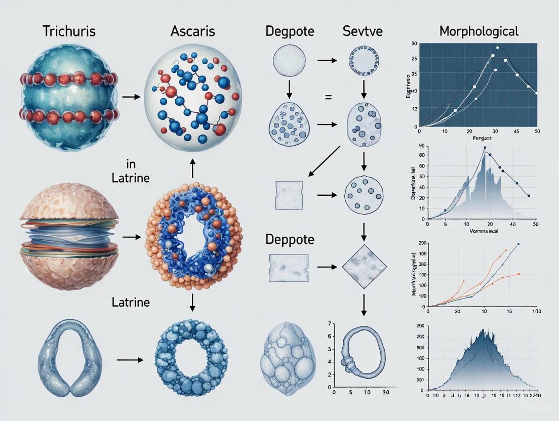
Visualizing Experimental Workflows
The following diagrams illustrate the logical relationships and experimental workflows described in this guide.
Eggshell Structure and Taphonomic Pathway
Molecular Analysis Workflow
The study of taphonomy—the processes affecting organisms after death—is crucial for interpreting archaeological and paleoparasitological evidence. Within the specific context of latrine research, the differential preservation of soil-transmitted helminth (STH) eggs, notably those of Trichuris (whipworm) and Ascaris (roundworm), provides a unique window into past human health, hygiene, and diet [11] [12]. Understanding the abiotic factors that govern the preservation of these bio-markers is fundamental to data integrity. This guide objectively compares the influence of three key abiotic taphonomic factors—desiccation, temperature fluctuations, and soil chemistry—on the comparative taphonomy of Trichuris versus Ascaris eggs, synthesizing current experimental data to inform methodological choices for researchers and scientists.
Comparative Analysis of Taphonomic Factors
The resilience of STH eggs is well-documented, allowing them to survive for centuries in latrine sediments [11]. However, the eggs of different species exhibit varying degrees of resistance to environmental pressures. The table below summarizes the comparative effects of key abiotic factors on Trichuris and Ascaris eggs, based on current empirical evidence.
Table 1: Comparative Impact of Abiotic Factors on Trichuris vs. Ascaris Eggs
| Taphonomic Factor | Impact on Trichuris Eggs | Impact on Ascaris Eggs | Comparative Summary & Key Evidence |
|---|---|---|---|
| Desiccation | Appears highly resistant; process can induce decompositional stasis [13]. | Resistant, but comparative susceptibility relative to Trichuris is less defined. | Both are highly resistant. Key Data: A quantitative study using custom PCBs to measure tissue resistivity found desiccation is driven by temperature and solar radiation, preserving organic matter [13]. |
| Temperature Fluctuations | Requires 5–38°C for 20–100 days to become infective; extremes can halt decomposition [14] [15]. | Requires 5–38°C for 8–37 days to become infective; cold delays soil biogeochemical changes [14] [16]. | Ascaris embryonation is faster. Key Data: Cold-season cadaver deposition delayed soil biochemical changes (e.g., carbon/nitrogen pulse) by months compared to warm deposition [16]. |
| Soil Chemistry | Survives acetolysis; found in pollen slides from medieval cesspits [11]. | Survives acetolysis; found in pollen slides from medieval cesspits [11]. | Both are highly chemically resistant. Key Data: Decomposition creates a Cadaver Decomposition Island (CDI), increasing soil nitrogen, carbon, and altering pH [16] [15]. Enhanced mineralization changes soil organic matter chemistry [16]. |
Detailed Experimental Protocols and Methodologies
To ensure the reproducibility of comparative taphonomy studies, detailed methodologies from key research are outlined below.
Table 2: Key Experimental Protocols in Taphonomic and Paleoparasitological Research
| Protocol Objective | Key Steps in Workflow | Critical Technical Notes |
|---|---|---|
| Conventional Paleoparasitological Analysis via Microscopy [12] | 1. Sample Collection: Obtain sediment from archaeological cesspits/latrines.2. Rehydration & Preparation: Treat samples with a rehydrating solution.3. Microscopy: Analyze prepared slides under light microscopy.4. Identification: Identify eggs based on morphology, size, and surface structures. | This method is foundational but often cannot distinguish between closely related species. The strong, chemically resistant shells of the eggs are key to their survival [12]. |
| Detection and Quantification of STH Eggs in Soil [17] | 1. Dissociation: Detach eggs from soil particulate matter.2. Flotation: Use a solution with appropriate specific density to isolate eggs.3. Recovery and Analysis: Recover eggs and identify/count via microscopy or molecular methods. | The choice of flotation solution is critical and should account for the different densities of various STH eggs. Inclusion of a dissociation step significantly improves egg recovery rates [17]. |
| Integrating Molecular Analysis (Metabarcoding & Metagenomics) [12] | 1. aDNA Extraction: Extract ancient DNA from sediment samples.2. Target Amplification (Metabarcoding): Amplify and sequence barcode genes (e.g., 18S rRNA).3. Shotgun Sequencing (Metagenomics): Sequence all DNA in a sample without targeting.4. Bioinformatic Analysis: Map sequences to reference genomes for species-specific identification. | This allows for a more precise, species-level identification (e.g., T. trichiura) and can describe the broader microbiome. It is particularly useful when egg morphology is degraded [12] [18]. |
| Quantifying Soft-Tissue Desiccation [13] | 1. Sensor Deployment: Insert custom-designed printed circuit board (PCB) sensors into soft tissue.2. Resistivity Measurement: Use sensors to measure electrical resistance as a proxy for moisture content.3. Environmental Monitoring: Concurrently record temperature, humidity, solar radiation, and rainfall.4. Data Modeling: Use generalized additive models to correlate environmental factors with desiccation rates. | This provides a quantitative, longitudinal measure of full-thickness tissue desiccation, moving beyond qualitative, stage-based descriptive systems [13]. |
Workflow for a Comprehensive Taphonomic Analysis
The following diagram illustrates the logical workflow integrating these protocols for a holistic analysis of a latrine sample, from collection to data synthesis.
The Scientist's Toolkit: Essential Research Reagents and Materials
Successful investigation into the taphonomy of STH eggs requires a specific set of reagents and materials. The following table details key solutions and their functions in standard experimental protocols.
Table 3: Key Research Reagent Solutions for STH Egg Analysis
| Reagent / Material | Primary Function | Application Notes |
|---|---|---|
| Flotation Solutions (e.g., Zinc Sulfate, Saturated Sodium Nitrate) | To isolate helminth eggs from soil/debris via density separation [17]. | The specific gravity of the solution is critical. It must be higher than that of the debris but lower than that of the STH eggs (often >1.20) to allow eggs to float and be recovered. |
| Rehydrating Solutions (e.g., Aqueous Phosphate Buffered Saline) | To rehydrate and soften ancient coprolites or latrine sediments before microscopic examination [12]. | Helps in the recovery of eggs from desiccated samples. May sometimes include a mild lytic step to release eggs from the sediment matrix. |
| PCR Reagents for Metabarcoding (Primers, Polymerases, dNTPs) | To enzymatically amplify target genes (e.g., 18S rRNA) from ancient DNA for species identification [12]. | Requires reagents suitable for degraded, low-concentration ancient DNA. Primers must be designed to target short, informative regions of DNA. |
| Next-Generation Sequencing (NGS) Kits | For library preparation and shotgun metagenomic sequencing of all DNA in a sample [12] [18]. | Allows for untargeted, high-throughput sequencing, providing data for species-specific identification and genomic studies without prior knowledge of the parasite community. |
| Custom Desiccation Sensors (Printed Circuit Boards - PCBs) | To quantitatively measure moisture content (via electrical resistivity) in soft tissues over time [13]. | Provides objective, continuous data on desiccation, moving beyond qualitative stage-based models. Designs are often open-source and customizable. |
The comparative taphonomy of Trichuris and Ascaris eggs in latrine environments is governed by a complex interplay of desiccation, temperature, and soil chemistry. While both parasites produce eggs of remarkable resilience, capable of surviving harsh chemical treatments and centuries of burial, subtle differences in their developmental requirements and potential compositional makeup may influence their long-term preservation and recovery. Modern research is greatly enhanced by moving from purely qualitative, morphological assessments towards integrated methodologies that combine classic microscopy with quantitative molecular biology (metagenomics) and geochemistry. The standardization of protocols, as called for in the field, along with the adoption of novel quantitative tools for measuring factors like desiccation, will be paramount in developing a more robust, data-driven understanding of parasite taphonomy. This, in turn, will refine interpretations of the archaeological record and provide deeper insights into the history of human health and disease.
The taphonomic analysis of parasite eggs in archaeological latrines provides critical insights into past human health and sanitation. A comparative examination of Trichuris (whipworm) and Ascaris (roundworm) egg preservation reveals significant differences driven by latrine conditions, particularly waterlogging, pH, and organic content. This review synthesizes experimental and observational data to demonstrate that Ascaris eggs generally exhibit greater resilience in waterlogged, anoxic environments with high organic content, while Trichuris eggs are more susceptible to degradation under fluctuating conditions. Understanding these differential preservation patterns is essential for accurate interpretation of archaeoparasitological data and reconstructing historical disease dynamics.
Archaeoparasitology, the study of ancient parasites, relies heavily on the recovery and identification of helminth eggs from archaeological contexts such as latrines, coprolites, and mummified remains [19]. The eggs of two common soil-transmitted helminths, Trichuris trichiura (whipworm) and Ascaris lumbricoides (roundworm), are frequently encountered in such studies [20]. However, their preservation is not uniform and is significantly influenced by taphonomic processes—the chemical, physical, and biological factors that affect organic remains after deposition [19] [21].
The latrine environment presents a complex matrix where variables like water saturation, pH levels, and organic matter content interact to either promote or hinder egg survival. A comprehensive understanding of how these factors differentially affect Trichuris versus Ascaris eggs is crucial for avoiding false negatives in parasite detection and for accurately assessing past infection patterns [19]. This guide systematically compares the taphonomic trajectories of these two parasite species under varying latrine conditions, providing researchers with a framework for interpreting archaeoparasitological data.
Morphological Foundations for Differential Preservation
The structural characteristics of Trichuris and Ascaris eggs form the basis for their differing preservation potential in archaeological contexts.
Trichuris eggs are typically oval-shaped with distinct polar plugs and measure approximately 50-58 μm in length and 22-27 μm in width [20]. Their wall consists of multiple layers, including a lipoprotein outer layer that provides some resistance to environmental pressures [19].
Ascaris eggs exhibit more variation but are generally subspherical to oval and larger, measuring 45-75 μm in length and 35-50 μm in width [19]. They possess a thicker, mammillated outer layer composed of chitinous material and mucopolysaccharides, which provides enhanced protection against chemical and biological degradation [19] [20].
Table 1: Comparative Morphology of Trichuris and Ascaris Eggs
| Characteristic | Trichuris trichiura | Ascaris lumbricoides |
|---|---|---|
| Shape | Oval with polar plugs | Subspherical to oval |
| Dimensions | 50-58 μm × 22-27 μm | 45-75 μm × 35-50 μm |
| Shell Structure | Multiple layers with lipoprotein | Thick, mammillated outer layer |
| Structural Resilience | Moderate | High |
These morphological differences directly influence how each egg type responds to taphonomic pressures in latrine environments, with the thicker, more complex structure of Ascaris eggs generally conferring greater resistance to degradation.
Impact of Waterlogging on Egg Preservation
Waterlogging represents a critical factor in latrine environments that profoundly influences the preservation of parasite eggs through its effects on oxygen availability and microbial activity.
Waterlogging and Anaerobic Conditions
Waterlogged conditions create anoxic environments that significantly slow the degradation of organic materials, including parasite eggs [22]. The reduced oxygen availability suppresses aerobic microbial activity that would otherwise contribute to egg decomposition. Research has demonstrated that eggs of both Trichuris and Ascaris can survive acetolysis procedures used in pollen extraction, indicating considerable chemical resilience [20]. However, water percolation through archaeological deposits can cause differential preservation based on morphological characteristics, with Ascaris eggs generally demonstrating superior survival rates in consistently waterlogged contexts [19].
A study of medieval coprolites from Nivelles, Belgium, revealed exceptional preservation of both Trichuris and Ascaris eggs in waterlogged conditions, with one burial containing approximately 1,577,679 total Trichuris eggs and 202,350 total Ascaris eggs [19]. This remarkable preservation was attributed to the anoxic environment created by water saturation.
Comparative Resilience in Hydric Environments
Ascaris eggs exhibit particularly high tolerance to waterlogged conditions. Experimental studies monitoring Ascaris suum egg viability found that eggs from sewage sludge maintained structural integrity for extended periods, though with diminished viability (3%) compared to those from fresh feces (52%) or adult worm uteri (96%) [23]. This demonstrates that while Ascaris eggs may remain morphologically identifiable in waterlogged contexts, their viability decreases over time.
The functional groups of humic substances change with increasing soil waterlogging, potentially creating chemical environments that either preserve or degrade parasite eggs [22]. In subaqueous soils, humic acids show lower aromaticity and complexity, potentially reducing their protective capacity for embedded eggs [22].
Table 2: Impact of Waterlogging on Egg Preservation
| Preservation Factor | Trichuris trichiura | Ascaris lumbricoides |
|---|---|---|
| Anaerobic Survival | Moderate | Excellent |
| Structural Integrity in Water | Good (polar plugs may compromise) | Excellent (thick shell provides protection) |
| Viability Retention | Limited data; moderate | Extended (8-12 weeks in experimental conditions) |
| Effect of Fluctuating Water Levels | Highly detrimental; promotes degradation | Moderate impact; better tolerance |
Trichuris eggs appear more susceptible to damage under fluctuating water conditions. Their polar plugs may present potential points of water ingress and structural weakness over time, particularly when water levels fluctuate [19]. Environments with pedoturbation effects induced by water movements show altered preservation dynamics that may differentially affect egg types [22].
Influence of pH and Chemical Environment
The chemical composition of the latrine matrix, particularly pH and organic content, significantly influences egg preservation through its effects on biological degradation and structural integrity.
Soil Biochemical Properties
Waterlogging affects both physicochemical and biochemical soil properties [22]. In anaerobic, waterlogged soils, enzyme activities are generally higher in subsurface horizons than in surface layers, contrary to expectations [22]. This unusual distribution results from the "combined effect of water movement, erosion processes and preservation of SOM under anaerobic conditions," creating a complex biochemical environment for egg preservation.
The microbial biomass carbon (MBC) and microbial quotient (Qmic) vary significantly with hydroperiod, influencing the microbial community responsible for decomposing organic remains, including parasite eggs [22] [24]. Fungal communities, which play important roles in degradation processes, show different responses to water level changes compared to bacterial communities, with fungal diversity and evenness being higher at low water levels [24].
Organic Matter Quality and Quantity
The quality and structure of soil organic matter (SOM) changes with increasing waterlogging [22]. The origin of organic matter in depositional environments can be discriminated using isotopic C signature, with terrestrial plant residues, riverine phytoplankton, and marine phytoplankton contributing different organic profiles [22].
In salt marsh systems, accumulation of nutrients and SOM is significantly magnified in intertidal systems, where pedoturbation effects induced by water movements are particularly strong [22]. This enhanced organic content may provide additional protective coating for parasite eggs or support microbial communities that either preserve or degrade them.
Table 3: Influence of Chemical Factors on Egg Preservation
| Chemical Factor | Effect on Trichuris | Effect on Ascaris |
|---|---|---|
| High Organic Content | Moderate preservation enhancement | Significant preservation enhancement |
| pH Variations | Moderate tolerance; alkaline conditions may damage plugs | High tolerance across wider pH range |
| Microbial Activity | Susceptible to enzymatic degradation | More resistant due to thicker shell |
| Humic Substances | Moderate protective effect | Strong protective effect |
Experimental Protocols and Methodological Considerations
Standardized methodologies are essential for comparative taphonomic analysis of parasite eggs in archaeological contexts.
Sample Collection and Processing
Dry samples should be collected using sterile instruments and placed in sterile containers for transportation to specialized laboratories [19]. For optimal recovery, multiple processing techniques should be employed, including:
- Kato-Katz technique: Effective for concentrated detection but may miss light infections
- Formol ethyl acetate sedimentation: Standard method for parasite concentration
- Wisconsin floatation: Provides high sensitivity (89.6% for geohelminths)
- Simple gravity sedimentation: Equally effective with 89.6% sensitivity [25]
Method combinations significantly enhance detection rates. The Kato-Katz technique/simple gravity sedimentation and Wisconsin floatation/simple gravity sedimentation combinations each provide 99.0% sensitivity for geohelminth egg recovery [25].
Viability Assessment and Incubation
For experimental studies assessing egg viability, prolonged incubation periods are necessary for accurate assessment, particularly for environmental samples. Research on Ascaris suum eggs demonstrates that:
- Eggs from adult worm uteri show high viability (96%) and develop larvae within 3 weeks
- Eggs from pig feces (52% viability) and sewage sludge (3% viability) show delayed development, requiring 8-12 weeks for conclusive viability assessment [23]
These findings highlight the importance of extended observation periods and the limitations of single time point assessments based solely on egg structure, which can lead to misclassification [23].
Surface Recovery Studies
Experimental approaches to egg recovery from surfaces provide insights into detection methodologies. Studies on cement-based surfaces demonstrate that:
- Mopping removes 95.6% (SD = 4.0%) of viable eggs with no significant differences between traditional mortar and sustainable mortar with fly ash [26]
- First-order decay rate constants (k) for Ascaris suum eggs average 0.029 day⁻¹ (SD = 0.074 day⁻¹) across all conditions [26]
- Decay rates are significantly influenced by temperature, with greater k values at 34°C compared to 15°C [26]
Figure 1: Experimental Workflow for Archaeoparasitological Analysis
The Scientist's Toolkit: Essential Research Reagents and Materials
Table 4: Essential Research Materials for Archaeoparasitology
| Research Material | Application/Function | Context |
|---|---|---|
| Sterile Containers | Prevents modern contamination during sample transport | Field collection [19] |
| Acetolysis Reagents | Chemical processing for palynological studies; parasite eggs survive this process | Laboratory processing [20] |
| Formol-Ethyl Acetate | Sedimentation technique for parasite egg concentration | Laboratory processing [25] |
| Sodium Nitrate Solution | Floatation medium for Wisconsin and similar techniques | Egg recovery and concentration [25] |
| Microscopy Slides and Coverslips | Mounting samples for morphological identification | Laboratory analysis [20] |
| Incubation Chambers | Maintaining stable temperature (e.g., 27°C) for viability assessment | Experimental viability studies [23] |
| Cement-based Surfaces | Experimental substrates for removal and inactivation studies | Intervention and transmission studies [26] |
Historical Prevalence and Taphonomic Bias
The differential preservation of Trichuris and Ascaris eggs has implications for interpreting historical disease patterns. Paleoparasitological data from mummies and latrines must be evaluated in light of taphonomic biases.
Temporal Prevalence Patterns
Analyses of medieval and post-medieval cesspit samples in Europe have revealed shifting patterns in parasite prevalence that may reflect both true infection rates and preservation biases. In the Netherlands, Trichuris appears more common in the medieval period (up to 1500 AD), while Ascaris becomes more prevalent in post-medieval samples [20]. This pattern could reflect:
- Changing sanitation practices and waste management
- Differential preservation in evolving latrine environments
- Shifting agricultural uses of human waste
East Asian Mummy Evidence
Studies of Korean mummies from the Joseon Dynasty (1392-1910 CE) show high prevalence of both parasites, with Trichuris present in 83.3% of mummies and Ascaris in 58.3% [27] [3]. However, by the 20th century, national surveys showed dramatic declines, particularly for Ascaris, which fell to 0.3% prevalence by 1992 [27]. Similar patterns were observed in China, where ancient mummies showed Ascaris prevalence of 62% compared to 46% in 1988-1992 national surveys [27] [3].
These declines are attributed to multiple factors, including improved sanitation infrastructure, replacement of night soil with chemical fertilizers, and public health campaigns [27]. The timing of these changes differed between parasite species, with Chinese liver fluke (Clonorchis sinensis) declining earlier than soil-transmitted nematodes in both Korea and China [27] [3].
The taphonomic analysis of Trichuris versus Ascaris eggs in latrine contexts reveals complex interactions between environmental factors and egg morphology. Waterlogging generally promotes preservation through anoxic conditions, but fluctuating water levels can be detrimental, particularly for the more vulnerable Trichuris eggs. The thicker, more complex shell structure of Ascaris eggs provides enhanced protection against chemical and biological degradation in diverse latrine environments.
Researchers must account for these differential preservation patterns when interpreting archaeoparasitological data. False negatives and skewed prevalence estimates can result from failure to consider how local conditions—particularly water saturation, organic content, and pH—selectively preserve certain egg types. Standardized methodologies, including multiple processing techniques and extended viability assessment periods, provide the most accurate recovery of parasite evidence.
Future research should focus on quantitative comparisons of egg survival rates under controlled conditions simulating various latrine environments. Such experimental approaches will strengthen the interpretive framework for archaeological findings and enhance our understanding of historical parasitism patterns.
Analysis of sediment samples from three 14th to 17th century latrines in medieval Brussels revealed consistent patterns of helminth co-infection, dominated by the soil-transmitted nematodes Ascaris sp. (roundworm) and Trichuris sp. (whipworm). This study, situated within a broader thesis on comparative taphonomy, demonstrates how differential egg preservation and advanced molecular techniques illuminate historical disease burden. The findings confirm poor sanitary conditions in this major medieval urban center and provide a framework for understanding parasite ecology and taphonomic survival in archaeological contexts, offering insights for both paleoparasitology and modern helminth control strategies.
The analysis of intestinal parasites from archaeological latrines provides a unique window into the health, diet, and sanitary conditions of past populations. During the late Medieval and Renaissance periods, Brussels established itself as a political and economic hub, with its population growing from 20,000 in 1300 to 26,000 in 1400 [28]. Such urbanization, without concomitant advances in sanitation, created environments conducive to the spread of fecal-oral parasites. This case study examines parasite eggs recovered from three latrines in central Brussels, dating from the 14th to the 17th centuries, to determine infection profiles and prevalence. Crucially, the findings are framed within a comparative taphonomic analysis of Trichuris versus Ascaris eggs—two nematodes frequently found in co-infection yet subject to different preservation pathways due to their distinct morphological and biochemical characteristics.
Materials and Methods
Archaeological Context and Sampling
Sediment samples were collected from eight distinct layers across three different latrine cesspits located approximately 500 meters apart in central Brussels [28].
Table 1: Description of Sampled Latrines in Medieval Brussels
| Latrine Designation | Construction | Date | Sample Layers Analyzed | Archaeological Context |
|---|---|---|---|---|
| Cesspit 1 | Brick-lined | 14th–15th c. CE | 2 layers (US4139, US4138) | Artisan/commercial centre with butcheries, breweries, and bakeries. |
| Cesspit 2 | Unlined | 14th–15th c. CE | 3 layers (US20-134, US23-45, US24-122) | Foundations of Café Greenwich; few artefacts recovered. |
| Cesspit 3 | Brick-lined | Mid-15th – first half of 17th c. CE | 1 layer (US 33-15) | Part of the same project as Cesspit 2 (BR111); waterlogged conditions. |
Microscopic Analysis
The paleoparasitological analysis followed established procedures [28] [29]. Briefly, a 0.2 g subsample from each sediment layer was disaggregated in a 0.5% trisodium phosphate solution for a minimum of two hours. The resulting suspension was filtered through a series of microsieves with descending mesh sizes (300 μm, 160 μm, and 20 μm). The material retained on the 20 μm sieve, which captures most helminth eggs (30-150 μm), was centrifuged, and the pellet was mixed with glycerol for microscopic examination at 400x magnification. Helminth eggs were identified based on size and morphological features using standard parasitological references [28].
Molecular Analysis
Enzyme-Linked Immunosorbent Assay (ELISA)
To detect protozoal parasites, a 1 g subsample was disaggregated and analyzed using commercially available ELISA kits (TECHLAB) designed to detect Cryptosporidium spp., Entamoeba histolytica, and Giardia duodenalis antigens. These kits have demonstrated 98-100% sensitivity and specificity in modern clinical trials [28].
Ancient DNA (aDNA) Analysis
For metagenomic and metabarcoding analysis, DNA was extracted from sediment samples in dedicated aDNA laboratories to prevent contamination [29] [30]. Libraries were prepared using blunt-end ligation and Illumina-specific adapters. High-throughput shotgun sequencing was performed on a HiSeq 2000/2500 platform. For 18S rRNA metabarcoding, the V9 region was amplified and sequenced to identify eukaryotic communities, with a focus on parasitic nematodes [29].
Results and Discussion
Parasite Prevalence and Co-infection Profile
Microscopic analysis confirmed the presence of helminth eggs in all three latrines, with a profile dominated by soil-transmitted helminths.
Table 2: Prevalence of Helminth Species in Medieval Brussels Latrines
| Parasite Species | Type | Medieval Prevalence | Renaissance Prevalence | Transmission Route |
|---|---|---|---|---|
| Ascaris sp. | Nematode (Roundworm) | Present | Present | Fecal-oral |
| Trichuris sp. | Nematode (Whipworm) | Present | Present | Fecal-oral |
| Capillaria sp. | Nematode | Present | Not Detected | Fecal-oral |
| Dicrocoelium dendriticum | Trematode (Fluke) | Present | Present | Food-borne (via ants) |
| Fasciola hepatica | Trematode (Liver Fluke) | Present | Present | Food-borne (via water plants) |
| Diphyllobothrium latum | Cestode (Tapeworm) | Present | Present | Food-borne (via raw fish) |
| Taenia sp. | Cestode (Tapeworm) | Present | Not Detected | Food-borne (via raw pork/beef) |
| Entamoeba histolytica | Protozoa | Detected by ELISA | Information Missing | Fecal-oral |
| Giardia duodenalis | Protozoa | Detected by ELISA | Detected by ELISA | Fecal-oral |
The data reveals a consistent pattern of co-infection with Ascaris and Trichuris from the Medieval period into the Renaissance. The presence of food-derived cestodes like Diphyllobothrium latum and Taenia sp. points to dietary habits involving raw or undercooked fish and meat [28]. The high prevalence of fecal-oral parasites reflects the poor sanitation and hygiene that characterized medieval urban centers. The decline of some species like Capillaria and Taenia in the Renaissance layers may reflect changing dietary practices or waste management, though the persistence of Ascaris and Trichuris highlights their enduring transmission advantage in urban environments [28].
Comparative Taphonomy:Trichurisvs.AscarisEgg Preservation
The consistent co-detection of Ascaris and Trichuris in archaeological contexts, from Brussels [28] to Cyprus [31] and Sardinia [29], belies significant differences in their egg morphology and resulting taphonomic resilience.
Table 3: Comparative Taphonomy of Ascaris and Trichuris Eggs
| Characteristic | Ascaris lumbricoides | Trichuris trichiura |
|---|---|---|
| Egg Size | 45-75 μm long, 35-50 μm wide [19] | 50-55 μm long, 20-25 μm wide [19] |
| Egg Morphology | Thick, mammillated outer layer that is often decorticated in archaeological samples [19] | Smooth, thick outer shell with bipolar plugs (opercula) [19] |
| Preservation Bias | The mammillated layer is prone to degradation, which can make identification more difficult over time [19] | The robust shell and distinctive plugs often lead to superior preservation and easier identification [19] |
| Quantitative Recovery | In a Cypriot castle latrine: 1,179 eggs/gram of sediment [31] | In a Cypriot castle latrine: 118 eggs/gram of sediment [31] |
| Molecular Survival | aDNA recoverable; allows for haplotype analysis and phylogenetics [30] | aDNA often well-preserved; allows for species-specific identification and distinction from T. suis [30] |
The mammillated coat of Ascaris eggs, while protective in fresh environments, can be prone to degradation and decortication over centuries, potentially complicating morphological identification [19]. In contrast, the robust, bipolar-plugged shell of Trichuris eggs often confers a taphonomic advantage, leading to excellent preservation. Quantitative data from a 12th-century Cypriot castle latrine underscores this difference, showing a recovery rate approximately ten times higher for Ascaris (1,179 eggs/gram) than for Trichuris (118 eggs/gram) [31]. This discrepancy could reflect true differences in ancient parasite burden, but it may also be influenced by taphonomic factors and the higher fecundity of female Ascaris worms.
Molecular Insights and Pathogen Detection
Molecular techniques significantly enhanced the findings from microscopic analysis. In a similar study of a 19th-century Sardinian cesspit, 18S rRNA metabarcoding assigned 33.5% of the eukaryotic reads to the Nematoda phylum, all of which belonged to the Trichuris genus [29]. Metagenomic sequencing aligned hundreds of reads to T. trichiura and Ascaris sp., enabling species-level confirmation and even the assembly of a partial ITS region for phylogenetic analysis [29]. This is crucial for distinguishing between the morphologically identical eggs of the human whipworm (T. trichiura) and the pig whipworm (T. suis), providing definitive evidence of human infection and refining our understanding of human-animal interactions [30]. Furthermore, ELISA testing confirmed the presence of the protozoans Giardia duodenalis and Entamoeba histolytica in Brussels, parasites that cause dysentery and are often undetectable by microscopy alone [28]. This combined approach paints a more complete picture of the pathogenic challenges faced by medieval urban populations.
Figure 1: Integrated Workflow for Archaeological Parasitology. This diagram outlines the multi-pronged experimental protocol, combining physical processing, microscopy, and molecular techniques to achieve comprehensive parasite profiling from ancient latrine sediments.
The Scientist's Toolkit: Key Research Reagents and Materials
Table 4: Essential Research Reagents and Materials for Paleoparasitology
| Item/Solution | Function in Protocol | Specific Application Example |
|---|---|---|
| Trisodium Phosphate (0.5%) | Rehydration and disaggregation of ancient sediment samples. | Used to form an aqueous suspension from 0.2-1.0g of latrine sediment to release parasite eggs [28] [29]. |
| Microsieves (20 μm, 160 μm, 300 μm) | Size-based filtration to concentrate parasite eggs. | Stacked sieves used to isolate helminth eggs (typically 30-150 μm) from larger debris and finer particles [28]. |
| Glycerol | Mounting medium for microscopy. | Mixed with the processed sediment pellet to clear debris and enhance microscopic visualization of eggs [28]. |
| Commercial ELISA Kits (e.g., TECHLAB) | Immunological detection of protozoan antigens. | Used to identify Giardia duodenalis, Cryptosporidium spp., and Entamoeba histolytica in 1g sediment subsamples [28]. |
| DNA Library Prep Kits (e.g., NEBNext) | Preparation of sequencing libraries from aDNA. | Blunt-end libraries were prepared for shotgun metagenomic sequencing on Illumina platforms [30]. |
| Flotation Buffer (e.g., Saturated NaCl/Glucose) | Density-based separation of parasite eggs. | Used for initial screening and egg quantification via flotation and McMaster counting chambers [30]. |
This case study of medieval Brussels latrines confirms a high burden of intestinal parasites, particularly the co-infection of Ascaris and Trichuris, in one of Europe's foremost medieval urban centers. The findings align with evidence from other contemporary sites, suggesting that such parasitic infections were widespread and persistent [31] [32]. The application of both microscopic and molecular methods was crucial for providing a comprehensive parasitological profile, revealing not only soil-transmitted helminths but also food-borne cestodes and protozoans. The comparative taphonomic framework highlights the necessity of interpreting archaeological parasite data with an understanding of the distinct preservation biases of different egg types. Future research, leveraging increasingly sophisticated aDNA techniques, will continue to refine our understanding of historical helminth infections, offering profound insights into the health, hygiene, and daily life of past populations.
The study of parasite egg distribution in archaeological contexts, particularly latrines, provides critical insights into historical disease ecology, sanitation, and human migration. The comparative taphonomy of Trichuris trichiura (whipworm) and Ascaris lumbricoides (roundworm) eggs offers a powerful framework for understanding how aggregate population patterns of parasites are preserved in the archaeological record. These two common soil-transmitted helminths exhibit different egg morphological characteristics and resilience to environmental degradation, making their comparative analysis particularly informative for reconstructing past human-parasite interactions. This guide objectively compares the theoretical frameworks and experimental methodologies used to analyze and interpret the distribution patterns of these parasites, providing researchers with a foundation for robust archaeological parasitology research.
Theoretical Foundations of Parasite Aggregation
The Ecological Principle of Aggregation
In host-parasite systems, the distribution of macroparasites is characteristically overdispersed, meaning the variance in parasite load exceeds the mean [33]. This results in parasite aggregation, where a small number of hosts harbor the majority of parasites while most hosts have few or none [33]. This non-random distribution pattern is considered an ecological 'law' in parasitology, with significant implications for statistical sampling, disease burden estimation, and intervention design in both modern and archaeological contexts.
Key Metrics for Quantifying Aggregation
Researchers employ several quantitative metrics to describe and track parasite aggregation:
Variance-to-Mean Ratio (VMR): A VMR approximately equal to 1 suggests a Poisson (random) distribution; values greater than 1 indicate parasite aggregation; and values less than 1 suggest a uniform distribution [33]. The related index of dispersion (D) is calculated as D = σ²/[μ(n-1)], where μ is the mean, σ² is the variance, and n is the number of sampled hosts [33].
Negative Binomial Parameter (k): This parameter is defined as k = μ²/(σ² - μ) [33]. Small k values (near zero) indicate strong aggregation, while large k values suggest a more random (Poisson) distribution [33]. This metric is particularly preferred in aggregation studies.
Taylor's Power Law: This method models the relationship between variance and mean using the formula: log(σ²) = log(a) + blog(μ), where a and b are parameters fitted to empirical data [33]. The slope b helps characterize the nature of the distribution.
Mechanisms Driving Aggregated Distributions
Multiple factors contribute to the aggregated distribution patterns observed in parasite populations:
- Host Heterogeneity: Differences in immune competence, behavior, and genetics among hosts create variation in susceptibility [33].
- Environmental Heterogeneity: Spatial and temporal variations in environmental conditions affect parasite survival and transmission [33].
- Complex Parasite Life Cycles: Feedback loops in parasite life cycles can amplify small initial differences into significant aggregation [33].
- Socio-Spatial Dynamics: In human populations, social networks and contact matrices with hub individuals can create superspreader events, leading to characteristic aggregation patterns [33].
Table 1: Key Theoretical Concepts in Parasite Aggregation
| Concept | Description | Implication for Egg Distribution |
|---|---|---|
| Overdispersion | Variance in parasite load exceeds the mean | Non-random distribution in latrine sediments |
| Negative Binomial Distribution | Statistical model fitting aggregated counts | Expected pattern in archaeological parasite egg counts |
| Superspreader Hosts | Few individuals contribute disproportionately to transmission | Explains extreme egg concentrations in specific coprolites |
| Spatiotemporal Heterogeneity | Environmental and host variation across space and time | Differential preservation of eggs in archaeological contexts |
Comparative Taphonomy: Trichuris vs. Ascaris Eggs
Morphological and Structural Differences
The differential preservation of Trichuris and Ascaris eggs in archaeological contexts stems from their distinct morphological characteristics:
Trichuris trichiura: Eggs are lemon-shaped with distinctive polar plugs at both ends, measuring approximately 50-54 μm × 22-23 μm [34]. The bipolar plugs may represent potential structural vulnerabilities during taphonomic processes.
Ascaris lumbricoides: Eggs are ovoid in shape without opercula, measuring approximately 45-75 μm × 35-50 μm [34]. Their more uniform structure potentially provides greater resistance to environmental degradation.
Taphonomic Factors Affecting Egg Preservation
Multiple taphonomic factors influence the differential preservation of parasite eggs in latrine sediments:
- Chemical Environment: Alkaline conditions within burials, evidenced by calcification of samples, promote superior egg preservation [34].
- Hydrological Conditions: Limited fluid percolation through grave sediment, often due to clay-rich soils and tightly sealed burial environments, enhances preservation [34].
- Sedimentary Matrix: The composition of surrounding sediments affects egg mobility and potential damage. Clay-rich matrices with low permeability offer superior preservation compared to sandy sediments [34].
- Temporal Depth: While all archaeological specimens represent ancient materials, the degree of preservation can vary significantly even within similar time periods based on local conditions.
Empirical Evidence from Archaeological Contexts
Case studies from medieval sites provide compelling evidence for differential preservation and distribution patterns:
Nivelles, Belgium Burial 122: Analysis revealed an unprecedented case of extreme parasitism, with calculated egg concentrations of 1,577,679 total eggs for Trichuris trichiura and 202,350 total eggs for Ascaris lumbricoides [34]. This case demonstrates the potential for extraordinary preservation under optimal conditions and provides evidence of aggregation at the individual host level.
Medieval and Renaissance Brussels: A comparative study of three latrines (14th-17th centuries) identified both Trichuris sp. and Ascaris sp. in medieval samples, with continued presence into the Renaissance period [35]. The domination of species spread by fecal contamination of food and drink was consistent across households, though some variation existed between different locations.
19th Century Sardinian Palace: Analysis of a cesspit sediment revealed both Trichuris sp. and Ascaris sp. eggs, with Trichuris appearing as the most represented genus [12]. This finding contributes to understanding health conditions and demonstrates preservation in more recent archaeological contexts.
Table 2: Comparative Egg Preservation in Archaeological Contexts
| Archaeological Site | Time Period | Trichuris Preservation | Ascaris Preservation | Key Preservation Factors |
|---|---|---|---|---|
| Nivelles, Belgium [34] | 1025-1159 AD | Extraordinary (1.5+ million eggs) | Extensive (200,000+ eggs) | Tightly covered oak coffin, clay matrix, limited fluid percolation |
| Brussels, Belgium [35] | 14th-17th centuries | Consistent across periods | Consistent across periods | Cesspit design, urban context, sediment composition |
| Sardinia, Italy [12] | 19th century | Excellent (most prevalent) | Good | Closed cesspit environment, favorable preservation conditions |
Experimental Methodologies and Protocols
Conventional Paleoparasitological Techniques
Traditional microscopy-based methods remain fundamental to parasite egg analysis:
- Sample Processing: Coprolites or sediment samples are rehydrated and subjected to chemical processing to enhance parasite egg visibility [34] [12].
- Microscopic Analysis: Processed samples are examined under light microscopy for egg identification based on morphometric characteristics [34].
- Quantification Methods: Eggs per gram (epg) and eggs per coprolite (epc) calculations provide standardized measures for comparing infection intensity across samples [34].
Molecular and Advanced Analytical Techniques
Modern approaches complement traditional microscopy with genetic and biochemical analyses:
- Metabarcoding (18S rRNA): This technique allows for taxonomic identification of eukaryotic organisms, including specific parasite species, through high-throughput sequencing of target genes [12].
- Shotgun Metagenomics: Direct sequencing of all DNA in a sample without target-specific amplification provides a comprehensive view of the taxonomic composition, including parasite DNA [12].
- Enzyme-Linked Immunosorbent Assay (ELISA): Immunological detection of parasite-specific antigens can confirm microscopic identifications and detect species not easily visible through microscopy [35].
Emerging Automated Detection Methods
Recent technological advances are revolutionizing parasite egg identification:
- YOLO Convolutional Block Attention Module (YCBAM): This framework integrates YOLO with self-attention mechanisms and the Convolutional Block Attention Module (CBAM) to enable precise identification and localization of parasitic elements in challenging imaging conditions [36].
- Deep Learning Approaches: Models such as NASNet-Mobile and ResNet-101 have achieved over 97% classification accuracy in distinguishing parasite eggs from other artifacts in microscopic slides [36].
- Automated Segmentation: U-Net and ResU-Net segmentation algorithms effectively separate pinworm eggs from complex digital microscopy backgrounds, achieving high dice scores and minimal diagnostic errors [36].
Research Workflow and Analytical Framework
The following diagram illustrates the integrated experimental workflow for analyzing parasite egg distribution patterns in archaeological contexts:
Diagram 1: Integrated Research Workflow for Parasite Egg Distribution Analysis
Essential Research Reagent Solutions and Materials
Table 3: Key Research Reagents and Materials for Parasite Egg Analysis
| Reagent/Material | Application | Function in Analysis |
|---|---|---|
| Lycopodium spore tablets [34] | Microscopic quantification | Extraction control for calculating egg concentration |
| Glycerol solution [34] | Slide preparation | Mounting medium for microscopic examination |
| Trichuris-specific antibodies [35] | ELISA testing | Immunological detection of Trichuris antigens |
| Ascaris-specific antibodies [35] | ELISA testing | Immunological detection of Ascaris antigens |
| 18S rRNA primers [12] | Metabarcoding | Taxonomic identification of eukaryotic parasites |
| DNA extraction kits (aDNA optimized) [12] | Molecular analysis | Isolation of ancient DNA from archaeological samples |
| Phosphate-buffered saline (PBS) [34] | Sample processing | Washing and rehydration of archaeological sediments |
| Hydrogen peroxide (H₂O₂) [34] | Sample processing | Dissolution of organic material to concentrate eggs |
The theoretical framework for understanding aggregate population patterns in parasite egg distribution integrates principles from ecology, statistics, and archaeology. The comparative taphonomy of Trichuris versus Ascaris eggs provides a robust approach for investigating parasite prevalence in past populations, while accounting for preservation biases that affect the archaeological record. As methodological advances continue to enhance detection sensitivity and taxonomic precision, researchers are better equipped to reconstruct historical disease burdens and transmission dynamics, offering valuable insights for both archaeology and modern parasitology.
From Sediment to Microscope: Methodologies for Optimal Egg Recovery and Quantification
Paleoparasitology, the study of ancient parasites, provides invaluable insights into human health, dietary practices, migration patterns, and sanitation throughout history. The reliability of these insights depends fundamentally on the methods used to recover parasite remains from archaeological sediments, coprolites, and mummified tissues. Among the various techniques developed, the RHM (Rehydration-Homogenization-Microsieving) method has emerged as a foundational, standardized protocol. This guide objectively compares the performance of the RHM method against alternative extraction techniques, presenting supporting experimental data within the specific context of studying the comparative taphonomy of Trichuris (whipworm) versus Ascaris (roundworm) eggs recovered from latrines. For researchers and drug development professionals, understanding these methodological nuances is crucial for interpreting datasets and designing robust paleoparasitological studies.
Core Protocol: The RHM Method
The RHM method is designed to gently liberate, concentrate, and clean parasite eggs from archaeological sediments while preserving their morphological integrity for accurate identification and quantification [28] [37].
Detailed Step-by-Step Workflow
The following diagram illustrates the streamlined, three-stage process of the RHM protocol:
Step 1: Rehydration A 0.2-0.5 g subsample of archaeological sediment is disaggregated in a 0.5% aqueous trisodium phosphate (Na₃PO₄) solution [28] [38]. The sample is left to soak for a minimum of 2 hours, though some protocols extend this to 48-72 hours or longer for highly compacted material [28] [38]. This step is critical for rehydrating and breaking down the sediment matrix without using harsh chemicals that could damage fragile parasite eggs.
Step 2: Homogenization The sample is gently mixed to form a uniform suspension. This ensures that subsequent subsampling is representative of the entire specimen. Vigorous agitation is avoided to prevent the destruction of more delicate ecological and parasitological remains.
Step 3: Microsieving The suspension is poured through a stack of microsieves with descending mesh sizes, typically 300 μm, 160 μm, and 20 μm [28]. This process efficiently separates parasite eggs, which are typically retained on the 20 μm sieve, from larger debris (retained on the 300 μm sieve) and very fine particles (which pass through the 20 μm sieve). Since most helminth eggs in northern Europe range from 30 to 150 μm, the 20 μm sieve effectively captures all relevant taxa [28].
Final Processing The material retained on the 20 μm sieve is centrifuged to remove excess water. The resulting pellet is then mixed with glycerol and mounted on a microscope slide for examination under 400x magnification [28]. Eggs are identified based on standard size and morphological characteristics [28].
Method Comparison: Performance and Taphonomic Implications
Different extraction methods can significantly impact the recovery of parasite eggs, often in a taxon-specific manner due to the distinct morphological and chemical composition of their eggshells. The following table summarizes the core characteristics of the RHM method against two common alternatives.
Table 1: Comparison of Paleoparasitological Extraction Methods
| Method | Core Principle | Key Advantages | Key Disadvantages | Impact on Trichuris vs. Ascaris |
|---|---|---|---|---|
| RHM (Rehydration-Homogenization-Microsieving) [28] [37] | Gentle chemical rehydration and physical separation | Preserves egg morphology; effective for delicate eggs; allows for quantification. | May retain more fine organic debris. | Optimal for Trichuris: High recovery of intact eggs with preserved polar plugs. Good for Ascaris: Recovers eggs with knobby outer coat (uterine layer) intact. |
| Acid-Based Extraction (e.g., HCl/HF) [37] [39] | Dissolution of mineral and organic matrix using strong acids | Reduces mineral and vegetal debris, concentrating some taxa. | Can decrease overall biodiversity; may damage eggs of some species. | Variable for Trichuris: HCl can concentrate eggs but HF may damage them [37]. Good for Ascaris: HCl can concentrate Ascaris eggs, but may alter morphology [37]. |
| Flotation-Centrifugation (e.g., Sheather's) [39] [38] | Separation based on density difference using a high-specific-gravity solution | Effective concentration of eggs from debris; standard in modern parasitology. | The sugar solution may not be ideal for all preservation states; may not recover severely degraded eggs. | Effective for both: Sheather's solution (SG 1.27) effectively recovers both taxa, especially when coupled with centrifugation [39]. |
Experimental data directly comparing these methods highlights their differential efficacy. One study tested acid and base combinations against the RHM protocol and found that while hydrochloric acid (HCl) could result in a concentration of Ascaris sp. eggs, the use of acids systematically decreased the diversity of parasite species identified compared to the standard RHM protocol [37]. The use of sodium hydroxide (a base) yielded even more negative results, likely due to chemical damage to the chitin in the eggshells [37]. This demonstrates that RHM is superior for comprehensive biodiversity studies.
Quantitative Data: Recovery Rates and Taphonomy
The choice of method directly influences quantitative results, which is critical for assessing past infection intensity. A study comparing RHM-derived methods on historical latrine sediments provided clear data on recovery rates.
Table 2: Quantitative Egg Recovery from Historical Latrine Sediments Using Different Methods [39]
| Method | Ascaris lumbricoides (epg) | Trichuris trichiura (epg) | Key Preservation Observations |
|---|---|---|---|
| Warnock & Reinhard (Palynological - HCl/HF) | 14,600 | 12,800 | Best morphology preservation. "Decorticated" Ascaris eggs (losing the diagnostic outer layer) were very rare. |
| Simplified (HCl only) | 23,100 | 17,100 | Effective recovery but with more debris. Slightly more "decorticated" Ascaris eggs observed. |
| Sheather's Flotation-Centrifugation | 6,400 | 7,100 | Recovery of eggs with intact morphology, but counts were lower than acid-based methods. |
Taphonomic Insights for Trichuris vs. Ascaris:* The differential preservation of these two common parasites is linked to eggshell biochemistry. The eggs of both possess a resistant chitinous layer, but a key difference exists: Ascaris lumbricoides eggs have an outer albuminous layer that gives them their characteristic knobby appearance, while Trichuris trichiura eggs lack this outer layer [39]. The outer layer of Ascaris is more susceptible to certain taphonomic and chemical processes, leading to "decortication" which can complicate identification [39]. The RHM and palynological methods, which avoid harsh chemicals, are most effective at preserving this delicate outer structure, ensuring accurate diagnosis of Ascaris [39]. The chitinous layer of Trichuris, with its helical fiber arrangement, is highly resilient, making its eggs generally robust across different extraction methods [39].
The Scientist's Toolkit: Essential Research Reagents
Successful implementation of paleoparasitological protocols requires a specific set of laboratory reagents and materials.
Table 3: Essential Research Reagents and Materials for the RHM Protocol
| Reagent / Material | Function in the Protocol | Application Note |
|---|---|---|
| Trisodium Phosphate (0.5% Solution) | Rehydration & Disaggregation: Softens and breaks down the compacted sediment matrix to release parasite eggs. | A gentle surfactant. Soaking time can be extended from 2 hours to several days for recalcitrant samples [28] [38]. |
| Microsieves (300, 160, 20 μm) | Size Fractionation: Physically separates parasite eggs from larger and smaller particulate debris. | The 20 μm sieve is critical as it captures most helminth eggs (30-150 μm) [28]. |
| Glycerol | Microscopy Mountant: A clearing agent that clarifies the sample and preserves the eggs for microscopic examination. | Provides a clear, stable medium for high-magnification observation of morphological details [28]. |
| Hydrochloric Acid (HCl) | Demineralization: Used in alternative protocols to dissolve calcium carbonates and other minerals in the sediment. | Can concentrate Ascaris eggs but may reduce species diversity and damage some egg types [37]. |
| Hydrofluoric Acid (HF) | Silicate Dissolution: Used in advanced palynological labs to dissolve silica and silicate minerals, drastically reducing mineral debris. | Highly hazardous. Requires specialized fume hoods and training. Preserves morphology well but is not accessible to all labs [39]. |
| Sheather's Sugar Solution | Flotation Medium: A high-specific-gravity solution used to float parasite eggs to the surface for collection. | Effective for concentration; centrifugation enhances recovery. Specific gravity of 1.27 is suitable for most helminth eggs [39]. |
The selection of an extraction method is a primary determinant of success in paleoparasitology. The evidence from comparative studies leads to a clear conclusion: the RHM protocol offers the most balanced and reliable approach for general paleoparasitological research, particularly when the study aims to accurately reconstruct parasite biodiversity and assess the taphonomic state of eggs.
For specific research questions, alternative methods have their place. Acid-based methods can be useful for concentrating particular taxa like Ascaris from heavily mineralized sediments, albeit at the cost of overall diversity. Flotation-centrifugation methods like Sheather's are highly effective for clean concentration. However, for a comprehensive analysis of latrine sediments that allows for direct comparison of the taphonomic pathways of Trichuris versus Ascaris eggs, the non-destructive, gentle nature of the RHM method makes it the recommended standardized protocol. Its ability to preserve the delicate outer albuminous layer of Ascaris eggs while simultaneously recovering the resilient Trichuris eggs without chemical alteration provides the most accurate dataset for interpreting past human health and hygiene.
The diagnosis of parasitic infections relies on several copromicroscopic and immunodiagnostic techniques, each with distinct advantages and limitations. Within the specific field of archaeoparasitology, which studies ancient parasites from archaeological contexts, the choice of diagnostic method is critical not only for detecting infections but also for understanding the taphonomic processes that affect parasite egg preservation. This guide provides an objective comparison of three cornerstone techniques—flotation, sedimentation, and enzyme-linked immunosorbent assay (ELISA)—framed within the context of researching the comparative taphonomy of Trichuris (whipworm) and Ascaris (roundworm) eggs in latrines. These two nematodes are frequently the subject of study due to their prevalence in historical populations and their differential resistance to decay, influenced by eggshell morphology and biochemistry [34] [29]. The data and protocols presented herein are designed to assist researchers in selecting the most appropriate method for their specific research questions, whether focused on modern diagnostics or ancient pathoecology.
The following tables summarize the core characteristics and quantitative performance of the three techniques, synthesizing data from modern veterinary and ancient parasitology studies.
Table 1: Core Characteristics and Diagnostic Performance of Key Techniques
| Parameter | Sedimentation | Flotation (Mini-FLOTAC) | ELISA |
|---|---|---|---|
| Basic Principle | Gravity-based settling of eggs [40] | Centrifugal flotation of eggs in a solution with specific gravity [41] | Antibody-based detection of antigens [42] |
| Sample Type | Faeces, coprolites, latrine sediments [40] [34] | Faeces [40] | Serum, coproantigens (faeces) [42] |
| Detection Target | Intact helminth eggs [34] | Intact helminth eggs [40] | Parasite antigens or host antibodies [42] |
| Key Advantage | Simple, low-cost; suitable for a wide range of eggs, including operculated and dense ones [34] | Higher sensitivity and efficiency for many nematode eggs; allows quantification (EPG) [40] [41] | High throughput; detects pre-patent infections; not reliant on egg morphology [42] |
| Key Limitation | Lower sensitivity, especially for low-intensity infections; time-consuming [41] [42] | Sensitivity depends on flotation solution and egg type [43] | Cannot distinguish past vs. current infection (antibody detection); requires specific reagents [42] |
| Typical Sensitivity | >90% at >20 EPG [40] | >90% at >20 EPG [40] | Up to 99% (e.g., Cathepsin L ELISA) [42] |
| Typical Specificity | ~100% (egg morphology is specific) [40] | ~100% (egg morphology is specific) [40] | Up to 100% (depends on antibody specificity) [42] |
Table 2: Quantitative Egg Recovery and Operational Metrics
| Aspect | Sedimentation | Flotation (Flukefinder) | Flotation (Mini-FLOTAC) |
|---|---|---|---|
| Egg Recovery at 10 EPG | Lower recovery [40] | Best results at this low level [40] | Moderate recovery [40] |
| Egg Recovery at 50/100 EPG | Lower recovery [40] | Moderate recovery [40] | Highest recovery [40] |
| Accuracy in Intensity Estimation | Less accurate [40] | Moderately accurate [40] | Most accurate [40] |
| Time per Test (approx.) | ~114 min (for 8 slides) [41] | Information Missing | ~21 min [41] |
| Detection Limit (EPG) | Information Missing | 0.5 (with 2g faeces examined) [40] | 5 (with 0.2g faeces examined) [40] |
Detailed Experimental Protocols
To ensure reproducibility and a clear understanding of the methodological basis for the data in this guide, detailed protocols for key techniques are outlined below.
Sedimentation Technique for Latrine Sediments
The sedimentation protocol is commonly used in paleoparasitology due to its ability to recover a wide variety of egg types without damage [34] [29].
- Sample Preparation: Approximately 1-2 grams of latrine sediment or coprolite is rehydrated in a 0.5% aqueous trisodium phosphate solution for at least 72 hours, with gentle agitation daily [34].
- Processing: The rehydrated sample is filtered through a series of sieves with decreasing mesh sizes (e.g., 1 mm, 250 μm, 63 μm) to remove large debris and concentrate the parasitic elements.
- Microscopy: The final residue is examined under a light microscope. Parasite eggs are identified and counted based on standard morphometric criteria [34] [29]:
Mini-FLOTAC Technique
The Mini-FLOTAC is a more modern, quantitative flotation technique [40].
- Sample Preparation: A faecal sample (5g) is homogenized in a filler solution (e.g., sodium chloride or zinc sulphate with a specific gravity of 1.20-1.35) to a final volume of 50 mL [40].
- Flotation and Analysis: The homogenized suspension is poured into the two chambers of the Mini-FLOTAC apparatus, which is then centrifuged. After centrifugation, the top layer containing floated eggs is visualized and counted through a calibrated grid using a microscope.
- Quantification: The number of eggs per gram (EPG) of faeces is calculated by multiplying the count by a dilution factor (e.g., 5, for a 5g sample in 50mL) [40].
Indirect ELISA for Serodiagnosis
ELISA is used for immunodiagnosis, typically detecting host antibodies against parasite antigens [42].
- Antigen Coating: A purified parasite antigen (e.g., Crude Excretory/Secretory (ES) products or recombinant Cathepsin L) is immobilized onto the wells of a microtiter plate.
- Sample Incubation: Test serum samples from the host are added to the wells. If present, specific antibodies will bind to the immobilized antigen.
- Detection: A secondary antibody conjugated to an enzyme (e.g., Horseradish Peroxidase - HRP) is added. This antibody binds to the host antibodies.
- Signal Generation and Readout: A chromogenic substrate for the enzyme is added. The enzymatic reaction produces a color change, the intensity of which is proportional to the amount of antibody in the sample and is measured spectrophotometrically [44] [42].
Taphonomic Considerations in Latrine Research
Taphonomy—the study of decay and preservation—is a central concern in archaeoparasitology. The differential preservation of Trichuris and Ascaris eggs significantly influences their detection in latrine sediments and must be considered when interpreting results.
Differential Egg Preservation: The distinct morphological and biochemical composition of helminth eggs leads to varying resistance to environmental degradation. Studies of medieval burials have demonstrated superior preservation of Trichuris eggs compared to other parasites in certain contexts [34]. Its thick, layered eggshell contributes to this resilience. Conversely, the mammillated coat of Ascaris eggs may be more susceptible to degradation over centuries, potentially leading to an underestimation of its prevalence if relying solely on microscopy [34].
Impact on Technique Selection: This taphonomic bias has direct methodological implications.
- Sedimentation/Flotation: These techniques are wholly dependent on the preservation of intact, morphologically recognizable eggs. A negative finding cannot definitively rule out a past infection, as the eggs may have completely decomposed [34] [29].
- ELISA and Molecular Methods: Immunological detection of parasite antigens (e.g., via sandwich ELISA) or molecular detection of ancient DNA (aDNA) can circumvent taphonomic limitations related to egg disintegration [42] [29]. For instance, metagenomic sequencing of 19th-century Sardinian latrine sediments successfully identified Trichuris trichiura and Ascaris aDNA, even in cases where egg counts were low or preservation was suboptimal [29]. This confirms the presence of the parasites and allows for species-level identification, which can be challenging based on egg morphology alone.
The Scientist's Toolkit: Essential Research Reagents & Materials
Table 3: Key Reagents and Materials for Parasite Diagnosis Research
| Item | Function/Application | Example & Notes |
|---|---|---|
| Flotation Solutions | To float parasite eggs for microscopy based on specific gravity. | Zinc sulphate (Sp.g. 1.20-1.35), Sugar solution (Sp.g. ≥1.2); choice affects recovery efficiency [40] [43]. |
| High-Affinity Antibodies | Critical for ELISA sensitivity and specificity. | Monoclonal Anti-Cathepsin L; high affinity improves sensitivity and reduces cross-reactivity [44] [42]. |
| Signal Amplification Systems | To enhance detection signal in ELISA for low-abundance analytes. | Biotin-Streptavidin-HRP, AMP'D ELISA System; can increase sensitivity up to 50-fold [44] [45]. |
| Recombinant Antigens | Used as standardized, pure antigens in immunoassays. | Recombinant Cathepsin L1, F2 antigen; improves specificity over crude extracts by reducing cross-reactivity [42]. |
| Sedimentation Sieves | To separate and concentrate parasite eggs from bulk sediment/debris. | Nested sieves (1mm, 250μm, 63μm); essential for processing coprolites and latrine sediments [34] [29]. |
| DNA Extraction Kits (aDNA optimized) | For extracting degraded DNA from ancient samples for molecular analysis. | Kits designed to recover short, damaged DNA fragments; crucial for paleoparasitological NGS studies [29]. |
The reliable diagnosis of Ascaris lumbricoides, a soil-transmitted helminth infecting approximately 819 million people globally, is complicated by the remarkable polymorphism of its eggs [46]. Among the various forms, fertilized decorticated eggs—which lack the outer mammillated layer—present a particular diagnostic challenge in both clinical and archaeological contexts. These decorticated eggs can be easily confused with artefacts such as pollen grains, plant cells, or fungal spores, leading to potential misdiagnosis and inaccurate burden estimates [47] [48]. This challenge is especially acute in paleoparasitology, where the comparative taphonomy of Trichuris versus Ascaris eggs must be understood to correctly interpret latrine findings and reconstruct historical disease patterns.
The diagnostic dilemma stems from the natural variation in Ascaris lumbricoides egg morphology. Fertilized corticated eggs are round-shaped, 45-75 μm in diameter, with a thick shell and an external mammillated layer, making them readily identifiable. In contrast, fertilized decorticated eggs lack this distinctive outer layer, while unfertilized eggs are elongated (up to 90 μm in length) with a thinner shell and more variable mammillations [47] [4]. This polymorphism means that technicians must be expertly trained to distinguish true parasites from confounding structures, a challenge that becomes even more pronounced in archaeological specimens where preservation quality varies [47].
Comparative Morphology and Diagnostic Challenges
Key Morphological Features of Ascaris Eggs and Common Artefacts
The accurate identification of Ascaris eggs in microscopy requires careful attention to specific morphological characteristics. Fertilized corticated eggs display a characteristic thick shell with an external mammillated layer, typically stained brown by bile, and measure approximately 45-75 μm in diameter. When the outer mammillated layer is absent (decorticated eggs), the underlying shell remains thick and hyaline, containing a single developing larva [4]. Unfertilized eggs are more elongated, measuring up to 90 μm in length, with a thinner shell and a more variable mammillated layer consisting of large, irregular protuberances [47] [4].
Stool samples frequently contain various artefacts that can be mistaken for decorticated Ascaris eggs. These include pollen grains, plant cells, psocid insects, diatoms, mushroom spores, and intestinal epithelial cells [47] [48]. Specific pollen grains from Lilium and Iris sibirica varieties have been noted to closely resemble Ascaris eggs, creating significant potential for misclassification, particularly in settings where technicians lack specialized training [48]. The similarity between these artefacts and true parasite eggs underscores the importance of experienced personnel in diagnostic laboratories.
Table 1: Comparative Morphology of Ascaris Egg Forms and Common Confounders
| Structure | Size | Shape | Shell Characteristics | Internal Features |
|---|---|---|---|---|
| Fertilized corticated Ascaris egg | 45-75 μm diameter | Round to oval | Thick shell with mammillated outer layer | Unsegmented ovum with clear spaces at poles |
| Fertilized decorticated Ascaris egg | 45-75 μm diameter | Round to oval | Thick, smooth shell without mammillations | Unsegmented ovum with clear spaces at poles |
| Unfertilized Ascaris egg | Up to 90 μm length | Elongated | Thin shell with variable protuberances | Mass of refractile granules |
| Trichuris trichiura egg | 50-55 μm × 20-25 μm | Barrel-shaped | Thick shell with smooth surface | Pair of polar plugs at each end |
| Pollen grains (e.g., Lilium) | Variable | Oval to round | Varied surface patterns | Internal cellular structures |
| Plant cells | Variable | Irregular | Cellulose cell wall | Variable internal contents |
Quantitative Evidence of Misdiagnosis
Recent studies have quantified the significant diagnostic challenges posed by decorticated Ascaris eggs. A 2021 study examining 286 stool samples from schoolchildren in India and adult immigrants in Italy found that of 64 samples initially positive for A. lumbricoides by Kato-Katz thick smear, 25 (39.1%) showed elements resembling fertilized decorticated eggs that were subsequently identified as artefacts when examined by Mini-FLOTAC and confirmed by negative coprocultures and quantitative PCR [47]. This indicates that nearly 40% of suspected decorticated eggs may represent false positives in standard microscopy examinations.
A more recent 2024 study of 650 stool samples from pregnant women further highlighted this issue, finding a prevalence of Ascaris of 5.4% by microscopy but only 2.6% by molecular methods. Strikingly, of the 35 samples positive by microscopy, only 5 were confirmed by PCR, while 30 samples (4.6% of total) contained structures resembling Ascaris that were not confirmed molecularly. This represents a misclassification rate of 85.7% for microscopy-positive samples, with the majority of misidentified cases involving decorticated eggs [48].
Table 2: Comparative Diagnostic Performance for Ascaris Detection Across Studies
| Study | Sample Size | Method | Prevalence | Key Findings |
|---|---|---|---|---|
| Maurelli et al., 2021 [47] | 286 | Kato-Katz | 22.4% | 39.1% of positives were artefacts resembling decorticated eggs |
| Maurelli et al., 2021 [47] | 286 | Mini-FLOTAC | 13.6% | Better differentiation of true eggs from artefacts |
| Ulaganeethi et al., 2024 [48] | 650 | Microscopy | 5.4% | Overestimation compared to molecular methods |
| Ulaganeethi et al., 2024 [48] | 650 | PCR | 2.6% | Gold standard confirmation |
| Cools et al., 2019 (cited in [47]) | - | qPCR | - | High specificity for egg DNA confirmation |
Methodological Comparisons in Diagnostic Approaches
Conventional Microscopy Techniques
The Kato-Katz thick smear technique remains the method recommended by the World Health Organization (WHO) for soil-transmitted helminth detection in endemic areas, particularly for mapping and monitoring control programs [46]. This method uses a 41.7 mg template of filtered stool, with a glycerol-soaked cellophane cover that clears the sample for easier visualization. While Kato-Katz is relatively inexpensive and allows for detection of multiple parasite species, its sensitivity is limited, particularly at lower infection intensities, and the microscopic view is often troubled by debris, increasing the risk of misclassifying artefacts as decorticated Ascaris eggs [47] [46].
The limitations of Kato-Katz are particularly pronounced for decorticated Ascaris eggs because the method lacks a flotation step that could help separate true eggs from confounding particles. The sensitivity of Kato-Katz can be improved by examining multiple slides from stools collected on consecutive days, but this approach increases the workload and may not be feasible in large-scale surveillance programs [46]. Additionally, the diagnostic accuracy of Kato-Katz is highly dependent on technician expertise, with specially trained personnel required to correctly identify the polymorphic forms of Ascaris eggs [47].
Flotation-Based Methods
Flotation-based concentration methods such as Mini-FLOTAC offer advantages for the identification of decorticated Ascaris eggs by providing a clearer microscopic view. The Mini-FLOTAC technique involves homogenizing 2 g of stool with 38 mL of zinc sulphate flotation solution (specific gravity = 1.35), allowing eggs to float to the surface while debris remains in the solution [47]. This process enhances the detection of true parasite eggs while reducing interference from artefacts.
Comparative studies have demonstrated that Mini-FLOTAC has higher specificity for Ascaris diagnosis compared to Kato-Katz, with one study reporting a prevalence of 13.6% by Mini-FLOTAC versus 22.4% by Kato-Katz, with the difference largely attributable to misclassification of artefacts as decorticated eggs in the Kato-Katz method [47]. The flotation and translation features of Mini-FLOTAC allow for a clearer view, facilitating correct identification of Ascaris eggs based on their fundamental structural characteristics rather than the highly variable presence or absence of the outer mammillated layer.
Molecular and Advanced Diagnostic Methods
Molecular techniques represent the most reliable approach for confirming suspected decorticated Ascaris eggs. Quantitative polymerase chain reaction (qPCR) methods targeting specific genetic markers can definitively distinguish true Ascaris eggs from artefacts. The protocol typically involves DNA extraction using kits such as the DNeasy Blood & Tissue kit (Qiagen), with reactions performed in a 20 μL mixture containing 10 μL of FastStart PCR Master Mix, forward and reverse primers, probe, and DNA template [47].
In archaeological contexts, molecular methods have been adapted for ancient DNA analysis, with 18S rRNA metabarcoding and metagenomic sequencing allowing precise species identification even in centuries-old latrine sediments [12]. These techniques have proven particularly valuable for differentiating human-specific Ascaris from potential animal-infecting species, providing crucial information for understanding historical disease patterns and transmission dynamics.
Taphonomic Considerations: Trichuris vs. Ascaris in Archaeological Contexts
Comparative Preservation in Latrine Sediments
In paleoparasitology, understanding the differential preservation of Trichuris versus Ascaris eggs is essential for accurate interpretation of latrine findings. Both parasites are frequently recovered from archaeological contexts, but their eggs exhibit distinct taphonomic characteristics. Ascaris eggs are particularly resilient due to their three-layered shell consisting of a vitelline layer, chitinous layer, and outer mammillated layer, which provides protection against environmental degradation [28] [12]. This robust structure allows Ascaris eggs to remain identifiable for centuries in latrine sediments, though the outer mammillated layer may detach over time, creating diagnostic challenges similar to those in clinical specimens.
Trichuris eggs, while also possessing a durable shell, display different preservation dynamics. Their characteristic barrel shape with polar plugs makes them morphologically distinct, but they may be subject to size variation due to environmental factors or anthelmintic treatment [49]. Recent research has documented the appearance of "large-sized" Trichuris eggs measuring approximately 69.3 × 32.0 μm alongside standard-sized eggs (55.2 × 26.1 μm) in populations receiving albendazole treatment, highlighting how therapeutic interventions can further complicate morphological diagnosis [49].
Molecular Evidence from Archaeological Studies
Advanced molecular techniques have significantly enhanced our ability to identify parasite remains in archaeological contexts. A study of a 19th-century aristocratic palace in Sardinia employed 18S rRNA metabarcoding and metagenomic sequencing alongside conventional microscopy to identify parasite aDNA in cesspit sediments [12]. This approach successfully identified Trichuris trichiura and Ascaris species, providing species-level confirmation that would be impossible based on morphology alone.
The Sardinian study generated 128,500 merged reads from 18S rRNA amplicons, with 33.5% of assigned reads corresponding to Nematoda, primarily Trichuris genus [12]. Shotgun metagenomics further confirmed the presence of T. trichiura-specific sequences, demonstrating the power of these techniques to resolve taxonomic uncertainties in archaeological specimens. Such molecular corroboration is particularly valuable for verifying the identity of decorticated Ascaris eggs, which may be ambiguous based on morphology alone.
Table 3: Taphonomic Comparison of Trichuris and Ascaris Eggs in Archaeological Contexts
| Characteristic | Trichuris trichiura | Ascaris lumbricoides |
|---|---|---|
| Typical egg size | 50-55 μm × 20-25 μm | 45-75 μm diameter (fertile) |
| Egg morphology | Barrel-shaped with polar plugs | Round/oval with mammillated layer |
| Shell structure | Thick with smooth surface | Multi-layered with outer mammillations |
| Preservation potential | High, but subject to size variation | Excellent due to robust shell |
| Main diagnostic challenge | Size variation post-treatment | Loss of outer layer (decortication) |
| Molecular targets | 18S rRNA, ITS regions | 18S rRNA, cox1 gene |
Essential Research Reagents and Methodologies
Research Reagent Solutions for Ascaris Diagnostics
Table 4: Essential Research Reagents and Materials for Ascaris Egg Identification
| Reagent/Material | Application | Specific Function | Example Protocol |
|---|---|---|---|
| Zinc sulphate flotation solution (sp. gr. 1.35) | Mini-FLOTAC | Enables egg flotation and debris separation | 2g stool + 38mL solution [47] |
| Glycerol-malachite green solution | Kato-Katz | Clears stool debris for better visualization | Soak cellophane overnight [47] |
| DNeasy Blood & Tissue Kit | DNA extraction | Purifies DNA from eggs for molecular analysis | Spin-column protocol [47] [49] |
| FastStart PCR Master Mix | qPCR amplification | Provides enzymes for target amplification | 20μL reaction with specific primers [47] |
| Trisodium phosphate (0.5%) | Paleoparasitology | Rehydrates and disaggregates ancient samples | 0.2g sediment + 0.5% TSP [28] |
| Primers (18S rRNA, ITS regions) | Molecular identification | Targets specific genetic sequences for speciation | Custom primers for Ascaris/Trichuris [49] [12] |
Standardized Diagnostic Protocols
For clinical diagnostics, the WHO recommends specific procedures for optimal detection of Ascaris eggs. Stool specimens should be preserved in formalin or another fixative, concentrated using the formalin-ethyl acetate sedimentation technique, and examined as a wet mount of the sediment [4]. Where concentration procedures are unavailable, a direct wet mount examination can detect moderate to heavy infections, though with reduced sensitivity.
In research settings, particularly those investigating decorticated eggs, a multimodal approach is recommended. This should include initial screening by a flotation-based method such as Mini-FLOTAC, followed by molecular confirmation of any suspected decorticated eggs using PCR-based methods [47] [48]. For morphological confirmation, some protocols include coproculture—culturing eggs at 25°C for 20 days to allow larval development inside the eggs, which can help verify viability and identity [47].
In paleoparasitology, standardized protocols involve microsieving through 300, 160, and 20 μm meshes to concentrate parasite eggs, followed by microscopy at 400× magnification and molecular confirmation through 18S rRNA metabarcoding or shotgun metagenomics [28] [12]. This combined approach maximizes the recovery of both well-preserved and degraded specimens, allowing for more comprehensive analysis of latrine sediments.
The reliable identification of decorticated Ascaris eggs remains a significant challenge in both clinical diagnostics and paleoparasitological research. The polymorphism of Ascaris eggs, combined with the presence of confounding artefacts in stool and sediment samples, necessitates a multimodal approach that combines morphological expertise with advanced molecular techniques. Flotation-based methods such as Mini-FLOTAC offer advantages over direct smear techniques by providing a clearer microscopic view and better separation of eggs from debris.
For the accurate interpretation of latrine sediments in archaeological contexts, understanding the comparative taphonomy of Trichuris versus Ascaris eggs is essential. While both parasites produce durable eggs that preserve well over centuries, their distinct morphological characteristics and responses to environmental factors require careful analytical consideration. Molecular methods, including metabarcoding and metagenomic sequencing, now provide powerful tools for species-level identification that can resolve uncertainties arising from morphological ambiguity.
As diagnostic technologies continue to advance, the integration of traditional microscopic expertise with molecular confirmation will be crucial for accurate parasite identification across both clinical and archaeological contexts. This integrated approach promises to enhance our understanding of both contemporary and historical patterns of parasitic infection, ultimately supporting more effective control strategies and more accurate reconstructions of past human health.
The quantification of parasite eggs, expressed as eggs per gram (EPG), is a fundamental metric in paleoparasitology that enables researchers to interpret parasite burden in ancient populations, assess pathogenicity, and understand the health consequences of infection in the past [50] [34]. This comparative guide examines the distinct methodological approaches required for accurate EPG calculation in two primary archaeological materials: sediment samples (from latrines, cesspits, and pelvic soil) and intact coprolites. The calculation of EPG provides a standardized measure that allows for meaningful comparisons of parasite infection levels across different archaeological contexts and time periods, moving beyond simple presence/absence data to enable paleoepidemiological interpretations [50]. Within the specific context of comparing taphonomic processes affecting Trichuris versus Ascaris eggs in latrine research, understanding these quantitative approaches is particularly crucial, as the two parasite species exhibit differential preservation potential in archaeological contexts [34] [39].
Comparative Table: EPG Approaches for Sediments vs. Coprolites
Table 1: Key methodological differences in EPG calculation for sediment versus coprolite samples.
| Aspect | Sediment Samples | Intact Coprolites |
|---|---|---|
| Sample Nature | Composite, mixed deposits from multiple deposition events [28] | Discrete, individual defecation events [34] |
| Primary Sources | Latrines, cesspits, pelvic soil from burials, sewer drains [28] [51] | Mummified intestines, preserved fecal masses from burials [34] |
| Subsample Weight | Typically 0.2g - 1g for initial processing [28] [51] | Entire coprolite or standardized fragment (e.g., 1g) [34] |
| Processing Challenge | Separation of parasite eggs from complex mineral/organic matrix [39] | Disaggregation of consolidated fecal matter [50] |
| EPG Calculation | Count × (Volume Factor / Weight Processed) [51] | (Total Eggs Counted / Coprolite Weight) × Dilution Factor [34] |
| Taphonomic Consideration | Eggs may originate from multiple hosts/human & animal [28] | Typically represents infection load of a single individual [34] |
Detailed Experimental Protocols for EPG Calculation
Standardized Sediment Processing Protocol
Processing sediment samples for EPG calculation requires careful liberation of parasite eggs from the complex soil matrix while preserving their morphological integrity for accurate identification. The following protocol has been validated across multiple archaeological studies of latrine and cesspit sediments [28] [51]:
- Subsampling: A precisely weighed subsample (typically 0.2g to 1.0g) is taken from the bulk sediment sample using a sterile spatula [28] [51].
- Disaggregation: The subsample is placed in a chemical solution of 0.5% trisodium phosphate and left to soak for a minimum of 2 hours to break down the sediment matrix and release the parasite eggs [28] [51].
- Microsieving: The resulting suspension is passed through a series of microsieves with descending mesh sizes (e.g., 300μm, 160μm, and 20μm) to remove large debris and concentrate the parasite eggs, which typically range from 20-150μm in size [28]. The material collected on the 20μm sieve contains the target helminth eggs.
- Microscopy and Counting: The concentrated sample is mixed with glycerol and examined under a light microscope at 200x and 400x magnification. The eggs of target species (e.g., Ascaris and Trichuris) are identified based on established size and morphological characteristics [28] [34] [51].
- EPG Calculation: The number of eggs counted is used to calculate the EPG value using a conversion factor that accounts for the subsample weight and the volume of reagents used in processing [51].
Coprolite Processing Protocol
The analysis of intact coprolites follows a different procedural pathway, as these samples represent a discrete fecal deposition from a single individual, requiring alternative processing and calculation methods [34]:
- Sample Preparation: A precisely measured fragment (e.g., 1.0g) is taken from the intact coprolite. If the entire coprolite is processed, its total weight is recorded [34].
- Rehydration and Disaggregation: The coprolite fragment undergoes rehydration in an aqueous solution, such as 0.5% trisodium phosphate, often with the addition of a lycopodium spore tablet for internal standardization of the count [50].
- Filtration and Concentration: The disaggregated sample is filtered through microsieves to remove large particulate matter and concentrate the parasite eggs [34].
- Microscopic Analysis: The concentrated sample is examined microscopically, and all parasite eggs are counted and identified.
- EPG Calculation: For a 1g sample, the egg count can be directly reported as EPG. If a smaller fragment is used, the count is multiplied by the appropriate factor to represent the whole coprolite. In cases where the entire coprolite is analyzed, the formula is: EPG = Total Eggs Counted / Total Weight of Coprolite [34].
The Scientist's Toolkit: Essential Research Reagents & Materials
Table 2: Key research reagents and materials for paleoparasitological analysis of sediments and coprolites.
| Reagent/Material | Function/Purpose | Application Context |
|---|---|---|
| Trisodium Phosphate (0.5% solution) | Disaggregation and rehydration of sediment and coprolite samples to release parasite eggs [28] [51] | Universal |
| Microsieves (20μm, 160μm, 300μm) | Size-based separation and concentration of parasite eggs from debris [28] | Universal |
| Glycerol | Microscopy mounting medium that preserves egg morphology and provides clarity [28] | Universal |
| Hydrofluoric Acid (HF) | Dissolution of silicate minerals in sediment samples for better egg recovery (palynological method) [39] | Sediments |
| Sheather's Sugar Solution | Flotation medium with specific gravity (1.27) for concentrating parasite eggs via centrifugation [39] | Coprolites |
| Lycopodium Spore Tablets | Addition of known spore quantity as an internal standard for calculating egg concentration [50] | Coprolites |
| TECHLAB ELISA Kits | Immunological detection of protozoan antigens (e.g., Giardia, Entamoeba, Cryptosporidium) [28] [51] | Multimethod Studies |
| HCl and NaPO₄ Buffer | Chemical disaggregation in DNA extraction protocols to liberate ancient parasite DNA [51] | Molecular Analyses |
Taphonomic Considerations forTrichurisvs.Ascarisin Latrine Research
The quantitative analysis of parasite eggs from archaeological sediments must account for the distinct taphonomic pathways that differentially affect the preservation and recovery of Trichuris versus Ascaris eggs. These differences significantly impact EPG calculations and subsequent interpretations:
Structural Differences: The eggs of Ascaris lumbricoides possess a thick, protein-rich outer uterine layer that gives them their characteristic knobby appearance, while Trichuris trichiura eggs have a smoother shell composed largely of lipids and lack this outer layer [39]. This structural variation results in different resistance to chemical and biological degradation in latrine environments.
Preservation Bias: Research has demonstrated that T. trichiura eggs often show superior preservation compared to A. lumbricoides eggs in certain burial contexts [34]. The chitinous layer of T. trichiura eggs features helically arranged fibers, while in A. lumbricoides the fibers are arranged randomly, potentially contributing to differential structural integrity over time [39].
Quantification Challenges: The phenomenon of "decortication" – where Ascaris eggs lose their diagnostic outer layer – can lead to misidentification or undercounting in latrine sediments [39]. Studies comparing processing methods have found that simplified techniques without hydrofluoric acid (HF) treatment may recover a higher proportion of these degraded Ascaris eggs, potentially skewing the relative prevalence compared to Trichuris [39].
Impact of Water Percolation: The transport of parasite eggs through sediment layers via water percolation occurs at different rates for different species, affecting their vertical distribution in latrine stratigraphy and consequently the EPG values calculated from different sampling depths [34].
These taphonomic factors necessitate careful interpretation of EPG data, as the measured egg concentrations represent a complex interplay of original infection intensity and post-depositional preservation dynamics that differ systematically between these two common parasite species.
Application of Microscopy and Morphological Identification Criteria
The analysis of parasite eggs recovered from ancient latrines provides a unique window into human health, diet, and migration patterns throughout history. Within this field of paleoparasitology, the comparative taphonomy of eggs from different nematode species, particularly Trichuris trichiura (whipworm) and Ascaris lumbricoides (roundworm), presents both a challenge and an opportunity for researchers. Taphonomy—the study of how organisms decay and become preserved—is crucial for accurately interpreting archaeological finds. This guide objectively compares the performance of various microscopy techniques for identifying these parasites, supported by experimental data and detailed protocols to assist researchers in selecting appropriate methodologies for their specific analytical needs.
The differential preservation of Trichuris and Ascaris eggs significantly impacts diagnostic accuracy. Ascaris eggs are known for their resilience to adverse environmental conditions and wastewater treatment processes, which is why they are often used as indicator organisms for the presence of other, more fragile helminth eggs [52]. Their thick, proteinaceous coats provide considerable protection. In contrast, Trichuris eggs, with their distinctive barrel shape and prominent polar plugs, may be more susceptible to certain degradation processes. Understanding these taphonomic differences is fundamental to choosing the right identification strategy, from basic light microscopy to advanced, AI-supported digital systems.
Comparative Performance of Microscopy Techniques
Various microscopy techniques offer different advantages and limitations for detecting and identifying soil-transmitted helminths. The table below summarizes the key characteristics of several common and emerging methods.
Table 1: Performance Comparison of Microscopy Techniques for STH Identification
| Technique | Key Principle | Best For | Throughput | Sensitivity Challenges | Required Expertise |
|---|---|---|---|---|---|
| Conventional Light Microscopy | Morphology-based identification using bright-field illumination [53] [5]. | Routine screening, high-intensity infections [54]. | Low to Moderate | Low sensitivity for light-intensity infections [54]. | High (trained microscopist) |
| Fluorescence Microscopy (Intrinsic) | Detection based on autofluorescence properties of eggs without dyes [52]. | Differentiating genus and species (e.g., A. lumbricoides vs A. suum) [52]. | Low | Requires specialized confocal equipment [52]. | High |
| Vital Staining (e.g., BacLight) | Uses fluorescent dyes (Syto 9, PI) to assess egg viability via membrane integrity [55]. | Viability assessment in sludge validation studies [55]. | Moderate | Can be toxic to viable eggs; may require egg recovery from samples [55]. | Moderate |
| Color-coded LED (cLEDscope) | Computational imaging with color-coded illumination for multi-contrast (BF, DF, DPC) imaging [56]. | Visualizing translucent specimens; single-shot multi-contrast data [56]. | High | Not yet widely validated for parasite eggs specifically. | Moderate to High |
| AI-Supported Digital Microscopy | Deep learning algorithms analyze digitized whole-slide images of Kato-Katz smears [54]. | Detecting light-intensity infections; high-throughput screening [54]. | High | Specificity can be lower than expert microscopy without verification [54]. | Low to Moderate (post-verification) |
Key Performance Insights from Experimental Data
Sensitivity in Light Infections: A 2025 study comparing diagnostic methods for Kato-Katz smears found that manual microscopy had low sensitivity for light-intensity infections: 50.0% for A. lumbricoides, 31.2% for T. trichiura, and 77.8% for hookworms. In contrast, an expert-verified AI system demonstrated significantly higher sensitivity: 100%, 93.8%, and 92.2%, respectively [54]. This is critical as light-intensity infections constituted 96.7% of positive samples in the study [54].
Quantitative Fluorescence Differences: Confocal microscopy research has demonstrated that the intrinsic fluorescence of different nematode eggs varies significantly. For instance, Ascaris lumbricoides eggs can show over 2.0 million counts per second, while Ascaris suum eggs show around 0.09 million counts per second under the same excitation power (25 µW), allowing for species-level discrimination [52].
Viability Assessment: The BacLight Live/Dead viability kit has been shown to accurately enumerate viable Ascaris suum (a surrogate for A. lumbricoides) eggs directly in sewage sludge, providing a faster alternative to conventional incubation-microscopy that requires an extended incubation period for embryonation [55].
Detailed Experimental Protocols
Protocol 1: Intrinsic Fluorescence Confocal Microscopy for Species Differentiation
This non-invasive method identifies nematode eggs based on their inherent fluorescent properties, requiring no dyes or tags [52].
Workflow Overview
The following diagram illustrates the key steps in the fluorescence-based identification process:
Diagram 1: Workflow for Fluorescence-Based Egg Identification
Materials & Reagents
- Home-built or Commercial Confocal Microscope: Equipped with lasers for UV (~390 nm) and visible light (e.g., 560 nm) excitation [52].
- Microsieves (300 µm, 160 µm, 20 µm): For separating eggs from particulate matter [28].
- Trisodium Phosphate (0.5% aqueous solution): For disaggregating sediment samples [28].
- Glycerol: For mounting samples for microscopy [28].
Procedure
- Sample Preparation: Disaggregate 0.2 g of sediment in 0.5% trisodium phosphate for a minimum of 2 hours. Pass the suspension through a series of microsieves (e.g., 300 µm, 160 µm, 20 µm). Material retained on the 20 µm sieve is most likely to contain helminth eggs [28].
- Wide-field Screening: Initially observe the sample under wide-field fluorescence microscopy using UV (390 nm) and green (560 nm) excitation to locate fluorescent eggs [52].
- Confocal Imaging: Transfer the sample to a confocal microscope. Acquire fluorescence scans (e.g., 100 µm × 100 µm) at a specific power (e.g., 20-25 µW). Record emission counts and feature sizes [52].
- Spectral and Lifetime Analysis: For precise identification, measure the full emission spectrum and fluorescence lifetime of the eggs [52].
- Identification: Compare the recorded data (brightness, feature size, spectrum) against known values for reference species. For example, Ascaris lumbricoides eggs are notably bright (>2.0 million counts/sec) with small features (4-6 µm), while Toxocara canis eggs have larger features (50-60 µm) [52].
Protocol 2: AI-Supported Digital Microscopy for Kato-Katz Smears
This method leverages deep learning to automate the detection of helminth eggs in digitized microscope slides, improving sensitivity and throughput [54].
Workflow Overview
The following diagram outlines the process of AI-supported diagnosis:
Diagram 2: Workflow for AI-Supported Diagnosis of Kato-Katz Smears
Materials & Reagents
- Portable Whole-Slide Scanner: For field-based digitization of Kato-Katz slides [54].
- Kato-Katz Kit: Including template, cellophane soaked in glycerol-malachite green, and microscope slides [54].
- Deep Learning Software: Pre-trained convolutional neural networks for detecting Ascaris, Trichuris, and hookworm eggs, including algorithms for disintegrated hookworm eggs [54].
Procedure
- Smear Preparation: Prepare standard Kato-Katz thick smears from stool samples using a 41.7 mg template. Clear the smear with glycerol-soaked cellophane for the recommended time [54].
- Slide Digitization: Scan the entire Kato-Katz smear using a portable whole-slide scanner to create a high-resolution digital image [54].
- Autonomous AI Analysis: Process the digital image with the deep learning algorithm. The AI will autonomously identify and mark potential helminth eggs [54].
- Expert Verification: Have at least two expert microscopists independently verify the AI-detected eggs in the digital smear. This step is crucial for maintaining high specificity [54].
- Result Compilation: Use a composite reference standard. A sample is considered positive if either (a) eggs were found during a parallel manual microscopy examination, or (b) two experts independently verified the AI-detected eggs in the digital smear [54].
The Scientist's Toolkit: Essential Research Reagents and Materials
Successful identification of parasites in latrine samples relies on a suite of specific reagents and tools.
Table 2: Essential Research Reagents and Materials for Latrine Parasitology
| Item | Specific Function | Application Notes |
|---|---|---|
| Trisodium Phosphate (0.5%) | Disaggregates sediment and fecal samples to release parasite eggs [28]. | Standard for paleoparasitology; minimal damage to eggs. |
| Microsieves (20 µm, 160 µm, 300 µm) | Size-based separation of eggs from debris [28]. | 20 µm sieve retains most helminth eggs (typically 30-150 µm). |
| Glycerol | Mounting medium for microscopy; clears debris for better visualization [28]. | Reduces light scattering and improves contrast. |
| Kato-Katz Reagents | Quantitative assessment of egg burden in modern stool samples (for comparison) [54]. | Includes template, cellophane, and glycerol-malachite green. |
| BacLight Live/Dead Viability Kit | Differentiates viable from non-viable eggs using membrane integrity dyes (Syto 9 & PI) [55]. | Faster than incubation; useful for sludge treatment validation. |
| LED Arrays (for cLEDscope) | Provides color-coded, angular illumination for computational microscopy [56]. | Enables single-shot acquisition of BF, DF, and DPC contrast. |
The choice of microscopy technique for the identification of Trichuris and Ascaris eggs in latrine research is highly context-dependent. For traditional morphological analysis, conventional light microscopy remains a fundamental tool, though its limitations in detecting light infections are clear. The emergence of fluorescence-based methods offers a powerful, non-invasive approach for precise genus and species differentiation, which is invaluable for detailed taphonomic studies and tracking the sources of contamination.
For high-throughput analysis, especially in modern public health monitoring which informs archaeological interpretations, AI-supported digital microscopy represents a significant advancement. Its superior sensitivity for light-intensity infections, which are often the most challenging to detect, makes it a compelling choice. However, the need for expert verification to maintain specificity underscores that the human researcher remains an integral part of the diagnostic loop. By understanding the performance characteristics and technical requirements of each method, researchers can effectively select and apply the optimal strategy for their specific paleoparasitological investigations.
Resolving Data Skew: Troubleshooting Taphonomic Bias and Analytical False Negatives
Analysis of archaeological latrines and sediments provides a critical window into human health, migration, and sanitation practices throughout history. Within this field of paleoparasitology, a consistent and puzzling pattern emerges: the eggs of the whipworm (Trichuris spp.) are frequently identified in far greater numbers and with better preservation than those of the roundworm (Ascaris spp.) [57] [2]. This discrepancy presents a taphonomic paradox, as both parasites share similar transmission routes and their eggs are released into the same environment through defecation. The resolution to this paradox appears to lie in the fundamental differences in the physical and biochemical resistance of their respective eggs to environmental stressors. This guide synthesizes experimental evidence demonstrating that Trichuris eggs possess superior resistance to desiccation and freezing compared to Ascaris eggs, a finding that fundamentally shapes the interpretation of latrine assemblages and other paleoenvironmental samples.
Comparative Experimental Data on Egg Resistance
A summary of key experimental findings on the resistance of Trichuris and Ascaris eggs to various stressors is provided in the table below. This quantitative data forms the evidence base for understanding their differential survival.
Table 1: Comparative Experimental Resistance of Trichuris and Ascaris Eggs
| Stress Factor | Experimental Findings | Implication for Latrine Taphonomy |
|---|---|---|
| Desiccation | Trichuris sp. is significantly more resistant than Ascaris sp. Desiccation exerts a major effect on the conservation of Ascaris eggs [57] [2]. | Ascaris eggs are underestimated in samples from arid environments and contexts with periodic drying. |
| Freezing | Freezing at -20°C for ≥24 hours inactivates T. vulpis and A. caninum eggs, but only freezing at -80°C for ≥24 hours inactivates >99% of robust A. suum eggs [58]. | Trichuris and Ascaris may show differential survival in permafrost or high-altitude contexts. |
| Chemical Disinfectants | 95% ethanol for ≥5 minutes inactivates T. vulpis and A. caninum eggs, but is less effective on A. suum. 10% povidone-iodine for ≥5 minutes inactivates all three species [58]. | Highlights the difficulty of inactivating Ascaris eggs, yet under natural conditions, Trichuris demonstrates greater overall resilience. |
| Alkaline pH & Temperature | A combination of alkalinization with NH₄OH at 30°C inactivates A. suum and T. muris eggs. A temperature of 40°C was unfavourable for development with or without ammonia [59]. | Suggests shared vulnerabilities under extreme chemical-physical stress, though natural latrine conditions rarely replicate these parameters. |
Further research on the temperature-dependent development of Trichuris suis eggs reveals their stability during storage at low temperatures. Eggs stored at 5–15°C do not initiate development, establishing a safe margin for storage, whereas embryonation accelerates at 30–34°C [60]. This robust, paused state at low temperatures complements their ability to withstand freezing.
Detailed Experimental Protocols
To ensure reproducibility and provide a clear framework for critical evaluation, this section outlines the key methodologies from the cited experiments.
Protocol for Testing Desiccation Resistance
The experimental analysis highlighting the comparative resistance of Trichuris versus Ascaris eggs involved a straightforward, controlled design [57] [2].
- Objective: To determine the relative resistance of Trichuris sp. and Ascaris sp. eggs to environmental stress, specifically desiccation.
- Methodology:
- Sample Preparation: Eggs of both parasites were harvested and standardized.
- Stress Application: The eggs were subjected to controlled desiccation stress.
- Viability Assessment: Post-treatment, egg viability was assessed using statistical analyses to compare survival rates between the two species.
- Key Outcome: The experiment demonstrated that desiccation exerts a major effect on the conservation of Ascaris eggs, providing a plausible explanation for their underestimation in archaeological and modern contexts where whipworms are more common [57] [2].
Protocol for Testing Inactivation by Disinfectants and Temperature
A comprehensive study systematically evaluated common laboratory disinfectants and storage conditions for their effectiveness in inactivating STH eggs [58].
- Objective: To provide an evidence base for universal protocols to inactivate STH eggs across multiple species in laboratory settings.
- Methodology:
- Test Subjects: Used animal-infecting proxy species: Ascaris suum (roundworm), Trichuris vulpis (whipworm), and Ancylostoma caninum (hookworm).
- Treatments Tested:
- Disinfectants: 10% povidone-iodine, 95% ethanol, and others, with exposure times from 1 minute to 1 hour.
- Temperatures: Freezing at -20°C (cycling and non-cycling) and -80°C for ≥24 hours.
- Fixatives: 95% ethanol, 70% ethanol, 10% formalin, and others, over durations from 24 hours to 4 weeks.
- Viability Assessment: Eggs were examined for inactivation based on morphological integrity and development capacity after treatment.
- Key Outcome: The study established minimum effective treatment conditions for each species, highlighting the differential resistance between Ascaris and the other helminths, including Trichuris [58].
The following workflow visualizes the multi-faceted experimental approach used to evaluate egg resistance across different stress factors:
The Scientist's Toolkit: Key Research Reagents & Materials
Successful research into helminth egg resistance requires specific reagents and materials. The following table details essential items for designing related experiments.
Table 2: Essential Research Reagents and Materials
| Item Name | Function/Application in Research |
|---|---|
| Povidone-Iodine (10%) | A proven effective disinfectant for surface decontamination; inactivates all tested STH eggs with ≥5 minutes of exposure [58]. |
| Ethanol (95%) | Effective disinfectant for inactivating Trichuris vulpis and Ancylostoma caninum eggs with ≥5 minutes of exposure, and all three STH species after ≥48 hours as a fixative [58]. |
| Formalin (10%) | A common fecal fixative; requires ≥4 weeks to inactivate Trichuris vulpis eggs, demonstrating the egg's resilience to short-term exposure [58]. |
| Controlled Temperature Incubators | Essential for studying temperature-dependent embryonic development and resistance, e.g., storing eggs at 5–15°C to halt development or at 30–34°C to accelerate it [60]. |
| Ultra-Low Temperature Freezer (-80°C) | Critical for evaluating freeze resistance; shown to inactivate even robust Ascaris suum eggs after ≥24 hours [58]. |
| Flotation Solutions (e.g., Salt/Sugar) | High-density solutions (e.g., NaCl, sucrose) used to concentrate and isolate parasite eggs from fecal or soil samples via floatation techniques [8]. |
| Liquid Nitrogen | Used in physical disruption methods for eggshells (freeze-thaw cycles) to facilitate DNA extraction for molecular analyses [8]. |
Molecular and Structural Basis of Resistance
The remarkable resilience of helminth eggs, particularly Trichuris, is not an emergent property but is rooted in their distinct biochemical and physical structures.
Eggshell Architecture: Ascarid eggs, including Ascaris, are protected by a thick outer shell composed of multi-layered lipids, ascarosides, chitin, and vitelline. This structure confers high resistance to environmental stressors and desiccation, allowing them to survive in soils for years [61]. The surface structure of Ascaris eggs is described as having a wider pitted coat, while Trichuris eggs have a different, robust morphology.
Anhydrobiosis Mechanisms: Studies on anhydrobiotic (life without water) nematodes like Panagrolaimus superbus provide clues to the molecular mechanisms of desiccation tolerance. These include the constitutive and inducible expression of protective molecules such as Late Embryogenesis Abundant (LEA) proteins and antioxidants (e.g., glutathione peroxidase, 1-Cys peroxiredoxin) [62]. A putative lineage expansion of the lea gene family in P. superbus suggests LEA3 proteins are vital components of the anhydrobiotic protection repertoire. While distinct from Trichuris, these mechanisms highlight potential evolutionary pathways for stress resistance in nematodes.
The following diagram illustrates the conceptual relationship between the egg's structural defenses and its molecular response to environmental stress, leading to its survival.
The experimental evidence consistently affirms that Trichuris eggs are more resistant to desiccation and other environmental stressors than Ascaris eggs. This finding is not merely a laboratory observation but has profound practical implications. For researchers in paleoparasitology and latrine archaeology, it necessitates a recalibration of interpretation: the relative abundance of Trichuris eggs in an assemblage is likely a reflection of taphonomic processes and differential survival, not necessarily the original parasite load. For laboratory personnel and biosafety officers, it underscores the need for stringent, evidence-based protocols—such as using 10% povidone-iodine or freezing at -80°C—to ensure complete inactivation of all STH species. Understanding this comparative resistance is fundamental to accurate historical reconstruction and modern laboratory safety.
Correcting for the Systematic Underestimation ofAscarisin Dry Contexts
In the field of archaeoparasitology, the analysis of parasite eggs from latrines, coprolites, and mummified remains provides crucial insight into historical human health, sanitation, and lifestyle. However, a persistent methodological challenge exists: the systematic underestimation of Ascaris lumbricoides (roundworm) compared to Trichuris trichiura (whipworm) in dry archaeological contexts. This taphonomic bias significantly distorts our understanding of historical parasite ecology and disease burden.
The differential preservation of these soil-transmitted helminths is not merely a methodological concern but a fundamental issue affecting all archaeological interpretation. Ascaris eggs possess a mammillated outer layer that is more susceptible to desiccation and mechanical damage than the robust, smooth shell of Trichuris eggs [19] [20]. This structural vulnerability means that in dry preservation environments—such as cesspits, desiccated coprolites, or mummified tissues—Ascaris evidence deteriorates at a faster rate, creating a false prevalence profile that overrepresents Trichuris.
Understanding this taphonomic paradox is essential for researchers reconstructing past parasitism, developing accurate comparative analyses, and informing drug development professionals about the long-term epidemiological history of these neglected tropical diseases. This guide provides a comprehensive comparison of methodologies to correct for this systematic bias, offering experimental protocols and analytical frameworks to achieve more accurate parasite prevalence data.
Comparative Morphology and Preservation Profiles
The structural differences between Ascaris and Trichuris eggs create their distinct preservation potentials. Recognizing these morphological characteristics is the first step in identifying taphonomic damage and correcting prevalence data.
Table 1: Comparative Morphology and Preservation Potential of Soil-Transmitted Helminth Eggs
| Characteristic | Ascaris lumbricoides | Trichuris trichiura |
|---|---|---|
| Egg Shape | Oval to round | Barrel-shaped (bipolar plugs) |
| Egg Dimensions | 45-75 μm x 35-50 μm [20] | 50-58 μm x 22-27 μm [20] |
| Shell Structure | Thick, mammillated outer protein layer | Thick, smooth outer shell |
| Shell Layers | Triple-layered: outer mammillated, middle glycolipid, inner chitinous | Multiple smooth layers |
| Structural Vulnerability | Mammillated layer prone to peeling and desiccation damage | Robust structure resistant to mechanical stress |
| Preservation Bias | Systematically underrepresented in dry contexts | Overrepresented due to superior preservation |
The mammillated outer coat of Ascaris eggs, while protective in moist environments, becomes brittle and detaches in fluctuating dry conditions [19]. This peeling effect can render eggs unrecognizable during microscopic analysis or cause them to disintegrate entirely. Conversely, the sleek, reinforced structure of Trichuris eggs enables superior resistance to abrasion, desiccation, and chemical degradation, explaining their predominant recovery in many archaeological assemblages [20].
Quantitative Evidence of Differential Preservation
Empirical data from multiple archaeological studies demonstrate clear patterns of differential preservation between these helminths, confirming the systematic bias against Ascaris.
Table 2: Archaeological Case Studies Demonstrating Differential Preservation
| Archaeological Context | Ascaris Prevalence | Trichuris Prevalence | Key Taphonomic Factors | Source |
|---|---|---|---|---|
| Medieval Cesspits (Netherlands) | Lower in medieval period [20] | Higher in medieval period [20] | Dry sediment conditions, acetolysis processing | [20] |
| Post-Medieval Cesspits (Netherlands) | Increased in post-medieval samples [20] | Decreased in post-medieval samples [20] | Changing preservation conditions | [20] |
| Medieval Burials (Nivelles, Belgium) | ~202,350 total eggs recovered [19] | ~1,577,679 total eggs recovered [19] | Water percolation filtering eggs by morphology | [19] |
| Italian Aristocratic Palace (19th Cent.) | Identified but less prevalent microscopically [12] | Most abundant genus in microscopy [12] | Cesspit environment favored preservation of both | [12] |
The Belgian case study is particularly revealing, where despite both parasites being present, Trichuris eggs outnumbered Ascaris eggs by nearly 8:1 [19]. This disproportionate recovery cannot be explained by biological prevalence alone and points strongly to morphology-based filtering, where water percolation through sediments selectively removes or degrades the more vulnerable Ascaris eggs.
Molecular analyses further corroborate this microscopic evidence. A study of a 19th-century Italian palace cesspit found that while Trichuris was visually dominant in microscopy, genetic identification through 18S rRNA metabarcoding revealed a more complex picture, with 33.5% of assigned reads corresponding to Nematoda (all assigned to Trichuris), and metagenomic sequencing successfully identifying Ascaris reads [12]. This confirms that molecular techniques can detect Ascaris presence even when physical egg counts are low.
Experimental Protocols for Taphonomic Correction
Integrated Microscopy-Molecular Workflow
Correcting for Ascaris underestimation requires a multimodal approach that combines traditional morphological analysis with molecular techniques. The following workflow provides a standardized protocol for comprehensive parasite assessment in dry contexts.
Detailed Methodological Protocols
Traditional Microscopy with Taphonomic Grading
For microscopic analysis, the standard rehydration and processing methods must be enhanced with systematic taphonomic assessment:
Sample Processing: Rehydrate 0.5-1g of sediment in 0.5% trisodium phosphate solution for 72 hours with regular agitation [19] [20]. Process using standardized Kato-Katz technique or acetolysis for pollen slides, noting that acetolysis-resistant eggs of both Ascaris and Trichuris can be recovered from medieval cesspits [20].
Microscopic Analysis: Examine under 10x and 40x objectives, counting all helminth eggs. Differentiate based on morphology: Ascaris (oval, mammillated coat, 45-75 μm) versus Trichuris (barrel-shaped, bipolar plugs, 50-58 μm) [20].
Taphonomic Grading System: Implement a 4-point preservation scale:
- Grade 0: Intact egg with no visible damage
- Grade 1: Minor mammillation loss or surface pitting (Ascaris only)
- Grade 2: Significant mammillation loss or shell deformation
- Grade 3: Fragmentary remains identifiable to genus
This grading allows for quantitative assessment of preservation bias and calculation of correction factors based on comparative degradation rates.
Molecular Detection and Quantification
Molecular techniques provide a crucial corrective to microscopic counts by detecting parasite DNA even when eggs are no longer morphologically intact:
aDNA Extraction: Use specialized ancient DNA extraction protocols with pre-treatment to remove contaminants. Silica-based extraction methods optimized for coprolites and sediments yield the best results for downstream applications [12].
18S rRNA Metabarcoding: Amplify the hypervariable regions of the 18S rRNA gene using universal eukaryotic primers. This approach allows simultaneous detection of multiple parasite genera without prior knowledge of expected taxa [12].
Shotgun Metagenomics: For greater taxonomic resolution, conduct shallow shotgun sequencing of aDNA extracts. This method can distinguish between closely related species and provide additional metagenomic context [12].
Authentication: Validate ancient DNA authenticity through damage pattern analysis using tools like mapDamage, though note that younger samples (150-200 years) may show limited degradation [12].
The Researcher's Toolkit: Essential Reagents and Materials
Table 3: Essential Research Reagents and Materials for Taphonomic Correction Studies
| Reagent/Material | Application | Function in Protocol | Considerations |
|---|---|---|---|
| Trisodium Phosphate Solution (0.5%) | Sample rehydration | Rehydrates desiccated specimens while minimizing further degradation | Concentration critical; avoid stronger solutions that may damage fragile eggs [19] |
| Kato-Katz Template & Cellophane | Microscopy processing | Standardized quantitative assessment of egg counts | Enables eggs per gram (EPG) calculation for intensity comparison [9] [63] |
| Silica-Based aDNA Extraction Kits | Molecular analysis | Purifies degraded ancient DNA while removing PCR inhibitors | Must include pre-treatment steps to eliminate surface contaminants [12] |
| 18S rRNA Universal Primers | Metabarcoding | Amplifies eukaryotic DNA for parasite identification | Provides broad taxonomic profile beyond just helminths [12] |
| Bioinformatics Pipelines (QIIME, MG-RAST) | Data analysis | Processes sequencing data for taxonomic assignment | Requires customized databases including parasite genomes [12] |
The systematic underestimation of Ascaris in dry archaeological contexts presents a significant challenge, but not an insurmountable one. By recognizing the structural vulnerabilities of Ascaris eggs and implementing integrated methodological approaches, researchers can correct for this taphonomic bias.
The path forward requires:
- Routine application of combined microscopy-molecular protocols in archaeoparasitology
- Development of context-specific correction factors based on comparative preservation studies
- Standardized taphonomic grading of egg condition across archaeological samples
- Cross-disciplinary collaboration between archaeoparasitologists, molecular biologists, and geoarchaeologists
Through these approaches, we can achieve more accurate understanding of historical helminth infections, providing better data for reconstructing past human health, sanitation practices, and the long-term epidemiology of neglected tropical diseases that continue to affect over 1.5 billion people globally [64]. This corrective lens is essential not only for archaeological accuracy but for informing contemporary public health interventions through historical analogy.
This guide provides a comparative analysis of the taphonomic processes affecting eggs of the soil-transmitted helminths Trichuris (whipworm) and Ascaris (roundworm) in latrine sediments, a primary source material in paleoparasitology. By applying a Five-Factor Taphonomic Framework—encompassing Abiotic, Contextual, Anthropogenic, Organismal, and Ecological influences—we objectively compare the post-depositional trajectory of these eggs. The resistance and degradation profiles of these parasites are critical for accurate microscopic diagnosis and molecular detection, directly impacting data reliability in parasitological, archaeological, and public health research. This analysis synthesizes current experimental data and methodologies to serve as a foundational resource for researchers and drug development professionals working with ancient and environmental samples.
Paleoparasitological analysis of sediments from shaft features, such as latrines and wells, provides direct evidence of parasitism and enables the investigation of the long-term human-parasite relationship [39]. Unlike coprolites or mummy samples, archaeologically recovered sediments experience the most variable taphonomic conditions, making the understanding of degradation processes essential for accurate interpretation [39]. Within these contexts, the eggs of Trichuris trichiura and Ascaris lumbricoides are among the most frequently recovered, yet their differing structural compositions lead to distinct preservation biases.
The Five-Factor Taphonomic Framework offers a structured approach to dissect these influences:
- Abiotic: Physical and chemical environmental factors.
- Contextual: Sediment composition and depositional environment.
- Anthropogenic: Human activities, including waste disposal and latrine use.
- Organismal: Intrinsic biological and structural characteristics of the eggs.
- Ecological: Biological interactions from microorganisms to scavengers.
This guide leverages contemporary experimental data to compare the performance of these two model parasites within this framework, providing supporting data and detailed methodologies to inform future research design.
Structural Biology & Organismal Factors
The foundational difference in taphonomic resilience between Trichuris and Ascaris lies in their distinct eggshell architectures. These Organismal factors predetermine how each egg will interact with its environment post-deposition.
1TrichurisEgg Ultrastructure
Advanced microscopy reveals Trichuris eggs possess a complex, multi-layered shell designed for environmental persistence [65].
- Pellicula Ovi (PO): The smooth outer layer, formerly known as the vitelline layer.
- Chitinous Layer (CHI): A middle layer composed of 12-14 sublayers of helically arranged chitin fibers, providing structural support and forming the polar plugs [39] [65].
- Electron-Dense Parietal Coating (EdPC): The inner layer, largely composed of lipids, which confers impermeability [39] [65].
The integrity of this structure, particularly the chitinous layer and robust polar plugs, makes Trichuris eggs highly resistant to chemical and physical degradation.
2AscarisEgg Ultrastructure
Ascaris eggs feature a different structural emphasis. While they also contain an internal lipoprotein layer and a chitinous layer, their most diagnostic feature is an outer albuminous layer [39]. This outer layer is protein-rich, acid mucopolysaccharide in composition, and gives the egg its characteristic knobby appearance [39]. However, this outer layer is also a taphonomic liability. The process of decortication, where this diagnostic outer layer is lost, can lead to misidentification, as the inner layers are less distinctive [39]. The chitinous layer in Ascaris is proportionally thicker than in Trichuris, but its fibers are arranged randomly rather than helically [39].
Table 1: Comparative Structural Biology of Trichuris and Ascaris Eggs
| Feature | Trichuris sp. | Ascaris sp. |
|---|---|---|
| Overall Shape | Barrel-shaped, with bipolar plugs [65] | Spherical or oval [39] |
| Outer Layer | Smooth Pellicula Ovi [65] | Knobby, albuminous (uterine) layer [39] |
| Middle Layer | Chitinous layer with helical fiber arrangement [39] | Chitinous layer with random fiber arrangement [39] |
| Inner Layer | Electron-dense parietal coating (lipid-rich) [65] | Lipoprotein layer [39] |
| Key Diagnostic | Polar plugs, barrel shape | Albuminous outer layer (when present) |
| Primary Weakness | Potential for polar plug dislodgement | Susceptibility to decortication (loss of outer layer) [39] |
Visualizing Structural Differences
The following diagram synthesizes the structural and taphonomic pathways of Trichuris and Ascaris eggs, highlighting key differences that lead to divergent preservation outcomes.
Experimental Data & Taphonomic Comparisons
Quantitative data from controlled experiments and archaeological case studies provide critical insight into the recovery and degradation profiles of Trichuris and Ascaris eggs.
Recovery Efficacy Across Different Methods
The choice of laboratory processing protocol significantly impacts egg recovery rates and the observed taphonomic damage, an Anthropogenic factor controlled by the researcher.
Table 2: Comparative Egg Recovery from Archaeological Sediments Using Different Methods [39]
| Processing Method | Trichuris Egg Count (per g) | Ascaris Egg Count (per g) | Key Observations on Egg Morphology |
|---|---|---|---|
| Warnock & Reinhard (Palynological - HF) | 918 | 12,288 | Optimal preservation; outer morphology intact [39] |
| HCl Only (Simplified) | 1,044 | 9,924 | Good recovery; minor morphological alterations [39] |
| Sheather's Centrifugation | 1,332 | 10,692 | Effective concentration; preserves degraded forms [39] |
| Microscopy (without processing) | 16 (per slide) | 10 (per slide) | Standard for initial surveys; counts not by weight [29] |
Key Findings: The data demonstrates that simplified methods like HCl-only processing and Sheather's centrifugation can yield equivalent or even higher counts of Trichuris eggs compared to full palynological processing, making them viable alternatives for non-specialized labs [39]. Notably, the recovery of Ascaris eggs was an order of magnitude higher than Trichuris in these specific samples, though this ratio is highly site-dependent.
Quantifying Egg Degradation and Damage
Taphonomic analysis involves categorizing eggs based on their preservation state to understand degradation patterns.
Table 3: Quantification of Egg Degradation Types in Archaeological Sediments [39]
| Parasite Species | Eggs with Intact Morphology | Decorticated/Degraded Eggs | Primary Taphonomic Alteration |
|---|---|---|---|
| Ascaris lumbricoides | Vast majority | Very rare [39] | Loss of outer knobby layer (decortication) [39] |
| Trichuris trichiura | High prevalence | Low incidence | Subtle erosion of surface features and polar plugs |
Key Findings: A crucial finding is that truly decorticated Ascaris eggs are, in fact, very rare in archaeological sediments when appropriate palynology-derived processing methods are used [39]. This suggests that reports of only decorticated eggs may indicate a misdiagnosis, potentially confusing them with other, less common nematode eggs. The structural integrity of Trichuris eggs makes severe degradation less common.
Essential Experimental Protocols
To ensure reproducible and comparable results in paleoparasitology, standardized protocols are essential. Below are detailed methodologies for key techniques cited in this guide.
Simplified Palynological Processing for Sediments
This protocol, derived from [39], is designed to liberate and concentrate parasite eggs from latrine sediments while preserving morphological integrity, without the need for hazardous hydrofluoric acid (HF).
Application: Optimal for processing latrine sediments for microscopic analysis of Trichuris and Ascaris eggs. Principle: Chemical digestion of sediment mineral matrix and organic debris, followed by density-based concentration of microfossils.
Procedure:
- Sample Pre-treatment: Disaggregate 5-10g of sediment in a 50ml tube with 10ml of 10% disodium phosphate solution for 12-24 hours.
- Chemical Digestion:
- Add 10ml of 10% Hydrochloric Acid (HCl) to dissolve carbonates. Agitate and centrifuge at 1500-2000 rpm for 5 minutes. Decant supernatant.
- Repeat HCl step if effervescence continues.
- (Optional HF Step): For silica-rich sediments, a dedicated lab can add 10ml of 55% Hydrofluoric Acid. This step requires a fume hood and specialized safety equipment. [39]
- Sieving and Concentration: Resuspend the residue in distilled water and sieve through 315µm and 5µm meshes. The material retained on the 5µm sieve contains the parasite eggs.
- Density Flotation: Transfer the residue to a 15ml tube and add Sheather's sugar solution (specific gravity 1.27). Centrifuge at 2000 rpm for 5 minutes. The parasite eggs will float to the surface.
- Microscope Slide Preparation: Pipette the surface film onto a microscope slide for examination under light microscopy at 100x-400x magnification.
Non-Invasive Identification via Intrinsic Fluorescence
This protocol, based on [52], allows for the detection and identification of nematode eggs without staining or chemical modification, preserving samples for further analysis.
Application: Distinguishing between nematode genera and species (e.g., A. lumbricoides vs A. suum) in environmental samples like sludge or processed sediments. Principle: Leveraging the unique autofluorescence signatures of eggshell components (proteins, lipids, chitin) under laser excitation.
Procedure:
- Sample Preparation: Isolate eggs from sediment using a simplified flotation method. Transfer a clean suspension of eggs onto a quartz microscope slide.
- Confocal Microscopy Setup:
- Use a confocal microscope equipped with lasers for UV (~390 nm) and visible light (e.g., 560 nm) excitation.
- Configure detectors to capture emission spectra across a range of wavelengths (e.g., 400-750 nm).
- Image and Spectral Acquisition:
- Acquire fluorescence images under set laser power and exposure conditions (e.g., 20-25 µW laser power).
- For each egg, capture the full emission spectrum.
- (Advanced) Perform fluorescence lifetime measurements (FLIM) for additional discriminatory data.
- Data Analysis:
- Genus/Species Identification: Compare the spectral signature and brightness (counts per second) of unknown eggs against a reference library. For example, A. lumbricoides exhibits very bright emission (>2.0 million counts/sec) compared to A. suum (~0.09 million counts/sec) under identical conditions [52].
- Viability Assessment: Viable and non-viable eggs may exhibit different fluorescence properties.
Workflow for a Comparative Taphonomic Study
The following diagram outlines a logical experimental workflow that integrates multiple methods to conduct a robust comparative taphonomic analysis of latrine sediments.
The Scientist's Toolkit: Essential Research Reagents & Materials
Successful paleoparasitological research relies on a suite of specific reagents and materials. The following table details key items used in the experimental protocols featured in this guide.
Table 4: Essential Reagents and Materials for Latrine Sediment Analysis
| Reagent / Material | Function / Application | Key Consideration |
|---|---|---|
| Hydrochloric Acid (HCl) | Digestion of carbonate minerals in sediment matrix [39]. | Standard laboratory grade; used in fume hood. A core component of simplified methods. |
| Hydrofluoric Acid (HF) | Digestion of silica/silicate minerals [39]. | Highly hazardous; requires specialized HF-safe lab equipment and rigorous safety protocols. |
| Sheather's Sugar Solution | High-density flotation medium (S.G. ~1.27) for concentrating parasite eggs via centrifugation [39]. | Effective for most nematode eggs; concentration efficiency validated against other methods [39]. |
| Disodium Phosphate | Pre-treatment solution for disaggregating sediment samples [39]. | Aids in breaking up clumps without damaging eggs. |
| Calcein / DAPI Stains | Fluorescent stains for visualizing eggshell structures (Calcein) and larval nuclear material (DAPI) in viability/development studies [65]. | Requires fluorescence microscope. DAPI staining revealed larval cell distribution in T. muris [65]. |
| Phusion U Green Mix | Ready-to-use master mix for PCR amplification of ancient DNA (aDNA) in metabarcoding studies [29]. | Formulated for high fidelity and robust amplification of degraded DNA templates. |
| Cryo-SEM / TEM Equipment | State-of-the-art ultrastructural analysis of eggshell layers and larval morphology [65]. | Cryo-fixation (HPF/FS) minimizes artifacts, providing a near-native view of the egg's structure [65]. |
| Confocal Microscope | Non-invasive detection and identification of eggs based on intrinsic fluorescence or applied stains [52]. | Enables genus/species differentiation without dyes by measuring autofluorescence signatures [52]. |
The comparative taphonomy of Trichuris and Ascaris eggs in latrine environments is a function of interconnected factors, masterfully explained by the Five-Factor Framework. Organismal factors, particularly the robust, multi-layered chitinous shell of Trichuris and the structurally vulnerable albuminous coat of Ascaris, set the baseline for preservation potential. Abiotic and Contextual factors like soil pH and microbial activity then act upon these inherent strengths and weaknesses. Crucially, Anthropogenic factors—the choice of archaeological recovery and laboratory processing methods—can either mitigate or exacerbate these taphonomic biases, as demonstrated by the efficacy of simplified palynological techniques in preserving diagnostic features [39].
The implications for researchers and drug development professionals are significant. Accurate prevalence data from past populations relies on correcting for the taphonomic loss of the more fragile Ascaris eggs. Furthermore, tracking the historical distribution of these parasites requires positive species identification, now facilitated by advanced techniques like autofluorescence imaging [52] and aDNA analysis [29]. By applying the standardized protocols and comparative data presented in this guide, future studies can generate more robust, comparable, and reliable datasets, ultimately refining our understanding of helminth evolution, ecology, and their long-term relationship with humans.
Control Sampling and Contextual Analysis to Rule in situ Contamination
In latrine research, the comparative taphonomy of Trichuris versus Ascaris eggs presents a significant analytical challenge. Accurate interpretation of past health, diet, and sanitation practices depends on reliably distinguishing genuine ancient parasitic infections from modern environmental contamination or post-depositional alteration. Control sampling provides the methodological foundation for making this critical distinction, ensuring that observed parasites truly reflect historical conditions rather than modern introduction or preservation biases. This guide systematically compares the experimental approaches and analytical techniques essential for validating paleoparasitological findings, with particular emphasis on the differential preservation characteristics between the thick-walled Trichuris eggs and the more fragile Ascaris eggs [12] [66]. The application of robust contamination control measures is especially crucial when studying low-biomass samples, where contaminant DNA can disproportionately influence results and lead to spurious interpretations [67].
Experimental Protocols for Contamination Control
Implementing rigorous experimental protocols throughout the collection and analysis workflow is fundamental to ruling out in situ contamination. The following methodologies represent current best practices in the field.
Stratified Sampling and Control Collection
Sample Collection Protocol: For cesspit or latrine sediments, researchers should collect from clearly identified stratigraphic layers, as demonstrated in studies of European medieval sites [28]. The process requires meticulous documentation of each layer's depth and matrix composition. Using decontaminated tools (sterilized with 80% ethanol and nucleic acid degrading solutions) for each sample prevents cross-contamination between layers [67]. Simultaneously, researchers must collect multiple control samples including: (1) sediment from outside the latrine feature to assess environmental background; (2) empty collection vessel controls to test for container contamination; (3) swabs of sampling equipment and gloves; and (4) aliquots of any preservation solutions used during collection [67]. These controls must accompany samples through all subsequent processing and analysis stages.
Microscopy and Molecular Analysis
Microscopy Protocol: Process 0.2g sediment subsamples through disaggregation in 0.5% trisodium phosphate for 2+ hours, followed by microsieving through 300μm, 160μm, and 20μm mesh sizes [28] [12]. The material retained on the 20μm sieve contains most helminth eggs (typically 30-150μm). Re-suspend this fraction in glycerol and examine under 400× magnification for morphological identification based on established size and morphological characteristics [28]. For molecular analysis, the duplicate subsample (typically 1g) undergoes DNA extraction using kits specifically designed for ancient or environmental samples. Incorporate extraction blank controls to detect kit reagent contamination [67] [12]. For metabarcoding, amplify the 18S rRNA gene (eukaryotes) and 16S rRNA gene (bacteria) using PCR protocols optimized for degraded DNA, followed by high-throughput sequencing. For metagenomic analysis, perform shotgun sequencing of DNA libraries to recover genomic fragments without amplification bias [12].
Table: Core Experimental Controls for Contamination Monitoring
| Control Type | Collection Method | Purpose | Interpretation of Positive Result |
|---|---|---|---|
| Field Blank | Sterile swab exposed to air during sampling | Detects airborne contamination during fieldwork | Indicates environmental contamination potential |
| Equipment Blank | Swab of sampling tools and gloves | Monitors decontamination effectiveness | Suggests inadequate tool decontamination |
| Extraction Blank | No-sample through DNA extraction | Identifies kit reagent contamination | Contaminated reagents; data may be unreliable |
| Negative PCR Control | Water instead of DNA template | Detects PCR amplicon contamination | Amplicon carry-over between reactions |
| Subsampling Control | Sediment from outside feature | Provides environmental background | Helps identify non-latrine parasite sources |
Comparative Taphonomy: Analytical Data and Interpretation
The structural differences between Trichuris and Ascaris eggs significantly impact their preservation potential and recovery rates in archaeological contexts, necessitating different interpretive approaches.
Quantitative Recovery and Morphological Preservation
Systematic studies of archaeological latrines demonstrate striking differences in recovery rates between parasite types. In a 19th-century Italian aristocratic palace, microscopic analysis revealed Trichuris eggs as the most abundant genus, with substantially higher counts compared to Ascaris eggs [12]. Similarly, research on medieval Brussels latrines identified both species but noted the persistent recovery of Trichuris across multiple chronological periods [28]. This pattern reflects the differential preservation potential between the thick-walled, barrel-shaped Trichuris eggs and the more fragile, oval Ascaris eggs whose mammillated coat is more susceptible to degradation. The robust nature of Trichuris eggs makes them more likely to survive taphonomic processes and resist mechanical damage during extraction, providing a reliable indicator of true infection even in suboptimal preservation environments [12].
Table: Comparative Taphonomic Characteristics of Trichuris vs. Ascaris Eggs
| Characteristic | Trichuris trichiura | Ascaris lumbricoides |
|---|---|---|
| Egg Morphology | Barrel-shaped, bipolar plugs | Oval, mammillated coat |
| Egg Wall Structure | Thick, multilayered | Thinner, proteinaceous coat |
| Size Range | 50-55μm x 20-25μm | 45-75μm x 35-50μm |
| Microscopy Recovery | Consistently high across studies [12] | Variable, often lower than Trichuris [28] |
| Molecular Recovery | High aDNA yield due to thick wall protection | Moderate aDNA yield due to degradation susceptibility |
| Typical Preservation | Often intact with preserved plugs | Frequently deformed or fragmented |
| Taphonomic Strength | High resistance to mechanical and chemical degradation | Moderate resistance, coat vulnerable to abrasion |
Molecular Recovery and Authentication
Molecular analyses corroborate microscopy findings while providing species-level identification. In the Sardinian study, 18S rRNA metabarcoding assigned 33.5% of eukaryotic reads specifically to the Trichuris genus, reflecting its dominance in the parasite community and superior DNA preservation [12]. Shotgun metagenomics enabled further taxonomic resolution, confirming the human-specific T. trichiura species and Ascaris genus through mapping to reference genomes [12]. To authenticate ancient DNA and rule modern contamination, researchers should analyze sequence fragmentation patterns and cytosine deamination signatures using tools like mapDamage [12]. The exceptional resilience of Trichuris eggs likely contributes to their enhanced DNA survival, whereas Ascaris eggs show greater susceptibility to molecular degradation. This differential preservation directly impacts molecular recovery rates and must be considered when interpreting relative abundance data.
Research Workflow and Diagnostic Pathways
The following diagram illustrates the integrated experimental workflow for control sampling and contextual analysis in latrine research:
Figure 1: Integrated Workflow for Contamination Control in Latrine Research
The Scientist's Toolkit: Essential Research Reagents and Materials
Successful contamination-controlled paleoparasitology requires specialized reagents and materials throughout the analytical pipeline.
Table: Essential Research Reagents and Materials for Controlled Paleoparasitology
| Item | Function | Application Notes |
|---|---|---|
| Trisodium Phosphate (0.5%) | Disaggregates sediment and rehydrates parasite eggs | Optimal concentration for releasing eggs without damage [28] |
| Microsieves (300, 160, 20μm) | Size-based separation of parasite eggs | 20μm sieve retains most helminth eggs (30-150μm) [28] |
| Glycerol Mounting Medium | Clarifies eggs for microscopic examination | Provides appropriate refractive index for morphological detail [28] |
| Ancient DNA Extraction Kits | Isulates degraded DNA from archaeological specimens | Designed for minimal reagent contamination; include inhibitor removal [12] |
| 18S rRNA PCR Primers | Amplifies eukaryotic DNA for metabarcoding | Allows parasite identification to genus/species level [12] |
| DNA Decontamination Solution | Removes surface DNA from equipment | Sodium hypochlorite or commercial DNA removal solutions [67] |
| UV-C Sterilization Chamber | Eliminates contaminating DNA on surfaces | Critical for pre-treating plasticware and tools [67] |
Control sampling and contextual analysis provide the necessary foundation for authenticating paleoparasitological findings in latrine research. The consistent application of these methods reveals fundamental differences in the taphonomic pathways of Trichuris versus Ascaris eggs, with the former demonstrating superior preservation in both morphological and molecular analyses. These differential preservation characteristics must inform all interpretations of parasite abundance and diversity in archaeological contexts. By implementing rigorous contamination control protocols—including comprehensive control sampling, replicated microscopic examination, and ancient DNA authentication—researchers can confidently distinguish between true ancient parasitic infections and modern contamination. This methodological rigor enables more accurate reconstructions of past health, sanitation, and living conditions, advancing our understanding of human-parasite relationships throughout history.
In archaeological contexts, particularly within burial environments, the simultaneous presence of plant matter and parasite eggs presents a significant interpretive challenge. Plant materials, including straw, hay, and medicinal herbs, were frequently incorporated into burial rituals across various cultures [68]. These intentional deposits, while valuable for understanding funerary practices, can be mistaken for or can obscure true parasitological evidence derived from the individual's digestive tract. This challenge is particularly acute in the study of soil-transmitted helminths such as Trichuris trichiura (whipworm) and Ascaris lumbricoides (roundworm), whose eggs must be reliably identified and quantified to reconstruct past health conditions and disease dynamics [34]. The discipline of funerary taphonomy provides the essential framework for understanding how various post-depositional processes affect the preservation and distribution of both plant and parasite remains in burial contexts [69]. This guide systematically compares the methodological approaches required to differentiate between ritual plant deposits and true parasitic infections, with particular emphasis on their application within latrine research and broader archaeoparasitological studies.
The Interpretive Challenge: Ritual Plant Use vs. Paleoparasitological Evidence
Evidence of Ritual Plant Use in Burials
Historical and archaeological records document the widespread use of plant materials in medieval and post-medieval burial rituals across Europe. Analyses of medieval graves in Nivelles, Belgium, revealed pollen assemblages dominated by Cerealia and wild Poaceae, which researchers interpreted as resulting from the use of straw and hay as bedding material in the graves [68]. Such practices were not merely practical but likely carried symbolic meaning, though the preservation of this evidence is often taphonomically biased toward sealed environments like crypts and sarcophagi [68]. The implications for palynological analysis are profound, as pollen assemblages recovered from the pelvic area of skeletons—traditionally interpreted as representing gut contents—may be contaminated or entirely composed of these external plant sources.
Risks of Misidentification
The risk of misidentifying pollen grains as parasite eggs represents a significant methodological pitfall in paleoparasitology. A prominent example of this confusion occurred with the misidentification of an Ephedra (joint-pine) pollen grain as a pinworm (Enterobius vermicularis) egg in a study from ancient Iran [70]. The diagnostic features were critically reassessed as follows:
- True pinworm eggs are characteristically D-shaped, asymmetrical with one flattened side, and possess a visible fissure for larval release rather than a detachable operculum [70].
- Ephedra pollen grains, in contrast, are symmetrical with multiple ridges (plicae) and grooves (pseudosulchi), features inconsistent with any known helminth egg [70].
This case underscores the necessity of interdisciplinary collaboration between parasitologists and palynologists to avoid erroneous diagnoses, particularly because pollen is ubiquitous and abundant in archaeological deposits, with some types closely resembling certain parasite eggs [70].
Comparative Taphonomy:Trichurisvs.Ascarisin Latrines and Burials
Differential Egg Preservation and Recovery
The taphonomic trajectories of Trichuris and Ascaris eggs differ significantly due to variations in egg morphology, shell structure, and resistance to environmental degradation. Understanding these differential preservation patterns is crucial for accurately interpreting parasitological evidence from both burial and latrine contexts.
Table 1: Comparative Taphonomy of Trichuris and Ascaris Eggs
| Taphonomic Factor | Trichuris trichiura | Ascaris lumbricoides |
|---|---|---|
| Egg Morphology | Lemon-shaped, with distinctive polar plugs at both ends [34]. | Ovoid shape, without opercula or plugs [34]. |
| Egg Size | 50–54 μm × 22–23 μm [34]. | 45–75 μm × 35–50 μm [34]. |
| Shell Structure | Thick-shelled, offering significant protection [21]. | Thick, mammillated outer layer, providing durability [21]. |
| Preservation Bias in Burials | Superior preservation observed in waterlogged, anoxic conditions [34]. | More susceptible to degradation in varying soil chemistries [21]. |
| Preservation in Latrines | Consistently well-preserved in cesspit sediments [29]. | Well-preserved, but underdeveloped eggs may decompose [21]. |
| Quantification Challenges | Potential for under-representation in sandy sediments [34]. | Viability affected by environmental conditions; may appear folded [21]. |
Quantitative Evidence from Archaeological Contexts
Table 2: Parasite Egg Concentrations from Archaeological Case Studies
| Archaeological Context | Period | Trichuris Concentration | Ascaris Concentration | Reference |
|---|---|---|---|---|
| Burial 122, Nivelles, Belgium | 1025–1159 AD | 1,577,679 total eggs [34] | 202,350 total eggs [34] | Rácz et al., 2015 |
| Lithuanian Mummy (VD20) | 18th–19th century | 4,779 eggs/gram [21] | 442 eggs/gram [21] | Searcey et al., 2015 |
| Ducal Palace Cesspit, Sardinia | 19th century | 16 eggs/slide (average) [29] | 10 eggs/slide (average) [29] | Mascali et al., 2020 |
The data from the Nivelles burial demonstrates an extreme case of parasitism, with statistical analysis revealing a strong positive correlation between A. lumbricoides and T. trichiura egg presence (eggs per gram: r² = 0.583; eggs per coprolite: r² = 0.71), suggesting possible co-infection dynamics [34]. The exceptional preservation in Burial 122 was attributed to taphonomic factors including a tightly sealed coffin preventing fluid percolation and burial in a low-permeability clay matrix [34].
Essential Methodologies for Differentiation
Integrated Analytical Workflow
The following diagram outlines a systematic workflow for differentiating burial ritual plant matter from true parasitological evidence, incorporating both field and laboratory procedures:
Critical Experimental Protocols
Standard Paleoparasitological Processing
The core methodology for parasite egg recovery involves chemical rehydration and concentration techniques:
- Sample Rehydration: Process sediment samples (0.5-1.0 g) in a solution of 0.5% trisodium phosphate (Na₃PO₄) for 72 hours, agitating periodically [29] [34].
- Microscope Screening: Examine the resulting suspensions under light microscopy at 100x-400x magnification for parasite eggs and pollen grains, using morphological keys for identification [29] [34].
- Quantification Method: Calculate egg concentrations using standardized counts (eggs per gram of sediment or eggs per coprolite) to enable comparative analysis between contexts and individuals [21] [34].
Palynological Sample Processing
Pollen analysis follows distinct protocols designed to concentrate and identify plant microremains:
- Chemical Processing: Treat samples with hydrochloric acid (HCl), potassium hydroxide (KOH), and hydrofluoric acid (HF) to remove carbonates, organic matter, and silicates respectively [68].
- Pollen Identification: Identify and count pollen grains using reference collections at 400x-1000x magnification, focusing on taxonomic identification to genus or species level where possible [68] [70].
Molecular Approaches
Next-generation sequencing techniques provide powerful complementary data:
- DNA Extraction: Use specialized ancient DNA (aDNA) extraction protocols that accommodate degraded DNA and minimize contamination [29].
- Metabarcoding: Amplify and sequence taxonomically informative gene regions (e.g., 18S rRNA) to identify eukaryotic communities, including both parasites and plants [29].
- Metagenomic Sequencing: Conduct shotgun sequencing of DNA extracts to recover genomic fragments from all organisms present without amplification bias [29].
The Researcher's Toolkit: Essential Reagents and Materials
Table 3: Essential Research Reagents and Materials for Differentiation Studies
| Reagent/Material | Application | Function in Analysis |
|---|---|---|
| Trisodium Phosphate (0.5%) | Paleoparasitology | Rehydration solution for desiccated coprolites and sediments to recover parasite eggs [34]. |
| Hydrochloric Acid (HCl) | Palynology | Dissolves carbonate minerals in sediment samples that could obscure microscopic analysis [68]. |
| Hydrofluoric Acid (HF) | Palynology | Removes silicate minerals and silica-based particles, concentrating organic residues [68]. |
| Glycerin Gelatin | Microscopy | Mounting medium for permanent microscope slides of parasite eggs and pollen grains. |
| 18S rRNA Primers | Molecular Analysis | PCR amplification of eukaryotic DNA for metabarcoding identification of parasites and plants [29]. |
| Light Microscope | Both Fields | Primary tool for morphological identification of parasite eggs and pollen grains (100x-1000x) [70] [34]. |
| Reference Collections | Both Fields | Curated specimens of known parasite eggs and pollen types for comparative identification [70]. |
Differentiating burial ritual plant matter from true parasitological evidence requires a multidisciplinary framework that integrates taphonomic principles, multiple analytical techniques, and interpretive caution. The co-occurrence of pollen and parasite eggs in archaeological contexts presents both a challenge and an opportunity—when properly differentiated, these microremains can provide complementary insights into past human behavior, health, and ritual practices. The comparative taphonomy of Trichuris and Ascaris eggs reveals distinct preservation patterns that must be accounted for in any interpretation of parasite prevalence and intensity in past populations. By employing the integrated workflow, methodological protocols, and specialized reagents outlined in this guide, researchers can optimize their interpretations and avoid the pitfalls of misidentification that have occasionally compromised previous studies. This approach enables a more nuanced understanding of both funerary practices and disease burden in archaeological populations, enriching our reconstruction of past human life ways.
Validating Patterns Through Cross-Contextual Analysis and Case Study Corroboration
The field of archaeoparasitology relies on diverse archaeological source materials to reconstruct the history of human-parasite interactions. Each type of source material—whether latrine sediments, cemetery coprolites, or mummified remains—presents distinct taphonomic challenges that significantly influence the interpretation of parasitic loads in past populations. Taphonomy, defined as the study of the degradation and decay of organisms, provides a critical framework for understanding differential parasite egg preservation across archaeological contexts [19].
This comparative analysis examines the preservation and recovery of two common soil-transmitted helminths—Trichuris trichiura (whipworm) and Ascaris lumbricoides (roundworm)—across different archaeological contexts. By systematically comparing evidence from latrines, skeletonized burials, and mummies, this guide aims to provide researchers with an objective assessment of the methodological considerations necessary for accurate interpretation of archaeoparasitological data, with particular emphasis on the comparative taphonomy of these parasite species.
Materials and Methods: Experimental Protocols Across Contexts
Sample Collection Protocols
The comparative analysis of parasite evidence requires standardized collection methodologies across different archaeological contexts to ensure valid cross-context validation:
- Dry sample collection: Using sterile instruments to collect material into sterile containers for transportation to specialized laboratories [19].
- Control sampling: Collecting comparative samples from different areas of the same context (e.g., pelvic soil versus head and foot soil in burials) to distinguish true parasite evidence from modern contamination [71].
- Context-specific approaches: For mummies, intestinal contents or pelvic soil samples; for skeletonized burials, coprolites or pelvic sediment; for latrines, sediment from multiple layers of the cesspit [19] [29].
Parasite Recovery and Identification Methods
Multiple complementary techniques are employed for parasite recovery and identification:
- Microscopic analysis: Rehydration and examination of samples under optical microscopy, with identification based on morphometric characteristics of eggs [29].
- Molecular techniques: DNA amplification through polymerase chain reaction (PCR) and sequencing of specific genetic markers [29].
- High-throughput sequencing: Metagenomic sequencing and metabarcoding approaches (e.g., 18S rRNA gene amplification) for comprehensive parasite identification [29].
Table 1: Comparative Methodological Approaches Across Archaeological Contexts
| Methodological Component | Latrine Sediments | Cemetery Coprolites | Mummified Remains |
|---|---|---|---|
| Sample Type | Cesspit sediments | Coprolites from pelvic area | Intestinal contents/tissues |
| Collection Approach | Stratified sampling | Sterile excavation | Minimally invasive biopsy |
| Microscopy Application | Standard | Standard | Limited by preservation |
| Molecular Analysis | Highly effective | Effective | Possible with well-preserved tissue |
| Control Samples | Surrounding soil | Head/foot soil | Adjacent tissues |
Comparative Data: Quantitative Preservation Across Contexts
Medieval Cemetery Preservation Patterns
Analysis of coprolites from medieval burials in Nivelles, Belgium, revealed dramatic differences in parasite egg concentrations between species:
- Trichuris trichiura: Approximately 1,577,679 total eggs recovered [19]
- Ascaris lumbricoides: Approximately 202,350 total eggs recovered [19]
This 7.8:1 ratio of Trichuris to Ascaris eggs far exceeds what would be expected based on relative fecundity alone, suggesting superior preservation potential for Trichuris eggs in cemetery contexts. The differential preservation was attributed primarily to water percolation effects and the distinct morphological characteristics of the eggs themselves [19].
Cesspit Sediment Preservation
Analysis of a 19th-century aristocratic palace cesspit in Sardinia, Italy, revealed a different preservation pattern:
Table 2: Parasite Egg Density in Sardinian Cesspit Samples [29]
| Parasite | Eggs Average per Slide | Standard Deviation |
|---|---|---|
| Trichuris sp. | 16 | ± 4.08 |
| Ascaris sp. | 10 | ± 3.02 |
| Diphyllobothrium sp. | 4 | ± 1.83 |
| Dicrocoelium sp. | 0.7 | ± 0.675 |
In this latrine context, Trichuris remained the best-preserved parasite, but the ratio to Ascaris was substantially lower at 1.6:1, suggesting contextual factors significantly influence relative preservation [29].
Mummified Tissue Preservation
The analysis of historic Lithuanian mummies revealed infections with both Trichuris trichiura and Ascaris lumbricoides, but highlighted taphonomic issues unique to mummification, including post-depositional movement of bodies and architectural changes to crypts that affected preservation conditions [19].
Taphonomic Framework: Factors Affecting Differential Preservation
The interpretation of archaeoparasitological data requires careful consideration of five major taphonomic factors that differentially affect parasite egg preservation:
Abiotic Factors
Non-living influences including ambient temperature, soil chemistry, and moisture regimes. The Nivelles cemetery case demonstrated how water percolation could selectively preserve Trichuris eggs over Ascaris eggs due to their differential resistance to hydrological effects [19].
Contextual Factors
The archaeological context itself significantly influences preservation. The anaerobic environment of sealed latrines often provides superior preservation compared to cemetery soils, where fluctuating conditions accelerate decomposition [19].
Organismal Factors
Biological characteristics of the parasites themselves significantly affect preservation potential. The thick-walled, barrel-shaped eggs of Trichuris demonstrate greater structural resilience compared to the more delicate, oval-shaped Ascaris eggs, contributing to their superior preservation across multiple contexts [19].
Ecological Factors
Interactions with decomposer organisms significantly impact preservation. Analysis of Medici embalming jars revealed no parasite eggs but an abundance of mites and dipteran puparia, suggesting that arthropod scavenging may play a substantial role in parasite egg destruction in some contexts [19].
Anthropogenic Factors
Human activities from deposition through recovery affect preservation. In the Lithuanian mummy study, curatorial practices and periodic movement of remains significantly influenced the parasite evidence that survived to the present [19].
Visualization of Research Workflows
Diagram 1: Cross-Context Research Workflow in Archaeoparasitology
The Scientist's Toolkit: Essential Research Materials
Table 3: Essential Research Reagents and Materials for Archaeoparasitology
| Research Tool | Application | Function in Analysis |
|---|---|---|
| Sterile Containers | Sample collection & transport | Prevents modern contamination during fieldwork |
| Optical Microscopy | Egg identification & counting | Morphometric analysis of parasite eggs |
| Micro-sieves | Sample processing | Size-based separation of parasite eggs from sediment |
| PCR Reagents | DNA amplification | Target-specific parasite identification |
| 18S rRNA Primers | Metabarcoding | Broad-spectrum eukaryotic parasite detection |
| Sequence Alignment Tools | Bioinformatics | Taxonomic classification of ancient DNA |
| Lysozyme Solution | Ancient DNA extraction | Digests contaminating microorganism cells |
| Proteinase K | Ancient DNA extraction | Degrades proteins and releases nucleic acids |
Discussion: Implications for Comparative Taphonomy
The cross-context comparison of Trichuris and Ascaris preservation reveals critical patterns for interpreting archaeoparasitological data:
- Context-dependent preservation: The same parasite species shows dramatically different preservation ratios across contexts (7.8:1 in cemeteries versus 1.6:1 in latrines) [19] [29].
- Species-specific taphonomy: Trichuris eggs consistently demonstrate superior preservation across all contexts, likely due to their structural morphology [19].
- Methodological complementarity: Combined microscopy and molecular approaches provide the most comprehensive assessment, as each method has inherent limitations and strengths [29].
These findings highlight that negative evidence (absence of parasite eggs) cannot be interpreted as absence of infection without careful consideration of taphonomic factors [19]. The integration of multiple lines of evidence from different archaeological contexts provides the most robust approach for reconstructing true parasitic loads in past populations.
Cross-context validation represents a crucial methodological paradigm in archaeoparasitology. The comparative analysis of latrine, cemetery, and mummy evidence demonstrates that taphonomic factors systematically influence the preservation and recovery of parasite eggs, with significant implications for interpreting past human-parasite relationships.
For researchers investigating ancient parasites, this comparative guide underscores the necessity of:
- Applying context-appropriate methodologies
- Considering species-specific taphonomy
- Implementing multi-proxy approaches
- Systematically reporting taphonomic factors
Future research directions should include more systematic quantification of taphonomic biases, development of correction factors for different archaeological contexts, and increased integration of ancient DNA analysis to overcome limitations of morphological preservation. Through careful attention to these comparative taphonomic principles, researchers can more accurately reconstruct parasitic infection patterns and their evolution through human history.
This case study provides a detailed analysis of an unprecedented case of extreme intestinal parasitism discovered in a medieval burial from Nivelles, Belgium. The findings offer a unique opportunity to examine both the health consequences of massive parasite loads and the taphonomic factors affecting parasite egg preservation in archaeological contexts. Situated within a broader thesis on the comparative taphonomy of Trichuris versus Ascaris eggs, this investigation reveals how burial conditions differentially preserve evidence of these common soil-transmitted helminths, with significant implications for interpreting the archaeological record and understanding parasitism in historical populations.
Materials and Methods
Archaeological Context
The archaeological investigation took place at the abbatial complex of Nivelles, Belgium, a site comprising three churches: Notre-Dame, St. Paul, and Saint-Pierre/Sainte-Gertrude [34]. Renovations at the Grand Place of Nivelles from March 2009 to January 2011 facilitated archaeological excavations that uncovered seven distinct features, including a well-preserved western burial ground used from the end of the 10th to the middle of the 13th centuries [34]. The cemetery was notable for its excellent preservation of organic materials under anaerobic conditions. This study focuses on three burials from this cemetery: Burial 009, Burial 119, and Burial 122 [34].
Radiocarbon dating of bone fragments established the chronological framework: the individual in Burial 009 died between cal AD 783 and 1018; Burial 119 between cal AD 1052 and 1274; and the individual from Burial 122, the primary subject of this case study, died between cal AD 1025 and 1159 [34]. The burial environments varied significantly: Burial 009 exhibited saturated sediment, Burial 119 showed sand infiltration, while Burial 122 was tightly covered with a thick oak board within a clay matrix of low permeability, limiting fluid percolation [34].
Paleoparasitological Laboratory Analysis
All samples were processed at the Palynology and Archaeoparasitology Laboratory, University of Nebraska School of Natural Resources, using standardized coprolite analysis techniques for recovering pollen, parasite eggs, starches, and macroremains [34]. The specific methodologies included:
- Coprolite Sampling: A single coprolite was recovered from Burial 009, three from Burial 119, and eight from Burial 122, the latter arranged in a nearly linear fashion in the lumbar region and pelvic girdle [34].
- Microscopic Analysis: Parasite egg concentrations were quantified using established coprolite analysis techniques, including calculating eggs per gram (epg) and eggs per coprolite (epc) [72] [34].
- Control Samples: Sediment samples from outside the abdominal area (designated as sample 9b) contained no parasite eggs, confirming that the eggs recovered from coprolites were not part of the surrounding burial sediment [34].
- Statistical Analysis: Correlation analyses were performed between A. lumbricoides and T. trichiura egg presence using both epg and epc measurements [72] [34].
Results
Extreme Parasitism in Burial 122
The analysis revealed an extraordinary case of parasitism in Burial 122, with calculated parasite egg concentrations far exceeding any previously documented in the archaeoparasitological literature:
Table 1: Parasite Egg Concentrations in Burial 122
| Parasite Species | Total Eggs | Eggs per Gram (epg) | Eggs per Coprolite (epc) |
|---|---|---|---|
| Trichuris trichiura | 1,577,679 | Up to 51,630 | 1,577,679 |
| Ascaris lumbricoides | 202,350 | Not specified | 202,350 |
The coprolites from Burial 122 were notably large and abundant, indicating a potential intestinal blockage [72] [34] [73]. Statistical analysis revealed a positive and significant correlation between A. lumbricoides and T. trichiura egg presence across the coprolites (eggs per gram: r² = 0.583; eggs per coprolite: r² = 0.71) [72] [34]. Furthermore, the coprolites showed a statistically significant increase in egg concentration from the upper colon to the lower colon [72].
Comparative Parasitism Across Burials
Table 2: Comparative Parasite Evidence Across Nivelles Burials
| Burial | Date Range | Coprolites Recovered | Parasites Identified | Preservation Quality |
|---|---|---|---|---|
| 009 | cal AD 783-1018 | 1 | T. trichiura, A. lumbricoides | Moderate |
| 119 | cal AD 1052-1274 | 3 | T. trichiura, A. lumbricoides | Moderate |
| 122 | cal AD 1025-1159 | 8 | T. trichiura, A. lumbricoides | Excellent |
Superior parasite egg preservation was observed in coprolites from Burial 122 compared to Burials 009 and 119, attributed to taphonomic factors including limited fluid percolation through the grave sediment due to the tight oak board covering and clay-rich soil matrix [72] [34].
Health Implications and Cause of Death
The individual from Burial 122 hosted an extremely high number of parasites, described in the research as "extreme parasitism" [73]. The parasite burden was likely sufficient to compromise intestinal peristalsis, leading to potentially fatal constipation [73]. Analysis of dietary remains revealed the individual had consumed an abundance of poorly-digested wheat glume, which may have exacerbated the existing intestinal issues [73]. Researchers concluded that the individual likely died of an intestinal obstruction caused at least in part by extreme parasitism [73].
Discussion
Taphonomic Considerations for Trichuris vs. Ascaris Eggs
The Nivelles findings contribute significantly to understanding the comparative taphonomy of Trichuris versus Ascaris eggs in archaeological contexts. The exceptional preservation in Burial 122, compared to the other burials, demonstrates how microenvironmental conditions within burials dramatically affect parasite egg survival and detection.
The differential preservation observed between burials highlights the importance of sealed contexts for parasite egg survival. The tightly covered oak board in Burial 122 created a stable microenvironment that limited fluid percolation, thereby enhancing preservation [72] [34]. This finding has methodological implications for archaeological sampling strategies, suggesting that sealed burial contexts may provide more reliable parasitological data than unsealed ones.
When comparing the preservation of Trichuris versus Ascaris eggs, the Nivelles data show both species preserved well in the optimal conditions of Burial 122. However, the thicker, mammillated shell of Ascaris eggs is generally considered more resistant to degradation than the lemon-shaped Trichuris eggs with their characteristic polar plugs. The fact that both species were well-preserved in Burial 122 but less so in other burials underscores how burial conditions can override intrinsic egg resistance properties.
Methodological Implications for Latrine vs. Burial Research
The Nivelles case study offers valuable insights for comparing parasitological data from different archaeological contexts, particularly burials versus latrines. Latrine sediments typically represent communal deposition over time, while burial coprolites provide individual snapshots of parasite load at death [28]. This distinction is crucial for interpreting parasite prevalence and infection intensity in past populations.
Table 3: Burial vs. Latrine Contexts in Paleoparasitology
| Characteristic | Burial Context | Latrine Context |
|---|---|---|
| Source | Individual | Community |
| Temporal Resolution | Single event | Cumulative |
| Association with specific individual | Direct | Indirect |
| Preservation potential | Variable (depends on burial conditions) | Often good (anaerobic conditions) |
| Quantitative potential | High (can calculate individual parasite load) | Limited (commingled sources) |
Research on medieval latrines from Brussels (14th-17th centuries) has identified a broader range of parasite species, including Ascaris sp., Capillaria sp., Dicrocoelium dendriticum, Entamoeba histolytica, Fasciola hepatica, Giardia duodenalis, Taenia sp., and Trichuris sp. [28]. This diversity contrasts with the limited parasite taxa found in the Nivelles burials, possibly reflecting the communal nature of latrine deposits versus individual infection patterns.
The unprecedented quantification of parasite load in Burial 122 demonstrates the potential of burial contexts to reveal individual health status in ways impossible from latrine sediments. The ability to calculate specific egg concentrations (epg and epc) provides unique insights into the severity of infection and its potential contribution to mortality.
Regional Patterns of Medieval Parasitism
The extreme parasitism observed in Burial 122 fits within broader patterns of medieval European parasitism. Contemporary studies from other regions reveal similar parasites in diverse contexts:
- In France, analysis of Merovingian skeletons (late-5th to late-9th centuries) from sealed stone sarcophagi revealed Ascaris lumbricoides, Trichuris trichiura, and Diphyllobothrium sp. [74].
- In northern Italy, Late Iron Age necropolises (3rd-1st c. BCE) showed evidence of Ascaridida infections, likely Ascaris sp. [75].
- In Spain, a Late Antique population (5th-7th c.) displayed a high prevalence of Ascaris sp. [76].
The Brussels latrine study identified continuing presence of Ascaris sp., D. dendriticum, F. hepatica, G. duodenalis, and Trichuris sp. from the Medieval to Renaissance periods, with species spread by fecal contamination of food and drink dominating the findings [28]. Researchers attributed these patterns to manuring practices with human excrement and flooding of the polluted River Senne [28].
The Scientist's Toolkit: Essential Research Materials
Table 4: Key Research Reagents and Materials for Paleoparasitology
| Reagent/Material | Function in Research | Application in Nivelles Study |
|---|---|---|
| Trisodium Phosphate (0.5%) | Disaggregation of archaeological samples | Rehydration and disaggregation of coprolites [34] |
| Glycerol | Mounting medium for microscopy | Mixed with pellet for microscopic examination [34] |
| Formalín Solution (10%) | Fungal and algal prevention | Added to samples during rehydration to prevent microbial growth [74] |
| Microsieves (300, 160, 20μm) | Particle size separation | Stacked filtration to concentrate parasite eggs [28] |
| Brightfield Optical Microscopy | Parasite egg identification and quantification | Primary method for egg detection and measurement [34] [75] |
Experimental Workflow
The following diagram illustrates the comprehensive methodology employed in paleoparasitological research, as applied in the Nivelles study and related investigations:
The extreme Trichuris load documented in the medieval burial from Nivelles, Belgium represents a landmark case in archaeoparasitology, providing unprecedented quantitative data on parasite burden in an individual from the archaeological record. This case study significantly advances our understanding of both medieval health and the taphonomic processes affecting parasite egg preservation.
When contextualized within broader research on comparative taphonomy, the Nivelles findings demonstrate that burial conditions—particularly sealed, anaerobic environments—can yield exceptional preservation that enables detailed quantification of parasite loads. The correlation between Ascaris and Trichuris egg presence suggests common infection pathways for these soil-transmitted helminths in medieval populations, likely related to sanitation practices and manuring with human excrement.
For future research, this case study underscores the importance of:
- Targeting sealed burial contexts for optimal parasite preservation
- Employing standardized quantification methods to enable cross-context comparisons
- Integrating dietary analysis with parasitological data to understand synergistic health effects
- Comparing burial data with latrine evidence to reconstruct both individual and community health profiles
The Nivelles individual's tragic demise from intestinal obstruction caused by extreme parasitism provides a poignant window into the health challenges faced by medieval populations, while simultaneously offering valuable methodological insights for reconstructing ancient diseases and living conditions through paleoparasitological analysis.
The analysis of parasite remains in latrines provides a unique window into the health, diet, and sanitary conditions of past populations. Within the field of paleoparasitology, a key area of research is comparative taphonomy—the study of the processes that affect organic remains after deposition. The eggs of the helminths Ascaris (roundworm) and Trichuris (whipworm) are frequently found in archaeological contexts due to their robust egg shells, yet they exhibit differential preservation rates influenced by their distinct morphological and biochemical compositions [19] [39]. This case study, set in a 12th-century Crusader castle latrine in Cyprus, utilizes a multi-methodological approach to not only identify parasitic infections but also to explore the taphonomic factors that govern the long-term survival of these eggs, providing a framework for more accurate interpretation of archaeoparasitological data.
Materials and Methods
Archaeological Context and Sampling
The sediment samples were collected from a latrine within the Frankish castle of Saranda Kolones in Cyprus, dating to the 12th century [19]. The castle was destroyed by an earthquake in 1222 CE, providing a firm terminus ante quem for the latrine's use. Sampling followed established paleoparasitological protocols to minimize contamination [51] [19]. Multiple sub-samples were taken from different stratigraphic layers of the latrine fill to account for potential heterogeneity in parasite egg distribution.
Paleoparasitological Laboratory Techniques
A combination of techniques was employed to maximize the recovery and identification of parasite eggs, each addressing specific taphonomic challenges.
- Microscopy-Based Sediment Analysis: The primary analysis followed a modified palynological processing method [39]. Approximately 0.5-1.0 g of sediment was disaggregated in a 0.5% aqueous trisodium phosphate solution. The sample was then homogenized through vortexing and micro-sieved to collect the fraction between 20 µm and 160 µm. This fraction was concentrated via centrifugation and examined under brightfield microscopy at 200x and 400x magnification for the identification of helminth eggs based on standard morphometric criteria [34] [51].
- Enzyme-Linked Immunosorbent Assay (ELISA): To complement microscopy and detect the presence of protozoan parasites whose cysts are often missed by optical methods, a 1 g subsample was processed for ELISA [51]. The sediment was disaggregated and micro-sieved, with the material below 20 µm collected for analysis. Commercial ELISA kits (Techlab, Blacksburg, USA) for Giardia duodenalis and Entamoeba histolytica were used following the manufacturer's protocols.
- Molecular Analysis via Sedimentary Ancient DNA (sedaDNA): DNA was extracted in dedicated ancient DNA facilities to prevent contamination [51]. A 0.25 g sediment subsample was subjected to a lysis buffer and garnet bead beating to physically disrupt the parasite eggs and release DNA. After proteinase K digestion and binding to a silica column, DNA libraries were prepared for Illumina sequencing. A targeted enrichment approach using parasite-specific baits was employed to increase the yield of pathogen DNA before high-throughput sequencing.
The following workflow diagram illustrates the sequence of these core analytical techniques.
The Scientist's Toolkit: Key Research Reagents and Materials
The following table details essential reagents and materials used in the featured methodologies, with explanations of their specific functions in paleoparasitological research.
| Research Reagent/Material | Function in Analysis |
|---|---|
| Trisodium Phosphate (0.5% Solution) | Disaggregates sediment and rehydrates desiccated organic material, facilitating the release of parasite eggs [51]. |
| Hydrofluoric Acid (HF) | Digests silicate minerals in archaeological sediments, purifying the sample for easier microscopic examination (used in advanced palynological processing) [39]. |
| Formalin-Ether | Used in concentration techniques like FET; formalin fixes biological structures, while ether dissolves excess organic debris, clarifying the sample [77]. |
| Saturated Sodium Nitrate Solution | A flotation medium with high specific gravity, causing buoyant parasite eggs to rise to the surface for easy collection [77]. |
| Guanidinium Isothiocyanate (in lysis buffer) | A potent chaotropic agent in sedaDNA extraction that denatures proteins, inhibits nucleases, and aids in the release of DNA from sediment and eggs [51]. |
| Proteinase K | A broad-spectrum serine protease that digests contaminating proteins and degrades nucleases, further liberating and protecting ancient DNA [51]. |
| Silica Columns | Used to bind and purify DNA from complex chemical inhibitors commonly found in sediment and fecal samples, crucial for downstream molecular applications [51]. |
| Parasite-Specific DNA Baits | Biotinylated oligonucleotides designed to hybridize with and enrich target parasite DNA from a complex background of environmental DNA during sequencing library preparation [51]. |
Results and Comparative Taphonomy
Parasite Taxa Identified
Analysis of the 12th-century Cypriot latrine confirmed infections with the helminths Ascaris lumbricoides and Trichuris trichiura [19]. These findings are consistent with other medieval sites in Europe, where these faecal-oral transmitted parasites were ubiquitous due to inadequate sanitation and the use of human faeces as fertilizer [34] [35].
Quantitative Data from Comparative Contexts
To place the findings from Cyprus in a broader context, the table below summarizes quantitative data on Ascaris and Trichuris prevalence and egg concentration from contemporary medieval sites. Such comparisons are essential for understanding regional infection dynamics and taphonomic influences.
Table: Comparative Quantitative Data from Medieval Latrine and Burial Contexts in Europe
| Archaeological Site & Context | Period | Ascaris Prevalence/Concentration | Trichuris Prevalence/Concentration | Key Taphonomic Factors |
|---|---|---|---|---|
| Nivelles, Belgium (Burial 122) [34] | Medieval (10th-13th c.) | 202,350 total eggs | 1,577,679 total eggs | Limited water percolation; sealed, anaerobic coffin environment. |
| Brussels, Belgium (Cesspits) [35] | Medieval & Renaissance (14th-17th c.) | Present in multiple layers | Present in multiple layers | Waterlogged, anoxic conditions in sealed pits. |
| Viking Settlements, Denmark [78] | Viking Age (up to 1000 yrs old) | Identified via microscopy | Species identified via aDNA from eggs | Moist, compact soil preserving egg chitin. |
| Ducal Palace, Sardinia (Cesspit) [12] | 19th Century | Identified via microscopy and metagenomics | Most abundant genus via microscopy and 18S rRNA sequencing | Closed, favourable cesspit environment. |
Taphonomic Assessment:Trichurisvs.Ascaris
A central finding of this and other studies is the differential preservation of Trichuris and Ascaris eggs, which can skew the apparent prevalence of these parasites in the archaeological record.
- Egg Morphology and Biochemistry: The eggshell of Ascaris is composed of a thick chitinous layer, a vitelline layer, and an outer albuminous coat (or uterine layer) that gives it its characteristic knobby and mammillated surface [39]. This outer proteinaceous layer is more susceptible to chemical and biological degradation than the inner chitinous layers. In contrast, the Trichuris egg lacks this outer coat; its shell consists of a chitinous layer and a vitelline layer, with its structural integrity derived from a helical arrangement of chitin fibers that may confer greater resistance to certain taphonomic pressures [39].
- Impact of Burial Conditions: The anaerobic, waterlogged, and chemically stable environment of the sealed Cypriot latrine, similar to the conditions in Nivelles and Brussels, promoted the exceptional preservation of both egg types [34] [19] [35]. In more variable environments, the loss of the outer coat of Ascaris eggs can lead to misidentification, as "decorticated" eggs are smooth and can be confused with other nematode species [39]. The recovery of Trichuris aDNA from Viking sediments, despite the eggs' age, further underscores the resilience of its eggshell's internal chitinous structure which protects genetic material [78].
Discussion
Methodological Efficacy for Taphonomic Studies
The multi-proxy approach employed in this study is critical for robust taphonomic analysis.
- Microscopy vs. Molecular Methods: Light microscopy remains the most effective method for the primary identification and quantification of helminth eggs, allowing for the observation of taphonomic degradation of the eggshell morphology [51] [39]. However, sedaDNA analysis with targeted enrichment provides a powerful complementary tool. It can confirm species identification when morphology is ambiguous (e.g., decorticated Ascaris eggs) and can detect the presence of parasite DNA even when the eggs themselves are no longer visible or identifiable under a microscope, as demonstrated in studies where whipworm DNA was detected where only roundworm eggs were visible microscopically [51].
- The Role of ELISA: While less focused on the helminths Ascaris and Trichuris, ELISA is highly sensitive for detecting protozoan parasites like Giardia and Entamoeba, which are fragile and rarely survive in identifiable form under microscopy [51]. Its inclusion provides a more complete picture of the diarrheal disease burden and adds another dimension to understanding the pathoecology of the site.
Implications for Public Health in the Crusader Period
The presence of these soil-transmitted helminths indicates significant fecal contamination of the local environment within the castle. Transmission was likely facilitated by poor sanitation, the use of human "night soil" for fertilizing gardens, and the concentration of people in a fortified settlement [35]. Infections with these parasites can cause malnutrition, anemia, and impaired cognitive development, potentially affecting the health and operational readiness of the castle's garrison and inhabitants.
This study of the 12th-century Crusader castle latrine in Cyprus confirms endemic infection with Ascaris and Trichuris. More importantly, it highlights the necessity of a comparative taphonomy framework for accurately interpreting paleoparasitological data. The differential preservation of Ascaris and Trichuris eggs, driven by their distinct biochemical structures, means that raw egg counts cannot be taken at face value. A multi-method approach, integrating advanced microscopy with molecular techniques like sedaDNA, is essential to correct for taphonomic bias, reveal the full spectrum of ancient parasites, and ultimately reconstruct a more accurate history of human health and disease.
This case study investigates the gastrointestinal parasite burden of a Late Antique population from a 4th-5th century CE emergency burial site in Florence, Italy, by comparing the taphonomic preservation and detection of Trichuris versus Ascaris eggs. Through the application of both microscopic analysis and paleogenetic techniques on pelvic sediments from 18 individuals, we identified a higher frequency of parasitic infections than previously recognized. The findings demonstrate a complex taphonomic profile, where Ascaris eggs were identified via microscopy in 27.7% of individuals, while ancient DNA (aDNA) analysis revealed a broader parasitic diversity, including Trichuris trichiura and Dicrocoelium dendriticum. This research underscores the critical importance of a multi-methodological approach in paleoparasitology to accurately assess historical disease burden and understand the differential preservation of parasite eggs in archaeological contexts.
The study of ancient parasites, or paleoparasitology, provides invaluable insights into the health, sanitation, and living conditions of past populations [79]. Traditional analysis has relied heavily on the microscopic identification of robust helminth eggs recovered from archaeological materials. However, the interpretive power of such studies is heavily influenced by taphonomic processes – the chemical, biological, and physical transformations that occur after deposition – which differentially affect the preservation and detectability of various parasite taxa [19]. The comparative taphonomy of Trichuris (whipworm) versus Ascaris (roundworm) eggs is of particular significance, as their relative frequencies in the archaeological record may not accurately reflect their true prevalence in past populations due to variations in eggshell morphology and resistance to decay.
This case study focuses on an emergency burial site dated to the 4th-5th centuries CE, discovered beneath the Uffizi Gallery in Florence, Italy. The site, containing 75 individuals mostly interred in multiple graves, is interpreted as a response to a catastrophic event, possibly an epidemic, coinciding with a period of siege and the disruption of the city's aqueduct [79] [80]. By applying an integrative methodology combining microscopy and paleogenetics, this research aims to provide a more nuanced understanding of the gastrointestinal parasite burden in this Late Antique population, while explicitly evaluating the taphonomic factors that shape our archaeological perception of these infections.
Materials and Methods
Archaeological Context and Sampling
Sediment samples (100 g each) were systematically collected from the pelvic area of 18 individuals exhumed from nine multiple graves [79] [80]. This sampling strategy targets the soil that would have been in contact with the decomposing intestines, potentially containing the eggs of gastrointestinal parasites. The preliminary dating of the site to the second half of the 4th and beginning of the 5th centuries CE was established based on minted coins associated with the skeletons [79].
Microscopic Analysis: The RHM Protocol
The primary method for microscopic analysis was the Rehydration-Homogenization-Microsieving (RHM) protocol, a standard in paleoparasitology [80]. The procedure is outlined below:
- Rehydration: 5 g of sediment from each sample were rehydrated for one week in a 50 ml solution of 0.5% aqueous trisodium phosphate (TSP) and 50 ml of 5% glycerinated solution. A few drops of 10% formalin were added to prevent modern microbial growth [80].
- Homogenization: Samples were crushed in a mortar and sonicated in an ultrasonic device for one minute at 50/60 Hz to liberate parasite eggs from the sediment matrix [80].
- Microsieving: The homogenized suspension was filtered through a column of stacked sieves with meshes of 315 μm, 160 μm, 50 μm, and 25 μm. The residues from the 50 μm and 25 μm meshes were retained for analysis, as this range captures the size of most known gastrointestinal parasite eggs (from ~30 μm to ~160 μm) [80].
- Microscopy: For each sample, twelve slides (22x22 mm) were prepared—six from the 50 μm residue and six from the 25 μm residue—and examined under light microscopy for the identification of parasite eggs based on morphometric characteristics [80].
Paleogenetic Analysis
To overcome the limitations of microscopy, a targeted paleogenetic approach was employed on a subset of samples [79].
- DNA Extraction: Whole DNA was extracted from ten subsamples (corresponding to five individuals) following established ancient DNA (aDNA) protocols for sediments.
- Targeted PCR Amplification: Polymerase Chain Reaction (PCR) was used to target specific DNA fragments of key parasites: Ascaris sp., Trichuris trichiura, and Dicrocoelium dendriticum.
- Analysis: The success of the PCR amplification was determined by visualizing the products via electrophoresis.
The following diagram illustrates this integrated experimental workflow.
Results and Comparative Taphonomy
Parasite Detection Frequencies
The integrated approach yielded significantly different results for parasite detection.
Table 1: Comparative Parasite Detection via Microscopy vs. Paleogenetics
| Parasite Taxon | Detection via Microscopy | Detection via Paleogenetics (aDNA) |
|---|---|---|
| Ascaris sp. | 5/18 individuals (27.7%) [80] | Detected in selected individuals [79] |
| Trichuris trichiura | Not detected via microscopy [79] | Detected in selected individuals [79] |
| Dicrocoelium dendriticum | Not detected via microscopy [79] | Detected in one individual [79] |
Table 2: Quantitative Microscopy Data from Positive Samples
| Lab Sample ID | Tomb/Individual ID | Sampled Area | Number of Ascarid-Type Eggs Identified |
|---|---|---|---|
| UFF17 P1 | T8 IND C | Lumbar vertebrae | Present (exact count not specified) |
| UFF17 P4 | T9 IND A | Pelvis | Present (exact count not specified) |
| UFF17 P5 | T9 IND C | Sacrum | 179 eggs |
| UFF17 P19 | T22 IND A | Pelvis | 1 egg |
| UFF17 P21 | T22 IND B | Coccyx | 1 egg |
| UFF17 P22 | T22 IND B | Sacrum | 1 egg |
Microscopic analysis identified ascarid-type eggs in 5 out of 18 individuals (27.7%), with a vast majority (179 of 186 total eggs) found in a single individual from Tomb 9 [80]. These eggs were classified as "decorticated" Ascaris eggs, which have lost their outer mammillated coat [80]. In contrast, no Trichuris eggs were observed under the microscope [79].
Strikingly, the paleogenetic analysis revealed a different parasitic profile. Among the five individuals tested with both methods, all tested positive for at least one parasite aDNA. aDNA of Trichuris trichiura was successfully detected in individuals where microscopic analysis had failed to find whipworm eggs [79].
Taphonomic Factors Affecting Egg Preservation
The discrepancy in detection rates between microscopy and paleogenetics, and between Ascaris and Trichuris, highlights the critical role of taphonomy. The following diagram synthesizes the key factors influencing parasite egg preservation.
The factors can be categorized as follows [19]:
- Abiotic Factors: The water percolation through the sandy and silty soil of the burial site likely contributed to the leaching and destruction of eggs, with thinner-shelled eggs being more susceptible [19].
- Organismal Factors: The thick, durable shell of Ascaris eggs confers greater resistance to chemical and physical degradation compared to other parasites. The outer mammillated layer of Ascaris eggs can be lost, resulting in "decorticated" eggs that are still identifiable as ascarid-type [80]. Trichuris eggs, while also robust, have a different morphology and may be more susceptible to certain taphonomic pressures, explaining their absence under microscopy but detection via aDNA.
- Ecological Factors: The presence of mites and dipteran puparia in related archaeological materials suggests that arthropod activity within the grave may contribute to the consumption or degradation of parasite eggs [19].
The Scientist's Toolkit: Essential Research Reagents and Materials
Successful paleoparasitological research requires a specific set of reagents and materials to ensure accurate and contamination-free results.
Table 3: Key Research Reagent Solutions and Materials
| Item | Function in Research |
|---|---|
| Trisodium Phosphate (TSP) Solution (0.5%) | Rehydrates and chemically cleans ancient sediment and coprolite samples, facilitating the release of parasite eggs from the matrix [80]. |
| Glycerinated Solution (5%) | Prevents the collapse of delicate parasite structures during the rehydration and drying processes, preserving morphological integrity for microscopy [80]. |
| Formalin Solution (10%) | Added in small quantities to rehydration solutions to inhibit the growth of modern fungi and bacteria, protecting the sample from biological contamination [80]. |
| Microsieving Column Set (315, 160, 50, 25 μm) | A stack of sieves with precisely calibrated mesh sizes to separate parasite eggs (concentrated in 25μm and 50μm residues) from larger debris and smaller silt particles [80]. |
| aDNA-Stable Reagents & Tubes | Specialized, low-DNA binding tubes and PCR-grade reagents are essential to prevent cross-contamination with modern DNA during the highly sensitive process of ancient DNA extraction and amplification [79]. |
| Target-Specific PCR Primers | Short, synthetic DNA sequences designed to bind to and amplify unique genetic markers of specific parasites (e.g., Ascaris, Trichuris), enabling taxonomic identification beyond morphological capabilities [79]. |
This case study powerfully demonstrates that relying on a single analytical method can lead to a significant underestimation of past parasitic infections. The integrated approach of microscopy and paleogenetics revealed a 100% parasite aDNA detection rate in the subsample tested, a stark contrast to the 27.7% prevalence suggested by microscopy alone [79].
The findings have profound implications for the comparative taphonomy of Trichuris and Ascaris. The consistent presence of Ascaris eggs under microscopy confirms their exceptional resilience, likely due to their thick, proteinaceous outer shell [80]. Conversely, the failure to detect Trichuris eggs microscopically, despite positive aDNA results, suggests that either its eggs are more susceptible to taphonomic degradation in this environment, or that the infection levels were low-intensity with egg counts too sparse for microscopic recovery. The detection of Trichuris aDNA indicates that the parasite was present in the population, but its signal is more fragile and can be erased from the microscopic record.
Furthermore, the identification of Dicrocoelium dendriticum aDNA introduces questions regarding diet and false parasitism, as this liver fluke is primarily a veterinary parasite, and its presence could be due to the consumption of infected livestock liver [79].
In conclusion, this research underscores that the observed prevalence of parasites in archaeological contexts is a complex product of both true historical infection and powerful taphonomic filters. Future studies in paleoparasitology must adopt a multi-proxy methodology to correct for the differential preservation of Trichuris versus Ascaris eggs. This approach is crucial for generating accurate reconstructions of past health, disease ecology, and living conditions, providing a more reliable dataset for understanding the evolution of human-parasite relationships.
The comparative taphonomy of Trichuris trichiura (whipworm) and Ascaris sp. (roundworm) eggs represents a fundamental line of inquiry in European paleoparasitology. As soil-transmitted helminths with similar fecal-oral transmission routes, these parasites plagued human populations for millennia, yet their eggs demonstrate markedly different preservation profiles in the archaeological record. Understanding these differential preservation patterns is crucial for accurately reconstructing parasite epidemiology, sanitation practices, and disease burden in past populations. This analysis synthesizes quantitative recovery data from diverse European archaeological contexts—spanning Late Antique to Renaissance periods—to elucidate consistent patterns in the relative recovery and preservation of these two common helminths. By examining data from pelvic soils, latrine sediments, and cesspit deposits across Spain, Italy, Belgium, and Germany, this guide provides researchers with evidence-based frameworks for interpreting parasitological findings within their specific taphonomic contexts.
Quantitative Recovery Patterns Across European Sites
Table 1: Comparative Recovery of Trichuris vs. Ascaris from European Archaeological Contexts
| Site Location | Time Period | Context | Ascaris sp. Recovery | Trichuris sp. Recovery | Relative Prevalence | Source |
|---|---|---|---|---|---|---|
| Granada, Spain | 5th-7th c. CE | Pelvic sediment (17 individuals) | 7/17 individuals (41%) | Not detected | Ascaris dominance | [76] |
| Florence, Italy | 4th-5th c. CE | Pelvic sediment (18 individuals) | 5/18 individuals (27.7%) | Not detected | Ascaris dominance | [81] |
| Brussels, Belgium | 14th-17th c. CE | Latrine sediments (3 cesspits) | Present in all periods | Present in all periods | Co-occurrence | [28] |
| Sardinia, Italy | 19th c. CE | Cesspit sediment | Present (microscopy) | Present (microscopy and aDNA) | Co-occurrence with Trichuris dominance | [12] |
| Multiple Sites (Europe) | Roman & Medieval | Multi-method analysis | Dominant in Roman/Medieval | Present but less frequent | Ascaris dominance in specific periods | [51] |
The aggregated data reveals a complex pattern of parasite recovery that varies significantly by archaeological context. In funerary contexts (pelvic sediments), Ascaris appears to demonstrate superior preservation or higher initial parasite loads, with recovery rates ranging from 27.7% to 41% of individuals, while Trichuris is notably absent from these same samples [76] [81]. In contrast, latrine and cesspit sediments frequently demonstrate the co-occurrence of both parasites, though often with variation in their relative abundance [28] [12]. The Sardinian study notably identified Trichuris as the most abundant genus via microscopy, with molecular analysis confirming the human-specific T. trichiura species [12].
Methodological Approaches and Taphonomic Considerations
Standardized Paleoparasitological Protocols
The Rehydration-Homogenization-Microsieving (RHM) method represents the current standard for morphological analysis of parasite eggs from archaeological sediments. This protocol involves: (1) rehydrating 0.5-5g of sediment in 0.5% aqueous trisodium phosphate (TSP) for one week; (2) homogenizing samples through mortar/pestle or sonication (1 minute at 50/60 Hz); and (3) microsieving through stacked sieves (315μm, 160μm, 50μm, 25μm) to concentrate parasite eggs [28] [81]. The 25μm and 50μm fractions are typically examined via light microscopy at 200× and 400× magnification for egg identification based on standard morphological criteria [28] [51].
Advanced Molecular and Immunological Methods
Table 2: Molecular and Immunological Methods in Paleoparasitology
| Method | Application | Sensitivity | Key Findings | Limitations |
|---|---|---|---|---|
| Enzyme-Linked Immunosorbent Assay (ELISA) | Detection of protozoan antigens (Giardia, Entamoeba, Cryptosporidium) | High for protozoa; 98-100% specificity in clinical trials | Identified Giardia duodenalis and Entamoeba histolytica in medieval Brussels | Less effective for helminths; requires specific antibodies |
| 18S rRNA Metabarcoding | Eukaryotic parasite identification via amplicon sequencing | Identified Trichuris as predominant eukaryote in Sardinian cesspit | Complementary to microscopy; can reveal unexpected taxa | PCR biases; requires reference databases |
| Shotgun Metagenomics | Untargeted sequencing of all DNA in sample | Lower sensitivity but broader scope | Confirmed T. trichiura and Ascaris sp. in Sardinia; revealed intestinal microbiome | High sequencing costs; complex bioinformatics |
| Sedimentary Ancient DNA (sedaDNA) with Targeted Enrichment | Capture of parasite DNA using specific probes | Identified whipworm at sites where only roundworm visible microscopically | Revealed Trichuris trichiura and T. muris co-occurrence | Requires specialized bait design; ancient DNA damage |
Multi-Method Workflow for Comprehensive Analysis
The most robust approach integrates complementary methodologies to overcome the limitations of individual techniques [51]. The following workflow diagram illustrates an optimal multi-method strategy for paleoparasitological research:
Essential Research Reagents and Materials
Table 3: Essential Research Reagents for Paleoparasitological Analysis
| Reagent/Equipment | Application | Function | Key Considerations |
|---|---|---|---|
| Trisodium Phosphate (TSP) 0.5% | Sample rehydration | Rehydrates desiccated parasite eggs without damaging morphology | Standardized concentration critical for egg recovery |
| Glycerol Solution | Microscopy slides | Creates optimal refractive index for egg visualization | Prevents sample crystallization during viewing |
| Formalin Solution (10%) | Sample preservation | Prevents microbial growth and organic pollution | Added in small quantities during rehydration |
| Microsieving Column | Particle size separation | Concentrates parasite eggs (25-160μm) from sediment | Standardized mesh sizes (315, 160, 50, 25μm) enable reproducibility |
| Commercial ELISA Kits | Protozoan antigen detection | Identifies Giardia, Entamoeba, Cryptosporidium | Clinical kits (TECHLAB) adapted for archaeological use |
| sedaDNA Extraction Buffers | Ancient DNA recovery | Lyses eggs and preserves degraded DNA | Guanidinium isothiocyanate-based buffers improve yield |
| Silica Columns | DNA purification | Binds and purifies ancient DNA from complex sediments | Dabney binding buffer method increases aDNA recovery |
Discussion: Implications for Taphonomic Interpretation
The consistent recovery disparity between Trichuris and Ascaris across funerary contexts suggests either differential preservation or original infection load. Three primary hypotheses may explain this pattern:
First, structural differences in egg morphology may confer preservation advantages. Ascaris eggs possess a thicker, triple-layered shell (approximately 35-50μm thick) with a distinctive mammillated outer layer, while Trichuris eggs have a thicker outer shell but different structural composition [81]. In some contexts, "decorticated" Ascaris eggs lacking the outer mammillated coat are observed, yet the inner shells remain intact [81].
Second, context-specific depositional environments significantly impact recovery. Latrine sediments often represent aggregated community waste with continuous parasite input, while pelvic soils from burials represent single infection events at death [76] [28] [81]. The waterlogged, anaerobic conditions typical of many cesspits (such as those in Brussels and Sardinia) appear favorable for preserving both parasite types [28] [12].
Third, molecular preservation factors differ between species. Ancient DNA studies successfully retrieved T. trichiura DNA from 1,000-year-old latrine sediments [18], while Ascaris aDNA recovery has been more challenging. The robust nature of Ascaris egg morphology compared to its molecular preservation highlights the importance of multi-method approaches.
These taphonomic considerations are essential for researchers interpreting negative findings or prevalence rates in archaeological samples, as absence of evidence does not necessarily equate to evidence of absence in paleoparasitological contexts.
This synthesis demonstrates that while Ascaris frequently demonstrates higher recovery rates in funerary contexts, both parasites co-occur consistently in latrine sediments across temporal and geographic ranges. The emerging pattern suggests that medieval urban centers maintained environmental conditions suitable for continuous transmission of both parasites, likely through contamination of food and water sources via human feces used for manure and flooding events [28]. The integration of microscopic, immunological, and molecular methods provides the most comprehensive reconstruction of past parasite diversity, with microscopy remaining the most effective technique for helminth egg identification, while ELISA offers superior sensitivity for protozoan detection [51]. Future research employing this multi-method framework on larger osteological series will further refine our understanding of how taphonomic factors shape the archaeological visibility of these common enteric parasites.
Conclusion
The comparative taphonomy of Trichuris and Ascaris eggs is not merely an academic exercise but a fundamental requirement for accurate paleoepidemiological reconstruction. The conclusive evidence demonstrates that Trichuris eggs possess superior resistance to environmental stressors, particularly desiccation, which can lead to a significant underestimation of true Ascaris prevalence in archaeological latrines and other contexts. This taphonomic bias must be actively corrected for in both methodological design and data interpretation. Future research must prioritize the development of adjusted quantitative models that account for these differential preservation rates. Furthermore, integrating molecular techniques to identify species and assess egg viability, alongside the creation of large, standardized, multi-site datasets, will be crucial. For biomedical and clinical research, these paleoparasitological insights underscore the long-term stability of these parasites in the environment and validate ecological models of parasite aggregation, informing our broader understanding of host-parasite co-evolution and the sustainability of soil-transmitted helminths.
References
- 1. Taphonomic considerations for the analysis of parasites in ... [https://pubmed.ncbi.nlm.nih.gov/29539509/]
- 2. Back to basics: Could simple experiments resolve important... [https://pubmed.ncbi.nlm.nih.gov/34748731/]
- 3. The Korean Journal of Parasitology 2019 [https://www.parahostdis.org/m/journal/view.php?number=2363]
- 4. DPDx - Ascariasis [https://www.cdc.gov/dpdx/ascariasis/index.html]
- 5. Morphologic comparison of intestinal parasites [https://www.cdc.gov/dpdx/diagnosticprocedures/stool/morphcomp.html]
- 6. Whipworm and roundworm infections [https://www.nature.com/articles/s41572-020-0171-3]
- 7. Structure and Chemistry of the Egg-Shell of a Nematode ... [https://www.sciencedirect.com/science/article/abs/pii/S004081661630338X]
- 8. Additional Egg Disruption Techniques Facilitate Better DNA Yield for... [https://scialert.net/fulltext/?doi=jp.2020.31.37]
- 9. Fine mapping of Ascaris lumbricoides, Trichuris trichiura and ... [https://pmc.ncbi.nlm.nih.gov/articles/PMC9385646/]
- 10. Regional variation in Ascaris lumbricoides and Trichuris ... [https://jphysiolanthropol.biomedcentral.com/articles/10.1186/s40101-016-0118-2]
- 11. Eggs of intestinal parasites whipworm (Trichuris) and mawworm (Ascaris): Non-pollen palynomorphs in archaeological samples [https://www.academia.edu/48038629/Eggs_of_intestinal_parasites_whipworm_Trichuris_and_mawworm_Ascaris_Non_pollen_palynomorphs_in_archaeological_samples]
- 12. Metagenomics and microscope revealed T. trichiura and other intestinal parasites in a cesspit of an Italian nineteenth century aristocratic palace | Scientific Reports [https://www.nature.com/articles/s41598-020-69497-8]
- 13. a novel electronic sensor to quantify soft-tissue desiccation ... [https://www.nature.com/articles/s41598-024-69446-9]
- 14. Soil-Transmitted Helminth Eggs Are Present in Soil at Multiple ... [https://journals.plos.org/plosone/article?id=10.1371/journal.pone.0157780]
- 15. Decomposition [https://en.wikipedia.org/wiki/Decomposition]
- 16. Season matters: Timing of cadaver deposition influences ... [https://www.sciencedirect.com/science/article/pii/S0379073825003640]
- 17. Detection and quantification of soil-transmitted helminths in ... [https://www.sciencedirect.com/science/article/pii/S0001706X16308518]
- 18. Population genomics of ancient and modern Trichuris trichiura | Nature Communications [https://www.nature.com/articles/s41467-022-31487-x]
- 19. Taphonomic considerations for the analysis of parasites in ... [https://www.sciencedirect.com/science/article/abs/pii/S1879981716300055]
- 20. Eggs of intestinal parasites whipworm (Trichuris) and ... [https://www.sciencedirect.com/science/article/abs/pii/S0034666712001728]
- 21. Taphonomic considerations of a whipworm infection in a ... [https://www.sciencedirect.com/science/article/abs/pii/S1879981714000813]
- 22. Effect of waterlogging on soil biochemical properties and ... [https://www.sciencedirect.com/science/article/abs/pii/S0016706118303501]
- 23. Influence of Incubation Time for Viability Assessment ... [https://pubmed.ncbi.nlm.nih.gov/41156680/]
- 24. Water Level Has Higher Influence on Soil Organic Carbon ... [https://pmc.ncbi.nlm.nih.gov/articles/PMC8779464/]
- 25. A comparison of methods for detecting the eggs of Ascaris ... [https://pubmed.ncbi.nlm.nih.gov/17426179/]
- 26. Investigating cement-based surfaces as a sustainable flooring ... [https://journals.plos.org/plosntds/article?id=10.1371/journal.pntd.0012919]
- 27. Differential Change in the Prevalence of the Ascaris ... [https://pmc.ncbi.nlm.nih.gov/articles/PMC6960239/]
- 28. A comparative study of parasites in three latrines from ... [https://pmc.ncbi.nlm.nih.gov/articles/PMC7677898/]
- 29. Metagenomics and microscope revealed T. trichiura and other ... [https://pmc.ncbi.nlm.nih.gov/articles/PMC7391740/]
- 30. Ancient DNA from latrines in Northern Europe and the ... [https://pmc.ncbi.nlm.nih.gov/articles/PMC5918799/]
- 31. Human intestinal parasites from a latrine in the 12th ... [https://www.sciencedirect.com/science/article/abs/pii/S1879981713000405]
- 32. Epidemiological insights from a large-scale investigation of ... [https://pmc.ncbi.nlm.nih.gov/articles/PMC7451528/]
- 33. On Spatiotemporal Overdispersion and Macroparasite Accumulation in Hosts Leading to Aggregation: A Quantitative Framework - PMC [https://pmc.ncbi.nlm.nih.gov/articles/PMC9844341/]
- 34. analysis of medieval burials in Nivelles, Belgium [https://www.sciencedirect.com/science/article/abs/pii/S0305440314003938]
- 35. A comparative study of parasites in three latrines from ... [https://www.cambridge.org/core/journals/parasitology/article/comparative-study-of-parasites-in-three-latrines-from-medieval-and-renaissance-brussels-belgium-14th17th-centuries/4A80630B40409B917A34422FFD8D85E7]
- 36. Automated detection of pinworm parasite eggs using YOLO ... [https://pmc.ncbi.nlm.nih.gov/articles/PMC12568454/]
- 37. Testing new parasite egg extraction methods in ... [https://pubmed.ncbi.nlm.nih.gov/29539456/]
- 38. New paleoparasitological techniques [https://www.sciencedirect.com/science/article/abs/pii/S030544030500172X]
- 39. Attempting to simplify methods in parasitology of ... [https://www.sciencedirect.com/science/article/abs/pii/S2352409X20303138]
- 40. Comparison of Mini-FLOTAC, Flukefinder® and sedimentation ... [https://pmc.ncbi.nlm.nih.gov/articles/PMC10399002/]
- 41. Fasciola hepatica: Comparison of the sedimentation and ... [https://www.sciencedirect.com/science/article/abs/pii/S0014489410001372]
- 42. Immunodiagnosis of Fascioliasis in Ruminants by ELISA ... [https://pmc.ncbi.nlm.nih.gov/articles/PMC10467593/]
- 43. Comparative studies on faecal egg counting techniques used ... [https://pmc.ncbi.nlm.nih.gov/articles/PMC8906068/]
- 44. Why are Sensitivity and Specificity Important Parameters for... - Enzo [https://www.enzo.com/note/why-are-sensitivity-and-specificity-important-parameters-for-an-elisa/]
- 45. and ELISA | MyBioSource Learning Center Sensitivity Specificity [https://www.mybiosource.com/learn/elisa-sensitivity-and-specificity/]
- 46. Human Ascariasis: Diagnostics Update - PMC - PubMed Central [https://pmc.ncbi.nlm.nih.gov/articles/PMC4630244/]
- 47. Ascaris lumbricoides eggs or artefacts? A diagnostic ... [https://pmc.ncbi.nlm.nih.gov/articles/PMC8564801/]
- 48. Are artefacts misleading the diagnosis of Ascaris ... [https://pmc.ncbi.nlm.nih.gov/articles/PMC11319683/]
- 49. and large-sized eggs of Trichuris trichiura in the feces ... [https://pmc.ncbi.nlm.nih.gov/articles/PMC10471467/]
- 50. Recovering parasites from mummies and coprolites [https://pmc.ncbi.nlm.nih.gov/articles/PMC5902992/]
- 51. Sedimentary ancient DNA as part of a multimethod ... [https://pmc.ncbi.nlm.nih.gov/articles/PMC12151383/]
- 52. Real-time detection and identification of nematode eggs ... [https://www.nature.com/articles/s41598-020-63747-5]
- 53. DPDx - Trichuriasis [https://www.cdc.gov/dpdx/trichuriasis/index.html]
- 54. AI-supported versus manual microscopy of Kato-Katz ... [https://www.nature.com/articles/s41598-025-07309-7]
- 55. Comparison of methodologies for enumerating and ... [https://www.sciencedirect.com/science/article/abs/pii/S0043135414006988]
- 56. Color-coded LED microscopy for multi-contrast and ... [https://pmc.ncbi.nlm.nih.gov/articles/PMC4679265/]
- 57. Back to basics: Could simple experiments resolve important parasitology enigmas? The rarity of Ascaris eggs compared with Trichuris eggs in archeology and other contexts - ScienceDirect [https://www.sciencedirect.com/science/article/abs/pii/S0001706X21004071]
- 58. Inactivating Effects of Common Laboratory Disinfectants ... [https://pmc.ncbi.nlm.nih.gov/articles/PMC8672909/]
- 59. Viability of Ascaris suum, Ascaris lumbricoides and Trichuris muris eggs to alkaline pH and different temperatures - PubMed [https://pubmed.ncbi.nlm.nih.gov/8778663/]
- 60. Temperature dependent embryonic development of ... [https://www.sciencedirect.com/science/article/abs/pii/S0304401715300686]
- 61. Potentials and challenges in the isolation and detection of ... [https://pmc.ncbi.nlm.nih.gov/articles/PMC9396397/]
- 62. A molecular analysis of desiccation tolerance mechanisms in ... [https://pmc.ncbi.nlm.nih.gov/articles/PMC3296651/]
- 63. Regional variation in Ascaris lumbricoides and Trichuris ... [https://pmc.ncbi.nlm.nih.gov/articles/PMC5123216/]
- 64. Global prevalence of Ascaris infection in humans (2010–2021) [https://idpjournal.biomedcentral.com/articles/10.1186/s40249-022-01038-z]
- 65. Structural insights into Trichuris muris eggs through 3D ... [https://www.sciencedirect.com/science/article/abs/pii/S0014489425000293]
- 66. Detangling the Crosstalk Between Ascaris, Trichuris and ... [https://pmc.ncbi.nlm.nih.gov/articles/PMC9174645/]
- 67. Guidelines for preventing and reporting contamination in ... [https://www.nature.com/articles/s41564-025-02035-2]
- 68. Analysis of pollen and intestinal parasite eggs from ... [https://www.sciencedirect.com/science/article/abs/pii/S2352409X15301577]
- 69. Funerary taphonomy: An overview of goals and methods [https://www.sciencedirect.com/science/article/abs/pii/S2352409X16301857]
- 70. Confusing a Pollen Grain with a Parasite Egg - PubMed Central [https://pmc.ncbi.nlm.nih.gov/articles/PMC6960252/]
- 71. 科学家在古墓中发现寄生虫虫卵 - 新闻- 科学网 [https://news.sciencenet.cn/sbhtmlnews/2014/7/289312.shtm]
- 72. Analysis of Medieval Burials in Nivelles, Belgium [https://www.academia.edu/9389417/Parasitology_in_an_Archaeological_Context_Analysis_of_Medieval_Burials_in_Nivelles_Belgium]
- 73. At Belgian archaeological sites, parasitology insights revealed [https://newsroom.unl.edu/announce/snr/3849/21744]
- 74. Paleoparasitology of Merovingian Corpses Buried in Stone ... [https://pmc.ncbi.nlm.nih.gov/articles/PMC6960255/]
- 75. Paleoparasitological evidence of helminth infections in ... [https://www.sciencedirect.com/science/article/pii/S1879981725000427]
- 76. Implications of the prevalence of Ascaris sp. in the funerary ... [https://www.sciencedirect.com/science/article/pii/S1879981723000505]
- 77. Diagnostic performance evaluation of ParaEgg for ... [https://pmc.ncbi.nlm.nih.gov/articles/PMC12303041/]
- 78. DNA in Viking poop sheds new light on ... [https://www.eurekalert.org/news-releases/963666]
- 79. Gastrointestinal parasite burden in 4th-5th c. CE Florence ... [https://www.sciencedirect.com/science/article/abs/pii/S1567134821000101]
- 80. Assessing the Parasitic Burden in a Late Antique ... [https://pmc.ncbi.nlm.nih.gov/articles/PMC6960238/]
- 81. Assessing the Parasitic Burden in a Late Antique ... [https://www.parahostdis.org/m/journal/view.php?number=2361]