Commercial vs. In-House PCR for Protozoa Detection: A Comprehensive Analysis of Analytical Sensitivity
This article provides a critical comparison of commercial and in-house real-time PCR (qPCR) assays for detecting pathogenic intestinal protozoa, a significant cause of global diarrheal diseases.
Commercial vs. In-House PCR for Protozoa Detection: A Comprehensive Analysis of Analytical Sensitivity
Abstract
This article provides a critical comparison of commercial and in-house real-time PCR (qPCR) assays for detecting pathogenic intestinal protozoa, a significant cause of global diarrheal diseases. Targeting researchers and diagnostics developers, it explores the foundational principles of these molecular methods, delves into specific methodological applications and protocol designs, addresses key troubleshooting and optimization challenges, and synthesizes findings from recent validation and comparative studies. The review highlights that while both platforms can achieve high specificity, their analytical sensitivity varies significantly based on the target pathogen, DNA extraction efficiency, and sample preservation methods, underscoring the need for standardized protocols to ensure reliable diagnostics in both clinical and research settings.
The Diagnostic Shift: From Microscopy to Molecular Assays for Intestinal Protozoa
Global Burden and Clinical Impact of Pathogenic Intestinal Protozoa
Pathogenic intestinal protozoa are a significant cause of global morbidity and mortality, particularly in resource-limited settings and among vulnerable populations. Cryptosporidium spp., Giardia duodenalis, and Entamoeba histolytica represent the most important diarrhoea-causing protozoa worldwide, responsible for substantial health burdens in both developing and developed nations [1] [2]. These parasites collectively affect approximately 3.5 billion people annually, with around 450 million individuals currently symptomatic [3]. The World Health Organization estimates that intestinal protozoal infections cause approximately 58 million cases of diarrhea in children each year, contributing significantly to childhood mortality and growth shortfalls, particularly in areas with poor sanitation and limited access to clean water [1]. This review examines the global burden of these pathogens and provides a comprehensive comparison of diagnostic methodologies, with particular emphasis on the analytical sensitivity of commercial versus in-house PCR assays.
Global Epidemiology and Clinical Significance
Geographical Distribution and Risk Factors
Intestinal protozoan infections exhibit distinct geographical patterns influenced by climate, sanitation infrastructure, and socioeconomic factors. Cryptosporidiosis shows strong seasonal drivers, with precipitation being a key factor in moist tropical locations and temperature more influential in mid-latitude and temperate climates [1]. In Malaysia and similar tropical regions, outbreaks frequently occur during rainy seasons and in areas with inadequate sanitation [3]. The parasites are transmitted via the fecal-oral route through multiple pathways including human-to-human contact, zoonotic transmission, and contamination of water and food supplies [1] [3].
Table 1: Key Epidemiological Indicators for Pathogenic Intestinal Protozoa
| Indicator | Cryptosporidium spp. | Giardia duodenalis | Entamoeba histolytica |
|---|---|---|---|
| Global Burden | 250-500 million cases/year [4] | ~200 million cases/year [3] | 50 million cases/year, 100,000 deaths [3] |
| Endemic Regions | Global; high incidence in India (13%), Thailand (7.3%) [3] | Global; prevalence of 1.1%-2.1% in children in temperate countries [3] | Central/South America, Africa, Asia (up to 25% in some areas) [3] |
| High-Risk Populations | Children <5 years, immunocompromised individuals [1] | Children in resource-poor settings [2] | All age groups in endemic areas [3] |
| Transmission | Waterborne, foodborne, zoonotic [1] | Waterborne, foodborne, person-to-person [2] | Fecal-oral, contaminated water/food [3] |
Clinical Manifestations and Public Health Impact
The clinical presentation of intestinal protozoal infections varies by pathogen but typically includes watery diarrhea, abdominal pain, nausea, and weight loss [3]. Cryptosporidiosis can be particularly severe in immunocompromised individuals and has been associated with growth faltering in children [1]. Entamoeba histolytica infection may lead to bloody diarrhea, fever, and in severe cases, liver abscesses [3]. Beyond gastrointestinal symptoms, certain protozoa can cause systemic manifestations; for instance, C. hominis infections are more frequently associated with nongastrointestinal symptoms such as joint pain, eye pain, headache, dizziness, and fatigue compared to C. parvum infections [1].
The public health impact of these pathogens is substantial, with waterborne outbreaks causing widespread disease. The 1993 Milwaukee cryptosporidiosis outbreak infected 400,000 people at a cost of $96 million, while a 2015 outbreak in Lancashire, UK affected approximately 300,000 households with an estimated cost of £15 million [4]. Features that contribute to the transmission of these parasites include low infectious doses (as few as 10-30 oocysts for Cryptosporidium), high shedding rates (up to 10⁸-10⁹ oocysts per bowel movement), environmental stability of transmission stages, and resistance to common disinfectants [1].
Diagnostic Challenges and Methodological Comparisons
Traditional Diagnostic Methods
Microscopic examination of stool samples remains the reference method for diagnosing intestinal protozoal infections in many settings, particularly in resource-limited regions [5] [3]. This approach includes concentrated wet mounts for detecting helminth eggs/larvae and protozoan cysts, and permanent stained smears for identifying protozoan cysts and trophozoites [6]. While microscopy is simple and low-cost, it has significant limitations including being labor-intensive, time-consuming, requiring highly skilled technicians, and having variable sensitivity and specificity [7] [6] [2]. Proficiency in microscopic diagnosis requires extensive training in parasite morphology, and maintaining competency is challenging, particularly in non-endemic areas where positive rates may be as low as 2-5% [6].
Immunodiagnostic methods such as immunochromatography and enzyme-linked immunosorbent assay (ELISA) have been developed as suitable techniques for rapid screening, offering improved sensitivity and specificity over microscopy for certain pathogens [7]. However, these methods may still lack the ability to differentiate between closely related species or detect low-level infections.
Molecular Diagnostics: Commercial vs. In-House PCR Assays
Molecular techniques, particularly real-time PCR (qPCR), have revolutionized the detection of intestinal protozoa by offering enhanced sensitivity, specificity, and the ability to differentiate between morphologically similar species [7] [2]. Both commercial and in-house PCR assays have been developed, each with distinct advantages and limitations.
Table 2: Performance Comparison of Commercial Multiplex PCR Assays for Intestinal Protozoa Detection
| Assay Name | Sensitivity (%) | Specificity (%) | Key Findings | Reference |
|---|---|---|---|---|
| BD Max Enteric Parasite Panel | G. duodenalis: 89%C. parvum/hominis: 75% | Not specified | Fully automated system; detected only C. parvum/hominis, missing other Cryptosporidium species | [5] |
| RIDAGENE Parasitic Stool Panel | G. duodenalis: 41%Cryptosporidium spp.: 100%D. fragilis: 71% | Not specified | Broad Cryptosporidium species detection; lower sensitivity for G. duodenalis | [5] |
| G-DiaPara | G. duodenalis: 64%C. parvum/hominis: 100% | Not specified | Performance varies significantly by pathogen | [5] |
| Allplex Gastrointestinal Parasite Panel 4 | High for major pathogens | High for major pathogens | Comprehensive detection but requires specific instrumentation | [2] |
| FTD Stool Parasites | High for major pathogens | High for major pathogens | Reliable performance but may require sample dilution for inhibition | [2] |
A comprehensive multicenter study comparing commercial and in-house RT-PCR tests for identifying intestinal protozoa demonstrated complete agreement between AusDiagnostics commercial tests and in-house PCR methods for detecting G. duodenalis, with both showing high sensitivity and specificity comparable to microscopy [7]. For Cryptosporidium spp. and D. fragilis detection, both methods showed high specificity but limited sensitivity, potentially due to inadequate DNA extraction from these parasites [7]. Molecular assays proved particularly critical for accurate diagnosis of E. histolytica, which is difficult to distinguish from non-pathogenic species by microscopy alone [7].
Emerging Diagnostic Technologies
Recent advances in diagnostic technologies offer promising alternatives to conventional methods. Microfluidic Impedance Cytometry (MIC) has been developed to characterize the AC electrical properties of single parasites, enabling rapid discrimination based on viability and species [4]. This approach can identify live and inactive C. parvum oocysts with over 90% certainty and discriminate between C. parvum, C. muris, and G. lamblia with over 92% certainty [4]. The method offers rapid processing (up to 1000 particles per second), minimal sample processing, and label-free detection, representing a significant advance over traditional approaches [4].
Artificial intelligence (AI) and deep convolutional neural networks (CNN) are also being applied to parasite detection. One validated model achieved 94.3% agreement for positive specimens and 94.0% for negative specimens before discrepant resolution, detecting considerably more organisms than traditional microscopy alone [6]. In comparative studies, AI consistently detected more organisms at lower dilutions than human technologists, regardless of experience level [6].
Loop-mediated isothermal amplification (LAMP) represents another promising technology, particularly for resource-limited settings. A prototype LAMP kit for Trypanosoma cruzi detection demonstrated analytical sensitivity of 1×10⁻² fg/μL of DNA, detecting up to 10-fold lower concentrations than qPCR in some comparisons [8]. LAMP reactions are carried out at a constant temperature (65°C) for 40 minutes, with results visible to the naked eye using calcein, making the technology suitable for point-of-care applications [8].
Experimental Protocols and Methodologies
Standardized DNA Extraction and Amplification Procedures
The accuracy of molecular detection of intestinal protozoa heavily depends on proper sample preparation and DNA extraction. Comparative studies have evaluated various extraction methodologies, including commercial automated systems and manual protocols.
Figure 1: Experimental Workflow for Detection of Intestinal Protozoa. This diagram illustrates the key steps in diagnostic protocols for intestinal protozoa detection, highlighting critical decision points in sample processing and methodology selection.
For DNA extraction from stool samples, a standardized protocol typically involves:
- Sample Pre-treatment: A pea-sized amount of stool is suspended in 500 μL of PBS and vortexed. For liquid stools, samples are used plain without dilution [5].
- Lysis Procedure: The suspension is centrifuged briefly (5 seconds at 500 g), and 90 μL of supernatant is added to 90 μL of lysis buffer and 20 μL of proteinase K. The mixture is incubated for 10 minutes at 65°C, followed by 10 minutes at 95°C [5].
- Mechanical Disruption: Two cycles of freezing and thawing are performed (at least 10 minutes at -80°C followed by 10 minutes at 95°C) to enhance cyst wall breakdown [5]. Bead-beating with MagNA Lyser Green Beads at maximum speed for 35 seconds (repeated twice) can further improve DNA yield from robust cysts [5].
- DNA Purification: Processed samples are extracted using automated systems like MagNA Pure 96 or BD Max with magnetic bead technology, eluting DNA in a 100 μL volume [5].
For commercial PCR assays, amplification reactions typically use 5-10 μL of DNA template in a 25 μL final volume, with cycling conditions varying by manufacturer. Most protocols include 45 amplification cycles with extension steps at 60°C [5] [2]. Internal controls are essential to detect PCR inhibition, which is common in stool samples due to complex matrices [5].
Comparative Sensitivity Testing
Methodology for determining analytical sensitivity typically involves:
- Reference Material Preparation: Quantified cultures of protozoan parasites are serially diluted in sterile PBS or negative stool matrices [8] [4].
- DNA Extraction and Amplification: Each dilution undergoes DNA extraction followed by amplification with both commercial and in-house methods [7] [5].
- Limit of Detection (LOD) Determination: The LOD is defined as the lowest concentration at which ≥95% of replicates test positive [8] [4].
- Cross-Platform Comparison: Results are compared across multiple platforms using standardized samples to control for variability [9] [2].
Studies have shown that sample preservation significantly impacts detection sensitivity. PCR results from preserved stool samples in fixatives often yield better results than fresh samples, likely due to improved DNA preservation [7]. Additionally, dilution of extracted DNA (1:10) can sometimes improve detection when PCR inhibitors are present in the original extract [5].
Research Reagent Solutions Toolkit
Table 3: Essential Research Reagents and Materials for Protozoan Detection Studies
| Reagent/Material | Function/Application | Examples/Specifications |
|---|---|---|
| DNA Extraction Kits | Nucleic acid purification from stool samples | QIAamp DNA Mini Kit (Qiagen), MagNA Pure 96 DNA and Viral NA Small Volume (Roche) |
| Commercial PCR Assays | Multiplex detection of protozoan pathogens | BD Max Enteric Parasite Panel, RIDAGENE Parasitic Stool Panel, Allplex Gastrointestinal Parasite Panel 4 |
| Reference Materials | Quality control and assay validation | Microbiologics parasite panels, Waterborne Inc. Cryptosporidium oocysts |
| Lysis Buffers | Breaking resistant cyst/oocyst walls | MagNA Pure 96 Bacteria Lysis Buffer, Proteinase K solutions |
| Inhibition Controls | Detection of PCR inhibitors in stool DNA | Internal control sequences, sample dilution protocols |
| Microfluidic Chips | Single parasite analysis by impedance | Custom fabricated chips with platinum electrodes (30μm width) |
| Culture Materials | Propagating reference parasite strains | PPLO agar for Mycoplasma pneumoniae, cell culture systems for protozoa |
The global burden of pathogenic intestinal protozoa remains substantial, with Cryptosporidium spp., G. duodenalis, and E. histolytica causing significant morbidity and mortality worldwide. Accurate detection of these pathogens is essential for clinical management, public health surveillance, and outbreak control. While microscopy remains important in resource-limited settings, molecular methods offer superior sensitivity and specificity, with multiplex PCR assays increasingly being adopted in clinical laboratories.
The comparison between commercial and in-house PCR methods reveals a complex landscape where no single assay demonstrates perfect performance for all parasites simultaneously. Commercial kits provide standardization and automation benefits but may lack sensitivity for certain pathogens or fail to detect less common species. In-house methods offer flexibility and can be optimized for specific detection needs but require extensive validation and lack standardization between laboratories.
Emerging technologies including microfluidic impedance cytometry, artificial intelligence, and isothermal amplification methods show promise for future diagnostic applications, particularly for point-of-care testing and resource-limited settings. Regardless of the method chosen, sample preparation and DNA extraction remain critical factors influencing detection sensitivity, with mechanical disruption of robust cyst walls being particularly important for reliable molecular detection.
Future directions should focus on harmonizing methodologies, improving DNA extraction protocols from difficult matrices, and developing multiplex platforms that balance comprehensive pathogen detection with practical implementation in clinical laboratories. As molecular technologies continue to evolve, they offer the potential for more accurate diagnosis and better understanding of the true global burden of these significant intestinal pathogens.
The diagnosis of parasitic and other microbial infections remains a significant challenge in clinical and research settings. For decades, traditional microscopy has served as the cornerstone of diagnostic parasitology, offering a seemingly straightforward approach to pathogen identification. However, advancing technology and increasing demands for precision have revealed critical limitations in conventional microscopic methods. This guide objectively examines the performance constraints of traditional microscopy compared with molecular alternatives, particularly polymerase chain reaction (PCR)-based methods, focusing on analytical sensitivity, specificity, and expertise requirements within protozoa research contexts.
Performance Comparison: Microscopy vs. Molecular Methods
Multiple studies have directly compared the diagnostic performance of traditional microscopy against various PCR methods for detecting pathogenic protozoa. The quantitative data below reveal consistent patterns across different parasites and study designs.
Table 1: Performance Metrics of Microscopy vs. PCR for Protozoan Detection
| Parasite | Study | Microscopy Sensitivity | PCR Sensitivity | Microscopy Specificity | PCR Specificity | Additional Findings |
|---|---|---|---|---|---|---|
| Cryptosporidium spp. | Morgan et al., 1998 [10] | 83.7% | 100% | 98.9% | 100% | PCR detected 7 additional positive samples missed by initial microscopy |
| Cryptosporidium spp. | Di Pietra et al., 2025 [11] [7] | Variable | High but limited by DNA extraction | Variable | High | Complete agreement for G. duodenalis; DNA preservation critical for PCR |
| Multiple intestinal protozoa | Köller et al., 2020 [12] | Not primary focus | Varying inter-assay agreement | Not primary focus | Varying inter-assay agreement | Commercial and in-house PCR showed comparable performance |
| Cutaneous Leishmania | Mesa et al., 2020 [13] | 32.7-90.4% (varies by sampling) | 61-100% | 86-100% | 11-100% | Pooled PCR sensitivity: 95%; specificity: 91% in smears |
Table 2: Practical Considerations for Diagnostic Methods in Parasitology
| Parameter | Traditional Microscopy | PCR-Based Methods |
|---|---|---|
| Hands-on time | ~15 minutes per sample [10] | ~60 minutes per sample (batch processing reduces cost) [10] |
| Cost per test | $0.30 (reagents only) [10] | $1.20-$2.57 (depending on batch size) [10] |
| Equipment requirements | Microscope, stains | Thermal cycler, electrophoresis equipment, real-time PCR systems |
| Expertise needed | High (experienced microscopists) [11] | Molecular biology techniques, data interpretation |
| Turnaround time | Minutes to hours after staining | 4.5 hours to 2 days [10] |
| Batch processing capability | Limited | Excellent [10] |
| Species differentiation capacity | Limited, especially for morphologically similar species [11] | Excellent, enables genotyping and source tracking [10] |
Experimental Protocols and Methodologies
Conventional Microscopy for Stool Parasites
The standard microscopic examination protocol typically involves collection of fecal specimens without preservatives, with processing within 1-2 weeks of collection. For Cryptosporidium detection, the cold Ziehl-Neelsen acid-fast staining procedure is commonly employed: a fecal suspension is smeared thinly onto a glass slide, fixed in absolute alcohol for 10 minutes, flooded with carbol fuchsin for 1 hour, washed, decolorized in 3% acid-alcohol (15 seconds to 1 minute), counterstained with 1% methylene blue for 4 minutes, then washed, dried, and examined under 20× and 40× objectives. One slide is typically reviewed per patient at a rate of 5 minutes per slide [10].
In multicentre studies, fresh stool samples are typically stained with Giemsa, while fixed samples are processed using the formalin-ethyl acetate (FEA) concentration technique following WHO and CDC guidelines [11]. This method relies on morphological identification of oocysts, cysts, or trophozoites by experienced microscopists.
PCR-Based Detection Methods
DNA extraction protocols vary but typically begin with fecal suspension dilution in phosphate-buffered saline (1:4 ratio). For the protocol described in Morgan et al., 20μl of this suspension is added to 80μl of 10% polyvinylpolypyrrolidone (PVPP) to reduce PCR inhibition and boiled for 10 minutes. The supernatant is then added to a tube containing Al buffer and glassmilk, vortexed, incubated at 72°C for 5 minutes, and spun. The pellet is washed twice with wash buffer, vacuum dried, and DNA is eluted using elution buffer with incubation at 72°C for 10 minutes [10].
More recent studies utilize automated extraction systems, such as the MagNA Pure 96 System using the MagNA Pure 96 DNA and Viral NA Small Volume Kit [11]. For PCR amplification, reaction mixtures typically include 5μl of extraction suspension, 2× TaqMan Fast Universal PCR Master Mix, primers and probe mix, and sterile water to a final volume of 25μl. Multiplex tandem PCR assays are performed using standard cycling conditions [11].
In-House vs. Commercial PCR Platforms
Recent multicentre comparisons have evaluated both commercial RT-PCR tests (e.g., AusDiagnostics) and in-house RT-PCR assays against traditional microscopy. These studies typically analyze hundreds of stool samples, both freshly collected and preserved in media like Para-Pak [11] [7]. The DNA extraction and amplification protocols are standardized across participating laboratories to ensure consistency, with technicians blinded to microscopy results during PCR analysis.
Technical Workflows: Microscopy vs. PCR
The diagram below illustrates the key procedural steps and decision points in traditional microscopic diagnosis compared to molecular approaches, highlighting critical limitations.
Research Reagent Solutions for Parasitology Studies
The table below details essential materials and their functions for researchers conducting comparative studies of diagnostic methods in parasitology.
Table 3: Essential Research Reagents for Diagnostic Parasitology Studies
| Reagent/Material | Function | Application Notes |
|---|---|---|
| Polyvinylpolypyrrolidone (PVPP) | Reduces PCR inhibition in fecal samples | Critical for reliable PCR from stool specimens [10] |
| Carbol fuchsin | Primary stain for acid-fast organisms | Identifies Cryptosporidium oocysts in Ziehl-Neelsen staining [10] |
| Methylthiazolyldiphenyl-tetrazolium bromide (MTT) | Cell viability assessment | Useful in culture-based confirmatory testing |
| Formalin-ethyl acetate (FEA) | Fecal sample preservation and concentration | Standard for microscopic reference methods [11] |
| S.T.A.R. Buffer (Stool Transport and Recovery) | Nucleic acid preservation | Maintains DNA integrity for molecular studies [11] |
| MagNA Pure 96 DNA and Viral NA Kit | Automated nucleic acid extraction | Standardizes DNA isolation across multiple samples [11] |
| TaqMan Fast Universal PCR Master Mix | PCR amplification | Provides consistent results for real-time PCR assays [11] |
| Species-specific primers and probes | Target amplification | Enables differentiation of pathogen species and genotypes [10] |
| Isocode Stix/Whatman Filter Paper | Sample collection and storage | Facilitates blood collection for molecular malaria studies [14] |
Discussion
Critical Limitations of Traditional Microscopy
The data consistently demonstrate three fundamental limitations of traditional microscopy that impact its utility in both clinical and research settings.
Analytical Sensitivity Constraints: Microscopy exhibits significant sensitivity limitations, particularly at low pathogen densities. In the detection of Cryptosporidium, microscopy showed only 83.7% sensitivity compared to PCR, failing to detect approximately 16% of true positive cases [10]. This sensitivity limitation becomes particularly problematic in asymptomatic infections or during surveillance studies where parasite densities are frequently low. For example, in malaria surveillance, microscopy performance decreases markedly at parasite densities below 500/μl, with sensitivity dropping to only 20-24% for parasite densities below 100/μl [14].
Specificity and Differentiation Challenges: While microscopy generally demonstrates high specificity (98.9% for Cryptosporidium detection) [10], it faces significant limitations in differentiating morphologically similar species. This is particularly problematic for Entamoeba histolytica, the pathogenic species that must be distinguished from non-pathogenic Entamoeba dispar, as microscopy cannot differentiate between these species [11]. Similarly, microscopy cannot differentiate between the human and bovine genotypes of Cryptosporidium parvum, a critical distinction for outbreak investigation and source tracking [10].
Expertise Dependency and Subjectivity: Traditional microscopy requires substantial technical expertise, introducing significant variability in results. The identification of parasites depends heavily on the experience and skill of the microscopist, making quality control challenging across different laboratories [11] [13]. This expertise requirement creates operational constraints in resource-limited settings where trained personnel may be unavailable. Additionally, microscopy is time-consuming and tedious, requiring experienced microscopists to accurately identify organisms, with examination times typically ranging from 5-20 minutes per slide [10] [14].
Advantages and Limitations of Molecular Alternatives
PCR-based methods address many of the limitations of microscopy but introduce different considerations for researchers. The primary advantages include superior sensitivity and specificity, the ability to genotype isolates, and reduced subjectivity in interpretation [10]. Molecular methods also enable batch processing, potentially improving efficiency in high-volume settings [10]. However, PCR requires more hands-on time, is more expensive in terms of reagent costs, and depends on efficient DNA extraction, which can be challenging for organisms with robust wall structures like Cryptosporidium oocysts [11]. Additionally, PCR assays are limited to detecting targeted pathogens, potentially missing unexpected organisms that might be visible through microscopic examination [11].
Traditional microscopy remains an important tool in parasitology but demonstrates significant limitations in sensitivity, specificity, and expertise dependency compared to molecular methods. While microscopy offers low reagent costs and immediate availability in resource-limited settings, its performance constraints necessitate complementary approaches in research contexts. Molecular methods, particularly PCR-based platforms, provide enhanced detection capabilities, species differentiation, and genotyping information critical for epidemiological investigations. Future diagnostic paradigms will likely leverage the complementary strengths of both approaches while addressing their respective limitations through standardized protocols and improved technical capacity.
The diagnosis of parasitic infections has been transformed by the advent of molecular diagnostics, particularly quantitative polymerase chain reaction (qPCR). This technology provides specific, sensitive, and rapid detection of parasitic pathogens that were traditionally identified through labor-intensive microscopic examination. While microscopy remains valuable for detecting helminths and some protozoa, its limitations in sensitivity and requirement for expert interpretation have driven the adoption of molecular methods [15]. qPCR has emerged as a powerful tool in parasitology due to its ability to detect low levels of parasite DNA, quantify pathogen load, and differentiate species with similar morphological characteristics [16].
The fundamental principle of qPCR involves the specific amplification of target DNA sequences with simultaneous quantification of the amplification products. This is achieved through monitoring fluorescence signals that increase proportionally with the amount of amplified DNA [16]. In microbial diagnostics, qPCR provides a wide dynamic range for quantification (7-8 Log10) and enables multiplexing of several targets in a single reaction [16]. These characteristics make it particularly valuable for diagnosing intestinal protozoa, where traditional methods suffer from limitations in sensitivity and specificity [15] [16].
Principles and Workflow of qPCR
Mathematical Foundation and Key Parameters
The quantitative capability of qPCR stems from the exponential nature of PCR amplification, where the number of DNA molecules theoretically doubles after each cycle when reaction efficiency is 100%. This amplification follows the equation: Nn = N0 × (1 + E)n, where Nn is the number of amplicons after n cycles, N0 is the initial template copy number, E is the PCR efficiency (0-100%), and n is the number of cycles [16].
The critical measurement in qPCR is the quantification cycle (Cq), defined as the cycle number at which fluorescence intensity exceeds a detectable threshold level. The Cq value is inversely proportional to the logarithm of the initial target concentration [16]. PCR efficiency (E) can be calculated from the slope of a standard curve using the formula: E = 10(-1/slope) - 1 [16]. Optimal qPCR assays demonstrate efficiency between 90-110%, corresponding to a slope of -3.6 to -3.1 [16].
Procedural Workflow
The qPCR workflow for parasitology diagnostics involves multiple critical steps to ensure accurate results. The visualization below outlines the generalized workflow from sample collection to final analysis:
Sample Collection and Storage: Proper collection and handling of specimens is crucial. Stool samples for intestinal protozoa detection should be transported in appropriate media and processed promptly to preserve nucleic acid integrity [15].
Nucleic Acid Extraction: DNA extraction from clinical samples uses commercial kits such as the QIAmp Viral RNA Kit (although designed for RNA, similar principles apply to DNA extraction) [17]. This step purifies target nucleic acids while removing inhibitors that could affect amplification.
qPCR Setup and Amplification: The reaction mixture includes primers, probes, master mix containing DNA polymerase, and template DNA. Thermal cycling consists of initial denaturation, followed by 40-45 cycles of denaturation, annealing, and extension [17] [18]. Fluorescence is measured after each cycle.
Comparative Performance of qPCR in Parasitology
qPCR Versus Traditional Methods
Multiple studies have demonstrated the superior sensitivity of qPCR compared to traditional microscopic methods for detecting intestinal protozoa. The following table summarizes key performance comparisons:
Table 1: Comparative Detection of Intestinal Protozoa by qPCR vs. Microscopy
| Parasite | qPCR Detection Rate | Microscopy Detection Rate | Study Duration | Sample Size |
|---|---|---|---|---|
| Giardia intestinalis | 1.28% (45/3,495) | 0.7% (25/3,495) | 3 years | 3,495 samples [15] |
| Cryptosporidium spp. | 0.85% (30/3,495) | 0.23% (8/3,495) | 3 years | 3,495 samples [15] |
| Entamoeba histolytica | 0.25% (9/3,495) | 0.68% (24/3,495)* | 3 years | 3,495 samples [15] |
| Dientamoeba fragilis | 8.86% (310/3,495) | 0.63% (22/3,495) | 3 years | 3,495 samples [15] |
| Blastocystis spp. | 19.25% (673/3,495) | 6.55% (229/3,495) | 3 years | 3,495 samples [15] |
Microscopy cannot differentiate *E. histolytica from non-pathogenic E. dispar [15]
The significantly higher detection rates for most parasites using qPCR highlight its enhanced sensitivity. However, microscopy remains valuable for detecting parasites not targeted by multiplex PCR panels, such as Cystoisospora belli and helminths [15].
Comparison of qPCR Performance for Different Parasitic Diseases
qPCR performance varies depending on the parasitic disease, sample type, and protocol used. The table below compares qPCR applications across different parasitic infections:
Table 2: qPCR Performance in Different Parasitic Infections
| Parasitic Disease | Target Gene | Sensitivity | Specificity | Sample Type | Reference |
|---|---|---|---|---|---|
| Visceral Leishmaniasis | kDNA minicircles (RV1/RV2 primers) | 91.3% (42/46) | 29.6% | Peripheral Blood [18] | |
| Visceral Leishmaniasis | kDNA minicircles (RV1/RV2 primers) | 97.78% | 61.82% | Peripheral Blood [18] | |
| Intestinal Protozoa | Multiplex PCR (AllPlex GIP) | Significantly higher than microscopy | High specificity demonstrated | Stool Samples [15] |
The kDNA target in Leishmania detection is particularly effective due to its high copy number (approximately 10,000 per parasite), significantly enhancing detection sensitivity [18].
Commercial Multiplex PCR Versus In-House Protocols
Commercial Multiplex PCR Assays
Commercial multiplex PCR panels offer standardized workflows for detecting multiple parasites simultaneously. The AllPlex Gastrointestinal Panel (Seegene) targets six protozoa: Giardia intestinalis, Cryptosporidium spp., Entamoeba histolytica, Dientamoeba fragilis, Blastocystis spp., and Cyclospora spp. [15]. These automated systems reduce processing time and minimize cross-contamination risks while maintaining high sensitivity.
Advantages of commercial systems include:
- Standardization: Consistent performance across different laboratories [15]
- Multiplexing: Simultaneous detection of multiple pathogens [15]
- Internal Controls: Inclusion of controls to monitor extraction and amplification efficiency [15]
- Automation: Compatibility with automated nucleic acid extraction and amplification systems [15]
In-House qPCR Assays
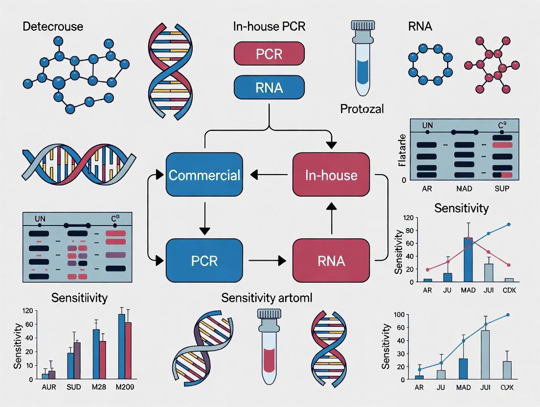
In-house (homebrew) qPCR assays provide flexibility in target selection and protocol optimization. These assays are particularly valuable for detecting parasites not included in commercial panels or for resource-limited settings. The following diagram illustrates the decision pathway for selecting between commercial and in-house qPCR approaches:
Essential Research Reagent Solutions
Successful implementation of qPCR in parasitology requires specific research reagents and materials. The following table outlines essential components and their functions:
Table 3: Essential Research Reagents for Parasitology qPCR
| Reagent/Category | Specific Examples | Function/Application | Key Considerations |
|---|---|---|---|
| Nucleic Acid Extraction Kits | QIAmp Viral RNA Kit [17], HiPurA Viral RNA Purification Kit [19] | Isolation of high-quality DNA from clinical samples | Efficient removal of PCR inhibitors; high recovery efficiency |
| Master Mixes | GoTaq Probe RT-qPCR System [17], SYBR Green ROX Plus mix [18] | Provides enzymes, buffers, nucleotides for amplification | Compatibility with detection chemistry; inhibitor resistance |
| Primer/Probe Sets | RV1/RV2 primers for Leishmania [18], Commercial assay primers | Target-specific amplification | Specificity; optimal annealing temperature; minimal dimer formation |
| Commercial Multiplex Panels | AllPlex Gastrointestinal Panel (Seegene) [15] | Simultaneous detection of multiple parasites | Included internal controls; comprehensive pathogen coverage |
| Positive Controls | Genomic DNA from reference strains [18] | Assay validation and quality assurance | Well-characterized reference materials |
| Instrument Systems | ABI7500 [19], Agilent AriaMx [20], QuantStudio Systems [21] | Amplification and fluorescence detection | Multiplexing capability; sensitivity; throughput |
Experimental Protocols for Key Applications
Protocol for Intestinal Protozoa Detection Using Multiplex PCR
The following protocol is adapted from the evaluation of the AllPlex Gastrointestinal Panel [15]:
Sample Preparation:
- Suspend fresh stool samples in FecalSwab medium
- Process samples using automated nucleic acid extraction systems (e.g., MICROLAB STARlet with Hamilton reagents)
- Include negative and positive controls in each extraction batch
DNA Extraction:
- Use standardized DNA extraction protocols provided by the manufacturer
- Elute DNA in appropriate buffer (volume typically 50-100μL)
- Store extracted DNA at -80°C if not used immediately
qPCR Amplification:
- Prepare reaction mix according to manufacturer's specifications
- Use CFX96 or equivalent real-time PCR instrument
- Cycling conditions: Follow manufacturer's recommended protocol
- Include internal control to monitor amplification efficiency
Result Interpretation:
- Analyze amplification curves using manufacturer's software
- Consider samples with Cq ≤40 as positive [15]
- Report qualitative results (positive/negative) to clinicians
Protocol for Visceral Leishmaniasis Detection
This protocol is adapted from studies evaluating qPCR for Leishmania detection in peripheral blood [18]:
DNA Extraction:
- Use 300μL of peripheral blood collected in EDTA tubes
- Extract DNA using Wizard Genomic DNA Purification kit or equivalent
- Elute DNA in 100μL sterile deionized water
qPCR Reaction Setup:
- Prepare 12.0μL PCR mix containing:
- 6.25μL SYBR Green ROX Plus mix
- 0.5μL each RV1 and RV2 primers (10pmol each)
- 4.75μL water
- Add 0.5μL DNA template (approximately 80-150ng)
- Include standard curve with serial dilutions of known parasite DNA (50,000 to 0.5 parasites)
Amplification Parameters:
- Initial denaturation: 95°C for 15 minutes
- 35 cycles of:
- Denaturation: 95°C for 15 seconds
- Annealing: 55°C for 30 seconds
- Extension: 72°C for 30 seconds
Data Analysis:
- Generate standard curve from serial dilutions
- Determine parasite load in unknown samples from standard curve
- Confirm amplicon size (145bp) by gel electrophoresis if needed
qPCR has fundamentally transformed parasitic disease diagnosis by providing superior sensitivity and specificity compared to traditional methods. The technology enables precise detection and quantification of parasites that are difficult to identify by microscopy alone. While commercial multiplex panels offer standardized, efficient detection of common intestinal protozoa, in-house protocols provide flexibility for specialized applications and resource-limited settings. The continuing evolution of qPCR technologies, including improved multiplexing capabilities and automation, will further enhance their application in parasitology. As molecular diagnostics continue to advance, qPCR remains a cornerstone technology for clinical diagnostics, epidemiological studies, and treatment monitoring of parasitic diseases.
The selection of an appropriate PCR testing platform is a critical decision for research and clinical laboratories. This choice often centers on using either a commercial test kit or a laboratory-developed test (LDT), also known as an in-house test. Within the specific context of analytical sensitivity comparison for commercial and in-house PCR protozoa research, understanding the performance characteristics, advantages, and limitations of each platform is fundamental to ensuring reliable and reproducible results. Commercial kits, which are pre-packaged with standardized reagents and protocols, offer the advantage of rapid implementation and regulatory compliance. In contrast, LDTs provide laboratories with the flexibility to design and optimize tests tailored to specific research needs, particularly valuable for novel or rare pathogens where commercial alternatives may not exist [22]. This guide objectively compares these two approaches, supported by experimental data and structured to inform researchers, scientists, and drug development professionals in their platform selection process.
Performance Comparison: Commercial Kits vs. Laboratory-Developed Tests
Direct comparative studies provide the most insightful data for platform selection. The performance of these platforms can be evaluated based on key metrics such as positive percent agreement (PPA, a measure of sensitivity), negative percent agreement (NPA, a measure of specificity), and the limit of detection (LOD).
Diagnostic Agreement and Sensitivity
A 2020 study comparing two commercial platforms (Roche cobas SARS-CoV-2 and Cepheid Xpert Xpress SARS-CoV-2) with several LDT variants found 100% agreement between all methods when testing clinical and simulated specimens. No false-positive or false-negative results were observed, demonstrating that with proper validation, both platforms can achieve exemplary diagnostic performance [23].
However, another comparative assessment of three different methods (Roche cobas, Mobidiag Amplidiag, and one LDT) on 183 clinical specimens revealed more nuanced differences. The reference standard was defined as the result obtained by at least two of the three methods.
Table 1: Performance Agreement of Two Commercial Kits and One LDT
| Assay Name | Type | Positive Percent Agreement (PPA) | Negative Percent Agreement (NPA) |
|---|---|---|---|
| Roche cobas SARS-CoV-2 | Commercial | 100% | 89.4% |
| Amplidiag COVID-19 | Commercial | 98.9% | 98.8% |
| Laboratory-Developed Test | In-House | 98.9% | 100% |
Source: Adapted from [24]
While the Roche cobas assay showed perfect PPA, its lower NPA suggests a potential for false-positive results in this specific evaluation. Conversely, the LDT showed perfect NPA, indicating a high reliability in confirming negative results [24].
Analytical Sensitivity and Limit of Detection
The Limit of Detection is a crucial parameter for determining the lowest quantity of a target that an assay can reliably detect. This is particularly important for detecting pathogens that may be present at low levels.
A comprehensive study comparing seven commercial SARS-CoV-2 assays using a single clinical specimen quantified by droplet digital PCR (ddPCR) found that the Abbott RealTime, Roche Cobas, and Xpert Xpress commercial assays demonstrated superior analytical sensitivity, detecting 100% of replicates at the lowest concentration tested (approximately 130-140 copies/mL) [25]. This highlights that well-designed commercial kits can achieve excellent sensitivity.
For LDTs, the analytical sensitivity is highly dependent on the individual components and protocols. A different study reported that the LOD for the E gene target across various LDTs and commercial kits varied from approximately 2 copies/reaction to over 30 copies/reaction. This variance was significantly influenced by the nucleic acid extraction method used, which changed the overall analytical sensitivity from 24 copies/mL to 574 copies/mL of specimen [23]. This underscores that for LDTs, the extraction method is a critical variable influencing the final assay sensitivity.
Experimental Protocols for Comparative Validation
For a researcher to independently verify or compare assay performance, following a structured validation protocol is essential. The guidelines below synthesize best practices for such comparative studies.
Sample Selection and Panel Construction
The foundation of a robust validation is a well-characterized sample panel.
- Sample Types: Use a combination of clinical specimens (e.g., nasopharyngeal swabs in viral transport medium) and reference materials [23] [26]. Clinical specimens provide real-world context, while reference materials, such as inactivated virus or encapsulated RNA (e.g., AccuPlex from SeraCare), allow for precise quantification and determination of the LOD [23] [25].
- Panel Composition: The panel should include a range of target concentrations, including samples near the expected LOD. It must also include negative controls and samples containing potentially cross-reactive organisms to assess specificity [23] [22]. A typical panel might include at least 10 positive and 10 negative clinical specimens [23].
Determining the Limit of Detection
The LOD is determined by testing serial dilutions of a standardized target material.
- Dilution Series: Prepare a serial dilution (e.g., 10-fold) of the reference material in the same matrix as the clinical sample (e.g., viral transport medium) [23] [25].
- Replication: Test each dilution with at least three to five technical replicates to establish a detection rate at each concentration [23]. Some studies use higher replicates (e.g., 10) where reagent availability allows [25].
- Calculation: The LOD is the lowest concentration at which ≥95% of the replicates are positive. The results are often reported in copies/reaction and can be converted to copies/mL based on sample input and elution volumes [23].
Assessing Analytical Specificity
Specificity ensures the assay does not produce false-positive results.
- Cross-Reactivity Testing: Test the assay against a panel of nucleic acids from other genetically similar or clinically relevant organisms that could be present in the sample type. For a respiratory virus assay, this would include other coronaviruses, influenza, and rhinoviruses [23]. For protozoan research, this would include related parasitic species.
- In Silico Analysis: Before wet-lab testing, perform an in silico analysis using genetic databases to check primer and probe sequences for homology with non-target organisms [27].
Verification of Commercial Assays
When introducing a commercially developed test, laboratories must verify the manufacturer's claims using their own staff and equipment. According to guidelines, this involves establishing that the manufacturer's performance specifications for accuracy, precision, and reportable range can be reproduced with local operations [22]. This process, while less extensive than a full LDT validation, is critical to ensure the test performs as expected in your specific laboratory environment.
The Scientist's Toolkit: Essential Reagents and Materials
The following table details key reagents and materials essential for developing, validating, and running both commercial and in-house PCR tests.
Table 2: Essential Research Reagent Solutions for PCR Assay Validation
| Item | Function | Application Notes |
|---|---|---|
| Reference Standard Material | Provides a quantified standard for determining LOD, linearity, and assay efficiency. | Includes inactivated virus, encapsulated RNA (e.g., SeraCare AccuPlex [23]), or RNA transcripts [28]. Critical for normalizing results across different assays. |
| Nucleic Acid Extraction Kits | Isolates and purifies target nucleic acid from the sample matrix. | The choice of extraction method significantly impacts the final LOD of the assay [23]. Examples include MagMAX, QIAamp, and easyMag systems [23] [25]. |
| Primer/Probe Sets | Binds specifically to the target DNA sequence for amplification and detection. | For LDTs, these are designed and optimized in-house. Performance varies; independent evaluation is recommended [28]. Commercial kits provide pre-optimized primers/probes. |
| Master Mix | Contains enzymes, dNTPs, and buffers necessary for the reverse transcription and amplification reactions. | Commercial kits include a proprietary master mix. For LDTs, labs can select from various vendors (e.g., New England Biolabs Luna kit [28]). |
| Internal Control | Monitors the entire process from extraction to amplification for potential failures or inhibition. | Can be exogenous (added to each sample) or endogenous (a human gene target). Commercial kits often include an internal control; it must be incorporated into LDT design [22]. |
| Positive & Negative Controls | Verifies assay performance and identifies contamination. | Must be included in every run. Positive control confirms test is working; negative control (no-template) checks for contamination [22]. |
The decision between implementing a commercial kit or an LDT is not a matter of declaring one universally superior to the other. Instead, the choice depends on a balance of performance, practicality, and purpose.
Commercial kits offer a streamlined path to implementation, with the benefits of standardization, regulatory compliance, and high throughput. Studies show that leading commercial assays can deliver excellent sensitivity and specificity, sometimes outperforming LDTs [25] [24]. They are ideal for laboratories with high testing volumes and those requiring rapid deployment of standardized testing.
Laboratory-developed tests provide unmatched flexibility, allowing researchers to adapt to new pathogens, customize targets, and control costs. While their performance is highly dependent on in-house expertise and rigorous validation, well-designed LDTs can achieve performance on par with, or even superior to, commercial alternatives [23] [24]. They are indispensable for basic research, diagnostic work on rare pathogens, and in situations where commercial kits are unavailable or cost-prohibitive.
For researchers comparing analytical sensitivity in protozoa or any other pathogen, the evidence strongly recommends a case-by-case evaluation. The key to success with either platform lies in rigorous, continuous validation and quality control, ensuring that the chosen method meets the specific analytical and clinical needs of the research project.
Inside the Assays: Protocol Design, Multiplexing, and Targets for Protozoan PCR
The selection of appropriate molecular diagnostic tools is fundamental to the integrity of research and clinical outcomes. The choice between commercial kits and in-house developed protocols presents a significant dilemma for laboratories, balancing factors such as standardization, cost, performance, and regulatory compliance. Commercial kits provide standardized reagents and streamlined workflows, ensuring reproducibility and convenience, whereas in-house methods offer customization and potential cost savings for high-volume testing. This guide objectively compares the performance of commercial real-time PCR (qPCR) kits and in-house protocols across various pathogens, supported by experimental data on analytical sensitivity, specificity, and practical implementation. The focus is on providing a clear comparison to inform researchers, scientists, and drug development professionals in their selection process.
Performance Comparison: Commercial Kits vs. In-House Protocols
Direct comparative studies provide the most valuable insights for evaluating the performance of commercial and in-house PCR methods. The data below summarize key findings from multiple diagnostic applications.
Table 1: Comparative Analytical Performance of Commercial Kits and In-House PCR Protocols
| Pathogen / Application | Method Category | Specific Name | Sensitivity | Specificity | Key Performance Findings | Source |
|---|---|---|---|---|---|---|
| SARS-CoV-2 | Commercial Kits (Comparison) | TaqPath (Thermo Fisher), BGI, Roche LightCycler | Varied against viral variants | N/R | Significant differences in Cq values found; sensitivity decreased for some kits against Omicron variant. | [29] |
| SARS-CoV-2 | Commercial Kits (Comparison) | Sansure Biotech, GeneFinder, TaqPath | No significant difference (p=0.107) | No significant difference | High positive association and Cohen’s κ coefficient; Sansure showed slightly better diagnostic performance. | [30] |
| SARS-CoV-2 | In-House Protocol | Triplex E+RdRp+RNase P | 98.3% | N/R | Detection limit: E gene (3.8 copies/μL), RdRp gene (33.8 copies/μL). | [31] |
| Enteropathogenic Bacteria | In-House vs. Commercial | In-house, FTD, ampliCube | In-house: 77.5-80.7%Commercial: 75-100% (strong positive) | In-house: 99.7-100%Commercial: 96-100% | Commercial kits showed acceptable agreement, suggesting replaceability of in-house assays. | [32] |
| Mycobacterium tuberculosis | In-House vs. Commercial | In-house, Cobas Amplicor | In-house: 81.3%Commercial: 71.9% | In-house: 98.9%Commercial: 100% | Both showed high sensitivity and specificity, deemed reliable for diagnosis. | [33] |
| Lupin (Food Allergen) | Commercial Kit | RT-PCR SPECIALfinder MC Lupin | LOD: 0.5 ppm (Ct 26-34) | 100% (in silico) | Validated as a specific, robust, and rapid method for detecting lupin traces in complex food matrices. | [34] |
Key Comparative Insights:
- Sensitivity and Specificity: Well-validated in-house protocols can achieve performance on par with, and sometimes superior to, commercial kits, as seen in tuberculosis detection [33]. However, commercial kits often demonstrate more consistent performance across different settings [30].
- Impact of Genetic Variants: A critical advantage of some in-house protocols is their flexibility. Commercial kits may suffer from reduced sensitivity when new viral variants with mutations in the primer/probe target regions emerge, necessitating continuous evaluation [29].
- Limit of Detection (LOD): In-house methods can be optimized for exceptionally low LODs, making them suitable for applications requiring high sensitivity, such as early pathogen detection [31].
Detailed Experimental Protocols
To ensure reproducibility and provide a clear understanding of the methodological groundwork, this section details the protocols from key studies cited in the comparison.
Protocol: Sensitivity Evaluation of Three SARS-CoV-2 qPCR Kits
Objective: To compare the detection performance of three commercial SARS-CoV-2 nucleic acid detection kits—Sansure Biotech, GeneFinderTM, and TaqPathTM—using identical clinical samples and equipment [30].
- Sample Preparation: 354 randomly selected nasopharyngeal swab samples from hospitalized COVID-19 patients were used. RNA was extracted from all samples and aliquoted for parallel testing.
- PCR Setup: Each of the three kits was used according to the manufacturer's instructions. Crucially, all reactions for a given sample were performed using the same RNA isolate and the same real-time PCR instrument to eliminate variables in sample quality and machine performance.
- Data Analysis: The Ct values for the ORF1ab and N genes were recorded. The final results (positive/negative) and average Ct values from the three kits were statistically compared using p-values and Cohen's κ coefficient to assess agreement.
Protocol: In-House Triplex RT-qPCR for SARS-CoV-2
Objective: To develop and standardize a cost-effective in-house RT-qPCR method for detecting SARS-CoV-2 in resource-constrained settings [31].
- Primers and Probes: TaqMan primers and probes for the E gene (Sarbecovirus genus), RdRp gene (SARS-CoV-2 specific), and RNase P gene (endogenous control) were synthesized based on established sequences [31].
- Reaction Setup:
- Triplex Reaction (20 µL): 5 µL sample RNA, 10 µL of 2x reaction buffer (SuperScript III Platinum One-Step RT-qPCR Kit), 0.5 µL enzyme mix, and 4.5 µL primer-probe mix.
- Thermocycling Conditions: Reverse transcription at 50°C for 20 min; initial denaturation at 95°C for 3 min; 45 cycles of 95°C for 15 s and 58°C for 60 s.
- Limit of Detection (LOD) Assay: Serial dilutions of a SARS-CoV-2 positive RNA sample and a plasmid standard were tested in triplex and duplex formats to determine the lowest detectable concentration of the virus.
- Validation: The sensitivity and specificity of the triplex assay were evaluated using 132 clinical samples and compared to a standard reference method.
Protocol: Comparison of In-House and Commercial PCR for Enteropathogenic Bacteria
Objective: To compare the performance of an established in-house multiplex real-time PCR with two commercial kits (FTD and ampliCube) for detecting enteropathogenic bacteria in stool samples [32].
- Sample Collection: The study used 341 samples, comprising 241 patient stool samples and 100 samples from external laboratory control schemes.
- Nucleic Acid Extraction: All samples underwent standardized DNA extraction using the QIAamp DNA Stool Mini Kit.
- Parallel PCR Testing: Each sample was tested in parallel using:
- The in-house multiplex real-time PCR for Salmonella spp., Shigella spp./EIEC, C. jejuni, and Yersinia spp.
- The FTD bacterial gastroenteritis kit.
- The ampliCube gastrointestinal bacterial panels 1 & 2.
- Data Analysis: A gold standard-based analysis was performed to calculate sensitivity and specificity. Latent class analysis (a statistical method that does not require a perfect gold standard) and Cohen's kappa were also used to assess agreement and performance.
Workflow and Decision Pathway
The following diagram illustrates the key decision-making pathway and procedural steps involved in selecting and implementing a PCR method, from initial choice to result interpretation.
The Scientist's Toolkit: Essential Research Reagent Solutions
This section details the core components and instruments that form the foundation of reliable PCR-based detection, whether for commercial or in-house applications.
Table 2: Essential Reagents and Instruments for PCR Diagnostics
| Item | Function | Example Use-Case |
|---|---|---|
| One-Step RT-qPCR Master Mix | Integrates reverse transcription and PCR amplification in a single tube, streamlining the workflow and reducing contamination risk. | Used in SARS-CoV-2 detection protocols for direct amplification from RNA extracts [29] [31]. |
| TaqMan Probes & Primers | Sequence-specific oligonucleotides that enable highly specific target detection and quantification in real-time PCR. | Form the core of both commercial kits and in-house protocols for genes like ORF1ab, N, E, and RdRp [29] [30] [31]. |
| Internal Control (IC) | A non-target nucleic acid sequence added to each reaction to monitor PCR inhibition and validate nucleic acid extraction. | RNase P gene for human RNA quality; Phocid Herpes Virus DNA or manufacturer's proprietary ICs [29] [32]. |
| Nucleic Acid Extraction Kit | Standardized reagents and protocols for isolating high-purity DNA/RNA from complex biological samples (e.g., stool, swabs). | QIAamp DNA Stool Mini Kit for bacteria; Magnetic bead-based kits (e.g., MGIEasy) for viral RNA [32] [31]. |
| Real-Time PCR Thermocycler | Instrument that amplifies nucleic acids while fluorescently monitoring product accumulation in real-time, generating Ct values. | Agilent AriaMx, Bio-Rad CFX96, Rotor-Gene Q [29] [31] [34]. |
| Standardized Plasmid DNA | A quantifiable DNA construct containing the target sequence, used for generating standard curves and determining the limit of detection (LOD). | Crucial for validating and quantifying in-house assays, as demonstrated in the LOD determination for SARS-CoV-2 [31]. |
Molecular diagnostics have revolutionized the detection of pathogenic protozoa, presenting clinical laboratories with a critical choice between implementing commercial kits or developing in-house real-time PCR (qPCR) assays. While microscopy remains the traditional diagnostic standard for intestinal protozoan infections, it suffers from significant limitations in sensitivity, specificity, and the ability to differentiate closely related species [11]. Molecular methods, particularly qPCR, offer enhanced detection capabilities but require careful validation to ensure diagnostic reliability. This guide objectively compares the performance of in-house and commercial PCR assays for protozoan detection, providing experimental data and methodologies to inform assay selection and optimization for researchers, scientists, and drug development professionals. The focus on analytical sensitivity comparison between commercial and in-house PCR platforms in protozoa research reveals a complex landscape where neither approach uniformly outperforms the other, but rather presents complementary strengths and limitations that must be carefully weighed against diagnostic requirements and laboratory capabilities.
Performance Comparison: Commercial vs. In-House PCR Assays
Analytical Sensitivity and Specificity Across Protozoan Targets
Table 1: Performance Comparison of Commercial and In-House PCR Assays for Intestinal Protozoa Detection
| Parasite | Assay Type | Sensitivity (%) | Specificity (%) | Reference |
|---|---|---|---|---|
| Giardia duodenalis | Commercial (AusDiagnostics) | 91.0 | 98.9 | [35] |
| Giardia duodenalis | In-house RT-PCR | 93.3 | 97.0 | [35] |
| Cryptosporidium spp. | Commercial (AusDiagnostics) | 78.9 | 99.1 | [35] |
| Cryptosporidium spp. | In-house RT-PCR | 78.9 | 100 | [35] |
| Dientamoeba fragilis | Commercial (AusDiagnostics) | 68.4 | 91.9 | [35] |
| Dientamoeba fragilis | In-house RT-PCR | 68.4 | 88.9 | [35] |
| Mycoplasma pneumoniae | In-house (RepMp1) | 77.5 | 99.7 | [9] [33] |
| Mycoplasma pneumoniae | Commercial (Cobas Amplicor) | 71.9 | 100 | [33] |
Recent multicenter evaluations demonstrate that both commercial and in-house PCR assays can achieve high performance standards for major protozoan pathogens. For Giardia duodenalis detection, a comparative study showed complete agreement between commercial (AusDiagnostics) and in-house PCR methods, with both demonstrating high sensitivity and specificity comparable to conventional microscopy [11]. The data revealed marginally higher sensitivity for the in-house assay (93.3% vs. 91.0%) but slightly higher specificity for the commercial platform (98.9% vs. 97.0%) [35].
For Cryptosporidium detection, both methods showed identical sensitivity (78.9%) but the in-house assay achieved marginally better specificity (100% vs. 99.1%) [35]. The detection of Dientamoeba fragilis proved more challenging for both platforms, with identical sensitivity (68.4%) but higher specificity for the commercial assay (91.9% vs. 88.9%) [35]. This pattern of variable performance across different targets underscores the importance of pathogen-specific validation rather than assuming uniform performance across a test menu.
Multiplex Assay Performance Characteristics
Table 2: Comparison of Commercial Multiplex PCR Assays for Diarrhea-Causing Protozoa
| Commercial Assay | Target Pathogens | Reported Sensitivity Range | Reported Specificity Range | Manufacturer |
|---|---|---|---|---|
| Gastroenteritis/Parasite Panel I | Cryptosporidium hominis/parvum, Giardia duodenalis, Entamoeba histolytica | 91-100% | 98-100% | Diagenode [2] |
| RIDAGENE Parasitic Stool Panel | Cryptosporidium hominis/parvum, Giardia duodenalis, Entamoeba histolytica | 90-100% | 97-100% | R-Biopharm [2] |
| Allplex Gastrointestinal Parasite Panel 4 | Cryptosporidium hominis/parvum, Giardia duodenalis, Entamoeba histolytica | 89-99% | 96-100% | Seegene [2] |
| FTD Stool Parasites | Cryptosporidium hominis/parvum, Giardia duodenalis, Entamoeba histolytica | 92-100% | 97-100% | Fast Track Diagnostics [2] |
Evaluations of four commercial multiplex qPCR assays demonstrated consistently high performance across major diarrheal protozoa, with all methods showing sensitivity and specificity generally exceeding 90% [2]. This comparative analysis revealed that while all commercial multiplex assays performed well, some variability exists in their ability to detect mixed infections and their lower limits of detection for specific targets. The study highlighted that commercial assays offer the advantage of standardized protocols and simplified implementation but may lack flexibility for specific research applications [2].
Experimental Protocols for Assay Validation
DNA Extraction and Purification Methodology
Proper nucleic acid extraction is critical for PCR performance, particularly for parasites with robust cyst walls that complicate DNA extraction. In comparative studies, researchers typically employ automated extraction systems to ensure consistency. One standardized protocol involves mixing 350 µl of Stool Transport and Recovery Buffer (S.T.A.R) with approximately 1 µl of each fecal sample using a sterile loop, followed by incubation for 5 minutes at room temperature and centrifugation at 2000 rpm for 2 minutes [11]. The supernatant (250 µl) is carefully collected, transferred to a fresh tube, and combined with 50 µl of internal extraction control. DNA extraction then proceeds using the MagNA Pure 96 DNA and Viral NA Small Volume Kit on the MagNA Pure 96 System (Roche Applied Sciences), which provides fully automated nucleic acid preparation based on magnetic separation of nucleic acid-bead complexes [11]. This standardized approach helps minimize extraction variability when comparing assay performance.
In-House RT-PCR Amplification Protocol
For in-house assay development, reaction mixtures typically include 5 µl of extracted DNA, 12.5 µl of 2× TaqMan Fast Universal PCR Master Mix (Thermo Fisher Scientific), primers and probe mix (2.5 µl), and sterile water to a final volume of 25 µl [11]. A multiplex tandem PCR assay is performed using standardized cycling conditions. Each assay should include appropriate negative, positive, and qPCR inhibition controls. The selection of primer-probe sequences should target conserved genomic regions with demonstrated specificity, while amplification conditions must be optimized for each target through empirical testing of annealing temperatures, primer concentrations, and cycle parameters [11] [28].
Analytical Sensitivity and Efficiency Testing
To determine detection limits and amplification efficiency, standard curves should be generated using serial dilutions of quantified target DNA or RNA. The PCR efficiency can be calculated using the equation: Efficiency = -1 + 10(-1/slope), with ideal values ranging from 90-110% [36] [28]. The limit of detection is established as the lowest concentration at which 95% of replicates test positive. This validation is particularly important for in-house assays, as primer-probe sets can exhibit significant variability in sensitivity. One study evaluating SARS-CoV-2 primer-probe sets found that while most sets performed comparably, one set (RdRp-SARSr) showed significantly reduced sensitivity due to a mismatch in the reverse primer [28], highlighting the importance of thorough validation even for established assays.
Workflow Diagram: PCR Assay Development and Validation
The workflow outlines the comprehensive process for developing and validating in-house PCR assays, from initial design through implementation. The process begins with careful primer/probe design and optimization, proceeds through rigorous analytical validation, and culminates in clinical evaluation before implementation. Each stage requires specific quality control measures to ensure the final assay meets diagnostic performance standards.
Decision Framework: Commercial vs. In-House Assay Selection
This decision framework illustrates the pathways for selecting between commercial and in-house PCR assays based on laboratory requirements, resources, and diagnostic needs. Commercial assays offer standardized workflows suitable for routine testing, while in-house development provides flexibility for custom targets but requires extensive validation and quality control protocols.
The Scientist's Toolkit: Research Reagent Solutions
Table 3: Essential Reagents and Materials for PCR Assay Development
| Reagent/Material | Function | Examples/Specifications | Application Notes |
|---|---|---|---|
| TaqMan Probes | Sequence-specific detection | MGB, TAMRA, QSY, QSY2 probes; FAM, VIC, ABY, JUN, Cy5 dyes | Enable multiplexing up to 6 targets; shorter, more specific probes with non-fluorescent quenchers maximize sensitivity [37] |
| qPCR Primers | Target amplification | Custom sequences; available dry or liquid format; guaranteed yield | Designed for use with TaqMan probes or SYBR dye; desalted and available in various scales [37] |
| Master Mixes | PCR reaction foundation | Contains DNA polymerase, dNTPs, buffers; e.g., LightCycler Multiplex RNA Virus Master, SuperScript III One-Step RT-PCR | Choice affects efficiency; comparison studies show different master mixes can impact results with same primer-probe sets [36] |
| Automated Extraction Systems | Nucleic acid purification | MagNA Pure 96 System (Roche), QIAamp DNA mini kit (Qiagen) | Critical for consistent results; automated systems reduce variability in DNA extraction [11] [9] |
| Reference Materials | Assay validation | Quantified DNA/RNA standards, control panels | Essential for determining sensitivity, specificity, and limit of detection; serial dilutions for standard curves [28] |
The selection of appropriate reagents forms the foundation of reliable PCR assay development. Dual-labeled TaqMan probes provide specific detection through fluorophore-quencher separation during amplification, with various chemistries available for different multiplexing needs [37]. Master mix selection significantly impacts amplification efficiency, with studies showing that the same primer-probe sets can perform differently in various master mixes [36]. Automated extraction systems minimize variability in nucleic acid purification, which is particularly important for parasites with robust cyst walls that complicate DNA extraction [11]. Reference materials and standardized controls are indispensable for proper validation of both commercial and in-house assays.
The choice between commercial and in-house PCR assays for protozoan detection involves careful consideration of performance requirements, laboratory resources, and intended applications. Commercial assays offer standardized, convenient solutions with generally reliable performance, while in-house methods provide flexibility and potential cost advantages but require extensive validation and expertise. The evidence indicates that both approaches can achieve high sensitivity and specificity when properly validated and implemented. Factors such as sample collection methods, DNA extraction procedures, and target pathogen characteristics significantly impact performance regardless of assay format. Future directions point toward increased multiplexing capabilities, improved standardization, and harmonization of molecular protocols to enhance comparability across laboratories and studies.
Molecular diagnostics for protozoan parasites rely on the precise detection of specific genomic targets. The choice of target gene fundamentally influences the sensitivity, specificity, and reliability of polymerase chain reaction (PCR) assays, impacting both clinical diagnostics and research. Within the context of comparing commercial multiplex PCR kits with laboratory-developed tests (in-house PCRs), understanding the properties of different molecular targets is crucial for assay selection and development. This guide objectively compares the three primary categories of molecular targets—18S ribosomal RNA (rRNA) genes, repetitive genomic elements, and species-specific genes—by synthesizing experimental data on their analytical performance in protozoan detection [38] [39].
Comparative Analysis of Molecular Targets
The table below summarizes the core characteristics, advantages, and limitations of the key molecular targets, providing a foundation for their comparison.
Table 1: Key Molecular Targets for Protozoan Detection
| Molecular Target | Copy Number (Typical Range) | Primary Advantage | Key Limitation | Ideal Use Case |
|---|---|---|---|---|
| 18S rRNA Gene | 4-8 copies (Plasmodium) [38], varies by species | Broadly conserved; enables pan-generic primers and metabarcoding [40] [41]. | Low copy number limits sensitivity; sequence variation can cause false negatives [42] [38] [39]. | Multiplex panels for diverse parasite screening; phylogenetic studies. |
| Repetitive Elements | 14-41 copies (e.g., Pvr47, Pfr364 in Plasmodium) [38], 200-300 copies (AF146527 in T. gondii) [43] | High copy number provides superior analytical sensitivity for low-level infections [38] [43]. | Species-specific; requires extensive genome mining for identification; potential absence in some strains [38] [43]. | High-sensitivity detection of specific pathogens; single-step and multiplex PCR. |
| Species-Specific Genes | Typically single copy (e.g., surface antigens, metabolic enzymes) | High specificity for precise species identification; useful for strain typing. | Lowest sensitivity due to single copy number; requires meticulous primer design for conserved regions. | Differentiation of morphologically similar species; virulence studies. |
Performance Data and Experimental Evidence
Sensitivity and Specificity in Protozoan Detection
Experimental data from controlled studies and clinical evaluations provide quantitative support for the comparisons outlined in Table 1.
Table 2: Experimental Performance Data for Different Molecular Targets
| Parasite / Assay Context | Target Gene | Reported Sensitivity | Comparison Findings | Source |
|---|---|---|---|---|
| Plasmodium falciparum & P. vivax | 18S rRNA | Reference standard (nested PCR) | Used as a benchmark for comparison. | [38] |
| Novel Repetitive Elements (Pfr364, Pvr47) | 1-10 parasites/μL in single-step PCR; 100% sensitivity vs. 18S rRNA nested PCR [38]. | More sensitive than 18S rRNA in a single-step PCR format [38]. | [38] | |
| Toxoplasma gondii | B1 gene (~35 copies) | Standard for diagnostic PCR | A robust and widely used target. | [43] |
| AF146527 repeat (200-300 copies) | Reported higher sensitivity in some studies [43]. | Absent in 4.8% of human-positive samples, arguing for B1 as a preferred target [43]. | [43] | |
| Intestinal Protozoa (Multiplex qPCR) | 18S rRNA or other targets in commercial panels | Detected parasites on the first stool sample [15]. | More efficient for protozoan detection than microscopy, but may miss some parasites not included in the panel [15]. | [15] |
| Various Intestinal Parasites | 18S rRNA V9 region (Metabarcoding) | Simultaneously detected 11 parasite species [40]. | Read counts varied significantly by species, influenced by factors like DNA secondary structure [40]. | [40] |
Impact of Genetic Diversity on Diagnostic Accuracy
A critical challenge in molecular diagnostics is the genetic variation within and between protozoan species. The 18S rRNA gene, while conserved, can exhibit nucleotide substitutions or deletions that prevent probe hybridization or primer binding, leading to false-negative results. For instance, variant Plasmodium ovale isolates from Vietnam possessed three nucleotide differences in the 18S rRNA probe region, preventing detection with a standard species-specific probe [42]. This underscores the necessity for careful primer and probe design based on comprehensive sequence databases and for the maintenance of microscopic methods for validation [42] [39].
Experimental Protocols for Target Validation
Workflow for Identifying and Validating Repetitive DNA Targets
The discovery of novel repetitive targets, such as Pfr364 and Pvr47 for Plasmodium [38], follows a structured bioinformatics and laboratory validation pipeline.
Figure 1: A pipeline for identifying novel repetitive DNA targets for molecular diagnostics, based on genomic data mining [38].
Step-by-Step Protocol [38]:
- Data Harvesting: Obtain assembled genome sequence data for the target parasite from databases like PlasmoDB.
- Consensus Repeat Sequence (CRS) Identification: Use programs like RepeatScout (with default parameters) to identify all repetitive sequences in the genome.
- Computational Screening:
- Eliminate CRS with internal tandem repeats using Tandem Repeat Finder (TRF).
- Screen CRS against the NCBI UniVec database and the host genome (e.g., human) using BLAST to remove non-specific sequences.
- Manually inspect passing CRS to ensure species specificity by comparing them to all available Plasmodium sequence data.
- Copy Number Determination: Use the screened repeats to search (BLAST) against the parasite's genome sequence to calculate the copy number for each repeat. Select repeats with high copy numbers and sufficient length (>300 bp) for primer design.
- Wet-Lab Validation:
- Primer Design: Design primers for the selected repetitive targets.
- PCR Optimization: Test primers using a high-fidelity polymerase. Assess sensitivity by spiking samples with known quantities of target material (e.g., plasmids, parasites).
- Clinical Evaluation: Verify sensitivity and specificity using microscopy-positive and -negative clinical specimens, comparing performance to a reference method like nested 18S rRNA PCR.
Protocol for 18S rRNA Metabarcoding for Multiplex Detection
Metabarcoding of the 18S rRNA gene allows for the simultaneous detection of a wide range of parasites in a single sample and is particularly useful for environmental or complex sample screening [44] [40] [41].
Step-by-Step Protocol [40] [41]:
- DNA Extraction: Extract total DNA from the sample (e.g., stool, water, oyster tissue) using a robust kit suitable for the matrix, such as the Fast DNA SPIN Kit for Soil.
- PCR Amplification: Amplify the target hypervariable region of the 18S rRNA gene (e.g., V4 or V9).
- Primers: Use universal eukaryotic primers with Illumina adapters attached, such as 1391F and EukBR for the V9 region.
- Master Mix: Use a high-fidelity hot-start ready mix.
- Cycling Conditions: Typically, 95°C for 5 min; 30-35 cycles of 98°C for 30s, 55°C for 30s, 72°C for 30s; final extension at 72°C for 5 min.
- Library Preparation and Sequencing: Perform a limited-cycle amplification to add multiplexing indices and complete Illumina sequencing adapters. Pool the amplicons and sequence on a platform such as the Illumina iSeq 100.
- Bioinformatic Analysis:
- Processing: Use a pipeline like QIIME 2. Demultiplex and trim reads, then denoise and filter chimeras with DADA2.
- Taxonomic Assignment: Classify the resulting Amplicon Sequence Variants (ASVs) against a curated reference database (e.g., NCBI nucleotide database or a custom database of parasite 18S rRNA sequences) to identify the protozoan species present.
The Scientist's Toolkit: Research Reagent Solutions
Table 3: Essential Reagents and Kits for Protozoan Molecular Research
| Reagent / Kit | Function | Example Use Case | Reference |
|---|---|---|---|
| High-Fidelity DNA Polymerase (e.g., KAPA HiFi) | Accurate amplification of target DNA with low error rates. | Critical for amplifying targets for sequencing and metabarcoding library construction [41]. | [41] |
| Commercial Multiplex PCR Panels (e.g., AllPlex GIP) | Simultaneous detection of multiple protozoan targets in a single, standardized reaction. | Efficient first-line screening of stool samples for common gastrointestinal protozoa in a clinical lab [15]. | [15] |
| DNA Extraction Kits for Stool/Soil (e.g., Fast DNA SPIN Kit) | Efficient lysis and purification of DNA from complex, inhibitor-rich samples. | Extraction of PCR-ready DNA from stool or environmental samples for sensitive detection [40]. | [40] |
| TOPcloner TA Kit | Easy cloning of PCR amplicons into plasmids for sequencing or creating control materials. | Generation of plasmid controls for validating and quantifying PCR assays [40]. | [40] |
| Illumina iSeq 100 Sequencing System | Next-generation sequencing for high-throughput, parallel sequencing of amplicons. | 18S rRNA metabarcoding for the profiling of protozoan communities in wastewater or clinical samples [44] [40]. | [44] [40] |
Multiplex qPCR represents a significant advancement in molecular diagnostics, enabling the simultaneous amplification and detection of multiple nucleic acid targets in a single reaction. Duplex qPCR, the most common and foundational form of multiplexing, allows researchers to detect two distinct targets using the same reagent pool. This technique has gained substantial traction in pathogen detection due to its ability to conserve valuable samples, reduce reagent costs, and minimize pipetting errors compared to singleplex reactions [45]. The core principle involves designing target-specific probes labeled with distinct fluorescent dyes—typically FAM for the target gene and VIC for an endogenous control—whose emission spectra are easily distinguishable by real-time PCR instruments [45].
The application of duplex qPCR has become particularly valuable in diagnostic contexts where differentiating between closely related pathogens is crucial for treatment decisions. For instance, in veterinary diagnostics, a newly developed TaqMan probe-based duplex qPCR assay successfully differentiates between Porcine Epidemic Diarrhea Virus (PEDV) GI and GII subtypes with sensitivities of 90 copies/μL and 40 copies/μL, respectively [46]. This level of discrimination is vital for implementing appropriate vaccination strategies, as vaccines derived from classical GI subtypes like CV777 often fail to provide effective protection against variant GII subtypes [46].
Duplex qPCR in Protozoan Pathogen Detection: Commercial vs. In-House Assays
The detection and differentiation of pathogenic intestinal protozoa represent a formidable challenge in clinical diagnostics. Traditional microscopy, while low-cost, suffers from significant limitations in sensitivity, specificity, and the ability to differentiate closely related species [11] [7]. Molecular methods, particularly real-time PCR (RT-PCR), have emerged as promising alternatives, especially in non-endemic areas with low parasitic prevalence [11].
Comparative Performance in Intestinal Protozoa Detection
A comprehensive 2025 multicentre study involving 18 Italian laboratories directly compared the performance of a commercial RT-PCR test (AusDiagnostics) against an in-house RT-PCR assay for detecting key intestinal protozoa: Giardia duodenalis, Cryptosporidium spp., Entamoeba histolytica, and Dientamoeba fragilis [11] [7]. The study analyzed 355 stool samples (230 fresh, 125 preserved) against the reference standard of conventional microscopy.
Table 1: Comparison of Commercial vs. In-House PCR for Protozoan Detection
| Pathogen | Assay Type | Sensitivity | Specificity | Remarks |
|---|---|---|---|---|
| Giardia duodenalis | Commercial (AusDiagnostics) | High | High | Complete agreement between commercial and in-house methods |
| In-house RT-PCR | High | High | Performance similar to conventional microscopy | |
| Cryptosporidium spp. | Commercial (AusDiagnostics) | Limited | High | Inadequate DNA extraction likely affected sensitivity |
| In-house RT-PCR | Limited | High | Better results with preserved stool samples | |
| Entamoeba histolytica | Commercial (AusDiagnostics) | Critical for accurate diagnosis | High | Microscopy cannot differentiate from non-pathogenic species |
| In-house RT-PCR | Critical for accurate diagnosis | High | Molecular differentiation is essential | |
| Dientamoeba fragilis | Commercial (AusDiagnostics) | Inconsistent | High | Detection inconsistency across methods |
| In-house RT-PCR | Inconsistent | High | Requires protocol standardization |
The data revealed complete agreement between commercial and in-house methods for detecting G. duodenalis, with both demonstrating high sensitivity and specificity comparable to conventional microscopy [11]. For Cryptosporidium spp. and D. fragilis, both molecular methods showed high specificity but limited sensitivity, likely attributable to inadequate DNA extraction from the robust wall structure of parasite oocysts [11] [7]. Molecular assays proved critical for accurately diagnosing E. histolytica, as microscopic examination cannot differentiate this pathogenic species from non-pathogenic Entamoeba counterparts [11].
Impact of Sample Preservation on Detection Efficacy
The study yielded noteworthy findings regarding sample preservation, with PCR results from preserved stool samples outperforming those from fresh samples, likely due to better DNA preservation in fixed specimens [11]. This finding has significant implications for protocol standardization in diagnostic laboratories, suggesting that sample preservation methods may enhance detection reliability for certain protozoan pathogens.
Experimental Protocols for Duplex qPCR Assays
Assay Design and Optimization
Implementing successful duplex qPCR requires meticulous experimental design and validation. Key considerations include:
- Primer and Probe Design: Primers must be specific and should not bind elsewhere in the template DNA, to the probe, or to each other [45]. The Tm of TaqMan probes should be approximately 10°C higher than the Tm of primers (around 68-70°C) [45].
- Amplicon Considerations: Amplicons should not overlap and should be approximately the same size. Researchers should verify amplicon coordinates using tools like the UCSC Genome Browser In Silico PCR utility and check for primer dimer formation using Multiple Primer Analyzer tools [45].
- Dye Selection: Choose dyes with minimal emission spectrum overlap. For duplex reactions, FAM and VIC dyes represent optimal combinations due to their distinct emission peaks at 517nm and 551nm, respectively [45]. Instrument compatibility is crucial, as different real-time PCR systems have specific filter sets and calibration requirements [47].
Table 2: Essential Research Reagent Solutions for Duplex qPCR
| Reagent Category | Specific Examples | Function in Duplex qPCR |
|---|---|---|
| Master Mixes | TaqPath ProAmp Master Mix [48], TaqMan Multiplex Master Mix [45] | Optimized buffer conditions for amplifying multiple targets simultaneously |
| Fluorophores | FAM, VIC, ABY, JUN [45] | Distinct reporter dyes for different targets with minimal spectral overlap |
| Dark Quenchers | ZEN, Iowa Black FQ, TAO [47] | Efficient quenching to reduce background fluorescence in multiplex reactions |
| DNA Extraction Kits | QIAamp UCP Pathogen Mini Kit [48], MagNA Pure 96 System [11] | High-quality nucleic acid extraction, crucial for sensitivity |
| Sample Preparation | S.T.A.R. Buffer [11] | Sample transport and preservation for improved DNA recovery |
Primer Limitation Strategy
A critical technical aspect of duplex qPCR involves addressing competition for limited reagents when one target (often the control gene) is significantly more abundant than others. Primer limitation provides an effective solution by using reduced primer concentrations (typically 150nM each instead of 900nM in singleplex reactions) for the more abundant target [45]. This approach ensures that the highly expressed gene reaches plateau due to primer exhaustion rather than reagent depletion, leaving sufficient nucleotides, polymerase, and other reagents for proper amplification of less abundant targets [45].
Workflow Visualization
The following diagram illustrates the key decision points and procedures in a standard duplex qPCR experimental workflow:
Validation Protocols
Comprehensive validation is essential before implementing duplex qPCR in diagnostic settings. The general validation procedure involves: (1) running singleplex reactions to confirm amplification; (2) establishing multiplex reaction conditions; (3) determining whether singleplex and multiplex reactions yield equivalent Ct values; and (4) optimizing primer/probe concentrations if discrepancies occur [45]. Each reaction should be performed in triplicate to ensure reproducibility. If high variation persists between replicates despite multiplexing, returning to singleplex reactions may be necessary [45].
Advanced Multiplexing Strategies Beyond Duplexing
While duplex qPCR represents the most accessible entry point to multiplexed detection, technological innovations are pushing the boundaries of multiplexing capabilities. Color cycle multiplex amplification (CCMA) represents a groundbreaking approach that significantly increases the number of detectable DNA targets in a single qPCR reaction using standard instrumentation [48]. Rather than relying solely on spectral distinction, CCMA utilizes fluorescence permutations across multiple cycles, theoretically allowing detection of up to 136 distinct DNA targets with just 4 fluorescence channels [48].
This advanced methodology employs rationally designed blockers to modulate cycle threshold (Ct) delays for different fluorescence signals, creating a pre-programmed pattern of fluorescence increases that identifies specific targets [48]. Experimental applications have demonstrated CCMA's potential through a single-tube qPCR assay screening 21 sepsis-related bacterial DNA targets in various clinical samples, achieving 89% clinical sensitivity and 100% clinical specificity [48].
Duplex qPCR establishes a robust foundation for efficient multi-pathogen detection, offering significant advantages in resource conservation, result precision, and diagnostic efficiency. The comparative analysis of commercial and in-house assays for protozoan detection reveals a nuanced landscape where molecular methods provide critical differentiation capabilities—particularly for distinguishing pathogenic Entamoeba histolytica from non-pathogenic species—though technical challenges in DNA extraction from certain protozoal organisms remain.
Future directions in multiplex qPCR point toward increasingly sophisticated approaches like color cycle multiplex amplification that transcend traditional spectral limitations. However, duplex qPCR maintains its position as the most accessible, validated, and practically implementable multiplexing strategy for routine diagnostic laboratories. As molecular technologies continue to evolve and standardize, duplex and advanced multiplex qPCR methodologies will undoubtedly play an expanding role in the accurate detection and differentiation of pathogenic organisms, ultimately enhancing disease surveillance, outbreak management, and patient care outcomes.
In molecular diagnostics and pathogen research, the accuracy of a polymerase chain reaction (PCR) test is fundamentally constrained by the initial steps of sample handling and nucleic acid preparation. For researchers working with protozoan pathogens, such as Leishmania species, the efficiency of DNA extraction from complex biological samples directly determines the analytical sensitivity of subsequent PCR assays [49]. This guide objectively compares various sample preparation and DNA extraction methods, providing supporting experimental data to inform protocol selection for commercial and in-house PCR applications in protozoan research. Optimal sample processing ensures that low parasite loads, often critical for diagnosing asymptomatic infections or monitoring treatment efficacy, are reliably detected, thereby avoiding more invasive diagnostic procedures [49].
Comparative Analysis of Sample Preparation Methodologies
The choice of sample type and lysis method significantly influences the final yield and purity of isolated DNA, impacting PCR sensitivity.
Sample Type: Whole Blood vs. Buffy Coat
The cellular component of blood used for DNA extraction is a primary factor affecting sensitivity.
- Whole Blood (WB): Contains all blood components, including a high abundance of host DNA and PCR inhibitors such as heme. This complex matrix can reduce the relative concentration of parasite DNA and interfere with amplification [49].
- Buffy Coat (BC): The leukocyte-rich layer, obtained by centrifuging whole blood, concentrates the white blood cells where intracellular parasites like Leishmania often reside. Its use provides a 10-fold increase in sensitivity compared to whole blood, as it enriches for target parasite DNA while reducing the volume of competing human DNA and inhibitors [49].
Table 1: Impact of Sample Type on PCR Sensitivity for Parasite Detection
| Sample Type | Description | Advantage | Key Finding |
|---|---|---|---|
| Whole Blood (WB) | Unseparated peripheral blood | Simple collection procedure | Lower sensitivity due to high host DNA background and inhibitors |
| Buffy Coat (BC) | Concentrated leukocyte fraction | Enriches infected host cells | 10-fold increase in sensitivity; best for detecting low parasitemia [49] |
Lysis and DNA Extraction Chemistries
The method used to break open cells and isolate DNA is equally critical. Research on visceral leishmaniasis diagnosis has demonstrated that at low parasite concentrations (≤100 parasites/ml), proteinase K (PK)-based methods proved clearly superior to guanidine-EDTA-based methods [49]. The combination of buffy coat samples with a PK-based lysis protocol yielded the highest sensitivity, allowing for the reliable detection of 10 parasites/ml of blood [49].
Beyond traditional chemical lysis, physical and enzymatic methods are also employed, often in combination [50].
- Physical Methods: Include bead beating or grinding with a mortar and pestle under liquid nitrogen, which are useful for disrupting tough cell walls or tissue structures.
- Enzymatic Methods: Utilize enzymes like lysozyme or proteinase K to degrade tissues and tough cell walls, and are often used in combination with other methods [50].
More recent comparisons of DNA extraction from Dried Blood Spots (DBS) have highlighted that simple, cost-effective methods can outperform standardized commercial kits. A 2025 study found that a Chelex-100 resin boiling method yielded significantly higher DNA concentrations compared to several column-based kits, including those from Qiagen and Roche [51]. This method is rapid and cost-effective but yields DNA with lower purity as it omits purification steps [51]. Column-based methods, which often rely on silica-binding chemistry under high-salt conditions, provide more purified DNA but at a higher cost and with longer processing times [50] [51].
The following workflow diagram summarizes the key decision points and steps in an optimized protocol for sensitive detection of blood-borne parasites.
Comparative Performance Data
Direct, back-to-back comparisons of methods provide the most actionable data for researchers. The following table summarizes key findings from two such studies, one on Leishmania detection and another on human DNA from DBS.
Table 2: Back-to-Back Comparison of DNA Extraction Method Performance
| Method Category | Specific Method / Kit | Key Performance Metric | Result / Sensitivity | Study Context |
|---|---|---|---|---|
| In-House Lysis + Extraction | Proteinase K Lysis + ΦC Extraction (Buffy Coat) | Parasite Detection Limit | 10 parasites/mL (Reliable) [49] | Visceral Leishmaniasis (Seeded Samples) |
| In-House Lysis + Extraction | Guanidine-EDTA Lysis + Silica Beads (Buffy Coat) | Parasite Detection Limit | ≤100 parasites/mL (Inconsistent) [49] | Visceral Leishmaniasis (Seeded Samples) |
| Commercial Kit (Column) | DNeasy Tissue Kit (Qiagen) | Protocol Features | Standardized, but costly and time-consuming [49] [51] | General DNA Purification |
| Boiling Method | Chelex-100 Resin | DNA Concentration (ACTB qPCR) | Significantly higher vs. column kits [51] | Human DNA from Dried Blood Spots |
| Commercial Kit (Column) | High Pure PCR Template Kit (Roche) | DNA Concentration (Spectrophotometry) | Higher vs. other column kits, but lower than Chelex [51] | Human DNA from Dried Blood Spots |
Detailed Experimental Protocols
To ensure reproducibility, below are detailed methodologies for two key experiments cited in this guide.
Optimized Proteinase K-based Protocol for Buffy Coat
This protocol, adapted from a visceral leishmaniasis study, is designed for maximal sensitivity from blood samples [49].
- Sample Preparation: Collect 5 ml of peripheral blood in an EDTA-coated tube. Centrifuge for 10 min at 1,600 × g to separate components.
- Buffy Coat Isolation: Transfer 500 μl of the buffy coat (the leukocyte-rich layer) to a new tube.
- Lysis: Add 1 ml of TNNT lysis buffer (0.5% Tween 20, 0.5% Nonidet P-40, 10 mM NaOH, 10 mM Tris, pH 7.2) and proteinase K to a final concentration of 320 μg/ml.
- Incubation: Incubate the mixture for 2 to 24 hours at a temperature of 56°C to facilitate complete digestion.
- Enzyme Inactivation: Boil the lysate for 10 minutes to inactivate the proteinase K.
- DNA Extraction: Subject the lysate to a phenol-chloroform extraction. Precipitate the DNA with ethanol and resuspend the final pellet in 130-200 μl of sterile distilled water or TE buffer.
- Storage: Store purified DNA at -20°C until PCR analysis.
Chelex-100 Boiling Method for Dried Blood Spots
This cost-effective protocol, optimized in a 2025 study, is ideal for high-throughput screening from DBS [51].
- Punch and Soak: Punch one 6 mm DBS disk into a microtube. Incubate overnight at 4°C in 1 mL of 0.5% Tween20 in PBS.
- Wash: Remove the Tween20 solution and add 1 mL of PBS. Incubate for 30 minutes at 4°C, then remove the PBS.
- Chelex Boiling: Add 50 μL of a pre-heated 5% (w/v) Chelex-100 solution. Pulse-vortex for 30 seconds.
- Incubate: Incubate at 95°C for 15 minutes, with brief pulse-vortexing every 5 minutes.
- Pellet Debris: Centrifuge for 3 minutes at 11,000 rcf to pellet Chelex beads and paper debris.
- Collect Supernatant: Carefully transfer the supernatant containing the DNA to a new tube. A second centrifugation step with a smaller-volume pipette is recommended for precision.
- Storage: Store the extracted DNA at -20°C.
The Scientist's Toolkit: Essential Research Reagents
The following table lists key reagents and their critical functions in the sample processing and DNA extraction workflows described above.
Table 3: Essential Reagents for Sensitive DNA Extraction Protocols
| Reagent / Solution | Function in the Protocol |
|---|---|
| EDTA (Ethylenediaminetetraacetic acid) | An anticoagulant in blood collection tubes; also chelates metal ions to inhibit DNase activity and prevent DNA degradation [49]. |
| Proteinase K | A broad-spectrum serine protease that digests histones and other cellular proteins, facilitating the release of intact DNA from the cell [49] [50]. |
| Chaotropic Salts (e.g., Guanidine HCl) | Disrupt cellular structures, inactivate nucleases, and enable binding of nucleic acids to silica matrices in column-based kits [49] [50]. |
| Phenol-Chloroform | A mixture used in liquid-liquid extraction to separate DNA from proteins and other cellular debris; proteins are denatured and partition into the organic phase, while DNA remains in the aqueous phase [49]. |
| Chelex-100 Resin | A chelating ion-exchange resin that binds metal ions which are cofactors for DNases. When boiled with the sample, it protects DNA from degradation, providing a rapid purification method [51]. |
| Silica Matrices | The core of column-based kits; DNA binds to the silica membrane in the presence of high salt, allowing contaminants to be washed away before low-salt elution of pure DNA [50] [51]. |
Overcoming Technical Hurdles: Extraction, Inhibition, and Sensitivity Gaps
The molecular diagnosis of infections caused by protozoan parasites such as Cryptosporidium spp., Giardia duodenalis, and Entamoeba histolytica represents a significant challenge for clinical researchers and diagnostic developers. The primary obstacle lies in the formidable cell walls of protozoan oocysts and cysts, which are engineered by evolution to protect the genetic material within from environmental extremes [52]. These robust structures, primarily composed of chitin and other complex macromolecules, efficiently resist conventional chemical lysis methods, leading to suboptimal DNA recovery and potential false-negative results in downstream PCR applications [53] [54]. Consequently, the analytical sensitivity of molecular assays for protozoan research is intrinsically linked to the efficacy of the initial DNA extraction process. This guide provides a systematic comparison of commercial and in-house DNA extraction methods, evaluating their performance against the challenge of disrupting resilient cyst and oocyst walls to support sensitive and reliable molecular detection.
Methodological Approaches: Extraction and Pretreatment Protocols
The efficiency of DNA extraction from protozoan cysts and oocysts hinges on specialized protocols designed to compromise their structurally complex walls. These methodologies can be broadly categorized into commercial kits and in-house developed methods, often incorporating a critical mechanical pretreatment step.
Commercial Extraction Kits
Commercial kits provide standardized protocols and reagents, offering consistency and ease of use. However, their performance can vary significantly, and modifications to the manufacturer's instructions are often necessary for optimal recovery of protozoan DNA.
QIAamp DNA Stool Mini Kit (Qiagen): This kit was systematically evaluated for its ability to recover DNA from oocysts and cysts directly from feces. Following the standard manufacturer's protocol, it demonstrated 100% sensitivity and specificity for Giardia and Entamoeba histolytica. However, its sensitivity for Cryptosporidium was only 60% (9/15 positive samples), highlighting the particular resilience of Cryptosporidium oocysts. A series of optimizations were shown to increase sensitivity to 100%. Key amendments included raising the lysis temperature to the boiling point for 10 minutes, extending the incubation time with the InhibitEX tablet to 5 minutes, using pre-cooled ethanol for precipitation, and employing a small elution volume (50-100 µL) [52].
Automated Systems (NucliSENS easyMAG, BioMérieux): In a multicenter comparison of DNA extraction methods for the detection of Enterocytozoon bieneusi spores, which possess a thick chitin wall, the NucliSENS easyMAG system, when coupled with an effective mechanical pretreatment, demonstrated superior performance. It achieved the highest frequencies of detection for low spore concentrations and yielded some of the lowest Ct values among the seven methods tested [54].
Other Commercial Kits: A comparative study of four commercial multiplex real-time PCR assays for detecting diarrhoea-causing protozoa utilized DNA extracted and purified with a GennAll genomic DNA extraction kit. The performance of the downstream PCR assays was contingent on this initial extraction quality [2]. Another study evaluating oocyst DNA extraction from environmental samples found that kits using paramagnetic resins, such as the MAGNEX DNA Kit, showed high sensitivity, detecting as few as 100 oocysts/mL [55].
In-House and Optimized Pretreatment Methods
In-house methods often focus on the critical pretreatment step, employing physical disruption to break open the resilient parasitic structures before nucleic acid purification.
Mechanical Pretreatment with Bead Beating: The mechanical disruption of oocysts using grinding beads has been consistently identified as a highly effective pretreatment. One study systematically evaluated eleven commercial mechanical lysis matrixes composed of beads of different sizes, shapes, and compositions (e.g., silica, garnet, ceramic). The best performance for Cryptosporidium parvum DNA extraction was achieved using a lysing matrix containing ceramic beads with a median diameter of 1.4 mm, processed in a FastPrep-24 grinder at a speed of 6.0 m/s for 60 seconds [53]. A separate study on E. bieneusi spores confirmed that bead beating significantly improved DNA yield, with optimal results obtained using a TissueLyser II at 30 Hz for 60 seconds with small commercial beads from ZymoResearch or MP Biomedicals [54].
Physical and Chemical Lysis Methods for Giardia: An evaluation of four in-house pretreatment methods for Giardia duodenalis cysts identified several effective techniques. The methods involved combinations of mechanical disruption using crushed cover glass or glass beads, vortexing, boiling at 100°C, and multiple freeze-thaw cycles using liquid nitrogen and a heating block. The method combining crushed cover glass, TAE buffer, shaking, and boiling yielded the highest DNA concentration, while the method incorporating vortexing with crushed cover glass, boiling, and freeze-thaw cycles resulted in the highest optical density purity measurement [56].
Table 1: Summary of Optimized DNA Extraction and Pretreatment Methods
| Target Parasite | Method Type | Key Protocol Steps | Performance Outcome | Source |
|---|---|---|---|---|
| Cryptosporidium spp. | Modified Commercial Kit | Boiling (10 min), extended InhibitEX incubation (5 min), pre-cooled ethanol, small elution volume (50-100 µL) | Sensitivity increased from 60% to 100% | [52] |
| Cryptosporidium parvum | Mechanical Pretreatment | Bead beating (6.0 m/s, 60 s) with 1.4 mm ceramic beads | Best DNA extraction performance | [53] |
| Giardia duodenalis | In-House Pretreatment | Crushed cover glass, TAE buffer, shaking, boiling (100°C/3 min) | Highest DNA concentration | [56] |
| Enterocytozoon bieneusi | Mechanical Pretreatment | Bead beating (30 Hz, 60 s) with small ZR BashingBeads/MP Lysing Matrix E beads | Highest detection rate and lowest Ct values | [54] |
Comparative Performance Data
Independent, comparative studies provide critical data on the sensitivity, specificity, and overall efficacy of different molecular detection pathways for intestinal protozoa.
Commercial Multiplex PCR Assays
A direct comparison of four commercial multiplex real-time PCR assays using a common reference panel of 126 well-characterized DNA samples revealed high overall performance but important differences [2]. The panel included samples positive for Cryptosporidium hominis/parvum (n=32), Giardia duodenalis (n=47), and Entamoeba histolytica (n=3).
Table 2: Diagnostic Performance of Commercial Multiplex PCR Assays
| Commercial Assay | Cryptosporidium | Giardia | Entamoeba histolytica | Key Findings |
|---|---|---|---|---|
| Gastroenteritis/Parasite Panel I (Diagenode) | 100% Se, 100% Sp | 100% Se, 100% Sp | 100% Se, 100% Sp | No cross-reactivity with related species like E. dispar. |
| RIDAGENE Parasitic Stool Panel (R-Biopharm) | 100% Se, 100% Sp | 100% Se, 100% Sp | 100% Se, 100% Sp | Reliable detection with no false positives. |
| Allplex Gastrointestinal Parasite Panel 4 (Seegene) | 100% Se, 100% Sp | 100% Se, 100% Sp | 100% Se, 100% Sp | Effectively identified all target pathogens. |
| FTD Stool Parasites (Fast Track Diagnostics) | 100% Se, 97.9% Sp | 97.9% Se, 100% Sp | 100% Se, 100% Sp | Showed a single false positive for Cryptosporidium. |
Another evaluation of the VIASURE real-time PCR assay (CerTest Biotec) against a large panel of 358 DNA samples reported high sensitivity and specificity: 0.96 and 0.99 for Cryptosporidium spp., 0.94 and 1.00 for G. duodenalis, and 0.96 and 1.00 for E. histolytica, respectively [57].
Commercial vs. In-House PCR Methods
A multicenter study involving 18 Italian laboratories compared a commercial RT-PCR test (AusDiagnostics) and an in-house RT-PCR assay against traditional microscopy for detecting several protozoa [11] [7]. The study analyzed 355 stool samples.
For Giardia duodenalis, there was complete agreement between the commercial and in-house PCR methods, with both demonstrating high sensitivity and specificity comparable to microscopy. However, for Cryptosporidium spp. and Dientamoeba fragilis, both molecular methods showed high specificity but limited sensitivity. The authors concluded that this limited sensitivity was likely due to inadequate DNA extraction from the parasite, underscoring that even robust PCR assays can be undermined by a suboptimal extraction protocol [11].
The Scientist's Toolkit: Essential Research Reagents & Materials
Successful DNA extraction from resilient parasitic forms requires a combination of specific reagents and laboratory equipment. The following table details key solutions used in the protocols cited in this guide.
Table 3: Key Research Reagent Solutions for Protozoan DNA Extraction
| Item Name | Function / Application | Specific Example / Protocol Context |
|---|---|---|
| Lysing Matrix Tubes | Mechanical disruption of robust cyst/oocyst walls via bead beating. | MP Biomedical Lysis Matrix tubes with ceramic (1.4 mm), glass, or garnet beads [53]. |
| InhibitEX Tablets / Technology | Adsorption and removal of PCR inhibitors common in stool samples. | Used in QIAamp DNA Stool Mini Kit; extended incubation (5 min) improves inhibitor removal [52]. |
| Silica-Membrane / Magnetic Bead Kits | Nucleic acid binding, washing, and purification post-lysis. | QIAamp kits (silica-membrane) and NucliSENS easyMAG (magnetic beads) for automated extraction [52] [54]. |
| FastPrep-24 / TissueLyser II | High-speed homogenizers that provide consistent, vigorous bead beating. | FastPrep-24 at 6.0 m/s for Cryptosporidium [53]; TissueLyser II at 30 Hz for E. bieneusi [54]. |
| Crushed Cover Glass | An inexpensive, irregularly-shaped alternative to commercial beads for mechanical lysis. | Effective for breaking Giardia cysts when used with vortexing and boiling [56]. |
Workflow and Decision Pathway
The following diagram summarizes the optimized experimental workflow for DNA extraction from protozoan cysts and oocysts, integrating the key steps and considerations discussed.
Diagram: Optimized Workflow for Protozoan DNA Extraction. Critical parameters for mechanical pretreatment and nucleic acid extraction that significantly impact analytical sensitivity are highlighted.
The challenge of breaking the robust walls of protozoan cysts and oocysts for DNA extraction remains a pivotal factor in the analytical sensitivity of molecular diagnostics and research. Evidence consistently demonstrates that while commercial PCR assays can exhibit excellent specificity, their sensitivity is often constrained by the efficacy of the upstream DNA extraction process [11] [2]. The integration of a vigorous, optimized mechanical pretreatment step, particularly bead beating with appropriately sized and composed beads, is a consistently proven strategy to dramatically improve DNA yield from Cryptosporidium oocysts, Giardia cysts, and E. bieneusi spores [53] [54] [56]. Furthermore, the modification of commercial kit protocols with steps such as extended boiling and small elution volumes can elevate performance to meet the demands of sensitive detection [52]. For researchers and drug developers, a focus on standardizing and optimizing the sample preparation phase is not merely a preliminary step but is as critical as the selection of the PCR assay itself for achieving reliable and reproducible results in protozoan research.
Managing PCR Inhibitors from Stool and Food Matrices
Molecular detection of pathogens in complex matrices such as stool and food represents a significant analytical challenge for researchers and diagnostic professionals. These samples contain a diverse range of PCR inhibitors—substances that can significantly reduce amplification efficiency and analytical sensitivity. Inhibitors commonly found in these matrices include complex polysaccharides, bile salts, fats and lipids, phenolic compounds, and various proteases. The fundamental mechanism of inhibition typically involves disruption of the polymerase activity, interference with cell lysis, or degradation of nucleic acids, ultimately leading to reduced detection sensitivity or false-negative results [58].
The impact of these inhibitors is particularly relevant in the context of protozoan pathogen detection, where target organisms like Cyclospora cayetanensis may be present at low levels and require highly sensitive detection methods. Effective management of these inhibitory substances is therefore essential for accurate surveillance, outbreak investigation, and drug development research. This guide systematically compares approaches for overcoming these challenges, focusing on experimental data and practical methodologies that can be implemented in both commercial and laboratory-developed protocols.
Comparative Analysis of Inhibition Management Strategies
Sample Processing and Nucleic Acid Extraction
The initial sample processing and nucleic acid extraction steps represent the first critical barrier against PCR inhibitors. Research demonstrates that the choice of extraction methodology significantly influences downstream amplification efficiency.
Table 1: Comparison of Sample Processing Methodologies for Inhibitor Management
| Methodology | Matrix Tested | Key Advantages | Limitations | Effect on Cq Values |
|---|---|---|---|---|
| Automated Microfluidic SP [58] | Complex foods (flour, spread, seed mix) | Integrated DNA extraction, concentration, and purification; processes large sample volumes (≥5g) for representativity | Requires specialized equipment; longer processing time (~2 hours) | Significant reduction (P<0.05) in inhibitory effects compared to crude extraction |
| Filter Paper Elution [59] | Environmental samples (simulated) | Low-cost, low-tech; suitable for transport and storage | Variable recovery based on paper type and elution buffer | Buffer-dependent: Lysis buffer showed minimal Cq shift vs. controls |
| Dilution of Template [60] | Universal | Simple, rapid, no specialized reagents | Reduces target concentration, may decrease sensitivity | Can reduce but not eliminate inhibition; may increase Cq |
| Centrifugal Filtration | Stool, food homogenates | Removes particulate matter and some inhibitors | Incomplete removal of soluble inhibitors; additional step | Moderate improvement; dependent on filter characteristics |
The selection of an appropriate elution buffer following extraction proves critical for optimal recovery. A comparative study evaluating three different buffers (PCR-grade water, Tris-EDTA buffer, and general lysis buffer) found that lysis buffer provided superior recovery of viral RNA from filter paper matrices, with no statistically significant difference in detection compared to buffer-specific positive controls [59]. This suggests that chemical components in lysis buffers effectively counteract inhibitor carryover, making them preferred for elution in challenging matrices.
PCR Formulation and Cycling Condition Modifications
Adjusting the PCR mixture itself represents a second strategic approach to mitigating inhibition. Experimental evidence supports several formulation modifications that enhance reaction robustness.
Table 2: PCR Component Modifications for Inhibition Management
| Reaction Component | Standard Condition | Enhanced Robustness Condition | Rationale | Evidence of Efficacy |
|---|---|---|---|---|
| DNA Polymerase [61] [62] | 1-2 units/50µL | Increased concentration (e.g., 2-4 units/50µL) | Overcomes enzyme binding or inactivation by inhibitors | Improved yield in presence of inhibitors; potential for nonspecific products [61] |
| MgCl₂ Concentration [62] | 1.5-2.0 mM | Titration between 0.5-5.0 mM | Mg²⁺ is a cofactor; affects primer annealing and enzyme activity | Critical for maintaining efficiency; optimal concentration is template-specific [62] |
| dNTP Concentration | 0.2 mM each | Balanced concentration maintenance | dNTPs chelate Mg²⁺; improper balance affects fidelity | Concentrations <0.01-0.015 mM approach Km, reducing efficiency [61] |
| Enhancer Additives [62] | None | BSA, Betaine, Mono-/Disaccharides (e.g., Sucrose) | Stabilize enzymes, reduce secondary structures, compete with inhibitors | Sucrose most effective carbohydrate enhancer for specific amplification [62] |
Beyond reaction components, thermal cycling parameters can be optimized. Increasing annealing temperature incrementally (e.g., testing 55-65°C range) enhances stringency and reduces mispriming caused by inhibitor interference [60]. Similarly, utilizing a three-step cycling protocol (denaturation, annealing, extension) rather than a two-step protocol allows for finer control over primer binding specificity, particularly beneficial for difficult templates [60].
Experimental Protocols for Inhibitor Management
Protocol 1: Integrated Microfluidic System for Complex Food Matrices
This protocol, adapted from a fully automated system for allergen detection, demonstrates a comprehensive approach to managing inhibitors in complex foods, applicable to protozoan detection [58].
Workflow Overview:
Detailed Steps:
- Sample Homogenization: Thoroughly grind and homogenize approximately 5 grams of food sample to ensure representativity, critical for detecting heterogeneous contamination [58].
- Automated Sample Preparation (SP): Utilize an integrated microfluidic system incorporating pumps, valves, reservoirs, and a filter membrane to process the large sample volume. This step achieves simultaneous DNA extraction, concentration, and purification [58].
- Purification: The system automatically purifies nucleic acids, removing PCR inhibitors such as fats and polyphenols common in chocolate, ham, and green vegetables [58].
- Multiplex qPCR Detection: Transfer the purified DNA to a microfluidic chip containing multiple reaction chambers. Perform qPCR with target-specific primers and probes. Include internal positive controls in each chamber to validate results and rule out false negatives due to residual inhibition [58].
Key Advantages: This method processes larger sample volumes than conventional techniques, improving target representation and detection sensitivity while simultaneously removing inhibitors through integrated purification. The entire process from sample to result requires approximately two hours [58].
Protocol 2: Filter Paper-Based Recovery and Elution for Environmental Samples
This protocol, derived from veterinary pathogen surveillance research, offers a cost-effective method suitable for field sampling and transport, with relevance to stool and environmental samples [59].
Workflow Overview:
Detailed Steps:
- Sample Inoculation: Apply a measured volume (e.g., 300 µL) of liquid sample (stool suspension, environmental swab eluate) to specific locations on a defined swatch (e.g., 5.1 cm × 3.8 cm) of selected filter paper [59].
- Drying: Allow the inoculated paper to air-dry completely. Filter paper acts as a stabilizer for nucleic acids during storage and transport [59].
- Elution: Soak the paper swatch in an appropriate elution buffer. The study compared PCR-grade water, Tris-EDTA buffer, and general lysis buffer, finding that general lysis buffer combined with a 1-4 hour soaking time yielded the best recovery of viral RNA, with no significant difference from positive controls [59].
- Nucleic Acid Extraction and RT-qPCR: Proceed with standard RNA extraction from the eluate, followed by RT-qPCR analysis. Normalize results using efficiency-standardized Cq values to account for variations in recovery efficiency [59].
Key Advantages: This method is particularly valuable for field studies and transport of infectious samples, as filter paper is inexpensive, easy to transport, and can stabilize nucleic acids. The choice of paper matrix significantly impacts recovery, with Whatman filter paper and cotton-based cellulose products showing favorable performance [59].
The Scientist's Toolkit: Essential Research Reagent Solutions
Successful management of PCR inhibitors requires strategic selection of reagents and materials. The following table outlines key solutions used in the featured experiments and their critical functions.
Table 3: Essential Reagents and Materials for Managing PCR Inhibition
| Reagent/Material | Primary Function | Application Example | Performance Consideration |
|---|---|---|---|
| Lysis Buffer [59] | Cell disruption, nucleic acid release, inhibitor denaturation | Elution of nucleic acids from filter paper matrices | Superior to water or TE buffer for recovery of PCR-detectable targets |
| Microfluidic Cartridges [58] | Integrated sample preparation, DNA purification, and qPCR | Automated processing of complex food samples | Removes inhibitors from challenging matrices (high fat, polyphenols) |
| Polymerase Enhancers [62] | Stabilization of DNA polymerase, reduction of secondary structures | Addition of sucrose or other carbohydrates to PCR mix | Sucrose identified as highly effective for specific, reliable amplification |
| Filter Paper (Whatman) [59] | Sample collection, storage, transport, and preliminary cleanup | Field collection of stool or environmental samples | High percentage of alpha cellulose provides consistent liquid recovery |
| DNA Polymerase [61] | Enzymatic amplification of target DNA | Standard and inhibitor-resistant PCR formulations | Increased units can overcome mild inhibition; inhibitor-resistant blends preferred for severe cases |
| Magnesium Chloride [62] | Cofactor for DNA polymerase, affects primer annealing | Titration to optimize specificity and yield in complex matrices | Critical concentration; typically 1.5-5.0 mM, requires optimization |
Effective management of PCR inhibitors in stool and food matrices is achievable through a multi-faceted approach combining robust sample processing, optimized nucleic acid extraction, and inhibition-resistant amplification chemistry. The experimental data and protocols presented demonstrate that strategies ranging from sophisticated microfluidic automation to simple filter paper elution can significantly improve detection sensitivity for protozoan pathogens and other targets. The consistent finding across studies—that method-specific optimization is non-negotiable—underscores the importance of validating any selected protocol with the intended sample matrices and target organisms. For researchers in drug development and diagnostic fields, implementing these evidence-based practices is essential for generating reliable, reproducible molecular data from the most challenging biological samples.
The reliability of molecular diagnostics and microbiome research hinges on the initial steps of sample handling. For stool samples, the choice between using fresh material or a fixed/preserved alternative represents a critical methodological crossroads. This decision directly influences downstream analyses, including PCR and metagenomic sequencing, by affecting the quantity and quality of recoverable DNA. Within the broader context of comparing commercial and in-house PCR protocols for protozoa and microbiome research, understanding the impact of sample preservation is foundational. This guide objectively compares the performance of fresh versus fixed stool preservation methods, drawing on recent experimental data to inform researchers, scientists, and drug development professionals.
Experimental Protocols and Methodologies
Key Comparative Study Designs
Recent investigations have systematically evaluated the impact of preservation methods on DNA yield. One comprehensive study tested 33 different conditions with varying collection, handling, and processing protocols on stool samples from infants and young children. The protocols included immediate freezing at -80°C, storage in ethanol, preservation in lysis buffer, and use of commercial stabilization kits like the OMNIgene Gut tube. The DNA was extracted using several commercial kits, including the ZR Fecal DNA MiniPrep (Zymo Research), QIAamp Fast Stool Mini Kit (Qiagen), and MO BIO Powersoil kit. The evaluation metrics focused on DNA yield, alpha diversity, and microbial community structure through 16S rRNA gene sequencing [63].
Another study provided a direct head-to-head comparison by collecting 18 matched pairs of mammalian fecal samples. Each pair was simultaneously transported in either 99.8% ethanol or a lysis buffer. The samples were processed between 55 and 461 days post-collection, allowing for an assessment of long-term preservation effects. The researchers quantified DNA yield and quality and further assessed the samples' suitability for microbiome profiling via Oxford Nanopore amplicon sequencing of bacterial 16S and protozoal 18S rRNA genes [64].
DNA Extraction and Downstream Analysis
In the comparative studies, the efficiency of DNA extraction was not only evaluated based on the preservation method but also on the extraction kit itself. A separate analysis of 12 DNA extraction methods—nine commercial kits and three laboratory protocols—highlighted that the lysis type is a key factor. Methods employing mechanical lysis (e.g., bead-beating) provided stable and high DNA yields, particularly for tough-to-lyse Gram-positive bacteria, whereas chemical and enzymatic methods showed lower efficiency [65].
For downstream applications, the extracted DNA was typically analyzed using real-time PCR (qPCR) or sequencing. The sensitivity of PCR, especially for low-abundance targets, is directly related to the initial DNA quantity and quality. One study on Mycobacterium leprae demonstrated that lower bacilloscopic indexes (a proxy for bacterial load) significantly reduced the sensitivity of PCR amplification for resistance genes, underscoring the importance of starting with high-yield, high-quality DNA [66].
Comparative Performance Data: Fresh vs. Fixed Stool
Quantitative and Qualitative DNA Yield
The following table summarizes the key performance metrics for different preservation methods, based on experimental data.
Table 1: Comparison of DNA Yield and Quality from Different Preservation Methods
| Preservation Method | Relative DNA Yield | DNA Quality / Integrity | Key Advantages | Key Limitations |
|---|---|---|---|---|
| Fresh, immediately frozen (-80°C) | High (Reference standard) | High | Preserves native microbial profile; minimal bias [63]. | Logistically challenging; requires constant cold chain. |
| Lysis Buffer (e.g., RNAlater) | High / Superior to ethanol [64] | Superior integrity; optimal for long-read sequencing [64] [67]. | Stable at room temp for transport; protects DNA from degradation. | May require specific downstream processing steps. |
| Ethanol (99.8%) | Significantly lower than lysis buffer [64] | Good purity on average, but higher variability [64]. | Widely available and inexpensive. | DNA shearing; lower yields can impact sensitivity [64]. |
| Commercial Kits (e.g., OMNIgene Gut) | Variable | Variable | Standardized for user convenience; room-temperature stable. | Proprietary formulas; cost can be a factor. |
The data indicates that lysis buffer is a highly effective preservative, yielding up to three times higher DNA concentration than ethanol in matched samples. Furthermore, electrophoretic analyses confirmed that DNA from lysis buffer-preserved samples had superior integrity, which is critical for long-read sequencing technologies like Nanopore [64]. While ethanol preservation resulted in DNA of excellent average purity (A260/280 ratio ~1.94), the results were highly dispersed (SD: 1.10), indicating less consistent performance compared to lysis buffer (A260/280 ratio ~1.92, SD: 0.27) [64].
Impact on Microbiome Community Structure
Beyond mere yield, the effect of preservation on the representational accuracy of the microbial community is paramount. The large-scale infant microbiome study concluded that, despite variability in DNA yield and alpha diversity between protocols, subject variability was the dominant driver of microbiome structure. The impact of the specific collection, handling, or processing protocol was comparatively minor within a standardized longitudinal study [63]. This suggests that while preservation method affects DNA quantity, robust cross-subject comparisons remain possible with a consistent protocol.
Impact on Downstream Analytical Sensitivity
The initial DNA yield has a direct bearing on the sensitivity of subsequent molecular assays. Research on leprosy diagnostics found that the sensitivity of PCR amplification for antimicrobial resistance genes was 50-70% lower in patients with low bacillary loads [66]. This highlights that suboptimal sample preservation, leading to reduced DNA yield, can critically impair the detection of low-abundance targets, a key concern in diagnostic protozoa research and resistance monitoring.
The Scientist's Toolkit: Essential Research Reagents
Selecting the right tools is critical for successful nucleic acid recovery from stool samples. The following table details key reagents and their functions based on the cited literature.
Table 2: Key Research Reagent Solutions for Stool DNA Studies
| Reagent / Kit Name | Primary Function | Key Features / Rationale |
|---|---|---|
| RNAlater / Lysis Buffer | Chemical preservation of nucleic acids in situ. | Inactivates nucleases; allows for room-temperature transport and storage; outperforms ethanol for DNA yield and integrity [63] [64]. |
| Zymo Research ZR Fecal DNA MiniPrep | DNA extraction and purification. | Utilizes bead-beating for mechanical lysis, effective for breaking Gram-positive bacterial cells [63]. |
| Qiagen QIAamp PowerFecal Pro DNA Kit | DNA extraction and purification. | Demonstrated superior DNA yield in comparative analyses; combines mechanical and chemical lysis [65]. |
| MO BIO Powersoil Kit | DNA extraction and purification. | A widely used standard in microbiome studies for effective DNA isolation from complex environmental samples [63]. |
| OMNIgene Gut Tube | Integrated sample collection and preservation. | A commercial all-in-one system designed to stabilize microbial DNA at room temperature for simplified logistics [63]. |
| Quick-DNA HMW MagBead Kit (Zymo Research) | Isolation of High Molecular Weight (HMW) DNA. | Uses magnetic beads for purification, minimizing shearing; recommended for long-read sequencing (e.g., Nanopore) [67]. |
Experimental Workflow and Impact Diagram
The diagram below illustrates the typical workflow for stool sample processing and how choices in preservation directly impact downstream analytical outcomes.
Figure 1: Workflow showing how preservation method choice directly influences DNA yield and downstream analytical success. The path from "Lysis Buffer" or "Fresh" samples leads to superior outcomes.
The evidence clearly demonstrates that sample preservation strategy has a profound impact on the success of stool-based molecular analyses. For research and diagnostics where maximizing DNA yield and preserving high molecular weight DNA are priorities—such as in protozoal detection, antimicrobial resistance screening, and shotgun metagenomics—preservation in a lysis buffer offers a superior alternative to traditional ethanol fixation. While fresh, immediately frozen samples remain the gold standard, lysis buffer provides a practical and high-performance solution for studies where a cold chain is not feasible. Ultimately, the choice of preservation method should be a deliberate decision, aligned with the specific analytical goals and logistical constraints of the study.
In the field of molecular diagnostics, the choice between commercial kits and in-house developed PCR assays is pivotal, especially for the detection of pathogenic intestinal protozoa. These infections, affecting billions globally, require diagnostic methods that are not only accurate but also efficient and cost-effective [11]. The optimization of these reactions—focusing on reducing reagent volumes and improving workflow efficiency—plays a critical role in making molecular diagnostics viable for both high-throughput laboratories and resource-limited settings. This guide provides an objective comparison of commercial and in-house real-time PCR (RT-PCR) assays for detecting enteric protozoa, framing the analysis within a broader thesis on analytical sensitivity. The data and protocols presented are designed to aid researchers, scientists, and drug development professionals in selecting and optimizing the most appropriate molecular tools for their specific needs.
Commercial vs. In-House PCR: A Comparative Framework
The debate between using commercial or in-house PCR assays often centers on a trade-off between standardization and customization. Commercial kits offer standardized protocols and reagents, which can significantly reduce development time and validation burdens. In-house assays, conversely, provide the flexibility to optimize reaction components and volumes for specific research questions or to reduce per-test costs. A key advantage of commercial multiplex panels is their comprehensive design, allowing for the simultaneous detection of multiple pathogens from a single sample, thereby improving diagnostic efficiency and turnaround time [68] [69]. However, as the following sections will demonstrate through experimental data, the performance of these approaches can vary significantly depending on the target pathogen and the specific protocols used.
Experimental Data Comparison
Performance in Detecting Intestinal Protozoa
A 2025 multicentre study involving 18 Italian laboratories provided a direct comparison of a commercial RT-PCR test (AusDiagnostics) and an in-house RT-PCR assay against conventional microscopy for identifying common intestinal protozoa. The study analyzed 355 stool samples [11].
Table 1: Comparative Performance of Commercial vs. In-House PCR for Intestinal Protozoa (2025 Multicentre Study)
| Pathogen | Method | Sensitivity | Specificity | Notes |
|---|---|---|---|---|
| Giardia duodenalis | Commercial PCR | High (Complete agreement with in-house) | High | Both methods demonstrated high sensitivity and specificity similar to microscopy [11]. |
| In-House PCR | High (Complete agreement with commercial) | High | ||
| Cryptosporidium spp. | Commercial PCR | High Specificity, Limited Sensitivity | High Specificity | Inconsistent detection; inadequate DNA extraction was a suggested cause [11]. |
| In-House PCR | High Specificity, Limited Sensitivity | High Specificity | ||
| Entamoeba histolytica | Commercial PCR | Data Not Specified | Data Not Specified | Molecular assays were noted as critical for accurate diagnosis [11]. |
| In-House PCR | Data Not Specified | Data Not Specified | ||
| Dientamoeba fragilis | Commercial PCR | High Specificity, Limited Sensitivity | High Specificity | Detection was inconsistent across methods [11]. |
| In-House PCR | High Specificity, Limited Sensitivity | High Specificity |
An independent 2025 validation study of the automated Seegene Allplex GI-Parasite Assay, which detects six protozoal pathogens, further illustrates the performance characteristics of a commercial multiplex platform. This study used microscopy as a reference standard [69].
Table 2: Performance of the Seegene Allplex GI-Parasite Commercial Multiplex Assay (2025 Validation Study)
| Pathogen | Sensitivity | Specificity | Positive Predictive Value (PPV) | Negative Predictive Value (NPV) |
|---|---|---|---|---|
| Blastocystis hominis (Bh) | 93.0% | 98.3% | 85.1% | 99.3% |
| Cryptosporidium spp. | 100% | 100% | 100% | 100% |
| Cyclospora cayetanensis (Cc) | 100% | 100% | 100% | 100% |
| Dientamoeba fragilis (Df) | 100% | 99.3% | 88.5% | 100% |
| Entamoeba histolytica (Eh) | 33.3% (increased to 75% with frozen specimens) | 100% | 100% | 99.6% |
| Giardia lamblia (Gl) | 100% | 98.9% | 68.8% | 100% |
Performance in Other Diagnostic Contexts
The comparative dynamics between commercial and in-house assays are not unique to parasitology. A 2017 study on detecting carbapenemase genes in gram-negative bacteria found that an in-house RT-PCR assay detected 79.0% (53/67) of OXA-type genes, outperforming the commercial Check-Direct CPE assay, which detected only 43.3% (29/67) [70]. This highlights that in-house methods can be optimized for superior sensitivity for specific, challenging targets. Conversely, commercial multiplex panels have demonstrated significant value in accelerating diagnosis for high-consequence infectious diseases, reducing transport times and biosafety concerns by enabling testing within isolation units [68].
Detailed Experimental Protocols
Protocol: Seegene Allplex GI-Parasite Commercial Assay
This protocol outlines the automated high-throughput method validated for the detection of six enteric protozoa [69].
- Sample Preparation: Stool samples are inoculated into FecalSwab tubes containing Cary-Blair media using the supplied swab. The tubes are vortexed for 10 seconds to ensure homogenization.
- Automated DNA Extraction:
- Platform: Hamilton STARlet liquid handler.
- Kit: STARMag 96 × 4 Universal Cartridge kit.
- Process: The automated system takes 50 µL of the stool suspension. The bead-based extraction process elutes the nucleic acid into a final volume of 100 µL.
- PCR Setup and Amplification:
- Kit: Allplex GI-Parasite Assay.
- Reaction Volume: 25 µL total, containing 5 µL of the extracted DNA and 20 µL of a mastermix (comprising 5 µL of 5X GI-P MOM primer, 10 µL RNase-free water, and 5 µL of EM2, which contains DNA polymerase, Uracil-DNA glycosylase, and buffer).
- Thermocycling Conditions: Performed on a Bio-Rad CFX96 system. The protocol includes a denaturing step followed by 45 cycles of:
- 95°C for 10 seconds
- 60°C for 1 minute
- 72°C for 30 seconds
- Detection: Four fluorophores (FAM, HEX, Cal Red 610, Quasar 670) are used for multiplex detection. A cycle threshold (Ct) value of ≤43 is considered positive as per the manufacturer's instructions.
Protocol: In-House RT-PCR for Intestinal Protozoa
This protocol is derived from the in-house assay used for comparison in the 2025 multicentre study [11].
- DNA Extraction:
- Sample Preparation: 350 µL of S.T.A.R. buffer is mixed with approximately 1 µL of fecal sample using a sterile loop. After incubation and centrifugation, 250 µL of the supernatant is transferred to a fresh tube and combined with 50 µL of an internal extraction control.
- Platform and Kit: MagNA Pure 96 System using the MagNA Pure 96 DNA and Viral NA Small Volume Kit for fully automated nucleic acid extraction via magnetic bead separation.
- PCR Amplification:
- Reaction Mix: Each 25 µL reaction contains:
- 5 µL of extracted DNA template.
- 12.5 µL of 2× TaqMan Fast Universal PCR Master Mix.
- 2.5 µL of a custom primer and probe mix.
- Sterile water to volume.
- Detection: A multiplex tandem PCR assay is performed, with specific details on thermocycling conditions adapted for the primer sets used.
- Reaction Mix: Each 25 µL reaction contains:
The Scientist's Toolkit: Research Reagent Solutions
The following table details key reagents and equipment essential for implementing the PCR assays discussed in this guide.
Table 3: Essential Research Reagents and Equipment for PCR-Based Protozoa Detection
| Item | Function / Role | Example Kits / Platforms (from studies) |
|---|---|---|
| Automated Nucleic Acid Extraction System | Purifies DNA/RNA from complex clinical samples, reducing manual labor and improving consistency. | Hamilton STARlet [69], MagNA Pure 96 System [11], KingFisher Flex [71] |
| Nucleic Acid Extraction Kit | Contains lysing buffers and purification matrices tailored to sample type (e.g., stool). | STARMag Universal Cartridge kit [69], MagNA Pure 96 DNA and Viral NA Small Volume Kit [11], QIAamp DNA Mini Kit [9] |
| Multiplex PCR Master Mix | A pre-mixed solution containing DNA polymerase, dNTPs, and optimized buffers for efficient amplification in multiplex reactions. | Seegene Allplex GI-Parasite MOM [69], TaqMan Fast Universal PCR Master Mix [11] [70] |
| Real-Time PCR Thermocycler | Instrument that amplifies DNA and detects the fluorescence signal in real-time, providing quantitative Ct values. | Bio-Rad CFX96 [71] [69], Roche LightCycler [9] |
| Primer & Probe Panels | Target-specific oligonucleotides for amplification and fluorescent detection of pathogen DNA. | Custom in-house mixes [11] [70], Commercial primer-probe sets (e.g., AusDiagnostics, Seegene) [11] [69] |
| Sample Transport/Preservation Media | Maintains pathogen integrity and nucleic acid stability from collection to processing. | Cary-Blair Media [69], S.T.A.R. Buffer [11], Para-Pak Media [11] |
Discussion and Technical Considerations
The experimental data reveals that there is no single superior option; the choice between commercial and in-house PCR depends heavily on the application requirements. Commercial multiplex panels, such as the BioFire FilmArray and Seegene Allplex, offer an excellent balance of speed, comprehensiveness, and reduced hands-on time, making them ideal for rapid diagnostics and high-throughput laboratories [68] [69]. However, their performance can be variable for certain pathogens, as seen with the low sensitivity for Entamoeba histolytica in the Seegene validation study [69]. This underscores the necessity of verifying assay performance against local pathogen prevalences and subtypes.
In-house assays provide a powerful alternative when customization, cost-control, or targeting of novel genetic variants is a priority. The ability to optimize reaction components and volumes allows researchers to enhance sensitivity for specific targets, as demonstrated by the superior detection of OXA-type carbapenemase genes [70]. The primary challenges with in-house methods are the extensive validation required and the potential for inter-laboratory variability, as seen in the inconsistent Dientamoeba fragilis detection across multiple centers [11]. A critical factor for both approaches, especially for protozoa, is the efficiency of DNA extraction, as the robust cyst walls can impede lysis and lead to false negatives [11].
The optimization of PCR reactions through volume reduction and efficiency improvements is a dynamic field. Commercial kits provide a turnkey solution that maximizes throughput and standardization, while in-house methods offer unparalleled flexibility for targeted research and development. The decision matrix for scientists should weigh factors such as target pathogen profile, required throughput, available budget, and technical expertise. Future developments in microfluidics, digital PCR, and isothermal amplification promise to further push the boundaries of sensitivity, speed, and cost-effectiveness in molecular diagnostics [71]. Ultimately, a deep understanding of the principles and trade-offs outlined in this guide will empower professionals to select and refine the optimal molecular tool for their specific diagnostic and research challenges.
Addressing Sensitivity Disparities for Dientamoeba fragilis and Cryptosporidium
The molecular diagnosis of gastrointestinal protozoa represents a significant advancement over traditional microscopy, offering enhanced sensitivity and specificity [11]. However, the performance of molecular assays is not uniform across all pathogens. Significant sensitivity disparities persist in the detection of certain parasites, notably Dientamoeba fragilis and Cryptosporidium species, even when using advanced molecular techniques [11] [72]. These inconsistencies present considerable challenges for clinical diagnostics and public health surveillance, potentially leading to underdiagnosis and misinterpretation of epidemiological data. This guide objectively compares the performance of various commercial and in-house PCR assays for detecting these problematic parasites, providing a synthesis of experimental data to inform researchers and developers. The underlying thesis is that assay performance is not solely dependent on the amplification chemistry but is profoundly influenced by a triad of critical factors: DNA extraction efficiency, sample preservation methods, and primer/probe design for specific genetic targets [11] [73] [72].
Performance Comparison of Molecular Assays
Extensive evaluations across multiple studies reveal pronounced variations in the ability of different PCR assays to detect D. fragilis and Cryptosporidium spp. The table below synthesizes key performance metrics from published comparative studies.
Table 1: Diagnostic Sensitivity of Various PCR Assays for Problematic Protozoa
| Parasite | Assay Name/Type | Sensitivity (%) | Specificity (%) | Key Findings/Context |
|---|---|---|---|---|
| Dientamoeba fragilis | In-house RT-PCR [74] | 100 | 100 | Detected 117 additional samples missed by conventional methods (microscopy/culture). |
| Dientamoeba fragilis | RIDAGENE Parasitic Stool Panel [72] | 71 | - | Performance varied significantly between commercial kits. |
| Dientamoeba fragilis | AusDiagnostics vs. In-house PCR [11] | Limited | High | Inconsistent detection, attributed to inadequate DNA extraction. |
| Cryptosporidium spp. | RIDAGENE Parasitic Stool Panel [75] | 87.5 (for C. hominis/parvum) | - | Superior performance for Cryptosporidium; 100-fold better detection limit than other tests. |
| Cryptosporidium spp. | FTD Stool Parasites [75] | 53 | - | Lower sensitivity for Cryptosporidium despite good performance for Giardia. |
| Cryptosporidium spp. | BD MAX Enteric Parasite Panel [72] | 75 (for C. parvum/hominis) | - | Detected primarily C. parvum/hominis; may miss other species. |
| Cryptosporidium spp. | G-DiaParaT* [72] | 100 (for C. parvum/hominis) | - | High sensitivity for C. parvum/hominis but limited species coverage. |
| Cryptosporidium parvum | Optimized Protocol Combination [73] | 100 (Detection Limit) | - | Achieved with mechanical pretreatment, Nuclisens Easymag extraction, and FTD amplification. |
The Critical Role of DNA Extraction and Sample Handling
A primary factor explaining sensitivity disparities, particularly for D. fragilis, is the efficiency of DNA extraction. The robust wall structure of protozoan cysts and oocysts complicates DNA liberation [11]. One study concluded that although the compared molecular assays performed well for Giardia duodenalis and Cryptosporidium in fixed specimens, "D. fragilis detection was inconsistent," and attributed this to "inadequate DNA extraction from the parasite" [11]. Another comparative study of three commercial multiplex PCR assays explicitly stated that "the DNA extraction seems to be the critical step" and that "no assay showed satisfactory results for all parasites simultaneously" [72].
Furthermore, the method of sample preservation significantly impacts results. The 2025 multicentre study found that "PCR results from preserved stool samples were better than those from fresh samples, likely due to better DNA preservation in the former" [11]. This highlights that pre-analytical variables are as crucial as the analytical process itself in overcoming sensitivity challenges.
Limits of Detection and Species Coverage
For Cryptosporidium, sensitivity issues are often related to the assay's detection limit and its ability to identify diverse species.
- Detection Limit: A study evaluating 30 different protocol combinations for C. parvum demonstrated that the choice of pretreatment, extraction, and amplification method in combination directly determines the detection limit. The most effective combination achieved 100% detection, while others were less sensitive [73]. Another evaluation showed the RIDAGENE assay had a 100-fold better detection limit for C. hominis/parvum compared to three other commercial tests [75].
- Species Coverage: Assays also vary in their ability to detect non-parvum/hominis species. The RIDAGENE assay successfully identified all Cryptosporidium species in a test panel, whereas the BD MAX and G-DiaParaT* assays detected only C. parvum/hominis, with one exception for C. meleagridis [72]. This narrow specificity can lead to false negatives in regions where other species are prevalent.
Detailed Experimental Protocols from Key Studies
Multicentre Comparison of Commercial vs. In-House PCR
A 2025 multicentre study involving 18 Italian laboratories provides a robust protocol for comparing assay performance [11].
- Sample Collection and Microscopy: The study analyzed 355 stool samples (230 fresh, 125 preserved in Para-Pak media). All samples were first examined using conventional microscopy according to WHO and CDC guidelines. Fresh samples were stained with Giemsa, while fixed samples were processed using the formalin-ethyl acetate (FEA) concentration technique.
- DNA Extraction: A standardized, automated protocol was used. Briefly, 350 µl of Stool Transport and Recovery Buffer (S.T.A.R. Buffer, Roche) was mixed with a small amount of feces. After centrifugation, 250 µl of supernatant was combined with an internal extraction control. DNA was extracted using the MagNA Pure 96 DNA and Viral NA Small Volume Kit on the MagNA Pure 96 System (Roche).
- PCR Amplification: Both a commercial RT-PCR test (AusDiagnostics) and an in-house RT-PCR assay were evaluated. The in-house reaction used 5 µl of extracted DNA, 12.5 µl of 2× TaqMan Fast Universal PCR Master Mix, a primers and probe mix, and sterile water to a 25 µl final volume. Amplification was performed on an ABI platform.
This study's design, which directly compared two molecular methods against a microscopy reference across multiple sites, provides high-quality evidence of the inherent sensitivity issues for D. fragilis and Cryptosporidium [11].
Development and Validation of a Novel Multiplex Assay
A 2022 study developed a novel multiplex real-time PCR for simultaneous detection of Cryptosporidium spp., G. duodenalis, and D. fragilis, offering a validated in-house protocol [76].
- Primer and Probe Design: The assay was designed using Primer Express software, targeting specific genetic markers:
- Cryptosporidium spp.: Oocyst wall protein (cowp1) gene.
- Giardia duodenalis: Small subunit ribosomal RNA (ssu rRNA) gene.
- Dientamoeba fragilis: Internal transcriber spacer (ITS) gene.
- Validation Panel: The assay was tested against a large panel of 424 well-characterized DNA samples, including 126 positive for Cryptosporidium, 132 for G. duodenalis, and 49 for D. fragilis. Specificity was assessed against 105 DNA samples from other parasites and 12 from healthy subjects.
- Performance Metrics: The optimized assay demonstrated a diagnostic sensitivity of 0.90–0.97 and specificity of 1. The limit of detection was estimated at 1 oocyst for Cryptosporidium and 5×10⁻⁴ cysts for G. duodenalis. It successfully detected four Cryptosporidium species (C. hominis, C. parvum, C. meleagridis, C. cuniculus) and five G. duodenalis assemblages (A–E) without cross-reactivity.
This protocol underscores the importance of extensive validation against a comprehensive DNA panel to ensure both high sensitivity and broad species coverage [76].
Visualizing Molecular Detection Workflows and Sensitivity Gaps
The following diagrams illustrate the standard workflow for molecular detection of intestinal protozoa and the key factors contributing to sensitivity disparities.
Figure 1: Workflow for molecular detection of intestinal protozoa, highlighting critical factors that impact sensitivity at each stage. The process, from sample collection to result, is influenced by preservation method, DNA extraction efficiency, and amplification chemistry design, which are particularly problematic for D. fragilis and Cryptosporidium.
Figure 2: Root causes and consequences of sensitivity disparities in D. fragilis and Cryptosporidium detection. Both parasites suffer from inefficient DNA extraction due to their robust structures, while Cryptosporidium is additionally affected by genetic diversity and preservation issues, leading to a shared consequence of underdiagnosis.
The Scientist's Toolkit: Key Research Reagent Solutions
Selecting appropriate reagents and platforms is fundamental to optimizing detection sensitivity. The table below lists key solutions cited in the research, along with their specific functions in the diagnostic workflow.
Table 2: Essential Research Reagents and Kits for Protozoan PCR Detection
| Reagent / Kit Name | Primary Function | Performance Notes |
|---|---|---|
| MagNA Pure 96 System (Roche) [11] | Automated nucleic acid extraction | Used in multicentre study; fully automated, uses magnetic separation. |
| S.T.A.R. Buffer (Roche) [11] | Stool transport and recovery | Used for sample pretreatment prior to automated DNA extraction. |
| Nuclisens EasyMag (BioMérieux) [77] [73] | Automated nucleic acid extraction | Identified as part of an optimal combination for C. parvum detection. |
| QIAamp DNA Stool Mini Kit (QIAGEN) [76] | Manual DNA extraction from stool | Used in the development and validation of a novel multiplex RT-PCR assay. |
| FTD Stool Parasites (Fast Track Diagnostics) [73] [75] | Multiplex PCR amplification | Showed 100% detection for C. parvum in one study and was particularly sensitive for G. duodenalis in another. |
| RIDAGENE Parasitic Stool Panel (R-Biopharm) [72] [75] | Multiplex PCR amplification | Demonstrated 100% sensitivity for all Cryptosporidium species in a test panel and superior detection limit for C. hominis/parvum. |
| Allplex Gastrointestinal Parasite Panel 4 (Seegene) [75] [76] | Multiplex PCR amplification | Includes an automated DNA extraction system; used for initial characterization in a validation study. |
The collective evidence demonstrates that sensitivity disparities in the molecular detection of D. fragilis and Cryptosporidium are a persistent and multifactorial problem. No single commercial assay currently delivers uniformly excellent performance for all protozoa, and in-house assays require rigorous validation [72] [75].
To address these gaps, future research and development should focus on:
- Standardizing DNA Extraction Protocols: The development and widespread adoption of validated, parasite-specific extraction protocols that efficiently break down robust cyst and oocyst walls are paramount [11] [73] [72].
- Optimizing Pre-analytical Conditions: Guidelines for sample collection and preservation must be defined and implemented to ensure DNA integrity, recognizing that fixed samples often yield superior molecular results [11].
- Expanding Assay Target Range: Primers and probes must be designed to capture the genetic diversity of Cryptosporidium species beyond C. parvum and C. hominis to prevent false negatives [72] [76].
- Implementing Rigorous Validation: New assays, whether commercial or in-house, should be validated against large, well-characterized sample panels that include multiple parasite species and genotypes to ensure robust real-world performance [75] [76].
By systematically addressing these areas, the field can move toward more reliable and equitable diagnosis of intestinal protozoan infections, ultimately improving patient outcomes and public health surveillance.
Benchmarking Performance: Head-to-Head Sensitivity and Agreement Studies
The diagnosis of pathogenic intestinal protozoa, significant global causes of diarrheal diseases, poses considerable challenges for clinical laboratories [11]. Microscopy, the traditional reference method, is limited by sensitivity, specificity, and an inability to differentiate closely related species, such as the pathogenic Entamoeba histolytica from non-pathogenic counterparts [11] [78]. Molecular diagnostic technologies, particularly real-time PCR (RT-PCR), are gaining traction in non-endemic areas with low parasitic prevalence due to their enhanced sensitivity and specificity [11]. This guide objectively compares the performance of commercial and in-house PCR assays for detecting Giardia duodenalis (also known as G. intestinalis or G. lamblia) and Entamoeba histolytica, synthesizing evidence from recent multicentre studies to inform researchers, scientists, and drug development professionals.
Performance Comparison of PCR Assays
Recent multicentre and comparative studies have evaluated the concordance and diagnostic performance of various PCR methodologies. The table below summarizes the key findings from these investigations.
Table 1: Performance Comparison of Commercial vs. In-House PCR Assays from Key Studies
| Study & Assays Compared | Giardia duodenalis Performance | Entamoeba histolytica Performance | Overall Concordance & Notes |
|---|---|---|---|
| Di Pietra et al. (2025) [11] [7]\n(18 Italian labs, n=355 samples) | Complete agreement between AusDiagnostics (commercial) and in-house PCR. Both showed high sensitivity and specificity similar to microscopy. | Molecular assays were critical for accurate diagnosis. Commercial and in-house showed high specificity. | PCR results from preserved stools were better than fresh samples. DNA extraction was a key factor for sensitivity. |
| Basmaciyan et al. [79]\n(4 Simplex vs. 3 Multiplex Kits, n=174 samples) | Simplex PCRs: 96.9% Sens/93.6% Spec.\nMultiplex PCRs: Lower sensitivity than Simplex. | Simplex PCRs: 100% Sens/100% Spec for E. histolytica; 95.5% Sens/100% Spec for E. dispar. | Simplex PCRs showed better sensitivity/specificity overall. Multiplex PCRs offer a context-dependent alternative. |
| Paulos et al. (2019) [2]\n(4 Commercial Multiplex Kits, n=126 DNA samples) | All four kits (Diagenode, R-Biopharm, Seegene, FTD) showed 100% specificity. Sensitivity varied: 91.5% (FTD) to 97.9% (Seegene). | All four kits showed 100% specificity and 100% sensitivity (3/3 positive samples detected). | All kits performed well for the three target protozoa. The Seegene assay showed the highest overall sensitivity. |
| Köller et al. (2020) [12]\n(Commercial vs. In-House, n=500 samples) | Substantial inter-assay agreement (κ=0.61-0.8) for G. duodenalis. | Moderate inter-assay agreement (κ=0.41-0.6) for E. histolytica. | Agreement varied significantly by parasite. Commercial and in-house PCR showed comparable performance. |
| Hartmeyer et al. (2014) [80]\n(In-house vs. 2 Commercial Kits, n=34 samples) | In-house PCR detected G. lamblia in 3-4 more samples than commercial kits (RIDA GENE, LightMix). | Equivalent detection of E. histolytica across all three assays. | In-house PCR showed higher sensitivity for Giardia and Cryptosporidium. Good general agreement between assays. |
Detailed Quantitative Data from Select Studies
The following tables provide detailed detection rates and performance metrics from two major studies, offering a granular view of the data.
Table 2: Detection Rates in the Köller et al. (2020) Study (per 250 samples) [12]
| Parasite | Detection Range Across PCR Assays |
|---|---|
| Giardia duodenalis | 184 – 205 |
| Entamoeba histolytica | 7 – 16 |
| Cryptosporidium spp. | 27 – 36 |
| Dientamoeba fragilis | 26 – 28 |
| Blastocystis spp. | 174 – 183 |
| Strongyloides stercoralis | 6 – 38 |
Table 3: Sensitivity of Commercial Kits in the Paulos et al. (2019) Study [2]
| Commercial Multiplex PCR Kit | Giardia duodenalis Sensitivity | Entamoeba histolytica Sensitivity |
|---|---|---|
| Seegene (Allplex) | 97.9% (46/47) | 100% (3/3) |
| Diagenode (Gastroenteritis Panel I) | 95.7% (45/47) | 100% (3/3) |
| R-Biopharm (RIDAGENE) | 93.6% (44/47) | 100% (3/3) |
| FTD (Stool Parasites) | 91.5% (43/47) | 100% (3/3) |
Experimental Protocols & Workflows
Key Methodologies from Cited Research
The reliability of PCR-based diagnostics is heavily influenced by the experimental protocol, from sample collection to data analysis. The following workflow generalizes the process used in the multicentre study by Di Pietra et al. [11].
1. Sample Collection and Storage:
- A total of 355 stool samples were collected, comprising 230 fresh samples and 125 samples preserved in Para-Pak media [11].
- Samples were promptly frozen and stored at -20°C after initial microscopic examination [11]. The study noted that PCR results from preserved samples were superior, likely due to better DNA integrity [11] [7].
2. DNA Extraction:
- For DNA purification, a standardized protocol was employed. Approximately 1 µl of faecal sample was mixed with 350 µl of Stool Transport and Recovery Buffer (S.T.A.R. Buffer, Roche) and incubated for 5 minutes at room temperature [11].
- After a brief centrifugation, 250 µl of the supernatant was combined with an internal extraction control. DNA extraction was then performed using the MagNA Pure 96 DNA and Viral NA Small Volume Kit on the MagNA Pure 96 System (Roche), a fully automated platform based on magnetic bead technology [11].
3. PCR Amplification and Detection:
- In-house RT-PCR: Each 25 µl reaction contained 5 µl of extracted DNA, 12.5 µl of 2× TaqMan Fast Universal PCR Master Mix (Thermo Fisher Scientific), a primers and probe mix (2.5 µl), and sterile water. A multiplex tandem PCR was performed on an ABI platform [11].
- Commercial RT-PCR (AusDiagnostics): The commercial test was used according to the manufacturer's instructions. The specific reaction setup and cycling conditions were not detailed in the abstract, but the study noted its performance was compared directly to the in-house assay [11].
- The multicentre study compared the performance of these molecular methods against conventional microscopy, which was performed according to WHO and CDC guidelines, including Giemsa staining for fresh samples and the formalin-ethyl acetate (FEA) concentration technique for fixed samples [11].
Assay Differentiation for Entamoeba Species
A critical advantage of molecular methods is their ability to differentiate the pathogenic E. histolytica from non-pathogenic species like E. dispar and E. moshkovskii, which are morphologically identical under microscopy [78]. One evaluation of the ParaGENIE G-Amoeba Real-Time PCR kit demonstrated that the PCR assay corrected several samples misidentified by microscopy, including both false-negative and false-positive results [81]. Another study in Malaysia utilized a nested PCR protocol targeting the small-subunit rRNA gene, with a primary PCR for the Entamoeba genus followed by a secondary, species-specific amplification to characterize E. histolytica, E. dispar, and E. moshkovskii [78].
The Scientist's Toolkit: Key Research Reagents & Solutions
The table below catalogues essential materials and their functions as identified in the featured research, providing a resource for experimental design and replication.
Table 4: Essential Research Reagents and Solutions for PCR-Based Protozoan Detection
| Item | Function / Application | Specific Examples / Manufacturers |
|---|---|---|
| Automated Nucleic Acid Extraction System | Standardized, high-throughput DNA purification from complex stool matrices. | MagNA Pure 96 System (Roche) [11] |
| Stool Transport & Lysis Buffer | Preserves nucleic acids and facilitates breakdown of hardy cyst/oocyst walls for DNA release. | S.T.A.R. Buffer (Roche) [11] |
| Real-Time PCR Master Mix | Provides enzymes, dNTPs, and optimized buffer for efficient DNA amplification with fluorescent probes. | TaqMan Fast Universal PCR Master Mix (Thermo Fisher Scientific) [11] |
| Commercial Multiplex PCR Kits | Pre-optimized assays for simultaneous detection of multiple pathogens, enhancing workflow efficiency. | AusDiagnostics [11], CerTest VIASURE [79], FTD Stool Parasites [79] [2], Diagenode Gastroenteritis Panel [2] |
| Real-Time PCR Cyclers | Instruments for amplifying DNA and monitoring fluorescence in real-time. | Corbett Rotor-Gene 6000 (Qiagen) [2], ABI platforms [11], Mx3005P (Agilent) [2], CFX96 (Bio-Rad) [2] |
| Sample Preservation Media | Maintains parasite DNA integrity during transport and storage, critical for sensitivity. | Para-Pak media [11] [7], 5% potassium dichromate [78] |
The collective evidence from recent multicentre studies indicates that both commercial and in-house PCR assays are highly effective for the detection of Giardia duodenalis and Entamoeba histolytica, demonstrating superior capabilities over microscopy for species differentiation. The concordance between different molecular platforms is generally high for Giardia, while results for Entamoeba histolytica and other protozoa like Cryptosporidium can be more variable, often influenced by DNA extraction efficiency and sample preservation methods [11] [12]. For laboratories, the choice between a commercial or in-house assay involves a trade-off between the potentially higher sensitivity and customization of in-house tests and the standardized, streamlined workflow offered by commercial kits [79] [80]. Future efforts should focus on the standardization of sample processing, DNA extraction, and assay protocols to ensure consistent, reliable, and comparable results across different diagnostic and research settings [11] [12].
Analyzing Limits of Detection (LOD) Across Different Assay Formats
The Limit of Detection (LOD) is a fundamental parameter in analytical science, representing the lowest concentration of an analyte that can be reliably distinguished from its absence. In diagnostic and research settings, comparing LOD across different methodological approaches is crucial for selecting appropriate assays, interpreting results accurately, and understanding the technical limitations of various platforms. This guide provides a systematic comparison of LOD performance across multiple assay formats, including digital PCR, real-time PCR, enzyme immunoassays, and other molecular detection methods, with supporting experimental data from recent studies.
Theoretical Framework of Detection Limits
Defining Analytical Sensitivity Parameters
The characterization of an assay's low-end performance requires understanding three distinct but related parameters: Limit of Blank (LoB), Limit of Detection (LOD), and Limit of Quantification (LOQ). These metrics form a continuum of an assay's capability to identify and measure trace analytes [82].
The Limit of Blank (LoB) represents the highest apparent analyte concentration expected to be found when replicates of a blank sample containing no analyte are tested. It is calculated statistically as: LoB = mean_blank + 1.645(SD_blank), assuming a Gaussian distribution where LoB represents 95% of observed blank values [82].
The Limit of Detection (LOD) is the lowest analyte concentration likely to be reliably distinguished from the LoB. The LOD is determined using both the measured LoB and test replicates of a sample containing low concentration of analyte: LOD = LoB + 1.645(SD_low concentration sample). This ensures that 95% of low concentration samples will produce values exceeding the LoB [82].
The Limit of Quantitation (LOQ) is the lowest concentration at which the analyte can not only be reliably detected but also measured with predefined goals for bias and imprecision. The LOQ may be equivalent to the LOD or at a much higher concentration, but it cannot be lower than the LOD [82].
Advanced Methodological Approaches for LOD/LOQ Determination
Beyond classical statistical approaches, advanced graphical methods have been developed for more realistic assessment of LOD and LOQ. The uncertainty profile approach has emerged as a powerful validation strategy based on tolerance intervals and measurement uncertainty [83]. This method involves computing β-content tolerance intervals and comparing them to acceptance limits, providing a decision-making graphical tool that simultaneously examines method validity and estimates measurement uncertainty [83].
Similarly, the accuracy profile method offers a graphical alternative to classical concepts, with both graphical strategies (uncertainty and accuracy profiles) proving more reliable than classical statistical approaches, which tend to provide underestimated LOD and LOQ values [83].
Comparative LOD Performance Across Methodologies
Digital PCR Platforms
Digital PCR represents the cutting edge in nucleic acid detection sensitivity, with different platforms demonstrating varying performance characteristics. A 2025 comparative study of two dPCR systems revealed distinct LOD and LOQ profiles [84].
The QIAcuity One nanoplate-based dPCR system demonstrated an LOD of approximately 0.39 copies/μL input (15.60 copies/reaction) and an LOQ of 1.35 copies/μL input (54 copies/reaction). In comparison, the QX200 droplet-based dPCR system showed a slightly better LOD of approximately 0.17 copies/μL input (3.31 copies/reaction) but a higher LOQ of 4.26 copies/μL input (85.2 copies/reaction) [84].
The precision of copy number estimation varied between platforms and was influenced by experimental conditions. For DNA extracted from Paramecium tetraurelia cells, precision was significantly affected by restriction enzyme choice. The QX200 system showed improved precision with HaeIII (all CVs < 5%) compared to EcoRI (CVs up to 62.1%), while the QIAcuity system was less affected by enzyme selection [84].
Real-Time PCR Assays
Real-time PCR remains the workhorse of molecular detection, with numerous commercial systems available. A comprehensive evaluation of SARS-CoV-2 detection kits revealed significant variation in analytical sensitivity across platforms [85]. The study found that 93% of rRT-PCR kits had an LOD < 1,000 copies/mL, with only one kit demonstrating an LOD > 1,000 copies/mL [85].
A focused comparison of seven commonly used automated SARS-CoV-2 molecular assays demonstrated that all platforms could detect 100% of replicates at a nucleocapsid gene concentration of approximately 1,300 copies/mL. However, at one logarithm lower concentration, only the Abbott, Roche, and Xpert Xpress assays detected 100% of tested replicates, indicating superior sensitivity for these platforms [86].
Antigen Detection Assays
Enzyme immunoassays (EIAs) for antigen detection provide an alternative to nucleic acid-based methods, with particular utility in resource-limited settings. A 2025 comparison of commercial and in-house Mp1p antigen-detecting EIAs for talaromycosis diagnosis demonstrated excellent sensitivity and specificity in both formats [87].
The commercial Wantai Mp1p EIA showed sensitivities of 95.1% in plasma and 91.5% in urine, while the in-house Mp1p EIA demonstrated sensitivities of 92.4% in plasma and 87.1% in urine. Both assays significantly outperformed blood culture, which detected only 78.6% of cases, and maintained high specificity (93-97%) across sample types [87].
Table 1: Comparison of LOD Performance Across Digital PCR Platforms
| Platform | Technology | LOD | LOQ | Precision (CV Range) | Key Applications |
|---|---|---|---|---|---|
| QIAcuity One | Nanoplate-based dPCR | 0.39 copies/μL | 1.35 copies/μL | 7-11% (varies with restriction enzyme) | Environmental monitoring, microbial eukaryote quantification |
| QX200 | Droplet-based dPCR | 0.17 copies/μL | 4.26 copies/μL | 6-13% (improves with HaeIII enzyme) | Gene copy number analysis, low-abundance targets |
Table 2: Performance Comparison of SARS-CoV-2 Molecular Detection Assays
| Assay Type | Representative Platforms | LOD Range | 100% Detection Threshold | Notable Features |
|---|---|---|---|---|
| rRT-PCR Kits | Multiple NMA-approved kits | <1,000 copies/mL (93% of kits) | Varies by platform | Wide implementation, established workflows |
| Automated Molecular Assays | Abbott, Roche, Xpert Xpress | Not specified | 100% detection at ~130 copies/mL | High sensitivity, automated processing |
| Other Molecular Assays | Various technologies | 68-2,264 copies/mL | Varies by platform | Diverse methodologies, different applications |
Table 3: Comparison of Antigen Detection vs. Culture Methods
| Assay Format | Sample Type | Sensitivity | Specificity | Turnaround Time | Comparison to Gold Standard |
|---|---|---|---|---|---|
| Commercial Wantai Mp1p EIA | Plasma | 95.1% | 96% | ~2 hours | Superior to blood culture (78.6% sensitivity) |
| Commercial Wantai Mp1p EIA | Urine | 91.5% | 95% | ~2 hours | Superior to blood culture |
| In-house Mp1p EIA | Plasma | 92.4% | 93% | ~6 hours | Superior to blood culture |
| In-house Mp1p EIA | Urine | 87.1% | 97% | ~6 hours | Superior to blood culture |
| Blood Culture | Blood | 78.6% | 100% | 5-28 days | Reference standard |
Experimental Protocols for LOD Determination
Digital PCR LOD Validation Protocol
The determination of LOD and LOQ for digital PCR platforms follows a standardized experimental approach [84]:
Sample Preparation: Serial dilutions of synthetic oligonucleotides or DNA extracted from known cell numbers are prepared. For the ciliate study, DNA was extracted from varying cell numbers of Paramecium tetraurelia.
Restriction Enzyme Digestion: To improve gene accessibility, especially for tandemly repeated genes, restriction enzyme treatment is incorporated. Both HaeIII and EcoRI have been tested for their effects on precision.
Partitioning and Amplification: The reaction mix is separated into thousands of individual partitions using either droplet-based (QX200) or nanoplate-based (QIAcuity One) technologies.
Endpoint PCR and Fluorescence Detection: After endpoint PCR, partitions are analyzed for fluorescence signals using either laser scanning (droplet systems) or nanoscale chamber imaging (nanoplate systems).
Poisson Statistical Analysis: Absolute gene copy numbers are calculated using Poisson statistics to account for random distribution of DNA molecules.
LOD/LOQ Calculation: The LOD is determined as the lowest concentration reliably distinguished from background, while LOQ is established using model-fitting approaches (e.g., 3rd degree polynomial model based on AIC values).
Real-Time PCR LOD Assessment
The analytical sensitivity of real-time PCR assays is typically determined through the following methodology [85] [86]:
Reference Material Preparation: Armored RNA reference materials or quantified clinical specimens are serially diluted to create a standard curve.
Extraction and Amplification: Nucleic acids are extracted using standardized kits, followed by amplification using platform-specific protocols.
Cross-Platform Comparison: The same diluted samples are tested across multiple platforms to enable direct comparison.
Probit Analysis: Multiple replicates at each dilution are tested to determine the concentration at which 95% of replicates test positive.
Reproducibility Assessment: Inter-assay and intra-assay variability are determined through repeated testing.
Antigen Detection EIA Validation
The protocol for determining LOD in enzyme immunoassays involves distinct steps [87]:
Plate Coating: Microtiter plates are coated with capture antibodies (e.g., rabbit Mp1p polyclonal antibodies) overnight at 4°C.
Blocking: Plates are blocked with appropriate blocking buffers (e.g., Tris-base with gelatin and casein) for 2 hours at 37°C.
Sample Incubation: Clinical samples (plasma, urine) are added undiluted and incubated for 1 hour at 37°C.
Detection: After washing, detection antibodies are added, followed by substrate addition for colorimetric development.
Cut-off Determination: The cutoff for positivity is determined based on receiver operating characteristic (ROC) curve analysis against a gold standard (e.g., culture confirmation).
Sensitivity Calculation: Sensitivity is calculated as the percentage of true positive samples correctly identified by the assay.
Essential Research Reagent Solutions
Table 4: Key Research Reagents for LOD Determination Studies
| Reagent Category | Specific Examples | Function in LOD Studies | Considerations for Selection |
|---|---|---|---|
| Restriction Enzymes | HaeIII, EcoRI | Improve accessibility of target genes, especially in tandem repeats | Significantly impacts precision in dPCR; HaeIII generally provides better performance |
| Reference Materials | Armored RNA, Synthetic oligonucleotides | Provide standardized materials for cross-platform comparison | Should mimic clinical sample composition; quantified by reference methods |
| Capture Antibodies | Rabbit anti-Mp1p polyclonal antibodies | Enable specific antigen detection in immunoassays | Critical for assay specificity; polyclonal often preferred for capture |
| Detection Systems | Fluorescent probes (FAM, VIC, ROX), HRP-conjugated antibodies | Generate measurable signals for quantification | Must match platform capabilities; impact signal-to-noise ratio |
| Nucleic Acid Extraction Kits | Silica-based extraction methods | Isolate and purify target nucleic acids | Efficiency critically impacts overall sensitivity; should be optimized for sample type |
The comparative analysis of LOD across different assay formats reveals a complex landscape where methodological choices significantly impact analytical sensitivity. Digital PCR platforms generally offer the highest sensitivity for nucleic acid detection, with LODs reaching fractional copy numbers per microliter, though performance varies between nanoplate and droplet-based systems. Real-time PCR assays demonstrate somewhat higher but still excellent LODs in the range of hundreds of copies per milliliter, with significant variability among commercial platforms. Antigen detection assays, while generally less sensitive than nucleic acid amplification methods, provide clinically useful sensitivity with the advantage of faster turnaround times and simpler implementation.
The selection of an appropriate assay format must consider not only the absolute LOD but also practical factors including throughput, cost, turnaround time, and technical requirements. Furthermore, the methodology used for LOD determination itself significantly impacts the reported values, with advanced approaches like uncertainty profiles providing more realistic assessments than classical statistical methods. As diagnostic technologies continue to evolve, ongoing comparative studies of analytical sensitivity will remain essential for guiding assay selection and implementation in both research and clinical settings.
The molecular diagnosis of intestinal protozoa represents a significant advancement over traditional microscopy, offering enhanced sensitivity, specificity, and throughput for clinical laboratories [15] [11]. However, the increasing adoption of molecular methods, comprising both commercial multiplex real-time PCR (qPCR) assays and laboratory-developed in-house tests, has revealed a critical challenge: variable agreement between different diagnostic platforms [12]. This inconsistency presents a substantial hurdle for researchers and clinicians in interpreting results, comparing epidemiological data, and making informed patient management decisions.
The fundamental issue lies in the lack of a perfect reference standard against which new molecular assays can be benchmarked. Microscopy, the traditional mainstay, is hampered by inherent limitations in sensitivity and specificity, and it cannot differentiate between morphologically identical species, such as the pathogenic Entamoeba histolytica and non-pathogenic Entamoeba dispar [11] [88]. Consequently, evaluating the performance of molecular tests requires sophisticated statistical approaches like Latent Class Analysis (LCA), which estimates sensitivity and specificity without relying on a single gold standard [12]. Understanding the spectrum of inter-assay agreement—from almost perfect to poor for different protozoa—is therefore paramount for advancing diagnostic accuracy and ensuring reliable detection of these clinically significant pathogens.
A comprehensive comparison of diagnostic assays, including both commercial kits and in-house real-time PCR platforms, reveals significant variation in their agreement when detecting different protozoan parasites. This variability underscores the importance of recognizing that performance is not uniform across all parasite targets.
The table below summarizes the inter-assay agreement for various parasites and microsporidia, as determined by a test comparison using Latent Class Analysis [12].
| Parasite | Level of Agreement | Kappa Statistic Range |
|---|---|---|
| Dientamoeba fragilis | Almost Perfect | 0.81 – 1.00 |
| Hymenolepis nana | Almost Perfect | 0.81 – 1.00 |
| Cryptosporidium spp. | Almost Perfect | 0.81 – 1.00 |
| Ascaris lumbricoides | Almost Perfect | 0.81 – 1.00 |
| Necator americanus | Substantial | 0.61 – 0.80 |
| Blastocystis spp. | Substantial | 0.61 – 0.80 |
| Giardia duodenalis | Substantial | 0.61 – 0.80 |
| Trichuris trichiura | Substantial | 0.61 – 0.80 |
| Entamoeba histolytica | Moderate | 0.41 – 0.60 |
| Microsporidia | Fair | 0.21 – 0.40 |
| Cyclospora spp. | Slight | 0.00 – 0.20 |
| Strongyloides stercoralis | Slight | 0.00 – 0.20 |
| Taenia spp. | Poor | < 0.00 |
This spectrum of agreement highlights several key points. Reliable detection can be expected for parasites like Cryptosporidium spp. and Giardia duodenalis, for which molecular assays are often specifically optimized [2] [69]. In contrast, the fair-to-poor agreement for others, such as microsporidia, Cyclospora spp., and Strongyloides stercoralis, indicates that current molecular methods are less consistent and require further development and standardization [12]. These findings emphasize that a "one-size-fits-all" assessment of molecular assays is inappropriate, and diagnostic choices should be informed by pathogen-specific performance data.
Experimental Data & Methodologies Underpinning Agreement
Key Experimental Protocols from Comparative Studies
The data on inter-assay agreement is derived from rigorous experimental comparisons. The following protocols outline the methodologies used in pivotal studies.
Multicenter Comparison of Commercial and In-House PCR
A multicenter study involving 18 Italian laboratories compared a commercial RT-PCR test (AusDiagnostics) and an in-house RT-PCR against microscopy for identifying key protozoa [11].
- Sample Collection: 355 stool samples were collected, comprising 230 fresh samples and 125 samples stored in preservation media.
- DNA Extraction: A standardized, automated process was used. A 350 µL stool suspension was mixed with S.T.A.R. buffer, centrifuged, and the supernatant was used for nucleic acid extraction on the MagNA Pure 96 System (Roche) using the MagNA Pure 96 DNA and Viral NA Small Volume Kit.
- PCR Amplification: The in-house real-time PCR was performed in a 25 µL reaction mixture containing 5 µL of extracted DNA, TaqMan Fast Universal PCR Master Mix, and specific primers and probes. The commercial AusDiagnostics kit was used according to the manufacturer's instructions.
- Analysis: Results from both molecular methods were compared to conventional microscopy, which was performed on fresh and fixed samples following WHO and CDC guidelines.
This study found complete agreement between the commercial and in-house methods for G. duodenalis, while detection of D. fragilis was inconsistent, and molecular methods proved critical for the accurate diagnosis of E. histolytica [11].
Large-Scale Routine Evaluation of a Multiplex PCR Assay
A prospective, large-scale study evaluated the performance of a commercial multiplex PCR (AllPlex Gastrointestinal Panel, Seegene) alongside microscopic examination over three years [15].
- Sample Processing: 3,495 stool samples from 2,127 patients were analyzed. Microscopic examination included direct wet mount and two concentration methods (flotation and diphasic).
- Molecular Detection: DNA was extracted from fresh stools in FecalSwab medium using a Hamilton MICROLAB STARlet automated system with Universal Cartridges. Multiplex qPCR was performed on a Bio-Rad CFX96 device using the Seegene AllPlex GIP assay, which targets six protozoa.
- Result Interpretation: A cycle threshold (Ct) value ≤ 40 was considered positive. The performance was assessed by comparing PCR results with microscopy findings.
The study concluded that multiplex PCR was more efficient for detecting most protozoa but emphasized that microscopy remains necessary for identifying parasites not included in the PCR panel, such as Cystoisospora belli and helminths [15].
Test Comparison Without a Gold Standard
A study directly compared the performance of one in-house real-time PCR and three commercial qPCR kits without a gold standard, using Latent Class Analysis [12].
- Sample Selection: Residual nucleic acid extracts from 500 stool samples were selected based on a patient history suggesting a high risk of parasitic infestation.
- Testing Procedure: Due to limited volume, 250 samples were tested with each of the four qPCR platforms. The platforms included a range of 3-15 PCR targets.
- Statistical Analysis: Latent Class Analysis (LCA) was employed to estimate the sensitivity and specificity of each test without assuming any single test to be perfect. Cycle threshold (Ct) values were also compared descriptively.
This approach revealed the varying inter-assay agreement kappa values presented in the overview table, highlighting the challenges in comparing results across different molecular platforms [12].
Workflow for Comparative Assay Evaluation
The process of comparing different PCR assays, as conducted in the studies cited, follows a logical sequence of sample processing, parallel testing, and data analysis. The following diagram illustrates this workflow for a comparative assay evaluation:
The Scientist's Toolkit: Key Research Reagent Solutions
The experimental protocols rely on a suite of critical reagents and platforms. The following table details these essential components and their functions in protozoan PCR research.
| Reagent / Platform | Function / Application | Examples / Specifications |
|---|---|---|
| Automated Nucleic Acid Extraction Systems | Standardized DNA purification from complex stool matrices; reduces human error and cross-contamination. | Hamilton STARlet [15], MagNA Pure 96 System (Roche) [11] |
| Commercial Multiplex PCR Kits | Simultaneous detection of multiple protozoan targets in a single reaction; improves lab workflow efficiency. | AllPlex GI-Parasite Assay (Seegene) [15] [69], RIDAGENE Parasitic Stool Panel (R-Biopharm) [2] |
| In-House PCR Assays | Customizable, lab-developed tests; allow for specific target selection and protocol adjustment. | Primers/probes for SSU rRNA, gdh genes [88] [89]; SYBR Green or TaqMan chemistry [15] [11] |
| Sample Preservation Media | Maintains parasite DNA integrity during storage and transport; critical for accurate results. | Cary-Blair media [69], Para-Pak media [11], Sodium Acetate-Acetic Acid-Formalin (SAF) [69] |
| Real-Time PCR Instruments | Platforms for DNA amplification and fluorescence detection; provide quantitative Cycle threshold (Ct) data. | Bio-Rad CFX96 [15] [69], Corbett Rotor-Gene 6000 [2], ABI QuantStudio 5 [15] |
The journey from perfect to poor inter-assay agreement for various protozoa underscores a critical reality in molecular parasitology: diagnostic performance is highly parasite-dependent. While assays for pathogens like Cryptosporidium spp. and Giardia duodenalis show robust and reliable agreement, the detection of others, such as microsporidia and Strongyloides stercoralis, remains challenging and inconsistent across platforms [12].
This landscape necessitates a targeted and informed approach to diagnostic selection and result interpretation. Researchers and clinicians must consider the specific protozoa of interest and the documented performance of assays for those targets. Future efforts must focus on the standardization of methods—from sample collection and DNA extraction to amplification protocols—and the development of optimized molecular tools for the problematic parasites currently yielding only fair-to-poor agreement. Until such standardization is achieved, understanding the inherent variability between assays is essential for accurately diagnosing protozoan infections and advancing public health research.
In molecular diagnostics, the polymerase chain reaction (PCR) is often hailed as the gold standard for detecting pathogens and genetic markers due to its high sensitivity and specificity [90]. However, when laboratories seek to validate new commercial or in-house PCR assays, a fundamental problem emerges: the absence of a perfect reference method against which to compare new tests. This "gold standard problem" is particularly acute in fields like protozoa research, where obtaining reliable control materials can be challenging. Without a validated reference, how can researchers objectively compare the analytical performance of different PCR methodologies?
This challenge is not merely theoretical. Studies have demonstrated that different PCR primer-probe sets for the same pathogen can exhibit significant variation in analytical sensitivity [28]. For instance, in SARS-CoV-2 testing, the RdRp-SARSr (Charité) confirmatory primer-probe set showed markedly lower sensitivity compared to other assays, likely due to a single primer mismatch [28]. Such discrepancies highlight the critical need for robust statistical approaches and standardized materials that enable fair comparisons between molecular assays even in the absence of a perfect reference standard.
Key Methodological Approaches for Comparative PCR
Standardized Reference Materials
The foundation of any comparative assay assessment is the use of well-characterized reference materials. In PCR studies, these typically consist of:
- Armored RNA Reference Materials: Virus-like particles (VLPs) containing target sequences offer significant advantages for standardization. They mimic the structure of viral particles while being non-infectious and stable. These VLPs can be quantified using digital PCR (ddPCR) and diluted to create standardized samples across a wide concentration range (e.g., 50,000 to 12.25 copies/mL) [91].
- RNA Transcript Standards: In vitro transcribed RNA fragments spanning primer and probe target regions provide an alternative reference material. These can be generated from pathogen isolates and validated for accurate quantification [28].
Statistical Methods for Sensitivity Assessment
Without a reference standard, researchers employ specialized statistical approaches to determine assay performance:
- Probit Regression Analysis: This method is used to determine the limit of detection (LOD), defined as the lowest concentration at which the positivity rate reaches ≥95% [91]. The analysis models the relationship between analyte concentration and detection probability.
- Efficiency Correction: Methods like the Pfaffl model account for differences in PCR amplification efficiency when comparing target quantities between samples, replacing the assumption of 100% efficiency with actual measured efficiency values [92].
Comparative Experimental Data: Commercial vs. In-House PCR Assays
SARS-CoV-2 Detection Kit Performance
A comprehensive evaluation of SARS-CoV-2 detection kits approved by China's National Medical Products Administration revealed substantial performance differences. Researchers used armored RNA reference materials to assess 13 rRT-PCR kits and 5 other molecular detection kits [91].
Table 1: Performance Comparison of SARS-CoV-2 rRT-PCR Kits
| Sample Concentration (copies/mL) | Percentage Requiring Retesting | Number of Kits (out of 13) |
|---|---|---|
| 50,000 | 0% | 0 |
| 12,500 | 7.69% | 1 |
| 3,125 | 15.38% | 2 |
| 781 | 23.08% | 3 |
The study found that 93% of rRT-PCR kits had an LOD better than 1,000 copies/mL, while only one kit performed worse [91]. For other molecular detection methods, the LOD ranged from 68 to 2,264 copies/mL, demonstrating the importance of independent verification of manufacturer claims.
GMO Detection Assay Comparison
A similar approach was used to compare in-house optimized PCR assays with three commercial kits for detecting genetically modified organisms (GMO) in food and feed [93]. All methods targeted screening elements P35S, T-nos, and P-FMV. The study found that both the NRL-optimized singleplex PCR methods and the three commercial kits fully respected all validation parameters according to minimum performance requirements, though they differed in practical implementation and workflow efficiency [93].
Table 2: Primer-Probe Set Sensitivity Comparisons for SARS-CoV-2 Detection
| Primer-Probe Set | PCR Efficiency | Relative Sensitivity | Key Findings |
|---|---|---|---|
| US CDC N1 | >90% | High | Most sensitive in clinical sample evaluation |
| US CDC N2 | >90% | Moderate | Less sensitive than N1 in clinical samples |
| RdRp-SARSr (Charité) | >90% | Low | 6-10 Ct higher than other sets; missed detection at 100-102 copies/μL |
| China CDC | >90% | High | Reliable detection at low concentrations |
HIV-1 Detection in Tanzanian Samples
An earlier study comparing in-house nested PCR systems with the commercial Amplicor HIV-1 PCR kit demonstrated how sample preparation methods significantly impact sensitivity [94]. All in-house primer sets showed higher sensitivity for HIV DNA detection in samples prepared by the Amplicor method compared to the Ficoll-Isopaque density gradient centrifugation method. The standard Amplicor kit showed only 59% sensitivity, while a modified version reached 98% sensitivity [94].
Advanced Molecular Techniques for Multiplex Detection
Color Cycle Multiplex Amplification (CCMA)
Traditional qPCR is limited by the number of spectrally distinct fluorophores available. CCMA represents a significant innovation that uses fluorescence permutations rather than combinations to dramatically increase multiplexing capability [48]. In CCMA, each DNA target elicits a pre-programmed pattern of fluorescence increases across multiple channels, with rationally designed delays in amplification using oligonucleotide blockers.
The theoretical multiplexing capacity with this approach is substantial: with 4 fluorescence channels (F=4) and 4 distinct timings (T=4), up to 136 distinct DNA targets can be detected in a single reaction [48]. Experimental validation demonstrated a single-tube qPCR assay screening 21 sepsis-related bacterial DNA targets with 89% clinical sensitivity and 100% clinical specificity [48].
Multicolor Combinatorial Probe Coding (MCPC)
MCPC uses a limited number (n) of differently colored fluorophores in various combinations to label each probe, enabling detection of up to 2^n-1 genetic targets in a single reaction [95]. With four different fluorophores, this strategy allows 15 uniquely labeled probes (4 single-color, 6 two-color combinations, 4 three-color combinations, and 1 four-color combination) [95].
Diagram 1: Multicolor Combinatorial Probe Coding (MCPC) Principle. Four fluorophores are used in various combinations to create up to 15 uniquely identifiable probes.
Experimental Protocols for Analytical Sensitivity Comparison
Reference Material Preparation
Armored RNA VLP Protocol [91]:
- Synthesize and clone target gene fragments (ORF1a, RdRP, ORF1b/S, N/E) into pACYC-MS2 vector
- Transform recombinant plasmid into BL21 (DE3) cells and induce protein expression
- Purify VLPs and digest with DNase and RNase to remove free nucleic acids
- Identify target sequences using quantitative PCR and quantify by droplet digital PCR
- Mix VLPs in equal proportions and create four-fold serial dilutions in DMEM
- Aliquot and store at -80°C until testing
Limit of Detection (LOD) Determination
Probit Regression Method [91]:
- Test multiple samples (typically n=21) across 7-8 concentrations in serial dilution
- Record positive/negative results according to manufacturer instructions
- Perform probit regression analysis in statistical software (e.g., MedCalc)
- Define LOD as concentration where positivity rate ≥95%
- Compare calculated LOD with manufacturer claims
Amplification Efficiency Calculation
Linear Regression Method [96]:
- Perform baseline subtraction using an iterative approach that excludes ground phase cycles
- Identify exponential phase of amplification using second derivative maximum method
- Determine PCR efficiency from the slope of the log(fluorescence) vs. cycle number plot
- Set common quantification threshold in exponential phase for all reactions
- Calculate efficiency-corrected target quantity using threshold, PCR efficiency, and Cq value
The Scientist's Toolkit: Essential Research Reagent Solutions
Table 3: Key Reagents for Comparative PCR Studies
| Reagent/Kit Type | Function | Examples |
|---|---|---|
| Armored RNA Reference Materials | Provide stable, quantifiable standards for sensitivity comparisons | In-house prepared VLPs [91] |
| Digital PCR Systems | Absolute quantification of reference materials without standard curves | Droplet digital PCR [91] |
| Probit Analysis Software | Statistical determination of limit of detection | MedCalc Statistical Software [91] |
| Multiplex PCR Master Mixes | Enable simultaneous amplification of multiple targets | TaqPath ProAmp Master Mix [48] |
| RNA Extraction Kits | Standardize nucleic acid preparation across compared kits | QIAamp UCP Pathogen Mini Kit [48] |
| Fluorophore-Labeled Probes | Enable multiplex detection through color coding | FAM, HEX, ROX, Cy5 probes [95] |
Data Analysis and Quality Control Considerations
Amplification Curve Analysis
Proper analysis of qPCR data requires careful attention to baseline correction and threshold setting [96]. The baseline fluorescence, which is amplification-independent, must be accurately subtracted to avoid distorting efficiency calculations. Web-based tools like LinRegPCR implement an automated baseline estimation that doesn't use the noisy ground phase measurements, followed by identification of the exponential phase using the second derivative maximum method [96].
Melting Curve Analysis
When using DNA-binding dyes, melting curve analysis is essential to verify amplification specificity [96]. This process involves:
- Smoothing raw fluorescence data from the melting protocol
- Calculating the negative first derivative plot
- Identifying peaks corresponding to specific amplification products
- Comparing observed melting temperatures with expected values for the target amplicon
- Flagging reactions that show unexpected melting profiles
Diagram 2: Statistical Approach Workflow for PCR Assay Comparison Without a Gold Standard. The methodology relies on standardized materials, multiple assay systems, and specialized statistical approaches.
The "gold standard problem" in PCR methodology comparison necessitates sophisticated alternatives that rely on standardized materials, robust experimental design, and specialized statistical approaches. Studies across various fields including virology, microbiology, and GMO testing have demonstrated that both commercial and in-house assays can meet performance criteria when properly validated [91] [93].
The emergence of innovative technologies like CCMA and MCPC highlights the ongoing evolution in molecular diagnostics that enables increasingly multiplexed detection without compromising sensitivity [48] [95]. For researchers conducting protozoa research, these approaches provide a framework for objectively comparing assay performance even in the absence of a perfect reference method.
As molecular diagnostics continue to advance, the implementation of standardized reference materials and rigorous statistical methodologies will remain essential for ensuring the reliability and comparability of PCR-based detection systems across laboratories and applications.
The accurate differentiation of the pathogenic protozoan Entamoeba histolytica from morphologically identical non-pathogenic relatives, primarily Entamoeba dispar, represents a critical challenge in clinical diagnostics and parasitology research. Misidentification can lead to either unnecessary treatment for harmless commensals or failure to treat a potentially fatal invasive infection. Within the context of analytical sensitivity comparisons between commercial and in-house PCR for protozoan detection, this guide objectively compares the performance of various diagnostic methods. We summarize supporting experimental data on diagnostic accuracy, provide detailed protocols for key experiments, and outline essential research reagents to inform the work of researchers, scientists, and drug development professionals.
Performance Comparison of Diagnostic Methods
The following tables summarize the performance characteristics of various diagnostic methods as reported in comparative studies.
Table 1: Comparative Performance of E. histolytica Detection Methods
| Method Category | Specific Method | Sensitivity (%) | Specificity (%) | Reference Standard | Study |
|---|---|---|---|---|---|
| Stool Antigen Detection | TechLab E. histolytica II | 71 | 100 | Real-time PCR | [97] |
| Entamoeba CELISA PATH | 28 | 100 | PCR | [98] | |
| Serology | Antibody Detection | 83.3 (90 in non-endemic) | 95.2 (98.8 in non-endemic) | Real-time PCR | [97] |
| Molecular (PCR) | In-house Real-time PCR (various targets) | 75 - 100 | 94 - 100 | Latent Class Analysis | [99] |
| PCR-SHELA | High (Specific values not provided) | High (Specific values not provided) | Culture & Isoenzyme Analysis | [100] | |
| Commercial Real-time PCR (Artus LC-PCR) | High (Specific values not provided) | High (Specific values not provided) | Consensus of Methods | [100] |
Table 2: Method Limitations and Applicability
| Method | Key Advantages | Major Limitations | Best Application Context |
|---|---|---|---|
| Microscopy | Low cost, widely available | Cannot differentiate species; ~60% sensitivity [98] | Initial screening in high-prevalence, resource-limited areas |
| Stool Antigen Tests | Faster than PCR; technically simple | Highly variable and often low sensitivity (28-71%) [98] [97] | When PCR is unavailable; requires validation in local population |
| Serology | High specificity in non-endemic areas [97] | Cannot distinguish past vs. current infection [97] | Suspected invasive amoebiasis (e.g., liver abscess) |
| PCR | High sensitivity & specificity; species differentiation | Requires specialized equipment and technical expertise | Gold standard for specific identification in research and reference labs |
Key Findings from Comparative Studies
- Stool antigen tests show significant variability. One study found the TechLab ELISA failed to identify any PCR-positive samples, while the CELISA PATH showed only 28% sensitivity [98]. Another study reported a higher sensitivity of 71% for the TechLab kit [97], highlighting that performance may be context-dependent.
- Serological tests demonstrate high specificity, particularly in non-endemic settings (98.8%), making them valuable for confirming E. histolytica infection in such populations when antibodies are present [97].
- Molecular methods (PCR) consistently show superior performance. A 2025 multicentre study confirmed the high reliability of PCR for detecting E. histolytica [11]. Another 2025 evaluation of three real-time PCR assays reported sensitivity and specificity ranges of 75-100% and 94-100%, respectively, though results varied by the specific assay and target sequence used [99].
Detailed Experimental Protocols
Protocol 1: Multicenter Comparison of PCR and Microscopy
This protocol is adapted from a 2025 Italian multicentre study comparing commercial and in-house PCR with microscopy [11].
1. Sample Collection and Preparation:
- Collect 355 stool samples (230 fresh and 125 preserved in Para-Pak media).
- For fresh samples, stain with Giemsa for microscopy.
- For preserved samples, process using the formalin-ethyl acetate (FEA) concentration technique.
- Freeze all samples at -20°C prior to DNA extraction.
2. DNA Extraction:
- Mix 350 µl of S.T.A.R. Buffer with approximately 1 µl of faecal sample using a sterile loop.
- Incubate for 5 minutes at room temperature and centrifuge at 2000 rpm for 2 minutes.
- Transfer 250 µl of supernatant to a fresh tube and add 50 µl of an internal extraction control.
- Perform nucleic acid extraction using the MagNA Pure 96 DNA and Viral NA Small Volume Kit on the MagNA Pure 96 System (Roche), a fully automated platform.
3. In-house Real-time PCR Amplification:
- Prepare a reaction mixture containing:
- 5 µl of extracted DNA
- 12.5 µl of 2× TaqMan Fast Universal PCR Master Mix
- 2.5 µl of primers and probe mix
- Sterile water to a final volume of 25 µl.
- Perform multiplex tandem PCR using an ABI platform.
- Prepare a reaction mixture containing:
4. Data Analysis:
- Compare the results of the in-house PCR, commercial AusDiagnostics PCR, and conventional microscopy to calculate sensitivity and specificity.
Protocol 2: Real-time PCR Comparison without a Reference Standard
This protocol uses Latent Class Analysis (LCA) to evaluate PCR assays in the absence of a perfect reference standard, as described in a 2025 study [99].
1. Sample and Assay Selection:
- Collect stool samples from the target population (e.g., Ghanaian individuals).
- Select three published E. histolytica-specific real-time PCR assays targeting different genetic sequences (e.g., small-subunit ribosomal RNA (SSU rRNA) gene and the SSU rRNA episomal repeat sequence (SREPH)).
2. Parallel Testing:
- Run all selected PCR assays on all collected samples (e.g., 873 stool samples).
- Record the cycle threshold (Ct) values for each positive result.
3. Data Analysis using Latent Class Analysis:
- Input the results (positive/negative) from all three tests for each sample into an LCA model.
- The LCA model estimates the true prevalence of the infection and the sensitivity and specificity of each test simultaneously, without relying on a single reference standard.
- Analyze discordant results, noting that Ct values >35 often show reduced reproducibility between different assays.
The Scientist's Toolkit: Key Research Reagent Solutions
Table 3: Essential Reagents and Kits for E. histolytica Differentiation Research
| Reagent/Kits | Function/Application | Example Product/Note |
|---|---|---|
| DNA Extraction Kits | Isolation of high-quality nucleic acids from complex stool samples. Critical for PCR accuracy. | Qiagen Stool Mini Kit [98]; MagNA Pure 96 System (Roche) [11] |
| Commercial PCR Kits | Standardized, quality-controlled molecular detection. | AusDiagnostics [11]; Artus RealArt LC-PCR [100] |
| Target-Specific Primers/Probes | In-house PCR development; targeting specific genomic regions. | Targets: SSU rRNA gene, SSU rRNA episomal repeat (SREPH) [99] |
| Antigen Detection ELISA | Rapid antigen detection; comparison with molecular methods. | TechLab E. histolytica II [98] [100]; Entamoeba CELISA PATH [98] |
| Reference Strain DNA | Positive control for assay development and validation. | Genomic DNA from a known E. histolytica strain (e.g., HTH-56:MUTM) [98] |
| Internal Extraction Controls | Monitoring PCR inhibition and extraction efficiency. | Included in automated extraction systems or added separately [11] |
Visual Workflows
Molecular Diagnosis Workflow
The following diagram illustrates a generalized workflow for the molecular differentiation of Entamoeba species, integrating steps from the cited protocols.
Diagnostic Method Selection Logic
This diagram outlines a decision-making process for selecting the appropriate diagnostic method based on the research or clinical context.
Conclusion
The comparative analysis of commercial and in-house PCR assays reveals a nuanced landscape for protozoan detection. While commercial kits offer standardization and convenience, in-house methods provide flexibility for specific targets and regional needs. Both platforms demonstrate high, comparable sensitivity for pathogens like Giardia duodenalis, but performance can vary significantly for others like Cryptosporidium spp. and Dientamoeba fragilis, often due to persistent DNA extraction challenges. The future of protozoa diagnostics lies in the continued optimization and standardization of sample processing, the expansion of multiplexed assays to cover broader pathogen panels cost-effectively, and the rigorous multi-center validation of these methods. For researchers and drug development professionals, selecting a platform must be guided by the specific protozoan targets, available infrastructure, and the required balance between standardization and customizability to advance both clinical diagnostics and public health monitoring.
References
- 1. Distribution, Public Health and Global of the... Clinical Impact [https://pmc.ncbi.nlm.nih.gov/articles/PMC2913630/]
- 2. Comparative performance evaluation of four commercial ... [https://journals.plos.org/plosone/article?id=10.1371/journal.pone.0215068]
- 3. Epidemiological Characteristics of Intestinal Infections and... Protozoal [https://www.researchprotocols.org/2025/1/e66350/]
- 4. Analysis of Parasitic Protozoa at the Single-cell Level using ... [https://www.nature.com/articles/s41598-017-02715-y]
- 5. Comparison of three commercial multiplex PCR assays for ... [https://pmc.ncbi.nlm.nih.gov/articles/PMC6144649/]
- 6. Detection of protozoan and helminth parasites in concentrated ... [https://pmc.ncbi.nlm.nih.gov/articles/PMC12607898/]
- 7. Comparative analysis of commercial and "In-House ... [https://pubmed.ncbi.nlm.nih.gov/40751255/]
- 8. Analytical sensitivity and specificity of a loop-mediated ... [https://journals.plos.org/plosntds/article?id=10.1371/journal.pntd.0005779]
- 9. Comparison of Commercial and In-House Real-Time PCR ... [https://pmc.ncbi.nlm.nih.gov/articles/PMC2643664/]
- 10. Comparison of PCR and Microscopy for Detection of ... - PMC [https://pmc.ncbi.nlm.nih.gov/articles/PMC104676/]
- 11. Comparative analysis of commercial and “In-House ... [https://pmc.ncbi.nlm.nih.gov/articles/PMC12317615/]
- 12. Comparison of commercial and in-house real-time PCR ... [https://www.sciencedirect.com/science/article/abs/pii/S0001706X19317693]
- 13. Test accuracy of polymerase chain reaction methods against ... [https://journals.plos.org/plosntds/article?id=10.1371/journal.pntd.0007981]
- 14. Comparison of PCR and microscopy for the detection of ... [https://malariajournal.biomedcentral.com/articles/10.1186/1475-2875-5-121]
- 15. Improvement of the diagnosis of intestinal protozoa using a ... [https://pmc.ncbi.nlm.nih.gov/articles/PMC12077082/]
- 16. A Basic Guide to Real Time PCR in Microbial Diagnostics [https://www.frontiersin.org/journals/microbiology/articles/10.3389/fmicb.2017.00108/full]
- 17. Comparison of diagnostic performance of RT-qPCR, RT ... [https://pmc.ncbi.nlm.nih.gov/articles/PMC10170900/]
- 18. Sensitivity of PCR and real-time PCR for the diagnosis of ... [https://pmc.ncbi.nlm.nih.gov/articles/PMC4027260/]
- 19. Comparative analysis of the diagnostic performance of five ... [https://www.nature.com/articles/s41598-021-00852-z]
- 20. The Comparative Analysis of Two RT-qPCR Kits for Detecting ... [https://pmc.ncbi.nlm.nih.gov/articles/PMC9259548/]
- 21. Real-Time PCR and Gene Expression [https://www.thermofisher.com/us/en/home/life-science/pcr/real-time-pcr/real-time-pcr-applications/gene-expression-using-real-time-pcr.html]
- 22. Validating Real-Time Polymerase Chain Reaction (PCR) Assays [https://pmc.ncbi.nlm.nih.gov/articles/PMC7834634/]
- 23. Comparison of commercial assays and laboratory developed ... [https://pmc.ncbi.nlm.nih.gov/articles/PMC7482591/]
- 24. Comparison of Two Commercial Platforms and a ... [https://www.sciencedirect.com/science/article/pii/S1525157821000076]
- 25. Comparison of the analytical sensitivity of seven commonly ... [https://pmc.ncbi.nlm.nih.gov/articles/PMC7405824/]
- 26. Assessment of 12 qualitative RT‐PCR commercial kits for the ... [https://pmc.ncbi.nlm.nih.gov/articles/PMC8014669/]
- 27. Quantitative PCR validation for research scientists: Part 1 of 2 [https://virologyresearchservices.com/2023/01/13/quantitative-pcr-validation-part-1-of-2/]
- 28. Analytical sensitivity and efficiency comparisons of SARS ... [https://www.nature.com/articles/s41564-020-0761-6]
- 29. Evaluation and comparison of the sensitivity of three ... [https://pmc.ncbi.nlm.nih.gov/articles/PMC9742446/]
- 30. Comparison and Sensitivity Evaluation of Three Different ... [https://pubmed.ncbi.nlm.nih.gov/34372527/]
- 31. Comparative Analysis of In-House RT-qPCR Detection ... [https://www.mdpi.com/2075-4418/12/11/2883]
- 32. Comparison of two commercial and one in-house real-time ... [https://pmc.ncbi.nlm.nih.gov/articles/PMC7753976/]
- 33. Comparison of In-house PCR with Conventional ... [https://pmc.ncbi.nlm.nih.gov/articles/PMC2615280/]
- 34. Performance Evaluation of a Commercial Real-Time PCR ... [https://www.mdpi.com/2304-8158/13/4/609]
- 35. Comparative analysis of commercial and “In-House ... [https://parasitesandvectors.biomedcentral.com/articles/10.1186/s13071-025-06879-9/tables/3]
- 36. Comparison of Primer-Probe Sets among Different Master ... [https://pmc.ncbi.nlm.nih.gov/articles/PMC7528141/]
- 37. and TaqMan | Thermo Fisher Scientific - US Probes qPCR Primers [https://www.thermofisher.com/us/en/home/life-science/oligonucleotides-primers-probes-genes/applied-biosystems-custom-primers-probes.html]
- 38. Applied Genomics: Data Mining Reveals Species-Specific ... [https://pmc.ncbi.nlm.nih.gov/articles/PMC3147814/]
- 39. The impact of genetic diversity in protozoa on molecular ... [https://www.sciencedirect.com/science/article/abs/pii/S1471492210002345]
- 40. Optimization of 18 S rRNA metabarcoding for the ... [https://www.nature.com/articles/s41598-024-76304-1]
- 41. Application of next generation sequencing for detection ... [https://pmc.ncbi.nlm.nih.gov/articles/PMC7708651/]
- 42. Sequence variation in the 18S rRNA gene, a target for PCR ... [https://pmc.ncbi.nlm.nih.gov/articles/PMC229233/]
- 43. Comparison of the AF146527 and B1 repeated elements, ... [https://pubmed.ncbi.nlm.nih.gov/19940050/]
- 44. Profiling pathogenic protozoan and their functional ... [https://www.sciencedirect.com/science/article/pii/S0048969723082323]
- 45. What is multiplex qPCR? [https://www.thermofisher.com/us/en/home/life-science/pcr/real-time-pcr/real-time-pcr-learning-center/real-time-pcr-basics/what-is-multiplex-qpcr.html]
- 46. Frontiers | Duplex for detecting and differentiating porcine... qPCR [https://www.frontiersin.org/journals/microbiology/articles/10.3389/fmicb.2025.1475273/full]
- 47. Selecting dyes for multiplex qPCR | IDT [https://www.idtdna.com/page/support-and-education/decoded-plus/recommended-dye-combinations-for-multiplex-qpcr/]
- 48. Advancing quantitative PCR with color cycle multiplex ... [https://pmc.ncbi.nlm.nih.gov/articles/PMC11417387/]
- 49. Comparison of Various Sample Preparation Methods for ... [https://pmc.ncbi.nlm.nih.gov/articles/PMC87785/]
- 50. DNA Purification | DNA Extraction Methods [https://www.promega.com/resources/guides/nucleic-acid-analysis/dna-purification/]
- 51. Comparison and Optimization of DNA Extraction Methods ... [https://pmc.ncbi.nlm.nih.gov/articles/PMC11932244/]
- 52. DNA Extraction from Protozoan Oocysts/Cysts in Feces for ... [https://pmc.ncbi.nlm.nih.gov/articles/PMC4096637/]
- 53. Comparative Study of Eleven Mechanical Pretreatment ... [https://www.mdpi.com/2076-2607/9/2/297]
- 54. Multicenter comparative study of Enterocytozoon bieneusi ... [https://www.nature.com/articles/s41598-024-66154-2]
- 55. Evaluation of Different Oocyst DNA Extraction Methods for ... [https://pubmed.ncbi.nlm.nih.gov/32533417/]
- 56. Comparison and evaluation of four methods for extracting ... [https://pmc.ncbi.nlm.nih.gov/articles/PMC5339212/]
- 57. Evaluation of a Novel Commercial Real-Time PCR Assay ... [https://www.sciencedirect.com/org/science/article/pii/S2165049722003419]
- 58. An integrated microfluidic platform for on-site qPCR analysis [https://pubs.rsc.org/en/content/articlehtml/2025/lc/d4lc00570h]
- 59. Comparison of the recovery of PCR-detectable porcine ... [https://pmc.ncbi.nlm.nih.gov/articles/PMC12586060/]
- 60. PCR Assay Optimization and Validation [https://www.sigmaaldrich.com/US/en/technical-documents/technical-article/genomics/pcr/assay-optimization-and-validation?srsltid=AfmBOoo6tYSrdH10nQxkYH5bolGrMcApjRylCHHRahsD68ych5vI0-o_]
- 61. PCR Setup—Six Critical Components to Consider [https://www.thermofisher.com/us/en/home/life-science/cloning/cloning-learning-center/invitrogen-school-of-molecular-biology/pcr-education/pcr-reagents-enzymes/pcr-component-considerations.html]
- 62. PCR Optimization: Factors Affecting Reaction Specificity ... [https://www.mybiosource.com/learn/pcr-optimization-factors-affecting-reaction-specificity-and-efficien/]
- 63. Reliability of stool microbiome methods for DNA yields and ... [https://pmc.ncbi.nlm.nih.gov/articles/PMC7221451/]
- 64. Comparison of DNA quantity and quality from fecal ... [https://www.sciencedirect.com/science/article/pii/S2352771424000570]
- 65. Comparative Evaluation of DNA Extraction Methods from ... [https://link.springer.com/10.1134/S0026893325700517]
- 66. Sensitivity of different DNA extraction methods and PCR to ... [https://pmc.ncbi.nlm.nih.gov/articles/PMC9459058/]
- 67. Comparison of 6 DNA extraction methods for isolation of high ... [https://bmcgenomics.biomedcentral.com/articles/10.1186/s12864-023-09537-5]
- 68. Comparative Evaluation of a multiplex PCR panel for high ... [https://www.sciencedirect.com/science/article/pii/S1201971225003698]
- 69. Validation of an Automated High-Throughput Multiplex ... [https://www.mdpi.com/2673-947X/5/1/8]
- 70. Comparison of in-house and commercial real time-PCR based ... [https://ann-clinmicrob.biomedcentral.com/articles/10.1186/s12941-017-0223-z]
- 71. Comparative Performance of Digital PCR and Real-Time RT ... [https://pmc.ncbi.nlm.nih.gov/articles/PMC12474457/]
- 72. Comparison of three commercial multiplex PCR assays for ... [https://pubmed.ncbi.nlm.nih.gov/30230444/]
- 73. Performance of 30 protocol combinations for the detection ... [https://www.sciencedirect.com/science/article/pii/S1684118225000064]
- 74. Evaluation of a real-time polymerase chain reaction assay ... [https://www.sciencedirect.com/science/article/abs/pii/S0732889310000520]
- 75. Comparative performance evaluation of four commercial ... [https://pmc.ncbi.nlm.nih.gov/articles/PMC6453453/]
- 76. Development, Optimisation and Validation of a Novel ... [https://www.mdpi.com/2076-0817/11/11/1277]
- 77. Comparison of Microscopy and PCR for Detection ... [https://pmc.ncbi.nlm.nih.gov/articles/PMC3264173/]
- 78. Real-time PCR assay in differentiating Entamoeba histolytica ... [https://pmc.ncbi.nlm.nih.gov/articles/PMC3765902/]
- 79. Commercial Simplex and Multiplex PCR Assays for the ... [https://pubmed.ncbi.nlm.nih.gov/34835453/]
- 80. In-house PCR had greater sensitivity than commercial test ... [https://www.healio.com/news/gastroenterology/20140908/icaachartmeyer_10-3928_1081_597x_20140901_07_1354957]
- 81. Evaluation of a new multiplex PCR assay (ParaGENIE G- ... [https://www.sciencedirect.com/science/article/pii/S1198743X18301551]
- 82. Limit of Blank, Limit of Detection and Limit of Quantitation [https://pmc.ncbi.nlm.nih.gov/articles/PMC2556583/]
- 83. Comparison of approaches for assessing detection and ... [https://www.nature.com/articles/s41598-024-83474-5]
- 84. Comparing the precision of two digital PCR applications for ... [https://www.nature.com/articles/s41598-025-13143-8]
- 85. Comparison of analytical sensitivity of SARS-CoV-2 ... [https://www.sciencedirect.com/science/article/pii/S1201971221006767]
- 86. Comparison of the analytical sensitivity of seven commonly ... [https://www.sciencedirect.com/science/article/abs/pii/S1386653220303206]
- 87. Comparative performance of a commercial and in-house ... [https://pmc.ncbi.nlm.nih.gov/articles/PMC12342302/]
- 88. Implementation of real-time PCR assays for diagnosing ... [https://pmc.ncbi.nlm.nih.gov/articles/PMC11978536/]
- 89. Development and Preliminary Application of a Triplex Real ... [https://pmc.ncbi.nlm.nih.gov/articles/PMC9097023/]
- 90. Polymerase Chain Reaction (PCR) - StatPearls - NCBI - NIH [https://www.ncbi.nlm.nih.gov/books/NBK589663/]
- 91. Comparison of analytical sensitivity of SARS-CoV-2 molecular ... [https://pmc.ncbi.nlm.nih.gov/articles/PMC8379823/]
- 92. PCR/qPCR Data Analysis [https://www.sigmaaldrich.com/US/en/technical-documents/technical-article/genomics/qpcr/data-analysis?srsltid=AfmBOor8Rd3-N4cLByo1RqjatBx40gWQj-4NWffqKJ7D4mQrNrbMC3vh]
- 93. Comparative assessment of three commercial kits and in ... [https://www.sciencedirect.com/science/article/pii/S2215016124003303]
- 94. Comparison of in-house and commercial sample ... [https://pubmed.ncbi.nlm.nih.gov/8968925/]
- 95. Multicolor Combinatorial Probe Coding for Real-Time PCR [https://journals.plos.org/plosone/article?id=10.1371/journal.pone.0016033]
- 96. Web-based LinRegPCR: application for the visualization and ... [https://bmcbioinformatics.biomedcentral.com/articles/10.1186/s12859-021-04306-1]
- 97. Diagnostic methods for differentiation of Entamoeba ... [https://www.sciencedirect.com/science/article/abs/pii/S1438422106001214]
- 98. Comparison of Stool Antigen Detection Kits to PCR for ... [https://pmc.ncbi.nlm.nih.gov/articles/PMC2395085/]
- 99. Evaluation of Each Three Entamoeba histolytica [https://www.mdpi.com/2076-2607/13/9/1976]
- 100. Comparison of PCR and antigen detection methods for ... [https://pmc.ncbi.nlm.nih.gov/articles/PMC1770502/]