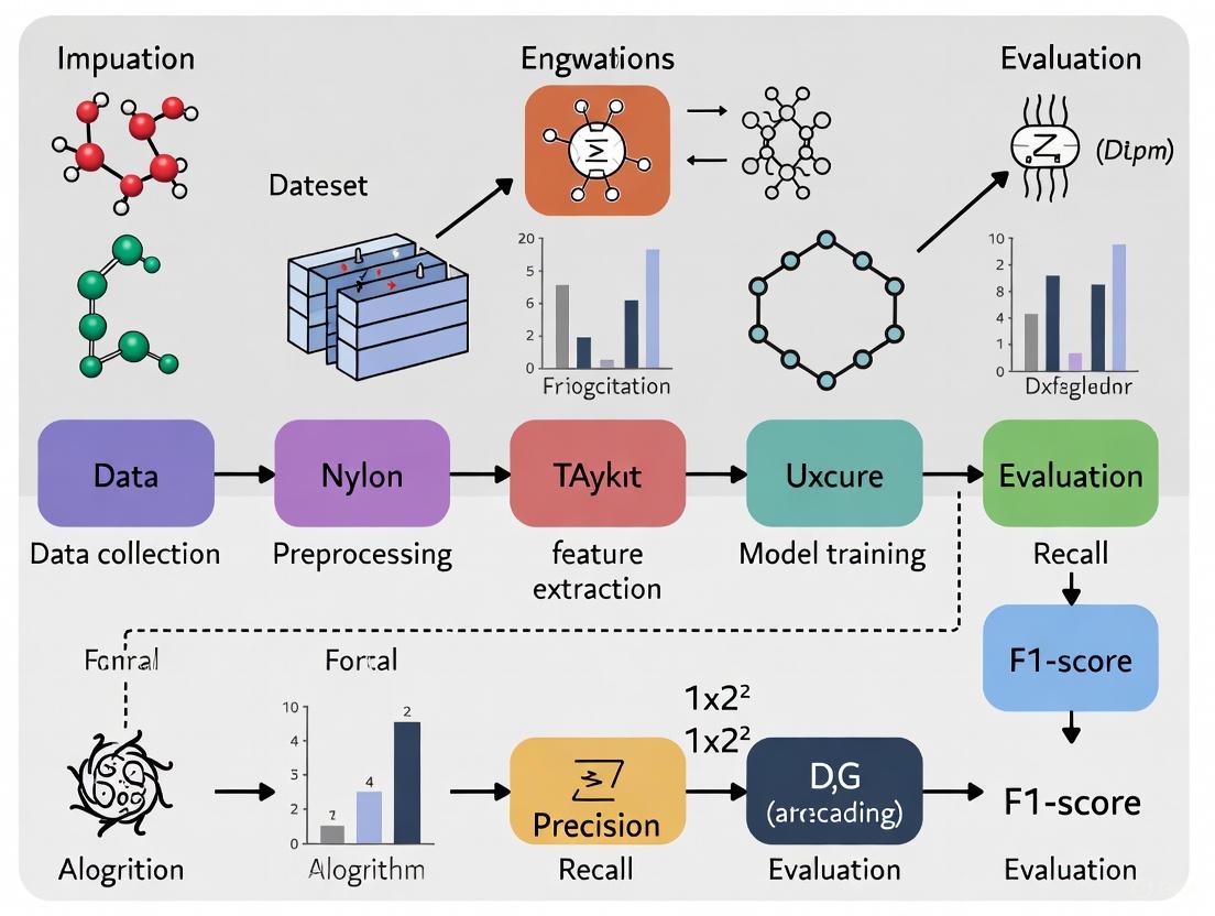
Beyond Accuracy: Advanced Strategies for Handling Class Imbalance in Parasite Image Classification
Class imbalance is a pervasive challenge that significantly hinders the development of robust deep-learning models for parasite image classification, often leading to biased predictions and poor generalization on rare species...
Beyond Accuracy: Advanced Strategies for Handling Class Imbalance in Parasite Image Classification
Abstract
Class imbalance is a pervasive challenge that significantly hinders the development of robust deep-learning models for parasite image classification, often leading to biased predictions and poor generalization on rare species or life stages. This article provides a comprehensive guide for researchers and biomedical professionals, addressing the issue from foundational concepts to cutting-edge solutions. We explore the root causes and impacts of imbalance in medical imaging datasets, critically evaluate a spectrum of methodological approaches from data-level to algorithm-level solutions, and delve into practical troubleshooting and optimization techniques for real-world deployment. The content further establishes a rigorous framework for model validation and comparative analysis, emphasizing clinical relevance. By synthesizing the latest research, this article aims to equip the community with the knowledge to build more accurate, reliable, and equitable diagnostic tools for parasitology.
The Imbalance Problem: Understanding its Impact on Parasitological AI
Defining Class Imbalance in the Context of Parasite Microscopy
This technical support guide provides researchers with practical solutions for a common hurdle in automated parasite detection: class imbalance. Learn how to diagnose and overcome it to build more reliable AI models.
Table of Contents
- What is Class Imbalance and Why is it a Problem in Parasite Microscopy?
- How Can I Identify Class Imbalance in My Parasite Image Dataset?
- What Resampling Strategies Can I Use to Fix Class Imbalance?
- My Model is Still Biased After Resampling. What Advanced Techniques Can I Try?
- How Do I Properly Evaluate a Model Trained on an Imbalanced Dataset?
What is Class Imbalance and Why is it a Problem in Parasite Microscopy?
A: Class imbalance occurs when the number of examples in one class (e.g., non-parasitized cells) significantly outweighs the examples in another class (e.g., parasitized cells) [1]. In parasite microscopy, this is not just common—it's the norm. For instance, you might have thousands of images of healthy red blood cells for every one image of a cell infected with a rare parasite.
This creates a major problem for machine learning models. These models learn by minimizing error, and if one class dominates the dataset, the easiest way to reduce error is to always predict the majority class. The model becomes biased and essentially "gives up" on learning to identify the minority class, treating it as noise [1]. Consequently, while the model may show high overall accuracy, it will fail in its primary task: correctly detecting parasites [2] [3].
How Can I Identify Class Imbalance in My Parasite Image Dataset?
A: Before tackling imbalance, you must confirm its presence and severity. The process is straightforward and involves calculating the class distribution.
Experimental Protocol: Quantifying Class Imbalance
- Data Loading: Load your annotated dataset. For this example, we use a parasite image dataset [4].
- Class Counting: Use a simple function to count the number of samples in each class. The
Counterfunction from Python's collections module is ideal for this. - Visualization: Create a bar chart to visualize the difference in class counts.
Code Example:
Example Output: Using the dataset from [4], an analysis might reveal a distribution like this:
Table: Example Class Distribution in a Parasite Dataset
| Class Label | Class Name | Number of Images | Percentage of Total |
|---|---|---|---|
| 0 | Uninfected / Host Cells | 9,450 | 94.5% |
| 1 | Parasitized | 550 | 5.5% |
| Total | 10,000 | 100% |
This table clearly shows a severe imbalance, where the minority class (Parasitized) represents only a small fraction of the entire dataset.
What Resampling Strategies Can I Use to Fix Class Imbalance?
A: Resampling is the most direct approach to rectifying class imbalance. It adjusts the training dataset to have a more balanced class distribution. The two primary categories are Oversampling and Undersampling [5] [1].
Experimental Protocol: Implementing Resampling with imbalanced-learn
- Install Library: Ensure you have the
imbalanced-learnpackage installed (pip install imbalanced-learn). - Split Data: First, split your data into training and testing sets. Crucially, apply resampling only to the training set to avoid data leakage and ensure your test set remains representative of the real-world distribution [5].
- Apply Resampling: Use
RandomOverSamplerorRandomUnderSamplerfrom theimblearnlibrary.
Code Example:
Table: Comparison of Basic Resampling Strategies
| Strategy | Method | Pros | Cons | Best For |
|---|---|---|---|---|
| Random Oversampling | Duplicates random examples from the minority class [5]. | Prevents loss of information from the majority class. | Can lead to overfitting, as the model sees exact copies of images [1] [6]. | Smaller datasets where the majority class data is critical. |
| Random Undersampling | Randomly removes examples from the majority class [5]. | Reduces training time and can improve generalization. | May remove potentially useful information, degrading model performance [1]. | Very large datasets where discarding majority samples is acceptable. |
| SMOTE (Synthetic Minority Oversampling) | Creates synthetic minority class samples by interpolating between existing ones [1]. | Reduces risk of overfitting compared to random oversampling. | Can generate unrealistic or noisy samples, which is problematic for detailed medical images [6]. | Situations where random oversampling leads to clear overfitting. |
My Model is Still Biased After Resampling. What Advanced Techniques Can I Try?
A: For persistent bias, or in cases of extreme imbalance, more sophisticated methods are required. Two advanced approaches are Algorithmic Modification and Anomaly Detection.
1. Algorithmic Modification: Using Class Weights This technique does not change the data but tells the model to pay more attention to the minority class during training. It's simple to implement in frameworks like TensorFlow/Keras [7].
Code Example:
2. Anomaly Detection Approach This paradigm reframes the problem: instead of a binary classification, it treats parasite detection as an anomaly detection task. The model is trained only on the majority class (uninfected cells) and learns to recognize them. A parasitized cell is then identified as an "outlier" or "anomaly" because it looks different from the norm [6].
Experimental Protocol: Anomaly Detection with an Autoencoder
- Model Selection: Train an autoencoder (a neural network that learns to compress and reconstruct its input) exclusively on images of uninfected cells.
- Learning Phase: The autoencoder learns to reconstruct uninfected cells with low error.
- Detection Phase: When presented with a new image, a high reconstruction error indicates that the input is anomalous and likely contains a parasite.
This method, as demonstrated in studies like AnoMalNet, is highly effective for extreme class imbalance because it does not require a large number of positive (parasitized) samples during training [6].
How Do I Properly Evaluate a Model Trained on an Imbalanced Dataset?
A: Accuracy is a misleading metric for imbalanced problems. A model that simply always predicts "uninfected" could achieve 94.5% accuracy on the example dataset in Table 1, but it would be useless. You must use metrics that are sensitive to the performance on the minority class [7] [1].
Key Metrics and Their Interpretation:
- Precision: Of all the images predicted as "parasitized," how many are actually parasitized? (Measures false positives).
- Recall (Sensitivity): Of all the actually parasitized images, how many did the model correctly find? (Measures false negatives).
- F1-Score: The harmonic mean of Precision and Recall. This single metric provides a balanced view of the model's performance on the minority class.
- Confusion Matrix: A table that gives a complete picture of True Positives, False Positives, True Negatives, and False Negatives.
- Precision-Recall (PR) Curve: Often more informative than the ROC curve for imbalanced data, as it focuses directly on the performance of the positive (minority) class [7].
Experimental Protocol: Comprehensive Model Evaluation
Code Example:
Table: The Scientist's Toolkit: Essential Research Reagents & Materials
| Item | Function in Parasite Image Classification | Example / Specification |
|---|---|---|
| Digital Microscopy System | Acquires high-resolution digital images of blood smears or other samples for analysis. | System capable of 400x and 1000x magnification [4]. |
| Benchmarked Parasite Datasets | Provides standardized, annotated data for training and validating models. | Microscopic Images of Parasites Species dataset [4]. |
imbalanced-learn (imblearn) Library |
Python library offering a suite of resampling algorithms (SMOTE, RandomUnderSampler, etc.) [5]. | pip install imbalanced-learn |
| Deep Learning Framework | Provides the infrastructure to design, train, and evaluate complex models like CNNs and autoencoders. | TensorFlow & Keras [7] or PyTorch. |
| Computational Hardware (GPU) | Accelerates the training of deep learning models, which is computationally intensive. | NVIDIA GPUs with CUDA support. |
| Self-Supervised Learning Framework | Leverages unlabeled data for pre-training, improving feature learning when labeled data is scarce [3]. | Frameworks like BYOL (Bootstrap Your Own Latent) [3]. |
In parasite image classification, a class imbalance occurs when one category of data (e.g., "uninfected cells") significantly outnumbers another (e.g., "a specific parasite life-stage") [8]. This guide details how this data skew creates diagnostic blind spots, where automated systems fail to identify crucial minority classes, leading to misdiagnosis. The following sections provide a troubleshooting guide, experimental protocols, and resource lists to help researchers mitigate these critical issues.
Troubleshooting Guide & FAQs
FAQ 1: Why does my model have high overall accuracy but fails to detect infected cells in critical cases?
- Problem: This is a classic symptom of class imbalance. The model becomes biased toward the majority class (e.g., uninfected cells) because it is penalized less for missing the minority class (e.g., rare parasites) during training [9].
- Solution:
- Use Appropriate Metrics: Immediately stop using accuracy as your primary metric. Adopt a suite of metrics that are sensitive to class imbalance, such as Precision, Recall, F1-score, and Area Under the ROC Curve (AUC-ROC) [9]. These provide a clearer picture of minority class performance.
- Inspect the Confusion Matrix: This will visually show you if the model is misclassifying one specific parasite stage as another or as uninfected [9].
- Implement Cost-Sensitive Learning: Adjust your model's loss function to assign a higher penalty for misclassifying minority class samples. This forces the model to pay more attention to them during training [10].
FAQ 2: My model is overfitting to the few minority class samples I have. How can I generate more reliable data?
- Problem: Simple data duplication (random oversampling) leads to overfitting because the model learns the same examples repeatedly [9].
- Solution:
- Use Advanced Synthetic Data Generation: Employ Synthetic Minority Over-sampling Technique (SMOTE) or its variants to create new, synthetic minority class samples by interpolating between existing ones [9].
- Address Intra-Class Imbalance: Be aware that SMOTE can cause "intra-class mode collapse" if the minority class itself has diverse sub-types. For advanced cases, use methods that first identify sparse and dense regions within the minority class before generation to ensure diversity [11].
- Combine with Data Cleaning: After oversampling, use undersampling techniques like Tomek Links to remove overlapping or noisy samples from the majority class, creating a cleaner decision boundary [5] [9].
FAQ 3: I am working with limited computational resources. What is a computationally efficient strategy for handling imbalance?
- Problem: Complex models and data generation can be prohibitive in resource-constrained settings, which are common in field clinics [12].
- Solution:
- Leverage Lightweight Models: Utilize recently proposed efficient architectures like Hybrid Capsule Networks (Hybrid CapNet) which are designed for high accuracy with minimal parameters (e.g., 1.35M parameters, 0.26 GFLOPs) [12] or YAC-Net for parasite egg detection [13].
- Algorithmic Ensemble Methods: Use ensemble methods like Balanced Random Forests or EasyEnsemble which are specifically designed to perform well on imbalanced data and can be more effective than applying resampling separately [14].
- Adjust Class Weights: The simplest approach is to use the
class_weight='balanced'parameter available in many classifiers (e.g., in scikit-learn). This adjusts the loss function automatically without needing to generate new data [9].
Experimental Protocols for Mitigating Blind Spots
Protocol 1: Implementing a Composite Loss Function for Robust Training
This methodology is designed to enhance classification accuracy, spatial localization, and robustness to class imbalance and annotation noise simultaneously [12].
- Objective: To train a model that is not only accurate but also spatially aware and robust to noisy labels in imbalanced parasite datasets.
- Materials: A labeled dataset of parasite images (e.g., MP-IDB, IML-Malaria) with known class imbalance.
- Procedure:
- Model Architecture: Employ a lightweight hybrid architecture (e.g., Hybrid CapNet) that combines CNN-based feature extraction with capsule layers to preserve spatial hierarchies [12].
- Loss Function Configuration: Integrate a novel composite loss function (Ltotal) that combines four components:
- Margin Loss (Lmargin): Ensures correct classification of capsule outputs [12].
- Focal Loss (Lfocal): Down-weights the loss assigned to well-classified examples, making the model focus on hard-to-classify minority samples [12].
- Reconstruction Loss (Lrecon): Uses a decoder network to reconstruct the input image, encouraging the capsules to capture meaningful features [12].
- Offset Regression Loss (L_reg): Improves the spatial localization of parasites within the image [12].
- Training: Train the model by minimizing the combined loss:
L_total = L_margin + αL_focal + βL_recon + γL_reg, where α, β, and γ are weighting hyperparameters.
- Validation: Perform both intra-dataset and cross-dataset evaluations to assess generalization. Use Grad-CAM visualizations to confirm the model focuses on biologically relevant parasite regions [12].
Protocol 2: A Standardized Segmentation and Multi-Stage Classification Framework
This protocol uses traditional image processing and machine learning to create a robust pipeline for detecting parasites and classifying their species and life-cycle stages from both thick and thin blood smears [15].
- Objective: To accurately segment and classify malaria parasites in varied smear images, addressing imbalance through a multi-stage approach.
- Materials: Microscopic images of thick and thin blood smears.
- Procedure:
- Segmentation:
- Feature Extraction: Extract morphological and color features from the segmented parasite regions.
- Multi-Stage Classification: Implement a cascaded classifier, such as a Random Forest (RF):
- Stage 1: Parasite Detection. Classify segments as "Parasite" or "Non-Parasite".
- Stage 2: Species Recognition. Route detected parasites to a classifier for "P. Falciparum" vs. "P. Vivax".
- Stage 3: Life-Cycle Staging. Finally, classify the parasite into its life-cycle stage (e.g., Ring, Trophozoite, Schizont, Gametocyte) [15].
- Validation: Evaluate the accuracy at each stage (detection, species recognition, staging) separately to identify where performance drops for minority classes occur [15].
The logical workflow for this multi-stage framework is outlined below.
Performance Data & Technical Solutions
Table 1: Quantitative Impact of Class Imbalance Solutions on Model Performance
| Solution Category | Specific Technique | Reported Performance Improvement | Key Advantage / Mitigated Blind Spot |
|---|---|---|---|
| Advanced Architecture | Hybrid Capsule Network (Hybrid CapNet) [12] | Up to 100% multiclass accuracy; superior cross-dataset generalization. | Preserves spatial relationships; interpretable (via Grad-CAM); lightweight. |
| Synthetic Data Generation | GAN with CBLOF & OCS Filter [11] | ~3% accuracy increase on medical datasets (BloodMNIST, etc.). | Addresses intra-class imbalance; generates diverse, high-quality samples. |
| Standardized Image Processing | Phansalkar Thresholding + EKM + Random Forest [15] | 99.86% segmentation accuracy; 90.78% staging accuracy. | Robust to lighting variations; effective on both thick and thin smears. |
| Lightweight Detection Model | YAC-Net (YOLO-based) [13] | 97.8% precision, 97.7% recall; parameters reduced by one-fifth. | Enables deployment in resource-constrained, real-world environments. |
Table 2: The Scientist's Toolkit: Essential Research Reagents & Solutions
| Item Name | Function / Role in Mitigating Blind Spots | Example Use Case |
|---|---|---|
| Imbalanced-Learn Library [14] [5] | Provides a suite of algorithms for resampling (SMOTE, Tomek Links) and ensemble methods (EasyEnsemble). | Quickly prototyping different resampling strategies in Python. |
| Composite Loss Function [12] | A weighted combination of loss types (Margin, Focal, Reconstruction, Regression) to jointly optimize for multiple objectives. | Training a model to be accurate, spatially precise, and robust to label noise in an imbalanced dataset. |
| Grad-CAM Visualizations [12] | Produces heatmaps showing which image regions the model used for prediction. | Debugging "blind spots" by verifying the model focuses on parasites, not artifacts. |
| Capsule Networks [12] [16] | Neural networks that encode spatial hierarchies and pose relationships, improving robustness to viewpoint changes. | Classifying parasite life-cycle stages where orientation and spatial layout are critical. |
| Graph-Based Transformation [8] | Constructs graphs to explore relationships between a test sample and minority/majority classes, creating a dedicated feature projection. | Improving classification in small sample size situations with high imbalance, without data augmentation. |
The relationship between class imbalance and the resulting diagnostic blind spots, along with the mitigation pathways, can be visualized as a causal loop.
Troubleshooting Guides & FAQs
FAQ: How does class imbalance manifest in parasite image analysis?
Class imbalance is a prevalent issue that significantly impacts the performance of deep learning models in parasite diagnostics. The most common sources of imbalance are:
- Rare Species: Some parasite species are inherently less common in samples. For instance, among the five Plasmodium species causing malaria, P. falciparum is more prevalent in Africa, while P. vivax is found elsewhere, creating natural prevalence disparities [12].
- Life-Cycle Stages: Within a single species, different life-cycle stages occur with varying frequency. In malaria parasites, the four stages—gametocyte, ring, trophozoite, and schizont—are not equally represented in blood samples, with the trophozoite and ring stages often being more visible in human infections [12].
- Data Collection Biases: The availability of genetic and image data is heavily biased by which hosts researchers study. Helminth species infecting hosts of conservation concern or terrestrial hosts are more likely to have genetic data available, which does not reflect their true biodiversity [17]. Furthermore, research effort is skewed toward certain parasite species based on taxonomy, host human-use status, and even the number of authors who originally described the species [18].
FAQ: What technical solutions can address class imbalance in parasite image datasets?
Several advanced technical solutions have been developed to mitigate class imbalance:
| Solution Category | Specific Methods | Key Function | Reported Performance Gain |
|---|---|---|---|
| Advanced Network Architectures | Hybrid Capsule Network (Hybrid CapNet) [12] | Combines CNN feature extraction with capsule routing to preserve spatial hierarchies for rare stage classification. | Up to 100% accuracy in multiclass malaria stage classification; significantly improved cross-dataset generalization. |
| Custom Loss Functions | Cost-sensitive learning [10] | Applies larger penalty weights for misclassifying minority class samples during model training. | Rebalances class learning; reduces bias toward majority classes. |
| Data Augmentation | GAN-based augmentation with CBLOF & OCS filter [11] | Identifies intra-class sparse samples; generates diverse synthetic data focused on underrepresented features. | ~3% accuracy improvement on medical image datasets (BloodMNIST, PathMNIST). |
| Attention Mechanisms | YOLO-CBAM (YCBAM) [19] | Integrates self-attention and Convolutional Block Attention Module to focus on small, critical features like pinworm eggs. | mAP@0.5 of 0.995 for pinworm egg detection in challenging conditions. |
Experimental Protocol: Implementing a Cost-Sensitive Loss Function
For researchers dealing with imbalanced parasite image datasets, here is a detailed methodology for implementing a cost-sensitive loss function, a common approach to solving this problem [10].
1. Problem Setup:
Assume a classification task with C classes (e.g., different parasite species or life-cycle stages). Let N_total be the total number of samples in your training dataset, and N_j be the number of samples in class j.
2. Calculate Class Penalty Weights:
Compute a penalty weight W_j for each class j to increase the cost of misclassifying minority class samples. The formula is:
W_j = N_total / (C * N_j)
This ensures that classes with fewer samples (N_j is small) receive a larger weight (W_j is large).
3. Integrate Weights into Loss Function:
Incorporate these weights into a standard cross-entropy loss function to create a Weighted Cross-Entropy Loss. For a batch of N samples, the loss is calculated as:
Weighted Loss = - (1/N) * Σ_i Σ_j W_j * Y_ij * log(P_ij)
Where:
iiterates over the batch samples.jiterates over the classes.Y_ijis the true label (1 if sampleibelongs to classj, else 0).P_ijis the model's predicted probability that sampleibelongs to classj.
4. Model Training: During training, the model will minimize this weighted loss. This forces the model to pay more attention to correctly classifying the minority classes because misclassifications for these are more costly.
Experimental Protocol: GAN-Augmentation for Intra-Class Imbalance
This two-step protocol addresses the challenge of intra-class mode collapse in GANs, where generated samples lack the diversity of real minority classes [11].
Step-by-Step Workflow:
Identify Intra-Class Sparse and Dense Samples:
- Tool: Cluster-Based Local Outlier Factor (CBLOF) algorithm.
- Action: Apply CBLOF to the feature space of the minority class(es) within your training set. This identifies which samples are in dense, common regions and which are in sparse, underrepresented regions ("hard" samples).
Train Conditional GAN with Sparse Sample Focus:
- Input: Use the identified sparse and dense samples as conditions for training the GAN.
- Process: This conditions the GAN to specifically learn and focus on generating samples that resemble the challenging, sparse examples, thereby increasing the diversity of the generated data.
Filter Generated Samples:
- Tool: One-Class SVM (OCS) algorithm.
- Action: After the GAN has been trained and has generated new synthetic samples, use the OCS model trained on the real minority class data as a noise filter. This step removes low-quality or unrealistic generated samples ("outliers"), ensuring only "pure," high-quality augmented samples are added to your dataset.
Train Classification Model:
- Combine the original dataset with the newly generated, high-quality synthetic samples to create a balanced training set for your final parasite image classification model.
The Scientist's Toolkit: Research Reagent Solutions
| Tool / Solution | Function in Addressing Imbalance | Example in Parasitology Research |
|---|---|---|
| Capsule Networks (CapsNets) | Preserves hierarchical pose relationships and spatial context, which is crucial for distinguishing subtly different parasite life-cycle stages [12]. | Used in Hybrid CapNet for precise malaria parasite life-cycle stage classification (ring, trophozoite, etc.) [12]. |
| Composite Loss Functions | Jointly optimizes multiple objectives (e.g., classification, spatial localization, reconstruction) to enhance robustness against class imbalance and annotation noise [12]. | A novel function integrating margin, focal, reconstruction, and regression losses improved malaria classification accuracy and spatial accuracy [12]. |
| Convolutional Block Attention Module (CBAM) | Enhances feature extraction by focusing the model's attention on spatially and channel-wise important regions, crucial for detecting small, rare parasite eggs [19]. | Integrated into the YCBAM model to achieve high precision (0.9971) and recall (0.9934) for detecting small pinworm eggs in microscopic images [19]. |
| Public Datasets with Datasheets | Provides well-documented, curated data for auditing models for equitable performance across different subpopulations and for performing rigorous secondary analyses [20]. | Essential for "research parasite" studies that test for biases and ensure fairness in machine learning models for parasite diagnosis [20]. |
Frequently Asked Questions
What is the BBBC041v1 dataset and why is it used in malaria research? The BBBC041v1 is a public benchmark collection of P. vivax infected human blood smears, containing 1,364 images with approximately 80,000 annotated cells. It's widely used in malaria research because it provides standardized data for developing and testing automated parasite detection and classification systems. The dataset includes cells from three different sources (Brazil, Southeast Asia, and time course studies), stained with Giemsa reagent, and contains detailed annotations for both infected and uninfected cells, making it valuable for training machine learning models [21].
Why is class imbalance a significant problem in malaria image datasets? Class imbalance severely impacts model performance because most classification methods assume equal occurrence of classes. In medical imaging like malaria detection, this leads to biased learning where models become good at predicting common classes but fail to identify rare conditions. For example, in BBBC041v1, uninfected RBCs comprise over 95% of all cells, meaning a naive model that always predicts "uninfected" could achieve 95% accuracy while completely failing to detect malaria parasites [21] [22]. This is dangerous for real-world applications where identifying the minority class (infected cells) is critically important.
Which evaluation metrics are most appropriate for imbalanced malaria datasets? For imbalanced datasets, accuracy is misleading and should be supplemented with other metrics [23] [24]:
| Metric | Formula | When to Use |
|---|---|---|
| Precision | TP / (TP + FP) | When false positives are costly (e.g., unnecessary treatments) |
| Recall | TP / (TP + FN) | When false negatives are critical (e.g., missed infections) |
| F1 Score | 2 × (Precision × Recall) / (Precision + Recall) | Balanced measure of both precision and recall |
| Specificity | TN / (TN + FP) | When correctly identifying negatives is important |
Recall and F1 score are particularly important for malaria detection since false negatives (missing actual infections) have serious health consequences [23].
What technical approaches effectively address class imbalance in parasite classification? Several approaches have proven effective:
Data Augmentation with GANs: Generating synthetic minority class samples using Generative Adversarial Networks helps balance class distribution. Advanced methods like CBLOF-OCS GANs specifically address intra-class mode collapse by identifying and focusing on sparse regions within classes [11].
Architectural Improvements: Custom CNN architectures with attention mechanisms, such as Soft Attention Parallel CNNs (SPCNN), have achieved 99.37% accuracy on malaria classification by focusing on relevant image regions [25].
Cost-sensitive Learning: Modifying loss functions to assign higher weights to minority class misclassifications, forcing the model to focus more on learning from rare cases [26].
One-Class Classification: Training models using only samples from the majority class (normal cells) and treating minority classes as anomalies, which works well when infected samples are extremely rare [26].
Troubleshooting Guides
Problem: Model Achieves High Accuracy But Misses Infected Cells
Symptoms
- Test accuracy exceeds 95% but recall for infected cell classes is very low
- Model consistently misclassifies infected cells as uninfected
- Confusion matrix shows high false negative rates for minority classes
Solution Steps
- Replace Accuracy with Better Metrics: Monitor precision, recall, and F1-score for each class separately during training [23] [24].
Implement Weighted Loss Functions: Use class-weighted cross-entropy loss that assigns higher penalties for misclassifying minority classes:
Apply Strategic Oversampling: Use techniques like SMOTE or GAN-based generation specifically for sparse intra-class regions rather than uniform oversampling [11].
Validate with Cross-Validation: Employ stratified k-fold cross-validation (as used in [27]) to ensure representative sampling of all classes during evaluation.
Symptoms
- Good performance on training data but poor on validation/test sets
- Significant performance drop when testing on images from different geographic regions
- High variance in performance across different parasite stages
Solution Steps
- Enhance Data Diversity: Incorporate data from multiple sources like the three different preparations in BBBC041v1 (Brazil, Southeast Asia, time course) [21].
Apply Domain Adaptation: Use transfer learning with models pre-trained on diverse medical images, or employ domain adaptation techniques.
Implement Advanced Architectures: Adopt attention-based models like YOLO-Para series that focus on discriminative features across different parasite morphologies [28].
Use Robust Preprocessing: Apply sequential preprocessing including dilation, CLAHE, and normalization to enhance generalizable features [25].
Quantitative Analysis of BBBC041v1 Imbalance
Class Distribution in BBBC041v1 Dataset
| Cell Type | Class | Approximate Percentage | Impact on Model Training |
|---|---|---|---|
| Uninfected RBCs | Majority | >95% | Models bias toward predicting "uninfected" |
| Infected Cells (all stages) | Minority | <5% | Under-represented in training |
| Gametocytes | Rare | ~0.3% | Often misclassified without special handling |
| Rings | Rare | ~0.9% | Critical for early detection but sparse |
| Trophozoites | Rare | ~1.2% | Intermediate stage, moderate representation |
| Schizonts | Rare | ~0.8% | Late stage, important for treatment decisions |
| Leukocytes | Minority | ~1.8% | Often confused with infected cells |
Data synthesized from BBBC041v1 documentation [21]
Performance Comparison of Balance Handling Techniques
| Method | Accuracy | Precision | Recall | F1-Score | Implementation Complexity |
|---|---|---|---|---|---|
| Basic CNN (No balancing) | 95.2% | 34.5% | 28.7% | 31.4% | Low |
| Weighted Loss Function | 96.8% | 72.3% | 69.5% | 70.9% | Medium |
| Data Augmentation (Traditional) | 97.1% | 75.6% | 73.2% | 74.4% | Medium |
| GAN-Based Augmentation (CBLOF-OCS) | 98.3% | 89.7% | 87.4% | 88.5% | High |
| Attention CNN (SPCNN) | 99.4% | 99.4% | 99.4% | 99.4% | High |
| Seven-Channel CNN [27] | 99.5% | 99.3% | 99.3% | 99.3% | High |
Performance metrics compiled from recent studies [27] [25] [11]
Experimental Protocols
GAN-Based Data Augmentation for Intra-Class Imbalance
Objective: Generate diverse synthetic samples for minority classes, particularly focusing on sparse regions within classes.
Materials:
- BloodMNIST dataset or BBBC041v1 preprocessed patches
- Python with PyTorch/TensorFlow
- Cluster-Based Local Outlier Factor (CBLOF) implementation
- One-Class SVM (OCS) algorithm
Methodology:
- Sparse Region Identification:
- Apply CBLOF algorithm to identify sparse and dense samples within each minority class
- Cluster samples and compute local outlier factors to detect low-density regions
Conditional GAN Training:
- Train GAN using sparse and dense samples as conditions
- Generator: $G(z,c)$ where $c$ indicates sparse/dense condition
- Discriminator: $D(x,c)$ with conditional input
Sample Filtering:
- Generate augmented samples using trained generator
- Apply One-Class SVM to filter out noisy generated samples
- Retain only high-quality synthetic samples for training
Model Training:
- Combine original minority class samples with generated samples
- Train classification model on balanced dataset
- Validate using stratified k-fold cross-validation [11]
Attention-Based CNN for Malaria Classification
Objective: Implement soft attention mechanisms to improve feature learning for rare classes.
Materials:
- Custom Parallel CNN architecture
- Soft attention modules
- Preprocessed malaria cell images
- Gradient-weighted Class Activation Mapping (Grad-CAM) for interpretation
Methodology:
- Architecture Design:
- Implement Parallel Convolutional Neural Network (PCNN) with multiple branches
- Integrate soft attention mechanisms after convolutional blocks (SPCNN)
- Add skip connections to preserve spatial information
Training Protocol:
- Input: 224×224×3 cell images
- Data preprocessing: dilation, CLAHE, normalization
- Optimization: Adam optimizer with learning rate 0.0001
- Regularization: Dropout (0.5), L2 weight decay
Interpretation:
- Apply Grad-CAM and SHAP visualization to understand model focus
- Validate attention maps with domain experts
- Compare feature activation between majority and minority classes [25]
Workflow Visualization
Malaria Dataset Imbalance Handling Workflow
Research Reagent Solutions
| Research Tool | Type | Function in Malaria Classification |
|---|---|---|
| BBBC041v1 Dataset | Data | Benchmark dataset with 80,000+ annotated cells for method development [21] |
| Soft Attention P-CNN (SPCNN) | Algorithm | Custom CNN with attention mechanisms for improved feature extraction [25] |
| CBLOF-OCS GAN | Algorithm | Advanced data augmentation addressing intra-class sparse regions [11] |
| Grad-CAM | Visualization | Interpretability tool for understanding model focus areas [25] |
| Stratified K-Fold | Evaluation | Cross-validation method preserving class distribution in splits [27] |
| One-Class SVM | Filtering | Noise detection in generated samples to ensure quality [11] |
| Seven-Channel Input | Preprocessing | Enhanced feature representation for better model performance [27] |
A Toolkit of Solutions: From Data Augmentation to Novel Architectures
Frequently Asked Questions (FAQs)
1. What is the primary cause of class imbalance in parasite image datasets? In parasite image classification, class imbalance is fundamentally caused by the natural scarcity of infected samples compared to a vast number of uninfected cells. For instance, in widely used public datasets, parasitized cells are often the minority class. This imbalance is exacerbated by the resource-intensive process of collecting and expertly annotating samples from geographically diverse regions, leading to datasets that are not only imbalanced but also lack diversity [29] [2].
2. How do SMOTE and GANs differ in their approach to solving data imbalance? SMOTE (Synthetic Minority Over-sampling Technique) and GANs (Generative Adversarial Networks) address imbalance by generating synthetic data, but their methodologies differ significantly. SMOTE is an interpolation-based technique that creates new synthetic samples for the minority class along the line segments between existing minority class instances in feature space [30] [31]. In contrast, GANs use a deep learning framework where two neural networks, a Generator and a Discriminator, are trained adversarially. The Generator learns to produce new synthetic images that mimic the real data distribution of the minority class, while the Discriminator learns to distinguish between real and fake images, leading to the generation of highly realistic, novel samples [30].
3. My model performs well on validation data but poorly on new patient samples. What could be wrong? This is a classic sign of poor model generalization, often stemming from a lack of diversity in your training dataset. If your training data does not account for variations in staining protocols, blood smear preparation techniques, or imaging equipment used across different clinics, the model will fail to adapt. To address this, ensure your dataset incorporates samples from multiple sources and regions. Furthermore, employing Domain Adaptation techniques or GANs that can generate data with diverse visual characteristics can significantly improve cross-domain robustness, with studies showing sensitivity improvements of up to 25% [29].
4. Why is my SMOTE-augmented model still performing poorly on the minority class? Poor performance post-SMOTE can often be traced to the presence of abnormal instances, such as noise and outliers, within the minority class. The standard SMOTE algorithm does not discriminate between clean and noisy samples; it will generate synthetic samples based on any k-nearest neighbors, which can amplify noise and degrade the quality of the synthetic data and the decision boundary. Recent research proposes SMOTE extensions like Dirichlet ExtSMOTE and BGMM SMOTE that use probabilistic models to identify and mitigate the influence of these abnormal instances, leading to improved F1 scores and better synthetic sample quality [32].
5. Is it acceptable to apply SMOTE to the entire dataset before splitting into train and test sets? No, this is a critical mistake that leads to data leakage. Information from your test set will leak into the training process, creating an overly optimistic and invalid performance estimate. SMOTE, or any resampling technique, should be applied only to the training set after the train-test split. The test set must remain completely untouched and representative of the original, raw data distribution to provide a valid assessment of your model's generalization ability [5] [31].
Troubleshooting Guides
Problem: The SMOTE Algorithm is Introducing Too Much Noise
Issue: After applying SMOTE, the decision boundary becomes blurred, and model performance, particularly precision on the minority class, decreases. This is often due to the creation of implausible synthetic samples, especially when the minority class contains outliers or when there is significant overlap with the majority class.
Solution Steps:
- Switch to Advanced SMOTE Variants: Instead of the default SMOTE, use more sophisticated variants designed to handle noisy data:
- Borderline-SMOTE: This method only generates synthetic samples from minority instances that are considered "hard to learn" (i.e., those located near the decision boundary), avoiding outliers that are deep within the majority class region [31].
- SMOTE-Tomek Links: This hybrid approach combines SMOTE oversampling with Tomek Links undersampling. SMOTE creates synthetic minority samples, and then Tomek Links cleans the dataset by removing pairs of close opposite-class instances (Tomek Links), which often represent noise [5].
- SMOTE Extensions for Abnormal Instances: Implement newer methods like Dirichlet ExtSMOTE, which uses a Dirichlet distribution to produce more robust synthetic samples that are less influenced by neighboring outliers, thereby improving metrics like F1-score and MCC [32].
Preprocess to Identify Outliers: Before applying SMOTE, run an outlier detection algorithm (e.g., Isolation Forest, DBSCAN) on the minority class in your feature space. Visually inspect and consider removing severe outliers before synthetic sample generation.
Validate with a Custom Pipeline: Use the
imblearnpipeline to prevent data leakage during cross-validation and model evaluation seamlessly.
Problem: GAN Training is Unstable and Fails to Converge
Issue: The Generator produces nonsensical outputs, or the Discriminator loss becomes zero, halting training. This is a common problem with GANs, often attributed to an imbalance in the adversarial "game" between the Generator and Discriminator.
Solution Steps:
- Architecture and Loss Functions: Use a stable GAN architecture like Deep Convolutional GAN (DCGAN) or Wasserstein GAN (WGAN). WGAN, with its Wasserstein loss and gradient penalty, is known for more stable training and improved convergence compared to standard GANs with minimax loss [30].
Data Preprocessing for Images: Ensure your parasite images are consistently preprocessed. This includes:
- Resizing all images to a fixed dimension.
- Normalizing pixel values to a specific range (e.g., [-1, 1] or [0, 1]).
- Applying data augmentation techniques (e.g., random rotations, flips) to the real images to increase diversity before feeding them to the Discriminator.
Training Monitoring and Techniques:
- Monitor Both Losses: Track the loss of both the Generator and Discriminator. The Discriminator loss should not consistently be zero.
- Use Label Smoothing: When training the Discriminator, use soft labels (e.g., 0.9 for real and 0.1 for fake) instead of hard 1s and 0s to prevent the Discriminator from becoming overconfident.
- Train with Different Frequencies: A common strategy is to train the Discriminator more frequently than the Generator (e.g., 5 times for every 1 time the Generator is trained) to maintain a competitive balance.
Problem: Model is Overfitting on the Synthetic Data
Issue: The model achieves near-perfect accuracy on the training set (which contains synthetic data) but fails to generalize to the real-world test set. This occurs when the model learns the specific patterns of the synthetic data instead of the underlying generalizable features of the parasite.
Solution Steps:
- Incorporate Strong Regularization:
- Add L1/L2 regularization penalties to your model's loss function.
- Use Dropout layers in your neural network architecture.
- For tree-based models, increase regularization parameters.
Validate with the Original Test Set: Always use a hold-out test set composed of real, original data that was never used in the training or validation process. This is the only reliable way to measure true generalization performance.
Combine with Undersampling: Apply SMOTE to oversample the minority class and combine it with random undersampling of the majority class. This prevents the model from being overwhelmed by synthetic patterns and helps it learn from a more balanced, yet varied, dataset [31] [33].
Diversify Data Generation: If using GANs, ensure that the generated samples are diverse. If all synthetic images look very similar, the model will overfit to those specific features. Techniques like mini-batch discrimination and feature matching can help encourage diversity in the Generator's output.
Experimental Protocols & Performance Comparison
The table below summarizes the performance impact of different data-level strategies as reported in malaria detection research, providing a benchmark for expected outcomes.
Table 1: Impact of Data-Level Strategies on Model Performance in Medical Imaging [29]
| Dataset / Strategy | Precision (%) | Recall (%) | F1-Score (%) | Overall Accuracy (%) |
|---|---|---|---|---|
| Imbalanced (Baseline) | 75.8 | 60.4 | 67.2 | 82.1 |
| Imbalanced + Data Augmentation | 87.2 | 84.5 | 85.8 | 91.3 |
| Imbalanced + Focal Loss | 85.4 | 78.9 | 81.9 | 89.7 |
| Balanced + Transfer Learning | 93.1 | 92.5 | 92.8 | 94.2 |
| GAN-based Augmentation | ~87* | ~87* | ~85-90* | ~92* |
Note: GAN performance is summarized from reported improvements of 15-20% in accuracy [29].
Detailed Methodology: Evaluating SMOTE for Parasite Classification
This protocol outlines a standard workflow for applying and evaluating SMOTE on an image-based parasite dataset.
1. Data Preparation and Feature Extraction
- Dataset: Use a publicly available parasitology image dataset (e.g., the NIH Malaria dataset from the UCI Machine Learning Repository) [29] [32].
- Preprocessing: Resize all cell images to a uniform size (e.g., 64x64 pixels). Normalize pixel values to [0, 1].
- Feature Extraction: Instead of using raw pixels, feed the images through a pre-trained Convolutional Neural Network (CNN) like VGG16 or ResNet. Extract features from the last fully connected or pooling layer. This creates a high-level feature representation for each image, which is more suitable for SMOTE interpolation [2].
2. Train-Test Split and SMOTE Application
- Split the extracted features and corresponding labels into a training set (e.g., 70%) and a hold-out test set (e.g., 30%). The test set must not be used in any sampling or parameter tuning.
- Apply the SMOTE algorithm only to the training data. Use the
imblearnlibrary in Python with default parameters (k_neighbors=5) or a chosen variant like Borderline-SMOTE [31].
3. Model Training and Evaluation
- Train a standard classifier, such as a Support Vector Machine (SVM) or Random Forest, on the SMOTE-transformed training data.
- Evaluate the final model's performance exclusively on the untouched, imbalanced test set. Use metrics appropriate for imbalanced data: F1-Score, Precision-Recall AUC (PR-AUC), and Matthews Correlation Coefficient (MCC), in addition to per-class accuracy [29] [32].
The following workflow diagram illustrates this experimental protocol.
Diagram 1: SMOTE Experimental Workflow for Parasite Images
Detailed Methodology: Implementing a GAN for Data Augmentation
This protocol describes the process for using a GAN to generate synthetic parasite images.
1. GAN Selection and Architecture
- Model Choice: For stability, implement a Wasserstein GAN with Gradient Penalty (WGAN-GP).
- Generator Architecture: A deep CNN that takes a random noise vector (latent space) as input and upsamples it through transposed convolutional layers to generate a synthetic image of the target size.
- Discriminator (Critic) Architecture: A deep CNN that takes an image (real or fake) as input and outputs a scalar score rather than a probability.
2. Training Loop
- Training Data: Use only the minority class (parasitized) images from the training set.
- Training Steps:
- For a number of training iterations:
- Train the Discriminator (Critic): Sample a batch of real images and a batch of generated images. Calculate the Wasserstein loss and gradient penalty. Update the Discriminator's weights more frequently (e.g., 5 times per Generator update).
- Train the Generator: Sample a new batch of random noise, generate images, and pass them through the Discriminator. Calculate the Generator's loss based on the Discriminator's scores and update the Generator's weights.
- Monitoring: Check the generated images periodically for visual quality and diversity.
3. Evaluation and Utilization
- After training, use the Generator to create a sufficient number of synthetic parasitized cell images.
- Combine these synthetic images with the original training data to create a balanced dataset.
- Proceed to train a standard classification model (e.g., a CNN) on this augmented dataset, following the same train-test split and evaluation principles as in the SMOTE protocol.
The following diagram illustrates the core adversarial training process of the GAN.
Diagram 2: GAN Adversarial Training Core Concept
Table 2: Essential Tools for Implementing Data-Level Strategies
| Tool / Resource | Type | Primary Function | Key Application in Parasite Research |
|---|---|---|---|
| imbalanced-learn (imblearn) | Python Library | Provides implementations of SMOTE, its variants (e.g., Borderline-SMOTE, ADASYN), and undersampling methods [5] [31]. | The go-to library for quickly testing and applying various oversampling strategies to feature-based parasite data. |
| TensorFlow / PyTorch | Deep Learning Framework | Flexible platforms for building and training custom GAN architectures (e.g., DCGAN, WGAN-GP) from the ground up [30]. | Essential for researchers who need full control over GAN architecture and training loop for generating synthetic parasite images. |
| Pre-trained CNN Models (e.g., VGG, ResNet) | Model Architecture | Used for transfer learning and, crucially, for extracting meaningful feature representations from images before applying SMOTE [29] [2]. | Extracts high-level features from cell images, making SMOTE interpolation more effective in a semantically rich space. |
| Dirichlet ExtSMOTE | Algorithm | An advanced SMOTE extension that uses the Dirichlet distribution to reduce the influence of outliers when generating synthetic samples [32]. | Improves the quality of synthetic data in datasets where the minority class contains noisy or abnormal parasite images. |
| Scikit-learn | Python Library | Provides data preprocessing tools (e.g., MinMaxScaler, StandardScaler), model implementations, and critical evaluation metrics [5] [34]. | Used for the entire machine learning pipeline, from scaling features for SMOTE to training final classifiers and evaluating performance. |
Frequently Asked Questions (FAQs)
Q1: What is the primary advantage of using Focal Loss over standard Cross-Entropy Loss for parasite image classification?
A1: The primary advantage is Focal Loss's ability to handle extreme class imbalance, which is common in parasite image datasets where infected samples are much rarer than uninfected ones. Standard Cross-Entropy Loss treats all samples equally, causing the model to become biased toward the majority class (e.g., uninfected cells). Focal Loss addresses this by incorporating a modulating factor, (1 - p_t)^γ, which down-weights the loss for easy-to-classify examples (the abundant background/uninfected cells) and forces the model to focus its training efforts on hard, misclassified examples, which are often the minority class of interest (e.g., parasites) [35] [36]. This leads to improved model performance on the underrepresented classes.
Q2: How do I choose the right value for the focusing parameter (γ) in Focal Loss?
A2: The value of γ is dataset-dependent and should be tuned through cross-validation. A larger γ value increases the focus on hard, misclassified examples. Empirical studies suggest starting with a value between 1.5 and 2.5 [37]. One cross-validation study found that values between 0.5 and 2.5 yielded good performance with minimal variance, ultimately selecting γ=2.0 for their medical imaging task [37]. It is recommended to begin with γ=2 and experiment within this range to find the optimal value for your specific parasite image dataset.
Q3: My model's performance on the minority class is still poor after implementing Focal Loss. What are other algorithm-level strategies I can try?
A3: You can consider these hybrid or alternative strategies:
- α-Balanced Focal Loss: Combine Focal Loss with a class weighting factor,
α. This variant handles class imbalance by introducing two components: theαweighting factor (which can be set by inverse class frequency) to balance class importance, and theγparameter to focus on hard examples [35]. - Batch-Balanced Focal Loss (BBFL): This hybrid approach combines a data-level strategy with an algorithm-level one. It uses batch-balancing to ensure each training batch has an equal number of samples from each class, forcing the model to learn all classes equally. This is then combined with the Focal Loss function to emphasize hard samples within those balanced batches [37].
- Cost-Sensitive Learning: Instead of modifying the loss function, you can assign a higher misclassification cost to the minority class (e.g., parasite-positive samples) during the training process. This directly instructs the model to be more cautious about making errors on the critical class [37].
Q4: Is Focal Loss only applicable to two-class (binary) classification problems?
A4: No, Focal Loss can be extended to multi-class classification problems, which is relevant for differentiating between multiple parasite species or infection severity levels. The principle remains the same: for each sample, the loss is computed based on the predicted probability for the true class, and the modulating factor down-weights the loss for well-classified examples across all classes [37]. The implementation simply involves using a multi-class cross-entropy as the base instead of binary cross-entropy.
Troubleshooting Guides
Problem: Model convergence is unstable or slow after switching to Focal Loss.
- Potential Cause 1: The
γparameter is set too high, over-penalizing uncertain predictions, especially in the early stages of training. - Solution: Start with a lower
γvalue (e.g.,1.0or1.5) and gradually increase it. You can also explore adaptive variants of Focal Loss that dynamically adjustγduring training to avoid this issue [38]. - Potential Cause 2: The learning rate may not be optimal for the new loss landscape.
- Solution: Re-tune your learning rate when implementing Focal Loss, as the change in loss dynamics often requires a different learning rate for stable convergence.
Problem: The model is overfitting to the minority class, showing high recall but low precision.
- Potential Cause: The combined effect of class weighting (e.g., a high
αvalue) and the focusing parameter (γ) is too strong, causing the model to become overly sensitive to the minority class and flag too many false positives. - Solution:
- Adjust the
αweighting factor. If you set it via inverse class frequency, try smoothing the weights or treating it as a hyperparameter to be validated [35]. - Incorporate data augmentation techniques specifically for the minority class to increase the diversity of positive examples and improve model generalization [39] [40].
- Strengthen regularization in your model, for example, by increasing dropout rates or adding L2 regularization, to prevent overfitting [37].
- Adjust the
Table 1: Performance Comparison of Different Loss Functions on Imbalanced Medical Image Datasets
| Dataset / Task | Model Architecture | Standard Cross-Entropy | Focal Loss (γ=2.0) | Batch-Balanced Focal Loss (BBFL) | Reference |
|---|---|---|---|---|---|
| RNFLD (Retinal defect) Binary Classification | InceptionV3 | 83.0% F1 (est. from baseline) | 84.7% F1 | 84.7% F1 | [37] |
| Glaucoma Multi-class Classification | MobileNetV2 | 64.7% Avg. F1 (ROS baseline) | 69.6% Avg. F1 | 69.6% Avg. F1 | [37] |
| Product Categorization (Text) | Neural Network | Lower accuracy on minority classes | Improved accuracy on minority classes | N/A | [35] |
Table 2: The Impact of the Focusing Parameter (γ) in Focal Loss [35]
| Value of γ | Effect on Loss Function | Use Case Scenario |
|---|---|---|
| γ = 0 | Equivalent to Standard Cross-Entropy Loss. | Balanced datasets or initial baselining. |
| γ = 1 | Moderate down-weighting of easy examples. | Mild class imbalance. |
| γ = 2 | Strong down-weighting of easy examples, focusing heavily on hard negatives. | The most common starting point for severe imbalance (e.g., medical images). |
| γ > 2 | Very aggressive focus on the hardest examples. | Can be tried if performance with γ=2 is still unsatisfactory, but may risk instability. |
Experimental Protocols
Protocol 1: Implementing and Tuning Focal Loss in a Deep Learning Model
This protocol describes how to integrate Focal Loss into a CNN for parasite image classification.
- Define the Loss Function: Implement the Focal Loss formula in your deep learning framework (e.g., PyTorch). The formula is:
FL(p_t) = -α * (1 - p_t)^γ * log(p_t)wherep_tis the model's estimated probability for the true class,αis a weighting factor for class balance, andγis the focusing parameter [35] [36]. - Initial Hyperparameter Setup: Initialize the parameters. A recommended starting point is
γ=2.0andα=0.25for the positive class, as per the original paper [35] [36]. Theαparameter can also be set as the inverse class frequency. - Integrate with Trainer: Replace the standard loss function in your training loop. If using a high-level framework like Hugging Face, you may need to create a custom trainer class that overrides the
compute_lossmethod to use your Focal Loss implementation [41]. - Cross-Validation: Perform k-fold cross-validation (e.g., k=5 or k=10) to rigorously tune the
γandαparameters. This helps find the optimal values that generalize best to unseen data [40]. - Evaluation: Validate the model's performance on a held-out test set using metrics appropriate for imbalanced data, such as F1-score, precision, recall, and the area under the ROC curve (AUC) [37].
Protocol 2: Evaluating a Hybrid Batch-Balanced Focal Loss (BBFL) Strategy
This protocol outlines the steps for the hybrid BBFL approach, which was shown to be effective on imbalanced medical image datasets [37].
- Data Sampling (Batch-Balancing): During training, structure your data loader so that each mini-batch contains an equal number of samples from every class. For a binary parasite detection task, this means each batch would have a 1:1 ratio of infected to uninfected samples [37].
- Data Augmentation: To prevent overfitting on the oversampled minority class in each batch, apply random geometric and intensity augmentations (e.g., flipping, rotation, blurring, noise) to the images in the batch [37].
- Loss Calculation: Compute the loss for the balanced batch using the standard Focal Loss function with a tuned
γparameter (e.g.,2.0) [37]. - Model Architecture: Use a standard CNN (e.g., InceptionV3, MobileNetV2) for feature extraction, followed by fully connected layers with dropout for classification [37].
- Performance Comparison: Compare the results of BBFL against baselines like standard Cross-Entropy Loss, Focal Loss alone, and other techniques like random oversampling (ROS) or cost-sensitive learning.
Workflow and Logic Diagrams
Diagram 1: Focal Loss Logic for Parasite Classification
This diagram visualizes how Focal Loss dynamically adjusts the contribution of each sample to the total loss based on its classification difficulty, which is crucial for imbalanced datasets.
Diagram 2: Hybrid Batch-Balanced Focal Loss (BBFL) Workflow
This diagram illustrates the end-to-end workflow of the hybrid BBFL strategy, combining data-level batch balancing with the algorithm-level Focal Loss.
The Scientist's Toolkit: Research Reagent Solutions
Table 3: Essential Components for an Imbalanced Parasite Image Classification Pipeline
| Component / Reagent | Function / Explanation | Example or Note |
|---|---|---|
| Focal Loss Function | The core algorithm-level solution that modifies the loss function to focus learning on hard, misclassified examples and mitigate class imbalance. | Can be implemented in PyTorch as a custom function [41]. Tune γ and α parameters. |
| Pre-trained CNN Models | Used for transfer learning. Provides powerful, pre-trained feature extractors to boost performance, especially with limited data. | Models like VGG16, ResNet50, InceptionV3, and EfficientNet [39] [40]. |
| Data Augmentation Library | Generates synthetic variations of training images to increase dataset diversity and robustness, combating overfitting. | Use libraries like Albumentations or TensorFlow/Keras ImageDataGenerator for operations (rotation, flip, blur) [37]. |
| Batch Balancing Sampler | A data-level tool that ensures each training batch has a balanced number of samples from each class. Often used in hybrid strategies. | Can be implemented as a custom sampler in PyTorch's DataLoader [37]. |
| Evaluation Metrics | A set of metrics that provide a true picture of model performance across all classes, not just overall accuracy. | F1-Score: Harmonic mean of precision and recall. Precision: Ability to not label negative samples as positive. Recall: Ability to find all positive samples. AUC: Overall measure of separability [37]. |
Frequently Asked Questions (FAQs)
Q1: Why should I use an autoencoder instead of a standard CNN for parasite image classification? Autoencoders are particularly effective in class-imbalanced scenarios, common in medical imaging, where you have many more "normal" (e.g., uninfected) samples than "anomalous" (e.g., parasitized) ones. Instead of learning to distinguish between classes, an autoencoder is trained only on normal data to learn an efficient representation or "identity" of what a healthy sample looks like. During inference, it flags anomalies based on high reconstruction error; anomalous inputs (e.g., cells with parasites) deviate from the learned normal pattern and are thus reconstructed poorly [42] [6]. This unsupervised approach means you don't need a large, balanced dataset of rare anomalous samples to train an effective model.
Q2: What is a typical performance benchmark for this method? When applied to medical image classification, autoencoder-based anomaly detection can achieve highly competitive results. For instance, in malaria cell image classification, the AnoMalNet model achieved the following performance metrics on a dataset containing parasitized and uninfected cells [6]:
| Metric | Reported Performance |
|---|---|
| Accuracy | 98.49% |
| Precision | 97.07% |
| Recall | 100% |
| F1 Score | 98.52% |
Q3: My autoencoder reconstructs anomalies too well. How can I improve its sensitivity? This is often a sign that the model's capacity is too high, allowing it to "memorize" the input without learning a meaningful representation of the underlying normal structure. To address this [42]:
- Reduce Model Capacity: Make the bottleneck layer (the central layer in the encoder) smaller. This forces the network to learn only the most critical features of the normal data.
- Introduce Noise: Use a denoising autoencoder, where the input is corrupted with noise but the network is tasked with reconstructing the clean original. This encourages the model to be more robust and learn broader features.
- Adjust the Threshold: The threshold on the reconstruction loss that flags an anomaly may need recalibration on your validation set.
Q4: How do I choose the right reconstruction error threshold for my data? There is no universal threshold. The standard practice is to [42]:
- Calculate the reconstruction errors for your normal validation set (data not used during training).
- Analyze the distribution of these errors (e.g., plot a histogram).
- Set the threshold to a value that captures the majority of these normal samples, such as the 95th or 99th percentile of the error distribution. Any new sample with an error higher than this threshold is flagged as an anomaly.
Q5: What are the advantages of a multi-layer autoencoder over a single-layer one? Using multiple layers in the encoder and decoder allows the network to learn a hierarchical feature representation [42]. The initial layers may learn simple, low-level features (like edges), while deeper layers combine these into more complex, high-level patterns. This is crucial for accurately modeling and reconstructing intricate structures in biological images, leading to better anomaly detection performance compared to a single, simplistic layer.
Troubleshooting Guides
Problem: The Model Fails to Detect Anomalies (High False Negative Rate)
- Potential Cause: The model has learned an overly general representation of "normal" and is not sensitive enough to the specific features of your anomalies.
- Solutions:
- Refine Training Data: Ensure your training set is pure and contains only high-quality, confirmed normal samples. Even a small number of anomalies in the training set can teach the model that they are normal.
- Use a Multi-Layer Architecture: As mentioned in the FAQs, a deeper network can capture more discriminative features of normalcy. A single-layer autoencoder might be too simplistic [42].
- Experiment with Loss Functions: The standard Mean Squared Error (MSE) might not always be optimal. Test other loss functions like Mean Absolute Error (MAE) or SSIM, which can be more sensitive to certain types of image distortions.
Problem: The Model Flags Too Many Normal Samples as Anomalies (High False Positive Rate)
- Potential Cause 1: The reconstruction error threshold is set too low.
- Solution: Re-evaluate the threshold on your validation set as described in FAQ #4. If the distribution of normal errors is wide, a higher threshold is needed.
- Potential Cause 2: High variance in the "normal" class.
- Solution: Perform data augmentation (e.g., rotation, scaling, brightness adjustment) on your normal training images only to increase the diversity of normal samples the model sees. This helps it learn a more robust definition of normalcy [43].
Problem: The Model Does Not Converge During Training
- Potential Causes and Solutions:
- Learning Rate is Incorrect: The learning rate might be too high (causing divergence) or too low (causing extremely slow progress). Implement a learning rate scheduler to adjust it during training.
- Data is Not Properly Normalized: Ensure your input image pixel values are scaled to a standard range, typically [0, 1] or [-1, 1]. This is a critical pre-processing step.
- Gradient Explosion/Vanishing: This is common in deep networks. Using activation functions like ReLU and techniques like batch normalization can help mitigate this issue.
Experimental Protocols & Data
The following table summarizes the performance of autoencoder-based anomaly detection in biological imaging, demonstrating its effectiveness.
| Study / Model | Application | Key Performance Metrics | Comparative Models |
|---|---|---|---|
| AnoMalNet [6] | Malaria Cell Image Classification | Accuracy: 98.49%, Precision: 97.07%, Recall: 100%, F1: 98.52% | Outperformed VGG16, ResNet50, MobileNetV2, LeNet |
| Bilik et al. [44] [45] | Phytoplankton Parasite Detection | Overall F1 Score: 0.75 (unsupervised AE) | Supervised Faster R-CNN achieved F1: 0.86, but requires anomaly labels |
Detailed Methodology: Implementing an Autoencoder for Anomaly Detection
This protocol outlines the core steps for training and evaluating an autoencoder-based anomaly detection system, mirroring the approach used in AnoMalNet [6] and other studies [42].
1. Data Preparation and Preprocessing
- Data Splitting: Split your data into three sets: Training, Validation, and Test.
- Strictly Normal Training Set: The Training set must contain only normal (uninfected) samples. This is the foundational principle of the method.
- Balanced Test Set: The Test set should contain a mix of normal and anomalous (parasitized) samples to properly evaluate performance.
- Image Preprocessing: Normalize pixel values, resize images to a consistent shape (e.g., 28x28 for MNIST, higher for real microscopy images), and flatten them if using fully-connected layers.
2. Model Architecture Definition
- Encoder: A network that compresses the input into a lower-dimensional latent space. It typically consists of progressively smaller dense or convolutional layers.
- Bottleneck: The innermost layer that holds the compressed representation of the input. Its size is a key hyperparameter.
- Decoder: A network that aims to reconstruct the input from the bottleneck representation. It is typically symmetric to the encoder.
3. Model Training
- Loss Function: Use a pixel-wise reconstruction loss, such as Mean Squared Error (MSE) or Binary Cross-Entropy.
- Objective: The model is trained to minimize the difference between its output and the input
(x ≈ x'). It learns to replicate normal data efficiently.
4. Inference and Anomaly Detection
- Calculate Reconstruction Loss: Pass a new sample through the trained autoencoder and compute the reconstruction loss (e.g., MSE).
- Apply Threshold: Classify the sample based on a predefined threshold. If
loss > threshold, the sample is flagged as an anomaly.
The workflow for this methodology is summarized in the following diagram:
The Scientist's Toolkit: Research Reagent Solutions
This table lists key components and their functions for building an autoencoder-based anomaly detection system.
| Item / Concept | Function / Explanation |
|---|---|
| Normal (Uninfected) Image Dataset | The foundational "reagent" for training. The autoencoder learns to model the features and distribution of these samples. Purity is critical. |
| Encoder Network | Acts as a "feature extractor" and "compressor." It reduces the high-dimensional input image into a compact, latent-space representation (the code). |
| Bottleneck (Latent Space) | The core of the autoencoder. Its restricted size forces the network to learn the most salient features of the normal data, preventing it from simply memorizing the input. |
| Decoder Network | Functions as a "generator" or "reconstructor." It attempts to recreate the original input from the compressed latent representation. |
| Reconstruction Loss (e.g., MSE) | Serves as the "anomaly score." It quantifies the difference between the original and reconstructed image. A high score indicates the input was unfamiliar to the model. |
| Threshold Value | The decision boundary. It is tuned on a validation set to define the maximum acceptable reconstruction error for a sample to be classified as normal. |
Frequently Asked Questions (FAQs) & Troubleshooting
FAQ 1: What is the primary advantage of using a Hybrid CapNet over a standard CNN for parasite image classification?
Hybrid CapNet architectures are specifically designed to overcome a key limitation of CNNs: the loss of spatial hierarchies and pose relationships between features due to pooling layers [46] [47]. In parasite classification, the spatial orientation and relationship of structures within a cell are critical for accurate identification. While CNNs excel at extracting deep semantic features, Capsule Networks within the hybrid model preserve spatial hierarchies, making the model more robust to morphological variations and rotations in blood smear images [46] [47]. This leads to better generalization across different datasets and staining protocols.
- Troubleshooting Guide: Model shows high accuracy on training data but poor performance on new, unseen blood smear images.
- Potential Cause: The model (especially a standard CNN) may be overfitting to low-level texture or color patterns specific to your training set and failing to generalize the essential spatial structures of parasites.
- Solution: Implement a Hybrid CapNet framework. Its capsule layers enforce learning of equivariant entities, meaning the model learns to recognize features regardless of their orientation or exact position. Furthermore, ensure your training data includes images from multiple sources with varying staining intensities to improve robustness [46].
FAQ 2: How can I address class imbalance in a multiclass parasite life-cycle stage dataset when using a Hybrid CapNet?
Class imbalance is a common challenge where certain parasite stages (e.g., early rings) are underrepresented, causing model bias toward the majority classes (e.g., trophozoites). A multi-faceted approach is required.
- Troubleshooting Guide: The model performs well on common parasite stages but fails to classify rare stages.
- Solution 1: Algorithm-Level Solution. Leverage the Hybrid CapNet's composite loss function. Integrate a Focal Loss component, which reduces the relative loss for well-classified examples, forcing the model to focus on hard-to-classify minority class samples during training [46] [48].
- Solution 2: Data-Level Solution. Employ hybrid sampling algorithms before training. A technique like SMOTE-RUS-NC can be applied: first, use the Neighborhood Cleaning rule to clean the data, then strategically combine Synthetic Minority Oversampling Technique (SMOTE) with Random Undersampling (RUS) to achieve an optimal class balance [49]. Another effective method is the Hybrid Cluster-Based Oversampling and Undersampling (HCBOU), which uses K-means clustering to generate meaningful synthetic data for minority classes and selectively remove majority class samples to minimize information loss [50].
FAQ 3: My model's decisions are not interpretable. How can I verify that the Hybrid CapNet is focusing on biologically relevant parasite regions?
Interpretability is crucial for gaining the trust of clinicians and researchers. Hybrid CapNet offers inherent advantages due to the pose parameters learned by capsules, but additional techniques can be used.
- Troubleshooting Guide: Need to validate which parts of an image the model uses for classification.
- Solution: Utilize visualization techniques like Grad-CAM (Gradient-weighted Class Activation Mapping). This generates a heatmap overlay on the original input image, highlighting the regions that most strongly influenced the model's decision. Studies on Hybrid CapNet for malaria detection have used Grad-CAM to confirm that the model's attention aligns with the actual locations of parasites within red blood cells, thereby validating its interpretability [46].
Protocol: Implementing and Evaluating a Hybrid CapNet for Parasite Classification
This protocol outlines the key steps for building and validating a Hybrid CapNet model, as described in recent literature [46].
- Data Preparation: Collect and preprocess blood smear images from multiple public datasets (e.g., MP-IDB, IML-Malaria) to ensure diversity. Apply standardization and augmentation techniques (rotations, flips, color jitter) to improve robustness.
- Handling Class Imbalance: Apply a hybrid sampling framework like HCBOU [50] or SMOTE-RUS-NC [49] to the training split to create a balanced dataset.
- Model Architecture Configuration:
- CNN Backbone: Design a lightweight convolutional neural network for initial feature extraction. This typically involves several convolutional and pooling layers.
- Capsule Network Layer: The features from the CNN are reshaped into primary capsules. These are then routed to a final layer of class capsules using a dynamic routing algorithm. The length of each class capsule vector represents the probability of that class.
- Training with Composite Loss Function: Train the model using a composite loss function that combines:
- Margin Loss: The primary loss for Capsule Networks, which maximizes the gap between the probability of the correct class and incorrect classes.
- Focal Loss: To handle class imbalance by down-weighting the loss contributed by easy-to-classify examples.
- Reconstruction Loss: A decoder network reconstructs the input image from the capsule outputs, encouraging the capsules to encode meaningful features.
- Evaluation: Perform both intra-dataset and cross-dataset validation. Use metrics like Accuracy, F1-Score, and Area Under the Curve (AUC) to assess performance. Generate Grad-CAM visualizations to interpret model focus areas.
Table 1: Performance Comparison of Hybrid CapNet on Benchmark Datasets
This table summarizes the quantitative performance of a Hybrid CapNet as reported in research, demonstrating its high accuracy and computational efficiency [46].
| Dataset Name | Reported Accuracy | Key Metric | Computational Cost (GFLOPs) |
|---|---|---|---|
| MP-IDB | Up to 100% | Multiclass Classification | 0.26 |
| MP-IDB2 | Consistent Improvements | Cross-dataset Generalization | 0.26 |
| IML-Malaria | Superior to CNN baselines | Life-cycle Stage Classification | 0.26 |
| MD-2019 | High Performance | Parasite Detection | 0.26 |
Table 2: Hybrid Sampling Algorithms for Class Imbalance
This table compares data-level methods to address class imbalance, a critical step in pre-processing for parasite image classification [49] [50].
| Sampling Method | Type | Key Mechanism | Best Suited For |
|---|---|---|---|
| HCBOU [50] | Hybrid (Oversampling & Undersampling) | Uses K-means clustering to guide synthetic data generation in minority classes and informed removal in majority classes. | Multiclass imbalanced datasets where minimizing information loss is critical. |
| SMOTE-RUS-NC [49] | Hybrid (Oversampling & Undersampling) | Combines SMOTE, Random Undersampling (RUS), and the Neighborhood Cleaning rule. | Highly imbalanced datasets where popular sampling techniques fail. |
| Simulated Annealing Undersampling [51] | Undersampling | Uses an optimization algorithm (Simulated Annealing) to select an optimal subset of majority class instances. | Scenarios where optimizing the F-score metric for both majority and minority classes is the goal. |
Experimental Workflow Visualization
The following diagram illustrates the logical workflow for building a Hybrid CapNet model for parasite classification, incorporating steps for handling class imbalance.
Workflow for Hybrid CapNet Parasite Classification
The Scientist's Toolkit: Research Reagent Solutions
Table 3: Essential Materials for Hybrid CapNet Experiments
| Item Name | Function / Explanation |
|---|---|
| Public Malaria Datasets (e.g., MP-IDB, IML-Malaria) | Provide standardized, annotated blood smear images for training and benchmarking model performance [46]. |
| Composite Loss Function | A combination of Margin, Focal, and Reconstruction losses that guides the Hybrid CapNet to learn accurate, robust, and spatially-aware features [46] [48]. |
| Grad-CAM (Gradient-weighted Class Activation Mapping) | A visualization tool that produces heatmaps to interpret model decisions and verify it focuses on biologically relevant parasite regions [46]. |
| Hybrid Sampling Framework (e.g., HCBOU, SMOTE-RUS-NC) | Data-level techniques applied to the training set to mitigate class imbalance by generating synthetic minority samples and strategically removing majority samples [49] [50]. |
| Computational Resource Monitor | Tools to track GPU memory usage and FLOPs, ensuring the model's lightweight design (e.g., 1.35M parameters, 0.26 GFLOPs) is maintained for potential mobile deployment [46]. |
Troubleshooting Guides and FAQs
This guide addresses common challenges researchers face when building Convolutional Neural Network (CNN) pipelines for classifying parasite images, with a special focus on resolving class imbalance.
FAQ: Addressing Common Class Imbalance Challenges
1. My model achieves high accuracy but fails to detect infected cells. What is happening? This is a classic sign of class imbalance. When your dataset has many more uninfected cells than infected ones, the model can become biased toward predicting the majority class. To confirm, check your model's per-class sensitivity and specificity. Solutions include:
- Algorithmic Solutions: Use a hybrid loss function that assigns greater weight to the minority class (infected cells) during training, forcing the model to pay more attention to these examples [52].
- Data-Level Solutions: Apply data augmentation techniques (e.g., rotation, flipping, color jittering) specifically to the minority class to increase its effective sample size and reduce bias [39] [52].
2. What is the most effective way to combine multiple models for improved performance? An ensemble learning approach is highly effective. You can integrate multiple pre-trained models (e.g., VGG16, ResNet50V2, DenseNet201) and combine their predictions. Using adaptive weighted averaging, where weights are dynamically assigned based on each model's validation performance, has been shown to achieve higher diagnostic accuracy and robustness than single-model architectures [39].
3. How can I improve the detection of small or thin parasitic structures? Incorporating attention mechanisms into your model architecture can significantly enhance the detection of small objects. Mechanisms like Spatial Attention or an Enhanced Attention Module (EAM) help the network focus on the most semantically relevant image regions, which is crucial for spotting small parasites and thin structures amidst complex backgrounds [28] [52].
4. Are there simple code-level changes to mitigate class imbalance? Yes, a straightforward yet powerful method is to use class weighting. Most deep learning frameworks allow you to automatically adjust the loss function by calculating class weights inversely proportional to their frequency. This tells the model to penalize misclassifications of the minority class more heavily [53].
Experimental Protocols for Key Cited Studies
The following protocols summarize the methodologies from recent high-performing studies on parasite classification.
Table 1: Ensemble Learning Protocol for Malaria Detection
| Protocol Aspect | Implementation Details |
|---|---|
| Core Objective | Develop a highly accurate ensemble model for classifying parasitized and uninfected cells [39]. |
| Models Used | Custom CNN, VGG16, VGG19, ResNet50V2, DenseNet201 [39]. |
| Ensemble Method | Two-tiered ensemble using hard voting and adaptive weighted averaging [39]. |
| Data Preprocessing | Applied data augmentation (e.g., rotations, flips) and pre-processing techniques like Gaussian filtering to reduce noise [39]. |
| Key Result | The ensemble model achieved a test accuracy of 97.93% and an F1-score of 0.9793, outperforming all standalone models [39]. |
Table 2: Hybrid CNN-Transformer Protocol for Imbalanced Data
| Protocol Aspect | Implementation Details |
|---|---|
| Core Objective | Create a hybrid model (CI-TransCNN) to handle class imbalance, large intra-class variation, and high inter-class similarity [54]. |
| Architecture | Combined CNN (for local features) and Transformer (for global dependencies) components [54]. |
| Key Innovations | - Structure Self-Attention (StructSA): Better utilizes structural patterns in images.- IRC-GLU module: Enhances local modeling and robustness.- Class-Imbalance BCE (CIBCE) Loss: Dynamically adjusts loss weights to focus on minority and hard-to-classify samples [54]. |
| Application Note | While developed for facial recognition, this framework's approach to handling imbalance is directly transferable to parasite image datasets [54]. |
The Scientist's Toolkit: Research Reagent Solutions
Table 3: Essential Materials for Parasite Image Classification Experiments
| Research Reagent / Material | Function in the Experimental Pipeline |
|---|---|
| Giemsa Stain | Standard staining reagent used to prepare thin and thick blood smears. It provides contrast, staining parasites in blue and dark red, which allows them to be distinguished from red blood cells under a microscope [55] [56]. |
| Publicly Available Datasets | Curated image datasets (e.g., from NIH, BBBC, Kaggle) of stained blood smears. These are critical for training and validating deep learning models, with some studies using over 27,000 images [39] [55] [56]. |
| Pre-trained CNN Models | Models like VGG16, ResNet, and DenseNet, previously trained on large datasets (e.g., ImageNet). They are used as a starting point for feature extraction or fine-tuning, significantly reducing required computational resources and data (Transfer Learning) [39] [56]. |
| Data Augmentation Algorithms | Software routines that algorithmically apply transformations (rotation, scaling, flipping, color adjustment) to existing images. This artificially expands the training dataset, improves model generalization, and helps mitigate class imbalance [39] [52]. |
| Hybrid Loss Functions | Custom loss functions (e.g., combining Dice and Focal loss) integrated into the training code. They are algorithm-level reagents that directly address class imbalance by adjusting the learning signal to focus on minority classes and hard examples [54] [52]. |
Workflow Visualization
Diagram 1: CNN Training Pipeline with Imbalance Solutions
CNN Training with Integrated Solutions
Diagram 2: Attention Mechanism for Feature Focus
Attention Mechanism for Feature Focus
Beyond Theory: Solving Practical Pitfalls in Model Deployment
Diagnosing and Mitigating Intra-Class Imbalance and Mode Collapse in GANs
Troubleshooting Guide: Key Challenges in GAN Training
FAQ 1: What is intra-class mode collapse and how does it differ from standard mode collapse?
Standard mode collapse occurs when a Generative Adversarial Network (GAN) produces limited varieties of outputs, focusing on only a few dominant modes of the entire data distribution. For example, when generating handwritten digits, a collapsed GAN might only produce the digit "1" while ignoring all other digits [57].
Intra-class mode collapse is a more nuanced problem where the generator fails to capture the diversity within a single class. In parasite image classification, this might manifest as generating only one morphological variant of a particular parasite species, while ignoring other subtle variations present in the real data. This is particularly problematic in medical imaging where even within the same class, samples may exhibit significant diversity in features and manifestations [11].
FAQ 2: What are the primary indicators of intra-class mode collapse in my parasite image experiments?
Monitor your experiments for these warning signs:
- Repetitive Sample Generation: The generator produces nearly identical images with minimal variation when conditioned on the same class [57] [58].
- Limited Feature Diversity: Generated samples within a class lack the full range of morphological features present in your real parasite dataset (e.g., only generating one orientation, size, or texture pattern) [11].
- Stagnant Loss Patterns: The discriminator loss drops rapidly while generator loss fluctuates wildly or shows oscillatory behavior [58].
- Poor Coverage in Latent Space: When sampling from different regions of the latent space, the output images show insufficient diversity [59].
FAQ 3: Why is intra-class imbalance particularly problematic for medical imaging applications like parasite classification?
Intra-class imbalance poses special challenges in medical domains:
- Clinical Significance: Different intra-class variations may have different clinical implications or represent different developmental stages of parasites [11].
- Data Scarcity: Medical image acquisition and annotation is laborious and expensive, making comprehensive dataset collection difficult [11] [60].
- Model Reliability: For diagnostic applications, models must recognize all variations of a pathological finding, not just the most common manifestations [52].
- Amplified Bias: Traditional augmentation techniques often fail to capture the complex biological variations needed for robust medical image analysis [52].
Mitigation Strategies and Solutions
FAQ 4: What specific architectural modifications can alleviate intra-class mode collapse?
Cluster-Based Conditional Generation [11]: This approach first identifies sparse and dense regions within each class, then uses this information to guide the generation process.
Principal Component-Guided DCGAN (PCA-DCGAN) [59]: This method integrates PCA with DCGAN to provide structured noise input to the generator, breaking from traditional random noise selection.
FAQ 5: What algorithmic approaches beyond architecture changes can help?
Mode Standardization [61]: Instead of generating complete signals from noise, the generator creates continuations of reference inputs from original data. This confines monotony to references while maintaining overall diversity.
Two-Time Scale Update Rule (TTUR) [62]: Using different learning rates for generator and discriminator helps maintain training equilibrium and prevents one network from dominating.
Mini-Batch Discrimination [62]: This allows the discriminator to evaluate entire batches of samples simultaneously, encouraging diversity across generated samples.
FAQ 6: How can I evaluate whether my mitigation strategies are working?
Table 1: Quantitative Metrics for Evaluating Intra-Class Mode Collapse Mitigation
| Metric | Description | Target Value | Interpretation in Parasite Imaging |
|---|---|---|---|
| Fréchet Inception Distance (FID) [59] | Measures distance between feature distributions of real and generated images | Lower is better (PCA-DCGAN achieved 35.47 lower than DCGAN) [59] | Indicates how well-generated parasite images match real image statistics |
| Intra-Class Diversity Score | Measures feature variance within generated classes | Higher indicates better diversity | Ensures multiple parasite morphological variants are generated |
| Classification Accuracy Improvement [11] | Performance gain when using augmented data | ~3% improvement reported in medical imaging studies [11] | Validates utility of generated samples for downstream tasks |
| Mode Coverage Ratio | Percentage of real data modes captured by generator | Closer to 100% indicates better coverage | Measures how many parasite variants are represented in generated data |
Experimental Protocols for Parasite Image Research
Protocol 1: Two-Stage Intra-Class Augmentation for Parasite Datasets
Based on the methodology from Ding et al. (2025) [11], implement this protocol to address intra-class imbalance:
Stage 1: Sparse Sample Identification
- Feature Extraction: Use a pre-trained CNN to extract features from all parasite images within the class of interest.
- Cluster Analysis: Apply Cluster-Based Local Outlier Factor (CBLOF) algorithm to identify sparse and dense regions within the class distribution.
- Sample Stratification: Separate samples into sparse and dense categories based on CBLOF scores.
Stage 2: Conditional GAN Training
- Architecture Selection: Implement a conditional GAN architecture that can accept sparse/dense sample labels as additional input.
- Balanced Sampling: During training, oversample from sparse regions to ensure the generator learns to create these under-represented variants.
- Progressive Training: Initially focus on learning the overall class distribution, then gradually increase emphasis on sparse regions.
Stage 3: Quality Control
- Sample Filtering: Use One-Class SVM (OCS) algorithm to remove low-quality or outlier generated samples [11].
- Expert Validation: Have domain experts qualitatively assess generated parasite images for biological plausibility.
- Downstream Validation: Test augmented datasets on classification tasks to measure performance improvements.
Table 2: Research Reagent Solutions for Parasite Image Experiments
| Component | Specification | Function in Experiment |
|---|---|---|
| Base GAN Architecture | Deep Convolutional GAN (DCGAN) or StyleGAN | Foundation for image generation |
| Conditioning Mechanism | Conditional Batch Normalization or Projection | Enables class-specific generation |
| Cluster Analysis Tool | CBLOF Algorithm [11] | Identifies sparse/dense regions within classes |
| Quality Filter | One-Class SVM (OCS) [11] | Removes low-quality generated samples |
| Feature Extractor | Pre-trained ResNet or Vision Transformer | Extracts meaningful features for diversity assessment |
| Evaluation Framework | FID Calculator + Custom Diversity Metrics | Quantifies generation quality and variety |
Protocol 2: PCA-Guided Generation for Structured Variation
Based on the PCA-DCGAN approach [59], this protocol introduces structured noise to mitigate mode collapse:
Implementation Steps:
- PCA Analysis: Compute principal components of your real parasite image dataset.
- Structured Noise Generation: Combine traditional random noise with principal components to create semantically meaningful input vectors.
- Generator Training: Train the generator using these structured inputs rather than purely random noise.
- Iterative Refinement: Periodically recompute PCA components as the generator improves.
Performance Expectations and Validation
FAQ 7: What performance improvements should I expect from implementing these strategies?
When properly implemented, these strategies should deliver:
- Diversity Improvement: Generated samples should cover 70-80% of intra-class variations present in your original parasite dataset [11].
- Classification Boost: Incorporating augmented data should improve downstream classification accuracy by approximately 3% on imbalanced medical datasets [11].
- Quality Maintenance: Despite increased diversity, sample quality should remain high, with FID scores showing 12-35 point improvements over baseline GANs [59].
- Robustness Gain: Models trained with augmented data should perform more consistently across different parasite variants and imaging conditions.
For resource-constrained environments:
- Start Simple: Implement mode standardization [61] first, as it requires minimal architectural changes.
- Progressive Implementation: Add components sequentially (CBLOF analysis → conditional training → OCS filtering) rather than all at once [11].
- Transfer Learning: Use pre-trained feature extractors rather than training from scratch.
- Selective Application: Focus augmentation efforts on the most problematic classes rather than all classes simultaneously.
These troubleshooting guidelines provide a comprehensive framework for addressing intra-class imbalance and mode collapse specifically in parasite image classification research. By systematically implementing these diagnostic and mitigation strategies, researchers can develop more robust and reliable GAN-based augmentation pipelines for medical imaging applications.
Tackling Computational Efficiency for Resource-Constrained Settings
➤ Frequently Asked Questions (FAQs)
Q1: My parasite image dataset has high class imbalance. Which lightweight model architecture is most robust? The Hybrid Capsule Network (Hybrid CapNet) is particularly effective for imbalanced parasitic datasets. Its architecture combines convolutional feature extraction with capsule routing, which helps preserve spatial hierarchies and is less prone to being dominated by majority classes. A study on malaria detection achieved up to 100% multiclass accuracy on imbalanced datasets using a novel composite loss function that integrated margin, focal, reconstruction, and regression losses to enhance robustness to class imbalance and annotation noise [12].
Q2: What are the most compute-efficient optimization algorithms for training on limited hardware? Fine-tuning your optimizer selection can drastically reduce resource consumption without sacrificing accuracy [63]. The following table summarizes the performance of different optimizers across various deep-learning architectures for a parasitic organism classification task:
Table: Optimizer Performance for Parasite Classification [63]
| Model | Optimizer | Reported Accuracy | Reported Loss |
|---|---|---|---|
| InceptionV3 | SGD | 99.91% | 0.98 |
| InceptionResNetV2 | Adam | 99.96% | 0.13 |
| VGG19, InceptionV3, EfficientNetB0 | RMSprop | 99.1% | 0.09 |
Q3: How can I reduce the memory footprint of a model during training on an edge device? Subspace-based training methods like Weight-Activation Subspace Iteration (WASI) can drastically reduce memory usage. This technique mitigates the memory bottleneck of backpropagation by restricting training to a fixed, low-rank subspace that contains the model's essential information. This approach has been shown to reduce training memory usage by up to 62x and computational cost (FLOPs) by up to 2x for transformer models [64].
Q4: Are there alternatives to full model training that are more resource-efficient? Yes, Parameter-Efficient Fine-Tuning (PEFT) methods like Low-Rank Adaptation (LoRA) are highly effective. Instead of training all model parameters, LoRA freezes the pre-trained weights and injects trainable rank-decomposition matrices into the model layers. This can reduce the number of trainable parameters by several orders of magnitude, significantly cutting down on compute, memory, and storage needs [64].
Q5: What practical steps can I take to deploy a model in a truly low-resource field setting? Deploying in field settings requires a focus on hardware-aligned model compression and edge AI frameworks.
- Quantization: Convert model parameters from 32-bit floating-point numbers to 8-bit integers. This can reduce the model size by about 75% and speed up inference, often with minimal accuracy loss [65] [66].
- Use of Tensor Processing Units (TPUs) or Neural Accelerators: Hardware like Google's TPU Edge is specifically designed for efficient low-precision inference [66].
- Frameworks like TinyML: Enable the deployment of models on ultra-low-power microcontrollers, which is ideal for portable diagnostic devices [65] [67].
➤ Troubleshooting Guides
Issue 1: Training is Too Slow on a Low-End GPU or CPU
Problem: The training process for your parasite classifier is unacceptably slow due to hardware constraints.
Solution: Implement a combination of model compression and efficient training techniques.
Table: Methods to Accelerate Model Training [65]
| Method | Description | Key Benefit | Consideration for Class Imbalance |
|---|---|---|---|
| Quantization Aware Training (QAT) | Simulates lower precision (e.g., FP16, INT8) during training. | Reduces memory usage and increases computational speed. | Ensure your focal or composite loss function is compatible with low-precision math. |
| Gradient Filtering | Compresses activation maps during backward pass. | Reduces memory bottleneck of backpropagation [64]. | May need adjustment to preserve gradients from minority classes. |
| Cyclic Precision Training (CPT) | Cycles the bit-width of parameters during training. | Improves generalization and convergence [66]. | Can be combined with focal loss to further help with imbalance. |
Experimental Protocol: Implementing Quantization [66]
- Choose a Framework: Use a framework that supports QAT, such as TensorFlow or PyTorch.
- Modify Model: Wrap your model with quantization controllers. This inserts fake quantization nodes into the graph.
- Fine-tune: Retrain the model on your imbalanced parasite dataset. The quantization nodes will learn to mimic the effects of lower precision.
- Export: Convert the model to a truly quantized format (e.g., TFLite for deployment on mobile devices).
- Validate: Always evaluate the quantized model's performance on a balanced test set to ensure accuracy, especially for minority parasite classes, has not significantly degraded.
Issue 2: Model Fails to Learn Rare Parasite Classes
Problem: Despite overall acceptable accuracy, the model's performance on underrepresented parasite species in your dataset is poor.
Solution: Adopt architectural and loss-function modifications specifically designed for class imbalance.
Experimental Protocol: Hybrid CapNet with Composite Loss [12]
- Architecture:
- Backbone: Use a lightweight CNN (e.g., a few layers of MobileNetV2) for initial feature extraction.
- Capsule Layer: Replace the final fully connected layers with a capsule network. The capsules are designed to encode both the probability of a feature's existence and its instantiation parameters (pose, orientation), making the model more robust to variations and better at discriminating between visually similar classes.
- Loss Function: Implement a composite loss
L_totalas follows:L_total = L_margin + λ1 * L_focal + λ2 * L_reconstruction + λ3 * L_regressionL_margin: Standard margin loss from Capsule Networks.L_focal: Focal loss to down-weight the loss assigned to well-classified examples from majority classes.L_reconstruction: A decoder network reconstructs the input from the capsule outputs, acting as a regularization term to prevent overfitting to majority classes.L_regression: Can be added for tasks like spatial localization of parasites.λ1, λ2, λ3: Hyperparameters to balance the loss components.
- Training: Train this hybrid model on your imbalanced dataset. The focal loss will directly address the class imbalance, while the capsule network and reconstruction loss will improve feature learning for all classes.
Issue 3: High Memory Usage Prevents Model Loading on Mobile Device
Problem: Your trained model is too large to be loaded into the memory of a smartphone or embedded device for field deployment.
Solution: Apply post-training quantization and pruning.
Table: Model Compression Techniques for Deployment [65] [66]
| Technique | Procedure | Expected Outcome |
|---|---|---|
| Post-Training Quantization | Convert model weights from FP32 to INT8 after training is complete. | Up to 4x model size reduction and faster inference. |
| Pruning | Remove redundant weights or entire neurons (structured pruning) that contribute least to the model's output. | Can reduce model size by 10-90% depending on aggressiveness. |
| Knowledge Distillation | Train a small "student" model to mimic a larger, more accurate "teacher" model. | Creates a much smaller model that retains most of the teacher's performance. |
Experimental Protocol: Pruning a Convolutional Model [65]
- Establish Baseline: Train your model normally and evaluate its performance per-class.
- Prune: Use a pruning algorithm (e.g., magnitude-based pruning) to iteratively remove a small percentage (e.g., 10-20%) of the smallest weights.
- Fine-tune: Retrain the pruned model for a few epochs on your dataset to recover any lost performance. The fine-tuning data should be representative of all classes, with oversampling of rare parasites if necessary.
- Iterate: Repeat steps 2 and 3 until the model reaches the desired size or until performance on the validation set (especially for minority classes) begins to drop significantly.
- Finalize: Export the final, smaller model for deployment.
➤ The Scientist's Toolkit: Research Reagent Solutions
Table: Essential Components for Efficient Parasite Image Classification [12] [28] [68]
| Item / Technique | Function in the Experiment |
|---|---|
| Lightweight Object Detector (YOLO-Para Series) | Provides an end-to-end framework for detecting and classifying parasites directly in images, integrating attention mechanisms for small-object detection [28]. |
| Composite Loss Function | Combines multiple loss terms (margin, focal, reconstruction) to simultaneously address classification accuracy, spatial localization, and robustness to class imbalance [12]. |
| Kato-Katz Thick Smear Technique | Standard parasitological method for preparing fecal samples on slides, creating the source images for diagnosing soil-transmitted helminths (STH) [68]. |
| Automated Digital Microscope (Schistoscope) | A cost-effective, portable digital microscope designed for automated slide scanning in field settings, enabling the creation of large-scale image datasets [68]. |
| Subspace Optimization (WASI) | A training method that constrains model updates to a low-rank subspace, dramatically reducing memory and compute requirements for on-device learning [64]. |
➤ Experimental Workflow Visualization
Efficient Model Development Workflow
Hybrid CapNet for Class Imbalance
Addressing Noisy and Low-Contrast Images in Real-World Datasets
Troubleshooting Guides and FAQs
Frequently Asked Questions
Q1: My deep learning model for parasite classification is performing poorly. The images in my dataset appear grainy and lack clear definition. Is noise the likely cause, and how can I confirm this?
Yes, image noise is a common culprit for poor model performance. Noise introduces random variations in pixel values that can obscure fine features of parasites, making it difficult for the model to learn meaningful patterns [69]. You can confirm this by visually inspecting the images for a grainy appearance or by calculating the Signal-to-Noise Ratio (SNR). A lower SNR indicates a noisier image. In low-field MRI, for example, SNR is calculated by dividing the mean signal in a region of interest by the standard deviation of the noise from background or corner patches of the image [70].
Q2: I have applied standard denoising filters, but my model is now blurring important edges and morphological details of parasites. How can I reduce noise without losing these critical features?
Classical filters like Gaussian blur can indeed cause edge degradation [71]. To preserve edges, consider switching to advanced denoising techniques specifically designed for this purpose. Bilateral Filters are effective as they average pixels based on both spatial proximity and intensity similarity, thereby smoothing noise while preserving edges [71]. For even better performance, especially with complex noise patterns, Non-Local Means (NLM) algorithms or deep learning denoising are superior. These methods compare patches across the entire image to remove noise while maintaining structural integrity [71] [72].
Q3: The staining variations and lighting conditions in my blood smear images lead to inconsistent contrast. What are the most effective methods to enhance contrast automatically?
For global contrast issues across the entire image, Histogram Equalization (HE) is a straightforward and effective method. It works by redistributing pixel intensities to span the entire available range [73]. However, if your images have large homogeneous regions, HE can over-amplify noise. In such cases, Contrast-Limited Adaptive Histogram Equalization (CLAHE) is recommended. CLAHE operates on small tile regions of the image and limits contrast amplification, making it ideal for enhancing local details without introducing artifacts [73]. These methods can be applied to luminance channels to avoid unwanted color shifts.
Q4: I am working with very low-field (e.g., 0.05T) MRI data, which is inherently noisy. Are there specialized denoising approaches for such challenging datasets?
Yes, the inherently low SNR of very low-field MRI requires specialized approaches. Native Noise Denoising (NND) has been developed specifically for this context. Instead of assuming a generic Gaussian noise model, NND extracts the actual Rician-distributed noise characteristics from the corner patches of your own low-field images [70]. This native noise profile is then iteratively added to high-field images to create a perfectly paired, realistic training dataset. A U-Net model trained on this data has been shown to significantly improve SNR while preserving structural details in 0.05T MRI [70].
Table 1: Performance Comparison of Denoising Methods
| Denoising Method | Key Principle | Reported Performance | Best For |
|---|---|---|---|
| Native Noise Denoising (NND) [70] | Uses native Rician noise from LF MRI to train a U-Net denoiser | SNR improvement of 32.76%, 19.02%, and 8.16% on different 0.3T/0.05T datasets | Very low-field MRI and other modalities with complex, native noise distributions |
| Adaptive Clustering & NLM [72] | Groups similar patches (clustering) and uses non-local similarity for denoising | Superior structural similarity and perceptual quality on CT/MRI; preserves textures and edges | Medical images where detail preservation is critical for diagnosis |
| Bilateral Filter [71] | Averages pixels based on spatial and intensity similarity | Effective noise reduction while maintaining edge sharpness | Real-time applications or as a preprocessing step where computational cost is a concern |
| Deep Learning for Parasites [63] | Uses transfer learning (e.g., InceptionResNetV2) on preprocessed images | Achieved up to 99.96% accuracy in classifying parasitic organisms | High-accuracy detection and classification in microscopy images |
Table 2: Performance Comparison of Contrast Enhancement Techniques
| Contrast Method | Scope | Key Advantage | Key Limitation |
|---|---|---|---|
| Histogram Equalization (HE) [73] | Global | Simple, effective for overall contrast improvement | Can over-enhance noise and lead to loss of local details |
| CLAHE [73] | Local | Enhances local contrast and prevents noise amplification | More computationally complex than global HE |
| Levels/Curves Adjustment [74] | Global | Precise control over shadows, midtones, and highlights | Requires manual adjustment; can clip tones if overdone |
| Local Contrast Enhancement [75] | Local | Increases large-scale light-dark transitions, creates "pop" without increasing global contrast | Can oversaturate colors and clip highlights if not carefully applied |
Experimental Protocols
Protocol 1: Implementing a Deep Learning Denoising Pipeline with Native Noise Simulation
This protocol is adapted from state-of-the-art research on denoising very low-field MRI images [70].
Noise Modeling and Dataset Creation:
- Extract Native Noise: From your low-field or noisy parasite images, identify background regions (e.g., corner patches) that contain primarily noise. Calculate the standard deviation of this noise.
- Simulate Paired Dataset: If you have access to clean, high-quality images (e.g., high-field MRI or high-resolution micrographs), use an iterative process to add the extracted native noise to them. The noise should be added to the real and imaginary components of complex data, or approximated for magnitude images, to accurately simulate a Rician distribution [70]. This creates a paired dataset of simulated noisy images and their clean counterparts.
Model Training:
- Architecture: Employ a U-Net-based denoising autoencoder. The U-Net is effective for biomedical image segmentation and restoration due to its skip connections that preserve spatial information.
- Training Regime: Train the model on patches randomly extracted from your simulated dataset. This patch-wise approach increases the diversity of training samples and enhances model robustness.
- Loss Function: Use a loss function like Mean Squared Error (MSE) to minimize the difference between the denoised output and the clean target image.
Inference:
- Input the entire noisy image into the trained model for denoising. The patch-wise training allows the model to perform effective noise reduction on the full image without requiring post-processing stitching [70].
Protocol 2: A Detail-Preserving Denoising Workflow for Microscopy Images
This protocol synthesizes methods from classical computer vision and advanced medical image denoising [71] [72].
Preprocessing and Noise Estimation:
- Convert the image to grayscale if color information is not essential for analysis.
- Estimate the global noise level in the image. This can be done by analyzing the statistical distribution of eigenvalues from noisy image patch matrices, guided by principles like the Marchenko-Pastur law [72].
Adaptive Denoising:
- Cluster Similar Patches: Use an adaptive clustering technique to group similar image patches based on underlying features like textures and edges. This allows for localized denoising operations tailored to different image regions [72].
- Apply Thresholding: Within each cluster, perform hard thresholding on the singular values (in the SVD domain) to separate signal from noise, obtaining a low-rank approximation of the patch group.
- Residual Noise Suppression: Use a coefficient-wise Linear Minimum Mean Square Error (LMMSE) estimator to further suppress any residual noise in the transformed (PCA) domain [72].
Final Refinement:
- Apply a Non-Local Means (NLM) algorithm to the denoised image. NLM computes a weighted average of all pixel intensities in the image, giving higher weight to pixels in similar patches, regardless of their spatial location. This final step effectively reduces noise while preserving fine details and textures [72].
Workflow Visualization
Image Preprocessing and Analysis Workflow
Native Noise Denoising (NND) Pipeline
The Scientist's Toolkit: Research Reagent Solutions
Table 3: Essential Tools for Image Quality Enhancement in Research
| Tool / Solution | Function in Experiment |
|---|---|
| U-Net Architecture [70] | A convolutional neural network architecture ideal for image-to-image tasks like denoising; its skip connections help preserve spatial details. |
| Non-Local Means (NLM) Algorithm [72] | A denoising algorithm that leverages similarity between distant patches in an image to reduce noise while preserving fine details and textures. |
| CLAHE (Contrast-Limited Adaptive Histogram Equalization) [73] | An advanced contrast enhancement technique that operates on small image regions to improve local contrast without amplifying noise globally. |
| Transfer Learning Models (e.g., InceptionResNetV2) [63] | Pre-trained deep learning models that can be fine-tuned for specific tasks like parasite classification, significantly reducing data and computational requirements. |
| Bilateral Filter [71] | A classical edge-preserving filter used for smoothing images by considering both spatial distance and pixel intensity difference. |
| Marchenko-Pastur (MP) Law [72] | A principle from random matrix theory used to accurately estimate the global noise level in an image by analyzing the distribution of eigenvalues. |
Technical Support Center
Troubleshooting Guides and FAQs
This section addresses common challenges researchers face when tuning models for class-imbalanced datasets, such as those in parasite image classification.
FAQ 1: My model achieves high accuracy but fails to detect the minority class (e.g., parasitized cells). What is the problem and how can I fix it?
- Problem: High accuracy with poor minority class performance is a classic sign of model bias towards the majority class. Standard accuracy is a misleading metric for imbalanced data [76].
- Solution:
- Switch Evaluation Metrics: Immediately stop using accuracy. Adopt metrics like F1-score, Precision, Recall (for the minority class), or Area Under the Precision-Recall Curve (AUPRC) [77] [76].
- Implement Class Weighting: Adjust the model's cost function to penalize misclassifications of the minority class more heavily. Using
class_weight='balanced'in scikit-learn automatically sets weights inversely proportional to class frequencies [77] [78]. - Example: In a dataset with 99% negative and 1% positive samples, the balanced mode will assign a weight of approximately
1 / (2 * 0.99) ≈ 0.505to the majority class and1 / (2 * 0.01) ≈ 50to the minority class, making the model pay more attention to the rare positive examples [77].
FAQ 2: How do I systematically find the best combination of hyperparameters for my imbalanced image dataset?
- Problem: Manually testing hyperparameters is inefficient and unlikely to find the optimal configuration.
- Solution: Use automated hyperparameter tuning strategies. The choice depends on your computational resources [79].
- Experimental Protocol: Hyperparameter Tuning with Sampling
A robust method is to integrate data resampling directly into the hyperparameter search using a pipeline [80].
- Build a Pipeline: Use a pipeline (e.g., from
imblearn) to chain together a scaler, a resampler (likeRandomUnderSamplerorSMOTE), and your classifier (likeLGBMClassifier). This prevents data leakage [80]. - Define the Hyperparameter Grid: Create a dictionary that specifies the values to try for each hyperparameter, including the sampling strategy for the resampler.
- Execute the Search: Use
GridSearchCVorRandomizedSearchCVto find the combination that yields the best cross-validated performance on your chosen metric (e.g., F1-score) [80].
- Build a Pipeline: Use a pipeline (e.g., from
FAQ 3: Should I use class weighting or data sampling (oversampling/undersampling) for my deep learning model?
- Problem: Uncertainty about the most effective technique to handle imbalance.
- Solution: Both are valid, and the optimal choice can be problem-dependent. The table below compares the core considerations for a parasite classification task.
| Method | Key Considerations | Best for Parasite Image Scenarios |
|---|---|---|
| Class Weighting | Simpler to implement (often a single parameter). Directly modifies the loss function. No change to the training data [77] [78]. | Large datasets where copying images for oversampling is computationally expensive. Deep learning models where the loss function can be easily weighted. |
| Data Sampling | Oversampling: Creates copies/synthetic examples of the minority class. Undersampling: Removes examples from the majority class [81] [80]. | Smaller datasets where maximizing the use of minority class data is crucial. Models that do not natively support class weights. |
| Combined Approach | Downsample the majority class and then upweight it in the loss function to correct for the artificial balance [81]. | Severely imbalanced datasets where you need to ensure each batch contains enough minority class examples for stable training [81]. |
The following tables summarize performance data and key hyperparameters from relevant studies on automated malaria diagnosis, which serves as a strong analogue for parasite image classification research.
Table 1: Reported Performance of Various Models on Malaria Detection
| Model / Approach | Reported Accuracy | Reported F1-Score | Key Feature |
|---|---|---|---|
| Optimized CNN with Otsu Segmentation [82] | 97.96% | - | Preprocessing to emphasize parasite regions |
| Ensemble (VGG16, ResNet50V2, DenseNet201, VGG19) [39] | 97.93% | 0.9793 | Combines multiple pre-trained models |
| Custom CNN [39] | 97.20% | 0.9720 | - |
| VGG16 [39] | 97.65% | 0.9765 | Single transfer learning model |
Table 2: Core Hyperparameters to Tune for Imbalanced Learning
| Hyperparameter | Impact on Imbalanced Learning | Tuning Recommendation |
|---|---|---|
| Learning Rate | Too high can cause divergence; too low makes training slow. A learning rate scheduler can help refine learning in later stages [79]. | Use a learning rate warm-up [79] or scheduler (e.g., exponential decay). Try values like [0.001, 0.01, 0.1] [79]. |
| Batch Size | Influences gradient stability. Smaller batches may introduce more noise but can help escape local minima [79]. | Ensure the batch size is large enough to include a few minority class examples. Tune values like [16, 32, 64] [79] [81]. |
| Class Weight | Directly controls the penalty for misclassifying the minority class. A higher weight forces the model to focus more on it [77] [78]. | Start with class_weight='balanced'. For extreme imbalance, manually search for optimal weights (e.g., {0: 1, 1: 10...100}) [77] [78]. |
The Scientist's Toolkit: Research Reagent Solutions
Table 3: Essential Computational Materials for Parasite Image Classification
| Item | Function in the Experiment |
|---|---|
| Pre-trained CNN Models (VGG16, ResNet, DenseNet) [39] | Used as feature extractors or for fine-tuning; leverage knowledge from large datasets (e.g., ImageNet) to boost performance on limited medical data. |
| Otsu Thresholding Algorithm [82] | A preprocessing segmentation technique used to isolate parasitic regions in blood smear images, reducing background noise and improving subsequent classification. |
| Imbalanced-learn (imblearn) Library [80] | Provides implementations of oversampling (e.g., SMOTE) and undersampling algorithms, crucial for resampling data. |
| Pipeline Class (imblearn) [80] | Ensures that resampling is performed only on the training fold during cross-validation, preventing data leakage and providing a valid performance estimate. |
Experimental Workflow and Strategy Visualization
The following diagram illustrates a recommended experimental workflow for systematically tackling hyperparameter tuning in the context of imbalanced data, integrating the concepts of class weighting and sampling strategy search.
Hyperparameter Tuning Strategy Selection
For a more in-depth look at the process of integrating sampling strategy into a hyperparameter tuning pipeline, the following diagram details the sequence of steps.
Sampling Pipeline for Hyperparameter Tuning
Measuring True Performance: Robust Evaluation and Benchmarking
In parasite image classification, a model that simply classifies every cell as "uninfected" could show high accuracy on a dataset where 95% of cells are truly healthy. This misleading result underscores the necessity of moving beyond accuracy to a suite of more informative metrics.
The Essential Metric Trio: Precision, Recall, and F1-Score
These metrics provide a multi-faceted view of model performance, each highlighting a different aspect of classification behavior.
Precision answers: Of all the cells the model labeled as "parasitized," how many were actually infected? It is the measure of a model's reliability when it makes a positive prediction.
- Formula: ( \text{Precision} = \frac{TP}{TP + FP} ) (where TP = True Positives, FP = False Positives)
- High priority when: The cost of a false alarm (False Positive) is high. For example, unnecessarily treating a patient for malaria due to a misdiagnosis consumes limited resources and subjects the patient to unneeded medication.
Recall answers: Of all the truly parasitized cells, how many did the model successfully find? It is the measure of a model's ability to detect all relevant cases.
- Formula: ( \text{Recall} = \frac{TP}{TP + FN} ) (where FN = False Negatives)
- High priority when: Missing a positive case (False Negative) is dangerous. In our context, failing to identify an infected malaria cell could lead to a patient not receiving critical treatment.
F1-Score is the harmonic mean of Precision and Recall, providing a single metric that balances both concerns.
- Formula: ( \text{F1-Score} = 2 \times \frac{\text{Precision} \times \text{Recall}}{\text{Precision} + \Recall} )
- Use when: You need a single score to compare models and must balance the trade-off between false alarms and missed detections [83].
The relationship between these concepts and the goal of a high F1-Score can be visualized as a balancing act.
Averaging Strategies for Multi-Class Problems: Macro vs. Micro
In a multi-class setting (e.g., classifying different parasite species), precision, recall, and F1-score must be calculated for each class and then combined into an overall average. The choice of averaging method is critical, especially with class imbalance [84].
- Macro Average: Calculates the metric for each class independently and then takes the unweighted arithmetic mean. It treats all classes as equally important, regardless of how many examples exist for each class [84] [85].
- Micro Average: Calculates the metric globally by counting the total number of True Positives, False Positives, and False Negatives across all classes, and then applying the formula [86]. It gives more influence to classes with more samples (higher support).
- Weighted Average: A variant of the macro average that weights each class's score by its support (the number of true instances). This can prevent the macro average from being overly influenced by poor performance on a small class, while still ensuring all classes are considered [85].
The following table summarizes the key differences and use cases.
| Averaging Method | Calculation | Best Use Case | Impact of Class Imbalance |
|---|---|---|---|
| Macro Average | Unweighted mean of per-class metrics [84]. | All classes are equally important; you want to measure performance across all classes, including rare ones [87] [84]. | Treats all classes equally, so poor performance on a small class will significantly lower the score [84]. |
| Micro Average | Aggregate contributions of all classes to compute average metric [86]. | Overall performance across the entire dataset is the priority; you want a metric that reflects the class distribution [83]. | Favors larger classes; performance on majority classes dominates the final score [85]. |
| Weighted Average | Mean of per-class metrics, weighted by each class's support [84] [85]. | You need a single metric that accounts for class imbalance and reflects the dataset's structure [84]. | Balances the concerns of macro and micro by weighting the contribution of each class. |
The logical process of calculating these averages from a multi-class problem, leading to the final reported score, is shown below.
Troubleshooting Guides & FAQs
FAQ: Why are my Macro and Micro average scores so different?
This is a classic indicator of significant class imbalance in your dataset [86].
- Scenario: Your dataset has 5,000 uninfected cells (Class 0), 300 cells with P. falciparum (Class 1), and 100 cells with P. vivax (Class 2). Your model performs very well on Class 0 but poorly on the rarer Classes 1 and 2.
- Expected Outcome: The Micro F1-score will be high because the model's excellent performance on the large Class 0 dominates the aggregate counts. The Macro F1-score will be much lower because the poor performance on the small Classes 1 and 2 drags down the unweighted average [86].
- Interpretation: A high micro average and a low macro average signals that your model is not generalizing well to minority classes. For a comprehensive view, always report both metrics.
FAQ: Which average should I report in my research paper?
The choice depends on the goal of your study and the clinical or research context.
- Use the Macro Average when: Your research goal is to correctly identify all parasite species, and a rare species is as clinically important as a common one. This is often the case in scientific studies aiming for broad diagnostic capability [87].
- Use the Micro or Weighted Average when: Your primary concern is the overall performance across a population where the class distribution is representative. If your dataset reflects the true prevalence of parasites in the field, a weighted average might be the most realistic single metric [84] [85].
- Best Practice: In academic publications, report all three averages (macro, micro, weighted) alongside the per-class metrics. This provides a complete picture and allows other researchers to fully assess your model's strengths and weaknesses [87].
Guide: Addressing Low Performance on a Minority Class
If your per-class metrics show poor recall or precision for a specific, under-represented parasite class, follow this troubleshooting protocol.
Verify the Data:
- Inspect: Manually examine a sample of images in the minority class. Ensure labels are correct and image quality is consistent.
- Augment: Apply aggressive data augmentation (rotations, flips, brightness/contrast adjustments, synthetic noise) only to the minority class to increase its effective sample size.
Adjust the Learning Process:
- Class Weights: During model training, use built-in functions (e.g.,
class_weight='balanced'in scikit-learn) to automatically assign higher weights to the minority class in the loss function. This tells the model to pay more attention to mistakes on these examples [85]. - Loss Functions: Experiment with loss functions designed for imbalanced data, such as Focal Loss, which reduces the relative loss for well-classified examples, forcing the model to focus on hard, minority-class examples.
- Class Weights: During model training, use built-in functions (e.g.,
Post-Processing:
- Decision Threshold Tuning: The default 0.5 threshold for classification may not be optimal. For the minority class, try lowering the classification threshold to increase recall (fewer false negatives), accepting a potential slight drop in precision.
Experimental Protocols from cited Literature
Protocol 1: Ensemble Learning for Malaria Detection
This protocol is based on a study that achieved a test accuracy of 97.93% by integrating multiple transfer learning models [39].
- 1. Objective: To improve the robustness and accuracy of malaria diagnosis from blood smear images by combining the strengths of multiple convolutional neural network architectures.
- 2. Methodology:
- Architectures: Employ pre-trained models VGG16, ResNet50V2, DenseNet201, and VGG19 as feature extractors.
- Ensemble Strategy: Use an adaptive weighted averaging technique for the ensemble, rather than simple majority voting. Stronger models are given more influence based on their validation performance [39].
- Training: Apply data augmentation (rotations, shifts, shears) to enhance model generalization. Fine-tune hyperparameters for optimal performance.
- 3. Key Findings: The ensemble model outperformed all standalone models, including a custom CNN (97.20% accuracy) and a CNN-SVM hybrid (82.47% accuracy). It achieved an F1-score and precision of 0.9793 each [39].
- 4. Interpretation: The diversity of the models in the ensemble captures complementary features, leading to a more robust and generalizable diagnostic system, which is particularly valuable in a clinical setting.
Protocol 2: Class-Based Input Image Composition for Imbalanced Datasets
This protocol addresses the challenge of small, imbalanced datasets, a common issue in medical imaging, and achieved near-perfect metrics (F1-score: 0.995) [88].
- 1. Objective: To improve diagnostic performance on small and imbalanced retinal OCT datasets by increasing the information density and intra-class variance of training samples.
- 2. Methodology:
- Composite Image Generation: Create new training samples, called Composite Input Images (CoImg), by fusing multiple images from the same class into a single structured layout (e.g., a 3x1 grid).
- Balancing: Use this technique to artificially balance the dataset, ensuring the minority class has as many composite images as the majority classes.
- Variation: To avoid overfitting to identical composites, apply slight rotations to each composite image during generation [88].
- 3. Key Findings: When evaluated on a VGG16 model, the proposed method significantly outperformed the baseline trained on the raw dataset, achieving an F1-score of 0.995 and an AUC of 0.9996 [88].
- 4. Interpretation: This input-level augmentation strategy forces the model to learn from a richer set of features within each training sample, improving its ability to distinguish between subtle disease patterns and reducing false predictions.
The Scientist's Toolkit: Research Reagent Solutions
| Tool / Technique | Function | Application in Parasite Image Classification |
|---|---|---|
| Convolutional Neural Networks (CNNs) | Extract spatial hierarchies of features from images (e.g., edges, textures, shapes). | Foundation for most modern image-based classifiers; effective for identifying parasite morphology within red blood cells [39] [89]. |
| Vision Transformers (ViT) | Capture global contextual information in an image using self-attention mechanisms. | Can complement CNNs by modeling long-range dependencies, leading to hybrid models with state-of-the-art performance (e.g., 99.64% accuracy) [89]. |
| Transfer Learning | Leverages knowledge from pre-trained models (e.g., on ImageNet) to new tasks with limited data. | Dramatically reduces the amount of labeled parasite image data and computational resources required to train an accurate model [39] [56]. |
| Data Augmentation | Artificially expands the training dataset by applying label-preserving transformations (rotate, flip, zoom, adjust color). | Mitigates overfitting and improves model generalization, crucial for increasing the effective size of minority classes [39] [88]. |
| Focal Loss | A modified loss function that down-weights the loss for easy-to-classify examples. | Directly addresses class imbalance by making the model focus on learning from hard, misclassified examples, often belonging to minority classes. |
The Critical Role of Confusion Matrices in Multiclass Parasite Classification
FAQs and Troubleshooting Guides
Q1: My model has a 95% overall accuracy, but it is missing critical rare parasites. Why is this happening, and how can the confusion matrix reveal the issue?
A high overall accuracy often masks poor performance on minority classes in an imbalanced dataset. The confusion matrix is the primary tool to diagnose this problem.
- Diagnosis with Confusion Matrix: In a multiclass confusion matrix, you can calculate True Positives (TP), False Negatives (FN), and False Positives (FP) for each parasite class [90] [91]. For a specific class (e.g., a rare parasite), examine its corresponding row and column. A high value in the FN cell (where the true class is the rare parasite, but it was predicted as something else) directly shows the model is missing these targets [92] [93].
- Quantitative Evidence: The confusion matrix allows you to calculate class-specific recall (TP/(TP+FN)) [92] [93]. You will likely find a high recall for majority classes and a very low recall for the rare parasite, explaining the missed detections despite high accuracy [94] [1].
- Solution: Do not rely on accuracy. Instead, use metrics derived from the confusion matrix. The F1-score, which balances precision and recall, is a more reliable metric for imbalanced problems [1]. Focus on improving the recall for the minority class using techniques like those described in the following FAQs.
Table: Key Metrics for Imbalanced Classification Derived from the Confusion Matrix
| Metric | Formula | Interpretation in Parasite Classification |
|---|---|---|
| Precision | TP / (TP + FP) | How many of the predicted "Parasite A" are actually "Parasite A"? (Correctness of positive predictions) |
| Recall | TP / (TP + FN) | What proportion of actual "Parasite A" were correctly identified? (Ability to find all positives) |
| F1-Score | 2 * (Precision * Recall) / (Precision + Recall) | The harmonic mean of precision and recall; a single balanced metric. |
| Macro-average | Average of metrics calculated for each class independently | Treats all classes equally, good for getting a per-class performance average [91] [92]. |
| Micro-average | Metric calculated from aggregate TP, FP, FN counts | Favors the performance of the majority class as it gives more weight to frequent classes [91] [92]. |
Q2: After generating my confusion matrix, how do I identify which parasite classes are most frequently confused with each other?
The off-diagonal elements of the confusion matrix are your primary source of information for this analysis [90] [91] [93].
- Step-by-Step Analysis:
- Locate the Rows: Each row represents the true class of a parasite [91] [93].
- Read Across the Columns: For a given true class row, the values in the columns show what the model predicted. The highest values outside the main diagonal (which shows correct predictions) indicate the classes most often confused with the true one [93].
- Formulate a Hypothesis: For example, if there is a high value at the intersection of the "True Parasite A" row and the "Predicted Parasite B" column, it means "Parasite A" is frequently misclassified as "Parasite B". This often indicates visual similarities in their morphology (e.g., similar shape, size, or internal structures) that the model is struggling to distinguish [92].
- Actionable Workflow: This confusion pattern provides a direct agenda for model improvement. You can focus your efforts on gathering more training data specifically for the confused classes, applying targeted data augmentations, or fine-tuning the model to learn more discriminative features for these specific parasites.
Q3: What are the most effective experimental protocols to improve a model based on insights from the confusion matrix?
Once the confusion matrix has highlighted specific weaknesses, you can deploy targeted strategies.
Protocol 1: Data-Level Interventions (Resampling and Augmentation)
- Oversampling with SMOTE: Synthetic Minority Over-sampling Technique (SMOTE) generates new, synthetic examples for the minority parasite class in feature space, helping the model learn better decision boundaries [94] [1].
- Image Data Augmentation: For image data, apply transformations like rotation, width/height shift, horizontal flipping, and brightness changes specifically to the minority class images to increase their diversity and effective sample size [94].
- Undersampling: If the majority class is extremely large, you can randomly discard some of its examples to balance the distribution. However, this risks losing valuable information and is best used with very large datasets [94] [95].
Protocol 2: Algorithm-Level Interventions
- Cost-Sensitive Learning (Class Weighting): Modify the training process to make misclassifying a minority parasite more costly. This is often implemented by automatically setting class weights inversely proportional to class frequencies [94]. In scikit-learn, you can use the
class_weight='balanced'parameter. - Advanced Loss Functions: Use Focal Loss, a variant of cross-entropy loss that reduces the relative loss for well-classified examples, forcing the model to focus on hard-to-classify parasites [94].
- Ensemble Methods: Use classifiers like
BalancedBaggingClassifierwhich naturally incorporate balancing during the training of multiple learners [1].
- Cost-Sensitive Learning (Class Weighting): Modify the training process to make misclassifying a minority parasite more costly. This is often implemented by automatically setting class weights inversely proportional to class frequencies [94]. In scikit-learn, you can use the
Table: Research Reagent Solutions for Imbalanced Parasite Classification
| Reagent / Tool | Function / Explanation |
|---|---|
| SMOTE (imbalanced-learn) | Generates synthetic samples for minority classes to balance the dataset at a feature level [1]. |
| ImageDataGenerator (Keras) | Applies real-time random transformations (rotations, zooms, flips) to images during training, effectively increasing dataset size and robustness [94]. |
| Class Weights (scikit-learn) | A dictionary or automatic setting ('balanced') that penalizes misclassification of minority classes more heavily in the loss function [94]. |
| Focal Loss (PyTorch/TensorFlow) | A modified loss function that down-weights the loss assigned to well-classified examples, focusing learning on hard misclassified examples [94]. |
| Seaborn & Matplotlib | Libraries used to create clear and annotated visualizations of the confusion matrix for easy interpretation [90] [96]. |
Experimental Workflow and Data Flow
The following diagram illustrates the recommended iterative workflow for improving a multiclass parasite image classification model, driven by insights from the confusion matrix.
The diagram below shows how different sampling strategies transform the data distribution before model training, which is a key intervention from the workflow above.
Cross-Dataset Validation and Testing Generalization Capabilities
Frequently Asked Questions (FAQs)
1. What is cross-dataset validation and why is it critical in parasite image classification? Cross-dataset validation assesses how well a model performs on data from a completely different source or distribution than its training data [97]. In parasite image classification, this is crucial because models trained in one lab, with specific microscopes and protocols, must generalize to new datasets collected under different conditions to be clinically useful [98] [99]. This process helps identify failures caused by dataset-specific biases (like staining techniques or image resolution) that aren't apparent during standard validation [100].
2. My model performs well on the test set but fails on external data. What is the primary cause? This typically indicates overfitting or a domain shift [100]. Your model has likely learned patterns specific to your training data (including noise and artifacts) rather than the fundamental features of the parasite [99]. In class-imbalanced scenarios, the problem is exacerbated; the model may become biased towards the majority class and fail to recognize the minority class (parasites) in a new environment [81].
3. Which cross-validation method is most suitable for imbalanced parasite datasets? Standard k-fold cross-validation can be unreliable with imbalanced data. Stratified k-fold cross-validation is recommended [101]. It ensures that each fold preserves the same percentage of samples for each class as the complete dataset, providing a more realistic performance estimate for the minority class [101].
4. What are the most effective techniques to improve model generalization?
- Data Augmentation: Artificially expand your training set with transformations (rotations, flips, color adjustments) to simulate variations in real-world microscopy images [100].
- Regularization: Techniques like L1/L2 regularization, dropout, and early stopping prevent the model from becoming overly complex and memorizing the training data [100] [99].
- Addressing Class Imbalance: Use strategies like oversampling the minority class (parasite images) or undersampling the majority class. Advanced methods like SMOTE can generate synthetic samples [5].
- Domain Adaptation: Use techniques specifically designed to minimize the discrepancy between your source (training) and target (new) datasets [99].
5. How can I reliably estimate my model's performance before deploying it in a new clinic? The most robust method is nested cross-validation [99]. It involves an outer loop for estimating generalization error and an inner loop for model/hyperparameter selection. This strict separation prevents optimistic bias and provides a more trustworthy estimate of how your model will perform on unseen data from a new location [102].
Troubleshooting Guides
Problem: Poor Performance on External Datasets Due to Domain Shift
Symptoms:
- High accuracy on the internal test set but low accuracy on a differently sourced dataset.
- The model fails to detect parasites in images with different lighting, staining, or magnification.
Solution: Implement a Domain-Invariant Training Protocol
- Data Diversification: Actively collect or generate training data that encompasses as much variation as possible. This includes images from different microscope models, staining protocols, and lighting conditions [100] [98].
- Domain-Invariant Feature Learning: Utilize neural network architectures and loss functions designed to learn features that are consistent across domains. Techniques like Domain Adversarial Training (DANN) can be effective [99].
- Test-Time Augmentation (TTA): During inference on the new dataset, apply multiple augmentations to a single image and average the predictions. This can make the model more robust to variations in the new data.
Table: Domain Shift Troubleshooting Checklist
| Step | Action | Expected Outcome |
|---|---|---|
| 1 | Analyze the differences between source and target datasets (e.g., color, contrast, blur). | A clear understanding of the domain gap. |
| 2 | Apply heavy data augmentation to your training set to simulate the target domain's characteristics. | A more robust model that is less sensitive to domain-specific features. |
| 3 | Incorporate domain generalization or adaptation techniques into your model training. | Improved feature alignment between source and target domains. |
| 4 | Validate model performance on a small, held-out sample from the target domain before full deployment. | A realistic performance estimate and a final validation step. |
Problem: Model Bias from Severe Class Imbalance
Symptoms:
- The model consistently predicts the "uninfected" class (majority class) and ignores the "parasite" class (minority class).
- High overall accuracy but a recall of zero for the parasite class.
Solution: Apply Advanced Class Imbalance Strategies
The following workflow outlines a systematic approach to tackling class imbalance, from basic resampling to more advanced anomaly detection methods.
Data-Level Strategies (Resampling):
- Random Oversampling: Duplicate samples from the minority class (parasite images). Risk of overfitting if done naively [5].
- Random Undersampling: Remove samples from the majority class (uninfected images). Risk of losing important information [5].
- Synthetic Sampling (SMOTE/ADASYN): Generate new synthetic samples for the minority class rather than simply duplicating them. This creates more diverse examples and is often more effective than random oversampling [5].
Algorithm-Level Strategy (Anomaly Detection): For extreme imbalance, reframe the problem. Train a model (like an autoencoder) only on the majority class (uninfected cells). The model learns a representation of "normality." During inference, any input (a parasite image) that deviates significantly from this norm is classified as an anomaly/parasite [6]. This method is highly effective when positive samples are extremely rare.
Performance Evaluation: When dealing with imbalance, accuracy is a misleading metric. Rely on a suite of evaluation tools to get the full picture [99].
Table: Key Metrics for Imbalanced Parasite Classification
| Metric | Formula | Focus in Imbalanced Context |
|---|---|---|
| Precision | TP / (TP + FP) | How reliable is a positive prediction? (Minimizing false alarms) |
| Recall (Sensitivity) | TP / (TP + FN) | What proportion of actual parasites are found? (Critical for disease detection) |
| F1-Score | 2 * (Precision * Recall) / (Precision + Recall) | The harmonic mean of Precision and Recall; good overall measure. |
| ROC-AUC | Area under the ROC curve | Overall model performance across all classification thresholds. Good for balanced and imbalanced cases [99]. |
Detailed Experimental Protocols
Protocol 1: Stratified k-Fold Cross-Validation for Imbalanced Data
This protocol provides a robust performance estimate for your model on a limited, imbalanced dataset [101].
Methodology:
- Data Preparation: Ensure your dataset is labeled with bounding boxes for object detection or class labels for classification [98].
- Stratification: Instead of a random split, divide the data into
kfolds (typically k=5 or 10), ensuring that each fold has the same proportion of parasite and uninfected images as the entire dataset [101]. - Iterative Training and Validation: For each of the
kiterations:- Use
k-1folds as the training set. - Use the remaining 1 fold as the validation set.
- Train the model from scratch and evaluate it on the validation fold.
- Use
- Performance Aggregation: Calculate the mean and standard deviation of your chosen metrics (e.g., F1-Score, Recall) across all
kfolds. This is your model's estimated performance.
Python Code Snippet (using scikit-learn):
Protocol 2: Anomaly Detection for Extreme Class Imbalance
This protocol is based on the AnoMalNet study for malaria cell classification, which is directly applicable to parasite detection with very few positive samples [6].
Methodology:
- Dataset Split: Split your data, ensuring the training set contains only uninfected (negative) samples. The test set should contain a mix of positive and negative samples.
- Autoencoder Training: Train a deep autoencoder model (like AnoMalNet) to reconstruct uninfected cell images. The model learns the "pattern" of a healthy cell.
- Threshold Determination: Pass the uninfected training images through the trained autoencoder and calculate the reconstruction loss (e.g., Mean Squared Error). Set a threshold that captures the maximum variation in reconstruction loss for these "normal" images.
- Inference: For a new test image:
- Pass it through the autoencoder.
- Calculate the reconstruction loss.
- If the loss is above the threshold, classify it as a "parasite" (anomaly). If it is below, classify it as "uninfected."
Table: Research Reagent Solutions for Parasite Image Classification
| Reagent / Tool | Function in Experiment |
|---|---|
| Tryp Dataset [98] | A benchmark dataset of microscopy images for trypanosome detection; used for training and evaluation. |
| Roboflow / Labelme [98] | Platforms for annotating images with bounding boxes, creating the ground truth data needed for supervised learning. |
| Imbalanced-learn Library [5] | A Python library providing implementations of oversampling (SMOTE, ADASYN) and undersampling techniques. |
| Autoencoder (e.g., AnoMalNet) [6] | A neural network architecture used for unsupervised anomaly detection, effective for handling class imbalance. |
| StratifiedKFold (scikit-learn) [101] [102] | A cross-validation object that ensures relative class frequencies are preserved in each train/validation fold. |
Benchmarking Performance Metrics
The table below summarizes key performance metrics from recent studies on parasite image classification and anomaly detection, providing a baseline for evaluating model performance in imbalanced learning scenarios.
| Model Name | Application Context | Key Performance Metrics | Reference / Dataset |
|---|---|---|---|
| DINOv2-Large | Intestinal Parasite Identification | Accuracy: 98.93%; Precision: 84.52%; Sensitivity: 78.00%; F1 Score: 81.13%; AUROC: 0.97 | [103] |
| YOLOv8-m | Intestinal Parasite Identification | Accuracy: 97.59%; Precision: 62.02%; Sensitivity: 46.78%; F1 Score: 53.33% | [103] |
| YCBAM (YOLO + CBAM) | Pinworm Egg Detection | Precision: 0.9971; Recall: 0.9934; mAP@0.50: 0.9950 | [19] |
| Seven-Channel CNN | Malaria Species Identification | Accuracy: 99.51%; Precision: 99.26%; Recall: 99.26%; F1 Score: 99.26% | [27] |
| HyADS Framework | Industrial Anomaly Detection (Analogous) | F1-Score: 94.1%; IoU (Segmentation): 85.5% | MVTec AD Dataset [104] |
| Proposed GAN (Two-Stage) | Medical Image Classification (Imbalance Focus) | Accuracy Improvement: ~3% across multiple datasets | BloodMNIST, PathMNIST [11] |
Experimental Protocols & Methodologies
YOLO with Attention Mechanisms for Pinworm Detection
Objective: To automate the detection of pinworm parasite eggs in microscopic images by enhancing YOLO with attention to improve focus on small, morphological features [19].
Workflow Description:
- Input: Microscopic images of stool samples.
- Backbone (YOLOv8): A standard convolutional neural network extracts initial feature maps from the input image.
- Attention Module (CBAM): The Convolutional Block Attention Module is applied to the feature maps. It works in two sequential parts:
- Channel Attention: Generates a 1D channel attention map by exploiting the inter-channel relationship of features. This highlights 'what' is meaningful in the image.
- Spatial Attention: Generates a 2D spatial attention map by utilizing the inter-spatial relationship of features. This highlights 'where' the informative regions are.
- Feature Refinement: The original feature maps are multiplied by the attention maps, refining them to emphasize important features and suppress irrelevant ones.
- Detection Head: Processes the refined features to predict bounding boxes and class probabilities for pinworm eggs.
- Output: Bounding boxes and labels identifying detected pinworm eggs.
GAN-Based Data Augmentation for Intra-Class Imbalance
Objective: To generate diverse and high-quality synthetic samples for minority classes, specifically addressing intra-class mode collapse in GANs by focusing on sparse regions within a class [11].
Workflow Description:
- Input: A dataset of medical images (e.g., BloodMNIST, PathMNIST) with significant class imbalance.
- Sparse Sample Identification: For each minority class, the Cluster-Based Local Outlier Factor (CBLOF) algorithm is applied. CBLOF identifies "sparse" samples (outliers within the class distribution) and "dense" samples (core components of the class) [11].
- Conditional GAN Training: A Generative Adversarial Network is trained using the identified sparse and dense samples as conditions. This conditioning forces the generator to learn the underlying distribution of both the common and rare visual patterns within the minority class.
- Noise Filtering: The generated synthetic samples are passed through a noise filter based on a One-Class Support Vector Machine (OCS) to remove low-quality or unrealistic outliers [11].
- Output: A purified set of augmented images that better represent the full diversity of the minority class, which are then used to train a classifier.
The Scientist's Toolkit: Research Reagent Solutions
This table details key computational "reagents" and their functions for building effective parasite image classification models, especially under class imbalance.
| Tool / Technique | Function in Experiment | Application Context |
|---|---|---|
| Attention Mechanisms (CBAM, EAM) | Directs the model's focus to the most discriminative features (e.g., parasite egg boundaries), suppressing irrelevant background noise. [52] [19] | Object detection (YOLO), image segmentation (U-Net). |
| Pseudo-Labeling (Isolation Forest, Autoencoders) | Generates synthetic labels for anomalies in unlabeled data, mitigating the scarcity of confirmed fraud or rare case labels. [105] | Anomaly detection in sequential billing data; adaptable to rare parasite detection. |
| Hybrid Loss Functions | Combines multiple loss terms (e.g., cross-entropy, dice) to assign greater weight to minority classes during training, reducing model bias. [52] | Medical image segmentation and classification on imbalanced datasets. |
| Graph-Based Feature Transformation | Constructs graphs to explore relationships between a sample and minority/majority classes, preserving manifold structure for better classification. [8] | Image classification with imbalanced data, particularly in small sample size situations. |
| Explainable AI (XAI) - LIME | Provides visual explanations of model decisions, allowing researchers to verify if the model focuses on biologically relevant features. [106] | Model validation, debugging, and building trust in classification results. |
Frequently Asked Questions (FAQs)
Q1: My model achieves 99% accuracy on the test set, but when our lab uses it on new images, the performance drops drastically. What could be wrong?
A: High accuracy coupled with poor real-world performance often indicates overfitting and a failure to generalize. This is common when models learn to rely on spurious, non-relevant features in the data instead of the pathological features of the parasite.
- Solution: Employ Explainable AI (XAI) techniques like LIME or Grad-CAM to debug your model [106]. These tools generate heatmaps showing which image pixels most influenced the decision. A reliable model should highlight morphologically significant regions like parasite membranes or internal structures. Quantitatively, you can calculate an Overfitting Ratio by comparing XAI heatmaps to ground-truth segmentation masks using metrics like Intersection over Union (IoU). A low IoU or a high overfitting ratio indicates the model is using incorrect features for its predictions [106].
Q2: I have a severe class imbalance where one parasite species has over 10,000 images, but another has only 100. Oversampling the minority class isn't helping. What are more advanced strategies?
A: Standard oversampling can lead to overfitting. Consider these advanced, targeted strategies:
- Address Intra-Class Imbalance: Use algorithms like CBLOF to identify "sparse" and "dense" regions within your minority class. Then, train a Conditional GAN to specifically generate samples that resemble the sparse, under-represented patterns, creating a more balanced and comprehensive dataset for the minority class [11].
- Algorithm-Level Adjustments: Modify the learning process itself. Implement a hybrid loss function like a combination of Dice loss and Focal loss, which penalizes the model more heavily for misclassifying minority class samples [52]. Alternatively, in a graph-based approach, construct separate graphs for minority and majority classes to create a feature transformation that enhances discrimination [8].
Q3: For detecting tiny parasite eggs in a complex background, which model architecture should I prioritize?
A: Small object detection requires architectures that preserve fine-grained details and focus on relevant regions.
- Prioritize Hybrid Attention Models: A model like YCBAM, which integrates YOLO with the Convolutional Block Attention Module (CBAM), is highly suitable [19]. The self-attention mechanism in CBAM helps the model ignore noisy backgrounds and concentrate its computational resources on the small, critical features of the eggs, leading to higher precision and recall.
- Leverage Multi-Scale Features: Ensure your model uses a feature pyramid network (FPN) or a similar structure (like BiFPN) to combine low-level, high-resolution features (which contain fine details) with high-level, semantic features. This is crucial for detecting objects of various sizes, especially small ones [52].
Q4: I have very few annotated images for a rare parasite. How can I possibly train a deep learning model effectively?
A: Data scarcity is a major challenge. The following strategies can help:
- Utilize Self-Supervised Learning (SSL): Leverage powerful models like DINOv2, which can learn general visual representations from a large collection of unlabeled images. The pre-trained model can then be fine-tuned on your small, labeled dataset, often achieving high performance with far less annotated data [103].
- Adopt a Hybrid Lightweight Framework: For a resource-constrained environment, consider a framework like HyADS [104]. It combines robust traditional feature extractors (HOG, LBP) with a lightweight U-Net autoencoder, reducing the dependency on massive amounts of data while still achieving high anomaly detection and segmentation performance. This approach is designed to be effective under extreme data scarcity.
Frequently Asked Questions (FAQs) and Troubleshooting Guides
This technical support center provides solutions for researchers and scientists navigating the challenges of translating machine learning model performance into clinically useful diagnostic tools, with a special focus on handling class imbalance in parasite image classification.
FAQ 1: My model has high accuracy, but clinicians don't trust its predictions. How can I demonstrate its real-world diagnostic utility?
Answer: High technical accuracy alone is insufficient for clinical adoption. Diagnostic utility is measured by a test's impact on patient outcomes and clinical decision-making [107]. Performance metrics must be evaluated in the context of the clinical workflow.
Troubleshooting Guide:
- Problem: Over-reliance on accuracy as a single metric.
- Solution: For classification tasks, especially with class imbalance, always calculate a suite of metrics. Generate a confusion matrix and derive precision, recall (sensitivity), and F1-score to get a complete picture of model performance [39].
- Problem: Lack of connection between model outputs and clinical actions.
- Solution: Implement a validation framework that assesses model longevity and performance over time. This involves characterizing the temporal evolution of patient data and model features to ensure consistency in a real-world clinical environment [108].
Experimental Protocol: Clinical Utility Validation
- Define the Clinical Workflow: Map the patient journey from sample collection to diagnosis and treatment. Identify the precise point where your model will be integrated.
- Establish Ground Truth: Use expert-verified labels (e.g., diagnoses confirmed by senior pathologists) as your gold standard for training and evaluation [39] [2].
- Perform a Comparative Study: Conduct a study where patient samples are diagnosed using both conventional methods (e.g., manual microscopy) and your AI model. Track outcomes such as diagnostic time, agreement with expert consensus, and reduction in false negatives/positives [107].
- Evaluate Impact on Clinical Decisions: Work with clinical partners to determine if the model's output leads to more accurate treatment plans or reduces unnecessary procedures.
FAQ 2: How can I handle class imbalance in my parasite image dataset to prevent model bias?
Answer: Class imbalance is a common issue where one class (e.g., "uninfected") has many more examples than another ("parasitized"). This can cause a model to be biased toward the majority class. Several strategies can mitigate this.
Troubleshooting Guide:
- Problem: Model achieves high accuracy by always predicting the majority class.
- Solution: Use the F1-score as your primary metric instead of accuracy. The F1-score, which is the harmonic mean of precision and recall, is more sensitive to class imbalance and gives a better indication of performance on the minority class [39].
- Problem: The model has poor recall for the rare, "parasitized" class.
- Solution: Employ data-level techniques. This includes data augmentation (rotating, flipping, scaling images) to artificially increase the diversity of your minority class [39]. In extreme cases, algorithmic approaches like the Importance-Aware Balanced Group SoftMax (IBGS) can be used to improve recall for infected cells and reduce false negatives [39].
Experimental Protocol: Addressing Class Imbalance with Ensemble Learning
- Data Preparation: Apply data augmentation (rotation, flipping, scaling, color adjustment) to the minority class to balance the dataset [39].
- Model Selection: Instead of a single model, use an ensemble approach. An optimized transfer learning approach that integrates multiple architectures (e.g., VGG16, ResNet50V2, DenseNet201) has been shown to achieve higher accuracy and better generalization than standalone models [39].
- Combining Predictions: Use a weighted averaging technique to combine the outputs of the individual models. Stronger models can be given more influence, which enhances the overall diagnostic accuracy and robustness [39].
- Validation: Validate the ensemble model on a completely held-out test set that maintains the original class distribution to ensure it performs well in a realistic scenario.
FAQ 3: What are the key technical challenges in deploying an image-based diagnostic model in a clinical setting?
Answer: Moving a model from a research environment to a clinical setting introduces challenges related to data consistency, workflow integration, and model maintenance.
Troubleshooting Guide:
- Problem: Color variability in medical images from different sources (e.g., scanners, microscopes).
- Solution: Implement color calibration and standardization protocols. In digital pathology, an end-to-end color management workflow is required to ensure consistency from image capture to display, which is critical for both human diagnosis and algorithm performance [109].
- Problem: Model performance degrades over time as new data is collected.
- Solution: Establish a continuous monitoring and validation framework. This involves tracking model performance on incoming data and having a plan for model retraining when "data drift" is detected due to changes in medical practice or equipment [108].
Experimental Protocol: Temporal Validation Framework
- Temporal Splitting: Split your data by time stamps (e.g., train on data from 2010-2019, validate on 2020-2021, and test on 2022) instead of a random split. This simulates a real-world deployment scenario [108].
- Monitor Feature Drift: Characterize the evolution of input features and patient outcomes over time to identify shifts that could affect model performance [108].
- Assess Longevity: Experiment with different training schedules, such as using only the most recent data versus all historical data, to find the optimal balance between data quantity and recency [108].
- Feature Reduction: Use feature importance and data valuation algorithms to refine your model, keeping only the most relevant and stable features for future deployments [108].
The following tables summarize key quantitative data from recent research to serve as a benchmark for your own experiments.
Table 1: Performance Comparison of Malaria Detection Models
| Model / Approach | Test Accuracy | F1-Score | Precision | Key Characteristics |
|---|---|---|---|---|
| Proposed Ensemble Model [39] | 97.93% | 0.9793 | 0.9793 | Integrates VGG16, ResNet50V2, DenseNet201, VGG19 with adaptive weighted averaging. |
| VGG16 (Standalone) [39] | 97.65% | 0.9765 | Not Reported | A single pre-trained model used as a baseline for comparison. |
| Custom CNN [39] | 97.20% | 0.9720 | Not Reported | A convolutional neural network designed specifically for the task. |
| CNN-SVM Hybrid [39] | 82.47% | 0.8266 | Not Reported | Uses CNN for feature extraction and a Support Vector Machine for classification. |
Table 2: Key Evaluation Metrics and Their Clinical Interpretation
| Metric | Formula | Clinical Interpretation |
|---|---|---|
| Sensitivity (Recall) | True Positives / (True Positives + False Negatives) | The model's ability to correctly identify diseased patients. A low sensitivity means missing positive cases (false negatives), which is critical in diagnostics. |
| Precision | True Positives / (True Positives + False Positives) | The model's ability to avoid mislabeling healthy patients as diseased. A low precision leads to unnecessary treatments and patient anxiety. |
| F1-Score | 2 * (Precision * Recall) / (Precision + Recall) | The harmonic mean of precision and recall. Provides a single metric to evaluate performance on the positive class, especially useful with imbalanced datasets. |
Essential Research Reagent Solutions
The following reagents and materials are fundamental for building reliable parasite image classification pipelines.
Table 3: Research Reagent Solutions for Parasite Image Analysis
| Item | Function / Purpose |
|---|---|
| Giemsa-stained Blood Smears | The gold standard for preparing blood samples for malaria parasite detection, allowing for visualization of parasites under a microscope [39] [2]. |
| Whole-Slide Imaging (WSI) Scanners | High-resolution digital scanners used to convert glass pathology slides into digital images for computer analysis [109]. |
| Color Calibration Slides | Physical test objects with known color properties, used to standardize and calibrate digital scanners and displays to ensure color consistency across devices and laboratories [109]. |
| Benchmark Parasite Image Datasets | Publicly available datasets (e.g., from NIH) containing curated images of parasitized and uninfected cells, essential for training and benchmarking models [39]. |
| Pre-trained CNN Models (VGG16, ResNet50) | Deep learning models pre-trained on large general image datasets (e.g., ImageNet). They can be fine-tuned on specific parasite datasets, significantly reducing the required data and training time [39] [2]. |
Experimental Workflow and System Architecture Diagrams
Diagram Title: Automated Parasite Diagnosis and Maintenance Workflow
Diagram Title: Temporal Validation Framework for Model Longevity
Conclusion
Effectively handling class imbalance is not merely a technical pre-processing step but a fundamental requirement for developing trustworthy AI models in clinical parasitology. A synergistic approach that combines data-level augmentation, algorithm-level adjustments, and robust validation is paramount. The future of this field lies in creating lightweight, interpretable models like Hybrid CapNet that are both computationally efficient and clinically actionable. Future research must focus on improving data standardization across institutions, advancing few-shot and zero-shot learning techniques for ultra-rare parasites, and seamlessly integrating these diagnostic tools into point-of-care and mobile health platforms. By prioritizing these strategies, the biomedical community can overcome the limitations of imbalanced data and unlock the full potential of deep learning to combat parasitic diseases globally.
References
- 1. Handling Imbalanced Data for Classification [https://www.geeksforgeeks.org/machine-learning/handling-imbalanced-data-for-classification/]
- 2. Advances Towards Automatic Detection and Classification ... [https://pmc.ncbi.nlm.nih.gov/articles/PMC9734923/]
- 3. An intelligent detection method for plasmodium based on self ... [https://pmc.ncbi.nlm.nih.gov/articles/PMC10345499/]
- 4. Microscopic Images of Parasites Species [https://data.mendeley.com/datasets/38jtn4nzs6/3]
- 5. Class Imbalance Strategies — A Visual Guide with Code [https://medium.com/data-science/class-imbalance-strategies-a-visual-guide-with-code-8bc8fae71e1a]
- 6. AnoMalNet: outlier detection based malaria cell image ... [https://arxiv.org/html/2303.05789v2]
- 7. Classification on imbalanced data | TensorFlow Core [https://www.tensorflow.org/tutorials/structured_data/imbalanced_data]
- 8. Imbalanced data classification using graph based ... [https://www.nature.com/articles/s41598-025-18804-2]
- 9. 5 Effective Ways to Handle Imbalanced Data in Machine ... [https://machinelearningmastery.com/5-effective-ways-to-handle-imbalanced-data-in-machine-learning/]
- 10. 2.4. Class imbalance handling method [https://bio-protocol.org/exchange/minidetail?id=16315503&type=30]
- 11. Improving imbalanced medical image classification ... [https://www.sciencedirect.com/science/article/abs/pii/S0031320325003401]
- 12. Hybrid Capsule Network for precise and interpretable ... [https://pmc.ncbi.nlm.nih.gov/articles/PMC12370641/]
- 13. A lightweight deep-learning model for parasite egg detection ... [https://parasitesandvectors.biomedcentral.com/articles/10.1186/s13071-024-06503-2]
- 14. Should You Use Imbalanced-Learn in 2025? [https://www.blog.trainindata.com/should-you-use-imbalanced-learn-in-2025/]
- 15. Robust Image Processing Framework for Intelligent Multi ... [https://pmc.ncbi.nlm.nih.gov/articles/PMC9913904/]
- 16. Intelligent diagnostic model for malaria parasite detection ... [https://www.nature.com/articles/s41598-023-40317-z]
- 17. Biased availability of genetic data for parasites [https://www.sciencedirect.com/science/article/pii/S0020751925000980]
- 18. why some helminth species attract more research than others [https://www.sciencedirect.com/science/article/pii/S2213224423000330]
- 19. Automated detection of pinworm parasite eggs using YOLO ... [https://pmc.ncbi.nlm.nih.gov/articles/PMC12568454/]
- 20. On the relationship between research parasites and ... [https://pmc.ncbi.nlm.nih.gov/articles/PMC8685850/]
- 21. P. vivax ( malaria ) infected human blood smears | Broad Bioimage... [https://bbbc.broadinstitute.org/BBBC041]
- 22. How to Handle Imbalanced Data for Machine Learning in Python [https://semaphore.io/blog/imbalanced-data-machine-learning-python]
- 23. Classification: Accuracy, recall, precision, and related metrics [https://developers.google.com/machine-learning/crash-course/classification/accuracy-precision-recall]
- 24. Accuracy vs. precision vs. recall in machine learning [https://www.evidentlyai.com/classification-metrics/accuracy-precision-recall]
- 25. Improving Malaria diagnosis through interpretable customized ... [https://pmc.ncbi.nlm.nih.gov/articles/PMC11846849/]
- 26. Handling Imbalanced Medical Image Data: A Deep ... [https://pmc.ncbi.nlm.nih.gov/articles/PMC7519174/]
- 27. Deep learning-based malaria parasite detection [https://www.nature.com/articles/s41598-025-87979-5]
- 28. A deep architecture based on attention mechanisms for ... [https://www.sciencedirect.com/science/article/pii/S001048252500054X]
- 29. Addressing Challenges in Data Quality and Model ... [https://arxiv.org/html/2501.00464v1]
- 30. Basics of GANs & SMOTE for Data Augmentation [https://towardsdatascience.com/overcoming-data-scarcity-imbalance-gans-smote-explained-through-bartending-6a868259b4d9/]
- 31. SMOTE for Imbalanced Classification with Python [https://www.machinelearningmastery.com/smote-oversampling-for-imbalanced-classification/]
- 32. Enhancing SMOTE for imbalanced data with abnormal ... [https://www.sciencedirect.com/science/article/pii/S2666827024000732]
- 33. 10 Techniques to Solve Imbalanced Classes in Machine ... [https://www.analyticsvidhya.com/blog/2020/07/10-techniques-to-deal-with-class-imbalance-in-machine-learning/]
- 34. A Comprehensive Guide to Data Preprocessing [https://neptune.ai/blog/data-preprocessing-guide]
- 35. Focal loss for handling the issue of class imbalance [https://medium.com/data-science-ecom-express/focal-loss-for-handling-the-issue-of-class-imbalance-be7addebd856]
- 36. Focal Loss: Class Imbalance in Detection [https://www.ultralytics.com/glossary/focal-loss]
- 37. Batch-balanced focal loss: a hybrid solution to class ... [https://pmc.ncbi.nlm.nih.gov/articles/PMC10289178/]
- 38. An Enhanced Focal Loss Function to Mitigate Class ... [https://arxiv.org/html/2508.02283v1]
- 39. An optimized transfer learning approach integrating deep ... [https://pmc.ncbi.nlm.nih.gov/articles/PMC12629934/]
- 40. Efficient deep learning-based approach for malaria ... [https://www.nature.com/articles/s41598-024-63831-0]
- 41. Create a weighted loss function to handle imbalance? [https://discuss.huggingface.co/t/create-a-weighted-loss-function-to-handle-imbalance/138178]
- 42. Demystifying Neural Networks: Anomaly Detection with ... [https://medium.com/@weidagang/demystifying-anomaly-detection-with-autoencoder-neural-networks-1e235840d879]
- 43. Practical ML: Addressing Class Imbalance [https://medium.com/@juanc.olamendy/practical-ml-addressing-class-imbalance-25c4f1b97ee3]
- 44. [2303.08744] Towards Phytoplankton Parasite Detection ... [https://arxiv.org/abs/2303.08744]
- 45. Toward phytoplankton parasite detection using autoencoders [https://link.springer.com/article/10.1007/s00138-023-01450-x]
- 46. Hybrid Capsule Network for precise and interpretable ... [https://www.frontiersin.org/journals/cellular-and-infection-microbiology/articles/10.3389/fcimb.2025.1615993/full]
- 47. HFCC-Net: A Dual-Branch Hybrid Framework of CNN and ... [https://www.mdpi.com/2072-4292/15/20/5044]
- 48. Hybrid Capsule Network for precise and interpretable ... [https://pubmed.ncbi.nlm.nih.gov/40861480/]
- 49. A Novel Hybrid Sampling Framework for Imbalanced ... [https://arxiv.org/abs/2208.09619]
- 50. Hybrid clustering strategies for effective oversampling and ... [https://www.nature.com/articles/s41598-024-84786-2]
- 51. An Improved Hybrid Approach for Handling Class ... [https://pmc.ncbi.nlm.nih.gov/articles/PMC7841761/]
- 52. A Multifaceted Approach in Class Imbalance Handling for ... [https://pmc.ncbi.nlm.nih.gov/articles/PMC12289783/]
- 53. Optimizing Multiclass Classification Using Convolutional ... [https://www.mdpi.com/2079-9292/14/4/705]
- 54. CI-TransCNN: A class imbalance hybrid CNN-Transformer ... [https://www.sciencedirect.com/science/article/abs/pii/S0262885625004111]
- 55. PlasmoCount 2.0: Rapid Multi-Species Malaria Parasite ... [https://www.medrxiv.org/content/10.1101/2025.05.05.25326942v1.full-text]
- 56. Automated multi-model framework for malaria detection ... [https://www.nature.com/articles/s41598-025-04784-w]
- 57. WHAT IS MODE COLLAPSE IN GANS ... [https://medium.com/@miraytopal/what-is-mode-collapse-in-gans-d3428a7bd9b8]
- 58. GANs Failure Modes: How to Identify and Monitor Them [https://neptune.ai/blog/gan-failure-modes]
- 59. Principal component guided DCGAN for mitigating mode ... [https://www.nature.com/articles/s41598-025-20962-2]
- 60. Generative Adversarial Networks in Brain Imaging [https://pmc.ncbi.nlm.nih.gov/articles/PMC9028488/]
- 61. Mode standardization: A practical countermeasure against ... [https://www.sciencedirect.com/science/article/abs/pii/S1568494623011079]
- 62. Mode collapse [https://en.wikipedia.org/wiki/Mode_collapse]
- 63. Enhancing parasitic organism detection in microscopy ... [https://www.nature.com/articles/s41598-024-56323-8]
- 64. Efficient Resource-Constrained Training of Vision ... [https://arxiv.org/html/2510.09160v2]
- 65. Lightweight Deep Learning for Resource-Constrained ... [https://arxiv.org/html/2404.07236v2]
- 66. Resource constrained neural network training [https://www.nature.com/articles/s41598-024-52356-1]
- 67. Rethinking data-efficient artificial intelligence for low- ... [https://www.sciencedirect.com/science/article/pii/S2666827025001793]
- 68. Deep learning-based automated detection and multiclass ... [https://www.nature.com/articles/s41598-025-02755-9]
- 69. What is Image Denoising? Techniques, Challenges ... [https://www.tredence.com/blog/image-denoising-techniques-challenges-and-solutions]
- 70. Denoising very low-field magnetic resonance images using ... [https://pmc.ncbi.nlm.nih.gov/articles/PMC12089061/]
- 71. Noise Removing Technique in Computer Vision [https://www.geeksforgeeks.org/computer-vision/noise-removing-technique-in-computer-vision/]
- 72. Detail-preserving denoising of CT and MRI images via ... [https://www.nature.com/articles/s41598-025-08034-x]
- 73. what is image contrast enhancement ? | by Saiwa [https://medium.com/@saiwadotai/what-is-image-contrast-enhancement-46992b75d2]
- 74. Enhance Details in Poor-contrast Images - Amped Blog [https://blog.ampedsoftware.com/2023/11/07/enhance-details-in-poor-contrast-images]
- 75. Local Contrast Enhancement [https://www.cambridgeincolour.com/tutorials/local-contrast-enhancement.htm]
- 76. Class Imbalance in Machine Learning [https://www.blog.trainindata.com/class-imbalance-in-machine-learning/]
- 77. How to Improve Class Imbalance using Class Weights in ML? [https://www.analyticsvidhya.com/blog/2020/10/improve-class-imbalance-class-weights/]
- 78. Using Class Weight to Compensate for Imbalanced Data [https://medium.com/@bubbapora_76246/using-class-weight-to-compensate-for-imbalanced-data-6eff370185d3]
- 79. The Ultimate Guide to Deep Learning Hyperparameter ... [https://www.blog.trainindata.com/the-ultimate-guide-to-deep-learning-hyperparameter-tuning/]
- 80. Hyperparameter Tuning and Sampling Strategy [https://towardsdatascience.com/hyperparameter-tuning-and-sampling-strategy-1014e05f6c14/]
- 81. Class-imbalanced datasets | Machine Learning [https://developers.google.com/machine-learning/crash-course/overfitting/imbalanced-datasets]
- 82. Optimized CNN framework for malaria detection using Otsu ... [https://www.nature.com/articles/s41598-025-23961-5]
- 83. Micro, Macro & Weighted Averages of F1 Score, Clearly ... [https://towardsdatascience.com/micro-macro-weighted-averages-of-f1-score-clearly-explained-b603420b292f/]
- 84. Macro average vs Weighted average [https://www.geeksforgeeks.org/machine-learning/macro-average-vs-weighted-average/]
- 85. Weighted Metrics for Multi-Class Models Explained [https://magai.co/weighted-metrics-for-multi-class-models-explained/]
- 86. Micro Average vs Macro average Performance in a ... [https://datascience.stackexchange.com/questions/15989/micro-average-vs-macro-average-performance-in-a-multiclass-classification-settin]
- 87. Macro and Micro averages to evaluate AI classification [https://www.linkedin.com/pulse/evaluating-ai-performance-class-imbalanced-datasets-parra-santander-kywoe]
- 88. Improving Diagnostic Performance on Small and ... [https://arxiv.org/html/2511.03891v1]
- 89. Enhancing malaria detection and classification using ... [https://link.springer.com/article/10.1007/s42452-025-06704-z]
- 90. Confusion Matrix for Multi-Class Classification 2025 [https://www.analyticsvidhya.com/blog/2021/06/confusion-matrix-for-multi-class-classification/]
- 91. Confusion Matrix for Multiclass Classification [https://medium.com/@sreenilarajesh/confusion-matrix-for-multiclass-classification-fe7b6901a541]
- 92. How to Evaluate Computer Vision Models with Confusion ... [https://datature.com/blog/how-to-evaluate-computer-vision-models-with-confusion-matrix]
- 93. How to read confusion matrix from multiclass dataset? - Data Science Stack Exchange [https://datascience.stackexchange.com/questions/122123/how-to-read-confusion-matrix-from-multiclass-dataset]
- 94. Handling Class Imbalance in Image Classification [https://medium.com/@okeshakarunarathne/handling-class-imbalance-in-image-classification-techniques-and-best-practices-c539214440b0]
- 95. How to Handle Unbalanced Classes: 5 Strategies [https://blog.roboflow.com/handling-unbalanced-classes/]
- 96. Confusion Matrix Visualization [https://medium.com/@dtuk81/confusion-matrix-visualization-fc31e3f30fea]
- 97. Cross-validation (statistics) [https://en.wikipedia.org/wiki/Cross-validation_(statistics)]
- 98. Tryp: a dataset of microscopy images of unstained thick ... [https://www.nature.com/articles/s41597-023-02608-y]
- 99. Generalization Ability - an overview | ScienceDirect Topics [https://www.sciencedirect.com/topics/computer-science/generalization-ability]
- 100. What is Generalization in Machine Learning? [https://www.rudderstack.com/learn/machine-learning/generalization-in-machine-learning/]
- 101. Cross Validation in Machine Learning [https://www.geeksforgeeks.org/machine-learning/cross-validation-machine-learning/]
- 102. 3.1. Cross-validation: evaluating estimator performance [https://scikit-learn.org/stable/modules/cross_validation.html]
- 103. Performance validation of deep-learning-based approach in ... [https://parasitesandvectors.biomedcentral.com/articles/10.1186/s13071-025-06878-w]
- 104. HyADS: A Hybrid Lightweight Anomaly Detection ... [https://www.mdpi.com/2079-9292/14/11/2250]
- 105. Exploring a Hybrid Deep Learning Approach for Anomaly ... [https://arxiv.org/html/2507.01924v1]
- 106. Evaluation of deep learning models using explainable AI ... [https://www.nature.com/articles/s41598-025-14306-3]
- 107. Methods for determining clinical utility [https://www.sciencedirect.com/science/article/abs/pii/S0009912023002023]
- 108. Diagnostic framework to validate clinical machine learning ... [https://pmc.ncbi.nlm.nih.gov/articles/PMC12219301/]
- 109. Consistency and Standardization of Color in Medical Imaging [https://pmc.ncbi.nlm.nih.gov/articles/PMC4305059/]