Automated vs Manual Parasite Detection: A Comprehensive Agreement Analysis for Biomedical Research
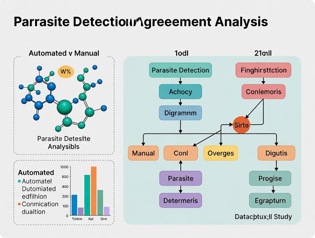
This article provides a comprehensive analysis of the agreement between automated and manual methods for parasite detection, a critical topic for researchers, scientists, and drug development professionals.
Automated vs Manual Parasite Detection: A Comprehensive Agreement Analysis for Biomedical Research
Abstract
This article provides a comprehensive analysis of the agreement between automated and manual methods for parasite detection, a critical topic for researchers, scientists, and drug development professionals. It explores the foundational principles of both classical microscopy and emerging AI-driven diagnostics. The content delves into the operational mechanisms of advanced methodologies like convolutional neural networks (CNNs) and fully automated digital analyzers, while also addressing prevalent challenges such as algorithm limitations and cost-effectiveness. Through a rigorous validation and comparative lens, the synthesis of performance metrics from recent studies offers evidence-based guidance for selecting and integrating diagnostic approaches, ultimately aiming to enhance the accuracy, efficiency, and scalability of parasitic disease control programs.
The Diagnostic Dichotomy: Unpacking Manual and Automated Parasite Detection
Frequently Asked Questions
Q: What are the most common signs that my microscope optics need cleaning? A: Common signs include reduced image contrast, blurred or ghosted images, and visible spots or debris in the field of view that remain stationary when you move the sample or rotate the oculars and objectives [1].
Q: My images lack contrast. The sample should be clear, but details are faint. What should I check? A: This is often a result of contaminated optics. Check for immersion oil residues on objectives, dust on eyepieces, and condensers. Ensure you are using Köhler illumination for even lighting. Also, verify that your sample preparation and staining are optimal [2] [1].
Q: How does manual microscopy compare to automated systems for detecting critical elements like casts in urinalysis? A: Manual microscopy remains the reference for identifying complex elements like casts. Studies show that while automated analyzers have good concordance with manual methods for red blood cells and white blood cells, they often have poor to no concordance for casts, making expert manual review essential for accurate identification [3] [4].
Q: What is the single most important thing I can do to maintain my microscope? A: Establish a consistent cleaning routine after every use, especially when using immersion oil. Immediately wiping the objective with a soft lens tissue and an appropriate cleaning fluid (like isopropanol) prevents oil from hardening and causing permanent damage [1].
Troubleshooting Common Manual Microscopy Issues
| Problem | Possible Causes | Solutions & Verification Steps |
|---|---|---|
| Poor Image Contrast | Dirty optics (objectives, condenser), incorrect Köhler illumination, poorly stained sample [1]. | Clean all optical surfaces. Verify Köhler setup. Check sample staining protocol [1]. |
| Blurred Zones/Ghosting | Dirt or debris on optical surfaces (slide, objective, condenser), sample too thick [1]. | Locate contaminant by rotating eyepieces/objectives; clean confirmed dirt. Use cleaned slides and ensure sample thickness is appropriate [1]. |
| Inconsistent Results | Non-standardized manual method (e.g., centrifugation speed, resuspension volume), variation between operators [3]. | Implement a standardized protocol for all steps. For urine sediment, follow guidelines for centrifugation and resuspension. Ensure staff training [3]. |
| Faint Fluorescence Signal | Contaminated objectives (especially by immersion oil), photobleaching, insufficient staining [1]. | Thoroughly clean objectives. Use antifade mounting media and minimize light exposure. Optimize staining concentration [1]. |
Manual vs. Automated Analysis: A Performance Comparison
The following table summarizes quantitative data on the agreement between manual microscopy and automated analyzers from published studies. Cohen's kappa coefficient is a statistical measure of agreement where 0-0.20 is slight, 0.21-0.40 is fair, 0.41-0.60 is moderate, 0.61-0.80 is substantial, and 0.81-1.00 is almost perfect agreement.
| Element Type | Concordance between Two Automated Analyzers (FUS-200 vs. Iris iQ200) [3] | Concordance: Manual vs. Automated Analyzers [3] | Concordance for Casts: Manual vs. Three Different Analyzers [4] |
|---|---|---|---|
| Erythrocytes (RBCs) | Good to Very Good | 86.1% (FUS-200), 89.0% (Iris iQ200) agreement rate [3]. | Very Good to Good agreement |
| Leukocytes (WBCs) | Good to Very Good | 74.1% (FUS-200), 80.4% (Iris iQ200) agreement rate [3]. | Very Good to Good agreement |
| Epithelial Cells | Good to Very Good | 82.7% (FUS-200), 78.9% (Iris iQ200) agreement rate [3]. | Information not specified |
| Casts | No concordance [3] | No concordance [3] | Moderate (Cobas 6500 κ=0.42; UN3000 κ=0.38; iRICELL 3000 κ=0.62) [4] |
Experimental Protocols
Standardized Protocol for Manual Microscopic Urine Sediment Analysis [3]
- Sample Collection: Collect a mid-stream urine sample (30 mL) in a primary container.
- Sample Transport: Transport at room temperature and analyze within one hour of collection.
- Centrifugation: Transfer 10 mL to a conical tube and centrifuge for 5 minutes at 1500 rpm (400 g).
- Resuspension: Decant the supernatant, leaving 0.5 mL of urine at the tube's bottom. Resuspend the sediment in this remaining urine.
- Microscopy: Place one drop of resuspended sediment on a microscope slide, apply a coverslip, and examine under the microscope.
- Examination & Counting: Systematically scan at least 10 different microscopic fields at both low (×100, LPF) and high power (×400, HPF) magnifications.
- Reporting: Report the average number of cells or particles per field (LPF or HPF). For critical verification, two independent evaluators should analyze the same slide, repeating the analysis with a new sample if results are inconsistent.
Protocol for Accurate Reporting of Microscopy Methods [5]
When publishing, include these critical details in your methods section for reproducibility:
- Microscope & Objective: Manufacturer, model, and objective lens details (manufacturer, magnification, numerical aperture e.g., Plan-Apochromat 63x/1.4 NA Oil).
- Light Source: Type (e.g., mercury lamp, LED, laser wavelengths).
- Excitation/Emission Optics: For fluorescence, specify filter sets (dichroic mirror, excitation, and emission filters with wavelengths).
- Detector & Settings: Camera type (e.g., CCD, PMT) and key settings (exposure time, gain, offset).
- Image Acquisition: For z-stacks, specify z-step size and total number of slices. For live imaging, state the time interval and total duration.
- Image Processing: Report any background subtraction, denoising, or deconvolution software and parameters used.
The Scientist's Toolkit: Essential Research Reagents & Materials
| Item | Function & Application |
|---|---|
| Lens Cleaning Fluid (e.g., isopropanol, ZEISS Cleaning Mixture) | Safely dissolves oil and grease from sensitive optical surfaces without damaging lens coatings [1]. |
| Soft Lens Paper/Tissues | Wipe optics without scratching. Avoids lint and wood chips found in cosmetic tissues [1]. |
| Air Blower | Removes loose dust from optical surfaces and microscope mechanics before wiping [1]. |
| Immersion Oil | Provides a continuous optical path between the specimen and the objective lens, essential for high-resolution imaging with oil-immersion objectives. |
| Stored in Ethanol (70%) | Slides and cover glasses should be stored in 70% ethanol and wiped dry before use to ensure they are clean and free of contaminants for microscopy [1]. |
| Supravital Stains | Stains applied to living cells to improve contrast and differentiation of formed elements in samples like urine sediment [3]. |
Manual Microscopy Workflow
Troubleshooting Image Quality Issues
Frequently Asked Questions (FAQs)
FAQ 1: What are the key advantages of automated parasite detection over manual microscopic examination?
Automated detection systems offer significant improvements in speed, accuracy, and scalability compared to traditional manual methods. They can process images in seconds, operate 24/7, and achieve high precision metrics (e.g., mAP of 0.995), minimizing human error and fatigue [6]. Manual examination is time-consuming, labor-intensive, and its sensitivity is highly dependent on the examiner's skill, often leading to false negatives and delayed diagnoses, especially in high-volume settings [6].
FAQ 2: Our research involves analyzing qualitative data from patient interviews. Can automation assist with this?
Yes, automation is highly effective for qualitative data analysis. AI-powered tools can gather, organize, and interpret non-numerical data (like interview transcripts) to uncover patterns and themes. This approach is faster and more cost-effective than manual coding and allows researchers to focus on interpreting insights rather than repetitive tasks [7]. These tools can perform various analysis methods, including thematic analysis and content analysis, to extract meaningful insights from unstructured data [7].
FAQ 3: We are setting up a multi-center clinical trial. How can automated platforms streamline our imaging data management?
Automated cloud-based imaging platforms are specifically designed for this purpose. They streamline data capture from multiple sources, curate it to common standards and formats (like BIDS), and automate processing pipelines. This ensures data security, regulatory compliance (e.g., HIPAA), and enables secure collaboration between internal and external partners, significantly enhancing productivity and reproducibility [8].
FAQ 4: When should we consider using manual data collection instead of an automated method?
Manual data collection remains preferable for small-scale projects or qualitative research that requires flexibility and human judgment. It is ideal for capturing nuanced data where personal interaction, adaptation to specific circumstances, or interpretation of non-verbal cues is essential [9]. For large-scale, data-intensive studies, however, automation is generally superior for efficiency and accuracy [9] [10].
FAQ 5: What is the FDA's perspective on using AI in drug development processes?
The FDA recognizes the increased use of AI throughout the drug product lifecycle and is actively developing a risk-based regulatory framework to promote innovation while protecting patient safety. The Center for Drug Evaluation and Research (CDER) has an AI Council to coordinate activities and policy, and has already reviewed numerous drug application submissions that incorporate AI/ML components [11].
Troubleshooting Guides
Issue 1: Automated detection model has high precision but low recall in identifying parasite eggs.
- Problem: The model is missing a significant number of true positive parasite eggs.
- Solution:
- Augment Training Data: Increase the variety and volume of your training dataset, specifically adding more examples of eggs in challenging conditions (e.g., different stains, debris, overlapping objects) [6].
- Integrate Attention Mechanisms: Enhance your model architecture with modules like the Convolutional Block Attention Module (CBAM). This helps the model focus on the most relevant spatial and channel-wise features of the parasite eggs, improving sensitivity to small and critical details [6].
- Review Annotation Quality: Ensure the ground truth annotations used for training are accurate and consistent, as errors here directly impact model performance.
Issue 2: Inconsistent data quality from multiple research sites is causing analysis errors.
- Problem: Data collected from different sources varies in format, quality, or standards.
- Solution:
- Implement Centralized Platform: Use a centralized data management platform that enforces common data standards (e.g., BIDS for imaging) and provides integrated, real-time quality control checks [8] [12].
- Automate Data Validation: Deploy systems that perform automated validation and verification upon data upload to catch issues like incorrect formatting or poor image quality early [12].
- Standardize Protocols: Establish and distribute clear, detailed standard operating procedures (SOPs) for data acquisition across all sites to minimize variation at the source.
Issue 3: Difficulty replicating results from a manually collected dataset with an automated query.
- Problem: The automated query does not identify the same patient cohort or data elements as the original manual study.
- Solution:
- Map Data Elements: Carefully map every manual data element to its electronic counterpart in the Clinical Data Repository (CDR) or EHR, acknowledging that some data from free text or paper sources may not be available [10].
- Validate Query Logic: Collaborate with clinicians and IT specialists to review the automated query algorithm (e.g., SQL script). The query may reveal "false negatives"—eligible patients who were accidentally excluded during manual collection due to human error [10].
- Use Codified Data: Where possible, substitute manually abstracted data with equivalent, standardized codified data from sources like hospital billing systems to improve consistency [10].
Data Comparison Tables
Table 1: Performance Comparison: Manual vs. Automated Parasite Egg Detection
| Metric | Manual Microscopy [6] | YCBAM Automated Model [6] |
|---|---|---|
| Detection Speed | Time-consuming; minutes to hours per sample | Near real-time; seconds per image |
| Precision | Variable; highly dependent on examiner skill and fatigue | 0.9971 |
| Recall/Sensitivity | Can lack sensitivity, leading to false negatives | 0.9934 |
| Key Differentiator | Labor-intensive, subjective, prone to human error | High-throughput, consistent, minimizes human error |
| mAP@0.50 | Not Applicable | 0.9950 |
| mAP@0.50:0.95 | Not Applicable | 0.6531 |
| Aspect | Manual Data Collection | Automated Data Collection |
|---|---|---|
| Best For | Small-scale projects, qualitative data, nuanced human judgment | Large datasets, real-time processing, repetitive tasks |
| Speed | Slow | Fast |
| Scalability | Low | High |
| Error Rate | Prone to human error (e.g., transcription, selection bias) | Minimized human error; consistent |
| Flexibility | High; can adapt on the fly | Lower; requires predefined rules |
| Upfront Cost | Lower | Higher investment |
Experimental Protocols
Protocol 1: Automated Detection of Pinworm Eggs Using the YCBAM Deep Learning Model
This protocol is based on the study detailed in [6].
1. Objective To automate the detection and localization of pinworm ( Enterobius vermicularis ) eggs in microscopic images using a deep learning framework integrating YOLOv8 with self-attention and Convolutional Block Attention Module (CBAM).
2. Materials and Reagents
- Microscopes: For generating high-quality digital images of slides.
- Annotated Image Dataset: A collection of microscopic images with pinworm eggs, annotated by parasitology experts (e.g., 255 images for segmentation, 1,200 for classification as used in related studies [6]).
- Computational Hardware: A computer with a powerful GPU (Graphics Processing Unit) for efficient model training.
- Software: Python programming environment with deep learning libraries (e.g., PyTorch, Ultralytics YOLO).
3. Methodology
- Step 1: Data Preparation. Collect and annotate microscopic images. Split the data into training, validation, and test sets.
- Step 2: Model Architecture. Implement the YCBAM (YOLO Convolutional Block Attention Module) architecture. This involves integrating the YOLOv8 object detection model with self-attention mechanisms and the CBAM module. CBAM sequentially infers attention maps along both the channel and spatial dimensions, helping the model focus on more informative features of the pinworm eggs.
- Step 3: Model Training. Train the YCBAM model on the training dataset. The model's parameters are optimized to minimize the loss function (e.g., box loss for localization).
- Step 4: Model Evaluation. Evaluate the trained model on the held-out test set using standard object detection metrics, including Precision, Recall, and mean Average Precision (mAP) at different Intersection over Union (IoU) thresholds.
4. Expected Outcomes Upon successful implementation, the model should achieve high performance metrics, such as a precision >0.99 and mAP@0.50 >0.99, demonstrating its capability as a highly accurate diagnostic tool [6].
Protocol 2: Comparative Study of Manual vs. Automated Data Collection for Clinical Research
This protocol is adapted from the methodology described in [10].
1. Objective To compare the accuracy, efficiency, and completeness of manual data collection versus automated data collection from an Electronic Health Record (EHR) or Clinical Data Repository (CDR) for a clinical research study.
2. Materials
- Access to EHR/CDR: With appropriate permissions and data governance.
- Structured Query Language (SQL) Tools: For building automated data extraction queries.
- Statistical Software: For data analysis (e.g., R, SPSS, Python with pandas).
3. Methodology
- Step 1: Define Study Cohort and Variables. Clearly define the inclusion/exclusion criteria and the specific data elements to be collected (e.g., patient demographics, lab values, medication records).
- Step 2: Manual Data Collection. Have clinical researchers or staff collect the required data for a defined patient cohort by manually reviewing EHRs and entering data into a spreadsheet, following the standard manual process.
- Step 3: Automated Data Collection. In parallel, an IT specialist or informatician develops an automated script (e.g., an SQL stored procedure) to extract the same data elements for the same patient cohort from the CDR.
- Step 4: Data Validation and Comparison. Compare the two resulting datasets.
- Patient Sets: Check for discrepancies in the patient cohorts (e.g., false positives/negatives in the manual set).
- Data Elements: Compare the values of individual data points to identify transcription or computational errors.
- Statistical Analysis: Use statistical tests (e.g., independent sample t-tests, frequency distributions) to analyze differences.
4. Expected Outcomes The automated method is expected to identify patients missed by manual collection ("false negatives") and reveal instances of human error, such as computational or transcription mistakes, thereby demonstrating superior completeness and accuracy [10].
Workflow and System Diagrams
Automated Parasite Detection Workflow
Manual vs. Automated Data Collection Process
The Scientist's Toolkit: Research Reagent Solutions
Table 3: Essential Tools for Automated Imaging Research
| Item | Function in Research |
|---|---|
| Clinical Data Repository (CDR) | A centralized database that aggregates clinical data from sources like EHRs, enabling automated data extraction for research via SQL queries [10]. |
| YOLO-based Deep Learning Model | An object detection algorithm (e.g., YOLOv8) that can be trained to identify and localize specific objects, such as parasite eggs, in digital images with high speed and accuracy [6]. |
| Attention Mechanisms (e.g., CBAM) | A module that can be integrated into convolutional neural networks to help the model focus on the most relevant parts of an image, improving feature extraction and detection performance for small objects [6]. |
| Cloud-Based Imaging Platform | A platform that streamlines the capture, curation, management, and automated analysis of medical imaging data from multiple sources, facilitating collaboration and ensuring data standardization [8]. |
| Computer-Assisted Qualitative Data Analysis Software (CAQDAS) | Software (e.g., NVivo, ATLAS.ti) used to organize and aid in the analysis of unstructured qualitative data, such as interview transcripts or open-ended survey responses [7]. |
Frequently Asked Questions (FAQs)
FAQ 1: What is the practical difference between accuracy and precision in a diagnostic context?
In diagnostic testing, accuracy and precision are distinct but complementary concepts. Accuracy refers to how close a measurement is to the true value. For example, in parasite detection, a test is accurate if it correctly identifies the presence and species of a parasite, reflecting a correct representation of reality [13]. Precision, however, does not concern itself with the true value. Instead, it refers to the reliability and repeatability of a measurement. A precise test will yield very similar results when the same sample is measured multiple times under consistent conditions, indicating low variation [13]. A test can be precise (repeatable) but not accurate (consistently off-target), or accurate on average but not precise (results are scattered around the true value).
FAQ 2: How is "agreement" different from accuracy when comparing a new automated method to manual microscopy?
Agreement assesses the level of concordance or consistency between two measurement methods, without necessarily declaring one as the absolute "truth." In validation studies, you often compare a new automated system to an established manual method. A high percentage agreement indicates that the two methods produce similar results under the same conditions [14]. Accuracy, in this context, is typically reserved for when a method is compared against a certified reference material or an undisputed gold standard method. In many practical scenarios, demonstrating a high degree of agreement with the current standard method is a critical step in validating a new technique's performance.
FAQ 3: Our new AI detection model has high precision but low accuracy. What are the most likely causes?
This pattern typically points to a consistent bias or systematic error in your measurement system. Potential causes include:
- Incorrect Training Data: The AI model may have been trained on a dataset that does not accurately represent the real-world population, leading to a biased model [13].
- Sample Preparation Bias: A consistent error in how samples are prepared, such as an incorrect dilution factor or staining protocol, can cause all measurements to be systematically offset from the true value [15].
- Calibration Drift: The instrument or software may be miscalibrated, causing it to consistently over- or under-estimate values.
FAQ 4: What does "integrity" mean for a KPI, and how do we ensure it?
In the context of KPIs, integrity means that the measures have sufficient accuracy and precision for their intended purpose. It is a practical assessment of whether a KPI is fit-for-purpose. You can ensure integrity by evaluating it against five dimensions [13]:
- Relevant: The data must be directly appropriate for the KPI's purpose.
- Reliable: Enough data must be collected to account for inherent variability over time.
- Representative: The data must describe the full scope of what the KPI is supposed to measure, without bias.
- Readable: The data must be clearly defined, legibly presented, and make sense to its users.
- Realistic: The value gained from using the data must be greater than the effort invested in collecting it.
Troubleshooting Common Experimental Issues
Problem: Disagreement between automated and manual parasite counts in stool samples.
Investigation Protocol:
- Verify Sample Quality: Confirm that the samples used for both methods are identical in terms of collection time, storage conditions, and homogeneity. Inconsistent results can stem from degraded or non-uniform samples.
- Re-examine Discrepant Samples: Retrieve the physical slides or digital images where the counts disagreed. Have a senior microscopist re-examine them manually to establish a more definitive count.
- Check for Species-Specific Issues: Analyze whether the disagreement is consistent across all parasite species or isolated to a specific type (e.g., certain egg morphologies). This can indicate an issue with the AI's training for that particular class [15].
- Review Instrument Logs: Check the automated system's logs for any errors during the processing or imaging of the discrepant samples.
- Assess Environmental Factors: Document the operating environment (temperature, humidity) for both methods, as extremes can occasionally affect instrument performance or sample integrity.
Resolution Steps:
- If the issue is species-specific, retrain the AI model with a larger and more diverse set of images for the problematic parasite [16].
- If the sample preparation is the variable, standardize the protocol (e.g., strictly control the stool sample size, dilution, and staining time) and retrain staff [15].
- If the automated system consistently misses low-intensity infections, validate its detection limit and adjust the sensitivity threshold or review the concentration method [14].
The following tables summarize key performance data from recent studies comparing automated and manual parasite detection methods.
Table 1: Overall Parasite Detection Level Comparison
| Methodology | Sample Size (n) | Positive Cases (n) | Detection Level | Statistical Significance (P-value) |
|---|---|---|---|---|
| Manual Microscopy [15] | 51,627 | 1,450 | 2.81% | P < 0.05 |
| KU-F40 Automated Analyzer [15] | 50,606 | 4,424 | 8.74% |
Table 2: Performance of AiDx Assist for Schistosoma Detection
| Sample Type | Operational Mode | Sensitivity | Specificity | Reference Standard |
|---|---|---|---|---|
| Stool (S. mansoni) [16] | Semi-Automated | 86.8% | 81.4% | Conventional Microscopy |
| Stool (S. mansoni) [16] | Fully Automated | 56.9% | 86.8% | Conventional Microscopy |
| Urine (S. haematobium) [16] | Semi-Automated | 94.6% | 90.6% | Conventional Microscopy |
| Urine (S. haematobium) [16] | Fully Automated | 91.9% | 91.3% | Conventional Microscopy |
Table 3: Comparison of Parasite Species Detection
| Parasite Species (Eggs) | Manual Microscopy Detection Level (n=51,627) | KU-F40 Automated Detection Level (n=50,606) | Statistical Significance (P-value) |
|---|---|---|---|
| Clonorchis sinensis [15] | 2.74% | 8.50% | P < 0.001 |
| Hookworm [15] | 0.04% | 0.11% | P < 0.001 |
| Blastocystis hominis [15] | 0.01% | 0.07% | P < 0.001 |
| Giardia lamblia [15] | 0.00% | 0.03% | P < 0.001 |
Experimental Protocols
Protocol 1: Validation of an Automated Analyzer vs. Manual Microscopy
This protocol is based on a large-scale retrospective comparison study [15].
- Sample Collection: Collect fresh stool samples from patients in clean, sterile containers. If the sample contains mucus, pus, or blood, prioritize collecting from these areas.
- Manual Microscopy (Reference Method):
- Specimen Prep: Place one to two drops of 0.9% saline on a sterile slide. Using a wooden applicator, take a match-head-sized amount of stool (approx. 2 mg) and mix with saline to create a uniform suspension. The thickness should allow newspaper print underneath to be legible. Place a coverslip on top.
- Examination: First, use a 10x objective lens to scan the entire slide (observe >10 fields of view). Then, switch to a 40x objective to identify and confirm suspected parasitic elements (observe >20 fields of view).
- Timing: Perform the examination within 2 hours of sample collection.
- Instrumental Method (KU-F40 Automated Analyzer):
- Specimen Prep: Collect a soybean-sized fecal specimen (approx. 200 mg) in the instrument's designated sterile container.
- Analysis: The instrument automatically performs dilution, mixing, filtration, and transfers 2.3 ml of the prepared sample to a flow counting chamber. It uses artificial intelligence and high-definition cameras to identify parasites.
- Manual Review: All suspected parasite detections by the instrument must be confirmed by laboratory personnel before a final report is issued.
- Timing: Complete analysis within 2 hours of sample collection.
- Statistical Analysis: Compile results and compare detection levels (positive rates) between the two methods using a Chi-square (χ²) test. A P-value of less than 0.05 is considered statistically significant.
Protocol 2: Field Evaluation of an AI-Based Microscope for Schistosoma
This protocol outlines the field evaluation of the AiDx Assist device [16].
- Study Design & Ethics: Obtain ethical approval from the relevant review board. Secure written informed consent from all participants or their guardians.
- Sample Collection: Provide participants with sterile containers for stool and urine. Collect samples at designated sites and transport them to the laboratory within 2 hours.
- Sample Processing:
- Stool (Kato-Katz): Use a template to transfer 41.7 mg of sieved stool to a microscope slide. Cover with a cellophane strip pre-soaked in malachite green. Apply light pressure to spread the smear and allow it to clear for 10 minutes before examination [16].
- Urine (Filtration): Homogenize the urine sample. Pass 10 ml through a 13 mm polycarbonate membrane (30 µm pore size) using a syringe and filter holder. Transfer the membrane to a glass slide [16].
- AiDx Assist Analysis:
- Semi-Automated Mode: Disable the AI algorithm. The device registers images, which are then visually examined by an expert who manually identifies and counts Schistosoma eggs.
- Fully Automated Mode: Enable the AI algorithm to automatically detect and count Schistosoma eggs. The operator confirms the output.
- Conventional Microscopy (Reference): Examine the same Kato-Katz and urine filtration slides under a standard light microscope (e.g., 10/40x objectives). Two independent microscopists should perform readings, and the average egg count is used for analysis.
- Data Analysis: Express egg counts as eggs per gram (EPG) of stool or eggs per 10 ml of urine. Calculate the sensitivity and specificity of the AiDx Assist using conventional microscopy as the reference standard.
Workflow and Relationship Diagrams
Diagram: Parasite Detection KPI Analysis Workflow
Diagram: KPI Relationship and Integrity Framework
The Scientist's Toolkit: Research Reagent Solutions
Table 4: Essential Materials for Parasite Detection Experiments
| Item | Function / Application |
|---|---|
| KU-F40 Fully Automated Fecal Analyzer [15] | An integrated system for automated sample processing, imaging, and AI-based analysis of fecal parasites. |
| AiDx Assist Digital Microscope [16] | A portable, automated microscope with integrated AI for detecting parasite eggs in stool (Kato-Katz) and urine (filtration) samples in field settings. |
| Kato-Katz Kit [16] | A standardized tool for quantitative diagnosis of helminth eggs, including templates for precise stool sampling and cellophane slides. |
| Polycarbonate Membrane Filters (30µm pore) [16] | Used for urine filtration methods to concentrate Schistosoma haematobium eggs for microscopic examination. |
| Malachite Green Solution [16] | A chemical used to pre-soak cellophane coverslips in the Kato-Katz technique, which helps clear debris for better egg visibility. |
| LEICA DM 300 Microscope [15] | A conventional light microscope used as a reference standard for manual parasite identification and quantification. |
Clinical and Economic Imperatives for Advancing Diagnostic Methods
Troubleshooting Guides & FAQs
This technical support center provides solutions for researchers conducting agreement analysis between automated and manual methods for intestinal parasite detection.
FAQ 1: How should discrepancies between AI and manual microscopy results be resolved? Discrepant findings should undergo a multi-person manual review by experienced technologists, a process known as discrepancy analysis. In a recent validation study, this process confirmed a 98.6% positive agreement and identified 169 additional organisms initially missed during manual review [17] [14]. This review is the definitive step for classifying true positives and false positives/negatives.
FAQ 2: What are the critical steps for preparing a high-quality wet mount for AI analysis? The two critical steps are preservation and staining. Use validated transport media such as Total-Fix or paired vials of 10% formalin and PVA (polyvinyl alcohol) [18]. Invalidate results from specimens submitted in unvalidated media like Ecofix or Protofix. Ensure the stool sample is thoroughly mixed with the preservative to fully fix the entire specimen [18].
FAQ 3: Our AI model's performance varies significantly with sample dilution. How can this be addressed? Performance decay at low parasite concentrations is expected. Conduct a formal Limit of Detection (LOD) study to establish the operational range of your system. A key validation finding is that AI systems can consistently detect parasites in highly diluted samples better than technologists, suggesting utility for early-stage or low-level infections [17] [14]. Use highly characterized, rare species panels to test robustness [17].
FAQ 4: What is the recommended number of stool specimens for a comprehensive parasite evaluation? For routine examination before treatment, collect a minimum of 3 specimens on alternate days [18]. This accounts for the intermittent shedding of parasites. Collecting multiple specimens the same day does not increase test sensitivity [18].
Data Presentation: Performance Comparison
Table 1: Key Quantitative Metrics from a Recent AI Validation Study for Parasite Detection in Stool Wet Mounts [17] [14]
| Metric | AI-Assisted Detection | Traditional Manual Microscopy |
|---|---|---|
| Positive Agreement (after discrepancy analysis) | 98.6% | Baseline |
| Additional Organisms Identified | 169 (missed in initial manual review) | Not Applicable |
| Training/Validation Sample Size | >4,000 parasite-positive samples | Not Applicable |
| Scope of Detection | 27 classes of parasites, including rare species | Varies with technologist expertise |
| Sensitivity in Diluted Samples | Consistently high, detects low-level infections | Decreases with parasite concentration |
Table 2: Essential Research Reagent Solutions for Parasite Detection Studies
| Reagent / Material | Primary Function in Experiment |
|---|---|
| Total-Fix | All-in-one stool preservative for fixation, preservation of cysts, eggs, and larvae [18]. |
| Formalin (10%) & PVA | Paired preservatives; formalin fixes morphology, PVA preserves stainability for permanent slides [18]. |
| Parasite-Positive Sample Panels | Characterized samples for training AI models and validating assay performance across diverse targets [17]. |
| Pinworm Paddle Collection Device | Specialized tool for collecting perianal samples for optimal detection of Enterobius vermicularis [18]. |
Experimental Protocols
Protocol 1: Building a Deep-Learning Model for Parasite Detection
This protocol details the methodology for developing a convolutional neural network (CNN) for detecting protozoan and helminth parasites in concentrated wet mounts of stool [17].
- Sample Collection & Curation: Assemble a robust training set of over 4,000 parasite-positive stool samples. The collection should be globally sourced (e.g., from the US, Europe, Africa, Asia) and must represent a wide variety of parasites, including at least 27 different classes and rare species (e.g., Schistosoma japonicum, Paracapillaria philippinensis) [17].
- Data Preparation (Annotation): Have expert parasitology technologists meticulously label all training images, identifying and classifying parasites. This annotated dataset serves as the ground truth for the model.
- Model Training: Train the CNN using the prepared dataset. This involves feeding the annotated images into the neural network so it can learn to distinguish parasitic structures from background debris and other stool components.
- Validation & Discrepancy Analysis: Test the trained model against a separate set of validated samples. All results that differ from the manual gold standard must undergo a blinded review by multiple experts to determine the correct classification. This step is critical for calculating final performance metrics like positive agreement [17] [14].
- Limit of Detection (LOD) Study: Perform serial dilutions of known positive samples to determine the lowest concentration of parasites the AI can reliably detect, comparing its sensitivity to that of human technologists at each dilution [17].
Protocol 2: Standardized Ova and Parasite (O&P) Examination for Method Comparison
This is the reference manual method against which automated systems are often validated [18].
- Specimen Procurement: Collect stool specimens into appropriate validated preservatives. A series of three specimens collected on alternate days is recommended for optimal sensitivity [18].
- Microscopy Slide Preparation: For wet mounts, prepare slides from the preserved concentrate. For permanent staining, prepare smears from the PVA-preserved sample and perform trichrome staining.
- Manual Microscopic Examination: A trained technologist systematically scans the entire coverslip area under multiple magnifications (e.g., 100x, 200x, 400x) using a light microscope.
- Identification & Documentation: The technologist identifies parasites based on morphological characteristics (size, shape, internal structures) and documents all findings.
- Result Reporting: Findings are reported, noting the presence and stage (e.g., cyst, trophozoite, ova, larva) of any pathogenic or non-pathogenic organisms.
Workflow Visualization
Inside the Algorithms: How AI and Automated Systems Detect Parasites
This technical support center provides guidelines for researchers developing Convolutional Neural Networks (CNNs) for automated parasite detection. This field is critical for global health, as traditional manual microscopy is time-consuming, labor-intensive, and subject to human error, especially in resource-limited settings [19] [20]. This guide addresses common technical challenges, offers detailed protocols, and provides resources to ensure your deep learning models are accurate, efficient, and robust.
Frequently Asked Questions (FAQs)
1. What is the role of CNNs in automated parasite detection? CNNs automate the analysis of blood smear or fecal sample images. They learn to identify characteristic features of parasites, such as their morphology and texture, directly from pixel data. This enables high-throughput, objective classification of samples as infected or uninfected, and can even distinguish between parasite species and life-cycle stages [19] [20] [21].
2. How can I improve my CNN model's accuracy if it's performing poorly?
- Implement Preprocessing: Integrate image segmentation techniques like Otsu's thresholding to isolate parasitic regions and reduce background noise. This has been shown to boost baseline CNN accuracy by approximately 3% [19].
- Use Data Augmentation: Artificially expand your dataset with rotations, flips, and brightness adjustments to improve model generalization.
- Try Advanced Architectures: Consider hybrid models, such as a CNN with a capsule network (CapsNet), which can better capture spatial hierarchies and parasite orientations [20].
3. My model trains well but fails on new data. How can I improve its generalizability?
- Cross-Dataset Validation: Train and test your model on different publicly available datasets (e.g., MP-IDB, IML-Malaria) to ensure it doesn't overfit to one data source [20].
- Employ Cross-Validation: Use k-fold cross-validation (e.g., 5-fold) during training to get a more reliable estimate of model performance [19].
- Balance Your Data: Ensure your training set has a roughly equal number of examples for each class (e.g., infected/uninfected, different parasite species) to prevent bias.
4. What are the computational requirements for deploying these models? Requirements vary by model complexity. A standard CNN may require significant resources, but newer, lightweight architectures like the Hybrid CapNet (1.35 million parameters, 0.26 GFLOPs) are designed for deployment on mobile devices in field settings with limited computational power [20].
Troubleshooting Guides
Issue 1: Low Classification Accuracy
Problem: Your CNN model is not achieving the desired accuracy on the test set.
Solution Steps:
- Verify Data Quality: Inspect your images for inconsistencies in staining, lighting, or focus. Clean your dataset of corrupt or mislabeled images.
- Apply Image Segmentation: Preprocess images with Otsu's thresholding to highlight relevant regions. One study increased accuracy from 95% to 97.96% using this method [19].
- Tune Hyperparameters: Systematically adjust learning rate, batch size, and number of epochs. Consider using automated hyperparameter tuning tools.
- Experiment with Architectures: Move from a baseline CNN to a more advanced model like EfficientNet-B7 or a hybrid Capsule Network, which can capture more complex features [19] [20].
Issue 2: Model Overfitting
Problem: The model performs perfectly on training data but poorly on validation/test data.
Solution Steps:
- Introduce Regularization: Add Dropout layers to your CNN to randomly disable neurons during training, forcing the network to learn redundant representations.
- Apply Data Augmentation: Expand your training dataset using transformations as mentioned in the FAQs.
- Implement Early Stopping: Halt the training process when the validation loss stops improving to prevent the model from memorizing the training data.
- Use a Composite Loss Function: For advanced models, a composite loss (e.g., combining margin, focal, and reconstruction losses) can enhance robustness to noise and class imbalance [20].
Issue 3: Long Training Times
Problem: The model takes an impractically long time to train.
Solution Steps:
- Optimize Hardware: Utilize GPUs (Graphics Processing Units) for training, as they are far more efficient than CPUs for the matrix calculations in deep learning [21].
- Simplify the Model: Choose a lightweight architecture designed for efficiency, such as the Hybrid CapNet [20].
- Adjust Input Dimensions: Resize input images to a smaller, but still representative, resolution to reduce computational load.
Experimental Protocols
Protocol 1: Baseline CNN with Otsu Preprocessing for Malaria Detection
This protocol is based on a study that achieved 97.96% accuracy for classifying malaria-infected cells [19].
1. Dataset Preparation
- Dataset: Use a large-scale dataset of blood smear images (e.g., 43,400 images).
- Split: Divide the dataset into 70% for training and 30% for testing.
- Preprocessing: Apply Otsu's thresholding-based segmentation to each RGB image. This algorithm automatically calculates the optimal threshold to separate the image into foreground (parasite-relevant regions) and background, retaining morphological context.
2. Model Training
- Architecture: Construct a 12-layer Convolutional Neural Network. A typical structure includes:
- Input layer
- Multiple convolutional layers with ReLU activation and pooling layers
- Fully connected (dense) layers
- Final classification layer with softmax activation
- Training: Train the model on the segmented training images. Use an appropriate optimizer (e.g., Adam) and loss function (e.g., categorical cross-entropy).
3. Model Validation
- Quantitative Segmentation Validation:
- Create a subset of images with manually annotated, pixel-wise ground truth masks.
- Compare the Otsu-generated masks against the reference masks.
- Calculate the mean Dice coefficient (target: ~0.85) and Jaccard Index (IoU) (target: ~0.74) to confirm segmentation effectiveness [19].
- Performance Evaluation: Evaluate the trained model on the held-out test set to determine final accuracy, precision, and recall.
- Cross-Validation: Perform 5-fold cross-validation to ensure result robustness (expected consistency: 94.8% - 97.8%) [19].
Protocol 2: Cross-Dataset Validation for Model Generalization
This protocol evaluates how well your model performs on data from different sources, which is crucial for real-world deployment [20].
1. Dataset Curation
- Gather multiple public benchmark datasets for parasite detection (e.g., MP-IDB, MP-IDB2, IML-Malaria, MD-2019).
2. Intra- and Cross-Dataset Evaluation
- Intra-Dataset Evaluation: For each dataset (Dataset A), perform a standard train/test split within the same dataset and record the performance.
- Cross-Dataset Evaluation: Train your model on the entire training set of one dataset (Dataset A). Then, test the trained model directly on the entire test set of a different dataset (Dataset B). Repeat this for various dataset pairs.
3. Analysis
- Compare the performance metrics (accuracy, F1-score) from intra-dataset and cross-dataset tests. A significant drop in cross-dataset performance indicates poor generalization, suggesting a need for more diverse training data or model architecture adjustments.
Workflow and Signaling Diagrams
CNN-Parasite Detection Workflow
Model Generalizability Decision Pathway
Research Reagent Solutions
Table: Essential Materials for CNN-based Parasite Detection Experiments
| Item Name | Function/Description | Example/Specification |
|---|---|---|
| Benchmark Datasets | Provides standardized image data for training and evaluating models. | MP-IDB, IML-Malaria, Malaria-Detection-2019 [20] |
| Otsu Thresholding Algorithm | Image segmentation method to isolate parasite regions and improve model focus. | Preprocessing step to boost CNN accuracy [19] |
| Lightweight CNN Architectures | Efficient models suitable for deployment in resource-constrained environments. | Hybrid CapNet (1.35M parameters) [20] |
| Graphical Processing Unit (GPU) | Hardware that dramatically accelerates the deep learning model training process. | Essential for handling large image datasets [21] |
| Evaluation Metrics Suite | Quantitative measures to assess model performance and segmentation quality. | Accuracy, F1-Score, Dice coefficient, Jaccard Index [19] [20] |
## FAQs: Instrument Operation and Performance
1. What are the key advantages of fully automated fecal analyzers over traditional manual microscopy?
Fully automated fecal analyzers address several critical limitations of manual microscopy. They enhance biosafety by processing specimens in a completely enclosed environment, reducing the risk of sample cross-contamination and exposure to pathogens for laboratory personnel [22]. They significantly improve detection sensitivity and throughput; one study reported that an automated instrument (KU-F40) had a parasite detection level of 8.74%, which was 3.11 times higher than the 2.81% detected by manual microscopy [22]. Furthermore, automation reduces labor intensity, minimizes subjective errors caused by inspector fatigue, and standardizes the testing process [22] [23] [24].
2. How does the AI and imaging technology in analyzers like the KU-F40 or FA280 work to identify parasites?
These instruments use a combination of advanced microscopy, high-definition digital imaging, and artificial intelligence (AI). The process involves:
- Sample Preparation: The system automatically dilutes, mixes, and filters a fecal sample [22] [23].
- Image Acquisition: A high-resolution camera (e.g., 5-megapixel HD CMOS) captures multiple images (over 300) through multi-field layered scanning, often including both low- and high-power objectives [22] [25].
- AI Analysis: Integrated AI software analyzes the captured images to automatically identify and categorize formed elements, including cells, crystals, and various parasite species and eggs [22] [25] [24]. Some models also feature an iodine staining function to improve the detection rate of certain parasites [25].
3. My results show a discrepancy between the automated analyzer and a manual method. How should this be investigated?
Discrepancies, particularly in cases of low infection intensity, are known to occur. The established protocol is to implement a mandatory manual re-examination rule. When the automated system flags a sample as positive or provides an uncertain identification, a trained technologist must review the captured images or perform a manual microscopic examination to confirm the result [22]. This combination of automation and expert review significantly improves the accuracy of the final report. Studies indicate that agreement between automated and manual methods (like Kato-Katz) is often higher in samples with high infection intensity [23].
4. What are the best practices for sample collection and preparation to ensure optimal analyzer performance?
Proper sample collection is fundamental. Key practices include:
- Sample Amount: Collect approximately a soybean-sized (about 200-500 mg) specimen [22] [23].
- Collection Device: Use the manufacturer's specified collection cups, which often feature filters and are designed for direct loading onto the instrument [22] [25].
- Timeliness: Process all samples within 2 hours of collection for the most reliable results [22]. If using transport systems like FecalSwab, note that stability varies by target pathogen (e.g., 24-48 hours for C. difficile) [26].
5. The instrument flags an error during the sample mixing or aspiration step. What are the likely causes?
This is often related to sample viscosity or particulate matter. First, ensure the sample is adequately homogenized before loading. The sample collection cup's built-in filter is designed to prevent large particulates from clogging the fluidic path; check if the filter is intact or obstructed. Consult the instrument's maintenance manual for specific error codes, which typically guide you to inspect and, if necessary, clean or replace components like the aspiration needle, tubing, or valves [27].
## Troubleshooting Guides
### Common Error Codes and Resolutions
| Error Code / Message | Possible Cause | Recommended Action |
|---|---|---|
| Clogged Fluid Path | Viscous sample, large debris obstructing the needle or tubing. | Manually clean the aspiration needle and fluidic path as per the service manual. Ensure samples are well-mixed and not overly solid. |
| Image Focusing Failure | Air bubbles in flow cell, camera or lens obstruction, faulty auto-focus mechanism. | Run a cleaning cycle to purge air bubbles. Gently clean the exterior of the camera lens and optics. Perform a focus calibration. |
| Low/Inconsistent Diluent | Empty diluent bottle, leak in diluent line, faulty pump. | Refill or replace the diluent bottle. Check tubing for cracks/leaks and connections. Prime the diluent line. |
| Communication Failure | Loose cable, software glitch, network issue. | Restart the instrument and host computer. Check all physical cable connections. Reinstall instrument driver software if needed. |
### Performance Issues and Quality Control
| Performance Issue | Root Cause Investigation | Corrective Action |
|---|---|---|
| Low Parasite Detection Sensitivity | AI algorithm needs retraining/updating; poor image quality; incorrect sample dilution. | Verify and update AI software. Check camera focus and clarity. Validate sample preparation volume and dilution ratio. |
| High Rate of False Positives | Misclassification of debris or artifacts as parasites. | Review false positive images and fine-tune the AI classification model. Implement mandatory manual verification for all positive results. |
| Inconsistent Results Between Runs | Instrument requires calibration; reagent lot variation; sampling error. | Run quality control samples with known targets. Perform full system calibration. Ensure consistent sample mixing and collection. |
## Experimental Protocols for Method Comparison
For researchers validating a new automated analyzer or comparing it against established manual methods, the following structured protocol is recommended.
### Protocol: Comparing Automated Fecal Analyzer vs. Kato-Katz for Helminth Detection
1. Objective: To evaluate the diagnostic agreement between a fully automated digital fecal analyzer (e.g., FA280) and the manual Kato-Katz technique for the detection and quantification of soil-transmitted helminths and Clonorchis sinensis [23].
2. Materials and Reagents:
- Fully automatic digital feces analyzer (e.g., FA280, KU-F40)
- Microscope (e.g., Olympus CX23)
- Kato-Katz templates and cellophane strips
- Sample collection cups and applicators
- Physiological saline (0.9%)
- Giemsa stain or Iodine stain (for manual verification)
3. Procedure:
- Sample Collection: Collect a single fresh stool specimen from each participant. For the automated analyzer, aliquot approximately 0.5 g into the manufacturer's proprietary collection cup. For the Kato-Katz method, prepare duplicate smears from a different portion of the same specimen using a 41.7 mg template [23].
- Automated Analysis: Load the sample cup into the analyzer. The instrument will automatically handle dilution, mixing, filtration, image capture, and AI analysis. Record all positive findings and the associated images.
- Manual Microscopy: Examine the Kato-Katz smears under a microscope by experienced technicians. Count the number of eggs for each parasite species to calculate eggs per gram (EPG) of feces.
- Data Analysis: For samples with discrepant results, perform a manual microscopic review of the images generated by the automated analyzer or re-examine the sample using a reference method (e.g., formalin-ether concentration technique) for adjudication.
4. Statistical Analysis:
- Calculate the positive detection rate for each method.
- Use McNemar's test (for paired nominal data) to determine if there is a statistically significant difference in detection between the two methods.
- Calculate the kappa (κ) statistic to evaluate the agreement between the methods beyond chance. A kappa value above 0.8 indicates strong agreement [23].
- Analyze agreement stratified by infection intensity (e.g., low, medium, high EPG) using Pearson’s Chi-square test [23].
The table below summarizes quantitative data from recent studies comparing automated fecal analyzers with manual microscopy.
Table 1: Performance Comparison of Automated vs. Manual Microscopy for Parasite Detection
| Study Instrument / Manual Method | Sample Size | Detection Rate (Automated) | Detection Rate (Manual) | Statistical Significance (P-value) | Agreement (Kappa, κ) |
|---|---|---|---|---|---|
| KU-F40 [22] / Manual Microscopy | 102,233 total | 8.74% (4424/50606) | 2.81% (1450/51627) | P < 0.05 | Not Specified |
| FA280 [23] / Kato-Katz | 1,000 | 10.0% (100/1000) | 10.0% (100/1000) | P > 0.999 | 0.82 (95% CI: 0.76-0.88) |
Table 2: Parasite Species Detection Capabilities of Automated Analyzers
| Parasite Species / Group | Detected by KU-F40? | Detected by Manual Microscopy in Study [22]? | Notes on Performance |
|---|---|---|---|
| Clonorchis sinensis | Yes | Yes | Significantly higher detection with KU-F40 (P < 0.05) [22] |
| Hookworm | Yes | Yes | Significantly higher detection with KU-F40 (P < 0.05) [22] |
| Blastocystis hominis | Yes | Yes | Significantly higher detection with KU-F40 (P < 0.05) [22] |
| Strongyloides stercoralis | Yes | Yes | Higher detection with KU-F40, but difference not significant (P > 0.05) [22] |
| Entamoeba histolytica/dispar | Yes [25] | Not reported | AI is trained to differentiate species [25] |
| Giardia lamblia | Yes [25] | Not reported | AI is trained to identify cysts/trophozoites [25] |
## The Scientist's Toolkit: Essential Research Reagents & Materials
Table 3: Key Materials and Reagents for Automated Fecal Analysis
| Item | Function & Specification | Application Note |
|---|---|---|
| Specialized Collection Cup | Sample collection and initial processing. Often contains a filter and is designed for direct instrument loading. | Ensures correct sample volume and pre-filtration, which is critical for smooth instrument operation [22] [25]. |
| Liquid Diluent | To automatically dilute and homogenize the fecal sample for consistent imaging and analysis. | Proprietary to each instrument. Required for creating a uniform suspension and preventing clogging [22]. |
| Cary-Blair Transport Medium | A non-nutritive medium for preserving enteric bacteria and some parasites in swab-based systems. | Used in systems like FecalSwab for stabilizing samples during transport, compatible with molecular testing [26]. |
| Iodine Staining Solution | Stains glycogen and nuclei of protozoan cysts to aid in morphological identification by AI. | The KU-F40 can automatically add iodine stain to improve detection of specific ova and parasites [25]. |
| Quality Control (QC) Samples | Simulated or known positive samples to verify instrument and AI algorithm performance. | Essential for daily QC protocols to ensure continued sensitivity and specificity of the automated system. |
## Workflow Visualization
The following diagram illustrates the end-to-end automated workflow of a fully automatic digital feces analyzer, from sample loading to result reporting.
Automated Fecal Analysis Workflow
Core System Components
Frequently Asked Questions (FAQs) and Troubleshooting Guide
Q1: Our smartphone application for reading malaria Rapid Diagnostic Tests (RDTs) performs well at high parasite densities but misses low-density infections. How can we improve detection sensitivity?
A1: Lower sensitivity at low parasite densities is a known challenge. Current research indicates this is a widespread issue; one study found RDT sensitivity for the Pf test line read by mobile apps was 47% at 20 parasites/µL, compared to 74% for the trained human eye [28]. To improve performance:
- Algorithm Enhancement: Focus development on improving line detection algorithms for faint band intensities. Future efforts should prioritize enhancing the software's ability to identify low-intensity test lines [28].
- Hardware Consistency: Ensure the smartphone's flash is consistently activated and that the RDT is placed in a uniform, well-lit environment to reduce shadow artifacts that can obscure faint lines.
- Confirmatory Testing: In a research or clinical setting, consider any negative result from a suspected low-density infection as indeterminate and confirm it with a more sensitive method like PCR [29].
Q2: The diagnostic specificity of our smartphone-based malaria screener is lower than traditional microscopy. What could be causing this, and how can we address it?
A2: Lower specificity, leading to potential false positives, has been observed with smartphone-based tools. A study on an NLM malaria screener app reported a specificity of 67.4%, compared to 100% for both RDT and microscopy [29]. This suggests the app is identifying artifacts or other cellular material as positive.
- Confirmatory Workflow: Implement a diagnostic protocol where all positive results from the smartphone app are confirmed by a manual review of the blood smear or a second diagnostic test before treatment is initiated [29].
- Population-Specific Training: If your patient population includes individuals with conditions like Sickle Cell Disease (SCD), ensure the algorithm is trained on their blood smears. Altered red blood cell morphology in SCD can obscure parasite detection and lead to misclassification [29].
- Image Quality Control: Verify that the smartphone is properly aligned with the microscope eyepiece and that image focus and contrast are optimized before capture to ensure the algorithm receives high-quality input data [29].
Q3: We are developing a non-invasive screening method using smartphone conjunctiva photography. What are the key technical considerations for standardizing image capture in the field?
A3: Standardization is critical for the success of this approach. Key considerations from a recent study include [30]:
- Lighting: Use ambient light only and avoid the smartphone's flashlight to prevent glare and maintain consistent color balance across images.
- Image Format: Capture images in JPEG format to facilitate a standardized acquisition protocol and manage file sizes.
- Automated Processing: Implement a software pipeline that includes automatic white balancing to correct for varying lighting conditions and a deep learning-based segmentation model to automatically demarcate the region of interest (the inner eyelid) for analysis.
Q4: How do automated parasite detection systems handle the challenge of differentiating between species and distinguishing parasites from artifacts or debris?
A4: Advanced deep learning models, particularly Convolutional Neural Networks (CNNs), are designed to address this. Their performance relies on two key factors:
- Sophisticated Architectures: Modern CNNs use hierarchical learning processes with multiple layers to extract relevant features (edges, textures, shapes) and recognize complex patterns. One model achieved 99.3% accuracy for P. falciparum and 98.29% for P. vivax by using a seven-channel input tensor to extract richer features from images [31].
- High-Quality Sample Preparation: The preanalytical stage is crucial. Techniques like Dissolved Air Flotation (DAF) can be used to process stool samples, achieving up to 91.2% parasite recovery in the float supernatant while effectively eliminating fecal debris. This provides a cleaner sample, which significantly improves the accuracy of the subsequent automated analysis [32].
Experimental Protocols for Validation and Comparison
Protocol 1: Validating a Smartphone Microscope Attachment for Blood Smear Analysis
This protocol is adapted from studies evaluating the NLM Malaria Screener app [29].
1. Sample Preparation:
- Collect whole blood via venipuncture or capillary puncture into an EDTA tube.
- Prepare a standard thick blood smear on a glass slide and stain with Giemsa stain.
2. Smartphone Imaging Setup:
- Equipment: Smartphone (e.g., Samsung Galaxy Note 8), microscope, the diagnostic app (e.g., NLM Malaria Screener) installed from an official app store.
- Procedure: a. Place the prepared blood smear slide on the microscope stage. b. Adjust the microscope's focus and magnification to the appropriate level (typically 100x oil immersion). c. Align the smartphone camera lens closely with the microscope eyepiece. d. Launch the app and use its calibration tool to ensure proper alignment. e. Adjust contrast and brightness within the app to optimize image quality. f. Capture images by moving the stage to different fields of view until a user-specified white blood cell (WBC) count threshold (e.g., 200 WBCs) is met.
3. Analysis and Comparison:
- Record the diagnostic result provided by the app.
- Compare the app's result against a reference standard, such as PCR or expert microscopy, calculating sensitivity, specificity, PPV, and NPV.
Protocol 2: Conducting a Non-Invasive Malaria Risk Stratification Study
This protocol is based on research using conjunctiva photography for malaria prescreening [30].
1. Subject Recruitment and Data Collection:
- Recruit an asymptomatic study population (e.g., school-age children in an endemic area).
- Collect baseline data: age, sex, body temperature.
- Perform reference standard tests: a malaria RDT and collection of a blood sample for hemoglobin level measurement and confirmatory PCR (if applicable).
2. Conjunctiva Image Acquisition:
- Equipment: Standard Android smartphone (e.g., Samsung Galaxy S22, Google Pixel 6).
- Procedure: a. Ensure the photo capture environment has consistent, ambient light. Do not use the flash. b. Gently pull down the subject's lower eyelid to expose the palpebral conjunctiva. c. Capture multiple photographs of the inner eyelid in JPEG format from slightly different angles.
3. Radiomic Analysis Workflow:
- Frontend Processing: Use a pre-trained deep learning model to automatically segment and demarcate the inner eyelid in each photo. Apply automatic white balancing.
- Feature Extraction & Selection: Extract a high-throughput of quantitative radiomic features (intensity, texture, transform) from the red, green, and blue color channels of the processed images. Use a feature selection algorithm (e.g., Random Forests) to identify the ten most important features for classification.
- Classification: Input the selected radiomic features into a deep neural network classifier to predict malaria risk (positive/negative). Validate the model using a separate test dataset not used during training.
Performance Data of Selected Diagnostic Platforms
The following table summarizes quantitative performance data from recent studies on various diagnostic platforms, which can be used for benchmarking.
Table 1: Performance Metrics of Automated and Mobile Diagnostic Platforms
| Diagnostic Platform / Technology | Target Disease / Parasite | Sensitivity | Specificity | Key Performance Metric | Citation |
|---|---|---|---|---|---|
| NLM Malaria Screener App (Microscope-based) | Malaria (in SCD patients) | 89.5% | 67.4% | Compared to PCR [29] | [29] |
| CNN Model (7-channel input) | Malaria species (P. falciparum, P. vivax) | 99.26% (Recall) | 99.63% | Multiclass accuracy of 99.51% [31] | [31] |
| Mobile Medical Apps (MMAs) for RDTs | Malaria (Pf line at >100 p/µL) | ~97% | 99% | Comparable to human eye at high densities [28] | [28] |
| Mobile Medical Apps (MMAs) for RDTs | Malaria (Pf line at 20 p/µL) | ~47% | 99% | Lower than human eye (74%) at low densities [28] | [28] |
| Conjunctiva Photography (Radiomics) | Malaria risk stratification | N/A | N/A | AUC = 0.76 (ROC curve) [30] | [30] |
| DAF Protocol + DAPI System | Intestinal parasites | 94% | N/A | Kappa agreement = 0.80 (substantial) [32] | [32] |
| YCBAM Model (YOLO-based) | Pinworm parasite eggs | 99.34% (Recall) | 99.71% (Precision) | mAP@0.5 = 0.995 [6] | [6] |
Research Reagent and Material Solutions
Table 2: Essential Research Reagents and Materials for Developing Smartphone-Based Diagnostics
| Item Name | Function / Application | Example / Note |
|---|---|---|
| Rapid Diagnostic Tests (RDTs) | Provide a standardized, immuno-chromatographic platform for validating image analysis algorithms. | Use WHO-prequalified combo RDTs (e.g., detecting Pf HRP2 and Pan pLDH) [28] [30]. |
| Giemsa Stain | Stains blood smears for microscopic identification of malaria parasites, used with smartphone microscope attachments. | Standard for blood film preparation [29]. |
| Surfactants (e.g., CTAB) | Used in sample processing protocols like Dissolved Air Flotation (DAF) to modify surface charges and improve parasite recovery from stool samples. | A 7% CTAB solution yielded a 73% slide positivity rate in one study [32]. |
| AI-Assisted Diagnostic Software | Provides a commercial benchmark or research tool for automated parasite detection and classification. | Examples include the Fusion Parasitology Suite for O&P testing or the DAPI system [33] [32]. |
| Supported Scanners | Digitize slides for high-throughput, AI-based analysis, creating gold-standard datasets for training mobile models. | Examples: Hamamatsu S360, Grundium Ocus 40 for creating whole-slide images [33]. |
Workflow Diagrams
Smartphone Blood Smear Analysis Workflow
Conjunctiva Photo Analysis Workflow
Welcome to the technical support center for the research project "Inception-Based Capsule Networks for Malaria Parasite Classification." This guide addresses common technical challenges and provides detailed experimental protocols to ensure the reproducibility of our findings, which are framed within a broader thesis on automated versus manual parasite detection agreement analysis.
Frequently Asked Questions (FAQs) & Troubleshooting
Q1: Our model is achieving high accuracy on the training set but poor performance on the validation set. What could be the cause? A: This is a classic sign of overfitting. We recommend the following steps:
- Data Augmentation: Apply random rotations (up to 20 degrees), horizontal and vertical flips, and slight brightness/contrast adjustments to your microscopic images to improve model generalization [31].
- Regularization: Incorporate Dropout layers with a rate of 0.5 within the Inception blocks. Our model used dropout to improve stability and reduce overfitting [31].
- Check Feature Selection: If you are using a feature selection step (e.g., for heart disease prediction analogous to our EWOA), ensure it is not overfitted to the training data. The feature selection process should be validated independently [34].
Q2: During training, the model's loss does not decrease and accuracy remains stagnant. How can we improve convergence? A: This suggests an optimization problem.
- Learning Rate Tuning: The learning rate might be too high or too low. We used the Adam optimizer with a learning rate of 0.0005, which provided stable convergence [31].
- Gradient Checking: Capsule Networks can sometimes suffer from unstable gradients. Monitor the gradient norms. Using a pre-trained Inception V3 model as a feature extractor can help provide stable initial gradients for the subsequent capsule layers [35].
- Dynamic Routing Adjustment: Experiment with the number of routing iterations in the capsule network. We found that 3 iterations provided a good balance between performance and computational cost [35].
Q3: The model is computationally expensive and slow to train. Are there any lightweight alternatives? A: Yes, for deployment in resource-constrained settings, consider a streamlined architecture.
- Hybrid CapNet: Our related research on Hybrid Capsule Networks (Hybrid CapNet) achieved high accuracy with only 1.35 million parameters and 0.26 GFLOPs, making it suitable for mobile devices [36] [37]. You can replace the standard capsule block with this more efficient hybrid.
- Channel Reduction: The number of capsules and capsule dimensions directly impact computational cost. Start with a smaller number of capsules (e.g., 32) and a low dimensions (e.g., 8) and gradually increase them while monitoring performance [35].
Q4: How can we improve the model's interpretability to understand why it classifies a cell as parasitized? A: Interpretability is crucial for clinical trust.
- Grad-CAM Visualizations: We successfully used Gradient-weighted Class Activation Mapping (Grad-CAM) to generate heatmaps that highlight the regions in the input image that were most important for the model's prediction. This allows researchers to verify that the model is focusing on biologically relevant parasite regions and not image artifacts [36].
- Capsule Activation Analysis: Examine the activity vectors of the output capsules. The norm of the vector indicates the probability of presence, and the orientation can capture instantiation parameters of the parasite, providing insight into its state [35].
Experimental Protocols & Methodologies
This section provides detailed methodologies for replicating the key experiments cited in our research.
Dataset Preprocessing and Augmentation Protocol
The following protocol was used to prepare the malaria cell image dataset for training [35] [31].
- Data Source: The model was trained on a publicly available dataset from the National Institutes of Health (NIH) repository, containing 13,779 images of thin blood smears with Plasmodium falciparum infections, categorized into parasitized and uninfected cells [35].
- Image Scaling: All images were resized to a uniform input size of 224x224 pixels to match the input requirements of the pre-trained Inception V3 model.
- Multi-Channel Input Preprocessing: For optimal performance, we implemented a seven-channel input tensor. This was created by:
- Using the original 3 RGB channels.
- Applying a feature enhancement algorithm to highlight hidden details.
- Applying the Canny edge detection algorithm to the three enhanced RGB channels to create three additional channels.
- The final channel was a composite of the enhanced features.
- This seven-channel input significantly boosted model performance compared to standard RGB input [31].
- Data Augmentation: The following real-time augmentations were applied during training to improve generalization:
- Random rotation (±15°)
- Horizontal and vertical flipping
- Brightness and contrast adjustments (±10%)
Model Training Protocol
This protocol outlines the steps for training the Inception-Based Capsule Network [35] [31].
- Architecture Initialization:
- Feature Extraction: Initialize the Inception V3 model with weights pre-trained on ImageNet. Remove its top classification layers. The output of this model serves as the rich feature input for the capsule network.
- Capsule Network: The capsule layer takes the extracted features. We used a dynamic routing-by-agreement mechanism with 3 iterations. The final capsule layer has 2 capsules (for parasitized and uninfected classes) with a 16-dimensional activity vector each.
- Loss Function: The margin loss was used for the capsule network output. The total loss is a composite that can include margin, focal, and reconstruction losses to enhance robustness [36].
- The formula for the margin loss for each class ( k ) is: ( Lk = Tk \max(0, m^+ - ||\mathbf{v}k||)^2 + \lambda (1 - Tk) \max(0, ||\mathbf{v}k|| - m^-)^2 ) where ( Tk = 1 ) if the class ( k ) is present, ( m^+ = 0.9 ), ( m^- = 0.1 ), and ( \lambda ) is a down-weighting factor to prevent the initial learning from shrinking the activity vectors of all classes.
- Training Configuration:
- Optimizer: Adam
- Learning Rate: 0.0005
- Batch Size: 256
- Epochs: 20
- Data Split: 80% for training, 10% for validation, and 10% for testing.
- Performance Validation:
- K-Fold Cross-Validation: A 5-fold cross-validation was performed to ensure model robustness. The dataset was split into five folds, and the model was trained and tested five times, each time with a different fold as the test set. Results were averaged [31].
- Performance Metrics: The model was evaluated based on Accuracy, Precision, Recall (Sensitivity), Specificity, and F1-Score.
Comparative Analysis Protocol
To validate the performance of our Inception-Capsule model against manual and other automated methods, we used the following protocol [35] [38] [31].
- Comparison Methods:
- Manual Microscopy: The gold standard. Blood smears were examined by expert microscopists.
- Rapid Diagnostic Test (RDT): A commercially available RDT kit (e.g., Malascan) was used.
- Other Deep Learning Models: Standard CNN architectures like VGG-SVM, LeNet, and AlexNet were trained on the same dataset.
- Real-time PCR: Used as a high-sensitivity reference standard in some analyses [38].
- Evaluation Metric: The primary metric for agreement analysis was the sensitivity and specificity compared to the gold standard (manual microscopy or PCR). The agreement was calculated using Cohen's Kappa coefficient where appropriate.
- Statistical Testing: Confidence intervals (95%) were calculated for all performance metrics. A p-value of <0.05 was considered statistically significant for differences in performance.
Results & Data Presentation
Quantitative Performance Data
The following tables summarize the key quantitative results from our experiments and related studies.
Table 1: Performance Comparison of Diagnostic Methods for Malaria [35] [38] [31]
| Diagnostic Method | Sensitivity (%) | Specificity (%) | Accuracy (%) | Notes / Reference Standard |
|---|---|---|---|---|
| Manual Microscopy | 21.4 - 99.0 | 57.0 - 100.0 | 95.8 - 99.5 | Varies with technician skill; Gold Standard [38] |
| Rapid Diagnostic Test (RDT) | 28.6 - 97.0 | 92.1 - 98.0 | ~90.0 | Lower sensitivity for non-falciparum species [38] |
| Real-time PCR | ~100.0 | ~100.0 | ~100.0 | Used as a high-sensitivity gold standard [38] |
| Standard CNN (e.g., VGG-SVM) | 95.0 - 97.0 | 96.0 - 98.0 | 93.1 - 97.4 | [35] [31] |
| Proposed Inception-Capsule Net | 99.3 | 99.6 | 99.5 | Our model (7-channel input) [31] |
| Lightweight Hybrid CapNet | ~100.0 | ~100.0 | ~100.0 | Multiclass classification on benchmark datasets [36] |
Table 2: Detailed Performance of Our Inception-Capsule Model (7-Channel Input) [31]
| Metric | Value (%) | Metric | Value (%) |
|---|---|---|---|
| Accuracy | 99.51 | F1-Score | 99.26 |
| Precision | 99.26 | Loss | 2.30 |
| Recall (Sensitivity) | 99.26 | K-Fold Accuracy (Avg) | 99.26 |
| Specificity | 99.63 | Parameters | ~1.35 Million (for Hybrid CapNet [36]) |
Table 3: Species-Specific Classification Accuracy of Our Model [31]
| Plasmodium Species | Classification Accuracy (%) |
|---|---|
| P. falciparum | 99.3 |
| P. vivax | 98.3 |
| Uninfected Cells | 99.9 |
Visual Workflows and Architectures
The following diagrams illustrate the core workflows and logical relationships in our research.
Inception-Capsule Network High-Level Architecture
Automated vs. Manual Detection Agreement Analysis Workflow
The Scientist's Toolkit: Research Reagent Solutions
Table 4: Essential Materials and Computational Tools for Experiment Replication
| Item Name | Function / Application in the Research | Specification / Notes |
|---|---|---|
| NIH Malaria Dataset | Public benchmark dataset for model training and evaluation. Contains images of parasitized and uninfected cells from thin blood smears [35]. | Contains 13,779 - 27,558 images. Ensure proper train/validation/test split (e.g., 80/10/10). |
| Pre-trained Inception V3 Model | Provides a robust foundation for feature extraction from images, leveraging transfer learning to improve performance and convergence [35]. | Available in deep learning frameworks like TensorFlow and PyTorch. Input size: 299x299 or 224x224 pixels. |
| Capsule Network Layer | Models hierarchical spatial relationships between features, improving robustness to pose and orientation changes compared to standard CNNs [35]. | Requires implementation of dynamic routing algorithm. Key parameters: number of capsules, dimensions, routing iterations. |
| Adam Optimizer | Adaptive learning rate optimization algorithm used for training the deep learning model. Provides efficient and effective convergence [31]. | Default parameters often used: beta1=0.9, beta2=0.999, epsilon=1e-7. Learning rate=0.0005. |
| Giemsa Stain | Standard staining reagent used on blood smears to highlight the Plasmodium parasites, making them visible under a microscope [39]. | Essential for preparing samples for both manual and automated digital microscopy. |
| Grad-CAM Tool | Generates visual explanations for decisions from deep learning models, crucial for interpreting results and building clinical trust [36]. | Integrated into libraries like TensorFlow. Helps verify the model focuses on correct cellular features. |
Frequently Asked Questions (FAQs)
FAQ 1: What are the most common data-related bottlenecks in an AI pipeline for medical image analysis?
Data bottlenecks often stem from fragmented and siloed data, where necessary datasets are disorganized and spread across separate systems (e.g., public datasets, customer repositories, file shares) in various structured and unstructured formats [40]. Furthermore, ensuring data integrity and compliance adds complexity, as data must be handled with strong security features and governance throughout the processing lifecycle [40].
FAQ 2: How do I know if my model is suffering from concept drift after deployment?
Continuous monitoring is essential. You should track the model's performance metrics for degradation or anomalies [41]. Amazon SageMaker Model Monitor is an example of a tool that can automatically detect concept drift in production models [42]. Updates and retraining are needed when input data shifts, model performance declines, or regulatory requirements change [43].
FAQ 3: What is the difference between a training pipeline and an inference pipeline?
A training pipeline is focused on developing and refining models using historical or labeled datasets. Its purpose is to improve model accuracy and adaptability by incorporating new information into retraining cycles [43]. An inference pipeline, however, applies a trained model to new, incoming data to produce predictions, classifications, or scores. Its purpose is to provide fast, repeatable outputs that integrate into operational workflows with minimal human intervention [43].
FAQ 4: Our research team is struggling with manually reviewing thousands of stool sample images. How can AI automation help?
A deep-learning AI system can significantly improve efficiency and accuracy. One study validated an AI system for detecting intestinal parasites in stool samples that achieved 98.6% positive agreement with manual review and identified 169 additional organisms that had been missed during earlier manual examinations [14]. This demonstrates that AI can provide superior clinical sensitivity, especially in detecting parasites at low levels or early infection stages [14].
Troubleshooting Guides
Issue 1: Poor Model Performance in Production Despite High Training Accuracy
| Potential Cause | Diagnostic Steps | Solution |
|---|---|---|
| Data Skew | Compare the summary statistics (mean, distribution) of the features in the training set versus the live, incoming data. | Revise the data collection strategy to better mirror real-world conditions. Implement a robust data validation step in the inference pipeline to check for skewed inputs [43]. |
| Concept Drift | Use monitoring tools (e.g., Amazon SageMaker Model Monitor) to track the statistical properties of the input data and model prediction distributions over time [42]. | Establish a retraining schedule or set up triggers to retrain the model automatically when drift is detected [41]. |
| Inadequate Preprocessing | Ensure the pre-processing steps (e.g., normalization, scaling) applied during training are identical and are being correctly applied during inference [43]. | Modularize the preprocessing code so the same code can be reused in both the training and inference pipelines, ensuring consistency [41]. |
Issue 2: Data Pipeline is Slow, Causing Delays in Model Training and Inference
| Potential Cause | Diagnostic Steps | Solution |
|---|---|---|
| Inefficient Data Storage | Check if the storage solution causes high latency, especially when reading large volumes of small image files. | Consider a storage technology built for AI, leveraging flash-based storage and linear scaling to achieve optimal data processing speeds at exabyte-scale [40]. |
| Lack of Parallelization | Profile the pipeline to see if CPU/GPU resources are underutilized and if stages can be run concurrently. | Design pipelines that execute multiple stages or components in parallel, reducing overall processing time [41]. Use services like AWS Glue for distributed data transformation [42]. |
| Frequent Data Movement | Audit the pipeline to see how often data is copied or moved between separate systems for preparation, training, and inference. | Adopt a unified data platform where all data processes, including in-place transformation, occur within a single system. This eliminates the cost and time of redundant data transfer and loading [40]. |
Quantitative Performance Data
The following table summarizes key quantitative findings from a clinical study on an AI system for parasite detection, which serves as a relevant benchmark for model performance in this field [14].
Table 1: AI Model Performance in Parasite Detection
| Metric | Value | Context / Benchmark |
|---|---|---|
| Positive Agreement | 98.6% | Agreement with manual review after discrepancy analysis [14]. |
| Additional Parasites Detected | 169 | Organisms identified by the AI that were initially missed by technologists [14]. |
| Training Set Size | >4,000 samples | Parasite-positive samples collected globally [14]. |
| Classes of Parasites | 27 | Including rare species from different geographical regions [14]. |
Experimental Protocol: AI-Assisted Parasite Detection
This protocol details the methodology for building and validating a deep-learning AI system for detecting parasites in stool samples, based on published research [14].
1. Sample Collection & Dataset Curation
- Source: Collect over 4,000 parasite-positive stool samples from a global network of laboratories to ensure diversity and representativeness [14].
- Scope: Include 27 classes of parasites, including rare species from different geographical regions (e.g., Schistosoma japonicum from the Philippines, Schistosoma mansoni from Africa) [14].
- Preparation: Prepare wet mounts of the stool samples for microscopic analysis [14].
2. AI Model Training
- Model Architecture: Employ a Convolutional Neural Network (CNN), a deep-learning architecture well-suited for image analysis [14].
- Training Process: Train the CNN on the curated dataset of sample images. The model learns to identify the visual patterns of cysts, eggs, or larvae of the various parasites [14].
3. Model Validation & Discrepancy Analysis
- Method: Conduct a blind comparison between the AI system's findings and the results from manual reviews by expert technologists [14].
- Resolution: Perform a detailed analysis on any discrepant results to determine the ground truth. This step is critical for accurately measuring the AI's performance and identifying its true positives and false positives/negatives [14].
4. Implementation & Deployment
- Integration: Integrate the validated AI model into the clinical laboratory's workflow. The research institution began implementation in 2019 and expanded to full wet-mount analysis by 2025 [14].
- Impact Assessment: Monitor the system's ability to handle high volumes of specimens without compromising quality, especially during periods of record-high testing demand [14].
AI Data Pipeline Workflow
The following diagram illustrates the end-to-end flow of data from acquisition to generating actionable insights, which is core to automated parasite detection research.
The Scientist's Toolkit: Research Reagent Solutions
Table 2: Essential Components for an AI-Driven Detection Research Pipeline
| Item | Function in the Research Context |
|---|---|
| High-Quality Training Datasets | Curated, labeled datasets (e.g., thousands of annotated parasite images) are the fundamental "reagent" for teaching an AI model to recognize specific biological structures [14]. |
| Computational Storage Platform | Provides the "lab bench" for data, offering scalable, high-speed storage to capture, process, and manage the unprecedented volumes of image data required for AI model training and inference [40]. |
| GPU Clusters | Act as the "high-throughput analyzer," providing the massive computational power required to accelerate the complex mathematical operations involved in training deep learning models on large image sets [40]. |
| Inference Pipeline | Functions as the "automated diagnostic instrument," operationalizing a trained model to automatically analyze new, unseen sample images and deliver fast, consistent predictions within a workflow [43]. |
| Model Monitoring Tools | Serve as the "quality control system," continuously tracking the performance and accuracy of deployed models to ensure they remain reliable and effective over time, detecting issues like concept drift [42] [41]. |
Navigating Diagnostic Hurdles: Limitations and Optimization Strategies
Frequently Asked Questions (FAQs)
FAQ 1: What is the fundamental trade-off between sensitivity and specificity, and why does it matter in parasite detection?
In diagnostic testing, including automated parasite detection, there is an inherent trade-off between sensitivity and specificity [44]. Sensitivity (true positive rate) is the ability of a test to correctly identify individuals who have the condition [44]. Specificity (true negative rate) is the ability to correctly identify those without the condition [44]. Increasing an algorithm's sensitivity often decreases its specificity, and vice-versa [45] [44]. This is crucial in parasite detection because misclassification can lead to false negatives (missing infections, with serious health consequences) or false positives (unnecessary treatments and increased costs) [45].
FAQ 2: In a research context, when should I prioritize sensitivity over specificity, or vice versa?
The choice to prioritize sensitivity or specificity depends directly on the goal of your research or clinical application [45].
- Prioritize High Sensitivity when the cost of missing a true positive is high. This is critical for:
- Outcome Ascertainment: Identifying all potential disease cases in a population [45].
- Reducing Burdens: Minimizing the costs and efforts associated with using a more accurate, but resource-intensive, gold standard test on many samples [45].
- Ruling-Out Disease: A test with very high sensitivity means a negative result is very reliable for excluding the condition [44].
- Prioritize High Specificity when falsely labelling a healthy sample as positive has serious consequences. This is important for:
- Classifying Outcomes: Confirming that a positive result is truly positive to ensure research integrity [45].
- Ruling-In Disease: A test with very high specificity means a positive result is very reliable for confirming the condition [44].
- Avoiding Unnecessary Follow-up: When a positive result would lead to expensive, invasive, or stressful further testing for the patient [44].
FAQ 3: Our validated algorithm performs well on our internal dataset but generalizes poorly to new data. What are common pitfalls?
A leading cause of poor generalization is algorithm overfitting, where the model learns patterns specific to the training data (including noise) rather than the underlying generalizable features of the parasite. Other common pitfalls include:
- Insufficient Validation: Many FDA-regulated AI algorithms have validation dataset sizes of under 1,000 patients, making it difficult to justify clinical application and infer generalizability [46].
- Inappropriate Gold Standard: Using a manual method with its own inherent variability or inaccuracy as the sole reference standard can lead to biased accuracy estimates [47].
- Population Drift: The algorithm is applied to a population with a different prevalence of parasite species or co-infections than the population it was trained on, affecting performance [45].
FAQ 4: What are the key steps for properly developing and validating a diagnostic algorithm?
A standardized workflow is essential for credible algorithm development [47]. The DEVELOP-RCD guidance outlines four integrated steps:
- Assess Suitability of Existing Algorithms: Before developing a new one, search for and evaluate pre-existing algorithms for your target health condition [47].
- Develop a New Algorithm: If no suitable algorithm exists, develop one using recommended methods, carefully selecting potential variables [47].
- Validate the Algorithm: Conduct a robust validation study, carefully considering population sampling, sample size, reference standard selection, and statistical methods for assessing sensitivity, specificity, PPV, and NPV [47].
- Evaluate Impact on Results: Assess how potential misclassification bias may impact effect estimation in your study, using correction methods or sensitivity analyses [47].
Troubleshooting Guides
Issue 1: Low Sensitivity (Too many false negatives)
- Problem: The automated system is missing true parasite infections that are confirmed by manual microscopy.
- Potential Causes & Solutions:
- Cause: Inadequate Training Data.
- Solution: Retrain the algorithm using a larger and more diverse set of parasite-positive samples, including rare species and samples with low parasite density. The AI tool from ARUP Laboratories, for example, was trained on over 4,000 parasite-positive samples from multiple continents to achieve high sensitivity [17] [14].
- Cause: Algorithm Threshold Set Too High.
- Solution: Recalibrate the classification threshold (the cut-off point for a positive vs. negative call). Lowering the threshold will increase sensitivity but may decrease specificity, so this trade-off must be evaluated [44].
- Cause: Poor Image Quality or Preparation.
- Solution: Standardize and quality-control the sample preparation process (e.g., staining consistency, slide thickness) to ensure input images are uniform and of high quality.
- Cause: Inadequate Training Data.
Issue 2: Low Specificity (Too many false positives)
- Problem: The system is flagging artifacts or non-parasite structures as positive, creating noise and unnecessary manual review.
- Potential Causes & Solutions:
- Cause: Artifacts in Training Data.
- Solution: Review and clean the training dataset to remove images with mislabeled objects or common artifacts. Augment training with negative samples that contain these confusing elements.
- Cause: Algorithm Threshold Set Too Low.
- Solution: Increase the classification threshold to make the algorithm more conservative in calling a result positive, thereby reducing false positives [44].
- Cause: Insufficient Feature Discrimination.
- Solution: For machine learning models, incorporate more discriminative features or use a more complex model architecture (like a deep learning ensemble) that can better distinguish between parasites and look-alikes [48].
- Cause: Artifacts in Training Data.
Issue 3: Inconsistent Performance Across Parasite Species
- Problem: The algorithm works well for one species (e.g., P. falciparum) but poorly for another (e.g., P. vivax) or for mixed infections.
- Potential Causes & Solutions:
- Cause: Class Imbalance in Training.
- Solution: Ensure the training dataset has a sufficient and balanced number of examples for all target parasite species. Use techniques like oversampling for rare species.
- Cause: Lack of Species-Specific Features.
- Solution: Develop and validate species-specific algorithms or a single multi-label algorithm explicitly trained to identify unique morphological features of each species, as done in species-specific PCR [49].
- Cause: Class Imbalance in Training.
Experimental Protocols & Data
Protocol 1: Algorithm Validation Against a Gold Standard
This protocol outlines how to validate the performance of an automated detection algorithm.
- Define Gold Standard: Establish the reference method (e.g., manual microscopy by expert parasitologists, PCR [49]).
- Sample Collection & Preparation: Collect a sufficient number of patient samples (e.g., blood, stool, urine). Prepare slides according to standardized methods (e.g., Giemsa-stained thin and thick blood smears for malaria [49], Kato-Katz for stool schistosomiasis [16]).
- Blinded Testing: Each sample is processed independently by both the automated algorithm and the gold standard method, with operators blinded to the other method's results.
- Data Analysis: Create a 2x2 contingency table comparing the algorithm's results to the gold standard. Calculate key performance metrics [45] [44].
Table 1: Performance Metrics for Diagnostic Algorithms
| Metric | Formula | Interpretation |
|---|---|---|
| Sensitivity | True Positives / (True Positives + False Negatives) | Ability to correctly identify true infections. |
| Specificity | True Negatives / (True Negatives + False Positives) | Ability to correctly identify uninfected samples. |
| Positive Predictive Value (PPV) | True Positives / (True Positives + False Positives) | Probability that a positive result is truly positive. |
| Negative Predictive Value (NPV) | True Negatives / (True Negatives + False Negatives) | Probability that a negative result is truly negative. |
Protocol 2: Comparing Automated vs. Manual Detection Limits
This protocol assesses an algorithm's ability to detect low-level infections.
- Sample Dilution: Take a sample with a known, high parasite density (quantified by gold standard methods). Serially dilute it with negative sample material to create a range of concentrations [17] [14].
- Replicate Testing: Test each dilution level multiple times using both the automated system and manual microscopy.
- Analysis: Determine the limit of detection (LoD) for each method—the lowest concentration at which the parasite is consistently detected. Studies have shown that AI tools can detect parasites at higher dilution levels than manual review, indicating superior sensitivity for low-level infections [17] [14].
Research Reagent Solutions
Table 2: Essential Materials for Parasite Detection Research
| Item | Function/Application |
|---|---|
| Giemsa Stain | Stains malarial parasites in blood smears for visualization under manual microscopy or for digitizing images for AI analysis [49]. |
| Kato-Katz Kit | A standardized tool for preparing thick stool smears for the microscopic quantification of soil-transmitted helminths and Schistosoma mansoni eggs [16]. |
| Rapid Diagnostic Tests (RDTs) | Immunochromatographic tests (e.g., detecting HRP2, pLDH) used as a point-of-care comparator or to triage samples for further analysis [49]. |
| PCR Reagents | Used for highly sensitive and specific nucleic acid-based detection of parasites, often serving as a molecular gold standard for algorithm validation [49]. |
| Sysmex XE-2100 Hemoanalyzer | An automated hematology analyzer that can flag abnormal scattergrams suggestive of malarial infection, useful for presumptive diagnosis and sample triage [49]. |
Workflow Diagrams
Algorithm Validation and Troubleshooting Pathway
Choosing Between Sensitivity and Specificity
The Impact of Training Data Quality and Volume on Model Performance
For researchers and scientists working on automated parasite detection systems, the performance of your AI models hinges on the data used to train them. The balance between the quality and volume of training data is not merely a technical consideration; it is the foundation of diagnostic reliability and the key to achieving strong agreement between automated and manual diagnostic methods. This guide addresses common experimental challenges and provides actionable protocols to optimize your data strategy for robust, high-performing models.
Frequently Asked Questions (FAQs) & Troubleshooting
FAQ 1: How much training data do I actually need for a parasite detection model?
The required data volume depends heavily on your model's complexity and the task. A one-size-fits-all answer doesn't exist, but several guidelines can help determine the optimal amount [50].
- The Rule of Thumb (10x Rule): A traditional guideline suggests having at least ten times more data points than the number of features in your model. However, this is more applicable to simpler linear models and often insufficient for complex deep learning architectures used in image analysis [50].
- Deep Learning Requirements: Complex models like Deep Neural Networks require substantially more data to learn effectively without overfitting. Their data needs can be orders of magnitude higher than those of simpler models [50].
- Empirical Evaluation: The most reliable method is to start with a smaller dataset and gradually increase the volume while monitoring performance metrics. This helps you identify the point of diminishing returns, where adding more data no longer yields significant performance gains [50].
Table 1: Data Volume Guidelines for Different Model Types in Parasite Detection
| Model Complexity | Recommended Starting Point | Key Considerations for Parasite Detection |
|---|---|---|
| Simple Model (e.g., Linear Regression) | 10x the number of features [50] | Suitable for tasks with straightforward, non-image data. |
| Complex Model (e.g., Deep Neural Network) | Thousands to millions of data points [50] | Essential for analyzing complex microscopic images; requires extensive data to capture morphological variations. |
| General Classification Task | 3,000 - 30,000 samples [50] | Varies significantly with the number of parasite species (classes) and image features. |
Troubleshooting Guide: If your model is underfitting (poor performance even on training data), you may need to increase data volume or use a more complex model. If it is overfitting (excellent training performance but poor on new data), ensure your dataset is large and diverse enough, and employ techniques like data augmentation [51] [50].
FAQ 2: My model performs well in training but fails on new patient samples. What is the cause?
This is a classic sign of overfitting, often caused by poor data variance and quality issues. Your model has likely learned the specific "noise" in your training set rather than generalizable patterns for detecting parasites [51].
- Root Cause: The training data lacks diversity. It may not account for variations in staining intensity, smear thickness, debris, or different imaging conditions across laboratory setups [51].
- Solutions:
- Data Augmentation: Artificially expand your dataset by applying random, realistic transformations to your existing images, such as rotations, adjustments to brightness/contrast, and adding slight blur [51].
- Prioritize Data Variance: Actively collect data that captures a wide range of real-world scenarios. This includes images from different microscope models, prepared by different technicians, and from patients with varying parasitic loads [51].
- Data Cleaning: Implement protocols to remove duplicates, handle missing values, and correct inaccurate labels in your dataset [51].
FAQ 3: How critical is data quality compared to data quantity?
While both are important, high-quality data is more critical than simply having a large volume. Inaccurate or biased data will lead to unreliable models, regardless of dataset size. Research shows that models trained on smaller, high-quality datasets can outperform models trained on large, low-quality datasets [52] [51] [50].
- Impact of Poor Quality: Incomplete, erroneous, or inconsistent training data produces unreliable models that make poor decisions. For instance, mislabeled parasite species in a training image will directly teach the model the wrong features [52] [53].
- The "Goldilocks Zone": The goal is to find the "just right" balance where you have a sufficient volume of high-quality, representative data. This avoids the extremes of underfitting (too little/noisy data) and overfitting (too much irrelevant data) [51].
Table 2: Troubleshooting Data Quality vs. Quantity
| Scenario | Symptoms | Recommended Actions |
|---|---|---|
| High Quantity, Low Quality | High training accuracy, low validation/test accuracy; model makes inconsistent or biased predictions [51]. | 1. Implement rigorous data cleaning and preprocessing [51].2. Conduct label accuracy audits [51].3. Use bias detection tools (e.g., AI Fairness 360, Fairlearn) [51]. |
| Low Quantity, High Quality | Poor performance even on training data; model fails to capture underlying patterns (underfitting) [50]. | 1. Apply data augmentation techniques [51].2. Utilize transfer learning with a pre-trained model [51].3. Employ active learning to prioritize valuable new data points [51]. |
Experimental Protocols for Data-Centric Research
This section outlines key methodologies from recent studies, providing a reproducible framework for your research.
Protocol 1: Evaluating AI Alarm Notification in a Fully Automated Feces Analyzer
This 2024 study validated the KU-F40 analyzer for intestinal parasite detection, providing a template for evaluating automated diagnostic systems [54].
- 1. Sample Collection: 1,030 fecal specimens were collected from hospital patients and numbered using a blind method [54].
- 2. Comparative Methods: Each sample was tested using four different techniques to allow for a robust comparison [54]:
- KU-F40 Normal Mode: The AI-based method under evaluation.
- KU-F40 Floating-Sedimentation Mode: An alternative AI mode on the same device.
- Acid-Ether Sedimentation Method: A established manual technique.
- Direct Smear Microscopy: The common manual microscopy method.
- 3. Statistical Analysis: Data was analyzed using SPSS 23 software. Key performance metrics were calculated, including sensitivity, specificity, and Kappa agreement. A p-value of less than 0.05 was considered statistically significant [54].
Table 3: Performance Metrics from a Comparative Parasite Detection Study [54]
| Detection Method | Sensitivity (%) | Specificity (%) | Kappa Agreement |
|---|---|---|---|
| KU-F40 Normal Mode | 71.2 | 94.7 | 0.633 |
| Acid-Ether Sedimentation | 83.1 | 100.0 | Not specified |
| Direct Smear Microscopy | 57.2 | 100.0 | Not specified |
| KU-F40 Floating-Sedimentation | 52.1 | 97.7 | Not specified |
Workflow Diagram: AI-Powered Parasite Detection & Validation
Protocol 2: A Multi-Model Deep Learning Framework for Malaria Detection
This 2025 study presents a sophisticated, high-accuracy framework for malaria detection using blood smear images, demonstrating the power of hybrid AI architectures [48].
- 1. Dataset: 27,558 microscopic thin blood smear images from a publicly available source [48].
- 2. Feature Extraction & Fusion:
- Transfer Learning: Use pre-trained models (ResNet-50, VGG16, DenseNet-201) to extract features from the images [48].
- Feature Fusion: Combine the features extracted from the different models into a single, comprehensive feature vector [48].
- Dimensionality Reduction: Apply Principal Component Analysis (PCA) to reduce the fused feature set, eliminating redundancy while preserving critical information [48].
- 3. Hybrid Classification:
- The reduced features are fed into a hybrid classifier combining a Support Vector Machine (SVM) and Long Short-Term Memory (LSTM) networks [48].
- A majority voting mechanism aggregates the predictions from all models (the three CNNs and the hybrid classifier) to produce the final, robust prediction [48].
- 4. Performance Metrics: The model achieved an accuracy of 96.47%, sensitivity of 96.03%, and specificity of 96.90%, showcasing the effectiveness of this ensemble approach [48].
Workflow Diagram: Advanced Multi-Model AI Diagnostic Framework
The Scientist's Toolkit: Research Reagent & Material Solutions
Table 4: Essential Materials for Automated Parasite Detection Experiments
| Item / Reagent | Function in the Experiment |
|---|---|
| KU-F40 Fully Automatic Feces Analyzer [54] | An integrated system that automates sample preparation, image capture, and AI-based analysis of fecal samples for parasites. |
| TF-Test Kit [32] | A standardized kit for collecting and filtering fecal samples on alternate days, ensuring a representative sample for analysis. |
| Hexadecyltrimethylammonium Bromide (CTAB) [32] | A cationic surfactant used in the Dissolved Air Flotation (DAF) protocol to modify surface charges, enhancing parasite recovery from fecal samples. |
| Dissolved Air Flotation (DAF) Device [32] | A system that generates microbubbles in a pressurized chamber to separate and concentrate parasites from fecal debris, improving detection sensitivity. |
| Pre-trained Deep Learning Models (e.g., ResNet-50, VGG16) [48] | Models previously trained on large general image datasets (like ImageNet), used as a starting point for specific parasite detection tasks via transfer learning. |
| Lugol's Dye Solution [32] | A staining solution used to prepare microscopy slides, which enhances the contrast of parasitic structures for both manual and automated image analysis. |
Technical Support Center: Troubleshooting Guides & FAQs
Understanding Your Audience and Problem Description
Effective troubleshooting starts by understanding that researchers have different needs. Some require immediate solutions, while others need in-depth architectural understanding [55]. Frame problems using the Symptom-Impact-Context framework [55]:
- Problem: Deployment to production fails with a connection timeout
- Impact: Production deployments are blocked, potentially affecting release schedules
- Context: Occurs most frequently during high-traffic periods (9 AM - 11 AM EST)
- Common Triggers: Multiple concurrent deployments, Network latency spikes, Insufficient timeout settings
Solution Architecture for Technical Issues
Present solutions with a multi-tiered approach [55]:
Quick Fix (Time: 5 minutes)
- For immediate solutions with minimal steps
- Example: Increasing timeout values in config.json
Standard Resolution (Time: 15 minutes)
- Complete solution with proper checks and verification
- Example: Implementing retry logic with exponential backoff
Root Cause Fix (Time: 30+ minutes)
- Long-term solution addressing underlying issues
- Example: Setting up proper load balancing strategy
Performance Comparison: Automated vs. Manual Detection
Diagnostic Accuracy Across Methods
The table below compares diagnostic performance for soil-transmitted helminths using a composite reference standard [56].
| Diagnostic Method | A. lumbricoides Sensitivity | T. trichiura Sensitivity | Hookworms Sensitivity | Specificity (All STHs) |
|---|---|---|---|---|
| Manual Microscopy | 50.0% | 31.2% | 77.8% | >97% |
| Autonomous AI | 50.0% | 84.4% | 87.4% | >97% |
| Expert-Verified AI | 100% | 93.8% | 92.2% | >97% |
Workflow Efficiency Metrics
| Efficiency Metric | Manual Process | Automated Workflow |
|---|---|---|
| Management Time on Cross-Cutting Processes | 40-65% of time [57] | Significant reduction |
| Potential Automatable Activities | ~60% of roles have 30%+ automatable activities [57] [58] | Automated |
| Employee Engagement Impact | Baseline | Increased by 25 percentage points [57] |
| Speed to Market | Baseline | Increased by >1.5 times [57] |
Experimental Protocols and Methodologies
Kato-Katz Thick Smear Preparation and Analysis
Sample Collection and Preparation [56]:
- Stool samples (n = 965) collected from school children in Kwale County, Kenya
- Kato-Katz thick smears prepared following WHO standard protocols
- Time constraint: Samples must be analyzed within 30-60 minutes due to glycerol disintegration of hookworm eggs
Digital Microscopy Workflow [56]:
- Portable whole-slide scanners deployed in primary healthcare setting
- Smears digitized for AI-based detection using deep learning algorithms
- Additional DL algorithm implemented to detect partially disintegrated hookworms
Comparative Analysis [56]:
- Three diagnostic methods compared: manual microscopy, autonomous AI, expert-verified AI
- Composite reference standard: combination of expert-verified eggs in physical and digital smears
- Infection intensity classified as light, moderate, or high by quantifying eggs per gram (EPG)
Workflow Optimization Methodology
Four-Step Process Optimization [57]:
- Eliminate: Remove non-essential meetings and reduce personnel
- Synchronize: Analyze how information cascades across units
- Streamline: Focus on decision-relevant input and output
- Automate: Implement digital workflows to reduce manual reporting
Diagnostic Approach [57]:
- Create heat maps of challenges related to cross-cutting processes
- Map challenges to the four levers (eliminate, synchronize, streamline, automate)
- Quantify preparation time and decision duplication across organizational layers
Cost-Benefit Analysis Framework
Cost-Benefit Analysis Components
| Cost Elements | Benefit Elements |
|---|---|
| Direct costs [59] | Direct benefits [59] |
| Indirect costs [59] | Indirect benefits [59] |
| Intangible costs [59] | Total benefits [59] |
| Opportunity costs [59] | Net benefits [59] |
| Costs of potential risks [59] | Intangible benefits [60] |
Cost-Benefit Calculation
Cost-Benefit Ratio Formula [59]:
Cost-Benefit Ratio = Sum of Present Value Benefits / Sum of Present Value Costs
Interpretation [59]:
- Result < 1: Benefit-cost ratio negative, project not a good investment
- Result > 1: Cost-benefit ratio positive, project generates financial benefits
Present Value Calculation [59]:
PV = FV/(1+r)^n where FV is Future Value, r is Rate of Return, n is Number of periods
Workflow Visualization
Diagnostic Method Selection Algorithm
Workflow Optimization Decision Framework
The Scientist's Toolkit: Research Reagent Solutions
Essential Materials for Parasite Detection Research
| Research Reagent | Function in Experiment |
|---|---|
| Kato-Katz Thick Smear Materials | Standardized stool preparation for microscopic analysis of helminth eggs [56] |
| Portable Whole-Slide Scanners | Digitize microscope slides for AI-based analysis and remote diagnosis [56] |
| Deep Learning Algorithms | AI-based detection of parasite eggs with improved sensitivity [56] |
| Glycerol Solution | Clears debris in Kato-Katz technique but causes hookworm egg disintegration [56] |
| Composite Reference Standard | Combines expert-verified eggs in physical and digital smears for accuracy validation [56] |
| Workflow Automation Software | Reduces manual tasks and improves efficiency in diagnostic processes [58] |
| Digital Assistive Tools | Screen sharing applications and remote support for technical troubleshooting [61] |
Technical Support Center
Frequently Asked Questions (FAQs)
Q1: Our validation study shows our AI model has high agreement with manual review, yet it consistently detects more parasites. Is this an indication of model over-detection or improved sensitivity?
A1: This is a common finding and often indicates improved sensitivity, not an error. A deep-learning convolutional neural network (CNN) can identify organisms missed during manual review due to its ability to consistently analyze samples, even at low parasite levels [14] [17]. In one study, an AI system demonstrated 98.6% positive agreement with manual review after discrepancy analysis, while also identifying 169 additional organisms that technologists had initially missed [14] [17]. A limit of detection study confirmed that AI consistently identified more parasites than technologists in highly diluted samples, suggesting a genuine enhancement in clinical sensitivity for early-stage or low-level infections [14].
Q2: What are the primary factors contributing to the sensitivity gap between automated and manual microscopy?
A2: The sensitivity gap arises from several factors related to both the human and automated processes [62]:
- Intermittent Faecal Shedding: Not every stool sample from an infected host contains parasites, leading to false negatives regardless of the detection method used [62].
- Imperfect Test Sensitivity in Manual Review: Human microscopists have variable and imperfect narrow-sense sensitivity. One study estimated the per-test sensitivity for Giardia to be between 46% and 64% for different observers [62].
- Human Fatigue and Throughput: Manual microscopy is a painstaking task requiring highly trained experts, making it susceptible to fatigue, especially during high-volume periods [14] [17].
- Automated System Training: The performance of an AI algorithm is dependent on the quality and diversity of the training data. A model trained on a robust dataset encompassing diverse parasite species is more likely to generalize well [14] [17].
Q3: How can we systematically validate an AI-based detection system against traditional manual microscopy?
A3: A robust validation protocol should include the following key steps [14] [63]:
- Use a Large and Diverse Sample Set: Train and validate the AI using thousands of parasite-positive samples representing multiple parasite classes from different geographical regions [14] [17].
- Perform Discrepancy Analysis: When the AI and manual review disagree, a third, expert review (e.g., by a senior parasitologist) should be conducted to resolve the discrepancy [14].
- Conduct a Limit of Detection (LOD) Study: Compare the performance of the AI and human technologists using serial dilutions of positive samples to determine the lowest parasite density each method can reliably detect [14].
- Compare Against an Enhanced Reference: In some studies, the AI's performance is benchmarked against a optimized manual protocol, such as the Dissolved Air Flotation (DAF) technique, which can achieve a slide positivity rate of up to 73% [63].
Troubleshooting Guides
Issue: High Discrepancy Rates Between AI and Manual Results During Initial Validation
| Potential Cause | Investigation Steps | Recommended Solution |
|---|---|---|
| Inconsistent sample processing | Audit the pre-analytical steps. Check if the same stool processing technique (e.g., DAF, TF-Test) is used for both arms of the validation [63]. | Standardize and document a single sample processing protocol for all samples before analysis. |
| AI model trained on non-representative data | Review the classes of parasites and their variants included in the AI's training set. Check if the model has been exposed to the species prevalent in your sample population [14]. | Retrain or fine-tune the AI model with a more representative dataset that includes local parasite species. |
| Variability among manual reviewers | Implement a blinded, duplicate reading by multiple technologists for a subset of samples to quantify inter-observer variability [62]. | Establish a quality control process with a senior parasitologist serving as the arbiter for difficult cases. |
Issue: Suboptimal Slide Positivity Rate, Affecting Both Manual and Automated Detection
| Potential Cause | Investigation Steps | Recommended Solution |
|---|---|---|
| Inefficient parasite recovery | Evaluate the recovery rate of your current stool processing method by spiking samples with known quantities of parasite eggs or cysts and measuring output [63]. | Adopt an optimized pre-analytical method like the Dissolved Air Flotation (DAF) technique, which uses surfactants like CTAB to improve parasite recovery from fecal debris [63]. |
| Intermittent parasite shedding | Collect multiple stool samples from the same subject over consecutive days and test each sample independently [62]. | Pool multiple samples from the same patient or increase the number of samples collected for analysis to increase the probability of detection [62]. |
Experimental Protocols & Data
Detailed Methodology: Integrated DAF and Automated Detection Protocol
This protocol, adapted from laboratory validation studies, optimizes parasite recovery and automated analysis [63].
- Sample Collection: Collect approximately 900 mg of fecal sample across three separate collection tubes on alternate days.
- Mechanical Filtration: Couple the collection tubes to a set of filters (400 μm and 200 μm mesh) and agitate for 10 seconds using a vortex mixer.
- Dissolved Air Flotation (DAF) Processing:
- Transfer a 9 mL aliquot of the filtered sample into a 50 mL flotation tube.
- Prepare the DAF saturation chamber with treated water and a surfactant (e.g., 7% CTAB).
- Pressurize the chamber to 5 bar for 15 minutes for air saturation.
- Inject a saturated fraction (10% of sample volume) into the bottom of the flotation tube.
- Allow microbubbles to act for 3 minutes, carrying parasites to the supernatant.
- Slide Preparation:
- Retrieve 0.5 mL of the supernatant using a Pasteur pipette and transfer it to a microcentrifuge tube containing 0.5 mL of ethyl alcohol.
- Homogenize and transfer a 20 μL aliquot to a microscope slide.
- Add 40 μL of 15% Lugol's dye solution and 40 μL of saline to the smear.
- Automated Image Analysis:
- Place the prepared slide in a motorized optical microscope integrated with the automated diagnosis system.
- The software controls the microscope, captures images, and uses a trained algorithm to identify and classify parasitic structures.
The table below consolidates key performance metrics from recent studies on automated parasite detection.
Table 1: Performance Comparison of Parasite Detection Methods
| Method | Key Performance Metric | Clinical / Analytical Notes | Source |
|---|---|---|---|
| AI (CNN) on Wet Mounts | 98.6% positive agreement with manual review; detected 169 additional organisms [14]. | Superior sensitivity for low-level infections; trained on 4,000+ samples, 27 parasite classes [14] [17]. | |
| DAF + Automated System (DAPI) | 94% sensitivity; 80% substantial kappa agreement [63]. | Optimized pre-analytical step (DAF) achieves 73% slide positivity [63]. | |
| Near POC Colorimetric LAMP | 95.2% sensitivity, 96.8% specificity vs. qPCR [64]. | Detected 94.9% (130/137) of asymptomatic infections; sample-to-result in <45 min [64]. | |
| Expert Microscopy | Estimated per-test sensitivity for Giardia: 64% [62]. | Performance is observer-dependent and affected by intermittent shedding [62]. | |
| Rapid Diagnostic Test (RDT) | 49.6% sensitivity for asymptomatic infections [64]. | Performance compromised by PfHRP2/3 gene deletions [64]. |
Workflow Visualization
Diagram 1: AI Validation Audit Workflow
Diagram 2: Sensitivity Gap Analysis Logic
The Scientist's Toolkit
Table 2: Key Research Reagent Solutions for Parasite Detection Studies
| Item | Function / Application |
|---|---|
| CTAB Surfactant | A cationic surfactant used in the DAF protocol to modify surface load, enhancing parasite recovery from fecal debris in the float supernatant [63]. |
| DAF Saturation Chamber | Laboratory equipment used to generate pressurized, air-saturated water for the DAF process, creating microbubbles that carry parasites to the sample surface [63]. |
| Lugol's Dye Solution | A classic iodine-based staining solution (e.g., 15% concentration) used to stain protozoan cysts and helminth eggs in wet mounts for better visualization under microscopy [63]. |
| Convolutional Neural Network (CNN) | A class of deep-learning artificial intelligence particularly effective for image analysis. Trained on thousands of labeled parasite images to automate detection in stool samples [14] [17]. |
| Lyophilised Colorimetric LAMP | A molecular biology reagent for Loop-Mediated Isothermal Amplification. Allows for rapid, instrument-free detection of Plasmodium DNA via a visible color change, suitable for near point-of-care use [64]. |
| TF-Test Kit | A commercial parasitological kit designed for the collection and filtration of stool samples across three alternate days, facilitating the examination of a larger fecal volume [63]. |
Frequently Asked Questions (FAQs)
FAQ 1: What are the main advantages of using AI-based parasite detection in low-resource settings?
AI-based systems offer significant benefits for low-resource environments, primarily through enhanced sensitivity and automation. Research demonstrates that a deep-learning convolutional neural network (CNN) achieved 98.6% positive agreement with manual microscopy while identifying 169 additional parasites missed by human technologists during initial review [14]. This improved detection is crucial in field settings where expert personnel may be scarce. Furthermore, these systems analyze samples consistently, reducing reliance on highly-trained experts and functioning effectively even with highly diluted samples, suggesting superior capability for detecting early-stage or low-burden infections [14].
FAQ 2: My AI model's performance dropped when deployed with a new microscope. How can I improve its robustness?
Performance drops due to changes in imaging hardware are common and often stem from a lack of visual diversity in the original training data. To enhance model robustness, implement a strategy of continuous data collection and model retraining. Integrate a data pipeline that systematically collects and annotates new images from the field-deployed microscope. As one study on automated pinworm egg detection highlighted, focusing on models that excel in "noisy and varied environments" is critical for real-world application [65]. Furthermore, employing data augmentation techniques during training—such as variations in lighting, magnification, and color contrast—can help create a more versatile model capable of generalizing across different imaging conditions [65].
FAQ 3: What is a simple way to check image quality before running an automated analysis?
A practical, field-ready method is the "Visual Clarity and Contrast Check". Manually inspect a subset of images for critical focus, even illumination, and absence of major obstructions. For a quantitative measure, you can use open-source tools to calculate basic image statistics. A sharp image should have a high variance in its Laplacian transformation (a measure of edge clarity), and a good contrast-to-noise ratio (CNR) confirms that parasite structures are distinguishable from the background. Establishing simple, quantifiable thresholds for these metrics during lab validation allows for rapid quality control in the field.
FAQ 4: How can I create an accessible workflow for both manual and automated diagnostic steps?
Ensuring accessibility is key for effective training and troubleshooting. For any visual workflow or flowchart, provide a parallel text-based description. This can be achieved using nested lists to represent the process steps and decision branches [66]. For instance, a diagnostic flowchart can be described as: "1. Prepare sample wet mount. 2. Examine under microscope. If X parasite is observed, then proceed to step 3; if Y artifact is seen, then refer to appendix A." This approach makes the procedure understandable to all staff, regardless of visual ability, and serves as a clear troubleshooting reference [66] [67].
Troubleshooting Guides
Problem 1: Low Detection Accuracy in Field-Collected Samples
Problem: The AI model, which performed well in the lab, shows low accuracy (e.g., low precision/recall) when analyzing images from field-deployed microscopes.
Solution: This is typically caused by a domain shift between lab and field images. Follow this structured guide to identify and correct the issue.
| Step | Action | Expected Outcome |
|---|---|---|
| 1 | Verify Image Quality: Check field images for focus, lighting, and stains. Compare directly to lab training images. | Identify specific discrepancies like blurriness or color shifts. |
| 2 | Run a Controlled Test | Confirms if the problem is data-related (most common) or a software deployment error. |
| 3 | Implement Data Augmentation | Model becomes more invariant to minor variations in color and texture. |
| 4 | Fine-Tune the Model | Model adapts to the specific visual characteristics of your field environment. |
| 5 | Establish a Feedback Loop | Creates a cycle of continuous improvement, steadily boosting field performance. |
Problem 2: Inconsistent Staining Affecting Automated Analysis
Problem: Variations in staining protocol execution (e.g., Giemsa, Kinyoun) in field labs lead to inconsistent color and contrast, causing the AI model to fail.
Solution: Standardize the staining process and make the model resilient to color variations.
- Step 1: Re-train the model with color augmentation. During training, artificially alter the hue, saturation, and brightness of your lab images. This teaches the model to focus on morphological features (shape, texture) rather than relying on a specific color signature.
- Step 2: Create a standardized staining protocol card. Develop a simple, visual job aid with color reference patches (using the specified color palette:
#4285F4,#EA4335,#FBBC05,#34A853) to guide technicians. This improves manual consistency. - Step 3: Implement a color normalization pre-processing step. Before analysis, computationally normalize the color profile of all field images to match a "gold standard" lab image. This minimizes the impact of staining variance.
The table below summarizes quantitative performance data from recent studies on automated parasite detection, providing a benchmark for field system evaluation.
Table 1: Performance Metrics of Automated Parasite Detection Systems
| Model / System | Parasite Type | Key Metric | Performance Value | Reference |
|---|---|---|---|---|
| Deep-learning CNN (ARUP) | Mixed Intestinal Parasites | Positive Agreement | 98.6% | [14] |
| YCBAM (YOLO with CBAM) | Pinworm Eggs | Precision | 99.71% | [65] |
| YCBAM (YOLO with CBAM) | Pinworm Eggs | Recall | 99.34% | [65] |
| YCBAM (YOLO with CBAM) | Pinworm Eggs | mAP@0.50 | 99.50% | [65] |
| Pretrained CNNs (e.g., ResNet-101) | Pinworm Eggs | Classification Accuracy | ~97% | [65] |
Experimental Protocols
Protocol 1: Validation of an AI Detection Model for Field Use
This protocol outlines the key steps for validating a machine learning model's performance on field-collected samples, a critical step before deployment.
- Objective: To assess the sensitivity, specificity, and robustness of an automated parasite detection model against manual microscopy in a low-resource field setting.
- Materials:
- AI model (e.g., YOLO-based, CNN)
- Field microscope with digital camera
- Prepared stool sample slides from the target region (n ≥ 500 recommended for statistical power)
- Standardized data collection form
- Methodology:
- Blinded Comparison: Each sample is processed independently by the AI system and by a trained microscopist who is blinded to the AI's result.
- Discrepancy Analysis: Any samples with conflicting results are reviewed by a second expert to establish a "referee" result.
- Data Analysis: Calculate key metrics from Table 1 (Precision, Recall, mAP, Agreement) to quantify performance.
- Troubleshooting: If performance is low, initiate the "Low Detection Accuracy" troubleshooting guide, focusing on domain shift and image quality.
Protocol 2: Workflow for Deploying and Updating a Field AI Model
This protocol describes the process for deploying a model and maintaining its performance through continuous learning.
Deployment Lifecycle
The Scientist's Toolkit
Table 2: Key Research Reagent Solutions for Automated Parasitology
| Item | Function in Research & Development |
|---|---|
| Convolutional Neural Network (CNN) | The deep learning architecture used for image analysis. It automatically and adaptively learns spatial hierarchies of features from images to identify parasites [14] [65]. |
| YOLO (You Only Look Once) | An object detection model that frames detection as a regression problem, enabling very fast processing times which are ideal for analyzing large numbers of field samples [65]. |
| Attention Modules (e.g., CBAM) | A component added to CNNs that helps the model focus on the most relevant parts of an image (e.g., a parasite egg) while ignoring irrelevant background noise, significantly improving detection accuracy [65]. |
| Solid Support Matrix (e.g., Cultrex BME) | Used for cultivating parasitic organisms or host cells in 3D models (organoids) for studying parasite life cycles or screening drug candidates in a more physiologically relevant environment [68]. |
| Luminex Assay | A multiplexing technology that can detect multiple parasite-specific antigens or host antibodies simultaneously from a single small sample volume, useful for serological surveys and differential diagnosis [68]. |
Evidence and Metrics: A Rigorous Comparison of Diagnostic Agreement
A Technical Support Center for Agreement Analysis in Parasite Detection Research
This resource provides technical guidance for researchers evaluating new automated diagnostic methods against traditional manual techniques. The following guides and FAQs address common analytical challenges in agreement statistics.
Frequently Asked Questions
Q1: What is the primary difference between percent agreement and Cohen's kappa? A1: Percent agreement is the simple proportion of cases where two methods or raters agree. In contrast, Cohen's kappa (κ) is a chance-corrected measure of agreement, calculated as the proportion of agreements beyond what is expected by chance alone [69] [70]. It is defined as κ = (fO - fE) / (N - fE), where fO is the number of observed agreements, fE is the number of agreements expected by chance, and N is the total number of observations [69]. Kappa is generally more robust because it accounts for the possibility of raters agreeing by guesswork.
Q2: My kappa value is 0.30. Is this considered acceptable in healthcare research? A2: A kappa of 0.30 falls into the "minimal" or "fair" agreement range according to common interpretation scales [70] [71]. However, acceptability is context-dependent. For critical healthcare diagnostics, higher agreement is often demanded. One guideline suggests that percent agreement should be at least 80%, and kappa values should be interpreted more strictly than older guidelines proposed [70] [71]. It is advisable to report both kappa and percent agreement to provide a complete picture.
Q3: How do I interpret the values of sensitivity and specificity? A3: These metrics evaluate a test's ability to correctly identify true positives and true negatives against a gold standard.
- Sensitivity (or Recall): The proportion of actual positives that are correctly identified. A high sensitivity means the test is good at ruling out the disease when it is absent (few false negatives).
- Specificity: The proportion of actual negatives that are correctly identified. A high specificity means the test is good at ruling in the disease when it is present (few false positives) [48].
Q4: What are the limitations of using Cohen's kappa? A4: Kappa has known limitations. It is influenced by the prevalence of the trait being measured, which means the same level of observed agreement can yield different kappa values depending on how common or rare the trait is [69] [70]. Furthermore, standard kappa treats all disagreements equally, which can be a problem for ordinal data where a "near miss" is better than a complete disagreement. In such cases, a weighted kappa is recommended [69].
Troubleshooting Guides
Guide 1: Resolving Low Kappa Values in a Method Comparison Study
Symptoms: Your analysis shows a low or "unacceptable" Cohen's kappa value when comparing a new automated detection method with a manual gold standard.
| Potential Cause | Diagnostic Steps | Corrective Action |
|---|---|---|
| Inconsistent Criterion Application | Review a sample of discordant cases (where methods disagree) with a third expert. Check for systematic patterns in disagreement. | Refine the classification criteria for the new method. Provide additional training or detailed guidelines for ambiguous cases. |
| Low Prevalence of the Trait | Calculate the prevalence of the parasite in your sample. Kappa can be artificially low when the trait is very rare or very common. | Report prevalence alongside kappa. Consider using other metrics like Prevalence-Adjusted Bias-Adjusted Kappa (PABAK) for a more complete assessment. |
| Inherent Subjectivity in Gold Standard | Assess the intra-rater reliability of the manual method (the same expert re-reading a subset of samples). A low value here indicates the gold standard itself is unstable. | Acknowledge this limitation in your study. If possible, use a panel of experts or an improved reference standard to establish a more robust "truth." |
Guide 2: Diagnosing High Disagreement in AI vs. Human Parasite Detection
Symptoms: An AI model for detecting parasites in stool or blood samples shows high disagreement rates with human technologists, despite high claimed accuracy.
| Potential Cause | Diagnostic Steps | Corrective Action |
|---|---|---|
| AI Detects Missed True Positives | Perform a discrepancy analysis where all disagreeing cases are re-examined by a senior expert. | If the AI is correct, this indicates it can improve diagnostic sensitivity. A study on an AI for stool parasite detection found it identified 169 additional organisms missed by manual review [14]. |
| Model Trained on Non-Representative Data | Audit the training data for the AI model. Ensure it includes a wide variety of parasite species, stains, and sample qualities from different geographical regions [14] [48]. | Curate a more comprehensive and diverse training dataset. Use data augmentation techniques to improve model robustness. |
| Human Fatigue or High Workload | Correlate disagreement rates with sample batch sequence or technologist workload metrics. | Implement the AI as an assistive tool to screen samples, flag potential positives, and reduce human fatigue, thereby improving overall lab efficiency and accuracy [14]. |
Experimental Protocols & Data Presentation
Standard Protocol for Agreement Analysis
The following workflow outlines a standard method for conducting a head-to-head agreement study between an automated and a manual detection method.
Statistical Interpretation Guidelines
Table 1: Interpretation of Cohen's Kappa and Percent Agreement for Health Research [70] [71]
| Kappa Value | Percent Agreement | Interpretation | Recommended for Healthcare? |
|---|---|---|---|
| ≤ 0.20 | ≤ 60% | None to Slight Agreement | No |
| 0.21 - 0.39 | ~61% - 79% | Minimal/Fair Agreement | Questionable |
| 0.40 - 0.59 | ~80% | Weak/Moderate Agreement | Minimal Acceptability |
| 0.60 - 0.79 | ~81% - 89% | Moderate/Substantial Agreement | Good |
| 0.80 - 0.90 | ~90% - 95% | Strong/Almost Perfect Agreement | Excellent |
| 0.91 - 1.00 | > 95% | Almost Perfect Agreement | Ideal |
Table 2: Example Performance Metrics from Recent Automated Detection Studies
| Study & Method | Gold Standard | Sensitivity | Specificity | Kappa (κ) |
|---|---|---|---|---|
| AI for Stool Parasites (Deep Learning) [14] | Manual Microscopy | 98.6% Agreement* | 98.6% Agreement* | Not Reported |
| Multi-Model Framework for Malaria [48] | Manual Blood Smear | 96.03% | 96.90% | Not Reported |
| Note: The stool parasite study [14] reported 98.6% positive agreement with manual review after discrepancy analysis, a metric that combines elements of sensitivity and specificity. |
The Scientist's Toolkit
Table 3: Essential Research Reagents & Materials for Parasite Detection Studies
| Item | Function in the Experiment |
|---|---|
| Giemsa Stain | A standard histological stain used to prepare thin and thick blood films for manual microscopy, allowing for the visual differentiation of malaria parasites within red blood cells [48]. |
| PCR Master Mix | Contains enzymes, nucleotides, and buffers necessary for the Polymerase Chain Reaction (PCR), used in molecular methods to detect specific parasite DNA sequences with high sensitivity [71]. |
| Gold Standard Reference Materials | Well-characterized, known positive and negative samples (e.g., confirmed parasite slides or DNA extracts) used to validate and benchmark the performance of any new detection method. |
| Convolutional Neural Network (CNN) Model | A class of deep learning algorithm, like those used in ResNet-50 or VGG16, which can be trained on thousands of images to automatically detect and classify parasites in digital samples [14] [48]. |
| Cell Culture & Live Parasites | Essential for in-vitro studies to maintain parasite life cycles, test drug efficacy, and generate well-controlled samples for developing and validating new detection assays. |
Quantitative Performance Comparison
The table below summarizes key performance metrics for manual microscopy and various AI-driven diagnostic approaches as reported in recent studies.
| Method | Reported Accuracy | Key Strengths | Notable Limitations |
|---|---|---|---|
| Manual Microscopy | Sensitivity: 97.8%, Specificity: 98.2% [72] | Gold standard; allows species ID and parasite density calculation [73] [39] | Prone to human error, especially for non-falciparum species; requires significant expertise [72] |
| AI Model: AIDMAN | 97.00% (whole image), 98.44% (clinical validation) [74] | Combines YOLOv5 & Transformer models; reduces false positives; handles real-world image interference [74] | Performance can be affected by image quality and impurities [74] |
| AI Model: 7-Channel CNN | 99.51% (cell classification) [31] | Excels at species differentiation (P. falciparum vs. P. vivax); uses advanced preprocessing [31] | Model complexity requires significant computational resources for training [31] |
| AI Model: EfficientNet-B2 | 97.57% [75] | High accuracy with lower computational resource requirements [75] | Primarily focused on binary classification (infected vs. uninfected) [75] |
| Unsupervised Image Processing | 100% Sensitivity, 50-88% Specificity [76] | Minimizes human intervention; useful for generating parasite clearance curves [76] | Specificity is highly variable and can be low [76] |
Experimental Protocols for Cited Methodologies
Protocol: Manual Microscopy and External Quality Assessment (EQA)
This protocol is adapted from the EQA study conducted in Senegal [72].
Objective: To assess the competency of laboratory technicians in malaria microscopy through slide re-checking and proficiency testing.
Materials:
- Giemsa-stained blood smears (thick and thin) from patients or a validated slide bank.
- Standard light microscopes (1000x magnification with oil immersion).
- Standardized laboratory register for recording results.
Procedure:
- Slide Collection: Randomly collect approximately 30 previously read and stored slides (15 declared positive and 15 declared negative) from a participant's laboratory.
- Blinded Re-checking: The collected slides are de-identified and re-examined by two independent expert microscopists (e.g., WHO Level 1). Any discrepancies are resolved by a third expert.
- Proficiency Testing: Provide the participant with a set of 8 validated reference slides:
- 3 positive slides: Include P. falciparum with varying parasite densities and morphological features (e.g., Maurer's dots).
- 5 negative slides.
- Data Analysis: Compare the participant's results against the expert consensus. Calculate:
- Error Rates: Major errors (High False Positive/Negative) and minor errors (Low False Positive/Negative).
- Sensitivity and Specificity: Performance relative to the expert result.
- Species Misidentification: Note any incorrect species calls (e.g., identifying P. ovale as P. vivax).
Protocol: AI-Based Model Training and Validation (AIDMAN)
This protocol is based on the AIDMAN system development [74].
Objective: To develop and validate a deep learning-based system for detecting malaria parasites in thin blood smear images.
Materials:
- Dataset: A large set of thin blood smear images from clinical settings, ideally containing overlapping cells and dye impurities to reflect real-world conditions.
- Computing Infrastructure: A system with a high-performance GPU (e.g., NVIDIA GeForce RTX 3060 or equivalent).
- Software: Python with deep learning libraries (e.g., PyTorch, TensorFlow).
Procedure:
- Data Preparation and Labeling:
- Split whole smear images into smaller patches containing individual cells.
- Have trained microscopists label each patch as "parasitized" or "uninfected".
- Randomly split the dataset into training, validation, and testing sets (e.g., 80/10/10).
- Model Training - Object Detection:
- Train a YOLOv5 model on the dataset to locate and identify red blood cells and potential parasites within the images.
- Model Training - Cell Classification:
- Train an Attentional Aligner Model (AAM) featuring a multi-scale feature extractor and a local context aligner for precise classification of each detected cell.
- Optimize hyperparameters, such as the number of attention heads and feature scales.
- System Integration and Diagnosis:
- Integrate the YOLOv5 detector and AAM classifier into a single pipeline (AIDMAN).
- For a new smear image, the system generates a heatmap highlighting the most characteristic infected cells, which is used by a final classifier to provide a diagnostic result for the entire image.
- Validation: Perform prospective clinical validation by testing the system on new images and comparing its performance against the diagnoses of expert microscopists.
Workflow Diagrams
Manual Microscopy Quality Control
AI-Driven Analysis Pipeline
Frequently Asked Questions & Troubleshooting
Q1: Our manual microscopy results show good sensitivity for P. falciparum, but we consistently misidentify non-falciparum species. What is the root cause and solution?
- Problem: This is a common issue identified in EQA programs. The root cause is often insufficient training and lack of regular exposure to non-falciparum species, leading to difficulty in recognizing subtle morphological differences [72]. For example, P. ovale may be misidentified as P. vivax.
- Solution: Implement recurring, targeted training sessions that focus specifically on the morphology of all endemic Plasmodium species. Incorporate a slide bank with confirmed cases of non-falciparum malaria into your laboratory's continuous quality improvement program [72].
Q2: When training an AI model for malaria detection, the performance on our internal clinical images is poor, despite high scores on public benchmark datasets. How can we improve real-world performance?
- Problem: Public datasets often contain clean, idealized images with minimal artifacts. Clinical images from the field often contain impurities, overlapping cells, and staining variations that the model hasn't learned, leading to poor generalization [74].
- Solution: Curate a large, diverse training dataset that is representative of your specific operational environment. The dataset should include images with common real-world imperfections. Techniques like data augmentation (e.g., rotation, color variation, adding noise) can also help improve model robustness [74].
Q3: Our AI model has high accuracy but a slow processing time, making it unsuitable for high-throughput settings. How can we optimize for speed?
- Problem: Complex model architectures, while accurate, can be computationally intensive.
- Solution: Investigate model compression techniques such as pruning (removing redundant neurons) and quantization (reducing the precision of the numbers used in the model). Alternatively, consider using architectures specifically designed for efficiency, such as MobileNet or the EfficientNet family, which provide a good balance between accuracy and computational cost [75].
Q4: Rapid Diagnostic Tests (RDTs) are widely used. What is the role of AI in relation to RDTs?
- Answer: AI can also enhance RDT-based diagnosis. One study integrated an AI mobile reader (HealthPulse) with RDTs. This integration not only improved diagnostic accuracy, particularly for species determination, but also provided unforeseen benefits like real-time quality assurance of the RDT kits themselves, flagging manufacturing faults like faint control lines [77]. AI can therefore complement RDTs by adding digital quality control and data capture.
The Scientist's Toolkit: Research Reagent Solutions
| Item | Function/Application | Key Considerations |
|---|---|---|
| Giemsa Stain | Standard Romanowsky stain for blood smears; differentially stains parasite chromatin (purple) and cytoplasm (blue) [39]. | Check expiration dates; improper staining is a major source of diagnostic error [72]. |
| Validated Reference Slide Bank | Serves as ground truth for training AI models and for proficiency testing of microscopists [72]. | Should include all relevant Plasmodium species and various parasite densities. |
| Thin/Thick Blood Smear Slides | Microscope slides for preparing patient samples. Thick smears for sensitivity, thin smears for species identification [39]. | Consistent preparation technique is critical for reproducible results. |
| AI Training Dataset (e.g., SmartMalariaNET) | A large, curated set of digital blood smear images used to train deep learning models [74]. | Must be representative of the target clinical environment, including images with artifacts. |
| Computational Hardware (GPU) | Accelerates the training and inference of complex deep learning models like CNNs and Transformers [74] [31]. | Essential for handling large image datasets and complex model architectures in a feasible time. |
Technical Support Center
Frequently Asked Questions (FAQs)
Q1: Our automated system (DAPI) is showing lower than expected sensitivity. What sample processing factors should we investigate? A: The preanalytical stage is critical. Ensure you are using the optimal dissolved air flotation (DAF) protocol. Studies show that using a 7% CTAB surfactant in the DAF process can achieve a maximum slide positivity of 73%, significantly higher than the 57% achieved with the modified TF-Test technique. The DAF protocol combined with automated DAPI analysis achieved a sensitivity of 94% and substantial agreement (kappa = 0.80) with the diagnostic standard, compared to 86% (kappa = 0.62) for the TF-Test-modified technique with automated analysis [32]. Verify your surfactant type and concentration, as they showed a parasite recovery range between 41.9% and 91.2% in the float supernatant [32].
Q2: How does the parasite detection level of a fully automated fecal analyzer compare to manual microscopy? A: A large-scale retrospective study found a significantly higher detection level with an automated instrument. The KU-F40 fully automated fecal analyzer demonstrated a parasite detection level of 8.74% (4,424 positives out of 50,606 samples), compared to 2.81% (1,450 positives out of 51,627 samples) for manual microscopy. This difference was statistically significant (χ² = 1661.333, P < 0.05) [15].
Q3: Does automation improve the detection of specific parasite species? A: Yes, automation can expand the range and enhance the detection of specific species. In one study, the manual microscopy method identified 5 types of parasites, whereas the KU-F40 automated instrumental method detected 9 types. The automated method showed statistically significant higher detection levels for Clonorchis sinensis eggs, hookworm eggs, Blastocystis hominis, and Giardia lamblia cysts and trophozoites (P < 0.05) [15].
Q4: What is a key advantage of AI-based systems over conventional methods for parasite density measurement? A: AI-based automated systems offer superior precision and consistency. One automated microscopic malaria parasite detection system demonstrated a lower percentage coefficient of variation (%CV) for parasitemia measurement across all density levels compared to conventional microscopic examination. This reduces the labor-intensive, subjective variability inherent in manual methods [78].
Troubleshooting Guides
Issue: Low Parasite Recovery in Automated Fecal Sample Processing Problem: The number of parasites recovered during the preanalytical processing stage is suboptimal, leading to false negatives in the subsequent automated analysis. Solution:
- Step 1: Confirm the chemical reagent used. The cationic surfactant Hexadecyltrimethylammonium bromide (CTAB) at a concentration of 7% has been validated for maximum parasite recovery and slide positivity in the DAF technique [32].
- Step 2: Standardize the smear assembly. After DAF processing with microbubbles, recover 0.5 ml of the floated supernatant and mix it with 0.5 ml of ethyl alcohol. Homogenize and transfer a 20 μL aliquot to a microscope slide for analysis [32].
- Step 3: Validate the tube volume. Research indicates no significant difference in parasite recovery between 10 ml and 50 ml tubes (P > 0.05). You can prioritize based on reagent cost and workflow efficiency [32].
Issue: Inconsistent Results Between Automated System and Manual Gold Standard Problem: Discrepancies are observed when comparing results from an automated diagnostic system with those from formal concentration techniques like the TF-Test. Solution:
- Step 1: Implement a manual review step. The KU-F40 protocol includes manual re-examination of suspected parasites identified by AI before outputting a final report, which significantly improves the accuracy of test results [15].
- Step 2: Check the sample preparation for the gold standard. For the modified TF-Test, ensure a total of approximately 900 mg of fecal sample is collected over three alternate days and processed according to the manufacturer's instructions to serve as a reliable comparator [32].
- Step 3: Evaluate the agreement statistically. Use sensitivity analysis and Kappa coefficient analysis. A system like DAF with DAPI achieved a kappa of 0.80, which is considered "substantial" agreement [32].
Comparative Performance Data
Table 1: Comparison of Detection Performance Between Automated and Manual Methods
| Method | Sensitivity | Kappa Agreement | Key Findings |
|---|---|---|---|
| DAF with DAPI (Automated) [32] | 94% | 0.80 (Substantial) | Maximum slide positivity of 73% with 7% CTAB surfactant. |
| TF-Test-modified with DAPI (Automated) [32] | 86% | 0.62 (Substantial) | Slide positivity of 57%. |
| KU-F40 (Automated) [15] | Not Specified | Not Specified | Overall detection level of 8.74%, significantly higher than manual microscopy (2.81%). |
Table 2: Parasite Species Detection by Manual vs. Automated Microscopy [15]
| Parasite Species | Manual Microscopy (n=51,627) | KU-F40 Automated (n=50,606) | P-value |
|---|---|---|---|
| Clonorchis sinensis eggs | 2.74% | 8.50% | < 0.001 |
| Hookworm eggs | 0.04% | 0.11% | < 0.001 |
| Blastocystis hominis | 0.01% | 0.07% | < 0.001 |
| Giardia lamblia | 0.00% | 0.03% | < 0.001 |
| Tapeworm eggs | 0.00% | 0.00% | 1.000 |
| Strongyloides stercoralis | 0.01% | 0.01% | 0.703 |
Experimental Protocols
Protocol 1: Standardized DAF Processing for Automated Diagnosis [32]
- Saturation: Fill the DAF saturation chamber with 500 ml of treated water containing 2.5 ml of 10% CTAB surfactant. Pressurize to 5 bar for 15 minutes.
- Sample Collection & Filtration: Collect ~300 mg of fecal sample in each of three TF-Test kit collection tubes on alternate days. Couple the tubes to a filter set (400 μm and 200 μm mesh) and vortex for 10 seconds.
- Flotation: Transfer 9 ml of the filtered sample to a test tube. Inject a saturated fraction (10% of the tube volume) via a depressurization cannula. Allow microbubbles to act for 3 minutes.
- Sample Recovery: Retrieve 0.5 ml of the floated supernatant with a Pasteur pipette and transfer to a microcentrifuge tube containing 0.5 ml of ethyl alcohol.
- Smear Preparation: Homogenize the sample. Transfer a 20 μL aliquot to a microscope slide. Add 40 μL of 15% Lugol’s dye and 40 μL of saline solution for observation.
Protocol 2: Manual Microscopy for Comparative Analysis [15]
- Place one to two drops of saline on a sterile slide.
- Take a match-head sized (approx. 2 mg) fresh fecal sample with an applicator stick and mix with saline to create a uniform suspension. Prioritize sampling from areas with mucus, pus, or blood.
- Place a coverslip. The slide thickness should allow newspaper print underneath to be legible.
- First, observe the entire slide with a 10x objective (over 10 fields). Then, examine and identify suspected parasitic elements with a 40x objective (over 20 fields).
- Analyze all samples within 2 hours of collection.
Workflow and System Diagrams
Automated DAF and AI Analysis Workflow
Fully Automated Fecal Analyzer Workflow
The Scientist's Toolkit: Research Reagent Solutions
Table 3: Essential Materials for Automated Parasite Detection Experiments
| Item | Function/Application | Example/Note |
|---|---|---|
| Cationic Surfactants (e.g., CTAB, CPC) | Modifies surface load in DAF process to enhance parasite recovery from fecal debris. | A 7% CTAB solution showed superior recovery, up to 91.2% in float supernatant [32]. |
| Dissolved Air Flotation (DAF) System | Processes stool samples to efficiently recover parasites and eliminate fecal debris for clearer slides. | Consists of a saturation chamber, air compressor, and tube rack. Aids in preanalytical standardization [32]. |
| Automated Diagnostic System (DAPI) | Automates microscope slide scanning and uses AI (e.g., neural networks) for parasite detection and classification. | Integrates a motorized microscope, digital camera, and analysis software. Achieved 94% sensitivity in one study [32]. |
| Fully Automated Fecal Analyzer (e.g., KU-F40) | Automates the entire process from sample dilution to AI-based identification of formed elements, including parasites. | Uses image analysis and AI. One study reported a detection level of 8.74% [15]. |
| TF-Test Kit | Standardized collection and filtration system for obtaining representative fecal samples over multiple days. | Used for both manual and automated (DAF) processing protocols to ensure sample quality and consistency [32]. |
| Ethyl Alcohol (70-95%) | Used to fix and preserve parasitic structures recovered from the flotation supernatant before smear preparation. | Mixed 1:1 with the recovered sample to prepare a stable smear for microscopy [32]. |
Troubleshooting Guide: FAQs on Automated vs. Manual Parasite Detection
This section addresses common technical and operational challenges researchers face when implementing or scaling parasite detection methods.
FAQ 1: Our automated parasite detection system is showing high throughput but has lower agreement with manual microscopy. What could be the cause?
- Potential Causes: This discrepancy often arises from low-contrast samples or blurry cell boundaries in blood smears, which can challenge automated image-based systems. The algorithm might be prioritizing speed over the nuanced recognition that a trained microscopist provides [79].
- Troubleshooting Steps:
- Verify Image Quality: Re-check a subset of the samples used for testing. Ensure smears are of diagnostic quality, with minimal debris and well-stained parasites.
- Review Model Training: Confirm that the AI model was trained on a dataset with sufficient variability, including samples with low contrast and blurry borders similar to those in your test set [79].
- Calibrate Confidence Thresholds: The system's confidence threshold for a positive detection may be set too high or low. Adjusting this threshold can improve agreement with manual methods, though it may involve a trade-off between sensitivity and specificity.
- Implement a Hybrid Review Protocol: For samples where the automated system's confidence score falls below a certain benchmark, flag them for manual review by a expert. This balances overall throughput with diagnostic accuracy.
FAQ 2: How can we reduce the test cycle time for our automated detection process without sacrificing accuracy?
- Potential Causes: Long cycle times are frequently due to non-value-added steps in the workflow, such as manual sample loading, data transfer delays, or complex, multi-step image preprocessing [80] [81].
- Troubleshooting Steps:
- Process Mapping: Create a detailed map of the entire workflow, from sample preparation to result delivery. This helps identify bottlenecks, such as unnecessary transportation of samples or redundant quality checks [80].
- Automate Pre-Analytical Steps: Investigate automation for sample preparation and loading. Integrating a laboratory execution system (LES) can streamline equipment movement and protocol adherence, reducing manual hands-on time [82].
- Optimize Computational Code: For AI-based detection, profile the analysis algorithm. Look for inefficiencies in the code and leverage optimized libraries for image processing to reduce computation time [79].
- Upgrade Data Infrastructure: Replace manual data transfer methods (e.g., USB drives, email) with integrated, compliant cloud systems to eliminate transfer delays and enable real-time data analytics [82].
FAQ 3: We are transitioning from a manual, lab-scale detection method to a larger, more automated system. What are the key scalability challenges?
- Potential Causes: The primary challenge is often ineffective knowledge transfer and process design that works for small batches but fails under larger volumes due to factors like increased variability and regulatory burden [83].
- Troubleshooting Steps:
- Bridge the R&D-GMP Gap: Early in development, involve personnel who understand both the science and the requirements of Good Manufacturing Practices (GMP). This ensures the process is designed for compliance and scale from the start [83].
- Focus on Knowledge Management: Use AI-assisted systems to organize and surface critical data and decisions made during the lab-scale development. This prevents knowledge loss during scale-up [83].
- Implement Real-Time Analytics: For cell-based therapies, especially, move towards rapid or real-time release testing. Slow, traditional quality control tests become major bottlenecks when scaling [83].
- Design for Variability: Ensure the automated system is robust and can handle the natural variation in sample quality encountered in larger, real-world datasets, avoiding overfitting to ideal lab conditions [79].
Quantitative Data Comparison: Manual vs. Automated Methods
The table below summarizes key performance metrics from relevant studies, highlighting the trade-offs between manual and automated approaches.
Table 1: Performance Metrics of Parasite Detection Methods
| Metric | Manual Microscopy | Traditional Machine Learning | Deep Learning (DANet [79]) |
|---|---|---|---|
| Reported Accuracy | ~99% Sensitivity, ~57% Specificity [79] | 78.89% - 96.3% [79] | 97.95% [79] |
| Key Computational Load | Not Applicable (Human-dependent) | Lower than DL | ~2.3 million parameters [79] |
| Suitable for Deployment | Requires trained personnel | Standard computing hardware | Mobile/Edge devices (e.g., Raspberry Pi) [79] |
| Primary Strengths | Gold standard, high sensitivity in expert hands | Less computationally intensive than DL | High accuracy & efficiency, deployable in low-resource settings |
| Primary Limitations | Time-consuming, operator-dependent, variable specificity | Limited by handcrafted features, lower accuracy | Requires quality training data |
Detailed Experimental Protocols
Protocol 1: Validating Automated Detection Against Manual Microscopy
Objective: To assess the agreement and performance of an automated parasite detection system compared to the manual microscopy gold standard.
Methodology:
- Sample Collection & Preparation: Collect blood samples and prepare standard thin and thick blood smears. Ensure slides are stained consistently (e.g., with Giemsa stain) [84].
- Manual Microscopy (Reference Standard):
- Have expert microscopists examine the slides without knowledge of the automated results.
- Record parasite count, species, and stage for each slide.
- Each slide should be read by at least two independent experts, with a third resolving discrepancies.
- Automated Detection:
- Digitize the blood smears using a high-resolution scanner.
- Process the digital images through the automated detection algorithm (e.g., a model like DANet [79]).
- Record the algorithm's output for parasite presence, count, and classification.
- Data Analysis:
- Calculate sensitivity, specificity, positive predictive value (PPV), and negative predictive value (NPV) using manual microscopy as the reference.
- Perform statistical tests (e.g., Cohen's Kappa) to measure agreement beyond chance.
Protocol 2: Measuring and Comparing Test Cycle Time
Objective: To quantitatively compare the total time required for parasite detection using manual versus automated workflows.
Methodology:
- Define Process Boundaries:
- Time Tracking:
- Manual Workflow: Time the process for a batch of
Nsamples (e.g., 20). Record: a) sample preparation time, b) smear preparation and staining time, c) microscopic examination time per slide, and d) data recording/reporting time. - Automated Workflow: Time the process for the same batch size. Record: a) sample loading time, b) automated staining/scanner run time, c) AI processing time per image, and d) automated report generation time.
- Manual Workflow: Time the process for a batch of
- Cycle Time Calculation: For each method, use the formula:
- Analysis: Compare the average cycle time per sample between the two methods and identify the steps contributing most to the time difference.
Workflow Visualization
The following diagram illustrates the logical workflow for validating an automated detection system and analyzing its impact on throughput and cycle time.
Automated vs Manual Parasite Detection Workflow
The Scientist's Toolkit: Essential Research Reagents & Materials
Table 2: Key Reagents and Materials for Parasite Detection assays
| Item | Function/Brief Explanation |
|---|---|
| Giemsa Stain | A classic Romanowsky stain used to visualize malaria parasites within red blood cells, distinguishing nuclear and cytoplasmic material [84]. |
| PCR Reagents (Primers, dNTPs, Polymerase) | Used for polymerase chain reaction-based detection, offering high sensitivity and specificity for parasite DNA, often used to validate other methods [84] [79]. |
| Nested PCR Primers (e.g., for cyt b gene) | A specific type of PCR using two sets of primers for heightened sensitivity and specificity in detecting haemosporidian parasites like Plasmodium [84]. |
| DNA Extraction Kit | For purifying high-quality genomic DNA from blood samples or insect vectors, which is a prerequisite for molecular detection methods [84]. |
| Cell Culture Media | For maintaining and growing parasites in vitro for controlled experiments, drug testing, or antigen production. |
| Specific Antibodies | Used in immunoassays (e.g., ELISA, Rapid Tests) to detect parasite-specific antigens in a patient's blood sample. |
| High-Resolution Slide Scanner | Critical for digitizing blood smears to create high-quality images for automated AI-based analysis [79]. |
The validation of new diagnostic methods, particularly in fields like parasitology, requires robust comparison against established standards. Current research demonstrates that manual interpretation of complex data—whether from medical images or microscopic slides—is often imperfect, inefficient, and subject to low inter-observer agreement, making proper and immediate assessment challenging [87]. Artificial Intelligence (AI) has emerged as a powerful tool to address this, offering the potential for automated, quantitative, and objective analysis.
This guide synthesizes evidence from validation studies, primarily in oncology, to provide a framework for troubleshooting similar automated vs. manual agreement analyses in parasite detection. The high-level workflow involves preparing your data, training and validating a model, and finally, statistically evaluating its performance against manual methods. The diagram below outlines this overarching process.
Troubleshooting Guides & FAQs for Validation Experiments
Data Preparation and Quality Control
Q1: Our automated model is performing poorly. Initial checks suggest the issue lies with the input data. What are the common data-related problems and how do we resolve them?
| Problem Area | Specific Issue | Symptoms | Recommended Solution |
|---|---|---|---|
| Image Quality | Poor quality or corrupted source images [87]. | Inconsistent model performance; failures in initial feature detection. | Implement a pre-processing quality control step to exclude poor-quality images [87]. |
| Region of Interest (ROI) Selection | Inconsistent or inaccurate manual segmentation of the area to be analyzed [87]. | High variation in model performance between different operators or batches. | Standardize the ROI selection process. Use a predefined protocol and train all personnel. Consider pathologist-assisted ROI selection before automated analysis [88]. |
| Data Augmentation | Limited dataset size leading to model overfitting. | High accuracy on training data but poor performance on new, unseen data. | Apply data augmentation techniques (e.g., rotation, flipping, color variation) to artificially expand the raw data volume and improve model generalizability [87]. |
Q2: How should we handle "equivocal" cases in our ground truth data during the analysis? Equivocal cases are a known challenge in visual scoring [88]. The best practice is to pre-define the handling method in your experimental protocol. One common approach is to have these cases adjudicated by a second, blinded expert reviewer. Any case where the two reviewers disagree is then reviewed by a third, senior expert to establish a consensus ground truth. This refined ground truth should then be used for model evaluation.
Model Training and Internal Validation
Q3: We observe a significant drop in performance when our model is applied to an external dataset. What could be causing this, and how can we prevent it?
This indicates a failure of model generalizability, often due to overfitting to your internal dataset's specific characteristics.
- Root Cause Analysis:
- Dataset Shift: The external dataset may come from a different source (e.g., different slide scanner, staining protocol, or patient population) not represented in your training data [88].
- Lack of External Validation: The model was only tested on data from the same source used for training, failing to simulate real-world conditions [87].
- Solution:
- Incorporate External Validation: Always include a validation step using a completely independent external dataset in your workflow [87] [88]. This is a key marker of a robust study.
- Algorithm Selection: Meta-regressions have shown that deep learning (DL) models, particularly Convolutional Neural Networks (CNNs), may demonstrate better performance in some contexts [88]. If using handcrafted radiomics/machine learning (ML), ensure feature engineering is robust.
Performance Evaluation and Statistical Analysis
Q4: What are the key performance metrics we should report to comprehensively validate our automated parasite detection system against manual methods?
Your study should report a core set of diagnostic accuracy metrics. The table below summarizes the pooled performance of AI from recent meta-analyses in cancer diagnostics, which can serve as a benchmark for high-quality validation [87] [88].
Table 1: Key Performance Metrics for Diagnostic Agreement from Recent Meta-Analyses
| Analysis Focus | Pooled Sensitivity (95% CI) | Pooled Specificity (95% CI) | Pooled AUC (95% CI) | Key Metric for Prognosis |
|---|---|---|---|---|
| Lung Cancer Diagnosis (209 studies) | 0.86 (0.84–0.87) | 0.86 (0.84–0.87) | 0.92 (0.90–0.94) | N/A |
| Lung Cancer Prognosis (58 studies) | 0.83 (0.81–0.86) | 0.83 (0.80–0.86) | 0.90 (0.87–0.92) | Hazard Ratio (HR) for OS: 2.53 (2.22–2.89) |
| HER2 Status Classification in Breast Cancer (25 contingency tables) | 0.97 (0.96–0.98) | 0.82 (0.73–0.88) | 0.98 (0.96–0.99) | N/A |
Q5: Our validation shows high statistical heterogeneity (e.g., I² > 90%). What does this mean, and how can we investigate it? High heterogeneity, as commonly found in meta-analyses (with I² values of 94-98% [87] [88]), indicates that the included studies are not all measuring the same effect. Your results may be influenced by variations in study methodology.
- Follow this troubleshooting path to identify the source:
The Researcher's Toolkit: Essential Reagents & Materials
The following table details key solutions and materials required for conducting a rigorous validation study, drawing parallels from AI-based histopathology analysis [87] [88].
Table 2: Key Research Reagent Solutions for Validation Studies
| Item Name | Function/Description | Application Note |
|---|---|---|
| Curated Image Dataset | A collection of digital whole slide images (WSIs) with a confirmed reference standard (e.g., expert manual detection). | The foundation of the study. Must include a sufficient sample size and be split into training, internal validation, and external validation sets [87] [88]. |
| Region of Interest (ROI) Annotation Tool | Software for manually segmenting specific areas (e.g., parasites) within images for model training and analysis. | Critical for pre-processing. Inconsistent segmentation is a major source of bias and performance variation [87]. |
| AI/ML Algorithm Suite | A set of computational models, which may include Deep Learning (e.g., CNN) or handcrafted radiomics/machine learning (e.g., Random Forest, SVM) [87]. | Model choice impacts performance. Deep learning integrates feature engineering and can show superior results in some applications [87] [88]. |
| Computational Patches | Fixed-size image sections extracted from full-size WSIs to facilitate manageable computational analysis [88]. | Breaks down high-resolution images for processing. The analysis of individual patches is aggregated to produce a final score for a whole slide [88]. |
| Statistical Analysis Software | Tools for calculating performance metrics (sensitivity, specificity, AUC), heterogeneity (I²), and generating pooled estimates. | Essential for the quantitative synthesis of results and for conducting subgroup analyses and meta-regressions to explore heterogeneity [87] [88]. |
| External Validation Cohort | A completely independent dataset, not used in model training or internal validation, sourced from a different institution or population. | The benchmark for testing model generalizability and robustness. Its use is a key differentiator between weak and strong validation studies [87]. |
Conclusion
The agreement analysis between automated and manual parasite detection reveals a paradigm shift towards integrated, AI-augmented diagnostic frameworks. While manual microscopy remains the foundational gold standard, automated systems offer unparalleled advantages in speed, throughput, and objective consistency, particularly for large-scale screening and repetitive tasks. However, the current evidence strongly advocates for a hybrid approach. The superior sensitivity of manual techniques and the indispensable role of expert user audits highlight that human oversight remains crucial for complex cases and validation. Future directions should focus on refining AI algorithms with larger, more diverse datasets to close sensitivity gaps, developing more cost-effective solutions for field deployment, and creating standardized validation protocols. For biomedical and clinical research, this evolution promises to accelerate drug efficacy studies, enhance disease surveillance, and ultimately contribute to more effective global control of parasitic diseases.
References
- 1. Microscope Cleaning & Maintenance: Expert Guide for ... [https://www.zeiss.com/microscopy/en/resources/insights-hub/foundational-knowledge/microscope-cleaning--maintenance.html]
- 2. Creating and troubleshooting microscopy analysis workflows [https://pmc.ncbi.nlm.nih.gov/articles/PMC10942474/]
- 3. The comparison of automated urine analyzers with manual ... [https://pmc.ncbi.nlm.nih.gov/articles/PMC5574505/]
- 4. A comparison of automated urine analyzers cobas 6500 ... [https://www.sciencedirect.com/science/article/pii/S2352551721000032]
- 5. Preparing your manuscript: guidelines for writing microscopy ... [https://focalplane.biologists.com/2021/05/25/preparing-your-manuscript-guidelines-for-writing-microscopy-methods-and-figures/]
- 6. Automated detection of pinworm parasite eggs using YOLO ... [https://pmc.ncbi.nlm.nih.gov/articles/PMC12568454/]
- 7. Qualitative Data Analysis: Step-by-Step Guide (Manual vs. ... [https://getthematic.com/insights/qualitative-data-analysis]
- 8. Data Management Platform for Imaging Research - Flywheel.io [https://flywheel.io/academic-research/]
- 9. Manual vs. Automated Data Collection: Which Method Wins? [https://www.sapien.io/blog/manual-vs-automated-data-collection-which-method-wins]
- 10. Comparison of Manual Versus Automated Data Collection ... [https://pmc.ncbi.nlm.nih.gov/articles/PMC3644815/]
- 11. Artificial Intelligence for Drug Development [https://www.fda.gov/about-fda/center-drug-evaluation-and-research-cder/artificial-intelligence-drug-development]
- 12. Automated Clinical Trials | Medical Imaging Platforms & AI [https://collectiveminds.health/articles/automated-clinical-trials-medical-imaging-platform-the-complete-guide]
- 13. How to Improve KPI Accuracy and Precision [https://www.staceybarr.com/measure-up/how-to-improve-kpi-accuracy-and-precision/]
- 14. AI System Outperforms Human Detection of Parasites in ... [https://clpmag.com/disease-states/infectious-diseases/gastrointestinal-infections/ai-system-outperforms-human-detection-parasites-stool-samples/]
- 15. The clinical application value of the KU-F40 fully automatic ... [https://pmc.ncbi.nlm.nih.gov/articles/PMC12234752/]
- 16. Evaluation of the AiDx Assist device for automated ... [https://www.frontiersin.org/journals/parasitology/articles/10.3389/fpara.2025.1440299/full]
- 17. AI tool detects intestinal parasites in stool samples more ... [https://www.news-medical.net/news/20251023/AI-tool-detects-intestinal-parasites-in-stool-samples-more-accurately-than-traditional-methods.aspx]
- 18. Diagnosis of Intestinal Parasites [https://www.questdiagnostics.com/healthcare-professionals/clinical-education-center/faq/faq203]
- 19. Optimized CNN framework for malaria detection using Otsu ... [https://www.nature.com/articles/s41598-025-23961-5]
- 20. Hybrid Capsule Network for precise and interpretable ... [https://pmc.ncbi.nlm.nih.gov/articles/PMC12370641/]
- 21. Review of deep learning: concepts, CNN architectures ... [https://pmc.ncbi.nlm.nih.gov/articles/PMC8010506/]
- 22. The clinical application value of the KU-F40 fully automatic ... [https://www.nature.com/articles/s41598-025-09935-7]
- 23. Assessment of the application of the FA280—a fully ... [https://pmc.ncbi.nlm.nih.gov/articles/PMC11702166/]
- 24. Automating the clinical stools exam using image ... [https://www.sciencedirect.com/science/article/pii/S2352914818302235]
- 25. Parasitology Diagnostics | KEYU Fully Automated Feces ... [https://www.analis.com/categ/automated-parasite-diagnostics]
- 26. FecalSwab® FAQ - Copan Diagnostics [https://www.copanusa.com/faqs/fecalswab/]
- 27. FA280 Service Manual V2.07.25 | PDF | Pump | Microscope [https://www.scribd.com/document/911191281/FA280-Service-Manual-V2-07-25]
- 28. A comparative evaluation of mobile medical APPS (MMAS) for ... [https://malariajournal.biomedcentral.com/articles/10.1186/s12936-020-03573-2]
- 29. Performance of a smartphone-based malaria screener in ... [https://pmc.ncbi.nlm.nih.gov/articles/PMC12148164/]
- 30. Smartphone conjunctiva photography for malaria risk ... [https://www.nature.com/articles/s41746-025-01548-8]
- 31. Deep learning-based malaria parasite detection [https://www.nature.com/articles/s41598-025-87979-5]
- 32. Laboratory validation of the automated diagnosis ... [https://parasitesandvectors.biomedcentral.com/articles/10.1186/s13071-024-06434-y]
- 33. Fusion Parasitology Suite [https://techcyte.com/products/fusion-parasitology-suite/]
- 34. Improved Inception-Capsule deep learning model with ... [https://www.nature.com/articles/s41598-025-18551-4]
- 35. Intelligent diagnostic model for malaria parasite detection ... [https://www.nature.com/articles/s41598-023-40317-z]
- 36. Hybrid Capsule Network for precise and interpretable ... [https://www.frontiersin.org/journals/cellular-and-infection-microbiology/articles/10.3389/fcimb.2025.1615993/full]
- 37. Hybrid Capsule Network for precise and interpretable ... [https://pubmed.ncbi.nlm.nih.gov/40861480/]
- 38. Challenges in Laboratory Diagnosis of Malaria in a Low ... [https://pmc.ncbi.nlm.nih.gov/articles/PMC8727160/]
- 39. Advances and challenges in automated malaria diagnosis ... [https://www.frontiersin.org/journals/microbiology/articles/10.3389/fmicb.2022.1006659/full]
- 40. AI Data Pipeline Architecture: How AI Models are Built and ... [https://www.vastdata.com/blog/ai-data-pipeline-architecture]
- 41. Machine learning pipeline: What it is, Why it matters, and ... [https://medium.com/@datasciencewizards/machine-learning-pipeline-what-it-is-why-it-matters-and-guide-to-building-it-2940d143fd37]
- 42. Building efficient data pipelines for AI and NLP applications ... [https://www.aiacceleratorinstitute.com/building-efficient-data-pipelines-for-ai-and-nlp-applications-in-aws/]
- 43. What is Inference Pipeline? [https://www.ai21.com/glossary/financial-services/inference-pipeline/]
- 44. Sensitivity and specificity [https://en.wikipedia.org/wiki/Sensitivity_and_specificity]
- 45. Trade-offs between accuracy measures for electronic ... [https://pmc.ncbi.nlm.nih.gov/articles/PMC3264740/]
- 46. FDA-regulated AI Algorithms: Trends, Strengths, and Gaps ... [https://www.sciencedirect.com/science/article/abs/pii/S1076633221004153]
- 47. Guidance of development, validation, and evaluation of ... [https://pmc.ncbi.nlm.nih.gov/articles/PMC11302358/]
- 48. Automated multi-model framework for malaria detection ... [https://www.nature.com/articles/s41598-025-04784-w]
- 49. Laboratory Diagnosis of Malaria: Comparison of Manual and ... [https://pmc.ncbi.nlm.nih.gov/articles/PMC5396426/]
- 50. How Much Training Data is Needed for Machine Learning? [https://unidata.pro/blog/how-much-training-data-is-needed-for-machine-learning/]
- 51. Striking the Balance Between AI Data Quality and Quantity [https://ctomagazine.com/balance-between-ai-data-quality-and-quantity/]
- 52. The effects of data quality on machine learning ... [https://www.sciencedirect.com/science/article/pii/S0306437925000341]
- 53. The Effects of Data Quality on Machine Learning ... [https://arxiv.org/abs/2207.14529]
- 54. Evaluation of alarm notification of artificial intelligence in ... [https://pmc.ncbi.nlm.nih.gov/articles/PMC11441955/]
- 55. How to Write a Troubleshooting Guide That Actually Helps ... [https://dev.to/elliot_brenya/how-to-write-a-troubleshooting-guide-that-actually-helps-users-2hh9]
- 56. AI-supported versus manual microscopy of Kato-Katz ... [https://www.nature.com/articles/s41598-025-07309-7]
- 57. Rethinking your process optimization strategy [https://www.mckinsey.com/capabilities/people-and-organizational-performance/our-insights/want-to-break-the-productivity-ceiling-rethink-the-way-work-gets-done]
- 58. Workflow Optimization: Benefits and Best Practices ... [https://camphouse.io/blog/workflow-optimization]
- 59. Cost-Benefit Analysis: A Quick Guide with Examples and ... [https://www.projectmanager.com/blog/cost-benefit-analysis-for-projects-a-step-by-step-guide]
- 60. Cost benefit analysis diagrams: what they are and how to ... [https://blog.mindmanager.com/cost-benefit-analysis-diagrams/]
- 61. 8 Tech Support Best Practices [https://www.eiresystems.com/8-tech-support-best-practices/]
- 62. Disentangling the effects of intermittent faecal shedding and ... [https://journals.plos.org/plosntds/article?id=10.1371/journal.pntd.0012719]
- 63. Laboratory validation of the automated diagnosis of intestinal ... [https://pmc.ncbi.nlm.nih.gov/articles/PMC11363502/]
- 64. Sensitive near point-of-care detection of asymptomatic and ... [https://pmc.ncbi.nlm.nih.gov/articles/PMC12514294/]
- 65. Automated detection of pinworm parasite eggs using YOLO ... [https://www.frontiersin.org/journals/bioengineering-and-biotechnology/articles/10.3389/fbioe.2025.1559987/full]
- 66. Key Considerations for Flowchart Accessibility - MN.gov [https://mn.gov/mnit/about-mnit/accessibility/news/?id=38-436349]
- 67. What Is a Troubleshooting Guide and 6 Steps to Create ... [https://clickhelp.com/clickhelp-technical-writing-blog/troubleshooting-guide-definition-importance-and-tips-for-creating/]
- 68. Protocols & Troubleshooting Guides [https://www.rndsystems.com/resources/protocols-troubleshooting-guides]
- 69. Kappa Statistics - an overview [https://www.sciencedirect.com/topics/nursing-and-health-professions/kappa-statistics]
- 70. Interrater reliability: the kappa statistic - PMC [https://pmc.ncbi.nlm.nih.gov/articles/PMC3900052/]
- 71. Methodological issues on evaluating agreement between two ... [https://parasitesandvectors.biomedcentral.com/articles/10.1186/s13071-022-05402-8]
- 72. Quality control of malaria microscopy reveals misdiagnosed ... [https://ann-clinmicrob.biomedcentral.com/articles/10.1186/s12941-018-0261-1]
- 73. Advances and challenges in automated malaria diagnosis ... [https://pmc.ncbi.nlm.nih.gov/articles/PMC9705958/]
- 74. An AI-based object detection system for malaria diagnosis ... [https://pmc.ncbi.nlm.nih.gov/articles/PMC10499858/]
- 75. AI revolutionizes malaria diagnosis with 97.57% accuracy ... [https://www.news-medical.net/news/20240612/AI-revolutionizes-malaria-diagnosis-with-975725-accuracy-using-EfficientNet.aspx]
- 76. Automated and unsupervised detection of malarial ... [https://pubmed.ncbi.nlm.nih.gov/22165867/]
- 77. New Study Confirms Artificial Intelligence Boosts Malaria ... - JSI [https://www.jsi.org/artificial-intelligence-boosts-malaria-diagnosis-accuracy/]
- 78. An Automated Microscopic Malaria Parasite Detection ... [https://pmc.ncbi.nlm.nih.gov/articles/PMC8002244/]
- 79. DANet a lightweight dilated attention network for malaria ... [https://www.nature.com/articles/s41598-025-20402-1]
- 80. Cycle Time: How to Optimize Time-to-Completion [https://whatfix.com/blog/cycle-time/]
- 81. Cycle Time vs Throughput Time. All You Need to Know [https://www.6sigma.us/six-sigma-in-focus/cycle-time-vs-throughput-time/]
- 82. Transforming Clinical Bioanalysis for Manufacturing ... [https://www.biopharminternational.com/view/transforming-clinical-bioanalysis-for-manufacturing-scalability]
- 83. Strategies for Scalable and GMP-Compliant Drug ... [https://www.bioanalysis-zone.com/from-lab-to-launch-strategies-for-scalable-and-compliant-drug-development-with-lenora-dieyi/]
- 84. Combining blood meal analysis and parasite detection yields ... [https://parasitesandvectors.biomedcentral.com/articles/10.1186/s13071-025-06931-8]
- 85. Defining the Difference Between Throughput and Cycle Time [https://www.worximity.com/blog/defining-the-difference-between-throughput-and-cycle-time]
- 86. Understanding Cycle Time: Definition, Calculation, and ... [https://www.6sigma.us/manufacturing/cycle-time/]
- 87. Systematic review and meta-analysis of artificial intelligence ... [https://pmc.ncbi.nlm.nih.gov/articles/PMC12378969/]
- 88. Systematic review and meta-analysis of artificial ... [https://www.nature.com/articles/s41746-025-01483-8]