Automated Phenotypic Screening for Novel Anthelmintics: High-Throughput Assays and AI-Driven Discovery
The escalating threat of anthelmintic resistance in human and veterinary parasitic nematodes necessitates the accelerated discovery of new therapeutic compounds.
Automated Phenotypic Screening for Novel Anthelmintics: High-Throughput Assays and AI-Driven Discovery
Abstract
The escalating threat of anthelmintic resistance in human and veterinary parasitic nematodes necessitates the accelerated discovery of new therapeutic compounds. This article provides a comprehensive overview of the modern, automated phenotypic screening platforms that are revolutionizing early-phase anthelmintic drug discovery. We explore the foundational principles of whole-organism screening, detailing specific methodological advances in infrared motility assays, high-content imaging, and semi-automated systems that achieve throughputs of over 10,000 compounds per week. The content further addresses critical troubleshooting and optimization parameters for assay development and highlights the growing role of in silico validation, including machine learning models that can prioritize candidates from millions of compounds. Aimed at researchers and drug development professionals, this review synthesizes current best practices and future directions, underscoring how integrated technological approaches are vital for overcoming resistance and delivering novel anthelmintics.
The Urgent Need for Novel Anthelmintics: Confronting a Global Resistance Crisis
The Global Burden of Parasitic Nematodes in Humans and Livestock
Parasitic nematodes represent a profound and persistent global health and economic challenge, affecting billions of humans and livestock animals worldwide. These parasites cause chronic, debilitating diseases that perpetuate cycles of poverty and productivity loss, particularly in developing regions. The current anthelmintic arsenal, dominated by a few drug classes like benzimidazoles and macrocyclic lactones, is threatened by emerging drug resistance, mirroring trends already well-established in veterinary parasites [1] [2] [3]. This crisis necessitates innovative drug discovery approaches. Automated phenotypic screening has emerged as a pivotal strategy for identifying novel anthelmintic compounds with new mechanisms of action, offering a pathway to overcome existing and future resistance challenges. This whitepaper details the global burden of these parasites and outlines the advanced technological platforms being deployed to discover the next generation of anthelmintic therapies.
Global Burden of Parasitic Nematodes
Impact on Human Health
Human gastrointestinal nematode infections are among the most prevalent conditions worldwide, affecting a significant proportion of the global population. The morbidity caused by these parasites results in substantial loss of healthy life years.
- Population Impact and Morbidity: Gastrointestinal nematode infections affect an estimated 3.5 billion people globally, with approximately 450 million individuals seriously ill as a result, primarily children and pregnant women. These infections cause about 125,000 deaths annually [1]. The disability-adjusted life years (DALYs) lost due to these parasites is considerable, with one analysis citing 39 million DALYs lost, which exceeds the burden of malaria (35.7 million DALYs) or measles (34.1 million DALYs) [1]. Another source estimates soil-transmitted nematodes alone are responsible for at least 4.98 million years lived with disability [2].
- Major Pathogenic Species: The nematode species of greatest medical importance are Ascaris lumbricoides (roundworm), Ancylostoma duodenale and Necator americanus (hookworms), Trichuris trichiura (whipworm), and Strongyloides stercoralis (threadworm) [1]. Over 50% of the world's population is affected by these major GI nematode species [1].
Table 1: Major Human Gastrointestinal Nematodes and Their Global Impact
| Nematode Species | Common Name | Global Prevalence (Estimates) | Primary Transmission Route | Key Morbidities |
|---|---|---|---|---|
| Ancylostoma duodenale / Necator americanus | Hookworm | 1.3 billion [1] | Skin contact with contaminated soil [1] | Anaemia, protein malnutrition, cognitive impairment in children [1] [2] |
| Ascaris lumbricoides | Roundworm | 1.3 billion [1] | Ingestion of eggs [1] | Intestinal obstruction, malnutrition, growth stunting [1] [2] |
| Trichuris trichiura | Whipworm | 1.05 billion [1] | Ingestion of eggs [1] | Diarrhoea, rectal prolapse, cognitive impairment [1] [2] |
| Strongyloides stercoralis | Threadworm | 30 million [1] | Skin contact with contaminated soil; autoinfection [1] | Disseminated infection in immunocompromised hosts [1] |
Impact on Livestock Health
Parasitic nematodes in livestock cause significant production losses and threaten animal welfare, representing a major constraint on global food security.
- Economic and Productivity Losses: Infections with animal-parasitic nematodes (APNs) are common and negatively affect livestock health, wellbeing, and productivity [4]. The economic impact is profound, with plant-parasitic nematodes alone causing an estimated $125 to $350 billion in annual crop yield losses [5]. While a specific figure for livestock nematodes is not provided in the results, the burden is acknowledged as substantial.
- Major Pathogenic Species in Livestock: Common and economically important gastrointestinal nematodes in livestock include Haemonchus contortus (the barber's pole worm), Ostertagia ostertagi, Teladorsagia circumcincta, Cooperia oncophora, and Trichostrongylus spp. [6] [7]. Dictyocaulus viviparus (the bovine lungworm) is another significant pathogen [4].
- The Problem of Anthelmintic Resistance: Anthelmintic drug resistance is a widespread and severe problem in parasitic nematodes of sheep, goats, and cattle, posing a major threat to global livestock farming [4]. This resistance is particularly concerning for key drug classes like the benzimidazoles [2] [3].
Table 2: Major Parasitic Nematodes of Livestock
| Nematode Species | Primary Host(s) | Site of Infection | Key Impact on Animal Health |
|---|---|---|---|
| Haemonchus contortus | Sheep, Goats, Cattle | Abomasum [6] | Anaemia, bottle-jaw, death due to blood loss [6] |
| Ostertagia ostertagi | Cattle | Abomasum [7] | Weight loss, diarrhoea, reduced milk yield (Type II ostertagiosis) [7] |
| Teladorsagia circumcincta | Sheep | Abomasum [8] [7] | Weight loss, poor condition, diarrhoea [8] |
| Cooperia oncophora | Cattle | Small intestine [7] | Weight loss, diarrhoea [7] |
| Dictyocaulus viviparus | Cattle | Lungs [4] [7] | Bronchitis, pneumonia ("husk") [7] |
Automated Phenotypic Screening for Novel Anthelmintics
The development of resistance to the limited number of available anthelmintics underscores the urgent need for new compounds [1] [2]. Automated phenotypic screening, which uses whole parasites to identify compounds that cause deleterious phenotypes, is a powerful approach for anthelmintic discovery that does not require prior knowledge of a compound's molecular target [2] [3].
High-Throughput Screening Assays and Platforms
Recent advances have focused on developing assays that are scalable, reproducible, and predictive of in vivo efficacy.
- Infrared Motility Interference Assay: This platform uses the WMicroTracker ONE instrument to measure nematode motility through infrared light beam-interference in 384-well plates. It is a practical, cost-effective, semi-automated HTS assay that can screen ~10,000 compounds per week, a ≥10-fold increase in throughput over previous video/image capture methods [6]. The system was optimized using exsheathed third-stage larvae (xL3) of H. contortus, with a larval density of 80 xL3 per well identified as suitable for screening [6].
- INVertebrate Automated Phenotyping Platform (INVAPP): INVAPP is a high-throughput, plate-based chemical screening system for quantifying motility and growth of nematodes. It is coupled with the Paragon algorithm to screen for compounds that affect motility and development [8]. This open-access system has been validated against known anthelmintics and used to screen chemical libraries, identifying both known anti-parasitic compounds and new chemotypes with anthelmintic activity, including benzoxaboroles and isoxazoles [8].
- Comparative Screening to Validate Surrogate Models: A critical study screened a 1,280-compound library against multiple stages of the hookworm Ancylostoma ceylanicum and the free-living model Caenorhabditis elegans [2]. The study found that screening with A. ceylanicum larval stages was superior to C. elegans, based on a lower false negative rate and the superior overall quality of actives. The "egg-to-larva" (E2L) assay using A. ceylanicum achieved a 69% true positive rate in identifying compounds active against adult hookworms, compared to much lower rates for C. elegans-based assays [2].
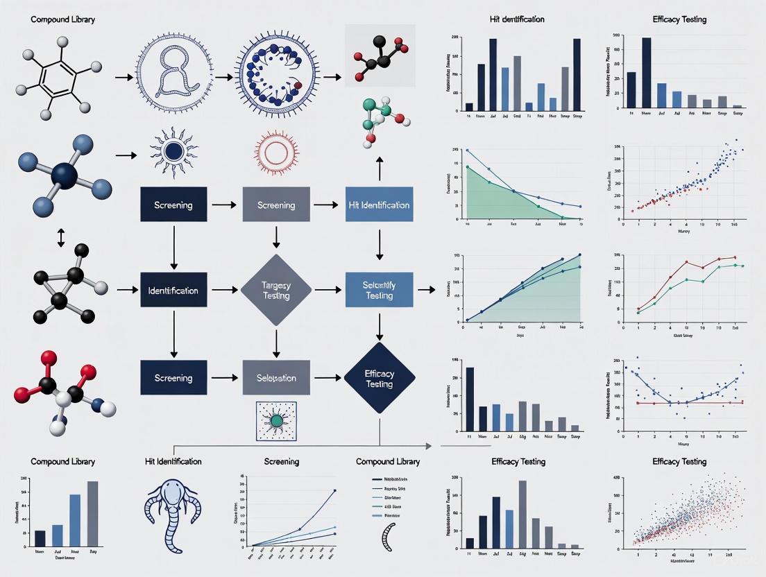
Diagram 1: HTS workflow for anthelmintic discovery.
Critical Experimental Protocols in Phenotypic Screening
The effectiveness of a phenotypic screen hinges on robust and standardized experimental protocols.
Protocol 1: High-Throughput Motility Screening with Infrared Interference
- Objective: To semi-automatically screen large chemical libraries for compounds that inhibit larval motility of parasitic nematodes.
- Parasite Material: Infective third-stage larvae (L3) of Haemonchus contortus are exsheathed (xL3) to initiate the assay [6].
- Assay Setup: xL3s are dispensed into 384-well plates at an optimized density of 80 larvae per well in an appropriate assay medium. Test compounds are added, typically at a starting concentration of 30 µM, with a DMSO control (e.g., 0.4%) and a positive control (e.g., monepantel) [6].
- Instrumentation and Data Acquisition: Plates are loaded into the WMicroTracker ONE instrument. Motility is quantified via infrared light beam-interference, using the "Threshold Average" algorithm (Mode 1), which provides a more quantitative and robust readout than alternative algorithms [6].
- Data Analysis: Activity counts (motility) are recorded over time. The quality of the screen is assessed using statistical parameters like the Z'-factor (a measure of assay robustness); a Z' > 0.5 is acceptable, with values > 0.7 considered excellent [6].
Protocol 2: Comparative Screening Across Nematode Species and Life Stages
- Objective: To evaluate the anthelmintic potential of compounds and validate screening models by comparing their activity across parasitic and free-living nematodes.
- Parasite and Model Material: This protocol utilizes adult Ancylostoma ceylanicum (harvested from infected hamsters), A. ceylanicum egg-to-larval (E2L) stages, and C. elegans L4/adult and E2A stages [2].
- Assay Setup: All life stages are incubated in 96-well or 384-well plates with test compounds (e.g., at 10 µM and 30 µM). For larval development assays (E2L, E2A), eggs are plated with an E. coli food source and monitored for 3-7 days [2].
- Endpoint Measurement: For adults, scoring is based on motility and morphology. For development assays, scoring is based on the inhibition of development from egg to subsequent stages [2].
- Data Analysis: The number of actives from each model is compared to the adult A. ceylanicum standard to calculate the true positive rate (TPR) and false negative rate for each surrogate model [2].
The Scientist's Toolkit: Key Research Reagent Solutions
Successful implementation of automated phenotypic screening relies on a suite of critical reagents and tools.
Table 3: Essential Research Reagents for Anthelmintic Screening
| Reagent / Tool | Function in Screening | Example Use Case |
|---|---|---|
| Haemonchus contortus L3/xL3 larvae | Primary screening target; a economically important parasite that can be maintained in vitro and is highly relevant to livestock health. | Motility-based HTS in the WMicroTracker ONE system [6]. |
| Ancylostoma ceylanicum adults & larvae | A human-parasitic hookworm that infects laboratory hamsters; provides a physiologically relevant screening model for human STNs. | Comparative screening to validate surrogate models and assess efficacy in vivo [2]. |
| Caenorhabditis elegans | A free-living model nematode with extensive genetic tools; used for preliminary screening and fundamental biology studies. | Moderate-throughput egg-to-adult (E2A) development assays [2]. |
| WMicroTracker ONE | Instrument that uses infrared light beam-interference to automatically quantify nematode motility in 384-well plates. | High-throughput primary phenotypic screening of compound libraries [6]. |
| INVAPP/Paragon Software | An automated imaging platform and algorithm for quantifying motility and development of nematodes in plate-based assays. | High-content phenotypic screening of chemical libraries on C. elegans and parasitic nematodes [8]. |
| FDA/EMA Approved Drug Library | A curated library of drugs with known safety and bioavailability profiles; screening it can facilitate drug repurposing. | Identification of existing drugs with previously unknown anthelmintic activity (e.g., sulconazole, pararosaniline) [2]. |
The global burden of parasitic nematodes on human and livestock health remains unacceptably high, and the threat of anthelmintic resistance is a ticking time bomb. Automated phenotypic screening platforms represent a paradigm shift in anthelmintic discovery, moving the field beyond reliance on a handful of drug classes. The ongoing development of more sophisticated, high-content, and high-throughput assays—such as those utilizing infrared interference and advanced image analysis—is critical for efficiently mining vast chemical spaces for novel actives. Future success will depend on continued innovation in screening technologies, a focus on physiologically relevant parasitic stages and species, and the integration of these phenotypic approaches with mechanistic studies. The ultimate goal is to build a robust pipeline of next-generation anthelmintics, ensuring long-term control over these pervasive and damaging parasites.
Anthelmintic resistance (AR) poses a critical and growing threat to global food security and animal health. AR is defined as a heritable loss of sensitivity to an anthelmintic in a parasite population that was previously susceptible to the same drug [9]. The escalating prevalence of AR in parasitic helminths (worms) of livestock jeopardizes the health and productivity of animals essential for human sustenance, leading to substantial economic losses estimated in the tens of billions of dollars annually [10]. The problem is global, affecting virtually all livestock species and all major classes of anthelmintic drugs across multiple continents [9]. The time from the introduction of a new anthelmintic to the emergence of resistance has, in some cases, been less than a decade, highlighting the rapid adaptive capacity of these parasites and the urgency of the situation [9]. This whitepaper details the current state of AR, its underlying mechanisms, and the pivotal role of advanced phenotypic screening technologies in the discovery of novel anthelmintics to safeguard food production and animal health.
Current Status and Contributing Factors
The development of anthelmintic resistance is evident in different helminths of almost every animal species and to different drug classes globally [9]. The three primary classes of anthelmintics most commonly used in ruminants—benzimidazoles (BZs), macrocyclic lactones (MLs), and imidazothiazoles/tetrahydropyrimidines (e.g., levamisole)—are all affected. Furthermore, resistance has even been reported to the more recent aminoacetonitrile derivative class (e.g., monepantel) [9] [11]. The situation is exacerbated by the widespread emergence of multiple drug resistance, where parasite populations simultaneously resist multiple drug classes, as documented in populations of Haemonchus contortus and other nematodes across Europe and Africa [9].
The factors accelerating the development of AR are multifaceted, involving a complex interplay of drug usage practices, parasite genetics, and farm management. Key contributing factors identified in recent studies are summarized in the table below.
Table 1: Key Factors Contributing to Anthelmintic Resistance and Associated Evidence
| Contributing Factor | Reported Impact / Evidence | Reference(s) |
|---|---|---|
| Frequent Treatment & Prophylactic Use | Increased likelihood of perceived resistance (OR=173.7); more frequent treatment gives resistant parasites a reproductive advantage. | [9] [12] |
| Underdosing | Visual weight estimation leads to underdosing, allowing survival of heterozygous resistant worms. | [9] |
| Combination Anthelmintic Use | Perceived as a significant risk factor (OR > 49.3), potentially due to improper use rather than the principle itself. | [12] |
| Lack of Veterinary Consultation | Farmers' ability to purchase anthelmintics without prescription increases risk of inappropriate treatment. | [12] |
| High Genetic Diversity of Parasites | Pre-existing resistant alleles in parasite populations are selected for under drug pressure. | [9] [11] |
| Shared Pastures & Animal Movement | Facilitates the spread of resistant parasites between flocks and herds, including cross-species transmission. | [12] |
Molecular Mechanisms of Resistance
Understanding the genetic and molecular basis of AR is crucial for developing diagnostic tools and informing new drug discovery. Resistance mechanisms vary by drug class and often involve multiple pathways acting in concert. The primary mechanisms include target-site mutations, enhanced drug efflux, and changes in drug metabolism [9]. Recent advances in genomics and transcriptomics have unveiled novel resistance genes that were previously obscured by unrelated genetic variation [11].
Table 2: Molecular Mechanisms of Anthelmintic Resistance in Haemonchus contortus
| Drug Class | Primary Target | Key Resistance Mechanisms | Specific Genetic Alterations / Genes Involved |
|---|---|---|---|
| Benzimidazoles (BZ) | β-tubulin | Target-site mutation | Single-nucleotide polymorphisms (SNPs) in the β-tubulin gene (e.g., F200Y). |
| Macrocyclic Lactones (ML) | Glutamate-gated chloride channels (GluCls) | Target-site changes, enhanced efflux, transcriptional regulation | Changes in expression of ligand-gated chloride channels (LGCC) and P-glycoproteins (P-gp); implication of transcription factor cky-1. |
| Levamisole (LEV) | Nicotinic acetylcholine receptors (nAChR) | Target-site mutation | Polymorphisms in nAChR subunit genes (e.g., S168T in hco-acr-8). |
| Monepantel (AD) | Nicotinic acetylcholine receptors (nAChR) | Target-site mutation | Polymorphisms in genes related to nAChR (e.g., Hco-mptl-1). |
The following diagram illustrates the conceptual workflow for identifying these resistance mechanisms, integrating classical and modern genomic approaches.
Detection and Diagnostic Methods
Accurate and timely detection of AR is essential for effective parasite management. Diagnostic methods can be broadly categorized into in vivo and in vitro techniques, with molecular tools providing increasingly high-resolution insights.
Standard Phenotypic Tests
- Fecal Egg Count Reduction Test (FECRT): This is the gold standard in vivo test. It involves comparing quantitative fecal egg counts from a group of animals before and after anthelmintic treatment. A reduction percentage below a specific threshold (e.g., 95% for certain drugs) indicates resistance [9].
- Egg Hatch Assay (EHA): An in vitro test primarily for BZ resistance. Parasite eggs are incubated in various concentrations of a drug (e.g., thiabendazole), and the concentration required to prevent 50% of eggs from hatching is determined. A higher EC50 indicates resistance [9] [11].
- Larval Development Test (LDT): This test assesses the ability of larvae to develop to the third stage in the presence of anthelmintics. It can be used for several drug classes and provides a quantitative measure of resistance [9].
- Larval Motility/Migration Tests: These assays evaluate the effects of drugs on larval motility or their ability to migrate through a sieve, which is particularly useful for screening MLs and LEV [9] [10].
Molecular and Genomic Techniques
Advanced molecular methods are revolutionizing AR detection by identifying specific genetic markers.
- Allele-Specific PCR (AS-PCR): Used to detect known point mutations, such as the F200Y SNP in the β-tubulin gene for BZ resistance [11].
- Deep Amplicon Sequencing: A powerful high-throughput method that allows for the sensitive detection and quantification of multiple low-frequency resistance alleles within a parasite population [11].
- Real-time PCR (qPCR): Employed to detect and genotype specific resistance SNPs, as used in the first report of the F200Y mutation in Bosnia and Herzegovina [12].
- Whole-Genome Sequencing and RNA-Seq: These untargeted approaches, often following genetic crosses of resistant and susceptible parasite strains, have been instrumental in discovering novel resistance loci and genes, such as cky-1 in ivermectin resistance [13] [11].
Automated Phenotypic Screening: A Pathway to Novel Anthelmintics
The diminishing efficacy of existing anthelmintics necessitates a accelerated pipeline for discovering new compounds. Automated phenotypic screening represents a powerful, unbiased strategy to identify novel anthelmintic chemotypes, even before their molecular targets are known.
Core Screening Platforms and Workflow
Modern phenotypic screening leverages miniaturization, automated microscopy, and sophisticated software to analyze the effects of thousands of small molecules on worm behavior, development, and viability. A key advancement is the development of user-friendly software like wrmXpress, which now includes a graphical user interface (GUI) to democratize access to these analytical pipelines [14]. The screening process generates rich, image-based data on phenotypes such as motility, which can be quantified into metrics like the "Wiggle Index" [10].
The following diagram outlines a streamlined workflow that integrates automated phenotypic screening with subsequent mechanistic investigation, forming a closed-loop discovery and validation system.
In Silico Prioritization and Machine Learning
To enhance the efficiency of phenotypic screening, machine learning (ML) models are now being deployed for in silico prediction of anthelmintic activity. One recent study trained a multi-layer perceptron classifier on a large dataset of bioactivity data for H. contortus. This model achieved 83% precision and 81% recall for identifying 'active' compounds and was used to screen over 14 million compounds from the ZINC15 database in silico [10]. Experimental assessment of just 10 selected candidates revealed two with significant potency, demonstrating a remarkable enrichment over random screening [10]. This approach exemplifies how computational methods can focus phenotypic screening efforts on the most promising candidates, drastically reducing time and resource requirements.
Mechanism of Action (MoA) Determination
A historical barrier to phenotypic screening has been the challenge of identifying a compound's MoA. Modern methods have made this increasingly tractable:
- Affinity-Based Methods: Using biotinylated or photo-crosslinkable analogs of the hit compound to pull down and identify direct protein targets from worm lysates via mass spectrometry or Western blot [15].
- Gene Expression Profiling: Comparing the transcriptomic signatures of drug-treated and untreated parasites can reveal modulated pathways and suggest the MoA [15].
- Resistance Selection: Applying low doses of the compound to parasites in vitro to select for resistant lines, followed by whole-genome sequencing to identify causal mutations that point to the drug target or resistance pathway [13] [15].
- Genetic Modifier Screening: Using techniques like CRISPR/Cas9 to validate potential targets identified through other methods [15].
The Researcher's Toolkit for Phenotypic Screening
Table 3: Essential Research Reagents and Platforms for Automated Phenotypic Screening
| Tool / Reagent | Function / Application | Specific Example / Note |
|---|---|---|
| wrmXpress Software | Integrated computational pipeline for analyzing image-based data of parasitic and free-living worms; quantifies motility, development, and other phenotypes. | Now features a GUI for point-and-click analysis, lowering the barrier to entry and enabling operation on personal computers [14]. |
| Open Scaffolds Collection | A diverse library of small-molecule compounds used for high-throughput phenotypic screening. | Used to train machine learning models; provided bioactivity data for 14,464 compounds [10]. |
| Pathogen Box | A collection of ~400 compounds with known or suspected activity against various pathogens. | Provides a valuable source of starting points for anthelmintic discovery and model training [10]. |
| ZINC15 Database | A public database containing millions of commercially available compounds for virtual screening. | Source of 14.2 million compounds for in silico screening using trained ML models [10]. |
| Multi-layer Perceptron (MLP) Classifier | A type of artificial neural network used for deep learning-based QSAR modeling. | Used to classify compounds as 'active', 'weakly active', or 'inactive' based on molecular descriptors and historical bioactivity data [10]. |
| High-Resolution Tracking Pipeline | A behavioral analysis tool for quantifying subtle drug-induced changes in worm movement. | Can demonstrate, for example, that praziquantel significantly affects multiple behavioral features of miracidia [14]. |
The threat of widespread anthelmintic resistance to global food security is clear and present. Combating this challenge requires a multi-pronged strategy that includes the prudent use of existing anthelmintics to slow the spread of resistance, coupled with the aggressive pursuit of new therapeutic entities. The integration of sophisticated phenotypic screening with machine learning-driven in silico prioritization and advanced MoA determination techniques constitutes a powerful, modernized pipeline for anthelmintic discovery. By leveraging these technologies, the research community can accelerate the identification and development of novel compounds with unique mechanisms of action, ultimately ensuring the sustainability of livestock production and the health of animals worldwide. The future of anthelmintic discovery lies in these interdisciplinary, data-driven approaches that can efficiently navigate the vast chemical space to find solutions to one of animal agriculture's most pressing problems.
Phenotypic Screening as a Primary Strategy for Anthelmintic Discovery
The discovery of novel anthelmintic compounds represents an urgent global health priority, driven by the significant burden of parasitic nematode infections and the escalating threat of drug resistance [16]. In response, phenotypic screening has re-emerged as a primary discovery strategy, enabling researchers to identify compounds that elicit measurable effects on whole organisms without presupposing molecular targets. This approach is particularly valuable for anthelmintic discovery where complex parasite biology often defies simple target-based strategies. The "biology-first" perspective of phenotypic screening allows for the identification of compounds that modulate physiologically relevant processes in the entire worm, increasing the probability of discovering mechanistically novel anti-parasities [17] [18]. Technological advancements in automated microscopy, high-content imaging, and computational analysis have transformed phenotypic screening from a low-throughput observational method to a quantitative, information-rich platform capable of capturing subtle, disease-relevant phenotypes at scale [17] [14].
Market and Technological Landscape
The cell-based assay market, which encompasses phenotypic screening technologies, is experiencing significant growth post-pandemic, with expanding applications in drug development and scientific research [19]. This growth is particularly relevant to anthelmintic discovery, where phenotypic screening platforms are becoming increasingly sophisticated. Several interconnected trends are fueling this expansion:
- Data Richness: Multiplexed assays and automated imaging now generate multi-dimensional phenotypic profiles [17].
- Scalability: New methods using pooled perturbations with computational deconvolution reduce sample size, labor, and costs while maintaining information-rich outputs [17].
- Computational Power: Artificial intelligence and machine learning models can interpret massive, noisy datasets to detect meaningful biological patterns [17].
The competitive landscape features numerous established and emerging players developing technologies for phenotypic analysis, including key companies providing instrumentation, reagents, and software solutions that support anthelmintic screening efforts [19].
Core Screening Platforms and Methodologies
Model Organisms and Assay Systems
Phenotypic screening for anthelmintics primarily utilizes whole-organism models that enable compound evaluation in physiologically relevant contexts:
Caenorhabditis elegans: The free-living nematode C. elegans serves as a powerful surrogate for parasitic helminths due to its genetic tractability, rapid life cycle, and physiological similarities to pathogenic species [16]. Its use has been validated through the identification of known anthelmintics and novel bioactives in screening campaigns [16].
Parasitic Helminths: Direct screening against pathogenic species such as Schistosoma mansoni provides the most clinically relevant data but presents challenges for high-throughput implementation due to complex life cycles and host dependencies [20] [14].
Quantitative Phenotypic Endpoints
Modern phenotypic screening moves beyond simple viability assessments to capture multifaceted phenotypic responses through quantitative metrics:
Table 1: Key Phenotypic Endpoints in Anthelmintic Screening
| Phenotypic Category | Specific Metrics | Measurement Technology |
|---|---|---|
| Motility | Movement units, thrashing rate, travel distance | Infrared beam interruption (WMicroTracker) [16], video tracking [14] |
| Morphology | Shape descriptors, size, texture | Automated image analysis [20] [14] |
| Development | Size, developmental stage, reproduction | Microscopy, image analysis [20] |
| Behavior | Complex movement patterns, response stimuli | High-resolution tracking pipelines [14] |
Experimental Workflow for Phenotypic Screening
The following diagram illustrates a standardized workflow for image-based phenotypic screening of anthelmintic compounds:
Detailed Experimental Protocol: C. elegans Motility Assay
Based on recently published methodology [16], the following protocol describes the optimization and execution of a phenotypic motility screen using C. elegans:
1. Worm Cultivation and Synchronization
- Maintain C. elegans (Bristol N2 strain) on nematode growth medium (NGM) plates seeded with E. coli OP50 as a food source.
- Synchronize populations to the L4 larval stage using standard bleaching protocols and age synchronization techniques.
- Detach L4 worms from agar plates and collect in M9 buffer.
- Centrifuge at 1,900 × g for 1 minute and wash in S medium to reduce bacterial concentration that might interfere with infrared detection.
2. Assay Optimization and Validation
- Worm Number Titration: Test various worm densities (30-200 L4 per well) to determine optimal signal-to-noise ratio while maintaining economical use of resources. 70 L4 per well provides sufficient motility units without compromising throughput [16].
- DMSO Tolerance: Evaluate solvent concentration (0.5-1.5% DMSO) across different assay volumes (100-200 µL). A final concentration of 1% DMSO in 100 µL volume provides optimal compound solubility without significant motility effects [16].
- Positive Controls: Include known anthelmintics (e.g., ivermectin, tolfenpyrad) as positive controls for motility inhibition.
3. Primary Screening Execution
- Spot 1 µL of compound solution in DMSO into clear, flat-bottomed 96-well polystyrene plates.
- For first-pass screens, use 40 µM compound concentration.
- Add approximately 70 synchronized L4 larvae in 100 µL S medium to each well.
- Include DMSO-only controls (1% final concentration) for normalization.
- Measure motility every 20 minutes for 24 hours using WMicroTracker ONE system, which detects movement via infrared light beam scattering at 880 nm.
- Maintain temperature at 25 ± 1°C throughout the assay.
- Normalize motility readings relative to DMSO controls.
- Define hits as compounds reducing motility to ≤25% of control levels.
4. Concentration-Response Analysis
- For confirmed hits, perform serial dilutions in DMSO (typically 0.005-100 µM range) using 96-well polypropylene dilution plates.
- Spot 1 µL aliquots into assay plates and test in triplicate.
- Measure motility as described above.
- Calculate half-maximal effective concentration (EC₅₀) using non-linear sigmoidal four-parameter logistic curve fitting in GraphPad Prism.
5. Counter-Screening for Cytotoxicity
- Assess compound cytotoxicity against mammalian cells (e.g., HEK293 cells) to determine selectivity indices.
- Culture HEK293 cells in DMEM with 10% FBS and 1% penicillin-streptomycin.
- Plate approximately 20,000 cells per well in clear-bottomed 96-well plates.
- Incubate with serially diluted compounds for 46 hours at 37°C with 5% CO₂.
- Add resazurin (0.5 mM final concentration) and incubate for additional 2 hours.
- Measure fluorescence (excitation 560 nm/emission 590 nm).
- Calculate half-maximal cytotoxic concentration (CC₅₀) using non-linear regression.
Data Analysis and Computational Methods
Time-Series Phenotypic Analysis
Advanced computational methods enable quantitative analysis of complex phenotypic responses over time. For helminth screening, time-series analysis provides particularly valuable insights:
Table 2: Computational Methods for Phenotypic Data Analysis
| Method Category | Specific Techniques | Application in Anthelmintic Screening |
|---|---|---|
| Time-Series Analysis | Dynamic time warping, similarity measures, clustering | Compare, cluster, and quantitatively reason about phenotypic responses over time [20] |
| Image Analysis | Shape, appearance, and motion-based phenotype quantification | Automatically monitor and quantify parasite phenotypes from biological images [20] |
| GUI-Based Tools | wrmXpress with graphical user interface | Lower barrier to entry for image-based phenotypic screening without command-line expertise [14] |
Phenotypic Data Clustering and Interpretation
The following diagram illustrates the computational pipeline for analyzing time-series phenotypic data:
Integration with Omics and AI Technologies
The future of phenotypic screening lies in its integration with multi-omics technologies and artificial intelligence, creating a powerful synergistic approach for anthelmintic discovery [17]. This integration addresses a key limitation of traditional phenotypic screening—the lack of immediate mechanism of action information—by adding molecular context to observed phenotypes.
Multi-Omics Integration Strategies:
- Transcriptomics: Reveals gene expression patterns associated with compound treatment, suggesting potential mechanisms of action.
- Proteomics: Clarifies signaling pathway alterations and post-translational modifications induced by bioactive compounds.
- Metabolomics: Contextualizes stress responses and metabolic adaptations to anthelmintic exposure.
- Epigenomics: Provides insights into regulatory modifications that may underlie phenotypic changes.
AI-Powered Platforms: Several AI platforms now specialize in integrating phenotypic data with other data modalities. For example, PhenAID combines cell morphology data, omics layers, and contextual metadata to identify phenotypic patterns correlating with mechanism of action, efficacy, or safety [17]. These platforms utilize high-content data from assays like Cell Painting, which visualizes multiple cellular components, and employ image analysis pipelines to detect subtle morphological changes that generate profiles for comparing biologically active compounds.
Essential Research Reagents and Tools
Table 3: Key Research Reagent Solutions for Anthelmintic Phenotypic Screening
| Reagent/Tool | Function | Example Application |
|---|---|---|
| WMicroTracker ONE | Detects nematode movement via infrared light beam scattering | Primary motility screening in C. elegans and parasitic helminths [16] |
| wrmXpress Software | Analyzes imaging data of parasitic and free-living worms with GUI | Image-based phenotypic screening without command-line expertise [14] |
| Medicines for Malaria Venture (MMV) Boxes | Open-source small molecule collections (COVID Box, Global Health Priority Box) | Source of chemically diverse compounds for anthelmintic screening [16] |
| C. elegans (Bristol N2) | Free-living nematode model for parasitic helminths | Surrogate organism for primary anthelmintic screening [16] |
| S Medium | Defined liquid culture medium for nematodes | Maintenance of C. elegans during compound exposure in liquid assays [16] |
Case Studies and Validation
Successful Implementation Examples
Recent screening campaigns demonstrate the practical application and success of phenotypic approaches for anthelmintic discovery:
MMV Box Screening Campaign:
- A screen of 400 compounds from MMV's COVID and Global Health Priority Boxes using a C. elegans motility assay identified twelve potent hits [16].
- Seven established macrocyclic lactone anthelmintics were correctly identified, validating the screening approach.
- Three novel bioactives (flufenerim, flucofuron, and indomethacin) with EC₅₀ values ranging from 0.211 to 23.174 µM were discovered.
- Counter-screening with HEK293 cells revealed varying selectivity indices, with CC₅₀ values ranging from 0.453 to >100 µM.
Schistosoma mansoni Behavioral Analysis:
- Implementation of a new high-resolution tracking pipeline in wrmXpress demonstrated that praziquantel significantly affects multiple behavioral features of miracidia [14].
- This approach enabled quantitative analysis of complex behavioral phenotypes beyond simple viability assessments.
Quantitative Results from Recent Screens
Table 4: Efficacy Data from Phenotypic Anthelmintic Screens
| Compound/Category | EC₅₀ (µM) | Motility Inhibition | Assay System |
|---|---|---|---|
| Flufenerim | 0.211 | >75% | C. elegans motility [16] |
| Flucofuron | 23.174 | >75% | C. elegans motility [16] |
| Indomethacin | Range reported | >75% | C. elegans motility [16] |
| Macrocyclic Lactones | Compound-specific | >75% | C. elegans motility [16] |
| Tolfenpyrad | Previously reported | 99.74% | C. elegans motility [16] |
Challenges and Future Directions
Despite significant advancements, phenotypic screening for anthelmintics faces several persistent challenges that guide future development:
Technical and Practical Limitations:
- Data Heterogeneity: Different data formats, ontologies, and resolutions complicate integration across platforms and studies [17].
- Tool Accessibility: While tools like wrmXpress with GUI lower barriers to entry, advanced computational methods still require specialized expertise [14].
- Model Limitations: C. elegans, while convenient, may not fully recapitulate all aspects of parasitic helminth biology, necessitating validation in pathogenic species.
Emerging Solutions and Future Trends:
- FAIR Data Standards: Implementation of Findable, Accessible, Interoperable, and Reusable data principles to address data heterogeneity issues [17].
- Open Biobank Initiatives: Increased sharing of compound libraries and screening data to accelerate discovery efforts.
- User-Friendly ML Toolkits: Development of accessible machine learning platforms that enable researchers without computational backgrounds to leverage advanced analytics.
- Multi-Modal Data Integration: Combining phenotypic data with genomics, transcriptomics, and proteomics to build comprehensive models of drug action [17].
The integration of phenotypic screening with omics technologies and artificial intelligence represents a paradigm shift in anthelmintic discovery [17]. This approach moves beyond traditional target-based screening to embrace biological complexity, enabling the identification of novel therapeutic starting points against devastating helminth infections that affect billions globally. As these technologies become more accessible and standardized, phenotypic screening will continue to evolve as a primary strategy for expanding the limited anthelmintic arsenal.
Parasitic nematodes infect hundreds of millions of people and livestock worldwide, causing significant disease burden and economic losses [21]. The control of these parasites faces a substantial threat from widespread anthelmintic resistance, particularly in livestock parasites like Haemonchus contortus, where resistance has been reported to all major drug classes [6] [22]. This resistance crisis has created an urgent need for novel anthelmintic compounds with new mechanisms of action. Traditional drug screening methods using parasitic nematodes are costly, labor-intensive, and low-throughput, creating a major bottleneck in anthelmintic discovery pipelines [22]. In response to this challenge, automated phenotypic screening platforms have emerged as powerful tools for rapidly identifying new anthelmintic candidates. These platforms increasingly utilize a dual-model organism approach, leveraging the complementary strengths of the free-living model nematode Caenorhabditis elegans and the parasitic nematode H. contortus [8] [23]. This technical guide examines the experimental frameworks, validation methodologies, and practical implementation of this integrated approach for automated phenotypic screening in anthelmintic research.
Model System Rationale and Comparative Biology
Caenorhabditis elegans as a Discovery Engine
C. elegans provides an exceptionally valuable model for initial anthelmintic discovery due to its experimental tractability, low maintenance costs, and high genetic similarity to parasitic nematodes [23]. As a free-living nematode belonging to clade V, C. elegans is a close relative to major gastrointestinal parasitic nematodes of humans and livestock, including H. contortus and Cooperia species [22] [23]. This phylogenetic relationship underpins its utility for anthelmintic discovery, as evidenced by research demonstrating that molecules lethal to C. elegans are more than 15 times more likely to kill parasitic nematodes compared to randomly selected compounds [22]. The majority of marketed anthelmintics show activity against C. elegans, and the model has been instrumental in elucidating drug mechanisms of action through forward genetic screens for resistant mutants [22].
Haemonchus contortus as a Parasitology Model
H. contortus, known as the barber's pole worm, represents a highly relevant parasitic model organism due to its significant economic impact on ruminant livestock, experimental tractability, and status as the most widely used parasitic nematode in drug discovery and anthelmintic resistance research [6] [24]. This blood-feeding nematode resides in the abomasum of ruminants, causing anemia, production losses, and mortality in severe infections [6]. Its direct lifecycle, high fecundity, and ability to be maintained in laboratory settings make it suitable for in vitro screening approaches [24]. The completion of its genome sequence has further elevated its status as a model organism, enabling comparative genomics and facilitating target identification [25] [24].
Table 1: Comparative Analysis of Nematode Model Organisms
| Characteristic | Caenorhabditis elegans | Haemonchus contortus |
|---|---|---|
| Organism Type | Free-living nematode | Parasitic nematode (ruminants) |
| Phylogenetic Clade | Clade V | Clade V (Strongylida) |
| Life Cycle Duration | ~3 days (25°C) | ~3 weeks (in host) |
| Maintenance Cost | Low (bacterial feed) | High (requires host) |
| Genetic Resources | Extensive (WormBase) | Draft genome & transcriptome [24] |
| Throughput Potential | Very high | Medium to high |
| Key Screening Advantage | Unlimited biomass, genetics | Direct parasite relevance |
| Primary Screening Role | Initial discovery, target ID | Validation, parasite specificity |
Automated Phenotypic Screening Platforms
Core Screening Technologies and Instruments
Automated phenotypic screening for anthelmintic discovery employs various technological approaches to quantify compound effects on worm motility, development, and viability:
Infrared Light Interference Systems: Instruments like WMicroTracker ONE utilize arrays of infrared light microbeams to detect motility through beam interruption counts. This system provides a quantitative readout of motility events per unit time (e.g., counts per 5 minutes) and can be configured in 96-well or 384-well formats [6] [23]. The system offers different acquisition algorithms, with Mode 1 (Threshold Average) providing superior performance for H. contortus xL3s with Z'-factors of 0.76 and signal-to-background ratios of 16.0 [6].
INVertebrate Automated Phenotyping Platform (INVAPP): This integrated system combines automated imaging with the Paragon algorithm for quantifying motility and development effects on parasitic worms. The platform has been validated for screening against C. elegans, H. contortus, Teladorsagia circumcincta, and Trichuris muris [8].
Image-Based Motility Assessment: Some platforms utilize video capture and digital segmentation of objects from the background to quantify motility characteristics. While potentially powerful, this approach can be challenging for parasitic worms due to movement characteristics and tendency to clump [6].
Experimental Protocols for High-Throughput Screening
Primary Screening Protocol Using WMicroTracker (C. elegans)
- Worm Preparation: Synchronize C. elegans at L4 larval stage using standard methods [23]. Wash worms three times with K saline (NaCl 51 mM, KCl 32 mM) by centrifugation at 1,000 × g.
- Plate Setup: Aliquot approximately 80 worms per well in 96-well microtiter plates using 60 µl of K saline containing 0.015% BSA [23].
- Compound Application: Add test compounds dissolved in DMSO (final concentration 0.5%) using a 6 × 6 matrix array for combination studies or single concentration for primary screens.
- Motility Measurement: Transfer plates to WMicroTracker instrument and record motility counts at 5-minute intervals for defined periods (typically 4-24 hours) [23].
- Data Analysis: Calculate percentage inhibition relative to negative controls (DMSO only) and positive controls (known anthelmintics).
Secondary Validation Protocol (H. contortus)
- Larval Production: Maintain H. contortus (Haecon-5 strain) in experimental sheep following approved ethical guidelines [21]. Collect feces from infected animals with patent infections and incubate at 27°C with >90% humidity for 1 week to yield L3 larvae.
- Larval Processing: Clean L3s by migration through nylon mesh (20 µm pore size) and store at 11°C for up to 6 months [21]. Artificially exsheath xL3s using 0.15% sodium hypochlorite for 20 minutes at 38°C.
- Assay Setup: Dispense 200-300 xL3s per well in 384-well plates containing LB* medium (lysogeny broth with antibiotics) [6]. Add hit compounds identified from C. elegans screening.
- Motility Measurement: Incubate plates for 90 hours at appropriate temperature, then measure motility using WMicroTracker with Mode 1 acquisition algorithm.
- Developmental Assessment: For extended assays, monitor larval development to L4 stage following motility measurements.
Table 2: Quantitative Performance Metrics of Phenotypic Screening Assays
| Screening Parameter | C. elegans Motility Assay | H. contortus xL3 Motility Assay | H. contortus Development Assay |
|---|---|---|---|
| Throughput Capacity | ~10,000 compounds/week [6] | ~10,000 compounds/week [6] | Lower throughput |
| Assay Format | 96-well or 384-well | 384-well optimized | 96-well or 384-well |
| Optimal Worm Density | ~80 worms/well [23] | 80-100 xL3s/well [6] | Varies by endpoint |
| Key Readout | Motility counts (infrared) | Motility counts (infrared) | Developmental stage |
| Z'-Factor | >0.5 (typically 0.6-0.8) | 0.76 [6] | Protocol dependent |
| Signal-to-Background | Variable | 16.0 [6] | Protocol dependent |
| Incubation Duration | 4-24 hours | Up to 90 hours | 7-10 days |
Screening Workflows and Experimental Design
Integrated Screening Cascade
The most effective approach for anthelmintic discovery combines the strengths of both model organisms in a sequential screening cascade. This begins with high-throughput primary screening against C. elegans, followed by secondary validation against parasitic nematodes such as H. contortus, and finally counter-screening against vertebrate models to exclude generally cytotoxic compounds [22].
Integrated Screening Cascade for Anthelmintic Discovery
Case Study: Successful Implementation
Burns et al. (2015) demonstrated the power of this approach by screening 67,012 compounds against C. elegans, identifying 275 "wactives" (worm-active compounds) that killed the nematode [22]. Subsequent screening against the parasitic nematodes Cooperia onchophora and H. contortus revealed that 103 compounds killed all three nematode species, while counter-screening in zebrafish and HEK293 cells identified 67 compounds with selective nematode toxicity [22]. Structural analysis organized these into 30 structurally distinct anthelmintic lead classes potentially targeting different molecular pathways.
The Scientist's Toolkit: Essential Research Reagents
Table 3: Key Research Reagent Solutions for Automated Phenotypic Screening
| Reagent/Resource | Specifications | Application and Function |
|---|---|---|
| Nematode Strains | C. elegans Bristol N2 (wild-type); H. contortus Haecon-5 strain or MHco3(ISE).N1 (inbred, drug-susceptible) | Primary screening and validation; genetically defined backgrounds reduce variability [23] [21] |
| Culture Media | NGM agar with E. coli OP50 for C. elegans; LB* (lysogeny broth with penicillin, streptomycin, amphotericin B) for H. contortus | Maintenance and assay conditions; antibiotics prevent microbial contamination [23] [21] |
| Automated Phenotyping System | WMicroTracker ONE (PhylumTech) or INVAPP with Paragon algorithm | High-throughput motility quantification via infrared interference or image analysis [6] [8] |
| Compound Libraries | MMV Global Health Priority Box, Pathogen Box, NPC, MIPE collections | Source of chemically diverse compounds with known bioactivity and safety profiles [26] [21] |
| Detection Reagents | Sodium hypochlorite (0.15% for exsheathment), DMSO (vehicle control) | Preparation of parasitic larvae; compound solubilization [21] |
| Bioinformatic Resources | WormBase (C. elegans), H. contortus genome assembly and annotation | Target identification, orthology mapping, and pathway analysis [25] [24] |
Data Analysis and Hit Validation
Quantitative Analysis Methods
Concentration-response curves (CRCs) are derived directly from primary screening data using quantitative high-throughput screening (qHTS) approaches where compounds are tested at multiple concentrations [26]. Activity criteria typically include high-quality dose-response curves, IC50 values ≤ 10 μM, and maximal response ≥ 65% compared to controls [26]. For anthelmintic screening, Z'-factors > 0.5 indicate robust assay performance, with optimized H. contortus motility assays achieving Z'-factors of 0.76 [6].
For drug combination studies, such as investigations of ivermectin and eprinomectin interactions, complete 6 × 6 concentration matrices are analyzed using multiple models including Highest Single Agent (HSA), Loewe additivity, Bliss independence, and Zero Interaction Potency (ZIP) to distinguish additive, synergistic, or antagonistic effects [23].
Hit Progression Criteria
The transition from screening hits to validated leads requires multiple layers of assessment:
- Efficacy Progression: Compounds must show reproducible activity in concentration-dependent manner against both C. elegans and H. contortus.
- Selectivity Filtering: Candidates should exhibit minimal toxicity against vertebrate models (HEK293 cells and zebrafish) with selectivity indices >10-fold preferred [22].
- Chemical Tractability: Hits with structural analogs showing similar bioactivity indicate potential for medicinal chemistry optimization [22].
- Parasite Stage Breadth: Ideal candidates show activity against multiple life stages (L3, L4, adult) of parasitic nematodes [21].
The integrated use of C. elegans and H. contortus in automated phenotypic screening represents a powerful strategy for addressing the critical need for novel anthelmintics. This approach leverages the high-throughput capacity and genetic tractability of C. elegans while maintaining physiological relevance through secondary validation in a parasitic system. As resistance to existing anthelmintics continues to spread, these automated screening platforms will play an increasingly vital role in replenishing the anthelmintic development pipeline. Future advancements in single-cell sequencing, machine learning, and automated image analysis will further enhance the efficiency and predictive power of these screening platforms, accelerating the discovery of next-generation anthelmintic therapeutics [25].
High-Throughput Workflows: From Motility Assays to AI-Powered Screening
The escalating challenge of anthelmintic resistance in parasitic nematodes necessitates a paradigm shift in drug discovery strategies, placing a premium on high-throughput phenotypic screening technologies [10]. Infrared-based motility assays represent a critical technological advancement in this field, automating the labor-intensive and time-consuming process of visually assessing nematode viability and behavior [27]. These assays provide the foundational phenotypic data required to identify novel chemotypes with nematocidal or nematostatic activity. By offering a quantitative and objective measure of parasite motility, instruments like the WMicrotracker platform accelerate the evaluation of compound libraries, thereby streamlining the early-stage pipeline for anthelmintic development [27] [10]. This technical guide details the principles, workflows, and applications of these assays within the context of automated phenotypic screening for novel anthelmintics.
Core Principles of Operation
Infrared-based motility assays function on the principle of detecting movement through the interaction of an infrared (IR) light source with the test organisms. The specific mechanism varies depending on the technology generation and configuration, but the underlying goal is to convert organism movement into quantifiable, electronic data.
The WMicrotracker ONE system utilizes a well-established method based on infrared light scattering. The device is equipped with an infrared microbeam grid that passes through the wells of a standard microtiter plate (e.g., 96-well format) [27]. When motile nematodes or other small organisms move within a well, they disrupt and scatter this IR light. A detector on the opposite side of the plate captures these interruptions. The instrument's software then quantifies these light-scattering events as "activity counts" over user-defined time intervals, providing a direct and real-time correlate of population motility [27]. This method is non-invasive and uses very low-power IR radiation, ensuring that the animals' natural behavior is not affected.
In contrast, the newer WMicrotracker SMART platform offers a dual-mode functionality, incorporating advanced imaging capabilities [28].
- Infrared Imaging Mode (for solid media): In this configuration, the system acquires a sequence of infrared images (typically one per second) of organisms cultured on solid medium in a 35mm Petri dish, which is placed upside down [28]. The technique leverages an optical phenomenon termed "Silhouette Amplification by Infrared Refraction." As infrared light waves encounter the worm-agar interface, they refract, generating an amplified image of the worm that is captured by a sensitive HD camera. Subsequent digital image processing enables the tracking of multiple worm paths simultaneously [28].
- Infrared Microbeam Grid Mode (for various media): This mode functions similarly to the WMicrotracker ONE, detecting movement through the diffraction of a grid of IR microbeams. It is suitable for solid, liquid, or air cultures and can define activity areas for chemotaxis experiments [28].
The following diagram illustrates the core decision logic for selecting and applying these technologies in a research workflow.
Detailed Experimental Workflows
The application of WMicrotracker systems in anthelmintic research involves standardized yet flexible protocols. Below are detailed methodologies for key assays, adapted from published research on plant-parasitic and parasitic nematodes [27].
Workflow for Motility Inhibition Assays
This protocol is designed for high-throughput screening of chemical compounds against motile nematode juveniles (J2) or adults in a liquid environment [27].
- Nematode Preparation: Collect motile stages of the nematode (e.g., H. schachtii J2 or D. destructor mixed stages) and concentrate them in sterile distilled water. Determine the concentration by counting the number of living nematodes in multiple 10 µL drops and adjust the suspension to the desired final concentration [27].
- Plate Loading: Dispense 54 µL of the nematode suspension into each well of a U-bottom 96-well plate. It is critical to include replicate wells for each test condition and controls.
- Pre-incubation: Seal the plate with a breathable seal or parafilm and allow it to incubate at the assay temperature (e.g., 20°C) for 20-30 minutes. This step allows the nematodes to settle at the bottom of the wells [27].
- Baseline Motility Measurement: Place the microtiter plate into the WMicrotracker ONE device and record the motility activity counts for a set period (e.g., 30 minutes). This establishes a baseline motility level for the population before treatment [27].
- Compound Addition: Add 6 µL of the test compound (prepared at a 10x concentration) to the designated wells. For controls, add 6 µL of sterile water (negative control) or a known motility-inhibiting substance like sodium azide (positive control) [27].
- Post-Treatment Motility Measurement: Return the plate to the WMicrotracker device and measure motility at various time points post-exposure (e.g., 2h, 6h, 24h). Between measurements, the plates should be sealed and maintained at the assay temperature with gentle shaking on an orbital shaker (150 rpm) to ensure adequate oxygen exchange [27].
- Data Analysis: Motility inhibition is calculated by comparing the post-treatment activity counts to the baseline and negative control counts. A significant reduction in activity indicates a nematicidal or nematostatic effect.
Workflow for Hatching Assay Using Cyst Crushing
This protocol assesses the effect of compounds on the hatching of nematode eggs, another critical life-stage target for anthelmintics [27].
- Cyst Processing: Collect approximately 300 mature cysts of H. schachtii and place them in a glass bottle with 3-5 mL of sterile distilled water or a hatching stimulant like 3 mM ZnCl₂.
- Egg Liberation: Add a medium-sized stirring bar to the bottle and crush the cysts on a magnetic stirrer at 1000 rpm for 5 minutes. This releases the eggs contained within the cysts.
- Egg Purification: Pass the suspension through a series of sieves to purify the eggs. A 30 μm mesh removes small debris, while a 116 μm mesh retains larger debris, allowing an enriched egg suspension (though not entirely free of J2 and mid-sized debris) to be collected [27].
- Plate Setup and Measurement: Distribute the egg suspension into a 96-well plate. The WMicrotracker then monitors the emergence of J2 from the eggs over time by detecting the movement of the newly hatched juveniles. The addition of test compounds allows for the quantification of hatching inhibition [27].
The integrated workflow below summarizes the key steps from nematode preparation to data analysis for both motility and hatching assays.
Quantitative Data and Validation
Infrared-based assays generate robust, quantitative data that can be used for dose-response analysis and machine learning model training, as demonstrated in recent anthelmintic discovery efforts [10].
Table 1: Key Performance Metrics from WMicrotracker ONE Motility Assays
| Metric | Description | Typical Value/Example | Interpretation in Screening |
|---|---|---|---|
| Activity Counts | Number of IR beam interruptions per time bin (e.g., 30 mins) [27] | Raw data output | Direct correlate of population motility; a decrease indicates inhibition. |
| Motility Inhibition | % Reduction in activity vs. negative control | e.g., >80% reduction at 24h [10] | Indicates strong nematicidal/nematostatic effect. |
| EC₅₀ | Concentration causing 50% effect | e.g., <50 µM for "active" compounds [10] | Standard measure of compound potency. |
Table 2: Activity Classification for Anthelmintic Screening Based on Motility Data
| Activity Label | Wiggle Index [10] | Viability [10] | EC₅₀ [10] | Interpretation |
|---|---|---|---|---|
| Active | < 0.25 | < 20% | < 50 µM | Primary hit for further investigation. |
| Weakly Active | 0.25 - 0.5 | 20% - 50% | 50 - 100 µM | Possible candidate or requiring optimization. |
| Inactive (None) | ≥ 0.5 | ≥ 50% | ≥ 100 µM | Not of immediate interest. |
The validity of this approach is underscored by its integration with machine learning. One study used WMicrotracker-like motility data from 15,000 compounds to train a multi-layer perceptron classifier, which achieved 83% precision and 81% recall in identifying "active" compounds [10]. This model successfully prioritized novel anthelmintic candidates from a database of over 14 million compounds, with experimental validation confirming significant inhibitory effects on Haemonchus contortus [10].
The Scientist's Toolkit: Essential Research Reagents and Materials
A successful screening campaign relies on a standardized set of biological and chemical reagents.
Table 3: Essential Research Reagent Solutions for Infrared Motility Assays
| Item | Function/Description | Example Application |
|---|---|---|
| U-bottom 96-well Plates | Optimal for nematode settlement and IR light passage in WMicrotracker ONE [27]. | Standard plate format for all liquid-based motility and hatching assays. |
| Model Nematode Species | Biologically relevant parasites used for screening. | Haemonchus contortus (barber's pole worm), Heterodera schachtii (beet cyst nematode) [27] [10]. |
| ZnCl₂ (3 mM) | Chemical stimulant of nematode hatching [27]. | Used in hatching assays to synchronize and increase J2 emergence from cysts. |
| Sodium Azide | Metabolic inhibitor used as a positive control for motility inhibition [27]. | Validates assay performance by confirming expected loss of motility. |
| Sterile Distilled Water | Vehicle and negative control substance. | Diluent for nematode suspensions and compound stocks; negative control for treatment. |
| 35mm Petri Dishes | Standard culture vessel for WMicrotracker SMART imaging mode [28]. | Used for detailed path tracking of nematodes on solid NGM media. |
Infrared-based motility assays, exemplified by the WMicrotracker platform, have firmly established themselves as a cornerstone technology in the modern anthelmintic discovery pipeline. By providing a high-throughput, quantitative, and objective measure of nematode phenotype, they directly address the bottleneck of labor-intensive manual assessment. The generated data not only facilitates the rapid screening of vast compound libraries but also provides the high-quality phenotypic information necessary to train sophisticated in silico models for the de novo prediction of anthelmintic candidates [10]. As the field moves forward, the integration of these automated phenotypic readouts with machine learning and cheminformatics represents the most promising pathway toward urgently needed novel anthelmintics, turning the tide against pervasive drug resistance.
High-Content, Image-Based Phenotypic Screening with Automated Analysis
High-content, image-based phenotypic screening represents a powerful paradigm in modern drug discovery, enabling the systematic and multiparametric analysis of cellular or whole-organism responses to chemical or genetic perturbations. This approach captures rich, high-dimensional data on morphology, subcellular architecture, and dynamic processes in an unbiased manner, revealing mechanisms and targets that hypothesis-driven methods might miss [29]. When framed within the context of anthelmintic research, this technology addresses a critical need: the discovery of novel therapeutic classes against parasitic worms through the identification of subtle, complex, or even cryptic phenotypes that traditional viability assays overlook [3]. The core of this methodology lies in the integration of high-throughput microscopy with advanced computational analysis, including deep learning, to convert visual information into quantifiable, biologically meaningful insights.
The resurgence of phenotypic drug discovery has been driven by its track record in delivering first-in-class medicines, often with novel mechanisms of action that target biologically relevant but previously undruggable cellular processes [30]. For anthelmintic development, this is particularly pertinent. Many existing anthelmintics, such as praziquantel and ivermectin, exhibit complex host-parasite interactions and may not display overt lethal phenotypes in simple in vitro cultures [3]. High-content screening (HCS) provides a framework to move beyond simplistic lethality endpoints, instead capturing nuanced phenotypic changes in motility, morphology, and subcellular structure that may better predict in vivo efficacy [31].
Technological Foundations: From Image Acquisition to Automated Analysis
The workflow for high-content, image-based screening is a multi-stage process, each component of which must be rigorously optimized to ensure the generation of high-quality, biologically relevant data.
Robust Assay Development and Image Acquisition
The foundation of any successful phenotypic screen is a robust and reproducible biological assay. Key considerations include selecting biologically relevant models—which for anthelmintic research may range from parasitic larval stages (e.g., schistosomula) to free-living model organisms—and optimizing seeding density, incubation conditions, and plate formats to minimize technical variability [29]. For image acquisition, consistency is paramount. Automated microscopy platforms must be configured with carefully tuned parameters, including appropriate exposure times to avoid saturation, correct autofocus offsets to ensure image clarity, and the capture of a sufficient number of images per well to achieve statistically robust sampling of the population [29]. The choice between label-free bright-field imaging and fluorescence multiplexing depends on the specific readout; for example, bright-field imaging enabled automated classification of schistosomula damage without the need for staining, while fluorescence reporters are essential for tracking specific proteins or organelles [32] [31].
The Rise of Deep Learning in Image Analysis
While traditional image analysis pipelines rely on a series of manual steps—segmenting cellular regions, extracting hundreds of hand-crafted features (e.g., cell area, staining intensity), and then selecting meaningful parameters for analysis—this process is labor-intensive, requires significant expertise, and can introduce bias [33]. Deep learning (DL), a subset of machine learning, has transformed this workflow. DL uses interconnected neural networks to perform tasks such as classification and prediction directly from raw image data [33].
In a typical DL application for HCS, labeled images are used to train neural networks that model phenotypes of interest. The algorithm learns to identify and classify cells or organisms without the need for prior segmentation or the creation of custom analysis workflows for each new assay [33]. This approach offers several key advantages:
- Increased Efficiency and Scalability: DL automates complex tasks, enabling the rapid and consistent analysis of thousands of images, which is essential for the large datasets generated in HCS [33].
- Identification of Complex Phenotypes: DL can extract spatiotemporal features and identify subtle or dynamic phenotypes that are difficult to detect using conventional methods, such as specific morphological patterns associated with ribosome biogenesis inhibition or distinct larval motility profiles [33] [32].
- Reduced Errors and Bias: By learning directly from annotated datasets, DL standardizes analysis, reducing variability and false positives/negatives associated with manual parameter tuning [33].
Table 1: Key Advantages of Deep Learning for High-Content Screening Analysis
| Advantage | Description | Impact on Anthelmintic Screening |
|---|---|---|
| End-to-End Automation | Eliminates need for manual segmentation and feature selection. | Enables rapid, uniform analysis of thousands of parasite images. |
| Handling of Complexity | Identifies subtle and multi-dimensional phenotypes. | Reveals cryptic parasite phenotypes beyond simple death/viability. |
| Scalability | Processes large datasets in a standardized, reproducible manner. | Makes large-scale compound library screens against parasites feasible. |
| Adaptability | Models can be updated with new data via incremental learning. | Allows continuous model refinement as new parasite phenotypes are discovered. |
Experimental Protocol: An Imaging-Based Pipeline for Ribosome Biogenesis Inhibitors
To illustrate a detailed methodology, we describe a pipeline established to identify chemical inhibitors of human ribosome biogenesis—a process relevant to cancer therapy—which showcases principles directly applicable to anthelmintic discovery [32].
Background and Objective
Ribosome biogenesis is a vulnerable pathway in cancer cells, but few chemical inhibitors acting on steps downstream of rRNA transcription are known. The objective was to create a sensitive, imaging-based screening pipeline capable of identifying compounds that impair ribosome synthesis in human cancer cells (HeLa cells), with the ability to distinguish direct effects from indirect ones like DNA damage [32].
Detailed Workflow and Readouts
The pipeline employed a multi-readout, single-cell imaging approach after a 6-hour compound treatment to capture rapid phenotypic changes [32].
- Cell Line and Culture: HeLa cells are used as a model human cancer cell line.
- Compound Treatment: Cells are treated with compounds from a library (e.g., >1000 FDA-approved drugs) for 6 hours.
- Fixation and Staining: Cells are fixed and subjected to immunofluorescence.
- Multi-Parameter Image Acquisition: High-content imaging is performed using the following key readouts:
- ENP1/BYSL Immunofluorescence (Nucleolar Integrity): The ribosome biogenesis factor ENP1 is enriched in nucleoli. Inhibition of RNA polymerase I activity (e.g., by CX-5461) causes nucleolar disintegration and dispersal of ENP1 throughout the nucleoplasm.
- ENP1/BYSL in Presence of Leptomycin B (Pre-40S Subunit Assembly): The nuclear export inhibitor Leptomycin B (LMB) causes ENP1 to relocate from nucleoli to the nucleoplasm. If a compound impairs early nucleolar assembly steps, ENP1 is retained in the nucleolus even when LMB is present.
- Fluorescent Ribosomal Protein Reporters (RPS2-YFP, RPL29-GFP): These reporters allow quantitative analysis of ribosomal subunit maturation and localization.
- Counter-Assays: To exclude common indirect effects, counter-assays for DNA damage and proteasome inhibition are established and run in parallel.
Automated Analysis and Hit Identification
Images are analyzed using high-content analysis software. Single-cell data is extracted for each readout, measuring features such as nucleolar intensity, nucleolar-to-nucleoplasmic ratio of ENP1, and fluorescence distribution of ribosomal reporters. The combination of readouts allows for the distinction between different mechanisms of action. For instance, a compound causing nucleolar disintegration (like CX-5461) and one causing nucleolar retention of ENP1 in the presence of LMB (like cycloheximide) can be clearly differentiated [32]. Hit compounds are identified based on their ability to induce these specific phenotypic profiles.
Table 2: Core Readouts for the Ribosome Biogenesis Screening Pipeline [32]
| Readout | Measurement | Phenotype of Inhibition | Biological Interpretation |
|---|---|---|---|
| ENP1 Localization | Nucleolar intensity and distribution of the ENP1 factor. | Loss of nucleolar signal; dispersal in nucleoplasm. | Inhibition of rRNA transcription; nucleolar disintegration. |
| ENP1 in LMB | Nucleolar retention of ENP1 upon blockade of nuclear export. | ENP1 remains trapped in nucleoli. | Defect in early, nucleolar steps of pre-40S ribosomal subunit assembly. |
| RPS2-YFP / RPL29-GFP | Localization and intensity of fluorescent ribosomal proteins. | Altered distribution or reduced signal. | Impairment of ribosomal subunit maturation and stability. |
Application in Anthelmintic Research: Identifying Subtle Phenotypes
The transition to high-content, image-based screening is particularly transformative for anthelmintic research, where many known drugs have subtle or complex in vitro phenotypes.
The Challenge of Conventional Anthelmintic Screens
Historically, most anthelmintics were discovered through in vivo screening in animal models of infection, an approach that captures host-parasite interactions and complex mechanisms of action but is low-throughput and resource-intensive [3]. A significant challenge for in vitro assays is that many established anthelmintics, including praziquantel, ivermectin, and diethylcarbamazine, require a host component (e.g., the immune system) for full efficacy or do not elicit an overt lethal phenotype in culture [3]. For example, praziquantel causes strong contraction of schistosomes in vitro, but this phenotype does not strictly correlate with its in vivo efficacy, as larval stages show the same contraction but are refractory to treatment [3]. This underscores the limitation of using single, simplistic phenotypic endpoints.
High-Content Solutions for Parasitic Worms
High-content, image-based screening directly addresses these challenges by enabling multiparametric analysis of parasite phenotypes.
- Label-Free, Image-Based Bayesian Classification for Schistosomula: A pioneering HCS for schistosomiasis drug discovery utilized bright-field imaging of schistosomula (larval stages) to capture drug-induced morphological changes in a label-free manner [31]. Automatic image analysis combined with Bayesian prediction models defined morphological damage, enabling hit/non-hit classification and detailed phenotype characterization. Motility was also quantified from time-lapse images. This system reliably detected over 99.8% of visually scored hits from a 10,000-compound library, demonstrating the power of automated analysis to enable large-scale screening against whole parasites [31].
- Phenotyping Beyond Viability: The true potential of HCS in anthelmintics lies in identifying "cryptic" phenotypes—sublethal changes in motility, development, reproduction, or specific tissue integrity that are nevertheless critical for the parasite's survival in vivo or its evasion of the host immune system [3]. Machine learning can be trained to recognize these complex phenotypic signatures, which may be more predictive of in vivo efficacy than simple death.
The Scientist's Toolkit: Essential Reagents and Solutions
The following table details key research reagents and tools central to implementing a high-content, image-based phenotypic screen, as derived from the cited methodologies.
Table 3: Research Reagent Solutions for High-Content Phenotypic Screening
| Tool / Reagent | Function in the Workflow | Example Application |
|---|---|---|
| Genedata Screener | An enterprise software platform that automates the entire HCI analysis pipeline, from image loading to result computation, using its High Content Extension and deep learning-based Imagence module [33]. | Streamlines and automates phenotypic imaging assay workflows for production-scale HCS in biopharma. |
| CLEMSite Software | A software prototype that automates correlative light and volume electron microscopy (CLEM), enabling the tracking and high-resolution volumetric imaging of multiple cells previously identified by light microscopy [34]. | Targets and automates FIB-SEM volume acquisition of specific cells (e.g., based on Golgi morphology) for ultrastructural analysis. |
| Ardigen phenAID | An AI-powered platform leveraging computer vision and deep learning to extract high-dimensional features from high-content screening images and predict compound mode of action [29]. | Integrates phenotypic profiles with other data (e.g., omics) to enhance hit identification and lead optimization. |
| CellPainting Protocol | A multiplexed fluorescence imaging assay that uses up to 6 dyes to reveal a broad spectrum of cellular components, generating rich morphological data [35]. | Used to create massive public image datasets (e.g., JUMP-CP) for training universal representation models for HCS data. |
| Fluorescent Ribosomal Reporters (e.g., RPS2-YFP, RPL29-GFP) | Genetically encoded fluorescent tags fused to ribosomal proteins to visualize and quantify ribosomal subunit maturation, localization, and abundance in live or fixed cells [32]. | Serves as a key readout in screens for ribosome biogenesis inhibitors in human cell lines. |
Advanced Computational Approaches and Data Management
The data generated in high-content screening is vast and complex, necessitating robust computational strategies and careful data management.
Self-Supervised Learning and Representation
Beyond supervised deep learning, self-supervised learning approaches are being explored to create universal representation models for HCS data. For example, models trained on massive public datasets like the JUMP-CP Cell Painting dataset can learn powerful feature representations without exhaustive manual labeling. Research shows that such self-supervised approaches can provide representations that are more robust to batch effects while achieving performance on par with standard supervised methods, offering valuable recommendations for training strategies [35].
Data Quality and FAIR Principles
The quality of input data is paramount, as AI models amplify both biological signals and technical noise. A rigorous quality control checklist must be applied before analysis, verifying image clarity, accurate cell segmentation, separation of controls in feature space, and the absence of strong plate or batch effects [29]. Furthermore, adhering to the FAIR (Findable, Accessible, Interoperable, Reusable) principles from the outset is critical for reproducibility and scalability. This involves:
- Storing data in interoperable formats.
- Using controlled vocabularies.
- Ensuring unique and traceable identifiers for all data and metadata [29].
Structured, machine-readable metadata is non-negotiable for AI-driven analysis. This metadata must include unique plate/well IDs, perturbation information (e.g., SMILES for compounds), experimental conditions (cell line, passage, dose), and detailed imaging parameters [29].
High-content, image-based phenotypic screening, powered by automated and deep learning analysis, represents a mature and powerful platform for modern drug discovery. By capturing complex biological reality in a multi-parametric and unbiased fashion, it excels at identifying novel mechanisms and expanding druggable target space. Within the specific field of anthelmintic research, this approach provides a long-needed pathway to overcome the limitations of traditional in vitro assays. It enables the systematic discovery of compounds that induce subtle yet therapeutically critical phenotypes in parasites, thereby offering a robust, scalable, and biologically relevant engine for identifying the next generation of much-needed anti-parasitic agents.
The discovery of novel anthelmintic compounds is urgently needed to address widespread drug resistance in parasitic nematodes, which poses a major threat to global health and food security. Open-science compound libraries represent a transformative approach to accelerating this discovery process by providing researchers with freely accessible, chemically diverse starting points for drug development. Among the most prominent resources are the Pathogen Box and COVID Box developed by Medicines for Malaria Venture (MMV), which contain carefully selected compounds with demonstrated activity against various pathogens [36]. These collections provide a unique opportunity to identify new anthelmintic leads through phenotypic screening, bypassing the traditional bottlenecks of proprietary compound acquisition.
The integration of these libraries into automated phenotypic screening platforms creates a powerful synergy for anthelmintic discovery. Phenotypic screening maintains the crucial advantage of assessing compound effects within the complex biological context of whole organisms, preserving physiological relevance that target-based approaches may miss [37]. When combined with the chemical diversity of open-access libraries and the reproducibility of automation, researchers can effectively navigate the vast chemical space to identify novel chemotypes with efficacy against resistant nematode strains. This approach has already yielded promising results, with the Pathogen Box contributing to machine learning models that successfully predicted new anthelmintic candidates [10].
The MMV Open-Access Library Platform
Library Composition and Design Principles
The MMV open-access libraries represent a strategic collection of drug-like molecules selected through a rigorous evaluation process. The Pathogen Box contains 400 compounds with documented activity against various pathogens, while the COVID Box and related Pandemic Response Box were developed specifically to address emerging viral threats [36] [38]. These libraries collectively comprise nearly 1,400 compounds, each characterized by confirmed chemical identity, purity, and prior evidence of biological activity [39]. The libraries are designed to facilitate drug repurposing and new lead identification by providing a curated set of compounds with optimized drug-like properties, reducing the attrition common in early-stage discovery.
A key feature of these libraries is their open-access data policy, which requires researchers to deposit screening results in public databases such as ChEMBL or through peer-reviewed publications within two years of data generation [39]. This policy creates a growing repository of structure-activity relationship data across multiple pathogens, enabling cross-disciplinary insights and machine learning applications. The value of this approach has been demonstrated in studies where screening the Pandemic Response Box identified ESI-09 as a potent compound against Echinococcus multilocularis, showing the potential for cross-pathogen anthelmintic discovery [40].
Access and Practical Implementation
Researchers can request MMV libraries free of charge, receiving sealed plates containing frozen 10 mM dimethyl sulfoxide (DMSO) solutions in 96-well format [39]. To maintain compound integrity and prevent cross-contamination, MMV recommends specific handling procedures including maintaining plates frozen until use and performing an intermediate dilution step to generate multiple copies of the master plate. The suggested protocol involves diluting the 10 mM stock solution to 1 mM in 100% DMSO, creating multiple working plates that minimize freeze-thaw cycles and associated compound degradation [39]. For screening, MMV recommends testing compounds at a 1 µM final concentration while keeping the final DMSO concentration in the assay buffer as low as possible to maintain organism viability.
Table 1: Key Characteristics of MMV Open-Access Libraries
| Library Attribute | Pathogen Box | COVID Box / Pandemic Response Box |
|---|---|---|
| Total Compounds | 400 | ~1,400 (across multiple boxes) |
| Compound Format | 10 mM in DMSO (10 µL/well) | 10 mM in DMSO (10 µL/well) |
| Plate Format | 96-well plates | 96-well plates |
| Screening Recommendation | 1 µM final concentration | 1 µM final concentration |
| Data Requirement | Public deposition within 2 years | Public deposition within 2 years |
| Cytotoxicity Data | Included for human cell lines | Included for human cell lines |
Integration with Automated Phenotypic Screening
Whole-Organism Screening Assays for Nematodes
Automated phenotypic screening against whole nematode organisms provides a comprehensive assessment of compound effects by capturing complex biological responses that single-target assays cannot replicate. Recent methodological advances have transformed these assays from low-throughput, labor-intensive processes to quantitative, semi-automated platforms capable of medium- to high-throughput screening [37]. At least 50 distinct phenotypic assays have been developed since 1977, with modern implementations focusing on measurable parameters including worm motility, growth/development, morphological changes, viability/lethality, and larval migration [37]. These endpoints provide complementary information about compound mechanisms and efficacy, enabling prioritization of hits with desired activity profiles.
For Haemonchus contortus as a model strongylid nematode, a standardized motility assay has been extensively validated using the Wiggle Index as a quantitative scoring system [10]. This approach has generated robust bioactivity data for over 15,000 compounds, creating a valuable dataset for machine learning and predictive modeling. Additional phenotypic endpoints such as pharyngeal pumping, egg hatching, and ATP production provide orthogonal validation of anthelmintic activity and preliminary insights into potential mechanisms of action before embarking on target deconvolution studies [37].
Automation and Workflow Integration
The implementation of automated phenotypic screening requires careful integration of multiple steps from compound management to data analysis. Modern screening platforms utilize liquid handling robots for consistent compound transfer, automated imaging systems for continuous monitoring of worm phenotypes, and high-content analysis software for quantitative assessment of multiple parameters simultaneously [37]. This automation enables reproducible screening of entire libraries like the MMV Pathogen Box with minimal operator intervention, while generating rich, multidimensional datasets for hit prioritization.
A critical consideration in automated screening is the balance between throughput and biological relevance. While high-throughput approaches typically use simplified systems such as larval stages, medium-throughput setups can accommodate more complex biological scenarios including adult worms and specialized host-parasite interaction models. The integration of environmental control systems maintains organism viability throughout extended screening runs, while quality control metrics such as Z'-factors ensure assay robustness and reproducibility across screening batches [37].
Diagram 1: Automated Phenotypic Screening Workflow. This diagram illustrates the integrated process from compound library screening to hit validation, highlighting key phenotypic endpoints used in anthelmintic discovery.
Experimental Protocols for Library Screening
Primary Screening Protocol
The following protocol describes a standardized approach for screening MMV libraries against parasitic nematodes using automated phenotypic assessment:
Compound Plate Preparation:
- Thaw the MMV library plates at room temperature in a desiccator to prevent condensation.
- Perform intermediate dilution using liquid handling robotics to create working plates at 1 mM concentration in 100% DMSO by transferring 10 µL of stock solution to 90 µL DMSO per well.
- Store working plates at -20°C sealed with parafilm until assay setup.
Nematode Preparation:
- Obtain third-stage (L3) larvae or young adult worms of Haemonchus contortus or relevant nematode species through standard culture methods.
- Wash worms extensively in assay medium (e.g., PBS or culture medium) to remove debris and metabolic waste.
- Adjust worm concentration to approximately 10-50 worms per 100 µL depending on assay format and endpoint measurement.
Screening Assay Setup:
- Using automated liquid handling, transfer 1 µL of compound from working plates to assay plates.
- Add 99 µL of worm suspension to each well, achieving final test concentration of 10 µM (or 1 µM for initial screening as recommended by MMV).
- Include control wells: DMSO-only (negative control), reference anthelmintics such as ivermectin or levamisole (positive control), and medium-only (background control).
- Seal plates with breathable membrane and incubate at appropriate temperature and humidity for parasite viability (typically 37°C with 5% CO₂ for mammalian parasites).
- Incubate for 24-72 hours depending on assay endpoint and parasite life stage.
Endpoint Assessment:
- For motility assessment, record video of each well using automated imaging systems and analyze using the Wiggle Index algorithm: WI = (1 - (Treated Movement/Control Movement)) with values <0.25 indicating high activity [10].
- For viability assessment, add ATP detection reagent and measure luminescence, normalizing to control wells.
- For morphological assessment, perform automated image analysis of worm structure, length, and integrity.
Hit Confirmation and Secondary Screening
Initial hits from primary screening require confirmation through dose-response analysis and counter-screening against mammalian cells:
Dose-Response Confirmation:
- Prepare serial dilutions of hit compounds (typically 8-point, 1:3 dilutions from 50 µM to nM range).
- Repeat phenotypic assay with dose-response format using n≥3 technical replicates.
- Calculate IC₅₀ values using nonlinear regression of normalized response curves.
Cytotoxicity Counter-Screening:
- Screen confirmed hits against mammalian cell lines (e.g., HEK293, HepG2) using viability assays.
- Calculate selectivity index (SI = Mammalian IC₅₀ / Nematode IC₅₀) to prioritize compounds with SI >10 [40].
Phenotypic Profiling:
- Assess additional endpoints including larval development inhibition, egg hatching suppression, and pharyngeal pumping cessation.
- Perform time-to-action studies to identify fast-acting versus slow-acting compounds.
Table 2: Key Research Reagent Solutions for Automated Phenotypic Screening
| Reagent/Resource | Function in Screening | Implementation Example |
|---|---|---|
| MMV Pathogen Box | Source of chemically diverse compounds with known bioactivity | 400 compounds in 96-well format, 10 mM in DMSO [39] |
| MMV COVID Box | Collection optimized for antiviral activity with potential anthelmintic cross-over | Part of ~1,400 compound collection from MMV open boxes [36] |
| Wiggle Index Algorithm | Quantitative assessment of nematode motility | Software for video analysis of worm movement; activity threshold <0.25 [10] |
| ATP Detection Reagents | Measurement of parasite viability | Luminescence-based cell viability assays [40] |
| Larval Migration Assay | Assessment of parasite mobility through membranes | Custom setup for quantifying anthelmintic effects on larval migration [37] |
Data Analysis and Hit Prioritization Framework
Quantitative Assessment and Quality Control
Robust data analysis is essential for distinguishing true hits from assay artifacts in phenotypic screening. The implementation of standardized scoring systems enables cross-assay comparison and prioritization. A three-tier classification system has been successfully applied to anthelmintic screening data, categorizing compounds as "active," "weakly active," or "inactive" based on quantitative thresholds [10]. For the Wiggle Index, values below 0.25 indicate strong activity, while values between 0.25-0.5 suggest partial efficacy [10]. Similarly, viability assays can classify compounds with less than 20% viability as active, while reduction assays identify compounds causing greater than 80% parasite reduction.
Quality control measures must be implemented throughout the screening process to ensure data reliability. The inclusion of reference anthelmintics in each plate provides internal standardization for assay performance, while Z-factor calculations assess overall assay quality based on the separation between positive and negative controls. Plate-wise normalization to control wells accounts for positional effects and inter-plate variability. Additionally, hit compounds should demonstrate dose-dependent responses and reproducible activity across multiple replicates before advancement to secondary screening.
Computational Prioritization and Machine Learning
The integration of computational approaches dramatically accelerates hit prioritization and expands the utility of screening data beyond immediate hits. Machine learning models, particularly multi-layer perceptron classifiers, have demonstrated impressive performance in predicting anthelmintic activity, achieving 83% precision and 81% recall despite training data with only 1% active compounds [10]. These models can be trained on phenotypic screening data to virtually screen enormous chemical databases, as demonstrated by the identification of novel anthelmintics through in silico screening of 14.2 million compounds in the ZINC15 database [10].
The application of these models creates a virtuous cycle of data generation and model refinement. As new screening data from MMV libraries becomes available through open-science policies, it enhances the predictive power of models for future screening campaigns. This approach is particularly valuable for identifying structural analogs of initial hits, enabling scaffold hopping and medicinal chemistry optimization based on predicted activity. The Pan-Canadian Chemical Library exemplifies how novel chemistry space can be explored through virtual libraries of synthetically accessible compounds, expanding beyond commercial availability [41].
Diagram 2: Hit Prioritization and Lead Identification Framework. This diagram outlines the multi-parameter decision process for advancing compounds from initial hits to lead candidates, incorporating key selection criteria.
Case Studies and Research Applications
Successful Implementations in Parasitology
The practical application of MMV libraries in parasitology research has yielded several promising anthelmintic candidates and validated the overall approach. In one notable study, screening the Pandemic Response Box against Echinococcus multilocularis identified ESI-09 as a potent compound with an IC₅₀ of 2.09 µM against metacestode vesicles and 2.45 µM against germinal layer cells [40]. Follow-up mechanistic studies revealed that ESI-09 functions as a mitochondrial uncoupler in parasite cells, representing a novel mechanism of action for anti-echinococcal compounds [40]. This finding demonstrates the value of phenotypic screening in identifying compounds with unexpected mechanisms that might be missed in target-based approaches.
In another implementation, data from screening the Pathogen Box and other compound collections against Haemonchus contortus was used to train machine learning models that successfully predicted new anthelmintic chemotypes [10]. Experimental validation of ten computationally prioritized compounds revealed significant inhibitory effects on larval motility and development, with two compounds exhibiting particularly high potency worthy of further investigation [10]. This integrated approach combines the empirical strength of phenotypic screening with the predictive power of computational methods, creating an efficient cycle of discovery and optimization.
Integration with Broader Drug Discovery Infrastructure
The MMV open-access libraries represent one component of a growing ecosystem of resources supporting anthelmintic discovery. Complementary compound collections include the Broad Institute Drug Repurposing Hub (7,934 compounds), the ReFRAME Library (14,000+ compounds), and various specialized libraries curated by the INTREPID Alliance [42]. Additionally, initiatives like the Pan-Canadian Chemical Library are exploring innovative chemistry spaces through virtual compound generation based on academic synthetic methodologies, creating entirely new regions of chemical space for screening [41].
Research infrastructures such as EU-OPENSCREEN provide screening platforms, compound management expertise, and data analysis tools that lower barriers to implementation for individual research groups [43]. Similarly, the NCATS compound collection offers comprehensive libraries and support services for screening activities, including specialized subsets for anti-infective discovery [44]. By leveraging these complementary resources alongside the MMV libraries, researchers can construct comprehensive screening strategies that maximize the probability of identifying novel anthelmintic leads with activity against resistant nematode strains.
The integration of open-science compound libraries with automated phenotypic screening represents a powerful paradigm for accelerating anthelmintic discovery. The MMV Pathogen and COVID Boxes provide validated starting points for identifying novel chemotypes with activity against parasitic nematodes, while the open-data policy creates a collaborative environment that benefits the entire research community. The continued refinement of phenotypic assays, combined with advanced computational approaches, enables efficient triage of compound libraries and prioritization of leads with the greatest potential for development.
Future advancements in this field will likely include increased implementation of high-content imaging that captures multiple phenotypic parameters simultaneously, and the integration of transcriptomic and proteomic profiling to facilitate mechanism of action studies for screening hits. Additionally, the growing application of deep learning methods to chemical biology promises to enhance both virtual screening capabilities and the analysis of complex phenotypic data [10] [41]. As these technologies mature alongside the expanding ecosystem of open-science resources, they create an increasingly powerful toolkit for addressing the critical challenge of anthelmintic resistance and developing the next generation of parasite control strategies.
The Rise of Machine Learning for In Silico Prediction of Nematocidal Compounds
Parasitic nematodes pose a significant threat to global health and food security, causing widespread morbidity in humans and substantial economic losses in livestock and agriculture. The control of these parasites has become increasingly challenging due to widespread resistance to most available chemotherapeutic drugs [10] [45]. Current anthelmintics such as ivermectin, albendazole, and diethylcarbamazine show limited efficacy against adult worms (macrofilaricidal effects) in many filarial species, and resistance issues are becoming increasingly conspicuous [46]. This therapeutic challenge has created an urgent need to discover and develop novel compounds with unique mechanisms of action to underpin effective parasite control programs [10].
In this context, in silico approaches, particularly machine learning (ML), have emerged as powerful tools to accelerate early drug discovery. By leveraging computational power and existing bioactivity data, researchers can now prioritize candidate compounds for experimental validation with greater speed and lower cost than traditional high-throughput screening methods alone [10]. This technical guide explores the methodologies, applications, and experimental integration of machine learning in the prediction and discovery of novel nematocidal compounds.
Machine Learning Approaches in Nematocide Discovery
Supervised Learning for Bioactivity Prediction
Supervised machine learning has demonstrated remarkable efficacy in classifying compounds with potential anthelmintic activity. In one prominent study, researchers employed a multi-layer perceptron classifier (a type of artificial neural network) trained on a labeled dataset of 15,000 small-molecule compounds with extensive bioactivity data against Haemonchus contortus [10]. Despite a significant class imbalance with only 1% of compounds carrying the 'active' label, the model achieved impressive performance with 83% precision and 81% recall for active compounds during testing [10].
The model was subsequently deployed to screen 14.2 million compounds from the ZINC15 database, leading to the experimental identification of ten candidates with significant inhibitory effects on the motility and development of H. contortus larvae and adults in vitro. Two of these compounds exhibited particularly high potency, meriting further exploration as lead candidates [10]. This workflow demonstrates how ML can dramatically accelerate the identification of promising chemical matter from vast virtual libraries.
Molecular Descriptors and Feature Representation
Effective machine learning models for activity prediction rely on appropriate molecular representation. Quantitative Structure-Activity Relationship modeling involves converting chemical structures into numerical descriptors that capture key physicochemical properties and structural features [10]. These descriptors enable statistical learning algorithms to establish relationships between molecular structure and biological activity.
While early QSAR applications employed linear regression models, contemporary approaches utilize more sophisticated algorithms including Random Forests, support vector machines, and deep neural networks capable of capturing complex, non-linear relationships in chemical data [10]. Deep learning methods are particularly well-suited for QSAR modeling due to multiple hidden layers that compute adaptive non-linear features, increasingly capturing complex data patterns with each iterative additional layer [10].
Comparison of Machine Learning Performance in Anthelmintic Discovery
Table 1: Performance Metrics of Various Computational Approaches in Nematocide Discovery
| Methodology | Application | Performance Metrics | Reference |
|---|---|---|---|
| Multi-layer Perceptron | Prediction of active compounds against H. contortus | 83% precision, 81% recall | [10] |
| Multivariate Image Analysis QSAR | Nematocidal activity against M. incognita | r² = 0.750, r²pred = 0.751 | [47] |
| Bivariate Phenotypic Screening | Macrofilaricidal lead identification | >50% hit rate | [48] |
| Molecular Docking | Koranimine binding to GluCl receptor | Binding affinity comparable to ivermectin | [49] |
ComplementaryIn SilicoApproaches
Structure-Based Virtual Screening
Structure-based methods provide a complementary approach to machine learning for nematocide discovery. Molecular docking simulations predict the binding orientation and affinity of small molecules to target macromolecules, enabling virtual screening of compound libraries. In one application against H. contortus, researchers screened 13 alkaloids from Sophora alopecuroides L. against P-glycoprotein (HC-Pgp), a transporter associated with ivermectin resistance [50]. Aloperine demonstrated strong binding affinity (-6.83 kcal/mol) and stable interaction with HC-Pgp in molecular dynamics simulations [50].
Similarly, virtual screening identified itraconazole, an existing antifungal agent, as a potential inhibitor of nematode calumenin—a novel drug target involved in fertility and cuticle development [46]. This approach to drug repurposing leverages existing pharmacological data to accelerate the identification of compounds with potential anthelmintic activity.
Target Identification and Validation
Identifying novel molecular targets is crucial for developing anthelmintics with new mechanisms of action. Proteomic approaches, including thermal proteome profiling and cellular thermal shift assays, have emerged as powerful tools for target deconvolution [45]. These methods detect changes in protein thermal stability upon compound binding, enabling identification of direct drug-protein interactions in complex biological systems.
For example, TPP applied to H. contortus larvae identified potential protein targets for several anthelmintic candidates, including UMW-868, ABX464, and UMW-9729 [45]. The functional validation of these targets often employs the model nematode Caenorhabditis elegans, where genetic tools are readily available to confirm target essentiality and compound mechanism of action [45].
Experimental Validation Workflows
IntegratedIn Silico-In VitroPipeline
Computational predictions require rigorous experimental validation to confirm anthelmintic activity. The following workflow illustrates a standardized approach for transitioning from in silico predictions to in vitro confirmation:
Diagram 1: Integrated in silico-in vitro validation workflow
Phenotypic Screening Assays
Multivariate phenotypic screening provides comprehensive characterization of compound effects across multiple parasite fitness traits. Advanced screening platforms now simultaneously assess neuromuscular activity, fecundity, metabolic function, and viability in adult worms, while primary screens often utilize the more abundantly available microfilariae to assess motility and viability [48]. This approach identified dozens of compounds with submicromolar macrofilaricidal activity, achieving a remarkable hit rate of >50% by leveraging abundantly accessible microfilariae for primary screening [48].
For plant-parasitic nematodes, standard assays include larval motility inhibition and development tests. Against Meloidogyne incognita, essential oils from Citrus sinensis, Cymbopogon nardus, and Melaleuca alternifolia demonstrated significant nematicidal activity with LC₅₀ values of 39.37, 43.22, and 76.28 μg/mL, respectively [51].
Research Reagent Solutions
Table 2: Essential Research Reagents and Resources for Nematocidal Compound Discovery
| Reagent/Resource | Specifications | Research Application | Example Use |
|---|---|---|---|
| ZINC15 Database | 14.2 million commercially available compounds | Virtual screening library | [10] |
| Open Scaffolds Collection | 14,464 diverse small molecules | Training ML models | [10] |
| Pathogen Box | 400 bioactive compounds | Bioactivity data generation | [10] |
| Haemonchus contortus | Barber's pole worm (sheep) | Primary phenotypic screening | [10] [50] |
| Caenorhabditis elegans | N2 wild type and mutant strains | Target validation & mechanism | [46] [45] |
| Brugia malayi | Filarial nematode | Macrofilaricide screening | [48] |
| Meloidogyne incognita | Root-knot nematode | Phytoparasite assays | [51] [47] |
| Tocriscreen 2.0 Library | 1,280 bioactive compounds | Chemogenomic screening | [48] |
Case Studies and Applications
Synergistic Combination Therapy
Computational approaches have identified promising synergists that enhance the efficacy of existing anthelmintics. Aloperine, a quinolizidine alkaloid from Sophora alopecuroides L., was found through molecular docking and dynamics simulations to inhibit HC-Pgp, a transporter associated with ivermectin resistance [50]. Combined administration of aloperine and ivermectin exerted significantly enhanced inhibitory effects on the development, motility, and morphological integrity of ivermectin-resistant H. contortus strains compared to monotherapy [50]. This approach demonstrates how computational methods can identify compounds that reverse resistance mechanisms.
Natural Product Discovery
Machine learning and molecular networking have accelerated the identification of nematicidal natural products. A combined metabolomics and genomics approach investigating the bacterial endophyte Peribacillus frigoritolerans BE93 revealed a cyclic imine heptapeptide, koranimine, as one of the most abundant secondary metabolites [49]. Molecular docking analysis indicated that koranimine binds to the allosteric site of the glutamate-gated chloride channel—the ivermectin binding site—suggesting a potentially shared mechanism of nematicidal activity [49].
Technical Implementation and Methodologies
Data Curation and Labeling Strategies
Successful machine learning models require carefully curated training data. One effective approach implements a three-tier labeling system classifying compounds as 'active', 'weakly active', or 'inactive' based on phenotypic assay results [10]. Numerical data from diverse assays—including Wiggle index, viability, reduction, EC₅₀, and MIC₇₅—can be mapped to these categories using standardized thresholds [10].
Table 3: Bioactivity Classification Rules for Training Data Curation
| Activity Label | Wiggle Index | Viability | Reduction | EC₅₀ | MIC₇₅ |
|---|---|---|---|---|---|
| Active | x < 0.25 | x < 20% | x > 80% | x < 50 µM | x < 1 µg/mL |
| Weakly Active | 0.25 ≤ x < 0.5 | 20% ≤ x < 50% | 80% ≥ x > 50% | 50 µM ≤ x < 100 µM | 1 µg/mL ≤ x < 10 µg/mL |
| Inactive | 0.5 ≤ x | 50% ≤ x | 50% ≥ x | 100 µM ≤ x | 10 µg/mL ≤ x |
Model Training and Optimization Considerations
Training effective prediction models requires addressing several technical challenges. Class imbalance is frequently encountered, as active compounds typically represent a small fraction (e.g., 1%) of screening datasets [10]. Techniques such as oversampling, undersampling, or cost-sensitive learning can mitigate this issue. Additionally, molecular representation must capture structurally relevant features while maintaining computational efficiency for large-scale virtual screening.
Machine learning and complementary in silico approaches have fundamentally transformed the landscape of nematocidal compound discovery. These methodologies enable researchers to prioritize chemical matter from vast virtual libraries, identify novel targets, and elucidate mechanisms of action with unprecedented efficiency. The integration of computational predictions with robust experimental validation frameworks creates a powerful pipeline for addressing the critical need for novel anthelmintics.
Future advances will likely focus on multi-task learning models that predict activity across multiple nematode species and developmental stages, along with improved explainable AI techniques that provide mechanistic insights alongside activity predictions. As resistance to current anthelmintics continues to escalate, these computational approaches will play an increasingly vital role in sustaining effective parasite control programs worldwide.
Optimizing Screening Assays: From Worm Density to Data Analysis
The resurgence of phenotypic screening in anthelmintic discovery necessitates rigorous assay validation to ensure reliability, reproducibility, and translational relevance. This technical guide delineates three foundational parameters—worm number, DMSO tolerance, and volume specifications—within the context of automated phenotypic screening platforms. We provide quantitative benchmarks, standardized protocols, and practical frameworks for establishing robust, high-throughput systems capable of identifying novel compounds against parasitic nematodes. By synthesizing data from recent technological advances and screening campaigns, this whitepaper serves as an essential resource for researchers and drug development professionals aiming to accelerate the discovery of next-generation anthelmintics in the face of widespread drug resistance.
The escalating threat of anthelmintic resistance across parasitic nematodes of humans and livestock has created an urgent need for novel therapeutic compounds [21]. Automated phenotypic screening, which uses whole organisms to identify compounds that disrupt vital nematode functions like motility and development, has emerged as a powerful approach in this discovery pipeline [52]. The validity and success of these medium- to high-throughput campaigns hinge on the precise optimization and standardization of critical biological and technical parameters. This guide addresses three such parameters—worm number per well, dimethyl sulfoxide (DMSO) tolerance, and assay volume—providing a foundational framework for assay validation within the broader context of automated phenotypic screening for novel anthelmintics.
Worm Number: Balancing Signal and Throughput
Selecting an appropriate number of worms per well is crucial for achieving a robust signal-to-noise ratio while minimizing overcrowding artifacts that can confound automated image analysis. The optimal density ensures sufficient data points for statistical significance without causing overlap that prevents individual worm tracking.
Table 1: Exemplary Worm Numbers in Phenotypic Screening Assays
| Nematode Species / Stage | Typical Worm Number per Well | Assay Format / Readout | Key Considerations |
|---|---|---|---|
| C. elegans (L1 larvae) | ~200 worms [52] | 96-well, motility/growth (INVAPP/Paragon) | Enables quantification of motility and development. |
| H. contortus (xL3 larvae) | 200-300 worms [21] | 96-well, motility-based screening | Used for primary screening and dose-response validation. |
| C. elegans (in PASS method) | >100 worms [53] | Imaging-based counting in "ponds" | High density requires advanced CNNs to resolve overlaps. |
Experimental Protocol: Determining Optimal Worm Number
- Preparation: Prepare a concentrated suspension of the target nematode (e.g., artificially exsheathed H. contortus L3s or synchronized C. elegans L1s) in the appropriate assay medium [21].
- Dilution Series: Serially dilute the suspension to create a range of densities (e.g., 50, 100, 200, 300, 500 worms per well in a 96-well plate). Each condition should be replicated multiple times (e.g., n=8 wells).
- Assay Execution: Run the standard motility or growth assay protocol using the automated platform (e.g., INVAPP/Paragon or another imaging system).
- Data Analysis: For each density, calculate the Z'-factor, a statistical parameter for assay quality assessment. The formula is: Z' = 1 - [ (3σ_c+ + 3σ_c-) / |μ_c+ - μ_c-| ] where σc+ and σc- are the standard deviations of the positive and negative controls, and μc+ and μc- are their respective means. A Z'-factor between 0.5 and 1.0 is considered an excellent assay [54].
- Validation: Choose the density that yields the highest Z'-factor and where the measured signal (e.g., motility score) is linearly responsive to experimental perturbations without evidence of overcrowding-induced suppression of motility.
DMSO Tolerance: Defining Solvent Thresholds
DMSO is the universal solvent for compound libraries in drug screening. However, it can exert toxic effects on nematodes at elevated concentrations, independently of the compound being tested. Therefore, establishing the maximum tolerated concentration of DMSO is a non-negotiable step in assay validation.
Key Findings from Screening Literature
- Human Spermatozoa Model: In a high-throughput phenotypic screen for male contraceptives, spermatozoa were tolerant to DMSO concentrations of 0.0625% to 0.1% (v/v) without significant impact on motility [54]. This provides a reference point for sensitive eukaryotic cell systems.
- General Practice: For many cell-based and whole-organism assays, the final DMSO concentration is kept at or below 0.1% to 1% to minimize solvent toxicity. The specific tolerance of the nematode strain and stage must be determined empirically.
Experimental Protocol: Establishing DMSO Tolerance
- Dose-Response Setup: Dispense a fixed number of worms (as determined in Section 2) into wells containing assay medium. Add DMSO to create a final concentration range (e.g., 0.1%, 0.25%, 0.5%, 1.0%, 1.5%).
- Controls: Include a negative control (0% DMSO, medium only) and a positive control (e.g., a known anthelmintic like ivermectin or levamisole).
- Incubation and Measurement: Incubate the plates under standard assay conditions (e.g., 20°C for C. elegans, 27°C for H. contortus) for the duration of the planned screen (e.g., 24-72 hours). Measure the primary readout (e.g., motility, viability, development) at relevant time points.
- Analysis: Plot the assay readout (e.g., percent motility) against DMSO concentration. The maximum tolerated concentration (MTC) is the highest DMSO concentration that does not cause a statistically significant reduction in the readout compared to the 0% DMSO control.
- Application: All subsequent compound screening should use a final DMSO concentration at or below the established MTC. For example, if screening a 10 mM compound stock at a 10 µM final concentration, the DMSO would be diluted 1000-fold; therefore, the stock must be prepared to ensure the final DMSO is within tolerance (e.g., a 1000x stock in 100% DMSO yields 0.1% DMSO final).
Volume: From Well Capacity to Pharmacokinetic Principles
The term "volume" in screening encompasses two critical concepts: the physical assay volume in the well and the pharmacological volume of distribution (Vd).
Assay Volume
This is the liquid volume in each well of the microtiter plate. Standard 96-well plates typically use assay volumes ranging from 50 µL to 200 µL [52] [21]. The chosen volume must be sufficient to support worm viability and allow for free movement for motility assays, while also being compatible with the imaging system's depth of field and the dispensing capabilities of the robotic platform.
Volume of Distribution (Vd) in Anthelmintic Context
While directly measured in vivo, understanding Vd is critical for translating in vitro hits into effective therapeutics. Vd is a pharmacokinetic parameter that describes a drug's propensity to distribute from the plasma into tissues [55] [56].
- Definition: Vd is the proportionality constant relating the total amount of drug in the body to its plasma concentration [55]. It is calculated as: Vd = Amount of drug in the body / Plasma concentration of drug.
- Clinical Significance: A drug with a high Vd has a strong tendency to leave the bloodstream and distribute into tissues, which is often desirable for targeting tissue-dwelling helminths. Conversely, a drug with a low Vd remains largely in the circulatory system [55] [56]. The Vd is a primary factor in determining the loading dose required to quickly achieve an effective therapeutic concentration: Loading dose = [Target plasma concentration (Cp) × Vd] / Bioavailability (F) [55].
Table 2: Research Reagent Solutions for Phenotypic Screening
| Reagent / Solution | Function in the Assay | Exemplification from Literature |
|---|---|---|
| LB* (Lysogeny Broth) | Culture and dilution medium for nematodes, supplemented with antibiotics. | Used for diluting compounds and suspending H. contortus and C. elegans in motility assays [21]. |
| S-complete Buffer | Maintenance and synchronization of C. elegans in liquid culture. | Used for growing and bleaching synchronous populations of C. elegans [52]. |
| Dimethyl Sulfoxide (DMSO) | Universal solvent for dissolving chemical compound libraries. | Final concentration meticulously kept low (e.g., ≤0.1%) to avoid toxicity to worms or cells [54]. |
| Compounds from MMV Libraries | Source of novel chemical entities with potential anthelmintic activity. | The "Global Health Priority Box" and "Pathogen Box" are curated libraries screened against H. contortus [21] [52]. |
| Fluorescent Dyes (e.g., for Cell Painting) | Enable high-content morphological profiling in cell-based assays. | Used in U2OS cells with a 316-compound library; dyes target nuclei, ER, mitochondria, F-actin, etc. [57]. |
Integrated Workflow for Assay Validation
The following diagram illustrates the logical workflow and decision points for validating these three critical parameters in sequence.
The systematic validation of worm number, DMSO tolerance, and volume parameters forms the bedrock of any robust, high-throughput phenotypic screening platform for anthelmintic discovery. By adhering to the quantitative guidelines and experimental protocols outlined in this whitepaper, researchers can significantly enhance the quality, reproducibility, and predictive power of their screens. As compound libraries grow and automation technologies advance, a rigorous foundation in these core principles will be indispensable for efficiently identifying the novel therapeutic entities desperately needed to combat parasitic nematodes.
Selecting Acquisition Algorithms and Defining Robust Hit Criteria (e.g., Z'-factor)
Automated phenotypic screening represents a paradigm shift in the discovery of novel anthelmintic compounds. Unlike target-based approaches, phenotypic screening captures the systemic impact of drug candidates on whole parasites, revealing complex responses through changes in morphology, movement, and appearance over time [58]. Within this framework, the selection of appropriate acquisition algorithms for data collection and the establishment of statistically rigorous hit criteria are critical for distinguishing true bioactive compounds from background noise. This technical guide examines core computational methodologies and validation metrics, particularly the Z'-factor, essential for robust high-throughput screening (HTS) in anthelmintic research.
Acquisition Algorithms for Phenotypic Screening
The transformation of raw image data into quantifiable phenotypic descriptors requires sophisticated computational pipelines. These algorithms automatically extract and track phenotypic features from high-throughput imaging data.
Core Image Analysis and Feature Extraction
Biological image analysis algorithms are designed to segment individual parasites from image backgrounds and compute shape-, appearance-, and motion-based phenotypes [58]. These features are captured as multi-dimensional time-series data, providing a quantitative representation of the parasite's phenotypic state. For the model nematode Haemonchus contortus, a representative set of analyzed phenotypic features includes:
- Motility/Movement: Measured via the Wiggle Index, which quantifies the degree of parasitic movement over time.
- Viability: A binary or proportional measure of live versus dead parasites.
- Developmental Stage: Assessment of larval or adult forms based on morphological cues.
- Shape Descriptors: Quantitative metrics for parasite length, width, area, and curvature.
Time-Series Analysis and Clustering
Representing phenotypic data as time-series enables the application of powerful analytical techniques to compare, differentiate, and cluster phenotypic responses [58]. This approach allows researchers to:
- Stratify Phenotypic Responses: Group parasites based on the variability of their response to different drugs, which is crucial for accounting for genetic variability and asynchronous development in schistosome cultures [58].
- Identify Representative Models: Determine central tendencies within phenotypic data clusters, which is valuable for summarizing complex information and understanding core trends in screening data [58].
Integrated Analysis Platforms
Tools like wrmXpress provide integrated computational pipelines for analyzing imaging data of parasitic and free-living worms. The development of graphical user interfaces (GUIs) for such platforms democratizes access to these advanced analyses, eliminating the need for command-line expertise and facilitating point-and-click configuration of analysis parameters [14]. This is particularly beneficial for medium-scale screens and makes sophisticated phenotyping more accessible to a broader range of researchers.
Defining Robust Hit Criteria with the Z'-factor
A critical step in assay validation is determining its suitability for high-throughput screening. The Z'-factor (Z-prime factor) is a statistical parameter specifically designed for this purpose.
Definition and Calculation of Z'-factor
The Z'-factor is a measure of assay effect size and quality, calculated exclusively from positive and negative control data, without intervention from test samples [59] [60]. It is defined by the equation:
Z'-factor = 1 - [3(σp + σn) / |μp - μn|]
Where:
- μp and μn are the means of the signals for the positive and negative controls.
- σp and σn are the standard deviations of the signals for the positive and negative controls [59] [60].
This formula integrates the dynamic range between the controls (the denominator) and the data variation from both controls (the numerator) into a single metric.
Interpretation and Benchmarks for Z'-factor
The Z'-factor provides a standardized scale for assessing assay quality, with values that cannot exceed 1 [59]. The following table outlines the standard interpretation of Z'-factor values:
Table 1: Interpretation of Z'-factor Values for Assay Quality Assessment
| Z'-factor Value | Interpretation | Screening Potential |
|---|---|---|
| 1.0 | An ideal assay. | Excellent. |
| 0.5 ≤ Z' < 1.0 | An excellent assay. | If σp=σn, 0.5 equates to a 12σ separation between μp and μn [59]. |
| 0 < Z' < 0.5 | A marginal or double assay. | May be acceptable depending on the context and unmet need [60]. |
| Z' = 0 | A "yes/no" type assay. | The positive and negative controls are indistinguishable. |
| Z' < 0 | The assay is not suitable. | There is too much overlap between controls for reliable hit identification [59]. |
Practical Application in Assay Development
In practice, an assay is first optimized according to the Z'-factor by tuning reagents, procedures, kinetics, and instrumentation. A high Z'-factor confirms the assay has sufficient dynamic range and low variability to generate useful data [60]. It is important to note that while a Z'-factor > 0.5 is a common benchmark for excellence, this threshold may be overly restrictive for more variable cell-based assays. A nuanced, case-by-case approach is recommended, considering the specific unmet need the assay aims to address [60].
Experimental Protocols
Protocol 1: Z'-factor Assay Validation for a Motility-Based Screen
This protocol describes the steps to validate a phenotypic screen based on parasite motility.
- Step 1: Plate Configuration. Seed a microplate (96- or 384-well) with positive control (e.g., a known anthelmintic like 100 µM Praziquantel) and negative control (e.g., DMSO vehicle) wells. A typical setup uses a minimum of 12 wells per control, randomly distributed across the plate to account for positional effects.
- Step 2: Assay Execution. Add the parasites (e.g., H. contortus L3 larvae or adults) to all wells and incubate under standard conditions for a predetermined period (e.g., 24-72 hours).
- Step 3: Automated Imaging. Use an automated microscope to capture high-resolution videos or time-lapse images of each well at regular intervals.
- Step 4: Data Acquisition. Process the images using software (e.g., wrmXpress) to calculate a motility metric, such as the Wiggle Index, for each well [14] [58].
- Step 5: Z'-factor Calculation. For the positive and negative control wells, calculate the mean (μp, μn) and standard deviation (σp, σn) of the final motility index. Input these values into the Z'-factor formula. An assay with a Z'-factor > 0.5 is generally considered excellent for a high-throughput motility screen [59].
Protocol 2: In Silico Prioritization Followed by Phenotypic Validation
This protocol leverages machine learning to prioritize compounds for subsequent experimental screening.
- Step 1: Data Curation and Model Training. Assemble a labeled dataset of small-molecule compounds with existing bioactivity data against the target parasite (e.g., H. contortus). Labels can be based on a three-tier system: "active," "weakly active," and "inactive," defined by thresholds for metrics like Wiggle Index or EC50 [10]. Train a multi-layer perceptron (MLP) classifier or another machine learning model on this dataset.
- Step 2: In Silico Screening. Use the trained model to screen a large virtual compound library (e.g., ZINC15). The model will infer and rank candidates based on their predicted nematocidal activity [10].
- Step 3: Experimental Validation. Select top-ranking candidates for in vitro testing. Assess their effects on parasite motility and development using the validated phenotypic assays from Protocol 1. Compounds showing significant inhibitory effects are advanced as lead candidates [10].
Workflow and Pathway Visualizations
Automated Phenotypic Screening Workflow
Diagram 1: Automated Screening Workflow
Z'-factor Calculation Pathway
Diagram 2: Z'-factor Calculation Pathway
The Scientist's Toolkit: Research Reagent Solutions
The following table details key materials and resources essential for conducting automated phenotypic screens for anthelmintic discovery.
Table 2: Essential Research Reagents and Resources for Phenotypic Screening
| Item | Function/Description | Example Use Case |
|---|---|---|
| wrmXpress Software | An integrated tool for analyzing imaging data of parasitic and free-living worms, featuring a GUI for point-and-click configuration [14]. | Automated quantification of worm phenotypes (shape, movement) from high-throughput image data. |
| Microplate Readers | Instruments for high-throughput, sensitive, and consistent measurement of assay signals in 96-, 384-, or 1536-well formats [60]. | Performing homogeneous time-resolved fluorescence (HTRF) or AlphaLISA assays for target engagement. |
| Praziquantel (PZQ) | The current standard-of-care anthelmintic drug; used as a positive control in phenotypic screens [58]. | Serves as a benchmark for assessing assay performance and validating the Z'-factor. |
| Open Scaffolds/Pathogen Box | Curated libraries of small-molecule compounds with known or potential bioactivity against pathogens [10]. | Provide a source of compounds for initial screening and training data for machine learning models. |
| Cell Viability Assays | Biochemical assays (e.g., CellTiter-Glo, MTT, resazurin) that measure metabolic activity as a proxy for parasite viability [60]. | Used as an endpoint measurement in whole-organism screens to determine compound toxicity. |
| H. contortus Larvae/Adults | A model parasitic nematode used in whole-organism screening due to available 'omic datasets and screening platforms [10]. | The primary biological system for phenotypic assessment of novel anthelmintic compounds in vitro. |
Image-based phenotypic screening is a fundamental technique for understanding helminth biology and advancing the discovery of new anthelmintics [14] [61]. The miniaturization of screening platforms and the adoption of automated microscopy have led to an explosion of imaging data, creating a critical need for software to organize and analyze these vast datasets [14]. Traditionally, these computational analyses are performed remotely on high-performance computing clusters, requiring expertise in command-line interfaces (CLI) and scripting to control software and job schedulers [14] [61]. This combination of specialized hardware requirements and advanced computational skills has created a significant barrier to entry for many research laboratories, potentially slowing the pace of anthelmintic discovery [61].
The development of efficient, performant computer and graphical processing units for personal computers, coupled with more affordable imaging solutions, has rendered remote servers unnecessary for many small to medium-scale screens [14]. However, most analytical software continues to require CLI interaction, maintaining this accessibility barrier. This technical guide explores how graphical user interfaces (GUIs) are democratizing cutting-edge analysis of image-based phenotyping of worms, making these tools more equitable and accessible to the broader research community [14] [61].
wrmXpress 2.0: A Case Study in GUI-Driven Democratization
wrmXpress is an established tool that integrates multiple popular computational pipelines for analyzing imaging data of parasitic and free-living worms [14]. The recently developed GUI for wrmXpress 2.0 represents a significant advancement in accessibility, operating on any personal computer using the operating system's native web browser [14] [61]. This architecture allows researchers to configure and run sophisticated analyses using an intuitive point-and-click approach, eliminating the need for programming expertise.
A key technical innovation in the GUI implementation is containerization of the application, which eliminates the need for users to install specialized programming libraries and dependencies [14]. This approach substantially increases ease of use while maintaining the analytical power of the underlying codebase. The development of the GUI necessitated a substantial reorganization of the wrmXpress backend, which simultaneously enabled the addition of a new pipeline for high-resolution tracking of worm behavior [14].
Technical Implementation and Features
The web-based GUI provides access to multiple analytical pipelines through an intuitive interface:
- Integrated Analysis Tools: Incorporates popular computational pipelines for comprehensive worm image analysis
- High-Resolution Behavioral Tracking: New pipeline enabled by the codebase reorganization supports detailed movement analysis
- Cross-Platform Compatibility: Functions on standard personal computers without specialized hardware requirements
- Containerized Deployment: Docker-based implementation eliminates dependency management concerns
The software demonstrates its functionality by showing that praziquantel significantly modulates the behavior of Schistosoma mansoni miracidia, affecting multiple behavioral features through the newly implemented tracking pipeline [14].
Complementary High-Throughput Screening Technologies
Infrared Light-Interference Motility Assay
Alongside image-based analysis, complementary phenotypic screening technologies have been developed to increase throughput for anthelmintic discovery. A practical, cost-effective semi-automated high-throughput screening (HTS) assay has been established that measures larval motility of the barber's pole worm (Haemonchus contortus) using infrared light beam-interference [6]. This assay utilizes the WMicroTracker ONE instrument (Phylumtech, Argentina) and achieves a throughput of approximately 10,000 compounds per week—a ten-fold increase over previous video/image capture methods [6].
Table 1: Key Experimental Parameters for Infrared Motility Assay
| Parameter | Specification | Experimental Rationale |
|---|---|---|
| Larval Stage | Exsheathed third-stage larvae (xL3s) of H. contortus | Represents infectious stage; can be stored for months reducing animal use [6] |
| Larval Density | 80 xL3s per well (384-well plate) | Optimal correlation between density and motility readings (R² = 91%) [6] |
| Acquisition Algorithm | Mode 1_Threshold Average | Superior performance (Z'-factor = 0.76, S/B ratio = 16.0) vs. Mode 0 [6] |
| Control Compounds | Monepantel (positive), 0.4% DMSO (negative) | Established reference anthelmintic and vehicle control [6] |
Experimental Protocol: HTS Motility Assay
Procedure:
- Larval Preparation: Obtain xL3s of H. contortus through standard laboratory propagation in sheep and subsequent exsheathment [6]
- Plate Preparation: Dispense 80 xL3s per well into 384-well plates containing test compounds or controls
- Incubation: Maintain plates at appropriate temperature and humidity for 90 hours
- Motility Measurement: Use WMicroTracker ONE instrument with Mode 1 acquisition algorithm to record motility via infrared light beam-interference
- Data Analysis: Calculate percent inhibition relative to negative and positive controls
- Hit Selection: Identify compounds causing significant motility reduction (typically >70% inhibition)
Validation Metrics:
- Z'-factor: ≥0.76 indicates excellent assay quality for HTS
- Signal-to-Background Ratio: 16.0 demonstrates strong differentiation capability
- Coefficient of Determination (R²): 0.91 for larval density versus motility correlation [6]
Essential Research Reagents and Materials
Table 2: Key Research Reagent Solutions for Anthelmintic Screening
| Reagent/Material | Function in Experimental Workflow | Application Notes |
|---|---|---|
| Parasite Strains | Schistosoma mansoni (miracidia), Haemonchus contortus (xL3s) | Maintained in laboratory animal models; source from reliable repositories like NIH-NIAID Schistosomiasis Resource Center [61] |
| Reference Compounds | Praziquantel, Monepantel | Positive controls for phenotypic effects; establish baseline efficacy [14] [6] |
| WMicroTracker ONE | Infrared light-interference motility measurement | Enables high-throughput screening of compound libraries [6] |
| Containerization Software | Docker or similar platforms | Enables deployment of wrmXpress without dependency conflicts [14] |
| 384-Well Plates | Miniaturized screening format | Increases throughput while reducing reagent costs [6] |
Workflow Visualization: Integrated GUI-Based Screening Pipeline
Diagram 1: Integrated screening workflow from sample to hit identification
Diagram 2: Transition from CLI to GUI reduces technical barriers
Implementation Guidelines for Accessible Scientific Software
Interface Design Considerations
Developing accessible scientific software requires careful attention to interface design principles that align with WCAG (Web Content Accessibility Guidelines) standards:
Color Contrast: Maintain minimum contrast ratios of 4.5:1 for normal text and 3:1 for large-scale text (18pt or 14pt bold) between foreground and background elements [62] [63]. This ensures legibility for users with visual impairments or color vision deficiencies.
Non-Text Elements: Provide sufficient contrast (≥3:1) for user interface components and graphical objects essential for understanding content [64]. This includes interactive elements, form boundaries, and data visualization components.
Alternative Access Methods: Ensure interface functionality through multiple input modalities beyond point-and-click interactions, including keyboard navigation and screen reader compatibility [64].
Data Visualization Best Practices
Effective data visualization is critical for interpreting screening results:
Chart Selection: Match visualization types to data characteristics—bar charts for categorical comparisons, line charts for temporal trends, scatter plots for correlations [65].
Context Provision: Include titles, annotations, and callouts to explain trends or anomalies in the data [65]. For example, note potential experimental artifacts or significant biological events that might affect results.
Consistent Semantics: Maintain uniform color schemes, fonts, and chart styles throughout analytical outputs to prevent misinterpretation [65]. Assign consistent colors to specific experimental conditions or treatment groups across all visualizations.
The development of graphical user interfaces for specialized analytical tools like wrmXpress 2.0 represents a significant advancement in democratizing access to cutting-edge research methodologies [14]. By eliminating dependencies on command-line expertise and specialized computing infrastructure, these interfaces lower barriers to entry for researchers focused on biological discovery rather than computational technicalities. When combined with robust high-throughput screening methodologies, such as infrared motility assays, GUI-based analysis platforms accelerate the identification of novel anthelmintic compounds [6]. This approach to software design—prioritizing accessibility without sacrificing analytical power—holds promise for multiple domains of biological research where computational complexity has historically impeded progress. As phenotypic screening continues to evolve towards increasingly sophisticated readouts and analyses, maintaining focus on accessible implementation will be essential for maximizing the research community's ability to address pressing global health challenges.
Counter-Screening for Cytotoxicity in Mammalian Cells (e.g., HEK293 Assays)
Within automated phenotypic screening pipelines for novel anthelmintics, counter-screening for cytotoxicity in mammalian cells represents a critical triage step. Its primary function is to de-prioritize compounds that exhibit general cellular toxicity early in the discovery process, thereby funneling resources toward compounds with selective anti-parasitic activity. The emergence of widespread resistance in parasitic nematodes to most commercially available drugs has created an urgent need for novel anthelmintic chemotypes [10] [45]. High-throughput phenotypic screening of compound libraries generates numerous "hit" compounds with desired anthelmintic efficacy [10]. However, hits that are broadly cytotoxic offer poor starting points for drug development, as they are likely to cause unacceptable adverse effects in eventual therapeutic use. Counter-screening against standard mammalian cell lines, such as HEK293 (human embryonic kidney) and 3T3 (murine fibroblast), provides an efficient, early filter for this general toxicity [66].
This whitepaper details the integration of cytotoxicity counter-screening within a broader anthelmintic discovery workflow, covering established in vitro assays, the application of AI-driven quantitative structure-activity relationship (QSAR) models for virtual screening, and detailed experimental protocols for laboratory validation.
Core In Vitro Cytotoxicity Assays
Cytotoxicity assays measure the detrimental effects of substances on cellular health, often leading to cell death [66]. In the context of anthelmintic discovery, these assays are used to identify compounds that demonstrate cytotoxic effects in mammalian cells, which are then typically excluded from further development [66]. Assays operate by measuring various cellular functions indicating cytotoxicity, such as cell membrane permeability, enzyme activity, ATP production, and coenzyme production [66].
Table 1: Common Cytotoxicity Assays Used in Counter-Screening
| Assay Name | Measured Parameter | Technology / Readout | Key Features |
|---|---|---|---|
| CellTiter-Glo [66] | ATP Concentration | Luminescence | Measures metabolic activity; high sensitivity, homogeneous format. |
| MTT Assay [67] | Mitochondrial Enzyme Activity | Absorbance | Colorimetric; cost-effective. |
| Flow Cytometry-Based Assays [67] | Cell Death Markers, Viability | Fluorescence | Multi-parameter analysis at single-cell level. |
| Single-Cell Microwell Array [67] | Dynamic Cell Death Processes | Automated Microscopy & Imaging | Real-time monitoring of cytotoxicity dynamics at single-cell resolution. |
A significant advantage of these in vitro assays is their ability to perform high-throughput screening, allowing researchers to efficiently identify toxic compounds from large numbers of samples [66].
In Silico Prediction of Cytotoxicity
The increasing costs associated with toxicity studies have spurred the development of in silico methods to predict cytotoxic potential early in the drug development process, preferably before compounds are synthesized [66]. AI-based QSAR models can support virtual screening campaigns, helping prioritize compounds with lower cytotoxic risks for subsequent experimental validation [66] [68].
For instance, the Cyto-Safe web application is a freely accessible tool that utilizes QSAR models built on a dataset of approximately 90,000 compounds tested on 3T3 and HEK 293 cell lines [66]. The model employs the Light Gradient Boosting Machine (LGBM) algorithm on molecular fingerprints to provide a binary prediction (cytotoxic/non-cytotoxic) for each cell line, a confidence percentage, and an explainable AI (XAI) analysis [66].
Table 2: Performance Metrics of a Representative QSAR Model (Cyto-Safe) for Cytotoxicity Prediction
| Metric | 3T3 Cell Line (1:5 Ratio) | HEK 293 Cell Line (1:5 Ratio) |
|---|---|---|
| Balanced Accuracy (BACC) | 0.87 | 0.88 |
| Area Under the Curve (AUC) | 0.94 | 0.95 |
| F1 Score | 0.86 | 0.87 |
| Matthews Correlation Coefficient (MCC) | 0.86 | 0.87 |
| Precision | 0.89 | 0.90 |
| Sensitivity (Recall) | 0.83 | 0.84 |
| Specificity | 0.91 | 0.92 |
Another approach involves using machine learning algorithms like support vector machine (SVM), random forest (RF), and extreme gradient boosting (XGBoost) to build highly accurate prediction models based on transcriptome features, achieving an area under the receiver operating characteristic curve (AUROC) of 0.90 for 50% cell viability prediction [69].
Experimental Protocol: CellTiter-Glo Viability Assay in HEK293 Cells
This protocol describes a standardized method for assessing compound cytotoxicity in HEK293 cells using the CellTiter-Glo luminescent assay, which quantifies ATP as a measure of metabolically active cells [66].
Materials and Reagents
Table 3: Research Reagent Solutions for Cytotoxicity Assay
| Reagent / Material | Function / Purpose | Example Source / Specification |
|---|---|---|
| HEK293 Cell Line | Target mammalian cells for counter-screening. | ATCC or other certified cell repositories. |
| DMEM Culture Medium | Growth medium for HEK293 cells. | Supplemented with 10% FBS and 1% Penicillin-Streptomycin [67]. |
| CellTiter-Glo Reagent | Luminescent reagent for quantifying ATP. | Commercial kit (e.g., from Promega). |
| Test Compounds | Novel anthelmintic "hit" compounds from primary screen. | Dissolved in DMSO, with final DMSO concentration ≤1%. |
| White, Opaque-walled Multiwell Plates | Platform for cell culture and luminescence reading. | 96-well or 384-well plates compatible with HTS readers. |
| Automated Liquid Handler | For consistent, high-throughput reagent dispensing. | - |
| Microplate Luminometer | For detecting luminescence signal. | - |
Step-by-Step Procedure
- Cell Seeding: Seed HEK293 cells in white, opaque-walled 96-well or 384-well tissue culture plates at a density of 5,000-10,000 cells per well in 100 μL of complete growth medium. Incubate the plates for 24 hours at 37°C in a 5% CO₂ incubator to allow for cell attachment and recovery.
- Compound Treatment: After 24 hours, add the test compounds to the wells. A standard screening concentration is 10 μM, prepared via serial dilution from a DMSO stock solution. Include control wells:
- Negative Control: Cells treated with vehicle (e.g., 0.1-1% DMSO).
- Positive Control: Cells treated with a known cytotoxic agent (e.g., 1-100 μM Staurosporine).
- Blank Control: Medium without cells to assess background luminescence.
- Incubation: Incubate the compound-treated cells for a defined period, typically 48 hours, at 37°C and 5% CO₂ [66].
- Luminescence Measurement: Equilibrate the plate and the CellTiter-Glo reagent to room temperature for approximately 30 minutes. Add a volume of CellTiter-Glo reagent equal to the volume of medium present in each well (e.g., 100 μL of reagent to 100 μL of medium). Mix the contents for 2 minutes on an orbital shaker to induce cell lysis. Allow the plate to incubate at room temperature for 10 minutes to stabilize the luminescent signal. Measure the luminescence using a microplate luminometer.
- Data Analysis: Calculate the percentage of cell viability for each test compound using the formula:
% Viability = (Luminescence of Test Compound - Luminescence of Blank) / (Luminescence of Negative Control - Luminescence of Blank) * 100Compounds exhibiting viability below a pre-defined threshold (e.g., <80% or EC₅₀ ≤10 μM) are considered cytotoxic and are typically deprioritized [66].
Integrated Workflow in Anthelmintic Discovery
The following diagram illustrates how cytotoxicity counter-screening is integrated into a comprehensive automated phenotypic screening workflow for novel anthelmintics.
Integrating robust cytotoxicity counter-screening into the anthelmintic discovery pipeline is indispensable for efficiently identifying selective compounds. The combined use of in silico AI-QSAR models and standardized in vitro assays against mammalian cell lines like HEK293 creates a powerful filter. This process helps prevent the costly advancement of broadly toxic compounds, focusing efforts on leads with a higher probability of success in later-stage development and clinical application. As machine learning models continue to improve with larger and more diverse datasets, their predictive accuracy for cytotoxicity will further accelerate the discovery of safe and effective novel anthelmintics, which are urgently needed to combat the growing issue of drug resistance [10] [66] [45].
From Hit to Candidate: Validation, Prioritization, and Future Leads
Concentration-Response Assays and Calculating EC50/CC50 Values
Concentration-response assays are fundamental tools in pharmacological research and drug discovery, serving as the primary method for quantifying compound efficacy and toxicity. In the context of anthelmintic research, these assays enable scientists to evaluate the potency of novel compounds against parasitic nematodes through whole-organism phenotypic screening. The half-maximal effective concentration (EC50) represents the concentration of a compound that produces 50% of the maximum response in a phenotypic assay, such as parasite motility inhibition or development arrest. Conversely, the half-maximal cytotoxic concentration (CC50) indicates the concentration that causes 50% cytotoxicity in mammalian host cells, providing crucial information for therapeutic index calculations [70] [71]. The reliable determination of these parameters is essential for prioritizing lead compounds in automated screening pipelines for novel anthelmintics, particularly as drug resistance threatens existing therapeutic options [72] [37].
Theoretical Foundations of Concentration-Response Modeling
Fundamental Principles of Dose-Response Relationships
Concentration-response relationships describe the quantitative relationship between the concentration of a compound and the magnitude of its biological effect. In anthelmintic screening, the response can be measured through various phenotypic endpoints, including motility reduction, developmental arrest, viability, or lethality [37]. These relationships typically follow a sigmoidal curve when response is plotted against the logarithm of concentration, characterized by a baseline response (lower asymptote), a maximum response (upper asymptote), a slope factor (Hill coefficient), and the point of inflection (EC50) [73] [74]. The fundamental principle underlying these assays is the law of mass action, where the biological response is proportional to the fraction of receptors or molecular targets occupied by the compound, though in whole-organism phenotypic screening, the precise molecular targets may initially be unknown [72].
Limitations of Traditional IC50/EC50 and Alternative Parameters
The traditional IC50/EC50 index, while widely used, possesses inherent limitations, primarily its time-dependent nature. Since both treated and control populations evolve over time at different growth rates, conducting the same assay with different endpoints can yield different IC50 values [75]. To address this limitation, researchers have proposed parameters based on effective growth rate calculations. The ICr0 represents the drug concentration at which the effective growth rate equals zero, while ICrmed corresponds to the concentration that reduces the control population's growth rate by half [75]. These parameters leverage the fact that under short-time exposure, cell proliferation follows exponential growth, characterized by a time-independent growth rate that provides more robust comparison across experimental conditions.
Table 1: Key Parameters in Concentration-Response Analysis
| Parameter | Definition | Application | Advantages |
|---|---|---|---|
| EC50 | Concentration producing 50% of maximal effect | Standard potency measurement | Universal benchmark for compound comparison |
| IC50 | Concentration causing 50% inhibition | Enzyme inhibition, viability assays | Standardized for inhibitory responses |
| CC50 | Concentration causing 50% cytotoxicity | Cytotoxicity assessment | Critical for therapeutic index calculation |
| ICr0 | Concentration where growth rate equals zero | Time-independent potency measure | Eliminates time-dependency limitations |
| ICrmed | Concentration halving control growth rate | Growth-based potency assessment | Reflects effect on population dynamics |
Experimental Design and Assay Configuration
Whole-Organism Phenotypic Screening Assays for Anthelmintics
Whole-organism phenotypic screening represents a primary approach in anthelmintic discovery, with Caenorhabditis elegans and Haemonchus contortus serving as key model nematodes [72] [71]. These assays measure diverse phenotypic endpoints to quantify anthelmintic activity:
- Motility Assays: Utilize infrared-based systems (e.g., WMicroTracker) that detect movement through infrared light beam scattering [71]. These systems provide quantitative, continuous readouts of worm motility in 96-well or 384-well formats, enabling medium-throughput screening.
- Viability/Lethality Assays: Employ dyes such as resazurin (Alamar Blue) that measure metabolic activity, or direct visual assessment of live/dead worms [37] [71].
- Development/Growth Assays: Monitor developmental progression through larval stages or body size measurements [37].
- Pharyngeal Pumping Assays: Quantify feeding behavior as a indicator of neuromuscular function [37].
- Egg Hatching Assays: Assess effects on embryonic development and hatching success [37].
- Larval Migration Assays: Measure the ability of larvae to migrate through sieves or matrices following drug exposure [37].
These phenotypic endpoints provide integrated measures of anthelmintic activity without requiring prior knowledge of molecular targets, making them ideal for discovering novel mechanisms of action.
Cytotoxicity Assays for Therapeutic Index Determination
Parallel assessment of cytotoxicity in mammalian cells is essential for determining selective toxicity against parasites. Standard protocols utilize cell lines such as HEK293 (human embryonic kidney cells) exposed to serial compound dilutions for 48 hours, with viability measured using resazurin or CellTiter-Glo assays [70] [71]. The CellTiter-Glo assay quantifies ATP levels as a marker of metabolic activity, providing a luminescent signal proportional to the number of viable cells [70]. The resulting CC50 values enable calculation of the selectivity index (SI = CC50/EC50), which predicts the potential therapeutic window for lead compounds.
Diagram 1: Experimental workflow for concentration-response assays in anthelmintic discovery, showing parallel assessment of anthelmintic activity and cytotoxicity.
EC50/CC50 Calculation Methods
Curve Fitting and Mathematical Modeling
The standard approach for EC50/CC50 determination involves fitting concentration-response data to nonlinear regression models. The most commonly used models include:
- Four-parameter logistic (4PL) model: Also known as the Hill equation, this model describes sigmoidal dose-response relationships using the parameters: lower asymptote (basal response), upper asymptote (maximum response), EC50, and Hill slope [73] [74]. The equation is: ( E = E{min} + \frac{E{max} - E_{min}}{1 + 10^{(logEC50 - logC) \times HillSlope}} ), where E is the effect, C is the compound concentration.
- Five-parameter logistic (5PL) model: Extends the 4PL model with an asymmetry parameter, providing flexibility for asymmetric dose-response curves [74].
- Three-parameter logistic (3PL) model: Assumes the lower asymptote is zero, useful when basal response is negligible [74].
These models are typically fitted using least-squares regression, with goodness-of-fit assessed through metrics like R², Akaike Information Criterion (AIC), or Bayesian Information Criterion (BIC) [73].
Distinguishing Between Absolute and Relative EC50
A critical consideration in EC50 determination is whether to calculate absolute or relative values:
- Absolute EC50: The concentration that produces exactly 50% response relative to the entire response scale (0-100%) [73]. This approach is appropriate when data are normalized against positive and negative controls with defined 0% and 100% response levels.
- Relative EC50: The concentration achieving 50% of the maximum response relative to the minimum observed response [73]. This is calculated based on the actual range of responses observed in the experiment and may be more appropriate when the upper and lower asymptotes are not well-defined.
The distinction is similarly relevant for T50 calculations in time-to-event studies, where absolute T50 considers the entire sample population, while relative T50 considers only the fraction that experiences the event [73].
Table 2: Software Tools for EC50/CC50 Calculation
| Software Tool | Key Features | Supported Models | Throughput Capacity |
|---|---|---|---|
| BioCurve Analyzer | Web-based Shiny app, DR and TE data analysis, model selection | 3PL, 4PL, 5PL, non-parametric | Medium to high |
| GraphPad Prism | Comprehensive curve fitting, statistical analysis | 3PL, 4PL, 5PL, custom equations | Medium |
| PLA 3.0 | Dose-response analysis package, Hill equation, curve comparison | 2-5 parameter logistic models | High |
| drc R Package | Flexible dose-response analysis, model averaging | Wide range of nonlinear models | High |
| Cheburator | Dose-response analysis, exclusion of zero concentration | Standard sigmoidal models | Low to medium |
Advanced Computational Approaches in Anthelmintic Screening
Machine Learning for Compound Prioritization
Recent advances in computational toxicology have introduced machine learning approaches to accelerate anthelmintic discovery. Supervised learning models, particularly multi-layer perceptron classifiers, can predict nematocidal activity based on chemical structure features [10]. These models are trained on large bioactivity datasets annotated with phenotypic screening results, learning complex relationships between molecular descriptors and anthelmintic efficacy. For example, models achieving 83% precision and 81% recall for 'active' compounds have been developed, enabling virtual screening of millions of compounds to prioritize candidates for experimental validation [10]. The Integrated Chemical Environment (ICE) provides curated high-throughput screening data and computational tools that support such in silico approaches, incorporating physicochemical properties, ToxCast bioactivity data, and quantitative structure-activity relationship models [76].
Target Deconvolution Techniques
Following the identification of active compounds, target deconvolution methods elucidate the mechanisms of action:
- Thermal Proteome Profiling (TPP): Monitors drug-induced thermal stability changes across the proteome using mass spectrometry, identifying direct protein targets [72]. This approach has identified protein targets for anthelmintic candidates in H. contortus larvae.
- Cellular Thermal Shift Assay (CETSA): Measures thermal stabilization of specific proteins upon compound binding in cellular contexts [72].
- Drug Affinity Responsive Target Stability (DARTS): Exploits reduced proteolytic susceptibility of drug-bound proteins [72].
- Affinity Purification: Uses modified compounds to pull down interacting proteins from parasite lysates [72].
These techniques are particularly valuable for phenotypic screening hits, enabling the transition from whole-organism activity to mechanism-based optimization.
Diagram 2: Integrated computational and experimental workflow for anthelmintic discovery, combining machine learning prioritization with experimental validation and target deconvolution.
Essential Research Reagents and Tools
Table 3: Key Research Reagents for Anthelmintic Concentration-Response Assays
| Reagent/Resource | Function | Application Examples |
|---|---|---|
| C. elegans N2 strain | Free-living model nematode | Primary phenotypic screening [71] |
| Haemonchus contortus | Parasitic nematode species | Secondary validation [72] |
| WMicroTracker ONE | Infrared motility detection | Quantitative motility measurement [71] |
| CellTiter-Glo | Luminescent ATP detection | Mammalian cell cytotoxicity [70] |
| Resazurin (Alamar Blue) | Fluorescent metabolic indicator | Viability assessment [71] |
| MMV Compound Boxes | Pre-plated small molecule libraries | Drug repurposing screening [71] |
| Open Scaffolds Collection | Diverse chemical libraries | Novel anthelmintic discovery [10] |
Protocol: Infrared-Based Motility Assay for Anthelmintic Screening
Assay Optimization and Validation
The following protocol details the optimization of a C. elegans motility assay using the WMicroTracker system, adapted from [71]:
Worm Preparation: Synchronize C. elegans to the L4 larval stage by standard methods. Detach worms from agar plates and collect in M9 buffer. Centrifuge at 1,900 × g for 1 minute and wash with S medium to reduce E. coli OP50 concentration, which can interfere with infrared detection.
Parameter Optimization:
- Worm Density: Test densities of 30-200 L4 larvae per well in 100 μL S medium. Optimal signal-to-noise ratio is achieved with 70 L4 larvae per well, balancing dynamic range and reagent economy [71].
- DMSO Tolerance: Evaluate DMSO concentrations from 0.5-1.5% in final volumes of 100-200 μL. A final concentration of 1% DMSO in 100 μL provides optimal compound solubility without significant motility effects [71].
- Time Course: Measure motility every 20 minutes for 24 hours to capture temporal effects of compound exposure.
Assay Performance Validation: Include known anthelmintics as positive controls (e.g., macrocyclic lactones: ivermectin, moxidectin) and insecticide tolfenpyrad to validate assay sensitivity [71].
Primary Screening and Concentration-Response Testing
Primary Screening: Test compounds at a single concentration (e.g., 40 μM) with 1% DMSO as negative control. Define hits as compounds reducing motility to ≤25% of DMSO control levels after 24-hour exposure [71].
Concentration-Response Assays: For hit confirmation, prepare serial compound dilutions (e.g., 9 concentrations from 0.005 μM to 40-100 μM) in DMSO using 96-well polypropylene dilution plates. Spot 1 μL aliquots into assay plates followed by addition of 70 L4 larvae in 99 μL S medium per well.
Data Analysis: Measure normalized motility relative to DMSO controls. Fit data to a four-parameter logistic curve using software such as Prism GraphPad or BioCurve Analyzer to calculate EC50 values with associated statistics (standard error, confidence intervals) [73] [71].
Counter-Screen Cytotoxicity: Assess cytotoxicity against HEK293 cells using resazurin assay after 46-hour compound exposure. Calculate CC50 values and selectivity indices (CC50/EC50) to prioritize hits with therapeutic potential [71].
This protocol exemplifies the integration of concentration-response assays within automated phenotypic screening platforms, generating robust EC50/CC50 data to drive anthelmintic discovery programs.
The discovery and development of novel anthelmintic compounds are urgently needed to combat the global health burden of parasitic worm infections and widespread drug resistance. This whitepaper provides a technical benchmark for anthelmintic drug discovery efforts, focusing on two distinct chemical classes: the established macrocyclic lactones and the emerging candidate tolfenpyrad. Within the context of automated phenotypic screening, we delineate their mechanisms of action, quantitative potency, and detailed experimental protocols for evaluation. This guide aims to equip researchers with the necessary frameworks to validate new hits and leads against these pharmacologically significant benchmarks.
Helminth infections represent a massive global health burden, infecting hundreds of millions of people and representing a significant economic burden in livestock production [77]. The control of these neglected tropical diseases (NTDs) is severely hampered by the limited arsenal of anthelmintic drugs, the insufficient efficacy of some existing treatments against certain parasites, and the widespread emergence of anthelmintic resistance [77] [6] [78]. The World Health Organization's goals for controlling or eliminating these diseases are therefore threatened, creating an pressing need for new anthelmintic compounds [77].
Modern anthelmintic discovery increasingly relies on open science models and public-private partnerships to reduce the costs of research and development [77]. A cornerstone of this approach is phenotypic screening, which uses whole parasites to identify compounds with anthelmintic activity without prior knowledge of a specific molecular target [78]. This method has been facilitated by the development of practical, cost-effective, and semi-automated screening platforms that can measure phenotypic features such as larval motility, development, and viability [6] [78]. Benchmarking new screening efforts against known anthelmintics with well-characterized efficacy and mechanisms is crucial for validating discovery pipelines. This guide provides a detailed technical benchmark for two such classes: the macrocyclic lactones and the repurposed pesticide, tolfenpyrad.
Benchmarking Compound Profiles
Macrocyclic Lactones
Macrocyclic lactones (MLs), with ivermectin as the prototype, are mainstays for controlling nematode and arthropod parasites [79]. They are widely used in both human and veterinary medicine.
- Mechanism of Action: MLs exert their anthelmintic effect by acting as agonists of a family of glutamate-gated chloride channels (GluCls) [79] [80]. This target is largely restricted to invertebrates in the phyla Nematoda and Arthropoda, which explains the selective toxicity of these compounds. The activation of these channels leads to hyperpolarization of nerve and muscle cells, resulting in paralysis and death of the parasite.
- Spectrum of Activity: MLs have a broad spectrum of activity against a wide range of nematodes and ectoparasites [79].
- Resistance: Widespread resistance to MLs, particularly in veterinary parasites, is a major issue. The genetic mechanisms of resistance are not fully resolved but are complex and may involve selection in genes encoding target sites and drug efflux pumps [81] [80].
Tolfenpyrad
Tolfenpyrad is an insecticide that was unexpectedly identified as a potent anthelmintic candidate through open-source phenotypic screening of the Medicines for Malaria Venture (MMV) Pathogen Box [77] [82]. It represents a promising new chemotype for anthelmintic development.
- Mechanism of Action: Tolfenpyrad acts as an inhibitor of mitochondrial complex I (NAH:ubiquinone oxidoreductase) in the electron transport chain [82]. Treatment with tolfenpyrad significantly reduces oxygen consumption in parasitic larvae, consistent with this mechanism [82].
- Spectrum of Activity: It demonstrates potent activity against the barber's pole worm (Haemonchus contortus), and selected derivatives also show promising activity against other parasitic nematodes like hookworms and whipworms [83].
- Resistance Profile: As a compound with a novel mechanism of action for helminths, it is not subject to existing cross-resistance for classical anthelmintic classes, making it a particularly valuable candidate.
Table 1: Quantitative Benchmarking of Anthelmintic Activity
| Compound | Parasite/Stage | Assay Type | IC₅₀ / Efficacy | Key Findings |
|---|---|---|---|---|
| Ivermectin (ML) | H. contortus (L4) | In vivo / Phenotypic | Standard dose | Rapid emergence of resistance with subtherapeutic dosing [81]. |
| Moxidectin (ML) | H. contortus (L4) | In vivo / Phenotypic | Standard dose | Longer-acting than ivermectin; resistance can emerge via a shared genetic locus [81]. |
| Tolfenpyrad | H. contortus (L4) | Larval development | 0.03 µM [83] | Potent inhibition of development. |
| H. contortus (xL3) | Motility inhibition | 0.02 - 3 µM [82] | Reproducible inhibition of larval motility. | |
| Tolfenpyrad Derivatives | H. contortus (L4) | Larval development | Subnanomolar (0.0001 µM) [83] | Significant potency enhancement via SAR. |
| Flufenerim (MMV1794206) | H. contortus larvae | Motility & development | IC₅₀ = 18 µM & 1.2 µM [21] | Another insecticide with anthelmintic activity, but shows high mammalian cell toxicity [21]. |
Experimental Protocols for Phenotypic Screening
Automated phenotypic screening is a critical component of modern anthelmintic discovery. The following protocols detail standardized methods for assessing compound activity.
Motility-Based High-Throughput Screening (HTS) Assay
This protocol utilizes infrared light-interference to measure larval motility in a high-throughput format [6].
Parasite Material Preparation:
- Use the barber's pole worm, Haemonchus contortus, as a model parasite.
- Collect third-stage larvae (L3s) from faecal cultures of experimentally infected hosts and store them in the dark at 11°C for up to 6 months [6].
- Prior to the assay, artificially exsheath L3s (xL3s) by exposing them to 0.15% (v/v) sodium hypochlorite for 20 minutes at 38°C [6] [21]. Wash the xL3s thoroughly with sterile physiological saline.
Assay Optimization and Setup:
- Determine the optimal larval density for a linear response in a 384-well plate. A density of 80 xL3s per well is effective [6].
- Use the WMicroTracker ONE instrument (or equivalent) for motility measurement. Select the "Mode 1_Threshold Average" acquisition algorithm for quantitative data with a high Z'-factor (>0.7) and a strong signal-to-background ratio [6].
- Resuspend test compounds in DMSO and dilute in supplemented lysogeny broth (LB*) to the desired screening concentration (e.g., 40 µM). Include negative (0.4-1% DMSO) and positive controls (e.g., monepantel) on every plate [6] [21].
Screening and Data Acquisition:
- Dispense 50 µL of the larval suspension (containing ~80 xL3s) into each well of a 384-well plate.
- Add test compounds and incubate plates for up to 90 hours at appropriate temperatures.
- Measure motility (recorded as "activity counts") at regular intervals using the infrared light-interference instrument.
- Calculate the percentage inhibition of motility for each compound relative to the negative and positive controls.
Larval Development Assay
This assay assesses the ability of compounds to inhibit the progression of larvae to the next developmental stage [82].
Inoculation and Incubation:
- Use freshly exsheathed xL3s or early fourth-stage larvae (L4s) of H. contortus.
- Culture larvae in the presence of the test compound in a suitable medium over several days to allow for development in control wells.
Endpoint Analysis:
- After an appropriate incubation period (e.g., 7-10 days), score the larvae microscopically.
- The primary endpoint is the number of larvae that have successfully developed to the next stage (e.g., L4 or adult) compared to the control. An IC₅₀ value for development inhibition can be calculated [82].
Adult Worm Motility Assay
To assess activity against mature parasites, an adult worm assay can be employed [21].
Parasite Collection:
- Collect adult H. contortus worms from the abomasum of experimentally infected hosts shortly after euthanasia.
- Wash the worms thoroughly in warm physiological saline.
Compound Exposure and Scoring:
- Incubate adult worms (e.g., in groups of 5) in culture medium containing the test compound. Use multi-well plates.
- Score worm motility visually at set time points (e.g., 2, 4, 6, 12, 24 hours) using a standardized motility scale (e.g., from 100% motile to completely immobile) [21]. The effect of a positive control like tolfenpyrad can be 100% immotility after 12 hours of incubation [21].
Visualizing Mechanisms and Workflows
Mechanism of Action: Signaling Pathways
The following diagram illustrates the distinct molecular targets of macrocyclic lactones and tolfenpyrad in parasitic nematodes.
High-Throughput Phenotypic Screening Workflow
This flowchart outlines the key steps in a typical automated phenotypic screening campaign for anthelmintic discovery.
The Scientist's Toolkit: Research Reagent Solutions
Successful implementation of the described protocols requires specific reagents and tools. The following table details key materials and their functions.
Table 2: Essential Research Reagents and Materials for Anthelmintic Screening
| Reagent/Material | Function in Assay | Example & Specifications |
|---|---|---|
| Model Parasite | Biologically relevant screening organism. | Haemonchus contortus (Haecon-5 strain), maintained in helminth-free sheep [6] [21]. |
| Control Anthelmintics | Benchmark for assay validation and data normalization. | Monepantel (positive control), Ivermectin, Tolfenpyrad (from commercial sources or MMV) [6] [82]. |
| Automated Motility Instrument | High-throughput, quantitative measurement of parasite motility. | WMicroTracker ONE (Phylumtech) using infrared light beam-interference [6]. |
| Assay Plates | Format for medium- to high-throughput screening. | 384-well plates, optimized for larval density and detection [6]. |
| Culture Media & Supplements | Maintenance of parasite viability during assay. | Supplemented Lysogeny Broth (LB*) with penicillin, streptomycin, and amphotericin B [21]. |
| Solvent Vehicle | Solubilization and dilution of test compounds. | Dimethyl Sulfoxide (DMSO), typically used at final concentrations of 0.4-1% [6] [21]. |
| Open-Access Compound Libraries | Source of novel chemical starting points for discovery. | MMV Pathogen Box, Global Health Priority Box [77] [82] [21]. |
Macrocyclic lactones and tolfenpyrad represent two distinct and critical benchmarks for anthelmintic discovery. While MLs are a foundational class targeting neuronal GluCl channels, their utility is compromised by resistance. Tolfenpyrad emerges from innovative phenotypic screening efforts as a potent inhibitor of mitochondrial function, offering a promising new mode of action. The detailed experimental protocols, quantitative benchmarks, and toolkits provided herein are designed to anchor and validate automated phenotypic screening campaigns. By rigorously benchmarking against these compounds, researchers can accelerate the identification and optimization of novel anthelmintic classes, bringing us closer to overcoming the challenge of drug-resistant parasitic worms.
The escalating challenge of anthelmintic resistance in parasitic nematodes has necessitated a paradigm shift in drug discovery strategies, with phenotypic screening re-emerging as a powerful approach for identifying first-in-class therapeutics. Unlike target-based discovery, which relies on modulation of predefined molecular targets, phenotypic drug discovery (PDD) employs whole-organism screening to identify compounds based on their therapeutic effects on disease phenotypes or biomarkers [30]. This approach has proven particularly valuable for identifying novel anthelmintic candidates, as it bypasses the need for complete understanding of complex parasite biology and often reveals unexpected mechanisms of action. Modern PDD combines the original concept of observing therapeutic effects in realistic disease models with advanced tools and strategies, including high-throughput screening technologies and computational prediction models [30] [10].
The strategic value of PDD is demonstrated by its track record of producing a disproportionate number of first-in-class medicines. Between 1999 and 2008, the majority of first-in-class drugs were discovered empirically without a predefined drug target hypothesis [30]. This success stems from PDD's ability to expand "druggable target space" to include unexpected cellular processes and novel mechanisms of action that might not have been considered in target-based approaches. In the context of anthelmintic discovery, phenotypic screening has enabled the identification of promising new bioactives like flufenerim and flucofuron, as well as the exploration of drug repurposing candidates that may offer accelerated paths to clinical application [16] [84].
Compound Profiles and Mechanisms of Action
Flufenerim: From Insecticide to Anthelmintic Candidate
Flufenerim is a pyrimidine-based compound initially developed as an insecticide with potent activity against major insect pests including the sweet potato whitefly (Bemisia tabaci), green peach aphid (Myzus persicae), and African cotton leafworm (Spodoptera littoralis) [85] [86]. Recent screening efforts have revealed its previously unrecognized anthelmintic potential through automated phenotypic screening. In a high-throughput study evaluating 400 compounds from the Medicines for Malaria Venture (MMV) collections, flufenerim demonstrated significant efficacy in a C. elegans motility assay, achieving an EC₅₀ value of 0.211 µM [16]. This positions it as a promising starting point for the development of new anthelmintics.
The compound's precise mode of action remains incompletely characterized, though its rapid and potent activity against both sap-sucking insects and nematodes suggests it targets a critical and conserved biological site. Flufenerim exhibits remarkably fast action, with insect mortality observed within 48 hours of exposure at concentrations under 10 mg ai L⁻¹ [85]. A distinctive characteristic is its short residual activity—approximately 4 days under laboratory conditions and 2 days outdoors—which may offer environmental advantages but presents formulation challenges for parasitic nematode control [85]. Counter-screening against HEK293 cells revealed a cytotoxic EC₅₀ of 0.453 µM, indicating a relatively narrow therapeutic window that will require careful optimization in future development [16].
Flucofuron: Broad-Spectrum Anti-Parasitic Activity
Flucofuron has emerged as a promising therapeutic agent with demonstrated efficacy against multiple parasites. Originally included in MMV compound collections, it has shown remarkable activity against the free-living amoeba Naegleria fowleri, causative agent of primary amoebic meningoencephalitis (PAM), with IC₅₀ values of 2.58 ± 0.64 µM (ATCC 30808 strain) and 2.47 ± 0.38 µM (ATCC 30215 strain) against trophozoites [87]. Notably, flucofuron displayed even greater potency against the resistant cyst stage (IC₅₀: 0.88 ± 0.07 µM), a significant advantage over many current therapeutics [87].
In the same phenotypic screen that identified flufenerim's anthelmintic potential, flucofuron demonstrated significant effects on C. elegans motility, with an EC₅₀ of 23.174 µM [16]. The compound induces programmed cell death in susceptible organisms through apoptotic pathways, evidenced by chromatin condensation, plasma membrane permeability alterations, and DNA fragmentation [87]. Flucofuron exhibits a favorable selectivity index of 32.55-33.96 for N. fowleri, with a cytotoxic CC₅₀ of 83.86 ± 20.76 µM in murine macrophages [87]. Against mammalian cells, flucofuron showed considerably lower cytotoxicity (EC₅₀ > 100 µM in HEK293 cells) compared to its anthelmintic activity, suggesting potential for therapeutic application [16].
Repurposing Candidates: Expanding the Anthelmintic Arsenal
Drug repurposing has gained significant traction as a strategy to accelerate anthelmintic development by leveraging existing compounds with known safety profiles. Several promising candidates have emerged through systematic phenotypic screening approaches:
Niclosamide: This anthelmintic has shown potential for repurposing but faces challenges due to low bioavailability and extensive intestinal glucuronidation. Pharmacokinetic studies indicate it has high plasma protein binding and is not a substrate for efflux transporters, but its solubility significantly influences intestinal permeability [88].
Benzhydroxamic acid derivatives: Novel compounds in this class, particularly OMK211, have demonstrated significant nematocidal activity against both Haemonchus contortus and C. elegans [89]. OMK211 exhibits highest efficacy against adult male H. contortus (IC₅₀ ∼ 1 µM) and shows activity against drug-resistant strains. Thermal proteome profiling revealed a C2-domain containing protein (A0A6F7Q0A8) as its putative molecular target, present in several nematodes but not in mammals, suggesting potential for selective toxicity [89].
Ivermectin and mebendazole: While a comprehensive meta-analysis of randomized clinical trials for COVID-19 found no statistically significant improvement in viral clearance or hospitalization duration with these anthelmintics, the drugs exhibited favorable safety profiles, and selected studies indicated potential benefits, particularly for mebendazole in reducing viral load and inflammation [90].
Table 1: Quantitative Profiling of Promising Bioactives
| Compound | Primary Activity | Potency (EC₅₀/IC₅₀) | Cytotoxicity (CC₅₀/EC₅₀) | Therapeutic Index |
|---|---|---|---|---|
| Flufenerim | Insecticide, Nematocidal | 0.211 µM (C. elegans) [16] | 0.453 µM (HEK293) [16] | 2.15 |
| Flucofuron | Anti-amoebic, Nematocidal | 2.58 µM (N. fowleri), 23.174 µM (C. elegans) [16] [87] | 83.86 µM (macrophages), >100 µM (HEK293) [16] [87] | 32.55-33.96 (N. fowleri) |
| OMK211 | Nematocidal | ∼1 µM (H. contortus adults) [89] | Not toxic in vitro or in vivo (mice) [89] | >100 (estimated) |
| Niclosamide | Anthelmintic | Variable (low bioavailability) [88] | High protein binding, extensive metabolism [88] | Undetermined |
Table 2: Repurposing Candidates and Their Potential Applications
| Compound | Original Indication | Repurposing Potential | Key Challenges |
|---|---|---|---|
| Ivermectin | Anthelmintic | Viral infections, inflammation [90] | Limited efficacy in clinical trials for new indications [90] |
| Mebendazole | Anthelmintic | Viral infections, inflammation [90] | Limited efficacy in clinical trials for new indications [90] |
| Niclosamide | Anthelmintic | Broad-spectrum antiviral, anticancer [88] [84] | Low bioavailability, extensive metabolism [88] |
| Tolfenpyrad | Insecticide | Nematocidal [16] | Cytotoxicity concerns [16] |
Experimental Protocols for Automated Phenotypic Screening
C. elegans Motility Assay Protocol
The infrared-based motility assay using C. elegans represents a cornerstone of automated phenotypic screening for anthelmintic discovery. This protocol employs the WMicroTracker ONE system, which detects nematode movement by measuring scattering of infrared light beams (880 nm) projected into each well of a microtiter plate [16].
Step-by-Step Methodology:
Nematode Culture and Preparation: Maintain C. elegans (Bristol N2 strain) under standard laboratory conditions. Synchronize populations to the L4 larval stage using established protocols [16]. Detach L4 worms from agar surfaces and collect in M9 buffer. Centrifuge at 1,900 × g for 1 minute and wash in S medium to reduce E. coli OP50 food source concentration, which might interfere with infrared detection.
Assay Optimization and Validation: Prior to screening, optimize critical parameters:
- Worm density: Test 30-200 L4 larvae per well. 70 L4 per well in 100 µL S medium provides optimal signal-to-noise ratio while maintaining resource efficiency [16].
- DMSO concentration: Evaluate 0.5-1.5% DMSO in final volumes of 100-200 µL. 1% DMSO in 100 µL final volume maintains compound solubility without significantly affecting motility [16].
- Control setup: Include 1% DMSO as negative control and established anthelmintics (e.g., 1 µM ivermectin) as positive controls.
Compound Screening: Spot 1 µL of test compounds in DMSO into clear, flat-bottomed 96-well polystyrene plates. For primary screening, use 40 µM compound concentration. Add approximately 70 L4 larvae in 100 µL S medium to each well. For concentration-response assays, prepare serial dilutions (e.g., 0.005-100 µM) in 96-well polypropylene dilution plates before transferring to assay plates [16].
Motility Measurement and Data Analysis: Place plates in the WMicroTracker ONE reader maintained at 25 ± 1°C. Measure motility every 20 minutes for 24 hours. Normalize motility values relative to DMSO controls. Define hits as compounds reducing motility to ≤25% of control values. Calculate EC₅₀ values using non-linear sigmoidal four-parameter logistic regression in software such as Prism GraphPad [16].
Cytotoxicity Counter-Screening
To assess selective toxicity, promising anthelmintic candidates should be counter-screened against mammalian cells:
Cell Culture: Maintain HEK293 cells in Dulbecco's Modified Eagle Medium (DMEM) supplemented with 10% fetal bovine serum and 1% penicillin-streptomycin at 37°C with 5% CO₂ [16]. Subculture at 60-80% confluency using 0.05% trypsin/EDTA.
Viability Assay: Spot 1 µL of serially diluted compounds (0.00007-40 µM) into 96-well assay plates. Add approximately 20,000 HEK293 cells in 99 µL supplemented DMEM to each well. Incubate for 46 hours at 37°C with 5% CO₂. Add 20 µL of 0.5 mM resazurin and incubate for additional 2 hours [16].
Detection and Analysis: Measure fluorescence using a plate reader (excitation 560 nm, emission 590 nm). Calculate half-maximal cytotoxic concentration (CC₅₀) values using non-linear regression analysis. Compare with anthelmintic EC₅₀ values to determine selectivity indices [16].
Programmed Cell Death Assessment
For compounds triggering apoptotic pathways, detailed mechanistic studies can be performed:
Chromatin Condensation: Treat organisms with test compounds at IC₉₀ concentrations for 24 hours. Stain with Hoechst 33342 dye and examine for bright blue fluorescence indicating chromatin compaction using fluorescence microscopy [87].
Membrane Integrity Assessment: Evaluate plasma membrane permeability using SYTOX Green dye. Treat organisms with test compounds, then incubate with SYTOX Green. Examine for green fluorescence indicating membrane damage compared to untreated controls [87].
DNA Fragmentation Analysis: Perform TUNEL assay to detect DNA fragmentation. Fix treated organisms, permeabilize, and incubate with terminal deoxynucleotidyl transferase and fluorescently labeled nucleotides. Visualize DNA strand breaks by fluorescence microscopy [87].
Annexin V/Propidium Iodide Staining: Differentiate apoptotic and necrotic cells using dual staining. Treat organisms, then incubate with Annexin V-FITC and propidium iodide. Analyze by flow cytometry or fluorescence microscopy to distinguish early apoptosis (Annexin V-positive only) from late apoptosis/necrosis (double-positive) [87].
Visualizing Experimental Workflows and Signaling Pathways
Diagram 1: Automated Phenotypic Screening Workflow for Novel Anthelmintics
Diagram 2: Putative Mechanisms of Action for Novel Bioactives
The Scientist's Toolkit: Essential Research Reagents and Platforms
Table 3: Key Research Reagent Solutions for Anthelmintic Screening
| Reagent/Platform | Function | Application Example |
|---|---|---|
| WMicroTracker ONE | Infrared-based motility measurement | Detects nematode movement via light scattering in 96-well plates [16] |
| C. elegans (Bristol N2) | Free-living nematode model | Whole-organism screening for anthelmintic activity [16] |
| MMV Compound Collections | Curated chemical libraries | Source of bioactive compounds for repurposing (COVID Box, Global Health Priority Box) [16] |
| HEK293 Cell Line | Mammalian cytotoxicity model | Counter-screening for selective toxicity assessment [16] |
| Resazurin Assay | Cell viability indicator | Fluorescence-based measurement of metabolic activity [16] |
| Annexin V/PI Apoptosis Kit | Cell death detection | Differentiation of apoptotic and necrotic cell death [87] |
| SYTOX Green | Nucleic acid stain for permeable cells | Membrane integrity assessment [87] |
| Hoechst 33342 | Chromatin condensation detection | Apoptosis marker through nuclear staining [87] |
Discussion and Future Perspectives
The integration of automated phenotypic screening platforms with modern computational approaches represents a powerful strategy for addressing the critical need for novel anthelmintics. The discovery that established insecticides like flufenerim and flucofuron possess significant anthelmintic activity underscores the value of systematic screening approaches in identifying unexpected bioactivities [16]. These findings also highlight the potential for cross-over applications between agricultural and human health contexts, particularly when conserved biological targets exist across diverse species.
Machine learning and artificial intelligence are increasingly important in enhancing phenotypic screening efficiency. Recent research demonstrates that neural network-based classification models can achieve 83% precision and 81% recall in predicting anthelmintic activity, despite high imbalance in training data where only 1% of compounds were labeled as active [10]. Such computational approaches enable virtual screening of millions of compounds, dramatically accelerating the identification of promising candidates for subsequent experimental validation. The integration of these in silico methods with high-throughput phenotypic screening creates a powerful synergistic workflow for anthelmintic discovery.
Future directions in phenotypic screening for anthelmintics will likely focus on improving model systems through the incorporation of more complex host-parasite interactions, enhancing screening throughput with miniaturization and automation, and developing better predictive models for in vivo efficacy. Additionally, mechanism-of-action studies for hits identified through phenotypic screening remain crucial, as understanding molecular targets facilitates medicinal chemistry optimization and safety profiling. As resistance to current anthelmintics continues to escalate, the innovative integration of phenotypic screening, computational prediction, and drug repurposing will be essential for developing the next generation of effective parasitic control agents.
Integrating Phenotypic Data with Computational Models for Candidate Prioritization
The control of socioeconomically important parasitic nematodes (helminths) has become increasingly challenging due to the widespread emergence of resistance to most commercially available anthelmintic drug classes [21] [10]. With the annual economic impact of helminth diseases in livestock estimated at tens of billions of dollars per annum, and human infections affecting hundreds of millions globally, there is an urgent need to discover and develop novel anthelmintic compounds with unique mechanisms of action [21] [10] [8]. Traditional target-based drug discovery approaches have limitations for these complex biological systems, often failing to capture the intricate parasite-host interactions. The resurgence of phenotypic screening—testing compounds based on their observable effects on whole organisms—signals a shift back to a biology-first approach, made exponentially more powerful by modern computational models [17] [91].
This integrated framework represents a fundamental transformation in anthelmintic discovery. By starting with biology, adding molecular depth through multi-omics technologies, and employing artificial intelligence (AI) to reveal complex patterns, researchers can now decode phenotypic complexity and fast-track the road from initial observation to viable drug candidate [17]. The core advantage of this approach lies in its unbiased nature—allowing the discovery of novel mechanisms of action without presupposing molecular targets, which is particularly valuable for diseases with complex or poorly understood biological pathways [91].
Computational Framework: From Phenotypic Data to Candidate Prediction
Machine Learning for Anthelmintic Prediction
Modern anthelmintic discovery has embraced sophisticated machine learning approaches to prioritize compounds from extensive chemical libraries. One advanced implementation involves a multi-layer perceptron classifier, a type of artificial neural network, trained on labeled datasets of small-molecule compounds with extensive bioactivity data against Haemonchus contortus as a model parasitic nematode [10].
The modeling approach addresses a significant challenge in anthelmintic datasets: high imbalance, with typically only 1% of compounds carrying the 'active' label. Despite this imbalance, the model demonstrated robust performance with 83% precision and 81% recall on the class of 'active' compounds during testing [10]. This performance is achieved through a structured data curation process:
- Three-tier labeling system: Compounds are classified as 'active,' 'weakly active,' or 'none' based on established thresholds across multiple phenotypic assays including Wiggle Index, viability, reduction, EC50, and MIC75 [10].
- Data integration: Assembling bioactivity data from multiple published sources creates a comprehensive training set of over 15,000 small-molecule compounds with evidence-based annotations [10].
- Descriptor computation: Molecular descriptors and fingerprints are calculated to represent chemical structures in a format suitable for neural network processing [10].
This computational framework enables the in silico screening of millions of compounds from databases such as ZINC15, dramatically accelerating the initial discovery phase and providing a prioritized list of candidates for experimental validation [10].
Workflow Integration
The process of integrating phenotypic screening with computational prioritization follows a systematic workflow that bridges in silico predictions with biological validation. The diagram below illustrates this integrated approach:
Figure 1: Integrated Workflow for Candidate Prioritization
AI and Multi-Omics Data Integration
AI and machine learning models enable the fusion of multimodal datasets that were previously too complex to analyze together [17]. Deep learning and interpretable models can combine heterogeneous data sources—including electronic health records, high-content imaging, multi-omics, and sensor data—into unified models that enhance predictive performance in disease diagnosis and biomarker discovery [17]. For anthelmintic discovery, this integration is critical for understanding the complex biological interactions between parasites and their hosts.
Multi-omics approaches provide biological context that enriches phenotypic observations [17]:
- Transcriptomics reveals active gene expression patterns in response to compound exposure
- Proteomics clarifies signaling and post-translational modifications
- Metabolomics contextualizes stress response and disease mechanisms
- Epigenomics gives insights into regulatory modifications
The integration of these omics layers enables a systems-level view of biological mechanisms that single-omics analyses cannot detect, improving prediction accuracy, target selection, and disease subtyping—all critical for precision medicine approaches in parasitology [17].
Experimental Platforms and Phenotypic Assays
High-Throughput Phenotypic Screening Technologies
Automated high-throughput systems have been developed specifically for phenotypic screening of chemical libraries on nematodes, including both model organisms and parasitic species. The INVertebrate Automated Phenotyping Platform (INVAPP) coupled with the Paragon algorithm represents one such advanced system designed for quantifying motility and development of parasitic worms in a high-throughput format [8].
Key features of this automated platform include:
- Rapid quantification of nematode motility and growth across multi-well plates
- Validation against known anthelmintics to establish efficacy benchmarks
- Application to diverse nematode species including C. elegans, H. contortus, T. circumcincta, and T. muris [8]
- Blinded screening capabilities for unbiased compound evaluation
This system was successfully applied to screen the Pathogen Box chemical library, identifying compounds with known anthelmintic or anti-parasitic activity (including tolfenpyrad, auranofin, and mebendazole) as well as 14 compounds previously undescribed as anthelmintics, including benzoxaborole and isoxazole chemotypes [8].
Phenotypic Screening Assay Protocols
For anthelmintic discovery, whole-organism, motility-based phenotypic screening assays provide a robust method for identifying active compounds. The following protocol outlines a standardized approach:
Primary Screening Protocol:
- Parasite strain maintenance: Maintain H. contortus (Haecon-5 strain) in experimental sheep, with faecal samples collected during patent infection [21].
- Larval production: Incubate faecal samples at 27°C and >90% relative humidity for 1 week to yield larvae, which are cleaned through nylon mesh filtration [21].
- Compound preparation: Resuspend test compounds in DMSO to 10 mM concentration, then dilute to working concentration (e.g., 40 μM) in supplemented culture medium [21].
- Larval preparation: Artificially exsheath L3 larvae via exposure to 0.15% sodium hypochlorite for 20 minutes at 38°C, achieving approximately 90% exsheathment [21].
- Assay setup: Transfer 200-300 exsheathed L3s per well in 50 μl medium, add test compounds, and incubate under appropriate conditions [21].
- Motility assessment: Quantify larval motility using automated imaging and analysis platforms such as INVAPP [8].
Dose-Response Validation:
- IC50 determination: For hit compounds, establish half-maximal inhibitory concentration (IC50) values for larval motility and development inhibition [21].
- Adult worm assays: Assess nematocidal activity against adult H. contortus, particularly focusing on female motility [21].
- Cytotoxicity screening: Evaluate hit compounds against mammalian cell lines (e.g., human hepatoma HepG2 cells) to determine selectivity indices [21].
The Scientist's Toolkit: Essential Research Reagents and Platforms
Table 1: Key Research Reagent Solutions for Automated Phenotypic Screening
| Reagent/Platform | Function | Application in Anthelmintic Screening |
|---|---|---|
| H. contortus Haecon-5 strain | Model parasitic nematode | Primary screening target for anthelmintic activity [21] |
| C. elegans | Free-living comparator nematode | Secondary validation and mechanism studies [21] [8] |
| Global Health Priority Box | Compound library (240 chemicals) | Source of novel anthelmintic candidates [21] |
| Pathogen Box | Compound library (400 chemicals) | Validated source for anthelmintic discovery [8] |
| INVAPP & Paragon | Automated phenotyping platform & algorithm | High-throughput motility and development quantification [8] |
| HepG2 cell line | Human hepatoma cells | Cytotoxicity and mitotoxicity assessment [21] |
| ZINC15 database | Public compound database (14.2M compounds) | Source for in silico screening [10] |
Data Analysis and Candidate Prioritization
Quantitative Assessment Metrics
The transition from phenotypic screening to candidate prioritization requires rigorous quantitative assessment of compound activity. The following metrics are essential for evaluating potential anthelmintic candidates:
Table 2: Quantitative Bioactivity Metrics for Anthelmintic Candidate Prioritization
| Metric | Definition | Threshold for Activity | Application |
|---|---|---|---|
| Wiggle Index | Quantitative measure of nematode motility | x < 0.25 (active); 0.25 ≤ x < 0.5 (weakly active) [10] | Primary phenotypic screening [10] [8] |
| IC50 (Motility) | Concentration for 50% inhibition of larval motility | < 50 μM (active); 50-100 μM (weakly active) [10] | Potency assessment [21] |
| IC50 (Development) | Concentration for 50% inhibition of larval development | < 50 μM (active); 50-100 μM (weakly active) [10] | Development impact evaluation [21] |
| CC50 (Cytotoxicity) | Concentration for 50% cytotoxicity in mammalian cells | >10-fold higher than anthelmintic IC50 preferred [21] | Selectivity index determination [21] |
| MIC75 | Minimum inhibitory concentration for 75% effect | < 1 μg/mL (active); 1-10 μg/mL (weakly active) [10] | Efficacy benchmarking [10] |
Case Study: Machine Learning-Powered Discovery
A recent implementation of this integrated approach demonstrated its power for accelerating anthelmintic discovery [10]. Researchers trained a multi-layer perceptron classifier on curated bioactivity data from phenotypic screens, then applied the model to screen 14.2 million compounds from the ZINC15 database in silico. Experimental assessment of just 10 predicted candidates revealed significant inhibitory effects on the motility and development of H. contortus larvae and adults in vitro, with two compounds exhibiting particularly high potency for further exploration as lead candidates [10].
This case study highlights several advantages of the integrated approach:
- Dramatically reduced experimental burden: Screening millions of compounds computationally before selecting a handful for experimental validation
- Identification of novel chemotypes: Discovery of structurally distinct compounds with anthelmintic activity
- Accelerated timeline: Compression of the initial discovery phase from years to months
In Vivo Validation Bridges
While phenotypic screening and computational prioritization identify promising candidates, in vivo validation remains essential to confirm efficacy and safety in a whole-organism context. Zebrafish have emerged as a particularly valuable model for this validation step, offering a unique combination of biological relevance and practical scalability [92].
Advantages of zebrafish for anthelmintic validation:
- Genetic similarity: Approximately 70% of human genes have at least one zebrafish ortholog, with 82% of human disease-related genes conserved [92]
- Physiological transparency: Transparent embryos allow direct visualization of morphological development and organ function [92]
- High-throughput compatibility: Embryos arrayed in multi-well plates enable automated drug administration and imaging [92]
- Rapid development cycle: Organs mature within 5 days post-fertilization, compressing study timelines [92]
- 3R advantages: Zebrafish embryos younger than 5 dpf are not classified as experimental animals in European guidelines [92]
The integration of zebrafish validation creates a comprehensive workflow: from in silico prediction to in vitro phenotypic confirmation, and finally to in vivo efficacy and toxicity assessment in a physiologically relevant whole-organism model.
Implementation Framework and Best Practices
Integrated Experimental-Computational Workflow
Successful implementation of phenotypic data integration with computational models requires a systematic workflow that bridges experimental and computational domains. The process can be visualized as follows:
Figure 2: Experimental-Computational Implementation Workflow
Addressing Implementation Challenges
While the integrated approach offers significant advantages, several practical challenges must be addressed for successful implementation:
Data Quality and Heterogeneity:
- Different data formats, ontologies, and resolutions complicate integration [17]
- Many datasets are incomplete or too sparse for effective training of advanced AI models [17]
- Implement FAIR (Findable, Accessible, Interoperable, Reusable) data standards to ensure data quality [17]
Model Interpretability and Trust:
- Deep learning and complex AI models often lack transparency, creating barriers for regulatory approval and scientific acceptance [17] [92]
- Employ interpretable AI approaches that provide insights into model decision-making processes
- Combine AI predictions with experimental validation to build confidence in computational outputs [92]
Infrastructure and Resource Requirements:
- Multi-modal AI demands large datasets and high computing resources [17]
- Balance computational and experimental approaches based on available resources and project timelines
- Leverage cloud computing and specialized hardware for computationally intensive modeling
Future Directions and Concluding Remarks
The integration of phenotypic data with computational models for candidate prioritization represents a fundamental shift in anthelmintic discovery—from a linear, target-focused process to a holistic, systems-level approach. As these technologies mature, several emerging trends will further enhance their impact:
- Advanced AI architectures: Incorporation of transformer models and graph neural networks for improved prediction of complex biological interactions [17]
- Multi-scale modeling: Integration of cellular, organoid, and whole-organism phenotypic data into unified predictive frameworks [92] [91]
- Real-time adaptive screening: Dynamic refinement of screening priorities based on ongoing results and model predictions
- Open collaboration platforms: Shared resources and benchmark datasets to accelerate method development and validation
For researchers pursuing novel anthelmintic development, this integrated approach offers a powerful strategy to navigate the complex landscape of parasite biology and drug discovery. By embracing both computational power and biological complexity, it enables more efficient identification of effective therapeutic candidates with novel mechanisms of action—addressing the critical need for new solutions in the face of widespread drug resistance. The future of anthelmintic discovery lies not in choosing between phenotypic and target-based approaches, but in strategically integrating both within a unified framework that leverages the strengths of each method [91].
Conclusion
Automated phenotypic screening represents a paradigm shift in the race to discover new anthelmintics. The integration of robust, high-throughput whole-organism assays with emerging computational intelligence, particularly machine learning, creates a powerful, synergistic pipeline for drug discovery. These platforms have proven their value by successfully identifying known anthelmintics and novel hit compounds like flufenerim from open-source libraries. Future success hinges on the continued adoption of open-science principles, the development of even more accessible analytical tools, and the strategic application of AI to navigate vast chemical space. For biomedical and clinical research, this means a more efficient, targeted path from initial screening to pre-clinical candidate, which is absolutely critical for mitigating the global impact of anthelmintic resistance and safeguarding human and animal health.
References
- 1. Human gastrointestinal nematode infections: are new control ... [https://pmc.ncbi.nlm.nih.gov/articles/PMC2517378/]
- 2. Drug Screening for Discovery of Broad-spectrum Agents ... [https://www.nature.com/articles/s41598-019-48720-1]
- 3. High-content approaches to anthelmintic drug screening [https://pmc.ncbi.nlm.nih.gov/articles/PMC8364491/]
- 4. Unearthing roundworms: Nematodes as determinants of ... [https://pmc.ncbi.nlm.nih.gov/articles/PMC12212149/]
- 5. Global impact of parasitic infections and the importance ... [https://www.frontiersin.org/journals/parasitology/articles/10.3389/fpara.2025.1546195/full]
- 6. High-Throughput Phenotypic Assay to Screen for ... [https://pmc.ncbi.nlm.nih.gov/articles/PMC8308562/]
- 7. Current Status and Impact of Animal Parasitic Nematodes [https://bbrc.in/current-status-and-impact-of-animal-parasitic-nematodes-a-review/]
- 8. An automated high-throughput system for phenotypic ... [https://www.sciencedirect.com/science/article/pii/S2211320717301057]
- 9. Anthelmintic Resistance and Its Mechanism: A Review - PMC [https://pmc.ncbi.nlm.nih.gov/articles/PMC8687516/]
- 10. Prediction and Prioritisation of Novel Anthelmintic ... [https://pmc.ncbi.nlm.nih.gov/articles/PMC11988817/]
- 11. A systematic review of the molecular mechanisms related ... [https://www.sciencedirect.com/science/article/abs/pii/S0304401725000056]
- 12. Anthelmintic Resistance in Livestock Farming - PubMed Central [https://pmc.ncbi.nlm.nih.gov/articles/PMC12298476/]
- 13. Genomic landscape of drug response reveals mediators ... [https://www.sciencedirect.com/science/article/pii/S2211124722013778]
- 14. A graphical user interface for wrmXpress 2.0 streamlines ... [https://www.sciencedirect.com/science/article/pii/S2211320725000119]
- 15. The power of sophisticated phenotypic screening and ... [https://pmc.ncbi.nlm.nih.gov/articles/PMC4779180/]
- 16. Discovery of small molecules with anthelmintic potential ... [https://pmc.ncbi.nlm.nih.gov/articles/PMC12502323/]
- 17. The Future of Drug Discovery: Integrating Phenotypic Data ... [https://ardigen.com/the-future-of-drug-discovery-integrating-phenotypic-data-with-omics-and-ai/]
- 18. Discovery of novel anti-fibrotics through phenotypic ... [https://www.sciencedirect.com/science/article/pii/S1359644625001631]
- 19. Cell-Based Assay & High Content Screening Market ... [https://finance.yahoo.com/news/cell-based-assay-high-content-160600386.html]
- 20. Quantification and clustering of phenotypic screening data ... [https://pubmed.ncbi.nlm.nih.gov/22369037/]
- 21. A phenotypic screen of the Global Health Priority Box ... [https://parasitesandvectors.biomedcentral.com/articles/10.1186/s13071-024-06183-y]
- 22. Caenorhabditis elegans is a useful model for anthelmintic ... [https://pmc.ncbi.nlm.nih.gov/articles/PMC4491176/]
- 23. Caenorhabditis elegans as a valuable model for the study ... [https://www.frontiersin.org/journals/pharmacology/articles/10.3389/fphar.2022.984905/full]
- 24. The genome and transcriptome of Haemonchus contortus, a ... [https://pmc.ncbi.nlm.nih.gov/articles/PMC4054779/]
- 25. Advancing biotechnology for parasitic nematode control [https://www.sciencedirect.com/science/article/pii/S0734975025000539]
- 26. Quantitative high-throughput phenotypic screening of pediatric ... [https://pmc.ncbi.nlm.nih.gov/articles/PMC5797010/]
- 27. Advanced screening methods for assessing motility and ... [https://pmc.ncbi.nlm.nih.gov/articles/PMC11264999/]
- 28. WMicrotracker SMART [https://www.phylumtech.com/home/en/wmicrotracker-smart-eng/]
- 29. From Phenotypic Screening to AI-Powered Insight [https://ardigen.com/from-phenotypic-screening-to-ai-powered-insight-how-to-generate-data-to-get-the-most-from-ardigen-phenaid/]
- 30. Phenotypic Drug Discovery: Recent successes, lessons ... [https://pmc.ncbi.nlm.nih.gov/articles/PMC9708951/]
- 31. Whole Organism High-Content Screening by Label-Free ... [https://journals.plos.org/plosntds/article?id=10.1371/journal.pntd.0001762]
- 32. Establishment of an imaging-based screening pipeline for the ... [https://bmcbiol.biomedcentral.com/articles/10.1186/s12915-025-02425-2]
- 33. Details [https://www.genedata.com/resources/learn/details/blog/high-content-imaging-the-automation-answer]
- 34. CLEMSite, a software for automated phenotypic screens using ... [https://pmc.ncbi.nlm.nih.gov/articles/PMC9802685/]
- 35. Decoding phenotypic screening: A comparative analysis of ... [https://www.sciencedirect.com/science/article/pii/S2001037024000461]
- 36. Screening of Medicines for Malaria Venture Open Boxes ... [https://www.mmv.org/newsroom/news-resources-search/screening-medicines-malaria-venture-open-boxes-identifies-potent]
- 37. Whole-organism phenotypic screening methods used in ... [https://www.sciencedirect.com/science/article/pii/S0734975022000337#!]
- 38. Screening of Medicines for Malaria Venture Open Boxes ... [https://pubmed.ncbi.nlm.nih.gov/41018631/]
- 39. Pathogen Box supporting information [https://www.mmv.org/mmv-open/pathogen-box/pathogen-box-supporting-information]
- 40. In vitro screening of the open-access Pandemic Response ... [https://www.sciencedirect.com/science/article/pii/S2211320725000326]
- 41. The Pan-Canadian Chemical Library: A Mechanism to ... [https://www.nature.com/articles/s41597-024-03443-5]
- 42. Registry of Antiviral Compound Libraries [https://www.intrepidalliance.org/antiviral-toolbox/registry-of-antiviral-compound-libraries/]
- 43. Meet EU-OPENSCREEN [https://www.eu-openscreen.eu/newsroom/events.html]
- 44. Compound Management [http://ncats.nih.gov/research/research-activities/compound-management]
- 45. Advances in Anthelmintic Target Identification [https://www.mdpi.com/1422-0067/26/8/3738]
- 46. Novel Findings of Anti-Filarial Drug Target and Structure ... [https://www.mdpi.com/1422-0067/19/11/3579]
- 47. In silico-guided proposition of potential nematocidal and ... [https://www.sciencedirect.com/science/article/pii/S2773078622000036]
- 48. Multivariate chemogenomic screening prioritizes new ... [https://www.nature.com/articles/s42003-023-04435-8]
- 49. In silico analysis of koranimine, a cyclic imine compound ... [https://www.nature.com/articles/s41598-022-20461-8]
- 50. Virtual screening, molecular dynamics simulations, and in ... [https://www.frontiersin.org/journals/veterinary-science/articles/10.3389/fvets.2025.1620324/full]
- 51. A Comprehensive in vitro and in silico Analysis ... [https://pmc.ncbi.nlm.nih.gov/articles/PMC7820373/]
- 52. An automated high-throughput system for phenotypic ... [https://pmc.ncbi.nlm.nih.gov/articles/PMC5734697/]
- 53. Automatic worm detection to solve overlapping problems ... [https://www.nature.com/articles/s41598-022-12576-9]
- 54. A phenotypic screening platform utilising human ... [https://elifesciences.org/articles/51739]
- 55. Volume of Distribution - StatPearls - NCBI Bookshelf - NIH [https://www.ncbi.nlm.nih.gov/books/NBK545280/]
- 56. Understanding Volume of Distribution | Pharmacokinetics [https://www.certara.com/knowledge-base/understanding-volume-of-distribution/]
- 57. Scalable, compressed phenotypic screening using pooled ... [https://www.nature.com/articles/s41587-024-02403-z]
- 58. Quantification and clustering of phenotypic screening data ... [https://pmc.ncbi.nlm.nih.gov/articles/PMC3471343/]
- 59. Z-factor [https://en.wikipedia.org/wiki/Z-factor]
- 60. The Z prime value (Z´) [https://www.bmglabtech.com/en/blog/the-z-prime-value/]
- 61. A GRAPHICAL USER INTERFACE FOR WRMXPRESS 2.0 ... [https://pubmed.ncbi.nlm.nih.gov/40166216/]
- 62. Text has minimum contrast[proposed] | ACT Rule | WAI [https://www.w3.org/WAI/standards-guidelines/act/rules/afw4f7/proposed/]
- 63. Color contrast - Accessibility - MDN Web Docs - Mozilla [https://developer.mozilla.org/en-US/docs/Web/Accessibility/Guides/Understanding_WCAG/Perceivable/Color_contrast]
- 64. Understanding WCAG 2 Contrast and Color Requirements [https://webaim.org/articles/contrast/]
- 65. The Ultimate Data Visualization Handbook for Designers [https://uxmag.medium.com/the-ultimate-data-visualization-handbook-for-designers-efa7d6e0b6fe]
- 66. Cyto-Safe: A Machine Learning Tool for Early Identification ... [https://pmc.ncbi.nlm.nih.gov/articles/PMC11684008/]
- 67. Protocol for assessing immune-target cell interactions using a ... [https://pmc.ncbi.nlm.nih.gov/articles/PMC11772131/]
- 68. AI-based toxicity prediction models using ToxCast data [https://www.sciencedirect.com/science/article/pii/S0300483X25001891]
- 69. Establishment of interpretable cytotoxicity prediction ... [https://www.sciencedirect.com/science/article/pii/S2211383525000528]
- 70. Determination of drug EC50 and CC50 [https://bio-protocol.org/exchange/minidetail?id=7939863&type=30]
- 71. Discovery of small molecules with anthelmintic potential in the ... [https://bmcresnotes.biomedcentral.com/articles/10.1186/s13104-025-07485-9]
- 72. Advances in Anthelmintic Target Identification - PMC [https://pmc.ncbi.nlm.nih.gov/articles/PMC12028019/]
- 73. a web-based shiny app for analyzing biological response curves [https://plantmethods.biomedcentral.com/articles/10.1186/s13007-025-01372-x]
- 74. Analyze the dose-response relationship [https://www.bioassay.de/pla-30/analyze-your-data/dose-response-analysis-package]
- 75. Assessment of Cell Viability in Drug Therapy: IC50 and ... [https://pmc.ncbi.nlm.nih.gov/articles/PMC11859577/]
- 76. ICE: Integrated Chemical Environment [https://ntp.niehs.nih.gov/whatwestudy/niceatm/comptox/ct-ice/ice]
- 77. Anthelmintic drug discovery: target identification, screening ... [https://pmc.ncbi.nlm.nih.gov/articles/PMC7277699/]
- 78. Whole-organism phenotypic screening methods used in ... [https://www.sciencedirect.com/science/article/pii/S0734975022000337]
- 79. Macrocyclic lactone anthelmintics: spectrum of activity and ... [https://pubmed.ncbi.nlm.nih.gov/22039785/]
- 80. The biochemical basis of anthelmintic action and resistance [https://www.sciencedirect.com/science/article/abs/pii/S002075190100131X]
- 81. Analyses of emerging macrocyclic lactone resistance [https://pubmed.ncbi.nlm.nih.gov/41052221/]
- 82. Screening of the 'Pathogen Box' identifies an approved ... [https://www.sciencedirect.com/science/article/pii/S2211320716300525]
- 83. Structure-Activity Relationship Studies of Tolfenpyrad ... [https://pubmed.ncbi.nlm.nih.gov/30571110/]
- 84. From Lab to Clinic: Success Stories of Repurposed Drugs in ... [https://pmc.ncbi.nlm.nih.gov/articles/PMC12535746/]
- 85. Flufenerim, a novel insecticide acting on diverse insect pests [https://pubmed.ncbi.nlm.nih.gov/20958045/]
- 86. Flufenerim, a Novel Insecticide Acting on Diverse Insect ... [https://www.academia.edu/18564396/Flufenerim_a_Novel_Insecticide_Acting_on_Diverse_Insect_Pests_Biological_Mode_of_Action_and_Biochemical_Aspects]
- 87. Flucofuron as a Promising Therapeutic Agent against Brain ... [https://pmc.ncbi.nlm.nih.gov/articles/PMC11184546/]
- 88. Niclosamide as a case study [https://www.sciencedirect.com/science/article/pii/S0753332224002786]
- 89. New derivatives of benzhydroxamic acid with nematocidal ... [https://www.sciencedirect.com/science/article/pii/S2211320725000223]
- 90. Repurposing Anthelmintic Drugs for COVID-19 Treatment [https://pubmed.ncbi.nlm.nih.gov/40426524/]
- 91. Phenotypic Screening: A Powerful Tool for Drug Discovery [https://www.technologynetworks.com/drug-discovery/articles/phenotypic-screening-a-powerful-tool-for-drug-discovery-398572]
- 92. AI-Driven Drug Discovery With Zebrafish In Vivo Data [https://www.zeclinics.com/blog/ai-driven-drug-discovery-zebrafish]