Automated DNA Extraction in Parasitology: Advanced Systems for Enhanced Molecular Diagnosis and High-Throughput Research
This article provides a comprehensive analysis of automated DNA extraction systems and their transformative role in parasitology.
Automated DNA Extraction in Parasitology: Advanced Systems for Enhanced Molecular Diagnosis and High-Throughput Research
Abstract
This article provides a comprehensive analysis of automated DNA extraction systems and their transformative role in parasitology. It explores the foundational principles of magnetic bead-based technology and its advantages over manual methods. The scope covers methodological applications for diverse parasites and challenging sample types, including stool, blood, and tissues. It delivers practical troubleshooting and optimization strategies to overcome common obstacles like PCR inhibitors and low parasitemia. Finally, the article presents a critical validation and comparative analysis of commercial systems and chemistries, offering evidence-based insights for researchers and drug development professionals to enhance diagnostic sensitivity, workflow efficiency, and data reproducibility in genomic studies.
The Rise of Automation in Parasitology: Core Principles and Technological Foundations
The accurate detection of low-level parasitemia represents a significant challenge in the diagnosis and management of parasitic diseases. Traditional diagnostic methods, particularly during chronic infection phases or in surveillance studies, often lack the sensitivity required to identify sub-microscopic parasite densities. Molecular techniques have dramatically improved detection capabilities, yet their effectiveness is heavily dependent on the initial steps of nucleic acid extraction. This application note examines the critical role of automated DNA extraction systems in enhancing parasitology research, with a specific focus on overcoming the limitations of low parasitemia detection. We present comprehensive data and standardized protocols demonstrating how automation improves diagnostic sensitivity, reproducibility, and throughput—key factors for large-scale studies and clinical trials where precision and efficiency are paramount.
Automated systems, particularly those utilizing magnetic bead-based technology, address several limitations of manual methods by standardizing the extraction process, reducing cross-contamination risks, and processing numerous samples simultaneously with minimal hands-on time. The integration of these systems is proving essential for advancing research on diseases such as malaria, Chagas disease, and various intestinal parasitic infections, where low and fluctuating parasite levels in blood or stool samples frequently complicate accurate diagnosis and monitoring of treatment efficacy.
Comparative Performance of Automated vs. Manual Extraction Methods
Enhanced Nucleic Acid Yield and Purity
Recent studies directly comparing automated magnetic bead-based extraction with traditional manual methods demonstrate clear advantages in both DNA yield and quality. Research on Chagas disease diagnostics revealed that the magnetic bead (MB) method yielded significantly higher DNA concentrations (66.92 ± 5.98 ng/μL via NanoDrop; 29.75 ± 4.07 ng/μL via Qubit) compared to the silica column (SC) method (31.88 ± 2.98 ng/μL via NanoDrop; 4.65 ± 1.48 ng/μL via Qubit) [1]. Furthermore, the purity ratios were substantially improved with the MB method (260/280: 1.88 ± 0.02; 260/230: 1.48 ± 0.10) compared to the SC method (260/280: 1.69 ± 0.03; 260/230: 0.99 ± 0.07) [1]. These metrics indicate that automated extraction not only recovers more genetic material but also produces purer samples with fewer contaminants that could inhibit downstream molecular applications.
Improved Analytical Sensitivity in PCR Detection
The superior performance of automated extraction translates directly to enhanced analytical sensitivity in molecular detection. In the evaluation of Trypanosoma cruzi satellite DNA, the MB method demonstrated lower Ct values across various parasite concentrations, indicating more efficient target detection [1]. Notably, at 100 parasite equivalents/mL, the ΔCt (CtSC - CtMB) was 4.87, representing an approximately 29-fold increase in satDNA detection efficiency for the automated system compared to the manual column-based method for the same sample concentration [1].
Similar findings were reported in malaria diagnostics, where an automated magnetic bead-based nucleic acid extraction method (sbeadex blood kit on KingFisher Flex System) showed comparable efficiency to manual silica column-based extraction (QIAamp DNA Blood Mini Kit) in RT-qPCR detection of Plasmodium falciparum [2]. Statistical analysis revealed no significant difference in Cq values between the two methods (p = 0.119), despite the automated system offering substantially higher throughput capacity [2].
Table 1: Comparative Performance of DNA Extraction Methods for Parasite Detection
| Extraction Method | DNA Yield (ng/μL) | Purity (A260/A280) | PCR Detection Efficiency (Ct values) | Sample Processing Time |
|---|---|---|---|---|
| Automated Magnetic Beads | 66.92 ± 5.98 (NanoDrop) 29.75 ± 4.07 (Qubit) [1] | 1.88 ± 0.02 [1] | Lower Ct values across concentrations [1] | 96 samples in ~2 hours [2] |
| Manual Silica Columns | 31.88 ± 2.98 (NanoDrop) 4.65 ± 1.48 (Qubit) [1] | 1.69 ± 0.03 [1] | Higher Ct values [1] | 12-24 samples in ~2 hours [2] |
| Phenol-Chloroform | High yield but variable purity [3] | Variable, often lower [3] | 8.2% detection rate for intestinal parasites [3] | 4-6 hours including overnight precipitation [3] |
Impact on Diverse Parasite Detection
The advantage of automated systems extends across various parasite species with different biological characteristics. A comprehensive evaluation of intestinal parasite detection compared four DNA extraction methods: conventional phenol-chloroform (P), modified phenol-chloroform with glass beads (PB), QIAamp Fast DNA Stool Mini Kit (Q), and QIAamp PowerFecal Pro DNA Kit (QB) [3]. The QB method, which incorporates bead-beating and column-based purification, demonstrated the highest PCR detection rate (61.2%) across all parasite groups tested (Blastocystis sp., Ascaris lumbricoides, Trichuris trichiura, hookworm, and Strongyloides stercoralis) [3]. In contrast, the conventional phenol-chloroform method showed only an 8.2% detection rate, successfully identifying only S. stercoralis [3]. This highlights the critical importance of efficient mechanical lysis combined with standardized purification—features inherent to optimized automated systems.
Standardized Protocols for Automated Parasite DNA Extraction
Automated Magnetic Bead-Based Extraction from Blood Samples
Principle: This protocol utilizes magnetic silica beads that bind nucleic acids in the presence of chaotropic salts, enabling automated washing and elution for high-quality DNA extraction from blood samples infected with blood-borne parasites [1] [2].
Materials:
- KingFisher Flex System (Thermo Fisher Scientific) or equivalent magnetic particle processor
- sbeadex blood kit (LGC) or similar magnetic bead-based kit
- EDTA-treated whole blood samples (100-200 μL)
- DNA/RNA Shield (Zymo Research) or RNAlater as preservation solution
- Ethanol (96-100%)
- Nuclease-free water
- Microcentrifuge tubes and KingFisher compatible plates
Procedure:
- Sample Preparation: Mix 100-200 μL of EDTA-treated whole blood with an equal volume of DNA/RNA Shield preservation solution [2]. For frozen samples, thaw completely at room temperature and mix by pipetting.
- Lysis: Transfer 200 μL of prepared sample to a deep-well plate containing lysis buffer. Incubate at 60°C for 20 minutes with constant shaking [2].
- Binding: Add magnetic beads to the lysed samples and incubate at room temperature with constant shaking for 5 minutes [2].
- Washing: Perform three sequential wash steps using wash buffers according to manufacturer specifications [2].
- Elution: Elute nucleic acids in 60-100 μL of nuclease-free water or TE buffer with incubation at 60°C for 10 minutes with shaking [2].
- Storage: Store extracted DNA at -20°C until use in downstream applications.
Quality Control: Assess DNA concentration and purity using spectrophotometry (A260/A280 ratio of ~1.8-2.0) [1]. Include positive and negative extraction controls in each run.
Automated Extraction from Stool Samples for Intestinal Parasites
Principle: This protocol combines mechanical disruption through bead-beating with automated magnetic bead purification to effectively lyse diverse intestinal parasites and recover inhibitor-free DNA [3] [4].
Materials:
- TissueLyser II (Qiagen) or similar bead-beating instrument
- Lysing Matrix E tubes (MP Biomedicals) containing silica beads
- QIAamp PowerFecal Pro DNA Kit (QIAGEN) or equivalent
- Automated nucleic extraction system (e.g., KingFisher Flex)
- Fresh or preserved stool samples (180-220 mg)
- PBS for washing
- Ethanol (96-100%)
Procedure:
- Sample Homogenization: Weigh 200 mg of stool sample into a Lysing Matrix E tube. Add appropriate lysis buffer [3] [4].
- Mechanical Lysis: Bead-beat samples on a TissueLyser II for two 6-minute intervals at 30 Hz, inverting tubes between runs [4].
- Incubation: Incubate lysates at 65°C for 10-15 minutes to complete lysis.
- Centrifugation: Centrifuge samples at 13,000 × g for 1 minute to pellet debris.
- Automated Extraction: Transfer supernatant to a deep-well plate and proceed with automated magnetic bead extraction according to manufacturer protocols [4].
- Elution: Elute DNA in 50-100 μL of elution buffer.
Quality Control: Include extraction controls and assess DNA quality via spectrophotometry. For inhibitor detection, perform spike-in assays with plasmid DNA containing target sequences [3].
Low-Cost Automated Extraction Using Open-Source Protocols
Principle: The DREX protocol provides a cost-effective, open-source alternative for automated nucleic acid extraction, particularly beneficial for large-scale studies where commercial kit expenses are prohibitive [4].
Materials:
- Magnetic silica beads
- Guanidinium thiocyanate-based lysis/binding buffer
- Citrate buffer (pH stabilization)
- Wash buffers (ethanol-based)
- Elution buffer (TE or nuclease-free water)
- Basic commercial pipetting robot or liquid handling system
- Deep-well plates
Procedure:
- Sample Lysis: Add sample to lysis/binding buffer containing guanidinium thiocyanate and citrate buffer [4].
- Binding: Incubate with magnetic silica beads to allow nucleic acid binding.
- Washing: Perform two wash steps with wash buffers.
- Elution: Elute with low-salt buffer or nuclease-free water.
- The protocol can be implemented on various automated platforms, with the entire process taking approximately 60-90 minutes for 96 samples [4].
Workflow Integration and Visualization
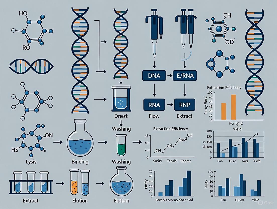
The integration of automated extraction systems into parasitology diagnostics creates a streamlined pathway from sample collection to molecular detection. The following workflow diagram illustrates the standardized process for detecting parasites in clinical samples:
Figure 1. Automated Workflow for Parasite DNA Extraction and Detection. This standardized pathway from sample collection to data analysis highlights the central role of automated extraction systems in ensuring consistent, high-quality results for parasitology research.
Essential Research Reagent Solutions
Successful implementation of automated parasite detection systems requires specific reagents optimized for different sample types and parasite characteristics. The following table details key solutions and their functions in the extraction and detection process:
Table 2: Essential Research Reagents for Automated Parasite DNA Extraction
| Reagent/Category | Function | Application Notes |
|---|---|---|
| DNA/RNA Shield [4] [2] | Preserves nucleic acids immediately upon sample collection, inhibits RNases and DNases | Enables room temperature storage and transportation; critical for field studies |
| Guanidinium Thiocyanate [1] [4] | Chaotropic salt that denatures proteins, facilitates nucleic acid binding to silica | Essential component of lysis buffers; improves DNA yield from tough parasite structures |
| Magnetic Silica Beads [1] [2] | Solid phase for nucleic acid binding, washing, and elution in automated systems | Enable high-throughput processing; reduce cross-contamination |
| Proteinase K [3] | Digests proteins and disrupts parasite structures | Particularly important for helminths with tough eggshells/cuticles |
| InhibitEX Buffer [3] | Adsorbs PCR inhibitors common in stool samples | Critical for intestinal parasite detection from stool specimens |
| RNAlater [2] | RNA stabilization solution for transcript-based detection | Preserves RNA for RT-qPCR assays targeting ribosomal RNA |
Automated DNA extraction systems represent a transformative advancement in parasitology research, directly addressing the critical challenge of detecting low parasitemia across multiple parasitic diseases. The data and protocols presented demonstrate that magnetic bead-based automated extraction consistently outperforms manual methods in DNA yield, purity, and ultimately, analytical sensitivity in molecular detection assays. The standardized workflows and reagent solutions outlined provide researchers with practical frameworks for implementation in various laboratory settings, from high-throughput clinical trials to resource-limited field studies. As molecular diagnostics continue to evolve toward greater sensitivity and precision, the integration of robust, automated extraction methodologies will remain fundamental to accurate parasite detection, species identification, and treatment monitoring—ultimately supporting global efforts in parasitic disease control and elimination.
Core Principle and Mechanism of Magnetic Bead DNA Binding
Magnetic bead-based nucleic acid extraction has become the dominant methodology in modern automated extraction systems due to its superior efficiency, scalability, and ease of automation. The fundamental mechanism relies on the use of superparamagnetic particles as a solid-phase support for binding, purifying, and concentrating nucleic acids from complex sample matrices [5].
The technology's effectiveness stems from a multi-component design. Each magnetic bead typically consists of a supermagnetic core, often composed of iron oxide (Fe₃O₄), which provides the essential property of rapid magnetic separation without the need for centrifugation or filtration. This core is surrounded by a protective shell, usually silicon dioxide (SiO₂), which stabilizes the core and provides a surface for chemical functionalization. The outermost layer is a functionalized surface, modified with chemical groups designed to selectively bind nucleic acids under specific buffer conditions [5]. The most common modification is carboxyl groups (-COOH), which facilitate DNA adsorption in the presence of high concentrations of chaotropic salts [5].
The binding process is governed by a well-established biochemical principle. Chaotropic salts, such as guanidine isothiocyanate (GITC) or guanidine hydrochloride, are added to the sample lysate. These salts disrupt the hydrogen-bonded network of water molecules, thereby reducing the solubility of nucleic acids and promoting their binding to the solid phase. Simultaneously, they denature proteins and inhibit nucleases, protecting the target DNA. Under optimized acidic conditions (typically pH 4.1-6.0), the negatively charged phosphate backbone of DNA exhibits reduced electrostatic repulsion with the silica surface, further enhancing adsorption efficiency [6]. One study demonstrated that adjusting the binding buffer pH from 8.6 to 4.1 increased DNA binding efficiency from 84.3% to 98.2% in a significantly shorter time [6]. The process concludes with a magnetic separation step, where an external magnetic field is applied to immobilize the bead-DNA complexes, allowing for efficient removal of contaminants and inhibitors through sequential washing.
Table 1: Core Components of a Functionalized Magnetic Bead and Their Roles
| Component | Material/Group | Primary Function |
|---|---|---|
| Supermagnetic Core | Iron Oxide (Fe₃O₄) | Enables rapid separation via external magnetic field |
| Protective Shell | Silicon Dioxide (SiO₂) | Stabilizes the core and provides a surface for functionalization |
| Functionalized Surface | Carboxyl (-COOH) Groups | Selectively binds nucleic acids in the presence of chaotropic salts |
Performance Advantages in Parasitology and Clinical Diagnostics
The application of magnetic bead-based extraction in parasitology research has demonstrated clear and quantifiable advantages over traditional methods, particularly for detecting low-abundance pathogens often encountered in chronic infections.
A 2025 study on Chagas disease provides a direct performance comparison between automated magnetic bead (MB) and traditional silica column (SC) extraction methods [7]. The results were striking: the magnetic bead method yielded significantly higher DNA concentrations (66.92 ± 5.98 ng/μL vs. 31.88 ± 2.98 ng/μL via NanoDrop) and superior purity, as evidenced by higher 260/280 nm ratios (1.88 ± 0.02 vs. 1.69 ± 0.03) [7]. This enhanced recovery directly translated to higher analytical sensitivity in real-time PCR (qPCR), with the MB method detecting Trypanosoma cruzi satellite DNA at lower levels, demonstrating approximately 29-fold more sensitivity at 100 parasite equivalents/mL [7].
Similar benefits have been documented in malaria research. A 2024 study found that automated magnetic bead-based extraction of total nucleic acids from Plasmodium falciparum-infected blood samples was equally effective as manual silica column-based kits for downstream reverse transcription qPCR (RT-qPCR) detection [2]. The critical advantage was operational; the automated method enabled the processing of numerous samples in a shorter timeframe, making it a valuable, efficient, and cost-effective tool for large-scale molecular epidemiological studies [2].
Furthermore, magnetic bead systems exhibit superior resistance to common PCR inhibitors found in clinical samples. A comparative study on HPV testing found that while the boiling method failed when hemoglobin concentrations exceeded 30 g/L, the magnetic bead method successfully detected HPV even at 60 g/L hemoglobin, showcasing its robust anti-interference capability [8].
Table 2: Quantitative Performance Comparison: Magnetic Bead vs. Silica Column Extraction
| Performance Metric | Magnetic Bead Method | Silica Column Method | Application Context |
|---|---|---|---|
| DNA Concentration | 66.92 ± 5.98 ng/μL [7] | 31.88 ± 2.98 ng/μL [7] | Chagas Disease Detection [7] |
| DNA Purity (A260/280) | 1.88 ± 0.02 [7] | 1.69 ± 0.03 [7] | Chagas Disease Detection [7] |
| Detection Sensitivity | ~29x higher at 100 Par. Eq./mL [7] | Baseline | Chagas Disease (qPCR) [7] |
| Inhibitor Resistance | Effective at 60 g/L Hemoglobin [8] | Failed at 30 g/L Hemoglobin [8] | HPV Genotyping [8] |
| Throughput | High (96 samples per run) [2] | Lower (Manual processing) | Malaria Detection [2] |
Detailed Protocol for Automated DNA Extraction from Whole Blood for Parasite Detection
The following protocol is adapted for the isolation of total nucleic acids from whole blood for the detection of blood-borne parasites like Plasmodium spp. and Trypanosoma cruzi, utilizing an automated magnetic bead system [2].
Materials and Reagents
- Sample Material: Whole blood samples collected in EDTA tubes.
- RNA Preservation Solution: RNAlater or DNA/RNA Shield.
- Automated Extraction System: e.g., KingFisher Flex System.
- Magnetic Bead Kit: e.g., sbeadex blood kit or equivalent.
- Lysis/Binding Buffer: Typically containing guanidine hydrochloride/isothiocyanate.
- Wash Buffers: Two washes with a buffer containing ethanol, followed by an optional third wash.
- Elution Buffer: Low-salt buffer like TE (10 mM Tris-HCl, 1 mM EDTA, pH 8.0) or nuclease-free water.
Experimental Procedure
Sample Preparation and Lysis:
- Mix 200 μL of whole blood (or blood preserved in a stabilization solution) with an equal volume of lysis/binding buffer in a deep-well plate. If the sample is preserved in RNAlater, pellet the cells by centrifugation and resuspend in 1X PBS to the original blood volume [2].
- Incubate the plate at 60°C for 20 minutes with constant shaking to ensure complete cell lysis and pathogen disruption [2].
Binding of Nucleic Acids:
- Add a defined volume of functionalized magnetic beads (e.g., 30 μL) to each sample well.
- Program the automated system to mix the bead-lysate complex for 5 minutes at room temperature with constant shaking. This facilitates the binding of nucleic acids to the bead surface [2].
Washing:
- The instrument transfers the magnetic bead-nucleic acid complexes through two or three sequential wash buffers.
- The first wash typically uses a buffer to remove salts, proteins, and other contaminants. The second (and third) wash, often containing ethanol, removes residual impurities and concentrates the pure nucleic acids on the beads [5] [2].
Elution:
- After the final wash, the beads are air-dried for a few minutes to allow residual ethanol to evaporate.
- The purified nucleic acids are eluted by incubating the beads in 50-100 μL of pre-warmed (60°C) elution buffer for 10 minutes with shaking [2].
- The instrument transfers the eluate containing the purified DNA/RNA to a clean elution plate, which is then ready for downstream applications like PCR or sequencing.
Workflow Visualization: Automated Magnetic Bead Nucleic Acid Extraction
The following diagram illustrates the streamlined, automated workflow from sample input to purified nucleic acid elution.
Essential Research Reagent Solutions for Magnetic Bead-Based Workflows
Successful implementation of magnetic bead-based DNA extraction relies on a suite of optimized reagents and tools.
Table 3: Key Reagent Solutions for Magnetic Bead-Based DNA Extraction
| Reagent / Material | Function / Role in Workflow | Example Specifications / Notes |
|---|---|---|
| Carboxyl-Modified Magnetic Beads | Solid-phase matrix for nucleic acid binding; superparamagnetic core enables separation. | 300 nm diameter; surface carboxyl group content: 200 μmol/g [5]. |
| Chaotropic Lysis/Binding Buffer | Denatures proteins, inactivates nucleases, promotes nucleic acid adsorption to beads. | 1 M Guanidine Isothiocyanate (GITC), pH 6.0 [5]. |
| Wash Buffer | Removes contaminants (proteins, salts, inhibitors) while retaining DNA bound to beads. | Typically contains ethanol (e.g., 70% v/v) to maintain binding conditions [5]. |
| Elution Buffer | Low-ionic-strength solution disrupts DNA-bead interaction, releasing purified DNA. | TE Buffer (10 mM Tris-HCl, 1 mM EDTA, pH 8.0) or nuclease-free water [5] [2]. |
| RNA Preservation Solution | Stabilizes nucleic acids in blood samples prior to extraction, preventing degradation. | RNAlater or DNA/RNA Shield [2]. |
| Automated Extraction Platform | Instrument that performs all binding, washing, and elution steps with minimal user input. | e.g., KingFisher Flex System; processes 96 samples per run [2]. |
The accurate detection and characterization of parasites through molecular methods are fundamental to modern parasitology research, diagnostics, and drug development. The efficacy of these DNA-based techniques, including PCR and next-generation sequencing, is critically dependent on the initial quality and quantity of the extracted nucleic acids. Automated DNA extraction systems provide a robust, resilient, and efficient solution to the challenges of manual protocols, enabling high-throughput processing while minimizing cross-contamination and operator-induced variability. This application note details the core components of these automated systems and provides a validated protocol for the isolation of high-quality DNA from complex sample matrices relevant to parasitology, such as fecal samples, blood, and tissue biopsies.
Key System Components
An automated DNA extraction system is an integrated platform comprising several key modules that work in concert to purify nucleic acids.
Liquid Handling Devices (LHDs)
LHDs are the core robotic components that automate the precise transfer of liquids for operations such as dilution, mixing, and inoculation [9]. Their adoption facilitates more experiments per unit of time and enhances the robustness and reproducibility of the extraction process against external factors and user error. Modern platforms range from simple, single-arm devices to complex, multi-device configurations, making automation accessible for both high-throughput screening and delicate, low-volume sampling applications in academic and industrial settings [9].
Lysis Module
The lysis module is responsible for disrupting sample cells and the tough structural components of parasites (e.g., oocysts, cysts, or teguments) to release genomic DNA. This stage is critical for determining the final yield and representativeness of the extraction.
- Chemical Lysis: This involves the addition of lysis buffers containing detergents (e.g., SDS), chaotropic salts (e.g., guanidinium thiocyanate), enzymes (e.g., proteinase K), and, for difficult-to-lyse organisms, lysozyme [10]. The choice of buffer must be optimized to effectively lyse both Gram-positive and Gram-negative bacteria (which may be part of the gut microbiome in fecal samples) as well as the target parasites.
- Physical Lysis: Bead-beating is a common physical method that uses vigorous shaking with microscopic beads to mechanically disrupt tough cell walls [10]. While effective, it can shear DNA, which is a significant concern for applications requiring high molecular weight (HMW) DNA. For HMW DNA, gentler methods like enzymatic lysis are often preferred to avoid DNA damage [10].
Nucleic Acid Binding and Elution Module
Following lysis, the released DNA is purified from contaminants like proteins, salts, and other PCR inhibitors.
- Binding Technology: The most common methods utilize silica-based membranes in a spin-column format or magnetic beads. In both, DNA binds to the silica surface in the presence of chaotropic salts. Automated systems then use robotic arms to move plates or employ magnetic separators to immobilize bead-bound DNA while wash buffers are added and removed [10].
- Purification and Elution: The bound DNA is washed with ethanol-based buffers to remove impurities. Finally, a low-salt elution buffer or nuclease-free water is added to disrupt the DNA-silica interaction, releasing pure, concentrated DNA into the eluate. The Solid-Phase Reversible Immobilization (SPRI) system used with magnetic beads is particularly effective for selectively purifying long DNA fragments [10].
Comparative Evaluation of DNA Extraction Methods
Selecting an appropriate DNA extraction method is paramount, as the protocol can significantly impact DNA yield, purity, and fragment length, thereby influencing downstream analytical results [11]. The following table summarizes a comparative evaluation of several DNA extraction methods, highlighting their performance characteristics.
Table 1: Comparison of DNA Extraction Methods and Kits
| Method/Kit Name | Lysis Method | Purification Method | Average DNA Yield | DNA Quality (Purity) | Suitability for Parasitology |
|---|---|---|---|---|---|
| Quick-DNA HMW MagBead Kit [10] | Chemical & Bead-beating | Magnetic Beads | High | High Molecular Weight (HMW) DNA | Excellent for long-read sequencing (e.g., Nanopore) to resolve complex genomes. |
| NucleoSpin Soil Kit [11] | Not Specified | Spin Column | High (for soils) | High Purity; Effective humic acid removal. | Recommended for diverse sample types; ideal for soil-transmitted parasite studies. |
| Modified CTAB Method [12] | Chemical (CTAB) | Phenol-Chloroform | High Concentration | Poor Quality (PCR inhibitors) | Not recommended for sensitive downstream applications. |
| Combination Approach [12] | Multiple | Multiple | High | High (Best Performance) | Recommended for processed samples but can be time-consuming and costly. |
| DNeasy Blood & Tissue [11] | Chemical (Lysozyme) | Spin Column | Variable | Good; Efficient for Gram-positive bacteria [11]. | Suitable for blood and tissue samples containing parasites. |
Detailed Protocol: Automated DNA Extraction from Fecal Samples for Parasite Detection
This protocol is optimized for the extraction of high-quality DNA from human or animal fecal samples for the subsequent detection of parasitic DNA via qPCR or metagenomic sequencing. It is designed for an automated liquid handling platform equipped with a magnetic bead-based purification module.
Materials and Reagents
Table 2: Research Reagent Solutions and Essential Materials
| Item | Function / Explanation |
|---|---|
| Lysis Buffer (with Proteinase K) | Disrupts sample matrix, digests proteins, and inactivates nucleases to release DNA. |
| Binding Buffer (with Chaotropic Salts) | Creates conditions for DNA to bind selectively to silica-coated magnetic beads. |
| Wash Buffer (Ethanol-Based) | Removes salts, proteins, and other contaminants from the bead-bound DNA without eluting it. |
| Nuclease-Free Water | Elutes pure DNA from the magnetic beads; free of enzymes that would degrade the sample. |
| Silica-Coated Magnetic Beads | Solid-phase particles that reversibly bind DNA in the presence of binding buffer. |
| Mock Community Standard | A defined mix of microbial cells (e.g., ZymoBIOMICS Microbial Community Standard) used as a process control to evaluate extraction efficiency and sequencing accuracy [10]. |
| DNA/RNA Shield | A storage solution that immediately stabilizes nucleic acids in samples, preventing degradation during transport or storage [10]. |
Experimental Workflow
The following diagram illustrates the automated workflow for DNA extraction, from sample preparation to quality control.
Step-by-Step Procedure
- Sample Preparation: Homogenize 180-220 mg of fecal sample in a suitable lysis buffer containing proteinase K. For a process control, spike in a known quantity of a mock community standard [10]. Centrifuge briefly to pellet large particulate matter and transfer the supernatant to a deep-well plate on the automated deck.
- Automated Lysis: The liquid handler will incubate the sample plate at a defined temperature (e.g., 56°C) with shaking for a set duration (e.g., 30-60 minutes) to complete the lysis process.
- Binding: The platform automatically adds a volume of binding buffer containing chaotropic salts and silica-coated magnetic beads to the lysate. The mixture is incubated with periodic mixing to allow DNA to bind to the beads.
- Washing: The magnetic module is activated to immobilize the beads. The liquid handler then removes and discards the supernatant. Two wash steps are performed by adding an ethanol-based wash buffer, immobilizing the beads, and removing the wash supernatant completely.
- Elution: After ensuring all wash buffer is evaporated, the magnetic module is deactivated. The handler adds a specified volume of nuclease-free water or elution buffer, mixes thoroughly to resuspend the beads, and incubates to elute the DNA. The magnetic module is activated one final time, and the purified DNA supernatant is transferred to a clean output plate.
Quality Control and Data Analysis
- Quantification and Purity: Assess the concentration and purity (A260/A280 and A260/A230 ratios) of the eluted DNA using a spectrophotometric system like NanoDrop [12]. High-quality DNA should have A260/A280 ratios between 1.8-2.0.
- DNA Integrity: Verify the integrity and fragment size of the extracted DNA using gel electrophoresis [12]. The presence of a high-molecular-weight smear is desirable.
- Amplification Efficiency: Evaluate the suitability of the DNA for downstream applications by performing qPCR assays targeting a conserved host or microbial gene (e.g., 16S rRNA) and a specific parasite gene [12]. Compare the cycle threshold (Ct) values to assess amplification efficiency and the presence of PCR inhibitors.
Automated DNA extraction systems, integrating precise liquid handlers, efficient lysis modules, and reliable binding and elution technologies, are indispensable for modern parasitology research. The provided protocol and comparative data underscore the importance of selecting a method that balances high yield with high quality, particularly for challenging sample types like feces. The recommended magnetic bead-based automated protocol ensures the isolation of high-quality DNA, free from common PCR inhibitors, thereby enabling sensitive and reliable detection and characterization of parasites in support of advanced diagnostics and drug development.
Automated DNA extraction systems represent a transformative advancement in parasitology research, offering significant improvements over manual methods. These systems are designed to address critical challenges in molecular diagnostics, including the need for standardized protocols, high-throughput processing, and reliable detection of low-abundance pathogens. This application note details the core advantages of automated nucleic acid extraction—throughput, reproducibility, and contamination control—within the context of parasitology research. We provide quantitative comparisons and detailed experimental protocols to guide researchers in selecting and implementing these systems for enhanced research efficiency and data quality.
Core Advantages of Automated Extraction
Enhanced Throughput and Efficiency
Automated nucleic acid extraction systems dramatically increase processing capacity and reduce hands-on time, enabling rapid response in research and diagnostic settings.
Table 1: Throughput and Time Efficiency of Automated Systems
| System / Kit Name | Method Type | Maximum Throughput (Samples/Run) | Total Processing Time | Hands-On Time |
|---|---|---|---|---|
| KingFisher Apex [13] | Magnetic bead-based | 96 | ~40 min | Minimal |
| Maxwell RSC 16 [13] | Magnetic bead-based | 16 | ~42 min | ~35 min |
| GenePure Pro [13] | Magnetic bead-based | 32 | ~35 min | ~25 min |
| T-Prep24 System [14] | Magnetic bead-based | 24 | ~30 min | Minimal |
| Manual Column-Based [13] | Spin column | Variable | ~100 min | ~100 min |
As illustrated in Table 1, automated systems can process a full plate of up to 96 samples in approximately 40 minutes, whereas manual processing for just 16 samples requires about 100 minutes of dedicated hands-on time [13]. This efficiency is crucial for parasitology studies involving large-scale screening, such as surveillance of parasitic infections like schistosomiasis or microsporidiosis in population studies [15] [16].
Improved Reproducibility and Data Quality
Automation minimizes human error and variability in liquid handling, leading to superior consistency in DNA yield, purity, and subsequent analytical results.
Table 2: Performance Comparison of Automated vs. Manual Methods
| Performance Metric | Manual Methods | Automated Methods | Experimental Evidence |
|---|---|---|---|
| Inter-sample Variation | Higher variability in DNA concentration and purity [13] | Lower inter-sample variability [13] | Comparison of 16S sequencing data from human fecal samples [13] |
| Sensitivity in Pathogen Detection | Variable; dependent on technician skill | Higher and more consistent sensitivity [16] [17] | Detection of E. bieneusi spores at low concentrations (5-25 spores/mL) [16] |
| Impact on Microbial Community Profiles | Higher batch effects potential | Reduced batch effects; more stable community representation [13] | 16S rRNA amplicon sequencing of mock communities and fecal samples [13] |
| Protocol Standardization | Difficult to standardize across labs and technicians | Open-source, transparent protocols enable perfect replication [4] | Standardized DREX protocol within the Earth Hologenome Initiative [4] |
A study on human fecal microbiota demonstrated that automated extractors yielded more consistent 16S rRNA gene amplicon sequencing results compared to manual processing, reducing technical artifacts in microbial community analysis [13]. Furthermore, for detecting the parasite Enterocytozoon bieneusi in stool samples, automated or semi-automated systems like the Nuclisens easyMAG and Quick DNA Fecal/Soil Microbe Microprep kit showed superior detection frequencies and lower Ct values at very low spore concentrations (5-25 spores/mL) compared to other methods [16].
Superior Contamination Control
Automated systems are engineered to minimize the risk of cross-contamination, a critical factor for the sensitivity required in detecting low-parasite-load infections.
- Closed Systems: Many automated platforms, such as the T-Prep24 system, function as closed systems, physically separating samples and reagents from the environment [14].
- Disposable Components: The use of disposable tips and pre-packed reagent plates eliminates carryover between samples [14] [18].
- Integrated Decontamination: Built-in ultraviolet (UV) light chambers are standard in systems like the KingFisher Apex, GenePure Pro, and Maxwell RSC 16, which decontaminate the work area between runs to prevent amplicon contamination [13].
Experimental Protocols for Parasitology Research
Protocol: Automated DNA Extraction from Stool Samples for Microsporidia Detection
This protocol is optimized for breaking tough parasitic spores, based on a multicenter study for detecting Enterocytozoon bieneusi [16].
1. Sample Pretreatment and Lysis:
- Transfer 200-300 µL of preserved stool sample to a tube containing lysing matrix E (e.g., MP Biomedicals) or ZR BashingBeads (Zymo Research).
- Add the appropriate volume of lysis buffer containing guanidine thiocyanate and proteinase K to the sample.
- Perform mechanical bead-beating using a homogenizer (e.g., TissueLyser II, Qiagen) at 30 Hz for 60 seconds to effectively disrupt the resilient chitin wall of microsporidia spores [16].
- Incubate the lysate at 70°C for 10 minutes to complete the lysis process.
2. Automated Nucleic Acid Purification:
- Transfer the clarified lysate (after brief centrifugation) to a deep-well plate compatible with your automated system.
- For magnetic bead-based systems (e.g., KingFisher Apex, Maxwell RSC), follow the manufacturer's instructions for binding, washing, and elution. The protocol typically involves:
- Binding of nucleic acids to silica-coated magnetic beads in the presence of a chaotropic salt.
- Two or more wash steps with ethanol-based buffers to remove inhibitors.
- Elution of pure DNA in a low-salt elution buffer or nuclease-free water (50-100 µL).
3. Quality Control and Downstream Application:
- Quantify the extracted DNA using a fluorescence-based method (e.g., Qubit Fluorometer).
- Assess purity by measuring absorbance ratios (A260/A280 and A260/A230) with a spectrophotometer.
- Use the DNA in a specific qPCR assay for E. bieneusi detection. The optimized extraction should yield low Cq values (<35) for samples with spore counts as low as 25 spores/mL [16].
Protocol: Automated Extraction from Filtered Urine for Schistosome DNA Detection
This protocol is designed for the sensitive detection of cell-free parasite DNA from Schistosoma mansoni and S. haematobium in urine samples, a key sample matrix in parasitology [15].
1. Sample Preparation:
- Collect urine samples on filter paper via filtration (~20 mL). A 6 mm punch of the dried filter paper is sufficient for DNA extraction.
- Place the filter paper punch directly into a lysis tube.
2. DNA Extraction and Purification:
- For high-throughput applications, use an automated magnetic bead-based system like the T-Prep24 or taco mini system.
- Add lysis/binding buffer to the tube and incubate to dissolve the filter paper and release DNA.
- The automated steps include:
- Binding of DNA to magnetic beads.
- Multiple washes to remove urinary inhibitors such as urea and salts.
- Elution in 50-100 µL of elution buffer.
3. Downstream LAMP Amplification:
- Use 5-10 µL of the eluted DNA in a Loop-Mediated Isothermal Amplification (LAMP) reaction.
- LAMP is particularly suitable for field-based parasitology diagnostics due to its resistance to inhibitors and isothermal amplification conditions [15].
The Scientist's Toolkit: Research Reagent Solutions
Table 3: Essential Reagents and Kits for Automated Parasitic DNA Extraction
| Reagent / Kit Name | Function | Application in Parasitology |
|---|---|---|
| Lysing Matrix E (MP Biomedicals) | A mixture of ceramic, silica, and glass beads for mechanical disruption of tough cell walls. | Critical for breaking open resilient parasitic forms like Cryptosporidium oocysts and Microsporidia spores [4] [16]. |
| DNA/RNA Shield (Zymo Research) | A preservation solution that immediately stabilizes nucleic acids and inactivates nucleases and pathogens. | Ideal for field collection of fecal or urine samples, maintaining nucleic acid integrity from remote locations [13] [17]. |
| MagMAX Pathogen RNA/DNA Kit (Thermo Fisher) | A magnetic bead-based kit optimized for binding and purifying nucleic acids from complex samples. | Validated for viral DNA extraction from environmental samples; applicable for parasite DNA from similar matrices [17]. |
| Guanidine Thiocyanate-based Lysis Buffer | A chaotropic salt that denatures proteins, inhibits RNases, and promotes nucleic acid binding to silica. | A key component in open-source and commercial lysis buffers for efficient release of DNA from parasites [4] [14]. |
| Silica-coated Magnetic Beads | The solid phase for nucleic acid binding, washing, and elution in automated magnetic bead systems. | The core technology in most high-throughput automated extractors for parasitology research [18] [17]. |
System Workflow and Technology Integration
The following diagram illustrates the integrated workflow of an automated DNA extraction system within a modern parasitology laboratory, highlighting the roles of throughput, reproducibility, and contamination control.
The adoption of automated DNA extraction systems provides parasitology researchers with a powerful toolkit to overcome the limitations of manual methods. The demonstrated enhancements in throughput, reproducibility, and contamination control are not merely incremental; they are foundational to generating high-quality, reliable data essential for advanced research and diagnostic applications. By implementing the standardized protocols and reagent solutions outlined in this application note, research and drug development professionals can significantly improve the efficiency and robustness of their molecular workflows, accelerating progress in the understanding and control of parasitic diseases.
Application-Oriented Protocols for Diverse Parasites and Sample Matrices
Optimizing Extraction from Complex Stool Samples for Intestinal Protozoa and Helminths
The shift toward molecular diagnostics in clinical parasitology, particularly within automated laboratory workflows, has highlighted a critical bottleneck: the efficient recovery of inhibitor-free, high-quality DNA from complex stool matrices. The success of any downstream nucleic acid amplification test (NAAT) is fundamentally dependent on the initial DNA extraction step, which must simultaneously overcome the resilience of parasitic structures and the potent PCR inhibitors inherent to feces [19] [20] [21]. This application note delineates optimized protocols and key considerations for DNA extraction, contextualized within a broader thesis on automated systems for parasitology research. We synthesize recent evidence to guide professionals in selecting and validating methods that ensure maximal sensitivity and specificity for both intestinal protozoa and helminths.
The Challenge: Sample and Parasite Complexity
Stool samples present unique challenges for molecular diagnostics. They contain a wide array of PCR inhibitors, including bil salts, complex carbohydrates, and hemoglobin breakdown products [19]. Simultaneously, the parasitic targets possess physical barriers that impede DNA release. Protozoan cysts have tough walls, but the most significant challenges come from helminths. Soil-transmitted helminth (STH) eggs, such as those from Ascaris lumbricoides and Trichuris trichiura, are protected by a chitinous shell that is notoriously difficult to lyse [19] [21]. Larvae, such as those of Strongyloides stercoralis, have a tough, sticky cuticle [19]. This combination demands extraction methods that are both mechanically robust and chemically efficient to ensure adequate DNA yield and purity.
Optimization Strategies and Experimental Data
Mechanical Pretreatment: The Role of Bead-Beating
The incorporation of a mechanical disruption step, specifically bead-beating, prior to chemical lysis is consistently identified as a critical factor for improving DNA recovery, especially from helminth eggs [19] [21].
Experimental Protocol: Bead-Beating Pretreatment
- Materials: 0.5 mm sterile glass beads (Omni International), lysis buffer, 2 mL sterile microcentrifuge tubes, vortex mixer with horizontal (tube-shaking) attachment.
- Procedure:
- Transfer 200 mg of stool sample into a 2 mL tube.
- Add 250 mg of 0.5 mm glass beads and 400 µL of an appropriate lysis buffer.
- Secure the tubes tightly and place them in the vortex mixer with a horizontal adapter.
- Vortex at maximum speed for 10 minutes to achieve a homogeneous slurry.
- Proceed with the standard chemical lysis and DNA purification steps of your chosen extraction method [19].
Supporting Data: A study comparing four DNA extraction methods for STHs found that adding a bead-beating step to a phenol-chloroform protocol significantly improved DNA recovery, particularly in samples with high fecal egg counts [21]. Furthermore, a comparative study of DNA extraction methods demonstrated that protocols incorporating bead-beating (PB and QB methods) yielded significantly higher PCR detection rates across a range of parasites, including the resilient Ascaris lumbricoides, compared to methods without this step [19] [3].
Comparative Performance of DNA Extraction Methods
The choice of DNA extraction methodology profoundly impacts diagnostic sensitivity. Studies have systematically compared various techniques, revealing clear differences in their efficacy.
Experimental Protocol: Comparative Evaluation of DNA Extraction Kits
- Design: Compare the performance of different extraction kits from the same set of stool samples preserved in 70% ethanol.
- Sample Preparation: Wash preserved stool samples three times with sterile distilled water before aliquoting for different extraction methods [19].
- Methods Compared: Include a traditional phenol-chloroform method (P), a bead-beating-enhanced phenol-chloroform method (PB), and two commercial kits: the QIAamp Fast DNA Stool Mini Kit (Q) and the QIAamp PowerFecal Pro DNA Kit (QB) [19].
- Evaluation Metrics: Assess DNA quantity and quality via spectrophotometry. Evaluate DNA integrity and presence of inhibitors via parasite-specific PCR and a spike-in test, where a known plasmid is added to the PCR reaction to identify false negatives caused by residual inhibitors [19].
The table below summarizes key findings from such a comparative study:
Table 1: Comparison of DNA Extraction Method Performance on Stool Samples
| Extraction Method | Relative DNA Yield | Overall PCR Detection Rate | Effectiveness on Resilient Parasites (e.g., Ascaris) | Key Advantage/Limitation |
|---|---|---|---|---|
| Phenol-Chloroform (P) | High (~4x kits) | 8.2% (Lowest) | Poor | High yield but inefficient lysis and inhibitor removal [19] |
| Phenol-Chloroform + Beads (PB) | High (~4x kits) | Moderate Improvement | Good | Mechanical lysis improves recovery [19] |
| QIAamp Fast DNA Stool (Q) | Lower | Moderate | Variable | Commercial convenience [19] |
| QIAamp PowerFecal Pro (QB) | Lower | 61.2% (Highest) | Excellent | Optimal for broad parasite detection; effective lysis and inhibitor removal [19] |
The QIAamp PowerFecal Pro DNA Kit (QB) demonstrated superior performance, achieving the highest PCR detection rate and being the only method to successfully extract DNA from all parasite groups tested, including fragile protozoa like Blastocystis sp. and helminths with robust eggs like A. lumbricoides [19]. The bead-beating step incorporated into this and similar kits is a major contributor to this efficacy.
Assay Selection: Differential Performance for Protozoa vs. Helminths
When deploying automated multiplex PCR panels, it is vital to understand that their performance is not uniform across all parasite types.
Supporting Data: A 2024 evaluation of the Seegene Allplex GI-Parasite and GI-Helminth assays on 97 clinical samples found a stark contrast in performance. The assay demonstrated excellent sensitivity for protozoa, detecting 100% of Dientamoeba fragilis and 95% of Blastocystis hominis infections, outperforming a conventional diagnostic workflow [22]. However, its sensitivity for helminths was suboptimal (59.1%), failing to detect a significant number of microscopy-confirmed infections, particularly Trichuris trichiura (20% sensitivity) [22]. This underscores that even with optimized extraction, some commercial molecular assays may not be suitable for all parasitic targets, and microscopy remains essential for helminth diagnosis in at-risk populations [23] [22].
The following workflow diagram synthesizes the optimal strategy for processing complex stool samples, from collection to analysis, integrating the key optimization steps discussed.
The Scientist's Toolkit: Essential Research Reagents
The following table details key reagents and kits instrumental for implementing the optimized protocols described in this note.
Table 2: Essential Reagents for DNA Extraction from Complex Stool Samples
| Research Reagent Solution | Function / Application |
|---|---|
| QIAamp PowerFecal Pro DNA Kit (QIAGEN) | Effective, bead-beating-based DNA extraction from difficult stool samples; optimal for a wide range of parasites [19] |
| 0.5 mm Sterile Glass Beads | Mechanical disruption of resilient parasite cysts, oocysts, and eggshells during the lysis step [19] |
| Seegene AllPlex GI-Parasite Assay | Multiplex real-time PCR panel for detection of major protozoa; compatible with automated systems [23] [24] |
| FecalSwab with Cary-Blair Medium (COPAN) | Standardized medium for stool sample suspension and transport, suitable for automated DNA extraction platforms [23] [24] |
| STARMag 96 × 4 Universal Cartridge (Seegene) | Magnetic-bead based cartridge for automated, high-throughput nucleic acid extraction on systems like the Hamilton STARlet [24] |
Optimizing DNA extraction is a non-negotiable prerequisite for reliable molecular detection of intestinal parasites in stool. The evidence conclusively shows that a mechanical lysis step, preferably using bead-beating, is essential for liberating DNA from resilient helminth eggs. Furthermore, the selection of a DNA extraction kit specifically designed to handle inhibitors and tough biological structures, such as the QIAamp PowerFecal Pro DNA Kit, dramatically increases diagnostic sensitivity. Finally, researchers and clinicians must be aware of the differential performance of commercial multiplex PCR panels, which excel for protozoa but may lack sensitivity for helminths, necessitating a complementary use of microscopy in specific clinical contexts. By adopting these optimized protocols, laboratories can significantly enhance the performance of their automated parasitology research and diagnostic pipelines.
The accurate detection of bloodborne parasitic infections, such as those caused by Plasmodium spp. and Trypanosoma cruzi, is fundamental to diagnosis, treatment monitoring, and epidemiological studies. Molecular techniques, particularly PCR and qPCR, have become indispensable tools in this endeavor, offering superior sensitivity and specificity over traditional microscopic and serological methods, especially during the chronic phases of these diseases when parasitemia is low [7] [25]. A critical factor influencing the success of any molecular assay is the efficiency of the DNA extraction process, which dictates the yield, purity, and ultimate sensitivity of parasite detection [7] [26]. This application note, framed within a broader thesis on automation in parasitology, evaluates and provides detailed protocols for DNA extraction methods proven to maximize detection sensitivity for Plasmodium and T. cruzi from blood samples.
Comparative Performance of DNA Extraction Methods
The selection of a DNA extraction method significantly impacts the sensitivity of downstream molecular applications. The following tables summarize key performance metrics from recent studies comparing various techniques for Plasmodium and T. cruzi.
Table 1: Comparison of DNA Extraction Methods for Trypanosoma cruzi Detection
| Sample Type | Extraction Method | Key Performance Findings | Reference |
|---|---|---|---|
| Whole Blood (GUANIDINE-EDTA) | Automated Magnetic Beads | Higher DNA yield and purity; Lower Cq values in qPCR; ~29x more satDNA detected at 100 Par. Eq./mL compared to silica column. | [7] |
| Whole Blood (GUANIDINE-EDTA) | Silica Column | Lower DNA yield and purity; Higher Cq values in qPCR. | [7] |
| Blood Clot | FastPrep + Silica Column Kit | Higher qPCR sensitivity; Additional positive samples detected compared to whole blood-guanidine method. | [25] |
| Whole Blood on Filter Paper | Qiagen DNeasy (90°C + extended PK incubation) | Optimal recovery of parasite DNA and host β-actin from field-collected samples. | [26] |
Table 2: Comparison of DNA Extraction and Processing Methods for Plasmodium falciparum Detection
| Method Category | Specific Method | Key Performance Findings | Reference |
|---|---|---|---|
| Host DNA Reduction | Lymphoprep + Plasmodipur Filtration | <30% human DNA in >70% of samples; enabled ~40x genome coverage in WGS from a single Illumina lane. | [27] |
| Nucleic Acid Extraction | Automated Magnetic Bead-Based (KingFisher) | Comparable Cq values to manual column-based kits in RT-qPCR; high-throughput, efficient, and reliable. | [28] |
| Nucleic Acid Extraction | Manual Silica Column-Based (QIAamp) | Comparable Cq values to automated magnetic bead system; effective for nucleic acid recovery. | [28] |
Detailed Experimental Protocols
Protocol 1: Automated Magnetic Bead-Based DNA Extraction forT. cruzifrom Whole Blood
This protocol, adapted from recent research, is designed for high sensitivity detection of T. cruzi in whole blood [7] [29].
Materials:
- Automated Nucleic Acid Extractor (e.g., KingFisher Flex System)
- Magnetic Bead-Based Kit (e.g., sbeadex blood kit)
- Blood samples collected in K₂EDTA tubes
- PBS
- Microcentrifuge tubes and plates compatible with the automated system
Procedure:
- Sample Preparation: Centrifuge 3-10 mL of whole blood at 2,000 × g for 15 minutes. Carefully transfer the plasma to a separate tube for archiving. The remaining cell pellet is used for DNA extraction.
- Lysis: Transfer the cell pellet to a nuclease-free 50 mL conical tube. Adjust the volume to 10 mL with PBS. Add lysis buffer and Proteinase K from the kit according to the manufacturer's instructions. Vortex thoroughly to mix.
- Incubation: Incubate the lysate at 60°C for 20 minutes with constant shaking in the automated instrument.
- Automated Extraction: Load the lysate, reagents, and magnetic beads into the designated plate. Run the automated program, which includes:
- Binding: Beads are mixed with the lysate for 5 minutes at room temperature to bind DNA.
- Washing: The bead-DNA complex is washed three times with wash buffers to remove impurities.
- Elution: DNA is eluted in 100 µL of elution buffer with incubation at 60°C for 10 minutes.
- Storage: Quantify the eluted DNA and store at -20°C.
Protocol 2: Optimized DNA Extraction from Blood Clots forT. cruzi
This protocol uses mechanical disruption to maximize DNA recovery from clot samples, which can trap parasites [25].
Materials:
- FastPrep-24 Instrument and Lysing Matrix E tubes
- Silica Column-Based DNA Purification Kit (e.g., High Pure PCR Template Preparation Kit, Roche)
- Guanidine-EDTA (GE) Buffer
- Proteinase K
- Internal Amplification Control (IAC)
Procedure:
- Sample Homogenization: Place approximately 300 µL of clot in a Lysing Matrix E tube.
- Lysis: Add 300 µL of GE buffer, 40 µL of Proteinase K (20 mg/mL), and 5 µL of IAC to the tube.
- Mechanical Disruption: Process the tube in the FastPrep instrument at 5.5 m/s for 30 seconds.
- Centrifugation: Centrifuge the homogenate at 14,000 × g for 2 minutes to pellet debris.
- DNA Purification: Transfer 450 µL of the supernatant to a new tube. Add 150 µL of binding buffer from the silica column kit.
- Incubation: Incubate the mixture at 70°C for 10 minutes.
- Column Purification: Complete the DNA purification following the manufacturer's instructions for the silica column kit, including wash steps.
- Elution: Elute DNA in 100 µL of elution buffer.
Protocol 3: Host DNA Depletion for EnhancedPlasmodiumGenome Sequencing
This protocol reduces human DNA contamination from clinical blood samples, significantly improving yield for whole genome sequencing of Plasmodium [27].
Materials:
- Lymphoprep density gradient medium
- Plasmodipur filters
- Vacuum source or syringe
Procedure:
- Density Gradient Centrifugation: Layer 3-5 mL of anti-coagulated whole blood over an equal volume of Lymphoprep in a centrifuge tube. Centrifuge at 800 × g for 20-30 minutes with the brake disengaged.
- Collect Buffy Coat and Red Blood Cells: After centrifugation, the top layer is plasma. The buffy coat (containing white blood cells) forms a thin layer below the plasma. Carefully aspirate the plasma and buffy coat layers, leaving the red blood cell (RBC) layer, which contains the parasites.
- Filter Depletion: Re-suspend the harvested RBC pellet in a suitable medium (e.g., RPMI or PBS). Pass the RBC suspension through a Plasmodipur filter according to the manufacturer's instructions. This filter selectively retains residual white blood cells.
- DNA Extraction: Extract DNA from the filtered RBCs using a preferred method, such as a commercial silica-based kit or an automated magnetic bead system [28].
Workflow Visualization
The following diagram illustrates the key decision points and pathways for selecting an optimal DNA extraction strategy based on research objectives.
The Scientist's Toolkit: Research Reagent Solutions
Table 3: Essential Reagents and Kits for Sensitive Detection of Bloodborne Parasites
| Item Name | Function/Application | Specific Example(s) |
|---|---|---|
| sbeadex blood kit | Automated, magnetic bead-based nucleic acid extraction from whole blood. High yield and purity. Suitable for high-throughput settings. | Used with KingFisher Flex system for Plasmodium [28] and T. cruzi [7] detection. |
| QIAamp DNA Blood Mini Kit | Manual, silica-membrane column-based DNA extraction. Reliable performance. Optimal for filter paper samples with protocol modifications. | Used for extraction from blood on Nobuto filter paper with high-temperature incubation [26]. |
| High Pure PCR Template Kit | Silica column-based purification of DNA from complex lysates. Effective post-mechanical disruption. | Used for DNA extraction from homogenized blood clots for T. cruzi [25]. |
| Lymphoprep & Plasmodipur | Two-step method for depleting human white blood cells from infected blood, enriching parasite content for superior sequencing. | Critical for whole genome sequencing of Plasmodium from clinical samples [27]. |
| Lysing Matrix E & FastPrep | Mechanical disruption system for tough samples. Efficiently lyses parasite cysts and host cells in blood clots. | Essential for maximizing DNA yield from blood clots for T. cruzi PCR [25]. |
| DNA/RNA Shield | Preservation solution that stabilizes nucleic acids at room temperature, preventing degradation during transport and storage. | Used for stabilizing Plasmodium RNA/DNA in whole blood prior to extraction [28]. |
The study of ancient parasites from forensic and archival bone and tooth samples provides unparalleled insights into the evolutionary history of pathogens, human migration patterns, and historical disease dynamics. Within the context of parasitology research, the extraction of high-quality nucleic acids from these challenging substrates is a critical first step for successful downstream genomic analyses. Ancient DNA (aDNA) from skeletal remains is typically highly fragmented, present in low concentrations, and contaminated with environmental inhibitors that can compromise polymerase chain reaction (PCR) efficacy [30] [31]. The implementation of automated DNA extraction systems addresses these challenges by standardizing protocols, reducing cross-contamination, and improving the reproducibility and throughput of samples processed for phylogenetic studies of parasite lineages [1] [32]. This application note details optimized protocols and comparative performance data for the automated extraction of parasite DNA from ancient skeletal material, providing a standardized framework for researchers in paleoparasitology and related fields.
Performance Comparison of Extraction Methodologies
The selection of an appropriate nucleic acid extraction method is paramount for maximizing the recovery of amplifiable parasite DNA from ancient skeletal samples. The following tables summarize key performance metrics from comparative studies, providing a basis for informed methodological selection.
Table 1: Comparison of Automated Nucleic Acid Extraction System Performance on Challenging Sample Types
| Extraction System | Core Technology | Sample Input | Average DNA Concentration (ng/µL) | Purity (A260/A280) | Key Findings |
|---|---|---|---|---|---|
| Magnetic Bead-Based System (e.g., KingFisher Apex, T-Prep24) | Magnetic Silica Beads | 200 µL spiked blood [1] | 29.75 ± 4.07 (Qubit) [1] | 1.88 ± 0.02 [1] | Superior yield and purity; higher sensitivity for low-parasitemia samples [1]. |
| Silica Column-Based System (e.g., QIAcube) | Silica Gel Membrane | 200 µL spiked blood [1] | 4.65 ± 1.48 (Qubit) [1] | 1.69 ± 0.03 [1] | Lower DNA yield and purity compared to magnetic bead methods [1]. |
| T-Prep24 System | Magnetic Beads | 200 µL respiratory sample [14] | 0.685 ng/µL (median, Qubit) [14] | N/R | Reliable for pathogen detection; minimal systematic bias in Cq values vs. other automated systems [14]. |
Table 2: Impact of Supplemental Lysis Methods on DNA Yield from Complex Matrices
| Lysis Method | Sample Type | Impact on DNA Yield & Microbiome Representation | Recommendation for Parasite Ova/Cysts |
|---|---|---|---|
| Bead-Beating | Human Stool [32] | Significantly increased yield; improved lysis of Gram-positive bacteria and robust microbial structures [32]. | Essential for liberating DNA from resilient parasite ova (e.g., Ascaris, Giardia) [32]. |
| Chemical Lysis Only (Guanidinium thiocyanate, Proteinase K) | Human Stool [32] | Lower DNA yield; under-representation of Gram-positive bacteria [32]. | May be insufficient for certain tough-walled parasites, leading to false negatives. |
Experimental Protocols for Parasite aDNA Extraction from Skeletal Material
Protocol 1: Silica-Magnetic Bead-Based Automated Extraction for Ancient Teeth
This protocol is adapted from a method designed to maximize the recovery of PCR-amplifiable DNA from ancient bone and teeth while minimizing co-extraction of inhibitors [30] [33]. The following steps are optimized for integration with automated magnetic bead-based platforms.
Materials and Reagents:
- Powdered ancient tooth or bone sample
- Lysis and Binding Buffer (e.g., containing Guanidinium thiocyanate)
- Proteinase K
- Molecular grade water
- Isopropanol
- Ethanol (70-80%)
- Silica-coated magnetic beads
- Collection plates or tubes compatible with the automated system
Procedure:
- Sample Preparation: Grind approximately 50-100 mg of tooth or bone into a fine powder under controlled conditions to prevent contamination.
- Demineralization and Digestion: Transfer the powder to a tube containing 1-3 mL of EDTA (0.5 M, pH 8.0) and 50-100 µL of Proteinase K (20 mg/mL). Incubate with rotation for 12-24 hours at room temperature [30] [33].
- Automated Processing:
- Load Samples and Reagents: Transfer the supernatant from step 2 to the input plate of the automated system. Load the required reagents (lysis/binding buffer, wash buffers, elution buffer) and magnetic beads according to the manufacturer's instructions.
- Run Program: Execute the automated extraction protocol. A typical workflow on a system like the KingFisher Apex involves:
- Post-Processing: Store the extracted DNA at -20°C or -80°C. Analyze DNA concentration and quality using a fluorescence-based method (e.g., Qubit fluorometer) and spectrophotometry (e.g., NanoDrop) to assess purity [14] [1].
Protocol 2: Manual Silica-Based Column Extraction with Pre-Automation Steps
This traditional method is included as a reference and for scenarios where automated systems are unavailable. It forms the basis of many commercial kit protocols and can be a useful pre-processing step.
Materials and Reagents:
- Powdered ancient tooth or bone sample
- EDTA (0.5 M, pH 8.0)
- Proteinase K
- Binding buffer (containing Guadinium thiocyanate)
- Wash buffer (e.g., with ethanol)
- Elution buffer
- Silica spin columns
- Collection tubes
Procedure:
- Sample Digestion: Digest 50-100 mg of bone/tooth powder in 1-3 mL of 0.5 M EDTA and 50-100 µL of Proteinase K (20 mg/mL) for 12-24 hours at room temperature with constant agitation [30].
- Binding: Combine the supernatant with 5-10 volumes of binding buffer and transfer to a silica spin column. Incubate and centrifuge to bind DNA.
- Washing: Wash the column twice with a wash buffer, followed by a centrifugation step to remove residual salts and contaminants.
- Elution: Elute the DNA in 50-100 µL of pre-heated (55°C) elution buffer or molecular grade water.
Workflow Visualization
The following diagram illustrates the complete experimental workflow for obtaining and analyzing parasite DNA from ancient skeletal remains, from sample preparation to final analysis.
The Scientist's Toolkit: Research Reagent Solutions
Table 3: Essential Reagents and Kits for Parasite aDNA Extraction from Skeletal Material
| Reagent / Kit | Function | Application Note |
|---|---|---|
| EDTA (Ethylenediaminetetraacetic acid) | Chelates calcium ions to demineralize the bone/tooth matrix, releasing trapped DNA [30] [33]. | Use at 0.5 M, pH 8.0. Incubation for 12-24 hours is critical for efficient decalcification. |
| Proteinase K | A broad-spectrum serine protease that digests proteins and inactivates nucleases, liberating DNA from cellular debris [30]. | Essential for breaking down osteocytes and potential parasite material embedded in the skeletal matrix. |
| Guanidinium Thiocyanate | A chaotropic agent that denatures proteins, inhibits nucleases, and promotes binding of nucleic acids to silica surfaces [30] [1]. | A key component of lysis and binding buffers in both manual and automated systems. |
| Silica-Coated Magnetic Beads | Solid-phase support for reversible DNA binding under high-ionic-strength conditions, enabling automated washing and elution [14] [1] [32]. | The core technology in modern automated extractors; offers superior yield and purity from complex samples. |
| Lysis & Binding Buffer | Facilitates cell lysis and creates optimal conditions for nucleic acid binding to silica. | Often contains guanidinium salts and detergents; formulation is kit-specific. |
| Wash Buffer | Removes contaminants, salts, and inhibitors from the bound nucleic acids without eluting the DNA. | Typically contains ethanol or alcohol-based solutions to maintain DNA binding while removing impurities. |
| Elution Buffer | A low-ionic-strength solution (e.g., Tris-HCl or water) that disrupts the DNA-silica interaction, releasing purified DNA. | Pre-heating to 55-70°C can increase elution efficiency. |
High-Throughput Workflows for Large-Scale Epidemiological Studies and Cohort Screening
Molecular diagnostics and genomic surveillance play an increasingly critical role in large-scale epidemiological studies focused on parasitic diseases. The analysis of cohorts exposed to or infected with parasites provides invaluable insights into disease pathogenesis, population dynamics, and therapeutic efficacy [34]. However, traditional manual methods for sample processing become impractical in studies involving thousands of participants, creating bottlenecks that delay critical public health insights. The implementation of high-throughput workflows addresses these challenges by significantly increasing processing capacity while improving reproducibility and reducing potential for human error [13] [35].
In parasitology, where many clinically relevant pathogens lack robust culture systems or are refractory to genetic manipulation, efficient molecular screening of large cohorts becomes particularly valuable [34]. Automated nucleic acid extraction systems represent a foundational component of these workflows, enabling reliable processing of diverse sample types from blood to stool specimens. This application note details integrated methodologies and comparative performance data to guide researchers in implementing optimized high-throughput workflows for parasitic disease studies.
High-Throughput Workflow Design Fundamentals
Core Principles for Epidemiological Studies
The transition to high-throughput processing requires careful consideration of several key principles. Throughput efficiency must balance the number of samples processed per run with the time required for completion, while extraction efficacy ensures sufficient quantity and quality of nucleic acids for downstream molecular analyses [13]. Process standardization across all samples minimizes technical variation that could confound biological results, and cost-effectiveness becomes increasingly important when scaling to thousands of samples [35].
Cohort Study Framework for Parasitology Research
Cohort studies provide an ideal epidemiological framework for investigating parasitic diseases, as they enable researchers to establish temporality between exposures and outcomes [36] [37]. In a typical design, participants are selected based on exposure status and followed over time to evaluate for the occurrence of parasitic infections or related health outcomes. These studies may be prospective (following participants forward in time) or retrospective (utilizing existing data and specimens) [36]. The longitudinal nature of cohort studies generates valuable data on infection incidence, parasite genetic evolution, and host-parasite interactions, particularly when integrated with automated high-throughput molecular methods.
Comparative Performance of Automated Nucleic Acid Extraction Systems
Table 1: Comparison of Automated Nucleic Acid Extraction Systems
| System | Throughput (samples/run) | Processing Time | Technology | Bead-Beating Compatibility | Relative Cost |
|---|---|---|---|---|---|
| KingFisher Apex | 1-96 | ~40 min (16 samples) | Magnetic beads | Required [13] | High |
| Maxwell RSC 16 | 1-16 | ~42 min (16 samples) | Magnetic beads | Optional [13] | Medium |
| GenePure Pro | 1-32 | ~35 min (16 samples) | Magnetic beads | Optional [13] | Medium |
| Magtration 12GC | 12 | Not specified | Magnetic beads | Not specified | Medium |
| Freedom EVO | Variable | Not specified | Magnetic beads | Not specified | High |
| RoboCTAB (OT-2) | 384 | Variable | CTAB + magnetic beads | Integrated [35] | Low |
Performance Metrics for Parasite Detection
Table 2: Performance Assessment for Parasite Detection
| Extraction Method | Sample Type | Target Parasite | Sensitivity | Key Findings | Reference |
|---|---|---|---|---|---|
| sbeadex + KingFisher | Whole blood | Plasmodium falciparum | Comparable to manual | No significant difference in Cq values (p=0.119); suitable for large-scale studies [2] | [2] |
| QIAamp DNA Blood Mini (Manual) | Whole blood | Plasmodium falciparum | Reference standard | Benchmark for comparison; longer processing time [2] | [2] |
| MagMAX Microbiome Ultra | Stool | Microbial communities | High with bead-beating | Bead-beating essential for Gram-positive bacteria [13] | [13] |
| RoboCTAB | Plant tissue | N/A (methodology) | High | Significantly higher yield than manual (1.87μg vs 1.06μg, p=0.004) [35] | [35] |
| Five automated methods | Amniotic fluid | Toxoplasma gondii | Variable (4.2%-100%) | Method-dependent efficacy for detecting low parasite concentrations [38] | [38] |
Integrated Experimental Protocols
High-Throughput Workflow for Malaria Cohort Screening
Principle: Automated extraction of total nucleic acids from whole blood samples enables efficient detection of Plasmodium species in large epidemiological cohorts [2].
Reagents and Materials:
- Blood samples collected in EDTA tubes
- RNA preservation solutions (RNAlater or DNA/RNA Shield)
- sbeadex blood kit (LGC)
- KingFisher Flex System
- TaqMan RNA-to-Ct 1-Step Kit
- Plasmodium-specific primers and probes targeting 18S rRNA
Procedure:
- Sample Preparation: Spike 130μL of serially diluted Plasmodium falciparum Dd2 parasites into 1170μL human whole blood from malaria-naïve donors.
- Preservation: Divide each sample and store in RNA preservation solutions (250μL infected blood + 650μL RNAlater or 250μL blood + 250μL DNA/RNA Shield).
- Automated Extraction: Process 200μL of preserved sample using sbeadex kit on KingFisher Flex System:
- Lysis: 60°C for 20 minutes with constant shaking
- Binding: Room temperature for 5 minutes with magnetic beads
- Washing: Three wash steps
- Elution: 60°C for 10 minutes with shaking in 100μL elution buffer
- Molecular Detection: Perform RT-qPCR with Plasmodium-specific 18S rRNA targets:
- Reaction volume: 10μL with 2.5μL template
- Cycling conditions: 48°C for 20 minutes, 96°C for 10 minutes, followed by 45 cycles of 96°C for 15s and 60°C for 60s
Validation: Compare Cq values with manual QIAamp DNA Blood Mini extraction to verify equivalent performance [2].
Automated Fecal Microbiome DNA Extraction for Parasitology Studies
Principle: Comprehensive lysis of diverse microbial populations including eukaryotic parasites requires mechanical disruption in addition to chemical lysis [13].
Reagents and Materials:
- Fecal samples preserved in DNA/RNA Shield
- MagMAX Microbiome Ultra Kit
- KingFisher Apex system
- FastPrep-24 bead beating system
- Qubit dsDNA HS Assay Kit
- NanoDrop Spectrophotometer
Procedure:
- Sample Homogenization: Add 300μL fecal sample to lysing matrix tubes.
- Mechanical Lysis: Process using FastPrep-24 at 6.0 m/s for 40 seconds.
- Automated Extraction: Transfer supernatant to KingFisher Apex for magnetic bead-based purification:
- Binding: 10 minutes with MagMAX magnetic beads
- Washing: Two wash steps with provided buffers
- Elution: 50-200μL elution buffer
- Quality Assessment: Quantify DNA using Qubit fluorometer and assess purity via NanoDrop (A260/A280 ratios).
- Downstream Analysis: Proceed with 16S rRNA amplicon sequencing or parasite-specific PCR assays.
Technical Note: Bead-beating significantly improves detection of Gram-positive organisms and parasites with robust cell walls [13].
RoboCTAB Protocol for Plant-Based Antiparasitic Compound Screening
Principle: CTAB-based extraction adapted for automated liquid handling enables cost-effective processing of thousands of plant specimens for antiparasitic compound discovery [35].
Reagents and Materials:
- Plant tissues (roots preferred for ease of collection)
- CTAB extraction buffer
- Chloroform:isoamyl alcohol (24:1)
- Isopropanol
- 70% ethanol
- Opentrons OT-2 liquid handler
- 96-well deep well plates
Procedure:
- Tissue Collection: Harvest root tissue using SMARTtray system for 96-well format compatibility.
- Automated Lysis: Add CTAB buffer to samples and incubate at 65°C for 30 minutes.
- Phase Separation: Add chloroform:isoamyl alcohol and separate aqueous phase.
- Precipitation: Transfer aqueous phase and add isopropanol for DNA precipitation.
- Washing and Elution: Wash pellet with 70% ethanol and resuspend in TE buffer.
- Quality Control: Assess DNA yield and purity for suitability in downstream genomic analyses.
Advantages: Processes up to 384 samples per run with significant labor cost reduction compared to manual methods [35].
Workflow Visualization
Figure 1: High-Level Workflow for Parasitology Cohort Studies. This diagram illustrates the integrated process from cohort identification through data analysis, highlighting the central role of automated nucleic acid extraction in large-scale studies.
The Researcher's Toolkit: Essential Research Reagent Solutions
Table 3: Key Reagents and Kits for High-Throughput Parasite DNA Extraction
| Reagent/Kits | Primary Function | Application Notes | Compatibility |
|---|---|---|---|
| DNA/RNA Shield (Zymo Research) | Sample preservation | Maintains nucleic acid integrity during storage/transport; enables direct processing without removal [13] [2] | All major automated systems |
| MagMAX Microbiome Ultra Kit | DNA/RNA co-extraction | Includes bead-beating step essential for tough parasite cysts/oocysts [13] | KingFisher systems |
| sbeadex blood kit | Nucleic acid purification | Optimized for blood samples; efficient for Plasmodium detection [2] | KingFisher systems |
| CTAB extraction buffer | Plant DNA extraction | Cost-effective for large-scale plant-based antiparasitic discovery [35] | RoboCTAB/OT-2 |
| QIAamp DNA Blood Mini Kit | Manual reference standard | Benchmark for validation automated methods [38] [2] | Manual processing |
Implementation Considerations for Large-Scale Studies
Economic and Operational Factors
When implementing high-throughput workflows, researchers must consider both initial investment and long-term operational costs. Automated systems using magnetic bead-based chemistry typically have higher per-sample reagent costs than traditional CTAB methods, but significantly reduce labor requirements [35]. For studies processing thousands of samples, this trade-off generally favors automation. The Opentrons OT-2 system offers a lower-cost entry point for laboratories with limited budgets, while premium systems like KingFisher provide higher throughput and more extensive automation capabilities [13] [35].
Quality Assurance and Validation
Rigorous quality control measures are essential when implementing automated extraction workflows. This includes regular processing of positive controls (e.g., mock communities with known parasite DNA) and negative controls to monitor for contamination [13]. For parasitology applications, validation should include sensitivity testing across the expected concentration range of target parasites, with particular attention to low-level infections common in asymptomatic carriers [38] [2]. Establishing a validation panel that includes challenging sample types (e.g., stool with inhibitors, hemolyzed blood) ensures robust performance across diverse cohort samples.
High-throughput workflows for nucleic acid extraction represent a transformative approach for large-scale epidemiological studies and cohort screening in parasitology research. The integration of automated extraction systems with standardized protocols enables rapid processing of thousands of samples while maintaining data quality and reproducibility. As parasitic disease research increasingly focuses on population-level dynamics, drug resistance monitoring, and surveillance in elimination settings, these automated workflows will play an essential role in generating timely, reliable data to guide public health interventions. The protocols and comparative data presented here provide researchers with a foundation for implementing these efficient, scalable approaches in their parasitology studies.
Overcoming Practical Hurdles: A Guide to Troubleshooting and Optimization
In the field of molecular parasitology, the accuracy of diagnostic and research outcomes is fundamentally dependent on the quality of nucleic acid extraction. Polymerase chain reaction (PCR) inhibitors present in complex sample matrices, particularly stool and environmental samples, represent a significant challenge for reliable pathogen detection. These inhibitors, which include bile salts, complex polysaccharides, hemoglobin, and urea, can co-purify with nucleic acids during extraction and subsequently interfere with polymerase activity, leading to false-negative results and reduced assay sensitivity [39] [40]. The impact of these inhibitors is not trivial; studies have reported complete PCR inhibition in approximately 12% of stool samples and partial inhibition in an additional 19%, highlighting the critical need for effective mitigation strategies in laboratory workflows [41] [42].
The transition toward automated DNA extraction systems in modern laboratories offers new opportunities to standardize inhibitor removal processes while increasing throughput. However, the efficiency of these systems varies considerably depending on the specific chemistry, sample pretreatment steps, and amplification methods employed [13] [20]. This application note provides a comprehensive evaluation of PCR inhibitor management strategies within the context of automated extraction platforms, with a specific focus on applications in parasitology research. We present quantitative comparisons of methodological approaches, detailed protocols for optimal nucleic acid recovery, and practical recommendations for integrating these strategies into automated workflows to enhance detection sensitivity for parasitic organisms in challenging sample matrices.
Understanding PCR Inhibitors in Stool Samples
Stool samples represent one of the most challenging matrices for molecular diagnostics due to their complex composition and variable inhibitor content. The primary PCR inhibitors in fecal material include bile salts, complex polysaccharides, biliary pigments, and bacterial debris, all of which can compromise polymerase activity through various mechanisms [39]. The concentration of these inhibitors is not constant across all sample types but varies based on host factors. Notably, research has demonstrated that the age and diet of individuals significantly influence inhibitor presence, with samples from infants under 6 months showing significantly lower inhibition (0%) compared to those from older infants (17% in 6-24 month olds), suggesting a protective effect of breastfeeding and simpler diet [42].
The impact of these inhibitors on detection sensitivity can be profound. Without appropriate mitigation strategies, the presence of inhibitors can reduce PCR sensitivity by three to five orders of magnitude, effectively rendering low-level pathogen infections undetectable [39]. This is particularly problematic in parasitology research, where target organisms may be present in low numbers amidst a complex background of fecal material and commensal microbiota. Furthermore, different parasite species present unique challenges for extraction due to their varying cell wall structures and life cycle stages, necessitating optimized lysis conditions that simultaneously address both organism recovery and inhibitor removal.
Table 1: Common PCR Inhibitors in Stool Samples and Their Effects
| Inhibitor Type | Source | Impact on PCR | Recommended Countermeasures |
|---|---|---|---|
| Bile salts | Digestive fluids | Disrupt polymerase activity | Aqueous two-phase separation, magnetic bead purification |
| Complex polysaccharides | Plant material, dietary fiber | Entrap nucleic acids, increase viscosity | Dilution, enhanced lysis, specialized binding buffers |
| Hemoglobin | Blood contamination | Interferes with polymerase function | Magnetic bead washing, filtration methods |
| Urea | Metabolic waste | Denatures enzymes | Dilution, buffer additives |
| Proteases | Bacterial content, host enzymes | Degrade polymerase | Proteinase K treatment, rapid processing |
Strategic Approaches to Combat PCR Inhibition
Sample Pretreatment and Mechanical Lysis
Effective management of PCR inhibitors begins at the sample preparation stage, before nucleic acid extraction proper. Mechanical disruption methods, particularly bead-beating, have demonstrated significant advantages for both liberating microorganisms from complex matrices and initiating inhibitor separation. Comparative studies have shown that the inclusion of a bead-beating step provides incremental yield improvements by effectively lysing robust microbial cells, including Gram-positive bacteria and parasitic cysts that might otherwise remain intact [13]. This approach is especially valuable in parasitology research where oocysts of organisms like Cryptosporidium and Giardia have resilient walls that resist standard chemical lysis.
The choice of preservation method during sample collection also profoundly influences downstream inhibition effects. Stabilization media such as DNA/RNA Shield (Zymo Research) effectively preserve nucleic acid integrity while preventing the release of inhibitory substances during storage [13] [40]. For large-scale field studies where cold chain logistics are impractical, dry collection methods using dried blood spot (DBS) cards or FTA cards provide alternative approaches, though these methods may yield lower DNA quantities and require protocol adjustments to maintain detection sensitivity [43]. When working with stabilized samples, researchers should note that dilution in preservation media may reduce DNA yield compared to non-stabilized materials, necessitating potential adjustments to sample input volumes to meet the requirements of downstream assays [40].
Automated Extraction Chemistry Selection
The selection of appropriate extraction chemistry is paramount for successful inhibitor removal in automated systems. Magnetic bead-based methods have emerged as particularly effective for stool samples due to their superior washing capabilities that separate inhibitors from nucleic acids before elution. Comparative evaluations of automated extractors have demonstrated that systems like the KingFisher Apex (ThermoFisher), Maxwell RSC (Promega), and GenePure Pro (Bioer) consistently outperform traditional column-based methods in terms of both DNA yield and reduction of co-purified inhibitors [13]. The fundamental advantage of these systems lies in their ability to perform multiple washing steps with different buffer compositions, each designed to address specific classes of inhibitory compounds.
The specific chemical composition of lysis and wash buffers varies by commercial kit but typically includes reagents specifically formulated to counteract common inhibitors. For instance, some systems incorporate polyvinylpyrrolidone (PVP) to bind polyphenols, guanidine thiocyanate to denature inhibitory proteins, and chelating agents to sequester metal ions that might interfere with polymerase activity [40]. When establishing automated protocols for parasitology applications, it is essential to select kits specifically validated for fecal samples rather than adapting protocols designed for cleaner sample types. The performance differences between systems can be significant; one comprehensive study evaluating 30 different protocol combinations for Cryptosporidium parvum detection found that optimal detection required specific combinations of pretreatment, extraction, and amplification methods rather than any single superior component [20].
PCR Amplification Enhancements
Even with optimized extraction, residual inhibitors may persist in eluted nucleic acids, necessitating complementary approaches at the amplification stage. The addition of bovine serum albumin (BSA) to PCR reactions has proven particularly effective for neutralizing a wide range of inhibitors common in stool samples. Research has demonstrated that BSA supplementation can eliminate inhibitory effects in previously compromised reactions, converting completely inhibited samples to positive amplification outcomes [41] [42]. The mechanism of BSA's protective effect is believed to involve its ability to bind inhibitory compounds while stabilizing the polymerase enzyme, thus preserving amplification efficiency.
Other amplification enhancements include the use of specialized polymerase enzymes formulated with inhibitor-resistant properties, reaction buffer modifiers such as betaine and trehalose that stabilize enzymatic activity, and dilution strategies that reduce inhibitor concentration below critical thresholds. However, dilution approaches must be carefully optimized to avoid simultaneously reducing target DNA below detection limits, particularly in samples with low pathogen load. For parasitology applications targeting multi-copy genes (a common strategy for sensitive detection of parasitic infections), the use of reverse transcription qPCR (RT-qPCR) can provide additional sensitivity by targeting abundant RNA transcripts in addition to DNA targets, effectively providing more template molecules for amplification [2].
Table 2: Performance Comparison of Automated Extraction Systems for Stool Samples
| Extraction System | Technology | Bead-Beating Compatibility | Processing Time (16 samples) | Relative DNA Yield | Inhibitor Removal Efficiency |
|---|---|---|---|---|---|
| KingFisher Apex | Magnetic beads | Required | ~40 minutes | High | High |
| Maxwell RSC 16 | Magnetic beads | Optional | ~42 minutes | High | High |
| GenePure Pro | Magnetic beads | Optional | ~35 minutes | Moderate-High | Moderate-High |
| Manual Column-Based | Silica membrane | Required | ~100 minutes | Variable | Variable |
Experimental Protocols for Inhibitor Management
Aqueous Two-Phase System for Inhibitor Removal
The aqueous two-phase system (ATPS) represents a sophisticated pretreatment approach that leverages the differential partitioning of inhibitors and target organisms between immiscible aqueous phases. This method, while requiring manual preparation before automated extraction, provides exceptional removal of bile salts and other hydrophobic inhibitors that challenge standard protocols [39].
Protocol:
- Sample Homogenization: Suspend 1 g of stool sample in 10 mL of phosphate-buffered saline (PBS) and vortex thoroughly to create a homogeneous suspension.
- Centrifugation: Centrifuge the homogenate at 500 × g for 5 minutes to remove large particulate matter while retaining microorganisms in suspension.
- Phase System Preparation: Prepare the two-phase system by combining 8% (w/w) polyethylene glycol 4000 (PEG 4000) and 11% (w/w) dextran 40 in the processed supernatant.
- Phase Separation: Mix thoroughly and allow phases to separate by gravity or mild centrifugation. PCR inhibitors, including bile salts, partition preferentially into the PEG-rich top phase, while bacteria and parasitic cells concentrate in the dextran-rich bottom phase.
- Recovery: Recover the dextran-rich bottom phase for subsequent automated nucleic acid extraction.
- Automated Extraction: Process the recovered phase using any standard automated extraction system optimized for microbial DNA recovery.
Validation: This method has demonstrated improvement in PCR detection sensitivity by 3-5 orders of magnitude in inhibitory stool samples, with specific application success for Helicobacter pylori and other gastrointestinal pathogens [39].
Integrated Bead-Beating and Automated Extraction Protocol
For comprehensive lysis of diverse microbial communities and robust parasite cysts while simultaneously addressing inhibitor removal, this integrated protocol combines mechanical disruption with automated magnetic bead-based purification.
Protocol:
- Sample Input: Transfer 300 μL of preserved stool sample (in DNA/RNA Shield or similar preservative) to a tube containing homogenization beads (e.g., PowerBead Pro Tubes).
- Mechanical Lysis: Process samples using a bead-beating instrument (e.g., FastPrep-24) at 6.0 m/s for 40 seconds to ensure complete disruption of resilient microbial forms [13].
- Centrifugation: Centrifuge samples at 14,000 × g for 5 minutes to pellet debris while retaining nucleic acids in suspension.
- Supernatant Transfer: Carefully transfer supernatant to a deep-well plate compatible with your automated extraction system, avoiding transfer of particulate matter.
- Automated Extraction: Program the automated system (e.g., KingFisher Apex) using a magnetic bead-based kit specifically designed for stool samples (e.g., MagMAX Microbiome Ultra Kit).
- Enhanced Washing: Incorporate an additional wash step beyond manufacturer recommendations to ensure complete inhibitor removal.
- Elution: Elute in 50-100 μL of molecular-grade water or TE buffer.
Validation: This approach has demonstrated superior recovery of Gram-positive bacteria and improved representation of microbial community diversity in comparative studies, with minimal residual PCR inhibition [13].
BSA Supplementation for Inhibition-Resistant PCR
For amplification of samples where residual inhibitors persist despite optimized extraction, this protocol describes the incorporation of BSA directly into PCR reactions to neutralize remaining inhibitory compounds.
Protocol:
- Master Mix Preparation: Prepare standard PCR master mix according to your established protocol for target detection.
- BSA Addition: Supplement the master mix with acetylated BSA at a final concentration of 400 μg/mL [41] [42].
- Template Addition: Add extracted DNA template (typically 2-5 μL per 25 μL reaction).
- Amplification Parameters: Use standard cycling conditions appropriate for your target and primer system.
- Inhibition Monitoring: Include an internal amplification control (IAC) in each reaction to verify that amplification efficiency has been restored.
Validation: Clinical validation studies demonstrated that BSA supplementation converted 100% of completely inhibited samples to positive amplification, effectively eliminating false-negative results due to residual inhibitors [42].
Workflow Integration and Automation Strategies
Implementing effective PCR inhibitor management requires a systematic approach that integrates multiple strategies throughout the entire workflow. The diagram below illustrates the decision pathway for selecting appropriate methods based on sample characteristics and research objectives.
Diagram: Strategic Workflow for Managing PCR Inhibitors in Complex Samples
For laboratories processing diverse sample types, consolidation of workflows offers significant efficiency advantages. Several automated systems now support multi-sample-type processing using a single chemistry and protocol. For instance, the MagMAX DNA Multi-Sample Ultra 2.0 kit enables processing of whole blood, saliva, and stool samples using identical KingFisher protocols, reducing method validation requirements and simplifying technician training [40]. When implementing such consolidated workflows, critical parameters requiring optimization include sample input volume, lysis incubation time, and bead-to-sample ratio during mechanical homogenization.
Automation also introduces considerations for inhibition monitoring across large sample batches. Incorporating internal controls that detect inhibition, rather than simply monitoring extraction efficiency, provides quality assurance for high-throughput operations. One effective approach involves spiking samples with a known quantity of exogenous nucleic acid (from organisms not present in the sample type) prior to extraction, then monitoring its amplification in a multiplex PCR reaction. This quality control measure allows identification of samples requiring repeat analysis or alternative processing before reporting false-negative results, ultimately improving the reliability of parasitology diagnostics and research data.
Essential Research Reagent Solutions
Table 3: Key Reagents and Kits for Combatting PCR Inhibition
| Reagent/Kits | Primary Function | Application Context | Example Products |
|---|---|---|---|
| Nucleic Acid Preservation Solutions | Stabilize nucleic acids, prevent inhibitor release | Sample collection, transport, and storage | DNA/RNA Shield (Zymo Research), RNAlater (Thermo Fisher) |
| Mechanical Homogenization Systems | Disrupt resilient cells, liberate nucleic acids | Processing of stool, environmental samples, parasitic cysts | Bead-beating systems (FastPrep-24), homogenizers |
| Magnetic Bead-Based Extraction Kits | Selective binding and washing of nucleic acids | Automated removal of PCR inhibitors | MagMAX Microbiome Ultra Kit (Thermo Fisher), Maxwell RSC Fecal Kits (Promega) |
| Polymerase Enhancers | Neutralize residual inhibitors in amplification | PCR setup for challenging samples | Bovine Serum Albumin (BSA), inhibitor-resistant polymerases |
| Automated Extraction Systems | High-throughput, consistent nucleic acid purification | Processing multiple sample types with minimal variability | KingFisher Apex (Thermo Fisher), Maxwell RSC (Promega), GenePure Pro (Bioer) |
Within the framework of automated DNA extraction systems for parasitology research, efficient and reliable cell disruption is the critical first step for successful molecular diagnosis. Robust lysis of pathogen cells and cysts is essential for the release of intact nucleic acids for downstream applications. Bead-beating, a mechanical homogenization method, has emerged as a superior technique for disrupting the resilient cell walls of parasites and other tough-to-lyse microorganisms. This Application Note details the integration of bead-beating protocols, providing validated parameters to enhance DNA yield and purity from challenging clinical samples in automated workflows, thereby improving the sensitivity of diagnostic assays for diseases such as schistosomiasis and soil-transmitted helminth infections [44] [15].
Principles and Advantages of Bead-Beating Technology
The fundamental principle of bead-beating involves the use of high-density grinding beads subjected to high-frequency oscillation. This process transfers kinetic energy to the sample, effectively disrupting cell walls through a combination of impact, shear, and squeezing forces [45]. For rigid cell walls, such as those of gram-positive bacteria and many parasitic cysts, the "bead-jumping" mode achieved at higher frequencies (>2500 rpm) is particularly effective, as direct impact can destroy the structural skeleton of the wall [45].
The choice of bead material is paramount and depends on the sample type and desired outcome. The table below compares the properties of common bead materials:
Table 1: Comparison of Bead-Beating Media Characteristics
| Bead Material | Density (g/cm³) | Mohs Hardness | Key Advantages | Ideal for Sample Types |
|---|---|---|---|---|
| Zirconia (Y-TZP) | 5.6 - 6.0 [45] | 8.5 [45] | High density for strong impact; extremely low wear (<0.1 mg/h); acid/alkali resistant [45] | Tough microorganisms (e.g., spores), fungal hyphae, plant tissues [45] |
| Glass | ~2.5 [45] | ~5.5 [45] | Cost-effective; suitable for many standard lysis applications | Yeast [46], gram-positive bacteria [47], some parasites |
| Stainless Steel | ~7.8 [45] | ~5.0 [45] | Very high density | Limited use due to significant heat generation and potential sample oxidation [45] |
The primary advantage of bead-beating over alternative methods like enzymatic lysis or chemical treatments is its broad applicability and physical nature. It introduces no chemical pollutants or enzyme residues, which is critical for sensitive molecular techniques, and can process a wide range of sample types with high efficiency in a short time (minute-level processing) [45]. Furthermore, mechanical disruption is easily integrated into automated, high-throughput systems, making it ideal for modern parasitology laboratories processing large numbers of clinical samples [15].
Quantitative Performance Data
Optimization of operational parameters is crucial for maximizing disruption efficiency while preserving nucleic acid integrity. The following tables consolidate key experimental findings from recent studies.
Table 2: Impact of Bead Material and Cycle Number on RNA Yield from Gram-Positive Bacteria [47]
| Bacterial Species | Homogenization Method | Relative RNA Yield (Fold Change) | RNA Integrity (RIN) |
|---|---|---|---|
| Lactococcus lactis | Glass Bead Beating (3 cycles) | >15 fold increase | >7 [47] |
| Enterococcus faecium | Glass Bead Beating (3 cycles) | >6 fold increase | >7 [47] |
| Staphylococcus aureus | Glass Bead Beating | Minimal added benefit | N/A [47] |
Table 3: Optimized Parameters for Yeast Cell Lysis Using Glass Beads [46]
| Parameter | Tested Range | Optimal Condition | Impact on Disruption Efficiency |
|---|---|---|---|
| Cell Suspension Concentration | 5%, 10%, 15% | 5% | Increase in concentration decreases efficiency [46] |
| Yeast/Glass Beads Ratio | 1:1, 1:2, 1:3 | 1:2 | Proportional increase with ratio [46] |
| Vortexing Cycles (10 min each) | 1, 2, 3 | 3 cycles | Increase with number of cycles [46] |
| Bead Size | 425-600 µm | 425-600 µm | Optimal for yeast cells [46] |
| Final Disruption Efficiency | 99.8% [46] |
Experimental Protocols
This protocol is designed for efficient lysis of tough-to-lyse gram-positive bacteria for high-quality RNA extraction.
Research Reagent Solutions & Essential Materials
- Lysis-resistant Gram-positive bacteria culture (e.g., Lactococcus lactis, Enterococcus faecium)
- Glass Beads (acid-washed), 100 µm to 1 mm diameter [47] [46]
- Lysis/Binding Buffer (compatible with downstream RNA extraction kit)
- RNAse inactivation reagent (e.g., β-mercaptoethanol)
- Microcentrifuge Tubes (2 mL), capable of withstanding high-speed agitation
- Vortex Adapter or Bead Beater (e.g., capable of 3000 rpm oscillation) [46]
- Centrifuge and Ice Bucket
Methodology
- Sample Preparation: Harvest bacterial cells by centrifugation. Resuspend the cell pellet in an appropriate volume of Lysis/Binding Buffer.
- Bead Loading: Transfer the cell suspension to a 2 mL tube containing ~500 µL of glass beads. Ensure a sufficient bead-to-sample ratio for effective collision (e.g., 1:2 ratio) [46].
- Mechanical Homogenization:
- Securely cap the tube and place it in a vortex adapter or bead beater.
- Process the sample at high speed (e.g., 3000 rpm) for 1-minute cycles [46].
- Critical: Between cycles, place the tube on ice for at least 1 minute to dissipate heat and prevent RNA degradation.
- Repeat the bead-beating cycle three times for maximum yield, as optimized in the source study [47].
- Clarification: Centrifuge the homogenized sample at high speed (e.g., 12,000 × g) for 2 minutes to pellet cell debris and beads.
- Nucleic Acid Recovery: Carefully transfer the clarified supernatant containing the released RNA to a new tube. Proceed with standard RNA purification steps.
Workflow for Automated DNA Extraction from Parasite Samples
This workflow integrates bead-beating into a sample preparation process for parasitology research, suitable for adaptation on automated liquid handling platforms.
Diagram: Automated Parasite DNA Extraction Workflow
Discussion
Integrating bead-beating into automated DNA extraction systems represents a significant advancement for parasitology research. The mechanical force provided by bead-beating is uniquely capable of breaking down the complex and resilient structures of parasite cysts and oocytes, leading to a higher yield of genetic material [15]. This is crucial for detecting low-intensity infections, a common challenge after mass drug administration programs, where sensitivity becomes paramount for accurate surveillance [44] [15].
The presented data shows that bead type and protocol parameters must be carefully optimized for the target organism. For instance, while zirconia beads offer superior hardness and minimal wear for the toughest samples, optimized glass bead protocols can achieve over 99% disruption efficiency for yeast and significant yield improvements for certain gram-positive bacteria [47] [46]. This optimization directly enhances the sensitivity of downstream molecular assays like LAMP (Loop-Mediated Isothermal Amplification), which is being developed as a highly sensitive field-applicable test for schistosomes [15]. By ensuring more complete cell disruption, bead-beating reduces false negatives and provides researchers and drug development professionals with more reliable data for assessing disease prevalence and intervention success.
In parasitology research, the integrity of nucleic acids from field-collected samples is the fundamental determinant of success for downstream molecular analyses, including pathogen detection, genotyping, and drug resistance monitoring. The pre-analytical phase—encompassing sample collection, preservation, and nucleic acid extraction—introduces more variability than any other step in the diagnostic pipeline. For researchers working with parasitic organisms, which range from delicate protozoan trophozoites to robust helminth eggs, selecting an appropriate preservation method is paramount to accurately capturing the biological reality of the infection. The challenges are particularly acute in remote field settings, where logistical constraints often preclude immediate processing or continuous cold chain storage. The advent of automated DNA extraction systems has standardized and improved throughput for the extraction phase, making the choice of preservation method the most critical variable affecting data quality, reproducibility, and biological interpretation.
This application note provides a structured evaluation of two leading preservation solutions—DNA/RNA Shield (Zymo Research) and RNAlater (Thermo Fisher Scientific)—within the context of a modern parasitology laboratory utilizing automated extraction platforms. We synthesize empirical data from diverse studies to guide researchers in making evidence-based decisions for preserving a wide array of sample types, from faecal specimens for coprological studies to vectors and tissues harbouring intracellular parasites.
Comparative Performance of Preservation Solutions
A systematic evaluation of preservation efficacy is essential for selecting the appropriate solution for a given research context. The table below summarizes key performance characteristics of DNA/RNA Shield and RNAlater, drawing on direct comparisons and individual application studies.
Table 1: Comparative Analysis of DNA/RNA Shield and RNAlater
| Characteristic | DNA/RNA Shield | RNAlater |
|---|---|---|
| Chemical Basis | A proprietary, non-toxic aqueous reagent that inactivates nucleases and pathogens [48]. | An aqueous, non-toxic reagent containing high concentrations of quaternary ammonium sulfates that denature RNases and DNases [49] [50]. |
| Primary Function | Stabilizes and protects both DNA and RNA in biological samples at room temperature [48]. | Stabilizes and protects cellular RNA and DNA in situ; rapidly permeates tissues to halt degradation [49] [51]. |
| Room Temp Stability | Effective for at least 8 weeks for viral RNA in mosquitoes; suitable for long-term storage at room temp [48]. | Effective for several weeks for plant viruses; samples can be stored for up to 7 years at -80°C for parasitology [49] [50]. |
| Parasitology Applications | Preservation of mosquitoes for arbovirus (e.g., dengue virus) surveillance [48]. | Preservation of parasite elements (eggs, cysts) in chimpanzee faeces; preservation of plant viruses in remote collections [49] [50]. |
| Compatibility with Automated Extraction | Fully compatible with magnetic bead-based automated platforms; used directly in protocols from Zymo Research and others [13]. | Requires a PBS rinse to remove excess preservative before processing on automated systems; compatible post-rinsing [52]. |
| Key Considerations | Can be used directly with some automated protocols without a rinse step, simplifying workflow [13]. | High salinity can interfere with certain downstream assays like parasitological concentration methods; a rinsing step is often necessary [49]. |
A direct comparison study on taxonomically disparate coral microbiomes, which share challenges with complex parasitic samples (e.g., mucopolysaccharide layers, PCR inhibitors), found that while significant differences existed between preservatives, they were subtle compared to the profound differences between host species. Both RNAlater and DNA/RNA Shield provided enough consistency to compare microbiomes across studies, especially when methodologies included internal controls like mock communities [52].
Detailed Experimental Protocols
Protocol 1: Preservation of Faecal Samples for Parasite DNA/RNA Analysis
Objective: To preserve nucleic acids of gastrointestinal parasites (e.g., Entamoeba, Giardia, Cryptosporidium, and helminths) in faecal samples for downstream molecular diagnostics, including PCR and qPCR.
Materials:
- Fresh faecal sample
- DNA/RNA Shield or RNAlater
- Leak-proof, sterile collection tube (e.g., 2 mL screw-cap tube)
- Personal protective equipment (PPE)
- -80°C freezer (for long-term storage)
Procedure:
- Collection: Collect a walnut-sized portion (approximately 0.5-1 g) of fresh stool into a clean, dry container, avoiding contamination with urine or water [53].
- Preservation:
- Using DNA/RNA Shield: Add 1-2 mL of DNA/RNA Shield to the collection tube. Subsample 0.1-0.2 g of faeces into the tube and vortex thoroughly to homogenize. The sample is now stable at room temperature [13].
- Using RNAlater: Add 0.5-1 mL of RNAlater to the collection tube. Subsample 0.1-0.2 g of faeces into the tube and ensure it is fully immersed. Incubate the sample overnight at 4°C to allow for full penetration, then store at -20°C or -80°C [49].
- Storage: For DNA/RNA Shield, samples can be stored at 4°C for up to 30 days or at room temperature for several weeks. For RNAlater, store at -20°C or -80°C for long-term preservation. For multi-year storage, -80°C is recommended [49] [50].
- Downstream Processing: For automated DNA extraction, use 300 µL of the homogenized faecal-preservative mixture if using DNA/RNA Shield. For samples preserved in RNAlater, a brief centrifugation and rinse with sterile PBS may be required before adding the sample to the extraction platform to reduce salt carryover [52] [13].
Protocol 2: Preservation of Vectors and Tissues for Intracellular Parasites
Objective: To preserve the nucleic acids of intracellular parasites (e.g., Plasmodium, Trypanosoma, Leishmania) within their host vectors (e.g., mosquitoes) or tissues for transcriptomic analysis and high-throughput sequencing.
Materials:
- Intact mosquito vector or tissue sample (e.g., liver, spleen biopsy)
- DNA/RNA Shield or RNAlater
- Sterile forceps and scissors
- 2 mL cryovials
Procedure:
- Collection: Collect the vector (e.g., whole mosquito) or tissue sample using aseptic techniques.
- Preservation:
- For small vectors/ tissue fragments (< 0.5 cm in one dimension): Fully immerse the intact sample in 1 mL of either DNA/RNA Shield or RNAlater in a cryovial. Ensure the solution penetrates the sample by inverting the tube several times.
- For larger tissues: Cut the tissue into sub-centimeter pieces using a sterile blade to facilitate preservative penetration before immersion [50].
- Storage and Shipping: Store samples at room temperature for short-term transport or fieldwork. DNA/RNA Shield has demonstrated efficacy for preserving viral RNA in mosquitoes for up to 8 weeks at room temperature [48]. For multi-year archives, store samples at -80°C. RNAlater has proven effective for preserving plant viral RNA/DNA in leaf tissues for room temperature shipping and long-term -80°C storage [50].
- Downstream Processing: Prior to automated extraction, remove the sample from the preservative. For tissue samples, a bead-beating step in a homogenizer is crucial for mechanical lysis of robust parasite cysts or cell walls. This can be integrated into the pre-extraction protocol of many automated systems [13] [54].
Integration with Automated DNA Extraction Systems
Automated nucleic acid extractors, such as the Promega Maxwell RSC and Thermo Fisher KingFisher Apex, leverage magnetic bead-based chemistry to provide high-throughput, reproducible DNA/RNA purification with minimal cross-contamination risk [13] [7]. The choice of preservation method directly impacts the performance of these automated platforms.
Magnetic Bead-Based Chemistry Workflow:
The following diagram illustrates the generalized workflow for automated nucleic acid extraction, highlighting key steps where preservation choice is critical.
Key Integration Considerations:
- Input Sample Preparation: Samples preserved in DNA/RNA Shield can often be loaded directly onto automated systems, as the reagent is designed to be compatible with downstream chemistry [13]. In contrast, RNAlater-preserved samples frequently require a centrifugation and PBS rinse step to remove the high-salt solution, which can interfere with the binding of nucleic acids to magnetic beads [52].
- The Imperative of Bead-Beating: Regardless of the preservative, effective lysis of all parasitic forms is critical. A study comparing automated extractors found that incorporating a bead-beating step provided incremental yield and significantly improved the representation of Gram-positive bacteria in faecal samples [13]. This is directly analogous to lysing tough-walled parasite oocysts (e.g., Cryptosporidium, Eimeria). Platforms with integrated homogenization or those used in conjunction with an external homogenizer (e.g., FastPrep) deliver superior results [54].
- Yield and Purity for Sensitive Detection: A comparison of magnetic bead-based versus silica-column extraction for diagnosing Chagas disease found that the magnetic bead method yielded a higher concentration of DNA with significantly improved purity (as measured by A260/280 and A260/230 ratios). This resulted in higher sensitivity in qPCR, enabling earlier detection of Trypanosoma cruzi satellite DNA [7].
The Scientist's Toolkit: Essential Research Reagents & Materials
Table 2: Key Reagents for Sample Preservation and Automated Extraction in Parasitology
| Item | Function | Example Application |
|---|---|---|
| DNA/RNA Shield | Stabilizes nucleic acids and inactivates pathogens at room temperature; allows direct sample input. | Field collection of faecal samples for pathogen metagenomics; preservation of virus vectors [13] [48]. |
| RNAlater | Stabilizes RNA and DNA profiles by inactivating RNases/DNases; ideal for tissue and cell samples. | Preservation of biopsy tissues for parasite transcriptomics; long-term archiving of field samples [49] [50]. |
| Magnetic Bead-Based Kits | Enable high-throughput, automated purification of nucleic acids with high consistency and yield. | Automated extraction of DNA from faecal samples for parasite qPCR; high-throughput screening [13] [7]. |
| Lysing Matrix Tubes | Contain ceramic/silica beads for mechanical disruption of tough cellular structures during homogenization. | Bead-beating for rupturing Giardia cysts or helminth eggs in stool samples; grinding insect vectors [52] [54]. |
| PBS (Phosphate Buffered Saline) | Used to rinse preservatives like RNAlater from samples before automated processing. | Washing samples to remove salts that inhibit nucleic acid binding to magnetic beads [52]. |
The integration of robust sample preservation and automated extraction represents a significant advancement in parasitology research. Both DNA/RNA Shield and RNAlater offer exceptional protection for nucleic acids, yet their optimal application depends on specific project goals. DNA/RNA Shield provides a superior solution for logistically challenging studies due to its room-temperature stability, direct compatibility with automated workflows, and ability to inactivate pathogens. RNAlater remains a proven and reliable choice for stabilizing transcriptional profiles in tissues and cells intended for long-term archival in ultra-low freezers.
For researchers leveraging automated DNA extraction systems, the critical success factors are: 1) selecting a preservative that aligns with the sample type and storage logistics, 2) incorporating a robust mechanical lysis step to liberate nucleic acids from resilient parasite structures, and 3) understanding the minor pre-processing needs of each preservative to ensure optimal performance of magnetic bead-based chemistry. By adopting these optimized protocols, laboratories can achieve highly reproducible, sensitive, and high-throughput molecular analyses of parasitic infections, accelerating both basic research and drug development efforts.
In the realm of parasitology research, the efficacy of DNA extraction fundamentally dictates the success of downstream molecular analyses, including pathogen identification, genotyping, and metagenomic studies. Automated DNA extraction systems provide the foundation for high-throughput processing, but their standardized protocols frequently require meticulous optimization to address the unique challenges presented by diverse parasitic organisms and sample matrices. Protocol tailoring—the strategic adjustment of specific parameters such as lysis time, temperature, and demineralization steps—emerges as a critical practice for maximizing nucleic acid yield, integrity, and representational accuracy. This approach is particularly vital when working with complex samples such as helminth eggs preserved in stool, intracellular protozoan parasites, or degraded parasitic forms in skeletal remains, where robust lysis is necessary yet must be balanced against the risk of DNA fragmentation.
The integration of optimized protocols into automated workflows enables researchers to mitigate the considerable technical variability that can confound experimental results [13]. The following application notes synthesize recent research findings to provide evidence-based guidance for tailoring these crucial upstream processes, thereby enhancing the reliability and reproducibility of parasitological data derived from automated DNA extraction systems.
Optimizing Mechanical Lysis Parameters for Soil-Transmitted Helminths
Mechanical lysis represents a pivotal step for disrupting the resilient eggs or cysts of many soil-transmitted parasites. However, uncontrolled homogenization can severely compromise DNA integrity, adversely affecting long-read sequencing applications. A systematic investigation utilizing a Design of Experiments (DoE) approach has quantified the relationship between homogenization parameters and DNA quality, offering a framework for precise optimization [55].
Key Experimental Findings on Lysis Intensity
The study identified homogenization speed and total homogenization time as the most significant factors influencing both DNA yield and mean fragment length, while the number of repeated homogenization cycles showed no significant effect. The data revealed a clear trade-off: higher energy input (combining faster speeds and longer durations) increases DNA yield but concurrently causes extensive DNA fragmentation [55].
Table 1: Impact of Homogenization Parameters on DNA Yield and Fragment Length from Soil Samples (adapted from Scientific Reports, 2024)
| Homogenization Speed (m/s) | Total Homogenization Time (s) | Approx. Distance Travelled (m) | DNA Yield (ng/µl) | Mean DNA Fragment Length (bp) |
|---|---|---|---|---|
| 4 | 5 | 20 | 80 | 9,324 |
| 4 | 10 | 40 | 95 | 7,487 |
| 6 | 30 | 180 | 165 | 4,406 |
| 10 | 60 | 600 | 215 | 3,692 |
| 10 | 96 | 960 | 220 | 3,418 |
Crucially, the research demonstrated that low-intensity lysis (e.g., 4 m/s for 10 seconds) could increase the mean length of purified DNA fragments by over 70% compared to manufacturer-recommended settings (6 m/s for 30 s), with only a moderate reduction in yield that remained ample for sequencing library preparation [55]. This optimized setting produced a mean fragment length of 7,487 bp versus 4,156 bp from the higher-intensity protocol. Furthermore, this inverse relationship between lysis intensity and DNA length proved consistent across diverse soil types (arable, pasture, heathland, and woodland), confirming the robustness of the approach for environmental samples containing parasite developmental stages [55].
Detailed Protocol: Optimized Mechanical Lysis for Environmental Samples
Application: Enhanced recovery of high-molecular-weight DNA from soil samples or stool concentrates containing resilient parasite forms (e.g., helminth eggs) for long-read sequencing [55].
- Sample Preparation: Weigh 0.25-0.5 g of soil or concentrated stool sediment into a lysing matrix tube.
- Lysis Buffer: Add the appropriate volume of lysis buffer specified by the automated extraction kit (e.g., MagMAX Microbiome Ultra Lysis Buffer).
- Mechanical Homogenization:
- Use a benchtop homogenizer (e.g., FastPrep-24).
- Set the instrument to 4 m/s for 10 seconds.
- For highly resistant samples, a single repetition of this cycle after a 1-minute rest on ice can be tested, but multiple cycles are generally not beneficial.
- Post-Lysis Processing: Centrifuge the lysate briefly (30 s at 10,000 × g) to pellet coarse debris.
- Automated Extraction: Transfer the clarified supernatant to the input well of the automated nucleic acid extractor (e.g., KingFisher Apex, GenePure Pro, or Maxwell RSC) and execute the manufacturer's standard protocol for magnetic bead-based purification.
Temperature Optimization in Host-Parasite Lysis Protocols
Temperature serves as a critical modulator of biochemical processes during lysis, influencing enzyme kinetics, membrane fluidity, and the stability of parasitic structures. Its effects must be considered both during the initial extraction and, for certain parasites, during in vivo development within the host, which can precondition the sample.
Temperature Effects on Parasite Development and Lysis Efficiency
Research on the aquatic parasite Lambornella clarki in its mosquito host revealed that elevated temperature accelerates parasite development. Parasites reached the infective cystacanth stage more rapidly at 17°C compared to 14°C [56]. This developmental preconditioning could potentially influence the structural robustness of parasitic stages subjected to subsequent lysis. Furthermore, the same study found evidence of local thermal adaptation, with parasite populations from warmer source environments exhibiting higher thermal optima for free-living growth rates [56]. This underscores the potential need for protocol adjustments based on the geographic origin of parasitic isolates.
A separate study on the acanthocephalan parasite Pomphorhynchus laevis in Gammarus pulex also reported that elevated temperature (17°C vs. 14°C) accelerated parasite development and increased the activity level of the host, although the specific behavioral manipulation induced by the parasite remained unchanged [57]. These findings highlight that temperature can have independent and sometimes divergent effects on the host and the parasite, which may influence the homogenization requirements for different sample types.
Detailed Protocol: Temperature-Controlled Enzymatic Lysis for Delicate Parasites
Application: Efficient lysis of intracellular or delicate parasites (e.g., Toxoplasma gondii, Plasmodium spp., microsporidia) where preserving DNA integrity is paramount and mechanical disruption is undesirable [38].
- Sample Input: Use 300 µL of amniotic fluid, body fluid, or homogenized tissue supernatant.
- Proteinase K Digestion:
- Prepare a lysis master mix containing proteinase K and the appropriate tissue lysis buffer.
- Incubate the sample with the lysis mix at 56°C for a minimum of 1 hour with constant agitation (e.g., 400 rpm on a thermomixer). For spores or cysts known to be highly resistant, the incubation time can be extended to 3 hours.
- Enzyme Inactivation: Following digestion, heat the lysate to 90°C for 10 minutes to inactivate the proteinase K.
- Cooling and Binding: Centrifuge the lysate briefly and transfer it to the automated system's input plate. For silica-based binding chemistries, ensure the lysate is cooled to room temperature before proceeding to prevent heat-induced damage to the binding matrix.
- Automated Extraction: Execute the standard binding, washing, and elution steps on the automated platform (e.g., BioRobot EZ1, MagNAPure Compact).
Advanced Demineralization Techniques for Skeletal Remains
The recovery of parasitic DNA from ancient or forensic skeletal samples presents a unique challenge, as the mineral matrix of bone can sequester and protect nucleic acids, while traditional pulverization methods can introduce inhibitors and cause extensive DNA fragmentation [58]. Demineralization is therefore a critical front-end step for such materials.
Evaluating Demineralization and Digestion Methods
Recent research has demonstrated that a powder-free approach using total demineralization followed by slicing of cortical bone can significantly improve STR profile quality compared to conventional pulverization [58]. In a comparison of five DNA extraction methods from degraded skeletal remains, organic extraction (phenol/chloroform/isoamyl alcohol) achieved the highest DNA quantification values and the most informative STR profiles [59]. However, for high-throughput automated workflows, silica-based methods offer a more practical and safer alternative.
Table 2: Comparison of DNA Extraction Methods for Degraded Skeletal Remains (adapted from ScienceDirect, 2023)
| Extraction Method | Key Characteristic | Reported Performance |
|---|---|---|
| Organic Extraction (Phenol/Chloroform) | Manual; considered a traditional "gold standard" | Highest DNA quantification and most informative STR profiles [59] |
| Silica in Suspension | Automated-friendly; captures small fragments | Often cited as best for small fragments in literature [59] |
| Silica Columns (High Pure) | Automated-friendly; efficient purification | Most efficient method in normalized data [59] |
| InnoXtract Bone | Optimized for bone | Excellent DNA yield after normalization [59] |
| PrepFiler BTA (Automated) | High-throughput robotic | Achieved good results, suitable for batch processing [59] |
Notably, the study found that while organic extraction performed best overall, silica column-based methods were the most efficient after data normalization, making them excellent candidates for automated workflows [59]. Furthermore, an alternative method involving collagenase I digestion of demineralized bone slices was explored, but it did not improve DNA recovery in aged samples compared to demineralization and slicing alone [58].
Detailed Protocol: Demineralization and Slicing for Compromised Bone
Application: Powder-free processing of small bone fragments (e.g., petrous bone) to recover endogenous human or parasitic DNA with minimal co-purification of inhibitors [58].
- Sample Cleaning and Preparation:
- Sand the exterior surface of the bone fragment to remove contaminants.
- Cut a small window (~2 x 2 cm) from the densest cortical area using a diamond-cutting wheel.
- Soak the bone piece in 50% bleach for 5 minutes, followed by rinses in distilled water and 100% ethanol.
- Total Demineralization:
- Submerge the bone piece in 4.5 mL of demineralization buffer (0.5 M EDTA, 1% Sodium Lauroyl Sarcosinate).
- Incubate with gentle agitation (40 rpm) at 56°C for 3-5 days, replacing the buffer every 24 hours until the bone becomes pliable.
- Slicing:
- Remove the demineralized bone from the buffer and rinse with molecular-grade water.
- Using a sterile scalpel, slice the bone into thin, translucent sections.
- Lysis and Automated Extraction:
- Transfer the slices to a tube containing lysis buffer and proteinase K.
- Incubate at 56°C with agitation until the tissue is fully digested (overnight to 24 hours).
- Centrifuge the lysate and load the supernatant onto an automated system (e.g., AutoMate Express) using a silica-based chemistry (e.g., PrepFiler BTA).
The Scientist's Toolkit: Essential Reagents for Protocol Tailoring
Successful protocol tailoring relies on a suite of specialized reagents and materials. The following table details key solutions and their functions in the optimization of lysis and demineralization workflows.
Table 3: Key Research Reagent Solutions for Lysis and Demineralization Optimization
| Reagent / Kit | Composition / Type | Primary Function in Protocol |
|---|---|---|
| Lysing Matrix E | Silica spheres and ceramic particles in a tube | Mechanical shearing of resilient cells/spores (e.g., Gram-positive bacteria, fungal hyphae, helminth eggs) during bead-beating [13] |
| MagMAX Microbiome Ultra Kit | Magnetic Bead-based Lysis/Binding Chemistry | Simultaneous lysis and binding of nucleic acids from complex samples; compatible with mechanical lysis pretreatment [13] |
| EDTA-based Demineralization Buffer | 0.5 M EDTA, 1% Sodium Lauroyl Sarcosinate | Chelates calcium ions to dissolve the hydroxyapatite mineral matrix of bone, releasing entrapped cells and DNA [58] |
| Proteinase K | Broad-spectrum serine protease | Enzymatic digestion of structural proteins and peptides, disrupting tissue architecture and inactivating nucleases [58] [38] |
| FastDNA Spin Kit for Soil | Silica Spin Column Kit | Manual benchmark for efficient DNA extraction from environmental samples; often used for comparison against automated methods [13] |
| Collagenase I | Microbial collagenase | Enzymatic degradation of collagen type I, the primary organic component of bone; can be used post-demineralization to further dissociate the matrix [58] |
Workflow Visualization and Decision Framework
The following diagram synthesizes the key decision points and procedural steps for tailoring a DNA extraction protocol based on sample type and research objectives.
Diagram 1: A decision framework for tailoring DNA extraction protocols based on sample type, outlining optimal lysis and demineralization strategies for integration with automated purification systems.
Benchmarking Performance: Comparative Data on Systems and Chemistries
In parasitology research, the accuracy of molecular diagnostics is fundamentally dependent on the quality of the isolated DNA. The choice between automated and manual DNA extraction methods carries significant implications for the sensitivity of downstream applications, particularly when detecting low-abundance parasites in complex biological samples. This application note provides a detailed, data-driven comparison of these methodologies, focusing on their performance in yield, purity, and analytical sensitivity, with specific protocols for parasitology research.
Quantitative Performance Comparison
The following tables consolidate quantitative data from recent studies, enabling a direct comparison of manual and automated DNA extraction methods across critical performance metrics.
Table 1: Comparison of DNA Yield and Purity from Parasitology Studies
| Study & Sample Type | Extraction Method | DNA Concentration (Mean ± SD) | Purity (A260/280) | Purity (A260/230) |
|---|---|---|---|---|
| Enterobius vermicularis eggs [60] | Automated (Magnetic Bead) | Higher Concentration | 1.0 - 2.0 (Excellent) | High (Fewer contaminants) |
| Manual (Spin Column) | Lower Concentration | - | Lower | |
| Trypanosoma cruzi in Blood [7] | Automated (Magnetic Bead) | NanoDrop: 66.92 ± 5.98 ng/μLQubit: 29.75 ± 4.07 ng/μL | 1.88 ± 0.02 | 1.48 ± 0.10 |
| Manual (Silica Column) | NanoDrop: 31.88 ± 2.98 ng/μLQubit: 4.65 ± 1.48 ng/μL | 1.69 ± 0.03 | 0.99 ± 0.07 |
Table 2: Diagnostic Sensitivity and Accuracy in Pathogen Detection
| Pathogen & Sample Type | Extraction Method | Sensitivity (%) | Specificity (%) | Accuracy (%) |
|---|---|---|---|---|
| E. coli in Whole Blood [61] | Automated (GraBon) | 52.0 | 100.0 | 76.5 |
| Manual (K-SL Kit) | 55.0 | 100.0 | 77.5 | |
| Manual (QIAamp Column) | 30.0 | 100.0 | 65.0 | |
| S. aureus in Whole Blood [61] | Automated (GraBon) | 55.0 | 100.0 | 77.5 |
| Manual (K-SL Kit) | 35.0 | 100.0 | 67.5 | |
| Manual (QIAamp Column) | 35.0 | 100.0 | 67.5 | |
| Enterobius vermicularis eggs [60] | Automated | 100.0 | - | 100.0 (16/16) |
| Manual (Spin Column) | - | - | 87.5 (14/16) |
Detailed Experimental Protocols
This protocol is validated for the detection of T. cruzi satellite DNA (satDNA) in blood samples spiked with guanidine–EDTA, optimized for high sensitivity in chronic Chagas disease.
Materials and Reagents
- Blood Samples: Collected in guanidine–EDTA solution for stabilization.
- Automated Extraction System: Platform utilizing magnetic bead chemistry (e.g., MagNA Pure).
- Lysis Buffer: Contains chaotropic salts (e.g., guanidine hydrochloride) to denature proteins and release nucleic acids.
- Wash Buffers: Typically alcohol-based solutions to remove contaminants while DNA is bound to beads.
- Elution Buffer: Low-ionic-strength solution such as TE buffer or nuclease-free water.
Workflow
Procedure
- Sample Preparation: Aliquot 300-500 µL of guanidine-EDTA stabilized whole blood into sample tubes.
- Automated Processing:
- Load samples, magnetic bead suspension, lysis buffer, wash buffers, and elution buffer onto the automated platform.
- Initiate the pre-programmed protocol. The instrument automatically: a. Mixes the sample with lysis buffer and magnetic beads. b. Incubates to allow DNA binding to the beads in a high-salt environment. c. Uses a magnet to capture beads and removes the supernatant. d. Performs two washes with wash buffer. e. Elutes pure DNA in 50-100 µL of elution buffer.
- Quality Control: Quantify DNA yield and purity using a spectrophotometer (NanoDrop) and fluorometer (Qubit). Assess functionality via qPCR for T. cruzi satDNA and an exogenous internal positive control (ExoIPC).
This manual column-based protocol is common for complex samples like stool but is included here for performance comparison.
Materials and Reagents
- Lysing Matrix Tubes: Contain ceramic or silica beads for mechanical disruption.
- Lysis Buffer: Sodium dodecyl sulfate (SDS) or other detergents for chemical lysis.
- Protein Precipitation Solution (PPS): For deproteinization.
- Silica-Binding Columns: DNA binds to the silica membrane in high-salt conditions.
- Wash Buffer: Contains salts and ethanol.
- Elution Buffer: TE buffer or nuclease-free water.
Workflow
Procedure
- Mechanical Lysis: Add 300 µL of stool suspension in DNA/RNA Shield to a lysing matrix tube. Homogenize using a bead-beating instrument at 6.0 m/s for 40 seconds [32].
- Chemical Lysis and Incubation: Add additional lysis buffer and Proteinase K (if required). Incubate at 56°C for 30 minutes.
- Protein Precipitation: Add a protein precipitation solution (PPS), vortex, and centrifuge at 14,000 × g for 5 minutes.
- DNA Binding: Transfer the supernatant to a silica membrane column and centrifuge. DNA binds during this high-salt step.
- Washing: Add 500 µL of wash buffer to the column and centrifuge. Repeat this step once. Perform an additional centrifugation for 2 minutes to dry the column [32].
- Elution: Add 50-100 µL of pre-heated (55°C) elution buffer to the center of the membrane. Incubate for 5 minutes and centrifuge to collect the purified DNA.
The Scientist's Toolkit: Research Reagent Solutions
Table 3: Essential Reagents and Kits for DNA Extraction in Parasitology
| Reagent/Kits | Function | Application Note |
|---|---|---|
| Chaotropic Salts (e.g., Guanidine HCl) | Disrupts cells, inactivates nucleases, and enables DNA binding to silica/magnetic beads [62]. | Critical for efficient lysis of hardy parasite cysts and oocysts. |
| Magnetic Silica Beads | Solid phase for DNA binding and purification; enables automated magnetic separation [7] [62]. | The core of automated extraction; superior for removing PCR inhibitors from blood. |
| DNA/RNA Shield | Preservation reagent that stabilizes nucleic acids and inactivates nucleases in field samples [32]. | Essential for transporting fecal or filter paper samples from remote field sites. |
| Proteinase K | Broad-spectrum serine protease that digests contaminating proteins and nucleases [62]. | Vital for breaking down tough parasite structures like the eggs of Enterobius vermicularis [60]. |
| Silica Membrane Columns | A stationary solid phase for DNA binding in manual spin-column protocols [62]. | Widely used but can be prone to clogging with complex samples like stool. |
| Elution Buffer (TE or water) | Low-ionic-strength solution that releases purified DNA from the silica matrix [62]. | The EDTA in TE buffer chelates magnesium, inhibiting residual nuclease activity. |
Discussion and Technical Insights
The consolidated data demonstrates a clear trend: automated magnetic bead-based systems consistently outperform manual silica-column methods in DNA yield, purity, and sensitivity. The significantly higher Qubit-measured DNA concentration from the automated method for T. cruzi extraction (29.75 ng/µL vs. 4.65 ng/µL) indicates a superior recovery of double-stranded DNA, which is critical for downstream molecular assays [7]. The higher purity ratios (A260/280 and A260/230) further suggest that automated systems more effectively remove contaminants like proteins and carbohydrates, which can inhibit enzymatic reactions in PCR and other applications [7] [60].
The enhanced diagnostic sensitivity observed in parasitology and bacteriology studies can be attributed to several factors. Automated systems minimize cross-contamination and sample-to-sample variability by standardizing the extraction process and reducing manual handling [32]. Furthermore, automated platforms like the GraBon system can process larger sample volumes (500 µL) and elute into a smaller volume (100 µL), effectively concentrating the target DNA and improving the detection of low-level parasitemia crucial for chronic Chagas disease and asymptomatic helminth infections [61].
While manual column-based methods remain a viable and cost-effective option for many laboratories, the transition to automation offers parasitology researchers significant advantages in reproducibility, throughput, and the ability to achieve the high sensitivity required for effective disease surveillance and treatment monitoring.
Automated nucleic acid extraction systems are indispensable in modern parasitology research, enabling high-throughput processing while minimizing human error and inter-sample variability. The performance of these systems directly impacts the sensitivity and accuracy of downstream molecular diagnostics. This application note provides a detailed comparative evaluation of three commercial automated DNA extraction systems—KingFisher Apex (ThermoFisher Scientific), Maxwell RSC 16 (Promega Corporation), and GenePure Pro (Bioer Technology)—focusing on their efficacy with complex biological samples relevant to parasitology research, particularly human stool samples.
The following tables summarize quantitative performance data for the three automated systems, based on a controlled comparison using human fecal samples and a mock microbial community (ZymoBIOMICS Microbial Community Standard) [32] [63].
Table 1: DNA Yield and Purity from Human Stool Samples
| Extraction System | Average DNA Yield (ng/µL) | Purity (A260/A280) | Inter-Sample Variability | Compatibility with Bead-Beating |
|---|---|---|---|---|
| KingFisher Apex | Data from specific study [32] | Data from specific study [32] | Lower | Limited (often requires external lysis) |
| Maxwell RSC 16 | Data from specific study [32] | Data from specific study [32] | Moderate | Limited (often requires external lysis) |
| GenePure Pro | Data from specific study [32] | Data from specific study [32] | Higher | Limited (often requires external lysis) |
Note: The original study [32] reported differences in yield, purity, and variability between the systems, but the exact numerical values for yield and purity were not displayed in the excerpt. All systems showed improved yield when a bead-beating step was incorporated prior to automated extraction.
Table 2: Impact on 16S rRNA Sequencing Data (Fecal Microbiota)
| Extraction System | Effect on Gram-positive Bacteria Representation | Effect on Alpha Diversity | Impact on Beta Diversity |
|---|---|---|---|
| KingFisher Apex | Moderate improvement with bead-beating | Significant differences observed [32] | Significant differences observed [32] |
| Maxwell RSC 16 | Moderate improvement with bead-beating | Significant differences observed [32] | Significant differences observed [32] |
| GenePure Pro | Moderate improvement with bead-beating | Significant differences observed [32] | Significant differences observed [32] |
Note: The study found that all three extractors yielded significant differences in downstream sequencing readouts, including alpha- and beta-diversity, underscoring that the choice of extractor is a critical consideration for microbiota studies [32].
Experimental Protocols
Sample Preparation and Pre-processing
Protocol: Mechanical Lysis of Stool Samples for Optimal DNA Yield
The following pre-processing protocol is critical for effective lysis of tough microbial cell walls, including spores and Gram-positive bacteria, and is recommended prior to automated extraction on any of the three systems [32].
- Sample Aliquot: Transfer 300 µL of preserved fecal sample (e.g., in DNA/RNA Shield) into a lysing matrix tube.
- Bead-Beating Homogenization:
- Use a high-speed bead beater (e.g., FastPrep-24 5G).
- Set homogenization speed to 6.0 m/s.
- Process for 40 seconds [32].
- Clarification: Centrifuge the homogenized lysate to pellet debris.
- Supernatant Transfer: Carefully transfer the clarified supernatant to the input tube or plate for automated extraction.
Automated DNA Extraction Protocols
The core DNA extraction principles for the three systems are based on binding nucleic acids to magnetic beads, followed by washing and elution. The specific reagents and cartridge formats are proprietary to each manufacturer.
Table 3: Key Automated Extraction Parameters
| Parameter | KingFisher Apex | Maxwell RSC 16 | GenePure Pro |
|---|---|---|---|
| Core Technology | Magnetic particle mover | Magnetic particle mover | Magnetic particle mover |
| Process Duration | ~40 minutes (estimate) | ~40 minutes [64] | Data from specific study [32] |
| Sample Throughput | 96 samples per run | 16 samples per run [64] | Data from specific study [32] |
| Typical Elution Volume | 50-200 µL | 50-200 µL [65] | Data from specific study [32] |
Workflow and Decision Pathway
The Scientist's Toolkit: Research Reagent Solutions
Table 4: Essential Materials for Automated DNA Extraction from Stool
| Item | Function / Application | Example Product(s) |
|---|---|---|
| Sample Preservation Buffer | Stabilizes nucleic acids in stool samples during collection and storage, preventing degradation. | DNA/RNA Shield Fecal Collection Tube (Zymo Research) [32] |
| Lysing Matrix Tubes | Contains beads for mechanical disruption of tough microbial cell walls during bead-beating. | Lysing Matrix E (MP Biomedicals) [32] |
| Automated Extraction Kit | Proprietary reagents (lysis, binding, wash, elution buffers) for use with a specific instrument platform. | Maxwell RSC Blood DNA Purification Kit [66], MagMAX CORE Nucleic Acid Purification Kit [66] |
| Magnetic Beads | Paramagnetic particles that bind nucleic acids in the presence of specific buffers, enabling purification. | Component of all featured systems [32] [1] [64] |
| Proteinase K | Enzyme that digests proteins and degrades nucleases, improving DNA yield and quality. | Proteinase K Solution (Promega, Qiagen) [32] [66] |
| Mock Community Standard | Controlled microbial mix used as a positive control and to evaluate extraction bias and sequencing accuracy. | ZymoBIOMICS Microbial Community Standard (Zymo Research) [32] |
The comparative data demonstrates that while all three automated systems—KingFisher Apex, Maxwell RSC 16, and GenePure Pro—are effective for DNA extraction, their performance in terms of DNA yield, purity, and inter-sample variability differs [32]. A critical finding for parasitology and microbiome research is that incorporating a bead-beating step prior to automated extraction is essential for the effective lysis of a comprehensive range of organisms, particularly Gram-positive bacteria [32]. This mechanical lysis leads to a more accurate representation of the microbial community in subsequent analyses.
The choice of system should be guided by specific research needs: the KingFisher Apex is suited for high-throughput laboratories, the Maxwell RSC 16 offers flexibility for various sample types, and the GenePure Pro presents a viable cost-effective alternative. Researchers must consider that the extraction system itself can introduce significant bias in downstream molecular analyses, and thus, the methodology should be consistently validated for its intended application [32] [20].
Within parasitology research, the efficacy of downstream molecular diagnostics is fundamentally dependent on the initial nucleic acid extraction step. The selection of an extraction method directly influences DNA yield, purity, and the subsequent sensitivity of detection, particularly critical for parasites that often exhibit low and intermittent parasitemia. Automated DNA extraction systems have emerged as powerful tools to enhance reproducibility and throughput in large-scale studies. These systems predominantly utilize one of two core chemistries: magnetic bead-based separation or silica column-based purification. This application note provides a detailed comparative analysis of these methods, supported by quantitative data and standardized protocols, to guide researchers in selecting the optimal approach for their parasitological investigations.
Comparative Performance Data
The efficacy of magnetic bead (MB) and silica column (SC) methods was evaluated across multiple studies involving various parasite species and sample matrices. Key performance metrics are summarized in the tables below.
Table 1: Overall Comparison of DNA Extraction Methods in Parasitology
| Performance Metric | Magnetic Bead (MB) Method | Silica Column (SC) Method | References |
|---|---|---|---|
| Typical DNA Yield | Higher yield (e.g., ~66.92 ng/μL via NanoDrop) | Lower yield (e.g., ~31.88 ng/μL via NanoDrop) | [7] |
| DNA Purity (A260/280) | Superior purity (e.g., ~1.88) | Lower purity (e.g., ~1.69) | [7] |
| Detection Sensitivity | Higher sensitivity, earlier Cq values in qPCR | Lower sensitivity, later Cq values | [67] [7] |
| Processing Time | Faster, especially when automated | Slower, manual steps can be time-consuming | [2] [6] |
| Throughput & Automation | High, easily integrated into robotic platforms | Low to medium, less amenable to full automation | [67] [2] |
| Reproducibility | Higher, due to hands-free automated processing | More variable, depends on manual skill | [7] |
Table 2: Method Performance by Specific Parasite and Sample Matrix
| Parasite | Sample Type | Best Performing Method | Key Findings | References |
|---|---|---|---|---|
| Trypanosoma cruzi | Blood (Guanidine-EDTA) | Magnetic Beads | 29-fold more satellite DNA detected at 100 Par. Eq./mL; higher sensitivity in chronic phase. | [7] [68] |
| Mycobacterium avium subsp. paratuberculosis (MAP) | Milk and Feces | Method-Dependent | Silica column (Blood & Tissue kit) was best for milk; magnetic separation (MagMAX) was best for both matrices. | [67] |
| Plasmodium falciparum | Whole Blood | Comparable | Both MB (sbeadex) and SC (QIAamp) showed comparable Cq values in RT-qPCR. | [2] |
| Intestinal Parasites (Blastocystis, Ascaris, etc.) | Human Stool | Magnetic Beads (QIAamp PowerFecal Pro) | Highest PCR detection rate (61.2%); effective for fragile protozoa and helminths with tough eggs/cuticles. | [19] |
| Schistosoma mansoni | Urine (cfDNA) | Silica Column & Specialized Kits | QIAamp Blood & Tissue and LAMP-PURE showed 100% sensitivity in LAMP detection. | [69] |
| Schistosoma haematobium | Urine (cfDNA) | Chelex & Heating | Chelex showed 100% sensitivity, outperforming silica columns (94.44%). | [69] |
Detailed Experimental Protocols
Protocol 1: Automated Magnetic Bead-Based DNA Extraction from Whole Blood forTrypanosoma cruziDetection
This protocol, adapted from Farani et al. (2025), is optimized for sensitive detection of low parasitemia in Chagas disease [7].
Research Reagent Solutions:
- Binding Buffer: Lysis binding buffer (LBB) containing guanidine hydrochloride and Triton X-100, pH ~4.1.
- Magnetic Beads: Silica-coated superparamagnetic particles.
- Wash Buffers: Two wash buffers, typically containing ethanol and chaotropic salts, to remove impurities.
- Elution Buffer: Low-salt buffer (e.g., Tris-EDTA or nuclease-free water), pre-warmed.
Procedure:
- Sample Lysis: Mix 200 µL of guanidine-EDTA anti-coagulated blood with an equal volume of binding buffer. Vortex thoroughly to ensure complete lysis.
- Automated Extraction: Transfer the lysate to a deep-well plate on an automated liquid handler (e.g., KingFisher Flex System).
- The instrument executes the following steps:
- Binding: Combine the lysate with magnetic beads and mix for 5 minutes at room temperature with constant shaking. The low pH of the buffer facilitates nucleic acid adsorption to the silica beads.
- Washing: The magnetic field transfers beads through two sequential wash buffers to remove proteins, inhibitors, and salts.
- Elution: The purified DNA is eluted from the beads into 100 µL of pre-warmed (60–80°C) elution buffer with incubation and shaking for 10 minutes to maximize yield.
- The instrument executes the following steps:
- Storage: Store extracted DNA at –20°C until use in downstream qPCR assays.
Protocol 2: Silica Column-Based DNA Extraction from Complex Stool Samples for Intestinal Parasites
This protocol, based on the study by Kaisar et al. (2022), is designed to handle PCR inhibitors and disrupt tough parasite structures found in stool [19].
Research Reagent Solutions:
- InhibitorEX Tablets/Buffer: Chemically binds PCR inhibitors present in stool.
- Lysis Buffer (AL): Contains guanidine hydrochloride and detergent for cell disruption.
- Proteinase K: An enzyme that digests proteins and helps degrade tough parasite eggshells and cuticles.
- Wash Buffers (AW1, AW2): Contain ethanol and chaotropic salts for purification.
- Elution Buffer (AE): Low-salt Tris-EDTA buffer.
Procedure:
- Pretreatment: Weigh 180–220 mg of stool sample. For the QIAamp PowerFecal Pro DNA Kit (QB) method, include a mechanical lysis step using a vortex adapter with glass beads for 10 minutes to disrupt hardy parasite eggs [19].
- Inhibitor Removal: Apply the sample supernatant to the InhibitorEX tablet/column or follow kit-specific steps to remove humic acids, bile salts, and other complex inhibitors.
- Chemical Lysis and Digestion: Add lysis buffer (AL) and Proteinase K to the flow-through. Incubate at 56°C for 30-60 minutes to complete the digestion of organic material and microbial cells.
- Column Purification:
- Bind DNA to the silica membrane by loading the lysate onto the QIAamp column and centrifuging.
- Wash the column twice with wash buffers AW1 and AW2.
- Perform a final "dry" spin with an empty column to remove residual ethanol.
- Elution: Elute the purified DNA in 100–200 µL of elution buffer (AE). Store at –20°C.
Workflow Visualization
The following diagram illustrates the logical relationship and procedural flow between the two core DNA extraction methods, highlighting key differentiators.
The Scientist's Toolkit
Table 3: Essential Reagents and Kits for DNA Extraction in Parasitology
| Item Name | Function/Application | Specific Examples |
|---|---|---|
| Guanidine-Based Lysis Buffer | Chaotropic salt that denatures proteins, inactivates nucleases, and promotes nucleic acid binding to silica. | Component of most commercial kits (e.g., QIAamp kits, MagMAX). |
| Silica-Coated Magnetic Beads | Solid phase for nucleic acid binding in automated systems; separated using a magnetic field. | sbeadex blood kit [2], VERSANT SP kits [6]. |
| Silica Membrane Columns | Solid phase for nucleic acid binding in manual spin-column protocols. | QIAquick, QIAamp, MinElute columns [70] [19]. |
| Proteinase K | Broad-spectrum serine protease that digests proteins and helps lyse tough parasitic structures. | Used in lysis step for stool and tissue samples [67] [19]. |
| Inhibitor Removal Reagents | Binds and removes PCR inhibitors common in complex samples like stool and soil. | InhibitorEX technology, Dithiothreitol (DTT) [19] [71]. |
| Mechanical Bead Beating Tubes | Contains ceramic or silica beads for the mechanical disruption of tough cell walls and eggshells. | Zirconia-silica beads used with a MagNA Lyser [67] [19]. |
Within parasitology research, the selection of a nucleic acid extraction method is a critical determinant of success in subsequent molecular applications. Automated DNA extraction systems have emerged as powerful tools for overcoming the limitations of manual protocols, particularly when processing complex parasitic samples such as oocysts and cysts, which possess robust walls that hinder efficient lysis. The integrity and purity of the isolated DNA directly impact the sensitivity, accuracy, and reliability of downstream diagnostic and genotyping methods. This application note evaluates the performance of four core downstream technologies—PCR, qPCR, LAMP, and NGS—when used with DNA purified via automated platforms, providing validated protocols and comparative data to guide researchers in selecting the optimal workflow for their parasitology studies.
Comparative Performance of Downstream Applications
The following table summarizes the performance characteristics of different molecular techniques when applied to the detection of protozoan parasites, based on DNA extracted from manually and automatedly processed samples.
Table 1: Comparison of Downstream Molecular Applications in Parasitology
| Method | Typified Sensitivity (Limit of Detection) | Key Advantages | Inherent Limitations / Challenges | Demonstrated Parasite Detection |
|---|---|---|---|---|
| Conventional PCR | 1,000 trophozoites [72] | Low equipment cost; simple result visualization. | Low sensitivity; prone to contamination in nested formats; semi-quantitative at best. | Entamoeba histolytica [72], Toxoplasma gondii [73] |
| Real-Time PCR (qPCR) | 1 fg/μL T. gondii DNA [73]; 100 trophozoites [72] | Quantitative; high sensitivity; reduced contamination risk; rapid. | Requires expensive instrumentation; susceptible to PCR inhibitors in sample. | Toxoplasma gondii [73], Entamoeba histolytica [72] |
| Loop-Mediated Isothermal Amplification (LAMP) | 10 fg/μL T. gondii DNA [73]; 1 trophozoite [72] | Isothermal (no thermocycler needed); rapid; high sensitivity and specificity; suitable for field use. | Primer design complexity; risk of false positives from carryover contamination. | Toxoplasma gondii [73], Plasmodium spp. [74], Entamoeba histolytica [72] |
| Next-Generation Sequencing (NGS) | 100 oocysts of C. parvum on 25g lettuce [75] | Unbiased detection; capable of discovering novel pathogens; provides genotyping data. | High cost; complex data analysis; requires significant DNA input. | Cryptosporidium parvum, C. hominis, Giardia duodenalis, Toxoplasma gondii [75] |
Detailed Experimental Protocols
Protocol 1: Metagenomic NGS for Detection of Foodborne Parasites
This protocol, adapted from a 2025 study, details a metagenomic NGS workflow for the simultaneous detection of multiple protozoan parasites from spiked lettuce, achieving sensitive identification of as few as 100 oocysts [75].
- Sample Preparation: Intentionally contaminate 25 g of lettuce leaves with a known number of parasite oocysts/cysts (e.g., Cryptosporidium parvum, Giardia duodenalis) and air-dry for 15 minutes [75].
- Elution & Concentration: Place the spiked lettuce in a stomacher bag with 40 mL of buffered peptone water + 0.1% Tween. Homogenize at 115 rpm for 1 minute. Filter the fluid through a 35 μm custom filter under vacuum to remove plant debris. Pellet the oocysts/cysts by centrifugation at 15,000 x g for 60 minutes at 4°C [75].
- Lysis & DNA Extraction:
- Lysis: Resuspend the pellet and lyse using a mechanical lysis device (e.g., OmniLyse) for 3 minutes to efficiently break the robust oocyst walls [75].
- Extraction: Recover DNA from the lysate using acetate precipitation. This method is effective for the difficult-to-lyse parasites and provides DNA suitable for sequencing [75].
- Whole Genome Amplification (WGA): Amplify the extracted DNA to generate sufficient mass (typically 0.16–8.25 μg) for NGS library preparation. This step is crucial when dealing with low-biomass samples [75].
- Sequencing & Analysis: Prepare sequencing libraries and run on a nanopore (MinION) or other NGS platform (e.g., Ion S5). Analyze the generated FASTQ files using a bioinformatics platform (e.g., CosmosID webserver) for taxonomic identification of microbes in the metagenome [75].
Protocol 2: LAMP for Rapid Diagnosis of Malaria
This protocol is based on a clinically validated, commercial LAMP kit for diagnosing malaria, which demonstrated diagnostic accuracy similar to nested PCR but with a greatly reduced time-to-result [74].
- Sample Collection: Collect venous blood into heparin tubes [74].
- DNA Purification: Use a dedicated extraction device (e.g., PURE extraction). Aliquot 35 μL of blood into a heating tube with extraction buffer. Incubate at 75°C for 5 minutes, then cool. Transfer the mix to a cleanup powder tube, shake vigorously, and elute ~25 μL of purified DNA [74].
- LAMP Reaction:
- Reconstitution: Transfer the 25 μL of purified DNA into a reaction tube containing vacuum-dried reagents with either Plasmodium genus-specific or P. falciparum-specific primers [74].
- Amplification: Incubate the sealed reaction tube at 65°C for 40 minutes in a real-time turbidimeter (e.g., LA-320C) [74].
- Result Detection:
- Turbidimetry: A positive result is indicated by an increase in turbidity exceeding 0.1 OD units per second [74].
- Visual Fluorescence: Post-amplification, incubate tubes at 85°C for 5 minutes to terminate the reaction. Score as positive or negative using a UV backlight to excite calcein fluorescence within the reaction mix [74].
Protocol 3: Robotic High-Throughput DNA Extraction for Genotyping
This protocol describes a semi-automated, high-throughput CTAB-based DNA extraction method (RoboCTAB) using a low-cost liquid handler, ideal for large-scale genotyping studies in parasitology [35].
- Tissue Harvesting: For high-throughput work, collect young root tissue (radicle) from germinated seeds. This tissue is easier to grind and avoids the high lipid content of seeds [35].
- Lysis in 96-Well Format: Transfer tissue samples to a deep-well plate and add CTAB lysis buffer. Seal the plate and lyse the samples, potentially using bead beating for efficient disruption [35].
- Robotic Purification: Use a liquid-handling robot (e.g., Opentrons OT-2) programmed to execute the following steps automatically:
- Bind DNA to magnetic beads.
- Perform multiple wash steps with wash buffer.
- Elute high-quality DNA in elution buffer [35].
- Downstream Application: The extracted DNA is of suitable quality and yield for genotyping-by-sequencing (GBS), SNP genotyping, and other NGS applications [35].
Workflow and Decision Pathways
The Scientist's Toolkit: Research Reagent Solutions
Table 2: Essential Reagents and Kits for Parasitic Nucleic Acid Analysis
| Item | Function/Application | Specific Example(s) |
|---|---|---|
| Nucleic Acid Preservation Reagent | Preserves DNA/RNA in samples at ambient temperature; inactivates pathogens for safe handling and transport. | DNA/RNA Shield (Zymo Research) [76] |
| Automated HMW DNA Extraction Kit | Isolates high molecular weight DNA, critical for long-read sequencing and complex genotyping. | chemagic DNA Blood Kits (Revvity) [77] |
| Magnetic Bead-Based Purification Kits | For automated, high-throughput nucleic acid isolation; ideal for NGS and qPCR. | Norgen Biotek's automated kits [78] |
| Robotic Liquid Handler | Automates liquid handling for DNA extraction, enabling high-throughput, reproducible processing. | Opentrons OT-2 [35] |
| LAMP Kit | Provides all reagents for rapid, isothermal amplification; ideal for point-of-care diagnostics. | Commercial Malaria LAMP Kit (Eiken Chemical) [74] |
| Modified Nucleotides (dUTP) | Label LAMP amplicons for downstream detection via lateral flow assays or microarrays. | Cy5-dUTP, Biotin-dUTP, Aminoallyl-dUTP [79] |
Conclusion
Automated DNA extraction systems represent a paradigm shift in parasitology, offering unparalleled efficiency, sensitivity, and standardization for both research and diagnostics. The integration of magnetic bead-based technology, particularly when combined with optimized mechanical lysis and sample-specific protocols, reliably overcomes historical challenges such as PCR inhibition and the detection of low-level parasitemia. Validation studies consistently demonstrate superior performance of automated systems over manual methods, yielding higher quality DNA that enhances the accuracy of downstream molecular applications. Future directions should focus on the development of open-source, cost-effective protocols to increase accessibility, further miniaturization and integration with point-of-care devices for field deployment, and the continuous refinement of systems to handle the most degraded and inhibitor-rich samples. Embracing these advanced extraction technologies is imperative for accelerating drug discovery, improving disease surveillance, and achieving the global goal of parasitic disease control and elimination.
References
- 1. Utility of Magnetic Bead-Based Automated DNA Extraction ... [https://www.mdpi.com/1422-0067/26/3/937]
- 2. Automated total nucleic acid extraction with magnetic beads ... [https://malariajournal.biomedcentral.com/articles/10.1186/s12936-024-05227-z]
- 3. Comparative Study of DNA Extraction Methods for the PCR ... [https://pmc.ncbi.nlm.nih.gov/articles/PMC9689707/]
- 4. Robust, Open‐Source and Automation‐Friendly DNA ... [https://pmc.ncbi.nlm.nih.gov/articles/PMC12550468/]
- 5. Magnetic Nanoparticle-Based DNA Isolation from Refined ... [https://pmc.ncbi.nlm.nih.gov/articles/PMC12469460/]
- 6. A rapid and high-yield method for nucleic acid extraction [https://www.nature.com/articles/s41598-025-95226-0]
- 7. Utility of Magnetic Bead-Based Automated DNA Extraction to ... [https://pmc.ncbi.nlm.nih.gov/articles/PMC11816604/]
- 8. Comparison of boiling versus magnetic bead techniques in ... [https://virologyj.biomedcentral.com/articles/10.1186/s12985-025-02999-x]
- 9. Automated liquid-handling operations for robust, resilient ... [https://www.sciencedirect.com/science/article/pii/S1369703X22003825]
- 10. Comparison of 6 DNA extraction methods for isolation of high ... [https://bmcgenomics.biomedcentral.com/articles/10.1186/s12864-023-09537-5]
- 11. Comparison of DNA extraction methods on different ... [https://www.nature.com/articles/s41598-024-59086-4]
- 12. Comparative evaluation of various DNA extraction methods ... [https://pmc.ncbi.nlm.nih.gov/articles/PMC11748555/]
- 13. A Comparison of Three Automated Nucleic Acid Extraction ... [https://pmc.ncbi.nlm.nih.gov/articles/PMC11678849/]
- 14. A Novel Automated Nucleic Acid Extraction System Based ... [https://pmc.ncbi.nlm.nih.gov/articles/PMC12191882/]
- 15. Comparative DNA extraction methods to use for LAMP assay ... [https://bmcinfectdis.biomedcentral.com/articles/10.1186/s12879-025-11370-y]
- 16. Multicenter comparative study of Enterocytozoon bieneusi ... [https://www.nature.com/articles/s41598-024-66154-2]
- 17. Comparison of DNA extraction methods for detecting ... [https://www.frontiersin.org/journals/veterinary-science/articles/10.3389/fvets.2025.1675115/full]
- 18. The Right Platform for High-Throughput DNA Extraction [https://norgenbiotek.com/blog/automation-right-platform-high-throughput-dna-extraction?srsltid=AfmBOor3scpcC8MVVN-As2DgRcIy8yxRAdqaF12iuZvOxN0IbI-Rxufc]
- 19. Comparative Study of DNA Extraction Methods for the PCR ... [https://www.mdpi.com/2075-4418/12/11/2588]
- 20. Performance of 30 protocol combinations for the detection ... [https://www.sciencedirect.com/science/article/pii/S1684118225000064]
- 21. Comparison of four DNA extraction and three preservation ... [https://journals.plos.org/plosntds/article?id=10.1371/journal.pntd.0007778]
- 22. Evaluation of the Allplex GI Parasite and Helminth PCR ... [https://pmc.ncbi.nlm.nih.gov/articles/PMC11430856/]
- 23. Improvement of the diagnosis of intestinal protozoa using a ... [https://pmc.ncbi.nlm.nih.gov/articles/PMC12077082/]
- 24. Validation of an Automated High-Throughput Multiplex ... [https://www.mdpi.com/2673-947X/5/1/8]
- 25. Improved DNA extraction technique from clot for the ... [https://pmc.ncbi.nlm.nih.gov/articles/PMC6329489/]
- 26. Optimization of DNA Extraction from Field-Collected ... [https://www.mdpi.com/2076-0817/10/8/1040]
- 27. An Effective Method to Purify Plasmodium falciparum DNA ... [https://journals.plos.org/plosone/article?id=10.1371/journal.pone.0022213]
- 28. Automated total nucleic acid extraction with magnetic ... [https://pmc.ncbi.nlm.nih.gov/articles/PMC11668005/]
- 29. DNA extraction and qPCR protocol for sensitive detection ... [https://www.protocols.io/view/dna-extraction-and-qpcr-protocol-for-sensitive-det-dyq57vy6.html]
- 30. Ancient DNA extraction from bones and teeth [https://www.nature.com/articles/nprot.2007.247]
- 31. 新型分析技術突破古老DNA研究瓶頸揭示人類歷史新視角 [https://geneonline.news/%E6%96%B0%E5%9E%8B%E5%88%86%E6%9E%90%E6%8A%80%E8%A1%93%E7%AA%81%E7%A0%B4%E5%8F%A4%E8%80%81dna%E7%A0%94%E7%A9%B6%E7%93%B6%E9%A0%B8%E6%8F%AD%E7%A4%BA%E4%BA%BA%E9%A1%9E%E6%AD%B7%E5%8F%B2%E6%96%B0/]
- 32. A Comparison of Three Automated Nucleic Acid Extraction ... [https://www.mdpi.com/2076-2607/12/12/2417]
- 33. Ancient DNA extraction from bones and teeth [https://pubmed.ncbi.nlm.nih.gov/17641642/]
- 34. Comparative analyses of parasites with a comprehensive ... [https://pmc.ncbi.nlm.nih.gov/articles/PMC8901074/]
- 35. High-Throughput DNA Extraction Using Robotic ... [https://pmc.ncbi.nlm.nih.gov/articles/PMC12348566/]
- 36. Methodology Series Module 1: Cohort Studies - PMC [https://pmc.ncbi.nlm.nih.gov/articles/PMC4763690/]
- 37. 6 Cohort Studies – STAT 507 | Epidemiological Research ... [https://online.stat.psu.edu/stat507/Lesson06.html]
- 38. Evaluation of five automated and one manual method for ... [https://www.sciencedirect.com/science/article/pii/S1198743X18300685]
- 39. Removal of PCR inhibitors from human faecal samples through the use of an aqueous two-phase system for sample preparation prior to PCR - ScienceDirect [https://www.sciencedirect.com/science/article/abs/pii/S0167701297009792]
- 40. DNA Extraction Guide: Mastering 7 Essential Sample Types [https://www.thermofisher.com/blog/life-in-the-lab/dna-extraction-guide-mastering-7-essential-sample-types/]
- 41. PCR inhibition in stool samples in relation to age of infants - PubMed [https://pubmed.ncbi.nlm.nih.gov/19196549/]
- 42. PCR inhibition in stool samples in relation to age of infants - ScienceDirect [https://www.sciencedirect.com/science/article/abs/pii/S1386653208004757]
- 43. DNA extraction protocols for animal fecal material on blood ... [https://journals.plos.org/plosone/article?id=10.1371/journal.pone.0313808]
- 44. A comparative analysis of the prevalence and intensity ... [https://link.springer.com/article/10.1007/s00436-025-08504-1]
- 45. Zirconia Bead Beating Technology in Mechanical Cell ... [https://www.niceabrasive.com/zirconia-bead-beating-technology-in-mechanical-cell-disruption-applications-and-optimization/]
- 46. A Simple and Efficient Mechanical Cell Disruption Method ... [https://www.mdpi.com/2076-3417/12/2/648]
- 47. An optimised bead beating RNA extraction method for ... [https://www.sciencedirect.com/science/article/pii/S2950194625000731]
- 48. Evaluation of long-term preservation methods for viral RNA ... [https://www.sciencedirect.com/science/article/abs/pii/S0166093424000119]
- 49. Use of RNAlater® as a preservation method for parasitic ... [https://www.sciencedirect.com/science/article/abs/pii/S0014489413001872]
- 50. Preserving plant samples from remote locations for ... [https://www.frontiersin.org/journals/microbiology/articles/10.3389/fmicb.2022.930329/full]
- 51. RNAlater Preserves Bacterial Gene Expression Profiles ... [https://www.thermofisher.com/us/en/home/references/ambion-tech-support/rna-isolation/tech-notes/rnalater-preserves-bacterial-gene-expression-profiles-for-array-.html]
- 52. Comparison of Preservation and Extraction Methods on ... [https://www.frontiersin.org/journals/marine-science/articles/10.3389/fmars.2021.684161/full]
- 53. Collection, Preservation, and Processing of Samples for ... [https://alliedguru.com/collection-preservation-and-processing-of-samples-for-parasitic-diagnosis/]
- 54. DNA-based quantification and counting of transmission stages ... [https://parasitesandvectors.biomedcentral.com/articles/10.1186/s13071-021-05119-0]
- 55. Statistical design approach enables optimised mechanical lysis for... [https://www.nature.com/articles/s41598-024-80584-y?error=cookies_not_supported&code=0c667487-ad36-4055-a6ec-ff42c07e82ab]
- 56. Temperature and intraspecific variation affect host-parasite ... [https://pmc.ncbi.nlm.nih.gov/articles/PMC10473705/]
- 57. Increased temperature has no consequence for behavioral ... [https://www.nature.com/articles/s41598-020-68577-z]
- 58. Improved DNA recovery and STR profile development from ... [https://pmc.ncbi.nlm.nih.gov/articles/PMC12046109/]
- 59. A comparison of five DNA extraction methods from ... [https://www.sciencedirect.com/science/article/pii/S0379073823001809]
- 60. COMPARISON OF AUTOMATED AND MANUAL DNA ... [https://www.scilit.com/publications/e549500d54eef90813003cb448203cb0]
- 61. Comparison evaluation of bacterial DNA extraction ... [https://pmc.ncbi.nlm.nih.gov/articles/PMC11794844/]
- 62. DNA Purification | DNA Extraction Methods [https://www.promega.com/resources/guides/nucleic-acid-analysis/dna-purification/]
- 63. A Comparison of Three Automated Nucleic Acid Extraction ... [https://pubmed.ncbi.nlm.nih.gov/39770620/]
- 64. Maxwell® RSC Instrument [https://www.promega.com/products/lab-automation/automated-dna-rna-extraction-purification-maxwell/maxwell-rsc-instrument/]
- 65. Maxwell® RSC Genomic DNA Kit [https://www.promega.com/products/nucleic-acid-extraction/genomic-dna/maxwell-rsc-genomic-dna-kit/]
- 66. Comparison of DNA extraction procedures for detection ... [https://pmc.ncbi.nlm.nih.gov/articles/PMC11515183/]
- 67. Efficiency of DNA Isolation Methods Based on Silica ... [https://www.mdpi.com/1996-1944/13/22/5112]
- 68. Utility of Magnetic Bead-Based Automated DNA Extraction to ... [https://pubmed.ncbi.nlm.nih.gov/39940705/]
- 69. Comparative DNA extraction methods to use for LAMP assay ... [https://brd.nci.nih.gov/brd/paper/bmc-infect-dis/2025/comparative-dna-extraction-methods-to-use-for-lamp-assay-as-molecular/141016]
- 70. Development and Optimization of a Silica Column-Based ... [https://pmc.ncbi.nlm.nih.gov/articles/PMC9032354/]
- 71. Integrated DNA and RNA extraction using magnetic beads ... [https://www.nature.com/articles/srep45199]
- 72. a comparative analysis study of LAMP, conventional PCR ... [https://bmcbiotechnol.biomedcentral.com/articles/10.1186/s12896-020-00629-8]
- 73. Comparison of loop-mediated isothermal amplification ... [https://www.sciencedirect.com/science/article/abs/pii/S0304401711006753]
- 74. Clinical Evaluation of a Loop-Mediated Amplification Kit for ... [https://pmc.ncbi.nlm.nih.gov/articles/PMC3719897/]
- 75. Metagenomic detection of protozoan parasites on leafy ... [https://pmc.ncbi.nlm.nih.gov/articles/PMC11949954/]
- 76. DNA/RNA Shield: DNA/RNA Reagent & Devices [https://www.zymoresearch.com/collections/dna-rna-shield?srsltid=AfmBOorwwQaplSA1uvNT2OI2M6ifV9g7DLN8RJdBPoiJtAaz_Po-DMfb]
- 77. Automated HMW DNA Extraction [https://www.revvity.com/category/automated-hmw-dna-extraction]
- 78. The Right Platform for High-Throughput DNA Extraction [https://norgenbiotek.com/blog/automation-right-platform-high-throughput-dna-extraction?srsltid=AfmBOooFmP704u2YVVMjJbXfFiIuWxjLJgAIQFgW51snpehyonAuAHGa]
- 79. Investigation and validation of labelling loop mediated ... [https://www.nature.com/articles/s41598-022-11320-7]