Assessing DNA Barcoding Accuracy for Medically Important Parasites: A Guide for Research and Diagnostic Development
This article provides a comprehensive assessment of DNA barcoding accuracy for medically significant parasites, addressing the critical needs of researchers and drug development professionals.
Assessing DNA Barcoding Accuracy for Medically Important Parasites: A Guide for Research and Diagnostic Development
Abstract
This article provides a comprehensive assessment of DNA barcoding accuracy for medically significant parasites, addressing the critical needs of researchers and drug development professionals. It explores the foundational principles of parasite DNA barcoding, evaluates current methodological applications and their sensitivity in clinical settings, details common troubleshooting and optimization strategies for complex samples, and offers a comparative analysis of technique validation. By synthesizing recent advancements and persistent challenges, this review serves as a strategic resource for enhancing diagnostic precision and guiding future research and development in medical parasitology.
The Foundation of Parasite DNA Barcoding: Principles, Targets, and Database Reliability
In the field of parasitology, accurate species identification is fundamental to disease diagnosis, outbreak control, and understanding parasite ecology. DNA barcoding has emerged as a powerful tool for parasite detection and differentiation, with the 18S ribosomal DNA (18S rDNA) and cytochrome c oxidase subunit I (COI) genes serving as two primary genetic markers used in research and clinical applications. This guide provides an objective comparison of these core genetic markers, evaluating their performance characteristics, applications, and limitations within the context of modern parasitology research. The selection between 18S rDNA, a nuclear marker, and COI, a mitochondrial marker, represents a critical methodological decision that influences the sensitivity, specificity, and taxonomic resolution of parasitic disease studies. By examining recent experimental data and technical protocols, this analysis aims to equip researchers and drug development professionals with evidence-based guidance for selecting appropriate molecular markers for their specific research requirements and experimental conditions.
Marker Fundamentals and Performance Comparison
The 18S rDNA and COI genes possess distinct molecular characteristics that directly influence their application in parasite identification. The 18S rDNA gene codes for the small subunit ribosomal RNA and is present in multiple copies within the parasite's nuclear genome, making it a highly sensitive target for detection [1]. This gene contains both highly conserved regions, which facilitate the design of universal primers, and variable regions, which provide taxonomic discrimination at various levels. In contrast, the COI gene is part of the mitochondrial genome and is typically present in higher copy numbers per cell, offering inherent advantages for detecting low-quantity DNA samples. The COI gene generally exhibits faster evolutionary rates than 18S rDNA, resulting in greater sequence variation between closely related species [2].
Table 1: Fundamental Characteristics of 18S rDNA and COI Genetic Markers
| Characteristic | 18S rDNA | Cytochrome c Oxidase I (COI) |
|---|---|---|
| Genomic Location | Nuclear genome | Mitochondrial genome |
| Gene Copy Number | Multiple copies | High copy number per cell |
| Evolutionary Rate | Relatively slow, conserved | Faster, more variable |
| Universal Primer Design | Well-established for eukaryotes | Available but more taxon-specific |
| Sequence Length | V4-V9 region ~1,200-1,500 bp | Standard barcode region ~650 bp |
| Amplification Efficiency | High, but may require host blocking in clinical samples | Generally high |
Table 2: Performance Comparison for Parasite Identification
| Performance Metric | 18S rDNA | Cytochrome c Oxidase I (COI) |
|---|---|---|
| Species Discrimination | Variable; excellent for some genera, poor for others | Generally excellent for species-level identification |
| Detection Sensitivity | 1-4 parasites/μL in blood [3] [4] | 0.02 pg/μL for Plasmodium [5] |
| Taxonomic Coverage | Broad eukaryotic range | More limited to specific parasite groups |
| Multi-Species Detection | Excellent for metabarcoding [6] | Requires multiple primer sets |
| Reference Databases | Well-curated for common parasites | Growing but incomplete for some taxa |
| Utility for Phylogenetics | Suitable for higher-level taxonomy | Superior for recent evolutionary relationships |
Experimental Evidence and Diagnostic Performance
18S rDNA Applications and Protocols
Recent advancements in 18S rDNA barcoding have demonstrated its utility in comprehensive parasite detection systems. A 2025 study developed a targeted next-generation sequencing approach using the V4-V9 regions of 18S rDNA on a portable nanopore platform, achieving sensitive detection of multiple blood parasites including Trypanosoma brucei rhodesiense, Plasmodium falciparum, and Babesia bovis in human blood samples with detection limits as low as 1, 4, and 4 parasites per microliter, respectively [3] [4]. The experimental protocol utilized universal primers F566 (CAGCAGCCGCGGTAATTCC) and 1776R (TACRGMWACCTTGTTACGAC) to generate approximately 1,200-1,500 bp amplicons spanning the V4-V9 regions [3]. To address the challenge of host DNA amplification in blood samples, researchers designed two blocking primers: a C3 spacer-modified oligo (3SpC3Hs1829R) that competes with the universal reverse primer, and a peptide nucleic acid oligo (PNAHs733F) that inhibits polymerase elongation, significantly improving parasite DNA enrichment [3] [4].
The robustness of 18S rDNA metabarcoding for multi-species detection was further demonstrated in a 2024 study that simultaneously identified 11 intestinal parasite species using the V9 region on the Illumina iSeq 100 platform [6]. The protocol achieved comprehensive detection across diverse parasites including Clonorchis sinensis (17.2% of reads), Entamoeba histolytica (16.7%), Dibothriocephalus latus (14.4%), and Trichuris trichiura (10.8%), though read count variations were observed potentially due to secondary structure influences [6]. For leishmaniasis diagnosis, qPCR targeting 18S rDNA demonstrated 98.5% sensitivity and 100% specificity when used in combination with HSP70 gene targets, highlighting its clinical utility for American Tegumentary Leishmaniasis detection [1].
COI Applications and Protocols
The COI gene has proven particularly valuable for species-level discrimination where 18S rDNA lacks sufficient variation. A foundational study comparing both markers for coccidian parasite identification found that COI sequences provided better species delimitation than 18S rDNA for Eimeria species, with phylogenetic analyses showing stronger support for monophyly of each chicken Eimeria species using COI [2]. The COI-based identification system successfully differentiated morphologically similar Eimeria species that infect chickens, demonstrating its utility for accurate species identification in these economically significant parasites.
In malaria diagnostics, a cytochrome oxidase gene-based multiplex PCR demonstrated enhanced sensitivity compared to 18S rRNA nested PCR, with a detection limit of 0.02 pg/μL versus 10 parasites/μL for the 18S rRNA method [5]. The cox gene multiplex PCR assay displayed 100% sensitivity and 97% specificity when validated against field samples, additionally proving more effective at detecting mixed Plasmodium falciparum and P. vivax infections [5]. This highlights COI's advantage for sensitive detection of low-level parasitemia in clinical settings.
Technical Workflows and Methodologies
The experimental workflows for 18S rDNA and COI analysis share common molecular biology principles but differ in specific technical approaches. The following diagram illustrates the comparative workflow for implementing these genetic markers in parasite identification studies:
18S rDNA Workflow Specifics
The 18S rDNA protocol typically begins with universal eukaryotic primers that target conserved regions flanking variable domains. For comprehensive coverage, the V4-V9 regions provide sufficient taxonomic resolution while maintaining amplifiability across diverse parasite taxa [3]. A critical technical consideration for blood and tissue samples is implementing host DNA blocking strategies using either C3-spacer modified oligonucleotides or peptide nucleic acid clamps [3] [7]. These blocking primers bind specifically to host 18S rDNA sequences and inhibit amplification through 3' terminal modifications, significantly enriching parasite DNA in the final sequencing library [3]. Following amplification, products can be sequenced on either portable nanopore platforms for field applications or higher-throughput Illumina systems for clinical studies, with subsequent bioinformatic analysis against specialized databases like SILVA or NCBI nt [3] [8].
COI Workflow Specifics
The COI workflow utilizes taxon-specific primers designed for particular parasite groups, as universal eukaryotic COI primers remain challenging to develop. Amplification typically targets the standard ~650 bp barcode region, which provides sufficient variation for species-level discrimination [2]. The mitochondrial location of COI often enables successful amplification from degraded or low-quality DNA samples, making it particularly valuable for archival material or environmental samples. Following sequencing, data analysis involves comparison against the Barcode of Life Data System or other reference databases containing verified parasite sequences [2]. The COI workflow generally requires fewer specialized reagents than 18S rDNA protocols that need host blocking, but may necessitate multiple parallel reactions for comprehensive parasite detection in complex samples.
Research Reagent Solutions
Table 3: Essential Research Reagents for Parasite DNA Barcoding
| Reagent Category | Specific Examples | Function & Application |
|---|---|---|
| Universal Primers | F566/1776R [3], 1391F/EukBR [6] | Amplification of 18S rDNA across diverse parasite taxa |
| COI Primers | Taxon-specific COI primers [2] | Species-level identification of particular parasite groups |
| Blocking Primers | C3-spacer modified oligos, PNA clamps [3] [7] | Inhibition of host DNA amplification in clinical samples |
| PCR Enzymes | KAPA HiFi HotStart ReadyMix [6] | High-fidelity amplification for sequencing applications |
| DNA Extraction Kits | QIAamp DNA Blood Mini Kit [5] [1], DNeasy Blood and Tissue Kit [7] | Efficient nucleic acid isolation from various sample types |
| Sequencing Platforms | Oxford Nanopore [3], Illumina iSeq 100 [6] | Generation of sequence data for barcode analysis |
| Reference Databases | NCBI nt, SILVA, BOLD Systems [3] [2] | Taxonomic assignment of sequence data |
The comparative analysis of 18S rDNA and COI genetic markers reveals complementary strengths that recommend their application in different research scenarios. The 18S rDNA marker excels in broad-spectrum detection of eukaryotic parasites, making it ideal for exploratory studies, environmental samples, and diagnostic applications where the parasite identity is unknown. Its compatibility with universal primer systems and advanced host-blocking methodologies enables sensitive detection in complex clinical samples. Conversely, the COI marker provides superior species-level resolution for taxonomic groups with well-developed primer systems, offering enhanced discrimination for closely related species and more reliable identification of cryptic species complexes. The higher evolutionary rate of COI makes it particularly valuable for population genetics studies and investigating recent divergence events.
For research requiring comprehensive parasite community analysis, 18S rDNA metabarcoding represents the current methodology of choice, while COI remains indispensable for precise species identification in well-characterized parasite systems. The emerging approach of multi-locus barcoding utilizing both markers provides the most robust identification system, leveraging the complementary strengths of each genetic marker to achieve both broad detection and precise taxonomic resolution. As reference databases continue to expand and sequencing technologies become more accessible, the integration of both markers in parasitology research will undoubtedly enhance our understanding of parasite biodiversity, ecology, and evolution.
DNA barcoding has revolutionized the taxonomic identification of parasites, offering a powerful tool to complement traditional morphological methods. For researchers studying medically important parasites, accurate species identification is paramount for understanding epidemiology, implementing control measures, and developing treatments. This identification process relies heavily on comparing unknown DNA sequences against reference databases, with the National Center for Biotechnology Information (NCBI) GenBank and the Barcode of Life Data Systems (BOLD) serving as the two primary public repositories. While both databases are widely used, they differ significantly in their curation protocols, data composition, and performance characteristics. Understanding these differences is crucial for parasitologists navigating the challenges of species identification in diverse research contexts, from ecological studies to diagnostic development.
The fundamental principle of DNA barcoding involves using a standardized short genetic marker, most commonly the mitochondrial cytochrome c oxidase subunit I (COI) gene for animals and parasites, to identify species through sequence comparison. This approach has proven particularly valuable in parasitology where morphological discrimination is often challenging due to the small size and structural similarity of many parasite species. As research on parasitic diseases advances, the reliability of these reference databases directly impacts the accuracy of species identification and, consequently, the validity of research findings and public health decisions.
Database Comparison: Structure, Curation, and Content
NCBI GenBank and BOLD differ fundamentally in their structure, curation standards, and data composition, leading to distinct strengths and limitations for parasitology research.
BOLD operates as an integrated data platform specifically designed for DNA barcoding, incorporating multiple data types beyond just DNA sequences. For a sequence to achieve formal barcode status on BOLD, it must be accompanied by several critical elements: species name, voucher data (including depositing institution and catalog number), collection record, identifier of the specimen, sequence longer than 500bp, primer information, and raw sequence trace files [9]. This comprehensive approach to data management is supported by quality checks performed by BOLD administrators before data is made public, including confirmation that sequences are not contaminants, represent true functional copies, and are of adequate quality [9]. BOLD also features a Barcode Index Number (BIN) system that automatically clusters sequences into operational taxonomic units (OTUs) based on genetic similarity, which typically correspond to species-level groupings and help identify potential cryptic diversity and problematic records [10].
In contrast, NCBI GenBank functions as a general-purpose sequence repository with broader scope but less specialized curation for barcoding applications. While GenBank does perform basic quality checks on submissions (e.g., vector contamination, proper translation of coding regions, correct taxonomy), it does not store sequence chromatograms, detailed collection metadata, or specimen photographs to the same extent as BOLD [9]. This difference in curation philosophy results in significant practical implications for researchers. GenBank typically exhibits higher sequence coverage but potentially lower sequence quality compared to BOLD, partly due to its less stringent metadata requirements and immediate public release of most submissions [10].
There is substantial sequence overlap between the databases, as all BOLD sequences are automatically submitted to GenBank (denoted by the "BARCODE" keyword), and BOLD periodically mines barcode sequences from GenBank [9]. However, this overlap is incomplete, making queries to both databases advisable for comprehensive analysis.
Table 1: Fundamental Characteristics of BOLD and NCBI GenBank
| Characteristic | BOLD Systems | NCBI GenBank |
|---|---|---|
| Primary Focus | Specialized DNA barcoding repository | General nucleotide sequence repository |
| Data Curation | Strict quality controls with administrator review; requires voucher data | Basic quality checks; less stringent metadata requirements |
| Key Features | BIN system for species delimitation; integrated specimen data | Vast sequence volume; broader taxonomic coverage |
| Metadata Requirements | Comprehensive (voucher, collection, specimen data) | Minimalist |
| Typical Sequence Quality | Higher quality standards | More variable quality |
Performance Assessment: Experimental Data and Comparative Analysis
Multiple studies have systematically evaluated the identification performance of BOLD and GenBank across various taxonomic groups, providing empirical evidence for their relative strengths in parasitology contexts.
A comprehensive 2019 assessment using curated reference materials from national collections found that database performance varied significantly across taxonomic groups. For insect taxa (which include many parasite vectors), GenBank outperformed BOLD for species-level identification (53% vs. 35% accuracy), though both databases performed comparably for plants and macro-fungi [9]. The study also demonstrated that a multi-locus barcode approach significantly increased identification success rates across both platforms, highlighting the importance of leveraging multiple genetic markers rather than relying solely on COI [9].
A more recent 2023 study focusing on over a thousand insect DNA barcodes from Colombia found that BOLD generally outperformed GenBank, with performance differences varying across orders and taxonomic levels [11]. The research reported higher accuracy rates for BOLD specifically for Coleoptera at the family level, and for both Coleoptera and Lepidoptera at genus and species levels. For other insect orders, both databases performed similarly [11]. This study also established that for the Scarabaeinae subfamily, species were correctly identified only when BOLD match percentages exceeded 93.4%, providing a valuable benchmark for confidence thresholds in species assignment [11].
For marine metazoans (including parasitic groups), a 2025 evaluation revealed that NCBI exhibited higher barcode coverage but lower sequence quality compared to BOLD [10]. Both databases displayed significant quality issues, including over- or under-represented species, short sequences, ambiguous nucleotides, incomplete taxonomic information, conflict records, high intraspecific distances, and low interspecific distances, potentially resulting from contamination, cryptic species, sequencing errors, or inconsistent taxonomic assignment [10].
Table 2: Performance Metrics for BOLD and NCBI GenBank Across Taxonomic Groups
| Taxonomic Group | Database | Species-Level ID | Genus-Level ID | Family-Level ID |
|---|---|---|---|---|
| Insects (General) | BOLD | 35% | Higher | Higher |
| GenBank | 53% | Lower | Lower | |
| Coleoptera & Lepidoptera | BOLD | Higher | Higher | Higher (Coleoptera only) |
| GenBank | Lower | Lower | Lower | |
| Plants & Macro-fungi | BOLD | ~57% | Comparable | Comparable |
| GenBank | ~57% | Comparable | Comparable | |
| Marine Metazoans | BOLD | Lower coverage, higher quality | Lower coverage, higher quality | Lower coverage, higher quality |
| GenBank | Higher coverage, lower quality | Higher coverage, lower quality | Higher coverage, lower quality |
The performance disparities between databases can be attributed to several factors. BOLD's more rigorous curation standards and the BIN system provide better quality control but at the cost of smaller reference libraries. GenBank's extensive coverage increases the probability of finding a match but also raises the risk of matching to misidentified or low-quality sequences. This tradeoff between coverage and accuracy represents a central consideration for parasitologists selecting an appropriate database for their specific research needs.
Experimental Protocols and Methodologies
To ensure reproducibility and proper interpretation of database search results, researchers should follow standardized protocols for sequence identification and validation. The methodologies below are derived from cited studies that have systematically evaluated database performance.
Database Search and Taxonomic Identification Protocol
Sequence Generation: Amplify and sequence appropriate barcode regions using standardized protocols. For parasitic worms and arthropods, COI is typically used; for protists, 18S rDNA is often more appropriate [12] [3].
Quality Control: Verify sequence quality through chromatogram inspection, remove low-quality base calls, and confirm the absence of contamination, chimeric sequences, and pseudogenes [9].
Multi-Locus Approach: When possible, utilize multiple genetic markers (e.g., COI, ITS, 18S rDNA) to increase identification confidence and resolution [9].
Dual Database Query: Search both BOLD and GenBank to maximize coverage and cross-validate results [9] [11].
Match Threshold Application: Employ conservative match thresholds for species-level identification (e.g., >93.4% match percentage on BOLD) [11].
Result Interpretation: Consider match consistency across databases, percentage identity, query coverage, and the taxonomic level of matches before finalizing identifications [9] [11].
Discordance Resolution: When database results conflict, prioritize matches from the database with better performance for the specific taxonomic group or seek additional verification through morphological examination or supplementary genetic markers [13].
Specialized Parasite Detection Workflow
For comprehensive blood parasite detection, a targeted next-generation sequencing approach using the 18S rDNA V4-V9 region has demonstrated enhanced species identification compared to shorter barcodes, particularly on portable sequencing platforms like nanopore [3]. This protocol includes:
Primer Design: Select universal primers (e.g., F566 and 1776R) covering >1kb of the 18S rDNA from V4 to V9 regions to ensure broad taxonomic coverage and improved species-level resolution [3].
Host DNA Suppression: Implement blocking primers (C3 spacer-modified oligos or peptide nucleic acid [PNA] oligos) to inhibit amplification of host DNA, significantly improving parasite detection sensitivity in blood samples [3].
Library Preparation and Sequencing: Prepare sequencing libraries following manufacturer protocols for the specific sequencing platform (e.g., nanopore).
Bioinformatic Analysis: Process sequences using appropriate classifiers (BLASTn with adjusted parameters or ribosomal database project naive Bayesian classifier) optimized for error-prone long-read data [3].
Diagram 1: Database Navigation Workflow for Parasite Identification
The Scientist's Toolkit: Essential Research Reagents and Materials
Successful DNA barcoding and parasite identification depend on properly selected laboratory materials and bioinformatic tools. The following table details essential solutions and their applications in parasite barcoding workflows.
Table 3: Essential Research Reagents and Materials for Parasite DNA Barcoding
| Reagent/Material | Function | Application Notes |
|---|---|---|
| DNA Extraction Kits (e.g., Qiagen DNeasy) | Isolation of high-quality genomic DNA from various sample types | Modified protocols may improve yield from small parasites [9] |
| CTAB Buffer | Lysis and preservation of DNA from complex samples | Particularly useful for plants, fungi, and samples with secondary compounds [9] |
| Proteinase K | Protein degradation for improved DNA release and purity | Essential for breaking down tough parasite structures [9] |
| Universal Primers (e.g., COI, 18S rDNA) | Amplification of barcode regions from diverse taxa | Multi-locus approach increases identification success [9] [3] |
| Blocking Primers (C3 spacer, PNA) | Suppression of host DNA amplification in host-associated samples | Critical for detecting parasites in blood or tissue samples [3] |
| PCR Reagents | Amplification of target barcode regions | Quality affects success rates for diverse parasite taxa |
| Sanger/NGS Sequencing Kits | Generation of barcode sequence data | Platform choice depends on required throughput and resolution |
| Bioinformatic Tools (BLAST, BOLD ID Engine) | Sequence comparison and taxonomic assignment | Dual database queries recommended [9] [11] |
Based on comparative performance data and database characteristics, we recommend the following strategies for parasitologists navigating reference databases:
Implement Dual Database Searches: Given their complementary strengths and weaknesses, query both BOLD and GenBank to maximize coverage and confidence in identifications [9] [11].
Apply Multi-Locus Barcoding: Utilize multiple genetic markers (COI, ITS, 18S rDNA) to increase identification success, as single-locus approaches have limitations for certain parasitic groups [9].
Employ Conservative Match Thresholds: Use stringent similarity thresholds (e.g., >93.4% for BOLD) for species-level assignments to minimize misidentification risks [11].
Leverage BOLD's BIN System: Utilize BOLD's Barcode Index Number system for initial species delimitation and identification of potentially problematic records or cryptic diversity [10].
Consider Taxonomic Group Performance: Select database priority based on documented performance for specific taxonomic groups, with BOLD generally preferred for Coleoptera and Lepidoptera, and GenBank potentially better for other insect groups [9] [11].
Validate Problematic Identifications: For critical identifications or conflicting results, seek additional verification through morphological examination, supplementary genetic markers, or expert consultation.
As DNA barcoding continues to evolve, parasitologists must remain informed about improving database quality and coverage. Future developments should focus on expanding reference sequences for underrepresented parasite groups, enhancing curation standards, and developing specialized tools for parasite identification that address the unique challenges in this field.
For decades, the diagnosis of parasitic infections has relied on traditional techniques such as microscopy and culture. While foundational, these methods are often limited by sensitivity, specificity, and reliance on expert personnel. This guide provides a comparative analysis of these conventional methods against modern molecular diagnostics, presenting experimental data that underscores a paradigm shift in medical parasitology. The evidence confirms that molecular techniques, including PCR and advanced DNA barcoding, offer superior accuracy essential for precise species identification, drug development, and effective disease management.
Microscopic examination, the long-standing cornerstone of parasitology, is characterized by its low cost and broad applicability, allowing for the detection of a wide range of parasites without prior suspicion of a specific agent [3]. However, its significant drawbacks include poor sensitivity, an inability to differentiate between morphologically similar species, and a dependence on the skill of the microscopist [14] [15] [3]. These limitations have direct clinical consequences, leading to misdiagnosis, delayed treatment, and an incomplete understanding of parasite epidemiology. Molecular diagnostics have emerged to address these critical gaps, offering a new level of precision.
Comparative Performance: Molecular vs. Traditional Methods
Experimental data from recent studies consistently demonstrates the enhanced performance of molecular methods across various parasite types.
Table 1: Diagnostic Performance for Blood Parasites (Malaria)
This data is derived from a prospective study of 117 symptomatic patients, using PCR as the gold standard [14].
| Diagnostic Method | Sensitivity (%) | Specificity (%) | Positivity Rate (%) | Key Limitations |
|---|---|---|---|---|
| Peripheral Blood Smear (PBS) | 93.4 | 100.0 | 93.4 | Requires skilled microscopist; can miss low parasitemia [14] |
| Quantitative Buffy Coat (QBC) | 96.7 | 92.0 | 96.7 | - |
| Rapid Diagnostic Test (RDT) | 92.4 | 88.0 | 92.4 | Cannot differentiate new from old infections [14] |
| Polymerase Chain Reaction (PCR) | 100.0 (Gold Standard) | 100.0 (Gold Standard) | 100.0 | Requires specialized equipment and technical expertise [14] |
Table 2: Diagnostic Performance for Intestinal Protozoa
Data from a Danish study of 889 fecal samples highlights the stark sensitivity difference for key intestinal parasites [16].
| Parasite | Sensitivity of Microscopy (%) | Sensitivity of PCR (%) |
|---|---|---|
| Giardia intestinalis | 38.0 | 100.0 |
| Cryptosporidium sp. | 0.0 | 100.0 |
| Dientamoeba fragilis | Not detected by routine microscopy | 100.0 |
| Blastocystis sp. | 30.0 (vs. culture) | Not Applicable |
A 2025 multicentre Italian study further reinforced these findings, showing that molecular assays are particularly critical for accurately distinguishing the pathogenic Entamoeba histolytica from non-pathogenic Entamoeba species, a task impossible with standard microscopy [15].
Experimental Protocols in Practice
To ensure reproducibility and provide context for the data, here are detailed methodologies from key cited studies.
Protocol 1: Real-Time PCR for Malaria Speciation
- Sample Collection: Venous blood collected in EDTA vacutainers [14].
- DNA Extraction: Using a commercial Qiagen blood mini kit [14].
- PCR Amplification: Performed with an ABI 7500 thermocycler using a fluorescence-based real-time PCR malaria differentiation kit [14].
- Analysis: Targets four Plasmodium species (P. vivax, P. falciparum, P. malariae, P. ovale). A cycle threshold (Ct) of <36 is considered positive [14].
Protocol 2: Microscopy vs. PCR for Intestinal Parasites
- Sample Type: 889 fresh fecal samples [16].
- Microscopy: Formol-ethyl acetate concentration technique (FECT) evaluated in duplicates by skilled microscopists [16].
- DNA Extraction: NucliSENS easyMag DNA extraction robot [16].
- Real-Time PCR: Multiplex real-time PCR assays for G. intestinalis, Cryptosporidium sp., E. histolytica, E. dispar, and D. fragilis [16].
Protocol 3: Advanced DNA Barcoding for Blood Parasites
- Principle: A targeted next-generation sequencing (NGS) approach using a portable nanopore platform [3].
- DNA Barcoding Target: The 18S rDNA V4–V9 region (>1 kb) for superior species-level resolution [3].
- Host DNA Suppression: Uses two blocking primers (a C3 spacer-modified oligo and a peptide nucleic acid (PNA) oligo) to selectively inhibit amplification of human 18S rDNA, enriching parasite DNA [3].
- Sensitivity: Successfully detected Trypanosoma brucei rhodesiense, Plasmodium falciparum, and Babesia bovis in spiked human blood samples with very low parasitemia [3].
Diagram 1: A comparative workflow illustrating the fundamental differences between traditional and molecular diagnostic pathways in parasitology. The dashed line highlights how molecular methods address the critical diagnostic gaps left by traditional morphology-based approaches.
The Scientist's Toolkit: Essential Research Reagents & Solutions
Successful implementation of molecular diagnostics relies on a suite of specialized reagents and tools.
Table 3: Key Reagents and Materials for Molecular Parasitology
| Research Reagent / Tool | Function in Diagnosis | Example Application |
|---|---|---|
| DNA Extraction Kits | Isolation of high-quality parasite DNA from complex clinical samples (blood, stool). | Qiagen blood mini kit for malaria PCR [14]; MagNA Pure 96 System for intestinal protozoa [15]. |
| Blocking Primers (PNA/C3) | Suppresses host DNA amplification during PCR, enriching for parasite target sequences. | Critical for sensitive detection of blood parasites via 18S rDNA barcoding by reducing overwhelming human DNA background [3]. |
| Universal 18S rDNA Primers | Amplifies a conserved but variable genetic region across a wide range of eukaryotic parasites. | Enables broad-range detection and DNA barcoding of unknown or unexpected parasites in blood and stool samples [3]. |
| Real-Time PCR Master Mix | Provides enzymes, nucleotides, and buffers for sensitive and specific amplification with fluorescent detection. | TaqMan Fast Universal PCR Master Mix used in in-house RT-PCR assays for intestinal protozoa [15]. |
| Commercial Multiplex PCR Kits | Allows simultaneous detection of multiple parasite targets in a single reaction, saving time and sample. | AusDiagnostics and Fast-track diagnostics kits for detecting major intestinal protozoa or Plasmodium species [14] [15]. |
Emerging Technologies and Future Directions
The field is rapidly advancing beyond standard PCR. Nanopore sequencing is being adapted for parasite detection using portable, low-cost platforms, making high-level genomic surveillance feasible in field settings [3]. Furthermore, integrative taxonomic approaches that combine DNA barcoding with morphological data are uncovering cryptic species complexes in vectors like Culicoides biting midges, which is crucial for understanding transmission cycles of diseases like leishmaniasis [17]. These innovations, alongside developments in CRISPR-Cas and multi-omics, are paving the way for next-generation point-of-care tests and the discovery of new diagnostic biomarkers [18].
The experimental evidence is unequivocal: molecular diagnostics offer a quantum leap in sensitivity and specificity over traditional morphological methods. For researchers and drug development professionals, the adoption of PCR, DNA barcoding, and emerging sequencing technologies is no longer optional but critical. These tools provide the accurate, species-specific data required for understanding complex parasite biology, tracking transmission pathways, validating therapeutic targets, and ultimately controlling the global burden of parasitic diseases.
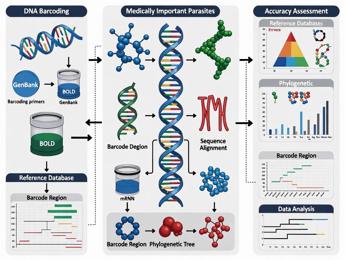
In the field of medical parasitology, accurate pathogen identification is a cornerstone of effective disease control, yet morphological discrimination of many parasite and vector species remains notoriously difficult due to their small size and limited morphological characters [13]. DNA barcoding has emerged as a powerful alternative, using short, standardized gene fragments to assign species identity with objectivity and precision [19]. For researchers and drug development professionals working with medically important parasites, understanding how to quantify the performance of these barcoding methods is critical for reliable application in both research and clinical settings. This guide examines the core metrics and experimental methodologies used to define and assess accuracy in DNA barcoding, providing a framework for the rigorous evaluation of pathogen detection tools.
Core Performance Metrics in DNA Barcoding
The accuracy of DNA barcoding is evaluated through a set of key performance metrics, which are primarily derived from the analysis of a reference sample—a collection of individuals from known species—against which query sequences of unknown taxonomic status are compared [20].
Primary Classification Metrics
The most fundamental metrics assess the method's ability to correctly assign species identities.
- Identification Success Rate: This is the overarching measure of a method's performance. In studies of medically important parasites and vectors, DNA barcoding has demonstrated an accuracy of 94-95% when compared to author identifications based on morphology or other established markers [21] [13].
- Intraspecific and Interspecific Divergence: A fundamental requirement for successful barcoding is a clear "barcode gap" where the genetic differences within a species (intraspecific divergence) are significantly smaller than the differences between species (interspecific divergence).
Comparative Performance of Analysis Methods
Different bioinformatic methods for assigning query sequences to species can yield varying levels of success. A comparative study of these methods revealed that no single method is best in all cases, but the simplest method, 'one nearest neighbour', was often the most reliable across different data set parameters [20]. The performance of all methods is heavily influenced by the molecular diversity of the data set [20].
Table 1: Key Performance Metrics for DNA Barcoding of Parasites and Vectors
| Metric | Typical Value/Definition | Interpretation & Importance |
|---|---|---|
| Overall Accuracy | 94-95% [21] | Accordance with identifications from morphology/other markers; indicates general reliability. |
| Intraspecific Divergence | Usually <2% [22] | Measures genetic variation within a species; lower values suggest more cohesive species. |
| Interspecific Divergence | Often >8% [22] | Measures genetic distance between different species; a clear gap from intraspecific is needed. |
| Barcode Coverage | 43% of 1,403 medically important species [21] | Proportion of species represented in reference databases; impacts general applicability. |
| Sensitivity (mNGS workflow) | 79.5% overall; 88.6% for bacteria [23] | Ability to correctly identify true positives; crucial for diagnostic applications. |
Experimental Protocols for Accuracy Assessment
The evaluation of barcoding accuracy relies on standardized laboratory and analytical workflows. The following protocols detail the key methodologies cited in performance studies.
Standard DNA Barcoding Protocol
The canonical protocol for DNA barcoding involves a series of steps from specimen collection to sequence analysis [19].
- Sample Collection & Vouchering: Specimens are carefully collected, and morphological vouchers are preserved and archived in a collection facility. This creates a permanent physical record linked to the genetic data, which is a standard practice in DNA barcoding [13].
- DNA Extraction: Genomic DNA is purified from a small tissue sample (e.g., leaf disc, insect leg, muscle tissue).
- PCR Amplification: A specific barcode region is amplified using polymerase chain reaction (PCR) with universal primers.
- Sequencing: The amplified product (amplicon) is sequenced, typically using Sanger sequencing [24].
- Data Analysis:
- The sequence is compared to reference databases such as the Barcode of Life Data (BOLD) system or GenBank using tools like BLAST [19] [13].
- Species assignment is made based on high similarity (e.g., ≥98% similarity to a reference sequence) [24].
- Phylogenetic methods like Neighbour-Joining (NJ) or maximum likelihood (PhyML) trees are also used to assess relationships and confirm identity [20].
DNA Barcoding Workflow
Metagenomic Next-Generation Sequencing (mNGS) Protocol
For detecting uncultivable parasites or multiple pathogens from complex samples, mNGS workflows have been developed. A streamlined protocol for acute undifferentiated fever demonstrates a unified approach [23].
- Sample Preparation: Total nucleic acid is isolated separately from 300 μl of EDTA whole blood and 300 μl of plasma.
- Fraction-Specific Processing:
- The plasma isolate is treated with DNase to enrich for viral RNA, followed by depletion of host ribosomal and messenger RNA.
- The whole blood isolate is processed with no additional manipulation to retain DNA and RNA from intracellular pathogens (e.g., Babesia sp., Plasmodium falciparum).
- Nucleic Acid Amplification & Library Preparation:
- The plasma fraction undergoes reverse transcription and sequence-independent single primer amplification (SISPA).
- The whole blood fraction is only reverse transcribed.
- Finally, the two processed fractions are combined into a single sequencing library.
- Sequencing & Analysis: The library is sequenced using either Illumina or Oxford Nanopore Technologies. A mathematical ranking approach (ClinSeq score) is used to quickly differentiate true pathogen signals from background noise [23].
Table 2: Essential Research Reagents for Barcoding and mNGS
| Reagent / Tool | Function | Example Use Case |
|---|---|---|
| Universal Primers (COI, rbcL, ITS) | Amplify standardized barcode regions from diverse specimens. | PCR amplification for species identification of a single parasite [19]. |
| BOLD Systems Database | Centralized repository for DNA barcodes with curated records. | Identifying a query sequence by matching it to a vouchered reference specimen [13]. |
| TURBO DNA-free Kit | DNase treatment to remove DNA and enrich for RNA targets. | Processing plasma samples in mNGS to improve detection of RNA viruses [23]. |
| Host rRNA Depletion Kits | Remove abundant host RNA to increase microbial signal. | Enhancing sensitivity for pathogen detection in complex blood samples [23]. |
| ClinSeq Score Algorithm | Mathematical ranking to prioritize true pathogens in mNGS data. | Reducing false positives and manual interpretation time in clinical diagnostics [23]. |
Analytical Frameworks and Bioinformatics
The bioinformatic processing of sequencing data is critical for accurate species assignment and presents a significant divide between traditional barcoding and more complex metagenomic approaches.
Analytical Methods for DNA Barcoding
The analysis of a single barcode sequence is relatively straightforward and relies on mature tools [24].
- Sequence Quality Control: Tools like Chromas or MEGA are used to check sequencing chromatograms, eliminate sequences with fuzzy bases, and assemble high-quality consensus sequences.
- Species Assignment: The primary method is a similarity search against reference databases. The BOLD database is often preferred as it contains barcode-compliant sequences linked to voucher specimens [13]. A sequence is typically assigned to a species if it shows ≥98% similarity to a reference sequence [24].
- Phylogenetic Analysis: Building trees (e.g., using Neighbour-Joining with Kimura 2-parameter distance) helps visualize the relationship between the query sequence and its nearest neighbours, confirming its placement within a specific species cluster [20].
From Single Sequence to Complex Communities: Metabarcoding
While DNA barcoding identifies a single specimen, metabarcoding extends this principle to identify all organisms within a complex environmental sample [24]. This is highly relevant for studying parasite diversity in hosts or ecosystems.
- Workflow Differences: Metabarcoding uses high-throughput sequencing (HGS) of mixed DNA samples, rather than Sanger sequencing of a single amplicon. The output is not a single sequence, but a sample-sequence-abundance matrix comprising millions of short reads [24].
- Bioinformatic Complexity: Analysis involves multiple steps including quality filtering, clustering sequences into Operational Taxonomic Units (OTUs) or Amplicon Sequence Variants (ASVs), and then annotating these units against reference databases. This process is far more computationally intensive than standard barcoding analysis [24].
Species Identification Logic
Current Coverage and Limitations
Understanding the limitations of DNA barcoding is essential for interpreting results accurately, especially in a medical context.
- Database Coverage: A significant challenge is the incomplete coverage of reference databases. For a checklist of 1,403 species of medically important parasites, vectors, and hazards, barcodes were available for only 43% of all species, though coverage was better (over half) for species of greater medical importance [21] [13]. This gap can severely limit identification success.
- Species Delineation Problems: Barcoding can fail when species are recently diverged, leading to incomplete lineage sorting and a lack of reciprocal monophyly [20]. This can result in shared barcodes between species or high intraspecific divergence, as seen in some tick genera [22].
- Technical Limitations: Standard barcoding with Sanger sequencing cannot identify mixed infections from a single sample. Furthermore, the presence of Wolbachia or other endosymbionts can sometimes confound COI-based identification due to potential horizontal transfer of mitochondria [20].
The accuracy of DNA barcoding in pathogen detection is a multi-faceted concept defined by quantitative metrics like identification success rate, intra- and interspecific divergence, and database coverage. For researchers targeting medically important parasites, the choice between standard barcoding and more comprehensive mNGS workflows depends on the specific diagnostic question, the scale of sampling, and available resources. While barcoding offers a highly accurate and cost-effective method for identifying individual specimens, emerging mNGS and metabarcoding approaches provide powerful, universal tools for uncovering complex and polymicrobial infections. As these technologies evolve and reference databases continue to expand, the precise metrics and frameworks outlined here will be essential for validating their performance and ensuring their reliable application in disease research and public health.
Advanced Methodologies in Practice: From Sample to Sequence
Within the field of medical parasitology, accurate species identification is a cornerstone for diagnosing infections, understanding transmission dynamics, and implementing effective control measures. DNA barcoding has emerged as a powerful tool for this purpose, with the 18S ribosomal DNA (18S rDNA) gene serving as a key target for eukaryotic pathogens [13]. For years, the V9 hypervariable region of the 18S rDNA has been a commonly used barcode for parasite detection and identification. However, the quest for higher resolution, especially for distinguishing between closely related parasite species, has driven the development of novel primer designs that expand the target to encompass the V4 through V9 regions. This guide objectively compares the performance of the novel V4–V9 barcoding approach against the traditional V9 region, providing supporting experimental data to inform researchers, scientists, and drug development professionals.
Primer Design and Workflow
The expansion from the V9 to the V4–V9 region represents a strategic shift towards leveraging longer, more informative DNA sequences. This design uses universal primers F566 and 1776R to amplify a segment spanning over 1 kb, which includes the V4, V5, V6, V7, V8, and V9 variable regions [3] [4]. This broader capture of the 18S rDNA gene provides a substantially greater number of nucleotide characters for phylogenetic analysis and species classification.
A significant challenge in detecting blood parasites using universal primers is the overwhelming amplification of host (e.g., human or cattle) 18S rDNA, which can obscure the target pathogen signal. To address this, the V4–V9 protocol incorporates a sophisticated host DNA suppression system using two distinct types of blocking primers [3] [4]:
- C3 Spacer-Modified Oligo (3SpC3_Hs1829R): This oligonucleotide competes with the universal reverse primer (1776R) for binding to host DNA. Its 3' end is modified with a C3 spacer, which halts polymerase elongation, thereby selectively inhibiting the amplification of host 18S rDNA.
- Peptide Nucleic Acid (PNA) Oligo (PNA_Hs733F): PNA oligos mimic DNA but possess an uncharged backbone, allowing them to bind to complementary host DNA sequences with higher affinity and specificity. Upon binding, they physically block the polymerase, preventing the amplification of the host template.
The following diagram illustrates the complete experimental workflow, from sample preparation to final analysis.
Performance Comparison: V4–V9 vs. V9 Barcoding
Species Identification Accuracy
The primary advantage of the expanded V4–V9 barcode is its enhanced capability for accurate species-level identification, which is particularly critical for the error-prone nanopore sequencing platform. Simulation studies involving major Plasmodium species demonstrated the superior robustness of the longer barcode.
Table 1: Misassignment Rates of Simulated Error-Prone Sequences
| 18S rDNA Region | Error Rate (%) | P. falciparum Misassigned | P. knowlesi Misassigned | P. ovale Misassigned | P. vivax Misassigned |
|---|---|---|---|---|---|
| V9 | 0.05 | 4/1000 | 4/1000 | 0/1000 | 3/1000 |
| V4–V9 | 0.05 | 0/1000 | 0/1000 | 0/1000 | 0/1000 |
| V9 | 0.10 | 10/1000 | 9/1000 | 2/1000 | 17/1000 |
| V4–V9 | 0.10 | 0/1000 | 0/1000 | 0/1000 | 0/1000 |
Data adapted from Supplemental Table 2 in [3] [4]. The table shows the number of sequences misassigned to another species out of 1000 simulated sequences.
The data shows that the V4–V9 region maintained perfect species assignment even at a 0.1% error rate, whereas the V9 region exhibited significant misassignment, which worsened with higher error rates [3] [4]. Furthermore, when using a naive Bayesian classifier, a higher proportion of V9 sequences could not be classified above the confidence threshold compared to V4–V9 sequences as sequencing error rates increased [3].
Analytical Sensitivity in Clinical Samples
The real-world performance of the V4–V9 targeted NGS test was validated using human blood samples spiked with known quantities of parasites. The assay demonstrated high sensitivity, detecting infections with very low parasite densities [3] [4] [25].
Table 2: Detection Sensitivity for Key Blood Parasites
| Parasite Species | Limit of Detection (Parasites/μL of Blood) |
|---|---|
| Trypanosoma brucei rhodesiense | 1 |
| Plasmodium falciparum | 4 |
| Babesia bovis | 4 |
The assay's utility was further confirmed in field applications. Analysis of cattle blood samples successfully revealed multiple Theileria species co-infections within a single host, showcasing its power to resolve complex, real-world infection scenarios that are often missed by traditional, targeted molecular tests [3] [4].
The Scientist's Toolkit: Essential Research Reagents
The successful implementation of the V4–V9 18S rDNA barcoding approach relies on a specific set of reagents and tools. The following table details these key components and their functions.
Table 3: Key Research Reagent Solutions for V4–V9 Barcoding
| Reagent / Tool | Function / Description |
|---|---|
| Universal Primers (F566 & 1776R) | Amplify the ~1.2 kb V4–V9 region of 18S rDNA from a wide range of eukaryotic parasites [3]. |
| Host Blocking Primer (3SpC3_Hs1829R) | C3 spacer-modified oligo that binds to host 18S rDNA and blocks polymerase extension, reducing host background [3] [4]. |
| Host Blocking Primer (PNA_Hs733F) | Peptide Nucleic Acid oligo that binds tightly to host 18S rDNA and sterically inhibits polymerase during PCR [3] [4]. |
| Portable Nanopore Sequencer | Sequencing platform (e.g., MinION) that enables long-read sequencing in resource-limited settings [3] [25]. |
| Phi29 DNA Polymerase | High-fidelity polymerase used in isothermal amplification methods like SWGA for enriching parasite DNA from host background [26]. |
The comparative data clearly demonstrates that expanding the DNA barcode target from the V9 to the V4–V9 region of the 18S rDNA gene significantly enhances resolution for parasite identification. The longer barcode provides a more robust genetic scaffold that mitigates the impact of sequencing errors inherent in portable platforms like nanopore sequencers, leading to fewer species misassignments [3]. Furthermore, the integration of specialized blocking primers is a critical innovation that makes this approach feasible for blood samples by effectively suppressing host DNA, thereby enriching for parasite DNA and achieving high sensitivity.
This novel primer design and associated protocol offer a powerful, comprehensive pathogen detection test. It retains the "open" nature of universal primers—capable of detecting unexpected or novel parasites—while achieving the species-level accuracy often associated with specific PCR assays [3]. This makes it particularly valuable for large-scale surveillance studies, diagnostic validation in endemic areas, and investigating complex multi-species co-infections. For researchers and drug development professionals, this enhanced resolution directly translates to more reliable data on parasite distribution, population genetics, and the true complexity of infections, ultimately informing better-targeted interventions and control strategies.
In the field of molecular parasitology, accurate species identification is crucial for effective disease diagnosis, treatment, and epidemiological surveillance. DNA barcoding using the 18S ribosomal DNA (rDNA) has emerged as a powerful tool for comprehensive parasite detection. However, a significant challenge arises when analyzing blood samples or host tissues, where overwhelming host DNA can swamp the target parasite signal during amplification. This contamination issue severely compromises detection sensitivity and specificity. To address this, researchers have developed sophisticated molecular strategies to selectively inhibit host DNA amplification. Among the most promising approaches are C3 spacer-modified blocking primers and peptide nucleic acid (PNA) clamps. This guide provides an objective comparison of these two techniques, evaluating their performance, applications, and implementation in parasite research.
Understanding the Host Contamination Problem
In molecular diagnostics of blood parasites, host DNA constitutes the majority of genetic material in extracted samples. When universal 18S rDNA primers are applied, they amplify both parasite and host sequences, with the latter dominating the reaction due to their abundance. This "swamping effect" can completely obscure the parasite signal, leading to false negatives, particularly with low-parasitemia infections.
Traditional methods like microscopic examination, while affordable, require expert microscopists and have poor species-level identification capabilities [3]. Species-specific molecular tests like PCR or rapid diagnostic tests offer sensitivity but can only detect targeted parasites, requiring prior knowledge of the pathogen [3]. The need for comprehensive detection methods that can identify unexpected or novel parasites has driven the development of targeted next-generation sequencing (NGS) approaches with effective host suppression strategies [3].
Head-to-Head Comparison: C3 Spacer vs. PNA Blocking Primers
The following table summarizes the core characteristics, mechanisms, and performance metrics of C3 spacer and PNA blocking primers:
Table 1: Direct Comparison of C3 Spacer and PNA Blocking Primers
| Feature | C3 Spacer-Modified Blocking Primers | PNA Oligonucleotide Blockers |
|---|---|---|
| Chemical Structure | Standard oligonucleotide with 3'-end C3 spacer (3 hydrocarbons) [27] | Synthetic DNA mimic with peptide backbone [28] |
| Mechanism of Action | Competes with universal primers; C3 spacer halts polymerase elongation [3] | Binds strongly to DNA; physically blocks polymerase progression [3] [28] |
| Design Strategy | Sequence-specific binding overlapping primer sites [3] | Short, high-specificity sequences (e.g., 17-mers) within amplicon [29] |
| Reported Inhibition Efficiency | Variable: 3.3%–32.9% to >99% depending on design [28] [27] | Consistently high: 80%–99.9% across studies [3] [28] [29] |
| Optimal Application Context | Effective in specific host-parasite systems with optimized design | Broadly effective across systems, especially with high host DNA burden |
| Cost Considerations | Lower synthesis cost, similar to standard primers | Higher synthesis cost due to specialized chemistry |
| Experimental Flexibility | Easier to design and optimize | Requires more stringent design and validation |
The differential effectiveness of these blockers is clearly demonstrated in direct comparative studies. For instance, one investigation reported that a PNA clamp suppressed 99.3%–99.9% of fish DNA amplification in herbivorous fish diet analysis, whereas a blocking primer achieved only 3.3%–32.9% suppression in the same system [28]. Similarly, in Anopheles mosquito microbiome studies, PNA blockers reduced host 18S rDNA sequences by more than 80%, while anneal-inhibiting blocking primers showed negligible efficiency [29].
Experimental Protocols and Performance Data
Implementation in Parasite Detection
Recent research has successfully integrated both blocking strategies into a nanopore-based NGS workflow for blood parasite identification. The approach combined universal primers targeting the V4–V9 region of 18S rDNA with two blocking primers: a C3 spacer-modified oligo competing with the universal reverse primer and a PNA oligo inhibiting polymerase elongation [3]. This combined method demonstrated remarkable sensitivity, detecting Trypanosoma brucei rhodesiense, Plasmodium falciparum, and Babesia bovis in human blood samples spiked with as few as 1, 4, and 4 parasites per microliter, respectively [3]. The test also successfully identified multiple Theileria species co-infections in field cattle blood samples [3].
Key Experimental Parameters
The table below outlines critical experimental parameters and performance outcomes from recent studies:
Table 2: Experimental Parameters and Performance Metrics
| Study System | Blocker Type | Optimal Concentration | Key Performance Outcome | Reference |
|---|---|---|---|---|
| Blood parasite detection (Human) | Combined C3 spacer + PNA | Not specified | Detection sensitivity: 1-4 parasites/μL | [3] |
| Herbivorous fish diet analysis | PNA clamp | Not specified | 99.3%-99.9% host suppression | [28] |
| Salmonid parasite communities | C3 spacer blocker | 0.5-2.0 μM | Improved parasite detection in gill swabs | [30] |
| Shrimp eukaryotic microbiota | C3 spacer (X-BP2-DPO) | Not specified | 99% inhibition of host 18S amplification | [27] |
| Anopheles gambiae microbiome | PNA blocker | 1.0-1.5 μM | >80% reduction of mosquito 18S sequences | [29] |
Detailed Methodology: Combined Blocking Approach for Blood Parasites
The following workflow illustrates the experimental protocol for implementing both blocking strategies in parasite detection:
Diagram 1: Experimental workflow for blood parasite detection using C3 spacer and PNA blocking primers.
Key experimental steps:
Primer Design: Universal primers F566 and 1776R targeting the V4-V9 region of 18S rDNA are selected for broad eukaryotic coverage [3].
Blocking Primer Design:
PCR Amplification: The reaction incorporates both universal primers and blocking primers at optimized concentrations to selectively amplify parasite DNA while suppressing host amplification.
Sequencing and Analysis: Amplified products are sequenced on a portable nanopore platform, followed by bioinformatic classification of parasite species.
The Scientist's Toolkit: Essential Research Reagents
Table 3: Key Research Reagent Solutions for Host Blocking Experiments
| Reagent / Tool | Function | Implementation Example |
|---|---|---|
| C3 Spacer-Modified Primers | Inhibits host DNA amplification by blocking polymerase extension | 3SpC3_Hs1829R for human 18S rDNA suppression [3] |
| PNA Clamps | Synthetic DNA analogs that block polymerase progression with high affinity | PNA_Hs733F for targeted human sequence inhibition [3] |
| Universal 18S rDNA Primers | Amplifies eukaryotic DNA across diverse taxa | F566 and 1776R primers for V4-V9 region amplification [3] |
| Portable Nanopore Sequencer | Enables field-deployable, real-time sequencing | MinION for resource-limited settings [3] |
| Bioinformatic Classification Tools | Analyzes error-prone long-read sequences for species identification | BLASTn with modified parameters for error-tolerant matching [3] |
Molecular Mechanisms of Host DNA Suppression
The following diagram illustrates how C3 spacer and PNA blocking primers function at the molecular level to suppress host DNA amplification:
Diagram 2: Molecular mechanisms of C3 spacer and PNA blocking primers.
Both C3 spacer and PNA blocking primers offer valuable strategies for combating host contamination in parasite DNA barcoding applications. The choice between them depends on specific research requirements:
- C3 spacer blockers provide a cost-effective solution that can achieve high suppression efficiency with careful design optimization, particularly valuable for resource-limited settings.
- PNA clamps deliver consistently superior performance across diverse experimental conditions, making them ideal for challenging applications with extreme host-to-parasite DNA ratios.
The emerging approach of combining both technologies in a single assay represents the current state-of-the-art, leveraging the complementary strengths of each method to achieve maximum sensitivity and specificity. As DNA barcoding continues to transform parasitology research, these host suppression strategies will play an increasingly vital role in enabling accurate detection and identification of medically important parasites.
Portable nanopore sequencers, pioneered by devices like the Oxford Nanopore MinION, have transitioned genomic analysis from centralized laboratories to field settings, creating new possibilities for real-time pathogen detection and biodiversity monitoring. For researchers studying medically important parasites, these instruments offer a compelling solution for real-time genomic surveillance and species identification in resource-limited environments where traditional sequencing infrastructure is unavailable. This evaluation examines the performance of portable nanopore sequencing platforms specifically within the context of parasite research, comparing their technical capabilities against alternative sequencing technologies and assessing their practical application for DNA barcoding accuracy in field conditions.
The unique value proposition of portable nanopore sequencers for parasite research lies in their long-read capabilities, minimal infrastructure requirements, and rapid turnaround time. These characteristics enable researchers to conduct comprehensive genomic investigations of complex parasitic organisms in field settings, from outbreak zones to remote biodiversity hotspots, fundamentally changing the paradigm of what's possible in field genomics.
Technical Performance Comparison of Sequencing Platforms
Understanding the relative strengths and limitations of available sequencing technologies is essential for selecting the appropriate platform for specific research applications. The table below provides a detailed comparison of portable nanopore sequencers against other common sequencing platforms, with particular emphasis on parameters critical for parasite research.
Table 1: Performance Comparison of Sequencing Technologies for Parasite Research
| Platform Characteristic | Portable Nanopore (MinION) | High-Throughput Nanopore (PromethION) | Illumina Short-Read | PacBio Long-Read |
|---|---|---|---|---|
| Read Length | Up to 2+ Mb [31] | Up to 2+ Mb [31] | 50-600 bp [31] | 10-25 kb average [31] |
| Accuracy (Raw Reads) | ~99% with Q20+ chemistry [32] | ~99% with Q20+ chemistry [32] | >99.9% [33] | >99.9% (HiFi mode) [31] |
| Portability | <100g weight, USB-powered [34] | Benchtop system [33] | Benchtop systems | Benchtop systems |
| Time to Result | Real-time data, hours from sample to answer [32] [35] | Real-time data, but longer run times | Days including library prep and run | Days including library prep and run |
| Cost per Sample | Low for field applications [36] | Moderate to high [36] | Low for high-throughput | High |
| DNA Modification Detection | Direct detection of base modifications [34] [31] | Direct detection of base modifications [31] | Requires special treatments | Direct detection |
| Complex Genome Resolution | Excellent for repetitive regions and structural variants [37] | Excellent for complex genomes [31] | Poor for repeats and structural variants | Good for complex regions |
For parasite research, several key distinctions emerge from this comparison. Portable nanopore sequencers provide unmatched portability and rapid turnaround, crucial for field applications where timely results impact research outcomes. The long-read capability is particularly valuable for resolving complex parasitic genomes characterized by repetitive elements and structural variations, as demonstrated in the sequencing of Trypanosoma cruzi, the causative agent of Chagas disease [37]. This parasite's genome contains highly repetitive regions and diverse multi-copy gene families that challenge short-read technologies, but which have been successfully resolved using nanopore sequencing [37].
While raw read accuracy has historically been a limitation of nanopore technology, recent developments have substantially improved this metric. The introduction of Q20+ chemistry has enabled raw read accuracy exceeding 99%, addressing what was previously a significant concern for applications requiring high base-calling precision [32]. This improvement is particularly relevant for DNA barcoding applications where single-nucleotide polymorphisms may differentiate between parasite species.
DNA Barcoding Accuracy Assessment in Parasite Research
DNA barcoding represents one of the most promising applications of portable nanopore sequencing in parasite research, enabling species identification through targeted amplification and sequencing of specific genetic regions. The accuracy of this approach depends on multiple factors, including target region selection, bioinformatic processing, and the inherent capabilities of the sequencing platform.
Experimental Approaches for Enhanced Accuracy
Recent research has demonstrated innovative methodologies to optimize DNA barcoding accuracy for parasite detection using portable nanopore sequencers. A 2025 study developed a targeted next-generation sequencing approach specifically designed for blood parasite identification in resource-limited settings [4]. The experimental protocol incorporated several key innovations:
Extended Target Regions: The researchers designed a DNA barcoding strategy targeting the 18S rDNA V4–V9 region (approximately >1 kb) instead of the more commonly used V9 region alone. This extended barcode provided significantly improved species discrimination compared to shorter regions, with misassignment rates decreasing from up to 17% with V9 alone to 0% with the V4–V9 region at error rates of 0.1 [4].
Host DNA Suppression: To overcome the challenge of host DNA contamination in blood samples, the protocol incorporated two blocking primers: a C3 spacer-modified oligo competing with the universal reverse primer and a peptide nucleic acid (PNA) oligo that inhibits polymerase elongation. This combination selectively reduced amplification of host DNA, enriching parasite sequences without requiring physical separation methods [4].
Sensitivity Validation: The assay demonstrated high sensitivity in spike-in experiments, successfully detecting Trypanosoma brucei rhodesiense, Plasmodium falciparum, and Babesia bovis in human blood samples with concentrations as low as 1, 4, and 4 parasites per microliter, respectively [4].
The workflow for this approach can be visualized as follows:
Figure 1: Workflow for parasite DNA barcoding using portable nanopore sequencing, incorporating host DNA suppression and extended target regions for enhanced species identification.
Bioinformatic Considerations for Field Applications
The accuracy of species identification in DNA barcoding depends heavily on bioinformatic processing, which presents unique challenges in field settings. Key considerations include:
Basecalling Algorithms: The conversion of raw electrical signals to nucleotide sequences has evolved from Hidden Markov Models to deep learning approaches, with current algorithms achieving significantly higher accuracy [31]. The development of real-time basecalling enables analysis during sequencing runs, providing preliminary results without waiting for run completion.
Reference Databases: Comprehensive and curated reference databases are essential for accurate species assignment. Studies have noted that ongoing improvements to public reference databases have increased match rates for parasite identification, from 51% in 2023 to 62% in 2025 when using COI barcodes [38]. This highlights the importance of using current, well-annotated databases for species identification.
Computational Requirements: While early nanopore analysis required substantial computational resources, recent advancements have optimized algorithms for field deployment. The development of portable computation solutions like the MinIT and optimized software pipelines has enabled complete analysis workflows on laptop-based systems in field settings [32].
Field Application Case Studies
The practical performance of portable nanopore sequencers is best evaluated through real-world applications in parasite research and related fields. Several case studies demonstrate their capabilities and limitations in field conditions.
Parasite Genome Assembly in Resource-Limited Settings
A 2023 study successfully established a scalable nanopore sequencing pipeline for Trypanosoma cruzi, the parasite causing Chagas disease [37]. This research is particularly noteworthy because:
- The team generated a high-quality genome assembly using nanopore sequencing alone, without supplementation from other technologies
- They resolved the parasite's highly repetitive genome, which contains approximately 27% transposable elements
- The assembly provided insights into genome diversification mechanisms, with transposable elements located significantly closer to multi-gene family coding sequences than to other genes
- The approach demonstrated feasibility for studying important genomic features in hybrid strains with relatively modest sequencing data requirements
This case highlights how portable nanopore sequencing can overcome challenges that have historically complicated parasite genomics, particularly for organisms with complex, repetitive genomes that resist assembly with short-read technologies.
Rapid Pathogen Detection in Agricultural Settings
A 2025 study implemented a portable, nanopore-based genotyping platform for near real-time detection of Puccinia graminis f. sp. tritici lineages and fungicide resistance [35]. This application demonstrates capabilities directly relevant to parasite research:
- The platform enabled complete genotyping within 48 hours of sample collection, from leaf sampling to lineage identification and resistance profiling
- Researchers sequenced a targeted panel of 276 genes using a portable MinION sequencer and standard laptop
- The system was successfully deployed in Kenya and Ethiopia, placing powerful genomic tools directly in the hands of local teams
- The approach supported faster, more informed disease control strategies against this economically significant pathogen
This case illustrates the operational feasibility of portable nanopore sequencing in field conditions and its utility for rapid decision-making in response to pathogenic threats.
Essential Research Reagent Solutions
Successful implementation of portable nanopore sequencing for parasite DNA barcoding requires specific reagents and materials optimized for field applications. The following table details key components of the research toolkit:
Table 2: Essential Research Reagent Solutions for Parasite DNA Barcoding
| Reagent/Material | Function | Field-Specific Considerations |
|---|---|---|
| DNeasy Blood & Tissue Kit (Qiagen) | DNA extraction from diverse sample types | Stable at room temperature; minimal equipment requirements [39] |
| Ligation Sequencing Kit (ONT) | Library preparation for nanopore sequencing | Compatible with field conditions; available in lyophilized format for cold-chain independence [32] |
| V4-V9 18S rDNA Primers | Amplification of extended barcode region | Enables higher species discrimination compared to shorter regions [4] |
| Host Blocking Primers (C3 spacer/PNA) | Selective inhibition of host DNA amplification | Critical for blood samples with high host:parasite ratios [4] |
| Q20+ Chemistry (ONT) | Enhanced sequencing accuracy | Raw read accuracy >99%; improved homopolymer resolution [32] |
| Portable Computing Solution | Real-time basecalling and analysis | MinIT or laptop-based analysis enables complete workflow in field [32] |
Portable nanopore sequencers have evolved from promising technological innovations to robust tools for field-based parasite research, offering compelling advantages in portability, real-time analysis, and long-read capabilities. While accuracy limitations historically constrained their application for DNA barcoding, recent developments in chemistry, experimental protocols, and bioinformatics have substantially addressed these concerns.
The unique value of these platforms lies in their ability to generate actionable genomic data in close proximity to sample collection, enabling rapid species identification, outbreak response, and biodiversity assessment in environments where traditional sequencing infrastructure is unavailable. For researchers studying medically important parasites, portable nanopore sequencing represents not merely a convenient alternative to laboratory-based approaches, but rather enables fundamentally new research paradigms that bridge the gap between field observation and genomic analysis.
As the technology continues to evolve, with ongoing improvements in accuracy, throughput, and field readiness, portable nanopore sequencers are poised to become increasingly central to parasite research and surveillance programs worldwide, particularly in resource-limited settings where the burden of parasitic diseases is often highest.
Accurate detection and species identification of blood-borne parasites such as Plasmodium, Trypanosoma, and Babesia are critical for diagnosis, treatment, and epidemiological control. These parasites continue to pose significant global health threats, with malaria alone causing an estimated 247 million cases and 619,000 deaths globally in 2021 [40]. Conventional diagnostic methods, particularly light microscopy, have remained the gold standard in many settings despite limitations in sensitivity and species-level resolution [40]. In recent years, molecular techniques have emerged as powerful tools capable of detecting low-level parasitemia and differentiating between species with high precision. This guide objectively compares the detection performance of various diagnostic platforms, focusing on the critical metrics of sensitivity, specificity, and detection limits, framed within the broader context of DNA barcoding accuracy assessment for medically important parasites.
Established Diagnostic Platforms and Their Performance
Microscopy and Rapid Diagnostic Tests
For over a century, microscopic examination of stained blood smears has served as the cornerstone of parasite diagnosis. This method allows for the direct visualization of parasites, determination of parasitic stages, and estimation of parasitemia. According to the World Health Organization, microscopy can detect malaria parasites at densities of 50 to 500 parasites/μL, with sensitivity highly dependent on the microscopist's expertise [41] [40]. In optimal conditions, skilled technicians can achieve a detection limit of approximately 50 parasites/μL, equivalent to 0.001% of infected red blood cells [41]. However, routine diagnostic laboratories often achieve lower sensitivity, detecting on average 500 parasites/μL (0.01% infected RBCs) [41].
Rapid diagnostic tests (RDTs) based on immunochromatographic principles have expanded diagnostic access in resource-limited settings. These tests typically detect specific malaria antigens such as histidine-rich protein 2 (HRP2) or Plasmodium lactate dehydrogenase (pLDH) with a sensitivity of approximately 100 parasites/μL [41]. A significant limitation of both microscopy and RDTs is their poor performance in detecting low-level parasitemia, which is particularly problematic in asymptomatic carriers, during convalescence, or in mixed infections [40].
Table 1: Performance Characteristics of Conventional Diagnostic Methods
| Diagnostic Method | Detection Limit (parasites/μL) | Sensitivity Range | Specificity Range | Key Limitations |
|---|---|---|---|---|
| Light Microscopy | 50–500 | Varies with technician expertise (75–95%) | High with experienced staff | Requires skilled technicians; poor species differentiation for some species |
| Rapid Diagnostic Tests (RDTs) | ~100 | ~100 parasites/μL [41] | Lower than microscopy and PCR [41] | Cannot detect acute disease before immune response; cannot distinguish active from past infection |
| Indirect Fluorescent Antibody Test (IFAT) | N/A (serological) | More sensitive than microscopy for Babesia [41] | Specificity challenges [41] | Cannot detect acute disease before immune response; cannot distinguish active from past infection |
Molecular Detection Methods
Molecular diagnostics have revolutionized parasite detection by offering significantly improved sensitivity and specificity compared to conventional methods. These techniques target parasite-specific DNA sequences, enabling detection even at very low parasitemia levels.
PCR and Real-Time PCR (qPCR)
Polymerase chain reaction (PCR) and its quantitative real-time variant (qPCR) have become reference standards in molecular parasitology. These methods typically target multi-copy genes such as the 18S ribosomal RNA (rRNA) gene, which provides exceptional specificity and conservation across Plasmodium and Babesia species [41].
Standard PCR assays for malaria demonstrate sensitivity and specificity ranging from 75% to 90.9% and 91.2% to 97%, respectively, with a remarkable limit of detection as low as 1–5 parasites/μL [41]. For Babesia diagnosis, PCR exhibits even higher performance, with sensitivity and specificity of 94.2% to 100% and 97.1% to 100%, respectively [41].
Real-time PCR (qPCR) technology has shown superior sensitivity compared to conventional PCR, with a limit of detection (LOD) below 0.1 parasites/μL for malaria [41] and 1–3 parasites/μL for Babesia [41]. A 2024 validation study of real-time PCR assays for detecting Plasmodium and Babesia species reported detection limits ranging from 30 to 0.0003 copies/μL for different Plasmodium species, and 0.2 copies/μL for Babesia species [42]. The assay demonstrated 100% sensitivity for detecting most Plasmodium and Babesia species, except for P. falciparum (97.7%) and B. microti (12.5%), with the latter attributed to limitations in microscopy for species identification rather than the PCR itself [42].
Table 2: Performance Characteristics of Molecular Diagnostic Methods
| Molecular Method | Detection Limit | Sensitivity | Specificity | Target Genes/Region |
|---|---|---|---|---|
| Conventional PCR | 1–5 parasites/μL (Plasmodium) [41] | 75–90.9% (Plasmodium) [41]; 94.2–100% (Babesia) [41] | 91.2–97% (Plasmodium) [41]; 97.1–100% (Babesia) [41] | 18S rRNA, other species-specific genes |
| Real-Time PCR (qPCR) | <0.1 parasites/μL (Plasmodium) [41]; 1–3 parasites/μL (Babesia) [41]; 0.0003–30 copies/μL (Plasmodium species) [42] | 90–97% (Plasmodium) [41]; 95.9–100% (Babesia) [41]; 97.7–100% (2024 validation) [42] | Up to 100% [41] | 18S rRNA, other multi-copy genes |
| Loop-Mediated Isothermal Amplification (LAMP) | 0.3–2.0 parasites/μL (Plasmodium) [43] | Similar to reference PET-PCR [43] | Similar to reference PET-PCR [43] | Genus-specific sequences (e.g., Illumigene Malaria LAMP) |
| DNA Barcoding (COI/18S) | Varies with platform | 94–95% accuracy for parasite identification [21] | 94–95% accuracy for parasite identification [21] | COI for vectors [44]; 18S V4–V9 for parasites [3] |
Loop-Mediated Isothermal Amplification (LAMP)
Loop-mediated isothermal amplification (LAMP) represents a significant advancement in molecular diagnostics, particularly for resource-limited settings. Unlike PCR, LAMP amplifies nucleic acids at a constant temperature (isothermal), typically around 62°C–65°C, eliminating the need for thermal cyclers [43]. The Illumigene Malaria LAMP assay, for instance, can detect Plasmodium species at the genus level with limits of detection of 2.0 parasites/μL using the simple filtration prep (SFP) method and 0.3 parasites/μL using the gravity-driven filtration prep (GFP) method [43]. This performance meets or exceeds the WHO recommended sensitivity of 2 parasites/μL [43]. Field evaluations in Senegal demonstrated that LAMP assays reliably detected Plasmodium parasites in a simple, low-tech format, providing a viable alternative to more complex molecular tests [43].
DNA Barcoding and Advanced Sequencing Approaches
DNA barcoding has emerged as a powerful tool for species identification, utilizing short standardized gene sequences as genetic markers. The mitochondrial cytochrome c oxidase subunit 1 (COI) gene has been widely used for mosquito vectors [44], while the 18S ribosomal RNA gene is preferred for parasite identification [3].
Studies have demonstrated that DNA barcoding achieves 94–95% accuracy in identifying medically important parasites and vectors [21]. Research in Singapore successfully used COI barcoding to identify 45 mosquito species with 100% success rate, highlighting its utility as a complement to morphology-based identification [44].
Recent advancements in DNA barcoding have focused on improving species-level resolution using longer gene regions. A 2025 study developed a targeted next-generation sequencing approach using the 18S rDNA V4–V9 region, which outperformed the commonly used V9 region for species identification [3]. This approach, combined with blocking primers to suppress host DNA amplification, successfully detected Trypanosoma brucei rhodesiense, Plasmodium falciparum, and Babesia bovis in human blood samples spiked with as few as 1, 4, and 4 parasites per microliter, respectively [3]. This represents a significant improvement in sensitivity compared to traditional barcoding approaches.
Experimental Protocols for Optimal Detection
Real-Time PCR Protocol for Plasmodium and Babesia Detection
A validated real-time PCR protocol for simultaneous detection of Plasmodium and Babesia species involves several critical steps. First, DNA is extracted from blood samples using commercial kits, with recommendations for including control samples. The assay typically targets the 18S rRNA gene using species-specific primers and probes in a multiplex reaction format. Reaction conditions include an initial denaturation at 95°C for 5 minutes, followed by 45 cycles of denaturation at 95°C for 15 seconds and annealing/extension at 60°C for 1 minute. This protocol has demonstrated limits of detection ranging from 0.0003 to 30 copies/μL for different Plasmodium species and 0.2 copies/μL for Babesia species, with no cross-reactivity observed among 64 DNA samples from various microorganisms [42].
DNA Barcoding Workflow with Nanopore Sequencing
The emerging protocol for enhanced blood parasite identification using V4–V9 18S rDNA barcoding on a nanopore platform involves a multi-step process. First, universal primers (F566 and 1776R) are used to amplify the >1 kb V4–V9 region of the 18S rDNA gene. To address the challenge of overwhelming host DNA in blood samples, two blocking primers are employed: a C3 spacer-modified oligo competing with the universal reverse primer and a peptide nucleic acid (PNA) oligo that inhibits polymerase elongation. The amplified products are then sequenced on a portable nanopore platform, and the resulting sequences are classified using blastn with adjusted parameters or a ribosomal database project (RDP) naive Bayesian classifier [3]. This approach has proven particularly valuable for detecting multiple parasite co-infections in field samples [3].
Visualization of Diagnostic Pathways and Workflows
This workflow illustrates the pathway from sample collection through various molecular detection methods to performance outcomes. The integration of multiple approaches enables comprehensive parasite detection with high sensitivity and specific species identification, addressing key limitations of conventional microscopy.
Essential Research Reagent Solutions
Successful implementation of sensitive detection assays for blood parasites requires specific research reagents and materials. The following table details key solutions and their functions in the experimental workflows discussed.
Table 3: Essential Research Reagents for Parasite Detection Assays
| Reagent/Material | Function | Application Examples |
|---|---|---|
| 18S rRNA Primers & Probes | Amplification and detection of target sequences in PCR/qPCR | Plasmodium and Babesia species identification [41] [42] |
| COI Gene Primers | DNA barcoding of mosquito vectors and some parasites | Mosquito species identification [44] |
| Blocking Primers (C3 spacer/PNA) | Suppress host DNA amplification in universal PCR | Enrich parasite DNA in blood samples for sequencing [3] |
| LAMP Reagents (lyophilized) | Isothermal amplification in low-resource settings | Illumigene Malaria LAMP assay [43] |
| Nanopore Sequencing Kits | Portable, real-time DNA sequencing | Field identification of multiple parasite species [3] |
| DNA Extraction Kits | Nucleic acid purification from blood samples | Sample preparation for all molecular assays |
| Positive Control DNA | Assay validation and quality control | Ensuring PCR efficiency and reliability |
The landscape of parasite detection has evolved significantly from reliance on morphological characteristics to sophisticated molecular assays capable of detecting single-parasite levels. While microscopy remains important in resource-limited settings, molecular techniques including real-time PCR, LAMP, and DNA barcoding offer substantially improved sensitivity and species discrimination. The integration of advanced sequencing technologies, particularly portable platforms coupled with optimized barcoding regions and host DNA suppression techniques, represents the cutting edge of diagnostic capabilities. These advancements directly support the broader thesis of DNA barcoding accuracy assessment by providing researchers and clinicians with validated, highly sensitive tools for parasite detection and identification. As these technologies continue to evolve, they promise to further enhance our ability to detect and monitor parasitic diseases with unprecedented precision, ultimately contributing to improved patient outcomes and more effective public health interventions.
The accurate identification of species is a cornerstone of research in parasitology, epidemiology, and drug development. For medically important parasites and vectors, traditional morphological identification is often insufficient, particularly for immature stages, cryptic species, or damaged specimens [13]. Molecular techniques have therefore become indispensable tools for precise species determination. Among these, DNA barcoding and multiplex PCR represent two powerful but fundamentally different approaches. This guide provides an objective comparison of these methodologies, focusing on their performance characteristics, experimental requirements, and optimal applications within biomedical research.
DNA barcoding, primarily using the mitochondrial cytochrome c oxidase I (COI) gene, serves as a broad-spectrum molecular identification system that compares unknown sequences to reference databases [45] [13]. In contrast, multiplex PCR is a targeted detection method that simultaneously amplifies specific sequences of pre-selected species in a single reaction [46]. The choice between these techniques has significant implications for research outcomes, particularly in studies involving disease vectors like mosquitoes or ticks where accurate identification directly influences control strategies and pathogen transmission studies [45] [47].
DNA Barcoding: A Broad-Spectrum Identification Approach
DNA barcoding operates on the principle of using a short, standardized genetic marker to identify organisms by comparing sequences to a reference library. The most common barcode region is the 650-basepair region of the mitochondrial cytochrome c oxidase I (COI) gene, which typically shows low intra-species variation but high inter-species divergence [45] [13]. This technique functions as a molecular diagnostic tool that can identify known species and flag potentially novel ones based on genetic distance thresholds [47].
The methodology involves DNA extraction, PCR amplification of the barcode region using universal primers, sequencing (typically via Sanger sequencing), and sequence comparison against databases such as GenBank or the Barcode of Life Data (BOLD) system [45] [13]. Its primary strength lies in its universality—the same fundamental approach can be applied across diverse taxonomic groups without prior knowledge of the specimen's identity.
Multiplex PCR: Targeted Multi-Species Detection
Multiplex PCR is a variant of the polymerase chain reaction that enables simultaneous amplification of multiple target sequences in a single reaction by incorporating more than one pair of primers [46]. This technique conserves valuable reagents, sample material, and processing time compared to running multiple singleplex reactions [48]. The fundamental principle involves carefully designing primer sets that specifically bind to unique genetic regions of different target species while ensuring compatibility in reaction conditions [46].
The development of an efficient multiplex PCR system requires meticulous optimization to address challenges such as primer dimer formation, preferential amplification of certain targets, and competition for reaction components [46] [48]. When properly optimized, multiplex PCR allows researchers to screen for a predetermined set of species of interest with high specificity and sensitivity, making it particularly valuable for surveillance programs targeting specific pathogens or vectors [45].
Performance Comparison and Experimental Data
Direct Comparative Study in Mosquito Surveillance
A recent large-scale study directly compared these techniques for identifying container-breeding mosquito species in Austria, analyzing 2,271 ovitrap samples collected in 2021 and 2022 [45] [49] [50]. The results demonstrated clear operational differences between the methodologies:
Table 1: Performance comparison of multiplex PCR versus DNA barcoding for mosquito surveillance
| Performance Metric | Multiplex PCR | DNA Barcoding |
|---|---|---|
| Success Rate | 1,990/2,271 samples (87.6%) | 1,722/2,271 samples (75.8%) |
| Mixed Species Detection | 47 samples | Not possible with standard Sanger sequencing |
| Throughput Capacity | High (simultaneous detection) | Lower (individual processing) |
| Identification Scope | Predefined Aedes species | Potentially any species with barcode reference |
This study revealed that multiplex PCR not only achieved higher overall identification success but also detected mixed-species compositions in 47 samples that would have been missed by standard DNA barcoding protocols [45]. This capability is particularly valuable when analyzing mosquito eggs from ovitraps, where multiple species may oviposit on the same substrate [45] [50].
Technical Performance Characteristics
Beyond the direct comparison in mosquito surveillance, each technique exhibits distinct technical performance characteristics:
Table 2: Technical characteristics of multiplex PCR versus DNA barcoding
| Technical Aspect | Multiplex PCR | DNA Barcoding |
|---|---|---|
| Specificity | High for targeted species | Broad across taxa |
| Sensitivity | Can be optimized for low-abundance targets | Standard PCR sensitivity |
| Multi-Species Detection | inherent capability | Limited with Sanger sequencing |
| Novel Species Detection | None | Possible through genetic distances |
| Quantitative Potential | Possible with qPCR formats | Not inherently quantitative |
| Automation Potential | High | Moderate |
Multiplex PCR's main limitation is its targeted nature—it can only detect species for which specific primers have been included in the assay design [46]. DNA barcoding, while broader in scope, faces challenges when processing mixed samples because Sanger sequencing typically produces unreadable chromatograms when multiple templates are amplified simultaneously [45]. Additionally, the universal primers used in DNA barcoding may not amplify all taxa with equal efficiency, potentially leading to failed reactions for certain species [47].
Experimental Protocols and Methodologies
DNA Barcoding Protocol for Medically Important Vectors
The standard DNA barcoding protocol involves several key steps that remain consistent across various research applications:
Specimen Collection and Preservation: Specimens should be preserved in 95-100% ethanol or at -80°C to prevent DNA degradation. Proper morphological documentation and voucher specimen retention are critical for validation [13].
DNA Extraction: Commercial kits (e.g., DNeasy Blood and Tissue Kit) typically provide reliable results. For difficult specimens such as ticks or arthropods with tough exoskeletons, additional mechanical disruption may be necessary [47].
PCR Amplification: The standard COI barcode region is amplified using universal primers. A typical 50μL reaction contains:
- 1X PCR buffer
- 2.0-2.5mM MgCl₂
- 0.2mM of each dNTP
- 0.2-0.5μM of each primer
- 1-2 units of DNA polymerase
- 2-5μL of DNA template
Thermal cycling conditions typically include an initial denaturation at 94°C for 2-5 minutes, followed by 35-40 cycles of denaturation (94°C, 30-60 seconds), annealing (45-55°C, 30-60 seconds), and extension (72°C, 60-90 seconds), with a final extension at 72°C for 5-10 minutes [47].
Sequencing and Analysis: PCR products are sequenced bidirectionally using Sanger sequencing. The resulting sequences are edited, assembled, and compared to reference databases using alignment tools and distance-based algorithms (e.g., BLASTn, nearest-neighbor methods) [47].
Multiplex PCR Development and Optimization
Developing a robust multiplex PCR assay requires systematic optimization:
Primer Design: Design primers with similar melting temperatures (usually 60±2°C) and lengths (18-25 base pairs) to ensure compatibility. Avoid complementary sequences at 3' ends to prevent primer-dimer formation. Primer concentration typically ranges from 0.1-0.5μM for each primer pair and may require empirical optimization [46] [48].
Reaction Optimization: A standard multiplex PCR reaction includes:
- 1X PCR buffer
- 2.0-4.0mM MgCl₂ (often higher than singleplex PCR)
- 0.2-0.4mM of each dNTP
- Optimized concentrations of each primer pair
- 1.5-3.0 units of DNA polymerase
- 2-5μL DNA template
Hot-start polymerase is recommended to reduce non-specific amplification [46].
Thermal Cycling Parameters: Use a touchdown PCR protocol or gradual annealing temperature optimization to enhance specificity. Extension times should be sufficient for the longest amplicon [45].
Validation: Validate the assay against reference specimens and compare sensitivity and specificity to singleplex PCR or DNA barcoding [45] [48].
The following diagram illustrates the key decision points in selecting and implementing these molecular identification methods:
Research Reagent Solutions and Essential Materials
Successful implementation of either technique requires specific reagents and materials. The following table outlines essential components for both methods:
Table 3: Essential research reagents and materials for species identification methods
| Reagent/Material | Function | Application in DNA Barcoding | Application in Multiplex PCR |
|---|---|---|---|
| DNA Extraction Kits (e.g., DNeasy Blood & Tissue Kit) | Nucleic acid purification | Standard protocol | Standard protocol |
| Universal COI Primers | Amplification of barcode region | Essential | Not typically used |
| Species-Specific Primers | Target-specific amplification | Not used | Critical component |
| DNA Polymerase (e.g., Taq polymerase) | DNA amplification | Standard concentration | May require increased concentration [46] |
| dNTP Mix | Nucleotides for DNA synthesis | Standard concentration (0.2mM each) | Standard concentration (0.2mM each) |
| MgCl₂ Solution | Cofactor for polymerase activity | Typically 1.5-2.5mM | Often higher (2.0-4.0mM) [46] |
| PCR Buffer | Reaction environment optimization | Standard composition | May require optimization with additives [46] |
| Agarose Gel | Amplicon visualization and size verification | Quality control | Essential for distinguishing multiple products |
| Sequencing Reagents | Sequence determination | Critical final step | Optional for validation |
| Size Selection Beads (e.g., SPRI beads) | Fragment size selection | Not always necessary | Helpful for cleaning complex reactions |
For multiplex PCR specifically, specialized master mixes such as TaqMan Multiplex Master Mix are available that are specifically formulated to offset competition for reagents in complex reactions [48]. For DNA barcoding, additional primers targeting alternative genetic regions (16S rDNA, ITS2, 12S rDNA) may be necessary when COI amplification fails [47].
Both multiplex PCR and DNA barcoding offer distinct advantages for species identification in medical and parasitological research. The choice between these techniques should be guided by specific research objectives, sample characteristics, and available resources.
Multiplex PCR excels in targeted surveillance programs where the species of interest are known in advance, sample material is limited, or detection of mixed infections is critical. Its higher throughput, lower cost per sample for multi-species detection, and ability to identify species mixtures make it particularly valuable for ongoing monitoring programs, such as the Austrian mosquito surveillance program where it outperformed DNA barcoding [45].
DNA barcoding remains the superior approach for discovery-based research, biodiversity assessments, and identifying unknown specimens where potentially novel species may be encountered. Its broad taxonomic applicability and ability to generate data compatible with global reference databases make it indispensable for comprehensive species inventories and phylogenetic studies [13].
For maximum effectiveness in large-scale studies of medically important parasites, a combined approach may be optimal—using multiplex PCR for high-throughput screening of common targets while reserving DNA barcoding for ambiguous specimens or potential new species reports. As sequencing technologies continue to evolve and decrease in cost, the integration of these complementary methodologies will further enhance our capacity to accurately identify and monitor the parasites and vectors that impact human health.
Troubleshooting DNA Barcoding: Overcoming Technical Pitfalls and Contamination
In the field of medical parasitology, accurate DNA barcoding is essential for identifying species of parasites and vectors to improve disease detection and monitoring [21] [13]. However, the reliability of Polymerase Chain Reaction (PCR), a foundational technique for such molecular diagnostics, is frequently compromised by inhibitors, primer-template mismatches, and low template quality. This guide objectively compares the performance of various solutions and products designed to overcome these challenges, providing structured experimental data and protocols to aid researchers in selecting the most effective methods for their work.
Combatting PCR Inhibitors: A Comparison of Removal Methods
PCR inhibitors are substances that co-purify with nucleic acids and can severely reduce amplification efficiency. They originate from various sources, including sample matrices (e.g., humic acid in soil, hemoglobin in blood) and reagents used during sample preparation [51]. Their mechanisms of action include interfering with the DNA polymerase, chelating magnesium ions essential for the reaction, or quenching fluorescence signals in real-time PCR and sequencing-by-synthesis platforms [51].
The removal of these inhibitors prior to amplification is often vital for successful forensic DNA typing and diagnostic PCR [52]. A comparative study evaluated the ability of four DNA extraction methods to remove eight common PCR inhibitors (melanin, humic acid, collagen, bile salt, hematin, calcium, indigo, and urea). The performance was assessed by the completeness of the resulting Short Tandem Repeat (STR) profiles [52].
Table 1: Efficacy of DNA Extraction Methods for Removing Common PCR Inhibitors
| Extraction Method | Mechanism of Action | Inhibitors Effectively Removed | STR Profile Result |
|---|---|---|---|
| PowerClean DNA Clean-Up Kit | Silica-based purification | All eight inhibitors tested | More complete STR profiles |
| DNA IQ System | Magnetic resin-based purification | All eight inhibitors tested | More complete STR profiles |
| Phenol-Chloroform Extraction | Liquid-phase separation of biomolecules | Only some of the eight inhibitors | Less complete STR profiles |
| Chelex-100 Method | Chelating resin | Only some of the eight inhibitors | Less complete STR profiles |
The data clearly demonstrates that the PowerClean DNA Clean-Up Kit and the DNA IQ System were superior, effectively removing all eight inhibitors tested and generating more complete STR profiles [52]. These methods involve robust purification that separates inhibitors from the nucleic acids. In contrast, the Phenol-Chloroform and Chelex-100 methods were less comprehensive, only capable of removing a subset of the inhibitors [52].
Experimental Protocol: Assessing Inhibitor Removal Efficacy
To evaluate the efficiency of inhibitor removal methods in your own laboratory, you can adopt the following protocol, modeled after comparative studies [52]:
- Sample Preparation: Spike a constant amount of control DNA (e.g., 0.1-1 ng) into solutions containing known, serial dilutions of the inhibitor of interest (e.g., humic acid, hematin).
- Inhibitor Removal: Subject each sample to the different DNA extraction/purification methods being compared.
- Downstream Amplification: Perform PCR amplification on the cleaned-up extracts. This can be endpoint PCR (e.g., for STR profiling) or, for more sensitive quantification, real-time PCR.
- Data Analysis: For STR profiling, compare the profile completeness. For qPCR, calculate the ∆Cq (difference in quantification cycle) between the inhibited and pure control samples. A smaller ∆Cq indicates more effective inhibitor removal.
Navigating Primer-Template Mismatches: Impact and Solutions
Primer-template mismatches, particularly near the 3'-end of the primer, can disrupt the polymerase active site and significantly reduce amplification efficiency [53]. This is a critical challenge in DNA barcoding of parasites, where genetic diversity can lead to sequence variations [21]. The impact of a mismatch is not uniform; it depends on the specific nucleotide combination, its position, and the DNA polymerase used [53] [54].
Recent research has systematically quantified the effects of 111 different primer-template mismatch combinations using two distinct types of DNA polymerases: a high-fidelity enzyme (Platinum Taq High Fidelity) and a standard enzyme (Takara Ex Taq Hot Start) [54].
Table 2: Impact of Single 3'-Terminal Nucleotide Mismatches on PCR Sensitivity
| Mismatch Type (Template-Primer) | Amplification Sensitivity with Platinum Taq HF | Amplification Sensitivity with Takara Ex Taq |
|---|---|---|
| A-A | 0% | 90% |
| A-G | 0% | 100% |
| G-A | 0% | Not Specified |
| C-C | 0% | Not Specified |
| T-G | 3% | 165% |
| G-T | 1% | 130% |
| A-C | 4% | 190% |
The data reveals a stark contrast between polymerase types. For critical single-nucleotide mismatches at the 3' end, the Platinum Taq High Fidelity polymerase suffered a severe or complete loss of sensitivity (0-4%) [54]. This is attributed to its high proofreading activity, which rigorously checks for and rejects mispaired primers. Conversely, Takara Ex Taq Hot Start polymerase showed remarkable tolerance to the same mismatches, often maintaining or even exceeding the sensitivity of the perfect match [54]. This makes standard polymerases like Takara Ex Taq a better choice for amplifying targets with known sequence variations, whereas high-fidelity enzymes are preferable when utmost specificity is required to avoid amplifying non-target sequences.
Experimental Protocol: Evaluating Mismatch Tolerance
To test the effect of a suspected primer-template mismatch:
- Primer Design: Design primers that include the putative mismatch at the 3'-end or other positions.
- PCR Setup: Perform parallel PCR reactions using the same template and primer sets but with different DNA polymerases (e.g., a high-fidelity versus a standard polymerase).
- Amplification and Analysis: Run qPCR and compare the amplification efficiency (derived from the standard curve slope) or the Cq values. A significant delay in Cq (e.g., >2 cycles) or a drop in efficiency indicates a detrimental mismatch effect.
Overcoming Low Template and Complex Workflows
Samples with low nucleic acid concentration, such as those from small parasites or clinical biopsies, present a significant challenge. The loss of material during multi-step purification processes can lead to complete PCR failure [55]. Furthermore, choosing the right technology for transcriptional profiling—quantitative PCR (qPCR) versus Next-Generation Sequencing (NGS)—involves a trade-off between throughput, cost, and information depth.
Solutions for Low Copy Number Templates
For low-yield samples, specialized kits are designed to minimize DNA loss and maximize recovery:
- RNAqueous-4PCR Kit: Optimized for the isolation of RNA free of genomic DNA contamination from very small samples (as little as 1 mg of tissue or 100 cells) [55].
- TaqMan PreAmp Master Mix Kit: Used to pre-amplify cDNA targets before quantitative real-time PCR. This uniform enrichment of specific genes is ideal for rare or small samples, providing more template for subsequent, reliable qPCR analysis [55].
qPCR vs. NGS: A Complementary Workflow
While NGS is powerful for discovery-based applications, qPCR remains the gold standard for targeted quantification and verification. The two technologies are often used together in a complementary workflow [56].
- Upstream of NGS: qPCR is used with assays like TaqMan to check cDNA integrity prior to the costly NGS run, ensuring sample quality.
- Downstream of NGS: qPCR is the go-to method for verifying the expression levels of a targeted panel of genes discovered during the NGS screen [56].
For targeted gene expression analysis of up to 20 transcripts, qPCR is generally faster, more cost-effective, and has a more straightforward benchtop workflow than targeted amplicon RNA-Seq [56]. The sensitivity and dynamic range of qPCR are sufficient for most experimental contexts.
The Scientist's Toolkit: Essential Research Reagents
Table 3: Key Reagents for Overcoming PCR Challenges
| Product / Kit | Primary Function | Key Application |
|---|---|---|
| PowerClean DNA Clean-Up Kit | DNA purification for efficient inhibitor removal | Preparing inhibitor-rich samples (e.g., from soil, blood) for PCR |
| DNA IQ System | Magnetic bead-based DNA purification | Automated extraction of DNA with effective inhibitor removal |
| Platinum Taq High Fidelity | High-fidelity DNA polymerase with proofreading | PCR requiring high accuracy and specificity, intolerant of primer mismatches |
| Takara Ex Taq Hot Start | Standard DNA polymerase with hot start | PCR tolerant of known primer-template mismatches |
| RNAqueous-4PCR Kit | Isolation of high-quality, DNA-free RNA from micro-samples | RNA extraction from minute samples like biopsies or microdissected cells |
| TaqMan PreAmp Master Mix | Preamplification of specific cDNA targets | Enhancing detection sensitivity for low-copy-number transcripts prior to qPCR |
| TaqMan Gene Expression Assays | Predesigned probe-based assays for qPCR | Gold-standard for targeted gene expression quantification and NGS verification |
Visualizing PCR Failure Pathways and Solutions
The following diagram illustrates the three major causes of PCR failure and the corresponding solutions discussed in this guide.
The workflow below outlines a strategic approach to diagnosing and resolving common PCR issues in the laboratory.
Successful PCR in demanding fields like parasite DNA barcoding requires a strategic approach to troubleshooting. The experimental data and comparisons presented herein demonstrate that there is no single solution. Effective inhibitor removal can be achieved with robust purification kits like the PowerClean or DNA IQ systems. Managing primer-template mismatches involves a critical choice between high-fidelity and standard DNA polymerases, with the latter offering greater tolerance. Finally, for low-template samples, specialized isolation and pre-amplification kits are essential to prevent DNA loss and ensure detection. By systematically applying these solutions, researchers can significantly enhance the reliability and accuracy of their molecular assays.
In the context of DNA barcoding for medically important parasites and vectors, accurate sequencing is not merely beneficial—it is fundamental to correct species identification, which in turn informs public health interventions and drug development strategies [21]. Third-generation sequencing (TGS) technologies, including Pacific Biosciences (PacBio) and Oxford Nanopore Technologies (ONT), generate long reads that are invaluable for spanning complex genomic regions but suffer from high error rates ranging from 10% to 15% [57] [58]. These errors present substantial challenges for DNA barcoding accuracy, as they can obscure the genetic differences crucial for distinguishing between morphologically similar parasite species [21] [59]. Managing these sequencing artifacts through sophisticated error correction methods therefore becomes an essential first step in any analytical pipeline aiming to leverage long-read sequencing for parasitic disease research.
The inherent value of long reads—their ability to span repetitive elements and provide haplotype-phase information—is counterbalanced by their error profiles. PacBio errors tend to be randomly distributed, while ONT errors occur more frequently in homopolymer regions [57]. Both platforms primarily produce insertion and deletion errors rather than substitutions, creating unique challenges for downstream analysis [58]. For researchers studying medically important parasites, where genetic barcoding aims to distinguish closely related species with significant public health implications, these errors can lead to misidentification and flawed scientific conclusions if not properly addressed [21] [59]. This comparison guide evaluates the performance of various error correction methodologies to inform selection criteria for parasite barcoding projects.
Error correction methods for long reads fall into two primary categories: hybrid methods, which utilize complementary short-read data, and non-hybrid (self-correction) methods, which rely solely on long reads [57]. A third, emerging category employs graph-based approaches that offer haplotype-aware correction, which is particularly valuable for detecting low-frequency variants in mixed infections or diverse parasite populations [58].
Table 1: Categories of Long-Read Error Correction Methods
| Method Type | Principle | Data Requirements | Key Advantages |
|---|---|---|---|
| Hybrid Methods | Uses accurate short reads to correct long reads | Long reads and short reads from the same sample | High correction quality; Efficient resource usage [57] |
| Non-Hybrid Methods | Uses overlaps among long reads for self-correction | Long reads only | No additional sequencing cost; Preserves long-range information [57] |
| Graph-Based Methods | Uses variation graphs to represent genetic diversity | Long reads (can incorporate short reads) | Haplotype-aware; Preserves low-frequency variants [58] |
Hybrid methods can be further divided into alignment-based and assembly-based approaches. Alignment-based methods such as Hercules, LSC, and proovread directly map short reads to long reads and generate consensus sequences, while assembly-based methods like LoRDEC and FMLRC first assemble short reads into contigs or de Bruijn graphs before mapping long reads to these structures [57]. Non-hybrid methods typically employ either multiple sequence alignment (MSA) approaches, as implemented in Canu and Racon, or de Bruijn graph (DBG) strategies used by tools like Daccord [58]. The emerging graph-based methods, exemplified by VeChat, represent a significant methodological shift by using variation graphs rather than consensus sequences as correction templates, thereby preserving haplotype-specific variations that might otherwise be erased [58].
Performance Comparison of Error Correction Tools
Correction Accuracy Across Methods
Comprehensive benchmarking studies reveal significant differences in correction performance across tools. A 2020 evaluation established benchmark datasets and evaluation criteria to assess both correction quality and computational requirements, providing robust comparative data [57]. More recent developments in graph-based methods have demonstrated additional improvements in accuracy, particularly for preserving haplotype diversity.
Table 2: Error Correction Performance Across Methods
| Tool | Method Type | Read Type | Error Reduction | Key Strengths |
|---|---|---|---|---|
| VeChat | Graph-based | PacBio | 4 to 15× less error [58] | Best for haplotype-aware correction |
| VeChat | Graph-based | ONT | 1 to 10× less error [58] | Preserves low-frequency variants |
| Proovread | Hybrid alignment-based | PacBio | ~20% accuracy gain [60] | High accuracy with sufficient short reads |
| LoRDEC | Hybrid assembly-based | ONT | ~19% accuracy gain [60] | Efficient graph-based hybrid approach |
| Canu | Non-hybrid MSA-based | Both | High contiguity post-correction [57] | Integrated assembly and correction |
| Hercules | Hybrid machine learning | Both | Machine learning approach [57] | Profile Hidden Markov Model implementation |
The performance of hybrid methods is constrained by fundamental algorithmic factors. Mathematical modeling has demonstrated that the original error rate of long reads significantly impacts correction efficacy, with a threshold of approximately 19% beyond which perfect correction becomes unlikely [60]. This has important implications for parasite researchers working with particularly challenging samples that may yield lower-quality reads. Both alignment-based and graph-based hybrid methods show diminished returns with increasing short-read coverage beyond 10×, making moderate coverage sufficient for most applications [60].
Computational Resource Requirements
Computational efficiency varies substantially between method types. Hybrid methods generally outperform non-hybrid methods in terms of combined resource usage when short reads are available [57]. However, for projects lacking short-read data or working with particularly long reads (>20 kb), non-hybrid methods provide the only feasible option despite their greater computational demands. Among hybrid methods, assembly-based approaches typically offer lower algorithmic complexity than alignment-based methods [60], making them more scalable for large parasite genome projects.
Experimental Protocols for Method Evaluation
Benchmarking Framework
The standard protocol for evaluating error correction methods utilizes both real and simulated sequencing data from model organisms with well-characterized genomes [57]. For example, the E. coli reference genome (strain K-12 MG1655) provides a controlled system for initial performance assessment [60]. The evaluation pipeline typically involves:
Data Preparation: Sequencing data (both long and short reads) is obtained from the same biological sample. Real data captures the full spectrum of sequencing artifacts, while simulated data enables controlled assessment of specific error parameters [57].
Error Correction Execution: Multiple correction tools are run on the same dataset using standardized parameters. Critical considerations include sequencing depth, with long read coverage of at least 20× recommended for non-hybrid methods [57].
Quality Assessment: Corrected reads are evaluated against a reference genome using metrics including:
Downstream Analysis Impact: Corrected reads are assembled or used for variant detection to assess the practical impact on biological conclusions [57].
Specialized Protocol for Parasite Barcoding
For DNA barcoding of medically important parasites, a modified protocol addresses the specific challenges of this application:
Sample Selection: Include multiple specimens from closely related parasite species and mixed samples to evaluate species discrimination and haplotype resolution [21] [59].
Barcode Amplification: Focus on standard barcode regions (e.g., COI for animals) while being mindful of potential amplification biases [44] [59].
Method Validation: Compare barcoding results against morphological identification and other molecular markers to identify misidentifications [44] [59].
Error Analysis: Specifically monitor for chimeras, nuclear mitochondrial pseudogenes (NUMTs), and contamination from host DNA or symbionts [59].
The following diagram illustrates the core decision pathway for selecting an appropriate error correction method in a parasite DNA barcoding study:
Impact on DNA Barcoding Accuracy for Parasite Research
Species Identification and Cryptic Diversity Discovery
Error correction directly impacts the accuracy of parasite identification through DNA barcoding. Uncorrected sequencing errors can obscure the "barcoding gap" – the difference between intraspecific and interspecific genetic variation that enables species discrimination [59]. Studies have demonstrated that COI-based DNA barcoding can achieve 100% success rates in identifying mosquito species when high-quality sequences are obtained [44]. However, error-prone sequencing can reduce identification accuracy to as low as 35-53% in some insect groups [59], which would be unacceptable for distinguishing medically important parasite species with different transmission dynamics or drug resistance profiles.
Proper error correction becomes particularly crucial when investigating cryptic species complexes, which are common among parasites. For example, within the genus Plasmodium, correct species identification directly impacts treatment decisions. Graph-based correction methods like VeChat significantly improve haplotype awareness in subsequent assemblies [58], enabling researchers to resolve strain-specific variations that might correlate with virulence or drug resistance.
Data Quality in Public Repositories
The quality of error correction has cascading effects on public data repositories. As DNA barcoding increasingly relies on reference databases like GenBank and BOLD, inaccurate sequences can propagate misidentifications [59]. Studies of Hemipteran insect barcodes have revealed that data errors are "not rare" in public databases, with most errors attributable to human factors including specimen misidentification and sample contamination [59]. Implementing robust error correction protocols at the data generation stage represents the first defense against such database contamination, which is especially critical for parasites where misidentification could impact clinical or public health decisions.
The Scientist's Toolkit: Essential Research Reagents and Computational Tools
Table 3: Research Reagent Solutions for Error Correction in DNA Barcoding
| Tool/Reagent | Function | Application Context |
|---|---|---|
| PacBio SMRTbell libraries | Template for long-read sequencing | Provides long reads (1-60 kb) for parasite genome and barcode analysis [61] |
| ONT ligation sequencing kits | Template for nanopore sequencing | Generates long reads (10-100+ kb) for portable parasite surveillance [61] |
| Illumina short-read kits | High-accuracy short reads | Provides complementary data for hybrid error correction approaches [57] |
| DNeasy Blood & Tissue Kit | DNA extraction from parasite samples | Ensves high-quality input DNA while reducing contamination [44] [59] |
| Universal COI primers | Amplification of barcode region | Standardized amplification for metazoan parasites [44] |
| VeChat software | Variation graph-based error correction | Haplotype-aware correction for mixed parasite infections [58] |
| LoRDEC | De Bruijn graph-based hybrid correction | Efficient correction when short reads are available [57] [60] |
| Canu | Integrated correction and assembly | All-in-one solution for parasite genome projects [57] |
| Racon | Consensus-based correction | Fast correction as part of iterative refinement pipelines [58] |
Error correction method selection represents a critical strategic decision in DNA barcoding projects for medically important parasites. Hybrid methods provide the highest correction quality when short reads are available, making them ideal for well-funded reference genome projects. However, for field-based parasite surveillance or studies of mixed infections, emerging graph-based self-correction methods offer compelling advantages by preserving haplotype diversity without requiring additional sequencing [58]. The table below summarizes key decision factors for method selection in different parasite research scenarios:
Table 4: Method Selection Guide for Parasite Research Scenarios
| Research Scenario | Recommended Method | Rationale | Key Considerations |
|---|---|---|---|
| Reference barcode library development | Hybrid alignment-based | Maximizes correction accuracy for public databases | Requires significant resources; Dependent on short read quality [57] [59] |
| Field surveillance with portable sequencing | Graph-based self-correction | No short reads needed; Preserves strain diversity | Higher computational demand; Emerging methodology [58] |
| Mixed infection/strain discrimination | Graph-based or MSA-based | Haplotype awareness critical for strain typing | VeChat specifically designed for this application [58] |
| Large-scale population genomics | Hybrid assembly-based | Balances accuracy with computational efficiency | LoRDEC effective with moderate short-read coverage [57] [60] |
| Resource-limited settings | Non-hybrid MSA-based | No additional sequencing costs | Correction quality dependent on long-read coverage [57] |
For the parasite research community, establishing standardized error correction protocols represents an essential step toward improving data quality in public barcode databases. As new correction algorithms continue to emerge, the focus should shift from simply maximizing accuracy to optimizing the preservation of biological diversity – ensuring that the very corrections designed to reduce errors do not inadvertently erase the natural variation that underlies parasite evolution, drug resistance, and transmission dynamics.
In the field of DNA barcoding for medically important parasites and vectors, the accuracy of species identification is paramount for both research and public health outcomes. Contamination, particularly from previously amplified PCR products, represents a significant threat to data integrity, potentially leading to misidentification of species and incorrect conclusions in epidemiological studies [62] [63]. The exquisite sensitivity of amplification-based techniques makes them vulnerable to contamination from even minute quantities of carryover amplicons, with a single typical PCR reaction generating up to 10⁹ copies of the target sequence [63]. Controlling this amplicon carryover is thus not merely a best practice but a fundamental requirement for reliable DNA barcoding, especially when working with low-biomass samples often encountered in parasite research [64].
This guide objectively compares two cornerstone strategies for contamination control: the UNG/dUTP enzymatic system and physical workflow separation. We evaluate their mechanisms, efficacy, implementation requirements, and performance data to provide researchers with evidence-based recommendations for safeguarding their DNA barcoding results.
Methodological Comparison: Mechanisms and Implementation
The UNG/dUTP Enzymatic Contamination Control System
The UNG (Uracil-N-Glycosylase) system is a widely adopted pre-amplification method for preventing carryover contamination of PCR products. Its mechanism relies on a biochemical distinction between naturally occurring DNA and previously amplified products [63] [65].
- Core Mechanism: In this system, deoxyuridine triphosphate (dUTP) is substituted for deoxythymidine triphosphate (dTTP) in all PCR amplification mixes. Consequently, all newly synthesized amplicons incorporate uracil bases in place of thymine. Prior to the start of a new PCR, the enzyme uracil-N-glycosylase (UNG) is added to the reaction mix. UNG selectively hydrolyzes uracil-containing DNA by cleaving the glycosidic bond, thereby creating abasic sites that fragment during the subsequent high-temperature denaturation step and cannot be amplified [63] [65].
- Key Considerations: A critical advancement in this field is the development of Cod UNG, derived from Atlantic cod. Unlike conventional UNG, Cod UNG can be completely and irreversibly heat-inactivated. This is particularly crucial for applications involving preamplification or the analysis of rare targets, as any residual UNG activity could degrade the newly synthesized amplicons and lead to inaccurate quantification [65].
- Compatibility with DNA Polymerases: The efficacy of the UNG/dUTP system can be influenced by the DNA polymerase used. While some polymerases incorporate dUTP efficiently, others may exhibit reduced performance. Notably, engineered polymerases like Neq2X7, a fusion archaeal polymerase, demonstrate high efficiency in amplifying DNA even with complete replacement of dTTP with dUTP, enabling robust amplification while maintaining contamination control [66].
Physical and Spatial Workflow Separation
Physical separation is a non-chemical, procedural approach to contamination control that relies on isolating the various stages of the PCR workflow to prevent amplicons from contacting pre-amplification reagents and samples [63].
- Spatial Segregation: This strategy involves establishing dedicated, physically separated laboratory areas for each stage of the process:
- Reagent Preparation Area: A clean, amplicon-free environment for preparing master mixes.
- Sample Preparation Area: A separate space for processing and adding nucleic acids from samples.
- Amplification Area: A designated area for running the PCR thermocyclers.
- Post-Amplification Analysis Area: A confined space for analyzing PCR products [63].
- Unidirectional Workflow: Traffic and material flow must strictly move from the cleanest area (reagent prep) towards the most contaminated area (post-amplification), with no backtracking [63].
- Adjunctive Decontamination Procedures: Work surfaces and equipment must be meticulously decontaminated. A common and effective protocol involves cleaning with 10% sodium hypochlorite (bleach), which causes oxidative damage to nucleic acids, followed by ethanol to remove the bleach [63]. It is important to note that autoclaving and ethanol kill cells but do not fully remove DNA; dedicated DNA removal solutions or bleach are required to eliminate contaminating DNA traces [64].
Table: Comparison of Core Contamination Control Methodologies
| Feature | UNG/dUTP System | Physical Workflow Separation |
|---|---|---|
| Primary Mechanism | Biochemical destruction of uracil-containing contaminants | Spatial isolation of amplicons from pre-PCR areas |
| Contaminant Target | Previous PCR products (containing dUTP) | All amplicons, regardless of composition |
| Key Reagents/Equipment | UNG enzyme, dUTP nucleotide mix, dUTP-compatible polymerase | Dedicated rooms/benches, aerosol-resistant pipettes, UV cross-linkers |
| Impact on Workflow | Integrated into reaction setup; minimal disruption | Requires significant laboratory space and procedural discipline |
| Cost Considerations | Reagent cost for UNG and dUTP | Infrastructure cost for multiple work areas & equipment duplication |
Experimental Data and Performance Comparison
Efficacy of the UNG/dUTP System
Experimental data robustly supports the efficacy of a properly implemented UNG/dUTP system. A study investigating Cod UNG demonstrated its powerful contamination-cleaning capability. When a pool of PCR products containing both thymine (dTTP) and uracil (dUTP) amplicons was treated with active Cod UNG prior to preamplification, the enzyme completely removed all uracil-containing template in 34 out of 45 assays (75.6%) [65]. On average, 97% of all uracil-containing template was degraded prior to amplification. The few instances where contamination persisted were correlated with assays that were contaminated with a very high number of molecules and contained few uracils in their sequence [65].
The performance of PCR with dUTP is highly dependent on the DNA polymerase used. Comparative benchmarking of the engineered Neq2X7 polymerase showed it could successfully amplify long (12 kb) DNA fragments even with dUTP completely replacing dTTP in the nucleotide mix, and with a significantly shortened extension time, a feat not achieved by other tested polymerases [66]. This highlights that polymerase choice is critical when implementing this system.
Table: Impact of dUTP vs. dTTP on Preamplification Performance (Based on 91 Assays)
| Performance Metric | dTTP (Standard) | dUTP (Contamination Control) | Statistical Significance |
|---|---|---|---|
| Average Amplification Efficiency | 102% | 94% | p < 0.0001 |
| Reproducibility (at low template concentration) | Lower | Higher (3 of 6 concentrations) | p < 0.05 |
| Sensitivity (Positive replicates at lowest concentration) | No significant difference | No significant difference | p > 0.05 |
The Complementary Role of Physical Controls
While the UNG/dUTP system targets carryover amplicons, physical separation and decontamination provide a broader defense against various contamination sources, including sample-to-sample cross-contamination and environmental microbes [64]. The integration of negative controls (e.g., blank extraction buffers, empty collection vessels, swabs of the air or PPE) is an essential component of this strategy. These controls, when carried through the entire workflow, are critical for identifying the sources and extent of contamination, which is a recommended best practice, especially in low-biomass studies [64].
Integrated Workflow for Parasite DNA Barcoding
For research on medically important parasites, where sample integrity is crucial for accurate species identification and subsequent public health decisions, an integrated approach combining both physical and enzymatic methods is strongly recommended [62] [63]. The following workflow diagram synthesizes these strategies into a coherent, contamination-resistant DNA barcoding pipeline.
The Scientist's Toolkit: Essential Reagents and Materials
Successful implementation of contamination control protocols requires specific reagents and equipment. The following table details key solutions for establishing a robust workflow.
Table: Essential Research Reagent Solutions for Contamination Control
| Item | Function/Description | Key Considerations |
|---|---|---|
| Cod UNG Enzyme | Uracil-DNA Glycosylase that hydrolyzes dUTP-containing DNA; can be completely heat-inactivated. | Prevents degradation of new amplicons in preamplification workflows; superior to conventional UNG for sensitive applications [65]. |
| dUTP Nucleotide Mix | Direct substitute for dTTP in PCR, enabling incorporation of uracil into new amplicons. | Must be used with a DNA polymerase that efficiently incorporates dUTP (e.g., Neq2X7) to maintain amplification efficiency [66]. |
| dUTP-Compatible DNA Polymerase (e.g., Neq2X7) | Engineered polymerase capable of efficient amplification with dUTP and of long/GC-rich targets. | Essential for maintaining PCR performance (yield, processivity) when the UNG/dUTP system is implemented [66]. |
| Sodium Hypochlorite (Bleach) | Chemical decontaminant that causes oxidative damage to nucleic acids on surfaces and equipment. | A 2-10% solution is effective; must be thoroughly rinsed (e.g., with ethanol) to prevent inhibition of downstream reactions [63]. |
| UV Cross-linker / Light Box | Source of ultraviolet light (254/300 nm) to induce thymidine dimers in contaminating DNA. | Used to decontaminate surfaces and equipment (e.g., pipettes) before use; efficacy varies with template length and GC-content [63]. |
The fight against contamination in DNA barcoding is waged on multiple fronts. The UNG/dUTP system provides a powerful, specific, and easily integrated biochemical defense against the most common source of false positives: carryover amplicons. Experimental data confirms that with optimized reagents like Cod UNG and dUTP-compatible polymerases (e.g., Neq2X7), this system can achieve near-complete elimination of uracil-containing contaminants with minimal impact on assay sensitivity [65] [66].
However, enzymatic control should not operate in a vacuum. Physical workflow separation establishes a fundamental, non-chemical barrier against a wider range of contaminants and is considered a foundational best practice in molecular diagnostics [63]. For research on medically important parasites, where the accuracy of DNA barcoding can directly impact disease understanding and control strategies, an integrated defense-in-depth approach is unequivocally recommended. This entails employing physical barriers and rigorous techniques as the first line of defense, supplemented by the targeted, enzymatic safety net of the UNG/dUTP system. This combined protocol ensures the generation of reliable, reproducible DNA barcode data, thereby upholding the integrity of scientific findings in this critical field.
Accurate species identification through DNA barcoding is foundational to parasitology research and drug development. However, the integrity of this tool is compromised by mislabeled and problematic records in reference databases, leading to cascading errors in scientific and clinical outcomes. This guide objectively compares the performance of standard and emerging protocols for identifying and mitigating these database errors, providing supporting experimental data framed within medical parasitology research.
[1-3]The Scope of the Problem: Database Errors in Practice
Database errors manifest primarily as misidentifications (sequences assigned to the wrong species) and insufficient taxonomic coverage. Their impact is significant: a 2014 analysis found that only 43% of 1,403 medically important parasite and vector species had representative DNA barcodes in the Barcode of Life Data (BOLD) system [13]. Of the species that were represented, a substantial portion relied on sequences mined from GenBank, which often do not meet the same rigorous standards for barcode compliance as those in dedicated barcode databases [13]. This lack of coverage and reliability stymies research on neglected tropical diseases, which affect over a billion people worldwide [13].
Experimental data from other fields illustrates the real-world consequences. A 2021 study on fish traceability using DNA mini-barcoding found an 11.6% mislabeling rate in Italian markets, a notable improvement from a decade prior, yet still a concern [67]. More alarmingly, a 2025 study on shark products in New England revealed a 10.5% substitution rate, including endangered sharks being sold as regulated species [68]. These examples from food safety underscore how misidentification in the supply chain confounds conservation efforts and market transparency. In a medical context, such errors could lead to misdiagnosis and flawed understanding of parasite epidemiology.
[4][5]Comparative Analysis of Barcoding and Mini-Barcoding for Error Mitigation
The core challenge of using a standardized ~650-bp COI fragment is its frequent degradation in processed or poorly preserved samples, a common scenario with field-collected medical specimens. Mini-barcoding and more comprehensive 18S rDNA approaches have emerged as alternatives to overcome this.
[67]Table 1: Performance Comparison of Standard and Mini-Barcoding Primers in Processed Fish Samples (2021 Study)
| Primer Set | Target Amplicon Size | Amplification Success Rate | Key Advantages | Key Limitations |
|---|---|---|---|---|
| Primer Set 1 | Mini-barcode | 87.3% | Better performance with highly degraded DNA. | Shorter sequence provides less phylogenetic information. |
| Primer Set 2 | Mini-barcode | 97.2% | High success rate; effective for identifying common substitutions. | Relies on comprehensive reference databases for reliable identification. |
This data demonstrates that mini-barcoding is a robust solution for samples where DNA integrity is a primary concern. The high amplification success rate is critical for ensuring that a sufficient number of samples can be included in analyses to draw statistically valid conclusions.
[6][9]Advanced Workflows for Enhanced Accuracy and Sensitive Parasite Detection
For error-prone sequencing platforms or to achieve comprehensive pathogen detection, longer barcodes and specialized enrichment protocols are necessary. A 2025 study designed a targeted next-generation sequencing (NGS) test for blood parasites using a portable nanopore platform [3]. To improve species identification on this error-prone platform, researchers targeted the 18S rDNA V4–V9 region (~1 kb) instead of the more commonly used V9 region alone. Simulation results showed that the longer V4–V9 barcode significantly reduced misassignment and improved classification confidence compared to the shorter V9 region, even with elevated sequencing error rates [3].
A critical innovation in this workflow was the use of blocking primers to mitigate the problem of host DNA overwhelming parasite signal in blood samples. The protocol used a combination of a C3 spacer-modified oligo and a peptide nucleic acid (PNA) oligo to selectively inhibit the amplification of the host's 18S rDNA [3]. This enrichment strategy allowed the test to detect parasites in spiked human blood samples with high sensitivity, identifying Trypanosoma brucei rhodesiense, Plasmodium falciparum, and Babesia bovis at concentrations as low as 1-4 parasites per microliter [3]. The method also successfully identified multiple Theileria species co-infections in field cattle blood samples.
Diagram 1: Parasite DNA Barcoding Workflow with Key Error Mitigation Steps.
[7][8][9]The Scientist's Toolkit: Essential Research Reagent Solutions
Table 2: Key Research Reagents for DNA Barcoding and Error Mitigation
| Reagent / Tool | Function | Application in Error Mitigation | Example / Specification |
|---|---|---|---|
| Blocking Primers [3] | Suppresses amplification of non-target DNA (e.g., host). | Reduces "background noise" from host DNA in clinical samples, enabling clearer detection of parasite signal. | C3 spacer-modified oligos or Peptide Nucleic Acid (PNA) oligos. |
| Universal 18S rDNA Primers [3] | Amplifies a standardized gene region across diverse eukaryotes. | Allows for comprehensive detection of known and unexpected parasites in a single assay. | Primers F566 & 1776R target V4–V9 regions. |
| Mini-Barcode Primers [67] | Amplifies short, standardized DNA fragments. | Enables identification from degraded or processed samples where full-length barcodes fail. | Targets a short (~200-300 bp) region of the COI gene. |
| Barcode-Compliant Reference Databases [13] | Provides validated sequences for species identification. | Mitigates misidentification by comparing unknown sequences against a verified reference library. | Barcode of Life Data (BOLD) Systems. |
Handling database errors is not a peripheral task but a central challenge in ensuring the reliability of DNA barcoding for medical parasitology. The experimental data and protocols compared here demonstrate a multi-faceted approach: employing mini-barcoding for difficult samples, adopting longer barcode regions and blocking primers for sensitive and specific detection, and rigorously curating reference databases. As climate change and globalization alter the distribution of parasites and vectors [13], the accuracy of our molecular tools becomes ever more critical. Future prospects point towards the increased integration of next-generation sequencing platforms and robust, well-validated reference libraries to provide researchers and drug developers with the reliable species identification data necessary to combat parasitic diseases effectively.
Validation and Comparative Analysis: Weighing DNA Barcoding Against Other Diagnostic Modalities
DNA barcoding has emerged as a powerful tool for species identification in parasitology. This guide objectively compares its performance against established diagnostic gold standards—microscopy and antigen tests—by synthesizing data from controlled experiments to aid researchers in selecting appropriate methods.
Quantitative Performance Comparison of Diagnostic Methods
The table below summarizes experimental data comparing the sensitivity and specificity of DNA barcoding against traditional diagnostic methods for parasite detection.
| Parasite/Pathogen | Diagnostic Method | Sensitivity (%) | Specificity (%) | Reference Standard | Key Findings |
|---|---|---|---|---|---|
| Blood Parasites (Plasmodium, Trypanosoma, Babesia) | Nanopore 18S rDNA Barcoding (V4-V9) | Approaching 100% at high parasite density [3] | Not explicitly stated | Microscopy / PCR | Detected 1-4 parasites/μL; superior species-level identification compared to microscopy [3] |
| Malaria (Plasmodium falciparum) | Antigen-Based RDT | 64.0 [69] | 100.0 (vs. microscopy) [69] | Microscopy | Less sensitive than antigen RDTs and microscopy in a febrile patient study [69] |
| Malaria (Plasmodium falciparum) | Antibody-Based RDT | 100.0 [69] | 0.0 (vs. microscopy) [69] | Microscopy | High false-positive rate; not recommended for diagnosis in endemic areas [69] |
| Medical Parasites (Various) | DNA Barcoding (General) | 94.0 - 95.0 (Accuracy) [21] | 94.0 - 95.0 (Accuracy) [21] | Morphology / Other Markers | High accuracy for species identification in a review of medically important parasites [21] |
| Parasitic Helminths (Nematodes, Trematodes) | Mitochondrial 12S rDNA Metabarcoding | High (Varies by primer and sample) [70] | High (Varies by primer and sample) [70] | Known Mock Communities | Effectively recovers a broad range of species from complex mock communities with environmental matrices [70] |
Experimental Protocols for Key Comparative Studies
Protocol: Sensitivity of Nanopore DNA Barcoding for Blood Parasites
This protocol outlines the method for a 2025 study that demonstrated high sensitivity of a long-read DNA barcoding approach for detecting blood parasites [3].
- Sample Preparation: Human blood samples were spiked with known, low quantities of culture-derived parasites (Trypanosoma brucei rhodesiense, Plasmodium falciparum, Babesia bovis). Parasite densities were quantified using microscopy prior to spiking [3].
- DNA Extraction: Genomic DNA was extracted from the spiked whole blood samples using commercial kits [3].
- PCR Amplification with Host Depletion:
- Primers: Universal primers (F566 and 1776R) targeting the V4-V9 hypervariable regions of the 18S ribosomal RNA (rRNA) gene were used for broad eukaryotic amplification [3].
- Host DNA Blocking: To overcome the challenge of overwhelming host DNA, two blocking primers were employed:
- C3-Spacer Modified Oligo: Competes with the universal reverse primer by binding to the host 18S rDNA template but has a C3 spacer at its 3' end that terminates polymerase elongation [3].
- Peptide Nucleic Acid (PNA) Oligo: Binds tightly to a specific sequence on the host 18S rDNA and inhibits polymerase elongation during PCR [3].
- Sequencing and Analysis: Amplified products were sequenced on a portable nanopore platform. Reads were classified to the species level using blastn against a curated database [3].
Protocol: Direct Comparison of Malaria RDTs and Microscopy
This protocol describes a 2012 study that directly compared two types of Rapid Diagnostic Tests (RDTs) against microscopy for malaria diagnosis [69].
- Patient Cohort: Blood samples were collected from 200 patients presenting with fever of 1-3 days duration and a clinical diagnosis of malaria [69].
- Sample Collection: Blood was collected into both EDTA bottles (for microscopy and antigen testing) and plain Khan tubes (for serum separation for antibody testing) [69].
- Microscopy (Gold Standard):
- Thick and thin blood films were prepared in triplicate and stained with Giemsa and Lieshman stains.
- Slides were examined under oil immersion (100x objective). A result was considered positive if visual parasites were seen with a parasitemia of ≥0.001% (∼50 parasites/μL) [69].
- Rapid Diagnostic Tests (RDTs):
- Data Analysis: Results from both RDT types were reported simply as positive or negative and compared to the microscopy results [69].
The Scientist's Toolkit: Essential Research Reagents
The following table details key reagents and materials essential for implementing DNA barcoding protocols as discussed in the cited research.
| Research Reagent | Function / Application | Examples from Literature |
|---|---|---|
| Universal 18S rDNA Primers | Amplify a conserved, informative genetic region across a wide range of eukaryotic parasites for barcoding. | F566 and 1776R primers targeting the V4-V9 regions [3]. |
| Blocking Primers (PNA / C3-spacer) | Selectively inhibit the amplification of abundant host DNA (e.g., human, cow) in PCR, enriching for parasite DNA. | PNA and C3-spacer oligos designed against host 18S rDNA to detect blood parasites [3]. |
| Portable Sequencer | Enable high-throughput, long-read sequencing in field or resource-limited settings for real-time pathogen surveillance. | Nanopore sequencing platform used for parasite detection from blood [3]. |
| Mock Community Standards | Comprise DNA from known parasite species at defined ratios to validate, optimize, and benchmark metabarcoding assay accuracy and sensitivity. | Communities of 20 parasitic helminth species used to test 12S and 16S rRNA primer efficacy [70]. |
Workflow Diagram: DNA Barcoding vs. Traditional Methods
The diagram below illustrates the procedural and complexity differences between DNA barcoding and traditional diagnostic methods.
Key Insights for Research Applications
For researchers and drug development professionals, the choice of diagnostic method depends heavily on the project's goals. Microscopy remains a vital, low-cost tool for initial screening and high-parasite-load scenarios, but its limitations in sensitivity and species-level resolution are well-documented [69] [71]. Antigen tests provide rapid results but are generally limited to a single target or a small panel of known pathogens, making them unsuitable for discovering novel or unexpected parasites [3].
The data demonstrates that DNA barcoding, particularly when using long-read sequences and host-depletion techniques, offers a superior alternative for precise species identification and detection of co-infections [3] [70]. Its higher accuracy, estimated at 94-95% for identifying medically important parasites, makes it exceptionally valuable for basic research, epidemiology studies, and monitoring drug efficacy where precise speciation is critical [21]. While the methodology requires more sophisticated instrumentation and bioinformatics expertise, its ability to comprehensively profile parasite communities in a single assay positions it as the leading technology for the future of parasitology research.
A comprehensive analysis of DNA barcoding performance across diverse organisms reveals a consistent trend of high diagnostic precision, with reported accuracy rates predominantly falling between 95.0% and 99.0%. This accuracy, however, is highly dependent on the completeness of reference libraries, the genetic markers used, and the taxonomic group in question.
The table below summarizes the documented accuracy rates of DNA barcoding across various fields of application.
| Field of Application | Reported Accuracy | Key Findings and Context |
|---|---|---|
| Medical Parasitology | 94–95% [21] [13] | Accords with author identifications based on morphology or other markers; coverage available for 43% of 1,403 medically important species [21] [13]. |
| General Diagnostic Techniques | 95.0% [71] | Accuracy for diagnosing medical parasites and arthropods using DNA barcoding; other state-of-the-art techniques like geometric morphometrics (94.0–100.0%) and AI (98.8–99.0%) showed similar or higher precision [71]. |
| Neotropical Freshwater Fish | 99.2% [72] | Correctly identified 252 of 254 species in a megadiverse region, despite recent radiation and many closely related species [72]. |
| Mite (Acari) Taxonomy | Varies by Rank [73] | Using a strict similarity threshold, 78.6% of BINs were correctly assigned to their order, and 68.6% to their family, demonstrating the challenge of higher taxonomic assignments [73]. |
Core Experimental Protocols for DNA Barcoding
The high accuracy rates cited above are generated through a standardized workflow. The following diagram outlines the key steps and potential pitfalls in this process.
The protocols underpinning these accuracy rates involve several critical stages:
- Specimen Collection and Vouchering: The foundation of a reliable barcode is a correctly identified specimen. Best practices involve collecting multiple individuals to capture intraspecific variation and creating a voucher specimen that is preserved and archived in a museum or collection. This provides a permanent physical reference that can be re-examined [21] [13]. Detailed collection data, including GPS coordinates and habitat, are crucial [59].
- Laboratory Analysis: DNA is extracted from tissue samples. The standard barcode region for animals, a ~658 base-pair fragment of the Cytochrome c Oxidase I (COI) gene, is amplified using Polymerase Chain Reaction (PCR) with universal primers [59] [72]. The amplified DNA is then sequenced. For plants, no single locus is sufficient, so a combination of plastid regions (e.g., rbcL, matK, trnH-psbA) and the nuclear ITS region is often used [74] [75].
- Data Analysis and Identification: The resulting sequence is curated to check for errors and uploaded to a database like the Barcode of Life Data Systems (BOLD). The primary method for identification is the Barcode Gap principle, which relies on the premise that interspecific genetic divergence is greater than intraspecific variation [76]. Tools like BOLD ID use distance-based algorithms (e.g., p-distances) to compare the query sequence against reference libraries and assign it to a species or Barcode Index Number (BIN) [73].
The Scientist's Toolkit: Essential Research Reagents & Materials
Successful DNA barcoding relies on a suite of specific reagents, tools, and databases. The following table details the key components of a functional barcoding toolkit.
| Tool/Reagent | Function | Specific Examples & Notes |
|---|---|---|
| Standard Primers | Amplify the target barcode region via PCR. | COI primers (e.g., LCO1490/HCO2198) for most animals [72]. A combination of rbcL, matK, trnH-psbA, and ITS for plants [74]. |
| Reference Databases | Curated libraries for sequence comparison and identification. | BOLD (Barcode of Life Data Systems): The primary curated repository for barcode data [21] [73]. GenBank: A broader database where sequence quality is variable; mined for barcode data but requires caution [21] [59]. |
| Voucher Collections | Physical archives of morphologically identified specimens. | Museum-curated specimens that provide the taxonomic foundation for reference sequences. Critical for validating barcode records [13]. |
| Barcode Gap Analysis | Statistical evaluation to set identification thresholds. | Uses genetic distance models (e.g., K2P) to calculate intra- vs. interspecific divergence. Thresholds are lineage-specific (e.g., 2-3% for insects) [73] [59] [76]. |
| Barcode Index Number (BIN) | Operational taxonomic unit used as a species proxy. | A BOLD system that clusters barcode sequences into putative species, facilitating the detection of cryptic diversity and aiding species discovery [73] [13]. |
Critical Limitations and Statistical Considerations
While reported accuracy is high, several factors can significantly impact diagnostic precision:
- Reference Library Completeness: Accuracy is highest in taxonomically well-understood and thoroughly sampled groups. Incompletely sampled groups see performance drop significantly, with error rates rising to ~17% or higher when relying on thresholds for species discovery [77].
- Data Quality and Human Error: Misidentified specimens in reference databases create cascading errors. A study of Hemiptera barcodes found that errors from specimen misidentification, sample confusion, and contamination are not rare and compromise the reliability of downstream identifications [59].
- The "Barcode Gap" Debate: The assumption of a clear gap between intra- and interspecific variation is not always valid. Overlap is common, and the use of fixed genetic distance thresholds (e.g., 2%) can be statistically problematic without rigorous lineage-specific validation [77] [76].
- Taxonomic Group Specificity: Success rates vary. Barcoding struggles with recently radiated species where coalescent lineages have not sorted, leading to polyphyletic or paraphyletic species in the gene tree [77].
In conclusion, DNA barcoding is a powerful tool with demonstrated accuracy rates typically between 95.0% and 99.0% for specimen identification, provided it is built upon a foundation of solid taxonomy, comprehensive reference libraries, and meticulous laboratory practice.
Database Showdown: NCBI's Coverage vs. BOLD's Curated Quality for Parasite ID
For researchers in parasitology, accurate species identification is the cornerstone of effective disease control, drug development, and epidemiological studies. DNA barcoding has emerged as a powerful tool for this purpose, relying heavily on public reference databases. The two main repositories are the Barcode of Life Data Systems (BOLD) and the National Center for Biotechnology Information (NCBI) GenBank. This guide provides an objective comparison of their performance for identifying medically important parasites, helping you choose the right tool for your research.
The following table summarizes the core performance characteristics of BOLD and GenBank based on comparative empirical studies.
Table 1: Overall Database Performance for Parasite and Vector Identification
| Performance Metric | BOLD Systems | NCBI GenBank |
|---|---|---|
| Overall Identification Accuracy | 94-95% [21] | 94-95% [21] |
| Typical Identification Success Rate | 35% for insects [9] | 53% for insects [9] |
| Coverage of Medically Important Species | ~43% of a 1403-species checklist [21] | Often contains sequences not found in BOLD [45] |
| Primary Strength | Curated Quality: Vouchered specimens, linked metadata, and quality checks [9] | Extensive Coverage: Vast sequence volume and broader taxonomic representation [9] |
| Primary Weakness | Lower species coverage for some taxa [9] | Risk of Contamination/Misidentification: Public submissions may contain errors [78] [9] |
| Best Suited For | Reference-grade identifications, building reliable reference datasets | Broad searches, finding sequences for rare or poorly represented species |
Under the Microscope: Experimental Data and Protocols
Objective comparison requires data from controlled studies that benchmark both databases against validated specimens.
Experimental Findings on Diagnostic Performance
A 2019 study provided a direct, taxon-wide comparison by using curated reference specimens from national collections to evaluate identification success rates [9]. The results for insect, plant, and macro-fungi taxa are summarized below. This study highlights that performance can be taxon-dependent, and GenBank's larger size can sometimes translate to higher identification success.
Table 2: Species-Level Identification Success Rates by Taxon [9]
| Taxonomic Group | BOLD Identification Success Rate | GenBank Identification Success Rate |
|---|---|---|
| Insects | 35% | 53% |
| Plants | ~81% | ~81% |
| Macro-fungi | ~57% | ~57% |
Furthermore, a 2024 study on mosquito identification found that while DNA barcoding using BOLD/GenBank is effective, it has a critical limitation: it cannot reliably identify multiple species in a single sample [45]. In this study, a tailored multiplex PCR detected species mixtures in 47 samples that were missed by standard DNA barcoding analysis [45].
Deconstructing the Benchmarking Methodology
To critically assess the data in comparison guides, it is essential to understand the experimental protocols used to generate them.
1. Specimen Curation and DNA Barcoding
- Specimen Sourcing: Research-grade studies use morphologically identified specimens vouchered in national collections (e.g., the National Museum of Natural History, US) [9]. This provides a verified ground truth.
- Laboratory Analysis: DNA is extracted from tissue samples. The standard DNA barcode regions are amplified via PCR and sequenced [9]. For parasites, this is typically the cytochrome c oxidase subunit I (COI) gene for animals and the 18S rRNA gene for protozoa [13] [3].
2. Database Search and Analysis Protocol
- Query Execution: The generated "unknown" barcode sequences are used as queries against both BOLD's identification engine and NCBI's BLAST tool [9].
- Result Validation: The top species match returned by each database is compared against the authoritative specimen identification. A match is recorded as a correct identification [9].
- Handling of Mixed Samples: For complex samples, researchers may employ techniques like multiplex PCR with species-specific primers to detect multiple species in a single reaction, a capability beyond standard barcoding workflows [45].
The workflow below illustrates the key steps and decision points for a researcher using these databases.
The Scientist's Toolkit: Essential Research Reagents and Materials
Successful DNA barcoding and database research relies on a suite of reliable reagents and materials.
Table 3: Key Reagent Solutions for DNA Barcoding Workflows
| Research Reagent / Kit | Critical Function | Example Application in Parasitology |
|---|---|---|
| DNA Extraction Kits (e.g., DNeasy Blood & Tissue, CTAB method) | Isolves high-quality genomic DNA from diverse sample types, including feces, blood, and whole parasites [9] [45]. | Essential for obtaining amplifiable DNA from clinical samples like stool for helminth identification [79]. |
| Universal PCR Primers (e.g., COI, 18S rDNA V4-V9) | Amplifies the standardized "barcode" region across a wide range of eukaryotic organisms [9] [3]. | F566/1776R primers target 18S rDNA to detect Apicomplexa, Nematoda, and other blood parasites [3]. |
| Blocking Primers (C3 spacer-modified, PNA) | Suppresses amplification of non-target DNA (e.g., host) in complex samples, enriching parasite signal [3]. | Critical for detecting low-parasitaemia blood parasites by blocking overwhelming human 18S rDNA [3]. |
| Multiplex PCR Assays | Simultaneously detects multiple target species in a single reaction [45]. | Identifies mixtures of Aedes mosquito eggs in ovitraps, a limitation of standard DNA barcoding [45]. |
| Next-Generation Sequencing (NGS) Platforms (e.g., Nanopore) | Enables high-throughput, comprehensive parasite detection via deep sequencing of barcode regions [3]. | Used for sensitive detection of Theileria co-infections in cattle blood and identifying novel parasites [3]. |
The showdown between BOLD and GenBank does not yield a single winner. Instead, the choice is context-dependent.
- For reference-grade identifications and building foundational datasets, BOLD's curated framework is superior. Its linkage to voucher specimens provides traceability and greatly reduces the risk of building research on misidentified data.
- For maximizing search breadth and finding sequences for rare or newly discovered parasites, GenBank's extensive coverage is invaluable, provided researchers remain vigilant about potential data quality issues.
The future of reliable parasite identification lies in the continued effort to expand BOLD's coverage and in the development of specialized, decontaminated databases like ParaRef, which was created by systematically removing widespread contamination from 831 published parasite genomes [78]. For the practicing researcher, a best-practice approach involves querying both databases and using technical methods like multiplex PCR or NGS to solve complex diagnostic challenges beyond the capability of a standard database search.
Parasitic infections represent a significant global health burden, particularly in tropical and subtropical regions, and often present not as solitary infections but as complex communities of co-infecting pathogens. The World Health Organization reports that intestinal parasitic infections alone affect approximately 67.2 million people worldwide [80]. Traditional diagnostic methods, including microscopy and immunodiagnostic tests, have long been the cornerstone of parasite detection. However, these approaches face considerable limitations when confronting co-infections, as they are often time-consuming, possess low sensitivity, and have a restricted detection range that struggles to identify multiple pathogens simultaneously [81] [80]. The intricate nature of parasite communities within a host introduces dynamic interactions where pathogen composition, order of colonization, competition for ecological niches, and immune modulation collectively influence disease outcomes [82].
The emergence of next-generation sequencing (NGS) technologies has revolutionized pathogen detection, offering new possibilities for unraveling this complexity. Among these approaches, targeted next-generation sequencing (tNGS) has demonstrated particular promise for comprehensive parasite detection. tNGS utilizes multiplexed targeted amplification and high-throughput sequencing to identify numerous clinically significant pathogens and drug-resistance genes simultaneously [81]. This review examines the transformative advantage of tNGS in revealing complex parasite communities, focusing on its technical capabilities, performance compared to alternative methods, and practical implementation in research and clinical settings.
The Technological Edge of Targeted NGS in Parasitology
Fundamental Principles of Targeted NGS
Targeted NGS represents a significant evolution in molecular diagnostics for parasitic diseases. Unlike metagenomic NGS (mNGS), which sequences all nucleic acids in a sample, tNGS employs multiplexed targeted amplification using pathogen-specific primers to enrich predefined sets of clinically relevant pathogens and drug-resistance genes [81]. This targeted enrichment strategy allows for enhanced detection sensitivity, especially for pathogens present in low abundance, while simultaneously reducing host nucleic acid contamination and background noise [80] [3].
The tNGS workflow involves several critical steps: nucleic acid extraction from clinical specimens, targeted amplification using panels designed to detect specific parasites, library preparation, high-throughput sequencing, and bioinformatic analysis against comprehensive pathogen databases. This process enables dual DNA and RNA detection from a single sample, facilitating the identification of diverse parasite taxa without prior knowledge of the specific infectious agents present [81]. The capability to process both nucleic acid types simultaneously is particularly valuable for diagnosing parasitic infections with complex life cycles involving different developmental stages.
Enhanced Detection of Co-infections and Cryptic Species
The application of tNGS in parasitology has revealed substantial complexity in parasite communities that was previously undetectable with conventional methods. A striking example comes from a longitudinal cohort study in western Kenya, where zebu calves were found infected with over 50 different pathogens, including numerous trypanosome and apicomplexan parasites [82]. This astonishing diversity underscores the limitation of traditional single-pathogen diagnostics and highlights the need for comprehensive detection methods.
tNGS has proven particularly valuable in identifying cryptic species complexes within parasite populations. An integrative taxonomic approach combining DNA barcoding with species delimitation analyses revealed multiple cryptic species in biting midges from southern Thailand, including complexes within Culicoides actoni, C. orientalis, C. huffi, C. palpifer, C. clavipalpis, and C. jacobsoni [83]. This level of taxonomic resolution is crucial for understanding transmission dynamics, as different cryptic species may vary significantly in their vector competence and host preferences.
Table 1: Parasite Co-infections Detected by Targeted NGS
| Parasite Combinations | Detection Method | Significance | Reference |
|---|---|---|---|
| Multiple Theileria species | 18S rDNA tNGS with nanopore sequencing | First documentation of multiple Theileria co-infections in cattle | [3] |
| Leishmania martiniquensis and L. orientalis | COI DNA barcoding with multiplex PCR | Sympatric infection in several Culicoides species; implications for vector competence | [83] |
| Crithidia sp. and Crithidia brevicula | COI DNA barcoding with species delimitation | Detection of mixed trypanosomatid infections in insect vectors | [83] |
| Various trypanosome and apicomplexan parasites | Epidemiological surveillance | Over 50 different pathogens detected in zebu calves | [82] |
Performance Comparison: tNGS Versus Alternative Diagnostic Methods
Superior Detection Rates Compared to Conventional Methods
When evaluated against conventional diagnostic techniques, tNGS consistently demonstrates superior performance in detecting parasitic infections, particularly in cases of co-infection. In a comprehensive study of pediatric community-acquired pneumonia, tNGS detected pathogens in 97.0% (200/206) of cases, significantly outperforming conventional microbial tests (CMTs) at 52.9% (109/206; p < 0.001) [84]. This enhanced detection capability extended specifically to co-infections, where tNGS substantially improved the identification of mixed bacterial-viral infections (p < 0.001) [84].
The limitations of conventional methods become particularly apparent in complex diagnostic scenarios. For instance, microscopic examination, while affordable and simple, requires expert microscopy and has poor species-level identification capabilities [3]. Similarly, culture-based methods are time-consuming and often yield false-negative results due to prior antibiotic use or inadequate specimen handling [84]. Immunological tests, including rapid diagnostic tests (RDTs), offer quick and cost-effective detection but rely on specific antibodies to recognize parasite antigens, limiting their utility for detecting novel or unexpected parasites [3].
Table 2: Comparative Performance of Diagnostic Methods for Parasite Detection
| Diagnostic Method | Detection Rate | Advantages | Limitations |
|---|---|---|---|
| Targeted NGS (tNGS) | 75.2-97.0% [81] [84] | Comprehensive pathogen detection, identification of co-infections, drug resistance profiling | Higher cost, technical expertise required, bioinformatic complexity |
| Metagenomic NGS (mNGS) | 73.9-82.0% [81] [85] | Untargeted approach, novel pathogen discovery | Host background contamination, higher cost, complex data interpretation |
| Conventional Culture | 19.0-46.0% [81] [85] | Gold standard for viability, antibiotic susceptibility testing | Time-consuming (2-5 days), low sensitivity, limited scope |
| Microscopy | 10-40% (for Entamoeba histolytica) [80] | Low cost, rapid, equipment simplicity | Low sensitivity, requires expertise, poor species differentiation |
| Conventional PCR | Varies by target | High sensitivity for specific targets, rapid turnaround | Limited to predefined targets, misses novel/unexpected pathogens |
| Antigen/Antibody Tests | Varies by target | Rapid, cost-effective, easy to perform | Limited to specific pathogens, cross-reactivity issues |
Comparative Advantages Over mNGS and Other Molecular Methods
While both tNGS and mNGS offer significant advantages over traditional methods, they differ in several key aspects relevant to parasite detection. A prospective study comparing broad-spectrum tNGS (bstNGS) with mNGS in critically ill patients with lower respiratory tract infections found that bstNGS detected 96.33% of the microorganisms identified by mNGS while demonstrating higher diagnostic accuracy (90.67% vs. 86.00%, p < 0.05) [85]. The targeted enrichment approach of tNGS effectively reduces host nucleic acid contamination, which is a significant challenge for mNGS, particularly in blood samples where host DNA can overwhelm pathogen signals [3] [85].
For specific applications in parasitology, tNGS panels can be optimized to target taxonomic markers such as the 18S rDNA V4-V9 region, which provides enhanced species identification compared to shorter regions like V9 alone [3]. This approach has successfully detected Trypanosoma brucei rhodesiense, Plasmodium falciparum, and Babesia bovis in human blood samples with sensitivities as low as 1-4 parasites per microliter [3]. The development of blocking primers to suppress host 18S rDNA amplification further improves detection sensitivity in blood samples by reducing overwhelming host background [3].
Compared to conventional molecular methods like Sanger sequencing, tNGS offers significant advantages in throughput and cost-efficiency. A study on Plasmodium falciparum drug resistance markers demonstrated that tNGS methods could be multiplexed with up to 96 samples per run, reducing costs by 86% compared to conventional Sanger sequencing [86]. Both Ion Torrent PGM and Illumina MiSeq platforms demonstrated excellent sensitivity for detecting minor alleles down to 1% density at 500X coverage, making them suitable for identifying mixed parasite infections and emerging drug resistance [86].
Implementation and Workflow: From Sample to Result
Experimental Protocols for Parasite Detection
The successful implementation of tNGS for parasite detection relies on robust and standardized experimental protocols. For blood parasite identification using a portable nanopore platform, researchers have developed a comprehensive workflow beginning with DNA extraction from blood samples, followed by a targeted amplification approach using universal primers (F566 and 1776R) covering the 18S rDNA V4-V9 region [3]. To address the challenge of host DNA contamination, this protocol incorporates two blocking primers (3SpC3Hs1829R and HsF866PNA): a C3 spacer-modified oligo competing with the universal reverse primer and a peptide nucleic acid (PNA) oligo that inhibits polymerase elongation [3].
For respiratory parasites in bronchoalveolar lavage fluid (BALF) samples, a standardized tNGS protocol involves collecting 300μL of sample, transferring it to a grinding tube with glass beads, and operating the instrument at 70Hz for 10 minutes [81]. The supernatant is then used for nucleic acid extraction and purification. Library construction employs a targeted amplification approach with pathogen-specific primers - for instance, the Pathogeno One 400+ Library Preparation Kit uses 288 microbial-specific primers for multiplex PCR amplification to enrich target pathogen sequences [81]. The resulting libraries are then sequenced on platforms such as the Illumina MiSeq with a sequencing read length of PE75 and an average of 0.1 million sequencing reads per sample.
Figure 1: tNGS Workflow for Parasite Detection - This diagram illustrates the key steps in targeted next-generation sequencing for parasite identification, from sample collection to final diagnostic report.
Bioinformatic Analysis and Interpretation
The bioinformatic analysis of tNGS data represents a critical component of the diagnostic pipeline. Following sequencing, raw data undergo preprocessing to remove low-quality reads, contaminated adapters, and duplicate reads [81]. For parasite detection, the filtered sequences are typically aligned against comprehensive reference databases containing genomic information for parasites of clinical significance. The interpretation of results must distinguish true pathogens from commensal organisms, transient colonizers, and background contamination, which remains a significant challenge in molecular parasitology [84].
To address this challenge, researchers have implemented quantitative and semi-quantitative approaches, including relative abundance thresholds, to improve diagnostic specificity. Parameters such as reads per kilobase per million mapped reads [lg(RPKM)], genomic coverage, and relative abundance help differentiate infection-related pathogens from background noise [84]. One study optimized relative abundance thresholds for pediatric pneumonia, successfully reducing the false-positive rate from 39.7% to 29.5% (p < 0.0001) [84]. For broad-spectrum tNGS, thresholds are typically set at RPM ≥ 6 for common pathogens and ≥ 0.5 for fungi and mycobacteria, with manual review to filter oral commensals unless proven significant [85].
Research Toolkit: Essential Reagents and Platforms
The implementation of tNGS in parasitology research requires specific reagents, instruments, and computational resources. The selection of appropriate tools depends on research objectives, sample types, and available infrastructure.
Table 3: Essential Research Reagents and Platforms for Parasite tNGS
| Category | Specific Product/Platform | Application in Parasite Research |
|---|---|---|
| Library Preparation Kits | Pathogeno One 400+ Library Preparation Kit [81] | Targeted amplification of 288 pathogens including parasites |
| Respiratory Pathogen Detection Kit (KingCreate) [84] | Detection of respiratory parasites with 153 microorganism-specific primers | |
| Nucleic Acid Extraction | Magnetic Bead-based Liquid Sample Pathogenic Microorganism Total Nucleic Acid Extraction Kit (Bingyuan-CJ0003) [81] | Simultaneous DNA/RNA extraction for comprehensive parasite detection |
| Proteinase K lyophilized powder (Magen) [84] | High-quality total nucleic acid extraction from BALF samples | |
| Sequencing Platforms | Illumina MiSeq [81] [86] | High-accuracy sequencing for parasite identification and drug resistance |
| Nanopore Sequencers [3] | Portable, real-time sequencing for field applications | |
| Ion Torrent PGM [86] | Semiconductor-based sequencing for parasite surveillance | |
| Bioinformatic Tools | BWA (Burrows-Wheeler Aligner) [85] | Alignment of sequencing reads to reference parasite genomes |
| fastp (version 0.23.1) [85] | Quality control and adapter trimming of raw sequencing data | |
| Specialized Reagents | Blocking Primers (C3 spacer-modified, PNA) [3] | Suppression of host DNA amplification in blood samples |
| Host Depletion Kits | Reduction of human background in clinical samples |
Clinical Impact and Therapeutic Guidance
The enhanced detection capabilities of tNGS for parasite co-infections translate directly into improved clinical decision-making and patient management. Studies have demonstrated that tNGS results frequently lead to modifications in antimicrobial therapy. In patients with lower respiratory tract infections, 44.5% (65 patients) had their medications modified based on tNGS findings, with the majority showing notable clinical improvement [81]. Similarly, in pediatric pneumonia cases, clinical management was adjusted based on tNGS results in 41.7% of patients, significantly shortening hospital stays in severe cases (p < 0.01) [84].
The comprehensive pathogen detection provided by tNGS is particularly valuable for immunocompromised patients, who often present with complex, mixed infections that evade conventional diagnostic methods. Interestingly, the positive detection rate by tNGS was not significantly different between immunocompromised and immunocompetent patients (88.6% vs. 80.5%, P=0.202), but was significantly higher than that by culture (P<0.001) [81]. This consistent performance across patient populations underscores the robustness of tNGS for detecting parasitic co-infections in vulnerable populations.
Future Directions in Parasite Detection
The future application of tNGS in parasitology will likely expand in several promising directions. Technical advancements will focus on improving the sensitivity and specificity of detection while reducing costs and turnaround times. The development of more comprehensive parasite panels, coupled with streamlined bioinformatic pipelines, will enhance the accessibility of this technology in resource-limited settings where parasitic diseases are most prevalent. The integration of portable sequencing platforms like nanopore devices with tNGS protocols shows particular promise for field applications [3].
Another significant frontier is the simultaneous detection of antimicrobial resistance markers alongside pathogen identification. tNGS panels targeting drug resistance genes in parasites like Plasmodium falciparum (including pfcrt, pfdhfr, pfdhps, pfmdr1, pfkelch, and pfcytochrome b) enable comprehensive surveillance of emerging resistance patterns [86]. This combined approach supports antimicrobial stewardship efforts by guiding appropriate therapy selection based on both pathogen identity and predicted drug susceptibility.
As tNGS technologies continue to evolve, their integration into routine parasitological diagnostics will transform clinical laboratories, enabling more precise, personalized management of parasitic infections. The ability to comprehensively characterize complex parasite communities will advance our understanding of disease pathogenesis, transmission dynamics, and the ecological factors driving parasitic diseases, ultimately contributing to improved global control efforts.
The accuracy of DNA barcoding, a method pivotal for species identification in research on medically important parasites, is fundamentally dependent on the completeness and quality of genetic reference databases. This guide objectively assesses the performance of DNA barcoding by examining two core limitations: taxon-specific resolution gaps, where genetic markers fail to distinguish between certain species, and regional database deficiencies, where geographic biases in data collection compromise identification efficacy. For researchers and drug development professionals, understanding these limitations is critical for interpreting barcoding results accurately and for directing resources toward filling the most critical knowledge gaps that impact biomedical research.
The reliability of DNA barcoding is directly constrained by the coverage and quality of public genetic repositories. The following table summarizes quantitative findings on database gaps from various ecosystems and taxonomic groups.
Table 1: Documented DNA Barcode Coverage and Gaps in Public Databases
| Taxonomic Group / Region | Database(s) Analyzed | Species-Level Barcode Coverage | Key Findings and Gaps | Citation |
|---|---|---|---|---|
| Marine Macrofauna (North Sea) | GenBank (COI), BOLD (COI) | 50.4% (GenBank), 42.4% (BOLD) | Despite being a heavily sampled area, nearly half of the 1,802 macrofauna taxa lack species-level COI barcodes. | [87] |
| Freshwater Macroinvertebrates (Iberian Peninsula) | BOLD, GenBank | ~79% (21% of species lack barcodes) | Enriching databases with local species sequences improved ecological status assessment for 16% of samples. | [88] |
| Marine Metazoans (W. & C. Pacific) | NCBI (COI), BOLD (COI) | Varies by phylum and region | NCBI: Higher coverage but lower sequence quality. BOLD: Lower coverage but higher quality. Significant gaps in south temperate regions and phyla like Porifera. | [10] |
| Medically Important Parasites & Vectors | Multiple | 43% (of 1,403 species) | Coverage rises to >50% for species of greater medical importance, encouraging proactive barcoding. | [21] |
A systematic analysis of marine metazoans in the Western and Central Pacific Ocean further revealed that database quality is as important as coverage. Common issues identified include over- or under-represented species, short sequences, ambiguous nucleotides, and inconsistent taxonomic information, which can result from contamination, sequencing errors, or misidentification [10]. The Barcode of Life Data System (BOLD)'s Barcode Index Number (BIN) system was noted for its utility in automatically clustering sequences and identifying problematic records, highlighting the benefit of curated databases [10].
Taxon-Specific Resolution Gaps
Beyond simple database coverage, the inherent resolution power of standard barcode markers varies significantly across taxa, posing a challenge for reliable identification.
Genetic Marker Performance
No single genetic marker provides perfect resolution across all parasitic helminths. The mitochondrial COI gene, while widely used, can exhibit high sequence variability that complicates primer design and amplification [70]. Conversely, the nuclear 18S rRNA gene, often used for nematodes, is limited by its high sequence conservation, resulting in poor species-level resolution [70]. A study evaluating mock helminth communities demonstrated that the mitochondrial 12S and 16S rRNA genes offer a promising alternative, showing high sensitivity and the ability to successfully detect a broad range of nematodes, trematodes, and cestodes to the species level [70].
In plants, the choice of marker is equally critical. A study of flowering plants on the Xisha Islands found that the nuclear internal transcribed spacer (ITS) region achieved a species resolution rate of >95%, whereas the plastid markers rbcL and matK delivered a poorer resolution of 85-90% [89]. This confirms that marker selection must be taxon-specific.
Case Study: Challenges in Clariid Catfish
A detailed analysis of clariid catfishes illustrates a common taxonomic challenge. Researchers systematically evaluated three mitochondrial markers—COI, Cytochrome b (Cytb), and the D-loop—for their barcoding efficacy [90]. The Cytb gene was identified as the most appropriate, showing a clear "barcoding gap" where intraspecific variation was typically less than 4.4%, while interspecific variation was generally more than 66.9% [90]. In contrast, the COI and D-loop datasets did not show a clear barcoding gap. The study also detected a species complex within the walking catfish (Clarias batrachus) and significant intraspecific divergence in the North African catfish (C. gariepinus), underscoring that even with an optimal marker, species delimitation can remain complex [90].
Experimental Protocols for Assessing Barcoding Gaps
To critically evaluate the limitations of DNA barcoding in a specific context, researchers can adopt the following validated experimental protocols.
Workflow for Evaluating Marker Resolution and Database Accuracy
The diagram below outlines a generalized workflow for an experiment designed to assess taxon-specific resolution and database deficiencies.
Key Methodological Details
Sequence Divergence and Barcoding Gap Analysis: Genetic distances within (intraspecific) and between (interspecific) species are calculated using a model such as the Kimura 2-parameter (K2P). The "barcoding gap" is the difference between the maximum intraspecific distance and the minimum interspecific distance for a given taxon. A positive gap facilitates reliable identification [90] [59]. This analysis can be performed using tools like the
spiderpackage in R [90].Species Delimitation Methods: Advanced analyses like the Bayesian Poisson Tree Process (bPTP) and the General Mixed Yule Coalescent (GMYC) are used on phylogenetic trees constructed from the barcode data to objectively delineate species boundaries and identify potential cryptic species or misidentified sequences [90].
Database Quality Interrogation: A systematic check of public databases (e.g., NCBI, BOLD) for the study taxa involves querying with verified sequences and recording the accuracy of the top matches. This process identifies species with no references (coverage gap) and those with erroneous labels (quality issue) [10] [87].
The Scientist's Toolkit: Essential Research Reagents & Materials
The following table lists key reagents, materials, and tools essential for conducting DNA barcoding assessments and associated taxonomic work.
Table 2: Key Reagents and Tools for DNA Barcoding Research
| Item Name | Function / Application | Specific Examples / Notes |
|---|---|---|
| CTAB Lysis Buffer | DNA extraction from complex tissues, particularly plants and fungi. | Standard protocol for plant DNA extraction used in the Xisha Islands flora study [89]. |
| Universal & Taxon-Specific Primers | PCR amplification of target barcode regions. | Primers for COI, ITS, rbcL, matK; mitochondrial rRNA primers (12S, 16S) show promise for parasitic helminths [70] [89]. |
| Silica Gel Desiccant | Rapid dehydration and preservation of tissue samples for DNA stability. | Used for preserving plant leaf samples in tropical field conditions [89]. |
| Voucher Specimens | Physical, morphological reference for a DNA sample; critical for verifying identification. | Best practice; deposited in a recognized herbarium or museum collection [59] [89]. |
| BOLD Systems / GenBank | Primary public repositories for depositing and comparing barcode sequences. | BOLD offers stricter curation and a BIN system for clustering OTUs [10] [87]. |
R Package spider |
Analytical tool for calculating barcoding gaps and nearest neighbor distances. | Used for statistical analysis of sequence divergence and barcoding gap evaluation [90]. |
The objective comparison presented in this guide demonstrates that the performance of DNA barcoding is not uniform. Its accuracy is co-determined by the biological characteristics of the target taxon and the man-made structure of reference databases. For medically important parasites, where misidentification can have direct health implications, a cautious approach is warranted. Researchers must not only select the most resolving genetic marker for their system—be it ITS for plants, Cytb for certain fish, or mitochondrial rRNA for helminths—but also actively engage in enriching databases with high-quality, vouchered sequences from underrepresented regions. Recognizing and systematically addressing these taxon-specific and regional limitations is fundamental to advancing the reliability of DNA barcoding as a tool for scientific research and public health.
Conclusion
DNA barcoding has firmly established itself as a powerful, high-accuracy tool for identifying medically important parasites, offering significant advantages over traditional microscopy in specificity and ability to detect co-infections. The successful application of this technology hinges on selecting appropriate genetic targets like the 18S rDNA V4–V9 region, implementing robust blocking primers to manage host DNA, and critically evaluating reference databases. Future progress depends on collaborative efforts to fill database gaps, especially for under-represented taxa and regions, and on the integration of barcoding with portable sequencing platforms to create deployable diagnostic solutions. For the research and drug development community, these advancements pave the way for more precise epidemiological surveillance, reliable efficacy testing of therapeutic agents, and ultimately, improved clinical outcomes in the global fight against parasitic diseases.
References
- 1. Comparison and clinical validation of qPCR assays targeting ... [https://journals.plos.org/plosntds/article?id=10.1371/journal.pntd.0008750]
- 2. DNA barcoding identifies Eimeria species and contributes ... [https://www.sciencedirect.com/science/article/abs/pii/S0020751911000993]
- 3. Enhanced blood parasite species identification using V4– ... [https://www.nature.com/articles/s41598-025-25108-y]
- 4. Enhanced blood parasite species identification using V4–V9 ... [https://pmc.ncbi.nlm.nih.gov/articles/PMC12639079/]
- 5. Comparative Assessment of Diagnostic Performance ... [https://pmc.ncbi.nlm.nih.gov/articles/PMC9441448/]
- 6. Optimization of 18 S rRNA metabarcoding for the ... [https://www.nature.com/articles/s41598-024-76304-1]
- 7. Improved 18S rDNA profiling of parasite ... [https://pmc.ncbi.nlm.nih.gov/articles/PMC10847071/]
- 8. 18S rDNA next-generation sequencing uncovers the ... [https://bmcvetres.biomedcentral.com/articles/10.1186/s12917-025-04887-6]
- 9. Assessment of BOLD and GenBank – Their accuracy ... [https://pmc.ncbi.nlm.nih.gov/articles/PMC6584008/]
- 10. Evaluation of DNA barcoding reference databases for marine ... [https://pmc.ncbi.nlm.nih.gov/articles/PMC12269782/]
- 11. Taxonomic identification accuracy from BOLD and GenBank ... [https://journals.plos.org/plosone/article?id=10.1371/journal.pone.0277379]
- 12. State-of-the-Art Techniques for Diagnosis of Medical ... [https://pmc.ncbi.nlm.nih.gov/articles/PMC8470585/]
- 13. Status and prospects of DNA barcoding in medically ... [https://www.sciencedirect.com/science/article/abs/pii/S1471492214001603]
- 14. Comparative evaluation of traditional and molecular ... [https://pmc.ncbi.nlm.nih.gov/articles/PMC10911188/]
- 15. Comparative analysis of commercial and “In-House ... [https://parasitesandvectors.biomedcentral.com/articles/10.1186/s13071-025-06879-9]
- 16. Comparison of Microscopy and PCR for Detection ... [https://pmc.ncbi.nlm.nih.gov/articles/PMC3264173/]
- 17. DNA barcoding of Culicoides biting midges (Diptera [https://pmc.ncbi.nlm.nih.gov/articles/PMC12121006/]
- 18. Recent Advancements in the Diagnosis of Parasitic Diseases [https://pubmed.ncbi.nlm.nih.gov/41138779/]
- 19. Using DNA Barcodes to Identify and Classify Living Things [https://dnabarcoding101.org/lab/]
- 20. DNA barcode analysis: a comparison of phylogenetic and ... [https://pmc.ncbi.nlm.nih.gov/articles/PMC2775147/]
- 21. Status and prospects of DNA barcoding in medically important parasites and vectors - PubMed [https://pubmed.ncbi.nlm.nih.gov/25447202/]
- 22. Prospects of using DNA barcoding for species identification and evaluation of the accuracy of sequence databases for ticks (Acari: Ixodida) - PubMed [https://pubmed.ncbi.nlm.nih.gov/24656809/]
- 23. Development and performance evaluation of a clinical ... [https://pmc.ncbi.nlm.nih.gov/articles/PMC12477237/]
- 24. DNA Barcoding vs. Metabarcoding: Five Comparing Parts [https://www.cd-genomics.com/microbioseq/resource-dna-barcording-metabarcoding-comparison.html]
- 25. Enhanced blood parasite species identification using V4- ... [https://pubmed.ncbi.nlm.nih.gov/41271826/?utm_source=FeedFetcher&utm_medium=rss&utm_campaign=None&utm_content=0GaYCevddEDAfC5HA0wlElkFqaKqOUgRmbLrsDcYiaD&fc=None&ff=20251122004650&v=2.18.0.post22+67771e2]
- 26. Leveraging genomic insights from the neglected malaria ... [https://bmcgenomics.biomedcentral.com/articles/10.1186/s12864-025-11292-8]
- 27. Development of a Blocking Primer to Inhibit the PCR ... [https://www.frontiersin.org/journals/microbiology/articles/10.3389/fmicb.2019.00830/full]
- 28. Effectiveness of blocking primers and a peptide nucleic acid ... [https://journals.plos.org/plosone/article?id=10.1371/journal.pone.0266268]
- 29. Preferential suppression of Anopheles gambiae host ... [https://www.nature.com/articles/s41598-017-03487-1]
- 30. Improved 18S rDNA profiling of parasite ... [https://link.springer.com/article/10.1007/s00436-024-08136-x]
- 31. Nanopore sequencing technology, bioinformatics and ... [https://www.nature.com/articles/s41587-021-01108-x]
- 32. Nanopore Sequencing Market Size & Share Analysis [https://www.mordorintelligence.com/industry-reports/nanopore-sequencing-market]
- 33. The applications and advantages of nanopore sequencing ... [https://www.nature.com/articles/s44259-025-00157-5]
- 34. delivery of nanopore sequencing to the genomics community [https://pmc.ncbi.nlm.nih.gov/articles/PMC5124260/]
- 35. Science unlocked: publication picks from April 2025 [https://nanoporetech.com/blog/science-unlocked-publication-picks-from-april-2025]
- 36. nanopore sequencing 2025-2033 Trends [https://www.datainsightsmarket.com/reports/nanopore-sequencing-1491336]
- 37. Nanopore sequencing resolves the complex, highly ... [https://nanoporetech.com/resource-centre/nanopore-sequencing-resolves-complex-highly-repetitive-genome-trypanosoma-cruzi]
- 38. DNA barcoding of museum-vouchered samples collected ... [https://www.sciencedirect.com/science/article/pii/S2666833525001510]
- 39. Species Distribution, DNA Barcodes, and Functional Traits [https://www.nature.com/articles/s41597-025-05298-w]
- 40. A Focus on the Diagnostic Assays in Non-Endemic Areas [https://www.mdpi.com/1422-0067/25/2/695]
- 41. Validation of real-time PCR assays for detecting ... [https://www.sciencedirect.com/science/article/abs/pii/S0001706X24002328]
- 42. Validation of real-time PCR assays for detecting ... [https://pubmed.ncbi.nlm.nih.gov/39134111/]
- 43. Evaluation of the Illumigene Malaria LAMP: A Robust ... [https://www.nature.com/articles/srep36808]
- 44. DNA barcoding: complementing morphological identification ... [https://parasitesandvectors.biomedcentral.com/articles/10.1186/s13071-014-0569-4]
- 45. Comparison of a multiplex PCR with DNA barcoding for ... [https://parasitesandvectors.biomedcentral.com/articles/10.1186/s13071-024-06255-z]
- 46. Multiplex PCR: Optimization and Application in Diagnostic ... [https://pmc.ncbi.nlm.nih.gov/articles/PMC88949/]
- 47. Assessment of four DNA fragments (COI, 16S rDNA, ITS2, 12S ... [https://parasitesandvectors.biomedcentral.com/articles/10.1186/1756-3305-7-93]
- 48. What is multiplex qPCR? [https://www.thermofisher.com/us/en/home/life-science/pcr/real-time-pcr/real-time-pcr-learning-center/real-time-pcr-basics/what-is-multiplex-qpcr.html]
- 49. Comparison of a multiplex PCR with DNA barcoding for ... [https://pmc.ncbi.nlm.nih.gov/articles/PMC10985852/]
- 50. Comparison of a multiplex PCR with DNA barcoding for ... [https://pubmed.ncbi.nlm.nih.gov/38566239/]
- 51. PCR inhibition in qPCR, dPCR and MPS—mechanisms and ... [https://pmc.ncbi.nlm.nih.gov/articles/PMC7072044/]
- 52. A comparison of four methods for PCR removal inhibitor [https://pubmed.ncbi.nlm.nih.gov/25553520/]
- 53. The Effect of Primer-Template Mismatches on the Detection ... [https://pmc.ncbi.nlm.nih.gov/articles/PMC2797725/]
- 54. Exploring the Impact of Primer–Template Mismatches on ... [https://pmc.ncbi.nlm.nih.gov/articles/PMC10888005/]
- 55. RNA Analysis with Low Copy Number Templates [https://www.thermofisher.com/us/en/home/references/ambion-tech-support/rna-isolation/tech-notes/isolation-to-detection-rna-analysis-with-low-copy-number-templates.html]
- 56. Real-Time PCR (qPCR) and Whole-Transcriptome NGS [https://www.thermofisher.com/us/en/home/life-science/pcr/real-time-pcr/ngs-comparison.html]
- 57. A comprehensive evaluation of long read error correction ... [https://bmcgenomics.biomedcentral.com/articles/10.1186/s12864-020-07227-0]
- 58. VeChat: correcting errors in long reads using variation ... [https://www.nature.com/articles/s41467-022-34381-8]
- 59. The devil is in the details: Problems in DNA barcoding ... [https://www.frontiersin.org/journals/ecology-and-evolution/articles/10.3389/fevo.2023.1149839/full]
- 60. Performance difference of graph-based and alignment ... [https://pmc.ncbi.nlm.nih.gov/articles/PMC6966875/]
- 61. Can long-read sequencing tackle the barriers, which ... [https://pmc.ncbi.nlm.nih.gov/articles/PMC11137202/]
- 62. (PDF) Status and prospects of DNA barcoding in medically ... [https://www.academia.edu/144773992/Status_and_prospects_of_DNA_barcoding_in_medically_important_parasites_and_vectors]
- 63. Preventing PCR Amplification Carryover Contamination in ... [https://www.annclinlabsci.org/content/34/4/389.long]
- 64. Guidelines for preventing and reporting contamination in ... [https://www.nature.com/articles/s41564-025-02035-2]
- 65. Preamplification with dUTP and Cod UNG Enables ... [https://pmc.ncbi.nlm.nih.gov/articles/PMC6214100/]
- 66. Neq2X7: a multi-purpose and open-source fusion DNA ... [https://bmcbiotechnol.biomedcentral.com/articles/10.1186/s12896-024-00844-7]
- 67. Efficiency of DNA Mini-Barcoding to Assess Mislabeling in ... [https://pmc.ncbi.nlm.nih.gov/articles/PMC8305242/]
- 68. DNA barcoding reveals mislabeling of endangered sharks ... [https://link.springer.com/article/10.1007/s10592-025-01675-5]
- 69. A comparative laboratory diagnosis of malaria: microscopy ... [https://pmc.ncbi.nlm.nih.gov/articles/PMC3609291/]
- 70. Sensitive and accurate DNA metabarcoding of parasitic ... [https://www.nature.com/articles/s41598-022-14176-z]
- 71. State-of-the-Art Techniques for Diagnosis of Medical ... [https://www.mdpi.com/2075-4418/11/9/1545]
- 72. Can DNA barcoding accurately discriminate megadiverse ... [https://bmcgenomdata.biomedcentral.com/articles/10.1186/1471-2156-14-20]
- 73. DNA barcodes enable higher taxonomic assignments in ... [https://www.nature.com/articles/s41598-021-95147-8]
- 74. DNA Barcoding Reveals Limited Accuracy of Identifications ... [https://pmc.ncbi.nlm.nih.gov/articles/PMC3885563/]
- 75. Assembly-free reads accurate identification (AFRAID) ... [https://www.sciencedirect.com/science/article/pii/S2468265924001677]
- 76. Lack of Statistical Rigor in DNA Barcoding Likely ... [https://www.frontiersin.org/journals/ecology-and-evolution/articles/10.3389/fevo.2022.859099/full]
- 77. DNA Barcoding: Error Rates Based on Comprehensive ... [https://pmc.ncbi.nlm.nih.gov/articles/PMC1287506/]
- 78. ParaRef: a decontaminated reference database for parasite ... [https://pmc.ncbi.nlm.nih.gov/articles/PMC12548146/]
- 79. Comparative Evaluation of Deep Learning Models for ... [https://pmc.ncbi.nlm.nih.gov/articles/PMC11943284/]
- 80. Applications of the powerful next-generation sequencing ... [https://www.sciencedirect.com/science/article/abs/pii/S0882401025004711]
- 81. Utilizing Targeted Next-Generation Sequencing for Rapid ... [https://pmc.ncbi.nlm.nih.gov/articles/PMC11748758/]
- 82. Parasite co-infection: an ecological, molecular and ... [https://pmc.ncbi.nlm.nih.gov/articles/PMC8767208/]
- 83. DNA barcoding of Culicoides biting midges (Diptera [https://parasitesandvectors.biomedcentral.com/articles/10.1186/s13071-025-06812-0]
- 84. Comparative and clinical impact of targeted next ... [https://pmc.ncbi.nlm.nih.gov/articles/PMC12238002/]
- 85. Performance of broad-spectrum targeted next-generation ... [https://pmc.ncbi.nlm.nih.gov/articles/PMC12139122/]
- 86. Comparative analysis of targeted next-generation ... [https://www.nature.com/articles/s41598-022-09474-5]
- 87. Significant taxon sampling gaps in DNA databases limit the ... [https://link.springer.com/article/10.1007/s12526-020-01093-5]
- 88. The influence of databases enrichment using local ... [https://www.sciencedirect.com/science/article/pii/S1470160X23015960]
- 89. DNA barcoding the flowering plants from the tropical coral ... [https://pmc.ncbi.nlm.nih.gov/articles/PMC6238132/]
- 90. Overcoming taxonomic challenges in DNA barcoding for ... [https://pmc.ncbi.nlm.nih.gov/articles/PMC10584641/]