Archaeoparasitology and Paleopathology: Unlocking Ancient Diseases for Modern Drug Discovery
This article provides a comprehensive analysis of the principles and applications of archaeoparasitology and paleopathology, two interdisciplinary fields that study ancient diseases and parasites.
Archaeoparasitology and Paleopathology: Unlocking Ancient Diseases for Modern Drug Discovery
Abstract
This article provides a comprehensive analysis of the principles and applications of archaeoparasitology and paleopathology, two interdisciplinary fields that study ancient diseases and parasites. Tailored for researchers, scientists, and drug development professionals, it explores the synergistic potential of these disciplines in reconstructing past human-pathogen interactions, refining diagnostic methodologies, and addressing contemporary biomedical challenges. The scope spans from foundational concepts and cutting-edge techniques—including paleogenomics, paleoproteomics, and molecular analysis—to troubleshooting common analytical limitations and validating findings through biocultural contextualization. Special emphasis is placed on the emerging frontier of 'de-extinct antimicrobials,' where ancient bioactive compounds are resurrected to combat modern multidrug-resistant pathogens, offering a novel pipeline for therapeutic discovery.
Tracing Ancient Scourges: The History and Core Principles of Disease Reconstruction
Paleopathology is the scientific study of ancient diseases and injuries in organisms through the examination of fossils, mummified tissue, skeletal remains, and analysis of coprolites [1]. As an interdisciplinary field, it converges history, archaeology, biomedical science, and pathology to elucidate how disease shaped, and was shaped by, human behavior, environment, and medical practices across civilizations [2]. The discipline's name derives from the Greek roots palaios (old), pathos (suffering), and -logia (study) [1].
Archaeoparasitology, a specialized subfield within paleopathology, focuses specifically on the study of parasites in archaeological contexts [3]. It investigates protozoan and metazoan parasites of humans in the past, as well as parasites that affected past human societies through domesticated animals [3]. Reinhard (1992) proposed distinguishing the term "archaeoparasitology" for all parasitological remains excavated from archaeological contexts derived from human activity, while reserving "paleoparasitology" for studies of non-human, paleontological material [3].
Table 1: Core Disciplinary Definitions and Scope
| Discipline | Core Definition | Primary Materials Studied | Temporal Scope |
|---|---|---|---|
| Paleopathology | Study of ancient diseases and injuries in organisms | Skeletal remains, mummified tissue, coprolites, art, documents [4] [1] | Prehistory to modern era |
| Archaeoparasitology | Study of parasites in archaeological contexts [3] | Coprolites, latrine sediments, mummy digestive contents, burial soils [3] | ~30,000 years ago to recent history [3] |
Historical Development and Evolution
The intellectual curiosity about ancient health deviations has long fascinated scholars, though paleopathology only gained substantial traction in the 20th century [1]. Historical evidence shows that ancient texts thousands of years old recorded disease instances like leprosy. The German naturalist Johann Friederich Esper is considered by some to herald the birth of paleopathology, with significant developments occurring between the mid-19th century and World War I [1]. During this formative period, pioneering physicians and anthropologists including Sir Marc Armand Ruffer, G. Elliot Smith, and Frederic Wood Jones clarified the medical nature of ancient skeletal pathologies [1]. Ruffer's 1910 work describing calcified Schistosoma haematobium eggs in Egyptian mummies represents the first archaeoparasitology report and foundational paleopathological research [3] [5].
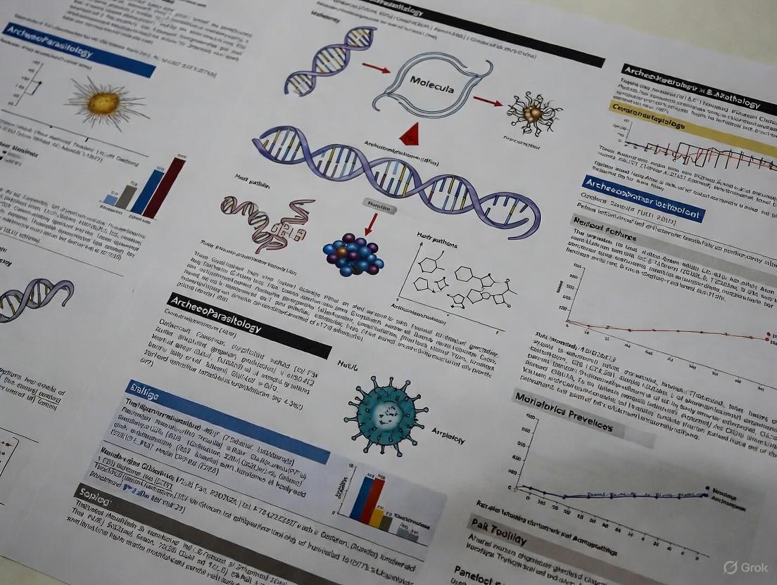
The discipline consolidated between the world wars with methods like radiology, histology, and serology being applied more frequently, improving diagnostic accuracy through statistical analysis [1]. After World War II, paleopathology transformed from primarily case-based descriptions to an important tool for understanding past populations, relating to epidemiology and demography [1]. Recent decades have witnessed revolutionary developments through molecular biology, which enables retrieval of ancient DNA and proteins from centuries or millennia-old samples [1] [6]. This has introduced powerful new dimensions to both paleopathology and archaeoparasitology, allowing unequivocal species-level identification and exploration of disease evolution [7].
Methodological Approaches
Paleopathological Methods and Techniques
The analytical approach in paleopathology depends heavily on the nature of the sample (bone, soft tissue, or hair), its size, preservation status, and allowable manipulation [1]. Human osteopathology forms a cornerstone of the discipline, classified into several pathological groups: congenital, degenerative, infectious, metabolic, traumatic, and neoplastic conditions [1].
Macroscopic analysis of skeletal remains begins with biological profiling (age, sex) and systematic examination for lesions or anomalies [4]. Radiological techniques including standard X-ray and computed tomography (CT) provide non-destructive visualization of internal structures. Tondini et al.'s study of ancient Egyptian skulls employed micro-CT and microscopic analysis to identify both healed cranial trauma and metastatic lytic lesions with perimortem cutmarks, suggesting possible surgical exploration of malignancy [2]. Histological analysis of thin bone sections can reveal microstructural changes, while biomolecular analyses including DNA and isotopic studies have dramatically expanded diagnostic capabilities [4] [1].
Archaeoparasitological Methods and Techniques
Archaeoparasitology employs specialized techniques for recovering and identifying parasite remains. The field primarily investigates materials including fossilized human or animal dung (coprolites), tissues and digestive contents of mummified corpses, and soil samples from latrines, cesspits, or middens [3]. Ectoparasites may be recovered from skin, scalp, clothing, wigs, or personal grooming accessories [3].
Microscopic analysis represents the foundational approach, identifying durable parasite eggs and cysts based on morphological characteristics. For example, studies of medieval deposits in Lübeck, Belgium, and Nivelles quantified Trichuris trichiura and Ascaris lumbricoides eggs, with one Belgian burial demonstrating remarkably high concentrations (approximately 1,577,679 T. trichiura eggs and 202,350 A. lumbricoides eggs) [8] [7].
Molecular analyses have dramatically enhanced diagnostic precision. Ancient DNA (aDNA) analysis through PCR amplification and sequencing enables species-level identification. Maixner et al. (2018) implemented a comprehensive protocol for molecular archaeoparasitology, extracting aDNA from archaeological sediments, amplifying parasite-specific markers (ITS-1 and β-tubulin for Trichuris; CytB and COX1 for Ascaris, Taenia, and Diphyllobothrium), and constructing maximum-likelihood phylogenies for definitive identification [7]. Immunological assays including enzyme-linked immunoassay (ELISA) detect parasite-specific antigens, while petrographic techniques aid identification of tissue-dwelling parasites like Capillaria hepatica [3].
Table 2: Key Research Reagents and Materials in Archaeoparasitology
| Research Reagent/Material | Function/Application | Example Use Case |
|---|---|---|
| Archaeological Sediments (latrine, coprolite, burial soils) | Source material for parasite egg recovery [3] [7] | Analysis of 31 stratigraphed latrine samples from medieval Lübeck [7] |
| Microscopy Reagents (glycerol, chemical mounting media) | Morphological identification of parasite eggs [8] [3] | Identification of Trichuris, Ascaris, Taenia, and Diphyllobothrium eggs [7] |
| aDNA Extraction Kits (silica-based binding methods) | Isolation of ancient parasite DNA from sediments and tissues [7] | Extraction of Trichuris trichiura ITS-1 and β-tubulin DNA from medieval samples [7] |
| PCR Master Mixes (Taq polymerase, dNTPs, buffers) | Amplification of parasite-specific genetic markers [7] | Amplification of Ascaris COX1, Taenia CytB, and Diphyllobothrium COX1 targets [7] |
| Species-Specific Primers | Targeted amplification of parasite DNA sequences [3] [7] | Differentiation of Taenia saginata from other Taenia species in medieval Lübeck [7] |
| Sanger Sequencing Reagents | Determination of parasite DNA sequences for phylogenetic analysis [7] | Construction of maximum-likelihood phylogenies for species confirmation [7] |
| Immunoassay Components (antibodies, substrates) | Detection of parasite antigens in ancient samples [3] | Paleo-auto-immunohistochemistry for Plasmodium detection in Corsican dental pulp [2] |
Experimental Protocols in Molecular Archaeoparasitology
Sample Processing and Ancient DNA Extraction
The following protocol adapts methodologies from Maixner et al. (2018) for comprehensive molecular archaeoparasitology [7]:
Sample Preparation: Collect 5-10g of archaeological sediment from defined contexts (latrine, coprolite, burial soil). For mummified tissue, sample 100-200mg from intestinal area or specified organ.
Parasite Egg Concentration: Process sediments using tri-sodium phosphate solution (0.5%) and vacuum filtration through 260μm and 30μm mesh filters to concentrate eggs while removing larger debris.
Microscopic Quantification: Prepare aliquots of concentrated sample for initial microscopic examination using light microscopy at 100-400x magnification. Quantify eggs per gram (EPG) using standardized counting chambers.
Ancient DNA Extraction: Transfer 500μL of concentrated egg suspension to aDNA-dedicated laboratory facilities. Extract DNA using silica-based methods optimized for ancient sediments, incorporating multiple negative controls to monitor contamination.
DNA Quality Assessment: Quantify aDNA yield using fluorometric methods appropriate for low-concentration, degraded DNA. Accept samples with detectable DNA for subsequent analysis.
PCR Amplification and Sequencing
Primer Selection: Design or select published primers targeting parasite-specific markers:
- Trichuris trichiura: ITS-1 and β-tubulin
- Ascaris spp.: Cytochrome b (CytB) and COX1
- Cestodes: Taenia spp. (CytB), Diphyllobothrium latum (COX1)
PCR Setup: Prepare reactions in aDNA-dedicated workspace with stringent contamination controls. Use 25-50μL reaction volumes with 1-5μL template DNA. Incorporate uracil-DNA-glycosylase (UDG) treatment to minimize damage-associated misincorporations.
Thermal Cycling: Optimize cycle conditions for ancient DNA:
- Initial denaturation: 94°C for 5min
- 45-55 cycles: Denaturation (94°C, 30s), Annealing (primer-specific TM, 30s), Extension (68°C, 45s)
- Final extension: 68°C for 10min
Sequence Analysis: Purify PCR products and perform Sanger sequencing. Process sequences using bioinformatics pipeline involving BLAST against NCBI GenBank database. Confirm identification by constructing maximum-likelihood phylogenies with reference sequences.
Key Research Applications and Findings
Paleopathological Insights into Ancient Health
Paleopathology provides critical insights into the health burdens, medical knowledge, and adaptive challenges of past societies. The analysis of two ancient Egyptian skulls from the Duckworth Collection revealed distinct pathological signatures: one evidenced healed cranial trauma, demonstrating therapeutic success, while another showed metastatic lytic lesions with perimortem cutmarks, suggesting possible surgical exploration of malignant lesions [2]. This juxtaposition illustrates both limitation and innovation in early medical care and contributes meaningfully to debates in the history of oncology [2].
Studies of the 14th century Black Death pandemic demonstrate paleopathology's power to resolve historical epidemiological debates. Molecular analyses have strongly supported the long-held assumption that bubonic plague (Yersinia pestis) caused the pandemic, with DNA evidence recovered from mass graves across Europe [1]. The 2013 excavation at Thornton Abbey in North Lincolnshire uncovered a mass grave of 48 individuals from the Black Death period, with the wide age range (1-45 years) suggesting devastating population impact beyond typical mortality patterns [1].
Archaeoparasitological Reconstructions of Past Lifestyles
Archaeoparasitology provides unique insights into dietary practices, sanitation, trade, and cultural interactions. Molecular analyses of medieval Lübeck, a pivotal Hanseatic trading center, revealed distinctive parasite signatures: ubiquitous faecal-oral transmitted nematodes (Ascaris lumbricoides and Trichuris trichiura) alongside high numbers of food-associated cestodes (Diphyllobothrium latum and Taenia saginata) [7]. The presence and temporal shift in these cestodes (with D. latum more prevalent earlier and Taenia later) indicate substantial alterations in diet or parasite availability around 1300 CE [7].
The application of molecular methods to Trichuris trichiura ITS-1 sequences revealed two distinct clades, one ubiquitous across sites and another restricted to medieval Lübeck and Bristol [7]. The high sequence diversity detected in Lübeck is consistent with its importance as a major trading center, potentially introducing parasite strains through commercial networks [7]. Such findings demonstrate how parasite evidence serves as an artefact-independent source of historical evidence, tracing connections invisible through material culture alone.
Table 3: Quantitative Findings from Archaeoparasitological Studies
| Site/Context | Period | Parasites Identified | Prevalence/Concentration | Interpretation |
|---|---|---|---|---|
| Lübeck, Germany (latrines) [7] | 12th-17th century CE | Trichuris trichiura | 107-4,935 eggs/gram [7] | Ubiquitous faecal-oral transmission in urban center |
| Ascaris lumbricoides | 45-1,645 eggs/gram [7] | Ubiquitous faecal-oral transmission | ||
| Diphyllobothrium latum | 49-1,414 eggs/gram [7] | Dietary consumption of undercooked freshwater fish | ||
| Taenia saginata | 133-8,310 eggs/gram [7] | Consumption of infected beef | ||
| Nivelles, Belgium (burials) [8] | Medieval | Trichuris trichiura | ~1,577,679 total eggs [8] | Extreme parasite burden in individual |
| Ascaris lumbricoides | ~202,350 total eggs [8] | Co-infection with multiple parasites | ||
| Bristol, UK (communal waste) [7] | Medieval | Trichuris trichiura | 78-8,559 eggs/gram [7] | High-intensity transmission in medieval port |
| Ascaris lumbricoides | 76-1,162 eggs/gram [7] | Co-endemicity of nematode species |
Taphonomic Considerations and Methodological Challenges
Both paleopathology and archaeoparasitology must contend with significant taphonomic challenges that influence interpretation. Archaeoparasitological analyses face interpretative challenges from diverse preservation environments, with differential parasite egg preservation influenced by five major taphonomic factors: abiotic, contextual, anthropogenic, organismal, and ecological [8]. Water percolation through deposits can differentially preserve eggs based on morphological characteristics, while arthropod activity may significantly impact preservation, as evidenced by abundance of mites and dipteran puparia in materials from Medici family embalming jars [8].
In skeletal paleopathology, the "osteological paradox" presents fundamental interpretive challenges, as skeletal lesions represent both disease occurrence and survival long enough for bony response to develop [4]. This requires careful consideration when inferring population health from pathological specimens. Molecular analyses face contamination risks that necessitate meticulous laboratory protocols, including physical separation of pre- and post-PCR areas, dedicated aDNA facilities, and multiple negative controls [1] [7]. The highly sensitive PCR methods required for low-abundance ancient DNA are particularly vulnerable to contamination from modern sources, requiring "suicide" PCR approaches that use single-use primers to prevent amplification of contaminating sequences from previous reactions [1].
Interdisciplinary Connections and Future Directions
Paleopathology and archaeoparasitology are inherently interdisciplinary, spanning the biomedical sciences, anthropology, and historical disciplines. Recent scholarship emphasizes incorporating theoretical paradigms that acknowledge the complex roles social behavior and environmental contexts play in disease processes, including syndemic relationships between diseases and conditions [6]. There is growing recognition of the need to avoid Cartesian epistemological frameworks of dualisms (body/culture, nature/nurture) and instead conceptualize the body as fully entangled within relational entities [6].
Future methodological developments will likely expand biomolecular applications, including proteomic approaches to identify parasite-specific proteins and enhanced aDNA techniques requiring minimal template material. The integration of multiple lines of evidence—osteological, molecular, historical, and archaeological—will continue to strengthen interpretations. As these fields mature, there is increasing attention to ethical issues of inequality and how paleopathological research might address rather than perpetuate inequities in understanding the past [6].
The pioneering work of figures like Adauto Araújo and Luiz Fernando Ferreira established archaeoparasitology as a globally relevant field, with research expanding geographically and methodologically to encompass diverse regions and time periods [9]. This global perspective continues to reveal varied patterns of past human-parasite interactions, contributing uniquely to our understanding of both the history of disease and the development of human cultural practices.
Archaeoparasitology, a multi-disciplinary field within paleopathology, is the study of parasites in archaeological contexts [3]. It provides unique insights into past human health, dietary practices, migration patterns, and cultural evolution by analyzing parasitic remains from archaeological materials. The field formally emerged with Sir Marc Armand Ruffer's groundbreaking 1910 study, which identified calcified Schistosoma haematobium eggs in Egyptian mummies dating to the 1250–1000 BC 20th Dynasty [3] [10]. This seminal work demonstrated that parasitic diseases afflicted ancient civilizations and established a new scientific approach to investigating ancient life. Ruffer's methodology laid the foundation for archaeoparasitology, creating a bridge between modern parasitology and archaeology that allows researchers to reconstruct aspects of ancient societies that are rarely documented in historical records [3] [10].
The broader field of paleopathology—the study of ancient diseases and injuries in organisms through examination of fossils, mummified tissue, and skeletal remains—provides the essential context for archaeoparasitological research [1]. As an interdisciplinary science, paleopathology integrates knowledge from clinical pathology, human osteology, epidemiology, social anthropology, and archaeology to understand the evolution of diseases and how past civilizations treated various conditions [1]. Archaeoparasitology specifically focuses on the protozoan and metazoan parasites of humans in the past, as well as parasites that affected past human societies through their infestations of domesticated animals [3]. This review traces the technical evolution of archaeoparasitology from its morphological beginnings to the current molecular era, highlighting key methodological advances and their implications for understanding human-parasite relationships throughout history.
Technical Historical Timeline
The development of archaeoparasitology has been marked by several key technological breakthroughs that have expanded the scope and precision of paleoparasitological research. The following table summarizes the major methodological milestones in the field's evolution.
Table 1: Historical Milestones in Archaeoparasitology Methodologies
| Time Period | Key Methodological Developments | Primary Research Materials | Representative Studies |
|---|---|---|---|
| 1910-1950s | Initial morphological identification of parasite eggs in mummified tissues | Mummified tissues (kidneys) | Ruffer (1910): S. haematobium in Egyptian mummies [3] |
| 1950s-1970s | Coprolite analysis, microscopy optimization | Coprolites, latrine sediments | Szidat (1940s): Ascaris and Trichuris in European bog bodies [10] |
| 1970s-1990s | Standardized egg concentration techniques, histological staining | Latrine soils, coprolites, pelvic soil from burials | Reinhard (1990s): Cultural ecology of parasitism on Colorado Plateau [11] |
| 1990s-2010s | Immunological assays (ELISA), early single-gene PCR | Mummified tissues, coprolites | Gonçalves et al. (2004): Amebiasis distribution using immunoassay [11] |
| 2010s-Present | High-throughput sequencing, phylogenetic reconstruction, multi-method integration | Diverse contexts: latrines, coprolites, mummies, burial sediments | Søe et al. (2018): Molecular archaeoparasitology of Medieval Lübeck [7] |
The trajectory of methodological innovation in archaeoparasitology demonstrates a consistent pattern of incorporating new technologies from adjacent fields, particularly molecular biology and biomedical sciences. This evolution has transformed the discipline from a descriptive science to an analytical one capable of addressing complex questions about human history and parasite evolution.
Core Principles and Definitions
Archaeoparasitology operates within a well-defined theoretical framework derived from both modern parasitology and archaeological science. Parasitism is defined as a symbiotic relationship in which one organism (the parasite) lives in or on another organism (the host), benefiting at the host's expense [3]. The distinction between endoparasites (living inside the host) and ectoparasites (living on the outside of the host) is fundamental, as it determines recovery methods and archaeological preservation potential [3].
A key conceptual framework in the field is Reinhard's distinction between archaeoparasitology (study of parasitological remains from archaeological contexts derived from human activity) and paleoparasitology (study of non-human, paleontological parasite material) [3]. This differentiation emphasizes the anthropological focus of archaeoparasitology and its role in understanding human culture and behavior.
The principle of pathoecology recognizes that parasitic infections result from complex interactions between human behavior, environmental factors, and parasite biology [12]. This ecological perspective enables researchers to reconstruct past environments and human activities through parasite assemblages. For example, the presence of fish tapeworm (Diphyllobothrium latum) indicates consumption of raw or undercooked fish, while whipworm (Trichuris trichiura) and roundworm (Ascaris lumbricoides) reflect fecal-oral transmission related to sanitation practices [7].
Another fundamental concept is differential preservation, which acknowledges that parasite remains survive through distinct mechanisms. Helminth eggs preserve well due to their chitinous shells, while protozoan cysts require exceptional conditions like desiccation or freezing for preservation [10]. This affects recovery rates and requires customized methodologies for different parasite types and archaeological contexts.
Methodological Evolution
Traditional Morphological Approaches
The foundational methodology of archaeoparasitology involves microscopic examination of parasite eggs recovered from archaeological contexts. The standard workflow begins with sample collection from archaeological materials including coprolites, latrine sediments, mummified tissues, and burial soils [3]. Samples undergo rehydration in aqueous solutions, often containing lycopodium spore tablets for quantitative analysis [13]. The rehydrated material is then processed through palynological concentration techniques involving chemical treatment (often with hydrochloric acid and potassium carbonate) and micro-sieving (5-20μm mesh) to concentrate parasitic elements [12] [10].
Microscopic identification relies on diagnostic morphological characteristics including egg shape, size, shell ornamentation, and opercular structures. Common measurements include:
- Ascaris spp.: 45-75μm long, 35-50μm wide, mammillated coat [7]
- Trichuris spp.: 50-55μm long, 20-25μm wide, bipolar plugs [7]
- Diphyllobothrium spp.: 68-73μm long, 42-48μm wide, oval with operculum [12]
Traditional methods remain valuable for initial screening and quantitative assessment of infection intensity, with egg counts often expressed as eggs per gram of sediment [7]. However, limitations include morphological convergence between related species and the inability to identify parasites that lack distinctive eggs or produce few eggs [13].
Molecular Revolution in Archaeoparasitology
The integration of molecular methods has dramatically expanded the analytical capabilities of archaeoparasitology. Ancient DNA (aDNA) analysis begins with specialized DNA extraction from archaeological samples, incorporating mechanical and chemical lysis to break down durable parasite egg shells [7] [13]. Extracts are then subjected to polymerase chain reaction (PCR) targeting taxon-specific genetic markers:
- Ribosomal RNA genes (18S, 28S) for phylogenetic placement [13]
- Mitochondrial genes (COX1, CytB) for species-level identification [7]
- Internal transcribed spacer regions (ITS-1, ITS-2) for population studies [7]
The resulting sequences are analyzed through phylogenetic reconstruction to confirm parasite identity and explore evolutionary relationships [7]. More recently, high-throughput sequencing approaches have enabled parallel detection of multiple parasites and studies of entire parasite communities [7].
Table 2: Key Genetic Targets in Molecular Archaeoparasitology
| Genetic Target | Resolution Level | Application Examples | References |
|---|---|---|---|
| 18S rRNA | Family/Genus differentiation | Distinguishing Physaloptera from Ascaris [13] | Jimenez et al. (2012) [13] |
| ITS-1/ITS-2 | Species/population level | Trichuris trichiura clades in Medieval Europe [7] | Søe et al. (2018) [7] |
| Mitochondrial COX1 | Species identification, phylogenetics | Diphyllobothrium latum confirmation [7] | Søe et al. (2018) [7] |
| Mitochondrial CytB | Species identification | Taenia saginata vs. T. solium [7] | Søe et al. (2018) [7] |
Complementary immunological approaches detect parasite-specific antigens through enzyme-linked immunosorbent assays (ELISA) or immunofluorescence [10]. These methods are particularly valuable for identifying protozoan parasites like Entamoeba histolytica and Giardia intestinalis, whose cysts rarely preserve in archaeological contexts [10].
Integrated Methodological Workflow
Modern archaeoparasitology employs a complementary approach that integrates morphological and molecular methods, leveraging the strengths of each while mitigating their limitations. The following diagram illustrates this integrated workflow:
Diagram 1: Integrated morphological and molecular analysis workflow for archaeoparasitology, illustrating the complementary nature of modern approaches.
This integrated methodology was exemplified in a study of 1,400-year-old desiccated fecal samples from La Cueva de los Muertos Chiquitos in Durango, Mexico [13]. Initial microscopic examination tentatively identified ascarid eggs, but subsequent DNA sequencing revealed the presence of physalopterid nematodes instead—a genus morphologically similar to ascarids but with different clinical implications [13]. This case demonstrates how the combined approach prevents misidentification and provides more accurate paleopathological diagnoses.
Experimental Protocols
Standard Microscopy Protocol for Egg Identification
The following protocol represents the current standard for morphological analysis of archaeoparasitological samples:
Sample Preparation: Remove approximately 1g of material from the archaeological sample using sterile instruments. For coprolites, include both interior and exterior portions to capture parasites with different depositional patterns (e.g., Enterobius vermicularis which lays eggs on the perianal skin) [13].
Rehydration: Place sample in 2-5ml Tris-EDTA buffer (pH 8.0) or 0.5% aqueous trisodium phosphate solution. Disaggregate with vortex mixing and allow to rehydrate for 72 hours on a slow rotator [13].
Concentration: Process rehydrated samples through sucrose or Sheather's sugar flotation (specific gravity 1.20-1.30). Centrifuge at 2500 rpm for 5 minutes to concentrate parasite eggs at the surface [13].
Microscopy: Transfer surface film to microscope slides using cover slips. Examine systematically at 100x and 400x magnification. Record egg morphology, measurements, and developmental stages when visible [13].
Quantification: Add known quantities of marker particles (e.g., lycopodium spores) during rehydration to calculate egg concentrations per gram of original material [7].
Ancient DNA Extraction and Amplification Protocol
For molecular analysis, the following specialized protocol is implemented in dedicated ancient DNA facilities:
Laboratory Requirements: Perform all pre-amplification steps in a positive pressure, HEPA-filtered, UVC-irradiated laboratory with dedicated equipment. Researchers must wear full sterile suits, masks, and double gloves to prevent contamination [13].
DNA Extraction: Use ~25μL aliquots of rehydrated sample with commercial fecal DNA isolation kits. Incorporate a mechanical/thermal lysis step: heat to 63°C for 5 minutes, freeze at -20°C for 5 minutes, then reheat to 63°C for 5 minutes to break durable egg shells [13].
PCR Amplification: Prepare reactions with 0.1μL of 5U/μL Platinum Taq, 3μL of 10X buffer, 0.9μL of 10mM dNTPs, 1.8μL of 25mM MgCl₂, 0.5μL of each 10μM primer, and 2μL of DNA extract in a total volume of 25μL [13].
Contamination Controls: Include extraction blanks and negative PCR controls in every run. Monitor for modern human DNA contamination when analyzing human-associated samples [13].
Sequencing and Analysis: Purify PCR products, sequence with appropriate platforms, and conduct BLAST searches against genomic databases. Construct phylogenetic trees for confirmatory identification [7] [13].
The Scientist's Toolkit: Essential Research Materials
Modern archaeoparasitology relies on specialized reagents and materials adapted from biomedical sciences and customized for ancient material analysis.
Table 3: Essential Research Reagents and Materials in Archaeoparasitology
| Research Tool | Composition/Type | Primary Function | Technical Considerations |
|---|---|---|---|
| Tris-EDTA Buffer | 10mM Tris-HCl, 1mM EDTA, pH 8.0 | Rehydration of desiccated samples | Maintains pH stability, minimizes DNA degradation [13] |
| Sheather's Sugar Solution | Sucrose solution (specific gravity 1.20-1.30) | Flotation concentration of parasite eggs | Higher specific gravity optimizes egg recovery [13] |
| Lycopodium Spore Tablets | Known quantity of Lycopodium clavatum spores | Quantitative egg counting | Added before rehydration for calculation of egg concentration [7] |
| DNA Extraction Kits | Commercial silica-membrane based kits | Ancient DNA isolation from archaeological samples | Modified with additional mechanical/thermal lysis steps [13] |
| Taxon-Specific Primers | 18-25 base pair oligonucleotides | PCR amplification of parasite aDNA | Designed against conserved ribosomal or mitochondrial genes [7] [13] |
Case Study: Molecular Analysis of Medieval Lübeck
The application of integrated methodologies is exemplified by a comprehensive study of Medieval Lübeck, a major Hanseatic trading center [7]. This research analyzed 152 samples from multiple European sites dating between Neolithic and Post Medieval periods, with particular focus on 31 stratigraphed latrine samples from Lübeck's founding quarter (12th-17th centuries CE) [7].
Microscopic analysis revealed high prevalence of fecal-oral transmitted nematodes (Ascaris and Trichuris) across all sites, with Lübeck samples showing particularly high concentrations (Trichuris: 107-4,935 eggs/g; Ascaris: 45-1,645 eggs/g) [7]. More notably, Lübeck samples contained substantial numbers of food-associated cestodes: Diphyllobothrium latum (fish tapeworm) in 14 of 31 samples (49-1,414 eggs/g) and Taenia saginata (beef tapeworm) in 19 of 31 samples (133-8,310 eggs/g) [7].
Molecular analyses provided crucial species-level identifications and revealed temporal patterns in cestode prevalence. DNA sequencing confirmed the microscopic identifications and enabled phylogenetic analysis of Trichuris trichiura ITS-1 sequences, which grouped into two clades—one ubiquitous across sites and one restricted to medieval Lübeck and Bristol [7]. The high genetic diversity of T. trichiura in Lübeck correlates with its historical role as a major trading center with extensive population contacts [7].
Temporal analysis revealed a significant shift in dietary patterns around 1300 CE: D. latum prevalence decreased while T. saginata increased, suggesting a transition from fish to beef consumption or changes in food preparation practices [7]. This case demonstrates how molecular archaeoparasitology provides artefact-independent evidence of historical dietary changes, trade connections, and cultural practices.
Current Status and Future Directions
Contemporary archaeoparasitology continues to evolve with technological advancements. The field is increasingly moving toward multi-marker approaches that simultaneously analyze parasite assemblages, human genetics, and dietary biomarkers from the same archaeological samples [7] [10]. High-throughput sequencing technologies enable reconstruction of complete parasite communities and detection of low-abundance species that would be missed by microscopy [7].
Future directions include:
- Paleoproteomic analyses of parasite remains to identify species and study parasite evolution
- Metagenomic approaches to reconstruct entire parasite communities and their interactions
- Isotopic studies of parasite remains to understand their trophic relationships and life cycles
- Improved reference databases for both morphological comparisons and genetic sequences
The integration of archaeoparasitological data with historical documents, archaeological findings, and climate records continues to enhance our understanding of how parasitic diseases shaped human societies, influenced cultural practices, and responded to environmental changes throughout history [3] [10].
As methodological innovations continue to expand the analytical capabilities of archaeoparasitology, the field promises to deliver increasingly nuanced insights into the complex relationships between humans, their parasites, and their environments across deep historical timescales. The progression from Ruffer's initial microscopic observations to contemporary molecular analyses represents a paradigm shift in how we study and interpret ancient life, demonstrating the powerful synergy between traditional archaeological methods and cutting-edge scientific technologies.
The biocultural approach represents a foundational paradigm in anthropological research, systematically integrating biological and cultural data to reconstruct past human life experiences. This framework is indispensable in archaeoparasitology and paleopathology, where it provides a context for interpreting evidence of disease and physiological stress recovered from archaeological remains. By rejecting the nature-culture dichotomy, this approach allows researchers to model the dynamic interactions between environmental constraints, cultural buffering systems, and physiological outcomes evident in human skeletons and preserved soft tissues. This technical guide delineates the core principles, methodological protocols, and analytical frameworks for implementing biocultural approaches in advanced archaeological research, with particular emphasis on their application for researchers investigating ancient disease and human adaptation.
Theoretical Foundations of the Biocultural Approach
The biocultural approach is fundamentally rooted in the understanding that human health, disease, and biological processes are inextricably linked to cultural practices and social systems. This perspective emerged as a significant departure from earlier descriptive approaches to human remains, shifting toward a more contextualized and integrative scientific framework [14].
Historical Development and Core Models
The conceptual breakthrough in biocultural modeling began with Buikstra's pioneering work, which positioned human remains within a larger context that included mortuary components, grave goods, and their relationship to broader archaeological site reconstructions [14]. This model established the critical linkage between studies of the dead and inferences about the living, encompassing social behaviors and biological well-being.
A more detailed framework was advanced by Goodman and colleagues, which emphasized modeling physiological disruption within a larger context of environmental and cultural factors that either prevent or manufacture stress [14]. This systemic model, outlined in Figure 1, incorporates several critical components:
- Environmental Constraints: The physical environment provides essential resources for survival, with constraints potentially limiting population viability.
- Cultural Buffering Mechanisms: Cultural systems and beliefs typically act as protective mechanisms during physiologically dangerous periods, though they may sometimes introduce stressors through practices like food taboos.
- Physiological Disruption: The inability to resist stressors results in measurable physiological disruptions, the severity of which depends on age, sex, health status, and genetic composition.
- Feedback Mechanisms: Disease and death have functional consequences for communities, affecting work capacity, reproductive success, and social cohesion.
Table 1: Evolution of Biocultural Models in Bioarchaeology
| Model Developer | Key Conceptual Advance | Primary Applications |
|---|---|---|
| Buikstra (1977) | Incorporated archaeological context and interdisciplinary perspective | Mortuary analysis, social organization studies |
| Goodman et al. (1984) | Systemic stress perspective with feedback mechanisms | Population-level analysis of disease and stress |
| Sheridan (1999, 2002) | Integration of historical documents and ethnohistoric data | Social stratification, occupational stress, childhood health |
Later expansions of the biocultural approach, such as Sheridan's model, incorporated historical dimensions and archival documents, enabling more sophisticated analysis of social stratification, differential access to resources, and occupationally related stress markers [14]. This evolution demonstrates how biocultural frameworks have progressively incorporated diverse data streams to address complex questions about past human societies.
Contemporary Theoretical Positioning
In contemporary practice, the biocultural approach explicitly challenges the entrenched nature-culture divide that has characterized much of archaeological interpretation. Recent scholarship in taphonomy critically examines the artificial separation of cultural transformations (C-transforms) from natural ones (N-transforms), arguing that this heuristic division fundamentally misrepresents the synergistic processes that shape archaeological assemblages [15]. This is particularly relevant for bioarchaeology, where the preservation of human remains results from environmental factors and culturally informed funerary practices acting in concert rather than in isolation [15].
The biocultural approach recognizes that taphonomic processes are not merely distortive but are themselves informative about past human behaviors and environmental conditions. This theoretical repositioning frames the degradation, loss, and damage observed in archaeological materials not simply as reductionist processes but as additive sources of information about the post-depositional history of remains and their cultural context [15].
Methodological Integration in Biocultural Research
Implementing the biocultural approach requires systematic methodology for collecting and integrating diverse datasets from skeletal remains, preserved tissues, and archaeological contexts. This integrated methodology enables researchers to address fundamental questions about human adaptation, disease burden, and cultural practices.
Skeletal Analysis and Stress Indicators
The human skeleton serves as a biological archive recording physiological experiences across the lifespan. Bioarchaeological analysis focuses on multiple stress indicators to determine patterns of acute and chronic stress within past populations [14].
Table 2: Key Skeletal and Dental Indicators of Physiological Stress
| Indicator | Biological System | Interpretation | Chronicity |
|---|---|---|---|
| Porotic hyperostosis/Cribra orbitalia | Cranial skeleton | Possible anemia (nutritional, parasitic) | Chronic |
| Linear enamel hypoplasia | Dentition | Growth disruption during childhood | Acute episode with permanent record |
| Periosteal reaction | Appendicular skeleton | Infection or trauma | Varies (active/healed) |
| Osteoarthritis | Joint surfaces | Mechanical stress/activity patterns | Chronic |
| Stature estimation/ long bone length | Postcranial skeleton | Nutritional status, developmental stress | Cumulative chronic |
The adult skeleton may not show effects of mild stressors, but the growing bones and teeth of children are often altered in measurable ways [14]. Chronic or episodic physiological stress can disrupt growth, leaving permanent markers on bone and teeth that persist into adulthood. These retrospective indicators are among the most valuable for reconstructing patterns of diet and disease from ancient skeletal remains, particularly because a majority of human remains recovered from archaeological contexts are under the age of 18 [14].
The interpretation of these skeletal indicators requires careful consideration of demographic patterning by age and sex, as well as the distribution and frequency of specific diseases across different subgroups within a population. This enables researchers to draw inferences about differential access to resources, occupational activities, and culturally mediated risk factors.
Archaeoparasitology Methods and Protocols
Archaeoparasitology examines parasites in archaeological contexts to reconstruct past human health, dietary practices, migration patterns, and sanitation [3]. The field investigates both endoparasites (protozoans and helminths found inside the host) and ectoparasites (ticks, lice, and fleas found on the host exterior), providing complementary evidence to skeletal data.
- Coprolites: Fossilized human or animal dung often contains parasite eggs, cysts, or remains [3].
- Mummified Tissues: Both natural and intentional mummies may preserve parasites in their digestive contents or tissues [3].
- Latrine, Cesspit, and Midden Soils: Sediments from waste disposal areas concentrate parasite remains [3].
- Burial Soils: Cemetery soils may contain parasite eggs, such as the Echinococcus granulosus cyst retrieved from a Polish cemetery [3].
- Artifacts and Clothing: Ectoparasites and their eggs may be found on combs, textiles, and other personal items [3].
Laboratory Analysis Techniques
Multiple analytical methods are employed in archaeoparasitology, each with specific protocols and applications:
Microscopic Identification: Durable parasite remains (eggs, cysts) are identified using compound or electron microscopy based on morphological characteristics [3]. Petrographic techniques have been used for eggs of Capillaria hepatica found in Roman period remains [3].
Immunological Assays: Enzyme-linked immunoassays (ELISA) detect parasite-specific antigens in archaeological samples, providing species-level identification [3].
Biomolecular Analysis: DNA sequencing of parasite remains recovered from archaeological contexts enables precise taxonomic identification and study of evolutionary relationships [3].
The analytical workflow for integrating skeletal and parasitological evidence follows a systematic process, as illustrated in Figure 2.
Figure 2: Analytical Workflow for Biocultural Research
Applied Biocultural Analysis: Experimental Protocols
This section provides detailed methodological protocols for generating and interpreting biocultural data in archaeoparasitology and paleopathology research.
Integrated Skeletal and Parasitological Assessment
Objective: To correlate evidence of parasitic infection with skeletal indicators of physiological stress within a population sample.
Materials:
- Human skeletal remains with contextual association
- Soil samples from pelvic region and burial context
- Microscopy equipment (light microscope, SEM if available)
- Reagents for immunological testing (ELISA kits)
- DNA extraction and amplification materials
- Osteometric board, calipers, and recording forms
Protocol:
Osteological Analysis:
- Conduct standard osteological inventory, determining age, sex, and preservation.
- Systematically record pathological lesions, including porotic hyperostosis, cribra orbitalia, periosteal reactions, and specific infections.
- Document dental pathologies, including enamel hypoplasias, caries, and abscesses.
- Record metric data for stature estimation and body proportions.
Soil Sample Processing:
- Collect sediment samples from cranial, pelvic, and foot regions of the burial.
- Process samples using rehydration in trisodium phosphate solution.
- Conduct micro-sieving to concentrate particulate matter.
- Prepare microscope slides for parasite egg identification.
Parasitological Analysis:
- Identify and quantify parasite eggs using morphological criteria.
- Perform immunological assays (ELISA) for specific parasite antigens.
- Extract, amplify, and sequence parasite DNA where preservation permits.
- Document species composition and infection intensity.
Data Integration:
- Correlate specific parasitic infections (e.g., hookworm) with skeletal evidence of anemia.
- Analyze demographic patterns in parasite burden and skeletal stress markers.
- Contextualize findings with archaeological evidence of diet, sanitation, and settlement patterns.
The Scientist's Toolkit: Essential Research Reagents and Materials
Table 3: Essential Research Reagents and Materials for Biocultural Analysis
| Item | Application | Function | Technical Specifications |
|---|---|---|---|
| Trisodium phosphate solution | Coprolite and soil sample processing | Rehydration and reconstitution of desiccated parasite eggs | 0.5% aqueous solution |
| ELISA test kits | Parasite antigen detection | Species-specific identification of parasite proteins | Commercial kits optimized for archaeological materials |
| PCR reagents | Ancient DNA analysis | Amplification of parasite DNA from archaeological remains | Includes enzymes tolerant of degraded DNA |
| Micro-sieving apparatus | Sample processing | Size-based separation of parasite eggs from sediment | Nested sieves (150μm, 300μm mesh) |
| - Light microscope with micrometer | Parasite egg identification and measurement | Morphological analysis and quantification | 100x-400x magnification with calibrated ocular micrometer |
| Osteometric board | Skeletal measurement | Standardized metric assessment of long bones | Standard 50cm length with fixed and movable arms |
| Digital calipers | Skeletal and dental measurement | Precise metric data collection | 0-150mm range, 0.01mm accuracy |
| - Histological preparation materials | Thin-section microscopy of skeletal tissue | Analysis of microstructural changes in bone | Includes embedding media, microtome, staining reagents |
Advanced Applications in Paleopathology and Archaeoparasitology
The biocultural approach enables researchers to address complex questions about human-environment interactions, cultural practices, and health outcomes in past populations. By integrating multiple lines of evidence, biocultural analysis provides insights unavailable through single-method approaches.
Reconstructing Ancient Disease Ecologies
Biocultural analysis facilitates the reconstruction of disease ecologies in ancient societies by correlating parasitological data with environmental, archaeological, and skeletal evidence. This integrated approach has revealed how:
- Sedentism and population density influenced the prevalence of specific infectious diseases, with soil-transmitted helminths increasing in sedentary agricultural communities [3].
- Dietary practices and food taboos created or mitigated nutritional stress, with food restrictions during pregnancy potentially limiting nutritional intake [14].
- Technological adaptations such as cooking practices, water management, and sanitation systems buffered populations from environmental pathogens [14].
- Cultural contact and migration introduced new pathogen strains, as evidenced by parasite species specific to different geographical regions [3].
The relationship between these factors and their manifestation in the archaeological record can be visualized as an interactive system, as shown in Figure 3.
Figure 3: Biocultural Model of Ancient Disease Ecology
Addressing Fundamental Research Questions
Biocultural approaches in archaeoparasitology and paleopathology have provided insights into numerous fundamental questions about the human past:
- Migration and Trade Patterns: The presence of parasite species outside their endemic ranges provides evidence of human mobility and contact between geographically separated populations [3].
- Animal Domestication: Parasites shared between humans and domesticated animals reveal the history of animal domestication and human-animal co-habitation [3].
- Climate Change: Shifts in parasite species composition in archaeological sequences reflect environmental changes and their impact on disease transmission [3].
- Sanitation and Hygiene: The prevalence of fecal-oral parasites in archaeological contexts provides evidence of sanitation practices and waste management [3].
- Ethnomedicine: The presence of medicinal plants in coprolites or associated artifacts, combined with evidence of specific parasites, can reveal ancient therapeutic practices [3].
Quantitative Methods in Biocultural Research
The biocultural approach generates complex, multivariate datasets that require sophisticated statistical analysis. Quantitative methods enable researchers to identify patterns, test hypotheses, and integrate diverse data types.
Statistical Frameworks for Biocultural Data
Bioarchaeologists employ a range of statistical approaches to analyze skeletal and parasitological data:
- Descriptive Statistics: Summarize central tendencies and variability in skeletal measurements, parasite counts, and lesion prevalence [16].
- Multivariate Statistics: Principal components analysis, correspondence analysis, and cluster analysis identify patterns in complex datasets [17].
- Spatial Analysis: Intrasite spatial analysis examines the distribution of pathological conditions relative to features and other burials [17].
- Demographic Reconstruction: Life table analysis and hazard models reconstruct mortality profiles and assess differential risk [17].
Table 4: Quantitative Measures in Biocultural Research
| Data Type | Measurement Scale | Analytical Approach | Research Question |
|---|---|---|---|
| Skeletal lesion frequency | Nominal (presence/absence) | Chi-square tests, logistic regression | Association between lesions and demographic factors |
| Parasite egg concentration | Ratio (eggs/gram) | Correlation analysis, ANOVA | Relationship between infection intensity and settlement density |
| - Stature estimation | Ratio (centimeters) | Linear regression, comparison of means | Secular trends in health and nutrition |
| Enamel hypoplasia timing | Interval (age at formation) | Survival analysis, Kaplan-Meier curves | Timing of stress events across subgroups |
| - Stable isotope ratios | Ratio (δ13C, δ15N) | Multivariate statistics, clustering | Dietary reconstruction and social stratification |
Bayesian Approaches in Biocultural Interpretation
Recent advances in quantitative methods include the application of Bayesian statistical models to biocultural questions. These approaches allow researchers to:
- Incorporate prior knowledge from ethnographic, historical, or clinical sources into archaeological interpretations [17].
- Estimate uncertainty in chronological associations between cultural changes and shifts in health indicators [17].
- Model complex causal pathways linking environmental factors, cultural practices, and biological outcomes [17].
- Integrate absolute dating evidence with relative chronological sequences from seriation [17].
These quantitative frameworks enhance the rigor of biocultural interpretations by explicitly modeling the probabilistic relationships between variables and acknowledging uncertainty in archaeological inference.
The biocultural approach provides an essential theoretical and methodological framework for interpreting human remains in their archaeological context. By systematically integrating skeletal, soft tissue, and cultural evidence, researchers can reconstruct nuanced pictures of past human life experiences, health challenges, and adaptive strategies. The continued refinement of biochemical, molecular, and statistical methods promises to further enhance our ability to extract meaningful information from archaeological remains, while maintaining sensitivity to the complex interplay between biology and culture that defines the human experience. For researchers in paleopathology and archaeoparasitology, this integrated approach remains fundamental to advancing our understanding of health and disease across human history.
Paleopathology, the interdisciplinary study of ancient diseases, provides critical insights into the health, diet, and living conditions of past populations. When combined with archaeoparasitology—the analysis of parasites in archaeological contexts—it offers a powerful framework for understanding the co-evolution of humans and their pathogens [18] [3]. This technical guide details the core skeletal markers of three major infectious diseases (tuberculosis, leprosy, and syphilis) and nutritional deficiencies, framing them within the methodological principles of these disciplines. For researchers and drug development professionals, this archeological perspective can reveal long-term patterns of host-pathogen interaction, informing modern therapeutic strategies by illustrating how these diseases have manifested and evolved in human populations over millennia.
The fundamental principle of paleopathological diagnosis rests on identifying specific osseous changes resulting from disease processes. However, a core challenge lies in the fact that diverse pathological processes can produce overlapping patterns of abnormal bone formation or destruction [19]. This necessitates rigorous differential diagnosis, which is achieved through systematic observation of skeletal remains, correlation with known clinical manifestations, and, increasingly, the application of biomolecular analyses to confirm pathogen presence [18]. The following sections provide a detailed guide to the diagnostic skeletal indicators, supported by current research and standardized methodologies.
Skeletal Markers of Infectious Diseases
Tuberculosis
Tuberculosis (TB), caused by the Mycobacterium tuberculosis complex, has afflicted humans for tens of thousands of years, with biomolecular evidence suggesting a presence in early human populations in Africa at least 70,000 years ago [18]. The disease primarily affects the lungs, but can disseminate to the skeleton in 1-5% of cases, leaving behind characteristic lesions [18].
Table 1: Key Skeletal Markers of Tuberculosis
| Skeletal Element | Pathognomonic Lesions | Description of Osseous Changes |
|---|---|---|
| Spine (Pott's Disease) | Lytic lesions, vertebral body collapse, ankylosis, kyphosis | Destruction of vertebral bodies leading to spinal curvature and fusion [18]. |
| Ribs | New bone formation on the internal surface | Periosteal reaction on the visceral surface of ribs due to proximal pulmonary infection [18]. |
| Large Joints | Single joint ankylosis | Fusion of joints, particularly in the hip, knee, and wrist [18]. |
| Extraspinal Sites | Unifocal lytic lesions with absence of new bone formation | Destructive lesions in bones such as the cranium or long bones [18]. |
The paleopathological diagnosis of TB has been revolutionized by paleomicrobiology. Traditional diagnosis based on morphological features alone is now often corroborated by the detection of ancient DNA (aDNA), lipid biomarkers, and proteins specific to the MTBC [18]. The earliest confirmed human cases, dating to 8000-10,000 years ago from the Near East, were identified through a combination of skeletal analysis, MicroCT scanning, and biomolecular assays [18].
Leprosy
Leprosy, caused by Mycobacterium leprae, is a chronic infectious disease that produces a range of pathognomonic skeletal lesions, particularly in the skull and the bones of the hands and feet [20]. Its slow progression allows for significant bone remodeling and deformation before death.
Table 2: Key Skeletal Markers of Leprosy
| Skeletal Region | Pathognomonic Lesions | Description of Osseous Changes |
|---|---|---|
| Craniofacial (Facies Leprosa) | Resorption of the anterior maxilla, loss of incisor teeth, remodeling of the nasal aperture | Erosion of the alveolar process causing tooth loss; destruction of the nasal spine and margins leading to a "saddle-nose" deformity [20]. |
| Hands and Feet | Resorption of the distal phalanges, metatarsals, and metacarpals | Progressive acro-osteolysis, beginning with the phalanges and moving proximally, often resulting in "mitten" deformities [20]. |
| Tibia and Fibula | Periostitis and new bone formation | Inflammation of the bone surface, leading to irregular, "sword-shin" bowing [20]. |
Diagnosis in skeletal remains relies on recognizing this pattern of infection, as some individual lesions can be caused by other conditions. Epidemiological approaches that evaluate multiple skeletal symptoms across samples have been developed to improve diagnostic accuracy and estimate disease frequency in past populations without relying solely on modern clinical standards [21]. Excavations of known leper cemeteries, such as those in medieval Denmark and the UK, have been fundamental in defining the osteological sequence of the disease [20].
Syphilis
The origin of syphilis (caused by Treponema pallidum) is a subject of debate, primarily between the Columbian and pre-Columbian theories. Paleopathology provides key evidence in this debate through the identification of specific skeletal lesions in remains from different eras and regions [22].
Skeletal markers of treponemal disease include:
- Cranial lesions: Caries sicca, a sequence of destructive and proliferative lesions on the cranium, and nodes/gummatous lesions [22].
- Saber shin: Anterior bowing and thickening of the tibia due to chronic periostitis.
- Nasal palatal destruction: Collapse of the nasal bridge and perforation of the hard palate.
- Dental anomalies: Hutchinson's incisors and mulberry molars, which are associated with congenital syphilis.
A study from Dalmatia (Croatia) reported a skeleton from the antique period (2nd to 6th century A.D.) with skeletal markers consistent with congenital syphilis, providing evidence that supports the pre-Columbian theory for the disease's presence in the Old World [22]. Differential diagnosis is critical to distinguish syphilitic lesions from those of tuberculosis, leprosy, or trauma.
Skeletal Markers of Nutritional Deficiencies
Nutritional diseases are a significant focus of paleopathology as they provide direct insight into dietary adequacy and resource allocation in past societies. While many deficiencies leave no osseous trace, vitamin C and D deficiencies can produce diagnostic changes.
Scurvy (Vitamin C Deficiency)
Scurvy results from a chronic deficiency of vitamin C, which is essential for collagen synthesis. This leads to widespread connective tissue disruption and hemorrhage. In the skeleton, this manifests as a proliferation of new bone formed in response to bleeding at muscle attachment sites and within joint spaces [23].
Key skeletal markers of scurvy include:
- Porotic hyperostosis: Increased porosity of the cranial vault bones.
- Subperiosteal new bone formation: Particularly on the external surfaces of the skull and long bones.
- Abnormal porosity and lesion formation at specific muscle attachment sites: For example, at the attachment of the temporalis muscle, the pterygoid plates, and the sphenoid bone [23] [19].
Diagnosis is challenging due to the comorbidity of other vitamin deficiencies, which can produce analogous osteological responses. A weighted diagnostic system that accounts for the pathophysiology of the disease and soft tissue anatomy has been proposed to improve diagnostic accuracy [23]. The identification of scurvy is highly meaningful as it serves as a proxy for low dietary diversity and general nutritional stress [23].
Pathophysiology of Scurvy and Resulting Skeletal Lesions
Other Nutritional Deficiencies
While scurvy is one of the few nutritional diseases with potentially diagnostic lesions, other deficiencies can be inferred from skeletal remains, though with less specificity.
- Anemia: Often identified through porotic hyperostosis (lesions on the cranial vault) and cribra orbitalia (lesions in the eye orbits). These changes result from the expansion of the hematopoietic marrow in response to red blood cell deficiency. Hookworm infection is one parasitic cause of anemia that can be detected through archaeoparasitology [3].
- General Malnutrition: Non-specific indicators include reduced growth in juveniles, reduced robusticity of the skeleton, and an increased prevalence of linear enamel hypoplasia (defects in tooth enamel indicating periods of physiological stress).
As noted in reviews, precise diagnoses of specific vitamin-related deficiencies are often problematic due to comorbid conditions and overlapping osteological responses [24]. Interpretation is therefore most robust when skeletal data is combined with other archaeological evidence, such as paleobotanical remains and isotopic analysis.
Experimental Protocols in Paleopathology & Archaeoparasitology
The rigorous identification of ancient diseases relies on a multi-methodological approach, integrating macroscopic, microscopic, and biomolecular techniques.
Macroscopic Skeletal Analysis
This is the foundational method in paleopathology. The protocol involves:
- Systematic Visual Inspection: A complete inventory of the skeleton is conducted, noting the presence, location, and morphology of any pathological lesions.
- Descriptive Terminology: Use of standardized anatomical and pathological terminology to ensure clarity and replicability [19].
- Differential Diagnosis: A structured rubric is used to systematically compare observed lesions against potential causes (e.g., infection, trauma, metabolic disease, or neoplasia) to arrive at the most probable diagnosis [19].
Paleomicrobiology and Ancient DNA (aDNA) Analysis
The detection of pathogen-specific biomolecules has revolutionized the field, allowing for unequivocal species-level diagnosis [18] [7].
Table 3: Key Biomolecular Methods in Paleopathology
| Method | Target | Application & Function |
|---|---|---|
| Polymerase Chain Reaction (PCR) | Pathogen-specific aDNA fragments | Amplifies trace amounts of ancient pathogen DNA for sequencing and identification [18]. |
| High-Throughput Sequencing | Entire ancient genomes (host and pathogen) | Provides a complete genetic picture, allowing for evolutionary studies of the pathogen [18]. |
| Lipid Biomarker Analysis | Mycobacterial cell wall mycolic acids | Uses HPLC to detect specific lipids from bacteria like M. tuberculosis, confirming its presence [18]. |
| Immunological Assays (e.g., ELISA) | Pathogen-specific proteins (antigens) | Detects the presence of proteins from specific parasites or bacteria in ancient tissues [3]. |
The workflow for aDNA analysis typically involves: dedicated clean-room facilities to prevent contamination; powdering of a small bone or tooth sample; DNA extraction using silica-based methods; library preparation for sequencing; target enrichment (e.g., using bait-capture for specific pathogens); and finally, high-throughput sequencing and bioinformatic analysis [7].
Archaeoparasitology Techniques
Archaeoparasitology focuses on the recovery and identification of parasite remains from archaeological contexts [9] [3].
- Sample Collection: Sources include coprolites (fossilized feces), sediment from latrines, cesspits, or the abdominal/pelvic region of burials, and the contents of mummified digestive tracts.
- Microscopy: The primary tool for identifying durable parasite eggs and cysts. Light microscopy is used for genus-level identification based on egg morphology, size, and shell features [7].
- Molecular Identification: aDNA is extracted from parasite eggs recovered from sediments. PCR and sequencing of genetic markers (e.g., ITS-1 for Trichuris, CytB for Ascaris and Taenia) allow for definitive species-level diagnosis and phylogenetic studies [7]. Confocal Laser Scanning Microscopy (CLSM) can also be used to enhance the visualization of parasite egg morphology [9].
Integrated Workflow for Paleopathological and Archaeoparasitological Research
The Scientist's Toolkit: Essential Research Reagents and Materials
The following table details key reagents, materials, and equipment essential for conducting advanced paleopathological and archaeoparasitological research.
Table 4: Essential Research Reagents and Materials
| Item/Solution | Function/Application |
|---|---|
| Silica-based DNA Extraction Kits | Designed to purify trace amounts of degraded aDNA from bone powder, coprolites, or sediment while removing PCR inhibitors. |
| Proteinase K | Enzyme used during DNA extraction to digest proteins and release DNA from ancient mineralized tissues. |
| PCR Reagents (Primers, dNTPs, Polymerase) | Essential for targeted amplification of pathogen-specific aDNA fragments (e.g., for M. tuberculosis complex or T. trichiura). |
| High-Performance Liquid Chromatography (HPLC) Systems | Used in lipid biomarker analysis to detect and characterize mycolic acids from mycobacteria, confirming TB diagnosis. |
| Next-Generation Sequencing (NGS) Kits | For library preparation, target enrichment, and sequencing of aDNA, enabling whole-genome studies of ancient pathogens. |
| Sodium Phosphate Buffer | A common component in immunological assays like ELISA, used to detect parasite-specific antigens in ancient samples. |
| Microscopy Stains (e.g., GMS, Ziehl-Neelsen) | Used to enhance the visualization of fungal elements or acid-fast bacteria (like M. tuberculosis) in thin sections of mummified tissue. |
| Polymerase Chain Reaction (PCR) Thermocycler | The core instrument for amplifying aDNA to detectable levels for subsequent sequencing and analysis. |
| Confocal Laser Scanning Microscope (CLSM) | Provides high-resolution, 3D imaging of archaeological specimens, useful for clarifying the morphology of parasite eggs [9]. |
The systematic study of skeletal markers of disease, grounded in the principles of paleopathology and augmented by archaeoparasitology, provides an irreplaceable archive of human health through deep time. The accurate identification of tuberculosis, leprosy, syphilis, and nutritional deficiencies like scurvy requires a rigorous, multi-pronged methodology that moves from careful macroscopic observation to confirmation with biomolecular tools. For researchers and pharmaceutical professionals, this historical depth provides critical context. Understanding the long-term evolutionary relationship between humans and pathogens, the historical prevalence of diseases, and the impact of past societal changes on human health can inform models of disease transmission, drug resistance emergence, and the development of new therapeutic and public health strategies. The continued refinement of diagnostic criteria and the integration of novel biomolecular techniques will undoubtedly unlock further secrets from the skeletal record, enhancing our ability to combat these diseases in the present and future.
Archaeoparasitology, a multi-disciplinary field within paleopathology, is dedicated to the study of parasites in archaeological contexts [3]. This discipline examines the protozoan and metazoan parasites of past human populations, as well as those that affected ancient societies through their domesticated animals [3]. The foundation of paleoparasitology dates to 1910, with the seminal discovery of Schistosoma haematobium eggs in Egyptian mummies from the 20th Dynasty (1250–1000 BCE) [10] [3]. The field has since evolved into a sophisticated scientific domain that integrates concepts from modern parasitology with archaeological and paleoenvironmental reconstructions [10]. By studying parasite remains, researchers can infer diverse aspects of ancient life, including dietary practices, hygiene standards, health conditions, and waste management systems [10]. Furthermore, this research provides invaluable insights into the long-term evolution of host-pathogen relationships and the history of infectious diseases [10].
The theoretical framework of archaeoparasitology rests on the ecological concept of parasitism, defined as an interaction where one organism (the parasite) lives in or on another organism (the host), deriving benefits at the host's expense [10]. It is estimated that at least half of all living species are parasites, with Homo sapiens known to be potentially infected by 179 eukaryotic parasites, including 35 specific ones [10]. The identification of parasites in ancient contexts relies on the observation of preserved markers categorized into three groups: macroremains (body parts of adult worms or larvae), dissemination and reproduction forms (eggs, cysts), and parasite-specific biomolecules (antigens and ancient DNA) [10].
Spectrum of Ancient Parasites: Classification and Archaeological Evidence
Archaeoparasitological investigations have revealed a diverse spectrum of parasitic species that infected past human populations. These findings are categorized based on taxonomy, transmission route, and the archaeological contexts from which they are recovered.
Soil-Transmitted Helminths (STHs)
Soil-transmitted helminths are among the most commonly reported parasites in archaeological contexts worldwide. They are transmitted by eggs passed in human feces, which contaminate soil in areas with poor sanitation [25]. The primary STHs identified in archaeological samples include:
- Roundworm (Ascaris lumbricoides) [26] [27] [25]
- Whipworm (Trichuris trichiura) [26] [27] [25]
- Hookworms (Necator americanus and Ancylostoma duodenale) [25]
- Strongyloides (Strongyloides stercoralis) [26] [25]
Table 1: Archaeological Evidence of Soil-Transmitted Helminths
| Parasite Species | Archaeological Contexts/Regions | Key Findings |
|---|---|---|
| Roundworm (A. lumbricoides) | Republic of Korea [26], Han Dynasty China [27], Europe [10], Mummies worldwide | One of the most ubiquitous parasites across time and geography [26] [27] [10]. |
| Whipworm (T. trichiura) | Republic of Korea [26], Han Dynasty China [27], Europe [10], Silk Road latrines [28] | Often found co-occurring with Ascaris; spread via trade routes like the Silk Road [26] [27] [28]. |
| Hookworm | Global, especially in tropical/subtropical areas [25] | Less frequently identified than Ascaris and Trichuris in archaeological records. |
| Strongyloides stercoralis | Republic of Korea [26] | Identified by larval forms; requires different diagnostic methods [26] [25]. |
Foodborne Trematodes
Foodborne trematodes, also known as flukes, require intermediate hosts (often aquatic snails and fish or crustaceans) to complete their life cycles. Human infection occurs through consumption of raw or undercooked food containing infective stages. Archaeological evidence confirms their presence in ancient diets and food preparation practices.
Table 2: Archaeological Evidence of Foodborne Trematodes
| Parasite Species | Archaeological Contexts/Regions | Key Findings |
|---|---|---|
| Chinese liver fluke (Clonorchis sinensis) | Republic of Korea [26], Han Dynasty China [27] | Indicates consumption of raw or undercooked freshwater fish [26] [27]. |
| Intestinal flukes (Metagonimus yokogawai, etc.) | Republic of Korea [26] | Provides evidence of specific regional dietary habits [26]. |
| Lung fluke (Paragonimus westermani) | Republic of Korea [26] [9] | Linked to consumption of raw crustaceans; suspected cause of historical hemoptysis cases [26] [9]. |
| Giant liver fluke (Fasciola hepatica) | Republic of Korea [26] | Suggests consumption of aquatic plants or contaminated water [26]. |
| Blood fluke (Schistosoma spp.) | Han Dynasty China (S. japonicum) [27], Ancient Egypt (S. haematobium) [3] [10], Europe [28] | The first parasite identified in mummies; provides evidence of water-contact activities [3] [27] [10]. |
Ectoparasites
Ectoparasites live on the exterior of the host's body and include arthropods such as lice, fleas, and ticks. In archaeological contexts, they are recovered from clothing, personal grooming accessories, mummified remains, and combs [3]. Their eggs (nits) may be found attached to individual hairs [3]. The study of ancient ectoparasites contributes to our understanding of personal hygiene, grooming practices, and the history of vector-borne diseases.
Methodological Framework in Archaeoparasitology
The reliability of archaeoparasitological findings depends on rigorous, standardized methodologies for sample collection, processing, and analysis. The following section outlines the core experimental protocols and techniques.
Sample Collection and Sediment Processing
The first critical step involves the careful collection of archaeological sediments from contexts with high potential for parasite preservation.
Table 3: Primary Archaeological Sampling Contexts
| Sample Context | Description | Parasite Types Recovered |
|---|---|---|
| Coprolites/Paleofeces | Fossilized or desiccated human or animal dung. | Gastrointestinal helminths and protozoa. |
| Latrine/Cesspit Sediments | Accumulated waste from toilets or waste pits. | Soil-transmitted helminths, foodborne trematodes. |
| Mummy Gut Contents | Sediments from the abdominal cavity or rectum of mummified remains. | Gastrointestinal helminths. |
| Burial Soil/Sediment | Soil from the pelvic region of skeletons or general burial soil. | A wide range of parasites, though interpretation requires caution. |
| Artifacts | Organic residues on combs, clothing, or textiles. | Ectoparasites (lice nits, fleas). |
Protocol 1: Standard Sediment Disaggregation and Micro-sieving
- Disaggregation: Place approximately 0.5-1.0 g of archaeological sediment into a sterile tube. Rehydrate using a 0.5% aqueous trisodium phosphate solution for at least 72 hours, agitating periodically [28].
- Sieving: Pour the rehydrated sample through a stack of geological sieves (e.g., 150 µm, 50 µm). The parasite eggs are typically retained on the 50 µm sieve.
- Concentration: Rinse the material from the 50 µm sieve into a conical tube. Centrifuge at 3000 rpm for 5 minutes to concentrate the eggs.
- Microscopy: Resuspend the pellet in a small volume of glycerol and transfer to a microscope slide for examination under light microscopy at 100x to 400x magnification.
Microscopy and Morphological Identification
The primary method for identifying helminth eggs is morphological analysis. Eggs are identified based on size, shape, shell thickness, ornamentation, and the presence of specialized structures like opercula or plugs.
Protocol 2: Confocal Laser Scanning Microscopy (CLSM) for Challenging Identifications For degraded or morphologically ambiguous eggs, CLSM can provide enhanced visualization.
- Staining: Stain the parasite egg suspension with a fluorescent dye (e.g., autofluorescence or specific stains).
- Imaging: Examine the stained sample under a confocal microscope. Optical sectioning allows for the reconstruction of 3D structure.
- Analysis: Highlight subtle morphological features not visible with standard light microscopy to aid in accurate identification [9].
Molecular and Immunological Techniques
For protozoan parasites, whose cysts are fragile and rarely preserve, and for species-level discrimination of helminths, biomolecular techniques are essential.
Protocol 3: Enzyme-Linked Immunosorbent Assay (ELISA) for Protozoan Antigens
- Extraction: Extract soluble proteins from archaeological sediment using a neutral pH buffer.
- Coating: Coat a microtiter plate with the extracted sample.
- Incubation: Add a primary antibody specific to a target parasite antigen (e.g., Entamoeba histolytica, Giardia intestinalis).
- Detection: Add a secondary antibody conjugated to an enzyme (e.g., horseradish peroxidase). Introduce a colorless substrate that the enzyme converts to a colored product.
- Quantification: Measure the color intensity spectrophotometrically to confirm the presence of the parasite [10].
Protocol 4: Ancient DNA (aDNA) Analysis of Parasites
- DNA Extraction: Perform DNA extraction in a dedicated aDNA facility to prevent contamination. Use silica-based methods to isolate highly degraded DNA.
- Library Preparation and Target Enrichment: Convert the extracted DNA into a sequencing library. Use hybridization capture with biotinylated probes to enrich for parasite DNA from the vast background of environmental and host DNA.
- Sequencing and Analysis: Sequence the enriched library on a high-throughput platform. Map the sequences to reference genomes to confirm the parasite species and conduct phylogenetic studies [10].
The Scientist's Toolkit: Essential Research Reagents and Materials
Successful archaeoparasitology research requires a suite of specialized reagents and materials.
Table 4: Key Research Reagent Solutions for Archaeoparasitology
| Reagent/Material | Function | Application Example |
|---|---|---|
| 0.5% Trisodium Phosphate | Rehydration solution | Disaggregates and rehydrates ancient sediments to release parasite eggs [28]. |
| Glycerol | Mounting medium | Suspends concentrated samples for clear visualization under light microscopy. |
| Micro-sieves (50 µm, 150 µm) | Particle separation | Isolates parasite eggs (typically >50 µm) from finer and coarser sediment fractions. |
| Species-specific Antibodies | Immunological detection | Binds to conserved parasite antigens in ELISA tests for protozoan identification [10]. |
| Biotinylated RNA Baits | Molecular capture | Enriches sequencing libraries for parasite ancient DNA from complex samples [10]. |
| Reference Microscopy Slide Collection | Morphological comparison | Provides a verified standard for identifying and differentiating parasite eggs by size and shape. |
Interpreting Findings in a Broader Context
The identification of parasites is only the first step; interpreting these findings within their archaeological and historical context is crucial. Parasite data can illuminate:
- Dietary Habits: The presence of foodborne trematodes like Clonorchis sinensis provides direct evidence of raw or undercooked fish consumption [27].
- Migration and Trade: The finding of parasite species outside their endemic range can trace human migration and trade routes, as demonstrated by whipworm eggs along the Silk Road [28].
- Sanitation and Health: The prevalence of soil-transmitted helminths reflects sanitation levels. Surprisingly, some studies show no decline in STH infections from the Bronze to Roman periods despite improved sanitation, likely due to practices like using "night soil" (human waste) as fertilizer [27] [28].
- Cultural Practices: Ectoparasite remains on combs and textiles inform about personal hygiene and grooming behaviors [3].
The study of the spectrum of ancient parasites—encompassing soil-transmitted helminths, foodborne trematodes, and ectoparasites—provides a unique and powerful window into the lives of past populations. As a core component of paleopathology, archaeoparasitology moves beyond simple identification to reconstruct ancient diets, health status, sanitation practices, and even trade networks. The field has matured from its descriptive origins into a sophisticated science employing a suite of complementary techniques, from high-resolution microscopy to advanced biomolecular methods. Standardizing these methodologies, as outlined in this guide, is essential for generating robust, comparable data. For researchers and drug development professionals, understanding the deep history of human-parasite relationships, as revealed through archaeology, provides critical evolutionary context for modern parasitic diseases and informs the development of targeted interventions. The continued integration of archaeoparasitological findings with other archaeological sciences promises to further illuminate the complex interplay between humans, their environments, and their parasites throughout history.
Advanced Techniques and Biomedical Applications: From Microscopy to Molecular De-Extinction
Archaeoparasitology and paleopathology are interdisciplinary fields dedicated to understanding health, disease, and human-parasite interactions throughout history by analyzing archaeological remains [3]. These disciplines rely on a fundamental principle: a multi-disciplinary approach that integrates archaeological context, material culture studies, and the analysis of human remains to reconstruct past life ways and disease processes [29]. The core objective is to identify and characterize diseases, which provides insights into the health, diet, migration, and sanitary practices of ancient populations [3] [30].
A rigorous, stepwise methodology is paramount. Investigations should prioritize non-destructive analyses before considering any techniques that require sampling or destruction of valuable archaeological material [30]. This principle ensures the maximum preservation of remains for future research. The "Diagnostic Toolkit" for this research is composed of three primary classes of techniques: macroscopic analysis, various forms of microscopy, and paleoradiology, which together provide a complementary and robust framework for scientific inquiry [29] [30].
Macroscopic Analysis
Macroscopic analysis is the first line of investigation in paleopathology, involving the visual examination of human remains for abnormalities and lesions.
Principle and Application
This approach seeks to identify pathological changes on skeletons or mummified tissues that indicate disease, trauma, or physiological stress [29]. A classic application is the identification of skeletal lesions, such as porotic hyperostosis (lesions on the cranial vault) and cribra orbitalia (lesions on the orbital roof), which are often associated with anaemias [29] [3]. In the context of archaeoparasitology, chronic anaemia can be a proxy indicator for certain parasitic infections, such as hookworm (Ancyslostoma duodenale), though dietary factors can also be a cause [3]. Therefore, while macroscopic changes can suggest the presence of disease, they are often not pathognomonic (definitively diagnostic) on their own [29].
Limitations
A key limitation of macroscopic analysis is that not all diseases leave marks on the skeleton, and even when present, the lesions may not be specific to a single disease [29]. For instance, the presence of cribra orbitalia is not enough to be certain of the presence of malaria, as it is a non-specific indicator of anaemia [29]. This underscores the necessity of corroborating macroscopic findings with other diagnostic methods.
Microscopy
Microscopy techniques provide magnification and resolution to analyze remains at a cellular and sub-cellular level, offering a deeper level of diagnostic detail.
Light Microscopy
High Resolution Light Microscopy (HRLM) is a fundamental tool for examining the fine details of archaeological samples, including plant remains, parasites, and tissues [31]. It can be used to identify parasite eggs, cysts, and other microscopic remains in coprolites (ancient feces) or sediment samples from latrines and cesspits [3].
A specialized form of light microscopy is Polarized Light Microscopy (PLM). PLM is a contrast-enhancing technique that analyzes the anisotropy of a specimen's optical properties—how its properties vary with the orientation of incident light [32]. This is particularly useful for studying crystalline materials.
- Principle: A polarizing microscope uses two polarizing filters (the polarizer and the analyzer) set at right angles to each other, creating a dark background. Birefringent (or doubly-refracting) specimens, which have more than one refractive index, interact with the plane-polarized light and appear bright with distinct polarization colors against this dark background [32].
- Application in Archaeobotany and Parasitology: PLM is widely used to identify plant remains like starch grains and phytoliths. Starch grains, for example, glow under polarized microscopy and show a distinct Maltese cross pattern, which is diagnostic [32]. It is also invaluable for studying the crystalline structure of pigments and fibers associated with artifacts [32].
Table 1: Key Light Microscopy Techniques in Paleopathology
| Technique | Primary Principle | Key Applications | Sample Type |
|---|---|---|---|
| High Resolution Light Microscopy (HRLM) | Magnification with visible light | Analysis of plant tissues, phytoliths, parasite eggs, general histology | Thin-sections, extracted particles [31] |
| Polarized Light Microscopy (PLM) | Optical anisotropy and birefringence | Identification of starch grains, crystalline pigments, fibers, minerals [32] | Thin-sections, particulate samples [32] |
Electron Microscopy
Electron microscopy uses a beam of electrons to achieve significantly higher resolutions than light microscopy, allowing for the examination of ultrastructural details.
Scanning Electron Microscopy (SEM) is particularly valuable for visualizing the surface morphology of archaeological specimens with great depth of field [33].
- Experimental Protocol for Parasite Analysis: A study on ancient head lice nits provides a clear methodological example [33].
- Sample Sourcing: Nits/eggs of Pediculus humanus capitis are extracted directly from the hair of naturally mummified human remains.
- Sample Preparation: To prevent damage, samples can be analyzed without rehydration when using a variable pressure mode SEM, which does not require full conductive coating, thus preserving native morphology [33].
- Imaging and Analysis: The sample is placed in the microscope chamber. SEM imaging at high vacuum and specific voltage (e.g., 15–20 kV) reveals exquisite morphological details such as the operculum (lid), aeropyles (respiratory openings), and the cementing substance that glues the nit to the hair [33]. This level of detail allows for definitive species identification and study of the parasite's life cycle.
Table 2: Advanced Imaging Techniques for Internal and External Morphology
| Technique | Resolution Range | Key Advantage | Common Use Cases |
|---|---|---|---|
| Scanning Electron Microscopy (SEM) | Nanometer-scale surface details | Exceptional depth of field for surface topology | Parasite morphology (lice, ticks), material composition, tool marks [33] [34] |
| X-ray MicroCT | Micrometer-scale internal structure | Non-destructive 3D internal visualization | Internal structure of coprolites, plant remains, bones, artifacts [31] |
Paleoradiology (CT/MDCT Scanning)
Paleoradiology involves the use of medical and advanced radiographic imaging technologies to study archaeological remains non-destructively [35].
Principles and Modalities
The core modalities include X-radiography, Computed Tomography (CT), and Multi-Detector CT (MDCT). These techniques rely on the differential attenuation of X-rays by materials of varying density to create an image [30]. MDCT, in particular, has revolutionized the field by providing enormous three-dimensional data sets that can be virtually dissected and analyzed without causing any damage to the specimen [30].
Applications and Workflow
Paleoradiology is used to investigate mummies, skeletal remains, and artifacts. Its applications are diverse:
- Trauma Analysis: Identifying cause of death, such as the weapon wounds on the mummy of Pharaoh Seqenenre-Taa-II [30].
- Virtual Autopsy: "Virtually unwrapping" fully wrapped mummies, like the scans of Pharaoh Amenhotep-I, to study the body, amulets, and embalming techniques [30].
- Disease Identification: Detecting evidence of conditions like rickets, pneumonia, and atherosclerosis [30].
- Internal Structure Analysis: Examining the internal contents of coprolites or the structure of fragile plant remains without breaking them open [31] [36].
The standard workflow begins with non-destructive MDCT scanning, which provides a comprehensive overview. The resulting data can then be used to guide minimally invasive sampling (e.g., via endoscopy) for further biomolecular analyses if required and permitted [30].
An Integrated Diagnostic Workflow
The most powerful insights in archaeoparasitology and paleopathology come from the integrative analysis of multiple techniques [29] [36]. No single method is sufficient to conclusively identify many ancient diseases. The following workflow diagram illustrates how these techniques can be synergistically combined in a rigorous research process.
Integrated Diagnostic Workflow in Paleopathology
This integrated approach is exemplified by a study on malaria in Egyptian remains, which combined macroscopic observations (skeletal lesions), rapid diagnostic tests (immunological), and genomic analyses (shotgun-capture sequencing) to draw more reliable conclusions than any single method could provide [29]. Similarly, the analysis of dog coprolites to understand human diet integrated macroscopic identification of contents, DNA shotgun sequencing, and stable isotope analysis, offering a comprehensive view of diet and health [36].
Essential Research Reagent Solutions and Materials
The following table details key materials and reagents used in the experimental protocols cited within this field.
Table 3: Key Research Reagents and Materials in Paleopathological Analysis
| Item / Solution | Function / Application | Example from Literature |
|---|---|---|
| Polarizing Filters (Polarizer & Analyzer) | Creates cross-polarized light for identifying birefringent materials in Polarized Light Microscopy (PLM) [32]. | Used to distinguish crystalline pigments and starch grains based on their optical anisotropy [32]. |
| Variable Pressure SEM Chamber | Allows analysis of non-conductive, fragile archaeological samples without extensive coating, preserving original morphology [33]. | Enabled study of ancient head lice nits without rehydration, preventing damage and observing structures like aeropyles and operculum [33]. |
| Immunochromatographic Rapid Tests (RDTs) | Detects specific Plasmodium antigens (proteins) in ancient skeletal material using antibody-antigen reactions [29]. | Applied to bone samples from individuals with skeletal lesions to test for the presence of malaria [29]. |
| Synchrotron Radiation (for X-ray MicroCT) | Provides highly coherent X-ray beam for propagation-based phase-contrast imaging, enhancing visibility of low-absorption materials [31]. | Used to non-destructively analyze the internal three-layered pericarp of archaeological drupe fruits without destructive sample preparation [31]. |
| DNA Sequencing Kits (for Shotgun Sequencing) | Allows for comprehensive metagenomic analysis of all DNA within a sample, not just targeted regions [36]. | Applied to dog coprolites to reconstruct diet, microbiome, and identify pathogens from the total extracted DNA [36]. |
The modern diagnostic toolkit for archaeoparasitology and paleopathology is inherently multi-faceted. Macroscopic analysis provides the initial observational framework, various forms of microscopy (from light to electron) reveal microscopic and ultrastructural evidence, and paleoradiology offers a non-destructive portal into the internal structures of remains. The rigor and reliability of research in this field are maximized when these techniques are applied in an integrated, context-sensitive manner, following a stepwise protocol that prioritizes the preservation of irreplaceable archaeological resources. This synergistic approach allows researchers to robustly reconstruct the history of disease and the intricate relationship between humans and their parasites in the past.
Molecular paleopathology represents a transformative approach within the broader fields of paleopathology and archaeoparasitology, moving beyond traditional morphological analysis to directly study ancient pathogens at the molecular level. Paleopathology, the study of ancient diseases through skeletal remains, mummified tissues, and other archaeological sources, has historically relied on visual identification of bone lesions and abnormalities to understand health in past populations [4] [1]. The integration of molecular techniques now enables researchers to extract biological information from centuries or millennia-old remains, providing unprecedented insights into pathogen evolution, host-pathogen interactions, and ancient disease dynamics [37]. This interdisciplinary field sits at the intersection of molecular biology, archaeology, and biomedical sciences, leveraging advances in analytical sensitivity to recover fragile biomolecules from archaeological contexts where preservation conditions are often suboptimal.
The foundational principle of molecular paleopathology rests on the recovery and analysis of three primary classes of biomolecules: ancient DNA (aDNA), proteins, and immunological markers. Each of these molecular archives offers complementary information about past diseases. While aDNA provides genetic blueprints of ancient pathogens and their evolutionary history, proteins offer insights into expressed physiological processes and can persist longer than DNA in many preservation environments [38]. Immunological assays, particularly enzyme-linked immunosorbent assay (ELISA), provide evidence of immune responses to past infections through the detection of pathogen-specific antigens or host antibodies [39]. Together, these methods have revolutionized our understanding of diseases such as tuberculosis, leprosy, plague, and various parasitic infections that affected ancient civilizations [1] [37].
Methodological Foundations
Ancient DNA (aDNA) Sequencing
Ancient DNA sequencing enables the reconstruction of pathogen genomes from archaeological remains, providing direct evidence of infectious diseases in the past. The aDNA from pathogens is typically highly degraded and fragmented, chemically modified, and often contaminated with environmental and modern DNA [40] [37]. The specialized workflow begins with sample preparation in dedicated cleanroom facilities to minimize contamination, followed by DNA extraction using silica-based methods that efficiently recover short DNA fragments [37].
Library preparation for next-generation sequencing incorporates dual-indexed adapters to track samples and detect contamination. Critical to aDNA authentication is the assessment of characteristic damage patterns, including cytosine deamination at fragment ends, which distinguishes authentic ancient molecules from modern contaminants [37]. Among the most significant applications of ancient pathogen DNA analysis has been the confirmation of Yersinia pestis as the causative agent of the Black Death through identification of its DNA in mass graves [1]. Similarly, aDNA analysis has shed light on the evolutionary history of diseases like tuberculosis and leprosy, revealing how these pathogens have co-evolved with human populations over centuries.
Table 1: Key Challenges and Solutions in Ancient DNA Sequencing
| Challenge | Impact on Analysis | Mitigation Strategy |
|---|---|---|
| DNA fragmentation | Short read lengths (30-100 bp) | Optimization of extraction for short fragments; use of single-stranded library prep |
| Cytosine deamination | DNA sequence errors | Damage pattern analysis; use of uracil-DNA-glycosylase treatment |
| Modern contamination | False positive results | Dedicated cleanroom facilities; biochemical authentication; statistical methods |
| Inhibitors in extracts | Reduced sequencing efficiency | Dilution of extracts; addition of BSA; use of purification kits |
Paleoproteomics
Paleoproteomics involves the study of ancient proteins, which can persist in archaeological remains long after DNA has degraded [38]. Proteins offer greater temporal depth than DNA, with documented survival for millions of years in protected environments like bones, teeth, and eggshells [38]. The field has been revolutionized by soft ionization mass spectrometry, particularly liquid chromatography-tandem mass spectrometry (LC-MS/MS), which enables high-sensitivity identification of protein sequences even from highly degraded samples [38].
The standard paleoproteomic workflow begins with protein extraction from powdered archaeological samples, followed by digestion with trypsin to generate peptides for analysis. The resulting peptides are separated by liquid chromatography and ionized for mass spectrometry analysis. Database searching against known protein sequences allows identification of ancient proteins, with careful attention to post-translational modifications and diagenetic alterations that accumulate over time [38]. A remarkable application of paleoproteomics includes the taxonomic identification of highly fragmented bones and shells, phylogenetic resolution of extinct species, and reconstruction of past human-animal interactions through analysis of residual proteins on artifacts [38].
Table 2: Mass Spectrometry Approaches in Paleoproteomics
| Instrumentation | First Use on Ancient Proteins | Key Applications | Advantages/Limitations |
|---|---|---|---|
| MALDI-TOF | 2000 [38] | Collagen, keratins, silk, shell [38] | Rapid analysis; limited to samples with dominant proteins |
| LC-MS/MS | 2006/2011 [38] | Proteomes of bone, enamel, dental calculus [38] | Handles complex samples; high sensitivity; requires extensive sample processing |
Immunological Assays (ELISA)
Enzyme-linked immunosorbent assay (ELISA) provides a method to detect specific antigens or antibodies in archaeological samples, offering evidence of past infections through immune recognition. The sandwich ELISA format is particularly valuable, employing two sets of antibodies to capture and detect target antigens from ancient samples [39]. This method has been adapted for paleopathological applications to identify pathogen-specific proteins that persist in skeletal and mummified remains.
The ELISA process involves several critical steps: first, a capture antibody specific to the target antigen is coated onto plate wells; after washing, the archaeological sample extract is added, allowing any target antigen to bind; following another wash, an enzyme-conjugated detection antibody is applied; finally, a substrate is added that produces a colorimetric reaction when cleaved by the enzyme, with intensity proportional to antigen concentration [39]. This method has been successfully applied to detect various ancient pathogens, though it requires careful validation to address potential cross-reactivity with related proteins and degradation effects in ancient samples [39].
Advanced Applications in Research and Drug Discovery
Molecular De-extinction for Antibiotic Discovery
A groundbreaking application of molecular paleopathology is the emerging field of molecular de-extinction, which seeks to resurrect ancient biomolecules for modern therapeutic applications [40]. This approach leverages both paleogenomics and paleoproteomics to mine the evolutionary record for novel bioactive compounds that could address contemporary medical challenges, particularly antibiotic resistance [40]. By analyzing genetic and protein sequences from extinct organisms, researchers have identified antimicrobial peptides with potent activity against modern pathogens.
Recent research has successfully resurrected defensins—small, disulfide-rich cationic peptides that play important roles in host immunity—from extinct vertebrate genomes [40]. Computational verification of these peptides identified six authentic β-defensins derived from extinct bird and mammalian species [40]. Even more remarkably, deep learning models have been trained to predict antimicrobial activity from the proteomes of extinct organisms (the "extinctome"), leading to the identification and synthesis of 69 peptides with verified activity against bacterial pathogens [40]. Notably, several peptides from extinct organisms, including Mammuthusin-2, Elephasin-2, and Mylodonin-2, have demonstrated potent anti-infective activity in mouse models of skin abscess and thigh infection, with efficacy comparable to the conventional antibiotic polymyxin B [40].
Integration of Machine Learning and Bioinformatics
The growing complexity of molecular data from ancient remains has necessitated the development of sophisticated computational approaches to identify and authenticate biomarkers of ancient diseases. Machine learning algorithms, such as the APEX multitask deep learning system, have been specifically designed to identify antimicrobial peptides from extinct proteomes [40]. These tools enable researchers to move from random screening to targeted, data-driven discovery of therapeutic candidates from ancient biomolecules.
Bioinformatic methods also play a crucial role in reconstructing evolutionary histories of pathogens and predicting the functional properties of resurrected ancient proteins. For instance, researchers have used bioinformatics and genetic methods to reconstruct ancestral "paleomycin" antibiotics, demonstrating how synthetic biology combined with computational techniques can determine the temporal evolution of antibiotics and revive ancient molecules [40]. Similarly, machine learning approaches have been used to mine proteomic and genomic data from Neanderthals and Denisovans, identifying encrypted peptide antibiotics that display antimicrobial activity in vitro and in preclinical models [40].
Experimental Protocols
Ancient DNA Extraction and Sequencing Protocol
The following protocol outlines the standard procedure for aDNA extraction from skeletal remains:
Sample Decontamination: Remove surface contamination by sanding the bone surface and treating with dilute sodium hypochlorite solution (0.5-1%) followed by multiple rinses with molecular-grade water.
Pulverization: Grind bone or tooth sample to fine powder under liquid nitrogen using a freezer mill or similar impact crusher.
Decalcification and Digestion: Incubate approximately 100 mg of bone powder in 1 mL of extraction buffer (0.45 M EDTA, pH 8.0, 0.25 mg/mL proteinase K) for 24-48 hours at 37°C with constant rotation.
DNA Binding and Purification: Concentrate DNA using silica-based spin columns specifically designed for aDNA, or employ binding to silica-coated magnetic beads in the presence of guanidium thiocyanate.
Library Preparation: Use single-stranded or double-stranded library preparation methods specifically optimized for damaged DNA, incorporating unique dual-index barcodes for sample identification.
Sequencing and Analysis: Sequence on appropriate platforms (Illumina for most applications); process data with aDNA-dedicated pipelines that include damage pattern analysis and authentication.
Paleoproteomic Analysis Protocol
For protein extraction and identification from ancient samples:
Sample Preparation: Powder tooth enamel or dense bone using a dental drill or mixer mill. Demineralize in 0.1-0.5 M HCl at 4°C for 24-48 hours.
Protein Extraction: Incubate demineralized residue in 0.1 M ammonium bicarbonate buffer containing 0.1% RapiGest (Waters) or similar detergent, with occasional vortexing.
Reduction and Alkylation: Add dithiothreitol to 5 mM final concentration and incubate at 60°C for 30 minutes; then add iodoacetamide to 15 mM and incubate in darkness for 30 minutes.
Digestion: Add sequencing-grade modified trypsin at 1:50 enzyme-to-protein ratio and incubate at 37°C for 12-16 hours.
Peptide Cleanup: Desalt peptides using C18 solid-phase extraction tips or stage tips.
LC-MS/MS Analysis: Separate peptides using nanoflow liquid chromatography coupled to tandem mass spectrometry with data-dependent acquisition.
Data Analysis: Search fragment spectra against appropriate databases using search engines such as MaxQuant or PEAKS, accounting for common post-translational modifications and deamidation.
Archaeological ELISA Protocol
Adapted ELISA for ancient pathogen detection:
Coating: Add 100 μL/well of capture antibody (1-10 μg/mL in carbonate-bicarbonate buffer, pH 9.6) to 96-well plate. Incubate overnight at 4°C.
Blocking: Wash plate 3× with PBS containing 0.05% Tween-20 (PBST); add 200 μL/well blocking buffer (1% BSA or 5% non-fat dry milk in PBST); incubate 1-2 hours at room temperature.
Sample Addition: Prepare archaeological extracts in blocking buffer; add 100 μL/well in duplicate or triplicate; include standard curve with known antigen concentrations; incubate 2 hours at room temperature.
Detection Antibody: Wash plate 3× with PBST; add 100 μL/well detection antibody conjugated to horseradish peroxidase (HRP); incubate 1-2 hours at room temperature.
Signal Development: Wash plate 5× with PBST; add 100 μL/well substrate solution (TMB or ABTS); incubate 15-30 minutes in darkness.
Signal Measurement: Stop reaction with 50 μL/well 1 M sulfuric acid (for TMB); read absorbance at appropriate wavelength (450 nm for TMB).
Research Reagent Solutions
Table 3: Essential Research Reagents for Molecular Paleopathology
| Reagent/Category | Specific Examples | Function in Analysis |
|---|---|---|
| DNA Extraction Kits | Silica-based spin columns (QIAamp DNA Micro Kit) | Recovery of short, damaged DNA fragments while removing inhibitors |
| Library Prep Kits | Single-stranded DNA library preparation kits | Construction of sequencing libraries from degraded aDNA with minimal bias |
| Capture Reagents | MyOne Streptavidin C1 beads; biotinylated RNA baits | Targeted enrichment of pathogen DNA from complex background |
| Mass Spectrometry Reagents | RapiGest SF Surfactant; sequencing-grade modified trypsin | Protein extraction and digestion for LC-MS/MS analysis |
| ELISA Components | NUNC Immuno plates; HRP-conjugated antibodies; TMB substrate | Sensitive detection of pathogen antigens in archaeological samples |
Workflow Diagrams
Ancient DNA Sequencing Workflow
Paleoproteomics Analysis Workflow
ELISA Detection Workflow
Molecular paleopathology represents a powerful interdisciplinary framework that integrates cutting-edge molecular techniques with traditional archaeological and paleopathological approaches. The complementary application of aDNA sequencing, paleoproteomics, and immunological assays enables a comprehensive understanding of ancient diseases that would be impossible using any single methodology alone. As these techniques continue to evolve, they offer unprecedented insights into the co-evolution of pathogens and hosts, the historical impact of infectious diseases on human societies, and novel approaches to addressing modern medical challenges through the study of ancient biomolecules.
The future of molecular paleopathology lies in the continued refinement of these methodologies, particularly through the integration of artificial intelligence and machine learning for data analysis and biomarker discovery. Furthermore, the emerging field of molecular de-extinction demonstrates the potential for ancient biomolecules to contribute to drug discovery programs, particularly in addressing the growing crisis of antimicrobial resistance. As technical capabilities advance, molecular paleopathology will undoubtedly continue to transform our understanding of both past and present diseases, creating bridges between archaeology, evolutionary biology, and modern medicine.
Archaeoparasitology, a specialized field within paleopathology, is the study of parasites in archaeological contexts [3]. It examines protozoan and metazoan parasites of past humans and animals to understand health, disease, and ecological relationships in ancient societies [3]. This field provides crucial insights into human-parasite co-evolution, ancient dietary practices, migration patterns, sanitation, and the overall health of past populations [3]. The first archaeoparasitology report identified calcified Schistosoma haematobium eggs in an Egyptian mummy, establishing a research tradition that now integrates biology, archaeology, and anthropology [3]. The broader field of paleopathology studies ancient diseases through skeletal and soft tissue remains, with parasitology becoming increasingly quantitative and focused on epidemiological patterns [41] [42]. This technical guide details the core methods for recovering parasite evidence from key archaeological sources, contextualized within modern paleopathological research frameworks.
Principles of Paleoparasitological Research
Paleoparasitology operates on several fundamental principles that guide research design and interpretation. The field recognizes that parasite remains – including eggs, cysts, larvae, and sometimes adult organisms – persist in specific archaeological contexts where conditions favor preservation [3] [43]. The principle of uniformitarianism suggests that natural processes observed today operated similarly in the past, allowing researchers to interpret archaeological evidence through understanding of modern parasite biology and ecology [44]. Stratigraphic association is crucial, as artifacts and biological remains found together are likely related in time and function, enabling reconstruction of past human activities and their parasite burdens [44].
A significant conceptual framework emerging in the field is ONE Paleopathology, which integrates human, animal, and environmental health from a deep-time perspective [45]. This holistic approach recognizes that human and animal health have been intertwined throughout evolution, with animals often acting as sentinels for environmental conditions and disease reservoirs [45]. Studies of ancient brucellosis, tuberculosis, and leprosy demonstrate how diseases spread between species over millennia, informing modern understanding of zoonotic disease origins and pathways [45].
The materials yielding parasite remains each present unique taphonomic challenges. Coprolites (preserved feces) provide direct evidence of intestinal parasites but vary in preservation quality based on depositional environment [43]. Mummified tissues, particularly digestive contents, can yield exceptionally preserved parasites but are rare in the archaeological record [3] [42]. Latrine soils and cesspit deposits contain accumulated waste from multiple individuals, offering population-level insights but requiring careful interpretation of context and chronology [43].
Source Materials and Their Challenges
Table 1: Primary Archaeological Sources for Paleoparasitology
| Source Material | Parasite Forms Recovered | Preservation Conditions | Key Challenges |
|---|---|---|---|
| Coprolites [43] | Eggs, larvae, cysts [43] | Dry caves, waterlogged/anoxic environments, extreme cold/freeze [43] | Contamination, desiccation/fragmentation of eggs, differential preservation by species [43] |
| Mummified Tissues [3] | Eggs, adult worms, tissue cysts [3] | Artificial/natural mummification, extreme cold, desiccation [3] | Rare occurrence, limited sampling from precious remains, ethical considerations [3] |
| Latrine Soils [43] | Eggs, cysts [43] | Waterlogged/anoxic environments, chemical preservation (e.g., salt, minerals) [43] | Mixed deposits, environmental degradation, difficulty quantifying individual infections [43] |
| Cemetery Soils [3] | Eggs, cysts (especially from tissue-dwelling parasites) [3] | Varied burial environments | Distinguishing true infections from environmental contamination [3] |
Taphonomic Considerations
Preservation potential varies significantly among parasite taxa due to differences in eggshell biochemistry and structure. Helminth eggs with thick, resistant walls (e.g., Ascaris, Trichuris) preserve better than fragile protozoan cysts [43]. Environmental conditions dramatically affect preservation; moist anaerobic environments and desiccating environments provide optimal conditions, while alternating wet-dry conditions and acidic soils can destroy evidence [43]. Differential preservation across parasite species can skew interpretations of past parasite communities, as robust eggs may be overrepresented compared to more fragile forms [43]. Recovery rates are also affected by egg size, specific gravity, and surface properties, which influence their behavior in flotation solutions [46] [47].
Parasite Egg Recovery and Quantification Methods
Flotation and Microscopy Techniques
Flotation methods exploit density differences to separate parasite elements from surrounding matrix. The specific gravity of the flotation solution is critical for optimal recovery [47].
Table 2: Comparison of Parasite Egg Recovery Methods
| Method | Principle | Optimal Specific Gravity | Recovery Efficiency | Limitations |
|---|---|---|---|---|
| Kato-Katz [47] | Direct smear and clearing | Not applicable | Lower ERR for most STH; detects ~50 EPG [47] | Reduced sensitivity for light infections, false negatives common [47] |
| Faecal Flotation (NaNO₃) [47] | Passive flotation | 1.20-1.30 (varies by parasite) [47] | SpGr 1.30 recovered 62.7% more Trichuris, 11% more N. americanus, 8.7% more Ascaris vs. SpGr 1.20 [47] | Lower recovery than centrifugal methods [47] |
| Double-Centrifugal Sugar Flotation [46] | Centrifugation-enhanced flotation | 1.30 [46] | Most sensitive method for nematode eggs in comparative studies [46] | More equipment-intensive, time-consuming [46] |
| Semi-Automated Flotation [48] | Low-density salt solution with sucrose flotation/precipitation | Solution-dependent | Processes 6g feces in 40min/24 samples; suited for digital recording [48] | Requires specialized equipment |
The Callen method, developed specifically for coprolite analysis, involves rehydration in aqueous trisodium phosphate solution, followed by screening and microscopic examination [43]. The Lutz method uses spontaneous sedimentation for egg recovery from coprolites and mummified tissues [43]. Modern comparisons show that centrifugation increases egg recovery compared to standing flotation methods, with specific gravity optimization significantly affecting results [46] [47].
Molecular and Immunological Techniques
Ancient DNA (aDNA) analysis allows species-level identification of parasites, even from degraded remains [3]. Quantitative PCR (qPCR) demonstrates significantly greater sensitivity than microscopy, detecting as little as 5 eggs per gram (EPG) compared to 50 EPG for Kato-Katz and flotation methods [47]. Immunological assays including enzyme-linked immunoassay (ELISA) can detect parasite-specific antigens in ancient remains [3]. Petrographic techniques have been used for eggs found in archaeological contexts, providing detailed structural information [3].
Quantitative Approaches and Paleoepidemiology
The field has evolved from presence/absence recording to quantitative paleoepidemiology [42]. Egg per gram (EPG) quantification provides data about parasite prevalence in ancient populations and identifies the pathological potential of parasitism [42]. This enables researchers to compare epidemiological patterns in ancient and modern populations, including the phenomenon of overdispersion (where most parasites are concentrated in a minority of hosts) [42]. Quantification methods must account for taphonomic biases and differential preservation across parasite taxa to produce realistic measures of past infection intensities [42].
Experimental Protocols for Parasite Recovery
Standardized Coprolite Processing Protocol
- Documentation and Sampling: Photograph and describe coprolite context and morphology. Subsample multiple areas for comprehensive analysis.
- Rehydration: Rehydrate 1-5g coprolite samples in 0.5% aqueous trisodium phosphate solution for 48-72 hours [43].
- Homogenization: Gently homogenize the rehydrated sample using glass rods or vortex mixers.
- Micro-sieving: Sieve suspension through stacked 250μm and 63μm mesh screens to remove large debris while retaining parasite eggs.
- Flotation: Transfer sediment to 15ml centrifuge tubes with zinc sulfate or sodium nitrate solution (specific gravity 1.20-1.30). Centrifuge at 2000-2500 RPM for 5 minutes [47].
- Microscopy: Transfer coverslips to slides and examine systematically at 100-400x magnification. Identify and count eggs using morphological criteria.
- EPG Calculation: Calculate eggs per gram using the formula: EPG = (Egg count × Flotation volume) / (Microscope aliquot volume × Sample weight) [42].
Mummified Tissue Processing
- Non-destructive Sampling: Use endoscopic techniques when possible to collect intestinal contents without damaging specimens.
- Tissue Rehydration: For dried tissues, rehydrate in solutions like Ruffer's solution (alcohol, water, and 5% sodium carbonate) [3].
- Digestion: For soft tissue examination, use enzymatic digestion (e.g., proteinase K) to release parasites while preserving morphology.
- Staining and Microscopy: Apply histological stains to enhance visualization of parasite structures in tissue sections.
Latrine Soil Processing
- Stratigraphic Sampling: Collect soil samples from defined stratigraphic layers to maintain chronological control.
- Sediment Processing: Use modified sedimentation techniques with chemical deflocculation to separate eggs from mineral matrix.
- Density Separation: Employ high-density solutions like zinc sulfate (SpGr 1.35-1.40) to recover eggs from mineral sediments [43].
- Quantification: Calculate eggs per gram of sediment, noting that these represent communal rather than individual infection levels.
Figure 1: Comprehensive Parasite Recovery Workflow
Research Reagent Solutions and Essential Materials
Table 3: Essential Research Reagents for Paleoparasitology
| Reagent/Material | Composition/Type | Function | Application Notes |
|---|---|---|---|
| Trisodium Phosphate [43] | 0.5% aqueous solution | Coprolite rehydration | Rehydrates without destroying parasite eggs; 48-72 hour processing [43] |
| Zinc Sulfate [46] [47] | Solution (SpGr 1.18-1.20) | Flotation medium | Recommended for Giardia cyst recovery; optimal for delicate forms [46] |
| Sodium Nitrate [47] | Solution (SpGr 1.20-1.30) | Flotation medium | SpGr 1.30 significantly increases recovery for most STH eggs [47] |
| Sheather's Sugar Solution [47] | Sucrose solution (SpGr 1.20-1.27) | Flotation medium | High viscosity improves egg recovery; used in centrifugal protocols [47] |
| Proteinase K | Enzyme solution | Tissue digestion | Releases parasites from mummified tissues while preserving morphology |
| Histological Stains | e.g., Trichrome, IHC | Visualization | Enhances contrast for microscopic identification |
Methodological Advances and Future Directions
Recent technological advances are transforming paleoparasitological research. Ancient DNA (aDNA) analysis enables precise species identification and study of genetic relationships between ancient and modern parasites [3] [44]. Quantitative PCR (qPCR) provides superior sensitivity for detecting low-intensity infections that challenge microscopic methods [47]. Experimental archaeology approaches systematically test how preservation conditions affect egg recovery, improving interpretation of archaeological findings [43].
The emerging ONE Paleopathology framework integrates human, animal, and environmental health perspectives, recognizing their interconnectedness throughout human evolution [45]. This approach is particularly valuable for understanding zoonotic disease transmission and how climate change has historically influenced disease spread [45]. Future methodological development will likely focus on standardizing quantification protocols, improving recovery efficiency for fragile parasites, and integrating multi-proxy approaches that combine parasitological data with other archaeological and paleoenvironmental indicators.
Figure 2: ONE Paleopathology Research Integration
The escalating global health crisis of antimicrobial resistance (AMR) presents one of the most significant challenges to modern medicine, with bacterial AMR directly responsible for 1.27 million global deaths in 2019 and contributing to 4.95 million deaths [49]. The World Health Organization has classified multidrug-resistant pathogens as critical priorities, emphasizing the urgent need for novel therapeutic approaches [50] [49]. This case study explores an innovative paradigm—molecular de-extinction—that leverages principles from archaeoparasitology and paleopathology to discover and resurrect ancient antimicrobial molecules from extinct organisms.
Archaeoparasitology, defined as the study of parasites in archaeological contexts, provides crucial methodological frameworks for analyzing biological remains from ancient specimens [3]. This field, along with paleopathology (the study of ancient diseases), has traditionally focused on understanding health in past populations through skeletal and mummified remains [40]. However, recent technological advances have expanded their applications into modern drug discovery by enabling researchers to reconstruct and functionally characterize ancient biomolecules with potential therapeutic value [40].
Molecular de-extinction represents a paradigm shift in antibiotic discovery, offering a unique reservoir of unexploited antimicrobial potential by mining the evolutionary history of life on Earth [40]. This approach selectively resurrects extinct genes, proteins, or metabolic pathways rather than entire organisms, leveraging two primary scientific disciplines: paleogenomics (the study of ancient DNA) and paleoproteomics (the analysis of ancient proteins preserved in fossilized and subfossil remains) [40]. The convergence of these fields with synthetic biology and artificial intelligence has created a powerful platform for addressing the antimicrobial resistance crisis through unconventional sources of novel bioactive compounds.
Technical Methodologies in Molecular De-Extinction
Paleogenomic Approaches
Paleogenomics aims to revive genes from extinct species by reconstructing their genomes and introducing them into closely related living organisms [40]. The first and most crucial step involves obtaining high-quality ancient DNA (aDNA) from preserved biological material, which presents significant technical challenges as aDNA is highly degraded, chemically modified, and often contaminated with microbial and environmental DNA [40].
Table 1: Key Research Reagents and Technologies for Paleogenomics
| Research Tool | Function/Application | Technical Specifications |
|---|---|---|
| Next-Generation Sequencing (NGS) | Recovery of fragmented aDNA | Enables reconstruction of highly degraded genetic material [40] |
| Third-Generation Long-Read Sequencing | Improved recovery of aDNA fragments | Provides more complete genomic assembly from ancient samples [40] |
| CRISPR-Cas9 Gene Editing | Introduction of ancient genes into modern organisms | Precise genome editing for functional testing [40] |
| Computational Genetic Assembly | Reconstruction of extinct genomes | Bioinformatics pipeline for assembling sequence data [40] |
| Synthetic Biology Techniques | Reconstruction of predicted peptides | Biochemical validation of antibiotic activity [40] |
The paleogenomic process involves DNA isolation from ancient specimens, next-generation sequencing, and computational genetic assembly [40]. This approach has yielded functional insights into evolutionary biology, such as the cold-adaptation mechanisms of Pleistocene megafauna and immune system evolution of extinct pathogens [40]. For antimicrobial discovery, researchers have identified eight extinct vertebrate genomes computationally verified for defensins—small, disulfide-rich cationic peptides that play important roles in host immunity [40]. Six authentic β-defensins have been identified through this approach, five derived from two different extinct bird species and one from a mammalian species [40].
Paleoproteomic Approaches
Molecular de-extinction via paleoproteomics involves the extraction, sequencing, computational reconstruction, and functional resurrection of proteins from extinct organisms [40]. This methodology leverages advances in mass spectrometry, bioinformatics, and synthetic biology to recover and study ancient biomolecules that may have been lost through evolutionary processes.
Table 2: Key Research Reagents and Technologies for Paleoproteomics
| Research Tool | Function/Application | Technical Specifications |
|---|---|---|
| High-Resolution Mass Spectrometry | Protein sequencing from ancient remains | Identifies amino acid sequences from fossilized material [40] |
| Bioinformatic Protein Modeling | Predicts structure and function of ancient proteins | Computational analysis of resurrected protein sequences [40] |
| Deep Learning Models (APEX) | Discovers encrypted antibiotic peptides | Multitask deep learning algorithm for antimicrobial peptide identification [40] |
| panCleave Random Forest Model | Proteome-wide cleavage site prediction | Predicts proteolytic processing of ancient proteins [40] |
| Synergistic Peptide Screening | Identifies peptide combinations with enhanced activity | Measures fractional inhibitory concentration (FIC) index [40] |
Paleoproteomics has enabled researchers to discover new antibiotic peptides through deep learning models trained to project antimicrobial activity by proteases in the proteomes of extinct organisms (the "extinctome") [40]. In one landmark study, a large collection of sequences not found in extant organisms were predicted to exhibit broad-spectrum antimicrobial activity, with 69 peptides synthesized and experimentally verified against bacterial pathogens [40]. Remarkably, several pairs of peptides from the same extinct organism exhibited strong synergistic interactions against pathogens such as A. baumannii and P. aeruginosa, with fractional inhibitory concentration (FIC) index values as low as 0.38 for A. baumannii [40].
AI and Machine Learning Integration
Artificial intelligence has dramatically accelerated the identification of favorable molecules from a largely random process to a deliberate, data-driven methodology [40]. The APEX deep learning framework, specifically developed for molecular de-extinction, has enabled researchers to mine the proteomes of archaic humans and extinct organisms for encrypted peptide antibiotics [51]. This AI-first approach has expanded beyond antibiotics into neuroscience, cancer, agriculture, and food science, potentially accelerating antibiotic discovery by what researchers estimate as "one million years of research time" [51].
Generative AI systems like APEX-GO can design improved peptide analogs, achieving an 85% hit rate in lab tests and a 72% success rate in improving antibiotic potency [51]. Multimodal AI systems such as APEX-DUO can design peptides with more than one function, such as penetrating human cells and killing intracellular bacteria, opening possibilities for designing new medicines that combine antibacterial and anti-inflammatory actions [51].
Experimental Results and Validation
Promising Antimicrobial Candidates from Extinct Organisms
Research teams have identified and validated numerous antimicrobial candidates from extinct organisms through the methodologies described above. Notable examples include peptides from Neanderthals, Denisovans, and various Pleistocene megafauna that show potent activity against contemporary multidrug-resistant pathogens.
Table 3: Validated De-Extinct Antimicrobial Peptides and Their Efficacy
| Peptide Name | Source Organism | Target Pathogens | Minimum Inhibitory Concentration | In Vivo Efficacy |
|---|---|---|---|---|
| Mylodonin-2 | Mylodon (extinct ground sloth) | A. baumannii, P. aeruginosa | Low micromolar to sub-micromolar range [40] | Comparable to polymyxin B in murine models [40] |
| Elephasin-2 | Palaeoloxodon (extinct elephant) | A. baumannii, P. aeruginosa | Low micromolar to sub-micromolar range [40] | Comparable to polymyxin B in murine models [40] |
| Mammuthusin-2 | Woolly mammoth | A. baumannii, P. aeruginosa | Low micromolar to sub-micromolar range [40] | Effective in mice with skin abscess or thigh infections [40] |
| Neanderthalin | Neanderthal | Drug-resistant bacteria | Not specified | Promising anti-infective activity comparable to polymyxin B [51] |
| Equusin-1 & Equusin-3 | Extinct Equidae | A. baumannii | MIC decreased by 64 times in combination (62.5 nmol L-1) [40] | Not specified |
The most active peptides tested in skin abscess infection models (Elephasin-2 and Mylodonin-2) demonstrated antibacterial activity comparable to the widely used antibiotic polymyxin B [40]. Similarly, Mylodonin-2 and Elephasin-2 exhibited comparable anti-infective efficacy to polymyxin B when using a murine deep thigh infection model, underscoring the potential of molecular de-extinction as a successful approach for antibiotic discovery [40].
Archaea as a Source of Novel Antimicrobials
Beyond extinct higher organisms, researchers have also explored Archaea—microbes that thrive in extreme environments—as potential sources of novel antimicrobial compounds [52]. The unique evolutionary pathways of these organisms, which are distinct from both bacteria and eukaryotes, have resulted in unusual biochemical defenses that may function differently from current antibiotics [52].
Using the APEX deep learning framework, researchers analyzed 233 species of Archaea and identified 12,000 antibiotic candidates, selecting 80 compounds for laboratory testing [52]. Remarkably, 93% of these compounds demonstrated antimicrobial activity against at least one drug-resistant bacterium [52]. In animal models, three selected candidates arrested the spread of a drug-resistant bacterium often acquired in hospitals, with one compound demonstrating activity comparable to polymyxin B, a last-line defense antibiotic [52].
Experimental Validation Protocols
In Vitro Antibacterial Activity Assessment
The standard protocol for evaluating de-extinct antimicrobials begins with determining the minimum inhibitory concentration (MIC) against a panel of multidrug-resistant bacterial pathogens [40]. Researchers typically employ broth microdilution methods according to Clinical and Laboratory Standards Institute (CLSI) guidelines, testing activity against priority pathogens including methicillin-resistant Staphylococcus aureus (MRSA), carbapenem-resistant Enterobacteriaceae, vancomycin-resistant Enterococcus, multidrug-resistant Pseudomonas aeruginosa, and multidrug-resistant Escherichia coli [50] [40].
For synergy studies, researchers calculate the fractional inhibitory concentration (FIC) index using checkerboard assays to identify peptide combinations with enhanced activity [40]. Combinations with FIC indices ≤0.5 are considered synergistic, as demonstrated by the Equusin-1 and Equusin-3 combination that decreased MICs by 64 times against A. baumannii [40].
In Vivo Efficacy Models
Promising candidates advance to animal models, typically starting with murine skin abscess and deep thigh infection models [40]. These models involve inoculating mice with bioluminescent pathogens, administering the antimicrobial peptide, and monitoring bacterial load reduction and survival rates. Effective peptides like Mylodonin-2 and Elephasin-2 have demonstrated efficacy comparable to polymyxin B in these models [40].
Researchers also assess toxicity and immunogenicity throughout these studies, with particular attention to hemolysis of human red blood cells and cytotoxicity against mammalian cell lines [52] [51]. This comprehensive validation approach ensures that only candidates with favorable efficacy and safety profiles advance further in the development pipeline.
Archaeoparasitology Context and Evolutionary Perspectives
The discovery and resurrection of ancient antimicrobials must be understood within the broader framework of archaeoparasitology, which examines parasites in archaeological contexts to understand host-parasite relationships throughout human history [3]. This field has demonstrated that parasitic infections have been an enduring challenge throughout human evolution, creating selective pressures that shaped our immune systems and defense mechanisms [3] [53].
Studies of ancient human remains from archaeological sites along the Silk Road, including those in Iran, have revealed a complex history of host-parasite interactions and disease exposure [53]. These interactions drove the evolution of defense molecules like defensins and cathelicidins, which represent the very antimicrobial peptides now being resurrected through molecular de-extinction approaches [40]. The evolutionary perspective provided by archaeoparasitology suggests that these ancient molecules may target highly conserved microbial structures, potentially making it more difficult for bacteria to develop resistance compared to conventional antibiotics.
The field of paleopathology contributes to this understanding by revealing how infectious diseases affected past human populations and how human genetic adaptations, including those related to immune function, evolved in response to these pressures [40]. For example, research on Neanderthal immune genes has provided insights into modern human susceptibility to certain infectious diseases, including our response to contemporary pathogens like SARS-CoV-2 [40]. This deep evolutionary context informs the selection of potential antimicrobial candidates from our extinct relatives.
Challenges and Ethical Considerations
Technical and Scientific Hurdles
Molecular de-extinction faces several significant technical challenges that must be addressed for successful translation into clinical applications. DNA degradation and incomplete genomic data often make full gene reconstruction difficult, requiring advanced computational methods to fill gaps in ancient sequences [40]. Functional uncertainty of resurrected molecules represents another challenge, including potential protein folding errors, absence of appropriate post-translational modifications, and unexpected toxicity or immunogenicity in modern biological systems [40].
Additional scientific hurdles include gene silencing, off-target effects, and the remote but concerning risk of horizontal gene transfer, where engineered genes could spread uncontrollably in ecosystems with unintended impacts [40]. Advanced technologies such as AI-based protein folding simulations, neural networks for predicting missing DNA fragments, and CRISPR-Cas9 gene editing with enhanced specificity are being developed to address these challenges [40].
Ethical and Regulatory Considerations
The emerging field of molecular de-extinction raises novel ethical questions that require careful consideration and appropriate governance frameworks. Bioethical concerns include questions about whether extinct molecules should be commercialized and how to ensure equitable access to therapies derived from our shared evolutionary heritage [40] [51]. The patentability of ancient molecules—once natural but now extinct—represents an uncharted area of intellectual property law that may require new legal frameworks [51].
Researchers in this field have begun consulting with bioethicists to establish responsible innovation practices, such as refraining from synthesizing molecules that resemble biotoxins or bio-weapons [51]. Ethical frameworks and collaboration between the scientific and regulatory communities will be vital to guide many of these considerations as the field advances toward clinical applications [40].
Molecular de-extinction represents a revolutionary approach to addressing the antimicrobial resistance crisis by leveraging our evolutionary history as a source of novel therapeutic agents. The convergence of archaeoparasitology, paleopathology, synthetic biology, and artificial intelligence has created a powerful platform for rediscovering and resurrecting ancient antimicrobial molecules with potent activity against contemporary multidrug-resistant pathogens.
The promising results from preclinical studies of de-extinct antimicrobial peptides, including demonstrated efficacy in animal models comparable to last-resort antibiotics like polymyxin B, underscore the potential of this approach [40]. As the field advances, future research directions will likely include expanding the search for antimicrobial candidates across more extinct species, optimizing lead candidates through generative AI, developing combination therapies that leverage synergistic peptide interactions, and advancing the most promising candidates through regulatory pathways toward clinical trials.
While significant technical and ethical challenges remain, the successful resurrection of ancient antimicrobial peptides demonstrates that Earth's lost biodiversity may hold the key to solving the antimicrobial resistance crisis [40]. By integrating insights from our evolutionary past with cutting-edge technologies, molecular de-extinction offers a promising pathway toward replenishing our depleted arsenal against multidrug-resistant pathogens and safeguarding global health for future generations.
Archaeoparasitology and paleopathology are interdisciplinary fields that study ancient parasites and diseases through the analysis of archaeological and paleontological materials. These disciplines provide a deep-time perspective on host-parasite relationships, offering crucial insights into the evolution of pathogens, the history of human disease, and the long-term outcomes of epidemic management strategies [9] [54]. By examining parasite remains in mummies, coprolites, latrine sediments, and other archaeological contexts, researchers can reconstruct patterns of parasitic infection, trace the co-evolutionary arms race between hosts and pathogens, and document how ancient societies perceived and responded to infectious diseases [55] [9]. This historical context is invaluable for modern medicine, as it reveals the fundamental principles governing host-parasite interactions and the evolutionary trajectories of infectious diseases that continue to affect human populations today.
The foundational principle of these fields is that parasites have been powerful selective forces throughout human history, driving genetic adaptation in host populations and shaping cultural practices [55]. Contemporary studies in diverse biological systems have confirmed that host-parasite co-evolution is responsible for the extraordinary genetic diversity observed in some genomic regions, such as major histocompatibility complex (MHC) genes in jawed vertebrates and resistance genes in plants [56]. This diversity is believed to evolve under balancing selection, where maintaining genetic variation provides a defensive advantage against rapidly evolving pathogens [56]. The genomic signatures of this ancient arms race are now being unraveled through advanced molecular techniques, providing a new understanding of the co-evolutionary process and its implications for modern drug development and therapeutic strategies.
Theoretical Frameworks of Host-Parasite Coevolution
Core Evolutionary Concepts and Models
Host-parasite coevolution represents a continuous evolutionary arms race characterized by reciprocal genetic adaptations between interacting species. This dynamic process follows a Red Queen hypothesis framework, where both host and parasite must constantly evolve merely to maintain their relative fitness [57]. Theoretical models have played a crucial role in shaping our understanding of these coevolutionary dynamics, with population genetics and the genetic basis of infection representing particularly impactful model features that qualitatively affect coevolutionary outcomes [57].
Two primary genetic models frame our understanding of these interactions:
- The gene-for-gene model suggests that for every resistance gene in the host, there is a corresponding avirulence gene in the parasite [57].
- The matching-alleles model proposes that infection success depends on specific genotype-to-genotype matching between host and parasite [57].
These coevolutionary dynamics are now understood to be responsible for maintaining much of the genetic diversity within natural populations, from blood group polymorphisms to histocompatibility systems [58]. Pathogens may also constitute the selective force responsible for the evolution and maintenance of sexual reproduction in animal and plant species, as sexual recombination provides a mechanism for generating genetic diversity to counter rapidly evolving parasites [58].
Genomic Signatures of Coevolution
The genomic footprints of host-parasite coevolution can be detected through population genetics approaches that identify signatures of balancing selection. Host-parasite coevolution is responsible for extraordinary genetic diversity in specific genomic regions, particularly those involved in immune recognition [56]. These signatures include:
- Trans-species polymorphisms (TSPs): Ancient genetic variants maintained across multiple species through balancing selection [56]
- Balancing selection: Heterozygote advantage or frequency-dependent selection that maintains genetic variation over evolutionary time [56]
- Selective sweeps: Rapid fixation of beneficial alleles that reduce genetic diversity in surrounding genomic regions [56]
Studies have documented numerous sites with TSPs and ancient balancing selection in the genomes of humans and chimpanzees, indicating shared coevolutionary histories with pathogens [56]. Similarly, research in plants like Capsella has demonstrated long-term balancing selection driving the evolution of immunity genes [56]. These genomic signatures pinpoint pathogens as a dominant selective pressure throughout human evolution, with polygenic patterns of adaptive introgression in modern humans mainly shaped by response to pathogens [56].
Table 1: Genomic Evidence of Host-Parasite Coevolution Across Taxa
| Taxonomic Group | Genomic Signature | Key Findings | Reference |
|---|---|---|---|
| Jawed Vertebrates | MHC polymorphism | Extraordinary diversity maintained by balancing selection; TSPs shared between humans and chimpanzees | [56] |
| Plants (Capsella) | Resistance (R) gene polymorphism | Long-term balancing selection drives immunity gene evolution | [56] |
| Human populations | Adaptive introgression | Pathogens are dominant selective force; ~900 genes show selection signals | [56] |
| North American Gray Wolves | Melanism allele | Natural selection and origin in response to pathogen pressure | [56] |
Archaeological Evidence for Ancient Parasitism and Epidemic Management
Global Evidence of Ancient Parasites
Archaeoparasitological findings from across the world provide tangible evidence of historical host-parasite relationships and their impact on human societies. The rapid diversification of archaeoparasitology has revealed varied infection patterns among human populations throughout history and prehistory [9]. Notable findings include:
East Asia: Analysis of parasite infection rates in China and Korea over the past 2,000 years shows that prevalence of Clonorchis sinensis (Chinese liver fluke) dropped earlier than Ascaris lumbricoides (roundworm) and Trichuris trichiura (whipworm) during the 20th century [9]. Studies of Joseon period mummies in Korea have provided clinical parasitological data that revisits historical records of paragonimiasis (lung fluke) infections [9].
Europe: Analysis of latrine sediments from the Ottoman Empire (early 1800s) in Acre revealed parasite species affecting inhabitants during this period [9]. Research on 5th to 9th century Merovingian specimens and 4th to 5th century Florentine graves in Italy has contributed to better knowledge of gastrointestinal parasites in medieval European populations [9].
Iran: Examination of mummified remains from the Chehrabad salt mine (6th to 4th century BC) revealed Taenia sp. (tapeworm) infections, while studies at other Iranian sites documented Dicrocoelium dendriticum (lancet fluke) and Fasciola hepatica (liver fluke) eggs, demonstrating a diversity of zoonotic parasites along the Silk Road trade routes [54].
North America: Parasitological analysis of coprolite deposits from 12th to 13th century Pueblo sites (Salmon and Aztec Ruins) revealed unprecedented levels of Enterobius vermicularis (pinworm) infection, potentially linked to population aggregation in response to warfare [9].
Arctic/Subarctic Siberia: Analysis of 13th to 18th century samples from the Nadym Gorodok site provided evidence that raw or undercooked fish were consumed by northern inhabitants, based on parasite remains [9].
Table 2: Archaeological Evidence of Parasite Infections in Ancient Populations
| Region/Period | Parasites Identified | Implied Route of Exposure | Significance | |
|---|---|---|---|---|
| Chehrabad Salt Mine, Iran (6th-4th C BC) | Taenia sp. (tapeworm) | Consumption of undercooked meat | Early evidence of zoonotic parasites in mining community | [54] |
| Pueblo III, SW USA (12th-13th C) | Enterobius vermicularis (pinworm) | Person-to-person in dense settlements | Hyper-infection linked to population aggregation | [9] |
| Merovingian Europe (5th-9th C) | Gastrointestinal helminths | Fecal-oral contamination | Baseline parasitism in medieval populations | [9] |
| Ottoman Acre (19th C) | Multiple GI species | Poor sanitation in urban context | Parasitism in early modern urban context | [9] |
| Japanese-occupied Taiwan (1920-1944) | Multiple species from latrines | Occupational exposure | Detailed historical parasitism in colonial context | [9] |
Ancient Epidemic Management Strategies
Textual records from ancient civilizations provide remarkable insights into how early societies perceived, understood, and managed epidemic diseases. These approaches typically blended spiritual, ritual, and practical measures:
Mesopotamia: Cuneiform sources reveal a sophisticated understanding of epidemics, with specific terminology such as mūtānu (literally "death" used as a generic word for epidemic) [55]. Mesopotamian elites employed preventive rituals, healing practices, amulets, and restrictive measures to cope with outbreaks, often attributing disease to supernatural causes like the pestilence god Nergal [55]. The "Plague Prayers" of King Mursili II document a Hittite response to a devastating twenty-year epidemic, using oracular inquiries and ritual purifications to address what was likely introduced by prisoners of war [55].
Comparative Models: Analysis using the "WHO's five stages of epidemic management" (anticipation, early detection, containment, control and mitigation, and elimination/eradication) applied to the Hittite epidemic reveals surprisingly comprehensive ancient approaches to disease management [55]. Similarly, Amarna letter EA 35 from Cyprus to Egypt describes economic disruptions caused by plague, indicating recognition of quarantine-like measures: "the Hand of Nergal (pestilence) is in my country and has slain all the men of my country, and there is not a (single) copper-worker" [55].
Conceptual Frameworks: Ancient Mesopotamian medical systems operated on principles of etiological divination, diagnosing illness by determining which deity or spirit caused it, in contrast to the emerging naturalistic explanations found in Hippocratic medicine [55]. This fundamental difference in disease conceptualization—supernatural versus natural causation—represented a significant transition in medical thought with implications for how societies managed epidemics.
Methodological Approaches in Archaeoparasitology
Experimental Protocols and Techniques
Archaeoparasitological research employs specialized methodologies for recovering, identifying, and analyzing ancient parasite remains. Standardized protocols have been developed to maximize recovery while minimizing contamination:
Sample Collection and Processing: Protocols call for collecting sediments from pelvic girdle regions of skeletons, latrine deposits, coprolites, or mummified tissue samples. Samples undergo rehydration in aqueous trisodium phosphate solution (0.5% for 72 hours) to reconstitute desiccated parasite eggs, followed by micro-sieving (using 150μm, 100μm, and 50μm mesh sizes) to concentrate remains [54]. Differential centrifugation (at 2500-3000 rpm for 5 minutes) further concentrates parasite eggs for microscopic examination.
Microscopic Identification: Light microscopy at 100x-400x magnification remains the standard for identifying and measuring parasite eggs. Key diagnostic features include egg size, shape, wall thickness and ornamentation (e.g., radial striation in Taenia sp., polar plugs in Trichuris), and internal structures. Confocal Laser Scanning Microscopy (CLSM) has emerged as a valuable tool for challenging identifications by highlighting subtle morphological features and enhancing visualization of parasite egg anatomy [9].
Molecular Analyses: Ancient DNA (aDNA) extraction and amplification protocols allow for species-level identification and phylogenetic studies. These techniques are particularly valuable for differentiating between closely related parasite species (e.g., Taenia saginata vs. T. solium) and reconstructing evolutionary relationships. Molecular methods require specialized aDNA facilities with contamination controls, UV irradiation, and separate pre- and post-PCR areas.
Differential Diagnosis: Accurate identification requires careful comparison with modern reference specimens and consideration of taphonomic alterations. Researchers must distinguish true parasite eggs from pollen grains or other microfossils that may resemble them, requiring interdisciplinary collaboration with palynologists [9].
The following workflow diagram illustrates the key stages in archaeoparasitological analysis:
Research Reagent Solutions and Essential Materials
Table 3: Essential Research Reagents and Materials for Archaeoparasitology
| Reagent/Material | Function/Application | Technical Specifications | Research Purpose | |
|---|---|---|---|---|
| Aqueous Trisodium Phosphate | Rehydration solution | 0.5% solution, 72-hour immersion | Reconstitutes desiccated parasite eggs for morphological analysis | [54] |
| Micro-sieving Meshes | Size-fractionation | 50μm, 100μm, 150μm mesh sizes | Concentrates parasite eggs while excluding larger debris | [54] |
| Glycerol Mounting Medium | Microscopy preparation | Refractive index ~1.47 | Permanent slides for morphological identification | [54] |
| aDNA Extraction Kits | Molecular analysis | Silica-membrane technology | Extracts degraded DNA from ancient parasite eggs | [9] |
| PCR Reagents | DNA amplification | Designed for degraded templates | Targets specific parasite DNA sequences for identification | [9] |
| CLSM | Advanced imaging | Laser excitation, optical sectioning | High-resolution 3D visualization of parasite egg morphology | [9] |
Applications to Modern Medicine and Drug Development
Informing Contemporary Disease Control
Understanding long-term host-parasite evolutionary dynamics provides crucial insights for modern medicine and pharmaceutical development. The principles derived from archaeoparasitology and coevolutionary theory have direct applications:
Vaccine Development: Life history theory applied to parasites suggests that vaccination strategies must account for potential evolutionary responses. Studies of nematode parasites have revealed that phenotypic plasticity in response to host immunity can lead to altered development timelines and transmission strategies [59]. Understanding these plastic responses is essential for predicting how parasites might evolve in response to vaccination campaigns.
Drug Discovery and Resistance Management: The evolutionary principles derived from host-parasite coevolution inform strategies to combat drug resistance. Parasites are prone to rapid evolution due to short generation times and large population sizes, creating an arms race similar to that observed in historical host-parasite interactions [59]. The application of life history theory to pathogens helps predict how drug pressure might select for resistant strains and informs the development of combination therapies that present multiple evolutionary challenges to pathogens.
Predictive Models of Disease Emergence: The archaeological record provides evidence of how human activities—such as trade, urbanization, and agricultural practices—have influenced disease patterns throughout history [55] [9] [54]. These historical precedents inform models predicting how contemporary globalization, climate change, and land use might affect future disease emergence and spread.
Evolutionary Insights for Therapeutic Design
The genomic signatures of host-parasite coevolution reveal potential targets for novel therapeutic interventions:
Immune Evasion Mechanisms: Ancient host-pathogen conflicts have left identifiable signatures in both host immune genes and pathogen genomes. Studying these patterns helps identify evolutionarily constrained pathogen proteins that make promising vaccine candidates because they are less able to evolve escape mutations [56] [60].
Host-Directed Therapies: Understanding the genetic basis of host resistance in historical contexts—such as the identification of protective alleles maintained by balancing selection—can inform the development of host-directed therapies that enhance natural defense mechanisms [56].
Antimicrobial Strategies: The evolutionary principles derived from parasite life history theory can inform more sustainable antimicrobial strategies. For example, understanding how pathogens allocate resources between growth, survival, and transmission under different selective pressures can help design treatment regimens that minimize the evolution of resistance [59].
The following diagram illustrates the coevolutionary dynamics that inform modern therapeutic development:
Archaeoparasitology and paleopathology provide an essential deep-time perspective on host-parasite relationships that directly informs modern medical research and pharmaceutical development. By reconstructing ancient pathogen evolution, documenting historical epidemic management strategies, and revealing the genomic signatures of long-term coevolutionary conflicts, these disciplines offer unique insights into the fundamental principles governing infectious diseases. The methodological approaches developed in archaeoparasitology—from precise morphological identification to ancient DNA analysis—provide powerful tools for extracting biological information from archaeological contexts.
The integration of these historical perspectives with modern evolutionary theory and genomic technologies creates a synergistic framework for addressing contemporary medical challenges. As we face emerging infectious diseases and the ongoing threat of antimicrobial resistance, the lessons from ancient host-parasite interactions become increasingly valuable for developing sustainable therapeutic strategies, predicting disease dynamics, and understanding the complex interplay between human societies and their pathogens across millennia.
Navigating Analytical Challenges: Preservation, Contamination, and Diagnostic Specificity
Taphonomy, derived from the Greek words táphos (burial) and nomos (law), is the study of how organisms decay and become fossilized or preserved in the archaeological and paleontological record [61]. Originally introduced to paleontology in 1940 by Soviet scientist Ivan Efremov, the field has expanded to encompass the study of all processes that affect organic remains from death through recovery and analysis [61]. For researchers in archaeoparasitology and paleopathology, understanding taphonomic processes is not merely academic—it is fundamental to interpreting the archaeological record accurately. Taphonomic biases systematically distort our window into the past, selectively preserving certain materials while destroying others. These processes create a preservation filter that must be characterized and understood to reconstruct valid epidemiological patterns from archaeological remains.
The scope of taphonomy encompasses two primary phases: biostratinomy, which includes events that occur between death and burial, and diagenesis, which encompasses events occurring after burial [61]. Throughout these stages, remains are subjected to a complex interplay of abiotic, contextual, anthropogenic, organismal, and ecological factors that collectively determine what survives for modern researchers to discover [8]. This technical guide examines how these taphonomic forces specifically affect three critical categories of archaeological evidence: osseous remains (bone), soft tissues, and parasite eggs. By quantifying these biases and developing methodologies to account for them, researchers can transform archaeological interpretation from speculative reconstruction to evidence-based science.
Taphonomic Effects on Bone
Bone Weathering and Destruction Processes
Bone taphonomy involves complex physical and chemical processes that begin immediately after death. The seminal work of Behrensmeyer established a systematic framework for understanding bone weathering through six distinct stages based on research on recent mammal bones in the Amboseli Basin of southern Kenya [62]. These stages progress from surface cracking and bone exfoliation to eventual skeletal collapse and disintegration, with most bones decomposing beyond recognition within 10 to 15 years under ambient environmental conditions [62]. The rate of bone deterioration is not uniform across all specimens; bones from juveniles and smaller animals (under 100 kg) weather more rapidly than those from large animals or adults, creating a systematic bias in the archaeological record toward preservation of larger, more robust elements [62].
Multiple environmental factors contribute to bone weathering, including temperature fluctuations, humidity, soil chemistry, and physical disturbance [62]. Of particular significance are freeze-thaw cycles, which experimentally induce progressive cracking that parallels natural weathering patterns. Laboratory studies subjecting deer metapodials to multiple freeze-thaw cycles demonstrated that thermal stress fatigue contributes significantly to bone microstructure degradation, though the experimental samples did not reach the first weathering stage (as defined by Behrensmeyer) even after 75 cycles [63]. This suggests that freeze-thaw action operates synergistically with other environmental factors rather than acting as a solitary destructive mechanism.
Table 1: Bone Weathering Stages Based on Behrensmeyer (1978) [62]
| Stage | Key Characteristics | Typical Timeframe | Taphonomic Implications |
|---|---|---|---|
| 0 | No visible cracking or flaking | Fresh bone | Minimal weathering, rare in archaeological contexts |
| 1 | Cracking parallel to fiber structure, articular surfaces may show mosaic cracking | Months to years | Early stage modification with good preservation of morphological features |
| 2 | Flaking of outer surface, cracking deeper with loose bones | 2-4 years | Moderate degradation, some surface detail loss |
| 3 | Bone surface covered with patches of rough, fibrous bone | 3-5 years | Significant surface modification, potential for specimen fragmentation |
| 4 | Bone tissue loosely attached, large fibrous patches, very rough texture | 4-6 years | Advanced deterioration, structural integrity compromised |
| 5 | Bone falling apart in situ, unable to recover intact | 6-15 years | Complete destruction, only fragments preserved |
Forensic Taphonomy and Analytical Approaches
The emerging subfield of forensic taphonomy applies taphonomic principles to criminal investigations, further illuminating processes affecting human remains [61]. This discipline recognizes two specialized branches: biotaphonomy, examining the decomposition and destruction of the organism itself, and geotaphonomy, studying how burial practices affect the surrounding environment [61]. Both approaches provide methodologies relevant to archaeological interpretation. Forensic applications demonstrate how taphonomic analysis can differentiate perimortem (at or near death) from postmortem events, a distinction equally crucial in archaeological contexts. For example, the analysis of bone fracture patterns can distinguish cultural practices (such as marrow extraction) from natural processes (such as carnivore gnawing or sediment pressure) [61].
Analytical techniques for assessing bone taphonomy include histology (microstructural analysis), spectroscopy, and scanning electron microscopy. These methods enable researchers to identify specific taphonomic signatures, such as the bone cylinders characteristic of carnivore activity or the spiral fractures indicative of fresh bone breakage [61]. Such diagnostic features allow archaeologists to reconstruct formation processes of bone assemblages, distinguishing cultural from natural accumulations—a fundamental challenge in site interpretation.
Taphonomic Effects on Soft Tissues and Exceptional Preservation
Preservation Environments and Mechanisms
While soft tissues decompose rapidly under most conditions, certain environments facilitate their exceptional preservation. Mummies, whether naturally or culturally created, represent one important preservation pathway, preserving a range of soft tissues including skin, hair, and internal organs [8]. Taphonomic issues unique to mummies include desiccation-induced tissue contraction, post-depositional insect activity, and resin-related chemical alterations in embalmed specimens [8]. The Medici family embalming jars, for instance, contained no parasite eggs but revealed an abundance of mites and dipteran puparia, suggesting that arthropods may play a significant role in the preservation or destruction of biological materials in such contexts [8].
Microbial activity represents another crucial factor in soft tissue taphonomy. Experimental studies have demonstrated that microbial mats can entomb animal carcasses in a sarcophagus of microbes that delays decay and facilitates fossilization of soft tissues [61]. This mechanism may explain many instances of exceptional preservation in the fossil record, particularly in Konzervat-lagerstätten—sites with extraordinary fossil completeness. The conditions within the Dinaledi Chamber of the Rising Star cave system, which preserved Homo naledi remains, illustrate how cave environments with specific sedimentological properties (clay-rich sediments derived from in situ weathering) can accumulate and preserve hominin remains without significant scavenger disturbance [64].
Pathoecology and Nidus Formation
The concept of pathoecology integrates taphonomic data with reconstructions of parasite transmission dynamics [65]. Drawing from Pavlovsky's nidus concept, pathoecology examines the spatial and ecological relationships between pathogens, vectors, reservoir hosts, and recipient hosts in past environments [65]. A nidus—a geographic area containing all elements necessary for pathogen transmission—can range in scale from a single room to an entire landscape. For example, a nidus for pinworm transmission might be confined to sleeping quarters where eggs are deposited on perianal folds and transferred between individuals, while a nidus for schistosomiasis would encompass water bodies containing specific snail intermediate hosts.
Taphonomic processes directly impact which elements of these ancient pathoecological systems are preserved. The recovery of parasite eggs from coprolites, mummies, and latrine sediments provides direct evidence of specific parasites, but represents only a partial picture of the original pathocenosis (the complete assemblage of pathogens affecting a population). The systematic identification of preservation biases enables researchers to distinguish between true absence of a parasite and its non-preservation due to taphonomic filters.
Taphonomic Effects on Parasite Eggs
Factors Affecting Parasite Egg Preservation
Parasite eggs recovered from archaeological materials exhibit differential preservation influenced by five major taphonomic factors: abiotic, contextual, anthropogenic, organismal, and ecological [8]. Abiotic factors include soil pH, temperature fluctuations, moisture regime, and sediment chemistry, all of which directly impact egg integrity. Contextual factors encompass depositional environment characteristics—whether materials come from mummies, coprolites, or latrine sediments—each presenting distinct preservation challenges and opportunities [8]. A study of medieval burials in Nivelles, Belgium, demonstrated that water percolation through sediments caused differential preservation of parasite eggs based on their morphological characteristics, with Trichuris trichiura eggs surviving in higher concentrations than more fragile varieties [8].
Organismal factors relate to the structural characteristics of the eggs themselves. Eggs with thicker shells, such as Ascaridida species, generally preserve better than those with thinner walls [66]. Ecological factors include the role of microorganisms, arthropods, and other biological agents in degrading or preserving eggs. The discovery of parasite eggs in a Late Triassic vertebrate coprolite from Thailand illustrates the potential for exceptional preservation under favorable conditions, with microscopic analysis revealing five distinct morphotypes of parasite eggs or sporocysts [66]. This finding not only extends the temporal range of parasitism but demonstrates the preservation potential of parasite eggs under optimal mineralogical conditions.
Table 2: Taphonomic Factors Affecting Parasite Egg Preservation in Archaeological Contexts [8]
| Factor Category | Specific Variables | Impact on Preservation |
|---|---|---|
| Abiotic | Soil pH, temperature, moisture, sediment chemistry | Chemical degradation of chitinous egg shells; dissolution in extreme pH |
| Contextual | Depositional environment (mummy, coprolite, latrine) | Differential preservation environments; desiccation vs. waterlogging |
| Anthropogenic | Cultural practices (burial, embalming, waste disposal) | Introduction of preservatives (resins) or destructive practices |
| Organismal | Egg shell thickness, morphology, surface features | Thick-shelled eggs (e.g., Ascaridida) preserve better than thin-shelled types |
| Ecological | Microbial activity, arthropod predation, root action | Biological degradation through consumption or enzymatic action |
Quantitative Approaches and Extraction Methods
The development of quantitative methods in archaeoparasitology represents a significant methodological advancement. Traditional presence/absence recording has been supplemented with eggs per gram (EPG) quantification, enabling researchers to estimate infection intensity and compare pathological potential across different time periods and geographic regions [65]. This approach has revealed patterns of parasite overdispersion in ancient populations similar to those observed in modern clinical studies, where a majority of parasites are concentrated in a minority of hosts [65]. At La Cueva de los Muertos Chiquitos, for example, 66% of coprolite samples were negative for pinworms, while the ten samples with the highest EPG counts contained 76% of all eggs recovered, demonstrating aggregated distribution patterns [65].
Extraction methodology significantly impacts parasite egg recovery rates. Comparative studies have tested various acid (hydrochloric and hydrofluoric acids) and base (sodium hydroxide) combinations against the standard rehydration-homogenization-micro-sieving (RHM) protocol [67]. Results indicate that while hydrochloric acid can concentrate certain taxa like Ascaris sp. or Trichuris sp., its use systematically decreases overall parasite diversity compared to the standard RHM protocol [67]. Basic solutions yield even more negative outcomes, likely due to chemical damage to the chitin component of eggshells. These findings underscore the importance of methodological standardization for comparative analyses across archaeological sites.
Advanced imaging techniques are increasingly employed to address identification challenges. Confocal laser scanning microscopy (CLSM) has proven particularly valuable for visualizing subtle morphological features and enhancing parasite egg anatomy visualization, especially for degraded or morphologically ambiguous specimens [9]. Similarly, biometric identification using permutational multivariate analysis of variance (PERMANOVA) has enabled researchers to determine whether eggs with similar morphology from different archaeological contexts represent the same species, as demonstrated with capillariid eggs from Patagonian archaeological sites [68].
Research Protocols and Methodological Guidelines
Standardized Experimental Protocols
To ensure reproducible and comparable results in archaeoparasitological research, the following standardized protocols are recommended based on current methodological research:
Parasite Egg Extraction Using RHM Protocol:
- Rehydration: Place archaeological sediment or coprolite sample (1-5g) in a 0.5% trisodium phosphate solution for 72 hours, agitating periodically.
- Homogenization: Thoroughly mix the rehydrated sample using a glass rod or vortex mixer until uniform consistency is achieved.
- Micro-sieving: Pass the homogenized material through a series of stacked sieves (250μm, 160μm, 65μm) with distilled water to remove large debris while retaining parasite eggs.
- Microscopic Analysis: Examine sediment retained on the 65μm sieve at 100-400x magnification for parasite eggs, counting a minimum of 100 fields for quantification.
- EPG Calculation: Apply the formula: EPG = (Egg count × Dilution factor) / Sample weight (g) to standardize infection intensity measurements [65] [67].
Bone Taphonomy Assessment Protocol:
- Macroscopic Examination: Document weathering stage according to Behrensmeyer's criteria (0-5), noting surface modifications, root etching, and carnivore damage.
- Histological Sampling: Extract small bone sections (2-3cm) from non-diagnostic areas using a rotary tool with diamond blade.
- Thin Section Preparation: Embed bone samples in epoxy resin, cut sections approximately 50-100μm thick, and mount on glass slides for microscopic analysis.
- Microstructural Analysis: Examine under polarized and normal light at 40-400x magnification to document microcracking, bacterial tunneling, and diagenetic alteration [63].
The Scientist's Toolkit: Essential Research Reagents and Materials
Table 3: Essential Research Reagents and Materials for Taphonomic Analysis
| Item | Application | Function | Technical Considerations |
|---|---|---|---|
| Trisodium phosphate solution | Coprolite rehydration | Rehydrates desiccated fecal material for parasite extraction | 0.5% aqueous solution standard; concentration affects recovery rates [67] |
| Hydrochloric acid (HCl) | Sediment processing | Dissolves mineral matrix to concentrate organic remains | Use decreases overall biodiversity; selective for certain taxa [67] |
| Glycerol | Microscope slides | Mounting medium for temporary parasite egg slides | Reduces air bubbles; optimal refractive index for visualization |
| Epoxy resin | Bone histology | Embedding medium for bone thin section preparation | Provides structural support during cutting and grinding |
| Series of calibrated sieves | Particle size separation | Separates parasite eggs from larger and smaller debris | 65μm mesh critical for retaining most parasite egg types |
| Confocal Laser Scanning Microscope | Parasite egg identification | High-resolution 3D imaging of microscopic morphology | Enhances visualization of subtle surface features [9] |
Visualization of Taphonomic Processes and Methodologies
The following diagram illustrates the complex interplay of taphonomic factors affecting archaeological preservation and the methodological approach required for their study:
Taphonomic Factors and Research Methods
This visualization illustrates the multidimensional nature of taphonomic processes, highlighting how different material categories (bone, soft tissue, parasite eggs) are affected by distinct taphonomic factors and require specialized methodological approaches for accurate analysis. The diagram emphasizes the comprehensive approach needed to address preservation bias across different material types in archaeological research.
Taphonomic processes impose systematic biases on the archaeological record that must be quantified and accounted for in any rigorous analysis of ancient human health and disease. From the microscopic level of parasite egg preservation to the macroscopic degradation of skeletal elements, these filters shape our understanding of past populations in predictable ways. The methodologies and frameworks presented in this technical guide provide researchers with evidence-based approaches to address these challenges.
Future advances in archaeoparasitology and paleopathology will depend on continued refinement of quantitative methods, standardized reporting of taphonomic contexts, and interdisciplinary collaboration across parasitology, archaeology, and taphonomy. By explicitly acknowledging and addressing preservation biases, researchers can transform these limitations into opportunities for developing more nuanced interpretations of the complex interactions between humans, parasites, and their shared environments throughout history. The principles outlined herein establish a foundation for such work, enabling more accurate reconstruction of ancient disease ecologies and their impact on human societies.
The fields of archaeoparasitology and paleopathology rely on the recovery and analysis of ancient biomolecules to reconstruct health profiles, disease dynamics, and human-environment interactions throughout history. Ancient DNA (aDNA) and proteins provide direct evidence of infectious diseases, dietary practices, and genetic adaptations in past populations [3] [69]. However, the analysis of these biomolecules is fraught with challenges due to their post-mortem degradation and extreme susceptibility to contamination from modern sources. The establishment of rigorous, reproducible protocols for contamination control is therefore not merely a technical concern but a fundamental prerequisite for producing valid scientific data in these disciplines [70] [71].
This technical guide outlines the core principles and methodologies for ensuring the authenticity of ancient DNA and protein analyses within the context of paleopathology and archaeoparasitology. It is structured to provide researchers with a comprehensive framework for navigating the unique challenges posed by ancient specimens, from initial sample collection to final data validation.
The Nature of the Challenge: Biomolecule Degradation and Contamination
Ancient biomolecules undergo extensive post-mortem damage that complicates their analysis and increases vulnerability to contamination. The primary issues include:
- DNA Degradation: After death, DNA fragments into short pieces, typically 100–500 base pairs in length. It also accumulates chemical damage, such as hydrolytic deamination, which causes cytosine to convert to uracil, resulting in C→T and G→A sequencing errors in subsequent analyses [70].
- Low Endogenous DNA Content: In many ancient specimens, the vast majority of recovered DNA is often of microbial or fungal origin, with the endogenous target DNA representing only a tiny fraction of the total [70].
- Modern Contamination: The extreme sensitivity of polymerase chain reaction (PCR) means that minuscule amounts of modern DNA—from excavators, laboratory personnel, or reagents—can be preferentially amplified over the damaged, low-concentration endogenous aDNA, leading to false results [70] [71].
Table 1: Major Types of Damage in Ancient DNA and Their Consequences
| Type of Damage | Biochemical Cause | Effect on Analysis |
|---|---|---|
| Strand Breaks | Hydrolytic cleavage of phosphodiester bonds | Fragmentation; prevents amplification of long sequences |
| Baseless Sites | Hydrolysis of N-glycosylic bonds | Loss of nucleotide information; strand breaks |
| Cytosine Deamination | Hydrolytic deamination to uracil | C→T and G→A miscoding lesions during sequencing |
| Inter-strand Crosslinks | Advanced Maillard reactions | Blocks polymerase enzyme, preventing amplification |
Foundational Principles for Authentic Ancient DNA Analysis
The history of aDNA research is marked by erroneous reports, which has led to the development of stringent authentication criteria. The following principles are now considered mandatory for credible aDNA work [70] [71]:
- Physically Isolated Workspaces: The entire pre-amplification process, including DNA extraction and library preparation, must be conducted in a dedicated, physically separated laboratory space with positive air pressure and UV irradiation to destroy ambient DNA.
- Extensive Negative Controls: Multiple extraction and PCR blank controls must be processed in parallel with the samples at every stage to monitor for contamination from reagents or the laboratory environment.
- Biochemical Assessment of Damage: The characteristic patterns of post-mortem damage, such as an elevated frequency of cytosine deamination at the ends of DNA fragments, must be verified.
- Reproducibility: Results must be replicated from multiple extractions and/or in independent laboratories.
- Phylogenetic Sense: The DNA sequences obtained must make phylogenetic sense, meaning they should be placed plausibly within the known evolutionary tree of related species or strains [71].
Detailed Methodological Protocols
Ancient DNA Extraction and Purification
The goal of aDNA extraction is to maximize the recovery of short, damaged DNA fragments while removing PCR inhibitors and contaminants.
Silica-Based Purification: This is a widely used and effective method. The protocol relies on the binding of DNA to silica particles in the presence of a chaotropic salt (e.g., guanidinium thiocyanate), which denatures proteins and other contaminants [71].
- Procedure: The powdered ancient sample (bone or tooth) is digested with a proteinase K and EDTA buffer. The lysate is then mixed with a binding buffer containing a chaotropic salt and a silica suspension. After incubation, the silica with bound DNA is pelleted by centrifugation and washed several times with an ethanol-based buffer. The pure DNA is finally eluted in a low-salt buffer like TE or nuclease-free water [71].
- Advantages: Effectively removes most PCR inhibitors and yields high-purity DNA suitable for downstream applications.
Phenol-Chloroform Extraction: A traditional method to remove proteins from a DNA sample.
- Procedure: The aqueous DNA lysate is mixed with an equal volume of phenol:chloroform:isoamyl alcohol. Upon centrifugation, denatured proteins partition into the organic phase and the interface, while the DNA remains in the upper aqueous phase, which is carefully collected for further cleaning, often by ethanol precipitation [72].
- Advantages: A cheap and effective way to remove proteins.
- Disadvantages: Time-consuming and involves hazardous chemicals, with a risk of phenol/chloroform carry-over that can inhibit enzymatic reactions [72].
Contamination-Controlled PCR and Sequencing
- Polymerase Selection: Use of high-fidelity polymerase enzymes (e.g., Pfu, Taq HiFi) is recommended to minimize sequence error rates and increase amplification efficiency [70].
- Uracil-N-Glycosylase (UNG) Treatment: To combat miscoding lesions from cytosine deamination, the pre-treatment of aDNA extracts with UNG can be employed. This enzyme removes uracil residues from DNA, preventing the C→T artifacts caused by the misincorporation of adenine opposite uracil during PCR [70].
- Cloning and Sequencing of PCR Products: Before the advent of high-throughput sequencing, cloning of PCR products was a critical step. By ligating individual PCR molecules into a plasmid vector, transforming bacteria, and then sequencing multiple individual clones, researchers could distinguish between consistent endogenous aDNA sequences (present in multiple clones) and sporadic sequences resulting from damage or early-cycle contamination [71].
- High-Throughput Sequencing and Bioinformatic Filtering: Modern paleogenomics relies on shotgun sequencing, which generates millions of random fragments. Bioinformatic pipelines are then used to:
- Map reads to a reference genome.
- Filter out reads that show atypical nucleotide composition or lack the expected patterns of ancient DNA damage.
- Authenticate sequences by quantifying the damage signature and checking for fragment length distributions characteristic of aDNA.
The following workflow diagram illustrates the core steps in the ancient DNA analysis pipeline, highlighting key contamination control points.
Protein-Based Analysis and Contamination Control
While this guide focuses on DNA, protein-based analyses (paleoproteomics) are increasingly important in paleopathology, especially for specimens where DNA is not preserved. The principles of contamination control are equally critical.
- Sample Decontamination: The outer surface of specimens (bone, tissue) must be physically removed or chemically cleaned to eliminate modern contaminants.
- Minimizing Protein Degradation: Protocols are designed to efficiently extract proteins while minimizing their degradation. This involves using solutions and centrifugation procedures to separate the target molecules from contaminants [73].
- Removal of Contaminants: Unique beads that bind proteins, lipids, and RNAs can be added to achieve high purity. In some methods, precipitation of detergents and proteins is used to remove potential interferents prior to analysis [73].
- Mass Spectrometry and Data Analysis: The identification of proteins via mass spectrometry relies on matching peptide sequences to databases. Authentication involves careful assessment to ensure that the identified proteins are consistent with the sample origin and do not represent common laboratory or environmental contaminants.
Table 2: Essential Research Reagents for Ancient Biomolecule Analysis
| Reagent / Material | Function in Protocol | Key Considerations |
|---|---|---|
| Silica Resin / Magnetic Beads | Selective binding and purification of DNA from a complex mixture. | Effective for short fragments; allows for desalting and concentration [72] [71]. |
| Proteinase K | Enzymatic digestion of tissues and breakdown of nucleases that would degrade DNA. | Critical for liberating DNA from ancient mineralized or mummified tissues [73]. |
| Chaotropic Salts | Denature proteins and facilitate DNA binding to silica. | Potential for carry-over; requires thorough washing [72]. |
| Uracil-N-Glycosylase (UNG) | Removes uracil bases from DNA, counteracting deamination artifacts. | Can improve sequence accuracy but may reduce already low DNA yields [70]. |
| Phenol-Chloroform | Organic solvent mixture that denatures and removes proteins. | Hazardous; requires careful disposal; risk of inhibitor carry-over [72]. |
| Ethanol & Sodium Acetate | Used for precipitation and desalting of DNA, concentrating the sample. | A cost-effective and widely used clean-up method [73] [72]. |
An Integrated ONE Paleopathology Perspective
The principles of contamination control extend beyond the laboratory to inform a broader, interdisciplinary understanding of health in deep time. The emerging paradigm of "ONE Paleopathology" integrates evidence from human, animal, and environmental sources to address contemporary health challenges from a deep-time perspective [45]. This holistic approach recognizes that human health has always been intertwined with animal and ecosystem health.
For instance, the study of ancient parasites (archaeoparasitology) has provided fundamental insights into past dietary practices, migration patterns, sanitary conditions, and the overall health of human societies [3]. Similarly, the application of rigorous aDNA analysis has allowed researchers to trace the origins and evolution of major pathogens like Mycobacterium tuberculosis and M. leprae, revealing complex histories of spillover between humans and animals (e.g., from humans to armadillos) [45]. These discoveries, made possible by stringent contamination controls, provide crucial context for modern public health efforts, demonstrating how past human-animal interactions have shaped the disease landscape of today.
The rigorous protocols for contamination control in ancient DNA and protein analysis are the bedrock upon which credible archaeoparasitology and paleopathology research is built. As methodological advancements continue to increase the sensitivity of analytical techniques, the threat of contamination and the need for robust authentication will only grow. By adhering to the established principles of dedicated clean laboratories, extensive use of controls, biochemical verification of damage, and independent replication, researchers can ensure the authenticity of their findings. This discipline and rigor unlock the potential of ancient biomolecules to provide unparalleled insights into the history of disease, ultimately enriching our understanding of human and animal health across millennia.
This technical guide addresses a fundamental challenge in paleopathology: the diagnostic specificity problem presented by non-specific skeletal lesions such as porotic hyperostosis. These porous lesions on the cranial vault (porotic hyperostosis) and orbital roof (cribra orbitalia) represent a differential diagnosis dilemma with multiple potential etiologies including anemia, scurvy, and other metabolic diseases. Within the broader context of archaeoparasitology and paleopathology research principles, this paper provides methodological frameworks for distinguishing between these conditions through integrated morphological, metric, molecular, and contextual analyses. We present standardized protocols for data collection and interpretation, enabling more precise disease identification in ancient skeletal remains and advancing our understanding of health and disease in past populations.
The study of ancient disease through skeletal remains faces a fundamental constraint: the human skeleton can respond to pathological insults in only a limited number of ways. Porotic hyperostosis, characterized by porous lesions on the cranial vault, and its orbital manifestation, cribra orbitalia, represent classic examples of this diagnostic challenge. These lesions have been traditionally associated with iron-deficiency anemia, particularly in subadult remains, where bone marrow expansion occurs as a compensatory mechanism for reduced oxygen-carrying capacity in the blood [74]. However, contemporary research has demonstrated that these lesions can result from multiple etiologies, including scurvy (vitamin C deficiency), rickets (vitamin D deficiency), infectious diseases, and other metabolic disorders [75] [76].
This diagnostic ambiguity represents a significant problem for interpreting health patterns in past populations. Accurate diagnosis is essential for reconstructing nutritional status, disease burden, and environmental conditions in archaeological contexts. Misdiagnosis can lead to incorrect interpretations of past human adaptation and suffering. For instance, attributing porotic hyperostosis solely to anemia without considering alternative explanations may obscure evidence of nutritional deficiencies or infectious diseases that have different implications for understanding past human lifeways.
Within the framework of archaeoparasitology—the study of parasites in archaeological contexts—the accurate identification of porotic hyperostosis takes on additional significance. Certain parasitic infections, particularly those causing chronic blood loss such as hookworm (Ancylostoma duodenale), can induce anemia that manifests skeletally as porotic hyperostosis [3]. Thus, differentiating between anemias of different origins contributes to understanding the parasitic disease burden in past populations and its interaction with other health conditions.
Pathological Basis and Differential Diagnosis of Porotic Hyperostosis
Anatomical and Physiological Mechanisms
Porotic hyperostosis results from the expansion of the diploë (the spongy bone between the inner and outer tables of the cranial vault) and subsequent thinning or destruction of the outer table, creating a porous appearance. This expansion occurs due to increased erythropoiesis (red blood cell production) in the cranial bone marrow, a compensatory mechanism in response to chronic anemia. The orbital roof is particularly affected because it normally contains red marrow in subadults, making it more susceptible to expansion under stimulatory conditions [74].
The biological basis for these changes lies in the marrow conversion process. During normal development, red marrow (hematopoietically active) converts to yellow marrow (hematopoietically inactive) in a predictable pattern with age. Cranial bones typically complete this conversion early in life, but certain pathological conditions can trigger reconversion of yellow to red marrow or hyperplasia of existing red marrow. This expansion creates pressure on the outer table of the skull, leading to resorption and the characteristic porous appearance [74].
Differential Diagnoses and Distinguishing Features
Table 1: Differential Diagnosis of Porotic Hyperostosis and Cribra Orbitalia
| Condition | Pathophysiological Mechanism | Distinctive Skeletal Features | Population Distribution |
|---|---|---|---|
| Iron-Deficiency Anemia | Bone marrow expansion compensating for reduced oxygen-carrying capacity | Symmetrical porosis, especially in orbital roof; may affect cranial vault [74] | Higher prevalence in weaning infants; agricultural populations [3] |
| Megaloblastic Anemia | Impaired DNA synthesis due to B12/folate deficiency | Similar to iron-deficiency but may be more severe | Associated with restricted diets; possible genetic predispositions |
| Scurvy (Vitamin C Deficiency) | Subperiosteal hemorrhages due to collagen defect; not true marrow expansion | Porotic lesions combined with specific signs: enlarged infraorbital foramen, porous sphenoid [75] | Higher in infants, children; populations with limited fresh food access |
| Hemolytic Anemias | Increased red blood cell destruction requiring compensatory erythropoiesis | Extreme marrow expansion; possible hemolytic facies | Endemic malaria regions; genetic disorders (e.g., thalassemia) |
| Inflammatory/Infectious Diseases | Bone marrow stimulation by inflammatory cytokines | Porosis typically localized to affected area; other signs of infection | Associated with specific disease burdens (e.g., tuberculosis) |
Methodological Approaches for Differential Diagnosis
Gross Morphological Analysis
The initial diagnostic step involves careful macroscopic examination of lesion distribution, appearance, and associated skeletal changes. Key morphological features to document include:
- Lesion location: Specific bones affected and distribution pattern (symmetrical vs. asymmetrical)
- Lesion appearance: Size, shape, and depth of porosity; presence of sieve-like vs. trabecular patterns
- Healing status: Active lesions with sharp margins vs. healed lesions with smoothed edges
- Associated pathologies: Presence of other skeletal indicators of disease
For scurvy diagnosis, Ortner and colleagues have identified specific pathognomonic features including porous lesions on the greater wing of the sphenoid bone and the infraorbital foramen margin, often occurring bilaterally [75]. These locations correspond to areas where cranial muscles attach and are sites of frequent subperiosteal bleeding in scorbutic individuals.
Metric Analysis: Cranial Vault Thickness
Recent research has demonstrated that cranial vault thickness (CVT) measurements can help distinguish between scurvy and anemia in subadult remains. A pilot study using computed tomography revealed that:
- Individuals with scurvy exhibit consistently thicker cranial vaults at lesion sites compared to non-pathological individuals
- This thickening is particularly pronounced in younger individuals (≤8 years old)
- The discriminatory power of CVT is strongest for distinguishing scurvy from non-pathological cases, though anemia diagnosis remains challenging with small sample sizes [75]
Table 2: Cranial Vault Thickness (CVT) Measurements in Differential Diagnosis
| Diagnostic Group | Sample Size | Mean CVT at Lesion Sites | Statistical Significance | Limitations |
|---|---|---|---|---|
| Scorbutic Individuals | 11 | Consistently thicker, especially in younger subjects | p<0.05 vs. controls | Small sample size; requires CT imaging |
| Anemic Individuals | 3 | Overlapped with both scorbutic and normal ranges | Not significant | Very small sample limits conclusions |
| Non-pathological Controls | 28 | Thinner vaults at comparable sites | Reference group | Age-matched controls essential |
Microscopic and Biochemical Analyses
Histological analysis of bone thin sections can reveal microstructural differences between various etiologies of porotic hyperostosis. Key features to examine include:
- Pattern of trabecular reorganization: Anemias typically show regular expansion, while inflammatory conditions may show disorganization
- Evidence of hemorrhaging: Hemosiderin deposits may be visible in cases of scurvy
- Bone formation/resorption dynamics: Differences in remodeling patterns between metabolic disorders
Biochemical approaches including stable isotope analysis and trace element analysis can provide supplemental data on diet and exposure to heavy metals that may contribute to anemia-like conditions.
Integrated Archaeoparasitological Approaches
Molecular Archaeoparasitology
The emerging field of molecular archaeoparasitology provides powerful tools for detecting specific parasitic infections that may cause anemia manifesting as porotic hyperostosis. Key methodologies include:
- Ancient DNA (aDNA) analysis: Extraction and amplification of parasite DNA from coprolites, latrine sediments, or abdominal soil samples
- Immunological assays: Detection of parasite-specific antigens using enzyme-linked immunoassay (ELISA) techniques
- Microscopic identification: Morphological analysis of parasite eggs in archaeological samples [10] [7]
These techniques have successfully identified various enteric parasites in archaeological contexts, including nematodes (Ascaris lumbricoides, Trichuris trichiura), cestodes (Taenia saginata, Diphyllobothrium latum), and trematodes (Schistosoma spp.) [7]. The presence of these parasites, particularly those causing chronic blood loss like hookworm, provides direct evidence for infectious etiologies of anemia in past populations.
Contextual Interpretation
Accurate diagnosis requires integrating skeletal evidence with archaeological context and historical data. Key contextual factors to consider include:
- Dietary evidence: Faunal and botanical remains indicating food availability and preferences
- Sanitation infrastructure: Presence and quality of latrines, sewage systems, and water sources
- Settlement patterns: Population density and mobility affecting disease transmission
- Cultural practices: Food preparation methods, hygiene behaviors, and medical treatments
For example, the high prevalence of fish tapeworm (Diphyllobothrium latum) in medieval Lübeck, identified through molecular archaeoparasitology, indicates consumption of raw or undercooked fish and potential competition for vitamin B12, contributing to megaloblastic anemia [7].
Experimental Protocols and Diagnostic Workflows
Comprehensive Diagnostic Workflow for Porotic Hyperostosis
The following workflow provides a systematic approach for differentiating causes of porotic hyperostosis in archaeological remains:
Cranial Vault Thickness Measurement Protocol
Based on the pilot study by Zuckerman et al. (2014), the following protocol provides detailed methodology for CVT analysis:
Materials and Equipment:
- Computed tomography (CT) scanner (clinical or micro-CT)
- DICOM viewing software with measurement capabilities
- Standardized cranial orientation platform
- Digital calipers (for direct measurement if CT unavailable)
Procedure:
- Sample Preparation: Clean cranial remains thoroughly using soft brushes and compressed air
- CT Scanning:
- Position cranium in standard anatomical orientation
- Use slice thickness ≤1.0mm for adequate resolution
- Ensure consistent scanning parameters across samples
- Landmark Identification:
- Identify porotic lesions on external cranial surface
- Select measurement points at center of lesions
- Include control points from non-affected areas of same bone
- Thickness Measurement:
- Using orthogonal slices in DICOM viewer, measure perpendicular distance from outer to inner table
- Take minimum of three measurements per site and calculate mean
- Record all measurements in standardized data sheet
- Data Analysis:
- Compare lesion thickness to non-affected controls
- Calculate thickness ratios between affected and unaffected areas
- Compare to reference population data if available
Interpretation Guidelines:
- CVT >2 standard deviations above control means suggests scurvy
- Symmetrical lesion distribution with moderate thickening suggests anemia
- Always consider age-at-death, as normal CVT varies with development
Molecular Archaeoparasitology Protocol
Adapted from Flammer et al. (2018), this protocol details the detection of parasite aDNA from archaeological samples:
Materials and Equipment:
- Ancient DNA laboratory with dedicated pre-PCR area
- Sterile sampling tools (drills, curettes)
- DNA extraction kits optimized for ancient samples
- PCR thermocycler and electrophoresis equipment
- Sequencing facility access
Procedure:
- Sample Collection:
- Collect abdominal soil samples from burials or latrine sediments
- Use sterile instruments to avoid contamination
- Document sample context and associated findings
- Microscopic Screening:
- Process sediments using micro-sieving and centrifugation
- Identify parasite eggs using light microscopy at 100-400x magnification
- Record egg counts per gram of sediment
- DNA Extraction:
- Use silica-based extraction methods optimized for ancient DNA
- Include extraction controls to monitor contamination
- Elute DNA in low-EDTA TE buffer
- PCR Amplification:
- Design primers targeting parasite-specific genes (e.g., ITS-1, CytB, COX1)
- Use touchdown PCR protocols to enhance specificity
- Include multiple negative controls
- Sequence Analysis:
- Purify PCR products and submit for sequencing
- Compare sequences to reference databases using BLAST
- Construct phylogenetic trees to confirm species identification
The Scientist's Toolkit: Essential Research Reagents and Materials
Table 3: Essential Research Reagents and Materials for Paleopathological Analysis
| Item | Function | Application Notes |
|---|---|---|
| Clinical/Micro-CT Scanner | High-resolution imaging for cranial vault thickness measurement | Enables non-destructive internal structure analysis; slice thickness ≤1.0mm recommended [75] |
| DICOM Viewing Software | Visualization and measurement of CT data | Allows precise orthogonal measurements; 3D reconstruction capabilities beneficial |
| Sterile Sampling Kits | Contamination-free collection of skeletal and sediment samples | Essential for molecular analyses; includes curettes, drills, and sterile containers |
| Ancient DNA Extraction Kits | Isolation of degraded DNA from archaeological specimens | Silica-based methods preferred; must be used in dedicated aDNA laboratory [7] |
| Parasite Egg Concentration Reagents | Processing of sediment samples for microscopic analysis | Includes sodium phosphate buffer, glycerin, and micro-sieves of various sizes [10] |
| Histological Processing Equipment | Preparation of bone thin sections for microscopic analysis | Includes embedding media, microtome, staining reagents, and slide preparation materials |
| PCR Reagents and Primers | Amplification of parasite-specific DNA sequences | Targets include ITS-1, CytB, COX1 genes; requires species-specific primer design [7] |
| Reference Collections | Comparative material for morphological assessment | Documented skeletal collections with known pathology essential for method validation [77] |
The diagnostic specificity problem presented by porotic hyperostosis requires a multidisciplinary approach integrating traditional paleopathological methods with advanced molecular techniques from archaeoparasitology. No single methodology can definitively distinguish between all potential etiologies; rather, a weighted diagnostic approach considering lesion morphology, distribution, metric data, and archaeological context yields the most reliable interpretations.
Future research should focus on expanding reference collections with known diagnoses, developing more refined metric standards for cranial vault thickness across different age groups, and advancing molecular techniques for detecting specific pathogens in skeletal remains. Such efforts will enhance diagnostic specificity and contribute to more accurate reconstructions of health and disease in past populations, fulfilling a core principle of both paleopathology and archaeoparasitology: to understand the complex interactions between humans, their environments, and their pathogens throughout history.
The disciplines of archaeoparasitology and paleopathology provide unparalleled insights into human health, migration, and cultural practices throughout history by examining parasites in archaeological contexts and ancient diseases evident in human remains [3]. However, this research operates within a complex framework of ethical and juridical considerations. The stewardship of human remains, respectful collaboration with descendant communities, and rigorous interpretation of scientific data are foundational to modern practice. These considerations are not merely ancillary concerns but are integral to the scientific validity and social license of research. This guide details the essential protocols and standards that researchers must adopt, framing them within the broader thesis that ethical, community-centered research produces not only more robust science but also fulfills our moral obligations to the past and present.
Ethical Frameworks and Principles
Contemporary ethical frameworks in paleopathology and archaeoparasitology demand a shift from a purely scientific, object-oriented approach to a human-centered, stewardship model. This transition acknowledges the historical injustices perpetrated by the field, which has roots in a "troubled legacy" where physical anthropology often perpetuated racialized science [78].
Foundational Ethical Principles
Leading professional organizations and institutions have established principles to guide researchers. The Paleopathology Association (PPA) mandates that members treat all remains with "dignity and respect" and ensures research is conducted as part of a design that addresses important questions about the deceased and their descendants [79]. Similarly, The Metropolitan Museum of Art's guidelines emphasize that human remains must be treated with "care, dignity, and respect due to them as once-living individuals," acknowledging that respect is also owed to the living [80]. These principles can be operationalized through several key commitments:
- Dignity and Respect: The fundamental principle that human remains are not merely specimens but are the physical remains of once-living people [80] [79].
- Community Consultation: Proactive and meaningful engagement with descendant communities is a cornerstone of ethical practice. This includes consulting on research design, display, and stewardship decisions [80] [78].
- Minimal Intervention: Conservation and analysis should employ the least invasive methods possible. Interventive conservation should only occur when absolutely necessary for preservation, and scientific analysis should be justified by significant knowledge gains [80].
- Transparency and Accountability: Research plans, methodologies, and findings must be made accessible to both the scientific community and the public, particularly descendant communities [79].
Operationalizing Ethics: A Pre-Research Checklist
Before initiating any research on human remains, scientists should carefully consider the following questions, adapted from contemporary scholarly commentary [78]:
- Beliefs and Wishes: What are the known beliefs and wishes of the deceased individual and their community?
- Informed Consent: Did the deceased or their living descendants provide consent, explicit or implicit, for the proposed research?
- Stakeholder Consultation: Who are the appropriate descendant communities or communities of interest, and have they been identified and consulted?
- Benefit and Beneficiaries: Whom does this research serve? Are the research outcomes and benefits clearly defined for and shared with the descendant communities?
Table: Core Ethical Principles and Their Applications
| Ethical Principle | Practical Application in Research | Institutional Example |
|---|---|---|
| Dignity & Respect | Handling remains with care; using respectful language in publications and displays. | PPA Statement of Ethical Principles [79] |
| Community Consultation | Co-creating research projects with community input; shared stewardship of collections. | The Met's consultation protocols [80] |
| Legal Compliance | Adhering to NAGPRA and other local, state, and federal laws. | Columbia University's NAGPRA compliance [81] |
| Transparency | Making data and research results accessible to scientific and public communities. | PPA requirement to document and share results [79] |
Juridical and Stewardship Frameworks
The legal landscape governing human remains is multi-layered, involving federal laws, international treaties, and institutional policies. Responsible stewardship requires strict adherence to these regulations while often exceeding their minimum requirements to meet ethical standards.
The Native American Graves Protection and Repatriation Act (NAGPRA)
NAGPRA is a critical U.S. federal law that mandates the repatriation of Native American human remains, funerary objects, sacred objects, and objects of cultural patrimony to lineal descendants and federally recognized tribes [81]. Institutions receiving federal funds must comply with NAGPRA, which involves:
- Inventory and Reporting: Creating detailed inventories of relevant remains and objects and publishing notices in the Federal Register [81].
- Consultation and Outreach: Proactively consulting with tribal representatives to determine the appropriate disposition of remains. As exemplified by Columbia University, this involves ongoing outreach to a list of affiliated tribes [81].
- Repatriation: Returning human remains and cultural items upon request from authorized tribal representatives [81].
It is important to note that NAGPRA's legal protections are specific to Native American tribes and Native Hawaiian organizations. This leaves other marginalized groups, such as African American communities with ancestors from cemeteries for enslaved people, without equivalent federal protections, creating an ethical imperative for institutions to develop their own robust policies [78].
Institutional Stewardship Policies
Museums and universities hold significant collections and are developing comprehensive guidelines to ensure ethical stewardship. The Metropolitan Museum of Art's guidelines provide a model that covers the full lifecycle of stewardship [80]:
- Collections Care and Conservation: Prioritizing preventive care and minimal, reversible interventive conservation. Decisions should involve consultation with descendant communities [80].
- Research and Scientific Analysis: Scientific analysis, including minimally invasive sampling, is only considered after assessing sensitivities and consulting with communities. The research must contribute significant, otherwise unattainable knowledge [80].
- Display: Human remains are displayed only after careful consideration of ethical and cultural sensitivities, with contextual information, and with options for visitors to avoid such displays [80].
- Acquisitions and Deaccessions: Acquisitions of human remains are rare and require rigorous provenance research. Deaccessioning and repatriation are considered on a case-by-case basis, evaluated equitably in response to requests from descendant communities [80].
Methodologies in Archaeoparasitology and Paleopathology
The scientific pursuit of knowledge in these fields relies on sophisticated methodologies for data collection and analysis. The accuracy of data interpretation is paramount, as errors can lead to incorrect historical and biological conclusions.
Archaeoparasitology: Techniques and Workflows
Archaeoparasitology is the "study of parasites in archaeological contexts" [3]. It involves the identification of protozoan and metazoan parasites from ancient materials to understand past human health, diet, migration, and sanitation.
Experimental Protocols and Reagents
The following workflow and table detail the standard methodologies for parasite recovery and identification.
Diagram 1: Archaeoparasitology Analysis Workflow
Table: Key Research Reagents and Materials in Archaeoparasitology
| Item / Reagent | Function / Application | Technical Notes |
|---|---|---|
| Coprolites / Latrine Soil | Primary source material for endoparasite analysis. | Fossilized feces or sediment from sanitation contexts [3]. |
| Tris-EDTA or Phosphate Buffers | Rehydration of desiccated samples for microscopic analysis. | Helps restore morphology for accurate identification [3]. |
| Microsieves (e.g., 25μm, 45μm) | Size-based filtration to concentrate parasite eggs and cysts. | Critical for separating small parasitic forms from sediment [3]. |
| Light Microscope | Primary tool for morphological identification of eggs/cysts. | Used for initial diagnosis and quantification [3]. |
| Petrographic Microscope | High-resolution microscopy for detailed morphological analysis. | Used for eggs of tissue-dwelling parasites like Capillaria hepatica [3]. |
| ELISA Kits | Immunological detection of specific parasite antigens. | Provides species-level identification via protein biomarkers [3]. |
| PCR Reagents | Amplification of ancient DNA (aDNA) for genetic sequencing. | Allows for definitive species identification and evolutionary studies [3]. |
Paleopathology and Geophysics: Advanced Data Interpretation
In paleopathology and related geophysical prospection, advanced data interpretation techniques are crucial to overcome the limitations of traditional methods.
Ground Penetrating Radar (GPR) Data Interpretation
In archaeological prospection, traditional GPR interpretation relies heavily on C-scans (2D amplitude maps) to identify subsurface features. However, this approach has key limitations: it can miss crucial information present in the original radargram (B-scan) profiles, such as waveform polarity and depth, which are vital for distinguishing archaeological features from natural geology [82].
Advanced interpretation approaches include:
- Multi-Attribute Analysis: Moving beyond simple amplitude maps to analyze multiple attributes like energy, similarity, and texture. This can reveal details invisible in standard maps, such as Roman structural remains in deep layers [82].
- Data Fusion: Integrating GPR data with other geophysical methods (e.g., magnetometry) to visualize complex subsurface conditions more completely [82].
- Deep Learning (DL): Using Convolutional Neural Networks (CNNs) to automatically classify structures in GPR data, achieving high accuracy (up to 92%) and reducing manual interpretation effort [82].
Table: Quantitative Comparison of GPR Data Interpretation Methods
| Interpretation Method | Key Advantage | Reported Accuracy / Outcome | Common Application |
|---|---|---|---|
| Traditional C-Scan Analysis | Simple visualization of high-reflectivity surfaces. | Baseline method; risks information loss. | Initial site assessment. |
| Integrated B-scan & C-scan | Provides contextual waveform data (polarity, depth). | Improves target identification accuracy. | Distinguishing archaeological from natural features [82]. |
| Multi-Attribute Analysis | Reveals subsurface details not visible in amplitude. | Enhanced detection of deep structural remains. | Complex sites like Roman settlements [82]. |
| Deep Learning (CNN) | Automates classification, reduces manual effort. | Up to 92% classification accuracy [82]. | High-resolution mapping of large areas. |
Community-Centered Collaboration and Interpretation
The interpretation of scientific data is not a value-neutral activity. Ethical research requires that interpretation be conducted in collaboration with descendant communities, whose knowledge can provide critical context and correct potential biases.
Models for Collaborative Interpretation
Successful projects are characterized by shared authority and benefit. The African Burial Ground project in New York is a celebrated model where the community was involved in the research design, and their wishes were respected throughout the process, transforming the study into a "celebration of the lives" of the individuals involved [78]. This collaborative model ensures that research questions are relevant, interpretations are culturally informed, and outcomes are beneficial to the communities whose ancestors are being studied.
The core of this approach involves:
- Co-Creation of Research Questions: Engaging communities from the outset to define the research agenda.
- Shared Data Ownership and Access: Ensuring communities have access to raw data and research findings.
- Cultural Brokerage: Involving community members in the interpretation of results to integrate scientific data with cultural knowledge.
The stewardship of human remains and the interpretation of associated data in archaeoparasitology and paleopathology are responsibilities that extend far beyond the laboratory. They are embedded within a dynamic landscape of ethical obligations and juridical requirements. Adherence to frameworks like NAGPRA and institutional policies is the baseline. True ethical practice is achieved by moving beyond compliance to embrace proactive, collaborative partnerships with descendant communities. This approach, which prioritizes dignity, respect, and shared authority, ultimately leads to more robust, culturally sensitive, and scientifically valid research. By centering these principles, researchers can honor the past while contributing meaningfully to our understanding of human history and health.
In the specialized fields of archaeoparasitology and paleopathology, research is fundamentally constrained by the finite and irreplaceable nature of archaeological material. The core dilemma revolves around balancing the maximum data yield against the minimum destruction of precious specimens [3]. Destructive analysis, while often providing profound insights, permanently alters or consumes samples, potentially precluding future research [83]. Minimally invasive techniques, many adapted from clinical medicine like endoscopy, offer a paradigm shift by facilitating internal examination and sample retrieval with negligible physical impact [84] [85]. This guide frames this methodological choice within the broader thesis of sustainable archaeological science, arguing that a principle of minimal intervention should guide research design to preserve cultural heritage for future generations while advancing current scientific understanding.
Theoretical Foundations: Minimal Intervention in Archaeological Science
The adoption of minimally invasive strategies is not merely a technical preference but an ethical commitment to the preservation of cultural heritage. The theoretical framework is built upon several key principles:
- Information Preservation: Every destructive act results in an irreversible loss of information. Minimally invasive techniques help maintain the physical and chemical integrity of artifacts and remains, allowing for repeated analyses and verification of results [83].
- Taphonomic Integrity: Destructive analysis can destroy crucial evidence about the burial environment and post-depositional processes. Endoscopic inspection, in contrast, allows for the in-situ observation of material relationships and preservation conditions without disturbance [3].
- Future-Proofing: By minimizing damage, we reserve material for future analytical techniques, which are certain to be more powerful and sophisticated. This is a core tenet of sustainable archaeology [83].
Minimal Invasive Techniques: Endoscopic Applications
Core Technology and Adaptation
Capsule endoscopy, a revolutionary clinical tool, consists of an ingestible, wireless camera that captures images of the gastrointestinal mucosa [85]. While its direct application in archaeoparasitology is limited, its underlying principle—non-destructive internal visualization—is highly relevant. More applicable are endoscopic systems that utilize flexible or rigid tubes with integrated lighting and imaging, which can be inserted into cavities, vessels, or burial contexts to inspect and sample contents without structural compromise [84] [85].
In a clinical context, these systems have proven superior in numerous scenarios. A meta-analysis of infected necrotizing pancreatitis treatment found that an endoscopic approach resulted in significantly lower complication rates (RR: 0.69), reduced new onset of organ failure (RR: 0.29), and shorter hospital stays (mean difference: -6.74 days) compared to minimally invasive surgery [86]. Similarly, in spinal surgeries, full-endoscopic decompression led to more effective pain improvement, shorter operative times, and fewer complications than microscopic decompression [87]. These clinical successes underscore the potential of endoscopic technology for sensitive archaeological applications.
Specific Archaeoparasitological Applications
In archaeoparasitology, endoscopic techniques can be deployed for several critical functions:
- Internal Cavity Inspection: Endoscopes can examine the internal volumes of sealed containers, burial urns, and natural cavities without opening them, preserving their structural and contextual integrity [3].
- Targeted Sample Retrieval: Using integrated channels, endoscopes can collect micro-samples from specific locations within a specimen, such as residue from particular areas of a ceramic vessel or sediment from within a cranial cavity, with pinpoint accuracy [84].
- Stratigraphic Documentation: The technology provides visual documentation of the stratigraphic relationships of materials within a context before any physical disturbance occurs, creating a permanent record of original conditions [83].
Table 1: Clinical Evidence Supporting Minimal Invasive Approaches
| Clinical Context | Minimal Invasive Technique | Key Advantage over Invasive Approach | Statistical Significance | Relevance to Archaeology |
|---|---|---|---|---|
| Infected Necrotizing Pancreatitis [86] | Endoscopic Drainage & Necrosectomy | ↓ Major Complications | RR: 0.69 (95% CI: 0.49-0.97) | Model for internal procedure with less damage |
| Lumbar Spinal Stenosis [87] | Full-Endoscopic Decompression | ↓ Operative Time, ↓ Complications | MD: -12.71 min; RR: 0.43 | Precision work in constrained spaces |
| COVID-19 Postmortem [84] | Minimally Invasive Tissue Sampling (MITS) | Feasible where full autopsy is impossible | N/A | Alternative to complete specimen dissection |
Destructive Analysis: Justification and Controlled Application
When Destruction is Methodologically Necessary
Despite the advantages of minimally invasive techniques, destructive analysis remains methodologically essential in several research scenarios within paleopathology and archaeoparasitology. The justification for destructive approaches must meet a high threshold of scientific necessity and should be clearly articulated in any research design.
Destructive analysis becomes justified when it provides data that is fundamentally inaccessible through non-destructive means. Molecular analyses, including ancient DNA (aDNA) and protein sequencing, require the physical consumption of sample material to extract biochemical information [3]. Similarly, certain types of microscopic examination, such as the identification of tissue-dwelling parasites like Capillaria hepatica through petrographic techniques, require the physical destruction of calcified cysts or tissue blocks [3]. Advanced chemical analyses, including lipid residue analysis or stable isotope mass spectrometry, also necessitate the destruction of sample material to access molecular information.
A Framework for Ethical Justification
To ethically justify destructive analysis, researchers should adhere to a structured decision-making framework:
- Demonstrate Infeasibility of Alternatives: Provide evidence that minimally invasive techniques cannot answer the research question, either through pilot studies or literature review.
- Minimum Sample Consumption: Use the smallest viable sample size through micro-sampling techniques and statistical power analysis [83].
- Maximize Data Yield: Ensure that the planned analysis extracts the maximum possible information from each consumed sample through multi-proxy approaches.
- Comprehensive Documentation: Create a thorough record of the specimen through photography, 3D scanning, and detailed description before destruction.
- Cultural and Ethical Compliance: Secure appropriate permissions from relevant cultural authorities and ethical review boards when working with human remains or culturally significant objects.
Comparative Methodologies: Experimental Protocols
Protocol for Endoscopic Sampling of Archaeological Contexts
The following protocol outlines the standardized methodology for implementing endoscopic techniques in archaeological settings:
- Equipment Preparation: Select an appropriate endoscope (flexible or rigid based on context). Sterilize all components using non-damaging methods (e.g., UV light, alcohol wipes) to prevent contamination. Calibrate imaging systems and test lighting [85].
- Context Documentation: Photograph and create a detailed written description of the specimen or context in its undisturbed state. Note orientation, visible features, and potential access points.
- Access and Insertion: Identify the least disruptive access point. For sealed containers, this may be an existing orifice or minimal incision. Gently insert the endoscope, documenting the process visually.
- Systematic Internal Survey: Conduct a slow, systematic pan of the internal environment. Capture still images and video of all surfaces and contents. Note spatial relationships between internal elements.
- Targeted Sampling: If material sampling is required, use integrated suction or micro-forceps to collect minimal samples from identified areas of interest. Document the exact location of each sample collection.
- Post-Procedure Documentation: Upon withdrawal, document the condition of the access point and the overall specimen to record any changes resulting from the procedure.
Protocol for Justified Destructive Sampling
When destructive analysis is necessary, the following controlled protocol should be implemented:
- Pre-Destruction Analysis: Conduct all possible non-destructive analyses first: X-ray, CT scanning, photography under different light spectra, and surface residue collection [83].
- Sample Selection: Choose the sample location that will yield the most information with the least visual or structural impact. For human remains, prefer non-diagnostic elements where possible.
- Controlled Extraction: Use sterile instruments to remove the minimal required sample. For hard tissues (bone, tooth), a low-speed drill with sterile bits can collect powder. For soft tissues or coprolites, use a scalpel for precise excision [3].
- Sub-sampling for Multiple Analyses: Divide the sample in a sterile environment to allocate material for different analyses (e.g., molecular, microscopic, chemical), prioritizing the most informative and conservative methods first.
- Curation of Remaining Material: Preserve any unused portion of the sample in appropriate conditions for future research, with detailed documentation of its provenance and handling.
The Scientist's Toolkit: Essential Research Reagents and Materials
Successful implementation of sampling strategies in archaeoparasitology requires specific materials and reagents. The following table details essential components of the research toolkit for both minimally invasive and destructive approaches.
Table 2: Essential Research Reagent Solutions for Archaeoparasitology
| Tool/Reagent | Primary Function | Application Context | Key Considerations |
|---|---|---|---|
| Flexible/Video Endoscope [85] | Internal visualization and targeted micro-sampling | Minimal Invasive Technique | Diameter, flexibility, and lighting capabilities must match context dimensions |
| Polyethylene Glycol (PEG) Solution [85] | Bowel cleansing in clinical CE; potential for sediment clarification | Sample Preparation | Concentration and osmolarity must be adjusted for archaeological materials |
| 96% Ethanol [88] | Preservation of organic samples for DNA and microscopic analysis | Sample Storage | Critical for preventing microbial degradation of paleoparasitological samples |
| Proteinase K | Digestion of organic material for ancient DNA extraction | Destructive Molecular Analysis | Concentration and incubation time must be optimized for ancient/degraded samples |
| Microsatellite Markers [88] | Individual identification and kinship analysis from minimal samples | Genetic Non-Invasive Sampling (gNIS) | Must be specifically optimized for degraded ancient DNA from non-invasive samples |
| CMOS Sensor Capsule [85] | Wireless imaging of inaccessible spaces | Minimal Invasive Technique | Resolution, battery life, and data transmission are key technical specifications |
Data Synthesis and Comparative Analysis
The integration of data from both minimally invasive and destructive techniques requires systematic organization and interpretation. The following table provides a framework for comparing the analytical outputs and their contributions to research conclusions in archaeoparasitology.
Table 3: Data Output Comparison Between Sampling Approaches
| Data Type | Minimal Invasive Source | Destructive Analysis Source | Integrated Interpretation |
|---|---|---|---|
| Spatial Context | Endoscopic imaging of in-situ relationships [85] | Lost through sample extraction | Critical for understanding parasite life cycles and human behavior |
| Parasite Identification | Visual identification of eggs/adults via imaging [3] | Microscopic and molecular analysis of samples [3] | Combined approach increases diagnostic specificity and reliability |
| Taxonomic Resolution | Limited to morphological characteristics | DNA sequencing for species/strain level ID [3] | Molecular data can confirm visual identifications from endoscopic surveys |
| Temporal Data | Stratigraphic position within context | Radiocarbon dating of consumed samples | Provides chronological framework for parasitism evidence |
| Sample Preservation | Maximum preservation for future research | Permanent alteration or consumption | Ethical balance between current knowledge and heritage preservation |
The future of archaeoparasitology and paleopathology lies in the thoughtful integration of sampling strategies, with a default preference for minimally invasive techniques and clearly justified application of destructive analysis. This approach aligns with the broader ethical framework of archaeological conservation while enabling robust scientific inquiry. As endoscopic technologies continue to advance, offering higher resolution and greater functionality, and as destructive techniques become more sensitive, requiring smaller samples, the field must continually reassess its methodological balance. By adopting a deliberate, transparent decision-making process for sampling strategies—one that prioritizes preservation without sacrificing analytical rigor—researchers can ensure that these irreplaceable windows into past health and lifeways remain available for discovery by future generations of scientists equipped with analytical capabilities we can scarcely imagine today.
Validating Findings and Integrating Multidisciplinary Evidence for Robust Interpretation
The study of ancient diseases presents a complex diagnostic challenge. Paleopathology, the study of ancient diseases and injuries in organisms through the examination of fossils, mummified tissue, and skeletal remains, and archaeoparasitology, the study of parasites in archaeological contexts, have traditionally relied on single-method approaches [3] [1]. While informative, these methods often yield incomplete pictures. The principle of triangulation—the systematic correlation of skeletal, molecular, and historical/archival data—provides a robust framework for overcoming the limitations inherent in each individual line of evidence. This integrated methodology is fundamental to a modern thesis in archaeoparasitology and paleopathology, as it moves beyond descriptive case studies to generate confident, evidence-based diagnoses that illuminate the health, lives, and deaths of past populations.
This technical guide outlines the core principles and methodologies for this integrative approach, designed for researchers, scientists, and drug development professionals interested in the evolution of infectious diseases and the validation of ancient pathological conditions.
The Three Pillars of Evidence
A robust paleopathological diagnosis rests on the integration of three distinct evidentiary pillars. The table below summarizes the primary sources, strengths, and limitations of each.
Table 1: The Three Pillars of Evidence in Paleopathological Triangulation
| Evidence Pillar | Primary Sources | Key Strengths | Inherent Limitations |
|---|---|---|---|
| Skeletal & Morphological | Osteological lesions, trauma, dental pathology, coprolites, mummified soft tissues [1] | Direct observation of pathological effect on tissues; evidence of healing or chronicity; can diagnose arthropathies, trauma, some infections [1]. | Many diseases leave no skeletal signature; lesions can be non-specific (e.g., periosteal reaction); diagnosis can be ambiguous [1]. |
| Molecular | Ancient DNA (aDNA), proteins, lipids, isotopes from bone, teeth, coprolites, mummified tissue [89] | Species-specific identification of pathogens (e.g., Y. pestis, M. tuberculosis); reveals phylogenetics and strain evolution [1]. | Risk of modern contamination; macromolecule degradation; technical expertise and stringent labs required; cannot confirm active disease [89]. |
| Historical & Archival | Early medical texts, illustrations, personal documents, skeletal context (grave goods, burial type), settlement patterns [3] [1] | Provides sociocultural context; describes symptoms and treatments; can indicate societal impact (e.g., mass graves) [1]. | Often subjective or biased; diagnoses in historical texts may not align with modern definitions; not all practices leave material traces. |
Integrative Workflow and Experimental Protocols
The following diagram illustrates the sequential and iterative workflow for triangulating evidence, from initial discovery to confident diagnosis.
Detailed Methodological Protocols
Skeletal and Morphological Analysis
Objective: To identify and document pathological alterations in human remains. Workflow:
- Osteological Inventory & Biological Profile: Determine age-at-death, sex, and stature [1].
- Macroscopic Examination: Systematically scan all bone surfaces for lesions (e.g., porotic hyperostosis, periostitis, lytic lesions, fractures) [1]. Use standardized recording forms.
- Differential Diagnosis: Compile a list of potential conditions that could cause the observed lesion pattern. For example, rib lesions can be associated with tuberculosis, but a differential diagnosis must consider other respiratory infections or trauma [1].
- Microscopic & Radiographic Analysis: Utilize microscopy (histology) and imaging (e.g., radiography, CT scanning) to examine the internal structure of bones, clarifying the nature and progression of lesions [1].
Molecular Screening Protocols
Objective: To retrieve and analyze biomolecules to identify specific pathogens.
Table 2: Key Molecular Methods in Paleopathology
| Method | Target | Protocol Summary | Key Applications |
|---|---|---|---|
| Ancient DNA (aDNA) Analysis | Pathogen DNA | 1. Surface Decontamination: Physical removal and chemical bleaching of bone/tooth surface. 2. Powdering: Grinding sample in a dedicated ancient DNA lab. 3. DNA Extraction: Using silica-based methods to capture degraded DNA. 4. Library Preparation & Sequencing: Building libraries for next-generation sequencing (NGS). 5. Bioinformatic Analysis: Mapping sequences to reference genomes to identify pathogens [1] [89]. | Confirming presence of specific pathogens (e.g., Yersinia pestis in Black Death victims [1]); studying pathogen evolution. |
| Immunological Assays (ELISA) | Pathogen-specific proteins (antigens) | 1. Sample Extraction: Solubilizing proteins from bone powder or coprolites. 2. Antigen Capture: Using pathogen-specific antibodies to bind target antigens in the sample. 3. Detection: Enzymatic reaction produces a measurable signal confirming presence [3]. | Detecting protozoan parasites (e.g., Entamoeba histolytica) in coprolites [3]. |
| Microscopy of Coprolites/Sediments | Parasite eggs and cysts | 1. Rehydration: Using trisodium phosphate solution to reconstitute desiccated samples. 2. Microscopy: Light or electron microscopy to identify and speciate parasites based on egg/cyst morphology [3]. | Determining burden of intestinal helminths (e.g., whipworm, roundworm) in past populations [3]. |
The following diagram details the stringent workflow for ancient DNA analysis, critical for avoiding contamination.
Historical and Archival Review
Objective: To contextualize biological findings within the cultural, medical, and environmental setting of the population. Workflow:
- Textual Analysis: Critically examine contemporary texts, medical treatises, and personal accounts for descriptions of diseases, symptoms, and treatments that align with the skeletal and molecular evidence [3] [1].
- Archaeological Context: Analyze burial practices (individual vs. mass graves), grave goods, and settlement data to infer social status, cause of death, or societal disruption from epidemics [1].
- Iconographic Analysis: Study contemporary art, sculpture, and pottery for depictions of disease or physical deformities (e.g., potential leishmaniasis in Mochica pottery [3]).
Case Study: The Transition to Agriculture and Health Status
A 2022 study provides a powerful example of triangulation, investigating the impact of the Neolithic transition on human health by integrating skeletal and paleogenomic data [90].
Research Question: Did the adoption of agriculture reduce health status in early European farmers?
- Skeletal Evidence: Osteological height was calculated from long bone measurements for 167 individuals from the Upper Paleolithic to the Iron Age [90].
- Molecular Evidence: A polygenic height score was calculated from ancient DNA from the same individuals, representing their genetic potential for stature [90].
- Triangulation & Finding: Researchers compared "achieved" osteological height to "predicted" genetic height. The discrepancy served as a proxy for environmental stress (e.g., poor nutrition, disease). The study found Neolithic individuals were significantly shorter than expected (-3.82 cm) compared to pre-Neolithic groups, suggesting increased health stress during the agricultural transition [90]. This finding was reinforced by incorporating paleopathological indicators of nonspecific stress [90].
The Scientist's Toolkit: Essential Research Reagents and Materials
The following table details key reagents and materials essential for conducting research in this field.
Table 3: Essential Research Reagents and Materials for Paleopathological Triangulation
| Item / Reagent | Function & Application |
|---|---|
| Trisodium Phosphate Solution | Rehydration of desiccated coprolites and sediment samples for microscopic parasite analysis [3]. |
| Silica-Based DNA Binding Columns | Purification and concentration of degraded ancient DNA from complex bone and tooth powder extracts [89]. |
| Pathogen-Specific Antibodies | Key reagents for immunological assays (e.g., ELISA) to detect pathogen-specific proteins in ancient samples [3]. |
| Next-Generation Sequencing (NGS) Kits | For building sequencing libraries from fragmented aDNA; essential for whole-genome screening of pathogens [1] [89]. |
| Dedicated aDNA Laboratory | Not a reagent, but a critical material setup. A physically isolated lab with positive air pressure, UV irradiation, and rigorous cleaning protocols to prevent modern DNA contamination [1] [89]. |
| Reference Pathogen Genomes | Curated genomic sequences from modern pathogens used for bioinformatic mapping and identification of ancient pathogen DNA [1]. |
The triangulation of skeletal, molecular, and historical/archival data represents the gold standard in modern archaeoparasitology and paleopathology. This guide has detailed the methodologies and workflows that enable researchers to move from tentative, single-source hypotheses to confident, multi-proxy diagnoses. By systematically correlating these independent lines of evidence, scientists can not only identify ancient diseases with greater precision but also interpret their broader implications for human history, from the health consequences of major economic transitions like the adoption of agriculture to the spread of devastating pandemics. This rigorous, integrative framework is indispensable for building a accurate and nuanced understanding of health and disease in the past.
The concept of pathocenosis, first proposed by medical historian Mirko D. Grmek in 1969, provides a revolutionary framework for understanding disease dynamics in historical populations. By analogy with "biocenosis" in ecology—which refers to a community of interacting organisms living in a specific habitat—Grmek defined pathocenosis as "the qualitatively and quantitatively defined group of pathological states present in a given population at a given time" [91]. This holistic perspective represents a fundamental principle for archaeoparasitology and paleopathology research, emphasizing that the frequency and distribution of each disease depend not only on specific infectious agents and ecological factors but also on the frequencies and distributions of all other diseases within the same population [91]. Grmek's theoretical contribution, developed within the context of the Annales School's research program under Fernand Braudel, integrated medical geography with disease ecology to create a synthetic approach to historical epidemiology [92].
The pathocenosis concept challenges traditional nosological frameworks that classify diseases in isolated categories, instead offering a temporal and spatial approach to understanding the complex interdependencies between various health conditions that coexist within a population [91]. This perspective has profound implications for how researchers interpret evidence of disease in archaeological contexts, moving beyond singular disease identification toward reconstructing comprehensive health profiles of past societies. Within the broader thesis of archaeoparasitology and paleopathology research, pathocenosis provides the theoretical foundation for interpreting how diseases interacted, competed, and influenced human populations throughout history, offering insights into the complex interplay between human biology, culture, and environment in shaping health outcomes across time [92].
Theoretical Framework and Historical Development
Core Principles and Definitions
The pathocenosis framework operates on several interconnected principles that guide its application in paleopathological research. First, it recognizes that diseases do not exist in isolation but rather form a complex network of interacting entities within a host population. Second, changes in one element of the pathocenosis can have cascading effects throughout the entire system, potentially leading to the emergence, decline, or disappearance of other diseases. Third, the structure of any given pathocenosis is shaped by multiple factors, including environmental conditions, human behaviors, population densities, and interactions with animal reservoirs [91]. Grmek insisted that the health state of a population constitutes a complex dynamic phenomenon leading to a wide range of epidemiological patterns subject to both environmental and human factors [91].
This theoretical perspective enables researchers to move beyond simple descriptions of paleopathological findings to explanatory models that account for the complex relationships between various health conditions. The concept emphasizes three key dimensions of disease dynamics: temporal (changes over historical time), spatial (geographical distribution), and relational (interdependencies between different diseases) [91]. This tripartite approach allows paleopathologists to reconstruct not just which diseases were present in a population, but how they collectively shaped the health landscape and how changes in one aspect of this landscape might have influenced other components of the system. The concept thus provides a powerful heuristic tool for interpreting archaeological evidence within its proper historical and ecological context.
Historical Antecedents and Intellectual Foundations
Grmek's conceptualization of pathocenosis did not emerge in an intellectual vacuum but built upon earlier attempts to understand disease dynamics historically and geographically. Charles Nicolle (1866-1936) had previously proposed a dynamic concept of the "birth, life and death of infectious diseases" that highlighted the continuity and dynamics of what could be called the pathogenic domain of a given host population [91]. Nicolle also pioneered the concept of "asymptomatic infection," which proved crucial for understanding how diseases could suddenly emerge as clinical entities under particular circumstances [91].
Grmek was also significantly influenced by the work of French-American geographer Jacques M. May (1896-1975), who pioneered an opening of medical geography toward disease ecology in the mid-twentieth century [92]. This intellectual lineage positioned Grmek's work at the intersection of medical geography and disease ecology, allowing him to develop a more comprehensive framework for understanding historical epidemiology. The integration of these perspectives within the context of the Annales School's emphasis on longue durée history—focusing on long-term historical structures rather than events—enabled Grmek to develop a truly historical approach to disease that accounted for both stability and change in pathological patterns over extended periods [92].
Quantitative Analysis of Disease Interactions in Pathocenosis
Typology of Disease Interrelationships
Within a pathocenosis, diseases can interact through several distinct mechanisms, each with different implications for disease frequency and distribution. Based on extensive historical and contemporary evidence, researchers have identified three primary categories of disease relationships that structure pathocenotic systems [91].
Table 1: Classification of Disease Interactions in Pathocenosis
| Interaction Type | Definition | Historical Example | Biological Mechanism |
|---|---|---|---|
| Antagonism | One disease hinders the spread of another | Latent herpes murine virus infection conferring resistance against Yersinia pestis in mice [91] | Immunological cross-protection; resource competition |
| Synergy | One disease facilitates the introduction or development of another | Nematode infestation increasing susceptibility to malaria, hepatitis B/C, and AIDS [91] | Immune modulation; epithelial barrier compromise |
| Independence | Two diseases have no influence on each other | Concurrent conditions with distinct etiologies and pathophysiological pathways | Non-overlapping ecological niches; absence of immunological interaction |
These interactions can be quantified through various epidemiological measures, including relative risks, odds ratios, and incidence rates, providing researchers with methodological approaches for testing hypotheses about disease relationships in past populations. The identification of these patterns enables paleopathologists to develop more nuanced interpretations of health evidence in archaeological contexts, moving beyond simple presence/absence recording toward reconstructing the complex disease ecologies that shaped past human experiences.
Documented Historical Shifts in Pathocenosis
Historical evidence reveals numerous examples of pathocenotic shifts, where changes in one disease regime precipitated transformations throughout the entire disease landscape. The detailed historical records from Europe provide particularly compelling evidence of these dynamics, demonstrating how major epidemiological transitions reshaped health profiles over centuries [91].
Table 2: Documented Pathocenotic Shifts in European History
| Time Period | Dominant Diseases | Proposed Pathocenotic Mechanisms | Demographic Impact |
|---|---|---|---|
| 12th-14th centuries | Endemic leprosy at peak prevalence | Relatively stable disease ecology preceding major disruption | Moderate population growth with localized health impacts |
| 14th-18th centuries | Plague pandemics (Black Death, Great Plague) | Massive population decline (40% during Black Death) disrupting transmission dynamics for density-dependent pathogens [91] | Catastrophic mortality with long-term demographic consequences |
| 17th-18th centuries | Decline of leprosy; rise of syphilis, smallpox, tuberculosis | Competitive exclusion; genetic selection (CCR5-Δ32 deletion reaching 10% frequency in European population) [91] | Altered mortality patterns with emerging chronic disease burden |
| Modern era | Tuberculosis, cholera, followed by AIDS | Changing social organization, urbanization, and global connectivity facilitating new disease emergence | Shifting morbidity and mortality patterns reflecting social and environmental changes |
These historical transitions illustrate the dynamic nature of pathocenosis and demonstrate how significant epidemiological events can reshape disease landscapes for generations. The legacy of the Black Death, for instance, may extend to contemporary pathocenosis through the genetic selection of the CCR5-Δ32 deletion, which provides protection against HIV/AIDS infection [91]. This example highlights the long-term consequences of pathocenotic disruptions and underscores the importance of historical perspective in understanding contemporary disease patterns.
Experimental Methodologies in Archaeoparasitology and Paleopathology
The reconstruction of ancient pathocenosis relies on multiple lines of evidence derived from archaeological contexts. Each material source requires specific sampling and analytical protocols to maximize information recovery while preserving archaeological integrity.
Coprolite Analysis: Fossilized human or animal feces provide direct evidence of intestinal parasites and dietary patterns. Standard protocols involve rehydration in aqueous trisodium phosphate solution (0.5% w/v) for 72 hours, followed by microscopic examination for parasite eggs, cysts, and other remains [3]. Quantitative analysis includes egg per gram (EPG) calculations to estimate parasite load intensity.
Dental Calculus Sampling: Dental calculus preserves evidence of oral pathogens, dietary components, and medicinal substances. Protocol involves careful removal with sterile dental scalers, followed by demineralization in EDTA or weak hydrochloric acid before microscopic and biomolecular analysis [3].
Soil Sampling from Latrines and Middens: Systematic sampling of archaeological sediments from features associated with human waste deposition. Standardized protocol includes collection of 100-500g samples at regular intervals (typically 5-10cm) through the stratigraphic sequence, with control samples from adjacent areas [3]. Processing involves sieving (300µm mesh) and microsieving (10µm mesh) before chemical flotation for parasite concentration.
Soft Tissue Analysis: Examination of mummified tissues for pathological lesions and molecular evidence of infection. Protocol includes macroscopic examination, endoscopic sampling, computed tomography, and biomolecular analysis using aDNA techniques [3]. Specialized histological preparation may include rehydration followed by standard histological staining.
Analytical Techniques for Pathogen Identification
Multiple analytical approaches enable the identification of pathogens in archaeological materials, each with specific applications, limitations, and methodological requirements.
Table 3: Analytical Methods in Archaeoparasitology Research
| Method | Applications | Sample Requirements | Limitations |
|---|---|---|---|
| Light Microscopy | Identification of parasite eggs, cysts, and morphological features | Processed samples mounted in glycerol or chemical mounting media | Limited to morphologically distinct taxa; preservation-dependent |
| Electron Microscopy | Ultrastructural analysis of poorly preserved or ambiguous remains | Critical point-dried or freeze-fractured samples with conductive coating | Expensive; requires specialized expertise and facilities |
| Immunological Assays (ELISA) | Detection of pathogen-specific antigens | Minimum 100mg tissue or calculus sample; potential cross-reactivity issues | Species-specific antibodies required; potential for false positives |
| Ancient DNA (aDNA) Analysis | Species identification; phylogenetic reconstruction; detection of non-morphological taxa | Strict aDNA facility requirements; minimal contamination protocols | High cost; DNA preservation variable; potential modern contamination |
| Protein Mass Spectrometry | Detection of pathogen-specific protein biomarkers | Clean samples without modern contamination or conservation treatments | Emerging methodology; reference databases incomplete |
These methodologies enable researchers to reconstruct various components of past pathocenosis, from intestinal parasites to respiratory infections, providing the empirical foundation for testing hypotheses about disease interactions in historical populations. The integration of multiple analytical approaches strengthens conclusions by providing complementary lines of evidence, helping to overcome the limitations inherent in working with ancient and often degraded materials.
Research Workflow and Diagnostic Pathways
The reconstruction of ancient pathocenosis requires a systematic approach to archaeological materials, from field recovery through laboratory analysis to data interpretation. The following diagram illustrates the standard research workflow in archaeoparasitology, highlighting key decision points and methodological pathways.
Diagram 1: Archaeoparasitology Research Workflow for Pathocenosis Reconstruction
This research pathway illustrates the multi-stage process of reconstructing ancient disease landscapes, from initial archaeological recovery through specialized laboratory analyses to synthetic interpretation. Each stage requires specific expertise and methodological rigor to ensure the validity of conclusions about past health dynamics. The workflow emphasizes the importance of parallel analytical approaches to overcome the limitations of any single methodology and generate robust reconstructions of pathocenotic relationships.
Essential Research Reagents and Materials
The experimental investigation of ancient pathocenosis requires specialized reagents and materials designed to address the unique challenges of working with archaeological specimens. The following table details essential solutions and their specific applications in paleopathological research.
Table 4: Essential Research Reagents in Archaeoparasitology
| Reagent/Material | Composition/Specifications | Primary Function | Application Notes |
|---|---|---|---|
| Trisodium Phosphate Rehydration Solution | 0.5% aqueous trisodium phosphate (w/v) | Rehydration of desiccated coprolites and mummified tissues | Standard 72-hour treatment; enables microscopic analysis without complete dissolution of structures |
| Glycerol Mounting Medium | 50% glycerol in distilled water with trace formaldehyde | Microscopy mounting medium for parasite eggs | Provides appropriate refractive index while preserving specimen integrity |
| aDNA Extraction Buffers | Guanidine thiocyanate, EDTA, Tris-HCl, proteinase K | Extraction of degraded ancient DNA from tissues and dental calculus | Requires dedicated aDNA facility; strict contamination control protocols |
| Immunoassay Detection Kits | Species-specific antibodies with enzyme conjugates | Detection of pathogen antigens in preserved tissues | Potential cross-reactivity requires validation with archaeological materials |
| Histological Processing Solutions | Ethanol series (70%-100%), xylene substitutes, paraffin embedding | Tissue preparation for microscopic examination | Modified protocols for mummified tissues may include rehydration steps |
| Microsieving Assemblies | Nested sieves (300µm, 100µm, 10µm mesh) | Size-based concentration of parasite remains | Sequential use enables recovery of diverse parasite taxa with different sizes |
These specialized reagents and materials enable researchers to overcome the preservation challenges inherent in archaeological specimens and extract meaningful pathological information from often highly degraded remains. Proper application of these research tools requires specialized training in archaeological science methods and an understanding of the taphonomic processes that affect biological materials in depositional environments.
The concept of pathocenosis provides a powerful theoretical framework for integrating diverse paleopathological data into a coherent understanding of past human health. By emphasizing the interconnectedness of diseases within populations and the dynamic nature of disease landscapes over time, this approach enables researchers to move beyond simple catalogues of pathological conditions toward sophisticated models of health ecology in past societies. The methodologies and analytical approaches outlined in this paper provide a roadmap for reconstructing these complex disease networks from archaeological evidence.
Future research in pathocenosis reconstruction will benefit from continued methodological refinements, particularly in the areas of biomolecular analysis and quantitative modeling. The integration of ancient pathogen genomics with paleoepidemiological data holds particular promise for testing specific hypotheses about disease interactions and co-evolution. As these techniques continue to develop, researchers will be increasingly able to address fundamental questions about the long-term relationships between human societies and their disease burdens, providing historical depth to our understanding of contemporary health challenges and potentially informing future public health interventions through historical analogy. The pathocenosis concept thus represents not only a tool for understanding the past but a bridge to more nuanced approaches to health and disease in the present and future.
The fields of archaeoparasitology and paleopathology are increasingly relying on integration with modern clinical and parasitological data to accurately reconstruct disease histories and their impacts on past populations. This interdisciplinary approach allows researchers to overcome the inherent limitations of fragmentary archaeological evidence by applying contemporary understanding of parasite biology, transmission dynamics, and host-pathogen interactions. The comparative analysis of ancient and modern parasitological data creates a powerful framework for interpreting paleopathological findings, enabling more nuanced understanding of how parasitic diseases have shaped, and been shaped by, human societies through time [2].
This methodological synergy is particularly valuable for establishing robust interpretations of ancient health challenges. By leveraging modern parasitological classifications, life cycle understanding, and diagnostic criteria, researchers can transform static archaeological findings into dynamic reconstructions of past disease ecology. Furthermore, the integration of contemporary drug resistance data and treatment approaches provides critical insights into the co-evolutionary arms race between parasites and their human hosts, revealing patterns that span centuries or even millennia [93] [94]. This technical guide outlines the principles and practices for effectively leveraging modern clinical data to advance archaeoparasitological research, providing methodologies and analytical frameworks for researchers working at this intersection.
Theoretical Framework: Principles of Integrated Parasitological Analysis
Foundational Concepts in Parasite-Host Dynamics
The theoretical foundation for integrating modern and ancient parasitological data rests upon several key principles that govern parasite-host relationships across time. First, the principle of uniformitarianism suggests that basic biological and ecological processes observed today operated similarly in the past, allowing for reasoned analogical reasoning when interpreting archaeological evidence [93]. Second, the concept of evolutionary conservatism in parasite life cycles enables researchers to apply modern understanding of transmission pathways and host specificity to ancient contexts, though with appropriate caution regarding potential changes over time [93] [95].
Parasites exhibit two primary life cycle strategies that have significant implications for their paleopathological identification and interpretation. Direct (monoxenous) life cycles involve a single host species, with parasites spending most of their adult lives in one host and their progeny transmitted directly to new hosts. Nematodes, trypanosomatids, and Cryptosporidium represent parasites with direct life cycles [93]. In contrast, indirect (heteroxenous) life cycles require both definitive and intermediate hosts, with parasite development occurring in the intermediate host before transmission to the definitive host where reproduction occurs. Examples include some filarial nematodes, Plasmodium, and Leishmania [93]. Understanding these life cycle patterns from modern parasitology provides essential context for interpreting the ecological requirements and transmission patterns of parasites identified in archaeological contexts.
Taphonomic Considerations and Diagnostic Certainty
A critical aspect of integrating modern and ancient data involves accounting for taphonomic processes – the chemical and physical changes that affect biological materials after deposition. These processes can differentially preserve or degrade parasite evidence, creating potential identification biases. For instance, the resilient eggs of Ascaris lumbricoides preserve well in archaeological contexts, while fragile protozoan cysts require specialized molecular methods for detection [96] [95].
The concept of diagnostic certainty in paleoparasitology operates on a spectrum, with different levels of confidence depending on the preservation quality, diagnostic characteristics, and analytical methods employed. Modern parasitological references provide the comparative basis for establishing identification criteria, but researchers must acknowledge the inherent limitations of working with ancient, often degraded materials [96] [2]. This requires developing standardized approaches for reporting uncertain identifications and clearly communicating the evidentiary basis for parasitological diagnoses in archaeological contexts.
Methodological Integration: Modern Techniques in Ancient Contexts
Microscopic and Morphological Analysis
Traditional microscopic examination remains a fundamental approach in paleoparasitology, relying on comparative morphology for parasite identification. This method draws directly from clinical parasitology, where microscopic examination of stool samples remains a standard diagnostic technique. In archaeological contexts, soil samples from abdominal regions of skeletons or from latrine deposits are processed and examined for parasite eggs, larvae, or cysts [96].
The challenges of morphological analysis in archaeological contexts were highlighted in a reanalysis of samples from Hachinohe Castle in Japan, where initial studies failed to identify parasite eggs that were detected in subsequent analyses using refined techniques. This case study underscores the importance of method standardization and the value of modern clinical parasitology references for accurate identification [96]. The comparative analysis revealed eggs from Trichuris trichiura, Metagonimus yokogawai, and Dibothriocephalus nihonkaienesis in Layer 21, and T. trichiura, Ascaris lumbricoides, and M. yokogawai eggs in Layer 20, which was initially believed to be free of parasites [96].
Molecular and Immunological Techniques
Advanced molecular and immunological methods have dramatically expanded the analytical potential of paleoparasitology. These techniques leverage modern clinical diagnostics and adapt them for archaeological applications:
- Paleo-auto-immunohistochemistry: This technique adapts immunohistochemical methods to detect parasite antigens in ancient tissues. In a study of medieval Corsican remains, this approach successfully identified Plasmodium spp., including Plasmodium falciparum, in dental pulp samples [2].
- Metagenomics: Shotgun sequencing of DNA from archaeological samples can identify parasite genetic material alongside host DNA. This method is particularly valuable for detecting parasites that leave no distinct morphological evidence [2].
- Immunochromatographic assays: Adapted from rapid diagnostic tests used in modern clinical settings, these assays can detect parasite-specific proteins in ancient samples, providing a relatively simple and cost-effective screening method [2].
- Ancient DNA (aDNA) shotgun sequencing: This method enables near-perfect species identification by quantifying X- and Y-chromosome reads, proving especially valuable when osteological indicators are absent or ambiguous [97].
- Dental proteomics: Where DNA preservation is insufficient, detecting amelogenin peptides in tooth enamel provides a minimally destructive and highly reliable alternative for sex estimation and potentially for parasite identification [97].
Chemical and Isotopic Analysis
Stable isotope analysis, widely used in bioarchaeology for dietary reconstruction, offers potential applications in parasitology through the identification of specific metabolic signatures associated with parasitic infections. While still emerging as an approach in paleoparasitology, the method shows promise for detecting physiological changes in hosts related to nutrient malabsorption or specific inflammatory responses [98].
Comparative Analytical Framework
Parasite Life Cycle Classification and Interpretation
The integration of modern parasitological classification systems provides a critical framework for interpreting archaeological findings. Understanding whether parasites identified in ancient contexts have direct or indirect life cycles informs reconstructions of past human behaviors, dietary practices, and environmental interactions.
Table 1: Comparison of Parasite Life Cycles and Their Archaeological Implications
| Life Cycle Type | Modern Examples | Archaeological Significance | Interpretive Insights |
|---|---|---|---|
| Direct (monoxenous) | Nematodes, trypanosomatids, Cryptosporidium [93] | Indicates fecal-oral transmission, poor sanitation | Reflects hygiene practices, population density, waste management |
| Indirect (heteroxenous) | Filarial nematodes, Plasmodium, Leishmania [93] | Requires specific intermediate hosts or vectors | Reveals dietary patterns (raw/undercooked foods), environmental exposures, vector habitats |
| Soil-transmitted helminths | Ascaris lumbricoides [95] | Eggs require soil incubation period | Suggests agricultural practices, soil contamination, barefoot exposure |
Diagnostic Methodologies Across Temporal Contexts
The comparison of diagnostic approaches between modern clinical parasitology and paleoparasitology reveals both continuities and adaptations necessary for working with ancient materials.
Table 2: Comparison of Diagnostic Methods in Modern Clinical and Paleoparasitological Contexts
| Diagnostic Method | Modern Clinical Application | Paleoparasitological Adaptation | Limitations in Archaeological Contexts |
|---|---|---|---|
| Microscopic morphology | Standard stool examination for eggs, larvae, cysts [95] | Sediment analysis from abdominal regions or latrines [96] | Taphonomic degradation, morphological ambiguity, limited species specificity |
| Molecular detection | PCR-based identification of parasite DNA | aDNA analysis from dental pulp, coprolites, or skeletal remains [2] | DNA degradation, contamination risk, technical expertise requirements |
| Immunological detection | Rapid diagnostic tests for specific parasites (e.g., malaria) | Paleo-auto-immunohistochemistry on ancient tissues [2] | Protein degradation, cross-reactivity, limited commercial test availability |
| Cultural reconstruction | Not applicable | Integration with archaeological context for behavioral inferences [96] | Indirect evidence, multiple interpretive possibilities |
Experimental Protocols and Methodological Guidelines
Integrated Workflow for Paleoparasitological Analysis
The following diagram outlines a comprehensive workflow for paleoparasitological analysis that incorporates both traditional and modern analytical techniques:
Molecular Detection Protocol for Ancient Parasites
For molecular detection of parasites in ancient remains, the following detailed protocol adapted from successful applications in paleomicrobiology provides a robust methodological framework:
Sample Selection: Target well-preserved tissues most likely to contain parasite DNA or antigens. Dental pulp has proven particularly effective for blood-borne parasites like Plasmodium spp., as demonstrated in the Corsican malaria study [2]. Alternatively, calcified nodules or coprolites may preserve intestinal parasites.
Contamination Control: Implement strict anti-contamination procedures throughout sampling and analysis, including:
- Dedicated ancient DNA workspace with positive air pressure and UV irradiation
- Extraction and PCR setup in separate physically isolated areas
- Multiple negative controls at extraction and amplification steps
- Reproducibility testing in independent laboratories when possible
DNA Extraction: Use silica-based extraction methods optimized for ancient DNA, which typically involves:
- Powdering samples using freezer mills or similar equipment
- Digestion in extraction buffer with proteinase K (24-48 hours)
- Binding to silica columns or beads
- Multiple washes to remove inhibitors
- Elution in low-EDTA or EDTA-free buffers
Library Preparation and Sequencing: For metagenomic approaches:
- Prepare double-stranded DNA libraries with appropriate adapters for ancient, fragmented DNA
- Use unique dual indexes to enable sample multiplexing and contamination tracking
- Sequence on appropriate platforms (Illumina for targeted approaches, shotgun for metagenomics)
Bioinformatic Analysis:
- Adapter removal and quality filtering
- Alignment to reference genomes (human and potential parasites)
- Metagenomic classification using tools like MEGAN or Kraken2
- Authentication criteria: damage patterns, fragment length distributions, reproducibility
Validation: Confirm findings through:
- Multiple independent extracts
- Different methodological approaches (e.g., immunological validation of molecular findings)
- Comparison with morphological evidence when available
The Scientist's Toolkit: Essential Research Reagents and Materials
Successful integration of modern and ancient parasitological data requires specialized reagents and materials adapted for archaeological applications. The following table details essential components of the paleoparasitology research toolkit:
Table 3: Essential Research Reagents and Materials for Integrated Paleoparasitology
| Category | Specific Reagents/Materials | Function | Considerations for Archaeological Applications |
|---|---|---|---|
| Sample Collection | Sterile containers, sampling tools (disposable scalpels, forceps) | Contamination-free recovery of archaeological specimens | Dedicated tools for each sample to prevent cross-contamination |
| Microscopy | Glycerol, phosphate-buffered saline, staining solutions | Morphological identification of parasite elements | Comparison with modern reference collections essential |
| Molecular Biology | Proteinase K, silica-based purification kits, library preparation reagents | DNA extraction and sequencing library preparation | Specialized ancient DNA protocols required due to fragmentation |
| Immunological Detection | Primary antibodies against parasite antigens, detection systems | Antigen detection in ancient tissues | Validation required for ancient protein preservation |
| Reference Materials | Modern parasite specimens, clinical parasitology atlases, genomic databases | Comparative analysis and identification | Contextual understanding of potential morphological evolution |
Data Interpretation and Analytical Challenges
Contextualizing Ancient Parasitological Findings
The accurate interpretation of ancient parasitological data requires careful contextualization within both modern clinical understanding and specific archaeological circumstances. Several key considerations must guide this interpretive process:
First, researchers must account for differential preservation across parasite species. The chitinous eggs of helminths like Ascaris lumbricoides preserve well in archaeological contexts, while fragile protozoan stages require molecular methods for detection [96] [95]. This preservation bias can create misleading patterns of parasite prevalence if not acknowledged.
Second, the sensitivity of detection methods varies significantly between modern clinical diagnostics and paleoparasitological techniques. Modern clinical labs can detect single-copy parasite DNA in minute quantities, while paleoparasitology typically requires higher parasite burdens for reliable detection due to DNA degradation and inhibitory substances in archaeological samples [2].
Third, changing parasite ecology over time presents interpretive challenges. While many parasites have maintained relatively stable host relationships, some may have shifted hosts or transmission patterns in response to environmental changes, human migrations, or agricultural practices. Integrating paleoclimatic data and archaeological evidence for subsistence strategies helps contextualize parasite findings within broader ecological frameworks [93] [95].
Addressing Drug Resistance in Archaeological Contexts
The emergence of drug-resistant parasites represents a significant challenge in modern parasitology, and archaeological perspectives can provide valuable temporal depth to understanding this phenomenon. Molecular detection of resistance markers in ancient parasites can reveal the deep history of parasite adaptation to therapeutic interventions [94].
For example, several strains of Plasmodium falciparum have developed resistance against widely used antimalarial drugs, while drug resistance in T. brucei gambiense and T. brucei rhodesiense has raised significant challenges in treating sleeping sickness [93]. Archaeological approaches could potentially detect the emergence of these resistance mechanisms through time, providing important historical context for contemporary drug development efforts.
This long-term perspective is particularly relevant for antimicrobial drug design, where understanding the evolutionary trajectories of resistance can inform the development of more durable therapeutic interventions [94]. The integration of ancient and modern data on parasite resistance patterns represents a promising frontier in paleoparasitological research with direct implications for contemporary public health planning.
The comparative analysis of modern clinical data and ancient parasitological findings represents a powerful approach for reconstructing disease histories and understanding long-term patterns in host-parasite relationships. By leveraging contemporary parasitological knowledge—including life cycle understanding, diagnostic criteria, and molecular characterization—researchers can extract significantly more information from archaeological remains than morphological identification alone would permit.
The methodological framework outlined in this technical guide provides a roadmap for effectively integrating modern and ancient data sources, emphasizing rigorous laboratory protocols, contextual interpretation, and clear communication of diagnostic certainty. As molecular methods continue to advance, particularly in the realms of sensitive DNA detection and protein analysis, the potential for recovering parasitological information from archaeological contexts will continue to expand.
This integrated approach not only enriches our understanding of past human health and adaptation but also provides valuable evolutionary context for contemporary parasitic disease challenges. By studying parasites across deep time perspectives, researchers can identify patterns of emergence, transmission, and resistance that inform both historical reconstruction and modern therapeutic development. The continued refinement of these comparative methodologies will ensure that archaeoparasitology remains at the forefront of interdisciplinary research into the long-term relationships between humans and their parasites.
The critical re-evaluation of historical diagnoses represents a paradigm shift in paleopathology and archaeoparasitology, enabling researchers to correct diagnostic inaccuracies and refine our understanding of disease evolution through technological innovation. This whitepaper examines the integration of advanced molecular, immunological, and computational technologies in retrospective diagnosis, addressing both the profound potential and significant methodological challenges inherent in analyzing archaeological evidence. Within the framework of archaeoparasitology—the study of parasites in archaeological contexts—we demonstrate how sophisticated analytical techniques are transforming historical disease identification, offering new insights into pathogen history, human migration patterns, and the health of past populations for contemporary research and drug development applications.
Retrospective diagnosis involves applying modern medical knowledge to identify diseases in historical figures or populations based on evidence from documents, artifacts, and human remains. This field operates at the intersection of clinical medicine, history, and archaeology, facing unique ontological and epistemic challenges. The ontological challenge questions whether modern disease concepts correspond directly to historical disease experiences, considering pathological changes over time (pathomorphosis) and evolving host-parasite relationships [99]. Simultaneously, the epistemic challenge concerns the fundamental impossibility of direct clinical examination and testing of historical subjects, requiring researchers to work with fragmented, often ambiguous evidence [99].
The field of archaeoparasitology, defined as the study of all parasitological remains excavated from archaeological contexts derived from human activity, provides essential methodological frameworks for these investigations [3]. By analyzing parasite eggs, cysts, and other biological evidence preserved in coprolites, mummified tissues, and latrine soils, researchers can reconstruct disease profiles of past populations with remarkable specificity. The first archaeoparasitology report described calcified Schistosoma haematobium eggs from Egyptian mummies, establishing a foundation that has since expanded to address fundamental questions about past human diet, migration, sanitation, and cultural practices [3].
Recent technological advances have dramatically enhanced the precision of retrospective diagnosis, enabling more accurate identification of pathogens and parasites that affected historical populations. These developments are particularly valuable for re-evaluating past diagnostic conclusions that were based solely on historical descriptions or less sophisticated analytical methods. For drug development professionals, understanding the historical trajectory of human-pathogen relationships provides crucial insights for anticipating disease evolution and developing targeted therapeutic interventions.
Methodological Foundations: Bridging Past and Present
Traditional Diagnostic Approaches in Paleopathology
Traditional methods in paleopathology and archaeoparasitology have primarily relied on morphological identification of pathological signs in skeletal remains and parasite remains in archaeological materials.
- Macroscopic skeletal analysis examines bones for lesions, deformities, and other abnormalities indicative of specific diseases. For instance, conditions like anemia may manifest as cribra orbitalia and porotic hyperostosis in skull bones, potentially associated with chronic conditions like hookworm infection [3].
- Microscopic analysis of archaeological samples identifies durable parasite remains, particularly helminth eggs and protozoan cysts, which can remain intact for thousands of years under favorable preservation conditions. These are typically recovered from coprolites (fossilized feces), mummified tissues, and soil samples from latrines, cesspits, or middens [3].
- Cultural and artistic interpretations examine artifacts, artwork, and historical texts for representations of disease manifestations. Pre-Columbian Mochica pottery depicting facial deformities characteristic of leishmaniasis and ancient Egyptian figurative art provide valuable indirect evidence of parasitic diseases in historical contexts [3].
Advanced Technological Applications
The integration of advanced technologies has addressed significant limitations of traditional morphological approaches, particularly for detecting pathogens that leave no distinct morphological signature.
- Molecular Analysis: The application of polymerase chain reaction (PCR) allows for the amplification of trace amounts of ancient DNA (aDNA) from archaeological specimens, enabling species-specific identification of pathogens [100]. This technique has revolutionized the detection of protozoan parasites like Trypanosoma cruzi (Chagas disease) and Giardia lamblia in mummified remains and coprolites, organisms that are difficult to identify through microscopy alone due to poor cyst preservation [100].
- Immunological Assays: Techniques such as enzyme-linked immunosorbent assay (ELISA) and immunofluorescence detect pathogen-specific antigens in ancient samples [3]. These methods have been pivotal for identifying cryptic parasite remains; for instance, confirming Giardia lamblia infections in prehistoric North American coprolites where cysts were not visible microscopically [100].
- Protein Analysis: Proteomic approaches identify stable protein biomarkers specific to pathogens or physiological responses to infection, providing another line of evidence when DNA is too degraded for analysis.
- Chemical Reconstitution Techniques: Methods like the trisodium phosphate technique (effective at 0.5% concentration) reconstitute desiccated parasite eggs and larvae in coprolites, enabling enhanced microscopic diagnosis and significantly advancing coprolite analysis [100].
Table 1: Comparative Analysis of Diagnostic Techniques in Archaeoparasitology
| Technique | Application | Key Advantage | Limitation |
|---|---|---|---|
| Macroscopic Analysis | Skeletal pathology, lesion identification | Non-destructive; provides context | Limited to diseases affecting bone |
| Light Microscopy | Identification of helminth eggs & larvae | Cost-effective; well-established | Requires preserved morphological structures |
| PCR (Polymerase Chain Reaction) | Pathogen aDNA amplification & identification | High specificity and sensitivity | Susceptible to contamination; complex extraction |
| ELISA (Enzyme-Linked Immunosorbent Assay) | Detection of pathogen-specific antigens | Can detect infections without visual remains | May cross-react with related antigens |
| Trisodium Phosphate Processing | Reconstitution of desiccated coprolites | Enhances microscopic recovery | Only applicable to specific sample types |
Case Study: Re-evaluation of Historical von Willebrand Disease Diagnoses
A comprehensive re-evaluation of von Willebrand disease (VWD) diagnoses exemplifies the critical importance of reappraising historical medical cases using contemporary standards and technologies. A 2018 study systematically reassessed 83 adults who had been diagnosed with VWD a median of 20 years prior, employing standardized bleeding scores (BS) and centralized laboratory analysis that included multiple measurements of von Willebrand factor (VWF) antigen and activity, factor VIII (FVIII), and VWF-dependent platelet function assays (PFA-100 and Multiplate aggregometry) [101].
The findings revealed significant diagnostic discrepancies: while all type 3 (100%) and most type 2 (90%) patients met current diagnostic criteria, only 26% of previously diagnosed type 1 patients were confirmed under contemporary standards [101]. Overall, just 63% (52/83) of historical VWD diagnoses were validated. Crucially, 25% of all historically diagnosed patients (21/83) exhibited repeatedly normal VWF activity levels (>50 IU/dL) upon retesting, suggesting they were likely misdiagnosed initially [101]. This re-evaluation underscores the evolving nature of diagnostic criteria and the necessity of periodic reassessment, particularly for complex disorders like type 1 VWD where diagnosis has historically been challenging.
The study further established that confirmed VWD cases consistently demonstrated abnormal bleeding scores and impaired VWF-dependent platelet functions, which correlated strongly with traditional VWF measurements [101]. These findings not only validate current diagnostic protocols but also provide drug development professionals with more precise phenotypic profiles for targeting therapeutic interventions. The significant rate of historical misdiagnosis highlights how technological and methodological advancements can substantially refine patient cohorts for clinical research and drug trials.
Experimental Protocols and Workflows
Integrated Diagnostic Workflow for Historical Tissue Analysis
The following workflow represents a systematic approach for re-evaluating historical diagnoses using multidisciplinary techniques:
Molecular Detection Protocol for Ancient Parasite DNA
The following detailed protocol outlines the specific steps for extracting and identifying ancient pathogen DNA from archaeological samples, a cornerstone technique in modern paleopathology:
Sample Decontamination
- Remove the external layer of the specimen (coprolite, tissue, or bone) using a sterile scalpel.
- Treat the surface with 0.5-1% sodium hypochlorite solution for 30 seconds, followed by multiple rinses with molecular-grade water.
- Expose the sample to UV irradiation for 60 minutes on each side to eliminate potential contemporary contaminants.
DNA Extraction
- Pulverize 100-500 mg of decontaminated sample under liquid nitrogen using a sterile mortar and pestle.
- Digest the powdered sample overnight at 55°C with rotation in a lysis buffer containing:
- EDTA (0.45 M, pH 8.0)
- Urea (1 M)
- Proteinase K (1 mg/mL)
- N-Laurylsarcosine (1%)
- Purify DNA using silica-based columns specifically designed for ancient DNA, incorporating multiple wash steps with ethanol-based buffers.
PCR Amplification
- Design primers targeting short, species-specific DNA fragments (<150 bp) suitable for degraded ancient DNA.
- Set up multiple 25-50 μL reactions containing:
- Ancient DNA extract (5-10 μL)
- Bovine Serum Albumin (BSA, 0.1-1.0 μg/μL) to inhibit PCR interferents
- Proofreading polymerase with ancient DNA optimization
- Use touchdown PCR cycling parameters with reduced extension temperatures to enhance specificity for damaged templates.
Sequencing and Analysis
- Purify PCR products and perform Sanger sequencing in both directions.
- Compare resulting sequences to curated genomic databases (e.g., GenBank) using BLAST alignment for species identification.
- Validate findings through multiple independent extractions and amplifications to exclude false positives from contamination.
Table 2: Essential Research Reagents for Ancient Pathogen Detection
| Reagent/Category | Specific Examples | Function in Analysis |
|---|---|---|
| Decontamination Agents | Sodium hypochlorite, UV light | Eliminates surface contaminants from archaeological specimens |
| Digestion Buffers | EDTA, Urea, Proteinase K, N-Laurylsarcosine | Demineralizes and digests samples to release biomolecules |
| DNA Binding Substrates | Silica-based membranes/beads | Selectively binds DNA for purification from inhibitors |
| PCR Components | Species-specific primers, BSA, proofreading polymerases | Amplifies target ancient DNA sequences with high fidelity |
| Detection Systems | Gel electrophoresis, Sanger sequencing reagents | Visualizes and sequences amplified DNA for identification |
Data Interpretation and Ethical Considerations
Analytical Framework for Historical Disease Identification
The re-evaluation of historical diagnoses requires a nuanced analytical approach that acknowledges both the capabilities and limitations of archaeological evidence. Researchers must distinguish between different levels of diagnostic certainty:
- Syndromic Identification: Recognizing a constellation of symptoms or pathological features without specifying an underlying modern disease entity. This approach is often more defensible given the ontological challenges of equating historical and contemporary disease categories [99].
- Pathogen-Specific Identification: Confidently linking remains to a specific modern pathogen through molecular or antigenic evidence, as with the identification of Trypanosoma cruzi DNA in South American mummies [100].
- Incidental Findings: Recognizing that some parasite remains may represent "pseudoparasitism" or accidental ingestion without established infection, as with fish parasites found in human coprolites [3].
Statistical analysis plays an increasingly important role in archaeological data interpretation. Quantitative methods in archaeology include descriptive statistics to summarize data trends, inferential statistics for making predictions about populations from samples, and cluster analysis for grouping artifacts with similar characteristics [102] [16]. For parasite data, prevalence rates and diversity indices can reveal significant patterns, such as the finding that hunter-gatherer populations typically exhibited a reduced parasite fauna compared to agricultural societies, reflecting changing settlement patterns and sanitation practices [100].
Ethical Dimensions and Professional Standards
The retrospective diagnosis of historical figures raises significant ethical considerations that researchers must navigate conscientiously. Critics rightly question the practice of diagnosing individuals with whom physicians never had a clinical relationship and without consent, potentially violating contemporary standards of medical ethics and professionalism [99].
Key ethical guidelines for researchers in this field include:
- Interdisciplinary Justification: Clearly articulate how a specific diagnosis contributes meaningfully to the scholarly understanding of the historical figure or period, beyond merely exercising diagnostic acumen [99].
- Methodological Transparency: Acknowledge the limitations and uncertainties inherent in retrospective diagnosis, avoiding overstatement of conclusions [99].
- Syndromic Caution: When possible, prefer syndromic descriptions over specific disease labels unless there is compelling, multi-modal evidence for a particular diagnosis [99].
- Cultural and Historical Sensitivity: Interpret findings within their appropriate historical and cultural contexts, recognizing that disease experiences and categorizations have varied across time and cultures [99].
Furthermore, researchers should consider the potential modern implications of their findings, particularly when studying human remains with cultural or descendant communities, ensuring compliance with established protocols for ethical engagement.
The critical re-evaluation of historical diagnoses using advanced technology represents a rapidly evolving frontier in archaeoparasitology and paleopathology. The integration of molecular biology, immunology, and sophisticated computational analytics with traditional archaeological methods has dramatically enhanced our capacity to identify diseases in past populations with unprecedented precision. The case study of von Willebrand disease re-evaluation demonstrates the tangible benefits of this approach, revealing significant rates of historical misdiagnosis that have implications for both clinical understanding and therapeutic development.
Future advancements in the field will likely focus on several key areas: (1) the development of even more sensitive detection methods for trace biomarkers; (2) the application of proteomic and metabolomic approaches to complement DNA and antigen analyses; (3) the integration of paleopathological data with genomic studies of pathogen evolution; and (4) the refinement of ethical frameworks for conducting and communicating retrospective diagnoses. For drug development professionals, these historical perspectives offer invaluable insights into the long-term trajectory of human-pathogen relationships, potentially informing the development of more effective, evolutionarily-informed therapeutic strategies. As these methodologies continue to mature, they will undoubtedly uncover new dimensions of our medical history while simultaneously raising important new questions at the intersection of science, history, and ethics.
This guide provides a methodological framework for the biocultural contextualization of ancient disease. It outlines the integration of quantitative paleopathological data, qualitative cultural interpretations, and visual analytical models to move beyond simple descriptions of pathology and toward a holistic understanding of health in past populations. Framed within the principles of archaeoparasitology and paleopathology, this approach emphasizes that disease patterns are not merely biological events but are profoundly shaped by cultural practices, social structures, and human-thing entanglements [103] [104]. The guide presents standardized protocols, data synthesis techniques, and visualization tools to equip researchers with the means to interpret how factors such as diet, sanitation, and migration co-constitute the health experiences of ancient communities.
Paleopathology, the study of ancient disease, has evolved from a descriptive science to an interdisciplinary field that seeks to interpret health within its broader context. A biocultural approach is foundational, positing that health and disease arise from the dynamic interplay between human biology and cultural and social environments. This perspective is aligned with newer theoretical frameworks, such as Human-Thing Entanglement theory, which emphasizes the mutual dependencies between humans and material things in daily life practices, and the flows of matter, energy, and information involved [103]. These entanglements—from food production systems to settlement structures—directly shape disease patterns.
The core objective of this guide is to provide a structured methodology for conducting biocultural analyses. This involves the systematic collection of diverse data types, their integration into a coherent narrative, and the visualization of complex relationships. This approach allows researchers to investigate how variables like social hierarchy can predispose certain groups to nutritional stress, or how migration patterns can introduce new pathogens to vulnerable populations. The subsequent sections detail the data requirements, experimental protocols, and analytical tools necessary for this rigorous contextualization.
Foundational Principles and Theoretical Frameworks
The interpretation of ancient disease is underpinned by several key principles derived from anthropological theory and archaeological practice.
- The Biocultural SynthESIs: Health is a product of continuous interaction between biology and culture. A skeletal lesion is a biological endpoint; the biocultural approach seeks to trace the cultural pathways that led to its formation.
- Structural Violence as an Interpretive Tool: This concept explains how social structures—economic, political, and legal—can systematically expose individuals to harm and limit their ability to avoid it [104]. In paleopathology, this translates to analyzing how social stratification creates differential health outcomes, visible in the skeletal record.
- Human-Thing Entanglement: As proposed by Ian Hodder, this theory provides a lens for analyzing the dependencies and dependencies between humans and things [103]. For instance, the adoption of agriculture (a cultural thing) entangles humans in a new system of labor, diet, and settlement, which has direct consequences for health, including increased dental caries and infectious disease. This "entrapment" in a web of dependencies is a powerful driver of long-term health trends.
Data Integration: A Multi-Dimensional Approach
A robust biocultural analysis requires the synthesis of multiple, independent data streams. Relying on a single data type creates blind spots and risks misinterpretation [105]. The interplay of quantitative, qualitative, and visual data provides a comprehensive evidence base.
Data Types and Collection Methods
The table below summarizes the core data types used in biocultural paleopathology, their definitions, and appropriate collection methods.
Table 1: Data Types and Collection Methods in Biocultural Paleopathology
| Data Type | What It Measures (The "What") | Collection Methods |
|---|---|---|
| Quantitative Data | Numerical metrics on disease prevalence and population health [105]. | • Age-at-death profiles• Prevalence rates of specific pathologies (e.g., porotic hyperostosis, periosteal reactions)• Stature and other anthropometric measures• Isotopic ratios (δ¹⁵N, δ¹³C) from bone collagen |
| Qualitative Data | The cultural, social, and symbolic context behind the numbers [105]. | • Mortuary context and grave goods analysis• Historical texts and iconography• Analysis of settlement patterns and sanitation infrastructure (e.g., latrines, water systems)• Ethnographic analogy |
| Visual Data | Spatial relationships, morphological details, and patterns that are difficult to describe numerically [105]. | • Skeletal element mapping of lesions• Microscopic imaging of histology slides• CT scans and 3D reconstructions• Geographic Information System (GIS) maps of site distribution and features |
The Interplay of Data Types in Practice
The true power of this approach lies in the integration of these data types. For example:
- Quantitative data might reveal a high prevalence of linear enamel hypoplasia (a growth arrest line in teeth) in a juvenile cohort.
- Qualitative data from mortuary analysis could indicate that these individuals were buried with fewer grave goods, suggesting lower social status.
- Visual data, such as spatial maps, might show that these individuals were located in areas of the settlement furthest from clean water sources.
- The biocultural interpretation synthesizes this information: social inequality (qualitative) led to differential access to resources (visual), resulting in a higher biological stress load during childhood (quantitative). This integrated finding can then be framed within entanglement theory, exploring how the human-thing relationships of social status and resource control became biologically entrapped.
Experimental Protocols and Methodologies
This section provides detailed methodologies for key analyses that generate data for biocultural contextualization.
Protocol for Paleopathological Recording and Diagnosis
Objective: To standardize the observation and diagnosis of pathological conditions on human skeletal remains.
- Macroscopic Observation: Conduct a systematic inventory of all skeletal elements under bright, oblique lighting to highlight subtle textural changes.
- Description of Lesions: For any abnormality, record:
- Location: Specific bone and portion of bone affected.
- Expression: Porotic, lytic, blastic, proliferative.
- Shape & Distribution: Focal, diffuse, symmetrical.
- Healing Status: Active, healing, healed.
- Differential Diagnosis: Based on the lesion's morphology and distribution, generate a list of potential conditions (e.g., anemia, infection, trauma, metabolic disease).
- Contextual Correlation: Correlate skeletal findings with archaeological context data (e.g., age, sex, burial type) to identify patterns.
Protocol for Stable Isotope Analysis for Dietary Reconstruction
Objective: To reconstruct individual diet and mobility as proxies for social structure and resource access.
- Sample Selection: Select intact bone or tooth specimens (ribs for adult diet, dentine for childhood signals) from individuals with known osteological profiles.
- Collagen Extraction:
- Demineralize bone powder in 0.5M HCl at 4°C.
- Gelatinize the residue in pH3 water at 70°C.
- Filter and freeze-dry the supernatant to obtain purified collagen.
- Isotopic Measurement: Using Isotope Ratio Mass Spectrometry (IRMS), measure:
- δ¹³C: Indicates consumption of marine vs. terrestrial resources, and C3 vs. C4 plants.
- δ¹⁵N: Trophic level indicator; signals reliance on animal protein.
- Data Interpretation: Compare isotopic values within and between population subgroups to infer dietary differences related to status, ethnicity, or occupation.
Protocol for Archaeoparasitological Analysis
Objective: To identify intestinal parasite remains from archaeological sediments to assess sanitation and health.
- Sample Collection: Collect sediment samples from the pelvic region of skeletons, from coprolites, or from latrine deposits.
- Microscopy Slide Preparation:
- Rehydrate and dissolve 0.5g of sediment in a 0.5% trisodium phosphate solution.
- Stain the solution with Lugol's iodine to enhance the visibility of parasite eggs.
- Centrifuge the solution and pipette the supernatant onto a microscope slide.
- Identification: Examine slides under light microscopy (100-400x magnification) and identify parasite eggs (e.g., Ascaris, Trichuris) based on morphology and size.
- Contextualization: Correlate parasite load with social data (e.g., status, neighborhood) to infer patterns of sanitation and hygiene.
Visualizing Biocultural Relationships: Workflows and Diagrams
Visual tools are critical for modeling the complex, non-linear relationships inherent in biocultural research. The following diagrams, created using DOT language, adhere to the specified color and contrast guidelines.
Biocultural Analysis Workflow
This diagram outlines the core iterative process of generating a biocultural interpretation, from data collection to synthesis.
Biocultural Determinants of Disease
This diagram maps the logical relationships between major biocultural factors and their potential health outcomes, illustrating pathways of influence.
The Scientist's Toolkit: Essential Research Reagents and Materials
The following table details key reagents, materials, and instruments essential for conducting the experimental protocols outlined in this guide.
Table 2: Essential Research Reagents and Materials for Paleopathology
| Item Name | Type | Primary Function in Research |
|---|---|---|
| Lugol's Iodine Solution | Chemical Reagent | Stains parasite eggs (e.g., Ascaris, Trichuris) during microscopy, improving contrast and visibility for identification [104]. |
| Hydrochloric Acid (HCl) | Chemical Reagent | Demineralizes bone and dental samples during the collagen extraction process for stable isotope analysis [104]. |
| Reference Skeletal Collection | Comparative Material | Provides a known-age, known-sex, and often known-pathology series for comparison, essential for accurate osteological profiling and pathological diagnosis. |
| Light Microscope | Instrument | Used for the morphological identification of parasite eggs from sediment samples and for examining thin-sections of bone for histology. |
| Isotope Ratio Mass Spectrometer (IRMS) | Instrument | Precisely measures the ratios of stable isotopes (e.g., ¹³C/¹²C, ¹⁵N/¹⁴N) in organic samples like bone collagen, enabling dietary and mobility reconstruction [104]. |
| Computed Tomography (CT) Scanner | Instrument | Creates non-destructive, high-resolution 3D images of internal skeletal structures, allowing for the visualization of pathology without physical destruction of the specimen. |
This guide has presented a comprehensive framework for the biocultural contextualization of disease in past populations. By moving from isolated data points to an integrated analysis that combines quantitative metrics, qualitative context, and visual models, researchers can achieve a more nuanced and humanized understanding of health and suffering in the past. The adoption of standardized protocols ensures reproducibility, while the application of theoretical frameworks like entanglement theory provides depth and explanatory power. The tools and methods detailed herein—from stable isotope analysis to visual workflow modeling—equip researchers to rigorously investigate the fundamental principle that disease is not just a biological state, but a narrative deeply embedded in the social, cultural, and environmental fabric of life.
Conclusion
Archaeoparasitology and paleopathology offer an unparalleled window into the deep history of human health, revealing the enduring interplay between pathogens, hosts, and their environment. The integration of advanced methodologies—from high-resolution CT scanning to paleogenomics—has transformed these fields from descriptive endeavors into dynamic, hypothesis-driven sciences. The most promising future direction lies in their direct application to contemporary biomedical challenges, particularly through the resurrection of 'de-extinct' antimicrobial molecules from ancient organisms, creating a novel pipeline for drug discovery against antibiotic-resistant infections. For researchers and drug development professionals, these disciplines provide critical evolutionary context for modern diseases, validate long-term host-pathogen dynamics, and uncover therapeutic strategies lost to time. Future progress depends on continued interdisciplinary collaboration, refinement of molecular techniques, and ethical engagement, firmly establishing the study of the past as a cornerstone for building a healthier future.
References
- 1. Paleopathology - Wikipedia [https://en.wikipedia.org/wiki/Paleopathology]
- 2. Frontiers | Editorial: Ancient diseases and medical care... [https://www.frontiersin.org/journals/medicine/articles/10.3389/fmed.2025.1672073/full]
- 3. Archaeoparasitology - Wikipedia [https://en.wikipedia.org/wiki/Archaeoparasitology]
- 4. Paleopathology - an overview | ScienceDirect Topics [https://www.sciencedirect.com/topics/medicine-and-dentistry/paleopathology]
- 5. Paleopathology | SpringerLink [https://link.springer.com/rwe/10.1007/978-1-4020-4409-0_53]
- 6. in the JAS: Peering Back and Looking Forward Paleopathology [https://ecommons.luc.edu/anthropology_facpubs/52/]
- 7. Molecular archaeoparasitology identifies cultural changes in the Medieval Hanseatic trading centre of Lübeck - PMC [https://pmc.ncbi.nlm.nih.gov/articles/PMC6191690/]
- 8. Taphonomic considerations for the analysis of parasites in... [https://pubmed.ncbi.nlm.nih.gov/29539509/]
- 9. Preface for Special Section on Archaeoparasitology: A Global Perspective on Ancient Parasites and Current Research Projects - PMC [https://pmc.ncbi.nlm.nih.gov/articles/PMC6960256/]
- 10. Accessing Ancient Population Lifeways through the Study of Gastrointestinal Parasites: Paleoparasitology [https://www.mdpi.com/2076-3417/11/11/4868]
- 11. (PDF) Archaeoparasitology [https://www.academia.edu/3015197/Archaeoparasitology]
- 12. New contribution of archaeoparasitology in the Far North of Eastern Siberia: First data about the parasitological spectrum of Stadukhinsky Fort in the 17th-18th centuries - ScienceDirect [https://www.sciencedirect.com/science/article/abs/pii/S2352409X21005162]
- 13. Clarifying Prehistoric Parasitism from a Complementary Morphological and Molecular Approach - PMC [https://pmc.ncbi.nlm.nih.gov/articles/PMC3640563/]
- 14. Bioarchaeology integrates osteology within a biocultural context [https://ebrary.net/54229/health/bioarchaeology_integrates_osteology_within_biocultural_context]
- 15. Theoretical Origins and Biocultural to Taphonomy in... Approaches [https://link.springer.com/article/10.1007/s10816-025-09722-8]
- 16. Digging Data: quantitative data analysis in Archaeology [https://www.southampton.ac.uk/courses/modules/arch2029]
- 17. Quantitative Methods in Archaeology Using R [https://www.cambridge.org/core/books/quantitative-methods-in-archaeology-using-r/DEAE593FA2418EA3B8ECD538C34ED2D5]
- 18. The paleopathological evidence on the origins of human tuberculosis ... [https://pmc.ncbi.nlm.nih.gov/articles/PMC7263064/]
- 19. Paleopathological rigor and differential diagnosis: Case studies involving terminology, description, and diagnostic frameworks for scurvy in skeletal remains - PubMed [https://pubmed.ncbi.nlm.nih.gov/29198404/]
- 20. in the High Middle Ages Part II: Leprosy and... Paleopathology [https://bonesdontlie.wordpress.com/2011/02/18/leprosy-partii/]
- 21. Epidemiological approach to the paleopathological diagnosis of leprosy [https://pubmed.ncbi.nlm.nih.gov/11471136/]
- 22. Which theory for the origin of syphilis is true? [https://pubmed.ncbi.nlm.nih.gov/25187322/]
- 23. Macroscopic features of scurvy in human skeletal remains: A literature synthesis and diagnostic guide - PMC [https://pmc.ncbi.nlm.nih.gov/articles/PMC6282809/]
- 24. Assessment of diet and recognition of nutritional deficiencies in paleopathological studies: A review - PubMed [https://pubmed.ncbi.nlm.nih.gov/26823189/]
- 25. - Soil infections transmitted helminth [https://www.who.int/news-room/fact-sheets/detail/soil-transmitted-helminth-infections]
- 26. Paleoparasitology research on ancient eggs and larvae in... helminth [https://pubmed.ncbi.nlm.nih.gov/38043533/]
- 27. Seen in the Ancient and Medical Parasites in... Archaeology Contexts [https://link.springer.com/rwe/10.1007/978-981-15-3354-9_41]
- 28. (PDF) Ancient Analysis: Exploring Infectious Diseases in... Parasite [https://www.academia.edu/123170146/Ancient_Parasite_Analysis_Exploring_Infectious_Diseases_in_Past_Societies]
- 29. The elusive parasite: comparing macroscopic , immunological, and... [https://link.springer.com/article/10.1007/s12520-021-01350-z]
- 30. Frontiers | Editorial: Ancient diseases and medical care: Paleopathological insights [https://www.frontiersin.org/journals/medicine/articles/10.3389/fmed.2023.1140974/full]
- 31. A correlation analysis of Light and X-ray MicroCT imaging... Microscopy [https://www.nature.com/articles/s41598-020-71726-z?error=cookies_not_supported]
- 32. 2.2. Polarized light – MOOC: Instrumental analysis of... microscopy [https://sisu.ut.ee/heritage-analysis/22-polarized-light-microscopy/]
- 33. Study of archaeological nits/eggs of Pediculus humanus capitis by scanning electron microscopy - ScienceDirect [https://www.sciencedirect.com/science/article/abs/pii/S096843281200251X]
- 34. Electron Microscopy in Parasitology | SpringerLink [https://link.springer.com/chapter/10.1007/978-3-642-60345-7_7]
- 35. What is paleoimaging? [https://www.paleoimaging.com/what-is-paleoimaging]
- 36. Integrative analysis of DNA, macroscopic remains and stable... [https://www.nature.com/articles/s41598-021-82362-6?error=cookies_not_supported&code=af822b2b-da32-4bc5-8dd9-8cd790bfcbee]
- 37. Paleomicrobiology: Diagnosis and Evolution of Ancient Pathogens - PubMed [https://pubmed.ncbi.nlm.nih.gov/31283430/]
- 38. - PMC Paleoproteomics [https://pmc.ncbi.nlm.nih.gov/articles/PMC9412968/]
- 39. Enzyme-linked immunosorbent assay ... | British Society for Immunology [https://www.immunology.org/public-information/bitesized-immunology/experimental-techniques/enzyme-linked-immunosorbent-assay]
- 40. de-extinction opens possibilities for new antibiotics | CAS Molecular [https://www.cas.org/resources/cas-insights/molecular-de-extinction]
- 41. International Journal of Paleopathology | ScienceDirect.com by Elsevier [https://www.sciencedirect.com/journal/international-journal-of-paleopathology]
- 42. from mummies and Recovering : an... parasites coprolites [https://pmc.ncbi.nlm.nih.gov/articles/PMC5902992/]
- 43. (PDF) Recovery of Parasite Remains From Coprolites and Latrines ... [https://www.academia.edu/90294795/Recovery_of_Parasite_Remains_From_Coprolites_and_Latrines_Aspects_of_Paleoparasitological_Technique]
- 44. of Prehistoric Archaeology - Principles , and Examples Methods [https://testbook.com/ias-preparation/principles-of-prehistoric-archaeology]
- 45. From ONE Health to ONE Paleopathology: Deep-Time Perspectives on Health in the Face of Climate and Environmental Change [https://www.mdpi.com/2673-8392/5/1/13]
- 46. Evaluation of Parasite Egg and Cyst Recovery Using Devices Designed ... [https://www.ncbi.nlm.nih.gov/pubmed/29131673]
- 47. Comparison of the egg recovery rates and limit of detection for soil-transmitted helminths using the Kato-Katz thick smear, faecal flotation and quantitative real-time PCR in human stool - PMC [https://pmc.ncbi.nlm.nih.gov/articles/PMC8153506/]
- 48. A simple, robust and semi-automated parasite egg isolation protocol - PubMed [https://pubmed.ncbi.nlm.nih.gov/17406611/]
- 49. Antimicrobial resistance [https://www.who.int/news-room/fact-sheets/detail/antimicrobial-resistance]
- 50. Current Treatment Strategies Against Multidrug-Resistant Bacteria: A Review - PMC [https://pmc.ncbi.nlm.nih.gov/articles/PMC9633024/]
- 51. Finding New Antibiotics From Ancient ... | Technology Networks [https://www.technologynetworks.com/drug-discovery/articles/resurrecting-ancient-antibiotics-with-ai-402310]
- 52. Scientists hunt for new antibiotics in ancient microbes [https://www.soci.org/news/2025/8/scientists-hunt-for-new-antibiotics-in-ancient-microbes]
- 53. Paleoparasitology and archaeoparasitology in Iran: A retrospective in differential diagnosis - ScienceDirect [https://www.sciencedirect.com/science/article/abs/pii/S1879981720300668]
- 54. Paleoparasitology and archaeoparasitology in Iran: A retrospective in differential diagnosis - ScienceDirect [https://www.sciencedirect.com/science/article/abs/pii/S1879981720300668?dgcid=rss_sd_all]
- 55. Plague in antiquity – Bryn Mawr Classical Review [https://bmcr.brynmawr.edu/2025/2025.11.08/]
- 56. – Host - parasite and its... | Nature Reviews Genetics co evolution [https://www.nature.com/articles/s41576-020-0269-1?error=cookies_not_supported&code=f77f6bbf-f8e6-48ee-aa0e-f12905e0cb92]
- 57. Coevolutionary theory of hosts and parasites [https://pubmed.ncbi.nlm.nih.gov/35030276/]
- 58. cambridge.org/core/journals/parasitology/article/abs/ coevolution -of... [https://www.cambridge.org/core/journals/parasitology/article/abs/coevolution-of-hosts-and-parasites/AD3B9037962266A448DF14786AB1D6F8]
- 59. Parasite Evolution and Life History Theory - PMC [https://pmc.ncbi.nlm.nih.gov/articles/PMC2957397/]
- 60. Life in cells, hosts, and vectors: parasite evolution across scales - PubMed [https://pubmed.ncbi.nlm.nih.gov/22465537/]
- 61. Taphonomy - Wikipedia [https://en.wikipedia.org/wiki/Taphonomy]
- 62. cambridge.org/core/journals/paleobiology/article/ taphonomic -and... [https://www.cambridge.org/core/journals/paleobiology/article/taphonomic-and-ecologic-information-from-bone-weathering/E68855B2E781D777631B0E464320808C]
- 63. (PDF) The effects of experimental freeze-thaw cycles to bone as... [https://www.academia.edu/129259956/The_effects_of_experimental_freeze_thaw_cycles_to_bone_as_a_component_of_subaerial_weathering]
- 64. Geological and taphonomic context for the new hominin species... [https://elifesciences.org/articles/09561]
- 65. Recovering parasites from mummies and coprolites: an epidemiological approach | Parasites & Vectors | Full Text [https://parasitesandvectors.biomedcentral.com/articles/10.1186/s13071-018-2729-4]
- 66. First discovery of parasite eggs in a vertebrate coprolite of the Late Triassic in Thailand - PMC [https://pmc.ncbi.nlm.nih.gov/articles/PMC10411797/]
- 67. Testing new parasite egg extraction methods in paleoparasitology and an attempt at quantification - PubMed [https://pubmed.ncbi.nlm.nih.gov/29539456/]
- 68. Biometric identification of capillariid eggs from archaeological sites in Patagonia - PubMed [https://pubmed.ncbi.nlm.nih.gov/23388621/]
- 69. Palaeopathology: Current challenges and medical impact - PubMed [https://pubmed.ncbi.nlm.nih.gov/26940256/]
- 70. - PMC Ancient DNA [https://pmc.ncbi.nlm.nih.gov/articles/PMC1634942/]
- 71. (PDF) Protocols for Ancient Typing DNA [https://www.academia.edu/105407962/Protocols_for_Ancient_DNA_Typing]
- 72. DNA Clean-Up: 5 Easy Tips [https://bitesizebio.com/142/5-ways-to-clean-up-a-dna-sample/]
- 73. Purification of Genomic DNA with Minimal Contamination of Proteins - PMC [https://pmc.ncbi.nlm.nih.gov/articles/PMC2777350/]
- 74. Cribra orbitalia and porotic : A biological approach to... hyperostosis [https://pubmed.ncbi.nlm.nih.gov/30259969/]
- 75. (PDF) Anemia or scurvy: A pilot study on differential of... diagnosis [https://www.academia.edu/32814013/Anemia_or_scurvy_A_pilot_study_on_differential_diagnosis_of_porous_and_hyperostotic_lesions_using_differential_cranial_vault_thickness_in_subadult_humans]
- 76. and Ancient Health | Intro to Archaeology Class Notes Paleopathology [https://fiveable.me/introduction-archaeology/unit-8/paleopathology-ancient-health/study-guide/vDAMsCHPBXzjr5wI]
- 77. The Virtual Database of the Documented Human Osteological Collection (DHOC) of the Certosa Cemetery of Bologna (Italy, 19th–20th Century) - PMC [https://pmc.ncbi.nlm.nih.gov/articles/PMC11815355/]
- 78. Navigating the Ethical Landscape of Human : Museums and... Remains [https://thearchaeologist.squarespace.com/blog/navigating-the-ethical-landscape-of-human-remains-museums-and-researchers-at-a-crossroads]
- 79. Association - About Paleopathology [https://paleopathology-association.wildapricot.org/Directors]
- 80. of Stewardship - The Metropolitan Museum of Art Human Remains [https://www.metmuseum.org/policies/human-remains-guidelines]
- 81. Departmental Statement on Ethics of Research and Stewardship of... [https://e3b.columbia.edu/content/departmental-statement-ethics-research-and-stewardship-human-and-non-human-skeletal-remains]
- 82. GPR Data Approaches in Archaeological Prospection Interpretation [https://www.academia.edu/123611240/GPR_Data_Interpretation_Approaches_in_Archaeological_Prospection]
- 83. (PDF) Sampling guidelines for paleoparasitological and paleodietary... [https://www.academia.edu/27471556/Sampling_guidelines_for_paleoparasitological_and_paleodietary_studies_pp_43_50_]
- 84. Minimally Invasive Tissue Sampling as an Alternative to Complete Diagnostic Autopsies in the Context of Epidemic Outbreaks and Pandemics: The Example of Coronavirus Disease 2019 (COVID-19) - PMC [https://pmc.ncbi.nlm.nih.gov/articles/PMC8672745/]
- 85. Capsule Endoscopy: Current Trends, Technological Advancements, and Future Perspectives in Gastrointestinal Diagnostics - PMC [https://pmc.ncbi.nlm.nih.gov/articles/PMC12189750/]
- 86. surgical approach for infected... Endoscopic versus minimally invasive [https://pmc.ncbi.nlm.nih.gov/articles/PMC10629416/]
- 87. Effects and Safety of Full- Comparative ... Endoscopic Versus [https://www.e-neurospine.org/journal/view.php?number=1328]
- 88. Genetic non-invasive sampling (gNIS) as a cost-effective tool for monitoring elusive small mammals | European Journal of Wildlife Research [https://link.springer.com/article/10.1007/s10344-018-1188-8]
- 89. Application of molecular biological methods on skeletal remains from... [https://www.linkedin.com/pulse/application-molecular-biological-methods-skeletal-remains-sedlacek]
- 90. An integrative and paleogenomic analysis of stature variation... skeletal [https://pubmed.ncbi.nlm.nih.gov/35389750/]
- 91. : A Holistic Approach to Pathocenosis Ecology | EcoHealth Disease [https://link.springer.com/article/10.1007/s10393-010-0326-x]
- 92. (PDF) At the intersection of medical geography and disease ecology... [https://www.academia.edu/122438167/At_the_intersection_of_medical_geography_and_disease_ecology_Mirko_Grmek_Jacques_May_and_the_concept_of_pathocenosis]
- 93. disease - Parasitic , Hosts, Prevention | Britannica Life Cycles [https://www.britannica.com/science/parasitic-disease/Parasite-life-cycles]
- 94. Emerging paradigms in anti-infective drug design | Parasitology | Cambridge Core [https://www.cambridge.org/core/journals/parasitology/article/emerging-paradigms-in-antiinfective-drug-design/1773F616303E7C3CD11B081BEC56B902]
- 95. Ascaris Lumbricoides: The Stomach Worm Nobody... | HowStuffWorks [https://science.howstuffworks.com/life/biology-fields/ascaris-lumbricoides.htm]
- 96. Problems in Japanese archaeoparasitology : Analysis of... [https://pubmed.ncbi.nlm.nih.gov/40888023/]
- 97. Bioarchaeology - Wikipedia [https://en.wikipedia.org/wiki/Bioarchaeology]
- 98. Paleodemography: Techniques & Analysis | Vaia [https://www.vaia.com/en-us/explanations/anthropology/archaeology-and-anthropology/paleodemography/]
- 99. Retrospective diagnosis of a famous historical figure: ontological... [https://peh-med.biomedcentral.com/articles/10.1186/1747-5341-9-10]
- 100. (PDF) Paleoparasitology: Perspectives With New Techniques [https://www.academia.edu/830363/Paleoparasitology_Perspectives_With_New_Techniques]
- 101. Comprehensive re - evaluation of historical von Willebrand disease... [https://pubmed.ncbi.nlm.nih.gov/29427305/]
- 102. Data Collection Methods: Archaeology Techniques [https://www.vaia.com/en-us/explanations/archaeology/archaeology-documentation/data-collection-methods/]
- 103. (PDF)... [https://www.academia.edu/45059168/%E8%80%83%E5%8F%A4%E6%9D%90%E6%96%99%E7%A0%94%E7%A9%B6%E7%9A%84%E6%96%B0%E8%A7%86%E8%A7%92_%E4%BA%BA%E4%B8%8E%E7%89%A9%E4%B9%8B%E9%97%B4%E5%85%B3%E7%B3%BB%E7%9A%84%E7%BA%A0%E7%BC%A0%E7%90%86%E8%AE%BA%E5%88%86%E6%9E%90_Human_Thing_Entanglement_A_New_Archaeological_Perspective_in_Chinese_]
- 104. The Routledge Handbook of Paleopathology | Anne L. Grauer | Taylor & F [https://www.taylorfrancis.com/books/edit/10.4324/9781003130994/routledge-handbook-paleopathology-anne-grauer]
- 105. How to Combine Quantitative , Qualitative and Visual Data for Insights [https://userpilot.com/blog/quantitative-qualitative-visual-data/]