AI-Powered Microscopy: Revolutionizing Helminth Egg Detection and Classification for Biomedical Research
This article comprehensively examines the transformative role of artificial intelligence (AI) in the image-based classification of helminth eggs, a critical diagnostic task in parasitology.
AI-Powered Microscopy: Revolutionizing Helminth Egg Detection and Classification for Biomedical Research
Abstract
This article comprehensively examines the transformative role of artificial intelligence (AI) in the image-based classification of helminth eggs, a critical diagnostic task in parasitology. It explores the foundational need for AI to overcome the limitations of traditional manual microscopy, which is time-consuming, labor-intensive, and prone to human error. The review details the application of state-of-the-art deep learning models, including YOLOv4, EfficientNet, and ConvNeXt, for automated detection and classification across multiple parasite species. It further addresses key methodological challenges such as dataset robustness and model optimization for complex scenarios like mixed infections and low-intensity cases. Finally, the article provides a rigorous comparative analysis of AI performance against manual microscopy, validating its superior sensitivity and specificity, particularly in resource-limited settings, and discusses its implications for drug development and global health surveillance.
The Diagnostic Imperative: Why AI is Revolutionizing Helminth Parasitology
Soil-transmitted helminth (STH) infections are among the most common neglected tropical diseases (NTDs) globally, affecting approximately 1.5 billion people worldwide and representing a substantial public health challenge [1] [2]. These infections, primarily caused by Ascaris lumbricoides (roundworm), Trichuris trichiura (whipworm), and hookworm species, disproportionately affect impoverished communities in tropical and subtropical regions [1]. The global burden is characterized by chronic disability that impacts physical growth, cognitive development, and economic productivity, particularly among vulnerable populations such as school-aged children [3] [4].
The World Health Organization has identified STH infections as a priority for control, with targets set to eliminate them as a public health problem by 2030 [1]. Recent advancements in artificial intelligence (AI) have created new opportunities for improving the diagnosis and monitoring of these infections, particularly through image-based classification of helminth eggs in microscopic samples [5] [6]. This application note examines the global burden of helminth infections and presents innovative AI-driven approaches for their detection and quantification, providing researchers and public health professionals with comprehensive data and methodologies to support control efforts.
Global Epidemiology and Burden of Disease
Prevalence and Distribution
The global prevalence of STH infections remains substantial, with recent estimates from the Global Burden of Disease Study 2021 indicating approximately 642.72 million cases worldwide [2]. The age-standardized prevalence rate (ASPR) was 8,429.89 per 100,000 population, representing a significant 69.6% decrease since 1990 [2]. This decline reflects the success of expanded control programs, though considerable geographic variation persists.
School-aged children bear the highest burden of infection, with a recent systematic review and meta-analysis reporting a global prevalence of 20.6% among this demographic [3]. The highest prevalence rates have been observed in Tanzania (67.41%) and Vietnam (65.04%), with Toxocara spp. (10.36%) and Ascaris lumbricoides (9.47%) identified as the most prevalent helminthic parasites [3]. This distribution underscores the importance of school-based deworming programs and the potential for AI-assisted screening to enhance surveillance in these high-risk populations.
Table 1: Global Prevalence and Disease Burden of Soil-Transmitted Helminths (2021)
| Helminth Species | Global Prevalence (millions) | DALYs (thousands) | Age-Standardized Prevalence Rate (per 100,000) |
|---|---|---|---|
| All STHs | 642.72 | 1,380 | 8,429.89 |
| Ascariasis | 293.80 | 647.53 | 3,856.33 |
| Trichuriasis | 266.87 | 193.92 | 3,482.27 |
| Hookworm | 112.82 | 540.20 | 1,505.49 |
Source: Global Burden of Disease Study 2021 [2]
Disability-Adjusted Life Years (DALYs)
The disease burden of STH infections extends beyond prevalence to include significant disability. In 2021, STH infections were responsible for 1.38 million disability-adjusted life years (DALYs) globally [2]. This metric quantifies the total health loss attributable to these infections, combining years of life lost due to premature mortality with years lived with disability.
The distribution of DALYs varies by parasite species. Hookworm infections account for the largest proportion (51.14%) of the total age-standardized DALY rate for STHs, primarily due to their contribution to anemia and protein malnutrition [1]. Trichuriasis, while highly prevalent, contributes less to overall DALYs (14.05%), with ascariasis intermediate in its impact [1] [2]. This distribution reflects the different pathophysiological mechanisms of each parasite species and their varying effects on human health.
Temporal Trends and Socioeconomic Correlations
Significant progress has been made in reducing the global burden of STH infections over the past three decades. From 1990 to 2021, the prevalence and DALYs of STH infections in China decreased dramatically by 85.08% and 98.01%, respectively [1]. The age-standardized prevalence rate dropped from 34,073.24 to 4,981.01 per 100,000, with an estimated annual percentage change (EAPC) of -6.62% [1]. Similar declining trends have been observed globally, though the rate of reduction varies by region and socioeconomic factors.
The socio-demographic index (SDI) shows a strong negative correlation with STH burden, with higher prevalence and DALY rates observed in regions with lower SDI scores [2]. This relationship highlights the connection between poverty, inadequate sanitation, and helminth transmission. Despite overall progress, projections indicate a potential rebound in trichuriasis by 2035 if control efforts are not sustained, underscoring the need for continued investment and innovation in STH control strategies [1].
AI-Based Approaches for Helminth Egg Detection and Classification
Diagnostic Challenges and AI Solutions
Traditional microscopic diagnosis of helminth infections relies on manual examination of fecal samples, a process that is time-consuming, labor-intensive, and requires specialized expertise [5] [7]. This method is prone to false negatives and missed detections, particularly in low-intensity infections or when multiple parasite species are present [6]. These limitations have motivated the development of AI-based diagnostic platforms that can automate the identification and quantification of helminth eggs with greater speed, accuracy, and consistency.
Artificial intelligence approaches, particularly deep learning algorithms, have demonstrated remarkable success in recognizing parasitic helminth eggs in microscopic images [5] [7]. These systems can enhance image clarity, remove noise, segment regions of interest, and classify parasite species with high accuracy, significantly reducing reliance on human expertise while maintaining real-time efficiency [5]. The integration of these technologies into diagnostic workflows has the potential to revolutionize parasitological examination in both clinical and public health settings.
Deep Learning Architectures and Platforms
Several deep learning architectures have been successfully applied to helminth egg detection, including Single Shot MultiBox Detector (SSD), U-Net, Faster R-CNN (Faster Region-based Convolutional Neural Network), and YOLOv4 (You Only Look Once) [6] [7]. Each architecture offers distinct advantages for different aspects of the detection and classification process, from image segmentation to object recognition.
The Helminth Egg Analysis Platform (HEAP) represents an integrated approach that combines multiple deep learning strategies to identify and quantify helminth eggs in microscopic specimens [6]. This user-friendly platform allows technicians to choose the best predictions from different algorithms and includes specialized features such as image binning and egg-in-edge algorithms based on pixel density detection to improve performance. The platform employs a distributed computing structure that enables efficient processing across multiple computer systems, making it adaptable to resource-limited settings [6].
Table 2: Performance Metrics of AI Algorithms for Helminth Egg Detection
| AI Algorithm | Application | Accuracy (%) | Precision (%) | Sensitivity (%) | Reference |
|---|---|---|---|---|---|
| U-Net with Watershed Algorithm | Image segmentation | 96.47 | 97.85 | 98.05 | [5] |
| CNN Classifier | Feature extraction and classification | 97.38 | N/A | N/A | [5] |
| YOLOv4 | Single species detection | 84.85-100* | N/A | N/A | [7] |
| YOLOv4 | Mixed species detection | 75.00-98.10* | N/A | N/A | [7] |
Accuracy range varies by parasite species; highest for Clonorchis sinensis and Schistosoma japonicum (100%), lower for E. vermicularis (89.31%), F. buski (88.00%), and T. trichiura (84.85%) [7]
Experimental Protocol for AI-Based Helminth Egg Detection
Protocol Title: Automated Detection and Classification of Helminth Eggs in Fecal Samples Using Deep Learning
Principle: This protocol describes a standardized method for preparing fecal samples, acquiring microscopic images, and applying deep learning algorithms for the automated detection and classification of helminth eggs.
Materials and Reagents:
- Stool samples preserved in 10% formalin or other suitable fixative
- Standard fecal parasite concentration reagents (formalin-ethyl acetate)
- Microscope slides (75 × 25 mm) and coverslips (18 × 18 mm)
- Light microscope with digital camera attachment (recommended 100x, 400x magnification)
- Computer workstation with GPU capability (minimum NVIDIA GeForce RTX 3090 recommended)
- Image analysis software (Python 3.8 with PyTorch framework)
Sample Preparation:
- Concentration Procedure: Process preserved stool samples using formalin-ethyl acetate concentration method to concentrate helminth eggs.
- Slide Preparation: Place two drops (approximately 10 μL) of vortex-mixed sediment on a clean microscope slide and cover with a coverslip, avoiding air bubbles.
- Quality Control: Examine slides under microscope to confirm presence and species of helminth eggs before proceeding to digital imaging.
Image Acquisition:
- Microscopy: Use light microscope with 100x and 400x magnification objectives for image capture.
- Digital Imaging: Capture multiple digital images from each slide using consistent lighting and exposure settings across all samples.
- Dataset Organization: Create separate directories for training, validation, and test sets with 8:1:1 ratio respectively.
AI Model Training (Using YOLOv4):
- Environment Setup: Configure Python 3.8 programming environment with PyTorch framework on GPU-enabled system.
- Image Preprocessing: Compress images to standard size, apply k-means algorithm for anchor size determination.
- Data Augmentation: Implement mosaic data augmentation and mixup data augmentation for sample expansion.
- Parameter Configuration: Set initial learning rate to 0.01 with decay factor of 0.0005, use Adam optimizer with momentum value of 0.937, batch size of 64.
- Training: Execute 300 epochs with backbone feature extraction network frozen for first 50 epochs to accelerate convergence.
- Validation: Use validation set for parameter optimization and prevention of overfitting.
Evaluation Metrics:
- Calculate precision and recall using formulas:
- Precision = TP / (TP + FP)
- Recall = TP / (TP + FN)
- Determine average precision (AP) for single target classes and mean average precision (mAP) for multiclass detection accuracy.
- Assess model performance using Intersection over Union (IoU) and Dice Coefficient at object level.
Research Reagent Solutions and Essential Materials
Table 3: Essential Research Reagents and Materials for Helminth Research
| Item | Function/Application | Specifications/Examples |
|---|---|---|
| Albendazole | Benzimidazole anthelmintic; frontline treatment for STH infections | Standard dose: 400 mg single administration; cure rates: A. lumbricoides (79.5%), hookworm (30.7%), T. trichiura (95.7%) [4] |
| Mebendazole | Benzimidazole anthelmintic; alternative frontline treatment | Standard dose: 500 mg single administration; child-friendly formulation available [4] |
| Praziquantel | Primary treatment for schistosomiasis and food-borne trematodiases | Standard dose: 40-60 mg/kg; active against all Schistosoma species [8] |
| Artemisinins | Repurposed antimalarials with anthelmintic activity | Includes artemether, artesunate; effective against Schistosoma spp., Fasciola spp. [8] |
| Microscope with Digital Camera | Image acquisition for AI-based diagnosis | Recommended: Light microscope with 100x, 400x magnification; digital camera attachment [7] |
| Helminth Egg Suspensions | Reference materials for algorithm training | Commercially available from scientific suppliers (e.g., Deren Scientific Equipment Co. Ltd.) [7] |
| Formalin-Ethyl Acetate | Fecal sample concentration and preservation | Standard parasitological concentration method for egg detection [7] |
Workflow and Signaling Pathways
The following diagram illustrates the integrated workflow for AI-based detection of helminth eggs, combining both laboratory procedures and computational analysis:
The AI-based diagnostic workflow integrates traditional parasitological methods with modern computational approaches, creating an efficient pipeline for helminth egg detection and classification. This integrated system addresses limitations of conventional microscopy while maintaining compatibility with established laboratory procedures [5] [6] [7].
Discussion and Future Perspectives
The integration of AI technologies into helminth diagnosis represents a paradigm shift in parasitology, offering solutions to longstanding challenges in manual microscopy. These advanced platforms demonstrate remarkable accuracy, with some studies reporting up to 100% recognition accuracy for specific helminth species such as Clonorchis sinensis and Schistosoma japonicum [7]. The continued development and validation of these systems will be essential for expanding their implementation in both clinical and public health settings.
Future research directions should focus on expanding training datasets to include more diverse helminth species and imaging conditions, refining recognition algorithms to improve performance on mixed infections, and optimizing platforms for use in resource-limited settings [6] [7]. Additionally, the combination of AI-based diagnostic tools with novel therapeutic approaches, including drug repurposing and combination therapies, holds promise for integrated control strategies that can more effectively reduce the global burden of helminth infections [8] [4].
As the field advances, the standardization of methodologies and validation across different population settings will be crucial for establishing the reliability and comparability of AI-based diagnostic platforms. With continued innovation and collaboration between computer scientists, parasitologists, and public health experts, these technologies have the potential to significantly contribute to global efforts to control and eliminate helminth infections as public health problems by 2030.
Manual microscopy remains the gold standard for diagnosing helminth infections and other parasitic diseases in many clinical and research settings. However, this method faces significant limitations related to the extensive time required for analysis, its dependence on highly trained expert personnel, and susceptibility to human error and variability. This application note details these limitations through quantitative data, outlines experimental protocols for AI-based diagnostic evaluation, and visualizes the critical workflows, underscoring the transformative potential of artificial intelligence in parasitology.
Manual microscopy of stool samples, particularly using the Kato-Katz thick smear technique, is the globally established gold standard for diagnosing soil-transmitted helminths (STHs) such as Ascaris lumbricoides, Trichuris trichiura, and hookworms [9]. Despite its widespread use, this method is increasingly recognized as a bottleneck in large-scale monitoring programs and drug development trials. The procedure is inherently labor-intensive, requiring skilled technicians to meticulously prepare and examine samples [10] [11]. The diagnostic accuracy is fundamentally linked to the expertise and subjective judgment of the microscopist, leading to concerns about consistency and reproducibility [10] [11]. Furthermore, the time-sensitive nature of certain assays, like the Kato-Katz technique which requires analysis within 30–60 minutes before hookworm eggs disintegrate, imposes significant logistical constraints [9]. These limitations collectively compromise diagnostic sensitivity, particularly for light-intensity infections that are becoming increasingly prevalent as global control efforts advance [9].
Quantitative Analysis of Limitations
The constraints of manual microscopy can be quantified across several key performance metrics. The following tables consolidate empirical data from recent studies, providing a direct comparison with AI-assisted methods.
Table 1: Comparative Diagnostic Performance of Manual Microscopy vs. AI-Based Methods for Soil-Transmitted Helminths (STHs)
| Diagnostic Method | Metric | A. lumbricoides | T. trichiura | Hookworm |
|---|---|---|---|---|
| Manual Microscopy | Sensitivity | 50.0% | 31.2% | 77.8% |
| Autonomous AI | Sensitivity | 50.0% | 84.4% | 87.4% |
| Expert-Verified AI | Sensitivity | 100% | 93.8% | 92.2% |
| Manual Microscopy | Specificity | >97% | >97% | >97% |
| Autonomous AI | Specificity | >97% | >97% | >97% |
| Expert-Verified AI | Specificity | >97% | >97% | >97% |
Data adapted from a study on 704 Kato-Katz smears, using a composite reference standard [9].
Table 2: Broader Operational Limitations of Manual Microscopy
| Limitation Category | Quantitative/Qualitative Impact |
|---|---|
| Time Consumption | Manual examination of slides consumes approximately 80% of the total time-to-result for Kato-Katz smear analysis [12]. |
| Expertise Dependency | Diagnostic accuracy is closely tied to the prior knowledge and physical condition of the operator, leading to inter-user variability [11] [13]. |
| Sensitivity in Light Infections | 96.7% of positive STH infections in a recent study were of light intensity, which are notoriously challenging to detect manually [9]. |
| Error and Variability | Human operators introduce variability in image acquisition and interpretation, affecting the consistency and reliability of results [10]. |
| Workflow Rigidity | Lack of remote-work capabilities; requires experts to be physically present in the laboratory, hindering collaboration and consultation [11]. |
Experimental Protocols for AI-Assisted Diagnostics Evaluation
To objectively evaluate and compare the performance of AI models against manual microscopy, standardized experimental protocols are essential. The following outlines a general workflow and a specific implementation for training a deep learning model.
General Workflow for AI-Based Diagnostic Assessment
AI Diagnostic Assessment Workflow
Procedure:
- Sample Collection and Preparation: Collect stool samples from the target population (e.g., school children in endemic areas). Prepare Kato-Katz thick smears according to WHO standard protocols [9].
- Digital Slide Imaging: Digitize the entire microscope slide using a portable whole-slide imaging scanner. This creates a high-resolution digital image that can be stored and analyzed remotely [9].
- Data Preprocessing: The digital whole-slide image is typically divided into smaller, manageable image patches. Techniques like BM3D for noise reduction and Contrast-Limited Adaptive Histogram Equalization (CLAHE) can be applied to enhance image clarity and contrast [5].
- AI Model Inference: Process the preprocessed image patches through a trained deep learning model (e.g., a YOLO variant). The model autonomously identifies and classifies parasite eggs within the images [7] [12] [9].
- Expert Verification: For validation studies, the AI-generated detections are reviewed and verified by expert microscopists. This step is crucial for generating high-quality training data and for the "expert-verified AI" diagnostic method [9].
- Result Interpretation & Performance Reporting: Compare the outputs from manual microscopy, autonomous AI, and expert-verified AI against a composite reference standard. Report key metrics such as sensitivity, specificity, and mean Average Precision (mAP).
Specific Protocol: Training a Lightweight YOLO Model for Egg Detection
This protocol details the methodology for developing a computationally efficient AI model, as described in [13].
Objective: To train and evaluate a lightweight deep learning model (YAC-Net) for rapid and accurate detection of parasitic eggs in microscopy images.
Materials: See the "Research Reagent Solutions" section below.
Method:
- Dataset and Preprocessing:
- Utilize a publicly available dataset such as the ICIP 2022 Challenge dataset.
- Partition the dataset for fivefold cross-validation to ensure robust performance evaluation.
- Apply standard image preprocessing, including resizing and normalization.
Model Architecture and Training:
- Baseline: Use YOLOv5n as the baseline model.
- Architecture Modification: Replace the standard Feature Pyramid Network (FPN) in the model's neck with an Asymptotic Feature Pyramid Network (AFPN). This structure better fuses spatial contextual information from different levels and reduces computational complexity.
- Backbone Enhancement: Modify the C3 module in the backbone to a C2f module to enrich gradient flow and improve feature extraction.
- Training Parameters: Train the model using an Adam optimizer. Set the initial learning rate to 0.01, use a batch size of 64, and train for a sufficient number of epochs (e.g., 300) with early stopping to prevent overfitting [7] [13].
Evaluation:
- Evaluate the model on a held-out test set.
- Report standard object detection metrics, including Precision, Recall, F1-score, and mean Average Precision at an IoU threshold of 0.5 (mAP_0.5).
- Compare the number of parameters and computational requirements against the baseline and other state-of-the-art models.
The Scientist's Toolkit: Research Reagent Solutions
Table 3: Essential Materials and Reagents for AI-Based Parasitology Research
| Item | Function/Description | Example Use Case |
|---|---|---|
| Kato-Katz Kit | Preparation of thick smears from stool samples for microscopic examination. Contains templates, cellophane soaked in glycerol-malachite green, etc. | Standardized sample preparation for STH diagnosis in field studies [9]. |
| Portable Whole-Slide Scanner | Digitizes entire microscope slides into high-resolution digital images for remote analysis and AI processing. | Enabling digital pathology in field laboratories and primary healthcare settings [9]. |
| Kubic FLOTAC Microscope (KFM) | A compact, portable digital microscope designed to autonomously analyze fecal specimens prepared with FLOTAC or Mini-FLOTAC techniques. | Automated image acquisition for on-field parasite egg detection in veterinary and human medicine [14]. |
| Annotated Image Datasets | Curated collections of digital microscopy images with labeled parasite eggs. Essential for training and validating AI models. | ICIP 2022 Challenge dataset [13]; Chula-ParasiteEgg-11 [14]; AI-KFM challenge dataset [14]. |
| Deep Learning Framework (e.g., PyTorch) | An open-source software library used for developing and training deep neural networks. | Implementing and training object detection models like YOLOv4, YOLOv7, and custom architectures like YAC-Net [7] [12] [13]. |
| High-Performance Computing (HPC) | GPU-accelerated workstations (e.g., NVIDIA GeForce RTX 3090). | Significantly reducing the time required for model training and hyperparameter optimization [7]. |
The limitations of manual microscopy—its time-consuming nature, reliance on scarce expertise, and proneness to human error—are quantifiable and significant, particularly in the context of large-scale helminth control programs and drug development trials. The integration of artificial intelligence, facilitated by standardized experimental protocols and specialized reagents, presents a viable and superior alternative. AI-assisted diagnostics demonstrably enhance sensitivity, especially for critical light-intensity infections, while improving standardization and operational efficiency. The ongoing development of lightweight, computationally efficient models promises to make this technology accessible in resource-limited settings, ultimately contributing to the global goal of eliminating parasitic diseases as a public health concern.
Application Notes: Performance of Deep Learning Models in Helminth Egg Classification
The application of deep learning for the image-based classification of helminth eggs demonstrates significant potential to automate and enhance traditional diagnostic processes. The following tables summarize quantitative performance data from recent studies, providing researchers with a benchmark for model selection and development.
Table 1: Performance Metrics of Deep Learning Models in Helminth Egg Classification [15]
| Deep Learning Model | Accuracy (%) | Precision (%) | Sensitivity/Recall (%) | Macro Average F1-Score (%) |
|---|---|---|---|---|
| ConvNeXt Tiny | - | - | - | 98.6 |
| EfficientNet V2 S | - | - | - | 97.5 |
| MobileNet V3 S | - | - | - | 98.2 |
| U-Net (Pixel-Level) | 96.47 | 97.85 | 98.05 | - |
| Custom CNN (Image-Level) | 97.38 | - | - | 97.67 |
Table 2: Object-Level Segmentation Performance of U-Net Model [5]
| Evaluation Metric | Performance Value (%) |
|---|---|
| Intersection over Union (IoU) | 96 |
| Dice Coefficient | 94 |
Experimental Protocols
Protocol A: Multi-Model Comparative Evaluation for Helminth Egg Classification
This protocol outlines the methodology for a comparative analysis of state-of-the-art deep learning models to classify Ascaris lumbricoides, Taenia saginata, and uninfected eggs from microscopic images [15].
I. Research Reagent Solutions
- Dataset of Microscopic Images: A diverse dataset comprising images of Ascaris, Taenia, and uninfected eggs. The dataset must be partitioned into training, validation, and test sets.
- Deep Learning Models: Pre-trained versions of ConvNeXt Tiny, EfficientNet V2 S, and MobileNet V3 S.
- Software Framework: A deep learning framework such as TensorFlow or PyTorch with necessary libraries for image preprocessing, model training, and evaluation (e.g., scikit-learn for metric calculation).
II. Experimental Procedure
- Data Preparation: Resize all images to the required input dimensions of each model (e.g., 224x224 pixels). Apply data augmentation techniques such as rotation, flipping, and brightness adjustment to increase dataset variability and improve model robustness.
- Model Configuration: Initialize the three pre-trained models. Replace the final classification layer of each model with a new layer containing three units (corresponding to the three classes: Ascaris, Taenia, uninfected).
- Model Training: Train each model on the training dataset. Use the validation set for hyperparameter tuning and to prevent overfitting. It is critical to maintain consistent training epochs and batch sizes across all models to ensure a fair comparison.
- Model Evaluation: Use the held-out test set to evaluate the final models. Calculate key performance metrics, including accuracy, precision, recall, and F1-score for each model and for each class.
- Statistical Analysis: Perform statistical assessment (e.g., confidence intervals, hypothesis testing) to demonstrate the reliability of the performance results.
Protocol B: AI-Based Workflow for Parasite Egg Segmentation and Classification
This protocol details an end-to-end AI approach that integrates image filtering, segmentation, and classification for diagnosing intestinal parasitic infections [5].
I. Research Reagent Solutions
- Microscopic Fecal Images: Images acquired from stool samples, containing various types of noise (Gaussian, Salt and Pepper, Speckle, Fog).
- Image Processing Algorithms: Block-Matching and 3D Filtering (BM3D) for denoising and Contrast-Limited Adaptive Histogram Equalization (CLAHE) for contrast enhancement.
- Segmentation Model: A U-Net model architecture for precise pixel-level segmentation of parasite eggs.
- Classification Model: A Convolutional Neural Network (CNN) for automatic feature learning and classification of the segmented regions of interest.
II. Experimental Procedure
- Image Preprocessing:
- Denoising: Apply the BM3D technique to the input microscopic images to remove noise while preserving the structural information of the parasite eggs [5].
- Contrast Enhancement: Use the CLAHE algorithm on the denoised images to improve the contrast between the eggs and the background, facilitating more accurate segmentation [5].
- Image Segmentation:
- Model Training: Train the U-Net model on the preprocessed images using the Adam optimizer. The training labels should be pixel-wise masks that identify egg regions.
- Region Extraction: Apply the trained U-Net model to segment the images. Subsequently, use a watershed algorithm on the segmented output to separate touching objects and extract precise Regions of Interest (ROI) [5].
- Classification:
- Feature Learning & Classification: Feed the extracted ROIs into the custom CNN. The network will automatically learn discriminative features in the spatial domain and perform the final classification (e.g., Ascaris, Taenia, uninfected) [5].
- Performance Validation:
- Segmentation Metrics: Evaluate the U-Net model at the pixel level (Accuracy, Precision, Sensitivity) and the object level (Intersection over Union, Dice Coefficient).
- Classification Metrics: Evaluate the final diagnostic output of the CNN using accuracy and macro-average F1-score.
Workflow Visualization
AI Diagnostic Workflow
Model Comparison Logic
The Scientist's Toolkit: Essential Research Reagents & Materials
Table 3: Key Research Reagent Solutions for AI-Based Helminth Egg Diagnostics
| Item | Function/Application in Research |
|---|---|
| Curated Image Dataset | A foundational reagent comprising high-quality, annotated microscopic images of helminth eggs (e.g., Ascaris lumbricoides, Taenia saginata) and uninfected samples for model training and validation [15] [5]. |
| Pre-trained Deep Learning Models (ConvNeXt, EfficientNet, etc.) | Model architectures with weights pre-trained on large-scale image datasets (e.g., ImageNet). They serve as a starting point for transfer learning, significantly reducing training time and data requirements [15]. |
| U-Net Architecture | A specific convolutional network architecture designed for biomedical image segmentation. It is essential for creating precise pixel-wise masks of parasite eggs in complex microscopic images [5]. |
| Block-Matching and 3D Filtering (BM3D) Algorithm | An advanced image processing algorithm used as a reagent to enhance image quality by effectively removing various types of noise (Gaussian, Salt-and-Pepper) from raw microscopic images [5]. |
| Contrast-Limited Adaptive Histogram Equalization (CLAHE) | A digital image processing reagent used to improve the contrast between the parasite eggs and the background, which is a critical preprocessing step for robust segmentation [5]. |
| Watershed Algorithm | A classical image segmentation reagent used in post-processing to separate touching or overlapping eggs in the image, ensuring accurate individual egg analysis and feature extraction [5]. |
The accurate detection and identification of parasitic helminths—specifically Ascaris, Trichuris, hookworm, Schistosoma, and Taenia—is a cornerstone of effective public health interventions, drug efficacy trials, and surveillance programs. Traditional diagnostic methods, primarily based on microscopic examination of stool samples, have long been the standard. However, these methods face significant challenges, including low sensitivity, especially in low-intensity infections, labor-intensive procedures, and reliance on skilled microscopists. The emergence of molecular techniques and artificial intelligence (AI) is reshaping the diagnostic landscape. This document provides a detailed overview of current diagnostic methodologies, highlights the integration of AI for image-based classification, and presents standardized protocols for researchers and drug development professionals working within the framework of advanced helminth research.
Current Diagnostic Landscape and Quantitative Comparisons
The following tables summarize the performance and characteristics of key diagnostic methods for the target helminths.
Table 1: Comparison of Traditional, Molecular, and AI-Based Diagnostic Methods
| Diagnostic Method | Key Parasites Detected | Typical Sensitivity & Specificity | Major Advantages | Major Limitations |
|---|---|---|---|---|
| Kato-Katz (KK) [16] [17] | Ascaris, Trichuris, Hookworm, Schistosoma | Varies; lower in low-intensity infections [16] | Low cost, quantifies eggs per gram (EPG), field-deployable | Low sensitivity post-treatment, labor-intensive, unable to differentiate cryptic species [16] [18] |
| qPCR [16] [19] | Ascaris, Trichuris, Hookworm, Schistosoma, Taenia | High sensitivity and specificity [19] | High sensitivity, species differentiation, quantifiable | Higher cost, requires lab infrastructure, complex result interpretation [16] |
| AI-Based Image Analysis [5] | Ascaris, Trichuris, Hookworm (Microscopy-based targets) | 97.38% accuracy, 97.85% precision reported [5] | High-throughput, automated, reduces manual labor | Requires high-quality images, initial model training, computational resources |
Table 2: Key Diagnostic Markers and Recent Discoveries for Target Helminths
| Parasite | Key Diagnostic Marker/Feature | Recent Finding / Clinical Significance |
|---|---|---|
| Trichuris spp. [18] | ITS2 rDNA region / Egg morphology | Existence of cryptic species (T. incognita); requires molecular differentiation [18] |
| Ascaris spp. [20] | Immunogenic proteins (e.g., ABA-1, paramyosin) | Recombinant proteins show promise for serodiagnostic assays detecting IgG4 [20] |
| Hookworm [21] | Egg morphology / Larval culture | Mathematical modeling informs transmission dynamics and intervention strategies [21] |
| Schistosoma spp. [17] | Egg morphology (spine location) / qPCR | WHO goals focus on heavy-intensity infection prevalence (<1% for EPHP) [17] |
| Taenia spp. [22] | Egg/proglottid morphology / Coproantigen | Monitoring & Surveillance (M&S) systems are less developed compared to other helminths [22] |
The Role of Artificial Intelligence in Image-Based Classification
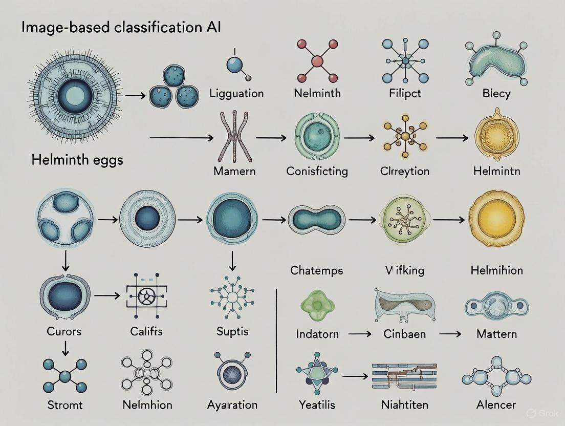
AI, particularly deep learning, is revolutionizing the image-based classification of helminth eggs. A typical AI workflow achieves high accuracy by automating the entire process from image preparation to final classification [5].
AI Classification Workflow
The following diagram illustrates the sequential steps for AI-based classification of helminth eggs from microscopic images:
This workflow integrates several advanced computational techniques:
- Image Preprocessing: The BM3D (Block-Matching and 3D Filtering) technique is employed to remove various types of noise (Gaussian, Salt and Pepper, Speckle, and Fog Noise) from microscopic images. Contrast-Limited Adaptive Histogram Equalization (CLAHE) enhances the contrast between parasite eggs and the background, improving feature visibility for subsequent analysis [5].
- Image Segmentation: A U-Net model, optimized with the Adam optimizer, is used for precise segmentation of potential parasite eggs. This model has demonstrated excellent performance at the pixel level, with reported accuracy of 96.47%, precision of 97.85%, and sensitivity of 98.05%. At the object level, it achieved a 96% Intersection over Union (IoU) and a 94% Dice Coefficient [5].
- Feature Extraction and Classification: Following segmentation, a watershed algorithm extracts Regions of Interest (ROIs). A Convolutional Neural Network (CNN) then performs automatic feature learning in the spatial domain to classify the eggs. This CNN classifier has achieved an overall accuracy of 97.38% with macro average F1 scores of 97.67% [5].
Detailed Experimental Protocols
Protocol 1: qPCR for Trichuris trichiura Detection
Principle: This protocol uses quantitative real-time PCR (qPCR) to detect Trichuris trichiura DNA in stool samples with high sensitivity and specificity, suitable for prevalence studies and efficacy trials [19].
Materials:
- Quick-DNA Fecal/Soil Microbe Miniprep Kit (Zymo Research)
- CFX96 Real-Time PCR Cycler (Bio-Rad Laboratories)
- Specific primers and FAM-labelled probe for T. trichiura [19]
Procedure:
- Sample Washing: Transfer approximately 150 mg of stool to a 15 mL tube with 10 mL of 1X PBS. Homogenize by shaking, centrifuge at 2000 g for 3 minutes, and discard supernatant. Repeat twice [19].
- DNA Extraction: Use the commercial kit according to the manufacturer's protocol. Include a bead-beating step (5 minutes at maximum speed) for mechanical lysis [19].
- qPCR Setup:
- Prepare a master mix containing primers and probe.
- Pipette 5 µL of master mix into reaction wells and add 2 µL of DNA template.
- Include positive controls (serial dilutions of plasmid standard) and no-template controls (nuclease-free water).
- Thermal Cycling: Run on the CFX96 system with the following conditions:
- Pre-denaturation: 95°C for 3 minutes.
- 40 cycles of: 95°C for 10 seconds (denaturation), 61°C for 1 minute (annealing/extension) [19].
- Analysis: A cycle threshold (Ct) value of ≤40 is considered positive for T. trichiura infection [19].
Protocol 2: AI-Based Egg Segmentation and Classification
Principle: This protocol outlines the steps for using a U-Net model for segmentation and a CNN for classification of helminth eggs in digital microscopic images [5].
Materials:
- Dataset of microscopic fecal images
- Computing hardware (GPU recommended)
- Python with libraries (TensorFlow/Keras, OpenCV, Scikit-image)
Procedure:
- Image Preprocessing:
- Apply the BM3D algorithm to reduce noise.
- Use CLAHE to enhance image contrast.
- Model Training (U-Net for Segmentation):
- Train the U-Net model on manually annotated images of helminth eggs.
- Use the Adam optimizer and a loss function like binary cross-entropy.
- Validate model performance using IoU and Dice Coefficient.
- Segmentation Inference:
- Apply the trained U-Net to new images to create segmentation masks.
- Use the watershed algorithm on the masks to separate touching eggs and extract ROIs.
- Classification:
- Feed the extracted ROIs into the pre-trained CNN classifier.
- The CNN outputs the probability for each helminth species.
The Scientist's Toolkit: Key Research Reagents and Materials
Table 3: Essential Research Reagents and Materials for Helminth Diagnostics
| Item | Function / Application | Example / Note |
|---|---|---|
| Kato-Katz Kit | Quantitative microscopic detection of STH and Schistosome eggs | Standard for field surveys; low cost but lower sensitivity [16] [17] |
| Nucleic Acid Extraction Kit | DNA purification from stool samples (e.g., Zymo Research, QIAamp DNA Mini Kit) | Includes bead-beating for mechanical disruption of tough egg shells [16] [19] |
| Species-Specific Primers/Probes | Target DNA amplification in qPCR/PCR | Enables species differentiation (e.g., T. trichiura vs T. incognita) [18] [19] |
| Recombinant Antigens (e.g., ABA-1, Paramyosin) | Targets for serological assays (ELISA) to detect host antibodies | Useful for detecting pre-patent or larval stage infections (e.g., in ascariasis) [20] |
| U-Net & CNN Models | AI-based segmentation and classification of egg images | Achieves high accuracy (>97%); requires computational resources and training data [5] |
| Block-Matching and 3D Filtering (BM3D) | Advanced image denoising in AI workflow | Effectively removes Gaussian, Salt and Pepper, Speckle, and Fog Noise [5] |
Deep Learning in Action: Architectures and Workflows for Egg Detection
The image-based classification of helminth eggs represents a critical challenge in parasitology and public health. Traditional diagnostic methods, reliant on manual microscopy, are characterized by subjectivity, low throughput, and a significant reliance on specialized expertise [23] [7]. This application note details the implementation and evaluation of two advanced object detection models—YOLOv4 and EfficientDet—within the context of an AI-driven research thesis aimed at automating the detection and classification of soil-transmitted helminths (STH) and Schistosoma mansoni eggs. These models offer the potential to deliver rapid, accurate, and scalable diagnostic solutions, which are crucial for monitoring and evaluation programs in resource-limited settings [23]. We provide a detailed, comparative framework to guide researchers and scientists in selecting, implementing, and validating these models for helminth egg analysis.
Model Architectures and Comparative Analysis
YOLOv4: Optimal Speed and Accuracy for Real-Time Detection
YOLOv4 is designed as a high-speed, one-stage object detector that prioritizes real-time performance without sacrificing accuracy. Its architecture is systematically divided into three key components, each contributing to its robust performance [24] [25]:
- Backbone: CSPDarknet53. This backbone serves as the primary feature extractor. It is based on Cross-Stage-Partial-connections (CSP) and DenseNet, which help to alleviate the vanishing gradient problem in deep networks, strengthen feature propagation, and reduce the number of parameters. This design removes computational bottlenecks and improves learning by passing an unedited version of the feature map to subsequent layers [24].
- Neck: PANet with SPP Block. The Path Aggregation Network (PANet) is used for feature aggregation, collecting feature maps from different stages of the backbone to prepare for detection. Additionally, a Spatial Pyramid Pooling (SPP) block is incorporated to increase the receptive field and isolate the most significant contextual features from the backbone [24].
- Head: YOLOv3. The detection head is responsible for the final prediction of bounding boxes and class labels. It utilizes an anchor-based approach and makes predictions at three different scales to improve the detection of objects of varying sizes [24].
A significant contribution of the YOLOv4 framework is its systematic use of training enhancements, termed "Bag of Freebies" and "Bag of Specials" [24] [25]:
- Bag of Freebies: These methods improve training accuracy without increasing inference time. They include advanced data augmentation techniques like Mosaic augmentation, which tiles four training images into one, teaching the model to recognize smaller objects and be less dependent on specific contextual backgrounds, and Self-Adversarial Training (SAT), which identifies and obscures the most relied-upon parts of an image, forcing the network to generalize. The loss function is also refined using CIoU loss to improve bounding box regression [24].
- Bag of Specials: These are modules that add a marginal computational cost during inference for a significant boost in accuracy. They include the Mish activation function, which helps push signals to optimal points for better feature creation, and DIoU NMS for more efficient suppression of overlapping bounding boxes [24].
EfficientDet: Scalable and Efficient Network Architecture
EfficientDet is a family of object detection models that achieves state-of-the-art performance with notably fewer parameters and training epochs compared to other architectures. Its core innovation lies in its scalable and efficient design, making it highly suitable for environments with limited computational resources [26].
- Backbone: EfficientNet. The backbone is pre-trained on ImageNet and is optimized through a compound scaling method that carefully balances the network's depth, width, and resolution. This results in a model that is both highly accurate and computationally efficient [24] [26].
- Neck: BiFPN. The Bi-directional Feature Pyramid Network (BiFPN) serves as the neck. It allows for easy, fast multi-scale feature fusion by enabling information to flow both top-down and bottom-up. This design is often a product of neural architecture search (NAS) to find the most effective connections for aggregating features from different levels of the backbone [24] [26].
- Scalability. A key advantage of the EfficientDet architecture is its scalable nature, denoted by the suffix (e.g., D0-D7), allowing researchers to select a model size that matches their specific accuracy and speed requirements [26].
Quantitative Model Comparison
The table below summarizes the key architectural and performance characteristics of YOLOv4 and EfficientDet, providing a direct comparison for researchers.
Table 1: Architectural and Performance Comparison of YOLOv4 and EfficientDet
| Feature | YOLOv4 | EfficientDet |
|---|---|---|
| Core Architecture | One-stage detector | One-stage detector |
| Backbone | CSPDarknet53 [24] | EfficientNet [26] |
| Neck | PANet with SPP [24] | BiFPN [24] |
| Key Strengths | High speed; ideal for real-time applications; extensive "Bag of Freebies" for robust training [24] [25] | High computational efficiency; excellent accuracy with fewer parameters and training epochs [26] |
| Typical mAP on COCO | State-of-the-art results (exact values depend on variant and configuration) [25] | Achieves best performance in fewest training epochs (exact values depend on variant) [26] |
| Inference Speed | Optimized for real-time detection on a single conventional GPU [25] | Highly efficient, suitable for scalable deployment [26] |
Experimental Protocols for Helminth Egg Analysis
This section provides detailed, step-by-step protocols for training and evaluating object detection models on a dataset of helminth egg images.
Dataset Curation and Preprocessing
A robust dataset is foundational for training an accurate model. The following protocol is adapted from recent studies on STH and S. mansoni detection [23] [7].
Image Acquisition:
- Equipment: Use a digital microscope (e.g., Schistoscope) or a standard light microscope (e.g., Nikon E100) capable of capturing high-resolution images (e.g., 2028x1520 pixels) [23] [7].
- Sample Preparation: Prepare fecal smears using the standard Kato-Katz technique with a 41.7 mg template. This ensures consistency with current field methods [23].
- Magnification: A 4x objective lens is typically sufficient for initial imaging [23].
Data Annotation:
- Software: Use annotation tools like LabelImg or Roboflow to draw bounding boxes around each parasite egg.
- Classes: Annotate eggs by species (e.g., A. lumbricoides, T. trichiura, hookworm, S. mansoni). Annotation should be performed by expert microscists to ensure ground truth accuracy [23].
- Data Splitting: Randomly shuffle the entire dataset and split it into training (70-80%), validation (10-20%), and test (10%) sets. This split prevents data leakage and ensures unbiased evaluation [23] [7].
Data Preprocessing:
- Image Cropping: Large field-of-view images can be automatically cropped into smaller tiles (e.g., 518x486 pixels) using a sliding window approach to facilitate model training and increase the number of samples [7].
- Data Augmentation: Apply a suite of augmentations to improve model generalization. For YOLOv4, this natively includes Mosaic and MixUp [24]. General augmentations beneficial for helminth eggs are:
- Geometric: Random scaling, cropping, flipping, and rotating [25].
- Photometric: Adjustments to brightness, contrast, hue, and saturation [25].
- Advanced Filtering: For pre-processing, techniques like Block-Matching and 3D Filtering (BM3D) can be used to remove noise, and Contrast-Limited Adaptive Histogram Equalization (CLAHE) can enhance contrast [5].
Model Training Protocol
The following steps outline the training procedure for a model like YOLOv4 on a custom helminth egg dataset.
Environment Setup:
Parameter Configuration:
- Anchor Boxes: Use the k-means clustering algorithm on your training set to determine optimal initial anchor box sizes tailored to helminth eggs [7].
- Optimizer: Use the Adam optimizer with a momentum of 0.937 [7].
- Learning Rate: Set an initial learning rate (e.g., 0.01) with a decay factor (e.g., 0.0005) [7].
- Batch Size: Set according to GPU memory (e.g., 64) [7].
- Training Epochs: Train for a sufficient number of epochs (e.g., 300), implementing early stopping if performance on the validation set does not improve for a set number of epochs (e.g., 200) [7].
Model Evaluation and Metrics
Rigorous evaluation is critical to assess model performance. The following metrics, computed on the held-out test set, are essential [27] [28].
- Intersection over Union (IoU): Measures the overlap between a predicted bounding box and the ground truth box. An IoU threshold of 0.50 is commonly used to define a correct detection [27] [28].
- Precision and Recall:
- F1-Score: The harmonic mean of precision and recall, providing a single balanced metric [27] [28].
- Average Precision (AP) and mean Average Precision (mAP):
- AP computes the area under the precision-recall curve for a single class.
- mAP is the average of AP over all object classes. mAP@0.50 uses an IoU threshold of 50%, while mAP@0.50:0.95 averages mAP over IoU thresholds from 0.50 to 0.95 in steps of 0.05, providing a more stringent assessment [27].
Table 2: Performance of Deep Learning Models in Helminth Egg Detection and Classification
| Model / Study | Task / Species | Key Metric | Reported Performance |
|---|---|---|---|
| YOLOv4 [7] | Detection of 9 helminth species | Accuracy | Ranged from 84.85% (T. trichiura) to 100% (C. sinensis, S. japonicum) |
| EfficientDet [23] | Detection of STH & S. mansoni | Weighted Average F-Score | 94.0% (± 1.98%) |
| ConvNeXt Tiny [15] | Classification of A. lumbricoides & T. saginata | F1-Score | 98.6% |
| EfficientNet V2 S [15] | Classification of A. lumbricoides & T. saginata | F1-Score | 97.5% |
| MobileNet V3 S [15] | Classification of A. lumbricoides & T. saginata | F1-Score | 98.2% |
The Scientist's Toolkit: Essential Research Reagents and Materials
Table 3: Essential Materials and Tools for AI-Based Helminth Egg Detection
| Item | Function / Description | Example / Specification |
|---|---|---|
| Digital Microscope | High-resolution image acquisition of prepared slides. | Schistoscope [23], Nikon E100 [7] |
| Annotation Software | For creating bounding box labels on images; generates dataset for model training. | LabelImg, Roboflow |
| Deep Learning Framework | Provides libraries and tools for building, training, and evaluating models. | PyTorch [7] |
| GPU Hardware | Accelerates the deep learning training process through parallel computation. | NVIDIA GeForce RTX 3090 [7] |
| Kato-Katz Kit | Standard method for preparing thick fecal smears for microscopic analysis. | 41.7 mg template [23] |
Workflow and Architecture Diagrams
The following diagram illustrates the end-to-end pipeline for developing an AI-based helminth egg detection system, from sample collection to model deployment.
YOLOv4 Architecture Breakdown
This diagram details the internal structure of the YOLOv4 object detector, showing the flow of data through its backbone, neck, and head.
This document provides detailed application notes and experimental protocols for using three advanced Convolutional Neural Networks (CNNs)—EfficientNet, ConvNeXt, and MobileNet—within the specific research context of image-based classification of helminth eggs. Accurate and efficient identification of helminth eggs in microscopic images is a critical step in diagnosing parasitic infections, monitoring community health, and assessing the efficacy of drug development campaigns. The manual microscopic examination is time-consuming, requires significant expertise, and is prone to human error. Artificial Intelligence (AI), particularly deep learning-based computer vision, offers a promising solution for automating this process, enabling high-throughput, reproducible, and precise analysis. The choice of neural network architecture is paramount, balancing the need for high accuracy with the constraints of computational resources, which are often limited in field or clinical settings. This guide focuses on three modern CNN families that represent different optimal trade-offs between these competing demands, providing researchers with the practical tools needed to implement and evaluate these models for their parasitology research.
The following sub-sections detail the core architectural principles and relative strengths of EfficientNet, ConvNeXt, and MobileNet.
EfficientNet: Compound Scaling for Balanced Performance
EfficientNet revolutionized model design by introducing a compound scaling method that systematically balances the network's depth (number of layers), width (number of channels), and resolution (input image dimensions) [29] [30]. Instead of arbitrarily scaling a single dimension, which leads to rapidly diminishing returns, EfficientNet scales all three in a coordinated manner, governed by a single compound coefficient (φ) [29]. This approach results in a family of models (B0 to B7) that achieve state-of-the-art accuracy with an order-of-magnitude fewer parameters and computational requirements (FLOPs) than previous models [30].
Its core building block is the MBConv layer (Mobile Inverted Bottleneck Convolution), which features depthwise separable convolutions and a Squeeze-and-Excitation (SE) module [29] [30]. The SE module acts as an attention mechanism, adaptively weighting the importance of each channel in a feature map, allowing the model to focus on more informative features [30]. For helminth egg classification, where subtle textural and morphological differences distinguish species, this focused feature extraction is highly beneficial. EfficientNet models, particularly versions like EfficientNetV2, are known for fine-tuning efficiently on small to mid-sized datasets, making them an excellent choice when labeled microscopic image data is limited [31].
ConvNeXt: A Modernized CNN for the 2020s
ConvNeXt is a pure CNN architecture that was redesigned by systematically modernizing a standard ResNet using insights and techniques from Vision Transformers (ViTs) [32]. It demonstrates that CNNs, when properly updated, can match or even surpass the performance of state-of-the-art transformers on various vision tasks while retaining the computational efficiency of convolutions [31] [32].
Key innovations of ConvNeXt include:
- Large Kernel Depthwise Convolutions: Replacing small (e.g., 3x3) kernels with larger (e.g., 7x7) depthwise convolutions to increase the receptive field, similar to the global context modeling of self-attention in ViTs [32].
- Inverted Bottleneck and Layer Normalization: Adopting an inverted bottleneck design common in mobile networks and replacing Batch Normalization with Layer Normalization, which improves training stability [32].
- Modernized Training Recipes: Utilizing advanced training strategies like AdamW optimizer, data augmentation (MixUp, CutMix), and regularization (label smoothing) [32].
ConvNeXt is particularly suited for research pipelines where high accuracy is the priority and computational resources for training are adequate. Its hierarchical design and powerful feature extraction capabilities make it a strong "safe bet for scalable production pipelines" in biomedical image analysis [31].
MobileNet: Lightweight Champion for Edge Deployment
The MobileNet family is specifically engineered for low-power, low-latency deployment on mobile and embedded devices [31] [33]. Its primary innovation is the heavy use of depthwise separable convolutions, which factorize a standard convolution into a depthwise convolution (applying a single filter per input channel) and a pointwise convolution (a 1x1 convolution to combine channel outputs) [31]. This factorization drastically reduces the model's parameter count and computational cost.
MobileNetV3, the latest major version, incorporates neural architecture search (NAS) to optimize the network structure further and includes a Squeeze-and-Excitation module in its bottleneck blocks [31] [33]. It is the go-to model for applications that require real-time inference on resource-constrained hardware, such as point-of-care diagnostic devices or portable field microscopes [31]. For helminth egg classification, a MobileNetV3-based model can be quantized (e.g., to INT8 precision) to achieve excellent performance on a smartphone or single-board computer, enabling decentralized analysis without reliance on cloud connectivity [31].
Table 1: Comparative Analysis of CNN Architectures for Image Classification
| Feature | EfficientNet | ConvNeXt | MobileNetV3 |
|---|---|---|---|
| Core Innovation | Compound Scaling of depth, width, & resolution [29] [30] | Modernized CNN with ViT-inspired designs [32] | Depthwise Separable Convolutions & NAS [31] |
| Key Building Block | MBConv with SE attention [29] [30] | Large-kernel depthwise conv [32] | Inverted Residual Bottleneck [31] |
| Typical Use Case | High-accuracy, efficient training on small datasets [31] | High-accuracy, research & cloud deployment [31] | Real-time inference on mobile/embedded devices [31] [33] |
| Strengths | Best trade-off: accuracy vs. efficiency [31] [30] | High performance, modern & scalable [31] | Extremely fast, low power, easily quantized [31] |
| Helminth Egg Application | Default choice for balanced performance | When highest accuracy is needed and compute is available | For point-of-care/field deployment |
Quantitative Performance Comparison
To make an informed model selection, it is essential to compare the quantitative performance of different variants within and across these model families. The following table summarizes key metrics on the standard ImageNet-1K benchmark, which serves as a proxy for their potential performance on other image classification tasks, such as helminth egg analysis.
Table 2: Model Performance and Complexity on ImageNet-1K Benchmark [31]
| Model Variant | Top-1 Accuracy (%) | Parameters (Millions) | FLOPs (Billions) | Notes |
|---|---|---|---|---|
| EfficientNetV2-L | 86.8 - 87.3 | 120 | 53 | Strong balance; efficient fine-tuning [31] |
| ConvNeXt-B | 85.8 | 89 | 15.4 | Modern CNN; matches Swin Transformer performance [31] |
| ViT-B/16 | ~85.4 | 86 | 17.6-55.5 | Transformer baseline; needs large-scale pre-training [31] |
| Swin-B | 86.4 | 88 | 47 | Hierarchical transformer for reference [31] |
| MobileNetV3-Large | ~75.2 | 5.4 | 0.2 | Edge-optimized; high speed, lower accuracy [31] |
| ResNet-50 (modern) | ~80.4 | 25.6 | 4.1 | Classic baseline for comparison [31] |
Experimental Protocols for Helminth Egg Classification
This section outlines a standardized workflow and detailed protocols for training and evaluating the featured CNN models on a proprietary dataset of helminth egg images.
The diagram below illustrates the end-to-end pipeline for developing a helminth egg classification system, from data preparation to model deployment.
Protocol 1: Data Preparation and Augmentation
Objective: To create a robust, balanced, and standardized dataset for training and evaluating deep learning models.
Data Preprocessing:
- Resizing: Uniformly resize all raw microscopic images to the input resolution required by the chosen model (e.g., 224x224 for EfficientNet-B0, 384x384 for higher-resolution fine-tuning) [31]. Maintain aspect ratio by padding if necessary to avoid distortion.
- Normalization: Normalize pixel values using the mean and standard deviation of the pre-training dataset (typically ImageNet). Common values are
mean = [0.485, 0.456, 0.406],std = [0.229, 0.224, 0.225].
Data Augmentation (Training Set Only): Apply random transformations to increase dataset diversity and improve model generalization. This is critical for preventing overfitting, especially with limited medical data [32].
- Geometric: Random horizontal and vertical flips, random rotations (e.g., ±15 degrees) [34].
- Photometric: Adjust brightness, contrast, and saturation slightly. Adding random Gaussian noise can also improve robustness.
- Advanced Techniques: For better performance, consider incorporating MixUp or CutMix, which combine images and labels to create new training samples [31] [32].
Dataset Splitting: Partition the data into three sets:
- Training (70%): Used to fit the model parameters.
- Validation (15%): Used for hyperparameter tuning and selecting the best model during training.
- Test (15%): Used only once for the final evaluation of the selected model's generalization performance. Ensure splits are stratified to preserve the class distribution.
Protocol 2: Model Training and Fine-tuning
Objective: To adapt a pre-trained model to the specific task of helminth egg classification effectively.
Model Initialization: Always start with a model pre-trained on a large-scale dataset like ImageNet. This transfer learning approach significantly speeds up convergence and improves final accuracy compared to training from scratch [31].
- Source: Download pre-trained weights from official sources like Torchvision (for ResNet, ConvNeXt), Hugging Face Transformers or timm library (for a wide variety of models including EfficientNet and ConvNeXt), or TensorFlow Hub (for EfficientNet and MobileNet) [31].
Fine-tuning Strategy:
- Feature Extraction (for very small datasets): Freeze all pre-trained layers (the "backbone") and only train a new randomly initialized classification head.
- Full Fine-tuning (recommended for datasets of >1000 images per class): Unfreeze all or most of the layers and train the entire network with a low learning rate. This allows the model to adapt its pre-learned features to the specifics of helminth egg morphology.
Training Configuration:
- Optimizer: Use AdamW with a learning rate (
lr) between 1e-5 and 1e-4 [34] [32]. This optimizer often provides better generalization than SGD with momentum in modern training recipes. - Loss Function: Use Cross-Entropy Loss. For imbalanced datasets, supplement it with Focal Loss or class-weighted cross-entropy to make the model focus on harder examples and prevent the majority class from dominating [31] [33].
- Regularization: Apply Dropout (rates of 0.2-0.5) and/or Stochastic Depth (DropConnect) to prevent overfitting [30] [35]. Label Smoothing can also be beneficial [31] [32].
- Epochs & Scheduling: Train for a sufficient number of epochs (e.g., 50-100) and use a cosine learning rate scheduler with a warm-up period to stabilize training early on [32].
- Optimizer: Use AdamW with a learning rate (
Protocol 3: Model Evaluation and Inference
Objective: To rigorously assess model performance and prepare it for deployment.
Performance Metrics: Evaluate the model on the held-out test set.
- Primary Metrics: Report Top-1 Accuracy, Precision, Recall, and F1-Score (preferably per-class and macro-averaged) [33]. The F1-Score is especially important for imbalanced datasets.
- Confusion Matrix: Generate a confusion matrix to identify specific inter-class confusion patterns (e.g., between morphologically similar helminth eggs).
Robustness and Deployment Readiness:
- Inference Latency: Measure the average time to classify a single image or a batch of images on the target hardware (e.g., a server GPU, a desktop CPU, or a mobile phone) [31].
- Model Compression (for MobileNet & EfficientNet): For edge deployment, apply post-training quantization to reduce model size and accelerate inference. This can shrink a model by 4x with minimal accuracy loss [31].
The Scientist's Toolkit: Research Reagent Solutions
The following table lists essential software and hardware "reagents" required to implement the described experimental protocols.
Table 3: Essential Research Reagents for AI-based Helminth Egg Classification
| Reagent / Tool | Type | Function / Application | Example / Note |
|---|---|---|---|
| Pre-trained Models | Software | Provides a powerful starting point via transfer learning, drastically reducing data and compute needs. | Torchvision, Hugging Face Hub, TIMM (PyTorch Image Models) library [31] |
| PyTorch / TensorFlow | Software | Core deep learning frameworks for model implementation, training, and inference. | PyTorch is common in research; TensorFlow has strong production tools (TF Lite) [31] |
| High-Performance GPU | Hardware | Accelerates model training and hyperparameter tuning, reducing experiment time from days to hours. | NVIDIA RTX series with >=8GB VRAM |
| Microscopy with Camera | Hardware | Captures high-quality digital images of helminth egg samples for dataset creation. | Standard laboratory microscope with a digital camera attachment |
| Labeling Software | Software | Enables researchers to annotate collected images, assigning the correct species label to each. | LabelImg, VGG Image Annotator, or commercial solutions |
| Optimization Libraries | Software | Provides implementations of optimizers, learning rate schedulers, and loss functions. | Included in PyTorch/TensorFlow (e.g., AdamW, CosineAnnealingLR) [32] |
The final choice of model architecture depends on the specific constraints and goals of the helminth egg classification project. The following decision diagram provides a clear pathway for researchers to select the most appropriate model.
In summary, EfficientNet, ConvNeXt, and MobileNet each offer distinct advantages for the image-based classification of helminth eggs. EfficientNet provides the most balanced approach for typical research settings, ConvNeXt is ideal for projects where accuracy is paramount and resources are plentiful, and MobileNet is indispensable for point-of-care applications. By following the detailed application notes, performance comparisons, and experimental protocols outlined in this document, researchers and drug development professionals can effectively leverage these state-of-the-art AI tools to advance the field of parasitology and improve global health outcomes.
End-to-End Artificial Intelligence-Powered Digital Pathology (AI-DP) platforms represent a transformative approach to diagnosing parasitic infections in resource-limited settings. These systems integrate field-deployable digital slide scanners with onboard AI analysis to automate the detection and classification of soil-transmitted helminth (STH) and Schistosoma mansoni (SCH) eggs in fecal samples [36]. This automation addresses critical limitations of conventional microscopy, which relies on labour-intensive processes requiring trained technicians and poses significant logistical challenges in areas where neglected tropical diseases (NTDs) are most prevalent [36] [23]. By providing near-real-time data with integrated quality controls, AI-DP platforms demonstrate significant potential for efficient monitoring and evaluation of large-scale deworming programs, aligning with the World Health Organization's (WHO) goal to eliminate NTDs as a public health problem by 2030 [36] [37].
Platform Architecture & Workflow
An AI-DP platform is engineered as an integrated system comprising several core components that work in sequence to transform a physical stool sample into a digitized, analyzed diagnostic result.
Core Components
The architecture typically consists of four main subsystems [36] [37]:
- Electronic Data Capture Tools: Facilitate the recording of sample metadata and patient information.
- Whole Slide Imaging (WSI) Scanners: Portable, robust scanners capable of automatically capturing high-resolution images of prepared microscopy slides in field conditions.
- Onboard AI Analysis Engine: A pre-trained deep learning model that processes the digitized slide images to detect, count, and classify parasite eggs.
- Result Verification Software: A user interface that allows technicians to review the AI-generated annotations, confirm results, and make corrections if necessary, thereby creating a human-in-the-loop validation system.
Integrated Workflow
The end-to-end process, from sample collection to final reporting, is visualized in the following workflow.
Performance Metrics of AI Models
The efficacy of the AI detection models is paramount to the platform's overall performance. Various deep learning architectures have been validated on large datasets of Kato-Katz slide images. The following table summarizes the reported performance metrics for different parasite species across multiple studies, highlighting the high accuracy achieved by these systems.
Table 1: Analytical Performance of AI Models for Helminth Egg Detection
| Parasite Species | Model Architecture | Precision (%) | Recall / Sensitivity (%) | F1-Score / mAP@0.5 | Data Source |
|---|---|---|---|---|---|
| Ascaris lumbricoides | EfficientDet [23] | 95.4 | 91.7 | 97.1 (AP) | 951 KK slides, 43,919 eggs [36] |
| Trichuris trichiura | EfficientDet [23] | 95.9 | 86.7 | 94.8 (AP) | 951 KK slides, 43,919 eggs [36] |
| Hookworm | EfficientDet [23] | 84.6 | 86.6 | 91.4 (AP) | 951 KK slides, 43,919 eggs [36] |
| Schistosoma mansoni | EfficientDet [23] | 89.1 | 79.1 | 89.2 (AP) | 951 KK slides, 43,919 eggs [36] |
| Multiple STH & SCH | YOLOv7-E6E (ID Setting) [37] | - | - | 97.47 (F1) | AI4NTD KK2.0 Dataset [37] |
| Multiple Parasite Eggs | YAC-Net (Lightweight YOLO) [13] | 97.8 | 97.7 | 0.9773 (F1) | ICIP 2022 Challenge Dataset [13] |
| Multiple Parasite Eggs | U-Net + CNN [5] | 97.85 (Pixel-level) | 98.05 (Sensitivity) | 97.38 (Accuracy) | Custom Dataset [5] |
Experimental Protocols
To ensure reproducible and reliable results, standardized protocols for data generation and model training are essential. The following sections detail the key experimental methodologies cited in the literature.
Sample Preparation and Image Acquisition
This protocol is foundational for creating high-quality datasets for both model training and clinical diagnosis [36] [23].
- Sample Collection: Collect fresh fecal samples in sterile, leak-proof containers. Maintain a cool chain if processing is not immediate.
- Kato-Katz Smear Preparation:
- Place a small amount of sieved stool sample on a clean glass slide.
- Press a template with a 41.7 mg hole onto the slide to standardize the sample amount.
- Fill the template hole with stool and remove excess material.
- Carefully remove the template.
- Place a glycerol-soaked cellophane strip over the sample and press firmly to create a uniform, transparent smear.
- Microscopy and Imaging:
- Place the prepared slide into a portable, automated digital microscope (e.g., Schistoscope [23] or other WSI scanner).
- Scan the slide using a 4x to 10x objective lens to capture field-of-view (FOV) images with resolutions such as 2028 x 1520 pixels [23].
- Systematically scan the entire slide to generate a whole-slide image composed of multiple FOVs.
AI Model Training and Evaluation
This protocol outlines the standard workflow for developing the core detection algorithm, as used in recent studies [23] [37] [13].
- Data Annotation:
- Expert microscopists manually annotate images from the training set, drawing bounding boxes around each parasite egg and labeling them with the correct species class.
- This creates the "ground truth" dataset used for supervised learning.
- Data Preprocessing & Augmentation:
- Model Training:
- Select a model architecture (e.g., YOLOv7, EfficientDet).
- Initialize training with pre-trained weights on general image datasets (Transfer Learning).
- Train the model using an optimizer (e.g., Adam) with a defined initial learning rate (e.g., 0.01) and learning rate decay.
- Use the validation set for hyperparameter tuning and to avoid overfitting.
- Model Evaluation:
- Evaluate the final model on the held-out test set.
- Calculate standard object detection metrics: precision, recall, F1-score, and mean Average Precision (mAP) at an Intersection over Union (IoU) threshold of 0.5.
The relationships between the core components of the AI model and the training process are illustrated below.
The Scientist's Toolkit: Research Reagent Solutions
Successful development and deployment of an AI-DP platform rely on a suite of specific materials and software tools. The following table details these essential components and their functions.
Table 2: Key Research Reagents and Materials for AI-DP Platform Development
| Item Name | Function / Application | Specifications / Examples |
|---|---|---|
| Kato-Katz Kit | Standardized preparation of thick fecal smears for microscopic detection of helminth eggs. | 41.7 mg template, glass slides, glycerol, cellophane strips [23]. |
| Portable Whole Slide Scanner | Automated digital imaging of microscopy slides in field settings. | Schistoscope [23]; Scanners with 4x-10x objectives, automated staging [36]. |
| AI4NTD KK2.0 Dataset | Benchmark dataset for training and evaluating STH and SCH egg detection models. | Contains >10,000 FOV images with ~20,000 annotated eggs [23] [37]. |
| Deep Learning Framework | Software environment for building, training, and deploying AI models. | PyTorch, TensorFlow [7] [37]. |
| Object Detection Model | The core algorithm for detecting and classifying parasite eggs in images. | YOLO variants (v4, v5, v7) [7] [37] [13], EfficientDet [23]. |
| Annotation Software | Tool for experts to create ground truth data by labeling eggs in images. | Software for drawing bounding boxes and assigning class labels [23]. |
| Computing Hardware | Infrastructure for model training and for running inference in deployment. | Training: NVIDIA GPUs (e.g., RTX 3090) [7]. Deployment: Edge computing devices [36] [23]. |
Accurate diagnosis of helminth infections is a critical public health challenge, with soil-transmitted helminths (STH) and schistosomiasis affecting over a billion people globally [36]. Traditional diagnostic methods, particularly manual microscopy of stool samples, are labor-intensive, time-consuming, and require substantial expertise, often leading to variable sensitivity and specificity [37]. Artificial intelligence (AI) has emerged as a transformative approach to automate and improve the accuracy of helminth egg classification in microscopic images.
This application note provides a comprehensive analysis of performance benchmarks for AI-driven classification of helminth eggs, focusing on the critical metrics of precision and recall across both single-species and mixed-species scenarios. We synthesize recent experimental data, detail standardized protocols for model training and evaluation, and identify key factors influencing diagnostic performance to support researchers and developers in creating robust, clinically applicable AI tools.
Performance Benchmarks
Single-Species Classification Performance
Table 1: Performance Benchmarks for Single-Species Helminth Egg Detection
| Helminth Species | Model Architecture | Precision (%) | Recall (%) | F1-Score/% | mAP/@IoU0.5 | Research Context |
|---|---|---|---|---|---|---|
| Ascaris lumbricoides | AI-DP Platform [36] | 95.4 | 91.7 | - | 97.1 | Field-deployable system |
| Trichuris trichiura | AI-DP Platform [36] | 95.9 | 86.7 | - | 94.8 | Field-deployable system |
| Hookworm | AI-DP Platform [36] | 84.6 | 86.6 | - | 91.4 | Field-deployable system |
| Schistosoma mansoni | AI-DP Platform [36] | 89.1 | 79.1 | - | 89.2 | Field-deployable system |
| Clonorchis sinensis | YOLOv4 [7] | 100.0 | - | - | - | Laboratory setting |
| Schistosoma japonicum | YOLOv4 [7] | 100.0 | - | - | - | Laboratory setting |
| Enterobius vermicularis | YOLOv4 [7] | 89.3 | - | - | - | Laboratory setting |
| Fasciolopsis buski | YOLOv4 [7] | 88.0 | - | - | - | Laboratory setting |
| Ascaris lumbricoides | YOLOv7-E6E [37] | - | - | 97.5 | - | In-distribution setting |
| Ascaris lumbricoides | ConvNeXt Tiny [38] | - | - | 98.6 | - | Multiclass classification |
| Taenia saginata | ConvNeXt Tiny [38] | - | - | 98.6 | - | Multiclass classification |
Mixed-Species and Complex Scenario Performance
Table 2: Performance in Mixed-Species and Out-of-Distribution Scenarios
| Experimental Scenario | Model Architecture | Key Performance Metrics | Notes |
|---|---|---|---|
| Mixed Group 1 [7] | YOLOv4 | 98.10%, 95.61% accuracy | A. lumbricoides and T. trichiura |
| Mixed Group 2 [7] | YOLOv4 | 94.86%, 93.28%, 91.43% accuracy | A. lumbricoides, T. trichiura, A. duodenale |
| Mixed Group 3 [7] | YOLOv4 | 93.34%, 75.00% accuracy | C. sinensis and Taenia spp. |
| OOD: Device Change [37] | YOLOv7 + 2x3 Montage | +8% Precision, +14.85% Recall, +21.36% mAP | Enhanced generalization via data augmentation |
| Complex OOD: Device + Unseen Eggs [37] | YOLOv7 variants | Variable performance drop | Highlights generalization challenge |
Experimental Protocols
AI Model Development Workflow
The following diagram illustrates the standardized experimental workflow for developing and evaluating AI models for helminth egg classification, as implemented in recent studies.
Detailed Methodological Components
Sample Preparation and Imaging
- Sample Collection: Helminth egg suspensions are acquired from verified biological suppliers or clinical isolates [7]. For experimental controls, parasite-free human stool samples are confirmed through pre-screening via flotation-filtration and qPCR [39].
- Slide Preparation: Two drops of vortex-mixed egg suspension (approximately 10 μL) are transferred to a microscope slide and covered with an 18mm × 18mm coverslip, avoiding air bubbles [7]. The Kato-Katz thick smear technique is widely used for field-deployable systems [36] [37].
- Imaging Protocol: Images are captured using light microscopes (e.g., Nikon E100) or whole-slide imaging scanners at consistent magnifications [7] [36]. Multiple images per slide are acquired to ensure comprehensive egg representation.
Data Preprocessing and Augmentation
- Data Splitting: Datasets are typically partitioned into training (80%), validation (10%), and test (10%) sets, maintaining class balance across splits [7].
- Image Preprocessing: Includes background normalization, cropping using sliding window approaches (e.g., 518 × 486 pixels), and resolution standardization [7].
- Data Augmentation: The 2×3 montage strategy significantly enhances out-of-distribution generalization by creating composite images, improving precision by 8% and recall by 14.85% in OOD scenarios [37]. Additional techniques include Mosaic and Mixup augmentations [7].
Model Training and Evaluation
- Model Selection: Recent studies have employed YOLO variants (v4, v7), ConvNeXt Tiny, EfficientNet V2 S, and MobileNet V3 S, selected based on performance-efficiency trade-offs [7] [37] [38].
- Training Parameters: Standard implementations use Python with PyTorch framework, Adam optimizer (learning rate=0.01, momentum=0.937), batch size of 64, and up to 300 epochs with early stopping [7].
- Evaluation Metrics: Primary metrics include precision, recall, F1-score, and mean Average Precision (mAP) at IoU threshold of 0.5. Advanced error analysis employs TIDE (Toolkit for Identifying Object Detection Errors) and Grad-CAM for model decision interpretation [37].
The Scientist's Toolkit
Table 3: Essential Research Reagents and Materials
| Item | Function/Application | Specifications/Notes |
|---|---|---|
| Helminth Egg Suspensions | Source of biological material for model training and validation | Commercially sourced (e.g., Deren Scientific Equipment Co. Ltd.) or clinical isolates; verify species and viability [7] |
| Kato-Katz Kit | Standardized slide preparation for field-based diagnostics | Recommended by WHO for STH and schistosomiasis screening; enables egg quantification [36] [37] |
| Sodium Nitrate (NaNO₃) Solution | Flotation-filtration for egg purification and concentration | Specific gravity of 1.30 recovers 62.7% more Trichuris spp. eggs than standard 1.20 SpGr [39] |
| Whole Slide Imaging Scanner | Digital pathology and image acquisition | Portable, field-deployable systems with onboard AI analysis capability [36] |
| Annotation Software | Ground truth establishment for training data | Manual expert annotation of egg coordinates and species classification; critical for supervised learning [36] [37] |
Factors Influencing Performance
Single vs. Mixed Species Complexity
AI models consistently demonstrate higher performance in single-species detection compared to mixed-species scenarios. For example, while YOLOv4 achieved 100% accuracy for Clonorchis sinensis and Schistosoma japonicum in single-species preparations, performance declined in mixed groups, particularly with Taenia spp. (75.00% accuracy in Group 3) [7]. This performance drop highlights the challenge of distinguishing between morphologically similar eggs in complex mixtures.
Out-of-Distribution Generalization
A critical factor in real-world deployment is model performance under out-of-distribution conditions, including variations in imaging devices and encounter with previously unseen egg types. While YOLOv7-E6E achieved exceptional in-distribution performance (F1-score of 97.47%), all models experienced performance degradation under OOD conditions, particularly when novel egg types were introduced [37]. The 2×3 montage data augmentation strategy demonstrated significant benefits for device shift scenarios but provided incomplete protection against completely novel egg types.
Egg Morphological Characteristics
Model performance varies substantially across helminth species, influenced by distinct egg morphological features. Species with characteristic egg structures like Ascaris lumbricoides consistently achieve higher precision (95.4%) and recall (91.7%) [36]. In contrast, hookworm eggs, which degrade rapidly and present detection challenges even in manual microscopy, correspond with lower AI model performance (84.6% precision, 86.6% recall) [36] [37].
Achieving high precision and recall in AI-based helminth egg classification requires careful consideration of species complexity, rigorous validation across diverse conditions, and implementation of specialized data augmentation strategies. While current models demonstrate exceptional performance in controlled, single-species scenarios (F1-scores >98%), real-world application in mixed-species and out-of-distribution contexts remains challenging. Future development should prioritize multi-species training datasets, advanced augmentation techniques, and comprehensive OOD testing to ensure robust field performance. These approaches will ultimately support the development of reliable AI diagnostics that can effectively contribute to global helminth control programs.
Overcoming Real-World Hurdles: Data, Deployment, and Model Refinement
The accurate image-based classification of helminth eggs using artificial intelligence (AI) represents a paradigm shift in the diagnosis of parasitic diseases, which affect over two billion people globally [40]. The performance and reliability of these deep learning models are fundamentally constrained by the quality, diversity, and robustness of the training datasets. Building robust datasets is therefore a critical prerequisite for developing AI tools that can generalize beyond ideal laboratory conditions to be effective in diverse, real-world diagnostic scenarios, particularly in resource-limited settings where these infections are most prevalent [37]. This document outlines detailed protocols and strategies for the key stages of dataset creation—annotation, augmentation, and bias mitigation—specifically within the context of helminth egg microscopy research.
Data Acquisition and Annotation Protocols
A foundational step in creating a reliable dataset is the standardized acquisition and meticulous annotation of helminth egg images.
Sample Preparation and Image Acquisition
Standardized sample preparation is crucial for ensuring consistency across images. The following protocol, adapted from recent studies, details the process for creating Kato-Katz thick smears, a widely used method in helminthology [41] [42].
Protocol 2.1: Kato-Katz Thick Smear Preparation and Digitization
- Objective: To consistently prepare stool smears and acquire digital microscopic images of helminth eggs for dataset construction.
- Materials:
- Fresh stool sample
- Microscope slide and 41.7 mg template
- Cellophane strips soaked in glycerol-malachite green solution
- Light microscope or portable digital scanner (e.g., Schistoscope [42])
- Image acquisition software
- Procedure:
- Template Filling: Place the microscope slide on a bench. Position the 41.7 mg template on the slide and fill it completely with the stool sample, ensuring no air pockets.
- Smear Preparation: Carefully remove the template, leaving a defined stool aliquot on the slide.
- Cellophane Covering: Place a glycerol-soaked cellophane strip over the stool aliquot and press down gently with another slide to create a uniform, transparent smear.
- Microscopy and Digitization: After a clearing time (typically 30-60 minutes), place the slide under a light microscope or a portable whole-slide scanner. For whole-slide imaging, systematically scan the entire smear area. For field-of-view (FOV) imaging, capture multiple, non-overlapping images at a consistent magnification (e.g., 4x objective) [42].
- Quality Control: Visually inspect a subset of acquired images for focus, illumination consistency, and the presence of artifact. Discard images with significant blurring, over-saturation, or debris that obscures >50% of the field of view.
Expert Annotation and Ground Truth Establishment
The annotation process establishes the "ground truth" that the AI model will learn from. Inconsistencies at this stage directly introduce label noise and degrade model performance.
- Annotation Tools: Use specialized software (e.g., VGG Image Annotator, LabelImg) that allows experts to draw bounding boxes around each parasite egg and assign a class label.
- Multi-Expert Validation: To ensure annotation accuracy and consistency, implement a multi-reader verification process. A proven strategy is to have AI-detected eggs independently verified by two expert microscopists [41]. Discrepancies are resolved by a third, senior expert. This method has been shown to create a highly reliable composite reference standard.
- Metadata Logging: For each annotated image, record essential metadata including the species of helminth egg (Ascaris lumbricoides, Trichuris trichiura, hookworm, Schistosoma mansoni, etc.), the source of the egg suspension, and the specific imaging device used.
Table 1: Key Research Reagent Solutions for Helminth Egg Image Acquisition
| Item Name | Function/Application | Specification Notes |
|---|---|---|
| Kato-Katz Kit | Preparation of thick stool smears for microscopic examination. | Includes 41.7 mg template and glycerol-soaked cellophane strips [41] [42]. |
| Helminth Egg Suspensions | Provide positive control samples for model training and validation. | Commercially sourced suspensions of species like A. lumbricoides and C. sinensis ensure known positive samples [40]. |
| Portable Digital Scanner (e.g., Schistoscope) | Enables whole-slide imaging and digitization of samples in field settings. | Cost-effective, automated microscope designed for resource-limited environments [42]. |
| Light Microscope | Standard tool for visual examination and image capture of samples. | Equipped with a digital camera; Nikon E100 model has been used in prior research [40]. |
Data Augmentation and Bias Mitigation Strategies
To combat overfitting and improve model generalization, the raw dataset must be strategically augmented. Furthermore, proactive steps are required to identify and mitigate inherent biases.
Advanced Data Augmentation
Data augmentation artificially expands the training dataset by creating modified versions of existing images. This teaches the model to recognize helminth eggs under various conditions.
- Standard Spatial and Color Augmentations: These include techniques like random rotation, flipping, scaling, shearing, and adjustments to brightness, contrast, and saturation. These should be applied on-the-fly during model training.
- Advanced Montage Augmentation: For challenging out-of-distribution (OOD) scenarios, such as when deploying a model with a new image capture device, a 2x3 montage augmentation strategy has proven highly effective [37]. This technique stitches multiple images into a single montage during training, forcing the model to learn features that are invariant to background and compositional changes. One study demonstrated that this method significantly enhanced OOD performance, increasing precision by 8%, recall by 14.85%, and mean average precision (mAP) by 21.36% [37].
Combating Dataset Bias
Bias in a dataset can lead to models that perform poorly on data from new sources or with different demographic characteristics.
- Source and Device Diversity: Actively collect images from multiple geographic locations and using different microscope models or scanners. This helps prevent the model from associating specific artifacts or background colors with a particular parasite class [40] [37].
- Stratified Sampling for Balanced Representation: Analyze the distribution of egg classes in your dataset. If certain species (e.g., A. lumbricoides) are overrepresented, use stratified sampling when creating training, validation, and test sets to ensure all classes are adequately represented [42]. This prevents the model from being biased towards the most common classes.
- Rigorous OOD Testing: Evaluate the final model on a separate test set collected from a different site or with different equipment than the training data. This is the only way to truly assess model robustness and generalization [37]. Performance will typically be lower on OOD data, highlighting areas for improvement.
Table 2: Quantitative Impact of Augmentation on Model Performance (YOLOv7)
| Augmentation Strategy | Scenario | Precision (%) | Recall (%) | mAP@0.5 (%) | Primary Benefit |
|---|---|---|---|---|---|
| Baseline (No Augmentation) | In-Distribution (ID) | 96.2 | 95.1 | 97.5 | -- |
| Standard Augmentations* | In-Distribution (ID) | 96.5 (+0.3) | 96.0 (+0.9) | 97.8 (+0.3) | Basic Invariance |
| 2x3 Montage Augmentation | Out-of-Distribution (OOD) | +8.0 | +14.85 | +21.36 | Enhanced Generalization [37] |
*Includes rotation, flipping, color jitter, etc. Baseline and improvement data adapted from [37].
Experimental Protocol: Model Training and Evaluation
This protocol integrates the aforementioned strategies into a complete workflow for training and evaluating a helminth egg detection model.
Protocol 4.1: End-to-End Model Training and Robustness Evaluation
- Objective: To train a deep learning model for helminth egg detection and rigorously evaluate its performance on both in-distribution and out-of-distribution data.
- Materials:
- Annotated dataset of helminth egg images (~10,000+ FOV images recommended [42])
- High-performance computing workstation with GPU (e.g., NVIDIA GeForce RTX 3090 [40])
- Deep learning framework (e.g., PyTorch or TensorFlow)
- Implementation of an object detection model (e.g., YOLOv4, YOLOv7, EfficientDet [40] [42] [37])
- Procedure:
- Dataset Splitting: Randomly shuffle the entire dataset and split it into training (70-80%), validation (10-15%), and test (10-15%) sets. Ensure stratification by egg class to maintain label distribution across splits [40] [42].
- Training with Augmentation: Train the model on the training set. Apply a standard augmentation pipeline (random flips, rotations, color jitter).-
- Critical Step: Integrate advanced augmentation like the 2x3 montage strategy [37] to improve OOD robustness.
- Hyperparameters: Use an initial learning rate of 0.01, Adam optimizer (momentum=0.937), and a batch size of 64. Employ early stopping if validation performance does not improve for 200 epochs [40].
- Validation and Model Selection: Use the validation set to tune hyperparameters and select the best-performing model checkpoint.
- Comprehensive Testing:
- In-Distribution (ID) Test: Evaluate the final model on the held-out test set from the same source as the training data. Report standard metrics (precision, recall, F1-score, mAP).
- Out-of-Distribution (OOD) Test: Evaluate the model on a completely separate test set, ideally from a different geographic region or captured with a different device [37]. This is the critical test for real-world applicability.
- Troubleshooting:
- Low OOD Performance: This indicates poor generalization. The primary solution is to diversify the training data and employ stronger data augmentation techniques like montage augmentation.
- High False Positives for a Specific Class: Re-examine the annotations for that class in the training set. There may be label noise or inconsistent bounding boxes. Consider adding more negative examples from challenging backgrounds.
The development of robust AI models for helminth egg classification is inextricably linked to the quality of the underlying datasets. By implementing the detailed protocols for systematic annotation, employing advanced augmentation strategies like the 2x3 montage, and proactively designing studies to combat bias through rigorous OOD testing, researchers can create datasets that significantly enhance model generalizability. These strategies are essential for translating promising AI-based diagnostic tools from research laboratories into reliable, impactful applications in the global fight against parasitic diseases.
Accurate detection of mixed and low-intensity helminth infections remains a significant challenge in helminth control programs. The following table summarizes the performance of manual microscopy versus artificial intelligence (AI)-supported methods for detecting soil-transmitted helminths (STHs), as demonstrated in recent field studies [41].
Table 1: Diagnostic performance comparison for STH detection in Kato-Katz thick smears (n=704)
| Diagnostic Method | A. lumbricoides Sensitivity | T. trichiura Sensitivity | Hookworm Sensitivity | Specificity (All STHs) |
|---|---|---|---|---|
| Manual Microscopy | 50.0% | 31.2% | 77.8% | >97% |
| Autonomous AI | 50.0% | 84.4% | 87.4% | >97% |
| Expert-Verified AI | 100% | 93.8% | 92.2% | >97% |
The data reveals that expert-verified AI significantly outperforms both manual microscopy and autonomous AI, particularly for detecting T. trichiura and hookworms in light-intensity infections [41]. Of the 122 smears classified as STH-positive according to the composite reference standard, 118 (96.7%) were light-intensity infections [41].
Experimental Protocols
Sample Preparation and Image Acquisition Protocol
Principle: Standardized preparation of Kato-Katz thick smears ensures consistent sample quality for digital imaging and AI analysis [23].
Materials:
- Stool samples collected in sterile 20 mL universal containers
- 41.7 mg template for Kato-Katz technique
- Glycerol for sample clearance
- Standard microscope slides
- Schistoscope devices or portable whole-slide scanners [23] [41]
Procedure:
- Process fecal samples using the standard Kato-Katz technique with a 41.7 mg template [23]
- Prepare thick smears on microscope slides with glycerol clearance
- Register sample slides using digital microscopy systems (e.g., Schistoscope configured with 4× objective lens, 0.10 NA) [23]
- Acquire Field-of-View (FOV) images at 2028 × 1520 pixel resolution [23]
- Screen and manually annotate images by expert microscopists to establish ground truth [23]
Technical Notes:
- Analysis must be performed within 30-60 minutes of preparation due to glycerol-induced disintegration of hookworm eggs [41]
- For field applications, establish field labs with multiple portable digital microscopes to accelerate data acquisition [23]
- Combine datasets from multiple sources to enhance robustness, using 70% for training, 20% for validation, and 10% for testing [23]
AI Model Development and Verification Protocol
Principle: Deep learning models with specialized architectures and verification systems enhance detection accuracy for challenging cases [43] [41].
Materials:
- Annotated dataset of STH and S. mansoni eggs
- EfficientDet or similar deep learning model architecture
- Additional deep learning algorithm for detecting disintegrated hookworm eggs [41]
- AI-verification tool for expert review
Procedure:
- Dataset Assembly: Combine field-registered images with existing datasets to ensure sufficient representation of all helminth species [23]
- Model Training: Employ transfer learning approach with annotated datasets [23]
- Specialized Detection: Implement additional DL algorithm specifically for detecting partially disintegrated hookworm eggs [41]
- Verification System: Develop AI-verification tool allowing experts to verify AI-detected eggs in digital smears [41]
- Validation: Compare autonomous AI and expert-verified AI against composite reference standard combining manual microscopy and digitally verified eggs [41]
Technical Notes:
- The additional disintegrated hookworm detection algorithm significantly increases sensitivity (p < 0.001) with minimal specificity impact when expert-verified [41]
- For mixed infections, implement multiclass classification capable of identifying multiple species in single images [23]
- Optimal performance achieved with weighted average scores of 95.9% Precision, 92.1% Sensitivity, 98.0% Specificity, and 94.0% F-Score across four helminth classes [23]
Workflow Visualization
Diagram 1: AI-enhanced diagnostic workflow for STH detection. This integrated approach combines autonomous AI analysis with expert verification, specifically targeting challenges in low-intensity and mixed infection detection [23] [41].
Multi-Resolution Analysis Architecture
Diagram 2: Multi-resolution AI framework for enhanced detection. The AMRI-Net architecture with EDAL strategy enables identification of both detailed and overarching patterns across imaging modalities while improving domain generalizability and transparency [43].
Research Reagent Solutions
Table 2: Essential materials and reagents for AI-based helminth detection research
| Research Reagent | Function in Experimental Protocol |
|---|---|
| Kato-Katz Template (41.7 mg) | Standardized stool sampling for consistent smear thickness and egg quantification [23] |
| Glycerol Solution | Clears debris in thick smears for improved visualization of helminth eggs [41] |
| Schistoscope Device | Cost-effective automated digital microscope for field-based image acquisition [23] |
| Portable Whole-Slide Scanners | Enables digitization of entire microscope slides outside high-end laboratories [41] |
| EfficientDet Model | Deep learning architecture for object detection providing balance of accuracy and efficiency [23] |
| Disintegrated Egg DL Algorithm | Specialized detection component for partially disintegrated hookworm eggs [41] |
| AI-Verification Software | Digital tool enabling expert microscopists to review and verify AI-detected eggs [41] |
| Composite Reference Standard | Combined ground truth incorporating both physical and digital smear verification [41] |
The image-based classification of helminth eggs using artificial intelligence (AI) represents a significant advancement in the diagnosis of parasitic infections, which affect over 1.5 billion people globally [23]. However, the deployment of such AI technologies is often constrained in resource-limited settings, where these infections are most prevalent, due to challenges including unreliable internet connectivity, high computational costs, and limited technical expertise. This application note details how the integration of portable digital microscopes with edge computing architectures can overcome these barriers, enabling automated, accurate, and real-time helminth egg analysis in field environments.
Performance Analysis of AI Models for Helminth Egg Detection
Recent research has demonstrated the high efficacy of various deep learning models for the detection and classification of helminth eggs in microscopic images. The table below summarizes the performance metrics of several prominent approaches as reported in recent scientific literature.
Table 1: Performance Metrics of AI Models for Helminth Egg Detection and Classification
| AI Model | Application Focus | Reported Accuracy | Key Performance Metrics | Reference |
|---|---|---|---|---|
| EfficientDet | Multiclass detection of STH & S. mansoni | N/A | 95.9% Precision, 92.1% Sensitivity, 98.0% Specificity, 94.0% F-Score | [23] |
| U-Net with Watershed & CNN | Parasite egg segmentation and classification | 97.38% (Classification) | Pixel-level: 96.47% Accuracy, 97.85% Precision, 98.05% Sensitivity; Object-level: 96% IoU | [5] |
| Integrated SSD, U-Net, Faster R-CNN | Helminth egg identification and quantification | N/A | Allows users to compare predictions from multiple strategies for optimal results | [6] |
These quantitative results confirm that deep learning models are highly capable of performing the complex task of identifying and categorizing parasitic eggs, forming a reliable software foundation for field-deployable diagnostic systems.
Experimental Protocols for AI-Based Helminth Analysis
Protocol 1: End-to-End Workflow for Field-Based Image Analysis
The following diagram outlines the complete experimental workflow, from sample preparation to diagnostic result, integrating hardware, AI processing, and edge computing.
Sample Preparation and Image Acquisition
- Sample Collection: Collect fecal samples in sterile containers from consented participants in field settings [23].
- Kato-Katz Technique: Process samples using the standard Kato-Katz thick smear technique with a 41.7 mg template to prepare slides for microscopic examination [23].
- Image Acquisition with Portable Scanners: Use portable, automated digital microscopes like the Schistoscope for image acquisition. The device should be configured with a 4x objective lens (0.10 NA) to automatically scan regions of interest on the prepared slides, generating hundreds of field-of-view (FOV) images per sample [23].
Protocol 2: AI Model Processing and Edge Deployment Workflow
The core AI analysis involves a multi-stage process optimized for accuracy and computational efficiency, as detailed in the following diagram.
Image Preprocessing for Enhanced Clarity
- Denoising: Apply the Block-Matching and 3D Filtering (BM3D) technique to effectively remove various types of noise (e.g., Gaussian, Salt and Pepper) from the microscopic images, enhancing image clarity for precise parasite detection [5].
- Contrast Enhancement: Use Contrast-Limited Adaptive Histogram Equalization (CLAHE) to improve the contrast between the parasite eggs and the background, facilitating easier segmentation by the AI model [5].
AI Model Training and Optimization
- Model Selection: Employ a Convolutional Neural Network (CNN) for classification via automatic feature learning [5]. Alternatively, object detection models like EfficientDet can be trained for integrated detection and classification [23].
- Transfer Learning: Utilize a transfer learning approach, fine-tuning a pre-trained model on a curated dataset of helminth egg images to reduce training time and computational requirements while maintaining high accuracy [23].
- Model Optimization: Optimize model parameters using algorithms like the Adam optimizer to achieve high performance metrics [5].
Edge Deployment and Security
- Deployment on Edge Devices: Deploy the trained and optimized model on an edge computing device (e.g., a single-board computer integrated with the portable scanner). This allows for local image processing without relying on cloud connectivity, ensuring faster response times and operation in remote areas [44].
- Modular Security (IoT Proxy): For connected systems, implement a modular security gateway such as the "IoT Proxy." This externalizes security functions like a VPN termination and a machine learning-based Intrusion Prevention System (IPS), protecting resource-limited IoT devices in the network without adding computational overhead to the devices themselves [45].
The Scientist's Toolkit: Essential Research Reagents and Materials
The successful implementation of this protocol relies on several key hardware and software components.
Table 2: Research Reagent Solutions for Portable Helminth Egg Analysis
| Item Name | Function/Application | Specification Notes |
|---|---|---|
| Schistoscope | A low-cost, automated digital microscope for image acquisition in field settings. | Capable of auto-focusing and scanning slides; configurable with 4x objective lens; enables edge AI processing [23]. |
| Kato-Katz Kit | For preparation of thick fecal smears from fresh stool samples. | Includes templates (e.g., 41.7 mg), cellophane soaked in glycerin, and microscope slides [23]. |
| Edge AI Platform | A compact computing device for running AI models locally. | Typically a single-board computer (e.g., Raspberry Pi) or integrated processor with sufficient CPU/GPU for model inference [44]. |
| Helminth Egg Image Dataset | A curated collection of annotated images for training and validating AI models. | Example: A dataset of over 10,000 FOV images with annotations for A. lumbricoides, T. trichiura, hookworm, and S. mansoni [23]. |
| IoT Proxy Gateway | A modular software component for securing IoT device traffic. | Provides VPN termination and oblivious authentication via ML, creating a secure network gateway for edge devices [45]. |
| Pre-trained AI Models | Models like U-Net, EfficientDet, or CNN for segmentation and classification. | Available via platforms like HEAP (Helminth Egg Analysis Platform), which provides pretrained models and labeling data [6]. |
The integration of portable scanning technology like the Schistoscope with robust AI models and edge computing architectures presents a viable and powerful solution for deploying advanced diagnostic tools in resource-limited settings. This approach addresses critical challenges related to connectivity, cost, and local expertise, enabling real-time, high-throughput analysis of helminth eggs. This supports the broader goal of controlling and eliminating neglected tropical diseases by improving the monitoring and evaluation of deworming programs in endemic regions.
Within the framework of image-based classification of helminth eggs using artificial intelligence (AI), a significant diagnostic challenge is the accurate differentiation of target parasitic eggs from non-parasitic artifacts and debris. This challenge persists even as AI-driven diagnostics demonstrate remarkable performance in controlled, in-distribution (ID) settings. The precision of convolutional neural networks (CNNs) and other deep learning models can be substantially compromised in real-world, out-of-distribution (OOD) scenarios due to the presence of confounding elements such as air bubbles, plant cells, clothing fibers, and other non-parasitic entities commonly found in sample preparations [46] [37]. This document outlines standardized protocols and analytical strategies to mitigate these diagnostic pitfalls, ensuring robust model generalization and reliable deployment in diverse clinical and field settings.
Quantitative Performance of AI Models in Parasitology
The following tables summarize the reported performance of various deep learning models in detecting and classifying parasitic eggs, highlighting their capabilities and the specific challenges of artifact differentiation.
Table 1: Performance of AI Models on Specific Parasite Egg Types
| Parasite Egg Type | Model Used | Key Metric | Performance | Reference |
|---|---|---|---|---|
| Enterobius vermicularis | Custom CNN | Accuracy / F1-Score | 90.0% | [46] |
| Enterobius vermicularis | Xception | Accuracy / Precision / Recall / F1-Score | 99.0% | [46] |
| Clonorchis sinensis | YOLOv4 | Recognition Accuracy | 100% | [7] |
| Schistosoma japonicum | YOLOv4 | Recognition Accuracy | 100% | [7] |
| E. vermicularis, F. buski, T. trichiura | YOLOv4 | Recognition Accuracy | 89.31%, 88.00%, 84.85% | [7] |
| Soil-transmitted helminths & S. mansoni | YOLOv7-E6E | F1-Score (ID setting) | 97.47% | [37] |
Table 2: Performance in Differentiating Eggs from Artifacts and Complex Scenarios
| Model / Scenario | Key Metric | Performance | Notes / Challenge |
|---|---|---|---|
| U-Net + CNN (for parasite eggs) | Pixel-Level Accuracy / Precision | 96.47% / 97.85% | Integrated BM3D & CLAHE for image clarity [5] |
| YAC-Net (Lightweight Model) | Precision / Recall | 97.8% / 97.7% | Designed for low computational resources [13] |
| YOLOv7 with Montage Augmentation | Precision / Recall (OOD) | +8% / +14.85% | Improvement on dataset shift (new capture device) [37] |
| Mixed Helminth Eggs (Group 3) | Recognition Accuracy | 75.00% | Demonstrates challenge with complex mixtures [7] |
Experimental Protocols for Robust AI-Assisted Diagnosis
Protocol 1: Sample Preparation and Image Acquisition for Enterobius vermicularis
This protocol is adapted from methodologies used to develop high-performance CNN models for detecting pinworm eggs amidst artifacts [46].
1. Sample Collection:
- Materials: Glass slides, transparent adhesive tape (~2 cm wide x 6 cm long).
- Procedure:
- Press the adhesive tape firmly against the child's perianal area for a few seconds.
- Adhere the tape, sample-side down, onto a glass slide, avoiding air bubbles.
- Store slides at room temperature for analysis within three weeks.
2. Image Acquisition:
- Equipment: High-resolution microscope (e.g., Olympus BX43 + DP27).
- Settings: Capture images at 400x magnification.
- Image Specifications: Resolution of 2448 × 1920 pixels, stored in Tagged Image File Format (TIFF) to preserve detail.
3. Data Curation and Labeling:
- Categorization: Manually curate images into two classes:
- Class 1 (Target): Enterobius vermicularis eggs.
- Class 0 (Artifacts): Air bubbles, plant cells, clothing fibers, and other debris.
- Expert Validation: A qualified medical technologist must supervise and confirm all identifications to ensure labeling accuracy.
Protocol 2: Data Preprocessing and Augmentation for OOD Generalization
This protocol details steps to enhance model resilience against image variability and unseen artifacts, as validated in studies on OOD performance [37].
1. Image Preprocessing:
- Noise Reduction: Apply denoising techniques such as Block-Matching and 3D Filtering (BM3D) to address Gaussian, Salt and Pepper, Speckle, and Fog noise in microscopic images [5].
- Contrast Enhancement: Use Contrast-Limited Adaptive Histogram Equalization (CLAHE) to improve contrast between subjects and the background [5].
- Standardization: Resize and crop all images to a uniform dimension (e.g., 370 x 370 pixels) using bicubic interpolation to preserve features [46].
2. Data Augmentation:
- Strategy: Implement a 2x3 montage data augmentation strategy.
- Procedure: Combine multiple source images into a single montage image during training. This technique has been shown to significantly improve precision, recall, and mean average precision (mAP) when models are faced with images from a new capture device [37].
Protocol 3: Model Training and Evaluation for Artifact Discrimination
1. Data Partitioning:
- Split the curated dataset into training, validation, and test sets using an 80:10:10 ratio or employ a five-fold cross-validation strategy [46] [7].
2. Model Training:
- Frameworks: Utilize PyTorch or TensorFlow.
- Optimizer: Use the Adam optimizer with an initial learning rate of 0.01 and momentum of 0.937 [7].
- Training Strategy: Employ early stopping if no improvement is observed after a set number of epochs (e.g., 200) to prevent overfitting [7].
3. Model Evaluation:
- Primary Metrics: Calculate precision, recall, F1-score, and mean Average Precision (mAP).
- Error Analysis: Use the Toolkit for Identifying object Detection Errors (TIDE) to break down errors into classification, localization, and background confusion [37].
- Model Interpretation: Apply Gradient-weighted Class Activation Mapping (Grad-CAM) to visualize regions of the image the model uses for decision-making, helping to identify if it is focusing on correct egg morphology or being misled by artifacts [37].
Workflow and Data Handling Diagrams
AI-Parasite Diagnostic Workflow
Data Preparation and Augmentation Logic
The Scientist's Toolkit: Essential Research Reagents and Materials
Table 3: Key Research Reagent Solutions for AI-Based Parasite Egg Detection
| Item | Function in the Experimental Context |
|---|---|
| Glass Slides & Adhesive Tape | Essential for the scotch tape technique to collect perianal samples for Enterobius vermicularis detection [46]. |
| Ag/AgCl Electrodes | Used in non-invasive measurement methodologies like electrogastrography; relevant for multi-modal diagnostic research [47]. |
| BM3D (Block-Matching 3D Filter) | An advanced algorithm for denoising microscopic images, effectively removing various noise types to enhance image clarity for segmentation [5]. |
| CLAHE (Contrast-Limited Adaptive Histogram Equalization) | An image processing technique to improve contrast between parasitic eggs and the background, facilitating better feature extraction [5]. |
| 2x3 Montage Augmentation | A data augmentation strategy that combats overfitting and improves model performance on out-of-distribution data from new image capture devices [37]. |
| TIDE (Toolkit for Identifying Object Detection Errors) | A software tool for detailed error analysis, crucial for diagnosing whether model failures are due to misclassification, mislocalization, or confusion with background artifacts [37]. |
| Grad-CAM (Gradient-weighted Class Activation Mapping) | An interpretation technique that generates visual explanations for model decisions, allowing researchers to verify if the AI focuses on correct morphological features [37]. |
Proof of Performance: Benchmarking AI Against Established Methods
The image-based classification of helminth eggs is a cornerstone of parasitology research and clinical diagnostics. Traditional manual microscopy, while established, is limited by its subjectivity, time-consuming nature, and declining sensitivity, particularly for low-intensity infections. The integration of Artificial Intelligence (AI) presents a paradigm shift, offering the potential for automated, high-throughput, and highly accurate analysis. This Application Note provides a structured, evidence-based comparison of the diagnostic sensitivity and specificity of AI-assisted versus manual microscopy, delivering validated experimental protocols and resource guidance for researchers and drug development professionals working in helminth classification.
Quantitative Performance Comparison
Recent meta-analyses and controlled studies directly comparing AI and manual methods for parasite detection provide robust quantitative performance data. The following tables summarize key findings for helminth infections and a related domain, diabetic retinopathy, illustrating a broader trend in image-based classification.
Table 1: Diagnostic Accuracy for Soil-Transmitted Helminths (STHs) in a Primary Healthcare Setting [48] [49]
| Parasite Species | Method | Sensitivity (%) | Specificity (%) | Notes |
|---|---|---|---|---|
| Hookworm | Expert-Verified AI | 92 | >97 | Significant improvement over manual. |
| Fully Autonomous AI | 85 | >97 | ||
| Manual Microscopy | 78 | >97 | ||
| Trichuris trichiura (Whipworm) | Expert-Verified AI | 94 | >97 | Manual sensitivity was very low. |
| Fully Autonomous AI | 88 | >97 | ||
| Manual Microscopy | 31 | >97 | ||
| Ascaris lumbricoides (Roundworm) | Expert-Verified AI | 100 | >97 | Manual missed half the infections. |
| Fully Autonomous AI | 100 | >97 | ||
| Manual Microscopy | 50 | >97 |
Table 2: Meta-Analysis of AI vs. Manual Screening in Diabetic Retinopathy [50] This data demonstrates the consistent performance advantage of AI in a different image-classification domain.
| Screening Condition | Method | Pooled Sensitivity (95% CI) | Pooled Specificity (95% CI) |
|---|---|---|---|
| Un-dilated Eyes | AI Screening | 0.90 (0.85–0.94) | 0.94 (0.91–0.96) |
| Manual Screening | 0.79 (0.60–0.91) | 0.99 (0.98–0.99) | |
| Dilated Eyes | AI Screening | 0.95 (0.91–0.97) | 0.87 (0.79–0.92) |
| Manual Screening | 0.90 (0.87–0.92) | 0.99 (0.99–1.00) |
Table 3: Summary of Other Relevant AI Diagnostic Performance Studies
| Pathogen / Condition | AI Method | Key Performance Metric | Comparison to Manual |
|---|---|---|---|
| Schistosoma haematobium [51] | AI-assisted tools (multiple) | Pooled Sensitivity: 0.88 (0.84–0.92)Pooled Specificity: 0.89 (0.85–0.93) | Superior to standard urine filtration microscopy, especially in community surveys. |
| Leukocyte Differential (Mouse Model) [52] | AI image processing (Biodock.ai) | Accuracy: As accurate as manual review. | Expedites process, removes subjectivity, and is hands-off. |
| Intestinal Parasites (General) [53] | Deep Learning Algorithms | High accuracy in diagnosing melanoma and detecting microorganisms. | Reduces manual tasks and improves precision in laboratory workflows. |
Detailed Experimental Protocol: AI-Supported Microscopy for STH Diagnosis
The following protocol is adapted from the study by von Bahr et al. (2025), which compared manual microscopy, fully autonomous AI, and expert-verified AI for diagnosing STHs in Kenya [48] [49].
Objective
To diagnose soil-transmitted helminth infections from stool samples using AI-supported digital microscopy and compare its diagnostic accuracy and efficiency against conventional manual microscopy.
Materials and Equipment
- Stool Samples: Collected from target population (e.g., schoolchildren in an endemic area).
- Kato-Katz Kit: Including templates, cellophane strips soaked in glycerol-malachite green, and polystyrene spatulas.
- Microscope Slides:
- Portable Whole-Slide Scanner: A compact, portable digital scanner capable of creating whole-slide images (e.g., similar to those used by Scopio Labs Ltd. [54]).
- Computer Workstation: Equipped with the AI analysis software containing pre-trained deep learning models for helminth egg detection.
- Standard Light Microscope: For manual microscopy.
Procedure
Step 1: Sample Preparation and Slide Creation
- Using the spatula, sieve a portion of the stool sample to remove large debris.
- Place a Kato-Katz template hole on a clean microscope slide.
- Fill the template hole completely with the sieved stool sample.
- Remove the template carefully, leaving a defined fecal mound on the slide.
- Cover the sample with a glycerol-soaked cellophane strip. Press gently to spread the sample into a uniform, transparent smear.
- Allow the slide to clear for the recommended time (e.g., 30-60 minutes) before examination.
Step 2: Digital Slide Acquisition
- Place the prepared Kato-Katz slide into the portable whole-slide scanner.
- Initiate the scanning process using the manufacturer's software. The scanner will automatically capture high-resolution digital images of the entire smear.
- The digital slide image is saved and made available for analysis.
Step 3: Image Analysis - AI Methods
- For Fully Autonomous AI:
- The digital slide is automatically processed by the deep learning algorithm.
- The algorithm analyzes the entire image, identifies potential helminth eggs and classifies them by species.
- A final report is generated without human intervention, listing detected parasites and their counts.
- For Expert-Verified AI:
- The AI software pre-processes the digital slide and presents all candidate objects it has identified as potential parasite eggs to a human expert via a graphical user interface.
- The expert reviews the presented candidates (typically only a few per slide, as opposed to hundreds of fields-of-view in manual microscopy) and confirms or rejects the AI's classification.
- This verification process takes less than one minute per sample. The software then generates the final, verified report.
Step 4: Image Analysis - Manual Microscopy (Reference)
- The same Kato-Katz slide is examined by a trained microscopist using a standard light microscope.
- The microscopist systematically scans the entire smear according to standard protocol, identifying and counting helminth eggs manually.
- Results are recorded for each parasite species.
Data Analysis
- Calculate the sensitivity and specificity for each method (Fully Autonomous AI, Expert-Verified AI) using manual microscopy as the initial reference standard. Molecular methods like PCR can be used for discordant analysis.
- Compare the time-to-result and hands-on time required for each method.
- Perform statistical analysis to determine significant differences in detection rates, particularly for light-intensity infections.
Workflow Visualization
The following diagram illustrates the key steps and decision points in the expert-verified AI protocol, highlighting the critical human-in-the-loop component.
The Scientist's Toolkit: Key Research Reagent Solutions
Table 4: Essential Materials and Tools for AI-Based Helminth Research
| Item | Function / Application in Research | Example / Note |
|---|---|---|
| Portable Whole-Slide Scanner | Digitizes standard microscopy slides for AI analysis, enabling remote diagnosis and data archiving. | Key players include Scopio Labs [54]; systems are designed for low-resource settings. |
| AI Diagnostic Platform | Provides the software environment and deep learning models for automated image analysis and classification. | Platforms like SchistoScope (for S. haematobium) [51] or Aiforia Technologies Oy [54] offer specialized solutions. |
| Kato-Katz Kit | Standardized method for preparing thick stool smears for the qualitative and quantitative diagnosis of helminth eggs. | Essential for creating consistent, comparable samples for both manual and AI-based studies [48]. |
| Cloud-Based Data Management | Stores, manages, and allows collaborative analysis of large volumes of digital slide images. | Critical for multi-center studies and for continuously improving AI models with new data [54]. |
| Digital Microscopy Databases | Curated, annotated image sets of helminth eggs used for training and validating new AI models. | The quality and diversity of these databases directly impact AI performance and generalizability [55]. |
The evidence from recent, rigorous head-to-head comparisons consistently demonstrates that AI-supported microscopy surpasses manual methods in diagnostic sensitivity for helminth infections, particularly in detecting low-intensity infections that are frequently missed by human readers. The expert-verified AI model, which combines the speed of automation with the nuanced judgment of a human expert, emerges as a superior approach, offering significant gains in accuracy without a corresponding increase in workload. For researchers in helminth classification and drug development, adopting these AI-powered tools and protocols can accelerate research, enhance the reliability of efficacy assessments for new compounds, and ultimately contribute to more effective disease control programs.
Soil-transmitted helminth (STH) infections represent a significant global health burden, disproportionately affecting populations in resource-limited settings. While manual microscopy of Kato-Katz thick smears remains the gold standard for diagnosis, it demonstrates critical limitations in detecting light-intensity infections, which constitute over 90% of cases in contemporary surveys. Recent advances in artificial intelligence (AI) have yielded diagnostic systems that substantially outperform human experts in sensitivity for these challenging low-burden infections while maintaining high specificity. This application note details the quantitative evidence, technical protocols, and essential research tools enabling this paradigm shift in parasitic disease diagnostics.
Quantitative Performance: AI vs. Manual Microscopy
Table 1: Comparative Diagnostic Sensitivity (%) for Light-Intensity STH Infections
| Parasite Species | Manual Microscopy | Autonomous AI | Expert-Verified AI | Reference |
|---|---|---|---|---|
| Ascaris lumbricoides | 50.0 | 50.0 | 100.0 | [41] |
| Trichuris trichiura | 31.2 | 84.4 | 93.8 | [41] |
| Hookworm | 77.8 | 87.4 | 92.2 | [41] |
| Composite STHs | 53.0 | 74.1 | 95.4 | [56] |
Table 2: Object-Level Segmentation Accuracy of an AI-based U-Net Model
| Performance Metric | Accuracy (%) | Citation |
|---|---|---|
| Pixel-Level Accuracy | 96.47 | [5] |
| Pixel-Level Precision | 97.85 | [5] |
| Pixel-Level Sensitivity | 98.05 | [5] |
| Intersection over Union (IoU) | 96.0 | [5] |
| Dice Coefficient | 94.0 | [5] |
Table 3: YOLOv4 Detection Accuracy for Various Parasite Eggs
| Parasite Egg | Recognition Accuracy | Citation |
|---|---|---|
| Clonorchis sinensis | 100% | [7] |
| Schistosoma japonicum | 100% | [7] |
| Enterobius vermicularis | 89.31% | [7] |
| Fasciolopsis buski | 88.00% | [7] |
| Trichuris trichiura | 84.85% | [7] |
Experimental Protocols
Protocol 1: End-to-End AI Diagnosis from Kato-Katz Smears
This protocol outlines the complete workflow for AI-assisted diagnosis of STH eggs from fecal samples, adapting methodologies from field deployments in Kenya [56] [41].
Sample Preparation & Imaging
- Collection: Collect stool samples in sterile containers.
- Slide Preparation: Prepare Kato-Katz thick smears using a standard 41.7 mg template. Clear slides with glycerol for the recommended time (30-60 minutes).
- Digitization: Scan prepared slides using a portable, whole-slide imaging (WSI) microscope scanner (e.g., Schistoscope [23]). Upload digital images to a cloud server or local processing unit via mobile networks.
AI Model Deployment & Analysis
- Model Input: Process the whole-slide image (WSI) with a deep learning system (DLS).
- Autonomous Detection: Run the initial inference using a convolutional neural network (CNN) or object detection model (e.g., YOLOv4, EfficientDet) trained to identify and classify helminth eggs [7] [23].
- Expert Verification: For optimal accuracy, implement a verification step where an expert microscopist reviews the AI-proposed annotations (bounding boxes or segmentation masks) within the digital image [41]. This "expert-verified AI" protocol achieves the highest sensitivity.
Quality Control
- Validate the AI system's performance against a composite reference standard that combines manual microscopy and expert-verified digital analysis [41].
- For hookworm, consider deploying a secondary, specialized algorithm to detect partially disintegrated eggs, which significantly increases sensitivity [41].
Protocol 2: U-Net-Based Egg Segmentation and Classification
This protocol details a high-accuracy approach for image segmentation and classification, suitable for refining datasets and quantitative analysis [5].
Image Preprocessing
- Denoising: Apply the Block-Matching and 3D Filtering (BM3D) technique to remove Gaussian, Salt and Pepper, Speckle, and Fog noise from the original microscopic images.
- Contrast Enhancement: Use Contrast-Limited Adaptive Histogram Equalization (CLAHE) to improve the contrast between parasite eggs and the background.
Image Segmentation & Feature Extraction
- Model Training: Train a U-Net model for semantic segmentation using annotated images of parasite eggs. Optimize the model using the Adam optimizer.
- Post-Processing: Apply a watershed algorithm to the segmented output to separate touching objects and extract precise Regions of Interest (ROI).
Classification
- Feature Learning: Develop a Convolutional Neural Network (CNN) that automatically learns distinguishing features from the spatial domain of the extracted ROIs.
- Training: Train the classifier on a labeled dataset of segmented eggs. The described model achieved 97.38% accuracy and macro average F1 scores of 97.67% [5].
Workflow and System Architecture
The Scientist's Toolkit: Research Reagent Solutions
Table 4: Essential Materials and Digital Tools for AI-Based Helminth Diagnostics
| Item | Function/Description | Application Note |
|---|---|---|
| Kato-Katz Template | Prepares standardized 41.7 mg fecal smears. | Essential for quantitative egg counts (EPG) and consistent slide thickness for imaging. |
| Portable Whole-Slide Scanner | Digitizes entire microscope slides for analysis. | Enables remote diagnosis and creates data for AI. Schistoscope is a cost-effective example [23]. |
| Segment Anything for Microscopy (μSAM) | A foundation model for segmenting objects in microscopy images. | Can be fine-tuned for segmenting helminth eggs, reducing annotation time [57]. |
| Convolutional Neural Network (CNN) | A class of deep learning model designed for image analysis. | The core engine for both object detection (YOLO) and image classification tasks [5] [58]. |
| U-Net Model | A specific neural network architecture for precise image segmentation. | Optimized for pixel-level segmentation of parasite eggs, achieving >96% IoU [5]. |
| YOLO (You Only Look Once) | A real-time object detection system. | YOLOv4 can detect and classify multiple parasite egg species in a single pass [7]. |
| Block-Matching and 3D Filtering (BM3D) | An advanced algorithm for image denoising. | Used as a preprocessing step to enhance image clarity by removing noise [5]. |
| Contrast-Limited Adaptive Histogram Equalization (CLAHE) | A method for improving image contrast. | Applied to images pre-segmentation to better distinguish eggs from the background [5]. |
The integration of artificial intelligence (AI) with manual expert verification represents a transformative hybrid methodology for the image-based classification of helminth eggs. This approach synergizes the speed, consistency, and scalability of deep learning with the nuanced expertise of human parasitologists. Recent validation studies in primary healthcare settings demonstrate that this hybrid model achieves significantly higher diagnostic sensitivity, particularly for light-intensity infections and challenging species like whipworm, while maintaining the high specificity required for monitoring large-scale deworming programs [9] [41] [59]. This application note details the quantitative superiority, experimental protocols, and essential research tools for implementing expert-verified AI systems in parasitology research and drug development.
Quantitative Performance Data
Table 1: Diagnostic Sensitivity (%) of Manual Microscopy, Autonomous AI, and Expert-Verified AI for Soil-Transmitted Helminths (STHs) [9] [41]
| Diagnostic Method | Ascaris lumbricoides | Trichuris trichiura | Hookworms |
|---|---|---|---|
| Manual Microscopy | 50.0% | 31.2% | 77.8% |
| Autonomous AI | 50.0% | 84.4% | 87.4% |
| Expert-Verified AI | 100% | 93.8% | 92.2% |
Note: Specificity exceeded 97% across all methods and species. The study was conducted on 704 Kato-Katz smears in a primary healthcare setting in Kenya, with a composite reference standard used for comparison [9].
Table 2: Performance of AI Models in Classifying Various Helminth Eggs from Microscopy Images [7] [15]
| Parasite Species | Model/Approach | Reported Accuracy / F1-Score |
|---|---|---|
| Clonorchis sinensis | YOLOv4 | 100% Accuracy |
| Schistosoma japonicum | YOLOv4 | 100% Accuracy |
| Enterobius vermicularis | YOLOv4 | 89.31% Accuracy |
| Ascaris lumbricoides | ConvNeXt Tiny | 98.6% F1-Score |
| Taenia saginata | ConvNeXt Tiny | 98.6% F1-Score |
| Ascaris lumbricoides | EfficientNet V2 S | 97.5% F1-Score |
| Ascaris lumbricoides | MobileNet V3 S | 98.2% F1-Score |
Experimental Protocols
Core Protocol: Expert-Verified AI Diagnosis for Kato-Katz Smears
This protocol is adapted from von Bahr et al. (2025) for the diagnosis of soil-transmitted helminths (STHs) in a primary healthcare setting [9] [41].
I. Sample Collection and Preparation
- Stool Sample Collection: Collect fresh stool samples from the study population (e.g., school-aged children in endemic areas).
- Kato-Katz Smear Preparation: Prepare thick smears from each stool sample using the standard Kato-Katz technique. The slides are typically examined within 30-60 minutes of preparation to prevent hookworm egg disintegration [9].
II. Whole Slide Imaging and Digitization
- Equipment: Use a portable, whole-slide imaging microscope scanner suitable for field settings.
- Digitization: Scan the entire Kato-Katz smear at appropriate magnification (e.g., 40x) to create a high-resolution digital whole-slide image (WSI) [60].
III. Autonomous AI Analysis
- AI Processing: Process the digital WSI through a deep learning-based AI system pre-trained to detect and classify STH eggs (A. lumbricoides, T. trichiura, and hookworms).
- Output Generation: The AI system generates an initial diagnosis (positive/negative for each species) and an egg count. It also flags potential eggs with bounding boxes or markers for expert review [9] [41].
IV. Expert Verification
- Tool: Use an AI-verification software tool that presents the expert with the AI-flagged potential eggs and the corresponding areas in the digital smear.
- Process: A trained microscopist reviews all AI findings. The expert confirms, rejects, or corrects the AI's identifications and egg counts.
- Time Efficiency: The verification process for a single smear typically takes a trained expert less than one minute to complete [59].
- Final Diagnosis: The expert's decisions are integrated with the AI's analysis to produce the final, verified diagnosis and quantitative egg count.
Protocol for AI Model Training and Validation
This protocol outlines the key steps for developing a deep learning model for helminth egg classification, as described in multiple studies [7] [15].
I. Data Curation and Preprocessing
- Image Acquisition: Capture microscopic images of helminth eggs from prepared slides under a light microscope. Include eggs from multiple species and in both single-species and mixed smears [7].
- Data Annotation (Labeling): Expert parasitologists manually annotate images, marking the location of each egg and labeling it with the correct species. This creates the "ground truth" for training.
- Data Splitting: Randomly divide the annotated dataset into a training set (~80%), a validation set (~10%), and a test set (~10%) [7].
- Data Augmentation: Apply techniques like Mosaic augmentation and mixup to artificially expand the dataset and improve model robustness. Images may also be cropped into smaller tiles for easier processing [7].
II. Model Selection and Training
- Model Architecture: Select a state-of-the-art deep learning model, such as YOLOv4 for object detection or ConvNeXt Tiny/EfficientNet for image classification [7] [15].
- Parameter Setting:
- Framework: Python with PyTorch or TensorFlow.
- Hardware: Utilize GPUs (e.g., NVIDIA GeForce RTX 3090) for accelerated training.
- Optimizer: Adam optimizer.
- Learning Rate: Set an initial learning rate (e.g., 0.01) with a decay factor.
- Training Epochs: Train for a sufficient number of epochs (e.g., 300) with early stopping if performance plateaus [7].
- The model is trained using the training set, and its performance is periodically evaluated on the validation set to fine-tune parameters and prevent overfitting.
III. Model Evaluation
- Performance Metrics: Evaluate the final model on the held-out test set using metrics such as precision, recall, F1-score, and mean Average Precision (mAP) [7] [15].
- Validation: Conduct studies in real-world settings to compare the AI's diagnostic accuracy against manual microscopy and a composite reference standard, as detailed in Table 1 [9].
Workflow and System Diagrams
Diagram 1: Expert-Verified AI Diagnostic Workflow. This diagram outlines the end-to-end process from sample collection to the final verified diagnosis, highlighting the integration of manual and digital steps.
Diagram 2: AI Verification Feedback Loop. This diagram details the iterative process where an expert reviews and corrects the AI's initial analysis, creating a feedback loop for continuous system improvement.
The Scientist's Toolkit: Research Reagent Solutions
Table 3: Essential Materials and Reagents for AI-Based Helminth Egg Classification
| Item | Function/Application in the Protocol |
|---|---|
| Kato-Katz Kit | Standardized materials (templates, cellophane soaked in glycerol-malachite green) for preparing thick smear slides from stool samples. Essential for consistent sample preparation in field studies [9] [60]. |
| Helminth Egg Suspensions | Commercially available, defined suspensions of eggs from various species (e.g., A. lumbricoides, T. trichiura). Used for training AI models and validating system performance [7]. |
| Portable Whole-Slide Scanner | A compact, portable digital microscope capable of automatically scanning entire Kato-Katz smears to create digital images. Enables digitization in resource-limited, field-based settings [9] [59]. |
| Pre-trained Deep Learning Models (YOLOv4, ConvNeXt Tiny, etc.) | The core AI algorithm. Pre-trained models on large image datasets can be fine-tuned with parasitology-specific images for tasks like object detection and classification of helminth eggs [7] [15]. |
| AI-Verification Software Platform | A specialized software interface that displays AI findings (e.g., marked potential eggs) to an expert for rapid review, confirmation, or correction. Drastically reduces expert workload [9] [59]. |
| High-Performance GPU (e.g., NVIDIA RTX 3090) | Graphics Processing Unit. Critical for accelerating the computationally intensive training of deep learning models, reducing training time from weeks to hours or days [7]. |
The diagnosis of parasitic helminth infections, which affect over 1.5 billion people globally, relies heavily on microscopic examination of stool samples, a process that is time-consuming, labor-intensive, and requires specialized expertise [5] [23]. The World Health Organization (WHO) has highlighted the urgent need for innovative diagnostic tools to support the control and elimination of neglected tropical diseases (NTDs), including soil-transmitted helminthiasis (STH) and intestinal schistosomiasis (SCH) [61]. In response, artificial intelligence (AI) has emerged as a transformative technology for automating the detection and classification of helminth eggs in microscopic images.
Target Product Profiles (TPPs) are critical documents that outline the desired characteristics and performance requirements for medical technologies to be utilized effectively in practice [62]. They provide a essential mechanism for health systems to "demand signal" to innovators, ensuring that new products align with real-world needs. However, a recent systematic review found that only one existing TPP specifically addressed an AI device, underscoring a significant gap in the current landscape [62]. This application note provides a comprehensive framework for developing and validating AI-based diagnostic tools for helminth infections that meet WHO standards through alignment with TPP requirements.
TPP Requirements for AI-Based Helminth Diagnostics
Core TPP Characteristics for Digital Health Technologies
Analysis of existing TPPs for digital health technologies reveals 33 key characteristics that should be considered during development [62]. These span multiple domains, from analytical performance to implementation practicality. For AI-based helminth diagnostics, the most critical characteristics include:
- Analytical Performance: Sensitivity, specificity, and precision across all target parasite species
- Clinical Validity: Demonstrated accuracy in real-world clinical populations
- Clinical Utility: Improved health outcomes and operational efficiency
- Regulatory Requirements: Compliance with relevant medical device regulations
- Human Factors: Usability by laboratory technicians in resource-limited settings
- Infrastructural Requirements: Compatibility with existing laboratory workflows and equipment
WHO Priority Areas for STH and SCH Diagnostics
The WHO has specifically emphasized the need for improved diagnostics for STH and SCH control programs [61]. The ideal AI-powered diagnostic platform should address the limitations of current standard methods (e.g., Kato-Katz technique), including:
- Low sensitivity at light infection intensities
- Rapid degeneration of hookworm eggs
- Substantial personnel costs and requirements for trained technicians
- Logistical challenges in resource-limited settings where these diseases are most prevalent
Performance Benchmarks: Current State of AI-Based Helminth Detection
Recent studies demonstrate significant progress in AI performance for helminth egg detection and classification. The table below summarizes key performance metrics from recent studies:
Table 1: Performance Metrics of Recent AI Models for Helminth Egg Detection and Classification
| Study | AI Model/Architecture | Target Parasites | Accuracy (%) | Precision (%) | Sensitivity/Recall (%) | Specificity (%) | F1-Score (%) |
|---|---|---|---|---|---|---|---|
| Sciencedirect (2025) [5] | U-Net + CNN | Multi-class parasites | 97.38 | 97.85 | 98.05 | N/R | 97.67 (macro avg) |
| Scientific Reports (2025) [23] | EfficientDet | STH + S. mansoni | N/R | 95.9 (±1.1) | 92.1 (±3.5) | 98.0 (±0.76) | 94.0 (±1.98) |
| Journal of Personalized Medicine (2025) [38] | ConvNeXt Tiny | Ascaris + Taenia | N/R | N/R | N/R | N/R | 98.6 |
| Journal of Personalized Medicine (2025) [38] | EfficientNet V2 S | Ascaris + Taenia | N/R | N/R | N/R | N/R | 97.5 |
| Journal of Personalized Medicine (2025) [38] | MobileNet V3 S | Ascaris + Taenia | N/R | N/R | N/R | N/R | 98.2 |
| medRxiv (2025) [61] | Custom CNN | A. lumbricoides | N/R | 95.4 | 91.7 | N/R | N/R |
| medRxiv (2025) [61] | Custom CNN | T. trichiura | N/R | 95.9 | 86.7 | N/R | N/R |
| medRxiv (2025) [61] | Custom CNN | Hookworm | N/R | 84.6 | 86.6 | N/R | N/R |
| medRxiv (2025) [61] | Custom CNN | S. mansoni | N/R | 89.1 | 79.1 | N/R | N/R |
| Frontiers in Microbiology (2024) [40] | YOLOv4 | C. sinensis + S. japonicum | 100 | N/R | N/R | N/R | N/R |
| Frontiers in Microbiology (2024) [40] | YOLOv4 | E. vermicularis | 89.31 | N/R | N/R | N/R | N/R |
N/R = Not Reported
These performance metrics demonstrate that AI models are achieving the high accuracy standards necessary for clinical adoption, though performance varies across parasite species, with some challenging morphologies (e.g., hookworm, S. mansoni) showing lower precision and recall values that require further optimization.
Experimental Protocols for AI Model Development and Validation
Comprehensive Workflow for AI-Based Helminth Diagnosis
The following diagram illustrates the complete workflow for developing and deploying an AI-based helminth diagnostic system aligned with WHO TPP requirements:
Sample Collection and Preparation Protocol
Objective: Prepare standardized fecal smear slides for imaging and analysis.
Materials:
- Sterile 20 mL universal containers for sample collection
- Kato-Katz template (41.7 mg) for standardized smear preparation [23]
- Microscope slides and coverslips (18×18 mm)
- Glycerin-malachite green solution for slide clearing
- Sample tracking system (QR codes for participant anonymization) [61]
Procedure:
- Collect fecal samples in sterile containers from participants with informed consent
- Process samples using standard Kato-Katz technique with 41.7 mg template
- Apply glycerin-malachite green solution to slides for optimal egg visibility
- Label slides with pseudonymized QR codes for participant tracking
- Store slides in appropriate conditions to prevent egg degradation (especially critical for hookworm eggs which degenerate rapidly)
Quality Control:
- Confirm adequate smear thickness and coverage
- Verify proper slide labeling and tracking
- Establish chain of custody documentation for regulatory compliance
Image Acquisition and Dataset Creation
Objective: Create a comprehensive, annotated dataset of helminth egg images for AI model training and validation.
Materials:
- Digital microscope (e.g., Schistoscope) with 4× objective lens (0.10 NA) [23]
- Whole slide imaging scanner for high-throughput processing [61]
- Automated focusing system for consistent image quality
- Image storage system with backup capabilities
Procedure:
- Acquire images using digital microscope with consistent magnification and lighting
- Capture multiple fields of view (FOV) per slide (typically 100-500 FOVs depending on smear density)
- Generate focus-stacked image tiles across all FOVs to ensure clarity [61]
- Compress images to standardized resolution (e.g., 2028×1520 pixels) while preserving diagnostic quality [23]
- Implement data augmentation techniques (Mosaic augmentation, mixup) to expand dataset diversity [40]
Annotation Protocol:
- Have expert microscopists manually identify and annotate all helminth eggs in images
- Establish standardized annotation guidelines for consistency across annotators
- Implement quality control with multiple annotators reviewing each image
- Categorize eggs by species (A. lumbricoides, T. trichiura, hookworm, S. mansoni, etc.)
- Divide dataset into training (70-80%), validation (10-15%), and test sets (10-15%) [23] [40]
Image Preprocessing Methodology
Objective: Enhance image quality and standardize inputs for optimal AI performance.
Materials:
- High-performance computing resources for image processing
- Image processing libraries (OpenCV, Scikit-image, etc.)
Procedure:
- Apply Block-Matching and 3D Filtering (BM3D) to remove Gaussian, Salt and Pepper, Speckle, and Fog Noise [5]
- Implement Contrast-Limited Adaptive Histogram Equalization (CLAHE) to enhance contrast between subjects and background [5]
- Normalize image intensities across the dataset
- Resize images to standardized dimensions appropriate for the selected AI architecture
- Apply color normalization to account for variations in staining and lighting conditions
AI Model Development Framework
Objective: Develop and optimize AI models for accurate helminth egg detection and classification.
Materials:
- GPU-accelerated computing hardware (e.g., NVIDIA GeForce RTX 3090) [40]
- Deep learning frameworks (PyTorch, TensorFlow)
- Implementation of various model architectures (U-Net, CNN, YOLO, EfficientDet)
Procedure:
- Model Selection: Evaluate multiple architectures:
Training Protocol:
- Initialize with pre-trained weights using transfer learning
- Set initial learning rate of 0.01 with decay factor of 0.0005 [40]
- Use Adam optimizer with momentum value of 0.937 [5] [40]
- Set batch size according to available GPU memory (typically 32-64)
- Train for sufficient epochs (300+) with early stopping if no improvement [40]
- Freeze backbone feature extraction network for first 50 epochs to accelerate convergence [40]
Data Augmentation:
- Apply random rotations, flips, and color variations
- Use advanced techniques like Mosaic and mixup augmentation [40]
- Implement random erasing to improve robustness to occlusions
Performance Validation Protocol
Objective: Rigorously validate AI model performance against TPP requirements.
Materials:
- Held-out test dataset with expert annotations
- Statistical analysis software
- Comparison data from manual microscopy
Procedure:
- Cross-Validation: Implement 5-fold cross-validation to ensure robust performance estimates [61]
Performance Metrics Calculation:
- Calculate precision, recall, specificity, F1-score for each parasite species
- Compute Intersection over Union (IoU) and Dice Coefficient for segmentation models [5]
- Determine average precision (AP) for object detection models
Statistical Analysis:
- Compute confidence intervals for all performance metrics
- Perform significance testing between different model architectures
- Analyze performance variation across different infection intensities
Comparison with Manual Microscopy:
- Compare egg counts between AI and expert microscopists
- Assess correlation between automated and manual quantification
- Evaluate diagnostic concordance (sensitivity/specificity relative to gold standard)
The Scientist's Toolkit: Essential Research Reagents and Materials
Table 2: Essential Research Reagents and Materials for AI-Based Helminth Diagnostics
| Category | Item | Specification/Function | Example Use Case |
|---|---|---|---|
| Sample Preparation | Kato-Katz Template | 41.7 mg for standardized fecal smears | Standardized sample preparation for STH/SCH [23] |
| Sample Preparation | Glycerin-Malachite Green | Slide clearing and egg visualization | Enhances contrast for microscopic examination [23] |
| Image Acquisition | Schistoscope | Low-cost, automated digital microscope | Field-deployable image acquisition [23] |
| Image Acquisition | Whole Slide Imaging Scanner | Automated slide scanning with focus stacking | High-throughput digital pathology [61] |
| Image Annotation | Expert Microscopist Time | Ground truth annotation for training data | Creating labeled datasets for supervised learning [23] |
| AI Development | GPU Workstation | High-performance computing for model training | Accelerated deep learning (e.g., NVIDIA RTX 3090) [40] |
| AI Development | Deep Learning Framework | PyTorch, TensorFlow for model implementation | Flexible AI model development and experimentation [40] |
| Model Architectures | U-Net | Image segmentation architecture | Precise parasite egg segmentation [5] |
| Model Architectures | YOLOv4 | Real-time object detection | Rapid detection of multiple egg types [40] |
| Model Architectures | EfficientDet | Balanced accuracy and efficiency | Resource-constrained deployment [23] |
| Software Tools | Electronic Data Capture System | Participant registration and sample tracking | Streamlined field data management [61] |
| Validation | Statistical Analysis Software | Performance metrics calculation | Rigorous validation against TPP requirements [61] |
AI Model Optimization and TPP Alignment Strategy
Performance Optimization Techniques
Multi-Stage Architecture: Implement a cascaded approach combining:
- U-Net segmentation with watershed algorithm for Region of Interest (ROI) extraction [5]
- CNN classification through automatic feature learning in spatial domain [5]
- Post-processing to filter false positives based on morphological characteristics
Ensemble Methods: Combine predictions from multiple models (e.g., EfficientDet, YOLOv4, and Faster R-CNN) to improve overall accuracy and robustness [23] [40].
Species-Specific Optimization: Tailar model parameters and training strategies for challenging species (e.g., hookworm and S. mansoni) which typically show lower performance metrics [61].
TPP Compliance Validation Framework
Analytical Performance Validation:
- Verify minimum sensitivity of 90% and specificity of 95% for each target species [61]
- Confirm precision and recall balance to minimize both false positives and false negatives
- Validate performance across all infection intensity levels (light, moderate, heavy)
Clinical Utility Assessment:
- Demonstrate significant reduction in analysis time compared to manual microscopy
- Verify usability by technicians with minimal AI expertise
- Confirm compatibility with existing public health reporting systems
Regulatory Preparedness:
- Document model development process for regulatory submission
- Implement quality control measures throughout the workflow
- Establish continuous monitoring system for model performance drift
Aligning AI-based helminth diagnostics with WHO TPP requirements demands a systematic approach to development, validation, and implementation. Current AI models show promising performance, with accuracy metrics exceeding 95% for many parasite species, positioning them to meet WHO standards for monitoring and evaluation of control programs. The experimental protocols outlined in this application note provide a roadmap for developing robust, TPP-compliant AI diagnostic systems that can significantly enhance the efficiency and accuracy of parasitic disease diagnosis in both clinical and public health settings. Continued refinement of these technologies, coupled with rigorous validation against TPP requirements, will be essential for achieving WHO goals for neglected tropical disease control and elimination by 2030.
Conclusion
The integration of AI into the image-based classification of helminth eggs marks a paradigm shift in parasitology diagnostics. Evidence consistently demonstrates that deep learning models not only match but often exceed the accuracy of manual microscopy, particularly for detecting light-intensity and mixed-species infections that are frequently missed by conventional methods. The hybrid expert-verified AI approach emerges as a particularly powerful model, combining the scalability of automation with the nuanced judgment of human expertise. Future directions must focus on the creation of larger, more diverse, and publicly available datasets to enhance model generalizability, the development of more efficient algorithms for deployment on low-cost, portable devices, and the rigorous validation of these systems across diverse geographical and clinical settings. For researchers and drug development professionals, these AI-driven tools promise to accelerate epidemiological studies, improve the monitoring of treatment efficacy in clinical trials, and ultimately contribute to the global goal of reducing helminth-related morbidity.
References
- 1. Burden of Soil-Transmitted Helminth Infections in China: Historical Trends (1990–2021) and Future Projections (2035) [https://www.mdpi.com/2076-0817/14/11/1114]
- 2. Global burden of soil-transmitted helminth infections, 1990–2021 - PMC [https://pmc.ncbi.nlm.nih.gov/articles/PMC11515461/]
- 3. A systematic review and meta-analysis on the global ... [https://bmcpublichealth.biomedcentral.com/articles/10.1186/s12889-025-23958-9]
- 4. Present drugs and future perspectives in treating soil- ... [https://www.frontiersin.org/journals/tropical-diseases/articles/10.3389/fitd.2023.1282725/full]
- 5. An artificial intelligence-based approach for human ... [https://www.sciencedirect.com/science/article/pii/S2772442525000516]
- 6. Helminth egg analysis platform (HEAP): An opened ... [https://www.sciencedirect.com/science/article/pii/S168411822100181X]
- 7. Recognition of parasitic helminth eggs via a deep learning- ... [https://pmc.ncbi.nlm.nih.gov/articles/PMC11614813/]
- 8. Repurposing drugs for the treatment and control ... [https://pmc.ncbi.nlm.nih.gov/articles/PMC4266803/]
- 9. AI-supported versus manual microscopy of Kato-Katz ... [https://pmc.ncbi.nlm.nih.gov/articles/PMC12205037/]
- 10. Manual microscopes vs. AI-based automated digital ... [https://www.e-consystems.com/blog/camera/applications/medical-life-sciences/manual-microscopes-vs-ai-based-automated-digital-microscopes/]
- 11. Challenges facing hematology laboratories today [https://scopiolabs.com/digital-cell-morphology/challenges-facing-hematology-laboratories-today/]
- 12. Detecting soil-transmitted helminth and Schistosoma mansoni ... [https://journals.plos.org/plosntds/article?id=10.1371/journal.pntd.0013234]
- 13. A lightweight deep-learning model for parasite egg detection ... [https://parasitesandvectors.biomedcentral.com/articles/10.1186/s13071-024-06503-2]
- 14. Automating parasite egg detection: insights from the first AI ... [https://www.frontiersin.org/journals/artificial-intelligence/articles/10.3389/frai.2024.1325219/full]
- 15. Comparative Evaluation of Deep Learning Models for ... [https://pmc.ncbi.nlm.nih.gov/articles/PMC11943284/]
- 16. Assessing the diagnostic value of qPCR for Trichuris trichiura [https://pmc.ncbi.nlm.nih.gov/articles/PMC12629931/]
- 17. assessing progress towards the 2020 and 2025 global goals [https://pmc.ncbi.nlm.nih.gov/articles/PMC6785807/]
- 18. Widespread Trichuris incognita reveals hidden diversity ... [https://www.nature.com/articles/s41467-025-64516-6]
- 19. Molecular diagnosis of Trichuris trichiura: Prevalence and ... [https://journals.plos.org/plosone/article?id=10.1371/journal.pone.0335643]
- 20. Identification of immunogenic proteins of Ascaris lung ... [https://www.sciencedirect.com/science/article/pii/S0020751925000918]
- 21. Computational analysis of a mathematical model ... [https://www.nature.com/articles/s41598-024-83123-x]
- 22. Currently Available Monitoring and Surveillance Systems for ... [https://pmc.ncbi.nlm.nih.gov/articles/PMC7168685/]
- 23. Deep learning-based automated detection and multiclass ... [https://www.nature.com/articles/s41598-025-02755-9]
- 24. What is YOLOv4? A Detailed Breakdown. [https://blog.roboflow.com/a-thorough-breakdown-of-yolov4/]
- 25. YOLOv4: High-Speed and Precise Object Detection [https://docs.ultralytics.com/models/yolov4/]
- 26. EfficientDet Object Detection Model: What is, How to Use [https://roboflow.com/model/efficientdet]
- 27. Performance Metrics Deep Dive [https://docs.ultralytics.com/guides/yolo-performance-metrics/]
- 28. Metrics Matter: A Deep Dive into Object Detection Evaluation [https://medium.com/@henriquevedoveli/metrics-matter-a-deep-dive-into-object-detection-evaluation-ef01385ec62]
- 29. Understanding EfficientNet Architecture — The Perfect ... [https://medium.com/@mechatronics420/understanding-efficientnet-architecture-the-perfect-balance-of-depth-width-resolution-809b35b5346c]
- 30. EfficientNet: Compound Scaling & Architecture [https://www.ultralytics.com/blog/what-is-efficientnet-a-quick-overview]
- 31. Image Classification Models: Top Picks for Your ML Pipeline [https://labelyourdata.com/articles/image-classification-models]
- 32. 43. ConvNeXt - by Massimiliano Viola [https://mlhonk.substack.com/p/43-convnext]
- 33. SFL-MobileNetV3: A lightweight network for weed ... [https://www.sciencedirect.com/science/article/pii/S0957417425008188]
- 34. An Efficient Medical Image Classification Method Based on ... [https://arxiv.org/html/2508.11532v1]
- 35. Rethinking model of EfficientNet-B9 for Brain Tumor ... [https://www.sciencedirect.com/science/article/pii/S2590123025040356]
- 36. An artificial intelligence-powered digital pathology platform ... [https://www.medrxiv.org/content/10.1101/2025.08.07.25332931v1]
- 37. Detecting soil-transmitted helminth and Schistosoma ... [https://pmc.ncbi.nlm.nih.gov/articles/PMC12251349/]
- 38. Comparative Evaluation of Deep Learning Models for ... [https://www.mdpi.com/2075-4426/15/3/121]
- 39. Comparison of the egg recovery rates and limit of detection ... [https://pmc.ncbi.nlm.nih.gov/articles/PMC8153506/]
- 40. Frontiers | Recognition of parasitic helminth eggs via a deep learning-based platform [https://www.frontiersin.org/journals/microbiology/articles/10.3389/fmicb.2024.1485001/full]
- 41. AI-supported versus manual microscopy of Kato-Katz ... [https://www.nature.com/articles/s41598-025-07309-7]
- 42. Deep learning-based automated detection and multiclass ... [https://pmc.ncbi.nlm.nih.gov/articles/PMC12219859/]
- 43. Deep learning-based image classification for AI-assisted ... [https://pmc.ncbi.nlm.nih.gov/articles/PMC12171332/]
- 44. Why Edge Computing Is the Next Big Leap for Smarter Medical ... [https://www.linkedin.com/pulse/why-edge-computing-next-big-leap-smarter-medical-devices-riken-shah-wcogf]
- 45. Security at the Edge for Resource-Limited IoT Devices - PMC [https://pmc.ncbi.nlm.nih.gov/articles/PMC10818527/]
- 46. Development of a Machine Learning Model for the ... [https://pmc.ncbi.nlm.nih.gov/articles/PMC11433018/]
- 47. Electrogastrography measurement systems and analysis ... [https://pmc.ncbi.nlm.nih.gov/articles/PMC11248517/]
- 48. AI-supported Microscopy Enhances the Detection of Intestinal ... [https://communities.springernature.com/posts/ai-supported-microscopy-enhances-the-detection-of-intestinal-parasitic-infections-in-primary-healthcare]
- 49. AI-supported microscopy enhances detection of parasitic ... [https://www.helsinki.fi/en/hilife-helsinki-institute-life-science/news-0/ai-supported-microscopy-enhances-detection-parasitic-infections-primary-healthcare]
- 50. Artificial intelligence versus manual screening for the ... [https://www.frontiersin.org/journals/medicine/articles/10.3389/fmed.2025.1519768/full]
- 51. Accuracy of AI-assisted diagnostic tools for Schistosoma ... [https://www.medrxiv.org/content/10.1101/2025.10.31.25339255v1.full-text]
- 52. Comparison of artificial intelligence image processing with ... [https://www.sciencedirect.com/science/article/pii/S2153353925000239]
- 53. AI Diagnostics: Revolutionizing Medical Diagnosis in 2026 [https://www.scispot.com/blog/ai-diagnostics-revolutionizing-medical-diagnosis-in-2025]
- 54. Artificial Intelligence Microscopy Market Research Report ... [https://finance.yahoo.com/news/artificial-intelligence-microscopy-market-research-155000080.html]
- 55. Generative AI in clinical (2020–2025): a mini-review of ... - PMC [https://pmc.ncbi.nlm.nih.gov/articles/PMC12620437/]
- 56. Diagnosis of soil-transmitted helminth infections with digital ... [https://pmc.ncbi.nlm.nih.gov/articles/PMC11008773/]
- 57. Segment Anything for Microscopy | Nature Methods [https://www.nature.com/articles/s41592-024-02580-4]
- 58. AI-Powered Microscopy Image Analysis: A Beginner's Guide [https://mediacy.com/blog/ai-powered-microscopy/]
- 59. AI microscopy improves parasite detection within primary ... [https://news.ki.se/ai-microscopy-improves-parasite-detection-within-primary-health-care]
- 60. A comprehensive evaluation of an artificial intelligence based ... [https://journals.plos.org/plosone/article?id=10.1371/journal.pone.0309816]
- 61. An artificial intelligence-powered digital pathology platform ... [https://www.medrxiv.org/content/10.1101/2025.08.07.25332931v1.full-text]
- 62. Target product profiles for digital health technologies ... [https://pmc.ncbi.nlm.nih.gov/articles/PMC12131871/]