Advanced Morphological Identification of Parasite Eggs: From Traditional Microscopy to AI-Driven Diagnostics
This article provides a comprehensive overview of morphological identification techniques for parasite eggs, addressing the critical needs of researchers, scientists, and drug development professionals.
Advanced Morphological Identification of Parasite Eggs: From Traditional Microscopy to AI-Driven Diagnostics
Abstract
This article provides a comprehensive overview of morphological identification techniques for parasite eggs, addressing the critical needs of researchers, scientists, and drug development professionals. We explore foundational principles of parasite egg morphology and the limitations of traditional manual microscopy. The content delves into advanced methodological applications, including AI and deep learning models like YOLO-based architectures and Convolutional Block Attention Modules that are revolutionizing diagnostic accuracy. We address key troubleshooting and optimization strategies for challenging imaging conditions and low-resource settings. Finally, we present rigorous validation and comparative analyses of emerging diagnostic platforms against established gold-standard methods, offering a complete perspective on current capabilities and future directions in parasitological research and drug discovery.
Fundamental Principles of Parasite Egg Morphology and Traditional Diagnostic Foundations
Core Morphological Characteristics of Major Helminth Eggs
The morphological identification of helminth eggs in stool samples remains a cornerstone of medical parasitology, essential for diagnosing infections that affect over a billion people globally [1] [2]. Despite advancements in molecular techniques, microscopy persists as the primary diagnostic method in most endemic regions due to its low cost and immediate availability [3] [4]. The accuracy of this method, however, hinges on the precise recognition of core morphological characteristics, which can be confounded by abnormal egg development, morphological similarities between species, and artifacts in sample preparation [3]. This technical guide details the essential morphological features of major helminth eggs and the standardized protocols for their identification, providing a critical resource for research and drug development initiatives focused on these neglected tropical diseases.
Core Morphological Characteristics
The reliable identification of helminth eggs relies on the careful assessment of key visual features. The table below summarizes the core morphological characteristics of major helminth eggs based on established morphological criteria [1].
Table 1: Core Morphological Characteristics of Major Helminth Eggs
| Helminth Species | Egg Size (µm) | Egg Shape | Shell Characteristics | Internal Features & Color | Key Distinctive Features |
|---|---|---|---|---|---|
| Ascaris lumbricoides (fertile) | ≈85 x 60 [3] | Round to oval | Thick, mammillated (albuminoid coat) | Unsegmented embryo; Golden-brown [5] | Thick, mammillated coat stained brown |
| Ascaris lumbricoides (unfertile) | Elongated, up to 110 [3] | Irregular, longer | Thinner, mammillated | Amorphous, filled with refractile granules; Brown | Irregular shape with internal granules |
| Trichuris trichiura | ≈80 [3] | Oval or barrel-shaped | Thick, smooth | Bipolar plugs (unsegmented embryo); Yellow-brown | Bipolar, plug-like prominences |
| Hookworm | ≈85 x 20 [5] | Oval | Thin, transparent | 4-32 segmented embryo (blastomeres); Clear | Thin shell with segmented embryo |
| Taenia saginata | 30-40 | Round | Thick, radially striated | 6-hooked embryo (oncosphere); Brown | Thick, radially striated shell |
| Hymenolepis nana | 30-47 | Round or oval | Thin, with polar filaments | 6-hooked oncosphere; Colorless or light | Polar filaments between shell and oncosphere |
| Hymenolepis diminuta | 60-80 | Round or oval | Thick, yellow | 6-hooked oncosphere; Yellow | Larger than H. nana, no polar filaments |
| Schistosoma mansoni | ≈175 x 65 | Elongated oval | Thin, transparent | Mature miracidium; Yellow or golden | Large lateral spine near one pole |
Experimental Protocols for Morphological Identification
Sample Collection and Preparation
Proper specimen collection and processing are fundamental to preserving egg morphology for accurate identification.
- Specimen Collection: Helminth specimens collected during necropsies should be relaxed before fixation. This is achieved by placing live worms in warm (37–42°C) saline solution or phosphate-buffered saline (PBS) for 8–16 hours until viability is lost. Specimens should then be cleaned of host tissue remnants using a soft brush to prevent obscuring morphological features [6].
- Egg Release for Analysis: Placing helminth specimens in distilled water or other hypotonic solution induces the release of eggs from the uterus, facilitating their collection for morphometric analysis. For instance, immersing the lung fluke Paragonimus mexicanus in water for 1–2 hours triggers egg release into the solution [6].
Standard Copromicroscopic Techniques
The following are widely used methods for the microscopic detection and quantification of helminth eggs.
- Kato-Katz Technique: This quantitative method involves placing a defined amount of stool (e.g., 41.7 mg) on a microscope slide and spreading it into a smear through a cellophane coverslip soaked in a glycerine-malachite green solution. The slide is examined under a light microscope, and eggs are identified based on morphology and counted. The number of eggs per gram of feces is calculated by multiplying the average egg count by 24 [7]. It is crucial to examine the smear within the appropriate time window, as prolonged clearing can cause swelling or dissolution of certain eggs, such as those of schistosomes and hookworms [3].
- Formalin-Ether Concentration Technique (FET): This qualitative method concentrates parasites from a larger stool sample. Stool is mixed with formalin to preserve organisms and then filtered. The filtrate is centrifuged with ether or ethyl acetate, which traps debris and fats in the solvent layer. The sediment at the bottom, which contains the concentrated parasites, is then examined microscopically [8].
- Sodium Nitrate Flotation (SNF): This technique utilizes a high-specific-gravity flotation solution (e.g., sodium nitrate) to separate helminth eggs from fecal debris. The stool sample is mixed with the solution and strained. After standing, eggs float to the surface and can be collected from the meniscus for microscopic examination [8].
Advanced Diagnostic and Research Workflows
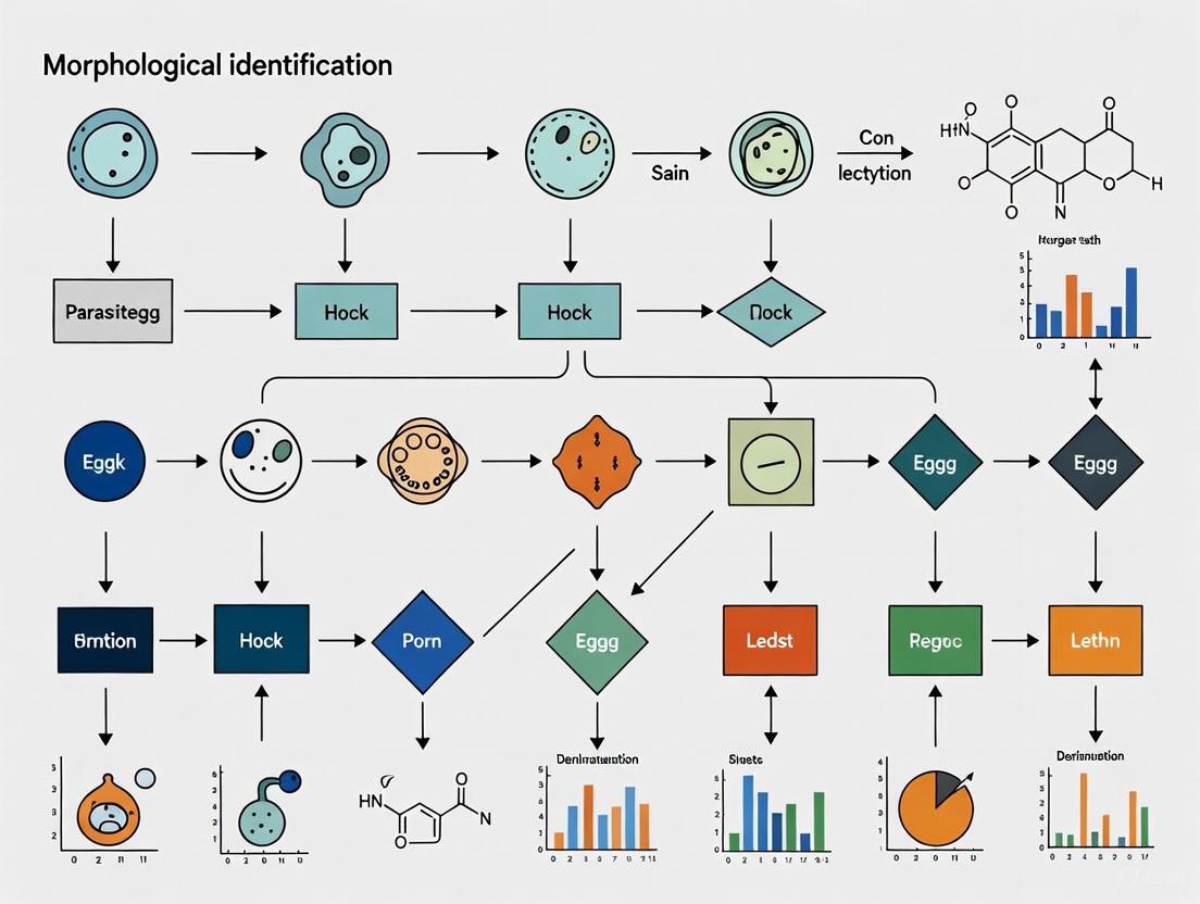
The integration of traditional morphology with new technologies is shaping the future of helminth diagnosis. The following workflow illustrates this integrative approach.
Diagram 1: Integrative taxonomy workflow for helminth identification, combining morphological, molecular, and digital pathology approaches [9] [4] [6].
The Scientist's Toolkit: Key Research Reagents and Materials
Table 2: Essential Research Reagents and Materials for Helminth Egg Analysis
| Reagent/Material | Function/Application | Example Use in Protocol |
|---|---|---|
| Glycerine-Malachite Green Solution | Clears and stains fecal debris for microscopy. | Kato-Katz smear preparation for egg visualization and quantification [7]. |
| 10% Neutral Buffered Formalin | Preserves helminth specimens and stool samples; fixes tissue for histology. | Sample preservation for FET; fixation of specimens for histopathological analysis [8] [6]. |
| Ethyl Acetate / Diethyl Ether | Solvent for lipid and debris extraction in concentration techniques. | Flotation and purification of parasite eggs in the Formalin-Ether Concentration Technique [8]. |
| Sodium Nitrate Flotation Solution | High-specific-gravity solution for buoyant separation of eggs. | Flotation of helminth eggs to the surface for easy collection in SNF [8]. |
| Phosphate-Buffered Saline (PBS) | Isotonic solution for specimen handling and relaxation. | Relaxing live helminths prior to morphological analysis to prevent contraction [6]. |
| Deep Learning Models (YOLOv7, CoAtNet) | AI-based object detection and classification of eggs in digital images. | Automated identification and quantification of eggs in whole-slide images [4] [2]. |
Challenges and Morphological Anomalies
A significant challenge in morphological identification is the occurrence of abnormal egg forms, which can complicate accurate diagnosis.
- Abnormal Nematode Egg Development: Highly abnormal forms of Ascaris lumbricoides eggs, including those with double morulae, giant eggs (up to 110 µm in length), and irregular shell shapes, have been observed in human populations with high infection intensity. Similar abnormalities, such as budded, triangular, and conjoined eggs, have been documented in experimental Baylisascaris procyonis infections in raccoons and dogs, particularly during the initial patency period [3].
- Abnormal Trematode Egg Morphology: Malformations in schistosome eggs have also been reported. For instance, Schistosoma haematobium eggs with variable spine morphology and S. mansoni eggs with double spines have been documented. Historical evidence suggests these abnormalities may be associated with egg production by immature worms [3].
- Impact of Technique on Morphology: The diagnostic technique itself can induce morphological artifacts. The Kato-Katz method is known to cause minor swelling and clearing of A. lumbricoides eggs and can lead to the collapse of schistosome eggs or dissolution of hookworm eggs if the smear is allowed to clear for too long [3]. This underscores the need for standardized examination timings.
The precise morphological identification of helminth eggs is a critical skill that underpins epidemiological surveillance, individual patient diagnosis, and the evaluation of interventional drug efficacy. While core characteristics provide a reliable foundation for identification, practitioners must be aware of the potential for morphological anomalies and technique-induced artifacts. The future of helminth diagnostics lies in an integrative approach that synergizes classical morphological expertise with advanced tools like deep learning and molecular biology. This multi-faceted methodology, as detailed in this guide, promises to enhance diagnostic accuracy and strengthen global efforts to control and eliminate helminth infections.
The morphological identification of parasite eggs, larvae, and cysts through microscopy remains a cornerstone of parasitological research and diagnosis. Despite advancements in molecular techniques, traditional methods based on flotation, sedimentation, and staining continue to provide the foundation for parasite detection in clinical, veterinary, and research settings [10] [11]. These techniques leverage the physical properties of parasitic structures—particularly their specific density and structural composition—to separate them from fecal debris and enhance their visibility for accurate identification [10]. The enduring value of these methods lies in their relatively low operational cost, moderate sensitivity and specificity, and their capacity to provide direct morphological evidence of infection, which is indispensable for species-level identification and burden assessment [10] [12].
Within the context of morphological identification research, understanding the technical principles, advantages, and limitations of each method is paramount. This guide provides an in-depth technical examination of these core techniques, supported by comparative data and detailed protocols, to equip researchers and drug development professionals with the knowledge to select and implement the most appropriate methods for their specific applications.
Core Technical Principles and Comparative Analysis
The effectiveness of concentration techniques hinges on the specific density (relative density) of the medium used and the application of force (either gravitational or centrifugal) to separate parasitic elements from fecal debris [10]. The table below summarizes the fundamental principles and applications of the primary technique categories.
Table 1: Core Principles of Parasite Concentration Techniques
| Technique Category | Physical Principle | Primary Force Applied | Typical Recovery | Key Considerations |
|---|---|---|---|---|
| Flotation | Suspension of fecal material in a medium with a specific density higher than that of parasite eggs/cysts (typically 1.20-1.35) [10]. | Spontaneous or centrifugal [10]. | Buoyant parasitic forms (e.g., nematode eggs, coccidian oocysts) that float to the surface [10]. | Can cause morphological distortion due to osmotic stress. Not ideal for heavy or operculated eggs [10]. |
| Sedimentation | Suspension of fecal material in a medium (often of lower specific density) where parasitic structures settle due to their greater density [10]. | Spontaneous (gravity) or centrifugal [10]. | A wider range of parasitic forms, including denser trematode eggs and operculated cestode eggs [10]. | Generally preserves morphology better. The process can be slower than flotation [10]. |
| Staining | Chemical interaction between dyes and specific structural components of the parasite (e.g., chitin in eggshells, acid-fastness of oocyst walls) [13] [14]. | Not applicable. | Enhances contrast and detail for specific identification and differentiation of species [13] [14]. | Requires expertise in interpretation. Some stains are permanent, while others are temporary [15]. |
A comparative study of four techniques for diagnosing Spirometra spp. eggs in wild carnivores demonstrated the profound impact of method selection on recovery rates. Sedimentation techniques (Lutz and modified Ritchie) significantly outperformed flotation techniques (Faust and modified Sheather), with the latter also causing a higher frequency of morphological alterations in the eggs [16]. Similarly, a Bayesian analysis of two spontaneous sedimentation tests (SST and Paratest) revealed generally high specificity (>93%) but low and variable sensitivity (35.8%-53.8%) for various parasites, underscoring the risk of underdiagnosis due to technical limitations [17].
The morphological details required for precise identification are extensively documented in resources such as the CDC's Comparative Morphology Tables. The following table consolidates key characteristics for common protozoan cysts, which are critical for microscopic differentiation.
Table 2: Differential Morphology of Common Protozoan Cysts Found in Human Stool [15]
| Species | Size (Diameter or Length) | Shape | Number of Nuclei (Mature Cyst) | Peripheral Chromatin | Cytoplasmic Inclusions |
|---|---|---|---|---|---|
| Entamoeba histolytica | 10-20 µm (usual 12-15 µm) | Usually spherical | 4 | Fine, uniform granules | Chromatoid bodies with bluntly rounded ends |
| Entamoeba coli | 10-35 µm (usual 15-25 µm) | Usually spherical, occasionally oval or triangular | 8 | Coarse, irregular granules | Chromatoid bodies less frequent, splinter-like with pointed ends |
| Entamoeba hartmanni | 5-10 µm (usual 6-8 µm) | Usually spherical | 4 | Similar to E. histolytica | Chromatoid bodies with bluntly rounded ends |
| Endolimax nana | 5-10 µm (usual 6-8 µm) | Spherical to Oval | 4 | None | Chromatoid bodies typically absent |
| Iodamoeba bütschlii | 5-20 µm (usual 10-12 µm) | Ovoidal, ellipsoidal, or other shapes | 1 | None | Compact, well-defined glycogen mass |
Detailed Methodologies and Protocols
Sedimentation Techniques
Centrifugal-Sedimentation (Formalin-Ethyl Acetate Technique)
The formalin-ethyl acetate sedimentation technique is a widely used standard in clinical laboratories due to its broad recovery profile [11].
Reagents:
- 5% or 10% buffered formalin
- Ethyl acetate
- Saline or detergent solution
Procedure:
- Emulsification: Commingle approximately 1-2 g of stool (fresh or preserved in formalin) with 10 mL of 5% or 10% formalin in a centrifuge tube. Filter the suspension through a sieve (500-600 µm mesh) to remove large particulate matter [10] [11].
- Centrifugation: Centrifuge the filtrate at 500 × g for 2-3 minutes. Decant the supernatant.
- Resuspension and Washing: Resuspend the sediment in fresh formalin, add saline to within a few centimeters of the tube rim, and recentrifuge. Decant the supernatant. This wash step may be repeated if necessary to clean the sediment.
- Ethyl Acetate Extraction: To the sediment, add 4-5 mL of 10% formalin (if not already present), fill the tube halfway with saline, and then add 3-4 mL of ethyl acetate. Stopper the tube and shake vigorously for 30 seconds. Remove the stopper carefully.
- Final Centrifugation: Centrifuge at 500 × g for 2-3 minutes. This step results in four layers: a plug of fecal debris at the top (ethyl acetate), a formalin layer, sedimented particulate matter, and the parasite-containing sediment at the very bottom.
- Examination: Loosen the debris plug from the tube sides with an applicator stick and decant the top three layers. The final sediment is used for preparing wet mounts (with or without iodine) and permanent stains for microscopic examination [11].
Flotation Techniques
Zinc Sulfate Flotation (Centrifugal-Flotation)
This technique is particularly effective for recovering protozoan cysts and some helminth eggs [10].
Reagents:
- Zinc sulfate solution, specific gravity 1.20 [10].
Procedure:
- Fecal Suspension: Prepare a fecal suspension as for the sedimentation technique and concentrate by centrifugation. Wash the sediment once with water.
- Flotation Medium: Resuspend the sediment in a small volume of zinc sulfate solution (specific gravity 1.18-1.20), then fill the tube to the brim with more zinc sulfate solution.
- Centrifugation: Centrifuge at 500 × g for 2-3 minutes.
- Sample Collection: Place a coverslip on the top of the meniscus and allow it to stand for 5-10 minutes. Carefully lift the coverslip straight up and place it on a glass slide for immediate microscopic examination [10].
Staining Procedures
Staining is critical for visualizing internal structures and for identifying parasites that are difficult to detect with routine stains.
Modified Acid-Fast Staining for Coccidia
This technique is essential for identifying oocysts of Cryptosporidium, Cystoisospora, and Cyclospora species [14].
Reagents:
- Absolute Methanol
- Kinyoun's Carbol Fuchsin
- Acid Alcohol (3% HCl in 95% Ethanol)
- 3% Malachite green (or Methylene Blue) counterstain
Procedure:
- Smear Preparation: Prepare a thin smear from concentrated stool sediment on a glass slide and dry on a slide warmer at 60°C.
- Fixation: Flood the slide with absolute methanol for 1 minute to fix.
- Staining: Apply Kinyoun's Carbol Fuchsin and stain for 5 minutes. Rinse gently with distilled water.
- Decolorization: Decolorize with Acid Alcohol for 1-2 minutes or until the stain no longer streams off the slide. Rinse thoroughly with distilled water.
- Counterstaining: Apply the Malachite green counterstain for 1-2 minutes. Rinse with distilled water.
- Examination: Air-dry the slide and examine under oil immersion (100x objective). Coccidian oocysts stain pinkish-red against a green background [14].
Ziehl-Neelsen Staining for Differentiation ofTaeniaSpecies
This method allows for the differentiation of Taenia saginata and T. solium eggs, which is crucial for public health and clinical management [13].
Reagents:
- 3% Carbol Fuchsin
- 70% Ethanol with 1% HCl (decolorizer)
- 3% Methylene Blue (counterstain)
Procedure:
- Smear Preparation: Prepare a fecal smear, air-dry, and heat-fix.
- Primary Staining: Flood the slide with 3% Carbol Fuchsin for 15 minutes. Heat the slide gently for 5 minutes, then allow it to cool.
- Rinsing: Rinse the slide with tap water.
- Decolorization: Decolorize with 1% Acid-Alcohol for 1-2 minutes, then rinse with tap water.
- Counterstaining: Apply 3% Methylene Blue for 5 minutes. Rinse with tap water and air-dry.
- Examination: Examine under oil immersion. T. saginata eggs stain a consistent magenta-red and are oval, while T. solium eggs appear purplish-blue and are more spherical [13].
The Scientist's Toolkit: Essential Research Reagents
The following table details key reagents used in the featured techniques, with their specific functions in the context of parasitological diagnostics.
Table 3: Essential Research Reagents for Parasitological Microscopy
| Reagent Solution | Technical Function | Application Example |
|---|---|---|
| Formalin (5-10% Buffered) | Preservative that fixes and hardens parasitic structures, preventing degeneration and bacterial overgrowth [11]. | Primary fixative in formalin-ethyl acetate sedimentation; preservation of stool samples for long-term storage. |
| Ethyl Acetate | Organic solvent that extracts fats, lipids, and other debris from the fecal suspension, resulting in a cleaner sediment [11]. | Used in the formalin-ethyl acetate sedimentation technique to create a debris plug. |
| Zinc Sulfate Solution (sp. gr. 1.20) | High-specific-density flotation medium that allows buoyant parasitic forms to rise to the surface [10]. | Recovery of protozoan cysts and some helminth eggs in centrifugal-flotation. |
| Saturated Sodium Chloride Solution | Economical high-specific-density flotation medium. | Used in spontaneous flotation techniques and McMaster egg counting. |
| Carbol Fuchsin | Primary stain in acid-fast procedures; binds to mycolic acids in cell walls of acid-fast organisms [13] [14]. | Differentiating Taenia species [13] and staining coccidian oocysts [14]. |
| Malachite Green / Methylene Blue | Counterstain that provides background coloration, enhancing contrast for the primary stain [14]. | Used in modified acid-fast and Ziehl-Neelsen staining to visualize non-acid-fast elements. |
| Peanut Agglutinin (PNA-FITC) | Fluorescently labeled lectin that binds specifically to carbohydrate motifs on the surface of certain parasite eggs [12]. | Fluorescent microscopic identification and differentiation of Haemonchus contortus eggs. |
Workflow and Method Selection
The following diagram illustrates a logical workflow for selecting the appropriate microscopic technique based on research objectives and parasite characteristics.
The morphological identification of parasite eggs through manual microscopy remains a cornerstone in parasitology research and clinical diagnosis. As the gold standard for diagnosing intestinal parasitic infections, this technique is crucial for epidemiological studies, drug efficacy testing, and patient management [18] [19]. Despite its foundational role, manual microscopy suffers from significant limitations that impact the reliability and efficiency of research outcomes. These constraints are particularly relevant for researchers, scientists, and drug development professionals who require high levels of accuracy and reproducibility in their work.
This technical guide examines the core limitations of time consumption and human error in manual microscopic analysis of parasite eggs. It further explores how emerging technologies, particularly artificial intelligence (AI) and deep learning, are being developed to address these challenges within the context of modern parasitology research. Understanding these limitations and potential solutions is essential for advancing morphological identification techniques and improving the quality of research outcomes in parasite studies.
Core Limitations of Manual Microscopy
Time Consumption and Throughput Constraints
The process of manual microscopy for parasite egg identification is inherently time-intensive, creating significant bottlenecks in research and diagnostic workflows. The following table quantifies the time-related constraints across different microscopic procedures:
Table 1: Time Consumption in Manual Microscopic Procedures
| Procedure Aspect | Time Requirement | Impact on Research |
|---|---|---|
| Complete sample examination | 8-10 minutes per sample [5] | Limits daily processing capacity; restricts sample size in studies |
| Manual sediment examination | Labor-intensive and time-consuming [20] | Reduces number of experiments feasible within project timelines |
| Centrifugation and preparation | 5 minutes at 1500 rpm (additional to examination) [20] | Increases hands-on researcher time per sample |
| Scanning multiple fields | Slow and fatiguing for operators [21] | Creates throughput bottlenecks in large-scale studies |
The cumulative effect of these time constraints significantly limits the scale and scope of parasitology research. Large-scale studies requiring examination of hundreds or thousands of samples become impractical, potentially leading to underpowered experiments or extended project timelines that delay critical findings in parasite biology and drug development.
Human Error and Diagnostic Variability
Human factors introduce substantial variability and error in parasite egg identification, potentially compromising research outcomes. The table below categorizes and describes common human errors in microscopic analysis:
Table 2: Categories and Impact of Human Errors in Microscopic Analysis
| Error Category | Description | Effect on Parasite Egg Identification |
|---|---|---|
| Observer Bias | Manual exposure settings, focus, and ROI selection vary between users [21] | Inconsistent identification of egg morphology between researchers |
| Fatigue-Related Errors | Decreased attention and decision-making ability due to prolonged eyepiece work [22] [23] | Missed detections (false negatives) during extended examination sessions |
| Omission Errors | Missing a step or skipping over elements in the visual field [22] | Failure to identify eggs present in samples, particularly at low concentrations |
| Commission Errors | Performing identification actions incorrectly [22] | Misclassification of parasite species based on morphological features |
| Procedural Errors | Not following established identification protocols [22] | Inconsistent application of diagnostic criteria across research teams |
The consequences of these errors are particularly pronounced in parasite egg identification due to several factors. The morphological similarity of different parasitic eggs and the abundance of impurities in samples create inherent challenges that require extensive training to overcome [5]. Furthermore, the relatively low sensitivity of manual microscopy, particularly at low parasite levels, can lead to false negatives that skew research data [18]. These limitations are compounded in resource-limited settings where access to highly trained microscopists may be restricted, potentially affecting the reliability of multi-center research trials [18].
Methodological Approaches to Address Limitations
Conventional Manual Microscopy Protocol
The standard protocol for manual microscopic examination of parasite eggs, as derived from established laboratory methods, involves multiple meticulous steps that contribute to both time consumption and potential error introduction [20]:
Sample Preparation Protocol
- Collection: Mid-stream samples (30 mL) are collected into appropriate primary containers
- Transport: Samples are transported in primary containers to prevent leakage or contamination
- Aliquoting: Samples are transferred to secondary translucent conical tubes (10 mL per tube)
- Centrifugation: First tube is centrifuged for 5 minutes at 1500 rpm (400 g)
- Sediment Preparation: Supernatant is decanted until 0.5 mL remains; sediment is resuspended
- Slide Preparation: One drop of sediment is placed on a microscope slide and covered with a cover slip
Microscopic Examination Protocol
- Slide Scanning: At least 10 different microscopic fields are scanned at magnifications of ×100 and ×400
- Element Identification: Formed elements are identified based on morphological characteristics
- Quantification: Results are calculated by averaging formed elements and reported as cells or particles per field
- Quality Control: Two independent evaluators (e.g., biochemistry specialist and biologist) examine the same slide; discrepancies trigger re-analysis with another sample
This multi-step process introduces numerous variables that affect reproducibility, including centrifugation speed and time, resuspension volume, staining techniques (if used), and individual interpretation of morphological features [20]. The requirement for independent verification by multiple specialists further increases the time investment while highlighting the inherent subjectivity of the method.
Emerging Automated and AI-Assisted Methodologies
Recent technological advances have introduced automated methodologies that address the limitations of manual microscopy through standardized, computational approaches:
Deep Learning-Based Detection Protocol [24] [5] [19]
- Image Acquisition: Sample slides are photographed via light microscope; low-cost USB microscopes (10×) or high-quality microscopes (1000×) may be used
- Data Preparation:
- Greyscale conversion and contrast enhancement to improve feature detection
- Image division into overlapping patches (e.g., 100×100 pixels) using sliding window approach
- Data augmentation through random flipping, rotation, and shifting to increase dataset size
- Model Training:
- Implementation of convolutional neural networks (CNN) such as YOLO variants, AlexNet, or ResNet50
- Transfer learning approach using pre-trained networks with fine-tuning on parasite egg datasets
- Dataset division into training, validation, and test sets (typical ratio: 8:1:1)
- Prediction and Analysis:
- Patch-by-patch classification of input images
- Probability mapping and reconstruction to identify egg locations
- Species classification based on learned morphological features
Table 3: Performance Comparison of AI Models for Parasite Egg Detection
| Model/Approach | Accuracy/Precision | Recall | mAP_0.5 | Computational Requirements |
|---|---|---|---|---|
| YAC-Net (YOLO-based) | 97.8% [19] | 97.7% [19] | 0.9913 [19] | 1.9M parameters [19] |
| YOLOv4 (Multiple Species) | 84.85-100% (varies by species) [24] | Not specified | Not specified | Moderate to high [24] |
| Transfer Learning (AlexNet/ResNet50) | Improved over state-of-the-art [5] | Not specified | Not specified | Moderate [5] |
| Human Expert | High but variable | High but decreases with fatigue [23] | Not applicable | Not applicable |
The following diagram illustrates the comparative workflow between manual and AI-assisted approaches, highlighting points where time consumption and error typically occur:
The Scientist's Toolkit: Research Reagent Solutions
Implementation of effective parasite egg identification protocols requires specific materials and computational resources. The following table details essential research reagents and solutions used in both conventional and advanced morphological identification methods:
Table 4: Essential Research Reagents and Solutions for Parasite Egg Morphological Identification
| Item | Function/Application | Implementation Context |
|---|---|---|
| Microscope Slides and Coverslips | Platform for preparing and examining samples | Standardized manual examination; AI-assisted imaging [20] [5] |
| Conical Tubes | Sample aliquoting and centrifugation | Sediment preparation for enhanced detection [20] |
| Staining Solutions | Enhancement of morphological features | Improved contrast for both human and AI-based identification [20] |
| Low-Cost USB Microscope | Digital image acquisition for automated systems | Resource-constrained settings; enables digital archiving [5] |
| High-Quality Microscope | High-resolution imaging for detailed morphology | Reference standard imaging; detailed morphological studies [5] |
| Deep Learning Models | Automated detection and classification | YOLO variants, CNN architectures for efficient analysis [24] [19] |
| Data Augmentation Algorithms | Expansion of training datasets | Improves model robustness with limited sample sizes [5] |
| Graphical Processing Units | Acceleration of model training and inference | Reduces computational time for AI-based approaches [24] |
The integration of these tools varies depending on the research context. While conventional manual microscopy relies primarily on physical laboratory equipment, AI-assisted approaches require both wet laboratory components and computational resources, creating a hybrid workflow that leverages the strengths of both traditional and technological methods.
Manual microscopy for parasite egg identification presents significant limitations in time efficiency and reliability that directly impact research quality and throughput in parasitology. The time-intensive nature of proper sample examination constrains study scale, while human factors introduce variability that threatens reproducibility. These challenges are particularly problematic in drug development research where consistent morphological assessment is crucial for evaluating treatment efficacy.
Emerging methodologies centered on deep learning and automation offer promising approaches to mitigate these limitations. Current research demonstrates that AI-assisted platforms can achieve high accuracy in parasite egg detection while reducing analysis time and minimizing human error. Nevertheless, the role of human expertise remains vital for system validation, complex case resolution, and quality control. The future of morphological identification in parasite research lies in hybrid approaches that leverage the strengths of both human expertise and computational consistency, potentially transforming how parasite egg analysis is conducted in research settings and accelerating progress in understanding and treating parasitic infections.
The morphological identification of parasite eggs remains a cornerstone of diagnostic parasitology, essential for accurate disease diagnosis and subsequent research and drug development. However, in many developed regions, the decline in parasitic infections due to improved sanitation has created a significant challenge: access to physical specimens for education and reference is becoming increasingly limited. This scarcity threatens the preservation of crucial morphological expertise among researchers and healthcare professionals. The construction of digital specimen databases presents a transformative solution to this problem, offering a sustainable, accessible, and scalable resource. By leveraging whole-slide imaging (WSI) technology, these databases create high-fidelity virtual slides of parasite specimens, ensuring that vital morphological knowledge is not only preserved but also enhanced for future research and educational endeavors. This technical guide explores the construction, implementation, and application of such databases, framed within the critical context of morphological identification research for parasite eggs.
The Construction of a Digital Parasite Specimen Database
Specimen Acquisition and Preparation
The foundational step in building a digital database is the curation of a diverse and well-characterized collection of physical specimens. In a pioneering initiative, researchers acquired 50 existing slide specimens of parasitic eggs, adult parasites, and arthropods from the collections of Kyoto University and the Kyoto Prefectural University of Medicine [25] [26]. These specimens included parasite eggs, adult worms, ticks, insects typically observed under low magnification, and malarial parasites requiring high magnification. A critical preparatory step was verifying that all slide samples were devoid of personal information, thus ensuring their ethical use for educational and research purposes, including data sharing. Some specimens were historically prepared at the universities, while others were procured from commercial suppliers and museums, guaranteeing a taxonomically broad representation.
Digital Scanning and Image Processing
The process of digitizing physical slides requires specialized equipment and methodologies to ensure high-quality, diagnostically useful outputs. In the cited project, digital scanning was performed using the SLIDEVIEW VS200 slide scanner by EVIDENT Corporation [25]. A key technical consideration for thicker smear specimens was the application of the Z-stack function. This technique involves varying the scan depth during image capture to accumulate layer-by-layer data, thereby accommodating three-dimensional structures and ensuring all focal planes are accurately represented in the final digital image [25]. Each slide was scanned individually, with a quality control protocol in place: slides containing out-of-focus areas were rescanned as necessary, and the clearest image was selected for inclusion in the final database by the reviewing authors [25].
Database Architecture and Management
The final stage involves structuring the digitized data into an accessible and organized system. For the constructed database, the virtual slide data was uploaded to a shared server (Windows Server 2022) to build the searchable database [25]. The folder structure was logically organized according to the taxonomic classification of the organisms, facilitating intuitive navigation. To significantly enhance the educational and reference utility, each specimen was accompanied by simple explanatory text. Critically, these notes were provided in both English and Japanese, making the resource accessible to a wider international audience of researchers and professionals [25]. The shared server is configured to allow approximately 100 simultaneous users to access and observe the data via a standard web browser on various devices, including laptops, tablets, and smartphones, without requiring specialized viewing software [25].
Quantitative Analysis of Database Utility and Performance
The value of a digital database is demonstrated through its performance metrics and its impact on research and education. The table below summarizes key quantitative data from both the construction of a specimen database and a related automated detection model, highlighting the efficacy of digital approaches.
Table 1: Quantitative Performance Metrics of Digital Parasitology Resources
| Resource Type | Key Metric | Reported Value | Implication for Research |
|---|---|---|---|
| Digital Specimen Database [25] | Number of Slide Specimens Digitized | 50 specimens | Provides a foundational, scalable collection for morphological reference. |
| Digital Specimen Database [25] | Simultaneous User Access | ~100 users | Enables widespread use in classroom settings and multi-institutional research. |
| YCBAM Detection Model [27] | Precision | 0.9971 | Extremely low false positive rate, crucial for reliable automated diagnosis. |
| YCBAM Detection Model [27] | Recall | 0.9934 | Very low false negative rate, ensuring most parasite eggs are identified. |
| YCBAM Detection Model [27] | Mean Average Precision (mAP @0.50 IoU) | 0.9950 | Demonstrates superior overall detection and localization accuracy. |
The integration of digital databases with advanced deep learning models, such as the YOLO Convolutional Block Attention Module (YCBAM), creates a powerful synergy. While the database provides the essential high-quality data for training, models like YCBAM offer a path toward high-throughput, automated analysis. The YCBAM architecture, which integrates YOLO with self-attention mechanisms and a Convolutional Block Attention Module (CBAM), has demonstrated exceptional performance in automating the detection of pinworm eggs in microscopic images [27]. Its high precision and recall indicate that it can significantly reduce diagnostic errors and save time, supporting researchers and healthcare professionals in making informed decisions. This is particularly relevant for diagnosing parasites like pinworms (Enterobius vermicularis), whose eggs are small (50–60 μm in length and 20–30 μm in width) and can be morphologically similar to other microscopic particles [27].
Experimental Protocols for Digital Workflows
Protocol 1: Whole-Slide Imaging for Database Construction
This protocol details the methodology for creating virtual slides for a digital specimen database [25].
- Step 1: Specimen Curation. Identify and select existing slide specimens from institutional collections, ensuring a representative range of taxa (e.g., parasite eggs, adults, arthropods). Verify that specimens are free of personal identifying information.
- Step 2: Scanner Calibration. Calibrate a whole-slide imaging scanner (e.g., SLIDEVIEW VS200) according to manufacturer specifications to ensure color fidelity and focus accuracy.
- Step 3: Scanning and Z-Stack Acquisition. For each slide, select the appropriate magnification (e.g., 40x for eggs/adults, 1000x for blood parasites). For specimens with thicker smears, activate the Z-stack function to capture multiple focal planes and accumulate layer-by-layer data.
- Step 4: Image Quality Control. Visually review each digitized slide for focus and clarity. Rescan any slides with out-of-focus areas. Authors or qualified personnel should select the clearest image for final inclusion.
- Step 5: Data Upload and Annotation. Upload the final virtual slide images to a dedicated shared server. Organize the files into a logical folder structure based on taxonomy. Attach explanatory notes for each specimen to facilitate learning and reference.
Protocol 2: Automated Detection of Parasite Eggs Using YCBAM
This protocol outlines the workflow for developing a deep learning model to automatically detect parasite eggs in digital microscopic images [27].
- Step 1: Dataset Preparation. Compile a dataset of microscopic images of parasite eggs, such as pinworm eggs. Annotate the images by labeling the bounding boxes of each egg. A typical dataset might include 255 images for segmentation tasks.
- Step 2: Model Architecture Integration. Implement the YCBAM architecture by integrating the Convolutional Block Attention Module (CBAM) and self-attention mechanisms into a YOLOv8 framework. This enhances feature extraction from complex backgrounds.
- Step 3: Model Training. Train the YCBAM model on the annotated dataset. Use a suitable loss function (e.g., box loss) and monitor its convergence during training. The reported training box loss for a pinworm model was 1.1410, indicating efficient learning [27].
- Step 4: Model Evaluation. Evaluate the trained model's performance on a separate test set. Calculate key metrics including Precision, Recall, and mean Average Precision (mAP) at an Intersection over Union (IoU) threshold of 0.50 to confirm detection accuracy.
- Step 5: Inference and Deployment. Deploy the trained model to analyze new microscopic images. The model will output precise identifications and localizations of parasite eggs, offering a tool to reduce diagnostic time and human error.
Visualization of Workflows and System Architecture
The following diagrams, created using Graphviz DOT language, illustrate the core workflows and logical relationships described in this guide. The color palette and contrast have been designed to meet WCAG 2.1 AA accessibility standards, ensuring readability for all users [28] [29] [30].
Diagram 1: Digital Specimen Database Construction Workflow
Diagram 2: Automated Detection Model Development Workflow
The Scientist's Toolkit: Essential Research Reagent Solutions
The development and utilization of digital specimen databases and associated analytical models rely on a suite of essential tools and reagents. The following table details key components of this research toolkit.
Table 2: Essential Research Reagents and Tools for Digital Parasitology
| Tool/Reagent Category | Specific Example | Function in Research |
|---|---|---|
| Whole-Slide Imaging Scanner | SLIDEVIEW VS200 (EVIDENT Corp) [25] | High-resolution digitization of physical microscope slides to create virtual specimens. |
| Computational Framework | YOLOv8 [27] | A real-time object detection system that forms the backbone for automated parasite egg identification models. |
| Attention Module | Convolutional Block Attention Module (CBAM) [27] | A neural network component integrated into models like YCBAM to enhance feature extraction from complex image backgrounds. |
| Digital Storage & Server | Windows Server 2022 [25] | Hosts the digital database, enabling secure, multi-user access and data management for collaborative research. |
| Analysis & Statistical Software | R / RStudio [31] | An open-source environment for statistical computing and graphics, used for analyzing experimental data and model performance. |
| Color Contrast Analyzer | Colour Contrast Analyser (CCA) [28] [30] | Ensures that all visualizations and user interfaces meet accessibility standards (WCAG) for inclusive science. |
The construction and implementation of digital specimen databases represent a critical advancement in the field of parasitology, directly supporting the morphological identification research essential for diagnostics and drug development. By preserving rare specimens in an accessible, non-degrading digital format, these databases act as a bulwark against the loss of morphological expertise. Furthermore, when these rich data sources are coupled with state-of-the-art deep learning models, they empower a new paradigm of high-throughput, accurate, and automated analysis. For researchers and scientists dedicated to combating parasitic diseases, the integration of digital databases and computational tools is no longer a luxury but a fundamental component of a modern, effective research toolkit.
Challenges in Low-Prevalence Settings and Resource-Limited Environments
The morphological identification of parasite eggs remains a cornerstone of parasitology research and clinical diagnosis. However, the reliability of this method faces significant challenges in low-prevalence settings and resource-limited environments. In these contexts, the diminishing expertise of microscopists, combined with economic constraints that limit access to advanced diagnostic tools, creates a perfect storm that compromises diagnostic accuracy and impedes effective parasite control [32] [33]. Although traditional microscopy is considered the gold standard for parasite detection, its sensitivity and specificity are not fixed attributes but are influenced by disease prevalence and the resources available for expert training [34]. This technical guide examines these challenges through an evidence-based lens and explores innovative solutions that combine optimized laboratory protocols with emerging artificial intelligence (AI) technologies to enhance diagnostic capabilities in these critical settings.
Technical Challenges in Low-Prevalence and Resource-Limited Settings
The Impact of Disease Prevalence on Test Performance
The fundamental assumption that sensitivity and specificity are intrinsic test properties remains valid only under ideal conditions. A comprehensive meta-epidemiological study analyzing 6,909 diagnostic test accuracy studies revealed a significant association between disease prevalence and these metrics. As prevalence increases, sensitivity tends to increase while specificity decreases [34]. This relationship poses particular challenges for morphological identification in low-prevalence settings, where the pre-test probability of infection is low, and the positive predictive value of tests diminishes accordingly. For parasitic diseases, this means that even highly specific morphological identification methods may yield more false positives when deployed in low-prevalence settings, potentially leading to unnecessary treatments and misallocation of limited resources.
Economic and Expertise Constraints
Resource-limited environments face compounded challenges that extend beyond test performance characteristics:
- Expertise Dilution: The diagnostic accuracy of manual microscopy is highly dependent on technician expertise and experience [33]. In low-prevalence settings, microscopists have limited opportunities to maintain their skills through regular practice, leading to decreased proficiency over time.
- Infrastructure Limitations: Many resource-constrained laboratories lack reliable electricity, quality microscopes, and appropriate storage facilities for reagents and samples, further compromising diagnostic accuracy [32].
- Time Constraints: Traditional microscopic examination is labor-intensive, requiring approximately 30 minutes per sample [35]. In high-volume settings with limited personnel, this often leads to rushed examinations and increased error rates.
Established and Emerging Solutions
Optimized Manual Protocols for Enhanced Egg Recovery
For researchers working with insect vectors or environmental samples, standardized protocols for parasite egg recovery are essential. Recent research has developed and validated efficient methods for recovering Taenia saginata and Ascaris suum eggs from house flies, which can be adapted for field use [36].
Table 1: Optimized Protocols for Parasite Egg Recovery from Insect Vectors
| Sample Source | Protocol Steps | Recovery Rate | Hands-on Time | Total Time |
|---|---|---|---|---|
| Gastrointestinal Tract | Homogenization in PBS + Centrifugation (2000 g, 2 min) | 79.7% (T. saginata), 74.2% (A. suum) | 1.5 minutes | 6.5 minutes |
| Exoskeleton | Vortexing (2 min) in Tween 80 + Passive Sedimentation (15 min) + Centrifugation (2000 g, 2 min) | 77.4% (T. saginata), 91.5% (A. suum) | 3.5 minutes | 20.5 minutes |
These protocols demonstrate that effective parasite egg recovery can be achieved with minimal hands-on time and basic laboratory equipment, making them particularly suitable for resource-constrained settings. The centrifugation-based method for gastrointestinal tract samples effectively removes large debris particles that could hinder the differentiation of eggs from other material, while the washing and sedimentation approach for exoskeletons successfully isolates eggs with minimal contamination [36].
AI-Assisted Diagnostic Platforms
Deep learning approaches, particularly YOLO-based models, have emerged as powerful tools for automating parasite egg identification, potentially overcoming the expertise gap in low-prevalence settings.
Table 2: Performance of AI Models for Parasite Egg Detection
| Model | Parasite Species | Performance Metrics | Detection Speed | Reference |
|---|---|---|---|---|
| YOLOv5 | Multiple intestinal parasites | mAP: ~97% | 8.5 ms/sample | [35] |
| YOLOv4 | Clonorchis sinensis, Schistosoma japonicum | Accuracy: 100% | Real-time efficiency | [33] |
| YOLOv4 | E. vermicularis, F. buski, T. trichiura | Accuracy: 89.31%, 88.00%, 84.85% | Real-time efficiency | [33] |
| YCBAM | Pinworm parasite eggs | Precision: 0.9971, Recall: 0.9934, mAP: 0.9950 | Efficient training convergence | [27] |
The YCBAM architecture, which integrates YOLO with self-attention mechanisms and Convolutional Block Attention Module, has demonstrated particular effectiveness in challenging imaging conditions by focusing on spatial and channel-wise information to improve feature extraction from complex backgrounds [27]. This approach significantly enhances the detection of small, critical features such as pinworm egg boundaries, which measure only 50-60 μm in length and 20-30 μm in width [27].
Experimental Protocols and Methodologies
Sample Preparation for Morphological Studies
Standardized sample preparation is crucial for both traditional microscopy and AI-assisted identification:
- Sample Collection: Collect fresh stool samples or parasite egg suspensions from reliable sources [33].
- Slide Preparation: Place two drops of vortex-mixed egg suspension (approximately 10 μL) on a slide and cover with an 18×18 mm coverslip, avoiding air bubbles [33].
- Microscopic Confirmation: Confirm egg species and integrity under a light microscope before proceeding with analysis [36].
- Image Acquisition: Capture images using standardized microscopy equipment. For bright-field imaging, use a ring-shaped LED cold light source positioned 10 cm above the sample [37].
AI Model Training and Implementation
For researchers implementing AI-assisted detection, the following methodology has proven effective:
Data Set Preparation:
Model Training:
- Utilize Python 3.8 with PyTorch framework on GPU-enabled systems [33].
- Set initial learning rate to 0.01 with decay factor of 0.0005 [33].
- Use Adam optimizer with momentum of 0.937 and batch size of 64 [33].
- Train for 300 epochs, freezing backbone feature extraction network for first 50 epochs [33].
Performance Evaluation:
Figure 1: AI-Assisted Parasite Egg Detection Workflow
The Scientist's Toolkit: Essential Research Reagents and Materials
Table 3: Essential Research Reagents for Parasite Egg Identification Studies
| Reagent/Material | Specification | Application | Technical Notes |
|---|---|---|---|
| Phosphate-Buffered Saline | 1X concentration, pH 7.4 | Egg homogenization and washing | Maintains osmotic balance; used in gastrointestinal tract recovery [36] |
| Tween 80 | 0.05% concentration | Surfactant for exoskeleton washing | Reduces surface tension without damaging egg morphology [36] |
| Microscopy Slides | Standard 75x25 mm, 1.0-1.2 mm thickness | Sample mounting for visualization | Pre-cleaned slides reduce artifacts [33] |
| Coverslips | 18x18 mm square | Creating uniform sample thickness | Minimizes air bubbles during preparation [33] |
| Gaussian Blur Filter | Sigma = 2 | Image processing for noise reduction | Improves feature detection in automated systems [37] |
| FastRandomForest Classifier | 1000 trees, depth 15 | Pixel classification in image analysis | Effective for distinguishing eggs from debris [37] |
Integrated Workflow for Chall Environments
Figure 2: Integrated Approach to Address Diagnostic Challenges
The most promising solution for low-prevalence and resource-limited settings involves integrating optimized manual protocols with AI-assisted platforms. This hybrid approach leverages the efficiency of standardized sample processing methods with the analytical power of deep learning algorithms, creating a diagnostic system that maintains accuracy despite limited resources and expertise.
The challenges of morphological identification of parasite eggs in low-prevalence and resource-limited environments are significant but not insurmountable. Through the implementation of optimized recovery protocols, standardized sample processing methods, and the integration of AI-assisted detection platforms, researchers and healthcare providers can maintain diagnostic accuracy despite constraints. The continued refinement of these approaches, particularly through the expansion of training datasets and adaptation to field conditions, holds promise for transforming parasitic disease diagnosis in the most challenging settings. As these technologies become more accessible and validated across diverse environments, they have the potential to bridge the diagnostic gap that currently impedes effective parasite control in resource-limited regions worldwide.
Advanced Methodologies: AI, Deep Learning and Automated Detection Systems
The morphological identification of parasite eggs represents a critical procedure in medical diagnostics and biological research. Conventional manual microscopic examination is time-consuming, labor-intensive, and susceptible to human error, particularly in resource-constrained settings. Deep learning architectures have emerged as transformative solutions, automating detection with remarkable precision and speed. This technical guide provides an in-depth analysis of state-of-the-art deep learning frameworks—primarily YOLO (You Only Look Once) variants and specialized Convolutional Neural Networks (CNNs)—for egg detection, with specific application to the morphological identification of parasite eggs. The content is structured to equip researchers, scientists, and drug development professionals with comprehensive methodological protocols, performance benchmarks, and implementation frameworks to advance research in parasitology and related fields.
Core Deep Learning Architectures for Egg Detection
YOLO-Based Architectures
The YOLO family of one-stage detectors has been extensively adapted for egg detection due to its optimal balance between speed and accuracy. Recent research has focused on enhancing standard YOLO architectures to address the specific challenges of egg morphology, such as small size, occlusion, and morphological similarities between species.
YOLO with Attention Mechanisms (YCBAM): A novel framework integrates the Convolutional Block Attention Module (CBAM) with YOLO to create YCBAM. This architecture leverages self-attention mechanisms to focus computational resources on salient image regions containing parasitic elements, significantly improving detection in challenging imaging conditions. The attention modules enhance feature extraction from complex backgrounds and increase sensitivity to critical small features like pinworm egg boundaries. Experimental evaluation demonstrated a precision of 0.9971, recall of 0.9934, and mean Average Precision (mAP) of 0.9950 at an IoU threshold of 0.50, confirming superior detection performance for pinworm eggs [27].
Lightweight YOLO Variants (YAC-Net): For deployment in resource-constrained environments, researchers have developed lightweight models. YAC-Net modifies YOLOv5n by replacing the feature pyramid network (FPN) with an asymptotic feature pyramid network (AFPN) and integrating a C2f module to enrich gradient flow. This architecture fully fuses spatial contextual information through hierarchical and asymptotic aggregation, with adaptive spatial fusion selecting beneficial features while ignoring redundant information. The model achieved a precision of 97.8%, recall of 97.7%, and mAP_0.5 of 0.9913 while reducing parameters by one-fifth compared to its baseline, making it suitable for automated detection systems with limited computational resources [19].
Comparative Performance Analysis: A comprehensive evaluation of compact YOLO variants (YOLOv5n, YOLOv5s, YOLOv7, YOLOv7-tiny, YOLOv8n, YOLOv8s, YOLOv10n, and YOLOv10s) for recognizing 11 parasite species eggs revealed that YOLOv7-tiny achieved the highest mAP of 98.7%, while YOLOv10n yielded the highest recall and F1-score of 100% and 98.6%, respectively. YOLOv8n achieved the fastest processing speed at 55 frames per second on a Jetson Nano, highlighting the critical trade-offs between accuracy, recall, and inference speed for practical deployments [38].
CNN and Hybrid Architectures
Beyond YOLO, researchers have developed specialized CNN architectures and hybrid models that leverage recent advances in deep learning for parasitic egg recognition.
Convolution and Attention Network (CoAtNet): This architecture combines the strengths of convolution and self-attention mechanisms. The model leverages the convolutional layers' spatial feature extraction capabilities while utilizing the attention mechanisms' global contextual understanding. When evaluated on the Chula-ParasiteEgg dataset containing 11,000 microscopic images, CoAtNet achieved an average accuracy of 93% and an average F1-score of 93%, demonstrating robust performance across multiple parasitic egg categories [4].
Morphological Regulated Variational Autoencoder (Morpho-VAE): For landmark-free morphological analysis, Morpho-VAE combines unsupervised and supervised learning to extract discriminative shape features. The architecture integrates a variational autoencoder with a classifier module, regulated by a hyperparameter α that balances reconstruction fidelity with classification performance. When applied to mandible shape analysis, the method achieved superior cluster separation compared to PCA and standard VAE, with potential application to parasitic egg morphology quantification [39].
Table 1: Performance Comparison of Deep Learning Architectures for Egg Detection
| Architecture | Base Model | Precision (%) | Recall (%) | mAP@0.5 (%) | Key Innovation |
|---|---|---|---|---|---|
| YCBAM [27] | YOLOv8 | 99.71 | 99.34 | 99.50 | Integrated CBAM attention module |
| YAC-Net [19] | YOLOv5n | 97.80 | 97.70 | 99.13 | AFPN and C2f modules for lightweight design |
| YOLOv7-tiny [38] | YOLOv7-tiny | - | - | 98.70 | Optimal balance of accuracy and efficiency |
| CoAtNet [4] | Custom | 93.00 | 93.00 | - | Hybrid convolution-attention mechanism |
| YOLOv4 [33] | YOLOv4 | 100.00* | - | - | Transfer learning for specific parasite eggs |
| YOLO-Goose [40] | YOLOv8s | - | - | 96.40 | Small-object detection layer for animal eggs |
*For specific species (Clonorchis sinensis and Schistosoma japonicum)
Experimental Protocols and Methodologies
Dataset Preparation and Preprocessing
The accuracy of deep learning models heavily depends on dataset quality and preprocessing techniques. Standardized protocols have emerged across studies for parasitic egg imaging.
Sample Collection and Imaging: Parasitic egg suspensions are obtained from biological suppliers or clinical samples. For microscopic imaging, two drops of vortex-mixed egg suspension (approximately 10μL) are placed on a slide and covered with an 18mm × 18mm coverslip, avoiding air bubbles. Imaging is performed using light microscopes (e.g., Nikon E100) at consistent magnification levels. For non-parasitic egg detection in agricultural settings, images may be captured using drones (e.g., DJI Phantom 4 Pro) or handheld devices in field conditions [33].
Data Preprocessing and Augmentation: Images are typically resized to standard dimensions (e.g., 640×640 pixels for YOLO models). To enhance model robustness, data augmentation techniques are extensively employed, including:
- Rotation and mirroring for orientation invariance
- Gaussian noise and salt-and-pepper noise injection for robustness to image quality variations
- Color space adjustments for illumination invariance
- Mosaic data augmentation and mixup for improved background contextualization [33] [41]
Cropping techniques using sliding window approaches are implemented to generate multiple training samples from single high-resolution images, particularly beneficial for small object detection [33].
Dataset Partitioning: Datasets are typically partitioned into training, validation, and test sets with ratios of 8:1:1. The training set builds model weights, the validation set guides hyperparameter tuning, and the test set provides unbiased performance evaluation [33].
Model Training Protocols
Consistent training protocols ensure reproducible model performance across different experimental setups.
Parameter Configuration: For YOLO models, standard training configurations include:
- Initial learning rate: 0.01 with decay factor of 0.0005
- Optimizer: Adam with momentum value of 0.937
- Batch size: 64
- Training epochs: 300 with early stopping if no improvement after 200 epochs
- Anchor sizes: Determined using k-means clustering on training data [33]
Implementation Frameworks: Models are typically implemented in Python using PyTorch or TensorFlow frameworks, trained on NVIDIA GPUs (e.g., RTX 3090), and optimized for deployment on edge devices like Raspberry Pi, Intel upSquared with Neural Compute Stick 2, and Jetson Nano for field applications [38].
Loss Functions: Traditional YOLO loss functions combine bounding box regression, objectness, and classification losses. Enhanced versions incorporate GIoU (Generalized Intersection over Union) loss to improve bounding box accuracy for small objects like eggs [40].
Architectural Diagrams and Workflows
YCBAM Architecture Workflow
Diagram Title: YCBAM Architecture with Dual Attention Paths
End-to-End Parasitic Egg Detection Pipeline
Diagram Title: End-to-End Parasitic Egg Detection System
Research Reagent Solutions and Essential Materials
Table 2: Essential Research Reagents and Materials for Parasitic Egg Detection Studies
| Item | Specification/Example | Function in Research |
|---|---|---|
| Parasitic Egg Suspensions | Commercially sourced (e.g., Deren Scientific Equipment Co.) | Provide standardized biological samples for model training and validation |
| Microscopy Slides and Coverslips | Standard glass slides (18mm × 18mm coverslips) | Sample preparation for microscopic imaging |
| Light Microscope | Nikon E100 with digital camera attachment | High-quality image acquisition of parasitic eggs |
| Computational Hardware | NVIDIA GPUs (e.g., RTX 3090), Jetson Nano for deployment | Model training and inference acceleration |
| Annotation Software | LabelImg, VGG Image Annotator | Bounding box annotation for training data preparation |
| Deep Learning Frameworks | PyTorch, TensorFlow with YOLO implementations | Model development and training infrastructure |
| Data Augmentation Tools | Albumentations, OpenCV | Dataset expansion and preprocessing |
| Edge Deployment Platforms | Raspberry Pi 4, Intel upSquared with NCS2 | Field-deployable inference systems for point-of-care diagnosis |
Performance Optimization Strategies
Lightweight Model Design
Model efficiency is crucial for deployment in resource-limited settings where parasitic infections are most prevalent. Effective strategies include:
Neural Architecture Compression: The integration of GhostNet as a backbone network reduces parameter count by 67.2% while maintaining detection accuracy. This approach replaces standard convolutional layers with ghost modules that generate more feature maps using cheap linear operations [40].
Neck Optimization: The implementation of Slim-neck structures using generalized efficient layer aggregation networks (GELAN) optimizes feature processing efficiency while reducing computational complexity. This design maintains high accuracy while decreasing inference time, essential for real-time applications [41].
Enhanced Feature Extraction
Advanced feature extraction techniques significantly improve small egg detection performance:
Omni-Dimensional Dynamic Convolution (ODConv): This approach employs a dynamic multi-dimensional attention mechanism to learn complementary attention across all four dimensions of the convolution kernel space (spatial, channel, kernel size, and number of filters). This enhances the model's ability to extract discriminative features from egg images with high morphological variation [41].
Receptive-Field Attention Head (RFAHead): Combining spatial attention with receptive-field features provides a more efficient mechanism for convolutional neural networks to extract and process image features. This specialized detection head improves performance for small and occluded targets common in parasitic egg microscopy [41].
Deep learning architectures, particularly enhanced YOLO variants and specialized CNNs, have demonstrated remarkable capabilities in automating the morphological identification of parasite eggs. The integration of attention mechanisms, lightweight design principles, and optimized feature extraction modules has addressed key challenges in egg detection, including small size, morphological similarity, and complex backgrounds. These technical advances provide researchers and clinicians with powerful tools for accurate, efficient parasite egg detection, with significant implications for diagnostic accuracy, epidemiological studies, and drug development initiatives targeting parasitic infections. Future research directions include multi-modal fusion of morphological and molecular data, self-supervised learning to reduce annotation burden, and federated learning approaches to enable collaborative model development while preserving data privacy across healthcare institutions.
The morphological identification of parasite eggs remains a cornerstone in the diagnosis of parasitic infections, which affect billions of people worldwide [33]. Conventional diagnosis relies on manual microscopic examination of stool samples, a process that is time-consuming, labor-intensive, and heavily dependent on the expertise of the examiner [42] [4]. These limitations can lead to diagnostic delays and errors, particularly in resource-constrained settings with high parasitic disease burdens. Consequently, the development of automated, accurate, and efficient diagnostic systems represents a critical research objective within the field of parasitology.
Recent advancements in artificial intelligence, particularly deep learning, have ushered in a new era for automated parasite egg detection and classification. Among these innovations, attention mechanisms have emerged as a powerful tool to enhance the capabilities of convolutional neural networks (CNNs). This technical guide provides an in-depth examination of two pivotal attention architectures—the Convolutional Block Attention Module (CBAM) and Self-Attention modules—within the context of parasitology research. It details their integration into state-of-the-art detection frameworks, presents quantitative performance comparisons, and outlines standardized experimental protocols for their implementation, thereby offering a comprehensive resource for researchers and developers in the field.
Technical Foundations of Attention Mechanisms
Attention mechanisms in deep learning are inspired by the human cognitive ability to focus selectively on salient parts of information while ignoring less relevant details. In computer vision, these mechanisms enable neural networks to prioritize informative regions and feature channels within an image, a capability particularly beneficial for analyzing complex microscopic images containing parasite eggs.
Convolutional Block Attention Module (CBAM)
CBAM is a lightweight, sequential attention module that enhances feature representations along both spatial and channel dimensions [27] [43]. It operates by sequentially inferring a 1D channel attention map and a 2D spatial attention map, which are then multiplied with the input feature map to adaptively refine features.
The channel attention branch focuses on "what" is meaningful in an input image. It uses both max-pooling and average-pooling operations to aggregate spatial information, generating two different spatial context descriptors. These descriptors are then fed into a shared multi-layer perceptron (MLP) to produce the channel attention map. The spatial attention branch, complementarily, focuses on "where" the informative regions are located. It computes a spatial attention map by utilizing the inter-spatial relationship of features. The channel-refined feature map is used as input, and two pooling operations (average-pooling and max-pooling) are applied along the channel axis to generate efficient feature descriptors. These are concatenated and convolved by a standard convolution layer to produce the final spatial attention map.
Self-Attention Mechanisms
Self-attention, also known as intra-attention, calculates the response at a position in a sequence by attending to all other positions and computing their weighted average. In vision tasks, it allows the model to capture long-range dependencies and global contextual information that may be challenging for standard convolutional layers with limited receptive fields. When applied to parasite egg images, self-attention mechanisms can model relationships between distant image regions, helping to identify eggs based on global morphological characteristics and their contextual surroundings [27] [43].
Integration Architectures for Parasite Detection
YCBAM: YOLO with CBAM for Pinworm Detection
The YCBAM framework represents a significant architectural innovation that integrates CBAM with the YOLOv8 object detection model to enhance pinworm egg detection [27] [44] [43]. The integration occurs at multiple strategic points within the network to strengthen feature representation.
- Architecture: The YCBAM model incorporates self-attention mechanisms and CBAM into the YOLOv8 backbone and neck. The self-attention modules are placed in the later stages of the backbone to capture global dependencies in high-level features. CBAM modules are inserted at key locations throughout the network to sequentially refine features along channel and spatial dimensions.
- Functionality: The self-attention mechanism enables the model to focus on relevant image regions while suppressing background noise, which is particularly valuable for detecting small, transparent pinworm eggs (measuring 50-60 μm in length and 20-30 μm in width) in complex microscopic backgrounds. The CBAM components enhance the model's sensitivity to discriminative features of parasite eggs, such as their characteristic shape and boundaries, by emphasizing meaningful feature channels and spatial regions.
- Performance: Experimental evaluations demonstrated that this integration achieved a precision of 0.9971 and recall of 0.9934 on pinworm egg detection, with a mean Average Precision (mAP@0.5) of 0.9950 [27].
Self-Attention with ResNeSt for Plasmodium Detection
Another effective implementation combines self-supervised learning with attention mechanisms for malaria parasite (plasmodium) detection [42]. This approach addresses the challenge of limited labeled data, which is common in medical imaging.
- Architecture: The model uses a ResNeSt backbone (which itself incorporates split-attention mechanisms) enhanced with additional spatial and channel attention modules. A critical innovation is the application of self-supervised learning for pre-training, where the model learns representative features from unlabeled data by predicting masked regions of cells in positive samples.
- Functionality: During pre-training, the network learns to reconstruct masked areas of parasite images, forcing it to develop a robust understanding of plasmodium morphology without requiring extensive labeled datasets. The attention modules then help the trained network focus on the most discriminative features during the fine-tuning stage with labeled data, particularly important for detecting tiny defect areas in plasmodium images where the parasite occupies only a small portion of the cell.
- Performance: This combined approach achieved a test accuracy of 97.8%, with 96.5% sensitivity and 98.9% specificity for plasmodium detection [42].
The workflow below illustrates the integration of attention mechanisms into parasite detection pipelines, from image preparation to final identification.
Performance Metrics and Comparative Analysis
The integration of attention mechanisms has yielded substantial improvements in the accuracy and efficiency of parasite egg detection systems. The table below summarizes quantitative performance metrics reported in recent studies.
Table 1: Performance Comparison of Attention-Enhanced Models in Parasitology
| Model | Application | Precision | Recall | mAP@0.5 | Accuracy | Key Innovation |
|---|---|---|---|---|---|---|
| YCBAM [27] [43] | Pinworm egg detection | 99.7% | 99.3% | 99.5% | - | YOLOv8 + CBAM + Self-Attention |
| ResNeSt + Attention [42] | Plasmodium detection | - | - | - | 97.8% | Self-supervised pre-training + Attention |
| CoAtNet [4] | General parasitic eggs | - | - | - | 93.0% | Convolution + Attention Network |
| YOLO-PAM [45] | Malaria parasite detection | - | - | 83.6% | - | Transformer-based attention in YOLO |
These results demonstrate that attention mechanisms consistently enhance baseline models. The YCBAM architecture achieves particularly high precision and recall, highlighting its effectiveness for precise localization and identification of parasitic elements in challenging imaging conditions. The self-attention and CBAM components enable the model to maintain high sensitivity while minimizing false positives, even with small target objects like pinworm eggs.
Experimental Protocols and Methodologies
Dataset Preparation and Preprocessing
Standardized dataset preparation is crucial for training robust attention-based detection models. The following protocol outlines key steps based on established methodologies in the field:
- Image Acquisition: Collect microscopic images of stool samples using a digital camera mounted on a light microscope. For pinworm detection, images should be obtained at 100-400x magnification [27] [43]. For plasmodium detection, use thin blood smear slides stained with Giemsa stain [42].
- Data Annotation: Annotate images using bounding boxes around parasite eggs. For multi-species detection, assign class labels to each annotation (e.g., A. lumbricoides, T. trichiura, E. vermicularis) [33]. Engage multiple domain experts (e.g., parasitologists with >10 years of experience) to ensure annotation accuracy and resolve disagreements through consensus [42].
- Dataset Splitting: Divide the annotated dataset into training, validation, and test sets using an approximate ratio of 8:1:1 [33]. Ensure that images from the same patient are contained within a single split to prevent data leakage.
- Data Augmentation: Apply extensive augmentation techniques to improve model generalization:
Model Training Protocol
The training process for attention-enhanced detection models follows this standardized protocol:
- Implementation Setup: Implement models using Python and deep learning frameworks such as PyTorch or TensorFlow. For YOLO-based models, utilize the appropriate official repository as a codebase [33].
- Pre-training Strategy: For limited datasets, employ self-supervised pre-training using methods like BYOL (Bootstrap Your Own Latent). Train the model to predict masked regions of cells in positive samples to learn general features without labels [42].
- Parameter Configuration: Set training hyperparameters as follows:
- Initial learning rate: 0.01 with cosine decay scheduling
- Optimizer: Adam (momentum=0.937) or SGD
- Batch size: 64 (adjust based on GPU memory)
- Training epochs: 300 with early stopping patience of 200 epochs [33]
- Anchor Optimization: Use k-means clustering on the training dataset to determine optimal anchor box sizes specific to parasite egg dimensions [33].
The following diagram illustrates the complete experimental workflow from dataset preparation through to model evaluation.
The Scientist's Toolkit: Essential Research Reagents and Materials
Successful implementation of attention-based detection systems for parasitology requires specific reagents, materials, and computational resources. The following table details these essential components and their functions in the research process.
Table 2: Essential Research Reagents and Materials for Attention-Based Parasite Detection
| Category | Item | Specification/Function |
|---|---|---|
| Biological Samples | Parasite egg suspensions | Commercially sourced (e.g., Deren Scientific Equipment Co.) or clinically obtained; should include target species (e.g., A. lumbricoides, T. trichiura, E. vermicularis) [33] |
| Microscopy Supplies | Glass slides and coverslips | Standard dimensions (e.g., 18mm × 18mm coverslips); for preparing fecal smears or blood films [33] |
| Light microscope | With digital camera attachment (e.g., Nikon E100); 100-400x magnification capability [33] | |
| Staining Reagents | Giemsa stain | For malaria blood smears to differentiate parasite stages [42] |
| Computational Resources | GPU workstation | NVIDIA GeForce RTX 3090 or equivalent; for model training and inference [33] |
| Deep learning frameworks | Python 3.8+ with PyTorch/TensorFlow; provides implementation environment [33] | |
| Datasets | Benchmark datasets | ICIP 2022 Challenge dataset, MP-IDB, IML; for model training and evaluation [19] [45] |
Attention mechanisms, particularly CBAM and self-attention modules, represent transformative advancements in the morphological identification of parasite eggs. By enabling deep learning models to focus on discriminative features and global contextual information, these techniques have demonstrated remarkable performance improvements in detection accuracy, precision, and robustness. The integration of attention mechanisms with self-supervised learning approaches further addresses the critical challenge of limited labeled data in medical imaging. As research in this field progresses, attention-based architectures are poised to become foundational components of next-generation automated diagnostic systems, ultimately enhancing parasitic disease management through more accurate, efficient, and accessible detection solutions. Future work should focus on expanding these approaches to encompass a broader range of parasitic species, improving model interpretability, and validating performance across diverse clinical settings and population groups.
The morphological identification of parasite eggs through microscopic examination remains the gold standard for diagnosing parasitic infections, a significant public health concern affecting over 1.5 billion people globally. However, this method is time-consuming, labor-intensive, and reliant on highly trained specialists, making it particularly unsuitable for resource-constrained settings. This whitepaper explores the transformative potential of lightweight deep learning models, with a specific focus on the YAC-Net architecture, to automate and revolutionize parasite egg detection. We present an in-depth technical analysis of how optimized model structures achieve a critical balance between high detection accuracy and computational efficiency, thereby paving the way for accessible, automated diagnostic solutions in remote and low-resource environments.
Intestinal parasitic infections (IPIs) remain a profound public health burden, particularly in tropical and subtropical developing nations with poor sanitation [19]. Soil-transmitted helminths (STHs) alone threaten over 800 million individuals worldwide, causing morbidity that manifests as diarrhea, malnutrition, anemia, and impaired cognitive development in children [4]. The World Health Organization notes that over 900 million children require treatment and intervention for these infections [19].
The definitive diagnosis of these conditions traditionally relies on the morphological identification of parasite eggs in stool samples via light microscopy. While this method is considered the gold standard, it suffers from critical limitations. It is challenging, time-consuming (approximately 30 minutes per sample), labor-intensive, and its accuracy is intrinsically dependent on the expertise and subjective judgment of the examiner [4] [35]. This leads to issues of diagnostic consistency, false negatives, and a lack of scalability. The problem is exacerbated in remote and impoverished areas, which often lack sufficient numbers of trained parasitologists [19].
The convergence of automated microscopic imaging and deep learning offers a promising pathway to overcome these bottlenecks. Convolutional Neural Networks (CNNs) have demonstrated remarkable efficacy in automating biomedical image analysis tasks. However, many high-performance models are computationally demanding, requiring significant resources that are unavailable in field settings. This creates a pressing need for lightweight, efficient models that retain high accuracy while being deployable on low-power, affordable hardware. This whitepaper delves into the core architectures of such models, dissecting their design principles and validating their performance within the critical context of parasitology research and diagnostics.
Technical Deep Dive: Core Lightweight Architectures
YAC-Net: An Asymptotic Feature Fusion Approach
YAC-Net is a lightweight deep-learning model explicitly designed for the rapid and accurate detection of parasitic eggs, aiming to reduce the overall cost of automation [19]. It uses YOLOv5n as its baseline model but introduces two key architectural improvements tailored to the specificity of parasite egg data.
- Asymptotic Feature Pyramid Network (AFPN): The model replaces the standard Feature Pyramid Network (FPN) in the neck of YOLOv5n with an AFPN structure. Unlike the FPN, which primarily integrates semantic feature information at adjacent levels, the hierarchical and asymptotic aggregation structure of AFPN fully fuses the spatial contextual information of egg images. Its adaptive spatial feature fusion mode allows the model to select beneficial features and ignore redundant information, thereby reducing computational complexity and improving detection performance [19].
- C2f Module in the Backbone: The C3 module in the backbone of YOLOv5n is modified to a C2f module. This enrichment of gradient flow information enhances the feature extraction capability of the backbone network, allowing the model to better learn the detailed morphological patterns of parasite eggs [19].
The following diagram illustrates the core architectural workflow of YAC-Net and a related attention-based model.
Other Noteworthy Lightweight Architectures
The pursuit of efficient detection has spawned several other optimized models. The following table summarizes key lightweight architectures for parasite egg detection.
Table 1: Overview of Lightweight Models for Parasite Egg Detection
| Model Name | Base Architecture | Core Improvements | Key Advantage |
|---|---|---|---|
| YAC-Net [19] | YOLOv5n | Asymptotic Feature Pyramid Network (AFPN), C2f module | Reduces parameters by 1/5 while improving precision & recall |
| YCBAM [27] | YOLOv8 | Integrates Convolutional Block Attention Module (CBAM) & self-attention | Superior performance in noisy/varied environments (mAP@0.5: 0.995) |
| YOLOv7-tiny [38] | YOLOv7-tiny | Lightweight by design, optimized for embedded systems | Achieved highest mAP (98.7%) in multi-species egg detection |
| YOLOv10n [38] | YOLOv10n | Natively designed for high efficiency | Achieved highest Recall & F1-score (100%, 98.6%) |
YCBAM (YOLO Convolutional Block Attention Module): This framework integrates YOLOv8 with self-attention mechanisms and the Convolutional Block Attention Module (CBAM). The self-attention mechanism allows the model to focus on essential image regions, reducing irrelevant background features. CBAM sequentially applies channel and spatial attention, enhancing feature extraction from complex backgrounds and increasing sensitivity to small, critical features like pinworm egg boundaries. This integration has demonstrated a mean Average Precision (mAP) of 0.9950 at an IoU threshold of 0.50 [27].
Comparative Performance of Compact YOLO Models: A comprehensive analysis of recent compact YOLO variants (YOLOv5n, YOLOv5s, YOLOv7, YOLOv7-tiny, YOLOv8n, YOLOv8s, YOLOv10n, YOLOv10s) for recognizing 11 parasite egg species found that YOLOv7-tiny achieved the highest overall mAP of 98.7%. Meanwhile, YOLOv10n yielded the highest recall and F1-score of 100% and 98.6%, respectively, while YOLOv8n had the least inference time on a Jetson Nano edge device [38].
Experimental Protocols and Performance Validation
Dataset Preparation and Model Training
A critical component of developing robust detection models is the use of comprehensive and well-annotated datasets.
Table 2: Key Research Reagent Solutions in Automated Parasitology
| Reagent / Material | Function in Research & Experimental Setup |
|---|---|
| Microscopic Image Datasets (e.g., Chula-ParasiteEgg, ICIP 2022 Challenge) | Provides standardized, annotated image data for training and validating deep learning models. Essential for benchmarking performance. |
| Virtual Slide Database [25] | Digital repository of whole-slide images from parasite specimens. Used for education, training AI models, and preserving rare specimens. |
| Embedded Deployment Platforms (Jetson Nano, Raspberry Pi 4, Intel upSquared with NCS2) [38] | Low-power, cost-effective hardware for testing real-time inference speed and model performance in resource-constrained settings. |
| Data Augmentation Tools (Roboflow) [35] | Software used for image annotation and applying transformations (rotation, scaling, color adjustment) to increase dataset size and variability, improving model generalization. |
- Dataset: The model is trained and evaluated on public datasets such as the ICIP 2022 Challenge dataset, which contains over 11,000 microscopic images of various parasite eggs [19] [4]. The dataset is typically split into training, validation, and test sets, with experiments often conducted using fivefold cross-validation to ensure statistical robustness [19].
- Image Pre-processing and Augmentation: Raw microscopic images undergo pre-processing, which may include resizing to a standard input size (e.g., 416x416 pixels). Data augmentation techniques are extensively used to increase the diversity of the training set and prevent overfitting. These techniques include rotation, scaling, color space adjustments, and noise injection, often facilitated by tools like Roboflow [35].
- Training Configuration: Models are trained using standard deep learning frameworks like PyTorch. The training process involves optimizing a loss function (e.g., binary cross-entropy for classification, CIOU for bounding box regression) using an optimizer like SGD or Adam. The models are trained over multiple epochs with a defined batch size [19] [35].
The experimental workflow from dataset preparation to performance evaluation is visualized below.
Quantitative Performance Analysis
The performance of lightweight models is rigorously evaluated using standard object detection metrics. The following table compiles key quantitative results from recent studies.
Table 3: Comparative Performance Metrics of Lightweight Detection Models
| Model | Precision (%) | Recall (%) | mAP@0.5 (%) | F1-Score | Model Parameters | Inference Speed |
|---|---|---|---|---|---|---|
| YAC-Net [19] | 97.8 | 97.7 | 99.13 | 0.9773 | 1,924,302 | N/A |
| YCBAM [27] | 99.71 | 99.34 | 99.50 | N/A | N/A | N/A |
| YOLOv5 (Baseline) [35] | ~96.7 | ~94.9 | ~96.42 | N/A | N/A | 8.5 ms/sample |
| YOLOv7-tiny [38] | N/A | N/A | 98.7 | N/A | N/A | N/A |
| CoAtNet-based Model [4] | N/A | N/A | N/A | 0.93 | N/A | N/A |
Analysis of Results: The data indicates that optimized lightweight models do not merely match but can surpass their baseline counterparts in accuracy. For instance, YAC-Net improved precision by 1.1% and recall by 2.8% over the YOLOv5n baseline while simultaneously reducing the number of parameters by one-fifth [19]. This demonstrates that strategic architectural changes can yield more efficient and more accurate models. Furthermore, models like YCBAM achieve near-perfect precision and recall, highlighting the effectiveness of attention mechanisms for this specific task [27]. For real-world deployment, inference speed is critical; YOLOv8n, for example, has been shown to process images at 55 frames per second on a Jetson Nano, making it suitable for real-time analysis [38].
The integration of lightweight deep learning models like YAC-Net, YCBAM, and optimized YOLO variants into the parasitological workflow marks a paradigm shift in diagnostic capabilities. These models directly address the critical challenges of diagnostic efficiency, specialist dependency, and scalability in resource-poor settings. By leveraging advanced feature fusion networks like AFPN and attention mechanisms like CBAM, these architectures achieve an optimal balance between high accuracy—evidenced by mAP scores exceeding 98%—and computational efficiency, enabling their deployment on low-cost, portable hardware.
The future of this field is promising. Research efforts are increasingly focused on developing even more robust and generalizable models. Future directions include the creation of larger, more diverse public datasets, the exploration of novel lightweight attention mechanisms, and the development of unified models capable of detecting a broader spectrum of parasitic organisms. The ultimate goal is the creation of fully integrated, end-to-end automated diagnostic systems that can provide rapid, accurate, and accessible parasitological analysis worldwide, thereby accelerating treatment, improving patient outcomes, and advancing public health initiatives against neglected tropical diseases.
Egg Hatching Assays for Anthelminthic Drug Development and Screening
The discovery and development of new anthelmintic drugs are urgently needed to combat parasitic nematode infections that affect hundreds of millions of people worldwide, particularly in resource-limited settings [46]. While current drug discovery efforts primarily utilize phenotypic and motility-based assays focused on larval and adult worm stages, the egg stage remains critically understudied [46]. Egg hatching assays provide a valuable tool to address this gap by enabling complete characterization of drug effects across all life stages of parasitic helminths [46] [47]. These assays are particularly valuable for detecting ovicidal properties of compounds, complementing ongoing anthelmintic discovery pipelines and allowing for a more comprehensive understanding of drug activity [46] [48].
The integration of egg hatching assays into screening platforms is especially relevant within the broader context of morphological identification of parasite eggs research. Traditional diagnosis through microscopic examination of eggs remains the gold standard in many settings, but this approach is time-consuming, labor-intensive, and subject to human error [19]. Recent advances in automated detection using deep learning models have improved diagnostic accuracy, yet the fundamental morphological characteristics of eggs remain central to both diagnosis and drug development [19] [27]. Egg hatching assays bridge these domains by providing a functional readout—viability and developmental competence—that complements morphological assessment.
This technical guide provides researchers and drug development professionals with comprehensive methodologies for implementing egg hatching assays, detailed experimental protocols for key parasite species, quantitative drug sensitivity data, and essential resources for establishing these assays in laboratory settings.
Fundamentals of Egg Hatching Assays
Egg hatching assays are in vitro tests designed to evaluate the effects of chemical compounds on the viability and hatching capacity of parasitic nematode eggs. These assays leverage the natural hatching process of nematode eggs while introducing experimental compounds to quantify their ovicidal or hatch-inhibiting properties. The fundamental principle involves isolating eggs from host feces, exposing them to varying concentrations of anthelmintic compounds under controlled conditions, and quantifying hatching rates compared to untreated controls [46] [47].
The assay endpoint is typically calculated as the percentage of eggs that successfully hatch into larvae, with effective concentration (EC50 or EC90) values derived from concentration-response curves [48]. For standard benzimidazole resistance testing in veterinary parasitology, the egg hatch assay (EHA) is recommended by the World Association for the Advancement of Veterinary Parasitology as the most reliable in vitro test, measuring the ability of eggs from resistant strains to embryonate and hatch in the presence of higher drug concentrations compared to susceptible strains [49].
Two primary experimental approaches exist based on the hatching biology of different parasite species:
- Spontaneous hatching assays suitable for hookworms and other nematodes whose eggs hatch readily in buffer solutions without specific external inducement [46].
- Bacteria-induced hatching assays required for Trichuris species and other parasites that rely on host microbiota as environmental triggers for hatching [47].
Quantitative Profiling of Anthelminthics Using Egg Hatching Assays
Comprehensive drug profiling against parasite eggs reveals important differences in compound efficacy across species and drug classes. The following tables summarize quantitative data on anthelmintic effects against hookworm and Trichuris species.
Table 1: Efficacy of anthelmintic drug classes against hookworm eggs (Based on [46])
| Drug Class | Specific Compounds | Potency Against Hookworm Eggs | EC50 Values | Notes |
|---|---|---|---|---|
| Benzimidazoles | Albendazole, Thiabendazole | Highly potent | <1 µM | Particularly strong ovicidal effects |
| Benzimidazoles | Mebendazole, Fenbendazole | Effective | Not specified | Reduced viability and prevented hatching |
| Macrolides | Ivermectin, Abamectin, Doramectin | Inactive | >100 µM | Limited effect on egg hatching |
| Other classes | Monepantel, Levamisole, Tribendimidine | Varied potencies | Species-dependent | Differed among hookworm species |
| Other classes | Emodepside, Oxantel pamoate, Pyrantel pamoate | Inactive | >100 µM | No significant inhibition of hatching |
Table 2: Comparative drug efficacy across parasite species in egg hatching assays
| Parasite Species | Most Potent Compound | EC50 Value | Ineffective Drug Classes | Reference |
|---|---|---|---|---|
| Trichuris muris | Oxantel pamoate | 2-4 µM | Benzimidazoles, Macrolides, Emodepside | [47] |
| Hookworms (A. duodenale, N. americanus) | Albendazole, Thiabendazole | <1 µM | Macrolides, Emodepside, Oxantel pamoate, Pyrantel pamoate | [46] |
| C. elegans (model organism) | Various (assay validation) | Compound-dependent | N/A | [48] |
The data reveal striking differences in drug sensitivity between egg stages versus other life stages. Benzimidazoles consistently demonstrate potent ovicidal activity against hookworms, while macrolide anthelmintics show limited efficacy despite their effectiveness against larval and adult stages [46] [47]. The varied potency of certain drug classes like tribendimidine across hookworm species highlights the importance of multi-species screening in drug development pipelines [46].
For Trichuris species, oxantel pamoate emerges as the most potent inhibitor of egg hatching, while benzimidazoles and macrolides surprisingly show minimal activity—a reversal of their efficacy profiles in other assay systems [47]. These findings underscore the unique value of egg hatching assays in revealing cryptic ovicidal effects that might be missed in conventional larval or adult worm assays.
Experimental Protocols
Standard Egg Hatching Assay for Hookworms
The following protocol is adapted from established methodologies for hookworm egg hatching assays [46]:
Parasite Material Preparation:
- Infect rodent models (mice or hamsters) with 100-150 infective stage larvae (L3) of the target hookworm species (Heligmosomoides polygyrus, Ancylostoma duodenale, or Necator americanus).
- Collect feces from infected animals starting 2-6 weeks post-infection, depending on species.
- Isolate eggs by filtering feces followed by purification using flotation in saturated sodium nitrate solution.
- Wash purified eggs twice in phosphate-buffered saline (PBS) and count to prepare a suspension of approximately 0.7 eggs/μL in supplemented PBS.
Assay Setup:
- Prepare compound working solutions in DMSO, ensuring final DMSO concentration does not exceed 1% (v/v).
- Dispense 30-40 eggs per well in sterile 96-well flat-bottom plates containing 200 μL of supplemented PBS with test compounds at desired concentrations.
- Include appropriate controls: vehicle control (DMSO alone), positive control (reference anthelmintic), and blank (media only).
- Incubate plates at room temperature (approximately 21°C) for 34-48 hours without light restriction.
Assessment and Data Analysis:
- Quantify hatched and unhatched eggs every 24 hours using an inverted transmitted-light microscope at 10× magnification.
- Calculate hatching percentage as (number of hatched eggs / total eggs) × 100.
- Normalize data against vehicle controls and generate concentration-response curves using appropriate statistical software.
- Determine EC50 values through non-linear regression analysis of the concentration-response data.
Key Optimization Parameters for Hookworm Eggs:
- Temperature: Room temperature (21°C) optimal; 4°C arrests development temporarily without affecting viability; 37°C reduces viability.
- Media: Simple buffers like PBS sufficient; no nutrient supplementation required.
- Osmolarity: Increased NaCl concentrations (>0.9%) cause developmental delays and reduced viability.
- Light: No significant effect on hatching rates observed.
Bacteria-Induced Egg Hatching Assay for Trichuris muris
This protocol details the specific requirements for Trichuris egg hatching, which depends on bacterial inducters [47]:
Parasite and Bacterial Preparation:
- Infect female C57BL/6NRj mice with 200-250 embryonated T. muris eggs.
- Collect feces 41 days post-infection and isolate unembryonated eggs through filtration and centrifugation.
- Store isolated eggs in purified water at room temperature in darkness for at least 3 months to allow complete embryonation.
- Culture Escherichia coli (DSM 30083) in Luria Broth or Brain Heart Infusion media overnight at 37°C.
Hatching Induction Optimization:
- Wash embryonated T. muris eggs three times with freshly prepared hatching media (RPMI 1640 supplemented with 5% tetracycline and 20% fetal calf serum).
- Standardize bacterial concentration to OD600 = 1.0 in hatching media.
- Test various bacterial species for hatching induction efficiency: E. coli, Pseudomonas aeruginosa, and Enterobacter hormaechei typically yield 50-70% hatching.
Drug Testing Protocol:
- Pre-incubate embryonated eggs with test compounds in hatching media for 2 hours.
- Add bacterial inducer (E. coli at OD600 = 1.0) to initiate hatching.
- Incubate plates at 37°C with 5% CO2 for 24-48 hours.
- Assess hatching microscopically by counting hatched larvae and intact eggs.
- Calculate percentage inhibition relative to vehicle-treated controls and determine EC50 values.
Caenorhabditis elegans Egg Hatching Assay
The free-living nematode C. elegans serves as a valuable model organism for anthelmintic screening [48]:
Egg Isolation and Preparation:
- Cultivate C. elegans on standard NGM plates with E. coli OP50 as food source.
- Collect gravid adults by washing plates with M9 buffer.
- Isolate eggs through hypochlorite treatment: incubate worms in alkaline hypochlorite solution (1% NaOCl, 0.25 M NaOH) for 5-10 minutes with occasional vortexing.
- Wash eggs three times in M9 buffer by centrifugation and resuspension.
Assay Procedure:
- Transfer approximately 100 eggs per well to 96-well plates containing M9 buffer with test compounds.
- Incubate at 20°C for 24-48 hours without shaking.
- Count unhatched eggs and hatched larvae under a dissecting microscope.
- Calculate hatching percentage and determine EC50/EC90 values for compound screening.
Advantages of C. elegans Model:
- Eliminates need for specialized infrastructure, hosts, and trained animal maintenance personnel.
- Cost-effective and accessible alternative to parasitic systems.
- Enables high-throughput screening of compound libraries.
- Provides insights into egg development and potential therapeutic targets.
Visual Experimental Workflows
Egg Hatching Assay Workflow Comparison
Essential Research Reagents and Materials
Table 3: Key research reagents for egg hatching assays
| Reagent Category | Specific Products | Function in Assay | Application Notes |
|---|---|---|---|
| Culture Media | Phosphate-buffered saline (PBS) | Basic hatching media for hookworms | Supplement with 1% penicillin/streptomycin, 5% amphotericin B [46] |
| Culture Media | RPMI 1640 | Hatching media for Trichuris species | Supplement with 5% tetracycline, 20% FCS for bacteria-induced hatching [47] |
| Culture Media | Luria Broth, Brain Heart Infusion | Bacterial culture for Trichuris hatching | Grow E. coli for optimal hatching induction (50-70% yields) [47] |
| Anthelmintic Compounds | Benzimidazoles (albendazole, thiabendazole) | Reference compounds for assay validation | Prepare 10 mM stocks in DMSO; EC50 <1 µM for hookworms [46] |
| Anthelmintic Compounds | Oxantel pamoate | Reference for Trichuris assays | EC50 2-4 µM against T. muris egg hatching [47] |
| Bacterial Strains | Escherichia coli (DSM 30083) | Hatching inducer for Trichuris | Most effective among tested species for T. muris [47] |
| Bacterial Strains | Pseudomonas aeruginosa, Enterobacter hormaechei | Alternative hatching inducers | Yield 50-70% hatching for Trichuris species [47] |
| Antibiotics/Antimycotics | Penicillin-Streptomycin solution | Prevent microbial contamination | Use at 1% in media for hookworm assays [46] |
| Antibiotics/Antimycotics | Amphotericin B | Antifungal protection | Use at 5% (250 μg/mL) in media [46] |
| Solvents | Dimethyl sulfoxide (DMSO) | Compound solvent | Use pure DMSO for 10 mM stocks; final concentration ≤1% [46] [47] |
Technical Considerations and Standardization
Egg hatching assays exhibit inherent variability that must be controlled through rigorous standardization:
Inter-laboratory Variation:
- Comparative studies have demonstrated significant differences in EC50 values between laboratories, even when using standardized protocols [49].
- Key factors contributing to variability include water quality (deionized water recommended), dilution procedures (DMSO preferred for benzimidazoles), and egg isolation methods [49].
Field Sampling Considerations:
- When using field isolates, high inter-monthly sampling variation occurs, making single timepoint assessments potentially unreliable [49].
- The proportion of resistant eggs in a population affects assay sensitivity; detection requires at least 25% resistant individuals in the population [49].
Quality Control Measures:
- Include reference strains with known susceptibility profiles in each assay run.
- Standardize egg counting methods through manual quantification or automated approaches.
- Maintain consistent incubation conditions (temperature, humidity, light) across experiments.
- Establish acceptability criteria for control hatching rates (typically >70% for vehicle controls).
Integration with Automated Detection Systems
Recent advances in automated parasite egg detection can enhance the throughput and objectivity of egg hatching assays:
Deep Learning Applications:
- Lightweight deep learning models like YAC-Net achieve high precision (97.8%) and recall (97.7%) in parasite egg detection from microscopy images [19].
- Integration of YOLO (You Only Look Once) architectures with attention mechanisms improves detection of small eggs in complex backgrounds [27].
- These systems reduce reliance on expert microscopists and enable higher throughput screening.
Automated Readout Systems:
- Automated imaging systems can quantify hatched versus unhatched eggs without manual counting.
- Machine learning algorithms can classify developmental stages and morphological abnormalities induced by compounds.
- These approaches improve reproducibility and enable more sophisticated phenotypic profiling.
Egg hatching assays represent a crucial methodological platform in anthelmintic drug discovery, providing unique insights into compound effects on the parasitic egg stage. The integration of these assays into screening pipelines enables comprehensive characterization of drug activity across the entire parasite life cycle, revealing ovicidal properties that might be missed in conventional larval or adult worm assays. The differential drug sensitivity patterns observed between parasite species and compound classes highlight the importance of multi-species screening approaches in anthelmintic development.
As drug discovery efforts intensify to address the growing challenge of anthelmintic resistance, egg hatching assays will play an increasingly important role in compound prioritization and mechanism-of-action studies. The ongoing development of automated detection systems and standardized protocols will further enhance the reproducibility and throughput of these assays, accelerating the identification of novel anthelmintics with broad-spectrum activity against all parasitic life stages.
Multi-Species Detection Platforms and Mixed Infection Capabilities
The morphological identification of parasite eggs remains a cornerstone of medical parasitology, essential for diagnosing soil-transmitted helminths (STHs) that infect approximately 1.5 billion people globally [50]. Traditional microscopic examination, while considered the diagnostic gold standard, faces significant challenges including time-consuming procedures, diagnostic subjectivity, and limited sensitivity, particularly in low-intensity infections [19] [4]. The critical need to detect multiple parasite species simultaneously in mixed infections has driven innovation toward automated, high-throughput detection platforms capable of accurate multi-species identification.
Recent technological convergence has produced two complementary approaches: molecular detection platforms that identify multiple pathogen nucleic acids simultaneously, and computational detection systems that leverage deep learning for automated morphological analysis. This technical guide examines the core architectures, methodologies, and performance characteristics of these advanced multi-species detection platforms within the context of parasitic egg identification research.
Molecular Multi-Species Detection Platforms
Multiplex molecular detection systems represent a powerful approach for simultaneously identifying multiple pathogens in a single assay. These platforms are particularly valuable for detecting co-circulating pathogens with similar clinical presentations but distinct therapeutic requirements.
The Surface 3-Step PCR Platform
A novel multiplex molecular platform demonstrates capability for simultaneous detection of multiple respiratory viruses (SARS-CoV-2, Influenza A/B, and RSV A/B) from environmental surface samples [51]. While developed for respiratory viruses, the core technological principles are directly transferable to parasitic detection applications.
Experimental Protocol: Environmental Sampling and Multiplex PCR [51]
- Sample Collection: 400 environmental surface swabs collected using synthetic-tip swabs with plastic shafts soaked in DNAse/RNAse-free water. A standardized surface area of 100 cm² was sampled by swabbing horizontally and vertically while rotating the swab.
- Sample Preservation: Immediately placed in 500 μL guanidine solution (viral transport medium) for viral nucleic acid inactivation and stabilization. Samples stored at -20°C until nucleic acid extraction.
- RNA Extraction: Nucleic acids extracted using Virus Nucleic Acid Isolation Kit (PureDireX) starting from 250 μL of VTM. To evaluate extraction efficiency and PCR inhibition, a synthetic RNA process control was added (1% of elution volume).
- Multiplex Real-time PCR: Platform incorporates two process controls: (1) synthetic RNA added directly to samples to monitor extraction efficiency, and (2) endogenous human control to verify sample quality. The platform demonstrates high sensitivity (98% valid results) capable of detecting low copy numbers of viral RNA targets.
- Quality Control: Inhibition identification through process controls; results validation through sensitivity thresholds; longitudinal sampling at multiple time points to monitor contamination dynamics.
Table 1: Performance Metrics of Multiplex Detection Platforms
| Platform/Model | Target Pathogens | Sensitivity | Specificity | Sample Type | Throughput |
|---|---|---|---|---|---|
| Surface 3-Step PCR | SARS-CoV-2, Flu A/B, RSV A/B | 98% (valid results) | Not specified | Environmental surfaces | 400 samples per study |
| YCBAM Model | Pinworm eggs | Precision: 0.9971, Recall: 0.9934 | mAP@0.5: 0.9950 | Microscopy images | Not specified |
| YAC-Net | Multiple parasite eggs | Precision: 97.8%, Recall: 97.7% | mAP@0.5: 0.9913 | Microscopy images | Not specified |
| CoAtNet | 11 parasitic egg categories | Average accuracy: 93% | F1 score: 93% | Microscopy images | 11,000 image dataset |
Protocol Adaptation for Parasitic Detection
The multiplex PCR methodology can be adapted for STH detection through the following modifications:
- Primer/Probe Design: Develop specific primer-probe sets for common STH targets (Ascaris lumbricoides, Trichuris trichiura, hookworm species).
- Sample Processing: Implement fecal sample homogenization and egg concentration protocols prior to nucleic acid extraction.
- Inhibition Control: Incorporate an internal amplification control specific to stool-derived inhibitors common in parasitology samples.
- Quantification: Establish standard curves for egg burden quantification using known reference materials.
Computational Detection Platforms
Deep learning approaches have revolutionized automated detection of parasitic eggs in microscopic images, addressing critical challenges in morphological identification through multi-species classification capabilities.
YOLO-Based Architectures for Parasite Egg Detection
YCBAM (YOLO Convolutional Block Attention Module)
The YCBAM architecture integrates YOLOv8 with self-attention mechanisms and Convolutional Block Attention Module (CBAM) to enhance pinworm egg detection in challenging imaging conditions [43].
Experimental Protocol: YCBAM Implementation [43]
- Dataset Preparation: Collection of microscopic images containing pinworm eggs; annotation of egg locations and species classifications.
- Model Architecture: YOLOv8 backbone integrated with self-attention mechanisms and CBAM. Self-attention focuses on essential image regions while reducing irrelevant background features. CBAM enhances spatial and channel-wise attention to improve feature extraction from complex backgrounds.
- Training Configuration: Adaptive spatial feature fusion mode selection to prioritize beneficial features and ignore redundant information. Gradient information enrichment through module modifications.
- Performance Validation: Evaluation using precision (0.9971), recall (0.9934), and mean Average Precision (mAP@0.5: 0.9950, mAP@50-95: 0.6531) metrics. Comparative analysis against baseline YOLO models.
YAC-Net Lightweight Model
YAC-Net represents an optimized lightweight model derived from YOLOv5n, designed specifically for resource-constrained settings where parasitic infections are most prevalent [19].
Experimental Protocol: YAC-Net Development [19]
- Baseline Model: YOLOv5n as starting architecture.
- Architectural Modifications:
- Neck structure modification from Feature Pyramid Network (FPN) to Asymptotic Feature Pyramid Network (AFPN) for improved spatial contextual information fusion.
- Backbone modification replacing C3 modules with C2f modules to enrich gradient flow and enhance feature extraction capability.
- Training Methodology: Five-fold cross-validation using ICIP 2022 Challenge dataset. Ablation studies to verify effectiveness of AFPN and C2f modules.
- Performance Metrics: Precision (97.8%), recall (97.7%), F1 score (0.9773), mAP@0.5 (0.9913) with 1,924,302 parameters (20% reduction from baseline).
CoAtNet (Convolution and Attention Network)
CoAtNet represents a hybrid approach combining convolutional operations with attention mechanisms for parasitic egg recognition, achieving 93% average accuracy and F1 score across 11,000 microscopic images from the Chula-ParasiteEgg dataset [4].
Experimental Protocol: CoAtNet Implementation [4]
- Dataset: Utilized Chula-ParasiteEgg dataset from ICIP2022 containing 11,000 microscopic images across multiple parasite species.
- Architecture: Combined convolutional layers' spatial efficiency with transformers' global attention capabilities. This hybrid approach captures both local morphological features and global contextual information.
- Training: Transfer learning implementation with progressive resizing. Data augmentation including rotation, flipping, color adjustments to improve model generalization.
- Evaluation: Multi-class classification performance assessment using accuracy, precision, recall, F1-score, and inference time measurements. Comparative analysis with pure CNN architectures.
Diagram 1: CoAtNet Parasite Detection Workflow
Table 2: Deep Learning Architectures for Multi-Species Parasite Detection
| Architecture | Base Model | Key Innovations | Advantages | mAP@0.5 | Parameters |
|---|---|---|---|---|---|
| YCBAM | YOLOv8 | Self-attention + CBAM integration | Enhanced small object detection | 0.9950 | Not specified |
| YAC-Net | YOLOv5n | AFPN + C2f modules | Lightweight, efficient | 0.9913 | ~1.9 million |
| CoAtNet | Hybrid CNN-Transformer | Convolution + attention | Balanced local/global features | Not specified | Not specified |
| CSAE (Autoencoder) | Custom | Selective encoding | Rare object detection | Not specified | High computational cost |
Advanced Sample Preparation Protocols
Effective multi-species detection requires optimized sample preparation to ensure efficient parasite egg recovery and compatibility with downstream detection platforms.
SIMPAQ (Single-Image Parasite Quantification) Protocol
The SIMPAQ system employs lab-on-a-disk (LoD) technology with two-dimensional flotation to concentrate and trap parasite eggs for automated imaging and quantification [50].
Experimental Protocol: Modified SIMPAQ Sample Preparation [50]
- Sample Collection: 1g stool sample collection and homogenization.
- Filtration: Initial filtration through 200μm filter to remove large debris while allowing parasite eggs to pass through.
- Flotation Solution: Addition of saturated sodium chloride flotation solution (density ~1.2 g/mL) to promote egg flotation while debris sediments.
- Surfactant Treatment: Incorporation of surfactant (e.g., Tween 20) to reduce egg adhesion to equipment surfaces.
- Centrifugation Protocol: Optimal centrifugation speed determination to maximize egg recovery while minimizing additional inertial forces (Coriolis, Euler) that deflect eggs.
- Lab-on-a-Disk Loading: Transfer of processed sample to LoD device with shortened channel length (27mm vs. original 37mm) to reduce egg path deflection.
- Imaging: Single-image capture of Field of View (FOV) where eggs are concentrated as monolayer for digital quantification.
Diagram 2: SIMPAQ Sample Preparation Workflow
Staining Optimization for Multi-Species Differentiation
Staining techniques enhance contrast and facilitate morphological differentiation between parasite species in mixed infections [52].
Experimental Protocol: Staining Optimization [52]
- Stain Selection: Evaluation of multiple staining solutions (e.g., quick-hot Gram-chromotrope) for differential interaction with parasite structures.
- Protocol Standardization: Optimization of staining time, temperature, and dye concentration to maximize species differentiation.
- Background Reduction: Techniques to enhance parasite-to-background contrast including refractive index manipulation and unevenness reduction.
- Quality Assessment: Quantitative evaluation of staining efficacy across multiple parasite species, particularly focusing on challenging species like Cryptosporidium and Microsporidia.
The Scientist's Toolkit: Research Reagent Solutions
Table 3: Essential Research Reagents for Multi-Species Parasite Detection
| Reagent/Equipment | Function | Application Examples | Technical Considerations |
|---|---|---|---|
| Guanidine Solution (VTM) | Nucleic acid stabilization and viral inactivation | Environmental sample preservation [51] | Maintains RNA integrity for molecular detection |
| Saturated Sodium Chloride Flotation Solution | Egg flotation through density separation | SIMPAQ protocol [50] | Density ~1.2 g/mL optimized for STH eggs |
| Surfactants (Tween 20) | Reduce surface adhesion of eggs | Sample preparation for LoD devices [50] | Concentration optimization critical for recovery |
| Virus Nucleic Acid Isolation Kit | Nucleic acid purification from complex samples | RNA extraction for multiplex PCR [51] | Includes inhibition removal steps |
| Synthetic RNA Process Control | Extraction and amplification control | Quality assurance in molecular detection [51] | Spiked-in before extraction |
| Staining Solutions (Gram-chromotrope) | Enhanced morphological differentiation | Microscopic identification [52] | Species-specific optimization required |
| AFPN (Asymptotic Feature Pyramid Network) | Deep learning feature integration | YAC-Net architecture [19] | Improves spatial context utilization |
| CBAM (Convolutional Block Attention Module) | Attention mechanism for feature selection | YCBAM model [43] | Enhances small object detection |
Performance Benchmarking and Validation
Rigorous validation is essential for multi-species detection platforms, particularly for mixed infection scenarios where analytical specificity is critical.
Multi-Species Detection Performance
The YCBAM model demonstrates exceptional performance in pinworm egg detection with precision of 0.9971 and recall of 0.9934, indicating minimal false positives and false negatives [43]. For broader parasite egg recognition, the CoAtNet model achieves 93% average accuracy and F1-score across multiple parasite species [4], while YAC-Net provides an optimal balance of performance and efficiency with 97.8% precision and 97.7% recall with reduced parameter count [19].
Comparative Platform Assessment
Molecular platforms like the Surface 3-Step PCR offer exceptional sensitivity (98% valid results) for nucleic acid detection [51], while computational approaches provide non-invasive, rapid morphological analysis without requiring complex sample processing. The SIMPAQ system bridges these approaches by enabling high-efficiency egg concentration with digital quantification capabilities [50].
Future Directions
Multi-species detection platforms continue to evolve toward integrated systems that combine molecular specificity with morphological context. Emerging trends include:
- Hybrid Detection Systems: Integration of molecular and computational approaches for confirmatory testing.
- Point-of-Care Adaptation: Optimization of platforms for resource-limited settings through equipment simplification and protocol streamlining.
- Expanded Species Panels: Development of more comprehensive detection panels covering common and emerging parasitic pathogens.
- Standardized Validation Frameworks: Establishment of benchmark datasets and performance metrics for cross-platform comparison.
The convergence of advanced molecular techniques, sophisticated computational models, and microfluidic sample processing represents a transformative pathway for parasitic egg identification research, promising enhanced diagnostic accuracy for mixed infections and improved public health outcomes in endemic regions.
Optimizing Diagnostic Accuracy: Addressing Technical Challenges and Performance Limitations
Within the broader scope of morphological identification of parasite eggs, image quality stands as a critical determinant for the accuracy and reliability of diagnostic and research outcomes. The challenges of low resolution and obscured morphology are frequently encountered when using low-cost diagnostic tools, such as USB microscopes, or when eggs are embedded in complex matrices like fecal debris [5]. These suboptimal conditions can significantly impede the identification of key morphological features essential for species classification, potentially leading to misdiagnosis and affecting subsequent drug development efforts. This technical guide synthesizes current research to provide methodologies and computational strategies for overcoming these impediments, thereby enhancing the fidelity of parasitic egg analysis in research settings.
Core Challenges in Suboptimal Image Analysis
Impact of Low Magnification and Resolution
The use of affordable, portable microscopes, such as low-cost USB models with 10x magnification, presents a significant trade-off between accessibility and image fidelity [5]. Unlike high-magnification (e.g., 1000x) microscopes that reveal unique internal textures and detailed characteristics of parasite eggs, low-magnification images contain substantially fewer discriminative features [5]. This lack of detail complicates both the detection of eggs against background debris and the classification of species based on subtle morphological differences. For instance, at low resolutions, the eggs of Ascaris lumbricoides and Hymenolepis diminuta can appear remarkably similar, primarily distinguishable by slight variations in ellipticity that become ambiguous in poor-quality images [5].
Obstruction and Morphological Obscuration
Beyond resolution limitations, the inherent nature of fecal samples introduces challenges of obscuration. Parasite eggs are often surrounded by impurities and debris, which can partially or fully occlude their morphology [5]. This overlapping contamination complicates manual identification and poses a significant problem for automated detection systems, which must learn to distinguish eggs from a cluttered background. Furthermore, the eggs themselves may exhibit intra-species morphological variations and can overlap within a sample, creating composite structures that are difficult to segment and analyze accurately [53].
Computational and Deep Learning Solutions
Model Architecture Innovations
Recent advancements in deep learning have yielded several model architectures specifically designed or adapted to address the challenges of low-quality parasitic egg images.
YAC-Net: This lightweight model is based on the YOLOv5n architecture but incorporates two key modifications to enhance performance on egg data while reducing computational demands [19]. First, it replaces the standard Feature Pyramid Network (FPN) with an Asymptotic Feature Pyramid Network (AFPN). The AFPN more effectively integrates spatial contextual information across different feature levels and uses adaptive spatial fusion to select beneficial features while ignoring redundant information, thereby improving detection and reducing complexity [19]. Second, it modifies the backbone by replacing the C3 module with a C2f module, which enriches gradient flow and enhances the model's feature extraction capability [19]. In experiments, YAC-Net reduced the number of parameters by one-fifth compared to YOLOv5n while simultaneously improving precision by 1.1%, recall by 2.8%, and mAP_0.5 by 0.0271 [19].
Convolution and Attention Networks (CoAtNet): This architecture hybridizes the strengths of Convolutional Neural Networks (CNNs) and attention mechanisms. Convolutions excel at local feature extraction, which is efficient and beneficial for understanding basic shapes and textures, while attention mechanisms excel at modeling global dependencies, allowing the model to understand contextual relationships across the entire image [4]. This combination has proven highly effective for parasitic egg recognition, achieving an average accuracy and F1-score of 93% on a dataset of 11,000 images, demonstrating robust performance across multiple parasite categories [4].
Custom Convolutional Neural Networks: For specific parasites, tailored CNN architectures have shown remarkable success. A study on Opisthorchis viverrini egg detection developed a compact CNN that, when combined with extensive data augmentation, achieved 100% accuracy, precision, recall, and F1-score, outperforming larger models like ResNet50 and VGG16 while maintaining a small file size of only 2.7 MB [54]. This highlights that task-specific architectures can sometimes outperform general-purpose, pre-trained models.
Data-Centric Techniques and Transfer Learning
Data Augmentation: To combat the problem of limited and imbalanced datasets, aggressive data augmentation is a prerequisite for training robust models. Effective techniques include [54]:
- Geometric Transformations: Random flipping (horizontal and vertical), and random rotation between 0 and 160 degrees.
- Spatial Shifting: Randomly shifting patches by up to 50 pixels horizontally and vertically around the egg location.
- Image Processing: Applying filtering, noise addition, and sharpening to simulate various image qualities.
Patch-Based Analysis with Sliding Windows: This technique is particularly useful for locating eggs in large, low-quality images. The process involves [5]:
- Dividing the source image into smaller, overlapping patches (e.g., 100x100 pixels).
- Classifying each patch individually using a trained CNN to determine if it contains an egg.
- Reconstructing a probability map from the classified patches to identify the most likely egg locations. This method allows the model to focus on small regions, improving the detection of tiny, indistinct eggs in a large field of view.
Transfer Learning: This strategy leverages neural networks (e.g., AlexNet, ResNet50) pre-trained on large, general-purpose image datasets (like ImageNet) [5]. The pre-trained features, which include basic shapes and textures, are fine-tuned on the specific task of parasitic egg recognition. This approach is faster and requires less data than training a model from scratch, making it highly effective for specialized medical imaging tasks where large, annotated datasets are scarce [5].
Table 1: Performance Comparison of Deep Learning Models for Parasite Egg Detection
| Model Name | Key Features | Average Accuracy/F1-Score | Key Advantages |
|---|---|---|---|
| YAC-Net [19] | Lightweight, AFPN, C2f module | ~97.8% Precision, 97.7% Recall | Reduced parameters, high speed, suitable for low-compute environments |
| CoAtNet [4] | Hybrid CNN-Attention mechanism | 93% | Balances local feature extraction with global context |
| Custom CNN (O. viverrini) [54] | Compact, task-specific architecture | 100% (on specific dataset) | Small model size (2.7 MB), high efficiency for targeted detection |
| ResNet50 (Transfer Learning) [5] | Deep residual learning, transfer learning | High (varies with dataset) | Leverages pre-trained features, good generalizability |
Experimental Protocols for Validation
Model Training and Evaluation Protocol
A standardized protocol is essential for fair comparison and validation of different models.
- Dataset Splitting: Divide the annotated image dataset into a training-validation set and a hold-out test set, typically in an 80:20 ratio [54].
- Cross-Validation: Employ k-fold cross-validation (e.g., k=5) during the training/validation phase to assess model stability and avoid overfitting [19] [54].
- Performance Metrics: Evaluate models on the hold-out test set using a comprehensive set of metrics:
- Accuracy, Precision, Recall, and F1-Score: Provide a balanced view of classification performance [19] [54].
- Mean Average Precision (mAP): Particularly mAP at an Intersection-over-Union (IoU) threshold of 0.5 (mAP_0.5), is standard for object detection models [19].
- ROC-AUC: Measures the model's ability to distinguish between classes across all classification thresholds [54].
- Object Detection Evaluation: For localization tasks, use the IoU metric. A detection is considered a true positive if the IoU between the predicted bounding box and the ground truth exceeds a set threshold (e.g., 0.5) [54].
Image Acquisition and Pre-processing Workflow
A consistent sample preparation and image processing pipeline is crucial for generating reliable data.
- Sample Preparation: Use the formalin-ethyl acetate concentration technique (FECT) to purify parasitic eggs from fecal samples before imaging [54]. Different FECT methods (e.g., Modified McMaster, Mini-FLOTAC) can yield different egg counts and sensitivities, which must be accounted for [55] [56] [57].
- Image Capture: Capture images using a digital camera attached to a microscope. For low-cost setups, a UVC microscope attachment on a smartphone can be used [5] [54]. Document magnification and resolution.
- Pre-processing:
- Grayscale Conversion: Reduce computational complexity by converting RGB images to a single channel [5].
- Contrast Enhancement: Apply techniques like histogram equalization to improve the visibility of low-level features (edges, curves) which aids deeper feature detection [5].
- Patch Extraction: For large images, extract overlapping patches (e.g., 100x100 pixels) to create a dataset for training a patch-based classifier [5].
The following workflow diagram illustrates the complete experimental pipeline from sample preparation to model prediction.
The Scientist's Toolkit: Research Reagent Solutions
Table 2: Essential Materials and Reagents for Parasitic Egg Imaging Research
| Item Name | Function/Application | Key Considerations |
|---|---|---|
| Low-Cost USB Microscope [5] | Image acquisition at low magnification (e.g., 10x). | Enables field deployment but yields low-resolution, low-detail images. |
| Formalin-Ethyl Acetate Concentration Technique (FECT) [54] | Purification of parasite eggs from fecal samples. | The "gold standard" for sample preparation; reduces debris and concentrates eggs. |
| Mini-FLOTAC / McMaster Techniques [55] [57] | Quantitative fecal egg counting (FEC). | Different methods have varying sensitivities and multiplication factors; choice affects egg count results. |
| Flotation Solution (Specific Gravity ≥1.2) [56] | Floating eggs to the surface for easier detection. | Sugar-based solutions are often optimal. Specific gravity is critical for recovering different egg types. |
| ImageJ / Fiji Software [53] | Open-source image analysis for manual/ semi-automated measurement and counting. | Used for measuring egg/parasite areas, and manual validation of automated counts. |
| Deep Learning Frameworks (e.g., PyTorch, TensorFlow) [19] [4] | Implementing and training custom CNN models like YAC-Net and CoAtNet. | Provide flexibility for model architecture design and fine-tuning. |
The morphological identification of parasite eggs from suboptimal images remains a non-trivial challenge at the intersection of parasitology and computer science. While low-resolution and obscured images degrade the visibility of critical morphological features, the integration of sophisticated computational strategies offers a powerful countermeasure. The synergistic application of innovative model architectures like YAC-Net and CoAtNet, rigorous data-centric techniques including augmentation and patch-based analysis, and standardized experimental protocols provides a robust framework for overcoming these hurdles. This multidisciplinary approach not only enhances the accuracy of automated diagnostic systems but also paves the way for their deployment in resource-limited settings, ultimately supporting broader public health initiatives and advancing research in parasitology and anthelmintic drug development.
Strategies for Differentiating Morphologically Similar Eggs and Artifacts
Within the field of parasitology, the accurate morphological identification of parasite eggs in faecal samples is a cornerstone of diagnosis and research. However, this task is frequently complicated by the presence of morphologically similar eggs from different parasite species and confounding non-biological artifacts. This challenge directly impacts the accuracy of prevalence studies, the efficacy of drug development trials, and the understanding of parasite epidemiology. The misidentification of a Trichuris suis egg (from swine) as Trichuris trichiura (in humans), for instance, can lead to flawed conclusions about zoonotic transmission [58]. This technical guide synthesizes traditional and advanced strategies to overcome these diagnostic hurdles, framing them within the ongoing evolution of morphological identification research. It provides a detailed overview of methodologies, from high-precision microscopy to cutting-edge artificial intelligence (AI), offering researchers a comprehensive toolkit for reliable differentiation.
Core Challenges in Morphological Differentiation
The primary obstacle in visual identification is the limited discriminatory power of conventional microscopic examination. Eggs from genetically distinct species can be visually indistinguishable, while artifacts like pollen grains, plant fibres, or air bubbles can mimic the size, shape, and refractive qualities of genuine eggs [58] [19].
A specific and significant challenge is the differentiation within the Trichuris genus. Coprodiagnostic techniques, such as the Kato-Katz or formalin-ether concentration methods, are the gold standard for detecting whipworm eggs but cannot differentiate between species [58]. Consequently, eggs found in human or non-human primate stool are typically defaulted to T. trichiura, while those from dogs or swine are identified as T. vulpis or T. suis, respectively. This practice obscures the true complexity of parasite speciation and transmission. Evidence suggests the existence of a species complex circulating in human and non-human primate populations, and there are documented cases of human parasitism by T. vulpis based on the presence of large eggs in faecal samples [58]. Furthermore, the discovery of two distinct egg sizes within the uterus of individual T. trichiura worms complicates the use of size as a sole differentiating criterion, highlighting the need for more sophisticated techniques [58].
Technical Strategies for Differentiation
Geometric Morphometric Analysis
Geometric morphometrics represents a quantitative leap beyond traditional microscopy. This methodology involves the statistical analysis of the geometry of an egg's shape, based on defined landmarks and measurements, after correcting for size, position, and orientation [58]. This allows researchers to quantify subtle phenotypic variations that are invisible to the naked eye.
- Experimental Protocol for Geometric Morphometrics:
- Sample Collection and Preparation: Obtain faecal samples and process them using a concentration technique such as the Telemann method (saline solution-ether-centrifugation) to sediment the eggs and free them from debris [58].
- Egg Isolation and Mounting: Identify and isolate individual eggs using a micropipette. Wash them thoroughly with distilled water and place them between a microscope slide and coverslip with a water-based mounting medium. Allow to dry for 24-48 hours to prevent deformation during imaging [58].
- Image Acquisition: Capture digital images of each egg using a microscope equipped with a high-resolution digital camera (e.g., Leitz Dialux 20 EB microscope with a Nikon Coolpix 5400 camera) at 100x magnification [58].
- Data Extraction with Image Analysis Software: Use image analysis software (e.g., ImagePro Plus) to obtain key metric data. The following measurements are crucial [58]:
- Lineal Biometrics: Egg perimeter (P).
- Areal Biometrics: Egg area (A).
- Shape Descriptors: Egg roundness (R = P²/4πA), where a value of 1.00 indicates a perfect circle. The Size Ratio (SR), defined as length over width, is also critical.
- Polar Opercula Measurements: Specific measurements of the characteristic mucoid plugs at both ends of the egg.
- Multivariate Statistical Analysis: Subject the collected data to a Principal Component Analysis (PCA). PCA acts as an efficient method to reduce the dimensionality of the data and reveal the primary axes of shape variation that best differentiate between groups (e.g., eggs from different host species) [58].
AI and Deep Learning-Based Detection
Deep learning models, particularly convolutional neural networks (CNNs), offer an end-to-end solution for automated detection and classification. These models learn discriminative features directly from large datasets of annotated images, bypassing the need for manual feature extraction and reducing subjectivity [24] [19].
- Experimental Protocol for AI Model Development (YOLOv4):
- Dataset Curation: Collect a large number of microscope images containing eggs from various parasite species and artifacts. The dataset must be meticulously labelled by experts, marking the location and class of each object [24].
- Data Preprocessing and Augmentation: Divide the dataset into training, validation, and test sets (typically at an 8:1:1 ratio). Apply data augmentation techniques like Mosaic and Mixup to increase the effective dataset size and improve model robustness. This involves randomly combining multiple images and adjusting their colors and orientations to simulate real-world variations [24].
- Model Selection and Training: Select a one-stage detector like YOLOv4 for its balance of speed and accuracy. Configure the model with specific training parameters: an initial learning rate of 0.01, a learning rate decay factor of 0.0005, the Adam optimizer with a momentum of 0.937, and a batch size of 64. The model is trained for multiple epochs (e.g., 300), with the early stopping if performance plateaus [24].
- Model Evaluation: Evaluate the model's performance on the held-out test set using metrics such as precision, recall, and mean Average Precision (mAP) [24]. The table below summarizes performance metrics for various parasite eggs as reported in a study using YOLOv4.
Table 1: Recognition Accuracy of an AI Model (YOLOv4) for Various Parasite Eggs [24]
| Parasite Egg Species | Recognition Accuracy |
|---|---|
| Clonorchis sinensis | 100% |
| Schistosoma japonicum | 100% |
| Ascaris lumbricoides | Data Not Specified |
| Trichuris trichiura | 84.85% |
| Enterobius vermicularis | 89.31% |
| Fasciolopsis buski | 88.00% |
Recent advancements focus on model lightweighting to enable deployment in resource-limited settings. For example, the YAC-Net model, an improvement on YOLOv5n, replaces the Feature Pyramid Network (FPN) with an Asymptotic Feature Pyramid Network (AFPN) and modifies the C3 module to a C2f module. This enriches gradient flow and improves feature fusion, achieving a precision of 97.8% and a recall of 97.7% while reducing the number of parameters by one-fifth compared to its baseline [19].
Quantitative Data and Experimental Workflows
Comparative Analysis of Differentiation Techniques
Table 2: Comparison of Techniques for Differentiating Parasite Eggs
| Technique | Key Principle | Key Measurements/Outputs | Advantages | Limitations |
|---|---|---|---|---|
| Conventional Microscopy [58] | Visual comparison based on morphology | Subjective assessment of size, shape, colour, and special structures (e.g., opercula) | Low cost; widely available; rapid | Subjective; unable to differentiate morphologically similar species; prone to human error |
| Geometric Morphometrics [58] | Multivariate statistical analysis of shape geometry | Egg Area, Perimeter, Roundness, Size Ratio, Principal Components | Quantifies subtle shape differences; objective and reproducible | Requires specialized software and statistical expertise; time-consuming |
| AI/Deep Learning [24] [19] | Automated feature learning via convolutional neural networks | Bounding box coordinates, class labels, probability scores (Precision, Recall, mAP) | High speed and accuracy; reduces reliance on expert knowledge; end-to-end automation | Requires large, annotated datasets and significant computational resources for training |
Workflow for Integrated Identification
The following diagram illustrates a logical workflow for the morphological identification of parasite eggs, integrating both traditional and modern techniques to maximize accuracy.
Diagram 1: Integrated workflow for parasite egg identification.
The Scientist's Toolkit: Research Reagent Solutions
Table 3: Essential Materials and Reagents for Parasite Egg Differentiation Experiments
| Item | Function | Application Context |
|---|---|---|
| Formalin-Ether | Fixative and solvent for faecal concentration; preserves egg morphology while removing fats and debris. | Sample preparation for concentration techniques (FECM) prior to microscopy or morphometrics [58]. |
| Sodium Acetate-Acetic Acid-Formalin (SAF) | Fixative and preservative solution for faecal samples. | Sedimentation techniques for long-term sample storage and subsequent microscopic analysis [58]. |
| Water-based Mounting Medium | Aqueous medium for mounting samples on slides; preserves egg integrity without causing distortion. | Preparing slides for high-resolution imaging for geometric morphometric analysis [58]. |
| Image Analysis Software (e.g., ImagePro Plus) | Software to capture digital images and extract precise metric data (area, perimeter, etc.). | Quantitative measurement of egg characteristics in geometric morphometric studies [58]. |
| Annotated Image Dataset | A curated collection of microscope images with labelled parasite eggs and artifacts. | Training and validating deep learning models for automated detection [24] [19]. |
| YOLO Model Weights File | The pre-trained or fine-tuned parameter file of a detection algorithm. | Deploying a trained AI model to perform inference on new, unseen microscope images [24]. |
The morphological identification of parasite eggs remains a cornerstone in diagnosing helminth infections that affect over 1.5 billion people globally [59]. Traditional microscopy-based identification requires highly trained technicians and is characterized by significant limitations: it is labor-intensive, time-consuming, and prone to human error, particularly when processing large volumes of samples in resource-limited settings [1] [60]. These challenges have prompted the development of computational approaches that aim to automate and enhance the accuracy of parasite egg identification.
However, implementing these computational solutions introduces a fundamental trade-off: the balance between analytical accuracy and computational resource requirements. High-accuracy methods often demand substantial processing power, specialized hardware, and extended analysis times, making them impractical for field deployment or clinics with limited technological infrastructure. This whitepaper examines this critical balance through the lens of recent technological advancements, providing researchers and drug development professionals with a framework for selecting appropriate methodologies based on their specific operational constraints and accuracy requirements.
Quantitative Comparison of Computational Methods
The evaluation of computational efficiency against accuracy metrics reveals distinct performance characteristics across different technological approaches. The table below summarizes key quantitative findings from recent studies:
Table 1: Performance Metrics of Computational Identification Methods
| Methodology | Reported Accuracy/Sensitivity | Computational Resources | Processing Time | Species Discriminated |
|---|---|---|---|---|
| Digital Image Algorithm & Pattern Recognition [1] | 80-90% sensitivity, 99% specificity | Standard computer system | <1 minute per image | 7 helminth species + Ascaris fertility |
| Geometric Morphometrics (GM) [59] | 72-100% classification accuracy | Mathematical/statistical software | Rapid processing | 12 human parasite species |
| Autofluorescence with Confocal Microscopy [60] | Genus and species differentiation | Confocal microscope, cryogenic capability | Real-time detection | 5 nematode species including A. lumbricoides vs A. suum |
| Machine Learning with GLCM [61] | High classification accuracy | Random forest/SVM algorithms | Variable | Tissue structural features |
These methodologies represent different points on the accuracy-resource spectrum. The digital image processing system offers a balanced approach with moderate hardware requirements and reasonable accuracy, while autofluorescence techniques provide superior differentiation capabilities but require specialized, costly equipment [1] [60]. Geometric morphometrics emerges as a potentially resource-efficient option with surprisingly high accuracy for distinguishing numerous parasite species [59].
Detailed Experimental Protocols and Methodologies
Digital Image Processing and Pattern Recognition System
The development of a system for identifying and quantifying seven species of helminth eggs utilized image processing tools and pattern recognition algorithms in three progressive stages [1]:
Sample Preparation: Wastewater samples were processed using the conventional United States Environmental Protection Agency (US EPA) technique for helminth egg quantification. For samples with total suspended solids (TSS) exceeding 150 mg/L, diluted concentrated sediment was prepared before microscopic imaging to optimize clarity.
Image Acquisition: Digital images were captured under standardized microscope conditions. The system was specifically designed to account for variations in egg morphology, including different life stages and fertility status (particularly for Ascaris lumbricoides).
Algorithm Processing: The system employed property analysis of helminth eggs to discriminate them from other particles in wastewater samples. Pattern recognition algorithms were trained to identify unique morphological characteristics across species, with successive versions refining discrimination efficiency through improved image processing techniques.
This protocol achieved its final validation with 99% specificity and 80-90% sensitivity across wastewater samples with varying particulate content [1].
Geometric Morphometrics for Egg Differentiation
The outline-based geometric morphometric (GM) approach provides a mathematical framework for distinguishing parasite eggs through shape analysis [59]:
Sample Collection: Helminth eggs were obtained from fresh human stool specimens in endemic areas and reference collections, with ethical compliance and within 2 hours of collection for fresh samples.
Microscopy and Digitization: Eggs were examined under light microscopy, and images were captured digitally. Species identification was confirmed by experienced parasitologists before analysis.
Landmark Mapping: The GM technique utilized outline-based analysis without traditional landmarks, focusing instead on geometric configurations, contours, concavities, and curves. This approach is particularly suitable for parasite eggs which often lack distinct landmarks.
Statistical Analysis: Quantitative geometric data comparing size and shape variables were processed using mathematical and statistical approaches. The analysis separated size and shape variables to enhance discrimination capability.
This protocol successfully distinguished 12 human parasite species with classification accuracy ranging from 72% to 100% depending on the species [59].
Autofluorescence-Based Identification
The autofluorescence method leverages intrinsic fluorescent properties of nematode eggs for identification without chemical staining [60]:
Sample Preparation: Nematode eggs were isolated from sludge samples without chemical fixation or staining to preserve native fluorescent properties.
Microscopy Setup: Both wide-field and confocal microscopy systems were employed. A home-built confocal microscope with 300 nm in-plane resolution was used for detailed imaging.
Excitation and Detection: Eggs were exposed to various excitation wavelengths (390 nm UV, 560 nm green laser) to stimulate autofluorescence. Emission spectra and fluorescence lifetimes were recorded as identification parameters.
Signal Processing: Characteristic fluorescence patterns were analyzed to differentiate genus and species, including the challenging discrimination between Ascaris lumbricoides and Ascaris suum.
This non-invasive technique successfully identified five nematode species based on their unique autofluorescence signatures without requiring fluorescent tags or dyes [60].
Visualizing Computational Workflows
Computational Identification Pipeline
Diagram 1: Computational identification pipeline for parasite eggs showing sequential processing stages from sample preparation to final classification.
Accuracy vs. Resource Trade-offs
Diagram 2: Relationship between computational resource investment and achievable identification accuracy across different methodologies.
Research Reagent Solutions and Essential Materials
Table 2: Essential Research Materials for Computational Parasitology
| Reagent/Material | Specification | Research Function |
|---|---|---|
| Biosurfactant Solution [61] | 1-4% concentration, 15-30% non-ionic surfactants | Tissue decellularization for comparative morphological studies |
| Formalin-Ether [59] | Laboratory grade | Sample preservation and concentration for traditional microscopy |
| QIAamp DNA Mini Kit [61] | Silica membrane spin column | DNA extraction and quantification for validation studies |
| LIVE/DEAD BacLight Kit [60] | Viability staining reagents | Fluorescent staining for comparative analysis with autofluorescence |
| GLCM Textural Analysis [61] | Gray Level Co-occurrence Matrix | Machine learning feature extraction for morphological profiling |
| SVM/Random Forest Algorithms [61] | Python/R implementations | Classification of parasite eggs based on morphological features |
Discussion: Strategic Implementation Considerations
The selection of an appropriate computational method for parasite egg identification requires careful consideration of the specific research context and operational constraints. For high-throughput screening in controlled laboratory settings, automated digital image processing systems offer an optimal balance between accuracy and processing efficiency [1]. In field deployments with limited technological infrastructure, geometric morphometrics presents a compelling alternative with minimal computational requirements while maintaining respectable classification accuracy [59].
The emerging approach of autofluorescence-based identification demonstrates exceptional capability for differentiating closely related species but necessitates significant hardware investment and technical expertise [60]. This method may be most appropriate for reference laboratories or validation studies where maximum discrimination is required. Machine learning approaches, particularly those utilizing textural analysis like GLCM, show promising adaptability but require extensive training datasets that may not be available in all settings [61].
Future developments in computational parasitology will likely focus on hybrid approaches that combine multiple methodologies to leverage their respective strengths while mitigating resource constraints. The integration of portable imaging devices with cloud-based processing represents a particularly promising direction for expanding access to accurate parasite identification in resource-limited settings where these infections are most prevalent.
Dataset Curation and Augmentation Techniques for Improved Model Generalization
Within the field of medical parasitology, the morphological identification of parasite eggs via microscopic examination remains a cornerstone of diagnosis. However, this process is inherently limited by its reliance on human expertise, making it time-consuming, labor-intensive, and susceptible to error and inter-observer variability [27] [4]. The application of deep learning for automating this task offers a promising solution, capable of enhancing diagnostic speed, accuracy, and accessibility, particularly in resource-constrained settings [19] [35]. The performance and generalization ability of these deep learning models are critically dependent on the quality, quantity, and diversity of the training data. This technical guide explores the essential practices of dataset curation and augmentation, framed within the specific challenges of parasite egg morphology research, to build robust and generalizable models.
Dataset Curation for Parasitic Egg Morphology
The foundation of any successful machine learning model is a meticulously curated dataset. For parasitic egg identification, this involves a focused effort on data collection, annotation, and addressing inherent biases.
Research in this domain typically utilizes datasets of microscopic images of stool samples. A prominent example is the Chula-ParasiteEgg dataset, which contains 11,000 microscopic images and was used in the ICIP 2022 Challenge [4]. Datasets are often assembled from clinical sources, such as hospital laboratories, and may involve imaging at standard magnifications (e.g., 10x) with resolutions around 416x416 pixels [35]. The collection process must account for variations in staining techniques, microscope lighting conditions, and the presence of debris and artifacts in stool samples to ensure the dataset is representative of real-world clinical environments.
Annotation and Labeling
High-quality, precise annotation is a non-negotiable prerequisite for models to learn effectively. In object detection tasks, this involves drawing bounding boxes around individual parasite eggs in each image. This process is often facilitated by open-source graphical tools like Roboflow [35]. The "human-in-the-loop" and active learning approaches can be highly beneficial here, where the model itself identifies uncertain cases for human annotators to review, thereby improving dataset quality while focusing human effort on the most challenging examples [62].
Addressing Class Imbalance and Bias
Parasite eggs from different species occur at varying frequencies in nature, which can lead to class imbalance in training datasets. A model trained on such data will be biased toward the more common classes and perform poorly on rare species. Curating a dataset that adequately represents all target parasite classes, including rare ones, is crucial for developing a model that generalizes well across the full spectrum of diagnostic scenarios.
Data Augmentation Techniques for Enhanced Generalization
Data augmentation artificially expands the training dataset by creating modified versions of existing images, which helps models learn invariant features and reduces overfitting [63]. This is particularly vital in medical fields where data, especially for rare conditions, can be scarce [64].
Basic Image Augmentation Techniques
These techniques apply simple geometric and color transformations to the images, forcing the model to become invariant to such changes. Common methods include:
- Position Augmentation: Random cropping, rotation, and horizontal or vertical flipping of images simulate different viewing angles and compositions [64] [65].
- Color Augmentation: Adjusting the brightness, contrast, and saturation of images helps the model learn to recognize eggs under various microscope lighting conditions and staining intensities [64].
Advanced and Synthetic Augmentation
For more complex scenarios, advanced techniques can generate highly realistic and challenging training samples.
- Generative Adversarial Networks (GANs): GANs can produce high-quality synthetic images of parasite eggs, which is especially useful for generating examples of rare species to balance a dataset [62] [65]. A study by NVIDIA demonstrated that using GANs for synthetic data can improve image classification accuracy by 5-10% [65].
- Mixup and CutMix: These methods combine pairs of images and their labels. Mixup blends two images linearly, while CutMix cuts a patch from one image and pastes it onto another [62] [65]. This encourages the model to learn more robust features from mixed contexts.
- AutoAugment: This technique automates the process of finding the optimal combination of augmentation policies for a specific dataset, maximizing model performance and reducing the need for manual experimentation [65].
Techniques for Specific Challenges in Parasitology
The unique characteristics of parasite egg images necessitate specialized augmentation strategies:
- Handling Small Objects: Parasite eggs can be very small (e.g., 50–60 μm) and can be confused with other particles [27]. Techniques that improve feature extraction for small objects, such as integrating attention modules, are beneficial.
- Complex Backgrounds: Eggs are often set against noisy and complex backgrounds of stool debris. Augmentations that add noise or random Gaussian filters can help the model learn to distinguish relevant features from the background [64] [66].
Table 1: Summary of Data Augmentation Techniques and Their Impact in Parasitology
| Technique Category | Example Methods | Application in Parasite Egg Research | Key Benefit |
|---|---|---|---|
| Basic Image | Rotation, Flipping, Cropping, Color jittering [64] [65] | Simulates variations in microscope orientation, focus, and lighting. | Improves invariance to positional and color changes. |
| Advanced Image | Mixup, CutMix [62] [65] | Creates hybrid samples, teaching the model to focus on egg morphology amidst artifacts. | Enhances model robustness and generalization. |
| Synthetic Data | GAN-based Augmentation [62] [65] | Generates samples of rare parasite eggs to address class imbalance. | Increases dataset diversity and size without new collection. |
| Automated | AutoAugment [65] | Discovers effective augmentation policies for a specific parasite egg dataset. | Optimizes performance and reduces manual design effort. |
Experimental Protocols and Model Architectures
The efficacy of curated and augmented data is validated through its application in training advanced deep-learning models. Below is a generalized workflow for a parasite egg detection experiment, synthesized from recent studies.
Detailed Experimental Workflow
- Data Curation: A dataset of microscopic images (e.g., 5393 images [35] or 11,000 images [4]) is collected. Each image is annotated by experts, who draw bounding boxes around all visible parasite eggs and assign the correct species label using an annotation tool [35].
- Data Augmentation: The training set is artificially expanded. A typical protocol might involve a combination of:
- Model Selection and Training: A suitable model architecture is chosen. The YOLO (You Only Look Once) family of models is a popular choice for its speed and accuracy in object detection [27] [19] [35]. The model is then trained on the augmented dataset.
- Architectural Enhancements for Morphology: To address the specific challenge of detecting small, morphologically similar eggs in cluttered backgrounds, researchers often enhance base models. A significant advancement is the integration of attention mechanisms. For example:
- The YCBAM framework integrates YOLO with a Convolutional Block Attention Module (CBAM) and self-attention. This forces the model to focus computational resources on the most informative spatial regions and feature channels, significantly improving the detection of small pinworm eggs [27].
- Performance Evaluation: The trained model is evaluated on a separate, unseen test set. Key metrics include:
- Precision: The accuracy of the positive predictions (e.g., 0.9971 for YCBAM [27]).
- Recall: The model's ability to find all relevant objects (e.g., 0.9934 for YCBAM [27]).
- mean Average Precision (mAP): A comprehensive metric for object detection accuracy (e.g., 0.9950 for YCBAM [27] and 0.9913 for a lightweight YAC-Net model [19]).
Table 2: Performance Metrics of Recent Deep Learning Models in Parasite Egg Detection
| Model Architecture | Reported Precision | Reported Recall | Reported mAP | Key Innovation | |
|---|---|---|---|---|---|
| YCBAM (YOLO + CBAM) [27] | 0.997 | 0.993 | 0.995 | Self-attention & CBAM for small object focus. | |
| YAC-Net [19] | 0.978 | 0.977 | 0.991 | Asymptotic Feature Pyramid Network (AFPN) for better feature fusion. | |
| YOLOv5 Framework [35] | ~0.97 (inferred) | ~0.97 (inferred) | ~0.97 | Transfer learning with CSPDarknet and PANet. | |
| CoAtNet [4] | - | - | - | 93% Accuracy (Image Classification) | Combines Convolution and Attention. |
| U-Net + CNN [66] | 0.978 | 0.980 (Sensitivity) | - | 96% IoU (Segmentation) | Two-stage pipeline with BM3D filtering and CLAHE. |
The Scientist's Toolkit: Essential Research Reagents and Materials
The following table details key materials and computational tools used in the development of automated parasite egg detection systems.
Table 3: Key Research Reagents and Solutions for Parasite Egg Detection Experiments
| Item / Solution | Function / Application | Example in Context |
|---|---|---|
| Microscopic Image Dataset | Serves as the foundational data for training and evaluating deep learning models. | Chula-ParasiteEgg dataset [4]; datasets from clinical hospitals [35]. |
| Annotation Software | Provides a graphical interface for experts to label parasite eggs with bounding boxes or segmentation masks. | Roboflow [35]; other open-source tools like LabelImg. |
| Deep Learning Framework | Provides the programming environment to define, train, and evaluate neural network models. | PyTorch (used by YOLOv5/YOLOv8), TensorFlow. |
| Pre-trained Model Weights | Serves as a starting point for training via transfer learning, improving performance when data is limited. | Models pre-trained on ImageNet (e.g., CSPDarknet in YOLOv5 [35]). |
| Attention Modules (CBAM) | Enhances model focus on spatially and channel-wise important features, crucial for small egg detection. | Integrated into YOLO architectures to form YCBAM [27]. |
| Data Augmentation Libraries | Automates the application of transformations (rotation, color jitter, etc.) to the training dataset. | Integrated in frameworks (e.g., TensorFlow's Keras Preprocessing, Albumentations). |
| High-Performance Computing (GPU) | Accelerates the computationally intensive process of model training, reducing development time. | NVIDIA GPUs (e.g., V100, A100) used for training complex models like GANs and YOLO. |
The journey toward robust and generalizable models for the morphological identification of parasite eggs is fundamentally guided by rigorous dataset curation and strategic data augmentation. By building high-quality, well-annotated datasets and employing a mix of basic and advanced augmentation techniques—from simple rotations to GAN-generated synthetic samples—researchers can effectively simulate the vast variability encountered in clinical practice. The integration of these data-centric strategies with modern, attention-enhanced deep learning architectures, such as YCBAM and YOLOv5, has already demonstrated remarkable performance, achieving precision and recall rates exceeding 99% in some studies. This powerful synergy between high-quality data and sophisticated models paves the way for the development of reliable, automated diagnostic tools that can alleviate the burden on healthcare professionals and improve patient outcomes worldwide.
Environmental Factor Management in Egg Hatching and Developmental Assays
Within the field of morphological identification of parasite eggs, the control of environmental factors during egg hatching and subsequent development is not merely a procedural necessity but a cornerstone of reliable and reproducible research. The phenotypic expression of a parasite egg, including its size, shape, and shell structure, can be significantly influenced by pre- and post-hatching conditions [3]. These morphological characteristics are the primary diagnostic features used in manual and automated species identification worldwide. Consequently, improper environmental management can introduce substantial variability, leading to misdiagnosis, compromised drug efficacy assays, and flawed research outcomes. This guide provides an in-depth technical framework for managing these critical factors, specifically contextualized for research involving the morphological study of helminth and other parasite eggs.
Critical Environmental Factors in Egg Development
The literature consistently shows that environmental modulation has a significant influence on the regulation of epigenetic mechanisms, which in turn control how an organism's genetic potential is used [67]. For parasite eggs, which develop outside a primary host, the incubation environment is the primary determinant of their developmental trajectory and morphological outcome.
Pre-hatching Environmental Factors
The period from egg embryonation to hatching is a critical window where environmental parameters dictate the viability and morphological normality of the developing larvae.
Table 1: Pre-hatching Environmental Factors and Their Impact on Egg Development
| Environmental Factor | Optimal Range | Biological Impact | Consequence of Deviation |
|---|---|---|---|
| Temperature | Species-dependent (e.g., 25-30°C for many soil-transmitted helminths) | Regulates metabolic rate and developmental speed; influences epigenetic markers like DNA methylation [67] | Low temperatures arrest development; high temperatures cause accelerated, abnormal development or death |
| Humidity | High (>80% RH) to prevent desiccation | Maintains turgor pressure and osmotic balance; critical for eggshell plasticity | Low humidity causes collapse and shrinkage; extreme humidity promotes fungal growth |
| Incubation Light Cycle | Not typically applicable for light-sensitive parasite eggs (dark conditions) | In avian models, continuous light during late incubation affects behavioral lateralization [68] | For parasites, premature exposure to light may alter hatching triggers |
Abnormalities in egg development and morphology have been directly associated with early infection in definitive hosts [3]. Instances of malformed nematode eggs, including those from the superfamily Ascaridoidea, often exhibit bizarre morphologies such as double morulae, giant eggs (up to 110 µm in length), and shells with budded, crescent, or triangular distortions [3]. These abnormalities are frequently observed during the initial stages of patency, with one study noting that obviously malformed eggs represented approximately 5% of eggs observed within the first two weeks of patency [3].
Post-hatching Environmental Factors
Upon hatching, the larval environment continues to exert a profound influence on developmental pathways and phenotypic expression.
Table 2: Post-hatching Factors Influencing Larval Development and Morphology
| Post-hatching Factor | Research Consideration | Effect on Morphology and Development |
|---|---|---|
| Nutrient Availability | Controlled media formulation for in vitro culture | Directly impacts growth rate, cuticle formation, and overall size; nutritional factors can modulate DNA methylation and histone modification [67] |
| Microbiome Exposure | Gnotobiotic (germ-free) vs. conventional models | Intestinal microbiota modification is a known regulator of epigenome in avian models; likely influences larval gut development and immunology [67] |
| Pathogen Load | Sterility protocols in assay environments | Pathogens (bacteria, viruses, fungi) activate immune responses that influence epigenetic mechanisms [67] |
Quantitative Morphological Analysis and Diagnostic Protocols
Standardized Morphological Characterization
A protocol for quantitative image-based morphological characterization is essential for objective analysis. The core steps involve:
- Sample Preparation and Imaging: Standardize the method of egg isolation (e.g., fecal flotation for helminths) and slide preparation. The Kato Katz method, for instance, is known to cause minor malformations if clearing time is not strictly controlled [3].
- Image Acquisition: Use high-resolution microscopy (e.g., 40x objective) with consistent lighting to capture digital images of eggs.
- Quantitative Phase Microscopy (QPM) and Edge Detection: For transparent samples, QPM can achieve imaging without damaging labels. A rapid, label-free method for identifying morphological characteristics uses the gradient of the phase distribution to determine the edge of sample substructures [69]. The gradient operator is highly sensitive to refractive index variation, which helps delineate the boundary of the eggshell and internal structures. The modulus squared of the gradient can be calculated to remove "shadow artifacts" and obtain a clear axial boundary projection [69].
- Morphometric Feature Extraction: From the segmented image, extract quantitative data including:
- Size Parameters: Length, width, length-to-width ratio.
- Shape Descriptors: Circularity, aspect ratio, contour roughness.
- Internal Features: Presence and number of morulae, larval coil size, polar plug dimensions.
Advanced Detection and Classification
Modern deep learning approaches are overcoming the limitations of manual diagnosis. Convolutional Neural Networks (CNNs) can automate the detection and classification of parasite eggs with high precision.
- YOLO-CBAM Architecture: A novel framework integrating YOLO with a Convolutional Block Attention Module (CBAM) and self-attention mechanisms has been developed for pinworm egg detection [27]. This model achieved a mean Average Precision (mAP) of 0.995 at an IoU threshold of 0.50, demonstrating superior performance in identifying small objects in complex backgrounds [27].
- Segmentation and Classification Models: U-Net and ResU-Net architectures have been used to segment pinworm eggs from microscopic images with a dice score of 0.95 [27]. For classification, pre-trained models like NASNet-Mobile and ResNet-101 have achieved up to 97% accuracy in distinguishing Enterobius vermicularis eggs from other artifacts [27].
These automated systems are critical for handling the variability introduced by malformed eggs, as they can learn a broader range of morphological presentations than might be included in a traditional diagnostic atlas.
Experimental Protocols for Key Assays
Protocol: Assessing the Impact of Incubation Temperature on Egg Morphology
Objective: To quantitatively determine the effect of temperature on the development and morphology of parasite eggs.
Materials:
- Purified parasite eggs (e.g., from fecal culture)
- Incubators set at graded temperatures (e.g., 20°C, 25°C, 30°C, 35°C)
- Phosphate-Buffered Saline (PBS) or neutral buffer
- Microscope slides, coverslips, and access to a digital microscope
Methodology:
- Egg Allocation: Suspend the purified eggs in PBS and aliquot equal volumes into separate containers.
- Incubation: Place each aliquot into a pre-set incubator. Maintain constant humidity (>80%) for all samples.
- Sampling: Extract a subsample from each temperature group every 24 hours for 10 days.
- Morphological Analysis: a. Prepare standardized wet mounts. b. Image at least 100 eggs per sample using a digital microscope. c. Use image analysis software (or an AI model like YCBAM [27]) to measure key morphometric parameters (length, width, circularity). d. Visually score each egg for abnormalities (e.g., shell distortion, multi-nucleated morulae) [3].
- Data Analysis: Perform ANOVA to compare the mean morphometric values across temperature groups. Use a Chi-squared test to compare the frequency of abnormal eggs between groups.
Protocol: In vitro Drug Sensitivity Assay Under Controlled Conditions
Objective: To evaluate the efficacy of a novel compound on larval hatching or development while controlling for environmental variability.
Materials:
- Embryonated parasite eggs
- Test compounds in a range of concentrations
- Culture plates (96-well)
- Positive control (e.g., albendazole)
- Negative control (culture media only)
- Inverted microscope, preferably with live-cell imaging capability
Methodology:
- Egg Preparation: Isolate and concentrate embryonated eggs. Confirm viability and normal morphology prior to assay.
- Plate Setup: Dispense 100 µL of egg suspension into each well of a 96-well plate. Add 100 µL of the test compound at 2x the final desired concentration. Each concentration should be tested in at least triplicate.
- Incubation: Incubate the plate under optimal, standardized conditions (e.g., 37°C, 5% CO2) for 24-72 hours.
- Endpoint Quantification: a. Hatching Inhibition: Score the number of hatched larvae versus intact eggs using an inverted microscope. b. Larval Motility: For hatched larvae, use video analysis to quantify motility as a proxy for viability. c. Morphological Aberrations: Document any teratogenic effects (e.g., larval stunting, curling, granulation) induced by the compound.
- Data Analysis: Calculate the percentage of hatching inhibition or larval mortality for each concentration. Use non-linear regression to determine the IC50/EC50 value of the test compound.
Visualization of Experimental Workflow and Relationships
The following diagram illustrates the integrated workflow for managing environmental factors and conducting morphological analysis in parasite egg research.
Diagram Title: Workflow for Environmental Management in Parasite Egg Research
The Scientist's Toolkit: Essential Research Reagent Solutions
Table 3: Key Reagents and Materials for Egg Hatching and Developmental Assays
| Item Name | Function/Application | Technical Specification |
|---|---|---|
| Quantitative Phase Microscope | Label-free, non-destructive morphological characterization of transparent eggs and larvae [69] | Enables measurement of optical phase shift; required for gradient-based substructure analysis. |
| YCBAM AI Detection Model | Automated, high-throughput detection and localization of parasite eggs in microscopic images [27] | A YOLO architecture integrated with Convolutional Block Attention Module (CBAM); precision >0.99. |
| Controlled Environment Incubators | Maintaining precise pre-hatching conditions (temperature, humidity, gas atmosphere) [67] [68] | Must offer stability (±0.5°C), humidity control (60-95% RH), and optional CO2/O2 control. |
| Gnotobiotic Isolation Systems | Studying the specific role of the microbiome on post-hatching development and epigenetics [67] | Flexible film isolators or individually ventilated cage systems to maintain axenic or gnotobiotic conditions. |
| Standardized Flotation Solution | Purification and concentration of parasite eggs from complex matrices like feces or soil. | Zinc sulfate (ZnSO4) or sodium nitrate (NaNO3) at specific gravities (e.g., 1.18-1.20) to float eggs. |
| Image Analysis Software | Extraction of quantitative morphometric data from digital micrographs. | Capable of batch processing, segmentation, and measurement of size/shape parameters (e.g., ImageJ, CellProfiler). |
Validation Frameworks and Comparative Performance Analysis of Diagnostic Platforms
The morphological identification of parasite eggs through microscopic examination remains a cornerstone of parasitosis diagnosis, particularly in resource-limited settings. However, the diagnostic efficacy of traditional methods and emerging automated platforms must be rigorously evaluated using standardized performance metrics. This technical guide provides an in-depth analysis of three critical metrics—sensitivity, specificity, and mean Average Precision (mAP)—within the context of parasitic egg identification research. We examine traditional copromicroscopic techniques alongside innovative deep learning-based approaches, presenting quantitative performance comparisons and detailed experimental methodologies. The whitepaper further outlines essential research reagents and computational tools, providing researchers and scientists with a comprehensive framework for evaluating diagnostic platforms in morphological parasitology.
Accurate performance assessment is fundamental to advancing diagnostic platforms for the morphological identification of parasite eggs. Sensitivity and specificity form the bedrock of diagnostic test evaluation, quantifying a test's ability to correctly identify true positives and true negatives, respectively [70]. These metrics are particularly crucial in parasitology, where low sensitivity can lead to undetected infections and continued transmission, while poor specificity may result in unnecessary treatments and resource waste.
With the advent of artificial intelligence (AI) in parasitology, the metric of mean Average Precision (mAP) has gained prominence for evaluating object detection algorithms [24] [19]. mAP provides a comprehensive measure of an algorithm's precision and recall across multiple parasite classes, making it ideally suited for assessing platforms that identify and classify diverse helminth eggs in complex microscopic images. Understanding the interplay and relative importance of these metrics is essential for researchers developing new diagnostic methodologies and comparing platform performance across studies.
Performance Metrics in Diagnostic Parasitology
Sensitivity and Specificity: Fundamental Diagnostic Indicators
Sensitivity and specificity are statistical measures used to evaluate the diagnostic performance of a test against a reference standard. In the context of parasitic egg identification, sensitivity measures the proportion of truly infected individuals (or samples) that are correctly identified as positive by the test, while specificity measures the proportion of truly uninfected individuals correctly identified as negative [70].
The mathematical formulations for these metrics are:
- Sensitivity = TP / (TP + FN) × 100%
- Specificity = TN / (TN + FP) × 100%
Where TP = True Positives, TN = True Negatives, FP = False Positives, and FN = False Negatives.
These metrics are particularly valuable for assessing traditional copromicroscopic techniques and newer diagnostic tools like ParaEgg, which demonstrated 85.7% sensitivity and 95.5% specificity in detecting human intestinal helminths compared to a composite gold standard [70]. The Kato-Katz technique, widely used in field settings, showed slightly higher sensitivity (93.7%) but identical specificity (95.5%) in the same study.
Mean Average Precision (mAP) in AI-Assisted Identification
For AI-based diagnostic platforms, mAP serves as the primary evaluation metric, especially for object detection models that must both locate and classify parasitic eggs within images. The mAP metric integrates precision and recall across all detection confidence thresholds to provide a comprehensive performance assessment [24] [19].
The calculation of mAP involves:
- Computing precision = TP / (TP + FP) and recall = TP / (TP + FN) at various confidence thresholds
- Plotting the precision-recall curve for each parasite egg class
- Calculating the Average Precision (AP) for each class as the area under its precision-recall curve
- Computing mAP as the mean of AP values across all classes
Recent studies applying deep learning models to parasitic egg detection have reported impressive mAP scores. The YAC-Net model, a lightweight deep learning architecture, achieved a mAP_0.5 (mAP at Intersection over Union threshold of 0.5) of 0.9913 on the ICIP 2022 Challenge dataset, while maintaining relatively low computational requirements with only 1,924,302 parameters [19].
Comparative Performance of Diagnostic Platforms
Table 1: Performance Metrics of Traditional Parasitological Diagnostic Methods
| Diagnostic Method | Sensitivity (%) | Specificity (%) | Parasite Types Detected | Reference |
|---|---|---|---|---|
| ParaEgg | 85.7 | 95.5 | Ascaris, Trichuris, Enterobius, hookworm, Hymenolepis | [70] |
| Kato-Katz Smear | 93.7 | 95.5 | Ascaris, Trichuris, hookworm | [70] |
| Formalin-Ether Concentration (FET) | 78.9* | 95.5 | Ascaris, Trichuris, Enterobius, hookworm, Hymenolepis | [70] |
| Sodium Nitrate Flotation (SNF) | 79.0* | 95.5 | Ascaris, Trichuris, Enterobius, hookworm, Hymenolepis | [70] |
| Harada Mori Technique | 47.4* | 95.5 | Primarily hookworm and Strongyloides | [70] |
*Approximate values calculated based on positive case detection rates reported in the study.
Table 2: Performance Metrics of AI-Based Platforms for Parasitic Egg Detection
| AI Platform | mAP/Accuracy | Precision (%) | Recall (%) | Key Parasite Eggs Detected | Reference |
|---|---|---|---|---|---|
| YAC-Net | mAP_0.5: 0.9913 | 97.8 | 97.7 | Multiple species from ICIP 2022 dataset | [19] |
| YOLOv4 | Accuracy: 84.85-100%* | N/R | N/R | A. lumbricoides, T. trichiura, E. vermicularis, etc. | [24] |
| CoAtNet | Accuracy: 93% | N/R | N/R | 11,000 microscopic images from Chula-ParasiteEgg dataset | [4] |
*Varies by species; N/R = Not Reported
Experimental Protocols for Method Validation
Protocol for Traditional Diagnostic Method Evaluation
The following protocol outlines the standardized methodology for evaluating traditional copromicroscopic techniques, as implemented in recent diagnostic performance studies [70]:
Sample Collection and Preparation:
- Collect fresh stool samples from human and animal populations using sterilized collection containers.
- Process samples within 24 hours of collection or preserve according to established protocols.
- Prepare standardized egg suspensions for controlled experiments using commercially available parasite egg specimens.
Diagnostic Procedure:
- Process each sample using multiple diagnostic methods in parallel: ParaEgg, Formalin-Ether Concentration (FET), Sodium Nitrate Flotation (SNF), Harada Mori Technique, and Kato-Katz Smear.
- For ParaEgg: Homogenize approximately 0.5g stool in distilled water, centrifuge at 2000 rpm for 3 minutes, add ether, vortex mix, and centrifuge again at 3000 rpm for 3 minutes.
- Examine sediment microscopically for parasite eggs by trained technologists.
Data Analysis:
- Establish a composite "gold standard" by combining results from all diagnostic methods.
- Calculate sensitivity, specificity, positive predictive value (PPV), and negative predictive value (NPV) for each method against the gold standard.
- Assess inter-method reliability and egg recovery rates through statistical analysis.
Protocol for AI-Based Diagnostic Platform Validation
Recent studies on AI-assisted parasitic egg detection have utilized the following experimental approach [24] [19] [33]:
Dataset Preparation:
- Collect parasitic egg suspensions for target species (e.g., Ascaris lumbricoides, Trichuris trichiura, Schistosoma japonicum).
- Prepare microscopic slides with single-species and mixed-species egg suspensions.
- Capture digital images using light microscopes equipped with digital cameras under standardized magnification and lighting conditions.
- Annotate images with bounding boxes around individual eggs using expert parasitologists.
Data Preprocessing:
- Split dataset into training, validation, and test sets following an 8:1:1 ratio.
- Apply data augmentation techniques including mosaic augmentation and mixup to increase dataset diversity.
- Resize images to model input dimensions (e.g., 518 × 486 pixels).
Model Training and Evaluation:
- Implement deep learning models (e.g., YOLOv4, YAC-Net) using PyTorch or TensorFlow frameworks.
- Train models with initial learning rate of 0.01, batch size of 64, and Adam optimizer.
- Employ transfer learning when working with limited datasets.
- Evaluate model performance using mAP, precision, recall, and F1-score on the held-out test set.
The following diagram illustrates the complete experimental workflow for developing and validating an AI-based parasitic egg detection system:
Interrelationship of Diagnostic Metrics
The relationship between sensitivity, specificity, and mAP is fundamental to understanding overall diagnostic platform performance. While sensitivity and specificity primarily evaluate binary classification performance (infected vs. non-infected), mAP provides a more nuanced assessment of detection and classification capabilities in multi-class scenarios common to parasitology.
The following diagram illustrates the conceptual relationship between these key metrics and their role in evaluating diagnostic platforms for parasitic egg identification:
The Scientist's Toolkit: Essential Research Reagents and Materials
Table 3: Essential Research Reagents and Materials for Parasitic Egg Identification Research
| Reagent/Material | Function/Application | Specification Notes |
|---|---|---|
| Parasite Egg Suspensions | Reference standards for method validation and model training | Commercially sourced from suppliers like Deren Scientific Equipment Co. Ltd. [24] |
| Microscopy Equipment | Gold-standard visualization and image acquisition | Light microscopes (e.g., Nikon E100) with digital camera systems [33] |
| Digital Image Datasets | Training and validation of AI models | Annotated images with bounding boxes (e.g., ICIP 2022 Challenge dataset) [19] |
| Computational Resources | Model training and inference | GPU workstations (e.g., NVIDIA GeForce RTX 3090) with deep learning frameworks [24] |
| Traditional Copromicroscopy Reagents | Sample processing for conventional diagnosis | Formalin, ether, sodium nitrate, malachite green, glycerol [70] |
| Sample Collection Supplies | Field specimen collection and transport | Sterilized stool cups, swabs, transport media, gloves [70] |
The rigorous evaluation of diagnostic platforms for morphological identification of parasite eggs requires a multifaceted approach to performance assessment. Sensitivity and specificity remain crucial metrics for traditional copromicroscopic methods, while mAP has emerged as the standard for evaluating AI-assisted detection platforms. The experimental protocols outlined in this whitepaper provide researchers with standardized methodologies for comparative platform assessment. As deep learning approaches continue to advance, with models like YAC-Net achieving mAP_0.5 scores exceeding 0.99, the integration of these performance metrics will be essential for validating new diagnostic technologies and ultimately improving parasitosis management in both clinical and public health settings. Future research should focus on establishing benchmark datasets and standardized evaluation frameworks to enable direct comparison across diverse diagnostic platforms.
Within the field of medical parasitology, the morphological identification of parasite eggs from microscopic images is a fundamental diagnostic procedure. For decades, this process has relied exclusively on manual examination by trained experts, a method that is both time-consuming and susceptible to human error due to examiner fatigue and the morphological similarities between different parasitic elements [27]. This manual diagnostic process forms the cornerstone of traditional methods. The emergence of artificial intelligence (AI), particularly deep learning-based computer vision models, presents a paradigm shift, offering the potential for automated, rapid, and highly accurate detection. This in-depth technical guide provides a comparative analysis of these two approaches, evaluating their methodologies, performance metrics, and implications for research and diagnostic laboratories. The transition to AI-assisted detection is driven by the need for scalable, efficient, and reliable diagnostic tools, especially in resource-constrained settings where parasitic infections like soil-transmitted helminth (STH) infections remain a serious public health constraint [19] [71].
Traditional Methods: Manual Microscopic Examination
Core Experimental Protocol
The traditional method for parasite egg identification is a multi-step, manual process that requires significant expertise and is documented across numerous clinical and veterinary parasitology guides. The following workflow details the standard procedure for a fecal egg count (FEC), a common diagnostic task.
Step-by-Step Methodology:
- Sample Collection & Preparation: A fecal specimen is collected and processed using techniques like FLOTAC or Mini-FLOTAC to separate and concentrate parasite eggs. These methods are recognized for outperforming competitors like McMaster in terms of FEC accuracy and sensitivity [71]. The sample is often placed on a glass slide, sometimes with a staining agent to enhance contrast.
- Microscopic Examination: A laboratory professional examines the prepared slide under a light microscope. This requires the technician to systematically scan the slide to locate potential parasitic elements.
- Morphological Identification: Identified objects are analyzed based on key morphological features, including:
- Size and Shape: Pinworm eggs, for example, measure 50–60 μm in length and 20–30 μm in width and have a characteristic asymmetrical, ovoid shape [27].
- Shell Characteristics: Pinworm eggs possess a thin, clear, bi-layered shell [27].
- Internal Structures: The presence of an embryonated larva that may be observed moving under the microscope is a key indicator of viability [27].
- Manual Counting and Reporting: The technician manually counts the identified eggs of each type. The result is often expressed as eggs per gram (EPG) of feces, which is then used for diagnosis and determining infection intensity.
Limitations and Challenges
The traditional workflow, while established, faces several significant challenges:
- Time and Labor Intensity: The process is slow, requiring a highly trained observer to stay focused for several hours, leading to a significant workload in settings with high sample volumes [27] [71].
- Subjectivity and Human Error: The accuracy of the test is closely related to the prior knowledge and physical condition of the technician. Fatigue can lead to count errors and misdiagnoses, potentially resulting in the prescription of inadequate drug dosages [19] [71].
- Lack of Scalability: The method is difficult to scale for large-scale screening programs or in regions with a shortage of trained personnel [27].
- Logistical Constraints: Transporting samples from farms or remote clinics to laboratories can take hours, causing delays in diagnosis and treatment [71].
AI-Assisted Detection: Deep Learning Approaches
Core Architectures and Workflow
AI-assisted detection leverages convolutional neural networks (CNNs) to create an end-to-end automated system. These models eliminate the need for manual feature extraction by learning to identify relevant patterns directly from the input images [19]. The one-stage YOLO (You Only Look Once) series of detectors is particularly favored for this task due to its strong performance and lower computational requirements compared to two-stage detectors, making it more suitable for deployment in resource-limited settings [19].
Key Technical Innovations in AI Models:
Recent research has introduced advanced architectural modifications to enhance model performance for the specific challenge of detecting small parasite eggs:
- Attention Mechanisms (YCBAM): The YOLO Convolutional Block Attention Module (YCBAM) integrates self-attention and CBAM into the YOLO architecture. This allows the model to focus computational resources on the most informative image regions, reducing interference from complex backgrounds and improving sensitivity to small features like egg boundaries [27].
- Advanced Feature Pyramids (YAC-Net): Replacing the standard Feature Pyramid Network (FPN) with an Asymptotic Feature Pyramid Network (AFPN) allows for better fusion of spatial contextual information across different network levels. This adaptive fusion selects beneficial features and ignores redundant information, boosting detection performance while reducing computational complexity [19].
- Lightweight Design (YAC-Net): Modifications such as replacing the C3 module with a C2f module in the backbone network enrich gradient flow, improving feature extraction capability without a proportional increase in parameters. This is critical for developing models that can run on lower-cost hardware [19].
Experimental Protocol for AI Model Development
The development and validation of an AI model for parasite egg detection follow a rigorous, data-driven protocol.
- Dataset Curation: A large dataset of microscopic images is collected and annotated by experts. Datasets like the one from the AI-KFM challenge are valuable as they represent real-world conditions, with images containing varying egg concentrations and contamination levels, as opposed to datasets focused on single, isolated eggs [71].
- Model Training and Validation: The dataset is typically split into training, validation, and test sets. Training is often conducted using fivefold cross-validation to ensure robustness. The model learns to map image features to egg locations and classes.
- Performance Evaluation: The trained model is evaluated on a held-out test set using standard object detection metrics, including Precision, Recall, F1 Score, and mean Average Precision (mAP) at various Intersection over Union (IoU) thresholds.
Comparative Analysis: Performance and Practical Application
Quantitative Performance Comparison
The following table summarizes key performance metrics reported in recent studies for both traditional and AI-assisted methods, highlighting the quantitative advantages of AI.
| Metric | Traditional Methods (Manual) | AI-Assisted Methods (YCBAM Model [27]) | AI-Assisted Methods (YAC-Net Model [19]) |
|---|---|---|---|
| Precision | Subjective, variable based on technician skill | 0.9971 | 0.978 |
| Recall | Subject to fatigue, leading to false negatives | 0.9934 | 0.977 |
| F1 Score | Not quantitatively defined | N/A (High, based on P & R) | 0.9773 |
| mAP@0.5 | Not applicable | 0.9950 | 0.9913 |
| Speed/Efficiency | Time-consuming (minutes to hours per sample) | Rapid (seconds per image after automation) | Rapid (seconds per image after automation) |
| Scalability | Low (requires trained personnel) | High (automated, can be deployed widely) | High (lightweight design aids deployment) |
The Researcher's Toolkit: Essential Materials and Reagents
This table details key reagents, tools, and software used in the development and execution of AI-assisted parasite egg detection systems, as featured in the cited research.
| Item Name | Type | Function / Explanation |
|---|---|---|
| FLOTAC / Mini-FLOTAC | Laboratory Apparatus | Used to prepare and concentrate fecal samples for both traditional and AI-assisted microscopy, improving egg detection sensitivity [71]. |
| Kubic FLOTAC Microscope (KFM) | Hardware | A portable digital microscope that autonomously scans and acquires images from prepared samples, enabling field-deployable automated detection [71]. |
| YOLO (You Only Look Once) | Software Algorithm | A family of one-stage, real-time object detection models (e.g., YOLOv5, YOLOv8) that form the backbone of many modern AI-based detection systems due to their speed and accuracy [27] [19]. |
| Convolutional Block Attention Module (CBAM) | Software Algorithm | An attention mechanism that enhances a CNN's feature extraction by sequentially inferring attention maps along both channel and spatial dimensions, helping the model focus on small eggs in complex backgrounds [27]. |
| Asymptotic Feature Pyramid Network (AFPN) | Software Algorithm | A feature fusion structure that more effectively integrates spatial and semantic information across different network scales than traditional FPN, leading to improved detection of small objects like parasite eggs [19]. |
| ICIP 2022 / AI-KFM Datasets | Research Resource | Publicly available, standardized datasets of annotated parasite egg images used for training and benchmarking deep learning models in a reproducible manner [19] [71]. |
Discussion and Future Directions
The quantitative data and methodological comparisons clearly demonstrate the superiority of AI-assisted detection in terms of accuracy, speed, and consistency. AI models effectively address the major limitations of traditional methods by providing an automated, scalable, and highly reliable diagnostic solution. The integration of attention mechanisms and lightweight model designs is particularly promising for making this technology accessible in low-resource environments, which are often the regions most burdened by parasitic infections [27] [19].
For researchers and drug development professionals, the implications are significant. Automated systems can process vast numbers of samples consistently, providing high-quality, quantitative data for large-scale epidemiological studies and clinical trials evaluating new anthelmintic drugs. The reduction in diagnostic errors also supports more precise treatment protocols and better patient outcomes.
Future work in this field will likely focus on expanding these models to detect a wider spectrum of parasitic organisms, improving generalization across different imaging devices and preparation techniques, and further optimizing models for edge computing on mobile devices to maximize their global health impact. The continued development of standardized, public datasets and benchmarking challenges, like the AI-KFM challenge, will be crucial to fostering innovation and collaboration in this rapidly advancing field [71].
This technical guide examines emerging diagnostic tools for the morphological identification of parasite eggs, with a specific focus on the ParaEgg concentration kit and complementary technological advancements. Intestinal parasitic infections remain a significant global health challenge, particularly in developing regions, where accurate diagnosis is crucial for effective treatment and control programs. Conventional copromicroscopic methods, though widely used, often lack sensitivity, especially in low-prevalence settings and for low-intensity infections. This whitepaper provides an in-depth analysis of the ParaEgg system's technical specifications, experimental performance data across multiple studies, and detailed protocols for implementation. Furthermore, it explores how artificial intelligence and microfluidic technologies are revolutionizing parasite egg identification through automated detection systems. Within the broader context of morphological identification research, these emerging tools represent significant advancements in diagnostic accuracy, efficiency, and accessibility, potentially transforming parasitology diagnostics in both clinical and field settings.
The morphological identification of parasite eggs in stool specimens remains the cornerstone of parasitology diagnostics, despite advancements in molecular and serological techniques. Conventional methods including direct smears, formalin-ether concentration (FEC), and Kato-Katz thick smears have been the gold standards for decades but present significant limitations in sensitivity, particularly for low-intensity infections and in resource-limited settings [72]. The diagnostic sensitivity of these traditional methods is compromised by several factors: inadequate sample processing, obstruction by fecal debris, and examiner fatigue or expertise variability [43] [27].
The emerging generation of diagnostic tools addresses these limitations through improved concentration methodologies and automated detection systems. The ParaEgg kit represents a significant innovation in sample preparation technology, specifically designed to optimize parasite egg recovery while minimizing obstructive debris. Concurrently, advances in artificial intelligence, particularly deep learning models integrated with computer vision systems, are transforming the detection and classification processes. These technological synergies enhance the fundamental practice of morphological identification by improving visualization conditions and analytical consistency, thereby supporting more accurate parasitological assessments in both clinical and research contexts.
Technical Specifications and Performance Analysis of ParaEgg
System Design and Operational Mechanism
The ParaEgg kit (KR 10-1057975) features an integral configuration consisting of three primary components: a body (15 ml conical tube), an insert with a 100-μm mesh filter positioned diagonally for effective debris filtration and egg collection, and a dedicated spoon for standardized sample measurement [73]. This design optimizes the sedimentation process while effectively separating parasitic eggs from fecal matter composed of vegetable and meat fibers. The diagonal orientation of the mesh filter represents a key innovation, enhancing filtration efficiency by preventing premature clogging while ensuring maximum egg recovery through selective particle separation based on size and density characteristics.
The procedural workflow begins with placing the insert into the body containing 8 ml of proprietary buffer, followed by the addition of 0.5 g of fecal sample using the provided spoon. After vortex emulsification, the tube undergoes initial centrifugation at 2,000 rpm (879 g) for 3 minutes. The insert is then discarded, and 3 ml of ethyl acetate is added to concentrate and separate parasitic eggs from residual fecal matter. A final centrifugation at 3,000 rpm (1,977 g) for 3 minutes completes the process, yielding a pellet containing concentrated parasitic eggs for microscopic examination [73]. This protocol eliminates the need for multiple transfer steps, reducing potential egg loss and maintaining sample integrity throughout processing.
Comparative Performance Data
Recent studies have demonstrated ParaEgg's superior performance compared to traditional concentration methods. The following table summarizes key quantitative findings from comparative evaluations:
Table 1: Comparative Performance of ParaEgg Against Traditional Methods in Human Samples
| Diagnostic Method | Positive Cases Detected | Sensitivity (%) | Specificity (%) | Egg Recovery Rate |
|---|---|---|---|---|
| ParaEgg | 24% | 85.7 | 95.5 | 81.5-89.0%* |
| Kato-Katz Smear | 26% | 93.7 | 95.5 | Not reported |
| Formalin-Ether Concentration | 18% | Not reported | Not reported | Not reported |
| Sodium Nitrate Flotation | 19% | Not reported | Not reported | Not reported |
| Harada Mori Technique | 9% | Not reported | Not reported | Not reported |
*Trichuris eggs: 81.5%; Ascaris eggs: 89.0% [8] [74]
In animal samples, ParaEgg demonstrated even more pronounced advantages, detecting 53% of positive cases compared to 48% for formalin-ether concentration, 45% for sodium nitrate flotation, and 29% for the Harada Mori technique [8]. Additional research focusing on trematode detection in endemic areas of Korea revealed that ParaEgg achieved a 100% detection rate from 100 egg-positive samples identified by water-ether concentration method (WECM), outperforming the Mini ParaSep kit which showed 92% sensitivity [73]. The same study reported that ParaEgg consistently yielded higher eggs per gram (EPG) counts (average 727 EPG) compared to WECM (524 EPG) and Mini ParaSep (432 EPG), confirming its superior concentration efficiency.
Table 2: ParaEgg Performance in Low-Intensity Infections Using Spiked Samples
| Egg Spiking Level | ParaEgg Detection Rate | Water-Ether Concentration Detection | Mini ParaSep Detection Rate |
|---|---|---|---|
| 10 eggs | 40% (2/5 samples) | 0% | 0% |
| 20 eggs | 80% | 80% | 60% |
| 30 eggs | 100% | 100% | 100% |
The diagnostic reliability of ParaEgg is further confirmed by its positive predictive value (PPV) of 97.1% and negative predictive value (NPV) of 80.1%, closely matching the performance profile of the Kato-Katz method while offering advantages in processing efficiency and debris clearance [74]. Microscopic examinations consistently report superior field clarity with ParaEgg processed samples, characterized by significantly reduced debris compared to traditional sedimentation techniques, facilitating more accurate morphological identification [73].
Advanced Detection Technologies Complementing Concentration Methods
AI-Assisted Parasite Egg Detection Systems
While concentration methods like ParaEgg improve sample preparation, artificial intelligence systems are revolutionizing the detection and classification phase. Deep learning models, particularly convolutional neural networks (CNNs), have demonstrated remarkable accuracy in automating parasite egg identification. The YOLO Convolutional Block Attention Module (YCBAM) represents a significant advancement in this domain, integrating YOLOv8 with self-attention mechanisms and the Convolutional Block Attention Module (CBAM) to enable precise identification and localization of parasitic elements in challenging imaging conditions [43] [27].
Experimental evaluations of the YCBAM framework demonstrated exceptional performance metrics, achieving a precision of 0.9971, recall of 0.9934, and mean Average Precision (mAP) of 0.9950 at an Intersection over Union (IoU) threshold of 0.50 [27]. The training box loss of 1.1410 indicated efficient learning and convergence. These results signify a substantial improvement over traditional manual microscopy, particularly for detecting small parasitic elements like pinworm eggs (50-60 μm in length and 20-30 μm in width) that present morphological challenges due to their transparent appearance and similarity to other microscopic particles [43].
Alternative lightweight deep learning models have also shown promising results. YAC-Net, a modified version of YOLOv5n incorporating an Asymptotic Feature Pyramid Network (AFPN) and C2f modules, achieved a precision of 97.8%, recall of 97.7%, and mAP_0.5 of 0.9913 while reducing parameters by one-fifth compared to its baseline [19]. This optimization is particularly valuable for resource-constrained settings where computational capacity may be limited, demonstrating that effective automated detection doesn't necessarily require extensive computing resources.
Microfluidic and Lab-on-a-Disk Technologies
Microfluidic technologies represent another innovative approach to parasite egg detection. The Single Imaging Parasite Quantification (SIMPAQ) device employs lab-on-a-disk (LoD) technology to concentrate and trap parasite eggs using two-dimensional flotation by combining centrifugation and flotation forces [75]. This system utilizes a saturated sodium chloride flotation solution slightly denser than parasite eggs, causing them to float while most stool particles sediment, thus isolating eggs from debris. Subsequent centrifugation packs the eggs into a monolayer on a converging imaging zone, allowing single-image capture using a digital camera [75].
Field tests using animal samples demonstrated that SIMPAQ correctly detected over 93% of positive cases (91.39-95.63% sensitivity against McMaster and 91.00-95.35% sensitivity against flotation method) and could identify eggs in samples deemed negative by reference techniques [75]. This performance highlights its potential for detecting low-intensity infections that often evade conventional diagnostic methods. Recent protocol modifications have further enhanced system efficiency by minimizing particle and egg loss during preparation and reducing debris in the disk, enabling improved egg capture and clearer images in the field of view [75].
Experimental Protocols and Methodologies
Standardized ParaEgg Protocol
The following detailed protocol is adapted from multiple studies evaluating ParaEgg performance [8] [73]:
Sample Preparation: Using the provided spoon, transfer approximately 0.5 g of fresh or preserved stool specimen to the ParaEgg insert already placed in the body containing 8 ml of proprietary buffer solution.
Emulsification: Securely cap the tube and vortex vigorously for 30-60 seconds until a homogeneous suspension is achieved. The buffer formulation optimizes sample dispersion while preserving egg morphology.
Initial Centrifugation: Centrifuge at 2,000 rpm (879 g) for 3 minutes using a standardized swing-bucket rotor centrifuge. This step separates larger particulate matter while allowing parasite eggs to pass through the 100-μm mesh.
Solvent Addition: Carefully remove and discard the insert assembly. Add 3 ml of ethyl acetate to the tube containing the filtrate. Note that ethyl acetate serves as a less flammable and toxic alternative to diethyl ether while maintaining effective fat extraction capabilities.
Secondary Centrifugation: Recap the tube and mix thoroughly by inversion for 30 seconds. Centrifuge at 3,000 rpm (1,977 g) for 3 minutes. This generates a four-layered system: an ethyl acetate layer, a debris plug, a formalin layer, and sediment containing concentrated parasite eggs.
Microscopic Examination: Discard the top three layers by rapid inversion of the tube. Using a pipette, resuspend the sediment and transfer 30-50 μL to a microscope slide for examination at 100× and 400× magnification. The entire preparation should be systematically examined following standard parasitological protocols.
Protocol Optimization Based on Methodological Research
Methodological research has identified critical factors influencing egg recovery efficiency in concentration techniques [76]:
Fixative Selection: Formalin diluted in water demonstrates superior recovery compared to formalin-saline solutions, potentially due to reduced osmotic effects on delicate egg structures.
Solvent Optimization: Ethyl acetate with 0.1% Triton X-100 significantly enhances recovery compared to ether alone, improving emulsification while reducing flammability concerns.
Centrifugation Parameters: A centrifugal force of 3,000 rpm (approximately 1,200 g) for 3 minutes optimizes egg recovery across multiple parasite species.
Filtration Specifications: Smaller pore sizes (425μm) maximize recovery of smaller eggs while effectively removing obstructive debris.
These optimizations have been systematically incorporated into the ParaEgg design, contributing to its enhanced performance compared to traditional concentration methods.
Visualization of Workflows
Figure 1: Integrated Diagnostic Workflows for Parasite Egg Identification
Figure 2: ParaEgg Sample Processing Protocol
Research Reagent Solutions
Table 3: Essential Research Reagents for Parasite Egg Concentration and Detection
| Reagent/Component | Function | Technical Specifications | Optimization Notes |
|---|---|---|---|
| Ethyl Acetate | Organic solvent for lipid extraction and debris separation | 3ml volume; analytical grade | Less flammable than ether; enhanced safety profile [76] |
| Proprietary Buffer | Sample emulsification and preservation | 8ml volume; optimized pH and osmolarity | ParaEgg-specific formulation; maintains egg morphology [73] |
| Formalin Solution | Fixative for sample preservation | 10% concentration in water | Superior to saline dilution for egg recovery [76] |
| Triton X-100 | Surfactant for improved emulsification | 0.1% concentration in formalin | Critical when using ethyl acetate; reduces interfacial tension [76] |
| Sodium Chloride Flotation Solution | Microfluidic density separation | Saturated solution (≥1.20 specific gravity) | Used in SIMPAQ devices for egg flotation [75] |
| Deep Learning Models | Automated egg detection and classification | YCBAM architecture with attention mechanisms | mAP: 0.9950; Precision: 0.9971 [43] [27] |
The integration of advanced concentration methodologies like ParaEgg with emerging detection technologies represents a significant advancement in the morphological identification of parasite eggs. The documented performance advantages of these systems—particularly in sensitivity, egg recovery rates, and diagnostic accuracy for low-intensity infections—address critical limitations of conventional copromicroscopy. As parasitology continues to evolve within global public health initiatives, these tools offer the potential to enhance surveillance accuracy, treatment efficacy monitoring, and ultimately contribute to more effective control programs for soil-transmitted helminth infections. Future research directions should focus on further optimizing sample processing protocols, validating automated detection systems across diverse epidemiological settings, and integrating these technologies into scalable diagnostic platforms suitable for resource-limited environments where the burden of intestinal parasitic infections remains highest.
Cross-Platform Validation in Human and Veterinary Parasitology
Cross-platform validation has emerged as a critical methodology in parasitology, addressing the pressing need for reliable and reproducible detection of parasitic infections across diverse diagnostic platforms and research settings. Within the broader context of morphological identification of parasite eggs, this approach ensures that findings from various imaging systems, computational models, and laboratory techniques can be integrated and compared with high confidence [19]. The automation of parasite egg detection represents a significant advancement in diagnostic parasitology, yet it introduces substantial challenges in maintaining consistency across different hardware configurations, imaging modalities, and analytical algorithms [19].
The fundamental challenge in cross-platform validation for parasitology stems from the inherent complexity of parasitic egg morphology and the varying capabilities of detection systems. As deep learning models become increasingly sophisticated for automated parasite identification, the need for robust validation frameworks that transcend individual laboratory setups becomes paramount [27]. This technical guide establishes comprehensive methodologies for cross-platform validation specifically tailored to the unique requirements of morphological parasite egg research, providing researchers with standardized protocols to ensure data integrity and methodological rigor across diverse experimental conditions.
Core Principles of Cross-Platform Validation
Cross-platform validation in parasitology operates on three foundational principles: accuracy verification, completeness checking, and consistency control. These principles ensure that morphological data for parasite eggs remains comparable and reliable when analyzed across different diagnostic systems [77].
Accuracy verification confirms that egg identification and measurements are correct when transitioning between platforms, preventing misleading insights that could derail research conclusions or clinical decisions. Completeness checking ensures all necessary data fields—including morphological descriptors, image quality metrics, and experimental conditions—are properly populated and transferred, supporting thorough comparisons across studies. Consistency control maintains uniform data standards and measurement techniques across platforms, enabling dependable longitudinal studies and multi-center research collaborations [77].
The validation process must account for platform-specific variations in image resolution, staining techniques, magnification standards, and detection algorithms. Each of these factors can significantly impact the perceived morphological characteristics of parasite eggs, potentially leading to misidentification or inaccurate quantification if not properly standardized and validated [19].
Computational Approaches for Egg Detection and Validation
Deep Learning Architectures for Parasite Egg Detection
Recent advancements in deep learning have revolutionized automated parasite egg detection, with several model architectures demonstrating exceptional performance. The YAC-Net model represents a lightweight deep-learning approach specifically designed for parasite egg detection in microscopy images. This model modifies the YOLOv5n baseline by implementing an Asymptotic Feature Pyramid Network (AFPN) structure and replacing the C3 module with a C2f module, enabling more effective fusion of spatial contextual information while reducing parameters by one-fifth compared to its baseline [19].
The YOLO Convolutional Block Attention Module (YCBAM) architecture integrates YOLOv8 with self-attention mechanisms and Convolutional Block Attention Module (CBAM) to enhance pinworm egg detection in challenging imaging conditions. This approach focuses computational resources on spatially relevant image regions while suppressing irrelevant background features, achieving a mean Average Precision (mAP) of 0.9950 at an IoU threshold of 0.50 [27].
Table 1: Performance Comparison of Deep Learning Models for Parasite Egg Detection
| Model | Precision | Recall | F1 Score | mAP@0.5 | Parameters |
|---|---|---|---|---|---|
| YAC-Net | 97.8% | 97.7% | 0.9773 | 0.9913 | 1,924,302 |
| YCBAM | 99.7% | 99.3% | - | 0.9950 | - |
| YOLOv5n (Baseline) | 96.7% | 94.9% | 0.9578 | 0.9642 | 2,450,000 |
Cross-Validation Methodologies
Robust validation of detection models requires rigorous cross-validation techniques to ensure generalizability across different population samples. K-fold cross-validation is particularly valuable in parasitology applications, where the original sample is randomly partitioned into k equal-sized subsamples [78]. Of these k subsamples, a single subsample is retained as validation data for testing the model, while the remaining k-1 subsamples are used as training data. This process is repeated k times, with each of the k subsamples used exactly once as validation data [78].
For the YAC-Net model, experimental protocols utilized fivefold cross-validation with the ICIP 2022 Challenge dataset, providing comprehensive performance assessment across multiple data partitions [19]. This approach helps identify overfitting and selection bias, giving researchers insight into how the model will generalize to independent datasets from real-world clinical or veterinary settings [78].
Experimental Protocols for Cross-Platform Validation
Data Source Identification and Standardization
The first critical step in cross-platform validation involves systematic mapping of all relevant data sources and their specific characteristics. Different platforms may generate data with varying resolutions, color depths, compression artifacts, and metadata structures, all of which can impact morphological analysis of parasite eggs.
Table 2: Data Source Validation Priorities for Parasite Egg Research
| Data Source Type | Key Metrics | Validation Priority |
|---|---|---|
| Digital Microscopy | Resolution, magnification, staining technique | High |
| Whole-Slide Imaging Systems | Scanning area, focus quality, compression ratio | High |
| Field-Based Mobile Microscopy | Camera specifications, lighting conditions, stabilization | Medium |
| Historical Image Archives | Documentation quality, standardization protocols | Medium |
| Automated Slide Scanners | Throughput, focus algorithms, image formatting | High |
Implementation requires standardized operating procedures for each platform, including calibration protocols, reference material validation, and metadata documentation. For morphological identification of parasite eggs, this includes establishing minimum resolution requirements, standardized staining protocols, and consistent magnification levels across all imaging platforms [19].
Validation Workflow and Quality Assessment
A comprehensive validation workflow integrates both automated checks and manual reviews to maintain data quality across platforms. The process begins with format validation to standardize data patterns and structures, followed by range validation to identify outliers that could distort analytical results. Cross-field validation ensures logical consistency between related data points, such as correlating egg morphological measurements with species identification [77].
Quality assessment in cross-platform validation follows a structured schedule aligned with research activities. Daily checks validate basic data formats, spot missing values, and confirm cross-platform synchronization. Weekly reviews investigate anomalies, identify recurring cross-platform patterns, and assess consistency metrics. Monthly audits conduct in-depth quality assessments, update validation rules as needed, and evaluate cross-platform integration health [77].
Diagram 1: Cross-platform validation workflow for parasitology data.
The Scientist's Toolkit: Research Reagent Solutions
Successful cross-platform validation in parasitology requires carefully selected research reagents and materials that ensure consistency across experimental setups. The following essential materials represent core components of a standardized parasitology research toolkit.
Table 3: Essential Research Reagents for Cross-Platform Parasitology Studies
| Reagent/Material | Function | Application Notes |
|---|---|---|
| ICIP 2022 Challenge Dataset | Benchmarking model performance | Standardized dataset for fivefold cross-validation [19] |
| Modified YOLOv5n Architecture | Baseline detection model | Implements AFPN and C2f modules for enhanced feature extraction [19] |
| YCBAM Framework | Pinworm egg detection | Integrates YOLOv8 with attention mechanisms for challenging conditions [27] |
| Convolutional Block Attention Module (CBAM) | Feature enhancement | Improves detection accuracy in complex backgrounds [27] |
| Fivefold Cross-Validation Protocol | Model validation | Assesses generalizability across data partitions [19] [78] |
| Standardized Staining Solutions | Morphological enhancement | Ensures consistent egg visualization across platforms |
| Reference Image Sets | Quality control | Validates platform performance and detection accuracy |
Implementation Framework and Technical Considerations
YOLO-Based Detection Architecture
The integration of YOLO-based architectures with attention mechanisms represents a significant advancement in parasite egg detection capabilities. These systems process microscopic images through a series of convolutional layers that extract increasingly complex features from the input data.
Diagram 2: YOLO-based parasite egg detection with attention mechanisms.
The YAC-Net implementation specifically modifies the neck of the YOLOv5n model to form an Asymptotic Feature Pyramid Network (AFPN) structure instead of a standard Feature Pyramid Network (FPN). Unlike FPN, which mainly integrates semantic feature information at adjacent levels, AFPN's hierarchical and asymptotic aggregation structure fully fuses spatial contextual information of egg images [19]. This adaptive spatial fusion helps the model select beneficial features while ignoring redundant information, thereby reducing computational complexity while improving detection performance [19].
Performance Metrics and Validation Criteria
Rigorous performance assessment requires multiple complementary metrics to evaluate different aspects of detection capability. Precision measures the model's ability to avoid false positives, while recall quantifies its sensitivity in identifying all true positive cases. The F1 score provides a harmonic mean of precision and recall, offering a balanced assessment of overall detection performance [19].
The mean Average Precision (mAP) at different Intersection over Union (IoU) thresholds serves as the primary metric for localization accuracy. mAP@0.5 evaluates detection performance at a standard 50% overlap threshold, while mAP@50-95 averages performance across multiple IoU thresholds from 0.5 to 0.95, providing a more comprehensive assessment of localization precision [27].
For cross-platform validation, additional metrics must include computational efficiency parameters such as model size, inference speed, and hardware requirements. These factors critically impact practical deployment across diverse diagnostic settings with varying resource constraints [19].
Cross-platform validation represents an essential methodology for advancing morphological identification of parasite eggs in both human and veterinary parasitology. The integration of standardized validation protocols with sophisticated deep learning architectures enables researchers to achieve unprecedented levels of detection accuracy while maintaining consistency across diverse diagnostic platforms. As the field continues to evolve, the implementation of robust cross-validation frameworks will be crucial for ensuring that research findings translate effectively into clinical and veterinary practice, ultimately enhancing diagnostic capabilities and improving patient outcomes across healthcare settings.
The morphological identification of parasite eggs represents a cornerstone in the diagnosis of parasitic infections, which affect billions of people worldwide, particularly in tropical and subtropical regions [4] [24]. While traditional microscopy remains the gold standard, this approach is time-consuming, labor-intensive, and highly dependent on technician expertise, leading to challenges in standardization and scalability [5]. The emergence of artificial intelligence (AI) and deep learning has revolutionized this field, offering the potential for automated, rapid, and highly accurate diagnostic systems. However, the development of robust AI models hinges on the availability of well-curated public datasets and standardized evaluation protocols that enable fair comparison between different methodologies and ensure generalizable performance [79] [19].
Benchmark datasets serve as the critical foundation for driving innovation in AI for parasitology. These carefully constructed datasets provide a common ground for researchers to train, validate, and compare their algorithms objectively. As noted in broader AI literature, benchmarks like ImageNet have historically propelled massive algorithmic advances by creating a unified evaluation framework [80]. In medical domains, including radiology and now parasitology, benchmark datasets are vital for validating AI software performance, increasing trustworthiness, and ensuring robust functionality in real-world applications [79]. Without such standardized benchmarks, the field risks fragmentation, with researchers reporting results on proprietary or non-comparable datasets, making true progress difficult to assess.
The current landscape of parasitic egg identification research is characterized by a growing but heterogeneous collection of public datasets and evaluation methodologies. This technical guide examines the state of these resources, detailing available datasets, standardized evaluation metrics, experimental protocols, and essential research tools. By framing this discussion within the context of morphological identification of parasite eggs, we aim to provide researchers with a comprehensive reference for conducting rigorous, reproducible benchmarking studies that can genuinely advance the field toward clinically viable automated diagnostic systems.
Public Datasets for Parasitic Egg Research
The development of robust AI models for parasitic egg identification requires diverse, well-annotated datasets that represent the biological variation encountered in clinical practice. Several public datasets have emerged as standards within the research community, though they vary significantly in scope, species coverage, and annotation quality.
Table 1: Major Public Datasets for Parasitic Egg Identification
| Dataset Name | Key Parasite Species | Image Count | Special Characteristics | Notable Usage in Literature |
|---|---|---|---|---|
| ICIP 2022 Challenge Dataset [19] | Multiple species | Not specified | Used in international competition; fivefold cross-validation common | YAC-Net model development [19] |
| Chula-ParasiteEgg [4] | Multiple species | 11,000 images | Comprehensive coverage; used for CoAtNet experiments | CoAtNet model achieving 93% accuracy and F1-score [4] |
| LIDC-IDRI [79] | Lung nodules (non-parasite) | N/A | Included as example of widely adopted benchmark in medical imaging | Referenced as example of public dataset reuse challenges [79] |
| LUNA16 [79] | Lung nodules (non-parasite) | N/A | Derived from LIDC-IDRI; demonstrates dataset adaptation | Example of specialized dataset for specific detection task [79] |
The representativeness of cases within a benchmark dataset is crucial for its utility and the generalizability of models trained on it. A dataset must reflect real-world scenarios, including the full spectrum of disease severity and diversity in demographics [79]. This is particularly challenging for rare parasitic diseases, where very large sample sizes would be needed for proper representation. One proposed method to address this limitation is augmenting datasets by generating synthetic data including variants of underrepresented subsets [79]. For segmentation tasks, the inclusion of synthetic cases has been shown to lead to an improvement of the intersection over union (IoU) of up to 30% [79].
Proper labeling constitutes another critical aspect of benchmark dataset quality. A well-curated benchmark dataset should be properly labeled to serve as a reference standard for validation studies, ideally through sufficiently long follow-up or pathological proof (biopsy/histology) [79]. In practice, reader consensus or majority voting is often used as a proxy, since comprehensive histological confirmation is typically unavailable in retrospective studies and ethically challenging to obtain prospectively. This inherently imperfect approach necessitates the involvement of domain experts, including parasitologists and trained technicians. The years of experience of these experts should be considered and reported, and cases with poor interobserver agreement should be identified and analyzed for systematic errors [79].
Standardized Evaluation Metrics and Protocols
Consistent evaluation methodologies are essential for comparing different algorithmic approaches to parasitic egg identification. The field has largely adopted standard computer vision metrics, though their application requires careful consideration of the specific challenges inherent to parasitology.
Key Performance Metrics
Table 2: Standard Evaluation Metrics for Parasitic Egg Detection and Classification
| Metric | Calculation | Interpretation | Typical Range in Literature |
|---|---|---|---|
| Precision | TP / (TP + FP) | Measures false positive rate; ability to avoid misclassifying debris as eggs | 97.8% in YAC-Net [19] to 99.7% in advanced models [43] |
| Recall (Sensitivity) | TP / (TP + FN) | Measures false negative rate; ability to identify all eggs present | 97.7% in YAC-Net [19] to 99.3% in attention-based models [43] |
| F1-Score | 2 × (Precision × Recall) / (Precision + Recall) | Harmonic mean of precision and recall | 0.9773 in YAC-Net [19] to 0.93 in CoAtNet [4] |
| mAP@0.5 | Mean average precision at IoU=0.5 | Overall detection accuracy considering both localization and classification | 0.9913 in YAC-Net [19]; 0.995 in attention-based models [43] |
| mAP@0.5:0.95 | mAP averaged over IoU from 0.5 to 0.95 | More stringent measure requiring better localization | 0.6531 in attention-based models [43] |
The evaluation process typically involves dividing the dataset into training, validation, and test sets, with common ratios being 8:1:1 as used in YOLOv4 implementations [24]. The training set is used for model development, the validation set for parameter tuning and optimization, and the test set for final evaluation of classification performance [24]. This separation is crucial for preventing overfitting and providing an accurate assessment of model generalizability.
Experimental Protocols and Methodologies
Several experimental protocols have emerged as standards in the field, with researchers employing rigorous methodologies to ensure robust evaluation:
Cross-Validation: Fivefold cross-validation is commonly used, as demonstrated in the YAC-Net experiments on the ICIP 2022 Challenge dataset [19]. This approach involves partitioning the dataset into five subsets, iteratively using four for training and one for validation, and averaging results across all folds to provide a more reliable performance estimate.
Data Augmentation: To address limited dataset sizes and improve model robustness, researchers employ extensive data augmentation techniques. These include random flipping (horizontally and vertically), random rotation between 0-160 degrees, and random shifting of image patches [5]. Such augmentation increases the effective size of training datasets and helps models become invariant to variations in egg orientation and position.
Transfer Learning: Given the challenges of collecting massive parasitology-specific datasets, transfer learning has become a standard approach. Researchers typically start with models pre-trained on large natural image datasets (e.g., ImageNet) and fine-tune them on parasitic egg data [5]. Studies have examined various network architectures, including AlexNet and ResNet50, to identify optimal trade-offs between architectural size and classification performance [5].
The evaluation of both single-species and mixed-species samples represents another important protocol consideration. Studies have shown that while models can achieve high accuracy on single-species samples (e.g., 100% for Clonorchis sinensis and Schistosoma japonicum), performance may decrease in mixed-species scenarios, highlighting the need for more robust evaluation protocols that reflect clinical reality [24].
Visualization of Benchmarking Workflows
The following diagram illustrates the standardized workflow for creating and evaluating benchmarks in parasitic egg identification, integrating dataset curation, model development, and performance assessment into a unified framework.
Diagram 1: Standardized Benchmarking Workflow for Parasite Egg Identification
This workflow emphasizes the iterative nature of benchmark development, where insights from model evaluation and real-world deployment inform subsequent rounds of data collection and refinement. The process begins with precisely defining the clinical use case and context, which determines the required dataset characteristics and evaluation criteria [79]. The dataset creation phase involves collecting images from diverse sources, expert annotation with quality control, preprocessing, and careful dataset splitting to ensure unbiased evaluation.
Research Reagents and Computational Tools
The experimental research in parasitic egg identification relies on both biological materials and computational frameworks. The table below details essential research reagents and tools referenced in the literature.
Table 3: Essential Research Reagents and Computational Tools
| Reagent/Tool | Specification/Version | Function in Research | Example Implementation |
|---|---|---|---|
| YOLO Models | YOLOv5n, YOLOv4, YOLOv8 | Object detection architectures for identifying and classifying parasite eggs | YAC-Net based on YOLOv5n [19]; YOLOv4 for helminth eggs [24] |
| Convolutional Block Attention Module (CBAM) | Integrated with YOLO | Enhances feature extraction by focusing on spatially and channel-wise important features | YCBAM architecture for pinworm detection [43] |
| Python Programming Environment | Python 3.8 | Primary programming language for model development and evaluation | YOLOv4 implementation [24] |
| PyTorch Framework | PyTorch | Deep learning framework for model training and inference | YOLOv4 implementation on NVIDIA GPU [24] |
| Asymptotic Feature Pyramid Network (AFPN) | Modified FPN structure | Fully fuses spatial contextual information through hierarchical aggregation | YAC-Net architecture improvement [19] |
| Transfer Learning Models | AlexNet, ResNet50 | Pretrained networks adapted for parasitic egg classification | Testing trade-off between architecture size and performance [5] |
| Data Augmentation Techniques | Mosaic, Mixup, rotation, flipping | Increases effective dataset size and improves model generalization | Used in YOLOv4 training [24] and patch-based approaches [5] |
| Microscopy Platforms | Light microscopes, low-cost USB microscopes | Image acquisition from stool samples | Nikon E100 for helminth eggs [24]; 10× USB microscopes [5] |
The selection of appropriate computational tools significantly impacts research outcomes. The YOLO (You Only Look Once) series of models has gained particular prominence due to its favorable balance between detection performance and computational efficiency, making it suitable for potential deployment in resource-limited settings [19] [24]. Recent advancements have focused on incorporating attention mechanisms, such as the Convolutional Block Attention Module (CBAM), which help models focus on morphologically relevant egg features while ignoring background debris and artifacts [43].
The computational environment typically involves Python-based ecosystems with deep learning frameworks like PyTorch or TensorFlow, running on GPU-accelerated hardware for efficient model training [24]. The reproducibility of research findings depends critically on consistent implementation details, including optimizer selection (e.g., Adam optimizer with momentum of 0.937), learning rate schedules (e.g., initial learning rate of 0.01 with decay factor of 0.0005), and training duration (e.g., 300 epochs with early stopping) [24].
Challenges and Future Directions
Despite significant progress in benchmarking methodologies for parasitic egg identification, several challenges persist that require continued attention from the research community.
Data contamination represents a critical issue in benchmark development, where public benchmarks may leak into or be deliberately injected into training sets, leading to test-set memorization and artificially inflated performance metrics [81]. This problem is particularly acute in parasitology, where datasets are often small and repeatedly reused across studies. The resulting contamination enables memorization of test items rather than true generalization, compromising the validity of benchmark results [81]. Solutions include the development of "live" benchmarks with rolling renewal of test items and the implementation of sealed execution environments that prevent access to test data during model development [81].
The creation of high-quality annotated datasets remains resource-intensive, requiring significant domain expertise and manual effort. Future directions include semi-supervised and weakly supervised approaches that can leverage both labeled and unlabeled data, as well as federated learning methods that enable model training across multiple institutions without sharing sensitive patient data [79]. There is also growing recognition of the need for more comprehensive metadata annotation, including demographic information, staining methods, and microscope settings, to enable better analysis of model performance across different population subgroups and technical conditions [79].
Standardization of evaluation protocols across the field remains an ongoing challenge. While metrics like precision, recall, and mAP have been widely adopted, implementation details vary significantly between research groups. There is a need for standardized evaluation harnesses that ensure consistent preprocessing, metric calculation, and statistical testing [81]. The development of a unified benchmarking platform specific to medical parasitology could significantly accelerate progress by providing a common framework for model comparison, similar to the role that ImageNet played for general computer vision [80].
Finally, there is increasing recognition of the need to move beyond purely technical metrics and incorporate clinical utility measures into benchmarking frameworks. This includes assessing operational characteristics such as inference speed, computational requirements, and compatibility with existing clinical workflows—factors that ultimately determine the real-world impact of automated parasitic egg identification systems [19] [5]. As the field matures, benchmarks must evolve to reflect not just algorithmic performance but also practical deployability in diverse healthcare settings, particularly in resource-constrained environments where parasitic infections are most prevalent.
Conclusion
The morphological identification of parasite eggs is undergoing a transformative shift from reliance on expert microscopy to increasingly sophisticated AI-driven platforms. The integration of deep learning models, particularly YOLO-based architectures with attention mechanisms, has demonstrated remarkable precision with mAP scores exceeding 0.99 in some implementations, while lightweight models have made automated detection feasible in resource-limited settings. Egg hatching assays complement these advances by enabling comprehensive drug efficacy testing across all parasitic life stages. Despite these advancements, challenges remain in standardizing validation frameworks and optimizing systems for complex real-world scenarios. Future directions should focus on expanding multi-species detection capabilities, enhancing model interpretability, developing integrated portable diagnostic systems, and creating larger, more diverse datasets to improve generalizability. These technological advances promise to significantly impact both clinical diagnostics and anthelminthic drug discovery pipelines, ultimately contributing to improved global management of parasitic diseases.
References
- 1. Identification and quantification of pathogenic helminth eggs ... [https://pmc.ncbi.nlm.nih.gov/articles/PMC4918693/]
- 2. Detecting soil-transmitted helminth and Schistosoma ... [https://pmc.ncbi.nlm.nih.gov/articles/PMC12251349/]
- 3. Abnormal Helminth Egg Development, Strange Morphology ... [https://pmc.ncbi.nlm.nih.gov/articles/PMC6056116/]
- 4. Parasitic egg recognition using convolution and attention ... [https://www.nature.com/articles/s41598-023-41711-3]
- 5. Parasitic Egg Detection and Classification in Low-Cost ... [https://link.springer.com/article/10.1007/s42979-023-02406-8]
- 6. Integrative taxonomy in helminth analysis: protocols and ... [https://parasitesandvectors.biomedcentral.com/articles/10.1186/s13071-025-06682-6]
- 7. Associations between helminth infection status and the ... [https://www.nature.com/articles/s41598-025-11170-z]
- 8. A comparative study with conventional copromicroscopy [https://www.sciencedirect.com/science/article/pii/S240567312500042X]
- 9. Helminth egg analysis platform (HEAP): An opened ... [https://www.sciencedirect.com/science/article/pii/S168411822100181X]
- 10. A historical review of the techniques of recovery ... [https://pmc.ncbi.nlm.nih.gov/articles/PMC7269538/]
- 11. Practical Guidance for Clinical Microbiology Laboratories [https://pmc.ncbi.nlm.nih.gov/articles/PMC5740970/]
- 12. Comparison of Four Diagnostic Methods for Detection and ... [https://www.frontiersin.org/journals/veterinary-science/articles/10.3389/fvets.2017.00239/full]
- 13. Application of Ziehl Neelsen Staining Method for Taenia spp ... [https://pmc.ncbi.nlm.nih.gov/articles/PMC9682374/]
- 14. Stool Specimens – Staining Procedures [https://www.cdc.gov/dpdx/diagnosticprocedures/stool/staining.html]
- 15. Morphologic comparison of intestinal parasites [https://www.cdc.gov/dpdx/diagnosticprocedures/stool/morphcomp.html]
- 16. Comparison of Four Parasitological Techniques for ... [https://pubmed.ncbi.nlm.nih.gov/31520294/]
- 17. Comparison of the performance of two spontaneous ... [https://www.sciencedirect.com/science/article/pii/S0001706X13003495]
- 18. Deep learning for microscopic examination of protozoan ... [https://pmc.ncbi.nlm.nih.gov/articles/PMC8886013/]
- 19. A lightweight deep-learning model for parasite egg detection ... [https://parasitesandvectors.biomedcentral.com/articles/10.1186/s13071-024-06503-2]
- 20. The comparison of automated urine analyzers with manual ... [https://pmc.ncbi.nlm.nih.gov/articles/PMC5574505/]
- 21. Innovative Digital Solutions to the Limitations of Manual ... [https://www.labx.com/resources/innovative-digital-solutions-to-the-limitations-of-manual-fluorescence-microscopy/5501]
- 22. A New Model for Determining Factors Affecting Human ... [https://www.mdpi.com/2073-8994/15/11/1967]
- 23. Human Error [https://www.tomosemi.com/wiki/human-error/]
- 24. Recognition of parasitic helminth eggs via a deep learning- ... [https://pmc.ncbi.nlm.nih.gov/articles/PMC11614813/]
- 25. Construction of a preliminary digital parasite specimen ... [https://www.nature.com/articles/s41598-025-07673-4]
- 26. Construction of a preliminary digital parasite specimen ... [https://pubmed.ncbi.nlm.nih.gov/40596597/]
- 27. Automated detection of pinworm parasite eggs using YOLO ... [https://pmc.ncbi.nlm.nih.gov/articles/PMC12568454/]
- 28. Making Color Usage Accessible [https://www.section508.gov/create/making-color-usage-accessible/]
- 29. Color Contrast Standards for Readability [https://pageoneformula.com/color-contrast-standards-for-readability/]
- 30. Color Contrast Accessibility: Complete WCAG 2025 Guide ... [https://www.allaccessible.org/blog/color-contrast-accessibility-wcag-guide-2025]
- 31. Top Quantitative Analysis Tools 2025: Unlock Data Insights [https://airbyte.com/top-etl-tools-for-sources/top-quantitative-analysis-tools-unlock-data-insights]
- 32. Diagnosis of Parasitic Diseases: Old and New Approaches [https://pmc.ncbi.nlm.nih.gov/articles/PMC2804041/]
- 33. Recognition of parasitic helminth eggs via a deep learning- ... [https://www.frontiersin.org/journals/microbiology/articles/10.3389/fmicb.2024.1485001/full]
- 34. The association of sensitivity and specificity with disease ... [https://pmc.ncbi.nlm.nih.gov/articles/PMC10356012/]
- 35. An Efficient and Effective Framework for Intestinal Parasite ... [https://www.mdpi.com/2075-4418/13/18/2978]
- 36. Performance evaluation of protocols for Taenia saginata and ... [https://pmc.ncbi.nlm.nih.gov/articles/PMC10729472/]
- 37. bright-field automated identification method for eggshell ... [https://pmc.ncbi.nlm.nih.gov/articles/PMC12361819/]
- 38. Comparative analysis of resource-efficient YOLO models ... [https://www.sciencedirect.com/science/article/pii/S2666521225000158]
- 39. A deep learning approach for morphological feature ... [https://www.nature.com/articles/s41540-023-00293-6]
- 40. Improved YOLO-Goose-Based Method for Individual ... [https://www.mdpi.com/2077-0472/15/13/1345]
- 41. Object detection algorithm for eggs of Pomacea ... [https://www.frontiersin.org/journals/plant-science/articles/10.3389/fpls.2025.1683763/full]
- 42. An intelligent detection method for plasmodium based on self ... [https://pmc.ncbi.nlm.nih.gov/articles/PMC10345499/]
- 43. Automated detection of pinworm parasite eggs using YOLO ... [https://www.frontiersin.org/journals/bioengineering-and-biotechnology/articles/10.3389/fbioe.2025.1559987/full]
- 44. Automated detection of pinworm parasite eggs using YOLO ... [https://pubmed.ncbi.nlm.nih.gov/41169809/]
- 45. YOLO-PAM: Parasite-Attention-Based Model for Efficient ... [https://www.mdpi.com/2313-433X/9/12/266]
- 46. Development of a hookworm egg hatching assay to ... [https://parasitesandvectors.biomedcentral.com/articles/10.1186/s13071-023-05771-8]
- 47. Trichuris muris egg-hatching assay for anthelminthic drug ... [https://pmc.ncbi.nlm.nih.gov/articles/PMC10590722/]
- 48. Evaluating anthelmintic activity through Caenorhabditis ... [https://www.sciencedirect.com/science/article/pii/S2215016124001961]
- 49. Variability of the egg hatch assay to survey benzimidazole ... [https://www.sciencedirect.com/science/article/abs/pii/S0304401714001496]
- 50. A Modified Sample Preparation Protocol for High-Efficiency ... [https://www.mdpi.com/2072-666X/16/8/847]
- 51. Simultaneous detection of respiratory virus RNA on ... [https://www.nature.com/articles/s41598-025-22296-5]
- 52. Staining blindly: an update on coloring techniques for fecal ... [https://pubmed.ncbi.nlm.nih.gov/38530362/]
- 53. Digital Image Analysis to Estimate the Minimum Number of ... [https://pmc.ncbi.nlm.nih.gov/articles/PMC6662016/]
- 54. Automatic detection of Opisthorchis viverrini egg in stool ... [https://pmc.ncbi.nlm.nih.gov/articles/PMC10836206/]
- 55. Comparison between methods for measuring fecal egg ... [https://pmc.ncbi.nlm.nih.gov/articles/PMC8703008/]
- 56. Comparative studies on faecal egg counting techniques ... [https://www.sciencedirect.com/science/article/pii/S2667114X21000406]
- 57. Comparison of FECPAK G2 , a modified Mini-FLOTAC ... [https://parasitesandvectors.biomedcentral.com/articles/10.1186/s13071-022-05266-y]
- 58. Differentiation of Trichuris species eggs from non-human ... [https://pmc.ncbi.nlm.nih.gov/articles/PMC7364051/]
- 59. A novel use of a geometric morphometric technique to ... [https://www.sciencedirect.com/science/article/abs/pii/S0014489422000753]
- 60. Real-time detection and identification of nematode eggs ... [https://www.nature.com/articles/s41598-020-63747-5]
- 61. Computational approaches for evaluating morphological ... [https://pmc.ncbi.nlm.nih.gov/articles/PMC10250676/]
- 62. Enhancing Model Performance Through Data ... [https://attentioninsight.com/enhancing-model-performance-through-data-augmentation-techniques/]
- 63. How does data augmentation improve generalization? [https://milvus.io/ai-quick-reference/how-does-data-augmentation-improve-generalization]
- 64. What is Data Augmentation? [https://aws.amazon.com/what-is/data-augmentation/]
- 65. Data Augmentation Strategies for Training Generative AI ... [https://www.xcubelabs.com/blog/data-augmentation-strategies-for-training-robust-generative-ai-models/]
- 66. An artificial intelligence-based approach for human ... [https://www.sciencedirect.com/science/article/pii/S2772442525000516]
- 67. Pre-hatching and post-hatching environmental factors ... [https://pubmed.ncbi.nlm.nih.gov/34932113/]
- 68. Influences of environmental hatching system factors on ... [https://www.sciencedirect.com/science/article/pii/S0168159125002072]
- 69. A Rapid Detection Method for Morphological ... [https://pmc.ncbi.nlm.nih.gov/articles/PMC5925155/]
- 70. Diagnostic performance evaluation of ParaEgg for ... [https://pmc.ncbi.nlm.nih.gov/articles/PMC12303041/]
- 71. Automating parasite egg detection: insights from the first AI ... [https://www.frontiersin.org/journals/artificial-intelligence/articles/10.3389/frai.2024.1325219/full]
- 72. Evaluation of different concentration techniques for ... [https://pmc.ncbi.nlm.nih.gov/articles/PMC11922389/]
- 73. A novel kit for enrichment of fecal helminth eggs [https://parahostdis.org/journal/view.php?doi=10.3347/PHD.24021]
- 74. Diagnostic performance evaluation of ParaEgg for ... [https://pubmed.ncbi.nlm.nih.gov/40727085/]
- 75. A Modified Sample Preparation Protocol for High-Efficiency ... [https://pmc.ncbi.nlm.nih.gov/articles/PMC12388490/]
- 76. Faecal Parasitology: Concentration Methodology Needs to ... [https://pmc.ncbi.nlm.nih.gov/articles/PMC4830587/]
- 77. Guide To Cross-Platform Competitor Data Validation [https://metricswatch.com/insights/guide-to-cross-platform-competitor-data-validation]
- 78. Cross-validation (statistics) [https://en.wikipedia.org/wiki/Cross-validation_(statistics)]
- 79. Recommendations for the creation of benchmark datasets for ... [https://pmc.ncbi.nlm.nih.gov/articles/PMC11473745/]
- 80. Building Benchmark Datasets to Drive Innovation in ... [https://www.renaissancephilanthropy.org/news-and-insights/building-benchmark-datasets-to-drive-innovation-in-education]
- 81. Benchmarking is Broken - Don't Let AI be its Own Judge [https://arxiv.org/html/2510.07575v2]