Advanced Environmental Sampling for Soil-Transmitted Helminths: Methodologies, Optimization, and Validation for Biomedical Research
This article provides a comprehensive guide to environmental sampling for soil-transmitted helminth (STH) stages, addressing key challenges in transmission hotspot identification and surveillance.
Advanced Environmental Sampling for Soil-Transmitted Helminths: Methodologies, Optimization, and Validation for Biomedical Research
Abstract
This article provides a comprehensive guide to environmental sampling for soil-transmitted helminth (STH) stages, addressing key challenges in transmission hotspot identification and surveillance. It covers the foundational principles of STH environmental persistence, details optimized protocols for soil and wastewater sampling in diverse settings, and presents troubleshooting strategies for common recovery and detection issues. A critical comparison of detection methodologies—from microscopy to modern molecular and deep learning approaches—is included, alongside validation frameworks to ensure data reliability. Designed for researchers, scientists, and drug development professionals, this resource supports the development of robust environmental surveillance systems essential for STH control and elimination programs.
Understanding Environmental STH Transmission: Persistence, Hotspots, and Public Health Impact
The Lifecycle and Environmental Resilience of Key STH Species
Soil-transmitted helminths (STHs) are a group of parasitic worms that infect over 1.5 billion people globally, causing a disease burden of more than 5 million disability-adjusted life years (DALYs) [1] [2]. The primary STH species affecting humans include the roundworm (Ascaris lumbricoides), whipworm (Trichuris trichiura), and hookworms (Necator americanus and Ancylostoma duodenale) [1]. These parasites share a common transmission pathway: their eggs are passed in human feces and must mature in the soil before becoming infectious to new hosts [1]. The resilience and development of these environmental stages are critical to the parasites' transmission success and present major challenges for control programs [3] [4]. This document provides application notes and protocols for researching these key aspects of STH biology within the context of environmental sampling for parasite stages.
Lifecycle Analysis and Environmental Transmission Pathways
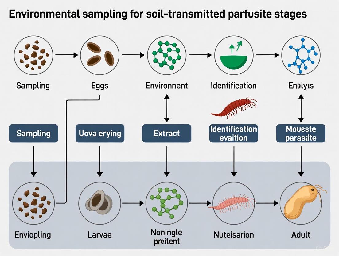
The complex lifecycle of STHs involves crucial developmental stages in the environment that determine transmission potential. Figure 1 illustrates the complete lifecycle and environmental progression of key STH species.
Key Lifecycle Characteristics:
- Fecal-oral transmission for A. lumbricoides and T. trichiura via ingestion of embryonated eggs from contaminated soil, water, or produce [1]
- Skin penetration for hookworms, where infective filariform larvae actively penetrate bare skin [1]
- Environmental maturation period requiring 3+ weeks for eggs to embryonate and become infective [1]
- No direct person-to-person transmission or infection from fresh feces due to required soil maturation period [1]
- No multiplication in human host for most STH species (except Strongyloides stercoralis which can auto-infect) [1]
Quantitative Analysis of Environmental Resilience Factors
The survival and development of STH environmental stages depend on multiple abiotic factors. Table 1 synthesizes empirical data on how environmental parameters affect STH resilience and development.
Table 1: Environmental Resilience Factors for Soil-Transmitted Helminths
| Environmental Factor | Optimal Conditions | Effect on STH Development & Survival | Key Evidence |
|---|---|---|---|
| Temperature | 20-30°C | Negative correlation with A. duodenale (r = -0.53) and S. stercoralis larvae survival [5] | Soil temperature increases parasite growth but can also increase mortality [6] |
| Soil pH | Alkaline conditions | High larvae counts associated with specific pH ranges (P<0.001) [7] | Forest ochrosols with high magnesium, calcium, and lime reduce acidity [7] |
| Soil Texture | Sandy-loamy | Associated with high larvae counts (P<0.001) [7] | Clay content associated with low larvae counts (P<0.001) [7] |
| Moisture | Humid conditions | Higher occurrence during rainy months (n=416) vs. dry months (n=290) [5] | Flooding drives water-borne epidemics; drought causes host aggregation [6] |
| Organic Matter | High carbon content | Associated with high larvae counts (P<0.001) [7] | Nitrogen content associated with low larvae counts (P<0.001) [7] |
| Seasonal Variation | Rainy seasons | Higher STH frequency in rainy seasons [5] | 93.75% non-pathogenic nematodes in winter vs. 82.50% in summer [8] |
Environmental Sampling and Detection Protocols
Soil Sampling Methodology
Spatial Sampling Design:
- Systematic unaligned sampling: Divide area into equal grids, randomly select sampling points within each grid [4]
- W-path transect sampling: Walk W-shaped path across sampling area, collecting at regular intervals [4]
- Spatial stratified sampling: Subdivide area into homogeneous zones based on expected contamination variance [4]
- Comprehensive grid sampling: Most reliable method, avoids underestimation from purposive sampling [4]
Sample Collection Protocol:
- Site Selection: Target high foot-traffic locations, defecation sites, shaded/moist areas, children's play areas [4] [2]
- Collection Depth: Use auger soil sampler at 3-5 cm depth [7] [5]
- Sample Weight: Collect 500g soil samples in clean, labelled polythene bags [5]
- Time of Collection: Between 09:00-12:00 hours to standardize temperature conditions [5]
- Transport: Store in cool, dark conditions to preserve parasite viability [5]
STH Recovery and Quantification Methods
Egg/Larvae Recovery Workflow: The process for isolating and identifying STH from environmental samples involves multiple purification and concentration steps. Figure 2 outlines the complete experimental workflow from sample collection to final analysis.
Key Techniques:
- Baermann technique: Extraction of larvae from soil using warm water immersion and migration [7] [5]
- Flotation method: Density separation using Sodium Chloride (NaCl) or Zinc Sulfate (ZnSO₄) solutions [9] [5]
- Sedimentation: Gravity-based concentration of helminth eggs [4]
- Culture methods: Incubation of eggs to larvae for viability assessment and species identification [7]
Advanced Detection Methods
Molecular Techniques:
- Multi-parallel qPCR assays: Detect STH DNA in environmental samples with high sensitivity [2]
- Metagenomic sequencing: Identify helminth species and potential zoonotic hybrids [7]
- Automated image identification: Emerging technology for improved quantification [4]
Viability Assessment:
- Larval culture: Using Baermann technique to assess development capacity [7]
- Staining methods: Molecular probes to determine egg viability [4]
- Embryonation assessment: Monitoring development to infective stages [4]
Research Reagent Solutions and Essential Materials
Table 2: Essential Research Reagents for STH Environmental Studies
| Reagent/Material | Application | Function | Example Specifications |
|---|---|---|---|
| Ionic Detergents (7X, Tween) | Sample processing | Chemical dissociation of ova from soil particles | 0.1-1% solutions in distilled water [4] |
| Flotation Solutions | Egg concentration | Density-based separation of helminth eggs | NaCl (specific gravity 1.20-1.25), ZnSO₄ [9] [5] |
| Baermann Apparatus | Larval isolation | Extraction of larvae from soil using migration | Funnel, mesh, tubing, clamp stand [7] [5] |
| DNA Extraction Kits | Molecular detection | Nucleic acid isolation from environmental samples | Commercial kits for soil/stool DNA extraction [2] |
| qPCR Master Mixes | Molecular quantification | Detection and quantification of STH DNA | Multi-parallel assays for specific STH species [2] |
| Microscopy Stains | Morphological ID | Enhanced visualization of parasitic structures | Iodine, lactophenol cotton blue [4] |
| Culture Media | Viability testing | Support development of eggs to larval stages | Agar-based or liquid media for nematode development [7] |
Implications for Control Programs and Research Directions
The environmental resilience of STH stages has profound implications for disease control. Mass Drug Administration (MDA) with albendazole or mebendazole reduces morbidity but rarely interrupts transmission due to persistent environmental contamination [6] [1]. Sustainable control requires integrated approaches including:
- Environmental interventions: Improved sanitation to reduce soil contamination [1] [8]
- Water, sanitation, and hygiene (WASH): Critical for breaking transmission cycles [9] [8]
- One Health approaches: Addressing zoonotic transmission between humans and animals [9]
- Climate change adaptation: Accounting for shifting transmission patterns due to environmental changes [6] [3]
Future research should prioritize:
- Standardized environmental monitoring protocols for STH [4] [2]
- Advanced detection methods for low-intensity infections [4] [2]
- Longitudinal studies on climate change impacts on STH transmission [6] [3]
- Integrated control strategies combining MDA, WASH, and veterinary public health [9]
Global Burden and the Critical Need for Environmental Surveillance
Soil-transmitted helminths (STHs), including Ascaris lumbricoides, Trichuris trichiura, and hookworms (Necator americanus and Ancylostoma duodenale), represent a significant global health burden, infecting an estimated 1.5 billion people worldwide and accounting for over five million disability-adjusted life years (DALYs) [2] [10]. Infections cause a range of health issues, from malnutrition and anemia to impaired cognitive development in children, and are inextricably linked to poverty and inadequate sanitation [2] [4]. Current STH control programs primarily rely on mass drug administration (MDA). However, MDA alone is often insufficient to interrupt transmission due to the persistent environmental reservoir of infectious STH stages [10] [11]. The primary transmission pathway involves STH eggs, passed in human feces, contaminating the soil, leading to new infections through egg ingestion or skin penetration by hookworm larvae [10].
Traditional surveillance, based on detecting eggs in human stool via microscopy, suffers from poor specificity and sensitivity, particularly in low-intensity infection settings, and is hampered by logistical challenges and participant stigma [2] [11]. Consequently, there is a critical need for enhanced surveillance tools. Environmental surveillance (ES)—the systematic detection of pathogen targets in environmental samples like soil and wastewater—offers a promising, non-invasive complement to stool-based surveys. This approach can provide a broader community-level assessment of STH circulation and help identify environmental transmission hotspots, which is vital for evaluating the impact of control programs beyond MDA [2] [10] [11].
Application Notes: The Value of Environmental Surveillance
Environmental surveillance for STHs addresses key limitations of current surveillance paradigms and provides unique insights into transmission dynamics.
Advantages Over Stool-Based Surveillance
- Community-Level Data: ES provides a composite measure of community contamination, overcoming the individual-level focus and sampling biases of stool surveys [2] [11].
- Non-Invasive Sampling: Collecting environmental matrices avoids the logistical, ethical, and cultural challenges associated with obtaining individual stool samples, potentially increasing public acceptance and feasibility of monitoring [11].
- Increased Sensitivity with Molecular Methods: Molecular detection methods like qPCR offer higher specificity and sensitivity compared to traditional microscopy, especially for low-intensity infections. They also allow for species-specific identification and multiplexing to detect multiple pathogens simultaneously [10] [11].
Evidence of Utility from Field Studies
Recent field studies demonstrate the practical application and value of ES. Research in rural and peri-urban settings in Benin and India detected STH DNA in both soil and wastewater samples, with an overall detection frequency of 36% in India and 25% in Benin across all sample types [2] [10]. A multi-country study across Kenya, Benin, and India found that detection of a specific STH species in household soil was strongly associated with increased odds of a household member being infected with the same species, validating soil surveillance as a indicator of infection risk [11]. Furthermore, studies have established that wastewater sediment samples outperformed grab samples and passive Moore swabs for STH detection, informing optimal sampling strategy [2] [10].
Table 1: Summary of STH Detection in Recent Environmental Surveillance Studies
| Location | Sample Type | Number of Samples | Detection Frequency (%) | Key Findings | Source |
|---|---|---|---|---|---|
| India (Tamil Nadu) | Soil | 95 | 33.7% (32/95) | STH prevalence varied by sample site type. | [2] [10] |
| Benin (Comé) | Soil | 121 | 32.2% (39/121) | No significant variation within a single site (e.g., across a market). | [2] [10] |
| India (Tamil Nadu) | Wastewater | 60 | 40.0% (24/60) | Wastewater sediment samples were the most effective type. | [2] [10] |
| Benin (Comé) | Wastewater | 64 | 12.5% (8/64) | Demonstrates feasibility in settings without networked sanitation. | [2] [10] |
| Kenya, Benin, India | Household Soil | 478 | A. lumbricoides: 31%T. trichiura: 3%Hookworm: 13% | Detection in soil strongly associated with household member infection. | [11] |
Experimental Protocols
Standardized protocols are essential for generating reliable and comparable data in environmental surveillance. The following methodologies have been field-tested in multiple countries.
Field Collection of Environmental Samples
Site Selection and Sampling Strategy
STHs are notoriously overdispersed in the environment, forming localized clusters of high contamination [4] [12]. Therefore, a purposive sampling strategy targeting high foot-traffic locations and potential contamination zones is recommended for efficient detection [2] [4].
- Sample Sites per Cluster: In each surveillance cluster, collect samples from:
- Two markets (samples from entrance, center, and frequently used path).
- Two schools (samples from entrance, classroom area, and path to latrine).
- Two open defecation fields (samples from entrance, center, and field edge).
- One or two community water points.
- Multiple households (e.g., five per cluster) [10].
- Spatial Sampling: Within a defined sample site (e.g., a 30 cm x 50 cm area), collect surface soil from within a disposable stencil. For wastewater, sample from flowing sections of drainage ditches [10].
Soil Collection Protocol
- Lay a disposable stencil (30 cm x 50 cm) on the ground at the sampling point.
- Using a sterile scoop, scrape the top layer of soil from inside the stencil area, collecting approximately 100 grams.
- Place the soil into a sterile Whirlpak bag.
- Seal the bag, label it, and wipe the exterior with 70% ethanol and 10% bleach.
- Store samples in a cooler box at 4°C and process within 24 hours of collection [10].
Wastewater Collection Protocol
Three simultaneous sample types are recommended for comprehensive surveillance:
- Grab Sample: Slowly immerse a sterile 500 mL Whirlpak bag into a flowing wastewater channel to collect liquid wastewater.
- Sediment Sample: Scrape approximately 250 mL of wet sediment from the bottom of the channel using a sterile scoop into a Whirlpak bag.
- Moore Swab (Passive Sample): Tie a 4x4 ply gauze to a fishing line, anchor it in the wastewater channel, and leave it suspended for 24 hours to filter microorganisms. After 24 hours, retrieve the swab and place it in a sterile Whirlpak bag [2] [10].
- Transport and store all samples as described for soil samples.
Laboratory Processing and Molecular Detection
Soil Processing and DNA Extraction
- Sieving: Pass the soil sample through a 2 mm mesh screen to remove rocks and debris.
- Homogenization: Thoroughly mix the sieved soil to ensure a representative sub-sample.
- Aliquoting: Scoop a 40-gram aliquot of the homogenized soil into a 50 mL centrifuge tube.
- Storage: Store the aliquoted soil at -80°C until DNA extraction.
- DNA Extraction: Extract DNA from the large volume (20 g) of soil using a commercial DNA extraction kit, following the manufacturer's protocol but potentially with optimizations for complex environmental matrices as detailed in [11].
Wastewater Processing and DNA Extraction
- Grab Samples: Vacuum-filter the liquid wastewater through a membrane filter. The filter, containing concentrated material, is then processed for DNA extraction [10].
- Sediment Samples: Process similarly to soil samples, with homogenization and DNA extraction from an aliquot.
- Moore Swabs: The gauze swab is processed directly, often by eluting captured material into a solution, followed by concentration and DNA extraction [2].
Molecular Detection by qPCR
- Assay Selection: Use validated, multi-parallel or multiplexed qPCR assays specific for the target STH species (e.g., A. lumbricoides, T. trichiura, N. americanus, A. duodenale) [2] [11].
- Amplification: Perform qPCR on the extracted DNA samples using standard cycling conditions for the chosen assays.
- Analysis: Quantify the results based on standard curves. Positive and negative controls must be included in every run to ensure accuracy.
The following workflow diagram summarizes the key steps from sample collection to analysis:
The Scientist's Toolkit: Research Reagent Solutions
Successful implementation of environmental STH surveillance relies on specific materials and reagents. The following table details key items and their functions.
Table 2: Essential Research Reagents and Materials for STH Environmental Surveillance
| Item | Function / Application | Specific Examples / Notes |
|---|---|---|
| Sterile Whirlpak Bags | Sample containment and transport for soil and wastewater. Pre-sterilized to prevent cross-contamination. | Bags of various sizes (e.g., 500 mL for water grabs) are used [10]. |
| Disposable Soil Stencil | Standardizes the surface area from which soil is collected (e.g., 30 cm x 50 cm), ensuring consistency. | Single-use to avoid transferring contamination between sites [10]. |
| Sterile Scoops | For collecting soil and wastewater sediment without introducing external contaminants. | |
| Moore Swab Materials | Passive sampling device for filtering pathogens from flowing wastewater over 24 hours. | 4x4 ply gauze, secured with fishing line [2] [10]. |
| DNA Extraction Kits | Isolation of high-quality PCR-inhibitor-free DNA from complex matrices like soil and wastewater sediment. | Commercial kits optimized for environmental samples are critical [11]. |
| qPCR Master Mix & Assays | Sensitive and specific detection and quantification of STH DNA. Enables multiplexing for multiple targets. | Species-specific primers and probes for A. lumbricoides, T. trichiura, hookworms, etc. [2] [11]. |
| Ionic Detergents (e.g., Tween) | Chemical dissociation of STH ova from soil and sediment particles during processing, improving recovery. | Reduces ova adhesion to matrix, mitigating loss during filtration [4] [12]. |
| Sieves / Mesh Screens | Removal of large debris (e.g., rocks, twigs) from soil samples prior to DNA extraction and homogenization. | Typically a 2 mm mesh size [10]. |
Environmental surveillance for STHs represents a paradigm shift in how public health programs can monitor and ultimately interrupt the transmission of these persistent parasitic infections. By moving beyond traditional, individual-focused stool surveys, ES provides a cost-effective, non-invasive, and community-level picture of environmental contamination. The protocols outlined here, developed and validated in multiple endemic countries, provide a robust framework for researchers and public health professionals to implement this powerful surveillance tool. As the global community works towards the sustainable control and elimination of STHs, integrating environmental surveillance into monitoring and evaluation frameworks will be critical for assessing progress, identifying residual transmission hotspots, and guiding targeted interventions.
Application Notes: Quantitative Findings on Environmental Contamination
Environmental surveillance in settings without networked sanitation reveals significant contamination of soil and wastewater with enteric pathogens, presenting a substantial transmission risk. The following table summarizes key quantitative findings from recent field studies in endemic regions.
Table 1: Detection Frequency of Soil-Transmitted Helminths (STH) in Environmental Samples from Benin and India
| Sample Type | Location/Sub-Type | Detection Rate (India) | Detection Rate (Benin) | Key Pathogens Identified |
|---|---|---|---|---|
| Soil | Overall | 33.7% (32/95) | 32.2% (39/121) | Ascaris lumbricoides, Trichuris trichiura, Hookworm species[*citation:2] [10] |
| Markets | Data N/A | Data N/A | ||
| Schools | Data N/A | Data N/A | ||
| Open Defecation Fields | Data N/A | Data N/A | ||
| Community Water Points | Data N/A | Data N/A | ||
| Wastewater | Overall | 40.0% (24/60) | 12.5% (8/64) | STHs and other enteric pathogens[*citation:2] [10] |
| Sediment Samples | Highest yield | Highest yield | ||
| Grab Samples | Lower yield | Lower yield | ||
| Moore Swabs | Lower yield | Lower yield |
These findings confirm that wastewater sediment samples outperform other liquid sample types for STH detection sensitivity, making them a preferred method for environmental surveillance. Furthermore, high foot-traffic public areas like markets and schools were identified as significant environmental reservoirs, while transmission dynamics studies highlight the role of schools and households as interconnected nodes in pathogen spread [13].
Experimental Protocols
Field Collection of Environmental Samples
Soil Sampling Protocol
- Site Selection: Collect from high foot-traffic locations: school entrances, classrooms, paths to latrines, market entrances/centers, open defecation fields (entrance, center, edge), and community water points [10].
- Collection Procedure:
- Lay a disposable 30 cm × 50 cm soil stencil on the sampling area.
- Using a sterile scoop, scrape the top surface soil from within the stencil area to collect approximately 100 grams.
- Place the soil into a sterile Whirlpak bag (e.g., WPB01350WA, Merck).
- Seal the bag, wipe the exterior with 70% ethanol followed by 10% bleach, and label appropriately.
- Store samples in a cooler box and transport to the laboratory at 4°C.
- Process samples within 24 hours of collection [10].
Wastewater Sampling Protocol
- Site Selection: Identify wastewater drainage channels in target communities [10].
- Sample Types & Collection:
- Grab Sample: Slowly immerse a sterile 500 mL Whirlpak bag into flowing wastewater to fill.
- Sediment Sample: Scrape approximately 250 mL of wet sediment from the channel bottom using a sterile scoop into a Whirlpak bag.
- Moore Swab: Tie a 4x4 ply gauze (e.g., ExcilonTM 7086) with fishing line. Anchor the swab in the wastewater channel for 24 hours to filter pathogens. Retrieve, place in a sterile Whirlpak bag (e.g., WPB01065WA, Merck) [10].
- Field Blank: Prepare one blank per day by filling a sterile bag with clean bottled water in the field.
- Storage & Transport: Wipe bags with ethanol and bleach, label, store in a cooler at 4°C, and process within 24 hours [10].
Laboratory Processing and Molecular Analysis
Soil Processing
- Sieve soil through a 2 mm mesh screen to remove rocks and debris.
- Thoroughly mix the sieved soil and aliquot 40 grams into a pre-labeled 50 mL centrifuge tube.
- Seal the tube cap with parafilm and store at -80°C until DNA extraction.
- Clean reusable soil screens with soap, soak in 70% ethanol for 2 minutes, and air dry before reuse [10].
Wastewater Processing
- Grab Samples: Vacuum-filter (e.g., using EZFITMIHE1, Merck) through a membrane to concentrate particulate matter [10].
- Sediment Samples: Process directly for DNA extraction due to high pathogen concentration.
- Moore Swabs: Elute captured material from the gauze into a buffer solution for further concentration.
- DNA Extraction & qPCR: Extract total nucleic acids from all processed samples. Analyze using multi-parallel qPCR assays for specific detection of STH DNA (e.g., Ascaris, Trichuris, hookworm). Multiplexed qPCR can be employed for simultaneous detection of a broader panel of enteric pathogens [2] [10].
Workflow Visualization
Environmental Surveillance Workflow for STH
The Scientist's Toolkit: Essential Research Reagent Solutions
Table 2: Key Reagents and Materials for Environmental STH Surveillance
| Item | Specification/Example | Primary Function in Protocol |
|---|---|---|
| Sterile Sample Bags | Whirlpak bags (e.g., WPB01350WA, WPB01065WA) | Aseptic collection and transport of soil and wastewater samples [10]. |
| Soil Sieve | 2 mm mesh screen | Removal of rocks and debris from soil samples to homogenize and facilitate processing [10]. |
| Moore Swab Material | 4x4 ply gauze (e.g., ExcilonTM 7086) | Passive filtration and concentration of pathogens from flowing wastewater over 24 hours [10]. |
| DNA Extraction Kit | Commercial kits for soil/stool/fecal samples | Isolation of high-quality total nucleic acids from complex environmental matrices [2] [10]. |
| qPCR Assays | Multi-parallel, species-specific primers/probes for STH; multiplex panels for enteric pathogens | Sensitive and specific detection and quantification of pathogen DNA [2] [10]. |
| Filtration Apparatus | Vacuum filtration system (e.g., EZFITMIHE1, Merck) | Concentration of pathogens from large volume liquid wastewater grab samples [10]. |
The Impact of Sanitation and Hygiene Practices on Soil Contamination
Soil contamination with the infective stages of soil-transmitted helminths (STHs) represents a significant environmental pathway for the transmission of parasitic diseases affecting approximately 1.5 billion people globally [14]. These parasites, including roundworms (Ascaris lumbricoides), whipworms (Trichuris trichiura), and hookworms (Necator americanus and Ancylostoma duodenale), complete their life cycles through soil, where eggs embryonate or larvae develop into infective stages [15] [4]. The persistence of STH eggs in soil can extend for years due to their resistant shells, making environmental contamination a critical reservoir for continued transmission [4]. Understanding and interrupting this environmental transmission route requires integrated approaches combining sanitation, hygiene, and accurate environmental monitoring methodologies framed within the context of environmental sampling for soil-transmitted parasite stages research.
This application note provides a comprehensive framework for assessing the impact of sanitation and hygiene interventions on soil contamination levels, detailing standardized protocols for environmental sampling, laboratory processing, and pathogen detection. The guidance is specifically tailored to support researchers, scientists, and public health professionals engaged in drug development and intervention studies aimed at breaking the cycle of environmental transmission of STHs.
Quantitative Evidence: WASH Interventions and STH Reduction
Meta-analyses of observational studies and controlled trials demonstrate that water, sanitation, and hygiene (WASH) interventions significantly reduce the odds of STH infection in human populations by reducing environmental exposure. The table below summarizes the protective associations between specific WASH factors and STH infections, based on comprehensive systematic reviews [15].
Table 1: Impact of WASH Access and Practices on Soil-Transmitted Helminth Infection
| WASH Factor | STH Type | Odds Ratio (OR) | 95% Confidence Interval | Protective Effect |
|---|---|---|---|---|
| Treated Water Use | Any STH | 0.46 | 0.36–0.60 | 54% reduction |
| Piped Water | A. lumbricoides | 0.40 | 0.39–0.41 | 60% reduction |
| Piped Water | T. trichiura | 0.57 | 0.45–0.72 | 43% reduction |
| Sanitation Access | Any STH | 0.66 | 0.57–0.76 | 34% reduction |
| Sanitation Access | A. lumbricoides | 0.62 | 0.44–0.88 | 38% reduction |
| Sanitation Access | T. trichiura | 0.61 | 0.50–0.74 | 39% reduction |
| Wearing Shoes | Hookworm | 0.29 | 0.18–0.47 | 71% reduction |
| Wearing Shoes | Any STH | 0.30 | 0.11–0.83 | 70% reduction |
| Handwashing Before Eating | A. lumbricoides | 0.38 | 0.26–0.55 | 62% reduction |
| Handwashing After Defecation | A. lumbricoides | 0.45 | 0.35–0.58 | 55% reduction |
| Soap Use/Availability | Any STH | 0.53 | 0.29–0.98 | 47% reduction |
The data indicates that specific hygiene practices, particularly shoe-wearing and handwashing, demonstrate the strongest protective effects against STH infection. While sanitation access shows consistent benefits, a recent systematic review noted that basic sanitation interventions implemented in several trials showed only small reductions in environmental pathogen detection and no significant effect on human or animal fecal markers, suggesting more comprehensive interventions may be necessary to effectively contain human waste and reduce environmental exposure [16].
Environmental Sampling Framework for STH Detection
Spatial Sampling Design Considerations
STH eggs and larvae exhibit highly overdispersed distributions in soil, with localized clusters of high contamination within areas of generally low concentration [4]. This spatial heterogeneity stems from the aggregation of high worm burdens in specific individuals, whose feces become focal contamination points [4]. Sampling designs must account for this heterogeneity to obtain accurate environmental assessments.
Table 2: Spatial Sampling Methods for STH Detection in Soil
| Sampling Method | Description | Application Context | Advantages/Limitations |
|---|---|---|---|
| Systematic Aligned Grid | Samples taken at regular intervals in a grid pattern | General contamination assessment across an area | Efficient for 2D spatial distribution; may miss hotspots |
| Systematic Unaligned Grid | Sampling points randomly selected within grid cells | General contamination assessment | Reduces bias compared to aligned grid |
| Transect Sampling | Samples collected along a linear path | Investigating contamination gradients from a source | Efficient for studying distance effects |
| W-Route Sampling | Investigator walks diagonal path forming "W" pattern | Large rectangular areas like fields | Comprehensive coverage; more time-consuming |
| Purposive Sampling | Samples taken from areas with high contamination likelihood | Targeted assessment of high-risk zones | May overestimate overall contamination |
| Spatial Stratified Sampling | Area divided into homogeneous zones with proportional sampling | Highly heterogeneous environments | Most efficient for heterogeneous distributions |
For most research applications, systematic unaligned grid or spatial stratified sampling approaches are recommended as they provide the best balance of practical implementation and statistical robustness for characterizing heterogeneous STH contamination [4]. Sampling should account for seasonal variations, with collections during both wet and dry seasons since STH prevalence and survival are influenced by climatic factors [4].
Site Selection and Environmental Factors
Field studies have detected STH contamination even in unexpected settings, with one campus in southern Brazil finding 35% of soil samples positive for hookworm eggs, 10% for roundworm eggs, and 5% for whipworm eggs [17]. This highlights that STH contamination extends beyond traditionally recognized endemic areas.
Key environmental factors influencing STH distribution in soil include:
- Moisture and shading: STH ova exhibit longest survival in moist, shaded conditions with limited sunlight [4]
- Soil type: Sandy soils with poor water retention may enhance desiccation, while impermeable anoxic soils can slow ova maturation [4]
- Human and animal activity: Areas with significant people circulation, animal presence, or observed defecation sites show higher contamination [17]
The following diagram illustrates the strategic environmental sampling workflow from site characterization through to sample collection:
Diagram 1: Environmental Sampling Workflow for STH Detection
Laboratory Processing and Detection Methods
Soil Processing and STH Recovery
The recovery of STH from soil matrices involves a multi-step process to separate, concentrate, and detect parasites from complex environmental samples. The key challenge is overcoming the adhesion of STH ova to soil particles, which can lead to substantial recovery losses if not properly addressed [4].
Table 3: Key Research Reagent Solutions for STH Recovery from Soil
| Reagent/Solution | Composition | Function | Application Notes |
|---|---|---|---|
| Ionic Detergents | 7X or Tween solutions | Chemical dissociation of ova from soil particles | Displaces phosphate anions on ova wall from cationic sites on soil |
| Flotation Solutions | Zinc sulfate (ZnSO₄), sucrose, sodium nitrate | Buoyancy-based separation of ova based on specific gravity | Specific gravity ~1.20-1.35; selects for viable eggs |
| Sedimentation Buffers | Tris-buffered saline (TBS), physiological saline | Gravity-based separation of ova from lighter debris | Takes advantage of higher density of STH eggs |
| Rinsing Solutions | Tween 20, Nacconol, physiological saline | Removal and recovery of STH from plant matter | Used for produce and vegetation samples |
| Homogenization Media | Aqueous solutions with detergents | Breaking up soil aggregates and distributing ova evenly | Critical step before fractionation |
STH Quantification and Viability Assessment
Modern detection methodologies have evolved significantly from basic microscopy to incorporate molecular and advanced computational approaches:
- Traditional Microscopy: Remains the foundation for morphological identification of STH eggs and larvae based on size, shape, and internal structures [18]
- Molecular Detection: PCR-based methods provide species-specific identification and can detect low-intensity contamination [4] [19]
- Advanced Imaging: Lightweight deep-learning models like YAC-Net achieve 97.8% precision in parasite egg detection while reducing computational requirements, making automation feasible in resource-limited settings [14]
- Viability Assessment: Critical for determining infection risk, using methods including:
- Larval development in vitro
- Staining techniques (e.g., vital dyes)
- Molecular indicators of viability [4]
The following diagram illustrates the complete laboratory processing workflow from sample preparation to detection:
Diagram 2: Laboratory Analysis Workflow for STH Detection in Soil
Comprehensive Experimental Protocol
Field Sampling Protocol
Title: Standardized Procedure for Soil Sampling for STH Detection
Purpose: To collect representative soil samples for qualitative and quantitative analysis of STH contamination while preserving parasite integrity and viability.
Materials:
- Sterile sampling instruments (spatulas, trowels, corers)
- Whirl-Pak bags or sterile containers
- Cooler with ice packs or dry ice
- GPS device or smartphone with GPS
- Data recording forms or electronic device
- Permanent markers
- Personal protective equipment (gloves, closed-toe shoes)
Procedure:
- Site Characterization: Document site features including vegetation, shading, moisture, evidence of human or animal activity, and potential contamination sources.
- Sampling Point Selection: Based on chosen sampling design (see Table 2), flag sampling points ensuring appropriate spacing and coverage.
- Soil Collection:
- Remove surface debris (leaves, twigs) without disturbing soil
- For most STH eggs, collect from top 0-2 cm of soil
- For hookworm larvae, extend to 5-10 cm depth as larvae migrate vertically
- Collect 100-200 g soil per sample using sterile instruments
- Place immediately into labeled sterile containers
- Sample Documentation: Record sample ID, date, time, location (GPS coordinates), soil characteristics, and environmental conditions.
- Sample Transport: Store samples at 4°C and process within 24-48 hours. For longer storage, freeze at -20°C but avoid repeated freeze-thaw cycles.
Quality Control:
- Include field blanks (sterile containers exposed to air during sampling)
- Maintain chain of custody documentation
- Use standardized sampling depth and volume across all samples
Laboratory Processing Protocol
Title: Concentration and Recovery of STH from Soil Samples
Purpose: To efficiently separate and concentrate STH eggs and larvae from soil matrices for detection and identification.
Materials:
- Ionic detergent solution (0.1% Tween 80 or 1% 7X)
- Sieves/mesh filters (100μm, 300μm)
- Centrifuge and centrifuge tubes
- Zinc sulfate flotation solution (specific gravity 1.20)
- Sedimentation cylinders or conical tubes
- Microscopic slides, coverslips, and staining reagents
Procedure:
- Sample Homogenization:
- Weigh 50 g of soil sample
- Add 200 mL of detergent solution
- Mix thoroughly by shaking or mechanical stirring for 10 minutes
- Filtration:
- Pass homogenate through 300μm sieve to remove large debris
- Collect filtrate and pass through 100μm sieve
- Retain material on 100μm sieve and wash into centrifuge tube
- Sedimentation:
- Centrifuge at 500 × g for 5 minutes
- Discard supernatant
- Resuspend pellet in detergent solution
- Repeat sedimentation 2-3 times until supernatant is clear
- Flotation:
- Resuspend final pellet in 5 mL zinc sulfate solution (specific gravity 1.20)
- Centrifuge at 500 × g for 5 minutes
- Transfer surface film to clean slide using wire loop
- Add coverslip for microscopic examination
- Microscopic Analysis:
- Systematically examine entire coverslip area at 100× magnification
- Identify and count STH eggs based on morphological characteristics
- Confirm suspicious structures at 400× magnification
- Express results as eggs per gram of soil
Quality Control:
- Include positive control samples with known STH egg concentrations
- Include negative control (sterile soil or water) through entire process
- Calculate and report recovery efficiency using spiked samples
Emerging Technologies and Future Directions
The field of environmental STH detection is rapidly evolving with several promising technological advances:
Advanced Detection Methods
- Molecular Genetic Assays: PCR and multiplex assays enable species-specific identification and detection of genetic markers associated with anthelmintic resistance [4] [19]
- Sensor Technologies: Emerging soil sensors including electrochemical sensors and spectroscopic methods like Laser-Induced Breakdown Spectroscopy (LIBS) offer potential for real-time monitoring [20]
- Automated Image Identification: Deep learning models such as YAC-Net demonstrate how automated parasite egg detection can achieve high precision (97.8%) while reducing computational requirements [14]
One Health Integration
The One Health approach recognizes the interconnectedness of human, animal, and environmental health and is particularly relevant for STH control given the zoonotic potential of some species [19]. This approach is essential for understanding the complex dynamics of anthelmintic resistance, where veterinary drug use may select for resistance in human STHs through shared genetic mechanisms [19] [21].
Accurate assessment of soil contamination with STH stages is fundamental to understanding the impact of sanitation and hygiene interventions on breaking transmission cycles. This application note provides comprehensive protocols for environmental sampling, laboratory processing, and detection that account for the spatial heterogeneity and analytical challenges inherent in STH environmental monitoring. The integration of traditional methods with emerging technologies in molecular detection, automated imaging, and environmental sensors will enhance our capacity to precisely measure intervention effectiveness and guide public health strategies for sustainable STH control.
The One Health approach is an integrated, unifying concept that aims to sustainably balance and optimize the health of people, animals, and ecosystems, recognizing their close interdependence [22]. This framework is particularly crucial for understanding and controlling soil-transmitted helminths (STHs), as their transmission occurs at the interface of human, animal, and environmental health [23]. Environmental systems, especially soil, serve as key reservoirs for zoonotic helminths, facilitating their transmission to both humans and animals through contamination with infected feces [24]. Livestock farms, including goat farms, have been identified as potential hotspots for this transmission, where helminth eggs can easily contaminate the soil [24]. This application note provides detailed protocols and data integration strategies for STH research within a comprehensive One Health framework, supporting the broader thesis on environmental sampling for soil-transmitted parasite stages.
One Health in Parasitology: Current Evidence
Recent studies demonstrate the utility of the One Health approach in uncovering parasite transmission dynamics across different environments.
Table 1: Key Findings from Recent One Health Parasitology Studies
| Location | Human Infection | Animal Infection | Environmental Contamination | Primary Zoonotic Parasites Identified |
|---|---|---|---|---|
| Ratchaburi, Thailand [24] | Not specified | 80-86% of goat farms positive for helminths | 60% of farms positive for human/animal parasitic helminths | Haemonchus contortus, Trichostrongylus colubriformis |
| Valdivia, Chile [23] | 28% parasite prevalence | 26% in owned dogs; 44% in stray dog feces | 30.5% of park soil samples contaminated | Toxocara sp., Trichuris vulpis, Giardia duodenalis, Blastocystis sp. |
| Dak Lak, Vietnam [25] | 13.7% hookworm (N. americanus) prevalence | Not specified | Associated with open defecation and unimproved water | Necator americanus (hookworm) |
Integrated analysis reveals significant connections between these compartments. In Chile, 33% of human sera tested positive for anti-Toxocara canis IgG antibodies, indicating exposure to this zoonotic parasite, while simultaneous environmental sampling found Toxocara sp. eggs in park soils [23]. Molecular characterization confirmed zoonotic subtypes of Giardia duodenalis and Blastocystis sp. in human samples, further supporting cross-species transmission [23].
Methodological Approaches for One Health Sampling
Environmental Soil Sampling and Processing
Protocol: Soil Collection and Helminth Isolation
- Sample Collection: Collect approximately 50 g of topsoil (from the top 5 cm depth) using a clean shovel. Store in individual ziplock bags and maintain at low temperature during transport to the laboratory [24].
- Helminth Isolation (Sedimentation and Flotation):
- Remove large debris and pass soil through a 0.2-mm sieve.
- Weigh 20 g of sieved soil into a 50-mL Falcon tube containing 1% Tween 80 solution.
- Mix thoroughly and allow to sediment for 1 hour to dissociate helminth eggs from soil particles.
- Repeat sedimentation twice, replacing supernatant with water to wash off the Tween 80 solution.
- Add 30 mL saturated sodium chloride (NaCl) to the sediment and separate into 15-mL Falcon tubes.
- Add additional NaCl to form a reverse meniscus and place a coverslip on top.
- After 15 minutes, remove the coverslip, place on a microscope slide, and examine for helminth eggs under a light microscope [24].
Molecular Detection and Quantification
Protocol: DNA Extraction and PCR Amplification
- DNA Extraction: Prior to extraction, wash soil samples in water to remove preservatives like ethanol. Extract genomic DNA using the DNeasy PowerSoil Pro Kit (Qiagen) following manufacturer's recommendations [24].
- PCR Amplification:
- Conduct multiple PCR sets for each sample using different genetic markers.
- For broad-range nematode detection: Amplify the 18S ribosomal RNA (rRNA) gene using primers from Holterman et al. (2006) [24].
- For platyhelminths (trematodes, cestodes): Amplify the 18S rRNA gene using primers from Routtu et al. (2014) [24].
- For species-specific detection: Target the internal transcribed spacer 2 (ITS2) region with newly designed primers for specific helminths (e.g., H. contortus, T. colubriformis) [24].
- Quantitative PCR (qPCR): For stool samples, qPCR provides higher sensitivity compared to conventional microscopy (94.1% vs. 68.1% for Ascaris spp.; 75.7% vs. 66.9% for hookworm). The multiplex real-time PCR can quantitatively detect Ascaris spp., N. americanus, Ancylostoma spp., and Trichuris spp. simultaneously [26].
Comparative Diagnostic Techniques
Table 2: Comparison of STH Diagnostic Methods
| Method | Sensitivity | Advantages | Limitations | Best Application |
|---|---|---|---|---|
| Kato-Katz [26] [25] | Low to moderate (especially in low transmission) | Low cost, provides intensity measurement (EPG), field-deployable | Poor sensitivity, requires fresh stool, operator-dependent, cannot distinguish hookworm species | High-transmission settings, resource-limited field surveys |
| Sodium Nitrate Flotation (SNF) [26] | Moderate to high | Higher sensitivity for hookworm than Kato-Katz, simple procedure | Limited quantification ability, requires centrifugation | Veterinary diagnostics, field studies with centrifugation capability |
| qPCR [26] [25] | High (particularly for light infections) | Species-specific identification, quantitation, detects multiple parasites simultaneously, high throughput | Higher cost, requires specialized equipment and training, DNA preservation critical | Low-transmission settings, research studies, monitoring elimination programs |
| Sedimentation-Flotation (Soil) [24] | Varies with soil type | Effective for environmental samples, non-invasive monitoring | Labor-intensive, may require optimization for different soil matrices | Environmental monitoring, One Health studies |
The Scientist's Toolkit: Essential Research Reagents and Materials
Table 3: Key Research Reagent Solutions for One Health Helminth Studies
| Reagent/Material | Application | Function | Example Specifics |
|---|---|---|---|
| PowerSoil DNA Isolation Kit (Mo Bio/Qiagen) [26] [24] | DNA extraction from soil and stool | Is high-quality genomic DNA from complex environmental samples while inhibiting humic acids | Used for both soil [24] and stool samples [26] prior to molecular identification |
| Tween 80 Solution (1%) [24] | Soil processing | Detergent solution that helps dissociate helminth eggs from soil particles during sedimentation | Critical step in modified sedimentation-flotation protocol for soil samples |
| Saturated Sodium Chloride (NaCl) [24] | Flotation technique | Creates high-specific gravity solution (≈1.20) that floats helminth eggs for recovery | Flotation solution for helminth egg isolation from soil sediments |
| Primers for 18S rRNA gene [24] | Molecular identification | Broad-range detection of nematodes and platyhelminths through amplification of conserved ribosomal region | Follows protocols from Holterman et al. (2006) for nematodes and Routtu et al. (2014) for platyhelminths |
| Species-specific ITS2 primers [24] | Molecular identification | Enables precise species-level detection of target helminths (e.g., H. contortus, T. colubriformis) | Newly designed primers based on NCBI reference sequences aligned using ClustalX and BioEdit |
| Potassium Dichromate (5% w/v) [26] | Sample preservation | Preserves stool samples for DNA analysis during transport and storage | Maintains DNA integrity for qPCR analysis over extended periods |
| Formalin (10%) [26] | Sample fixation | Preserves stool samples for microscopic examination without significant degradation of helminth eggs | Used for fixed samples examined with sodium nitrate flotation technique |
| PAF Fixative (Phenol, Alcohol, Formaldehyde) [23] | Sample fixation | Multipurpose fixative for parasitological examination of fecal samples using Modified Burrows Method | Used for both human and dog fecal samples in One Health studies |
Data Integration and Workflow Visualization
One Health Research Workflow
Soil Sampling and Analysis Protocol
Risk Factor Analysis and Intervention Planning
Epidemiological studies using qPCR have identified specific risk factors that inform targeted interventions:
- Socioeconomic factors: Higher odds of N. americanus infection among ethnic minority groups (aOR 1.42, 95% CI 1.05-1.93) and children whose primary female carers had lower education levels [25].
- WASH infrastructure: Open defecation at school (aOR 1.42), unimproved household water supply (aOR 1.28), and lack of flushing toilets (aOR 0.58 for protection) significantly associated with hookworm infection [25].
- Environmental exposure: Soil contamination with zoonotic parasites in public parks (30.5% of samples in Chile) creates transmission hotspots, particularly in areas where children play [23].
These findings highlight the importance of integrating specific WASH improvements with deworming programs and targeting interventions to high-risk populations through a coordinated One Health approach.
Field Sampling Protocols for Soil and Wastewater in Endemic Settings
Within the framework of environmental sampling for soil-transmitted helminth (STH) research, the collection of representative soil samples is a critical foundational step. Soil-transmitted helminths, including hookworms (Necator americanus, Ancylostoma duodenale), roundworms (Ascaris lumbricoides), and whipworms (Trichuris trichiura), present a significant global health burden, infecting an estimated 1.5 billion people worldwide [2] [7]. The transmission of these parasites is inextricably linked to soil contamination, making accurate environmental surveillance essential for understanding transmission dynamics and evaluating the efficacy of control interventions such as Mass Drug Administration (MDA) and Water, Sanitation, and Hygiene (WASH) programs [2] [7]. This protocol details evidence-based strategies for soil sampling, focusing on depth, quantity, and location selection to optimize the detection of STH stages in soil, thereby supporting drug development and public health initiatives aimed at interrupting the transmission cycle.
Core Sampling Strategy Principles
A defensible soil sampling strategy for STH research must be built upon four foundational principles that ensure data quality and representativeness. Adherence to these principles mitigates sampling error and provides a reliable basis for scientific conclusions and public health decisions.
Representative Sampling Locations: Selection of sampling locations must be informed by a preliminary understanding of the site's human and environmental dynamics. Initial geophysical and surface investigations help identify key zones of interest, particularly areas of high human foot traffic and potential faecal contamination, leading to a targeted sampling strategy [27] [2]. Furthermore, the application of GPS tracking technology to monitor the movement of both infected and non-infected individuals within a community has proven effective in identifying specific sites that are likely sources of infection, such as rubbish dumps, public toilet facilities, water sources, and children's playgrounds [7].
Adequate Sample Quantity: The collection of a sufficient number of soil cores is paramount to capturing the inherent spatial heterogeneity of STH eggs and larvae in the environment. Submitting a sample based on an insufficient number of cores is a common source of error. It is recommended to collect 20–30 cores per defined sampling region (e.g., a field, a market square, a playground) to form a single composite sample [28]. This approach ensures that the final analyzed sample is representative of the millions of pounds of soil in the target area.
Appropriate Equipment and Techniques: The choice of sampling equipment can influence the accuracy of the data obtained. An auger soil sampler is commonly used for collecting soil from identified sites [7]. To ensure sample integrity, sampling containers should be clean and made of non-galvanized material to prevent contamination, particularly from zinc [28]. Lubricants such as WD-40 may be applied to probes to prevent soil adhesion, with testing showing a negligible effect on subsequent sample analysis [28].
Proper Sample Handling and Documentation: From the moment of collection, proper handling is critical to preserving sample integrity. Soil samples should be kept cool during transport—ideally in a cooler—and refrigerated or frozen upon returning from the field to minimize microbial activity and nutrient transformations [28]. Each sample must be clearly labelled with depth, field identification, and sample ID, and shipped to the laboratory as soon as possible to avoid alterations due to prolonged transit times in warm conditions [28].
Detailed Methodological Protocols
Defining Sampling Locations for STH Research
The selection of sampling locations must be a deliberate process informed by the target parasite's epidemiology and human-environment interactions.
Protocol for Site Identification:
- Develop a Conceptual Site Model (CSM): Prior to sampling, formulate a CSM that outlines potential sources of faecal contamination, pathways of STH transmission, and human receptors. This model guides the development of Data Quality Objectives (DQOs) [29].
- Employ GPS Tracking and Spatial Analysis: To identify behaviourally-informed sampling sites, enrol participant cohorts (both infected and non-infected individuals) and equip them with GPS data loggers to record their movements over a period (e.g., 10 consecutive days) [7].
- Overlay Movement Data on Community Maps: Download and process the GPS data using spatial analysis software (e.g., ArcGIS, Python) to produce a grid map of the community. Identify sites that are differentially and frequently visited by the participant groups [7].
- Target High-Risk Communal Areas: Collect soil from the central points of identified high-traffic grids, as well as from predefined public places of interest. These include rubbish dumps, public toilet facilities, water sources, children's playgrounds, schools, and places of religious activity [7]. A study in a Brazilian university campus, which found a 35% positivity for hookworm eggs, underscores the importance of sampling areas with significant human circulation [17].
Statistical Considerations for Location Planning:
- Minimum Sample Size: Statistical robustness requires an adequate number of sampling locations. A minimum of 8–10 sampling locations is generally required to calculate statistical parameters with an acceptable level of uncertainty, though many guidelines recommend 20 or more locations due to the typical heterogeneity of soils and STH distribution [29].
- Random and Stratified Sampling: The underlying assumption of statistical analysis is that collected measurements are a random sample of the target population, free from bias. Stratified sampling should be employed if the site contains different soil subgroups or land-use types (e.g., residential areas, agricultural plots, public squares) [29].
Determining Sampling Depth and Quantity
Sampling depth and the number of cores are determined by the biology of the target STH and principles of statistical representativeness.
Protocol for Depth-Based Sampling:
- Standard Surface Sampling: For the initial detection of STH eggs, which are typically deposited on or near the soil surface through faecal matter, collect samples from the top 5 cm of soil [7]. Studies have successfully identified hookworm, roundworm, and whipworm eggs using this depth [17] [7].
- Stratified Depth Sampling for Larvae: To investigate the vertical distribution of larvae (e.g., hookworm filariform larvae), or to account for soil stratification in no-till environments, collect samples in incremental depth layers (e.g., 0–2 inches, 2–4 inches, 4–6 inches) [28]. These increments must be kept separate and analyzed individually.
- Ensure Depth Consistency: It is vital to maintain consistent sampling depths across all locations within a defined study area. Combining multiple depth increments into a single sample will skew results and invalidate data interpretation [28].
Protocol for Composite Sample Formation:
- Define the Sampling Region: Clearly demarcate the area represented by a single composite sample (e.g., one playground, one market square).
- Collect Soil Cores: Using a soil probe or auger, collect a minimum of 20–30 individual soil cores from random, but representative, locations within the defined region. Avoid obvious anomalies like saline patches, eroded knolls, or direct fence lines [28].
- Composite and Homogenize: Place all collected cores from the same depth increment into a clean, non-galvanized container. Mix the soil thoroughly to homogenize the sample before sub-sampling for laboratory analysis [28].
Table 1: Summary of Key Soil Sampling Parameters for STH Research
| Parameter | Recommended Protocol | Rationale & Context |
|---|---|---|
| Number of Locations | Minimum 8-10, ideally 20+ [29] | Ensures statistical power and accounts for spatial heterogeneity of STH contamination. |
| Cores per Composite Sample | 20–30 cores [28] | Captures micro-scale variability; a single composite represents millions of pounds of soil. |
| Sampling Depth (STH Eggs) | 0–5 cm [7] | STH eggs are initially deposited on/near the soil surface through faecal contamination. |
| Incremental Depth Sampling | 0–2 in, 2–4 in, 4–6 in [28] | Investigates larval migration and stratification of STH stages in the soil profile. |
Sample Handling, Transport, and Analysis Workflow
A standardized workflow from collection to analysis is essential for preserving sample integrity and ensuring the reliability of results. The following diagram summarizes the key stages of this process.
The Scientist's Toolkit: Essential Materials and Reagents
The following table details key equipment and reagents required for the execution of the soil sampling protocols described herein.
Table 2: Research Reagent Solutions and Essential Materials for Soil Sampling
| Item | Function/Application | Technical Notes |
|---|---|---|
| GPS Data Loggers (e.g., i-gotU, Globalsat DG-100) [7] | Tracks human movement patterns to identify high-risk sites for soil sampling. | Devices should record coordinates every 6-10 seconds for high-resolution spatial data. |
| Soil Auger or Probe [7] | Facilitates the collection of standardized soil cores at specified depths. | Handheld or truck-mounted; lubricants (e.g., WD-40) can be applied to prevent soil sticking [28]. |
| Non-Galvanized Sample Containers/Bags [28] | Holds soil samples during collection and transport. | Prevents contamination of samples with zinc, which could interfere with certain analyses. |
| Portable Cooler [28] | Maintains sample integrity by keeping soil cool during transport from the field. | Minimizes microbial activity and biochemical transformations before lab analysis. |
| qPCR Assays [2] | Detects and quantifies STH DNA from soil and environmental samples. | More sensitive and specific than microscopy, especially for low-intensity contamination [2]. |
| Baermann Technique Apparatus [7] | Isolates and cultures live helminth larvae from soil samples. | Used for morphological identification and viability assessment of STH larvae. |
Application in STH Research: Key Findings and Outcomes
The application of structured soil sampling strategies has yielded critical insights into the environmental epidemiology of STHs. Research in a university campus in southern Brazil, which employed systematic environmental sampling, found that 35% of soil samples contained hookworm eggs, 10% contained Ascaris lumbricoides eggs, and 5% contained Trichuris trichiura-like eggs. Notably, some samples also contained infective filariform larvae, indicating a tangible risk of human percutaneous infection [17]. Another study in Benin and India demonstrated that STH DNA could be detected in 36% and 25% of all environmental samples (including soil and wastewater sediment), respectively, confirming the widespread environmental contamination in settings without networked sanitation [2].
Furthermore, investigations into soil properties have revealed significant associations with STH presence. Studies have found that soil factors such as pH, carbon content, and sandy-loamy texture are associated with higher larvae counts, while nitrogen content and clay soil texture are associated with lower counts [7]. These findings highlight the importance of documenting basic soil properties as part of a comprehensive environmental surveillance program for STHs, as they can influence the survival and development of parasitic stages in the environment.
Wastewater-based epidemiology (WBE) has emerged as a powerful public health tool, providing a non-invasive, community-level snapshot of pathogen circulation. While traditionally applied in sewered settings, its utility in areas without networked sanitation—where soil-transmitted helminths (STHs) and other enteric pathogens are often most prevalent—requires adapted methodologies [30] [2]. In these contexts, passive samplers and targeted environmental sampling offer a viable alternative to conventional, equipment-intensive autosamplers [31] [32]. This Application Note provides detailed protocols for grab, sediment, and Moore swab sampling, contextualized within environmental surveillance for soil-transmitted parasite stages.
The table below summarizes the key characteristics, applications, and performance metrics of the three primary sampling methods used in non-sewered areas.
Table 1: Comparison of Wastewater Sampling Methods for Non-Sewered Areas
| Sampling Method | Description | Typical Deployment | Target Analytes | Key Advantages | Reported Performance |
|---|---|---|---|---|---|
| Grab Sample | Collection of a single, discrete water sample at a specific time and location [33]. | Instantaneous | General water quality parameters, pathogens [33]. | Simple, rapid, low-cost. | May over- or underestimate pathogen loads due to temporal variability [33]. |
| Sediment Sample | Collection of settled solids from the bottom of drainage ditches, canals, or water bodies [2]. | Instantaneous | STH eggs/larvae, and other enteric pathogens that adsorb to solids [2]. | Concentrates pathogens; more stable than liquid matrices. | Outperformed grab and Moore swabs for STH DNA detection in a multi-country study [2]. |
| Moore Swab | A passive sampler, typically cotton gauze or other material, suspended in water flow to accumulate pathogens over time [31] [32]. | 6-72 hours (6h often optimal) [31] | SARS-CoV-2, PMMoV, bacterial pathogens, STHs [31] [2]. | Integrates a temporal profile; cost-effective; no power needed. | Captured ~10x higher PMMoV vs. composite; comparable sensitivity to autosamplers for SARS-CoV-2 [31] [32]. |
Detailed Experimental Protocols
Protocol A: Moore Swab Deployment and Processing
Principle: A folded cotton material passively accumulates microorganisms and suspended solids from flowing wastewater, acting as a continuous, time-weighted sample [31].
Table 2: Reagent Solutions for Moore Swab Protocol
| Research Reagent | Function/Explanation |
|---|---|
| Cotton Gauze | The sampling substrate; provides a large surface area for trapping solids and microbes [31]. |
| Fishing Line/String | Used to secure and deploy the swab within the water flow, allowing for easy retrieval [31]. |
| Lysis Buffer (e.g., containing GuSCN) | Disrupts viral particles and microbial cells to release nucleic acids and protects RNA from degradation [34]. |
| Paramagnetic Particles (PMPs) | Bind to nucleic acids in a high-salt buffer, enabling their purification and concentration via magnetic separation [34]. |
| Wash Buffers | Remove PCR inhibitors and contaminants from the PMP-nucleic acid complex while retaining the target nucleic acids [34]. |
Procedure:
- Swab Preparation: Use sterile, unfolded cotton gauze (approx. 20 cm x 20 cm). Fold it into a multi-layered pad to increase surface area [31].
- Deployment: Secure the swab firmly using a string or fishing line. Suspend it in the wastewater flow (e.g., in an open drainage ditch, stream, or at the outlet of a septic tank) to ensure continuous contact. Record deployment time and location.
- Retrieval: After the desired deployment period (studies suggest 6 hours may be optimal to avoid saturation in some settings [31]), carefully retrieve the swab wearing appropriate personal protective equipment. Place it in a sterile, leak-proof container and transport it to the lab on ice or at 4°C.
- Elution: Aseptically transfer the swab to the barrel of a 50-60 mL syringe placed inside a 50 mL centrifuge tube. Press the syringe plunger to express as much liquid as possible from the swab [32]. Alternatively, the swab can be mechanically squeezed to collect the filtrate [31].
- Concentration & Nucleic Acid Extraction: The expressed liquid can be processed directly or further concentrated. Methods include:
- Centrifugal Ultrafiltration: Adjust supernatant pH post-centrifugation and use a centrifugal filter device (e.g., 30-kDa MWCO) to concentrate the sample to a final volume of ~1 mL [32].
- Exclusion-based Sample Preparation (ESP): A simplified method using paramagnetic particles (PMPs) for nucleic acid extraction and concentration, reducing processing time and consumable use by >70% and >40%, respectively [34].
- Extraction & Analysis: Extract nucleic acids (RNA/DNA) from the concentrate or eluate using commercial kits. Analyze the extract via downstream molecular assays (e.g., ddRT-PCR, qPCR, metagenomic sequencing) for target pathogens [31] [32].
Figure 1: Workflow for Moore swab deployment and processing.
Protocol B: Sediment and Soil Sampling for STHs
Principle: STH eggs and larvae settle and persist in sediments and soil, which act as environmental reservoirs. This protocol optimizes their recovery for detection [35] [2].
Procedure:
- Site Selection: Collect samples from areas with likely human fecal contamination. High-priority sites include:
- Collection: Using a trowel or scoop, collect approximately 100-200 grams of surface soil or sediment (0-2 cm depth) [35]. Place the sample in a sterile, sealed container.
- Straining-Flotation Method for Egg Recovery:
- Pre-treatment: Suspend 50-100 g of soil/sediment in a surfactant solution (e.g., 0.1% Tween 80) or 0.1N NaOH to help dissociate eggs from particles [35].
- Straining: Mix vigorously and allow heavy particles to settle. Decant and strain the supernatant through a series of sieves (e.g., 150 μm, 63 μm, 20 μm) to retain STH eggs.
- Flotation: Transfer the material retained on the finest sieve to a centrifuge tube. Add a flotation solution (FS) with a specific gravity of ~1.2-1.3 (e.g., saturated NaCl, ZnSO₄, or sucrose). Centrifuge at 500-1000 × g for 5-10 minutes [35].
- Recovery: Carefully place a coverslip on the meniscus of the tube or collect the top layer to harvest the floated eggs. Transfer to a microscope slide for counting and identification.
- Molecular Detection:
- DNA Extraction: For molecular assays, DNA can be extracted directly from the soil/sediment sample or from the pellet obtained during flotation using commercial soil DNA extraction kits.
- qPCR Analysis: Use multiplex qPCR assays with species-specific primers and probes for STHs (e.g., Ascaris lumbricoides, Trichuris trichiura, hookworms) [35] [2]. This offers higher sensitivity and specificity compared to microscopy.
Protocol C: Grab Sampling
Principle: An instantaneous sample provides a snapshot of the water quality at a specific moment [33].
Procedure:
- Collection: At the chosen site and time, submerge a sterile sample bottle (e.g., 1L) to collect a representative volume of water. Avoid disturbing sediment.
- Processing: Due to potential low pathogen concentration, grab samples often require concentration. Methods include:
- Membrane Filtration: Filter a known volume of water through a membrane filter (e.g., 0.45 μm pore size), then elute captured pathogens from the filter.
- Centrifugation: Centrifuge a large volume of water to pellet solids and associated pathogens.
- Analysis: Process the concentrate for nucleic acid extraction or direct microscopic examination, similar to other sample types.
Performance and Data Interpretation
The selection of a sampling method directly influences the sensitivity and representativeness of surveillance data. The following diagram and table summarize key performance relationships.
Figure 2: Method selection involves trade-offs between key performance factors.
Table 3: Guidance on Method Selection for Research Objectives
| Research Objective | Recommended Method(s) | Rationale and Evidence |
|---|---|---|
| Community-Level STH Surveillance | Sediment Sampling and Soil Sampling | Sediment samples outperformed grab samples and Moore swabs for STH DNA detection in rural and peri-urban India and Benin [2]. |
| Trend Analysis of Viral Pathogens (e.g., SARS-CoV-2) | Moore Swab | Captured temporal trends comparable to 24h composite samples and showed higher accumulation of SARS-CoV-2 and PMMoV RNA in some studies [31] [32]. |
| Rapid, Low-Cost Pathogen Screening | Grab Sample or Moore Swab | Grab sampling is the simplest method. Moore swabs offer a favorable balance of cost, ease of use, and temporal integration [32]. |
| Identifying STH Environmental Hotspots | Soil Sampling from high foot-traffic areas (markets, households) | Studies in Brazil and Ethiopia successfully detected STH eggs in soil from public spaces, identifying specific contamination hotspots [35] [17]. |
Implementing effective wastewater surveillance in non-sewered areas is critical for monitoring STHs and other enteric pathogens. No single method is universally superior; the choice depends on the target pathogen, environmental context, and research goals. Sediment sampling is highly effective for STHs, while Moore swabs excel in capturing temporal trends for viral pathogens. By applying these standardized protocols, researchers and public health professionals can generate reliable, comparable data to inform soil-transmitted parasite control programs and broader public health interventions.
Optimized Straining-Flotation Methods for Egg Recovery from Soil
The control of soil-transmitted helminths (STHs), which infect an estimated 1.5 billion people globally, remains a significant public health challenge [4]. While mass drug administration programs have reduced morbidity, they are unlikely to break transmission cycles unless coupled with environmental interventions [4] [35]. Effective environmental monitoring requires sensitive and reliable methods to detect and quantify STH eggs in soil, yet the current methodologies exhibit substantial limitations in sensitivity, reproducibility, and comparability [4] [36]. This document details optimized straining-flotation protocols for the recovery of STH eggs from soil matrices, framed within a broader thesis on environmental sampling for soil-transmitted parasite research. These methods are designed to address key challenges in environmental STH detection, providing researchers, scientists, and drug development professionals with standardized procedures to assess environmental contamination and intervention efficacy.
Background and Significance
Soil-transmitted helminths, including the roundworm (Ascaris lumbricoides), whipworm (Trichuris trichiura), and hookworms (Necator americanus and Ancylostoma duodenale), impose a substantial global disease burden, contributing to impaired growth, cognitive development, and anemia in endemic populations [4] [25]. Transmission occurs when infective eggs or larvae in the environment are ingested or penetrate the skin [35] [25]. The remarkable environmental persistence of STH eggs, which can survive for years in soil, underscores the critical need for accurate environmental assessment tools [4].
A primary challenge in environmental STH detection is the fundamental overdispersion of STHs in environmental media. Localized clusters of high contamination exist within areas of generally low concentration, a distribution pattern driven by the aggregation of heavy worm burdens in specific individuals whose feces become focal contamination points [4]. This heterogeneity necessitates robust sampling strategies and highly sensitive detection methods to accurately characterize environmental risk.
Optimized Straining-Flotation Method
The optimized straining-flotation method presented herein is designed to maximize the recovery of STH eggs from soil samples through a series of steps that separate eggs from soil particles and concentrate them for detection. This protocol, validated through laboratory experiments, has an analytical sensitivity of 50 eggs per 100 grams of soil and demonstrates recovery rates of 36.0% for Ascaris eggs and 8.0% for Trichuris eggs [35]. The process entails nine consecutive steps, from sample homogenization to final analysis, balancing diagnostic performance with practical applicability in resource-variable settings.
Experimental Workflow
The following diagram illustrates the comprehensive workflow for the optimized straining-flotation method, integrating key procedures for sample processing, egg recovery, and detection.
Detailed Protocol
Sample Homogenization and Pre-Treatment
- Purpose: To ensure even distribution of STH eggs throughout the sample and dissociate eggs from soil particles.
- Procedure:
- Weigh 100 grams of soil sample [35].
- Add an ionic detergent solution (e.g., 7X, Tween, or Nacconol) to the soil [4]. These detergents function by displacing phosphate anions on the egg wall from cationic sites on soil particles, thereby reducing adhesion and improving recovery [4].
- Mix the sample thoroughly to achieve complete homogenization, which is critical for obtaining reliable concentration estimates by minimizing variability from disproportionate egg loss during subsequent processing [4].
Sieving and Particle Separation
- Purpose: To remove large debris and particles that may interfere with downstream recovery and detection.
- Procedure:
- Pass the homogenized soil suspension through a sieve with an appropriate mesh size (e.g., 50–250 μm) [35]. The selection of mesh size is a critical consideration; finer meshes retain more ova but may also retain more interfering fine particles [4].
- Carefully rinse the sieve to ensure all material is collected. Homogenization and dissociation prior to this filtration step are essential to mitigate the risk of losing ova that are adherent to discarded soil particles [4].
Concentration and Flotation
- Purpose: To separate and concentrate STH eggs from the remaining soil particles based on density differences.
- Procedure:
- Transfer the sieved suspension to a 50 mL conical tube [35].
- Centrifuge at 2,000 × g for 5 minutes to pellet the solids, including the STH eggs [35].
- Discard the supernatant. Resuspend the pellet in water, then equally divide the suspension into two 15 mL tubes. Repeat the centrifugation step [35].
- Discard the supernatants and add a flotation solution (FS) to each tube. The choice of flotation solution—such as NaCl, ZnSO₄, sucrose, NaNO₃, or MgSO₄—is crucial, as it must have a specific density that allows STH eggs to float while heavier soil particles sink [35] [36]. The optimal density may vary for different STH species [36].
- Centrifuge the tubes at 2,000 × g for 2 minutes. This forces the denser soil particles to the bottom while the less dense STH eggs rise to the surface in the flotation solution [35].
Collection and Analysis
- Purpose: To harvest the purified eggs for identification and quantification.
- Procedure:
- Carefully filter the supernatants (now containing the floated eggs) through a fine mesh sieve, such as a PuriStrainer [35].
- Transfer the material retained on the sieve to either a microscopic slide for immediate examination or a 2 mL tube for subsequent molecular analysis [35].
- For microscopic analysis, identify and count STH eggs based on morphological characteristics. For enhanced sensitivity and species-specific identification, particularly in low-intensity infections, process the sample for qPCR [35] [25] [37].
Performance Characterization
Quantitative Performance Metrics
The optimized straining-flotation method has been systematically validated to determine its analytical performance, providing researchers with essential metrics for experimental planning and data interpretation.
Table 1: Performance Metrics of the Optimized Straining-Flotation Method
| Parameter | Performance Value | Experimental Conditions |
|---|---|---|
| Analytical Sensitivity | 50 eggs per 100 grams of soil | Laboratory validation [35] |
| Ascaris Recovery Rate | 36.0% | Laboratory validation [35] |
| Trichuris Recovery Rate | 8.0% | Laboratory validation [35] |
| Centrifugation Force | 2,000 × g | For both initial and flotation steps [35] |
Comparative Method Analysis
Multiple methodological variations exist for STH egg recovery from soil, differing in key parameters that significantly impact performance. The selection of methods should be guided by research objectives, target parasites, and available resources.
Table 2: Comparison of Key Methodological Variables in STH Egg Recovery
| Method Variable | Options Reported in Literature | Impact on Performance |
|---|---|---|
| Sample Weight | 30–500 grams [35] | Larger samples may improve detection sensitivity but increase processing burden. |
| Flotation Solution | NaCl, ZnSO₄, Sucrose, NaNO₃, MgSO₄ [35] | Different solutions have varying densities and viscosities, affecting egg recovery efficiency for different STH species [36]. |
| Sieving Mesh Size | 50–250 μm [35] | Finer meshes improve egg retention but may clog more easily and retain interfering debris [4]. |
| Detection Method | Microscopy, qPCR [35] | qPCR offers higher sensitivity, species differentiation, and ability to detect Strongyloides spp. [25] [37]. |
The Scientist's Toolkit: Essential Research Reagents
Successful implementation of the straining-flotation method requires specific reagents and materials, each serving a critical function in the recovery process.
Table 3: Essential Research Reagents for Straining-Flotation Methods
| Reagent/Material | Function/Purpose | Examples/Notes |
|---|---|---|
| Ionic Detergents | Chemical dissociation of eggs from soil particles | 7X, Tween, Nacconol [4] |
| Flotation Solutions | Buoyancy-mediated separation of eggs from denser debris | NaCl, ZnSO₄, Sucrose; choice depends on required density [35] |
| PuriStrainer Sieves | Final collection of purified eggs from flotation supernatant | Specific mesh size not stated but critical for recovery [35] |
| Centrifuge | Sedimentation of eggs and soil particles | Capable of 2,000 × g [35] |
| DNA Extraction Kits | Nucleic acid isolation for molecular detection | Required for qPCR analysis [35] [25] |
| qPCR Reagents | Sensitive detection and species-specific identification | Primers/probes for Ascaris, Trichuris, Necator, Ancylostoma spp. [25] [37] |
Applications in Environmental Research
The application of this optimized method in field research has revealed the ubiquitous presence of helminth life stages in endemic environments. A study in Jimma Town, Ethiopia, found STH contamination in school compounds, households, and open markets, correlating with poor sanitary facilities and low community awareness [35]. Furthermore, molecular analysis of environmental samples can detect a broader range of medically significant helminths, providing a more comprehensive picture of environmental transmission risks [35].
The integration of quantitative PCR (qPCR) with straining-flotation methods represents a significant advancement, overcoming limitations of traditional microscopy. qPCR provides superior sensitivity, enables differentiation of hookworm species, and allows for detection of Strongyloides stercoralis, which is not possible with Kato-Katz microscopy [25] [37]. This is crucial for informing control programs, as certain species like S. stercoralis and T. trichiura are poorly responsive to albendazole, necessitating the inclusion of ivermectin in control programs [37].
The optimized straining-flotation method detailed herein provides a standardized, validated approach for quantifying STH eggs in soil, a critical capability for understanding and interrupting environmental transmission pathways. While conventional microscopy remains a viable detection option, coupling this recovery method with qPCR detection significantly enhances sensitivity and specificity, enabling more accurate risk assessment and monitoring of intervention effectiveness. This protocol offers researchers a foundational tool for generating comparable data on environmental STH contamination, ultimately contributing to the design and evaluation of integrated control strategies that combine drug administration with environmental and behavioral interventions.
Sample Preservation, Storage, and Transport to the Laboratory
Within the framework of environmental sampling for soil-transmitted parasite stages research, the pre-analytical phase of sample management is a critical determinant of data reliability. Soil-transmitted helminths (STHs), including Ascaris lumbricoides, Trichuris trichiura, hookworms (Necator americanus and Ancylostoma duodenale), and Strongyloides stercoralis, infect over a billion people globally, causing significant morbidity in endemic regions [38] [15]. Research and control programs, which rely on accurate detection and quantification of STH eggs and larvae from environmental matrices such as soil, water, and produce, are fundamentally dependent on the integrity of samples upon arrival at the laboratory [4]. The robust nature of STH ova allows them to survive for years in the environment, but their viability and the integrity of their genetic material for molecular assays can be compromised by improper handling [4] [39]. This document provides detailed application notes and protocols for the preservation, storage, and transport of environmental samples to ensure optimal recovery and accurate analysis for both microscopic and molecular diagnostics.
Sample Collection and Initial Handling
Sampling Strategy and Collection
The inherent spatial overdispersion of STHs in the environment, driven by localized fecal contamination, necessitates a careful sampling strategy to obtain representative samples [4].
- Spatial Sampling Designs: Several systematic approaches are recommended over purposive sampling to avoid bias and obtain a true estimate of environmental contamination [4].
- Systematic Aligned Sampling: Sampling at regular intervals along a grid.
- Systematic Unaligned Sampling: A random component is introduced within a grid framework.
- Transect Sampling: Samples are collected along a straight line, useful for studying gradients.
- W-path Sampling: A efficient method for rectangular plots, where the investigator walks in a "W" pattern, taking samples at regular intervals [4].
- Sample Matrices: Techniques vary by matrix:
- Soil: Collect from the top layer of soil, as this is where human exposure is most likely. The use of sterile tools is essential to prevent cross-contamination [4].
- Water: Collect from standing or slow-moving water. Due to rapid settling of ova, sampling from sediment or the water-sediment interface may yield higher recovery [4].
- Biosolids and Sludge: Sample from multiple points within a source and homogenize thoroughly [4].
- Produce and Vegetation: Leafy greens and root vegetables are high-risk crops. Samples should involve rinsing or swabbing a defined surface area [4].
Initial Homogenization and Stabilization
Immediately following collection, samples must be stabilized to prevent biological and chemical changes.
- Homogenization: Soil, biosolid, and vegetation rinse samples require homogenization to create a uniform composition, which is critical for accurate sub-sampling and analysis. Techniques include grinding, blending, and the use of ionic detergents (e.g., 7X, Tween) to chemically dissociate STH ova from soil and organic particles, thereby improving recovery during subsequent processing [4].
- Initial Preservation Decision: The choice of preservative depends on the downstream analytical method (microscopy vs. molecular) and the available storage conditions. A detailed comparison of preservation methods is provided in Section 3.
Preservation Methodologies and Comparative Analysis
Selecting an appropriate preservation method is crucial for maintaining the target analyte, whether it is egg morphology for microscopy or nucleic acids for molecular detection. The following table summarizes key preservation methods evaluated for STH research.
Table 1: Comparative Analysis of Preservation Methods for Soil-Transmitted Helminths
| Preservation Method | Recommended Use | Optimal Storage Temp | Efficacy for DNA Preservation | Key Advantages | Key Disadvantages |
|---|---|---|---|---|---|
| 95% Ethanol | Molecular diagnostics (DNA) | Ambient (for ≤60 days) | Effective at 32°C for 60 days [40] | Low cost, readily available, non-toxic compared to alternatives [40] | May not preserve egg morphology for microscopy |
| Potassium Dichromate | Molecular diagnostics (DNA) | Ambient | Highly effective at 32°C for 60 days [40] | Excellent protection against DNA degradation at high temperatures [40] | Toxic, requires careful handling and disposal |
| Silica Bead Desiccation | Molecular diagnostics (DNA) | Ambient | Highly effective at 32°C for 60 days [40] | Very effective for DNA stabilization, no liquid reagents [40] | Two-step process can be more laborious |
| RNA later | Molecular diagnostics (DNA/RNA) | Ambient | Moderately effective at 32°C for 60 days [40] | Stabilizes both DNA and RNA | More expensive than other options |
| Formalin (10%) | Morphology-based detection | 4°C | Not optimal; can cross-link and fragment DNA [40] | Excellent preservative for egg and larval morphology [38] | Hazardous (carcinogen), degrades DNA for PCR |
| Freezing (-20°C) | Gold standard for molecular work | -20°C | Most effective [40] | Best method for preserving nucleic acids long-term | Often impractical in field settings; requires reliable cold chain |
| FTA Cards | Molecular diagnostics (DNA) | Ambient | Highly effective at 32°C for 60 days [40] | Room temperature storage, easy transport | Limited sample volume, requires specific elution protocols |
Detailed Protocol: Preservation of Fecal and Environmental Samples for Molecular Analysis
This protocol is adapted from comparative studies that used quantitative PCR to assess the preservation of hookworm DNA over 60 days at simulated tropical ambient temperature (32°C) and at 4°C [40].
Objective: To preserve STH DNA in fecal or environmental samples for subsequent molecular detection via PCR or qPCR. Principle: Chemical preservatives or desiccation stabilize nucleic acids by inactulating nucleases present in the sample matrix, preventing the degradation of DNA from fragile STH eggs [40].
Materials (Research Reagent Solutions):
Table 2: Essential Research Reagents for Sample Preservation
| Reagent/Material | Function | Example/Note |
|---|---|---|
| 95% Ethanol | Denatures nucleases and stabilizes DNA | Most pragmatic choice for most field situations [40] |
| Potassium Dichromate | Oxidizing agent that stabilizes DNA | Effective but toxic; use with personal protective equipment [40] |
| Silica Gel Beads | Desiccant that removes water, halting enzymatic degradation | Used in a two-step process for optimal recovery [40] |
| FTA Cards | Chemically treated cellulose matrix for nucleic acid capture | Allows for room-temperature storage and transport [40] |
| RNA later | Aqueous solution that stabilizes and protects nucleic acids | Stabilizes both RNA and DNA [40] |
| PaxGene Blood DNA Tube | Commercial solution for DNA stabilization | Can be repurposed for stool preservation with some effect [40] |
| Inert Containers | Sample storage without leaching contaminants | Use screw-cap tubes resistant to the preservative |
| Chain of Custody Forms | Tracks sample handling from collection to analysis | Critical for regulatory compliance and data integrity [41] |
Procedure:
- Sample Aliquotting: Subdivide the homogenized sample into multiple, appropriate, leak-proof containers. This allows for replicate analysis or the use of different preservatives.
- Preservative Addition:
- For 95% Ethanol: Use a minimum of 3 volumes of 95% ethanol to 1 volume of sample. Ensure the sample is fully submerged and mixed thoroughly [40].
- For Potassium Dichromate: Add a 2.5% (w/v) solution of potassium dichromate to the sample. This method is highly effective but requires subsequent safety measures for disposal [40].
- For Silica Bead Desiccation: This is a two-step method. First, add a small amount of sample to 2-3 volumes of 95% ethanol and vortex. Second, add 2-3 volumes of silica beads, mix, and incubate at room temperature to allow for complete desiccation [40].
- Labeling: Label each container indelibly with a unique sample ID, date, time, preservative used, and collector's initials.
- Initial Storage: If a cold chain is available, store samples at 4°C immediately. If not, keep samples away from direct sunlight and in the coolest possible location until transport.
Storage and Transport to the Laboratory
Storage Conditions
- Temperature: Research indicates that for molecular detection, samples preserved in 95% ethanol, potassium dichromate, or with silica beads can be stored at 32°C for up to 60 days without significant degradation of hookworm DNA [40]. When available, storage at 4°C is effective for all preservatives and is the recommended practice for short-term storage. For long-term storage (months to years), -20°C is the gold standard [40].
- Container Integrity: Use containers that are resistant to the preservative and can withstand pressure changes during transport. Parafilm or similar sealing films can be used around lids for added security.
Transport Logistics
- Packaging: Package samples in accordance with IATA regulations for the transport of biological substances. Use a triple-packaging system:
- Primary receptacle: Leak-proof sample tube containing the preserved sample.
- Secondary packaging: Leak-proof and impact-resistant packaging (e.g., a sealed plastic bag or container) to contain any leakage from the primary receptacle.
- Outer packaging: A robust box with sufficient absorbent material (e.g., paper towel, absorbent pads) to soak up the entire liquid content in case of a leak.
- Chain of Custody: Maintain a rigorous chain of custody document that tracks the sample from collection to analysis. This log should record every individual who handled the sample, along with dates, times, and any deviations from standard procedure [41].
- Shipping: For expedited shipping, use insulated containers with cold packs if storage at 4°C is required. For samples preserved with methods validated for ambient temperature (e.g., 95% ethanol), cold packs are not necessary, simplifying logistics and reducing cost [40].
The diagram below illustrates the complete decision pathway for the preservation, storage, and transport of environmental samples for STH research.
The reliability of environmental sampling data for soil-transmitted parasite stages is inextricably linked to the rigor applied during sample preservation, storage, and transport. The choice of method must be dictated by the analytical endpoint: molecular diagnostics require DNA-stabilizing preservatives like 95% ethanol, whereas morphology-based identification relies on fixatives like formalin. The validation of methods such as 95% ethanol for ambient temperature storage for up to 60 days provides a critical, field-pragmatic solution for molecular STH research in resource-limited settings, breaking the dependency on a continuous cold chain. By adhering to the detailed protocols and workflows outlined in this document, researchers can ensure that samples arriving at the laboratory accurately represent the environmental conditions from which they were collected, thereby underpinning the validity of all subsequent scientific and public health conclusions.
Ethical Considerations and Community Engagement for Fieldwork
Fieldwork for environmental sampling of soil-transmitted helminths (STH) presents unique ethical and practical challenges that require rigorous community engagement frameworks. STHs, including Ascaris lumbricoides, Trichuris trichiura, and hookworms, infect approximately 1.5 billion people globally, with transmission occurring when eggs passed in human feces contaminate soil [2] [10]. Environmental surveillance through soil and wastewater sampling provides a non-invasive alternative to stool-based microscopy, which exhibits poor sensitivity in low-intensity infections and faces compliance challenges due to stigma [2] [10]. This protocol establishes ethical guidelines for engaging communities in environmental STH research, particularly in resource-limited settings without networked sanitation infrastructure. The framework balances scientific rigor with respect for community autonomy, cultural sensitivity, and equitable benefit sharing.
Ethical Principles and Regulatory Compliance
Core Ethical Principles
Research involving environmental sampling in human communities must adhere to four foundational principles: respect for persons through autonomous informed consent; beneficence by maximizing benefits and minimizing harms; justice in equitable selection of participants and distribution of research benefits; and respect for community through cultural sensitivity and collaborative partnership. These principles guide all stages of research from planning to results dissemination.
Regulatory Framework and Documentation
Researchers must obtain formal approval from relevant institutional review boards (IRBs) or ethics committees before initiating fieldwork. Documentation requirements include detailed protocols for community consent processes, data management plans protecting participant confidentiality, material transfer agreements for samples, and emergency procedures for adverse events. Studies in Benin and India successfully obtained IRB approvals from national health ministries and academic institutions [10], demonstrating compliance pathways for international collaborations.
Table 1: Essential Regulatory Documents for STH Environmental Fieldwork
| Document Type | Purpose | Examples from Research |
|---|---|---|
| IRB Approval | Formal research ethics authorization | Christian Medical College, Vellore (IRB Min No. 12549); Benin Ministry of Health (IRB00006860) [10] |
| Informed Consent Forms | Document participant agreement | Written consent from heads of households for soil sampling; school administrator consent for school-based sampling [10] |
| Data Management Plan | Protect participant confidentiality and data security | SurveyCTO forms with GPS coordinates on password-protected tablets [10] |
| Material Transfer Agreements | Govern sample sharing and use | Laboratory processing protocols with barcoded sample tracking [10] |
Community Engagement Framework
Pre-Engagement Assessment
Before initiating contact, researchers must conduct comprehensive community profiling to understand socio-political structures, power dynamics, historical research experiences, and cultural norms. This assessment should identify legitimate community representatives, appropriate communication channels, and potential barriers to participation. Engagement with local health authorities, community leaders, and existing health programs establishes legitimacy and identifies synergistic opportunities.
Structured Engagement Protocol
Effective community engagement follows a phased approach with specific activities and stakeholders at each stage, as visualized below:
Informed Consent Procedures
Consent processes must be culturally adapted and context-specific. For environmental STH sampling, consent is required from multiple stakeholders:
- Heads of households for property access for soil sampling [10]
- School administrators for sampling in educational settings [10]
- Community representatives for public space access (markets, water points)
- Individual participants for surveys or additional data collection
Consent discussions should transparently address potential benefits, risks, data usage, and how results will be shared with the community. The use of local languages, visual aids, and independent community advocates enhances comprehension. Documentation should accommodate both written and witnessed verbal consent for populations with varying literacy levels.
Fieldwork Implementation Protocols
Environmental Sampling Methodology
Standardized protocols for soil and wastewater sampling ensure data quality while minimizing community disruption. Successful STH surveillance in Benin and India employed these methodologies [2] [10]:
Table 2: Environmental Sampling Protocols for STH Detection
| Sample Type | Collection Method | Processing Protocol | Storage Conditions |
|---|---|---|---|
| Surface Soil | 100g from 30cm×50cm area using sterile scoop; sieved through 2mm mesh [10] | 40g aliquots in 50mL tubes; DNA extraction [10] | -80°C until processing [10] |
| Wastewater Grab | 500mL sterile bag immersed in flowing channel [10] | Vacuum filtration; DNA extraction [10] | 4°C; process within 24 hours [10] |
| Wastewater Sediment | 250mL wet sediment from channel bottom with sterile scoop [10] | DNA extraction from sediment [10] | 4°C; process within 24 hours [10] |
| Moore Swabs | Gauze tied with fishing line, suspended 24 hours in wastewater flow [10] | DNA extraction from gauze matrix [10] | 4°C; process within 24 hours [10] |
Community-Based Sampling Workflow
Integrating community members into sampling processes enhances local capacity and research sustainability. The following workflow illustrates a participatory approach to environmental sampling:
Research Reagent Solutions and Materials
Table 3: Essential Research Materials for STH Environmental Surveillance
| Category | Specific Items | Function/Application | Implementation Notes |
|---|---|---|---|
| Sample Collection | Sterile soil scoops, 30cm×50cm disposable stencils, 500mL Whirlpak bags [10] | Standardized soil collection from defined surface areas | Single-use items prevent cross-contamination between sites |
| Wastewater Sampling | 4×4 ply gauze, fishing line, sterile Whirlpak bags [10] | Passive filtration (Moore swabs), sediment and grab samples | 24-hour suspension for Moore swabs enhances pathogen capture |
| Sample Processing | 2mm mesh sieves, 50mL centrifuge tubes, vacuum filtration systems [10] | Debris removal, sample aliquoting, concentration | Reusable sieves require ethanol sterilization between uses |
| Molecular Analysis | DNA extraction kits, qPCR reagents, multiplex assay components [2] [10] | STH DNA detection and quantification | Multi-parallel qPCR enables species-specific identification |
| Field Documentation | Password-protected tablets, GPS units, SurveyCTO forms [10] | Spatial data collection, sample tracking | Maintains chain of custody and enables spatial analysis |
Data Management and Community Feedback
Ethical Data Handling
Community agreements should explicitly address data ownership, usage limitations, and publication plans. GPS coordinates and household identifiers require secure storage with access restrictions. Collaborative data analysis with local researchers strengthens capacity and ensures appropriate interpretation within cultural context.
Results Dissemination Framework
Researchers have an ethical obligation to share findings with participating communities through accessible formats:
- Community summary reports in local languages with visualizations
- Public meetings with open discussion of results and implications
- Engagement with local health authorities to inform public health practice
- Educational materials about STH transmission and prevention based on findings
The timeline for feedback should be established during initial engagement, with preliminary results shared within 6 months and final results within 12 months of data collection.
Monitoring and Evaluation of Engagement
Continuous evaluation of community engagement effectiveness ensures ethical standards maintenance throughout the research lifecycle. Key metrics include: participant retention rates, community satisfaction with communication processes, equitable distribution of research benefits, and successful resolution of concerns. Community advisory boards provide ongoing feedback mechanisms and conflict resolution pathways.
Ethical environmental sampling for STH research requires integrating robust scientific methods with respectful, collaborative community partnerships. This protocol provides a framework for engaging communities as active participants rather than research subjects, ultimately enhancing both ethical integrity and scientific validity. By adhering to these guidelines, researchers can contribute to STH control efforts while building community trust and capacity for sustainable public health improvement.
Overcoming Diagnostic Challenges: From Low Recovery Rates to Genetic Diversity
Soil-transmitted helminths (STHs), including Ascaris (roundworm), Trichuris (whipworm), and hookworms, impose a substantial global health burden, infecting an estimated 1.5 billion people worldwide [4]. Accurate detection and quantification of helminth eggs from environmental and fecal samples are fundamental to monitoring infection prevalence, guiding mass drug administration programs, and assessing intervention efficacy. The sensitivity of diagnostic techniques becomes critically important in the later stages of control programs, where infection prevalence and intensity decrease, necessitating methods capable of detecting low-level infections to make informed decisions about interrupting preventive chemotherapy [42] [43].
Flotation techniques, which exploit density differences between parasite eggs and fecal debris, form the cornerstone of STH diagnostics. These methods rely on flotation solutions with specific gravities (SpGr) greater than that of the target eggs, causing them to float to the surface for recovery and identification [44]. The efficiency of these techniques is influenced by multiple factors, including the specific gravity and viscosity of the flotation solution, the application of centrifugal force, and the specific parasite species being targeted. This application note provides a detailed comparative analysis of these critical parameters and offers standardized protocols to maximize egg recovery rates for environmental and clinical research on soil-transmitted parasites.
Theoretical Foundations of Flotation Techniques
Principles of Flotation
Fecal flotation procedures separate parasite life stages (eggs, oocysts, cysts, larvae) from other debris based on density differences. Density is expressed as specific gravity (SpGr), the ratio of an object's density to the density of water. Successful flotation occurs when a parasite with lower density than the surrounding flotation solution migrates to the surface due to buoyancy forces overcoming gravity and viscosity [45].
The specific gravity of most parasite eggs ranges between 1.05 and 1.23 [44]. Therefore, the flotation solution must have a SpGr greater than that of the target eggs for them to float. The upward buoyant force is enhanced by centrifugation, which forces heavier debris to the bottom more rapidly than gravity alone, resulting in cleaner preparations and higher parasite recovery rates [45].
Key Parasite Specific Gravities
Table 1: Specific Gravity of Common Soil-Transmitted Helminths
| Parasite | Specific Gravity (SpGr) | Implications for Flotation |
|---|---|---|
| Hookworm (A. caninum) | 1.055 [44] | Floats readily in most solutions (SpGr >1.055) |
| Roundworm (T. canis) | 1.090 [44] | Requires solution >1.090; may not float in low SpGr ZnSO₄ (1.1) |
| Whipworm (T. vulpis) | 1.145 [44] | Challenging to float; requires higher SpGr solutions (>1.20) |
Comparative Analysis of Flotation Parameters
Flotation Solution Specific Gravity
The specific gravity of the flotation solution significantly impacts egg recovery rates. While standard protocols often recommend solutions with SpGr of 1.20-1.27, recent evidence suggests that optimizing SpGr for specific parasites can dramatically improve recovery.
Table 2: Optimization of Flotation Solution Specific Gravity
| Flotation Solution | Standard SpGr | Optimal SpGr | Impact on Egg Recovery |
|---|---|---|---|
| Sodium Nitrate (NaNO₃) | 1.20 [44] | 1.30 [42] [43] | Recovers 62.7% more Trichuris eggs and 8.7% more Ascaris eggs vs. SpGr 1.20 |
| Zinc Sulfate (ZnSO₄) | 1.18-1.20 [44] | 1.20 [44] | SpGr 1.1 fails to recover T. vulpis and T. canis effectively |
| Sheather's Sugar | 1.27-1.33 [44] | 1.27-1.33 [44] | Effective for tapeworm eggs; higher viscosity may impede flotation in passive methods |
Centrifugation vs. Passive Flotation
Centrifugal flotation consistently demonstrates superior performance compared to passive (simple) flotation techniques across multiple parasite species. One study evaluating 206 fecal samples known to contain hookworm eggs found that the direct smear technique failed to detect eggs 72.82% of the time, while the passive Ovassay technique and centrifugation yielded false-negative results of only 4.85% and 0.97%, respectively [44].
For whipworm eggs in 203 samples, centrifugation (4.93% false negatives) significantly outperformed passive flotation (32.02% false negatives) and direct smear (92.61% false negatives) [44]. Centrifugation is particularly valuable for recovering heavier eggs such as Trichuris vulpis and Taenia species, which may not float effectively in passive techniques even with extended standing times [45].
Comparison of Diagnostic Techniques
Table 3: Comparison of Diagnostic Performance Across Methods
| Diagnostic Method | Limit of Detection (EPG) | Relative Advantages | Key Limitations |
|---|---|---|---|
| Kato-Katz | 50 EPG [42] [43] | WHO standard, inexpensive, reproducible | Reduced sensitivity for low-intensity infections |
| Faecal Flotation (SpGr 1.30) | 50 EPG [42] [43] | Cleaner preparations, better visualization | Lower sensitivity than molecular methods |
| Quantitative PCR | 5 EPG [42] [43] | Highest sensitivity, species identification | Higher cost, requires specialized equipment |
Quantitative PCR demonstrates significantly greater sensitivity with the ability to detect as little as 5 EPG for all three STHs, compared to 50 EPG by both Kato-Katz and faecal flotation [42] [43]. This makes qPCR particularly suitable for monitoring programs in later stages of control when infection intensities decline.
Experimental Protocols
Standardized Centrifugal Flotation Protocol
Workflow: Centrifugal Flotation Technique
Sample Preparation
- Weigh 2-5 grams of feces or environmental sample [45] [46]
- Add flotation solution (SpGr 1.30 recommended) at approximately 10:1 solution-to-sample ratio [45]
- Mix thoroughly to distribute fecal material throughout solution
- Strain through double-layer gauze or 0.3mm mesh into clean container to remove large debris [45] [46]
Centrifugation Parameters
- For fixed-angle centrifuges: 1200 rpm for 5-10 minutes [45]
- For swinging bucket centrifuges: Gradually increase to 800 rpm over 2-3 minutes, then centrifuge for 10 minutes [45]
- Allow centrifuge to come to complete stop naturally without braking
Post-Centrifugation Processing
- Add flotation solution to form slight positive meniscus
- Place coverslip (18-22mm²) on tube, avoiding air bubbles
- Let stand for 10 minutes (20 minutes if using sucrose solutions) [45]
- Remove coverslip in one deliberate upward motion and transfer to microscope slide
- Examine immediately under compound microscope (100-400x magnification)
Flotation Solution Preparation Formulations
Sodium Nitrate (SpGr 1.30)
- Add 420g NaNO₃ to 1000mL distilled water [42]
- Heat gently while stirring until completely dissolved
- Cool to room temperature and verify SpGr with hydrometer
- Adjust if necessary by adding more salt or water
Sheather's Sugar Solution (SpGr 1.27)
- Add 454g sucrose to 355mL distilled water [44] [42]
- Heat gently while stirring until completely dissolved
- Add 1.2g phenol or 1.2mL formalin as preservative (optional)
- Cool and verify SpGr with hydrometer
Zinc Sulfate (SpGr 1.20)
- Add 430g ZnSO₄·7H₂O to 1000mL distilled water [44]
- Stir until completely dissolved
- Verify SpGr and adjust as needed
The Scientist's Toolkit
Table 4: Essential Research Reagent Solutions for STH Egg Recovery
| Reagent/Solution | Function | Application Notes |
|---|---|---|
| Sodium Nitrate (SpGr 1.30) | Optimal flotation for Trichuris, Ascaris, hookworms | Highest recovery rates for most STHs; check SpGr regularly |
| Sheather's Sugar Solution | Flotation of delicate protozoan oocysts, tapeworm eggs | Viscous; requires longer flotation time; avoids egg collapse |
| Zinc Sulfate (SpGr 1.20) | Standard flotation for most nematode eggs | Less effective for heavier eggs (e.g., Trichuris, Taenia) |
| Tween 40 / 7X Detergent | Chemical dissociation from environmental matrices | Reduces egg adhesion to soil particles; improves recovery [4] [47] |
| Saturated Sodium Chloride | Economical flotation solution | Lower SpGr (1.18) limits effectiveness for heavier eggs |
Environmental Sampling Considerations
Environmental sampling for STH eggs requires specialized approaches due to the fundamental overdispersion of STH in environmental media, with localized clusters of high contamination within areas of generally low concentration [4].
Spatial Sampling Strategies
- Systematic aligned sampling: Grid-based approach efficient for estimating two-dimensional spatial distribution [4]
- Transect sampling: Appropriate for investigating variance along environmental gradients or distance from contamination sources [4]
- Spatial stratified sampling: Subdividing sites into homogeneous areas; efficient for highly heterogeneous distributions [4]
- Purposive sampling: Targeting high-risk areas (defecation sites, shaded/moist areas, children's play areas) [4]
Soil Processing Techniques
- Homogenization: Critical for reducing variability as STHs are unevenly distributed within samples [4]
- Chemical dissociation: Using ionic detergents (7X, Tween) to displace STH eggs from soil particles [4] [47]
- Filtration/sieving: Removing larger particles that interfere with recovery and detection [4]
- Sedimentation: Concentrating eggs by gravity before flotation [4]
Maximizing STH egg recovery requires careful optimization of both flotation solution properties and processing techniques. The evidence indicates that centrifugal flotation with sodium nitrate solution at SpGr 1.30 provides superior recovery rates for most soil-transmitted helminths compared to traditional methods. The significantly enhanced sensitivity of qPCR (5 EPG) positions it as the optimal choice for monitoring programs in advanced stages of control where detecting low-intensity infections is crucial. These standardized protocols provide researchers with evidence-based methodologies to enhance the accuracy and sensitivity of STH detection in both clinical and environmental samples, supporting more effective surveillance and control programs.
Addressing Sensitivity Limitations in Low-Intensity Infection Settings
The environmental surveillance of Soil-Transmitted Helminths (STH) is a critical tool for monitoring parasite circulation and informing public health interventions, particularly in mass drug administration (MDA) control programs. However, the effectiveness of this surveillance is often hampered by a significant challenge: low-intensity infections, which are characterized by reduced parasite egg shedding and subsequent low environmental pathogen concentration [2]. In such settings, traditional stool-based microscopy methods like Kato-Katz exhibit poor sensitivity and specificity, leading to substantial underestimation of true prevalence and hindering elimination efforts [2]. This document outlines application notes and detailed protocols to overcome these sensitivity limitations, framed within a broader thesis on environmental sampling for soil-transmitted parasite stages. The strategies herein are designed to enable researchers, scientists, and drug development professionals to reliably detect STH and other enteric pathogens in environments without networked wastewater infrastructure, thereby providing a more accurate and efficient tool for surveillance and program evaluation.
Quantitative Data on Sample Type Performance
Selecting the appropriate environmental sample type is paramount for maximizing detection sensitivity. The following table summarizes detection frequencies for various sample types from recent field studies in settings without networked sanitation, which are often characterized by low-intensity infections.
Table 1: Detection Frequency of Soil-Transmitted Helminths (STH) by Sample Type and Location
| Sample Type | Location | Detection Frequency | Key Findings and Advantages |
|---|---|---|---|
| Wastewater Sediment | India, Benin | 36% (India), 25% (Benin) overall for wastewater samples | Outperformed grab samples and passive Moore swabs for STH detection; likely concentrates parasite stages [2]. |
| Soil (High Foot-Traffic Areas) | India | 34% (32/95 samples) | Effective for detecting environmental contamination; high foot-traffic areas are potential hotspots for transmission [2]. |
| Soil (High Foot-Traffic Areas) | Benin | 32% (39/121 samples) | Confirms soil as a significant reservoir for STH stages even in low-intensity settings [2]. |
| Passive Moore Swabs | India, Benin | Part of the 36%/25% overall wastewater detection | Passive sampling over hours to days captures and concentrates pathogens from larger water volumes, improving sensitivity [2] [48]. |
| Large-Volume Water Samples (DEUF) | N/A | N/A (Methodology) | Filtration of 10-50L (surface water) or ≥100L (groundwater) drastically lowers the detection limit by concentrating microbes [48]. |
Experimental Protocols for Enhanced Sensitivity
Site Selection and Hypothesis-Driven Sampling
An environmental investigation for STH should not be random but guided by a clear hypothesis about fecal source and contamination routes [48].
- Objective: To identify potential contamination sources, reservoirs, and modes of transport for STH within a dynamic growing or community environment.
- Team: Include an environmental microbiologist and/or environmental engineer as subject matter experts (SMEs) to aid in hypothesis generation and sampling plan design [48].
- Hypothesis Generation: Formulate hypotheses based on:
- Routine Environmental Monitoring Data: Analyze historical data for trends and patterns [48].
- Adjacent Land Use: Identify proximity to animal operations, farms, or defecation areas [48].
- Water Quality and Use: Trace irrigation or drainage water sources and conveyance systems [48].
- Human and Animal Movement: Target high foot-traffic locations (e.g., markets, paths, communal areas) where soil sample collection is recommended [2].
- Sampling Plan: The plan should identify [48]:
- Types of samples: Soil, wastewater sediment, large-volume water, submerged biofilm, animal feces.
- Sample sites: Include locations to test the investigation hypothesis, including sites expected to be positive and negative.
- Number of samples: Ensure representative sampling. For time-dependent variables, determine a sampling frequency.
- Sample volume: For water, use large-volume sampling (≥10L) to improve pathogen detection likelihood [48].
Sample Collection and Processing Workflow
The following diagram illustrates the core workflow for collecting and processing environmental samples to maximize sensitivity for STH detection.
Diagram 1: Enhanced Sensitivity Workflow for STH Environmental Sampling.
Detailed Methodologies for Key Sample Types
Protocol 1: Collection of Wastewater Sediment and Soil from High Foot-Traffic Areas
- Principle: Sediments and soils in specific locations act as sinks, concentrating STH eggs and larvae from the environment, making them superior targets over liquid wastewater alone [2].
- Materials: Sterile scoops or spatulas, sterile 50mL conical tubes or Whirl-Pak bags, coolers with ice packs, personal protective equipment (PPE).
- Procedure:
- Wastewater Sediment: Use a sterile scoop to collect the top few inches of sediment from drainage ditches or wastewater channels. Place the sample into a sterile collection vessel [48].
- Soil: At pre-identified high foot-traffic locations (e.g., markets, around latrines, paths), collect ~50g of surface soil (top 1-2 cm) using a sterile scoop into a sterile bag [2].
- Storage: Label all samples clearly and store immediately in a cooled container (4°C) for transport to the laboratory. Process within 24 hours or freeze at -20°C for longer storage.
Protocol 2: Large-Volume Water Sampling via Dead-End Ultrafiltration (DEUF) and Moore Swabs
- Principle: Pathogens in water are often diluted below the detection limit of small-volume grabs. DEUF and Moore swabs concentrate microbes from tens to hundreds of liters of water, drastically improving detection sensitivity [48].
- Materials (DEUF): Kidney dialysis filters (e.g., Ultraflux F200N, Fresenius Medical Care), peristaltic pump, tubing, disposable gloves, sanitizer, coolers [48].
- Procedure (DEUF):
- Set up the pump and tubing according to the joint EPA-CDC protocol. Connect a sterile ultrafilter.
- Submerge the filter's intake in the water source (e.g., irrigation canal, river).
- Filter a minimum of 10 liters for surface water (up to 50L if water is clear) and at least 100 liters for groundwater [48].
- After processing, seal the filter and store it on ice for shipment to the lab, where it will be backflushed to recover concentrated microbes.
- Materials (Moore Swab): Cheesecloth, permeable container or cartridge, string, weight.
- Procedure (Moore Swab):
- Compress or fold sterilized cheesecloth into a permeable container to create a swab.
- Secure the swab in the water flow using a string and weight, ensuring it remains submerged.
- Leave the swab in place for a period of hours to days to allow passive accumulation of pathogens.
- Retrieve the swab, place it in a sterile container, and transport to the lab on ice for testing [48].
Molecular Detection via Multi-Parallel qPCR
- Principle: Molecular methods like qPCR offer higher specificity and sensitivity compared to microscopy, especially for low-intensity infections. Multi-parallel assays allow for the simultaneous detection of multiple STH species and other enteric pathogens in a single sample, providing a comprehensive surveillance picture [2].
- Materials: Nucleic acid extraction kits (e.g., QIAamp PowerFecal Pro DNA Kit), real-time PCR system, multi-parallel qPCR assay reagents, primers and probes for target STH species (Ascaris lumbricoides, Trichuris trichiura, Necator americanus, Ancylostoma duodenale) and relevant controls.
- Procedure:
- Extraction: Extract total nucleic acid from processed samples (e.g., sediment, soil eluate, DEUF concentrate) according to the manufacturer's protocol.
- Assay Setup: Utilize validated multi-parallel qPCR assays. This involves a single reaction well capable of detecting and differentiating multiple targets.
- Amplification: Run the qPCR with appropriate cycling conditions.
- Analysis: Determine positivity based on cycle threshold (Ct) values. A lower Ct value indicates a higher starting concentration of the target DNA in the sample.
The Scientist's Toolkit: Research Reagent Solutions
The following table details essential reagents and materials required for implementing the sensitive environmental surveillance protocols described in this document.
Table 2: Essential Research Reagents and Materials for Sensitive STH Environmental Surveillance
| Item | Function/Application | Key Considerations |
|---|---|---|
| Multi-Parallel qPCR Assays | Simultaneous detection and differentiation of multiple STH species (and other enteric pathogens) from a single sample [2]. | Replaces less sensitive microscopy; provides high-specificity, quantitative data essential for surveillance in low-intensity settings. |
| Dead-End Ultrafiltration (DEUF) System | Concentration of microbes (bacteria, parasites, viruses) from large volumes of water (10L - 100L+) for enhanced pathogen detection [48]. | Critical for detecting diluted pathogens in water; follows joint EPA-CDC protocol. |
| Passive Moore Swabs | Passive sampling of flowing water over time to concentrate pathogens; constructed from cheesecloth [2] [48]. | Cost-effective method for continuous monitoring and capturing intermittent contamination events. |
| Sterile Sediment/Soil Collection Tools | Aseptic collection of sediment and soil samples to prevent cross-contamination. | Includes sterile scoops, spatulas, and collection vessels. Targeting high foot-traffic areas and wastewater sediments is key [2] [48]. |
| Nucleic Acid Extraction Kits | Isolation of inhibitor-free DNA from complex environmental matrices like soil and sediment for downstream molecular analysis. | Must be optimized for tough environmental samples to ensure high yield and purity for sensitive qPCR. |
Impact of Soil Type and Seasonal Variation on Sampling Efficacy
Soil-transmitted helminths (STHs), including Ascaris lumbricoides, Trichuris trichiura, and hookworms, infect approximately 1.5 billion people globally, posing a significant public health burden in tropical and subtropical regions [35] [2]. The transmission of these parasites is inherently linked to environmental conditions, as eggs or larvae passed in human feces must mature in the soil to become infectious [35]. Accurate environmental surveillance is therefore critical for understanding transmission dynamics and evaluating the success of control programs, such as Mass Drug Administration (MDA) and Water, Sanitation, and Hygiene (WASH) interventions [2] [7].
The efficacy of surveillance and research efforts is highly dependent on the representativeness of the soil samples collected. Two of the most critical factors influencing this efficacy are soil type and seasonal variation. These factors directly affect the survival, development, and spatial distribution of STH life stages in the environment [7]. Furthermore, they influence the physical and chemical properties of the soil, which can impact the performance of laboratory detection methods [35]. This application note provides detailed protocols and evidence-based guidance for optimizing soil sampling strategies to account for soil and seasonal variables, specifically within the context of STH research.
Quantitative Data on Key Influencing Factors
Impact of Soil Physicochemical Properties
Soil characteristics are fundamental determinants of STH larval prevalence and detection sensitivity. A study investigating environmental determinants of hookworms identified specific soil properties significantly associated with larval counts [7].
Table 1: Soil Properties and Their Association with STH Larvae Counts
| Soil Property | Association with STH Larvae Counts | Statistical Significance (P-value) |
|---|---|---|
| pH | Positive association with higher counts | < 0.001 |
| Soil Carbon Content | Positive association with higher counts | < 0.001 |
| Sandy-Loamy Texture | Positive association with higher counts | < 0.001 |
| Nitrogen Content | Negative association with lower counts | < 0.001 |
| Clay Content | Negative association with lower counts | < 0.001 |
The same study, which utilized metagenomic sequencing, identified the dominant helminth species in soil samples as Panagrolaimus superbus, Parastrongyloides trichosuri, Trichuris trichuria, and Ancylostoma caninum (dog hookworm). Notably, Necator americanus, a primary human hookworm, was not identified in the soil, highlighting the potential for zoonotic transmission and the importance of precise diagnostic tools [7].
Impact of Seasonal Variation
Seasonal changes directly impact soil microclimate and biochemical properties, which in turn affect microbial communities and, by extension, the survival of STH stages. Research in tropical dry deciduous forests and the Gangetic region has demonstrated clear seasonal fluctuations in key soil parameters [49] [50].
Table 2: Seasonal Fluctuations in Key Soil Parameters
| Soil Parameter | Observed Seasonal Change | Notes and Context |
|---|---|---|
| Soil Moisture | Dramatically increases in summer [50] | Directly influences larval survival and microbial activity. |
| Electrical Conductivity (EC) | Significantly positive relationship with winter microbial respiration [49] | Higher in summer (0.62 to 1.03 ds m⁻¹) than winter in one study [50]. |
| Soil pH | Rises in fall/seasonal shifts [51] [50] | Shifts from acidic to slightly neutral in summer; can be lower during peak growing season [51]. |
| Soil Organic Carbon (SOC) | Increases during summer [50] | – |
| Microbial Respiration (SR) | Higher in winter [49] | – |
| Metabolic Quotient (qCO₂) | Higher in winter [49] | Has a significantly positive relationship with soil moisture and EC [49]. |
| Accessible Nitrogen (N) & Phosphorus (P) | Limited impact from seasonal fluctuations [50] | A two-way ANOVA showed limited seasonal effects in a forest study [50]. |
These seasonal dynamics influence the entire soil ecosystem. For instance, the seasonal variation of MBC/MBN ratios signifies shifts in microbial communities, with fungi potentially dominating over bacteria during winter [49]. This is critical because microbial activity is integral to the decomposition of organic matter, including parasite eggs.
Experimental Protocols for Environmental Soil Sampling
Pre-Sampling Planning: Site Selection and Sampling Design
The first step involves a strategic approach to where and how many samples to collect.
- GPS Tracking for Hot-Spot Identification: To identify potential high-transmission sites, researchers can track the movement of both infected and non-infected participants using GPS data loggers (e.g., i-gotU, Globalsat DG-100) for 7-10 consecutive days. Data is overlaid on a community map to identify sites differentially frequented by each group. Soil samples are then collected from the central points of these identified grids and communal areas like rubbish dumps, playgrounds, and water sources [7].
- Sampling Pattern and Density: The sampling pattern should capture field variability.
- Grid Sampling: This systematic approach divides a field into equal-sized squares (e.g., 0.4 ha, 1.0 ha, 2.0 ha). A composite soil sample is pulled from within each grid cell. Smaller grid sizes (e.g., ≤1.0 ha) have been shown to depict spatial nutrient variability more accurately and yield higher application accuracy for variable-rate prescriptions, though they are more costly [52].
- Zone-Based Sampling: This method uses spatial characteristics (e.g., soil type, elevation, yield history) to delineate homogenous management zones within a field. Samples are collected randomly from within each zone and composited [53] [52].
- Sample Number and Compositing: To account for inherent soil variability, collect 10-20 cores per composite sample from a given area or management zone. These cores should be thoroughly mixed in a clean, plastic bucket before a representative sub-sample is sent for analysis [53] [54]. For a whole-field composite sample, a "Z" or "W" pattern across the field is recommended, avoiding atypical areas like fertilizer bands, headlands, or former manure stacks [53].
Sample Collection Protocol
Materials: Clean plastic bucket, soil probe or auger, GPS unit, labels, permanent marker, sealable plastic bags or sample boxes.
- Identify Sampling Location: Use a GPS unit to navigate to the pre-determined sampling point [52] [7].
- Clear Surface Debris: Gently remove loose leaves, rocks, and other surface organic matter from the sampling spot.
- Determine Sampling Depth: The recommended depth for STH egg detection is 0-5 cm [7]. For general soil fertility and pH analysis, a depth of 6-8 inches (approx. 15-20 cm) is standard in plowed agricultural fields. Consistency in depth is critical for year-to-year comparisons [54].
- Collect Soil Cores: Using a soil probe or auger, collect the specified number of cores (10-20) from around the sampling point. The probe should be inserted vertically to ensure a consistent and accurate depth profile [54].
- Composite and Mix Samples: Place all cores from a single management zone or grid into a clean plastic bucket. Break up the soil cores and mix the soil thoroughly [53].
- Sub-sample and Package: After mixing, fill a sample bag or box with approximately 2 cups of the composite soil. Label the bag clearly with a unique sample ID, location, and date [53] [54].
- Transport and Storage: Samples should be transported to the laboratory promptly. If immediate processing is not possible, they can be left to air-dry in a dust-free location. Do not expose samples to extreme heat [53].
Optimized Laboratory Detection of STHs in Soil
An optimized straining-flotation method followed by qPCR provides a balance of diagnostic performance and specificity.
- Optimized Straining-Flotation Method:
- Pre-treatment: Soil samples (100 grams is a common starting point) may be pre-treated with a surfactant or NaOH to help separate eggs from soil particles [35].
- Straining: The soil suspension is strained through a series of sieves (e.g., mesh sizes 50–250 μm) to remove large debris and fine particles, retaining the helminth eggs [35].
- Flotation: The retained material is suspended in a flotation solution (e.g., ZnSO₄, NaCl, sucrose) with a specific density. The solution is centrifuged (e.g., 1,000–2,500 rpm) to float the eggs to the surface [35].
- Recovery: The surface film containing the eggs is transferred to a microscope slide for direct examination or for DNA extraction. This method has an analytical sensitivity of 50 eggs per 100 grams of soil, with recovery rates of approximately 36.0% for Ascaris and 8.0% for Trichuris [35].
- qPCR for Enhanced Detection: For greater sensitivity and specificity, particularly in settings with low-intensity environmental contamination, DNA is extracted from the purified sample and subjected to species-specific qPCR assays. This method allows for the differentiation of human and animal (zoonotic) STH species [35] [2].
- Sample Type Consideration: In settings without networked sanitation, wastewater sediment samples from drainage ditches have been shown to outperform grab samples and passive Moore swabs for STH detection [2].
Visualization of Sampling Strategies and Workflows
Environmental Sampling Strategy
Laboratory Analysis Workflow
The Scientist's Toolkit: Research Reagent Solutions
Table 3: Essential Materials and Reagents for STH Soil Sampling and Analysis
| Item | Function / Application | Specifications / Examples |
|---|---|---|
| GPS Data Logger | Georeferencing sample locations and tracking human movement to identify high-risk sites. | e.g., i-gotU, Globalsat DG-100; records coordinates every 6-10 seconds [7]. |
| Soil Probe/Auger | Collecting standardized soil cores with minimal cross-contamination. | Standard soil probe or auger for collecting cores at 0-5 cm depth for STHs [7] [54]. |
| Flotation Solution | Separating helminth eggs from soil debris based on density. | Zinc sulfate (ZnSO₄), Sodium chloride (NaCl), Sucrose, Sodium nitrate (NaNO₃) [35]. |
| Test Sieves | Purifying soil samples by removing large debris and fine particles. | Mesh sizes ranging from 50 μm to 250 μm [35]. |
| qPCR Reagents | Sensitive and specific detection and quantification of STH DNA. | Includes DNA extraction kits, primers, probes, and master mixes for multi-parallel assays [35] [2]. |
| Centrifuge | Concentrating helminth eggs during the flotation process. | Capable of generating centrifugal force of 1,000 - 2,500 rpm [35]. |
Navigating Genetic Variation in STH Populations for Molecular Assays
The accurate detection and monitoring of Soil-transmitted helminths (STHs), which include Ascaris lumbricoides, Trichuris trichiura, hookworms (Necator americanus, Ancylostoma duodenale), and Strongyloides stercoralis, are fundamental to global control and elimination efforts. These parasites collectively affect over a billion people worldwide [39] [55]. Molecular diagnostics, particularly quantitative polymerase chain reaction (qPCR), are increasingly vital for population-level surveys and post-treatment surveillance due to their enhanced sensitivity, especially in low-intensity infection settings where conventional microscopy falters [39] [56]. However, the development and reliability of these molecular tools face a significant challenge: the extensive and often uncharacterized genetic diversity within and between STH populations [39].
Recent genome-wide analyses have revealed that STHs are not genetically uniform. A landmark 2025 study utilizing low-coverage genome sequencing of samples from 27 countries identified substantial genetic differentiation across geographic regions [39] [57]. This population-biased genetic variation includes single nucleotide polymorphisms (SNPs), copy number variants (CNVs), and cryptic diversity between closely related species, some of which occur directly within genomic regions currently targeted by standard qPCR diagnostics [39] [58]. These findings underscore a critical point: molecular assays designed from a limited set of reference sequences may lack sensitivity or fail entirely when deployed in different geographical regions due to sequence mismatches. Therefore, navigating this genetic landscape is not merely an academic exercise but a practical necessity for developing robust, universally effective diagnostic tools to support the WHO's 2030 goals for STH control [39].
Key Genetic Diversity Findings and Diagnostic Implications
Global Population Structure and Cryptic Diversity
Comprehensive genomic analysis of STH-positive samples from diverse geographical regions has revealed distinct genetic connectivity and diversity profiles. Different STH species exhibit varying degrees of population structure; some show broad, interconnected genetic networks, while others are more geographically constrained [39]. This has direct implications for molecular surveillance, as a diagnostic assay validated in one region may not be equally effective in another due to underlying genetic differences in the local parasite population.
A particularly critical finding is the cryptic diversity between human- and pig-infective Ascaris species [39] [57]. Conventional morphological identification often fails to distinguish these closely related species, leading to potential misdiagnosis and an incomplete understanding of transmission dynamics. Molecular assays that can differentiate between these species are essential for accurate surveillance and for assessing zoonotic transmission risks, which is a key component of effective control programs in endemic areas.
Variation in Diagnostic Target Regions
The integrity of molecular diagnostic targets is paramount for assay performance. Research has definitively identified substantial copy number and sequence variants within commonly used diagnostic target regions in STH genomes [39]. For example, many existing qPCR assays were designed to amplify specific regions of the ribosomal DNA (rDNA) cluster, including the internal transcribed spacers (ITS-1 and ITS-2), or mitochondrial genes like cytochrome oxidase-I [56] [55]. These regions were chosen for their copy number or species-specific signatures. However, CNVs in multi-copy genes can lead to inaccurate quantification of parasite load, while sequence polymorphisms in primer and probe binding sites can reduce annealing efficiency, resulting in false negatives and an underestimation of prevalence [39].
Table 1: Common Molecular Targets for STH Detection and Associated Challenges
| Target Region | Typical Application | Reported Advantages | Potential Pitfalls from Genetic Variation |
|---|---|---|---|
| rDNA (ITS-1, ITS-2) | Species identification, multi-parallel qPCR [55] | Moderate copy number, useful for discrimination [56] | Copy number variation between isolates, sequence polymorphisms [39] |
| Mitochondrial DNA (e.g., COI) | Species detection, phylogenetics [56] | High copy number per cell, increased sensitivity [56] | Can be too conserved for strain discrimination; sequence variants possible [56] |
| Repetitive Non-Coding DNA | High-sensitivity qPCR [56] | Very high copy number (thousands/genome), low limit of detection [56] | Potential for cross-hybridization; variability between populations not fully mapped [39] |
The following diagram illustrates the pathway through which genetic variation in STH populations impacts the efficacy of molecular diagnostic assays, culminating in tangible consequences for public health interventions.
Experimental Protocols for Assessing Genetic Variation
Protocol 1: Genome-Wide Diversity Screening via Low-Coverage Sequencing
This protocol is designed for the initial assessment of genetic diversity in STH samples from different geographical populations, providing a broad overview of variation that could impact diagnostics [39].
Sample Collection and Preparation:
- Sample Types: Collect adult worms, fecal samples, or semi-purified eggs from endemic regions. The 2025 study analyzed 128 worms, 842 fecal samples, and 30 purified egg samples [39].
- Preservation: For fecal samples, use preservation methods that maintain DNA integrity, especially if a cold chain is unreliable. Silica bead desiccation, FTA cards, or 5% potassium dichromate have demonstrated good DNA stability at 32°C for up to 60 days [56].
- DNA Extraction: Employ a rigorous extraction protocol to lyse resilient helminth eggs. Use a commercial stool DNA kit that includes a mechanical disruption step (bead beating) with non-degradable ceramic or zirconium beads to ensure efficient breakage of the eggshell [56] [55]. Include an internal control during extraction to monitor for PCR inhibitors [55].
Library Preparation and Sequencing:
- Library Prep: Prepare sequencing libraries using a standard kit compatible with low-input DNA. Do not perform size selection to avoid bias against large parasite DNA fragments.
- Sequencing: Sequence on an Illumina platform to generate low-coverage whole-genome data (aim for ~1-5x coverage). This approach is cost-effective for surveying many samples [39].
Bioinformatic Analysis:
- Read Mapping: Map sequencing reads to available STH reference genomes (e.g., A. lumbricoides, N. americanus, T. trichiura) [39].
- Variant Calling: Identify SNPs and indels using a pipeline like GATK. Precisely call CNVs using read-depth-based algorithms (e.g., CONIFER, CNVnator) [39].
- Variant Annotation: Annotate identified variants against a curated list of known diagnostic target sequences (e.g., ITS-1, ITS-2, repetitive elements) to flag those that may interfere with existing assays [39].
Protocol 2: In Vitro Validation of Diagnostic Assay Performance
This protocol tests the functional impact of identified genetic variants on the performance of qPCR diagnostics [39].
Primer and Probe Design:
- Design multiple qPCR primer pairs and TaqMan probes targeting the same genomic region.
- Create variant-specific and consensus assays: some assays should use the original, non-variant sequences, while others should be redesigned to account for common polymorphisms identified in Protocol 1.
qPCR Assay Validation:
- Template DNA: Use genomic DNA extracted from characterized STH samples or synthetic gBlocks containing the wild-type and variant target sequences.
- Reaction Setup: Perform triplicate qPCR reactions for each assay-template combination. Use a master mix optimized for inhibitor resistance.
- Controls: Include a non-target DNA control and a positive extraction control. Crucially, incorporate an internal amplification control (IAC) into each reaction to distinguish true negatives from PCR inhibition [55].
Data Analysis:
- Sensitivity (Limit of Detection): Perform a standard curve analysis with serial dilutions of known DNA quantities. Compare the LoD between variant and wild-type templates for each assay.
- Amplification Efficiency: Calculate the amplification efficiency (E) for each reaction. A significant drop in efficiency with a variant template indicates a functional impact.
- Statistical Comparison: Use a t-test to compare Cq values between wild-type and variant templates at the same concentration. A statistically significant increase (e.g., ΔCq > 2) confirms a negative impact on assay performance [39].
The Scientist's Toolkit: Research Reagent Solutions
Successful research into STH genetic variation requires a carefully selected suite of reagents and materials. The following table details essential components for sample processing, molecular analysis, and validation.
Table 2: Key Research Reagents and Materials for STH Genetic Studies
| Category & Item | Specific Example / Format | Critical Function |
|---|---|---|
| Sample Preservation | Silica bead kits, FTA cards, 5% Potassium Dichromate [56] | Stabilizes nucleic acids in fecal samples without strict cold chain, vital for field work. |
| Mechanical Lysis Reagents | Zirconia/Silica Beads (0.1mm, 0.5mm) [56] [55] | Essential for disrupting tough helminth egg shells during DNA extraction to maximize yield. |
| Nucleic Acid Extraction Kits | Stool-specific DNA kits (e.g., QIAamp PowerFecal Pro) [56] | Removes potent PCR inhibitors common in stool and soil, improving downstream success. |
| Inhibition Monitoring | Internal Amplification Control (IAC) DNA [55] | Distinguishes true target negatives from false negatives due to PCR inhibition. |
| Assay Controls | Synthetic gBlocks (Wild-type & Variant) [39] | Provides standardized, reproducible templates for validating assay sensitivity and impact of sequence variation. |
| qPCR Master Mix | Inhibitor-resistant formulations [55] | Contains additives to mitigate effects of residual inhibitors, ensuring robust amplification. |
The journey towards effective molecular diagnosis of STHs must account for the dynamic and diverse nature of parasite genomes. The evidence is clear: genetic variation is a fundamental characteristic of STH populations that can directly compromise the accuracy of molecular assays if left unaddressed [39]. To navigate this complexity, researchers and assay developers should adopt a proactive, iterative workflow. This process begins with comprehensive genomic screening of target populations to map diversity, followed by in silico re-evaluation of primer and probe binding sites to avoid variable regions, and culminates in rigorous in vitro validation using characterized samples and synthetic controls that reflect the spectrum of natural variation [39].
For public health programs, the adoption of molecular diagnostics, despite their higher initial cost compared to microscopy, offers significant long-term benefits through greatly improved sensitivity and specificity [56] [55]. The future of STH diagnostics lies in the development of highly multiplexed, next-generation sequencing panels that target multiple, independent genomic regions. This approach not only mitigates the risk of failure from variation at a single site but also facilitates the simultaneous detection of co-infections and the discrimination of cryptic species, providing a more powerful and resilient tool for supporting global STH control and elimination efforts [39] [56].
Quality Control Measures Throughout the Sample Processing Chain
Within research on soil-transmitted helminths (STHs), which infect over a billion people globally, the integrity of research data and the validity of experimental outcomes are fundamentally dependent on the quality control measures implemented throughout the sample processing chain [59]. STHs, including Ascaris lumbricoides, Trichuris trichiura, and hookworms, present a unique set of challenges for environmental sampling due to their genetic diversity, varying egg survival rates, and the low-intensity of infections common in post-treatment surveillance settings [59] [60]. The shift towards molecular diagnostics, driven by the need for higher sensitivity in low-prevalence settings, further underscores the necessity of robust, standardized protocols to manage pre-analytical and analytical variables [59] [39]. This document outlines comprehensive quality control measures and detailed protocols to ensure the reliability and reproducibility of STH research, from field sampling to data analysis.
Quantitative Data on STH Prevalence and Diagnostic Sensitivity
Effective quality control begins with an understanding of the sample prevalence and the performance characteristics of diagnostic methods. The tables below summarize recent epidemiological data and diagnostic sensitivity to inform sampling strategies and quality assurance.
Table 1: Recent Prevalence Data for Soil-Transmitted Helminths (STHs) in an Endemic Region
| STH Species | Prevalence before 2015 | Prevalence 2015-2019 | Prevalence after 2020 | Notable Regional Context |
|---|---|---|---|---|
| Ascaris lumbricoides | 13.8% (95% CI: 11.5%, 16.8%) [60] | Significant change observed [60] | 9.4% (95% CI: 6.8%, 13.1%) [60] | Highest prevalence historically in Southern Nations, Nationalities, and Peoples' Region (SNNPR) and Oromia [60] |
| Trichuris trichiura | No significant change in prevalence over time reported [60] | |||
| Hookworms | No significant change in prevalence over time reported [60] | Highest burden often in adults [60] |
Table 2: Comparison of Diagnostic Methods for STH Detection in Stool and Environmental Samples
| Diagnostic Method | Reported Sensitivity | Key Quality Considerations | Ideal Use Case |
|---|---|---|---|
| Kato-Katz (KK) Microscopy | Significantly reduced sensitivity for low infection burdens [59] [39] | Low cost, simplicity; effective for moderate-to-heavy intensity infections [59] | Large-scale field monitoring where infection intensity is high [59] |
| Quantitative Polymerase Chain Reaction (qPCR) | High sensitivity and specificity, particularly in low prevalence settings [59] [39] | Susceptible to inhibition; genetic variation in target sequences can impact accuracy [59] [39] | Post-treatment surveillance, low-intensity infection studies, and species-specific identification [59] |
| Formalin-Ether Concentration Technique (FECT) | Higher sensitivity than direct smear for light infections | Dependent on technician skill; standardized centrifugation is critical | Concentration of eggs/larvae from large sample volumes |
| Direct Microscopy | Lower sensitivity compared to concentration techniques [60] | Rapid but prone to false negatives; quality of stain is crucial | Initial rapid assessment in clinical settings |
Detailed Experimental Protocols
Protocol 1: Field Collection and Storage of Environmental Samples for STH Ova
Purpose: To standardize the collection, initial processing, and storage of soil and faecal samples to preserve the viability and genetic integrity of STH stages for downstream analysis [59] [60].
Scope and Applicability: This protocol applies to the collection of soil from areas of suspected contamination (e.g., farmland, household compounds) and human faecal samples from endemic regions. It is designed for research aiming at both microscopic and molecular analysis.
Materials and Supplies:
- Sample Containers: 120 ml sterile, wide-mouth, screw-cap containers (for faeces); sealable plastic bags or 50 ml conical tubes (for soil).
- Cooler Box: With ice packs or liquid nitrogen for temporary field storage.
- Personal Protective Equipment (PPE): Nitrile gloves, lab coats.
- Ethanol (70%): For decontamination of surfaces.
- Sodium Nitrate (NaNO₃) Solution (1.2 sp. gr.): For flotation-based egg isolation.
- DNA/RNA Shield or similar nucleic acid preservation buffer: For samples destined for molecular analysis.
- Permanent Markers and Waterproof Labels.
Personnel Qualifications: Personnel must be trained in safe sample handling procedures and the use of PPE to minimize exposure to biohazards.
Protocol Steps:
- Site Selection and Labeling:
- Select sampling sites based on epidemiological data (e.g., areas with high STH prevalence) [60].
- Label all containers with a unique sample ID, date, time, and GPS coordinates using a waterproof marker.
- Soil Sample Collection:
- Collect ~50 grams of topsoil (from the top 2 cm) from multiple spots within a defined area using a clean trowel. Composite the sub-samples into a single representative sample.
- Place the composite sample into a pre-labeled bag or tube.
- Faecal Sample Collection:
- Instruct participants to collect a portion of stool into the provided container, avoiding contamination with urine or water.
- The container should be collected and placed in a cooler with ice packs within 2 hours of defecation.
- Initial Processing and Storage:
- For Microscopy/Culture: Process faecal samples using the Kato-Katz technique or culture methods within 24 hours of collection. If immediate processing is not possible, store samples at 4°C for a maximum of 48 hours.
- For Molecular Analysis: For long-term storage and DNA preservation, homogenize a portion of the faecal or soil sample with DNA/RNA Shield according to the manufacturer's instructions and store at -20°C or -80°C.
- Sample Tracking: Maintain a detailed chain-of-custody log documenting sample collection, transfer, and storage conditions [61].
Protocol 2: Quality Control and Validation of qPCR Assays for STH Detection
Purpose: To ensure the sensitivity and specificity of qPCR diagnostics by accounting for genetic variation and checking for PCR inhibitors in sample extracts [59] [62].
Scope and Applicability: This protocol is critical for molecular epidemiological studies and post-treatment surveillance where diagnostic accuracy is paramount. It outlines steps for assay validation and routine quality control.
Materials and Supplies:
- DNA Extraction Kit: Suitable for soil or faeces (e.g., QIAamp PowerFecal Pro DNA Kit).
- qPCR Master Mix: A commercial master mix suitable for probe-based assays.
- Primers and Probes: Designed against conserved genomic regions, with awareness of potential sequence variants [59].
- Synthetic Oligonucleotides: For generating standard curves.
- Internal Amplification Control (IAC): A non-target DNA sequence with its own primer/probe set to detect inhibition.
- Real-Time PCR Instrument.
Protocol Steps:
- Nucleic Acid Extraction:
- Extract DNA from samples and controls following the manufacturer's protocol. Include a process control (e.g., a known negative sample) to monitor cross-contamination.
- Elute DNA in a defined volume of elution buffer and measure concentration using a spectrophotometer (e.g., Nanodrop). Note that quality is more critical than quantity for qPCR success.
- Assay Validation for Genetic Variants:
- In silico analysis: Check primer and probe sequences against publicly available genome data to identify potential mismatches in regional STH strains [59].
- In vitro validation: Test assays against a panel of well-characterized DNA from different geographical regions to ensure uniform amplification efficiency [59].
- Run QC with Each qPCR Plate:
- Standard Curve: Include a 5-point, 10-fold serial dilution of the synthetic standard to determine amplification efficiency (should be 90-110%).
- Negative Controls: Include a no-template control (NTC) to check for reagent contamination.
- Inhibition Check: Spike the IAC into all clinical samples and controls. A significant delay in the IAC's Ct value in a sample indicates the presence of inhibitors.
- Data Interpretation and Acceptance Criteria:
- The run is valid only if the NTC shows no amplification, the standard curve has an R² value >0.98, and the positive control and IAC in the negative control amplify as expected.
- Samples showing evidence of inhibition should be diluted, re-extracted, or purified further.
Workflow Visualization
The following diagram illustrates the integrated quality control checkpoints throughout the sample processing chain.
Sample Processing QC Workflow
The Scientist's Toolkit: Research Reagent Solutions
The following table details essential reagents and materials required for implementing the quality control measures described in this document.
Table 3: Essential Research Reagents and Materials for STH Sample Processing
| Item | Function/Application | Quality Control Notes |
|---|---|---|
| DNA/RNA Preservation Buffer | Preserves nucleic acid integrity in field-collected samples prior to DNA extraction, preventing degradation. | Essential for ensuring accurate molecular results; batch testing for nuclease contamination is recommended. |
| Internal Amplification Control (IAC) | Non-target DNA sequence used in qPCR to distinguish true target negatives from false negatives caused by PCR inhibition. | Must be added to each reaction; a change in its Ct value indicates potential inhibition in the sample [62]. |
| Synthetic DNA Standards | Precisely quantified oligonucleotides containing the qPCR target sequence, used for generating standard curves. | Allows for absolute quantification; ensures each qPCR run is within acceptable efficiency parameters (90-110%) [62]. |
| Certified Parasite Reference Material | Genomically characterized DNA or fixed eggs from well-defined STH strains. | Critical for validating the sensitivity of molecular assays and accounting for regional genetic variation [59] [39]. |
| Formalin-Ether Solutions | Used in the concentration and preservation of helminth eggs for microscopic examination. | Reagent grade chemicals and standardized protocols are required to ensure egg recovery efficiency and technician safety. |
Comparative Analysis of Detection Platforms: From Microscopy to AI and qPCR
In environmental research, particularly in the study of soil-transmitted parasites, the accurate detection and quantification of pathogenic organisms is fundamental. Traditional microscopy has long been the cornerstone of parasitological analysis, but molecular methods such as quantitative polymerase chain reaction (qPCR) are increasingly applied for their sensitivity and specificity. This application note provides a structured comparison of these methodologies, focusing on their performance characteristics in complex environmental matrices. The selection of an appropriate method directly influences the reliability of data used for public health risk assessment and drug development efficacy evaluations. Framed within the context of environmental sampling for soil-transmitted parasite stages, this document summarizes quantitative performance data, outlines detailed experimental protocols, and provides actionable guidance for researchers and scientists.
Comparative Performance Analysis
The choice between microscopy and qPCR involves trade-offs between sensitivity, specificity, throughput, and informational content. The following tables summarize their core performance characteristics and applications as evidenced by recent studies.
Table 1: Comparative Analytical Performance of Microscopy and qPCR
| Performance Parameter | Microscopy | Quantitative PCR (qPCR) |
|---|---|---|
| Sensitivity (LoD) | Lower; limited by visual field and operator skill [63] | Higher; can detect 1.4 to 54.5 cells, depending on the target and sample type [64] |
| Specificity | Moderate; relies on morphological expertise, prone to misidentification [63] | High; determined by primer/probe sequence for the target gene [65] [64] |
| Quantification | Semi-quantitative (e.g., cells per gram or liter); can miss broken cells [63] | Fully quantitative (gene copies per unit volume); high precision (CV 6-13%) [66] [63] |
| Throughput & Speed | Low; time-consuming and labor-intensive [63] | High; rapid, automated, and high-throughput capability [67] [63] |
| Informational Scope | Broad; can observe morphology and viability without prior knowledge of targets [63] | Targeted; requires prior knowledge of the target gene sequence [65] |
| Susceptibility to Inhibition | Low; minimal effect from sample matrix | Moderate; can be inhibited by humic acids, but digital PCR is more robust [66] |
Table 2: Application-Based Method Selection in Environmental Studies
| Research Context | Recommended Method | Key Evidence from Literature |
|---|---|---|
| High-Sensitivity Detection in low-biomass environments (e.g., treated wastewater, drinking water) | qPCR / digital PCR | qPCR was more sensitive than metagenomic sequencing in diluted oxidation pond water [65]; dPCR shows high sensitivity and resistance to inhibition [66]. |
| Broad-Spectrum Surveillance or discovery of unknown targets | Microscopy / Metagenomic Sequencing | Microscopy does not require prior assumption of targets, unlike qPCR [63]. Metagenomics can reveal multiple, unexpected gene subtypes [65]. |
| High-Throughput Screening of many samples (e.g., for routine monitoring) | qPCR / HT-qPCR | qPCR is rapid, accurate, and straightforward with high throughput [63]. HT-qPCR can simultaneously detect 10 microbial source tracking markers [67]. |
| Absolute Quantification and cross-study comparison | qPCR with AQ methods | Absolute quantification is essential for reliable comparison. qPCR and dPCR are key molecular methods for AQ [68]. |
| Viability and Morphological Assessment | Microscopy (often with staining) | Microscopy allows for the observation of cell integrity and morphology, which DNA-based methods cannot distinguish [63]. |
Experimental Protocols
Protocol for Phytoplankton Analysis via qPCR
This protocol, adapted from a multi-year river study, exemplifies the qPCR workflow for quantifying eukaryotic microorganisms in environmental water samples [63].
Workflow Overview
Step-by-Step Procedure
- Sample Collection and Preservation: Collect water samples in sterile containers. Transport immediately on ice and store at -80°C until processing to prevent microbial growth and DNA degradation.
- Filtration and Biomass Concentration: Filter a known volume of water (e.g., 100 mL to 1 L, depending on turbidity) through a 0.22 μm polycarbonate membrane filter. The filter captures microbial cells, including phytoplankton and parasites.
- DNA Extraction: Use a commercial DNA extraction kit, such as the PowerSoil Pro DNA Extraction Kit (QIAGEN), optimized for environmental samples. Incorporate a bead-beating step (e.g., 3 min at 3,000 oscillations/minute) to ensure efficient lysis of tough cell walls. Include extraction blanks to control for kit contamination.
- qPCR Assay Setup:
- Reaction Mix: Prepare a 10-20 μL reaction containing 1x master mix (e.g., JumpStart Taq ReadyMix), primers and probe at optimized concentrations (e.g., 0.5 μM and 0.15 μM, respectively), and 5 μL of template DNA.
- Thermocycling Conditions: Use a standard two-step amplification: initial denaturation at 95°C for 10 min, followed by 40-45 cycles of 95°C for 15 sec and 60°C for 60 sec. Run all samples and standards in triplicate.
- Standard Curve: Generate a standard curve using a serial dilution of a plasmid containing the target gene sequence (e.g., 18S rRNA for parasites) of known concentration. This enables absolute quantification.
- Data Analysis: Calculate the gene copy number in each sample by interpolating from the standard curve. Normalize results to the original sample volume to express final abundance as gene copies per liter.
Protocol for Microscopic Analysis of Environmental Contamination
This protocol for detecting Clostridium difficile spores on surfaces demonstrates a standardized culture-based quantification method, the principles of which are transferable to analyzing soil or water concentrates for parasite ova [64].
Workflow Overview
Step-by-Step Procedure
- Surface Sampling: Use a pre-moistened sterile sponge swab to sample a defined surface area (e.g., 10x10 cm). Apply a consistent pressure and a systematic back-and-forth pattern to ensure efficient recovery of spores and particles.
- Elution: Aseptically transfer the sponge swab head into a sterile bag or container containing a neutralizing broth (e.g., Dey-Engley broth). Massage the container vigorously for a set time (e.g., 2 minutes) to elute the collected material from the swab.
- Culture and Enumeration: Plate serial dilutions of the eluent onto selective media, such as CHROMagar, which allows for presumptive identification based on colony color and morphology. Incubate plates under anaerobic conditions at 37°C for 24-48 hours.
- Quantification: Count the characteristic colonies and calculate the concentration of cultivable spores in the original sample, expressed as Colony Forming Units (CFU) per unit of surface area. The Limit of Detection (LoD) for this culture method has been reported at approximately 54.5 cells [64].
Research Reagent Solutions
Table 3: Essential Reagents and Kits for Environmental Sample Analysis
| Reagent / Kit | Function / Application | Example Use Case |
|---|---|---|
| PowerSoil Pro DNA Kit (QIAGEN) | DNA extraction from complex environmental matrices (soil, water filters). Efficiently removes PCR inhibitors like humic substances. | Used for extracting DNA from wastewater filters for subsequent qPCR or metagenomic sequencing [65]. |
| JumpStart Taq ReadyMix (Sigma) | Pre-mixed qPCR master mix containing Taq polymerase, dNTPs, and optimized buffer. | Used in qPCR detection of Clostridium difficile from environmental surfaces [64]. |
| CHROMagar Media | Selective and differential culture medium. Allows presumptive identification of target organisms based on colony color. | Used for the quantitative culture of C. difficile from contaminated surfaces [64]. |
| Host-Specific Primers/Probes (BacHum, BacR) | Oligonucleotides for qPCR that target host-associated genetic markers (e.g., from Bacteroidales). | Applied in Microbial Source Tracking (MST) to identify human or ruminant fecal contamination in water [67]. |
| Restriction Enzymes (HaeIII, EcoRI) | Enzymes that cut DNA at specific sequences. Can be used to digest DNA to improve accessibility in qPCR/dPCR. | Treatment with HaeIII significantly improved the precision of gene copy number quantification in digital PCR assays [66]. |
The decision between microscopy and qPCR for environmental sampling is not a matter of identifying a universally superior technique, but of selecting the most fit-for-purpose tool. For soil-transmitted parasite research, microscopy remains a valuable, broad-spectrum tool for morphological confirmation. However, qPCR offers a powerful, high-throughput alternative for sensitive quantification and specific identification, especially in large-scale monitoring programs or drug efficacy studies requiring precise metrics. The emerging practice of absolute quantification, particularly using internal standards, will further enhance the reliability and cross-comparability of environmental data. Researchers are best served by understanding the complementary strengths and limitations of each method, as outlined in this application note, to design robust and informative environmental surveillance studies.
Validating Molecular Diagnostics Against a Backdrop of Global Genetic Diversity
Soil-transmitted helminths (STHs), including the roundworm (Ascaris lumbricoides), whipworm (Trichuris trichiura), and hookworms (Necator americanus and Ancylostoma duodenale), infect over a billion people globally, causing substantial morbidity in tropical and subtropical regions [39]. The World Health Organization's 2030 control strategy relies on mass drug administration (MDA) and improved sanitation, with diagnostics playing a critical role in monitoring prevalence and confirming elimination [39]. Molecular diagnostics, particularly quantitative PCR (qPCR), offer enhanced sensitivity over traditional microscopy, especially in low-prevalence settings post-MDA [39] [25]. However, the global genetic diversity of STHs presents a significant challenge to the reliability of these molecular tools [39] [58] [69]. This application note outlines the impact of genetic variation on molecular diagnostics and provides validated protocols for environmental surveillance, supporting researchers in developing robust, population-biased diagnostic assays.
Global Genetic Diversity of STHs and Impact on Molecular Diagnostics
Extent and Implications of Genetic Diversity
Recent genomic studies analyzing worm, faecal, and purified egg samples from 27 countries have revealed substantial population-biased genetic variation in STHs, affecting current molecular diagnostic targets [39] [58] [69]. Key findings include:
- Significant copy number and sequence variants in regions currently targeted by diagnostic assays [39] [69]
- Differences in genetic connectivity and diversity across geographic regions [39]
- Cryptic diversity between closely related human- and pig-infective species [39]
- Spatially heterogeneous distribution of STHs in environmental matrices, complicating detection [4]
Table 1: Key Genetic Variants Affecting STH Diagnostic Targets
| STH Species | Type of Genetic Variation | Geographic Distribution | Impact on Diagnostics |
|---|---|---|---|
| Ascaris spp. | Copy number variants in target regions | Multiple countries [39] | Potential qPCR efficiency reduction |
| Necator americanus | Sequence polymorphisms in qPCR primer regions | Heterogeneous across populations [39] [25] | Decreased detection sensitivity |
| Trichuris trichiura | Population-biased genetic markers | Region-specific variants [39] | Possible false negatives |
| Hookworm species | Cryptic diversity between species | Zoonotic potential [39] | Species misidentification |
Impact on qPCR Diagnostic Performance
The genetic variation identified in STH populations directly impacts molecular diagnostic performance through several mechanisms:
- Reduced hybridization efficiency of primers and probes due to target sequence mismatches [39]
- Altered amplification efficiency in qPCR assays, affecting quantification accuracy [39]
- Potential false-negative results in regions where genetic variants diverge from original assay targets [39]
- Challenges for species-specific identification, particularly for closely related species with zoonotic potential [39]
In vitro validation assays have confirmed that these genetic variants can significantly affect qPCR diagnostic sensitivity and specificity, highlighting the need for population-adjusted assay design and thorough validation protocols [39].
Environmental Sampling Strategies for STH Detection
Sample Collection Methodologies
Environmental surveillance provides a non-invasive approach to monitor STH circulation, particularly in settings without networked wastewater infrastructure [2] [10]. The following table summarizes effective sampling strategies validated in recent field studies:
Table 2: Environmental Sampling Strategies for STH Detection
| Sample Type | Optimal Collection Method | Recommended Locations | Detection Efficiency | Implementation Context |
|---|---|---|---|---|
| Soil | Surface scraping (top 1-2 cm) using sterile scoop from 30cm × 50cm area [2] [10] | High foot-traffic areas: school entrances, markets, open defecation fields, community water points [2] [10] | 26-34% detection rate across studies [2] [10] | Rural and peri-urban settings without networked sanitation |
| Wastewater Sediment | Scraping 250mL of wet sediment from drainage ditch bottoms [2] [10] | Wastewater drainage channels, storm drains [2] [10] | Outperforms grab samples and Moore swabs [2] | Areas with informal wastewater systems |
| Passive Moore Swabs | Gauze tied with fishing line, suspended in water for 24 hours [2] [10] | Flowing wastewater channels [2] [10] | Moderate detection efficiency [2] | Settings with flowing wastewater |
| Wastewater Grab Samples | 500mL sterile bag immersed in flowing water [10] | Drainage sites, flowing channels [10] | Lower efficiency compared to sediment [2] | Quick assessment of water contamination |
Spatial Sampling Design
Considering the overdispersion of STHs in environmental matrices, sampling design significantly impacts detection sensitivity [4]:
- Systematic aligned sampling: Effective for estimating two-dimensional spatial distribution [4]
- Spatial stratified sampling: Most efficient for highly heterogeneous distributions [4]
- Transect sampling: Appropriate for investigating variance along environmental gradients [4]
- Purposive sampling: Targets high-risk areas but may underestimate overall contamination [4]
Comprehensive systematic sampling generally provides more reliable estimates of environmental contamination compared to purposive approaches [4].
Experimental Protocols for Environmental STH Detection
Soil Sample Processing and DNA Extraction
Principle: This protocol maximizes recovery of STH eggs and genetic material from soil matrices through dissociation, flotation, and DNA purification, adapted from recent field studies in Benin and India [2] [10].
Materials:
- Disposable soil stencils (30cm × 50cm)
- Sterile scoops and Whirlpak bags
- 2mm mesh sieves
- Sodium nitrate flotation solution (specific gravity 1.2-1.3)
- Commercial DNA extraction kit (e.g., DNeasy PowerSoil Kit)
- Centrifuge with swinging bucket rotor
- 50mL centrifuge tubes
Procedure:
- Field Collection:
- Lay disposable soil stencil on sampling area
- Collect approximately 100g of top surface soil using sterile scoop
- Transfer to Whirlpak bag, seal, and label
- Store at 4°C and process within 24 hours [10]
Sample Preparation:
- Sieve soil through 2mm mesh to remove debris and rocks
- Homogenize sieved soil thoroughly
- Aliquot 40g of sieved soil into 50mL centrifuge tube [10]
STH Recovery:
- Add 30mL of dissociation solution (1% 7X detergent or Tween 20)
- Vortex vigorously for 2 minutes to dissociate eggs from soil particles
- Centrifuge at 500 × g for 10 minutes
- Discard supernatant, resuspend pellet in flotation solution
- Centrifuge at 500 × g for 10 minutes
- Transfer supernatant to new tube containing deionized water
- Centrifuge at 800 × g for 10 minutes and retain pellet [4]
DNA Extraction:
Quality Control:
- Include field blank (bottled water processed identically to samples)
- Process positive control (soil spiked with known STH eggs) with each batch
- Assess DNA quality and quantity using spectrophotometry [2] [10]
Wastewater Sediment Processing
Principle: This protocol optimizes recovery of STH genetic material from wastewater sediments, which have demonstrated superior detection sensitivity compared to other wastewater sample types [2] [10].
Materials:
- Sterile scoops and Whirlpak bags
- Vacuum filtration system with 0.45µm membranes
- Centrifuge and appropriate tubes
- DNA extraction kit suitable for complex matrices
Procedure:
- Collection: Scrape 250mL of wet sediment from bottom of drainage channels using sterile scoop [10]
- Transport: Transfer to Whirlpak bag, store at 4°C, process within 24 hours
- Processing:
- DNA Extraction: Follow manufacturer's protocol for complex environmental samples
qPCR Assay Validation for Genetically Diverse STHs
Principle: This protocol validates qPCR assays against diverse genetic variants of STHs, ensuring reliable detection across different geographic populations [39].
Materials:
- Species-specific primer and probe sets
- qPCR master mix
- Standard thermal cycler or real-time PCR instrument
- DNA templates from different geographic regions
- Positive control plasmids containing target sequences
Procedure:
- Assay Design:
In Silico Validation:
- Test primer specificity using BLAST against STH genome databases
- Check for potential cross-reactivity with related species
- Verify coverage against known genetic variants [39]
Experimental Validation:
- Test assays against DNA from diverse geographical isolates
- Compare performance with reference method (e.g., microscopy)
- Determine limit of detection for each major genetic variant [39]
Analysis:
- Calculate sensitivity and specificity for each genetic variant
- Assess amplification efficiency across different templates
- Optimize annealing temperature if needed for variant-specific assays [39]
The Scientist's Toolkit: Essential Research Reagents and Materials
Table 3: Key Research Reagent Solutions for STH Environmental Surveillance
| Reagent/Material | Specifications | Application | Performance Considerations |
|---|---|---|---|
| Flotation Solutions | Sodium nitrate (specific gravity 1.20-1.25) or Zinc sulfate (specific gravity 1.18-1.20) [4] | STH egg recovery from soil and sediment through density separation | Solution density must be calibrated for target STH species; affects egg recovery efficiency [4] |
| Chemical Dissociation Agents | 1% 7X detergent, Tween 20, or Tween 80 solutions [4] | Displacing STH eggs from soil particles during sample processing | Critical for reducing egg loss; improves homogenization and recovery [4] |
| Nucleic Acid Extraction Kits | Commercial kits validated for complex environmental matrices (e.g., DNeasy PowerSoil) [2] [10] | DNA isolation from soil, sediment, and wastewater samples | Must include inhibitors removal steps; affects downstream qPCR efficiency [2] |
| qPCR Master Mixes | Multiplex-capable formulations with inhibitor-resistant polymerases [2] [25] | Parallel detection of multiple STH species in single reaction | Enables high-throughput screening; reduces reagent costs [2] [25] |
| Species-Specific Primers/Probes | Validated against diverse genetic variants; may include degenerate bases [39] | Molecular detection and differentiation of STH species | Must be validated against local genetic variants to ensure detection sensitivity [39] |
| Sample Collection Materials | Sterile Whirlpak bags, disposable soil stencils, sterile scoops [10] | Maintaining sample integrity during collection and transport | Standardized materials reduce cross-contamination and improve reproducibility [10] |
Implementation Considerations and Future Perspectives
Implementing effective environmental surveillance for STHs requires addressing several practical considerations. The genetic diversity of STH populations necessitates regional validation of molecular assays before deployment [39]. Sample processing protocols must be optimized for local environmental conditions and matrix types [4]. Emerging technologies such as lab-on-a-chip devices and digital PCR show promise for enhancing detection sensitivity and quantification accuracy [36] [70].
Future research should focus on developing cost-effective approaches for monitoring genetic variation in STH populations and establishing standardized protocols for environmental surveillance. Integrating environmental data with human infection prevalence will provide a more comprehensive understanding of transmission dynamics and support the evaluation of intervention effectiveness [2] [10].
The global genetic diversity of soil-transmitted helminths presents significant challenges for molecular diagnostic development but also opportunities for more targeted surveillance approaches. By implementing the environmental sampling strategies, processing protocols, and validation frameworks outlined in this application note, researchers can develop robust detection systems that account for population-specific genetic variation. These approaches will be essential for monitoring the success of STH control programs as we progress toward the WHO 2030 goals, particularly in the context of decreasing prevalence where sensitive molecular tools become increasingly important.
Deep Learning and Automated Image Analysis for Egg Identification and Classification
Soil-transmitted helminths (STHs) represent a significant global health challenge, infecting approximately 1.5 billion people worldwide and contributing substantially to the global disease burden, particularly in tropical and subtropical regions with limited sanitation infrastructure [35] [70]. Traditional diagnostic methods for detecting STH eggs in environmental and human stool samples rely primarily on manual microscopic examination, which is time-consuming, labor-intensive, and susceptible to human error, especially in settings with high sample volumes or low-intensity infections [71] [35].
The integration of deep learning and automated image analysis for egg identification and classification represents a paradigm shift in parasitology diagnostics and environmental surveillance. This approach leverages advanced computational models to enhance the accuracy, efficiency, and scalability of detecting parasitic elements in complex sample matrices. Within the broader context of environmental sampling for soil-transmitted parasite stages research, these technologies enable more precise mapping of contamination hotspots and more effective monitoring of intervention programs [2] [35].
This protocol outlines comprehensive methodologies for implementing deep learning-based approaches to STH egg identification, spanning environmental sample collection, image acquisition, computational model development, and quantitative performance validation. The guidance is specifically tailored for researchers, scientists, and drug development professionals engaged in parasitic disease research and control.
Deep Learning Architectures for Egg Detection and Classification
Performance Comparison of Deep Learning Models
Table 1: Performance metrics of deep learning models for parasite egg detection and classification
| Model Architecture | Average Precision (%) | Recall (%) | F1-Score | mAP@0.5 | Parameters | Application Context |
|---|---|---|---|---|---|---|
| YCBAM (YOLO + CBAM) | 99.7 | 99.3 | - | 99.5 | - | Pinworm egg detection [71] |
| YAC-Net (Modified YOLOv5) | 97.8 | 97.7 | 0.977 | 99.1 | 1.92M | General parasite eggs [14] |
| CoAtNet (Convolution + Attention) | - | - | 0.930 | - | - | Multi-class parasite eggs [72] |
| GoogLeNet | - | - | - | - | - | Egg damage detection [73] |
| ResNet-50 | 97.0* | - | - | - | - | Parasite egg classification [71] |
| Convolutional Selective Autoencoder | 92.0-96.0* | - | - | - | - | Nematode egg detection [72] |
Note: Values marked with * indicate classification accuracy rather than detection precision; mAP@0.5 = mean Average Precision at Intersection over Union threshold of 0.5; CBAM = Convolutional Block Attention Module
Model Selection Guidelines
The choice of an appropriate deep learning architecture depends on the specific research requirements, computational constraints, and target parasite species. For high-precision detection of pinworm eggs in microscopic images, the YCBAM architecture integrating YOLO with Convolutional Block Attention Module (CBAM) has demonstrated exceptional performance, achieving a precision of 99.71% and recall of 99.34% [71]. This model effectively combines YOLOv8 with self-attention mechanisms and CBAM to enhance feature extraction from complex backgrounds, showing particular strength in identifying small objects in challenging imaging conditions.
For resource-constrained environments or applications requiring rapid processing, lightweight models such as YAC-Net provide an optimal balance between performance and computational efficiency. YAC-Net modifies the YOLOv5n architecture by implementing an Asymptotic Feature Pyramid Network (AFPN) and C2f module, reducing parameters by one-fifth while maintaining high detection performance (97.8% precision, 97.7% recall) [14].
When classifying multiple parasite egg types simultaneously, convolution and attention networks (CoAtNet) have demonstrated robust performance, achieving an average F1-score of 93% across multiple parasite egg categories [72]. The integration of attention mechanisms allows the model to focus on spatially informative regions while suppressing irrelevant background features, which is particularly valuable for differentiating morphologically similar eggs.
Environmental Sampling Protocols
Soil Sample Collection for STH Egg Detection
Table 2: Environmental sampling protocols for soil-transmitted helminth detection
| Sample Type | Collection Method | Recommended Quantity | Collection Sites | Preservation Method | Processing Timeline |
|---|---|---|---|---|---|
| Soil | Surface scraping (0-2 cm depth) | 100-200 grams | School compounds, households, markets, playgrounds [35] [17] | 4°C in sterile containers | Within 24 hours [35] |
| Wastewater Sediment | Centrifugation or passive settlement | 50-100 mL sediment | Drainage ditches, wastewater outlets [2] | 4°C in sterile containers | Within 12 hours |
| Wastewater (Grab Sample) | Single-point collection | 200-500 mL | Surface waters, open drains [2] | 4°C in sterile containers | Within 6 hours |
| Wastewater (Moore Swab) | Passive filtration over 24-72 hours | N/A | Water flows with suspected low contamination [2] | 4°C in sealed bags | Within 24 hours of retrieval |
Laboratory Processing for Soil Samples
Diagram Title: Soil Sample Processing Workflow for STH Egg Detection
The optimized soil straining-flotation method presented in Diagram 1 enables efficient separation of STH eggs from soil particles. Critical parameters that affect egg recovery rates include mesh size (recommended 50-250 μm), flotation solution type and density, and centrifugal force [35]. This protocol has demonstrated an analytical sensitivity of 50 eggs per 100 grams of soil with recovery rates of 36.0% for Ascaris eggs and 8.0% for Trichuris eggs [35].
For wastewater samples, sediment samples have demonstrated superior performance for STH detection compared to grab samples or Moore swabs, as eggs tend to accumulate in sediments [2]. Multi-parallel qPCR assays following DNA extraction provide highly sensitive and species-specific detection of STHs in environmental samples [2].
Image Acquisition and Dataset Preparation
Microscope Imaging Protocol
Consistent image acquisition is fundamental for training robust deep learning models. The following protocol ensures standardized image quality:
Microscope Setup: Use a compound microscope with 10x or 40x objective lenses. Consistent lighting is critical; employ adjustable LED illumination systems to minimize shadows and glare [71] [74].
Digital Camera Configuration: Set resolution to a minimum of 1920x1080 pixels. Use manual white balance and exposure settings to maintain consistency across images. Save images in lossless formats (e.g., PNG) to preserve image quality [74].
Sample Preparation: For soil samples, process according to Section 3.2 before transferring to slides. For stool samples, the Kato-Katz thick smear technique represents the diagnostic standard, though it has limitations for low-intensity infections [70]. The SIMPAQ (Single-Image Parasite Quantification) lab-on-a-disk device provides an alternative approach that concentrates eggs into a single imaging zone through centrifugation and flotation, significantly improving egg capture efficiency [70].
Image Enhancement: Apply Enhanced Super-Resolution Generative Adversarial Networks (ESRGAN) to improve image quality and resolution, particularly for low-quality source images [75]. This preprocessing step has been shown to improve classification accuracy by enhancing visual features of parasite eggs.
Dataset Curation and Annotation
Table 3: Dataset requirements for training deep learning models in parasite egg detection
| Parameter | Minimum Requirement | Recommended | Example Implementation |
|---|---|---|---|
| Number of images | 1,000 | 10,000+ | Chula-ParasiteEgg: 11,000 images [72] |
| Classes | 3-5 common STH species | 7+ species including rare types | HAM10000: 7 skin cancer types [75] |
| Annotation format | Bounding boxes | Bounding boxes + segmentation masks | ICIP2022 Challenge dataset [72] |
| Annotation tools | LabelImg | VGG Image Annotator, LabelStudio | - |
| Data augmentation | Rotation, flipping | ESRGAN, CycleGAN, color variation | CycleGAN for generalization [74] |
| Train/Val/Test split | 60/20/20 | 70/15/15 with cross-validation | 5-fold cross-validation [14] |
Effective dataset curation requires careful attention to class imbalance, which is common in parasitic egg datasets. Techniques such as oversampling of rare classes, synthetic data generation using Generative Adversarial Networks (GANs), and strategic data augmentation can significantly improve model performance on underrepresented classes [75] [74].
Experimental Validation and Performance Metrics
Quantitative Evaluation Framework
Rigorous validation is essential for assessing model performance in real-world scenarios. The following metrics provide comprehensive evaluation:
Precision and Recall: Precision measures the accuracy of positive predictions, while recall measures the ability to find all positive samples. The YCBAM model achieved a precision of 0.9971 and recall of 0.9934 for pinworm egg detection, indicating minimal false positives and false negatives [71].
Mean Average Precision (mAP): mAP summarizes the precision-recall curve across different Intersection over Union (IoU) thresholds. The YCBAM model achieved a mAP of 0.9950 at IoU threshold of 0.50 and 0.6531 across IoU thresholds from 0.50 to 0.95 [71].
F1-Score: The harmonic mean of precision and recall provides a balanced metric, particularly valuable for imbalanced datasets. CoAtNet achieved an average F1-score of 93% across multiple parasite egg categories [72].
Comparative Performance Analysis
Diagram Title: Comprehensive Model Validation Framework
Validation should extend beyond quantitative metrics to include comparison with human experts, cross-testing on diverse datasets, and assessment of clinical utility. Studies have demonstrated that deep learning models can outperform human experts in specific detection tasks, with one study showing a deep learning model achieving higher classification accuracy than 58 dermatologists in skin cancer identification [75].
Implementation and Integration Guidelines
Research Reagent Solutions
Table 4: Essential research reagents and materials for STH egg detection and analysis
| Reagent/Material | Function | Application Specifics | Alternative Options |
|---|---|---|---|
| Sodium chloride (NaCl) | Flotation solution | Prepare saturated solution (density ~1.20 g/mL) | Zinc sulfate (ZnSO₄), sodium nitrate (NaNO₃) [35] |
| NaOH (0.1-0.5%) | Sample pre-treatment | Dissolves organic debris, improves egg recovery | Surfactants (Tween-20, Triton X-100) [35] |
| Formal ether/ethyl acetate | Sample concentration | Separates eggs from debris in stool samples | Mini-FLOTAC, McMaster techniques [70] |
| DNA extraction kits | Molecular analysis | Enables qPCR for species-specific detection | Various commercial kits [2] |
| qPCR master mixes | Nucleic acid amplification | Multi-parallel detection of STH species | SYBR Green vs. TaqMan chemistries [2] |
| Staining solutions (e.g., iodine) | Visual enhancement | Improves contrast for microscopic examination | - |
Computational Requirements and Deployment
Successful implementation of deep learning approaches requires appropriate computational infrastructure:
Training Environment: High-performance workstations with dedicated GPUs (minimum 8GB VRAM), 16GB+ system RAM, and sufficient storage for large image datasets. The YAC-Net model requires approximately 1.92 million parameters, making it suitable for deployment on modest hardware [14].
Software Stack: Python-based deep learning frameworks such as PyTorch or TensorFlow, alongside specialized computer vision libraries (OpenCV) and data manipulation tools (Pandas, NumPy).
Deployment Options: For field applications, consider edge computing devices with optimized models or cloud-based solutions for centralized analysis. The simplified parameter structure of lightweight models like YAC-Net enables deployment on mobile devices with limited computational resources [14].
Deep learning and automated image analysis represent transformative technologies for egg identification and classification in environmental sampling research for soil-transmitted parasites. This protocol provides comprehensive guidance on implementing these approaches, from environmental sample collection through computational analysis and validation.
The integration of attention mechanisms with established architectures like YOLO has demonstrated exceptional performance in detecting and classifying parasite eggs in complex environmental samples. When combined with optimized sample preparation methods and standardized imaging protocols, these computational approaches enable more sensitive, efficient, and scalable monitoring of STH contamination in environmental settings.
As research in this field advances, future developments will likely focus on multi-modal approaches combining computer vision with molecular detection methods, further optimization of lightweight models for field deployment, and expansion to encompass broader spectra of parasitic organisms. These advancements will enhance our understanding of environmental transmission dynamics and support more effective intervention strategies for soil-transmitted helminthiases.
Within environmental research on soil-transmitted helminths (STH), the reliability of analytical data is paramount. Sensitive and specific diagnostic methods are essential for accurately mapping community burden, assessing infection levels, and guiding intervention strategies for parasites such as Ascaris lumbricoides, hookworms, and Trichuris trichiura [76]. Establishing robust method validation parameters—including the Limit of Detection (LOD), Limit of Quantitation (LOQ), precision, and accuracy—is therefore a critical foundation for any research aiming to detect parasite stages in environmental samples like soil, wastewater, or sludge-amended soil [76] [77]. This document provides detailed application notes and experimental protocols for establishing these key parameters, framed within the context of environmental sampling for STH.
Core Definitions and Importance in Environmental STH Research
Limit of Blank (LOB) is the highest apparent analyte concentration expected to be found when replicates of a blank sample containing no analyte are tested [78]. In environmental STH testing, a blank might be a sample of distilled water or confirmed negative soil.
Limit of Detection (LOD) is the lowest amount of analyte in a sample that can be detected, though not necessarily quantified as an exact value [78] [79] [80]. For STH eggs in soil, this represents the minimal number of eggs that can be reliably distinguished from the background noise of the method.
Limit of Quantitation (LOQ) is the lowest amount of analyte that can be quantitatively determined with acceptable precision and accuracy [79] [80]. In practice, this is the smallest number of STH eggs per gram of environmental sample that can be consistently counted with confidence.
Precision expresses the closeness of agreement between a series of measurements obtained from multiple sampling of the same homogeneous sample under the prescribed conditions. It is usually measured as repeatability (intra-assay precision) and intermediate precision (inter-day, inter-analyst) [79] [80]. High precision is crucial for monitoring changes in environmental contamination over time or assessing the impact of interventions.
Accuracy refers to the closeness of agreement between a test result and the accepted reference value [79]. It measures the exactness of the method. For STH, this typically involves spiking a known number of parasite eggs into a sample matrix and assessing the percent recovery [79].
The following table summarizes the calculation methods and performance goals for LOD and LOQ.
Table 1: Summary of LOD and LOQ Determination Methods
| Parameter | Common Calculation Methods | Typical Performance Goal | Primary Application in STH Research |
|---|---|---|---|
| Limit of Detection (LOD) | - Standard deviation of the blank and slope: ( \text{LOD} = 3.3 \sigma / \text{Slope} ) [78] [79]- Visual evaluation [78]- Signal-to-noise ratio (S/N = 2-3) [78] [79] | Distinguish a true signal from background noise. | Detecting the presence of STH eggs in low-endemicity environments or post-intervention surveillance [76] [77]. |
| Limit of Quantitation (LOQ) | - Standard deviation of the blank and slope: ( \text{LOQ} = 10 \sigma / \text{Slope} ) [78] [79]- Visual evaluation [78]- Signal-to-noise ratio (S/N = 10) [78] [79] | Quantitate with defined precision (e.g., ≤10% RSD) and accuracy [79]. | Quantifying egg concentrations in environmental samples to assess contamination levels and infection risk [76]. |
Experimental Protocols
Protocol for Determining Limit of Detection (LOD) and Limit of Quantitation (LOQ)
This protocol is adapted for the context of quantifying STH eggs from environmental samples using a concentration method followed by microscopy or molecular analysis.
1. Principle The LOD and LOQ are determined based on the standard deviation of the response and the slope of a calibration curve constructed using low-concentration samples, as per ICH guidelines [78] [80].
2. Research Reagent Solutions & Materials Table 2: Key Research Reagent Solutions for STH Egg Recovery and Quantification
| Item | Function in the Protocol |
|---|---|
| Fluorescently-labeled STH eggs | Serve as a recoverable tracer with enhanced detection capability for method validation and calibration. |
| Sodium nitrate flotation solution (specific gravity 1.20-1.35) | Separates helminth eggs from denser debris in soil samples via flotation [76]. |
| Sieving apparatus (100-500µm mesh) | Removes large particulate matter from environmental samples to facilitate egg isolation. |
| Homogenization device (e.g., stomacher) | Ensures even distribution of STH eggs throughout the sample matrix for representative sub-sampling. |
| Reference soil material (certified negative for STH) | Provides a consistent and defined matrix for preparing spiked samples for accuracy and recovery studies. |
3. Procedure
- Step 1: Sample Preparation. Prepare a homogeneous negative soil matrix. Spike this matrix with a known, low concentration of STH eggs (e.g., 5, 10, 20, 50 eggs per gram) to create a calibration curve. A minimum of five concentration levels is recommended [79].
- Step 2: Sample Analysis. Process each spiked sample through the entire analytical workflow (e.g., flotation, concentration, microscopy counting or DNA extraction and qPCR) [76]. Analyze a minimum of six replicates per concentration level to adequately characterize variability [78].
- Step 3: Data Calculation. Plot the measured response (e.g., egg count, qPCR cycle threshold) against the nominal spiked concentration. Perform a linear regression analysis. Calculate the LOD and LOQ using the formulas:
- Step 4: Verification. Confirm the calculated LOD and LOQ by independently preparing and analyzing samples at these concentrations. For LOQ, the results should demonstrate a precision of ≤10% RSD and accuracy (e.g., 80-120% recovery) [79].
Protocol for Determining Precision
1. Principle Precision is evaluated at multiple levels: repeatability (intra-assay) and intermediate precision, which includes variations such as different days and analysts [79] [80].
2. Procedure
- Repeatability: A single analyst should prepare and analyze a minimum of six replicates of a homogeneous environmental sample spiked with a known quantity of STH eggs at the LOQ and at a higher concentration (e.g., within the expected range of field samples). All analyses should be performed in one session using the same equipment [79]. Report the mean, standard deviation, and percent relative standard deviation (%RSD).
- Intermediate Precision: A second analyst (or the same analyst on a different day) should repeat the repeatability experiment using the same sample material and protocol but with independently prepared reagents [79]. The results from both sets are compared, often using a statistical test like a Student's t-test, to check for significant differences.
Protocol for Determining Accuracy
1. Principle Accuracy is determined by comparing the test results from the analysis of a sample with a known, spiked concentration of analyte to the true value, typically reported as percent recovery [79] [80].
2. Procedure
- Step 1: Sample Spiking. Spike the negative soil matrix with a known number of STH eggs at a minimum of three concentration levels (e.g., low, medium, high) covering the specified range of the method. A minimum of nine determinations in total (e.g., three concentrations with three replicates each) is recommended [79].
- Step 2: Analysis and Calculation. Process the spiked samples through the complete analytical method. Calculate the percent recovery for each sample using the formula:
- ( \text{Recovery} = (\text{Measured Concentration} / \text{Spiked Concentration}) \times 100\% )
- Step 3: Reporting. Report the mean recovery and confidence interval for each concentration level. The acceptance criteria should be defined a priori (e.g., 70-130% recovery for low concentrations, tightening for higher concentrations) [79].
Workflow and Data Interpretation
The following diagram illustrates the logical relationship and workflow for establishing the key validation parameters discussed in this document.
Method Validation Workflow
Interpreting Results in an Environmental Context A successfully validated method must be fit-for-purpose. For STH environmental monitoring, the final LOD and LOQ must be low enough to detect parasite stages at concentrations that pose a public health risk [76] [77]. The precision and accuracy achieved during validation provide confidence that observed changes in environmental contamination over time are real and not merely artifacts of analytical variability.
Green Chemistry and Cost-Benefit Analysis of Analytical Techniques
The application of Green Analytical Chemistry (GAC) principles to environmental sampling for soil-transmitted helminths (STHs) represents a critical advancement in parasitology research. STHs, including Ascaris lumbricoides, Trichuris trichiura, hookworms (Necator americanus and Ancylostoma duodenale), and Strongyloides stercoralis, infect approximately 1.5 billion people globally, with transmission occurring through soil and produce contaminated with parasite eggs or larvae [1]. Traditional methods for detecting these environmental stages rely heavily on reagents and processes that generate significant chemical waste and pose environmental concerns [81] [12]. The core objective of GAC is to redesign analytical procedures to minimize their environmental impact, focusing on reducing or eliminating hazardous substances, decreasing energy consumption, and minimizing waste generation [81] [82]. Within environmental parasitology, this involves evaluating every step—from sample collection and processing to parasite recovery, identification, and viability assessment—through a green chemistry lens, while maintaining the sensitivity and specificity required for accurate transmission monitoring, especially in low-intensity infection settings approaching elimination [83].
Green Chemistry Principles in Parasitology Research
The 12 principles of GAC provide a systematic framework for making environmental parasitology methods more sustainable [82]. Their direct application to STH environmental sampling is outlined below:
- Waste Prevention: Traditional methods often use large volumes of solvents and reagents. GAC promotes miniaturization and direct analysis to avoid waste generation at the source. This can be achieved by scaling down sample volumes and using micro-extraction techniques [81] [84].
- Safer Solvents and Auxiliaries: This principle encourages replacing hazardous solvents (e.g., ethers, chlorinated compounds) with bio-based solvents, deep eutectic solvents (DES), or surfactant solutions that are biodegradable and less toxic [81].
- Design for Energy Efficiency: Energy-intensive steps like centrifugation and prolonged incubation are common. GAC favors ambient temperature analyses and techniques that reduce or eliminate the need for centrifugation, such as passive flotation or microfluidic concentration [84].
- Use of Renewable Feedstocks: This involves selecting reagents derived from renewable resources. For instance, biopolymers can be used as flotation solutions instead of sucrose or zinc sulfate, which have a higher environmental footprint [81].
- Reduce Derivatives: Derivatization steps, which often require additional reagents, should be avoided. Direct microscopic analysis or molecular detection without DNA purification (e.g., direct PCR) aligns with this principle [12].
- Real-time Analysis for Pollution Prevention: Integrating process analytical technology (PAT) and on-site sensors for STH detection could allow for immediate results, preventing the pollution associated with transporting and processing samples in central labs [85].
- Inherently Safer Chemistry for Accident Prevention: Choosing reagents with low toxicity and volatility (e.g., phosphate buffers over organic solvents) minimizes risks to laboratory personnel and the environment [82].
- Safer Solvents and Auxiliaries: This is a recurring core principle, emphasizing that the choice of solvent is one of the most impactful decisions in greening a method [81] [82].
- Design for Degradation: All chemicals used in the analytical process should break down into innocuous degradation products after disposal. Biodegradable surfactants and DES are prime examples [81].
- Analytical Methodologies: This principle stresses the need for further development of green chemistry tools and the education of a new generation of scientists in their application [82].
Current and Emerging Analytical Techniques
The evolution of diagnostic and environmental detection methods for STHs highlights a clear trend towards techniques that are not only more sensitive and specific but also align with green chemistry principles.
Conventional Microscopy-Based Techniques
Table 1: Conventional Microscopy-Based Techniques for STH Detection
| Technique | Procedure Summary | Key Reagents | Limitations & Environmental Impact |
|---|---|---|---|
| Kato-Katz (KK) [76] | Sieve stool, transfer fixed amount to slide, cover with glycerol-soaked cellophane, clear, and count eggs via microscopy. | Glycerol, Cellophane | Low sensitivity for light infections/hookworm; generates plastic/paper waste; reagent-intensive [76] [83]. |
| Formalin-Ether Concentration (FEC) [76] | Concentrate parasites via centrifugation with formalin and ether, examine sediment. | Formalin, Diethyl Ether | Uses hazardous, volatile ether; formalin is toxic; generates hazardous chemical waste [76]. |
| FLOTAC/Mini-FLOTAC [76] | Homogenize sample in flotation solution, fill chamber, allow flotation, count eggs via microscope. | Sucrose, Zinc Sulfate, Sodium Nitrate | High reagent consumption; generates sugar/salt solutions as waste; requires specialized device [76]. |
| McMaster [76] | Homogenize sample in flotation solution, transfer to counting chamber, count eggs in grid. | Sucrose, Sodium Chloride | Quantifies eggs per gram; high reagent use; generates waste solution [76]. |
Molecular and Advanced Techniques
Table 2: Emerging and Molecular Techniques for STH Detection
| Technique | Procedure Summary | Key Reagents | Advantages & Green Chemistry Alignment |
|---|---|---|---|
| qPCR [83] | Extract DNA, perform qPCR with species-specific primers/probes. | DNA Extraction Kits, Primers, Probes, Master Mix | Superior sensitivity/specificity; detects species; minimal sample/reagent volumes; reduces waste [12] [83]. |
| Loop-Mediated Isothermal Amplification (LAMP) | Isothermal nucleic acid amplification with multiple primers, less sensitive to inhibitors. | Betaine, Primers, DNA Polymerase | Potential for field use; reduced energy consumption (no thermal cycler) [76]. |
| Automated Image Analysis [12] | Digital imaging of samples with software for automatic egg identification/counting. | Flotation Solutions | Reduces expert microscope time; high throughput; digital data minimizes paper waste [12]. |
| Liquid Chromatography-Mass Spectrometry (LC-MS) | Detection of parasite-specific metabolic markers. | Acetonitrile, Methanol, Formic Acid | High specificity; uses hazardous solvents but very low volumes; requires advanced equipment [86]. |
The workflow for selecting and applying these techniques, from sample collection to data interpretation, is outlined below.
Diagram 1: Analytical Workflow for STH Environmental Detection. This workflow illustrates the primary pathways for detecting soil-transmitted helminths (STHs) in environmental samples, from collection to final analysis.
Comprehensive Cost-Benefit Analysis
Choosing an analytical method requires a balanced consideration of economic, environmental, and performance factors.
Table 3: Comprehensive Cost-Benefit Analysis of STH Analytical Techniques
| Analytical Technique | Initial Setup Cost | Operational Cost per Sample | Analytical Performance (Sensitivity) | Environmental Impact (NEMI/GAPI Score) | Best-Suited Application Context |
|---|---|---|---|---|---|
| Kato-Katz | Very Low | Very Low | Low to Moderate [83] | High reagent use, paper/plastic waste | High-transmission settings, morbidity surveys |
| FLOTAC | Moderate (device) | Low to Moderate | Moderate to High [76] | High reagent consumption | Research settings requiring high egg recovery |
| FEC | Low | Low | Moderate | High (hazardous waste: formalin/ether) [76] | Clinical diagnostics (comprehensive parasitology) |
| qPCR | High (thermocycler) | High | Very High [83] | Lower solvent use, miniaturized reactions [81] | Low-transmission monitoring, species differentiation, research |
| LAMP | Moderate (heating block) | Moderate | High | Low energy use, minimal waste | Field-deployable diagnostics, low-resource labs |
| Automated Imaging | High (scanner/software) | Very Low (after setup) | Moderate (depends on algorithm) | Low chemical use, digital output [12] | High-throughput soil screening, program monitoring |
The relationship between analytical sensitivity, cost, and suitability for different programmatic goals is a key decision point.
Diagram 2: Method Selection Logic for STH Monitoring. This decision tree guides the selection of analytical techniques based on program goals, prevalence setting, and resource constraints.
Detailed Experimental Protocols
Protocol: Mini-FLOTAC for Soil Analysis
Title: Concentration and Quantification of STH Eggs from Soil Using Mini-FLOTAC. Application: Environmental monitoring of soil contamination with STH eggs. Principle: Parasite eggs are separated from the soil matrix and concentrated by flotation in a high-specific-gravity solution before being counted in a standardized chamber [76].
Materials:
- Mini-FLOTAC device and reader
- Sodium nitrate flotation solution (specific gravity 1.20-1.35)
- Soil sampling corer
- Sieve (150-200 µm mesh)
- Centrifuge and conical tubes
- Disposable pipettes
- Distilled water
- Vortex mixer
Procedure:
- Sample Collection: Collect ~100g of soil from the surface (0-2cm depth) using a corer. Use a systematic sampling grid to account for spatial overdispersion of eggs [12].
- Homogenization and Sieving: Air-dry the soil if overly wet. Homogenize manually. Pass 10g of soil through a sieve using a gentle water spray to remove large debris and retain eggs.
- Washing and Concentration: Transfer the sieved material to a 50mL conical tube. Centrifuge at 1500 x g for 5 minutes. Carefully decant the supernatant.
- Flotation: Re-suspend the pellet in 10mL of sodium nitrate flotation solution. Vortex vigorously for 1 minute. Fill the two chambers of the Mini-FLOTAC device with the suspension.
- Egg Recovery: Allow the device to stand undisturbed for 10-15 minutes to let the eggs float to the surface.
- Counting: Rotate the disk of the Mini-FLOTAC device and read both chambers under a microscope (100x magnification). Identify and count STH eggs based on morphological characteristics.
- Calculation: Calculate the number of eggs per gram of dry soil (EPG).
Protocol: qPCR for Detection of STH DNA in Environmental Samples
Title: Molecular Detection of STHs in Water and Soil Using qPCR. Application: Highly sensitive and species-specific detection of STHs in environmental samples, crucial for low-prevalence settings and viability/cross-transmission studies [12] [83]. Principle: DNA is extracted from recovered parasites and amplified using species-specific primers and a fluorescent probe, allowing for quantitative detection.
Materials:
- DNA extraction kit (e.g., PowerSoil DNA Isolation Kit)
- qPCR instrument
- Species-specific primers and TaqMan probes for target STH
- qPCR master mix (e.g., containing DNA polymerase, dNTPs, buffer)
- Microcentrifuge tubes and filter tips
- Real-time PCR plates
Procedure:
- Sample Processing: Concentrate parasites from water samples by continuous-flow centrifugation or membrane filtration. For soil, use a method that maximizes egg recovery (e.g., sieving and flotation).
- DNA Extraction: Transfer the pellet or concentrated sample to a bead-beating tube from the extraction kit. Follow the manufacturer's protocol for cell lysis, DNA binding, washing, and elution. Include negative (extraction blank) and positive controls.
- qPCR Reaction Setup: Prepare a reaction mix containing 1x qPCR master mix, forward and reverse primers (e.g., 400nM each), probe (e.g., 200nM), and nuclease-free water. Aliquot the mix into the PCR plate and add the extracted DNA template.
- Amplification: Seal the plate and place it in the qPCR instrument. Use a standard thermal cycling protocol (e.g., 95°C for 10 min, followed by 40 cycles of 95°C for 15 sec and 60°C for 1 min).
- Data Analysis: Determine the cycle threshold (Ct) value for each sample. Use a standard curve (if quantitative data is needed) to determine the equivalent number of eggs or genome copies. Species are identified by the specific probe fluorescence channel.
The Scientist's Toolkit: Research Reagent Solutions
Table 4: Essential Research Reagents and Materials for STH Environmental Analysis
| Item | Function/Application | Key Features & Green Alternatives |
|---|---|---|
| Deep Eutectic Solvents (DES) [81] | Green alternative for egg flotation and DNA extraction. | Biodegradable, low toxicity, can be synthesized from natural compounds (e.g., choline chloride + urea). |
| Surfactant Solutions [81] | Aid in dissociating eggs from soil/vegetable matrices during washing. | Replace traditional solvents with bio-based, non-ionic surfactants for improved safety and biodegradability. |
| Sodium Nitrate (NaNO₃) | High-specific-gravity solution for flotation techniques (FLOTAC). | Effective and less expensive than zinc sulfate; can be recycled/reused to reduce waste [76]. |
| Magnetic Ionic Liquids [81] | Solvent for dispersive liquid-liquid microextraction (DLLME) of analytes. | Non-volatile, tunable physicochemical properties, can be manipulated with magnets to simplify recovery. |
| DNA Extraction Kits | Isolate PCR-quality DNA from complex environmental samples. | Modern kits use smaller volumes of reagents, reducing plastic and chemical waste. |
| TaqMan Probes & Master Mix | Enable specific and sensitive detection of STH DNA in qPCR assays. | Lyophilized reagents reduce cold chain shipping energy; ready-made mixes minimize pipetting steps and errors. |
| Cellophane Sheets | Used in Kato-Katz method for sample clearing. | A relatively low-impact material; however, digital methods eliminate this consumable entirely [84]. |
Conclusion
Effective environmental sampling for STHs requires an integrated approach that combines optimized field protocols with validated, sensitive detection technologies. The move from traditional microscopy toward molecular methods and AI-driven diagnostics is crucial for accurate surveillance, especially in low-prevalence settings post-MDA. However, genetic diversity of STHs presents a significant challenge that must be accounted for in diagnostic development. Future research must focus on standardizing methodologies, understanding environmental transmission dynamics, and integrating environmental data with clinical epidemiology. For biomedical and clinical research, these advanced surveillance strategies are imperative for monitoring intervention success, guiding resource allocation, and ultimately achieving sustainable STH control and elimination.
References
- 1. Soil-transmitted helminth infections [https://www.who.int/news-room/fact-sheets/detail/soil-transmitted-helminth-infections]
- 2. Environmental surveillance of soil-transmitted helminths and ... [https://journals.plos.org/water/article?id=10.1371/journal.pwat.0000337]
- 3. implications of climate change and human behavior [https://www.sciencedirect.com/science/article/abs/pii/S1471492210001194]
- 4. Methods for quantification of soil-transmitted helminths in ... [https://pmc.ncbi.nlm.nih.gov/articles/PMC4679500/]
- 5. Ecological risk factors of soil-transmitted helminths infections ... [https://bnrc.springeropen.com/articles/10.1186/s42269-022-00700-8]
- 6. Global change, parasite transmission and disease control [https://pmc.ncbi.nlm.nih.gov/articles/PMC5352815/]
- 7. Investigating Environmental Determinants of Soil-Transmitted ... [https://pmc.ncbi.nlm.nih.gov/articles/PMC10371187/]
- 8. Soil-Transmitted Parasites and Non-Pathogenic ... [https://www.mdpi.com/2673-6772/4/1/1]
- 9. Prevalence and risk factors of soil-transmitted helminths in ... [https://bmcinfectdis.biomedcentral.com/articles/10.1186/s12879-025-11099-8]
- 10. Environmental surveillance of soil-transmitted helminths ... [https://pmc.ncbi.nlm.nih.gov/articles/PMC12383780/]
- 11. Soil surveillance for monitoring soil-transmitted helminths [https://journals.plos.org/plosntds/article?id=10.1371/journal.pntd.0012416]
- 12. Methods for Quantification of Soil-Transmitted Helminths in ... [https://www.sciencedirect.com/science/article/abs/pii/S1471492215001786]
- 13. Empirical evidence of transmission over a school-household ... [https://pmc.ncbi.nlm.nih.gov/articles/PMC9968452/]
- 14. A lightweight deep-learning model for parasite egg ... [https://parasitesandvectors.biomedcentral.com/articles/10.1186/s13071-024-06503-2]
- 15. Water, Sanitation, Hygiene, and Soil-Transmitted Helminth ... [https://pmc.ncbi.nlm.nih.gov/articles/PMC3965411/]
- 16. Effects of water, sanitation, and hygiene interventions on ... [https://www.sciencedirect.com/science/article/pii/S2542519623000281]
- 17. Soil-transmitted helminths detected from environmental ... [https://www.sciencedirect.com/science/article/pii/S2949704323000100]
- 18. Parasitic diagnosis: A journey from basic microscopy to cutting ... [https://pmc.ncbi.nlm.nih.gov/articles/PMC12617590/]
- 19. Anthelmintic resistance in soil-transmitted helminths [https://pmc.ncbi.nlm.nih.gov/articles/PMC10730643/]
- 20. A survey on inline soil pollution measurement and ... [https://www.sciencedirect.com/science/article/pii/S0263224125010140]
- 21. Interactions of anthelmintic veterinary drugs with the soil ... [https://www.sciencedirect.com/science/article/abs/pii/S0269749123011375]
- 22. One health [https://www.who.int/health-topics/one-health]
- 23. a One Health study in an urban area in southern of Chile - PMC [https://pmc.ncbi.nlm.nih.gov/articles/PMC11969455/]
- 24. One Health implications and first evidence of ... [https://link.springer.com/article/10.1007/s00436-025-08541-w]
- 25. Epidemiology of soil-transmitted helminths using quantitative ... [https://parasitesandvectors.biomedcentral.com/articles/10.1186/s13071-023-05809-x]
- 26. Quantitative Polymerase Chain Reaction for Diagnosis of Soil ... [https://pmc.ncbi.nlm.nih.gov/articles/PMC6159597/]
- 27. Four golden rules of soil testing | Douglas Partners [https://www.douglaspartners.com.au/knowledge-sharing/four-golden-rules-of-soil-testing/]
- 28. 101 | Saskatchewan Pulse Growers Soil Sampling [https://saskpulse.com/resources/soil-sampling-101/]
- 29. 11 Statistics - Soil Background and Risk Assessment - ITRC [https://sbr-1.itrcweb.org/statistics/]
- 30. A narrative review of wastewater surveillance: pathogens ... [https://www.frontiersin.org/journals/public-health/articles/10.3389/fpubh.2024.1445961/full]
- 31. Metagenomic evaluation of the performance of passive ... [https://www.sciencedirect.com/science/article/abs/pii/S0043135424001714]
- 32. Comparison of Auto Sampling and Passive ... [https://pmc.ncbi.nlm.nih.gov/articles/PMC8955177/]
- 33. Grab and composite samples: Variations in the analysis of ... [https://www.sciencedirect.com/science/article/pii/S2213343723002257]
- 34. Development and Validation of a Simplified Method for ... [https://pmc.ncbi.nlm.nih.gov/articles/PMC9115885/]
- 35. Assessment of environmental contamination with soil ... [https://pmc.ncbi.nlm.nih.gov/articles/PMC9009776/]
- 36. Detection and quantification of soil-transmitted helminths in ... [https://www.sciencedirect.com/science/article/pii/S0001706X16308518]
- 37. Using quantitative PCR to identify opportunities to strengthen ... [https://journals.plos.org/plosntds/article?id=10.1371/journal.pntd.0010350]
- 38. Diagnostic Techniques of Soil-Transmitted Helminths [https://pmc.ncbi.nlm.nih.gov/articles/PMC7344795/]
- 39. Global diversity of soil-transmitted helminths reveals ... [https://www.nature.com/articles/s41467-025-61687-0]
- 40. A comparative analysis of preservation techniques for the ... [https://journals.plos.org/plosntds/article?id=10.1371/journal.pntd.0006130]
- 41. Environmental Sample Preparation [https://www.organomation.com/environmental-sample-preparation]
- 42. Comparison of the egg recovery rates and limit of detection ... [https://pmc.ncbi.nlm.nih.gov/articles/PMC8153506/]
- 43. Comparison of the egg recovery rates and limit of detection for ... [https://journals.plos.org/plosntds/article?id=10.1371/journal.pntd.0009395]
- 44. Clinical diagnostic parasitology: The inefficiency of ... [https://www.dvm360.com/view/clinical-diagnostic-parasitology-inefficiency-common-techniques-proceedings]
- 45. Optimize intestinal parasite detection with centrifugal fecal ... [https://www.dvm360.com/view/optimize-intestinal-parasite-detection-with-centrifugal-fecal-flotation]
- 46. Comparative assessment of Mini-FLOTAC, McMaster and ... [https://parasitesandvectors.biomedcentral.com/articles/10.1186/s13071-024-06637-3]
- 47. Quantitative comparison of various methods for detecting ... [https://www.sciencedirect.com/science/article/pii/S0304401700002764]
- 48. On-Farm Environmental Investigations Guidance [https://www.cdc.gov/healthy-water-data/programs/on-farm-investigations-guidance.html]
- 49. Seasonal variations in soil characteristics control microbial ... [https://www.sciencedirect.com/science/article/pii/S2405844024116246]
- 50. Decoding seasonal changes: soil parameters and ... [https://www.frontiersin.org/journals/microbiology/articles/10.3389/fmicb.2024.1258934/full]
- 51. Is Fall the Best Time for Soil Sampling? [https://www.midwesternbioag.com/is-fall-the-best-time-for-soil-sampling/]
- 52. Efficacy and Economics of Different Soil Sampling Grid ... [https://www.mdpi.com/2073-4395/15/4/903]
- 53. Soil Testing [https://www.vegetables.bayer.com/us/en-us/resources/growing-tips-and-innovation-articles/agronomic-spotlights/soil-testing.html]
- 54. How can soil sampling affect lab results? - Crops and Soils [https://cropsandsoils.extension.wisc.edu/articles/how-can-soil-sampling-affect-lab-results/]
- 55. Molecular Diagnostics for Soil-Transmitted Helminths - PMC [https://pmc.ncbi.nlm.nih.gov/articles/PMC5014250/]
- 56. Molecular Tools for Diagnosis and Surveillance of Soil- ... [https://www.mdpi.com/2673-6772/1/3/12]
- 57. Global diversity of soil-transmitted helminths reveals ... [https://www.research.ed.ac.uk/en/publications/global-diversity-of-soil-transmitted-helminths-reveals-population/]
- 58. Global diversity of soil-transmitted helminths reveals ... [https://pubmed.ncbi.nlm.nih.gov/40640199/]
- 59. Global diversity of soil-transmitted helminths reveals ... [https://pmc.ncbi.nlm.nih.gov/articles/PMC12246136/]
- 60. Trends in the prevalence and intensity of soil-transmitted ... [https://pmc.ncbi.nlm.nih.gov/articles/PMC12335801/]
- 61. Standard Operating Protocols (SOPs) - WebGURU [https://webguru.sites.northeastern.edu/standard-operating-protocols-sops-2/]
- 62. A guideline for reporting experimental protocols in life sciences [https://pmc.ncbi.nlm.nih.gov/articles/PMC5978404/]
- 63. A qPCR-based, multi-year study in twelve large rivers ... [https://www.sciencedirect.com/science/article/abs/pii/S0043135423001148]
- 64. Comparison of qPCR versus culture for the detection and ... [https://journals.plos.org/plosone/article?id=10.1371/journal.pone.0201569]
- 65. Comparative analysis of qPCR and metagenomics for ... [https://pmc.ncbi.nlm.nih.gov/articles/PMC11705827/]
- 66. Comparing the precision of two digital PCR applications for ... [https://www.nature.com/articles/s41598-025-13143-8]
- 67. Validation and application of high-throughput quantitative ... [https://www.sciencedirect.com/science/article/pii/S0048969724037513]
- 68. The quest for environmental analytical microbiology: absolute ... [https://microbiomejournal.biomedcentral.com/articles/10.1186/s40168-024-02009-2]
- 69. Global diversity of soil-transmitted helminths reveals ... [https://researchonline.lshtm.ac.uk/id/eprint/4676740]
- 70. A Modified Sample Preparation Protocol for High-Efficiency ... [https://www.mdpi.com/2072-666X/16/8/847]
- 71. Frontiers | Automated detection of pinworm parasite eggs using YOLO... [https://www.frontiersin.org/journals/bioengineering-and-biotechnology/articles/10.3389/fbioe.2025.1559987/full]
- 72. Parasitic egg recognition using convolution and attention network [https://www.nature.com/articles/s41598-023-41711-3?error=cookies_not_supported]
- 73. damage detection for improved quality... | CoLab Automating egg [https://colab.ws/articles/10.1111%2F1750-3841.17553]
- 74. Artificial intelligence in intestinal parasitic egg study: a comprehensive... [https://link.springer.com/article/10.1007/s12652-025-04993-1]
- 75. Melanoma Detection Using Deep Learning-Based ... [https://pmc.ncbi.nlm.nih.gov/articles/PMC9777935/]
- 76. Diagnostic Techniques for Soil-Transmitted Helminths [https://pmc.ncbi.nlm.nih.gov/articles/PMC8349539/]
- 77. Diagnostic tools for soil-transmitted helminths control and ... [https://pmc.ncbi.nlm.nih.gov/articles/PMC5832200/]
- 78. Method Validation Essentials, Limit of Blank ... [https://www.biopharminternational.com/view/method-validation-essentials-limit-blank-limit-detection-and-limit-quantitation]
- 79. Analytical Method Validation: Back to Basics, Part II [https://www.chromatographyonline.com/view/analytical-method-validation-back-basics-part-ii]
- 80. ICH and FDA Guidelines for Analytical Method Validation [https://www.labmanager.com/ich-and-fda-guidelines-for-analytical-method-validation-34252]
- 81. Green analytical chemistry approaches on environmental ... [https://www.sciencedirect.com/science/article/abs/pii/S2214158822000046]
- 82. Green analytical chemistry: integrating sustainability into ... [https://pmc.ncbi.nlm.nih.gov/articles/PMC11772533/]
- 83. Testing for soil-transmitted helminth transmission elimination [https://journals.plos.org/plosntds/article?id=10.1371/journal.pntd.0006114]
- 84. Top Trends for Quality Control Laboratories in 2025 [https://www.confience.io/blog/top-trends-for-quality-control-laboratories-in-2025]
- 85. 2025 Trends In Pharmaceutical Analytical Methods ... [https://agnopharma.com/blog/trends-in-pharmaceutical-analytical-methods-development-validation/]
- 86. Top lab trends for 2025 | LabLeaders [https://diagnostics.roche.com/global/en/article-listing/lab-leaders/article/top-lab-trends-2025.html]