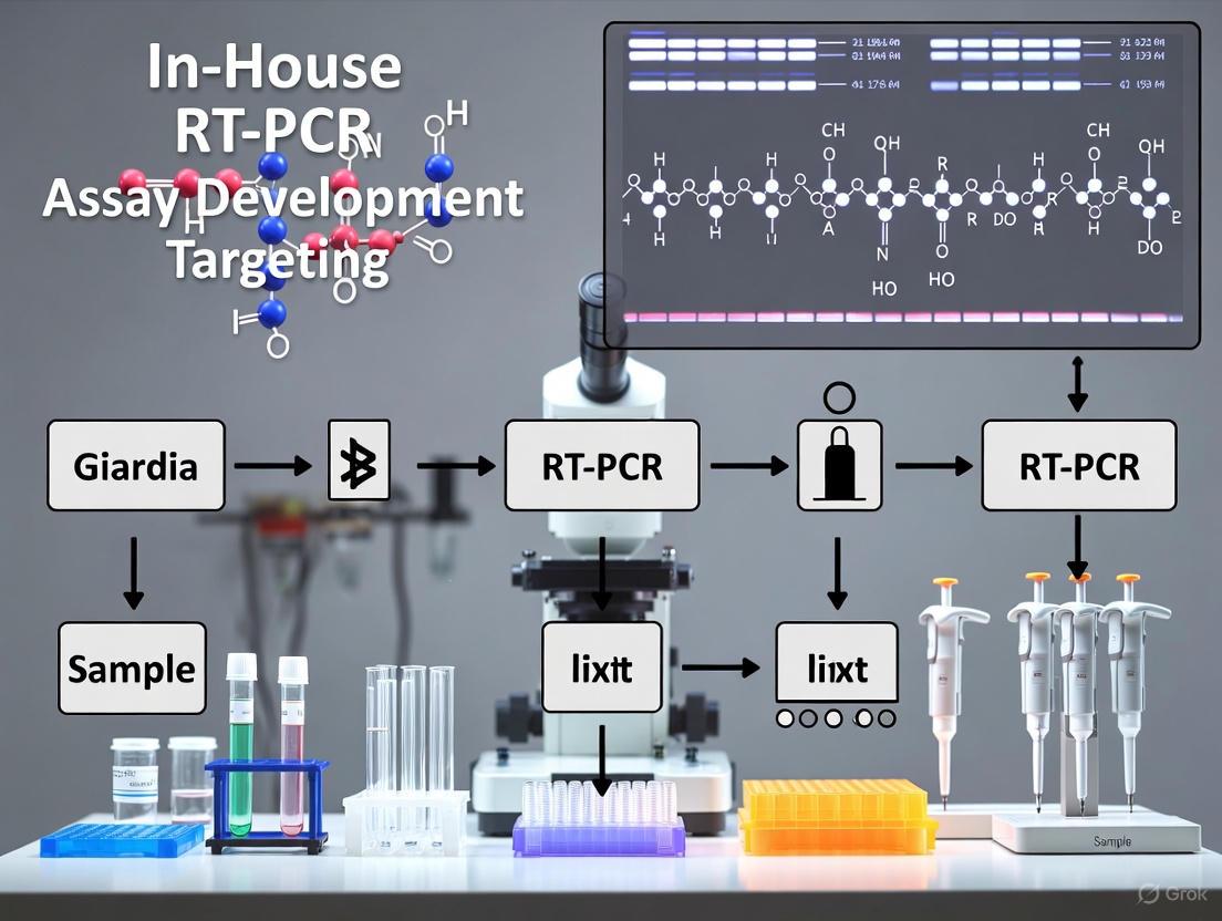
Developing a High-Performance In-House RT-PCR Assay for Giardia duodenalis: A Comprehensive Guide from Design to Validation
This article provides a comprehensive guide for researchers and scientists on the development, optimization, and validation of an in-house real-time PCR (RT-PCR) assay for the detection of Giardia duodenalis.
Developing a High-Performance In-House RT-PCR Assay for Giardia duodenalis: A Comprehensive Guide from Design to Validation
Abstract
This article provides a comprehensive guide for researchers and scientists on the development, optimization, and validation of an in-house real-time PCR (RT-PCR) assay for the detection of Giardia duodenalis. Covering the entire workflow, it explores the critical selection of genetic targets, with evidence highlighting the superior performance of the 18S rRNA gene. It details methodological steps for primer design and reaction setup, offers troubleshooting strategies for common pitfalls like inhibition and suboptimal DNA yield, and establishes a framework for rigorous analytical and clinical validation, including comparison with commercial tests and microscopy. The content synthesizes current research to empower professionals in creating a sensitive, specific, and cost-effective diagnostic tool for both clinical and research settings.
Laying the Groundwork: Giardia Biology and Rationale for In-House Molecular Assays
Global Burden and Epidemiological Significance
Giardia duodenalis (also known as Giardia intestinalis or Giardia lamblia) is a flagellated protozoan parasite recognized as a major cause of diarrheal illness worldwide [1] [2]. This parasite infects the human intestinal tract and causes giardiasis, a disease of significant public health concern.
Global Prevalence and Impact
The global burden of G. duodenalis is substantial, with an estimated 280 million symptomatic human cases occurring annually worldwide [3] [4] [2]. In developing countries, giardiasis affects approximately 20-30% of the population, while in developed countries, it affects about 2-5% [3]. The World Health Organization has included giardiasis in the 'Neglected Disease Initiative' due to its significant disease burden and close association with poverty [3] [4].
G. duodenalis exhibits a cosmopolitan distribution, found in every region of the United States and around the world [5]. In the U.S. alone, more than 1 million people get sick from Giardia each year, making it more common than any other gut parasite [5].
Infection in Nonhuman Mammals
A comprehensive systematic review and meta-analysis of studies published from 1980 to 2023, which analyzed data from nearly 5 million animals across various species and countries, revealed that approximately 13.6% of nonhuman mammals worldwide are infected with Giardia [3] [4]. The highest prevalence rates were observed in rodents (28.0%) and hoofed animals (Artiodactyla, 17.0%), with herbivores, semiaquatic, and wild animals also showing elevated infection rates [4].
Table 1: Global Prevalence of Giardia Infection in Nonhuman Mammals
| Host Category | Prevalence (%) | Notes |
|---|---|---|
| Overall Nonhuman Mammals | 13.6 | Based on analysis of ~5 million animals [4] |
| Rodentia | 28.0 | Highest prevalence among mammalian orders [4] |
| Artiodactyla | 17.0 | Includes wild and domestic ungulates [4] |
| Sheep and Goats | Varies by region | Highest in Europe, lowest in Asia [2] |
| Herbivorous Animals | 17.0 | Higher than carnivorous and omnivorous [4] |
| Semiaquatic Animals | 29.0 | Facilitated by waterborne transmission [4] |
| Wild Animals | 19.0 | Potential source for livestock and humans [4] |
Parasite Biology and Genetic Diversity
Classification and Species
The genus Giardia comprises nine valid species: G. agilis, G. ardeae, G. cricetidarum, G. duodenalis, G. microti, G. muris, G. peramelis, G. psittaci, and G. varani [3] [4]. These species have marked differences in morphological characteristics, host range and specificity, and genetic traits [3].
G. duodenalis is the only Giardia species able to infect humans and is currently regarded as a multispecies complex with eight (A-H) genetic assemblages [3] [4]:
- Assemblages A and B: Primarily found in humans and other mammals (zoonotic)
- Assemblages C and D: Predominantly infect canids
- Assemblage E: Found in wild and domestic ungulates
- Assemblage F: Infects felids
- Assemblage G: Primarily targets rodents
- Assemblage H: Typically identified in marine pinnipeds [3] [2]
Life Cycle and Transmission
The life cycle of G. duodenalis consists of two main stages: the trophozoite and the cyst [1]. The trophozoite is the vegetative, replicating form that attaches to the intestinal wall in the small intestine. As parasites move distally through the intestine, they encyst into environmentally stable cysts that are excreted in feces [1].
Transmission occurs via the fecal-oral route, primarily through ingestion of contaminated water or food, or directly from contact with infected individuals or animals [5]. The infectious dose is remarkably low, with ingestion of as few as 10 cysts sufficient to establish infection in a susceptible host [2]. Infected individuals can excrete up to 10¹Ⱐcysts daily in their feces, contributing significantly to environmental contamination [3] [4].
Pathogenesis and Clinical Manifestations
Clinical Spectrum
Giardiasis presents with a wide spectrum of clinical manifestations, ranging from asymptomatic carriage to acute or chronic diarrheal illness [1]. Symptoms typically begin 1 to 2 weeks after infection and may include:
- Diarrhea (2-5 times per day)
- Gas and abdominal bloating
- Smelly, greasy poop that can float
- Stomach cramps or pain
- Upset stomach or nausea
- Dehydration (loss of fluids) [5]
Acute symptoms generally last for 2 to 6 weeks, but some people experience long-term symptoms that can persist for years [5]. Notably, Giardia has been identified as an independent risk factor for reduced early-life linear growth in children from low- and middle-income countries, even in the absence of overt diarrheal symptoms [6].
Mechanisms of Growth Faltering
Recent research has revealed that Giardia-associated growth impairments occur through mechanisms distinct from traditional inflammatory enteropathy. Unlike other intestinal pathogens that cause linear growth restriction through intestinal or systemic inflammation, Giardia seldom associates with chronic inflammation in children [6].
The proposed pathogenesis involves:
- Dose-dependent linear growth deficits and increased gut permeability independent of intestinal inflammation markers [6]
- Broad amino acid deficiencies in infected children [6]
- Overproduction of specific phenolic acids (byproducts of intestinal bacterial amino acid metabolism) [6]
- Disruption of nutrient metabolism rather than inflammatory enteropathy [6]
This growth faltering appears contingent upon a convergence of the intestinal protozoa with nutritional and intestinal bacterial factors [6].
Diagram 1: Giardia Pathogenesis Pathway. The diagram illustrates the non-inflammatory pathway through which Giardia infection leads to growth impairment, primarily via disruption of nutrient metabolism and amino acid absorption rather than through typical inflammatory responses.
Molecular Detection and Diagnostic Protocols
Molecular methods, particularly PCR-based assays, have surpassed traditional microscopy for Giardia detection due to superior sensitivity and reduced investigator dependence [7]. For resource-rich non-endemic settings, sensitivity and specificity of microscopy for Giardia diagnosis in human stool samples have been estimated at 72% and 99%, respectively, while various real-time PCR assays demonstrate sensitivities ranging from 90% to >99% and specificities from 76% to virtually 100% [7].
Real-Time PCR Screening Assays
Comparative studies have evaluated real-time PCR assays targeting different genes of G. duodenalis:
- 18S rRNA gene-specific assay: 100% sensitivity and specificity [7]
- gdh (glutamate dehydrogenase) gene-specific assay: 17.5% sensitivity and 92.3% specificity [7]
- bg (beta-giardin) gene-specific assay: 31.7% sensitivity and 100% specificity [7]
For assemblage discrimination, bg gene-specific assays without locked nucleic acids (LNA) demonstrated 100% sensitivity and specificity for both assemblages A and B [7].
Detailed Protocol: Real-Time PCR for Gi Detection and Assemblage Discrimination
Methodology for Screening and Differentiation of G. duodenalis Assemblages A and B
Reagents and Equipment:
- QIAamp DNA Stool Mini Kit (Qiagen) or equivalent for DNA extraction
- Primers and TaqMan probes specific for target genes (18S rRNA, bg, tpi)
- Real-time PCR instrument (RotorGene Q, MIC, or equivalent)
- PCR-grade water
- Positive control plasmids containing target sequences
- Appropriate reaction mix for probe-based qPCR
Procedure:
Nucleic Acid Extraction
- Extract genomic DNA from 200 mg stool samples using commercial kit
- Include inhibition control (e.g., Phocid herpes virus DNA) to monitor PCR inhibition
- Elute DNA in 100-200 μL elution buffer
- Store extracted DNA at -20°C until analysis
Screening PCR Setup
- Prepare reaction mix containing:
- 1X TaqMan Universal Master Mix
- 900 nM forward and reverse primers
- 200 nM TaqMan probe
- 5 μL template DNA
- PCR-grade water to 25 μL total volume
- Run amplification with cycling conditions:
- 95°C for 10 min (initial denaturation)
- 45 cycles of 95°C for 15 sec and 60°C for 1 min
- Prepare reaction mix containing:
Assemblage Discrimination PCR
- For positive samples, perform duplex real-time PCR targeting assemblage-specific regions of bg gene
- Use two probe channels with different fluorophores for simultaneous detection of assemblages A and B
- Apply same cycling conditions as screening PCR
Analysis and Interpretation
- Determine cycle threshold (Ct) values for samples and controls
- Samples with Ct ≤ 40 are considered positive
- For assemblage discrimination, analyze fluorescence in respective channels to assign to assemblage A or B
Table 2: Diagnostic Performance of Real-Time PCR Targets for G. duodenalis
| Target Gene | Assay Type | Sensitivity (%) | Specificity (%) | Notes |
|---|---|---|---|---|
| 18S rRNA | Screening | 100 | 100 | Recommended for primary screening [7] |
| Beta-giardin (bg) | Screening | 31.7 | 100 | Lower sensitivity but high specificity [7] |
| Glutamate dehydrogenase (gdh) | Screening | 17.5 | 92.3 | Lowest sensitivity among screening targets [7] |
| bg (without LNA) | Assemblage discrimination | 100 | 100 | For both A and B assemblages [7] |
| Triose phosphate isomerase (tpi) | Assemblage discrimination | 82.1-100 | 97.8-100 | Variable performance between assemblages [7] |
Alternative Detection Methods
Loop-mediated isothermal amplification (LAMP) has emerged as a sensitive, specific, and rapid method for Giardia detection with faster turnaround times than nested PCR and lower risk of cross-contamination [8]. The EF1α LAMP assay can detect as few as 10 Giardia cysts spiked on leafy greens, making it suitable for food safety testing and environmental surveillance [8].
For food safety applications, an optimized washing procedure with 0.1% Alconox solution followed by LAMP detection provides effective recovery and identification of Giardia cysts from leafy greens, with detection rates of 19/20 (95%) for samples spiked with 100 cysts [8].
Diagram 2: Giardia Molecular Detection Workflow. The flowchart outlines the stepwise procedure for screening and genotyping Giardia duodenalis, from sample collection through final interpretation, highlighting the two-stage process of initial screening followed by assemblage discrimination for positive samples.
Research Reagent Solutions
Table 3: Essential Research Reagents for Giardia Molecular Detection
| Reagent/Category | Specific Examples | Function/Application |
|---|---|---|
| DNA Extraction Kits | QIAamp DNA Stool Mini Kit, QIAamp DNA Fast Stool Mini Kit | Efficient isolation of inhibitor-free DNA from complex matrices [7] [9] |
| Real-Time PCR Master Mix | TaqMan Universal Master Mix | Probe-based detection providing specific amplification with real-time monitoring [7] [9] |
| Target-Specific Primers/Probes | 18S rRNA, bg, gdh, tpi gene targets | Specific detection and genotyping of G. duodenalis assemblages [7] |
| Positive Control Plasmids | Recombinant plasmids containing target sequences (Emh, Gla, CryP) | Quality control, standard curves, and determination of assay limits [9] |
| Inhibition Control | Phocid herpes virus (PhHV) DNA | Monitoring PCR inhibition in sample extracts [7] |
| LAMP Reagents | Bst polymerase, EF1α primers | Isothermal amplification for resource-limited settings or rapid screening [8] |
Accurate detection of the protozoan parasite Giardia duodenalis (also known as G. lamblia or G. intestinalis) is fundamental to research efforts aimed at understanding its epidemiology, pathogenesis, and drug development. For decades, diagnosis has relied primarily on microscopic examination and, more recently, antigen-based tests. While these methods have served a purpose, their limitations are increasingly significant in the context of advanced research. This application note articulates the scientific case for adopting molecular diagnostics, specifically in-house reverse transcription polymerase chain reaction (RT-PCR) assays, for Giardia research. We detail the performance shortcomings of traditional methods, present quantitative comparative data, and provide detailed protocols for implementing robust, sensitive, and specific molecular assays that can discriminate between genetically distinct Giardia assemblages.
Performance Limitations of Traditional Diagnostic Methods
Microscopy: The Established but Flawed Gold Standard
Microscopic examination of stool specimens remains the most common diagnostic method for Giardia, particularly in resource-limited settings [10] [11]. However, this technique suffers from several critical drawbacks:
- Low and Variable Sensitivity: A direct comparison with PCR revealed that microscopy for G. lamblia had a sensitivity of only 64.4% and a specificity of 86.6%, with a Cohen's kappa value of 0.51, indicating only moderate agreement with the molecular gold standard [10]. A systematic review further confirmed that microscopic methods typically exhibit sensitivities between 60% and 89% [11].
- Operator Dependence: The accuracy of microscopy is highly dependent on the skill and experience of the microscopist, leading to subjectivity and potential misdiagnosis [10] [9].
- Inability to Discern Assemblages: Crucially for research, microscopy cannot differentiate between the genetically distinct assemblages of Giardia (e.g., A and B), which have potential differences in zoonotic transmission and virulence [12] [13]. It also cannot distinguish Giardia from other non-pathogenic protozoa or degenerated cellular debris [10].
Antigen Tests: Rapid but Lacking Genotypic Information
Immunological methods, such as Enzyme-Linked Immunosorbent Assays (ELISAs) and Rapid Diagnostic Tests (RDTs), detect Giardia-specific antigens like GSA-65 [14]. While these tests are faster and easier to perform than microscopy, they have significant limitations:
- Qualitative Nature and False Results: Most available immunoassays are qualitative and lack the sensitivity to detect low-level infections [12]. False positives and false negatives have been reported, with some commercial kits even being recalled [12] [15].
- Persistence Post-Treatment: Antigen tests may remain positive after successful treatment because they detect non-viable parasites or soluble antigens, making them unsuitable for monitoring treatment efficacy [14].
- No Genotyping Capability: Like microscopy, antigen tests cannot differentiate between the different assemblages of G. duodenalis, a critical shortcoming for molecular epidemiological studies and transmission tracking [12].
Table 1: Comparative Performance of Diagnostic Methods for Giardia duodenalis
| Method | Sensitivity (%) | Specificity (%) | Genotyping Capability | Key Limitations |
|---|---|---|---|---|
| Microscopy | 64.4 - 89 [10] [11] | 86.6 - 100 [10] [11] | No | Low sensitivity, operator-dependent, cannot distinguish assemblages |
| Antigen Tests (ELISA) | ~98 (kit-dependent) [14] | ~98 (kit-dependent) [14] | No | Cannot distinguish assemblages, may stay positive after treatment |
| PCR (18S rRNA target) | 100 [13] | 100 [13] | Yes (with specific assays) | High cost, requires specialized equipment and expertise |
| PCR (gdh target) | 17.5 [13] | 92.3 [13] | Yes | Inconsistent performance across different genetic targets |
| PCR (bg target) | 31.7 - 90 [16] [13] | 100 [13] | Yes | Inconsistent performance across different genetic targets |
The Molecular Solution: In-House RT-PCR Assay Development
Molecular techniques, particularly PCR and real-time PCR (qPCR), overcome the limitations of traditional methods by offering superior sensitivity, specificity, and the ability to genotype. The development of an in-house RT-PCR assay provides researchers with full control over the reaction parameters, primer/probe design, and the flexibility to adapt the protocol for multiplexing or detecting viable parasites.
Critical Considerations for Assay Design
Selection of Genetic Targets
The choice of target gene profoundly influences the assay's sensitivity, specificity, and application. The following genes are commonly used:
- 18S rRNA Gene: This gene is present in multiple copies, conferring high analytical sensitivity. Assays targeting this gene are excellent for screening and have demonstrated 100% sensitivity and specificity in comparative studies [13].
- Beta-Giardin (bg) Gene: A single-copy gene that allows for effective genotyping. However, its sensitivity as a screening tool can be variable, reported as low as 31.7% in some assessments [13].
- Triose Phosphate Isomerase (tpi) Gene: Another single-copy gene frequently used for genotyping assemblages A and B with high specificity [13].
- Glutamate Dehydrogenase (gdh) Gene: Also used for genotyping, though it may exhibit lower sensitivity for primary screening [10] [13].
For a comprehensive approach, a tandem protocol using the 18S rRNA gene for highly sensitive screening, followed by a bg or tpi-specific assay for genotyping of positive samples, is recommended.
Viability Assessment via mRNA Detection
A significant advantage of RT-PCR is its ability to detect mRNA, which is labile and rapidly degraded in non-viable parasites. This can be used to infer cyst viability and potential infectivity.
- Heat Induction Method: Viable Giardia cysts subjected to a heat shock (e.g., 45°C for 20 minutes) will undergo de novo mRNA synthesis of heat shock proteins (e.g., hsp70) or the beta-giardin gene. This nascent mRNA can be detected by qRT-PCR, confirming viability [17].
- Integrated Cell Culture-RT-PCR (ECC-RT-PCR): This more advanced method involves exposing mammalian cell monolayers (e.g., Caco-2) to excysted trophozoites. The subsequent attachment of trophozoites, changes in transcellular resistance, and expression of variant-specific protein (VSP) mRNA are correlated with viability and infectivity [18].
Experimental Protocol: A Detailed Workflow for a Duplex RT-PCR Assay
This protocol outlines the steps for a duplex real-time PCR assay capable of simultaneously detecting G. duodenalis and discriminating between the human-pathogenic assemblages A and B, based on established methodologies [9] [13].
Figure 1: Workflow for a duplex real-time PCR assay for Giardia detection and genotyping.
Step 1: Sample Collection and DNA Extraction
- Sample Collection: Collect fresh stool samples. For preserved samples, use sodium acetate-acetic acid-formalin (SAF) or other suitable preservatives. For viability testing, process samples immediately or use appropriate RNA-stabilizing buffers.
- DNA Extraction: Use commercial kits designed for stool samples, such as the QIAamp DNA Stool Mini Kit (Qiagen) or the MagNA Pure 96 System (Roche), which effectively remove PCR inhibitors [10] [12] [15].
- Optional Cyst Disruption: To optimize DNA yield from hardy cysts, subject the sample to three cycles of freezing in liquid nitrogen and thawing at 70-95°C prior to extraction [10].
- Inhibition Control: Include an internal control, such as Phocid herpes virus (PhHV) DNA, spiked into the sample to detect PCR inhibition [13].
Step 2: Primer and Probe Design
Design specific primers and TaqMan probes for the chosen genetic targets. The following sequences serve as examples:
- Screening Probe (18S rRNA): Use a conserved region to detect all G. duodenalis.
- Genotyping Probes (bg gene): Design assemblage-specific probes. For example:
- Assemblage A-Specific Probe: Label with 6-FAM fluorophore.
- Assemblage B-Specific Probe: Label with HEX/VIC fluorophore.
- Primers: A single primer set can often be used to amplify a region encompassing the variable sequences targeted by the assemblage-specific probes [17] [13].
Table 2: Research Reagent Solutions for Giardia Duplex RT-PCR
| Reagent / Material | Function / Application | Example Product / Specification |
|---|---|---|
| QIAamp DNA Stool Mini Kit | Efficient DNA extraction from complex stool matrices; removes PCR inhibitors. | Qiagen [10] [12] |
| TaqMan Universal PCR Master Mix | Provides optimized buffer, enzymes, dNTPs for probe-based qPCR. | Applied Biosystems [15] |
| Custom TaqMan Probes | Assemblage-specific detection; 6-FAM for Assemblage A, HEX for Assemblage B. | Synthesized by commercial providers [9] |
| PUC19 Plasmid with Cloned Inserts | Quantitative standard for determining copy number and assay validation. | Contains target bg or 18S rRNA gene fragments [9] |
| ABI 7900HT Real-Time PCR System | Instrument platform for running qPCR and analyzing fluorescence data. | Applied Biosystems [15] |
Step 3: Duplex Real-Time PCR Setup and Amplification
- Reaction Composition:
- 2x TaqMan Universal PCR Master Mix: 12.5 µL
- Forward Primer (bg or 18S rRNA target, 10 µM): 0.9 µL
- Reverse Primer (bg or 18S rRNA target, 10 µM): 0.9 µL
- Assemblage A-specific Probe (10 µM): 0.25 µL
- Assemblage B-specific Probe (10 µM): 0.25 µL
- Template DNA: 5 µL
- Nuclease-free water: to a final volume of 25 µL
- Thermocycling Conditions:
- Initial Denaturation: 95°C for 10 minutes
- 45 Cycles of:
- Denaturation: 95°C for 15 seconds
- Annealing/Extension: 60°C for 1 minute
Step 4: Data Analysis and Interpretation
- Cycle Threshold (Ct): A positive signal is typically defined by a Ct value below a predetermined cutoff (e.g., 40).
- Genotyping: A sample is assigned to Assemblage A if only the FAM channel fluoresces, Assemblage B if only the HEX/VIC channel fluoresces, or a mixed infection if both channels produce a signal.
- Quantification: Generate a standard curve using serial dilutions of a plasmid containing the target gene sequence to determine the parasite load in unknown samples [9].
Microscopy and antigen testing, while historically valuable, present significant limitations for advanced Giardia research, including poor sensitivity, an inability to determine viability, and a complete lack of genotyping resolution. The adoption of in-house RT-PCR assays provides a powerful alternative, offering unparalleled sensitivity and the critical ability to discriminate between the genetically distinct assemblages of G. duodenalis. The detailed protocols and considerations outlined in this application note provide a robust foundation for researchers to implement these superior molecular tools, thereby enhancing the quality and depth of giardiasis research, from basic biology to drug development and molecular epidemiology.
This application note delineates the core advantages of employing in-house Real-Time PCR (RT-PCR) assays for the detection and analysis of Giardia duodenalis, a significant human intestinal protozoan pathogen. While commercial PCR kits offer standardized protocols, in-house assays provide researchers and drug development professionals with unparalleled customization, cost-effectiveness, and flexibility, which are critical for advanced molecular epidemiological studies and assay optimization. Framed within a broader thesis on in-house RT-PCR development, this document provides a comparative performance analysis and detailed experimental protocols to guide assay implementation.
The diagnosis of Giardia duodenalis and other intestinal protozoa is transitioning from traditional microscopic methods to molecular techniques, primarily due to the latter's superior sensitivity and specificity [19] [20]. Clinical laboratories often face a choice between commercial molecular diagnostic tests and laboratory-developed in-house assays. Commercial kits provide a streamlined, validated solution for routine screening. However, for research purposes—such as investigating genetic diversity, tracking outbreaks, or developing novel detection methods—in-house assays present significant benefits. They allow for the customization of sample processing, selection of target genes relevant to specific research questions, and can be more cost-effective for high-throughput or specialized studies [19] [12]. This flexibility is paramount for advancing our understanding of Giardia pathogenicity and transmission dynamics.
Performance Comparison: In-House vs. Commercial Assays
The table below summarizes key findings from recent studies comparing the performance of in-house and commercial RT-PCR assays for detecting intestinal protozoa, including Giardia duodenalis.
Table 1: Comparative Performance of Molecular Assays for Giardia duodenalis and Other Intestinal Protozoa
| Study Focus | Assay Type | Key Performance Findings | Reference |
|---|---|---|---|
| Multicentre comparison in Italy | Commercial RT-PCR (AusDiagnostics) vs. In-house RT-PCR | Complete agreement for G. duodenalis detection; both showed high sensitivity and specificity comparable to microscopy. | [19] |
| Evaluation of a commercial multiplex PCR | Allplex GI-Parasite Assay | Sensitivity: 100%, Specificity: 99.2% for G. duodenalis. Demonstrates high accuracy of modern commercial kits. | [20] |
| Screening PCR target gene comparison | In-house assays (different targets) | 18S rRNA assay: 100% sensitivity & specificity.beta-giardin (bg) assay: 31.7% sensitivity, 100% specificity.gdh assay: 17.5% sensitivity, 92.3% specificity. | [7] |
| Assemblage discrimination (A vs. B) | In-house assays (different targets) | bg gene assay (without LNA): 100% sensitivity & specificity for both assemblages.tpi gene assay: 100% sensitivity, 97.8% specificity for A; 82.1% sensitivity, 100% specificity for B. | [7] |
The data reveals that well-designed in-house assays can perform on par with, and in some aspects surpass, commercial alternatives. The critical importance of target gene selection is evident, with the 18S rRNA gene providing exceptional sensitivity for screening, while the beta-giardin gene allows for reliable genotyping [12] [7]. This choice is a key area where in-house customization provides a direct research advantage.
Experimental Protocol: In-House RT-PCR forGiardia duodenalis
This protocol outlines a validated method for the detection and genotyping of Giardia duodenalis from human stool specimens, synthesizing robust methodologies from the literature.
Sample Collection and DNA Extraction
Materials:
- Fresh or preserved (e.g., in Para-Pak media or 2.5% potassium dichromate) stool samples [19] [21].
- Lysis buffer (e.g., ASL Buffer from Qiagen or ATL Buffer from DNeasy kit) [12] [20].
- Commercial DNA extraction kit (e.g., QIAamp DNA Stool Mini Kit [7] or E.Z.N.A. Stool DNA Kit [21]).
- Proteinase K.
- Microcentrifuge and water bath or incubator.
Workflow:
- Homogenization: Suspend approximately 180-200 mg of stool specimen in 1-1.5 mL of lysis buffer. Vortex thoroughly to create a homogeneous suspension [20] [21].
- Incubation: Add Proteinase K (e.g., 40 µL) and incubate at 55°C for a minimum of 4 hours, or overnight, to ensure complete lysis of hardy (oo)cysts [12].
- Inhibition Removal: Centrifuge the lysate at full speed (e.g., 14,000 rpm) for 2-5 minutes to pellet stool debris and inhibitors. Use the supernatant for extraction [20].
- Nucleic Acid Extraction: Follow the manufacturer's instructions for the chosen commercial silica-column-based kit. Automated systems like the Microlab Nimbus can also be employed for high throughput [20].
- Elution: Elute purified DNA in 50-100 µL of the provided elution buffer or nuclease-free water. Store at -20°C or -80°C until PCR analysis.
RT-PCR Amplification for Detection and Genotyping
Materials:
- TaqMan Fast Universal PCR Master Mix (2X) [19].
- Primers and dual-labeled hydrolysis probes (FAM/HEX, etc.) for target genes.
- Nuclease-free water.
- PCR plates and a calibrated real-time PCR instrument.
Primer and Probe Selection: The selection of target genes dictates the assay's application:
- Screening (High Sensitivity): Target the 18S rRNA gene [7].
- Genotyping (Assemblage A/B Discrimination): Target the beta-giardin (bg) or triose phosphate isomerase (tpi) genes [12] [7]. Probes can be designed to exploit single nucleotide polymorphisms (SNPs) between assemblages.
Table 2: Research Reagent Solutions for Giardia RT-PCR
| Reagent / Tool | Function / Rationale | Example / Specification |
|---|---|---|
| Silica-column DNA Kits | Efficiently purifies DNA while removing PCR inhibitors common in stool. | QIAamp DNA Stool Mini Kit, E.Z.N.A. Stool DNA Kit [7] [21] |
| TaqMan Master Mix | Provides enzymes, dNTPs, and optimized buffer for robust probe-based qPCR. | TaqMan Fast Universal PCR Master Mix (2X) [19] |
| 18S rRNA Primers/Probe | High-copy target for maximum analytical sensitivity in screening assays. | [7] |
| beta-giardin (bg) Primers/Probe | Well-conserved single-copy gene suitable for sensitive detection and genotyping. | Primers: GiarF/GiarR; Probe: GiarP [22] [12] |
| Locked Nucleic Acid (LNA) Probes | Increases probe binding affinity, improving mismatch discrimination for genotyping. | Can be used in assemblage-specific probes [7] |
Reaction Setup: Prepare a 25 µL reaction mixture as follows [19] [22]:
- 12.5 µL of TaqMan Master Mix (2X)
- Forward and Reverse Primers (e.g., 0.32 µM each)
- Probe (e.g., 0.12 µM)
- 5 µL of template DNA
- Nuclease-free water to 25 µL
Amplification Protocol: Run the reaction on a real-time PCR cycler using the following cycling conditions [22] [12]:
- Initial Denaturation: 95°C for 5-10 minutes.
- Amplification (45 cycles):
- Denature: 95°C for 10-15 seconds.
- Anneal/Extend: 60°C for 30-60 seconds (acquire fluorescence at this step).
Analysis:
- A positive test result is typically defined as a cycle threshold (Ct) value of less than 45 [20].
- For genotyping, use assays specific for assemblages A and B, potentially in a multiplex format, to determine the presence of one or both assemblages [12] [7].
Workflow and Strategic Application
The following diagram illustrates the strategic decision-making process and experimental workflow for implementing an in-house RT-PCR assay for Giardia research.
The development and implementation of in-house RT-PCR assays for Giardia duodenalis provide a powerful, flexible tool for the research community. The principal advantages are:
- Customization: Researchers can tailor every aspect of the assay, from sample pre-treatment and DNA extraction methods to the selection of target genes and reaction conditions, specific to their research needs [19] [7]. This is indispensable for genotyping, investigating new genetic markers, or adapting the protocol to challenging sample matrices.
- Cost-Effectiveness: For high-volume screening or surveillance studies, in-house assays can be significantly less expensive per sample than recurring purchases of commercial kits, without compromising performance [19].
- Flexibility: The protocol can be rapidly modified to incorporate new findings, such as novel genetic targets or improved probe chemistries like locked nucleic acids (LNA) for better genotype discrimination [7].
In conclusion, while commercial kits serve an important role in standardized clinical diagnostics, in-house RT-PCR assays are the cornerstone of innovative Giardia research. The ability to control and optimize the entire workflow enables scientists to address complex questions in parasitology and drug development that are beyond the scope of off-the-shelf solutions.
A Step-by-Step Protocol: From Primer Design to Amplification
The development of robust in-house RT-PCR assays is a critical skill for researchers focusing on enteric pathogens like Giardia duodenalis (also known as Giardia lamblia or Giardia intestinalis). Effective molecular diagnostics hinge on the precise in silico design of primers and probes, which ensures high sensitivity and specificity while reducing the need for extensive empirical optimization [23]. This protocol details a comprehensive bioinformatics workflow for designing and validating oligonucleotides for hydrolysis probe-based (TaqMan) RT-qPCR assays, with specific application to Giardia research. The principles outlined are derived from current methodologies successfully applied in parasitic disease diagnostics [24] [23] [25].
A properly designed assay allows for the specific detection of Giardia genetic material, even in complex samples like stool or environmental water, facilitating accurate assessment of infection burden and enabling high-throughput screening essential for epidemiological studies and drug efficacy trials [23].
Core Design Parameters and Principles
Fundamental Oligonucleotide Properties
Successful in silico design requires adherence to established thermodynamic and sequence-composition rules for primers and probes [26].
Table 1: Optimal Design Parameters for Primers and TaqMan Probes
| Parameter | Primers | Hydrolysis (TaqMan) Probes |
|---|---|---|
| Length | 18–30 bases [26] | 18–30 bases; can be extended with internal quenchers (e.g., ZEN/TAO) [26] |
| Melting Temperature (Tm) | 60–64°C; ideal is 62°C [26] | 65–75°C; 5–10°C higher than primers [24] [26] |
| GC Content | 35–65%; ideal is 50% [26] | 35–65% [26] |
| Tm Difference (Forward vs. Reverse) | ≤ 2°C [24] [26] | - |
| Annealing Temperature (Ta) | 3–5°C below the primer Tm [27] [28] | - |
| Amplicon Length | 70–150 bp (ideal); up to 400 bp is acceptable [24] [26] | - |
| 3' End | Avoid stretches of 3 or more G/Cs; avoid secondary structures and complementarity [26] | Avoid a G at the 5' end [26] |
Target Sequence Selection forGiardia
For Giardia assay development, select a conserved genomic region. Common targets include genes coding for surface proteins (e.g., VSP), the β-giardin gene, the glutamate dehydrogenase (gdh) gene, or the triosephosphate isomerase (tpi) gene. To avoid false positives from genomic DNA contamination when working with RNA, design assays to span an exon-exon junction [26]. Retrieve multiple target gene sequences for Giardia duodenalis from public databases (e.g., GenBank) to perform a robust multiple sequence alignment (MSA) and identify conserved regions suitable for primer and probe binding.
Experimental Protocols for In Silico Design and Validation
Protocol: Primer and Probe Design Workflow
Step 1: Identify Conserved Target Region
- Procedure: Using software like Geneious or the MAFFT algorithm, align multiple Giardia target gene sequences (e.g., β-giardin) obtained from GenBank [25]. Manually inspect the alignment to identify a ~100-150 bp region with 100% sequence conservation across all isolates of interest.
- Rationale: Targeting a conserved region is paramount for a robust assay that can detect different Giardia strains and avoids false negatives due to sequence variations [29].
Step 2: Design Primer Pairs
- Procedure: Input the selected target sequence into a design tool like IDT's PrimerQuest or Primer3Plus. Set the parameters according to Table 1. The tool will generate several candidate primer pairs. Select a pair where the forward and reverse primers have closely matched Tm values (within 2°C) [24] [26].
- Rationale: Primers with mismatched Tms can lead to inefficient and non-specific amplification, as one primer may bind optimally while the other does not [30].
Step 3: Design the Hydrolysis Probe
- Procedure: Using the same design tool, specify the need for a hydrolysis probe. The probe should be designed to bind between the forward and reverse primer binding sites without overlapping them. Ensure its Tm is 5–10°C higher than the primers' Tm. For double-quenched probes, specify an internal quencher (e.g., ZEN) to lower background fluorescence [26].
- Rationale: A higher Tm ensures the probe is fully bound during the primer annealing and extension phases, allowing for efficient cleavage and fluorescence emission [26].
Protocol: Specificity and Secondary Structure Analysis
Step 4: In Silico Specificity Check (BLAST)
- Procedure: Perform a Nucleotide BLAST (BLASTN) search for each oligonucleotide sequence against the entire Homo sapiens (taxid:9606) and other relevant genomes (e.g., gut microbiome) using the NCBI web tool, which can be accessed directly from IDT's OligoAnalyzer [24] [26]. The primers and probe must be 100% complementary only to the intended Giardia target. Any significant off-target matches require a redesign.
- Rationale: This step is critical to ensure the assay does not amplify non-target DNA, which would lead to false-positive results [25].
Step 5: Secondary Structure and Dimer Analysis
- Procedure: Use IDT's OligoAnalyzer Tool to analyze each primer and the probe for hairpins, self-dimers, and cross-dimers (heterodimers between forward/reverse primers and probe). The ΔG value for any predicted structure should be weaker (more positive) than -9.0 kcal/mol [26]. Use the Thermo Fisher Scientific Multiple Primer Analyzer to check for cross-dimerization between all oligonucleotides in the mix.
- Rationale: Stable secondary structures or dimers can prevent primers and probes from binding to the template, drastically reducing or even preventing amplification [26].
Step 6: In Silico PCR Validation
- Procedure: Use the UCSC In Silico PCR tool to check if the primer pair amplifies a single, correctly sized product from the reference human genome and other potentially contaminating genomes. The tool should return no products for a specific assay [24].
- Rationale: This is a final check to rule out the amplification of non-specific products from the host or common contaminants that might be present in the sample [24].
Melting Temperature (Tm) Calculation and Optimization
Advanced Tm Calculation Method
The most accurate Tm predictions use the SantaLucia nearest-neighbor method [27]. This method accounts for the sequence context of each base pair, terminal effects, and precise salt corrections, achieving accuracy within 1–2°C of experimental values.
Table 2: Reaction Conditions for Accurate Tm Calculation in Tm Calculators
| Condition | Standard PCR/qPCR Value | Notes |
|---|---|---|
| Na⺠Concentration | 50 mM | If buffer contains Kâº, use the total monovalent cation concentration [27]. |
| Mg²⺠Concentration | 1.5–3.0 mM | Varies by polymerase buffer; 3–5 mM is common for qPCR [26] [27]. |
| Oligo Concentration | 0.25 µM (primers) | Standard for most assays; can be optimized from 0.1–0.5 µM [27]. |
| dNTP Concentration | 0.2–0.8 mM | dNTPs chelate Mg²âº; the calculator should account for this [24] [26]. |
| Additives (DMSO) | 0–10% | Reduces Tm by ~0.6°C per 1%; useful for GC-rich targets [27]. |
Protocol: Using an Online Tm Calculator
- Procedure: Access a sophisticated calculator such as the Oligopool Tm Calculator or IDT's OligoAnalyzer. Input the oligonucleotide sequence. Set the salt and additive conditions to match your planned reaction buffer (see Table 2). Run the calculation and record the Tm.
- Optimization: If the calculated Tm for a primer is outside the 58–65°C range, adjust the primer length. To increase Tm, add bases to the 5' end; to decrease it, shorten the primer from the 5' end. Recalculate until all parameters fall within the optimal ranges [27].
The Scientist's Toolkit: Research Reagent Solutions
Table 3: Essential Reagents and Software for In Silico Assay Development
| Item | Function/Description | Example Providers/Versions |
|---|---|---|
| Oligo Design Software | Designs primers/probes based on input parameters and sequence. | IDT PrimerQuest, Primer3, Primer Express [26] [31] |
| Sequence Alignment Tool | Identifies conserved regions across multiple target sequences. | MAFFT, Geneious, MEGA [25] |
| Tm & Structure Analyzer | Calculates Tm and analyzes secondary structures/dimers. | IDT OligoAnalyzer, Thermo Fisher Multiple Primer Analyzer [26] |
| Specificity Validation Tool | Checks for unique binding of oligonucleotides in a genome. | NCBI Nucleotide BLAST, UCSC In Silico PCR [24] [26] |
| Universal PCR Master Mix | Allows use of a universal 60°C annealing temp, simplifying optimization. | Platinum DNA Polymerases (Thermo Fisher) [30] |
| Double-Quenched Probes | Reduces background fluorescence, improving signal-to-noise ratio. | Probes with ZEN/TAO internal quenchers (IDT) [26] |
| 2-Iodothiophenol | 2-Iodothiophenol|CAS 37972-89-7|Aromatic Building Block | |
| 4-n-Propylthiophenol | 4-n-Propylthiophenol – For Research Use |
Troubleshooting Common In Silico Design Issues
When issues are identified during the in silico phase, they must be addressed before proceeding to costly synthesis and wet-lab testing. Common problems and their solutions include:
- Suboptimal Tm: Primers with Tm values below 55°C or above 70°C should be redesigned. Adjust the length of the primer, typically by adding or removing bases from the 5' end, to fine-tune the Tm into the optimal 58–62°C range [27].
- Secondary Structures and Dimers: If analysis reveals significant hairpins or dimer formation (ΔG < -9.0 kcal/mol), the oligonucleotide sequence must be redesigned to eliminate self-complementary regions [26].
- Off-Target BLAST Hits: If BLAST analysis shows significant complementarity to non-target sequences, the assay specificity is compromised. Return to the multiple sequence alignment and select an alternative, unique target region for primer and probe design [25].
The development of a robust in-house reverse transcription polymerase chain reaction (RT-PCR) assay for Giardia lamblia (also known as G. duodenalis or G. intestinalis) presents a significant challenge for researchers and drug development professionals. The primary obstacle lies in the efficient extraction of high-quality DNA from the parasite's robust cyst wall, which is resistant to environmental factors and conventional lysis methods. This resistance, combined with the presence of PCR inhibitors in stool specimens, can severely compromise assay sensitivity and reliability [32] [33]. The cyst wall protects the parasite from harsh conditions but also acts as a formidable barrier to nucleic acid extraction, making its effective disruption a critical first step in any molecular detection protocol. This application note details the key considerations and optimized protocols for overcoming these challenges to ensure successful downstream RT-PCR analysis.
Comparative Analysis of DNA Extraction Method Efficiencies
Evaluating the efficiency of DNA extraction methods is paramount for successful Giardia detection. Research directly comparing different approaches reveals significant variations in DNA yield, purity, and subsequent PCR performance. The following table summarizes the key findings from comparative studies:
Table 1: Comparison of DNA Extraction Method Efficiencies for Giardia duodenalis Cysts
| Method Category & Name | Key Steps / Description | Reported DNA Concentration (mean) | Reported Purity (A260/280 ratio) | PCR Success Rate / Sensitivity | Primary Application Context |
|---|---|---|---|---|---|
| Mechanical Lysis (Method II) [32] | Crushed cover glass + TAE buffer, shaking, boiling | Highest (P<0.01) | 1.0 (P<0.01) | Not explicitly stated | Human fecal samples |
| Mechanical Lysis (Method I) [32] | Crushed cover glass, vortex, boil, freeze-thaw (6x) | Lowest (0.4) | 1.9 (P<0.01) | Not explicitly stated | Human fecal samples |
| Phenol-Chloroform Isoamyl Alcohol (PCI) [34] | Conventional organic extraction | Most concentrated DNA | Best A260/230 purity | 70% (SSU rRNA gene target) | Human fecal specimens |
| QIAamp DNA Stool Mini Kit [34] | Commercial silica-column based kit | Lower concentration than PCI | Best A260/280 purity | 60% (SSU rRNA gene target) | Human fecal specimens |
| Glass Beads + PCI [33] | Bead beating followed by organic extraction | Not specified | Not specified | ~85% (gdh gene target) | Human fecal samples |
| Freeze-Thaw + Glass Beads + QIAamp Kit [33] | Combined physical and kit-based method | Not specified | Not specified | ~95% (gdh gene target) | Human fecal samples |
The data indicates that methods incorporating vigorous mechanical disruption, such as using crushed cover glass or glass beads, are highly effective for breaking the resilient cyst wall [32]. Furthermore, combining physical disruption methods like bead beating and freeze-thaw cycles with commercial kit chemistry appears to offer a superior balance of high DNA yield and removal of PCR inhibitors, resulting in the highest reported PCR sensitivity [33].
Detailed Experimental Protocols for Cyst Disruption and DNA Extraction
Based on the evaluated literature, the following protocols are recommended for efficient DNA extraction from Giardia cysts. These can be adapted for use prior to your in-house RT-PCR assay development.
This protocol is designed for maximum cyst wall breakage.
- Sample Preparation: Begin with 200 µL of purified Giardia cyst suspension. Cyst purification can be achieved through sucrose flotation techniques (e.g., using a 0.85 M sucrose solution) [32] [34].
- Mechanical Lysis:
- Add 200 mg of sterilized, crushed cover glass (0.4–0.5 mm) and 200 µL of TAE buffer (40 mM Tris-Acetate, 1 mM EDTA, pH ~8.3) to the sample.
- Mix the sample thoroughly by shaking at 2000 rpm for several minutes to mechanically disrupt the cyst walls.
- Thermal Lysis: Transfer the mixture to a thermal block and boil at 100°C for 3 minutes. This heat treatment further aids in lysing the cysts and inactivating nucleases.
- DNA Purification: The lysate is now ready for DNA purification. Use a commercial stool DNA extraction kit (e.g., QIAamp DNA Stool Mini Kit) according to the manufacturer's instructions to purify the genomic DNA, which effectively removes PCR inhibitors present in the stool [34].
This protocol leverages multiple disruption mechanisms for high efficiency.
- Cyst Pretreatment: Subject a purified cyst suspension to multiple freeze-thaw cycles. A typical cycle involves freezing in liquid nitrogen for 5 minutes followed by boiling in a water bath for 7 minutes. Repeat this process 5-7 times [34] [33].
- Bead Beating: Add 200 mg of glass beads (0.4-0.5 mm) to the freeze-thawed sample. Vortex the mixture vigorously for 10 minutes to augment the physical disruption of the cyst walls [32] [33].
- DNA Extraction and Purification: Process the resulting lysate using a commercial DNA extraction kit, such as the QIAamp DNA Stool Mini Kit, following the standard protocol provided by the manufacturer [33].
The workflow below illustrates the decision-making process for selecting an appropriate DNA extraction method based on research goals and resources.
Diagram 1: Method Selection Workflow for Giardia DNA Extraction.
The Scientist's Toolkit: Essential Reagents and Materials
Successful DNA extraction from Giardia cysts relies on a specific set of reagents and materials designed to address the challenges of cyst wall disruption and inhibitor removal.
Table 2: Key Research Reagent Solutions for Giardia DNA Extraction
| Reagent / Material | Function in Protocol | Specific Examples & Notes |
|---|---|---|
| Mechanical Disruption Aids | Physically breaks the robust cyst wall through abrasive force. | Crushed cover glass (0.4-0.5 mm) [32]; Glass beads (0.4-0.5 mm) [32] [33]. |
| Commercial DNA Extraction Kits | Purifies DNA and removes common PCR inhibitors from complex stool matrices. | QIAamp DNA Stool Mini Kit (Qiagen) [34] [33] [13]; GennAll DNA extraction kit [32]. |
| Lysis Buffers | Creates a chemical environment conducive to cell lysis and stabilizes nucleic acids. | TAE Buffer (Tris-Acetate-EDTA) [32]; Kit-specific ASL lysis buffer [34]. |
| Enzymatic & Chemical Aids | Aids in breaking down cyst components and reducing disulfide bonds in cyst wall proteins. | Proteinase K (included in many kits) [34]; Beta-mercaptoethanol (2ME) can be used in dilution [32]. |
| Inhibitor Removal Agents | Binds to and removes non-DNA impurities that inhibit downstream PCR. | InhibitEX tablets (included in some kits) [34]; Bovine Serum Albumin (BSA) can be added to PCR mix [34]. |
| Salicylyl chloride | Salicylyl chloride, CAS:70679-67-3, MF:C7H6Cl2O, MW:177.02 g/mol | Chemical Reagent |
| UT-B-IN-1 | UT-B-IN-1 | Urea Transporter Inhibitor | For Research | UT-B-IN-1 is a potent & selective UT-B urea transporter inhibitor for renal physiology research. For Research Use Only. Not for human or veterinary use. |
The development of a sensitive and reliable in-house RT-PCR assay for Giardia is fundamentally dependent on the initial steps of DNA extraction. The resilient nature of the Giardia cyst wall necessitates the use of aggressive and strategic disruption methods. As demonstrated, protocols incorporating robust mechanical lysis—using agents like crushed cover glass or glass beads—often combined with thermal shock (freeze-thaw cycles) and commercial purification kits, provide the most effective pathway to high-quality, amplifiable DNA [32] [33]. By carefully selecting and optimizing the DNA extraction protocol based on the considerations and data presented herein, researchers and drug development professionals can ensure that their subsequent molecular detection assays achieve the sensitivity and reproducibility required for successful research and diagnostic outcomes.
Within the framework of developing robust in-house real-time PCR (RT-PCR) assays for Giardia duodenalis research, the meticulous optimization of master mix composition and thermocycling parameters is a critical determinant of success. This protocol details the establishment of a highly accurate RT-PCR method, enabling both the sensitive screening for Giardia duodenalis and the specific discrimination of its major human-infective assemblages, A and B. The methods described herein are founded on comparative assessments of multiple gene targets and reaction conditions, providing researchers with a validated foundation for diagnostic assay development and epidemiological studies [13].
Key Research Reagent Solutions
The following reagents are fundamental to the execution of the RT-PCR protocols described in this document.
Table 1: Essential Research Reagents for Giardia RT-PCR
| Reagent/Solution | Function & Application Notes |
|---|---|
| QIAamp DNA Stool Mini Kit (Qiagen) | Nucleic acid extraction from complex stool matrices; effective removal of PCR inhibitors as demonstrated in clinical validations [12] [13]. |
| Bst 2.0 WarmStart DNA Polymerase | Ideal for isothermal amplification (LAMP); provides high sensitivity and speed for detecting Giardia in food and environmental samples [35] [36]. |
| Betaine | PCR and LAMP additive; reduces secondary structure in DNA, enhancing amplification efficiency and reliability, particularly in touchdown LAMP [35]. |
| Locked Nucleic Acid (LNA) Probes | Specially modified oligonucleotides for assemblage discrimination; increase probe duplex stability and improve hybridization specificity in complex assays [13]. |
| Plasmid Positive Controls | Quantification standard and run control; contain cloned target sequences (e.g., β-giardin, 18S rRNA) for determining assay limits of detection and monitoring performance [13] [21]. |
| Allplex Gastrointestinal-Parasite Assay (Seegene) | Commercial multiplex PCR standard; useful for benchmarking the diagnostic performance of in-house assays during validation [16]. |
Quantitative Comparison of Giardia PCR Assay Performance
The selection of a target gene is one of the most consequential choices in assay design, directly impacting diagnostic accuracy. The following data, derived from a head-to-head comparison of 872 clinical samples, provides a quantitative basis for this decision.
Table 2: Performance of Screening RT-PCR Assays by Target Gene [13]
| Target Gene | Estimated Sensitivity (%) | Estimated Specificity (%) | Remarks |
|---|---|---|---|
| 18S rRNA | 100.0 | 100.0 | Highly recommended for screening; superior accuracy. |
| Beta-giardin (bg) | 31.7 | 100.0 | Specific, but lacks sensitivity for reliable screening. |
| Glutamate dehydrogenase (gdh) | 17.5 | 92.3 | Not recommended as a primary screening target. |
For genotyping, assays targeting the beta-giardin (bg) gene have demonstrated exceptional performance. A comparison of assemblage-specific duplex RT-PCR assays on 53 positive samples revealed the following metrics for detecting Assemblage A: sensitivity of 100% and specificity of 100% for the bg assay without LNAs; and sensitivity of 100% and specificity of 97.8% for the bg assay with LNA. For Assemblage B, the results were: sensitivity of 100% and specificity of 100% for the bg assay without LNAs; and sensitivity of 96.4% and specificity of 84.0% for the bg assay with LNA [13].
Experimental Protocols
Protocol 1: Highly Sensitive Screening RT-PCR for G. duodenalis
This protocol outlines the procedure for screening stool samples for G. duodenalis using the 18S rRNA gene target, which offers optimal sensitivity and specificity [13].
- Primer/Probe Sequences: Utilize the previously published assay targeting the 18S rRNA gene [13] [37]. (Forward: 5'-AAGTGTGGTGCAGACGGACTC-3'; Reverse: 5'-CTGCTGCTTCGCAGAGTTG-3'; Probe: 5'-FAM-TGCAGCTGGCCGTAC-TAMRA-3').
- Master Mix Composition: The following reaction is optimized for a final volume of 25 µL.
Table 3: Master Mix for 18S rRNA Screening Assay
Component Final Concentration/Amount 2x Commercial Master Mix (e.g., Qiagen QuantiNova) 12.5 µL Forward Primer (10 µM) 0.4 µM Reverse Primer (10 µM) 0.4 µM Dual-Labeled Probe (5 µM) 0.2 µM PCR-Grade H₂O To 25 µL Template DNA 2–5 µL - Thermocycling Conditions: The following protocol is run on a Stratagene Mx4000 or equivalent real-time PCR instrument [12] [13].
- Initial Denaturation: 95°C for 5 minutes.
- Amplification (45 cycles):
- Denature: 95°C for 15 seconds.
- Anneal/Extend: 60°C for 60 seconds (with fluorescence acquisition).
Protocol 2: Assemblage Discrimination Duplex RT-PCR
This protocol enables the simultaneous differentiation of the zoonotically important assemblages A and B in a single tube by targeting the beta-giardin (bg) gene.
- Primer/Probe Sequences: Use assemblage-specific probes as published [13]. For example:
- Assemblage A Probe: 5'-FAM-AGCAGACGGGTTGTGTTCC-BHQ1-3'
- Assemblage B Probe: 5'-HEX-TGCAGACGGGTTGTGTTCC-BHQ1-3'
- Master Mix Composition: The duplex reaction requires careful balancing of primers and probes.
Table 4: Master Mix for Assemblage Discrimination Duplex Assay
Component Final Concentration/Amount 2x Commercial Master Mix 12.5 µL bg Forward Primer (10 µM) 0.4 µM bg Reverse Primer (10 µM) 0.4 µM Assemblage A Probe (5 µM) 0.1 µM Assemblage B Probe (5 µM) 0.1 µM PCR-Grade H₂O To 25 µL Template DNA 2–5 µL - Thermocycling Conditions: Conditions are identical to the screening protocol, allowing for seamless integration into a laboratory workflow. Fluorescence is acquired in both the FAM and HEX/VIC channels during the annealing/extension step.
Workflow Visualization
The following diagram outlines the procedural pathway from sample receipt to final analysis, integrating both screening and genotyping assays.
Diagram 1: Giardia PCR Assay Workflow
This application note provides a validated framework for establishing a highly accurate in-house RT-PCR system for Giardia duodenalis. The data unequivocally supports the use of the 18S rRNA gene for maximal screening sensitivity and the beta-giardin gene for robust assemblage discrimination. Adherence to the specified master mix compositions and thermocycling protocols ensures an optimal balance between diagnostic sensitivity and efficiency, providing a reliable tool for both clinical diagnostics and advanced research applications.
Intestinal protozoan parasites such as Giardia duodenalis (also known as G. lamblia or G. intestinalis), Cryptosporidium spp., and Entamoeba histolytica represent a significant global health burden, particularly in children, immunocompromised individuals, and those in resource-limited settings [38] [39]. These pathogens share similar clinical presentations including diarrhea, abdominal pain, and cramping, yet they require different treatment approaches, making accurate differential diagnosis essential [38] [40]. Traditional diagnostic methods based on microscopic examination face limitations including poor sensitivity, inability to differentiate pathogenic from non-pathogenic species, and requirement for multiple samples [9] [40].
Multiplex molecular panels provide a powerful solution to these diagnostic challenges by enabling simultaneous detection of multiple pathogens in a single reaction [16] [9]. This approach offers significant advantages for routine laboratory diagnosis, including improved workflow efficiency, reduced hands-on time, and comprehensive assessment of co-infections [39] [40]. The implementation of these panels, particularly for detecting Giardia alongside Cryptosporidium and E. histolytica, forms the focus of these application notes.
Comparative Performance of Multiplex Detection Assays
Performance Characteristics of Established Assays
Extensive validation studies have demonstrated that multiplex PCR assays consistently outperform traditional microscopy in sensitivity and specificity for detecting gastrointestinal parasites [40]. The following table summarizes the performance characteristics of various detection formats as reported in recent studies:
Table 1: Performance Comparison of Multiplex Detection Methods for Key Intestinal Protozoa
| Detection Method/Assay | Target Parasites | Sensitivity (%) | Specificity (%) | Limit of Detection | Reference |
|---|---|---|---|---|---|
| Conventional Multiplex PCR | E. histolytica | 86.36 | 95.74 | Not specified | [38] |
| Conventional Multiplex PCR | G. intestinalis | 90.91 | 95.74 | Not specified | [38] |
| Conventional Multiplex PCR | Cryptosporidium spp. | 90.91 | 95.74 | Not specified | [38] |
| In-house Multiplex Real-time PCR | Cryptosporidium spp. | 90-97 | 100 | 1 oocyst | [16] |
| In-house Multiplex Real-time PCR | G. duodenalis | 90-97 | 100 | 5×10â»â´ cysts | [16] |
| VIASURE Commercial Real-time PCR | Cryptosporidium spp. | 96 | 99 | Multiple species detected | [39] |
| VIASURE Commercial Real-time PCR | G. duodenalis | 94 | 100 | Multiple assemblages detected | [39] |
| VIASURE Commercial Real-time PCR | E. histolytica | 96 | 100 | Not specified | [39] |
| Triplex qPCR | E. histolytica, G. lamblia, C. parvum | >95 | 100 | 500 copies/μL | [9] |
Analytical Sensitivities and Detection Limits
The limits of detection (LOD) represent critical parameters for assay implementation. The novel multiplex real-time PCR assay developed by Laude et al. demonstrated an exceptional LOD of 1 oocyst for Cryptosporidium and 5×10â»â´ cysts for G. duodenalis [16]. Another triplex qPCR assay could detect as little as 500 copies/μL of plasmid DNA for all three targets with amplification efficiencies exceeding 95% [9]. This high level of sensitivity is particularly important for detecting asymptomatic or chronic infections where parasite loads may be low [39].
Table 2: Detection Limits and Linear Ranges of Representative Multiplex Assays
| Assay Type | Linear Dynamic Range | Target Genes | Detection Capabilities | Reference |
|---|---|---|---|---|
| Triplex qPCR | 5×10² to 5×10⸠copies/μL | E. histolytica (16S-like SSU rRNA), G. lamblia (gdh), C. parvum (18SrRNA) | Four Cryptosporidium species (C. hominis, C. parvum, C. meleagridis, C. cuniculus); Five G. duodenalis assemblages (A-E) | [9] |
| Novel Multiplex qPCR | Not specified | Cryptosporidium spp. (SSU rRNA), G. duodenalis (SSU rRNA), D. fragilis (SSU rRNA) | No cross-reactivity with other intestinal parasites | [16] |
| Multiplex Real-time PCR | Not specified | E. histolytica (CP8), G. intestinalis (Cathepsin L-like protease), Cryptosporidium spp. (SSU rRNA) | Specific differentiation of pathogenic E. histolytica from non-pathogenic species | [38] |
Experimental Protocols for Multiplex Detection
DNA Extraction and Purification from Stool Specimens
Sample Pretreatment and DNA Extraction Protocol (adapted from [38]):
- Sample Preparation: Aliquot 0.5 g of stool specimen and wash twice with sterile PBS. Centrifuge at 14,000 rpm for 5 minutes.
- Freeze-Thaw Lysis: Perform six freeze-thaw cycles by alternating between liquid nitrogen (30 seconds) and 95°C water bath (1 minute) to break cyst/oocyst walls.
- DNA Extraction: Use QIAamp DNA Stool Mini Kit (QIAGEN) with InhibitEX tablets according to manufacturer's instructions to remove PCR inhibitors.
- DNA Elution: Elute purified DNA in 0.2 mL of AE buffer and store at -20°C until analysis.
- Quality Assessment: Measure DNA concentration and purity using spectrophotometry (A260/A280 ratio of 1.8-2.0 indicates acceptable purity).
Critical Considerations: The freeze-thaw step is essential for efficient disruption of hardy cyst and oocyst walls [38]. The inclusion of InhibitEX technology or similar inhibitor removal methods is crucial as stool samples contain numerous PCR inhibitors that can significantly reduce assay sensitivity [38] [41].
Primer and Probe Design Strategies
Target Gene Selection:
- Entamoeba histolytica: Cysteine protease-8 (CP8) gene or 16S-like SSU rRNA gene [38] [9]
- Giardia duodenalis: Cathepsin L-like protease, glutamate dehydrogenase (gdh), or beta-giardin genes [38] [9] [17]
- Cryptosporidium spp.: Small subunit ribosomal RNA (SSU rRNA) or Cryptosporidium oocyst wall protein (COWP) genes [38] [41]
Design Principles:
- Use Primer-BLAST or similar tools to ensure specificity [38]
- Maintain similar melting temperatures (Tm ± 2°C) for all primers in the multiplex reaction [38] [9]
- Avoid complementarity between primers to prevent dimer formation [21]
- For probe-based assays, select non-overlapping fluorescence channels (FAM, HEX, Cy5, CAL Fluor Red) for distinct detection [9] [41]
- Verify specificity in silico against database sequences [9]
Figure 1: Workflow for designing primers and probes for multiplex PCR detection of gastrointestinal parasites
Multiplex PCR Setup and Thermal Cycling
Conventional Multiplex PCR Protocol (adapted from [38]):
Reaction Setup:
- Prepare master mix in 50 μL volume containing:
- 10× PCR Buffer (20 mM (NH₄)₂SO₄, 75 mM Tris-HCl, pH 8.8)
- 0.2 mM dNTP mix
- 1 mM MgClâ‚‚
- 1 unit/μL Taq DNA polymerase
- 20 pmol/μL of each primer (sense & antisense for all targets)
- 1 μL DNA template
- Nuclease-free water to adjust volume
Thermal Cycling Conditions:
- Initial Denaturation: 95°C for 5 minutes
- 35 Cycles of:
- Denaturation: 94°C for 1 minute
- Annealing: 55°C for 1.5 minutes
- Extension: 72°C for 2 minutes
- Final Extension: 72°C for 10 minutes
- Hold: 4°C indefinitely
Real-time Multiplex PCR Protocol (adapted from [9] [41]):
Reaction Components:
- 10 μL of 2× PCR premix
- 2 μL primer mixture (4 pmol each primer)
- 2 μL probe mixture (4 pmol each probe)
- 1-2 μL DNA template
- Nuclease-free water to 20 μL total volume
Cycling Parameters:
- Pre-incubation: 95°C for 15 minutes
- 40 Cycles of:
- Denaturation: 95°C for 10 seconds
- Annealing/Extension: 60°C for 1 minute
Validation and Interpretation:
- Include positive controls (plasmid DNA with target sequences) and negative controls (no-template) in each run [9]
- For conventional PCR: Analyze 15 μL of PCR product by gel electrophoresis with appropriate size markers [38]
- For real-time PCR: Set threshold in exponential phase of amplification and record cycle threshold (Ct) values [9]
The Scientist's Toolkit: Essential Research Reagents
Table 3: Key Research Reagent Solutions for Multiplex PCR Development
| Reagent/Category | Specific Examples | Function/Application | Reference |
|---|---|---|---|
| DNA Extraction Kits | QIAamp DNA Stool Mini Kit (QIAGEN), E.Z.N.A. Stool DNA Kit (Omega Bio-tek) | Efficient DNA extraction with inhibitor removal | [38] [21] |
| PCR Enzymes/Master Mixes | TOP-real qPCR 2×Premix (Enzynomics), Custom master mixes with Taq polymerase | Provides optimized buffer, dNTPs, and enzyme for amplification | [38] [41] |
| Positive Control Templates | Recombinant plasmids (pUC19 with target inserts), Synthetic DNA fragments | Assay validation, standard curve generation, sensitivity determination | [9] [41] |
| Commercial Multiplex Kits | VIASURE PCR Detection Kit (CerTest Biotec), Allplex GI Parasite Assay (Seegene) | Ready-to-use optimized reagents for standardized detection | [39] [42] |
| Primers and Probes | Custom-designed oligonucleotides with FAM, HEX, Cy5, CAL Fluor Red labels | Target-specific amplification with multiplex detection capability | [9] [41] |
| Insencol Acetate | Insencol Acetate, MF:C22H36O3, MW:348.5 g/mol | Chemical Reagent | Bench Chemicals |
| Butylferrocene, 97% | Butylferrocene, 97%, MF:C14H18Fe, MW:242.14 g/mol | Chemical Reagent | Bench Chemicals |
Implementation Strategies and Technical Considerations
Workflow Integration and Validation
Figure 2: Streamlined diagnostic workflow for multiplex detection of gastrointestinal parasites
Implementing multiplex panels requires careful validation against reference methods. One recent study benchmarking multiplex PCR against microscopy found detection rates of 9.17% for Entamoeba histolytica/dispar/moshkovskii, 11.25% for Giardia lamblia, and 2.92% for Cryptosporidium spp. by microscopy, with multiplex PCR providing enhanced differentiation of pathogenic species [40]. For successful implementation:
- Establish Validation Panels: Use well-characterized DNA samples positive for target parasites and related non-target organisms to verify assay specificity [16]
- Determine Precision: Assess intra-assay and inter-assay coefficients of variation; well-optimized multiplex qPCR should show CV <2% [9]
- Verify Sensitivity: Test dilution series of control DNA to establish limits of detection for each target [9] [41]
- Implement Quality Control: Include internal controls (e.g., bacteriophage T4 DNA) to monitor extraction efficiency and PCR inhibition [41]
Troubleshooting Common Challenges
- Amplification Bias: If one target amplifies less efficiently, optimize primer concentrations (typically 0.1-0.5 μM each) and adjust MgCl₂ concentration (1-3 mM) [38] [9]
- Inhibition Issues: Use inhibition-resistant polymerases or dilute extracted DNA (1:5-1:10) if amplification fails in internal controls [38]
- Cross-reactivity: Verify specificity against DNA from related parasites (e.g., E. dispar, Entamoeba coli) and human genomic DNA [16] [39]
Multiplex PCR panels for simultaneous detection of Giardia, Cryptosporidium, and Entamoeba histolytica represent a significant advancement over traditional microscopy, offering enhanced sensitivity, specificity, and workflow efficiency. The protocols and implementation strategies outlined in these application notes provide researchers with practical guidance for developing, optimizing, and validating these assays in various laboratory settings. As molecular diagnostics continue to evolve, these multiplex approaches will play an increasingly vital role in the accurate diagnosis and epidemiological monitoring of parasitic gastrointestinal infections.
Refining Your Assay: Solving Common Problems and Enhancing Performance
The development of robust in-house reverse transcription PCR (RT-PCR) assays for Giardia research is frequently complicated by the presence of PCR inhibitors in sample matrices. These substances, which can originate from stool components, sample collection materials, or nucleic acid extraction reagents, interfere with amplification efficiency and can lead to false-negative results, thereby compromising diagnostic accuracy and research validity [43] [44]. For reliable detection of Giardia duodenalis—a significant global enteric pathogen—implementing strategic approaches to overcome inhibition is not merely beneficial but essential for data integrity [7] [15]. This application note details practical methodologies for identifying and mitigating PCR inhibition, with a specific focus on internal control strategies and sample purification techniques tailored to Giardia research. The protocols outlined herein are designed to integrate seamlessly into in-house RT-PCR assay development workflows, enabling researchers to achieve sensitive and reproducible detection of Giardia assemblages despite challenging sample matrices.
Understanding PCR Inhibition in Giardia Detection
PCR inhibitors are substances that interfere with the biochemical processes of amplification through various mechanisms. In the context of Giardia detection from stool samples, inhibitors frequently include complex polysaccharides, bile salts, bacterial metabolites, and humic substances [43] [44]. These compounds can affect PCR through multiple pathways: some bind directly to DNA polymerase enzymes, effectively inhibiting their catalytic activity; others interact with nucleic acids, preventing efficient denaturation or primer annealing; and some substances, particularly colored compounds, can quench fluorescence signals, thereby interfering with accurate real-time detection [43].
The impact of these inhibitors is particularly problematic in Giardia research due to the typically low abundance of target organisms in clinical and environmental samples. Even minor inhibition can significantly reduce detection sensitivity, potentially leading to false-negative results and inaccurate prevalence data [7]. Furthermore, different Giardia assemblages may exhibit varying susceptibility to inhibition effects, potentially introducing bias in genotyping studies [12].
Detection of PCR Inhibition
Recognizing the presence of PCR inhibitors is the critical first step in mitigation. Several indicators can signal inhibition in real-time PCR assays:
- Delayed quantification cycle (Cq) values across all samples, including positive controls
- Reduced amplification efficiency, typically manifested by a standard curve slope outside the ideal range of -3.1 to -3.6 (90-110% efficiency)
- Abnormal amplification curves, including flattened trajectories, inconsistent exponential phases, or failure to reach the detection threshold [44]
The most reliable method for detecting inhibition involves the use of internal amplification controls (IAC), which are exogenous nucleic acid sequences introduced into each reaction at a known concentration. Inhibition is indicated when the IAC shows delayed Cq values or failed amplification [45]. For Giardia-specific assays, this approach has proven essential for distinguishing true target absence from amplification failure [7].
Strategic Implementation of Internal Controls
Design and Selection of Internal Controls
Internal controls serve as critical indicators of amplification efficiency within each individual reaction. For Giardia PCR assays, effective internal controls should exhibit amplification characteristics similar to the target sequence while remaining distinctly detectable. Two primary approaches have been successfully implemented in Giardia research:
Exogenous non-competitive controls involve adding a non-target DNA sequence to the reaction mix. For example, several studies have utilized Phocid herpes virus (PhHV) DNA as an internal control, which is amplified using separate primer/probe sets without competing with the Giardia target [7]. This approach allows for direct assessment of reaction efficiency without affecting target amplification.
Commercial control materials specifically designed for Giardia assays provide standardized reference points. These typically consist of known concentrations of Giardia DNA or cultured organisms spiked into sample matrices, enabling both inhibition assessment and analytical sensitivity verification [45].
Implementation Protocol for Internal Controls
The following protocol describes the incorporation of internal controls into Giardia RT-PCR assays:
Materials:
- Prepared PCR master mix (primers, probes, buffer, dNTPs, DNA polymerase)
- Giardia-specific primers and probes (e.g., targeting 18S rRNA, bg, or gdh genes)
- Internal control DNA (e.g., PhHV DNA or commercial Giardia control)
- Internal control-specific primers and probes with distinct fluorophores
- Template DNA from test samples
- PCR plates or tubes and appropriate real-time PCR instrument
Procedure:
- Prepare Control Reactions:
- Create a positive control well containing only the internal control DNA to establish its expected Cq value
- Include a no-template control to confirm absence of contamination
Assay Setup:
- Add internal control DNA to each test sample reaction at a predetermined concentration
- Ensure the internal control probe is labeled with a fluorophore distinguishable from the Giardia-specific probe (e.g., FAM for Giardia, HEX/VIC for IAC)
Amplification and Analysis:
- Run the PCR with appropriate cycling conditions for both targets
- Monitor amplification curves for both Giardia and internal control channels
- Interpret results according to the following criteria:
Table 1: Interpretation of Internal Control Results
| Internal Control Cq | Giardia Target Cq | Interpretation | Recommended Action |
|---|---|---|---|
| Within expected range | ≤ Cutoff value | Positive detection | Report positive result |
| Within expected range | > Cutoff value or no amplification | Negative for Giardia | Report negative result |
| Significantly delayed (>3 cycles) | Any value | Inhibition suspected | Purify sample and retest |
| No amplification | Any value | Reaction failure | Investigate and repeat assay |
Sample Purification Strategies for Inhibitor Removal
Nucleic Acid Extraction and Purification Methods
Effective sample purification is paramount for successful Giardia detection, as stool samples represent one of the most challenging matrices for molecular diagnostics. Several extraction methodologies have been evaluated specifically for Giardia research:
Silica-based column purification methods, such as the QIAamp DNA Stool Mini Kit, have demonstrated effectiveness in removing PCR inhibitors while maintaining target DNA recovery. These methods typically incorporate chemical lysis buffers and proteinase K digestion to break down cyst walls and release nucleic acids [7] [12]. The protocol generally involves:
- Sample incubation in lysis buffer with proteinase K (55°C for 1-4 hours)
- Application of lysate to silica membrane columns
- Washing steps to remove inhibitors
- Elution of purified DNA in low-salt buffers or water [12]
Magnetic bead-based systems, such as the MagNA Pure 96 system, offer automated nucleic acid extraction with consistent inhibitor removal. These systems are particularly valuable for high-throughput Giardia studies, reducing cross-contamination risk while effectively purifying DNA from complex stool matrices [15].
Specialized inhibitor removal treatments can be incorporated for particularly challenging samples. These include:
- Chelex 100 resin: Chelating resin that binds metal ions required for inhibitor activity
- Polyvinylpyrrolidone (PVP): Effective for binding polyphenolic compounds like humic acids
- Bovine serum albumin (BSA): Competes with DNA polymerase for binding to inhibitory substances [44] [46]
Comprehensive Purification Protocol for Difficult Samples
This optimized protocol combines multiple approaches for effective inhibitor removal from stool samples suspected to contain high inhibitor levels:
Materials:
- Stool sample (200 mg)
- Lysis buffer (e.g., ATL buffer from Qiagen)
- Proteinase K (40 μL)
- Inhibitor removal solution (Chelex 100/PVP mixture)
- Silica-based purification columns
- Water baths or heating blocks (56°C, 70°C)
- Centrifuge with appropriate rotors
- Ethanol (96-100%)
- Elution buffer (10 mM Tris-Cl, pH 8.5)
Procedure:
- Sample Preparation:
- Homogenize 200 mg stool sample in 1.4 mL lysis buffer
- Add 40 μL proteinase K, vortex thoroughly
- Incubate at 56°C for 1-4 hours (or overnight for complete cyst disruption)
Mechanical Disruption:
- Perform three freeze-thaw cycles (2 min liquid nitrogen, 2 min 95°C water bath)
- Sonicate with cup horn sonicator for 30 min at 185W output
- Centrifuge at 13,000 × g for 5 min, transfer supernatant to new tube [46]
Inhibitor Removal Treatment:
- Add 200 μL of Chelex 100/PVP mixture to supernatant
- Incubate at 56°C for 30 min with occasional vortexing
- Centrifuge at 10,000 × g for 5 min, transfer supernatant
Column Purification:
- Apply treated supernatant to silica membrane column
- Centrifuge at 8,000 × g for 1 min, discard flow-through
- Add 500 μL wash buffer AW1, centrifuge at 8,000 × g for 1 min
- Add 500 μL wash buffer AW2, centrifuge at 13,000 × g for 3 min
- Place column in clean collection tube, elute with 50-100 μL elution buffer [12] [46]
Inhibition Check:
- Test purified DNA with internal control PCR as described in Section 3
- If inhibition persists, dilute template 1:5-1:10 and retest, or repeat purification
Comparative Performance of Mitigation Strategies
Efficacy Evaluation of Different Approaches
The effectiveness of various inhibition mitigation strategies has been systematically evaluated in Giardia research contexts. The following table summarizes the performance characteristics of different approaches:
Table 2: Comparison of PCR Inhibition Mitigation Strategies for Giardia Detection
| Strategy | Mechanism of Action | Advantages | Limitations | Reported Impact on Sensitivity |
|---|---|---|---|---|
| Silica-column purification | Selective DNA binding and washing | Effective for most inhibitors; commercial kits available | Potential DNA loss (10-90%); cost | Sensitivity maintained at 1-10 cysts [12] [46] |
| Magnetic bead purification | Magnetic separation of nucleic acids | Automated; high throughput; consistent results | Equipment cost; optimization required | Comparable to silica columns [15] |
| Dilution of template | Reduces inhibitor concentration | Simple; no additional reagents | May dilute target below detection limit | Variable; dependent on initial target concentration [44] |
| Polymerase blends | Enhanced enzyme inhibitor resistance | Direct solution; no extra steps | Cost; may not overcome strong inhibition | Improved detection in inhibitory samples [43] |
| BSA addition | Binds inhibitors competitively | Inexpensive; easy to implement | Limited efficacy for strong inhibitors | Moderate improvement [44] |
| Internal controls | Monitoring amplification efficiency | Identifies false negatives; quality assurance | Does not prevent inhibition; additional assay design | Prevents misclassification of inhibited samples [7] [45] |
Integrated Workflow for Optimal Results
Based on comparative performance data, an integrated approach combining multiple strategies typically yields the most reliable results for Giardia detection. The following workflow visualization represents the recommended pathway for overcoming PCR inhibition:
Research Reagent Solutions for Giardia PCR
Successful implementation of inhibition mitigation strategies requires specific reagents and materials optimized for Giardia detection. The following table details essential components for establishing robust in-house RT-PCR assays:
Table 3: Essential Research Reagents for Giardia PCR Inhibition Management
| Reagent Category | Specific Examples | Function in Giardia PCR | Application Notes |
|---|---|---|---|
| Nucleic Acid Extraction Kits | QIAamp DNA Stool Mini Kit, MagNA Pure 96 System | Purify DNA while removing inhibitors from stool matrices | Combine with mechanical disruption (freeze-thaw, sonication) for cyst lysis [7] [15] |
| Inhibitor-Resistant Polymerases | Phusion Flash, GoTaq Endure | Maintain activity in presence of residual inhibitors | Particularly valuable for direct PCR approaches with minimal purification [43] [44] |
| Internal Control Systems | Phocid Herpes Virus (PhHV) DNA, commercial Giardia controls | Monitor amplification efficiency in each reaction | Use distinct fluorophore (e.g., HEX/VIC) different from target probe [7] [45] |
| Inhibitor-Binding Reagents | Chelex 100, Polyvinylpyrrolidone (PVP), BSA | Bind to and neutralize specific inhibitor classes | Add to extraction buffer or PCR reaction; BSA at 0.1-0.5 μg/μL final concentration [44] [46] |
| Target-Specific Primers/Probes | 18S rRNA, β-giardin (bg), glutamate dehydrogenase (gdh) | Amplify Giardia-specific sequences | 18S rRNA assays show superior sensitivity (100%) compared to gdh (17.5%) or bg (31.7%) genes [7] |
| Positive Control Materials | Plasmid controls with target sequences, cultured Giardia cysts | Assay validation and quantification | Include in each run; expected Cq range ±2 cycles [45] |
Effective management of PCR inhibition through integrated application of internal controls and sample purification strategies is fundamental to successful in-house RT-PCR assay development for Giardia research. The approaches detailed in this application note—ranging from strategic implementation of internal amplification controls to optimized nucleic acid purification protocols—provide researchers with practical methodologies to overcome the analytical challenges posed by complex sample matrices. The comparative data presented demonstrates that a systematic approach combining robust internal controls, effective purification methods, and inhibitor-resistant reaction components yields the most reliable detection of Giardia across diverse sample types. By implementing these evidence-based protocols, researchers can significantly enhance the accuracy, sensitivity, and reproducibility of their molecular assays, thereby advancing our understanding of Giardia epidemiology, pathogenesis, and treatment.
Within the framework of developing robust in-house reverse transcription polymerase chain reaction (RT-PCR) assays for Giardia research, the sample preparation phase is critical. The resilience of the Giardia cyst wall and the presence of PCR inhibitors in stool matrices pose significant challenges to achieving high diagnostic sensitivity [34]. This application note provides a detailed, evidence-based protocol for pre-treatment and DNA extraction, specifically tailored to overcome these hurdles and ensure the reliable detection of Giardia duodenalis in human fecal specimens. The methods outlined herein are designed to support research scientists and drug development professionals in obtaining high-quality genomic DNA, thereby enhancing the accuracy and reproducibility of downstream molecular analyses.
Performance Comparison of Methods
The selection of DNA extraction methodology and pre-treatment protocol directly impacts the yield, purity, and ultimate PCR success. The tables below summarize comparative data on different extraction kits and the efficacy of various pre-treatment steps to guide experimental design.
Table 1: Comparative Performance of DNA Extraction Methods for Giardia duodenalis [34]
| Extraction Method | DNA Concentration (ng/µL) | Purity (A260/280) | Purity (A260/230) | Diagnostic Sensitivity |
|---|---|---|---|---|
| Phenol-Chloroform Isoamyl Alcohol (PCI) | Highest | Acceptable | Lower | 70% |
| QIAamp DNA Stool Mini Kit | High | Good | Best | 60% |
| YTA Stool DNA Isolation Mini Kit | Lower | Acceptable | Acceptable | 60% |
Table 2: Impact of Cyst Wall Disruption Pre-treatments on DNA Yield [12] [47] [34]
| Pre-treatment Method | Protocol Description | Key Outcome |
|---|---|---|
| Freeze-Thaw Cycling | 3-7 cycles of freezing in liquid nitrogen (5 min) and thawing at 70-95°C (5-7 min) [12] [34]. | Facilitates cyst wall breakdown, increasing DNA availability. |
| Sonication | 3 x 20-second bursts post-proteinase K digestion [12]. | Aids in mechanical disintegration of cysts. |
| Glass Bead Disruption | Adding glass beads to the sample prior to nucleic acid extraction [34]. | Improves cyst wall breakdown through mechanical agitation. |
Detailed Experimental Protocols
Cyst Purification and Pre-treatment Workflow
This protocol describes the steps for purifying Giardia cysts from stool and applying mechanical pre-treatments to disrupt the tough cyst wall.
Materials:
- Stool samples positive for Giardia cysts
- Distilled water
- Cooled 1 M sucrose solution
- Liquid nitrogen
- Water bath (70-95°C)
- Glass beads (optional)
Procedure:
- Homogenization: Dilute approximately 10 grams of fecal specimen in 30 mL of distilled water and vortex thoroughly to homogenize [34].
- Clarification: Pass the suspension through a 4-layer gauze and centrifuge at 600 × g for 5 minutes. Discard the supernatant and resuspend the pellet in distilled water. Repeat this wash step twice [34].
- Purification: Add the suspended sample to 15 mL of cooled 1 M sucrose solution and centrifuge at 800 × g for 5 minutes [34].
- Cyst Collection: Gently collect the middle layer, which contains the purified cysts, and transfer it to a new tube.
- Washing: Wash the collected cysts three times by adding distilled water and centrifuging at 600 × g for 5 minutes. Finally, resuspend the pellet in distilled water to a final volume of 1 mL [34].
- Cyst Disruption (Freeze-Thaw): Aliquot the purified cyst suspension. Subject the aliquots to 7 consecutive cycles of freezing in liquid nitrogen for 5 minutes and immediately thawing in a water bath at 70°C for 7 minutes [34]. Store the pre-treated samples at -20°C until DNA extraction.
DNA Extraction Using the QIAamp DNA Stool Mini Kit with Modifications
This protocol is based on the manufacturer's instructions but incorporates key modifications from published research to enhance lysis efficiency and inhibitor removal for Giardia [12] [34].
Materials:
- QIAamp DNA Stool Mini Kit (Qiagen)
- Proteinase K
- Ethanol (100%)
- Water bath or heating block (70°C and 95°C)
- Laboratory centrifuge
Procedure:
- Lysis: Transfer 200 µL of pre-treated stool sample or purified cyst suspension to a 2 mL microtube. Add 1.4 mL of Buffer ASL from the kit and vortex vigorously [34].
- Heat Lysis: Incubate the tube at 95°C for 15 minutes (modified from standard protocol) to ensure complete lysis [34]. Centrifuge the tube at full speed (≥14,000 × g) for 1 minute.
- Inhibitor Removal: Transfer the supernatant to a new 2 mL microtube. Add an InhibitEX tablet to the supernatant, vortex immediately and continuously for 1 minute or until the tablet is completely suspended.
- Incubation: Incubate the suspension at room temperature for 1 minute to allow inhibitors to adsorb. Centrifuge at full speed for 3 minutes.
- Protein Digestion: Pipette 200 µL of the supernatant into a new 1.5 mL microtube. Add 200 µL of Buffer AL and 20 µL of Proteinase K. Mix by pulse-vortexing for 15 seconds.
- Incubation: Incubate at 70°C for 10 minutes [34].
- DNA Binding: Add 200 µL of ethanol (100%) to the lysate and mix by pulse-vortexing.
- Column Preparation: Apply the entire mixture to the QIAamp spin column and centrifuge at 10,000 × g for 1 minute. Place the column in a clean 2 mL collection tube and discard the flow-through.
- Washing: Add 500 µL of Buffer AW1 to the column. Centrifuge at 10,000 × g for 1 minute. Place the column in a new 2 mL collection tube and discard the flow-through.
- Second Wash: Add 500 µL of Buffer AW2 to the column. Centrifuge at 20,000 × g for 3 minutes.
- Elution: Place the QIAamp column in a clean 1.5 mL microtube. Apply 100 µL of double-distilled water directly onto the center of the membrane. Incubate at room temperature for 5 minutes, then centrifuge at 20,000 × g for 1 minute. For higher DNA yield, repeat the elution with a second 100 µL of water [12].
- Storage: Store the extracted DNA at -20°C until used for PCR amplification.
Workflow Visualization
The following diagram illustrates the complete integrated workflow from sample receipt to PCR-ready DNA, incorporating the critical pre-treatment and extraction steps detailed in the protocols.
The Scientist's Toolkit
Table 3: Essential Research Reagent Solutions for Giardia DNA Extraction
| Reagent / Kit | Function | Application Note |
|---|---|---|
| QIAamp DNA Stool Mini Kit | Silica-membrane-based extraction of DNA from stools, includes reagents for inhibitor removal. | The included InhibitEX matrix is crucial for adsorbing PCR inhibitors common in fecal samples [34]. |
| Phenol-Chloroform Isoamyl Alcohol (PCI) | Organic solvent mixture for liquid-liquid extraction of DNA, denaturing and removing proteins. | An effective in-house method that can yield high DNA concentrations, though purity may vary [34]. |
| Proteinase K | Broad-spectrum serine protease that digests proteins and nucleases. | Essential for breaking down the cyst wall and inactivating nucleases after initial physical disruption [12] [34]. |
| Bovine Serum Albumin (BSA) | Protein additive used in PCR master mixes. | Can bind residual PCR inhibitors co-extracted with DNA, improving amplification efficiency [34]. |
| InhibitEX Tablet / Buffer | Proprietary resin or buffer designed to bind and remove PCR inhibitors from complex samples. | Critical component of commercial kits for obtaining inhibitor-free DNA from stool [34]. |
| Buffer ASL & AL | Lysis buffers provided in kits; ASL for initial suspension and AL for final lysis with chaotropic salts. | Heating Buffer ASL to 95°C (modified protocol) significantly improves cyst lysis [12] [34]. |
| Difuroyl Disulfide | Difuroyl Disulfide, MF:C10H6O4S2, MW:254.3 g/mol | Chemical Reagent |
| 1-Bromopropane-1-D1 | 1-Bromopropane-1-D1, MF:C3H7Br, MW:124.00 g/mol | Chemical Reagent |
Within the framework of in-house reverse transcription polymerase chain reaction (RT-PCR) assay development for Giardia research, the refinement of amplification conditions is paramount for achieving high sensitivity, specificity, and reliability. Researchers and drug development professionals are increasingly turning to sophisticated biochemical additives and specialized thermal-cycling protocols to overcome common challenges in molecular assay development. This application note details the empirical evaluation of two such advanced techniques: the use of the additive betaine, and the implementation of touchdown PCR protocols. The data and optimized protocols presented herein are designed to be directly integrated into the development pipeline of robust in-house RT-PCR assays for the detection and genotyping of Giardia duodenalis, thereby accelerating diagnostic and therapeutic innovation.
Optimization Techniques and Performance Data
The systematic evaluation of betaine and touchdown protocols reveals significant impacts on assay performance metrics. The quantitative data from these studies are summarized in the table below for direct comparison.
Table 1: Performance Metrics of Assay Optimization Techniques in Giardia Detection
| Optimization Technique | Assay Format | Key Performance Outcome | Impact on Detection Limit | Effect on Detection Time/Amplification Efficiency |
|---|---|---|---|---|
| Betaine Addition | Touchdown LAMP(TD-LAMP) | Increased analytical sensitivity and accelerated reaction kinetics [35] | 8-fold increase in sensitivity for Assemblage A (20 fg/assay) and B (19.5 fg/assay) [35] | Detection time reduced to <49 min (Assemblage A) and <35 min (Assemblage B) [35] |
| Touchdown Protocol | Loop-Mediated Isothermal Amplification(LAMP) | Suppression of non-specific amplification, leading to higher specificity and reliability [35] | Enabled consistent detection at lower template concentrations [35] | Shorter time to positive (TTP) result compared to conventional methods [8] |
| Combined Approach | TD-LAMP with Betaine | Optimal performance, combining the benefits of both techniques for superior speed and sensitivity [35] | Achieved the highest reported sensitivity in the evaluated studies [35] | Resulted in the fastest and most robust detection platform [35] |
Betaine
Betaine (a trimethyl derivative of glycine) is a common PCR enhancer that functions by reducing the formation of secondary structures in DNA templates, particularly in GC-rich regions. It does this by acting as a stabilizing osmolyte, effectively equalizing the contribution of GC and AT base pairs to DNA stability. This promotes easier strand separation during denaturation and improves primer annealing specificity, which in turn enhances the overall efficiency and yield of the amplification reaction [35].
Touchdown PCR
Touchdown PCR is a thermal-cycling strategy designed to improve amplification specificity by gradually lowering the annealing temperature during the initial cycles of the reaction. The process begins at an annealing temperature several degrees above the calculated melting temperature (( Tm )) of the primers. This high stringency ensures that only the most perfectly matched primer-target hybrids are stable enough to initiate amplification. The annealing temperature is then incrementally reduced (e.g., by 0.2°C per cycle) in subsequent cycles until the optimal ( Tm ) or "touchdown" temperature is reached. This protocol preferentially enriches the desired specific amplicon early in the reaction, which then outcompetes non-specific products during the remaining cycles, leading to a cleaner and more specific result [35] [48].
Experimental Protocols
Optimized Touchdown LAMP Protocol with Betaine
This protocol, adapted from a 2025 study, outlines the steps for a highly sensitive and rapid detection of Giardia duodenalis [35].
Workflow Overview:
- Primer Sets: Utilize published LAMP primer sets targeting the Giardia EF1α or 18S rRNA genes [35] [8].
- Reaction Setup:
- Prepare a master mix containing:
- 1.25 µM each of FIP and BIP primers
- 0.25 µM each of F3 and B3 primers
- 0.625 µM of LF and/or LB loop primers (if applicable)
- 1.4 mM of each dNTP
- 0.8 M Betaine (final concentration)
- 20 mM Tris-HCl (pH 8.8), 10 mM KCl, 10 mM (NH4)2SO4, 8 mM MgSO4, 0.1% Tween 20
- 8 U of Bst 2.0 WarmStart DNA Polymerase
- Add DNA template (e.g., 2-5 µL of extracted sample DNA) to a final reaction volume of 25 µL.
- Prepare a master mix containing:
- Thermal Cycling:
- Initial Denaturation (Optional): 90°C for 30 seconds [35].
- Touchdown Amplification: Incubate at 66°C, decreasing the temperature by 0.2°C per cycle for 30 cycles (down to 60°C), followed by a final hold at 60°C for an additional 30-60 minutes. Monitor fluorescence in real-time.
- Reaction Halt: 80°C for 5 minutes to terminate the reaction.
- Product Detection:
- Analyze amplification curves to determine the time to positive (TTP) result.
- Alternatively, confirm amplicons using post-amplification electrophoresis or lateral flow dipsticks.
Multiplex Touchdown PCR for Enteric Parasites
This protocol enables the simultaneous detection of Giardia lamblia alongside other diarrhea-causing protozoa, using touchdown PCR to ensure specificity in a complex multiplex reaction [48].
- Primer Design: Design or select primers for multiple targets with similar annealing temperatures. For example:
- Cryptosporidium parvum: COWP gene (555 bp product)
- Giardia lamblia: gdh gene (188 bp product)
- Cyclospora cayetanensis: 18S rRNA gene (400 bp product) [48]
- Reaction Setup:
- Prepare a 30 µL reaction mixture containing:
- 1x PCR Pre-Mix
- Primer mixture (e.g., 10 pmol of each primer per parasite)
- DNA template (1-3 µL)
- No betaine was reported in this specific protocol [48].
- Prepare a 30 µL reaction mixture containing:
- Thermal Cycling:
- Initial Denaturation: 95°C for 5 minutes.
- Touchdown Phase: 20 cycles of:
- Denaturation: 95°C for 30 seconds.
- Annealing: Start at 65°C for 40 seconds, decreasing by 0.2°C per cycle.
- Extension: 72°C for 1 minute.
- Standard Phase: 25 cycles of:
- 95°C for 30 seconds.
- 61.2°C for 40 seconds.
- 72°C for 1 minute.
- Final Extension: 72°C for 5 minutes.
- Product Detection: Analyze PCR products by gel electrophoresis.
The Scientist's Toolkit: Research Reagent Solutions
Table 2: Essential Reagents for Giardia RT-PCR Assay Optimization
| Reagent / Material | Function / Role in Assay Optimization | Specific Example / Note |
|---|---|---|
| Betaine (5M Solution) | PCR enhancer that reduces secondary structure formation, improves specificity and yield in GC-rich regions [35]. | Use at a final concentration of 0.8 M in LAMP assays; effective in both PCR and isothermal amplifications [35]. |
| Bst 2.0 WarmStart Polymerase | Strand-displacing DNA polymerase for isothermal (LAMP) assays; WarmStart technology minimizes non-specific activity at low temperatures [35]. | Preferred enzyme for touchdown LAMP protocols due to high fidelity and processivity [35]. |
| Target-Specific Primers | Outer (F3/B3) and inner (FIP/BIP) primers for LAMP; specific forward/reverse primers for PCR. | Primers for Giardia EF1α or 18S rRNA genes are well-established [35] [8]. |
| Nucleic Acid Extraction Kit | For purifying DNA from complex samples (stool, water, food). Critical for removing PCR inhibitors. | Kits like QIAamp DNA Stool Mini Kit or DNeasy Blood & Tissue Kit are commonly used [46] [12]. |
| dNTP Mix | Building blocks for DNA synthesis. | Use a balanced mixture of dATP, dCTP, dGTP, and dTTP at optimal concentration (e.g., 1.4 mM each in LAMP) [35]. |
| Magnesium Sulfate (MgSOâ‚„) | Essential cofactor for DNA polymerase activity. Concentration optimization is crucial for efficiency. | Used at 8 mM in the optimized LAMP protocol [35]. |
| Fluorescent Intercalating Dye / Labelled Probes | For real-time monitoring of amplification (qPCR/qLAMP). Enables quantification and TTP analysis. | SYTO, SYBR Green, EvaGreen dyes; or sequence-specific TaqMan probes [8] [46]. |
| Lateral Flow Dipsticks | For rapid, visual endpoint detection of labeled amplicons (e.g., FAM/biotin). Useful in field settings. | Milenia Genline HybriDetect or PCRD Nucleic Acid Detector strips [49]. |
| Maltol-d3 | Maltol-d3, MF:C6H6O3, MW:129.13 g/mol | Chemical Reagent |
| alpha-D-glucose-d7 | alpha-D-glucose-d7, MF:C6H12O6, MW:187.20 g/mol | Chemical Reagent |
The integration of betaine and touchdown protocols provides a powerful strategy for enhancing the performance of in-house molecular assays for Giardia. The combination, particularly in a LAMP format, has demonstrated an 8-fold increase in analytical sensitivity and a significant reduction in detection time [35]. For implementation, researchers should first optimize the concentration of betaine and the starting temperature/ramp rate of the touchdown cycle for their specific primer-template system. The protocols detailed herein serve as a robust starting point for the development of highly sensitive and specific in-house RT-PCR assays, directly contributing to the goals of advancing Giardia research and diagnostic development.
Locked Nucleic Acid (LNA) represents a class of nucleic acid analogs that significantly enhance the performance of PCR primers and probes, a capability particularly valuable for challenging diagnostic targets like Giardia duodenalis. The fundamental structure of an LNA nucleotide incorporates a methylene bridge that connects the 2'-oxygen of the ribose sugar to the 4'-carbon, effectively "locking" the sugar in a rigid C3'-endo (RNA-like) conformation [50]. This structural modification confers two major advantages for molecular assay design: first, it dramatically increases the thermal stability (melting temperature, Tm) of the oligonucleotide when hybridized to its complementary DNA or RNA target, often by 2–8°C per incorporated LNA base; and second, it enhances sequence specificity, particularly improving the discrimination of single-base mismatches at or near the LNA substitution site [51] [52]. These properties make LNA-modified oligonucleotides exceptionally well-suited for refining in-house RT-PCR assays, where cross-reactivity with closely related non-target species or genetic variants presents a significant challenge to assay robustness and reliability [50].
Within the specific context of Giardia research, the application of LNA technology addresses several persistent methodological challenges. The genetic diversity of Giardia duodenalis, with its multiple assemblages (e.g., A and B) exhibiting varying zoonotic potential, necessitates detection methods capable of high specificity to distinguish between genotypes that may differ by only a few nucleotides in critical target regions [12] [37]. Conventional DNA primers and probes may struggle to achieve consistent and reliable discrimination under standard cycling conditions. By incorporating LNA bases into primers and probes targeting well-established Giardia markers such as the β-giardin gene [12], researchers can develop assays with improved affinity for the true target sequence and reduced likelihood of amplifying or detecting non-target sequences, thereby reducing false positives and ensuring more accurate genotyping results. The enhanced binding affinity also allows for the design of shorter probes, which can be beneficial when working with degraded sample material, a common issue with environmental or clinical stool samples [50].
LNA Design and Integration Strategies
Core Principles for LNA Incorporation
The strategic placement of LNA monomers within oligonucleotides is critical for maximizing assay performance while avoiding potential pitfalls such as self-dimerization or non-specific amplification. A central guiding principle is that LNA bases should be incorporated at positions where maximum discriminatory power is required. For PCR primers, this often means placing LNA modifications within the last few bases at the 3'-end, a region where terminal mismatches have the most pronounced effect on amplification efficiency [50]. This positioning leverages the enhanced mismatch discrimination of LNA to prevent primer extension on non-target sequences, thereby conferring high sequence specificity to the amplification step itself. For dual-labeled hydrolysis probes (e.g., TaqMan probes), LNA modifications are most beneficial when placed in the middle region of the probe, often encompassing sites known to contain sequence polymorphisms between target and non-target organisms [50] [53]. This design increases the probe's binding affinity and can make hybridization kinetics more favorable under real-time PCR conditions, leading to a stronger fluorescent signal and improved cycle threshold (Ct) values.
When designing LNA-modified oligonucleotides, several practical considerations must be acknowledged. First, the number of LNA bases should be optimized; typically, multiple LNAs can be incorporated into each oligonucleotide to further enhance specificity, but excessive modification can sometimes lead to increased susceptibility to mispriming or synthetic challenges [50]. A general recommendation is to modify between 3 and 6 nucleotides in a typical 20-mer oligonucleotide. Second, the dramatic increase in Tm necessitates careful recalibration of annealing temperatures, as standard calculations for DNA oligonucleotides do not apply. Software tools that account for LNA modifications are essential for accurate Tm prediction. Finally, as noted in application-based research, the process of optimization may require an empirical component. It is often necessary to design several oligonucleotides with the LNA placed in different positions and to test them in parallel to identify the configuration that provides the best balance of sensitivity, specificity, and robust amplification [50]. This iterative process, while potentially increasing initial development time, ultimately yields a far more reliable assay.
Practical Workflow for LNA Assay Development
The following diagram illustrates the systematic workflow for developing and validating an LNA-enhanced RT-PCR assay.
Application Protocols
Protocol: LNA Primer Design and Testing for Giardia Genotype Discrimination
Objective: To design and validate LNA-modified primers for the specific detection and differentiation of Giardia duodenalis assemblages A and B via real-time PCR, targeting the β-giardin gene [12].
Step 1: Target Sequence Alignment and LNA Primer Design
- Retrieve β-giardin gene sequences for Giardia assemblages A (e.g., GenBank AY258617) and B (e.g., GenBank AY258616) from public databases [12].
- Perform multiple sequence alignment to identify assemblage-specific single-nucleotide polymorphisms (SNPs) suitable for discriminatory primer design.
- Design forward and reverse primers (18-22 bases) such that the 3'-terminal nucleotide overlaps a key SNP distinguishing the assemblages.
- Incorporate LNA monomers at the ultimate and penultimate bases of the 3'-end to maximize allele-specific amplification. For example:
5'-AGC GCC AGG CCT CGT T-3'(LNA bases underlined and denoted with '+' asC+T+Tin manufacturer specifications) [50] [12]. - Order LNA primers from a specialized supplier, specifying the modified bases clearly using conventions like underlining, curly brackets
{C+T+T}, or a '+' prefix.
Step 2: PCR Amplification and Specificity Testing
- Prepare a master mix for real-time PCR. The following table summarizes a recommended 25 µL reaction setup:
Table 1: Reaction Setup for LNA Primer Specificity Testing
Component Final Concentration Volume (µL) 2x PCR Master Mix 1x 12.5 Forward LNA Primer (10 µM) 400 nM 1.0 Reverse LNA Primer (10 µM) 400 nM 1.0 Template DNA 1-10 ng/µL 5.0 Nuclease-free Water - 5.5 Total Volume 25.0 - Use DNA extracted from reference strains of G. duodenalis assemblages A and B as templates. Include a no-template control (NTC).
- Perform amplification with the following cycling parameters on a real-time PCR instrument:
- Initial Denaturation: 95°C for 10 min.
- 40 Cycles of:
- Denaturation: 95°C for 15 sec.
- Annealing: 62–67°C for 30–60 sec. (This temperature range is a starting point; a gradient should be used to optimize).
- Fluorescence acquisition at the end of each annealing/extension step.
Step 3: Data Analysis and Validation
- Analyze amplification plots and Ct values. The correct LNA primer set should yield a low Ct value with its matched assemblage DNA and show significantly delayed or no amplification with the non-matched assemblage.
- Compare the performance to standard DNA primers targeting the same region to confirm the enhanced specificity provided by the LNA modification [12].
Protocol: Developing an LNA-Enhanced Hydrolysis Probe for Specific Pathogen Detection
Objective: To develop a highly specific real-time PCR assay using an LNA-modified TaqMan probe for the detection of Philaenus italosignus, a vector for Xylella fastidiosa [53]. This protocol is directly applicable to designing specific probes for Giardia.
Step 1: Probe Design and Synthesis
- Identify a unique target sequence within the organism's genome (e.g., a species-specific region of a mitochondrial or ribosomal gene).
- Design a standard DNA hydrolysis probe (e.g., 20-30 nucleotides) that spans a region with high sequence uniqueness.
- Incorporate LNA monomers at every 3rd-4th base within the probe sequence, paying special attention to positions critical for mismatch discrimination. This enhances the probe's affinity and increases its Tm, allowing for the use of shorter probes if necessary [53] [52].
- An example modification: A DNA probe sequence
5'-TGT TCG TTC GGG TTT CGG AGG TAG TT-3'could be modified to an LNA version:5'-T+CGTT+CGGGTTT+CGGAGGT-3'(where+indicates an LNA base) [52]. - Order the probe with a 5' fluorescent reporter dye (e.g., FAM) and a 3' non-fluorescent quencher (e.g., BHQ-1).
Step 2: Assay Optimization and Sensitivity Determination
- Set up real-time PCR reactions as in Protocol 3.1, replacing the intercalating dye with the LNA probe.
- Prepare a standard curve using a synthetic gBlock gene fragment or plasmid containing the target sequence, serially diluted in known copy numbers (e.g., from 10^6 to 10^1 copies/µL).
- Use the following cycling conditions, optimizing the annealing temperature based on the new, higher Tm of the LNA probe:
- Initial Denaturation: 95°C for 3 min.
- 45 Cycles of:
- Denaturation: 95°C for 15 sec.
- Annealing/Extension: 68–72°C for 60 sec. (Acquire fluorescence at this step).
Step 3: Specificity and Cross-Reactivity Testing
- Test the optimized assay against a panel of genomic DNA from closely related non-target species (e.g., other Philaenus or Giardia species/assemblages) to confirm the absence of cross-reactivity.
- The enhanced specificity of the LNA probe should result in no signal or a Ct value significantly later than the cutoff established for a positive result when tested with non-target DNA [53].
Data Analysis and Technical Considerations
Performance Metrics of LNA-Modified Oligonucleotides
The integration of LNA technology into molecular assays provides quantifiable improvements in key performance indicators. The following table summarizes experimental data from various applications, demonstrating the enhancements achievable with LNA modifications.
Table 2: Performance Enhancement of LNA-Modified Oligonucleotides in Diagnostic Assays
| Application / Target | Assay Type | Key Performance Metric with LNA | Comparison to Standard DNA | Source |
|---|---|---|---|---|
| Giardia lamblia Genotyping (β-giardin gene) | Real-time PCR with probes | Detection and differentiation of Assemblages A & B | Mismatches in standard primers/probes reduced sensitivity (increased Ct); LNA improves mismatch discrimination [12]. | [12] |
| Philaenus italosignus Identification | qPCR with LNA probe | Specific identification of insect vector | Enabled reliable identification of all instars and genders, overcoming morphological limitations [53]. | [53] |
| Forensic STR Analysis (e.g., D18S51) | PCR & Capillary Electrophoresis | Average Peak Height (RFU) | ~24% average increase in peak height across multiple loci, indicating improved amplification efficiency [51]. | [51] |
| Quantitative Methylation-Specific PCR (IGSF4) | Real-time QMSP | Analytical Sensitivity | Reliable detection of 5 genome equivalents of methylated DNA in 10,000-fold excess unmethylated DNA [52]. | [52] |
| Scar Fibrosis Model (CTGF gene) | Antisense Oligo (LNA-ASO) | In vivo Therapeutic Efficacy | LNA-ASO#1 showed superior pharmacodynamics in reducing scar formation vs. MOE-ASO in mouse/rabbit models [54]. | [54] |
The Scientist's Toolkit: Essential Reagents for LNA Assay Development
Successful implementation of LNA-based assays relies on a set of key reagents and resources. The following table catalogs the essential components of this toolkit.
Table 3: Research Reagent Solutions for LNA Assay Development
| Reagent / Resource | Function and Importance in LNA Workflow | Example / Specification |
|---|---|---|
| LNA-Modified Oligonucleotides | Core reagents providing enhanced binding affinity and specificity for primers and probes. | Custom synthesis from specialized providers (e.g., Sigma-Proligo, Tsingke). Modifications noted as {C+T+T} or +C [50] [51]. |
| High-Fidelity DNA Polymerase | For robust and accurate amplification, especially critical when using high Tm primers. | Hot-start, master mixes compatible with modified oligonucleotides. |
| DNA Extraction Kits (Stool) | Efficient removal of PCR inhibitors from complex samples like feces is crucial for sensitivity. | QIAamp DNA Stool Mini Kit (QIAGEN) or similar, with protocol modifications for cyst wall disruption [12] [34]. |
| Nucleic Acid Standards | For absolute quantification and determination of assay limit of detection (LoD). | Plasmid clones or synthetic gBlocks containing the target sequence [52]. |
| Positive Control DNA | Essential for validating each assay run and ensuring reagent integrity. | Genomic DNA from confirmed positive samples or reference strains (e.g., Giardia WB strain for Assemblage A) [12]. |
Troubleshooting and Technical Notes
Despite the significant advantages, working with LNA-modified oligonucleotides presents unique challenges. A common issue is synthetic failure or low yield during oligonucleotide synthesis if the sequence is long or has a high GC/LNA content; consulting with the synthesis provider during the design phase can mitigate this. Unexpectedly high background or non-specific amplification can occur if the annealing temperature is too low for the elevated Tm of the LNA primer; performing a temperature gradient PCR is essential for identifying the optimal, stringent annealing temperature. If sensitivity is lower than expected, consider the position and number of LNA modifications, as over-modification can sometimes hinder polymerase extension. As demonstrated in one study, testing several primers with LNA in different positions (e.g., 1 base from the 3' end vs. 3 bases from the end) is a proven strategy to find the optimal configuration [50]. Finally, researchers must be cognizant of the increased cost and potential for longer turnaround times for LNA oligonucleotide synthesis compared to standard DNA oligos, and plan their projects and budgets accordingly [50].
Establishing Assay Reliability: Validation, Comparison, and Real-World Application
In the development of in-house reverse transcription polymerase chain reaction (RT-PCR) assays for Giardia research, establishing robust analytical validation parameters is paramount for generating reliable, reproducible, and accurate data. The Limit of Detection (LOD) defines the lowest quantity of a target nucleic acid that can be reliably detected by an assay, while the dynamic range establishes the interval over which the relationship between the target quantity and the assay signal is linear and quantitative [55] [56]. For researchers and drug development professionals working with Giardia, a pathogen with significant genetic diversity, a well-characterized assay is crucial for detecting low-level infections in clinical and environmental samples, discriminating between genotypes (assemblages), and accurately monitoring pathogen load in intervention studies [12] [22] [57]. This document outlines detailed protocols and application notes for determining the LOD and dynamic range, framed within the context of Giardia RT-PCR assay development.
Key Concepts and Definitions
- Limit of Detection (LOD): The lowest concentration of Giardia nucleic acid at which the assay produces a positive result in at least 95% of replicates [58] [56]. This is distinct from the Limit of Quantification (LOQ), which is the lowest concentration that can be measured with acceptable precision and accuracy.
- Dynamic Range: The range of template concentrations over which the PCR amplification efficiency is constant and the fluorescent signal is directly proportional to the initial quantity of the target nucleic acid [55] [56]. A well-optimized qPCR assay typically has a linear dynamic range spanning 5 to 6 orders of magnitude.
- PCR Efficiency (E): A measure of how efficiently a target is amplified during each PCR cycle. Ideal efficiency is 100%, corresponding to a doubling of product every cycle. In practice, efficiencies between 90% and 110% are generally acceptable [56]. Efficiency is calculated from the slope of the standard curve using the formula: ( E = (10^{-1/slope} - 1) \times 100\% ) [56].
- Coefficient of Determination (R²): A statistical measure representing the linearity of the standard curve. An R² value of ≥ 0.980 is typically considered acceptable, indicating a strong linear relationship between the log of the template concentration and the Ct value [55] [56].
Experimental Design and Preliminary Considerations
Selection of Molecular Targets forGiardia
Choosing an appropriate genetic target is critical for the assay's specificity and sensitivity. The table below summarizes commonly used targets for Giardia detection and genotyping.
Table 1: Genetic Targets for Giardia duodenalis PCR Assays
| Target Gene | Utility and Characteristics | Reported Sensitivity in Studies | Key Considerations |
|---|---|---|---|
| 18S rRNA (ssrRNA) [13] [22] [57] | Highly sensitive target for screening; multi-copy gene enhances detection. | 100% sensitivity (estimated via LCA) [13]. LOD of 0.4 cyst equivalents per reaction [57]. | Conserved sequence; useful for broad screening but may require additional assays for genotyping. |
| β-giardin (bg) [12] [13] | Used for both detection and genotyping (discriminating assemblages A and B). | Sensitivity: 31.7% (screening), 100% (assemblage A), 96.4-100% (assemblage B) [13]. | Shows considerable genetic diversity, making it suitable for assemblage discrimination. |
| Glutamate dehydrogenase (gdh) [9] [13] | Used for detection and genotyping. | Lower sensitivity for screening (17.5%) [13]. | |
| Triose phosphate isomerase (tpi) [13] | Used for genotyping. | Sensitivity for assemblage B: 82.1% [13]. |
The Scientist's Toolkit: Essential Research Reagents
The following reagents and kits are fundamental for executing the protocols described in this document.
Table 2: Key Research Reagent Solutions for Giardia RT-PCR
| Reagent / Kit | Function / Application | Example Use in Protocol |
|---|---|---|
| QIAamp DNA Stool Mini Kit [12] [13] [22] | Nucleic acid extraction from complex stool matrices. | Removes PCR inhibitors and yields high-quality DNA from clinical samples. |
| TaqMan Universal SensiMix [22] | Ready-to-use master mix for probe-based qPCR. | Provides enzymes, dNTPs, and optimized buffer for highly specific hydrolysis probe assays. |
| Specific Primers & TaqMan Probes [12] [9] [22] | Target-specific amplification and detection. | Designed against Giardia genes (e.g., 18S rRNA, bg, gdh); probes are labeled with fluorophores (e.g., FAM) and quenchers (e.g., BHQ). |
| Locked Nucleic Acid (LNA) Probes [59] [13] | Enhanced hybridization affinity for short probes. | Useful for discriminating single-nucleotide polymorphisms between Giardia assemblages. |
| Cloned Plasmid Standards [9] [23] | Quantified standards for generating standard curves. | Plasmid (e.g., PUC19) containing the target amplicon sequence is used to determine LOD and dynamic range. |
Detailed Experimental Protocols
Protocol 1: Preparation of a Quantitative Standard Curve
Objective: To generate a serial dilution of a known standard for establishing the dynamic range, PCR efficiency, and LOD of the assay.
Materials:
- Purified and quantified plasmid DNA containing the target Giardia sequence (e.g., a fragment of the 18S rRNA gene) [9] [23].
- Nuclease-free water or TE buffer.
- qPCR master mix (e.g., TaqMan SensiMix [22]).
- Specific primers and probe for the Giardia target.
- Real-time PCR instrument.
Procedure:
- Determine Plasmid Copy Number: Calculate the copy number concentration of the stock plasmid using the formula: ( \text{Copy Number (copies/μL)} = \frac{\text{Concentration (g/μL)}}{\left(660 \frac{g}{mol} \times \text{Plasmid Length (bp)}\right)} \times 6.022 \times 10^{23} \text{ mol}^{-1} ) [9].
- Prepare Serial Dilutions: Perform a 10-fold serial dilution series of the plasmid stock in nuclease-free water, typically spanning 6 to 8 orders of magnitude (e.g., from ( 5 \times 10^8 ) to ( 5 \times 10^1 ) copies/μL) [9] [55]. Use low-binding tubes to minimize DNA loss.
- Run qPCR Reactions: Amplify each dilution in triplicate (or a minimum of 6 replicates for the low-concentration dilutions near the expected LOD [58]) alongside no-template controls (NTCs). Use a reaction volume of 10-25 µL [23] [22] containing 1X master mix, optimized primer and probe concentrations (e.g., 300-900 nM for primers, 100-200 nM for probes [12] [22]), and 5 µL of template.
- Data Collection: Run the qPCR protocol with cycling conditions appropriate for your master mix and probe chemistry (e.g., initial denaturation at 95°C for 5 min, followed by 45 cycles of 95°C for 10 s and 60°C for 30 s [22]).
Protocol 2: Determining the Dynamic Range and PCR Efficiency
Objective: To analyze the data from the standard curve to define the quantitative range and amplification efficiency of the assay.
Procedure:
- Plot the Standard Curve: Using the qPCR instrument software, plot the mean Ct value for each dilution on the y-axis against the logarithm (base 10) of the known initial template copy number on the x-axis.
- Calculate Linearity and Efficiency: The software will typically generate a linear regression line. Record the slope and the coefficient of determination (R²).
Protocol 3: Establishing the Limit of Detection (LOD)
Objective: To statistically determine the lowest concentration that can be reliably detected by the assay.
Materials:
- The same low-concentration plasmid dilutions from Protocol 1, specifically those at the lower end of the dynamic range.
Procedure:
- Preliminary Testing: Run at least 20 independent replicates of the candidate low-concentration dilutions (e.g., containing 1-10 target copies) and the NTCs [58].
- Calculate Detection Rate: For each candidate concentration, calculate the proportion of replicates that resulted in a positive amplification (a Ct value below a predetermined threshold).
- Determine the LOD: The LOD is the lowest concentration at which at least 19 out of 20 replicates (95%) test positive [58] [56]. Probit analysis can also be used for a more rigorous statistical determination of the LOD with 95% confidence [58].
Data Analysis and Interpretation
Performance Criteria and Benchmarking
After executing the protocols, compare your assay's performance against established benchmarks. The following table provides a consolidated view of performance data from published Giardia assays for reference.
Table 3: Consolidated Performance Data from Giardia qPCR Assays
| Assay Target | Reported Dynamic Range | Reported PCR Efficiency | Reported LOD | Source / Context |
|---|---|---|---|---|
| 18S rRNA | ( 5 \times 10^2 ) to ( 5 \times 10^8 ) copies/μL [9] | >95% [9] | 0.4 cyst equivalents per reaction [57] | Triplex qPCR for intestinal protozoa [9]. |
| β-giardin (Assemblage A & B) | Not explicitly stated | Not explicitly stated | Detected low parasite loads; sensitivity varied by assemblage [13]. | Comparative evaluation of genotyping assays [13]. |
| General qPCR Best Practices | 5-6 log10 concentrations [56] | 90-110% [56] | 95% detection rate in replicated tests [56] | MIQE guidelines and industry standards [55] [56]. |
Workflow Visualization for LOD and Dynamic Range Determination
The following diagram illustrates the logical workflow and key decision points in the process of determining the LOD and dynamic range for a Giardia RT-PCR assay.
Diagram 1: Workflow for determining LOD and dynamic range.
Troubleshooting Common Issues
- Poor PCR Efficiency (<90% or >110%): This can be caused by suboptimal primer/probe design, inhibitory residues in the sample, or errors in pipetting during dilution series preparation. Re-inspect primer sequences in silico and ensure accurate preparation of standards [56].
- Narrow Dynamic Range or Poor Linearity (Low R²): Often results from PCR inhibition at high template concentrations or inaccurate pipetting at low concentrations. Verify pipetting technique and consider diluting the sample or using an inhibitor removal kit during nucleic acid extraction [12].
- Inconsistent Replicates at Low Concentrations: Variability is inherent at concentrations near the LOD due to the stochastic nature of molecule sampling (Poisson distribution). Increasing the number of replicates (e.g., 20) is necessary for a reliable LOD determination [58] [56].
Application in Giardia Research
A properly validated RT-PCR assay is indispensable for advanced Giardia research. It enables:
- Accurate Pathogen Burden Quantification: Essential for studying infection progression and the efficacy of novel drug candidates in pre-clinical models [23].
- Detection of Low-Level and Asymptomatic Infections: Critical for epidemiological studies and controlling transmission, as real-time PCR has been shown to detect up to 30.77% more positive samples compared to microscopy in field studies [22].
- Reliable Genotyping: Discriminating between assemblages A and B, which may differ in zoonotic potential and clinical presentation, requires assays with high specificity and well-defined dynamic ranges for accurate relative quantification [12] [13].
By meticulously following these protocols to determine the LOD and dynamic range, researchers can ensure that their in-house Giardia RT-PCR assays are analytically sensitive, reproducible, and fit for purpose in both basic research and drug development pipelines.
The development of in-house reverse transcription polymerase chain reaction (RT-PCR) assays for Giardia duodenalis requires rigorous specificity testing to ensure accurate diagnosis and reliable research outcomes. This application note provides detailed protocols for validating assay specificity through cross-reactivity testing against commensal gut flora and genetically related parasites. We summarize performance data of established molecular targets, outline step-by-step experimental procedures for specificity verification, and visualize the complete validation workflow. The methodologies presented herein support the development of robust molecular assays that minimize false-positive results in both clinical and research settings, with particular emphasis on distinguishing between the zoonotically important assemblages A and B of G. duodenalis.
Molecular diagnostics for Giardia duodenalis (also known as G. lamblia or G. intestinalis) have progressively replaced traditional microscopy due to superior sensitivity and specificity [22]. However, the accuracy of in-house RT-PCR assays depends critically on thorough validation against commensal microorganisms and phylogenetically related parasites that may be present in clinical samples [13]. The complex ecosystem of the human gastrointestinal tract contains diverse prokaryotic and eukaryotic organisms that can cross-react with primers and probes designed for Giardia detection, potentially compromising assay specificity [60].
This protocol addresses the critical need for standardized approaches to specificity testing during the development of RT-PCR assays for Giardia research. We focus on practical methodologies for evaluating cross-reactivity, with emphasis on distinguishing the human-pathogenic assemblages A and B from each other, from non-pathogenic Giardia assemblages, and from other common enteric protozoa [13] [61]. The procedures outlined are particularly relevant for drug development studies where accurate differentiation between viable and non-viable organisms is essential for evaluating therapeutic efficacy [17].
Key Molecular Targets and Their Specificity Profiles
Selecting appropriate genetic targets is fundamental to developing specific Giardia detection assays. The beta-giardin (bg), triose phosphate isomerase (tpi), and small-subunit ribosomal RNA (ssrRNA) genes have emerged as the most frequently utilized targets, each with distinct advantages and limitations for specific detection applications [13].
Table 1: Performance Characteristics of Molecular Targets for G. duodenalis Detection
| Target Gene | Assay Type | Sensitivity | Specificity | Discrimination Capability | Key Considerations |
|---|---|---|---|---|---|
| ssrRNA | Screening | 100% | 100% | Species-level | Highly conserved; excellent for screening but limited assemblage discrimination |
| Beta-giardin (bg) | Screening & Differentiation | 31.7%-100% | 92.3%-100% | Assemblage A & B differentiation | Variable sensitivity; reliable assemblage discrimination |
| Triose phosphate isomerase (tpi) | Differentiation | 82.1%-100% | 84%-100% | Assemblage A & B differentiation | Good sensitivity; excellent specificity for assemblage B |
| Glutamate dehydrogenase (gdh) | Screening | 17.5% | 92.3% | Limited discrimination | Lower sensitivity; not recommended as primary target |
The ssrRNA gene assays provide the highest sensitivity (100%) and specificity (100%) for initial screening purposes, making them ideal for detecting Giardia at the species level [13] [22]. For differentiation between the clinically important assemblages A and B, the bg gene offers the most consistent performance, with sensitivity and specificity of 100% for both assemblages when using probes without locked nucleic acids (LNA) [13]. The tpi gene also provides excellent specificity (100%) for assemblage B, though with somewhat lower sensitivity (82.1%) compared to bg gene targets [13].
Wet-Lab Experimental Protocol for Specificity Testing
Panel Composition and Preparation
A comprehensive specificity panel must include organisms with high phylogenetic similarity to G. duodenalis and microorganisms commonly present in the sample matrix.
Table 2: Recommended Specificity Testing Panel for Giardia Assay Validation
| Category | Recommended Strains/Organisms | Rationale for Inclusion | Source |
|---|---|---|---|
| Non-target Giardia assemblages | Assemblage C, D, E, F, G | Genetic similarity; assess assemblage-specific detection | [17] |
| Related protozoa | G. muris, Cryptosporidium parvum, Entamoeba histolytica, E. dispar | Common co-occurring pathogens; phylogenetic relations | [12] [9] |
| Commensal protozoa | Blastocystis spp., Tritrichomonas spp., Chilomastix mesnili | Part of normal gut eukaryome; assess false positives | [60] [61] |
| Bacterial flora | Escherichia coli, Bacteroides fragilis, Lactobacillus spp. | Predominant gut bacteria; high abundance in samples | [62] |
| Other stool components | Human genomic DNA, undefined stool matrix | Identify inhibition or non-specific amplification | [63] |
DNA Extraction Protocol:
- Sample Processing: Use 200-220 mg of stool sample or 10^6 cultured organisms [12] [61].
- Lysis Conditions: Employ mechanical lysis with bead beating (0.1 mm glass beads) combined with chemical lysis using ATL buffer (Qiagen) and proteinase K (40 μL) incubation at 55°C for 4 hours, followed by overnight incubation [12].
- Inhibitor Removal: Utilize the QIAamp DNA Stool Mini Kit (Qiagen) with inhibitor removal technology [12] [13] [61].
- DNA Elution: Elute in 100 μL of molecular grade water or TE buffer [12].
- Quality Assessment: Measure DNA concentration using spectrophotometry (NanoDrop) and verify integrity by conventional PCR amplification of a housekeeping gene.
Cross-Reactivity Testing Procedure
- Template Preparation: Dilute extracted DNA from each panel organism to 1 ng/μL in nuclease-free water.
- PCR Setup: Prepare reactions containing 1X SensiMix, 0.32-0.5 μM of each primer, 0.12-0.15 μM probe, and 5 μL of template DNA in a total volume of 25 μL [13] [22].
- Amplification Parameters: Use the following cycling conditions on a real-time PCR instrument:
- Initial denaturation: 98°C for 5 minutes
- 45 cycles of:
- Denaturation: 98°C for 10 seconds
- Annealing/Extension: 60°C for 30 seconds [22]
- Data Interpretation: Consider cross-reactivity when amplification occurs with Ct values < 35 in non-target samples. Optimize primer/probe sequences if unexpected amplification is observed.
Assemblage Discrimination Protocol
For differentiation of G. duodenalis assemblages A and B:
- Probe Design: Design assemblage-specific probes targeting single nucleotide polymorphisms (SNPs) in the bg or tpi genes [17] [13].
- Multiplex Setup: Develop a duplex reaction containing both assemblage-specific probes labeled with different fluorophores (e.g., FAM for assemblage A, HEX/VIC for assemblage B).
- Specificity Verification: Test each probe against DNA from both assemblages to ensure no cross-detection between assemblages.
- Data Analysis: Use the cycle threshold (Ct) difference and amplification curve profile to assign assemblages.
Figure 1: Experimental workflow for comprehensive specificity testing of Giardia RT-PCR assays
The Scientist's Toolkit: Essential Research Reagents
Table 3: Key Research Reagent Solutions for Giardia Molecular Detection
| Reagent/Category | Specific Examples | Function/Application | Considerations |
|---|---|---|---|
| DNA Extraction Kits | QIAamp DNA Stool Mini Kit, PSP Spin Stool DNA Kit | Efficient nucleic acid purification with inhibitor removal | Critical for overcoming PCR inhibition from stool components [12] [22] |
| PCR Master Mixes | TaqMan 2X SensiMix, commercial qPCR mixes | Provides enzymes, dNTPs, and optimized buffer | Use kits with uracil-DNA glycosylase (UDG) for carryover prevention |
| Specific Primers/Probes | bg-F: 5'-GACGCTCTCCCCAAGGAC-3'\nbg-R: 5'-CTGCGCACGCTGCTCG-3' | Target amplification and detection | Design probes to target assemblage-specific SNPs [17] [22] |
| Positive Controls | Plasmids with cloned bg, tpi, or ssrRNA genes; reference strains WB (A) and H3 (B) | Assay performance verification | Quantify to create standard curves; verify sequence integrity [17] [13] |
| Inhibition Controls | Phocid herpesvirus (PhHV) DNA, synthetic internal controls | Detection of PCR inhibitors in samples | Spike into lysis buffer to monitor extraction efficiency [13] |
Data Analysis and Interpretation
Proper analysis of specificity testing data requires both quantitative assessment of amplification metrics and qualitative evaluation of assay performance under realistic conditions.
Specificity Criteria:
- No Amplification: Non-target organisms should show no amplification (Ct = 0 or undetermined) or significantly delayed amplification (Ct > 35) compared to target templates (typically Ct < 30) [13].
- Assemblage Discrimination: Assemblage-specific probes must show at least a 5 Ct difference between matching and non-matching assemblages, with no cross-detection at template concentrations ≤ 10^2 copies/μL [17] [13].
- Signal Strength: Positive control reactions should generate robust amplification curves with clear exponential phases and plateau regions.
Troubleshooting Common Specificity Issues:
- Non-specific Amplification: Redesign primers to increase annealing temperature or add minor groove binders to increase specificity [13].
- Probe Cross-reactivity: Incorporate locked nucleic acids (LNA) into probes to enhance discrimination of single nucleotide differences between assemblages [13].
- Background in Negative Controls: Implement strict separate pre- and post-PCR areas, use dedicated equipment, and include uracil-N-glycosylase (UNG) treatment to prevent amplicon contamination [17].
Comprehensive specificity testing is an indispensable component of in-house RT-PCR assay development for Giardia research. The protocols outlined in this application note provide a systematic approach for verifying assay specificity against commensal flora and related parasites, with particular emphasis on distinguishing the clinically relevant assemblages A and B. By implementing these standardized methodologies, researchers can ensure the reliability of their molecular assays, thereby generating robust data for drug development studies and epidemiological investigations. The continued refinement of specificity testing protocols will further enhance the accuracy of Giardia detection in complex biological matrices.
In the development and validation of in-house reverse-transcription polymerase chain reaction (RT-PCR) assays for Giardia research, a critical step involves rigorously evaluating the assay's diagnostic performance. This evaluation ensures that the test reliably detects the presence or absence of the target pathogen. The core metrics for this assessment—sensitivity, specificity, positive predictive value (PPV), and negative predictive value (NPV)—are foundational to clinical and research decision-making [64] [65]. These metrics are derived by comparing the results of the new index test (e.g., the in-house RT-PCR) against those of a reference standard, often called the "gold standard," which is the best available method for definitively diagnosing the condition [65] [66]. This article details the calculation, interpretation, and application of these metrics within the context of developing an in-house RT-PCR assay for Giardia.
Foundational Concepts and Calculations
The performance of a diagnostic test is typically summarized using a 2x2 contingency table, which cross-tabulates the results of the index test with those of the reference standard [64] [65]. The following diagram illustrates this foundational relationship and the origin of all subsequent metrics.
From this table, the four key metrics are calculated as follows [64] [65] [66]:
- Sensitivity (True Positive Rate): The proportion of people with the disease who are correctly identified by the test as positive.
- Formula: Sensitivity = [a / (a + c)] × 100
- Specificity (True Negative Rate): The proportion of people without the disease who are correctly identified by the test as negative.
- Formula: Specificity = [d / (b + d)] × 100
- Positive Predictive Value (PPV): The probability that a person with a positive test result actually has the disease.
- Formula: PPV = [a / (a + b)] × 100
- Negative Predictive Value (NPV): The probability that a person with a negative test result truly does not have the disease.
- Formula: NPV = [d / (c + d)] × 100
Application in Giardia RT-PCR Assay Development
Molecular methods like RT-PCR are increasingly used for detecting intestinal protozoa like Giardia lamblia, Cryptosporidium parvum, and Entamoeba histolytica [9] [19]. These assays offer advantages in sensitivity and specificity over traditional microscopy, which can be subjective and miss low-level infections [19] [67]. When developing an in-house RT-PCR, its performance must be benchmarked against a reference standard.
For example, a 2025 multicentric study evaluating a commercial multiplex PCR for intestinal protozoa used conventional techniques (microscopy, antigen testing, and culture) as the reference standard. The reported performance metrics for Giardia duodenalis were [67]:
- Sensitivity: 100%
- Specificity: 99.2%
Another study developing a triplex qPCR for E. histolytica, G. lamblia, and C. parvum reported a limit of detection (LOD) of 500 copies/μL and no cross-reactivity with other non-target species, demonstrating high analytic specificity [9]. The following workflow generalizes the key experimental steps for validating an in-house RT-PCR assay against a reference standard.
Experimental Protocol: Comparative Validation Study
Objective: To determine the clinical sensitivity, specificity, PPV, and NPV of an in-house RT-PCR assay for Giardia lamblia by comparing its performance to a reference standard method.
Materials:
- Stool samples from a cohort of patients with suspected enteric infections and known negative controls.
- DNA extraction kit (e.g., QIAamp DNA Stool Mini Kit, Qiagen).
- PCR reagents: specific primers and TaqMan probes for the Giardia β-giardin gene [46] [17], dNTPs, hot-start DNA polymerase, and buffer.
- Real-time PCR instrument.
- Components for the reference standard: microscopy materials (e.g., stains, concentrators) and/or a validated commercial antigen test [19] [67].
Methodology:
- Sample Preparation: Collect and aliquot each stool sample. Process one aliquot for DNA extraction using the manufacturer's protocol, with optional additional steps like freeze-thaw and sonication to improve cyst wall lysis [46]. The other aliquot is processed for the reference standard test.
- Reference Standard Testing: Perform the reference standard test (e.g., microscopic examination by an experienced technician or a commercial antigen test) on all samples. Categorize samples as positive or negative for Giardia.
- In-House RT-PCR Testing: Perform the RT-PCR assay on extracted DNA samples in a blinded fashion. The reaction mixture typically includes:
- 5-12.5 μL of 2x PCR Master Mix
- Forward and reverse primers (e.g., 400-900 nM each)
- TaqMan probe (e.g., 100-250 nM)
- 5 μL of template DNA
- Nuclease-free water to a total volume of 25 μL [9] [46].
- Cycling conditions: Initial denaturation (95°C for 2-10 min), followed by 40-50 cycles of denaturation (95°C for 15 sec) and annealing/extension (60°C for 1 min) [9].
- Data Collection: Record the cycle threshold (Ct) values for each sample. Apply a predetermined Ct cut-off to classify samples as positive or negative.
Data Analysis and Performance Metrics
After testing all samples, results are compiled into a 2x2 contingency table. The metrics are calculated using the formulas provided in Section 1. It is essential to report these values with their 95% confidence intervals to convey the precision of the estimate [64].
Exemplary Data from Giardia Molecular Assays
Table 1: Reported Performance Metrics of Molecular Assays for Giardia and Related Protozoa
| Study / Assay Description | Sensitivity (%) | Specificity (%) | PPV (%) | NPV (%) | Reference Standard |
|---|---|---|---|---|---|
| Allplex GI-Parasite Assay [67] | 100 | 99.2 | Not Reported | Not Reported | Microscopy & Antigen Testing |
| Triplex qPCR for E. histolytica, G. lamblia, C. parvum [9] | Analytical LOD: 500 copies/μL | 100 (No cross-reactivity) | Not Reported | Not Reported | Specificity verified against other parasite DNA |
| In-House vs. Commercial PCR for G. duodenalis [19] | Complete Agreement | Complete Agreement | Not Reported | Not Reported | Conventional Microscopy |
Note: PPV and NPV are highly dependent on disease prevalence and are therefore not always reported in assay development studies that use a pre-selected sample set. LOD = Limit of Detection.
The Scientist's Toolkit: Research Reagent Solutions
Table 2: Essential Reagents for Giardia RT-PCR Assay Development
| Reagent / Material | Function / Rationale | Exemplary Product(s) |
|---|---|---|
| DNA Extraction Kit | Efficiently breaks robust cyst walls and purifies inhibitor-free DNA for reliable PCR amplification. | QIAamp DNA Stool Mini Kit (Qiagen) [19], E.Z.N.A. Stool DNA Kit [21] |
| Specific Primers & Probes | Targets a unique genetic sequence of the pathogen. The β-giardin gene is a common, specific target for Giardia [46] [17]. | Custom oligonucleotides designed against β-giardin or other target genes [9] [46] |
| Real-Time PCR Master Mix | Provides optimal buffer, enzymes, and dNTPs for efficient and specific amplification with fluorescent probe detection. | TaqMan Fast Universal PCR Master Mix [19], commercial kits from various suppliers |
| Positive Control Plasmid | Contains cloned target sequence to validate assay performance, create standard curves, and determine the LOD. | Recombinant plasmid with cloned β-giardin gene fragment [9] |
| Reference Standard Materials | Provides the definitive result against which the new assay is validated. | Microscopy reagents (e.g., stains, concentrators), commercial antigen tests (e.g., RIDAQUICK) [9] [67] |
Interpreting Metrics and Critical Considerations
Sensitivity and Specificity are considered intrinsic properties of a test and are relatively stable across populations [66]. A highly sensitive test is crucial for "ruling out" disease (SnNOUT: a highly Sensitive test, when Negative, rules OUT the disease) [65]. This is critical in screening for pathogens to prevent false negatives. A highly specific test is vital for "ruling in" disease (SpPIN: a highly Specific test, when Positive, rules IN the disease) [65], avoiding false positives that could lead to unnecessary treatment or anxiety.
Predictive Values (PPV & NPV) are highly dependent on the prevalence of the condition in the population being tested [64] [65]. Even a test with high sensitivity and specificity can have a low PPV if it is used in a population where the disease is very rare. Therefore, these values must be interpreted in the specific context of use.
Trade-offs and Thresholds: There is an inherent trade-off between sensitivity and specificity. Adjusting the test's cut-off value (e.g., the Ct value in PCR) can increase sensitivity but will decrease specificity, and vice versa [64] [66]. The optimal threshold is determined by the clinical or research context.
In conclusion, the rigorous calculation of sensitivity, specificity, and predictive values against a robust reference standard is a non-negotiable step in the development and validation of any in-house diagnostic assay, including RT-PCR for Giardia. These metrics provide the quantitative evidence needed for researchers and clinicians to trust the assay's results and apply them confidently in both laboratory and potential clinical settings.
The diagnosis of gastrointestinal protozoan infections, particularly giardiasis caused by Giardia duodenalis (also known as G. lamblia or G. intestinalis), remains a significant challenge in clinical and research laboratories. While microscopic examination of stool specimens has long served as the traditional diagnostic mainstay, molecular techniques—especially real-time PCR (RT-PCR)—are increasingly replacing microscopy as the first-line diagnostic method in industrialized nations [16]. This application note synthesizes recent evidence from head-to-head comparisons between in-house RT-PCR assays, commercial multiplex PCR platforms, and conventional microscopy, providing a structured benchmark for researchers developing in-house molecular assays for Giardia research. The data presented herein are framed within the broader context of optimizing diagnostic accuracy, streamlining laboratory workflows, and advancing drug development initiatives against this significant enteric pathogen.
Performance Benchmarking: Quantitative Data Synthesis
The transition to molecular methods is driven by their superior sensitivity and specificity compared to traditional microscopy, which is labor-intensive, requires skilled technicians, and suffers from limited sensitivity [16] [15]. The tables below summarize key performance metrics from recent comparative studies.
Table 1: Overall Diagnostic Performance of Microscopy versus PCR for Giardia duodenalis and Entamoeba histolytica
| Pathogen | Diagnostic Method | Sensitivity (%) | Specificity (%) | Cohen's Kappa | Study Context |
|---|---|---|---|---|---|
| Giardia duodenalis | Microscopy | 64.4 | 86.6 | 0.51 | Resource-limited setting [10] |
| PCR (Gold Standard) | 100 | 100 | - | Resource-limited setting [10] | |
| Commercial PCR (G-DiaParaTrio) | 92 | 100 | - | Multi-center panel [68] | |
| Entamoeba histolytica | Microscopy | 64.2 | 83.6 | 0.47 | Resource-limited setting [10] |
| PCR (Gold Standard) | 100 | 100 | - | Resource-limited setting [10] | |
| Commercial PCR (G-DiaParaTrio) | 100 | 100 | - | Multi-center panel [68] |
Table 2: Comparative Performance of Different Molecular Assays and Targets for Giardia duodenalis Detection
| Assay Type / Target Gene | Sensitivity (%) | Specificity (%) | Notes | Citation |
|---|---|---|---|---|
| Screening PCRs | ||||
| 18S rRNA gene | 100 | 100 | Best diagnostic accuracy [13] | |
| gdh gene | 17.5 | 92.3 | Low sensitivity [13] | |
| bg gene | 31.7 | 100 | Moderate sensitivity [13] | |
| Assemblage Discrimination | ||||
| bg gene (without LNA) | 100 (A & B) | 100 (A & B) | Excellent for both assemblages [13] | |
| tpi gene | 100 (A), 82.1 (B) | 97.8 (A), 100 (B) | Variable performance for Assemblage B [13] | |
| Multiplex qPCR (In-house) | 90 - 97 | 100 | Detects Cryptosporidium, Giardia, D. fragilis [16] |
Experimental Protocols for Assay Comparison
For scientists seeking to validate their in-house RT-PCR assays against established methods, the following core methodologies provide a foundational framework.
Protocol: Reference Standard Microscopic Examination
Microscopy remains the formal reference standard, though its limitations are well-documented [15] [10].
- Sample Preparation: Process fresh stool samples within 30 minutes of collection for optimal trophozoite preservation. For preserved samples, employ a formalin-ethyl acetate (FEA) concentration technique [15] [10].
- Staining and Examination: Perform direct wet mount examination with saline and Lugol's iodine. Examine the entire surface of a 22 x 22 mm coverslip using both 10x and 40x objectives [69].
- Quality Control: Each slide should be read by two independent microscopists, with a third acting as a tie-breaker in case of discrepant results [10]. This protocol is crucial for minimizing subjective interpretation errors.
Protocol: In-house RT-PCR Assay forGiardia duodenalis
The following protocol, synthesized from recent studies, can be adapted for in-house assay development.
- DNA Extraction: Use approximately 200 mg of stool sample with a dedicated kit such as the QIAamp Stool DNA Mini Kit (Qiagen). To optimize disruption of the robust Giardia cyst wall, subject samples to three freeze-thaw cycles (liquid nitrogen for 5 min followed by 70°C water bath for 5 min, repeated twice) prior to extraction [10].
- PCR Reaction Setup:
- Reaction Mix: 12.5 µL of 2× TaqMan Fast Universal PCR Master Mix, 2.5 µL of primer/probe mix (final concentration 0.5 µM each), 5 µL of extracted DNA template, and sterile water to a final volume of 25 µL [15].
- Primer/Probe Selection: For screening, target the 18S rRNA gene due to its high copy number and demonstrated diagnostic accuracy. For assemblage discrimination, the bg gene without locked nucleic acids (LNA) has shown superior performance [13].
- Amplification Conditions: Use the following cycling regimen on a real-time PCR instrument: 1 cycle of 95°C for 10 min; followed by 45 cycles of 95°C for 15 s and 60°C for 1 min [15].
- Inhibition Control: Include an internal control, such as Phocid herpes virus (PhHV) DNA, in each reaction to detect PCR inhibition [13].
Protocol: Commercial Multiplex PCR Assay Evaluation
- Automated DNA Extraction: Utilize automated systems like the MagNA Pure 96 System (Roche) or QiaSymphony (Qiagen) to ensure consistency, reduce cross-contamination, and improve throughput [15] [68].
- Commercial Kit Procedure: Follow manufacturer instructions for kits such as the Allplex Gastrointestinal-Parasite Assay (Seegene) or G-DiaParaTrio (Diagenode Diagnostics). These typically involve resuspending stool in a transport medium, automated DNA extraction, and multiplex PCR setup with integrated positive and negative controls [16] [69] [68].
- Data Analysis: Analyze amplification curves using the proprietary software provided with the kit (e.g., Seegene Viewer). A cycle threshold (Cq) value ≤ 40 is generally considered positive [69].
Visual Experimental Workflow
The following diagram illustrates the parallel workflows for the three primary diagnostic methods discussed, highlighting the streamlined nature of molecular approaches.
The Scientist's Toolkit: Research Reagent Solutions
Table 3: Essential Reagents and Kits for Giardia Molecular Detection Assays
| Reagent / Kit | Function / Application | Key Characteristics |
|---|---|---|
| DNA Extraction | ||
| QIAamp Stool DNA Mini Kit (Qiagen) | Nucleic acid purification from stool | Effective for tough cyst walls; includes inhibitor removal [13] [10] |
| MagNA Pure 96 System (Roche) | Automated nucleic acid extraction | High throughput; reduces cross-contamination [15] |
| PCR Components | ||
| TaqMan Fast Universal PCR Master Mix (Thermo Fisher) | qPCR amplification | Compatible with multiplex reactions; fast cycling [15] |
| Primers/Probes | ||
| 18S rRNA gene primers/probe | Screening for Giardia duodenalis | High sensitivity and specificity; multi-copy target [13] |
| bg gene primers/probe | Discriminating Assemblages A & B | Differentiates zoonotic potential without LNA probes [13] |
| Commercial Kits | ||
| Allplex GIP Assay (Seegene) | Multiplex detection of GI parasites | Targets 6 protozoa; includes internal control [16] [69] |
| G-DiaParaTrio (Diagenode) | Multiplex detection of 3 key protozoa | High specificity for G. intestinalis, Cryptosporidium, E. histolytica [68] |
The collective evidence demonstrates that both in-house and commercial RT-PCR assays consistently outperform traditional microscopy in the detection of Giardia duodenalis, offering superior sensitivity and specificity. The choice between in-house and commercial molecular methods involves a strategic trade-off: well-optimized in-house assays provide flexibility, cost-effectiveness for high-volume testing, and the ability to target specific genetic markers relevant to research questions [16] [13]. In contrast, commercial multiplex panels offer standardization, convenience, and simultaneous detection of a broader panel of pathogens, which is advantageous in clinical diagnostic settings [15] [69] [68].
For researchers focused on Giardia assay development, the selection of the target gene is paramount. The 18S rRNA gene is unequivocally recommended for screening purposes due to its high analytical sensitivity [13]. For studies investigating transmission cycles, zoonotic potential, or virulence, the bg gene provides a reliable target for discriminating the human-pathogenic assemblages A and B [13]. A synergistic approach, utilizing a highly sensitive screening PCR followed by a discriminatory assay for genotyping, represents the most robust molecular strategy for advanced giardiasis research.
While molecular methods are the future cornerstone of parasitic diagnosis, microscopy retains value in specific scenarios: detecting parasites not included in multiplex PCR panels (e.g., Cystoisospora belli, helminths) and in resource-limited settings where cost and infrastructure are primary constraints [69] [10]. Ultimately, the data and protocols presented herein provide a solid foundation for benchmarking in-house RT-PCR assays, thereby contributing to the advancement of giardiasis research, drug development, and global disease surveillance.
Conclusion
The development of a robust in-house RT-PCR assay for Giardia duodenalis is a multifaceted process that demands a structured approach from foundational knowledge to rigorous validation. The evidence strongly supports the 18S rRNA gene as the most reliable target for screening due to its high sensitivity and specificity. Successful implementation hinges on meticulous optimization of the entire workflow, from DNA extraction to amplification, and a comprehensive validation framework to ensure diagnostic accuracy. Future directions should focus on standardizing protocols across laboratories, developing high-throughput and automated platforms to improve efficiency and accessibility, and exploring novel isothermal amplification methods like LAMP for potential point-of-care applications. Such advancements will be crucial in expanding the role of molecular diagnostics in managing giardiasis within both clinical and public health contexts.
References
- 1. Giardia duodenalis: Biology and Pathogenesis - PMC [https://pmc.ncbi.nlm.nih.gov/articles/PMC8404698/]
- 2. Prevalence and risk factors of Giardia duodenalis in sheep ... [https://www.sciencedirect.com/science/article/abs/pii/S0167587725002089]
- 3. Global prevalence of Giardia infection in nonhuman ... [https://journals.plos.org/plosntds/article?id=10.1371/journal.pntd.0013021]
- 4. Global prevalence of Giardia infection in nonhuman ... [https://pmc.ncbi.nlm.nih.gov/articles/PMC12052165/]
- 5. About Giardia Infection [https://www.cdc.gov/giardia/about/index.html]
- 6. Giardia hinders growth by disrupting nutrient metabolism ... [https://www.nature.com/articles/s41467-023-38363-2]
- 7. Comparative Evaluation of Real-Time Screening PCR ... [https://pmc.ncbi.nlm.nih.gov/articles/PMC9321168/]
- 8. Optimization and validation of a loop-mediated isothermal ... [https://pmc.ncbi.nlm.nih.gov/articles/PMC8209402/]
- 9. Development and Preliminary Application of a Triplex Real ... [https://www.frontiersin.org/journals/microbiology/articles/10.3389/fmicb.2022.888529/full]
- 10. Comparison of Microscopy and PCR for Detection ... [https://pmc.ncbi.nlm.nih.gov/articles/PMC8047253/]
- 11. Systematic Review of Diagnostic Approaches for Human ... [https://pmc.ncbi.nlm.nih.gov/articles/PMC10887752/]
- 12. Real-Time PCR Assay for Detection and Genotype ... [https://pmc.ncbi.nlm.nih.gov/articles/PMC446313/]
- 13. Comparative Evaluation of Real-Time Screening PCR ... [https://www.mdpi.com/2076-2607/10/7/1310]
- 14. Giardia – Advantages and Disadvantages of Various ... [https://battlab.com/giardia-advantages-and-disadvantages-of-various-detection-methods/]
- 15. Comparative analysis of commercial and “In-House ... [https://parasitesandvectors.biomedcentral.com/articles/10.1186/s13071-025-06879-9]
- 16. Development, Optimisation and Validation of a Novel ... [https://pmc.ncbi.nlm.nih.gov/articles/PMC9693193/]
- 17. A real-time RT-PCR method to detect viable Giardia ... [https://www.sciencedirect.com/science/article/abs/pii/S0043135411001436]
- 18. ECC–RT-PCR: a new method to determine the viability and ... [https://www.sciencedirect.com/science/article/pii/S1201971212000379]
- 19. Comparative analysis of commercial and “In-House ... [https://pmc.ncbi.nlm.nih.gov/articles/PMC12317615/]
- 20. Evaluation of Allplex™ GI-Parasite Assay—A Multiplex Real ... [https://pmc.ncbi.nlm.nih.gov/articles/PMC12390207/]
- 21. A Multiplex PCR Assay for Simultaneous Detection of ... [https://www.mdpi.com/2306-7381/11/9/448]
- 22. Real-time PCR: Benefits for Detection of Mild and ... [https://pmc.ncbi.nlm.nih.gov/articles/PMC3475312/]
- 23. Implementation of real-time PCR assays for diagnosing ... [https://link.springer.com/article/10.1007/s00436-025-08483-3]
- 24. In Silico Design of Quantitative Polymerase Chain ... [https://pmc.ncbi.nlm.nih.gov/articles/PMC12025841/]
- 25. Specificity of Primers and Probes for Molecular Diagnosis ... [https://pmc.ncbi.nlm.nih.gov/articles/PMC12566895/]
- 26. Rules and Tips for PCR & qPCR Primer Design | IDT [https://www.idtdna.com/page/support-and-education/decoded-plus/how-to-design-primers-and-probes-for-pcr-and-qpcr/]
- 27. 2025 Melting Temperature (Tm) Calculator: Complete Guide [https://oligopool.com/resources/tutorials/calculating-tm]
- 28. Faster PCR Optimization [https://bitesizebio.com/31937/optimizing-your-pcr-faster/]
- 29. testing the in silico predictions of false negative results due ... [https://www.frontiersin.org/journals/cellular-and-infection-microbiology/articles/10.3389/fcimb.2025.1524025/full]
- 30. How to Simplify PCR Optimization Steps for Primer Annealing [https://www.thermofisher.com/us/en/home/brands/thermo-scientific/molecular-biology/molecular-biology-learning-center/molecular-biology-resource-library/spotlight-articles/pcr-annealing-optimization-universal-annealing.html]
- 31. Development of a one-step multiplex RT-qPCR method for ... [https://www.frontiersin.org/journals/cellular-and-infection-microbiology/articles/10.3389/fcimb.2024.1540710/full]
- 32. Comparison and evaluation of four methods for extracting ... [https://pmc.ncbi.nlm.nih.gov/articles/PMC5339212/]
- 33. Giardia intestinalis: DNA extraction approaches to improve ... [https://www.sciencedirect.com/science/article/abs/pii/S0014489411000518]
- 34. Evaluation of Three Protocols of DNA Extraction for ... [https://brieflands.com/journals/jjm/articles/63096]
- 35. Evaluation of Touchdown Loop-Mediated Isothermal ... [https://www.mdpi.com/2673-6772/5/2/25]
- 36. Optimization and validation of a loop-mediated isothermal ... [https://www.sciencedirect.com/science/article/pii/S2405676621000147]
- 37. Detection and identification of Giardia species using real ... [https://www.sciencedirect.com/science/article/abs/pii/S0167701221001470]
- 38. Synchronous Identification of Entamoeba histolytica, Giardia ... [https://pmc.ncbi.nlm.nih.gov/articles/PMC6019599/]
- 39. Evaluation of a Novel Commercial Real-Time PCR Assay ... [https://pmc.ncbi.nlm.nih.gov/articles/PMC9241732/]
- 40. Multiplex PCR for gastrointestinal parasites in stool [https://www.sciencedirect.com/science/article/abs/pii/S0732889324003018]
- 41. Development of Molecular Diagnosis Using Multiplex Real ... [https://www.parahostdis.org/journal/view.php?number=2249]
- 42. Cryptosporidium, Giardia & E. histolytica [https://www.certest.es/products/multiplex-cryptosporidium-giardia-e-histolytica/]
- 43. PCR inhibition in qPCR, dPCR and MPS—mechanisms and ... [https://pmc.ncbi.nlm.nih.gov/articles/PMC7072044/]
- 44. Overcoming qPCR Inhibitors: Strategies for Reliable ... [https://www.promegaconnections.com/overcoming-qpcr-inhibitors-strategies-for-reliable-quantification/]
- 45. Giardia lamblia PCR Run Control: Enhancing Accuracy ... [https://affigen.com/blogs/news/giardia-lamblia-pcr-run-control-enhancing-accuracy-and-reliability-in-molecular-diagnostic-assays]
- 46. Real-Time PCR for Quantification of Giardia and ... [https://pmc.ncbi.nlm.nih.gov/articles/PMC194944/]
- 47. Development of a qRT-PCR method to assess the viability ... [https://www.sciencedirect.com/science/article/abs/pii/S0956713515300384]
- 48. Multiplex-Touchdown PCR to Simultaneously Detect ... [https://pmc.ncbi.nlm.nih.gov/articles/PMC5127538/]
- 49. Application of a recombinase polymerase amplification (RPA ... [https://parasitesandvectors.biomedcentral.com/articles/10.1186/s13071-020-04168-1]
- 50. Utilization of locked nucleic acids in PCR primers and ... [https://www.npdn.org/newsletter/2025/05/utilization_locked_nucleic_acids_pcr_primers_and_probes_enhance_specificity]
- 51. Increased amplification success from forensic samples with ... [https://www.sciencedirect.com/science/article/abs/pii/S187249731000075X]
- 52. Locked Nucleic Acids Can Enhance the Analytical ... [https://pmc.ncbi.nlm.nih.gov/articles/PMC2175541/]
- 53. Rapid Detection of Philaenus italosignus Drosopoulos ... [https://pmc.ncbi.nlm.nih.gov/articles/PMC12565103/]
- 54. Locked nucleic acid-modified antisense oligonucleotides ... [https://www.frontiersin.org/journals/pharmacology/articles/10.3389/fphar.2025.1623640/full]
- 55. Quantitative PCR validation for research scientists: Part 1 of 2 [https://virologyresearchservices.com/2023/01/13/quantitative-pcr-validation-part-1-of-2/]
- 56. High-Throughput Data Analysis for qPCR [https://www.neb.com/en-us/tools-and-resources/feature-articles/development-of-a-high-throughput-data-analysis-method-for-quantitative-real-time-pcr?srsltid=AfmBOoqWEUHknO0IKHlt8fPem-ZgekJ5t5cukPLsTtKAv0oj9_SOdXG4]
- 57. Detection and identification of Giardia species using real ... [https://pubmed.ncbi.nlm.nih.gov/34271057/]
- 58. Methods to determine limit of detection and ... [https://www.sciencedirect.com/science/article/pii/S2214753516300286]
- 59. Comparison of different real-time PCR chemistries and their ... [https://pmc.ncbi.nlm.nih.gov/articles/PMC2322970/]
- 60. Intestinal immune responses to commensal and pathogenic ... [https://pmc.ncbi.nlm.nih.gov/articles/PMC9533738/]
- 61. Implementation of real-time PCR assays for diagnosing ... [https://pmc.ncbi.nlm.nih.gov/articles/PMC11978536/]
- 62. Commensal DNA Limits Regulatory T Cell Conversion and ... [https://www.sciencedirect.com/science/article/pii/S1074761308004160]
- 63. Simultaneous detection of viral and bacterial enteric ... [https://pmc.ncbi.nlm.nih.gov/articles/PMC9151412/]
- 64. Sensitivity, Specificity, and Predictive Values: Foundations, ... [https://pmc.ncbi.nlm.nih.gov/articles/PMC5701930/]
- 65. Understanding and using sensitivity, specificity ... [https://pmc.ncbi.nlm.nih.gov/articles/PMC2636062/]
- 66. Sensitivity and specificity [https://en.wikipedia.org/wiki/Sensitivity_and_specificity]
- 67. Evaluation of Allplex™ GI-Parasite Assay—A Multiplex ... [https://www.mdpi.com/2414-6366/10/8/234]
- 68. Is real-time PCR-based diagnosis similar in performance to ... [https://www.sciencedirect.com/science/article/pii/S1198743X15009441]
- 69. Improvement of the diagnosis of intestinal protozoa using a ... [https://pmc.ncbi.nlm.nih.gov/articles/PMC12077082/]